DE10318048A1 - Non-fluorescent reporter proteins that can be activated by proteolysis for fluorescence and their use for the detection of protease-dependent events - Google Patents
Non-fluorescent reporter proteins that can be activated by proteolysis for fluorescence and their use for the detection of protease-dependent events Download PDFInfo
- Publication number
- DE10318048A1 DE10318048A1 DE2003118048 DE10318048A DE10318048A1 DE 10318048 A1 DE10318048 A1 DE 10318048A1 DE 2003118048 DE2003118048 DE 2003118048 DE 10318048 A DE10318048 A DE 10318048A DE 10318048 A1 DE10318048 A1 DE 10318048A1
- Authority
- DE
- Germany
- Prior art keywords
- protease
- protein
- amino acid
- fluorescence
- nucleic acid
- Prior art date
- Legal status (The legal status is an assumption and is not a legal conclusion. Google has not performed a legal analysis and makes no representation as to the accuracy of the status listed.)
- Withdrawn
Links
Classifications
-
- C—CHEMISTRY; METALLURGY
- C12—BIOCHEMISTRY; BEER; SPIRITS; WINE; VINEGAR; MICROBIOLOGY; ENZYMOLOGY; MUTATION OR GENETIC ENGINEERING
- C12Q—MEASURING OR TESTING PROCESSES INVOLVING ENZYMES, NUCLEIC ACIDS OR MICROORGANISMS; COMPOSITIONS OR TEST PAPERS THEREFOR; PROCESSES OF PREPARING SUCH COMPOSITIONS; CONDITION-RESPONSIVE CONTROL IN MICROBIOLOGICAL OR ENZYMOLOGICAL PROCESSES
- C12Q1/00—Measuring or testing processes involving enzymes, nucleic acids or microorganisms; Compositions therefor; Processes of preparing such compositions
- C12Q1/34—Measuring or testing processes involving enzymes, nucleic acids or microorganisms; Compositions therefor; Processes of preparing such compositions involving hydrolase
- C12Q1/37—Measuring or testing processes involving enzymes, nucleic acids or microorganisms; Compositions therefor; Processes of preparing such compositions involving hydrolase involving peptidase or proteinase
-
- C—CHEMISTRY; METALLURGY
- C07—ORGANIC CHEMISTRY
- C07K—PEPTIDES
- C07K14/00—Peptides having more than 20 amino acids; Gastrins; Somatostatins; Melanotropins; Derivatives thereof
- C07K14/435—Peptides having more than 20 amino acids; Gastrins; Somatostatins; Melanotropins; Derivatives thereof from animals; from humans
- C07K14/43504—Peptides having more than 20 amino acids; Gastrins; Somatostatins; Melanotropins; Derivatives thereof from animals; from humans from invertebrates
- C07K14/43595—Peptides having more than 20 amino acids; Gastrins; Somatostatins; Melanotropins; Derivatives thereof from animals; from humans from invertebrates from coelenteratae, e.g. medusae
-
- G—PHYSICS
- G01—MEASURING; TESTING
- G01N—INVESTIGATING OR ANALYSING MATERIALS BY DETERMINING THEIR CHEMICAL OR PHYSICAL PROPERTIES
- G01N33/00—Investigating or analysing materials by specific methods not covered by groups G01N1/00 - G01N31/00
- G01N33/48—Biological material, e.g. blood, urine; Haemocytometers
- G01N33/50—Chemical analysis of biological material, e.g. blood, urine; Testing involving biospecific ligand binding methods; Immunological testing
- G01N33/53—Immunoassay; Biospecific binding assay; Materials therefor
- G01N33/536—Immunoassay; Biospecific binding assay; Materials therefor with immune complex formed in liquid phase
- G01N33/542—Immunoassay; Biospecific binding assay; Materials therefor with immune complex formed in liquid phase with steric inhibition or signal modification, e.g. fluorescent quenching
-
- G—PHYSICS
- G01—MEASURING; TESTING
- G01N—INVESTIGATING OR ANALYSING MATERIALS BY DETERMINING THEIR CHEMICAL OR PHYSICAL PROPERTIES
- G01N2333/00—Assays involving biological materials from specific organisms or of a specific nature
- G01N2333/435—Assays involving biological materials from specific organisms or of a specific nature from animals; from humans
- G01N2333/43504—Assays involving biological materials from specific organisms or of a specific nature from animals; from humans from invertebrates
- G01N2333/43595—Assays involving biological materials from specific organisms or of a specific nature from animals; from humans from invertebrates from coelenteratae, e.g. medusae
-
- G—PHYSICS
- G01—MEASURING; TESTING
- G01N—INVESTIGATING OR ANALYSING MATERIALS BY DETERMINING THEIR CHEMICAL OR PHYSICAL PROPERTIES
- G01N2333/00—Assays involving biological materials from specific organisms or of a specific nature
- G01N2333/90—Enzymes; Proenzymes
- G01N2333/914—Hydrolases (3)
- G01N2333/948—Hydrolases (3) acting on peptide bonds (3.4)
- G01N2333/95—Proteinases, i.e. endopeptidases (3.4.21-3.4.99)
- G01N2333/964—Proteinases, i.e. endopeptidases (3.4.21-3.4.99) derived from animal tissue
- G01N2333/96425—Proteinases, i.e. endopeptidases (3.4.21-3.4.99) derived from animal tissue from mammals
- G01N2333/96427—Proteinases, i.e. endopeptidases (3.4.21-3.4.99) derived from animal tissue from mammals in general
- G01N2333/9643—Proteinases, i.e. endopeptidases (3.4.21-3.4.99) derived from animal tissue from mammals in general with EC number
- G01N2333/96466—Cysteine endopeptidases (3.4.22)
Landscapes
- Health & Medical Sciences (AREA)
- Life Sciences & Earth Sciences (AREA)
- Chemical & Material Sciences (AREA)
- Immunology (AREA)
- Engineering & Computer Science (AREA)
- Organic Chemistry (AREA)
- Molecular Biology (AREA)
- Zoology (AREA)
- Biochemistry (AREA)
- General Health & Medical Sciences (AREA)
- Proteomics, Peptides & Aminoacids (AREA)
- Analytical Chemistry (AREA)
- Biomedical Technology (AREA)
- Genetics & Genomics (AREA)
- Biophysics (AREA)
- Physics & Mathematics (AREA)
- Urology & Nephrology (AREA)
- Medicinal Chemistry (AREA)
- Biotechnology (AREA)
- Hematology (AREA)
- Microbiology (AREA)
- Wood Science & Technology (AREA)
- Toxicology (AREA)
- General Engineering & Computer Science (AREA)
- Bioinformatics & Cheminformatics (AREA)
- Cell Biology (AREA)
- Tropical Medicine & Parasitology (AREA)
- Gastroenterology & Hepatology (AREA)
- Food Science & Technology (AREA)
- General Physics & Mathematics (AREA)
- Pathology (AREA)
- Micro-Organisms Or Cultivation Processes Thereof (AREA)
- Measuring Or Testing Involving Enzymes Or Micro-Organisms (AREA)
Abstract
Die Erfindung betrifft eine Nukleinsäure, codierend für ein nicht fluoreszierendes, aber proteolytisch zur Fluoreszenz aktivierbares Protein, welches an seinem N-Terminus mindestens eine ein- oder beidseitig von Protease-Schnittstellen flankierte Stör-Aminosäuresequenz umfasst, sowie das von dieser Nukleinsäure kodierte Protein oder eines seiner Fusionsproteine. DOLLAR A Die Erfindung betrifft weiterhin eine Nukleinsäure, codierend für ein nicht fluoreszierendes, aber proteolytisch zur Fluoreszenz aktivierbares Protein, welches mindestens eine ein- oder beidseitig von Protease-Schnittstellen flankierte Stör-Aminosäuresequenz umfasst, die im Protein nach der Aminosäure mit der Position 1, 2, 3, 4, 5, 6, 7, 8, 9, 10, 11, 12. 13, 14, 15, gezählt vom N-Terminus des Proteins, insertiert ist, sowie das von dieser Nukleinsäure kodierte Protein oder eines seiner Fusionsproteine. DOLLAR A In einem weiteren Aspekt betrifft die Erfindung einen Vektor, der eine der genannten Nukleinsäuren umfasst, sowie einen Kit, der einen dieser Vektoren bzw. ein Protein, welches von einem dieser Vektoren kodiert wird, umfasst. DOLLAR A Weiterhin umfasst die Erfindung ein Verfahren zur Detektion oder zur Charakterisierung einer Protease-Aktivität in einer Zelle, umfassend die folgenden Verfahrensschritte: DOLLAR A a) Transfektion einer Zelle mit einem rekombinanten Vektor, kodierend für ein nicht fluoreszierendes, aber proteolytisch zur Fluoreszenz aktivierbares Protein, welches mindestens eine ein- oder beidseitig von ...The invention relates to a nucleic acid coding for a non-fluorescent protein which can be activated proteolytically for fluorescence and which comprises at its N-terminus at least one interfering amino acid sequence flanked on one or both sides by protease interfaces, and the protein encoded by this nucleic acid or one of its fusion proteins. DOLLAR A The invention further relates to a nucleic acid coding for a non-fluorescent protein that can be activated proteolytically for fluorescence, which comprises at least one interfering amino acid sequence flanked on one or both sides by protease cleavage sites, which in the protein has the position 1 after the amino acid 2, 3, 4, 5, 6, 7, 8, 9, 10, 11, 12. 13, 14, 15, counted from the N-terminus of the protein, and the protein encoded by this nucleic acid or one of its fusion proteins , DOLLAR A In a further aspect, the invention relates to a vector which comprises one of the nucleic acids mentioned, and a kit which comprises one of these vectors or a protein which is encoded by one of these vectors. DOLLAR A The invention further comprises a method for the detection or characterization of a protease activity in a cell, comprising the following method steps: DOLLAR A a) Transfection of a cell with a recombinant vector, coding for a non-fluorescent, but proteolytically activatable protein for fluorescence , which has at least one on one or both sides of ...
Description
Die Erfindung betrifft eine Nukleinsäure codierend für ein nicht fluoreszierendes, aber proteolytisch zur Fluoreszenz aktivierbares Protein, welches an seinem N-Terminus mindestens eine ein- oder beidseitig von Protease-Schnittstellen flankierte Stör-Aminosäuresequenz umfasst, sowie das von dieser Nukleinsäure kodierte Protein oder eines seiner Fusionsproteine.The The invention relates to a nucleic acid coding for a non-fluorescent one that can be activated proteolytically for fluorescence Protein which has at least one on or at its N-terminus Interfering amino acid sequence flanked on both sides by protease cleavage sites comprises, as well as the protein encoded by this nucleic acid or one of its fusion proteins.
Die Erfindung betrifft weiterhin eine Nukleinsäure codierend für ein nicht fluoreszierendes, aber proteolytisch zur Fluoreszenz aktivierbares Protein, welches mindestens eine ein- oder beidseitig von Protease-Schnittstellen flankierte Stör-Aminosäuresequenz umfasst, die im Protein nach der Aminosäure mit der Position 1, 2, 3, 4, 5, 6, 7, 8, 9, 10, 11, 12, 13, 14, 15, gezählt vom N-Terminus des Proteins, insertiert ist, sowie das von dieser Nukleinsäure kodierte Protein oder eines seiner Fusionsproteine.The The invention further relates to a nucleic acid coding for a fluorescent, but can be activated proteolytically for fluorescence Protein that has at least one on one or both sides of protease cleavage sites flanked sturgeon amino acid sequence comprises, in the protein after the amino acid with the position 1, 2, 3, 4, 5, 6, 7, 8, 9, 10, 11, 12, 13, 14, 15, counted from the N-terminus of the protein, is inserted, as well as the protein encoded by this nucleic acid or one of its fusion proteins.
In einem weiteren Aspekt, betrifft die Erfindung einen Vektor, der eine der genannten Nukleinsäuren umfasst, sowie einen Kit, der einen dieser Vektoren bzw. ein Protein, welches von einem dieser Vektoren kodiert wird, umfasst.In In another aspect, the invention relates to a vector that comprises one of the nucleic acids mentioned, and a kit containing one of these vectors or a protein which encoded by one of these vectors.
Weiterhin umfasst die Erfindung ein Verfahren zur Detektion oder zur Charakterisierung einer Protease-Aktivität in einer Zelle umfassend die folgenden Verfahrensschritte:
- a) Transfektion einer Zelle mit einem rekombinanten Vektor, kodierend für ein nicht fluoreszierendes, aber proteolytisch zur Fluoreszenz aktivierbares Protein, welches mindestens eine ein oder beidseitig von Protease-Schnittstellen flankierte Stör-Aminosäuresequenz am N-Terminus angehängt oder durch Insertion umfasst,
- b) Aktivierung einer bislang inaktiven Protease in der Zelle oder Aktivierung der Expression einer Protease in der Zelle, wobei die Protease-Schnittstellen aus a) Substrate dieser Protease darstellen,
- c) Generierung eines fluoreszierenden Proteins durch proteolytische Entfernung der mindestens einen ein- oder beidseitig von Protease-Schnittstellen flankierten Stör-Aminosäuresequenz aus a) aus dem nicht fluoreszierenden Protein aus a),
- d) Detektion der Fluoreszenz des fluoreszierenden Proteins aus c).
- a) transfection of a cell with a recombinant vector, coding for a non-fluorescent, but proteolytically activatable for fluorescence protein, which at least one interfering amino acid sequence flanked on one or both sides by protease cleavages is attached to the N-terminus or comprises by insertion,
- b) activation of a previously inactive protease in the cell or activation of the expression of a protease in the cell, the protease interfaces from a) being substrates of this protease,
- c) generation of a fluorescent protein by proteolytic removal of the at least one interfering amino acid sequence from a) flanked on one or both sides by protease interfaces from the non-fluorescent protein from a),
- d) detection of the fluorescence of the fluorescent protein from c).
Der Abbau bzw. die Spaltung von Proteinen in Organismen wird durch die Enzymfamile der Proteasen katalysiert. In den letzten Jahren zeigte sich, dass Proteasen über den reinen Proteinabbau hinaus ein weitreichendes Aufgabenspektrum in zellulären Systemen übernehmen. Zahlreiche Proteasen haben intrazellulär lokalisierte Substrate. So spielt beispielsweise das Proteasom, ein Multienzymkomplex, eine zentrale Rolle beim Abbau von Ubiquitin-markierten Proteinen. Auch apoptotische Signalkaskaden werden von Proteasen, den Caspasen und Calpainen initiiert und kontrolliert. Weiterhin sind proteolytische Aktivitäten zur Regulation des Zellzyklus notwendig, die Aktivität der Separase. Extrazelluläre bzw. sezernierte Proteasen regulieren beispielsweise Prozesse in der Embryonalentwicklung (Romboid), sind für Auswachsen von Zellfortsätzen von Bedeutung (insbesondere Metalloproteasen), dienen der Aktivierung von Enzymen und Signalkaskaden (insbesondere Plasminogen, Thrombin und Renin) oder katalysieren die Nahrungsverdauung (insbesondere Trypsin, Chymotrypsin).The Degradation or cleavage of proteins in organisms is caused by Enzyme family of proteases catalyzed. Showed in recent years yourself that proteases over a wide range of tasks beyond pure protein degradation in cellular Systems. Numerous proteases have intracellularly localized substrates. So for example, the proteasome, a multienzyme complex, plays one central role in the degradation of ubiquitin-labeled proteins. Also Apoptotic signal cascades are caused by proteases, the caspases and Calpainen initiated and controlled. They are also proteolytic activities necessary for the regulation of the cell cycle, the activity of the separase. Extracellular or secreted proteases regulate processes in embryonic development (romboid) are important for the outgrowth of cell processes (especially metalloproteases) serve to activate enzymes and signal cascades (especially plasminogen, thrombin and renin) or catalyze food digestion (especially trypsin, chymotrypsin).
Darüber hinaus sind Proteasen auch wichtige Angriffspunkte für pharmazeutische Substanzen. Insbesondere die Caspasen, deren Hemmung zur Verhinderung von Zelltod führt, oder die Familie der Secretasen, die bei der Entstehung der Alzheimerschen Krankheit eine Rolle spielen, stellen wichtige physiologische Targets für therapeutisch wirksame Substanzen dar. Weiterhin sind virale Proteasen interessante Targets für die Therapie von Krankheiten wie AIDS oder Hepatitis.Furthermore proteases are also important targets for pharmaceutical substances. In particular the caspases, whose inhibition to prevent cell death leads, or the family of secretases involved in the development of Alzheimer's Illness play a role, make important physiological targets for therapeutic effective substances. Furthermore, viral proteases are interesting Targets for the therapy of diseases such as AIDS or hepatitis.
Aus den oben genannten Gründen kommt der spezifischen Analyse und Diagnostik von Proteasen eine besondere Bedeutung zu. Der in vivo-Nachweis von proteolytischen Aktivitäten erfolgt häufig über die Detektion des entsprechenden gespaltenen Substrats der betreffenden Protease. Hierbei haben sich insbesondere fluoreszenzbasierte Nachweisverfahren durchgesetzt (Twining, SS; Anal Biochem 1984 Nov 15;143(1):30-4). Diese beruhen meistens auf der Kopplung von einem Peptid, welches die spezifische Erkennungs- und/oder Schnittstelle für die Protease umfasst, und einem bzw. mehreren Fluoreszenzfarbstoffen. Die Farbstoffe ändern durch die Spaltung des Peptides ihre fluoreszierenden Eigenschaften. Diese modifizierten Fluoreszenzeigenschaften können anschließend durch eine entsprechende Fluoreszenz-Messung nachgewiesen werden.Out the reasons mentioned above comes the specific analysis and diagnosis of proteases special meaning too. The in vivo detection of proteolytic activities is often done through the Detection of the corresponding cleaved substrate of the relevant one Protease. In particular, there are fluorescence-based detection methods enforced (Twining, SS; Anal Biochem 1984 Nov 15; 143 (1): 30-4). These are mostly based on the coupling of a peptide, which the specific recognition and / or interface for the protease comprises, and one or more fluorescent dyes. The dyes change through the cleavage of the peptide has its fluorescent properties. This modified fluorescence properties can then by a corresponding fluorescence measurement can be demonstrated.
Die einfachste Variante dieses Verfahrens ist die direkte Kopplung synthetischer Farbstoffmoleküle an den C- oder N-Terminus eines kurzen Substratpeptides. Der dabei entstehende Fluoreszenz-Resonanz-Energietransfer (FRET), bzw. ein auftretender „Selfquenching-Effekt" wird durch die Spaltung des Peptidsubstrates aufgehoben und kann durch eine vergleichende Fluoreszenz-Messung vor und nach der proteolytischen Spaltung des Substratpeptides gemessen werden (zB. Jones, LJ et al.; Anal Biochem 1997 Sep 5;251(2):144-52).The simplest variant of this method is the direct coupling of synthetic dye molecules to the C or N terminus of a short substrate peptide. The resulting fluorescence resonance energy gietransfer (FRET), or a selfquenching effect that occurs, is eliminated by cleavage of the peptide substrate and can be measured by a comparative fluorescence measurement before and after the proteolytic cleavage of the substrate peptide (e.g. Jones, LJ et al .; Anal Biochem 1997 Sep 5; 251 (2): 144-52).
In einem ähnlichen Verfahren, das ebenfalls auf dem FRET-Effekt basiert, werden in einer Zelle fluoreszierende Fusionsproteine mit unterschiedlichen spektralen Eigenschaften exprimiert. Die Fusionsproteine umfassen hierbei insbesondere das „green fluorescent protein" (GFP) oder eine seiner Varianten und eine spezifische Substrat-Schnittstelle für eine Protease. Auch hier hebt die Spaltung der Substrat-Schnittstelle einen FRET Effekt auf und erlaubt somit die Messung der proteolytischen Aktivität (Pollok, BA; Trends Cell Biol 1999 Feb;9(2):57-60).In a similar one Processes that are also based on the FRET effect are described in fusion proteins with different fluorescence spectral properties expressed. The fusion proteins include in particular the “green fluorescent protein "(GFP) or one of its variants and a specific substrate interface for one Protease. Here too, the cleavage of the substrate interface lifts a FRET effect and thus allows the measurement of the proteolytic activity (Pollok, BA; Trends Cell Biol 1999 Feb; 9 (2): 57-60).
Bei diesen Verfahren zur Detektion Protease-abhängiger zellulärer Ereignisse unterscheidet man zwischen solchen Verfahren, bei denen das Nachweis-Substrat die Membran passieren kann oder nicht passieren kann. Kann das Nachweis-Substrat die Membran nicht durchdringen, so muß das Substrat der Protease einem Zell-Lysat bzw. einer Analyseprobe zugegeben werden. Die Aktivität wird dann in vitro bestimmt. Bei solchen Verfahren, bei denen die Substrate für die Protease die Zellmembran passieren können, können die nachzuweisenden Protease-Substrate direkt auf die Zellen gegeben. Die Protease-Substrate werden in diesem Fall von den Zellen aufgenommen und werden in vivo in der Zelle umgesetzt. Die proteolytische Aktivität kann somit ebenfalls in vivo – und ohne die Zugabe von chemischen Substanzen oder künstlich synthetisierten Peptiden – gemessen werden, was für viele Fragestellungen einen großen Vorteil darstellt.at this method for the detection of protease-dependent cellular events a distinction is made between those methods in which the detection substrate the membrane may or may not pass. Can the detection substrate does not penetrate the membrane, so the substrate of the protease a cell lysate or an analysis sample are added. The activity will then determined in vitro. In such processes in which the substrates for the Protease can cross the cell membrane, the protease substrates to be detected given directly to the cells. The protease substrates are in In this case, they are taken up by the cells and in vivo in the Cell implemented. The proteolytic activity can thus also in vivo - and without the addition of chemical substances or artificially synthesized peptides - measured become what for many questions a big one Represents advantage.
Die Notwendigkeit zur Bereitstellung von Protease-Substraten, die die Zellmembran passieren können, wird bei in vivo-Messungen durch die direkte Expression des nachzuweisenden Protease-Substrates in den entsprechenden Zellen vermieden.The Need to provide protease substrates that the Cell membrane can pass in in vivo measurements by the direct expression of the detected Protease substrate avoided in the corresponding cells.
Die Entdeckung des GFPs als Werkzeug in der Zellbiologie hat sich bei der Analyse vieler intrazellulärer Prozesse als sehr hilfreich erwiesen (Chalfie, M et al.; Science. 1994 Feb 11;263(5148):802-5). Das Grün fluoreszierende Protein der Qualle Aequorea victoria ist 238 Aminosäuren lang. Die Wildtyp-Variante des Proteins (GFP) absorbiert bei 395 nm und 475 nm und emittiert bei 508 nm. Die Fluoreszenz wird verursacht durch die interne Ser-Tyr-Gly-Sequenz an der Aminosäure-Position 65-67, die mittels Ringbildung eine p-Hydroxybenzyliden-Imidazol-Struktur bildet.The The discovery of the GFP as a tool in cell biology has contributed to analyzing many intracellular Processes proved to be very helpful (Chalfie, M et al .; Science. 1994 Feb 11; 263 (5148): 802-5). The green fluorescent protein of the Jellyfish Aequorea victoria is 238 amino acids long. The wild type variant of the protein (GFP) absorbs at 395 nm and 475 nm and emits at 508 nm. The fluorescence is caused by the internal Ser-Tyr-Gly sequence at the amino acid position 65-67, which form a p-hydroxybenzylidene imidazole structure by ring formation forms.
Die Aufklärung der Kristallstruktur von GFP zeigte, dass GFP eine sehr stabile "β-barrel-Struktur" besitzt, in deren Zentrum sich der Fluorophor befindet. Der starre Aufbau des GFP größtenteils aus „β-Faltblattstrukturen" ist für die große Stabilität des GFPs verantwortlich.The enlightenment the crystal structure of GFP showed that GFP has a very stable "β-barrel structure", in the center of which is the Fluorophore located. The rigid structure of the GFP for the most part from "β-sheet structures" is for the great stability of the GFP responsible.
In
den vergangenen Jahren wurden durch die Einführung von Mutationen in die
Wildtyp-Form des
Proteins GFP zahlreiche Varianten des GFPs, unter anderem auch Farb-Varianten, entwickelt.
Besonders zu erwähnen
sind die Varianten mit unterschiedlichen spektralen Eigenschaften
(Farb-Varianten), wie insbesondere das YFP (Yellow Fluorescent Protein),
das CFP (Cyan Fluorescent Protein) und das BFP (Blue Fluorescent Protein),
sowie Varianten des GFPs mit unterschiedlicher Expressionstärke und
Lebensdauer, wie insbesondere EGFP (Enhanced green fluorescent protein),
EYFP (Enhanced yellow fluorescent protein), ECFP (Enhanced cyan
fluorescent protein), sowie pH-sensitive Varianten (
Darüber hinaus
werden GFP und seine Varianten auch als Reporter-Gene für die Detektion
von Protein-Protein-Interaktionen und durch Kopplung an entsprechende
Sensormoleküle
auch als Calcium-Indikatoren verwendet (
Wie oben bereits dargelegt, können GFPs auch zur Analyse der Aktivität von Proteasen verwendet werden. Die hierzu bekannten Verfahren beruhen auf dem FRET-Effekt. Hierbei werden in der Regel zwei Varianten des GFPs benutzt, bei denen sich das Emissions- und das Absorptionsspektrum überlagern. Befinden sich beide GFP-Varianten in großer räumlicher Nähe, so erfolgt nach Anregung der Variante A (z.B. CFP) durch FRET die Anregung der Variante B (z.B. YFP). Die Aminosäuresequenzen für beide Varianten sind durch eine entsprechende Protease-Schnittstelle miteinander verknüpft, so daß ein permanenter FRET-Effekt auftritt. Wird diese Verbindung durch Proteolyse getrennt, so verändert sich das Verhältnis der Emissionsmaxima der beiden GFP-Varianten, was anschließend gemessen werden kann (WO0073437).How already set out above GFPs can also be used to analyze the activity of proteases. The methods known for this are based on the FRET effect. in this connection As a rule, two variants of the GFP are used, in which the emission and absorption spectrum overlap. Are both GFP variants in large spatial Close, so After variant A (e.g. CFP) has been initiated by FRET, the suggestion is made variant B (e.g. YFP). The amino acid sequences for both Variants are linked to one another by a corresponding protease interface connected, so that a permanent FRET effect occurs. This connection is made by proteolysis separated, so changed yourself the relationship the emission maxima of the two GFP variants, which was then measured can be (WO0073437).
Eine sehr ähnliche Methode zur Bestimmung von Proteaseaktivität mit Hilfe von GFP ist der Bioluminescence Resonanz Energie Transfer (BRET). BRET basiert generell auf dem selben Prinzip wie der beschriebene FRET-Sensor, die Energie wird lediglich nicht durch die Anregung einer GFP-Variante A erzeugt, sondern durch die Aktivierung eines chemielumineszierenden Proteins. Die Messung der Protease-Aktivität erfolgt hier wiederum über die Änderung des Emissionsmaximums von GFP.A very similar method for determining protease activity using GFP is bioluminescence resonance energy transfer (BRET). BRET is generally based on the same principle as the FRET sensor described, the energy is only generated not by the excitation of a GFP variant A, but by activating a chemiluminescent protein. The protease activity is again measured here by changing the emission maximum of GFP.
Nachteil dieser FRET-basierten Verfahren ist in erster Linie der relativ große instrumentelle Aufwand bei der Detektion. Darüber hinaus sind die Unterschiede der Emissionspektra häufig sehr schwach und machen das System sehr anfällig für Fehler.disadvantage this FRET-based method is primarily the relative one size instrumental effort in the detection. In addition, the differences the emission spectra often very weak and make the system very susceptible to errors.
Den direkten Nachweis einer Proteaseaktivität mittels fluoreszierender Proteine erlaubt ein Verfahren, bei dem, in das ansonsten sehr proteolysestabile GFP, an bestimmten Stellen Protease-Schnittstellen insertiert werden. Diese Insertionen sind so gewählt, dass bei Proteolyse die Fluoreszenz verloren geht (Chiang, CF et al.; Arch. Biochem. Biophys. 2001, 394, 229). Dieses Verfahren erfordert jedoch eine sehr starke und eine über längere Zeit anhaltende Proteaseaktivität, um eine signifikante Abnahme der Fluoreszenz des permanent nachsynthetisierten GFPs zu detektieren. Dadurch wird die Vielfalt der analysierbaren Proteaseaktivitäten stark begrenzt. Insbesondere zeitlich kurz anhaltende, transiente und schwache Proteaseaktivitäten können mit Hilfe dieses Verfahrens nicht detektiert und analysiert werden.The direct detection of protease activity using fluorescent Proteins allows a process in which, otherwise, very proteolysis-stable GFP, protease interfaces can be inserted at specific locations. These insertions are chosen that fluorescence is lost in proteolysis (Chiang, CF et al .; Arch. Biochem. Biophys. 2001, 394, 229). This procedure requires however, a very strong and prolonged protease activity to a significant decrease in the fluorescence of the permanently post-synthesized Detect GFPs. This makes the variety of analyzable Protease activities strong limited. Especially short-term, transient and weak protease activity can with With the help of this method it cannot be detected and analyzed.
Ein weiteres Verfahren, in welchem GFP zur Detektion benutzt wird, beruht auf der Änderung der Lokalisation eines fluoreszierenden Proteins. Dabei wird ein Fusionsprotein exprimiert, welches GFP oder eine seiner Varianten umfasst. Das GFP ist hierbei am N-Terminus über eine spezifische Protease-Schnittstelle mit einem Kernexportsignal fusioniert und am C-Terminus des Fusionsproteins befindet sich ein Kernimportsignal. Das Auftreten einer Protease-Aktivität führt zur Abspaltung des Kernexportsignals. Dies führt zur Akkumulation der Fluoreszenz im Kern (BD-Bioscience ApoAlert Caspase-3-sensor; Clontechniques, 2002, 4). Nachteil dieses Verfahrens ist, daß es eine entsprechend starke Protease-Aktivität voraussetzt, um ein eindeutig positives Signal zu erhalten. Die Sensitivität des Verfahrens ist somit begrenzt. Die automatische Detektion der Lokalisationsänderung des fluoreszierenden Proteins ist sehr aufwendig und ungenau, daher ist das Verfahren auch nur bedingt geeignet zum Hochdurchsatz-Screening.On Another method in which GFP is used for detection is based on the change the location of a fluorescent protein. In doing so, a Fusion protein expresses which GFP or one of its variants includes. The GFP is at the N-terminus via a specific protease interface fused with a nuclear export signal and at the C-terminus of the fusion protein there is a core import signal. The appearance of protease activity leads to Splitting off the core export signal. This leads to the accumulation of fluorescence in the core (BD-Bioscience ApoAlert Caspase-3-sensor; Clontechniques, 2002, 4). The disadvantage of this method is that it is correspondingly strong Protease activity assumes to receive a clearly positive signal. The sensitivity the procedure is therefore limited. The automatic detection of the location change of the fluorescent protein is very complex and imprecise, therefore the method is also only of limited suitability for high-throughput screening.
Ein Verfahren, das den variablen Einsatz von Reportergenen erlaubt, ist das protease based gene switching system, das in WO 99/11801 beschrieben wird. Hierbei wird ein Transkriptionsaktivator über eine spezifische Protease-Schnittstelle an einer Transmembrandomäne verankert und somit inaktiviert. Proteolytische Spaltung führt zur Freisetzung des Aktivators, der anschließend die Transkription eines zuvor inserierten Reportergens anschaltet. Ein Nachteil der Methode ist die stabile Lokalisation des Substrates an der Zellmembran: die Lokalisation der Protease-Schnittstelle an der Membran erfordert, daß auch die Protease an der Membran lokalisiert ist bzw. in deren Nähe gelangt. Darüber hinaus bedeutet der Nachweis der Reportergen-Expression eine in vielen Fällen unerwünschte zeitliche Verzögerung zwischen dem zu analysierenden Ereignis, d.h. dem Proteolyse-abhängigen Ereignis in der Zelle, und dem Detektionssignal.On Process that allows the variable use of reporter genes is the protease based gene switching system described in WO 99/11801 is described. Here, a transcriptional activator has a specific Protease interface anchored to a transmembrane domain and thus inactivated. Proteolytic cleavage leads to release the activator, which then transcribes a previously reporter gene activated. A disadvantage of the method is the stable localization of the substrate on the cell membrane: requires the location of the protease interface on the membrane, that too the protease is located on or near the membrane. About that In addition, the detection of reporter gene expression means an in many cases undesirable delay between the event to be analyzed, i.e. the proteolysis-dependent event in the cell, and the detection signal.
Die oben genannten Verfahren zur Detektion von Protease-Aktivitäten beinhalten jeweils mindestens einen der folgenden Nachteile:
- – keine Detektion in vivo
- – relativ großer instrumenteller Aufwand bei der Detektion im Rahmen der FRET-basierten Verfahren
- – geringe Sensitivität des Verfahrens
- – große Fehleranfälligkeit des Verfahrens
- – zeitliche Verzögerung zwischen dem zu analysierenden Ereignis, d.h. dem Proteolyse-abhängigen Ereignis in der Zelle, und dem Detektionssignal
- – geringe Eignung zum Hochdurchsatz-Screening.
- - no detection in vivo
- - Relatively large amount of instrumental effort for detection in the context of the FRET-based methods
- - low sensitivity of the process
- - The process is very susceptible to errors
- - Time delay between the event to be analyzed, ie the proteolysis-dependent event in the cell, and the detection signal
- - poor suitability for high-throughput screening.
Es ist somit Aufgabe der vorliegenden Erfindung, ein Verfahren zur Detektion von Protease-Aktivitäten bzw. von Protease-abhängigen Ereignissen in der Zelle bereitzustellen, welches zumindest einige der obigen Nachteile der Verfahren nach dem Stand der Technik überwindet. Weiterhin ist es Aufgabe der Erfindung, Nukleinsäuren und Proteine zur Durchführung dieses Verfahrens bereitzustellen.It is therefore the object of the present invention, a method for Detection of protease activities or of protease-dependent To provide events in the cell, which at least some overcomes the above disadvantages of the prior art methods. It is also an object of the invention to implement nucleic acids and proteins Provide procedure.
Diese Aufgabe wird zum einen gelöst durch die Bereitstellung eines Verfahrens, welches die Detektion eines fluoreszierenden Sensorproteins in Abhängigkeit von der Aktivität einer bestimmten Protease in vivo erlaubt. Grundlage des Systems ist die heterologe Expression einer nicht fluoreszierenden Variante eines in seiner Wildtyp-Version fluoreszierenden Proteins, wie insbesondere die heterologe Expression einer Variante der GFP-Familie, welche eine kurze, von Protease-Schnittstellen ein- oder beidseitig flankierte Domäne im N-terminalen Bereich des Proteins umfasst. Diese Domäne weist vorzugsweise eine α-helikale räumliche Struktur auf. Die von Protease-Schnittstellen ein- oder beidseitig flankierte, vorzugsweise α-helikale Stör-Domäne befindet sich hierbei entweder am unmittelbaren N-Terminus des Proteins oder am N-Terminus eines seiner N-terminal trunkierten Fragmente oder ist nach der Aminosäure mit der Position 1, 2, 3, 4, 5, 6, 7, 8, 9, 10, 11, 12, 13, 14, 15, vorzugsweise aber nach der Aminosäure mit der Position 2, 3, 4, 5, 6, 7, 8, 9, gezählt vom N-Terminus des Proteins, ins Protein insertiert.This object is achieved on the one hand by the provision of a method which allows the detection of a fluorescent sensor protein as a function of the activity of a specific protease in vivo. The system is based on the heterologous expression of a non-fluorescent variant of a protein which fluoresces in its wild-type version, in particular the heterologous expression of a variant of the GFP family which has a short domain in the N-terminal flanked by protease interfaces on one or both sides Area of the protein includes. This domain preferably has an α-helical spatial structure. The α-helical interfering domain, flanked on one or both sides by protease interfaces, is preferably located either at the immediate N-terminus of the protein or at the N-terminus of one of its N-terminally truncated fragments or is after the amino acid with the position 1, 2, 3, 4, 5, 6, 7, 8, 9, 10, 11, 12, 13, 14, 15, but preferably after the amino acid with the Position 2, 3, 4, 5, 6, 7, 8, 9, counted from the N-terminus of the protein, inserted into the protein.
Durch die Anwesenheit der ein- oder beidseitig von Protease-Schnittstellen flankierten Stör-Domäne am unmittelbaren N-Terminus (Anhängen der Stör-Domäne) oder im Bereich des N-Terminus (Insertion der Stör-Domäne) verliert das Protein seine fluoreszierenden Eigenschaften bzw. wird seine Fluoreszenz derart in ihrer Intensität reduziert, dass Zellen, die dieses Protein exprimieren, mit Standardverfahren nicht mehr von solchen Zellen unterscheidbar sind, die dieses Protein nicht exprimieren. Das so modifizierte Protein erscheint somit – im Vergleich zum Wildtyp-Protein wie ein nicht fluoreszierendes Protein.By the presence of protease interfaces on one or both sides flanked sturgeon domain at the immediate N-terminus (append the sturgeon domain) or in the region of the N-terminus (insertion of the sturgeon domain) the protein loses its fluorescent properties or its fluorescence becomes such in their intensity reduces cells that express this protein using standard procedures are no longer distinguishable from those cells that contain this protein do not express. The protein thus modified appears - in comparison to the wild-type protein like a non-fluorescent protein.
Nach der proteolytischen Entfernung der Stör-Domäne durch eine in der Zelle auftretende, zu detektierende Protease-Aktivität kommt es jedoch wieder zu einer drastischen Erhöhung der Fluoreszenz-Intensität bzw. erlangt das Protein seine fluoreszierenden Eigenschaften zurück. Die Fluoreszenz des Proteins kann somit in Abhängigkeit von dem Auftreten einer Protease-Aktivität in der betreffenden Zelle detektiert werden.To the proteolytic removal of the sturgeon domain by one in the cell However, protease activity to be detected occurs again a drastic increase the fluorescence intensity or the protein regains its fluorescent properties. The Fluorescence of the protein can thus be dependent on the occurrence a protease activity can be detected in the cell in question.
Dieses Verfahren erlaubt den unmittelbaren Nachweis einer proteolytischen Aktivität in lebendigen Zellen, Geweben oder Organismen in vivo. Weiterhin erlaubt ein solches Verfahren durch das Protease-abhängige „Umschalten" eines nicht fluoreszierenden Proteins in ein fluoreszierendes Protein eine relativ einfache Detektion des Fluoreszenz-Signals vor einem niedrigen Hintergrund-Rauschen, insbesondere im Vergleich zu der recht aufwendig zu messenden Fluoreszenzänderung bei den FRET-basierten Verfahren. Dies begründet die hohe Sensitivität und geringe Fehleranfälligkeit des Verfahrens, sowie dessen Eignung zum Hochdurchsatz-Screening. Ein weiterer Vorteil des Verfahrens ist, daß es zwischen der Protease-Aktivität in der Zelle, d.h. zwischen dem zu analysierenden Ereignis und dem Detektionssignal zu keinerlei zeitlichen Verzögerung kommt. Da auch eine geringe oder transiente, d.h. zeitlich begrenzte Protease-Aktivität in der Zelle zu einem fluoreszierenden Protein in der Zelle führt, eignet sich das erfindungsgemäße Verfahren auch zur Detektion transienter Protease-abhängiger Ereignisse, was von großem Vorteil ist. Weiterhin kann das nicht fluoreszierende, aber proteolytisch zur Fluoreszenz aktivierbare Protein an beliebigen Stellen in und außerhalb der Zelle lokalisiert werden, z.B. durch nukleäre Import- und Exportsignale, sowie durch membranständige Domänen oder Präsequenzen für sekretorische Proteine. Die Fluoreszenzdetektion dieser Sensorproteine ist damit nicht auf ein bestimmtes Kompartiment der Zelle beschränkt.This The method allows the immediate detection of a proteolytic activity in living cells, tissues or organisms in vivo. Farther allows such a process by the protease-dependent "switching" of a non-fluorescent Protein in a fluorescent protein is a relatively easy detection of the fluorescence signal against a low background noise, especially in comparison to the rather elaborate measurement of the fluorescence change in the FRET-based Method. This justifies the high sensitivity and low susceptibility to errors the method, as well as its suitability for high-throughput screening. Another advantage of the method is that it is between the protease activity in the Cell, i.e. between the event to be analyzed and the detection signal at no time delay comes. Since a slight or transient, i.e. temporary Protease activity leads to a fluorescent protein in the cell the method according to the invention also for the detection of transient protease-dependent events, which of great Advantage is. Furthermore, this can be non-fluorescent, but proteolytic protein can be activated for fluorescence anywhere in and except for Cell can be located e.g. through nuclear import and export signals, as well as through membrane-bound domains or pre-sequences for secretary Proteins. The fluorescence detection of these sensor proteins is now not limited to a specific compartment of the cell.
Die erfindungsgemäße Aufgabe wird zum anderen auch durch die Bereitstellung solcher Nukleinsäuren und Proteine gelöst, die zur Durchführung des erfindungsgemäßen Verfahrens notwendig sind.The task according to the invention is also made possible by the provision of such nucleic acids and Proteins dissolved, the to carry out of the method according to the invention are necessary.
So wird die Aufgabe insbesondere durch die Bereitstellung einer Nukleinsäure gelöst, die für ein nicht fluoreszierendes, aber proteolytisch zur Fluoreszenz aktivierbares Protein kodiert, welches an seinem N-Terminus mindestens eine ein- oder beidseitig von Protease-Schnittstellen flankierte Stör-Aminosäuresequenz umfasst.So the object is achieved in particular by providing a nucleic acid which for a not fluorescent, but can be activated proteolytically for fluorescence Encodes protein which at its N-terminus has at least one or on both sides of protease interfaces flanked sturgeon amino acid sequence.
Vorzugsweise umfasst hierbei das nicht fluoreszierende, aber proteolytisch zur Fluoreszenz aktivierbare Protein eine Sequenz nach SEQ ID No. 1 oder nach SEQ ID No. 2 oder eines ihrer Fragmente mit mindestens 5 Aminosäuren Länge, an deren N-Terminus die ein- oder beidseitig von Protease-Schnittstellen flankierte Stör-Aminosäuresequenz fusioniert ist.Preferably includes the non-fluorescent, but proteolytic Fluorescence activatable protein a sequence according to SEQ ID No. 1 or according to SEQ ID No. 2 or one of its fragments with at least 5 amino acids Length, at their N-terminus the one or both sides of protease interfaces flanked sturgeon amino acid sequence is merged.
Besonders bevorzugt wird hierbei als nicht fluoreszierendes, aber proteolytisch zur Fluoreszenz aktivierbares Protein eine natürliche oder artifizielle Variante der GFP-Familie aus Aequorea victoria, insbesondere EGFP (Acc. No. U76561), EYFP (Acc. No. AJ510163, kodiert von der Region 6103-6822), ECFP (Acc. No. AJ510158, kodiert von der Region 6058-6780), GFP (Acc. No. X83959 ), YFP (Acc. No. AY189981, kodiert von der Region 1603-2331) oder CFP (Acc. No. BD136947) eingesetzt, wobei diese Variante der GFP-Familie an ihrem N-Terminus eine ein- oder beidseitig von Protease-Schnittstellen flankierte Stör-Aminosäuresequenz umfasst.Especially preference is given here as non-fluorescent, but proteolytic protein that can be activated for fluorescence is a natural or artificial variant the GFP family from Aequorea victoria, in particular EGFP (Acc. No. U76561), EYFP (Acc. No. AJ510163, encoded by region 6103-6822), ECFP (Acc. No. AJ510158, encoded by region 6058-6780), GFP (Acc. No. X83959 ), YFP (Acc. No. AY189981, encoded by region 1603-2331) or CFP (Acc. No. BD136947) used, this variant of the GFP family at its N-terminus one or both sides flanked by protease interfaces Sturgeon amino acid sequence includes.
Alternativ wird die Aufgabe insbesondere durch die Bereitstellung einer Nukleinsäure gelöst, die für ein nicht fluoreszierendes, aber proteolytisch zur Fluoreszenz aktivierbares Protein kodiert, welches mindestens eine ein- oder beidseitig von Protease-Schnittstellen flankierte Stör-Aminosäuresequenz umfasst, die nach der Aminosäure mit der Position 1, 2, 3, 4, 5, 6, 7, 8, 9, 10, 11, 12, 13, 14, 15, vorzugsweise aber nach der Aminosäure mit der Position 2, 3, 4, 5, 6, 7, 8, 9, gezählt vom N-Terminus des Proteins, im Protein insertiert ist.alternative the object is achieved in particular by providing a nucleic acid which for a not fluorescent, but can be activated proteolytically for fluorescence Encodes protein that has at least one on one or both sides of Protease cleaved flanked interfering amino acid sequence that follows the amino acid with the positions 1, 2, 3, 4, 5, 6, 7, 8, 9, 10, 11, 12, 13, 14, 15, but preferably after the amino acid with the position 2, 3, 4, 5, 6, 7, 8, 9, counted from the N-terminus of the protein inserted in the protein.
In einer bevorzugten Ausführungsform kodiert die erfindungsgemäße Nukleinsäure für ein nicht fluoreszierendes, aber proteolytisch zur Fluoreszenz aktivierbares Protein, welches an seinem N-Terminus eine Aminosäuresequenz nach Sequenz nach SEQ ID No. 1 umfasst und bei dem mindestens eine ein- oder beidseitig von Protease-Schnittstellen flankierte Stör-Aminosäuresequenz nach der Aminosäure mit der Position 1, 2, 3, 4, 5, 6, 7, 8, 9, 10, 11, 12, 13, 14 oder 15, gezählt vom N-Terminus des Proteins, insertiert ist.In a preferred embodiment, the nucleic acid according to the invention codes for a non-fluo resected but proteolytically activatable protein for fluorescence, which has an amino acid sequence according to the sequence according to SEQ ID No. at its N-terminus 1 and in which at least one interfering amino acid sequence flanked on one or both sides by protease cleavages after the amino acid with the position 1, 2, 3, 4, 5, 6, 7, 8, 9, 10, 11, 12, 13 , 14 or 15, counted from the N-terminus of the protein.
In einer weiteren bevorzugten Ausführungsform kodiert die erfindungsgemäße Nukleinsäure für ein nicht fluoreszierendes, aber proteolytisch zur Fluoreszenz aktivierbares Protein, welches an seinem N-Terminus eine Aminosäuresequenz nach Sequenz nach SEQ ID No. 2 umfasst und bei dem mindestens eine ein- oder beidseitig von Protease-Schnittstellen flankierte Stör-Aminosäuresequenz nach der Aminosäure mit der Position 2, 3, 4, 5, 6, 7, 8 oder 9, gezählt vom N-Terminus des Proteins, insertiert ist.In a further preferred embodiment the nucleic acid according to the invention does not code for a fluorescent, but can be activated proteolytically for fluorescence Protein which has an amino acid sequence at its N-terminus by sequence according to SEQ ID No. 2 includes and in which at least one Interfering amino acid sequence flanked on one or both sides by protease cleavage sites after the amino acid with the position 2, 3, 4, 5, 6, 7, 8 or 9, counted from the N-terminus of the protein, is inserted.
Besonders bevorzugt sind solche Nukleinsäuren, die für ein nicht fluoreszierendes, aber proteolytisch zur Fluoreszenz aktivierbares Protein kodieren, wobei dieses Protein eine natürliche oder artifizielle Variante der GFP-Familie aus Aequorea victoria ist, welche mindestens eine ein- oder beidseitig von Protease-Schnittstellen flankierte Stör-Aminosäuresequenz umfasst, die nach der Aminosäure mit der Position 1, 2, 3, 4, 5, 6, 7, 8, 9, 10, 11, 12, 13, 14 oder 15, vorzugsweise nach der Aminosäure mit der Position 2, 3, 4, 5, 6, 7, 8 oder 9, gezählt vom N-Terminus des Proteins, in die Variante der GFP-Familie insertiert ist. Die natürlichen oder artifiziellen Varianten der GFP-Familie aus Aequorea victoria sind insbesondere die GFP-Varianten: EGFP (Acc. No. U76561), EYFP (Acc. No. AJ510163, kodiert von der Region 6103-6822), ECFP (Acc. No. AJ510158, kodiert von der Region 6058-6780), GFP (Acc. No. X83959 ), YFP (Acc. No. AY189981, kodiert von der Region 1603-2331) oder CFP (Acc. No. BD136947).Especially preferred nucleic acids are the for a non-fluorescent one that can be activated proteolytically for fluorescence Encode protein, which protein is a natural or artificial variant of GFP family from Aequorea victoria, which has at least one or interference amino acid sequence flanked on both sides by protease cleavage sites includes that after the amino acid with the position 1, 2, 3, 4, 5, 6, 7, 8, 9, 10, 11, 12, 13, 14 or 15, preferably after the amino acid with the position 2, 3, 4, 5, 6, 7, 8 or 9, counted from the N-terminus of the protein, is inserted in the variant of the GFP family. The natural ones or artificial variants of the GFP family from Aequorea victoria are in particular the GFP variants: EGFP (Acc. No. U76561), EYFP (Acc. No. AJ510163, encoded by region 6103-6822), ECFP (Acc. No. AJ510158, encoded by region 6058-6780), GFP (Acc. No. X83959 ), YFP (Acc. No. AY189981, encoded by region 1603-2331) or CFP (Acc. No. BD136947).
Die Stör-Aminosäuresequenz kann generell jede beliebige Länge größer als 3 Aminosäure, vorzugsweise größer als 5 Aminosäuren, stärker bevorzugt als 7 Aminosäuren, insbesondere größer als 10 Aminosäuren besitzen. In einer besonders bevorzugten Ausführungsform besitzt die Stör-Aminosäuresequenz eine Länge zwischen 5 und 100 Aminosäuren.The Sturgeon amino acid sequence can generally be any length larger than 3 amino acid, preferably greater than 5 amino acids, stronger preferably as 7 amino acids, especially larger than 10 amino acids have. In a particularly preferred embodiment, the interfering amino acid sequence has a length between 5 and 100 amino acids.
Die Stör-Aminosäuresequenz kann generell jede beliebige Aminosäuresequenz besitzen. Vorzugsweise besitzt die Stör-Aminosäuresequenz aber eine Aminosäuresequenz, die dazu neigt, α-Helix-Strukturen auszubilden. Um diese Voraussetzungen zu erfüllen, umfasst die Stör-Aminosäuresequenz vorzugsweise solche Aminosäuren, die „Helixbildner" sind, wie insbesondere Leucin und Methionin, sowie polare oder geladene Aminosäuren wie insbesondere Glutamat, Aspartat, Arginin und Lysin. Letztere sollen die Gesamthydrophobizität senken und das Entstehen einer Transmembrandomäne verhindern.The Sturgeon amino acid sequence can generally have any amino acid sequence. Preferably has the sturgeon amino acid sequence but an amino acid sequence, which tends to form α-helix structures. To meet these requirements, includes the sturgeon amino acid sequence preferably such amino acids the "helix formers" are, in particular Leucine and methionine, as well as polar or charged amino acids such as especially glutamate, aspartate, arginine and lysine. The latter should the total hydrophobicity lower and prevent the emergence of a transmembrane domain.
Insbesondere kann die Stör-Aminosäuresequenz eine Aminosäuresequenz nach SEQ ID No. 3 oder ein mindestens 5 Aminosäuren langes Fragment dieser Aminosäuresequenz nach SEQ ID No. 3 besitzen.In particular can the interfering amino acid sequence an amino acid sequence according to SEQ ID No. 3 or a fragment of at least 5 amino acids long amino acid sequence according to SEQ ID No. Own 3.
Die Stör-Aminosäuresequenzen sind in den erfindungsgemäßen nicht fluoreszierenden, aber proteolytisch zur Fluoreszenz aktivierbaren Proteinen ein- oder beidseitig von Protease-Schnittstellen flankiert.The Sturgeon amino acid sequences are not in the invention fluorescent, but can be activated proteolytically for fluorescence Proteins flanked on one or both sides by protease interfaces.
Als „Protease-Schnittstelle" wird eine kurze Aminosäuresequenz definiert, die von einer spezifischen Protease erkannt und spezifisch geschnitten wird. So wird beispielsweise die Protease-Schnittstelle „ENLYFQG" spezifisch von der NIa-Protease des Tobacco Etch Virus (im folgenden „TEV-Protease" oder „TEV" genannt) erkannt und zwischen Q und G spezifisch von der TEV-Protease geschnitten. Andere Proteasen besitzen hingegen andere spezifische Protease-Schnittstellen, die größtenteils dem Fachmann bekannt sind.A "protease interface" is a short amino acid sequence defined by a specific protease recognized and specific is cut. For example, the protease interface "ENLYFQG" is specifically used by NIa protease of the Tobacco Etch Virus (hereinafter referred to as "TEV protease" or "TEV") was recognized and cut between Q and G specifically from the TEV protease. In contrast, other proteases have other specific protease interfaces, for the most part are known to the expert.
Im Rahmen der vorliegenden Erfindung können als Protease-Schnittstellen generell alle bekannten spezifischen Protease-Schnittstellen eingesetzt werden, die dem Fachmann bekannt sind. Man wird jeweils die Protease-Schnittstelle zur ein- oder beidseitigen Flankierung der Stör-Aminosäuresequenz einsetzen, deren zugehörige Protease-Aktivität im Rahmen des Verfahrens detektiert oder charakterisiert werden soll. Als Protease-Schnittstelle können vorzugsweise die spezifischen Schnittstellen der Serin-/Threonin-Proteasen, der Cystein-Proteasen, der Aspartat-Proteasen, der Metalloproteasen und der unklassifizierten Proteasen eingesetzt werden. Insbesondere können die Erkennungs- und Schnittstellen der Proteasen aus Tabelle 1 als Protease-Schnittstellen zur Flankierung der Stör-Aminosäuresequenz verwendet werden. Viele der entsprechenden Schnittstellen der Proteasen aus Tabelle 1 sind dem Fachmann aus der Fachliteratur bekannt.in the The present invention can be used as protease interfaces generally all known specific protease interfaces are used become known to those skilled in the art. One becomes the protease interface to flank the sturgeon amino acid sequence on one or both sides, the associated protease activity within the framework of the method is to be detected or characterized. As Protease cleavage can preferably the specific interfaces of the serine / threonine proteases, the cysteine proteases, the aspartate proteases, the metalloproteases and the unclassified Proteases are used. In particular, the detection and interfaces of the proteases from Table 1 as protease interfaces for flanking the sturgeon amino acid sequence be used. Many of the corresponding interfaces of the proteases Table 1 is known to the person skilled in the art from the specialist literature.
Tabelle
1: 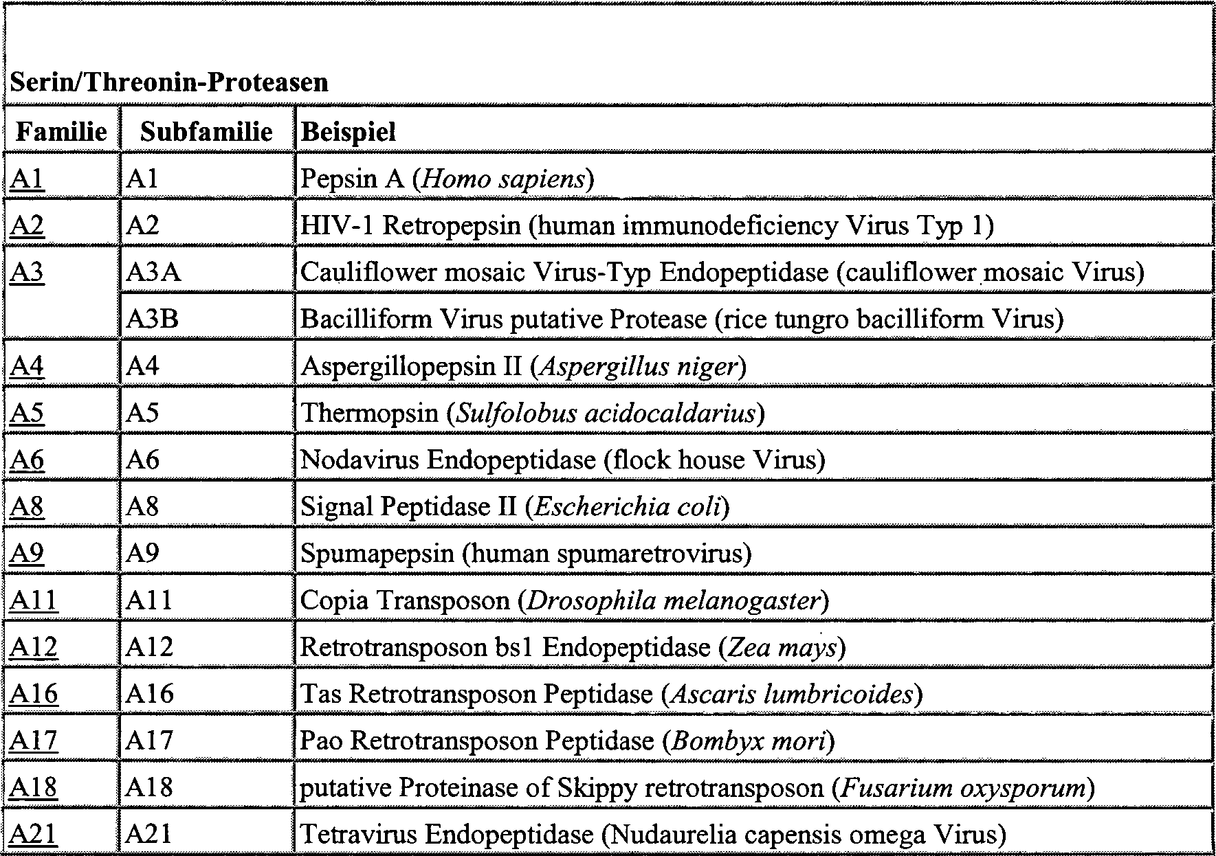


Ein weiterer Gegenstand der Erfindung sind solche Nukleinsäuren, die für ein Fusionsprotein eines der oben genannten nicht fluoreszierenden, aber proteolytisch zur Fluoreszenz aktivierbaren Proteine kodieren. Ein solches Fusionsprotein umfasst ein nicht fluoreszierendes, aber proteolytisch zur Fluoreszenz aktivierbares Protein, welches mindestens eine ein- oder beidseitig von Protease-Schnittstellen flankierte Stör-Aminosäuresequenz entweder am N-Terminus angehängt oder als Insertion nach der Aminosäure mit der Position 1, 2, 3, 4, 5, 6, 7, 8, 9, 10, 11, 12, 13, 14, 15 enthält, fusioniert mit einem weiteren Protein oder mit einer weiteren Proteindomäne. Das weitere Protein oder die weitere Proteindomäne kann hierbei in der Regel sowohl am N-Terminus als auch am C-Terminus des Fusionsproteins lokalisiert sein. Die Fusion am C-Terminus ist lediglich insoweit bevorzugt, als daß eine am N-Terminus lokalisierte weitere Proteindomäne durch die Protease-Aktivität, die die von Protease-Schnittstellen flankierte Stör-Aminosäuresequenz am N-Terminus abspaltet, auch zur Abspaltung der weiteren Proteindomäne führen würde.On the invention furthermore relates to those nucleic acids which for a Fusion protein of one of the above non-fluorescent, but encode proteins that can be activated proteolytically for fluorescence. On such fusion protein includes a non-fluorescent, however protein that can be activated proteolytically for fluorescence, which at least one flanked on one or both sides by protease interfaces Sturgeon amino acid sequence either appended to the N-terminus or as an insertion after the amino acid with the position 1, 2, 3, 4, 5, 6, 7, 8, 9, 10, 11, 12, 13, 14, 15 contains, fused with another Protein or with another protein domain. The other protein or the other protein domain can usually be at both the N-terminus and the C-terminus of the fusion protein. The fusion at the C-terminus is preferred only to the extent that one located at the N-terminus another protein domain through protease activity, which is the interfering amino acid sequence flanked by protease cleavage sites at the N-terminus, would also lead to the splitting off of the further protein domain.
„Weitere Proteindomänen" im Sinne der obigen Definition des Fusionsproteins können vorzugsweise verschiedene Lokalisationsdomänen sein, die für die Kompartimentierung des Fusionsproteins in der Zelle verantwortlich sind, wie insbesondere ein Kernlokalisationssignal („nuclear localization signal" NLS), ein Kernexportsignal („nuclear export signal" NES), eine Membrandomäne, enthaltend vorwiegend hydrophobe Aminosäuren, eine klassische Präsequenz, die das Protein als „sekretorisches Protein" kennzeichnet, sowie weitere bekannte Lokalisationssignale für verschiedene Zellkompartimente."Further Protein domains "in the sense of the above Definition of the fusion protein can preferably be different localization domains for compartmentalization of the fusion protein in the cell are responsible, such as in particular a nuclear localization signal ("nuclear localization signal "NLS), a nuclear export signal ("nuclear export signal "NES), a membrane domain, containing predominantly hydrophobic amino acids, a classic pre-sequence, which the protein as a "secretory Protein " as well as other known localization signals for various cell compartments.
Ein weiterer Gegenstand der Erfindung ist eine Expressionskassette, die eine der oben genannten Nukleinsäuren bzw. Nukleinsäuresequenzen unter der Kontrolle eines Promoters umfassen.On Another object of the invention is an expression cassette, the one of the above-mentioned nucleic acids or nucleic acid sequences under the control of a promoter.
Der Promoter kann hierbei jeder bekannte Promoter sein, der in der Wirtszelle, in die die Expressionskassette eingebracht werden soll, aktiv ist, d.h. in dieser Wirtszelle die Transkription des nachgeschalteten Reportergens aktiviert. Der Promoter kann hierbei ein konstitutiver Promoter sein, der das nachgeschaltete Reportergen ständig exprimiert, oder ein nicht-konstitutiver Promoter, der nur zu definierten Zeitpunkten im Laufe der Entwicklung oder unter bestimmten Umständen (insbesondere unter Einfluß eines Transkriptionsaktivators oder in Abwesenheit eines Transkriptionsrepressors) exprimiert. Erfindungsgemäße Expressionskassetten, die als Promoter beispielsweise den CMV-Promoter enthalten, eignen sich für die Expression des nachgeschalteten nicht fluoreszierenden, aber proteolytisch zur Fluoreszenz aktivierbaren Reporter-Proteins in eukaryontischen, insbesondere in Säugetier-Wirtszellen und Hefe-Wirtszellen. Erfindungsgemäße Expressionskassetten, die als Promoter beispielsweise den lac-Promoter enthalten, eignen sich für die Expression des nachgeschalteten nicht fluoreszierenden, aber proteolytisch zur Fluoreszenz aktivierbaren Reporter-Proteins in prokaryontischen, insbesondere in Bakterien-Wirtszellen.The Promoter can be any known promoter that is in the host cell, into which the expression cassette is to be inserted is active, i.e. in this host cell the transcription of the downstream Reporter gene activated. The promoter can be a constitutive one Be a promoter that constantly expresses the downstream reporter gene, or a non-constitutive promoter who only works at defined times in the course of development or under certain circumstances (in particular under the influence of a Transcription activator or in the absence of a transcription repressor) expressed. Expression cassettes according to the invention, the contain the CMV promoter, for example, are suitable for expression the downstream non-fluorescent, but proteolytic for fluorescence activatable reporter protein in eukaryotic, especially in mammalian host cells and yeast host cells. Expression cassettes according to the invention, the the lac promoter, for example, are suitable as promoters for the Expression of the downstream non-fluorescent, but proteolytic for fluorescence activatable reporter protein in prokaryotic, especially in bacterial host cells.
Der Ausdruck „unter der Kontrolle" eines Promoters bedeutet, daß die Promotersequenz und die Sequenz kodierend für das zu exprimierende nicht fluoreszierende, aber proteolytisch zur Fluoreszenz aktivierbare Reporter-Protein so miteinander verknüpft sind, daß die Expression des Reporter-Proteins möglich ist.The expression "under the control" of a promoter means that the promoter sequence and the sequence coding for the non-fluorescent to be expressed but activated proteolytically for fluorescence bare reporter protein are linked so that expression of the reporter protein is possible.
Die erfindungsgemäße Expressionskassette kann gegebenenfalls weitere Kontrollsequenzen enthalten. Unter einer Kontrollsequenz wird eine beliebige Nukleotidsequenz verstanden, die die Expression des nicht fluoreszierenden, aber proteolytisch zur Fluoreszenz aktivierbaren Reporter-Proteins beeinflußt, wie insbesondere der Promoter, eine Operatorsequenz, d.h. die DNA-Bindungstelle für einen Transkriptionsaktivator oder einen Transkriptionsrepressor, eine Terminator-Sequenz, eine Polyadenylierungssequenz oder eine Ribosombindungsstelle.The Expression cassette according to the invention may contain additional control sequences. Under one Control sequence is understood any nucleotide sequence which is the expression of the non-fluorescent but proteolytic influences reporter protein that can be activated by fluorescence, such as especially the promoter, an operator sequence, i.e. the DNA binding site for one Transcription activator or a transcription repressor, a Terminator sequence, a polyadenylation sequence or a ribosome binding site.
Ein weiterer Gegenstand der Erfindung ist ein rekombinanter Vektor, der eine der obigen erfindungsgemäßen Expressionskassetten enthaltend ein nicht fluoreszierendes, aber proteolytisch zur Fluoreszenz aktivierbares Protein umfasst.On the invention furthermore relates to a recombinant vector, containing one of the above expression cassettes according to the invention a non-fluorescent one that can be activated proteolytically for fluorescence Protein includes.
Ein solcher rekombinanter Vektor kann zusätzlich eine Nukleotidsequenz enthalten, durch die der Vektor sich in der betreffenden Wirtszelle replizieren kann. Solche Nukleotidsequenzen werden in der Regel „origin of replication" (deut. Replikationsursprung) genannt. Beispiele für solche Nukleotidsequenzen sind der SV40-Replikationsursprung, der in Säugetier-Wirtszellen zum Einsatz kommt, und in Hefe-Wirtszellen die Hefe-Plasmid 2μ Replikationsgene REP 1-3.On such a recombinant vector can additionally contain a nucleotide sequence contained, by which the vector is located in the host cell concerned can replicate. Such nucleotide sequences are usually “origin of replication "(Deut. Origin of replication). Examples of such nucleotide sequences are the SV40 origin of replication, that in mammalian host cells is used, and in yeast host cells the yeast plasmid 2μ replication genes REP 1-3.
Der rekombinante Vektor kann weiterhin einen oder mehrere Selektionsmarker enthalten. Unter einem Selektionsmarker versteht man ein Gen, welches unter der Kontrolle eines Promoters steht und welches für ein Protein kodiert, das einen physiologischen Defekt der Wirtszelle komplementiert. Selektionsmarker stellen insbesondere das Gen kodierend für die Dihydrofolat Reduktase (DHFR) dar, oder auch ein Gen, welches die Resistenz gegen Antibiotika, wie insbesondere Ampicillin, Kanamycin, Tetracyclin, Blasticidin, Gentamycin, Chloramphenicol, Neomycin oder Hygromycin bewirkt.The recombinant vector can also have one or more selection markers contain. A selection marker is a gene that is under the control of a promoter and which is a protein encodes that complements a physiological defect of the host cell. Selection markers are in particular the gene coding for the dihydrofolate Reductase (DHFR), or a gene that shows resistance to Antibiotics, such as in particular ampicillin, kanamycin, tetracycline, Blasticidin, gentamycin, chloramphenicol, neomycin or hygromycin causes.
Eine große Anzahl von rekombinanten Vektoren zur Expression eines Zielproteins in prokaryontischen oder eukaryontischen Wirtszellen sind nach dem Stand der Technik bekannt und viele sind auch kommerziell erhältlich.A size Number of recombinant vectors for the expression of a target protein in prokaryotic or eukaryotic host cells are after the State of the art and many are also commercially available.
Ein weiterer Gegenstand der Erfindung ist eine Wirtszelle, die mit dem erfindungsgemäßen rekombinanten Vektor transient oder stabil transformiert worden ist.On Another object of the invention is a host cell with the recombinant according to the invention Vector has been transformed transiently or stably.
Die Auswahl der geeigneten Wirtszelle, hängt von einer großen Anzahl an Faktoren ab, die dem Fachmann bekannt sind. Diese Faktoren beinhalten insbesondere den gewählten Vektortyp, die Toxizität des exprimierten Proteins für die betreffende Wirtszelle, die zu beantwortende Fragestellung, die Expressionscharakteristika und physiologischen Wechselwirkungen des betreffenden Zielproteins in der Wirtszelle, die Sicherheitsrisiken und Kosten. Generell kann jede beliebige pro- oder eukaryontische Zelle bzw. Organismus als Wirtszelle eingesetzt werden.The Choosing the appropriate host cell depends on a large number on factors known to the person skilled in the art. These factors include especially the chosen one Vector type, toxicity of the expressed protein for the host cell concerned, the question to be answered, the expression characteristics and physiological interactions of the target protein in question in the host cell, the security risks and costs. Generally any pro or eukaryotic Cell or organism can be used as a host cell.
Beispiele für geeignete prokaryontische Wirtszellen sind gram-positive Bakterien wie insbesondere Bacillus subtilis, Bacillus licheniformis, Bacillus brevis, Streptomyces lividans etc. oder gram-negative Bakterien wie insbesondere E. coli.Examples for suitable prokaryotic host cells are gram-positive bacteria such as Bacillus in particular subtilis, Bacillus licheniformis, Bacillus brevis, Streptomyces lividans etc. or gram-negative bacteria such as in particular E. coli.
Beispiele für geeignete eukaryontische Wirtszellen sind die Spezies der Saccharomyces oder Schizosaccharomyces, insbesondere Saccharomyces cerevisae.Examples for suitable eukaryotic host cells are the species of Saccharomyces or Schizosaccharomyces, especially Saccharomyces cerevisae.
Beispiele für Zelllinien, die von Säugetieren stammen und die ebenfalls als Wirtszellen in Frage kommen, sind insbesondere die Zelllinien COS-1 (ATCC CRL 1650), COS-7 (ATCC CRL 1651), CHO K1 (ATCC CCL 61), NIH 3T3 (ATCC CRL 1658), HeLa (ATCCL 2), MRC-5 (ATCC CCL 171), HEK 293 (ATCC CRL1573).Examples for cell lines, that of mammals stem and are also suitable as host cells in particular the cell lines COS-1 (ATCC CRL 1650), COS-7 (ATCC CRL 1651), CHO K1 (ATCC CCL 61), NIH 3T3 (ATCC CRL 1658), HeLa (ATCCL 2), MRC-5 (ATCC CCL 171), HEK 293 (ATCC CRL1573).
Der rekombinante Vektor kann in die betreffende Wirtszelle durch jegliche Transfektions-, Transformations- oder Injektionstechnik, die dem Fachmann bekannt ist, eingeführt werden. Insbesondere kann der rekombinante Vektor durch eine der folgenden Techniken in die betreffende Wirtszelle eingeführt werden: Calciumphosphat-Präzipitation, Elektroporation, Protoplastenfusion, Nukleinsäure-Injektion, Lipofektion, „gene gun" unterstützte Techniken, Infektion mit Virus-Partikeln oder Virus-abgeleiteten Partikeln und Protein-Transduktion mit TAT oder TAT-ähnlichen Sequenzen.The recombinant vector can be introduced into the host cell in question by any Transfection, transformation or injection technology that the Is known to those skilled in the art become. In particular, the recombinant vector by one of the the following techniques are introduced into the host cell concerned: Calcium phosphate precipitation, Electroporation, protoplast fusion, nucleic acid injection, lipofection, "gene gun" supported techniques, Infection with virus particles or virus-derived particles and protein transduction with TAT or TAT-like sequences.
Die Wirtszelle kann hierbei entweder transient oder stabil transformiert worden sein. Bei der „transienten Transformation" einer Zelle bleibt der eingeführte Vektor in der Zelle in der Regel autonom, d.h. er integriert sich nicht in das Wirtszellgenom. Bei Teilung der transient transformierten Zellen in Tochterzellen wird der Vektor nicht mit übertragen. Dies führt dazu, daß der eingeführte Vektor sich nach mehreren Wachstumscyclen der transformierten Zellen so lange „herausverdünnt" bis die meisten Zellen keinen Vektor mehr enthalten. Auch bei transient transformierten Zellen kann man den Verlust des Vektors nach mehreren Wachstumscyclen unterbinden, indem der Selektionsdruck auf die Gegenwart des rekombinanten Vektors, der einen Selektionsmarker wie oben definiert umfasst, aufrecht erhalten wird.The host cell can either have been transformed transiently or stably. In the "transient transformation" of a cell, the vector introduced generally remains autonomous in the cell, ie it is integrated not in the host cell genome. When the transiently transformed cells are divided into daughter cells, the vector is not transmitted. This leads to the fact that the introduced vector "dilutes out" after several growth cycles of the transformed cells until most cells no longer contain any vector. Even with transiently transformed cells, the loss of the vector after several growth cycles can be prevented by the selection pressure on the Presence of the recombinant vector comprising a selection marker as defined above is maintained.
Bei der „stabilen Transformation" einer Zelle integriert sich der eingeführte Vektor, der meist in linearisierter Form eingeführt wird, in das Wirtsgenom der Zelle. Bei Teilung der stabil transformierten Zellen in Tochterzellen werden demnach die ursprünglichen Vektorsequenzen als Bestandteil des Wirtsgenoms mit übertragen. Eine auf einem rekombinanten Vektor befindliche Expressionskassette wird daher dauerhaft, d.h. über eine große Anzahl von Wachstumscyclen hinweg, in den Tochterzellen exprimiert.at the “stable Transformation "one The introduced cell integrates Vector, which is mostly introduced in linearized form, into the host genome the cell. When the stably transformed cells are divided into daughter cells therefore the original Vector sequences transferred as part of the host genome. An expression cassette located on a recombinant vector is therefore permanent, i.e. over a size Number of growth cycles expressed in the daughter cells.
Transiente, sowie stabile Transformationstechniken sind dem Fachmann bekannt und in gängigen Nachschlagewerken nachzulesen (Freshney, IR; Culture of Animal Cells, 2000, 4th Ed. Wiley-Liss).transient, and stable transformation techniques are known to the person skilled in the art and in common reference books (Freshney, IR; Culture of Animal Cells, 2000, 4th Ed. Wiley-Liss).
Ein weiterer Gegenstand der Erfindung ist ein Kit zur Detektion und/oder zur Analyse von von Protease-Aktivitäten oder von Protease-abhängigen Ereignissen, welcher mindestens eine der folgenden Komponenten umfasst:
- a) eine Nukleinsäure kodierend für ein nicht fluoreszierendes, aber proteolytisch zur Fluoreszenz aktivierbares Reporter-Protein, welches mindestens eine ein- oder beidseitig von Protease-Schnittstellen flankierte Stör-Aminosäuresequenz entweder am N-Terminus angehängt oder als Insertion nach der Aminosäure mit der Position 1, 2, 3, 4, 5, 6, 7, 8, 9, 10, 11, 12, 13, 14, 15 enthält, oder eine Nukleinsäure kodierend für ein Fusionsprotein dieses Reporter-Proteins,
- b) eine Expressionskassette umfassend eine Nukleinsäure nach a) unter der Kontrolle eines Promoters,
- c) einen rekombinanten Vektor, der mindestens eine der Expressionskassetten nach b) umfasst,
- d) eine Wirtszelle, die mit mindestens einem rekombinanten Vektor nach c) transient oder stabil transformiert worden ist,
- e) ein Protein, welches von einer Nukleinsäure nach a), von einer Expressionskassette nach b) oder von einem rekombinanten Vektor nach c) kodiert wird oder welches von einer Wirtszelle nach d) exprimiert wird.
- a) a nucleic acid coding for a non-fluorescent, but proteolytically activatable for fluorescence reporter protein which has at least one interfering amino acid sequence flanked on one or both sides by protease cleavages either attached to the N-terminus or as an insertion after the amino acid with position 1 , 2, 3, 4, 5, 6, 7, 8, 9, 10, 11, 12, 13, 14, 15, or a nucleic acid coding for a fusion protein of this reporter protein,
- b) an expression cassette comprising a nucleic acid according to a) under the control of a promoter,
- c) a recombinant vector which comprises at least one of the expression cassettes according to b),
- d) a host cell which has been transiently or stably transformed with at least one recombinant vector according to c),
- e) a protein which is encoded by a nucleic acid according to a), by an expression cassette according to b) or by a recombinant vector according to c) or which is expressed by a host cell according to d).
Die Detektion und/oder Analyse von Protease-Aktivitäten oder von Protease-abhängigen Ereignissen erfolgt vorzugsweise in vitro in dem Zellextrakt einer Zelle, die mit einem erfindungsgemäßen rekombinanten Vektor transformiert worden ist, insbesondere aber in vivo in einer Zelle, die mit einem erfindungsgemäßen rekombinanten Vektor transformiert worden ist.The Detection and / or analysis of protease activities or of protease-dependent events takes place preferably in vitro in the cell extract of a cell that is associated with a recombinant vector according to the invention has been transformed, but especially in vivo in a cell, those with a recombinant according to the invention Vector has been transformed.
Die Detektion und/oder Analyse von Protease-Aktivitäten oder von Protease-abhängigen Ereignissen erfolgt vorzugsweise mittels Fluoreszenzmikroskopie von Zellen, die mit einem erfindungsgemäßen rekombinanten Vektor transformiert worden sind. Die Detektion und/oder Analyse von Protease-Aktivitäten oder von Protease-abhängigen Ereignissen kann weiterhin mittels Fluoreszenzspekroskopie bzw. mittels Fluorescence Aided Cell Sorting („FACS") erfolgen.The Detection and / or analysis of protease activities or of protease-dependent events takes place preferably by means of fluorescence microscopy of cells with a recombinant according to the invention Vector have been transformed. The detection and / or analysis of protease activities or from protease-dependent Events can still be determined using fluorescence spectroscopy or using Fluorescence Aided Cell Sorting ("FACS").
Mit Hilfe des erfindungsgemäßen Verfahrens bzw. mit Hilfe des erfindungsgemäßen Kits können beliebige Protease-Aktivitäten in Zellen, Zellextrakten, Zellüberständen, in Fraktionen von Zellextrakten oder Zellüberständen detektiert und analysiert werden.With Help of the method according to the invention or with the help of the kit according to the invention can any protease activity in cells, cell extracts, cell supernatants, in Fractions of cell extracts or cell supernatants are detected and analyzed become.
Insbesondere können solche Protease-Aktivitäten detektiert und analysiert werden, die in der Zelle lediglich transient, d.h. vorübergehend und nur in kurzen Zeiträumen auftreten. Da auch eine nur kurzzeitig auftretende Protease-Aktivität in der Zelle zur proteolytischen Entfernung der Stör-Aminosäuresequenzen aus dem Reporter-Protein – und damit letztlich zum Umschalten von einem nicht fluoreszierenden Reporter in einen fluoreszierenden Reporter – führt, können mit dem erfindungsgemäßen Verfahren auch vorzugsweise transient auftretende, sowie schwache Protease-Aktivitäten gemessen werden.In particular can such protease activities are detected and analyzed, which are only transient in the cell, i.e. temporarily and only in short periods of time occur. Since there is only a short-term protease activity in the Cell for proteolytic removal of sturgeon amino acid sequences from the reporter protein - and thus ultimately to switch from a non-fluorescent reporter in a fluorescent reporter - can, with the inventive method also preferably transient and weak protease activities measured become.
Verschiedenste physiologisch signifikante zelluläre Ereignisse bzw. Signaltransduktionswege beinhalten das Auftreten von Protease-Aktivitäten, insbesondere das Auftreten von transienten, d.h. kurzzeitigen Protease-Aktivitäten in der Zelle. Insbesondere die Apoptose ist ein solches physiologische relevantes zelluläres Ereignis, welches mit dem Auftreten von Protease-Aktivitäten, insbesondere mit der Aktivität der Caspasen und der Calpaine einhergeht. Durch die Detektion der Aktivität dieser Apoptoserelevanten Proteasen könnten daher insbesondere apoptotische Zellen mittels des erfindungsgemäßen Verfahrens als fluoreszierende Zellen detektiert werden.various physiologically significant cellular events or signal transduction pathways involve the appearance of protease activities, particularly the appearance of transients, i.e. short-term protease activities in the Cell. Apoptosis in particular is such a physiological one relevant cellular event, which is associated with the appearance of protease activities, in particular with the activity of the caspases and the calpaine goes along. By detecting the activity of this Proteases relevant to apoptosis could therefore in particular apoptotic cells using the method according to the invention can be detected as fluorescent cells.
Generell können jedoch mit den erfindungsgemäßen Kits und/oder Verfahren alle beliebigen Protease-abhängigen Ereignisse in der Zelle detektiert werden.As a general rule can however with the kits according to the invention and / or method any protease dependent events in the cell can be detected.
Weiterhin können mit den erfindungsgemäßen Kits und/oder Verfahren nicht nur Protease-Aktivitäten oder Protease-abhängige Ereignisse selbst, sondern auch solche zellulären Ereignisse detektiert und analysiert werden, die die Ursache für das Auftreten der Protease-Aktivität in der Zelle oder eine Folge des Auftretens der Protease-Aktivität in der Zelle sind. Die Ursache für das Auftreten einer bestimmten Protease-Aktivität in der Zelle kann insbesondere auch das Auftreten einer spezifischen Protein-Protein-Interaktion sein.Farther can with the kits according to the invention and / or methods not just protease activities or protease dependent events itself, but also detects such cellular events and be analyzed, which is the cause of the appearance of protease activity in the Cell or a consequence of the appearance of protease activity in the Are cell. The cause of the occurrence of a certain protease activity in the cell can in particular also the occurrence of a specific protein-protein interaction his.
Ein weiterer Gegenstand der Erfindung ist ein nicht fluoreszierendes, aber proteolytisch zur Fluoreszenz aktivierbares Protein oder Fusionsprotein, welches von einer der oben definierten Nukleinsäuren kodiert wird.On Another object of the invention is a non-fluorescent, but protein or fusion protein which can be activated proteolytically for fluorescence, which is encoded by one of the nucleic acids defined above.
Besonders bevorzugt sind solche Reporter-Proteine oder Reporter-Fusionsproteine, die ein Derivat einer natürlichen oder artifiziellen Variante der GFP-Familie aus Aequorea victoria, insbesondere ein Derivat der Varianten EGFP (Acc. No. U76561), EYFP (Acc. No. AJ510163, kodiert von der Region 6103-6822), ECFP (Acc. No. AJ510158, kodiert von der Region 6058-6780), GFP (Acc. No. X83959 ), YFP (Acc. No. AY189981, kodiert von der Region 1603-2331) oder CFP (Acc. No. BD136947) sind.Especially such reporter proteins or reporter fusion proteins are preferred, which is a derivative of a natural or artificial variant of the GFP family from Aequorea victoria, in particular a derivative of the variants EGFP (Acc. No. U76561), EYFP (Acc. No. AJ510163, encoded by region 6103-6822), ECFP (Acc. No. AJ510158, encoded by region 6058-6780), GFP (Acc. No. X83959 ), YFP (Acc. No. AY189981, encoded by region 1603-2331) or CFP (Acc. No. BD136947).
Die Erfindung betrifft weiterhin ein Verfahren zur Detektion und/oder zur Charakterisierung einer Protease-Aktivität in einer Zelle, welche die folgenden Verfahrensschritte umfasst:
- a) Transfektion einer Zelle mit dem oben genannten rekombinanten Vektor zur rekombinanten Expression eines nicht fluoreszierenden Proteins oder Fusionsproteins, welches mindestens eine ein- oder beidseitig von Protease-Schnittstellen flankierte Stör-Aminosäuresequenz am N- Terminus angehängt oder durch Insertion in die oben genannten Protein-Positionen umfasst,
- b) Aktivierung einer bislang inaktiven Protease in der Zelle oder Aktivierung der Expression einer Protease in der Zelle, wobei die in Schritt a) genannten Protease-Schnittstellen Substrate dieser Protease darstellen,
- c) Generierung eines fluoreszierenden Proteins oder Fusionsproteins durch proteolytische Entfernung der mindestens einen ein- oder beidseitig von Protease-Schnittstellen flankierten Stör-Aminosäuresequenz aus Schritt a) aus dem nicht fluoreszierenden Protein oder Fusionsprotein aus a),
- d) Detektion der Fluoreszenz des fluoreszierenden Proteins oder Fusionsproteins aus c).
- a) Transfection of a cell with the above-mentioned recombinant vector for the recombinant expression of a non-fluorescent protein or fusion protein which appends at least one interfering amino acid sequence flanked on one or both sides by protease cleavage sites at the N-terminus or by insertion into the above-mentioned protein Positions includes
- b) activation of a previously inactive protease in the cell or activation of the expression of a protease in the cell, the protease interfaces mentioned in step a) being substrates of this protease,
- c) generation of a fluorescent protein or fusion protein by proteolytic removal of the at least one interfering amino acid sequence flanked on one or both sides by protease cleavage sites from step a) from the non-fluorescent protein or fusion protein from a),
- d) detection of the fluorescence of the fluorescent protein or fusion protein from c).
In Schritt a) wird eine geeignete Wirtszelle mit einem erfindungsgemäßen rekombinanten Vektor zur rekombinanten Expression eines nicht fluoreszierenden Proteins, aber proteolytisch zur Fluoreszenz aktivierbaren Reporter-Proteins entweder transient oder stabil transformiert. Das nicht fluoreszierende Protein besitzt hierbei entweder mindestens eine ein- oder beidseitig von Protease-Schnittstellen flankierte Stör-Aminosäuresequenz, welche unmittelbar am N-Terminus des Proteins angehängt ist, oder mindestens eine ein- oder beidseitig von Protease-Schnittstellen flankierte Stör-Aminosäuresequenz, welche nach einer Aminosäure mit der Position 1, 2, 3, 4, 5, 6, 7, 8, 9, 10, 11, 12, 13, 14, 15 im Protein insertiert ist. Das nicht fluoreszierende Protein kann auch an seinem N-Terminus oder vorzugsweise an seinem C-Terminus mit einer weiteren Proteindomäne fusioniert sein. Die Transformation kann über alle dem Fachmann bekannten Transformations- und Transfektionstechniken erfolgen, so insbesondere durch Calciumphosphat-Präzipitation, Elektroporation, Protoplastenfusion, Nukleinsäure-Injektion, Lipofektion, „gene gun" unterstützte Techniken, Infektion mit Virus-Partikeln oder mit Virus-abgeleiteten Partikeln und Protein-Transduktion mit TAT oder TAT-ähnlichen Sequenzen. Der rekombinante Vektor besitzt hierbei vorzugsweise die oben bereits erläuterten Eigenschaften.In Step a) becomes a suitable host cell with a recombinant according to the invention Vector for the recombinant expression of a non-fluorescent Protein, but reporter protein that can be activated proteolytically for fluorescence either transiently or stably transformed. The non-fluorescent Protein has either at least one or both sides Interfering amino acid sequence flanked by protease cleavage sites, which appended to the N-terminus of the protein is, or at least one one or Interfering amino acid sequence flanked on both sides by protease cleavage sites, which after an amino acid with the positions 1, 2, 3, 4, 5, 6, 7, 8, 9, 10, 11, 12, 13, 14, 15 is inserted in the protein. The non-fluorescent protein can also at its N-terminus or preferably at its C-terminus merged with another protein domain his. The transformation can be over all transformation and transfection techniques known to the person skilled in the art take place, in particular by calcium phosphate precipitation, Electroporation, protoplast fusion, nucleic acid injection, lipofection, "gene gun" supported techniques, Infection with virus particles or with virus-derived particles and protein transduction with TAT or TAT-like sequences. The recombinant Vector preferably has those already explained above Characteristics.
In Schritt b) erfolgt gegebenenfalls die Aktivierung einer bislang inaktiven Protease in der Zelle oder auch die Aktivierung der Expression einer Protease in der Zelle. Die in Schritt a) genannten Protease-Schnittstellen stellen hierbei Substrate dieser aktivierten oder exprimierten Protease aus Schritt b) dar.In Step b) is where appropriate the activation of one so far inactive protease in the cell or activation of expression a protease in the cell. The protease interfaces mentioned in step a) provide substrates of this activated or expressed protease from step b).
Die Aktivierung einer bislang inaktiven Protease in der Zelle oder die Aktivierung der Expression einer Protease in der Zelle erfolgt vorzugsweise dadurch, daß die transformierte Zelle aus Schritt a) solchen Bedingungen ausgesetzt wird, unter denen die zu detektierende Protease-Aktivität in der Zelle oder das zu detektierende Protease-abhängige Ereignis in der Zelle auftreten kann. Solche Bedingungen umfassen insbesondere die Expression oder Aktivierung einer weiteren Komponente (insbesondere eines weiteren Proteins) in der Zelle, welche eventuell unmittelbar oder mittelbar zu der betreffenden Protease-Aktivität führen kann, die Zugabe von Substanzen zur unmittelbaren oder mittelbaren Inhibierung oder Aktivierung der betreffenden Protease-Aktivität, sowie das Bereitstellen bestimmter physikalisch-chemischer Bedingungen, wie beispielsweise Temperaturänderungen, pH-Änderungen etc., die unmittelbar oder mittelbar zu der betreffenden Protease-Aktivität führen können.The activation of a previously inactive protease in the cell or the activation of the expression of a protease in the cell is preferably carried out by exposing the transformed cell from step a) to those conditions under which the protease activity to be detected in the cell or to Detecting protease-dependent event can occur in the cell. Such conditions include, in particular, the expression or activation of a further component (in particular a further protein) in the cell, which may possibly lead directly or indirectly to the protease activity in question, the addition of substances for direct or indirect inhibition or activation of the protease in question. Activity, as well as the provision of certain physico-chemical conditions, such as temperature changes, pH changes, etc., which are directly or indirectly related to the protease Ab can lead activity.
Das erfindungsgemäße Verfahren wird vorzugsweise zur Detektion und zur Charakterisierung von Apoptose-relevanten Protease-Aktivitäten eingesetzt. Hierbei könnten die transformierten Zellen aus Schritt a) im Rahmen von Schritt b) solchen Bedingungen ausgesetzt werden, die bekanntermaßen Apoptose verursachen.The inventive method is preferably used for the detection and characterization of apoptosis-relevant Protease activities used. This could the transformed cells from step a) in the context of step b) exposed to conditions known to be apoptosis cause.
Das erfindungsgemäße Verfahren wird vorzugsweise zur Detektion und zur Charakterisierung von solchen Protease-Aktivitäten eingesetzt werden, die infolge einer spezifischen Protein-Protein-Interaktion in der betreffenden Zelle auftreten. Hierbei könnten die transformierten Zellen aus Schritt a) im Rahmen von Schritt b) mit einem oder mit zwei Expressionsvektoren transformiert werden, die für zwei verschiedene, eventuell interagierende Fusionsproteine kodieren.The inventive method is preferably used for the detection and characterization of such Protease activities used as a result of a specific protein-protein interaction occur in the cell in question. Here, the transformed cells from step a) in the context of step b) with one or two Expression vectors are transformed that are for two different, possibly encode interacting fusion proteins.
In Schritt c) wird die mindestens eine ein- oder beidseitig von Protease-Schnittstellen flankierten Stör-Aminosäuresequenz aus Schritt a) aus dem nicht fluoreszierenden Protein oder Fusionsprotein aus Schritt a) in Abhängigkeit von dem Auftreten einer Protease-Aktivität in Schritt b) proteolytisch entfernt. Eine erfolgreiche proteolytische Entfernung der Stör-Aminosäuresequenz im zunächst nicht fluoreszierenden Reporter-Protein oder Fusionsprotein führt dazu, daß ein fluoreszierendes Reporter-Protein oder Fusionsprotein generiert wird, dessen Fluoreszenz detektierbar ist.In Step c) is the at least one or both sides of protease interfaces flanked sturgeon amino acid sequence from step a) from the non-fluorescent protein or fusion protein from step a) depending of the appearance of protease activity in step b) proteolytically removed. Successful proteolytic removal the sturgeon amino acid sequence im initially non-fluorescent reporter protein or fusion protein causes the existence fluorescent reporter protein or fusion protein generated whose fluorescence is detectable.
In Schritt d) wird die in Abhängigkeit von der Protease-Aktivität auftretende Fluoreszenz des Reporter-Proteins oder des Reporter-Fusionsproteins mittels geeigneter Meßmethoden detektiert. Im allgemeinen eignen sich zur Detektion des Fluoreszenz-Signals alle dem Fachmann bekannten Verfahren, die die folgenden Schritte beinhalten:
- - Anregung mit Licht der Absorptionswellenlänge des fluoreszierenden Proteins
- - Detektion von Licht mit der Emissionswellenlänge des fluoreszierenden Proteins.
- - Excitation with light of the absorption wavelength of the fluorescent protein
- - Detection of light with the emission wavelength of the fluorescent protein.
Bei der Fluoreszenz-Detektion in vivo in lebenden Zellen eignet sich insbesondere die Fluoreszenzmikroskopie zur Detektion der Fluoreszenz-Signals. Weiterhin eignen sich zur Detektion des Fluoreszenz-Signals auch die Fluoreszenz-Spektroskopie, sowie die Detektion fluoreszierender Zellen mit Hilfe von FACS.at fluorescence detection in vivo in living cells is suitable especially fluorescence microscopy to detect the fluorescence signal. Furthermore, they are also suitable for the detection of the fluorescence signal the fluorescence spectroscopy, as well as the detection of fluorescent Cells using FACS.
Bei dem erfindungsgemäßen Verfahren können sowohl endogene Proteasen, als auch exogene Proteasen, die in der Wirtszelle mit Hilfe eines rekombinanten Expressionsvektors exprimiert werden, detektiert und charakterisiert werden. Der rekombinante Expressionsvektor umfasst hierbei eine Nukleinsäuresequenz, die die betreffende Protease unter der Kontrolle eines Promoters kodiert. Unter einer „endogenen Protease" wird im folgenden eine Protease verstanden, die in der betreffenden Wirtszelle selbst entweder konstitutiv, d.h. fortdauernd, oder nicht konstitutiv, d.h. nur zu bestimmten Zeitpunkten oder unter bestimmten Bedingungen exprimiert wird. Unter einer „exogenen Protease" wird im folgenden eine Protease verstanden, die nicht vom Wirtsgenom selbst exprimiert wird, sondern mit Hilfe eines von außen durch Transformation eingebrachten Expressionsvektors in der betreffenden Wirtszelle exprimiert wird.at the inventive method can both endogenous proteases and exogenous proteases that are in the Host cell expressed using a recombinant expression vector be detected and characterized. The recombinant Expression vector in this case comprises a nucleic acid sequence that the relevant one Protease encoded under the control of a promoter. Under an "endogenous Protease "is published in following understood a protease in the host cell concerned itself either constitutive, i.e. continuous, or not constitutive, i.e. only expressed at certain times or under certain conditions becomes. Under an "exogenous Protease "is published in following understood a protease that is not from the host genome itself is expressed, but with the help of one brought in from outside by transformation Expression vector is expressed in the host cell in question.
Das erfindungsgemäße Verfahren kann allgemein zur Detektion und zur Charakterisierung von Protease-Aktivitäten oder von Protease-abhängigen zellulären Ereignissen eingesetzt werden. Unter einem Protease-abhängigen zellulären Ereignis sind vorzugsweise auch „natürlich auftretende Protease-abhängige Signalkaskaden", wie insbesondere die Apoptose, die Blutgerinnung, bestimmte entwicklungsspezifische Signalkaskaden, der Protein-Abbau in der Zelle zu verstehen. Unter einem Protease-abhängigen zellulären Ereignis sind vorzugsweise auch „artifizielle Protease-abhängige Ereignisse" zu verstehen, die insbesondere dann stattfinden, wenn das Auftreten einer Protease-Aktivität experimentell von dem Auftreten eines anderen zellulären Ereignisses, wie beispielsweise einer spezifischen Protein-Protein-Interaktion abhängt.The inventive method can be used for the detection and characterization of protease activities or of protease-dependent cellular Events. Under a protease-dependent cellular event are preferably also "naturally occurring" Protease-dependent Signaling cascades " such as in particular apoptosis, blood clotting, certain development-specific Signal cascades to understand the protein breakdown in the cell. Under a protease-dependent cellular Event are preferably also to be understood as "artificial protease-dependent events" which in particular take place when the occurrence of protease activity is experimental of the occurrence of another cellular event, such as depends on a specific protein-protein interaction.
Eine besonders bevorzugte Abwandlung des erfindungsgemäßen Verfahrens kann zur Detektion von Protein-Protein-Interaktionen eingesetzt werden und umfasst die folgenden Verfahrensschritte:
- A) Expression eines ersten und eines zweiten Fusionsproteins in einer geeigneten Wirtszelle, wobei das erste Fusionsprotein eine erfindungsgemäße Nukleinsäure kodierend für ein nicht fluoreszierendes, aber proteolytisch zur Fluoreszenz aktivierbares Reporterprotein als erste Domäne und ein erstes interagierendes Protein als zweite Domäne umfasst, und wobei das zweite Fusionsprotein eine den flankierenden Protease-Schnittstellen entsprechende Protease als erste Domäne und ein zweites interagierendes Protein als zweite Domäne umfasst,
- B) Interaktion des ersten interagierenden Proteins des ersten Fusionsprotein mit dem zweiten interagierenden Protein des zweiten Fusionsprotein,
- C) proteolytische Entfernung der Stör-Aminosäuresequenz aus dem Reporterprotein des ersten Fusionsproteins durch die Protease des zweiten Fusionsproteins,
- D) Detektion des fluoreszierenden Reporterproteins in Abhängigkeit von der spezifischen Interaktion zwischen den beiden interagierenden Domänen der beiden Fusionsproteine.
- A) Expression of a first and a second fusion protein in a suitable host cell, the first fusion protein comprising a nucleic acid according to the invention coding for a non-fluorescent, but proteolytically activatable for fluorescence reporter protein as the first domain and a first interacting protein as the second domain, and wherein the second Fusion protein comprises a protease corresponding to the flanking protease interfaces as the first domain and a second interacting protein as the second domain,
- B) interaction of the first interacting protein of the first fusion protein with the second interacting protein of the second fusion protein,
- C) proteolytic removal of the interfering amino acid sequence from the reporter protein of the first fusion protein by the protease of the second fusion protein,
- D) Detection of the fluorescent reporter protein depending on the specific interaction between the two interacting domains of the two fusion proteins.
Die erfindungsgemäßen Verfahren eignen sich nicht nur zur reinen Detektion von Protease-Aktivitäten, sondern auch zur näheren Charakterisierung von Protease-Aktivitäten oder von Protease-abhängigen Ereignissen. So können mit Hilfe der erfindungsgemäßen Verfahren insbesondere auch solche Substanzen identifiziert werden, die eine definierte physiologische Protease-Aktivität in der Zelle inhibieren oder aktivieren. Solche Inhibitoren oder Aktivatoren definierter, physiologisch relevanter, insbesondere Krankheits-relevanter Protease-abhängiger Ereignisse könnten eine signifikante Bedeutung bei der Entwicklung neuer Arzneimittel haben.The method according to the invention are not only suitable for the pure detection of protease activities, but also also for closer Characterization of protease activities or of protease-dependent events. So can using the method according to the invention in particular also those substances are identified that a inhibit defined physiological protease activity in the cell or activate. Such inhibitors or activators more defined, physiologically relevant, in particular disease-related protease-dependent events could a significant importance in the development of new drugs to have.
Gegenstand der Erfindung ist daher auch ein Screening-Verfahren zur Identifikation oder zur Charakterisierung von Inhibitoren oder von Aktivatoren Protease-abhängiger Ereignisse in der Zelle. Ein solches Screening-Verfahren könnte die Verfahrensschritte a) bis d) umfassen, die in einem ersten Versuchsansatz in Anwesenheit und in einem zweiten Versuchsansatz in Abwesenheit mindestens einer Test-Substanz, die potentiell Inhibitor- oder Aktivator-Eigenschaften bezüglich des getesteten Protease-abhängigen Ereignisses haben könnte, durchgeführt werden. Letztlich kann die Fluoreszenz als Maß der in der Zelle aufgetretenen Protease-Aktivität zwischen dem ersten und dem zweiten Versuchsansatz quantitativ verglichen werden, um die tatsächlichen Inhibitor- oder Aktivator-Eigenschaften der mindestens einen Test-Substanz zu beurteilen.object The invention is therefore also a screening method for identification or to characterize inhibitors or activators Protease-dependent Events in the cell. Such a screening procedure could Process steps a) to d) include that in a first experimental approach in the presence and in a second attempt at absence at least one test substance that has potential inhibitor or activator properties in terms of of the protease-dependent tested Event could have carried out become. Ultimately, fluorescence can be used as a measure of that occurring in the cell Protease activity quantitatively compared between the first and the second approach be the actual Inhibitor or activator properties of the at least one test substance to judge.
Gegenstand der Erfindung ist auch ein Screening-Verfahren zur Identifikation oder zur Charakterisierung von Inhibitoren oder von Aktivatoren definierter Protein-Protein-Interaktionen in der Zelle. Ein solches Screening-Verfahren könnte die Verfahrensschritte A) bis D) umfassen, die in einem ersten Versuchsansatz in Anwesenheit und in einem zweiten Versuchsansatz in Abwesenheit einer oder mehrerer Test-Substanzen, die potentiell Inhibitor- oder Aktivator-Eigenschaften bezüglich einer bestimmten Protein-Protein-Interaktion haben könnte, durchgeführt werden. Letztlich kann die Fluoreszenz als Intensitätsmaß der in der Zelle aufgetretenen Protein-Protein-Interaktionen zwischen dem ersten und dem zweiten Versuchsansatz quantitativ verglichen werden, um die tatsächlichen Inhibitor- oder Aktivator-Eigenschaften der Test-Substanzen) zu beurteilen.object the invention is also a screening method for identification or to characterize inhibitors or activators defined protein-protein interactions in the cell. Such a screening procedure could include the procedural steps A) to D) include those present in a first trial and in a second attempt in the absence of one or more Test substances that have potential inhibitor or activator properties in terms of a particular protein-protein interaction could have, carried out become. Ultimately, fluorescence can be used as a measure of the intensity of the protein-protein interactions occurred between the cell first and second experimental approaches are compared quantitatively, to the actual Inhibitor or activator properties of the test substances) judge.
Ein weiterer Gegenstand der Erfindung ist ein transgenes nicht menschliches Tier, welches eine der oben genannten, erfindungsgemäßen Expressionskassetten in seinem Genom permanent integriert hat. Dieses transgene, nicht menschliche Tier ist vorzugsweise eine Maus oder eine Ratte. Ein solches transgenes, nicht menschliches Tier kann vorzugsweise zur Untersuchung von proteolytischen Vorgängen in vivo, insbesondere zur Analyse von Caspaseaktivitäten als Indikator für apoptotische Vorgänge eingesetzt werden.On another object of the invention is a transgenic non-human Animal which is one of the above-mentioned expression cassettes according to the invention has permanently integrated into its genome. This transgenic, not human animal is preferably a mouse or a rat. On such a transgenic, non-human animal can preferably be used for Investigation of proteolytic processes in vivo, in particular for the analysis of caspase activities as an indicator of apoptotic processes be used.
Die
Erfindung wird durch das Sequenzprotokoll erläutert, welches die folgenden
Sequenzen umfasst:
SEQ ID NO. 1 enthält ein Fragment des Enhanced
Yellow Fluorescent Protein (EYFP), welches die ersten 15 Aminosäuren des
Proteins aus Aequorea victoria umfasst,
SEQ ID NO. 2 enthält ein Fragment
des Enhanced Yellow Fluorescent Protein (EYFP), welches die ersten
9 Aminosäuren
des Proteins aus Aequorea victoria umfasst,
SEQ ID NO. 3 enthält ein Beispiel
für eine
funktionale Stör-Aminosäuresequenz,
die dazu neigt, α-Helix-Strukturen
auszubilden und gleichzeitig überwiegend
hydrophile Aminosäuren
umfasst (wie insbesondere Glutamat, Aspartat, Arginin und Lysin),
SEQ
ID NO. 4 enthält
ein Beispiel für
eine von Klonierungsstellen und TEV-Protease-Schnittstellen beidseitig flankierte,
funktionale Stör-Aminosäuresequenz,
SEQ
ID NO. 5 enthält
die vollständige
Nukleotidsequenz kodierend für
das Enhanced Yellow Fluorescent Protein (EYFP),
SEQ ID NO.
6 enthält
die vollständige
Aminosäuresequenz
des Enhanced Yellow Fluorescent Proteins (EYFP),
SEQ ID NO.
7 enthält
die vollständige
Nukleotidsequenz des SwitchEYFP-Klons swYFP-CS (Insertion zwischen
die Aminosäuren
6 und 7 von EYFP),
SEQ ID NO. 8 enthält die vollständige Aminosäuresequenz
des SwitchEYFP-Klons swYFP-CS (Insertion zwischen die Aminosäuren 6 und
7 von EYFP),
SEQ ID NO. 9 enthält die vollständige Nukleotidsequenz
des SwitchEYFP-Klons swYFP-H3 (Insertion zwischen die Aminosäuren 9 und
10 von EYFP),
SEQ ID NO. 10 enthält die vollständige Aminosäuresequenz
des SwitchEYFP-Klons swYFP-H3 (Insertion zwischen die Aminosäuren 9 und
10 von EYFP),
SEQ ID NO. 11 enthält den Sinn-Strang des Adapters
aus Beispiel 1,
SEQ ID NO. 12 enthält den Gegensinn-Strang des
Adapters aus Beispiel 1.The invention is illustrated by the sequence listing, which comprises the following sequences:
SEQ ID NO. 1 contains a fragment of the Enhanced Yellow Fluorescent Protein (EYFP), which comprises the first 15 amino acids of the protein from Aequorea victoria,
SEQ ID NO. 2 contains a fragment of the Enhanced Yellow Fluorescent Protein (EYFP), which comprises the first 9 amino acids of the protein from Aequorea victoria,
SEQ ID NO. 3 contains an example of a functional interfering amino acid sequence which tends to form α-helix structures and at the same time predominantly comprises hydrophilic amino acids (such as in particular glutamate, aspartate, arginine and lysine),
SEQ ID NO. 4 contains an example of a functional interfering amino acid sequence flanked on both sides by cloning sites and TEV protease cleavage sites,
SEQ ID NO. 5 contains the complete nucleotide sequence coding for the enhanced yellow fluorescent protein (EYFP),
SEQ ID NO. 6 contains the complete amino acid sequence of the enhanced yellow fluorescent protein (EYFP),
SEQ ID NO. 7 contains the complete nucleotide sequence of the SwitchEYFP clone swYFP-CS (insertion between amino acids 6 and 7 of EYFP),
SEQ ID NO. 8 contains the complete amino acid sequence of the SwitchEYFP clone swYFP-CS (insertion between amino acids 6 and 7 of EYFP),
SEQ ID NO. 9 contains the complete nucleotide sequence of the SwitchEYFP clone swYFP-H3 (insertion between amino acids 9 and 10 of EYFP),
SEQ ID NO. 10 contains the complete amino acid sequence of the SwitchEYFP clone swYFP-H3 (insertion between amino acids 9 and 10 of EYFP),
SEQ ID NO. 11 contains the sense strand of the adapter from example 1,
SEQ ID NO. 12 contains the counter-sense strand of the adapter from example 1.
Die Erfindung wird weiterhin durch die folgende Zeichnung näher erläutert.The The invention is further illustrated by the following drawing.
Der SwitchEYFP-Expressionsvektor, mit dem die Zellen aus Spalte B transfiziert worden waren, entspricht hierbei dem Expressionsvektor aus dem Klon swYFP-CS mit der Ausnahme, daß das SwitchEYFP-Protein eine Kernlokalisationsdomäne (nuc) umfasst. Wie aus dem Vergleich der Phasenkontrast- und der Fluoreszenzaufnahme aus B ersichtlich ist, fluoresziert in Spalte B – wie es zu erwarten wäre – daher auch nur der Nucleus der Zelle.The SwitchEYFP expression vector with which the cells from column B are transfected was the expression vector from the clone swYFP-CS with the exception that the SwitchEYFP protein comprises a nuclear localization domain (nuc). As from the Comparison of phase contrast and fluorescence images from B it can be seen that fluorescence in column B - as would be expected - therefore even just the nucleus of the cell.
Der SwitchEYFP-Expressionsvektor, mit dem die Zellen aus Spalte C transfiziert worden waren, entspricht dem Expressionsvektor aus dem Klon swYFP-CS mit der Ausnahme, daß das SwitchEYFP-Protein eine Membranlokalisationsdomäne aus dem Membranprotein Syntaxin (Syx) umfasst. Wie aus dem Vergleich der Phasenkontrast- und der Fluoreszenzaufnahme aus C ersichtlich ist, fluoresziert in Spalte C – wie es zu erwarten wäre – daher auch nur die Zellmembran der Zelle.The SwitchEYFP expression vector with which the cells from column C are transfected was the expression vector from the clone swYFP-CS with the exception that the SwitchEYFP protein is a membrane localization domain from the membrane protein syntaxin (Syx) includes. As from the comparison of the phase contrast and the Fluorescence image can be seen from C, fluoresces in column C - how it would be expected - therefore even just the cell membrane of the cell.
Der Expressionsvektor E entspricht dabei dem Expressionsvektor aus dem Klon swYFP-CS (Insertionsstelle zwischen Aminosäure 6 und 7 von EYFP).The Expression vector E corresponds to the expression vector from the Clone swYFP-CS (insertion site between amino acids 6 and 7 of EYFP).
Der Expressionsvektor D umfasst die Nukleotidsequenz für ein EYFP-Protein unter der Kontrolle eines Promoters, wobei dieses EYFP-Protein erst mit der 7. Aminosäure (E an Position 7) beginnt und zusätzlich an seinem N-Terminus die folgende Aminosäuresequenz umfasst: die 11 Aminosäuren „RLMMALLTIHL" der Stör-Aminosäuresequenz nach SEQ ID No. 3, eine TEV-Protease-Schnittstelle (Aminosäuren „ENLYFQ'G") und die aus der Klonierung resultierenden Aminosäuren „SGKHE".The Expression vector D comprises the nucleotide sequence for an EYFP protein under the control of a promoter, this EYFP protein only with the 7th amino acid (E at position 7) and also at its N-terminus the following amino acid sequence includes: the 11 amino acids "RLMMALLTIHL" of the sturgeon amino acid sequence according to SEQ ID No. 3, a TEV protease interface (amino acids "ENLYFQ'G") and those resulting from the cloning Amino acids "SGKHE".
Der Expressionsvektor C umfasst die Nukleotidsequenz für ein EYFP-Protein unter der Kontrolle eines Promoters, wobei dieses EYFP-Protein erst mit der 7. Aminosäure (E an Position 7) beginnt und zusätzlich an seinem N-Terminus die folgende Aminosäuresequenz umfasst: eine TEV-Protease-Schnittstelle (Aminosäuren „ENLYFQ'G") und die aus der Klonierung resultierenden Aminosäuren „SGKHE".The Expression vector C comprises the nucleotide sequence for an EYFP protein under the control of a promoter, this EYFP protein only with the 7th amino acid (E at position 7) and also at its N-terminus the following amino acid sequence includes: a TEV protease interface (amino acids "ENLYFQ'G") and the amino acids "SGKHE" resulting from the cloning.
Der Expressionsvektor B umfasst die Nukleotidsequenz für ein EYFP-Protein unter der Kontrolle eines Promoters, wobei dieses EYFP-Protein erst mit der 7. Aminosäure (E an Position 7) beginnt und zusätzlich an seinem N-Terminus mit einer Aminosäuresequenz umfassend lediglich die aus der Klonierung resultierenden Aminosäuren „SGKHE" fusioniert wird.The Expression vector B comprises the nucleotide sequence for an EYFP protein under the control of a promoter, this EYFP protein only with the 7th amino acid (E at position 7) and also at its N-terminus with an amino acid sequence comprising only the amino acids “SGKHE” resulting from the cloning is fused.
Der Expressionsvektor A umfasst die Nukleotidsequenz für ein EYFP-Protein unter der Kontrolle eines Promoters, wobei dieses EYFP-Protein erst mit der 7. Aminosäure (E an Position 7) beginnt und keine zusätzlichen Aminosäuresequenzen an seinem N-Terminus angehängt hat.The Expression vector A comprises the nucleotide sequence for an EYFP protein under the control of a promoter, this EYFP protein only with the 7th amino acid (E at position 7) and no additional amino acid sequences appended to its N-terminus Has.
Lediglich
PC12-Zellen, die mit dem Expressionsvektor E oder D transfiziert
worden waren, zeigten keinerlei Fluoreszenz (
„K" für Kontroll-Zellen,
die lediglich mit dem Leervektor transfiziert worden sind,
„A" für Zellen,
die mit einem Expressionsvektor, der eine vollständige TEV-Protease exprimiert, transfiziert worden
sind,
„B" für solche
Zellen, die mit einem ersten und einem zweiten Expressionsvektor
transfiziert worden sind, wobei der erste Expressiosvektor ein Fragment
der TEV-Protease,
umfassend die Aminosäuren
1 bis 118, fusioniert mit der Interaktionsdomäne GCN4 exprimiert, und wobei
der zweite Expressionsvektor ein Fragment der TEV-Protease, umfassend
die Aminosäuren
119 bis 243, fusioniert mit einer weiteren Interaktionsdomäne GCN4
exprimiert,
„C" für solche
Zellen, die mit einem ersten und einem zweiten Expressionsvektor
transfiziert worden sind, wobei der erste Expressionsvektor ein
Fragment der TEV-Protease
umfassend die Aminosäuren
1 bis 118 exprimiert, und wobei der zweite Expressionsvektor ein
Fragment der TEV-Protease umfassend die Aminosäuren 119 bis 243 exprimiert.
"K" for control cells that have only been transfected with the empty vector,
"A" for cells that have been transfected with an expression vector expressing a complete TEV protease,
"B" for those cells that have been transfected with a first and a second expression vector, the first expression vector expressing a fragment of the TEV protease, comprising amino acids 1 to 118, fused to the interaction domain GCN4, and wherein the second expression vector is a Fragment of the TEV protease, comprising the amino acids 119 to 243, fused to a further interaction domain expressed GCN4,
"C" for those cells which have been transfected with a first and a second expression vector, the first expression vector expressing a fragment of the TEV protease comprising amino acids 1 to 118, and the second expression vector comprising a fragment of the TEV protease comprising the Amino acids 119 to 243 expressed.
Alle Zellen K, A, B, C sind außer mit den oben bezeichneten Protease-Vektoren auch mit einem Expressionsvektor, der ein erfindungsgemäßes switchEYFP exprimiert, kotransfiziert.All Cells A, B, C are excluded with the protease vectors described above, also with an expression vector, a switchEYFP according to the invention expressed, co-transfected.
Die Erfindung wird durch die folgenden experimentellen Beispiele näher erläutert.The Invention is illustrated by the following experimental examples.
Beispiel 1: Herstellung bzw. Screening nach nicht fluoreszierenden, aber proteolytisch zur Fluoreszenz aktivierbaren EYFP-Varianten durch InsertionsmutageneseExample 1: Production or screening for non-fluorescent, but proteolytic for Fluorescence-activated EYFP variants by insertion mutagenesis
Zur
Herstellung nicht fluoreszierender, aber proteolytisch zur Fluoreszenz
aktivierbarer EYFP-Reporterproteine wurde das offene Leseraster
des Enhanced Yellow Fluorescent Protein (EYFP) mit den Oligonukleotid-Primern
EYFP-Xhol-sense (Seq: gggctcgagaccatggtgagcaagggcgagga) und EYFP
Xhol-antisense (Seq: gggctcgagcttgtacagctcgtccatgccga) mittels PCR
amplifiziert und in den rekombinanten Vektor pCMV-EM7-Zeo-βGal kloniert.
Der aus dieser Klonierung hervorgegangene rekombinante Vektor wird
im folgenden als pCMV-EYZG bezeichnet. Der Vektor enthält den CMY-Promoter,
eine SV40-Polyadenylierungssequenz sowie den bakteriellen Minimalpromoter
EM7 zur Expression nachgeschalteter Gene in E.coli und in höheren eukaryontischen
Zellen. Das Transkript des pCMV-EYZG kodiert für ein Fusionsprotein aus drei
Domänen,
nämlich
aus EYFP, aus dem ZeoR-Protein, einem Protein, das Resistenz gegen
Zeocin vermittelt, und aus dem β-Galaktosidase-Protein
von E.coli. Die zur Klonierung verwendeten Schnittstellen waren
hierbei so gewählt,
dass ein durchgehendes Leseraster für alle drei Domänen des
Fusionsproteins generiert wurde. (siehe
Die Insertionsmutagenese zur Herstellung nicht fluoreszierenden, aber proteolytisch zur Fluoreszenz aktivierbaren EYFP-Varianten wurde über die folgenden Verfahrensschritte durchgeführt:
- 1) Mit dem Plasmid pCMV-EYZG wurde eine Transposon-vermittelte Zufallsmutagenese mit Hilfe des GPS-LS Kit von New England Biolabs (NEB) nach Angaben des Herstellers durchgeführt. Hierbei wurden zunächst E.coli-Zellen mit dem Target-Plasmid pCMV-EYZG und mit dem Transprimer-Donor-Plasmid aus dem Kit transformiert. Durch eigentliche Mutagenese erfolgte durch die Anwesenheit der TnsABC-Transposase in der Zelle. Aus der Mutagenese gingen etwa 500 Ampicillin-, Zeocin- und Chlormphenicol-resistente rekombinante Bakterienklone hervor und wurden nach Kultivierung auf entsprechenden LB-Selektionsplatten amplifiziert. Etwa 1000 Bakterienkolonien der amplifizierten rekombinanten Bakterienklone wurden vereinigt und die Plasmid-DNA wurde nach Standardverfahren (Qiaprep, Qiagen) isoliert. Die Selektion auf Ampicillin-Resistenz garantiert hierbei, daß das Target-Plasmid in der Zelle anwesend ist. Die Selektion auf Zeocin-Resistenz garantiert hierbei, daß die Insertion des Transposons nicht zu einer Verschiebung des Leserasters geführt hat. Die Selektion auf Chloramphenicol-Resistenz garantiert hierbei, daß ein Transposon im Target-Plasmid anwesend ist.
- 2) Die in Schritt 1 gewonnene komplexe Plasmid-DNA-Mischung wurde mit dem Restriktionsenzym XhoI (NEB) verdaut. Nach gelelektrischer Auftrennung wurde ein 1,9 kb langes Fragment umfassend das EYFP mitsamt zufällig inseriertem Transposon aus dem Gel eluiert (Qiaprep, Qiagen) und anschließend mit einem XhoI-linearisierten, dephosphorylierten pCMV-EM7 ZeoR-βGal-Vektor ligiert. Die Ligationsprodukte wurden in E. coli-Zellen transformiert und auf Ampicillin- und Chloramphenicol-Resistenz selektioniert. Etwa 5000 erfolgreich selektionierte Bakterienkolonien wurden vereinigt. Anschließend wurde aus diesen Kolonien Plasmid-DNA nach Standardverfahren (Qiaprep, Qiagen) isoliert.
- 3) Die in Schritt 2 gewonnene komplexe Plasmid-DNA-Mischung wurde mit dem Restriktionsenzym PmeI (NEB) verdaut. Durch diesen Restriktionsschnitt wird ein großes Fragment aus dem Transposon entfernt, so daß nach dem PmeI-Restriktionsschnitt nur noch wenige flankierende Basen des Transposons, nämlich 15 bp (5 Aminosäuren auf Protein-Ebene, siehe Produktbeschreibung des Herstellers, GPS-LS Kit, NEB) an der Insertionsstelle verbleiben. Die Produkte wurden anschließend mit alkalischer Phosphatase behandelt und gelelektrophoretisch aufgetrennt. Die an zufälligen Stellen des EYFP-Leserasters lineariserten Vektoren wurden aus dem Gel nach Standardmethoden isoliert (Qiaquick, Qiagen).
- 4) Es wurde ein Adapter mit einer von TEV-Protease-Schnittstellen flankierten Stör-Nukleotidsequenz kodierend für eine Stör-Aminosäuresequenz durch Hybridisierung der partiell überlappenden 5' phosphorylierten Oligonukleotide mit der Sequenz 5'P-agcggtgaaaacctatacttccaaggcctgctggcactgctgatgatg aaggccctgctgcgccgcctgctgaaggcact-3' (SEQ ID NO. 11) und mit der Sequenz 5'-accgctaccttggaagtacaggttttctaaatgaatagtcagcagtgccatcatcagc-3' (SEQ ID NO. 12) und durch Auffüllen der einzelsträngigen Bereiche mit dem Klenow-Enzym (Roche) hergestellt. Die flankierenden „TEV-Protease-Schnittstellen" stellen hierbei spezifische Erkennungs- und Spaltstellen der NIa TEV-Protease dar. Das Stör-Fragment kodiert in der angegebenen Orientierung für eine von TEV-Schnittstellen flankierte, starre, partiell geladene α-Helix. Die Analyse der vermutlichen Sekundärstruktur des Stör-Fragments wurde mit dem Programm Predator (EMBL) durchgeführt. Die linearisierten, dephosphorylierten Vektoren aus Schritt 3 wurden mit dem 5'-phosphorylierten Adapter mit der von TEV-Protease-Schnittstellen flankierten Stör-Nukleotidsequenz, im folgenden als 'Stör-Fragment' bezeichnet, ligiert (siehe Beispiel 1, Nr. 4). Die Ligationsprodukte wurden in E.coli-Zellen transformiert. Rekombinante Klone wurden mit Ampicillin und Zeocin selektioniert. 480 erfolgreich selektionierte Bakterienkolonien wurden einzeln amplifiziert. Anschließend wurde die Plasmid-DNA der Einzelklone nach Standardverfahren (Qiaprep, Qiagen) isoliert.
- 5) Die Plasmid-DNA der Einzelklone aus Schritt 4 wurde anschließend in PC12-Zellen analysiert. Für jeden Einzelklon wurden jeweils zwei Transformationsansätze durchgeführt. In dem ersten Transformationsansatz wurden je 1 μl der Plasmid-Präparationen mit Lipofectamin 2000 (Invitrogen) in PC12-Zellen transfiziert. In einem zweiten Transformationsansatz wurden je 1 μl der Plasmid-Präparationen und 0,1 μg eines Expressionsvektors, der die TEV-Protease unter der Kontrolle eines Promoters exprimieren kann (pCMV-TEV), mit Lipofectamin 2000 (Invitrogen) in PC12-Zellen transfiziert. Anschließend wurde überprüft, ob die transformierten PC12-Zellen des jeweils ersten und des jeweils zweiten Transformationsansatzes fluoreszieren oder nicht. Selektioniert wurden solche Klone, die in Abwesenheit der TEV-Protease (erster Transformationsansatz) aufgrund ihrer Insertion der Stör-Aminosäuresequenz im EYFP nicht fluoreszieren, aber in Gegenwart der TEV-Protease (zweiter Transformationsansatz) aufgrund der proteolytischen Abspaltung der Stör-Aminosäuresequenz aus dem EYFP fluoreszieren. Bei zwei Klonen konnte tatsächlich ohne TEV-Protease-Aktivität keine Epifluoreszenz detektiert werden, nach Kotransfektion der entsprechenden Plasmid-DNA und des TEV-Protease exprimierenden Vektors konnten jedoch fluoreszierende Zellen detektiert werden. Diese beiden Klone wurden als swYFP-H3 und swYFP-CS bezeichnet.
- 1) A transposon-mediated random mutagenesis was carried out with the plasmid pCMV-EYZG using the GPS-LS kit from New England Biolabs (NEB) according to the manufacturer. E.coli cells were first transformed with the target plasmid pCMV-EYZG and with the transprimer donor plasmid from the kit. The actual mutagenesis resulted in the presence of the TnsABC transposase in the cell. The mutagenesis gave rise to about 500 ampicillin, zeocin and chloromphenicol-resistant recombinant bacterial clones and were amplified on appropriate LB selection plates after cultivation. About 1000 bacterial colonies of the amplified recombinant bacterial clones were pooled and the plasmid DNA was isolated using standard methods (Qiaprep, Qiagen). The selection for ampicillin resistance guarantees that the target plasmid is present in the cell. The selection for Zeocin resistance guarantees that the insertion of the transposon has not led to a shift in the reading frame. The selection for chloramphenicol resistance guarantees that a transposon is present in the target plasmid.
- 2) The complex plasmid-DNA mixture obtained in step 1 was digested with the restriction enzyme XhoI (NEB). After gel-electric separation, a 1.9 kb fragment comprising the EYFP together with randomly inserted transposon eluted from the gel (Qiaprep, Qiagen) and then ligated with an XhoI-linearized, dephosphorylated pCMV-EM7 ZeoR-βGal vector. The ligation products were transformed into E. coli cells and selected for ampicillin and chloramphenicol resistance. About 5000 successfully selected bacterial colonies were pooled. Plasmid DNA was then isolated from these colonies using standard methods (Qiaprep, Qiagen).
- 3) The complex plasmid-DNA mixture obtained in step 2 was digested with the restriction enzyme PmeI (NEB). This restriction cut removes a large fragment from the transposon, so that after the PmeI restriction cut only a few flanking bases of the transposon, namely 15 bp (5 amino acids at the protein level, see product description of the manufacturer, GPS-LS Kit, NEB) remain at the insertion site. The products were then treated with alkaline phosphatase and separated by gel electrophoresis. The vectors linearized at random locations on the EYFP reading frame were isolated from the gel by standard methods (Qiaquick, Qiagen).
- 4) An adapter with a sturgeon nucleotide sequence flanked by TEV protease interfaces was encoded for a sturgeon amino acid sequence by hybridization of the partially overlapping 5 'phosphorylated oligonucleotides with the sequence 5'P-agcggtgaaaacctatacttccaaggcctgctggcactgctgatctgatgctQctgagctgggQgggctgaggctgaggaggagcctgaggaggagcaggaggaggaggaggaggaggagcaggcctgagcaggcctgaggagcaggcctgaggagcctgaggaggagcaggcggcgcgccgccfcfcfcqqcqqqcqqqqcqqqqqqqqqqqqqqqqqq. 11) and with the sequence 5'-accgctaccttggaagtacaggttttctaaatgaatagtcagcagtgccatcatcagc-3 '(SEQ ID NO. 12) and by filling in the single-stranded regions with the Klenow enzyme (Roche). The flanking "TEV protease interfaces" represent specific recognition and cleavage sites of the NIa TEV protease. The sturgeon fragment codes in the given orientation for a rigid, partially charged α-helix flanked by TEV interfaces. The analysis The presumed secondary structure of the sturgeon fragment was carried out using the Predator program (EMBL) The linearized, dephosphorylated vectors from step 3 were identified using the 5'-phosphorylated adapter with the sturgeon nucleotide sequence flanked by TEV protease interfaces, hereinafter referred to as' Sturgeon fragment ', ligated (see Example 1, No. 4). The ligation products were transformed into E. coli cells. Recombinant clones were selected with ampicillin and Zeocin. 480 successfully selected bacterial colonies were amplified individually. DNA of the individual clones isolated according to standard methods (Qiaprep, Qiagen).
- 5) The plasmid DNA of the individual clones from step 4 was then analyzed in PC12 cells. Two transformation approaches were carried out for each individual clone. In the first transformation approach, 1 μl of the plasmid preparations were transfected with Lipofectamin 2000 (Invitrogen) in PC12 cells. In a second transformation approach, 1 μl each of the plasmid preparations and 0.1 μg of an expression vector which can express the TEV protease under the control of a promoter (pCMV-TEV) were transfected with Lipofectamin 2000 (Invitrogen) in PC12 cells. It was then checked whether the transformed PC12 cells of the first and second transformation approaches fluoresce or not. Those clones were selected which do not fluoresce in the absence of the TEV protease (first transformation approach) due to their insertion of the interfering amino acid sequence in the EYFP, but in the presence of the TEV protease (second transformation approach) due to the proteolytic cleavage of the interfering amino acid sequence from the EYFP fluoresce. No epifluorescence could actually be detected in two clones without TEV protease activity, but after co-transfection of the corresponding plasmid DNA and the vector expressing TEV protease, fluorescent cells could be detected. These two clones were named swYFP-H3 and swYFP-CS.
Das aus dem Klon swYFP-CS hervorgehende EYFP-Protein besitzt eine von TEV-Protease-Schnittstellen flankierte Stör-Aminosäuresequenz, die zwischen den Aminosäuren 6 und 7 inseriert worden ist. Das aus dem Klon swYFP-H3 hervorgehende EYFP-Protein besitzt eine von TEV-Protease-Schnittstellen flankierte Stör-Aminosäuresequenz, die zwischen den Aminosäuren 9 und 10 inseriert worden ist.The EYFP protein resulting from the clone swYFP-CS has one of TEV-protease cleavage flanked sturgeon amino acid sequence, the between the amino acids 6 and 7 has been inserted. The resulting from the clone swYFP-H3 EYFP protein has an interfering amino acid sequence flanked by TEV protease cleavage sites, the between the amino acids 9 and 10 has been inserted.
Somit scheint der N-Terminus des EYFP für die Insertion bzw. für das Anhängen einer Stör-Aminosäuresequenz zur Generierung eines nicht fluoreszierenden, aber proteolytisch zur Fluoreszenz aktivierbaren Reporterproteins geeignet zu sein. Eine solche Stör-Aminosäuresequenz-Insertion, die als proteolytischer Schalter zwischen Fluoreszenz oder Nicht-Fluoreszenz eines Reporterproteins fungiert, kann für zahlreiche Assay-Varianten zur Detektion und/oder zur Charakterisierung von Protease-Aktivitäten oder von Proteaseabhängigen Ereignissen eingesetzt werden.Consequently seems the N-terminus of the EYFP for the insertion or for the appending one Sturgeon amino acid sequence to generate a non-fluorescent but proteolytic to be suitable for fluorescence-activatable reporter protein. Such an interference amino acid sequence insertion, the as a proteolytic switch between fluorescence or non-fluorescence A reporter protein can function for numerous assay variants for the detection and / or for the characterization of protease activities or of protease addicts Events.
Beispiel 2: Feinanalyse der Insertionsstelle und der Länge der KillersequenzExample 2: Fine analysis the insertion point and the length the killer sequence
Der kritische Bereich für die Insertion der Schaltersequenz ist der N-Terminus von EYFP. Zur genauen Bestimmung, welche Insertions-Positionen sich besonders zum „Ab- und Anschalten" der Fluoreszenz des Reporterproteins eignen, wurde die von TEV-Protease-Schnittstellen flankierte Stör-Aminosäuresequenz in getrennten Experimenten nach jeder der ersten 12 Aminosäuren des Proteins EYFP insertiert. Die so hergestellten EYFP-Varianten wurden anschließend durch Kotransfektion mit einem Vektor, der die TEV-Protease exprimiert, in PC12-Zellen getestet.The critical area for the insertion of the switch sequence is the N-terminus of EYFP. For exact To determine which insertion positions are particularly suitable for "switching the fluorescence of the reporter protein off and on", the interfering amino acid sequence flanked by TEV protease cleavage sites was inserted in separate experiments after each of the first 12 amino acids of the protein EYFP EYFP variants were then tested in PC12 cells by cotransfection with a vector expressing the TEV protease.
Tabelle 2 zeigt das Ergebnis dieser Experimente: so steht beispielsweise „switchEYFP_1_Störseq._2" für einen Expressionsvektor, der ein switchEYFP unter der Kontrolle eines Promoters umfasst, wobei das switchEYFP eine Insertion einer beidseitig von TEV-Protease-Schnittstellen flankierte Stör-Aminosäuresequenz nach SEQ ID No. 3 enthält. Das Ergebnis, welches in Tabelle 2 zusammengefaßt ist, zeigt, daß prinzipiell bei allen Insertionen zwischen den Aminosäuren 2 und 9 EYFP-Varianten entstehen, die proteolytisch „anschaltbar" bezüglich ihrer Fluoreszenz sind, d.h. diese Varianten führen in Abwesenheit einer Protease-Aktivität nicht zur Fluoreszenz, aber in Gegenwart einer Protease-Aktivität zur Fluoreszenz. Bei EYFP-Varianten, die zu einer weiteren Verkürzung von EYFP führen (10/11, 11/12 etc.), funktioniert das „proteolytische Anschalten" der Fluoreszenz des Proteins weniger gut, da die Minimalsequenz zur Ausbildung der Fluoreszenz unterschritten wird (Li et al.; J Biol Chem. 1997 Nov 7;272(45):28545-9).table 2 shows the result of these experiments: for example, “switchEYFP_1_Störseq._2” stands for one Expression vector representing a switchEYFP under the control of a Promoters comprises, the switchEYFP an insertion of a bilateral Interfering amino acid sequence flanked by TEV protease cleavages according to SEQ ID No. 3 contains. The result, which is summarized in Table 2, shows that in principle for all insertions between amino acids 2 and 9 EYFP variants arise which can be "switched on" proteolytically with regard to their Are fluorescence, i.e. these variants do not result in the absence of protease activity Fluorescence, but in the presence of protease activity for fluorescence. For EYFP variants that lead to a further shortening of EYFP (10/11, 11/12 etc.), the “proteolytic Turn on "the Fluorescence of the protein is less good because the minimal sequence for Below fluorescence (Li et al .; J Biol Chem. 1997 Nov 7; 272 (45): 28545-9).
Es konnte weiterhin gezeigt werden, daß alternativ zur Insertion an beliebiger Position zwischen den Aminosäuren 2 und 9 die von TEV-Protease-Schnittstellen flankierte Stör-Aminosäuresequenz auch einfach an den N-Terminus des EYFPs angehängt werden kann. Hierbei können die ersten 1 bis 9 Aminosäuren des EYFPs auch durchaus deletiert und die flankierte Stör-Aminosäuresequenz direkt an den N-Terminus des entsprechenden restlichen EYFP-Fragments fusioniert sein.It could also be shown to be an alternative to insertion at any position between amino acids 2 and 9 that of TEV protease cleavage sites flanked sturgeon amino acid sequence can also simply be appended to the N-terminus of the EYFP. Here, the first 1 to 9 amino acids of the EYFP and the flanked interfering amino acid sequence directly to the N-terminus of the corresponding remaining EYFP fragment be merged.
Tabelle
2: 
Beispiel 3: Unterschiedlich lokalisierte Varianten von nicht fluoreszierenden, aber proteolytisch zur Fluoreszenz aktivierbaren ReporterproteinenExample 3: Different localized variants of non-fluorescent but proteolytic reporter proteins that can be activated by fluorescence
Nicht fluoreszierende, aber proteolytisch zur Fluoreszenz aktivierbare Reporterproteine, die eine ein- oder zweiseitig von Protease-Schnittstellen flankierte Stör-Aminosäuresequenz am N-Terminus angehängt oder an den obigen Positionen insertiert haben, lassen sich auch durch Fusion an andere Proteine oder Proteindomänen an verschiedenen Stellen innerhalb der Zelle lokalisieren. So konnten beispielsweise eine switch-EYFP-Variante mit Kernlokalisation und eine switch-EYFP-Variante mit Membranverankerung in PC12-Zellen erfolgreich getestet werden.Not fluorescent, but can be activated proteolytically for fluorescence Reporter proteins that are one- or two-sided from protease cleavage sites flanked sturgeon amino acid sequence appended to the N-terminus or inserted at the above positions can also be Fusion to other proteins or protein domains in different places localize within the cell. For example, a switch EYFP variant with core localization and a switch EYFP variant with membrane anchoring successfully tested in PC12 cells.
Beispiel 3.1: Nicht fluoreszierendes, proteolytisch zur Fluoreszenz aktivierbares EYFP mit KernlokalisationExample 3.1: Non-fluorescent, EYFP with nuclear localization that can be activated proteolytically for fluorescence
An
den C-Terminus eines switch-EYFPs wurde ein Kernlokalisationssignal
im Leseraster fusioniert (im folgenden „switchEYFPnuc" genannt). Dies führte dazu,
daß das
Protein bevorzugt in den Zellkern von eukaryontischen Zellen transportiert
wird. In
Beispiel 3.2: Nicht fluoreszierendes, proteolytisch zur Fluoreszenz aktivierbares EYFP mit MembranlokalisationExample 3.2: Non-fluorescent, EYFP with membrane localization that can be activated proteolytically for fluorescence
Ein
switchEYFP wurde an den N-Terminus eines Fragmentes des synaptischen
Membranproteins Syntaxin 1A (Aminosäure 1–147) fusioniert (im folgenden „switchEYFPsyx" genannt). Aufgrund
der C-terminalen Transmembrandomäne
wird das Fusionsprotein an der Zellmembran von eukaryontischen Zellen
verankert. In
Beispiel 4: Expression von nicht fluoreszierenden, proteolytisch zur Fluoreszenz aktivierbaren EYFP in weiteren eukaryontischen ZellenExample 4: Expression from non-fluorescent, proteolytically activatable for fluorescence EYFP in other eukaryotic cells
Die
Funktion der switchEYFPs wurden zusätzlich in weiteren Zelllinien
getestet. In
Beispiel 5: Expression von nicht fluoreszierenden, proteolytisch zur Fluoreszenz aktivierbaren EYFP in ProkaryontenExample 5: Expression from non-fluorescent, proteolytically activatable for fluorescence EYFP in prokaryotes
Es sollte untersucht werden, ob nicht fluoreszierende, proteolytisch zur Fluoreszenz aktivierbare EYFPs (d.h. switchEYFPs) auch in prokaryontischen Zellen funktionell sind. Hierzu wurden E.coli-Zellen des Typs DH5αpBAD_TEV mit Expressionsvektoren kodierend für switchEYFPs transformiert. DH5αpBAD_TEV-Zellen enthalten ein Plasmid, welches die TEV-Protease unter der Kontrolle eines mit Arabinose induzierbaren Promotors exprimiert. Dieses Plasmid besitzt weiterhin einen „p15A-origin", so das Kotransformation mit „ColE1-Origin-Vektoren" möglich sind.It should be examined whether not fluorescent, proteolytic EYFPs (i.e. switchEYFPs) that can be activated for fluorescence also in prokaryotic Cells are functional. For this purpose E.coli cells of the type DH5αpBAD_TEV were used transformed with expression vectors coding for switchEYFPs. DH5αpBAD_TEV cells contain a plasmid which controls the TEV protease of a promoter inducible with arabinose. This plasmid still has a "p15A-origin", according to the co-transformation with "ColE1 Origin Vectors" are possible.
Diese
E.coli-Zellen wurden mit einem Expressionsvektor kodierend für ein switchEYFP
unter der Kontrolle des EM7-Minimalpromotors transformiert. Anschließend wurde
durch die Zugabe von Arabinose zu den Zellen die Expression der
TEV-Protease induziert. In
Beispiel 6: Minimale Länge der von TEV-Protease-Schnittstellen flankierten Stör-AminosäuresequenzExample 6: Minimum length of the Interfering amino acid sequence flanked by TEV protease cleavage sites
Bei den im Rahmen des Screening-Verfahrens (siehe Beispiel 1) zunächst identifizierten switchEYFP-Klonen swYFP-CS (Stör-Aminosäuresequenz insertiert zwischen Aminosäure 6 und 7 von EYFP) und swYFP-H3 (Stör-Aminosäuresequenz insertiert zwischen Aminosäure 9 und 10 von EYFP) sollte überprüft werden, welcher Anteil der Stör-Aminosäuresequenz für den Verlust der Fluoreszenz des EYFPs verantwortlich ist, bzw. wie lang die Stör-Aminosäuresequenz sein muß, um ihren Zweck, nämlich die Zerstörung der EYFP-Fluoreszenz und die Wiederherstellung der EYFP-Fluoreszenz nach ihrer proteolytischen Entfernung, zu erfüllen.at those initially identified in the screening process (see Example 1) switchEYFP clones swYFP-CS (interfering amino acid sequence inserted between amino acid 6 and 7 of EYFP) and swYFP-H3 (interfering amino acid sequence inserted between amino acid 9 and 10 of EYFP) should be checked what proportion of the sturgeon amino acid sequence for the Loss of fluorescence from the EYFPs is responsible, or how long the sturgeon amino acid sequence have to be, for their purpose, namely the destruction the EYFP fluorescence and restoration of EYFP fluorescence after its proteolytic removal fulfill.
Dazu
wurden die Deletionsmutanten A bis D des Vektors aus dem Klon swYFP-CS
hergestellt. Bei diesen Deletionsmutanten wurden jeweils Teile der
Stör-Aminosäuresequenz
nach SEQ ID No. 3, gemeinsam mit einer TEV-Protease-Schnittstelle
und der GPS-Linker Sequenz (SGKHE), an ein EYFP-Fragment angehängt, welches
erst mit der 7. Aminosäure
der EYFP-Sequenz beginnt (siehe
Die
Variante E aus
Bei
der Variante D des Expressionsvektors aus
Bei
der Variante C des Expressionsvektors wurde nur die TEV-Schnittstelle
mit der GPS-Linkersequenz
und bei der Variante B des Expressionsvektors lediglich die GPS-Linkersequenz alleine
N-terminal an ein EYFP-Fragment ankloniert, welches erst mit der
7. Aminosäure
der EYFP-Sequenz beginnt. Weiterhin wurde eine Variante A des Expressionsvektors
hergestellt, die keine zusätzliche
Sequenz trägt,
die jedoch erst bei Aminosäure
E7 startet (
Die
Konstrukte A, B, C, D, E wurden durch Transfektion in PC12-Zellen
getestet und mikroskopisch analysiert.
Beispiel 7: SwitchEYFP als Interaktions-SensorExample 7: SwitchEYFP as an interaction sensor
Ein
erfindungsgemäßes SwitchEYFP
kann insbesondere auch als Indikator für Protein-Protein Interaktionen eingesetzt werden.
Dabei sollte die spezifische Interaktion zwischen zwei Proteinen,
zwischen einem Protein und einer Substanz oder zwischen zwei Proteinen
und einer Substanz zur Aktivierung einer proteolytischen Aktivität führen. Diese
Kopplung zwischen einer Protein-Protein-Interaktion und einer proteolytischen Aktivität kann vorzugsweise
durch die Protein-Protein-Interaktions-abhängige Transkomplementation
einer funktionalen Protease erreicht werden, wie in der prioritätsfrüheren und
nachveröffentlichen
Anmeldung (interne Bezeichnung
Hierbei kann das erste Fusionsprotein insbesondere ein Fragment umfassend die Aminosäuren 1 bis 118 der TEV-Protease und ein erstes potentiell interagierendes Protein umfassen und das zweite Fusionsprotein kann insbesondere ein Fragment umfassend die Aminosäuren 119 bis 243 der TEV-Protease und ein zweites potentiell interagierendes Protein umfassen. Eine Teilung der TEV-Protease zwischen den Aminosäuren 118 und 119 zur Transkomplementation hat sich experimentell als günstig erwiesen, d.h. die hierbei generierten TEV-Protease-Fragmente sind für sich alleine nicht funktionell, bilden jedoch eine funktionale TEV-Protease, sofern diese sich durch die Interaktion der interagierenden Domänen der Fusionproteine räumlich aneinander annähern. Es sind jedoch auch Fusionsproteine mit anderen Proteasefragmenten als den oben beschriebenen oder auch Fusionsproteine mit überlappenden Proteasefragmenten zur Bereitstellung einer Protease-Aktivität durch Transkomplementation fähig.Here, the first fusion protein can in particular comprise a fragment comprising amino acids 1 to 118 of the TEV protease and a first potentially interacting protein, and the second fusion protein can in particular comprise a fragment comprising amino acids 119 to 243 of the TEV protease and a second potentially interacting protein , A division of the TEV protease between amino acids 118 and 119 for transcomplementation has proven to be favorable experimentally, ie the TEV protease fragments generated in this way are not functional in themselves, but form a functional TEV protease, provided that these are characterized by Interaction of the interacting domains of the fusion proteins move closer together. However, they are also fusion proteins with protease fragments other than those described above, or else fusion proteins with overlapping protease fragments to provide a pro capable of tease activity by transcomplementation.
Im Rahmen dieses Beispiels sollte die nach Transkomplementation zweier Proteasefragmente wiedererlangte Protease-Aktivität mit Hilfe eines erfindungsgemäßen switchEYFPs als Detektor nachgewiesen werden. Als Modell wurden die Interaktionsdomänen des GCN4-Leuzin-Zipper-Proteins verwendet, an deren C-Terminus die Protease-Fragmente Aminosäure 1 bis 118 und Aminosäure 119 bis 243 fusioniert worden waren. Die Interaktionsdomänen des GCN4-Leuzin-Zipper-Proteins sind dafür bekannt, Homodimere auszubilden. Die oben genannten Konstrukte, sowie eine positive und eine negative Kontrolle wurden jeweils gemeinsam mit einem Expressionsvektor, der eine beidseitig durch TEV-Protease-Schnittstellen flankierte Stör-Aminosäuresequenz, welche nach der Aminosäure 6 von EYFP insertiert ist (switchEYFP_6_Killer_7), in PC12-Zellen kotransfiziert.in the This example should be based on the transcomplementation of two Protease fragments regained protease activity with the help of a switchEYFP according to the invention can be detected as a detector. The interaction domains of the GCN4 leucine zipper protein used, at the C-terminus of the protease fragments amino acid 1 to 118 and amino acid 119 to 243 had been merged. The interaction domains of the GCN4 leucine zipper proteins are known to form homodimers. The above constructs, plus a positive and a negative Controls were each together with an expression vector, one flanked on both sides by TEV protease interfaces Sturgeon amino acid sequence which after the amino acid 6 of EYFP is inserted (switchEYFP_6_Killer_7) in PC12 cells cotransfected.
SEQUENCE
LISTING 


Claims (30)
Priority Applications (2)
| Application Number | Priority Date | Filing Date | Title |
|---|---|---|---|
| DE2003118048 DE10318048A1 (en) | 2003-04-17 | 2003-04-17 | Non-fluorescent reporter proteins that can be activated by proteolysis for fluorescence and their use for the detection of protease-dependent events |
| PCT/EP2004/004142 WO2004092211A1 (en) | 2003-04-17 | 2004-02-19 | Non-fluorescent report protein activateable by proteolyse for fluorescence and the use thereof for detecting protease-depending events |
Applications Claiming Priority (1)
| Application Number | Priority Date | Filing Date | Title |
|---|---|---|---|
| DE2003118048 DE10318048A1 (en) | 2003-04-17 | 2003-04-17 | Non-fluorescent reporter proteins that can be activated by proteolysis for fluorescence and their use for the detection of protease-dependent events |
Publications (1)
| Publication Number | Publication Date |
|---|---|
| DE10318048A1 true DE10318048A1 (en) | 2004-11-04 |
Family
ID=33103529
Family Applications (1)
| Application Number | Title | Priority Date | Filing Date |
|---|---|---|---|
| DE2003118048 Withdrawn DE10318048A1 (en) | 2003-04-17 | 2003-04-17 | Non-fluorescent reporter proteins that can be activated by proteolysis for fluorescence and their use for the detection of protease-dependent events |
Country Status (2)
| Country | Link |
|---|---|
| DE (1) | DE10318048A1 (en) |
| WO (1) | WO2004092211A1 (en) |
Families Citing this family (5)
| Publication number | Priority date | Publication date | Assignee | Title |
|---|---|---|---|---|
| US7842460B2 (en) | 2004-06-21 | 2010-11-30 | Progenra Inc. | Method for assessing proteolytic enzyme activity using ubiquitin fusion substrate |
| CA3073137A1 (en) | 2016-08-19 | 2018-02-22 | Jeffrey S. Bartlett | Methods and compositions for treating conditions using recombinant self-complementary adeno-associated virus |
| AU2017371043B2 (en) | 2016-12-07 | 2022-12-15 | University Of Florida Research Foundation, Incorporated | IL-1Ra cDNAs |
| CN110628801A (en) * | 2019-10-09 | 2019-12-31 | 武汉博欧特生物科技有限公司 | Protein expression and purification method |
| CN112813017B (en) * | 2019-11-18 | 2023-03-28 | 广东省农业科学院环境园艺研究所 | Transient expression system of cymbidium protoplast and construction method and application thereof |
Citations (7)
| Publication number | Priority date | Publication date | Assignee | Title |
|---|---|---|---|---|
| EP0928968A1 (en) * | 1998-01-12 | 1999-07-14 | Universität Basel | Screening method for apoptosis and necrosis |
| DE19819889A1 (en) * | 1998-05-04 | 1999-11-11 | Fraunhofer Ges Forschung | Isolating nucleic acid from samples by binding to array of immobilized, random capture probes |
| WO1999057314A1 (en) * | 1998-05-04 | 1999-11-11 | Fraunhofer-Gesellschaft zur Förderung der angewandten Forschung e.V. | Electrical integrated nucleic acid isolation, purification and detection |
| US6197928B1 (en) * | 1997-03-14 | 2001-03-06 | The Regents Of The University Of California | Fluorescent protein sensors for detection of analytes |
| WO2001046691A1 (en) * | 1999-12-22 | 2001-06-28 | Biosignal Packard Inc. | A bioluminescence resonance energy transfer (bret) system with broad spectral resolution between donor and acceptor emission wavelengths and its use |
| US6410255B1 (en) * | 1999-05-05 | 2002-06-25 | Aurora Biosciences Corporation | Optical probes and assays |
| US6495664B1 (en) * | 1998-07-24 | 2002-12-17 | Aurora Biosciences Corporation | Fluorescent protein sensors of post-translational modifications |
Family Cites Families (2)
| Publication number | Priority date | Publication date | Assignee | Title |
|---|---|---|---|---|
| WO2000073437A1 (en) * | 1999-05-27 | 2000-12-07 | Merck Frosst Canada & Co. | Assays for caspase activity using green fluorescent proteins |
| FR2830020B1 (en) * | 2001-09-25 | 2003-12-19 | Univ Victor Segalen Bordeaux 2 | NUCLEIC ACID FOR OBTAINING A POLYNUCLEOTIDE ENCODING A DETECTION AND ISOLATION PROTEIN OF A "PREY" LIGAND AND APPLICATIONS OF THIS NUCLEIC ACID |
-
2003
- 2003-04-17 DE DE2003118048 patent/DE10318048A1/en not_active Withdrawn
-
2004
- 2004-02-19 WO PCT/EP2004/004142 patent/WO2004092211A1/en active Application Filing
Patent Citations (7)
| Publication number | Priority date | Publication date | Assignee | Title |
|---|---|---|---|---|
| US6197928B1 (en) * | 1997-03-14 | 2001-03-06 | The Regents Of The University Of California | Fluorescent protein sensors for detection of analytes |
| EP0928968A1 (en) * | 1998-01-12 | 1999-07-14 | Universität Basel | Screening method for apoptosis and necrosis |
| DE19819889A1 (en) * | 1998-05-04 | 1999-11-11 | Fraunhofer Ges Forschung | Isolating nucleic acid from samples by binding to array of immobilized, random capture probes |
| WO1999057314A1 (en) * | 1998-05-04 | 1999-11-11 | Fraunhofer-Gesellschaft zur Förderung der angewandten Forschung e.V. | Electrical integrated nucleic acid isolation, purification and detection |
| US6495664B1 (en) * | 1998-07-24 | 2002-12-17 | Aurora Biosciences Corporation | Fluorescent protein sensors of post-translational modifications |
| US6410255B1 (en) * | 1999-05-05 | 2002-06-25 | Aurora Biosciences Corporation | Optical probes and assays |
| WO2001046691A1 (en) * | 1999-12-22 | 2001-06-28 | Biosignal Packard Inc. | A bioluminescence resonance energy transfer (bret) system with broad spectral resolution between donor and acceptor emission wavelengths and its use |
Non-Patent Citations (10)
| Title |
|---|
| CHIESA,Anna, et.al.: Recombinant aequorin and green fluorescent protein as valuable tools in the study of cell signalling. In: Biochem. J. 2001, 355, S.1-12 * |
| CHIESA,Anna, et.al.: Recombinant aequorin and green fluorescent protein as valuable tools in the study of cell signalling. In: Biochem. J. 2001, 355, S.1-12; |
| JONES,J., et.al.: Development and application of a GEP-FRET intracellular caspase assay for drug screening. In: J. Biomol. Screen. 2000, Oct. 5 (5), S.307-318 * |
| JONES,J., et.al.: Development and application of a GEP-FRET intracellular caspase assay for drug screening. In: J. Biomol. Screen. 2000, Oct. 5 (5), S.307-318; |
| KOHL,Tobias, et.al.: A protease assay for two-photon crosscorrelation and FRET analysis based solely on fluorescent proteins. In: PNAS, Sept. 17, 2002, Vol.99, No.19, S.12161-12166 * |
| KOHL,Tobias, et.al.: A protease assay for two-photon crosscorrelation and FRET analysis based solely on fluorescent proteins. In: PNAS, Sept. 17, 2002, Vol.99, No.19, S.12161-12166; |
| MAHAJAN,N.P., et.al.: Novel mutant green fluorescent protein protease substrates reveal the activation of specific caspases during apoptosis. In: Chem. Biol. 1999, Jun. 6 (6), S.401-409 * |
| MAHAJAN,N.P., et.al.: Novel mutant green fluorescent protein protease substrates reveal the activation of specific caspases during apoptosis. In: Chem. Biol. 1999, Jun. 6 (6), S.401-409; |
| reichen? In: Mitteilungen der deutschen Patentanwälte, 2003, H.2, S.57-64; |
| WOLFRAM,Markus: "Reach-Through Claims" und "Reach-Through Licensing" - Wie weit kann Patentschutz auf biotechnologische Research Tools * |
Also Published As
| Publication number | Publication date |
|---|---|
| WO2004092211A1 (en) | 2004-10-28 |
Similar Documents
| Publication | Publication Date | Title |
|---|---|---|
| EP1483576B1 (en) | Novel method for detecting and analyzing protein interactions in vivo | |
| DE60026603T2 (en) | Bioluminescence resonance energy transfer (BRET) system with high spectral resolution between donor and acceptor emission wavelengths and their use | |
| Daniels et al. | Expression of multiple transgenes from a single construct using viral 2A peptides in Drosophila | |
| DE69936103T2 (en) | FUSION PROTEINS CONSISTING OF PROTEIN AND LIBRARIES OF RANDOM PEPTIDES | |
| Bedbrook et al. | Genetically encoded spy peptide fusion system to detect plasma membrane-localized proteins in vivo | |
| EP1088233B9 (en) | A bioluminescence resonance energy transfer (bret) system and its use | |
| Degtyar et al. | Dance of the SNAREs: assembly and rearrangements detected with FRET at neuronal synapses | |
| US11325952B2 (en) | Light-gated signaling modulation | |
| DE60033551T2 (en) | PROBE FOR ANALYZING PROTEIN-PROTEIN INTERACTIONS AND CORRESPONDING METHOD THEREFOR | |
| EP1381624B1 (en) | Specific detection of proteolytic enzymes by means of a hybrid protein | |
| EP1307482B1 (en) | Two coloured fluorimetric protease assay | |
| DE10318048A1 (en) | Non-fluorescent reporter proteins that can be activated by proteolysis for fluorescence and their use for the detection of protease-dependent events | |
| DE60210266T2 (en) | METHOD FOR DETECTING MODULATORS OF A SECRETASE ACTIVITY | |
| DE60104019T2 (en) | Compositions, methods and reagent kits for detecting protein-protein interaction interfering substances | |
| WO2005033706A1 (en) | Method for the detection and analysis of protein-protein interactions | |
| Divéki et al. | Limited utility of blue fluorescent protein (BFP) in monitoring plant virus movement | |
| DE60032632T2 (en) | METHOD OF IDENTIFYING MODULATORS OF PROTEASE-ACTIVATED RECEPTORS (PAR) | |
| US8236937B2 (en) | Methods and compositions for the detection and isolation of ligands | |
| EP2135092A1 (en) | Fluorescence-based assay for detecting compounds for modulating the sodium-calcium exchanger (ncx) in forward mode | |
| US20040042961A1 (en) | Development of an in vivo functional assay for proteases | |
| DE602005005663T2 (en) | PROCESS FOR CHARACTERIZING COMPOUNDS | |
| O’Connor et al. | A protease-initiated model of wound detection | |
| EP1141716B1 (en) | Method for the cellular high-throughput-detection of receptor ligand interactions | |
| DE69635355T2 (en) | Method for the detection of biologically active substances | |
| DE10328775B4 (en) | In vivo test system for the detection of HIV protease activity |
Legal Events
| Date | Code | Title | Description |
|---|---|---|---|
| OM8 | Search report available as to paragraph 43 lit. 1 sentence 1 patent law | ||
| 8130 | Withdrawal |