EP0153047B1 - Transrectal prostate biopsy device - Google Patents
Transrectal prostate biopsy device Download PDFInfo
- Publication number
- EP0153047B1 EP0153047B1 EP85300617A EP85300617A EP0153047B1 EP 0153047 B1 EP0153047 B1 EP 0153047B1 EP 85300617 A EP85300617 A EP 85300617A EP 85300617 A EP85300617 A EP 85300617A EP 0153047 B1 EP0153047 B1 EP 0153047B1
- Authority
- EP
- European Patent Office
- Prior art keywords
- cannula
- stylet
- handle
- distal end
- guide tube
- Prior art date
- Legal status (The legal status is an assumption and is not a legal conclusion. Google has not performed a legal analysis and makes no representation as to the accuracy of the status listed.)
- Expired
Links
Images


Classifications
-
- A—HUMAN NECESSITIES
- A61—MEDICAL OR VETERINARY SCIENCE; HYGIENE
- A61B—DIAGNOSIS; SURGERY; IDENTIFICATION
- A61B10/00—Other methods or instruments for diagnosis, e.g. instruments for taking a cell sample, for biopsy, for vaccination diagnosis; Sex determination; Ovulation-period determination; Throat striking implements
- A61B10/02—Instruments for taking cell samples or for biopsy
- A61B10/0233—Pointed or sharp biopsy instruments
- A61B10/0241—Pointed or sharp biopsy instruments for prostate
-
- A—HUMAN NECESSITIES
- A61—MEDICAL OR VETERINARY SCIENCE; HYGIENE
- A61B—DIAGNOSIS; SURGERY; IDENTIFICATION
- A61B17/00—Surgical instruments, devices or methods, e.g. tourniquets
- A61B17/00234—Surgical instruments, devices or methods, e.g. tourniquets for minimally invasive surgery
- A61B2017/00238—Type of minimally invasive operation
- A61B2017/00274—Prostate operation, e.g. prostatectomy, turp, bhp treatment
-
- A—HUMAN NECESSITIES
- A61—MEDICAL OR VETERINARY SCIENCE; HYGIENE
- A61B—DIAGNOSIS; SURGERY; IDENTIFICATION
- A61B18/00—Surgical instruments, devices or methods for transferring non-mechanical forms of energy to or from the body
- A61B2018/00315—Surgical instruments, devices or methods for transferring non-mechanical forms of energy to or from the body for treatment of particular body parts
- A61B2018/00547—Prostate
Definitions
- the present invention relates to a prostatic biopsy needle, and more particularly to a medical instrument that can provide a reasonable core of prostatic tissue through a transrectal or trans- perineal route.
- the Travenol TRU-CUT O biopsy needle comprises a hollow tubular cutting cannula having a sharpened distal end attached to a plastic handle.
- a coaxial solid stylet telescopes within the cannula and is attached to a knob at its proximal end. The distal end of the stylet is sharpened and includes a transverse slot or specimen notch adjacent to the sharpened end.
- a biopsy needle of this type is disclosed in US-A-3477423.
- the physician positions the stylet to project slightly from the cannula.
- the index finger of one hand is placed along the cannula with the tip in contact with the stylet distal end and the handle is held in the palm.
- Approaching the prostate gland transrectally, the gland is explored with the finger tip to locate a nodule or suspicious area. After locating a point for a sample; the needle is eased forward into the nodule. Once in place, the stylet is plunged to the desired depth.
- the physician then must then remove his hand and finger, grasp the stylet knob in one hand, and push the cannular handle forward with the other hand.
- the cutting end moves along the stylet and severs a sample of tissue projecting into the transverse slot in the tip of the stylet.
- the entire needle is then withdrawn from the gland and the sample removed from the stylet.
- the manipulation of the cannula during this latter step is quite difficult since the tip of the stylet is embedded in the soft and pliable prostatic tissue several inches from the handle.
- the stylet knob gives very little steady support to the needle assembly and the stylet tip, due to its smaller diameter, penetrates the tissue somewhat easier than the tubular carinula cutting edge.
- the entire needle moves forward, puncturing the bladder or urethra. It is also common to attempt to move the cannula forward only to have the stylet back out of the tissue. When this occurs, the physician must remove the needle, reposition the stylet, and try again.
- transrectal biopsy needle which can be guided to the required point of the prostate gland by the physician's finger, a sampling stylet inserted, and a cutting ' cannula plunged forward without removal of-the finger.
- US-A-2496111 discloses a biopsy needle in which a cylindrical sample is but by advance of a cannula and a barbed stylet projecting through the cannula is refracted to secure the sample.
- the pre-characterising clause of claim 1 is based on the disclosure of this document.
- the present invention is characterised over the disclosure of this document by a tissue-collecting slot in the stylet adjacent its distal end, in that the fully extended position of the stylet is defined by engagement of stop means on the knob and the handle respectively, in that the handle defines spaced limit positions for the actuating means corresponding to the retracted and extended positions of the cannula, and in that movement of the cannula from the retracted position to the extended position severs tissue located in the slot and retains the severed tissue in the slot.
- a plastic handle in the form of an elongated rectangular block having a hollow guide tube extending from its forward end with the proximal end of the tube anchored in the handle.
- the distal end of the guide tube is smooth and rounded to prevent abrasion of tissue.
- a hollow , cannula is slidably disposed in telescoping relationship to the guide tube.
- the proximal end of the cannula is attached to a thumb tab disposed in a slot in the handle. When the thumb tab is fully retracted, the distal end of the cannula is sheathed within the distat end of the guide tube.
- the cannula At the most forward position of the cannula, its distal end extends a preselected distance from the distal end of the guide tube. This distance may be on the order of 2 to 3 cm.
- the distal end of the cannula is cut at an angle to form a cutting edge thereof.
- a sampling stylet which may be solid, is inserted through an opening in the rear end of the handle into the cannula.
- a push knob is attached to the proximal end of the stylet.
- the knob includes a detent attached to the handle to hold the stylet in a retracted position. In such position, the distal end of the stylet which is sharpened projects about 1 cm beyond the distal end of the guide tube.
- the knob may be pushed forward until it stops against the rear end of the handle causing the distal end of the stylet to project 2 to 4 cm beyond the guide tube.
- the distal end of the stylet includes a transverse slot or notch for capturing a tissue sample.
- the physician uses the tip of the index finger to guide the tip of the guide tube and the slightly projecting stylet to a point on the prostate gland from which a tissue specimen is required while holding the handle in the palm of the hand. Once the device is in place, the hand and finger are held stationary for the remainder of the procedure.
- the physician uses his other hand to push the stylet knob forward, penetrating the gland with the stylet. Next, he plunges the cannula forward into the gland, slicing a core of tissue captured within the transverse slot in the distal end of the stylet. At this point, the physician removes the device from the gland with the sample. Thus, the procedure is quickly and efficiently accomplished with minimum risk to the patient.
- the handle and guide tube permit the physician to maintain one finger in contact with the gland and to hold the handle perfectly steady in the palm of the hand.
- the stylet cannot back out of the tissue and the larger diameter of the guide tube ensures that the entire needle does not move forward. Thus, the danger of puncturing the bladder or urethra is eliminated.
- a handle 44 formed from plastic, such as polystyrene, and designed for the needle to be disposable after use.
- Handle 44 preferably includes a curved bottom portion 58 which will fit comfortably in the physician's palm.
- a recess or slot 48 is provided in the rear portion of handle 44 as well as slot 54 adjacent the bottom surface 58.
- a guide tube 30 is disposed in the forward end of handle 44 as best seen in Figure 2 and is cemented or otherwise anchored therein.
- Guide tube 30 may be of any suitable material, such as stainless steel, and may have a projecting length on the order of 11 to 16 cm and an outside diameter of about 2 mm. Slidably disposed within guide tube 30 is cutting cannula 20 having a diameter of about 1.5 mm and an overall length of about 15 to 20 cm. As may be noted from Figure 2, the proximal end of cannula 20 is provided with a thumb tab 46 attached thereto. Thumb tab 46 is configured to fit recess 48 and to be able to slide from the rear position shown in Figure 2 to a forward position as indicated by arrow B. The amount of movement will depend upon the size of sample desired; however, a movement of about 2 cm is typical. An opening 32 in the forward end of body 44 includes clearance for cannula 20 to permit it to slide easily within guide tube 30.
- a catch tab 45 is attached to the rear of thumb tab 46 to engage a depression 47 in the rear end wall of body 44. When cannula 20 is fully retracted, as shown in Figure 2, catch tab 45 and recess 47 will maintain such position until the physician desired to move cannula 20 forward.
- a stylet 10 which may have a length of about 19 to 24 cm and a diameter of about 1 mm, is telescopically disposed within cannula 20 and extend through opening 11 in the rear wall end of body 44 and thumb tab 46.
- a push knob 50 is attached to the proximal end of stylet 10 and is movable forward to contact the rear wall of body 44 as indicated by arrow A in Figure 2.
- a movement forward of about 2.5 cm is suitable
- a stopbar 52 provided with a catch portion 56 is connected to push knob 50. Stopbar 52 is shown in Figure 2 in the full rearward position in which catch 56 has engaged tab 57 in recess 54.
- knob 50 and stopbar 52 are formed from plastic which has sufficient flexibility to cause catch 56 to disengage when a slight foward pressure is placed on push knob 50.
- the distal end of cannula 20 does not extend beyond the distal end of the guide tube 30 while the tip of stylet 10 extends slightly beyond the distal end of guide tube 30.
- the physician may then explore the surface of the gland to find a nodule or other suspicious portion thereof. At that point, he may force the sharpened tip 12 of stylet 10 into the nodule as indicated in Figure 4. Next, the physician, using the left hand 78, pushes push knob 50 forward as initiated by arrow A in Figure 3, causing stylet 10 to penetrate the gland 40 as indicated in Figure 5.
- the distal end of stylet 10 includes a transverse slot 14 adjacent the sharpened end 12.
- the tissue that is displaced by the stylet will project into slot 14.
- cannula 20 has a shapened edge 22 and has a sliding fit over stylet 10 which causes cutting edge 22 to cleanly slice through the tissue extending into slot 14 having a core sample 42 shown in Figure 6.
- the physician has been able to hold handle 44 and guide tube 30 completely stationary with his right hand 76 during both the penetration of the gland with stylet 10 and the capturing of sample 42 by means of cutting cannula 20. Therefore, an accurate and clean core specimen is obtained.
- the physician may carefully search the gland for the point to be investigated and once having located such point, may be very quickly complete the procedure for obtaining the sample and withdraw the biopsy needle from the patient's body.
- the improved stability of the needle and the larger diameter of the guide effectively prevent any possibility of the stylet being inadvertently plunged further forward when the cannula is advanced, and the danger of puncturing the bladder or urethra is eliminated.
- backing out of the cannula after insertion of the stylet is also completely eliminated and the risk to the patient of damage and infection is greatly reduced.
- An alternative embodiment of my invention provides a biopsy needle which automatically ensures a clean cut of the specimen.
- This embodiment differs from that described above only in the handle design shown in cross-sectional view in Figure 7.
- the cannula 20, when in the retracted position, is spring-loaded by means of a coil spring 70 in recess 71 at the rear portion of handle 60.
- Spring 70 is maintained in the compressed condition by detent lever 64 which engages thumb tab 68.
- a release tab 66 is provided at the rear of detent lever 64 working against coil spring 72.
Description
- The present invention relates to a prostatic biopsy needle, and more particularly to a medical instrument that can provide a reasonable core of prostatic tissue through a transrectal or trans- perineal route.
- In the diagnosis of malignancies of the prostate gland, it is known to utilize cytologic studies based on a fine needle aspiration biopsy. For example, a device for this purpose is disclosed in U.S. Patent No. 3,595,217 to Rheinfrank. A hollow biopsy needle is passed through a guide tube attached to the operator's finger which is placed on the prostate gland. The needle penetrates the gland and a syringe attached to the needle withdraws a tissue sample. Unfortunately, the use of an aspirating needle to obtain samples from the prostatic tissue has not, in general, produced satisfactory diagnostic material and this approach has been largely abandoned in the United States.
- Consequently, a preferred approach is to obtain a core sample. Many physicians utilize a biopsy needle available from Travenol Laboratories, Inc., of Deerfield, Illinois. The Travenol TRU-CUTO biopsy needle comprises a hollow tubular cutting cannula having a sharpened distal end attached to a plastic handle. A coaxial solid stylet telescopes within the cannula and is attached to a knob at its proximal end. The distal end of the stylet is sharpened and includes a transverse slot or specimen notch adjacent to the sharpened end. A biopsy needle of this type is disclosed in US-A-3477423.
- To obtain a prostatic sample using the Travenol needle, the physician positions the stylet to project slightly from the cannula. The index finger of one hand is placed along the cannula with the tip in contact with the stylet distal end and the handle is held in the palm. Approaching the prostate gland transrectally, the gland is explored with the finger tip to locate a nodule or suspicious area. After locating a point for a sample; the needle is eased forward into the nodule. Once in place, the stylet is plunged to the desired depth.
- The physician then must then remove his hand and finger, grasp the stylet knob in one hand, and push the cannular handle forward with the other hand. Theoretically, the cutting end moves along the stylet and severs a sample of tissue projecting into the transverse slot in the tip of the stylet. The entire needle is then withdrawn from the gland and the sample removed from the stylet.
- In practice, the manipulation of the cannula during this latter step is quite difficult since the tip of the stylet is embedded in the soft and pliable prostatic tissue several inches from the handle. The stylet knob gives very little steady support to the needle assembly and the stylet tip, due to its smaller diameter, penetrates the tissue somewhat easier than the tubular carinula cutting edge. On occasions, when attempting to push the cannula into the tissue, the entire needle moves forward, puncturing the bladder or urethra. It is also common to attempt to move the cannula forward only to have the stylet back out of the tissue. When this occurs, the physician must remove the needle, reposition the stylet, and try again.
- Since the procedure involves puncturing of the colon wall, each attempt increases the risk of infection. Most physicians limit such attempts to two or three passes. Even with a successful insertion of the cannula, the instability of the Travenol needle often results in a limiting cbre sample.
- Thus, there is a long-felt and unfilled need for a transrectal biopsy needle which can be guided to the required point of the prostate gland by the physician's finger, a sampling stylet inserted, and a cutting' cannula plunged forward without removal of-the finger.
- US-A-2496111 discloses a biopsy needle in which a cylindrical sample is but by advance of a cannula and a barbed stylet projecting through the cannula is refracted to secure the sample. The pre-characterising clause of claim 1 is based on the disclosure of this document.
- The present invention is characterised over the disclosure of this document by a tissue-collecting slot in the stylet adjacent its distal end, in that the fully extended position of the stylet is defined by engagement of stop means on the knob and the handle respectively, in that the handle defines spaced limit positions for the actuating means corresponding to the retracted and extended positions of the cannula, and in that movement of the cannula from the retracted position to the extended position severs tissue located in the slot and retains the severed tissue in the slot.
- In the preferred embodiment of the invention, a plastic handle in the form of an elongated rectangular block is provided having a hollow guide tube extending from its forward end with the proximal end of the tube anchored in the handle. The distal end of the guide tube is smooth and rounded to prevent abrasion of tissue. A hollow , cannula is slidably disposed in telescoping relationship to the guide tube. The proximal end of the cannula is attached to a thumb tab disposed in a slot in the handle. When the thumb tab is fully retracted, the distal end of the cannula is sheathed within the distat end of the guide tube. At the most forward position of the cannula, its distal end extends a preselected distance from the distal end of the guide tube. This distance may be on the order of 2 to 3 cm. The distal end of the cannula is cut at an angle to form a cutting edge thereof.
- A sampling stylet, which may be solid, is inserted through an opening in the rear end of the handle into the cannula. A push knob is attached to the proximal end of the stylet. The knob includes a detent attached to the handle to hold the stylet in a retracted position. In such position, the distal end of the stylet which is sharpened projects about 1 cm beyond the distal end of the guide tube. The knob may be pushed forward until it stops against the rear end of the handle causing the distal end of the stylet to project 2 to 4 cm beyond the guide tube. The distal end of the stylet includes a transverse slot or notch for capturing a tissue sample.
- With the cannula and the stylet fully retracted, the physician uses the tip of the index finger to guide the tip of the guide tube and the slightly projecting stylet to a point on the prostate gland from which a tissue specimen is required while holding the handle in the palm of the hand. Once the device is in place, the hand and finger are held stationary for the remainder of the procedure.
- The physician uses his other hand to push the stylet knob forward, penetrating the gland with the stylet. Next, he plunges the cannula forward into the gland, slicing a core of tissue captured within the transverse slot in the distal end of the stylet. At this point, the physician removes the device from the gland with the sample. Thus, the procedure is quickly and efficiently accomplished with minimum risk to the patient.
- The handle and guide tube permit the physician to maintain one finger in contact with the gland and to hold the handle perfectly steady in the palm of the hand. The stylet cannot back out of the tissue and the larger diameter of the guide tube ensures that the entire needle does not move forward. Thus, the danger of puncturing the bladder or urethra is eliminated.
- Therefore, it is a principal object of my invention to provide an improved transrectal biopsy needle for producing core tissue sample quickly and accurately.
- It is another object of my invention to provide a biopsy needle that can be guided to a desired location within the patient's body by means of a finger which is maintained in place during the remainder of the procedure.
- It is still another object of my invention to provide a biopsy needle which may be guided to a desired location by the physician using one hand, and be held steady by that hand while the other hand is used to collect a tissue sample.
- It is a further object of my invention to provide a biopsy needle having an external guide tube, a shapened cutting cannula telescoping within the guide tube, and a stylet having a tissue-collecting slot in the distal end thereof telescoped within the cannula in which the guide tube is located by the physician at a desired point and remains stationary at that point during the remainder of the tissue-collecting procedure.
- It is yet a further object of my invention to provide a biopsy needle having a handle held in the palm of the physician's hand with the guide tube attached thereto and extending therefrom and in which the handle includes means for advancing the stylet and the cannula.
- These and other objects and advantages of the invention will become apparent from the following detailed description when read in conjunction with the drawings.
-
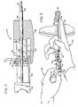
- Figure 1 is a perspective view of one embodiment of my invention having cut away parts to illustrate the construction thereof;
- Figure 2 is a cross-sectional view of the handle portion of the biopsy needle of Figure 1;
- Figure 3 is a view showing the use of my invention in which the physician has guided the guide tube and stylet point to a desired point on a prostate gland;
- Figure 4, 5, and 6 illustrate the sequence of operation of the tissue sample-collecting portion of my invention in which:
- Figure 4 shows the guide tube having the stylet and cannula retracted with the tip of the stylet inserted into tissue; and
- Figure 5 shows the cannula retracted and the stylet plunged forward to a depth suitable for collecting a sample of the tissue;
- Figure 6 shows the cutting cannula plunged forward severing a sample of the tissue in a slot in the stylet tip; and
- Figure 7 illustrates a partial cross section of the handle of an alternative embodiment of my invention in which the cutting cannula is spring-loaded in its retracted position.
- Referring to Figures 1 and 2, the various elements of my improved transrectal biopsy needle may be noted. Although the description herein below may make reference to certain materials and constructions, it will be obvious to those skilled in the art to adapt other materials and arrangements of the elements, and the illustrations are for exemplary purposes only. I prefer to utilize a
handle 44 formed from plastic, such as polystyrene, and designed for the needle to be disposable after use.Handle 44 preferably includes acurved bottom portion 58 which will fit comfortably in the physician's palm. A recess orslot 48 is provided in the rear portion ofhandle 44 as well asslot 54 adjacent thebottom surface 58. Aguide tube 30 is disposed in the forward end ofhandle 44 as best seen in Figure 2 and is cemented or otherwise anchored therein.Guide tube 30 may be of any suitable material, such as stainless steel, and may have a projecting length on the order of 11 to 16 cm and an outside diameter of about 2 mm. Slidably disposed withinguide tube 30 is cuttingcannula 20 having a diameter of about 1.5 mm and an overall length of about 15 to 20 cm. As may be noted from Figure 2, the proximal end ofcannula 20 is provided with athumb tab 46 attached thereto.Thumb tab 46 is configured to fitrecess 48 and to be able to slide from the rear position shown in Figure 2 to a forward position as indicated by arrow B. The amount of movement will depend upon the size of sample desired; however, a movement of about 2 cm is typical. An opening 32 in the forward end ofbody 44 includes clearance forcannula 20 to permit it to slide easily withinguide tube 30. - A
catch tab 45 is attached to the rear ofthumb tab 46 to engage adepression 47 in the rear end wall ofbody 44. When cannula 20 is fully retracted, as shown in Figure 2,catch tab 45 andrecess 47 will maintain such position until the physician desired to movecannula 20 forward. - A
stylet 10, which may have a length of about 19 to 24 cm and a diameter of about 1 mm, is telescopically disposed withincannula 20 and extend through opening 11 in the rear wall end ofbody 44 andthumb tab 46. Apush knob 50 is attached to the proximal end ofstylet 10 and is movable forward to contact the rear wall ofbody 44 as indicated by arrow A in Figure 2. A movement forward of about 2.5 cm issuitable A stopbar 52 provided with acatch portion 56 is connected to pushknob 50.Stopbar 52 is shown in Figure 2 in the full rearward position in which catch 56 has engagedtab 57 inrecess 54. Preferably,knob 50 andstopbar 52 are formed from plastic which has sufficient flexibility to causecatch 56 to disengage when a slight foward pressure is placed onpush knob 50. With both. stylet 1 andcannula 20 in their fully retracted positions, the distal end ofcannula 20 does not extend beyond the distal end of theguide tube 30 while the tip ofstylet 10 extends slightly beyond the distal end ofguide tube 30. - Turning now to Figure 3 through 6, additional features of my invention and the method of use will be described. In the particular application of my invention for producing core tissue sample from a prostate gland, the physician places the
handle 44 in the palm of the right hand withthumb tab 46 projecting outward. The tip of theindex finger 76 is placed at the distal end ofguide tube 30 which is in the condition illustrated by the cross-sectional view of Figure 4 with thestylet 10 andcannula 20 fully retracted. The physician permits the tip ofstylet 10 having a sharpenedportion 12 to be forced against the fingertip. Using the right hand only, he inserts theindex finger 76 and guidetube 30 into the patient's rectum and contacts theprostate gland 40 with the finger tip. The physician may then explore the surface of the gland to find a nodule or other suspicious portion thereof. At that point, he may force the sharpenedtip 12 ofstylet 10 into the nodule as indicated in Figure 4. Next, the physician, using theleft hand 78, pushes pushknob 50 forward as initiated by arrow A in Figure 3, causingstylet 10 to penetrate thegland 40 as indicated in Figure 5. - As will be noted from Figure 5, the distal end of
stylet 10 includes atransverse slot 14 adjacent the sharpenedend 12. Whenstylet 10 is inserted intogland 40, the tissue that is displaced by the stylet will project intoslot 14. - Next, the physician, with his left hand, thrusts
thumb tab 46 sharply forward to its stop as indicated by arrow B. As will be noted from Figure 4, the distal end ofcannula 20 has ashapened edge 22 and has a sliding fit overstylet 10 which causes cuttingedge 22 to cleanly slice through the tissue extending intoslot 14 having acore sample 42 shown in Figure 6. - As will now be clear, the physician has been able to hold
handle 44 and guidetube 30 completely stationary with hisright hand 76 during both the penetration of the gland withstylet 10 and the capturing ofsample 42 by means of cuttingcannula 20. Therefore, an accurate and clean core specimen is obtained. As may also be understood, the physician may carefully search the gland for the point to be investigated and once having located such point, may be very quickly complete the procedure for obtaining the sample and withdraw the biopsy needle from the patient's body. The improved stability of the needle and the larger diameter of the guide effectively prevent any possibility of the stylet being inadvertently plunged further forward when the cannula is advanced, and the danger of puncturing the bladder or urethra is eliminated. Advantageously, backing out of the cannula after insertion of the stylet, as commonly occurs with prior art needles of this type, is also completely eliminated and the risk to the patient of damage and infection is greatly reduced. - As discussed above, it is desirable that the advancing of the cutting cannula be performed rapidly to ensure a clean cut of the tissue sample. An alternative embodiment of my invention provides a biopsy needle which automatically ensures a clean cut of the specimen. This embodiment differs from that described above only in the handle design shown in cross-sectional view in Figure 7. The
cannula 20, when in the retracted position, is spring-loaded by means of acoil spring 70 in recess 71 at the rear portion of handle 60.Spring 70 is maintained in the compressed condition by detent lever 64 which engagesthumb tab 68. A release tab 66 is provided at the rear of detent lever 64 working againstcoil spring 72. As will be understood, at the point at which the physician desires to movecannula 20 forward, he pushes down on release tab 66 permitting spring 77 to snapthumb tab 68 fully forward. - Although I have described my invention with respect-to a transrectal biopsy needle for the prostate gland, it will be apparent to those of skill in the art that the invention may be used to obtain core tissue samples from many other parts of the human body. Similarly, although I have illustrated various lengths and diameters or elements, it is to be understood that these can be modified to suit particular applications of my invention and any such modification are to be considered to fall within the scope of my invention.
Claims (6)
Applications Claiming Priority (2)
| Application Number | Priority Date | Filing Date | Title |
|---|---|---|---|
| US579158 | 1984-02-10 | ||
| US06/579,158 US4600014A (en) | 1984-02-10 | 1984-02-10 | Transrectal prostate biopsy device and method |
Publications (3)
| Publication Number | Publication Date |
|---|---|
| EP0153047A2 EP0153047A2 (en) | 1985-08-28 |
| EP0153047A3 EP0153047A3 (en) | 1986-08-20 |
| EP0153047B1 true EP0153047B1 (en) | 1989-05-24 |
Family
ID=24315793
Family Applications (1)
| Application Number | Title | Priority Date | Filing Date |
|---|---|---|---|
| EP85300617A Expired EP0153047B1 (en) | 1984-02-10 | 1985-01-30 | Transrectal prostate biopsy device |
Country Status (5)
| Country | Link |
|---|---|
| US (1) | US4600014A (en) |
| EP (1) | EP0153047B1 (en) |
| JP (1) | JPS60182939A (en) |
| CA (1) | CA1253766A (en) |
| DE (1) | DE3570355D1 (en) |
Families Citing this family (196)
| Publication number | Priority date | Publication date | Assignee | Title |
|---|---|---|---|---|
| US4776346A (en) * | 1984-02-10 | 1988-10-11 | Dan Beraha | Biopsy instrument |
| US4702261A (en) * | 1985-07-03 | 1987-10-27 | Sherwood Medical Company | Biopsy device and method |
| SE456886B (en) * | 1986-02-19 | 1988-11-14 | Radiplast Ab | DEVICE FOR TAPE SAMPLING WITH A NATIONAL DISPENSER |
| US4913143A (en) * | 1986-05-28 | 1990-04-03 | The United States Of America As Represented By The Secretary Of The Air Force | Trephine assembly |
| US4799494A (en) * | 1986-10-22 | 1989-01-24 | Wang Ko P | Percutaneous aspiration lung biopsy needle assembly |
| US4681123A (en) * | 1986-10-31 | 1987-07-21 | Valtchev Konstantin L | Chorion biopsy instrument |
| US4733662A (en) * | 1987-01-20 | 1988-03-29 | Minnesota Mining And Manufacturing Company | Tissue gripping and cutting assembly for surgical instrument |
| GB8709021D0 (en) * | 1987-04-15 | 1987-05-20 | Taylor J | Soft tissue biopsy device |
| US4799496A (en) * | 1987-06-03 | 1989-01-24 | Lake Region Manufacturing Company, Inc. | Guide wire handle |
| US4781202A (en) * | 1987-08-31 | 1988-11-01 | Janese Woodrow W | Biopsy cannula |
| SE459635B (en) * | 1987-11-19 | 1989-07-24 | Radiplast Ab | DRIVER CONTAINS A DEVICE FOR TAPE SAMPLING |
| US5146921A (en) * | 1987-11-27 | 1992-09-15 | Vance Products Inc. | Biopsy instrument stylet and cannula assembly |
| US5048538A (en) * | 1989-11-27 | 1991-09-17 | Vance Products Incorporated | Biopsy instrument |
| US4881551A (en) * | 1988-02-01 | 1989-11-21 | Hart Enterprises, Inc. | Soft tissue core biopsy instrument |
| US4907599A (en) * | 1988-02-01 | 1990-03-13 | Hart Enterprises, Inc. | Soft tissue core biopsy instrument |
| US5060658A (en) * | 1988-02-23 | 1991-10-29 | Vance Products Incorporated | Fine-needle aspiration cell sampling apparatus |
| US4989614A (en) * | 1988-02-23 | 1991-02-05 | Vance Products Incorporated | Fine-needle aspiration cell sampling methods |
| WO1989010091A1 (en) * | 1988-04-19 | 1989-11-02 | Zalaform Kft. | Device for taking specimens, in particular for histological examination of human organs |
| US4873991A (en) * | 1988-09-21 | 1989-10-17 | Skinner Bruce A J | Biopsy needle |
| US4903709A (en) * | 1988-09-21 | 1990-02-27 | Skinner Bruce A J | Biopsy method |
| DE8901530U1 (en) * | 1989-02-10 | 1989-03-30 | Lohrmann, Guenter, Dr., 2060 Bad Oldesloe, De | |
| DE3903956A1 (en) * | 1989-02-10 | 1990-08-16 | Guenter Dr Lohrmann | GUIDE DEVICE FOR A PUNCHING BIOPSIA NEEDLE CONNECTED TO A BIOPSI PUNCHING DEVICE |
| US5617874A (en) * | 1989-03-29 | 1997-04-08 | Baran; Gregory W. | Automated biopsy instrument |
| US5025797A (en) * | 1989-03-29 | 1991-06-25 | Baran Gregory W | Automated biopsy instrument |
| US4917100A (en) * | 1989-05-08 | 1990-04-17 | Nottke James E | Biopsy needle for use with spring-operated actuating mechanism |
| US4958625A (en) * | 1989-07-18 | 1990-09-25 | Boston Scientific Corporation | Biopsy needle instrument |
| USRE34056E (en) * | 1989-07-31 | 1992-09-08 | C.R. Bard, Inc. | Tissue sampling device |
| US5036860A (en) * | 1989-11-24 | 1991-08-06 | Medical Device Technologies, Inc. | Disposable soft tissue biopsy apparatus |
| US4940061A (en) * | 1989-11-27 | 1990-07-10 | Ingress Technologies, Inc. | Biopsy instrument |
| AU638087B2 (en) * | 1989-11-27 | 1993-06-17 | Vance Products Incorporated D/B/A Cook Urological Incorporated | Biopsy instrument, stylet and cannula assembly |
| FR2658082B1 (en) * | 1990-02-15 | 1995-06-09 | Metais Joel | IMPROVEMENTS IN IMPLANTABLE VASCULAR ACCESS DEVICES. |
| US5172701A (en) * | 1990-02-28 | 1992-12-22 | Medical Device Technologies, Inc. | Single use automated soft tissue aspiration biopsy device |
| DE4006175A1 (en) * | 1990-02-28 | 1991-08-29 | Angiomed Ag | Taking corporeal samples by biopsy - by hollow needle in which stiletto is inserted, both needle and stiletto being spring-activated |
| US5243994A (en) * | 1990-03-16 | 1993-09-14 | Ryder International Corporation | Instrument for tissue sampling including a carriage assembly |
| US5121751A (en) * | 1990-03-16 | 1992-06-16 | Ryder International Corporation | Instrument for tissue sampling |
| US5188118A (en) * | 1990-11-07 | 1993-02-23 | Terwilliger Richard A | Automatic biopsy instrument with independently actuated stylet and cannula |
| US5183052A (en) * | 1990-11-07 | 1993-02-02 | Terwilliger Richard A | Automatic biopsy instrument with cutting cannula |
| US5249583A (en) * | 1991-02-01 | 1993-10-05 | Vance Products Incorporated | Electronic biopsy instrument with wiperless position sensors |
| US5201323A (en) * | 1991-02-20 | 1993-04-13 | Brigham & Women's Hospital | Wire-guided cytology brush |
| US5127419A (en) * | 1991-07-02 | 1992-07-07 | Antoine Kaldany | Biopsy instrument with slotted driving member |
| US5358474A (en) * | 1991-07-02 | 1994-10-25 | Intermed, Inc. | Subcutaneous drug delivery device |
| US5562613A (en) * | 1991-07-02 | 1996-10-08 | Intermed, Inc. | Subcutaneous drug delivery device |
| US5316013A (en) * | 1991-08-26 | 1994-05-31 | Hart Enterprises, Inc. | Oriented biopsy needle assembly |
| US5249582A (en) * | 1991-08-30 | 1993-10-05 | Hart Enterprises | Oriented biopsy needle assembly |
| US5284156A (en) * | 1991-08-30 | 1994-02-08 | M3 Systems, Inc. | Automatic tissue sampling apparatus |
| DE69210757T2 (en) * | 1991-09-27 | 1996-10-02 | Dlp Inc | Biopsy needle with spacer |
| WO1993022971A1 (en) * | 1992-05-11 | 1993-11-25 | Boston Scientific Corporation | Multiple needle biopsy instrument |
| US5220926A (en) * | 1992-07-13 | 1993-06-22 | Jones George T | Finger mounted core biopsy guide |
| US5318040A (en) * | 1992-08-27 | 1994-06-07 | Kensey Nash Corporation | Instruments and methods for performing medical procedures via small percutaneous incisions or punctures without using a trocar |
| US5383886A (en) * | 1992-10-13 | 1995-01-24 | Kensey Nash Corporation | Methods and instruments for performing medical procedures percutaneously without a trocar |
| US5271414A (en) * | 1992-09-30 | 1993-12-21 | Becton, Dickinson And Company | Biopsy cannula having non-cylindrical interior |
| US5360406A (en) * | 1992-11-19 | 1994-11-01 | Minnesota Mining And Manufacturing Company | Stylet for retrograde coronary sinus cannula |
| US5313958A (en) * | 1993-04-22 | 1994-05-24 | Alberto Bauer | Surgical biopsy instrument |
| US5394887A (en) * | 1994-01-14 | 1995-03-07 | Haaga; John R. | Biopsy needle |
| US5477862A (en) * | 1994-03-14 | 1995-12-26 | Haaga; John R. | Cutting tip for biopsy needle |
| US5526822A (en) * | 1994-03-24 | 1996-06-18 | Biopsys Medical, Inc. | Method and apparatus for automated biopsy and collection of soft tissue |
| US5649547A (en) * | 1994-03-24 | 1997-07-22 | Biopsys Medical, Inc. | Methods and devices for automated biopsy and collection of soft tissue |
| US5449001A (en) * | 1994-04-14 | 1995-09-12 | Terwilliger; Richard A. | Biopsy needle |
| US5551442A (en) * | 1994-05-17 | 1996-09-03 | Ryder International Corporation | Activation arrangement with safety lock-out for tissue sampling instrument |
| US5595185A (en) * | 1994-08-11 | 1997-01-21 | N.M.B. Medical Applications Ltd. | Single puncture multi-biopsy gun |
| US5476099A (en) * | 1994-08-31 | 1995-12-19 | Boston Scientific Corporation | High velocity tissue sample cutter |
| US5507298A (en) * | 1994-09-23 | 1996-04-16 | M3 Systems, Inc., D/B/A/ Manan Medical Products, Inc. | Forward-fired automatic tissue sampling apparatus |
| US5538010A (en) * | 1994-10-05 | 1996-07-23 | Proact Ltd. | Biopsy needle device |
| US5692519A (en) * | 1995-02-23 | 1997-12-02 | Dianon Systems, Inc | Methods for determining suitable patients for prostate surgery incorporating a prostate needle biopsy |
| US5779647A (en) | 1995-06-07 | 1998-07-14 | Chau; Sonny | Automated biopsy instruments |
| US6659996B1 (en) | 1995-11-09 | 2003-12-09 | Intermed, Inc. | Device for delivering biological agents |
| US5906599A (en) * | 1995-11-09 | 1999-05-25 | Intermed, Inc. | Device for delivering biological agents |
| IT1285549B1 (en) * | 1996-01-26 | 1998-06-18 | Alberto Bauer | TISSUE COLLECTION SYSTEM (BIOPSY) USING A BIOPSY NEEDLE APPLIANCE AND A TESO A GETTING STARTED GUIDE |
| US5823970A (en) * | 1996-03-22 | 1998-10-20 | Medical Device Technologies, Inc. | Biopsy needle set |
| US5752923A (en) * | 1996-06-24 | 1998-05-19 | Medical Device Technologies, Inc. | Biopsy instrument with handle and needle set |
| US5871495A (en) * | 1996-09-13 | 1999-02-16 | Eclipse Surgical Technologies, Inc. | Method and apparatus for mechanical transmyocardial revascularization of the heart |
| US6120520A (en) | 1997-05-27 | 2000-09-19 | Angiotrax, Inc. | Apparatus and methods for stimulating revascularization and/or tissue growth |
| US6051008A (en) | 1996-12-02 | 2000-04-18 | Angiotrax, Inc. | Apparatus having stabilization members for percutaneously performing surgery and methods of use |
| US6102926A (en) | 1996-12-02 | 2000-08-15 | Angiotrax, Inc. | Apparatus for percutaneously performing myocardial revascularization having means for sensing tissue parameters and methods of use |
| IT1292837B1 (en) * | 1997-04-03 | 1999-02-11 | Alberto Bauer | SURGICAL APPARATUS FOR BIOPSY. |
| US6017316A (en) * | 1997-06-18 | 2000-01-25 | Biopsys Medical | Vacuum control system and method for automated biopsy device |
| US6050955A (en) * | 1997-09-19 | 2000-04-18 | United States Surgical Corporation | Biopsy apparatus and method |
| US6142955A (en) * | 1997-09-19 | 2000-11-07 | United States Surgical Corporation | Biopsy apparatus and method |
| US6019733A (en) * | 1997-09-19 | 2000-02-01 | United States Surgical Corporation | Biopsy apparatus and method |
| US6193673B1 (en) | 1998-02-20 | 2001-02-27 | United States Surgical Corporation | Biopsy instrument driver apparatus |
| US6139562A (en) * | 1998-03-30 | 2000-10-31 | Agilent Technologies, Inc. | Apparatus and method for incising |
| US6391005B1 (en) | 1998-03-30 | 2002-05-21 | Agilent Technologies, Inc. | Apparatus and method for penetration with shaft having a sensor for sensing penetration depth |
| US6283925B1 (en) | 1998-05-12 | 2001-09-04 | Medical Device Technologies, Inc. | Biopsy needle handle |
| US6083176A (en) * | 1998-08-11 | 2000-07-04 | Medical Device Technologies, Inc. | Automated biopsy needle handle |
| US6106484A (en) * | 1998-05-12 | 2000-08-22 | Medical Device Technologies, Inc. | Reusable automated biopsy needle handle |
| AU760879B2 (en) | 1998-11-25 | 2003-05-22 | United States Surgical Corporation | Biopsy system |
| AU2001273421A1 (en) | 2000-07-13 | 2002-01-30 | Bioheart, Inc. | Deployment system for myocardial cellular material |
| US6712773B1 (en) | 2000-09-11 | 2004-03-30 | Tyco Healthcare Group Lp | Biopsy system |
| US6602203B2 (en) * | 2000-10-13 | 2003-08-05 | Ethicon Endo-Surgery, Inc. | Remote thumbwheel for a surgical biopsy device |
| US8641644B2 (en) | 2000-11-21 | 2014-02-04 | Sanofi-Aventis Deutschland Gmbh | Blood testing apparatus having a rotatable cartridge with multiple lancing elements and testing means |
| JP3996057B2 (en) | 2000-11-27 | 2007-10-24 | タイコ ヘルスケア グループ リミテッド パートナーシップ | Tissue extractor |
| US6673023B2 (en) | 2001-03-23 | 2004-01-06 | Stryker Puerto Rico Limited | Micro-invasive breast biopsy device |
| US20020138021A1 (en) * | 2001-03-23 | 2002-09-26 | Devonrex, Inc. | Micro-invasive tissue removal device |
| US20020138091A1 (en) * | 2001-03-23 | 2002-09-26 | Devonrex, Inc. | Micro-invasive nucleotomy device and method |
| US9795747B2 (en) | 2010-06-02 | 2017-10-24 | Sanofi-Aventis Deutschland Gmbh | Methods and apparatus for lancet actuation |
| US9226699B2 (en) | 2002-04-19 | 2016-01-05 | Sanofi-Aventis Deutschland Gmbh | Body fluid sampling module with a continuous compression tissue interface surface |
| US7749174B2 (en) | 2001-06-12 | 2010-07-06 | Pelikan Technologies, Inc. | Method and apparatus for lancet launching device intergrated onto a blood-sampling cartridge |
| ATE485766T1 (en) | 2001-06-12 | 2010-11-15 | Pelikan Technologies Inc | ELECTRICAL ACTUATING ELEMENT FOR A LANCET |
| US9427532B2 (en) | 2001-06-12 | 2016-08-30 | Sanofi-Aventis Deutschland Gmbh | Tissue penetration device |
| US8337419B2 (en) | 2002-04-19 | 2012-12-25 | Sanofi-Aventis Deutschland Gmbh | Tissue penetration device |
| US7025774B2 (en) | 2001-06-12 | 2006-04-11 | Pelikan Technologies, Inc. | Tissue penetration device |
| US7981056B2 (en) | 2002-04-19 | 2011-07-19 | Pelikan Technologies, Inc. | Methods and apparatus for lancet actuation |
| DE60234598D1 (en) | 2001-06-12 | 2010-01-14 | Pelikan Technologies Inc | SELF-OPTIMIZING LANZET DEVICE WITH ADAPTANT FOR TEMPORAL FLUCTUATIONS OF SKIN PROPERTIES |
| US6752769B2 (en) | 2001-06-26 | 2004-06-22 | Health Research, Inc. | Core bite biopsy needle |
| US8518036B2 (en) * | 2002-03-05 | 2013-08-27 | Kimberly-Clark Inc. | Electrosurgical tissue treatment method |
| US8882755B2 (en) * | 2002-03-05 | 2014-11-11 | Kimberly-Clark Inc. | Electrosurgical device for treatment of tissue |
| US6896675B2 (en) | 2002-03-05 | 2005-05-24 | Baylis Medical Company Inc. | Intradiscal lesioning device |
| US8043287B2 (en) * | 2002-03-05 | 2011-10-25 | Kimberly-Clark Inc. | Method of treating biological tissue |
| US8579831B2 (en) | 2002-04-19 | 2013-11-12 | Sanofi-Aventis Deutschland Gmbh | Method and apparatus for penetrating tissue |
| US8784335B2 (en) | 2002-04-19 | 2014-07-22 | Sanofi-Aventis Deutschland Gmbh | Body fluid sampling device with a capacitive sensor |
| US7674232B2 (en) | 2002-04-19 | 2010-03-09 | Pelikan Technologies, Inc. | Method and apparatus for penetrating tissue |
| US7892185B2 (en) | 2002-04-19 | 2011-02-22 | Pelikan Technologies, Inc. | Method and apparatus for body fluid sampling and analyte sensing |
| US7976476B2 (en) | 2002-04-19 | 2011-07-12 | Pelikan Technologies, Inc. | Device and method for variable speed lancet |
| US7901362B2 (en) | 2002-04-19 | 2011-03-08 | Pelikan Technologies, Inc. | Method and apparatus for penetrating tissue |
| US9248267B2 (en) | 2002-04-19 | 2016-02-02 | Sanofi-Aventis Deustchland Gmbh | Tissue penetration device |
| US8360992B2 (en) | 2002-04-19 | 2013-01-29 | Sanofi-Aventis Deutschland Gmbh | Method and apparatus for penetrating tissue |
| US7232451B2 (en) | 2002-04-19 | 2007-06-19 | Pelikan Technologies, Inc. | Method and apparatus for penetrating tissue |
| US7226461B2 (en) | 2002-04-19 | 2007-06-05 | Pelikan Technologies, Inc. | Method and apparatus for a multi-use body fluid sampling device with sterility barrier release |
| US9314194B2 (en) | 2002-04-19 | 2016-04-19 | Sanofi-Aventis Deutschland Gmbh | Tissue penetration device |
| US7229458B2 (en) | 2002-04-19 | 2007-06-12 | Pelikan Technologies, Inc. | Method and apparatus for penetrating tissue |
| US7297122B2 (en) | 2002-04-19 | 2007-11-20 | Pelikan Technologies, Inc. | Method and apparatus for penetrating tissue |
| US8267870B2 (en) | 2002-04-19 | 2012-09-18 | Sanofi-Aventis Deutschland Gmbh | Method and apparatus for body fluid sampling with hybrid actuation |
| US8221334B2 (en) | 2002-04-19 | 2012-07-17 | Sanofi-Aventis Deutschland Gmbh | Method and apparatus for penetrating tissue |
| US7175642B2 (en) | 2002-04-19 | 2007-02-13 | Pelikan Technologies, Inc. | Methods and apparatus for lancet actuation |
| US7909778B2 (en) | 2002-04-19 | 2011-03-22 | Pelikan Technologies, Inc. | Method and apparatus for penetrating tissue |
| US7892183B2 (en) | 2002-04-19 | 2011-02-22 | Pelikan Technologies, Inc. | Method and apparatus for body fluid sampling and analyte sensing |
| US9795334B2 (en) | 2002-04-19 | 2017-10-24 | Sanofi-Aventis Deutschland Gmbh | Method and apparatus for penetrating tissue |
| US7491178B2 (en) | 2002-04-19 | 2009-02-17 | Pelikan Technologies, Inc. | Method and apparatus for penetrating tissue |
| US8702624B2 (en) | 2006-09-29 | 2014-04-22 | Sanofi-Aventis Deutschland Gmbh | Analyte measurement device with a single shot actuator |
| US7547287B2 (en) | 2002-04-19 | 2009-06-16 | Pelikan Technologies, Inc. | Method and apparatus for penetrating tissue |
| US7331931B2 (en) | 2002-04-19 | 2008-02-19 | Pelikan Technologies, Inc. | Method and apparatus for penetrating tissue |
| US8574895B2 (en) | 2002-12-30 | 2013-11-05 | Sanofi-Aventis Deutschland Gmbh | Method and apparatus using optical techniques to measure analyte levels |
| US7063672B2 (en) * | 2003-01-31 | 2006-06-20 | Inter-V Manan | Integrated biopsy needle assembly |
| US7156815B2 (en) * | 2003-03-19 | 2007-01-02 | Biomedical Resources, Inc. | Soft tissue biopsy instrument |
| EP1628567B1 (en) | 2003-05-30 | 2010-08-04 | Pelikan Technologies Inc. | Method and apparatus for fluid injection |
| DK1633235T3 (en) | 2003-06-06 | 2014-08-18 | Sanofi Aventis Deutschland | Apparatus for sampling body fluid and detecting analyte |
| WO2006001797A1 (en) | 2004-06-14 | 2006-01-05 | Pelikan Technologies, Inc. | Low pain penetrating |
| US8308708B2 (en) | 2003-07-15 | 2012-11-13 | Abbott Cardiovascular Systems Inc. | Deployment system for myocardial cellular material |
| US8282576B2 (en) | 2003-09-29 | 2012-10-09 | Sanofi-Aventis Deutschland Gmbh | Method and apparatus for an improved sample capture device |
| EP1680014A4 (en) | 2003-10-14 | 2009-01-21 | Pelikan Technologies Inc | Method and apparatus for a variable user interface |
| EP1673015B1 (en) | 2003-10-14 | 2014-03-19 | Suros Surgical Systems, Inc. | Vacuum assisted biopsy needle set |
| US7988642B2 (en) | 2003-10-14 | 2011-08-02 | Suros Surgical Systems, Inc. | Vacuum assisted biopsy device |
| US8048003B2 (en) | 2003-10-14 | 2011-11-01 | Suros Surgical Systems, Inc. | Vacuum assisted biopsy device |
| US8357103B2 (en) | 2003-10-14 | 2013-01-22 | Suros Surgical Systems, Inc. | Vacuum assisted biopsy needle set |
| EP1706026B1 (en) | 2003-12-31 | 2017-03-01 | Sanofi-Aventis Deutschland GmbH | Method and apparatus for improving fluidic flow and sample capture |
| US7822454B1 (en) | 2005-01-03 | 2010-10-26 | Pelikan Technologies, Inc. | Fluid sampling device with improved analyte detecting member configuration |
| US20050165329A1 (en) * | 2004-01-22 | 2005-07-28 | Reflux Corporation | Multiple biopsy collection device |
| US8828203B2 (en) | 2004-05-20 | 2014-09-09 | Sanofi-Aventis Deutschland Gmbh | Printable hydrogels for biosensors |
| US9775553B2 (en) | 2004-06-03 | 2017-10-03 | Sanofi-Aventis Deutschland Gmbh | Method and apparatus for a fluid sampling device |
| EP1765194A4 (en) | 2004-06-03 | 2010-09-29 | Pelikan Technologies Inc | Method and apparatus for a fluid sampling device |
| US20060074345A1 (en) | 2004-09-29 | 2006-04-06 | Hibner John A | Biopsy apparatus and method |
| US7329227B2 (en) * | 2004-10-21 | 2008-02-12 | Inter-V-Manan | Forward-fired automatic tissue sampling apparatus with safety lock |
| US8652831B2 (en) | 2004-12-30 | 2014-02-18 | Sanofi-Aventis Deutschland Gmbh | Method and apparatus for analyte measurement test time |
| ITMO20050033A1 (en) | 2005-02-11 | 2006-08-12 | Hs Hospital Service Spa | DEVICE FOR BIOPSY. |
| US7662109B2 (en) * | 2006-02-01 | 2010-02-16 | Ethicon Endo-Surgery, Inc. | Biopsy device with replaceable probe incorporating static vacuum source dual valve sample stacking retrieval and saline flush |
| US7867173B2 (en) * | 2005-08-05 | 2011-01-11 | Devicor Medical Products, Inc. | Biopsy device with replaceable probe and incorporating vibration insertion assist and static vacuum source sample stacking retrieval |
| US7896817B2 (en) | 2005-08-05 | 2011-03-01 | Devicor Medical Products, Inc. | Biopsy device with manually rotated sample barrel |
| US20080004545A1 (en) * | 2005-08-05 | 2008-01-03 | Garrison William A | Trigger Fired Radial Plate Specimen Retrieval Biopsy Instrument |
| USRE46135E1 (en) | 2005-08-05 | 2016-09-06 | Devicor Medical Products, Inc. | Vacuum syringe assisted biopsy device |
| US7828748B2 (en) * | 2005-08-05 | 2010-11-09 | Devicor Medical Products, Inc. | Vacuum syringe assisted biopsy device |
| US7854707B2 (en) | 2005-08-05 | 2010-12-21 | Devicor Medical Products, Inc. | Tissue sample revolver drum biopsy device |
| DE102005062740B3 (en) * | 2005-12-22 | 2007-07-19 | INSTITUT FüR MIKROTECHNIK MAINZ GMBH | Biopsy needle set, gun and biopsy device for the minimally invasive removal of tissue samples |
| US8562628B2 (en) * | 2006-04-03 | 2013-10-22 | Conceptus, Inc. | Linear motion delivery system for female sterilization device |
| US7938786B2 (en) * | 2006-12-13 | 2011-05-10 | Devicor Medical Products, Inc. | Vacuum timing algorithm for biopsy device |
| US9345457B2 (en) | 2006-12-13 | 2016-05-24 | Devicor Medical Products, Inc. | Presentation of biopsy sample by biopsy device |
| US20140039343A1 (en) | 2006-12-13 | 2014-02-06 | Devicor Medical Products, Inc. | Biopsy system |
| US20130324882A1 (en) | 2012-05-30 | 2013-12-05 | Devicor Medical Products, Inc. | Control for biopsy device |
| US8480595B2 (en) * | 2006-12-13 | 2013-07-09 | Devicor Medical Products, Inc. | Biopsy device with motorized needle cocking |
| US8251916B2 (en) * | 2006-12-13 | 2012-08-28 | Devicor Medical Products, Inc. | Revolving tissue sample holder for biopsy device |
| US8702623B2 (en) * | 2008-12-18 | 2014-04-22 | Devicor Medical Products, Inc. | Biopsy device with discrete tissue chambers |
| US7981049B2 (en) * | 2006-12-13 | 2011-07-19 | Devicor Medical Products, Inc. | Engagement interface for biopsy system vacuum module |
| US9039634B2 (en) * | 2007-11-20 | 2015-05-26 | Devicor Medical Products, Inc. | Biopsy device tissue sample holder rotation control |
| US8454531B2 (en) * | 2007-11-20 | 2013-06-04 | Devicor Medical Products, Inc. | Icon-based user interface on biopsy system control module |
| US20090131819A1 (en) * | 2007-11-20 | 2009-05-21 | Ritchie Paul G | User Interface On Biopsy Device |
| US20090131821A1 (en) * | 2007-11-20 | 2009-05-21 | Speeg Trevor W V | Graphical User Interface For Biopsy System Control Module |
| US7575556B2 (en) * | 2007-11-20 | 2009-08-18 | Ethicon Endo-Surgery, Inc. | Deployment device interface for biopsy device |
| US7858038B2 (en) * | 2007-11-20 | 2010-12-28 | Devicor Medical Products, Inc. | Biopsy device with illuminated tissue holder |
| US7806835B2 (en) * | 2007-11-20 | 2010-10-05 | Devicor Medical Products, Inc. | Biopsy device with sharps reduction feature |
| US8052616B2 (en) * | 2007-11-20 | 2011-11-08 | Devicor Medical Products, Inc. | Biopsy device with fine pitch drive train |
| WO2009126900A1 (en) | 2008-04-11 | 2009-10-15 | Pelikan Technologies, Inc. | Method and apparatus for analyte detecting device |
| US8348856B1 (en) | 2008-12-16 | 2013-01-08 | Zanetta Malanowska-Stega | Simultaneous multiple method out-patient uterus biopsy device and method |
| US9375169B2 (en) | 2009-01-30 | 2016-06-28 | Sanofi-Aventis Deutschland Gmbh | Cam drive for managing disposable penetrating member actions with a single motor and motor and control system |
| US8965476B2 (en) | 2010-04-16 | 2015-02-24 | Sanofi-Aventis Deutschland Gmbh | Tissue penetration device |
| US9060759B2 (en) | 2011-03-31 | 2015-06-23 | Cook Medical Technologies Llc | Adjustable-throw biopsy needle |
| US9445790B2 (en) * | 2011-12-23 | 2016-09-20 | Medical Components, Inc. | Insertion device for providing fine needle aspiration and core biopsy |
| GB2503668B (en) * | 2012-07-03 | 2018-02-07 | Univ Hospitals Of Leicester Nhs Trust | Delivery apparatus |
| ES2482612B1 (en) | 2012-10-25 | 2015-05-11 | Universitat Pompeu Fabra | System for the prevention of bacterial infections in needle paths |
| WO2014081812A1 (en) | 2012-11-21 | 2014-05-30 | C.R. Bard, Inc. | Core needle biopsy device |
| US10092276B2 (en) | 2013-03-15 | 2018-10-09 | Cook Medical Technologies Llc | Tissue acquisition device with indication system |
| WO2014175698A1 (en) | 2013-04-26 | 2014-10-30 | 서울대학교병원 | Biopsy needle |
| US9149260B2 (en) | 2014-02-28 | 2015-10-06 | 3DBiopsy LLC | Biopsy needle assembly |
| GB2528304B (en) * | 2014-07-17 | 2020-05-13 | Cambridge Univ Hospitals Nhs Foundation Trust | Perineal prostate biopsy apparatus |
| US10245009B2 (en) * | 2014-08-26 | 2019-04-02 | Stephen Henry Miller | Apparatus for safely and accurately excising core tissue samples from palpated nodules or surface lesions |
| PL3288467T3 (en) * | 2015-05-01 | 2022-03-07 | C. R. Bard, Inc. | Biopsy device |
| WO2018107175A1 (en) | 2016-12-09 | 2018-06-14 | Malanowska Stega Zanetta | Brush biopsy device, kit and method |
| US11272908B2 (en) | 2018-11-12 | 2022-03-15 | Will Richardson, M.D., P.A. | Handheld biopsy punch pen |
Family Cites Families (12)
| Publication number | Priority date | Publication date | Assignee | Title |
|---|---|---|---|---|
| US2004559A (en) * | 1932-11-22 | 1935-06-11 | Wappler Frederick Charles | Method and instrument for electrosurgical treatment of tissue |
| US2496111A (en) * | 1947-09-26 | 1950-01-31 | Turkel Henry | Biopsy needle |
| US3477423A (en) * | 1967-01-09 | 1969-11-11 | Baxter Laboratories Inc | Biopsy instrument |
| SU400319A1 (en) * | 1969-03-31 | 1973-10-01 | DEVICE FOR BIOPSII | |
| US3587560A (en) * | 1969-04-07 | 1971-06-28 | Jacob A Glassman | Biopsy instrument and method of obtaining biopsy |
| US3995619A (en) * | 1975-10-14 | 1976-12-07 | Glatzer Stephen G | Combination subcutaneous suture remover, biopsy sampler and syringe |
| SU707576A1 (en) * | 1976-03-17 | 1980-01-05 | Всесоюзный Ордена Трудового Красного Знамени Научно-Исследовательский Институт Животноводства | Muscular tissue biopsy apparatus |
| US4243048A (en) * | 1976-09-21 | 1981-01-06 | Jim Zegeer | Biopsy device |
| EP0010321A1 (en) * | 1978-10-19 | 1980-04-30 | Renzo Dr. Brun Del Re | Device for the single-handed operation of a biopsy instrument |
| US4249541A (en) * | 1979-04-26 | 1981-02-10 | David S. Pratt | Biopsy device |
| GB2053691B (en) * | 1979-07-24 | 1983-04-27 | Wolf Gmbh Richard | Endoscopes |
| US4396021A (en) * | 1980-12-15 | 1983-08-02 | Baumgartner George C | Surgical instrument and process |
-
1984
- 1984-02-10 US US06/579,158 patent/US4600014A/en not_active Expired - Fee Related
-
1985
- 1985-01-30 DE DE8585300617T patent/DE3570355D1/en not_active Expired
- 1985-01-30 JP JP60017679A patent/JPS60182939A/en active Granted
- 1985-01-30 EP EP85300617A patent/EP0153047B1/en not_active Expired
- 1985-02-06 CA CA000473687A patent/CA1253766A/en not_active Expired
Also Published As
| Publication number | Publication date |
|---|---|
| EP0153047A3 (en) | 1986-08-20 |
| CA1253766A (en) | 1989-05-09 |
| US4600014A (en) | 1986-07-15 |
| DE3570355D1 (en) | 1989-06-29 |
| EP0153047A2 (en) | 1985-08-28 |
| JPS60182939A (en) | 1985-09-18 |
| JPH0463692B2 (en) | 1992-10-12 |
Similar Documents
| Publication | Publication Date | Title |
|---|---|---|
| EP0153047B1 (en) | Transrectal prostate biopsy device | |
| EP0268641B1 (en) | Biopsy instrument | |
| US8414602B2 (en) | Biopsy devices and methods | |
| US5249582A (en) | Oriented biopsy needle assembly | |
| US4735215A (en) | Soft tissue biopsy instrument | |
| US4907599A (en) | Soft tissue core biopsy instrument | |
| US4881551A (en) | Soft tissue core biopsy instrument | |
| US7766843B2 (en) | Biopsy method | |
| US5449001A (en) | Biopsy needle | |
| US8591435B2 (en) | Methods and devices for biopsy and collection of soft tissue | |
| US5823970A (en) | Biopsy needle set | |
| EP1829487B1 (en) | Biopsy device | |
| EP0720442B1 (en) | Multiple biopsy sampling coring device | |
| EP2982309A1 (en) | Exchangeable core biopsy needle | |
| WO1995027441A1 (en) | Needle aspiration tissue extraction device | |
| EP3045118B1 (en) | Exchangable core biopsy needle | |
| EP0919192A2 (en) | Biopsy instrument including tip for tissue dilation | |
| WO1993020753A1 (en) | Biopsy needles |
Legal Events
| Date | Code | Title | Description |
|---|---|---|---|
| PUAI | Public reference made under article 153(3) epc to a published international application that has entered the european phase |
Free format text: ORIGINAL CODE: 0009012 |
|
| AK | Designated contracting states |
Designated state(s): BE DE FR GB |
|
| PUAL | Search report despatched |
Free format text: ORIGINAL CODE: 0009013 |
|
| AK | Designated contracting states |
Kind code of ref document: A3 Designated state(s): BE DE FR GB |
|
| 17P | Request for examination filed |
Effective date: 19861027 |
|
| 17Q | First examination report despatched |
Effective date: 19880128 |
|
| GRAA | (expected) grant |
Free format text: ORIGINAL CODE: 0009210 |
|
| AK | Designated contracting states |
Kind code of ref document: B1 Designated state(s): BE DE FR GB |
|
| REF | Corresponds to: |
Ref document number: 3570355 Country of ref document: DE Date of ref document: 19890629 |
|
| ET | Fr: translation filed | ||
| PLBE | No opposition filed within time limit |
Free format text: ORIGINAL CODE: 0009261 |
|
| STAA | Information on the status of an ep patent application or granted ep patent |
Free format text: STATUS: NO OPPOSITION FILED WITHIN TIME LIMIT |
|
| 26N | No opposition filed | ||
| PGFP | Annual fee paid to national office [announced via postgrant information from national office to epo] |
Ref country code: FR Payment date: 19951221 Year of fee payment: 12 |
|
| PGFP | Annual fee paid to national office [announced via postgrant information from national office to epo] |
Ref country code: GB Payment date: 19951227 Year of fee payment: 12 |
|
| PGFP | Annual fee paid to national office [announced via postgrant information from national office to epo] |
Ref country code: BE Payment date: 19960102 Year of fee payment: 12 |
|
| PGFP | Annual fee paid to national office [announced via postgrant information from national office to epo] |
Ref country code: DE Payment date: 19960329 Year of fee payment: 12 |
|
| PG25 | Lapsed in a contracting state [announced via postgrant information from national office to epo] |
Ref country code: GB Effective date: 19970130 |
|
| PG25 | Lapsed in a contracting state [announced via postgrant information from national office to epo] |
Ref country code: BE Effective date: 19970131 |
|
| BERE | Be: lapsed |
Owner name: BERAHA DAN Effective date: 19970131 |
|
| GBPC | Gb: european patent ceased through non-payment of renewal fee |
Effective date: 19970130 |
|
| PG25 | Lapsed in a contracting state [announced via postgrant information from national office to epo] |
Ref country code: FR Effective date: 19970930 |
|
| PG25 | Lapsed in a contracting state [announced via postgrant information from national office to epo] |
Ref country code: DE Effective date: 19971001 |
|
| REG | Reference to a national code |
Ref country code: FR Ref legal event code: ST |