EP0185444A2 - Cloning and expression of HTLV-III DNA - Google Patents
Cloning and expression of HTLV-III DNA Download PDFInfo
- Publication number
- EP0185444A2 EP0185444A2 EP85307260A EP85307260A EP0185444A2 EP 0185444 A2 EP0185444 A2 EP 0185444A2 EP 85307260 A EP85307260 A EP 85307260A EP 85307260 A EP85307260 A EP 85307260A EP 0185444 A2 EP0185444 A2 EP 0185444A2
- Authority
- EP
- European Patent Office
- Prior art keywords
- htlv
- iii
- polypeptide
- dna
- cdna
- Prior art date
- Legal status (The legal status is an assumption and is not a legal conclusion. Google has not performed a legal analysis and makes no representation as to the accuracy of the status listed.)
- Granted
Links
- 230000014509 gene expression Effects 0.000 title claims abstract description 15
- 238000010367 cloning Methods 0.000 title description 11
- 108090000765 processed proteins & peptides Proteins 0.000 claims abstract description 118
- 108020004414 DNA Proteins 0.000 claims abstract description 100
- 102000004196 processed proteins & peptides Human genes 0.000 claims abstract description 97
- 229920001184 polypeptide Polymers 0.000 claims abstract description 86
- 238000000034 method Methods 0.000 claims abstract description 42
- 108020004511 Recombinant DNA Proteins 0.000 claims abstract description 11
- 238000004519 manufacturing process Methods 0.000 claims abstract description 10
- 238000003018 immunoassay Methods 0.000 claims abstract description 3
- 208000030507 AIDS Diseases 0.000 claims description 78
- 108090000623 proteins and genes Proteins 0.000 claims description 73
- 239000012634 fragment Substances 0.000 claims description 65
- 210000004027 cell Anatomy 0.000 claims description 57
- 239000013598 vector Substances 0.000 claims description 43
- 108700026244 Open Reading Frames Proteins 0.000 claims description 38
- 102000004169 proteins and genes Human genes 0.000 claims description 38
- 239000002299 complementary DNA Substances 0.000 claims description 25
- 241000700605 Viruses Species 0.000 claims description 22
- 108091028043 Nucleic acid sequence Proteins 0.000 claims description 20
- 210000001124 body fluid Anatomy 0.000 claims description 20
- 241000588724 Escherichia coli Species 0.000 claims description 19
- 239000003298 DNA probe Substances 0.000 claims description 17
- 108700004025 env Genes Proteins 0.000 claims description 17
- 108091008146 restriction endonucleases Proteins 0.000 claims description 17
- 101150030339 env gene Proteins 0.000 claims description 16
- 239000000523 sample Substances 0.000 claims description 14
- 238000001514 detection method Methods 0.000 claims description 9
- 239000013604 expression vector Substances 0.000 claims description 8
- 239000007790 solid phase Substances 0.000 claims description 8
- 230000015572 biosynthetic process Effects 0.000 claims description 7
- 210000004408 hybridoma Anatomy 0.000 claims description 7
- 239000006154 MacConkey agar Substances 0.000 claims description 6
- 238000003556 assay Methods 0.000 claims description 6
- 108010005774 beta-Galactosidase Proteins 0.000 claims description 6
- 230000001413 cellular effect Effects 0.000 claims description 6
- 108020004707 nucleic acids Proteins 0.000 claims description 6
- 102000039446 nucleic acids Human genes 0.000 claims description 6
- 150000007523 nucleic acids Chemical class 0.000 claims description 6
- 108020003215 DNA Probes Proteins 0.000 claims description 5
- 239000003391 RNA probe Substances 0.000 claims description 5
- 102000005936 beta-Galactosidase Human genes 0.000 claims description 5
- 108020004518 RNA Probes Proteins 0.000 claims description 4
- 239000013592 cell lysate Substances 0.000 claims description 4
- 108060003951 Immunoglobulin Proteins 0.000 claims description 3
- 238000012258 culturing Methods 0.000 claims description 3
- 102000018358 immunoglobulin Human genes 0.000 claims description 3
- 238000003780 insertion Methods 0.000 claims description 3
- 230000037431 insertion Effects 0.000 claims description 3
- 230000001131 transforming effect Effects 0.000 claims description 3
- 108010079246 OMPA outer membrane proteins Proteins 0.000 claims description 2
- 238000007747 plating Methods 0.000 claims description 2
- 108010054576 Deoxyribonuclease EcoRI Proteins 0.000 claims 1
- 239000003463 adsorbent Substances 0.000 claims 1
- 238000003149 assay kit Methods 0.000 claims 1
- 230000001747 exhibiting effect Effects 0.000 claims 1
- 238000010008 shearing Methods 0.000 claims 1
- 239000002773 nucleotide Substances 0.000 abstract description 19
- 125000003729 nucleotide group Chemical group 0.000 abstract description 19
- 238000002955 isolation Methods 0.000 abstract description 5
- 108020000999 Viral RNA Proteins 0.000 abstract description 3
- 235000018102 proteins Nutrition 0.000 description 29
- 102000037865 fusion proteins Human genes 0.000 description 24
- 108020001507 fusion proteins Proteins 0.000 description 24
- 210000004369 blood Anatomy 0.000 description 17
- 239000008280 blood Substances 0.000 description 17
- 239000000499 gel Substances 0.000 description 16
- 210000002966 serum Anatomy 0.000 description 14
- 239000010839 body fluid Substances 0.000 description 13
- 239000006166 lysate Substances 0.000 description 12
- 239000000427 antigen Substances 0.000 description 11
- 102000036639 antigens Human genes 0.000 description 11
- 108091007433 antigens Proteins 0.000 description 11
- 230000004927 fusion Effects 0.000 description 11
- 208000015181 infectious disease Diseases 0.000 description 10
- 108700004029 pol Genes Proteins 0.000 description 10
- 229920002401 polyacrylamide Polymers 0.000 description 10
- 210000002845 virion Anatomy 0.000 description 10
- 102100034349 Integrase Human genes 0.000 description 9
- 239000000020 Nitrocellulose Substances 0.000 description 9
- 238000009396 hybridization Methods 0.000 description 9
- 229920001220 nitrocellulos Polymers 0.000 description 9
- 101150088264 pol gene Proteins 0.000 description 9
- 210000003296 saliva Anatomy 0.000 description 9
- 230000003612 virological effect Effects 0.000 description 9
- 108091032973 (ribonucleotides)n+m Proteins 0.000 description 8
- 101100295756 Acinetobacter baumannii (strain ATCC 19606 / DSM 30007 / JCM 6841 / CCUG 19606 / CIP 70.34 / NBRC 109757 / NCIMB 12457 / NCTC 12156 / 81) omp38 gene Proteins 0.000 description 8
- 238000004458 analytical method Methods 0.000 description 8
- 210000000628 antibody-producing cell Anatomy 0.000 description 8
- 101150042295 arfA gene Proteins 0.000 description 8
- 230000000694 effects Effects 0.000 description 8
- 101150087557 omcB gene Proteins 0.000 description 8
- 101150115693 ompA gene Proteins 0.000 description 8
- 108091026890 Coding region Proteins 0.000 description 7
- 241000598436 Human T-cell lymphotropic virus Species 0.000 description 7
- 239000013612 plasmid Substances 0.000 description 7
- 238000012216 screening Methods 0.000 description 7
- 238000001262 western blot Methods 0.000 description 7
- 101710121417 Envelope glycoprotein Proteins 0.000 description 6
- FAPWRFPIFSIZLT-UHFFFAOYSA-M Sodium chloride Chemical compound [Na+].[Cl-] FAPWRFPIFSIZLT-UHFFFAOYSA-M 0.000 description 6
- 239000011543 agarose gel Substances 0.000 description 6
- 230000000890 antigenic effect Effects 0.000 description 6
- 230000001580 bacterial effect Effects 0.000 description 6
- 238000006243 chemical reaction Methods 0.000 description 6
- 241001430294 unidentified retrovirus Species 0.000 description 6
- 101710132601 Capsid protein Proteins 0.000 description 5
- 210000001744 T-lymphocyte Anatomy 0.000 description 5
- 230000000120 cytopathologic effect Effects 0.000 description 5
- 201000010099 disease Diseases 0.000 description 5
- 208000037265 diseases, disorders, signs and symptoms Diseases 0.000 description 5
- 238000003119 immunoblot Methods 0.000 description 5
- 238000000338 in vitro Methods 0.000 description 5
- 239000012071 phase Substances 0.000 description 5
- 210000000582 semen Anatomy 0.000 description 5
- 239000000243 solution Substances 0.000 description 5
- 238000013519 translation Methods 0.000 description 5
- 208000009746 Adult T-Cell Leukemia-Lymphoma Diseases 0.000 description 4
- 206010001413 Adult T-cell lymphoma/leukaemia Diseases 0.000 description 4
- 101000758020 Alkalihalobacillus pseudofirmus (strain ATCC BAA-2126 / JCM 17055 / OF4) Uncharacterized aminotransferase BpOF4_10225 Proteins 0.000 description 4
- 241000894006 Bacteria Species 0.000 description 4
- 241000283707 Capra Species 0.000 description 4
- 101000584877 Clostridium pasteurianum Putative peroxiredoxin in rubredoxin operon Proteins 0.000 description 4
- 101000744710 Clostridium pasteurianum Uncharacterized glutaredoxin-like 8.6 kDa protein in rubredoxin operon Proteins 0.000 description 4
- 108020004705 Codon Proteins 0.000 description 4
- 238000001712 DNA sequencing Methods 0.000 description 4
- 101000653281 Enterobacteria phage T4 Uncharacterized 11.1 kDa protein in Gp30-rIII intergenic region Proteins 0.000 description 4
- 101000653283 Enterobacteria phage T4 Uncharacterized 11.5 kDa protein in Gp31-cd intergenic region Proteins 0.000 description 4
- 101000618323 Enterobacteria phage T4 Uncharacterized 7.3 kDa protein in mobB-Gp55 intergenic region Proteins 0.000 description 4
- 101000618324 Enterobacteria phage T4 Uncharacterized 7.9 kDa protein in mobB-Gp55 intergenic region Proteins 0.000 description 4
- 101000953380 Escherichia coli (strain K12) Uncharacterized protein YccE Proteins 0.000 description 4
- LFQSCWFLJHTTHZ-UHFFFAOYSA-N Ethanol Chemical compound CCO LFQSCWFLJHTTHZ-UHFFFAOYSA-N 0.000 description 4
- 101001083288 Methanococcus vannielii 50S ribosomal protein L32e Proteins 0.000 description 4
- 241000699670 Mus sp. Species 0.000 description 4
- 108010076504 Protein Sorting Signals Proteins 0.000 description 4
- 101000961876 Pyrococcus woesei Uncharacterized protein in gap 3'region Proteins 0.000 description 4
- 101001056912 Saccharopolyspora erythraea 6-deoxyerythronolide-B synthase EryA1, modules 1 and 2 Proteins 0.000 description 4
- 101001056915 Saccharopolyspora erythraea 6-deoxyerythronolide-B synthase EryA2, modules 3 and 4 Proteins 0.000 description 4
- 101001056914 Saccharopolyspora erythraea 6-deoxyerythronolide-B synthase EryA3, modules 5 and 6 Proteins 0.000 description 4
- 101000819251 Staphylococcus aureus Uncharacterized protein in ileS 3'region Proteins 0.000 description 4
- 101000819248 Staphylococcus aureus Uncharacterized protein in ileS 5'region Proteins 0.000 description 4
- 101000977049 Streptomyces cinnamonensis Uncharacterized N-acetyltransferase in mutA 5'region Proteins 0.000 description 4
- 101000977048 Streptomyces cinnamonensis Uncharacterized protein in mutB 3'region Proteins 0.000 description 4
- 125000000539 amino acid group Chemical group 0.000 description 4
- AVKUERGKIZMTKX-NJBDSQKTSA-N ampicillin Chemical compound C1([C@@H](N)C(=O)N[C@H]2[C@H]3SC([C@@H](N3C2=O)C(O)=O)(C)C)=CC=CC=C1 AVKUERGKIZMTKX-NJBDSQKTSA-N 0.000 description 4
- 229960000723 ampicillin Drugs 0.000 description 4
- 210000003719 b-lymphocyte Anatomy 0.000 description 4
- 108700004026 gag Genes Proteins 0.000 description 4
- 238000009169 immunotherapy Methods 0.000 description 4
- 230000002458 infectious effect Effects 0.000 description 4
- BPHPUYQFMNQIOC-NXRLNHOXSA-N isopropyl beta-D-thiogalactopyranoside Chemical compound CC(C)S[C@@H]1O[C@H](CO)[C@H](O)[C@H](O)[C@H]1O BPHPUYQFMNQIOC-NXRLNHOXSA-N 0.000 description 4
- 210000004698 lymphocyte Anatomy 0.000 description 4
- 239000000047 product Substances 0.000 description 4
- 230000009257 reactivity Effects 0.000 description 4
- 238000002415 sodium dodecyl sulfate polyacrylamide gel electrophoresis Methods 0.000 description 4
- 230000009466 transformation Effects 0.000 description 4
- 101000645498 Alkalihalobacillus pseudofirmus (strain ATCC BAA-2126 / JCM 17055 / OF4) Uncharacterized protein BpOF4_10220 Proteins 0.000 description 3
- 101001132313 Clostridium pasteurianum 34.2 kDa protein in rubredoxin operon Proteins 0.000 description 3
- 101000618325 Enterobacteria phage T4 Uncharacterized 12.4 kDa protein in mobB-Gp55 intergenic region Proteins 0.000 description 3
- 101000653284 Enterobacteria phage T4 Uncharacterized 9.4 kDa protein in Gp31-cd intergenic region Proteins 0.000 description 3
- 108010054278 Lac Repressors Proteins 0.000 description 3
- 101000758676 Pyrococcus woesei Uncharacterized 24.7 kDa protein in gap 5'region Proteins 0.000 description 3
- 108010092799 RNA-directed DNA polymerase Proteins 0.000 description 3
- 150000001413 amino acids Chemical group 0.000 description 3
- 230000005540 biological transmission Effects 0.000 description 3
- 239000012503 blood component Substances 0.000 description 3
- 239000013599 cloning vector Substances 0.000 description 3
- 230000000295 complement effect Effects 0.000 description 3
- 238000012217 deletion Methods 0.000 description 3
- 230000037430 deletion Effects 0.000 description 3
- 101150098622 gag gene Proteins 0.000 description 3
- 210000004754 hybrid cell Anatomy 0.000 description 3
- 230000003053 immunization Effects 0.000 description 3
- 238000002649 immunization Methods 0.000 description 3
- 235000013336 milk Nutrition 0.000 description 3
- 239000008267 milk Substances 0.000 description 3
- 210000004080 milk Anatomy 0.000 description 3
- 230000003472 neutralizing effect Effects 0.000 description 3
- 239000008188 pellet Substances 0.000 description 3
- 230000002265 prevention Effects 0.000 description 3
- 238000012163 sequencing technique Methods 0.000 description 3
- 239000011780 sodium chloride Substances 0.000 description 3
- 238000012360 testing method Methods 0.000 description 3
- 102000040650 (ribonucleotides)n+m Human genes 0.000 description 2
- 101710094648 Coat protein Proteins 0.000 description 2
- 102000004594 DNA Polymerase I Human genes 0.000 description 2
- 108010017826 DNA Polymerase I Proteins 0.000 description 2
- 241000701959 Escherichia virus Lambda Species 0.000 description 2
- 108010054218 Factor VIII Proteins 0.000 description 2
- 102000001690 Factor VIII Human genes 0.000 description 2
- 208000031220 Hemophilia Diseases 0.000 description 2
- 208000009292 Hemophilia A Diseases 0.000 description 2
- FFEARJCKVFRZRR-BYPYZUCNSA-N L-methionine Chemical compound CSCC[C@H](N)C(O)=O FFEARJCKVFRZRR-BYPYZUCNSA-N 0.000 description 2
- 108091036060 Linker DNA Proteins 0.000 description 2
- 241001465754 Metazoa Species 0.000 description 2
- 206010028980 Neoplasm Diseases 0.000 description 2
- 102000043276 Oncogene Human genes 0.000 description 2
- 108700020796 Oncogene Proteins 0.000 description 2
- 206010035226 Plasma cell myeloma Diseases 0.000 description 2
- 238000002105 Southern blotting Methods 0.000 description 2
- 108091081024 Start codon Proteins 0.000 description 2
- 208000000389 T-cell leukemia Diseases 0.000 description 2
- 208000028530 T-cell lymphoblastic leukemia/lymphoma Diseases 0.000 description 2
- 108020005202 Viral DNA Proteins 0.000 description 2
- 229940024606 amino acid Drugs 0.000 description 2
- 235000001014 amino acid Nutrition 0.000 description 2
- 230000000903 blocking effect Effects 0.000 description 2
- 239000000872 buffer Substances 0.000 description 2
- 238000004113 cell culture Methods 0.000 description 2
- 239000000306 component Substances 0.000 description 2
- NKLPQNGYXWVELD-UHFFFAOYSA-M coomassie brilliant blue Chemical compound [Na+].C1=CC(OCC)=CC=C1NC1=CC=C(C(=C2C=CC(C=C2)=[N+](CC)CC=2C=C(C=CC=2)S([O-])(=O)=O)C=2C=CC(=CC=2)N(CC)CC=2C=C(C=CC=2)S([O-])(=O)=O)C=C1 NKLPQNGYXWVELD-UHFFFAOYSA-M 0.000 description 2
- 238000003745 diagnosis Methods 0.000 description 2
- 238000002405 diagnostic procedure Methods 0.000 description 2
- 229960000301 factor viii Drugs 0.000 description 2
- 239000012530 fluid Substances 0.000 description 2
- 231100000221 frame shift mutation induction Toxicity 0.000 description 2
- 230000037433 frameshift Effects 0.000 description 2
- 238000001502 gel electrophoresis Methods 0.000 description 2
- 238000010353 genetic engineering Methods 0.000 description 2
- 210000002443 helper t lymphocyte Anatomy 0.000 description 2
- 230000000984 immunochemical effect Effects 0.000 description 2
- 230000002163 immunogen Effects 0.000 description 2
- 238000011065 in-situ storage Methods 0.000 description 2
- 230000002779 inactivation Effects 0.000 description 2
- 238000011534 incubation Methods 0.000 description 2
- 230000000977 initiatory effect Effects 0.000 description 2
- 230000036210 malignancy Effects 0.000 description 2
- 238000013507 mapping Methods 0.000 description 2
- 229930182817 methionine Natural products 0.000 description 2
- 201000000050 myeloid neoplasm Diseases 0.000 description 2
- YBYRMVIVWMBXKQ-UHFFFAOYSA-N phenylmethanesulfonyl fluoride Chemical compound FS(=O)(=O)CC1=CC=CC=C1 YBYRMVIVWMBXKQ-UHFFFAOYSA-N 0.000 description 2
- 239000002243 precursor Substances 0.000 description 2
- 230000008569 process Effects 0.000 description 2
- 238000000527 sonication Methods 0.000 description 2
- 210000004989 spleen cell Anatomy 0.000 description 2
- 238000010186 staining Methods 0.000 description 2
- 210000001519 tissue Anatomy 0.000 description 2
- 238000012546 transfer Methods 0.000 description 2
- 238000002255 vaccination Methods 0.000 description 2
- 229960005486 vaccine Drugs 0.000 description 2
- -1 #175 and #191) Proteins 0.000 description 1
- BFSVOASYOCHEOV-UHFFFAOYSA-N 2-diethylaminoethanol Chemical compound CCN(CC)CCO BFSVOASYOCHEOV-UHFFFAOYSA-N 0.000 description 1
- 229920001817 Agar Polymers 0.000 description 1
- 206010003445 Ascites Diseases 0.000 description 1
- DCXYFEDJOCDNAF-UHFFFAOYSA-N Asparagine Natural products OC(=O)C(N)CC(N)=O DCXYFEDJOCDNAF-UHFFFAOYSA-N 0.000 description 1
- 108010077805 Bacterial Proteins Proteins 0.000 description 1
- 108091003079 Bovine Serum Albumin Proteins 0.000 description 1
- 102100022641 Coagulation factor IX Human genes 0.000 description 1
- GUBGYTABKSRVRQ-WFVLMXAXSA-N DEAE-cellulose Chemical compound OC1C(O)C(O)C(CO)O[C@H]1O[C@@H]1C(CO)OC(O)C(O)C1O GUBGYTABKSRVRQ-WFVLMXAXSA-N 0.000 description 1
- 102000053602 DNA Human genes 0.000 description 1
- 230000007023 DNA restriction-modification system Effects 0.000 description 1
- 108010014303 DNA-directed DNA polymerase Proteins 0.000 description 1
- 102000016928 DNA-directed DNA polymerase Human genes 0.000 description 1
- 102000004163 DNA-directed RNA polymerases Human genes 0.000 description 1
- 108090000626 DNA-directed RNA polymerases Proteins 0.000 description 1
- KCXVZYZYPLLWCC-UHFFFAOYSA-N EDTA Chemical compound OC(=O)CN(CC(O)=O)CCN(CC(O)=O)CC(O)=O KCXVZYZYPLLWCC-UHFFFAOYSA-N 0.000 description 1
- 101710202200 Endolysin A Proteins 0.000 description 1
- 101710091045 Envelope protein Proteins 0.000 description 1
- 102000004190 Enzymes Human genes 0.000 description 1
- 108090000790 Enzymes Proteins 0.000 description 1
- 108010076282 Factor IX Proteins 0.000 description 1
- 229920001917 Ficoll Polymers 0.000 description 1
- 101150038242 GAL10 gene Proteins 0.000 description 1
- 101710177291 Gag polyprotein Proteins 0.000 description 1
- 102100024637 Galectin-10 Human genes 0.000 description 1
- 108700039691 Genetic Promoter Regions Proteins 0.000 description 1
- 208000031886 HIV Infections Diseases 0.000 description 1
- 208000005599 HTLV-I Infections Diseases 0.000 description 1
- 206010020460 Human T-cell lymphotropic virus type I infection Diseases 0.000 description 1
- 241000701044 Human gammaherpesvirus 4 Species 0.000 description 1
- 206010061598 Immunodeficiency Diseases 0.000 description 1
- 208000029462 Immunodeficiency disease Diseases 0.000 description 1
- 206010062016 Immunosuppression Diseases 0.000 description 1
- DCXYFEDJOCDNAF-REOHCLBHSA-N L-asparagine Chemical compound OC(=O)[C@@H](N)CC(N)=O DCXYFEDJOCDNAF-REOHCLBHSA-N 0.000 description 1
- 239000012741 Laemmli sample buffer Substances 0.000 description 1
- 102000004895 Lipoproteins Human genes 0.000 description 1
- 108090001030 Lipoproteins Proteins 0.000 description 1
- 208000008771 Lymphadenopathy Diseases 0.000 description 1
- 206010025323 Lymphomas Diseases 0.000 description 1
- 101710175243 Major antigen Proteins 0.000 description 1
- 101710125418 Major capsid protein Proteins 0.000 description 1
- 101710141347 Major envelope glycoprotein Proteins 0.000 description 1
- 241001529936 Murinae Species 0.000 description 1
- 241000699666 Mus <mouse, genus> Species 0.000 description 1
- 108090001074 Nucleocapsid Proteins Proteins 0.000 description 1
- 108091034117 Oligonucleotide Proteins 0.000 description 1
- 241000283973 Oryctolagus cuniculus Species 0.000 description 1
- ISWSIDIOOBJBQZ-UHFFFAOYSA-N Phenol Chemical compound OC1=CC=CC=C1 ISWSIDIOOBJBQZ-UHFFFAOYSA-N 0.000 description 1
- 208000007452 Plasmacytoma Diseases 0.000 description 1
- 241000276498 Pollachius virens Species 0.000 description 1
- 239000004793 Polystyrene Substances 0.000 description 1
- 101710188315 Protein X Proteins 0.000 description 1
- 240000004808 Saccharomyces cerevisiae Species 0.000 description 1
- 229920002684 Sepharose Polymers 0.000 description 1
- 238000012300 Sequence Analysis Methods 0.000 description 1
- 208000025317 T-cell and NK-cell neoplasm Diseases 0.000 description 1
- 108020005038 Terminator Codon Proteins 0.000 description 1
- 239000007983 Tris buffer Substances 0.000 description 1
- 229920004890 Triton X-100 Polymers 0.000 description 1
- 239000013504 Triton X-100 Substances 0.000 description 1
- 108010003533 Viral Envelope Proteins Proteins 0.000 description 1
- 108010067390 Viral Proteins Proteins 0.000 description 1
- JLCPHMBAVCMARE-UHFFFAOYSA-N [3-[[3-[[3-[[3-[[3-[[3-[[3-[[3-[[3-[[3-[[3-[[5-(2-amino-6-oxo-1H-purin-9-yl)-3-[[3-[[3-[[3-[[3-[[3-[[5-(2-amino-6-oxo-1H-purin-9-yl)-3-[[5-(2-amino-6-oxo-1H-purin-9-yl)-3-hydroxyoxolan-2-yl]methoxy-hydroxyphosphoryl]oxyoxolan-2-yl]methoxy-hydroxyphosphoryl]oxy-5-(5-methyl-2,4-dioxopyrimidin-1-yl)oxolan-2-yl]methoxy-hydroxyphosphoryl]oxy-5-(6-aminopurin-9-yl)oxolan-2-yl]methoxy-hydroxyphosphoryl]oxy-5-(6-aminopurin-9-yl)oxolan-2-yl]methoxy-hydroxyphosphoryl]oxy-5-(6-aminopurin-9-yl)oxolan-2-yl]methoxy-hydroxyphosphoryl]oxy-5-(6-aminopurin-9-yl)oxolan-2-yl]methoxy-hydroxyphosphoryl]oxyoxolan-2-yl]methoxy-hydroxyphosphoryl]oxy-5-(5-methyl-2,4-dioxopyrimidin-1-yl)oxolan-2-yl]methoxy-hydroxyphosphoryl]oxy-5-(4-amino-2-oxopyrimidin-1-yl)oxolan-2-yl]methoxy-hydroxyphosphoryl]oxy-5-(5-methyl-2,4-dioxopyrimidin-1-yl)oxolan-2-yl]methoxy-hydroxyphosphoryl]oxy-5-(5-methyl-2,4-dioxopyrimidin-1-yl)oxolan-2-yl]methoxy-hydroxyphosphoryl]oxy-5-(6-aminopurin-9-yl)oxolan-2-yl]methoxy-hydroxyphosphoryl]oxy-5-(6-aminopurin-9-yl)oxolan-2-yl]methoxy-hydroxyphosphoryl]oxy-5-(4-amino-2-oxopyrimidin-1-yl)oxolan-2-yl]methoxy-hydroxyphosphoryl]oxy-5-(4-amino-2-oxopyrimidin-1-yl)oxolan-2-yl]methoxy-hydroxyphosphoryl]oxy-5-(4-amino-2-oxopyrimidin-1-yl)oxolan-2-yl]methoxy-hydroxyphosphoryl]oxy-5-(6-aminopurin-9-yl)oxolan-2-yl]methoxy-hydroxyphosphoryl]oxy-5-(4-amino-2-oxopyrimidin-1-yl)oxolan-2-yl]methyl [5-(6-aminopurin-9-yl)-2-(hydroxymethyl)oxolan-3-yl] hydrogen phosphate Polymers Cc1cn(C2CC(OP(O)(=O)OCC3OC(CC3OP(O)(=O)OCC3OC(CC3O)n3cnc4c3nc(N)[nH]c4=O)n3cnc4c3nc(N)[nH]c4=O)C(COP(O)(=O)OC3CC(OC3COP(O)(=O)OC3CC(OC3COP(O)(=O)OC3CC(OC3COP(O)(=O)OC3CC(OC3COP(O)(=O)OC3CC(OC3COP(O)(=O)OC3CC(OC3COP(O)(=O)OC3CC(OC3COP(O)(=O)OC3CC(OC3COP(O)(=O)OC3CC(OC3COP(O)(=O)OC3CC(OC3COP(O)(=O)OC3CC(OC3COP(O)(=O)OC3CC(OC3COP(O)(=O)OC3CC(OC3COP(O)(=O)OC3CC(OC3COP(O)(=O)OC3CC(OC3COP(O)(=O)OC3CC(OC3COP(O)(=O)OC3CC(OC3CO)n3cnc4c(N)ncnc34)n3ccc(N)nc3=O)n3cnc4c(N)ncnc34)n3ccc(N)nc3=O)n3ccc(N)nc3=O)n3ccc(N)nc3=O)n3cnc4c(N)ncnc34)n3cnc4c(N)ncnc34)n3cc(C)c(=O)[nH]c3=O)n3cc(C)c(=O)[nH]c3=O)n3ccc(N)nc3=O)n3cc(C)c(=O)[nH]c3=O)n3cnc4c3nc(N)[nH]c4=O)n3cnc4c(N)ncnc34)n3cnc4c(N)ncnc34)n3cnc4c(N)ncnc34)n3cnc4c(N)ncnc34)O2)c(=O)[nH]c1=O JLCPHMBAVCMARE-UHFFFAOYSA-N 0.000 description 1
- 230000009471 action Effects 0.000 description 1
- 230000004913 activation Effects 0.000 description 1
- 239000008272 agar Substances 0.000 description 1
- 230000009830 antibody antigen interaction Effects 0.000 description 1
- 239000002518 antifoaming agent Substances 0.000 description 1
- 229960001230 asparagine Drugs 0.000 description 1
- 235000009582 asparagine Nutrition 0.000 description 1
- 238000011717 athymic nude mouse Methods 0.000 description 1
- 239000011324 bead Substances 0.000 description 1
- 239000013060 biological fluid Substances 0.000 description 1
- 239000010836 blood and blood product Substances 0.000 description 1
- 229940125691 blood product Drugs 0.000 description 1
- 238000009835 boiling Methods 0.000 description 1
- 210000001185 bone marrow Anatomy 0.000 description 1
- 229940098773 bovine serum albumin Drugs 0.000 description 1
- 201000011510 cancer Diseases 0.000 description 1
- 239000006143 cell culture medium Substances 0.000 description 1
- 239000003795 chemical substances by application Substances 0.000 description 1
- 101150034144 ci gene Proteins 0.000 description 1
- 238000010276 construction Methods 0.000 description 1
- 238000010924 continuous production Methods 0.000 description 1
- 238000007796 conventional method Methods 0.000 description 1
- 230000009260 cross reactivity Effects 0.000 description 1
- 239000012228 culture supernatant Substances 0.000 description 1
- 230000007812 deficiency Effects 0.000 description 1
- 230000000994 depressogenic effect Effects 0.000 description 1
- 230000029087 digestion Effects 0.000 description 1
- 229940079593 drug Drugs 0.000 description 1
- 239000003814 drug Substances 0.000 description 1
- 238000010828 elution Methods 0.000 description 1
- 238000007824 enzymatic assay Methods 0.000 description 1
- 230000002255 enzymatic effect Effects 0.000 description 1
- 230000007717 exclusion Effects 0.000 description 1
- 229960004222 factor ix Drugs 0.000 description 1
- 235000013861 fat-free Nutrition 0.000 description 1
- 210000004700 fetal blood Anatomy 0.000 description 1
- 230000013595 glycosylation Effects 0.000 description 1
- 238000006206 glycosylation reaction Methods 0.000 description 1
- 201000009277 hairy cell leukemia Diseases 0.000 description 1
- 238000003306 harvesting Methods 0.000 description 1
- 230000000652 homosexual effect Effects 0.000 description 1
- 230000000521 hyperimmunizing effect Effects 0.000 description 1
- 230000007813 immunodeficiency Effects 0.000 description 1
- 230000016784 immunoglobulin production Effects 0.000 description 1
- 230000036046 immunoreaction Effects 0.000 description 1
- 230000001506 immunosuppresive effect Effects 0.000 description 1
- 238000001727 in vivo Methods 0.000 description 1
- 239000000411 inducer Substances 0.000 description 1
- 230000006698 induction Effects 0.000 description 1
- 230000005764 inhibitory process Effects 0.000 description 1
- 238000001990 intravenous administration Methods 0.000 description 1
- 208000032839 leukemia Diseases 0.000 description 1
- 230000007774 longterm Effects 0.000 description 1
- 210000001165 lymph node Anatomy 0.000 description 1
- 208000018555 lymphatic system disease Diseases 0.000 description 1
- 230000003211 malignant effect Effects 0.000 description 1
- 210000004962 mammalian cell Anatomy 0.000 description 1
- 210000004779 membrane envelope Anatomy 0.000 description 1
- 239000013642 negative control Substances 0.000 description 1
- 108700026241 pX Genes Proteins 0.000 description 1
- 230000008506 pathogenesis Effects 0.000 description 1
- 230000007170 pathology Effects 0.000 description 1
- 230000007030 peptide scission Effects 0.000 description 1
- 210000005259 peripheral blood Anatomy 0.000 description 1
- 239000011886 peripheral blood Substances 0.000 description 1
- 210000003200 peritoneal cavity Anatomy 0.000 description 1
- 239000008363 phosphate buffer Substances 0.000 description 1
- 229920003023 plastic Polymers 0.000 description 1
- 239000004033 plastic Substances 0.000 description 1
- 108010089520 pol Gene Products Proteins 0.000 description 1
- 229920002223 polystyrene Polymers 0.000 description 1
- 239000001267 polyvinylpyrrolidone Substances 0.000 description 1
- 229920000036 polyvinylpyrrolidone Polymers 0.000 description 1
- 235000013855 polyvinylpyrrolidone Nutrition 0.000 description 1
- 238000002360 preparation method Methods 0.000 description 1
- 230000001566 pro-viral effect Effects 0.000 description 1
- 201000005484 prostate carcinoma in situ Diseases 0.000 description 1
- 238000000746 purification Methods 0.000 description 1
- 238000003127 radioimmunoassay Methods 0.000 description 1
- 239000011541 reaction mixture Substances 0.000 description 1
- 230000001105 regulatory effect Effects 0.000 description 1
- 230000008439 repair process Effects 0.000 description 1
- 230000010076 replication Effects 0.000 description 1
- 230000003362 replicative effect Effects 0.000 description 1
- 229920006395 saturated elastomer Polymers 0.000 description 1
- 239000001509 sodium citrate Substances 0.000 description 1
- NLJMYIDDQXHKNR-UHFFFAOYSA-K sodium citrate Chemical compound O.O.[Na+].[Na+].[Na+].[O-]C(=O)CC(O)(CC([O-])=O)C([O-])=O NLJMYIDDQXHKNR-UHFFFAOYSA-K 0.000 description 1
- 239000007787 solid Substances 0.000 description 1
- 101150055349 sor gene Proteins 0.000 description 1
- 210000000952 spleen Anatomy 0.000 description 1
- 238000010561 standard procedure Methods 0.000 description 1
- 239000000126 substance Substances 0.000 description 1
- 239000006228 supernatant Substances 0.000 description 1
- 239000012134 supernatant fraction Substances 0.000 description 1
- 208000024891 symptom Diseases 0.000 description 1
- 238000003786 synthesis reaction Methods 0.000 description 1
- 230000001225 therapeutic effect Effects 0.000 description 1
- RTKIYNMVFMVABJ-UHFFFAOYSA-L thimerosal Chemical compound [Na+].CC[Hg]SC1=CC=CC=C1C([O-])=O RTKIYNMVFMVABJ-UHFFFAOYSA-L 0.000 description 1
- 238000013518 transcription Methods 0.000 description 1
- 230000035897 transcription Effects 0.000 description 1
- 230000002103 transcriptional effect Effects 0.000 description 1
- LENZDBCJOHFCAS-UHFFFAOYSA-N tris Chemical compound OCC(N)(CO)CO LENZDBCJOHFCAS-UHFFFAOYSA-N 0.000 description 1
- 238000011144 upstream manufacturing Methods 0.000 description 1
- 108700026220 vif Genes Proteins 0.000 description 1
- 238000003260 vortexing Methods 0.000 description 1
Images


Classifications
-
- C—CHEMISTRY; METALLURGY
- C12—BIOCHEMISTRY; BEER; SPIRITS; WINE; VINEGAR; MICROBIOLOGY; ENZYMOLOGY; MUTATION OR GENETIC ENGINEERING
- C12Q—MEASURING OR TESTING PROCESSES INVOLVING ENZYMES, NUCLEIC ACIDS OR MICROORGANISMS; COMPOSITIONS OR TEST PAPERS THEREFOR; PROCESSES OF PREPARING SUCH COMPOSITIONS; CONDITION-RESPONSIVE CONTROL IN MICROBIOLOGICAL OR ENZYMOLOGICAL PROCESSES
- C12Q1/00—Measuring or testing processes involving enzymes, nucleic acids or microorganisms; Compositions therefor; Processes of preparing such compositions
- C12Q1/70—Measuring or testing processes involving enzymes, nucleic acids or microorganisms; Compositions therefor; Processes of preparing such compositions involving virus or bacteriophage
- C12Q1/701—Specific hybridization probes
- C12Q1/702—Specific hybridization probes for retroviruses
- C12Q1/703—Viruses associated with AIDS
-
- C—CHEMISTRY; METALLURGY
- C07—ORGANIC CHEMISTRY
- C07K—PEPTIDES
- C07K14/00—Peptides having more than 20 amino acids; Gastrins; Somatostatins; Melanotropins; Derivatives thereof
- C07K14/005—Peptides having more than 20 amino acids; Gastrins; Somatostatins; Melanotropins; Derivatives thereof from viruses
-
- C—CHEMISTRY; METALLURGY
- C07—ORGANIC CHEMISTRY
- C07K—PEPTIDES
- C07K16/00—Immunoglobulins [IGs], e.g. monoclonal or polyclonal antibodies
- C07K16/08—Immunoglobulins [IGs], e.g. monoclonal or polyclonal antibodies against material from viruses
- C07K16/10—Immunoglobulins [IGs], e.g. monoclonal or polyclonal antibodies against material from viruses from RNA viruses
- C07K16/1036—Retroviridae, e.g. leukemia viruses
- C07K16/1045—Lentiviridae, e.g. HIV, FIV, SIV
- C07K16/1054—Lentiviridae, e.g. HIV, FIV, SIV gag-pol, e.g. p17, p24
-
- C—CHEMISTRY; METALLURGY
- C07—ORGANIC CHEMISTRY
- C07K—PEPTIDES
- C07K16/00—Immunoglobulins [IGs], e.g. monoclonal or polyclonal antibodies
- C07K16/08—Immunoglobulins [IGs], e.g. monoclonal or polyclonal antibodies against material from viruses
- C07K16/10—Immunoglobulins [IGs], e.g. monoclonal or polyclonal antibodies against material from viruses from RNA viruses
- C07K16/1036—Retroviridae, e.g. leukemia viruses
- C07K16/1045—Lentiviridae, e.g. HIV, FIV, SIV
- C07K16/1063—Lentiviridae, e.g. HIV, FIV, SIV env, e.g. gp41, gp110/120, gp160, V3, PND, CD4 binding site
-
- C—CHEMISTRY; METALLURGY
- C12—BIOCHEMISTRY; BEER; SPIRITS; WINE; VINEGAR; MICROBIOLOGY; ENZYMOLOGY; MUTATION OR GENETIC ENGINEERING
- C12Q—MEASURING OR TESTING PROCESSES INVOLVING ENZYMES, NUCLEIC ACIDS OR MICROORGANISMS; COMPOSITIONS OR TEST PAPERS THEREFOR; PROCESSES OF PREPARING SUCH COMPOSITIONS; CONDITION-RESPONSIVE CONTROL IN MICROBIOLOGICAL OR ENZYMOLOGICAL PROCESSES
- C12Q1/00—Measuring or testing processes involving enzymes, nucleic acids or microorganisms; Compositions therefor; Processes of preparing such compositions
- C12Q1/68—Measuring or testing processes involving enzymes, nucleic acids or microorganisms; Compositions therefor; Processes of preparing such compositions involving nucleic acids
- C12Q1/6813—Hybridisation assays
-
- A—HUMAN NECESSITIES
- A61—MEDICAL OR VETERINARY SCIENCE; HYGIENE
- A61K—PREPARATIONS FOR MEDICAL, DENTAL OR TOILETRY PURPOSES
- A61K38/00—Medicinal preparations containing peptides
-
- C—CHEMISTRY; METALLURGY
- C07—ORGANIC CHEMISTRY
- C07K—PEPTIDES
- C07K2319/00—Fusion polypeptide
- C07K2319/40—Fusion polypeptide containing a tag for immunodetection, or an epitope for immunisation
-
- C—CHEMISTRY; METALLURGY
- C07—ORGANIC CHEMISTRY
- C07K—PEPTIDES
- C07K2319/00—Fusion polypeptide
- C07K2319/61—Fusion polypeptide containing an enzyme fusion for detection (lacZ, luciferase)
-
- C—CHEMISTRY; METALLURGY
- C07—ORGANIC CHEMISTRY
- C07K—PEPTIDES
- C07K2319/00—Fusion polypeptide
- C07K2319/80—Fusion polypeptide containing a DNA binding domain, e.g. Lacl or Tet-repressor
-
- C—CHEMISTRY; METALLURGY
- C12—BIOCHEMISTRY; BEER; SPIRITS; WINE; VINEGAR; MICROBIOLOGY; ENZYMOLOGY; MUTATION OR GENETIC ENGINEERING
- C12N—MICROORGANISMS OR ENZYMES; COMPOSITIONS THEREOF; PROPAGATING, PRESERVING, OR MAINTAINING MICROORGANISMS; MUTATION OR GENETIC ENGINEERING; CULTURE MEDIA
- C12N2740/00—Reverse transcribing RNA viruses
- C12N2740/00011—Details
- C12N2740/10011—Retroviridae
- C12N2740/14011—Deltaretrovirus, e.g. bovine leukeamia virus
- C12N2740/14022—New viral proteins or individual genes, new structural or functional aspects of known viral proteins or genes
-
- C—CHEMISTRY; METALLURGY
- C12—BIOCHEMISTRY; BEER; SPIRITS; WINE; VINEGAR; MICROBIOLOGY; ENZYMOLOGY; MUTATION OR GENETIC ENGINEERING
- C12N—MICROORGANISMS OR ENZYMES; COMPOSITIONS THEREOF; PROPAGATING, PRESERVING, OR MAINTAINING MICROORGANISMS; MUTATION OR GENETIC ENGINEERING; CULTURE MEDIA
- C12N2740/00—Reverse transcribing RNA viruses
- C12N2740/00011—Details
- C12N2740/10011—Retroviridae
- C12N2740/16011—Human Immunodeficiency Virus, HIV
- C12N2740/16111—Human Immunodeficiency Virus, HIV concerning HIV env
- C12N2740/16122—New viral proteins or individual genes, new structural or functional aspects of known viral proteins or genes
-
- C—CHEMISTRY; METALLURGY
- C12—BIOCHEMISTRY; BEER; SPIRITS; WINE; VINEGAR; MICROBIOLOGY; ENZYMOLOGY; MUTATION OR GENETIC ENGINEERING
- C12N—MICROORGANISMS OR ENZYMES; COMPOSITIONS THEREOF; PROPAGATING, PRESERVING, OR MAINTAINING MICROORGANISMS; MUTATION OR GENETIC ENGINEERING; CULTURE MEDIA
- C12N2740/00—Reverse transcribing RNA viruses
- C12N2740/00011—Details
- C12N2740/10011—Retroviridae
- C12N2740/16011—Human Immunodeficiency Virus, HIV
- C12N2740/16211—Human Immunodeficiency Virus, HIV concerning HIV gagpol
- C12N2740/16222—New viral proteins or individual genes, new structural or functional aspects of known viral proteins or genes
Definitions
- This invention is in the fields of molecular biology and virology and in particular relates to human T cell leukemia virus - type III (HTLV-III).
- HTLV-III human T cell leukemia virus - type III
- HTLV human T cell leukemia-lymphoma virus
- ATLL adult T-cell leukemia-lymphoma
- HTLV-II HTLV-type II
- HTLV-type III has been isolated from may patients with acquired immunodeficiency syndrome (AIDS).
- AIDS acquired immunodeficiency syndrome
- HTLV-III refers to prototype virus isolated from AIDS patients. Groups reported to be at greatest risk for AIDS include homosexual or bisexual males; intravenous drug users and Haitian immigrants to the United States. Hemophiliacs who receive blood products pooled from donors and recipients of multiple blood transfusions are also at risk. Clinical manifestations of AIDS include severe, unexplained immune deficiency which generally involves a depletion of helper T lymphocytes. These may be accompanied by malignancies and infections. The mortality rate for patients with AIDS is high.
- AIDS A less severe form of AIDS also exists, in which there may be lymphadenopathy and depressed helper T cell counts; there is not, however, the devastating illness characteristic of full-blown AIDS.
- pre-AIDS early AIDS
- HTLV-III As the etiological agent of the infectious AIDS.
- HTLV-III has been shown to share several properties with HTLV-I and HTLV-II but also to be morphologically, biologically and antigenically distinguishable.
- R.C. Gallo et al. Frequent Detection and Isolation of Cytopathic Petroviruses (HTLV-III) from Patients with AIDS and .t Risk for AIDS. Science, 224:500-503. (1984).
- HTLV-III has been shown to be antigenically related to HTLV-I and HTLV-II by demonstrating cross- reactivity with antibodies to HTLV-I and HTLV-II core proteins, p24 and p19, and envelope antigens and by nucleic acid cross-hybridization studies with cloned HTLV-I and HTLV-II DKAs.
- HTLV-I and HTLV-II it lacked the ability to infect and transform T cells from normal umbilical cord blood and bone marrow in vitro, and has the cytopathic effect on infected cells only.
- the RNA genome of HTLV-III contains three genes which encode viral proteins: 1) the gag gene, which encodes the internal structural (nucleocapsid or core) proteins; 2) the pol gene, which encodes the RNA-directed DNA polymerase (reverse transcriptase); and 3) the env gene, which encodes the envelope glycoproteins of the virion.
- the HTLV-III genome contains a region designated Px, located between the env gene and the 3' LTR, which appears to be involved in functional killing of the virus.
- This invention is based upon applicant's cloning of HTLV-III DNA in recombinant/vector host systems capable of expressing immunoreactive HTLV-III polypeptides.
- HTLV-III DNA in systems which express immunoreactive- polypeptides, applicant has developed methods useful in the diagnosis, treatment and prevention of AIDS.
- Applicant has developed methods of detecting HTLV-III and antibodies against HTLV-III in body fluids (e.g., blood, saliva, semen), and methods useful in immunotherapy (e.g., vaccination and passive immunization against AIDS).
- body fluids e.g., blood, saliva, semen
- immunotherapy e.g., vaccination and passive immunization against AIDS.
- applicant has developed methods of making HTLV-III DNA probes and RNA probes useful in detecting HTLV-III in body fluids.
- Polypeptides encoded by segments of the HTLV-III genome have been produced by these recombinant DNA methods. For example, polypeptides encoded by three regions of the HTLV-JII genome (an env gene sequence, an env-lor gene sequence and a 1.1Kb EcoRI restriction fragment from HTLV-III cDNA) have been produced. The polypeptides expressed have been isolated. These polypeptides are immunoreactive with sera of patients having AIDS and with antibodies to HTLV-III and thus are useful in screening blood and other body fluids for the presence of antibodies against HTLV-III.
- Applicant's invention threfore provides a method not only for diagnosing AIDS, but also for preventing the transmission of the disease to others through blood or blood components harboring HTLV-III.
- the latter is particularly valuable in screening donated blood before it is transfused or used to obtain blood components (e.g., Factor VIII for the treatment of hemophilia; Factor IX)
- Polypeptides produced by the recombinant DNA methods are employed in the production of antibodies, including monoclonal antibodies, against the virus.
- antibodies form the basis for immunoassay and diagncstic techniques for directly detecting HTLV-III in body fluids such as blood, saliva, semen, etc.
- Neutralizing antibodies against the virus may be used to passively immunize against the disease.
- Applicant's cloning of HTLV-III DNA in such recombinant vector host systems also provides the basis for determination of the nucleotide sequence of HTLV-III DNA.
- the DNA probes are homologous to DNA regions which are unique to the HTLV-III genome.
- DNA probes provide another method of detecting HTLV-III in blood, saliva or other body fluids.
- RNA probes which contain regions unique to the HTLV-III genome can also be formed and used for the detection of HTLV-III in body fluids.
- HTLV-III HTLV-bovine leukemia virus
- BLV HTLV-bovine leukemia virus
- HTLV-III also differs from HTLV-I and -II in the extent of infectious virion production in vivo and in vitro.
- High titers of cell free, infectious virions can be obtained from AIDS patient semen and saliva and from the supernatant of cultures infected with HTLV-III.
- Very few, if any, cell free infectious virions can be recovered from adult T-cell leukemia lymphoma (ATLL) patients or from cultures infected with HTLV-I or -II.
- ATLL adult T-cell leukemia lymphoma
- Envelope glycoprotein is the major antigen recognized by the antiserum of AIDS patients.
- HTLV resembles other retroviruses, for which the envelope glycoprotein is typically the most antigenic viral polypeptide.
- the neutralizing antibodies are generally directed toward the envelope glycoprotein of the retrovirus.
- Serum samples from 88 percent to 100 percent of those with AIDS have been shown to have antibodies reactive with antigens of HTLV-III; the major immune reactivity was directed against p41, the presumed envelope antigen of HTLV-III.
- Antibodies to core proteins have also been demonstrated in serum of AIDS patients, but do not appear to be as effective an indicator of infection as is the presence of antibodies to envelope antigen.
- the p4l antigen of HTLV-III has been difficult to characterize because the viral envelope is partially destroyed during the process of virus inactivation and purification.
- This invention responds to the great need to characterize this antigenic component of the HTLV-III virus and to determine the existence and identity of other viral antigenic components in several ways. It provides products, such as HTLV-III polypeptides, antibodies to the polypeptides and RNA and DNA probes, as well as methods for their production. These serve as the basis for screening, diagnostic and therapeutic products and methods.
- This invention relates to HTLV-III polypeptides which are produced by translation of recombinant DNA sequences encoding HTLV-III proteins.
- Polypeptides which are produced in this way and which are immunoreactive with serum from AIDS patients or antibodies to HTLV-III are referred to as recombinant DNA-produced immunoreactive HTLV-III polypeptides. They include, but are not limited to, antigenic HTLV-III core and envelope polypeptides which are produced by translation of the recombinant DNA sequences specific to the ga 2 and the env DNA sequences encoding HTLV-III core proteins and envelope glycoproteins, respectively.
- polypeptides which are produced by translation of the recombinant DNA sequences included in a 1.1Kb EcoRI restriction fragment of HTLV-III cDNA and recombinant DNA sequences specific to the sor gene and the Px genes of HTLV-III.
- the sor DNA sequence is common to replication competent HTLV-III viruses.
- the Px genes contain a coding sequence with one large open reading frame (lor), located between the env gene and the 3' end of the HTLV-III genome. Both the env DNA sequences and the lor DNA sequences are located within the same open reading frame of the HTLV-III genome and this gene region is accordingly designated env-lor.
- the polypeptides encoded by these regions of the HTLV III can be used in immunochemical assays for detecting antibodies against HTLV-III and HTLV-VIII infection. These methods can assist in diagnosing AIDS. In addition, they can also be employed to screen blood before it is used for transfusions or for the production of blood components (e.g., Factor VIII for the treatment of hemophilia). Availability of screening technics will reduce the risk of AIDS transmission.
- Detection of antibodies reactive with the polypeptides can be carried out by a number of established methods.
- an immunoreactive HTLV III polypeptide can be affixed to a solid phase (such as polystyrene bead or other solid support).
- the sold phase is then incubated with blood sample to be tested for antibody against HTLV-III. After an appropriate incubation period the solid phase and blood sample are separated.
- Antibody bound to the solid phase can be detected with labeled polypeptide or with a labeled antibody against human immunoglobulin.
- HTLV-III polypeptides can be used in a vaccine prevention of AIDS.
- immunogenic polypeptides which elicit neutralizing antibody would be employed.
- the leading candidates for use in vaccines are the viral envelop polypeptides.
- the polypeptides can also be used to produce antibodies, including monoclonal antibodies, against the HTLV-III polypeptides. These antibodies can be used in immunochemical assays for direct detection of the virus in body fluids (such as blood, saliva and semen). Assays employing monoclonal antibody against specific HTLV III antigenic determinants will reduce false-positive results thereby improving accuracy of assays for the virus. Antibodies against the virus may also be useful in immunotherapy. For example, antibodies may be used to passively immunize against the virus.
- the methods of producing the polypeptides are also a subject of this invention, as are diagnostic methods based on these polypeptides.
- This invention also provides methods for the isolation of genes of HTLV-III which encode immunoreactive polypeptides; identification of the nucleotide sequence of these genes; introduction of DNA sequences specific to these viral DNA sequences into appropriate vectors to produce viral RNA and the formation of DNA probes.
- These probes are comprised of sequences specific to HTLV-III DNA and are useful, for example, for detecting complementary HTLV-III DNA sequences in body fluids (e.g., blood).
- HTLV-III DNA which encode immunoreactive HTLV-III polypeptides.
- polypeptides which are immunoreactive with serum from AIDS patients or antibodies to HTLV-III.
- polypeptides include the core protein, a 15Kd peptide encoded by a 1.1Kb EcoRI HTLV-III restriction fragment of HTLV-III DNA and the envelope glycoprotein. These methods are also used to sequence the fragments which encode the polypeptides.
- the proviral genes integrated into host cell DNA are molecularly cloned and the nucleotide sequences of the cloned provirus is determined.
- HTLV-III DNA An E. coli expression library of HTLV-III DNA is constructed.
- the HTLV-III genome is cloned and cuts are then made in the cloned HTLV-III genome with restriction enzymes to produce DNA fragments.
- Figures 1 and 2 HTLV-III DNA fragments of approximately 200-500bp are isolated from agarose gel, end repaired with T . polymerase and ligated to linker DNA.
- the linker ligated DNA is then treated with a restriction enzyme, purified from agarose gel and cloned in an expression vector.
- Examples of the expression vectors used are: OmpA, pIN (A,B and C), lambda pL, T7, lac, Trp, ORF and lambda gtll.
- mammalian cell vectors such as pSV28pt, pSV2neo, pSVdhfr and VPV vectors, and yeast vectors, such as GALI and GAL10
- the bacterial vectors contain the lac coding sequences, into which HTLV-II1 DNA can be inserted for the generation of E-galactosidase fusion protein.
- the recombinant vectors are then introduced into bacteria (e.g., E.coli); those cells which take up a vector containing HTLV-III DNA are said to be transformed.
- bacteria e.g., E.coli
- the cells are then screened to identify cells which have been transformed and are expressing the fusion protein.
- the bacteria are plated on MacConkey agar plates in order to verify the phenotype of clone. If functional B-galactosidase is being produced, the colony will appear red.
- Bacterial colonies are also screened with HTLV-III DNA probes to identify clones containing the DNA regions of interest (e.g., HTLV-III gag, pol and env DNA sequences). Clones which are positive when screened with the DNA probe and positive on the MacConkey agar plates are isolated.
- HTLV-III DNA probes to identify clones containing the DNA regions of interest (e.g., HTLV-III gag, pol and env DNA sequences).
- HTLV-III polypeptides which are immunoreactive with HTLV-III specific antibody.
- the cells from the selected colonies are grown in culture under conditions allowing the expression of the hybrid protein.
- Cell protein is then obtained by means known in the art. For example, the culture can be centrifuged and the resulting cell pellet broken. Polypeptides secreted by the host cell can be obtained (without disruption of the cells) from the cell culture supernatant.
- the total cellular protein is analysed by being run on an SDS polyacrylamide gel electrophoresis.
- the fusion proteins are identified at a position on the gel which contains no other protein.
- Western blot analyses are also carried out on the clones which screened positive. Such analyses are performed with serum from AIDS patients, with the result that it is possible to identify those clones expressing HTLV-III B-galactosidase fusion proteins (antigens) that cross-react with the HTLV-III specific antibody.
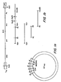
- Lambda 10 clones harboring HTLV-III DNA are cloned from the replicated form of the virus. As the retrovirus is replicating, double stranded DNA is being produced. The cloned HTLV-III DNA is digested with the restriction enzyme SstI. ( Figure la) Because there are two Sstl recognition sites within the LTR of HTLV-III DNA, one LTR region is not present in the cloned DNA sequence removed from the lambda 10 vector. As a result, a small (approximately 200 bp) fragment of the HTLV-III DNA is missing.
- the resulting DNA is linearized and fragments are produced by digesting the linearized genomic DNA spanning the env gene region with restriction enzymes. For example, fragments are produced using Kpn or EcoRI plus HindIII, as shown in Figure lb.
- the resulting 2.3kb KpnI-KpnI fragments; 1.0kbEcoRI-EcoRI fragments and 2.4Kb EcoRI-HindIII fragments are solated by gel electrophoresis and electro- elution. These fragments are randomly sheared to produce smaller iragments.
- the fragments thus produced are separatedfrom agarose gel and DNA fragments between about 200-500 bp are eluted.
- the eluted 200-500bp DNA fragments are end filled through the use of E. coli T 4 polymerase and blunt end ligated into an open reading frame expression (ORF) vector, such as pMR100.
- ORF open reading frame expression
- This ligation may occur at the SmaI site of the pMR100 vector, which contains two promoter regions, hybrid coding sequences of lambdaCI gene and lacI-LacZ gene fusion sequence. In the vector, these are out of frame sequences; as a result, the vector is nonproductive.
- the HTLV-III DNA is inserted into the vector; the correct DNA fragments will correct the reading frame, with the result that CI-HTLV-III-B-galactosidase fusion proteins are produced.
- the expression of the hybrid is under the control of the lac promoter. Based on the sequence of pMR100, it appears that if a DNA fragment insert cloned into the SmaI site is to generate a proper open reading frame between the lambdaCI gene fragment and the lac-Z fragment, the inserted DNA'must not contain any stop codons in the reading frame set by the frame of the lambdaCI gene.
- the recombinant pMR100 vectors are then introduced into E. coli.
- the bacteria are plated on MacConkey agar plates to verify the phenotype of the clone. If functional B-galactosidase is being produced, the colony will appear red.
- the colonies are also screened with HTLV-III DNA probes, for the purpose of identifying those clones containing the insert. Clones which are positive when screened with the DNA probe and positive on the MacConkey agar plates are isolated.
- the cells from the selected colonies are grown in culture.
- the culture is spun down and the cell pellet broken.
- Total. cellular protein is analysed by being run on an SDS polyacrylamide gel.
- the fusion proteins are identified at a position on the gel which contains no other protein. ( Figure 4)
- HTLV-III env gene containing recombinant clones was identified by colony hybridization.
- the production of larger fusion polypeptides bearing functional B-galactosidase activity was verified by phenotype identification on MacConkey agar plates; by B-galactosidase enzymatic assays and by analysis on 75% SDS-polyacrylamide gels.
- Immunoreactivity of the larger protein with antibody to HTLV-III was assessed by western blot analysis using serum from AIDS patients. These large fusion proteins also reacted with anti-B-galactosidase and anti-CI antiserum. This finding is consistent with the hypothesis that they are proteins of CI-HTLV-III- lacIZ.
- the open reading frame insert fragment of HTLV-III is further analyzed by DNA sequencing analysis. Because one of the two BamHI sites flanking the Smal cloning site in pMR100 is destroyed in the cloning step, positive clones are digested with restriction enzymes HindIII and clal to liberate the inserted HTLV-III DNA fragment.
- the HTLV-III ORF inserts are isolated from the fusion recombinant and cloned into M13 sequencing cloning vector mp18 and mp19 digested with HindIII and AccI. DNA sequences of the positive ORF clones are then determined.
- Fragments of HTLV-III DNA of approximately 200-500 bps are isolated from agarose gel, end repaired with T 4 polymerase and ligated to EcoRI linker.
- the EcoRI linker ligated DNA is then treated with EcoRI purified from 1% agarose gel and cloned in an expression vector, lambda gtll.
- This vector contains lac Z gene coding sequences into which the foreign DNA can be inserted for the generation of B-galactosidase fusion protein.
- the expression of the hybrid gene is under the control of lac repressor.
- the lac repressor ne, lac I is carried on a separate plasmid pMC9 in the host cell, E. coli Y1090.
- AIDS patient serum was used to probe the lambdagtll library of HTLV-III genome DNA containing 1.5x10 recombinant phage. In a screen of 5000 recombinants, 100 independent clones that produced strong signals were isolated. The positive recombinant DNA clones were further characterized for their specific gene expression. Rabbit hyperimmune serum against P24 was also used to identify the gag gene specific clones. Nick-translated DNA probes of specific HTLV-III gene, specifically the gag gene, env gene and Px gene were used to group the positive immunoreactive clones into specific gene region.
- Recombinant clones that produced strong signals with AIDS serum and contain insert DNA spanning the HTLV-III gag, pol, sor and env-lor gene regions were examined in detail by mapping their insert with restriction enzymes and DNA sequencing analysis.
- HTLV-III DNA is sheared randomly into fragments of about 300-500 bp in size. The fragments are cloned, for example, using ml3, and the colonies screened to identify those having an HTLV-III DNA fragment insert. The nucleotide sequence is then generated, with multiple analysis producing overlaps in the sequence. Both strands of the HTLV-III DNA are sequenced to determine orientation. Restriction mapping is used to check the sequencing data generated.
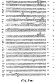
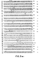
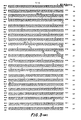
- the nucleotide sequence of one cloned HTLV-III genome is shown in Figure 3, in which the position of sequences encoding gag protein pl7 and the N-terminus of gag p24 and the C-terminus of gag pl5 (which overlaps with the N-terminus of the pol protein) are indicated.
- the open reading frames (ORF) for pol, sor and env-lor are also indicated.
- the sequence of the remaining 182 base pairs of the HTLV-III DNA not present in clone BH10 was derived from clone HXB2.
- the sequences of two additional clones (BH8 and BH5) are also shown.
- Restriction enzyme sites are listed above the nucleotide sequence;. sites present in clone BH8 but not in clone BH10 are in parentheses. Deletions are noted ([]) at nucleotides 251, 254, 5671 and 6987-7001. The nucleotide positions (to the right of each line) start with the transcriptional initiation site. The amino acid residues are numbered (to the right of each line) for the four largest open reading frames starting after the preceding termination codon in each case except gag which is enumerated from the first methionine codon. A proposed peptide cleavage site (V) and possible asparagine-linked glycosylation sites are shown ( * ) for the env-lor open reading frame.
- sequences in the LTR derived from clones BH8 and BH10 listed in the beginning of the figure are derived from the 3'-portion of each clone and are'assumed to be identical to those present in the 5'-LTR of the integrated copies of these viral genomes.
- Clone HXB2 was derived from a recombinant phage library of Xbal digested DNA from HTLV-III infected H9 cells cloned in lambdaJl.
- H9 cells are human leukemic cells infected by a pool of HTLV-III from blood of AIDS patients, F. Wong-Staal, Nature, 312, November, 1984.
- Cloning vector clones BH10, BH8, and BH5 were derived from a library of SstI digested DNA from the Hirt supernatant fraction of HTLV-III infected H9 cells cloned in lambdagtiles.lambdab.
- Clones BH8, BHS, and a portion of HXB2 were sequenced as described by Maxam and Gilbert. (1980) Maxam, A. M. and Gilbert, Co. Methods in Enzymology. 65: 499-560.
- Clone BH10 was sequenced by the method of Sanger modified by the use of oligonucleotides complementary to the M13 insert sequence as primers and using Klenow fragment of DNA polymerase I or reverse transcriptase as the polymerase.
- DNA sequences which are an entire gene or segment of a gene from HTLV-III are inserted into a vector, such as a T7 vector.
- the vector has the Tceu promoter from the T cell gene 10 promoter and DNA sequences encoding eleven amino acids from the T cell gene 10 protein.
- the vectors are then used to transform cells, such as E. coli.
- the T7 vector makes use of the T7 polymerase, which catalyzes RNA formation and recognizes only T7 promoter, which is the site where RNA polymerase binds for the initiation of transcription.
- the T7 polymerase does not recognize E. coli promoter.
- HTLV-III DNA sequences are inserted after the promoter and polymerase genes of the T7 vector, which recognizes them to the exclusion of other signals, and a terminator is placed immediately after the HTLV-III DNA sequences, the T7 vector will direct manufacture RNA complementary to the HTLV-III DNA insert.
- RNA proves and DNA HTLV-III probes must have a distinctive region of the HTLV-III genome in order to be useful in detecting HTLV-III in body fluids. There is relatively little homology between the HTLV-III genome and the HTLV-I and -II genomes and probes contain regions which are unique to IITLV-III (i.e., not shared with HTLV-I or -II). For example, nucleotide sequences in the env gene region of HTLV-III can be used.
- Either viral RNA or DNA can be used for detecting HTLV-III in, for example, saliva, which is known to have a very high concentration of the virus. This can be done, for example, by means of a dot blot, in which the saliva sample is denatured, blotted onto paper and then screened using either type of probe. If saliva is used as the test fluid, detection of HTLV-III is considerable faster and easier than is the case if blood is tested.
- Monoclonal antibodies reactive with HTLV-III polypeptides are produced by antibody-producing cell lines.
- the antibody-producing cell lines may be hybridoma cell lines commonly known as hybridomas.
- the hybrid cells are formed by fusion of cells which produce antibody to HTLV-III polypeptide and an immortalizing cell, that is, a cell which imparts long term tissue culture stability on the hybrid cell.
- the first fusion partner - the antibody-producing cell - can be a spleen cell of an animal immunized against HTLV-III polypeptide.
- the antibody-producing cell can be isolated B lymphocyte which produces antibody against an HTLV-III antigen.
- the lymphocyte can be obtained from the spleen, peripheral blood, lymph nodes or other tissue.
- the second fusion partner - the immortal cell - can be a lymphoblastoid cell or a plasmacytoma cell such as a myeloma cell, itself an antibody-producing cell but also malignant.
- Murine hybridomas which produce monoclonal antibodies against HTLV-III polypeptide are formed by the fusion of mouse myeloma cells and spleen cells from mice immunized against the polypeptide. To immunize the mice, a variety of different immunization protocols may be followed. For instance mice may receive primary and boosting immunizations of the purified polypeptide. The fusions are accomplished by standard procedures. Kohler and Milstein, (1975) Nature (London) 256, 495-497; Kennet, R., (1980) in Monoclonal Antibodies (Kennet et al., Eds. pp. 365-367, Plenum Press, NY).
- the hybridomas are then screened for production of antibody reactive with the polypeptide. This can be performed by screening procedures known in the art.
- Another way of forming the antibody-producing cell line is by transformation of antibody-producing cells.
- a B lymphocyte obtained from an animal immunized against HTLV-III polypeptide may be infected and transformeed with a virus such as the Epstein-Barr virus in the case of hur,an B lymphocytes to give an immortal antibody-producing cell.
- a virus such as the Epstein-Barr virus in the case of hur,an B lymphocytes to give an immortal antibody-producing cell.
- the B lymphocyte may be transformed by a transforming gene or transforming gene product.
- the monoclonal antibodies against HTLV-III polypeptide can be produced in large quantities by injecting antibody-producing hybridomas into the peritoneal cavity of mice and, after an appropriate time, harvesting the ascites fluid which contains very high titer of homogenous antibody and isolating the monoclonal antibodies therefrom.
- Xenogeneic hybridomas should be injected into irradiated or athymic nude mice.
- the antibodies may be produced by culturing cells which produce HTLV-III polypeptide in vitro and isolating secreted monoclonal antibodies from the cell culture medium.
- the antibodies produced according to these methods can be used in diagnostic assays (e.g., detecting HTLV-III in body fluids) and in passive immunotherapy.
- the antibodies reactive with HTLV-III polypeptides provide the basis for diagnostic tests for the detection of AIDS or the presence of HTLV-III in biological fluids (e.g., blood, semen, saliva) and for passive immunotherapy.
- biological fluids e.g., blood, semen, saliva
- HTLV-III antiigen
- this method results in far fewer false positive test results than do tests, in which antibody against HTLV-VIII is detected.
- T4 DNA polymerase was used to fill in and/or trim the single strand DNA termini generated by the sonication procedure. DNA fragments were incubated with T4 polymerase in the absence of added nucleotides for five minutes at 37°C to remove nucleotides from 3' end and then all 4 nucleotide precursors were added to a final concentration of 100 uM and the reaction mixture was incubated another 30 minutes to repair the 5'-end single stranded overhang. The reaction was stopped by heat inactivation of the enzyme at 68°C for 10 minutes. DNA was phenol extracted once, ethanol precipitated and resuspended in TE.
- the sonicated blunt end repaired HTLV-III DNA fragments were ligated into the Smal site of the ORF expression vector pMR100 and transformed into host cell LG90 using standard transformation procedures.
- B-galactosidase positive phenotype of the transformant were identified by plating the transformed cell on ampicillin (25 ug/ml) containing McConkey agar plates and scoring the phenotype after 20 hours at 37°C.
- Electrophoretic transfer of proteins from SDS-PAGE gels to nitrocellulose paper was carried out according to Towbin et. al.. After the transfer, the filter was incubated at 37°C for two hours in a solution of 5% (w/v) nonfat milk in PBS containing 0.1% antifoam A and 0.0001% merthiolate to saturate all available protein binding sites. Reactions with AIDS antisera were carried out in the same milk buffer containing 1% AIDS patient antisera that had been preabsorbed with E. coli lysate. Reactions were performed in a sealed plastic bag at 4°C for 18-24 hours on a rotatory shaker.
- the filter was washed three times for 20 minutes each at room temperature in a solution containing 0.5% deoxycholic, 0.1 M NaCl, 0.5% triton X-100; 10 mm phosphate buffer pH 7.5 and 0.1 mM PMSF.
- the nitrocellulose was then incubated with the second goat antihuman antibody that had been iodinated with 1251.
- the reaction with the iodinated antibody was carried out at room temperature for 30 minutes in the same milk buffer as was used for the first antibody.
- the nitrocellulose was then washed as previously described and exposed at -70°C using Kodak XAR5 film with an intensifying screen.
- E. coli LG90 transformants were screened with HTLV-III DNA probes containing the DNA regions of interest (e.g. HTLV-III gag, env'or Px gene specific sequences). Colonies were grown on nitrocellulose filter and screened according to the procedure of Grunstein and Hogness by using a nick-translated HTLV-III DNA as hybridization probe.
- HTLV-III DNA probes containing the DNA regions of interest (e.g. HTLV-III gag, env'or Px gene specific sequences). Colonies were grown on nitrocellulose filter and screened according to the procedure of Grunstein and Hogness by using a nick-translated HTLV-III DNA as hybridization probe.
- the DNA fragment was in general excised by restriction endonuclease digestion, gel purified, and 32 P-labeled to a specific activity of 0.5x10 8 cpm/ug by nick-translation (Rigby, P.W.J. et al., J. Mol. Biol. 113, 237 (1977).
- Duplicate nitrocellulose filters with DNA fixed to them were prehybri- dized with 6xSSC (0.9 M NaCl/0.09 M sodium citrate, pH 7.0), 5X Denhardt's solution (Denhardt's solution: 0.02% each of polyvinylpyrrolidone, Ficoll and bovine serum albumin) 10 ug of denatured sonicated E.
- ompA An expression vector, pIN-III-ompA (ompA) was used.
- ompA has the lipoprotein (the most abundant protein in E.coli) gene promoter (lpp) and the lacUV5 promoter-operator ( Figure 1).
- ompA vectors also contain the DNA segment encoding the lac repressor, which allows the expression of the inserted DNA to be regulated by lac operon inducers such as IPTG.
- the ompA cloning vehicles contain three unique restriction enzyme sites EcoRI, HindIII, Barn HI in all three reading frames and permit the insertion of DNA into any of these restriction sites.
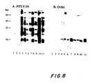
- lysates of E.coli cell containing HTLV-III DNA recombinant plasmids were electrophoresed on 12.5% SDS-polyacrylamide gel and electroblotted onto nitrocellulose filters. The filters were then incubated first with well-characterized sera from AIDS patients and next with 125 I-labelled goat anti-human IgG antibodies. The washed filters were autoradiographed to identify peptides reactive with anti-HTLV-III antibodies.
- transformants of all three plasmid constructs produced a 15 Kd peptide that is strongly reactive with anti-HTLV-III antibodies in sera from AIDS patients ( Figure 6 lane 1, purified HTLV-III virions; lanes 2 and 3, O1R6 uninduced and induced; lanes 4 and 5, 02R7 uninduced and induced; lanes 6 and 7 03R3 uninduced and induced). This reactivity is not detected when sera from normal individuals is used.
- DNA sequence data of the HTLV-III genome indicates that there is an open reading frame inside the pol gene located at the 5'-end of the EcoRI fragment.
- AAGGAG Algarno sequence
- ATG initiation codon
- position 41-43 The 15 Kd peptide synthesized by all three recombinants appears to be translated from the transcripts using this internal initiation codon. If this is true, the peptide starts from the ATG located at position 41-43 and ends at the stop codon at position 446-448, producing a peptide of 135 amino acid residues encoded by the 3'-end segment of the pol gene of HTLV-III.
- the 03R3 construct in which the reading frame of the HTLV-III DNA pol gene is in phase with that set by the vector, produced two additional peptides about 19 Kd and 16.5 Kd in size ( Figure 6). It is possible that the 19 Kd peptide contains an additional 35 amino acid residues, 21 of which are from the signal peptide encoded by the ompA 3 vector and 14 encoded by the inserted HTLV-III DNA itself.
- the 16.5 Kd peptide may be the processed 19 Kd peptide in which the signal peptide is cleaved.
- the O1R6 and 02R7 constructs also produces another peptide of about 17.5 Kd ( Figure 6) and weakly reactive with sera of AIDS patients.
- the origin of this peptide is not clear.
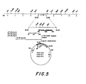
- the 1.1 Kb EcoRI fragment contains a second potential coding region designated as the short open reading frame (SOR) extending from nucleotide position 360 to 965 ( Figure 5).
- SOR short open reading frame
- Four of the five AUG methionine codons in this region are near the 5'-end of this open reading frame.
- This DNA segment could encode peptides of 192, 185, 177 or 164 amino acid residues. However, there is no clearly recognizable ribosome binding site at the 5'-end of this open reading frame.
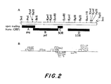
- HTLV-III DNA was excised from lambda BH-10, which is a previously constructed recombinant lambda phage containing a 9 Kb segment of HTLV-III DNA inserted into the vector lambdagtwes lambda B ( Figure 2a).
- This HTLV-III DNA was sonicated and DNA fragments of about 0.5 Kb purified by gel electrophoresis, end repaired, and inserted into the SmaI site of the open reading frame (ORF) vector, pMR100 ( Figure 9).
- This vector contains a bacterial lac promotor DNA segment linked to a second DNA fragment containing a hybrid coding sequence in which the N-terminus (5' segment) of the lambda CI gene of bacteriophage lambda is fused to an N-terninal-deleted lacIZ gene (3' segment).
- a short linker DNA fragment, containing a Smal cloning site, has been inserted between these two fragments in such a manner that a frame shift mutation has been introduced upstream of the lacIZ-coding DNA.
- pMR100 does not produce any detectable B-galactosidase activity when introduced into cells of the Lac host E. coli LG90.
- the insertion of foreign DNA containing an open reading frame, in this case the HTLV-III DNA, at the SmaI cloning site can reverse the frame shift mutation if the inserted coding sequence is in the correct reading frame with respect to both the lambdaCI leader and the lacIZ gene.
- Transformants were screened on MacConkey plates to detect individual clones that expressed B-galactosidase enzymatic activity in situ.
- the three-element-fused genes were expressed as tripartite fusion proteins, havirg a portion of the lambdaCI protein at the N-terminus, the HTLV-III segment in the middle, and the lacIZ polypeptide at the C-terminus.
- the proteins produced by the Lac + clones were analyzed by resolving cell lysates on 7.5% SDS-polyacrylamide gels along with those of the cortrol Lac + clone pMR200, which produced a lambdaCI-B-galactosidase fusion protein.
- the lacIZ gene in pMR200 is identical to that in pMR100 excert that it has a single base pair deletion which brings it in phase with the lambdaCI gene to produce an active B-galactosidase.
- the B-galactosidase and its fusion protecins are separated from the bulk of proteins in the cell lysates on the SDS-polyacrylamide gels and can be easily identified by Coomassie brilliant blue staining as shown in Figure 10a.
- Some of the Lac + clones containing HTLV-III DNA produce polypeptides that are larger (15,000 to 27,000 daltons) than the lambdaCI-lacIZ fusion protein.
- the peptides produced by the Lac + clones were examined by Western blot analysis for immunoreactivity with sera from AIDS patients. After the lysates of Lac + clones were electrophoresed in SDS-polyacrylamide gels, they were electro-transferred to nitrocellulose filters. These protein blots were first reacted with AIDS patient sera and then with 125 I -labeled goat anti-human IgG. The autoradiograph in Figure 10b shows the immunoreactivity of a representative fused protein with the serum from an AIDS patient.
- the recombinant peptides also reacted with anti-B-galactosidase antiserum, consistent with the proposition that they had the general structure lambdaCI-HTLV-III peptide-LacIZ. From the immunoreactivity pattern of the negative controls, pMR100 and pMR200, which do not contain an HTLV-III DNA insert, it is evident that this particular AIDS serum contains antibodies reactive with several bacterial proteins of the host E. coli. This is not surprising, since AIDS patients are usually infected with a number of bacteria. Absorbing AIDS patiant sera with Sepharose 4B conjugated with E. coli extract reduced the background immunoreactivity to some extent but did not completely eliminate it.
- HTLV-III DNA-containing Lac colonies were analyzed in SDS polyacrylamide gels using Coomassie brilliant blue staining and Western blotting. About half of them were found to express fusion proteins containing extra peptides of about 100-200 amino acids, corresponding to DNA inserts of 300-600 bp long. Of these fusion proteins, 20 were found to react specifically with sera from AIDS patients. The unreactive clones probably contain peptides that fold in such a way that they are not reactive with antibodies or correspond to regions of HTLV-III protein molecules which are not immunogenic in AIDS patients. The other half of the Lac + clones expressed fusion proteins whose sizes were not obviously different from that of the lambdaCI B-galactosidase protein. None from this group of fusion proteins was found to react with sera from AIDS patients.
- the HTLV-III DNA inserts from Lac + ORF clones were mapped to specific segments in the HTLV-III genome using Southern blotting procedures. In these studies, each plasmid clone was labelled with 32 P by nick-translation and hybridized to a battery of HTLV-III DNA restriction fragments. This hybridization analysis mapped all of the Lac ORF clones into four open reading frame segments designated ORF-A, ORF-B, ORF-C, and ORF-D ( Figure 2a) consistent with the DNA sequencing data.
- the open reading frames ORF-A and -B, corresponding to the coding regions of the gag and pol genes, are 1.5 Kb and 3.0 Kb long, respectively.
- ORF-C is about 0.6 Kb long, slightly overlaps with the ORF-B region, and is capable of encoding a polypeptide of 21 overlaps with the ORF-B region, and is capable of encoding a polypeptide of 21 Kd.
- the location of ORF-C and its overlap with the pol gene are pronounced cf the structure of the env genes in H TLV-I and -II.
- ORF-C designated as the short open reading frame (sor), is too short to code for the entire envelope protein.
- the fourth open reading frame, ORF-D is 2.5 Kb long and could encode both a large precursor of the major envelope glycoprotein and another protein derived from the 3' terminus, which may be analogous to the lor products of HTLV-I and -II.
- This gene region of HTLV-III, designated env-lor, is at least twice as long as the lor of HTLV-I and HTLV-II and it is presently unclear whether single or multiple proteins are encoded herein.
- This invention has industrial applicability in screening for the presence of HTLV-III DNA in body fluids and the diagnosis of AIDS.
- the invention also provides a fusion protein produced by the method of claim 19 or an HTLV-III envelope polypeptide produced by the method of claim 19.
Abstract
Description
- This invention is in the fields of molecular biology and virology and in particular relates to human T cell leukemia virus - type III (HTLV-III).
- The term human T cell leukemia-lymphoma virus (HTLV) refers to a unique family of T cell tropic retroviruses. These viruses play an important role in the pathogenesis of certain T cell neoplasms. There are presently three known types of HTLVs. One subgroup of the family, HTLV-type I (HTLV-I), is linked to the cause of adult T-cell leukemia-lymphoma (ATLL) that occurs in certain regions of Japan, the Caribbean and Africa. HTLV-type II (HTLV-II) has been isolated from a patient with a T-cell variant of hairy cell leukemia. M.Popovic et al., Detection, Isolation, and Continuous Production of Cytopathic Retroviruses (HTLV-III) from Patients with AIDS and Pre-AIDS. Science, 224:497-500 (1984).
- HTLV-type III (HTLV-III) has been isolated from may patients with acquired immunodeficiency syndrome (AIDS). HTLV-III refers to prototype virus isolated from AIDS patients. Groups reported to be at greatest risk for AIDS include homosexual or bisexual males; intravenous drug users and Haitian immigrants to the United States. Hemophiliacs who receive blood products pooled from donors and recipients of multiple blood transfusions are also at risk. Clinical manifestations of AIDS include severe, unexplained immune deficiency which generally involves a depletion of helper T lymphocytes. These may be accompanied by malignancies and infections. The mortality rate for patients with AIDS is high. A less severe form of AIDS also exists, in which there may be lymphadenopathy and depressed helper T cell counts; there is not, however, the devastating illness characteristic of full-blown AIDS. There are many individuals, who are classified as having early AIDS (pre-AIDS), who exhibit these signs. It is not now possible to predict who among them will develop the more serious symptoms.
- Much of the evidence implicates HTLV-III as the etiological agent of the infectious AIDS. First, there is consistent epidemiology; greater than 95% of the patients with AIDS have antibodies specific for HTLV-III. Second, there has been reproducible identification and isolation of virus in this disease; more than 100 variants of HTLV-III have been isolated from AIDS patients. Third, there has been transmission of the disease to normal healthy individuals who received blood transfusions fror infected blood donors.
- HTLV-III has been shown to share several properties with HTLV-I and HTLV-II but also to be morphologically, biologically and antigenically distinguishable. R.C. Gallo et al., Frequent Detection and Isolation of Cytopathic Petroviruses (HTLV-III) from Patients with AIDS and .t Risk for AIDS. Science, 224:500-503. (1984). For example, HTLV-III has been shown to be antigenically related to HTLV-I and HTLV-II by demonstrating cross- reactivity with antibodies to HTLV-I and HTLV-II core proteins, p24 and p19, and envelope antigens and by nucleic acid cross-hybridization studies with cloned HTLV-I and HTLV-II DKAs. 'However, unlike HTLV-I and HTLV-II, it lacked the ability to infect and transform T cells from normal umbilical cord blood and bone marrow in vitro, and has the cytopathic effect on infected cells only.
- Like the RNA genome of other retroviruses, the RNA genome of HTLV-III contains three genes which encode viral proteins: 1) the gag gene, which encodes the internal structural (nucleocapsid or core) proteins; 2) the pol gene, which encodes the RNA-directed DNA polymerase (reverse transcriptase); and 3) the env gene, which encodes the envelope glycoproteins of the virion. In addition, the HTLV-III genome contains a region designated Px, located between the env gene and the 3' LTR, which appears to be involved in functional killing of the virus.
- At this time, AIDS is still difficult to diagnose before the onset of clinical manifestations. There is no method presently available for the.prevention of the disease. Treatment of those with AIDS is generally not successful and victims succumb to the devastating effects HTLV-III has on the body.
- This invention is based upon applicant's cloning of HTLV-III DNA in recombinant/vector host systems capable of expressing immunoreactive HTLV-III polypeptides. Based on the cloning of HTLV-III DNA in systems which express immunoreactive- polypeptides, applicant has developed methods useful in the diagnosis, treatment and prevention of AIDS. Applicant has developed methods of detecting HTLV-III and antibodies against HTLV-III in body fluids (e.g., blood, saliva, semen), and methods useful in immunotherapy (e.g., vaccination and passive immunization against AIDS). In addition, applicant has developed methods of making HTLV-III DNA probes and RNA probes useful in detecting HTLV-III in body fluids.
- Polypeptides encoded by segments of the HTLV-III genome have been produced by these recombinant DNA methods. For example, polypeptides encoded by three regions of the HTLV-JII genome (an env gene sequence, an env-lor gene sequence and a 1.1Kb EcoRI restriction fragment from HTLV-III cDNA) have been produced. The polypeptides expressed have been isolated. These polypeptides are immunoreactive with sera of patients having AIDS and with antibodies to HTLV-III and thus are useful in screening blood and other body fluids for the presence of antibodies against HTLV-III. Applicant's invention threfore provides a method not only for diagnosing AIDS, but also for preventing the transmission of the disease to others through blood or blood components harboring HTLV-III. The latter is particularly valuable in screening donated blood before it is transfused or used to obtain blood components (e.g., Factor VIII for the treatment of hemophilia; Factor IX)
- Polypeptides produced by the recombinant DNA methods are employed in the production of antibodies, including monoclonal antibodies, against the virus. Such antibodies form the basis for immunoassay and diagncstic techniques for directly detecting HTLV-III in body fluids such as blood, saliva, semen, etc. Neutralizing antibodies against the virus may be used to passively immunize against the disease.
- Applicant's cloning of HTLV-III DNA in such recombinant vector host systems also provides the basis for determination of the nucleotide sequence of HTLV-III DNA. The DNA probes are homologous to DNA regions which are unique to the HTLV-III genome. DNA probes provide another method of detecting HTLV-III in blood, saliva or other body fluids. RNA probes which contain regions unique to the HTLV-III genome can also be formed and used for the detection of HTLV-III in body fluids.
-
- Figure 1 is a representation of HTLV-III DNA. Figure la shows sites at which the genome is cut by the restriction enzyme SstI and Figure lb shows the fragments of HTLV-III genome produced through the action of restriction enzymes Kpn, EcoRI and Hind III.
- Figure 2 is a representation of HTLV-III DNA. Figure 2a shows the location of restriction enzyme sites in the genome and Figure 2b shows the location in the HTLV-III genome of DNA inserts in open reading frame clones. The (+) and (-) indicate reactivity and lack of reactivity, respectively, of the fusion protein expressed by cells transformed by the ORF vectors with sera of AIDS patients.
- Figure 3 shows the nucleotide sequence for HTLV-III DNA and the predicted amino acid sequence of the four longest open reading frames. Restriction enzyme sites are indicated above the nucleotide sequence.
- Figure 4 is an immunoblot showing the position on an SDS polyacrylamide gel of HTLV-III env-Beta-galactosidase fusion proteins.
- Figure 5 shows sites at which the genome is cut by the restriction enzyme EcoRI and construction of recombinant plasmids carrying HTLV-III DNA.
- Figure 6 is an immunoblot showing the positions on nitrocellulose blots of peptides produced by bacterial cells transformed by recombinant constructs ompAl-R-6; ompA2-R-7 and ompA3-R-3, into which a l.lKb EcoRI HTLV-III cDNA restriction fragment had been inserted. Figure 6a shows the nucleotide sequence of the ompA signal peptide and the pertinent region of recombinant plasmids ompAl-R-6; ompA2-R-7 and ompA3-R-3.
- Figure 7 is an immunoblot showing blocking of reaction between HTLV-III antigens and an AIDS serum by lysates of E.coli containing HTLV-III DNA recombinant plasmid ompAl-R-6 (lanes 1-5) and no blocking of the reaction by lysates of E.coli control cells (lanes 6-10).
- Figure 8 is an immunoblot showing the presence or absence of antibodies against the peptide encoded by the 1.1Kb EcoRI HTLV-III restriction fragment of HTLV-III cDNA in sera from healthy individuals (lanes 1-3) and from AIDS patients (lanes 4-11). Purified HTLV-III virus (panel A) or total cell lysate of bacterial clone ompAl-R-6(OlR6) were reacted with sera samples.
- Figure 9 represents the open reading frame expression vector pMRIOO having HTLV-III DNA.
- Figure 10 represents lambdaCI-HTLV-III beta-galactosidase fusion proteins. Figure 10a is an immunoblot showing the position on SDS polyacrylamide gel of lambdaCI-HTLV-III beta-galactosidase fusion proteins, and Figure 10b shows the immunoreactivity of such proteins with sera from AIDS patients.
- Despite the similarity between HTLV-III and the other members of the HTLV-bovine leukemia virus (BLV) family of viruses, the biology and pathology of HTLV-III differs substantially. For example, relatively little homology has been found in the HTLV-III genome when compared with that of the HTLV-I or -II genome. Infection with HTLV-III often results in profound immunosuppression (AIDS), consequent to the depletion of the OKT4(+) cell population. This effect is mirrored by a pronounced cytopathic, rather than transforming, effect of HTLV-III infection upon the OKT4(+) cells in lymphocyte cultures in vitro. In contrast, infection with HTLV-I results in a low incidence of T-cell leukemia lymphoma (an OKT4(+) cell malignancy). There is evidence for some degree of immunodeficiency in HTLV-I patients as well. Infection of primary lymphocytes in culture by HTLV-I and -II results in vitro transformation of predominantly OKT4(+) cells. A cytopathic effect of HTLV-I infection upon lymphocytes is apparent, but the effect is not as pronounced as that observed for HTLV-III.
- HTLV-III also differs from HTLV-I and -II in the extent of infectious virion production in vivo and in vitro. High titers of cell free, infectious virions can be obtained from AIDS patient semen and saliva and from the supernatant of cultures infected with HTLV-III. Very few, if any, cell free infectious virions can be recovered from adult T-cell leukemia lymphoma (ATLL) patients or from cultures infected with HTLV-I or -II.
- Envelope glycoprotein is the major antigen recognized by the antiserum of AIDS patients. In this respect, HTLV resembles other retroviruses, for which the envelope glycoprotein is typically the most antigenic viral polypeptide. In addition, the neutralizing antibodies are generally directed toward the envelope glycoprotein of the retrovirus. Serum samples from 88 percent to 100 percent of those with AIDS have been shown to have antibodies reactive with antigens of HTLV-III; the major immune reactivity was directed against p41, the presumed envelope antigen of HTLV-III. Antibodies to core proteins have also been demonstrated in serum of AIDS patients, but do not appear to be as effective an indicator of infection as is the presence of antibodies to envelope antigen.
- The p4l antigen of HTLV-III has been difficult to characterize because the viral envelope is partially destroyed during the process of virus inactivation and purification. This invention responds to the great need to characterize this antigenic component of the HTLV-III virus and to determine the existence and identity of other viral antigenic components in several ways. It provides products, such as HTLV-III polypeptides, antibodies to the polypeptides and RNA and DNA probes, as well as methods for their production. These serve as the basis for screening, diagnostic and therapeutic products and methods.
- This invention relates to HTLV-III polypeptides which are produced by translation of recombinant DNA sequences encoding HTLV-III proteins. Polypeptides which are produced in this way and which are immunoreactive with serum from AIDS patients or antibodies to HTLV-III are referred to as recombinant DNA-produced immunoreactive HTLV-III polypeptides. They include, but are not limited to, antigenic HTLV-III core and envelope polypeptides which are produced by translation of the recombinant DNA sequences specific to the ga2 and the env DNA sequences encoding HTLV-III core proteins and envelope glycoproteins, respectively. They also include the polypeptides which are produced by translation of the recombinant DNA sequences included in a 1.1Kb EcoRI restriction fragment of HTLV-III cDNA and recombinant DNA sequences specific to the sor gene and the Px genes of HTLV-III. The sor DNA sequence is common to replication competent HTLV-III viruses. The Px genes contain a coding sequence with one large open reading frame (lor), located between the env gene and the 3' end of the HTLV-III genome. Both the env DNA sequences and the lor DNA sequences are located within the same open reading frame of the HTLV-III genome and this gene region is accordingly designated env-lor.
- The polypeptides encoded by these regions of the HTLV III can be used in immunochemical assays for detecting antibodies against HTLV-III and HTLV-VIII infection. These methods can assist in diagnosing AIDS. In addition, they can also be employed to screen blood before it is used for transfusions or for the production of blood components (e.g., Factor VIII for the treatment of hemophilia). Availability of screening technics will reduce the risk of AIDS transmission.
- Detection of antibodies reactive with the polypeptides can be carried out by a number of established methods. For example, an immunoreactive HTLV III polypeptide can be affixed to a solid phase (such as polystyrene bead or other solid support). The sold phase is then incubated with blood sample to be tested for antibody against HTLV-III. After an appropriate incubation period the solid phase and blood sample are separated. Antibody bound to the solid phase can be detected with labeled polypeptide or with a labeled antibody against human immunoglobulin.
- HTLV-III polypeptides can be used in a vaccine prevention of AIDS. For vaccination against the virus, immunogenic polypeptides which elicit neutralizing antibody would be employed. The leading candidates for use in vaccines are the viral envelop polypeptides.
- The polypeptides can also be used to produce antibodies, including monoclonal antibodies, against the HTLV-III polypeptides. These antibodies can be used in immunochemical assays for direct detection of the virus in body fluids (such as blood, saliva and semen). Assays employing monoclonal antibody against specific HTLV III antigenic determinants will reduce false-positive results thereby improving accuracy of assays for the virus. Antibodies against the virus may also be useful in immunotherapy. For example, antibodies may be used to passively immunize against the virus.
- The methods of producing the polypeptides are also a subject of this invention, as are diagnostic methods based on these polypeptides.
- This invention also provides methods for the isolation of genes of HTLV-III which encode immunoreactive polypeptides; identification of the nucleotide sequence of these genes; introduction of DNA sequences specific to these viral DNA sequences into appropriate vectors to produce viral RNA and the formation of DNA probes. These probes are comprised of sequences specific to HTLV-III DNA and are useful, for example, for detecting complementary HTLV-III DNA sequences in body fluids (e.g., blood).
- Genetic engineering methods are used to isolate segments of HTLV-III DNA which encode immunoreactive HTLV-III polypeptides. Among these are polypeptides which are immunoreactive with serum from AIDS patients or antibodies to HTLV-III. These polypeptides include the core protein, a 15Kd peptide encoded by a 1.1Kb EcoRI HTLV-III restriction fragment of HTLV-III DNA and the envelope glycoprotein. These methods are also used to sequence the fragments which encode the polypeptides. The proviral genes integrated into host cell DNA are molecularly cloned and the nucleotide sequences of the cloned provirus is determined.
- An E. coli expression library of HTLV-III DNA is constructed. The HTLV-III genome is cloned and cuts are then made in the cloned HTLV-III genome with restriction enzymes to produce DNA fragments. (Figures 1 and 2) HTLV-III DNA fragments of approximately 200-500bp are isolated from agarose gel, end repaired with T. polymerase and ligated to linker DNA. The linker ligated DNA is then treated with a restriction enzyme, purified from agarose gel and cloned in an expression vector. Examples of the expression vectors used are: OmpA, pIN (A,B and C), lambda pL, T7, lac, Trp, ORF and lambda gtll. In addition, mammalian cell vectors such as pSV28pt, pSV2neo, pSVdhfr and VPV vectors, and yeast vectors, such as GALI and GAL10, may be used.
- The bacterial vectors contain the lac coding sequences, into which HTLV-II1 DNA can be inserted for the generation of E-galactosidase fusion protein. The recombinant vectors are then introduced into bacteria (e.g., E.coli); those cells which take up a vector containing HTLV-III DNA are said to be transformed. The cells are then screened to identify cells which have been transformed and are expressing the fusion protein. For example, the bacteria are plated on MacConkey agar plates in order to verify the phenotype of clone. If functional B-galactosidase is being produced, the colony will appear red.
- Bacterial colonies are also screened with HTLV-III DNA probes to identify clones containing the DNA regions of interest (e.g., HTLV-III gag, pol and env DNA sequences). Clones which are positive when screened with the DNA probe and positive on the MacConkey agar plates are isolated.
- This identification of cells harboring the HTLV-III DNA sequences makes it possible to produce HTLV-III polypeptides which are immunoreactive with HTLV-III specific antibody. The cells from the selected colonies are grown in culture under conditions allowing the expression of the hybrid protein. Cell protein is then obtained by means known in the art. For example, the culture can be centrifuged and the resulting cell pellet broken. Polypeptides secreted by the host cell can be obtained (without disruption of the cells) from the cell culture supernatant.
- The total cellular protein is analysed by being run on an SDS polyacrylamide gel electrophoresis. The fusion proteins are identified at a position on the gel which contains no other protein. Western blot analyses are also carried out on the clones which screened positive. Such analyses are performed with serum from AIDS patients, with the result that it is possible to identify those clones expressing HTLV-III B-galactosidase fusion proteins (antigens) that cross-react with the HTLV-III specific antibody.
-
Lambda 10 clones harboring HTLV-III DNA are cloned from the replicated form of the virus. As the retrovirus is replicating, double stranded DNA is being produced. The cloned HTLV-III DNA is digested with the restriction enzyme SstI. (Figure la) Because there are two Sstl recognition sites within the LTR of HTLV-III DNA, one LTR region is not present in the cloned DNA sequence removed from the lambda10 vector. As a result, a small (approximately 200 bp) fragment of the HTLV-III DNA is missing. - The resulting DNA is linearized and fragments are produced by digesting the linearized genomic DNA spanning the env gene region with restriction enzymes. For example, fragments are produced using Kpn or EcoRI plus HindIII, as shown in Figure lb. The resulting 2.3kb KpnI-KpnI fragments; 1.0kbEcoRI-EcoRI fragments and 2.4Kb EcoRI-HindIII fragments are solated by gel electrophoresis and electro- elution. These fragments are randomly sheared to produce smaller iragments. The fragments thus produced are separatedfrom agarose gel and DNA fragments between about 200-500 bp are eluted.
- The eluted 200-500bp DNA fragments are end filled through the use of E. coli T4 polymerase and blunt end ligated into an open reading frame expression (ORF) vector, such as pMR100. This ligation may occur at the SmaI site of the pMR100 vector, which contains two promoter regions, hybrid coding sequences of lambdaCI gene and lacI-LacZ gene fusion sequence. In the vector, these are out of frame sequences; as a result, the vector is nonproductive. The HTLV-III DNA is inserted into the vector; the correct DNA fragments will correct the reading frame, with the result that CI-HTLV-III-B-galactosidase fusion proteins are produced. The expression of the hybrid is under the control of the lac promoter. Based on the sequence of pMR100, it appears that if a DNA fragment insert cloned into the SmaI site is to generate a proper open reading frame between the lambdaCI gene fragment and the lac-Z fragment, the inserted DNA'must not contain any stop codons in the reading frame set by the frame of the lambdaCI gene.
- The recombinant pMR100 vectors are then introduced into E. coli. The bacteria are plated on MacConkey agar plates to verify the phenotype of the clone. If functional B-galactosidase is being produced, the colony will appear red. The colonies are also screened with HTLV-III DNA probes, for the purpose of identifying those clones containing the insert. Clones which are positive when screened with the DNA probe and positive on the MacConkey agar plates are isolated.
- The cells from the selected colonies are grown in culture. The culture is spun down and the cell pellet broken. Total. cellular protein is analysed by being run on an SDS polyacrylamide gel. The fusion proteins are identified at a position on the gel which contains no other protein. (Figure 4)
- Western blot analyses are also carried out on the clones which screened positive. Sera from AIDS patients are used, thus making it possible to identify those clones which express the HTLV-III-B-galactosidase fusion proteins that cross-react with the HTLV-III specific antibody. 1000 clones were screened by this method; 6 were positive.
- Because of the nature of the pMR100 cloning vehicle, a productive DNA insert should also be expressed as a part of a larger fusion polypeptide. HTLV-III env gene containing recombinant clones was identified by colony hybridization. The production of larger fusion polypeptides bearing functional B-galactosidase activity was verified by phenotype identification on MacConkey agar plates; by B-galactosidase enzymatic assays and by analysis on 75% SDS-polyacrylamide gels. Immunoreactivity of the larger protein with antibody to HTLV-III was assessed by western blot analysis using serum from AIDS patients. These large fusion proteins also reacted with anti-B-galactosidase and anti-CI antiserum. This finding is consistent with the hypothesis that they are proteins of CI-HTLV-III- lacIZ.
- The open reading frame insert fragment of HTLV-III is further analyzed by DNA sequencing analysis. Because one of the two BamHI sites flanking the Smal cloning site in pMR100 is destroyed in the cloning step, positive clones are digested with restriction enzymes HindIII and clal to liberate the inserted HTLV-III DNA fragment. The HTLV-III ORF inserts are isolated from the fusion recombinant and cloned into M13 sequencing cloning vector mp18 and mp19 digested with HindIII and AccI. DNA sequences of the positive ORF clones are then determined.
- Fragments of HTLV-III DNA of approximately 200-500 bps are isolated from agarose gel, end repaired with T4 polymerase and ligated to EcoRI linker. The EcoRI linker ligated DNA is then treated with EcoRI purified from 1% agarose gel and cloned in an expression vector, lambda gtll. This vector contains lac Z gene coding sequences into which the foreign DNA can be inserted for the generation of B-galactosidase fusion protein. The expression of the hybrid gene is under the control of lac repressor. The lac repressor ne, lac I, is carried on a separate plasmid pMC9 in the host cell, E. coli Y1090. AIDS patient serum was used to probe the lambdagtll library of HTLV-III genome DNA containing 1.5x10 recombinant phage. In a screen of 5000 recombinants, 100 independent clones that produced strong signals were isolated. The positive recombinant DNA clones were further characterized for their specific gene expression. Rabbit hyperimmune serum against P24 was also used to identify the gag gene specific clones. Nick-translated DNA probes of specific HTLV-III gene, specifically the gag gene, env gene and Px gene were used to group the positive immunoreactive clones into specific gene region.
- Recombinant clones that produced strong signals with AIDS serum and contain insert DNA spanning the HTLV-III gag, pol, sor and env-lor gene regions were examined in detail by mapping their insert with restriction enzymes and DNA sequencing analysis.
- Genetic engineering methods are used to determine the nucleotide sequence of HTLV-III DNA. One technique that can be used to determine the sequence is a shotgun/random sequencing methods. HTLV-III DNA is sheared randomly into fragments of about 300-500 bp in size. The fragments are cloned, for example, using ml3, and the colonies screened to identify those having an HTLV-III DNA fragment insert. The nucleotide sequence is then generated, with multiple analysis producing overlaps in the sequence. Both strands of the HTLV-III DNA are sequenced to determine orientation. Restriction mapping is used to check the sequencing data generated.
- The nucleotide sequence of one cloned HTLV-III genome (BH10) is shown in Figure 3, in which the position of sequences encoding gag protein pl7 and the N-terminus of gag p24 and the C-terminus of gag pl5 (which overlaps with the N-terminus of the pol protein) are indicated. The open reading frames (ORF) for pol, sor and env-lor are also indicated. The sequence of the remaining 182 base pairs of the HTLV-III DNA not present in clone BH10 (including a portion of R, U5, the tRNA primer binding site and a portion of the leader sequence) was derived from clone HXB2. The sequences of two additional clones (BH8 and BH5) are also shown. Restriction enzyme sites are listed above the nucleotide sequence;. sites present in clone BH8 but not in clone BH10 are in parentheses. Deletions are noted ([]) at nucleotides 251, 254, 5671 and 6987-7001. The nucleotide positions (to the right of each line) start with the transcriptional initiation site. The amino acid residues are numbered (to the right of each line) for the four largest open reading frames starting after the preceding termination codon in each case except gag which is enumerated from the first methionine codon. A proposed peptide cleavage site (V) and possible asparagine-linked glycosylation sites are shown (*) for the env-lor open reading frame. The sequences in the LTR derived from clones BH8 and BH10 listed in the beginning of the figure are derived from the 3'-portion of each clone and are'assumed to be identical to those present in the 5'-LTR of the integrated copies of these viral genomes.
- Clone HXB2 was derived from a recombinant phage library of Xbal digested DNA from HTLV-III infected H9 cells cloned in lambdaJl. H9 cells are human leukemic cells infected by a pool of HTLV-III from blood of AIDS patients, F. Wong-Staal, Nature, 312, November, 1984. Cloning vector clones BH10, BH8, and BH5 were derived from a library of SstI digested DNA from the Hirt supernatant fraction of HTLV-III infected H9 cells cloned in lambdagtiles.lambdab. Both libraries were screened with cDNA probe synthesized from virion RNA using oligo.dT as a primer. Clones BH8, BHS, and a portion of HXB2 were sequenced as described by Maxam and Gilbert. (1980) Maxam, A. M. and Gilbert, Co. Methods in Enzymology. 65: 499-560. Clone BH10 was sequenced by the method of Sanger modified by the use of oligonucleotides complementary to the M13 insert sequence as primers and using Klenow fragment of DNA polymerase I or reverse transcriptase as the polymerase.
- DNA sequences which are an entire gene or segment of a gene from HTLV-III are inserted into a vector, such as a T7 vector. In this embodiment, the vector has the Tceu promoter from the
T cell gene 10 promoter and DNA sequences encoding eleven amino acids from theT cell gene 10 protein. - The vectors are then used to transform cells, such as E. coli. The T7 vector makes use of the T7 polymerase, which catalyzes RNA formation and recognizes only T7 promoter, which is the site where RNA polymerase binds for the initiation of transcription. The T7 polymerase does not recognize E. coli promoter. As a result, if HTLV-III DNA sequences are inserted after the promoter and polymerase genes of the T7 vector, which recognizes them to the exclusion of other signals, and a terminator is placed immediately after the HTLV-III DNA sequences, the T7 vector will direct manufacture RNA complementary to the HTLV-III DNA insert.
- Determination of the nucleotide sequence of HTLV-III DNA also provides the basis for the formation of DNA probes. Both RNA proves and DNA HTLV-III probes must have a distinctive region of the HTLV-III genome in order to be useful in detecting HTLV-III in body fluids. There is relatively little homology between the HTLV-III genome and the HTLV-I and -II genomes and probes contain regions which are unique to IITLV-III (i.e., not shared with HTLV-I or -II). For example, nucleotide sequences in the env gene region of HTLV-III can be used.
- Either viral RNA or DNA can be used for detecting HTLV-III in, for example, saliva, which is known to have a very high concentration of the virus. This can be done, for example, by means of a dot blot, in which the saliva sample is denatured, blotted onto paper and then screened using either type of probe. If saliva is used as the test fluid, detection of HTLV-III is considerable faster and easier than is the case if blood is tested.
- Monoclonal antibodies reactive with HTLV-III polypeptides are produced by antibody-producing cell lines. The antibody-producing cell lines may be hybridoma cell lines commonly known as hybridomas. The hybrid cells are formed by fusion of cells which produce antibody to HTLV-III polypeptide and an immortalizing cell, that is, a cell which imparts long term tissue culture stability on the hybrid cell. In the formation of the hybrid cell lines, the first fusion partner - the antibody-producing cell - can be a spleen cell of an animal immunized against HTLV-III polypeptide. Alternatively, the antibody-producing cell can be isolated B lymphocyte which produces antibody against an HTLV-III antigen. The lymphocyte can be obtained from the spleen, peripheral blood, lymph nodes or other tissue. The second fusion partner - the immortal cell - can be a lymphoblastoid cell or a plasmacytoma cell such as a myeloma cell, itself an antibody-producing cell but also malignant.
- Murine hybridomas which produce monoclonal antibodies against HTLV-III polypeptide are formed by the fusion of mouse myeloma cells and spleen cells from mice immunized against the polypeptide. To immunize the mice, a variety of different immunization protocols may be followed. For instance mice may receive primary and boosting immunizations of the purified polypeptide. The fusions are accomplished by standard procedures. Kohler and Milstein, (1975) Nature (London) 256, 495-497; Kennet, R., (1980) in Monoclonal Antibodies (Kennet et al., Eds. pp. 365-367, Plenum Press, NY).
- The hybridomas are then screened for production of antibody reactive with the polypeptide. This can be performed by screening procedures known in the art.
- Another way of forming the antibody-producing cell line is by transformation of antibody-producing cells. For example, a B lymphocyte obtained from an animal immunized against HTLV-III polypeptide may be infected and transformeed with a virus such as the Epstein-Barr virus in the case of hur,an B lymphocytes to give an immortal antibody-producing cell. See, e.g., Kozbor and Rodor (1983) Immunology Todav 4(3), 72-79. Alternatively, the B lymphocyte may be transformed by a transforming gene or transforming gene product.
- The monoclonal antibodies against HTLV-III polypeptide can be produced in large quantities by injecting antibody-producing hybridomas into the peritoneal cavity of mice and, after an appropriate time, harvesting the ascites fluid which contains very high titer of homogenous antibody and isolating the monoclonal antibodies therefrom. Xenogeneic hybridomas should be injected into irradiated or athymic nude mice. Alternatively, the antibodies may be produced by culturing cells which produce HTLV-III polypeptide in vitro and isolating secreted monoclonal antibodies from the cell culture medium. The antibodies produced according to these methods can be used in diagnostic assays (e.g., detecting HTLV-III in body fluids) and in passive immunotherapy. The antibodies reactive with HTLV-III polypeptides provide the basis for diagnostic tests for the detection of AIDS or the presence of HTLV-III in biological fluids (e.g., blood, semen, saliva) and for passive immunotherapy. For example, it is possible to produce
anti p 41, to attach it to a solid phase using conventional techniques and to contact the body fluid to be tested with the immobilized antibody. In this way, HTLV-III (antigen) can be detected in the body fluid; this method results in far fewer false positive test results than do tests, in which antibody against HTLV-VIII is detected. - This invention will now be further illustrated by the following examples.
- 10 ug of gel purified HTLV-III restriction fragments were sonicated to fragment size on average of 500 bps. After sonication, the DNA was passed through a DEAE-cellulose column in 0.1XTBE in order to reduce the volume. The DEAE-bound DNA was washed with 5 ml of 0.2 M NaCl-TE (2 M NaCl, 10 mm Tris HC1 pH 7.5, 1 mM EDTA) and then eluted with 1 M NaCl-TE, and ethanol precipitated. The size range of the sonicated DNA was then determined on 1.2% agarose gel. DNA fragments of desired length (200-500 bps) was eluted from the gel. T4 DNA polymerase was used to fill in and/or trim the single strand DNA termini generated by the sonication procedure. DNA fragments were incubated with T4 polymerase in the absence of added nucleotides for five minutes at 37°C to remove nucleotides from 3' end and then all 4 nucleotide precursors were added to a final concentration of 100 uM and the reaction mixture was incubated another 30 minutes to repair the 5'-end single stranded overhang. The reaction was stopped by heat inactivation of the enzyme at 68°C for 10 minutes. DNA was phenol extracted once, ethanol precipitated and resuspended in TE.
- The sonicated blunt end repaired HTLV-III DNA fragments were ligated into the Smal site of the ORF expression vector pMR100 and transformed into host cell LG90 using standard transformation procedures. B-galactosidase positive phenotype of the transformant were identified by plating the transformed cell on ampicillin (25 ug/ml) containing McConkey agar plates and scoring the phenotype after 20 hours at 37°C.
- Ten milliliter samples of cells from an overnight saturated culture grown in L broth containing ampicillin (25 ug/ml) were centrifuged, the cell pellet was resuspended in 500 ul of 1.2 fold concentrated Laemmli sample buffer. The cells were resuspended by vortexing and boiling for 3 minutes at 100°C. The lysate was then repeated by being forced through a 22 guage needle to reduce the lysate viscosity. Approximately 10 ul of the protein samples were electrophoresed in 7.5% SDS-PAGE (SDS-polyacrylamide) gels.
- Electrophoretic transfer of proteins from SDS-PAGE gels to nitrocellulose paper was carried out according to Towbin et. al.. After the transfer, the filter was incubated at 37°C for two hours in a solution of 5% (w/v) nonfat milk in PBS containing 0.1% antifoam A and 0.0001% merthiolate to saturate all available protein binding sites. Reactions with AIDS antisera were carried out in the same milk buffer containing 1% AIDS patient antisera that had been preabsorbed with E. coli lysate. Reactions were performed in a sealed plastic bag at 4°C for 18-24 hours on a rotatory shaker. Following this incubation, the filter was washed three times for 20 minutes each at room temperature in a solution containing 0.5% deoxycholic, 0.1 M NaCl, 0.5% triton X-100; 10 mm phosphate buffer pH 7.5 and 0.1 mM PMSF.
- To visualize antigen-antibody interactions, the nitrocellulose was then incubated with the second goat antihuman antibody that had been iodinated with 1251. The reaction with the iodinated antibody was carried out at room temperature for 30 minutes in the same milk buffer as was used for the first antibody. The nitrocellulose was then washed as previously described and exposed at -70°C using Kodak XAR5 film with an intensifying screen.
- E. coli LG90 transformants were screened with HTLV-III DNA probes containing the DNA regions of interest (e.g. HTLV-III gag, env'or Px gene specific sequences). Colonies were grown on nitrocellulose filter and screened according to the procedure of Grunstein and Hogness by using a nick-translated HTLV-III DNA as hybridization probe.
- The DNA fragment was in general excised by restriction endonuclease digestion, gel purified, and 32 P-labeled to a specific activity of 0.5x10⁸ cpm/ug by nick-translation (Rigby, P.W.J. et al., J. Mol. Biol. 113, 237 (1977). Duplicate nitrocellulose filters with DNA fixed to them were prehybri- dized with 6xSSC (0.9 M NaCl/0.09 M sodium citrate, pH 7.0), 5X Denhardt's solution (Denhardt's solution: 0.02% each of polyvinylpyrrolidone, Ficoll and bovine serum albumin) 10 ug of denatured sonicated E. coli DNA per ml at 55°C for 3-5 hours. The filters were then placed in a fresh sample of the same solution to which the denatured hybridization probe had been added. Hybridization was permitted to take place at 68°C for 16 hours. The filters were washed repeatedly in 0.3XSSC at 55°C, and then exposed to x-ray film.
- An expression vector, pIN-III-ompA (ompA) was used. ompA has the lipoprotein (the most abundant protein in E.coli) gene promoter (lpp) and the lacUV5 promoter-operator (Figure 1). ompA vectors also contain the DNA segment encoding the lac repressor, which allows the expression of the inserted DNA to be regulated by lac operon inducers such as IPTG. The ompA cloning vehicles contain three unique restriction enzyme sites EcoRI, HindIII, Barn HI in all three reading frames and permit the insertion of DNA into any of these restriction sites.
- Various restriction fragments were excised from the recombinant clone, lambdaBH10, which contains a 9 Kb long HTLV-III DNA insert in the SstI site of the vector lambdagtWES lambdaB. These restriction fragments were them inserted into the ompA vectors at all three reading frames and used to transform E.coli JA221 cells. Transformants were first screened for HTLV-III DNA by in situ colony hybridization using nick-translated HTLV-III DNA probes. The positive clones were then screened for expression of HTLV-III antigenic peptides using HTLV-III specific antibodies. For this, lysates of E.coli cell containing HTLV-III DNA recombinant plasmids were electrophoresed on 12.5% SDS-polyacrylamide gel and electroblotted onto nitrocellulose filters. The filters were then incubated first with well-characterized sera from AIDS patients and next with 125I-labelled goat anti-human IgG antibodies. The washed filters were autoradiographed to identify peptides reactive with anti-HTLV-III antibodies.
- Several gene segments that encode peptides showing immunoreactivity with anti-HTLV-III antibodies were demonstrated. Among these is a 1.1 Kb EcoRI restriction fragment. This fragment was inserted into ompA vectors in all three reading frames (Figure 5). Cells were grown at 37°C in L broth containing 100mg/ml. ampicillin to an OD600 of 0.2. At this time, the cell cultures were divided into two aliquots. IPTG was added to one aliquot to a final concentration of 2mM (induced). IPTG was not added to the other aliquot (uninduced). Upon IPTG induction, transformants of all three plasmid constructs (designated OmpA1-R-6 (OlR6), OmpA2-R-7 (02R7), and OmpA3-R-3 (O3R3)) produced a 15 Kd peptide that is strongly reactive with anti-HTLV-III antibodies in sera from AIDS patients (Figure 6
lane 1, purified HTLV-III virions;lanes lanes lanes - DNA sequence data of the HTLV-III genome indicates that there is an open reading frame inside the pol gene located at the 5'-end of the EcoRI fragment. DNA sequence analysis of the three recombinant constructs, OlR6, 02R7 and P3R3, confirmed that each of these recombinants has a different reading frame of the HTLV-III plus strand coupled to the coding sequence of each vector. Only in 03R3 is the reading frame of the inserted DNA in phase with that set by the signal peptide in the ompA vector; in O1R6 and 02R7 the pol gene segment DNA is out of phase (Figure 6a).
- There is a 6 bp ribosome binding site, AAGGAG (Shine-Dalgarno sequence), located at nucleotide position 24-29 and an initiation codon, ATG, located 11 bp downstream (position 41-43). The 15 Kd peptide synthesized by all three recombinants appears to be translated from the transcripts using this internal initiation codon. If this is true, the peptide starts from the ATG located at position 41-43 and ends at the stop codon at position 446-448, producing a peptide of 135 amino acid residues encoded by the 3'-end segment of the pol gene of HTLV-III.
- In addition to the 15 Kd peptide, the 03R3 construct, in which the reading frame of the HTLV-III DNA pol gene is in phase with that set by the vector, produced two additional peptides about 19 Kd and 16.5 Kd in size (Figure 6). It is possible that the 19 Kd peptide contains an additional 35 amino acid residues, 21 of which are from the signal peptide encoded by the ompA3 vector and 14 encoded by the inserted HTLV-III DNA itself. The 16.5 Kd peptide may be the processed 19 Kd peptide in which the signal peptide is cleaved.
- The O1R6 and 02R7 constructs also produces another peptide of about 17.5 Kd (Figure 6) and weakly reactive with sera of AIDS patients. The origin of this peptide is not clear. The 1.1 Kb EcoRI fragment contains a second potential coding region designated as the short open reading frame (SOR) extending from
nucleotide position 360 to 965 (Figure 5). Four of the five AUG methionine codons in this region are near the 5'-end of this open reading frame. This DNA segment could encode peptides of 192, 185, 177 or 164 amino acid residues. However, there is no clearly recognizable ribosome binding site at the 5'-end of this open reading frame. - Further evidence also supports the conclusion that the 15 Kd peptide is indeed derived from the pol gene. First, deletion of the 3'-end StuI to EcoRI fragment from the 1.1 Kb EcoRI insert from O1R6, 02R7 and 03R8 (Figure 5) does not affect the synthesis of the 15 Kd peptide. Second, clones containing only the 5'-end EcoRI to NdeI fragment still produce the same 15 Kd peptide. Finally, several recombinant clones containing various DNA fragments having the SOR coding sequence properly inserted into the open reading frame cloning vector, pMR100, produced lambdaCI-HTLV-III B-galactosidase tripartite fusion proteins which have very little immunoreactivity with anti-HTLV-III antibodies present in sera from AIDS patients.
- Significant immunoreactivity against the 15 Kd peptide derived from the viral pol gene in sera from AIDS patients was detected. The identity of this immunoreactive peptide, with respect to the banding pattern of HTLV-III virion antigen in SDS-polyacrylamide gel electrophoresis, was determined by means of a competition inhibition inmunoassay. Purified HTLV-III virions were treated with SDS, electrophoresed, and electroblotted onto a nitrocellulose filter. Identical filter strips containing disrupted HTLV-III virions were incubated with well characterized serum from an AIDS patient in the presence or absence of lysates of 01R6, 02R7, or control bacterial clones. The specific immunoreaction between anti-HTLV-III antibodies present in sera of the AIDS patients and the blotted virion proteins were then revealed by 125I-labeled goat anti-human antibody. As shown in Figure 7, lysates of O1R6 block the immunoreactivity of the viral p31 protein with the AIDS serum, while lysates of control cells do not. This result suggests that the recombinant 15 Kd peptide encoded by 3'-end of the viral pol gene is also a part of another virion protein, p31, in contrast to the view shared by some that p31 is a cellular protein which co-purifies with HTLV-III virions.
- The prevalence in the sera of AIDS patients of antibodies against the 15 Kd peptide was also evaluated. In Western blot analysis employing the lysate of O1R6 as the source of antigen, a panel of coded sera from AIDS patients and normal healthy individuals was tested. All of the 20 AIDS sera and none of the 8 norral controls reacted with the 15 Kd peptide. Representative results are shown in (Figure 8). These data indicate that most, if not all, AIDS patlents produce antibodies against the viral p31 protein.
- HTLV-III DNA was excised from lambda BH-10, which is a previously constructed recombinant lambda phage containing a 9 Kb segment of HTLV-III DNA inserted into the vector lambdagtwes lambda B (Figure 2a). This HTLV-III DNA was sonicated and DNA fragments of about 0.5 Kb purified by gel electrophoresis, end repaired, and inserted into the SmaI site of the open reading frame (ORF) vector, pMR100 (Figure 9). This vector contains a bacterial lac promotor DNA segment linked to a second DNA fragment containing a hybrid coding sequence in which the N-terminus (5' segment) of the lambda CI gene of bacteriophage lambda is fused to an N-terninal-deleted lacIZ gene (3' segment). A short linker DNA fragment, containing a Smal cloning site, has been inserted between these two fragments in such a manner that a frame shift mutation has been introduced upstream of the lacIZ-coding DNA. As a result, pMR100 does not produce any detectable B-galactosidase activity when introduced into cells of the Lac host E. coli LG90. The insertion of foreign DNA containing an open reading frame, in this case the HTLV-III DNA, at the SmaI cloning site can reverse the frame shift mutation if the inserted coding sequence is in the correct reading frame with respect to both the lambdaCI leader and the lacIZ gene. Transformants were screened on MacConkey plates to detect individual clones that expressed B-galactosidase enzymatic activity in situ.
- Among the 6000 ampicillin resistant transformants screened, about 300 were found to express B-galactosidase activity. Colony hybridization using 32p-labelled nick-translated HTLV-III DNA as a probe revealed that all these Lac+ clones contained HTLV-III DNA. In the Lac clones the HTLV-III fragment inserted into the Sma I site of pMR100 must contain no stop codcns in the reading frame set by the lambdaCI leader segment and the lacIZ gene must also be in the correct translational reading frame. The three-element-fused genes were expressed as tripartite fusion proteins, havirg a portion of the lambdaCI protein at the N-terminus, the HTLV-III segment in the middle, and the lacIZ polypeptide at the C-terminus.
- The proteins produced by the Lac+ clones were analyzed by resolving cell lysates on 7.5% SDS-polyacrylamide gels along with those of the cortrol Lac+ clone pMR200, which produced a lambdaCI-B-galactosidase fusion protein. The lacIZ gene in pMR200 is identical to that in pMR100 excert that it has a single base pair deletion which brings it in phase with the lambdaCI gene to produce an active B-galactosidase. By virtue of the very large size of the B-galactosidase and its fusion protecins, they are separated from the bulk of proteins in the cell lysates on the SDS-polyacrylamide gels and can be easily identified by Coomassie brilliant blue staining as shown in Figure 10a. Some of the Lac+ clones containing HTLV-III DNA produce polypeptides that are larger (15,000 to 27,000 daltons) than the lambdaCI-lacIZ fusion protein. These findings are consistent w:th data that the DNA inserts are up to 700 bp long. The B-galactosidase fusion proteins accounted for about 1-2% of total cellular protein.
- The peptides produced by the Lac+ clones were examined by Western blot analysis for immunoreactivity with sera from AIDS patients. After the lysates of Lac+ clones were electrophoresed in SDS-polyacrylamide gels, they were electro-transferred to nitrocellulose filters. These protein blots were first reacted with AIDS patient sera and then with 125 I-labeled goat anti-human IgG. The autoradiograph in Figure 10b shows the immunoreactivity of a representative fused protein with the serum from an AIDS patient. The recombinant peptides also reacted with anti-B-galactosidase antiserum, consistent with the proposition that they had the general structure lambdaCI-HTLV-III peptide-LacIZ. From the immunoreactivity pattern of the negative controls, pMR100 and pMR200, which do not contain an HTLV-III DNA insert, it is evident that this particular AIDS serum contains antibodies reactive with several bacterial proteins of the host E. coli. This is not surprising, since AIDS patients are usually infected with a number of bacteria. Absorbing AIDS patiant sera with Sepharose 4B conjugated with E. coli extract reduced the background immunoreactivity to some extent but did not completely eliminate it.
- About 300 independent HTLV-III DNA-containing Lac colonies were analyzed in SDS polyacrylamide gels using Coomassie brilliant blue staining and Western blotting. About half of them were found to express fusion proteins containing extra peptides of about 100-200 amino acids, corresponding to DNA inserts of 300-600 bp long. Of these fusion proteins, 20 were found to react specifically with sera from AIDS patients. The unreactive clones probably contain peptides that fold in such a way that they are not reactive with antibodies or correspond to regions of HTLV-III protein molecules which are not immunogenic in AIDS patients. The other half of the Lac+ clones expressed fusion proteins whose sizes were not obviously different from that of the lambdaCI B-galactosidase protein. None from this group of fusion proteins was found to react with sera from AIDS patients.
- The HTLV-III DNA inserts from Lac+ ORF clones were mapped to specific segments in the HTLV-III genome using Southern blotting procedures. In these studies, each plasmid clone was labelled with 32P by nick-translation and hybridized to a battery of HTLV-III DNA restriction fragments. This hybridization analysis mapped all of the Lac ORF clones into four open reading frame segments designated ORF-A, ORF-B, ORF-C, and ORF-D (Figure 2a) consistent with the DNA sequencing data. The open reading frames ORF-A and -B, corresponding to the coding regions of the gag and pol genes, are 1.5 Kb and 3.0 Kb long, respectively. ORF-C is about 0.6 Kb long, slightly overlaps with the ORF-B region, and is capable of encoding a polypeptide of 21 overlaps with the ORF-B region, and is capable of encoding a polypeptide of 21 Kd. The location of ORF-C and its overlap with the pol gene are reminiscent cf the structure of the env genes in HTLV-I and -II. However, ORF-C, designated as the short open reading frame (sor), is too short to code for the entire envelope protein. The fourth open reading frame, ORF-D, is 2.5 Kb long and could encode both a large precursor of the major envelope glycoprotein and another protein derived from the 3' terminus, which may be analogous to the lor products of HTLV-I and -II. This gene region of HTLV-III, designated env-lor, is at least twice as long as the lor of HTLV-I and HTLV-II and it is presently unclear whether single or multiple proteins are encoded herein.
- Both Southern blotting and DNA sequencing studies were employed to analyze a number of clones. As shown in Figure 2b, the Lac ORF clones expressing fusion proteins immunoreactive with sera from AIDS patients were located in ORF-A (e.g. #175 and #191), ORF-B (
e.g. # e.g. # ORF clone # 76 located in ORF-D. - Analysis of the open reading frame structures in HTLV-III posed questions as to which open reading frame(s) corresponds to the env gene. It is possible that the env-lor region in HTLV-III contains all or a part of the env gene in addition to the presumed lor gene. Recent evidence suggests that the lor in HTLV-I encodes a 42 Kd protein involved in the process of viral activation and transformation. When the lysate of one of the ORF clones (#127 in Figure 2b) was tested against sera from 20 AIDS patients ar.d 12 healthy normals in a strip radioimmunoassay based on the Western blot technique, immunoreactivity against the lambdaCI-HTLV-III-B-galactasidase fusion polypeptide was detected in the sera from 19 of the AIDS patients and none from normal controls. This result indicates that the protein encoded by the portion of the env-lor region contained in ORF clone * 127 is produced in HTLV-III infected cells and induces antibody production in most if not all AIDS patients.
- This invention has industrial applicability in screening for the presence of HTLV-III DNA in body fluids and the diagnosis of AIDS.
- Those skilled in the art will recognize, or be able to ascertain, using no more than routine experimentation, numerous equivalents to the specific substances and procedures described herein. Such equivalents are considered to be within the scope of this invention and are covered by the following claims.
- The invention also provides a fusion protein produced by the method of
claim 19 or an HTLV-III envelope polypeptide produced by the method ofclaim 19.
Claims (27)
Priority Applications (2)
| Application Number | Priority Date | Filing Date | Title |
|---|---|---|---|
| EP93200929A EP0552850B1 (en) | 1984-10-10 | 1985-10-10 | Cloning and expression of HTLV-III DNA |
| AT85307260T ATE98992T1 (en) | 1984-10-10 | 1985-10-10 | CLONING AND EXPRESSION OF HTLV-III DNA. |
Applications Claiming Priority (4)
| Application Number | Priority Date | Filing Date | Title |
|---|---|---|---|
| US65933984A | 1984-10-10 | 1984-10-10 | |
| US06/693,866 US9328391B1 (en) | 1984-08-22 | 1985-01-23 | Cloning and expression of HIV-1 DNA |
| US693866 | 1985-01-23 | ||
| US659339 | 1985-01-23 |
Related Child Applications (2)
| Application Number | Title | Priority Date | Filing Date |
|---|---|---|---|
| EP93200929A Division EP0552850B1 (en) | 1984-10-10 | 1985-10-10 | Cloning and expression of HTLV-III DNA |
| EP93200929.3 Division-Into | 1993-03-31 |
Publications (3)
| Publication Number | Publication Date |
|---|---|
| EP0185444A2 true EP0185444A2 (en) | 1986-06-25 |
| EP0185444A3 EP0185444A3 (en) | 1986-08-13 |
| EP0185444B1 EP0185444B1 (en) | 1993-12-22 |
Family
ID=27097813
Family Applications (2)
| Application Number | Title | Priority Date | Filing Date |
|---|---|---|---|
| EP85307260A Expired - Lifetime EP0185444B1 (en) | 1984-10-10 | 1985-10-10 | Cloning and expression of HTLV-III DNA |
| EP93200929A Expired - Lifetime EP0552850B1 (en) | 1984-10-10 | 1985-10-10 | Cloning and expression of HTLV-III DNA |
Family Applications After (1)
| Application Number | Title | Priority Date | Filing Date |
|---|---|---|---|
| EP93200929A Expired - Lifetime EP0552850B1 (en) | 1984-10-10 | 1985-10-10 | Cloning and expression of HTLV-III DNA |
Country Status (6)
| Country | Link |
|---|---|
| US (1) | US9328391B1 (en) |
| EP (2) | EP0185444B1 (en) |
| JP (3) | JPH07149796A (en) |
| AT (2) | ATE451462T1 (en) |
| CA (1) | CA1341409C (en) |
| DE (3) | DE3587697T4 (en) |
Cited By (42)
| Publication number | Priority date | Publication date | Assignee | Title |
|---|---|---|---|---|
| EP0201716A3 (en) * | 1985-04-08 | 1987-03-04 | Genetic Systems Corporation | Expression of immunologically reactive viral proteins |
| EP0214555A1 (en) * | 1985-08-28 | 1987-03-18 | Kyowa Hakko Kogyo Co., Ltd. | Fused antigen polypeptide |
| FR2587720A2 (en) * | 1985-09-25 | 1987-03-27 | Oncogen | VACCINE AGAINST ACQUIRED IMMUNODEFICIENT SYNDROME AND IMMUNOGENIC INTERMEDIATES FOR THE PREPARATION OF THIS VACCINE AND IMMUNOLOGICAL ASSAYS FOR THE DETECTION OF THIS SYNDROME |
| EP0229701A2 (en) | 1986-01-10 | 1987-07-22 | F. Hoffmann-La Roche Ag | Detection of viruses by amplification and hybridization |
| FR2593519A1 (en) * | 1985-09-25 | 1987-07-31 | Oncogen | Vaccine against the acquired immunodeficiency syndrome and intermediates for preparing this vaccine |
| EP0248534A2 (en) * | 1986-05-06 | 1987-12-09 | Epitope, Inc. | Method of detecting AIDS virus infection |
| EP0251612A2 (en) * | 1986-06-23 | 1988-01-07 | Bristol-Myers Squibb Company | Human monoclonal antibody to lymphadenopathy-associated virus |
| EP0255190A2 (en) * | 1986-08-01 | 1988-02-03 | Repligen Corporation | Recombinant polypeptides and their uses, inclusing assay for aids virus |
| FR2603107A1 (en) * | 1986-08-20 | 1988-02-26 | Genetic Systems Corp | COMPOSITIONS, VACCINE AND MONOCLONAL ANTIBODIES USEFUL FOR THE TREATMENT OF CONDITIONS BY HIV VIRUS AND METHODS OF PRODUCING THE SAME AND DIAGNOSING THE PRESENCE OF THE VIRUS |
| EP0260691A2 (en) * | 1986-09-19 | 1988-03-23 | Cenfold Holdings S.A. | Immunotherapy for aids patients |
| EP0267802A2 (en) * | 1986-11-14 | 1988-05-18 | Genetic Systems Corporation | Synthetic antigen for the detection of aids-related disease |
| US4753873A (en) * | 1986-02-03 | 1988-06-28 | Cambridge Bioscience Corporation | Peptides for the diagnosis of HTLV-III antibodies, their preparation and use |
| EP0293184A2 (en) * | 1987-05-29 | 1988-11-30 | Smithkline Beecham Corporation | Immunodiagnostic assays using chimeric antigens |
| JPS63294800A (en) * | 1986-11-26 | 1988-12-01 | エフ.ホフマンーラ ロシュ アーゲー | Detection of htlv virus and htlv ii virus |
| EP0305777A2 (en) * | 1987-08-14 | 1989-03-08 | BEHRINGWERKE Aktiengesellschaft | Synthesis of human immunodeficiency virus type 1 antigens and their use as diagnostic and therapeutic agents |
| US4839288A (en) * | 1986-01-22 | 1989-06-13 | Institut Pasteur | Retrovirus capable of causing AIDS, antigens obtained from this retrovirus and corresponding antibodies and their application for diagnostic purposes |
| US4861707A (en) * | 1987-02-02 | 1989-08-29 | E. I. Du Pont De Nemours And Company | Human immunodeficiency virus antigen |
| WO1989011285A1 (en) * | 1988-05-23 | 1989-11-30 | Steffen Gay | Diagnostics and therapy for rheumatoid arthritis |
| GB2223226A (en) * | 1988-09-30 | 1990-04-04 | Olympus Optical Co | A monoclonal antibody against HIV peptide |
| EP0373070A1 (en) * | 1988-12-06 | 1990-06-13 | Centre National De La Recherche Scientifique | Preparation of a library of peptidic antigenic determinants, new peptides built from or containing these determinants and use thereof, namely for diagnosis |
| US5008182A (en) * | 1986-01-10 | 1991-04-16 | Cetus Corporation | Detection of AIDS associated virus by polymerase chain reaction |
| EP0462627A1 (en) | 1984-10-18 | 1991-12-27 | Institut Pasteur | Antigens and peptides of lav. |
| US5087557A (en) * | 1986-06-23 | 1992-02-11 | Genetic Systems Corporation | Human monoclonal antibody to lymphadenopathy-associated virus |
| US5141867A (en) * | 1987-02-02 | 1992-08-25 | E. I. Du Pont De Nemours And Company | Nucleotide sequence encoding a human immunodeficiency virus antigen |
| US5156949A (en) * | 1984-10-31 | 1992-10-20 | Chiron Corporation | Immunoassays for antibody to human immunodeficiency virus using recombinant antigens |
| US5166050A (en) * | 1986-08-20 | 1992-11-24 | Bristol-Myers Squibb Company | Monoclonal antibodies and peptides useful in treating and diagnosing HIV infections |
| US5176995A (en) * | 1985-03-28 | 1993-01-05 | Hoffmann-La Roche Inc. | Detection of viruses by amplification and hybridization |
| US5328835A (en) * | 1985-04-08 | 1994-07-12 | Bristol-Myers Squibb Company | Expression of immunologically reactive HIV envelope proteins |
| US5597896A (en) * | 1986-01-22 | 1997-01-28 | Institut Pasteur | Retrovirus human immunodeficiency virus type 2(HIV2), capable of causing AIDS, antigens obtained from this retrovirus and corresponding antibodies and their application for diagnostic purposes |
| US5712385A (en) * | 1989-07-11 | 1998-01-27 | Gen-Probe Incorporated | Detection of human immunodeficiency virus type 1 |
| US5721095A (en) * | 1990-08-16 | 1998-02-24 | Genelabs Diagnostics Pte Ltd. | HIV-1/HIV-2 viral detection kit and method |
| US5981278A (en) * | 1987-05-29 | 1999-11-09 | Tanox, Inc. | Chimeric monoclonal antibodies which neutralize HIV-1 infection and their applications in therapy and prevention for AIDS |
| US6013432A (en) * | 1984-10-31 | 2000-01-11 | Chiron Corporation | Immunoassay of HIV antibodies using recombinant or synthetic selected pol sequence |
| US6235479B1 (en) | 1999-04-13 | 2001-05-22 | Bio Merieux, Inc. | Methods and devices for performing analysis of a nucleic acid sample |
| US6252059B1 (en) | 1993-03-26 | 2001-06-26 | Gen-Probe Incorporated | Detection of human immunodeficiency virus type 1 |
| US6514691B1 (en) | 1986-01-22 | 2003-02-04 | Institut Pasteur | Peptides of human immunodeficiency virus type 2 (HIV-2), antibodies against peptides of HIV-2, and methods and kits for detecting HIV-2 |
| US6589734B1 (en) * | 1989-07-11 | 2003-07-08 | Gen-Probe Incorporated | Detection of HIV |
| WO2003095605A2 (en) * | 2002-05-08 | 2003-11-20 | Pharmacia Italia Spa | A chimeric reverse transcriptase and methods for identifying telomerase inhibitors |
| US6657050B1 (en) | 1987-05-29 | 2003-12-02 | Tanox, Inc. | Chimeric viral-neutralizing immunoglobulins |
| US7285271B1 (en) | 1984-10-31 | 2007-10-23 | Novartis Vaccines And Diagnostics, Inc. | Antigenic composition comprising an HIV gag or env polypeptide |
| US7393949B1 (en) | 1987-12-24 | 2008-07-01 | Novartis Vaccines And Diagnostics, Inc. | Human immunodeficiency virus (HIV) nucleotide sequences |
| US7803524B2 (en) | 1994-02-23 | 2010-09-28 | Siemens Healthcare Diagnostics Products Gmbh | Antigenic HIV gp41 chimeric polypeptides comprising the MVP5180/91 epitope SKGKLIS |
Families Citing this family (5)
| Publication number | Priority date | Publication date | Assignee | Title |
|---|---|---|---|---|
| EP0907740A1 (en) * | 1997-01-02 | 1999-04-14 | Universidade Federal De Minas Gerais | THE PROCESS FOR EXPRESSION AND PRODUCTION OF RECOMBINANT PROTEIN HYBRID PROTEIN (p24/p17) DERIVED FROM HUMAN IMMUNODEFICIENCY VIRUSES (HIV-1) |
| US6420545B1 (en) | 1999-05-24 | 2002-07-16 | The Trustees Of The University Of Pennsylvania | CD4-independent HIV envelope proteins as vaccines and therapeutics |
| WO2012005595A2 (en) * | 2010-07-09 | 2012-01-12 | Wouter Leonard De Laat | V3-d genomic region of interest sequencing strategies |
| EP3073979B1 (en) | 2013-11-25 | 2019-06-05 | Koninklijke Philips N.V. | Compact electro-mechanical chest compression drive |
| CN103623399A (en) * | 2013-11-29 | 2014-03-12 | 遵义医学院 | Method for preparing and identifying recombinant bifidobacterium vaccine of taenia solium TSO45W-4B gene |
Citations (3)
| Publication number | Priority date | Publication date | Assignee | Title |
|---|---|---|---|---|
| EP0138677A2 (en) * | 1983-09-21 | 1985-04-24 | Office National d'Etudes et de Recherches Aérospatiales (O.N.E.R.A.) | Heat-exchanger tubes, heat-exchangers provided therewith and gas-turbine plants comprising such a heat-exchanger |
| WO1985004903A1 (en) * | 1984-04-23 | 1985-11-07 | United States Of America, Represented By The Unite | Isolation of proteins of htlv-iii, serological detection of antibodies to htlv-iii in sera of patients with aids and pre-aids conditions, and detection of htlv-iii infection by immuno-assays using htlv-iii and its proteins |
| EP0178978A1 (en) * | 1984-09-19 | 1986-04-23 | Institut Pasteur | Cloned DNA sequences, hybridizable with genomic RNA of lymphadenopathy-associated virus (LAV) |
Family Cites Families (11)
| Publication number | Priority date | Publication date | Assignee | Title |
|---|---|---|---|---|
| NO794196L (en) * | 1978-12-22 | 1980-06-24 | Biogen Nv | PROCEDURE FOR PREPARING A RECOMBINED DNA MOLECULE |
| US4358535A (en) | 1980-12-08 | 1982-11-09 | Board Of Regents Of The University Of Washington | Specific DNA probes in diagnostic microbiology |
| US4401756A (en) | 1981-04-14 | 1983-08-30 | Immunex Corporation | Process for preparing human interleukin 2 |
| GB8324800D0 (en) * | 1983-09-15 | 1983-10-19 | Pasteur Institut | Antigens |
| US4520113A (en) | 1984-04-23 | 1985-05-28 | The United States Of America As Represented By The Secretary Of The Department Of Health And Human Services | Serological detection of antibodies to HTLV-III in sera of patients with AIDS and pre-AIDS conditions |
| IL76082A (en) * | 1984-08-22 | 1991-07-18 | Us Health | Molecular clones of the genome of htlv-iii and a process for the preparation thereof |
| EP0201540B2 (en) | 1984-10-18 | 2001-10-31 | Institut Pasteur | Envelope antigens of lymphadenopathy associated virus and their applications |
| CA1341423C (en) * | 1984-10-31 | 2003-03-04 | Paul A. Luciw | Recombinant proteins of viruses associated with lymphadenopathy syndrome and/or acquired immune deficiency syndrome |
| US4725669A (en) | 1984-11-09 | 1988-02-16 | President And Fellows Of Harvard College | Assay for detecting infection by human T-cell lymphotropic virus-III |
| DE3588134T2 (en) | 1984-12-24 | 1997-03-20 | Genentech Inc | Fusion of AIDS-related polypeptides |
| US4735896A (en) | 1986-03-04 | 1988-04-05 | United Biomedical, Inc. | Synthetic peptide and process of using same for the detection and diagnosis of AIDS and pre-AIDS conditions |
-
1985
- 1985-01-23 US US06/693,866 patent/US9328391B1/en active Active
- 1985-10-08 CA CA000492492A patent/CA1341409C/en not_active Expired - Lifetime
- 1985-10-10 DE DE3587697T patent/DE3587697T4/en not_active Expired - Lifetime
- 1985-10-10 AT AT93200929T patent/ATE451462T1/en active
- 1985-10-10 EP EP85307260A patent/EP0185444B1/en not_active Expired - Lifetime
- 1985-10-10 EP EP93200929A patent/EP0552850B1/en not_active Expired - Lifetime
- 1985-10-10 AT AT85307260T patent/ATE98992T1/en not_active IP Right Cessation
- 1985-10-10 DE DE85307260A patent/DE3587697D1/en not_active Expired - Lifetime
- 1985-10-10 DE DE3588255T patent/DE3588255D1/en not_active Expired - Lifetime
-
1994
- 1994-04-12 JP JP6098231A patent/JPH07149796A/en active Pending
-
2002
- 2002-05-13 JP JP2002137039A patent/JP3569766B2/en not_active Expired - Lifetime
-
2004
- 2004-03-17 JP JP2004076079A patent/JP2004224798A/en active Pending
Patent Citations (3)
| Publication number | Priority date | Publication date | Assignee | Title |
|---|---|---|---|---|
| EP0138677A2 (en) * | 1983-09-21 | 1985-04-24 | Office National d'Etudes et de Recherches Aérospatiales (O.N.E.R.A.) | Heat-exchanger tubes, heat-exchangers provided therewith and gas-turbine plants comprising such a heat-exchanger |
| WO1985004903A1 (en) * | 1984-04-23 | 1985-11-07 | United States Of America, Represented By The Unite | Isolation of proteins of htlv-iii, serological detection of antibodies to htlv-iii in sera of patients with aids and pre-aids conditions, and detection of htlv-iii infection by immuno-assays using htlv-iii and its proteins |
| EP0178978A1 (en) * | 1984-09-19 | 1986-04-23 | Institut Pasteur | Cloned DNA sequences, hybridizable with genomic RNA of lymphadenopathy-associated virus (LAV) |
Non-Patent Citations (9)
| Title |
|---|
| NATURE, volume 312, no. 5990, November 8, 1984, pages 166-169. B.H. HAHN et al.: "Molecular cloning and characterization of the HTLV-III virus associated with AIDS" * |
| NATURE, volume 313, no. 6000, January 24, 1985, pages 277-284. L. RATNER et al.: "Complete nucleotide sequence of the AIDS virus, HTLV-III" * |
| NATURE, volume 315, no. 6015, May 9, 1985, pages 151-154. N.T. CHANG et al.: "An HTLV-III peptide produced by recombinant DNA is immunoreactive with sera from patients with AIDS" * |
| NATURE, volume 316, no. 6025, July 18, 1985, pages 262-265. A.G. FISHER et al.: "A molecular clone of HTLV-III with biological activity" * |
| Science, vol. 224, 4.5.84, pp. 503-505 * |
| Science, vol. 225, 20.07.84, pp. 321-323 * |
| Science, vol. 225, 6.7.84, pp. 63-66 * |
| SCIENCE, volume 224, no. 4648, May 4, 1984, pages 506-508. M.G. SARNGADHARAN et al.: "Antibodies reactive with human T-lymphotropic retroviruses (HTLV-III) in the serum of patients with AIDS" * |
| SCIENCE, volume 225, August 31, 1984, pages 927-930. S.K. ARYA et al.: "Homology of genome of AIDS-associated virus with genomes of human T-cell leukemia viruses" * |
Cited By (73)
| Publication number | Priority date | Publication date | Assignee | Title |
|---|---|---|---|---|
| EP0462627A1 (en) | 1984-10-18 | 1991-12-27 | Institut Pasteur | Antigens and peptides of lav. |
| US7205101B1 (en) | 1984-10-31 | 2007-04-17 | Novartis Vaccines And Diagnostics, Inc. | Human immunodeficiency virus (HIV) nucleotide sequences, recombinant polypeptides, and applications thereof |
| US6013432A (en) * | 1984-10-31 | 2000-01-11 | Chiron Corporation | Immunoassay of HIV antibodies using recombinant or synthetic selected pol sequence |
| US6531276B1 (en) | 1984-10-31 | 2003-03-11 | Chiron Corporation | Methods for detecting human immunodeficiency virus nucleic acid |
| US5156949A (en) * | 1984-10-31 | 1992-10-20 | Chiron Corporation | Immunoassays for antibody to human immunodeficiency virus using recombinant antigens |
| USRE41158E1 (en) | 1984-10-31 | 2010-03-02 | Novartis Vaccines And Diagnostics, Inc. | Human immunodeficiency virus (HIV) nucleotide sequences |
| US7527925B1 (en) | 1984-10-31 | 2009-05-05 | Novartis Vaccines And Diagnostics, Inc. | Purified pol DNA, a recombinant DNA construct for expression of HIV pol polypeptide sequences and a cell containing such construct |
| US7425428B1 (en) | 1984-10-31 | 2008-09-16 | Novartis Vaccines And Diagnostics, Inc. | Recombinant DNA construct for HIV envelope polypeptides and polypeptides produced thereby |
| US7408053B1 (en) | 1984-10-31 | 2008-08-05 | Novartis Vaccines And Diagnostics, Inc. | Recombinant DNA constructs for GAG polypeptides and polypeptides produced thereby |
| US7285271B1 (en) | 1984-10-31 | 2007-10-23 | Novartis Vaccines And Diagnostics, Inc. | Antigenic composition comprising an HIV gag or env polypeptide |
| US7273695B1 (en) | 1984-10-31 | 2007-09-25 | Novartis Vaccines And Diagnostics, Inc. | HIV immunoassays using synthetic envelope polypeptides |
| EP0518443A1 (en) * | 1984-10-31 | 1992-12-16 | Chiron Corporation | Recombinant proteins of viruses associated with lymphadenopathy syndrome and/or acquired immune deficiency syndrome |
| US5176995A (en) * | 1985-03-28 | 1993-01-05 | Hoffmann-La Roche Inc. | Detection of viruses by amplification and hybridization |
| US5328835A (en) * | 1985-04-08 | 1994-07-12 | Bristol-Myers Squibb Company | Expression of immunologically reactive HIV envelope proteins |
| EP0201716A3 (en) * | 1985-04-08 | 1987-03-04 | Genetic Systems Corporation | Expression of immunologically reactive viral proteins |
| US4939094A (en) * | 1985-08-28 | 1990-07-03 | Kyowa Hakko Kogyo Co., Ltd. | Fused antigen polypeptide |
| EP0214555A1 (en) * | 1985-08-28 | 1987-03-18 | Kyowa Hakko Kogyo Co., Ltd. | Fused antigen polypeptide |
| US5082776A (en) * | 1985-08-28 | 1992-01-21 | Kyowa Hakko Kogyo Co., Ltd. | Method of producing an adult t cell leukemia virus fused antigen polypeptide |
| FR2587720A2 (en) * | 1985-09-25 | 1987-03-27 | Oncogen | VACCINE AGAINST ACQUIRED IMMUNODEFICIENT SYNDROME AND IMMUNOGENIC INTERMEDIATES FOR THE PREPARATION OF THIS VACCINE AND IMMUNOLOGICAL ASSAYS FOR THE DETECTION OF THIS SYNDROME |
| FR2593519A1 (en) * | 1985-09-25 | 1987-07-31 | Oncogen | Vaccine against the acquired immunodeficiency syndrome and intermediates for preparing this vaccine |
| EP0229701A2 (en) | 1986-01-10 | 1987-07-22 | F. Hoffmann-La Roche Ag | Detection of viruses by amplification and hybridization |
| US5008182A (en) * | 1986-01-10 | 1991-04-16 | Cetus Corporation | Detection of AIDS associated virus by polymerase chain reaction |
| US5597896A (en) * | 1986-01-22 | 1997-01-28 | Institut Pasteur | Retrovirus human immunodeficiency virus type 2(HIV2), capable of causing AIDS, antigens obtained from this retrovirus and corresponding antibodies and their application for diagnostic purposes |
| US4839288A (en) * | 1986-01-22 | 1989-06-13 | Institut Pasteur | Retrovirus capable of causing AIDS, antigens obtained from this retrovirus and corresponding antibodies and their application for diagnostic purposes |
| US6296807B1 (en) | 1986-01-22 | 2001-10-02 | Institut Pasteur | Kits for the detection of human immunodeficiency virus type 2 (HIV-2) antigens |
| US6514691B1 (en) | 1986-01-22 | 2003-02-04 | Institut Pasteur | Peptides of human immunodeficiency virus type 2 (HIV-2), antibodies against peptides of HIV-2, and methods and kits for detecting HIV-2 |
| US4753873A (en) * | 1986-02-03 | 1988-06-28 | Cambridge Bioscience Corporation | Peptides for the diagnosis of HTLV-III antibodies, their preparation and use |
| EP0248534A3 (en) * | 1986-05-06 | 1989-05-03 | Epitope, Inc. | Method of detecting aids virus infection |
| EP0248534A2 (en) * | 1986-05-06 | 1987-12-09 | Epitope, Inc. | Method of detecting AIDS virus infection |
| US5087557A (en) * | 1986-06-23 | 1992-02-11 | Genetic Systems Corporation | Human monoclonal antibody to lymphadenopathy-associated virus |
| EP0251612A2 (en) * | 1986-06-23 | 1988-01-07 | Bristol-Myers Squibb Company | Human monoclonal antibody to lymphadenopathy-associated virus |
| EP0251612A3 (en) * | 1986-06-23 | 1990-05-23 | Genetic Systems Corporation | Human monoclonal antibody to lymphadenopathy-associated virus |
| EP0525828A2 (en) * | 1986-08-01 | 1993-02-03 | Repligen Corporation | Recombinant polypeptides and their uses, including assay for aids virus |
| EP0255190A2 (en) * | 1986-08-01 | 1988-02-03 | Repligen Corporation | Recombinant polypeptides and their uses, inclusing assay for aids virus |
| EP0525828A3 (en) * | 1986-08-01 | 1993-02-24 | Repligen Corporation | Recombinant polypeptides and their uses, including assay for aids virus |
| EP0255190A3 (en) * | 1986-08-01 | 1990-08-29 | Repligen Corporation | Recombinant polypeptides and their uses, inclusing assay for aids virus |
| GB2196634B (en) * | 1986-08-20 | 1991-05-15 | Genetic Systems Corp | Monoclonal antibodies to hiv and related peptides |
| FR2603107A1 (en) * | 1986-08-20 | 1988-02-26 | Genetic Systems Corp | COMPOSITIONS, VACCINE AND MONOCLONAL ANTIBODIES USEFUL FOR THE TREATMENT OF CONDITIONS BY HIV VIRUS AND METHODS OF PRODUCING THE SAME AND DIAGNOSING THE PRESENCE OF THE VIRUS |
| US5166050A (en) * | 1986-08-20 | 1992-11-24 | Bristol-Myers Squibb Company | Monoclonal antibodies and peptides useful in treating and diagnosing HIV infections |
| BE1000811A4 (en) * | 1986-08-20 | 1989-04-11 | Genetic Systems Corp | Monoclonal Antibodies, PEPTIDES AND COMPOSITIONS CONTAINER FOR THE DIAGNOSIS AND TREATMENT OF HIV INFECTION BY VIRUSES. |
| EP0260691A2 (en) * | 1986-09-19 | 1988-03-23 | Cenfold Holdings S.A. | Immunotherapy for aids patients |
| EP0260691A3 (en) * | 1986-09-19 | 1989-03-15 | Cenfold Holdings S.A. | Immunotherapy for aids patients |
| EP0267802A2 (en) * | 1986-11-14 | 1988-05-18 | Genetic Systems Corporation | Synthetic antigen for the detection of aids-related disease |
| EP0267802A3 (en) * | 1986-11-14 | 1991-03-06 | Genetic Systems Corporation | Synthetic antigen for the detection of aids-related disease |
| US5079351A (en) * | 1986-11-26 | 1992-01-07 | Cetus Corporation | Oligonucleotides and kits for detection of htlvi and htlvii viruses by hybridization |
| JPS63294800A (en) * | 1986-11-26 | 1988-12-01 | エフ.ホフマンーラ ロシュ アーゲー | Detection of htlv virus and htlv ii virus |
| US4861707A (en) * | 1987-02-02 | 1989-08-29 | E. I. Du Pont De Nemours And Company | Human immunodeficiency virus antigen |
| US5141867A (en) * | 1987-02-02 | 1992-08-25 | E. I. Du Pont De Nemours And Company | Nucleotide sequence encoding a human immunodeficiency virus antigen |
| US6657050B1 (en) | 1987-05-29 | 2003-12-02 | Tanox, Inc. | Chimeric viral-neutralizing immunoglobulins |
| EP0293184A2 (en) * | 1987-05-29 | 1988-11-30 | Smithkline Beecham Corporation | Immunodiagnostic assays using chimeric antigens |
| US5981278A (en) * | 1987-05-29 | 1999-11-09 | Tanox, Inc. | Chimeric monoclonal antibodies which neutralize HIV-1 infection and their applications in therapy and prevention for AIDS |
| EP0293184A3 (en) * | 1987-05-29 | 1991-03-13 | Smithkline Beecham Corporation | Immunodiagnostic assays using chimeric antigens |
| EP0305777A3 (en) * | 1987-08-14 | 1990-01-31 | BEHRINGWERKE Aktiengesellschaft | Synthesis of human immunodeficiency virus type 1 antigens and their use as diagnostic and therapeutic agents |
| EP0305777A2 (en) * | 1987-08-14 | 1989-03-08 | BEHRINGWERKE Aktiengesellschaft | Synthesis of human immunodeficiency virus type 1 antigens and their use as diagnostic and therapeutic agents |
| US7393949B1 (en) | 1987-12-24 | 2008-07-01 | Novartis Vaccines And Diagnostics, Inc. | Human immunodeficiency virus (HIV) nucleotide sequences |
| US7442525B1 (en) | 1987-12-24 | 2008-10-28 | Novartis Vaccines And Diagnostics, Inc. | Method for expressing HIV polypeptides |
| WO1989011285A1 (en) * | 1988-05-23 | 1989-11-30 | Steffen Gay | Diagnostics and therapy for rheumatoid arthritis |
| GB2223226B (en) * | 1988-09-30 | 1992-10-28 | Olympus Optical Co | A monoclonal antibody against synthetic peptide |
| GB2223226A (en) * | 1988-09-30 | 1990-04-04 | Olympus Optical Co | A monoclonal antibody against HIV peptide |
| WO1990006132A1 (en) * | 1988-12-06 | 1990-06-14 | Centre National De La Recherche Scientifique (Cnrs) | Method for the preparation of a library of antigenic peptide determinants, new peptides formed of or including the antigenic determinants obtained and application of the peptides particularly to diagnosis |
| FR2640877A1 (en) * | 1988-12-06 | 1990-06-29 | Centre Nat Rech Scient | |
| EP0373070A1 (en) * | 1988-12-06 | 1990-06-13 | Centre National De La Recherche Scientifique | Preparation of a library of peptidic antigenic determinants, new peptides built from or containing these determinants and use thereof, namely for diagnosis |
| US6589734B1 (en) * | 1989-07-11 | 2003-07-08 | Gen-Probe Incorporated | Detection of HIV |
| US7083922B2 (en) | 1989-07-11 | 2006-08-01 | Gen-Probe Incorporated | Detection of HIV |
| US5856088A (en) * | 1989-07-11 | 1999-01-05 | Gen-Probe Incorporated | Detection of human immunodeficiency virus type 1 |
| US5712385A (en) * | 1989-07-11 | 1998-01-27 | Gen-Probe Incorporated | Detection of human immunodeficiency virus type 1 |
| US5721095A (en) * | 1990-08-16 | 1998-02-24 | Genelabs Diagnostics Pte Ltd. | HIV-1/HIV-2 viral detection kit and method |
| US6252059B1 (en) | 1993-03-26 | 2001-06-26 | Gen-Probe Incorporated | Detection of human immunodeficiency virus type 1 |
| US6649749B2 (en) | 1993-03-26 | 2003-11-18 | Gen-Probe Incorporated | Detection of human immunodeficiency virus type 1 |
| US7803524B2 (en) | 1994-02-23 | 2010-09-28 | Siemens Healthcare Diagnostics Products Gmbh | Antigenic HIV gp41 chimeric polypeptides comprising the MVP5180/91 epitope SKGKLIS |
| US6235479B1 (en) | 1999-04-13 | 2001-05-22 | Bio Merieux, Inc. | Methods and devices for performing analysis of a nucleic acid sample |
| WO2003095605A3 (en) * | 2002-05-08 | 2004-02-19 | Pharmacia Italia Spa | A chimeric reverse transcriptase and methods for identifying telomerase inhibitors |
| WO2003095605A2 (en) * | 2002-05-08 | 2003-11-20 | Pharmacia Italia Spa | A chimeric reverse transcriptase and methods for identifying telomerase inhibitors |
Also Published As
| Publication number | Publication date |
|---|---|
| EP0185444A3 (en) | 1986-08-13 |
| ATE451462T1 (en) | 2009-12-15 |
| JP2003038190A (en) | 2003-02-12 |
| DE3587697T2 (en) | 1994-06-30 |
| DE3587697D1 (en) | 1994-02-03 |
| JP3569766B2 (en) | 2004-09-29 |
| US9328391B1 (en) | 2016-05-03 |
| JP2004224798A (en) | 2004-08-12 |
| CA1341409C (en) | 2002-12-17 |
| EP0552850A1 (en) | 1993-07-28 |
| DE3587697T4 (en) | 1995-03-30 |
| EP0185444B1 (en) | 1993-12-22 |
| ATE98992T1 (en) | 1994-01-15 |
| EP0552850B1 (en) | 2009-12-09 |
| DE3588255D1 (en) | 2010-01-21 |
| JPH07149796A (en) | 1995-06-13 |
Similar Documents
| Publication | Publication Date | Title |
|---|---|---|
| EP0552850B1 (en) | Cloning and expression of HTLV-III DNA | |
| US6858712B1 (en) | Cloning and expression of HTLV-III DNA | |
| US6458527B1 (en) | HIV immunoassays using gag polypeptides | |
| US4784941A (en) | Expression and diagnostic use of pENV-3 encoded peptides which are immunologically reactive with antibodies to LAV | |
| JP3073752B2 (en) | HIV envelope protein and method for detecting antibody against HIV antigen using the same | |
| Aldovini et al. | Synthesis of the complete trans-activation gene product of human T-lymphotropic virus type III in Escherichia coli: demonstration of immunogenicity in vivo and expression in vitro. | |
| USRE41158E1 (en) | Human immunodeficiency virus (HIV) nucleotide sequences | |
| EP0201716B1 (en) | Expression of immunologically reactive viral proteins | |
| US7205101B1 (en) | Human immunodeficiency virus (HIV) nucleotide sequences, recombinant polypeptides, and applications thereof | |
| US6635752B2 (en) | Variant of LAV viruses | |
| US7285271B1 (en) | Antigenic composition comprising an HIV gag or env polypeptide | |
| JPS6226300A (en) | Cloning and developping of htlv-iii dna | |
| Goh et al. | Expression of the art gene protein of human T-lymphotropic virus type III (HTLV-III/LAV) in bacteria | |
| WO1994000562A1 (en) | A novel human immunodeficiency virus | |
| US5853978A (en) | Molecularly cloned acquired immunodeficiency syndrome polypeptides and methods of use | |
| Saman et al. | The open reading frame ORF S3 of equine infectious anemia virus is expressed during the viral life cycle | |
| JPS63254983A (en) | Virus vector encoding p25 protein of vurus being pathogen of aids and recombinant dna, infected cell culture, obtained protein, vaccine and obtained antiboby | |
| US5063053A (en) | Isolation and purification of the R18 antigen of HTLV-III | |
| EP0293792A2 (en) | Human immunodeficiency virus GAG-encoded proteins | |
| US20040014172A1 (en) | Molecularly cloned acquired immunodeficiency syndrome polypeptides and their methods of use | |
| EP0235923B1 (en) | Sor gene product from human t-cell lymphotropic virus iii |
Legal Events
| Date | Code | Title | Description |
|---|---|---|---|
| PUAI | Public reference made under article 153(3) epc to a published international application that has entered the european phase |
Free format text: ORIGINAL CODE: 0009012 |
|
| AK | Designated contracting states |
Kind code of ref document: A2 Designated state(s): AT BE CH DE FR GB IT LI LU NL SE |
|
| PUAL | Search report despatched |
Free format text: ORIGINAL CODE: 0009013 |
|
| 17P | Request for examination filed |
Effective date: 19860513 |
|
| AK | Designated contracting states |
Kind code of ref document: A3 Designated state(s): AT BE CH DE FR GB IT LI LU NL SE |
|
| 17Q | First examination report despatched |
Effective date: 19880217 |
|
| RIN1 | Information on inventor provided before grant (corrected) |
Inventor name: WONG-STAAL,FLOSSIE Inventor name: GALLO,ROBERT C. Inventor name: CHANG, NANCY T. |
|
| GRAA | (expected) grant |
Free format text: ORIGINAL CODE: 0009210 |
|
| AK | Designated contracting states |
Kind code of ref document: B1 Designated state(s): AT BE CH DE FR GB IT LI LU NL SE |
|
| PG25 | Lapsed in a contracting state [announced via postgrant information from national office to epo] |
Ref country code: BE Effective date: 19931222 |
|
| REF | Corresponds to: |
Ref document number: 98992 Country of ref document: AT Date of ref document: 19940115 Kind code of ref document: T |
|
| XX | Miscellaneous (additional remarks) |
Free format text: TEILANMELDUNG 93200929.3 EINGEREICHT AM 31/03/93. |
|
| REF | Corresponds to: |
Ref document number: 3587697 Country of ref document: DE Date of ref document: 19940203 |
|
| ITF | It: translation for a ep patent filed |
Owner name: MODIANO & ASSOCIATI S.R.L. |
|
| ET | Fr: translation filed | ||
| K2C2 | Correction of patent specification (partial reprint) published |
Effective date: 19931222 |
|
| NLR4 | Nl: receipt of corrected translation in the netherlands language at the initiative of the proprietor of the patent | ||
| ET1 | Fr: translation filed ** revision of the translation of the patent or the claims | ||
| PLBE | No opposition filed within time limit |
Free format text: ORIGINAL CODE: 0009261 |
|
| STAA | Information on the status of an ep patent application or granted ep patent |
Free format text: STATUS: NO OPPOSITION FILED WITHIN TIME LIMIT |
|
| 26N | No opposition filed | ||
| EAL | Se: european patent in force in sweden |
Ref document number: 85307260.1 |
|
| REG | Reference to a national code |
Ref country code: GB Ref legal event code: IF02 |
|
| PGFP | Annual fee paid to national office [announced via postgrant information from national office to epo] |
Ref country code: NL Payment date: 20040916 Year of fee payment: 20 |
|
| PGFP | Annual fee paid to national office [announced via postgrant information from national office to epo] |
Ref country code: AT Payment date: 20040921 Year of fee payment: 20 |
|
| PGFP | Annual fee paid to national office [announced via postgrant information from national office to epo] |
Ref country code: GB Payment date: 20041006 Year of fee payment: 20 |
|
| PGFP | Annual fee paid to national office [announced via postgrant information from national office to epo] |
Ref country code: FR Payment date: 20041020 Year of fee payment: 20 |
|
| PGFP | Annual fee paid to national office [announced via postgrant information from national office to epo] |
Ref country code: SE Payment date: 20041021 Year of fee payment: 20 |
|
| PGFP | Annual fee paid to national office [announced via postgrant information from national office to epo] |
Ref country code: LU Payment date: 20041027 Year of fee payment: 20 |
|
| PGFP | Annual fee paid to national office [announced via postgrant information from national office to epo] |
Ref country code: CH Payment date: 20041029 Year of fee payment: 20 |
|
| PGFP | Annual fee paid to national office [announced via postgrant information from national office to epo] |
Ref country code: DE Payment date: 20041130 Year of fee payment: 20 |
|
| PG25 | Lapsed in a contracting state [announced via postgrant information from national office to epo] |
Ref country code: GB Free format text: LAPSE BECAUSE OF EXPIRATION OF PROTECTION Effective date: 20051009 |
|
| PG25 | Lapsed in a contracting state [announced via postgrant information from national office to epo] |
Ref country code: NL Free format text: LAPSE BECAUSE OF EXPIRATION OF PROTECTION Effective date: 20051010 |
|
| REG | Reference to a national code |
Ref country code: GB Ref legal event code: PE20 |
|
| REG | Reference to a national code |
Ref country code: CH Ref legal event code: PL |
|
| EUG | Se: european patent has lapsed | ||
| NLV7 | Nl: ceased due to reaching the maximum lifetime of a patent |
Effective date: 20051010 |