EP0196519A2 - Method and apparatus for transmyocardial revascularization using a laser - Google Patents
Method and apparatus for transmyocardial revascularization using a laser Download PDFInfo
- Publication number
- EP0196519A2 EP0196519A2 EP86103443A EP86103443A EP0196519A2 EP 0196519 A2 EP0196519 A2 EP 0196519A2 EP 86103443 A EP86103443 A EP 86103443A EP 86103443 A EP86103443 A EP 86103443A EP 0196519 A2 EP0196519 A2 EP 0196519A2
- Authority
- EP
- European Patent Office
- Prior art keywords
- needle
- laser
- laser beam
- tissue
- surgical
- Prior art date
- Legal status (The legal status is an assumption and is not a legal conclusion. Google has not performed a legal analysis and makes no representation as to the accuracy of the status listed.)
- Withdrawn
Links
Images
Classifications
-
- A—HUMAN NECESSITIES
- A61—MEDICAL OR VETERINARY SCIENCE; HYGIENE
- A61B—DIAGNOSIS; SURGERY; IDENTIFICATION
- A61B18/00—Surgical instruments, devices or methods for transferring non-mechanical forms of energy to or from the body
- A61B18/18—Surgical instruments, devices or methods for transferring non-mechanical forms of energy to or from the body by applying electromagnetic radiation, e.g. microwaves
- A61B18/20—Surgical instruments, devices or methods for transferring non-mechanical forms of energy to or from the body by applying electromagnetic radiation, e.g. microwaves using laser
- A61B18/201—Surgical instruments, devices or methods for transferring non-mechanical forms of energy to or from the body by applying electromagnetic radiation, e.g. microwaves using laser with beam delivery through a hollow tube, e.g. forming an articulated arm ; Hand-pieces therefor
-
- A—HUMAN NECESSITIES
- A61—MEDICAL OR VETERINARY SCIENCE; HYGIENE
- A61B—DIAGNOSIS; SURGERY; IDENTIFICATION
- A61B18/00—Surgical instruments, devices or methods for transferring non-mechanical forms of energy to or from the body
- A61B18/18—Surgical instruments, devices or methods for transferring non-mechanical forms of energy to or from the body by applying electromagnetic radiation, e.g. microwaves
- A61B18/20—Surgical instruments, devices or methods for transferring non-mechanical forms of energy to or from the body by applying electromagnetic radiation, e.g. microwaves using laser
- A61B18/22—Surgical instruments, devices or methods for transferring non-mechanical forms of energy to or from the body by applying electromagnetic radiation, e.g. microwaves using laser the beam being directed along or through a flexible conduit, e.g. an optical fibre; Couplings or hand-pieces therefor
- A61B18/24—Surgical instruments, devices or methods for transferring non-mechanical forms of energy to or from the body by applying electromagnetic radiation, e.g. microwaves using laser the beam being directed along or through a flexible conduit, e.g. an optical fibre; Couplings or hand-pieces therefor with a catheter
- A61B18/245—Surgical instruments, devices or methods for transferring non-mechanical forms of energy to or from the body by applying electromagnetic radiation, e.g. microwaves using laser the beam being directed along or through a flexible conduit, e.g. an optical fibre; Couplings or hand-pieces therefor with a catheter for removing obstructions in blood vessels or calculi
-
- A—HUMAN NECESSITIES
- A61—MEDICAL OR VETERINARY SCIENCE; HYGIENE
- A61B—DIAGNOSIS; SURGERY; IDENTIFICATION
- A61B17/00—Surgical instruments, devices or methods, e.g. tourniquets
- A61B17/00234—Surgical instruments, devices or methods, e.g. tourniquets for minimally invasive surgery
- A61B2017/00238—Type of minimally invasive operation
- A61B2017/00243—Type of minimally invasive operation cardiac
- A61B2017/00247—Making holes in the wall of the heart, e.g. laser Myocardial revascularization
-
- A—HUMAN NECESSITIES
- A61—MEDICAL OR VETERINARY SCIENCE; HYGIENE
- A61B—DIAGNOSIS; SURGERY; IDENTIFICATION
- A61B18/00—Surgical instruments, devices or methods for transferring non-mechanical forms of energy to or from the body
- A61B2018/00315—Surgical instruments, devices or methods for transferring non-mechanical forms of energy to or from the body for treatment of particular body parts
- A61B2018/00345—Vascular system
- A61B2018/00351—Heart
- A61B2018/00392—Transmyocardial revascularisation
-
- A—HUMAN NECESSITIES
- A61—MEDICAL OR VETERINARY SCIENCE; HYGIENE
- A61B—DIAGNOSIS; SURGERY; IDENTIFICATION
- A61B90/00—Instruments, implements or accessories specially adapted for surgery or diagnosis and not covered by any of the groups A61B1/00 - A61B50/00, e.g. for luxation treatment or for protecting wound edges
- A61B90/03—Automatic limiting or abutting means, e.g. for safety
- A61B2090/033—Abutting means, stops, e.g. abutting on tissue or skin
- A61B2090/036—Abutting means, stops, e.g. abutting on tissue or skin abutting on tissue or skin
Definitions
- the laser has developed into a very useful tool in modern surgery. Because of pinpoint accuracy and minimal peripheral thermal,damage, the laser has found wide use in many areas of medicine. With the introduction of laser units with 25 to 100 watts power output, the carbon dioxide laser has begun to be used for excision and vaporization of tissue in neurosurgery and plastic surgery as well as gastroenterology, urology, otolaryngology, gynecology and, most recently, in cardiac applications.
- Transmyocardial revascularization is a recently developed method for treating ischemic heart disease.
- Heart disease is the leading cause of disability and death in all industrialized nations, accounting for nearly twice as many deaths as those resulting from cancer. The majority of these deaths are due to ischemic heart disease, a condition in which the heart muscle or myocardium does not receive an adequate nutritive blood supply.
- Transmyocardial revascularization is a technique used to supplement the blood supply delivered to the heart by providing the ischemic inner surface (endocardium) direct access to blood within the ventricular chamber. Normally the endocardium does not have direct access to the ventricular chamber and receives its nutritive blood supply entirely from the coronary arteries branching through the heart wall from its outer surface (epicardium).
- a carbon dioxide laser has been used in transmyocardial revascularization.
- the laser was used to vaporize tissue from the epicardium through the endocardium to the ventricular chamber, thereby promoting the ischemic myocardium direct access to blood within the chamber.
- the vaporized tissue at the heart's outer surface (epicardium) must be sutured to prevent copious blood loss due to the forceful pumping action of the ventricular cavity. This is time-consuming and clearly dangerous to the patient.
- the focal point of the laser beam cannot be maintained as it proceeds from the epicardium through the endocardium. If the laser beam is focused at the epicardial surface, it will be unfocused mid-way through the heart wall. An unfocused C0 2 laser beam does not precisely Vaporize the tissue, but instead merely heats and coagulates the tissue. This does not allow for laser perforation through a thick tissue wall. Prolonged durations of the laser beam are required to penetrate the full thickness of the heart wall.
- the epicardium must be reperforated for each channel created. But channels within the epicardium are usually inappropriate. In most conditions of ischemic heart disease, it is the endocardium, not the epicardium, which is deprived of a nutritive blood supply. In the above technique, the vaporization of the epicardium is incidental to providing the laser beam access to the endocardial tissue.
- Davi U.S. Patent 4,266,548 discloses a surgical laser coupled with a hollow canula to direct a laser beam to tissue which is to be vaporized.
- This apparatus is used in the treatment of cardiac myopathies due to structural and functional abnormalities. In this application an incision is made in the heart to locate the area to be vaporized and provide access to the canula.
- Clark U.S. Patent 4,336,809 discloses a xenon ion laser which uses an optical needle to apply laser light to a desired area.
- the needle has a fiber optic core used to transmit the light to the desired region.
- Such fiber optic systems are presently incompatible with high intensity infrared lasers such as carbon dioxide lasers. Therefore, it is unsuitable for use when high intensity radiation is required.
- a surgical instrument which includes a needle which is adapted to be mounted te a laser body with means to align the shaft of the needle along the path of the laser beam.
- the needle acts to cut or perforate tissue and provide a path for the laser beam.
- the invention is premised upon the realization that such a surgical instrument can be provided with means to axially adjust the needle to enable the tip of the needle to be positioned at the focal point of the laser beam. This permits use of different needles having different lengths, yet maintaining the tip of the needle at the focal point of the laser beam.
- a preferred embodiment of the invention incorporates a C0 2 surgical laser.
- This instrument can be specially adapted for use in particular operations such as transmyocardial revascularization wherein the shaft of the needle is specially adapted or sized for this end use.
- the surgical laser can be provided with an injection port to permit forcing of saline through the needle to clean out the interior of the needle.
- the present invention is further premised upon the realization that transmyocardial revascularization can be performed efficiently with minimal damage to the epicardium by focusing a carbon dioxide laser through a short needle, puncturing the epicardium with the needle and subsequently cutting through the endocardium with the laser beam.
- the technique of transmyocardial revascularization is significantly improved when aided by the present invention.
- the needle's tip is then exposed to the endocardium for lasering and vaporization.
- the insertion of the device within the epicardium does not vaporize the tissue of the outer heart wall. Instead, it simply separates the tissue which recoils to its native position after the needle's removal.
- each of the above cited problems is remedied. There is no vaporization of the heart's outer surface, thereby eliminating surface bleeding and the need for suture. Because the laser beam is focused not at the epicardial surface but at the needle's tip a sharp focus is provided at the endocardium.
- This invention provides for only vaporization of the endocardium, thereby significantly decreasing the duration of laser activation. Also, placement of the apparatus within the epicardium serves to, in effect, anchor the laser beam. The device moves in accordance with the heartbeat, thereby decreasing peripheral damage.
- the present invention permits formation of multiple channels within the endocardium with only one needle perforation of the epicardium. Once the apparatus is inserted within the epicardium, multiple pivots of the handpiece at different angles provide multiple endocardial channels. The vaporized channels are appropriately created only within the endocardium.
- a surgical laser 11 suitable for use in transmyocardial revascularization includes a source of high energy laser light depicted diagrammatically as 12.
- this source of laser light will be a carbon dioxide laser which radiates a beam of high energy laser light through a series of arms 13 to a hand held manipulator or handpiece 14.
- the arms 13 include a series of mirrors (such as mirror 16, Fig. 2) which direct the generated laser beam to the handpiece 14 and permit relatively free movement of the handpiece independent the carbon dioxide laser 12.
- a remote foot switch (not shown) is also provided to activate the laser.
- the handpiece 14 includes a tubular body 15 which provides a path for a laser beam directed from the mirror 16 to the forward end 17 of body 15. Also within the body 15 is a focusing lens 20 which acts to focus the carbon dioxide laser beam at a fixed focal point 21 forward of forward end 17. The lens directs the laser light reflected from mirror 16 along a laser beam path shown in dotted lines 19. The laser beam reaches its highest intensity at its focal point 21. This focal point is determined by the location and shape of lens 20.
- the laser beam path extends through a hollow needle 22.
- the needle 22 includes a pointed tip 23, shank 24 and an enlarged body portion or hub 25.
- the hub 25 has flared shoulders 26 which cooperate with external threads 33 of a needle mount or holder 27 to provide a means to attach the needle 22 and hold it stationary with respect to the handpiece.
- the interior of hub 25 is preferably hollowed out as much as possible to provide clearance for the laser beam.
- the needle 22 mounts to the body 15 of handpiece 14 in a manner which enables the location of the needle to be adjusted relative to the body 15 and thus relative to the laser beam path. More specifically, the needle 22 threads into needle mount 27 which is adjustabley positioned within a hollow cylindrical holder 28. The holder 28 is fixed to the body 15 by a cap 29.
- the needle mount includes a hollow cylindrical metal body 31 and a tubular extension 30 adapted to fit within the hub of needle 22.
- the externally threaded portion 33 lies between the tubular extension 30 and the body 31.
- the needle mount 27 is spaced from the tubular holder 28. This spacing permits the needle mount to be moved forwardly, rearwardly and laterally relative to the holder 28 and thus relative to the handpiece 14 to insure that the laser path extends directly through the needle 22.
- the exact position of needle mount 27 relative to the holder 28 is fixed by a pair of opposed set screws 34 and 35 which pass through the holder 28 and impinge upon the surface of needle mount 27. Thus these provide for forward, rearward and lateral adjustment of the needle mount and thus needle 22 relative to the body 15.
- the holder 28 has a shoulder 36 which abuts the end surface 37 of the body 15 of the handpiece 14. Holder 28 is held in place by cap 29 which can be fixed to the body 15 by various means. As shown in Fig. 1, the cap is held in position by set screw 29a.
- the handpiece is also provided with structure which permits the flushing of the needle with saline should it become plugged or fouled.
- the handpiece includes a port 46 which permits introduction of saline into the handpiece between lens 20 and needle 22. Saline forced into port 46 floods the area between lens 20 and needle 22. The saline is forced through the shank 24 of the needle cleaning the needle.
- the laser light generated by the carbon dioxide laser 12 reflects off mirror 16 passes through the handpiece through lens 20 where it is focused at focal point 21.
- the focal point 21 should lie at the tip of needle 22 or at least within about 3 cm of the tip.
- the laser beam path 19 should pass directly through the hollow interior of the needle.
- the needle is mounted on needle mount 27 and the needle mount 27 is adjusted relative to holder 28 and laser body 15 to align the hollow interior of the needle along the laser path with the tip 23 of the needle at about the focal point 21.
- the focal point for a laser with a certain lens should be known and thus the position of the needle point adjusted accordingly.
- the forward and rearward adjustment is made by removing cap 29, loosening the set screws 34 and 35 and moving the needle mount 27 forwardly or rearwardly relative to holder. This adjustment permits needles having various length shanks to be used and interchanged if desired.
- the lateral adjustment is established by relative movement of the set screws 34 and 35.
- This laser can be used in operations in which a laser beam is required at an unexposed portion of an organ.
- the point of the needle is sharp enough so that as it penetrates tissue it creates an opening by spreading the tissue apart providing a path for the laser beam. This causes only minimal tissue damage.
- the length of the needle shank is chosen according to the desired depth of this needle puncture and will vary from operation to operation.
- the gauge of the needle is also a variable according to the size of the generated laser beam. Typically an 18 gauge thin welled needle is suitable, although a different gauge needle may be desired or required with different carbon dioxide lasers.
- transmyocardial revascularization it is desirable to cut holes through the endocardium into one of the heart cavities to allow blood to pass from the cavity into the endocardium.
- this method will be used to restore blood flow to an ischemic subendocardium perhaps associated with left ventricular hypertrophy. Specifically this provides ventricular blood to the myocardial vasculature.
- transmyocardial revascularization is conducted by surgically exposing the exterior surface of the heart.
- the left anterior free wall of the left ventricle near the left anterior descending and left circumflex coronaries are subjected to laser treatment. These are the areas which tend to be most ischemic.
- the ischemic areas are provided with as many perforations as possible without affecting the contractility of the heart. Generally in any area 10 to 20 perforations per square centimeter are created.
- the needle 22 is simply pressed into the epicardial portion 42 of the anterior free wall of the heart 39 until hub 25 abuts against the epicardial surface 41 of the heart.
- an ischemic heart can have a thickness of approximately 20 millimeters.
- the shank of the needle is about 14 millimeters providing a laser induced perforation of about 6 millimeters.
- a period of one to two seconds is sufficient with a laser having 80 watts power.
- the handpiece is then pulled away to pull the needle from the heart.
- the needle is reinserted at a separate location and the procedure is repeated until the desired density of perforations is obtained.
- an alternate method of providing multiple perforations is to insert the needle through the epicardium 42 towards the endocardium 44 and provide multiple perforations 45a, 45b and 45c in the endocardium 44. This is accomplished by pushing the needle into the epicardium 42, activating the laser to create first perforation 45a, and subsequently pivoting the handpiece, and again actuating the laser to form second perforation 45b. The handpiece can then be pivoted again about the laser tip and third perforation 45c can be made from the same point but at different angles to provide multiple perforations. This in effect increases the number of perforations through the endocardium without increasing the number of perforations through the epicardium. This reduces the effect this surgical procedure has on the contractility of the heart. These laser induced perforations provide a flow path for blood into the endocardium from the ventricular chamber.
Abstract
Description
- The laser has developed into a very useful tool in modern surgery. Because of pinpoint accuracy and minimal peripheral thermal,damage, the laser has found wide use in many areas of medicine. With the introduction of laser units with 25 to 100 watts power output, the carbon dioxide laser has begun to be used for excision and vaporization of tissue in neurosurgery and plastic surgery as well as gastroenterology, urology, otolaryngology, gynecology and, most recently, in cardiac applications.
- Lasers are particularly useful in transmyocardial revascularization. Transmyocardial revascularization is a recently developed method for treating ischemic heart disease. Heart disease is the leading cause of disability and death in all industrialized nations, accounting for nearly twice as many deaths as those resulting from cancer. The majority of these deaths are due to ischemic heart disease, a condition in which the heart muscle or myocardium does not receive an adequate nutritive blood supply. Transmyocardial revascularization is a technique used to supplement the blood supply delivered to the heart by providing the ischemic inner surface (endocardium) direct access to blood within the ventricular chamber. Normally the endocardium does not have direct access to the ventricular chamber and receives its nutritive blood supply entirely from the coronary arteries branching through the heart wall from its outer surface (epicardium).
- A carbon dioxide laser has been used in transmyocardial revascularization. In short, the laser was used to vaporize tissue from the epicardium through the endocardium to the ventricular chamber, thereby promoting the ischemic myocardium direct access to blood within the chamber.
- Using the above technique creates several problems. The vaporized tissue at the heart's outer surface (epicardium) must be sutured to prevent copious blood loss due to the forceful pumping action of the ventricular cavity. This is time-consuming and clearly dangerous to the patient. Further, the focal point of the laser beam cannot be maintained as it proceeds from the epicardium through the endocardium. If the laser beam is focused at the epicardial surface, it will be unfocused mid-way through the heart wall. An unfocused C02 laser beam does not precisely Vaporize the tissue, but instead merely heats and coagulates the tissue. This does not allow for laser perforation through a thick tissue wall. Prolonged durations of the laser beam are required to penetrate the full thickness of the heart wall. This is especially pertinent to the hypertrophied heart. Prolonged exposure to the high energy of the laser beam exposes peripheral tissue to dangerously excessive thermal damage. Furthermore, controlled perforation in a rapidly beating heart may be impossible with prolonged durations of laser activation. It is also difficult to create a straight channel from the epicardium to the endocardium using the above technique in a beating heart.
- Generally a large number of perforations are required. The epicardium must be reperforated for each channel created. But channels within the epicardium are usually inappropriate. In most conditions of ischemic heart disease, it is the endocardium, not the epicardium, which is deprived of a nutritive blood supply. In the above technique, the vaporization of the epicardium is incidental to providing the laser beam access to the endocardial tissue.
- Although there are many different types of instruments used to focus a laser for surgical uses to date none of these implements are adaptable for use in transmyocardial re,vascularization. Many different devices have been used to provide for precise application of a laser beam in surgical applications in- eluding the combination of a laser beam with a laparoscope or in combination with an endoscope. These devices focus the laser beam slightly beyond the distal end of the operating channel of either the endoscope or the laparoscope and provide an intense laser beam at a precise location.
- Another device used to apply a laser beam to a particular location is described in Davi U.S. Patent 4,266,548. Davi discloses a surgical laser coupled with a hollow canula to direct a laser beam to tissue which is to be vaporized. This apparatus is used in the treatment of cardiac myopathies due to structural and functional abnormalities. In this application an incision is made in the heart to locate the area to be vaporized and provide access to the canula.
- Clark U.S. Patent 4,336,809 discloses a xenon ion laser which uses an optical needle to apply laser light to a desired area. The needle has a fiber optic core used to transmit the light to the desired region. Such fiber optic systems are presently incompatible with high intensity infrared lasers such as carbon dioxide lasers. Therefore, it is unsuitable for use when high intensity radiation is required.
- The present invention is premised on the realization that a surgical instrument can be provided which includes a needle which is adapted to be mounted te a laser body with means to align the shaft of the needle along the path of the laser beam. The needle acts to cut or perforate tissue and provide a path for the laser beam.
- Further, the invention is premised upon the realization that such a surgical instrument can be provided with means to axially adjust the needle to enable the tip of the needle to be positioned at the focal point of the laser beam. This permits use of different needles having different lengths, yet maintaining the tip of the needle at the focal point of the laser beam.
- A preferred embodiment of the invention incorporates a C02 surgical laser. This instrument can be specially adapted for use in particular operations such as transmyocardial revascularization wherein the shaft of the needle is specially adapted or sized for this end use.
- The surgical laser can be provided with an injection port to permit forcing of saline through the needle to clean out the interior of the needle.
- The present invention is further premised upon the realization that transmyocardial revascularization can be performed efficiently with minimal damage to the epicardium by focusing a carbon dioxide laser through a short needle, puncturing the epicardium with the needle and subsequently cutting through the endocardium with the laser beam.
- The technique of transmyocardial revascularization is significantly improved when aided by the present invention. By initially perforating the epicardium with the laser needle device, the needle's tip is then exposed to the endocardium for lasering and vaporization. The insertion of the device within the epicardium does not vaporize the tissue of the outer heart wall. Instead, it simply separates the tissue which recoils to its native position after the needle's removal. With use of the disclosed apparatus, each of the above cited problems is remedied. There is no vaporization of the heart's outer surface, thereby eliminating surface bleeding and the need for suture. Because the laser beam is focused not at the epicardial surface but at the needle's tip a sharp focus is provided at the endocardium. This invention provides for only vaporization of the endocardium, thereby significantly decreasing the duration of laser activation. Also, placement of the apparatus within the epicardium serves to, in effect, anchor the laser beam. The device moves in accordance with the heartbeat, thereby decreasing peripheral damage. The present invention permits formation of multiple channels within the endocardium with only one needle perforation of the epicardium. Once the apparatus is inserted within the epicardium, multiple pivots of the handpiece at different angles provide multiple endocardial channels. The vaporized channels are appropriately created only within the endocardium.
- This invention as well as its advantages will be further appreciated in light of the following detailed description and drawings in which:
-
- Fig. 1 is a schematic view of a surgical laser system for use with the present invention;
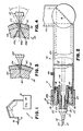
- Fig. 2 is a cross-sectional view of the handpiece of the present invention;
- Fig. 3 is a cross-sectional perspective view broken away of the heart undergoing transmyocardial revascularization according to the method of the present invention; and
- Fig. 4 is a view similar to Fig. 3 illustrating the manner in which the laser may provide multiple perforations.
- As shown in Fig. 1, a
surgical laser 11 suitable for use in transmyocardial revascularization includes a source of high energy laser light depicted diagrammatically as 12. Preferably this source of laser light will be a carbon dioxide laser which radiates a beam of high energy laser light through a series ofarms 13 to a hand held manipulator orhandpiece 14. Thearms 13 include a series of mirrors (such as mirror 16, Fig. 2) which direct the generated laser beam to thehandpiece 14 and permit relatively free movement of the handpiece independent the carbon dioxide laser 12. A remote foot switch (not shown) is also provided to activate the laser. - The
handpiece 14 includes a tubular body 15 which provides a path for a laser beam directed from the mirror 16 to the forward end 17 of body 15. Also within the body 15 is a focusinglens 20 which acts to focus the carbon dioxide laser beam at a fixedfocal point 21 forward of forward end 17. The lens directs the laser light reflected from mirror 16 along a laser beam path shown in dottedlines 19. The laser beam reaches its highest intensity at itsfocal point 21. This focal point is determined by the location and shape oflens 20. - The laser beam path extends through a
hollow needle 22. Theneedle 22 includes a pointedtip 23,shank 24 and an enlarged body portion orhub 25. Thehub 25 has flaredshoulders 26 which cooperate withexternal threads 33 of a needle mount orholder 27 to provide a means to attach theneedle 22 and hold it stationary with respect to the handpiece. The interior ofhub 25 is preferably hollowed out as much as possible to provide clearance for the laser beam. - The
needle 22 mounts to the body 15 ofhandpiece 14 in a manner which enables the location of the needle to be adjusted relative to the body 15 and thus relative to the laser beam path. More specifically, theneedle 22 threads intoneedle mount 27 which is adjustabley positioned within a hollow cylindrical holder 28. The holder 28 is fixed to the body 15 by acap 29. - The needle mount includes a hollow
cylindrical metal body 31 and atubular extension 30 adapted to fit within the hub ofneedle 22. The externally threadedportion 33 lies between thetubular extension 30 and thebody 31. - The
needle mount 27 is spaced from the tubular holder 28. This spacing permits the needle mount to be moved forwardly, rearwardly and laterally relative to the holder 28 and thus relative to thehandpiece 14 to insure that the laser path extends directly through theneedle 22. The exact position ofneedle mount 27 relative to the holder 28 is fixed by a pair ofopposed set screws needle mount 27. Thus these provide for forward, rearward and lateral adjustment of the needle mount and thus needle 22 relative to the body 15. - The holder 28 has a
shoulder 36 which abuts theend surface 37 of the body 15 of thehandpiece 14. Holder 28 is held in place bycap 29 which can be fixed to the body 15 by various means. As shown in Fig. 1, the cap is held in position by set screw 29a. The handpiece is also provided with structure which permits the flushing of the needle with saline should it become plugged or fouled. The handpiece includes aport 46 which permits introduction of saline into the handpiece betweenlens 20 andneedle 22. Saline forced intoport 46 floods the area betweenlens 20 andneedle 22. The saline is forced through theshank 24 of the needle cleaning the needle. - In operation the laser light generated by the carbon dioxide laser 12 reflects off mirror 16 passes through the handpiece through
lens 20 where it is focused atfocal point 21. Thefocal point 21 should lie at the tip ofneedle 22 or at least within about 3 cm of the tip. Further thelaser beam path 19 should pass directly through the hollow interior of the needle. To provide for this the needle is mounted onneedle mount 27 and theneedle mount 27 is adjusted relative to holder 28 and laser body 15 to align the hollow interior of the needle along the laser path with thetip 23 of the needle at about thefocal point 21. The focal point for a laser with a certain lens should be known and thus the position of the needle point adjusted accordingly. The forward and rearward adjustment is made by removingcap 29, loosening theset screws needle mount 27 forwardly or rearwardly relative to holder. This adjustment permits needles having various length shanks to be used and interchanged if desired. The lateral adjustment is established by relative movement of theset screws - This laser can be used in operations in which a laser beam is required at an unexposed portion of an organ. The point of the needle is sharp enough so that as it penetrates tissue it creates an opening by spreading the tissue apart providing a path for the laser beam. This causes only minimal tissue damage. The length of the needle shank is chosen according to the desired depth of this needle puncture and will vary from operation to operation. The gauge of the needle is also a variable according to the size of the generated laser beam. Typically an 18 gauge thin welled needle is suitable, although a different gauge needle may be desired or required with different carbon dioxide lasers.
- One particular application for which this apparatus is particularly suited is transmyocardial revascularization. In this application it is desirable to cut holes through the endocardium into one of the heart cavities to allow blood to pass from the cavity into the endocardium. Typically this method will be used to restore blood flow to an ischemic subendocardium perhaps associated with left ventricular hypertrophy. Specifically this provides ventricular blood to the myocardial vasculature.
- According to the present invention, transmyocardial revascularization is conducted by surgically exposing the exterior surface of the heart. The left anterior free wall of the left ventricle near the left anterior descending and left circumflex coronaries are subjected to laser treatment. These are the areas which tend to be most ischemic. The ischemic areas are provided with as many perforations as possible without affecting the contractility of the heart. Generally in any area 10 to 20 perforations per square centimeter are created.
- As shown in Fig. 3, to perforate the endocardial tissue of the left anterior free wall of the left ventricle the
needle 22 is simply pressed into theepicardial portion 42 of the anterior free wall of theheart 39 untilhub 25 abuts against theepicardial surface 41 of the heart. Typically an ischemic heart can have a thickness of approximately 20 millimeters. Preferably the shank of the needle is about 14 millimeters providing a laser induced perforation of about 6 millimeters. Once the needle is forced into the heart the laser is activated for a period of time sufficient to penetrate through the remaining portion of the endocardium into the left ventricle cavity providing aperforation 43 through theendocardium 44. Generally a period of one to two seconds is sufficient with a laser having 80 watts power. The handpiece is then pulled away to pull the needle from the heart. The needle is reinserted at a separate location and the procedure is repeated until the desired density of perforations is obtained. - As shown in Fig. 4 an alternate method of providing multiple perforations is to insert the needle through the epicardium 42 towards the
endocardium 44 and providemultiple perforations endocardium 44. This is accomplished by pushing the needle into theepicardium 42, activating the laser to create first perforation 45a, and subsequently pivoting the handpiece, and again actuating the laser to formsecond perforation 45b. The handpiece can then be pivoted again about the laser tip andthird perforation 45c can be made from the same point but at different angles to provide multiple perforations. This in effect increases the number of perforations through the endocardium without increasing the number of perforations through the epicardium. This reduces the effect this surgical procedure has on the contractility of the heart. These laser induced perforations provide a flow path for blood into the endocardium from the ventricular chamber. - The use of surgical laser of the present invention is not limited to transmyocardial revascularization. Those skilled in the art will readily appreciate the use of the laser in various applications to obtain the benefits of the present invention.
- Thus having described my invention I claim:
Claims (20)
Applications Claiming Priority (2)
| Application Number | Priority Date | Filing Date | Title |
|---|---|---|---|
| US718069 | 1985-04-01 | ||
| US06/718,069 US4658817A (en) | 1985-04-01 | 1985-04-01 | Method and apparatus for transmyocardial revascularization using a laser |
Publications (2)
| Publication Number | Publication Date |
|---|---|
| EP0196519A2 true EP0196519A2 (en) | 1986-10-08 |
| EP0196519A3 EP0196519A3 (en) | 1987-07-01 |
Family
ID=24884700
Family Applications (1)
| Application Number | Title | Priority Date | Filing Date |
|---|---|---|---|
| EP86103443A Withdrawn EP0196519A3 (en) | 1985-04-01 | 1986-03-14 | Method and apparatus for transmyocardial revascularization using a laser |
Country Status (4)
| Country | Link |
|---|---|
| US (1) | US4658817A (en) |
| EP (1) | EP0196519A3 (en) |
| JP (1) | JPS61268246A (en) |
| CA (1) | CA1259104A (en) |
Cited By (14)
| Publication number | Priority date | Publication date | Assignee | Title |
|---|---|---|---|---|
| WO1989006519A2 (en) * | 1988-01-25 | 1989-07-27 | Refractive Laser Research & Development Program, L | Method and apparatus for laser surgery |
| WO1989006518A1 (en) * | 1988-01-12 | 1989-07-27 | Ulrich-Dardenne-Stiftung E.V. | Device for ablative photodecomposition of organic and inorganic substances, in particular hard dental materials |
| US4911712A (en) * | 1988-04-14 | 1990-03-27 | Heraeus Lasersonics, Inc. | Medical laser probe |
| EP0511805A2 (en) * | 1991-05-01 | 1992-11-04 | Coherent, Inc. | Coupler for a laser delivery system |
| EP0515867A2 (en) * | 1991-05-01 | 1992-12-02 | The Trustees Of Columbia University In The City Of New York | Myocardial revascularization through the endocardial surface using a laser |
| EP0553576A1 (en) * | 1990-09-24 | 1993-08-04 | Plc Medical Systems, Inc. | Heart-synchronized pulsed laser system |
| US5674217A (en) * | 1993-10-01 | 1997-10-07 | Wahlstrom; Dale A. | Heart synchronized extractor for an implanted object |
| US5807383A (en) * | 1996-05-13 | 1998-09-15 | United States Surgical Corporation | Lasing device |
| US5947989A (en) * | 1996-12-12 | 1999-09-07 | United States Surgical Corporation | Method and apparatus for transmyocardial revascularization |
| US5980545A (en) * | 1996-05-13 | 1999-11-09 | United States Surgical Corporation | Coring device and method |
| WO2000056233A1 (en) | 1999-03-22 | 2000-09-28 | Saphir Medical Products Gmbh | Surgical apparatus with pressurised liquid jet for transmyocardial revascularization |
| US6135996A (en) * | 1998-04-17 | 2000-10-24 | Baxter International, Inc. | Controlled advancement lasing device |
| FR2797581A1 (en) | 1999-08-20 | 2001-02-23 | Saphir Medical Products Gmbh | Surgical apparatus uses pressurized liquid jet for transmyocardial revascularization to form conduits in myocardium wall where the surgeon can control liquid pressure and pulses |
| US6283955B1 (en) | 1996-05-13 | 2001-09-04 | Edwards Lifesciences Corp. | Laser ablation device |
Families Citing this family (221)
| Publication number | Priority date | Publication date | Assignee | Title |
|---|---|---|---|---|
| US4785805A (en) * | 1985-05-28 | 1988-11-22 | Surgical Laser Technologies, Inc. | Two-piece disposable laser delivery system |
| US4781185A (en) * | 1986-07-21 | 1988-11-01 | Gv Medical, Inc. | Connecting apparatus for catheter assembly |
| GB8725566D0 (en) * | 1987-10-31 | 1987-12-02 | Pilkington Medical Systems Ltd | Laser system |
| US4917083A (en) * | 1988-03-04 | 1990-04-17 | Heraeus Lasersonics, Inc. | Delivery arrangement for a laser medical system |
| US5074861A (en) * | 1988-05-23 | 1991-12-24 | Schneider Richard T | Medical laser device and method |
| US5114422A (en) * | 1989-12-11 | 1992-05-19 | Ioan Cosmescu | Laser laparoscope assembly and method therefor |
| US5242439A (en) * | 1990-01-12 | 1993-09-07 | Laserscope | Means for inserting instrumentation for a percutaneous diskectomy using a laser |
| US6113587A (en) | 1990-09-24 | 2000-09-05 | Plc Medical Systems, Inc. | Handpiece for a medical laser system |
| US5280378A (en) * | 1990-10-19 | 1994-01-18 | I.L. Med, Inc. | Cyclically scanned medical laser |
| US5885272A (en) * | 1990-10-30 | 1999-03-23 | Aita; Michael | System and method for percutaneous myocardial revascularization |
| US5380316A (en) * | 1990-12-18 | 1995-01-10 | Advanced Cardiovascular Systems, Inc. | Method for intra-operative myocardial device revascularization |
| AU714277B2 (en) * | 1991-05-01 | 1999-12-23 | Trustees Of Columbia University In The City Of New York, The | Myocardial revascularization through the endocardial surface using a laser |
| US5697909A (en) * | 1992-01-07 | 1997-12-16 | Arthrocare Corporation | Methods and apparatus for surgical cutting |
| US5697281A (en) * | 1991-10-09 | 1997-12-16 | Arthrocare Corporation | System and method for electrosurgical cutting and ablation |
| US7429262B2 (en) * | 1992-01-07 | 2008-09-30 | Arthrocare Corporation | Apparatus and methods for electrosurgical ablation and resection of target tissue |
| US6179824B1 (en) | 1993-05-10 | 2001-01-30 | Arthrocare Corporation | System and methods for electrosurgical restenosis of body lumens |
| US5683366A (en) | 1992-01-07 | 1997-11-04 | Arthrocare Corporation | System and method for electrosurgical tissue canalization |
| US6277112B1 (en) | 1996-07-16 | 2001-08-21 | Arthrocare Corporation | Methods for electrosurgical spine surgery |
| US7297145B2 (en) * | 1997-10-23 | 2007-11-20 | Arthrocare Corporation | Bipolar electrosurgical clamp for removing and modifying tissue |
| US6024733A (en) * | 1995-06-07 | 2000-02-15 | Arthrocare Corporation | System and method for epidermal tissue ablation |
| US5902272A (en) | 1992-01-07 | 1999-05-11 | Arthrocare Corporation | Planar ablation probe and method for electrosurgical cutting and ablation |
| US6063079A (en) * | 1995-06-07 | 2000-05-16 | Arthrocare Corporation | Methods for electrosurgical treatment of turbinates |
| US6102046A (en) * | 1995-11-22 | 2000-08-15 | Arthrocare Corporation | Systems and methods for electrosurgical tissue revascularization |
| US6210402B1 (en) | 1995-11-22 | 2001-04-03 | Arthrocare Corporation | Methods for electrosurgical dermatological treatment |
| US6159194A (en) * | 1992-01-07 | 2000-12-12 | Arthrocare Corporation | System and method for electrosurgical tissue contraction |
| US6770071B2 (en) * | 1995-06-07 | 2004-08-03 | Arthrocare Corporation | Bladed electrosurgical probe |
| US5681282A (en) * | 1992-01-07 | 1997-10-28 | Arthrocare Corporation | Methods and apparatus for ablation of luminal tissues |
| US5891095A (en) * | 1993-05-10 | 1999-04-06 | Arthrocare Corporation | Electrosurgical treatment of tissue in electrically conductive fluid |
| US6500173B2 (en) | 1992-01-07 | 2002-12-31 | Ronald A. Underwood | Methods for electrosurgical spine surgery |
| US5697882A (en) * | 1992-01-07 | 1997-12-16 | Arthrocare Corporation | System and method for electrosurgical cutting and ablation |
| US5236360A (en) * | 1992-07-23 | 1993-08-17 | Laser Medical Technology, Inc. | Optical members for laser transmission |
| US6749604B1 (en) * | 1993-05-10 | 2004-06-15 | Arthrocare Corporation | Electrosurgical instrument with axially-spaced electrodes |
| US6832996B2 (en) | 1995-06-07 | 2004-12-21 | Arthrocare Corporation | Electrosurgical systems and methods for treating tissue |
| US6915806B2 (en) * | 1993-05-10 | 2005-07-12 | Arthrocare Corporation | Method for harvesting graft vessel |
| US6117109A (en) * | 1995-11-22 | 2000-09-12 | Arthrocare Corporation | Systems and methods for electrosurgical incisions on external skin surfaces |
| US5541081A (en) * | 1994-03-22 | 1996-07-30 | President And Fellows Of Harvard College | Process for assessing oocyte and embryo quality |
| US5695457A (en) * | 1994-07-28 | 1997-12-09 | Heartport, Inc. | Cardioplegia catheter system |
| US5591159A (en) * | 1994-11-09 | 1997-01-07 | Taheri; Syde A. | Transcavitary myocardial perfusion apparatus |
| US6690963B2 (en) | 1995-01-24 | 2004-02-10 | Biosense, Inc. | System for determining the location and orientation of an invasive medical instrument |
| US6602248B1 (en) | 1995-06-07 | 2003-08-05 | Arthro Care Corp. | Methods for repairing damaged intervertebral discs |
| US6251104B1 (en) | 1995-05-10 | 2001-06-26 | Eclipse Surgical Technologies, Inc. | Guiding catheter system for ablating heart tissue |
| US20050004634A1 (en) * | 1995-06-07 | 2005-01-06 | Arthrocare Corporation | Methods for electrosurgical treatment of spinal tissue |
| US7393351B2 (en) * | 1995-06-07 | 2008-07-01 | Arthrocare Corporation | Apparatus and methods for treating cervical inter-vertebral discs |
| US6772012B2 (en) * | 1995-06-07 | 2004-08-03 | Arthrocare Corporation | Methods for electrosurgical treatment of spinal tissue |
| US5951541A (en) * | 1995-06-07 | 1999-09-14 | Cardiogenesis Corporation | Channel forming device with a secured distal extremity |
| US6149620A (en) * | 1995-11-22 | 2000-11-21 | Arthrocare Corporation | System and methods for electrosurgical tissue treatment in the presence of electrically conductive fluid |
| US6132451A (en) * | 1995-06-07 | 2000-10-17 | Eclipse Surgical Technologies, Inc. | Optical fiber for myocardial channel formation |
| US6837888B2 (en) * | 1995-06-07 | 2005-01-04 | Arthrocare Corporation | Electrosurgical probe with movable return electrode and methods related thereto |
| US7572251B1 (en) | 1995-06-07 | 2009-08-11 | Arthrocare Corporation | Systems and methods for electrosurgical tissue treatment |
| ATE231363T1 (en) * | 1995-06-07 | 2003-02-15 | Eclipse Surgical Tech | SURGICAL DEVICE PROVIDED WITH A PENETRATION DEPTH LIMITER FOR CREATING CHANNELS |
| US5728091A (en) * | 1995-06-07 | 1998-03-17 | Cardiogenesis Corporation | Optical fiber for myocardial channel formation |
| US5611797A (en) * | 1995-07-26 | 1997-03-18 | Virginia C. George | Combination handpiece and surgical laser tool |
| US6228082B1 (en) | 1995-11-22 | 2001-05-08 | Arthrocare Corporation | Systems and methods for electrosurgical treatment of vascular disorders |
| US6228078B1 (en) | 1995-11-22 | 2001-05-08 | Arthrocare Corporation | Methods for electrosurgical dermatological treatment |
| US7758537B1 (en) | 1995-11-22 | 2010-07-20 | Arthrocare Corporation | Systems and methods for electrosurgical removal of the stratum corneum |
| US7270661B2 (en) * | 1995-11-22 | 2007-09-18 | Arthocare Corporation | Electrosurgical apparatus and methods for treatment and removal of tissue |
| US6896672B1 (en) * | 1995-11-22 | 2005-05-24 | Arthrocare Corporation | Methods for electrosurgical incisions on external skin surfaces |
| US6461350B1 (en) | 1995-11-22 | 2002-10-08 | Arthrocare Corporation | Systems and methods for electrosurgical-assisted lipectomy |
| US5846080A (en) * | 1995-12-20 | 1998-12-08 | W&H Dentalwerk Gmbh | Laser dental devices and methods |
| IL125259A (en) * | 1996-01-08 | 2002-12-01 | Biosense Inc | Apparatus for myocardial revascularization |
| US5769843A (en) * | 1996-02-20 | 1998-06-23 | Cormedica | Percutaneous endomyocardial revascularization |
| US5713894A (en) * | 1996-02-27 | 1998-02-03 | Murphy-Chutorian; Douglas | Combined mechanical/optical system for transmyocardial revascularization |
| US6258083B1 (en) | 1996-03-29 | 2001-07-10 | Eclipse Surgical Technologies, Inc. | Viewing surgical scope for minimally invasive procedures |
| US5725523A (en) * | 1996-03-29 | 1998-03-10 | Mueller; Richard L. | Lateral-and posterior-aspect method and apparatus for laser-assisted transmyocardial revascularization and other surgical applications |
| US5725521A (en) * | 1996-03-29 | 1998-03-10 | Eclipse Surgical Technologies, Inc. | Depth stop apparatus and method for laser-assisted transmyocardial revascularization and other surgical applications |
| AU5279898A (en) | 1996-03-29 | 1998-03-26 | Eclipse Surgical Technologies, Inc. | Minimally invasive method and apparatus for forming revascularization channels |
| US5891133A (en) * | 1996-03-29 | 1999-04-06 | Eclipse Surgical Technologies, Inc. | Apparatus for laser-assisted intra-coronary transmyocardial revascularization and other applications |
| US5766164A (en) * | 1996-07-03 | 1998-06-16 | Eclipse Surgical Technologies, Inc. | Contiguous, branched transmyocardial revascularization (TMR) channel, method and device |
| US5782823A (en) * | 1996-04-05 | 1998-07-21 | Eclipse Surgical Technologies, Inc. | Laser device for transmyocardial revascularization procedures including means for enabling a formation of a pilot hole in the epicardium |
| US6152918A (en) | 1996-04-05 | 2000-11-28 | Eclipse Surgical Technologies, Inc. | Laser device with auto-piercing tip for myocardial revascularization procedures |
| US5738680A (en) * | 1996-04-05 | 1998-04-14 | Eclipse Surgical Technologies, Inc. | Laser device with piercing tip for transmyocardial revascularization procedures |
| US5703985A (en) * | 1996-04-29 | 1997-12-30 | Eclipse Surgical Technologies, Inc. | Optical fiber device and method for laser surgery procedures |
| CA2207570A1 (en) * | 1996-06-13 | 1997-12-13 | Eclipse Surgical Technologies, Inc. | Intraoperative myocardial device and stimulation procedure |
| US5662124A (en) * | 1996-06-19 | 1997-09-02 | Wilk Patent Development Corp. | Coronary artery by-pass method |
| US5766163A (en) * | 1996-07-03 | 1998-06-16 | Eclipse Surgical Technologies, Inc. | Controllable trocar for transmyocardial revascularization (TMR) via endocardium method and apparatus |
| US7104986B2 (en) * | 1996-07-16 | 2006-09-12 | Arthrocare Corporation | Intervertebral disc replacement method |
| US6726684B1 (en) | 1996-07-16 | 2004-04-27 | Arthrocare Corporation | Methods for electrosurgical spine surgery |
| US7357798B2 (en) | 1996-07-16 | 2008-04-15 | Arthrocare Corporation | Systems and methods for electrosurgical prevention of disc herniations |
| US5840075A (en) * | 1996-08-23 | 1998-11-24 | Eclipse Surgical Technologies, Inc. | Dual laser device for transmyocardial revascularization procedures |
| US5693041A (en) * | 1996-08-23 | 1997-12-02 | Eclipse Surgical Technologies, Inc. | Laser delivery means ring stabilization method and apparatus for surgical and other procedures |
| US5976164A (en) | 1996-09-13 | 1999-11-02 | Eclipse Surgical Technologies, Inc. | Method and apparatus for myocardial revascularization and/or biopsy of the heart |
| US5871495A (en) * | 1996-09-13 | 1999-02-16 | Eclipse Surgical Technologies, Inc. | Method and apparatus for mechanical transmyocardial revascularization of the heart |
| US5855577A (en) * | 1996-09-17 | 1999-01-05 | Eclipse Surgical Technologies, Inc. | Bow shaped catheter |
| US5755714A (en) * | 1996-09-17 | 1998-05-26 | Eclipse Surgical Technologies, Inc. | Shaped catheter for transmyocardial revascularization |
| AU1275997A (en) * | 1996-10-11 | 1998-05-11 | Transvascular, Inc. | Methods and apparatus for bypassing arterial obstructions and/or performing other transvascular procedures |
| US5931834A (en) | 1996-10-15 | 1999-08-03 | Eclipse Surgical Technologies, Inc. | Method for non-synchronous laser-assisted myocardial revascularization |
| US5989274A (en) | 1996-10-17 | 1999-11-23 | Ethicon Endo-Surgery, Inc. | Methods and devices for improving blood flow to a heart of a patient |
| US5873366A (en) * | 1996-11-07 | 1999-02-23 | Chim; Nicholas | Method for transmyocardial revascularization |
| US5999678A (en) * | 1996-12-27 | 1999-12-07 | Eclipse Surgical Technologies, Inc. | Laser delivery means adapted for drug delivery |
| CA2225521C (en) | 1996-12-27 | 2004-04-06 | Eclipse Surgical Technologies, Inc. | Laser assisted drug delivery apparatus |
| US5925012A (en) * | 1996-12-27 | 1999-07-20 | Eclipse Surgical Technologies, Inc. | Laser assisted drug delivery |
| DE69732696T2 (en) | 1997-01-08 | 2006-04-13 | Biosense Webster, Inc., Diamond Bar | MONITORING MYOCARDIAL REVASCULARIZATION |
| US5913853A (en) | 1997-01-30 | 1999-06-22 | Cardiodyne, Inc. | Laser energy device and procedure for forming a channel within tissue |
| US6001091A (en) * | 1997-02-03 | 1999-12-14 | Eclipse Surgical Technologies, Inc. | Revascularization with heart pacing |
| US5993443A (en) * | 1997-02-03 | 1999-11-30 | Eclipse Surgical Technologies, Inc. | Revascularization with heartbeat verification |
| US6056742A (en) * | 1997-02-03 | 2000-05-02 | Eclipse Surgical Technologies, Inc. | Revascularization with laser outputs |
| US5968059A (en) | 1997-03-06 | 1999-10-19 | Scimed Life Systems, Inc. | Transmyocardial revascularization catheter and method |
| US6086534A (en) * | 1997-03-07 | 2000-07-11 | Cardiogenesis Corporation | Apparatus and method of myocardial revascularization using ultrasonic pulse-echo distance ranging |
| US6093177A (en) | 1997-03-07 | 2000-07-25 | Cardiogenesis Corporation | Catheter with flexible intermediate section |
| US5876373A (en) * | 1997-04-04 | 1999-03-02 | Eclipse Surgical Technologies, Inc. | Steerable catheter |
| US6024703A (en) | 1997-05-07 | 2000-02-15 | Eclipse Surgical Technologies, Inc. | Ultrasound device for axial ranging |
| US6855143B2 (en) * | 1997-06-13 | 2005-02-15 | Arthrocare Corporation | Electrosurgical systems and methods for recanalization of occluded body lumens |
| US5951543A (en) * | 1997-06-30 | 1999-09-14 | Clinicon Corporation | Delivery system and method for surgical laser |
| US5938633A (en) * | 1997-07-09 | 1999-08-17 | Ethicon Endo-Surgery, Inc. | Ultrasonic surgical devices |
| JP2001510701A (en) * | 1997-07-22 | 2001-08-07 | ジェイムズ コーリア, | Apparatus and method for transmyocardial vascular regeneration by laser ablation |
| US5935119A (en) * | 1997-08-06 | 1999-08-10 | United States Surgical Corporation | Perfusion structure |
| AU8901598A (en) | 1997-08-07 | 1999-03-01 | Cardiogenesis Corporation | System and method of intra-operative myocardial revascularization using pulsed sonic energy |
| US5911729A (en) * | 1997-08-13 | 1999-06-15 | United States Surgical Corporation | Electrocautery coring using solid needle |
| DE19740824A1 (en) | 1997-09-17 | 1999-03-18 | Laser & Med Tech Gmbh | Channel production device for heart muscle tissue |
| US6554794B1 (en) | 1997-09-24 | 2003-04-29 | Richard L. Mueller | Non-deforming deflectable multi-lumen catheter |
| JPH11221229A (en) | 1997-09-24 | 1999-08-17 | Eclipse Surgical Technol Inc | Catheter |
| US6106520A (en) * | 1997-09-30 | 2000-08-22 | Hearten Medical, Inc. | Endocardial device for producing reversible damage to heart tissue |
| EP1018961A1 (en) | 1997-10-02 | 2000-07-19 | Cardiogenesis Corporation | Transmyocardial revascularization using radiofrequency energy |
| US5980548A (en) * | 1997-10-29 | 1999-11-09 | Kensey Nash Corporation | Transmyocardial revascularization system |
| US6162214A (en) * | 1997-10-30 | 2000-12-19 | Eclipse Surgical Technologies, Inc. | Corning device for myocardial revascularization |
| US6156029A (en) * | 1997-11-25 | 2000-12-05 | Eclipse Surgical Technologies, Inc. | Selective treatment of endocardial/myocardial boundary |
| US6197324B1 (en) | 1997-12-18 | 2001-03-06 | C. R. Bard, Inc. | System and methods for local delivery of an agent |
| US6251418B1 (en) | 1997-12-18 | 2001-06-26 | C.R. Bard, Inc. | Systems and methods for local delivery of an agent |
| AU2022199A (en) * | 1997-12-31 | 1999-07-19 | Hydrocision, Inc. | Fluid jet cutting system for cardiac applications |
| US5976124A (en) * | 1998-01-05 | 1999-11-02 | Spectranetics Corporation | Phototherapy device and method |
| US5997531A (en) * | 1998-01-29 | 1999-12-07 | Cardiodyne, Inc. | User actuated laser energy device and procedure for forming a channel within tissue |
| EP0938871A3 (en) | 1998-02-27 | 2001-03-07 | ECLIPSE SURGICAL TECHNOLOGIES, Inc. | Surgical apparatus |
| US6199554B1 (en) * | 1998-03-27 | 2001-03-13 | The Brigham And Women's Hospital, Inc. | Method and apparatus for combining injury-mediated therapy and drug delivery |
| US6263880B1 (en) | 1998-06-22 | 2001-07-24 | Neovasys, Inc. | Method of enhancing blood flow in tissue |
| US6447504B1 (en) | 1998-07-02 | 2002-09-10 | Biosense, Inc. | System for treatment of heart tissue using viability map |
| US7276063B2 (en) | 1998-08-11 | 2007-10-02 | Arthrocare Corporation | Instrument for electrosurgical tissue treatment |
| US7435247B2 (en) | 1998-08-11 | 2008-10-14 | Arthrocare Corporation | Systems and methods for electrosurgical tissue treatment |
| US6261304B1 (en) | 1998-09-10 | 2001-07-17 | Percardia, Inc. | Delivery methods for left ventricular conduit |
| US6689121B1 (en) | 1998-09-24 | 2004-02-10 | C. R. Bard, Inc. | Systems and methods for treating ischemia |
| US6432126B1 (en) * | 1998-09-30 | 2002-08-13 | C.R. Bard, Inc. | Flexible vascular inducing implants |
| US6283935B1 (en) | 1998-09-30 | 2001-09-04 | Hearten Medical | Ultrasonic device for providing reversible tissue damage to heart muscle |
| US6458092B1 (en) | 1998-09-30 | 2002-10-01 | C. R. Bard, Inc. | Vascular inducing implants |
| US6248112B1 (en) | 1998-09-30 | 2001-06-19 | C. R. Bard, Inc. | Implant delivery system |
| US6692520B1 (en) | 1998-12-15 | 2004-02-17 | C. R. Bard, Inc. | Systems and methods for imbedded intramuscular implants |
| US6176856B1 (en) | 1998-12-18 | 2001-01-23 | Eclipse Surgical Technologies, Inc | Resistive heating system and apparatus for improving blood flow in the heart |
| US6363938B2 (en) | 1998-12-22 | 2002-04-02 | Angiotrax, Inc. | Methods and apparatus for perfusing tissue and/or stimulating revascularization and tissue growth |
| US6986784B1 (en) | 1999-05-14 | 2006-01-17 | C. R. Bard, Inc. | Implant anchor systems |
| US6855160B1 (en) | 1999-08-04 | 2005-02-15 | C. R. Bard, Inc. | Implant and agent delivery device |
| US20040073155A1 (en) * | 2000-01-14 | 2004-04-15 | Broncus Technologies, Inc. | Methods and devices for maintaining patency of surgically created channels in tissue |
| US6709427B1 (en) * | 1999-08-05 | 2004-03-23 | Kensey Nash Corporation | Systems and methods for delivering agents into targeted tissue of a living being |
| US7462162B2 (en) | 2001-09-04 | 2008-12-09 | Broncus Technologies, Inc. | Antiproliferative devices for maintaining patency of surgically created channels in a body organ |
| US7422563B2 (en) * | 1999-08-05 | 2008-09-09 | Broncus Technologies, Inc. | Multifunctional tip catheter for applying energy to tissue and detecting the presence of blood flow |
| US6692494B1 (en) * | 1999-08-05 | 2004-02-17 | Broncus Technologies, Inc. | Methods and devices for creating collateral channels in the lungs |
| US7022088B2 (en) * | 1999-08-05 | 2006-04-04 | Broncus Technologies, Inc. | Devices for applying energy to tissue |
| US20020087155A1 (en) | 1999-08-30 | 2002-07-04 | Underwood Ronald A. | Systems and methods for intradermal collagen stimulation |
| US7232421B1 (en) | 2000-05-12 | 2007-06-19 | C. R. Bard, Inc. | Agent delivery systems |
| US6695836B1 (en) * | 2000-07-03 | 2004-02-24 | Radius Medical Technologies, Inc. | Device and method for myocardial revascularization |
| US7204847B1 (en) | 2000-07-28 | 2007-04-17 | C. R. Bard, Inc. | Implant anchor systems |
| US6893421B1 (en) * | 2000-08-08 | 2005-05-17 | Scimed Life Systems, Inc. | Catheter shaft assembly |
| US6595958B1 (en) | 2000-08-08 | 2003-07-22 | Scimed Life Systems, Inc. | Tortuous path injection device and method |
| US6613017B1 (en) * | 2000-08-08 | 2003-09-02 | Scimed Life Systems, Inc. | Controlled depth injection device and method |
| US6546276B1 (en) | 2000-09-12 | 2003-04-08 | Claudio I. Zanelli | Ultrasonic based detection of interventional medical device contact and alignment |
| US6436059B1 (en) | 2000-09-12 | 2002-08-20 | Claudio I. Zanelli | Detection of imd contact and alignment based on changes in frequency response characteristics |
| US20030158545A1 (en) * | 2000-09-28 | 2003-08-21 | Arthrocare Corporation | Methods and apparatus for treating back pain |
| US20020107510A1 (en) * | 2001-02-05 | 2002-08-08 | Andrews Robert R. | Laser apparatus useful for myocardial revascularization |
| US6666863B2 (en) | 2001-03-01 | 2003-12-23 | Scimed Life Systems, Inc. | Device and method for percutaneous myocardial revascularization |
| US20020133149A1 (en) * | 2001-03-17 | 2002-09-19 | Arthrocare Corporation | Electrosurgical systems and methods for hair transplantation and epilation |
| US6837884B2 (en) * | 2001-06-18 | 2005-01-04 | Arthrocare Corporation | Electrosurgical apparatus having compound return electrode |
| US7708712B2 (en) | 2001-09-04 | 2010-05-04 | Broncus Technologies, Inc. | Methods and devices for maintaining patency of surgically created channels in a body organ |
| US20050060042A1 (en) * | 2001-09-04 | 2005-03-17 | Broncus Technologies, Inc. | Methods and devices for maintaining surgically created channels in a body organ |
| AU2002362310A1 (en) * | 2001-09-14 | 2003-04-01 | Arthrocare Corporation | Methods and apparatus for treating intervertebral discs |
| US20030088245A1 (en) * | 2001-11-02 | 2003-05-08 | Arthrocare Corporation | Methods and apparatus for electrosurgical ventriculostomy |
| US6920883B2 (en) * | 2001-11-08 | 2005-07-26 | Arthrocare Corporation | Methods and apparatus for skin treatment |
| US20030130738A1 (en) * | 2001-11-08 | 2003-07-10 | Arthrocare Corporation | System and method for repairing a damaged intervertebral disc |
| WO2003068055A2 (en) | 2002-02-11 | 2003-08-21 | Arthrocare Corporation | Electrosurgical apparatus and methods for laparoscopy |
| US20030208196A1 (en) * | 2002-05-03 | 2003-11-06 | Arthrocare Corporation | Control system for limited-use device |
| AU2003268458A1 (en) * | 2002-09-05 | 2004-03-29 | Arthrocare Corporation | Methods and apparatus for treating intervertebral discs |
| EP1596705B1 (en) * | 2003-02-05 | 2018-09-12 | Arthrocare Corporation | Temperature indicating electrosurgical apparatus |
| US7794456B2 (en) | 2003-05-13 | 2010-09-14 | Arthrocare Corporation | Systems and methods for electrosurgical intervertebral disc replacement |
| EP1651127B1 (en) | 2003-07-16 | 2012-10-31 | Arthrocare Corporation | Rotary electrosurgical apparatus |
| US8308682B2 (en) | 2003-07-18 | 2012-11-13 | Broncus Medical Inc. | Devices for maintaining patency of surgically created channels in tissue |
| US7708733B2 (en) * | 2003-10-20 | 2010-05-04 | Arthrocare Corporation | Electrosurgical method and apparatus for removing tissue within a bone body |
| US7491200B2 (en) * | 2004-03-26 | 2009-02-17 | Arthrocare Corporation | Method for treating obstructive sleep disorder includes removing tissue from base of tongue |
| US7704249B2 (en) | 2004-05-07 | 2010-04-27 | Arthrocare Corporation | Apparatus and methods for electrosurgical ablation and resection of target tissue |
| EP1773227B1 (en) | 2004-06-24 | 2016-04-13 | ArthroCare Corporation | Electrosurgical device having planar vertical electrodes |
| US8409167B2 (en) | 2004-07-19 | 2013-04-02 | Broncus Medical Inc | Devices for delivering substances through an extra-anatomic opening created in an airway |
| EP1786499A2 (en) * | 2004-07-19 | 2007-05-23 | Broncus Technologies, Inc. | Methods and devices for maintaining patency of surgically created channels in a body organ |
| US20060095031A1 (en) * | 2004-09-22 | 2006-05-04 | Arthrocare Corporation | Selectively controlled active electrodes for electrosurgical probe |
| US7632267B2 (en) | 2005-07-06 | 2009-12-15 | Arthrocare Corporation | Fuse-electrode electrosurgical apparatus |
| US8876746B2 (en) * | 2006-01-06 | 2014-11-04 | Arthrocare Corporation | Electrosurgical system and method for treating chronic wound tissue |
| US7691101B2 (en) | 2006-01-06 | 2010-04-06 | Arthrocare Corporation | Electrosurgical method and system for treating foot ulcer |
| US7879034B2 (en) | 2006-03-02 | 2011-02-01 | Arthrocare Corporation | Internally located return electrode electrosurgical apparatus, system and method |
| EP2020943B1 (en) | 2006-05-30 | 2015-07-08 | ArthroCare Corporation | Hard tissue ablation system |
| GB2452103B (en) | 2007-01-05 | 2011-08-31 | Arthrocare Corp | Electrosurgical system with suction control apparatus and system |
| US7862560B2 (en) | 2007-03-23 | 2011-01-04 | Arthrocare Corporation | Ablation apparatus having reduced nerve stimulation and related methods |
| US20090150331A1 (en) * | 2007-12-07 | 2009-06-11 | Roche Diagnostics Operations, Inc. | Method and system for creating reports |
| US9358063B2 (en) | 2008-02-14 | 2016-06-07 | Arthrocare Corporation | Ablation performance indicator for electrosurgical devices |
| US8747400B2 (en) | 2008-08-13 | 2014-06-10 | Arthrocare Corporation | Systems and methods for screen electrode securement |
| US20100114110A1 (en) * | 2008-10-30 | 2010-05-06 | Arthrocare Corporation | Intervertebral disc access assembly |
| US8355799B2 (en) | 2008-12-12 | 2013-01-15 | Arthrocare Corporation | Systems and methods for limiting joint temperature |
| US8574187B2 (en) | 2009-03-09 | 2013-11-05 | Arthrocare Corporation | System and method of an electrosurgical controller with output RF energy control |
| US8257350B2 (en) | 2009-06-17 | 2012-09-04 | Arthrocare Corporation | Method and system of an electrosurgical controller with wave-shaping |
| US8323279B2 (en) | 2009-09-25 | 2012-12-04 | Arthocare Corporation | System, method and apparatus for electrosurgical instrument with movable fluid delivery sheath |
| US8317786B2 (en) | 2009-09-25 | 2012-11-27 | AthroCare Corporation | System, method and apparatus for electrosurgical instrument with movable suction sheath |
| US8372067B2 (en) | 2009-12-09 | 2013-02-12 | Arthrocare Corporation | Electrosurgery irrigation primer systems and methods |
| US8747399B2 (en) | 2010-04-06 | 2014-06-10 | Arthrocare Corporation | Method and system of reduction of low frequency muscle stimulation during electrosurgical procedures |
| US8696659B2 (en) | 2010-04-30 | 2014-04-15 | Arthrocare Corporation | Electrosurgical system and method having enhanced temperature measurement |
| US8979838B2 (en) | 2010-05-24 | 2015-03-17 | Arthrocare Corporation | Symmetric switching electrode method and related system |
| USD658760S1 (en) | 2010-10-15 | 2012-05-01 | Arthrocare Corporation | Wound care electrosurgical wand |
| US8568405B2 (en) | 2010-10-15 | 2013-10-29 | Arthrocare Corporation | Electrosurgical wand and related method and system |
| US8685018B2 (en) | 2010-10-15 | 2014-04-01 | Arthrocare Corporation | Electrosurgical wand and related method and system |
| US10448992B2 (en) | 2010-10-22 | 2019-10-22 | Arthrocare Corporation | Electrosurgical system with device specific operational parameters |
| US8747401B2 (en) | 2011-01-20 | 2014-06-10 | Arthrocare Corporation | Systems and methods for turbinate reduction |
| US9131597B2 (en) | 2011-02-02 | 2015-09-08 | Arthrocare Corporation | Electrosurgical system and method for treating hard body tissue |
| US9271784B2 (en) | 2011-02-09 | 2016-03-01 | Arthrocare Corporation | Fine dissection electrosurgical device |
| US9168082B2 (en) | 2011-02-09 | 2015-10-27 | Arthrocare Corporation | Fine dissection electrosurgical device |
| US9011428B2 (en) | 2011-03-02 | 2015-04-21 | Arthrocare Corporation | Electrosurgical device with internal digestor electrode |
| JP2014521381A (en) | 2011-05-13 | 2014-08-28 | ブロンカス テクノロジーズ, インコーポレイテッド | Methods and devices for tissue ablation |
| US8709034B2 (en) | 2011-05-13 | 2014-04-29 | Broncus Medical Inc. | Methods and devices for diagnosing, monitoring, or treating medical conditions through an opening through an airway wall |
| US9788882B2 (en) | 2011-09-08 | 2017-10-17 | Arthrocare Corporation | Plasma bipolar forceps |
| WO2013078235A1 (en) | 2011-11-23 | 2013-05-30 | Broncus Medical Inc | Methods and devices for diagnosing, monitoring, or treating medical conditions through an opening through an airway wall |
| US20130231647A1 (en) * | 2012-03-05 | 2013-09-05 | BFM Group International GMBH | Laser therapy for endogenously enhancing ventricular function |
| US9254166B2 (en) | 2013-01-17 | 2016-02-09 | Arthrocare Corporation | Systems and methods for turbinate reduction |
| US9713489B2 (en) | 2013-03-07 | 2017-07-25 | Arthrocare Corporation | Electrosurgical methods and systems |
| US9693818B2 (en) | 2013-03-07 | 2017-07-04 | Arthrocare Corporation | Methods and systems related to electrosurgical wands |
| US9801678B2 (en) | 2013-03-13 | 2017-10-31 | Arthrocare Corporation | Method and system of controlling conductive fluid flow during an electrosurgical procedure |
| US9962150B2 (en) | 2013-12-20 | 2018-05-08 | Arthrocare Corporation | Knotless all suture tissue repair |
| US10420607B2 (en) | 2014-02-14 | 2019-09-24 | Arthrocare Corporation | Methods and systems related to an electrosurgical controller |
| US9526556B2 (en) | 2014-02-28 | 2016-12-27 | Arthrocare Corporation | Systems and methods systems related to electrosurgical wands with screen electrodes |
| US11832877B2 (en) | 2017-04-03 | 2023-12-05 | Broncus Medical Inc. | Electrosurgical access sheath |
| CN108784802A (en) * | 2018-07-17 | 2018-11-13 | 中山大学附属第医院 | A kind of belt carcass puncture needle component |
Citations (2)
| Publication number | Priority date | Publication date | Assignee | Title |
|---|---|---|---|---|
| WO1980001238A1 (en) * | 1978-12-18 | 1980-06-26 | S Davi | Excising pathological tissue using a beam of radiation |
| US4564011A (en) * | 1982-03-22 | 1986-01-14 | Leon Goldman | Laser optic device and method |
Family Cites Families (9)
| Publication number | Priority date | Publication date | Assignee | Title |
|---|---|---|---|---|
| US3087486A (en) * | 1959-03-05 | 1963-04-30 | Cenco Instr Corp | Cardiac electrode means |
| US3538919A (en) * | 1967-04-07 | 1970-11-10 | Gregory System Inc | Depilation by means of laser energy |
| US3622743A (en) * | 1969-04-28 | 1971-11-23 | Hrand M Muncheryan | Laser eraser and microwelder |
| US3693623A (en) * | 1970-12-25 | 1972-09-26 | Gregory System Inc | Photocoagulation means and method for depilation |
| US3821510A (en) * | 1973-02-22 | 1974-06-28 | H Muncheryan | Hand held laser instrumentation device |
| US3982541A (en) * | 1974-07-29 | 1976-09-28 | Esperance Jr Francis A L | Eye surgical instrument |
| US4269174A (en) * | 1979-08-06 | 1981-05-26 | Medical Dynamics, Inc. | Transcutaneous vasectomy apparatus and method |
| US4311138A (en) * | 1980-03-10 | 1982-01-19 | Sugarman Edward D | Illuminated hypodermic needle |
| US4336809A (en) * | 1980-03-17 | 1982-06-29 | Burleigh Instruments, Inc. | Human and animal tissue photoradiation system and method |
-
1985
- 1985-04-01 US US06/718,069 patent/US4658817A/en not_active Expired - Fee Related
-
1986
- 1986-03-14 EP EP86103443A patent/EP0196519A3/en not_active Withdrawn
- 1986-03-17 CA CA000504265A patent/CA1259104A/en not_active Expired
- 1986-03-28 JP JP61070645A patent/JPS61268246A/en active Pending
Patent Citations (2)
| Publication number | Priority date | Publication date | Assignee | Title |
|---|---|---|---|---|
| WO1980001238A1 (en) * | 1978-12-18 | 1980-06-26 | S Davi | Excising pathological tissue using a beam of radiation |
| US4564011A (en) * | 1982-03-22 | 1986-01-14 | Leon Goldman | Laser optic device and method |
Cited By (18)
| Publication number | Priority date | Publication date | Assignee | Title |
|---|---|---|---|---|
| WO1989006518A1 (en) * | 1988-01-12 | 1989-07-27 | Ulrich-Dardenne-Stiftung E.V. | Device for ablative photodecomposition of organic and inorganic substances, in particular hard dental materials |
| WO1989006519A3 (en) * | 1988-01-25 | 1989-11-16 | Refractive Laser Res & Dev | Method and apparatus for laser surgery |
| WO1989006519A2 (en) * | 1988-01-25 | 1989-07-27 | Refractive Laser Research & Development Program, L | Method and apparatus for laser surgery |
| US4911712A (en) * | 1988-04-14 | 1990-03-27 | Heraeus Lasersonics, Inc. | Medical laser probe |
| EP0553576A1 (en) * | 1990-09-24 | 1993-08-04 | Plc Medical Systems, Inc. | Heart-synchronized pulsed laser system |
| EP0511805A2 (en) * | 1991-05-01 | 1992-11-04 | Coherent, Inc. | Coupler for a laser delivery system |
| EP0515867A3 (en) * | 1991-05-01 | 1993-03-31 | The Trustees Of Columbia University In The City Of New York | Myocardial revascularization through the endocardial surface using a laser |
| EP0511805A3 (en) * | 1991-05-01 | 1993-06-23 | Coherent, Inc. | Coupler for a laser delivery system |
| EP0515867A2 (en) * | 1991-05-01 | 1992-12-02 | The Trustees Of Columbia University In The City Of New York | Myocardial revascularization through the endocardial surface using a laser |
| EP0876795A3 (en) * | 1991-05-01 | 2000-06-07 | The Trustees of Columbia University in the City of New York | Myocardial revascularization through the endocardial surface using a laser |
| US5674217A (en) * | 1993-10-01 | 1997-10-07 | Wahlstrom; Dale A. | Heart synchronized extractor for an implanted object |
| US5807383A (en) * | 1996-05-13 | 1998-09-15 | United States Surgical Corporation | Lasing device |
| US5980545A (en) * | 1996-05-13 | 1999-11-09 | United States Surgical Corporation | Coring device and method |
| US6283955B1 (en) | 1996-05-13 | 2001-09-04 | Edwards Lifesciences Corp. | Laser ablation device |
| US5947989A (en) * | 1996-12-12 | 1999-09-07 | United States Surgical Corporation | Method and apparatus for transmyocardial revascularization |
| US6135996A (en) * | 1998-04-17 | 2000-10-24 | Baxter International, Inc. | Controlled advancement lasing device |
| WO2000056233A1 (en) | 1999-03-22 | 2000-09-28 | Saphir Medical Products Gmbh | Surgical apparatus with pressurised liquid jet for transmyocardial revascularization |
| FR2797581A1 (en) | 1999-08-20 | 2001-02-23 | Saphir Medical Products Gmbh | Surgical apparatus uses pressurized liquid jet for transmyocardial revascularization to form conduits in myocardium wall where the surgeon can control liquid pressure and pulses |
Also Published As
| Publication number | Publication date |
|---|---|
| EP0196519A3 (en) | 1987-07-01 |
| JPS61268246A (en) | 1986-11-27 |
| CA1259104A (en) | 1989-09-05 |
| US4658817A (en) | 1987-04-21 |
Similar Documents
| Publication | Publication Date | Title |
|---|---|---|
| US4658817A (en) | Method and apparatus for transmyocardial revascularization using a laser | |
| US5620439A (en) | Catheter and technique for endovascular myocardial revascularization | |
| US5554152A (en) | Method for intra-operative myocardial revascularization | |
| US5725521A (en) | Depth stop apparatus and method for laser-assisted transmyocardial revascularization and other surgical applications | |
| US5782823A (en) | Laser device for transmyocardial revascularization procedures including means for enabling a formation of a pilot hole in the epicardium | |
| US5951543A (en) | Delivery system and method for surgical laser | |
| US6156029A (en) | Selective treatment of endocardial/myocardial boundary | |
| US5389096A (en) | System and method for percutaneous myocardial revascularization | |
| US6066131A (en) | Contiguous, branched transmyocardial revascularization (TMR) channel, method and device | |
| US5738680A (en) | Laser device with piercing tip for transmyocardial revascularization procedures | |
| US5713894A (en) | Combined mechanical/optical system for transmyocardial revascularization | |
| US5766163A (en) | Controllable trocar for transmyocardial revascularization (TMR) via endocardium method and apparatus | |
| US5885272A (en) | System and method for percutaneous myocardial revascularization | |
| JP2001509715A (en) | Laser energy device and procedure for forming a conduit in tissue | |
| JPH10192294A (en) | Trocar | |
| JPH1057389A (en) | Laser abrasion apparatus | |
| US6283955B1 (en) | Laser ablation device | |
| EP1021737B1 (en) | Apparatus for ablating tissue | |
| WO1998049963A1 (en) | Method and apparatus for performing transmyocardial revascularization | |
| JP2001510701A (en) | Apparatus and method for transmyocardial vascular regeneration by laser ablation | |
| Shehada et al. | TMLR: Potential Mechanisms of Action and Design Parameters for Excimer-Based Clinical Systems | |
| AU9409698A (en) | Selective treatment of endocardial/myocardial boundary |
Legal Events
| Date | Code | Title | Description |
|---|---|---|---|
| PUAI | Public reference made under article 153(3) epc to a published international application that has entered the european phase |
Free format text: ORIGINAL CODE: 0009012 |
|
| AK | Designated contracting states |
Kind code of ref document: A2 Designated state(s): AT BE CH DE FR GB IT LI LU NL SE |
|
| PUAL | Search report despatched |
Free format text: ORIGINAL CODE: 0009013 |
|
| AK | Designated contracting states |
Kind code of ref document: A3 Designated state(s): AT BE CH DE FR GB IT LI LU NL SE |
|
| 17P | Request for examination filed |
Effective date: 19871121 |
|
| 17Q | First examination report despatched |
Effective date: 19900208 |
|
| STAA | Information on the status of an ep patent application or granted ep patent |
Free format text: STATUS: THE APPLICATION HAS BEEN WITHDRAWN |
|
| 18W | Application withdrawn |
Withdrawal date: 19900308 |
|
| R18W | Application withdrawn (corrected) |
Effective date: 19900308 |
|
| RIN1 | Information on inventor provided before grant (corrected) |
Inventor name: HARDY, ROGER I. |