EP0200362B1 - Process for amplifying, detecting, and/or cloning nucleic acid sequences - Google Patents
Process for amplifying, detecting, and/or cloning nucleic acid sequences Download PDFInfo
- Publication number
- EP0200362B1 EP0200362B1 EP86302298A EP86302298A EP0200362B1 EP 0200362 B1 EP0200362 B1 EP 0200362B1 EP 86302298 A EP86302298 A EP 86302298A EP 86302298 A EP86302298 A EP 86302298A EP 0200362 B1 EP0200362 B1 EP 0200362B1
- Authority
- EP
- European Patent Office
- Prior art keywords
- nucleic acid
- sequence
- primer
- primers
- strands
- Prior art date
- Legal status (The legal status is an assumption and is not a legal conclusion. Google has not performed a legal analysis and makes no representation as to the accuracy of the status listed.)
- Expired - Lifetime
Links
- 0 CC(C1OC(C)=C(C)C1C=C1C(*)C2)[C@]1OC2OC Chemical compound CC(C1OC(C)=C(C)C1C=C1C(*)C2)[C@]1OC2OC 0.000 description 1
Images


Classifications
-
- B—PERFORMING OPERATIONS; TRANSPORTING
- B01—PHYSICAL OR CHEMICAL PROCESSES OR APPARATUS IN GENERAL
- B01L—CHEMICAL OR PHYSICAL LABORATORY APPARATUS FOR GENERAL USE
- B01L7/00—Heating or cooling apparatus; Heat insulating devices
- B01L7/52—Heating or cooling apparatus; Heat insulating devices with provision for submitting samples to a predetermined sequence of different temperatures, e.g. for treating nucleic acid samples
-
- C—CHEMISTRY; METALLURGY
- C07—ORGANIC CHEMISTRY
- C07K—PEPTIDES
- C07K14/00—Peptides having more than 20 amino acids; Gastrins; Somatostatins; Melanotropins; Derivatives thereof
- C07K14/795—Porphyrin- or corrin-ring-containing peptides
- C07K14/805—Haemoglobins; Myoglobins
-
- C—CHEMISTRY; METALLURGY
- C12—BIOCHEMISTRY; BEER; SPIRITS; WINE; VINEGAR; MICROBIOLOGY; ENZYMOLOGY; MUTATION OR GENETIC ENGINEERING
- C12N—MICROORGANISMS OR ENZYMES; COMPOSITIONS THEREOF; PROPAGATING, PRESERVING, OR MAINTAINING MICROORGANISMS; MUTATION OR GENETIC ENGINEERING; CULTURE MEDIA
- C12N15/00—Mutation or genetic engineering; DNA or RNA concerning genetic engineering, vectors, e.g. plasmids, or their isolation, preparation or purification; Use of hosts therefor
- C12N15/09—Recombinant DNA-technology
- C12N15/10—Processes for the isolation, preparation or purification of DNA or RNA
-
- C—CHEMISTRY; METALLURGY
- C12—BIOCHEMISTRY; BEER; SPIRITS; WINE; VINEGAR; MICROBIOLOGY; ENZYMOLOGY; MUTATION OR GENETIC ENGINEERING
- C12Q—MEASURING OR TESTING PROCESSES INVOLVING ENZYMES, NUCLEIC ACIDS OR MICROORGANISMS; COMPOSITIONS OR TEST PAPERS THEREFOR; PROCESSES OF PREPARING SUCH COMPOSITIONS; CONDITION-RESPONSIVE CONTROL IN MICROBIOLOGICAL OR ENZYMOLOGICAL PROCESSES
- C12Q1/00—Measuring or testing processes involving enzymes, nucleic acids or microorganisms; Compositions therefor; Processes of preparing such compositions
- C12Q1/68—Measuring or testing processes involving enzymes, nucleic acids or microorganisms; Compositions therefor; Processes of preparing such compositions involving nucleic acids
- C12Q1/6813—Hybridisation assays
- C12Q1/6827—Hybridisation assays for detection of mutation or polymorphism
-
- C—CHEMISTRY; METALLURGY
- C12—BIOCHEMISTRY; BEER; SPIRITS; WINE; VINEGAR; MICROBIOLOGY; ENZYMOLOGY; MUTATION OR GENETIC ENGINEERING
- C12Q—MEASURING OR TESTING PROCESSES INVOLVING ENZYMES, NUCLEIC ACIDS OR MICROORGANISMS; COMPOSITIONS OR TEST PAPERS THEREFOR; PROCESSES OF PREPARING SUCH COMPOSITIONS; CONDITION-RESPONSIVE CONTROL IN MICROBIOLOGICAL OR ENZYMOLOGICAL PROCESSES
- C12Q1/00—Measuring or testing processes involving enzymes, nucleic acids or microorganisms; Compositions therefor; Processes of preparing such compositions
- C12Q1/68—Measuring or testing processes involving enzymes, nucleic acids or microorganisms; Compositions therefor; Processes of preparing such compositions involving nucleic acids
- C12Q1/6844—Nucleic acid amplification reactions
- C12Q1/6853—Nucleic acid amplification reactions using modified primers or templates
-
- C—CHEMISTRY; METALLURGY
- C12—BIOCHEMISTRY; BEER; SPIRITS; WINE; VINEGAR; MICROBIOLOGY; ENZYMOLOGY; MUTATION OR GENETIC ENGINEERING
- C12Q—MEASURING OR TESTING PROCESSES INVOLVING ENZYMES, NUCLEIC ACIDS OR MICROORGANISMS; COMPOSITIONS OR TEST PAPERS THEREFOR; PROCESSES OF PREPARING SUCH COMPOSITIONS; CONDITION-RESPONSIVE CONTROL IN MICROBIOLOGICAL OR ENZYMOLOGICAL PROCESSES
- C12Q1/00—Measuring or testing processes involving enzymes, nucleic acids or microorganisms; Compositions therefor; Processes of preparing such compositions
- C12Q1/68—Measuring or testing processes involving enzymes, nucleic acids or microorganisms; Compositions therefor; Processes of preparing such compositions involving nucleic acids
- C12Q1/6844—Nucleic acid amplification reactions
- C12Q1/6858—Allele-specific amplification
-
- C—CHEMISTRY; METALLURGY
- C12—BIOCHEMISTRY; BEER; SPIRITS; WINE; VINEGAR; MICROBIOLOGY; ENZYMOLOGY; MUTATION OR GENETIC ENGINEERING
- C12Q—MEASURING OR TESTING PROCESSES INVOLVING ENZYMES, NUCLEIC ACIDS OR MICROORGANISMS; COMPOSITIONS OR TEST PAPERS THEREFOR; PROCESSES OF PREPARING SUCH COMPOSITIONS; CONDITION-RESPONSIVE CONTROL IN MICROBIOLOGICAL OR ENZYMOLOGICAL PROCESSES
- C12Q1/00—Measuring or testing processes involving enzymes, nucleic acids or microorganisms; Compositions therefor; Processes of preparing such compositions
- C12Q1/68—Measuring or testing processes involving enzymes, nucleic acids or microorganisms; Compositions therefor; Processes of preparing such compositions involving nucleic acids
- C12Q1/6844—Nucleic acid amplification reactions
- C12Q1/686—Polymerase chain reaction [PCR]
-
- B—PERFORMING OPERATIONS; TRANSPORTING
- B01—PHYSICAL OR CHEMICAL PROCESSES OR APPARATUS IN GENERAL
- B01J—CHEMICAL OR PHYSICAL PROCESSES, e.g. CATALYSIS OR COLLOID CHEMISTRY; THEIR RELEVANT APPARATUS
- B01J2219/00—Chemical, physical or physico-chemical processes in general; Their relevant apparatus
- B01J2219/00274—Sequential or parallel reactions; Apparatus and devices for combinatorial chemistry or for making arrays; Chemical library technology
- B01J2219/00277—Apparatus
- B01J2219/00495—Means for heating or cooling the reaction vessels
-
- B—PERFORMING OPERATIONS; TRANSPORTING
- B01—PHYSICAL OR CHEMICAL PROCESSES OR APPARATUS IN GENERAL
- B01J—CHEMICAL OR PHYSICAL PROCESSES, e.g. CATALYSIS OR COLLOID CHEMISTRY; THEIR RELEVANT APPARATUS
- B01J2219/00—Chemical, physical or physico-chemical processes in general; Their relevant apparatus
- B01J2219/00274—Sequential or parallel reactions; Apparatus and devices for combinatorial chemistry or for making arrays; Chemical library technology
- B01J2219/00583—Features relative to the processes being carried out
- B01J2219/0059—Sequential processes
-
- B—PERFORMING OPERATIONS; TRANSPORTING
- B01—PHYSICAL OR CHEMICAL PROCESSES OR APPARATUS IN GENERAL
- B01J—CHEMICAL OR PHYSICAL PROCESSES, e.g. CATALYSIS OR COLLOID CHEMISTRY; THEIR RELEVANT APPARATUS
- B01J2219/00—Chemical, physical or physico-chemical processes in general; Their relevant apparatus
- B01J2219/00274—Sequential or parallel reactions; Apparatus and devices for combinatorial chemistry or for making arrays; Chemical library technology
- B01J2219/00718—Type of compounds synthesised
- B01J2219/0072—Organic compounds
- B01J2219/00722—Nucleotides
-
- C—CHEMISTRY; METALLURGY
- C12—BIOCHEMISTRY; BEER; SPIRITS; WINE; VINEGAR; MICROBIOLOGY; ENZYMOLOGY; MUTATION OR GENETIC ENGINEERING
- C12Q—MEASURING OR TESTING PROCESSES INVOLVING ENZYMES, NUCLEIC ACIDS OR MICROORGANISMS; COMPOSITIONS OR TEST PAPERS THEREFOR; PROCESSES OF PREPARING SUCH COMPOSITIONS; CONDITION-RESPONSIVE CONTROL IN MICROBIOLOGICAL OR ENZYMOLOGICAL PROCESSES
- C12Q1/00—Measuring or testing processes involving enzymes, nucleic acids or microorganisms; Compositions therefor; Processes of preparing such compositions
- C12Q1/68—Measuring or testing processes involving enzymes, nucleic acids or microorganisms; Compositions therefor; Processes of preparing such compositions involving nucleic acids
- C12Q1/6876—Nucleic acid products used in the analysis of nucleic acids, e.g. primers or probes
- C12Q1/6883—Nucleic acid products used in the analysis of nucleic acids, e.g. primers or probes for diseases caused by alterations of genetic material
- C12Q1/6886—Nucleic acid products used in the analysis of nucleic acids, e.g. primers or probes for diseases caused by alterations of genetic material for cancer
-
- C—CHEMISTRY; METALLURGY
- C12—BIOCHEMISTRY; BEER; SPIRITS; WINE; VINEGAR; MICROBIOLOGY; ENZYMOLOGY; MUTATION OR GENETIC ENGINEERING
- C12Q—MEASURING OR TESTING PROCESSES INVOLVING ENZYMES, NUCLEIC ACIDS OR MICROORGANISMS; COMPOSITIONS OR TEST PAPERS THEREFOR; PROCESSES OF PREPARING SUCH COMPOSITIONS; CONDITION-RESPONSIVE CONTROL IN MICROBIOLOGICAL OR ENZYMOLOGICAL PROCESSES
- C12Q1/00—Measuring or testing processes involving enzymes, nucleic acids or microorganisms; Compositions therefor; Processes of preparing such compositions
- C12Q1/68—Measuring or testing processes involving enzymes, nucleic acids or microorganisms; Compositions therefor; Processes of preparing such compositions involving nucleic acids
- C12Q1/6876—Nucleic acid products used in the analysis of nucleic acids, e.g. primers or probes
- C12Q1/6888—Nucleic acid products used in the analysis of nucleic acids, e.g. primers or probes for detection or identification of organisms
- C12Q1/689—Nucleic acid products used in the analysis of nucleic acids, e.g. primers or probes for detection or identification of organisms for bacteria
-
- C—CHEMISTRY; METALLURGY
- C40—COMBINATORIAL TECHNOLOGY
- C40B—COMBINATORIAL CHEMISTRY; LIBRARIES, e.g. CHEMICAL LIBRARIES
- C40B40/00—Libraries per se, e.g. arrays, mixtures
- C40B40/04—Libraries containing only organic compounds
- C40B40/06—Libraries containing nucleotides or polynucleotides, or derivatives thereof
-
- C—CHEMISTRY; METALLURGY
- C40—COMBINATORIAL TECHNOLOGY
- C40B—COMBINATORIAL CHEMISTRY; LIBRARIES, e.g. CHEMICAL LIBRARIES
- C40B60/00—Apparatus specially adapted for use in combinatorial chemistry or with libraries
- C40B60/14—Apparatus specially adapted for use in combinatorial chemistry or with libraries for creating libraries
Definitions
- the present invention relates to a process for amplifying existing nucleic acid sequences if they are present in a test sample and detecting them if present by using a probe. More specifically, it relates to a process for producing any particular nucleic acid sequence from a given sequence of DNA or RNA in amounts which are large compared to the amount initially present so as to facilitate detection of the sequences.
- the DNA or RNA may be single- or double-stranded, and may be a relatively pure species or a component of a mixture of nucleic acids.
- the process of the invention utilizes a repetitive reaction to accomplish the amplification of the desired nucleic acid sequence.
- the target nucleic acid sequence may be only a small portion of the DNA or RNA in question, so that it may be difficult to detect its presence using nonisotopically labeled or end-labeled oligonucleotide probes.
- Much effort is being expended in increasing the sensitivity of the probe detection systems, but little research has been conducted on amplifying the target sequence so that it is present in quantities sufficient to be readily detectable using currently available methods.
- One known method for synthesizing nucleic acids de novo involves the organic synthesis of a nucleic acid from nucleoside derivatives. This synthesis may be performed in solution or on a solid support.
- One type of organic synthesis is the phosphotriester method, which has been utilized to prepare gene fragments or short genes. In the phosphotriester method, oligonucleotides are prepared which can then be joined together to form longer nucleic acids.
- this method see Narang, S.A., et al., Meth. Enzymol. , 68 , 90 (1979) and U.S. Patent No. 4,356,270. The patent describes the synthesis and cloning of the somatostatin gene.
- a second type of organic synthesis is the phosphodiester method, which has been utilized to prepare a tRNA gene. See Brown, E.L., et al., Meth. Enzymol. , 68 , 109 (1979) for a description of this method.
- the phosphodiester method involves synthesis of oligonucleotides which are subsequently joined together to form the desired nucleic acid.
- nucleic acids in large amounts from small amounts of the initial existing nucleic acid. These methods involve the cloning of a nucleic acid in the appropriate host system, where the desired nucleic acid is inserted into an appropriate vector which is used to transform the host. When the host is cultured the vector is replicated, and hence more copies of the desired nucleic acid are produced.
- appropriate vector which is used to transform the host.
- the vector is replicated, and hence more copies of the desired nucleic acid are produced.
- subcloning nucleic acid fragements see Maniatis, T., et al., Molecular Cloning: A Laboratory Manual , Cold Spring Harbor Laboratory, pp. 390-401 (1982). See also the techniques described in U.S. Patent Nos. 4,416,988 and 4,403,036.
- a third method for synthesizing nucleic acids is a hybrid of the above-described organic synthesis and molecular cloning methods.
- the appropriate number of oligonucleotides to make up the desired nucleic acid sequence is organically synthesized and inserted sequentially into a vector which is amplified by growth prior to each succeeding insertion.
- the present invention bears some similarity to the molecular cloning method; however, it does not involve the propagation of any organism and thereby avoids the possible hazards or inconvenience which this entails.
- the present invention also does not require synthesis of nucleic acid sequences unrelated to the desired sequence, and thereby the present invention obviates the need for extensive purification of the product from a complicated biological mixture.
- the present invention resides in a process for amplifying one or more specific nucleic acid sequences present in a nucleic acid or mixture thereof using primers and agents for polymerization and then detecting the amplified sequence.
- the extension product of one primer when hybridized to the other becomes a template for the production of the desired specific nucleic acid sequence, and vice versa, and the process is repeated as often as is necessary to produce the desired amount of the sequence.
- This method is expected to be more efficient than the methods described above for producing large amounts of nucleic acid from a target sequence and to produce such nucleic acid in a comparatively short period of time.
- the present method is especially useful for amplifying rare species of nucleic acid present in a mixture of nucleic acids for effective detection of such species.
- the present invention provides a process for detecting the presence or absence of at least one specific double-stranded nucleic acid sequence in a sample, or distinguishing between two different double-stranded nucleic acid sequences in said sample, which process comprises first exponentially amplifying the specific sequence or sequences (if present) by the following steps, and then detecting the thus-amplified sequence or sequences (if present):
- the invention further provides a process wherein a single-stranded nucleic acid sequence which it is desired to detect is first treated under hybirdizing conditions with a primer, inducing agent for polymerization and the different nucleotides, to form a complementary strand thereto thereby to provide a starting double-stranded nucleic acid sequence and to permit detection of said single-stranded sequence.
- steps (a) to (c) may be conducted sequentially or simultaneously.
- kits for the detection of at least one specific nucleic acid sequence in a sample which kit comprises, in packaged form, a multicontainer unit having:
- the invention provides a process for cloning into a vector a specific double-stranded nucleic acid sequence or sequences contained in a nucleic acid or a mixture of nucleic acids, which process comprises first amplifying said sequence or sequences, by the following steps:
- the invention herein relates to a process for synthesizing a nucleic acid fragment from an existing double-stranded nucleic acid sequence having fewer nucleotides than the fragment being synthesized and two oligonucleotide primers, wherein the nucleic acid being synthesized is comprised of a left segment, a core segment and a right segment, and wherein the core segment represents at least substantially the nucleotide sequence of said existing nucleic acid sequence, and the right and left segments represent the nucleotide sequence present at one end of each of the two primers, the other ends of which are complementary or substantially complementary to ends of the single strands produced by separating the strands of said existing nucleic acid sequence, which process comprises:
- Co-pending European Application No. 201184 discloses processes for amplifying nucleic acid sequences.
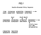
- Figure 1 illustrates a 94 base pair length sequence of human ⁇ -globin desired to be amplified.
- the single base pair change which is associated with sickle cell anemia is depicted beneath the 94-mer.
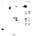
- Figure 2 illustrates a photograph of an ethidium bromide-stained polyacrylamide gel demonstrating amplification of the 94-mer contained in human wild-type DNA and in a plasmid containing a 1.9 kb Bam HI fragment of the normal ⁇ -globin gene (designated pBR328:HbA).
- Figure 3 illustrates a photograph of an ethidium bromide-stained polyacrylamide gel demonstrating amplification of any of the specific target 94-mer sequence present in pBR328:HbA, a plasmid containing a 1.9 kb Bam HI fragment of the sickle cell allele of ⁇ -globin (designated pBR328:HbS), pBR328:HbA where the sequence to be amplified is cleaved with Mst II, and pBR328:HbS where the sequence to be amplified has been treated but not cleaved with Mst II.
- pBR328:HbS plasmid containing a 1.9 kb Bam HI fragment of the sickle cell allele of ⁇ -globin
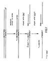
- Figure 4 illustrates in detail the steps and products of the polymerase chain reaction for amplification of the desired 94-mer sequence of human ⁇ -globin for three cycles using two oligonucleotide primers.
- Figure 5 represents a photograph of an ethidium bromide-stained polyacrylamide gel demonstrating amplification after four cycles of a 240-mer sequence in pBR328:HbA, where the aliquots are digested with Nco I (Lane 3), Mst II (Lane 4) or Hin fI (Lane 5). Lane 1 is the molecular weight standard and Lane 2 contains the intact 240-bp product.
- Figure 6 illustrates the sequence of the normal ( ⁇ A ) and sickle cell ( ⁇ S ) ⁇ -globin genes in the region of the Dde I and Hin fI restriction sites, where the single lines for ⁇ A mark the position of the Dde I site (CTGAG) and the double bars for ⁇ A and ⁇ S mark the position of the Hin fI site (GACTC).
- Figure 7 illustrates the results of sequential digestion of normal ⁇ -globin using a 40-mer probe and Dde I followed by Hin fI restriction enzymes.
- Figure 8 illustrates the results of sequential digestion of sickle ⁇ -globin using the same 40-mer probe as in Figure 7 and Dde I followed by Hin fI restriction enzymes.
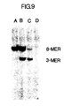
- Figure 9 illustrates a photograph of an ethidium bromide-stained polyacrylamide gel demonstrating the use of the same 40-mer probe as in Figure 7 to specifically characterize the beta-globin alleles present in samples of whole human DNA which have been subjected to amplification, hybridization with the probe, and sequential digestion with Dde I and Hin fI.
- Figure 10 illustrates a photograph of a 6% NuSieve agarose gel visualized using ethidium bromide and UV light. This photograph demonstrates amplification of a sub-fragment of 110-bp amplification product which sub-fragment is an inner nested set within the 110-bp fragment.
- oligonucleotide as used herein in referring to primers, probes, oligomer fragments to be detected, oligomer controls and unlabeled blocking oligomers is defined as a molecule comprised of two or more deoxyribonucleotides or ribonucleotides, preferably more than three. Its exact size will depend on many factors, which in turn depend on the ultimate function or use of the oligonucleotide.
- primer refers to an oligonucleotide whether occurring naturally as in a purified restriction digest or produced synthetically, which is capable of acting as a point of initiation of synthesis when placed under conditions in which synthesis of a primer extension product which is complementary to a nucleic acid strand is induced, i.e., in the presence of nucleotides and an agent for polymerization such as DNA polymerase and at a suitable temperature and pH.
- the primer is preferably single stranded for maximum efficiency in amplification, but may alternatively be double stranded. If double stranded, the primer is first treated to separate its strands before being used to prepare extension products.
- the primer is an oligodeoxyribonucleotide.
- the primer must be sufficiently long to prime the synthesis of extension products in the presence of the agent for polymerization.
- the exact lengths of the primers will depend on many factors, including temperature and source of primer.
- the oligonucleotide primer typically contains 15-25 or more nucleotides, although it may contain fewer nucleotides. Short primer molecules generally require cooler temperatures to form sufficiently stable hybrid complexes with template.
- the primers herein are selected to be “substantially" complementary to the different strands of each specific sequence to be amplified. This means that the primers must be sufficiently complementary to hybridize with their respective strands. Therefore, the primer sequence need not reflect the exact sequence of the template. For example, a non-complementary nucleotide fragment may be attached to the 5' end of the primer, with the remainder of the primer sequence being complementary to the strand. Alternatively, non-complementary bases or longer sequences can be interspersed into the primer, provided that the primer sequence has sufficient complementarity with the sequence of the strand to be amplified to hybridize therewith and thereby form a template for synthesis of the extension product of the other primer.
- restriction endonucleases and “restriction enzymes” refer to bacterial enzymes each of which cut double-stranded DNA at or near a specific nucleotide sequence.
- DNA polymorphism refers to the condition in which two or more different nucleotide sequences can exist at a particular site in DNA.
- restriction fragment length polymorphism refers to the differences among individuals in the lengths of restriction fragments formed by digestion with a particular restriction endonuclease.
- the present invention is directed to a process for amplifying any one or more desired specific nucleic acid sequences suspected of being in a nucleic acid. Because large amounts of a specific sequence may be produced by this process, the present invention may be used for improving the efficiency of cloning DNA or messenger RNA and for amplifying a target sequence to facilitate detection thereof.
- the present process involves a chain reaction for producing, in exponential quantities relative to the number of reaction steps involved, at least one specific nucleic acid sequence given (a) that the ends of the required sequence are known in sufficient detail that oligonucleotides can be synthesized which will hybridize to them, and (b) that a small amount of the sequence is available to initiate the chain reaction.
- the product of the chain reaction will be a discrete nucleic acid duplex with termini corresponding to the ends of the specific primers employed.
- nucleic acid in purified or nonpurified form, can be utilized as the starting nucleic acid or acids, provided it is suspected of containing the specific nucleic acid sequence desired.
- the process may employ, for example, DNA or RNA, including messenger RNA, which DNA or RNA may be single stranded or double stranded.
- a DNA-RNA hybrid which contains one strand of each may be utilized.
- a mixture of any of these nucleic acids may also be employed, or the nucleic acids produced from a previous amplification reaction herein using the same or different primers may be so utilized.
- the specific nucleic acid sequence to be amplified may be only a fraction of a larger molecule or can be present initially as a discrete molecule, so that the specific sequence constitutes the entire nucleic acid. It is not necessary that the sequence to be amplified be present initially in a pure form; it may be a minor fraction of a complex mixture, such as a portion of the ⁇ -globin gene contained in whole human DNA or a portion of nucleic acid sequence due to a particular microorganism which organism might constitute only a very minor fraction of a particular biological sample.
- the starting nucleic acid may contain more than one desired specific nucleic acid sequence which may be the same or different. Therefore, the present process is useful not only for producing large amounts of one specific nucleic acid sequence, but also for amplifying simultaneously more than one different specific nucleic acid sequence located on the same or different nucleic acid molecules.
- the nucleic acid or acids may be obtained from any source, for example, from plasmids such as pBR322, from cloned DNA or RNA, or from natural DNA or RNA from any source, including bacterial, yeast, viruses, and higher organisms such as plants or animals.
- DNA or RNA may be extracted from blood, tissue material such as chorionic villi or amniotic cells by a variety of techniques such as that described by Maniatis et al., Molecular Cloning (1982), 280-281.
- any specific nucleic acid sequence can be produced by the present process. It is only necessary that a sufficient number of bases at both ends of the sequence be known in sufficient detail so that two oligonucleotide primers can be prepared which will hybridize to different strands of the desired sequence and at relative positions along the sequence such that an extension product synthesized from one primer, when it is separated from its template (complement), can serve as a template for extension of the other primer into a nucleic acid of defined length.
- the greater the knowledge about the bases at both ends of the sequence the greater can be the specificity of the primers for the target nucleic acid sequence, and thus the greater the efficiency of the process.
- primer as used hereinafter may refer to more than one primer, particularly in the case where there is some ambiguity in the information regarding the terminal sequence(s) of the fragment to be amplified. For instance, in the case where a nucleic acid sequence is inferred from protein sequence information a collection of primers containing sequences representing all possible codon variations based on degeneracy of the genetic code will be used for each strand. One primer from this collection will be homologous with the end of the desired sequence to be amplified.
- the oligonucleotide primers may be prepared using any suitable method, such as, for example, the phosphotriester and phosphodiester methods described above, or automated embodiments thereof.
- diethylphosphoramidites are used as starting materials and may be synthesized as described by Beaucage et al., Tetrahedron Letters (1981), 22:1859-1862.
- One method for synthesizing oligonucleotides on a modified solid support is described in U.S. Patent No. 4,458,066. It is also possible to use a primer which has been isolated from a biological source (such as a restriction endonuclease digest).
- the specific nucleic acid sequence is produced by using the nucleic acid containing that sequence as a template. If the nucleic acid contains two strands, it is necessary to separate the strands of the nucleic acid before it can be used as the template, either as a separate step or simultaneously with the synthesis of the primer extension products. This strand separation can be accomplished by any suitable denaturing methods including physical, chemical or enzymatic means.
- One physical method of separating the strands of the nucleic acid involves heating the nucleic acid until it is completely (>99%) denatured. Typical heat denaturation may involve temperatures ranging from about 80 to 105°C for times ranging from about 1 to 10 minutes.
- Strand separation may also be induced by an enzyme from the class of enzymes known as helicases or the enzyme RecA, which has helicase activity and in the presence of riboATP is known to denature DNA.
- the reaction conditions suitable for separating the strands of nucleic acids with helicases are described by Kuhn Hoffmann-Berling, CSH-Quantitative Biology , 43 :63 (1978), and techniques for using RecA are reviewed in C. Radding, Ann. Rev. Genetics , 16 :405-37 (1982).
- the original nucleic acid containing the sequence to be amplified is single stranded, its complement is synthesized by adding one or two oligonucleotide primers thereto. If an appropriate single primer is added, a primer extension product is synthesized in the presence of the primer, an agent for polymerization and the four nucleotides described below. The product will be partially complementary to the single-stranded nucleic acid and will hybridize with the nucleic acid strand to form a duplex of unequal length strands that may then be separated into single strands as described above to produce two single separated complementary strands. Alternatively, two appropriate primers may be added to the single-stranded nucleic acid and the reaction carried out.
- the primer extension product(s) produced will be completely complementary to the strands of the original nucleic acid and will hybridize therewith to form a duplex of equal length strands to be separated into single-stranded molecules.
- the strands are ready to be used as a template for the synthesis of additional nucleic acid strands.
- This synthesis can be performed using any suitable method. Generally it occurs in a buffered aqueous solution, preferably at a pH of 7-9, most preferably about 8.
- a molar excess for cloned nucleic acid, usually about 1000:1 primer:template, and for genomic nucleic acid, usually about 106:1 primer:template
- a molar excess for cloned nucleic acid, usually about 1000:1 primer:template, and for genomic nucleic acid, usually about 106:1 primer:template
- the amount of complementary strand may not be known if the process herein is used for diagnostic applications, so that the amount of primer relative to the amount of complementary strand cannot be determined with certainty.
- the amount of primer added will generally be in molar excess over the amount of complementary strand (template) when the sequence to be amplified is contained in a mixture of complicated long-chain nucleic acid strands. A large molar excess is preferred to improve the efficiency of the process.
- the deoxyribonucleoside triphosphates dATP, dCTP, dGTP and TTP are also added to the synthesis mixture in adequate amounts and the resulting solution is heated to about 90-100°C for from about 1 to 10 minutes, preferably from 1 to 4 minutes. After this heating period the solution is allowed to cool to from 20-40°C, which is preferable for the primer hybridization.
- an agent for polymerization To the cooled mixture is added an agent for polymerization, and the reaction is allowed to occur under conditions known in the art. This synthesis reaction may occur at from room temperature up to a temperature above which the agent for polymerization no longer functions efficiently. Thus, for example, if DNA polymerase is used as the agent for polymerization, the temperature is generally no greater than about 45°C.
- DMSO dimethylsulfoxide
- an effective amount e.g., 10% by volume
- DMSO is added to the amplification mixture, and the reaction is carried at 35-40°C, to obtain detectable results or to enable cloning.
- the agent for polymerization may be any compound or system which will function to accomplish the synthesis of primer extension products, including enzymes.
- Suitable enzymes for this purpose include, for example, E. coli DNA polymerase I, Klenow fragment of E. coli DNA polymerase I, T4 DNA polymerase, other available DNA polymerases, reverse transcriptase, and other enzymes, including heat-stable enzymes, which will facilitate combination of the nucleotides in the proper manner to form the primer extension products which are complementary to each nucleic acid strand.
- the synthesis will be initiated at the 3' end of each primer and proceed in the 5' direction along the template strand, until synthesis terminates, producing molecules of different lengths.
- the newly synthesized strand and its complementary nucleic acid strand form a double-stranded molecule which is used in the succeeding steps of the process.
- the strands of the double-stranded molecule are separated using any of the procedures described about to provide single-stranded molecules.
- New nucleic acid is synthesized on the single-stranded molecules. Additional inducing agent, nucleotides and primers may be added if necessary for the reaction to proceed under the conditions prescribed above. Again, the synthesis will be initiated at one end of the oligonucleotide primers and will proceed along the single strands of the template to produce additional nucleic acid. After this step, half of the extension product will consist of the specific nucleic acid sequence bounded by the two primers.
- the steps of strand separation and extension product synthesis can be repeated as often as needed to produce the desired quantity of the specific nucleic acid sequence. As will be described in further detail below, the amount of the specific nucleic acid sequence produced will accumulate in an exponential fashion.
- the appropriate number of different oligonucleotide primers are utilized. For example, if two different specific nucleic acid sequences are to be produced, four primers are utilized. Two of the primers are specific for one of the specific nucleic acid sequences and the other two primers are specific for the second specific nucleic acid sequence. In this manner, each of the two different specific sequences can be produced exponentially by the present process.
- the present invention can be performed in a step-wise fashion where after each step new reagents are added, or simultaneously, where all reagents are added at the initial step, or partially step-wise and partially simultaneous, where fresh reagent is added after a given number of steps.
- a method of strand separation such as heat, is employed which will inactivate the agent for polymerization, as in the case of a heat-labile enzyme, then it is necessary to replenish the agent for polymerization after every strand separation step.
- the simultaneous method may be utilized when a number of purified components, including an enzymatic means such as helicase, is used for the strand separation step.
- the reaction mixture may contain, in addition to the nucleic acid strand(s) containing the desired sequence, the strand-separating enzyme (e.g., helicase), an appropriate energy source for the strand-separating enzyme, such as rATP, the four nucleotides, the oligonucleotide primers in molar excess, and the inducing agent, e.g., Klenow fragment of E. coli DNA polymerase I.
- the strand-separating enzyme e.g., helicase
- an appropriate energy source for the strand-separating enzyme such as rATP
- the four nucleotides such as rATP
- the oligonucleotide primers in molar excess
- the inducing agent e.g., Klenow fragment of E. coli DNA polymerase I.
- a heat-stable inducing agent such as a thermostable polymerase may be employed which will operate at an elevated temperature, preferably 65-90°C depending on the inducing agent, at which temperature the nucleic acid will consist of single and double strands in equilibrium. For smaller lengths of nucleic acid, lower temperatures of about 50°C may be employed. The upper temperature will depend on the temperature at which the enzyme will degrade or the temperature above which an insufficient level of primer hybridization will occur.
- a heat-stable enzyme is described, e.g., by A. S. Kaledin et al., Biokhimiya , 45 , 644-651 (1980).
- reaction may be halted by inactivating the enzymes in any known manner or separating the components of the reaction.
- the process of the present invention may be conducted continuously.
- the reaction may be cycled through a denaturing region, a reagent addition region, and a reaction region.
- the enzyme used for the synthesis of primer extension products can be immobilized in a column.
- the other reaction components can be continuously circulated by a pump through the column and a heating coil in series, thus the nucleic acids produced can be repeatedly denatured without inactivating the enzyme.
- the long products thus produced will act as templates for one or the other of the oligonucleotide primers during subsequent cycles and will produce molecules of the desired sequence [S+] or [S ⁇ ] These molecules will also function as templates for one or the other of the oligonucleotide primers, producing further [S+] and [S ⁇ ], and thus a chain reaction can be sustained which will result in the accumulation of [S] at an exponential rate relative to the number of cycles.
- [S] The specific sequence to be amplified, [S], can be depicted diagrammatically as:
- the appropriate oligonucleotide primers would be: so that if DNA containing [S] is separated into single strands and its single strands are hybridized to Primers 1 and 2, the following extension reactions can be catalyzed by DNA polymerase in the presence of the four deoxyribonucleoside triphosphates: On denaturation of the two duplexes formed, the products are: If these four strands are allowed to rehybridize with Primers 1 and 2 in the next cycle, agent for polymerization will catalyze the following reactions: If the strands of the above four duplexes are separated, the following strands are found:
- each strand which terminates with the oligonucleotide sequence of one primer and the complementary sequence of the other is the specific nucleic acid sequence [S] that is desired to be produced.
- the steps of this process can be repeated indefinitely, being limited only by the amount of Primers 1 and 2, the agent for polymerization and nucleotides present.
- the number of cycles used is that required to produce a detectable signal, an amount which will depend, e.g., on the nature of the sample. For example, if the sample is pure or diluted, fewer cycles may be required than if it is a complex mixture. If the sample is human genomic DNA, preferably the number of cycles is from about 10-30.
- the amount of original nucleic acid remains constant in the entire process, because it is not replicated.
- the amount of the long products increases linearly because they are produced only from the original nucleic acid.
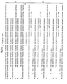
- the amount of the specific sequence increases exponentially. Thus, the specific sequence will become the predominant species. This is illustrated in the following table, which indicates the relative amounts of the species theoretically present after n cycles, assuming 100% efficiency at each cycle: When a single-stranded nucleic acid is utilized as the template, only one long product is formed per cycle.
- the method herein may be utilized to clone a particular nucleic acid sequence for insertion into a suitable expression vector.
- the vector may then be used to transform an appropriate host organism to produce the gene product of the sequence by standard methods of recombinant DNA technology.
- the amplification process herein may yield a mixture of nucleic acids, resulting from the original template nucleic acid, the expected target amplified products, and various background non-target products.
- the amplified product can also be a mixture if the original template DNA contains multiple target sequences, such as in a heterozygous diploid genome or when there is a family of related genes.
- the primers herein may be modified to assist the rapid and specific cloning of the mixture of DNAs produced by the amplification reaction.
- the same or different restriction sites are incorporated at the 5' ends of the primers to result in restriction sites at the two ends of the amplified product.
- the amplified product can then be easily inserted into plasmid or vial vectors and cloned. This cloning allows the analysis or expression of individual amplified products, not a mixture.
- the same restriction site can be used for both primers, the use of different sites allows the insertion of the product into the vector with a specific orientation and suppresses multiple insertions as well as insertions arising from amplifications based on only one of the two primers.
- the specific orientation is useful when cloning into single-strand sequencing vectors, when single-strand hybridization probes are used, or when the cloned product is being expressed.
- One method to prepare the primers is to choose a primer sequence which differs minimally from the target sequence. Regions in which each of the primers is to be located are screened for homology to restriction sites appropriate to the desired vector. For example, the target sequence "CAGTATCCGA" differs by only one base from one containing a Bam HI site. A primer sequence is chosen to match the target exactly at its 3' end, and to contain the altered sequence and restriction site near its 5' end (for example, CAGgATCCGA, where the lower case letter symbolizes a mismatch with the target sequence). This minimally altered sequence will not interfere with the ability of the primer to hybridize to the original target sequence and to initiate polymerization.
- the primer is copied, becomes the target, and matches exactly with new primers.
- the products are cleaved with the appropriate restriction enzymes, optionally separated from inhibitors of ligation such as the nucleotide triphosphates and salts by passing over a desalting column or molecular weight chromatography column, and inserted by ligation into a cloning vector such as bacteriophage M13.
- the gene may then be sequenced and/or expressed using well known techniques.
- the second method for preparing the primers involves taking the 3' end of the primers from the target sequence and adding the desired restriction site(s) to the 5' end of the primer.
- a Hin dIII site could be added to make the sequence "cgaagcttCAGTATCCGA", where lower case letters are as described above.
- the added bases would not contribute to the hybridization in the first cycle of amplification, but would match in subsequent cycles.
- the final amplified products are then cut with restriction enzyme(s) and cloned and expressed as described above.
- the gene being amplified may be, for example, human beta-hemoglobin or the human HLA DQ, DR or DP- ⁇ and - ⁇ genes.
- the process herein can be used for in vitro mutagenesis.
- the oligodeoxyribonucleotide primers need not be exactly complementary to the DNA sequence which is being amplified. It is only necessary that they be able to hybridize to the sequence sufficiently well to be extended by the polymerase enzyme or by whatever other inducing agent is employed.
- the product of a polymerase chain reaction wherein the primers employed are not exactly complementary to the original template will contain the sequence of the primer rather than the template, thereby introducing an in vitro mutation. In further cycles this mutation will be amplified with an undiminished efficiency because no further mispaired primings are required.
- the mutant thus produced may be inserted into an appropriate vector by standard molecular biological techniques and might confer mutant properties on this vector such as the potential for production of an altered protein.
- the primer can contain as part of its sequence a non-complementary sequence provided that a sufficient amount of the primer contains a sequence which is complementary to the strand to be amplified.
- a nucleotide sequence which is not complementary to the template sequence such as, e.g., a promoter, linker, coding sequence, etc.
- a nucleotide sequence which is not complementary to the template sequence such as, e.g., a promoter, linker, coding sequence, etc.
- sufficient cycles are run to achieve the desired amount of new template containing the non-complementary nucleotide insert. This allows production of large quantities of the combined fragments in a relatively short period of time (e.g., two hours or less) using a simple technique.
- the process herein may be used to synthesize a nucleic acid fragment from an existing nucleic acid fragment which is shorter than its product (called the core segment) using certain primers the 3' ends of which are complementary to or substantially complementary to the 3' ends of the single strands produced by separating the strands of the original shorter nucleic acid fragments, and the 5' ends of which primers contain sequence information to be appended to the core segment.
- This process comprises:
- Steps (b) and (c) are repeated as often as necessary, usually at least 5 times, to produce the required amount of the full-length double-stranded product to synthesize the final product (i.e., the effective amount).
- the core segment may be obtained as the product of a previous amplification cycle.
- the product produced in step (d) may be purified before a new cycle of extension and amplification, or used directly by employing the reaction mixture containing the product.
- mutants of the original nucleic acid may be made by using primers which are substantially complementary at their 3' ends to the 3' ends of the single strands of the original shorter nucleic acid.
- the amplified double-stranded products can be digested with the appropriate restriction enzymes and ligated directly into an M13 vector for rapid cloning and sequencing.
- the M13 plaques containing the specific amplified target sequences can be identified by hybridizing plaque lift filters with a probe specific for the target sequence.
- the method herein may also be used to enable detection and/or characterization of specific nucleic acid sequences associated with infectious diseases, genetic disorders or cellular disorders such as cancer, e.g., oncogenes.
- Amplification is useful when the amount of nucleic acid available for analysis is very small, as, for example, in the prenatal diagnosis of sickle cell anemia using DNA obtained from fetal cells. Amplification is particularly useful if such an analysis is to be done on a small sample using non-radioactive detection techniques which may be inherently insensitive, or where radioactive techniques are being employed but where rapid detection is desirable.
- genetic diseases may include specific deletions and/or mutations in genomic DNA from any organism, such as, e.g., sickle cell anemia, cystic fibrosis, ⁇ -thalassemia, ⁇ -thalassemia, and the like.
- Sickle cell anemia can be readily detected via oligomer restriction analysis or a RFLP-like analysis following amplification of the appropriate DNA sequence by the present method.
- ⁇ -Thalassemia can be detected by the absence of a sequence
- ⁇ -thalassemia can be detected by the presence of a polymorphic restriction site closely linked to a mutation which causes the disease.
- All of these genetic diseases may be detected by amplifying the appropriate sequence and analyzing it by Southern blots without using radioactive probes.
- a small sample of DNA from, e.g., amniotic fluid containing a very low level of the desired sequence is amplified, cut with a restriction enzyme, and analyzed via a Southern blotting technique.
- the use of non-radioactive probes is facilitated by the high level of the amplified signal.
- a small sample of DNA may be amplified to a convenient level and then a further cycle of extension reactions performed wherein nucleotide derivatives which are readily detectable (such as 32 ⁇ -labeled or biotin labeled nucleoside triphosphates) are incorporated directly into the final DNA product, which may be analyzed by restriction and electrophoretic separation or any other appropriate method.
- nucleotide derivatives which are readily detectable such as 32 ⁇ -labeled or biotin labeled nucleoside triphosphates
- the nucleic acid may be exposed to a particular restriction endonuclease prior to amplification. Since a sequence which has been cut cannot be amplified, the appearance of an amplified fragment, despite prior restriction of the DNA sample, implies the absence of a site for the endonuclease within the amplified sequence. The presence or absence of an amplified sequence can be detected by an appropriate method.
- Sickle cell anemia is a hemoglobin disease which is caused by a single base pair change in the sixth codon of the ⁇ -globin gene.
- Figure 6 illustrates the sequences of normal and sickle cell ⁇ -globin genes in the region of their polymorphism, where the single bars mark the location of a Dde I site present only in the normal gene and where the double bars mark the location of a Hin fI site which is non-polymorphic and thus present in both the normal and sickle cell alleles.
- Figure 7 illustrates the process of oligomer restriction of normal ⁇ -globin DNA using a probe spanning both restriction sites and labeled where the asterisk appears.
- the probe is preferably labeled at the end which is fewer base pairs from the restriction site than the other end of the probe.
- the DNA amplified as provided herein, is denatured and annealed to the labeled probe.
- the amplification may be carried out at elevated temperatures (35-40°C) in the presence of dimethyl sulfoxide to minimize formation of secondary structure.
- the enzyme Dde I cleaves the DNA at the reformed Dde I site and generates a labeled octamer.
- FIG. 8 illustrates the same process applied to the sickle cell allele of ⁇ -globin DNA.
- the enzyme Dde I cannot cleave the duplex formed by the amplified DNA and the labeled probe because of the A-A base pair mismatch.
- the enzyme Hin fI does restrict the hybrid and a labeled trimer is produced.
- the method can diagnose the DNA of an individual as being either homozygous for the wild type, homozygous for the sickle type or a heterozygous carrier of the sickle cell trait, since a specific signal is associated with the presence of either allele.
- Use of this above-described method to amplify the pertinent sequence allows for a rapid analysis or a single copy gene using a probe with only a single 32 ⁇ label.
- Various infectious diseases can be diagnosed by the presence in clinical samples of specific DNA sequences characteristic of the causative microorganism. These include bacteria, such as Salmonella, Chlamydia, Neisseria; viruses, such as the hepatitis viruses, and parasites, such as the Plasmodium responsible for malaria.
- U.S. Patent 4,358,535 issued to Falkow describes the use of specific DNA hybridization probes for the diagnosis of infectious diseases.
- a problem inherent in the Falkow procedure is that a relatively small number of pathogenic organisms may be present in a clinical sample from an infected patient and the DNA extracted from these may constitute only a very small fraction of the total DNA in the sample. Specific amplification of suspected sequences prior to immobilization and hybridization detection of the DNA samples could greatly improve the sensitivity and specificity of these procedures.
- the probe may be a biotinylated probe in which the biotin is attached to a spacer arm of the formula: where Y is 0, NH or N-CHO, x is a number from 1 to 4, and y is a number from 2 to 4.
- the spacer arm is in turn attached to a psoralen moiety of the formula: The psoralen moiety intercalates into and crosslinks a "gapped circle" probe as described by Courage-Tebbe et al., Biochim. Biophys. Acta , 697 (1982) 1-5, wherein the single-stranded hybridization region of the gapped circle spans the region contained in the primers.
- the amplification process can also be utilized to produce sufficient quantities of DNA from a single copy human gene such that detection by a simple non-specific DNA stain such as ethidium bromide can be employed so as to make a DNA diagnosis directly.
- a simple non-specific DNA stain such as ethidium bromide
- the process herein can also be used to detect DNA polymorphism which may not be associated with any pathological state.
- a 25 base pair sequence having the nucleotide sequence contained on a 47 base pair Fok I restriction fragment of pBR322 obtainable from ATCC was prepared as follows.
- a Fok I digest of pBR322 containing the 47-bp fragment was produced by digesting pBR322 with Fok I in accordance with the conditions suggested by the supplier, New England Biolabs Inc.
- the primers which were utilized were 5' d(CCTCGGCACCG) 3' and 5' d(AGCATCCAGGGTG) 3', and were prepared using conventional techniques.
- the mixture was heated to 85°C for five minutes and allowed to cool to ambient temperature. Five units of the Klenow fragment of E. coli DNA polymerase 1 were added and the temperature was maintained for 15 minutes. After that time, the mixture was again heated to 85°C for five minutes and allowed to cool. Five units of the Klenow fragment were again added and the reaction was carried out for 15 minutes. The heating, cooling and synthesis steps were repeated eleven more times.
- the desired sequence to be amplified was a 94 base pair sequence contained within the human beta-globin gene and spanning the Mst II site involved in sickle cell anemia.
- the sequence has the nucleotide sequence shown in Figure 1.
- oligodeoxyribonucleotide primers were prepared by the method described below: 5' CACAGGGCAGTAACG 3' Primer A and 5' TTTGCTTCTGACACA 3' Primer B
- Oligodeoxyribonucleotide Deprotection and Purification Procedures The solid support was removed from the column and exposed to 1 ml concentrated ammonium hydroxide at room temperature for four hours in a closed tube. The support was then removed by filtration and the solution containing the partially protected oligodeoxynucleotide was brought to 55°C for five hours. Ammonia was removed and the residue was applied to a preparative polyacrylamide gel. Electrophoresis was carried out at 30 volts/cm for 90 minutes after which the band containing the product was identified by UV shadowing of a fluorescent plate. The band was excised and eluted with 1 ml distilled water overnight at 4°C.
- Oligodeoxyribonucleotides Test aliquots of the purified oligonucleotides were 32 ⁇ labeled with polynucleotide kinase and ⁇ -32 ⁇ -ATP. The labeled compounds were examined by autoradiography of 14-20% polyacrylamide gels after electrophoresis for 45 minutes at 50 volts/cm. This procedure verifies the molecular weight.
- Base composition was determined by digestion of the oligodeoxyribonucleotide to nucleosides by use of venom diesterase and bacterial alkaline phosphatase and subsequent separation and quantitation of the derived nucleosides using a reverse phase HPLC column and a 10% acetonitrile, 1% ammonium acetate mobile phase.
- Human genomic DNA homozygous for normal ⁇ -globin was extracted from the cell line Molt4 (obtained from Human Genetic Mutant Cell Repository and identified as Gm2219c) using the technique described by Stetler et al., Proc. Nat. Acad. Sci. (1982), 79:5966-5970.
- a 1.9 kb Bam HI fragment of the normal ⁇ -globin gene was isolated from the cosmid pFCII and inserted into the Bam HI site of pBR328 (Soberon, et al., Gene (1980) 9 :287-305).
- This fragment which encompasses the region that hybridizes to the synthetic 40-mer probe, includes the first and second exons, first intron, and 5' flanking sequences of the gene (Lawn et al., Cell (1978), 15:1157-1174).
- This clone was designated pBR328:HbA and deposited under ATCC No. 39,698 on May 25, 1984.
- the corresponding 1.9 kb Bam HI fragment of the sickle cell allele of ⁇ -globin was isolated from the cosmid pFC12 and cloned as described above. This clone was designated pBR328:HbS and deposited under ATCC No. 39,699 on May 25, 1984.
- Each recombinant plasmid was transformed into and propagated in E. coli MM294 (ATCC No. 39,607).
- a total of 100 ⁇ g each of pBR328:HbA and pBR328:HbS were individually digested with 20 units of Mst II (New England Biolabs) for 16 hours at 37°C in 200 ⁇ l of 150 mM NaCl, 12 mM Tris HCl (pH 7.5), 12 mM MgCl2, 1 mM dithiothreitol (DTT), and 100 ⁇ g/ml bovine serum albumin (BSA).
- the products are designated pBR328:HbA/ Mst II and pBR328:HbS/ Mst II, respectively.
- Lane 1 is 0.1 picomole of a 58-bp synthetic fragment control one strand of which is complementary to the above probe.
- Lane 2 is 4 ⁇ l of Reaction I prior to the first amplification cycle.
- Lane 3 is 4 ⁇ l of Reaction I after the 20th amplification cycle.
- Lane 4 is 4 ⁇ l of Reaction II after five amplification cycles.
- Lane 5 is a molecular weight standard consisting of a Fok I (New England Biolabs) digest of pBR322 (New England Biolabs) labeled with alpha-32 ⁇ -dNTPs and polymerase.
- Lane 3 shows that after twenty cycles the reaction mixture I contained a large amount of the specific sequence of the proper molecular weight and no other detectable products.
- Reaction mixture II after five cycles also contained this product, as well as the starting nucleic acid and other products, as shown by Lane 4.
- Figure 3 is an autoradiograph of the electrophoresis.
- Lane 1 is a molecular weight standard
- Lane 2 is Reaction II
- Lane 3 is Reaction III
- Lane 4 is Reaction IV
- Lane 5 is Reaction V.
- Another lane for Reaction VI with no DNA as control had no images in any of the lanes. It can be seen from the figure that the 94-bp fragment predicted from the target DNA was present only where intact ⁇ -globin DNA sequences were available for amplification, i.e., pBR328:HbA (Lane 2), pBR328:HbS (Lane 3) and pBR328:HbS/ Mst II (Lane 5).
- Mst II digestion cuts pBR328:HbA in the 94-mer sequence rendering it incapable of being amplified, and the 94-mer band does not appear in Lane 4.
- the 94-mer sequence in pBR328:HbS does not cut when the plasmid is digested with Mst II and thus is available for amplification as shown in Lane 5.
- Figure 4 illustrates the chain reaction for three cycles in amplifying the 94-bp sequence.
- PCO1 and PCO2 are Primers A and B. The numbers on the right indicate the cycles, whereas the numbers on the left indicates the cycle number in which a particular molecule was produced.
- This example illustrates amplification of 110 bp sequence spanning the allelic Mst II site in the human hemoglobin gene.
- This example illustrates amplification of a 240 bp sequence spanning the allelic Mst II site in the human hemoglobin gene. This sequence contains Nco I, Hin fI and Mst II restriction sites.
- reaction was allowed to proceed for 10 minutes after which the cycle of solution A addition, heating, cooling, adding polymerase, and reacting was repeated three times.
- To a 5.0 ⁇ l aliquot of the reactions was added 5 picomoles of each oligonucleotide primer described above.
- the solution was heated to 100°C for four minutes and allowed to come to ambient temperature, after which 3 picomoles each of the alpha-32 ⁇ -labeled deoxyribonucleoside triphosphates and 4 units Klenow fragment were added.
- the reaction in a final volume of 10 ⁇ l and at the salt concentrations given above, was allowed to proceed for 10 minutes.
- the polymerase activity was terminated by heating for 20 minutes at 60°C.
- FIG. 5 illustrates the autoradiograph of the electrophoresis, where Lane 1 is the molecular weight standard, Lane 2 is without digestion with enzyme (240 bp intact), Lane 3 is digestion with Nco I (131 and 109 bp), Lane4 is digestion with Mst II (149 and 91 bp), and Lane 5 is digestion with Hin fI (144 and 96 bp). The autoradiograph is consistent with the amplification of the 240 bp sequence.
- This example illustrates use of the process herein to detect sickle cell anemia by sequential digestion.
- a labeled DNA probe, RS06, of the sequence where * indicates the label, and an unlabeled blocking oligomer, RS10, of the sequence which has three base pair mismatches with RS06 were synthesized according to the procedures provided in Example 2(1)
- the probe RS06 was labeled by contacting five pmole thereof with 4 units of T4 polynucleotide kinase (New England Biolabs) and 50 pmole ⁇ 32 ⁇ -ATP (New England Nuclear, about 7200 Ci/mmole) in a 40 ⁇ l reaction volume containing 70 mM Tris buffer (pH 7.6), 10 mM MgCl2, 1.5 mM spermine, and 2.5 mM dithiothreitol for 90 minutes at 37°C.
- the total volume was then adjusted to 100 ⁇ l with 25 mM EDTA and purified according to the procedure of Maniatis et al., Molecular Cloning (1982), 464-465 over a 1 ml Bio Gel P-4 spin dialysis column from BioRad equilibrated with Tris-EDTA (TE) buffer (10 mM Tris buffer, 0.1 mM EDTA, pH 8.0).
- TE Tris-EDTA
- the labeled probe was further purified by electrophoresis on a 18% polyacrylamide gel (19:1 acrylamide:BIS, BioRad) in Tris-boric acid-EDTA (TBE) buffer (89 mM Tris, 89 mM boric acid, 2.5 mM EDTA, pH 8.3) for 500 vhr. After localization by autoradiography, the portion of the gel containing the labeled probe was excised, crushed and eluted into 0.2 ml TE buffer overnight at 4°C. TCA precipitation of the reaction product indicated that the specific activity was 4.9 Ci/mmole and the final concentration was 20 pmole/ml.
- the unlabeled RS10 blocking oligomer was used at a concentration of 200 pmole/ml.
- High molecular weight genomic DNA was isolated from the lymphoid cell lines Molt4, SC-1 and Gm2064 using essentially the method of Stetler et al., PNAS (1982), 79 , 5966-5970 (for Molt4) and Maniatis et al., Molecular Cloning (1982), 280-281.
- Molt4 (Human Mutant Cell Repository, GM2219C) is a T cell line homozygous for normal ⁇ -globin, and SC-1, deposited with ATCC on March 19, 1985, is an EBV-transformed B cell line homozygous for the sickle cell allele.
- GM2064 Human Mutant Cell Repository, GM2064 was originally isolated from an individual homozygous for hereditary persistance of fetal hemoglobin (HPFH) and contains no beta- or delta-globin gene sequences. All cell lines were maintained in RPMI-1640 with 10% fetal calf serum.
- a clinical blood sample designated CH12 from a known sickle cell carrier (AS) was obtained from Dr. Bertram Lubin of Children's Hospital in Oakland, California.
- Genomic DNA was prepared from the buffy coat fraction, which is composed primarily of peripheral blood lymphocytes, using a modification of the procedure described by Nunberg et al., Proc. Nat. Acad. Sci. , 75 , 5553-5556 (1978).
- the cells were resuspended in 5 ml Tris-EDTA-NaCl (TEN) buffer (10 mM Tris buffer pH 8, 1 mM EDTA, 10 mM NaCl) and adjusted to 0.2 mg/ml proteinase K, 0.5% SDS, and incubated overnight at 37°C. Sodium perchlorate was then added to 0.7 M and the lysate gently shaken for 1-2 hours at room temperature. The lysate was extracted with 30 ml phenol/chloroform (1:1), then with 30 ml chloroform, and followed by ethanol precipitation of the nucleic acids. The pellet was resuspended in 2 ml of TE buffer and RNase A added to 0.005 mg/ml.
- TEN Tris-EDTA-NaCl
- the DNA was extracted once each with equal volumes of phenol, phenol/chloroform, and chloroform, and ethanol precipitated.
- the DNA was resuspended in 0.5 ml TE buffer and the concentration was determined by absorbance at 260 nm.
- genomic DNA Two micrograms of genomic DNA was amplified in an initial 100 ⁇ l reaction volume containing 10 mM Tris buffer (pH 7.5), 50 mM NaCl, 10 mM MgCl2, 150 pmole of Primer A of the sequence d(CACAGGGCACTAACG), and 150 pmole of Primer B of the sequence d(CTTTGCTTCTGACACA) and overlayed with about 100 ⁇ l mineral oil to prevent evaporation.
- reaction was terminated by heating at 95°C for two minutes.
- the mineral oil was extracted with 0.2 ml of chloroform and discarded.
- the final reaction volume was 130 ⁇ l.
- the mineral oil was extracted with 0.2 ml chloroform, and 18 ⁇ l of the reaction mixture (45 ng genomic DNA) was loaded onto a 30% polyacrylamide mini-gel (19:1, Bio Rad) in a Hoeffer SE200 apparatus.
- the gel was electrophoresed at approximately 300 volts for one hour until the bromphenol blue dye front migrated to 3.0 cm off-origin.
- the top 1.5 cm of the gel was removed and the remaining gel was exposed for four days with one intensification screen at -70°C.
- Lane A contains Molt4 DNA
- Lane B CH12
- Lane C SC-1
- Lane D GM2064.
- Molt4 represents the genotype of a normal individual with two copies of the ⁇ A gene per cell (AA)
- CH12 is a clinical sample from a sickle cell carrier with one ⁇ A and one ⁇ S gene per cell (AS)
- SC-1 represents the genotype of a sickle cell individual with two copies of the ⁇ S gene per cell (SS).
- GM2064 which contains no beta- or delta-globin sequences, is present as a negative control.
- the Dde I-cleaved, ⁇ A -specific octamer is present only in those DNA's containing the ⁇ A gene (Lanes A and B) and the Hin fI-cleaved, ⁇ S -specific trimer is present only in those DNA's containing the ⁇ S (Lanes B and C).
- the presence of both trimer and octamer is diagnostic for a sickle cell carrier and is distinguishable from a normal individual (Lane A) with only octamer and a sickle cell afflicted individual (Lane C) with only trimer.
- This example illustrates direct detection of a totally unpurified single copy gene in whole human DNA on gels without the need for a labeled probe.
- Example 3 Using the technique described in Example 3, a 110-bp fragment from a sequence in the first exon of the beta-globin gene was amplified from 10 micrograms of whole human DNA after 20 cycles. This 110-bp fragment produced after 20 cycles was easily visualized on gels stained with ethidium bromide.
- the sequence was not amplified when it was first cut with the restriction enzyme Dde I unless, as in the beta-globin S allele, the sequence does not contain the restriction site recognized by the enzyme.
- This example illustrates the use of different primers to amplify various fragments of pBR328 and 322.
- This example illustrates the invention process wherein an in vitro mutation is introduced into the amplified segment.
- a primer which is not a perfect match to the template sequence but which is nonetheless able to hybridize sufficiently to be enzymatically extended produces a long product which contains the sequence of the primer rather than the corresponding sequence of the original template.
- the long product serves as a template for the second primer to introduce an in vitro mutation. In further cycles this mutation is amplified with an undiminished efficiency, because no further mispaired primings are required.
- a primer which carries a non-complementary extension on its 5' end was used to insert a new sequence in the product adjacent to the template sequence being copied.
- This example illustrates employing nested sets of primers to decrease the background in the amplification of single copy genes.
- a ten- ⁇ l aliquot was removed from the reaction mixture and subjected to a further ten cycles of amplification using each of the primers: which amplify a 58-bp fragment contained within the 110-bp fragment produced above.
- This final ten cycles of amplification was accomplished by diluting the 10- ⁇ l aliquot into 90 ⁇ l of the fresh Tris-acetate buffer described above containing 100 nanomoles each dNTP and 200 pmoles of each primer. Reaction conditions were as above. After ten cycles a 10- ⁇ l aliquot (corresponding to 100 nanograms of the original DNA) was applied to a 6% NuSieve (FMC Corp.) agarose gel and visualized using ethidium bromide.
- Figure 10 illustrates this gel illuminated with UV light and photographed through a red filter as is known in the art.
- Lane 1 is molecular weight markers.
- Lane 2 is an aliquot of the reaction described above.
- Lane 3 is an aliquot of a reaction identical to that described above, except that the original wild-type DNA was cleaved with Dde I prior to amplification.
- Lane 4 is an aliquot of a reaction identical to that described above, except that human DNA homozygous for the sickle betaglobin allele was treated with Dde I prior to amplification (the sickle allele does not contain a Dde I site in the fragment being amplified here).
- Lane 5 is an aliquot of a reaction identical to that described above, except that salmon sperm DNA was substituted for human DNA.
- Lane 6 is an aliquot of a reaction identical to that described above, except that the aliquot was treated with Dde I after amplification ( Dde I should convert the 58-bp wild-type product into 27-and 31-bp fragments).
- Lane 7 is an aliquot of the Lane 4 material treated with Dde I after amplification (the 58-bp sickle product contains no Dde I site).
- Detection of a 58-bp fragment representative of a single-copy gene from one microgram of human DNA using only ethidium bromide staining of an agarose gel requires an amplification of about 500,000-fold. This was accomplished by using the two nested sets of oligonucleotide primers herein. The first set amplifies the 110-bp fragment and the inner nested set amplifies a sub-fragment of this product up to the level of convenient detection shown in Figure 10.
- This procedure of using primers amplifying a smaller sequence contained within the sequence being amplified in the previous amplification process and contained in the extension products of the other primers allows one to distinguish the wild-type from the sickle allele at the beta-globin locus without resorting to either radioisotopic or non-radioisotopic probe hybridization methodology such as that of Conner et al., PNAS (USA), 80 :278 (1983) and Leary et al., PNAS (USA), 80 :4045 (1983).
- the present process is expected to be useful in detecting, in a patient DNA sample, a specific sequence associated with an infectious disease such as, e.g., Chlamydia using a biotinylated hybridization probe spanning the desired amplified sequence and using the process described in U.S. 4,358,535, supra .
- the biotinylated hybridization probe may be prepared by intercalation and irradiation of a partially double-stranded DNA with a 4'-methylene substituted 4,5'-8-trimethylpsoralen attached to biotin via a spacer arm of the formula: where Y is O, NH or N-CHO, x is a number from 1 to 4, and y is a number from 2 to 4.
- Detection of the biotinyl groups on the probe may be accomplished using a streptavidin-acid phosphatase complex commercially obtainable from Enzo Biochem Inc. using the detection procedures suggested by the manufacturer in its brochure.
- the hybridized probe is seen as a spot of precipitated stain due to the binding of the detection complex, and the subsequent reaction catalyzed by acid phosphatase, which produces a precipitable dye.
- Example 7 the process of Example 7 was basically used to amplify a 119 base pair fragment on the human ⁇ -hemoglobin gene using the primers: where lower case letters denote mismatches from wild-type sequence to create restriction enzyme sites.
- Table I illustrates a diagram of the primers GH18 and GH19 which are used for cloning and sequencing a 119-base pair fragment of the human ⁇ -globin gene and which are designed to contain internal restriction sites.
- the start codon ATG is underlined.
- GH18 is a 26-base oligonucleotide complementary to the negative strand and contains an internal Pst I site.
- GH19 is a 23-base oligonucleotide complementary to the plus strand and contains an internal Hind III recognition sequence. Arrows indicate the direction of extension by DNA polymerase I. The boxed sequences indicate the restriction enzyme recognition sequences of each primer. These primers were selected by first screening the regions of the gene for homology to the Pst I and Hind III restriction sites of bacteriophage M13. The primers were then prepared as described in previous examples.
- the amplified product was ethanol precipitated to desalt and concentrate the sample, redissolved in a restriction buffer of 10 mM Tris pH 8, 10 mM MgCl2, 1 mM DTT, 100 mM NaCl2, and simultaneously digested with Pst I and Hind III. After digestion the sample was desalted with a Centricon 10 concentrator and ligated overnight at 12°C with 0.3 micrograms of the Pst I/ Hind III digested vector M13mp10w, which is publicly available from Boehringer-Mannheim.
- E. coli strain JM103 which is publicly available from BRL in Bethesda, MD.
- the procedure followed for preparing the transformed strain is described in Messing, J. (1981) Third Cleveland Symposium on Macro-molecules:Recombinant DNA , ed. A. Walton, Elsevier, Amsterdam, 143-153.
- the transformation mixture was plated onto x-gal media for screening via plaque hybridization with nylon filters.
- the filters were probed with a ⁇ -globin-specific oligonucleotide probe RS24 of the sequence 5'-CCCACAGGGCAGTAACGGCAGACTTCTCCTCAGGAGTCAG-3' to determine the number of ⁇ -globin inserts.
- the filters were then reprobed with the primer PC04 to determine the total number of inserts.
- Table II summarizes the plating and plaque hybridization data.
- the filters were probed with the primer PC04 to determine the percentage of inserts resulting from amplification and cloning: 1206 clear plaques (90% of total number of clear plaques) hybridized to the primer. Fifteen plaques hybridized to the ⁇ -globin specific probe RS24. The percentage of ⁇ -globin positive plaques among the amplified primer-positive plaques is approximately 1%.
- DNA from phage DNA minipreparation of three ⁇ -globin positive and two ⁇ -globin negative (but PC04 primer positive) plaques were analyzed by restriction enzyme analysis. Mst II digestion of DNA from M13 clones containing the amplified ⁇ -globin fragment should generate a characteristic 283 base-pair fragment. Following Mst II digestion, the three ⁇ -globin positive clones all produced the predicted 283 base pair fragment, while the two clones which were positive only with the primer produced larger fragments.
- the gel from this analysis was transferred to a MSI nylon filter and hybridized with a radiolabeled nick-translated ⁇ -globin probe prepared by standard nick translation methods as described by Rigby et al., J. Mol. Biol. (1977), 113 :237-51.
- the only bands which hybridized to the ⁇ -globin probe were the three ⁇ -globin positive clones. The two other clones had inserts which did not hybridize to the ⁇ -globin probe.
- the modified linker primers were nearly as efficient as the unmodified primers in amplifying the ⁇ -globin sequence.
- the primers were able to facilitate insertion of amplified DNA into cloning vectors. Due to the amplification of other segments of the genome, only 1% of the clones contained hemoglobin sequences.
- Restriction site-modified primers were also used to amplify and clone and partially sequence the human N-ras oncogene and to clone 240-base pair segments of the HLA DQ- ⁇ and DQ- ⁇ genes. All of these amplifications were carried out in the presence of 10% by volume dimethylsulfoxide at 37°C.
- the primers for amplifying HLA DQ- ⁇ and DQ- ⁇ genes were much more specific for their intended targets than were the ⁇ -globin and DR- ⁇ primers, which, rather than giving a discrete band on an ethidium bromide stained agarose gel, produced only a smear.
- the HLA DQ- ⁇ primers produced up to 20% of clones, with amplified inserts which contained the desired HLA target fragment, whereas 1% of the ⁇ -globin clones contained the target sequence.
- the HLA DQ- ⁇ and DQ- ⁇ gene cloning was only effective when the DMSO was present and the temperature was elevated.
- This example illustrates the use of the process herein to prepare the TNF gene of 494 base pairs starting from two oligonucleotides of 74 base pairs each.
- the primers employed were prepared by the method described in Example 2 and are identified below, each being 74 mers.
- Each reaction contained 100 ⁇ l of: 2 mM of each of dATP, dCTP, dGTP and dTTP 3 ⁇ M of each of the primers used at that step 1 x polymerase buffer, (30 mM Tris-acetate, 60 mM Na-acetate, 10 mM Mg-acetate, 2.5 mM dithiothreitol)
- Each cycle constituted:
- the present invention is seen to provide a process for detecting sequences in nucleic acids by first amplifying one or more specific nucleic acid sequences using a chain reaction in which primer extension products are produced which can subsequently act as templates for further primer extension reactions.
- the process is especially useful in detecting nucleic acid sequences which are initially present in only very small amounts.
- the amplification process can be used for molecular cloning.
Abstract
Description
- The present invention relates to a process for amplifying existing nucleic acid sequences if they are present in a test sample and detecting them if present by using a probe. More specifically, it relates to a process for producing any particular nucleic acid sequence from a given sequence of DNA or RNA in amounts which are large compared to the amount initially present so as to facilitate detection of the sequences. The DNA or RNA may be single- or double-stranded, and may be a relatively pure species or a component of a mixture of nucleic acids. The process of the invention utilizes a repetitive reaction to accomplish the amplification of the desired nucleic acid sequence.
- For diagnostic applications in particular, the target nucleic acid sequence may be only a small portion of the DNA or RNA in question, so that it may be difficult to detect its presence using nonisotopically labeled or end-labeled oligonucleotide probes. Much effort is being expended in increasing the sensitivity of the probe detection systems, but little research has been conducted on amplifying the target sequence so that it is present in quantities sufficient to be readily detectable using currently available methods.
- Several methods have been described in the literature for the synthesis of nucleic acids de novo or from an existing sequence. These methods are capable of producing large amounts of a given nucleic acid of completely specified sequence.
- One known method for synthesizing nucleic acids de novo involves the organic synthesis of a nucleic acid from nucleoside derivatives. This synthesis may be performed in solution or on a solid support. One type of organic synthesis is the phosphotriester method, which has been utilized to prepare gene fragments or short genes. In the phosphotriester method, oligonucleotides are prepared which can then be joined together to form longer nucleic acids. For a description of this method, see Narang, S.A., et al., Meth. Enzymol., 68, 90 (1979) and U.S. Patent No. 4,356,270. The patent describes the synthesis and cloning of the somatostatin gene.
- A second type of organic synthesis is the phosphodiester method, which has been utilized to prepare a tRNA gene. See Brown, E.L., et al., Meth. Enzymol., 68, 109 (1979) for a description of this method. As in the phosphotriester method, the phosphodiester method involves synthesis of oligonucleotides which are subsequently joined together to form the desired nucleic acid.
- Although the above processes for de novo synthesis may be utilized to synthesize long strands of nucleic acid, they are not very practical to use for the synthesis of large amounts of a nucleic acid. Both processes are laborious and time-consuming, require expensive equipment and reagents, and have a low overall efficiency. The low overall efficiency may be caused by the inefficiencies of the synthesis of the oligonucleotides and of the joining reactions. In the synthesis of a long nucleic acid, or even in the synthesis of a large amount of a shorter nucleic acid, many oligonucleotides would need to be synthesized and many joining reactions would be required. Consequently, these methods would not be practical for synthesizing large amounts of any desired nucleic acid.
- Methods also exist for producing nucleic acids in large amounts from small amounts of the initial existing nucleic acid. These methods involve the cloning of a nucleic acid in the appropriate host system, where the desired nucleic acid is inserted into an appropriate vector which is used to transform the host. When the host is cultured the vector is replicated, and hence more copies of the desired nucleic acid are produced. For a brief description of subcloning nucleic acid fragements, see Maniatis, T., et al., Molecular Cloning: A Laboratory Manual, Cold Spring Harbor Laboratory, pp. 390-401 (1982). See also the techniques described in U.S. Patent Nos. 4,416,988 and 4,403,036.
- A third method for synthesizing nucleic acids, described in U.S. Patent No. 4,293,652, is a hybrid of the above-described organic synthesis and molecular cloning methods. In this process, the appropriate number of oligonucleotides to make up the desired nucleic acid sequence is organically synthesized and inserted sequentially into a vector which is amplified by growth prior to each succeeding insertion.
- The present invention bears some similarity to the molecular cloning method; however, it does not involve the propagation of any organism and thereby avoids the possible hazards or inconvenience which this entails. The present invention also does not require synthesis of nucleic acid sequences unrelated to the desired sequence, and thereby the present invention obviates the need for extensive purification of the product from a complicated biological mixture.
- In J.Mol.Biol, 56 (1971), 341-361, Kleppe et al discuss primer extension reactions using templates corresponding to portions of a tRNA gene, in which reactions the primers are used are complementary to substantial parts of corresponding templates and are extended therealong thereby to provide duplex DNAs. These template copying reactions, involving simple primer extension, are termed "repair replication" by the authors. The final paragraph of the article theorises that if duplex DNA denaturation is effected in the presence of appropriate primers, two structures consisting of a full length template strand complexed to a primer could be produced upon cooling, and repair replication achieved by adding DNA polymerase. The paragraph suggests that this process could be repeated. However, there is no detailed explanation of the precise techniques to be carried out, nor any discussion of which primers are "appropriate", and the possibility of a problem of template renaturation (reforming a duplex) is discussed with the suggestion that, if necessary, strand separation would have to be resorted to with subsequent repair replication.
- The present invention resides in a process for amplifying one or more specific nucleic acid sequences present in a nucleic acid or mixture thereof using primers and agents for polymerization and then detecting the amplified sequence. The extension product of one primer when hybridized to the other becomes a template for the production of the desired specific nucleic acid sequence, and vice versa, and the process is repeated as often as is necessary to produce the desired amount of the sequence. This method is expected to be more efficient than the methods described above for producing large amounts of nucleic acid from a target sequence and to produce such nucleic acid in a comparatively short period of time. The present method is especially useful for amplifying rare species of nucleic acid present in a mixture of nucleic acids for effective detection of such species.
- More specifically, the present invention provides a process for detecting the presence or absence of at least one specific double-stranded nucleic acid sequence in a sample, or distinguishing between two different double-stranded nucleic acid sequences in said sample, which process comprises first exponentially amplifying the specific sequence or sequences (if present) by the following steps, and then detecting the thus-amplified sequence or sequences (if present):
- (a) separating the nucleic acid strands in the sample and treating the sample with a molar excess of a pair of oligonucleotide primers for each different specific sequence being detected, one primer for each strand, under hybridizing conditions and in the presence of an inducing agent for polymerization and the different nucleoside triphosphates such that for each of said strands an extension product of the respective primer is synthesized which is complementary to the strand, wherein said primers are selected so that each is substantially complementary to one end of the sequence to be amplified on one of the strands such that the extension product synthesized from one primer, when it is separated from its complement, can serve as a template for synthesis of an extension product of the other primer of the pair;
- (b) treating the sample resulting from (a) under denaturing conditions to separate the primer extension products from their templates;
- (c) treating as in (a) the sample resulting from (b) with oligonucleotide primers such that a primer extension product is synthesized using each of the single strands produced in step (b) as a template; and, if desired,
- (d) repeating steps (b) and (c) at least once;
whereby exponential amplification of the nucleic acid sequence or sequences, if present, results thus permitting detection thereof; and, if desired, - (e) adding to the product of step (c) or (d) a labelled oligonucleotide probe capable of hybridizing to said sequence to be detected; and
- (f) determining whether said hybridization has occurred.
- The invention further provides a process wherein a single-stranded nucleic acid sequence which it is desired to detect is first treated under hybirdizing conditions with a primer, inducing agent for polymerization and the different nucleotides, to form a complementary strand thereto thereby to provide a starting double-stranded nucleic acid sequence and to permit detection of said single-stranded sequence.
- The steps (a) to (c) may be conducted sequentially or simultaneously.
- In another embodiment the invention relates to a kit for the detection of at least one specific nucleic acid sequence in a sample, which kit comprises, in packaged form, a multicontainer unit having:
- (a) each of two oligonucleotide primers for each different sequence to be detected, wherein
- (i) if the specific nucleic acid sequence to be detected is single-stranded one primer is substantially complementary to one end of the strand so that an extension product of said one primer formed under hybridizing conditions and in the presence of an inducing agent for polymerization and the different nucleoside triphosphates is substantially complementary to said strand, and the other primer is substantially complementary to one end of said extension product and can be used under hybridizing conditions and in the presence of an inducing agent for polymerization and the different nucleoside triphosphates to synthesize another extension product employing said extension product of said one primer as a template thereby providing a nucleic acid consisting of two strands; or
- (ii) if the specific nucleic acid sequence to be detected is double-stranded the primers are such that each is substantially complementary to one end of one of the strands and an extension product synthesized from each primer using its complement strand as a template under hybridizing conditions and in the presence of an inducing agent for polymerization and the different nucleoside triphosphates, when separated from its complement, can serve as a template for synthesis of an extension product of the other primer under hybridizing conditions and in the presence of an inducing agent for polymerization and the different nucleoside triphosphates;
- (b) an agent for polymerization;
- (c) each of the different nucleoside triphosphates;
- (d) an oligonucleotide probe capable of hybridizing to said sequence if it is present in said sample; and
- (e) means for detecting hybrids of said probe and said sequence.
- In yet another embodiment, the invention provides a process for cloning into a vector a specific double-stranded nucleic acid sequence or sequences contained in a nucleic acid or a mixture of nucleic acids, which process comprises first amplifying said sequence or sequences, by the following steps:
- (a) separating the nucleic acid strands and treating the nucleic acid(s) with a molar excess of a pair of oligonucleotide primers for each different specific sequence being amplified and cloned, one for each strand, under hybridizing conditions and in the presence of an inducing agent for polymerization and the different nucleoside triphosphates such that for each of said strands an extension product of each primer is synthesized which is complementary to its respective strand, wherein said primers are selected so that each is substantially complementary to one end of the sequence to be amplified on one of the strands such that the extension product synthesized from one primer, when it is separated from its complement, can serve as a template for synthesis of the extension product of the other primer of the pair and wherein said primers each contain a restriction site which is the same as or different from the restriction site(s) on the other primer(s);
- (b) separating the primer extension products from the templates on which they were synthesized to produce single-stranded molecules;
- (c) treating as in (a) the single-stranded molecules generated from step (b) with oligonucleotide primers such that a primer extension product is synthesized using each of the single strands produced in step (b) as a template; and, if desired,
- (d) repeating steps (b) and (c) at least once;
whereby exponential amplification of the nucleic acid sequence or sequences results; - (e) adding to the product of step (c) or (d) a restriction enzyme for each of said restriction sites to obtain cleaved products in a restriction digest; and
- (f) ligating the cleaved product(s) into one or more cloning vectors.
- In yet another embodiment, the invention herein relates to a process for synthesizing a nucleic acid fragment from an existing double-stranded nucleic acid sequence having fewer nucleotides than the fragment being synthesized and two oligonucleotide primers, wherein the nucleic acid being synthesized is comprised of a left segment, a core segment and a right segment, and wherein the core segment represents at least substantially the nucleotide sequence of said existing nucleic acid sequence, and the right and left segments represent the nucleotide sequence present at one end of each of the two primers, the other ends of which are complementary or substantially complementary to ends of the single strands produced by separating the strands of said existing nucleic acid sequence, which process comprises:
- (a) treating the strands of said existing fragment with a molar excess of two oligonucleotide primers, one for each of the strands, under hybridizing conditions and in the presence of an inducing agent for polymerization and the different nucleoside triphosphates such that for each strand an extension product of the respective primer is synthesized which is complementary to the strand, wherein said primers are selected so as each to be substantially complementary to one end of a strand of said existing sequence such that the extension product synthesized from one primer, when it is separated from its complement, can serve as a template for synthesis of an extension product of the other primer, and wherein each primer also contains a sequence of nucleotides which are not complementary to said existing sequence and which correspond to the two ends of the nucleic acid fragment being synthesized;
- (b) separating the primer extension products from the templates on which they were synthesized to produce single-stranded molecules; and
- (c) treating the single-stranded molecules generated from step (b) with the primers of step (a) under conditions such that a primer extension product is synthesized using each of the single strands produced in step (b) as a template so as to produce two intermediate double-stranded nucleic acid molecules, into each of which has been incorporated the nucleotide sequence present at one end of the oligonucleotide primers, and two full-length double-stranded nucleic acid molecules, into each of which has been incorporated the nucleotide sequence present at one end of both of the oligonucleotide primers;
- (d) repeating steps (b) and (c) for a sufficient number of times to produce the full-length double-stranded molecules in an effective amount.
The core fragment may be obtained by:
treating two oligonucleotides, each of which has at one end thereof a nucleotide sequence which is complementary to an end of the other oligonucleotide and which are non-complementary to each other at their other ends, with an agent for polymerization and the different nucleoside triphosphates under conditions such that an extension product of each oligonucleotide is synthesized which is complementary to the other of the resulting extension products. - Co-pending European Application No. 201184 discloses processes for amplifying nucleic acid sequences.
- Figure 1 illustrates a 94 base pair length sequence of human β-globin desired to be amplified. The single base pair change which is associated with sickle cell anemia is depicted beneath the 94-mer.
- Figure 2 illustrates a photograph of an ethidium bromide-stained polyacrylamide gel demonstrating amplification of the 94-mer contained in human wild-type DNA and in a plasmid containing a 1.9 kb BamHI fragment of the normal β-globin gene (designated pBR328:HbA).
- Figure 3 illustrates a photograph of an ethidium bromide-stained polyacrylamide gel demonstrating amplification of any of the specific target 94-mer sequence present in pBR328:HbA, a plasmid containing a 1.9 kb BamHI fragment of the sickle cell allele of β-globin (designated pBR328:HbS), pBR328:HbA where the sequence to be amplified is cleaved with MstII, and pBR328:HbS where the sequence to be amplified has been treated but not cleaved with MstII.
- Figure 4 illustrates in detail the steps and products of the polymerase chain reaction for amplification of the desired 94-mer sequence of human β-globin for three cycles using two oligonucleotide primers.
- Figure 5 represents a photograph of an ethidium bromide-stained polyacrylamide gel demonstrating amplification after four cycles of a 240-mer sequence in pBR328:HbA, where the aliquots are digested with NcoI (Lane 3), MstII (Lane 4) or HinfI (Lane 5).
Lane 1 is the molecular weight standard andLane 2 contains the intact 240-bp product. - Figure 6 illustrates the sequence of the normal (βA) and sickle cell (βS) β-globin genes in the region of the DdeI and HinfI restriction sites, where the single lines for βA mark the position of the DdeI site (CTGAG) and the double bars for βA and βS mark the position of the HinfI site (GACTC).
- Figure 7 illustrates the results of sequential digestion of normal β-globin using a 40-mer probe and DdeI followed by HinfI restriction enzymes.
- Figure 8 illustrates the results of sequential digestion of sickle β-globin using the same 40-mer probe as in Figure 7 and DdeI followed by HinfI restriction enzymes.
- Figure 9 illustrates a photograph of an ethidium bromide-stained polyacrylamide gel demonstrating the use of the same 40-mer probe as in Figure 7 to specifically characterize the beta-globin alleles present in samples of whole human DNA which have been subjected to amplification, hybridization with the probe, and sequential digestion with DdeI and HinfI.
- Figure 10 illustrates a photograph of a 6% NuSieve agarose gel visualized using ethidium bromide and UV light. This photograph demonstrates amplification of a sub-fragment of 110-bp amplification product which sub-fragment is an inner nested set within the 110-bp fragment.
- The term "oligonucleotide" as used herein in referring to primers, probes, oligomer fragments to be detected, oligomer controls and unlabeled blocking oligomers is defined as a molecule comprised of two or more deoxyribonucleotides or ribonucleotides, preferably more than three. Its exact size will depend on many factors, which in turn depend on the ultimate function or use of the oligonucleotide.
- The term "primer" as used herein refers to an oligonucleotide whether occurring naturally as in a purified restriction digest or produced synthetically, which is capable of acting as a point of initiation of synthesis when placed under conditions in which synthesis of a primer extension product which is complementary to a nucleic acid strand is induced, i.e., in the presence of nucleotides and an agent for polymerization such as DNA polymerase and at a suitable temperature and pH. The primer is preferably single stranded for maximum efficiency in amplification, but may alternatively be double stranded. If double stranded, the primer is first treated to separate its strands before being used to prepare extension products. Preferably, the primer is an oligodeoxyribonucleotide. The primer must be sufficiently long to prime the synthesis of extension products in the presence of the agent for polymerization. The exact lengths of the primers will depend on many factors, including temperature and source of primer. For example, depending on the complexity of the target sequence, the oligonucleotide primer typically contains 15-25 or more nucleotides, although it may contain fewer nucleotides. Short primer molecules generally require cooler temperatures to form sufficiently stable hybrid complexes with template.
- The primers herein are selected to be "substantially" complementary to the different strands of each specific sequence to be amplified. This means that the primers must be sufficiently complementary to hybridize with their respective strands. Therefore, the primer sequence need not reflect the exact sequence of the template. For example, a non-complementary nucleotide fragment may be attached to the 5' end of the primer, with the remainder of the primer sequence being complementary to the strand. Alternatively, non-complementary bases or longer sequences can be interspersed into the primer, provided that the primer sequence has sufficient complementarity with the sequence of the strand to be amplified to hybridize therewith and thereby form a template for synthesis of the extension product of the other primer.
- As used herein, the terms "restriction endonucleases" and "restriction enzymes" refer to bacterial enzymes each of which cut double-stranded DNA at or near a specific nucleotide sequence.
- As used herein, the term "DNA polymorphism" refers to the condition in which two or more different nucleotide sequences can exist at a particular site in DNA.
- The term "restriction fragment length polymorphism" ("RFLP") refers to the differences among individuals in the lengths of restriction fragments formed by digestion with a particular restriction endonuclease.
- The present invention is directed to a process for amplifying any one or more desired specific nucleic acid sequences suspected of being in a nucleic acid. Because large amounts of a specific sequence may be produced by this process, the present invention may be used for improving the efficiency of cloning DNA or messenger RNA and for amplifying a target sequence to facilitate detection thereof.
- In general, the present process involves a chain reaction for producing, in exponential quantities relative to the number of reaction steps involved, at least one specific nucleic acid sequence given (a) that the ends of the required sequence are known in sufficient detail that oligonucleotides can be synthesized which will hybridize to them, and (b) that a small amount of the sequence is available to initiate the chain reaction. The product of the chain reaction will be a discrete nucleic acid duplex with termini corresponding to the ends of the specific primers employed.
- Any source of nucleic acid, in purified or nonpurified form, can be utilized as the starting nucleic acid or acids, provided it is suspected of containing the specific nucleic acid sequence desired. Thus, the process may employ, for example, DNA or RNA, including messenger RNA, which DNA or RNA may be single stranded or double stranded. In addition, a DNA-RNA hybrid which contains one strand of each may be utilized. A mixture of any of these nucleic acids may also be employed, or the nucleic acids produced from a previous amplification reaction herein using the same or different primers may be so utilized. The specific nucleic acid sequence to be amplified may be only a fraction of a larger molecule or can be present initially as a discrete molecule, so that the specific sequence constitutes the entire nucleic acid. It is not necessary that the sequence to be amplified be present initially in a pure form; it may be a minor fraction of a complex mixture, such as a portion of the β-globin gene contained in whole human DNA or a portion of nucleic acid sequence due to a particular microorganism which organism might constitute only a very minor fraction of a particular biological sample. The starting nucleic acid may contain more than one desired specific nucleic acid sequence which may be the same or different. Therefore, the present process is useful not only for producing large amounts of one specific nucleic acid sequence, but also for amplifying simultaneously more than one different specific nucleic acid sequence located on the same or different nucleic acid molecules.
- The nucleic acid or acids may be obtained from any source, for example, from plasmids such as pBR322, from cloned DNA or RNA, or from natural DNA or RNA from any source, including bacterial, yeast, viruses, and higher organisms such as plants or animals. DNA or RNA may be extracted from blood, tissue material such as chorionic villi or amniotic cells by a variety of techniques such as that described by Maniatis et al., Molecular Cloning (1982), 280-281.
- Any specific nucleic acid sequence can be produced by the present process. It is only necessary that a sufficient number of bases at both ends of the sequence be known in sufficient detail so that two oligonucleotide primers can be prepared which will hybridize to different strands of the desired sequence and at relative positions along the sequence such that an extension product synthesized from one primer, when it is separated from its template (complement), can serve as a template for extension of the other primer into a nucleic acid of defined length. The greater the knowledge about the bases at both ends of the sequence, the greater can be the specificity of the primers for the target nucleic acid sequence, and thus the greater the efficiency of the process. It will be understood that the word primer as used hereinafter may refer to more than one primer, particularly in the case where there is some ambiguity in the information regarding the terminal sequence(s) of the fragment to be amplified. For instance, in the case where a nucleic acid sequence is inferred from protein sequence information a collection of primers containing sequences representing all possible codon variations based on degeneracy of the genetic code will be used for each strand. One primer from this collection will be homologous with the end of the desired sequence to be amplified.
- The oligonucleotide primers may be prepared using any suitable method, such as, for example, the phosphotriester and phosphodiester methods described above, or automated embodiments thereof. In one such automated embodiment diethylphosphoramidites are used as starting materials and may be synthesized as described by Beaucage et al., Tetrahedron Letters (1981), 22:1859-1862. One method for synthesizing oligonucleotides on a modified solid support is described in U.S. Patent No. 4,458,066. It is also possible to use a primer which has been isolated from a biological source (such as a restriction endonuclease digest).
- The specific nucleic acid sequence is produced by using the nucleic acid containing that sequence as a template. If the nucleic acid contains two strands, it is necessary to separate the strands of the nucleic acid before it can be used as the template, either as a separate step or simultaneously with the synthesis of the primer extension products. This strand separation can be accomplished by any suitable denaturing methods including physical, chemical or enzymatic means. One physical method of separating the strands of the nucleic acid involves heating the nucleic acid until it is completely (>99%) denatured. Typical heat denaturation may involve temperatures ranging from about 80 to 105°C for times ranging from about 1 to 10 minutes. Strand separation may also be induced by an enzyme from the class of enzymes known as helicases or the enzyme RecA, which has helicase activity and in the presence of riboATP is known to denature DNA. The reaction conditions suitable for separating the strands of nucleic acids with helicases are described by Kuhn Hoffmann-Berling, CSH-Quantitative Biology, 43:63 (1978), and techniques for using RecA are reviewed in C. Radding, Ann. Rev. Genetics, 16:405-37 (1982).
- If the original nucleic acid containing the sequence to be amplified is single stranded, its complement is synthesized by adding one or two oligonucleotide primers thereto. If an appropriate single primer is added, a primer extension product is synthesized in the presence of the primer, an agent for polymerization and the four nucleotides described below. The product will be partially complementary to the single-stranded nucleic acid and will hybridize with the nucleic acid strand to form a duplex of unequal length strands that may then be separated into single strands as described above to produce two single separated complementary strands. Alternatively, two appropriate primers may be added to the single-stranded nucleic acid and the reaction carried out.
- If the original nucleic acid constitutes the sequence to be amplified, the primer extension product(s) produced will be completely complementary to the strands of the original nucleic acid and will hybridize therewith to form a duplex of equal length strands to be separated into single-stranded molecules.
- When the complementary strands of the nucleic acid or acids are separated, whether the nucleic acid was originally double or single stranded, the strands are ready to be used as a template for the synthesis of additional nucleic acid strands. This synthesis can be performed using any suitable method. Generally it occurs in a buffered aqueous solution, preferably at a pH of 7-9, most preferably about 8. Preferably, a molar excess (for cloned nucleic acid, usually about 1000:1 primer:template, and for genomic nucleic acid, usually about 10⁶:1 primer:template) of the two oligonucleotide primers is added to the buffer containing the separated template strands. It is understood, however, that the amount of complementary strand may not be known if the process herein is used for diagnostic applications, so that the amount of primer relative to the amount of complementary strand cannot be determined with certainty. As a practical matter, however, the amount of primer added will generally be in molar excess over the amount of complementary strand (template) when the sequence to be amplified is contained in a mixture of complicated long-chain nucleic acid strands. A large molar excess is preferred to improve the efficiency of the process.
- The deoxyribonucleoside triphosphates dATP, dCTP, dGTP and TTP are also added to the synthesis mixture in adequate amounts and the resulting solution is heated to about 90-100°C for from about 1 to 10 minutes, preferably from 1 to 4 minutes. After this heating period the solution is allowed to cool to from 20-40°C, which is preferable for the primer hybridization. To the cooled mixture is added an agent for polymerization, and the reaction is allowed to occur under conditions known in the art. This synthesis reaction may occur at from room temperature up to a temperature above which the agent for polymerization no longer functions efficiently. Thus, for example, if DNA polymerase is used as the agent for polymerization, the temperature is generally no greater than about 45°C. Preferably an amount of dimethylsulfoxide (DMSO) is present which is effective in detection of the signal or the temperature is 35-40°C. Most preferably, 5-10% by volume DMSO is present and the temperature is 35-40°C. For certain applications, where the sequences to be amplified are over 110 base pair fragments, such as the HLA DQ-α or -β genes, an effective amount (e.g., 10% by volume) of DMSO is added to the amplification mixture, and the reaction is carried at 35-40°C, to obtain detectable results or to enable cloning.
- The agent for polymerization may be any compound or system which will function to accomplish the synthesis of primer extension products, including enzymes. Suitable enzymes for this purpose include, for example, E. coli DNA polymerase I, Klenow fragment of E. coli DNA polymerase I, T4 DNA polymerase, other available DNA polymerases, reverse transcriptase, and other enzymes, including heat-stable enzymes, which will facilitate combination of the nucleotides in the proper manner to form the primer extension products which are complementary to each nucleic acid strand. Generally, the synthesis will be initiated at the 3' end of each primer and proceed in the 5' direction along the template strand, until synthesis terminates, producing molecules of different lengths. There may be agents, however, which initiate synthesis at the 5' end and proceed in the other direction, using the same process as described above.
- The newly synthesized strand and its complementary nucleic acid strand form a double-stranded molecule which is used in the succeeding steps of the process. In the next step, the strands of the double-stranded molecule are separated using any of the procedures described about to provide single-stranded molecules.
- New nucleic acid is synthesized on the single-stranded molecules. Additional inducing agent, nucleotides and primers may be added if necessary for the reaction to proceed under the conditions prescribed above. Again, the synthesis will be initiated at one end of the oligonucleotide primers and will proceed along the single strands of the template to produce additional nucleic acid. After this step, half of the extension product will consist of the specific nucleic acid sequence bounded by the two primers.
- The steps of strand separation and extension product synthesis can be repeated as often as needed to produce the desired quantity of the specific nucleic acid sequence. As will be described in further detail below, the amount of the specific nucleic acid sequence produced will accumulate in an exponential fashion.
- When it is desired to produce more than one specific nucleic acid sequence from the first nucleic acid or mixture of nucleic acids, the appropriate number of different oligonucleotide primers are utilized. For example, if two different specific nucleic acid sequences are to be produced, four primers are utilized. Two of the primers are specific for one of the specific nucleic acid sequences and the other two primers are specific for the second specific nucleic acid sequence. In this manner, each of the two different specific sequences can be produced exponentially by the present process.
- The present invention can be performed in a step-wise fashion where after each step new reagents are added, or simultaneously, where all reagents are added at the initial step, or partially step-wise and partially simultaneous, where fresh reagent is added after a given number of steps. If a method of strand separation, such as heat, is employed which will inactivate the agent for polymerization, as in the case of a heat-labile enzyme, then it is necessary to replenish the agent for polymerization after every strand separation step. The simultaneous method may be utilized when a number of purified components, including an enzymatic means such as helicase, is used for the strand separation step. In the simultaneous procedure, the reaction mixture may contain, in addition to the nucleic acid strand(s) containing the desired sequence, the strand-separating enzyme (e.g., helicase), an appropriate energy source for the strand-separating enzyme, such as rATP, the four nucleotides, the oligonucleotide primers in molar excess, and the inducing agent, e.g., Klenow fragment of E. coli DNA polymerase I. If heat is used for denaturation in a simultaneous process, a heat-stable inducing agent such as a thermostable polymerase may be employed which will operate at an elevated temperature, preferably 65-90°C depending on the inducing agent, at which temperature the nucleic acid will consist of single and double strands in equilibrium. For smaller lengths of nucleic acid, lower temperatures of about 50°C may be employed. The upper temperature will depend on the temperature at which the enzyme will degrade or the temperature above which an insufficient level of primer hybridization will occur. Such a heat-stable enzyme is described, e.g., by A. S. Kaledin et al., Biokhimiya, 45, 644-651 (1980). Each step of the process will occur sequentially notwithstanding the initial presence of all the reagents. Additional materials may be added as necessary. After the appropriate length of time has passed to produce the desired amount of the specific nucleic acid sequence, the reaction may be halted by inactivating the enzymes in any known manner or separating the components of the reaction.
- The process of the present invention may be conducted continuously. In one embodiment of an automated process, the reaction may be cycled through a denaturing region, a reagent addition region, and a reaction region. In another embodiment, the enzyme used for the synthesis of primer extension products can be immobilized in a column. The other reaction components can be continuously circulated by a pump through the column and a heating coil in series, thus the nucleic acids produced can be repeatedly denatured without inactivating the enzyme.
- The present invention is demonstrated diagrammatically below where double-stranded DNA containing the desired sequence [S] comprised of complementary strands [S⁺] and [S⁻] is utilized as the nucleic acid. During the first and each subsequent reaction cycle extension of each oligonucleotide primer on the original template will produce one new ssDNA molecule product of indefinite length which terminates with only one of the primers. These products, hereafter referred to as "long products," will accumulate in a linear fashion: that is, the amount present after any number of cycles will be proportional to the number of cycles.
- The long products thus produced will act as templates for one or the other of the oligonucleotide primers during subsequent cycles and will produce molecules of the desired sequence [S⁺] or [S⁻] These molecules will also function as templates for one or the other of the oligonucleotide primers, producing further [S⁺] and [S⁻], and thus a chain reaction can be sustained which will result in the accumulation of [S] at an exponential rate relative to the number of cycles.
- By-products formed by oligonucleotide hybridizations other than those intended are not self-catalytic (except in rare instances) and thus accumulate at a linear rate.
- The specific sequence to be amplified, [S], can be depicted diagrammatically as:

The appropriate oligonucleotide primers would be:
so that if DNA containing [S]
is separated into single strands and its single strands are hybridized toPrimers 
On denaturation of the two duplexes formed, the products are:
If these four strands are allowed to rehybridize withPrimers 
If the strands of the above four duplexes are separated, the following strands are found:
- It is seen that each strand which terminates with the oligonucleotide sequence of one primer and the complementary sequence of the other is the specific nucleic acid sequence [S] that is desired to be produced.
- The steps of this process can be repeated indefinitely, being limited only by the amount of
Primers - The amount of original nucleic acid remains constant in the entire process, because it is not replicated. The amount of the long products increases linearly because they are produced only from the original nucleic acid. The amount of the specific sequence increases exponentially. Thus, the specific sequence will become the predominant species. This is illustrated in the following table, which indicates the relative amounts of the species theoretically present after n cycles, assuming 100% efficiency at each cycle:

When a single-stranded nucleic acid is utilized as the template, only one long product is formed per cycle. - The method herein may be utilized to clone a particular nucleic acid sequence for insertion into a suitable expression vector. The vector may then be used to transform an appropriate host organism to produce the gene product of the sequence by standard methods of recombinant DNA technology.
- Normally, such cloning would either involve direct ligation into a vector or the addition of oligonucleotide linkers followed by restriction enzyme cleavage. Both of these methods involve, however, the inefficient blunt-end ligation reaction. Also, neither technique would control for the orientation or multiplicity of insertion of the amplified product into the cloning vector.
- The amplification process herein may yield a mixture of nucleic acids, resulting from the original template nucleic acid, the expected target amplified products, and various background non-target products. The amplified product can also be a mixture if the original template DNA contains multiple target sequences, such as in a heterozygous diploid genome or when there is a family of related genes.
- The primers herein may be modified to assist the rapid and specific cloning of the mixture of DNAs produced by the amplification reaction. In such modification the same or different restriction sites are incorporated at the 5' ends of the primers to result in restriction sites at the two ends of the amplified product. When cut with the appropriate enzymes, the amplified product can then be easily inserted into plasmid or vial vectors and cloned. This cloning allows the analysis or expression of individual amplified products, not a mixture.
- Although the same restriction site can be used for both primers, the use of different sites allows the insertion of the product into the vector with a specific orientation and suppresses multiple insertions as well as insertions arising from amplifications based on only one of the two primers. The specific orientation is useful when cloning into single-strand sequencing vectors, when single-strand hybridization probes are used, or when the cloned product is being expressed.
- One method to prepare the primers is to choose a primer sequence which differs minimally from the target sequence. Regions in which each of the primers is to be located are screened for homology to restriction sites appropriate to the desired vector. For example, the target sequence "CAGTATCCGA..." differs by only one base from one containing a BamHI site. A primer sequence is chosen to match the target exactly at its 3' end, and to contain the altered sequence and restriction site near its 5' end (for example, CAGgATCCGA, where the lower case letter symbolizes a mismatch with the target sequence). This minimally altered sequence will not interfere with the ability of the primer to hybridize to the original target sequence and to initiate polymerization. After the first amplification cycle the primer is copied, becomes the target, and matches exactly with new primers. After the amplification process, the products are cleaved with the appropriate restriction enzymes, optionally separated from inhibitors of ligation such as the nucleotide triphosphates and salts by passing over a desalting column or molecular weight chromatography column, and inserted by ligation into a cloning vector such as bacteriophage M13. The gene may then be sequenced and/or expressed using well known techniques.
- The second method for preparing the primers involves taking the 3' end of the primers from the target sequence and adding the desired restriction site(s) to the 5' end of the primer. For the above example, a HindIII site could be added to make the sequence "cgaagcttCAGTATCCGA...", where lower case letters are as described above. The added bases would not contribute to the hybridization in the first cycle of amplification, but would match in subsequent cycles. The final amplified products are then cut with restriction enzyme(s) and cloned and expressed as described above. The gene being amplified may be, for example, human beta-hemoglobin or the human HLA DQ, DR or DP-α and -β genes.
- In addition, the process herein can be used for in vitro mutagenesis. The oligodeoxyribonucleotide primers need not be exactly complementary to the DNA sequence which is being amplified. It is only necessary that they be able to hybridize to the sequence sufficiently well to be extended by the polymerase enzyme or by whatever other inducing agent is employed. The product of a polymerase chain reaction wherein the primers employed are not exactly complementary to the original template will contain the sequence of the primer rather than the template, thereby introducing an in vitro mutation. In further cycles this mutation will be amplified with an undiminished efficiency because no further mispaired primings are required. The mutant thus produced may be inserted into an appropriate vector by standard molecular biological techniques and might confer mutant properties on this vector such as the potential for production of an altered protein.
- The process of making an altered DNA sequence as described above could be repeated on the altered DNA using different primers so as to induce further sequence changes. In this way a series of mutated sequences could gradually be produced wherein each new addition to the series could differ from the last in a minor way, but from the original DNA source sequence in an increasingly major way. In this manner changes could be made ultimately which were not feasible in a single step due to the inability of a very seriously mismatched primer to function.
- In addition, the primer can contain as part of its sequence a non-complementary sequence provided that a sufficient amount of the primer contains a sequence which is complementary to the strand to be amplified. For example, a nucleotide sequence which is not complementary to the template sequence (such as, e.g., a promoter, linker, coding sequence, etc.) may be attached at the 5' end of one or both of the primers, and thereby appended to the product of the amplification process. After the extension primer is added, sufficient cycles are run to achieve the desired amount of new template containing the non-complementary nucleotide insert. This allows production of large quantities of the combined fragments in a relatively short period of time (e.g., two hours or less) using a simple technique.
- Moreover, the process herein may be used to synthesize a nucleic acid fragment from an existing nucleic acid fragment which is shorter than its product (called the core segment) using certain primers the 3' ends of which are complementary to or substantially complementary to the 3' ends of the single strands produced by separating the strands of the original shorter nucleic acid fragments, and the 5' ends of which primers contain sequence information to be appended to the core segment. This process comprises:
- (a) treating the strands of said existing fragment with two oligonucleotide primers under conditions such that an extension product of each primer is synthesized which is complementary to each nucleic acid strand, wherein said primers are selected to as to be substantially complementary to the 3' end of each strand of said existing fragment such that the extension product synthesized from one primer, when it is separated from its complement, can serve as a template for synthesis of the extension product of the other primer, and wherein each primer contains, at its 5' end, a sequence of nucleotides which are not complementary to said existing fragment and which correspond to the two ends of the nucleic acid fragment being synthesized;
- (b) separating the primer extension products from the templates on which they were synthesized to produce single-stranded molecules; and
- (c) treating the single-stranded molecules generated from step (b) with the primers of step (a) under conditions such that a primer extension product is synthesized using each of the single strands produced in step (b) as a template so as to produce two intermediate double-stranded nucleic acid molecules, into each of which has been incorporated the nucleotide sequence present in the 5' end of one of the oligonucleotide primers, and two full-length double-stranded nucleic acid molecules, into each of which has been incorporated the nucleotide sequence present in the 5' ends of both of the oligonucleotide primers;
- (d) repeating steps (b) and (c) for a sufficient number of times to produce the full-length double-stranded molecules in an effective amount;
- (e) treating the strands of the product of step (d) with two primers so as to lengthen the product of step (d) on both ends; and
- (f) repeating steps (a)-(d) using the product of step (d) as the core fragment and two oligonucleotide primers which are complementary or substantially complementary to the 3' ends of the single strands produced by separating the strands of the product of step (d).
- Steps (b) and (c) are repeated as often as necessary, usually at least 5 times, to produce the required amount of the full-length double-stranded product to synthesize the final product (i.e., the effective amount). In addition, the core segment may be obtained as the product of a previous amplification cycle. The product produced in step (d) may be purified before a new cycle of extension and amplification, or used directly by employing the reaction mixture containing the product.
- If the 3' ends of the primers are not exactly complementary to the 3' ends of the single strands of the original shorter nucleic acid, the core fragment of the product will not be exactly the same as the sequence information resident in the original shorter nucleic acid. Therefore, mutants of the original nucleic acid may be made by using primers which are substantially complementary at their 3' ends to the 3' ends of the single strands of the original shorter nucleic acid.
- If restriction site linkers are incorporated into the primers, then the amplified double-stranded products can be digested with the appropriate restriction enzymes and ligated directly into an M13 vector for rapid cloning and sequencing. The M13 plaques containing the specific amplified target sequences can be identified by hybridizing plaque lift filters with a probe specific for the target sequence.
- The method herein may also be used to enable detection and/or characterization of specific nucleic acid sequences associated with infectious diseases, genetic disorders or cellular disorders such as cancer, e.g., oncogenes. Amplification is useful when the amount of nucleic acid available for analysis is very small, as, for example, in the prenatal diagnosis of sickle cell anemia using DNA obtained from fetal cells. Amplification is particularly useful if such an analysis is to be done on a small sample using non-radioactive detection techniques which may be inherently insensitive, or where radioactive techniques are being employed but where rapid detection is desirable.
- For purposes of this invention genetic diseases may include specific deletions and/or mutations in genomic DNA from any organism, such as, e.g., sickle cell anemia, cystic fibrosis, α-thalassemia, β-thalassemia, and the like. Sickle cell anemia can be readily detected via oligomer restriction analysis or a RFLP-like analysis following amplification of the appropriate DNA sequence by the present method. α-Thalassemia can be detected by the absence of a sequence, and β-thalassemia can be detected by the presence of a polymorphic restriction site closely linked to a mutation which causes the disease.
- All of these genetic diseases may be detected by amplifying the appropriate sequence and analyzing it by Southern blots without using radioactive probes. In such a process, for example, a small sample of DNA from, e.g., amniotic fluid containing a very low level of the desired sequence is amplified, cut with a restriction enzyme, and analyzed via a Southern blotting technique. The use of non-radioactive probes is facilitated by the high level of the amplified signal.
- In another embodiment a small sample of DNA may be amplified to a convenient level and then a further cycle of extension reactions performed wherein nucleotide derivatives which are readily detectable (such as ³²ρ-labeled or biotin labeled nucleoside triphosphates) are incorporated directly into the final DNA product, which may be analyzed by restriction and electrophoretic separation or any other appropriate method. An example of this technique in a model system is demonstrated in Figure 5.
- In further embodiment, demonstrated in a model system in Figure 3, the nucleic acid may be exposed to a particular restriction endonuclease prior to amplification. Since a sequence which has been cut cannot be amplified, the appearance of an amplified fragment, despite prior restriction of the DNA sample, implies the absence of a site for the endonuclease within the amplified sequence. The presence or absence of an amplified sequence can be detected by an appropriate method.
- A practical application of this technique can be illustrated by its use in facilitating the detection of sickle cell anemia via the oligomer restriction technique described herein and in Saiki et al., Biotechnology, 3:1008-1012 (1985). Sickle cell anemia is a hemoglobin disease which is caused by a single base pair change in the sixth codon of the β-globin gene. Figure 6 illustrates the sequences of normal and sickle cell β-globin genes in the region of their polymorphism, where the single bars mark the location of a DdeI site present only in the normal gene and where the double bars mark the location of a HinfI site which is non-polymorphic and thus present in both the normal and sickle cell alleles. Figure 7 illustrates the process of oligomer restriction of normal β-globin DNA using a probe spanning both restriction sites and labeled where the asterisk appears. (The probe is preferably labeled at the end which is fewer base pairs from the restriction site than the other end of the probe.) The DNA, amplified as provided herein, is denatured and annealed to the labeled probe. The amplification may be carried out at elevated temperatures (35-40°C) in the presence of dimethyl sulfoxide to minimize formation of secondary structure. The enzyme DdeI cleaves the DNA at the reformed DdeI site and generates a labeled octamer. Under the conditions used in the test the octamer is short enough to dissociate from the duplex. The subsequent addition of the enzyme HinfI has no effect on the now single-stranded octamer. Figure 8 illustrates the same process applied to the sickle cell allele of β-globin DNA. The enzyme DdeI cannot cleave the duplex formed by the amplified DNA and the labeled probe because of the A-A base pair mismatch. The enzyme HinfI, however, does restrict the hybrid and a labeled trimer is produced. In practice the method can diagnose the DNA of an individual as being either homozygous for the wild type, homozygous for the sickle type or a heterozygous carrier of the sickle cell trait, since a specific signal is associated with the presence of either allele. Use of this above-described method to amplify the pertinent sequence allows for a rapid analysis or a single copy gene using a probe with only a single ³²ρ label.
- Various infectious diseases can be diagnosed by the presence in clinical samples of specific DNA sequences characteristic of the causative microorganism. These include bacteria, such as Salmonella, Chlamydia, Neisseria; viruses, such as the hepatitis viruses, and parasites, such as the Plasmodium responsible for malaria. U.S. Patent 4,358,535 issued to Falkow describes the use of specific DNA hybridization probes for the diagnosis of infectious diseases. A problem inherent in the Falkow procedure is that a relatively small number of pathogenic organisms may be present in a clinical sample from an infected patient and the DNA extracted from these may constitute only a very small fraction of the total DNA in the sample. Specific amplification of suspected sequences prior to immobilization and hybridization detection of the DNA samples could greatly improve the sensitivity and specificity of these procedures.
- Routine clinical use of DNA probes for the diagnosis of infectious diseases would be simplified considerably if non-radioactively labeled probes could be employed as described in EP 63,879 to Ward. In this procedure biotin-containing DNA probes are detected by chromogenic enzymes linked to avidin or biotin-specific antibodies. This type of detection is convenient, but relatively insensitive. The combination of specific DNA amplification by the present method and the use of stably labeled probes could provide the convenience and sensitivity required to make the Falkow and Ward procedures useful in a routine clinical setting.
- In addition, the probe may be a biotinylated probe in which the biotin is attached to a spacer arm of the formula:

where Y is 0, NH or N-CHO, x is a number from 1 to 4, and y is a number from 2 to 4. The spacer arm is in turn attached to a psoralen moiety of the formula:
The psoralen moiety intercalates into and crosslinks a "gapped circle" probe as described by Courage-Tebbe et al., Biochim. Biophys. Acta, 697 (1982) 1-5, wherein the single-stranded hybridization region of the gapped circle spans the region contained in the primers. - The amplification process can also be utilized to produce sufficient quantities of DNA from a single copy human gene such that detection by a simple non-specific DNA stain such as ethidium bromide can be employed so as to make a DNA diagnosis directly.
- In addition to detecting infectious diseases and pathological abnormalities in the genome of organisms, the process herein can also be used to detect DNA polymorphism which may not be associated with any pathological state.
- The following examples are offered by way of illustration and are not intended to limit the invention in any manner. In these examples all percentages are by weight if for solids and by volume if for liquids, and all temperatures are in degrees Celsius unless otherwise noted.
- A 25 base pair sequence having the nucleotide sequence

contained on a 47 base pair FokI restriction fragment of pBR322 obtainable from ATCC was prepared as follows. AFokI digest of pBR322 containing the 47-bp fragment was produced by digesting pBR322 with FokI in accordance with the conditions suggested by the supplier, New England Biolabs Inc. The primers which were utilized were 5' d(CCTCGGCACCG) 3' and 5' d(AGCATCCAGGGTG) 3', and were prepared using conventional techniques. The following ingredients were added to 33 µl of buffer which consisted of 25 mM potassium phosphate, 10 mM magnesium chloride and 100mM sodium chloride at pH 7.5: 2433 pmoles of each of the primers described above, 2.4 pmoles of the FokI digest of pBR322, 12 nmoles of dATP, 22 nmoles of dCTP, 19 nmoles of dGTP and 10 nmoles of TTP. - The mixture was heated to 85°C for five minutes and allowed to cool to ambient temperature. Five units of the Klenow fragment of E.
coli DNA polymerase 1 were added and the temperature was maintained for 15 minutes. After that time, the mixture was again heated to 85°C for five minutes and allowed to cool. Five units of the Klenow fragment were again added and the reaction was carried out for 15 minutes. The heating, cooling and synthesis steps were repeated eleven more times. - After the final repetition, a 5 µl aliquot was removed from the reaction mixture. This was heated to 85°C for three minutes and allowed to cool to ambient temperature. 12.5 pmoles of a α-ρ³² -deoxycytidine triphosphate and 5 units of Klenow fragment were added and the reaction was allowed to proceed for 15 minutes. The labeled products were examined by polyacrylamide gel electrophoresis. The FokI digest was labeled in a similar fashion and served as a control and molecular weight markers. The only heavily labeled band visible after the 13 cycles was the intended 25 base pair sequence.
- The desired sequence to be amplified was a 94 base pair sequence contained within the human beta-globin gene and spanning the MstII site involved in sickle cell anemia. The sequence has the nucleotide sequence shown in Figure 1.
- The following two oligodeoxyribonucleotide primers were prepared by the method described below:
5' CACAGGGCAGTAACG 3' Primer A
and
5' TTTGCTTCTGACACA 3' Primer B
- Automated Synthesis Procedures: The diethylphosphoramidites, synthesized according to Beaucage and Caruthers (Tetrahedron Letters (1981) 22:1859-1862) were sequentially condensed to a nucleoside derivatized controlled pore glass support using a Biosearch SAM-1. The procedure included detritylation with trichloroacetic acid in dichloromethane, condensation using benzotriazole as activating proton donor, and capping with acetic anhydride and dimethylaminopyridine in tetrahydrofuran and pyridine. Cycle time was approximately 30 minutes. Yields at each step were essentially quantitative and were determined by collection and spectroscopic examination of the dimethoxytrityl alcohol released during detritylation.
- Oligodeoxyribonucleotide Deprotection and Purification Procedures: The solid support was removed from the column and exposed to 1 ml concentrated ammonium hydroxide at room temperature for four hours in a closed tube. The support was then removed by filtration and the solution containing the partially protected oligodeoxynucleotide was brought to 55°C for five hours. Ammonia was removed and the residue was applied to a preparative polyacrylamide gel. Electrophoresis was carried out at 30 volts/cm for 90 minutes after which the band containing the product was identified by UV shadowing of a fluorescent plate. The band was excised and eluted with 1 ml distilled water overnight at 4°C. This solution was applied to an Altech RP18 column and eluted with a 7-13% gradient of acetonitrile in 1% ammonium acetate buffer at pH 6.0. The elution was monitored by UV absorbance at 260 nm and the appropriate fraction collected, quantitated by UV absorbance in a fixed volume and evaporated to dryness at room temperature in a vacuum centrifuge.
- Characterization of Oligodeoxyribonucleotides: Test aliquots of the purified oligonucleotides were ³²ρ labeled with polynucleotide kinase and γ-³²ρ-ATP. The labeled compounds were examined by autoradiography of 14-20% polyacrylamide gels after electrophoresis for 45 minutes at 50 volts/cm. This procedure verifies the molecular weight. Base composition was determined by digestion of the oligodeoxyribonucleotide to nucleosides by use of venom diesterase and bacterial alkaline phosphatase and subsequent separation and quantitation of the derived nucleosides using a reverse phase HPLC column and a 10% acetonitrile, 1% ammonium acetate mobile phase.
- Human genomic DNA homozygous for normal β-globin was extracted from the cell line Molt4 (obtained from Human Genetic Mutant Cell Repository and identified as Gm2219c) using the technique described by Stetler et al., Proc. Nat. Acad. Sci. (1982), 79:5966-5970.
- A 1.9 kb BamHI fragment of the normal β-globin gene was isolated from the cosmid pFCII and inserted into the BamHI site of pBR328 (Soberon, et al., Gene (1980) 9:287-305). This fragment, which encompasses the region that hybridizes to the synthetic 40-mer probe, includes the first and second exons, first intron, and 5' flanking sequences of the gene (Lawn et al., Cell (1978), 15:1157-1174). This clone was designated pBR328:HbA and deposited under ATCC No. 39,698 on May 25, 1984.
- The corresponding 1.9 kb BamHI fragment of the sickle cell allele of β-globin was isolated from the cosmid pFC12 and cloned as described above. This clone was designated pBR328:HbS and deposited under ATCC No. 39,699 on May 25, 1984.
- Each recombinant plasmid was transformed into and propagated in E. coli MM294 (ATCC No. 39,607).
- A total of 100 µg each of pBR328:HbA and pBR328:HbS were individually digested with 20 units of MstII (New England Biolabs) for 16 hours at 37°C in 200 µl of 150 mM NaCl, 12 mM Tris HCl (pH 7.5), 12 mM MgCl₂, 1 mM dithiothreitol (DTT), and 100 µg/ml bovine serum albumin (BSA). The products are designated pBR328:HbA/MstII and pBR328:HbS/MstII, respectively.
- To 100 µl of buffer consisting of 60 mM sodium acetate, 30 mM Tris acetate and 10 mM magnesium acetate at pH 8.0 was added 2 µl of a solution containing 100 picomoles of Primer A (of the sequence d(CACAGGCACTAACG)), 100 picomoles of Primer B (of the sequence d(TTTGCTTCTGACACA)) and 1000 picomoles each of dATP, dCTP, dGTP and TTP. In addition, one of the following sources of DNA described above was added:
10 µg whole human wild-type DNA (Reaction I)
0.1 picomole pBR328:HbA (Reaction II)
0.1 picomole pBR328:Hbs (Reaction III)
0.1 picomole pBR328:HbA/MstII (Reaction IV)
0.1 picomole pBR328:HbS/MstII (Reaction V)
No target DNA (Reaction VI)
Each resulting solution was heated to 100°C for four minutes and allowed to cool to room temperature for two minutes, whereupon 1 µl containing four units of Klenow fragment of E. coli DNA polymerase was added. Each reaction was allowed to proceed for 10 minutes, after which the cycle of adding the primers, nucleotides and DNA, heating, cooling, adding polymerase, and reacting was repeated nineteen times for Reaction I and four times for Reactions II-VI. - Four microliter aliquots of Reactions I and II removed before the first cycle and after the last cycle of each reaction were applied to a 12% polyacrylamide gel 0.089 M in Tris-borate buffer at pH 8.3 and 2.5 mM in EDTA. The gel was electrophoresed at 25 volts/cm for four hours, transferred to a nylon membrane serving as solid phase support and probed with a 5'-³²ρ-labeled 40 bp synthetic fragment, prepared by standard techniques, of the sequence

in 30% formamide, 3 x SSPE, 5 x Denhardt's, 5% sodium dodecyl sulfate at pH 7.4. Figure 2 is an autoradiograph of the probed nylon membrane for Reactions I and II.Lane 1 is 0.1 picomole of a 58-bp synthetic fragment control one strand of which is complementary to the above probe.Lane 2 is 4 µl of Reaction I prior to the first amplification cycle.Lane 3 is 4 µl of Reaction I after the 20th amplification cycle.Lane 4 is 4 µl of Reaction II after five amplification cycles.Lane 5 is a molecular weight standard consisting of a FokI (New England Biolabs) digest of pBR322 (New England Biolabs) labeled with alpha-³²ρ-dNTPs and polymerase.Lane 3 shows that after twenty cycles the reaction mixture I contained a large amount of the specific sequence of the proper molecular weight and no other detectable products. Reaction mixture II after five cycles also contained this product, as well as the starting nucleic acid and other products, as shown byLane 4. - To 5.0 µl aliquots of Reactions II-VI after the fourth cycle were added 5 pmoles of each primer describved above. The solutions were heated to 100°C for four minutes and allowed to equilibrate to room temperature. Three pmoles each of alpha-³²ρ-dATP, alpha-³²ρ-dCTP, alpha-³²ρ-dGTP and alpha-³²ρ-TTP and four units of Klenow fragment were added. The reaction, in a final volume of 10 µl and at the salt concentrations given above, was allowed to proceed for 10 minutes. The polymerase activity was terminated by heating for 20 minutes at 60°C. Four µl aliquots of Reactions II-VI were loaded onto a 12% polyacrylamide gel 0.089 M in Tris-borate buffer at pH 8.3 and 2.5 mM in EDTA. The gel was electrophoresed at 25 volts/cm for four hours after which autoradiography was performed.
- Figure 3 is an autoradiograph of the electrophoresis.
Lane 1 is a molecular weight standard,Lane 2 is Reaction II,Lane 3 is Reaction III,Lane 4 is Reaction IV andLane 5 is Reaction V. Another lane for Reaction VI with no DNA as control had no images in any of the lanes. It can be seen from the figure that the 94-bp fragment predicted from the target DNA was present only where intact β-globin DNA sequences were available for amplification, i.e., pBR328:HbA (Lane 2), pBR328:HbS (Lane 3) and pBR328:HbS/MstII (Lane 5). MstII digestion cuts pBR328:HbA in the 94-mer sequence rendering it incapable of being amplified, and the 94-mer band does not appear inLane 4. In contrast, the 94-mer sequence in pBR328:HbS does not cut when the plasmid is digested with MstII and thus is available for amplification as shown inLane 5. - Figure 4 illustrates the chain reaction for three cycles in amplifying the 94-bp sequence. PCO1 and PCO2 are Primers A and B. The numbers on the right indicate the cycles, whereas the numbers on the left indicates the cycle number in which a particular molecule was produced.
- This example illustrates amplification of 110 bp sequence spanning the allelic MstII site in the human hemoglobin gene.
- A total of 1.0 microgram whole human DNA, 100 picomoles d(ACACAACTGTGTTCACTAGC) and 100 picomoles d(CAACTTCATCCACGTTCACC) the primers having been prepared by the technique of Example 2, were dissolved in 100 µl of a solution which was:
1.5 mM in each of the four deoxyribonucleoside triphosphates
30 mM in Tris acetate buffer at pH 7.9
60 mM in sodium acetate
10 mM in magnesium acetate
0.25 mM in dithiothreitol
The solution was heated to 100°C for one minute and brought rapidly to 25°C for one minute, after which was added 2.5 units Klenow fragment of DNA polymerase. The polymerase reaction was allowed to proceed for two minutes at 25°C, after which the cycle of heating, cooling, adding Klenow, and reacting was repeated as often as desired. - With a 70% efficiency at each cycle, 15 cycles resulted in the synthesis of 1.4 femtomoles of the desired 110 bp fragment of the β-globin gene.
- This example illustrates amplification of a 240 bp sequence spanning the allelic MstII site in the human hemoglobin gene. This sequence contains NcoI, HinfI and MstII restriction sites.
- To 100 µl of a mixture of 60 mM sodium acetate, 30 mM Tris acetate and 10 mM magnesium acetate at pH 8.0 containing 0.1 pmole pBR328:HbA was added 2 µl of Solution A containing:
100 pmoles d(GGTTGGCCAATCTACTCCCAGG) primer
100 pmoles d(TAACCTTGATACCAACCTGCCC) primer
1000 pmoles each of dATP, dCTP, dGTP and TTP
The two primers were prepared by the technique described in Example 2. The solution was heated to 100°C for four minutes and allowed to cool in ambient air for two minutes, after which was added 1 µl containing four units Klenow fragment of E. coli DNA polymerase. The reaction was allowed to proceed for 10 minutes after which the cycle of solution A addition, heating, cooling, adding polymerase, and reacting was repeated three times. To a 5.0 µl aliquot of the reactions was added 5 picomoles of each oligonucleotide primer described above. The solution was heated to 100°C for four minutes and allowed to come to ambient temperature, after which 3 picomoles each of the alpha-³²ρ-labeled deoxyribonucleoside triphosphates and 4 units Klenow fragment were added. The reaction, in a final volume of 10 µl and at the salt concentrations given above, was allowed to proceed for 10 minutes. The polymerase activity was terminated by heating for 20 minutes at 60°C. Two µl aliquots were digested with NcoI, MstII, or HinfI and loaded onto a 12% polyacrylamide gel 0.089 M in Tris-borate buffer at pH 8.3 and 2.5 mM in EDTA. The gel was electrophoresed at 25 volts/cm for four hours and autoradiography was performed. Figure 5 illustrates the autoradiograph of the electrophoresis, whereLane 1 is the molecular weight standard,Lane 2 is without digestion with enzyme (240 bp intact),Lane 3 is digestion with NcoI (131 and 109 bp), Lane4 is digestion with MstII (149 and 91 bp), andLane 5 is digestion with HinfI (144 and 96 bp). The autoradiograph is consistent with the amplification of the 240 bp sequence. - This example illustrates use of the process herein to detect sickle cell anemia by sequential digestion.
- A labeled DNA probe, RS06, of the sequence

where * indicates the label, and an unlabeled blocking oligomer, RS10, of the sequence
which has three base pair mismatches with RS06 were synthesized according to the procedures provided in Example 2(1) The probe RS06 was labeled by contacting five pmole thereof with 4 units of T4 polynucleotide kinase (New England Biolabs) and 50 pmole γ⁻³²ρ-ATP (New England Nuclear, about 7200 Ci/mmole) in a 40 µl reaction volume containing 70 mM Tris buffer (pH 7.6), 10 mM MgCl₂, 1.5 mM spermine, and 2.5 mM dithiothreitol for 90 minutes at 37°C. The total volume was then adjusted to 100 µl with 25 mM EDTA and purified according to the procedure of Maniatis et al., Molecular Cloning (1982), 464-465 over a 1 ml Bio Gel P-4 spin dialysis column from BioRad equilibrated with Tris-EDTA (TE) buffer (10 mM Tris buffer, 0.1 mM EDTA, pH 8.0). The labeled probe was further purified by electrophoresis on a 18% polyacrylamide gel (19:1 acrylamide:BIS, BioRad) in Tris-boric acid-EDTA (TBE) buffer (89 mM Tris, 89 mM boric acid, 2.5 mM EDTA, pH 8.3) for 500 vhr. After localization by autoradiography, the portion of the gel containing the labeled probe was excised, crushed and eluted into 0.2 ml TE buffer overnight at 4°C. TCA precipitation of the reaction product indicated that the specific activity was 4.9 Ci/mmole and the final concentration was 20 pmole/ml. - The unlabeled RS10 blocking oligomer was used at a concentration of 200 pmole/ml.
- High molecular weight genomic DNA was isolated from the lymphoid cell lines Molt4, SC-1 and Gm2064 using essentially the method of Stetler et al., PNAS (1982), 79, 5966-5970 (for Molt4) and Maniatis et al., Molecular Cloning (1982), 280-281.
- Molt4 (Human Mutant Cell Repository, GM2219C) is a T cell line homozygous for normal β-globin, and SC-1, deposited with ATCC on March 19, 1985, is an EBV-transformed B cell line homozygous for the sickle cell allele. GM2064 (Human Mutant Cell Repository, GM2064) was originally isolated from an individual homozygous for hereditary persistance of fetal hemoglobin (HPFH) and contains no beta- or delta-globin gene sequences. All cell lines were maintained in RPMI-1640 with 10% fetal calf serum.
- A clinical blood sample designated CH12 from a known sickle cell carrier (AS) was obtained from Dr. Bertram Lubin of Children's Hospital in Oakland, California. Genomic DNA was prepared from the buffy coat fraction, which is composed primarily of peripheral blood lymphocytes, using a modification of the procedure described by Nunberg et al., Proc. Nat. Acad. Sci., 75, 5553-5556 (1978).
- The cells were resuspended in 5 ml Tris-EDTA-NaCl (TEN) buffer (10 mM
Tris buffer pH - Two micrograms of genomic DNA was amplified in an initial 100 µl reaction volume containing 10 mM Tris buffer (pH 7.5), 50 mM NaCl, 10 mM MgCl₂, 150 pmole of Primer A of the sequence d(CACAGGGCACTAACG), and 150 pmole of Primer B of the sequence d(CTTTGCTTCTGACACA) and overlayed with about 100 µl mineral oil to prevent evaporation.
- Each DNA sample underwent 15 cycles of amplification where one cycle is composed of three steps:
- 1) Denature in a heat block set a 95°C for two minutes.
- 2) Transfer immediately to a heat block set at 30°C for two minutes to allow primers and genomic DNA to anneal.
- 3) Add 2 µl of a solution containing 5 units of the Klenow fragment of E. coli DNA polymerase I (New England Biolabs), 1 nmole each of dATP, dCTP, dGTP and dTTP, in a buffer composed of 10mM Tris (pH 7.5), 50 mM NaCl, 10 mM MgCl₂, and 4 mM dithiothreitol. This extension reaction was allowed to proceed for 10 minutes at 30°C.
- After the final cycle, the reaction was terminated by heating at 95°C for two minutes. The mineral oil was extracted with 0.2 ml of chloroform and discarded. The final reaction volume was 130 µl.
- Forty-five microliters of the amplified genomic DNA was ethanol precipitated and resuspended in an equal volume of TE buffer. Ten microliters (containing the pre-amplification equivalent of 154 ng of genomic DNA) was dispensed into a 1.5 ml Microfuge tube and 20 µl of TE buffer to a final volume of 30 µl. The sample was overlayed with mineral oil and denatured at 95°C for 10 minutes. Ten microliters of 0.6 M NaCl containing 0.02 pmole of labeled RS06 probe was added to the tube, mixed gently, and immediately transferred to a 56°C heater block for one hour. Four microliters of unlabeled RS10 blocking oligomer (0.8 pmole) was added and the hybridization continued for an additional 10 minutes at the same temperature. Five microliters of 60 mM MgCl₂/0.1% BSA and 1 µl of DdeI (10 units, New England Biolabs) were added and the reannealed DNA was digested for 30 minutes at 56°C. One microliter of HinfI (10 units, New England Biolabs) was then added and incubated for another 30 minutes. The reaction was stopped by the addition of 4 µl 75 mM EDTA and 6 µl tracking dye to a final volume of 61 µl.
- The mineral oil was extracted with 0.2 ml chloroform, and 18 µl of the reaction mixture (45 ng genomic DNA) was loaded onto a 30% polyacrylamide mini-gel (19:1, Bio Rad) in a Hoeffer SE200 apparatus. The gel was electrophoresed at approximately 300 volts for one hour until the bromphenol blue dye front migrated to 3.0 cm off-origin. The top 1.5 cm of the gel was removed and the remaining gel was exposed for four days with one intensification screen at -70°C.
- Each lane contains 45 ng of amplified genomic DNA. Lane A contains Molt4 DNA; Lane B, CH12; Lane C, SC-1; and Lane D, GM2064. Molt4 represents the genotype of a normal individual with two copies of the βA gene per cell (AA), CH12 is a clinical sample from a sickle cell carrier with one βA and one βS gene per cell (AS), and SC-1 represents the genotype of a sickle cell individual with two copies of the βS gene per cell (SS). GM2064, which contains no beta- or delta-globin sequences, is present as a negative control.
- As seen in the photograph, the DdeI-cleaved, βA-specific octamer is present only in those DNA's containing the βA gene (Lanes A and B) and the HinfI-cleaved, βS-specific trimer is present only in those DNA's containing the βS (Lanes B and C). The presence of both trimer and octamer (Lane B) is diagnostic for a sickle cell carrier and is distinguishable from a normal individual (Lane A) with only octamer and a sickle cell afflicted individual (Lane C) with only trimer.
- As a comparison, repeating the experiment described above using non-amplified genomic DNA revealed that the amplification increased the sensitivity of detection by at least 1000 fold.
- This example illustrates direct detection of a totally unpurified single copy gene in whole human DNA on gels without the need for a labeled probe.
- Using the technique described in Example 3, a 110-bp fragment from a sequence in the first exon of the beta-globin gene was amplified from 10 micrograms of whole human DNA after 20 cycles. This 110-bp fragment produced after 20 cycles was easily visualized on gels stained with ethidium bromide.
- The sequence was not amplified when it was first cut with the restriction enzyme DdeI unless, as in the beta-globin S allele, the sequence does not contain the restriction site recognized by the enzyme.
-
- A. A total of 100 fmoles pBR328 containing a 1.9 kb insert from the human beta-globin A allele, 50 nmoles each alpha-32P-dNTP at 500 Ci/mole, and 1 nmole of each of the primers used in Example 3 were dissolved in a solution containing 100 µl 30 mM Tris-acetate at pH 7.9, 60 mM sodium acetate, 100 mM dithiothreitol, and 10 mM magnesium acetate. This solution was brought to 100°C for two minutes and cooled to 25°C for one minute. A total of 1µl containing 4.5 units Klenow fragment of E. coli DNA polymerase I and 0.09 units inorganic pyrophosphatase was added to prevent the possible build-up of pyrophosphate in the reaction mixture, and the reaction was allowed to proceed for two minutes at 25°C, after which the cycle of heating, cooling, adding enzyme, and reacting was repeated nine times. Ten-µl aliquots were removed and added to 1 µl 600 mM EDTA after each synthesis cycle. Each was analyzed on a 14% polyacrylamide gel in 90 mM Tris-borate and 2.5 mM EDTA at pH 8.3 and 24 volts/cm for 2.5 hours. The completed gel was soaked for 20 minutes in the same buffer with the addition of 0.5 µg/ml ethidium bromide, washed with the original buffer, and photographed in UV light using a red filter.
The 110-bp fragment produced was excised from the gel under ultraviolet light and the incorporated ³²ρ counted by Cerenkov radiation. An attempt to fit the data to an equation of the form:

- B. The above experiment was repeated except that 100 nmoles of each dNTP was added to a 100 µl reaction, no radiolabel was employed, and aliquots were not removed at each cycle. After 10 cycles the reaction was terminated by boiling for two minutes and rehybridization was performed at 57°C for one hour. The sequence of the 110-bp product was confirmed by subjecting 8 µl aliquots to restriction analysis by addition of 1 µl bovine serum albumin (25 mg/ml) and 1 µl of the appropriate restriction enzyme (HinfI, MnlI, MstII, NcoI) and by reaction at 37°C for 15 hours. PAGE was performed as described above.
- This example illustrates the use of different primers to amplify various fragments of pBR328 and 322.
- A. The experiment described in Example 7A was repeated except using the following primers: d(TTTGCTTCTGACACAACTGTGTTCACTAGC) and d(GCCTCACCACCAACTTCATCCACGTTCACC) to produce a 130-bp fragment of pBR328.
- B. The experiment described in Example 7A was repeated except using the following primers: d(GGTTGGCCAATCTACTCCCAGG) and d(TGGTCTCCTTAAACCTGTCTTG) to produce a 262-bp fragment of pBR328. The reaction time was 20 minutes per cycle.
- C. The experiment described in Example 8B was repeated except that 100 fmoles of an MstII digest of pBR328 containing a 1.9 kb insert from the human beta-globin S allele was used as initial template. This plasmid was cleaved several times by MstII but not inside the sequence to be amplified. In addition, the primers employed were as follows:to produce a 240-bp fragment.

- D. The experiment described in Example 7B was repeated except that 100 fmoles of an NruI digest of pBR322 was used as template, 200 nmoles of each dNTP were used in the 100 µl reaction, and the primers were:to produce a 500-bp fragment from pBR322. Reaction times were 20 minutes per cycle at 37°C. Final rehybridization was 15 hours at 57°C. Electrophoresis was on a 4% agarose gel.

- This example illustrates the invention process wherein an in vitro mutation is introduced into the amplified segment.
- A. A total of 100 fmoles of pBR322 linearized with NruI, 1 nmole each of the primers:designed to produce a 75-bp fragment, 100 nmole each dNTP, in 100

µl 40 mM Tris atpH 8, 20 mM in MgCl₂, 5 mM in dithiothreitol, and 5 mg/ml bovine serum albumin were combined. The mixture was brought to 100°C for one minute, cooled for 0.5 minutes in a water bath at 23°C, whereupon 4.5 units Klenow fragment and 0.09 units inorganic pyrophosphatase were added, and a reaction was allowed to proceed for three minutes. The cycle of heating, cooling, adding enzymes, and reacting was repeated nine times. The tenth reaction cycle was terminated by freezing and an 8-µl aliquot of the reaction mixture was applied to a 4% agarose gel visualized with ethidium bromide. - B. The experiment described in Example 9A was repeated except that the oligonucleotide primers employed were:These primers are designed to produce a 101-bp fragment, 26 nucleotides of which (in the second listed primer) are not present in pBR322. These nucleotides represent the sequence of the T7 promoter, which was appended to the 75-bp sequence from pBR322 by using the primer with 20 complementary bases and a 26-base 5' extension. The procedure required less than two hours and produced two picomoles of relatively pure 101-bp fragment from 100 fmoles of pBR322.

The T7 promoter can be used to initiate RNA transcription. T7 polymerase may be added to the 101-bp fragment to produce single-stranded RNA. - C. The experiment described in Example 8D was repeated except that the oligonucleotide primers employed were as follows:to produce a 1000-bp fragment from pBR322.

- D. The experiment described in Example 9C was repeated except that the oligonucleotide primers employed were as follows:so as to produce a 1026-bp fragment, 26 nucleotides of which (in the second listed primer) are not present in pBR322 and represent the T7 promoter described above. The promoter has been inserted adjacent to a 1000-bp fragment from pBR322.

- The results indicate that a primer which is not a perfect match to the template sequence but which is nonetheless able to hybridize sufficiently to be enzymatically extended produces a long product which contains the sequence of the primer rather than the corresponding sequence of the original template. The long product serves as a template for the second primer to introduce an in vitro mutation. In further cycles this mutation is amplified with an undiminished efficiency, because no further mispaired primings are required. In this case, a primer which carries a non-complementary extension on its 5' end was used to insert a new sequence in the product adjacent to the template sequence being copied.
- This example illustrates employing nested sets of primers to decrease the background in the amplification of single copy genes.
- Whole human DNA homozygous for the wild-type β-globin allele was subjected to twenty cycles of amplification as follows: a total of 10 µg DNA, 200 picomoles each of the primers:

and 100 nanomoles each dNTP in 100 µl of 30 mM Tris-acetate pH 7.9, 60 mM sodium acetate, 10 mM dithiothreitol, and 10 mM magnesium acetate were heated to 100°C for one minute, cooled to 25°C for one minute, and treated with 2 units Klenow fragment for two minutes. The cycle of heating, cooling and adding Klenow was repeated 19 times. A ten-µl aliquot was removed from the reaction mixture and subjected to a further ten cycles of amplification using each of the primers:
which amplify a 58-bp fragment contained within the 110-bp fragment produced above. This final ten cycles of amplification was accomplished by diluting the 10-µl aliquot into 90 µl of the fresh Tris-acetate buffer described above containing 100 nanomoles each dNTP and 200 pmoles of each primer. Reaction conditions were as above. After ten cycles a 10-µl aliquot (corresponding to 100 nanograms of the original DNA) was applied to a 6% NuSieve (FMC Corp.) agarose gel and visualized using ethidium bromide. - Figure 10 illustrates this gel illuminated with UV light and photographed through a red filter as is known in the art.
Lane 1 is molecular weight markers.Lane 2 is an aliquot of the reaction described above.Lane 3 is an aliquot of a reaction identical to that described above, except that the original wild-type DNA was cleaved with DdeI prior to amplification.Lane 4 is an aliquot of a reaction identical to that described above, except that human DNA homozygous for the sickle betaglobin allele was treated with DdeI prior to amplification (the sickle allele does not contain a DdeI site in the fragment being amplified here).Lane 5 is an aliquot of a reaction identical to that described above, except that salmon sperm DNA was substituted for human DNA.Lane 6 is an aliquot of a reaction identical to that described above, except that the aliquot was treated with DdeI after amplification (DdeI should convert the 58-bp wild-type product into 27-and 31-bp fragments).Lane 7 is an aliquot of theLane 4 material treated with DdeI after amplification (the 58-bp sickle product contains no DdeI site). - Detection of a 58-bp fragment representative of a single-copy gene from one microgram of human DNA using only ethidium bromide staining of an agarose gel requires an amplification of about 500,000-fold. This was accomplished by using the two nested sets of oligonucleotide primers herein. The first set amplifies the 110-bp fragment and the inner nested set amplifies a sub-fragment of this product up to the level of convenient detection shown in Figure 10. This procedure of using primers amplifying a smaller sequence contained within the sequence being amplified in the previous amplification process and contained in the extension products of the other primers allows one to distinguish the wild-type from the sickle allele at the beta-globin locus without resorting to either radioisotopic or non-radioisotopic probe hybridization methodology such as that of Conner et al., PNAS (USA), 80:278 (1983) and Leary et al., PNAS (USA), 80:4045 (1983).
- The present process is expected to be useful in detecting, in a patient DNA sample, a specific sequence associated with an infectious disease such as, e.g., Chlamydia using a biotinylated hybridization probe spanning the desired amplified sequence and using the process described in U.S. 4,358,535, supra. The biotinylated hybridization probe may be prepared by intercalation and irradiation of a partially double-stranded DNA with a 4'-methylene substituted 4,5'-8-trimethylpsoralen attached to biotin via a spacer arm of the formula:

where Y is O, NH or N-CHO, x is a number from 1 to 4, and y is a number from 2 to 4. Detection of the biotinyl groups on the probe may be accomplished using a streptavidin-acid phosphatase complex commercially obtainable from Enzo Biochem Inc. using the detection procedures suggested by the manufacturer in its brochure. The hybridized probe is seen as a spot of precipitated stain due to the binding of the detection complex, and the subsequent reaction catalyzed by acid phosphatase, which produces a precipitable dye. - In this example, the process of Example 7 was basically used to amplify a 119 base pair fragment on the human β-hemoglobin gene using the primers:

where lower case letters denote mismatches from wild-type sequence to create restriction enzyme sites. The full scheme is shown in Table I. Table I illustrates a diagram of the primers GH18 and GH19 which are used for cloning and sequencing a 119-base pair fragment of the human β-globin gene and which are designed to contain internal restriction sites. The start codon ATG is underlined. GH18 is a 26-base oligonucleotide complementary to the negative strand and contains an internal PstI site. GH19 is a 23-base oligonucleotide complementary to the plus strand and contains an internal HindIII recognition sequence. Arrows indicate the direction of extension by DNA polymerase I. The boxed sequences indicate the restriction enzyme recognition sequences of each primer. These primers were selected by first screening the regions of the gene for homology to the PstI and HindIII restriction sites of bacteriophage M13. The primers were then prepared as described in previous examples.
- After twenty cycles of amplification of 1 microgram of human genomic DNA isolated from the
cell line Molt 4 as described in Example 2, 1/14th of the reaction product was hybridized to the labeled β-globin specific oligonucleotide probe, RS06, of the sequence 5'-CTGACTCCTGAGGAGAAGTCTGCCGTTACTGCCCTGTGGG-3' using the methods described above for oligomer restriction. Following solution hybridization, the reaction mixture was treated with DdeI under restriction digestion conditions as described above, to produce an 8-base pair oligonucleotide. The amount of this 8-base pair product is proportional to the amount of amplified product produced. The digestion products were resolved on a 30% polyacrylamide gel and visualized by autoradiography. - Analysis of the autoradiogram revealed that the amplification was comparable in efficiency to that of amplification with primers PC03 (5'-ACACAACTGTGTTCACTAGC-3') and PC04 (5'-CCACTTGCACCTACTTCAAC-3'), which are complementary to the negative and positive strands, respectively, of the wild-type β-globin.
- The amplified product was ethanol precipitated to desalt and concentrate the sample, redissolved in a restriction buffer of 10
mM Tris pH Centricon 10 concentrator and ligated overnight at 12°C with 0.3 micrograms of the PstI/HindIII digested vector M13mp10w, which is publicly available from Boehringer-Mannheim. - The entire ligation mixture was transformed into E. coli strain JM103, which is publicly available from BRL in Bethesda, MD. The procedure followed for preparing the transformed strain is described in Messing, J. (1981) Third Cleveland Symposium on Macro-molecules:Recombinant DNA, ed. A. Walton, Elsevier, Amsterdam, 143-153.
- The transformation mixture was plated onto x-gal media for screening via plaque hybridization with nylon filters. The filters were probed with a β-globin-specific oligonucleotide probe RS24 of the sequence 5'-CCCACAGGGCAGTAACGGCAGACTTCTCCTCAGGAGTCAG-3' to determine the number of β-globin inserts. The filters were then reprobed with the primer PC04 to determine the total number of inserts.
- Table II summarizes the plating and plaque hybridization data. The filters were probed with the primer PC04 to determine the percentage of inserts resulting from amplification and cloning: 1206 clear plaques (90% of total number of clear plaques) hybridized to the primer. Fifteen plaques hybridized to the β-globin specific probe RS24. The percentage of β-globin positive plaques among the amplified primer-positive plaques is approximately 1%.

- DNA from phage DNA minipreparation of three β-globin positive and two β-globin negative (but PC04 primer positive) plaques were analyzed by restriction enzyme analysis. MstII digestion of DNA from M13 clones containing the amplified β-globin fragment should generate a characteristic 283 base-pair fragment. Following MstII digestion, the three β-globin positive clones all produced the predicted 283 base pair fragment, while the two clones which were positive only with the primer produced larger fragments.
- The gel from this analysis was transferred to a MSI nylon filter and hybridized with a radiolabeled nick-translated β-globin probe prepared by standard nick translation methods as described by Rigby et al., J. Mol. Biol. (1977), 113:237-51. The only bands which hybridized to the β-globin probe were the three β-globin positive clones. The two other clones had inserts which did not hybridize to the β-globin probe.
- Ten β-globin positive clones which were shown by restriction enzyme analysis to contain the β-globin insert were sequenced using the M13-dideoxy sequencing method. Of the ten clones, nine were identical to the β-globin wild-type sequence. The other clone was identical to the ∂-globin gene which had been shown to be amplified to only a small degree by the β-globin primers.
- In conclusion, the modified linker primers were nearly as efficient as the unmodified primers in amplifying the β-globin sequence. The primers were able to facilitate insertion of amplified DNA into cloning vectors. Due to the amplification of other segments of the genome, only 1% of the clones contained hemoglobin sequences.
- Nine of the ten clones were found to be identical to the published β-globin sequence, showing that the technique amplifies genomic DNA with high fidelity. One clone was found to be identical with the published ∂-globin sequence, confirming that the primers are specific for the β-globin gene despite their having significant sequence homology with δ-globin.
- When cloning was carried out with a 267 base pair fragment of the β-globin gene, cloning was effective only when dimethylsulfoxide was present (10% by volume at 37°C) in the amplification procedure.
- Restriction site-modified primers were also used to amplify and clone and partially sequence the human N-ras oncogene and to clone 240-base pair segments of the HLA DQ-α and DQ-β genes. All of these amplifications were carried out in the presence of 10% by volume dimethylsulfoxide at 37°C. The primers for amplifying HLA DQ-α and DQ-β genes were much more specific for their intended targets than were the β-globin and DR-β primers, which, rather than giving a discrete band on an ethidium bromide stained agarose gel, produced only a smear. In addition, the HLA DQ-α primers produced up to 20% of clones, with amplified inserts which contained the desired HLA target fragment, whereas 1% of the β-globin clones contained the target sequence. The HLA DQ-α and DQ-β gene cloning was only effective when the DMSO was present and the temperature was elevated.
- This example illustrates the use of the process herein to prepare the TNF gene of 494 base pairs starting from two oligonucleotides of 74 base pairs each.
- The primers employed were prepared by the method described in Example 2 and are identified below, each being 74 mers.

-
- I. Ten cycles of the protocol indicated below were carried out using primers TN10 and TN11, which interact as shown in the diagram below, step (a).
- II. A total of 2 µl of the reaction mixture from Part I above was added to the primers LL09 and LL12. The protocol described below was carried out for 15 cycles, so that the primers would interact with the product of Part I as shown in the diagram below, step (b).
- III. A total of 2 µl of the reaction mixture from Part II above was added to the primers TN08 and TN13. The protocol described below was carried out for 15 cycles, so that the primers would interact with the product of Part II as shown in the diagram below, step (c).
- IV. A total of 2 µl of the reaction mixture from Part III above was added to the primers LL07 and LL14. The protocol described below was carried out for 15 cycles, so that the primers would interact with the product of Part III as shown in the diagram below, step (d).
- Each reaction contained 100 µl of:
2 mM of each of dATP, dCTP, dGTP and dTTP
3 µM of each of the primers used at that step
1 x polymerase buffer, (30 mM Tris-acetate, 60 mM Na-acetate, 10 mM Mg-acetate, 2.5 mM dithiothreitol)
Each cycle constituted: - 1) 1 min. in boiling water
- 2) 1 min. cooling at room temperature
- 3) add 1 µl (5 units) of the Klenow fragment of DNA polymerase
- 4) allow the polymerization reaction to proceed for 2 min. For the next cycle start again at
step 1. -

- In summary, the present invention is seen to provide a process for detecting sequences in nucleic acids by first amplifying one or more specific nucleic acid sequences using a chain reaction in which primer extension products are produced which can subsequently act as templates for further primer extension reactions. The process is especially useful in detecting nucleic acid sequences which are initially present in only very small amounts. Also, the amplification process can be used for molecular cloning.

Claims (13)
- A process for detecting the presence or absence of at least one specific double-stranded nucleic acid sequence in a sample, or distinguishing between two different double-stranded nucleic acid sequences in said sample, which process comprises first exponentially amplifying the specific sequence or sequences (if present) by the following steps, and then detecting the thus-amplified sequence or sequences (if present):(a) separating the nucleic acid strands in the sample and treating the sample with a molar excess of a pair of oligonucleotide primers for each different specific sequence being detected, one primer for each strand, under hybridizing conditions and in the presence of an inducing agent for polymerization and the different nucleoside triphosphates such that for each of said strands an extension product of the respective primer is synthesized which is complementary to the strand, wherein said primers are selected so that each is substantially complementary to one end of the sequence to be amplified on one of the strands such that the extension product synthesized from one primer, when it is separated from its complement, can serve as a template for synthesis of an extension product of the other primer of the pair;(b) treating the sample resulting from (a) under denaturing conditions to separate the primer extension products from their templates;(c) treating as in (a) the sample resulting from (b) with oligonucleotide primers such that a primer extension product is synthesized using each of the single strands produced in step (b) as a template; and, if desired,(d) repeating steps (b) and (c) at least once;
whereby exponential amplification of the nucleic acid sequence or sequences, if present, results thus permitting detection thereof; and, if desired,(e) adding to the product of step (c) or (d) a labelled oligonucleotide probe capable of hybridizing to said sequence to be detected; and(f) determining whether said hybridization has occurred. - A process of Claim 1, wherein a sequence to be amplified and detected is contained within a larger sequence.
- A process of Claim 1 or Claim 2, wherein the agent for polymerization is E. coli DNA polymerase I, Klenow fragment of E. coli DNA polymerase I, T4 DNA polymerase, reverse transcriptase, or a heat-stable enzyme.
- A process of any one of Claims 1-3, wherein the nucleic acid is DNA and the primers are oligodeoxyribonucleotides.
- A process of any one of Claims 1-4, wherein each primer employed contains a restriction site which is the same as or different from a restriction site the other primer, and after step (c) or (d) and before step (e) the product of step (c) or (d) (as the case may be) is cleaved with a restriction enzyme specific for each of said restriction sites and the cleaved products are separated from the uncleaved products and used in step (e).
- A process of any one of Claims 1-5, wherein a sequence to be amplified and detected is associated with a genetic, cancerous or infectious disease.
- A process of any one of Claims 1-6, wherein a single-stranded nucleic acid sequence which it is desired to detect is first treated under hybridizing conditions with a primer, inducing agent for polymerization and the different nucleoside triphosphates, to form a complementary strand thereto thereby to provide said starting double-stranded nucleic acid sequence and to permit detection of said single-stranded sequence.
- A process for cloning into a vector a specific double-stranded nucleic acid sequence or sequences contained in a nucleic acid or a mixture of nucleic acids, which process comprises first amplifying said sequence or sequences, by the following steps:(a) separating the nucleic acid strands and treating the nucleic acid(s) with a molar excess of a pair of oligonucleotide primers for each different specific sequence being amplified and cloned, one for each strand, under hybridizing conditions and in the presence of an inducing agent for polymerization and the different nucleoside triphosphates such that for each of said strands an extension product of each primer is synthesized which is complementary to its respective strand, wherein said primers are selected so that each is substantially complementary to one end of the sequence to be amplified on one of the strands such that the extension product synthesized from one primer, when it is separated from its complement, can serve as a template for synthesis of the extension product of the other primer of the pair and wherein said primers each contain a restriction site which is the same as or different from the restriction site(s) on the other primer(s);(b) separating the primer extension products from the templates on which they were synthesized to produce single-stranded molecules;(c) treating as in (a) the single-stranded molecules generated from step (b) with oligonucleotide primers such that a primer extension product is synthesized using each of the single strands produced in step (b) as a template; and, if desired,(d) repeating steps (b) and (c) at least once;
whereby exponential amplification of the nucleic acid sequence or sequences results;(e) adding to the product of step (c) or (d) a restriction enzyme for each of said restriction sites to obtain cleaved products in a restriction digest; and(f) ligating the cleaved product(s) into one or more cloning vectors. - A process for synthesizing a nucleic acid fragment from an existing double-stranded nucleic acid sequence having fewer nucleotides than the fragment being synthesized and two oligonucleotide primers, wherein the nucleic acid being synthesized is comprised of a left segment, a core segment and a right segment, and wherein the core segment represents at least substantially the nucleotide sequence of said existing nucleic acid sequence, and the right and left segments represent the nucleotide sequence present at one end of each of the two primers, the other ends of which are complementary or substantially complementary to ends of the single strands produced by separating the strands of said existing nucleic acid sequence, which process comprises:(a) treating the strands of said existing fragment with a molar excess of two oligonucleotide primers, one for each of the strands, under hybridizing conditions and in the presence of an inducing agent for polymerization and the different nucleoside triphosphates such that for each strand an extension product of the respective primer is synthesized which is complementary to the strand, wherein said primers are selected so as each to be substantially complementary to one end of a strand of said existing sequence such that the extension product synthesized from one primer, when it is separated from its complement, can serve as a template for synthesis of an extension product of the other primer, and wherein each primer also contains a sequence of nucleotides which are not complementary to said existing sequence and which correspond to the two ends of the nucleic acid fragment being synthesized;(b) separating the primer extension products from the templates on which they were synthesized to produce single-stranded molecules; and(c) treating the single-stranded molecules generated from step (b) with the primers of step (a) under conditions such that a primer extension product is synthesized using each of the single strands produced in step (b) as a template so as to produce two intermediate double-stranded nucleic acid molecules, into each of which has been incorporated the nucleotide sequence present at one end of the oligonucleotide primers, and two full-length double-stranded nucleic acid molecules, into each of which has been incorporated the nucleotide sequence present at one end of both of the oligonucleotide primers;(d) repeating steps (b) and (c) for a sufficient number of times to produce the full-length double-stranded molecules in an effective amount.
- A process as claimed in Claim 9, wherein the product of step (d) is then used to produce a yet further extended nucleic acid sequence by extending it in accordance with a process as defined in Claim 9.
- A process as claimed in Claim 9 or Claim 10, wherein the existing nucleic acid sequence is obtained by treating two oligonucleotides, each of which has at one end thereof a nucleotide sequence which is complementary to an end of the other oligonucleotide and which are non-complementary to each other at their other ends, with an agent for polymerization and the different nucleoside triphosphates under conditions such that an extension product of each oligonucleotide is synthesized which is complementary to the other of the resulting extension products.
- A process according to any one of Claims 8 to 10, wherein a single-stranded nucleic acid sequence is first treated under hybridizing conditions with a primer, inducing agent for polymerization and the different nucleoside triphosphate, to form a complementary strand thereto thereby to provide said starting double-stranded nucleic acid sequence.
- A kit for the detection of at least one specific nucleic acid sequence in a sample, which kit comprises, in packaged form, a multicontainer unit having:(a) each of two oligonucleotide primers for each different sequence to be detected, wherein(i) if the specific nucleic acid sequence to be detected is single-stranded one primer is substantially complementary to one end of the strand so that an extension product of said one primer formed under hybridizing conditions and in the presence of an inducing agent for polymerization and the different nucleoside triphosphates is substantially complementary to said strand, and the other primer is substantially complementary to one end of said extension product and can be used under hybridizing conditions and in the presence of an inducing agent for polymerization and the different nucleoside triphosphates to synthesize another extension product employing said extension product of said one primer as a template thereby providing a nucleic acid consisting of two strands; or(ii) if the specific nucleic acid sequence to be detected is double-stranded the primers are such that each is substantially complementary to one end of one of the strands and an extension product synthesized from each primer using its complement strand as a template under hybridizing conditions and in the presence of an inducing agent for polymerization and the different nucleoside triphosphates, when separated from its complement, can serve as a template for synthesis of an extension product of the other primer under hybridizing conditions and in the presence of an inducing agent for polymerization and the different nucleoside triphosphates;(b) an agent for polymerization;(c) each of the different nucleoside triphosphates;(d) an oligonucleotide probe capable of hybridizing to said sequence if it is present in said sample; and(e) means for detecting hybrids of said probe and said sequence.
Priority Applications (3)
| Application Number | Priority Date | Filing Date | Title |
|---|---|---|---|
| AT86302298T ATE84822T1 (en) | 1985-03-28 | 1986-03-27 | METHODS FOR AMPLIFICATION, DETECTION AND/OR CLONING OF NUCLEIC ACID SEQUENCES. |
| EP92201243A EP0509612B1 (en) | 1985-03-28 | 1986-03-27 | Process for amplifying and detecting nucleic acid sequences |
| EP92201245A EP0502589B1 (en) | 1985-03-28 | 1986-03-27 | Kit for use in amplifying and detecting nucleic acid sequences |
Applications Claiming Priority (6)
| Application Number | Priority Date | Filing Date | Title |
|---|---|---|---|
| US71697585A | 1985-03-28 | 1985-03-28 | |
| US716975 | 1985-03-28 | ||
| US06/791,308 US4683202A (en) | 1985-03-28 | 1985-10-25 | Process for amplifying nucleic acid sequences |
| US791308 | 1985-10-25 | ||
| US828144 | 1986-02-07 | ||
| US06/828,144 US4683195A (en) | 1986-01-30 | 1986-02-07 | Process for amplifying, detecting, and/or-cloning nucleic acid sequences |
Related Child Applications (4)
| Application Number | Title | Priority Date | Filing Date |
|---|---|---|---|
| EP92201245A Division EP0502589B1 (en) | 1985-03-28 | 1986-03-27 | Kit for use in amplifying and detecting nucleic acid sequences |
| EP92201243A Division EP0509612B1 (en) | 1985-03-28 | 1986-03-27 | Process for amplifying and detecting nucleic acid sequences |
| EP92201245.5 Division-Into | 1992-05-04 | ||
| EP92201243.0 Division-Into | 1992-05-04 |
Publications (3)
| Publication Number | Publication Date |
|---|---|
| EP0200362A2 EP0200362A2 (en) | 1986-11-05 |
| EP0200362A3 EP0200362A3 (en) | 1987-02-25 |
| EP0200362B1 true EP0200362B1 (en) | 1993-01-20 |
Family
ID=27418965
Family Applications (1)
| Application Number | Title | Priority Date | Filing Date |
|---|---|---|---|
| EP86302298A Expired - Lifetime EP0200362B1 (en) | 1985-03-28 | 1986-03-27 | Process for amplifying, detecting, and/or cloning nucleic acid sequences |
Country Status (12)
| Country | Link |
|---|---|
| EP (1) | EP0200362B1 (en) |
| JP (1) | JP2546576B2 (en) |
| AT (3) | ATE206168T1 (en) |
| AU (1) | AU591104B2 (en) |
| CA (2) | CA1291429C (en) |
| DE (5) | DE502589T1 (en) |
| DK (3) | DK171161B1 (en) |
| ES (3) | ES8706823A1 (en) |
| HK (1) | HK84994A (en) |
| IE (1) | IE61304B1 (en) |
| IL (1) | IL78284A (en) |
| SG (1) | SG111293G (en) |
Cited By (96)
| Publication number | Priority date | Publication date | Assignee | Title |
|---|---|---|---|---|
| EP0194545A2 (en) * | 1985-03-15 | 1986-09-17 | Molecular Diagnostics, Inc. | Labelling of oligonucleotides |
| EP0202688A2 (en) * | 1985-05-24 | 1986-11-26 | Enzo Biochem, Inc. | Method and composition for detecting analyte moieties |
| EP0236069A2 (en) * | 1986-02-25 | 1987-09-09 | The Perkin-Elmer Corporation | Apparatus and method for performing automated amplification of nucleic acid sequences and assays using heating and cooling steps |
| EP0258017A2 (en) * | 1986-08-22 | 1988-03-02 | F. Hoffmann-La Roche Ag | Purified thermostable enzyme and process for amplifying, detecting, and/or cloning nucleic acid sequences using said enzyme |
| EP0272098A2 (en) * | 1986-12-15 | 1988-06-22 | City Of Hope National Medical Center | Method for amplification and detection of RNA sequences |
| GB2202328A (en) * | 1987-03-11 | 1988-09-21 | Orion Yhtymae Oy | An improved method for assaying of nucleic acids, a reagent combination and a kit therefore |
| WO1988010315A1 (en) * | 1987-06-19 | 1988-12-29 | Siska Diagnostics, Inc. | Transcription-based nucleic acid amplification/detection systems |
| EP0297379A2 (en) * | 1987-06-30 | 1989-01-04 | Miles Inc. | Method for amplifying genes |
| EP0301968A1 (en) * | 1987-07-31 | 1989-02-01 | La Region Wallonne | Human papilloma virus nucleic acid probes |
| EP0303155A2 (en) * | 1987-08-13 | 1989-02-15 | MERCK PATENT GmbH | Method to identify nucleic-acid sequences |
| US4808520A (en) * | 1985-03-15 | 1989-02-28 | Molecular Diagnostics, Inc. | Labelling of oligonucleotides |
| EP0310229A1 (en) * | 1987-07-31 | 1989-04-05 | The Board Of Trustees Of The Leland Stanford Junior University | Selective amplification of target polynucleotide sequences |
| EP0317239A2 (en) * | 1987-11-13 | 1989-05-24 | Native Plants Incorporated | Method and device for improved restriction fragment length polymorphism analysis |
| EP0328829A2 (en) * | 1987-12-21 | 1989-08-23 | Amoco Corporation | Method for affinity assays using target amplification |
| EP0329822A2 (en) * | 1988-02-24 | 1989-08-30 | Cangene Corporation | Nucleic acid amplification process |
| WO1989009281A1 (en) * | 1988-03-25 | 1989-10-05 | Akzo N.V. | Method for amplifying and detecting nucleic acid in a test liquid |
| EP0336412A2 (en) * | 1988-04-08 | 1989-10-11 | SCLAVO S.p.A. | Synthetic oligonucleotides useful for the determination of Chlamydia trachomatis in a biological sample |
| EP0348529A1 (en) * | 1987-12-25 | 1990-01-03 | Wakunaga Seiyaku Kabushiki Kaisha | Method for detecting target nucleic acid in specimen |
| GB2221909A (en) * | 1988-07-28 | 1990-02-21 | Ici Plc | A method for the amplification of nucleotide sequences. |
| EP0357011A2 (en) * | 1988-08-30 | 1990-03-07 | Abbott Laboratories | Detection and amplification of target nucleic acid sequences |
| EP0229701A3 (en) * | 1986-01-10 | 1990-03-07 | Cetus Corporation | Detection of viruses by amplification and hybridization detection of viruses by amplification and hybridization |
| WO1990003443A1 (en) * | 1988-09-23 | 1990-04-05 | Cetus Corporation | Structure-independent dna amplification by the polymerase chain reaction |
| FR2637612A1 (en) * | 1988-10-06 | 1990-04-13 | Pasteur Institut | NUCLEOTIDE SEQUENCES ENCODING A PROTEIN WITH UREASIC ACTIVITY |
| WO1990004649A1 (en) * | 1988-10-24 | 1990-05-03 | MAX-PLANCK-Gesellschaft zur Förderung der Wissenschaften e.V. | Method for direct sequencing of nucleic acid chains using the polymerase chain reaction and deoxynucleoside thiotriphosphates |
| WO1990006045A2 (en) * | 1988-11-21 | 1990-06-14 | Dynal As | Nucleic acid probes |
| EP0378224A2 (en) * | 1989-01-13 | 1990-07-18 | The General Hospital Corporation | Method of detecting small cell carcinoma and use of acylpeptide hydrolase encoding sequences therefor |
| WO1990009456A1 (en) * | 1989-02-16 | 1990-08-23 | Viktor Balazs | Malignancy test (cancer test) using the polymerase chain reaction |
| WO1990010064A1 (en) * | 1989-03-03 | 1990-09-07 | Genentech, Inc. | Improved methods for in vitro dna amplification and genomic cloning and mapping |
| FR2645878A1 (en) * | 1989-04-17 | 1990-10-19 | Inst Nat Sante Rech Med | NUCLEOTIDE SEQUENCES OF ACTINOMYCETALS, APPLICATIONS FOR THE SYNTHESIS OR DETECTION OF NUCLEIC ACIDS, PRODUCTS OF EXPRESSION OF SUCH SEQUENCES AND APPLICATION AS IMMUNOGENIC COMPOSITIONS |
| US4965188A (en) * | 1986-08-22 | 1990-10-23 | Cetus Corporation | Process for amplifying, detecting, and/or cloning nucleic acid sequences using a thermostable enzyme |
| EP0401037A2 (en) * | 1989-06-01 | 1990-12-05 | Life Technologies Inc. | Process for controlling contamination of nucleic acid amplification reactions |
| US4994368A (en) * | 1987-07-23 | 1991-02-19 | Syntex (U.S.A.) Inc. | Amplification method for polynucleotide assays |
| FR2651007A1 (en) * | 1989-08-18 | 1991-02-22 | Pasteur Institut | NUCLEIC ACID FRAGMENTS AND DIAGNOSTIC REAGENTS DERIVED FROM THE GENOME OF HHV6 / SIE AND METHOD OF DIAGNOSING HHV6 INFECTIONS |
| GB2235689A (en) * | 1989-09-06 | 1991-03-13 | Ici Plc | Amplification of a target nucleotide sequence |
| EP0418960A2 (en) * | 1989-09-22 | 1991-03-27 | Eastman Kodak Company | Nucleic acid detection method using unequal primer concentrations in polymerase chain reaction |
| WO1991007505A1 (en) * | 1989-11-21 | 1991-05-30 | Dynal A.S. | Cloning method and kit |
| WO1991008306A1 (en) * | 1989-12-01 | 1991-06-13 | The Blood Center Of Southeastern Wisconsin, Inc. | Polymorphism of human platelet membrane glycoprotein iib and diagnostic and therapeutic applications thereof |
| WO1991008304A1 (en) * | 1989-11-24 | 1991-06-13 | Isis Innovation Limited | Prenatal genetic determination |
| EP0437774A1 (en) * | 1990-01-17 | 1991-07-24 | Roche Diagnostics GmbH | Process for the preparation of modified nucleic acids |
| EP0438292A2 (en) * | 1990-01-17 | 1991-07-24 | NIHON CHEMICAL RESEARCH KABUSHIKI KAISHA also known as JCR PHARMACEUTICALS CO., LTD | Method of preparing primers for polymerase chain reaction and DNA amplification using said primers |
| EP0443748A2 (en) * | 1990-02-06 | 1991-08-28 | National University Of Singapore | Human leukocyte antigen typing |
| WO1991017262A1 (en) * | 1990-04-30 | 1991-11-14 | The University Of British Columbia | Method of establishing identity |
| EP0461496A1 (en) * | 1990-06-08 | 1991-12-18 | BEHRINGWERKE Aktiengesellschaft | Method for the quantitative determination of DNA sequences |
| US5077192A (en) * | 1988-10-25 | 1991-12-31 | The General Hospital Corporation | Method of detecting antigenic, nucleic acid-containing macromolecular entities |
| WO1992007957A1 (en) * | 1990-10-31 | 1992-05-14 | Cimino George D | Methods and compositions for minimizing inhibition of nucleic acid amplification |
| EP0487218A1 (en) * | 1990-10-31 | 1992-05-27 | Tosoh Corporation | Method for detecting or quantifying target nucleic acid |
| US5130238A (en) * | 1988-06-24 | 1992-07-14 | Cangene Corporation | Enhanced nucleic acid amplification process |
| US5133940A (en) * | 1987-12-23 | 1992-07-28 | Orion Corporation Ltd. | Apparatus, for amplification of nucleic acids |
| EP0500224A2 (en) * | 1991-01-31 | 1992-08-26 | Becton, Dickinson and Company | Exonuclease mediated strand displacement amplification |
| EP0512334A2 (en) * | 1991-05-02 | 1992-11-11 | F. Hoffmann-La Roche Ag | Methods for detecting a target nucleic acid in a sample |
| US5213961A (en) * | 1989-08-31 | 1993-05-25 | Brigham And Women's Hospital | Accurate quantitation of RNA and DNA by competetitive polymerase chain reaction |
| EP0543612A2 (en) * | 1991-11-19 | 1993-05-26 | Becton, Dickinson and Company | Nucleic acid target generation |
| US5348853A (en) * | 1991-12-16 | 1994-09-20 | Biotronics Corporation | Method for reducing non-specific priming in DNA amplification |
| US5356774A (en) * | 1986-04-16 | 1994-10-18 | The Trustees Of Columbia University In The City Of New York | Replicative RNA-based amplification/detection systems |
| EP0624641A2 (en) * | 1993-05-14 | 1994-11-17 | F. Hoffmann-La Roche Ag | Thermostable nucleic acid polymerase |
| US5369003A (en) * | 1991-09-26 | 1994-11-29 | Boehringer Mannheim Gmbh | Process for the specific production of ribonucleic acids |
| US5397698A (en) * | 1987-07-23 | 1995-03-14 | Syntex (U.S.A.) Inc. | Amplification method for polynucleotide detection assays |
| US5468613A (en) * | 1986-03-13 | 1995-11-21 | Hoffmann-La Roche Inc. | Process for detecting specific nucleotide variations and genetic polymorphisms present in nucleic acids |
| US5474916A (en) * | 1991-09-26 | 1995-12-12 | Boehringer Mannheim Gmbh | Promotor controlled specific amplification of nucleic acid sequences |
| US5489513A (en) * | 1992-10-30 | 1996-02-06 | Bayer Akteingesellschaft | Specific gene probes and processes for the diagnostic investigation of Candida albicans |
| US5504007A (en) * | 1989-05-19 | 1996-04-02 | Becton, Dickinson And Company | Rapid thermal cycle apparatus |
| US5547860A (en) * | 1993-01-29 | 1996-08-20 | Bayer Aktiengesellschaft | Sulphocoumarin-containing nucleotides and their use in processes for detecting nucleic acids |
| US5567583A (en) * | 1991-12-16 | 1996-10-22 | Biotronics Corporation | Methods for reducing non-specific priming in DNA detection |
| US5574141A (en) * | 1991-08-28 | 1996-11-12 | Boehringer Mannheim Gmbh | Functionalized carrier materials for the simultaneous synthesis and direct labeling of oligonucleotides as primers for template-dependent enzymatic nucleic acid syntheses |
| US5604099A (en) * | 1986-03-13 | 1997-02-18 | Hoffmann-La Roche Inc. | Process for detecting specific nucleotide variations and genetic polymorphisms present in nucleic acids |
| US5605794A (en) * | 1991-09-06 | 1997-02-25 | Boehringer Mannheim Gmbh | Method of detecting variant nucleic acids |
| US5612199A (en) * | 1991-10-11 | 1997-03-18 | Behringwerke Ag | Method for producing a polynucleotide for use in single primer amplification |
| US5679512A (en) * | 1992-07-31 | 1997-10-21 | Behringwerke Ag | Method for introducing defined sequences at the 3'end of polynucleotides |
| US5683896A (en) * | 1989-06-01 | 1997-11-04 | Life Technologies, Inc. | Process for controlling contamination of nucleic acid amplification reactions |
| US5710381A (en) * | 1990-11-29 | 1998-01-20 | The Perkin-Elmer Corporation | Two piece holder for PCR sample tubes |
| US5766847A (en) * | 1988-10-11 | 1998-06-16 | Max-Planck-Gesellschaft Zur Forderung Der Wissenschaften E.V. | Process for analyzing length polymorphisms in DNA regions |
| US5819842A (en) * | 1991-12-05 | 1998-10-13 | Potter; Derek Henry | Method and apparatus for temperature control of multiple samples |
| US6015666A (en) * | 1994-06-23 | 2000-01-18 | Bayer Aktiengesellschaft | Rapid DNA test for detecting quinolone-resistant Staphylococcus aureus pathogens in clinical material |
| US6066455A (en) * | 1992-04-15 | 2000-05-23 | Roche Diagnostics Gmbh | Method of detecting nucleic acids |
| US6090591A (en) * | 1987-07-31 | 2000-07-18 | The Board Of Trustees Of The Leland Stanford Junior University | Selective amplification of target polynucleotide sequences |
| US6103468A (en) * | 1997-10-07 | 2000-08-15 | Labatt Brewing Company Limited | Rapid two-stage polymerase chain reaction method for detection of lactic acid bacteria in beer |
| US6214587B1 (en) | 1994-03-16 | 2001-04-10 | Gen-Probe Incorporated | Isothermal strand displacement nucleic acid amplification |
| US6248516B1 (en) | 1988-11-11 | 2001-06-19 | Medical Research Council | Single domain ligands, receptors comprising said ligands methods for their production, and use of said ligands and receptors |
| US6368826B1 (en) | 1998-12-03 | 2002-04-09 | Hoffmann-La Roche Inc. | IGF-1 receptor interacting proteins |
| US6369207B1 (en) | 1990-12-31 | 2002-04-09 | Promega Corporation | Nucleic acid amplification with DNA-dependent RNA polymerase activity of RNA replicases |
| US6368797B1 (en) | 1998-10-01 | 2002-04-09 | Variagenics, Inc. | Methods for treating or identifying a subject at risk for a neurological disease by determining the presence of a variant GPIIIa and/or variant BPIIb allele |
| US6410721B1 (en) | 1993-07-29 | 2002-06-25 | The Regents Of The University Of California | Polynucleotide decoys that inhibit MHC-II expression and uses thereof |
| USRE37891E1 (en) | 1986-10-23 | 2002-10-22 | Vysis, Inc. | Target and background capture methods with amplification for affinity assays |
| USRE37984E1 (en) | 1988-10-11 | 2003-02-11 | Max-Planck-Gesellschaft Zur Forderung Der Wissenschaften E.V. | Process for analyzing length polymorphisms in DNA regions |
| US6579682B1 (en) | 1998-03-10 | 2003-06-17 | The Regents Of University Of California | Methods and tools for identifying compounds which modulate atherosclerosis by impacting LDL-proteoglycan binding |
| US6680192B1 (en) | 1989-05-16 | 2004-01-20 | Scripps Research Institute | Method for producing polymers having a preselected activity |
| US7141377B2 (en) | 1990-08-06 | 2006-11-28 | Roche Molecular Systems, Inc. | Process for discrimination of nucleic acid sequence variants |
| US7413855B2 (en) | 2003-01-29 | 2008-08-19 | Roche Molecular Systems, Inc. | Method for bisulfite treatment |
| US7425423B1 (en) | 1996-10-03 | 2008-09-16 | Roche Diagnostics Gmbh | Thermostable nucleic acid polymerase from Thermococcus gorgonarius |
| US7501240B2 (en) | 2003-12-02 | 2009-03-10 | Roche Molecular Systems, Inc. | Method for bisulfite treatment |
| US7785788B2 (en) | 2002-12-13 | 2010-08-31 | New Lab Clinical Research Inc. | Method for the detection of risk factors associated with myocardial infarction |
| US7919063B2 (en) | 2005-09-06 | 2011-04-05 | Roche Molecular Systems, Inc. | Device with a compressible matrix having homogeneous flow profile |
| US7981606B2 (en) | 2005-12-21 | 2011-07-19 | Roche Molecular Systems, Inc. | Control for nucleic acid testing |
| US7989169B2 (en) | 2004-09-03 | 2011-08-02 | Roche Molecular Systems, Inc. | Selective amplification of methylated nucleic acids |
| EP3018215A1 (en) | 2006-02-13 | 2016-05-11 | Fluidigm Canada Inc. | Gene expression assays conducted by elemental analysis |
| US9394332B2 (en) | 2002-08-29 | 2016-07-19 | Epigenomics Ag | Method for bisulfite treatment |
Families Citing this family (61)
| Publication number | Priority date | Publication date | Assignee | Title |
|---|---|---|---|---|
| AU632857C (en) * | 1986-08-22 | 2003-10-09 | F. Hoffmann-La Roche Ag | Purified thermostable enzyme |
| US5079351A (en) * | 1986-11-26 | 1992-01-07 | Cetus Corporation | Oligonucleotides and kits for detection of htlvi and htlvii viruses by hybridization |
| DE3853678T2 (en) * | 1987-02-26 | 1995-08-31 | Univ Sydney | METHOD FOR DETECTING THE CARCINOGENIC HUMAN PAPILLOMAVIRUS. |
| AU618965B2 (en) * | 1987-07-31 | 1992-01-16 | Board Of Trustees Of The Leland Stanford Junior University | Selective amplification of target polynucleotide sequences |
| US8148065B1 (en) * | 1988-04-06 | 2012-04-03 | City Of Hope | Ligation amplification of nucleic acid sequences |
| AU4829690A (en) | 1988-12-16 | 1990-07-10 | Siska Diagnostics, Inc. | Self-sustained, sequence replication system |
| GB8902689D0 (en) * | 1989-02-07 | 1989-03-30 | Ici Plc | Assay method |
| US6969586B1 (en) | 1989-05-16 | 2005-11-29 | Scripps Research Institute | Method for tapping the immunological repertoire |
| US6291158B1 (en) | 1989-05-16 | 2001-09-18 | Scripps Research Institute | Method for tapping the immunological repertoire |
| US6291160B1 (en) * | 1989-05-16 | 2001-09-18 | Scripps Research Institute | Method for producing polymers having a preselected activity |
| US6291159B1 (en) | 1989-05-16 | 2001-09-18 | Scripps Research Institute | Method for producing polymers having a preselected activity |
| US6291161B1 (en) * | 1989-05-16 | 2001-09-18 | Scripps Research Institute | Method for tapping the immunological repertiore |
| CA2585164C (en) | 1989-06-02 | 2010-02-02 | Institut Pasteur | Nucleotides sequences derived from the genome of type hiv-1, hiv-2 and siv retroviruses, and their applications especially for the amplification the genomes of these retroviruses and for in vitro diagnosis of infections due to these viruses |
| EP0731175A3 (en) * | 1989-07-11 | 2004-05-26 | Gen-Probe Incorporated | Oligonucleotide probes for HIV nucleic acid |
| US7049102B1 (en) | 1989-09-22 | 2006-05-23 | Board Of Trustees Of Leland Stanford University | Multi-gene expression profile |
| WO1991015599A1 (en) * | 1990-04-05 | 1991-10-17 | Amrad Corporation Limited | A method for detecting dna |
| CA2080305A1 (en) | 1991-10-11 | 1993-04-12 | Linda M. Western | Method for producing a polynucleotide for use in single primer amplification and phosphorothioate-containing oligonucleotides as primers in nucleic acid amplification |
| US6033854A (en) * | 1991-12-16 | 2000-03-07 | Biotronics Corporation | Quantitative PCR using blocking oligonucleotides |
| CA2135073C (en) * | 1992-05-06 | 2002-11-19 | Daniel L. Kacian | Nucleic acid sequence amplification method, composition and kit |
| US6294323B1 (en) | 1993-04-14 | 2001-09-25 | Behringwerke Ag | Self initiating single primer amplification of nucleic acids |
| DE4420732A1 (en) | 1994-06-15 | 1995-12-21 | Boehringer Mannheim Gmbh | Device for the treatment of nucleic acids from a sample |
| EP0752474A1 (en) | 1995-07-07 | 1997-01-08 | Boehringer Mannheim Gmbh | Nucleic acid coding for CMP-N-acetyl-neuraminic acid hydroxylase and its use for the production of modified glycoproteins |
| US6881537B1 (en) | 1997-08-08 | 2005-04-19 | Biomerieux, B.V. | Nucleic acid sequences that can be used as primers and probes in the amplification and detection of all subtypes of HIV-1 |
| JPH11146799A (en) * | 1997-09-10 | 1999-06-02 | Snow Brand Milk Prod Co Ltd | New primer and its use |
| DE19848665A1 (en) * | 1998-10-22 | 2000-04-27 | Bayer Ag | New primers and probes from the factor V gene, useful for detecting the mutation that causes activated protein C resistance |
| US20020146728A1 (en) | 1998-12-03 | 2002-10-10 | Tanja Ligensa | IGF-1 receptor interacting proteins |
| DE19916929A1 (en) | 1999-04-15 | 2000-10-19 | Bayer Ag | New oligonucleotide primers, useful for amplifying human telomerase RNA for diagnosis, prognosis and monitoring of cancer |
| US20020025519A1 (en) | 1999-06-17 | 2002-02-28 | David J. Wright | Methods and oligonucleotides for detecting nucleic acid sequence variations |
| EP1099757B1 (en) | 1999-11-11 | 2010-02-24 | F. Hoffmann-La Roche Ag | Process for the determination of CTp11 and for determining whether a tumor sample has metatastic potential |
| WO2001090353A1 (en) | 2000-05-19 | 2001-11-29 | F.Hoffmann-La Roche Ag | A process for determining the tumoricidal potential of a sample by the use of a nucleic acid which is downregulated in human tumor cells |
| JP3489053B2 (en) | 2000-08-28 | 2004-01-19 | 日本航空電子工業株式会社 | Cable connector |
| AU2002211566A1 (en) | 2000-10-10 | 2002-04-22 | Grant W. Waterer | Method for identifying increased risk of death from community acquired pneumonia |
| CN1306272C (en) | 2000-11-17 | 2007-03-21 | 罗切斯特大学 | In vitro methods of producing and identifying immunoglobulin molecules in eukaryotic cells |
| EP1666150B1 (en) | 2004-11-20 | 2015-01-07 | Roche Diagnostics GmbH | Nucleic acid preparation |
| WO2006098464A1 (en) | 2005-03-14 | 2006-09-21 | Link Genomics, Inc. | Method for diagnosis of prostate cancer |
| AU2006246975B2 (en) | 2005-05-17 | 2011-08-11 | Ozgene Pty Ltd | Sequential cloning system |
| EP1748081A1 (en) | 2005-07-28 | 2007-01-31 | Roche Diagnostics GmbH | Analytical method and device |
| EP1798650A1 (en) | 2005-12-19 | 2007-06-20 | Roche Diagnostics GmbH | Analytical method and instrument |
| EP1798542A1 (en) | 2005-12-19 | 2007-06-20 | Roche Diagnostics GmbH | Analytical method and instrument |
| EP1962084A1 (en) | 2007-02-21 | 2008-08-27 | Roche Diagnostics GmbH | Apparatus for emitting and detecting beams of light |
| WO2009072987A1 (en) | 2007-12-06 | 2009-06-11 | Agency For Science, Technology And Research | Integrated apparatus for conducting and monitoring chemical reactions |
| CN102047124B (en) | 2008-03-31 | 2013-07-17 | 新加坡科技研究局 | Fluid processing and transfer using inter-connected multi-chamber device |
| EP2127751B1 (en) | 2008-05-19 | 2012-05-16 | Roche Diagnostics GmbH | Improved cooler / heater arrangement with solid film lubricant |
| JP2012508031A (en) | 2008-11-11 | 2012-04-05 | ダニスコ・ユーエス・インク | Protease containing one or more combination mutations |
| MX339402B (en) | 2008-11-11 | 2016-05-25 | Danisco Us Inc | Compositions and methods comprising serine protease variants. |
| US20110236900A1 (en) | 2008-11-27 | 2011-09-29 | Riken | Novel muts protein and method for determing mutation using the same |
| MX2011010041A (en) | 2009-04-01 | 2011-11-18 | Danisco Us Inc | Compositions and methods comprising alpha-amylase variants with altered properties. |
| US8728790B2 (en) | 2009-12-09 | 2014-05-20 | Danisco Us Inc. | Compositions and methods comprising protease variants |
| DK2566960T3 (en) | 2010-05-06 | 2017-05-22 | Procter & Gamble | CONSUMER PRODUCTS WITH PROTEASE VARIETIES |
| AR086281A1 (en) | 2011-05-05 | 2013-12-04 | Danisco Us Inc | COMPOSITIONS AND METHODS THAT INCLUDE VARIANTS OF SERINA PROTEASAS |
| CN103917642A (en) | 2011-10-28 | 2014-07-09 | 丹尼斯科美国公司 | Variant maltohexaose-forming alpha-amylase variants |
| CA2858252A1 (en) | 2011-12-22 | 2013-06-27 | Danisco Us Inc. | Variant alpha-amylases and methods of use, thereof |
| EP4026902A1 (en) | 2012-06-08 | 2022-07-13 | Danisco US Inc. | Variant alpha amylases with enhanced activity on starch polymers |
| KR20150067336A (en) | 2012-10-12 | 2015-06-17 | 다니스코 유에스 인크. | Compositions and methods comprising a lipolytic enzyme variant |
| SG11201507272RA (en) | 2013-03-15 | 2015-10-29 | Theranos Inc | Nuclei acid amplification |
| US9416387B2 (en) | 2013-03-15 | 2016-08-16 | Theranos, Inc. | Nucleic acid amplification |
| CN111378568A (en) | 2013-09-06 | 2020-07-07 | 赛拉诺斯知识产权有限责任公司 | Systems and methods for detecting infectious diseases |
| EP2889382A1 (en) | 2013-12-30 | 2015-07-01 | Centrum Badan DNA Sp. z o.o. | Oligonucleotides, use thereof, methods and a kit for detection of DNA Borrelia burgdorferi, Bartonella henselae, Babesia divergens, Anaplasma phagocytophilum |
| US11016084B2 (en) * | 2015-10-05 | 2021-05-25 | The University Of North Carolina At Chapel Hill | Decoding methods for multiplexing assays and associated fluidic devices, kits, and solid supports |
| JP7181522B2 (en) * | 2018-09-18 | 2022-12-01 | 国立研究開発法人農業・食品産業技術総合研究機構 | Primer set and method for amplifying target nucleotide sequence using same |
| CN114729346A (en) | 2019-09-13 | 2022-07-08 | 丹尼斯科美国公司 | Thermostable glucose isomerase variants |
Family Cites Families (5)
| Publication number | Priority date | Publication date | Assignee | Title |
|---|---|---|---|---|
| ZA811368B (en) * | 1980-03-24 | 1982-04-28 | Genentech Inc | Bacterial polypedtide expression employing tryptophan promoter-operator |
| US4351901A (en) * | 1980-03-24 | 1982-09-28 | Cetus Corporation | Method for single nucleotide alteration |
| EP0138242A1 (en) * | 1983-09-07 | 1985-04-24 | Duphar International Research B.V | Method of preparing DNA complementary to the genome of coronaviruses |
| JPS61501186A (en) * | 1983-10-11 | 1986-06-19 | インターナシヨナル ジネテイツク エンジニアリング インコーポレイテツド | Production and expression of Thaumatin gene |
| US4711848A (en) * | 1984-03-14 | 1987-12-08 | Zymogenetics, Inc. | Site specific mutagenesis in alpha-1-antitrypsin |
-
1986
- 1986-03-26 ES ES553468A patent/ES8706823A1/en not_active Expired
- 1986-03-26 DK DK144986A patent/DK171161B1/en not_active IP Right Cessation
- 1986-03-27 EP EP86302298A patent/EP0200362B1/en not_active Expired - Lifetime
- 1986-03-27 AT AT92201245T patent/ATE206168T1/en not_active IP Right Cessation
- 1986-03-27 DE DE199292201245T patent/DE502589T1/en active Pending
- 1986-03-27 AT AT92201243T patent/ATE206169T1/en not_active IP Right Cessation
- 1986-03-27 IE IE84386A patent/IE61304B1/en not_active IP Right Cessation
- 1986-03-27 CA CA000505395A patent/CA1291429C/en not_active Expired - Lifetime
- 1986-03-27 AU AU55323/86A patent/AU591104B2/en not_active Expired
- 1986-03-27 DE DE198686302298T patent/DE200362T1/en active Pending
- 1986-03-27 DE DE8686302298T patent/DE3687537T2/en not_active Expired - Lifetime
- 1986-03-27 AT AT86302298T patent/ATE84822T1/en not_active IP Right Cessation
- 1986-03-27 DE DE3650762T patent/DE3650762T2/en not_active Expired - Lifetime
- 1986-03-27 DE DE3650763T patent/DE3650763D1/en not_active Expired - Lifetime
- 1986-03-27 CA CA000617031A patent/CA1340121E/en not_active Expired - Fee Related
- 1986-03-27 IL IL78284A patent/IL78284A/en unknown
-
1987
- 1987-01-09 ES ES557288A patent/ES8800357A1/en not_active Expired
- 1987-01-09 ES ES557287A patent/ES8800356A1/en not_active Expired
-
1992
- 1992-06-12 JP JP4177761A patent/JP2546576B2/en not_active Expired - Lifetime
-
1993
- 1993-10-02 SG SG111293A patent/SG111293G/en unknown
-
1994
- 1994-08-18 HK HK84994A patent/HK84994A/en not_active IP Right Cessation
-
1995
- 1995-03-17 DK DK026595A patent/DK171871B1/en not_active IP Right Cessation
-
1996
- 1996-02-09 DK DK13296A patent/DK175469B1/en not_active IP Right Cessation
Non-Patent Citations (1)
| Title |
|---|
| Kleppe et al (1971), J. Mol. Biol., 56, pages 341-361 * |
Cited By (166)
| Publication number | Priority date | Publication date | Assignee | Title |
|---|---|---|---|---|
| US4808520A (en) * | 1985-03-15 | 1989-02-28 | Molecular Diagnostics, Inc. | Labelling of oligonucleotides |
| EP0194545A3 (en) * | 1985-03-15 | 1988-01-13 | Molecular Diagnostics, Inc. | Labelling of oligonucleotides |
| EP0194545A2 (en) * | 1985-03-15 | 1986-09-17 | Molecular Diagnostics, Inc. | Labelling of oligonucleotides |
| EP0202688A2 (en) * | 1985-05-24 | 1986-11-26 | Enzo Biochem, Inc. | Method and composition for detecting analyte moieties |
| EP0202688A3 (en) * | 1985-05-24 | 1987-09-02 | Enzo Biochem, Inc. | Method and composition for detecting analyte moieties |
| EP0229701A3 (en) * | 1986-01-10 | 1990-03-07 | Cetus Corporation | Detection of viruses by amplification and hybridization detection of viruses by amplification and hybridization |
| EP0236069A2 (en) * | 1986-02-25 | 1987-09-09 | The Perkin-Elmer Corporation | Apparatus and method for performing automated amplification of nucleic acid sequences and assays using heating and cooling steps |
| EP0236069A3 (en) * | 1986-02-25 | 1990-10-03 | The Perkin-Elmer Corporation | Apparatus and method for performing automated amplification of nucleic acid sequences and assays using heating and cooling steps |
| US5604099A (en) * | 1986-03-13 | 1997-02-18 | Hoffmann-La Roche Inc. | Process for detecting specific nucleotide variations and genetic polymorphisms present in nucleic acids |
| US5468613A (en) * | 1986-03-13 | 1995-11-21 | Hoffmann-La Roche Inc. | Process for detecting specific nucleotide variations and genetic polymorphisms present in nucleic acids |
| US5620851A (en) * | 1986-04-16 | 1997-04-15 | The Trustees Of Columbia University In The City Of New York | Replicative RNA-based amplification/detection systems |
| US5356774A (en) * | 1986-04-16 | 1994-10-18 | The Trustees Of Columbia University In The City Of New York | Replicative RNA-based amplification/detection systems |
| EP0258017A2 (en) * | 1986-08-22 | 1988-03-02 | F. Hoffmann-La Roche Ag | Purified thermostable enzyme and process for amplifying, detecting, and/or cloning nucleic acid sequences using said enzyme |
| US4965188A (en) * | 1986-08-22 | 1990-10-23 | Cetus Corporation | Process for amplifying, detecting, and/or cloning nucleic acid sequences using a thermostable enzyme |
| EP0258017A3 (en) * | 1986-08-22 | 1989-10-11 | F. Hoffmann-La Roche Ag | Purified thermostable enzyme and process for amplifying, detecting, and/or cloning nucleic acid sequences using said enzyme |
| USRE37891E1 (en) | 1986-10-23 | 2002-10-22 | Vysis, Inc. | Target and background capture methods with amplification for affinity assays |
| EP0272098A3 (en) * | 1986-12-15 | 1990-06-06 | City Of Hope National Medical Center | Method for amplification and detection of rna sequences |
| EP0272098A2 (en) * | 1986-12-15 | 1988-06-22 | City Of Hope National Medical Center | Method for amplification and detection of RNA sequences |
| BE1003990A5 (en) * | 1987-03-11 | 1992-07-28 | Orion Yhtymae Oy | Method for determination of nucleic acid, and combination of kit reagents acid dose ces. |
| GB2202328B (en) * | 1987-03-11 | 1991-07-03 | Orion Yhtymae Oy | Method for assaying nucleic acids, reagent combination and kit therefor |
| FR2613077A1 (en) * | 1987-03-11 | 1988-09-30 | Orion Yhtymae Oy | METHOD FOR DETERMINING NUCLEIC ACIDS, COMBINATION OF REAGENTS AND KIT FOR DETERMINING SUCH ACIDS |
| GB2202328A (en) * | 1987-03-11 | 1988-09-21 | Orion Yhtymae Oy | An improved method for assaying of nucleic acids, a reagent combination and a kit therefore |
| GR880100396A (en) * | 1987-06-19 | 1989-03-08 | Siska Diagnostics Inc | Transcription - based nucleic-amplification/detection systems |
| WO1988010315A1 (en) * | 1987-06-19 | 1988-12-29 | Siska Diagnostics, Inc. | Transcription-based nucleic acid amplification/detection systems |
| EP0297379A2 (en) * | 1987-06-30 | 1989-01-04 | Miles Inc. | Method for amplifying genes |
| EP0297379A3 (en) * | 1987-06-30 | 1989-07-26 | Molecular Diagnostics, Inc. | Assay of sequences using amplified genes |
| US4994368A (en) * | 1987-07-23 | 1991-02-19 | Syntex (U.S.A.) Inc. | Amplification method for polynucleotide assays |
| US5397698A (en) * | 1987-07-23 | 1995-03-14 | Syntex (U.S.A.) Inc. | Amplification method for polynucleotide detection assays |
| US6410276B1 (en) | 1987-07-31 | 2002-06-25 | The Board Of Trustees Of The Leland Stanford Junior University | Selective amplification of target polynucleotide sequences |
| EP0310229A1 (en) * | 1987-07-31 | 1989-04-05 | The Board Of Trustees Of The Leland Stanford Junior University | Selective amplification of target polynucleotide sequences |
| EP0301968A1 (en) * | 1987-07-31 | 1989-02-01 | La Region Wallonne | Human papilloma virus nucleic acid probes |
| US6090591A (en) * | 1987-07-31 | 2000-07-18 | The Board Of Trustees Of The Leland Stanford Junior University | Selective amplification of target polynucleotide sequences |
| EP0303155A2 (en) * | 1987-08-13 | 1989-02-15 | MERCK PATENT GmbH | Method to identify nucleic-acid sequences |
| EP0303155A3 (en) * | 1987-08-13 | 1989-03-29 | MERCK PATENT GmbH | Method to identify nucleic-acid sequences |
| US5512467A (en) * | 1987-08-21 | 1996-04-30 | The General Hospital Corporation | Acyl-peptide hydrolase and methods of production and use |
| EP0317239A2 (en) * | 1987-11-13 | 1989-05-24 | Native Plants Incorporated | Method and device for improved restriction fragment length polymorphism analysis |
| EP0317239A3 (en) * | 1987-11-13 | 1990-01-17 | Native Plants Incorporated | Method and device for improved restriction fragment length polymorphism analysis |
| EP0328829A2 (en) * | 1987-12-21 | 1989-08-23 | Amoco Corporation | Method for affinity assays using target amplification |
| EP0328829A3 (en) * | 1987-12-21 | 1990-09-19 | Amoco Corporation | Method for affinity assays using target amplification |
| US5133940A (en) * | 1987-12-23 | 1992-07-28 | Orion Corporation Ltd. | Apparatus, for amplification of nucleic acids |
| US5601976A (en) * | 1987-12-25 | 1997-02-11 | Wakunaga Seiyaku Kabushiki Kaisha | Method for detecting target nucleic acid in specimen |
| EP0348529A1 (en) * | 1987-12-25 | 1990-01-03 | Wakunaga Seiyaku Kabushiki Kaisha | Method for detecting target nucleic acid in specimen |
| EP0348529A4 (en) * | 1987-12-25 | 1992-07-29 | Wakunaga Seiyaku Kabushiki Kaisha | Method for detecting target nucleic acid in specimen |
| EP0329822A3 (en) * | 1988-02-24 | 1990-08-01 | Cangene Corporation | Nucleic acid amplification process |
| EP0329822A2 (en) * | 1988-02-24 | 1989-08-30 | Cangene Corporation | Nucleic acid amplification process |
| WO1989009281A1 (en) * | 1988-03-25 | 1989-10-05 | Akzo N.V. | Method for amplifying and detecting nucleic acid in a test liquid |
| EP0336412A2 (en) * | 1988-04-08 | 1989-10-11 | SCLAVO S.p.A. | Synthetic oligonucleotides useful for the determination of Chlamydia trachomatis in a biological sample |
| EP0336412A3 (en) * | 1988-04-08 | 1991-07-31 | SCLAVO S.p.A. | Synthetic oligonucleotides useful for the determination of chlamydia trachomatis in a biological sample |
| US5130238A (en) * | 1988-06-24 | 1992-07-14 | Cangene Corporation | Enhanced nucleic acid amplification process |
| GB2221909B (en) * | 1988-07-28 | 1992-11-11 | Ici Plc | A method for the amplification of nucleotide sequences |
| AU635212B2 (en) * | 1988-07-28 | 1993-03-18 | Zeneca Limited | A method for the amplification of nucleotide sequences |
| EP0356021A2 (en) * | 1988-07-28 | 1990-02-28 | Zeneca Limited | A method for the amplification of nucleotide sequences |
| GB2221909A (en) * | 1988-07-28 | 1990-02-21 | Ici Plc | A method for the amplification of nucleotide sequences. |
| EP0356021A3 (en) * | 1988-07-28 | 1991-10-09 | Zeneca Limited | A method for the amplification of nucleotide sequences |
| EP0357011A2 (en) * | 1988-08-30 | 1990-03-07 | Abbott Laboratories | Detection and amplification of target nucleic acid sequences |
| EP0357011A3 (en) * | 1988-08-30 | 1991-06-26 | Abbott Laboratories | Detection and amplification of target nucleic acid sequences |
| WO1990003443A1 (en) * | 1988-09-23 | 1990-04-05 | Cetus Corporation | Structure-independent dna amplification by the polymerase chain reaction |
| US5849295A (en) * | 1988-10-06 | 1998-12-15 | Institut Pasteur | Nucleotide sequences coding for a protein with urease activity |
| FR2637612A1 (en) * | 1988-10-06 | 1990-04-13 | Pasteur Institut | NUCLEOTIDE SEQUENCES ENCODING A PROTEIN WITH UREASIC ACTIVITY |
| US6146634A (en) * | 1988-10-06 | 2000-11-14 | Institut Pasteur And Institut National De Le Sante Et De La Recherche Medicale | Proteins with urease activity |
| WO1990004030A1 (en) * | 1988-10-06 | 1990-04-19 | Institut Pasteur | Sequences of nucleotides coding for a protein having an ureasic activity |
| EP0367644A1 (en) * | 1988-10-06 | 1990-05-09 | Institut Pasteur | Nucleotide sequence codings for a protein with urea reactivity |
| US5837472A (en) * | 1988-10-06 | 1998-11-17 | Institut Pasteur | Nucleotide sequences coding for a protein with urease activity |
| US5695931A (en) * | 1988-10-06 | 1997-12-09 | Institut Pasteur | Nucleotide sequences coding for a protein with urease activity |
| US5766847A (en) * | 1988-10-11 | 1998-06-16 | Max-Planck-Gesellschaft Zur Forderung Der Wissenschaften E.V. | Process for analyzing length polymorphisms in DNA regions |
| USRE37984E1 (en) | 1988-10-11 | 2003-02-11 | Max-Planck-Gesellschaft Zur Forderung Der Wissenschaften E.V. | Process for analyzing length polymorphisms in DNA regions |
| WO1990004649A1 (en) * | 1988-10-24 | 1990-05-03 | MAX-PLANCK-Gesellschaft zur Förderung der Wissenschaften e.V. | Method for direct sequencing of nucleic acid chains using the polymerase chain reaction and deoxynucleoside thiotriphosphates |
| US5077192A (en) * | 1988-10-25 | 1991-12-31 | The General Hospital Corporation | Method of detecting antigenic, nucleic acid-containing macromolecular entities |
| US6248516B1 (en) | 1988-11-11 | 2001-06-19 | Medical Research Council | Single domain ligands, receptors comprising said ligands methods for their production, and use of said ligands and receptors |
| US6545142B1 (en) | 1988-11-11 | 2003-04-08 | Medical Research Council Of The United Kingdom | Single domain ligands, receptors comprising said ligands, methods for their production, and use of said ligands and receptors |
| WO1990006045A3 (en) * | 1988-11-21 | 1991-03-21 | Holmes Michael John | Nucleic acid probes |
| WO1990006045A2 (en) * | 1988-11-21 | 1990-06-14 | Dynal As | Nucleic acid probes |
| EP0378224A3 (en) * | 1989-01-13 | 1991-11-27 | The General Hospital Corporation | Method of detecting small cell carcinoma and use of acylpeptide hydrolase encoding sequences therefor |
| EP0378224A2 (en) * | 1989-01-13 | 1990-07-18 | The General Hospital Corporation | Method of detecting small cell carcinoma and use of acylpeptide hydrolase encoding sequences therefor |
| WO1990009456A1 (en) * | 1989-02-16 | 1990-08-23 | Viktor Balazs | Malignancy test (cancer test) using the polymerase chain reaction |
| WO1990010064A1 (en) * | 1989-03-03 | 1990-09-07 | Genentech, Inc. | Improved methods for in vitro dna amplification and genomic cloning and mapping |
| FR2645878A1 (en) * | 1989-04-17 | 1990-10-19 | Inst Nat Sante Rech Med | NUCLEOTIDE SEQUENCES OF ACTINOMYCETALS, APPLICATIONS FOR THE SYNTHESIS OR DETECTION OF NUCLEIC ACIDS, PRODUCTS OF EXPRESSION OF SUCH SEQUENCES AND APPLICATION AS IMMUNOGENIC COMPOSITIONS |
| US5817459A (en) * | 1989-04-17 | 1998-10-06 | Institut National De La Sante Et De La Recherche Mediale-Inserm | Nucleotide sequences of actinomycetales, oligonucleotides of said sequences and their use for detecting the presence of actinomycetales |
| US5877273A (en) * | 1989-04-17 | 1999-03-02 | Institut National De La Sante Et De La Recherche Mediale-Inserm & Institute Pasteur | Peptides encoded by nuclease sequences of actinomycetales and application as immunogenic compositions |
| WO1990012875A1 (en) * | 1989-04-17 | 1990-11-01 | Institut National De La Sante Et De La Recherche Medicale (Inserm) | Nucleotidic sequences of actinomycetales, applications to the synthesis or detection of nucleic acids, products of expression of such sequences and application as immunogenic compositions |
| US6680192B1 (en) | 1989-05-16 | 2004-01-20 | Scripps Research Institute | Method for producing polymers having a preselected activity |
| US5504007A (en) * | 1989-05-19 | 1996-04-02 | Becton, Dickinson And Company | Rapid thermal cycle apparatus |
| EP0401037A2 (en) * | 1989-06-01 | 1990-12-05 | Life Technologies Inc. | Process for controlling contamination of nucleic acid amplification reactions |
| US6287823B1 (en) | 1989-06-01 | 2001-09-11 | Invitrogen Corporation | Process for controlling contamination of nucleic acid amplification reactions |
| US5945313A (en) * | 1989-06-01 | 1999-08-31 | Life Technologies, Inc. | Process for controlling contamination of nucleic acid amplification reactions |
| US5683896A (en) * | 1989-06-01 | 1997-11-04 | Life Technologies, Inc. | Process for controlling contamination of nucleic acid amplification reactions |
| EP0401037A3 (en) * | 1989-06-01 | 1991-09-18 | Life Technologies Inc. | Process for controlling contamination of nucleic acid amplification reactions |
| FR2651007A1 (en) * | 1989-08-18 | 1991-02-22 | Pasteur Institut | NUCLEIC ACID FRAGMENTS AND DIAGNOSTIC REAGENTS DERIVED FROM THE GENOME OF HHV6 / SIE AND METHOD OF DIAGNOSING HHV6 INFECTIONS |
| US5213961A (en) * | 1989-08-31 | 1993-05-25 | Brigham And Women's Hospital | Accurate quantitation of RNA and DNA by competetitive polymerase chain reaction |
| GB2235689B (en) * | 1989-09-06 | 1993-10-20 | Ici Plc | Amplification of a target nucleotide sequence |
| GB2235689A (en) * | 1989-09-06 | 1991-03-13 | Ici Plc | Amplification of a target nucleotide sequence |
| EP0418960A3 (en) * | 1989-09-22 | 1991-09-18 | Eastman Kodak Company | Nucleic acid detection method using unequal primer concentrations in polymerase chain reaction |
| EP0418960A2 (en) * | 1989-09-22 | 1991-03-27 | Eastman Kodak Company | Nucleic acid detection method using unequal primer concentrations in polymerase chain reaction |
| WO1991007505A1 (en) * | 1989-11-21 | 1991-05-30 | Dynal A.S. | Cloning method and kit |
| US5525493A (en) * | 1989-11-21 | 1996-06-11 | Dynal A/S | Cloning method and kit |
| WO1991008304A1 (en) * | 1989-11-24 | 1991-06-13 | Isis Innovation Limited | Prenatal genetic determination |
| WO1991008306A1 (en) * | 1989-12-01 | 1991-06-13 | The Blood Center Of Southeastern Wisconsin, Inc. | Polymorphism of human platelet membrane glycoprotein iib and diagnostic and therapeutic applications thereof |
| US5436163A (en) * | 1989-12-01 | 1995-07-25 | The Blood Center Of Southeastern Wisconsin | Polymorphism of human platelet membrane glycoprotein IIb and diagnostic applications thereof |
| US5741637A (en) * | 1990-01-17 | 1998-04-21 | Boehringer Mannheim Gmbh | Process for the production of modified nucleic acids |
| EP0438292A3 (en) * | 1990-01-17 | 1991-09-18 | Nihon Chemical Research Kabushiki Kaisha Also Known As Jcr Pharmaceuticals Co., Ltd. | Method of preparing primers for polymerase chain reaction and dna amplification using said primers |
| EP0438292A2 (en) * | 1990-01-17 | 1991-07-24 | NIHON CHEMICAL RESEARCH KABUSHIKI KAISHA also known as JCR PHARMACEUTICALS CO., LTD | Method of preparing primers for polymerase chain reaction and DNA amplification using said primers |
| EP0437774A1 (en) * | 1990-01-17 | 1991-07-24 | Roche Diagnostics GmbH | Process for the preparation of modified nucleic acids |
| US5552275A (en) * | 1990-02-06 | 1996-09-03 | National University Of Singapore | Human leukocyte antigen typing |
| EP0443748A2 (en) * | 1990-02-06 | 1991-08-28 | National University Of Singapore | Human leukocyte antigen typing |
| EP0443748A3 (en) * | 1990-02-06 | 1991-09-11 | National University Of Singapore | Human leukocyte antigen typing |
| GB2249627A (en) * | 1990-04-30 | 1992-05-13 | Univ British Columbia | Method of establishing identity |
| GB2249627B (en) * | 1990-04-30 | 1994-08-03 | Univ British Columbia | Method of establishing identity |
| WO1991017262A1 (en) * | 1990-04-30 | 1991-11-14 | The University Of British Columbia | Method of establishing identity |
| EP0461496A1 (en) * | 1990-06-08 | 1991-12-18 | BEHRINGWERKE Aktiengesellschaft | Method for the quantitative determination of DNA sequences |
| US6337182B1 (en) | 1990-06-08 | 2002-01-08 | Dade Behring Marburg Gmbh | Method for the quantitative determination of DNA sequences |
| US7445900B2 (en) | 1990-08-06 | 2008-11-04 | Roche Molecular Systems, Inc. | Homogeneous assay system |
| US7141377B2 (en) | 1990-08-06 | 2006-11-28 | Roche Molecular Systems, Inc. | Process for discrimination of nucleic acid sequence variants |
| WO1992007957A1 (en) * | 1990-10-31 | 1992-05-14 | Cimino George D | Methods and compositions for minimizing inhibition of nucleic acid amplification |
| EP0487218A1 (en) * | 1990-10-31 | 1992-05-27 | Tosoh Corporation | Method for detecting or quantifying target nucleic acid |
| US6015534A (en) * | 1990-11-29 | 2000-01-18 | The Perkin-Elmer Corporation | PCR sample tube |
| US5710381A (en) * | 1990-11-29 | 1998-01-20 | The Perkin-Elmer Corporation | Two piece holder for PCR sample tubes |
| US6369207B1 (en) | 1990-12-31 | 2002-04-09 | Promega Corporation | Nucleic acid amplification with DNA-dependent RNA polymerase activity of RNA replicases |
| US6677140B2 (en) | 1990-12-31 | 2004-01-13 | Promega Corporation | Nucleic acid amplification with DNA-dependent RNA polymerase activity of RNA replicases |
| EP0500224A2 (en) * | 1991-01-31 | 1992-08-26 | Becton, Dickinson and Company | Exonuclease mediated strand displacement amplification |
| EP0500224A3 (en) * | 1991-01-31 | 1992-11-04 | Becton Dickinson And Company | Exonuclease mediated strand displacement amplification |
| EP0512334A2 (en) * | 1991-05-02 | 1992-11-11 | F. Hoffmann-La Roche Ag | Methods for detecting a target nucleic acid in a sample |
| AU665185B2 (en) * | 1991-05-02 | 1995-12-21 | F. Hoffmann-La Roche Ag | Homogeneous methods for nucleic acid amplification and detection |
| EP0512334A3 (en) * | 1991-05-02 | 1993-03-03 | F. Hoffmann-La Roche Ag | Methods for detecting a target nucleic acid in a sample |
| US6814934B1 (en) | 1991-05-02 | 2004-11-09 | Russell Gene Higuchi | Instrument for monitoring nucleic acid amplification |
| US6171785B1 (en) | 1991-05-02 | 2001-01-09 | Roche Molecular Systems, Inc. | Methods and devices for hemogeneous nucleic acid amplification and detector |
| US5574141A (en) * | 1991-08-28 | 1996-11-12 | Boehringer Mannheim Gmbh | Functionalized carrier materials for the simultaneous synthesis and direct labeling of oligonucleotides as primers for template-dependent enzymatic nucleic acid syntheses |
| US5605794A (en) * | 1991-09-06 | 1997-02-25 | Boehringer Mannheim Gmbh | Method of detecting variant nucleic acids |
| US5728530A (en) * | 1991-09-06 | 1998-03-17 | Boehringer Mannheim Gmbh | Method of detecting variant nucleic acids using extension oligonucleotides which differ from each other at both a position corresponding to the variant nucleic acids and one or more additional positions |
| US5474916A (en) * | 1991-09-26 | 1995-12-12 | Boehringer Mannheim Gmbh | Promotor controlled specific amplification of nucleic acid sequences |
| US5369003A (en) * | 1991-09-26 | 1994-11-29 | Boehringer Mannheim Gmbh | Process for the specific production of ribonucleic acids |
| US5612199A (en) * | 1991-10-11 | 1997-03-18 | Behringwerke Ag | Method for producing a polynucleotide for use in single primer amplification |
| EP0543612A2 (en) * | 1991-11-19 | 1993-05-26 | Becton, Dickinson and Company | Nucleic acid target generation |
| EP0543612A3 (en) * | 1991-11-19 | 1993-07-14 | Becton, Dickinson & Company | Nucleic acid target generation |
| US5270184A (en) * | 1991-11-19 | 1993-12-14 | Becton, Dickinson And Company | Nucleic acid target generation |
| US5819842A (en) * | 1991-12-05 | 1998-10-13 | Potter; Derek Henry | Method and apparatus for temperature control of multiple samples |
| US5567583A (en) * | 1991-12-16 | 1996-10-22 | Biotronics Corporation | Methods for reducing non-specific priming in DNA detection |
| US5348853A (en) * | 1991-12-16 | 1994-09-20 | Biotronics Corporation | Method for reducing non-specific priming in DNA amplification |
| US5712386A (en) * | 1991-12-16 | 1998-01-27 | Biotronics Corporation | Kits for detecting a target nucleic acid with blocking oligonucleotides |
| US6066455A (en) * | 1992-04-15 | 2000-05-23 | Roche Diagnostics Gmbh | Method of detecting nucleic acids |
| US5683879A (en) * | 1992-07-31 | 1997-11-04 | Behringwerke Ag | Methods for producing a single stranded polydeoxynucleotide having two different defined sequences and kits |
| US5679512A (en) * | 1992-07-31 | 1997-10-21 | Behringwerke Ag | Method for introducing defined sequences at the 3'end of polynucleotides |
| US5489513A (en) * | 1992-10-30 | 1996-02-06 | Bayer Akteingesellschaft | Specific gene probes and processes for the diagnostic investigation of Candida albicans |
| US5547860A (en) * | 1993-01-29 | 1996-08-20 | Bayer Aktiengesellschaft | Sulphocoumarin-containing nucleotides and their use in processes for detecting nucleic acids |
| EP0624641A2 (en) * | 1993-05-14 | 1994-11-17 | F. Hoffmann-La Roche Ag | Thermostable nucleic acid polymerase |
| US6410721B1 (en) | 1993-07-29 | 2002-06-25 | The Regents Of The University Of California | Polynucleotide decoys that inhibit MHC-II expression and uses thereof |
| US6214587B1 (en) | 1994-03-16 | 2001-04-10 | Gen-Probe Incorporated | Isothermal strand displacement nucleic acid amplification |
| US6015666A (en) * | 1994-06-23 | 2000-01-18 | Bayer Aktiengesellschaft | Rapid DNA test for detecting quinolone-resistant Staphylococcus aureus pathogens in clinical material |
| US7759107B2 (en) | 1996-10-03 | 2010-07-20 | Roche Diagnostics Gmbh | Thermostable nucleic acid polymerase from Thermococcus gorgonarius |
| US7425423B1 (en) | 1996-10-03 | 2008-09-16 | Roche Diagnostics Gmbh | Thermostable nucleic acid polymerase from Thermococcus gorgonarius |
| US8008054B2 (en) | 1996-10-03 | 2011-08-30 | Roche Molecular Systems, Inc. | Thermostable nucleic acid polymerase from Thermococcus gorgonarius |
| US6103468A (en) * | 1997-10-07 | 2000-08-15 | Labatt Brewing Company Limited | Rapid two-stage polymerase chain reaction method for detection of lactic acid bacteria in beer |
| US7038011B2 (en) | 1998-03-10 | 2006-05-02 | The Regents Of The University Of California | Methods and tools for identifying compounds which modulate atherosclerosis by impacting LDL-proteoglycan binding |
| US6933284B2 (en) | 1998-03-10 | 2005-08-23 | The Regents Of The University Of California | Methods and tools for identifying compounds which modulate atherosclerosis by impacting LDL-proteoglycan binding |
| US6579682B1 (en) | 1998-03-10 | 2003-06-17 | The Regents Of University Of California | Methods and tools for identifying compounds which modulate atherosclerosis by impacting LDL-proteoglycan binding |
| US6368797B1 (en) | 1998-10-01 | 2002-04-09 | Variagenics, Inc. | Methods for treating or identifying a subject at risk for a neurological disease by determining the presence of a variant GPIIIa and/or variant BPIIb allele |
| US6368826B1 (en) | 1998-12-03 | 2002-04-09 | Hoffmann-La Roche Inc. | IGF-1 receptor interacting proteins |
| US9868756B2 (en) | 2002-08-29 | 2018-01-16 | Epigenomics Ag | Method for bisulfite treatment |
| US9394332B2 (en) | 2002-08-29 | 2016-07-19 | Epigenomics Ag | Method for bisulfite treatment |
| US7785788B2 (en) | 2002-12-13 | 2010-08-31 | New Lab Clinical Research Inc. | Method for the detection of risk factors associated with myocardial infarction |
| US7413855B2 (en) | 2003-01-29 | 2008-08-19 | Roche Molecular Systems, Inc. | Method for bisulfite treatment |
| US7501240B2 (en) | 2003-12-02 | 2009-03-10 | Roche Molecular Systems, Inc. | Method for bisulfite treatment |
| US7989169B2 (en) | 2004-09-03 | 2011-08-02 | Roche Molecular Systems, Inc. | Selective amplification of methylated nucleic acids |
| US7919063B2 (en) | 2005-09-06 | 2011-04-05 | Roche Molecular Systems, Inc. | Device with a compressible matrix having homogeneous flow profile |
| US7981606B2 (en) | 2005-12-21 | 2011-07-19 | Roche Molecular Systems, Inc. | Control for nucleic acid testing |
| EP3018215A1 (en) | 2006-02-13 | 2016-05-11 | Fluidigm Canada Inc. | Gene expression assays conducted by elemental analysis |
| EP3561071A1 (en) | 2006-02-13 | 2019-10-30 | Fluidigm Canada Inc. | Gene expression assays conducted by elemental analysis |
Also Published As
Similar Documents
| Publication | Publication Date | Title |
|---|---|---|
| EP0200362B1 (en) | Process for amplifying, detecting, and/or cloning nucleic acid sequences | |
| EP0502589B1 (en) | Kit for use in amplifying and detecting nucleic acid sequences | |
| US4683195A (en) | Process for amplifying, detecting, and/or-cloning nucleic acid sequences | |
| US4800159A (en) | Process for amplifying, detecting, and/or cloning nucleic acid sequences | |
| US6040166A (en) | Kits for amplifying and detecting nucleic acid sequences, including a probe | |
| US6197563B1 (en) | Kits for amplifying and detecting nucleic acid sequences | |
| US4965188A (en) | Process for amplifying, detecting, and/or cloning nucleic acid sequences using a thermostable enzyme | |
| US5629178A (en) | Method for enhancing amplification in the polymerase chain reaction employing peptide nucleic acid (PNA) | |
| EP0237362B1 (en) | Process for detecting specific nucleotide variations and genetic polymorphisms present in nucleic acids and kits therefor | |
| EP0164054B1 (en) | Method for detection of polymorphic restriction sites and nucleic acid sequences | |
| WO1996013611A9 (en) | A method for enhancing amplification in the polymerase chain reaction employing peptide nucleic acid (pna) | |
| JPH0467960B2 (en) | ||
| IE83456B1 (en) | Kit for use in amplifying and detecting nucleic acid sequences | |
| IE83464B1 (en) | Process for amplifying and detecting nucleic acid sequences | |
| DK175248B1 (en) | Kit for amplification and detection of specific nucleic acid sequences | |
| IE19930227A1 (en) | Kit for use in amplifying and detecting nucleic acid sequences | |
| DK175778B1 (en) | Kit for amplification and detection of specific nucleic acid sequences - used to characterise or detect sequences associated with infectious diseases, genetic disorders and cellular disorders |
Legal Events
| Date | Code | Title | Description |
|---|---|---|---|
| PUAI | Public reference made under article 153(3) epc to a published international application that has entered the european phase |
Free format text: ORIGINAL CODE: 0009012 |
|
| AK | Designated contracting states |
Kind code of ref document: A2 Designated state(s): AT BE CH DE FR GB IT LI LU NL SE |
|
| PUAB | Information related to the publication of an a document modified or deleted |
Free format text: ORIGINAL CODE: 0009199EPPU |
|
| PUAL | Search report despatched |
Free format text: ORIGINAL CODE: 0009013 |
|
| RA1 | Application published (corrected) |
Date of ref document: 19861210 Kind code of ref document: A2 |
|
| AK | Designated contracting states |
Kind code of ref document: A3 Designated state(s): AT BE CH DE FR GB IT LI LU NL SE |
|
| 17P | Request for examination filed |
Effective date: 19870216 |
|
| 17Q | First examination report despatched |
Effective date: 19880531 |
|
| ITCL | It: translation for ep claims filed |
Representative=s name: BARZANO' E ZANARDO MILANO S.P.A. |
|
| TCAT | At: translation of patent claims filed | ||
| TCNL | Nl: translation of patent claims filed | ||
| EL | Fr: translation of claims filed | ||
| DET | De: translation of patent claims | ||
| RAP1 | Party data changed (applicant data changed or rights of an application transferred) |
Owner name: F. HOFFMANN-LA ROCHE AG |
|
| GRAA | (expected) grant |
Free format text: ORIGINAL CODE: 0009210 |
|
| AK | Designated contracting states |
Kind code of ref document: B1 Designated state(s): AT BE CH DE FR GB IT LI LU NL SE |
|
| REF | Corresponds to: |
Ref document number: 84822 Country of ref document: AT Date of ref document: 19930215 Kind code of ref document: T |
|
| XX | Miscellaneous (additional remarks) |
Free format text: TEILANMELDUNG 92201243.0 EINGEREICHT AM 27/03/86. |
|
| ITF | It: translation for a ep patent filed |
Owner name: BARZANO' E ZANARDO MILANO S.P.A. |
|
| REF | Corresponds to: |
Ref document number: 3687537 Country of ref document: DE Date of ref document: 19930304 |
|
| ET | Fr: translation filed | ||
| PLBI | Opposition filed |
Free format text: ORIGINAL CODE: 0009260 |
|
| PLBI | Opposition filed |
Free format text: ORIGINAL CODE: 0009260 |
|
| PLAB | Opposition data, opponent's data or that of the opponent's representative modified |
Free format text: ORIGINAL CODE: 0009299OPPO |
|
| 26 | Opposition filed |
Opponent name: RHONE POULENC AGRICULTURE LIMITED Effective date: 19931019 Opponent name: CIS BIO INTERNATIONAL Effective date: 19931018 Opponent name: APPLIGENE S.A. Effective date: 19931018 Opponent name: ORION CORP.LTD.PATENT DEPT. Effective date: 19931015 |
|
| 26 | Opposition filed |
Opponent name: PASTEUR SANOFI DIAGNOSTICS Effective date: 19931020 Opponent name: ABBOTT LABORATORIES Effective date: 19931020 Opponent name: RHONE POULENC AGRICULTURE LIMITED Effective date: 19931019 Opponent name: CIS BIO INTERNATIONAL Effective date: 19931018 Opponent name: APPLIGENE S.A. Effective date: 19931018 Opponent name: ORION CORP.LTD.PATENT DEPT. Effective date: 19931015 |
|
| R26 | Opposition filed (corrected) |
Opponent name: ORION CORP.LTD.PATENT DEPT. * 931018 APPLIGENE S.A Effective date: 19931015 |
|
| NLR1 | Nl: opposition has been filed with the epo |
Opponent name: RHONE POULENC AGRICULTURE LIMITED Opponent name: CIS BIO INTERNATIONAL Opponent name: APPLIGENE S.A. Opponent name: ORION CORP.LTD.PATENT DEPT. |
|
| NLR1 | Nl: opposition has been filed with the epo |
Opponent name: PASTEUR SANOFI DIAGNOSTICS Opponent name: ABBOTT LABORATORIES |
|
| EPTA | Lu: last paid annual fee | ||
| EAL | Se: european patent in force in sweden |
Ref document number: 86302298.4 |
|
| PLBO | Opposition rejected |
Free format text: ORIGINAL CODE: EPIDOS REJO |
|
| PLBI | Opposition filed |
Free format text: ORIGINAL CODE: 0009260 |
|
| PLBO | Opposition rejected |
Free format text: ORIGINAL CODE: EPIDOS REJO |
|
| PLBQ | Unpublished change to opponent data |
Free format text: ORIGINAL CODE: EPIDOS OPPO |
|
| 26 | Opposition filed |
Opponent name: ORGANON TEKNIKA N.V. Effective date: 19951113 Opponent name: PASTEUR SANOFI DIAGNOSTICS Effective date: 19931020 Opponent name: ABBOTT LABORATORIES Effective date: 19931020 Opponent name: RHONE POULENC AGRICULTURE LIMITED Effective date: 19931019 Opponent name: CIS BIO INTERNATIONAL Effective date: 19931018 Opponent name: APPLIGENE S.A. Effective date: 19931018 Opponent name: ORION CORP.LTD.PATENT DEPT. Effective date: 19931015 |
|
| APAC | Appeal dossier modified |
Free format text: ORIGINAL CODE: EPIDOS NOAPO |
|
| APAA | Appeal reference recorded |
Free format text: ORIGINAL CODE: EPIDOS REFN |
|
| NLR1 | Nl: opposition has been filed with the epo |
Opponent name: ORGANON TEKNIKA N.V. Opponent name: PASTEUR SANOFI DIAGNOSTICS Opponent name: ABBOTT LABORATORIES Opponent name: RHONE POULENC AGRICULTURE LIMITED Opponent name: CIS BIO INTERNATIONAL Opponent name: APPLIGENE S.A. Opponent name: ORION CORP.LTD.PATENT DEPT. |
|
| APCC | Communication from the board of appeal sent |
Free format text: ORIGINAL CODE: EPIDOS OBAPO |
|
| XX | Miscellaneous (additional remarks) |
Free format text: TEILANMELDUNGEN 92201243.0 UND 92201245.5 EINGEREICHT AM 04.05.92. |
|
| APCC | Communication from the board of appeal sent |
Free format text: ORIGINAL CODE: EPIDOS OBAPO |
|
| PLBQ | Unpublished change to opponent data |
Free format text: ORIGINAL CODE: EPIDOS OPPO |
|
| PLAB | Opposition data, opponent's data or that of the opponent's representative modified |
Free format text: ORIGINAL CODE: 0009299OPPO |
|
| R26 | Opposition filed (corrected) |
Opponent name: ORION CORP.LTD.PATENT DEPT. * 19931018 APPLIGENE S Effective date: 19931015 |
|
| NLR1 | Nl: opposition has been filed with the epo |
Opponent name: ORGANON TEKNIKA N.V. Opponent name: PASTEUR SANOFI DIAGNOSTICS Opponent name: ABBOTT LABORATORIES Opponent name: RHONE POULENC AGRICULTURE LIMITED Opponent name: CIS BIO INTERNATIONAL Opponent name: APPLIGENE S.A. Opponent name: ORION CORP.LTD.PATENT DEPT. |
|
| REG | Reference to a national code |
Ref country code: GB Ref legal event code: IF02 |
|
| APBU | Appeal procedure closed |
Free format text: ORIGINAL CODE: EPIDOSNNOA9O |
|
| PLBN | Opposition rejected |
Free format text: ORIGINAL CODE: 0009273 |
|
| STAA | Information on the status of an ep patent application or granted ep patent |
Free format text: STATUS: OPPOSITION REJECTED |
|
| 27O | Opposition rejected |
Effective date: 20031107 |
|
| NLR2 | Nl: decision of opposition |
Effective date: 20031107 |
|
| PGFP | Annual fee paid to national office [announced via postgrant information from national office to epo] |
Ref country code: LU Payment date: 20041220 Year of fee payment: 20 |
|
| PGFP | Annual fee paid to national office [announced via postgrant information from national office to epo] |
Ref country code: GB Payment date: 20050207 Year of fee payment: 20 Ref country code: AT Payment date: 20050207 Year of fee payment: 20 |
|
| PGFP | Annual fee paid to national office [announced via postgrant information from national office to epo] |
Ref country code: NL Payment date: 20050209 Year of fee payment: 20 |
|
| PGFP | Annual fee paid to national office [announced via postgrant information from national office to epo] |
Ref country code: FR Payment date: 20050302 Year of fee payment: 20 |
|
| PGFP | Annual fee paid to national office [announced via postgrant information from national office to epo] |
Ref country code: SE Payment date: 20050303 Year of fee payment: 20 |
|
| PGFP | Annual fee paid to national office [announced via postgrant information from national office to epo] |
Ref country code: IT Payment date: 20050311 Year of fee payment: 20 |
|
| PGFP | Annual fee paid to national office [announced via postgrant information from national office to epo] |
Ref country code: CH Payment date: 20050317 Year of fee payment: 20 |
|
| PGFP | Annual fee paid to national office [announced via postgrant information from national office to epo] |
Ref country code: DE Payment date: 20050331 Year of fee payment: 20 |
|
| PGFP | Annual fee paid to national office [announced via postgrant information from national office to epo] |
Ref country code: BE Payment date: 20050412 Year of fee payment: 20 |
|
| APAH | Appeal reference modified |
Free format text: ORIGINAL CODE: EPIDOSCREFNO |
|
| PG25 | Lapsed in a contracting state [announced via postgrant information from national office to epo] |
Ref country code: GB Free format text: LAPSE BECAUSE OF EXPIRATION OF PROTECTION Effective date: 20060326 |
|
| PG25 | Lapsed in a contracting state [announced via postgrant information from national office to epo] |
Ref country code: NL Free format text: LAPSE BECAUSE OF EXPIRATION OF PROTECTION Effective date: 20060327 |
|
| REG | Reference to a national code |
Ref country code: GB Ref legal event code: PE20 |
|
| EUG | Se: european patent has lapsed | ||
| NLV7 | Nl: ceased due to reaching the maximum lifetime of a patent |
Effective date: 20060327 |
|
| BE20 | Be: patent expired |
Owner name: F. *HOFFMANN-LA ROCHE A.G. Effective date: 20060327 |
|
| PLAB | Opposition data, opponent's data or that of the opponent's representative modified |
Free format text: ORIGINAL CODE: 0009299OPPO |
|
| PLAB | Opposition data, opponent's data or that of the opponent's representative modified |
Free format text: ORIGINAL CODE: 0009299OPPO |
|
| PLAB | Opposition data, opponent's data or that of the opponent's representative modified |
Free format text: ORIGINAL CODE: 0009299OPPO |
|
| PLAB | Opposition data, opponent's data or that of the opponent's representative modified |
Free format text: ORIGINAL CODE: 0009299OPPO |
|
| PLAB | Opposition data, opponent's data or that of the opponent's representative modified |
Free format text: ORIGINAL CODE: 0009299OPPO |