EP0215772A2 - Method and device for diagnosing tumours using sera - Google Patents
Method and device for diagnosing tumours using sera Download PDFInfo
- Publication number
- EP0215772A2 EP0215772A2 EP86890254A EP86890254A EP0215772A2 EP 0215772 A2 EP0215772 A2 EP 0215772A2 EP 86890254 A EP86890254 A EP 86890254A EP 86890254 A EP86890254 A EP 86890254A EP 0215772 A2 EP0215772 A2 EP 0215772A2
- Authority
- EP
- European Patent Office
- Prior art keywords
- serum
- light
- fluorescence intensity
- measured
- sample
- Prior art date
- Legal status (The legal status is an assumption and is not a legal conclusion. Google has not performed a legal analysis and makes no representation as to the accuracy of the status listed.)
- Granted
Links
- 238000000034 method Methods 0.000 title claims abstract description 30
- 206010028980 Neoplasm Diseases 0.000 title claims abstract description 29
- 210000002966 serum Anatomy 0.000 claims abstract description 64
- 230000005284 excitation Effects 0.000 claims abstract description 29
- 201000010099 disease Diseases 0.000 claims abstract description 16
- 208000037265 diseases, disorders, signs and symptoms Diseases 0.000 claims abstract description 16
- 230000001613 neoplastic effect Effects 0.000 claims abstract description 14
- 230000005855 radiation Effects 0.000 claims abstract description 13
- 238000003745 diagnosis Methods 0.000 claims abstract description 10
- 102000009027 Albumins Human genes 0.000 claims abstract description 4
- 108010088751 Albumins Proteins 0.000 claims abstract description 4
- 102000006395 Globulins Human genes 0.000 claims abstract description 4
- 108010044091 Globulins Proteins 0.000 claims abstract description 4
- 238000005259 measurement Methods 0.000 claims description 19
- 239000003929 acidic solution Substances 0.000 claims description 7
- 239000012528 membrane Substances 0.000 claims description 7
- 229920002678 cellulose Polymers 0.000 claims description 4
- 239000001913 cellulose Substances 0.000 claims description 4
- 238000011156 evaluation Methods 0.000 claims description 3
- -1 iodide ions Chemical class 0.000 claims description 2
- 102000004169 proteins and genes Human genes 0.000 abstract description 6
- 108090000623 proteins and genes Proteins 0.000 abstract description 6
- 238000012360 testing method Methods 0.000 abstract description 4
- 239000000523 sample Substances 0.000 description 25
- 230000002378 acidificating effect Effects 0.000 description 10
- 238000002189 fluorescence spectrum Methods 0.000 description 9
- 230000003287 optical effect Effects 0.000 description 8
- 238000001228 spectrum Methods 0.000 description 7
- VEXZGXHMUGYJMC-UHFFFAOYSA-N Hydrochloric acid Chemical compound Cl VEXZGXHMUGYJMC-UHFFFAOYSA-N 0.000 description 6
- 230000003595 spectral effect Effects 0.000 description 5
- 230000008901 benefit Effects 0.000 description 4
- 238000000295 emission spectrum Methods 0.000 description 4
- 238000010791 quenching Methods 0.000 description 4
- 230000000171 quenching effect Effects 0.000 description 4
- QIVBCDIJIAJPQS-VIFPVBQESA-N L-tryptophane Chemical compound C1=CC=C2C(C[C@H](N)C(O)=O)=CNC2=C1 QIVBCDIJIAJPQS-VIFPVBQESA-N 0.000 description 3
- QIVBCDIJIAJPQS-UHFFFAOYSA-N Tryptophan Natural products C1=CC=C2C(CC(N)C(O)=O)=CNC2=C1 QIVBCDIJIAJPQS-UHFFFAOYSA-N 0.000 description 3
- 239000000654 additive Substances 0.000 description 3
- 210000004369 blood Anatomy 0.000 description 3
- 239000008280 blood Substances 0.000 description 3
- 230000000875 corresponding effect Effects 0.000 description 3
- 238000011161 development Methods 0.000 description 3
- 238000010586 diagram Methods 0.000 description 3
- 230000000694 effects Effects 0.000 description 3
- 238000012623 in vivo measurement Methods 0.000 description 3
- 230000036210 malignancy Effects 0.000 description 3
- 230000007935 neutral effect Effects 0.000 description 3
- NLKNQRATVPKPDG-UHFFFAOYSA-M potassium iodide Chemical compound [K+].[I-] NLKNQRATVPKPDG-UHFFFAOYSA-M 0.000 description 3
- XLYOFNOQVPJJNP-UHFFFAOYSA-N water Substances O XLYOFNOQVPJJNP-UHFFFAOYSA-N 0.000 description 3
- 230000000996 additive effect Effects 0.000 description 2
- 210000004027 cell Anatomy 0.000 description 2
- 230000008859 change Effects 0.000 description 2
- 239000000306 component Substances 0.000 description 2
- 238000001514 detection method Methods 0.000 description 2
- 238000010790 dilution Methods 0.000 description 2
- 239000012895 dilution Substances 0.000 description 2
- 210000003743 erythrocyte Anatomy 0.000 description 2
- 239000000463 material Substances 0.000 description 2
- 210000000056 organ Anatomy 0.000 description 2
- 230000035515 penetration Effects 0.000 description 2
- 238000012216 screening Methods 0.000 description 2
- 239000000126 substance Substances 0.000 description 2
- HNGIZKAMDMBRKJ-LBPRGKRZSA-N (2S)-2-acetamido-3-(1H-indol-3-yl)propanamide Chemical compound C1=CC=C2C(C[C@H](NC(=O)C)C(N)=O)=CNC2=C1 HNGIZKAMDMBRKJ-LBPRGKRZSA-N 0.000 description 1
- HRPVXLWXLXDGHG-UHFFFAOYSA-N Acrylamide Chemical compound NC(=O)C=C HRPVXLWXLXDGHG-UHFFFAOYSA-N 0.000 description 1
- 238000012935 Averaging Methods 0.000 description 1
- YZCKVEUIGOORGS-OUBTZVSYSA-N Deuterium Chemical compound [2H] YZCKVEUIGOORGS-OUBTZVSYSA-N 0.000 description 1
- MYMOFIZGZYHOMD-UHFFFAOYSA-N Dioxygen Chemical compound O=O MYMOFIZGZYHOMD-UHFFFAOYSA-N 0.000 description 1
- 208000009849 Female Genital Neoplasms Diseases 0.000 description 1
- 208000008636 Neoplastic Processes Diseases 0.000 description 1
- 239000002253 acid Substances 0.000 description 1
- 150000007513 acids Chemical class 0.000 description 1
- 238000004164 analytical calibration Methods 0.000 description 1
- 238000004458 analytical method Methods 0.000 description 1
- 230000015572 biosynthetic process Effects 0.000 description 1
- 239000012503 blood component Substances 0.000 description 1
- 201000011510 cancer Diseases 0.000 description 1
- 230000001413 cellular effect Effects 0.000 description 1
- 238000004587 chromatography analysis Methods 0.000 description 1
- 238000005253 cladding Methods 0.000 description 1
- 230000002596 correlated effect Effects 0.000 description 1
- 230000008878 coupling Effects 0.000 description 1
- 238000010168 coupling process Methods 0.000 description 1
- 238000005859 coupling reaction Methods 0.000 description 1
- 238000013461 design Methods 0.000 description 1
- 229910052805 deuterium Inorganic materials 0.000 description 1
- 230000004069 differentiation Effects 0.000 description 1
- 229910001882 dioxygen Inorganic materials 0.000 description 1
- 238000001962 electrophoresis Methods 0.000 description 1
- 238000005516 engineering process Methods 0.000 description 1
- 239000000835 fiber Substances 0.000 description 1
- 230000001900 immune effect Effects 0.000 description 1
- 238000011835 investigation Methods 0.000 description 1
- 230000003211 malignant effect Effects 0.000 description 1
- 238000000691 measurement method Methods 0.000 description 1
- 239000013307 optical fiber Substances 0.000 description 1
- 230000000149 penetrating effect Effects 0.000 description 1
- 239000012488 sample solution Substances 0.000 description 1
- 238000005070 sampling Methods 0.000 description 1
- 230000035945 sensitivity Effects 0.000 description 1
- 238000004611 spectroscopical analysis Methods 0.000 description 1
Images

Classifications
-
- G—PHYSICS
- G01—MEASURING; TESTING
- G01N—INVESTIGATING OR ANALYSING MATERIALS BY DETERMINING THEIR CHEMICAL OR PHYSICAL PROPERTIES
- G01N21/00—Investigating or analysing materials by the use of optical means, i.e. using sub-millimetre waves, infrared, visible or ultraviolet light
- G01N21/62—Systems in which the material investigated is excited whereby it emits light or causes a change in wavelength of the incident light
- G01N21/63—Systems in which the material investigated is excited whereby it emits light or causes a change in wavelength of the incident light optically excited
- G01N21/64—Fluorescence; Phosphorescence
- G01N21/6486—Measuring fluorescence of biological material, e.g. DNA, RNA, cells
-
- G—PHYSICS
- G01—MEASURING; TESTING
- G01N—INVESTIGATING OR ANALYSING MATERIALS BY DETERMINING THEIR CHEMICAL OR PHYSICAL PROPERTIES
- G01N21/00—Investigating or analysing materials by the use of optical means, i.e. using sub-millimetre waves, infrared, visible or ultraviolet light
- G01N21/01—Arrangements or apparatus for facilitating the optical investigation
- G01N21/03—Cuvette constructions
- G01N2021/0385—Diffusing membrane; Semipermeable membrane
-
- G—PHYSICS
- G01—MEASURING; TESTING
- G01N—INVESTIGATING OR ANALYSING MATERIALS BY DETERMINING THEIR CHEMICAL OR PHYSICAL PROPERTIES
- G01N21/00—Investigating or analysing materials by the use of optical means, i.e. using sub-millimetre waves, infrared, visible or ultraviolet light
- G01N21/62—Systems in which the material investigated is excited whereby it emits light or causes a change in wavelength of the incident light
- G01N21/63—Systems in which the material investigated is excited whereby it emits light or causes a change in wavelength of the incident light optically excited
- G01N21/64—Fluorescence; Phosphorescence
- G01N2021/6417—Spectrofluorimetric devices
- G01N2021/6421—Measuring at two or more wavelengths
-
- G—PHYSICS
- G01—MEASURING; TESTING
- G01N—INVESTIGATING OR ANALYSING MATERIALS BY DETERMINING THEIR CHEMICAL OR PHYSICAL PROPERTIES
- G01N21/00—Investigating or analysing materials by the use of optical means, i.e. using sub-millimetre waves, infrared, visible or ultraviolet light
- G01N21/62—Systems in which the material investigated is excited whereby it emits light or causes a change in wavelength of the incident light
- G01N21/63—Systems in which the material investigated is excited whereby it emits light or causes a change in wavelength of the incident light optically excited
- G01N21/64—Fluorescence; Phosphorescence
- G01N21/6428—Measuring fluorescence of fluorescent products of reactions or of fluorochrome labelled reactive substances, e.g. measuring quenching effects, using measuring "optrodes"
- G01N2021/6432—Quenching
-
- G—PHYSICS
- G01—MEASURING; TESTING
- G01N—INVESTIGATING OR ANALYSING MATERIALS BY DETERMINING THEIR CHEMICAL OR PHYSICAL PROPERTIES
- G01N21/00—Investigating or analysing materials by the use of optical means, i.e. using sub-millimetre waves, infrared, visible or ultraviolet light
- G01N21/62—Systems in which the material investigated is excited whereby it emits light or causes a change in wavelength of the incident light
- G01N21/63—Systems in which the material investigated is excited whereby it emits light or causes a change in wavelength of the incident light optically excited
- G01N21/64—Fluorescence; Phosphorescence
- G01N21/645—Specially adapted constructive features of fluorimeters
- G01N2021/6484—Optical fibres
Definitions
- the invention relates to a method for tumor diagnosis using Sera and a device for performing the method.
- tumor markers play the most important role in the diagnosis of malignancies via serum tests. These are parameters that can give an indication of a neoplastic process.
- Methods of tumor diagnosis are mainly used immunological methods.
- the object of the invention is to provide a method for tumor diagnosis with which the differentiation between normal sera and tumor sera is possible in a simple manner and with relatively high accuracy, or to propose a device for carrying out this method.
- the serum to be examined is excited with excitation radiation of at least a wavelength between 250 and 300 nm, that the fluorescence intensity of the serum is measured at predetermined emission wavelengths, and that deviations of these measured values from those of a standard or standard serum are determined and it is concluded that there are neoplastic diseases.
- a novel spectroscopic method for tumor diagnosis is thus available, taking advantage of the fact that the emission-optical properties of a normal serum differ slightly from those of a tumor serum.
- the fluorescence spectrum of the normal serum shows an emission maximum at approx. 337 nm, which mainly results from protein-bound tryptophan (Trp).
- the fluorescence intensity of the serum to be examined is measured, standardized and compared with the fluorescence intensity of a standard or standard serum at an emission wavelength of more than 340 nm, and that the presence of neoplastic diseases is inferred from a decrease in the standardized fluorescence intensity .
- the spectra can be normalized, for example, to their peak maximum. It can thus be seen that the fluorescence spectra of human serum change in a very characteristic manner when neoplastic diseases occur. These changes occur, for example, on the long-wave flank of the spectrum, where they are particularly pronounced.
- a deviation from the normal spectral distribution determining fluorescence is not enough; only to determine the fluorescence intensity, since its explicitly intensity varies from serum to serum.
- it is expedient to normalize the fluorescence intensity by relating it to the intensity at the emission maximum of the spectrum.
- the fluorescence intensity 1 of the diluted human serum is determined at the emission maximum, i.e. at approx. 337 nm.
- the fluorescence intensity Ix is determined on the long-wave flank of the emission spectrum, i.e. in the range between 350 and 400 nm at an excitation wavelength of 250 up to 300 nm, typically at 287 nm.
- the quotient of the two intensities (I / I) must reach a certain minimum value in a healthy subject. If this value is fallen short of, this is indicative of a tumor.
- This minimum value is determined by calibration with a standard. This can be, for example, an average of several sera from healthy people or a synthetic standard such as N-acetyl-tryptophanamide.
- the implementation of the method by means of the two-wavelength measurement is easier and faster than the acquisition of an entire emission spectrum and subsequent subtraction from a stored standard normal serum, or also the formation of a ratio I x / I o from the data of the entire emission spectrum. This is essential advantage in the context of screening examinations.
- the fluorescence intensity of the serum to be examined is additionally measured, normalized and compared with the fluorescence intensity of a standard serum at an emission wavelength and that the presence of neoplastic diseases is inferred from the increase in the standardized fluorescence intensity .
- the accuracy of the method can be further increased by including the short-wave edge of the spectrum and measuring the intensity changes in this area.
- the hit probability can be increased significantly with the aid of the three-wavelength measurement, by additionally measuring the fluorescence intensity Iy in the short-wave region of the spectrum.
- ratio I x / I o may only reach a certain minimum value in Sera of healthy persons and at the same time the ratio Iy / Io may reach a certain maximum value, these values are undercut or exceeded in Sera by patients with neoplastic diseases.
- the fluorescence intensity of the serum to be examined is measured and normalized both in physiological and in acidic solution with pH values below 4 and that the difference or the ratio of the standardized measured values indicates the presence of neoplastic diseases is closed.
- Another difference in the fluorescence-optical properties of the sera of healthy or tumor-sick people was found when studying the pH dependence of the corresponding fluorescence properties.
- the emission maximum of human serum from healthy people is, with only very slight deviations, between 336.5 and 337.5 nm (measured on an Aminco SPF 500 instrument). In comparison, the emission maximum from Tumorsera is somewhat shorter ren wavelengths shifted. This applies to pH values in the physiological range.
- the Trp emission maximum of the sera from patients with malignancies is red-shifted compared to those of healthy people. All deviations observed in the physiological pH range only change the sign in acidic solution.
- all measurement methods as described above can also be used in the acidic range for measurements in the acidic pH range.
- other quotients of the fluorescence intensities apply.
- the mean value of the individual quotients must be determined in the acidic pH range by arithmetically averaging a certain number of quotas from sera of healthy people or using a standard and the normal range must be determined.
- the measuring method described last has the advantage that when measuring simultaneously at a physiological or strongly acidic pH value - to determine the normal ranges of the quotients - a large number of control sera does not have to be measured. Only in the physiological range are the intensities I o and I x , as well as in the acidic range and x measured. The difference of the quotients / minus xx I x / I o results in a negative value in the long-wave range of healthy people. If this value is positive, this is indicative of a tumor. Knowing the above fact, the measuring method is therefore independent of the exact position of the normal range of the quotients, which means that none Calibration using standard normal serum is required.
- a further variant of the invention presents the possibility that the fluorescence intensity of the serum to be examined is determined at at least one emission wavelength in the presence and in the absence of a dynamic fluorescence quencher and that the difference or the ratio is used to infer the presence of neoplastic diseases.
- the fluorescence quenching usually obeys the Stern-Volmer relationship:
- I o means the fluorescence of the Trp in the absence of an quencher
- I L the intensity in the presence of a concentration (L) of quencher
- K sv the quenching constant
- iodide ions are used as dynamic fluorescence quenchers.
- acrylamide, molecular oxygen and some other molecules are also able to reversibly reduce the fluorescence of the protein-bound tryptophan.
- the serum is diluted with a factor of 200-1000 before the measurement, for example with water.
- albumin or globulin sub-fractions of the serum are used for the measurement. These can be isolated from the serum by known methods, for example by electrophoresis or chromatography and dissolved in water in a concentration of, for example, 0.1-0.3 mg / ml, and fed to the measurement.
- An advantageous embodiment of the invention exists if the measurement is carried out at two different excitation wavelengths, preferably at 287 and 295 nm, and the fluorescence intensity is measured at three different wavelengths, preferably at 325, 337 and 365 nm.
- the greatest accuracy in the three-wavelength measurement can be achieved by using two different excitation wavelengths.
- a measuring cell is required for each excitation wavelength.
- I o or I x are measured in the long-wave range, and in the second case I 'or I' y in the short-wave range.
- the ratio I x / I o may a certain minimum value and at the same time the ratio I '/ I' reach a certain maximum value.
- y 0 These values are undercut or exceeded in Sera by patients with neoplastic diseases.
- This provides a simple device in which the wavelength required for the excitation is realized either by a light source emitting in this area or by a filter.
- One embodiment of the invention provides that two sample spaces are available, to which light, possibly different excitation wavelengths, can be fed to the same light source via a filter.
- This variant of the device is required for all measuring methods in which a sample is excited with different wavelengths, or in which part of the sample is admixed with an additive which influences the fluorescence.
- only one light guide to and to the sample spaces is provided per measuring device, which light is both the excitation and also guides the fluorescence radiation, a dichroic mirror or a beam splitter coupling the latter out of the light guide and feeding it to the measuring device.
- a separate light guide with a typical diameter of 100 to 150 micrometers for each wavelength combination consisting of the excitation wavelength and the emission wavelength to be measured.
- the induced fluorescent light is fed to the corresponding measuring device with the aid of a beam splitter. It is also possible to successively couple several wavelengths into just one optical fiber, for example with the help of a rotating filter wheel.
- the sample space is enclosed by a protein-permeable membrane, preferably a cellulose membrane, attached to the sample-side end of the light guide.
- a protein-permeable membrane preferably a cellulose membrane
- a thin protein-permeable membrane is advantageous to cover the end of the light guide with a thin protein-permeable membrane.
- the penetrating serum is diluted so that the internal filter effects that occur with undiluted sera can be eliminated. Concentration gradients that occur are negligible due to the short measuring time (approx. 2 s). Secondly, this reduces the risk of air bubbles being introduced into the test material (e.g. blood).
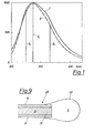
- the wavelength in nm is plotted on the abscissa and the intensity on the ordinate.
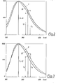
- Fig. 2 shows typical normalized fluorescence spectra of a normal serum, once, labeled I at physiological pH (7.4) and then, labeled III, in 0.1 molar hydrochloric acid. With Normalsera there is a clear blue shift when measuring in the strongly acidic range.
- the simultaneous determination of the fluorescence intensity at two different wavelengths can be carried out in an arrangement as outlined in FIG. 4.
- the light from a light source 1 falls through a light filter 2 onto a sample 4 (for example serum or protein fraction in high dilution) into a sample space 5 that is permeable to both the excitation and the emission radiation. If a requirement of typically 2 ml of sample solution is required, this is calculated a need for serum of 2-10 microliters or 0.2-0.6 mg albumin or globulin fraction).
- the fluorescent light is emitted from the sample space 5 in all directions of the space and is determined simultaneously with the aid of two measuring devices 25 consisting of optical filters 8, 9 and two light detectors 11, 12 at two wavelengths.
- the one optical filter 8 is transparent to the light Wavelength 337 nm and preferably an interference filter.
- the other filter 9 can be an interference filter or a steep edge filter and preferably transmits light of the wavelength above 340 nm.
- the light intensity is measured using the photodetectors 11 and 12, which e.g. can be a SEV-based photo amplifier, a photodiode, or a phototransistor.
- the resulting electrical signals are now fed to an evaluation unit (not shown further) and divided there.
- the ratio of the two light intensities depends on the device and is influenced, among other things, by the spectral properties of the filters 8, 9 and the spectral sensitivity of the photo amplifiers.
- the quotient of the intensities at 365 and 337 nm for the normal serum is 0.714 +/- 0.008 when averaged from 20 samples.
- the simultaneous determination of the fluorescence intensity at three different wavelengths can be carried out in an arrangement as outlined in FIG. 5.
- light from the light source 1 falls through a light filter 3, which is only permeable to light of the wavelength of typically 295 nm, onto the sample 4.
- the intensity of the emission light is now measured at three different wavelengths, namely an intensity I (365 nm) Light filter x 9 and detector 12), an intensity 1 at 337 nm (light filter 8 and detector 11) and an intensity I y at 325 nm (light filter 7 and detector 10).
- the samples 4, 6 are filled into the sample spaces 5.
- the excitation light from the light source 1 falls through the light filter 2, which is permeable to light of the wavelength 287 nm, onto the Sample 4 and excitation light of the wavelength 295 nm through a filter 3 onto the sample 6.
- the intensity of the emission light is now measured at three different emission wavelengths, specifically for each of the samples 4 and 6 at 337 nm with a light filter 8 and a detector 11, the fluorescence intensities I o and Io.
- the light filters 9 and 7 are transmissive to light of the wavelengths 365 and 325 nm, the fluorescence intensities I x and 1 being measured at the detectors 12 and 10, respectively.
- the analytical wavelengths specified are device-specific and are determined by the spectral characteristics of the light source and the photo amplifier.
- the analytical wavelengths specified are device-specific and are determined by the spectral characteristics of the light source and the photo amplifier.
- the excitation filter 3 must be replaced by a filter 2, the emission filter 7 by filter 9 and detector 10 by detector 12.
- the light from the light source 1 then falls through the light filter 2 onto an acidic 6 or a neutral sample 4.
- the fluorescent light is emitted from both sample spaces in all directions and strikes the optical filters 8 and 9.
- the fluorescence intensity is determined simultaneously with two light detectors 11 and 12 at two wavelengths.
- the optical filters 8 are again transparent to light of the wavelength 337 nm and provide the fluorescence intensities I o and I ', the optical filters 9 for light of the wavelength 0 ge 365 nm and provide the fluorescence intensities I x and I' x . x
- a device is shown, the measurement by means of fluorescence quencher method in an analyti wavelength.
- the excitation light falls from the light source 1 through two light filters 2 onto the samples 4, 6 located in the sample rooms 5.
- Sample 4 contains diluted serum
- sample 6 contains the same diluted serum and the quencher additive.
- the fluorescence radiation propagates from the two samples 4, 6 in all directions.
- the fluorescence intensities of the two samples are determined at the emission wavelengths of 337 nm (or longer wavelength) with the aid of a suitable optical filter and correlated in a division unit.
- the quotient I L / I o will be higher in the case of a serum of a person with a tumor than the corresponding quotient, determined on a standard.
- FIG. 8 shows an arrangement chosen on the basis of FIG. 5, with the aid of which an in vivo measurement is also possible.
- lenses 13 are coupled into light guides 14 from a UV light source 1 after passing through the filters 2.
- the light guides 14 bring the excitation light to the measuring location.
- the induced fluorescent light returns to the measuring devices via light guide 15. It is also possible, as shown in FIG. 8, to guide the excitation light and the fluorescent light for each emission wavelength to be measured in a light guide and the fluorescent light in each case with the aid of a dichroic mirror or a beam splitter 16, 17, 18 onto one of the lenses 19 to steer.
- the filters 7, 8 and 9 set to 325, 337 and 365 nm in the individual measuring devices 25, the respective intensity I is determined and evaluated as described earlier.
- the ends of the light guides 15 are preferably combined into a catheter-shaped bundle 20.
- the light source 1 a reference photodetector can be assigned to compensate for intensity fluctuations.
- Fig. 9 shows the end 21 of a light guide 15 made of a catheter-shaped bundle 20 with a balloon-shaped cellulose membrane. 22, which prevents the penetration of cellular components into the sample space 5, whereas blood serum can diffuse, 23 and 24 are the core and cladding of the light guide 15.
Abstract
Im Gegensatz zu bekannten relativ aufwendigen Verfahren zur Tumordiagnose über Serumtests wird erfindungsgemäß das zu untersuchende Serum, oder dessen Protein-Subfraktionen (Albumin und/oder Globulin), mit Anregungsstrahlung zumindest einer Wellenlänge zwischen 250 und 300 nm angeregt, die Fluoreszenzintensität des Serums bei vorbestimmten Emissionswellenlängen gemessen und Abweichungen dieser Meßwerte von jenen eines Standards oder Standardserums festgestellt. Daraus wird auf das Vorliegen neoplastischer Erkrankungen geschlossen.In contrast to known relatively complex methods for tumor diagnosis via serum tests, the serum to be examined, or its protein sub-fractions (albumin and / or globulin), is excited with excitation radiation of at least a wavelength between 250 and 300 nm, the fluorescence intensity of the serum at predetermined emission wavelengths measured and deviations of these measured values from those of a standard or standard serum determined. This suggests the existence of neoplastic diseases.
Description
Die Erfindung betrifft ein Verfahren zur Tumordiagnose mittels Sera und eine Vorrichtung zur Durchführung des Verfahrens.The invention relates to a method for tumor diagnosis using Sera and a device for performing the method.
Das beträchtliche Interesse, das an einer frühzeitigen und sicheren Tumordiagnose zur Zeit besteht, hat dazu geführt, daß verschiedenste Untersuchungsmethoden hinsichtlich ihrer Eignung zur Tumordiagnose geprüft worden sind. Dabei ist die Suche nach Tumoren in verschiedenen Gewebsteilen oder Organen recht aufwendig, da in diesem Fall eine Vielzahl von Proben zu nehmen ist, wobei die Gewinnung von Zellproben von schwer zugänglichen Organen eine nicht unwesentliche Hürde bei der Früherkennung von Tumoren im Rahmen der Gesundenuntersuchungen bildet.Gerade für den letztgenannten Zweck sind direkte Untersuchungen des Serums allen anderen Methoden vorzuziehen.The considerable interest that currently exists in the early and reliable diagnosis of tumors has led to the fact that a wide variety of examination methods have been examined with regard to their suitability for tumor diagnosis. The search for tumors in different parts of the tissue or organs is quite complex, since in this case a large number of samples have to be taken, whereby the collection of cell samples from organs that are difficult to access is a not insignificant hurdle in the early detection of tumors in the context of healthy examinations. For the last-mentioned purpose, direct tests of the serum are preferable to all other methods.
Besondere Aufmerksamkeit hat dabei die Untersuchung des Humanserums gefunden, dessen Probennahme zur Routine in der klinischen Analytik gehört und welches somit von sehr vielen Personen leicht und in kurzer Zeit erhalten werden kann. Dies ist für Screening-Untersuchungen von besonderer Wichtigkeit.The investigation of human serum, the sampling of which is routine in clinical analysis, has received particular attention and can therefore be obtained easily and quickly by a large number of people. This is of particular importance for screening examinations.
Die bedeutendste Rolle bei der Diagnose von Malignomenüber Serumtests spielen die sogenannten Tumormarker.Dabei handelt es sich um Parameter, die Hinweise auf einen neoplastischen Prozeß geben können. Bei den bekannten relativ aufwendigen Verfahren zur Tumordiagnose werden dabei vor allem immunologische Methoden angewandt.The so-called tumor markers play the most important role in the diagnosis of malignancies via serum tests. These are parameters that can give an indication of a neoplastic process. In the known relatively complex Methods of tumor diagnosis are mainly used immunological methods.
Aufgabe der Erfindung ist es, ein Verfahren zur Tumordiagnose anzugeben, mit welchem in einfacher Weise und mit relativ großer Genauigkeit die Differenzierung zwischen Normalsera und Tumorsera möglich ist bzw. eine Vorrichtung zur Durchführung dieses Verfahrens vorzuschlagen.The object of the invention is to provide a method for tumor diagnosis with which the differentiation between normal sera and tumor sera is possible in a simple manner and with relatively high accuracy, or to propose a device for carrying out this method.
Diese Aufgabe wird erfindungsgemäß dadurch gelöst,daß das zu untersuchende Serum mit Anregungsstrahlung zumindest einer Wellenlänge zwischen 250 und 300 nm angeregt wird, daß die Fluoreszenzintensität des Serums bei vorbestimmten Emissionswellenlängen gemessen wird, sowie daß Abweichungen dieser Meßwerte von jenen eines Standards oder Standardserums festgestellt werden und daraus auf das Vorliegen neoplastischer Erkrankungen geschlossen wird. Damit steht ein neuartiges spektroskopisches Verfahren zur Tumordiagnose zur Verfügung, wobei der Umstand ausgenützt wird, daß sich die emissionsoptischen Eigenschaften eines Normalserums in geringer, aber eindeutiger Weise von denen eines Tumorserums unterscheiden. Das Fluoreszenzspektrum des Normalserums zeigt ein Emissionsmaximum bei ca. 337 nm, welches überwiegend von proteingebundenem Tryptophan (Trp) herrührt.This object is achieved in that the serum to be examined is excited with excitation radiation of at least a wavelength between 250 and 300 nm, that the fluorescence intensity of the serum is measured at predetermined emission wavelengths, and that deviations of these measured values from those of a standard or standard serum are determined and it is concluded that there are neoplastic diseases. A novel spectroscopic method for tumor diagnosis is thus available, taking advantage of the fact that the emission-optical properties of a normal serum differ slightly from those of a tumor serum. The fluorescence spectrum of the normal serum shows an emission maximum at approx. 337 nm, which mainly results from protein-bound tryptophan (Trp).
In einer Ausgestaltung der Erfindung ist vorgesehen, daß die Fluoreszenzintensität des zu untersuchenden Serums bei einer Emissionswellenlänge von über 340 nm gemessen, normiert und mit der Fluoreszenzintensität eines Standards oder Standardserums verglichen wird und daß aus einer Abnahme der normierten Fluoreszenzintensität auf das Vorliegen neoplastischer Erkrankungen geschlossen wird. Die Normierung der Spektren kann dabei beispielsweise auf deren Peak-Maximum bezogen werden. Es zeigt sich somit, daß sich die Fluoreszenzspektren des Humanserums beim Auftreten neoplastischer Erkrankungen in ganz charakteristischer Weise verändern.Diese Veränderungen treten beispielsweise auf der langwelligen Flanke des Spektrums auf, wo sie besonders gut ausgeprägt sind.In one embodiment of the invention, it is provided that the fluorescence intensity of the serum to be examined is measured, standardized and compared with the fluorescence intensity of a standard or standard serum at an emission wavelength of more than 340 nm, and that the presence of neoplastic diseases is inferred from a decrease in the standardized fluorescence intensity . The spectra can be normalized, for example, to their peak maximum. It can thus be seen that the fluorescence spectra of human serum change in a very characteristic manner when neoplastic diseases occur. These changes occur, for example, on the long-wave flank of the spectrum, where they are particularly pronounced.
Um eine Abweichung von der normalen spektralen Verteilung der Fluoreszenz festzustellen, ist es nicht ausreichend; nur die Fluoreszenzintensität zu bestimmen, da deren abselute Intensität von Serum zu Serum schwankt. Um solche Schwankungen auszuschalten, ist es zweckmäßig, die Fluoreszenzintensität zu normieren, indem sie zur Intensität am Emissionsmaximum des Spektrums in Beziehung gesetzt wird. Zu diesem Zweck bestimmt man die Fluoreszenzintensität 1 des verdünnten Humanserums am Emissionsmaximum, also bei ca. 337 nm. In einer zweiten Messung bestimmt man die Fluoreszenzintensität Ix an der langwelligen Flanke des Emissionsspektrums, also im Bereich zwischen 350 und 400 nm bei einer Anregungswellenlänge von 250 bis 300 nm, typischerweise bei 287 nm. Der Quotient der beiden Intensitäten (I /I ) muß bei einem gesunden Probanden einen gewissen Mindestwert erreichen. Falls dieser Wert unterschritten wird, ist dies indikativ für einen Tumor. Dieser Mindestwert wird durch Eichung mit einem Standard ermittelt. Das kann z.B. ein gemittelter Wert aus mehreren Sera gesunder Personen sein oder ein synthetischer Standard,wie z.B. N-Acetyl-Tryptophanamid.A deviation from the normal spectral distribution determining fluorescence is not enough; only to determine the fluorescence intensity, since its absurd intensity varies from serum to serum. In order to eliminate such fluctuations, it is expedient to normalize the fluorescence intensity by relating it to the intensity at the emission maximum of the spectrum. For this purpose, the
Bei Sera von Patienten mit krebsartigen Erkrankungen wurden Abweichungen der Intensitätsverhältnisse bis zu einem Verhältnis von 0,636 gefunden, wobei im Normalfall ein Verhältnis von 0,714 festgestellt wird. Auf alle Fälle ist vor jeder Meßreihe eine sorgfältige Geräteeichung vorzunehmen. Mit Hilfe der Messung bei zwei Emissionswellenlängen (Zwei-Wellenlängenmessung) ist eine Trefferwahrscheinlichkeit von maximal 60 % für die Erkennung der malignen Erkrankung erzielbar. Bemerkenswert ist dabei der Umstand, daß keine falsche positive Anzeige beobachtet wurde.In sera from patients with cancer-like diseases, deviations in the intensity ratios up to a ratio of 0.636 were found, whereby a ratio of 0.714 is normally found. In any case, careful instrument calibration must be carried out before each series of measurements. With the help of the measurement at two emission wavelengths (two-wavelength measurement), a hit probability of maximum 60% can be achieved for the detection of the malignant disease. It is remarkable that no false positive indication was observed.
Die Durchführung des Verfahrens mittels der Zwei-Wellenlängenmessung ist einfacher und schneller als die Aufnahme eines gesamten Emissionsspektrums und anschließender Subtraktion von einem gespeicherten Standard-Normal-Serum, oder auch der Bildung eines Verhältnisses Ix/Io aus den Daten des gesamten Emissionsspektrums. Dies ist ein wesentlicher Vorteil beim Einsatz im Rahmen von Screening-Untersuchungen.The implementation of the method by means of the two-wavelength measurement is easier and faster than the acquisition of an entire emission spectrum and subsequent subtraction from a stored standard normal serum, or also the formation of a ratio I x / I o from the data of the entire emission spectrum. This is essential advantage in the context of screening examinations.
In einer Weiterbildung der Erfindung ist vorgesehen, daß die Fluoreszenzintensität des zu untersuchenden Serums zusätzlich bei einer Emissionswellenlänge zwischen 320 und 330 nm gemessen, normiert und mit der Fluoreszenzintensität eines Standardserums verglichen wird und daß aus der Zunahme der normierten Fluoreszenzintensität auf das Vorliegen neoplastischer Erkrankungen geschlossen wird. Durch die Einbeziehung der kurzwelligen Flanke des Spektrums und die Messung der Intensitätsänderungen in diesem Bereich kann die Genauigkeit des Verfahrens weiter erhöht werden. Die Trefferwahrscheinlichkeit kann mit Hilfe der Drei-Wellenlängenmessung, durch die zusätzliche Messung der Fluoreszenzintensität Iy im kurzwelligen Bereich des Spektrums, wesentlich gesteigert werden.In a further development of the invention it is provided that the fluorescence intensity of the serum to be examined is additionally measured, normalized and compared with the fluorescence intensity of a standard serum at an emission wavelength and that the presence of neoplastic diseases is inferred from the increase in the standardized fluorescence intensity . The accuracy of the method can be further increased by including the short-wave edge of the spectrum and measuring the intensity changes in this area. The hit probability can be increased significantly with the aid of the three-wavelength measurement, by additionally measuring the fluorescence intensity Iy in the short-wave region of the spectrum.
Während nun das Verhältnis Ix/Io bei Sera gesunder Personen nur einen gewissen Mindestwert und gleichzeitig das Verhältnis Iy/Io einen gewissen Maximalwert erreichen dürfen, werden diese Werte bei Sera von Patienten mit neoplastischen Erkrankungen unter- bzw. überschritten.While the ratio I x / I o may only reach a certain minimum value in Sera of healthy persons and at the same time the ratio Iy / Io may reach a certain maximum value, these values are undercut or exceeded in Sera by patients with neoplastic diseases.
In einer anderen Ausgestaltung der Erfindung ist vorgesehen, daß die Fluoreszenzintensität des zu untersuchenden Serums sowohl in physiologischer als auch in saurer Lösung von pH-Werten unter 4 gemessen und normiert wird und daß aus der Differenz oder dem Verhältnis der normierten Meßwerte auf das Vorliegen neoplastischer Erkrankungen geschlossen wird. Ein weiterer Unterschied in den fluoreszenz-optischen Eigenschaften der Sera gesunder bzw. tumorkranker Personen wurde beim Studium der pH-Abhängigkeit der entsprechenden Fluoreszenzeigenschaften gefunden. Das Emissionsmaximum des Humanserums gesunder Personen liegt, mit nur sehr geringen Abweichungen, zwischen 336,5 und 337,5 nm (gemessen an einem Aminco SPF 500 Instrument). Im Vergleich dazu ist das Emissionsmaximum von Tumorsera zu etwas kürzeren Wellenlängen verschoben. Dies gilt für pH-Werte im physiologischen Bereich.In another embodiment of the invention it is provided that the fluorescence intensity of the serum to be examined is measured and normalized both in physiological and in acidic solution with pH values below 4 and that the difference or the ratio of the standardized measured values indicates the presence of neoplastic diseases is closed. Another difference in the fluorescence-optical properties of the sera of healthy or tumor-sick people was found when studying the pH dependence of the corresponding fluorescence properties. The emission maximum of human serum from healthy people is, with only very slight deviations, between 336.5 and 337.5 nm (measured on an Aminco SPF 500 instrument). In comparison, the emission maximum from Tumorsera is somewhat shorter ren wavelengths shifted. This applies to pH values in the physiological range.
Mißt man hingegen in stark saurer Lösung, z.B. in 0,1-molarer Salzsäure, so tritt beim Normalserum eine Blauverschiebung des Emissionsmaximums gegenüber dem physiologischen Bereich auf, während dieses beim Tumorserum nur schwach oder gar nicht verschoben wird, sodaß in stark saurer Lösung das Emissionsmaximum der Tumorasera gegenüber demjenigen der Normalsera zu etwas längeren Wellenlängen verschoben ist.However, if you measure in a strongly acidic solution, e.g. in 0.1-molar hydrochloric acid, there is a blue shift in the emission maximum in the normal serum compared to the physiological range, while this is only slightly or not shifted in the tumor serum, so that in a strongly acidic solution the emission maximum of the tumorasera is somewhat longer than that of the normal sera Wavelengths is shifted.
In stark saurer Lösung ist also das Trp-Emissionsmaximum der Sera von Patienten mit Malignomen gegenüber solchen gesunder Personen rotverschoben. Sämtliche im physiologischen pH-Bereich beobachteten Abweichungen ändern in saurer Lösung lediglich das Vorzeichen. Somit können bei Messungen im sauren pH-Bereich sämtliche Meßverfahren, wie sie oben beschrieben sind, auch im sauren Bereich angewendet werden. Bei der Messung im sauren pH-Bereich gelten allerdings andere Quotienten der Fluoreszenzintensitäten. Ebenso wie im neutralen pH-Bereich muß im sauren pH-Bereich der Mittelwert der einzelnen Quotienten durch arithmetische Mittelung einer gewissen Anzahl von Quotienten von Sera gesunder Personen oder mit Hilfe eines Standards bestimmt und der Normalbereich ermittelt werden.In a strongly acidic solution, the Trp emission maximum of the sera from patients with malignancies is red-shifted compared to those of healthy people. All deviations observed in the physiological pH range only change the sign in acidic solution. Thus, all measurement methods as described above can also be used in the acidic range for measurements in the acidic pH range. When measuring in the acidic pH range, however, other quotients of the fluorescence intensities apply. Just as in the neutral pH range, the mean value of the individual quotients must be determined in the acidic pH range by arithmetically averaging a certain number of quotas from sera of healthy people or using a standard and the normal range must be determined.
Das zuletzt beschriebene Meßverfahren hat den Vorteil, daß bei gleichzeitiger Messung bei physiologischem bzw. stark saurem pH-Wert - zur Bestimmung der Normalbereiche der Quotienten - keine große Anzahl von Kontrollsera vermessen werden muß. Es werden lediglich im physiologischen Bereich die Intensitäten Io bzw. Ix , sowie im sauren Bereich ![]()
![]()
![]()
![]()
![]()
![]()
![]()
![]()
Als weitere Variante der Erfindung stellt sich die Möglichkeit dar, daß die Fluoreszenzintensität des zu untersuchenden Serums bei mindestens einer Emissionswellenlänge in Gegenwart und in Abwesenheit eines dynamischen Fluoreszenzlöschers bestimmt wird und daß aus der Differenz oder dem Verhältnis auf das Vorliegen neoplastischer Erkrankungen geschlossen wird. Die Fluoreszenzlöschung gehorcht dabei üblicherweise der Stern-Volmer-Beziehung:
Hier bedeuten Io die Fluoreszenz des Trp in Abwesenheit eines Löschers, IL die Intensität in Anwesenheit einer Konzentration (L) an Löscher und Ksv die Löschkonstante.Here I o means the fluorescence of the Trp in the absence of an quencher, I L the intensity in the presence of a concentration (L) of quencher and K sv the quenching constant.
Es wird nun gefunden, daß sich die Fluoreszenz des Serums gesunder Personen durch Verwendung einer geeigneten Löschsubstanz deutlich stärker Löschen läßt als die Fluoreszenz des Serums von tumorerkrankten Personen.It is now found that the fluorescence of the serum of healthy people can be quenched significantly more than the fluorescence of the serum of people with tumor by using a suitable quenching substance.
Da die Fluoreszenzlöschung in ihrer Anwendung auf Sera eine Wellenlängenabhängigkeit zeigt (das Verhältnis von Io/IL ist nur in erster Näherung für alle Wellenlängen konstant), ergibt sich die Möglichkeit, daß anstelle der Absolutintensität der Fluoreszenz, welche sehr anfällig gegenüber Verdünnungsfehlern ist, ein Quotient zu einer Aussage herangezogen wird. Am einfachsten geschieht dies dadurch, daß man den Quotienten zweier Lichtintensitäten I x und Iy bzw. ![]()
![]()
![]()
![]()
Von Vorteil bei der Erfindung ist es, wenn Iodidionen als dynamische Fluoreszenzlöscher eingesetzt werden. Außerdem sind auch Acrylamid, molekularer Sauerstoff und einige andere Moleküle in der Lage, die Fluoreszenz des proteingebundenen Tryptophans in reversibler Weise zu vermindern.It is advantageous in the invention if iodide ions are used as dynamic fluorescence quenchers. In addition, acrylamide, molecular oxygen and some other molecules are also able to reversibly reduce the fluorescence of the protein-bound tryptophan.
Meßtechnisch kann man zur Ausnützung dieses Effektes so vorgehen, daß man das Tumorserum einmal in Anwesenheit und einmal in Abwesenheit von z.B. 0,1 mol/1 Kaliumiodid vermißt und die beiden Fluoreszenzintensitäten bei 337 nm vergleicht.In terms of measurement technology, one can use this effect in such a way that the tumor serum is present once in the presence and once in the absence of e.g. 0.1 mol / 1 potassium iodide was measured and the two fluorescence intensities at 337 nm were compared.
Es kann auch im Zusammenhang mit der Signalausbeute von Vorteil sein, daß das Serum vor der Messung verdünnt wird, beispielsweise mit Wasser, mit einem Faktor 200-1000.It may also be advantageous in connection with the signal yield that the serum is diluted with a factor of 200-1000 before the measurement, for example with water.
Eine weitere Möglichkeit das Meßergebnis qualitativ und quantitativ zu verbessern, ist in Weiterbildung der Erfindung dadurch gegeben, daß Albumin- oder Globulin-Subfraktionen des Serums zur Messung herangezogen werden. Diese können durch bekannte Methoden aus dem Serum isoliert werden, beispielsweise durch Elektrophorese oder Chromatographie und gelöst in Wasser in einer Konzentration von beispielsweise 0,1-0,3 mg/ml der Messung zugeführt werden.Another possibility of improving the measurement result qualitatively and quantitatively is provided in a further development of the invention in that albumin or globulin sub-fractions of the serum are used for the measurement. These can be isolated from the serum by known methods, for example by electrophoresis or chromatography and dissolved in water in a concentration of, for example, 0.1-0.3 mg / ml, and fed to the measurement.
Eine vorteilhafte Ausgestaltung der Erfindung liegt vor, wenn die Messung bei zwei verschiedenen Anregungswellenlängen, vorzugsweise bei 287 und 295 nm erfolgt und die Fluoreszenzintensität bei drei verschiedenen Wellenlängen, vorzugsweise bei 325, 337 und 365 nm gemessen wird. Die größte Genauigkeit bei der Drei-Wellenlängenmessung läßt sich durch Verwendung von zwei verschiedenen Anregungswellenlängen erreichen. Um eine Probe nun bei zwei Anregungswellenlängen gleichzeitig vermessen zu können, benötigt man für jede Anregungswellenlänge eine Meßküvette. Gemessen werden im einen Fall Io bzw. Ix im langwelligen Bereich, sowie im zweiten Fall I' bzw. I' y im kurzwelligen Bereich. Das Verhältnis Ix/Io darf dabei einen gewissen Mindestwert und gleichzeitig das Verhältnis I'/I' einen gewissen Maximalwert erreichen. y 0 Diese Werte werden bei Sera von Patienten mit neoplastischen Erkrankungen unter- bzw. überschritten.An advantageous embodiment of the invention exists if the measurement is carried out at two different excitation wavelengths, preferably at 287 and 295 nm, and the fluorescence intensity is measured at three different wavelengths, preferably at 325, 337 and 365 nm. The greatest accuracy in the three-wavelength measurement can be achieved by using two different excitation wavelengths. In order to be able to measure a sample at two excitation wavelengths at the same time, a measuring cell is required for each excitation wavelength. In one case I o or I x are measured in the long-wave range, and in the second case I 'or I' y in the short-wave range. The ratio I x / I o may a certain minimum value and at the same time the ratio I '/ I' reach a certain maximum value.
Eine erfindungsgemäße Vorrichtung zur Durchführung des Verfahrens nach der Erfindung ist dadurch gegeben, daß zumindest ein das zu untersuchende Serum enthaltender Probenraum, der gegebenenfalls ein Filter passierenden Anregungsstrahlung aus einer Lichtquelle ausgesetzt ist, daß dem Probenraum zumindest eine Meßeinrichtung, bestehend aus Filter und Lichtdetektor, zugeordnet ist und daß jeder Lichtdetektor mit einer Auswerteeinheit in Verbindung steht. Damit ist eine einfache Vorrichtung gegeben, bei der die für die Anregung benötigte Wellenlänge entweder durch eine in diesem Bereich emittierende Lichtquelle oder durch ein Filter realisiert ist.A device according to the invention for carrying out the method according to the invention is given in that at least one sample space containing the serum to be examined, which is possibly exposed to excitation radiation from a light source passing through a filter, is assigned to the sample space at least one measuring device consisting of filter and light detector and that each light detector is connected to an evaluation unit. This provides a simple device in which the wavelength required for the excitation is realized either by a light source emitting in this area or by a filter.
Eine Ausgestaltung der Erfindung sieht vor, daß zwei Probenräume vorhanden sind, welchen über je ein Filter Licht, ggf. unterschiedlicher Anregungswellenlänge, der selben Lichtquelle zuführbar ist. Diese Variante der Vorrichtung wird für alle Meßverfahren benötigt, bei denen eine Probe mit unterschiedlichen Wellenlängen angeregt wird, oder bei welchen einem Teil der Probe ein die Fluoreszenz beeinflussender Zusatz beigemengt wird.One embodiment of the invention provides that two sample spaces are available, to which light, possibly different excitation wavelengths, can be fed to the same light source via a filter. This variant of the device is required for all measuring methods in which a sample is excited with different wavelengths, or in which part of the sample is admixed with an additive which influences the fluorescence.
In einer besonderen Ausgestaltung der Erfindung ist vorgesehen, daß sich zwischen dem bzw. den Probenräumen und der Lichtquelle Lichtleiter für die Anregungsstrahlung befinden, sowie daß die emittierte Fluoreszenzstrahlung der Probe jeder Meßeinrichtung mittels weiterer Lichtleiter zuführbar ist. Durch Zusammenfassen der Lichtleiter zu einem Bündel ist es möglich, dieses als invasiven Katheter auszubilden und in-vivo Messungen im Vollblut durchzuführen.In a special embodiment of the invention it is provided that there are light guides for the excitation radiation between the sample space (s) and the light source, and that the emitted fluorescent radiation of the sample can be fed to each measuring device by means of further light guides. By combining the light guides into a bundle, it is possible to design this as an invasive catheter and to carry out in vivo measurements in whole blood.
In einer Weiterbildung der Erfindung ist vorgesehen, daß je Meßeinrichtung nur ein Lichtleiter zum bzw. zu den Probenräumen vorhanden ist, welcher sowohl die Anregungsals auch die Fluoreszenzstrahlung leitet, wobei ein dichroitischer Spiegel oder ein Strahlteiler letztere aus dem Lichtleiter auskoppelt und der Meßeinrichtung zuführt. Zweckmäßigerweise verwendet man für jede Wellenlängenkombination, bestehend aus Anregungswellenlänge und zu messender Emissionswellenlänge, einen eigenen Lichtleiter mit einem typischen Durchmesser von 100 bis 150 Mikrometer. Das induzierte Fluoreszenzlicht wird mit Hilfe eines Strahlteilers der entsprechenden Meßeinrichtung zugeführt. Es ist auch möglich, in nur eine optische Fiber mehrere Wellenlängen z.B. mit Hilfe eines sich drehenden Filterrades, sukzessive einzukoppeln.In a further development of the invention it is provided that only one light guide to and to the sample spaces is provided per measuring device, which light is both the excitation and also guides the fluorescence radiation, a dichroic mirror or a beam splitter coupling the latter out of the light guide and feeding it to the measuring device. It is expedient to use a separate light guide with a typical diameter of 100 to 150 micrometers for each wavelength combination consisting of the excitation wavelength and the emission wavelength to be measured. The induced fluorescent light is fed to the corresponding measuring device with the aid of a beam splitter. It is also possible to successively couple several wavelengths into just one optical fiber, for example with the help of a rotating filter wheel.
Schließlich ist es von Vorteil, wenn bei der Erfindung der Probenraum durch eine am probenseitigen Ende des Lichtleiters, angebrachte protein-permeable Membran, vorzugsweise einer Zellulose-Membran, umschlossen ist. Um optische und mechanische Störungen durch größere Blutbestandteile (z.B. Erythrozyten) bei Vollblutmessungen zu vermeiden, ist es vorteilhaft, das Ende des Lichtleiters mit einer dünnen protein-permeablen Membran zu bedecken. Diese erlaubt zwar den Zutritt von Proteinen, verhindert aber das Eindringen der Erythrozyten. Als ideales Material für diesen Zweck kann man Zellulose-Membranen ansehen.Finally, it is advantageous if, in the invention, the sample space is enclosed by a protein-permeable membrane, preferably a cellulose membrane, attached to the sample-side end of the light guide. In order to avoid optical and mechanical disturbances due to larger blood components (e.g. erythrocytes) in whole blood measurements, it is advantageous to cover the end of the light guide with a thin protein-permeable membrane. Although this allows the entry of proteins, it prevents the penetration of the erythrocytes. Cellulose membranes can be regarded as the ideal material for this purpose.
Für in-vivo Messungen auf fiberoptischem Wege eignen sich natürlich nur jene Verfahren, bei denen keine schädlichen Additive wie Säuren und Fluoreszenzlöscher etc. zugesetzt werden müssen.Of course, only those methods in which no harmful additives such as acids and fluorescence quenchers etc. need to be added are suitable for in-vivo measurements by fiber optics.
Aus folgenden Gründen ist es zweckmäßig, den Probenraum mit Wasser zu füllen: Erstens wird das eindringende Serum dadurch verdünnt, sodaß die bei unverdünnten Sera auftretenden inneren Filtereffekte entfallen können. Auftretende Konzentrationsgradienten sind wegen der kurzen Meßzeit (ca. 2s) vernachlässigbar. Zweitens wird dadurch die Gefahr der Einbringung von Luftblasen in das Untersuchungsmaterial (z.B. Blut) herabgesetzt.It is advisable to fill the sample chamber with water for the following reasons: First, the penetrating serum is diluted so that the internal filter effects that occur with undiluted sera can be eliminated. Concentration gradients that occur are negligible due to the short measuring time (approx. 2 s). Secondly, this reduces the risk of air bubbles being introduced into the test material (e.g. blood).
Weitere Merkmale und Vorteile der Erfindung sind nachstehend anhand von in der Zeichnung dargestellten Ausführungsbeispielen, sowie Diagrammen näher erläutert.Further features and advantages of the invention are explained in more detail below on the basis of exemplary embodiments shown in the drawing and diagrams.
Es zeigen:
Figur 1 bisFigur 3 Diagramme von Fluoreszenzspektren,Figur 4 bisFigur 8 verschiedene Vorrichtungen zur Durchführung des Verfahrens in schematischer Darstellung, undFigur 9 einDetail aus Figur 8 in vergrößertem Maßstab.
- Figure 1 to
- FIG. 3 diagrams of fluorescence spectra,
- Figure 4 to
- Figure 8 shows various devices for performing the method in a schematic representation, and
- Figure 9 shows a detail of Figure 8 on an enlarged scale.
In den in Fig. 1 bis 3 dargestellten Diagrammen ist auf der Abszisse die Wellenlänge in nm und auf der Ordinate die Intensität aufgetragen.In the diagrams shown in FIGS. 1 to 3, the wavelength in nm is plotted on the abscissa and the intensity on the ordinate.
In Fig. 1 ist ein normiertes Fluoreszenzspektrum I eines Humanserums als durchgezogene Linie dargestellt. Es wurde bei einer Anregungswellenlänge von 287 nm erhalten und zeigt ein Emissionsmaximum bei 337 nm, welches überwiegend von protein-gebundenen Tryptophan (Trp) herrührt. Die Bande erstreckt sich über den gesamten UV-Bereich und zum Teil in den sichtbaren Bereich. Dies ist mit ein Grund für die bläulich-grünenatürliche Fluoreszenz des Serums.1 shows a normalized fluorescence spectrum I of a human serum as a solid line. It was obtained at an excitation wavelength of 287 nm and shows an emission maximum at 337 nm, which mainly results from protein-bound tryptophan (Trp). The band extends over the entire UV range and partly in the visible range. This is one of the reasons for the bluish-green natural fluorescence of the serum.
Daneben wird gefunden, daß sich bei einem Fluoreszenzspektrum II eines Tumorserums eine spektrale Verschiebung des Fluoreszenzmaximums ergibt. Dies ist aus der strichlierten Kurve in Fig. 1 ersichtlich, welche, wieder in normierter Form, das Emissionsspektrum des Serums einer Patientin mit einem gynäkologischen Tumor zeigt. Mit Io ist die Intensität im Emissionsmaximum bezeichnet, mit Ix bzw. i jene im langwelligen (365 nm) bzw. im kurzwelligen (325 nm) Bereich des Fluoreszenzspektrums II eines Tumorserums. Deutlich ist ein Unter- bzw. überschreiten der Werte im Bezug auf das Fluoreszenzspektrum I eines Normalserums zu sehen.In addition, it is found that there is a spectral shift in the fluorescence maximum in the case of a fluorescence spectrum II of a tumor serum. This can be seen from the dashed curve in FIG. 1, which, again in normalized form, shows the emission spectrum of the serum of a patient with a gynecological tumor. I o denotes the intensity in the emission maximum, Ix and i denote those in the long-wave (365 nm) and in the short-wave (325 nm) region of the fluorescence spectrum II of a tumor serum. The values in relation to the fluorescence spectrum I of a normal serum can clearly be seen to fall below or exceed the values.
Fig. 2 zeigt typische normierte Fluoreszenzspektren eines Normalserums, einmal, gekennzeichnet mit I bei physiologischem pH-Wert (7,4) und dann, bezeichnet mit III, in 0,1-molarer Salzsäure. Es tritt somit bei Normalsera eine deutliche Blauverschiebung bei Messung im stark sauren Bereich auf.Fig. 2 shows typical normalized fluorescence spectra of a normal serum, once, labeled I at physiological pH (7.4) and then, labeled III, in 0.1 molar hydrochloric acid. With Normalsera there is a clear blue shift when measuring in the strongly acidic range.
Dagegen werden in Fig. 3 normierte Fluoreszenzspektren eines Tumorserums gezeigt, wobei das Spektrum II bei einem pH-Wert von 7,4 und das Spektrum IV in 0,1-molarer Salzsäure gemessen wurde. Das Trp-Emissionsmaximum der Sera von Patienten mit Malignomen ist gegenüber solchen gesunder Personen in saurer Lösung rotverschoben. Gemessen werden die Intensitäten sowohl im Emissionsmaximum 1 bzw. I' als auch im langwelligen Bereich bei 365 nm I bzw. I'. x x Die gestrichenen Werte werden bei saurem pH-Wert gemessen.By contrast, normalized fluorescence spectra of a tumor serum are shown in FIG. 3, spectrum II being measured at a pH of 7.4 and spectrum IV in 0.1 molar hydrochloric acid. The Trp emission maximum of the sera from patients with malignancies is red-shifted in acidic solution compared to those of healthy people. The intensities are measured both in the
Die gleichzeitige Bestimmung der Fluoreszenzintensität bei zwei verschiedenen Wellenlängen kann in einer Anordnung erfolgen, wie sie in Fig. 4 skizziert ist. Hier fällt das Licht aus einer Lichtquelle 1 durch ein Lichtfilter 2 auf eine Probe 4 (z.B. Serum oder Proteinfraktion in starker Verdünnung) in einen sowohl für die Anregungs- als auch für die Emissionsstrahlung durchlässigen Probenraum 5. Bei einem Bedarf von typischerweise 2ml Probenlösung errechnet sich ein Bedarf an Serum von 2 - 10 Mikrolitern oder 0,2 bis 0,6 mg Albumin- oder Globulin-Fraktion). Das Fluoreszenzlicht wird aus dem Probenraum 5 nach allen Richtungen des Raums emittiert und mit Hilfe von zwei Meßeinrichtungen 25 bestehend aus optischen Filtern 8, 9 und zwei Lichtdetektoren 11, 12, bei zwei Wellenlängen gleichzeitig bestimmt.The simultaneous determination of the fluorescence intensity at two different wavelengths can be carried out in an arrangement as outlined in FIG. 4. Here, the light from a
Das eine optische Filter 8 ist durchlässig für Licht der Wellenlänge 337 nm und vorzugsweise ein Interferenzfilter. Das andere Filter 9 kann ein Interferenzfilter oder ein Steilkantenfilter sein und läßt vorzugsweise Licht der Wellenlänge über 340 nm durch.The one
Die Lichtintensität wird mit Hilfe der Photodetektoren 11 und 12 gemessen, welche z.B. ein Photoverstärker auf SEV-Basis, eine Photodiode, oder ein Phototransistor sein können. Die resultierenden elektrischen Signale werden nun einer nicht weiter dargestellten Auswerteeinheit zugeführt und dort dividiert.The light intensity is measured using the
Das Verhältnis der beiden Lichtintensitäten ist geräteabhängig und wird unter anderem durch die spektralen Eigenschaften der Filter 8, 9 sowie der spektralen Empfindlichkeit der Photoverstärker beeinflußt. Bei dem Gerät SPF 500 der Marke Aminco beträgt der Quotient der Intensitäten bei 365 und 337 nm für das Normalserum 0,714 +/-0,008 bei einer Mittelung aus 20 Proben.The ratio of the two light intensities depends on the device and is influenced, among other things, by the spectral properties of the
Die gleichzeitige Bestimmung der Fluoreszenzintensität bei drei verschiedenen Wellenlängen kann in einer Anordnung erfolgen, wie sie in Fig. 5 skizziert ist. Hierbei fällt Licht aus der Lichtquelle 1 durch ein Lichtfilter 3, welches nur für Licht der Wellenlänge von typischerweise 295 nm durchlässig ist, auf die Probe 4. Die Intensität des Emissionslichtes wird nun bei drei verschiedenen Wellenlängen gemessen und zwar bei 365 nm eine Intensität I (Lichtfilter x 9 und Detektor 12), bei 337 nm eine Intensität 1 (Lichtfilter 8 und Detektor 11) und bei 325 nm eine Intensität Iy (Lichtfilter 7 und Detektor 10).The simultaneous determination of the fluorescence intensity at three different wavelengths can be carried out in an arrangement as outlined in FIG. 5. Here, light from the
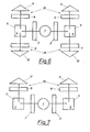
Mit Hilfe der in Fig. 6 gezeigten Vorrichtung ist es möglich bei zwei Anregungswellenlängen zu messen, wozu zwei Probenräume 5 vorhanden sind. Hierbei werden die Proben 4, 6 in die Probenräume 5 gefüllt. Aus der Lichtquelle 1 fällt das Anregungslicht durch das Lichtfilter 2, welches für Licht der Wellenlänge 287 nm durchlässig ist, auf die Probe 4 und Anregungslicht der Wellenlänge 295 nm durch ein Filter 3 auf die Probe 6. Die Intensität des Emissionslichtes wird nun bei drei verschiedenen Emissionswellenlängen gemessen und zwar bei jeder der Proben 4 und 6 bei 337 nm mit einem Lichtfilter 8 und einem Detektor 11 die Fluoreszenzintensitäten Io und Io. Die Lichtfilter 9 und 7 sind durchlässig für Licht der Wellenlänge 365 bzw. 325 nm, wobei die Fluoreszenzintensitäten Ix bzw. 1 an den Detektoren 12 bzw. 10 gemessen werden. x y With the aid of the device shown in FIG. 6, it is possible to measure at two excitation wavelengths, for which purpose two
Es muß angeführt werden, daß die angegebenen analytischen Wellenlängen gerätespezifisch sind und durch die spektrale Charakteristik der Lichtquelle und des Photoverstärkers bestimmt werden. Bei Verwendung anderer Bauteile als in der genannten Vorrichtung, in welcher z.B. eine Deuteriumlampe als Lichtquelle und ein Photomultiplier als Detektor dienen, sind geringfügige Abweichungen bei den Wellenlängen zu erwarten, bei welchen maximale Änderungen gegenüber den Normalsera auftreten.It must be stated that the analytical wavelengths specified are device-specific and are determined by the spectral characteristics of the light source and the photo amplifier. When using components other than the device mentioned, in which e.g. If a deuterium lamp serves as the light source and a photomultiplier as the detector, slight deviations in the wavelengths are to be expected, at which maximum changes compared to the normal sera occur.
Mit der in Fig. 6 gezeigten Vorrichtung ist es auch möglich,gleichzeitig bei neutralem und saurem pH-Wert zu messen. Dabei muß jedoch das Anregungsfilter 3 durch ein Filter 2 ersetzt werden, sowie das Emissionsfilter 7 durch Filter 9 und Detektor 10 durch Detektor 12. Das Licht aus der Lichtquelle 1 fällt dann durch die Lichtfilter 2 auf eine saure 6 bzw. eine neutrale Probe 4. Das Fluoreszenzlicht wird aus beiden Probenräumen nach allen Richtungen emittiert und trifft auf die optischen Filter 8 bzw. 9. Die Fluoreszenzintensität wird mit je zwei Lichtdetektoren 11 bzw. 12 bei zwei Wellenlängen gleichzeitig bestimmt. Die optischen Filter 8 sind wieder durchlässig für Licht der Wellenlänge 337 nm und liefern die Fluoreszenzintensitäten Io und I', die optischen Filter 9 für Licht der Wellenläno 0 ge 365 nm und liefern die Fluoreszenzintensitäten Ix und I'x. xWith the device shown in Fig. 6, it is also possible to measure simultaneously at neutral and acidic pH. However, the
In Fig. 7 ist eine Vorrichtung dargestellt, welche die Messung mittels Fluoreszenzlöscher-Methode bei einer analytischen Wellenlänge zeigt. Aus der Lichtquelle 1 fällt das Anregungslicht durch zwei Lichtfilter 2 auf die in den Probenräumen 5 befindlichen Proben 4, 6. Probe 4 enthält verdünntes Serum, Probe 6 gleich verdünntes Serum und den Löscherzusatz. Aus den beiden Proben 4, 6 breitet sich die Fluoreszenzstrahlung nach allen Richtungen aus. Die Fluoreszenzintensitäten der beiden Proben werden bei den Emissionswellenlängen 337 nm (oder langwelliger) mit Hilfe eines geeigneten optischen Filters bestimmt und in einer Divisionseinheit in Beziehung gesetzt. Der Quotient IL/Io wird im Fall eines Serums einer tumorerkrankten Person höher liegen als der entsprechende Quotient, bestimmt an einem Standard.In Fig. 7, a device is shown, the measurement by means of fluorescence quencher method in an analyti wavelength. The excitation light falls from the
Zur Messung bei zwei analytischen Wellenlängen bedient man sich einer Meßanordnung nach Fig. 6, in welcher das Licht aus einer Lichtquelle 1 auf die beiden Proben 4, 6 fällt, deren jeweilige Fluoreszenz bei den analytischen Wellenlängen mit Hilfe jeweils zweier optischer Filter und zweier Photodetektoren bestimmt wird.6, in which the light from a
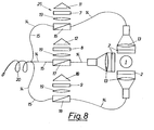
Fig. 8 zeigt eine in Anlehnung zu Fig. 5 gewählte Anordnung, mit deren Hilfe auch eine in-vivo-Messung möglich ist. Aus einer UV-Lichtquelle 1 wird Licht der Wellenlänge 290 bis 295 nm nach Passieren der Filter 2 mittels Linsen 13 in Lichtleiter 14 eingekoppelt. Die Lichtleiter 14 bringen das Anregungslicht an den Meßort. Das induzierte Fluoreszenzlicht gelangt über Lichtleiter 15 zu den Meßeinrichtungen zurück. Es ist auch möglich, wie in Fig. 8 dargestellt, das Anregungs- und das Fluoreszenzlicht für jede zu messende Emissionswellenlänge in einem Lichtleiter zu leiten und das Fluoreszenzlicht jeweils mit Hilfe eines dichroitischen Spiegels oder eines Strahlteilers 16, 17, 18 auf eine der Linsen 19 zu lenken. Nach Passieren der Filter 7, 8 und 9 eingestellt auf 325, 337 und 365 nm in den einzelnen Meßeinrichtungen 25, wird die jeweilige Intensität I bestimmt und wie früher beschrieben ausgewertet. Die Enden der Lichtleiter 15 werden vorzugsweise zu einem katheterförmigen Bündel 20 zusammengefaßt. Der Lichtquelle 1 kann zur Kompensation von Intensitätsschwankungen ein Referenzphotodetektor zugeordnet werden.FIG. 8 shows an arrangement chosen on the basis of FIG. 5, with the aid of which an in vivo measurement is also possible. After passing through the
,Fig. 9 zeigt das Ende 21 eines Lichtleiters 15 aus einem katheterförmigen Bündel 20 mit einer ballonförmigen Zellulosemembran. 22, welche das Eindringen von zellulären Bestandteilen in den Probenraum 5 verhindert, während hingegen Blutserum eindiffundieren kann, 23 und 24 sind Kern und Mantel des Lichtleiters 15., Fig. 9 shows the
Claims (14)
Applications Claiming Priority (2)
| Application Number | Priority Date | Filing Date | Title |
|---|---|---|---|
| AT0270385A AT387860B (en) | 1985-09-16 | 1985-09-16 | METHOD AND DEVICE FOR TUMOR DIAGNOSIS BY MEANS OF SERA |
| AT2703/85 | 1985-09-16 |
Publications (3)
| Publication Number | Publication Date |
|---|---|
| EP0215772A2 true EP0215772A2 (en) | 1987-03-25 |
| EP0215772A3 EP0215772A3 (en) | 1987-08-19 |
| EP0215772B1 EP0215772B1 (en) | 1992-07-01 |
Family
ID=3538963
Family Applications (1)
| Application Number | Title | Priority Date | Filing Date |
|---|---|---|---|
| EP86890254A Expired - Lifetime EP0215772B1 (en) | 1985-09-16 | 1986-09-09 | Method and device for diagnosing tumours using sera |
Country Status (5)
| Country | Link |
|---|---|
| US (1) | US4755684A (en) |
| EP (1) | EP0215772B1 (en) |
| JP (1) | JPS62103571A (en) |
| AT (1) | AT387860B (en) |
| DE (1) | DE3685866D1 (en) |
Cited By (16)
| Publication number | Priority date | Publication date | Assignee | Title |
|---|---|---|---|---|
| DE3736027A1 (en) * | 1987-10-24 | 1989-05-03 | Gerhard Dipl Phys Artmann | Method for determining the shape of cells which prevails at a specific instant and device for carrying out the method |
| WO1990012536A1 (en) * | 1989-04-14 | 1990-11-01 | Massachusetts Institute Of Technology | Spectral diagnosis of diseased tissue |
| EP0512965A1 (en) * | 1991-05-08 | 1992-11-11 | Xillix Technologies Corporation | Endoscopic imaging system for diseased tissue |
| US5590660A (en) * | 1994-03-28 | 1997-01-07 | Xillix Technologies Corp. | Apparatus and method for imaging diseased tissue using integrated autofluorescence |
| US5647368A (en) * | 1996-02-28 | 1997-07-15 | Xillix Technologies Corp. | Imaging system for detecting diseased tissue using native fluorsecence in the gastrointestinal and respiratory tract |
| US5769792A (en) * | 1991-07-03 | 1998-06-23 | Xillix Technologies Corp. | Endoscopic imaging system for diseased tissue |
| US9610021B2 (en) | 2008-01-25 | 2017-04-04 | Novadaq Technologies Inc. | Method for evaluating blush in myocardial tissue |
| US9816930B2 (en) | 2014-09-29 | 2017-11-14 | Novadaq Technologies Inc. | Imaging a target fluorophore in a biological material in the presence of autofluorescence |
| US10041042B2 (en) | 2008-05-02 | 2018-08-07 | Novadaq Technologies ULC | Methods for production and use of substance-loaded erythrocytes (S-IEs) for observation and treatment of microvascular hemodynamics |
| US10219742B2 (en) | 2008-04-14 | 2019-03-05 | Novadaq Technologies ULC | Locating and analyzing perforator flaps for plastic and reconstructive surgery |
| US10265419B2 (en) | 2005-09-02 | 2019-04-23 | Novadaq Technologies ULC | Intraoperative determination of nerve location |
| US10278585B2 (en) | 2012-06-21 | 2019-05-07 | Novadaq Technologies ULC | Quantification and analysis of angiography and perfusion |
| US10434190B2 (en) | 2006-09-07 | 2019-10-08 | Novadaq Technologies ULC | Pre-and-intra-operative localization of penile sentinel nodes |
| US10492671B2 (en) | 2009-05-08 | 2019-12-03 | Novadaq Technologies ULC | Near infra red fluorescence imaging for visualization of blood vessels during endoscopic harvest |
| US10631746B2 (en) | 2014-10-09 | 2020-04-28 | Novadaq Technologies ULC | Quantification of absolute blood flow in tissue using fluorescence-mediated photoplethysmography |
| US10992848B2 (en) | 2017-02-10 | 2021-04-27 | Novadaq Technologies ULC | Open-field handheld fluorescence imaging systems and methods |
Families Citing this family (24)
| Publication number | Priority date | Publication date | Assignee | Title |
|---|---|---|---|---|
| US5270116A (en) * | 1986-07-10 | 1993-12-14 | Minnesota Mining And Manufacturing Company | Process for fluorimetric monitoring of functional coatings and compositions and fluorescent agents therefor |
| US4978731A (en) * | 1986-07-10 | 1990-12-18 | Minnesota Mining And Manufacturing Company | Process for fluorimetric monitoring of functional coatings and compositions and fluorescent agents therefor |
| US5087670A (en) * | 1986-07-10 | 1992-02-11 | Minnesota Mining And Manufacturing Company | Process for fluorimetric monitoring of functional coatings and compositions and fluorescent agents therefor |
| US4922113A (en) * | 1986-07-10 | 1990-05-01 | Minnesota Mining And Manufacturing Company | Process for fluorimetric monitoring of functional coatings and compositions and fluorescent agents therefor |
| IE81170B1 (en) * | 1988-10-21 | 2000-05-31 | Zeneca Ltd | Pyridine derivatives |
| CN1034120C (en) * | 1988-10-21 | 1997-02-26 | 曾尼卡有限公司 | Pyridine derivatives |
| US5118559A (en) * | 1989-05-31 | 1992-06-02 | Minnesota Mining And Manufacturing Company | Fluorescent degree of cure monitors |
| US5182316A (en) * | 1989-05-31 | 1993-01-26 | Minnesota Mining And Manufacturing Company | Fluorescent degree of cure monitors |
| US5047444A (en) * | 1989-05-31 | 1991-09-10 | Minnesota Mining And Manufacturing Company | Fluorescent degree of cure monitors |
| DE69127002T2 (en) | 1990-01-31 | 1997-11-20 | Fuji Photo Film Co Ltd | Color photographic silver halide material |
| US5418130A (en) * | 1990-04-16 | 1995-05-23 | Cryopharm Corporation | Method of inactivation of viral and bacterial blood contaminants |
| US6187572B1 (en) | 1990-04-16 | 2001-02-13 | Baxter International Inc. | Method of inactivation of viral and bacterial blood contaminants |
| FR2694193B1 (en) * | 1992-07-28 | 1994-10-28 | Abraxas Bio Labs Sa | Cytodiagnostic method using alstonine as a selective marker and diagnostic kit containing this marker. |
| US5991653A (en) * | 1995-03-14 | 1999-11-23 | Board Of Regents, The University Of Texas System | Near-infrared raman spectroscopy for in vitro and in vivo detection of cervical precancers |
| US6258576B1 (en) | 1996-06-19 | 2001-07-10 | Board Of Regents, The University Of Texas System | Diagnostic method and apparatus for cervical squamous intraepithelial lesions in vitro and in vivo using fluorescence spectroscopy |
| US5697373A (en) * | 1995-03-14 | 1997-12-16 | Board Of Regents, The University Of Texas System | Optical method and apparatus for the diagnosis of cervical precancers using raman and fluorescence spectroscopies |
| US5842995A (en) * | 1996-06-28 | 1998-12-01 | Board Of Regents, The Univerisity Of Texas System | Spectroscopic probe for in vivo measurement of raman signals |
| US6080584A (en) * | 1996-12-02 | 2000-06-27 | The Research Foundation Of City College Of New York | Method and apparatus for detecting the presence of cancerous and precancerous cells in a smear using native fluorescence spectroscopy |
| GB9717021D0 (en) * | 1997-08-12 | 1997-10-15 | Kalibrant Limited | A detector |
| US6055451A (en) * | 1997-12-12 | 2000-04-25 | Spectrx, Inc. | Apparatus and method for determining tissue characteristics |
| US6174291B1 (en) | 1998-03-09 | 2001-01-16 | Spectrascience, Inc. | Optical biopsy system and methods for tissue diagnosis |
| AU6139199A (en) | 1998-09-11 | 2000-04-03 | Spectrx, Inc. | Multi-modal optical tissue diagnostic system |
| US6697666B1 (en) | 1999-06-22 | 2004-02-24 | Board Of Regents, The University Of Texas System | Apparatus for the characterization of tissue of epithelial lined viscus |
| US20040064053A1 (en) * | 2002-09-30 | 2004-04-01 | Chang Sung K. | Diagnostic fluorescence and reflectance |
Citations (2)
| Publication number | Priority date | Publication date | Assignee | Title |
|---|---|---|---|---|
| FR2229063A1 (en) * | 1973-05-07 | 1974-12-06 | Atomic Energy Commission | |
| DE2813317A1 (en) * | 1977-04-01 | 1978-10-05 | Olympus Optical Co | Cancer diagnosis device with direct indication - uses photometric polarisation microscope and fluorescent light reception and analysis system giving stabilised measurements |
Family Cites Families (12)
| Publication number | Priority date | Publication date | Assignee | Title |
|---|---|---|---|---|
| US3327117A (en) * | 1963-08-12 | 1967-06-20 | Ibm | Cancer cell detector using two wavelengths for comparison |
| US3973129A (en) * | 1975-01-10 | 1976-08-03 | Bell Telephone Laboratories, Incorporated | Fluorimetric apparatus and method for analysis of body fluid |
| IL49776A (en) * | 1976-06-13 | 1979-05-31 | Elscint Ltd | Method and appararus for differentiating of normal and abnormal mammalian cells |
| JPS53122498A (en) * | 1977-03-31 | 1978-10-25 | Laurel Bank Machine Co | Money exchanging and dispensing machine |
| JPS53135660A (en) * | 1977-04-30 | 1978-11-27 | Olympus Optical Co Ltd | Fluorescent photometric microscope using laser light |
| US4215275A (en) * | 1977-12-07 | 1980-07-29 | Luxtron Corporation | Optical temperature measurement technique utilizing phosphors |
| AT362079B (en) * | 1978-12-18 | 1981-04-27 | Helmut Dr Wachter | METHOD FOR DIAGNOSTICING, IN PARTICULAR EARLY DETECTION OF MALIGNEN TUMORS |
| US4419583A (en) * | 1980-05-31 | 1983-12-06 | Noeller Hans Guenter | Polarization fluoroimmunoassay with pulsed light source |
| JPS5784339A (en) * | 1980-11-17 | 1982-05-26 | Hitachi Ltd | Fluorescence polarizing apparatus |
| JPS5832145A (en) * | 1981-08-20 | 1983-02-25 | Kyoto Giken:Kk | Diagnosing device for cell |
| US4476870A (en) * | 1982-03-30 | 1984-10-16 | The United States Of America As Represented By The Department Of Health And Human Services | Fiber optic PO.sbsb.2 probe |
| JPS5913697A (en) * | 1982-07-12 | 1984-01-24 | Fujitsu Ltd | Liquid phase epitaxial growth device |
-
1985
- 1985-09-16 AT AT0270385A patent/AT387860B/en not_active IP Right Cessation
-
1986
- 1986-09-09 EP EP86890254A patent/EP0215772B1/en not_active Expired - Lifetime
- 1986-09-09 DE DE8686890254T patent/DE3685866D1/en not_active Expired - Fee Related
- 1986-09-10 US US06/905,558 patent/US4755684A/en not_active Expired - Lifetime
- 1986-09-16 JP JP61219107A patent/JPS62103571A/en active Granted
Patent Citations (2)
| Publication number | Priority date | Publication date | Assignee | Title |
|---|---|---|---|---|
| FR2229063A1 (en) * | 1973-05-07 | 1974-12-06 | Atomic Energy Commission | |
| DE2813317A1 (en) * | 1977-04-01 | 1978-10-05 | Olympus Optical Co | Cancer diagnosis device with direct indication - uses photometric polarisation microscope and fluorescent light reception and analysis system giving stabilised measurements |
Non-Patent Citations (7)
| Title |
|---|
| BIOLOGICAL ABSTRACTS, Band 63, 1977, Abstract Nr. 63027733, Philadelphia, US; V.I. FIRSOVA et al.: "Some parameters of UV fluorescene of rat blood serum with transplanted lympho sarcoma"; & VORROSY ONKOLIGGI, 1976, 2 (8), 95-97 * |
| BIOLOGICAL ABSTRACTS/RRM, Band 27, 1984, Abstract Nr. 27055866, Philadelphia, US; CHARNITSKI YA. et al.: "Luminescence of blood serum and the diagnosis of some diseases"; & NUAK (USSR), Band 0, Nr. 5, 1983, Seiten 61-65 * |
| DIE PHARMAZIE, Band 37, Nr. 5, Mai 1982, Seiten 323-343, Ost-Berlin DDR; W. SCHWARZE et al.: "Fluoreszenzmethoden - ihre Anwendung zur quantitativen Analyse organischer Substanzen" * |
| MOSC. ONCOLOGY RES., 25 Dezember 1975; Nr. 07/89 B/04< * |
| OPTICAL ENGINEERING, Band 22, Nr. 5, September - Oktober 1983, Seiten 596-601, Bellingham, Washinton, US; G. YAO-JUN: "Fluorescence experimental techniques and some aplications in modern biolgoy" * |
| TRENDS IN ANAYLICAL CHEMISTRY, Band 4, Nr. 7, August 1985, Seiten 184-188, Amsterdam, NL; O.S. WOLFBEIS: "Fluorescence optical sensors in analytical chemistry" * |
| TRENDS IN ANAYLICAL CHEMISTRY, Band 5, Nr. 2, Februar 1986, Seiten 37-43, Amsterdam, NL; C. NAI et al.: "Bioanalytical applications of fluorescence quenching" * |
Cited By (24)
| Publication number | Priority date | Publication date | Assignee | Title |
|---|---|---|---|---|
| DE3736027A1 (en) * | 1987-10-24 | 1989-05-03 | Gerhard Dipl Phys Artmann | Method for determining the shape of cells which prevails at a specific instant and device for carrying out the method |
| WO1990012536A1 (en) * | 1989-04-14 | 1990-11-01 | Massachusetts Institute Of Technology | Spectral diagnosis of diseased tissue |
| EP0512965A1 (en) * | 1991-05-08 | 1992-11-11 | Xillix Technologies Corporation | Endoscopic imaging system for diseased tissue |
| US5507287A (en) * | 1991-05-08 | 1996-04-16 | Xillix Technologies Corporation | Endoscopic imaging system for diseased tissue |
| US5769792A (en) * | 1991-07-03 | 1998-06-23 | Xillix Technologies Corp. | Endoscopic imaging system for diseased tissue |
| US5827190A (en) * | 1994-03-28 | 1998-10-27 | Xillix Technologies Corp. | Endoscope having an integrated CCD sensor |
| US5590660A (en) * | 1994-03-28 | 1997-01-07 | Xillix Technologies Corp. | Apparatus and method for imaging diseased tissue using integrated autofluorescence |
| US5647368A (en) * | 1996-02-28 | 1997-07-15 | Xillix Technologies Corp. | Imaging system for detecting diseased tissue using native fluorsecence in the gastrointestinal and respiratory tract |
| US10265419B2 (en) | 2005-09-02 | 2019-04-23 | Novadaq Technologies ULC | Intraoperative determination of nerve location |
| US10434190B2 (en) | 2006-09-07 | 2019-10-08 | Novadaq Technologies ULC | Pre-and-intra-operative localization of penile sentinel nodes |
| US9610021B2 (en) | 2008-01-25 | 2017-04-04 | Novadaq Technologies Inc. | Method for evaluating blush in myocardial tissue |
| US11564583B2 (en) | 2008-01-25 | 2023-01-31 | Stryker European Operations Limited | Method for evaluating blush in myocardial tissue |
| US9936887B2 (en) | 2008-01-25 | 2018-04-10 | Novadaq Technologies ULC | Method for evaluating blush in myocardial tissue |
| US10835138B2 (en) | 2008-01-25 | 2020-11-17 | Stryker European Operations Limited | Method for evaluating blush in myocardial tissue |
| US10219742B2 (en) | 2008-04-14 | 2019-03-05 | Novadaq Technologies ULC | Locating and analyzing perforator flaps for plastic and reconstructive surgery |
| US10041042B2 (en) | 2008-05-02 | 2018-08-07 | Novadaq Technologies ULC | Methods for production and use of substance-loaded erythrocytes (S-IEs) for observation and treatment of microvascular hemodynamics |
| US10492671B2 (en) | 2009-05-08 | 2019-12-03 | Novadaq Technologies ULC | Near infra red fluorescence imaging for visualization of blood vessels during endoscopic harvest |
| US10278585B2 (en) | 2012-06-21 | 2019-05-07 | Novadaq Technologies ULC | Quantification and analysis of angiography and perfusion |
| US11284801B2 (en) | 2012-06-21 | 2022-03-29 | Stryker European Operations Limited | Quantification and analysis of angiography and perfusion |
| US10488340B2 (en) | 2014-09-29 | 2019-11-26 | Novadaq Technologies ULC | Imaging a target fluorophore in a biological material in the presence of autofluorescence |
| US9816930B2 (en) | 2014-09-29 | 2017-11-14 | Novadaq Technologies Inc. | Imaging a target fluorophore in a biological material in the presence of autofluorescence |
| US10631746B2 (en) | 2014-10-09 | 2020-04-28 | Novadaq Technologies ULC | Quantification of absolute blood flow in tissue using fluorescence-mediated photoplethysmography |
| US10992848B2 (en) | 2017-02-10 | 2021-04-27 | Novadaq Technologies ULC | Open-field handheld fluorescence imaging systems and methods |
| US11140305B2 (en) | 2017-02-10 | 2021-10-05 | Stryker European Operations Limited | Open-field handheld fluorescence imaging systems and methods |
Also Published As
| Publication number | Publication date |
|---|---|
| EP0215772A3 (en) | 1987-08-19 |
| ATA270385A (en) | 1988-08-15 |
| US4755684A (en) | 1988-07-05 |
| EP0215772B1 (en) | 1992-07-01 |
| JPH0558506B2 (en) | 1993-08-26 |
| JPS62103571A (en) | 1987-05-14 |
| AT387860B (en) | 1989-03-28 |
| DE3685866D1 (en) | 1992-08-06 |
Similar Documents
| Publication | Publication Date | Title |
|---|---|---|
| EP0215772B1 (en) | Method and device for diagnosing tumours using sera | |
| DE4239016C2 (en) | Method for determining the concentration of free ions within a cell using a fluorescent indicator dye | |
| EP1028318B1 (en) | Device for the optical measurement of the total hemoglobin concentration | |
| EP1130998B1 (en) | Device for non-invasively detecting the oxygen metabolism in tissues | |
| US4930516A (en) | Method for detecting cancerous tissue using visible native luminescence | |
| DE69838560T2 (en) | ACETIC ACID AS A SIGNAL IMPROVED CONTRASTANT IN FLUORESCENT SPECTROSCOPY | |
| DE2944113C2 (en) | ||
| EP2710348B1 (en) | Method and system for determining the concentration of substances in bodily fluids | |
| DE4417639A1 (en) | Analysis of concns. of substances in a biological sample | |
| WO1981000622A1 (en) | Process and device of molecular spectroscopy particularly for determining metabolites | |
| DE3031249A1 (en) | METHOD AND DEVICE FOR DETECTING CARIES. | |
| DE2422016A1 (en) | DIAGNOSIS OF DISEASE CONDITIONS BY FLUORESCENCE MEASUREMENTS USING SCANNING LASER BEAMS | |
| DE2741068A1 (en) | METHOD AND DEVICE FOR DIAGNOSIS ON TISSUE SAMPLES | |
| EP0808453A1 (en) | Process for examining biological tissue by spectroscopy | |
| EP0247103B1 (en) | Device for measuring eye lens opacity | |
| DE2443950B2 (en) | METHOD FOR DETERMINING THE ABNORMAL BEHAVIOR OF MAMMALIAN CELLS | |
| CH640350A5 (en) | Instrument for the quantitative determination of optically active substances | |
| DE2603069C3 (en) | Method and device for determining the total protein content or individual amino acids | |
| DE3702210A1 (en) | Method of determining the concentration of components, in particular oxygen, contained in a substance | |
| DE4325529C2 (en) | Device for determining the concentration of substances in the blood | |
| EP3591378A1 (en) | Method for assaying lipids, haemoglobin and bilirubin in body fluid samples | |
| DE10129754A1 (en) | Detection of the presence of substances in vital tissue materials by passing light of a given wavelength through the material for its intensity to be compared with a reference system | |
| DE2721676A1 (en) | Flashlight spectrophotometer for clinical analyses - uses xenon tube and pulse amplitude detector circuit for absorption and attenuated total reflection measurements | |
| DE3844651A1 (en) | Device for determining variations in size of tissue particles | |
| DE102004051830A1 (en) | Multifunctional reference system for analyte determination by fluorescence |
Legal Events
| Date | Code | Title | Description |
|---|---|---|---|
| PUAI | Public reference made under article 153(3) epc to a published international application that has entered the european phase |
Free format text: ORIGINAL CODE: 0009012 |
|
| AK | Designated contracting states |
Kind code of ref document: A2 Designated state(s): DE FR GB IT |
|
| ITCL | It: translation for ep claims filed |
Representative=s name: MODIANO & ASSOCIATI S.R.L. |
|
| EL | Fr: translation of claims filed | ||
| PUAL | Search report despatched |
Free format text: ORIGINAL CODE: 0009013 |
|
| AK | Designated contracting states |
Kind code of ref document: A3 Designated state(s): DE FR GB IT |
|
| 17P | Request for examination filed |
Effective date: 19870930 |
|
| 17Q | First examination report despatched |
Effective date: 19890829 |
|
| RAP1 | Party data changed (applicant data changed or rights of an application transferred) |
Owner name: AVL MEDICAL INSTRUMENTS AG |
|
| GRAA | (expected) grant |
Free format text: ORIGINAL CODE: 0009210 |
|
| AK | Designated contracting states |
Kind code of ref document: B1 Designated state(s): DE FR GB IT |
|
| REF | Corresponds to: |
Ref document number: 3685866 Country of ref document: DE Date of ref document: 19920806 |
|
| ET | Fr: translation filed | ||
| GBT | Gb: translation of ep patent filed (gb section 77(6)(a)/1977) | ||
| ITF | It: translation for a ep patent filed |
Owner name: MODIANO & ASSOCIATI S.R |
|
| PLBE | No opposition filed within time limit |
Free format text: ORIGINAL CODE: 0009261 |
|
| STAA | Information on the status of an ep patent application or granted ep patent |
Free format text: STATUS: NO OPPOSITION FILED WITHIN TIME LIMIT |
|
| 26N | No opposition filed | ||
| PGFP | Annual fee paid to national office [announced via postgrant information from national office to epo] |
Ref country code: DE Payment date: 19930816 Year of fee payment: 8 |
|
| PGFP | Annual fee paid to national office [announced via postgrant information from national office to epo] |
Ref country code: GB Payment date: 19930901 Year of fee payment: 8 |
|
| PGFP | Annual fee paid to national office [announced via postgrant information from national office to epo] |
Ref country code: FR Payment date: 19930930 Year of fee payment: 8 |
|
| PG25 | Lapsed in a contracting state [announced via postgrant information from national office to epo] |
Ref country code: GB Effective date: 19940909 |
|
| GBPC | Gb: european patent ceased through non-payment of renewal fee |
Effective date: 19940909 |
|
| PG25 | Lapsed in a contracting state [announced via postgrant information from national office to epo] |
Ref country code: FR Effective date: 19950531 |
|
| PG25 | Lapsed in a contracting state [announced via postgrant information from national office to epo] |
Ref country code: DE Effective date: 19950601 |
|
| REG | Reference to a national code |
Ref country code: FR Ref legal event code: ST |
|
| PG25 | Lapsed in a contracting state [announced via postgrant information from national office to epo] |
Ref country code: IT Free format text: LAPSE BECAUSE OF NON-PAYMENT OF DUE FEES Effective date: 20050909 |