EP0246864A2 - Hybridisation probes - Google Patents
Hybridisation probes Download PDFInfo
- Publication number
- EP0246864A2 EP0246864A2 EP87304433A EP87304433A EP0246864A2 EP 0246864 A2 EP0246864 A2 EP 0246864A2 EP 87304433 A EP87304433 A EP 87304433A EP 87304433 A EP87304433 A EP 87304433A EP 0246864 A2 EP0246864 A2 EP 0246864A2
- Authority
- EP
- European Patent Office
- Prior art keywords
- probe
- nucleotide
- detectable
- sequence
- hybrid
- Prior art date
- Legal status (The legal status is an assumption and is not a legal conclusion. Google has not performed a legal analysis and makes no representation as to the accuracy of the status listed.)
- Granted
Links
Images

Classifications
-
- C—CHEMISTRY; METALLURGY
- C12—BIOCHEMISTRY; BEER; SPIRITS; WINE; VINEGAR; MICROBIOLOGY; ENZYMOLOGY; MUTATION OR GENETIC ENGINEERING
- C12Q—MEASURING OR TESTING PROCESSES INVOLVING ENZYMES, NUCLEIC ACIDS OR MICROORGANISMS; COMPOSITIONS OR TEST PAPERS THEREFOR; PROCESSES OF PREPARING SUCH COMPOSITIONS; CONDITION-RESPONSIVE CONTROL IN MICROBIOLOGICAL OR ENZYMOLOGICAL PROCESSES
- C12Q1/00—Measuring or testing processes involving enzymes, nucleic acids or microorganisms; Compositions therefor; Processes of preparing such compositions
- C12Q1/68—Measuring or testing processes involving enzymes, nucleic acids or microorganisms; Compositions therefor; Processes of preparing such compositions involving nucleic acids
- C12Q1/6813—Hybridisation assays
- C12Q1/6827—Hybridisation assays for detection of mutation or polymorphism
-
- C—CHEMISTRY; METALLURGY
- C12—BIOCHEMISTRY; BEER; SPIRITS; WINE; VINEGAR; MICROBIOLOGY; ENZYMOLOGY; MUTATION OR GENETIC ENGINEERING
- C12Q—MEASURING OR TESTING PROCESSES INVOLVING ENZYMES, NUCLEIC ACIDS OR MICROORGANISMS; COMPOSITIONS OR TEST PAPERS THEREFOR; PROCESSES OF PREPARING SUCH COMPOSITIONS; CONDITION-RESPONSIVE CONTROL IN MICROBIOLOGICAL OR ENZYMOLOGICAL PROCESSES
- C12Q1/00—Measuring or testing processes involving enzymes, nucleic acids or microorganisms; Compositions therefor; Processes of preparing such compositions
- C12Q1/68—Measuring or testing processes involving enzymes, nucleic acids or microorganisms; Compositions therefor; Processes of preparing such compositions involving nucleic acids
- C12Q1/6844—Nucleic acid amplification reactions
- C12Q1/6858—Allele-specific amplification
Definitions
- the present invention relates to a method for the use of hybridisation probes, hybridisation probes therefor and kits for use in such a method.
- the present invention is particularly concerned with discriminating a specific base sequence from a variant base sequence and relates in particular to the advantages which may be obtained by subjecting adjacent segments of a target base sequence to hybridisation with a detectable first nucleotide probe and a second nucleotide probe.
- the present invention thus relates inter alia to discriminating specific base sequences whilst ameliorating the problem of background non-specific hybridisation commonly encountered with hybridisation of short oligonucleotide probes to complex genomes.
- hybridisation probes are useful for detecting, monitoring, locating and isolating nucleic acids and other molecules of scientific or clinical interest.
- Particularly useful are small oligonucleotide hybridisation probes which may for example be used to detect changes in DNA base sequences in relation to certain disease states such as phenylketonuria, alphaantitrypsin deficiency, alpha- and beta-thalassaemia and sickle cell anaemia. These disease states are often associated with known single point mutations in genes.
- oligonucleotide hybridisation probes Whilst short oligonucleotide hybridisation probes are of considerable utility, they are usually associated with a high background of non specific hybridisation particularly when used for analysis of the genomes of higher organisms. This problem arises from the greater complexity of the genome. Thus for example mammals may contain of the order of a thousand-fold more DNA per cell than bacteria, and many of the nucleotide sequences may be repeated. For any given nucleotide probe therefore stable duplexes (hereinafter also referred to as hybrids) other than the desired duplex will usually be formed. This problem is particularly exacerbated by using very high oligonucleotide concentrations in order to accelerate the hybridisation process.
- Oligonucleotide hybridisation analysis of complex genomes using gel electrophoresis requires the preparation of pure DNA (or RNA) molecules from the organisms, fragmentation of DNA molecules by treatment with restriction endonucleases and, in most cases, transfer of resolved DNA (or RNA) molecules from the gel to a solid support for hybridisation.
- DNA or RNA
- the present invention also relates inter alia to simplifying the detection of translocations which hitherto have only been detectable by gel methods.
- the present invention is thus based, at least in part, on the discovery of a method for discriminating between alternative nucleotide sequences whilst ameliorating the above-mentioned problems.
- the method of the present invention is thus of interest in inter alia simplifying the detection of translocations and enabling a mutation in a given base sequence to be readily detected by for example allowing meaningful analysis of directly immobilised DNA or RNA samples or alternatively DNA or RNA samples in solution.
- the shorter the nucleotide probe the poorer is the selectivity of hybridisation and thus correspondingly the greater is the background.
- Tm melting temperature
- the present invention is based at least in part, on the discovery that the different hybridisation properties of a short nucleotide probe relative to a comparitively longer nucleotide probe may be exploited to advantage in discriminating between alternative nucleotide sequences, whilst ameliorating the above-mentioned problems.
- the present invention is further based on the discovery of a method which substantially simplifies the hybridisation analysis thus providing a robust technique for discriminating between alternative nucleotide sequences.
- a method for discriminating between alternative nucleotide sequences comprises subjecting adjacent segments of a target base sequence to hybridisation with a detectable first nucleotide probe and with a second nucleotide probe, to form a hybrid, the nucleotide sequence of the first and second probe being such that where they form a split probe hybrid with a complementary target sequence they may subsequently be linked subjecting any hybrid obtained to linkage, followed by detection of any hybrid obtained; the DNA sequence of the detectable first nucleotide probe and of the second nucleotide probe being such that a potential mismatch in the target sequence lies either between the said probes or at the terminal end of one of said probes, which terminal end is contiguous with the other of the said probes; the method being effected such that a complementary target sequence is discriminated from a target sequence with one or more non-complementary nucleotides.
- a linked probe may be detected by any convenient means for example selective denaturing or where one of the probes carries one member of a binding pair then by use of the other member of the binding pair.
- Appropriate examples of binding pairs are discussed hereinafter. It will be appreciated that one might identify either a complementary or a non-complementary target sequence by the absence of a dectectable hybrid and this is included within the scope of the present invention.
- the probes are designed to hybridise to the target sequence on either side of a potential mismatch, there being a gap, preferably a single nucleotide gap, between the probes or the probes may be designed to hybridise to contiguous sequences in the target sequence any potential mismatch being present at the terminal end of one of the probes which terminal end is contiguous with the other probe.
- the effect of the present invention is that specificity is mainly the result of the interaction between two independently interacting probes and the means of linking the aforesaid probes, for example T4 DNA ligase.
- the present invention enables a method of discrimination to be effected in which linkage e.g. ligation by for example T4 DNA ligase is expected to be less efficient where the residue(s) at either the 3 ⁇ or the 5 ⁇ site of the contiguous probes to be joined are not complementary to the sequence to which they are hybridised.
- linkage e.g. ligation by for example T4 DNA ligase is expected to be less efficient where the residue(s) at either the 3 ⁇ or the 5 ⁇ site of the contiguous probes to be joined are not complementary to the sequence to which they are hybridised.
- the present invention enables a method of discrimination to be effected in which incorporation of nucleotides with for example the Klenow fragment of DNA polymerase I or calf thymus DNA polymerase alpha to fill in the gap and allow linkage, with for example DNA ligase, will be significantly less efficient where the deoxy nucleotide triphosphate added, to for example the DNA polymerase reaction mixture is not complementary to the base residue under examination.
- the present invention thus enables the hybridisation analysis to be substantially simplified and allows the production of a robust technique for discriminating between alternative nucleotide sequences.
- the need to rely on the use of a hybridisation or wash within a narrow temperature range and for a specific time may be obviated thus overcoming the difficulties inherent in any technique which relies on narrow margins of error.
- the present invention even renders it possible in at least certain embodiments to effect the hybridisation analysis at room temperature.
- the method of the present invention may for example be effected either by detection of a detectable signal, resulting from complementary hybrid formation where a complementary target sequence is present, the hybrid, if necessary, being denatured prior to detection; or by failure to detect a detectable signal, resulting from the absence of hybrid formation across a mismatch or from selective denaturation of any hybrid formed, where a target sequence with one or more non-complementary nucleotides is present.
- the second nucleotide may or may not be detectable depending on the embodiment of the method to be employed.
- the term detectable is used herein to mean capable of detection.
- the first nucleotide probe need not carry a signalling means such as a radioactive label or a non-radioactive signalling complex, although such may be present, provided that the probe may subsequently be treated to render it capable of signalling. It is thus possible to discriminate between sequences in a complex genome differing by as little as one base pair.
- any hybrid obtained is subjected to a linking reaction for linking the detectable first nucleotide probe to the second nucleotide probe, any hybrid obtained being subjected to appropriate treatment whereby any hybrid present, in which linkage of the detectable first nucleotide probe to second nucleotide probe has not been effected, is denatured whereas no denaturation is effected for a perfectly complementary detectable linked oligonucleotide-target sequence hybrid.
- the probe will generally be an oligonucleotide (as hereinafter defined).
- Appropriate treatment may for example include selective denaturation or one of the probes may comprise one member of a binding pair.
- Linkage of adjacent stable hybrids of first and second nucleotide probes results in greater hybrid thermal stability for the detectable oligonucleotide hybridised to a specific target sequence as opposed to detectable oligonucleotides hybridised to non specific target sequences with no adjacent oligonucleotide probe present.
- oligonucleotide as used herein means a nucleotide sequence which is either incapable of forming a hybrid with a target sequence containing as little as a single base pair mismatch or is capable of forming such a hybrid, such a hybrid however being destabilised by the presence of as little as a single base pair mismatch such that it may be selectively denatured under conditions which would not denature a corresponding perfectly complementary hybrid.
- the potential variant sequence may be present in the segment of the target sequence to which the detectable first nucleotide probe hybridises, it may be present in the segment of the target sequence to which the second nucleotide probe hybridises or it may be present between the said segments.
- the probes will be designed such that where the potential variant sequence is present in the segment of the target sequence to which either the detectable first nucleotide probe or the second nucleotide probe hybridises, then the potential variant sequence will be at the terminal end of one of said probes, which terminal end is contiguous with the other of the said probes.
- any hybrid present in which the first probe alone is hybridised to the target sequence without linkage to the second probe will denature thus leaving detectable probe hybridised only to the target sequence which does not contain a mismatch with the complementary sequence of the second probe.
- the first or second nucleotide probe may if desired have one member of a binding pair.
- the one member of a binding pair will be carried by, or be part of, the second nucleotide probe.
- the other member of the binding pair may be in solution or on a support.
- the second nucleotide probe linked to the detectable first nucleotide probe may be isolated on a support carrying the other member of the binding pair.
- a binding pair may for example be a protein-ligand or antigen-antibody interaction such as an avidin-biotin or dinitrophenyl-antidinitrophenyl antibody interaction.
- one member of the binding pair may be a nucleotide sequence, which sequence may be a portion of the probe sequence or may be a nucleotide sequence branch on the probe.
- the other member of the binding pair may for example be a protein which binds to the nucleotide sequence or another nucleotide sequence to which it hybridises.
- the hybridisation and/or selective denaturation is preferably effected at a temperature selected to give effective hybridisation selectivity, preferably maximum hybridisation selectivity for the specific length of the linked probe.
- the hybridisation and/or selective denaturation is effected in aqueous solution at an elevated temperature suitable for selective hybridisation to a mammalian genome which for a split probe when linked is preferably above 60°C generally about 68°C.
- hybridisation and/or selective denaturation of such a split probe when linked may be effected at a lower temperature in the presence of an organic solvent which is effective to destabilize the hybrid, such as a solvent containing formamide.
- an organic solvent which is effective to destabilize the hybrid, such as a solvent containing formamide.
- the hybridisation and/or selective denaturation is effected at about 42°C.
- a method of hybridisation probing which comprises subjecting adjacent segments of the target base sequence to hybridisation with a detectable first nucleotide probe and with a second nucleotide probe such that a split probe hybrid would be formed with a complementary target sequence, where necessary subjecting the hybrid obtained to selective denaturing whereby to denature whichever of the first and second nucleotide probes is hybridised to a portion of the target base sequence with one or more non-complementary nucleotides and subsequently subjecting the hybrid obtained to a linking reaction whereby to link together the detectable first nucleotide probe to the second nucleotide probe and where necessary subjecting the hybrid obtained to selective denaturation whereby to discriminate between a target sequence complementary to the first and second nucleotide probes and a target sequence with one or more nucleotides non-complementary to one of the first and second nucleotide probes.
- the hybridisation or selective denaturation may be effected for example at a temperature above the melting temperature of the split or single nucleotide probe hybrid but below the melting temperature of the linked probe hybrid or in the presence of an organic solvent effective to destabilise the hybrid as discussed above. Where hybridisation is effected under selective denaturing conditions a further selective denaturing step after the hybridisation but before linking may be avoided.
- the first and second nucleotide probes may be linked by any convenient means known per se such as for example by enzymatic ligation using, for example, a DNA ligase or by covalent/non-covalent linking using for example a biotin-avidin cross-link.
- the detectable first nucleotide probe and/or the second nucleotide probe may initially not be capable of being linked, e.g. ligated, together until the gap(s) between them is (are) filled with DNA polymerase in the presence of the target sequence of interest.
- the effect of linking the detectable first nucleotide probe and second nucleotide probe is to increase the thermal stability of the cross-linked probe hybrid obtained over the corresponding detectable probe hybrid alone.
- the temperature of the linked probe hybrid may be increased to a temperature which denatures the corresponding split probe hybrids or a corresponding hybrid in which only one of the detectable first nucleotide probe and second nucleotide probes is hybridised to the target base sequence.
- the linked probe hybrid which includes the complementary target base sequence is thus left intact.
- the method of the present invention thus enables one to discriminate a base sequence which is perfectly complementary to the detectable probe sequence from alternative sequences.
- the non-detectable second polynucleotide of a split probe will have one member of a binding pair associated therewith and will hybridise to genomic sequences adjacent to potential translocation breakpoints.
- the detectable probe will preferably be several kilobases long and span a region of potential translocation breakpoints.
- Quantitative linkage of polynucleotides comprising the split probe following hybridisation will only occur where the genomic region is contiguous and uninterrupted by translocations. The occurrence of a translocation will preclude a proportion of the detectable probe from linking to the non-detectable polynucleotide. On subsequent denaturation of hybridised polynucleotide and application of the other member of the binding pair, a proportion of the detectable polynucleotide will not then be found to be associated with the polynucleotide complexed with the other member of the binding pair.
- the binding pair will preferably comprise an antigen-antibody or a biotin-avidin interaction whereby, for example, denatured hybrids are passed through a solid phase complexed with one member of the binding pair and the eluate analysed for the presence of the detectable probe.
- a target base sequence e.g. a complex genome in which the detectable first nucleotide probe is adapted to hybridise across a region of potential translocation(s) and the non-detectable second nucleotide probe is adapted to hybridise to sequences adjacent to said potential translocation(s), the non-detectable second nucleotide probe carrying one member of a binding pair, the method comprising formation of a split probe hybrid which is subjected to linking to form a linked probe hybrid which hybrid is then denatured.
- each of the detectable first nucleotide probe and second nucleotide probe carry a moiety such that after hybrid formation, and denaturation where appropriate, a signal is only detectable if a nucleotide sequence is obtained which carries both the moiety attached to the detectable first nucleotide probe and the moiety attached to the second nucleotide probe.
- the nucleotide probes may for example be oligonucleotide probes.
- the method of this embodiment might for example be effected using non-radioactive energy transfer procedures (see for example European Patent Publication No. 70685 of Standard Oil Co.) or enzyme chanelling techniques (see for example European Patent Publication No. 95087 of Syva Co.).
- this embodiment may provide a homogenous assay for a specific target nucleotide sequence.
- a method for discriminating between alternative nucleotide sequences in which a target sequence is subjected to hybridisation with more than two oligonucleotide probes such that where a complementary target sequence is present a split probe hybrid is obtained, the detectable first nucleotide probe being a terminal oligonucleotide probe which is detectable and the second nucleotide probe being the other terminal oligonucleotide probe which has attached thereto one member of a binding pair, individual other oligonucleotide probe(s) being hybridised separately but adjacently to a contiguous target sequence between the said terminal oligonucleotide probes, the hybrid obtained is then subjected to selective denaturation whereby to denature any oligonucleotide probe hybridised to the target sequence across a base pair mismatch, and the individual oligonucleotide probes linked to join the detectable oligonucleotide probe to the oligonucleotide
- one terminal oligonucleotide (hybridising adjacent to a series of other oligonucleotides) for example has attached thereto a detectable signalling moiety while the other terminal oligonucleotide has attached a moeity forming part of a binding pair the moiety being effective to enable the oligonucleotide to be recovered in solution from a mixture containing other nucleotide sequences.
- Adjacently hybdridised oligonucleotides are linked together by appropriate treatment in order to effectively join the signalling and binding moieties via a linked probe where no oligonucleotide hybrid is denatured prior to linkage.
- the linked probe hybrid is denatured and contacted with the other member of the binding pair which recognises one terminal oligonucleotide in the linked probe.
- the linked probe may thus if desired be separated from other oligonucleotides in the mixture and then analysed for the presence of the detectable moiety
- association of the binding oligonucleotide with the signalling oligonucleotide is dependent on the presence, upon linking of adjacently hybridised oligonucleotides, of all adjacent oligonucleotides between the hybridised signalling oligonucleotide and the binding oligonucleotide.
- the binding pair effecting separation of the linked probe with an associated binding moiety may be avidin and biotin or an antigen and associated specific antibody. This embodiment provides for analysis of longer stretches of target nucleotide sequence than with just two adjacent hybridising linked or split probes as in other embodiments.
- a further embodiment of the present invention comprises hybridising the detectable first nucleotide probe and second nucleotide probe to each side of a potential variant sequence in a target sequence and subsequently attempting to link the said probes, preferably by the use of DNA polymerase introducing a nucleotide(s) complementary to either the normal or the suspected variant sequence and ligation, followed by detection of any hybrid obtained.
- detection may for example be effected by subjecting any hybrid obtained to selective denaturing whereby any hybrid present, in which linkage of the detectable first nucleotide probe to the second nucleotide probe has not been effected, is denatured.
- the DNA polymerase used may for example be the Klenow fragment of DNA polymerase I or calf thymus DNA polymerase alpha. Ligation is preferably effected with DNA ligase.
- the detectable first nucleotide probe and second nucleotide probe are preferably such that they hybridise to the target sequence whereby to leave a gap of a single nucleotide between the said probes.
- nucleotide probe should carry a signal or a residue capable of producing a signal
- signals or residues may be known per se e.g. a radioactive label.
- the residue which is capable of producing a non-radioisotopic signal may alternatively comprise a signalling part normally separated from the oligonucleotide by a spacer group.
- This spacer group may if desired be linked to the signalling part of the complex by a direct covalent link or by a protein-ligand or antigen-antibody interaction, for example, an avidin-biotin or dinitrophenyl-antidinitrophenyl antibody interaction.
- the signalling part of the residue may itself be capable of signalling or may be capable of producing a signal by interaction with an appropriate agent according to methods known per se .
- a preferred signalling part of the covalently attached residue incorporates a system for producing an enzymatically-activated production of colour change.
- Preferred enzyme systems involve alkaline phosphatase, acid phosphatase, beta galactosidase, luciferase, or horseradish peroxidase. Such enzyme systems are not themselves capable of signalling, but are capable of producing a signal in the presence of an appropriate substrate according to methods known per se .
- the signalling part of the covalently-attached residue may operate according to any conventional technique such as for example by luminescence, fluorescence or by means of colour.
- nucleic acid probe In use it will generally be necessary for the nucleic acid probe to detect minute amounts of the fully complementary oligonucleotide sequence. In such circumstances it will be advantageous to incorporate within the probe a means of amplifying the signal.
- the amplification can be carried out by known techniques, for example using one or more of the systems described in European Patent Publications 27036 (Self), 49606 (Self), 58539 (Self) and 60123 (Self).
- split probe hybrids which comprise a detectable first nucleotide probe and a second nucleotide probe hybridised to adjacent segments of a target base sequence, the detectable first nucleotide probe being capable of linkage to the second nucleotide probe.
- the detectable first nucleotide probe preferably has a nucleotide sequence homologous to a segment of a base sequence associated with a disease state or homologous to a target base sequence adjacent to such a variant sequence or homologous to the corresponding normal sequence.
- a variant sequence is generally a point mutation.
- sequences of the nucleotide probes are such that they are preferably homologous to segments of the target base sequence either side of the potential variant sequence.
- the non-nucleotidyl cross-link may for example be a disulphide link or a biotin-avidin link.
- At least the first nucleotide probe is detectable and thus preferably carries a signal or a residue capable of producing a signal as described above.
- kits for discriminating between alternative nucleotide sequences which comprises a detectable first nucleotide probe and a second nucleotide probe, each probe having a nucleotide sequence homologous to adjacent segments of a target sequence, a potential variant sequence being present in one of said segments or therebetween; the kit additionally containing a reagent(s) for linking said probes where the probes are not so linked.
- the potential variant sequence will commonly be a point mutation associated with a disease state.
- the kit will preferably contain a detectable first nucleotide probe and second nucleotide probe for detecting a point mutation associated with a disease state as well as a detectable first nucleotide probe and a second nucleotide probe for detecting the corresponding normal sequence in which the point mutation was absent.
- the kit will also advantageously contain means for extracting DNA and/or means for immobilising DNA on a solid support.
- At least one of the detectable first nucleotide probe and second nucleotide probe may carry one member of a binding pair, the other member of the binding pair being present for example on a solid support provided with the kit.
- the kit will also comprise appropriate buffer solutions and/or washing solutions and will contain written or printed instructions for use of the kit according to the method of the present invention.
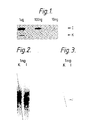
- FIGS. 1 and 3 are autoradiographs in which I represents the DNA sequence of human interferon alpha2 in the plasmid pIFS1101 as set out in Edge et al , Nucleic Acids Research Vol 11, (1983) p6419 to p6435 and K represents the DNA sequence which codes for the interferon apha2 analogue [Ala28]IFN-alpha2 in which the codon TCC is replaced by GCT.
- This analogue base sequence is herein designated as pK9.
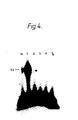
- Figure 4 is an autoradiograph in which lane M shows a signal corresponding to a 32 base long oligonucleotide marker band, lane 1 shows a signal corresponding to a 32 base long ligation product derived from oligonucleotides 11(-1) and 13 (see hereinafter) each with a single dCTP residue incorporated at its 3 ⁇ end. Lanes 2 to 5 fail to shown any signal correponding to a 32 base long oligonucleotide.
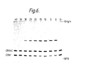
- Figure 5 shows an autoradiograph of aliquots of ligase reaction mixtures containing 5 ⁇ OH trp 3, 5 ⁇ OH trp 4, 5 ⁇ 32p CRN1 and 5 ⁇ 32p CRN2 carried out between 0 and 40°C, these temperatures of incubation appearing at the top of the autoradiograph.
- O- is a no enzyme control.
- BPB indicates the position of the bromophenol blue marker dye.
- Figure 6 shows an autoradiograph of aliquots of ligase reaction mixtures containing 5 ⁇ OH trp 3, 5 ⁇ OH trp 4, 5 ⁇ 32p CRN1 and 5 ⁇ 32p CRN4C carried out between 0 and 40°C, these temperatures of incubation appearing at the top of the autoradiograph.
- O- is a no enzyme control.
- BPB indicates the position of the bromophenol blue marker dye.
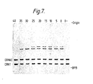
- Figure 7 shows an autoradiograph of aliquots of ligase reaction mixtures containing 5 ⁇ OH trp 3, 5 ⁇ OH trp 4, 5 ⁇ 32p CRN1 and 5 ⁇ 32p CRN6C carried out between 0 and 40°C, these temperatures of incubation appearing at the top of the autoradiograph.
- O- is a no enzyme control.
- BPB indicates the position of the bromophenol blue marker dye.
- SSC 0.15M NaCl + 0.015M sodium citrate
- Kinase buffer is 0.066M Tris.HCl pH 7.6, 1mM spermidine, 0.01M MgCl2, 15mM dithiothreitol and 0.2 mg/ml bovine serum albumin (BSA);
- 10 x CORE BUFFER is 500 mM Tris HCl pH8, 100 mM MgCl2, 500 mM NaCl;
- 10 x nick tranlation buffer is 0.5 M Tris.HCl (pH 7.2), 0.1 M MgSO4, 1mM dithiothreitol and 500 ⁇ g/ml BSA (Pentax, fraction V);
- Triton-X-100 is a polyoxyethylene ether surface active compound;
- Ficoll is a non-ionic synthetic polymer of sucrose having a molecular weight of approximately 400,000 in dialysed and lyophilised form;
- Nonidet P-40 is an o
- Plasmids used for oligonucleotide hybridisation analysis were pIFS1101 (Edge et al., Nucleic Acids Research, Vol 11 (1983) p6419 to 6435) and a derivative thereof designated herein as pK9 in which the codon GCT coding for alanine is present at amino acid position 28 in place of the codon TCC which codes for serine in HuIFN-alpha2.
- Aliquots of 1 ug, 100 ng and 10 ng were diluted to 153 ul in water. To these were added 30 ul 2MNaOH, 17 ul 1M Tris.HCl pH7.4 and 100 ul 20 x SSC (SSC is 0.15M NaCl, 0.015M sodium citrate).
- Oligonucleotides 11 and 13 (Edge et al., see above) corresponding to the sequence of the synthetic interferon alpha2 gene in pIFS1101 were used for hybridisation. Oligonucleotides 11 and 13 have the following DNA sequences:- Oligonucleotide 11 5 ⁇ CGTATCTCCCTGTTC 3 ⁇ Oligonucleotide 13 5 ⁇ TCCTGTCTGAAAGAC 3 ⁇ Oligonucleotide 13 was labelled with 32p as follows: to 400 ng of oligonucleotide in 18.5 ul H2O was added 20 ul 32p gamma ATP (Adenosine 5 ⁇ -[32p]triphosphate, triethylammonium salt in aqueous solution, approx.
- Oligonucleotides 11 and 13 have the following DNA sequences:- Oligonucleotide 11 5 ⁇ CGTATCTCCCTGTTC 3 ⁇ Oligonucleotide 13 5 ⁇ TCCTGTCTGAAAGAC
- Nitrocellulose filters onto which pIFS1101 or pK9 DNA had been immobilised were prehybridised for 2 hours at 68°C in 5 x Denhardt's reagent (1 g/l Ficoll, 1 g/l polyvinylpyrrolidone, 1 g/l BA (bovine albumin, Fraction V, Miles Laboratories), 5 x SSC, 50mM sodium phosphate pH7, 1% glycine, 0.1% sodium dodecyl sulphate (SDS) and 100 ug sonicated denatured herring sperm DNA (Sigma).
- Denhardt's reagent 1 g/l Ficoll, 1 g/l polyvinylpyrrolidone, 1 g/l BA (bovine albumin, Fraction V, Miles Laboratories)
- 5 x SSC 50mM sodium phosphate pH7, 1% glycine, 0.1% sodium dodecyl sulphate (SDS)
- Hybridisations were performed in 2 mls of 5 x SSC, 0.5% NP40 (BDH) and 250 ug/ml tRNA (Sigma, type X-S) with 100 ng of either oligonucleotides 11 and 13 added together or, as a control, 100 ng of oligonucleotide 13 added alone. Hybridisations were performed overnight at room temperature. Filters were then washed in 6 x SSC, 0.06% sodium pyrophosphate and 20mM sodium phosphate buffer, pH7 for 5 minutes at room temperature and, after changing the wash buffer, for a further 3 minutes at room temperature.
- Filters were then treated with phosphate buffered saline (PBS; 0.01M sodium phosphate pH7.4, 0.13M NaCl) containing 2% BA and 0.1% Triton-X-100(BDH) for 30 minutes at 16°C. Filters were then rinsed 5 times with PBS and 2% BA to remove the Triton-X-100.
- PBS phosphate buffered saline
- Filters were then rinsed 5 times with PBS and 2% BA to remove the Triton-X-100.
- filters were treated with 66mM Tris.HCl pH7.6, 5mMMgCl2, 5mM dithiothreitol, 1mM ATP, 500 ug/ml BSA and 0.3 U/ul T4 DNA ligase (Boehringer, 5U/ul). Incubation was for 80 minutes at 16°C.
- This background signal represented about 1% of the corresponding signal obtained for pIFS1101 and resulted from the minimal hybridisation of oligonucleotide 13 because of hybrid mismatches with the corresponding region of the plasmid pK9.
- adjacent hybridised oligonucleotide probes were shown by this example to cross link efficiently by ligation on nitrocellulose filters.
- the ligation time could be reduced to 5 minutes without any loss of autoradiographic signal intensity.
- hybridisation temperature was 32°C using 100 ng oligonucleotide 11 and 100 ng 32p labelled oligonucleotide 13 as probes.
- the filters were washed for 30 minutes at room temperature in 5 x SSC, 0.06% sodium pyrophosphate and 20mM sodium phosphate pH7 and then at 40°C. for 15 minutes to remove most of the 32p labelled oligonucleotide associated with the pK9 plasmid DNA.
- the filters were then exposed at -70°C to Kodak X-Omat AR Film using intensifying screens. After this 40°C wash, a background signal derived from hybridisation to Daudi cell DNA was still evident as shown in Figure 2.
- Dried agarose gels were hybridised directly with 100 ng of both oligonucleotides 11 and 32p labelled 13 in 50 mM sodium phosphate pH7, 0.9 M NaCl, 5 mM EDTA, 0.3% SDS and 10 ug/ml E.coli DNA at 32°C overnight.
- the dried gels were subsequently washed in 2 x SSPE (SSPE is 10mM sodium phosphate pH7, 0.18M NaCl, 1mM EDTA) and 0.1% SDS at room temperature for 30 minutes and then at 40°C for 15 minutes in the same washing buffer.
- SSPE is 10mM sodium phosphate pH7, 0.18M NaCl, 1mM EDTA
- oligonucleotides 11 and 13 hybridise to DNA immobilised either in nitrocellulose or dried gels were cross-linked by ligation, as described in Example 1, and finally washed at 60°C for 15 minutes as above. This resulted in a single band corresponding to 1 ng of plasmid pIFS1101 with little observable background and with no signal evident corresponding to pK9 DNA as shown in Figure 3 for DNA immobilised on nitrocellulose.
- the plasmid DNA used for analysis was pIFS1101 (Edge et al., Nucleic Acids Research, vol. 11 (1983) p6419 to 6435). 2 ⁇ g of pIFS1101 plasmid DNA was digested with EcoRI (20 units, Bethesda Research Laboratories) in a reaction volume of 20 ⁇ l for 2 hours using conditions recommended by the enzyme supplier. The reaction mixture was extracted once with phenol: chloroform (1:1) and once with chloroform before adding 2 ⁇ l 3M sodium acetate pH 5.2 and 45 ⁇ l ethanol. The DNA was precipitated at -20°C overnight, washed in 70% EtOH and dried prior to resuspension in 30 ⁇ l H2O.
- oligonucleotides used for analysis were designated 11(-1) and 13 where oligonucleotide 13 was used to construct at the synthetic interferon alpha2 gene in pIFS1101 and oligonucleotide 11(-1) is a derivative of oligonucleotide 11 used for gene construction with a sequence shifted 5 ⁇ by 1 base.
- sequences of the oligonucleotides were as follows:- Oligonucleotide 11(-1) 5 ⁇ CCGTATCTCCCTGTT3 ⁇ Oligonucleotide 13 5 ⁇ TCCTGTCTGAAAGAC3 ⁇
- Oligonucleotide 13 was 5 ⁇ end labelled with 32p using polynucleotide kinase as described for synthetic linkers (forward reaction) in Molecular Cloning, a Laboratory Manual (Editors: Maniatis, Fritsch and Janbrook, Cold Spring Harbor). The kinase reaction was stopped by heating to 70°C for 10 minutes followed by 2 successive phenol extractions. The solution was then extracted with ether and excess ether was blown off.
- DNA samples were dissolved in 10 ⁇ l H2O to which was added 1 ⁇ l of either 2nM dCTP or 2nM dGTP (PL Biochemicals). Then 2.5 ⁇ l of 10 x nick translation buffer (see Molecular Cloning, a Laboratory Manual referred to above) 10 x buffer is 0.5 M Tris.HCl (pH 7.2), 0.1M MgS04 1mM dithiothreitol and 500 ⁇ g/ml BSA (Pentax, fraction V) was added and the solutions were diluted with water to 25 ⁇ l. 2 Units of the Klenow fragment of DNA polymerase I (Boehringer) was added and the solutions incubated at room temperature for 30 minutes.
- 10 x buffer is 0.5 M Tris.HCl (pH 7.2), 0.1M MgS04 1mM dithiothreitol and 500 ⁇ g/ml BSA (Pentax, fraction V) was added and the solutions were diluted with water to 25 ⁇ l. 2 Unit
- oligonucleotides in this study were characterised by DNA sequencing (Maxam A M and Gilbert W., Proc. Natl. Acad. Sci. USA 74 , 560-564 (1977)). Ligation products were quantified after separation by polyacrylamide gel electrophoresis by Cerenkov counting of radioactivity in putative product bands. In all cases where a mismatched oligonucleotide was ligated in the presence of the fully complementary oligonucleotide CRN 1 the yields of mismatched ligated product were minimal until the mismatched base was separated from the ligation point by 6 base pairs and until the temperature of the ligation reaction in these cases was less then 35°C.
- the region bearing the mismatch is the -35 region of the synthetic E. Coli trp promoter.
- CRN2 control
- CRN3A control
- 6C 6C
- Oligonucleotides were synthesised by the improved solid phase phosphotriester method (Gait, M J et al (1980) Nucleic Acids Research 8 , 1081-1096 and Markham, A F et al (1980) Nucleic Acid Research 8 , 5193-5205) and purified by high performance liquid chromatography initially on Parisil 10-SAX anion-exchange columns (Jones Chromatography, Glamorgan or Whatman Ltd) then on C18 ⁇ Bondapak reverse phase columns (Waters Associates Inc.) as described (Newton, C R et al (1983) Anal. Biochem. 129 , 22-30).
- T4 DNA ligase was from Boehringer Mannheim GmbH and the unit definition used throughout is that according to Weiss et al (Weiss, B et al (1968) Journal of Biological Chemistry 243, 4543-4555). One unit being the enzyme activity which exchanges at 37°C Inmole 32PPi into Norit absorbable material within 20 minutes. Ligase reactions were carrid out in 66 mM Tris-HCl (pH7.6), 6.6 mM MgCl2, 10 mM dithiothreitol and 0.4 mM ATP.
- T4 polynucleotide kinase was from New England Biolabs, the unit definition being the enzyme activity producing 1nmole of acid insoluble 32p in 30 minutes at 37°C.
- Kinase reactions were carried out in 50 mM Tris-HCl (pH 9.0), 10 mM MgCl2, 20 mM dithiothreitol, 0.1 mM spermidine, 0.1 mM EDTA.
- the ATP concentration was variable depending on the subsequent use of phosphorylated oligonucleotide.
- Adenosine 5 ⁇ -[ ⁇ -32p] triphosphate, triethylammonium salt (32p ATP) was from Amersham International, 1mCi per ml in aqueous ethanol with specific activity>5000 Ci/mmol.
- Electrophoresis was through 20% acrylamide (19.33% acrylamide, 0.67% bisacrylamide) gels containing 7M Urea, 50 mM Tris-borate (pH 8.3) 1 mM EDTA unless otherwise stated.
- Electrophoresis samples were either resuspended or diluted with 80% formamide, 10 mM NaOH, 1 mM EDTA, 0.1% bromophenol blue. Samples were heat denatured at 100°C for 2 minutes then rapidly cooled on ice prior to electrophoresis. Electrophoresis was across a potential gradient of 40V/cm till the bromophenol blue had migrated 15 cm from the origin unless stated otherwise.
- 32p labelled oligonucleotides were purified by polyacylamide gel electrophoresis and DNA was recovered from gels by electroelution into dialysis bags containing 10 mM Tris-borate (pH 8.3), 0.2 mM EDTA (1 ml) across a potential gradient of 16V/cm for 45 minutes after which the polarity was reversed for 20 seconds. Electroeluates were concentrated by successive butanol extractions to a voldume of approximately 500 ⁇ l, extracted with phenol-chloroform [buffered with 0.5 M Tris- HCl (pH 7.5) then extracted with water] (500 ⁇ l).
- Aqueous phases were extracted with ether (3 x 1 ml), further concentrated by butanol extraction to approximately 20 ⁇ l, dried in vacuo and ethanol precipitated. Ethanol precipitation was accomplished by resuspending the DNA in 0.3 M sodium acetate (pH 5.6, 250 ⁇ l) then addition and mixing of ethanol (800 ⁇ l), chilling at -70°C for 90 minutes or longer followed by centrifuging for 15 minutes in an Eppendorf microcentrifuge. The pellets were dried in vacuo , resespended in water (500 ⁇ l).
- Cerenkov radiation was determined using an Intertechnique SL4000 Liquid Scintillation Counter with on line M1750 silent 700 printer. Electroeluates were transferred to siliconised 750ml microcentrifuge tubes placed in 20 ⁇ l standard plastic counting vials (Packard). Samples were allowed to dark adapt for 10 minutes then Cerenkov radiation determined by setting the Liquid Scintillation spectrometer to count for tritium. Bands from gels wee excised, placed directly into plastic counting vials and Cerenkov radiation was determined as described for electroeluates. Quenching of Cerenkov radiation in gel bands relative to the same amount of 32p-labelled oligonucleotide in solution could thus be allowed for.
- a total of 220 competition reactions were performed. Each mismatch oligonucleotide or CRN2 (sequence control) were competed with CRN1 in a DNA Ligase assay at 0, 5, 10 , 15, 20, 25, 30, 35 and 40°C. In each case a 0°C no enzyme control was included. All competitions were carried out in a volume of 60 ⁇ 1 and contained trp 4(5.0pmole), 3 (7.5p Mole), CRN1 (7.5p Mole) and competing oligonucleotide (7.5p Mole). In molar concentrations total oligonucleotide was 456.5 nM, trp 4 was 83 nM, trp 3, CRN 1 and competitor were all 124. 5 nM.
- oligonucleotide For each competing oligonucleotide a "competition mix” was prepared comprising competitor (75p Mole) and CRN1 (75 p Mole) in aqueous solution (390 ⁇ l). The competition mixes were divided into 10 equal portions. Both oligonucleotides were 5 ⁇ - phosphorylated with 32p -ATP and polynucleotide kinase. They were further purified by polyacrylamide gel electrophoresis, electroelution and ethanol precipitation as described above. Other reaction components were combined in a " trp mix” which contained trp 3 (7.5p Mole), trp 4 (5.0p Mole) and 10X DNA ligase buffer (6 ⁇ l) per 20 ⁇ l.
- trp mix which contained trp 3 (7.5p Mole), trp 4 (5.0p Mole) and 10X DNA ligase buffer (6 ⁇ l) per 20 ⁇ l.
Abstract
Description
- The present invention relates to a method for the use of hybridisation probes, hybridisation probes therefor and kits for use in such a method. The present invention is particularly concerned with discriminating a specific base sequence from a variant base sequence and relates in particular to the advantages which may be obtained by subjecting adjacent segments of a target base sequence to hybridisation with a detectable first nucleotide probe and a second nucleotide probe.
- The present invention thus relates inter alia to discriminating specific base sequences whilst ameliorating the problem of background non-specific hybridisation commonly encountered with hybridisation of short oligonucleotide probes to complex genomes.
- Nucleic acid hybridisation analysis is a technique of wide applicability in the fields of biomedical research and recombinant DNA technology. Thus for example hybridisation probes are useful for detecting, monitoring, locating and isolating nucleic acids and other molecules of scientific or clinical interest. Particularly useful are small oligonucleotide hybridisation probes which may for example be used to detect changes in DNA base sequences in relation to certain disease states such as phenylketonuria, alphaantitrypsin deficiency, alpha- and beta-thalassaemia and sickle cell anaemia. These disease states are often associated with known single point mutations in genes.
- Whilst short oligonucleotide hybridisation probes are of considerable utility, they are usually associated with a high background of non specific hybridisation particularly when used for analysis of the genomes of higher organisms. This problem arises from the greater complexity of the genome. Thus for example mammals may contain of the order of a thousand-fold more DNA per cell than bacteria, and many of the nucleotide sequences may be repeated. For any given nucleotide probe therefore stable duplexes (hereinafter also referred to as hybrids) other than the desired duplex will usually be formed. This problem is particularly exacerbated by using very high oligonucleotide concentrations in order to accelerate the hybridisation process. This problem can be circumvented by using gel electrophoresis to resolve different DNA (or RNA) fragments before oligonucleotide hybridisation to ensure that a specifically hybridising fragment is resolved from non-specifically hybridising fragments. For example, Woo et al (Banbury Report, Recombinant DNA Applications to Human Disease, Cold Spring Harbor Laboratory (1983) p105-110) employ the Southern transfer technique to resolve small restriction fragments hybridising to an alpha₁ -antitrypsin oligonucleotide probe from higher molecular weight species of DNA which also hybridise to this probe.
- Oligonucleotide hybridisation analysis of complex genomes using gel electrophoresis requires the preparation of pure DNA (or RNA) molecules from the organisms, fragmentation of DNA molecules by treatment with restriction endonucleases and, in most cases, transfer of resolved DNA (or RNA) molecules from the gel to a solid support for hybridisation. The complexity of this procedure makes it unsuitable for routine diagnostic use and difficult to automate.
- Simpler methods for hybridisation analysis with specifically hybridising long polynucleotide probes are exemplified by J Brandsma and G Miller, Proc. Nat. Acad. Sci.(USA) Vol 77 (1980) p6851-6855 for DNA and by T. Manser and M L Gefter Proc.Natl. Acad.Sci USA Vol 81 (1984) p2470-2474 for RNA and involve directly immobilising DNA or RNA from crude cellular lysates onto solid supports. Because of non-specific hybridisation, such direct immobilisation methods are not usually compatible with the use of oligonucleotide probes.
- In addition to the above problem of background non-specific hybridisation the present invention also relates inter alia to simplifying the detection of translocations which hitherto have only been detectable by gel methods.
- The present invention is thus based, at least in part, on the discovery of a method for discriminating between alternative nucleotide sequences whilst ameliorating the above-mentioned problems. The method of the present invention is thus of interest in inter alia simplifying the detection of translocations and enabling a mutation in a given base sequence to be readily detected by for example allowing meaningful analysis of directly immobilised DNA or RNA samples or alternatively DNA or RNA samples in solution. In this connection it is known that the shorter the nucleotide probe the poorer is the selectivity of hybridisation and thus correspondingly the greater is the background. Moreover the shorter the nucleotide probe the weaker is the stability of the duplex formed as measured by its melting temperature (Tm) which is the temperature of the midpoint of thermal transition. The present invention is based at least in part, on the discovery that the different hybridisation properties of a short nucleotide probe relative to a comparitively longer nucleotide probe may be exploited to advantage in discriminating between alternative nucleotide sequences, whilst ameliorating the above-mentioned problems.
- The present invention is further based on the discovery of a method which substantially simplifies the hybridisation analysis thus providing a robust technique for discriminating between alternative nucleotide sequences.
- Thus according to one feature of the present invention there is provided a method for discriminating between alternative nucleotide sequences, which method comprises subjecting adjacent segments of a target base sequence to hybridisation with a detectable first nucleotide probe and with a second nucleotide probe, to form a hybrid, the nucleotide sequence of the first and second probe being such that where they form a split probe hybrid with a complementary target sequence they may subsequently be linked subjecting any hybrid obtained to linkage, followed by detection of any hybrid obtained;
the DNA sequence of the detectable first nucleotide probe and of the second nucleotide probe being such that a potential mismatch in the target sequence lies either between the said probes or at the terminal end of one of said probes, which terminal end is contiguous with the other of the said probes;
the method being effected such that a complementary target sequence is discriminated from a target sequence with one or more non-complementary nucleotides. - Where necessary any hybrid obtained following linkage may be subjected to selective denaturing.
- The presence of a linked probe may be detected by any convenient means for example selective denaturing or where one of the probes carries one member of a binding pair then by use of the other member of the binding pair. Appropriate examples of binding pairs are discussed hereinafter. It will be appreciated that one might identify either a complementary or a non-complementary target sequence by the absence of a dectectable hybrid and this is included within the scope of the present invention.
- It will be appreciated that hitherto discriminatory specificity has been achieved by careful manipulation of the temperature of hybridisation and washing of bound discriminatory probes. In the present invention the probes are designed to hybridise to the target sequence on either side of a potential mismatch, there being a gap, preferably a single nucleotide gap, between the probes or the probes may be designed to hybridise to contiguous sequences in the target sequence any potential mismatch being present at the terminal end of one of the probes which terminal end is contiguous with the other probe. The effect of the present invention is that specificity is mainly the result of the interaction between two independently interacting probes and the means of linking the aforesaid probes, for example T4 DNA ligase. Thus the present invention enables a method of discrimination to be effected in which linkage e.g. ligation by for example T4 DNA ligase is expected to be less efficient where the residue(s) at either the 3ʹ or the 5ʹ site of the contiguous probes to be joined are not complementary to the sequence to which they are hybridised. Similarly where a gap, preferably a single nucleotide gap, is present between the two probes the present invention enables a method of discrimination to be effected in which incorporation of nucleotides with for example the Klenow fragment of DNA polymerase I or calf thymus DNA polymerase alpha to fill in the gap and allow linkage, with for example DNA ligase, will be significantly less efficient where the deoxy nucleotide triphosphate added, to for example the DNA polymerase reaction mixture is not complementary to the base residue under examination.
- The present invention thus enables the hybridisation analysis to be substantially simplified and allows the production of a robust technique for discriminating between alternative nucleotide sequences. Thus for example the need to rely on the use of a hybridisation or wash within a narrow temperature range and for a specific time may be obviated thus overcoming the difficulties inherent in any technique which relies on narrow margins of error. The present invention even renders it possible in at least certain embodiments to effect the hybridisation analysis at room temperature.
- The method of the present invention may for example be effected either by detection of a detectable signal, resulting from complementary hybrid formation where a complementary target sequence is present, the hybrid, if necessary, being denatured prior to detection; or by failure to detect a detectable signal, resulting from the absence of hybrid formation across a mismatch or from selective denaturation of any hybrid formed, where a target sequence with one or more non-complementary nucleotides is present.
- Thus whilst the first nucleotide probe must be detectable the second nucleotide may or may not be detectable depending on the embodiment of the method to be employed.
- The term detectable is used herein to mean capable of detection. Thus the first nucleotide probe need not carry a signalling means such as a radioactive label or a non-radioactive signalling complex, although such may be present, provided that the probe may subsequently be treated to render it capable of signalling. It is thus possible to discriminate between sequences in a complex genome differing by as little as one base pair.
- Where a split probe hybrid would be formed following hybridisation, with a complementary target base sequence, any hybrid obtained is subjected to a linking reaction for linking the detectable first nucleotide probe to the second nucleotide probe, any hybrid obtained being subjected to appropriate treatment whereby any hybrid present, in which linkage of the detectable first nucleotide probe to second nucleotide probe has not been effected, is denatured whereas no denaturation is effected for a perfectly complementary detectable linked oligonucleotide-target sequence hybrid. Where a probe is designed to hybridise to a portion of the target sequence which contains a suspected mismatch, the probe will generally be an oligonucleotide (as hereinafter defined). Appropriate treatment may for example include selective denaturation or one of the probes may comprise one member of a binding pair. Linkage of adjacent stable hybrids of first and second nucleotide probes results in greater hybrid thermal stability for the detectable oligonucleotide hybridised to a specific target sequence as opposed to detectable oligonucleotides hybridised to non specific target sequences with no adjacent oligonucleotide probe present.
- The expression "oligonucleotide" as used herein means a nucleotide sequence which is either incapable of forming a hybrid with a target sequence containing as little as a single base pair mismatch or is capable of forming such a hybrid, such a hybrid however being destabilised by the presence of as little as a single base pair mismatch such that it may be selectively denatured under conditions which would not denature a corresponding perfectly complementary hybrid.
- It will be appreciated that the potential variant sequence may be present in the segment of the target sequence to which the detectable first nucleotide probe hybridises, it may be present in the segment of the target sequence to which the second nucleotide probe hybridises or it may be present between the said segments. The probes will be designed such that where the potential variant sequence is present in the segment of the target sequence to which either the detectable first nucleotide probe or the second nucleotide probe hybridises, then the potential variant sequence will be at the terminal end of one of said probes, which terminal end is contiguous with the other of the said probes.
- Thus for example where the potential variant sequence is present in the segment of the target sequence to which the second nucleotide probe hybridises, formation of a split probe hybrid, will result either in formation of a weak hybrid of the second probe across the mismatch or if the second probe is very short no hybridisation at all. Where a weak hybrid is formed selective denaturing will result in denaturing of the second probe hybrid. If the hybrid obtained is then subjected to linking, linkage can only take place where the second probe is hybridised to the target sequence and thus where no mismatch exists in the second probe: target sequence hybrid. If the hybrid obtained after linking is subjected to selective denaturing any hybrid present in which the first probe alone is hybridised to the target sequence without linkage to the second probe will denature thus leaving detectable probe hybridised only to the target sequence which does not contain a mismatch with the complementary sequence of the second probe.
- The first or second nucleotide probe may if desired have one member of a binding pair. Generally the one member of a binding pair will be carried by, or be part of, the second nucleotide probe. The other member of the binding pair may be in solution or on a support. Thus where appropriate, the second nucleotide probe linked to the detectable first nucleotide probe may be isolated on a support carrying the other member of the binding pair. Such a binding pair may for example be a protein-ligand or antigen-antibody interaction such as an avidin-biotin or dinitrophenyl-antidinitrophenyl antibody interaction.
- Indeed one member of the binding pair may be a nucleotide sequence, which sequence may be a portion of the probe sequence or may be a nucleotide sequence branch on the probe. The other member of the binding pair may for example be a protein which binds to the nucleotide sequence or another nucleotide sequence to which it hybridises.
- The hybridisation and/or selective denaturation is preferably effected at a temperature selected to give effective hybridisation selectivity, preferably maximum hybridisation selectivity for the specific length of the linked probe. Advantageously the hybridisation and/or selective denaturation is effected in aqueous solution at an elevated temperature suitable for selective hybridisation to a mammalian genome which for a split probe when linked is preferably above 60°C generally about 68°C. Alternatively hybridisation and/or selective denaturation of such a split probe when linked may be effected at a lower temperature in the presence of an organic solvent which is effective to destabilize the hybrid, such as a solvent containing formamide. For example where about 50% formamide is used the hybridisation and/or selective denaturation is effected at about 42°C.
- In a further embodiment of the present invention there is provided a method of hybridisation probing which comprises subjecting adjacent segments of the target base sequence to hybridisation with a detectable first nucleotide probe and with a second nucleotide probe such that a split probe hybrid would be formed with a complementary target sequence, where necessary subjecting the hybrid obtained to selective denaturing whereby to denature whichever of the first and second nucleotide probes is hybridised to a portion of the target base sequence with one or more non-complementary nucleotides and subsequently subjecting the hybrid obtained to a linking reaction whereby to link together the detectable first nucleotide probe to the second nucleotide probe and where necessary subjecting the hybrid obtained to selective denaturation whereby to discriminate between a target sequence complementary to the first and second nucleotide probes and a target sequence with one or more nucleotides non-complementary to one of the first and second nucleotide probes.
- The hybridisation or selective denaturation may be effected for example at a temperature above the melting temperature of the split or single nucleotide probe hybrid but below the melting temperature of the linked probe hybrid or in the presence of an organic solvent effective to destabilise the hybrid as discussed above. Where hybridisation is effected under selective denaturing conditions a further selective denaturing step after the hybridisation but before linking may be avoided.
- The first and second nucleotide probes may be linked by any convenient means known per se such as for example by enzymatic ligation using, for example, a DNA ligase or by covalent/non-covalent linking using for example a biotin-avidin cross-link.
- The detectable first nucleotide probe and/or the second nucleotide probe may initially not be capable of being linked, e.g. ligated, together until the gap(s) between them is (are) filled with DNA polymerase in the presence of the target sequence of interest.
- The effect of linking the detectable first nucleotide probe and second nucleotide probe is to increase the thermal stability of the cross-linked probe hybrid obtained over the corresponding detectable probe hybrid alone. Thus for example the temperature of the linked probe hybrid may be increased to a temperature which denatures the corresponding split probe hybrids or a corresponding hybrid in which only one of the detectable first nucleotide probe and second nucleotide probes is hybridised to the target base sequence. The linked probe hybrid which includes the complementary target base sequence is thus left intact. The method of the present invention thus enables one to discriminate a base sequence which is perfectly complementary to the detectable probe sequence from alternative sequences.
- It will be appreciated that while the above embodiment of the invention is most useful for directing oligonucleotides to specific hybridisation sites in complex genomes in order for example, to analyse for the presence of point mutations it is also of use for analysis of other variations in complex genomes such as translocations. For analysis of translocations, it is preferred that the non-detectable second polynucleotide of a split probe will have one member of a binding pair associated therewith and will hybridise to genomic sequences adjacent to potential translocation breakpoints. The detectable probe will preferably be several kilobases long and span a region of potential translocation breakpoints. Quantitative linkage of polynucleotides comprising the split probe following hybridisation will only occur where the genomic region is contiguous and uninterrupted by translocations. The occurrence of a translocation will preclude a proportion of the detectable probe from linking to the non-detectable polynucleotide. On subsequent denaturation of hybridised polynucleotide and application of the other member of the binding pair, a proportion of the detectable polynucleotide will not then be found to be associated with the polynucleotide complexed with the other member of the binding pair. The binding pair will preferably comprise an antigen-antibody or a biotin-avidin interaction whereby, for example, denatured hybrids are passed through a solid phase complexed with one member of the binding pair and the eluate analysed for the presence of the detectable probe.
- Thus in a further embodiment of the invention we provide a method for the analysis of translocations in a target base sequence e.g. a complex genome in which the detectable first nucleotide probe is adapted to hybridise across a region of potential translocation(s) and the non-detectable second nucleotide probe is adapted to hybridise to sequences adjacent to said potential translocation(s), the non-detectable second nucleotide probe carrying one member of a binding pair, the method comprising formation of a split probe hybrid which is subjected to linking to form a linked probe hybrid which hybrid is then denatured.
- In another embodiment of the present invention, there is provided a method as hereinbefore defined wherein each of the detectable first nucleotide probe and second nucleotide probe carry a moiety such that after hybrid formation, and denaturation where appropriate, a signal is only detectable if a nucleotide sequence is obtained which carries both the moiety attached to the detectable first nucleotide probe and the moiety attached to the second nucleotide probe.
- The nucleotide probes may for example be oligonucleotide probes.
- The method of this embodiment might for example be effected using non-radioactive energy transfer procedures (see for example European Patent Publication No. 70685 of Standard Oil Co.) or enzyme chanelling techniques (see for example European Patent Publication No. 95087 of Syva Co.). Thus this embodiment may provide a homogenous assay for a specific target nucleotide sequence.
- In another embodiment of the invention there is provided a method for discriminating between alternative nucleotide sequences in which a target sequence is subjected to hybridisation with more than two oligonucleotide probes such that where a complementary target sequence is present a split probe hybrid is obtained, the detectable first nucleotide probe being a terminal oligonucleotide probe which is detectable and the second nucleotide probe being the other terminal oligonucleotide probe which has attached thereto one member of a binding pair, individual other oligonucleotide probe(s) being hybridised separately but adjacently to a contiguous target sequence between the said terminal oligonucleotide probes, the hybrid obtained is then subjected to selective denaturation whereby to denature any oligonucleotide probe hybridised to the target sequence across a base pair mismatch, and the individual oligonucleotide probes linked to join the detectable oligonucleotide probe to the oligonucleotide probe having one member of a binding pair attached thereto to form a linked probe hybrid which is then denatured and contacted with the other member of the binding pair whereby the detectable linked probe nucleotide sequence including the said binding pair may be separated from other nucleotide sequences. Thus one terminal oligonucleotide (hybridising adjacent to a series of other oligonucleotides) for example has attached thereto a detectable signalling moiety while the other terminal oligonucleotide has attached a moeity forming part of a binding pair the moiety being effective to enable the oligonucleotide to be recovered in solution from a mixture containing other nucleotide sequences. Adjacently hybdridised oligonucleotides are linked together by appropriate treatment in order to effectively join the signalling and binding moieties via a linked probe where no oligonucleotide hybrid is denatured prior to linkage. Loss of any individual oligonucleotide hybrid results in subsequent failure to join the signalling and binding moieties. The linked probe hybrid is denatured and contacted with the other member of the binding pair which recognises one terminal oligonucleotide in the linked probe. The linked probe may thus if desired be separated from other oligonucleotides in the mixture and then analysed for the presence of the detectable moiety
- Thus the association of the binding oligonucleotide with the signalling oligonucleotide is dependent on the presence, upon linking of adjacently hybridised oligonucleotides, of all adjacent oligonucleotides between the hybridised signalling oligonucleotide and the binding oligonucleotide. The binding pair effecting separation of the linked probe with an associated binding moiety may be avidin and biotin or an antigen and associated specific antibody. This embodiment provides for analysis of longer stretches of target nucleotide sequence than with just two adjacent hybridising linked or split probes as in other embodiments.
- A further embodiment of the present invention comprises hybridising the detectable first nucleotide probe and second nucleotide probe to each side of a potential variant sequence in a target sequence and subsequently attempting to link the said probes, preferably by the use of DNA polymerase introducing a nucleotide(s) complementary to either the normal or the suspected variant sequence and ligation, followed by detection of any hybrid obtained. Such detection may for example be effected by subjecting any hybrid obtained to selective denaturing whereby any hybrid present, in which linkage of the detectable first nucleotide probe to the second nucleotide probe has not been effected, is denatured.
- The DNA polymerase used may for example be the Klenow fragment of DNA polymerase I or calf thymus DNA polymerase alpha. Ligation is preferably effected with DNA ligase. The detectable first nucleotide probe and second nucleotide probe are preferably such that they hybridise to the target sequence whereby to leave a gap of a single nucleotide between the said probes.
- Where it is desired that the nucleotide probe should carry a signal or a residue capable of producing a signal, such signals or residues may be known per se e.g. a radioactive label.
- The residue which is capable of producing a non-radioisotopic signal may alternatively comprise a signalling part normally separated from the oligonucleotide by a spacer group. This spacer group may if desired be linked to the signalling part of the complex by a direct covalent link or by a protein-ligand or antigen-antibody interaction, for example, an avidin-biotin or dinitrophenyl-antidinitrophenyl antibody interaction. It will be understood that the signalling part of the residue may itself be capable of signalling or may be capable of producing a signal by interaction with an appropriate agent according to methods known per se. Thus, for example a preferred signalling part of the covalently attached residue incorporates a system for producing an enzymatically-activated production of colour change. Preferred enzyme systems involve alkaline phosphatase, acid phosphatase, beta galactosidase, luciferase, or horseradish peroxidase. Such enzyme systems are not themselves capable of signalling, but are capable of producing a signal in the presence of an appropriate substrate according to methods known per se. The signalling part of the covalently-attached residue may operate according to any conventional technique such as for example by luminescence, fluorescence or by means of colour.
- In use it will generally be necessary for the nucleic acid probe to detect minute amounts of the fully complementary oligonucleotide sequence. In such circumstances it will be advantageous to incorporate within the probe a means of amplifying the signal. The amplification can be carried out by known techniques, for example using one or more of the systems described in European Patent Publications 27036 (Self), 49606 (Self), 58539 (Self) and 60123 (Self).
- According to a further feature of the present invention there are provided split probe hybrids which comprise a detectable first nucleotide probe and a second nucleotide probe hybridised to adjacent segments of a target base sequence, the detectable first nucleotide probe being capable of linkage to the second nucleotide probe.
- The detectable first nucleotide probe preferably has a nucleotide sequence homologous to a segment of a base sequence associated with a disease state or homologous to a target base sequence adjacent to such a variant sequence or homologous to the corresponding normal sequence. Such a variant sequence is generally a point mutation. Where the sequence of the detectable first nucleotide probe is adjacent to a variant sequence the sequences of the nucleotide probes are such that they are preferably homologous to segments of the target base sequence either side of the potential variant sequence.
- The non-nucleotidyl cross-link may for example be a disulphide link or a biotin-avidin link.
- At least the first nucleotide probe is detectable and thus preferably carries a signal or a residue capable of producing a signal as described above.
- In order to enable the method of the present invention to be effected conveniently it will generally be advantageous to incorporate the nucleotide probes in a kit and this kit is therefore regarded as a further feature of the invention.
- Thus according to a further feature of the present invention there is provided a kit for discriminating between alternative nucleotide sequences which comprises a detectable first nucleotide probe and a second nucleotide probe, each probe having a nucleotide sequence homologous to adjacent segments of a target sequence, a potential variant sequence being present in one of said segments or therebetween; the kit additionally containing a reagent(s) for linking said probes where the probes are not so linked.
- The potential variant sequence will commonly be a point mutation associated with a disease state. In such a case the kit will preferably contain a detectable first nucleotide probe and second nucleotide probe for detecting a point mutation associated with a disease state as well as a detectable first nucleotide probe and a second nucleotide probe for detecting the corresponding normal sequence in which the point mutation was absent.
- It will be appreciated in this regard that certain disease states are characterised by point mutations which appear in different regions of the human DNA genome. In such a case it would be important to determine that each relevant point mutation was present or absent and thus more than one set of probes (detectable first nucleotide probe and second nucleotide probe at least) would be employed, one set of probes to detect the presence or absence of each point mutation. It would also be advantageous to include in the kit set(s) of probes for the corresponding normal or variant sequence.
- The kit will also advantageously contain means for extracting DNA and/or means for immobilising DNA on a solid support.
- If desired at least one of the detectable first nucleotide probe and second nucleotide probe may carry one member of a binding pair, the other member of the binding pair being present for example on a solid support provided with the kit.
- The kit will also comprise appropriate buffer solutions and/or washing solutions and will contain written or printed instructions for use of the kit according to the method of the present invention.
- With reference to the drawings.-
Figures 1, 2 and 3 are autoradiographs in which I represents the DNA sequence of human interferon alpha₂ in the plasmid pIFS1101 as set out in Edge et al, NucleicAcids Research Vol 11, (1983) p6419 to p6435 and K represents the DNA sequence which codes for the interferon apha₂ analogue [Ala²⁸]IFN-alpha₂ in which the codon TCC is replaced by GCT. This analogue base sequence is herein designated as pK9. - Figure 4 is an autoradiograph in which lane M shows a signal corresponding to a 32 base long oligonucleotide marker band,
lane 1 shows a signal corresponding to a 32 base long ligation product derived from oligonucleotides 11(-1) and 13 (see hereinafter) each with a single dCTP residue incorporated at its 3ʹ end.Lanes 2 to 5 fail to shown any signal correponding to a 32 base long oligonucleotide. - Figure 5 shows an autoradiograph of aliquots of ligase reaction mixtures containing 5ʹ
OH trp 3, 5ʹOH trp 4, 5ʹ 32p CRN1 and 5ʹ ³²p CRN2 carried out between 0 and 40°C, these temperatures of incubation appearing at the top of the autoradiograph. O- is a no enzyme control. BPB indicates the position of the bromophenol blue marker dye. - Figure 6 shows an autoradiograph of aliquots of ligase reaction mixtures containing 5ʹ
OH trp 3, 5ʹOH trp 4, 5ʹ ³²p CRN1 and 5ʹ ³²p CRN4C carried out between 0 and 40°C, these temperatures of incubation appearing at the top of the autoradiograph. O- is a no enzyme control. BPB indicates the position of the bromophenol blue marker dye. - Figure 7 shows an autoradiograph of aliquots of ligase reaction mixtures containing 5ʹ
OH trp 3, 5ʹOH trp 4, 5ʹ ³²p CRN1 and 5ʹ ³²p CRN6C carried out between 0 and 40°C, these temperatures of incubation appearing at the top of the autoradiograph. O- is a no enzyme control. BPB indicates the position of the bromophenol blue marker dye. - The invention is illustrated, but not limited, by the following Examples. In the Examples, unless otherwise stated, the solutions are aqueous and the % values are w/v.
- The constitution of various reagents is as follows:-
SSC is 0.15M NaCl + 0.015M sodium citrate;
Kinase buffer is 0.066M Tris.HCl pH 7.6, 1mM spermidine, 0.01M MgCl₂, 15mM dithiothreitol and 0.2 mg/ml bovine serum albumin (BSA);
10 x CORE BUFFER is 500 mM Tris HCl pH8, 100 mM MgCl₂, 500 mM NaCl;
10 x nick tranlation buffer is 0.5 M Tris.HCl (pH 7.2), 0.1 M MgSO₄, 1mM dithiothreitol and 500 µg/ml BSA (Pentax, fraction V);
Triton-X-100 is a polyoxyethylene ether surface active compound;
Ficoll is a non-ionic synthetic polymer of sucrose having a molecular weight of approximately 400,000 in dialysed and lyophilised form;
Nonidet P-40 is an octyl phenol ethylene oxide condensate containing an average of 9 moles ethylene oxide per molecule;
Denhardt's reagent is 0.2 g/l Ficol 400,000, 0.2 g/l polyvinyl pyrrolidone (PVP) and 0.2 g/l bovine albumin (BA);
PBS - (phosphate buffered saline) is 0.01M sodium phosphate pH 7.4 and 0.13M NaCl;
SSPE is 10 mM sodium phosphate pH7, 0.18M NaCl and 1 mM EDTA; - The following contractions are used:-
DNA deoxyribonucleic acid
tRNA transfer ribonucleic acid
EDTA ethylenediaminetetraacetic acid
PBS phosphate buffered saline
SDS sodium dodecyl sulphate
2 x SSC double concentration SSC
6 x SSC six times concentration of SSC
20 x SSC twenty times concentration of SSC
BSA bovine serum albumin
BA bovine albumin
PVP polyvinyl pyrrolidone
Tris tris(hydroxymethyl)aminomethane
10 x Core buffer ten times the concentration of CORE BUFFER
ATP adenosine triphosphate
NP40 Nonidet P.40
10 x nick translation buffer ten times the concentration of nick translation buffer - The following are trade marks
Minifold
Triton-X-100
CORE BUFFER
Ficoll - Plasmids used for oligonucleotide hybridisation analysis were pIFS1101 (Edge et al., Nucleic Acids Research, Vol 11 (1983) p6419 to 6435) and a derivative thereof designated herein as pK9 in which the codon GCT coding for alanine is present at amino acid position 28 in place of the codon TCC which codes for serine in HuIFN-alpha₂. Aliquots of 1 ug, 100 ng and 10 ng were diluted to 153 ul in water. To these were added 30 ul 2MNaOH, 17 ul 1M Tris.HCl pH7.4 and 100 ul 20 x SSC (SSC is 0.15M NaCl, 0.015M sodium citrate). Samples were heated to 100°C for 10 minutes, then placed on ice and 70 ul 1M Tris.HCl pH7 was added. Dilutions of plasmid DNA were filtered onto BA 85 nitrocellulose filters (Schleicher and Schuell Keene, N.H., USA) using a Schleicher and Schuell Minifold II apparatus according to the manufacturer's instructions. The nitrocellulose paper was cut to fit the Minifold apparatus and wetted by floating on 2 x SSC before mounting in the apparatus. DNA samples were then applied to the wells and filtered at a rate of approximately 0.5 ml per minute. The nitrocellulose was then air dried for 10 minutes and baked at 80°C for 2 hours in vacuo. The
oligonucleotides 11 and 13 (Edge et al., see above) corresponding to the sequence of the synthetic interferon alpha₂ gene in pIFS1101 were used for hybridisation.Oligonucleotides 11 and 13 have the following DNA sequences:-
Oligonucleotide 11 5ʹ CGTATCTCCCTGTTC 3ʹ
Oligonucleotide 13 5ʹ TCCTGTCTGAAAGAC 3ʹ
Oligonucleotide 13 was labelled with ³²p as follows: to 400 ng of oligonucleotide in 18.5 ul H₂O was added 20 ul ³²p gamma ATP (Adenosine 5ʹ-[³²p]triphosphate, triethylammonium salt in aqueous solution, approx. 3000 Ci/nmol, 10 uCi/ul. Amersham International), 4.5 ul 10 x kinase buffer (10 x buffer = 0.66M Tris. HCl pH7.6, 10mM spermidine, 0.1M MgCl₂, 150nM dithiothreitol and 2 mg/ml bovine serum albumin (BSA, Nucleic Acid Enzyme Grade, Bethesda Research Laboratories)) and 2 ul T4 polynucleotide kinase (5 units/ul, Boehringer Mannheim). After 1 hour at 37°C, half of the sample was applied to a 4 ml column of Biogel P2 (Biorad; pre-equilibrated with 10mM Tris.HCl pH8, 1mMEDTA). The ³²p labelled nucleotide was eluted in the void volume of the elution buffer comprising 10mM Tris.HCl pH8, 1mM EDTA. 100 ng of ³²p labelled oligonucleotide probe was used for hybridisation together with 100 ngunlabelled oligonucleotide 11. Nitrocellulose filters onto which pIFS1101 or pK9 DNA had been immobilised were prehybridised for 2 hours at 68°C in 5 x Denhardt's reagent (1 g/l Ficoll, 1 g/l polyvinylpyrrolidone, 1 g/l BA (bovine albumin, Fraction V, Miles Laboratories), 5 x SSC, 50mM sodium phosphate pH7, 1% glycine, 0.1% sodium dodecyl sulphate (SDS) and 100 ug sonicated denatured herring sperm DNA (Sigma). Hybridisations were performed in 2 mls of 5 x SSC, 0.5% NP40 (BDH) and 250 ug/ml tRNA (Sigma, type X-S) with 100 ng of eitheroligonucleotides 11 and 13 added together or, as a control, 100 ng of oligonucleotide 13 added alone. Hybridisations were performed overnight at room temperature. Filters were then washed in 6 x SSC, 0.06% sodium pyrophosphate and 20mM sodium phosphate buffer, pH7 for 5 minutes at room temperature and, after changing the wash buffer, for a further 3 minutes at room temperature. Filters were then treated with phosphate buffered saline (PBS; 0.01M sodium phosphate pH7.4, 0.13M NaCl) containing 2% BA and 0.1% Triton-X-100(BDH) for 30 minutes at 16°C. Filters were then rinsed 5 times with PBS and 2% BA to remove the Triton-X-100. For cross-linking of hybridised probes, filters were treated with 66mM Tris.HCl pH7.6, 5mMMgCl₂, 5mM dithiothreitol, 1mM ATP, 500 ug/ml BSA and 0.3 U/ul T4 DNA ligase (Boehringer, 5U/ul). Incubation was for 80 minutes at 16°C. Filters were then washed at 60°C for 20 minutes in 6 x SSC, 0.06% sodium pyrophosphate and 20mM sodium phosphate pH7. The filters were then wrapped in Saran wrap and exposed to autoradiographic film for 2 days. Figure 1 shows that after the final wash at 60°C, an appreciable autoradiographic signal was observed only for the plasmid pIFS1101 (I) where hybridisedoligonucleotides 11 and 13 had been cross linked by ligation. A background signal corresponding to immobilised pK9 (K) was observed after hybridisation and cross linking ofoligonucleotides 11 and 13. This background signal represented about 1% of the corresponding signal obtained for pIFS1101 and resulted from the minimal hybridisation of oligonucleotide 13 because of hybrid mismatches with the corresponding region of the plasmid pK9. Thus adjacent hybridised oligonucleotide probes were shown by this example to cross link efficiently by ligation on nitrocellulose filters. Moreover, with the experimental conditions described the ligation time could be reduced to 5 minutes without any loss of autoradiographic signal intensity. - High molecular weight DNA was isolated from the lymphoblastoid line Daudi using the method of Blin and Stafford (Nucleic Acids Research, Vol.3 (1976) p2303). 200 ug of Daudi DNA was diluted into 320 ul H₂0 and to this was added 40 ul of 10 x CORE BUFFER (Bethesda Research Laboratories) and 40 ul EcoRI (50 units/ul Northumbria Biologicals, Cramlington, Northumberland, UK). Following an incubation at 37°C for 3 hours, the solution was extracted with an equal volume of phenol/chloroform (1:1), then with 1 volume of chloroform and finally three times with 1 volume of H₂O saturated ether. The DNA was adjusted to 0.3M sodium acetate and precipitated with 2 volumes of ice cold ethanol. The DNA pellet was washed in cold 70% ethanol and the pellet, dried and redissolved in 100 ul H₂O.
- 3 ug of the plasmids pIFS1101 and pK9 (see Example 1) were dissolved in 17 ul H₂O and to this was added 2 ul 10 x CORE BUFFER and 1 ul EcoR1 (see above). The mixture was incubated for 3 hours at 37°C and was followed by solvent extractions and Et0H precipitations as for the Daudi DNA above. The dried pellets were redissolved in 10 ul H₂O.
- 10 ug of EcoR1 digested Daudi DNA was mixed with 1 ng aliquots of either pIFS1101 or pK9 and subjected to gel electrophoresis on a 0.5% horizontal agarose gel as described in Maniatias et al (Molecular Cloning, A Laboratory Manual: Cold Spring Harbor Laboratory, 1982). Following electrophoresis, DNA was either transferred onto nitrocellulose filters by the Southern transfer procedure (Maniatis et al) for subsequent hybridisation or, alternatively, DNA was probed directly within dried agarose gels prepared as described by Studencki and Wallace (DNA,
volume 3, p7 to 15 (1984)). For DNA transferred onto nitrocellulose filters hybridisation was as described in Example 1 except that the hybridisation temperature was 32°C using 100ng oligonucleotide 11 and 100 ng ³²p labelled oligonucleotide 13 as probes. The filters were washed for 30 minutes at room temperature in 5 x SSC, 0.06% sodium pyrophosphate and 20mM sodium phosphate pH7 and then at 40°C. for 15 minutes to remove most of the ³²p labelled oligonucleotide associated with the pK9 plasmid DNA. The filters were then exposed at -70°C to Kodak X-Omat AR Film using intensifying screens. After this 40°C wash, a background signal derived from hybridisation to Daudi cell DNA was still evident as shown in Figure 2. - Dried agarose gels were hybridised directly with 100 ng of both
oligonucleotides 11 and ³²p labelled 13 in 50 mM sodium phosphate pH7, 0.9 M NaCl, 5 mM EDTA, 0.3% SDS and 10 ug/ml E.coli DNA at 32°C overnight. The dried gels were subsequently washed in 2 x SSPE (SSPE is 10mM sodium phosphate pH7, 0.18M NaCl, 1mM EDTA) and 0.1% SDS at room temperature for 30 minutes and then at 40°C for 15 minutes in the same washing buffer. The result was similar to that observed on nitrocellulose filters (Figure 2). In order to remove background due to hybridisation to Daudi cell DNA,oligonucleotides 11 and 13 hybridise to DNA immobilised either in nitrocellulose or dried gels were cross-linked by ligation, as described in Example 1, and finally washed at 60°C for 15 minutes as above. This resulted in a single band corresponding to 1 ng of plasmid pIFS1101 with little observable background and with no signal evident corresponding to pK9 DNA as shown in Figure 3 for DNA immobilised on nitrocellulose. - The plasmid DNA used for analysis was pIFS1101 (Edge et al., Nucleic Acids Research, vol. 11 (1983) p6419 to 6435). 2 µg of pIFS1101 plasmid DNA was digested with EcoRI (20 units, Bethesda Research Laboratories) in a reaction volume of 20 µl for 2 hours using conditions recommended by the enzyme supplier. The reaction mixture was extracted once with phenol: chloroform (1:1) and once with chloroform before adding 2 µl 3M sodium acetate pH 5.2 and 45 µl ethanol. The DNA was precipitated at -20°C overnight, washed in 70% EtOH and dried prior to resuspension in 30 µl H₂O.
- The oligonucleotides used for analysis were designated 11(-1) and 13 where oligonucleotide 13 was used to construct at the synthetic interferon alpha₂ gene in pIFS1101 and oligonucleotide 11(-1) is a derivative of
oligonucleotide 11 used for gene construction with a sequence shifted 5ʹ by 1 base. Thus the sequences of the oligonucleotides were as follows:-
Oligonucleotide 11(-1) 5ʹCCGTATCTCCCTGTT3ʹ
Oligonucleotide 13 5ʹTCCTGTCTGAAAGAC3ʹ - Oligonucleotide 13 was 5ʹ end labelled with ³²p using polynucleotide kinase as described for synthetic linkers (forward reaction) in Molecular Cloning, a Laboratory Manual (Editors: Maniatis, Fritsch and Janbrook, Cold Spring Harbor). The kinase reaction was stopped by heating to 70°C for 10 minutes followed by 2 successive phenol extractions. The solution was then extracted with ether and excess ether was blown off.
- 4 µl aliquots of EcoRI digested pIFS1101 DNA were added to 50 µl aliquots of a solution of 10 x SSC, 1% NP40 and 0.5 µg/ml tRNA. 1 pmole aliquots of ³²p labelled oligonucleotide 13 either with or without 1 pmole of oligonucleotide 11(-1) was added and the solutions diluted to 100 µl with water. The solutions were heated to 100°C for 5 minutes and then placed in a water bath at 39°C for 2 hours in order for the oligonucleotides to hybridise to the target pIFS1101 DNA.
- DNA samples were dissolved in 10 µl H₂O to which was added 1 µl of either 2nM dCTP or 2nM dGTP (PL Biochemicals). Then 2.5 µl of 10 x nick translation buffer (see Molecular Cloning, a Laboratory Manual referred to above) 10 x buffer is 0.5 M Tris.HCl (pH 7.2), 0.1M MgS0₄ 1mM dithiothreitol and 500 µg/ml BSA (Pentax, fraction V) was added and the solutions were diluted with water to 25µl. 2 Units of the Klenow fragment of DNA polymerase I (Boehringer) was added and the solutions incubated at room temperature for 30 minutes. 1.5 µl 10 x nick translation buffer was added together with 4
µl 10 mM ATP and 7.5 µl H₂O. 2 units of T4 DNA ligase (Bethesda Research Laboratories) were added and the solutions were incubated at 14°C for 4 hours. - 2 µl of reaction mixtures were analysed on an 8% native polyacrylamide gel. Prior to loading, samples were heated to 100°C for 5 minutes and rapidly cooled on ice before adding 1 µl of gel loading buffer (0.25% bromophenol blue, 0.25% xylene cyanol and 15% Ficoll type 400). Figure 4 shows the results of this gel analysis. An autoradiographic signal corresponding to a 32 base long oligonucleotide marker band (lane M) was observed only in
lane 1 where the polymerase reaction had included dCTP prior to ligase treatment. Other samples included polymerase treatment with dCTP present but without ligase treatment (lane 2) or without oligonucleotide 11(-1) (lane 5). Inclusion of dGTP in place of dCTP in the polymerase step either with ligase treatment (lane 3) or without ligase treatment (lane 4) did not result in an autoradiographic signal in the 32 base position. - These results indicate that production of the 32 base long ligation product derived from oligonucleotide 11(-1) and 13 requires inclusion of the correct deoxynucleotide triphosphate (dCTP) for filling in the single base pair gap opposite a G residue between the individually hybridised oligonucleotides. Inclusion of the incorrect nucleotide (e.g. dGTP) or, by inference, susbtitution of the G residue in the gap by an alternative residue, precludes the formation of the 32 base long ligation product. Thus this constitutes a diagnostic test for the presence or absence of a specific nucleotide in a target DNA sequence.
- In a further exemplification of the invention a systematic study was performed on the effect of mismatches on ligation with T4 DNA ligase over a range of temperatures. Every possible mismatch possibility within one strand of a seven base-pair overlap was examined (Table 1). Ligation of mismatched oligonucleotides was assayed in competition experiments in the presence of equimolar quantities of the
non-mismatched oligonucleotide CRN 1. The sequences correspond to the -35 region of the E. coli trp promoter Windass, J D et alNucleic Acids Research 10, 6639-6657 (1982)). All oligonucleotides in this study were characterised by DNA sequencing (Maxam A M and Gilbert W., Proc. Natl. Acad. Sci. USA 74, 560-564 (1977)). Ligation products were quantified after separation by polyacrylamide gel electrophoresis by Cerenkov counting of radioactivity in putative product bands. In all cases where a mismatched oligonucleotide was ligated in the presence of the fullycomplementary oligonucleotide CRN 1 the yields of mismatched ligated product were minimal until the mismatched base was separated from the ligation point by 6 base pairs and until the temperature of the ligation reaction in these cases was less then 35°C.
- The region bearing the mismatch is the -35 region of the synthetic E. Coli trp promoter. In competition with
CRN 1 for annealing to the 5ʹ overlap fromtrp 4 are CRN2 (control) of CRN3A to 6C. These oligonucleotides cover all possible single base pair mismatches with thetrp 4 5ʹ overlap. - Oligonucleotides were synthesised by the improved solid phase phosphotriester method (Gait, M J et al (1980) Nucleic Acids Research 8, 1081-1096 and Markham, A F et al (1980) Nucleic Acid Research 8, 5193-5205) and purified by high performance liquid chromatography initially on Parisil 10-SAX anion-exchange columns (Jones Chromatography, Glamorgan or Whatman Ltd) then on C₁₈ µBondapak reverse phase columns (Waters Associates Inc.) as described (Newton, C R et al (1983) Anal. Biochem. 129, 22-30).
- Sequencing of 5ʹ³²p radiolabelled oligonucleotides were as described by Maxam and Gilbert (Maxam, A M et al(1977) Proceeding of the National Academy of Sciences, USA 74, 560-564) for the cytosine plus thymine, adenine, and guanosine greater than adenine cleavages and as described by Rubin and Schmid (Rubin, C M et al (1980) Nucleic Acids Research 8, 4613- 4619) for the partial cytosine specific cleavage with hydroxylamine hydrochloride.
- T4 DNA ligase was from Boehringer Mannheim GmbH and the unit definition used throughout is that according to Weiss et al (Weiss, B et al (1968) Journal of Biological Chemistry 243, 4543-4555). One unit being the enzyme activity which exchanges at 37°C Inmole ³²PPi into Norit absorbable material within 20 minutes. Ligase reactions were carrid out in 66 mM Tris-HCl (pH7.6), 6.6 mM MgCl₂, 10 mM dithiothreitol and 0.4 mM ATP.
- T4 polynucleotide kinase was from New England Biolabs, the unit definition being the enzyme activity producing 1nmole of acid insoluble ³²p in 30 minutes at 37°C. Kinase reactions were carried out in 50 mM Tris-HCl (pH 9.0), 10 mM MgCl₂, 20 mM dithiothreitol, 0.1 mM spermidine, 0.1 mM EDTA. The ATP concentration was variable depending on the subsequent use of phosphorylated oligonucleotide.
- Adenosine 5ʹ -[γ-³²p] triphosphate, triethylammonium salt (³²p ATP) was from Amersham International, 1mCi per ml in aqueous ethanol with specific activity>5000 Ci/mmol.
- Autoradiography was at room temperature or at -70°C using Kodak XR-5 film with Ilford fast tungstate intensifying screens [Laskey, R A et al (1977) Febs Letters 82, 314-341].
- Electrophoresis was through 20% acrylamide (19.33% acrylamide, 0.67% bisacrylamide) gels containing 7M Urea, 50 mM Tris-borate (pH 8.3) 1 mM EDTA unless otherwise stated.
- Electrophoresis samples were either resuspended or diluted with 80% formamide, 10 mM NaOH, 1 mM EDTA, 0.1% bromophenol blue. Samples were heat denatured at 100°C for 2 minutes then rapidly cooled on ice prior to electrophoresis. Electrophoresis was across a potential gradient of 40V/cm till the bromophenol blue had migrated 15 cm from the origin unless stated otherwise.
- ³²p labelled oligonucleotides were purified by polyacylamide gel electrophoresis and DNA was recovered from gels by electroelution into dialysis bags containing 10 mM Tris-borate (pH 8.3), 0.2 mM EDTA (1 ml) across a potential gradient of 16V/cm for 45 minutes after which the polarity was reversed for 20 seconds. Electroeluates were concentrated by successive butanol extractions to a voldume of approximately 500 µl, extracted with phenol-chloroform [buffered with 0.5 M Tris- HCl (pH 7.5) then extracted with water] (500 µl). Aqueous phases were extracted with ether (3 x 1 ml), further concentrated by butanol extraction to approximately 20 µl, dried in vacuo and ethanol precipitated. Ethanol precipitation was accomplished by resuspending the DNA in 0.3 M sodium acetate (pH 5.6, 250µl) then addition and mixing of ethanol (800 µl), chilling at -70°C for 90 minutes or longer followed by centrifuging for 15 minutes in an Eppendorf microcentrifuge. The pellets were dried in vacuo, resespended in water (500 µl).
- Cerenkov radiation was determined using an Intertechnique SL4000 Liquid Scintillation Counter with on line M1750 silent 700 printer. Electroeluates were transferred to siliconised 750ml microcentrifuge tubes placed in 20µl standard plastic counting vials (Packard). Samples were allowed to dark adapt for 10 minutes then Cerenkov radiation determined by setting the Liquid Scintillation spectrometer to count for tritium. Bands from gels wee excised, placed directly into plastic counting vials and Cerenkov radiation was determined as described for electroeluates. Quenching of Cerenkov radiation in gel bands relative to the same amount of ³²p-labelled oligonucleotide in solution could thus be allowed for.
- A total of 220 competition reactions were performed. Each mismatch oligonucleotide or CRN2 (sequence control) were competed with CRN1 in a DNA Ligase assay at 0, 5, 10 , 15, 20, 25, 30, 35 and 40°C. In each case a 0°C no enzyme control was included. All competitions were carried out in a volume of 60µ1 and contained trp 4(5.0pmole), 3 (7.5p Mole), CRN1 (7.5p Mole) and competing oligonucleotide (7.5p Mole). In molar concentrations total oligonucleotide was 456.5 nM,
trp 4 was 83 nM,trp 3,CRN 1 and competitor were all 124. 5 nM. - For each competing oligonucleotide a "competition mix" was prepared comprising competitor (75p Mole) and CRN1 (75 p Mole) in aqueous solution (390 µl). The competition mixes were divided into 10 equal portions. Both oligonucleotides were 5ʹ- phosphorylated with ³²p -ATP and polynucleotide kinase. They were further purified by polyacrylamide gel electrophoresis, electroelution and ethanol precipitation as described above. Other reaction components were combined in a "trp mix" which contained trp 3 (7.5p Mole), trp 4 (5.0p Mole) and 10X DNA ligase buffer (6 µl) per 20 µl.
- For each temperature at which competition was studied, one aliquot of each competition mix and an aliquot of trp mix (200 µl) as transferred to each competition mix and preincubated for 30 minutes. DNA ligase also, equilibrated at the requisite temperature was added (1 unit in µl) and mixed except to the 0°C no enzyme controls where water (1 µl) was added. Incubations were for 30 minutes and terminated by extraction with phenol/chloroform and stored at -70C prior to electrphoresis.
- Aliquots (10 µl) were removed from the aqueous phases of the reactions performed across the temperature range for each competition and these aliquots mixed with separate portions of formamide/Na0H/bromophenol blue (20 µl) and electrophoresed as described. The series of temperature ranges for individual competitor oligonucleotides were run on separate gels which were autoradiographed as described (see Figures 5, 6 and 7).
- Both bands from 0°C no enzyme control were excised and Cerenkov counted to determine the actual proportion of competing oligonucleotide relative to CRN1. The product band from CRN1 and the visible product bands from competitor were similarly isolated and Cerenkov counted. If no competitor product band was visible the area of gel was isolated where product was anticipated by inspection of the autoradiograph from the competition of CRN1 and CRN2. These bands were also Cerenkov counted to determine the proportion of mismatched product in the total product at each temperature.
- Typical results after a 30 min ligation at 0°C are presented in the Table. Where small amounts of mismatch ligation products were observed the yields of these were essentially constant at ligation temperatures of 0, 5, 10, 15, 20 and 25°C.
- Yields of mismatched products were consistently reduced by approximately 50% when ligation was performed at 30°C and further reduced after incubation at 35°C.


- The results suggest that detection of point mutations in DNA should be possible by performing assays in the presence of appropriate pairs of oligonucleotides one of which is complementary to the normal sequence and one of which is complementary to the mutated sequence followed by analysis of the ligation products.
Claims (14)
the DNA sequence of the detectable first nucleotide probe and of the second nucleotide probe being such that a potential mismatch in the target sequence lies either between the said probes or at the terminal end of one of said probes which is contiguous with the other of the said probes;
the method being effected such that a complementary target sequence is discriminated from a target sequence with one or more non-complementary nucleotides.
Applications Claiming Priority (2)
| Application Number | Priority Date | Filing Date | Title |
|---|---|---|---|
| GB868612087A GB8612087D0 (en) | 1986-05-19 | 1986-05-19 | Hybridisation probes |
| GB8612087 | 1986-05-19 |
Publications (4)
| Publication Number | Publication Date |
|---|---|
| EP0246864A2 true EP0246864A2 (en) | 1987-11-25 |
| EP0246864A3 EP0246864A3 (en) | 1989-08-23 |
| EP0246864B1 EP0246864B1 (en) | 1994-07-13 |
| EP0246864B2 EP0246864B2 (en) | 2007-05-23 |
Family
ID=10598059
Family Applications (1)
| Application Number | Title | Priority Date | Filing Date |
|---|---|---|---|
| EP87304433A Expired - Lifetime EP0246864B2 (en) | 1986-05-19 | 1987-05-19 | Hybridisation probes |
Country Status (7)
| Country | Link |
|---|---|
| EP (1) | EP0246864B2 (en) |
| JP (2) | JP2622255B2 (en) |
| AT (1) | ATE108490T1 (en) |
| DE (1) | DE3750198T3 (en) |
| ES (1) | ES2058112T3 (en) |
| GB (1) | GB8612087D0 (en) |
| HK (1) | HK125195A (en) |
Cited By (42)
| Publication number | Priority date | Publication date | Assignee | Title |
|---|---|---|---|---|
| WO1993000447A1 (en) * | 1991-06-28 | 1993-01-07 | Abbott Laboratories | Amplification of target nucleic acids using gap filling ligase chain reaction |
| WO1994024311A1 (en) * | 1993-04-19 | 1994-10-27 | Abbott Laboratories | Ligase chain reaction with endonuclease iv correction and contamination control |
| WO1995020679A1 (en) * | 1994-01-26 | 1995-08-03 | Hybridon, Inc. | Method of detecting sub-ppb levels of oligonucleotides in biological fluids |
| EP0726905A1 (en) * | 1993-11-03 | 1996-08-21 | Molecular Tool, Inc. | Single nucleotide polymorphisms and their use in genetic analysis |
| WO1996031622A1 (en) * | 1995-04-07 | 1996-10-10 | Oxford Gene Technology Limited | Detecting dna sequence variations |
| DE19610255A1 (en) * | 1996-03-15 | 1997-09-18 | Univ Heidelberg | Nucleic acid sequences and method for the detection of translocations between chromosomes |
| EP0832287A1 (en) * | 1995-06-07 | 1998-04-01 | Lynx Therapeutics, Inc. | Oligonucleotide tags for sorting and identification |
| WO1998048005A1 (en) | 1997-04-24 | 1998-10-29 | University Of Washington | Targeted gene modification by parvoviral vectors |
| US5985550A (en) * | 1990-04-20 | 1999-11-16 | Behringwerke Ag | Double receptor polynucleotide assay method |
| WO2000031302A1 (en) * | 1998-11-25 | 2000-06-02 | Isis Pharmaceuticals, Inc. | In situ binary synthesis of biologically effective molecules |
| EP0754240A4 (en) * | 1994-02-07 | 2001-02-07 | Orchid Biosciences Inc | Ligase/polymerase-mediated genetic bit analysis?tm of single nucleotide polymorphisms and its use in genetic analysis |
| US6294336B1 (en) | 1996-03-19 | 2001-09-25 | Orchid Biosciences, Inc. | Method for analyzing the nucleotide sequence of a polynucleotide by oligonucleotide extension on an array |
| US6537748B1 (en) | 1991-03-05 | 2003-03-25 | Orchid Biosciences, Inc. | Reagent for nucleic acid typing by primer extension |
| EP1333094A2 (en) | 1996-10-01 | 2003-08-06 | Geron Corporation | Human telomerase catalytic subunit |
| WO2004044242A1 (en) * | 2002-11-14 | 2004-05-27 | John Nicholas Housby | Polymorphism assay |
| US6960457B1 (en) | 1997-09-04 | 2005-11-01 | Stanford University | Reversible immobilization of arginine-tagged moieties on a silicate surface |
| US7014994B1 (en) | 1999-03-19 | 2006-03-21 | Cornell Research Foundation,Inc. | Coupled polymerase chain reaction-restriction-endonuclease digestion-ligase detection reaction process |
| US7026117B2 (en) | 1990-02-16 | 2006-04-11 | Orchid Biosciences, Inc. | Method for determining specific nucleotide variations |
| US7108998B2 (en) | 2001-06-11 | 2006-09-19 | Ecopia Biosciences Inc. | Nucleic acid fragment encoding an NRPS for the biosynthesis of anthramycin |
| US7291490B2 (en) | 2001-06-11 | 2007-11-06 | Ecopia Biosciences Inc. | Nucleic acid fragment encoding an NRPS for the biosynthesis of anthramycin |
| EP1860200A1 (en) * | 2001-12-03 | 2007-11-28 | Illumina, Inc. | Multiplexed methylation detection methods |
| US7476735B2 (en) | 2005-05-20 | 2009-01-13 | Integrated Dna Technologies, Inc. | Compounds and methods for labeling oligonucleotides |
| US7550276B2 (en) | 1999-10-04 | 2009-06-23 | Olympus Corporation | Method of detecting nucleic acid |
| WO2010034690A2 (en) * | 2008-09-24 | 2010-04-01 | Biomerieux | Method for lowering the dependency towards sequence variation of a nucleic acid target in a diagnostic hybridization assay |
| US7838270B2 (en) | 2001-05-22 | 2010-11-23 | The University Of Chicago | Target-dependent transcription using deletion mutants of N4 RNA polymerase |
| US8084588B2 (en) | 2003-11-14 | 2011-12-27 | Integrated Dna Technologies, Inc. | Fluorescence quenching azo dyes, their methods of preparation and use |
| US8114979B2 (en) | 2002-09-20 | 2012-02-14 | Integrated Dna Technologies, Inc. | Di-alpha amino anthraquinone compositions |
| US8148065B1 (en) * | 1988-04-06 | 2012-04-03 | City Of Hope | Ligation amplification of nucleic acid sequences |
| WO2012058073A2 (en) | 2010-10-27 | 2012-05-03 | Harrisvaccines, Inc. | Methods and compositions to protect aquatic invertebrates from disease |
| EP2484759A2 (en) | 2004-02-04 | 2012-08-08 | BioGeneriX AG | Methods of refolding mammalian glycosyltransferases |
| WO2012140676A2 (en) | 2011-04-12 | 2012-10-18 | Appaiah C B | Chimeric antibacterial polypeptides |
| WO2013066665A1 (en) | 2011-10-19 | 2013-05-10 | Harrisvaccines, Inc | Method of rapidly producing improved vaccines for animals |
| WO2015145463A1 (en) | 2014-03-27 | 2015-10-01 | Gangagen, Inc. | Phage-derived compositions for improved mycobacterial therapy |
| US9637777B2 (en) | 2003-10-28 | 2017-05-02 | Bioarray Solutions, Ltd. | Optimization of gene expression analysis using immobilized capture probes |
| US9709559B2 (en) | 2000-06-21 | 2017-07-18 | Bioarray Solutions, Ltd. | Multianalyte molecular analysis using application-specific random particle arrays |
| US9850536B2 (en) | 2000-02-07 | 2017-12-26 | Illumina, Inc. | Multiplex nucleic acid reactions |
| US10087445B2 (en) | 2010-03-26 | 2018-10-02 | Integrated Dna Technologies, Inc. | Modifications for antisense compounds |
| EP3511417A1 (en) | 2013-01-28 | 2019-07-17 | Kansas State University Research Foundation | Rift valley fever virus glycoproteins, gn and gc, and their use |
| US10415081B2 (en) | 2001-10-15 | 2019-09-17 | Bioarray Solutions Ltd. | Multiplexed analysis of polymorphic loci by concurrent interrogation and enzyme-mediated detection |
| US10422001B2 (en) | 2011-07-08 | 2019-09-24 | Keygene N.V. | Sequence based genotyping based on oligonucleotide ligation assays |
| WO2019238765A1 (en) | 2018-06-12 | 2019-12-19 | Keygene N.V. | Nucleic acid amplification method |
| WO2022256695A1 (en) | 2021-06-03 | 2022-12-08 | Mazen Animal Health Inc. | Oral administration of coronavirus spike protein for altering cytokine levels and providing passive immunity to newborn pigs |
Families Citing this family (45)
| Publication number | Priority date | Publication date | Assignee | Title |
|---|---|---|---|---|
| AU622426B2 (en) * | 1987-12-11 | 1992-04-09 | Abbott Laboratories | Assay using template-dependent nucleic acid probe reorganization |
| US5449602A (en) * | 1988-01-13 | 1995-09-12 | Amoco Corporation | Template-directed photoligation |
| US5093245A (en) * | 1988-01-26 | 1992-03-03 | Applied Biosystems | Labeling by simultaneous ligation and restriction |
| IE61148B1 (en) * | 1988-03-10 | 1994-10-05 | Ici Plc | Method of detecting nucleotide sequences |
| JP2856804B2 (en) * | 1988-03-24 | 1999-02-10 | ユニバーシティ オブ アイオワ リサーチ ファウンデイション | Catalytic hybridization system for nucleic acid sequence detection based on activity as a cofactor in catalysis to cleave labeled complementary nucleic acid probes |
| US4988617A (en) * | 1988-03-25 | 1991-01-29 | California Institute Of Technology | Method of detecting a nucleotide change in nucleic acids |
| CA1341584C (en) * | 1988-04-06 | 2008-11-18 | Bruce Wallace | Method of amplifying and detecting nucleic acid sequences |
| AU634969B2 (en) * | 1988-06-24 | 1993-03-11 | Amgen, Inc. | Method and reagents for detecting nucleic acid sequences |
| WO1990001069A1 (en) * | 1988-07-20 | 1990-02-08 | Segev Diagnostics, Inc. | Process for amplifying and detecting nucleic acid sequences |
| US6004826A (en) * | 1988-07-20 | 1999-12-21 | David Segev | Repair-mediated process for amplifying and detecting nucleic acid sequences |
| US5382511A (en) * | 1988-08-02 | 1995-01-17 | Gene Tec Corporation | Method for studying nucleic acids within immobilized specimens |
| US5185243A (en) * | 1988-08-25 | 1993-02-09 | Syntex (U.S.A.) Inc. | Method for detection of specific nucleic acid sequences |
| GB2225112A (en) * | 1988-11-22 | 1990-05-23 | Ici Plc | Hybridisation probes |
| CA2005927A1 (en) * | 1988-12-21 | 1990-06-21 | Chander Bahl | Method of preparing nucleotide probes using a bridging complement |
| EP0379369B1 (en) * | 1989-01-19 | 1996-09-04 | BEHRINGWERKE Aktiengesellschaft | Nucleic acid amplification using single primer |
| EP0385410B1 (en) * | 1989-02-28 | 1996-10-02 | Canon Kabushiki Kaisha | Partially double-stranded oligonucleotide and method for forming oligonucleotide |
| CA2012983A1 (en) * | 1989-03-27 | 1990-09-27 | Sydney Brenner | Process for nucleic acid detection by binary amplification |
| US5091302A (en) * | 1989-04-27 | 1992-02-25 | The Blood Center Of Southeastern Wisconsin, Inc. | Polymorphism of human platelet membrane glycoprotein iiia and diagnostic and therapeutic applications thereof |
| US5573907A (en) * | 1990-01-26 | 1996-11-12 | Abbott Laboratories | Detecting and amplifying target nucleic acids using exonucleolytic activity |
| EP0439182B1 (en) * | 1990-01-26 | 1996-04-24 | Abbott Laboratories | Improved method of amplifying target nucleic acids applicable to both polymerase and ligase chain reactions |
| US5494810A (en) * | 1990-05-03 | 1996-02-27 | Cornell Research Foundation, Inc. | Thermostable ligase-mediated DNA amplifications system for the detection of genetic disease |
| US5210015A (en) * | 1990-08-06 | 1993-05-11 | Hoffman-La Roche Inc. | Homogeneous assay system using the nuclease activity of a nucleic acid polymerase |
| US5846710A (en) * | 1990-11-02 | 1998-12-08 | St. Louis University | Method for the detection of genetic diseases and gene sequence variations by single nucleotide primer extension |
| US5981176A (en) * | 1992-06-17 | 1999-11-09 | City Of Hope | Method of detecting and discriminating between nucleic acid sequences |
| US5695933A (en) * | 1993-05-28 | 1997-12-09 | Massachusetts Institute Of Technology | Direct detection of expanded nucleotide repeats in the human genome |
| US5552278A (en) * | 1994-04-04 | 1996-09-03 | Spectragen, Inc. | DNA sequencing by stepwise ligation and cleavage |
| US5714330A (en) * | 1994-04-04 | 1998-02-03 | Lynx Therapeutics, Inc. | DNA sequencing by stepwise ligation and cleavage |
| US5831065A (en) * | 1994-04-04 | 1998-11-03 | Lynx Therapeutics, Inc. | Kits for DNA sequencing by stepwise ligation and cleavage |
| US6013445A (en) * | 1996-06-06 | 2000-01-11 | Lynx Therapeutics, Inc. | Massively parallel signature sequencing by ligation of encoded adaptors |
| USRE43097E1 (en) | 1994-10-13 | 2012-01-10 | Illumina, Inc. | Massively parallel signature sequencing by ligation of encoded adaptors |
| US5728526A (en) * | 1995-06-07 | 1998-03-17 | Oncor, Inc. | Method for analyzing a nucleotide sequence |
| US6251691B1 (en) | 1996-04-25 | 2001-06-26 | Bioarray Solutions, Llc | Light-controlled electrokinetic assembly of particles near surfaces |
| US6492111B1 (en) * | 1998-11-25 | 2002-12-10 | Isis Pharmaceuticals, Inc. | In situ binary synthesis of biologically effective molecules |
| WO2000062036A1 (en) * | 1999-04-12 | 2000-10-19 | Nanogen/Becton Dickinson Partnership | Amplification and separation of nucleic acid sequences using strand displacement amplification and bioelectronic microchip technology |
| JP4598338B2 (en) * | 1999-10-04 | 2010-12-15 | オリンパス株式会社 | Nucleic acid detection method |
| JP3668075B2 (en) * | 1999-10-12 | 2005-07-06 | 光夫 板倉 | Suspension system for determining genetic material sequence, method for determining genetic material sequence using the suspension system, and SNPs high-speed scoring method using the suspension system |
| US7955794B2 (en) | 2000-09-21 | 2011-06-07 | Illumina, Inc. | Multiplex nucleic acid reactions |
| US8076063B2 (en) | 2000-02-07 | 2011-12-13 | Illumina, Inc. | Multiplexed methylation detection methods |
| US7262063B2 (en) | 2001-06-21 | 2007-08-28 | Bio Array Solutions, Ltd. | Directed assembly of functional heterostructures |
| US7526114B2 (en) | 2002-11-15 | 2009-04-28 | Bioarray Solutions Ltd. | Analysis, secure access to, and transmission of array images |
| WO2005031305A2 (en) | 2003-09-22 | 2005-04-07 | Bioarray Solutions, Ltd. | Surface immobilized polyelectrolyte with multiple functional groups capable of covalently bonding to biomolecules |
| US7848889B2 (en) | 2004-08-02 | 2010-12-07 | Bioarray Solutions, Ltd. | Automated analysis of multiplexed probe-target interaction patterns: pattern matching and allele identification |
| US9506057B2 (en) | 2010-03-26 | 2016-11-29 | Integrated Dna Technologies, Inc. | Modifications for antisense compounds |
| AU2011230496B2 (en) | 2010-03-26 | 2015-09-17 | Integrated Dna Technologies, Inc. | Methods for enhancing nucleic acid hybridization |
| CN113474466A (en) | 2019-02-21 | 2021-10-01 | 主基因有限公司 | Polyploid genotyping |
Citations (5)
| Publication number | Priority date | Publication date | Assignee | Title |
|---|---|---|---|---|
| EP0070685A2 (en) * | 1981-07-17 | 1983-01-26 | Amoco Corporation | Homogeneous nucleic acid hybridization diagnostics by non-radiative energy transfer |
| EP0130515A2 (en) * | 1983-07-05 | 1985-01-09 | Molecular Diagnostics, Inc. | Testing DNA samples for particular nucleotide sequences |
| EP0185494A2 (en) * | 1984-12-13 | 1986-06-25 | Applied Biosystems, Inc. | Detection of specific sequences in nucleic acids |
| EP0224126A2 (en) * | 1985-11-25 | 1987-06-03 | The University of Calgary | Covalently linked complementary oligodeoxynucleotides as universal nucleic acid sequencing primer linkers |
| WO1987006956A1 (en) * | 1986-05-05 | 1987-11-19 | Battelle Memorial Institute | Analytical method for detecting and measuring specifically sequenced nucleic acids |
-
1986
- 1986-05-19 GB GB868612087A patent/GB8612087D0/en active Pending
-
1987
- 1987-05-19 DE DE3750198T patent/DE3750198T3/en not_active Expired - Lifetime
- 1987-05-19 EP EP87304433A patent/EP0246864B2/en not_active Expired - Lifetime
- 1987-05-19 AT AT87304433T patent/ATE108490T1/en not_active IP Right Cessation
- 1987-05-19 ES ES87304433T patent/ES2058112T3/en not_active Expired - Lifetime
- 1987-05-19 JP JP62120326A patent/JP2622255B2/en not_active Expired - Fee Related
-
1995
- 1995-08-03 HK HK125195A patent/HK125195A/en not_active IP Right Cessation
-
1996
- 1996-10-07 JP JP8282926A patent/JP2683336B2/en not_active Expired - Lifetime
Patent Citations (5)
| Publication number | Priority date | Publication date | Assignee | Title |
|---|---|---|---|---|
| EP0070685A2 (en) * | 1981-07-17 | 1983-01-26 | Amoco Corporation | Homogeneous nucleic acid hybridization diagnostics by non-radiative energy transfer |
| EP0130515A2 (en) * | 1983-07-05 | 1985-01-09 | Molecular Diagnostics, Inc. | Testing DNA samples for particular nucleotide sequences |
| EP0185494A2 (en) * | 1984-12-13 | 1986-06-25 | Applied Biosystems, Inc. | Detection of specific sequences in nucleic acids |
| EP0224126A2 (en) * | 1985-11-25 | 1987-06-03 | The University of Calgary | Covalently linked complementary oligodeoxynucleotides as universal nucleic acid sequencing primer linkers |
| WO1987006956A1 (en) * | 1986-05-05 | 1987-11-19 | Battelle Memorial Institute | Analytical method for detecting and measuring specifically sequenced nucleic acids |
Non-Patent Citations (1)
| Title |
|---|
| NUCLEIC ACIDS RESEARCH, vol. 14, no. 22, 1986, page 9219, Oxford, GB; P.K. ROGAN: "Restriction mapping by preferential ligation of adjacent digestion fragments" * |
Cited By (65)
| Publication number | Priority date | Publication date | Assignee | Title |
|---|---|---|---|---|
| US8148065B1 (en) * | 1988-04-06 | 2012-04-03 | City Of Hope | Ligation amplification of nucleic acid sequences |
| US7132235B2 (en) | 1990-02-16 | 2006-11-07 | Orchid Cellmark Inc. | Reagent kit for determining specific nucleotide variations |
| US7026117B2 (en) | 1990-02-16 | 2006-04-11 | Orchid Biosciences, Inc. | Method for determining specific nucleotide variations |
| US6063565A (en) * | 1990-04-20 | 2000-05-16 | Dade Behring Marburg Gmbh | Double receptor polynucleotide assay method |
| US5985550A (en) * | 1990-04-20 | 1999-11-16 | Behringwerke Ag | Double receptor polynucleotide assay method |
| US6537748B1 (en) | 1991-03-05 | 2003-03-25 | Orchid Biosciences, Inc. | Reagent for nucleic acid typing by primer extension |
| WO1993000447A1 (en) * | 1991-06-28 | 1993-01-07 | Abbott Laboratories | Amplification of target nucleic acids using gap filling ligase chain reaction |
| WO1994024311A1 (en) * | 1993-04-19 | 1994-10-27 | Abbott Laboratories | Ligase chain reaction with endonuclease iv correction and contamination control |
| EP0726905A4 (en) * | 1993-11-03 | 1997-12-17 | Molecular Tool Inc | Single nucleotide polymorphisms and their use in genetic analysis |
| EP0726905A1 (en) * | 1993-11-03 | 1996-08-21 | Molecular Tool, Inc. | Single nucleotide polymorphisms and their use in genetic analysis |
| WO1995020679A1 (en) * | 1994-01-26 | 1995-08-03 | Hybridon, Inc. | Method of detecting sub-ppb levels of oligonucleotides in biological fluids |
| EP0754240A4 (en) * | 1994-02-07 | 2001-02-07 | Orchid Biosciences Inc | Ligase/polymerase-mediated genetic bit analysis?tm of single nucleotide polymorphisms and its use in genetic analysis |
| WO1996031622A1 (en) * | 1995-04-07 | 1996-10-10 | Oxford Gene Technology Limited | Detecting dna sequence variations |
| EP1693469A3 (en) * | 1995-04-07 | 2007-07-04 | Oxford Gene Technology Ip Limited | Detecting DNA sequence variations |
| US8084211B2 (en) | 1995-04-07 | 2011-12-27 | Oxford Gene Technology Ip Limited | Method for analysing polynucleotide targets using tethered oligonucleotide probes |
| EP1308523A2 (en) * | 1995-04-07 | 2003-05-07 | Oxford Gene Technology IP Limited | Detecting DNA sequence variations |
| US7192707B2 (en) | 1995-04-07 | 2007-03-20 | Oxford Gene Technology Ip Limited | Methods for analyzing polynucleotide targets using tethered oligonucleotide probes |
| EP1308523A3 (en) * | 1995-04-07 | 2003-12-10 | Oxford Gene Technology IP Limited | Detecting DNA sequence variations |
| EP0832287A4 (en) * | 1995-06-07 | 2002-05-15 | Lynx Therapeutics Inc | Oligonucleotide tags for sorting and identification |
| EP0832287A1 (en) * | 1995-06-07 | 1998-04-01 | Lynx Therapeutics, Inc. | Oligonucleotide tags for sorting and identification |
| DE19610255B4 (en) * | 1996-03-15 | 2004-11-04 | Universität Heidelberg | Process for the preparation of nucleic acid sequences and process for the detection of translocations between chromosomes |
| DE19610255A1 (en) * | 1996-03-15 | 1997-09-18 | Univ Heidelberg | Nucleic acid sequences and method for the detection of translocations between chromosomes |
| US6294336B1 (en) | 1996-03-19 | 2001-09-25 | Orchid Biosciences, Inc. | Method for analyzing the nucleotide sequence of a polynucleotide by oligonucleotide extension on an array |
| EP2213740A1 (en) | 1996-10-01 | 2010-08-04 | The Regents of the University of Colorado | Human telomerase catalytic subunit |
| EP1333094A2 (en) | 1996-10-01 | 2003-08-06 | Geron Corporation | Human telomerase catalytic subunit |
| WO1998048005A1 (en) | 1997-04-24 | 1998-10-29 | University Of Washington | Targeted gene modification by parvoviral vectors |
| US6960457B1 (en) | 1997-09-04 | 2005-11-01 | Stanford University | Reversible immobilization of arginine-tagged moieties on a silicate surface |
| WO2000031302A1 (en) * | 1998-11-25 | 2000-06-02 | Isis Pharmaceuticals, Inc. | In situ binary synthesis of biologically effective molecules |
| US7014994B1 (en) | 1999-03-19 | 2006-03-21 | Cornell Research Foundation,Inc. | Coupled polymerase chain reaction-restriction-endonuclease digestion-ligase detection reaction process |
| US7550276B2 (en) | 1999-10-04 | 2009-06-23 | Olympus Corporation | Method of detecting nucleic acid |
| US10837059B2 (en) | 2000-02-07 | 2020-11-17 | Illumina, Inc. | Multiplex nucleic acid reactions |
| US9850536B2 (en) | 2000-02-07 | 2017-12-26 | Illumina, Inc. | Multiplex nucleic acid reactions |
| US9709559B2 (en) | 2000-06-21 | 2017-07-18 | Bioarray Solutions, Ltd. | Multianalyte molecular analysis using application-specific random particle arrays |
| US7838270B2 (en) | 2001-05-22 | 2010-11-23 | The University Of Chicago | Target-dependent transcription using deletion mutants of N4 RNA polymerase |
| US7108998B2 (en) | 2001-06-11 | 2006-09-19 | Ecopia Biosciences Inc. | Nucleic acid fragment encoding an NRPS for the biosynthesis of anthramycin |
| US7291490B2 (en) | 2001-06-11 | 2007-11-06 | Ecopia Biosciences Inc. | Nucleic acid fragment encoding an NRPS for the biosynthesis of anthramycin |
| US10415081B2 (en) | 2001-10-15 | 2019-09-17 | Bioarray Solutions Ltd. | Multiplexed analysis of polymorphic loci by concurrent interrogation and enzyme-mediated detection |
| EP1860200A1 (en) * | 2001-12-03 | 2007-11-28 | Illumina, Inc. | Multiplexed methylation detection methods |
| US8114979B2 (en) | 2002-09-20 | 2012-02-14 | Integrated Dna Technologies, Inc. | Di-alpha amino anthraquinone compositions |
| WO2004044242A1 (en) * | 2002-11-14 | 2004-05-27 | John Nicholas Housby | Polymorphism assay |
| US9637777B2 (en) | 2003-10-28 | 2017-05-02 | Bioarray Solutions, Ltd. | Optimization of gene expression analysis using immobilized capture probes |
| US8084588B2 (en) | 2003-11-14 | 2011-12-27 | Integrated Dna Technologies, Inc. | Fluorescence quenching azo dyes, their methods of preparation and use |
| EP2484759A2 (en) | 2004-02-04 | 2012-08-08 | BioGeneriX AG | Methods of refolding mammalian glycosyltransferases |
| US7605243B2 (en) | 2005-05-20 | 2009-10-20 | Integrated Dna Technologies, Inc. | Compounds and methods for labeling oligonucleotides |
| US7645872B2 (en) | 2005-05-20 | 2010-01-12 | Integrated Dna Technologies, Inc. | Compounds and methods for labeling oligonucleotides |
| US8030460B2 (en) | 2005-05-20 | 2011-10-04 | Integrated Dna Technologies, Inc. | Compounds and methods for labeling oligonucleotides |
| US7803936B2 (en) | 2005-05-20 | 2010-09-28 | Integrated Dna Technologies, Inc. | Compounds and methods for labeling oligonucleotides |
| US8258276B2 (en) | 2005-05-20 | 2012-09-04 | Integrated Dna Technologies, Inc. | Compounds and methods for labeling oligonucleotides |
| US7476735B2 (en) | 2005-05-20 | 2009-01-13 | Integrated Dna Technologies, Inc. | Compounds and methods for labeling oligonucleotides |
| WO2010034690A3 (en) * | 2008-09-24 | 2010-06-10 | Biomerieux | Method for lowering the dependency towards sequence variation of a nucleic acid target in a diagnostic hybridization assay |
| US20110236892A1 (en) * | 2008-09-24 | 2011-09-29 | Biomerieux | Method for lowering the dependency towards sequence variation of a nucleic acid target in a diagnostic hybridization assay |
| CN102165077A (en) * | 2008-09-24 | 2011-08-24 | 生物梅里埃公司 | Method for lowering the dependency towards sequence variation of a nucleic acid target in a diagnostic hybridization assay |
| WO2010034690A2 (en) * | 2008-09-24 | 2010-04-01 | Biomerieux | Method for lowering the dependency towards sequence variation of a nucleic acid target in a diagnostic hybridization assay |
| EP2172563A1 (en) * | 2008-09-24 | 2010-04-07 | bioMérieux S.A. | Method for lowering the dependency towards sequence variation of a nucleic acid target in a diagnostic hybridization assay |
| US10087445B2 (en) | 2010-03-26 | 2018-10-02 | Integrated Dna Technologies, Inc. | Modifications for antisense compounds |
| WO2012058072A1 (en) | 2010-10-27 | 2012-05-03 | Harrisvaccines, Inc | Method of rapidly producing vaccines for animals |
| WO2012058073A2 (en) | 2010-10-27 | 2012-05-03 | Harrisvaccines, Inc. | Methods and compositions to protect aquatic invertebrates from disease |
| WO2012140676A2 (en) | 2011-04-12 | 2012-10-18 | Appaiah C B | Chimeric antibacterial polypeptides |
| US10422001B2 (en) | 2011-07-08 | 2019-09-24 | Keygene N.V. | Sequence based genotyping based on oligonucleotide ligation assays |
| WO2013103434A1 (en) | 2011-10-19 | 2013-07-11 | Harrisvaccines, Inc. | Methods and compositions to protect aquatic invertebrates from disease |
| WO2013066665A1 (en) | 2011-10-19 | 2013-05-10 | Harrisvaccines, Inc | Method of rapidly producing improved vaccines for animals |
| EP3511417A1 (en) | 2013-01-28 | 2019-07-17 | Kansas State University Research Foundation | Rift valley fever virus glycoproteins, gn and gc, and their use |
| WO2015145463A1 (en) | 2014-03-27 | 2015-10-01 | Gangagen, Inc. | Phage-derived compositions for improved mycobacterial therapy |
| WO2019238765A1 (en) | 2018-06-12 | 2019-12-19 | Keygene N.V. | Nucleic acid amplification method |
| WO2022256695A1 (en) | 2021-06-03 | 2022-12-08 | Mazen Animal Health Inc. | Oral administration of coronavirus spike protein for altering cytokine levels and providing passive immunity to newborn pigs |
Also Published As
| Publication number | Publication date |
|---|---|
| JP2683336B2 (en) | 1997-11-26 |
| JP2622255B2 (en) | 1997-06-18 |
| EP0246864B2 (en) | 2007-05-23 |
| HK125195A (en) | 1995-08-11 |
| DE3750198D1 (en) | 1994-08-18 |
| EP0246864A3 (en) | 1989-08-23 |
| DE3750198T2 (en) | 1995-01-26 |
| JPS6322197A (en) | 1988-01-29 |
| ATE108490T1 (en) | 1994-07-15 |
| JPH09182591A (en) | 1997-07-15 |
| GB8612087D0 (en) | 1986-06-25 |
| DE3750198T3 (en) | 2008-01-03 |
| ES2058112T3 (en) | 1994-11-01 |
| EP0246864B1 (en) | 1994-07-13 |
Similar Documents
| Publication | Publication Date | Title |
|---|---|---|
| EP0246864B1 (en) | Hybridisation probes | |
| STUDENCKI et al. | Allele-specific hybridization using oligonucleotide probes of very high specific activity: discrimination of the human βA-and βS-globin genes | |
| US4675283A (en) | Detection and isolation of homologous, repeated and amplified nucleic acid sequences | |
| US4988617A (en) | Method of detecting a nucleotide change in nucleic acids | |
| US6673551B2 (en) | Probes for variance detection | |
| Saluz et al. | A simple high-resolution procedure to study DNA methylation and in vivo DNA-protein interactions on a single-copy gene level in higher eukaryotes. | |
| CA1219793A (en) | Method of detecting mutations in dna | |
| US4751177A (en) | Methods and kits for performing nucleic acid hybridization assays | |
| Mashal et al. | Detection of mutations by cleavage of DNA heteroduplexes with bacteriophage resolvases | |
| Urdea et al. | A novel method for the rapid detection of specific nucleotide sequences in crude biological samples without blotting or radioactivity; application to the analysis of hepatitis B virus in human serum | |
| US4647529A (en) | Hybridization method of detecting nucleic acid sequences with probe containing thionucleotide | |
| US4752566A (en) | Displacement polynucleotide method and reagent complex employing labeled probe polynucleotide | |
| US6514698B1 (en) | DNA methyltransferase genotyping | |
| EP0455517A1 (en) | Restriction amplification assay | |
| JP2802125B2 (en) | Nucleic acid detection method | |
| JPH0740960B2 (en) | Nucleic acid detection kit | |
| CA2366942A1 (en) | Genetic screening | |
| EP0146815A2 (en) | Nucleic acid hybridization assay employing antibodies to intercalation complexes | |
| EP0387452A2 (en) | Method of preparing nucleotide probes using a hybridizable complementary element | |
| McInnes et al. | Non–Radioactive Photobiotin–Labeled Probes Detect Single Copy Genes and Low Abundance mRNA | |
| WO1996015264B1 (en) | Method for identifying nucleic acid sequences from different biological sources through amplification of the same | |
| US6440673B1 (en) | Methods for identifying nucleic acid mutations using mismatch modification | |
| GB2225112A (en) | Hybridisation probes | |
| US5451502A (en) | Restriction amplification assay | |
| Liechty et al. | Use of microsatellite DNA polymorphisms on mouse chromosome 11 for in vitro analysis of thymidine kinase gene mutations |
Legal Events
| Date | Code | Title | Description |
|---|---|---|---|
| PUAI | Public reference made under article 153(3) epc to a published international application that has entered the european phase |
Free format text: ORIGINAL CODE: 0009012 |
|
| AK | Designated contracting states |
Kind code of ref document: A2 Designated state(s): AT BE CH DE ES FR GB GR IT LI LU NL SE |
|
| PUAL | Search report despatched |
Free format text: ORIGINAL CODE: 0009013 |
|
| AK | Designated contracting states |
Kind code of ref document: A3 Designated state(s): AT BE CH DE ES FR GB GR IT LI LU NL SE |
|
| 17P | Request for examination filed |
Effective date: 19900130 |
|
| 17Q | First examination report despatched |
Effective date: 19910719 |
|
| RAP1 | Party data changed (applicant data changed or rights of an application transferred) |
Owner name: BIO-RAD LABORATORIES, INC. |
|
| GRAA | (expected) grant |
Free format text: ORIGINAL CODE: 0009210 |
|
| AK | Designated contracting states |
Kind code of ref document: B1 Designated state(s): AT BE CH DE ES FR GB GR IT LI LU NL SE |
|
| PG25 | Lapsed in a contracting state [announced via postgrant information from national office to epo] |
Ref country code: NL Effective date: 19940713 Ref country code: GR Free format text: LAPSE BECAUSE OF FAILURE TO SUBMIT A TRANSLATION OF THE DESCRIPTION OR TO PAY THE FEE WITHIN THE PRESCRIBED TIME-LIMIT Effective date: 19940713 Ref country code: BE Effective date: 19940713 Ref country code: AT Effective date: 19940713 |
|
| REF | Corresponds to: |
Ref document number: 108490 Country of ref document: AT Date of ref document: 19940715 Kind code of ref document: T |
|
| ITF | It: translation for a ep patent filed |
Owner name: ING. A. GIAMBROCONO & C. S.R.L. |
|
| REF | Corresponds to: |
Ref document number: 3750198 Country of ref document: DE Date of ref document: 19940818 |
|
| ET | Fr: translation filed | ||
| REG | Reference to a national code |
Ref country code: ES Ref legal event code: FG2A Ref document number: 2058112 Country of ref document: ES Kind code of ref document: T3 |
|
| NLV1 | Nl: lapsed or annulled due to failure to fulfill the requirements of art. 29p and 29m of the patents act | ||
| EAL | Se: european patent in force in sweden |
Ref document number: 87304433.3 |
|
| PLBI | Opposition filed |
Free format text: ORIGINAL CODE: 0009260 |
|
| PG25 | Lapsed in a contracting state [announced via postgrant information from national office to epo] |
Ref country code: LU Free format text: LAPSE BECAUSE OF NON-PAYMENT OF DUE FEES Effective date: 19950531 |
|
| 26 | Opposition filed |
Opponent name: PERKIN ELMER CORP. Effective date: 19950413 |
|
| PLBF | Reply of patent proprietor to notice(s) of opposition |
Free format text: ORIGINAL CODE: EPIDOS OBSO |
|
| PLBF | Reply of patent proprietor to notice(s) of opposition |
Free format text: ORIGINAL CODE: EPIDOS OBSO |
|
| PLBQ | Unpublished change to opponent data |
Free format text: ORIGINAL CODE: EPIDOS OPPO |
|
| PLAB | Opposition data, opponent's data or that of the opponent's representative modified |
Free format text: ORIGINAL CODE: 0009299OPPO |
|
| R26 | Opposition filed (corrected) |
Opponent name: PERKIN ELMER CORP. Effective date: 19950413 |
|
| RDAH | Patent revoked |
Free format text: ORIGINAL CODE: EPIDOS REVO |
|
| APAC | Appeal dossier modified |
Free format text: ORIGINAL CODE: EPIDOS NOAPO |
|
| APAE | Appeal reference modified |
Free format text: ORIGINAL CODE: EPIDOS REFNO |
|
| APAC | Appeal dossier modified |
Free format text: ORIGINAL CODE: EPIDOS NOAPO |
|
| APAC | Appeal dossier modified |
Free format text: ORIGINAL CODE: EPIDOS NOAPO |
|
| REG | Reference to a national code |
Ref country code: GB Ref legal event code: IF02 |
|
| PGFP | Annual fee paid to national office [announced via postgrant information from national office to epo] |
Ref country code: SE Payment date: 20040521 Year of fee payment: 18 |
|
| PGFP | Annual fee paid to national office [announced via postgrant information from national office to epo] |
Ref country code: CH Payment date: 20040524 Year of fee payment: 18 |
|
| PGFP | Annual fee paid to national office [announced via postgrant information from national office to epo] |
Ref country code: ES Payment date: 20040609 Year of fee payment: 18 |
|
| APBA | Date of receipt of statement of grounds of appeal deleted |
Free format text: ORIGINAL CODE: EPIDOSDNOA3O |
|
| APBC | Information on closure of appeal procedure deleted |
Free format text: ORIGINAL CODE: EPIDOSDNOA9O |
|
| APBP | Date of receipt of notice of appeal recorded |
Free format text: ORIGINAL CODE: EPIDOSNNOA2O |
|
| APBP | Date of receipt of notice of appeal recorded |
Free format text: ORIGINAL CODE: EPIDOSNNOA2O |
|
| APAY | Date of receipt of notice of appeal deleted |
Free format text: ORIGINAL CODE: EPIDOSDNOA2O |
|
| APBQ | Date of receipt of statement of grounds of appeal recorded |
Free format text: ORIGINAL CODE: EPIDOSNNOA3O |
|
| PG25 | Lapsed in a contracting state [announced via postgrant information from national office to epo] |
Ref country code: SE Free format text: LAPSE BECAUSE OF NON-PAYMENT OF DUE FEES Effective date: 20050520 Ref country code: ES Free format text: LAPSE BECAUSE OF NON-PAYMENT OF DUE FEES Effective date: 20050520 |
|
| PG25 | Lapsed in a contracting state [announced via postgrant information from national office to epo] |
Ref country code: LI Free format text: LAPSE BECAUSE OF NON-PAYMENT OF DUE FEES Effective date: 20050531 Ref country code: CH Free format text: LAPSE BECAUSE OF NON-PAYMENT OF DUE FEES Effective date: 20050531 |
|
| PLAB | Opposition data, opponent's data or that of the opponent's representative modified |
Free format text: ORIGINAL CODE: 0009299OPPO |
|
| APBU | Appeal procedure closed |
Free format text: ORIGINAL CODE: EPIDOSNNOA9O |
|
| APAH | Appeal reference modified |
Free format text: ORIGINAL CODE: EPIDOSCREFNO |
|
| APBU | Appeal procedure closed |
Free format text: ORIGINAL CODE: EPIDOSNNOA9O |
|
| REG | Reference to a national code |
Ref country code: CH Ref legal event code: PL |
|
| EUG | Se: european patent has lapsed | ||
| PGFP | Annual fee paid to national office [announced via postgrant information from national office to epo] |
Ref country code: FR Payment date: 20060517 Year of fee payment: 20 |
|
| PGFP | Annual fee paid to national office [announced via postgrant information from national office to epo] |
Ref country code: GB Payment date: 20060525 Year of fee payment: 20 |
|
| PGFP | Annual fee paid to national office [announced via postgrant information from national office to epo] |
Ref country code: IT Payment date: 20060531 Year of fee payment: 20 |
|
| PGFP | Annual fee paid to national office [announced via postgrant information from national office to epo] |
Ref country code: DE Payment date: 20060630 Year of fee payment: 20 |
|
| REG | Reference to a national code |
Ref country code: ES Ref legal event code: FD2A Effective date: 20050520 |
|
| PUAH | Patent maintained in amended form |
Free format text: ORIGINAL CODE: 0009272 |
|
| STAA | Information on the status of an ep patent application or granted ep patent |
Free format text: STATUS: PATENT MAINTAINED AS AMENDED |
|
| 27A | Patent maintained in amended form |
Effective date: 20070523 |
|
| AK | Designated contracting states |
Kind code of ref document: B2 Designated state(s): AT BE CH DE ES FR GB GR IT LI LU NL SE |
|
| REG | Reference to a national code |
Ref country code: GB Ref legal event code: PE20 |
|
| REG | Reference to a national code |
Ref country code: ES Ref legal event code: FD2A Effective date: 20050520 |
|
| PG25 | Lapsed in a contracting state [announced via postgrant information from national office to epo] |
Ref country code: GB Free format text: LAPSE BECAUSE OF EXPIRATION OF PROTECTION Effective date: 20070518 |
|
| ET3 | Fr: translation filed ** decision concerning opposition |