EP0261833A2 - Apparatus for mammalian nerve regeneration - Google Patents
Apparatus for mammalian nerve regeneration Download PDFInfo
- Publication number
- EP0261833A2 EP0261833A2 EP87307938A EP87307938A EP0261833A2 EP 0261833 A2 EP0261833 A2 EP 0261833A2 EP 87307938 A EP87307938 A EP 87307938A EP 87307938 A EP87307938 A EP 87307938A EP 0261833 A2 EP0261833 A2 EP 0261833A2
- Authority
- EP
- European Patent Office
- Prior art keywords
- nerve
- proximal
- electric current
- distal
- regeneration
- Prior art date
- Legal status (The legal status is an assumption and is not a legal conclusion. Google has not performed a legal analysis and makes no representation as to the accuracy of the status listed.)
- Granted
Links
Images


Classifications
-
- A—HUMAN NECESSITIES
- A61—MEDICAL OR VETERINARY SCIENCE; HYGIENE
- A61N—ELECTROTHERAPY; MAGNETOTHERAPY; RADIATION THERAPY; ULTRASOUND THERAPY
- A61N1/00—Electrotherapy; Circuits therefor
- A61N1/18—Applying electric currents by contact electrodes
- A61N1/20—Applying electric currents by contact electrodes continuous direct currents
Definitions
- the invention relates to the regeneration of a damaged mammalian nerve, and particularly to a method and apparatus for in vivo mammalian nerve regeneration using an electric current from the proximal nerve end (that is, the nerve end closest to the cell body) to the distal nerve end (that is, the nerve end furthest from the cell body) with the nerve ends either spaced apart from each other or sutured together.
- proximal changes involve a complex series of responses of the cell body to the injury, which seem to prepare the intact portion for regeneration. Such changes include alterations in axonal transport characteristics, protein processing, and nucleic acid synthesis. Morphologically regenerating growth cones from the damaged proximal stump often appear, and the axons will begin to regenerate from the proximal stump towards the original target region.
- the reactive Schwann cells provide the communicative means for regeneration to the target by providing diffusable growth-promoting factors, and by providing a suitable growth surface established by the plasma membrane and/or basal lamina, or extracellular matrix. All of these morphological and biochemical changes are primarily dependent upon several factors, including the severity and location of the injury depending upon the proximity to the perikaryon, the size of the axons injured, and the species involved. For example, higher vertebrates have less capacity to regenerate the peripheral nervous system (PNS) axons effectively.
- PNS peripheral nervous system
- growth-promotors The mechanisms underlying the axonal growth, guidance, and maturation appear to be strongly influenced by trophic factors in the environment which are appropriate and necessary for regeneration and referred to in the art as "growth-promotors". These substances may be specific for a particular target tissue and are likely to have a wide spectrum of growth-promoting potencies.
- a moderate or severe injury to a mammalian nerve may not lead to appropriate innervation of the target tissue may be the proportionally greater distance (due to Wallerian degeneration) those axons must travel.
- trophic substances from nearby tissues may have a greater trophic potential than the intended target tissue.
- gap length of an injury has been shown in the prior art to be a primary factor in determining the success of functional regeneration.
- This lack of target specificity has been suggested in the literature as the underlying cause of the frequent formation of neuromas, as well as the inappropriate contact on other tissues.
- significant improvements have been observed after neural anastomoses have been made.
- the current method of choice in neurosurgical repair of damage to peripheral nerves is simple anastomosing of the cut end of the nerve, although this intervention is limited.
- An alternative solution employs the use of a structure or "bridge" across the gap length from the cut proximal stump to either the distal portion of the nerve or to the target tissue itself.
- the various materials which have been used to bridge the gap include peripheral nerve grafts, mesothelial chambers, millipore and silicone tubes.
- Nerv guide tubes are electrically passive, that is, do not include any electrical current, and have the shape of a hollow cylinder.
- the nerve guide tubes are generally made of either silicon or bioresorbable substances.
- the nerve guide tubes can be filled or coated with a growth supporting matrix such as laminin that promotes neural growth over greater distances than the unmodified nerve guide tube alone.
- the nerve guide tubes have been studied, reported in the literature and are commercially available.
- the host body usually causes the interior of the nerve guide tube to fill with a viscous fluid containing proteins and other material in an amorphous matrix. Protein strands appear oriented along the longitudinal axis of the chamber, and may serve as the substrate for cellular migration. Before axons appear, Schwann cells and fibroblasts infiltrate the matrix. In general, blood vessels appear last, although some studies have observed capillary formation prior to axonal growth. It has been suggested that the early invading cells modify the matrix of the nerve guide tube and thereby facilitate the ingrowth of axons. It is important to note that the extracellular matrix will form in the absence of a distal segement.
- the nerve guide tubes provide an "artificial" environment suitable for supporting axonal growth over relatively long distances.
- a "nerve guide means” is an electrically passive physical structure for enhancing nerve regeneration and includes prior art nerve guide tubes and other structures which are not tubular such as a plate-shaped object, solid tubes and other shapes which are effective for enhancing nerve regeneration.
- Some cell types have been shown to be affected by static and dynamic electromagnetic fields. Most extensively studied are the effects of electromagnetic fields on bone growth, and prior art reports of in vitro results have demonstrated beneficial effects to some specific types of cells. Recently, published studies in bone-derived cell cultures have shown that electromagnetic fields induce specific biochemical alterations. Such effects include cAMP fluctuations, altered states of actin polymerization, enhanced DNA synthesis and changes in calcium uptake. The exact mechanisms responsible for electromagnetic field induced-bone growth have not been characterized fully. Due to the complex morphology of the neuronal cell, as well as its ability to grow in vitro , the nervous system is particularly well-suited for studies of the effect of electromagnetic fields on the growth of cells.
- neurite elongation and orientation can be influenced by an electromagnetic field. Specifically, within a static electric field, neurite growth is directed toward the cathode. Changes in the orientation of these neurites can be observed with light microscopy after a period of time from about several minutes to about several hours. All neurons that have been studied to date in vitro respond in some way to an applied electromagnetic field. Many variations in electromagnetic field parameters have been used to observe changes in neurite growth.
- an electromagnetic field may alter the membrane's voltage potential asymmetrically, thereby perturbing growth-controlling transport processes across the membrane.
- the cytoplasm has far less resistivity than the plasma membrane, and the voltage potential drop is on the order of 10 ⁇ 4 volts.
- the majority of work published involving electromagnetic fields on whole cells in vitro use a static electromagnetic field in the range of 0.1 to 15 V/cm, roughly translating into an average of 10 mv/cell diameter. Assuming that 50% of this voltage is exerted across the plasma membrane at each end of the cell, this would result in hyperpolarization or depolarization of 5 mv, depending on the polarity.
- Most neuronal resting membrane potentials are approximately -70 to -90 mv and local ion conductances or enzyme activation states at the membrane may be changed enough to alter or modify the normal function.
- an electrophoretic accumulation of molecules responsible for neuritic extension and/or adhesion may occur toward the membrane.
- a charged macromolecule of ordinary electrophoretic mobility (1 micron/sec/V/cm) across a 10 micron distance requires 104 to 106 seconds (three hours to ten days). It is possible that higher electromagnetic field strengths (approximately 10 V/cm) can cause substantial intracellular migration of growth-related molecules, or receptors for trophic substances. It has been shown in the prior art that the accumulation of surface glycoproteins can occur electrophoretically at the cathode in isolated cultured cells. Membrane glycoproteins are believed to play a crucial role in cell adhesion to the substratum.
- U.S. Patent No. 4,306,561 discloses methods and apparatuses for the reattachment and repair of severed nerves in a human body.
- the '561 Patent describes the use of direct current from the proximal nerve end to the distal nerve end to test electrical continuity in the nerve.
- the '561 Patent does not, however, suggest the use of electric current as a means for regeneration of nerves. Further, the '561 Patent strongly discourages suturing nerve ends together.
- the '561 Patent discloses a device for holding the nerve ends in abutment which requires vacuum lines to engage the nerve ends and is generally in the form of a modified forceps.
- the device is not at all suitable for being implanted and the disclosure limits its use to a period of about 5 hours because the patient has an open wound during the use of the device.
- mammalian nerve regeneration and particularly peripheral nerve regeneration is a complex phenomenon. Furthermore, it can present serious problems to the neurosurgeon who wishes to intervene in some way to increase the chances of good functional recovery following severe damage to nerves, particularly to peripheral nerves.
- the present invention relates to regeneration of a damaged mammalian nerve and overcomes these and other shortcomings of the prior art.
- the word “nerve” means, generally, fibers of the central or peripheral nervous system. It is a primary object of the invention to provide a method and apparatus for in vivo mammalian nerve regeneration of a damaged nerve using an electric current from the proximal nerve end to the distal nerve end with the nerve ends either spaced apart from each other or sutured substantially in abutment to each other.
- an "electric current from the proximal nerve end to the distal nerve end” is an electric current more positive at the proximal nerve end than the distal nerve end and is referred to as "correctly oriented" current.
- the apparatus for in vivo mammalian nerve regeneration of a damaged nerve having proximal and distal nerve ends, in which the apparatus includes means between the nerve ends to maintain the nerve ends in proximity to each other, and means to produce an electric current from the proximal nerve end to the distal nerve end at a level and for a period of time to regenerate the damaged nerve.
- Another embodiment of the apparatus for in vivo mammalian nerve regeneration of a damaged nerve having proximal and distal nerve ends includes a nerve guide means extending between the nerve ends to induce nerve growth, and means to produce an electric current from the proximal nerve end to the distal nerve end at a level and for a time to regenerate the damaged nerve.
- the invention also relates to one embodiment of a method of in vivo mammalian nerve regeneration of a damaged nerve having proximal and distal nerve ends, in which the method includes suturing the nerve ends in substantial abutment to each other and, thereafter, producing an electric current from the proximal nerve end to the distal nerve end at a level and for a period of time to regenerate the damaged nerve.
- the invention relates to another embodiment of a method for in vivo mammalian nerve regeneration of a damaged nerve having proximal and distal nerve ends, in which the method includes means including positioning a nerve guide means near the nerve ends to enhance nerve growth and, concurrently, producing an electric current from the proximal nerve end to the distal nerve end at a level and for a period of time to regenerate the damaged nerve.
- Nerves can become damaged for many reasons such as physical impact, severing, or some other physcial trauma. If the extent of the damage is limited, the damaged portion can be removed and the nerve ends can be easily abutted. For such a situation, it is convenient to suture the nerve ends together to maintain the nerve ends in substantial abutment to each other. This allows an implantable apparatus according to the invention to be used conveniently.
- the inventors have discovered that an electric current from the proximal nerve end to the distal nerve end can produce nerve regeneration for nerve ends which have been sutured together in contrast to the disclosure in the aforementioned U.S. Patent No. 4,306,561.
- the regeneration of the nerve can require an extended application of the electric current, and it is advantageous to have the apparatus in an implantable physical form so that the electric current can be maintained for a long period of time without having to maintain the subject either under operating conditions or immobile.
- the electric current is maintained for at least about a week, and preferably for longer than about one month.
- An electric current of about 1.4 microamps has been found to be satisfactory.
- the range of suitable electric current depends upon the nerves to be regenerated, the location of nerve injury, the subject, and the period of time the electric current is to be maintained. These parameters can be based on data relating to in vitro experiments and can be determined experimentally.
- a nerve guide means substantially improves the nerve regeneration process for both nerves sutured together or nerves spaced apart from each other.
- the electrical circuit for producing electric current according to the invention in its simplest embodiment, includes an electric cell and two wires connected to the terminals of the electric cell.
- the electric cell can be a commercially available miniature battery such as a battery used for hearing aids.
- One suitable electric cell is a type 13M miniature disc battery which has a thickness of about 5 millimeters and a diameter of about 7 millimeters and produces a voltage of about 1.4 volts.
- the wire used is preferably made of a metal compatible with a living body.
- the wires are insulated except for the portions to be used to produce the electric current through the damaged nerve.
- the insulating material is preferably compatible with a living body.
- One suitable insulating material is the material having the trademark TEFLON.
- the wire is thin and typically has a diameter of about 35 microns. It is preferable to incude a resistor in series with the electric cell in order to limit the electric current to a predetermined amount.
- the prior art includes many references stating the range of electric current used for in vitro experiments, and this provides a guide for the level of electric current which may be suitable for in vivo mammalian nerve regenerating according to the invention.
- the generally maximum electric current used for the examples herein was determined by having the electric circuit of the electric cell and the wires completed with the wire ends in a physiological saline environment, and measuring the electric current. From this data a resistor was selected so that the electric current for this situation would be about 1.5 microamps. The resistor used had a resistance of about 1 megohm.
- the battery and resistor are preferably placed into a form suitable for implanting. The battery and resistor with the wires attached can be encased in several layers of an epoxy, and then the epoxy was covered with a medical grade adhesive such as sold under the trademark SILASTIC. It is known that SILASTIC minimizes tissue reaction to an implanted substance.
- the nerve guide means serves several important functions including maintaining the electrodes separated from each other, providing a convenient structure for contacting the wires with the respective nerve end, and providing a desirable environment for the growth and regeneration of nerves.
- Many nerves have a generally circular cross-section, so that a nerve guide means in the form of a hollow circular cylinder is suitable for nerve regeneration of many types of nerves.
- the diameter of the inside cross section depends on the cross section of the nerve.
- the hollow central portion of the cylinder can be some other shape better adapted for the cross-sectional shape of a nerve. In practice, it is often necessary to engage the cylinder onto the nerve and later on to remove the cylinder.
- a slit or cut from one end to the other end of the cylinder will allow the cylinder to be spread open for these operations.
- a conventional surgical tool can be used to engage and remove the cylinder.
- the nerve guide means can also have the shape of an incomplete tube open at one side to simplify the engagement and disengagement of the tube.
- a nerve guide means in the form of a tube may not be convenient.
- a nerve guide means in the form of a curved plate can be used. The plate can be held in place by the use of sutures to enable implanting.
- an apparatus is generally designated by the numeral 20.
- the apparatus 20 is shown in FIG. 1 engaged with a nerve 22 in a hand 24 during an operation to implant the apparatus 20.
- the apparatus 20 includes a combined battery and resistor 26, insulated wires 28 and 30 and a hollow cylinder 32 which serves as a nerve guide means.
- the cylinder 32 has a cut 34 from one end to the other end of the cylinder 32 so that the cylinder 32 can be opened with a conventional surgical tool (not shown) as shown in FIG. 2, to engage or disengage the nerve 22.
- the cylinder 32 is about 0.5 in. long, has an inside diameter of about 0.16 in. and an outside diameter of abut 0.24 in but this can be varied in accordance with the size of the nerve.
- FIG. 2 shows the proximal nerve end 36 sutured to the distal nerve end 38 by sutures 40 so that the nerve ends 36 and 38 are substantially abutting each other.
- the cylinder 32 engages the nerve 22 so that the sutured nerve ends 36 and 38 are within the cylinder 32.
- the sutured nerve ends 36 and 38 are centrally positioned in the cylinder 32.
- the procedure for implanting the apparatus 20 into the hand 24 can be done using conventional techniques.
- FIG. 1 shows the use of a conventional surgical tool 42 (shown in part) for maintaining the hand 24 opened to receive the apparatus 20.
- FIG. 4 shows a cross sectional view of the nerve 22 in the cylinder 32, as seen along the lines 4-4 of FIG. 3. As best seen in FIGS.
- the insulated wire 28 ends in a bare wire 44 which extends through a small hole in the wall of the cylinder 32 into the interior of the cylinder 32 so that it can electrically contact the proximal nerve end 36.
- the insulated wire 30 has a bare wire end 46 which extends through a hole in the wall of the cylinder 32 into the interior of the cylinder 32 so that it can electrically contact the distal nerve end 38.
- the bare wire ends 44 and 46 can be fixed in position by the use of a suitable bonding agent such as SILASTIC sold by Dow Chemical Co.
- FIG. 5 shows a sectional view of an apparatus 50 which is another embodiment of the invention.
- the apparatus 50 includes a hollow cylinder 52 serving as a nerve guide means, with a cylinder 54 generally bonded perpendicular to the cylinder 52 using SILASTIC.
- the cylinder 52 is referred to as the "nerve guide tube” and the cylinder 54 is referred to as the "lead tube”.
- a battery and resistor (not shown) have insulated wires 56 and 58 extending down the cylinder 54 and out through holes 60 and 62, respectively.
- the wire 56 has a bare end 64 extending through a hole 66 in the cylinder 52, into the interior of the cylinder 52 to make electrical contact with a nerve end 68.
- the insulated wire 54 has a bare wire end 70 which extends through a hole 74 in the cylinder 52, to the interior of the cylinder 52 to make electrical contact with nerve end 72. Nerve ends 68 and 72 are spaced apart from each other.
- FIG. 6 shows a further embodiment of the invention in which a plate 80 is used as a nerve guide means and to maintain ends of wires 82 and 84 spaced apart and in contact with nerve ends (not shown).
- a combined battery and resistor 86 provides an electric current through the damaged nerve in order to enhance regeneration of the damaged nerve.
- Apparatuses according to the invention as shown in FIG. 5 were implanted in rats, and measurements were made to evaluate the performance of the apparatuses.
- Adult male Sprague-Dawley rats weighing approximately 3000g were used.
- a KETAMINE anesthesia is administered to the rats (100mv/100g body weight, supplenented with ROMPUN).
- the implantation of the apparatuses are performed by a variation of the method disclosed in an apparatus by M. Politis and P.S. Spencer which appeared in "Brain Research", Vol. 278, pp. 229-231, 1983.
- the nerve guide tube (corresponding to the cylinder 52) is placed near the nerve to be used, and the lead tube (corresponding to the cylinder 54) is sutured to the musculature in order to provide mechanical stability during manipulation. Thereafter, using a small scissors, the nerve is transected just before the first bifurcation of the sciatic nerve. This is an ideal location to transect the nerve because it allows maximum manipulation of the nerve while being distal enough to avoid severly traumatizing the perikarya. One centimeter of the distal stump is frozen on dry ice, allowed to thaw and then sutured to the proximal stump using 9-0 silk. The nerve guide tube was placed over the sutured nerve.
- the lead tube is then further sutured to the musculature to provide additional mechanical stability.
- a long incision is made from the original thigh incision to extend to the dorsal aspect of the lower lumbar region.
- the power supply is fastened to the dorsal fascia with 5-0 silk.
- Each lead tube is gently fastened to the overlying fascia with 8-0 silk ligatures for additional stability.
- the skin overlying the power supply is then sutured closed.
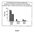
- FIG. 7 shows the results of implantation of apparatuses after transection-freeze lesion of rat sciatic nerves described above for three groups, each containing four rats.
- correctly oriented current flow was used in one group of rats.
- the current flow was reversed, and in a third group of rats no current was used.
- the axon count in the distal stump was consistently higher for rats having apparatuses according to the invention using correctly oriented current, as compared to rats having apparatuses which had inverted electric current or no electric current.
- Apparatuses according to the invention as shown in FIG. 6 were implanted in rats and measurements were made to evaluate the performance of the apparatuses to facilitate functional recovery in rats following severe brain injury.
- the medial fimbria bundle which contains cholinergic efferents projecting from the septum to dorsal hippocampus were unilaterly damaged.
- This partial-lesion paradigm reproducibly results in significant biochemical and bevavioural deficits. The following is a more detailed description of the operation and subsequent evaluation.
- the tips of the fine forceps are then closed and opened several times, thereby crushing the medial fimbria bundle which projects from the septum to the hippocampus.
- the embodiment of the invention as shown in FIG. 6, having dimensions of 2.5mm wide and 4.5mm long was inserted through the opening and slid into place so as to rest in the ventricular space just above the hippocampus.
- the cathode is positioned posteriorly over the dorsal hippocampus with the anode being 1mm rostral to the crushed medial fibria bundle.
- the implant is inverted with the cathode rostral and the anode caudal.
- the implants are anchored in place with GEL-FOAM packing, and the electrical leads are run to the skull opening to the back of the neck where the attached battery power unit was subcutaneously sutured to the musculature. The facia and skin openings are then brought into apposition and sutured in place.
- the Y-Maze Test is carried out by placing a rat in the center of three arms or paths, and observing the number of times the rat enters one of the three arms as well as the specific sequence of arms entered over a fifteen minute test period. The total number of arms entered is indicative of the degree of overall behavioural activity and the percent of spontaneous alternations (in which the rat enters a new or different arm on consecutive trials from the center position as opposed to repeatedly reentering the same arm) is considered to be indicative of learning and memory.
- the Swim Test is another learning and memory dependent paradigm in which the rat is placed at the same starting position in a large body of water and is forced to swim around until it finds a single fixed platform.
- the rat is placed in the Swim Test apparatus for one trial per day and is allowed to swim until the platform is found and it can climb out of the water onto the platform or is placed on to another platform after a maximum time of 180 seconds has passed.
- the Swim Test was conducted on three successive days.
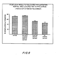
- FIGS. 8, 9 and 10 show the results of the Y-Maze Test and Swim Test paradigms conducted with rats having brain implants with correctly oriented electric current, inverted electric current or no electric current as compared to normal rats.
- FIG. 8 shows the percent of spontaneous alternations for the Y-Maze Test. The rats having brain implants with inverted or no electric current had a lower percentage of spontaneous alternations than the rats having brain implants with correctly oriented electric current and normal rats.
- FIG. 9 is also the results of the Y-Maze Test and indicates a significantly greater number of arm entries for rats having brain implants with inverted electric current or no electric current as compared to rats having brain implants with correctly oriented electric current and normal rats.
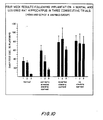
- FIG. 10 shows the results of the Swim Test.
- the normal rats and rats with the brain implants with correctly oriented electric current had latencies to the platform of 35 to 60 seconds respectively on the first test trials, but this latency improved to 7 and 17 seconds respectively by the third trials.
- This learning curve as indicated by the intermediate values for the second trials was not observed for the brain implanted rats having inverted electric current or no electric current.
- the mean latency for the third test trials for the rats having brain implants with inverted electric current or no electric current still exceeded the slowest latencies observed during the first trial for the normal rats and the rats having brain implants with correctly oriented electric current.
- the rats were sacrificed for the biochemical assessment of the level of acetylcholinesterase (AChE) activity in the left hippocampus.
- AChE acetylcholinesterase
- the brains are quickly removed from the skulls and are sagittally transected into two hemispheres.
- the left hemisphere is placed over ice and the left dorsal hippocampus is dissected out.
- the head of the hippocampus is the site closest to the injury. Any remaining portion of the ventral-most hippocampus is trimmed away and the remaining body of the dorsal hippocampus is equally divided in half along the naso-temporal axis.
- the naso-most, or rostral/proximal portion is known to receive the greatest percentage of cholinergic innervation from the medial fimbria.
- the resulting naso-section and temporal section of hippocampus is then homogenized and the level of AChE activity is assayed by routine spectrophotometric techniques. The results were analyzed in terms of absolute AChE values expressed as mg/ml of net weight and as a percentage of control AChE activity from the normal rats.
- the brain implants according to the invention partially reversed or prevented the functional deficits that ordinarily follow partial brain lesion. It can be concluded that the implant according to the invention is efficacious for the repair of the lesioned brain, even though the lesion of the medial fimbria still resulted in biochemical and behavioral deficits, because the treatment was a relatively short period of time of four weeks.
Abstract
Description
- The invention relates to the regeneration of a damaged mammalian nerve, and particularly to a method and apparatus for in vivo mammalian nerve regeneration using an electric current from the proximal nerve end (that is, the nerve end closest to the cell body) to the distal nerve end (that is, the nerve end furthest from the cell body) with the nerve ends either spaced apart from each other or sutured together.
- Considerable research relating to nerve growth and nerve regeneration has produced numerous publications relating to these topics. The distinction between "nerve growth" and "nerve regeneration" is significant. The growth of a nerve does not assure that the nerve will function even partially as it did prior to damage. That is, "nerve growth" does not assure the functioning of the nerve as a channel for communication of information. In contrast, "nerve regeneration" as used herein is the regeneration of a nerve to serve at least partially as part of the communication system of the nervous system. It is well known in the art that there are substantial differences between a mammalian nervous sytem and other nervous systems. Thus, the evaluation of methods and apparatuses for mammalian nerve regeneration must be carried out on a mammal to have any value and credibility.
- When axons of the mammalian peripheral nervous sytem (PNS) are severely damaged (i.e., in compression or transection injuries), several phenomena may take place. First, if left unmodified, the distal stump will nearly always degenerate (Wallerian degeneration). This degeneration is associated with concomitant chromatolytic changes proximally in the perikaryon. If damage to the nerve is sufficiently severe, the axons in the proximal portion will degenerate, followed by the degeneration and (usually) death of the perikaryon. If the damage to the nerve is less severe and left unmodified, a number of biochemical changes begin to occur in the remaining proximal portion. These proximal changes involve a complex series of responses of the cell body to the injury, which seem to prepare the intact portion for regeneration. Such changes include alterations in axonal transport characteristics, protein processing, and nucleic acid synthesis. Morphologically regenerating growth cones from the damaged proximal stump often appear, and the axons will begin to regenerate from the proximal stump towards the original target region.
- Such damage is likely to produce collateral sprouting from neighboring axons that are not as severely injured and which did not undergo Wallerian degeneration. The newly growing neurities probably use the degenerated distal segment as a guide to the denervated target area. Presumably, the reactive Schwann cells provide the communicative means for regeneration to the target by providing diffusable growth-promoting factors, and by providing a suitable growth surface established by the plasma membrane and/or basal lamina, or extracellular matrix. All of these morphological and biochemical changes are primarily dependent upon several factors, including the severity and location of the injury depending upon the proximity to the perikaryon, the size of the axons injured, and the species involved. For example, higher vertebrates have less capacity to regenerate the peripheral nervous system (PNS) axons effectively.
- In the central nervous system (CNS), the unaided attempts at regeneration often are quickly aborted by the body, resulting in a completely degenerated proximal stump and perikaryon. Recent publications have improved the understanding of the cellular events in the CNS following injury. Transplantation of embryonic tissues into the brain or spinal cord has proven to be a useful tool for determining the essential factors for regeneration. Because embryonic cells have a high potential for growth and differentiation, their transplantation should provide a suitable environment capable of promoting and supporting growth of the lesioned adult central or peripheral nervous system. Fetal cell implants, particularly neurons and muscle fibers implanted into the nervous system, possess the ability to induce axons to grow with a strong attraction to the grafted fetal tissue. Much of the published work relating to transplantation has focused on attempts to induce regeneration in the CNS. The results from a large number of investigators have shown that, unlike the previous assumptions of past decades, parts of the CNS are indeed capable of limited regeneration. Both functional and morphological data suggest that the injured brain and spinal cord can recover a certain degree of function following trauma. The contribution of the graft to the reconstruction of the host CNS is not well established. For example, the role of collateral sprouting from undamaged fibers of the host into the damaged region containing the graft has not yet been determined. A major determinant of the extent of regeneration is the environment encountered by the regrowing axons. The mechanisms underlying the axonal growth, guidance, and maturation appear to be strongly influenced by trophic factors in the environment which are appropriate and necessary for regeneration and referred to in the art as "growth-promotors". These substances may be specific for a particular target tissue and are likely to have a wide spectrum of growth-promoting potencies.
- The reason a moderate or severe injury to a mammalian nerve may not lead to appropriate innervation of the target tissue may be the proportionally greater distance (due to Wallerian degeneration) those axons must travel. Presumably, trophic substances from nearby tissues may have a greater trophic potential than the intended target tissue. As a result, gap length of an injury has been shown in the prior art to be a primary factor in determining the success of functional regeneration. This lack of target specificity has been suggested in the literature as the underlying cause of the frequent formation of neuromas, as well as the inappropriate contact on other tissues. Thus, significant improvements have been observed after neural anastomoses have been made. The current method of choice in neurosurgical repair of damage to peripheral nerves is simple anastomosing of the cut end of the nerve, although this intervention is limited.
- Simple anastomosing will not be sufficient in those circumstances where damage and degeneration is so extensive that the distance between the remaining proximal and distal stumps is excessive. An alternative solution employs the use of a structure or "bridge" across the gap length from the cut proximal stump to either the distal portion of the nerve or to the target tissue itself. In animal studies, the various materials which have been used to bridge the gap include peripheral nerve grafts, mesothelial chambers, millipore and silicone tubes.
- Of particular interest are the artifically produced and commercially available "nerve cuffs" or "nerve guide tubes" which are implanted and extend between the stumps. Prior art nerve guide tubes are electrically passive, that is, do not include any electrical current, and have the shape of a hollow cylinder. The nerve guide tubes are generally made of either silicon or bioresorbable substances. The nerve guide tubes can be filled or coated with a growth supporting matrix such as laminin that promotes neural growth over greater distances than the unmodified nerve guide tube alone. The nerve guide tubes have been studied, reported in the literature and are commercially available. During the regenerative process, between two and three weeks after injury, the host body usually causes the interior of the nerve guide tube to fill with a viscous fluid containing proteins and other material in an amorphous matrix. Protein strands appear oriented along the longitudinal axis of the chamber, and may serve as the substrate for cellular migration. Before axons appear, Schwann cells and fibroblasts infiltrate the matrix. In general, blood vessels appear last, although some studies have observed capillary formation prior to axonal growth. It has been suggested that the early invading cells modify the matrix of the nerve guide tube and thereby facilitate the ingrowth of axons. It is important to note that the extracellular matrix will form in the absence of a distal segement. No axonal outgrowth will occur, indicating that the matrix is by itself insufficient to promote axonal growth. Perhaps the distal stump provides a humoral agent diffusible in the matrix which is necessary for growth and/or guidance of axons. This would be similar to the requirement for Schwann cell or muscle cell "conditioned media" in the growth of sensory, sympathetic or motor neurons in vitro. Axon diameter and density are greater when a distal stump is present. Whatever the exact mechanisms responsible for growth, (structural, cellular, and/or humoral), and wherever the site of action (at the axon or its substratum), the nerve guide tubes provide an "artificial" environment suitable for supporting axonal growth over relatively long distances.
- As used herein, a "nerve guide means" is an electrically passive physical structure for enhancing nerve regeneration and includes prior art nerve guide tubes and other structures which are not tubular such as a plate-shaped object, solid tubes and other shapes which are effective for enhancing nerve regeneration.
- Some cell types have been shown to be affected by static and dynamic electromagnetic fields. Most extensively studied are the effects of electromagnetic fields on bone growth, and prior art reports of in vitro results have demonstrated beneficial effects to some specific types of cells. Recently, published studies in bone-derived cell cultures have shown that electromagnetic fields induce specific biochemical alterations. Such effects include cAMP fluctuations, altered states of actin polymerization, enhanced DNA synthesis and changes in calcium uptake. The exact mechanisms responsible for electromagnetic field induced-bone growth have not been characterized fully. Due to the complex morphology of the neuronal cell, as well as its ability to grow in vitro, the nervous system is particularly well-suited for studies of the effect of electromagnetic fields on the growth of cells. In the past ten years, many studies have demonstrated that neurite elongation and orientation can be influenced by an electromagnetic field. Specifically, within a static electric field, neurite growth is directed toward the cathode. Changes in the orientation of these neurites can be observed with light microscopy after a period of time from about several minutes to about several hours. All neurons that have been studied to date in vitro respond in some way to an applied electromagnetic field. Many variations in electromagnetic field parameters have been used to observe changes in neurite growth.
- The vigorous in vitro response of all neuronal cell types to a wide range of electromagnetic field effects (and thus an apparent lack of specificity for cell type or stimulus) has been used as an argument against the concept that endogenous fields serve a primary role in the guidance of growing neuronal processes in vivo. This is often supported by studies showing neurite growth to a target in the absence of intrinsic action potentials, and axonal synaptogenesis occurring during the blockade of postsynaptic ion channels. Growth and guidance in the nervous system is complex. The local microenvironment with respect to the events required for growth and guidance must necessarily be important for the establishment of proper channels to the target cell. These events are multifactorial, and include a variety of bioelectrochemical processes such as the timing of membrane interactions between growing axons and glia. It is likely that such multiple interactions are subtle, and may require extremely small local electrical interactions. Action potentials or postsynaptic events may therefore be insufficient alone, or temporally inappropriate to affect the growth process significantly.
- The method of application of the electromagnetic fields used in the in vitro studies is vastly different from what would be possible under local microenvironmental conditions. Typically, in vitro studies apply an electrmagnetic field across a large population of cells, often in a culture dish having a volume of enormous size in comparison to the cell, and with a concomitantly applied homogenous current density. Experimental results substantiate that electromagnetic fields may serve a modulatory function in orienting neurite growth within localized regions.
- The application of extracellular direct current electric fields in vitro may accelerate as well as orient the growth of neurites in embryonic explants or in dissociated neuronal cultures. The mechanisms by which these biochemical alterations occur are not well understood, nor are they necessarily directly related. In addition, different neuron types appear to respond differently with regard to stimulation amplitude and duration. The biophysical mechanism responsible may be related to the electrophoretic redistribution of cytoplasmic components which may occur if an extracellular potential produces a voltage potential drop in the cytoplasm. The site of most of the cell's electrical resistance is the plasma membrane so that the electromagnetic field would be the strongest and have the greatest voltage potential difference relative to the cytoplasm. Thus, an electromagnetic field may alter the membrane's voltage potential asymmetrically, thereby perturbing growth-controlling transport processes across the membrane. The cytoplasm has far less resistivity than the plasma membrane, and the voltage potential drop is on the order of 10⁻⁴ volts. The majority of work published involving electromagnetic fields on whole cells in vitro use a static electromagnetic field in the range of 0.1 to 15 V/cm, roughly translating into an average of 10 mv/cell diameter. Assuming that 50% of this voltage is exerted across the plasma membrane at each end of the cell, this would result in hyperpolarization or depolarization of 5 mv, depending on the polarity. Most neuronal resting membrane potentials are approximately -70 to -90 mv and local ion conductances or enzyme activation states at the membrane may be changed enough to alter or modify the normal function.
- Furthermore, an electrophoretic accumulation of molecules responsible for neuritic extension and/or adhesion may occur toward the membrane. A charged macromolecule of ordinary electrophoretic mobility (1 micron/sec/V/cm) across a 10 micron distance requires 10⁴ to 10⁶ seconds (three hours to ten days). It is possible that higher electromagnetic field strengths (approximately 10 V/cm) can cause substantial intracellular migration of growth-related molecules, or receptors for trophic substances. It has been shown in the prior art that the accumulation of surface glycoproteins can occur electrophoretically at the cathode in isolated cultured cells. Membrane glycoproteins are believed to play a crucial role in cell adhesion to the substratum. Cathodal accumulation of these molecules at the membrane may be responsible for some of the orienting effects of the electromagnetic field. These hypotheses are consistent with the majority of the prior art data showing that most changes in directionality or growth rate occur within twenty-four hours of exposure in vitro. Thus, an electromagnetic field in vitro produces a growth promotion effect as well as a guidance effect.
- U.S. Patent No. 4,306,561 discloses methods and apparatuses for the reattachment and repair of severed nerves in a human body. The '561 Patent describes the use of direct current from the proximal nerve end to the distal nerve end to test electrical continuity in the nerve. The '561 Patent does not, however, suggest the use of electric current as a means for regeneration of nerves. Further, the '561 Patent strongly discourages suturing nerve ends together. The '561 Patent discloses a device for holding the nerve ends in abutment which requires vacuum lines to engage the nerve ends and is generally in the form of a modified forceps. Thus, the device is not at all suitable for being implanted and the disclosure limits its use to a period of about 5 hours because the patient has an open wound during the use of the device. The requirement disclosed in the '561 Patent that the nerve ends abut each other precludes the regeneration of a damaged nerve for which the nerve ends are spaced apart.
- From the background given above, it can be appreciated that mammalian nerve regeneration and particularly peripheral nerve regeneration is a complex phenomenon. Furthermore, it can present serious problems to the neurosurgeon who wishes to intervene in some way to increase the chances of good functional recovery following severe damage to nerves, particularly to peripheral nerves.
- The present invention relates to regeneration of a damaged mammalian nerve and overcomes these and other shortcomings of the prior art. As used herein, the word "nerve" means, generally, fibers of the central or peripheral nervous system. It is a primary object of the invention to provide a method and apparatus for in vivo mammalian nerve regeneration of a damaged nerve using an electric current from the proximal nerve end to the distal nerve end with the nerve ends either spaced apart from each other or sutured substantially in abutment to each other. As used herein, an "electric current from the proximal nerve end to the distal nerve end" is an electric current more positive at the proximal nerve end than the distal nerve end and is referred to as "correctly oriented" current.
- It is a further object of the invention to provide a method and apparatus for in vivo mammalian nerve regeneration of a damaged nerve using an electric current from the proximal nerve end to the distal nerve end and a nerve guide means.
- It is a still further object of the invention to provode a method and apparatus for in vivo mammalian nerve regeneration of a damaged nerve using an electric current from the proximal nerve end to the distal nerve end and a nerve guide means including a matrix conducive to the growth of nerve cells.
- It is a yet further object of the present invention to provide an implantable apparatus for in vivo mammalian nerve regeneration of a damaged nerve.
- In accordance with illustrative embodiments demonstrating objects and features of the present invention, there is provided one embodiment of the apparatus for in vivo mammalian nerve regeneration of a damaged nerve having proximal and distal nerve ends, in which the apparatus includes means between the nerve ends to maintain the nerve ends in proximity to each other, and means to produce an electric current from the proximal nerve end to the distal nerve end at a level and for a period of time to regenerate the damaged nerve.
- Another embodiment of the apparatus for in vivo mammalian nerve regeneration of a damaged nerve having proximal and distal nerve ends, includes a nerve guide means extending between the nerve ends to induce nerve growth, and means to produce an electric current from the proximal nerve end to the distal nerve end at a level and for a time to regenerate the damaged nerve.
- The invention also relates to one embodiment of a method of in vivo mammalian nerve regeneration of a damaged nerve having proximal and distal nerve ends, in which the method includes suturing the nerve ends in substantial abutment to each other and, thereafter, producing an electric current from the proximal nerve end to the distal nerve end at a level and for a period of time to regenerate the damaged nerve.
- Additionally, the invention relates to another embodiment of a method for in vivo mammalian nerve regeneration of a damaged nerve having proximal and distal nerve ends, in which the method includes means including positioning a nerve guide means near the nerve ends to enhance nerve growth and, concurrently, producing an electric current from the proximal nerve end to the distal nerve end at a level and for a period of time to regenerate the damaged nerve.
- The above descriptions, as well as further objects, features and advantages of the present invention will be more fully understood by reference to the following detailed description of the presently preferred, but nonetheless illustrative embodiments in accordance with the present invention, when taken in conjunction with the accompanying drawings in which:
- FIG. 1 is a perspective view of the front of a surgically opened human hand, with an apparatus according to the invention applied to a damaged nerve in the hand;
- FIG. 2 is a generally front perspective view on an enlarged scale of the apparatus shown in FIG. 1 as it is being engaged onto nerve ends sutured together, with the instrument holding the apparatus not shown for clarity in illustration;
- FIG. 3 is a fragmentary perspective view of the apparatus shown in FIG. 2 after it has engaged the sutured nerve ends;
- FIG. 4 is a sectional view along the line 4-4 shown in FIG. 3 and shows the damaged nerve encircled by the apparatus;
- FIG. 5 is a sectional view of another embodiment of the apparatus enclosing spaced apart nerve ends;
- FIG. 6 is a fragmentary perspective view of a spinal cord in a human neck with yet another embodiment of the apparatus, with portions of the neck omitted for clarity in illustration;
- FIG. 7 is a bar graph showing axon count after twelve days for rats having implanted apparatuses according to the invention with correctly oriented electric current, inverted electric current, and no electric current being applied to the electrodes in the apparatuses;
- FIG. 8 shows a bar graph indicating percent of spontaneous alternation after four weeks for rats having implanted apparatuses in the brain, according to the invention, with correctly oriented electric current, inverted electric current and no electric current, as compared to rats having no lesions and no implanted apparatuses (normal);
- FIG. 9 shows a bar graph indicating the number of arm entries in fifteen minutes following four weeks of an implantation of apparatuses according to the invention in lesioned rat hippocampus with correctly oriented electric current, inverted electric current and no electric current, as compared to rats having no lesions and no implanted apparatuses (normal); and
- FIG. 10 shows a bar graph indicating Swim Test time in seconds after four weeks for rats following implantation of the apparatuses according to the invention in lesioned rat hippocampus with correctly oriented electric current, inverted electric current and no electric current, as compared to rats having no lesions and no implanted apparatuses (normal).
- Nerves can become damaged for many reasons such as physical impact, severing, or some other physcial trauma. If the extent of the damage is limited, the damaged portion can be removed and the nerve ends can be easily abutted. For such a situation, it is convenient to suture the nerve ends together to maintain the nerve ends in substantial abutment to each other. This allows an implantable apparatus according to the invention to be used conveniently. The inventors have discovered that an electric current from the proximal nerve end to the distal nerve end can produce nerve regeneration for nerve ends which have been sutured together in contrast to the disclosure in the aforementioned U.S. Patent No. 4,306,561. The regeneration of the nerve can require an extended application of the electric current, and it is advantageous to have the apparatus in an implantable physical form so that the electric current can be maintained for a long period of time without having to maintain the subject either under operating conditions or immobile. Generally, the electric current is maintained for at least about a week, and preferably for longer than about one month. An electric current of about 1.4 microamps has been found to be satisfactory. The range of suitable electric current depends upon the nerves to be regenerated, the location of nerve injury, the subject, and the period of time the electric current is to be maintained. These parameters can be based on data relating to in vitro experiments and can be determined experimentally.
- In the case of extensive nerve damage, the removal of the damaged portion could result in the nerve ends being spaced apart a distance which does not allow the nerve ends to be brought into abutment to each other without further nerve damage. In such a situation, it is advantageous to allow the nerve ends to be spaced apart while carrying out nerve growth and regeneration. It has been found that the growth of nerve ends spaced apart from each other under the influence of an electric current from the proximal nerve end to the distal nerve end will produce a regenerated nerve. A nerve guide means substantially improves the nerve regeneration process for both nerves sutured together or nerves spaced apart from each other.
- It is preferable to have the apparatus according to the invention in an implantable physical form so that an electric current can be maintained with or without a nerve guide means for an extended period of time such as a week or several weeks or even longer while allowing the patient a minimum of discomfort. The electrical circuit for producing electric current according to the invention, in its simplest embodiment, includes an electric cell and two wires connected to the terminals of the electric cell. The electric cell can be a commercially available miniature battery such as a battery used for hearing aids. One suitable electric cell is a type 13M miniature disc battery which has a thickness of about 5 millimeters and a diameter of about 7 millimeters and produces a voltage of about 1.4 volts. The wire used is preferably made of a metal compatible with a living body. One suitable metal is stainless steel. Preferably, the wires are insulated except for the portions to be used to produce the electric current through the damaged nerve. The insulating material is preferably compatible with a living body. One suitable insulating material is the material having the trademark TEFLON. Preferably, the wire is thin and typically has a diameter of about 35 microns. It is preferable to incude a resistor in series with the electric cell in order to limit the electric current to a predetermined amount. The prior art includes many references stating the range of electric current used for in vitro experiments, and this provides a guide for the level of electric current which may be suitable for in vivo mammalian nerve regenerating according to the invention. The generally maximum electric current used for the examples herein was determined by having the electric circuit of the electric cell and the wires completed with the wire ends in a physiological saline environment, and measuring the electric current. From this data a resistor was selected so that the electric current for this situation would be about 1.5 microamps. The resistor used had a resistance of about 1 megohm. The battery and resistor are preferably placed into a form suitable for implanting. The battery and resistor with the wires attached can be encased in several layers of an epoxy, and then the epoxy was covered with a medical grade adhesive such as sold under the trademark SILASTIC. It is known that SILASTIC minimizes tissue reaction to an implanted substance.
- It is preferable to use the instant apparatus including a nerve guide means, because the nerve guide means serves several important functions including maintaining the electrodes separated from each other, providing a convenient structure for contacting the wires with the respective nerve end, and providing a desirable environment for the growth and regeneration of nerves. Many nerves have a generally circular cross-section, so that a nerve guide means in the form of a hollow circular cylinder is suitable for nerve regeneration of many types of nerves. The diameter of the inside cross section depends on the cross section of the nerve. The hollow central portion of the cylinder can be some other shape better adapted for the cross-sectional shape of a nerve. In practice, it is often necessary to engage the cylinder onto the nerve and later on to remove the cylinder. A slit or cut from one end to the other end of the cylinder will allow the cylinder to be spread open for these operations. A conventional surgical tool can be used to engage and remove the cylinder. The nerve guide means can also have the shape of an incomplete tube open at one side to simplify the engagement and disengagement of the tube. In some circumstances, such as nerves in the central nervous system in the spinal column, or brain, a nerve guide means in the form of a tube may not be convenient. For such a situation, a nerve guide means in the form of a curved plate can be used. The plate can be held in place by the use of sutures to enable implanting.
- Referring now to FIGS. 1-4, an apparatus according to one embodiment of the invention is generally designated by the numeral 20. The
apparatus 20 is shown in FIG. 1 engaged with anerve 22 in ahand 24 during an operation to implant theapparatus 20. Theapparatus 20 includes a combined battery andresistor 26,insulated wires hollow cylinder 32 which serves as a nerve guide means. Thecylinder 32 has a cut 34 from one end to the other end of thecylinder 32 so that thecylinder 32 can be opened with a conventional surgical tool (not shown) as shown in FIG. 2, to engage or disengage thenerve 22. Typically, thecylinder 32 is about 0.5 in. long, has an inside diameter of about 0.16 in. and an outside diameter of abut 0.24 in but this can be varied in accordance with the size of the nerve. - FIG. 2 shows the
proximal nerve end 36 sutured to thedistal nerve end 38 bysutures 40 so that the nerve ends 36 and 38 are substantially abutting each other. As can be seen in FIG. 3, thecylinder 32 engages thenerve 22 so that the sutured nerve ends 36 and 38 are within thecylinder 32. Preferably, the sutured nerve ends 36 and 38 are centrally positioned in thecylinder 32. The procedure for implanting theapparatus 20 into thehand 24 can be done using conventional techniques. FIG. 1 shows the use of a conventional surgical tool 42 (shown in part) for maintaining thehand 24 opened to receive theapparatus 20. FIG. 4 shows a cross sectional view of thenerve 22 in thecylinder 32, as seen along the lines 4-4 of FIG. 3. As best seen in FIGS. 2 and 3, theinsulated wire 28 ends in abare wire 44 which extends through a small hole in the wall of thecylinder 32 into the interior of thecylinder 32 so that it can electrically contact theproximal nerve end 36. Similarly, theinsulated wire 30 has a bare wire end 46 which extends through a hole in the wall of thecylinder 32 into the interior of thecylinder 32 so that it can electrically contact thedistal nerve end 38. The bare wire ends 44 and 46 can be fixed in position by the use of a suitable bonding agent such as SILASTIC sold by Dow Chemical Co. - FIG. 5 shows a sectional view of an
apparatus 50 which is another embodiment of the invention. Theapparatus 50 includes ahollow cylinder 52 serving as a nerve guide means, with acylinder 54 generally bonded perpendicular to thecylinder 52 using SILASTIC. As used in the example herein, thecylinder 52 is referred to as the "nerve guide tube" and thecylinder 54 is referred to as the "lead tube". A battery and resistor (not shown) have insulatedwires cylinder 54 and out throughholes wire 56 has abare end 64 extending through ahole 66 in thecylinder 52, into the interior of thecylinder 52 to make electrical contact with anerve end 68. Similarly, theinsulated wire 54 has a bare wire end 70 which extends through ahole 74 in thecylinder 52, to the interior of thecylinder 52 to make electrical contact withnerve end 72. Nerve ends 68 and 72 are spaced apart from each other. - FIG. 6 shows a further embodiment of the invention in which a
plate 80 is used as a nerve guide means and to maintain ends ofwires resistor 86 provides an electric current through the damaged nerve in order to enhance regeneration of the damaged nerve. - Apparatuses according to the invention as shown in FIG. 5 were implanted in rats, and measurements were made to evaluate the performance of the apparatuses. Adult male Sprague-Dawley rats weighing approximately 3000g were used. Prior to sterile surgey, a KETAMINE anesthesia is administered to the rats (100mv/100g body weight, supplenented with ROMPUN). The implantation of the apparatuses are performed by a variation of the method disclosed in an apparatus by M. Politis and P.S. Spencer which appeared in "Brain Research", Vol. 278, pp. 229-231, 1983. The nerve guide tube (corresponding to the cylinder 52) is placed near the nerve to be used, and the lead tube (corresponding to the cylinder 54) is sutured to the musculature in order to provide mechanical stability during manipulation. Thereafter, using a small scissors, the nerve is transected just before the first bifurcation of the sciatic nerve. This is an ideal location to transect the nerve because it allows maximum manipulation of the nerve while being distal enough to avoid severly traumatizing the perikarya. One centimeter of the distal stump is frozen on dry ice, allowed to thaw and then sutured to the proximal stump using 9-0 silk. The nerve guide tube was placed over the sutured nerve. The lead tube is then further sutured to the musculature to provide additional mechanical stability. A long incision is made from the original thigh incision to extend to the dorsal aspect of the lower lumbar region. The power supply is fastened to the dorsal fascia with 5-0 silk. Each lead tube is gently fastened to the overlying fascia with 8-0 silk ligatures for additional stability. The skin overlying the power supply is then sutured closed.
- FIG. 7 shows the results of implantation of apparatuses after transection-freeze lesion of rat sciatic nerves described above for three groups, each containing four rats. In one group of rats, correctly oriented current flow was used. In a second group of rats, the current flow was reversed, and in a third group of rats no current was used. The axon count in the distal stump was consistently higher for rats having apparatuses according to the invention using correctly oriented current, as compared to rats having apparatuses which had inverted electric current or no electric current.
- Apparatuses according to the invention as shown in FIG. 6 were implanted in rats and measurements were made to evaluate the performance of the apparatuses to facilitate functional recovery in rats following severe brain injury. For this purpose, the medial fimbria bundle which contains cholinergic efferents projecting from the septum to dorsal hippocampus were unilaterly damaged. This partial-lesion paradigm reproducibly results in significant biochemical and bevavioural deficits. The following is a more detailed description of the operation and subsequent evaluation.
- Adult male Sprague-Dawley rats weighing approximately 300g are anesthetized with NEMBUTAL anesthesia. The scalp and underlining fascia is resected to expose the skull. A 5mm square bone flap is cut posterior to bregma and over the left hemisphere lateral to the midline (sagittal) suture. Approximately 3mm square area of cortex is carefully removed by suction, and the newly exposed underlining corpus callosum is gently resected until the head of the hippocampus is visible through a dissecting microscope. A pair of fine surgical forceps is placed just anterior to the exposed head of the hippocampus and inserted 1.5mm into the brain. The tips of the fine forceps are then closed and opened several times, thereby crushing the medial fimbria bundle which projects from the septum to the hippocampus. After completion of the injury to the medial fimbria bundle the embodiment of the invention as shown in FIG. 6, having dimensions of 2.5mm wide and 4.5mm long, was inserted through the opening and slid into place so as to rest in the ventricular space just above the hippocampus. For rats having correctly oriented electric current, the cathode is positioned posteriorly over the dorsal hippocampus with the anode being 1mm rostral to the crushed medial fibria bundle. For rats having inverted electric current, the implant is inverted with the cathode rostral and the anode caudal. This is also done for the rats which would have no electric current. The implants are anchored in place with GEL-FOAM packing, and the electrical leads are run to the skull opening to the back of the neck where the attached battery power unit was subcutaneously sutured to the musculature. The facia and skin openings are then brought into apposition and sutured in place.
- Four weeks after the implantation, measurements were carried out to evaluate the rats having brain implants as compared to rats having no lesions and no implants. Two conventional behavioral paradigms were used: the Y-Maze Test and the Swim Test. The Y-Maze Test is carried out by placing a rat in the center of three arms or paths, and observing the number of times the rat enters one of the three arms as well as the specific sequence of arms entered over a fifteen minute test period. The total number of arms entered is indicative of the degree of overall behavioural activity and the percent of spontaneous alternations (in which the rat enters a new or different arm on consecutive trials from the center position as opposed to repeatedly reentering the same arm) is considered to be indicative of learning and memory. The Swim Test is another learning and memory dependent paradigm in which the rat is placed at the same starting position in a large body of water and is forced to swim around until it finds a single fixed platform. The rat is placed in the Swim Test apparatus for one trial per day and is allowed to swim until the platform is found and it can climb out of the water onto the platform or is placed on to another platform after a maximum time of 180 seconds has passed. The Swim Test was conducted on three successive days.
- FIGS. 8, 9 and 10 show the results of the Y-Maze Test and Swim Test paradigms conducted with rats having brain implants with correctly oriented electric current, inverted electric current or no electric current as compared to normal rats. FIG. 8 shows the percent of spontaneous alternations for the Y-Maze Test. The rats having brain implants with inverted or no electric current had a lower percentage of spontaneous alternations than the rats having brain implants with correctly oriented electric current and normal rats. FIG. 9 is also the results of the Y-Maze Test and indicates a significantly greater number of arm entries for rats having brain implants with inverted electric current or no electric current as compared to rats having brain implants with correctly oriented electric current and normal rats. FIG. 10 shows the results of the Swim Test. The normal rats and rats with the brain implants with correctly oriented electric current had latencies to the platform of 35 to 60 seconds respectively on the first test trials, but this latency improved to 7 and 17 seconds respectively by the third trials. This learning curve as indicated by the intermediate values for the second trials was not observed for the brain implanted rats having inverted electric current or no electric current. Moreover, the mean latency for the third test trials for the rats having brain implants with inverted electric current or no electric current, still exceeded the slowest latencies observed during the first trial for the normal rats and the rats having brain implants with correctly oriented electric current.
- After completion of the behavioural paradigms, the rats were sacrificed for the biochemical assessment of the level of acetylcholinesterase (AChE) activity in the left hippocampus. After the rats are sacrificed, the brains are quickly removed from the skulls and are sagittally transected into two hemispheres. The left hemisphere is placed over ice and the left dorsal hippocampus is dissected out. The head of the hippocampus is the site closest to the injury. Any remaining portion of the ventral-most hippocampus is trimmed away and the remaining body of the dorsal hippocampus is equally divided in half along the naso-temporal axis. The naso-most, or rostral/proximal portion, is known to receive the greatest percentage of cholinergic innervation from the medial fimbria. The resulting naso-section and temporal section of hippocampus is then homogenized and the level of AChE activity is assayed by routine spectrophotometric techniques. The results were analyzed in terms of absolute AChE values expressed as mg/ml of net weight and as a percentage of control AChE activity from the normal rats.
- Biochemical assay and analysis is only performed in the lesioned or left hemisphere, because other studies have demonstrated that analysis of the contralateral side often yields variable results and is not an adequate internal control. The adequacy of the medial fimbria crushed lesions was verified by the significant AChE depletion in both absolute and percent of control values for the naso- and temporal hippocampal sections for all the lesioned rats. The rats having brain implants with correctly oriented electric current had greater AChE activity in the naso-section (65%) and temporal section (72%) than observed in the rats having brain implants with inverted electric current or no electric current, which averaged about 50% for both sections. Thus, the brain implants according to the invention partially reversed or prevented the functional deficits that ordinarily follow partial brain lesion. It can be concluded that the implant according to the invention is efficacious for the repair of the lesioned brain, even though the lesion of the medial fimbria still resulted in biochemical and behavioral deficits, because the treatment was a relatively short period of time of four weeks.
- Experiments have also been conducted for regenerating optic nerves. In separate experiments, sixteen apparatuses according to the invention were constructed. These apparatuses provided a discreted distribution of electric current locally on a nerve. In each experiment, rats with optic nerve lesions were implanted with apparatuses according to the invention. The animals were allowed to survive for four weeks after the implantation. Afterwards, all of the animals were sacrificed and the tissue 2.5mm distal to the lesion was processed for histological analysis by light microscopy (LM) using toluidine blue as well as neurofilament-specific staining. The results in every experimental animal showed that all of the animals implanted with the apparatuses and correctly oriented current exhibited significant ingrowth of axons through and beyond the lesions, as compared to control optic nerves having apparatuses without electric current, or with reverse electric current. The optic nerves with apparatuses with correctly oriented current contained a significant number of myelin figures (averaging between 175 to 380 in each experiment), and reorganization of the tissue matrix including increased vascularity was apparent. All of the control optic nerves showed signs of debris and degeneration, and no indication of myelin figures. Whether the fibers observed in these experiments were regenerated optic fibers or re-routed peripheral fibers, was addressed in other experiments. Ten animals were implanted with apparatuses according to the invention with correctly oriented electric current on the right optic nerves. After two weeks, the retinae of five of the animals were avulsed, and the animals were allowed to survive for two more weeks. At that time, the avulsed and non-avulsed animals were sacrificed and the optic nerves analyzed as described above. In the non-avulsed animals, all optic nerves showed regenerating axons in the region distal to the lesion as in previous studies. Four of the five avulsed animals showed no signs of regenerating axons, indicating that removal of the retina destroys the complement of axons. Thus, in these animals, the CNS (in this case, ganglion cells) could be made to regenerate through a lesion and extend into the distal portion of the nerve.
- Although the invention herein has been described with reference to particular embodiments, it is to be understood that these embodiments are merely illustrative of the principal and application of the invention. Thus, it is to be understood that numerous modifications may be made in the illustrative embodiments and other arrangements may be devised without departing from the spirit and scope of the invention as defined in the following Claims.
Claims (14)
the method preferably comprising positioning a nerve guide means proximate said proximal and distal nerve ends and concurrently producing an electric current from said proximal nerve end to said distal nerve end at a level and for a period of time to produce nerve regeneration; and wherein the electric current is preferably maintained for at least about one week.
Applications Claiming Priority (2)
| Application Number | Priority Date | Filing Date | Title |
|---|---|---|---|
| US06/905,787 US4774967A (en) | 1986-09-09 | 1986-09-09 | Method and apparatus for mammalian nerve regeneration |
| US905787 | 1986-09-09 |
Publications (3)
| Publication Number | Publication Date |
|---|---|
| EP0261833A2 true EP0261833A2 (en) | 1988-03-30 |
| EP0261833A3 EP0261833A3 (en) | 1990-07-18 |
| EP0261833B1 EP0261833B1 (en) | 1994-06-01 |
Family
ID=25421467
Family Applications (1)
| Application Number | Title | Priority Date | Filing Date |
|---|---|---|---|
| EP87307938A Expired - Lifetime EP0261833B1 (en) | 1986-09-09 | 1987-09-08 | Apparatus for mammalian nerve regeneration |
Country Status (7)
| Country | Link |
|---|---|
| US (1) | US4774967A (en) |
| EP (1) | EP0261833B1 (en) |
| JP (1) | JP2843324B2 (en) |
| AT (1) | ATE106256T1 (en) |
| CA (1) | CA1324412C (en) |
| DE (1) | DE3789930T2 (en) |
| ES (1) | ES2056825T3 (en) |
Cited By (8)
| Publication number | Priority date | Publication date | Assignee | Title |
|---|---|---|---|---|
| US4877029A (en) * | 1987-03-30 | 1989-10-31 | Brown University Research Foundation | Semipermeable nerve guidance channels |
| US4878913A (en) * | 1987-09-04 | 1989-11-07 | Pfizer Hospital Products Group, Inc. | Devices for neural signal transmission |
| US5030225A (en) * | 1987-03-13 | 1991-07-09 | Brown University Research Foundation | Electrically-charged nerve guidance channels |
| WO2002062216A1 (en) * | 2001-02-09 | 2002-08-15 | Pali Jeno | Device for measuring the activity and for stimulation of nerve fibres |
| EP1453570A2 (en) * | 2001-11-13 | 2004-09-08 | Dynamed Systems, LLC | Apparatus and method for repair of spinal cord injury |
| US7147647B2 (en) | 2002-04-26 | 2006-12-12 | Medtronic, Inc. | Sintered titanium tube for the management of spinal cord injury |
| US9931121B2 (en) | 2011-10-17 | 2018-04-03 | University Of Utah Research Foundation | Methods and devices for connecting nerves |
| US10842494B2 (en) | 2011-10-17 | 2020-11-24 | University Of Utah Research Foundation | Methods and devices for connecting nerves |
Families Citing this family (71)
| Publication number | Priority date | Publication date | Assignee | Title |
|---|---|---|---|---|
| US4919140A (en) * | 1988-10-14 | 1990-04-24 | Purdue Research Foundation | Method and apparatus for regenerating nerves |
| US4979511A (en) * | 1989-11-03 | 1990-12-25 | Cyberonics, Inc. | Strain relief tether for implantable electrode |
| US5327902A (en) * | 1993-05-14 | 1994-07-12 | Lemmen Roger D | Apparatus for use in nerve conduction studies |
| US20040006396A1 (en) * | 1993-11-02 | 2004-01-08 | Ricci John L. | Transcutaneous devices having nueral interface |
| US5853424A (en) * | 1996-03-13 | 1998-12-29 | Medtronic, Inc | Method of and apparatus for preventing tissue ingrowth in infusion catheters |
| US11865326B1 (en) | 1997-01-27 | 2024-01-09 | Innovations Holdings, L.L.C. | High precision, multi-output pulse and/or signal generators |
| US6132360A (en) * | 1998-05-22 | 2000-10-17 | Halpern; Alan A. | Magnetic stretching of magnetized neurons for spinal cord or peripheral nerve repair and regeneration |
| US6487450B1 (en) | 2000-02-24 | 2002-11-26 | Cedars-Sinai Medical Center | System and method for preventing Sudden Cardiac Death by nerve sprouting from right stellate ganglion |
| US7831305B2 (en) | 2001-10-15 | 2010-11-09 | Advanced Neuromodulation Systems, Inc. | Neural stimulation system and method responsive to collateral neural activity |
| AU2002320145A1 (en) * | 2001-06-21 | 2003-01-08 | Vanderbilt University | Method for promoting reinnervation of denervated tissue |
| AU2002362052A1 (en) | 2001-12-04 | 2003-06-17 | The Trustees Of The University Of Pennsylvania | Device and method using integrated neuronal cells and an electronic device |
| AU2003209287A1 (en) | 2002-01-15 | 2003-07-30 | The Regents Of The University Of California | System and method providing directional ultrasound therapy to skeletal joints |
| US7819826B2 (en) * | 2002-01-23 | 2010-10-26 | The Regents Of The University Of California | Implantable thermal treatment method and apparatus |
| US20070129761A1 (en) | 2002-04-08 | 2007-06-07 | Ardian, Inc. | Methods for treating heart arrhythmia |
| US20070135875A1 (en) | 2002-04-08 | 2007-06-14 | Ardian, Inc. | Methods and apparatus for thermally-induced renal neuromodulation |
| US9308043B2 (en) | 2002-04-08 | 2016-04-12 | Medtronic Ardian Luxembourg S.A.R.L. | Methods for monopolar renal neuromodulation |
| US9636174B2 (en) | 2002-04-08 | 2017-05-02 | Medtronic Ardian Luxembourg S.A.R.L. | Methods for therapeutic renal neuromodulation |
| US8150519B2 (en) | 2002-04-08 | 2012-04-03 | Ardian, Inc. | Methods and apparatus for bilateral renal neuromodulation |
| US20080213331A1 (en) | 2002-04-08 | 2008-09-04 | Ardian, Inc. | Methods and devices for renal nerve blocking |
| US7162303B2 (en) | 2002-04-08 | 2007-01-09 | Ardian, Inc. | Renal nerve stimulation method and apparatus for treatment of patients |
| US20140018880A1 (en) | 2002-04-08 | 2014-01-16 | Medtronic Ardian Luxembourg S.A.R.L. | Methods for monopolar renal neuromodulation |
| US7853333B2 (en) | 2002-04-08 | 2010-12-14 | Ardian, Inc. | Methods and apparatus for multi-vessel renal neuromodulation |
| US6978174B2 (en) | 2002-04-08 | 2005-12-20 | Ardian, Inc. | Methods and devices for renal nerve blocking |
| US8347891B2 (en) | 2002-04-08 | 2013-01-08 | Medtronic Ardian Luxembourg S.A.R.L. | Methods and apparatus for performing a non-continuous circumferential treatment of a body lumen |
| US7756583B2 (en) | 2002-04-08 | 2010-07-13 | Ardian, Inc. | Methods and apparatus for intravascularly-induced neuromodulation |
| US8774922B2 (en) | 2002-04-08 | 2014-07-08 | Medtronic Ardian Luxembourg S.A.R.L. | Catheter apparatuses having expandable balloons for renal neuromodulation and associated systems and methods |
| US8774913B2 (en) | 2002-04-08 | 2014-07-08 | Medtronic Ardian Luxembourg S.A.R.L. | Methods and apparatus for intravasculary-induced neuromodulation |
| US7617005B2 (en) | 2002-04-08 | 2009-11-10 | Ardian, Inc. | Methods and apparatus for thermally-induced renal neuromodulation |
| US7620451B2 (en) | 2005-12-29 | 2009-11-17 | Ardian, Inc. | Methods and apparatus for pulsed electric field neuromodulation via an intra-to-extravascular approach |
| US8131371B2 (en) | 2002-04-08 | 2012-03-06 | Ardian, Inc. | Methods and apparatus for monopolar renal neuromodulation |
| US8145317B2 (en) | 2002-04-08 | 2012-03-27 | Ardian, Inc. | Methods for renal neuromodulation |
| US7653438B2 (en) | 2002-04-08 | 2010-01-26 | Ardian, Inc. | Methods and apparatus for renal neuromodulation |
| US8145316B2 (en) | 2002-04-08 | 2012-03-27 | Ardian, Inc. | Methods and apparatus for renal neuromodulation |
| US8150520B2 (en) | 2002-04-08 | 2012-04-03 | Ardian, Inc. | Methods for catheter-based renal denervation |
| US9308044B2 (en) | 2002-04-08 | 2016-04-12 | Medtronic Ardian Luxembourg S.A.R.L. | Methods for therapeutic renal neuromodulation |
| JP2005524448A (en) | 2002-05-03 | 2005-08-18 | アフェレント コーポレイション | Method and apparatus for enhancing neurophysiological abilities |
| US20080077192A1 (en) | 2002-05-03 | 2008-03-27 | Afferent Corporation | System and method for neuro-stimulation |
| US7113816B2 (en) * | 2002-06-18 | 2006-09-26 | Nippon Cable System Inc. | Ultra-miniature in-vivo electrode used for measuring bioelectrical neural activity |
| US6907884B2 (en) | 2002-09-30 | 2005-06-21 | Depay Acromed, Inc. | Method of straddling an intraosseous nerve |
| US8361067B2 (en) | 2002-09-30 | 2013-01-29 | Relievant Medsystems, Inc. | Methods of therapeutically heating a vertebral body to treat back pain |
| US7258690B2 (en) | 2003-03-28 | 2007-08-21 | Relievant Medsystems, Inc. | Windowed thermal ablation probe |
| US7121996B2 (en) * | 2003-04-02 | 2006-10-17 | Donna Hancher | Severed spinal cord regeneration using natural processes and patient's own resources |
| US7725178B2 (en) * | 2004-06-30 | 2010-05-25 | Cedars-Sinai Medical Center | Method and system for the prediction of cardiac arrhythmias, myocardial ischemia, and other diseased condition of the heart associated with elevated sympathetic neural discharges |
| US7937143B2 (en) | 2004-11-02 | 2011-05-03 | Ardian, Inc. | Methods and apparatus for inducing controlled renal neuromodulation |
| JP4485323B2 (en) * | 2004-11-04 | 2010-06-23 | 独立行政法人科学技術振興機構 | Method for manufacturing nerve regeneration electrode device and nerve regeneration electrode device |
| WO2006074425A1 (en) * | 2005-01-06 | 2006-07-13 | Halton Oy | Ventilation register and ventilation systems |
| US20060194724A1 (en) * | 2005-02-25 | 2006-08-31 | Whitehurst Todd K | Methods and systems for nerve regeneration |
| US20090254148A1 (en) * | 2005-10-21 | 2009-10-08 | Borgens Richard B | Telemetrically Controllable System for Treatment of Nervous Sytem Injury |
| US20090132003A1 (en) * | 2005-10-21 | 2009-05-21 | Purdue Research Foundation | Wireless Electrical Stimulation of Neural Injury |
| US20070198066A1 (en) * | 2005-11-03 | 2007-08-23 | Greenberg Robert J | Method and apparatus for visual neural stimulation |
| US7822474B2 (en) * | 2005-11-30 | 2010-10-26 | Cedars-Sinai Medical Center | Methods for the prediction of arrhythmias and prevention of sudden cardiac death |
| AU2009296474B2 (en) | 2008-09-26 | 2015-07-02 | Relievant Medsystems, Inc. | Systems and methods for navigating an instrument through bone |
| US10028753B2 (en) | 2008-09-26 | 2018-07-24 | Relievant Medsystems, Inc. | Spine treatment kits |
| US8652129B2 (en) | 2008-12-31 | 2014-02-18 | Medtronic Ardian Luxembourg S.A.R.L. | Apparatus, systems, and methods for achieving intravascular, thermally-induced renal neuromodulation |
| US8758374B2 (en) | 2010-09-15 | 2014-06-24 | University Of Utah Research Foundation | Method for connecting nerves via a side-to-side epineurial window using artificial conduits |
| EP3449856B1 (en) | 2010-10-25 | 2023-06-28 | Medtronic Ardian Luxembourg S.à.r.l. | Device for evaluation and feedback of neuromodulation treatment |
| WO2013101772A1 (en) | 2011-12-30 | 2013-07-04 | Relievant Medsystems, Inc. | Systems and methods for treating back pain |
| WO2013134733A2 (en) | 2012-03-08 | 2013-09-12 | Medtronic Ardian Luxembourg Sarl | Biomarker sampling in the context of neuromodulation devices and associated systems and methods |
| US9750568B2 (en) | 2012-03-08 | 2017-09-05 | Medtronic Ardian Luxembourg S.A.R.L. | Ovarian neuromodulation and associated systems and methods |
| US10588691B2 (en) | 2012-09-12 | 2020-03-17 | Relievant Medsystems, Inc. | Radiofrequency ablation of tissue within a vertebral body |
| US20140110296A1 (en) | 2012-10-19 | 2014-04-24 | Medtronic Ardian Luxembourg S.A.R.L. | Packaging for Catheter Treatment Devices and Associated Devices, Systems, and Methods |
| EP2914186B1 (en) | 2012-11-05 | 2019-03-13 | Relievant Medsystems, Inc. | Systems for creating curved paths through bone and modulating nerves within the bone |
| US9724151B2 (en) | 2013-08-08 | 2017-08-08 | Relievant Medsystems, Inc. | Modulating nerves within bone using bone fasteners |
| US10194980B1 (en) | 2014-03-28 | 2019-02-05 | Medtronic Ardian Luxembourg S.A.R.L. | Methods for catheter-based renal neuromodulation |
| US10194979B1 (en) | 2014-03-28 | 2019-02-05 | Medtronic Ardian Luxembourg S.A.R.L. | Methods for catheter-based renal neuromodulation |
| US9980766B1 (en) | 2014-03-28 | 2018-05-29 | Medtronic Ardian Luxembourg S.A.R.L. | Methods and systems for renal neuromodulation |
| WO2018232145A1 (en) * | 2017-06-14 | 2018-12-20 | Wichita State University | Apparatus and method for neural regeneration |
| EP3700617A4 (en) | 2017-10-25 | 2021-08-04 | Epineuron Technologies Inc. | Systems and methods for delivering neuroregenerative therapy |
| US10589089B2 (en) | 2017-10-25 | 2020-03-17 | Epineuron Technologies Inc. | Systems and methods for delivering neuroregenerative therapy |
| CA3150339A1 (en) | 2019-09-12 | 2021-03-18 | Brian W. Donovan | Systems and methods for tissue modulation |
| US11364381B2 (en) | 2019-10-01 | 2022-06-21 | Epineuron Technologies Inc. | Methods for delivering neuroregenerative therapy and reducing post-operative and chronic pain |
Citations (7)
| Publication number | Priority date | Publication date | Assignee | Title |
|---|---|---|---|---|
| DE1803513A1 (en) * | 1969-02-27 | 1970-05-21 | Medtronic Inc | Medical apparatus for implantation in the body of a living being |
| FR2194456A1 (en) * | 1972-08-04 | 1974-03-01 | Arco Nuclear Co | |
| US3833002A (en) * | 1973-09-10 | 1974-09-03 | J Palma | Apparatus for aiding severed nerves to join |
| FR2361917A1 (en) * | 1976-08-17 | 1978-03-17 | Fellus Marcel | Medical ear probe using acupuncture principle - has loops on probes connected to electromagnetic induction device |
| US4306561A (en) * | 1979-11-05 | 1981-12-22 | Ocean Trading Co., Ltd. | Holding apparatus for repairing severed nerves and method of using the same |
| DE3046764A1 (en) * | 1980-12-12 | 1982-09-09 | Med-Tronik GmbH, 7632 Friesenheim | Transcutaneous nerve stimulation instrument - has output suitable for electrode or ferro magnetic inductor and has control circuit consisting of transistor, capacitor and transformer |
| US4623355A (en) * | 1984-03-16 | 1986-11-18 | Sawruk Stephen D | Prosthetic axon |
Family Cites Families (7)
| Publication number | Priority date | Publication date | Assignee | Title |
|---|---|---|---|---|
| US3157181A (en) * | 1962-05-02 | 1964-11-17 | Dow Chemical Co | Nerve electrode apparatus |
| US3822708A (en) * | 1972-12-07 | 1974-07-09 | Clinical Technology Corp | Electrical spinal cord stimulating device and method for management of pain |
| US3955560A (en) * | 1974-06-10 | 1976-05-11 | Stein Richard B | Implantable neural electrode |
| US4046141A (en) * | 1975-07-14 | 1977-09-06 | Liberty Mutual Insurance Company | Method and apparatus for interfacing to anatomic signal sources |
| WO1985001213A1 (en) * | 1983-09-14 | 1985-03-28 | Jacob Zabara | Neurocybernetic prosthesis |
| US4632116A (en) * | 1984-06-29 | 1986-12-30 | Rosen Joseph M | Microelectronic axon processor |
| US4628942A (en) * | 1984-10-11 | 1986-12-16 | Case Western Reserve University | Asymmetric shielded two electrode cuff |
-
1986
- 1986-09-09 US US06/905,787 patent/US4774967A/en not_active Expired - Lifetime
-
1987
- 1987-09-03 CA CA000546101A patent/CA1324412C/en not_active Expired - Fee Related
- 1987-09-08 ES ES87307938T patent/ES2056825T3/en not_active Expired - Lifetime
- 1987-09-08 EP EP87307938A patent/EP0261833B1/en not_active Expired - Lifetime
- 1987-09-08 DE DE3789930T patent/DE3789930T2/en not_active Expired - Fee Related
- 1987-09-08 AT AT87307938T patent/ATE106256T1/en not_active IP Right Cessation
- 1987-09-09 JP JP62224212A patent/JP2843324B2/en not_active Expired - Fee Related
Patent Citations (7)
| Publication number | Priority date | Publication date | Assignee | Title |
|---|---|---|---|---|
| DE1803513A1 (en) * | 1969-02-27 | 1970-05-21 | Medtronic Inc | Medical apparatus for implantation in the body of a living being |
| FR2194456A1 (en) * | 1972-08-04 | 1974-03-01 | Arco Nuclear Co | |
| US3833002A (en) * | 1973-09-10 | 1974-09-03 | J Palma | Apparatus for aiding severed nerves to join |
| FR2361917A1 (en) * | 1976-08-17 | 1978-03-17 | Fellus Marcel | Medical ear probe using acupuncture principle - has loops on probes connected to electromagnetic induction device |
| US4306561A (en) * | 1979-11-05 | 1981-12-22 | Ocean Trading Co., Ltd. | Holding apparatus for repairing severed nerves and method of using the same |
| DE3046764A1 (en) * | 1980-12-12 | 1982-09-09 | Med-Tronik GmbH, 7632 Friesenheim | Transcutaneous nerve stimulation instrument - has output suitable for electrode or ferro magnetic inductor and has control circuit consisting of transistor, capacitor and transformer |
| US4623355A (en) * | 1984-03-16 | 1986-11-18 | Sawruk Stephen D | Prosthetic axon |
Cited By (10)
| Publication number | Priority date | Publication date | Assignee | Title |
|---|---|---|---|---|
| US5030225A (en) * | 1987-03-13 | 1991-07-09 | Brown University Research Foundation | Electrically-charged nerve guidance channels |
| US4877029A (en) * | 1987-03-30 | 1989-10-31 | Brown University Research Foundation | Semipermeable nerve guidance channels |
| US4878913A (en) * | 1987-09-04 | 1989-11-07 | Pfizer Hospital Products Group, Inc. | Devices for neural signal transmission |
| WO2002062216A1 (en) * | 2001-02-09 | 2002-08-15 | Pali Jeno | Device for measuring the activity and for stimulation of nerve fibres |
| EP1453570A2 (en) * | 2001-11-13 | 2004-09-08 | Dynamed Systems, LLC | Apparatus and method for repair of spinal cord injury |
| EP1453570A4 (en) * | 2001-11-13 | 2009-12-09 | Dynamed Systems Llc | Apparatus and method for repair of spinal cord injury |
| US7147647B2 (en) | 2002-04-26 | 2006-12-12 | Medtronic, Inc. | Sintered titanium tube for the management of spinal cord injury |
| US9931121B2 (en) | 2011-10-17 | 2018-04-03 | University Of Utah Research Foundation | Methods and devices for connecting nerves |
| US10772633B2 (en) | 2011-10-17 | 2020-09-15 | University Of Utah Research Foundation | Methods and devices for connecting nerves |
| US10842494B2 (en) | 2011-10-17 | 2020-11-24 | University Of Utah Research Foundation | Methods and devices for connecting nerves |
Also Published As
| Publication number | Publication date |
|---|---|
| JP2843324B2 (en) | 1999-01-06 |
| ES2056825T3 (en) | 1994-10-16 |
| JPS63125266A (en) | 1988-05-28 |
| EP0261833A3 (en) | 1990-07-18 |
| DE3789930D1 (en) | 1994-07-07 |
| CA1324412C (en) | 1993-11-16 |
| EP0261833B1 (en) | 1994-06-01 |
| US4774967A (en) | 1988-10-04 |
| DE3789930T2 (en) | 1995-01-12 |
| ATE106256T1 (en) | 1994-06-15 |
Similar Documents
| Publication | Publication Date | Title |
|---|---|---|
| EP0261833B1 (en) | Apparatus for mammalian nerve regeneration | |
| Borgens et al. | Transected dorsal column axons within the guinea pig spinal cord regenerate in the presence of an applied electric field | |
| Morihisa et al. | Adrenal medulla grafts survice and exhibit catecholamine-specific fluorescence in the primate brain | |
| Paino et al. | Regrowth of axons in lesioned adult rat spinal cord: promotion by implants of cultured Schwann cells | |
| Dehay et al. | Absence of interhemispheric connections of area 17 during development in the monkey | |
| Fiandaca et al. | Adrenal medullary autografts into the basal ganglia of Cebus monkeys: injury-induced regeneration | |
| Aguayo | Axonal regeneration from injured neurons in the adult mammalian central nervous system | |
| Mitchell et al. | Effects of chronic high-rate electrical stimulation on the cochlea and eighth nerve in the deafened guinea pig | |
| POLITIS et al. | Facilitated regeneration in the rat peripheral nervous system using applied electric fields | |
| US8401635B2 (en) | Device and method using integrated neuronal cells and an electronic device | |
| POLITIS et al. | Mammalian optic nerve regeneration following the application of electric fields | |
| Politis et al. | Regeneration of rat optic axons into peripheral nerve grafts | |
| Zanakis | Differential effects of various electrical parameters on peripheral and central nerve regeneration | |
| JPH05501969A (en) | Surgical instruments and cell isolation and transplantation techniques | |
| Shepherd et al. | Basis of electrical stimulation of the cochlea and the cochlear nucleus | |
| Chang et al. | Electrophysiological study of recovery of peripheral nerves regenerated by a collagen-glycosaminoglycan copolymer matrix | |
| Liu et al. | Innervation of caudal denervated ventral roots and their target muscles by the rostral spinal motoneurons after implanting a nerve autograft in spinal cord—injured adult marmosets | |
| Heiduschka et al. | Perforated microelectrode arrays implanted in the regenerating adult central nervous system | |
| Mathers et al. | The electrolytic lesion as a model of spinal cord damage and repair in the adult rat | |
| Salame et al. | Central nervous system axonal regeneration into sciatic nerver grafts: Physiological properties of the grafts and histologic findings in the neuraxis | |
| CA2235278A1 (en) | Use of protein s-100b in medicines and medicines containing the protein s-100b | |
| Thanos et al. | Implantable visual prostheses | |
| Bledsoe et al. | Changes in the central auditory system with deafness and return of activity via a cochlear prosthesis | |
| Dinger et al. | Regeneration of taste buds by nongustatory nerve fibers | |
| De la Torre et al. | Laser or razor? A novel experimental peripheral nerve repair technique |
Legal Events
| Date | Code | Title | Description |
|---|---|---|---|
| PUAI | Public reference made under article 153(3) epc to a published international application that has entered the european phase |
Free format text: ORIGINAL CODE: 0009012 |
|
| AK | Designated contracting states |
Kind code of ref document: A2 Designated state(s): AT BE CH DE ES FR GB GR IT LI LU NL SE |
|
| PUAL | Search report despatched |
Free format text: ORIGINAL CODE: 0009013 |
|
| AK | Designated contracting states |
Kind code of ref document: A3 Designated state(s): AT BE CH DE ES FR GB GR IT LI LU NL SE |
|
| 17P | Request for examination filed |
Effective date: 19910307 |
|
| 17Q | First examination report despatched |
Effective date: 19920408 |
|
| GRAA | (expected) grant |
Free format text: ORIGINAL CODE: 0009210 |
|
| AK | Designated contracting states |
Kind code of ref document: B1 Designated state(s): AT BE CH DE ES FR GB GR IT LI LU NL SE |
|
| REF | Corresponds to: |
Ref document number: 106256 Country of ref document: AT Date of ref document: 19940615 Kind code of ref document: T |
|
| REF | Corresponds to: |
Ref document number: 3789930 Country of ref document: DE Date of ref document: 19940707 |
|
| ITF | It: translation for a ep patent filed |
Owner name: SOCIETA' ITALIANA BREVETTI S.P.A. |
|
| ET | Fr: translation filed | ||
| RAP4 | Party data changed (patent owner data changed or rights of a patent transferred) |
Owner name: AMERICAN BIOINTERFACE CORPORATION |
|
| REG | Reference to a national code |
Ref country code: CH Ref legal event code: PFA Free format text: AMERICAN BIOINTERFACE CORPORATION |
|
| REG | Reference to a national code |
Ref country code: ES Ref legal event code: FG2A Ref document number: 2056825 Country of ref document: ES Kind code of ref document: T3 |
|
| REG | Reference to a national code |
Ref country code: GR Ref legal event code: FG4A Free format text: 3013132 |
|
| EAL | Se: european patent in force in sweden |
Ref document number: 87307938.8 |
|
| PLBE | No opposition filed within time limit |
Free format text: ORIGINAL CODE: 0009261 |
|
| STAA | Information on the status of an ep patent application or granted ep patent |
Free format text: STATUS: NO OPPOSITION FILED WITHIN TIME LIMIT |
|
| 26N | No opposition filed | ||
| REG | Reference to a national code |
Ref country code: GB Ref legal event code: IF02 |
|
| PGFP | Annual fee paid to national office [announced via postgrant information from national office to epo] |
Ref country code: BE Payment date: 20020925 Year of fee payment: 16 |
|
| PGFP | Annual fee paid to national office [announced via postgrant information from national office to epo] |
Ref country code: ES Payment date: 20020926 Year of fee payment: 16 |
|
| PGFP | Annual fee paid to national office [announced via postgrant information from national office to epo] |
Ref country code: GR Payment date: 20020927 Year of fee payment: 16 Ref country code: CH Payment date: 20020927 Year of fee payment: 16 |
|
| PGFP | Annual fee paid to national office [announced via postgrant information from national office to epo] |
Ref country code: SE Payment date: 20020930 Year of fee payment: 16 Ref country code: NL Payment date: 20020930 Year of fee payment: 16 Ref country code: LU Payment date: 20020930 Year of fee payment: 16 Ref country code: AT Payment date: 20020930 Year of fee payment: 16 |
|
| PG25 | Lapsed in a contracting state [announced via postgrant information from national office to epo] |
Ref country code: LU Free format text: LAPSE BECAUSE OF NON-PAYMENT OF DUE FEES Effective date: 20030908 Ref country code: AT Free format text: LAPSE BECAUSE OF NON-PAYMENT OF DUE FEES Effective date: 20030908 |
|
| PG25 | Lapsed in a contracting state [announced via postgrant information from national office to epo] |
Ref country code: SE Free format text: LAPSE BECAUSE OF NON-PAYMENT OF DUE FEES Effective date: 20030909 |
|
| PG25 | Lapsed in a contracting state [announced via postgrant information from national office to epo] |
Ref country code: ES Free format text: LAPSE BECAUSE OF NON-PAYMENT OF DUE FEES Effective date: 20030910 |
|
| PGFP | Annual fee paid to national office [announced via postgrant information from national office to epo] |
Ref country code: GB Payment date: 20030922 Year of fee payment: 17 Ref country code: FR Payment date: 20030922 Year of fee payment: 17 |
|
| PG25 | Lapsed in a contracting state [announced via postgrant information from national office to epo] |
Ref country code: LI Free format text: LAPSE BECAUSE OF NON-PAYMENT OF DUE FEES Effective date: 20030930 Ref country code: CH Free format text: LAPSE BECAUSE OF NON-PAYMENT OF DUE FEES Effective date: 20030930 Ref country code: BE Free format text: LAPSE BECAUSE OF NON-PAYMENT OF DUE FEES Effective date: 20030930 |
|
| PGFP | Annual fee paid to national office [announced via postgrant information from national office to epo] |
Ref country code: DE Payment date: 20031030 Year of fee payment: 17 |
|
| BERE | Be: lapsed |
Owner name: AMERICAN *BIOINTERFACE CORP. Effective date: 20030930 |
|
| PG25 | Lapsed in a contracting state [announced via postgrant information from national office to epo] |
Ref country code: NL Free format text: LAPSE BECAUSE OF NON-PAYMENT OF DUE FEES Effective date: 20040401 |
|
| PG25 | Lapsed in a contracting state [announced via postgrant information from national office to epo] |
Ref country code: GR Free format text: LAPSE BECAUSE OF NON-PAYMENT OF DUE FEES Effective date: 20040402 |
|
| EUG | Se: european patent has lapsed | ||
| REG | Reference to a national code |
Ref country code: CH Ref legal event code: PL |
|
| NLV4 | Nl: lapsed or anulled due to non-payment of the annual fee |
Effective date: 20040401 |
|
| PG25 | Lapsed in a contracting state [announced via postgrant information from national office to epo] |
Ref country code: GB Free format text: LAPSE BECAUSE OF NON-PAYMENT OF DUE FEES Effective date: 20040908 |
|
| REG | Reference to a national code |
Ref country code: ES Ref legal event code: FD2A Effective date: 20030910 |
|
| PG25 | Lapsed in a contracting state [announced via postgrant information from national office to epo] |
Ref country code: DE Free format text: LAPSE BECAUSE OF NON-PAYMENT OF DUE FEES Effective date: 20050401 |
|
| GBPC | Gb: european patent ceased through non-payment of renewal fee |
Effective date: 20040908 |
|
| PG25 | Lapsed in a contracting state [announced via postgrant information from national office to epo] |
Ref country code: FR Free format text: LAPSE BECAUSE OF NON-PAYMENT OF DUE FEES Effective date: 20050531 |
|
| REG | Reference to a national code |
Ref country code: FR Ref legal event code: ST |
|
| PG25 | Lapsed in a contracting state [announced via postgrant information from national office to epo] |
Ref country code: IT Free format text: LAPSE BECAUSE OF NON-PAYMENT OF DUE FEES Effective date: 20050908 |