EP0422220B1 - Image processing apparatus - Google Patents
Image processing apparatus Download PDFInfo
- Publication number
- EP0422220B1 EP0422220B1 EP89903812A EP89903812A EP0422220B1 EP 0422220 B1 EP0422220 B1 EP 0422220B1 EP 89903812 A EP89903812 A EP 89903812A EP 89903812 A EP89903812 A EP 89903812A EP 0422220 B1 EP0422220 B1 EP 0422220B1
- Authority
- EP
- European Patent Office
- Prior art keywords
- data
- gamma
- fitting
- function
- equation
- Prior art date
- Legal status (The legal status is an assumption and is not a legal conclusion. Google has not performed a legal analysis and makes no representation as to the accuracy of the status listed.)
- Expired - Lifetime
Links
Images


Classifications
-
- G—PHYSICS
- G06—COMPUTING; CALCULATING OR COUNTING
- G06T—IMAGE DATA PROCESSING OR GENERATION, IN GENERAL
- G06T5/00—Image enhancement or restoration
- G06T5/50—Image enhancement or restoration by the use of more than one image, e.g. averaging, subtraction
-
- H—ELECTRICITY
- H04—ELECTRIC COMMUNICATION TECHNIQUE
- H04N—PICTORIAL COMMUNICATION, e.g. TELEVISION
- H04N5/00—Details of television systems
- H04N5/14—Picture signal circuitry for video frequency region
- H04N5/20—Circuitry for controlling amplitude response
- H04N5/202—Gamma control
Definitions
- the present invention relates to an image processor in which gamma function curves are set which approximate to curves showing the movement of a contrast medium on a tomogram image taken by a tomograph and various characteristics of the set gamma function curves are constituted as a computer image.
- a tomograph is a device for obtaining a tomogram image of a subject body based on X-ray transmission data obtained by irradiating the subject from its surroundings with X rays.
- a water-soluble contrast medium is injected into a vein and an image of a blood vessel or an internal organ, etc. which changes with the lapse of time are observed.
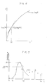
- the time serial measurement data of a certain point or a part of concern in a tomogram image are said to form a gamma function curve as shown in Fig.
- a method for gamma fitting there is a method in which parameters for making an error minimum is found out asymptotically with a grid search method, etc., or a method in which the gamma function is brought into a normal equation by transforming the gamma function into another function, for example, a logarithmic function.
- the former case that is, in an asymptotic method a great many calculations are needed and moreover the solution can not be always obtained.
- a transformation method to a logarithmic function there are too large errors for practical use in the area where values of data are small. Therefore, it is difficult to perform accurate gamma fitting with a small amount of calculations, and it was not possible to obtain a high quality computer image using the gamma function curves.
- EP-A-0 088 935 discloses an image processor provided with a gamma fitting means for setting a gamma function curve approximating to a curve showing the movement of a contrast medium in a tomogram image obtained by a tomograph and for constituting various characteristics of the set gamma function curve with respect to a computer image, comprising: data clip means (DVP 73) for manipulating data, logarithmic transformation means (look-up table), weighting means for weighting images, disturbance removal means (adder and memory) for removing disturbance data and function decision means for setting an approximate gamma function curve.
- DVP 73 data clip means
- logarithmic transformation means look-up table
- weighting means for weighting images
- disturbance removal means adder and memory
- An object of the present invention is to realize an image processor in which an amount of calculation for gamma fitting is increased, a gamma fitting of high precision is performed in a short time and a computer image of high quality can be formed.
- an image processor as defined in claim 1.
- a further embodiment of the invention is disclosed in claim 2.
- An image processor functions as shown in the following: the amount of calculations for gamma fitting is decreased by transforming a gamma function to a first degree equation through logarithmic transformation; the accuracy of gamma fitting is improved by weighting measurement data transformed to a logarithmic function, and by removing disturbance in the measurement data; a smooth computer image can be formed by operating computer image data from gamma function curves which are fitted to respective picture elements, and by interpolating the data for incalculable picture elements.
- Fig. 1, 1 is a data clip device which replaces a datum having a negative CT number to that having a positive number close to zero when the time serial measurement data of a certain point or a part of concern on a plurality of tomogram images obtained by slicing in a plurality of times of scans are transformed to the data in a coordinate system which is based on the time and the CT number of the leading data.
- the data clip device processes input data as shown below. An example of input data is shown in Fig. 4, and the process for the data will be explained. Each point in the figure is plotted based on the data obtained by slicing at each point of time.
- the numbers attached to the data are data numbers, and data 1 express the data of CT number CT1 obtained at a time T1 in the first slicing, and data i express the data of CT number CTi obtained at a time Ti in the No. i slicing.
- the data clip 1 transforms the data shown in Fig. 4 for gamma fitting to the data in a coordinate system based on the time T1 and the CT number CT1 of data 1, a leading data, that is, the data just before the arrival of a contrast medium.
- the coordinate-transformed data are shown in Fig. 5. In the graph shown in Fig.
- a part 2 is a logarithmic transformation device which sustains an expression of a gamma function in equation (1) in the form of a first degree equation being transformed to a logarithmic function, and transforms the output data of the data clip device 1 to a logarithmic function and substitutes them successively for the coefficients in equation (1).
- the device sustains the expression of gamma function in equation (1) in the form of a three element first degree equation being transformed to a logarithmic function as shown in equation (3).
- M ax+by+cz
- a part 3 is a weighting device to make the accuracy of gamma fitting in a logarithmic space equivalent to the accuracy of gamma fitting in a real space. Even if gamma fitting is performed by the output data of the logarithmic transformation device 2, it is a phenomenon in the logarithmic space, and the fitting is largely influenced by the measurement data of small values, therefore the accuracy for approximation in the real space is degraded.
- the object of the weighting device 3 is to improve the above-mentioned problem. Let ei' be the residual at a measurement point i, when the left side of equation (4) does not become zero.
- equation (5) In general for gamma fitting the values of x, y and z which make the square value sum of ei's minimum are to be found as shown in equation (5). Since the evaluation of equation (5) is made concerning a logarithmic space, in the present invention to improve the fitting accuracy in a real space, equation (5) is evaluated by a weighting function w1, a function of M1.
- (X0, logX0) be the coordinates of P
- the change sensitivity in the ordinate Y for the change in the abscissa X of data P can be expressed by a differential coefficient 1/X0 at point P on the curve log X.
- a part 4 is a disturbance removal device for removing data errors in the weighted slice data from the weighting device 3 caused by disturbances such as the ones in which a secondary peak is generated at the trailing edge of a curve or the trailing edge of the curve is blunted as shown in Fig. 7 caused by the influence of blood re-circulation etc.
- Fig. 7 (1, 2, 3, --- m ---p --- n) are measurement data
- the data m + 1 are the data of which trailing edge is blunted
- data P are the data showing a secondary peak.
- the curve connecting measurement data shows the curve of a gamma function to be found, and the data m + 1, the data P, etc. are regarded to be the data which do not show original correct values because of disturbances.
- the percentage value RP for a peak value PV which appears in the measurement data is set beforehand.
- the measurement data m of the slice which fall smaller than (RP/100) x PV (peak value) for the first time will be found and the measurement data m are made to be the final data to be used for gamma fitting.
- the disturbance removal device 4 transforms equation (7) to equation (8).
- equation (9) Besides x, y and z, another parameter t0 (refer to equation (3)) has to be decided to solve equation (9).
- equation (3) the values of t0 of several kinds to several tens of kinds, which are selected as explained later, are substituted for b1 and c1 in equation (3), and equation (9) is solved independently for respective values of t0; in the result, a gamma function is decided from K, ⁇ , ⁇ and t0 which make the square value sum E of the residuals minimum among all solutions for gamma fitting. How to select the several kinds to several tens of kinds of t0 values is shown in Fig. 8.
- a percentage value AP of measurement data for a peak value PV, and the number of times of repetition of fitting N are set beforehand.
- the measurement data (circle marks in the graph) of each slice are compared with the value (AP/100) x PV to find out a first slice of which measurement data become higher than (AP/100) x PV, and let T be the time of it.
- Let t0 be a value selected in the following equation and the gamma fitting of N times is performed. t 0 ⁇ T ⁇ j where
- t0 decided as described above is positioned as shown in Fig. 8.
- l be a slice right after t0, then the data of slices before l - 1 are not used. They are not used for gamma fitting, however, they are used for the evaluation of the square value sum E of the residuals as shown in equation (10) to be able to perform correct evaluation for any t0.
- equation (10) the first term on the right side shows the slice data which are not used for gamma fitting, and the second term shows the slice data which are used for gamma fitting and the slice data from l to m are reflected in equation (9).
- the gamma function decision device 5 solves a normal equation in equation (9) for t0's of N times, and a gamma function to be found is decided by K, ⁇ , ⁇ and t0 when the square value sum E of the residuals become minimum.
- a gamma fitting device as described above has advantages as shown in the following.
- a gamma fitting device is not limited to the above-mentioned embodiment.
- explanation is given about the data, time and the increment of a CT number, however an arbitrary time serial data can be approximated by a gamma function.
- FIG. 2 An image processor using the gamma fitting device as mentioned above is constituted as shown in Fig. 2.
- 11 is the gamma fitting device which performs gamma fitting operation for each picture element of the picture element data of each tomograph image obtained in each slicing, and decides a gamma function which approximates most precisely to the time serial change of the picture data.
- a part 12 is a data control device which controls the gamma fitting device 11 to perform the operation for finding a gamma function fitted for respective picture element data based on the input data of scanned images; in the result, all the sets of parameters K, ⁇ , ⁇ , and t0 which express gamma function curves of respective picture element data obtained from the gamma fitting device 11 are stored in the data control device by picture elements; and after that gamma function curves are reproduced by the above-mentioned parameters K, ⁇ , ⁇ , and t0, and various kinds of computer image data such as a peak value and an area below a gamma function curve,to be described later, are operated.
- a part 13 is an interpolation device, and when it finds a mark of operation suspension in the input data from the data control device 12, re-search of image data is performed over the range of N x N matrix centering the picture element, and when there are found more than M effective data in the matrix of N x N, an average value of these effective data will be substituted for the mark of operation suspension to fill data blank, and when picture element data are completed, a computer image constituted by the data control device 12 is supplemented to be completed.
- the data control device 12 calculates various kinds of computer images, for example, as shown in the following which are specified beforehand based on the gamma fitting performed by the gamma fitting device 11.
Abstract
Description
- The present invention relates to an image processor in which gamma function curves are set which approximate to curves showing the movement of a contrast medium on a tomogram image taken by a tomograph and various characteristics of the set gamma function curves are constituted as a computer image.
- A tomograph is a device for obtaining a tomogram image of a subject body based on X-ray transmission data obtained by irradiating the subject from its surroundings with X rays. As one of the techniques of tomography there is a method for diagnosis of a morbid part as shown in the following: a water-soluble contrast medium is injected into a vein and an image of a blood vessel or an internal organ, etc. which changes with the lapse of time are observed. The time serial measurement data of a certain point or a part of concern in a tomogram image are said to form a gamma function curve as shown in Fig. 3 when a time axis is taken as the axis of abscissas and the change ina CT number is taken as the axis of ordinates, therefore it is necessary to set a gamma function which approximates most precisely to the measurement data (hereinafter referred to as gamma fitting). In Fig. 3, the points shown with small circles express the data of an identical point on tomogram images obtained in respective times (on respective slices), and they are connected with a
gamma function curve 21 which approximates most precisely to the time serial change of the measurement data. The gamma function is expressed in equation (1).
- As a method for gamma fitting, there is a method in which parameters for making an error minimum is found out asymptotically with a grid search method, etc., or a method in which the gamma function is brought into a normal equation by transforming the gamma function into another function, for example, a logarithmic function. In the former case, that is, in an asymptotic method a great many calculations are needed and moreover the solution can not be always obtained. In the latter case, that is, in a transformation method to a logarithmic function, there are too large errors for practical use in the area where values of data are small. Therefore, it is difficult to perform accurate gamma fitting with a small amount of calculations, and it was not possible to obtain a high quality computer image using the gamma function curves.
- The prior art document EP-A-0 088 935 discloses an image processor provided with a gamma fitting means for setting a gamma function curve approximating to a curve showing the movement of a contrast medium in a tomogram image obtained by a tomograph and for constituting various characteristics of the set gamma function curve with respect to a computer image, comprising: data clip means (DVP 73) for manipulating data, logarithmic transformation means (look-up table), weighting means for weighting images, disturbance removal means (adder and memory) for removing disturbance data and function decision means for setting an approximate gamma function curve.
- An object of the present invention is to realize an image processor in which an amount of calculation for gamma fitting is increased, a gamma fitting of high precision is performed in a short time and a computer image of high quality can be formed.
- According to the present invention there is provided an image processor as defined in
claim 1. A further embodiment of the invention is disclosed inclaim 2. - An image processor according to the present invention functions as shown in the following: the amount of calculations for gamma fitting is decreased by transforming a gamma function to a first degree equation through logarithmic transformation; the accuracy of gamma fitting is improved by weighting measurement data transformed to a logarithmic function, and by removing disturbance in the measurement data; a smooth computer image can be formed by operating computer image data from gamma function curves which are fitted to respective picture elements, and by interpolating the data for incalculable picture elements.
-
- Fig. 1 is a conceptual block diagram of an embodiment of a gamma fitting device to be used for an image processor according to the present invention;
- Fig. 2 is a conceptual block diagram of an embodiment of an image processor according to the present invention;
- Fig. 3 is a graph showing a gamma function curve which is approximate to measurement data;
- Fig. 4 is a graph showing an example of input data to a gamma fitting device;
- Fig. 5 is an illustrative representation of coordinate transformation by a data clip device;
- Fig. 6 is a graph showing a relational curve between a real space and a logarithmic space;
- Fig. 7 is a graph showing input data containing disturbance; and
- Fig. 8 is an illustrative representation of a method for deciding to.
- In Fig. 1, 1 is a data clip device which replaces a datum having a negative CT number to that having a positive number close to zero when the time serial measurement data of a certain point or a part of concern on a plurality of tomogram images obtained by slicing in a plurality of times of scans are transformed to the data in a coordinate system which is based on the time and the CT number of the leading data. The data clip device processes input data as shown below. An example of input data is shown in Fig. 4, and the process for the data will be explained. Each point in the figure is plotted based on the data obtained by slicing at each point of time. The numbers attached to the data are data numbers, and
data 1 express the data of CT number CT1 obtained at a time T1 in the first slicing, and data i express the data of CT number CTi obtained at a time Ti in the No. i slicing. Thedata clip 1 transforms the data shown in Fig. 4 for gamma fitting to the data in a coordinate system based on the time T1 and the CT number CT1 ofdata 1, a leading data, that is, the data just before the arrival of a contrast medium. The coordinate-transformed data are shown in Fig. 5. In the graph shown in Fig. 5, the increment of the CT number ofdata 2 is negative, so that thedata clip device 1 replaces the negative value with a certain value A close to zero which is set beforehand (A > 0). The coordinate transformation is, therefore performed as shown in equation (2).
- ti =
- Ti - T1
- Mi' =
- CTi - CT1 (when CTi - CT1 ≥ A)
= A (when CTi - CT1 < A) - A
part 2 is a logarithmic transformation device which sustains an expression of a gamma function in equation (1) in the form of a first degree equation being transformed to a logarithmic function, and transforms the output data of thedata clip device 1 to a logarithmic function and substitutes them successively for the coefficients in equation (1). The device sustains the expression of gamma function in equation (1) in the form of a three element first degree equation being transformed to a logarithmic function as shown in equation (3).
- M=log . f
- x=log . K
- y=α
- z=β
- a=1
- b=log . (t-to) (Hereinafter abbreviate loge as log)
- c=-(t-to)
- In the
data clip device 1, the respective values M1, b1 and c1 in equation (3) are given by the coordinate-transformed output data (t1, M1'). Therefore, equation (4) is obtained from equation (3). A proper value is applied to t0 until the value of it is decided later.
- A
part 3 is a weighting device to make the accuracy of gamma fitting in a logarithmic space equivalent to the accuracy of gamma fitting in a real space. Even if gamma fitting is performed by the output data of thelogarithmic transformation device 2, it is a phenomenon in the logarithmic space, and the fitting is largely influenced by the measurement data of small values, therefore the accuracy for approximation in the real space is degraded. The object of theweighting device 3 is to improve the above-mentioned problem. Let ei' be the residual at a measurement point i, when the left side of equation (4) does not become zero. In general for gamma fitting the values of x, y and z which make the square value sum of ei's minimum are to be found as shown in equation (5).

- A lot of things can be considered about the above-mentioned weighting function w1, but an example of them will be explained referring to Fig. 6. In comparison with a data set X in a real space, consideration is made on the log X which is obtained by logarithmic transformation of the data set X. Fig. 6 is a graph showing a curve of Y = log X corresponding to the data X in a real space. In the figure, let (X0, logX0) be the coordinates of P, then the change sensitivity in the ordinate Y for the change in the abscissa X of data P can be expressed by a
differential coefficient 1/X0 at point P on the curve log X. Therefore, it is made possible to obtain gamma fitting which is not influenced by nonlinearity generated in logarithmic transformation for an arbitrary data X by multiplying the reciprocal of a differential coefficient by the residual in a logarithmic space. Because of this, in the case of transformation to a logarithmic space, the weighting function w (X) can be decided to be w (X) = 1/(1/X) = X. Let w1 in equation (6) be w1 = M1' and let the expression be sustained in theweighting device 3, and by substituting input data t1, N1' successively, the gamma fitting equivalent to that in a real space can be obtained. Therefore, the equation corrected by the weighting function becomes the one just as shown in equation (7).
- A
part 4 is a disturbance removal device for removing data errors in the weighted slice data from theweighting device 3 caused by disturbances such as the ones in which a secondary peak is generated at the trailing edge of a curve or the trailing edge of the curve is blunted as shown in Fig. 7 caused by the influence of blood re-circulation etc. In Fig. 7, (1, 2, 3, --- m ---p --- n) are measurement data, and the data m + 1 are the data of which trailing edge is blunted and data P are the data showing a secondary peak. The curve connecting measurement data shows the curve of a gamma function to be found, and the data m + 1, the data P, etc. are regarded to be the data which do not show original correct values because of disturbances. In thedisturbance removal device 4, the percentage value RP for a peak value PV which appears in the measurement data is set beforehand. The measurement data m of the slice which fall smaller than (RP/100) x PV (peak value) for the first time will be found and the measurement data m are made to be the final data to be used for gamma fitting. Thedisturbance removal device 4 transforms equation (7) to equation (8).
- m =
- final data to. be used for gamma fitting
- E =
- square value sum of residual to be evaluated
- A part 5 is a gamma function decision device for deciding a gamma function by finding the value of a variable which makes the square value sum E of the residuals in equation (8) obtained in the
disturbance removal device 4 minimum. It performs operation as shown below. From equation (8), normal equations are obtained in which partial differential coefficients are put equal to zero as shown in the following.


- These simultaneous equations are expressed as shown in equation (9).


- ΔT
- =T/N,
- j
- =0, 1, 2, ...N-1
- In the result of gamma fitting of N times, a gamma function in which E becomes minimum is decided to be the one to be found.
- The t0 decided as described above is positioned as shown in Fig. 8. Let l be a slice right after t0, then the data of slices before l - 1 are not used. They are not used for gamma fitting, however, they are used for the evaluation of the square value sum E of the residuals as shown in equation (10) to be able to perform correct evaluation for any t0.

- In equation (10), the first term on the right side shows the slice data which are not used for gamma fitting, and the second term shows the slice data which are used for gamma fitting and the slice data from l to m are reflected in equation (9). The gamma function decision device 5, as described above, solves a normal equation in equation (9) for t0's of N times, and a gamma function to be found is decided by K,α,β and t0 when the square value sum E of the residuals become minimum.
- A gamma fitting device as described above has advantages as shown in the following.
- (1) Calculation quantity is decreased much and calculation time is shortened much in performing least square approximation by solving normal equations obtained through logarithmic transformation of measurement quantities.
- (2) The gamma fitting equivalent to the gamma fitting in a real space can be performed by performing weighting in a logarithmic space, which makes it possible to improve the accuracy of gamma fitting.
- (3) It becomes possible to perform accurate gamma fitting by original data by realizing the removal of disturbance.
- (4) An optimum starting time of gamma fitting can be automatically decided by the decision method of t0 and the evaluation by equation (10).
- A gamma fitting device is not limited to the above-mentioned embodiment. In the embodiment, explanation is given about the data, time and the increment of a CT number, however an arbitrary time serial data can be approximated by a gamma function. As an example of a weighting function, w (X) = X is adopted but another weighting function can be used.
- An image processor using the gamma fitting device as mentioned above is constituted as shown in Fig. 2. In Fig. 2, 11 is the gamma fitting device which performs gamma fitting operation for each picture element of the picture element data of each tomograph image obtained in each slicing, and decides a gamma function which approximates most precisely to the time serial change of the picture data. A
part 12 is a data control device which controls the gamma fitting device 11 to perform the operation for finding a gamma function fitted for respective picture element data based on the input data of scanned images; in the result, all the sets of parameters K,α,β, and t0 which express gamma function curves of respective picture element data obtained from the gamma fitting device 11 are stored in the data control device by picture elements; and after that gamma function curves are reproduced by the above-mentioned parameters K,α,β, and t0, and various kinds of computer image data such as a peak value and an area below a gamma function curve,to be described later, are operated. In the operation process of computer images as described in the above, if an excessive numerical value is generated and a picture element which may overflow is found, the operation will be suspended and a mark of operation suspension is stored in place of the picture element data. Apart 13 is an interpolation device, and when it finds a mark of operation suspension in the input data from thedata control device 12, re-search of image data is performed over the range of N x N matrix centering the picture element, and when there are found more than M effective data in the matrix of N x N, an average value of these effective data will be substituted for the mark of operation suspension to fill data blank, and when picture element data are completed, a computer image constituted by thedata control device 12 is supplemented to be completed. By this arrangement, it is made possible to supplement smoothly the value of an incalculable picture element caused by overflow, etc. by interpolation from the circumference and to obtain a computer image of good picture quality and good to look at. A computer image output from the interpolation device is displayed in a display device (not shown in the drawing). The matrix size N and the number of data M shall be decided properly depending on the place of observation or the degree of a morbid state. The data controldevice 12 calculates various kinds of computer images, for example, as shown in the following which are specified beforehand based on the gamma fitting performed by the gamma fitting device 11. - (1) A peak value of a gamma function curve
- (2) A period of time from the origin of the coordinate to the peak value
- (3) An area below a curve
- (4) inflection point width
- (5) Time of appearance
- (6) Mean passage time
- (7) Corrected mean passage time
- (8) Relative flow rate
- The explanation on a most preferred form for the execution of the present invention is given as described in the above, however it will be easy to make various transformation.
Claims (2)
- Image processor provided with a gamma fitting means (11) for setting a gamma function curve approximating to a curve showing the movement of the contrast medium by a tomogram image obtained by a tomograph and for constituting various characteristics of the set gamma function curve with respect to a computer image, comprising: data clip means (1), logarithmic transformation means (2), weighting means (3), disturbance removal means (4) and function decision means (5) for setting an approximate gamma function curve,
characterized in thatthe data clip means (1) performs a coordinate transformation of measurement data, whereby respective leading data which is the data just before the arrival of the contrast medium, are used as origin of a coordinate system based on time and X-ray transmission data, said data clip means shifts data being negative due to said coordinate transformation to positive data by replacing a negative value by a predefined positive value close to zero,the logarithmic transformation means (2) stores a 3 element first degree equation, a gamma function undergone logarithmic transformation, and substitutes the data from said data clip means (1) for the coefficients of said equation,the weighting means (3) weights the output data of said logarithmic transformation means (2) to obtain gamma fitting which is not influenced by nonlinearity generated by logarithmic transformation,the disturbance removal means (4) removes disturbance data to correct a disorder of a curve caused by disturbances, the correction is performed by determining final data which are finally used for gamma fitting, thereby refering to a preset value in correspondance with the peak value of measurement data,the function decision means (5) inputs data from the disturbance removal means (4) and outputs parameters of a gamma function obtained by least square approximation, time offset calculation and not considering certain slice data. - Image processor according to claim 1, characterized by further comprisinga data control means (12) for forming a computer image based on a gamma function curve obtained by the calculation of gamma function decision parameters output from said fitting means (11) by controlling the operation of said gamma fitting means (11), andan interpolation means (13) for completing a computer image by interpolating the picture elements in an incalculable part in the operation process by said data control means (12).
Applications Claiming Priority (1)
| Application Number | Priority Date | Filing Date | Title |
|---|---|---|---|
| PCT/JP1989/000327 WO1990011050A1 (en) | 1989-03-28 | 1989-03-28 | Image processing apparatus |
Publications (3)
| Publication Number | Publication Date |
|---|---|
| EP0422220A1 EP0422220A1 (en) | 1991-04-17 |
| EP0422220A4 EP0422220A4 (en) | 1992-07-22 |
| EP0422220B1 true EP0422220B1 (en) | 1996-07-03 |
Family
ID=13958615
Family Applications (1)
| Application Number | Title | Priority Date | Filing Date |
|---|---|---|---|
| EP89903812A Expired - Lifetime EP0422220B1 (en) | 1989-03-28 | 1989-03-28 | Image processing apparatus |
Country Status (3)
| Country | Link |
|---|---|
| EP (1) | EP0422220B1 (en) |
| DE (1) | DE68926785T2 (en) |
| WO (1) | WO1990011050A1 (en) |
Cited By (2)
| Publication number | Priority date | Publication date | Assignee | Title |
|---|---|---|---|---|
| US6705777B2 (en) | 1999-12-30 | 2004-03-16 | Eastman Kodak Company | System and method for digital film development using visible light |
| US8204332B2 (en) | 2009-07-01 | 2012-06-19 | Shenzhen Mindray Bio-Medical Electronics Co., Ltd. | Method and device for tone scale curve generation |
Families Citing this family (41)
| Publication number | Priority date | Publication date | Assignee | Title |
|---|---|---|---|---|
| EP0901614B1 (en) | 1996-05-10 | 2005-08-10 | Eastman Kodak Company | Luminance-priority color sensor |
| US6442301B1 (en) | 1997-01-06 | 2002-08-27 | Applied Science Fiction, Inc. | Apparatus and method for defect channel nulling |
| US6380539B1 (en) | 1997-01-30 | 2002-04-30 | Applied Science Fiction, Inc. | Four color trilinear CCD scanning |
| US6017688A (en) | 1997-01-30 | 2000-01-25 | Applied Science Fiction, Inc. | System and method for latent film recovery in electronic film development |
| WO1999040729A1 (en) | 1998-02-04 | 1999-08-12 | Applied Science Fiction, Inc. | Multilinear array sensor with an infrared line |
| ATE224622T1 (en) | 1998-02-23 | 2002-10-15 | Applied Science Fiction Inc | PROGRESSIVE AREA SCANNING IN ELECTRONIC FILM DEVELOPMENT |
| US6393160B1 (en) | 1998-03-13 | 2002-05-21 | Applied Science Fiction | Image defect correction in transform space |
| US6594041B1 (en) | 1998-11-20 | 2003-07-15 | Applied Science Fiction, Inc. | Log time processing and stitching system |
| US6437358B1 (en) | 1999-02-04 | 2002-08-20 | Applied Science Fiction, Inc. | Apparatus and methods for capturing defect data |
| US6781620B1 (en) | 1999-03-16 | 2004-08-24 | Eastman Kodak Company | Mixed-element stitching and noise reduction system |
| AU6202100A (en) | 1999-06-29 | 2001-01-31 | Applied Science Fiction, Inc. | Slot coating device for electronic film development |
| AU6914900A (en) | 1999-08-17 | 2001-03-13 | Applied Science Fiction, Inc. | Method and system for using calibration patches in electronic film processing |
| CN1375161A (en) | 1999-09-16 | 2002-10-16 | 应用科学小说公司 | Method and system for altering defects in a digital image |
| WO2001027688A2 (en) | 1999-10-08 | 2001-04-19 | Applied Science Fiction, Inc. | System and method for correcting defects in digital images through selective fill-in from surrounding areas |
| US6498867B1 (en) | 1999-10-08 | 2002-12-24 | Applied Science Fiction Inc. | Method and apparatus for differential illumination image-capturing and defect handling |
| US6924911B1 (en) | 1999-10-12 | 2005-08-02 | Eastman Kodak Company | Method and system for multi-sensor signal detection |
| US6711302B1 (en) | 1999-10-20 | 2004-03-23 | Eastman Kodak Company | Method and system for altering defects in digital image |
| WO2001035339A2 (en) * | 1999-10-29 | 2001-05-17 | Cnr Consiglio Nazionale Delle Ricerche | Automatic analysis of anatomical images time sequence |
| US6683995B2 (en) | 1999-12-23 | 2004-01-27 | Eastman Kodak Company | Method and apparatus for correcting large defects in digital images |
| US7164511B2 (en) | 1999-12-29 | 2007-01-16 | Eastman Kodak Company | Distinguishing positive and negative films system and method |
| US6704458B2 (en) | 1999-12-29 | 2004-03-09 | Eastman Kodak Company | Method and apparatus for correcting heavily damaged images |
| US6862117B1 (en) | 1999-12-30 | 2005-03-01 | Eastman Kodak Company | Method and apparatus for reducing the effect of bleed-through on captured images |
| US6540416B2 (en) | 1999-12-30 | 2003-04-01 | Applied Science Fiction, Inc. | System and method for digital film development using visible light |
| US6505977B2 (en) | 1999-12-30 | 2003-01-14 | Applied Science Fiction, Inc. | System and method for digital color dye film processing |
| US6447178B2 (en) | 1999-12-30 | 2002-09-10 | Applied Science Fiction, Inc. | System, method, and apparatus for providing multiple extrusion widths |
| US6554504B2 (en) | 1999-12-30 | 2003-04-29 | Applied Science Fiction, Inc. | Distributed digital film processing system and method |
| US6720560B1 (en) | 1999-12-30 | 2004-04-13 | Eastman Kodak Company | Method and apparatus for scanning images |
| US6813392B2 (en) | 1999-12-30 | 2004-11-02 | Eastman Kodak Company | Method and apparatus for aligning multiple scans of the same area of a medium using mathematical correlation |
| US6707557B2 (en) | 1999-12-30 | 2004-03-16 | Eastman Kodak Company | Method and system for estimating sensor dark current drift and sensor/illumination non-uniformities |
| US6788335B2 (en) | 1999-12-30 | 2004-09-07 | Eastman Kodak Company | Pulsed illumination signal modulation control & adjustment method and system |
| US6475711B1 (en) | 1999-12-31 | 2002-11-05 | Applied Science Fiction, Inc. | Photographic element and digital film processing method using same |
| US6664034B2 (en) | 1999-12-31 | 2003-12-16 | Eastman Kodak Company | Digital film processing method |
| US6786655B2 (en) | 2000-02-03 | 2004-09-07 | Eastman Kodak Company | Method and system for self-service film processing |
| US6599036B2 (en) | 2000-02-03 | 2003-07-29 | Applied Science Fiction, Inc. | Film processing solution cartridge and method for developing and digitizing film |
| US6619863B2 (en) | 2000-02-03 | 2003-09-16 | Eastman Kodak Company | Method and system for capturing film images |
| WO2002025345A2 (en) | 2000-09-22 | 2002-03-28 | Applied Science Fiction | Lens focusing device, system and method for use with multiple light wavelengths |
| US6733960B2 (en) | 2001-02-09 | 2004-05-11 | Eastman Kodak Company | Digital film processing solutions and method of digital film processing |
| US6987892B2 (en) | 2001-04-19 | 2006-01-17 | Eastman Kodak Company | Method, system and software for correcting image defects |
| US6805501B2 (en) | 2001-07-16 | 2004-10-19 | Eastman Kodak Company | System and method for digital film development using visible light |
| US8879832B2 (en) * | 2012-06-26 | 2014-11-04 | Xerox Corporation | Color matrix code |
| US9589217B2 (en) | 2014-07-09 | 2017-03-07 | Xeroc Corporation | Augmenting barcodes with secondary encoding for anti-counterfeiting |
Family Cites Families (5)
| Publication number | Priority date | Publication date | Assignee | Title |
|---|---|---|---|---|
| US4504908A (en) * | 1982-03-15 | 1985-03-12 | General Electric Company | Matched filter for X-ray temporal subtraction |
| JPS62179435A (en) * | 1986-01-31 | 1987-08-06 | 株式会社島津製作所 | Method for forming image of circulatory function |
| JPS62179436A (en) * | 1986-02-04 | 1987-08-06 | 株式会社東芝 | Computed tomography apparatus |
| JPS6486942A (en) * | 1987-09-30 | 1989-03-31 | Yokogawa Medical Syst | Image processing apparatus |
| JPH06249832A (en) * | 1993-02-26 | 1994-09-09 | Toa Denpa Kogyo Kk | Measurement of chlorous acid ion |
-
1989
- 1989-03-28 DE DE68926785T patent/DE68926785T2/en not_active Expired - Lifetime
- 1989-03-28 EP EP89903812A patent/EP0422220B1/en not_active Expired - Lifetime
- 1989-03-28 WO PCT/JP1989/000327 patent/WO1990011050A1/en active IP Right Grant
Non-Patent Citations (1)
| Title |
|---|
| PATENT ABSTRACTS OF JAPAN vol. 13, no. 293 (C-615)(3641) 6 July 1989 & JP-A-1 086 942 * |
Cited By (2)
| Publication number | Priority date | Publication date | Assignee | Title |
|---|---|---|---|---|
| US6705777B2 (en) | 1999-12-30 | 2004-03-16 | Eastman Kodak Company | System and method for digital film development using visible light |
| US8204332B2 (en) | 2009-07-01 | 2012-06-19 | Shenzhen Mindray Bio-Medical Electronics Co., Ltd. | Method and device for tone scale curve generation |
Also Published As
| Publication number | Publication date |
|---|---|
| WO1990011050A1 (en) | 1990-10-04 |
| DE68926785T2 (en) | 1997-01-02 |
| EP0422220A1 (en) | 1991-04-17 |
| DE68926785D1 (en) | 1996-08-08 |
| EP0422220A4 (en) | 1992-07-22 |
Similar Documents
| Publication | Publication Date | Title |
|---|---|---|
| EP0422220B1 (en) | Image processing apparatus | |
| US5594807A (en) | System and method for adaptive filtering of images based on similarity between histograms | |
| JP3761094B2 (en) | Method for reconstructing a three-dimensional image of an object | |
| EP0424912A2 (en) | Region extracting method and three-dimensional display method | |
| CN104599239A (en) | Medical image metal artifact eliminating method and device | |
| EP0952458B1 (en) | Method and apparatus for ultrasound imaging using adaptive gray mapping | |
| JP2000279416A (en) | Three-dimensional imaging method and system | |
| Busse et al. | A model based approach to improve the performance of the geometric filtering speckle reduction algorithm | |
| Parrott et al. | Towards statistically optimal interpolation for 3D medical imaging | |
| CN107527339B (en) | Magnetic resonance scanning method, device and system | |
| US4682301A (en) | Digital filter for processing two-dimensional digital image | |
| CN107590512A (en) | The adaptive approach and system of parameter in a kind of template matches | |
| US4729100A (en) | CT System which convolutes projection data with a frequency varying filter function | |
| EP0362849A2 (en) | Method and system for fitting image positions | |
| CN115205450A (en) | Three-dimensional scanning data processing method, device, system, equipment and medium | |
| CN114596261A (en) | Wear detection method, device, terminal and medium based on three-dimensional reconstruction of tool nose | |
| JPH021082A (en) | Apparatus and method for generating definition of three-dimensional surface | |
| JP2856661B2 (en) | Density converter | |
| JPH0444545B2 (en) | ||
| US6914602B2 (en) | Approximating gradients with offset midpoints | |
| EP0601898A2 (en) | Adaptive input/output apparatus using selected sample data according to evaluation quantity | |
| US5821942A (en) | Ray tracing through an ordered array | |
| US6208766B1 (en) | Process for influencing the quality images furnished by an electronic imaging system | |
| CN111210877A (en) | Method and device for deducing physical property parameters | |
| US6404922B1 (en) | Curve length measuring device and measuring method therefore and storage medium which stores control program for measuring length of curve |
Legal Events
| Date | Code | Title | Description |
|---|---|---|---|
| PUAI | Public reference made under article 153(3) epc to a published international application that has entered the european phase |
Free format text: ORIGINAL CODE: 0009012 |
|
| 17P | Request for examination filed |
Effective date: 19901221 |
|
| AK | Designated contracting states |
Kind code of ref document: A1 Designated state(s): DE FR GB NL |
|
| A4 | Supplementary search report drawn up and despatched |
Effective date: 19920605 |
|
| AK | Designated contracting states |
Kind code of ref document: A4 Designated state(s): DE FR GB NL |
|
| 17Q | First examination report despatched |
Effective date: 19941220 |
|
| GRAH | Despatch of communication of intention to grant a patent |
Free format text: ORIGINAL CODE: EPIDOS IGRA |
|
| GRAH | Despatch of communication of intention to grant a patent |
Free format text: ORIGINAL CODE: EPIDOS IGRA |
|
| GRAA | (expected) grant |
Free format text: ORIGINAL CODE: 0009210 |
|
| AK | Designated contracting states |
Kind code of ref document: B1 Designated state(s): DE FR GB NL |
|
| REF | Corresponds to: |
Ref document number: 68926785 Country of ref document: DE Date of ref document: 19960808 |
|
| ET | Fr: translation filed |
Free format text: CORRECTIONS |
|
| PLBE | No opposition filed within time limit |
Free format text: ORIGINAL CODE: 0009261 |
|
| STAA | Information on the status of an ep patent application or granted ep patent |
Free format text: STATUS: NO OPPOSITION FILED WITHIN TIME LIMIT |
|
| 26N | No opposition filed | ||
| REG | Reference to a national code |
Ref country code: GB Ref legal event code: IF02 |
|
| PGFP | Annual fee paid to national office [announced via postgrant information from national office to epo] |
Ref country code: GB Payment date: 20070327 Year of fee payment: 19 |
|
| PGFP | Annual fee paid to national office [announced via postgrant information from national office to epo] |
Ref country code: FR Payment date: 20070319 Year of fee payment: 19 |
|
| PGFP | Annual fee paid to national office [announced via postgrant information from national office to epo] |
Ref country code: NL Payment date: 20080324 Year of fee payment: 20 |
|
| PGFP | Annual fee paid to national office [announced via postgrant information from national office to epo] |
Ref country code: DE Payment date: 20080430 Year of fee payment: 20 |
|
| GBPC | Gb: european patent ceased through non-payment of renewal fee |
Effective date: 20080328 |
|
| REG | Reference to a national code |
Ref country code: FR Ref legal event code: ST Effective date: 20081125 |
|
| PG25 | Lapsed in a contracting state [announced via postgrant information from national office to epo] |
Ref country code: FR Free format text: LAPSE BECAUSE OF NON-PAYMENT OF DUE FEES Effective date: 20080331 |
|
| PG25 | Lapsed in a contracting state [announced via postgrant information from national office to epo] |
Ref country code: NL Free format text: LAPSE BECAUSE OF EXPIRATION OF PROTECTION Effective date: 20090328 |
|
| NLV7 | Nl: ceased due to reaching the maximum lifetime of a patent |
Effective date: 20090328 |
|
| PG25 | Lapsed in a contracting state [announced via postgrant information from national office to epo] |
Ref country code: GB Free format text: LAPSE BECAUSE OF NON-PAYMENT OF DUE FEES Effective date: 20080328 |