EP0446713B1 - Blood separation system - Google Patents
Blood separation system Download PDFInfo
- Publication number
- EP0446713B1 EP0446713B1 EP91102992A EP91102992A EP0446713B1 EP 0446713 B1 EP0446713 B1 EP 0446713B1 EP 91102992 A EP91102992 A EP 91102992A EP 91102992 A EP91102992 A EP 91102992A EP 0446713 B1 EP0446713 B1 EP 0446713B1
- Authority
- EP
- European Patent Office
- Prior art keywords
- bag
- blood
- outlet
- tubing
- satellite
- Prior art date
- Legal status (The legal status is an assumption and is not a legal conclusion. Google has not performed a legal analysis and makes no representation as to the accuracy of the status listed.)
- Expired - Lifetime
Links
Images
Classifications
-
- A—HUMAN NECESSITIES
- A61—MEDICAL OR VETERINARY SCIENCE; HYGIENE
- A61M—DEVICES FOR INTRODUCING MEDIA INTO, OR ONTO, THE BODY; DEVICES FOR TRANSDUCING BODY MEDIA OR FOR TAKING MEDIA FROM THE BODY; DEVICES FOR PRODUCING OR ENDING SLEEP OR STUPOR
- A61M1/00—Suction or pumping devices for medical purposes; Devices for carrying-off, for treatment of, or for carrying-over, body-liquids; Drainage systems
- A61M1/02—Blood transfusion apparatus
- A61M1/0209—Multiple bag systems for separating or storing blood components
Definitions
- This disclosure is concerned generally with collection and separation systems for whole blood and specifically with a blood component separation system that can be partially automated.
- Whole blood is commonly separated into its major components of less dense plasma and more dense red blood cells (RBCs) by first drawing the whole blood into a plastic bag known as a donor or primary bag. The bag's contents are then centrifuged under controlled conditions to result in a lower, more dense portion of packed RBCs and an upper less dense plasma portion, which may be rich in platelets (platelet rich plasma or PRP).
- RBCs red blood cells
- the donor bag is typically connected by plastic tubing to one or more satellite bags into which separated blood components (e.g. the PRP) may be expressed by external manipulation for further processing or use.
- separated blood components e.g. the PRP
- the classical method of preparing platelet transfusion products from whole blood collections consists of initial centrifugation of whole blood in a plastic blood bag at relatively low centrifugal force to separate most of the PRP from the red cells.
- the PRP is commonly expressed into an attached satellite blood bag. This is followed by centrifugation of the PRP in the satellite bag at relatively high centrifugal force to form a lower sediment of platelets and an upper platelet poor plasma (PRP).
- the sedimented platelets are in the form of a pellet or "button" which is resuspended in a small volume (50-60 mL) of donor plasma to give the platelet concentrate.
- the buffy coat plus either a small volume of plasma or a synthetic medium is then centrifuged at low centrifugal force to separate platelet concentrate (upper layer) from residual red cells and leukocytes.
- platelet concentrate upper layer
- the Amsterdam method while apparently giving respectable platelet yields, was cumbersome and labor-intensive.
- the buffy coat transfer step required the operator to massage the bag to prevent hang-up of the "sticky" buffy coat layer. These manipulations might influence platelet function and release of granulocyte enzymes. There was also no way to control the volume of buffy coat removed.
- U.S. Patent 3,911,918 to Turner discloses a blood bag having an hour glass shape. That bag has a top portion for plasma, a bottom portion for RBCs and a middle portion for platelets and white blood cells.
- the hour glass shape is said to help position clamping or sealing devices at the juncture of the separated components after whole blood in the bag is centrifuged. This system has not been used on any significant commercial scale to date.
- Our system for separation of blood components comprises a main plastic bag having inlet and at least two outlet ports, all of the outlets being at the top of the bag.
- One outlet port is in closed communication with or capable of being connected to an empty plasma satellite bag adapted to hold plasma.
- the other outlet port communicates at one end with a tube extending into and within the bag and close to the bag bottom and at the other end is in closed communication with or capable of being connected to a second bag adapted to hold red blood cells.
- the second satellite bag contains an RBC preservative solution such as AS-3.
- whole blood is drawn into the main bag via the inlet port and then centrifuged to form an upper, less dense plasma portion, an intermediate buffy coat portion (containing platelets) and a lower, more dense packed red blood cell portion.
- Pressure is then applied to the bag to express the upper plasma portion through the first outlet into the empty plasma satellite bag via a conventional blood bag connecting tubing.
- a sensor or photocell adapted to sense a color change as the last of the plasma (or first of the buffy coat) passes through the tubing.
- the color change at the start or the top of the buffy coat portion can activate the sensor at which time the sensor activates a clamp (or valve) which closes the tubing connected to the first outlet port.
- the senor When the tubing of the first outlet is thus closed, the sensor simultaneously opens or activates a clamp (valve) in a tubing connecting the main bag with the second satellite bag adapted to hold red blood cells. This allows passage of the RBCs (from the tube extending into the bottom of the bag) to pass into the second satellite bag until a pre-determined volume of buffy coat remains in the main or primary bag. This leaves only buffy coat and platelets in the original bag.
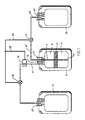
- the Figure is a plan view of a preferred blood bag separation system of this disclosure.
- Our system for the separation of blood components and particularly for the preparation of platelet concentrate from buffy coat comprises a bag having sensor activated clamps or valves associated with each of the outlet ports as part of the bag.
- the outlet port for the RBCs communicates with a tubular extension extending to the bottom of the bag interior.
- the tube is flexible to minimize breaking or bending during centrifuging. This also helps avoid bag puncture and breaking of the solvent bond where the tube connects to the outlet port.
- the tube is preferably transparent and extends to within about 1.27 cm (1/2 inch) of the interior bottom of the bag.
- it is made from conventional blood bag PVC tubing and has a beveled tip to avoid blockage and assure fluid (RBC) flow even if the end of the tube actually touches or presses against the bag bottom.
- a preferred system is a "triple" blood bag consisting of a primary or donor bag and two satellite bags pre-connected to the primary bag by conventional blood bag tubing.
- the primary bag is made using conventional techniques but is modified in that the blood collection bag has a RBC outlet tube extending from a top outlet port to the bottom of the bag as shown in Figure 1. All bags and connecting tubings are made from conventional blood bag plastics (e.g. PVC and the like) and are essentially transparent.
- the triple unit After collection of whole blood into bag 3, the triple unit is centrifuged at relatively high centrifugal force to form upper plasma component 9, intermediate buffy coat component 13, and lower red cell component 11.
- the bag 3 is then placed in a simple pressure-separator device (blood bag expresser) consisting of a moving spring-loaded expresser plate and a fixed plate.
- the separation system includes two on-off tubing clamps, 19 and 31, one on tubing 17 and one on tubing 27, activated by a sensor such as a photocell. Plasma passes through tube 17 while the tubing that conveys red cells passes through tube 27.
- a simple clamp 19 on tubing 17 is open and a simple clamp 31 on tubing 27 is closed.
- Plasma is expressed into bag 21 by pressure on bag 3 until red cells (in the buffy coat) are first detected in tubing 17 by the photocell sensor 16, at which time clamp 19 on tubing 17 closes in response to an electrical signal on wire line 25 from sensor 16 and the clamp 31 on tubing 27 opens in response to a signal on similar line 25.
- Red cells are then expressed from the beveled bottom 7a of tube extension 7 through the top of bag 3 into second satellite bag 35 containing a conventional red cell preservation solution such as AS-3 or the like (not shown).
- the expression continues until only a volume of about 50mL (the buffy coat 13) remains in bag 3. This volume may be set, if desired, by a simple stop between the expresser plate and the backplate against which the bag 3 is pressed in an otherwise conventional blood bag expresser.
- a preferred method of processing buffy coat to platelet concentrate after centrifugation is as follows. After the plasma and RBC expressions and detaching the plasma bag 21 and the red cell bag 35, the buffy coat 13 is held in the primary bag 3 overnight, preferably at room temperature with agitation. A number of bags containing buffy coat, preferably 6, are pooled together using a Sterile Connection Device (e.g. such as that shown in U.S. Patent 4,507,119) into a bag containing a platelet additive solution. A platelet pooling bag such as that shown in U.S. 4,857,190 to S. Wada and B. Kuhleman can be used.
- a Sterile Connection Device e.g. such as that shown in U.S. Patent 4,507,119
- a platelet pooling bag such as that shown in U.S. 4,857,190 to S. Wada and B. Kuhleman can be used.
- the pool of platelets is centrifuged at low centrifugal force to form an upper platelet concentrate (PC) layer and a lower layer of undesirable red cells and leukocytes.
- the PC may also be expressed through a leukocyte filter (e.g. as shown in U.S. 4,810,378 to R.A. Carmen et al) into a 1000 mL storage bag made of a plastic with high 02 and CO2 transmission rates for storage (e.g. as shown in U.S. 4,280,497 to Warner et al.).
- the triple bag system of this invention was used to prepare components including buffy coat platelet concentrate from 6 individual units of blood.
- the buffy coats were processed individually rather than as a pool.
- Whole blood was collected into a primary blood bag and the bag was centrifuged at 3000Xg for 9 minutes.
- the plasma upper layer was expressed into an attached empty satellite bag followed by expression of red cell lower layer from the bottom and out of the top of the primary bag into an attached bag containing AS-3 RBC preservation solution.
- Platelet counts were performed on all fractions using either an electronic cell counter (plasma and platelet concentrate) or a manual method. Data are summarized in the table below.
Abstract
Description
- This disclosure is concerned generally with collection and separation systems for whole blood and specifically with a blood component separation system that can be partially automated.
- Whole blood is commonly separated into its major components of less dense plasma and more dense red blood cells (RBCs) by first drawing the whole blood into a plastic bag known as a donor or primary bag. The bag's contents are then centrifuged under controlled conditions to result in a lower, more dense portion of packed RBCs and an upper less dense plasma portion, which may be rich in platelets (platelet rich plasma or PRP).
- The donor bag is typically connected by plastic tubing to one or more satellite bags into which separated blood components (e.g. the PRP) may be expressed by external manipulation for further processing or use.
- The above system for separating blood into its major components has remained virtually unchanged since the 1950's when plastic blood bags were introduced commercially on a large scale.
- The classical method of preparing platelet transfusion products from whole blood collections consists of initial centrifugation of whole blood in a plastic blood bag at relatively low centrifugal force to separate most of the PRP from the red cells. The PRP is commonly expressed into an attached satellite blood bag. This is followed by centrifugation of the PRP in the satellite bag at relatively high centrifugal force to form a lower sediment of platelets and an upper platelet poor plasma (PRP). The sedimented platelets are in the form of a pellet or "button" which is resuspended in a small volume (50-60 mL) of donor plasma to give the platelet concentrate.
- With good technique, about 2/3 of the platelets in a whole blood collection unit are recovered in the platelet concentrate. This is equivalent to about 8X10¹⁰ platelets per concentrate. However, achieving this yield of platelets requires strict attention to centrifugation protocols, frequent calibration of the centrifuges, and operator diligence. The fact that the minimum standard for platelet yield is only 5.5x10¹⁰ per concentrate attests to the operator-dependent nature of this procedure.
- Recently, some transfusion services in Europe have begun to investigate and in some cases employ an alternate method of platelet preparation, specifically preparation from buffy coat. In this procedure the initial centrifugation of whole blood is performed at relatively high centrifugal force to form an upper layer of relatively cell-free plasma, an intermediate buffy coat layer containing platelets and leukocytes, and a lower layer of red cells.
- The buffy coat plus either a small volume of plasma or a synthetic medium is then centrifuged at low centrifugal force to separate platelet concentrate (upper layer) from residual red cells and leukocytes. Data suggest that platelets prepared in this fashion are of improved quality, presumably because platelet activation that would otherwise occur during the pelleting step of the PRP centrifugation method is avoided.
- The original work on buffy coat platelets was done at the Dutch Red Cross. Referred to as the Amsterdam method, it employed a standard quadruple plastic bag system. After centrifugation of blood and removal of plasma from the main bag, the buffy coat layer was transferred to an empty connected satellite bag and then processed to platelet concentrate. Using this method, Pietersz et al. (Vox Sang 1985; 49:81-85) found a mean of 7.2x10¹⁰ platelets per concentrate; the volume of blood collected in this study was 500 mL. Kretschmer et al. (Infusionstherapie 1988; 15:232-239) found a mean of 6.3x10¹⁰ platelets per concentrate from 450 mL blood collections.
- The Amsterdam method, while apparently giving respectable platelet yields, was cumbersome and labor-intensive. The buffy coat transfer step required the operator to massage the bag to prevent hang-up of the "sticky" buffy coat layer. These manipulations might influence platelet function and release of granulocyte enzymes. There was also no way to control the volume of buffy coat removed.
- Other efforts to improve platelet separation procedures or at least make it less burdensome are known. For example, U.S. Patent 3,911,918 to Turner discloses a blood bag having an hour glass shape. That bag has a top portion for plasma, a bottom portion for RBCs and a middle portion for platelets and white blood cells. The hour glass shape is said to help position clamping or sealing devices at the juncture of the separated components after whole blood in the bag is centrifuged. This system has not been used on any significant commercial scale to date.
- In U.S. Patent 4,608,178 to A. S. Johansson and C. F. Hogman there is disclosed a "top/bottom" with which the upper and lower portions of separated blood components can be simultaneously expressed from a specially designed bag which leaves behind in the bag the intermediate portion known as buffy coat. The expression of that system is controlled by a pressure plate on the bag and sensors which monitor the position of the intermediate layer such that it remains in the bag while the upper plasma is expressed from a top part and the lower red blood cells are expressed from a bottom part in the bag. Hence, the name top/bottom bag. The sensors in that system assure the simultaneous expression of the top and bottom components.
- The above described systems are fairly recent and it is not clear yet whether those systems will in time replace existing blood separation systems based on the use of a relatively simple unmodified donor bag.
- However, the systems do offer new ways to prepare platelets (contained in the intermediate or buffy coat portion). The patent to Johansson and Hogman show how to do this in a semi-automated manner. Hence, it potentially represents a semi-automated way to prepare platelets.
- In an effort to overcome problems associated with the Amsterdam method, Johansson and Hogman (see above-cited patent) developed the bag system with the top and bottom drainage of the primary bag and a sensor device which allowed partially automated blood separation. Kretschmer et al. used that type of system to prepare platelet concentrates from buffy coats and found a mean of 6.7x10¹⁰ platelets per unit.
- We have now found a novel alternative to the above described automated system, the details of which are described below.
- Our system for separation of blood components comprises a main plastic bag having inlet and at least two outlet ports, all of the outlets being at the top of the bag. One outlet port is in closed communication with or capable of being connected to an empty plasma satellite bag adapted to hold plasma. The other outlet port communicates at one end with a tube extending into and within the bag and close to the bag bottom and at the other end is in closed communication with or capable of being connected to a second bag adapted to hold red blood cells. In preferred embodiments the second satellite bag contains an RBC preservative solution such as AS-3.
- In use, whole blood is drawn into the main bag via the inlet port and then centrifuged to form an upper, less dense plasma portion, an intermediate buffy coat portion (containing platelets) and a lower, more dense packed red blood cell portion. Pressure is then applied to the bag to express the upper plasma portion through the first outlet into the empty plasma satellite bag via a conventional blood bag connecting tubing. At a convenient location along the tubing is a sensor or photocell adapted to sense a color change as the last of the plasma (or first of the buffy coat) passes through the tubing. For example, the color change at the start or the top of the buffy coat portion can activate the sensor at which time the sensor activates a clamp (or valve) which closes the tubing connected to the first outlet port.
- When the tubing of the first outlet is thus closed, the sensor simultaneously opens or activates a clamp (valve) in a tubing connecting the main bag with the second satellite bag adapted to hold red blood cells. This allows passage of the RBCs (from the tube extending into the bottom of the bag) to pass into the second satellite bag until a pre-determined volume of buffy coat remains in the main or primary bag. This leaves only buffy coat and platelets in the original bag.
- The Figure is a plan view of a preferred blood bag separation system of this disclosure.
- Our system for the separation of blood components and particularly for the preparation of platelet concentrate from buffy coat comprises a bag having sensor activated clamps or valves associated with each of the outlet ports as part of the bag. The outlet port for the RBCs communicates with a tubular extension extending to the bottom of the bag interior. In preferred embodiments, the tube is flexible to minimize breaking or bending during centrifuging. This also helps avoid bag puncture and breaking of the solvent bond where the tube connects to the outlet port. The tube is preferably transparent and extends to within about 1.27 cm (1/2 inch) of the interior bottom of the bag. Preferably, it is made from conventional blood bag PVC tubing and has a beveled tip to avoid blockage and assure fluid (RBC) flow even if the end of the tube actually touches or presses against the bag bottom.
- A preferred system is a "triple" blood bag consisting of a primary or donor bag and two satellite bags pre-connected to the primary bag by conventional blood bag tubing. The primary bag is made using conventional techniques but is modified in that the blood collection bag has a RBC outlet tube extending from a top outlet port to the bottom of the bag as shown in Figure 1. All bags and connecting tubings are made from conventional blood bag plastics (e.g. PVC and the like) and are essentially transparent.
- After collection of whole blood into
bag 3, the triple unit is centrifuged at relatively high centrifugal force to formupper plasma component 9, intermediatebuffy coat component 13, and lowerred cell component 11. Thebag 3 is then placed in a simple pressure-separator device (blood bag expresser) consisting of a moving spring-loaded expresser plate and a fixed plate. The separation system includes two on-off tubing clamps, 19 and 31, one ontubing 17 and one ontubing 27, activated by a sensor such as a photocell. Plasma passes throughtube 17 while the tubing that conveys red cells passes throughtube 27. - At the start of the plasma expression, a
simple clamp 19 ontubing 17 is open and asimple clamp 31 ontubing 27 is closed. Plasma is expressed intobag 21 by pressure onbag 3 until red cells (in the buffy coat) are first detected intubing 17 by thephotocell sensor 16, at which time clamp 19 ontubing 17 closes in response to an electrical signal onwire line 25 fromsensor 16 and theclamp 31 ontubing 27 opens in response to a signal onsimilar line 25. Red cells are then expressed from thebeveled bottom 7a oftube extension 7 through the top ofbag 3 intosecond satellite bag 35 containing a conventional red cell preservation solution such as AS-3 or the like (not shown). The expression continues until only a volume of about 50mL (the buffy coat 13) remains inbag 3. This volume may be set, if desired, by a simple stop between the expresser plate and the backplate against which thebag 3 is pressed in an otherwise conventional blood bag expresser. - A preferred method of processing buffy coat to platelet concentrate after centrifugation is as follows. After the plasma and RBC expressions and detaching the
plasma bag 21 and thered cell bag 35, thebuffy coat 13 is held in theprimary bag 3 overnight, preferably at room temperature with agitation. A number of bags containing buffy coat, preferably 6, are pooled together using a Sterile Connection Device (e.g. such as that shown in U.S. Patent 4,507,119) into a bag containing a platelet additive solution. A platelet pooling bag such as that shown in U.S. 4,857,190 to S. Wada and B. Kuhleman can be used. The pool of platelets is centrifuged at low centrifugal force to form an upper platelet concentrate (PC) layer and a lower layer of undesirable red cells and leukocytes. The PC may also be expressed through a leukocyte filter (e.g. as shown in U.S. 4,810,378 to R.A. Carmen et al) into a 1000 mL storage bag made of a plastic with high 0₂ and CO₂ transmission rates for storage (e.g. as shown in U.S. 4,280,497 to Warner et al.). - The triple bag system of this invention was used to prepare components including buffy coat platelet concentrate from 6 individual units of blood. In this case the buffy coats were processed individually rather than as a pool. Whole blood was collected into a primary blood bag and the bag was centrifuged at 3000Xg for 9 minutes. The plasma upper layer was expressed into an attached empty satellite bag followed by expression of red cell lower layer from the bottom and out of the top of the primary bag into an attached bag containing AS-3 RBC preservation solution.
- About 50 mL of the intermediate buffy coat layer was left in the collection bag. Fifty milliliters of the plasma in the connected satellite bag was added back to the buffy coat. This was followed by overnight incubation of the primary bag contents at 22°C on a flatbed shaker and centrifugation at 400Xg for 6 minutes to separate platelet concentrate from residual red cells and leukocytes.
- Platelet counts were performed on all fractions using either an electronic cell counter (plasma and platelet concentrate) or a manual method. Data are summarized in the table below.

- Data show that this system gives a very good yield of platelets into platelet concentrate (8.7±3.4X10¹⁰ or 68%). Of note is the very low loss of platelets to the red cell fraction. Platelet losses to plasma and residual buffy coat fractions can be further reduced by optimizing centrifugation protocols.
- Given the above disclosure it is thought variations will now occur to those skilled in the art. Accordingly, the above examples should be construed as illustrative and the scope of the invention disclosed herein should be limited only by the following claims:
Claims (11)
- A system for separation of blood components in whole blood, the system comprising a blood bag (3) having at least two outlet ports (17) and (27) at the top of the bag, a first port (27) and a second port (17), the first port (27) communicating with a tubular member (7) extending into and within the interior of the bag (3) and terminating at a distance just above the bag bottom.
- The system of Claim 1 where the tubular member extends to within about 1,27 cm (1/2 inches) of the bottom of the bag's interior.
- The system of claim 2 wherein the tubular member is flexible and terminates as a bevel.
- The system of Claim 1 wherein the second outlet port communicates with a first satellite blood bag (21) via tubing.
- The system of Claim 4 wherein the tubing communicating with an external clamp (19) includes sensor means (16) to stop passage of fluid through the tubing.
- The system of Claim 1 wherein the first outlet port communicates with a second satellite blood bag (35) via tubing.
- The system of Claim 6 wherein the tubing communicating with an external clamp (19) includes sensor means (16) to stop passage of fluid through the tubing.
- The system of Claim 5 wherein the sensor means, upon sensing and stopping passage of fluid in the tubing, communicates with and opens an external valve (19) on the tubing connecting the second outlet port with the blood bag so that fluid may pass from the bag through the outlet port to the second satellite bag.
- A method of separating blood into components comprising the steps of(a) introducing whole blood into a blood bag (3) having at least two outlet ports (17) and (27) at the top of the bag (3) one of the outlet ports communicating with a tubular member (7) extending into and with in the bag and ending within about 1,27 cm (1/2 inch) of the bag's interior bottom;(b) centrifuging the blood bag (3) at relatively high centrifugal force to form an upper plasma portion (9), an intermediate buffy coat portion (13) and a lower red blood cell portion;(c) applying pressure to the bag to express plasma through an outlet port out of the bag; and(d) applying pressure to the bag to express the red cell protein out of the bag through the tubular member.
- The method of Claim 9 wherein each outlet port is in closed communication via tubing with a satellite bag (21,35).
- The method of Claim 9 wherein the tubings connecting the satellite bags with the outlet ports include sensor means (16) and valve means (19,31) that cooperate in a predetermined manner to open and close fluid passage through the outlet ports.
Applications Claiming Priority (2)
| Application Number | Priority Date | Filing Date | Title |
|---|---|---|---|
| US493024 | 1983-05-09 | ||
| US07/493,024 US5102407A (en) | 1990-03-13 | 1990-03-13 | Blood separation system |
Publications (3)
| Publication Number | Publication Date |
|---|---|
| EP0446713A2 EP0446713A2 (en) | 1991-09-18 |
| EP0446713A3 EP0446713A3 (en) | 1992-01-29 |
| EP0446713B1 true EP0446713B1 (en) | 1994-08-10 |
Family
ID=23958590
Family Applications (1)
| Application Number | Title | Priority Date | Filing Date |
|---|---|---|---|
| EP91102992A Expired - Lifetime EP0446713B1 (en) | 1990-03-13 | 1991-02-28 | Blood separation system |
Country Status (7)
| Country | Link |
|---|---|
| US (1) | US5102407A (en) |
| EP (1) | EP0446713B1 (en) |
| AT (1) | ATE109655T1 (en) |
| CA (1) | CA2037813A1 (en) |
| DE (1) | DE69103297T2 (en) |
| DK (1) | DK0446713T3 (en) |
| ES (1) | ES2056508T3 (en) |
Cited By (1)
| Publication number | Priority date | Publication date | Assignee | Title |
|---|---|---|---|---|
| US5616254A (en) | 1990-11-06 | 1997-04-01 | Pall Corporation | System and method for processing biological fluid |
Families Citing this family (52)
| Publication number | Priority date | Publication date | Assignee | Title |
|---|---|---|---|---|
| US5104526A (en) * | 1987-01-30 | 1992-04-14 | Baxter International Inc. | Centrifugation system having an interface detection system |
| US6780333B1 (en) | 1987-01-30 | 2004-08-24 | Baxter International Inc. | Centrifugation pheresis method |
| US5360545A (en) * | 1989-09-12 | 1994-11-01 | Pall Corporation | Filter for obtaining platelets |
| US5258126A (en) * | 1989-09-12 | 1993-11-02 | Pall Corporation | Method for obtaining platelets |
| DK119490D0 (en) * | 1990-05-14 | 1990-05-14 | Unes As | Apparatus for the preparation of a concentrate of coagulation factors, such as the fibrinogen, from a blood portion |
| US5154716A (en) * | 1990-11-06 | 1992-10-13 | Miles Inc. | Bottom blood bag separation system |
| US5935092A (en) | 1990-12-20 | 1999-08-10 | Baxter International Inc. | Systems and methods for removing free and entrained contaminants in plasma |
| DK167517B1 (en) * | 1991-11-11 | 1993-11-15 | Squibb & Sons Inc | CONTAINER FOR INCLUSION AND SEPARATION OF A FLUID, PRETTY BLOOD PLASMA, IN ITS INGREDIENTS |
| CA2072378C (en) * | 1991-11-21 | 2000-12-26 | Vlado Ivan Matkovich | System for processing separate containers of biological fluid |
| US5686238A (en) * | 1992-02-10 | 1997-11-11 | Baxter International Inc. | Method and device for testing blood units for viral contamination |
| US5785869A (en) * | 1992-02-10 | 1998-07-28 | Baxter International Inc. | Method for creating a leukocyte rich sample from a mixed population of blood cells |
| SE9200857D0 (en) * | 1992-03-19 | 1992-03-19 | Omega Teknik Hb | SEAT AND DEVICE MOVE OUT OF BLOOD COMPONENTS |
| CA2083075A1 (en) * | 1992-06-10 | 1993-12-11 | Vlado I. Matkovich | System for treating transition zone material |
| WO1993025295A1 (en) * | 1992-06-10 | 1993-12-23 | Pall Corporation | System for treating transition zone material |
| AU674692B2 (en) * | 1992-07-13 | 1997-01-09 | Haemonetics Puerto Rico, Llc | Automatic processing of biological fluids such as whole bloodpacked red cells, platelet concentrate & plasma |
| GB9218581D0 (en) * | 1992-09-02 | 1992-10-14 | Pall Corp | Removal of unwanted fluids from processed blood products |
| US5316681A (en) * | 1992-11-06 | 1994-05-31 | Baxter International Inc. | Method of filtering body fluid using a rinse chamber bag |
| ZA948564B (en) * | 1993-11-19 | 1995-07-26 | Bristol Myers Squibb Co | Liquid separation apparatus and method |
| US5437598A (en) * | 1994-01-21 | 1995-08-01 | Cobe Laboratories, Inc. | Automation of plasma sequestration |
| EP0708674B1 (en) * | 1994-05-11 | 2001-11-21 | Baxter International Inc. | Blood collection system |
| US5547108A (en) * | 1994-08-02 | 1996-08-20 | Pall Corporation | Expressor |
| US5733446A (en) * | 1994-12-02 | 1998-03-31 | Bristol-Myers Squibb Company | Centrifuge with annular filter |
| MX9704017A (en) * | 1994-12-02 | 1998-02-28 | Bristol Myers Squibb Co | Method and device for separating fibrin monomer from blood plasma. |
| CZ164697A3 (en) * | 1994-12-02 | 1998-03-18 | Bristol-Myers Sguibb Company | Method of separating a plasma component and apparatus for making the same |
| US5733545A (en) * | 1995-03-03 | 1998-03-31 | Quantic Biomedical Partners | Platelet glue wound sealant |
| EP0836487A1 (en) * | 1995-06-06 | 1998-04-22 | Quantic Biomedical Partners | Device and method for concentrating plasma |
| US5656154A (en) * | 1995-06-07 | 1997-08-12 | Organ, Inc. | Method and apparatus for separating a fluid into components and for washing a material |
| US5865785A (en) * | 1996-02-23 | 1999-02-02 | Baxter International Inc. | Systems and methods for on line finishing of cellular blood products like platelets harvested for therapeutic purposes |
| US6190855B1 (en) | 1996-10-28 | 2001-02-20 | Baxter International Inc. | Systems and methods for removing viral agents from blood |
| US6168718B1 (en) | 1996-11-08 | 2001-01-02 | Pall Corporation | Method for purifying blood plasma and apparatus suitable therefor |
| JP4638986B2 (en) | 1998-10-16 | 2011-02-23 | テルモ メディカル コーポレイション | Blood processing equipment |
| US6716187B1 (en) * | 1999-07-08 | 2004-04-06 | Implant Innovations, Inc. | Platelet concentration syringe kit |
| WO2001003756A1 (en) | 1999-07-08 | 2001-01-18 | Implant Innovations, Inc. | Platelet concentration syringe kit |
| US6387086B2 (en) * | 1999-07-29 | 2002-05-14 | Baxter International Inc. | Blood processing set including an integrated blood sampling system |
| US7824343B2 (en) | 1999-07-29 | 2010-11-02 | Fenwal, Inc. | Method and apparatus for blood sampling |
| CA2373689A1 (en) | 1999-07-29 | 2001-02-08 | Thomas W. Coneys | Sampling tube holder for blood sampling system |
| US7435231B2 (en) | 1999-07-29 | 2008-10-14 | Fenwal, Inc. | Biological sample device receiver |
| US20020185186A1 (en) * | 2001-05-08 | 2002-12-12 | Nexell Therapeutics, Inc. | Fluid transfer devices and methods of use |
| US7264608B2 (en) * | 2001-12-05 | 2007-09-04 | Fenwal, Inc. | Manual processing systems and methods for providing blood components conditioned for pathogen inactivation |
| US6820506B2 (en) * | 2002-03-27 | 2004-11-23 | 3M Innovative Properties Company | Multi-chambered pump-valve device |
| US7241281B2 (en) | 2002-04-08 | 2007-07-10 | Thermogenesis Corporation | Blood component separation method and apparatus |
| US20080299538A1 (en) * | 2003-02-28 | 2008-12-04 | Caridianbct Biotechnologies, Llc | Pathogen Inactivation of Whole Blood |
| US20050137517A1 (en) * | 2003-12-19 | 2005-06-23 | Baxter International Inc. | Processing systems and methods for providing leukocyte-reduced blood components conditioned for pathogen inactivation |
| US7442178B2 (en) * | 2005-03-09 | 2008-10-28 | Jacques Chammas | Automated system and method for blood components separation and processing |
| AU2006305989B2 (en) | 2005-10-26 | 2013-10-24 | Genesis Technologies Limited | Acellular bioabsorbable tissue regeneration matrices produced by incubating acellular blood products |
| JP5695574B2 (en) | 2009-11-10 | 2015-04-08 | テルモ株式会社 | Blood bag system and blood processing method |
| US9555171B2 (en) | 2010-09-30 | 2017-01-31 | Depuy Mitek, Llc | Methods and devices for collecting separate components of whole blood |
| CN107096081B (en) * | 2011-03-28 | 2019-09-06 | 新健康科学股份有限公司 | For using the method and system of inert carrier gas and manifold component removal oxygen and carbon dioxide in red blood cell blood treatment process |
| CN103191016B (en) * | 2012-01-09 | 2015-07-29 | 金卫医疗科技(上海)有限公司 | The soft bag of separation of blood plasma DNA purity is improved when a kind of blood is continuously separated |
| CN105050606B (en) * | 2012-11-30 | 2019-01-08 | 东丽株式会社 | The artificial manufacturing method for saving liquid displacement platelet solution |
| US11666693B2 (en) * | 2016-09-06 | 2023-06-06 | Fresenius Kabi Deutschland | Automated method for leukocyte collection from whole blood |
| CN110484502A (en) * | 2019-07-27 | 2019-11-22 | 王太林 | A kind of activity leukocyte collection device and collection method |
Family Cites Families (12)
| Publication number | Priority date | Publication date | Assignee | Title |
|---|---|---|---|---|
| US2934069A (en) * | 1956-10-19 | 1960-04-26 | Baxter Laboratories Inc | Plasma aspirating equipment |
| US3304977A (en) * | 1965-01-11 | 1967-02-21 | Velikanje Moore & Countryman | Blood container |
| US4146172A (en) * | 1977-10-18 | 1979-03-27 | Baxter Travenol Laboratories, Inc. | Centrifugal liquid processing system |
| SE416378B (en) * | 1979-03-28 | 1980-12-22 | Johansson A S | SET ON SEPARATION OF BLOOD COMPONENTS FROM WHOLE BLOOD APPLICABLE BLOOD PASS SYSTEM FOR EXECUTIVE DEVICE SET |
| US4413771A (en) * | 1979-09-10 | 1983-11-08 | E. I. Du Pont De Nemours And Company | Method and apparatus for centrifugal separation |
| SE8206767D0 (en) * | 1982-11-26 | 1982-11-26 | Seroteknik Hb | SET AND DEVICE FOR BATTERY CENTRIFUGAL SEPARATION OF BLOOD |
| JPS6171064A (en) * | 1984-09-13 | 1986-04-11 | 日本赤十字社 | Instrument for separating blood components |
| US4810378A (en) * | 1986-04-21 | 1989-03-07 | Miles Laboratories, Inc. | Red blood cell filtering system |
| US4917804A (en) * | 1986-10-31 | 1990-04-17 | Baxter International Inc. | Method and vessel for separation of cryoglobin |
| FR2625320B1 (en) * | 1987-12-28 | 1990-05-18 | Rgl Transfusion Sanguine Centr | AUTOMATIC BLOOD COMPONENT SEPARATION APPARATUS |
| US4895275A (en) * | 1988-08-30 | 1990-01-23 | Corpak, Inc. | Dispensing spike for penetrable pre-filled shape retentive containers |
| US4997577A (en) * | 1989-12-20 | 1991-03-05 | Baxter International Inc. | Systems and methods for removing undesired matter from blood cells |
-
1990
- 1990-03-13 US US07/493,024 patent/US5102407A/en not_active Expired - Fee Related
-
1991
- 1991-02-28 AT AT91102992T patent/ATE109655T1/en not_active IP Right Cessation
- 1991-02-28 DE DE69103297T patent/DE69103297T2/en not_active Expired - Fee Related
- 1991-02-28 ES ES91102992T patent/ES2056508T3/en not_active Expired - Lifetime
- 1991-02-28 DK DK91102992.4T patent/DK0446713T3/en active
- 1991-02-28 EP EP91102992A patent/EP0446713B1/en not_active Expired - Lifetime
- 1991-03-08 CA CA002037813A patent/CA2037813A1/en not_active Abandoned
Cited By (1)
| Publication number | Priority date | Publication date | Assignee | Title |
|---|---|---|---|---|
| US5616254A (en) | 1990-11-06 | 1997-04-01 | Pall Corporation | System and method for processing biological fluid |
Also Published As
| Publication number | Publication date |
|---|---|
| ATE109655T1 (en) | 1994-08-15 |
| DE69103297T2 (en) | 1994-12-01 |
| EP0446713A2 (en) | 1991-09-18 |
| EP0446713A3 (en) | 1992-01-29 |
| US5102407A (en) | 1992-04-07 |
| DK0446713T3 (en) | 1994-09-19 |
| ES2056508T3 (en) | 1994-10-01 |
| DE69103297D1 (en) | 1994-09-15 |
| CA2037813A1 (en) | 1991-09-14 |
Similar Documents
| Publication | Publication Date | Title |
|---|---|---|
| EP0446713B1 (en) | Blood separation system | |
| US5154716A (en) | Bottom blood bag separation system | |
| CA2035929C (en) | Pre-storage filtration of platelets | |
| US5236716A (en) | Platelets concentrate with low white blood cells content | |
| US5300060A (en) | Blood bag system for separation and isolation of neocytes and gerocytes | |
| US7452344B2 (en) | Platelet concentration syringe kit | |
| US4464167A (en) | Pheresis apparatus | |
| US4416654A (en) | Pheresis apparatus | |
| EP1267990B1 (en) | Systems and methods for collecting leukocyte-reduced blood components, including plasma that is free or virtually free of cellular blood species | |
| US4772256A (en) | Methods and apparatus for autotransfusion of blood | |
| CA1280721C (en) | Bag for separation and isolation of blood components | |
| AU651646B2 (en) | System and method for processing biological fluids | |
| US5104788A (en) | Method of preparing neocytes and gerocytes in a closed system | |
| EP1202758A1 (en) | Platelet concentration syringe kit | |
| US8333725B2 (en) | Device comprising a container system for a bodily fluid | |
| US5360545A (en) | Filter for obtaining platelets | |
| US6428712B1 (en) | Gravity driven liquid filtration system and method for filtering biological liquid | |
| US4969882A (en) | Bag for separation and isolation of blood components | |
| EP0523180B1 (en) | A blood bag for use in separating blood components | |
| WO2000007642A1 (en) | Biological fluid processing system | |
| JP2005000394A (en) | Blood cell separator and method for separating/transferring blood component | |
| JPH0644919B2 (en) | Bag connection |
Legal Events
| Date | Code | Title | Description |
|---|---|---|---|
| PUAI | Public reference made under article 153(3) epc to a published international application that has entered the european phase |
Free format text: ORIGINAL CODE: 0009012 |
|
| AK | Designated contracting states |
Kind code of ref document: A2 Designated state(s): AT BE CH DE DK ES FR GB GR IT LI LU NL SE |
|
| PUAL | Search report despatched |
Free format text: ORIGINAL CODE: 0009013 |
|
| AK | Designated contracting states |
Kind code of ref document: A3 Designated state(s): AT BE CH DE DK ES FR GB GR IT LI LU NL SE |
|
| 17P | Request for examination filed |
Effective date: 19920318 |
|
| 17Q | First examination report despatched |
Effective date: 19930716 |
|
| GRAA | (expected) grant |
Free format text: ORIGINAL CODE: 0009210 |
|
| AK | Designated contracting states |
Kind code of ref document: B1 Designated state(s): AT BE CH DE DK ES FR GB GR IT LI LU NL SE |
|
| REF | Corresponds to: |
Ref document number: 109655 Country of ref document: AT Date of ref document: 19940815 Kind code of ref document: T |
|
| REF | Corresponds to: |
Ref document number: 69103297 Country of ref document: DE Date of ref document: 19940915 |
|
| REG | Reference to a national code |
Ref country code: DK Ref legal event code: T3 |
|
| REG | Reference to a national code |
Ref country code: ES Ref legal event code: FG2A Ref document number: 2056508 Country of ref document: ES Kind code of ref document: T3 |
|
| ET | Fr: translation filed | ||
| ITF | It: translation for a ep patent filed |
Owner name: ING. C. GREGORJ S.P.A. |
|
| PGFP | Annual fee paid to national office [announced via postgrant information from national office to epo] |
Ref country code: LU Payment date: 19950101 Year of fee payment: 5 |
|
| EAL | Se: european patent in force in sweden |
Ref document number: 91102992.4 |
|
| REG | Reference to a national code |
Ref country code: GR Ref legal event code: FG4A Free format text: 3013865 |
|
| PLBE | No opposition filed within time limit |
Free format text: ORIGINAL CODE: 0009261 |
|
| STAA | Information on the status of an ep patent application or granted ep patent |
Free format text: STATUS: NO OPPOSITION FILED WITHIN TIME LIMIT |
|
| 26N | No opposition filed | ||
| PG25 | Lapsed in a contracting state [announced via postgrant information from national office to epo] |
Ref country code: LU Free format text: LAPSE BECAUSE OF NON-PAYMENT OF DUE FEES Effective date: 19960228 |
|
| REG | Reference to a national code |
Ref country code: CH Ref legal event code: PUE Owner name: MILES INC. TRANSFER- BAYER CORPORATION * BAYER COR |
|
| NLS | Nl: assignments of ep-patents |
Owner name: PALL CORPORATION |
|
| NLT1 | Nl: modifications of names registered in virtue of documents presented to the patent office pursuant to art. 16 a, paragraph 1 |
Owner name: BAYER CORPORATION |
|
| BECA | Be: change of holder's address |
Free format text: 960918 *PALL CORP.:25 HARBOR PARK DRIVE PORT WASHINGTON NY 11050 |
|
| BECH | Be: change of holder |
Free format text: 960918 *PALL CORP.:25 HARBOR PARK DRIVE PORT WASHINGTON NY 11050 |
|
| PGFP | Annual fee paid to national office [announced via postgrant information from national office to epo] |
Ref country code: DK Payment date: 19970212 Year of fee payment: 7 |
|
| REG | Reference to a national code |
Ref country code: FR Ref legal event code: TP Ref country code: FR Ref legal event code: CD |
|
| PGFP | Annual fee paid to national office [announced via postgrant information from national office to epo] |
Ref country code: CH Payment date: 19970311 Year of fee payment: 7 |
|
| PGFP | Annual fee paid to national office [announced via postgrant information from national office to epo] |
Ref country code: BE Payment date: 19970410 Year of fee payment: 7 |
|
| REG | Reference to a national code |
Ref country code: ES Ref legal event code: PC2A |
|
| REG | Reference to a national code |
Ref country code: GB Ref legal event code: 732E |
|
| PG25 | Lapsed in a contracting state [announced via postgrant information from national office to epo] |
Ref country code: LI Free format text: LAPSE BECAUSE OF NON-PAYMENT OF DUE FEES Effective date: 19980228 Ref country code: CH Free format text: LAPSE BECAUSE OF NON-PAYMENT OF DUE FEES Effective date: 19980228 Ref country code: BE Free format text: LAPSE BECAUSE OF NON-PAYMENT OF DUE FEES Effective date: 19980228 |
|
| PG25 | Lapsed in a contracting state [announced via postgrant information from national office to epo] |
Ref country code: DK Free format text: LAPSE BECAUSE OF NON-PAYMENT OF DUE FEES Effective date: 19980302 |
|
| BERE | Be: lapsed |
Owner name: PALL CORP. Effective date: 19980228 |
|
| REG | Reference to a national code |
Ref country code: CH Ref legal event code: PL |
|
| PGFP | Annual fee paid to national office [announced via postgrant information from national office to epo] |
Ref country code: SE Payment date: 19990204 Year of fee payment: 9 |
|
| PGFP | Annual fee paid to national office [announced via postgrant information from national office to epo] |
Ref country code: FR Payment date: 19990209 Year of fee payment: 9 |
|
| PGFP | Annual fee paid to national office [announced via postgrant information from national office to epo] |
Ref country code: AT Payment date: 19990211 Year of fee payment: 9 |
|
| PGFP | Annual fee paid to national office [announced via postgrant information from national office to epo] |
Ref country code: ES Payment date: 19990222 Year of fee payment: 9 |
|
| PGFP | Annual fee paid to national office [announced via postgrant information from national office to epo] |
Ref country code: NL Payment date: 19990224 Year of fee payment: 9 |
|
| PGFP | Annual fee paid to national office [announced via postgrant information from national office to epo] |
Ref country code: GR Payment date: 19990226 Year of fee payment: 9 |
|
| PGFP | Annual fee paid to national office [announced via postgrant information from national office to epo] |
Ref country code: GB Payment date: 19990304 Year of fee payment: 9 |
|
| PGFP | Annual fee paid to national office [announced via postgrant information from national office to epo] |
Ref country code: DE Payment date: 19991231 Year of fee payment: 10 |
|
| REG | Reference to a national code |
Ref country code: DK Ref legal event code: EBP |
|
| PG25 | Lapsed in a contracting state [announced via postgrant information from national office to epo] |
Ref country code: GB Free format text: LAPSE BECAUSE OF NON-PAYMENT OF DUE FEES Effective date: 20000228 Ref country code: AT Free format text: LAPSE BECAUSE OF NON-PAYMENT OF DUE FEES Effective date: 20000228 |
|
| PG25 | Lapsed in a contracting state [announced via postgrant information from national office to epo] |
Ref country code: GR Free format text: LAPSE BECAUSE OF NON-PAYMENT OF DUE FEES Effective date: 20000229 |
|
| PG25 | Lapsed in a contracting state [announced via postgrant information from national office to epo] |
Ref country code: SE Free format text: LAPSE BECAUSE OF NON-PAYMENT OF DUE FEES Effective date: 20000301 Ref country code: ES Free format text: THE PATENT HAS BEEN ANNULLED BY A DECISION OF A NATIONAL AUTHORITY Effective date: 20000301 |
|
| PG25 | Lapsed in a contracting state [announced via postgrant information from national office to epo] |
Ref country code: NL Free format text: LAPSE BECAUSE OF NON-PAYMENT OF DUE FEES Effective date: 20000901 |
|
| GBPC | Gb: european patent ceased through non-payment of renewal fee |
Effective date: 20000228 |
|
| PG25 | Lapsed in a contracting state [announced via postgrant information from national office to epo] |
Ref country code: FR Free format text: LAPSE BECAUSE OF NON-PAYMENT OF DUE FEES Effective date: 20001031 |
|
| NLV4 | Nl: lapsed or anulled due to non-payment of the annual fee |
Effective date: 20000901 |
|
| EUG | Se: european patent has lapsed |
Ref document number: 91102992.4 |
|
| REG | Reference to a national code |
Ref country code: FR Ref legal event code: ST |
|
| REG | Reference to a national code |
Ref country code: ES Ref legal event code: FD2A Effective date: 20011010 |
|
| PG25 | Lapsed in a contracting state [announced via postgrant information from national office to epo] |
Ref country code: DE Free format text: LAPSE BECAUSE OF NON-PAYMENT OF DUE FEES Effective date: 20011201 |
|
| PG25 | Lapsed in a contracting state [announced via postgrant information from national office to epo] |
Ref country code: IT Free format text: LAPSE BECAUSE OF NON-PAYMENT OF DUE FEES;WARNING: LAPSES OF ITALIAN PATENTS WITH EFFECTIVE DATE BEFORE 2007 MAY HAVE OCCURRED AT ANY TIME BEFORE 2007. THE CORRECT EFFECTIVE DATE MAY BE DIFFERENT FROM THE ONE RECORDED. Effective date: 20050228 |