EP0454898A1 - Glycosaminoglycan-modified protein, process for the production of this protein, use and pharmaceutical compositions containing the same - Google Patents
Glycosaminoglycan-modified protein, process for the production of this protein, use and pharmaceutical compositions containing the same Download PDFInfo
- Publication number
- EP0454898A1 EP0454898A1 EP90119607A EP90119607A EP0454898A1 EP 0454898 A1 EP0454898 A1 EP 0454898A1 EP 90119607 A EP90119607 A EP 90119607A EP 90119607 A EP90119607 A EP 90119607A EP 0454898 A1 EP0454898 A1 EP 0454898A1
- Authority
- EP
- European Patent Office
- Prior art keywords
- glycosaminoglycan
- protein
- modified
- modified protein
- reducing terminal
- Prior art date
- Legal status (The legal status is an assumption and is not a legal conclusion. Google has not performed a legal analysis and makes no representation as to the accuracy of the status listed.)
- Granted
Links
- 102000004169 proteins and genes Human genes 0.000 title claims abstract description 122
- 108090000623 proteins and genes Proteins 0.000 title claims abstract description 122
- 238000000034 method Methods 0.000 title claims description 43
- 238000004519 manufacturing process Methods 0.000 title claims description 4
- 230000008569 process Effects 0.000 title claims description 3
- 239000008194 pharmaceutical composition Substances 0.000 title claims 3
- 229920002683 Glycosaminoglycan Polymers 0.000 claims abstract description 68
- 125000003277 amino group Chemical group 0.000 claims abstract description 18
- 230000001590 oxidative effect Effects 0.000 claims abstract description 6
- 125000003172 aldehyde group Chemical group 0.000 claims abstract description 5
- KIUKXJAPPMFGSW-DNGZLQJQSA-N (2S,3S,4S,5R,6R)-6-[(2S,3R,4R,5S,6R)-3-Acetamido-2-[(2S,3S,4R,5R,6R)-6-[(2R,3R,4R,5S,6R)-3-acetamido-2,5-dihydroxy-6-(hydroxymethyl)oxan-4-yl]oxy-2-carboxy-4,5-dihydroxyoxan-3-yl]oxy-5-hydroxy-6-(hydroxymethyl)oxan-4-yl]oxy-3,4,5-trihydroxyoxane-2-carboxylic acid Chemical compound CC(=O)N[C@H]1[C@H](O)O[C@H](CO)[C@@H](O)[C@@H]1O[C@H]1[C@H](O)[C@@H](O)[C@H](O[C@H]2[C@@H]([C@@H](O[C@H]3[C@@H]([C@@H](O)[C@H](O)[C@H](O3)C(O)=O)O)[C@H](O)[C@@H](CO)O2)NC(C)=O)[C@@H](C(O)=O)O1 KIUKXJAPPMFGSW-DNGZLQJQSA-N 0.000 claims description 28
- 229920002674 hyaluronan Polymers 0.000 claims description 28
- 229960003160 hyaluronic acid Drugs 0.000 claims description 28
- 125000003178 carboxy group Chemical group [H]OC(*)=O 0.000 claims description 27
- 102000004190 Enzymes Human genes 0.000 claims description 24
- 108090000790 Enzymes Proteins 0.000 claims description 24
- SQDAZGGFXASXDW-UHFFFAOYSA-N 5-bromo-2-(trifluoromethoxy)pyridine Chemical compound FC(F)(F)OC1=CC=C(Br)C=N1 SQDAZGGFXASXDW-UHFFFAOYSA-N 0.000 claims description 17
- 229920001287 Chondroitin sulfate Polymers 0.000 claims description 17
- 229940059329 chondroitin sulfate Drugs 0.000 claims description 17
- 229920002567 Chondroitin Polymers 0.000 claims description 13
- 239000002253 acid Substances 0.000 claims description 11
- DLGJWSVWTWEWBJ-HGGSSLSASA-N chondroitin Chemical compound CC(O)=N[C@@H]1[C@H](O)O[C@H](CO)[C@H](O)[C@@H]1OC1[C@H](O)[C@H](O)C=C(C(O)=O)O1 DLGJWSVWTWEWBJ-HGGSSLSASA-N 0.000 claims description 11
- 238000007254 oxidation reaction Methods 0.000 claims description 11
- 230000003647 oxidation Effects 0.000 claims description 9
- ATDGTVJJHBUTRL-UHFFFAOYSA-N cyanogen bromide Chemical compound BrC#N ATDGTVJJHBUTRL-UHFFFAOYSA-N 0.000 claims description 7
- 229920000045 Dermatan sulfate Polymers 0.000 claims description 6
- AVJBPWGFOQAPRH-FWMKGIEWSA-L dermatan sulfate Chemical compound CC(=O)N[C@H]1[C@H](O)O[C@H](CO)[C@H](OS([O-])(=O)=O)[C@@H]1O[C@H]1[C@H](O)[C@@H](O)[C@H](O)[C@H](C([O-])=O)O1 AVJBPWGFOQAPRH-FWMKGIEWSA-L 0.000 claims description 6
- 229940051593 dermatan sulfate Drugs 0.000 claims description 6
- 229920002971 Heparan sulfate Polymers 0.000 claims description 5
- HTTJABKRGRZYRN-UHFFFAOYSA-N Heparin Chemical compound OC1C(NC(=O)C)C(O)OC(COS(O)(=O)=O)C1OC1C(OS(O)(=O)=O)C(O)C(OC2C(C(OS(O)(=O)=O)C(OC3C(C(O)C(O)C(O3)C(O)=O)OS(O)(=O)=O)C(CO)O2)NS(O)(=O)=O)C(C(O)=O)O1 HTTJABKRGRZYRN-UHFFFAOYSA-N 0.000 claims description 5
- 230000003213 activating effect Effects 0.000 claims description 5
- 229920000669 heparin Polymers 0.000 claims description 5
- 229960002897 heparin Drugs 0.000 claims description 5
- 238000007273 lactonization reaction Methods 0.000 claims description 5
- 230000007812 deficiency Effects 0.000 claims description 3
- 201000010099 disease Diseases 0.000 claims description 3
- 208000037265 diseases, disorders, signs and symptoms Diseases 0.000 claims description 3
- KXCLCNHUUKTANI-RBIYJLQWSA-N keratan Chemical compound CC(=O)N[C@@H]1[C@@H](O)C[C@@H](COS(O)(=O)=O)O[C@H]1O[C@@H]1[C@@H](O)[C@H](O[C@@H]2[C@H](O[C@@H](O[C@H]3[C@H]([C@@H](COS(O)(=O)=O)O[C@@H](O)[C@@H]3O)O)[C@H](NC(C)=O)[C@H]2O)COS(O)(=O)=O)O[C@H](COS(O)(=O)=O)[C@@H]1O KXCLCNHUUKTANI-RBIYJLQWSA-N 0.000 claims description 3
- 206010061218 Inflammation Diseases 0.000 claims description 2
- 206010028980 Neoplasm Diseases 0.000 claims description 2
- 201000011510 cancer Diseases 0.000 claims description 2
- 239000003937 drug carrier Substances 0.000 claims description 2
- 230000004054 inflammatory process Effects 0.000 claims description 2
- 229920002997 teichuronic acid Polymers 0.000 claims description 2
- 239000003085 diluting agent Substances 0.000 claims 1
- 238000001727 in vivo Methods 0.000 abstract description 5
- 230000002035 prolonged effect Effects 0.000 abstract description 3
- 230000001766 physiological effect Effects 0.000 abstract description 2
- 235000018102 proteins Nutrition 0.000 description 96
- LFQSCWFLJHTTHZ-UHFFFAOYSA-N Ethanol Chemical compound CCO LFQSCWFLJHTTHZ-UHFFFAOYSA-N 0.000 description 43
- 239000000203 mixture Substances 0.000 description 38
- 102000016938 Catalase Human genes 0.000 description 33
- 108010053835 Catalase Proteins 0.000 description 33
- 238000002360 preparation method Methods 0.000 description 28
- 239000000047 product Substances 0.000 description 26
- 230000000694 effects Effects 0.000 description 23
- 229940088598 enzyme Drugs 0.000 description 23
- 239000002244 precipitate Substances 0.000 description 23
- 238000006243 chemical reaction Methods 0.000 description 22
- 239000011541 reaction mixture Substances 0.000 description 22
- ZMXDDKWLCZADIW-UHFFFAOYSA-N N,N-Dimethylformamide Chemical compound CN(C)C=O ZMXDDKWLCZADIW-UHFFFAOYSA-N 0.000 description 21
- 102000019197 Superoxide Dismutase Human genes 0.000 description 21
- 108010012715 Superoxide dismutase Proteins 0.000 description 21
- WEVYAHXRMPXWCK-UHFFFAOYSA-N Acetonitrile Chemical compound CC#N WEVYAHXRMPXWCK-UHFFFAOYSA-N 0.000 description 18
- FPQQSJJWHUJYPU-UHFFFAOYSA-N 3-(dimethylamino)propyliminomethylidene-ethylazanium;chloride Chemical compound Cl.CCN=C=NCCCN(C)C FPQQSJJWHUJYPU-UHFFFAOYSA-N 0.000 description 17
- 241000283690 Bos taurus Species 0.000 description 17
- 239000000243 solution Substances 0.000 description 16
- HEMHJVSKTPXQMS-UHFFFAOYSA-M Sodium hydroxide Chemical compound [OH-].[Na+] HEMHJVSKTPXQMS-UHFFFAOYSA-M 0.000 description 15
- -1 aldehyde compound Chemical class 0.000 description 15
- JUJWROOIHBZHMG-UHFFFAOYSA-N Pyridine Chemical compound C1=CC=NC=C1 JUJWROOIHBZHMG-UHFFFAOYSA-N 0.000 description 14
- XLYOFNOQVPJJNP-UHFFFAOYSA-N water Substances O XLYOFNOQVPJJNP-UHFFFAOYSA-N 0.000 description 14
- 239000012528 membrane Substances 0.000 description 13
- 239000008055 phosphate buffer solution Substances 0.000 description 12
- DCXYFEDJOCDNAF-UHFFFAOYSA-M asparaginate Chemical compound [O-]C(=O)C(N)CC(N)=O DCXYFEDJOCDNAF-UHFFFAOYSA-M 0.000 description 11
- 150000001875 compounds Chemical class 0.000 description 11
- 108010092464 Urate Oxidase Proteins 0.000 description 10
- 210000000845 cartilage Anatomy 0.000 description 9
- 238000001962 electrophoresis Methods 0.000 description 9
- 239000007864 aqueous solution Substances 0.000 description 8
- 210000003743 erythrocyte Anatomy 0.000 description 8
- 238000001914 filtration Methods 0.000 description 8
- NOESYZHRGYRDHS-UHFFFAOYSA-N insulin Chemical compound N1C(=O)C(NC(=O)C(CCC(N)=O)NC(=O)C(CCC(O)=O)NC(=O)C(C(C)C)NC(=O)C(NC(=O)CN)C(C)CC)CSSCC(C(NC(CO)C(=O)NC(CC(C)C)C(=O)NC(CC=2C=CC(O)=CC=2)C(=O)NC(CCC(N)=O)C(=O)NC(CC(C)C)C(=O)NC(CCC(O)=O)C(=O)NC(CC(N)=O)C(=O)NC(CC=2C=CC(O)=CC=2)C(=O)NC(CSSCC(NC(=O)C(C(C)C)NC(=O)C(CC(C)C)NC(=O)C(CC=2C=CC(O)=CC=2)NC(=O)C(CC(C)C)NC(=O)C(C)NC(=O)C(CCC(O)=O)NC(=O)C(C(C)C)NC(=O)C(CC(C)C)NC(=O)C(CC=2NC=NC=2)NC(=O)C(CO)NC(=O)CNC2=O)C(=O)NCC(=O)NC(CCC(O)=O)C(=O)NC(CCCNC(N)=N)C(=O)NCC(=O)NC(CC=3C=CC=CC=3)C(=O)NC(CC=3C=CC=CC=3)C(=O)NC(CC=3C=CC(O)=CC=3)C(=O)NC(C(C)O)C(=O)N3C(CCC3)C(=O)NC(CCCCN)C(=O)NC(C)C(O)=O)C(=O)NC(CC(N)=O)C(O)=O)=O)NC(=O)C(C(C)CC)NC(=O)C(CO)NC(=O)C(C(C)O)NC(=O)C1CSSCC2NC(=O)C(CC(C)C)NC(=O)C(NC(=O)C(CCC(N)=O)NC(=O)C(CC(N)=O)NC(=O)C(NC(=O)C(N)CC=1C=CC=CC=1)C(C)C)CC1=CN=CN1 NOESYZHRGYRDHS-UHFFFAOYSA-N 0.000 description 8
- BDAGIHXWWSANSR-UHFFFAOYSA-N methanoic acid Natural products OC=O BDAGIHXWWSANSR-UHFFFAOYSA-N 0.000 description 8
- OUUQCZGPVNCOIJ-UHFFFAOYSA-M Superoxide Chemical class [O-][O] OUUQCZGPVNCOIJ-UHFFFAOYSA-M 0.000 description 7
- 210000004369 blood Anatomy 0.000 description 7
- 239000008280 blood Substances 0.000 description 7
- 239000007853 buffer solution Substances 0.000 description 7
- UMJSCPRVCHMLSP-UHFFFAOYSA-N pyridine Natural products COC1=CC=CN=C1 UMJSCPRVCHMLSP-UHFFFAOYSA-N 0.000 description 7
- QTBSBXVTEAMEQO-UHFFFAOYSA-N Acetic acid Chemical compound CC(O)=O QTBSBXVTEAMEQO-UHFFFAOYSA-N 0.000 description 6
- 108010024976 Asparaginase Proteins 0.000 description 6
- 102000015790 Asparaginase Human genes 0.000 description 6
- KWYUFKZDYYNOTN-UHFFFAOYSA-M Potassium hydroxide Chemical compound [OH-].[K+] KWYUFKZDYYNOTN-UHFFFAOYSA-M 0.000 description 6
- 241000282898 Sus scrofa Species 0.000 description 6
- 229960003272 asparaginase Drugs 0.000 description 6
- IMFACGCPASFAPR-UHFFFAOYSA-N tributylamine Chemical compound CCCCN(CCCC)CCCC IMFACGCPASFAPR-UHFFFAOYSA-N 0.000 description 6
- MDNRBNZIOBQHHK-KWBADKCTSA-N (2s)-2-[[(2s)-2-[[2-[[(2s)-2-amino-5-(diaminomethylideneamino)pentanoyl]amino]acetyl]amino]-3-carboxypropanoyl]amino]-3-methylbutanoic acid Chemical compound CC(C)[C@@H](C(O)=O)NC(=O)[C@H](CC(O)=O)NC(=O)CNC(=O)[C@@H](N)CCCN=C(N)N MDNRBNZIOBQHHK-KWBADKCTSA-N 0.000 description 5
- NWUYHJFMYQTDRP-UHFFFAOYSA-N 1,2-bis(ethenyl)benzene;1-ethenyl-2-ethylbenzene;styrene Chemical compound C=CC1=CC=CC=C1.CCC1=CC=CC=C1C=C.C=CC1=CC=CC=C1C=C NWUYHJFMYQTDRP-UHFFFAOYSA-N 0.000 description 5
- MHAJPDPJQMAIIY-UHFFFAOYSA-N Hydrogen peroxide Chemical compound OO MHAJPDPJQMAIIY-UHFFFAOYSA-N 0.000 description 5
- 241001465754 Metazoa Species 0.000 description 5
- 229940081735 acetylcellulose Drugs 0.000 description 5
- 229920002301 cellulose acetate Polymers 0.000 description 5
- 239000000706 filtrate Substances 0.000 description 5
- 125000004435 hydrogen atom Chemical group [H]* 0.000 description 5
- 239000007924 injection Substances 0.000 description 5
- 238000002347 injection Methods 0.000 description 5
- 208000028867 ischemia Diseases 0.000 description 5
- 239000007800 oxidant agent Substances 0.000 description 5
- 229920000642 polymer Polymers 0.000 description 5
- 210000002966 serum Anatomy 0.000 description 5
- 238000000108 ultra-filtration Methods 0.000 description 5
- LMDZBCPBFSXMTL-UHFFFAOYSA-N 1-ethyl-3-(3-dimethylaminopropyl)carbodiimide Chemical compound CCN=C=NCCCN(C)C LMDZBCPBFSXMTL-UHFFFAOYSA-N 0.000 description 4
- OSWFIVFLDKOXQC-UHFFFAOYSA-N 4-(3-methoxyphenyl)aniline Chemical compound COC1=CC=CC(C=2C=CC(N)=CC=2)=C1 OSWFIVFLDKOXQC-UHFFFAOYSA-N 0.000 description 4
- VEXZGXHMUGYJMC-UHFFFAOYSA-N Hydrochloric acid Chemical compound Cl VEXZGXHMUGYJMC-UHFFFAOYSA-N 0.000 description 4
- 102000004877 Insulin Human genes 0.000 description 4
- 108090001061 Insulin Proteins 0.000 description 4
- 241000702660 Rice gall dwarf virus Species 0.000 description 4
- UIIMBOGNXHQVGW-UHFFFAOYSA-M Sodium bicarbonate Chemical compound [Na+].OC([O-])=O UIIMBOGNXHQVGW-UHFFFAOYSA-M 0.000 description 4
- FAPWRFPIFSIZLT-UHFFFAOYSA-M Sodium chloride Chemical compound [Na+].[Cl-] FAPWRFPIFSIZLT-UHFFFAOYSA-M 0.000 description 4
- 102000003990 Urokinase-type plasminogen activator Human genes 0.000 description 4
- 108090000435 Urokinase-type plasminogen activator Proteins 0.000 description 4
- SMEGJBVQLJJKKX-HOTMZDKISA-N [(2R,3S,4S,5R,6R)-5-acetyloxy-3,4,6-trihydroxyoxan-2-yl]methyl acetate Chemical compound CC(=O)OC[C@@H]1[C@H]([C@@H]([C@H]([C@@H](O1)O)OC(=O)C)O)O SMEGJBVQLJJKKX-HOTMZDKISA-N 0.000 description 4
- 235000019253 formic acid Nutrition 0.000 description 4
- 238000002523 gelfiltration Methods 0.000 description 4
- 229940125396 insulin Drugs 0.000 description 4
- 239000003456 ion exchange resin Substances 0.000 description 4
- 229920003303 ion-exchange polymer Polymers 0.000 description 4
- 150000002596 lactones Chemical class 0.000 description 4
- 239000007788 liquid Substances 0.000 description 4
- 210000004185 liver Anatomy 0.000 description 4
- BEOOHQFXGBMRKU-UHFFFAOYSA-N sodium cyanoborohydride Chemical compound [Na+].[B-]C#N BEOOHQFXGBMRKU-UHFFFAOYSA-N 0.000 description 4
- JQWHASGSAFIOCM-UHFFFAOYSA-M sodium periodate Chemical compound [Na+].[O-]I(=O)(=O)=O JQWHASGSAFIOCM-UHFFFAOYSA-M 0.000 description 4
- 229960005356 urokinase Drugs 0.000 description 4
- BFSVOASYOCHEOV-UHFFFAOYSA-N 2-diethylaminoethanol Chemical compound CCN(CC)CCO BFSVOASYOCHEOV-UHFFFAOYSA-N 0.000 description 3
- ZCYVEMRRCGMTRW-UHFFFAOYSA-N 7553-56-2 Chemical compound [I] ZCYVEMRRCGMTRW-UHFFFAOYSA-N 0.000 description 3
- 241000228245 Aspergillus niger Species 0.000 description 3
- PEDCQBHIVMGVHV-UHFFFAOYSA-N Glycerine Chemical compound OCC(O)CO PEDCQBHIVMGVHV-UHFFFAOYSA-N 0.000 description 3
- 108010003272 Hyaluronate lyase Proteins 0.000 description 3
- 102000001974 Hyaluronidases Human genes 0.000 description 3
- OKKJLVBELUTLKV-UHFFFAOYSA-N Methanol Chemical compound OC OKKJLVBELUTLKV-UHFFFAOYSA-N 0.000 description 3
- 241000699666 Mus <mouse, genus> Species 0.000 description 3
- NQTADLQHYWFPDB-UHFFFAOYSA-N N-Hydroxysuccinimide Chemical compound ON1C(=O)CCC1=O NQTADLQHYWFPDB-UHFFFAOYSA-N 0.000 description 3
- VMHLLURERBWHNL-UHFFFAOYSA-M Sodium acetate Chemical compound [Na+].CC([O-])=O VMHLLURERBWHNL-UHFFFAOYSA-M 0.000 description 3
- ZMANZCXQSJIPKH-UHFFFAOYSA-N Triethylamine Chemical compound CCN(CC)CC ZMANZCXQSJIPKH-UHFFFAOYSA-N 0.000 description 3
- 230000004913 activation Effects 0.000 description 3
- 239000003513 alkali Substances 0.000 description 3
- 238000005804 alkylation reaction Methods 0.000 description 3
- 150000001412 amines Chemical class 0.000 description 3
- 239000002585 base Substances 0.000 description 3
- 239000003638 chemical reducing agent Substances 0.000 description 3
- 238000004587 chromatography analysis Methods 0.000 description 3
- 230000000052 comparative effect Effects 0.000 description 3
- 229920001577 copolymer Polymers 0.000 description 3
- 239000003814 drug Substances 0.000 description 3
- 238000010353 genetic engineering Methods 0.000 description 3
- 229960002773 hyaluronidase Drugs 0.000 description 3
- 229910052740 iodine Inorganic materials 0.000 description 3
- 239000011630 iodine Substances 0.000 description 3
- 230000004048 modification Effects 0.000 description 3
- 238000012986 modification Methods 0.000 description 3
- 239000002504 physiological saline solution Substances 0.000 description 3
- 229920001467 poly(styrenesulfonates) Polymers 0.000 description 3
- 239000000843 powder Substances 0.000 description 3
- 108090000765 processed proteins & peptides Proteins 0.000 description 3
- 230000009467 reduction Effects 0.000 description 3
- 150000003839 salts Chemical class 0.000 description 3
- 229920006395 saturated elastomer Polymers 0.000 description 3
- 239000001632 sodium acetate Substances 0.000 description 3
- 235000017281 sodium acetate Nutrition 0.000 description 3
- ASOKPJOREAFHNY-UHFFFAOYSA-N 1-Hydroxybenzotriazole Chemical compound C1=CC=C2N(O)N=NC2=C1 ASOKPJOREAFHNY-UHFFFAOYSA-N 0.000 description 2
- HZAXFHJVJLSVMW-UHFFFAOYSA-N 2-Aminoethan-1-ol Chemical compound NCCO HZAXFHJVJLSVMW-UHFFFAOYSA-N 0.000 description 2
- BTJIUGUIPKRLHP-UHFFFAOYSA-N 4-nitrophenol Chemical compound OC1=CC=C([N+]([O-])=O)C=C1 BTJIUGUIPKRLHP-UHFFFAOYSA-N 0.000 description 2
- FTOAOBMCPZCFFF-UHFFFAOYSA-N 5,5-diethylbarbituric acid Chemical compound CCC1(CC)C(=O)NC(=O)NC1=O FTOAOBMCPZCFFF-UHFFFAOYSA-N 0.000 description 2
- 239000000275 Adrenocorticotropic Hormone Substances 0.000 description 2
- QGZKDVFQNNGYKY-UHFFFAOYSA-N Ammonia Chemical compound N QGZKDVFQNNGYKY-UHFFFAOYSA-N 0.000 description 2
- 102100023995 Beta-nerve growth factor Human genes 0.000 description 2
- BTBUEUYNUDRHOZ-UHFFFAOYSA-N Borate Chemical compound [O-]B([O-])[O-] BTBUEUYNUDRHOZ-UHFFFAOYSA-N 0.000 description 2
- 102000055006 Calcitonin Human genes 0.000 description 2
- 108060001064 Calcitonin Proteins 0.000 description 2
- OYPRJOBELJOOCE-UHFFFAOYSA-N Calcium Chemical compound [Ca] OYPRJOBELJOOCE-UHFFFAOYSA-N 0.000 description 2
- 241000283153 Cetacea Species 0.000 description 2
- 101800001982 Cholecystokinin Proteins 0.000 description 2
- 102100025841 Cholecystokinin Human genes 0.000 description 2
- 241000251730 Chondrichthyes Species 0.000 description 2
- 102400000739 Corticotropin Human genes 0.000 description 2
- 101800000414 Corticotropin Proteins 0.000 description 2
- 108010022152 Corticotropin-Releasing Hormone Proteins 0.000 description 2
- 102000012289 Corticotropin-Releasing Hormone Human genes 0.000 description 2
- 239000000055 Corticotropin-Releasing Hormone Substances 0.000 description 2
- WHUUTDBJXJRKMK-GSVOUGTGSA-N D-glutamic acid Chemical compound OC(=O)[C@H](N)CCC(O)=O WHUUTDBJXJRKMK-GSVOUGTGSA-N 0.000 description 2
- RTZKZFJDLAIYFH-UHFFFAOYSA-N Diethyl ether Chemical compound CCOCC RTZKZFJDLAIYFH-UHFFFAOYSA-N 0.000 description 2
- IAZDPXIOMUYVGZ-UHFFFAOYSA-N Dimethylsulphoxide Chemical compound CS(C)=O IAZDPXIOMUYVGZ-UHFFFAOYSA-N 0.000 description 2
- 238000002965 ELISA Methods 0.000 description 2
- 102000003974 Fibroblast growth factor 2 Human genes 0.000 description 2
- 108090000379 Fibroblast growth factor 2 Proteins 0.000 description 2
- 102000004269 Granulocyte Colony-Stimulating Factor Human genes 0.000 description 2
- 108010017080 Granulocyte Colony-Stimulating Factor Proteins 0.000 description 2
- 108010017213 Granulocyte-Macrophage Colony-Stimulating Factor Proteins 0.000 description 2
- 102100039620 Granulocyte-macrophage colony-stimulating factor Human genes 0.000 description 2
- 108010051696 Growth Hormone Proteins 0.000 description 2
- 239000000095 Growth Hormone-Releasing Hormone Substances 0.000 description 2
- 241000282414 Homo sapiens Species 0.000 description 2
- 108090000723 Insulin-Like Growth Factor I Proteins 0.000 description 2
- CKLJMWTZIZZHCS-REOHCLBHSA-N L-aspartic acid Chemical compound OC(=O)[C@@H](N)CC(O)=O CKLJMWTZIZZHCS-REOHCLBHSA-N 0.000 description 2
- 102000016943 Muramidase Human genes 0.000 description 2
- 108010014251 Muramidase Proteins 0.000 description 2
- 108010062010 N-Acetylmuramoyl-L-alanine Amidase Proteins 0.000 description 2
- 108010067372 Pancreatic elastase Proteins 0.000 description 2
- 102000016387 Pancreatic elastase Human genes 0.000 description 2
- 102000003982 Parathyroid hormone Human genes 0.000 description 2
- 108090000445 Parathyroid hormone Proteins 0.000 description 2
- 206010034576 Peripheral ischaemia Diseases 0.000 description 2
- 102000013566 Plasminogen Human genes 0.000 description 2
- 108010051456 Plasminogen Proteins 0.000 description 2
- 108010038512 Platelet-Derived Growth Factor Proteins 0.000 description 2
- 102000010780 Platelet-Derived Growth Factor Human genes 0.000 description 2
- 229920001213 Polysorbate 20 Polymers 0.000 description 2
- 239000004365 Protease Substances 0.000 description 2
- 102100022831 Somatoliberin Human genes 0.000 description 2
- 101710142969 Somatoliberin Proteins 0.000 description 2
- 102100038803 Somatotropin Human genes 0.000 description 2
- PPBRXRYQALVLMV-UHFFFAOYSA-N Styrene Chemical compound C=CC1=CC=CC=C1 PPBRXRYQALVLMV-UHFFFAOYSA-N 0.000 description 2
- QAOWNCQODCNURD-UHFFFAOYSA-N Sulfuric acid Chemical compound OS(O)(=O)=O QAOWNCQODCNURD-UHFFFAOYSA-N 0.000 description 2
- 102000003978 Tissue Plasminogen Activator Human genes 0.000 description 2
- 108090000373 Tissue Plasminogen Activator Proteins 0.000 description 2
- 102000004887 Transforming Growth Factor beta Human genes 0.000 description 2
- 108090001012 Transforming Growth Factor beta Proteins 0.000 description 2
- 102000055135 Vasoactive Intestinal Peptide Human genes 0.000 description 2
- 108010003205 Vasoactive Intestinal Peptide Proteins 0.000 description 2
- 238000002835 absorbance Methods 0.000 description 2
- 230000002378 acidificating effect Effects 0.000 description 2
- 239000008186 active pharmaceutical agent Substances 0.000 description 2
- 229910052784 alkaline earth metal Inorganic materials 0.000 description 2
- 230000029936 alkylation Effects 0.000 description 2
- 239000003708 ampul Substances 0.000 description 2
- 239000000427 antigen Substances 0.000 description 2
- 102000036639 antigens Human genes 0.000 description 2
- 108091007433 antigens Proteins 0.000 description 2
- 229960005261 aspartic acid Drugs 0.000 description 2
- UFKDESIUZHMUGL-UHFFFAOYSA-N boron;formonitrile;sodium Chemical compound [B].[Na].N#C UFKDESIUZHMUGL-UHFFFAOYSA-N 0.000 description 2
- 229960004015 calcitonin Drugs 0.000 description 2
- BBBFJLBPOGFECG-VJVYQDLKSA-N calcitonin Chemical compound N([C@H](C(=O)N[C@@H](CC(C)C)C(=O)NCC(=O)N[C@@H](CCCCN)C(=O)N[C@@H](CC(C)C)C(=O)N[C@@H](CO)C(=O)N[C@@H](CCC(N)=O)C(=O)N[C@@H](CCC(O)=O)C(=O)N[C@@H](CC(C)C)C(=O)N[C@@H](CC=1NC=NC=1)C(=O)N[C@@H](CCCCN)C(=O)N[C@@H](CC(C)C)C(=O)N[C@@H](CCC(N)=O)C(=O)N[C@@H]([C@@H](C)O)C(=O)N[C@@H](CC=1C=CC(O)=CC=1)C(=O)N1[C@@H](CCC1)C(=O)N[C@@H](CCCNC(N)=N)C(=O)N[C@@H]([C@@H](C)O)C(=O)N[C@@H](CC(N)=O)C(=O)N[C@@H]([C@@H](C)O)C(=O)NCC(=O)N[C@@H](CO)C(=O)NCC(=O)N[C@@H]([C@@H](C)O)C(=O)N1[C@@H](CCC1)C(N)=O)C(C)C)C(=O)[C@@H]1CSSC[C@H](N)C(=O)N[C@@H](CO)C(=O)N[C@@H](CC(N)=O)C(=O)N[C@@H](CC(C)C)C(=O)N[C@@H](CO)C(=O)N[C@@H]([C@@H](C)O)C(=O)N1 BBBFJLBPOGFECG-VJVYQDLKSA-N 0.000 description 2
- 229910052791 calcium Inorganic materials 0.000 description 2
- 239000011575 calcium Substances 0.000 description 2
- 239000003729 cation exchange resin Substances 0.000 description 2
- 239000007795 chemical reaction product Substances 0.000 description 2
- 239000003795 chemical substances by application Substances 0.000 description 2
- 229940107137 cholecystokinin Drugs 0.000 description 2
- IDLFZVILOHSSID-OVLDLUHVSA-N corticotropin Chemical compound C([C@@H](C(=O)N[C@@H](CO)C(=O)N[C@@H](CCSC)C(=O)N[C@@H](CCC(O)=O)C(=O)N[C@@H](CC=1NC=NC=1)C(=O)N[C@@H](CC=1C=CC=CC=1)C(=O)N[C@@H](CCCNC(N)=N)C(=O)N[C@@H](CC=1C2=CC=CC=C2NC=1)C(=O)NCC(=O)N[C@@H](CCCCN)C(=O)N1[C@@H](CCC1)C(=O)N[C@@H](C(C)C)C(=O)NCC(=O)N[C@@H](CCCCN)C(=O)N[C@@H](CCCCN)C(=O)N[C@@H](CCCNC(N)=N)C(=O)N[C@@H](CCCNC(N)=N)C(=O)N1[C@@H](CCC1)C(=O)N[C@@H](C(C)C)C(=O)N[C@@H](CCCCN)C(=O)N[C@@H](C(C)C)C(=O)N[C@@H](CC=1C=CC(O)=CC=1)C(=O)N1[C@@H](CCC1)C(=O)N[C@@H](CC(N)=O)C(=O)NCC(=O)N[C@@H](C)C(=O)N[C@@H](CCC(O)=O)C(=O)N[C@@H](CC(O)=O)C(=O)N[C@@H](CCC(O)=O)C(=O)N[C@@H](CO)C(=O)N[C@@H](C)C(=O)N[C@@H](CCC(O)=O)C(=O)N[C@@H](C)C(=O)N[C@@H](CC=1C=CC=CC=1)C(=O)N1[C@@H](CCC1)C(=O)N[C@@H](CC(C)C)C(=O)N[C@@H](CCC(O)=O)C(=O)N[C@@H](CC=1C=CC=CC=1)C(O)=O)NC(=O)[C@@H](N)CO)C1=CC=C(O)C=C1 IDLFZVILOHSSID-OVLDLUHVSA-N 0.000 description 2
- 229960000258 corticotropin Drugs 0.000 description 2
- 238000000502 dialysis Methods 0.000 description 2
- 229940079593 drug Drugs 0.000 description 2
- 238000010828 elution Methods 0.000 description 2
- 238000004108 freeze drying Methods 0.000 description 2
- 150000004676 glycans Chemical class 0.000 description 2
- 239000000122 growth hormone Substances 0.000 description 2
- 229940088597 hormone Drugs 0.000 description 2
- 239000005556 hormone Substances 0.000 description 2
- 125000002887 hydroxy group Chemical group [H]O* 0.000 description 2
- NPZTUJOABDZTLV-UHFFFAOYSA-N hydroxybenzotriazole Substances O=C1C=CC=C2NNN=C12 NPZTUJOABDZTLV-UHFFFAOYSA-N 0.000 description 2
- CBOIHMRHGLHBPB-UHFFFAOYSA-N hydroxymethyl Chemical compound O[CH2] CBOIHMRHGLHBPB-UHFFFAOYSA-N 0.000 description 2
- 230000005847 immunogenicity Effects 0.000 description 2
- 230000000977 initiatory effect Effects 0.000 description 2
- VBUWHHLIZKOSMS-RIWXPGAOSA-N invicorp Chemical compound C([C@@H](C(=O)N[C@@H](CC(C)C)C(=O)N[C@@H](CC(N)=O)C(=O)N[C@@H](CO)C(=O)N[C@@H]([C@@H](C)CC)C(=O)N[C@@H](CC(C)C)C(=O)N[C@@H](CC(N)=O)C(O)=O)NC(=O)[C@H](CCCCN)NC(=O)[C@H](CCCCN)NC(=O)[C@@H](NC(=O)[C@H](C)NC(=O)[C@H](CCSC)NC(=O)[C@H](CCC(N)=O)NC(=O)[C@H](CCCCN)NC(=O)[C@H](CCCNC(N)=N)NC(=O)[C@H](CC(C)C)NC(=O)[C@H](CCCNC(N)=N)NC(=O)[C@@H](NC(=O)[C@H](CC=1C=CC(O)=CC=1)NC(=O)[C@H](CC(N)=O)NC(=O)[C@H](CC(O)=O)NC(=O)[C@@H](NC(=O)[C@H](CC=1C=CC=CC=1)NC(=O)[C@@H](NC(=O)[C@H](C)NC(=O)[C@H](CC(O)=O)NC(=O)[C@H](CO)NC(=O)[C@@H](N)CC=1NC=NC=1)C(C)C)[C@@H](C)O)[C@@H](C)O)C(C)C)C1=CC=C(O)C=C1 VBUWHHLIZKOSMS-RIWXPGAOSA-N 0.000 description 2
- 210000003734 kidney Anatomy 0.000 description 2
- 210000003141 lower extremity Anatomy 0.000 description 2
- 239000004325 lysozyme Substances 0.000 description 2
- 229960000274 lysozyme Drugs 0.000 description 2
- 235000010335 lysozyme Nutrition 0.000 description 2
- 239000000199 parathyroid hormone Substances 0.000 description 2
- 229960001319 parathyroid hormone Drugs 0.000 description 2
- VLTRZXGMWDSKGL-UHFFFAOYSA-N perchloric acid Chemical compound OCl(=O)(=O)=O VLTRZXGMWDSKGL-UHFFFAOYSA-N 0.000 description 2
- 239000008363 phosphate buffer Substances 0.000 description 2
- 239000000256 polyoxyethylene sorbitan monolaurate Substances 0.000 description 2
- 235000010486 polyoxyethylene sorbitan monolaurate Nutrition 0.000 description 2
- 229920001282 polysaccharide Polymers 0.000 description 2
- 239000005017 polysaccharide Substances 0.000 description 2
- SCVFZCLFOSHCOH-UHFFFAOYSA-M potassium acetate Chemical compound [K+].CC([O-])=O SCVFZCLFOSHCOH-UHFFFAOYSA-M 0.000 description 2
- 210000000664 rectum Anatomy 0.000 description 2
- IZTQOLKUZKXIRV-YRVFCXMDSA-N sincalide Chemical compound C([C@@H](C(=O)N[C@@H](CCSC)C(=O)NCC(=O)N[C@@H](CC=1C2=CC=CC=C2NC=1)C(=O)N[C@@H](CCSC)C(=O)N[C@@H](CC(O)=O)C(=O)N[C@@H](CC=1C=CC=CC=1)C(N)=O)NC(=O)[C@@H](N)CC(O)=O)C1=CC=C(OS(O)(=O)=O)C=C1 IZTQOLKUZKXIRV-YRVFCXMDSA-N 0.000 description 2
- 235000017557 sodium bicarbonate Nutrition 0.000 description 2
- 229910000030 sodium bicarbonate Inorganic materials 0.000 description 2
- 239000011780 sodium chloride Substances 0.000 description 2
- 239000002904 solvent Substances 0.000 description 2
- 239000000126 substance Substances 0.000 description 2
- 239000000758 substrate Substances 0.000 description 2
- 239000000829 suppository Substances 0.000 description 2
- 239000000725 suspension Substances 0.000 description 2
- 230000002459 sustained effect Effects 0.000 description 2
- 229920001059 synthetic polymer Polymers 0.000 description 2
- 238000012360 testing method Methods 0.000 description 2
- ZRKFYGHZFMAOKI-QMGMOQQFSA-N tgfbeta Chemical compound C([C@H](NC(=O)[C@H](C(C)C)NC(=O)CNC(=O)[C@H](CCC(O)=O)NC(=O)[C@H](CCCNC(N)=N)NC(=O)[C@H](CC(N)=O)NC(=O)[C@H](CC(C)C)NC(=O)[C@H]([C@@H](C)O)NC(=O)[C@H](CCC(O)=O)NC(=O)[C@H]([C@@H](C)O)NC(=O)[C@H](CC(C)C)NC(=O)CNC(=O)[C@H](C)NC(=O)[C@H](CO)NC(=O)[C@H](CCC(N)=O)NC(=O)[C@@H](NC(=O)[C@H](C)NC(=O)[C@H](C)NC(=O)[C@@H](NC(=O)[C@H](CC(C)C)NC(=O)[C@@H](N)CCSC)C(C)C)[C@@H](C)CC)C(=O)N[C@@H]([C@@H](C)O)C(=O)N[C@@H](C(C)C)C(=O)N[C@@H](CC=1C=CC=CC=1)C(=O)N[C@@H](C)C(=O)N1[C@@H](CCC1)C(=O)N[C@@H]([C@@H](C)O)C(=O)N[C@@H](CC(N)=O)C(=O)N[C@@H](CCC(O)=O)C(=O)N[C@@H](C)C(=O)N[C@@H](CC=1C=CC=CC=1)C(=O)N[C@@H](CCCNC(N)=N)C(=O)N[C@@H](C)C(=O)N[C@@H](CC(C)C)C(=O)N1[C@@H](CCC1)C(=O)N1[C@@H](CCC1)C(=O)N[C@@H](CCCNC(N)=N)C(=O)N[C@@H](CCC(O)=O)C(=O)N[C@@H](CCCNC(N)=N)C(=O)N[C@@H](CO)C(=O)N[C@@H](CCCNC(N)=N)C(=O)N[C@@H](CC(C)C)C(=O)N[C@@H](CC(C)C)C(O)=O)C1=CC=C(O)C=C1 ZRKFYGHZFMAOKI-QMGMOQQFSA-N 0.000 description 2
- 229960000187 tissue plasminogen activator Drugs 0.000 description 2
- 125000005270 trialkylamine group Chemical group 0.000 description 2
- WHNFPRLDDSXQCL-UAZQEYIDSA-N α-msh Chemical compound C([C@@H](C(=O)N[C@@H](CO)C(=O)N[C@@H](CCSC)C(=O)N[C@@H](CCC(O)=O)C(=O)N[C@@H](CC=1NC=NC=1)C(=O)N[C@@H](CC=1C=CC=CC=1)C(=O)N[C@@H](CCCNC(N)=N)C(=O)N[C@@H](CC=1C2=CC=CC=C2NC=1)C(=O)NCC(=O)N[C@@H](CCCCN)C(=O)N1[C@@H](CCC1)C(=O)N[C@@H](C(C)C)C(N)=O)NC(=O)[C@H](CO)NC(C)=O)C1=CC=C(O)C=C1 WHNFPRLDDSXQCL-UAZQEYIDSA-N 0.000 description 2
- MLIWQXBKMZNZNF-KUHOPJCQSA-N (2e)-2,6-bis[(4-azidophenyl)methylidene]-4-methylcyclohexan-1-one Chemical compound O=C1\C(=C\C=2C=CC(=CC=2)N=[N+]=[N-])CC(C)CC1=CC1=CC=C(N=[N+]=[N-])C=C1 MLIWQXBKMZNZNF-KUHOPJCQSA-N 0.000 description 1
- GEYOCULIXLDCMW-UHFFFAOYSA-N 1,2-phenylenediamine Chemical compound NC1=CC=CC=C1N GEYOCULIXLDCMW-UHFFFAOYSA-N 0.000 description 1
- VLXSIHLNPYRFFN-UHFFFAOYSA-N 1,4-dioxane;methanol Chemical compound OC.C1COCCO1 VLXSIHLNPYRFFN-UHFFFAOYSA-N 0.000 description 1
- JDIIGWSSTNUWGK-UHFFFAOYSA-N 1h-imidazol-3-ium;chloride Chemical compound [Cl-].[NH2+]1C=CN=C1 JDIIGWSSTNUWGK-UHFFFAOYSA-N 0.000 description 1
- LHJGJYXLEPZJPM-UHFFFAOYSA-N 2,4,5-trichlorophenol Chemical compound OC1=CC(Cl)=C(Cl)C=C1Cl LHJGJYXLEPZJPM-UHFFFAOYSA-N 0.000 description 1
- GOJUJUVQIVIZAV-UHFFFAOYSA-N 2-amino-4,6-dichloropyrimidine-5-carbaldehyde Chemical group NC1=NC(Cl)=C(C=O)C(Cl)=N1 GOJUJUVQIVIZAV-UHFFFAOYSA-N 0.000 description 1
- FNEHAOQZWPHONV-UHFFFAOYSA-N 9h-carbazole;sulfuric acid Chemical compound OS(O)(=O)=O.C1=CC=C2C3=CC=CC=C3NC2=C1 FNEHAOQZWPHONV-UHFFFAOYSA-N 0.000 description 1
- 244000215068 Acacia senegal Species 0.000 description 1
- 229920000936 Agarose Polymers 0.000 description 1
- 102000009027 Albumins Human genes 0.000 description 1
- 108010088751 Albumins Proteins 0.000 description 1
- GUBGYTABKSRVRQ-XLOQQCSPSA-N Alpha-Lactose Chemical compound O[C@@H]1[C@@H](O)[C@@H](O)[C@@H](CO)O[C@H]1O[C@@H]1[C@@H](CO)O[C@H](O)[C@H](O)[C@H]1O GUBGYTABKSRVRQ-XLOQQCSPSA-N 0.000 description 1
- 241000894006 Bacteria Species 0.000 description 1
- 101710129634 Beta-nerve growth factor Proteins 0.000 description 1
- 108010015428 Bilirubin oxidase Proteins 0.000 description 1
- WKBOTKDWSSQWDR-UHFFFAOYSA-N Bromine atom Chemical compound [Br] WKBOTKDWSSQWDR-UHFFFAOYSA-N 0.000 description 1
- 102100038518 Calcitonin Human genes 0.000 description 1
- 241000222120 Candida <Saccharomycetales> Species 0.000 description 1
- 241000283707 Capra Species 0.000 description 1
- BVKZGUZCCUSVTD-UHFFFAOYSA-L Carbonate Chemical compound [O-]C([O-])=O BVKZGUZCCUSVTD-UHFFFAOYSA-L 0.000 description 1
- 229920002134 Carboxymethyl cellulose Polymers 0.000 description 1
- 102000011632 Caseins Human genes 0.000 description 1
- 108010076119 Caseins Proteins 0.000 description 1
- 108090000819 Chondroitin-sulfate-ABC endolyases Proteins 0.000 description 1
- 102000037716 Chondroitin-sulfate-ABC endolyases Human genes 0.000 description 1
- 108090000317 Chymotrypsin Proteins 0.000 description 1
- 102000004127 Cytokines Human genes 0.000 description 1
- 108090000695 Cytokines Proteins 0.000 description 1
- 229930182847 D-glutamic acid Natural products 0.000 description 1
- 229920002307 Dextran Polymers 0.000 description 1
- QOSSAOTZNIDXMA-UHFFFAOYSA-N Dicylcohexylcarbodiimide Chemical compound C1CCCCC1N=C=NC1CCCCC1 QOSSAOTZNIDXMA-UHFFFAOYSA-N 0.000 description 1
- 241000196324 Embryophyta Species 0.000 description 1
- 102000003951 Erythropoietin Human genes 0.000 description 1
- 108090000394 Erythropoietin Proteins 0.000 description 1
- 241000588724 Escherichia coli Species 0.000 description 1
- LYCAIKOWRPUZTN-UHFFFAOYSA-N Ethylene glycol Chemical compound OCCO LYCAIKOWRPUZTN-UHFFFAOYSA-N 0.000 description 1
- 108010010803 Gelatin Proteins 0.000 description 1
- 102000006395 Globulins Human genes 0.000 description 1
- 108010044091 Globulins Proteins 0.000 description 1
- 102000051325 Glucagon Human genes 0.000 description 1
- 108060003199 Glucagon Proteins 0.000 description 1
- WQZGKKKJIJFFOK-GASJEMHNSA-N Glucose Natural products OC[C@H]1OC(O)[C@H](O)[C@@H](O)[C@@H]1O WQZGKKKJIJFFOK-GASJEMHNSA-N 0.000 description 1
- WHUUTDBJXJRKMK-UHFFFAOYSA-N Glutamic acid Natural products OC(=O)C(N)CCC(O)=O WHUUTDBJXJRKMK-UHFFFAOYSA-N 0.000 description 1
- 229920000084 Gum arabic Polymers 0.000 description 1
- 108010001336 Horseradish Peroxidase Proteins 0.000 description 1
- 108060003951 Immunoglobulin Proteins 0.000 description 1
- 102000004218 Insulin-Like Growth Factor I Human genes 0.000 description 1
- 108090001117 Insulin-Like Growth Factor II Proteins 0.000 description 1
- 102000048143 Insulin-Like Growth Factor II Human genes 0.000 description 1
- 102000014429 Insulin-like growth factor Human genes 0.000 description 1
- 102000006992 Interferon-alpha Human genes 0.000 description 1
- 108010047761 Interferon-alpha Proteins 0.000 description 1
- 102000003996 Interferon-beta Human genes 0.000 description 1
- 108090000467 Interferon-beta Proteins 0.000 description 1
- 102000008070 Interferon-gamma Human genes 0.000 description 1
- 108010074328 Interferon-gamma Proteins 0.000 description 1
- 102000014150 Interferons Human genes 0.000 description 1
- 108010050904 Interferons Proteins 0.000 description 1
- 102100020873 Interleukin-2 Human genes 0.000 description 1
- 108010002350 Interleukin-2 Proteins 0.000 description 1
- 102000000646 Interleukin-3 Human genes 0.000 description 1
- 108010002386 Interleukin-3 Proteins 0.000 description 1
- QNAYBMKLOCPYGJ-REOHCLBHSA-N L-alanine Chemical compound C[C@H](N)C(O)=O QNAYBMKLOCPYGJ-REOHCLBHSA-N 0.000 description 1
- GUBGYTABKSRVRQ-QKKXKWKRSA-N Lactose Natural products OC[C@H]1O[C@@H](O[C@H]2[C@H](O)[C@@H](O)C(O)O[C@@H]2CO)[C@H](O)[C@@H](O)[C@H]1O GUBGYTABKSRVRQ-QKKXKWKRSA-N 0.000 description 1
- URLZCHNOLZSCCA-VABKMULXSA-N Leu-enkephalin Chemical compound C([C@@H](C(=O)N[C@@H](CC(C)C)C(O)=O)NC(=O)CNC(=O)CNC(=O)[C@@H](N)CC=1C=CC(O)=CC=1)C1=CC=CC=C1 URLZCHNOLZSCCA-VABKMULXSA-N 0.000 description 1
- 102000004895 Lipoproteins Human genes 0.000 description 1
- 108090001030 Lipoproteins Proteins 0.000 description 1
- 101800001751 Melanocyte-stimulating hormone alpha Proteins 0.000 description 1
- 102400000740 Melanocyte-stimulating hormone alpha Human genes 0.000 description 1
- 101710200814 Melanotropin alpha Proteins 0.000 description 1
- 241000699670 Mus sp. Species 0.000 description 1
- 108010025020 Nerve Growth Factor Proteins 0.000 description 1
- 206010030113 Oedema Diseases 0.000 description 1
- 241000273340 Ornithonyssus Species 0.000 description 1
- 241000283973 Oryctolagus cuniculus Species 0.000 description 1
- 108090000526 Papain Proteins 0.000 description 1
- 102000057297 Pepsin A Human genes 0.000 description 1
- 108090000284 Pepsin A Proteins 0.000 description 1
- 102000035195 Peptidases Human genes 0.000 description 1
- 108091005804 Peptidases Proteins 0.000 description 1
- 108010071384 Peptide T Proteins 0.000 description 1
- 239000002202 Polyethylene glycol Substances 0.000 description 1
- 239000004372 Polyvinyl alcohol Substances 0.000 description 1
- ZLMJMSJWJFRBEC-UHFFFAOYSA-N Potassium Chemical compound [K] ZLMJMSJWJFRBEC-UHFFFAOYSA-N 0.000 description 1
- 102100027467 Pro-opiomelanocortin Human genes 0.000 description 1
- OFOBLEOULBTSOW-UHFFFAOYSA-N Propanedioic acid Natural products OC(=O)CC(O)=O OFOBLEOULBTSOW-UHFFFAOYSA-N 0.000 description 1
- 239000004373 Pullulan Substances 0.000 description 1
- 229920001218 Pullulan Polymers 0.000 description 1
- 241000700159 Rattus Species 0.000 description 1
- 229910006069 SO3H Inorganic materials 0.000 description 1
- 108010086019 Secretin Proteins 0.000 description 1
- 102100037505 Secretin Human genes 0.000 description 1
- 229920002684 Sepharose Polymers 0.000 description 1
- 102000005157 Somatostatin Human genes 0.000 description 1
- 108010056088 Somatostatin Proteins 0.000 description 1
- 229920002472 Starch Polymers 0.000 description 1
- 229930006000 Sucrose Natural products 0.000 description 1
- CZMRCDWAGMRECN-UGDNZRGBSA-N Sucrose Chemical compound O[C@H]1[C@H](O)[C@@H](CO)O[C@@]1(CO)O[C@@H]1[C@H](O)[C@@H](O)[C@H](O)[C@@H](CO)O1 CZMRCDWAGMRECN-UGDNZRGBSA-N 0.000 description 1
- 244000299461 Theobroma cacao Species 0.000 description 1
- 235000005764 Theobroma cacao ssp. cacao Nutrition 0.000 description 1
- 235000005767 Theobroma cacao ssp. sphaerocarpum Nutrition 0.000 description 1
- 108090001109 Thermolysin Proteins 0.000 description 1
- 229920001615 Tragacanth Polymers 0.000 description 1
- 102000004338 Transferrin Human genes 0.000 description 1
- 108090000901 Transferrin Proteins 0.000 description 1
- 108090000631 Trypsin Proteins 0.000 description 1
- 102000004142 Trypsin Human genes 0.000 description 1
- 101710162629 Trypsin inhibitor Proteins 0.000 description 1
- 229940122618 Trypsin inhibitor Drugs 0.000 description 1
- 102000044159 Ubiquitin Human genes 0.000 description 1
- 108090000848 Ubiquitin Proteins 0.000 description 1
- LEHOTFFKMJEONL-UHFFFAOYSA-N Uric Acid Chemical compound N1C(=O)NC(=O)C2=C1NC(=O)N2 LEHOTFFKMJEONL-UHFFFAOYSA-N 0.000 description 1
- TVWHNULVHGKJHS-UHFFFAOYSA-N Uric acid Natural products N1C(=O)NC(=O)C2NC(=O)NC21 TVWHNULVHGKJHS-UHFFFAOYSA-N 0.000 description 1
- GXBMIBRIOWHPDT-UHFFFAOYSA-N Vasopressin Natural products N1C(=O)C(CC=2C=C(O)C=CC=2)NC(=O)C(N)CSSCC(C(=O)N2C(CCC2)C(=O)NC(CCCN=C(N)N)C(=O)NCC(N)=O)NC(=O)C(CC(N)=O)NC(=O)C(CCC(N)=O)NC(=O)C1CC1=CC=CC=C1 GXBMIBRIOWHPDT-UHFFFAOYSA-N 0.000 description 1
- 108010004977 Vasopressins Proteins 0.000 description 1
- 102000002852 Vasopressins Human genes 0.000 description 1
- 102100033220 Xanthine oxidase Human genes 0.000 description 1
- 108010093894 Xanthine oxidase Proteins 0.000 description 1
- 239000000205 acacia gum Substances 0.000 description 1
- 235000010489 acacia gum Nutrition 0.000 description 1
- 239000012190 activator Substances 0.000 description 1
- 238000001042 affinity chromatography Methods 0.000 description 1
- 229960003767 alanine Drugs 0.000 description 1
- 235000004279 alanine Nutrition 0.000 description 1
- 150000001342 alkaline earth metals Chemical class 0.000 description 1
- 229940024606 amino acid Drugs 0.000 description 1
- 235000001014 amino acid Nutrition 0.000 description 1
- 150000001413 amino acids Chemical class 0.000 description 1
- 229910021529 ammonia Inorganic materials 0.000 description 1
- 238000010171 animal model Methods 0.000 description 1
- 150000001450 anions Chemical class 0.000 description 1
- 239000002260 anti-inflammatory agent Substances 0.000 description 1
- 229940121363 anti-inflammatory agent Drugs 0.000 description 1
- 239000003146 anticoagulant agent Substances 0.000 description 1
- 239000003435 antirheumatic agent Substances 0.000 description 1
- 229960004676 antithrombotic agent Drugs 0.000 description 1
- 239000012223 aqueous fraction Substances 0.000 description 1
- KBZOIRJILGZLEJ-LGYYRGKSSA-N argipressin Chemical compound C([C@H]1C(=O)N[C@@H](CCC(N)=O)C(=O)N[C@@H](CC(N)=O)C(=O)N[C@@H](CSSC[C@@H](C(N[C@@H](CC=2C=CC(O)=CC=2)C(=O)N1)=O)N)C(=O)N1[C@@H](CCC1)C(=O)N[C@@H](CCCN=C(N)N)C(=O)NCC(N)=O)C1=CC=CC=C1 KBZOIRJILGZLEJ-LGYYRGKSSA-N 0.000 description 1
- 235000003704 aspartic acid Nutrition 0.000 description 1
- 239000000305 astragalus gummifer gum Substances 0.000 description 1
- 230000001746 atrial effect Effects 0.000 description 1
- 229960002319 barbital Drugs 0.000 description 1
- 230000008901 benefit Effects 0.000 description 1
- WQZGKKKJIJFFOK-VFUOTHLCSA-N beta-D-glucose Chemical compound OC[C@H]1O[C@@H](O)[C@H](O)[C@@H](O)[C@@H]1O WQZGKKKJIJFFOK-VFUOTHLCSA-N 0.000 description 1
- OQFSQFPPLPISGP-UHFFFAOYSA-N beta-carboxyaspartic acid Natural products OC(=O)C(N)C(C(O)=O)C(O)=O OQFSQFPPLPISGP-UHFFFAOYSA-N 0.000 description 1
- 230000015572 biosynthetic process Effects 0.000 description 1
- UORVGPXVDQYIDP-UHFFFAOYSA-N borane Chemical class B UORVGPXVDQYIDP-UHFFFAOYSA-N 0.000 description 1
- MOOAHMCRPCTRLV-UHFFFAOYSA-N boron sodium Chemical compound [B].[Na] MOOAHMCRPCTRLV-UHFFFAOYSA-N 0.000 description 1
- 210000004556 brain Anatomy 0.000 description 1
- GDTBXPJZTBHREO-UHFFFAOYSA-N bromine Substances BrBr GDTBXPJZTBHREO-UHFFFAOYSA-N 0.000 description 1
- 229910052794 bromium Inorganic materials 0.000 description 1
- 235000001046 cacaotero Nutrition 0.000 description 1
- 238000011088 calibration curve Methods 0.000 description 1
- 239000002775 capsule Substances 0.000 description 1
- 150000001718 carbodiimides Chemical class 0.000 description 1
- 239000001768 carboxy methyl cellulose Substances 0.000 description 1
- 235000010948 carboxy methyl cellulose Nutrition 0.000 description 1
- 239000008112 carboxymethyl-cellulose Substances 0.000 description 1
- 229940023913 cation exchange resins Drugs 0.000 description 1
- 150000001768 cations Chemical class 0.000 description 1
- 210000004027 cell Anatomy 0.000 description 1
- 230000008859 change Effects 0.000 description 1
- 229920001429 chelating resin Polymers 0.000 description 1
- 229960002376 chymotrypsin Drugs 0.000 description 1
- 238000009833 condensation Methods 0.000 description 1
- 230000005494 condensation Effects 0.000 description 1
- 238000007796 conventional method Methods 0.000 description 1
- 238000010790 dilution Methods 0.000 description 1
- 239000012895 dilution Substances 0.000 description 1
- 150000002016 disaccharides Chemical class 0.000 description 1
- 238000002845 discoloration Methods 0.000 description 1
- 239000002934 diuretic Substances 0.000 description 1
- 230000000857 drug effect Effects 0.000 description 1
- 239000003995 emulsifying agent Substances 0.000 description 1
- 239000002158 endotoxin Substances 0.000 description 1
- 239000002532 enzyme inhibitor Substances 0.000 description 1
- 229940125532 enzyme inhibitor Drugs 0.000 description 1
- 229940105423 erythropoietin Drugs 0.000 description 1
- 125000004185 ester group Chemical group 0.000 description 1
- 229940031098 ethanolamine Drugs 0.000 description 1
- 238000002474 experimental method Methods 0.000 description 1
- 238000009472 formulation Methods 0.000 description 1
- 125000000524 functional group Chemical group 0.000 description 1
- 239000000499 gel Substances 0.000 description 1
- 239000008273 gelatin Substances 0.000 description 1
- 229920000159 gelatin Polymers 0.000 description 1
- 235000019322 gelatine Nutrition 0.000 description 1
- 235000011852 gelatine desserts Nutrition 0.000 description 1
- MASNOZXLGMXCHN-ZLPAWPGGSA-N glucagon Chemical compound C([C@@H](C(=O)N[C@H](C(=O)N[C@@H](CCC(N)=O)C(=O)N[C@@H](CC=1C2=CC=CC=C2NC=1)C(=O)N[C@@H](CC(C)C)C(=O)N[C@@H](CCSC)C(=O)N[C@@H](CC(N)=O)C(=O)N[C@@H]([C@@H](C)O)C(O)=O)C(C)C)NC(=O)[C@H](CC(O)=O)NC(=O)[C@H](CCC(N)=O)NC(=O)[C@H](C)NC(=O)[C@H](CCCNC(N)=N)NC(=O)[C@H](CCCNC(N)=N)NC(=O)[C@H](CO)NC(=O)[C@H](CC(O)=O)NC(=O)[C@H](CC(C)C)NC(=O)[C@H](CC=1C=CC(O)=CC=1)NC(=O)[C@H](CCCCN)NC(=O)[C@H](CO)NC(=O)[C@H](CC=1C=CC(O)=CC=1)NC(=O)[C@H](CC(O)=O)NC(=O)[C@H](CO)NC(=O)[C@@H](NC(=O)[C@H](CC=1C=CC=CC=1)NC(=O)[C@@H](NC(=O)CNC(=O)[C@H](CCC(N)=O)NC(=O)[C@H](CO)NC(=O)[C@@H](N)CC=1NC=NC=1)[C@@H](C)O)[C@@H](C)O)C1=CC=CC=C1 MASNOZXLGMXCHN-ZLPAWPGGSA-N 0.000 description 1
- 229960004666 glucagon Drugs 0.000 description 1
- 239000008103 glucose Substances 0.000 description 1
- 235000001727 glucose Nutrition 0.000 description 1
- 235000013922 glutamic acid Nutrition 0.000 description 1
- 239000004220 glutamic acid Substances 0.000 description 1
- 239000008187 granular material Substances 0.000 description 1
- 239000003102 growth factor Substances 0.000 description 1
- 238000004128 high performance liquid chromatography Methods 0.000 description 1
- 239000003906 humectant Substances 0.000 description 1
- 230000003301 hydrolyzing effect Effects 0.000 description 1
- 230000001900 immune effect Effects 0.000 description 1
- 102000018358 immunoglobulin Human genes 0.000 description 1
- 229940072221 immunoglobulins Drugs 0.000 description 1
- 239000004615 ingredient Substances 0.000 description 1
- 230000002401 inhibitory effect Effects 0.000 description 1
- 229960003130 interferon gamma Drugs 0.000 description 1
- 229960001388 interferon-beta Drugs 0.000 description 1
- 229940047124 interferons Drugs 0.000 description 1
- 229940076264 interleukin-3 Drugs 0.000 description 1
- 238000001361 intraarterial administration Methods 0.000 description 1
- 238000007918 intramuscular administration Methods 0.000 description 1
- 238000001990 intravenous administration Methods 0.000 description 1
- 239000008101 lactose Substances 0.000 description 1
- GZQKNULLWNGMCW-PWQABINMSA-N lipid A (E. coli) Chemical class O1[C@H](CO)[C@@H](OP(O)(O)=O)[C@H](OC(=O)C[C@@H](CCCCCCCCCCC)OC(=O)CCCCCCCCCCCCC)[C@@H](NC(=O)C[C@@H](CCCCCCCCCCC)OC(=O)CCCCCCCCCCC)[C@@H]1OC[C@@H]1[C@@H](O)[C@H](OC(=O)C[C@H](O)CCCCCCCCCCC)[C@@H](NC(=O)C[C@H](O)CCCCCCCCCCC)[C@@H](OP(O)(O)=O)O1 GZQKNULLWNGMCW-PWQABINMSA-N 0.000 description 1
- 229920006008 lipopolysaccharide Polymers 0.000 description 1
- 229960003511 macrogol Drugs 0.000 description 1
- 239000011976 maleic acid Substances 0.000 description 1
- 244000005700 microbiome Species 0.000 description 1
- 239000000178 monomer Substances 0.000 description 1
- HOGDNTQCSIKEEV-UHFFFAOYSA-N n'-hydroxybutanediamide Chemical compound NC(=O)CCC(=O)NO HOGDNTQCSIKEEV-UHFFFAOYSA-N 0.000 description 1
- LKPFBGKZCCBZDK-UHFFFAOYSA-N n-hydroxypiperidine Chemical compound ON1CCCCC1 LKPFBGKZCCBZDK-UHFFFAOYSA-N 0.000 description 1
- 230000001452 natriuretic effect Effects 0.000 description 1
- 239000002674 ointment Substances 0.000 description 1
- 230000003287 optical effect Effects 0.000 description 1
- 239000003960 organic solvent Substances 0.000 description 1
- 230000003204 osmotic effect Effects 0.000 description 1
- 229940055729 papain Drugs 0.000 description 1
- 235000019834 papain Nutrition 0.000 description 1
- 230000000849 parathyroid Effects 0.000 description 1
- 238000007911 parenteral administration Methods 0.000 description 1
- 229940111202 pepsin Drugs 0.000 description 1
- KHIWWQKSHDUIBK-UHFFFAOYSA-N periodic acid Chemical class OI(=O)(=O)=O KHIWWQKSHDUIBK-UHFFFAOYSA-N 0.000 description 1
- 239000008014 pharmaceutical binder Substances 0.000 description 1
- 239000008024 pharmaceutical diluent Substances 0.000 description 1
- 239000000449 pharmaceutical disintegrant Substances 0.000 description 1
- 230000000144 pharmacologic effect Effects 0.000 description 1
- OQUKIQWCVTZJAF-UHFFFAOYSA-N phenol;sulfuric acid Chemical compound OS(O)(=O)=O.OC1=CC=CC=C1 OQUKIQWCVTZJAF-UHFFFAOYSA-N 0.000 description 1
- 229920003023 plastic Polymers 0.000 description 1
- 239000004033 plastic Substances 0.000 description 1
- 229920001223 polyethylene glycol Polymers 0.000 description 1
- 238000006116 polymerization reaction Methods 0.000 description 1
- 229920002451 polyvinyl alcohol Polymers 0.000 description 1
- 229910052700 potassium Inorganic materials 0.000 description 1
- 239000011591 potassium Substances 0.000 description 1
- 235000011056 potassium acetate Nutrition 0.000 description 1
- OXCMYAYHXIHQOA-UHFFFAOYSA-N potassium;[2-butyl-5-chloro-3-[[4-[2-(1,2,4-triaza-3-azanidacyclopenta-1,4-dien-5-yl)phenyl]phenyl]methyl]imidazol-4-yl]methanol Chemical compound [K+].CCCCC1=NC(Cl)=C(CO)N1CC1=CC=C(C=2C(=CC=CC=2)C2=N[N-]N=N2)C=C1 OXCMYAYHXIHQOA-UHFFFAOYSA-N 0.000 description 1
- 235000019419 proteases Nutrition 0.000 description 1
- 235000019423 pullulan Nutrition 0.000 description 1
- 229960002101 secretin Drugs 0.000 description 1
- OWMZNFCDEHGFEP-NFBCVYDUSA-N secretin human Chemical compound C([C@@H](C(=O)N[C@H](C(=O)N[C@@H](CO)C(=O)N[C@@H](CCC(O)=O)C(=O)N[C@@H](CC(C)C)C(=O)N[C@@H](CO)C(=O)N[C@@H](CCCNC(N)=N)C(=O)N[C@@H](CC(C)C)C(=O)N[C@@H](CCCNC(N)=N)C(=O)N[C@@H](CCC(O)=O)C(=O)NCC(=O)N[C@@H](C)C(=O)N[C@@H](CCCNC(N)=N)C(=O)N[C@@H](CC(C)C)C(=O)N[C@@H](CCC(N)=O)C(=O)N[C@@H](CCCNC(N)=N)C(=O)N[C@@H](CC(C)C)C(=O)N[C@@H](CC(C)C)C(=O)N[C@@H](CCC(N)=O)C(=O)NCC(=O)N[C@@H](CC(C)C)C(=O)N[C@@H](C(C)C)C(N)=O)[C@@H](C)O)NC(=O)[C@@H](NC(=O)CNC(=O)[C@H](CC(O)=O)NC(=O)[C@H](CO)NC(=O)[C@@H](N)CC=1NC=NC=1)[C@@H](C)O)C1=CC=CC=C1 OWMZNFCDEHGFEP-NFBCVYDUSA-N 0.000 description 1
- 229910052708 sodium Inorganic materials 0.000 description 1
- 125000004436 sodium atom Chemical group 0.000 description 1
- 239000012279 sodium borohydride Substances 0.000 description 1
- 229910000033 sodium borohydride Inorganic materials 0.000 description 1
- 239000007787 solid Substances 0.000 description 1
- NHXLMOGPVYXJNR-ATOGVRKGSA-N somatostatin Chemical compound C([C@H]1C(=O)N[C@H](C(N[C@@H](CO)C(=O)N[C@@H](CSSC[C@@H](C(=O)N[C@@H](CCCCN)C(=O)N[C@@H](CC(N)=O)C(=O)N[C@@H](CC=2C=CC=CC=2)C(=O)N[C@@H](CC=2C=CC=CC=2)C(=O)N[C@@H](CC=2C3=CC=CC=C3NC=2)C(=O)N[C@@H](CCCCN)C(=O)N[C@H](C(=O)N1)[C@@H](C)O)NC(=O)CNC(=O)[C@H](C)N)C(O)=O)=O)[C@H](O)C)C1=CC=CC=C1 NHXLMOGPVYXJNR-ATOGVRKGSA-N 0.000 description 1
- 229960000553 somatostatin Drugs 0.000 description 1
- 239000003381 stabilizer Substances 0.000 description 1
- 239000008107 starch Substances 0.000 description 1
- 235000019698 starch Nutrition 0.000 description 1
- 238000007920 subcutaneous administration Methods 0.000 description 1
- 239000005720 sucrose Substances 0.000 description 1
- 208000024891 symptom Diseases 0.000 description 1
- 238000003786 synthesis reaction Methods 0.000 description 1
- 239000006188 syrup Substances 0.000 description 1
- 235000020357 syrup Nutrition 0.000 description 1
- 239000003826 tablet Substances 0.000 description 1
- 210000001550 testis Anatomy 0.000 description 1
- 238000002560 therapeutic procedure Methods 0.000 description 1
- 210000001541 thymus gland Anatomy 0.000 description 1
- VZCYOOQTPOCHFL-UHFFFAOYSA-N trans-butenedioic acid Natural products OC(=O)C=CC(O)=O VZCYOOQTPOCHFL-UHFFFAOYSA-N 0.000 description 1
- 239000012581 transferrin Substances 0.000 description 1
- 239000012588 trypsin Substances 0.000 description 1
- 239000002753 trypsin inhibitor Substances 0.000 description 1
- 229940116269 uric acid Drugs 0.000 description 1
- VBEQCZHXXJYVRD-GACYYNSASA-N uroanthelone Chemical compound C([C@@H](C(=O)N[C@H](C(=O)N[C@@H](CS)C(=O)N[C@@H](CC(N)=O)C(=O)N[C@@H](CS)C(=O)N[C@H](C(=O)N[C@@H]([C@@H](C)CC)C(=O)NCC(=O)N[C@@H](CC=1C=CC(O)=CC=1)C(=O)N[C@@H](CO)C(=O)NCC(=O)N[C@@H](CC(O)=O)C(=O)N[C@@H](CCCNC(N)=N)C(=O)N[C@@H](CS)C(=O)N[C@@H](CCC(N)=O)C(=O)N[C@@H]([C@@H](C)O)C(=O)N[C@@H](CCCNC(N)=N)C(=O)N[C@@H](CC(O)=O)C(=O)N[C@@H](CC(C)C)C(=O)N[C@@H](CCCNC(N)=N)C(=O)N[C@@H](CC=1C2=CC=CC=C2NC=1)C(=O)N[C@@H](CC=1C2=CC=CC=C2NC=1)C(=O)N[C@@H](CCC(O)=O)C(=O)N[C@@H](CC(C)C)C(=O)N[C@@H](CCCNC(N)=N)C(O)=O)C(C)C)[C@@H](C)O)NC(=O)[C@H](CO)NC(=O)[C@H](CC(O)=O)NC(=O)[C@H](CC(C)C)NC(=O)[C@H](CO)NC(=O)[C@H](CCC(O)=O)NC(=O)[C@@H](NC(=O)[C@H](CC=1NC=NC=1)NC(=O)[C@H](CCSC)NC(=O)[C@H](CS)NC(=O)[C@@H](NC(=O)CNC(=O)CNC(=O)[C@H](CC(N)=O)NC(=O)[C@H](CC(C)C)NC(=O)[C@H](CS)NC(=O)[C@H](CC=1C=CC(O)=CC=1)NC(=O)CNC(=O)[C@H](CC(O)=O)NC(=O)[C@H](CC=1C=CC(O)=CC=1)NC(=O)[C@H](CO)NC(=O)[C@H](CO)NC(=O)[C@H]1N(CCC1)C(=O)[C@H](CS)NC(=O)CNC(=O)[C@H]1N(CCC1)C(=O)[C@H](CC=1C=CC(O)=CC=1)NC(=O)[C@H](CO)NC(=O)[C@@H](N)CC(N)=O)C(C)C)[C@@H](C)CC)C1=CC=C(O)C=C1 VBEQCZHXXJYVRD-GACYYNSASA-N 0.000 description 1
- 229940099259 vaseline Drugs 0.000 description 1
- 229960003726 vasopressin Drugs 0.000 description 1
- 210000003462 vein Anatomy 0.000 description 1
- 235000021249 α-casein Nutrition 0.000 description 1
Images


Classifications
-
- C—CHEMISTRY; METALLURGY
- C07—ORGANIC CHEMISTRY
- C07K—PEPTIDES
- C07K14/00—Peptides having more than 20 amino acids; Gastrins; Somatostatins; Melanotropins; Derivatives thereof
-
- C—CHEMISTRY; METALLURGY
- C07—ORGANIC CHEMISTRY
- C07K—PEPTIDES
- C07K1/00—General methods for the preparation of peptides, i.e. processes for the organic chemical preparation of peptides or proteins of any length
- C07K1/107—General methods for the preparation of peptides, i.e. processes for the organic chemical preparation of peptides or proteins of any length by chemical modification of precursor peptides
- C07K1/1072—General methods for the preparation of peptides, i.e. processes for the organic chemical preparation of peptides or proteins of any length by chemical modification of precursor peptides by covalent attachment of residues or functional groups
- C07K1/1077—General methods for the preparation of peptides, i.e. processes for the organic chemical preparation of peptides or proteins of any length by chemical modification of precursor peptides by covalent attachment of residues or functional groups by covalent attachment of residues other than amino acids or peptide residues, e.g. sugars, polyols, fatty acids
-
- A—HUMAN NECESSITIES
- A61—MEDICAL OR VETERINARY SCIENCE; HYGIENE
- A61K—PREPARATIONS FOR MEDICAL, DENTAL OR TOILETRY PURPOSES
- A61K47/00—Medicinal preparations characterised by the non-active ingredients used, e.g. carriers or inert additives; Targeting or modifying agents chemically bound to the active ingredient
- A61K47/50—Medicinal preparations characterised by the non-active ingredients used, e.g. carriers or inert additives; Targeting or modifying agents chemically bound to the active ingredient the non-active ingredient being chemically bound to the active ingredient, e.g. polymer-drug conjugates
- A61K47/51—Medicinal preparations characterised by the non-active ingredients used, e.g. carriers or inert additives; Targeting or modifying agents chemically bound to the active ingredient the non-active ingredient being chemically bound to the active ingredient, e.g. polymer-drug conjugates the non-active ingredient being a modifying agent
- A61K47/56—Medicinal preparations characterised by the non-active ingredients used, e.g. carriers or inert additives; Targeting or modifying agents chemically bound to the active ingredient the non-active ingredient being chemically bound to the active ingredient, e.g. polymer-drug conjugates the non-active ingredient being a modifying agent the modifying agent being an organic macromolecular compound, e.g. an oligomeric, polymeric or dendrimeric molecule
- A61K47/61—Medicinal preparations characterised by the non-active ingredients used, e.g. carriers or inert additives; Targeting or modifying agents chemically bound to the active ingredient the non-active ingredient being chemically bound to the active ingredient, e.g. polymer-drug conjugates the non-active ingredient being a modifying agent the modifying agent being an organic macromolecular compound, e.g. an oligomeric, polymeric or dendrimeric molecule the organic macromolecular compound being a polysaccharide or a derivative thereof
-
- C—CHEMISTRY; METALLURGY
- C08—ORGANIC MACROMOLECULAR COMPOUNDS; THEIR PREPARATION OR CHEMICAL WORKING-UP; COMPOSITIONS BASED THEREON
- C08B—POLYSACCHARIDES; DERIVATIVES THEREOF
- C08B37/00—Preparation of polysaccharides not provided for in groups C08B1/00 - C08B35/00; Derivatives thereof
- C08B37/006—Heteroglycans, i.e. polysaccharides having more than one sugar residue in the main chain in either alternating or less regular sequence; Gellans; Succinoglycans; Arabinogalactans; Tragacanth or gum tragacanth or traganth from Astragalus; Gum Karaya from Sterculia urens; Gum Ghatti from Anogeissus latifolia; Derivatives thereof
- C08B37/0063—Glycosaminoglycans or mucopolysaccharides, e.g. keratan sulfate; Derivatives thereof, e.g. fucoidan
-
- C—CHEMISTRY; METALLURGY
- C12—BIOCHEMISTRY; BEER; SPIRITS; WINE; VINEGAR; MICROBIOLOGY; ENZYMOLOGY; MUTATION OR GENETIC ENGINEERING
- C12N—MICROORGANISMS OR ENZYMES; COMPOSITIONS THEREOF; PROPAGATING, PRESERVING, OR MAINTAINING MICROORGANISMS; MUTATION OR GENETIC ENGINEERING; CULTURE MEDIA
- C12N9/00—Enzymes; Proenzymes; Compositions thereof; Processes for preparing, activating, inhibiting, separating or purifying enzymes
- C12N9/96—Stabilising an enzyme by forming an adduct or a composition; Forming enzyme conjugates
-
- A—HUMAN NECESSITIES
- A61—MEDICAL OR VETERINARY SCIENCE; HYGIENE
- A61K—PREPARATIONS FOR MEDICAL, DENTAL OR TOILETRY PURPOSES
- A61K38/00—Medicinal preparations containing peptides
Definitions
- This invention relates to a protein modified with a glycosaminoglycan. More particularly, it relates to a protein modified with a glycosaminoglycan which is obtained by reacting a glycosaminoglycan activated with a specific activator with a protein.
- proteins produced by genetic engineering suffer from a problem of the deficiency of a sugar chain, which seriously affects the stability in vivo .
- polysaccharides include agarose, carboxymethyl cellulose, dextran (T.P. King, L. Kochoumian, K. Ishizaka, L. Kichtenstein, P.S. Norman: Arch. Biochem. Biophys., 169 , 464 - 473 (1975)), pullulan (M.
- U.S. Patent 4,485,754 proposes a stabilized protein which is obtained by reacting a protein such as superoxide dismutase or insulin with chondroitin sulfate in the presence of 1-ethyl-3-(3-dimethylaminopropyl)carbodiimide.
- the protein thus obtained is in the form of a complicated polymer involving a polymer of the protein per se , since plural carboxyl groups in aspartic acid or glutamic acid contained in the protein are activated by 1-enthyl-3-(3-dimethylaminopropyl)carbodiimide, respectively.
- this product should be further improved from the viewpoint of the pharmacological effect of the unmodified superoxide dismutase or insulin as a monomer.
- An object of the present invention is to provide a glycosaminoglycan-modified protein, which is highly stable in vivo and can express the inherent physiological effect of the starting protein for a prolonged period of time without forming any polymer of a complicated structure.
- glycosaminoglycan-modified protein obtained by reacting glycosaminoglycan activated by reducing terminal residue-limiting oxidation method, carboxyl group-activating method, reducing terminal residue-lactonization method or cyanogen bromide activation method with a protein.
- a glycosaminoglycan-modified protein wherein an amino group of a protein is bound to an aldehyde group, which has been formed by reducing and partially oxidizing the reducing terminal sugar moiety of a glycosaminoglycan is provided.
- a glycosaminoglycan-modified protein wherein at least some of carboxyl groups in the uronic acid moiety of a glycosaminoglycan are bound to a protein via an amide bond is provided.
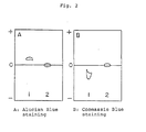
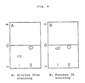
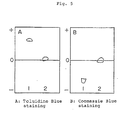
- Figs. 1 to 6 show each an electrophoretic pattern of the glycosaminoglycan-modified protein obtained in Examples and a mixture of a glycosaminoglycan and a protein.
- Lane 1 stands for the electrophoretic pattern of a mixture of bovine-derived catalase and hyaluronic acid (HA) the reducing terminal residue of which was limitedly oxidized (O-HA) and Lane 2 stands for the pattern of HA-modified bovine-derived catalase.
- Lane 1 stands for the electrophoretic pattern of a mixture of Aspergillus niger -derived catalase and O-HA and Lane 2 stands for the pattern of HA-modified Asperugillus niger -derived catalase.
- Lane 1 stands for the electrophoretic pattern of a mixture of uricase and O-HA and Lane 2 stands for the pattern of HA-modified uricase.
- Lane 1 stands for the electrophoretic pattern of a mixture of asparaginase and O-HA and Lane 2 stands for the pattern of HA-modified asparaginase.
- Lane 1 stands for the electrophoretic pattern of a mixture of superoxide disumutase (SOD) and chondroitin sulfate derived from bovine tracheal cartilages the reducing terminal residue of which was limitedly oxidized (O-CS(T)) and Lane 2 stands for the pattern of CS(T)-modified SOD (Lot No. 121-2).
- SOD superoxide disumutase
- O-CS(T) chondroitin sulfate derived from bovine tracheal cartilages the reducing terminal residue of which was limitedly oxidized
- Lane 2 stands for the pattern of CS(T)-modified SOD (Lot No. 121-2).
- Lane 1 stands for the electrophoretic pattern of SOD derived from bovine red blood cell
- Lane 2 stands for the pattern of SOD derived from dog red blood cell
- Lane 3 stands for the pattern of HA-modified SOD derived from dog red blood cell
- Lane 4 stands for HA-modified SOD derived from bovine red blood cell.
- Fig. 7 shows an electrophoretic pattern of chondroitin sulfate described in Comparative Example.
- Lane S stands for CS(T)
- Lanes 1, 2, 3, 4, 5, 6 and 7 each stands for SOD treated with 0.0436 mg, 0.145 mg, 0.436 mg, 1.45 mg, 4.36 mg, 14.5 mg and 43.6 mg of water-soluble carbodiimide (WSC), respectively
- Lane 8 stands for SOD treated with 43.6 mg of WSC and 0.1 N sodium hydroxide.
- Fig. 8 is chromatograms of gel filtration referred to in Comparative Example.
- A is a chromatogram for a mixture of chondroitin sulfate originating from bovine tracheal cartilages and superoxide dismutase, B for the product of the lot No. B, C for the product of the lot No. C and D for the product of the lot No. 121-2 prepared in Example 2.
- the glycosaminoglycan-modified protein of the present invention may be produced by, for example, reacting a glycosaminoglycan activated by reducing terminal residue-limiting oxidation method, reducing terminal residue-lactonization method, carboxyl group activating method or cyanogen bromide activation method with a protein.
- glycosaminoglycan-modified protein of the present invention is described in detail below.
- This method comprises reducing and partially oxidizing the reducing terminal sugar moiety of a glycosaminoglycan to thereby cleave said terminal sugar moiety and form an aldehyde group and producing a protein modified with the glycosaminoglycan by the reducing alkylation reaction between the aldehyde group and an amino group of the protein.
- the reaction scheme of this method is as follows:
- R1 and R2 each represents groups commonly observed at the 2- and 5-positions of the reducing terminal sugar moiety of a glycosaminoglycan and examples of R1 include OH, NH2 and NHCOCH3 while examples of R2 include CH2OH, COOH and CH2OSO3M, wherein M represents a hydrogen atom, an alkali or alkaline earth metal or an amine such as trialkylamine or pyridine; and P, n and GAG are as defined above.
- the glycosaminoglycan of the above formula (IV) is first reduced to thereby cleave the reducing terminal sugar moiety thereof.
- the compound of the formula (V) is obtained.
- the reducing agent to be used in this reduction step include alkali boron hydrides such as sodium boron hydride and sodium boron cyanohydride.
- the above reduction may be effected in an appropriate liquid medium, for example, a buffer solution such as borate buffer solution (pH 8.3), phosphate buffer solution (pH 8.6) or a mixture of such a buffer solution with an organic solvent such as dimethylformamide, acetonitrile or dioxane methanol usually at a temperature of from 0 to 40 °C, preferably from 15 to 20 °C.
- a buffer solution such as borate buffer solution (pH 8.3), phosphate buffer solution (pH 8.6) or a mixture of such a buffer solution with an organic solvent such as dimethylformamide, acetonitrile or dioxane methanol usually at a temperature of from 0 to 40 °C, preferably from 15 to 20 °C.
- the amount of the above-mentioned reducing agent may vary depending on the type. It may be used in an amount of from 5 to 50 equivalents, preferably from 10 to 20 equivalents, per mole of the compound of the formula (IV).
- the compound of the formula (V) thus obtained is partially oxidized.
- R1 in the formula (V) is an OH group
- an aldehyde compound of the formula (VI) is formed by this oxidation.
- R2 in the formula (V) is a CH2OH group
- an aldehyde compound of the formula (VII) is formed.
- the oxidizing agent to be used in this oxidation include alkali periodates such as sodium periodate or potassium periodate.
- the oxidizing agent may be employed usually in an amount of from 1 to 30 equivalents, preferably from 5 to 10 equivalents, per one mole of the compound of the formula (V).
- the above-mentioned oxidation may be conducted generally at a temperature of from 0 to 20 °C, preferably from 0 to 5 °C.
- the aldehyde compound of the formula (VI) or (VII) thus obtained may be reacted with an amino group of a protein by a known reducing alkylation method.
- the desired glycosaminoglycan-modified protein of the present invention represented by the formula (I) or (II) may be obtained.
- the reducing alkylation may be conducted by reacting the aldehyde compound of the formula (VI) or (VII) with the protein in a liquid medium selected from, for example, the above-mentioned ones usually at a temperature of from 15 to 60 °C. Simultaneously with this reaction or after the completion the reaction, the reaction mixture is subjected to reduction with a reducing agent such as sodium boron cyanohydride.
- n may generally range from an integer of 1 to 100, preferably from 1 to 10 on average.
- This method comprises partially oxidizing the reducing terminal sugar moiety of a glycosaminoglycan to thereby cleave the terminal sugar moiety followed by forming a lactone. Then, a protein modified with the glycosaminoglycan is prepared by reacting the lactone with an amino group of the protein.
- the reaction scheme of this method is as follows:
- A represents a potassium or sodium atom; and P, n, GAG, R1 and R2 are as defined above.
- the glycosaminoglycan of the formula (IV) is first oxidized to cleave the reducing terminal sugar moiety.
- a carboxyl compound of the formula (VIII) is obtained.
- the oxidizing agent to be used in this oxidation step include iodine and bromine.
- the oxidizing agent may be used usually in an amount of from 2 to 20 equivalents, preferably from 5 to 15 equivalents, per mole of the compound of the formula (IV).
- the oxidation reaction may be conducted in a liquid medium selected from among, for example, the above-mentioned ones at a temperature of from 0 to 40 °C, preferably from 15 to 20 °C.
- potassium hydroxide or sodium hydroxide is added to the reaction mixture to decompose the remaining oxidizing agent.
- the thus-obtained solution containing the compound of the formula (VIII) is applied to a column packed with 200 ml of strongly acidic cation exchange resins such as Dowex 50 and Amberlite IR120 and allowed it to pass through the column over 1 hour to obtain the passed-through fraction.
- the column is washed with water, this water fraction is combined with the above-obtained passed-through fraction and then the combined fraction is allowed to stand overnight at 4 °C to form the lactone compound of the formula (IX).
- the lactone compound of the formula (IX) thus obtained is then reacted with a protein to give the glycosaminoglycan-modified protein represented by the formula (III).
- the reaction between the lactone compound of the formula (IX) and the protein may be conducted by reacting the lactone in the form of a trialkylamine or adjusting the pH value of a mixture of the lactone and the protein to 4 to 7 with an aqueous solution of sodium hydroxide followed by effecting the reaction at 0 to 70 °C, preferably 15 to 50 °C.
- n may generally range from an integer of 1 to 100, preferably from 1 to 10 on average.
- glycosaminoglycans carry each an uronic acid moiety represented by the following formula:
- the protein modified with the glycosaminoglycan can be obtained by binding a carboxyl group in the uronic acid moiety of a glycosaminoglycan to an amino group of the protein.
- This method comprises activating a carboxyl group in an uronic acid moiety of a glycosaminoglycan by a method widely known in peptide chemistry and then reacting the carboxyl group thus activated with a protein.
- the carboxyl group in the uronic acid moiety of the glycosaminoglycan may be activated by, for example, reacting the glycosaminoglycan with a compound selected from N-hydroxysuccinimide, p-nitrophenol, N- hydroxybenzotriazole, N-hydroxypiperidine, N-hydroxysuccinamide and 2,4,5-trichlorophenol to convert the carboxyl group into an active ester group.
- a compound selected from N-hydroxysuccinimide, p-nitrophenol, N- hydroxybenzotriazole, N-hydroxypiperidine, N-hydroxysuccinamide and 2,4,5-trichlorophenol to convert the carboxyl group into an active ester group.
- the glycosaminoglycan is converted into a salt with an appropriate amine such as tri(n-butyl)amine, triethylamine, pyridine. Then, the resulting salt is reacted with N-hydroxysuccinimide in an appropriate solvent such as dimethylformamide, pyridine, dimethylsulfoxide in the presence of a condensation agent such as 1-ethyl-3-(dimethylaminopropyl)carbodiimide, dicyclohexylcarbodiimide at a temperature of from 0 to 50 °C.
- a glycosaminoglycan having an activated carboxyl group is obtained.
- glycosaminoglycan having an activated carboxyl group is reacted with a protein to give the glycosaminoglycan-modified protein of the present invention.
- an aqueous solution of the protein is added to an aqueous solution of the glycosaminoglycan having an activated carboxyl group or a phosphate buffer solution (pH 6 to 9) containing the glycosaminoglycan having an activated carboxyl group and the mixture is allowed to react at 0 to 50 °C, preferably 15 to 25 °C, for 30 minutes to 20 hours.
- the above-mentioned carboxyl group-activating method makes it possible to obtain a glycosaminoglycan-modified protein wherein at least some of carboxyl groups in the uronic acid moiety of the glycosaminoglycan are bound to the protein via an amide bond.
- This method comprises activating an amino group or a carboxyl group of a protein, or a carboxyl group, a hydroxyl group or an functional group in the reducing terminal residue of a glycosaminoglycan and then allowing the mixture to react to bind the glycosaminoglycan to the protein.
- glycosaminoglycan-modified protein can be obtained according to this method as follows.
- Glycosaminoglycan is dissolved in 2 M phosphate buffer (pH 11.5) and an acetonitrile solution of cyanogen bromide is added thereto. After reacting the mixture at 4°C for 5 minutes, acetonitrile is added to the reaction mixture to give a precipitate. After removal of excessive cyanogen bromide, the precipitate is dissolved in 0.1 M sodium hydrogencarbonate solution. A protein was added thereto and a reaction is carried out at 4°C for 20 hours to obtain a desired product.
- the glycosaminoglycan-modified protein produced by one of the above-mentioned methods may be separated and purified by a conventional method.
- the reaction mixture is desalted with the use of a dialysis membrane or an ultrafiltration membrane.
- the desired product is separated from the unreacted glycosaminoglycan and protein and purified with the use of an anion exchanger or a cation exchanger.
- the product may be separated and purified by gel filtration by taking advantage of the difference in molecular weight.
- affinity chromatography using a carrier on which an enzyme inhibitor, a substrate or an antibody is immobilized.
- the glycosaminoglycan employed in the present invention for modifying a protein may be selected over a wide range depending on the required characteristics of the desired glycosaminoglycan-modified protein and the purpose of the modification, without particular restriction. More particularly, it may be selected from among coromic acid, hyaluronic acid, chondroitin, chondroitin sulfate, teichuronic acid, dermatan sulfate, heparin, heparan sulfate, keratosulfate, keratopolysulfate and derivatives thereof such as chondroitin polysulfate.
- glycosaminoglycans are chondroitin sulfate, chondroitin polysulfate, heparin, heparan sulfate and dermatan sulfate.
- hyaluronic acid, dermatan sulfate and chondroitin sulfate are suitable for the production of an antirheumatic agent and an antiinflammatory agent.
- These glycosaminoglycans can be used alone or in combination of two or more.
- the protein to be modified with the glycosaminoglycan is not particularly restricted.
- the proteins are physiologically active proteins originating from various animals including human, microorganisms and plants as well as those produced by chemical synthesis or using genetic engineering techniques.
- Specific examples of the proteins include cytokines (for example, various interferons such as interferon- ⁇ , interferon- ⁇ and interferon- ⁇ , interleukin-2, interleukin-3), hormones [for example, insulin, growth hormone-releasing factor (GRF), calcitonin, calcitonin gene-relating peptide (CGRP), atrial natriuretic hormone (ANP), vasopressin, corticotropin-releasing factor (CRF), vasoactive intestinal peptide (VIP), secretin, ⁇ -melanocyte-stimulating hormone ( ⁇ -MSH), adrenocorticotropic hormone (ACTH), cholecystokinin (CCK), glucagon, parathyroid hormone (PTH), parat
- a glycosaminoglycan residue is chemically bound to a protein.
- the amount of the glycosaminoglycan to be introduced into the protein may vary depending on, for example, the protein and/or glycosaminoglycan, its molecular weight and the final usage of the formed glycosaminoglycan-modified protein.
- the suitable introduction amount of each glycosaminoglycan may be easily determined by those skilled in the art by simple experiments.
- the glycosaminoglycan may be introduced into the protein in an amount of from 1 to 99.9 % by weight, preferably from 90 to 95 % by weight, based on the weight of the protein to be modified.
- a glycosaminoglycan-modified protein hardly react with the antibody corresponding to the unmodified protein. Furthermore, the antibody productivity of the protein is substantially lowered by the modification as described in the following Examples.
- the glycosaminoglycan-modified protein when administered to a living organism, its activity is sustained for a long time, compared with the activity of the corresponding unmodified protein. Thus, the glycosaminoglycan-modified protein shows an improved stability in vivo .
- Drugs containing the glycosaminoglycan-modified protein of the present invention may be formulated into various forms for oral administration, for example, granules, fine subtilaes, powders, tablets, capsules, syrups, troches, suspensions or solutions.
- the glycosaminoglycan-modified protein may be orally administered as such.
- it may be formulated into injections for intravenous, intra-arterial, intraportal, intrapleuroperitoneal, intramuscular, subcutaneous or intratumor administration.
- it may be in the form of a powder and formulated into an injection upon use. It may be formulated into suppositories for per rectum or ointments for parenteral administration.
- preparations may be formulated together with known organic or inorganic liquid or solid pharmaceutical carriers, diluents, binders, disintegrating agents and the like suitable for oral, parenteral or per rectum administration.
- suitable for oral, parenteral or per rectum administration e.g., lactose, sucrose, glucose, starch, gum arabic, tragacanth gum.
- stabilizers, humectants, emulsifiers, aromatics and components for varying osmotic pressure may be added thereto. Examples thereof include gelatin, glycerol, vaseline, wax, plastics and higher alcohol.
- salts may be optionally used as an assistant ingredient for maintaining the pH value of the formulation at an appropriate level.
- a water-soluble or hydrophilic base such as macrogol or an oily base such as cacao oil may be used.
- the dose of the glycosaminoglycan-modified protein of the present invention may vary depending on the disease to be treated and symptoms and age of the patient. It is preferable to continuously or intermittently administer the compound to a patient in a dose of from 1 to 5000 mg/day in the case of oral administration or from 1 to 100 mg/day in the case of injection.
- the activity and molecular weight of the GAG-modified proteins and the content of glycosaminoglycans and proteins were determined under the following conditions.
- One unit (U) of enzyme is defined as the amount of the enzyme decomposing 1.0 ⁇ M of hydrogen peroxide in a hydrogen peroxide solution having a hydrogen peroxide concentration ranging from 9.2 to 10.3 mM per minute at pH 7.0 and 25 °C.
- One unit of enzyme is defined as the amount of the enzyme that produced a decrease in absorbance of 0.001 of a suspension of a gram-positive bacterium (ML-Cell) at 540 nm per minute at pH 6.2 and 35 °C.
- One unit of enzyme is defined as the amount of the enzyme liberating 1 ⁇ M of an unsaturated disaccharide from hyaluronic acid per minute at pH 6.2 and 37 °C.
- One unit of enzyme is defined as the amount of the enzyme hydrolyzing 1 ⁇ M of N-acetyl-tri-L-alanine methyl ester per minute at pH 8.5 and 25 °C.
- One unit of enzyme is defined as the amount of the enzyme oxidizing 1 ⁇ M of uric acid per minute at pH 8.5 and 25 °C.
- One unit of enzyme is defined as the amount of the enzyme that produced a plasminogen, when plasminogen is treated with urokinase at pH 7.5 and 37 °C, which gives a change of 1.0 of an absorbance of a substance soluble in perchloric acid formed from ⁇ -casein per minute at ⁇ 275.
- One unit of enzyme is defined as the amount of the enzyme producing 1.0 ⁇ M of ammonia from L-aspargine per minute at pH 8.6 and 37 °C.
- One unit of enzyme is defined as the amount of the enzyme inhibiting 50 % of the activity of xanthine oxidase at pH 7.8 and 25°C.
- a sample is subjected to high performance liquid chromatography using a G6000PWx2 (Tosoh Corporation) column. Elution was carried out with a 0.2 M sodium chloride aqueous solution. The molecular weight of a sample is determined based on a calibration curve.
- a sample is subjected to carbazole-sulfuric acid reaction in accordance with the method as described in T. Bitter and H. M. Muir, Analytical Biochemistry, 4 , 330-334 (1962).
- the content of a protein is determined by the method of Lowry et al. as described in O. H. Lowry, N. J. Rosenbrough, A. L. Farr and R. J. Randall, J. Biol. Chem., 193 , 265-275 (1951).
- Hyaluronic acid [originating from comb: HA100 (MW 1,000,000)], chondroitin sulfate [originating from bovine tracheal cartilages: CS(T), shark cartilages: CS(S), whale cartilages: CS(W)], dermatan sulfate (originating from pig skin: DS), heparin (originating from pig intestinum um um: Hep) and heparan sulfate (originating from pig kidney: HS) were treated in the same manner as in the above step A under the conditions as specified in Table 1. Thus reducing terminal residue-opened glycosaminoglycans (R-GAGs) were obtained.
- O-GAGs reducing terminal residue-limiting oxidized glycosaminoglycans
- Acetylcellulose membrane (SEPARAX, JOOKOO Co., Ltd.) 0.1 M formic acid/pyridine (pH 3.0) 0.5 mA/cm, 30 min. and
- P2 is a catalase residue exclusive of 26 amino groups, on an average, from catalase and X is as defined above.
- Uricase content 1.8 %.
- Hyaluronic acid content 98.2 %.
- Acetylcellulose membrane SEPARAX, JOOKOO Co., Ltd.
- 0.1 M formic acid/pyridine pH 3.0
- P3 is a uricase residue exclusive of 5.5 amino groups, on an average, from uricase and X is as defined above.
- Catalase originating from bovine liver, superoxide dismutase originating from bovine erythrocyte, hyaluronidase originating from bovine testis, lysozyme originating from albumen (the above enzymes are all manufactured by Seikagaku Corporation) and urokinase originating from human renal cell (Kyowa Hakko Kogyo Co., Ltd.) were modified with the products of lot Nos. 111 to 116 in the same manner as in the above step I under the conditions specified in Table 3.
- Table 4 shows the analytical data of the products thus obtained.
- Fig. 5 shows the electrophoretic pattern of the product of lot No. 121-1 as a representative. The electrophoresis was conducted under the same conditions as those described in I.
- Catalase or HA-catalase dissolved in a 0.05 M phosphate buffer solution of pH 7.0 (hereinafter abbreviated as PBS) was intraperitoneally (hereinafter abbreviated as i.p.) administered once a week for 12 weeks to female Swiss-Webster mice (each group has 4 animals) in a dose of 0.1 mg in terms of protein.
- PBS phosphate buffer solution of pH 7.0
- the titer of each serum was determined by HPO-ELISA (enzyme-linked immunosorbent assay with the use of horseradish peroxidase) in accordance with the method of Voller et al.
- 100 ⁇ l of an antigen solution diluted with a carbonate buffer solution (0.5 M, pH 9.5) to 10 ⁇ g/ml was used to inoculate wells of the Nunc Immuno II microtiter plates. The plate were incubated at 4°C overnight and then washed with a physiological saline containing 0.05% of Tween 20 thrice.
- 100 ⁇ l of a test serum diluted with the Tween 20-containing PBS was added to each well.
- mice serum control Three controls, i.e., an antigen control, an antiserum control and a normal mouse serum control were also prepared. After allowing to stand at room temperature for 1 hour, 100 ⁇ l portions of HPO-conjugated goat anti-mouse immunoglobulins (IgG + IgA + IgM) were added to each well and incubated at room temperature for 1 hour and 30 minutes. 100 ⁇ l of o-phenylenediamine substrate was added to each well and incubated for 10 minutes. Next, 0.01 ml of 4 N sulfuric acid was added to cease the reaction. The titer is expressed in the antibody dilution ratio giving an optical density of 0.01 based on the control serum.
- HPO-conjugated goat anti-mouse immunoglobulins IgG + IgA + IgM
- the antigenicity or immunogenicity of HA-catalase is lower than that of unmodified catalase.
- a ddy male mouse aged 8 weeks is kept in a keeping device and the right hind leg is tied around with a suture (Brain No. 2). One end of the suture is fixed while a weight of 500 g is suspended from another end of the suture.
- ischemia is conducted for a definite period of time. Before the ischemia and 60 minutes thereafter, the thickness of the foot-pad of the animal was measured with slide calipers. Then, the foot-pad is cut and weighed.
- a physiological saline is intravenously administered immediately before the initiation of the ischemia.
- specimen shown in Table 6 was administered 30 minutes before the initiation of the ischemia and immediately before the same. Each group has 5 animals.
- the catarase used as a specimen was originated from bovine liver.
- the HA-catalase used was prepared as follows. 10 mg of the HA-catalase of lot No. 120-1 was dissolved in 5.2 ml of a physiological saline and filtered through a membrane of 0.22 ⁇ m (Milex GV; mfd. by Nippon Millipore Kogyo K.K.). 1 ml portions of the filtrate were distributed to ampuls to give a preparation for injection. The preparation contained 600 U of the enzyme per ampul.
- Chondroitin sulfate [originating from bovine tracheal cartilages: CS(T), shark cartilages: CS(S), whale cartilages: CS(W)], heparin (originating from swine intestinum ***: Hep), heparan sulfate (originating from swine kidney: HS) and dermatan sulfate (originating from swine skin DS) were treated in the same manner as in the above step I under the conditions as specified in Table 7. The thus-obtained products are shown in Table 7.
- P7 is a superoxide dismutase residue exclusive of 80 amino groups, on an average, from superoxide dismutase
- Y' is a hydrogen atom of the reducing terminal residue of chondroitin sulfate
- the sum of n, m and l is 40 and X' R3 and R4 are as defined above.
- hyaluronic acid 500 mg was dissolved in 50 ml of water. 5 ml of a 0.1 M iodine solution in methanol was added thereto and the resulting mixture was allowed to react at room temperature for 6 hours. Then, approximately 5 ml of 0.1 N potassium hydroxide was added to the reaction mixture so as to cause the discoloration of the liberated iodine. Ethanol saturated with potassium acetate was added to the solution. The precipitate thus formed was collected by filtration and dissolved in 50 ml of water. The resulting solution was contacted with 50 ml of a strongly acidic ion exchange resin [Dowex 50(H+)] at 5 °C for 20 hours.
- a strongly acidic ion exchange resin [Dowex 50(H+)]
- hyaluronic acid-modified RGDV The characteristics of hyaluronic acid-modified RGDV were as follows: Lot No. 320. Yield: 40.2 mg. RGDV content: 3.3 %. Hyaluronic acid content: 96.7 %. and In the above formulae, X is as defined above.
- comb-derived hyaluronic acid 400 mg was dissolved in 2 M phosphate buffer (pH 11.5) and 1 ml of an acetonitrile solution of cyanogen bromide (100 mg/ml) was added thereto to allow the resulting mixture to react at 4°C for 5 minutes. Immediately after the termination of the reaction, 150 ml of acetonitrile was added to the reaction mixture to form precipitate. The thus-obtained precipitate was washed quickly with acetonitrile and was dissolved in 0.1 M sodium hydrogencarbonate solution.
- the characteristics of the hyaluronic acid-modified SOD thus obtained were as follows. Yield: 350 mg SOD content: 21.2% Hyaluronic acid content: 78.8% Molecular weight: 172,000 Activity: 82.1% of that of unmodified SOD Electrophoresis: cf. Fig. 6.
- the activity of the WSC-modified SOD is lower than the glycosaminoglycan-modified SOD of the present invention, though the contents of SOD are the same as each other.
- WSC method (lot No. B): 44.4 % of the activity of unmodified SOD.
Abstract
Description
- This invention relates to a protein modified with a glycosaminoglycan. More particularly, it relates to a protein modified with a glycosaminoglycan which is obtained by reacting a glycosaminoglycan activated with a specific activator with a protein.
- Recently, attempts have been frequently made to apply enzymes and physiologically active proteins as drugs for treating various diseases including cancer, inflammation and hereditary enzyme deficiency.
- However, many of these enzymes and physiologically active proteins are heterogenic to living organisms. Thus the administration of these compounds as such to an organism would cause a problem of immunogenicity or deteriorate the stability in vivo. When such a compound is to be formulated into a medicine, therefore, it is required to relieve an anaphylatic reaction and to improve the prolonged drug effect.
- Furthermore, attempts have been made to produce these proteins in a large amount through genetic engineering techniques. However, proteins produced by genetic engineering suffer from a problem of the deficiency of a sugar chain, which seriously affects the stability in vivo.
- Accordingly, there has been proposed to modify an enzyme or a physiologically active protein with various synthetic polymers or polysaccharides so as to pharmacologically improve the stability thereof. Examples of the synthetic polymers employed therefor include poly-L-aspartic acid (M. Okada, A. Matsushima, A. Katsuhata, T. Aoyama, T. Ando and Y. Inada: Int. Archs Allerlrgy Appl. Immun. 76, 79 - 81 (1965)) and derivatives thereof, poly-D- or -L-lysine, D-glutamic acid/D-lysine copolymer (F.T. Liu and D.H. Katz: Proc. Natl. Acad. Sci. USA, 76, 1430 - 1434 (1979)), polyvinyl alcohol pyran copolymer, polyethylene glycol and derivatives thereof and styrene/maleic acid copolymer (H. Maeda, M. Ueda, T. Morinaga and T. Matsumoto: J. Ned. Chem., 28, 455 - 461 (1985)). Examples of the polysaccharides include agarose, carboxymethyl cellulose, dextran (T.P. King, L. Kochoumian, K. Ishizaka, L. Kichtenstein, P.S. Norman: Arch. Biochem. Biophys., 169, 464 - 473 (1975)), pullulan (M. Usui and T. Matsuhashi: J. Immunol., 122, 1266 - 1272 (1979) and N. Matsuhashi: Genkansa no Jikken Dobutsu Moderu (Experimental Animal Model for Hyposensitization), ed. by S. Kobayashi et al., Genkansa Ryoho no Kiso to Rinsho (Bases and Clinical Application of Hyposensitization Therapy), 4 - 11, Chugai Igakusho (1982)) and lipopolysaccharide.
- On the other hand, U.S. Patent 4,485,754 proposes a stabilized protein which is obtained by reacting a protein such as superoxide dismutase or insulin with chondroitin sulfate in the presence of 1-ethyl-3-(3-dimethylaminopropyl)carbodiimide. However, the protein thus obtained is in the form of a complicated polymer involving a polymer of the protein per se, since plural carboxyl groups in aspartic acid or glutamic acid contained in the protein are activated by 1-enthyl-3-(3-dimethylaminopropyl)carbodiimide, respectively. Thus, this product should be further improved from the viewpoint of the pharmacological effect of the unmodified superoxide dismutase or insulin as a monomer.
- An object of the present invention is to provide a glycosaminoglycan-modified protein, which is highly stable in vivo and can express the inherent physiological effect of the starting protein for a prolonged period of time without forming any polymer of a complicated structure.
- The above object of the present invention can be achieved by a glycosaminoglycan-modified protein obtained by reacting glycosaminoglycan activated by reducing terminal residue-limiting oxidation method, carboxyl group-activating method, reducing terminal residue-lactonization method or cyanogen bromide activation method with a protein.
- According to an embodiment of the present invention, a glycosaminoglycan-modified protein wherein an amino group of a protein is bound to an aldehyde group, which has been formed by reducing and partially oxidizing the reducing terminal sugar moiety of a glycosaminoglycan is provided.
- According to another embodiment of the present invention, a glycosaminoglycan-modified protein of the following formula:

wherein P represents a protein residue exclusive of n amino groups from the protein; and GAG represents a glycosaminoglycan residue exclusive of the reducing terminal sugar moiety from the glycosaminoglycan is provided. - According to another embodiment of the present invention, a glycosaminoglycan-modified protein wherein at least some of carboxyl groups in the uronic acid moiety of a glycosaminoglycan are bound to a protein via an amide bond is provided.
- Figs. 1 to 6 show each an electrophoretic pattern of the glycosaminoglycan-modified protein obtained in Examples and a mixture of a glycosaminoglycan and a protein.
- In Fig. 1,
Lane 1 stands for the electrophoretic pattern of a mixture of bovine-derived catalase and hyaluronic acid (HA) the reducing terminal residue of which was limitedly oxidized (O-HA) andLane 2 stands for the pattern of HA-modified bovine-derived catalase. - In Fig. 2,
Lane 1 stands for the electrophoretic pattern of a mixture of Aspergillus niger-derived catalase and O-HA and Lane 2 stands for the pattern of HA-modified Asperugillus niger-derived catalase. - In Fig. 3,
Lane 1 stands for the electrophoretic pattern of a mixture of uricase and O-HA andLane 2 stands for the pattern of HA-modified uricase. - In Fig. 4,
Lane 1 stands for the electrophoretic pattern of a mixture of asparaginase and O-HA andLane 2 stands for the pattern of HA-modified asparaginase. - In Fig. 5,
Lane 1 stands for the electrophoretic pattern of a mixture of superoxide disumutase (SOD) and chondroitin sulfate derived from bovine tracheal cartilages the reducing terminal residue of which was limitedly oxidized (O-CS(T)) andLane 2 stands for the pattern of CS(T)-modified SOD (Lot No. 121-2). - In Fig. 6,
Lane 1 stands for the electrophoretic pattern of SOD derived from bovine red blood cell,Lane 2 stands for the pattern of SOD derived from dog red blood cell,Lane 3 stands for the pattern of HA-modified SOD derived from dog red blood cell, andLane 4 stands for HA-modified SOD derived from bovine red blood cell. - Fig. 7 shows an electrophoretic pattern of chondroitin sulfate described in Comparative Example. In Fig. 7, Lane S stands for CS(T),
Lanes - Fig. 8 is chromatograms of gel filtration referred to in Comparative Example. In Fig. 8, A is a chromatogram for a mixture of chondroitin sulfate originating from bovine tracheal cartilages and superoxide dismutase, B for the product of the lot No. B, C for the product of the lot No. C and D for the product of the lot No. 121-2 prepared in Example 2.
- The glycosaminoglycan-modified protein of the present invention may be produced by, for example, reacting a glycosaminoglycan activated by reducing terminal residue-limiting oxidation method, reducing terminal residue-lactonization method, carboxyl group activating method or cyanogen bromide activation method with a protein.
- The process for production of glycosaminoglycan-modified protein of the present invention is described in detail below.
- This method comprises reducing and partially oxidizing the reducing terminal sugar moiety of a glycosaminoglycan to thereby cleave said terminal sugar moiety and form an aldehyde group and producing a protein modified with the glycosaminoglycan by the reducing alkylation reaction between the aldehyde group and an amino group of the protein. The reaction scheme of this method is as follows:
-


In the above reaction scheme, R¹ and R² each represents groups commonly observed at the 2- and 5-positions of the reducing terminal sugar moiety of a glycosaminoglycan and examples of R¹ include OH, NH₂ and NHCOCH₃ while examples of R² include CH₂OH, COOH and CH₂OSO₃M, wherein M represents a hydrogen atom, an alkali or alkaline earth metal or an amine such as trialkylamine or pyridine; and P, n and GAG are as defined above. - In this method, the glycosaminoglycan of the above formula (IV) is first reduced to thereby cleave the reducing terminal sugar moiety thereof. Thus the compound of the formula (V) is obtained. Examples of the reducing agent to be used in this reduction step include alkali boron hydrides such as sodium boron hydride and sodium boron cyanohydride. The above reduction may be effected in an appropriate liquid medium, for example, a buffer solution such as borate buffer solution (pH 8.3), phosphate buffer solution (pH 8.6) or a mixture of such a buffer solution with an organic solvent such as dimethylformamide, acetonitrile or dioxane methanol usually at a temperature of from 0 to 40 °C, preferably from 15 to 20 °C.
- The amount of the above-mentioned reducing agent may vary depending on the type. It may be used in an amount of from 5 to 50 equivalents, preferably from 10 to 20 equivalents, per mole of the compound of the formula (IV).
- Next, the compound of the formula (V) thus obtained is partially oxidized. When R¹ in the formula (V) is an OH group, an aldehyde compound of the formula (VI) is formed by this oxidation. When the R² in the formula (V) is a CH₂OH group, on the other hand, an aldehyde compound of the formula (VII) is formed. Examples of the oxidizing agent to be used in this oxidation include alkali periodates such as sodium periodate or potassium periodate. The oxidizing agent may be employed usually in an amount of from 1 to 30 equivalents, preferably from 5 to 10 equivalents, per one mole of the compound of the formula (V). The above-mentioned oxidation may be conducted generally at a temperature of from 0 to 20 °C, preferably from 0 to 5 °C.
- The aldehyde compound of the formula (VI) or (VII) thus obtained may be reacted with an amino group of a protein by a known reducing alkylation method. Thus, the desired glycosaminoglycan-modified protein of the present invention represented by the formula (I) or (II) may be obtained. The reducing alkylation may be conducted by reacting the aldehyde compound of the formula (VI) or (VII) with the protein in a liquid medium selected from, for example, the above-mentioned ones usually at a temperature of from 15 to 60 °C. Simultaneously with this reaction or after the completion the reaction, the reaction mixture is subjected to reduction with a reducing agent such as sodium boron cyanohydride.
- Regarding the glycosaminoglycan-modified protein of the formula (I) or (II), n may generally range from an integer of 1 to 100, preferably from 1 to 10 on average.
- This method comprises partially oxidizing the reducing terminal sugar moiety of a glycosaminoglycan to thereby cleave the terminal sugar moiety followed by forming a lactone. Then, a protein modified with the glycosaminoglycan is prepared by reacting the lactone with an amino group of the protein. The reaction scheme of this method is as follows:
-
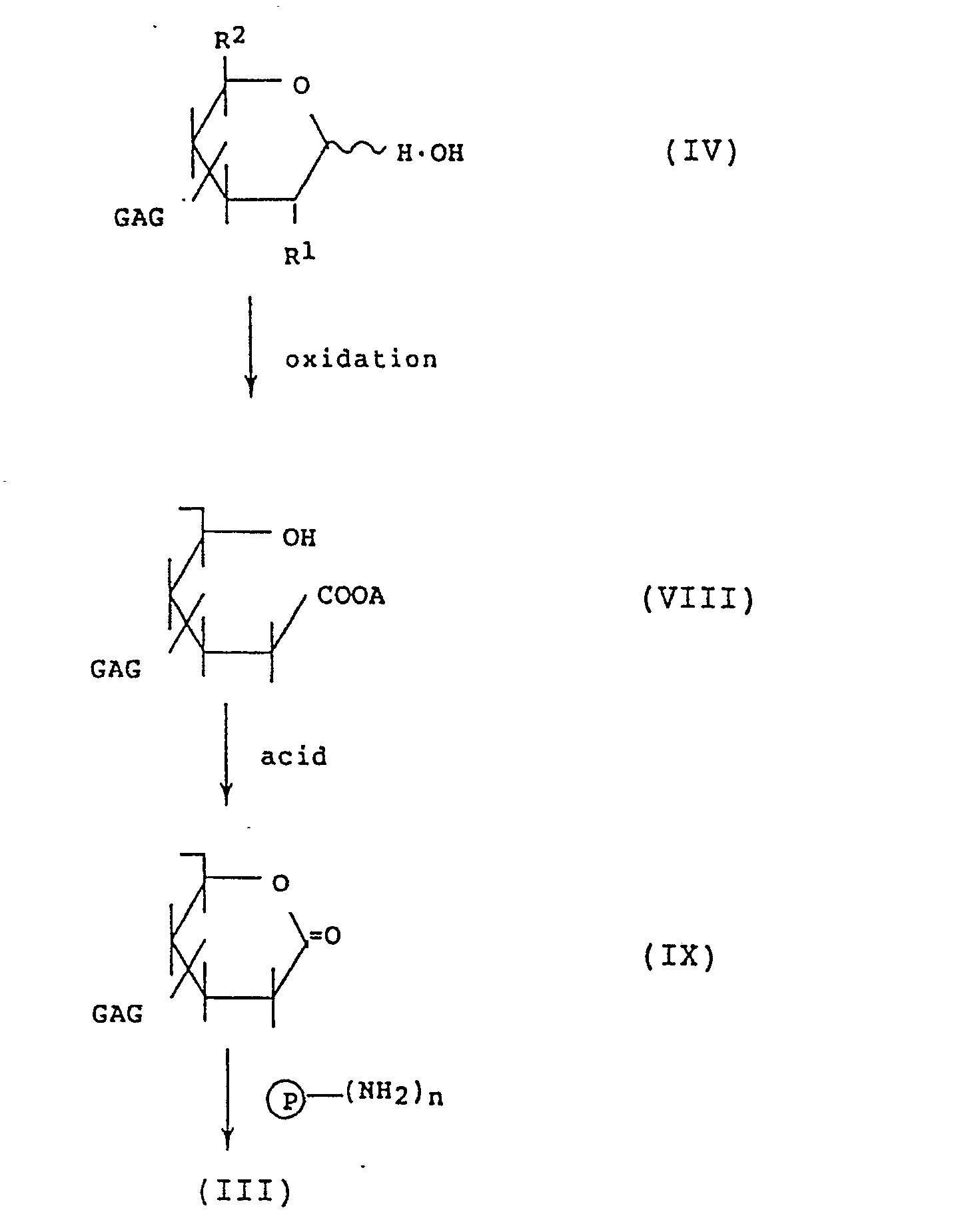
In the above reaction scheme, A represents a potassium or sodium atom; and P, n, GAG, R¹ and R² are as defined above. - In this method, the glycosaminoglycan of the formula (IV) is first oxidized to cleave the reducing terminal sugar moiety. Thus, a carboxyl compound of the formula (VIII) is obtained. Examples of the oxidizing agent to be used in this oxidation step include iodine and bromine. The oxidizing agent may be used usually in an amount of from 2 to 20 equivalents, preferably from 5 to 15 equivalents, per mole of the compound of the formula (IV). The oxidation reaction may be conducted in a liquid medium selected from among, for example, the above-mentioned ones at a temperature of from 0 to 40 °C, preferably from 15 to 20 °C. After the oxidation reaction, potassium hydroxide or sodium hydroxide is added to the reaction mixture to decompose the remaining oxidizing agent.
- The thus-obtained solution containing the compound of the formula (VIII) is applied to a column packed with 200 ml of strongly acidic cation exchange resins such as
Dowex 50 and Amberlite IR120 and allowed it to pass through the column over 1 hour to obtain the passed-through fraction. The column is washed with water, this water fraction is combined with the above-obtained passed-through fraction and then the combined fraction is allowed to stand overnight at 4 °C to form the lactone compound of the formula (IX). - The lactone compound of the formula (IX) thus obtained is then reacted with a protein to give the glycosaminoglycan-modified protein represented by the formula (III). The reaction between the lactone compound of the formula (IX) and the protein may be conducted by reacting the lactone in the form of a trialkylamine or adjusting the pH value of a mixture of the lactone and the protein to 4 to 7 with an aqueous solution of sodium hydroxide followed by effecting the reaction at 0 to 70 °C, preferably 15 to 50 °C.
- Regarding the glycosaminoglycan-modified protein of the formula (III), n may generally range from an integer of 1 to 100, preferably from 1 to 10 on average.
- With a few exceptions (e.g., keratosulfate and keratopolysulfate), glycosaminoglycans carry each an uronic acid moiety represented by the following formula:

- According to this method, the protein modified with the glycosaminoglycan can be obtained by binding a carboxyl group in the uronic acid moiety of a glycosaminoglycan to an amino group of the protein.
- This method comprises activating a carboxyl group in an uronic acid moiety of a glycosaminoglycan by a method widely known in peptide chemistry and then reacting the carboxyl group thus activated with a protein.
- The carboxyl group in the uronic acid moiety of the glycosaminoglycan may be activated by, for example, reacting the glycosaminoglycan with a compound selected from N-hydroxysuccinimide, p-nitrophenol, N- hydroxybenzotriazole, N-hydroxypiperidine, N-hydroxysuccinamide and 2,4,5-trichlorophenol to convert the carboxyl group into an active ester group.
- More particularly, the glycosaminoglycan is converted into a salt with an appropriate amine such as tri(n-butyl)amine, triethylamine, pyridine. Then, the resulting salt is reacted with N-hydroxysuccinimide in an appropriate solvent such as dimethylformamide, pyridine, dimethylsulfoxide in the presence of a condensation agent such as 1-ethyl-3-(dimethylaminopropyl)carbodiimide, dicyclohexylcarbodiimide at a temperature of from 0 to 50 °C. Thus, a glycosaminoglycan having an activated carboxyl group is obtained.
- Then, the glycosaminoglycan having an activated carboxyl group is reacted with a protein to give the glycosaminoglycan-modified protein of the present invention.
- Namely, an aqueous solution of the protein is added to an aqueous solution of the glycosaminoglycan having an activated carboxyl group or a phosphate buffer solution (
pH 6 to 9) containing the glycosaminoglycan having an activated carboxyl group and the mixture is allowed to react at 0 to 50 °C, preferably 15 to 25 °C, for 30 minutes to 20 hours. - The above-mentioned carboxyl group-activating method makes it possible to obtain a glycosaminoglycan-modified protein wherein at least some of carboxyl groups in the uronic acid moiety of the glycosaminoglycan are bound to the protein via an amide bond.
- This method comprises activating an amino group or a carboxyl group of a protein, or a carboxyl group, a hydroxyl group or an functional group in the reducing terminal residue of a glycosaminoglycan and then allowing the mixture to react to bind the glycosaminoglycan to the protein.
- More specifically, the glycosaminoglycan-modified protein can be obtained according to this method as follows.
- Glycosaminoglycan is dissolved in 2 M phosphate buffer (pH 11.5) and an acetonitrile solution of cyanogen bromide is added thereto. After reacting the mixture at 4°C for 5 minutes, acetonitrile is added to the reaction mixture to give a precipitate. After removal of excessive cyanogen bromide, the precipitate is dissolved in 0.1 M sodium hydrogencarbonate solution. A protein was added thereto and a reaction is carried out at 4°C for 20 hours to obtain a desired product.
- The glycosaminoglycan-modified protein produced by one of the above-mentioned methods may be separated and purified by a conventional method. For example, the reaction mixture is desalted with the use of a dialysis membrane or an ultrafiltration membrane. Then, the desired product is separated from the unreacted glycosaminoglycan and protein and purified with the use of an anion exchanger or a cation exchanger. Alternately, the product may be separated and purified by gel filtration by taking advantage of the difference in molecular weight. In the cases of some proteins, furthermore, it is possible to separate and purify the desired product by affinity chromatography using a carrier on which an enzyme inhibitor, a substrate or an antibody is immobilized.
- The glycosaminoglycan employed in the present invention for modifying a protein may be selected over a wide range depending on the required characteristics of the desired glycosaminoglycan-modified protein and the purpose of the modification, without particular restriction. More particularly, it may be selected from among coromic acid, hyaluronic acid, chondroitin, chondroitin sulfate, teichuronic acid, dermatan sulfate, heparin, heparan sulfate, keratosulfate, keratopolysulfate and derivatives thereof such as chondroitin polysulfate. When the desired product is used as an antithrombotic agent and an antiarteriosclerotic agent, preferred glycosaminoglycans are chondroitin sulfate, chondroitin polysulfate, heparin, heparan sulfate and dermatan sulfate. On the other hand, hyaluronic acid, dermatan sulfate and chondroitin sulfate are suitable for the production of an antirheumatic agent and an antiinflammatory agent. These glycosaminoglycans can be used alone or in combination of two or more.
- The protein to be modified with the glycosaminoglycan is not particularly restricted. Usable as the proteins are physiologically active proteins originating from various animals including human, microorganisms and plants as well as those produced by chemical synthesis or using genetic engineering techniques. Specific examples of the proteins include cytokines (for example, various interferons such as interferon-α, interferon-β and interferon-γ, interleukin-2, interleukin-3), hormones [for example, insulin, growth hormone-releasing factor (GRF), calcitonin, calcitonin gene-relating peptide (CGRP), atrial natriuretic hormone (ANP), vasopressin, corticotropin-releasing factor (CRF), vasoactive intestinal peptide (VIP), secretin, α-melanocyte-stimulating hormone (α-MSH), adrenocorticotropic hormone (ACTH), cholecystokinin (CCK), glucagon, parathyroid hormone (PTH), parathyroid hormone-relating protein (PTHrP), somatostatin, enkephalin], growth factors [for example, growth hormone (GH), insulin-like growth factor (IGF-I, IGF-II), β-nerve growth factor (β-NGF), basic fibroblast growth factor (bFGF), transforming growth factor-β (TGF-β), erythropoietin, granulocyte colony stimulating factor (G-CSF), granulocyte macrophage colony stimulating factor (GM-CSF), platelet-derived growth factor (PDGF), epidermal growth factor (EGF)], enzymes [for example, tissue plasminogen activator (TPA), elastase, superoxide dismutase (SOD), bilirubin oxidase, catalase, uricase, urokinase, thermolysin, trypsin, chymotrypsin, Vs protease, pepsin, papain, hyaluronidase, chondroitin ABC lyase, asparaginase] and other proteins [for example, ubiquitin, insulin secretion-activating protein (IAP), serum thymus factor (STF), peptide-T, albumin, globulin, transferrin, lipoprotein, lipid A derivative, Ornithonyssus protein, trypsin inhibitor].
- In the glycosaminoglycan-modified protein of the present invention, a glycosaminoglycan residue is chemically bound to a protein. The amount of the glycosaminoglycan to be introduced into the protein may vary depending on, for example, the protein and/or glycosaminoglycan, its molecular weight and the final usage of the formed glycosaminoglycan-modified protein. However, the suitable introduction amount of each glycosaminoglycan may be easily determined by those skilled in the art by simple experiments. Generally, the glycosaminoglycan may be introduced into the protein in an amount of from 1 to 99.9 % by weight, preferably from 90 to 95 % by weight, based on the weight of the protein to be modified.
- A glycosaminoglycan-modified protein hardly react with the antibody corresponding to the unmodified protein. Furthermore, the antibody productivity of the protein is substantially lowered by the modification as described in the following Examples.
- Further, when the glycosaminoglycan-modified protein is administered to a living organism, its activity is sustained for a long time, compared with the activity of the corresponding unmodified protein. Thus, the glycosaminoglycan-modified protein shows an improved stability in vivo.
- Drugs containing the glycosaminoglycan-modified protein of the present invention may be formulated into various forms for oral administration, for example, granules, fine subtilaes, powders, tablets, capsules, syrups, troches, suspensions or solutions. Alternately, the glycosaminoglycan-modified protein may be orally administered as such. Alternately, it may be formulated into injections for intravenous, intra-arterial, intraportal, intrapleuroperitoneal, intramuscular, subcutaneous or intratumor administration. Furthermore, it may be in the form of a powder and formulated into an injection upon use. It may be formulated into suppositories for per rectum or ointments for parenteral administration. These preparations may be formulated together with known organic or inorganic liquid or solid pharmaceutical carriers, diluents, binders, disintegrating agents and the like suitable for oral, parenteral or per rectum administration. (e.g., lactose, sucrose, glucose, starch, gum arabic, tragacanth gum). Furthermore, stabilizers, humectants, emulsifiers, aromatics and components for varying osmotic pressure may be added thereto. Examples thereof include gelatin, glycerol, vaseline, wax, plastics and higher alcohol. Furthermore, salts may be optionally used as an assistant ingredient for maintaining the pH value of the formulation at an appropriate level. For suppositories, a water-soluble or hydrophilic base such as macrogol or an oily base such as cacao oil may be used.
- The dose of the glycosaminoglycan-modified protein of the present invention may vary depending on the disease to be treated and symptoms and age of the patient. It is preferable to continuously or intermittently administer the compound to a patient in a dose of from 1 to 5000 mg/day in the case of oral administration or from 1 to 100 mg/day in the case of injection.
- To further illustrate the present invention, and not by way of limitation, the following Examples are given.
- In the following Examples, the activity and molecular weight of the GAG-modified proteins and the content of glycosaminoglycans and proteins were determined under the following conditions.
- One unit (U) of enzyme is defined as the amount of the enzyme decomposing 1.0 µM of hydrogen peroxide in a hydrogen peroxide solution having a hydrogen peroxide concentration ranging from 9.2 to 10.3 mM per minute at pH 7.0 and 25 °C.
- One unit of enzyme is defined as the amount of the enzyme that produced a decrease in absorbance of 0.001 of a suspension of a gram-positive bacterium (ML-Cell) at 540 nm per minute at pH 6.2 and 35 °C.
- One unit of enzyme is defined as the amount of the enzyme liberating 1 µM of an unsaturated disaccharide from hyaluronic acid per minute at pH 6.2 and 37 °C.
- One unit of enzyme is defined as the amount of the enzyme hydrolyzing 1 µM of N-acetyl-tri-L-alanine methyl ester per minute at pH 8.5 and 25 °C.
- One unit of enzyme is defined as the amount of the enzyme oxidizing 1 µM of uric acid per minute at pH 8.5 and 25 °C.
- One unit of enzyme is defined as the amount of the enzyme that produced a plasminogen, when plasminogen is treated with urokinase at pH 7.5 and 37 °C, which gives a change of 1.0 of an absorbance of a substance soluble in perchloric acid formed from α-casein per minute at Δ₂₇₅.
- One unit of enzyme is defined as the amount of the enzyme producing 1.0 µM of ammonia from L-aspargine per minute at pH 8.6 and 37 °C.
- One unit of enzyme is defined as the amount of the enzyme inhibiting 50 % of the activity of xanthine oxidase at pH 7.8 and 25°C.
- A sample is subjected to high performance liquid chromatography using a G6000PWx2 (Tosoh Corporation) column. Elution was carried out with a 0.2 M sodium chloride aqueous solution. The molecular weight of a sample is determined based on a calibration curve.
- A sample is subjected to carbazole-sulfuric acid reaction in accordance with the method as described in T. Bitter and H. M. Muir, Analytical Biochemistry, 4, 330-334 (1962).
- A sample is subjected to phenol-sulfuric acid reaction in accordance with the method described in M. Dubois, K. A. Gilles, J. K. Hamilton, P. A. Rebers and F. Smith, Anal. Chem., 28, 350-356 (1956).
- The content of a protein is determined by the method of Lowry et al. as described in O. H. Lowry, N. J. Rosenbrough, A. L. Farr and R. J. Randall, J. Biol. Chem., 193, 265-275 (1951).
- 2000 mg of hyaluronic acid (MW 10000) was dissolved in 200 ml of a 0.05 M borate buffer solution (pH 8.3) and 182 mg of sodium borohydride was added thereto. The mixture was allowed to react at room temperature for 5 hours. The pH value of the reaction mixture was adjusted to 4.5 with acetic acid and ethanol was added. The precipitate thus formed was collected by filtration and dissolved in 200 ml of water. Then, the resulting solution was passed through a column packed with 1000 ml of a cation exchange resin [Dowex 50 (H⁺)]. The eluate and water used to wash the column were combined and ethanol saturated with sodium acetate was added. The precipitate thus formed was collected by filtration.
Lot No. 100.
Yield: 1800 mg. - 1700 mg of the R-HA prepared in the above step A was dissolved in 250 ml of 40 mM imidazole hydrochloride (pH 6.5) and 139.96 mg of sodium periodate was added thereto at 0 °C followed by 1-hour of reaction. Ethanol was added to the reaction mixture and the precipitate thus formed was collected by filtration. The precipitate was dissolved in water and ethanol was added again to give a precipitate. Immediately thereafter, the precipitate (O-HA) was dissolved in a phosphate buffer solution or water and used in the reaction with a protein.
Lot No. 110-1.
Yield: 1600 mg. - Hyaluronic acid [originating from comb: HA100 (MW 1,000,000)], chondroitin sulfate [originating from bovine tracheal cartilages: CS(T), shark cartilages: CS(S), whale cartilages: CS(W)], dermatan sulfate (originating from pig skin: DS), heparin (originating from pig intestinum tenue: Hep) and heparan sulfate (originating from pig kidney: HS) were treated in the same manner as in the above step A under the conditions as specified in Table 1. Thus reducing terminal residue-opened glycosaminoglycans (R-GAGs) were obtained. Subsequently, reducing terminal residue-limiting oxidized glycosaminoglycans (O-GAGs) were prepared in the same manner as in the above step B under the conditions as indicated in Table 2. Tables 1 and 2 also show the products thus-obtained and yield thereof.

- 100 mg of the product of lot No. 110-2 was dissolved in 10 ml of a 0.005 M phosphate buffer solution (pH 8.0) and 2.3 mg of catalase originating from bovine liver (Seikagaku Corporation) was added thereto. The mixture was allowed to react at room temperature for 6 hours. Then, 0.4 mg of sodium cyanoborohydride was added thereto and the resulting mixture was allowed to react at room temperature for 20 hours. Then, the reaction mixture was filtered with an ultrafiltration device provided with a membrane of a fractionating molecular weight of 300,000 to remove low molecular weight substances. Then, the filtrate was purified with DEAE ion exchange resin chromatography and desalted with a dialysis membrane followed by lyophilization.
Lot No. 120-1.
Yield: 30.1 mg.
Catalase content: 2.1 %.
Hyaluronic acid content: 97.9 %.
Activity: 74.5 % of that of unmodified catalase.
[α]D: - 78.0 (C = 1, H₂O).
Electrophoresis: cf. Fig. 1.
Acetylcellulose membrane (SEPARAX, JOOKOO Co., Ltd.)
0.1 M formic acid/pyridine (pH 3.0)
0.5 mA/cm, 30 min.
In the above formulae, P₁ is a catalase residue exclusive of 11 amino acids, on an average, from catalase and X is a hydrogen atom of non-reducing terminal residue of hyaluronic acid. - 100 mg of the product of lot No. 110-2 and 1 mg of catalase derived from Aspergillus niger (Seikagaku Corporation) were prepared in the same manner as in the above I. The characteristics of the hyaluronic acid-modified catalase thus obtained were as follows:
Lot No. 120-2.
Yield: 29.0 mg.
Catalase content: 0.92 %.
Hyaluronic acid content: 99.08 %.
Activity: 85.1 % of that of unmodified catalase.
[α]D: - 78.8 (C = 1, H₂O).
Electrophoresis: cf. Fig. 2.
Acetylcellulose membrane (SEPARAX, JOOKOO Co., Ltd.)
0.1 M formic acid/pyridine (pH 3.0)
0.5 mA/cm, 30 min.
and
In the above formulae, P₂ is a catalase residue exclusive of 26 amino groups, on an average, from catalase and X is as defined above. - 100 mg of the product of lot No. 110-2 was dissolved in 10 ml of a phosphate buffer solution (pH 8.0) and 2.5 mg of uricase originating from Candida (Seikagaku Corporation) was added thereto. The mixture was allowed to react at room temperature for 10 hours. Then, 0.4 mg of sodium cyanoborohydride was added thereto and the resulting mixture was allowed to react at room temperature for 20 hours. Ethanol was added to the reaction mixture and the precipitate thus formed was collected by filtration. Then, the precipitate was dissolved in water and purified with DEAE ion exchange resin chromatography. The characteristics of hyaluronic acid-modified uricase thus obtained were as follows:
Lot No. 120-3.
Yield: 23.5 mg.
Uricase content: 1.8 %.
Hyaluronic acid content: 98.2 %.
Activity: 73.4 % of that of unmodified urikase.
[α]D: - 77.9 (C = 1, H₂O).
Electrophoresis: cf. Fig. 3.
Acetylcellulose membrane (SEPARAX, JOOKOO Co., Ltd.)
0.1 M formic acid/pyridine (pH 3.0)
0.5 mA/cm, 30 min.
In the above formulae, P₃ is a uricase residue exclusive of 5.5 amino groups, on an average, from uricase and X is as defined above. - 100 mg of the product of lot No. 110-2 was dissolved in 10 ml of a 0.05 M phosphate buffer solution (pH 8.0) and 5 mg of asparaginase originating from Escherichia coli (Kyowa Hakko Kogyo Co., Ltd.) was added thereto. The mixture was allowed to react at room temperature for 10 hours. Then, 1 mg of sodium cyanoborohydride was added thereto and the resulting mixture was allowed to react for 20 hours. Ethanol was added to the reaction mixture and the precipitate thus formed was collected by filtration. Then, the precipitate was dissolved in water and purified with DEAE ion exchange resin chromatography. The hyaluronic acid-modified asparaginase thus obtained was as follows:
Lot No. 120-4.
Asparaginase content: 4.8 %.
Hyaluronic acid content: 95.2 %.
Activity: 84.6 % of that of unmodified asparaginase.
[α]D: - 76.9 (C = 1, H₂O).
Electrophoresis: cf. Fig. 4.
Acetylcellulose membrane (SEPARAX, JOOKOO Co., Ltd.)
0.069 M veronal buffer solution (pH 8.6)
0.5 mA/cm, 40 min.
In the above formulae, P₄ is an asparaginase residue exclusive of 2.7 amino groups, on an average, from asparaginase and X is as defined above. - 100 mg of the product of lot No. 117 was dissolved in 10 ml of 0.05 M phosphate buffer solution (pH 8.0) and 2.3 mg of superoxide dismutase (Sigma) originating from bovine red blood cell was added thereto. The mixture was allowed to react at room temperture for 20 hours. Then, 0.4 mg of sodium cyanoborohydride was added to the reaction mixture and the resulting mixture was allowed to react at room temperature for 2 hours. Ethanol was added to the reaction mixture to form a precipitate. The precipitate was washed well with ethanol and dried to obtain a hyaluronic acid-modified superoxide dismutase. The characteristics of this product are as follows.
Yield: 89.7 mg
Superoxide dismutase content: 2.0 %
Hyaluronic acid content: 98.0 %
Molecular weight: 1,000,000
Activity: 89.8 % of that of unmodified superoxide dismutase
[α]D: -78.0 (C=1, H₂O)
Electrophoresis: cf. Fig. 6 - Catalase originating from bovine liver, superoxide dismutase originating from bovine erythrocyte, hyaluronidase originating from bovine testis, lysozyme originating from albumen (the above enzymes are all manufactured by Seikagaku Corporation) and urokinase originating from human renal cell (Kyowa Hakko Kogyo Co., Ltd.) were modified with the products of lot Nos. 111 to 116 in the same manner as in the above step I under the conditions specified in Table 3. Table 4 shows the analytical data of the products thus obtained.
- Fig. 5 shows the electrophoretic pattern of the product of lot No. 121-1 as a representative. The electrophoresis was conducted under the same conditions as those described in I.

- Antigenecity of catalase originating from Aspergillus niger (Seikagaku Corporation) and the catalase modified with hyaluronic acid (lot No. 120-2) were examined in the following manner.
- Catalase or HA-catalase dissolved in a 0.05 M phosphate buffer solution of pH 7.0 (hereinafter abbreviated as PBS) was intraperitoneally (hereinafter abbreviated as i.p.) administered once a week for 12 weeks to female Swiss-Webster mice (each group has 4 animals) in a dose of 0.1 mg in terms of protein. On the 0, 3rd, 6th, 9th and 12th weeks, the blood of each animal was collected from the retoroorbital plexus and stored at - 20 °C. The titer of each serum was determined by HPO-ELISA (enzyme-linked immunosorbent assay with the use of horseradish peroxidase) in accordance with the method of Voller et al. 100 µl of an antigen solution diluted with a carbonate buffer solution (0.5 M, pH 9.5) to 10 µg/ml was used to inoculate wells of the Nunc Immuno II microtiter plates. The plate were incubated at 4°C overnight and then washed with a physiological saline containing 0.05% of
Tween 20 thrice. 100 µl of a test serum diluted with the Tween 20-containing PBS was added to each well. Three controls, i.e., an antigen control, an antiserum control and a normal mouse serum control were also prepared. After allowing to stand at room temperature for 1 hour, 100 µl portions of HPO-conjugated goat anti-mouse immunoglobulins (IgG + IgA + IgM) were added to each well and incubated at room temperature for 1 hour and 30 minutes. 100 µl of o-phenylenediamine substrate was added to each well and incubated for 10 minutes. Next, 0.01 ml of 4 N sulfuric acid was added to cease the reaction. The titer is expressed in the antibody dilution ratio giving an optical density of 0.01 based on the control serum. - The results are shown in Table 5.

- As shown in Table 5, the antigenicity or immunogenicity of HA-catalase is lower than that of unmodified catalase.
- A ddy male mouse aged 8 weeks is kept in a keeping device and the right hind leg is tied around with a suture (Brain No. 2). One end of the suture is fixed while a weight of 500 g is suspended from another end of the suture. Thus, ischemia is conducted for a definite period of time. Before the ischemia and 60 minutes thereafter, the thickness of the foot-pad of the animal was measured with slide calipers. Then, the foot-pad is cut and weighed. To a control group, a physiological saline is intravenously administered immediately before the initiation of the ischemia. To test groups, specimen shown in Table 6 was administered 30 minutes before the initiation of the ischemia and immediately before the same. Each group has 5 animals. The catarase used as a specimen was originated from bovine liver. The HA-catalase used was prepared as follows. 10 mg of the HA-catalase of lot No. 120-1 was dissolved in 5.2 ml of a physiological saline and filtered through a membrane of 0.22 µm (Milex GV; mfd. by Nippon Millipore Kogyo K.K.). 1 ml portions of the filtrate were distributed to ampuls to give a preparation for injection. The preparation contained 600 U of the enzyme per ampul.
- The effects were calculated in accordance with the following equations.
-

It is believed that a sample showing A of 40 or more is effective. -

It is believed that a sample showing A of 60 or more is effective. - The results are shown in Table 6.

- These results indicate that the unmodified catalase showed no effect while the HA-catalase was obviously effective when administered 30 minutes before the ischemia. From these results, it is presumed that the HA-catalase is stable in the blood and exerts a sustained activity.
- 100 mg of the product of the O-CS(T) of lot No. 111 was dissolved in 10 ml of a 0.05 M phosphate buffer solution (pH 8.0) and 10 mg of ercatonin (Seikagaku Corporation) was added thereto. The mixture was allowed to react for 20 hours. Then the reaction mixture was filtered with an ultrafiltration device provided with a membrane of a fractionating molecular weight of 10,000 to remove the unreacted ercatonin followed by lyophilization. The characteristics of chondroitin sulfate-modified ercatonin thus obtained (lot No. 127) were as follows:
Yield: 98.2 mg
Ercatonin content: 5.50 %.
Chondroitin sulfate content: 94.50 %.
[α]D : - 21.9 (C = 1, H₂O).
In the above formulae, P₅ is an ercatonin residue exclusive of 2 amino groups, on an average, from ercatonin, X' is a hydrogen atom of the non-reducing terminal residue of chondroitin sulfate, R₃ and R₄ are not same and each represents -H or -SO₃H. X. Activity and stability in blood of chondroitin sulfate-modified ercatonin (lot No. 127) - a) 5 µg/kg of ercatonin and 5 µg/kg, in terms of ercatonin, of chondroitin sulfate-modified ercatonin (lot No. 127) were administered to rats from the tail vein. The maximum decrease in the blood calcium level thus caused was referred to as 100. Then, the time required for achieving a decrease in the blood calcium level of 50 was measured. The results are as follows:
ercatonin: 48 hours; and
lot No. 127: 72 hours.
Thus, it was found out that the product of lot No. 127 would slowly exert its effect. - b) 5 µg/kg of ercatonin (27.6 µCi/mg) prepared from [1-¹⁴C] alanine (50 mCi/mmol) and 5 µg/kg, in terms of ercatonin, of chondroitin sulfate-modified ercatonin (1.52 µCi/mg) prepared by modifying the ercatonin with the O-CS(T) of lot No. 111 were intravenously injected into rabbits. The concentration of each compound in the blood determined 2 minutes after the injection was referred to as 100 and the half-time was measured. The results are as follows:
ercatonin: 10 minutes; and
chondroitin sulfate-modified ercatonin: 50 minutes. -
- a. 2800 mg of tri-n-butylamine salt of hyaluronic acid (MW 1,000,000) was dissolved in 280 ml of dimethylformamide. 0.17 ml of water-soluble carbodiimide [WSC 1-ethyl-3-(dimethylaminopropyl)-carbodiimide)] and 0.23 g of N-hydroxysuccinimide were added thereto and the mixture was allowed to react at room temperature for 20 hours. Ethanol saturated with sodium acetate was added to the reaction mixture and the precipitate thus formed was collected by filtration.
NS-HA lot No. 200.
Yield: 2500 mg. - b. 2800 mg of tri-n-butylamine salt of hyaluronic acid (MW 1,000,000) was dissolved in 280 ml of dimethylformamide. 0.17 ml of WSC and 0.27 g of N-hydroxybenzotriazole were added thereto and the resulting mixture was allowed to react at room temperature for 20 hours. The reaction mixture was treated in the same manner as described above.
NB-HA lot No. 2001-1.
Yield: 2680 mg. - c. 2800 mg of tri-n-butylamine salt of hyaluronic acid was dissolved in 280 ml of dimethylformamide. 0.17 ml of WSC and 0.28 g of p-nitrophenol were added thereto and the obtained mixture was allowed to react at room temperature for 20 hours. The reaction mixture was treated in the same manner as in the above step a.
PN-HA lot No. 200-2.
Yield: 2775 mg. - Chondroitin sulfate [originating from bovine tracheal cartilages: CS(T), shark cartilages: CS(S), whale cartilages: CS(W)], heparin (originating from swine intestinum tenue: Hep), heparan sulfate (originating from swine kidney: HS) and dermatan sulfate (originating from swine skin DS) were treated in the same manner as in the above step I under the conditions as specified in Table 7. The thus-obtained products are shown in Table 7.

- 100 mg of the product of lot No. 200 was dissolved in 20 ml of water and 2.5 mg of catalase originating from bovine liver (Seikagaku Corporation) was added thereto. The obtained mixture was allowed to react at room temperature at pH 7.0 for 20 hours. Then the reaction mixture was cooled to 0 °C and the pH value of the mixture was adjusted to 10.0 with 0.1 N sodium hydroxide. Immediately thereafter, the mixture was neutralized with acetic acid and filtered with an ultrafiltration device provided with a membrane of a fractionating molecular weight of 300,000 to remove the unreacted catalase. The characterictics of the carboxyl group-activated hyaluronic acid-modified catalase thus obtained were as follows:
Lot No. 210.
Yield: 44 mg.
Catalase content: 2.3 %.
Hyaluronic acid content: 97.7 %.
Activity: 84.3 % of that of unmodified catalase.
[α]D: - 77.9 (C = 1, H₂O).
wherein P₆ is a catalase residue exclusive of 10 amino groups, on an average, from catalase, Y is a hydrogen atom of the reducing terminal residue of hyaluronic acid, X is as defied above and the sum of n, m and ℓ is 2,500. - 100 mg of the product of lot No. 201 and 2.5 mg of superoxide dismutase originating from bovine erythrocyte (Seikagaku Corporation) were reacted in the same manner as in the above I. The characteristics of the carboxyl group activated chondroitin sulfate-modified superoxide dismutase thus obtained were as follows:
Lot No. 211.
Yield: 41.2 mg.
Superoxide dismutase content: 2.0 %.
Chondroitin sulfate content: 98.0 %.
Activity: 81.4 % of that of unmodified superoxide dismutase.
[α]D: - 78.0 (C = 1, H₂O).
wherein P₇ is a superoxide dismutase residue exclusive of 80 amino groups, on an average, from superoxide dismutase, Y' is a hydrogen atom of the reducing terminal residue of chondroitin sulfate, the sum of n, m and ℓ is 40 and X' R₃ and R₄ are as defined above. - 500 mg of hyaluronic acid (MW 10,000) was dissolved in 50 ml of water. 5 ml of a 0.1 M iodine solution in methanol was added thereto and the resulting mixture was allowed to react at room temperature for 6 hours. Then, approximately 5 ml of 0.1 N potassium hydroxide was added to the reaction mixture so as to cause the discoloration of the liberated iodine. Ethanol saturated with potassium acetate was added to the solution. The precipitate thus formed was collected by filtration and dissolved in 50 ml of water. The resulting solution was contacted with 50 ml of a strongly acidic ion exchange resin [Dowex 50(H⁺)] at 5 °C for 20 hours. The filtrate thus obtained was neutralized with tri-n-butylamine. The excessive amine was removed with ether and the residue was lyophilized. Thus, tri-n-butylamine of reducing terminal residue-lactonized hyaluronic acid was obtained as white powder.
Lot No. 310.
Yield: 410 mg. - Reducing sugar determined by Somogyi-Nelson's method: none.
- 60 mg of the product of lot NO. 310 was dissolved in 20 ml of dimethylformamide and 2.5 mg of a dimethylformamide solution of RGDV was added thereto. The resulting mixture was allowed to react at 60 °C for 2 hours. 10 ml of a 25 % aqueous solution of sodium acetate was added to the reaction mixture and the resulting mixture was allowed to stand at room temperature for 30 minutes. Then, ethanol was added thereto and the precipitate thus formed was collected by filtration. The filtrate was treated with an ultrafiltration device provided with a membrane of a fractionating molecular weight of 5000 to remove the unreacted RGDV. Then, the filtrate was lyophilized. The characteristics of hyaluronic acid-modified RGDV were as follows:
Lot No. 320.
Yield: 40.2 mg.
RGDV content: 3.3 %.
Hyaluronic acid content: 96.7 %.
and
In the above formulae, X is as defined above. - 400 mg of comb-derived hyaluronic acid (MW 150,000) was dissolved in 2 M phosphate buffer (pH 11.5) and 1 ml of an acetonitrile solution of cyanogen bromide (100 mg/ml) was added thereto to allow the resulting mixture to react at 4°C for 5 minutes. Immediately after the termination of the reaction, 150 ml of acetonitrile was added to the reaction mixture to form precipitate. The thus-obtained precipitate was washed quickly with acetonitrile and was dissolved in 0.1 M sodium hydrogencarbonate solution. 10 ml of a 1% solution of superoxide dismutase (SOD) derived from dog red blood cell was added to the reation mixture and the reaction was carried out at 4°C for 20 hours. Ethanol was added to the reaction mixture to form precipitate and the resulting precipitate was dissolved in water. After adding 0.1 ml of ethanol amine to the mixture, the reaction was carried out at room temperature for one hour. Ethanol was added to the reaction mixutre to obtain precipitate and the thus-obtained precipitate was washed with ethanol well and dried to obtain hyaluronic acid-modified SOD.
- The characteristics of the hyaluronic acid-modified SOD thus obtained were as follows.
Yield: 350 mg
SOD content: 21.2%
Hyaluronic acid content: 78.8%
Molecular weight: 172,000
Activity: 82.1% of that of unmodified SOD
Electrophoresis: cf. Fig. 6. - Comparison of chondroitin sulfate-modified superoxide dismutase of U.S. Patent 4,585,754 with the present invention:
- A) Formation of chondroitin sulfate with the use of water-soluble carbodiimide:
25 mg of chondroitin sulfate originating from bovine tracheal cartilages [CS(T)] was dissolved in 10 ml of water and the pH value of the resulting solution was adjusted to 5.0 with 0.1 N hydrochloric acid. Then, 0.0436, 0.145, 0.436, 1.45, 4.36, 14.5 and 43.6 mg of water-soluble carbodiimide [WSC; 1-ethyl-3-(dimethylaminopropyl)carbodiimide hydrochloride] was added thereto. Each mixture thus obtained was allowed to react at room temperature for 6 hours. Then, the reaction mixture was dialyzed against a 0.015 M phosphate buffer solution (pH 7.0) and subjected to electrophoresis with the use of a cellulose acetate membrane [0.1 M pyridine/formic acid buffer solution (pH 3.0), 30 minutes, 0.5 mA/cm. Further, the reaction product, to which 43.6 mg of WSC had been added, was mixed with 0.1 N aqueous solution of sodium hydroxide, treated for 1 hour and then subjected to electrophoresis. As a result, it was found out that the CS(T) per se was obviously esterified (cf. Fig. 6). - B) Identification of the polymerization of SOD per se with water-soluble carbodiimide and chondroitin sulfate-modified SOD polymer:
5 mg of SOD was dissolved in 10 ml of water and the pH value of the resulting solution was adjusted to 5.0 with 0.1 N hydrochloric acid. Then, 2.5 mg of WSC was added thereto and the resulting mixture was allowed to react at room temperature for 6 hours. Then, it was dialyzed against a 0.015 M phosphate buffer solution (pH 7.0). The aqueous solution thus obtained was referred to as lot No. B.
In accordance with U.S. Patent 4,585,754, separately, 25 mg of CS(T) was dissolved in 10 ml of water and the pH value of the resulting solution was adjusted to 5.0. Then, 5 mg of SOD and 12.5 mg of WSC were added thereto and the mixture was allowed to react at room temperature for 6 hours. This reaction mixture was treated in the same manner as in the above step A). The aqueous solution thus obtained was referred to as lot No. C. - C) Difference in gel chromatographic elution pattern
Each product obtained above was subjected to gel filtration using a Sepharose CL4B (Pharmacia, Sweden; 1.2 x 100 cm) column with the use of a 0.05 M phosphate buffer solution (pH 7.0) containing 0.3 M sodium chloride as a developing solvent. The eluate was collected in 3.45 ml portions in tubes. Fig. 7 shows the results of the gel filtration. - As shown in Fig. 7, it was found that a reaction between a protein carrying both of a carboxyl group and an amino group (SOD in this case) with chondroitin sulfate in the presence of WSC would give a polymer of a complicated structure as a reaction product.
- Further, the activity of the WSC-modified SOD is lower than the glycosaminoglycan-modified SOD of the present invention, though the contents of SOD are the same as each other.
- While the invention has been described in detail and with reference to specific embodiments thereof, it will be apparent to one skilled in the art that various changes and modification can be made therein without departing from the spirit and scope thereof.

Claims (10)
- A glycosaminoglycan-modified protein wherein an amino group of a protein is bound to an aldehyde group, which has been formed by reducing and partially oxidizing a reducing terminal sugar moiety of a glycosaminoglycan.
- A glycosaminoglycan-modified protein represented by the formula:

- A glycosaminoglycan-modified protein represented by the formula:

- A glycosaminoglycan-modified protein represented by the formula:

- A glycosaminoglycan-modified protein wherein at least some of carboxyl groups in the uronic acid moiety of a glycosaminoglycan are bound to a protein via an amide bond.
- A glycosaminoglycan-modified protein according to claim 1, wherein the glycosaminoglycan content ranges from 1 to 99.9% by weight based on the weight of the protein.
- A glycosaminoglycan-modified protein according to claim 1, wherein the glycosaminoglycan is selected from colominic acid, hyaluronic acid, chondroitin, chondroitin sulfate, teichuronic acid, dermatan sulfate, heparin, heparan sulfate, keratosulfate, keratopolysulfate and derivatives thereof.
- A process for the production of a glycosaminoglycan-modified protein of claim 1 which comprises activating a glycosaminoglycan by reducing terminal residue-limiting oxidation method, carboxyl group-activating method, reducing terminal residue-lactonization method or cyanogen bromide activating method and reacting the activated glycosaminoglycan with a protein.
- A pharmaceutical composition containing a glycosaminoglycan-modified protein of claim 1 and a pharmaceutically acceptable carrier or diluent.
- Use of a glycosaminoglycan-modified protein according to anyone of the claims 1 to 7 for preparing a pharmaceutical composition for the treatment of various diseases including cancer, inflammation and hereditary enzyme deficiency.
Applications Claiming Priority (2)
| Application Number | Priority Date | Filing Date | Title |
|---|---|---|---|
| JP2081163A JP2975632B2 (en) | 1990-03-30 | 1990-03-30 | Glycosaminoglycan-modified protein |
| JP81163/90 | 1990-03-30 |
Publications (2)
| Publication Number | Publication Date |
|---|---|
| EP0454898A1 true EP0454898A1 (en) | 1991-11-06 |
| EP0454898B1 EP0454898B1 (en) | 1996-03-13 |
Family
ID=13738789
Family Applications (1)
| Application Number | Title | Priority Date | Filing Date |
|---|---|---|---|
| EP90119607A Expired - Lifetime EP0454898B1 (en) | 1990-03-30 | 1990-10-12 | Glycosaminoglycan-modified protein, process for the production of this protein, use and pharmaceutical compositions containing the same |
Country Status (12)
| Country | Link |
|---|---|
| US (1) | US5310881A (en) |
| EP (1) | EP0454898B1 (en) |
| JP (1) | JP2975632B2 (en) |
| KR (1) | KR100188382B1 (en) |
| AT (1) | ATE135375T1 (en) |
| AU (1) | AU649416B2 (en) |
| CA (1) | CA2027447A1 (en) |
| DE (1) | DE69025920T2 (en) |
| DK (1) | DK0454898T3 (en) |
| ES (1) | ES2083989T3 (en) |
| FI (1) | FI102836B1 (en) |
| NO (1) | NO179044C (en) |
Cited By (11)
| Publication number | Priority date | Publication date | Assignee | Title |
|---|---|---|---|---|
| WO1992022331A1 (en) * | 1991-06-06 | 1992-12-23 | The School Of Pharmacy | Pharmaceutical compositions |
| EP0562624A2 (en) * | 1992-03-26 | 1993-09-29 | Modrovich, Ivan E. | Stabilization of functional proteins |
| EP0686397A2 (en) * | 1994-05-09 | 1995-12-13 | Boehringer Mannheim Gmbh | Use of chondroitin sulphate proteoglycans for protection of neurons |
| EP0693499A1 (en) * | 1994-07-22 | 1996-01-24 | Seikagaku Kogyo Kabushiki Kaisha (Seikagaku Corporation) | Glycosaminoglycan derivative, copolymer gel of acrylamide and said derivative and process for identifying enzyme |
| GB2266239B (en) * | 1992-03-25 | 1996-03-06 | Jevco Ltd | Wound healing compositions containing chondroitin sulphate oligosaccharides |
| WO1996009805A2 (en) * | 1994-09-23 | 1996-04-04 | Zonagen, Inc. | Chitosan induced immunopotentiation |
| US5846951A (en) * | 1991-06-06 | 1998-12-08 | The School Of Pharmacy, University Of London | Pharmaceutical compositions |
| US5912000A (en) * | 1994-09-23 | 1999-06-15 | Zonagen, Inc. | Chitosan induced immunopotentiation |
| WO2005016949A2 (en) * | 2003-08-12 | 2005-02-24 | Lipoxen Technologies Limited | Sialic acid derivatives |
| WO2006090119A1 (en) | 2005-02-23 | 2006-08-31 | Lipoxen Technologies Limited | Activated sialic acid derivatives for protein derivatisation and conjugation |
| EP1886696A1 (en) * | 2006-08-03 | 2008-02-13 | Endotis Pharma | Conjugates of antithrombin binding oligosaccharide derivatives and therapeutic proteins |
Families Citing this family (21)
| Publication number | Priority date | Publication date | Assignee | Title |
|---|---|---|---|---|
| AU649110B2 (en) * | 1991-03-07 | 1994-05-12 | Mect Corporation | Pharmaceutical preparation |
| US6245900B1 (en) * | 1994-02-23 | 2001-06-12 | Kyowa Hakko Kogyo Co., Ltd. | Platelet production promoting agent |
| US5677276A (en) * | 1994-12-23 | 1997-10-14 | La Jolla Cancer Research Foundation | Immobilization of peptides to hyaluronate |
| RU2176509C2 (en) * | 1995-09-19 | 2001-12-10 | Сейкагаку Корпорейшн | Agent enhancing anti-inflammatory effect of immunodepressant (versions), method of enhancement of anti-inflammatory effect of immunodepressant, method of inflammation treatment |
| US6491965B1 (en) | 1995-11-30 | 2002-12-10 | Hamilton Civic Hospitals Research Development, Inc. | Medical device comprising glycosaminoglycan-antithrombin III/heparin cofactor II conjugates |
| US6562781B1 (en) | 1995-11-30 | 2003-05-13 | Hamilton Civic Hospitals Research Development Inc. | Glycosaminoglycan-antithrombin III/heparin cofactor II conjugates |
| US7045585B2 (en) * | 1995-11-30 | 2006-05-16 | Hamilton Civic Hospital Research Development Inc. | Methods of coating a device using anti-thrombin heparin |
| WO1998013519A1 (en) * | 1996-09-25 | 1998-04-02 | Universal Healthwatch, Inc. | Diagnostic test devices with improved fluid movement and resistance to interferences |
| AU6357900A (en) * | 1999-07-20 | 2001-02-05 | Amgen, Inc. | Hyaluronic acid-protein conjugates, pharmaceutical compositions and related methods |
| EP2305311A3 (en) * | 2001-10-10 | 2011-07-20 | BioGeneriX AG | Glycoconjugation of peptides |
| ES2556338T3 (en) * | 2001-10-10 | 2016-01-15 | Novo Nordisk A/S | Remodeling and glycoconjugation of peptides |
| DE10209822A1 (en) | 2002-03-06 | 2003-09-25 | Biotechnologie Ges Mittelhesse | Coupling of low molecular weight substances to a modified polysaccharide |
| DE10209821A1 (en) | 2002-03-06 | 2003-09-25 | Biotechnologie Ges Mittelhesse | Coupling of proteins to a modified polysaccharide |
| WO2004024776A1 (en) | 2002-09-11 | 2004-03-25 | Fresenius Kabi Deutschland Gmbh | Method of producing hydroxyalkyl starch derivatives |
| US8673333B2 (en) * | 2002-09-25 | 2014-03-18 | The Johns Hopkins University | Cross-linked polymer matrices, and methods of making and using same |
| WO2004029137A2 (en) * | 2002-09-25 | 2004-04-08 | Johns Hopkins University School Of Medicine | Cross-linked polymer matrices, and methods of making and using same |
| US7862831B2 (en) * | 2002-10-09 | 2011-01-04 | Synthasome, Inc. | Method and material for enhanced tissue-biomaterial integration |
| WO2005014655A2 (en) | 2003-08-08 | 2005-02-17 | Fresenius Kabi Deutschland Gmbh | Conjugates of hydroxyalkyl starch and a protein |
| WO2006089119A2 (en) * | 2005-02-18 | 2006-08-24 | Cartilix, Inc. | Biological adhesive |
| US8658593B2 (en) | 2011-06-06 | 2014-02-25 | The Cleveland Clinic Foundation | Treatment of extracellular matrix to reduce inflammation |
| EP3053599A4 (en) | 2013-09-30 | 2017-06-14 | Seikagaku Corporation | Method for improving blood persistence of protein |
Citations (7)
| Publication number | Priority date | Publication date | Assignee | Title |
|---|---|---|---|---|
| EP0081853A1 (en) * | 1981-12-15 | 1983-06-22 | Sentron v.o.f. | Conjugates of anticoagulant and protein |
| EP0242416A1 (en) * | 1986-04-22 | 1987-10-28 | Valcor Scientific, Ltd. | Method of protein stabilization |
| EP0265865A2 (en) * | 1986-10-28 | 1988-05-04 | B. Braun Melsungen AG | Blood substitute |
| WO1989009624A1 (en) * | 1986-06-27 | 1989-10-19 | Cetus Corporation | Solubilization of proteins for pharmaceutical compositions |
| EP0344068A1 (en) * | 1988-05-24 | 1989-11-29 | Elf Sanofi | N-polyosyl-polypeptides |
| EP0345616A2 (en) * | 1988-06-04 | 1989-12-13 | Hoechst Aktiengesellschaft | Hirudin derivatives with delayed action |
| WO1990001332A1 (en) * | 1988-08-10 | 1990-02-22 | Cetus Corporation | Plasminogen activator-heparin conjugates |
Family Cites Families (9)
| Publication number | Priority date | Publication date | Assignee | Title |
|---|---|---|---|---|
| US2824092A (en) * | 1955-01-04 | 1958-02-18 | Robert E Thompson | Process of preparation of a gelatincarboxymethyl cellulose complex |
| GB1479268A (en) * | 1973-07-05 | 1977-07-13 | Beecham Group Ltd | Pharmaceutical compositions |
| US4301153A (en) * | 1977-03-21 | 1981-11-17 | Riker Laboratories, Inc. | Heparin preparation |
| CA1171375A (en) * | 1980-09-15 | 1984-07-24 | Ulf P.F. Lindahl | Oligosaccharides having selective anticoagulation activity |
| US4356170A (en) * | 1981-05-27 | 1982-10-26 | Canadian Patents & Development Ltd. | Immunogenic polysaccharide-protein conjugates |
| US4526714A (en) * | 1982-12-13 | 1985-07-02 | Cordis Europa N.V. | Conjugates of anticoagulant and protein |
| SE8200751L (en) * | 1982-02-09 | 1983-08-10 | Olle Larm | PROCEDURE FOR COVALENT COUPLING FOR MANUFACTURE OF CONJUGATE AND REQUIRED PRODUCTS |
| US4585754A (en) * | 1984-01-09 | 1986-04-29 | Valcor Scientific, Ltd. | Stabilization of proteins and peptides by chemical binding with chondroitin |
| US4863907A (en) * | 1984-06-29 | 1989-09-05 | Seikagaku Kogyo Co., Ltd. | Crosslinked glycosaminoglycans and their use |
-
1990
- 1990-03-30 JP JP2081163A patent/JP2975632B2/en not_active Expired - Lifetime
- 1990-10-10 AU AU64506/90A patent/AU649416B2/en not_active Ceased
- 1990-10-10 NO NO904378A patent/NO179044C/en unknown
- 1990-10-11 FI FI905003A patent/FI102836B1/en not_active IP Right Cessation
- 1990-10-12 CA CA002027447A patent/CA2027447A1/en not_active Abandoned
- 1990-10-12 DE DE69025920T patent/DE69025920T2/en not_active Expired - Fee Related
- 1990-10-12 AT AT90119607T patent/ATE135375T1/en active
- 1990-10-12 ES ES90119607T patent/ES2083989T3/en not_active Expired - Lifetime
- 1990-10-12 DK DK90119607.1T patent/DK0454898T3/en active
- 1990-10-12 EP EP90119607A patent/EP0454898B1/en not_active Expired - Lifetime
- 1990-10-12 KR KR1019900016160A patent/KR100188382B1/en not_active IP Right Cessation
-
1992
- 1992-07-02 US US07/908,910 patent/US5310881A/en not_active Expired - Lifetime
Patent Citations (7)
| Publication number | Priority date | Publication date | Assignee | Title |
|---|---|---|---|---|
| EP0081853A1 (en) * | 1981-12-15 | 1983-06-22 | Sentron v.o.f. | Conjugates of anticoagulant and protein |
| EP0242416A1 (en) * | 1986-04-22 | 1987-10-28 | Valcor Scientific, Ltd. | Method of protein stabilization |
| WO1989009624A1 (en) * | 1986-06-27 | 1989-10-19 | Cetus Corporation | Solubilization of proteins for pharmaceutical compositions |
| EP0265865A2 (en) * | 1986-10-28 | 1988-05-04 | B. Braun Melsungen AG | Blood substitute |
| EP0344068A1 (en) * | 1988-05-24 | 1989-11-29 | Elf Sanofi | N-polyosyl-polypeptides |
| EP0345616A2 (en) * | 1988-06-04 | 1989-12-13 | Hoechst Aktiengesellschaft | Hirudin derivatives with delayed action |
| WO1990001332A1 (en) * | 1988-08-10 | 1990-02-22 | Cetus Corporation | Plasminogen activator-heparin conjugates |
Non-Patent Citations (2)
| Title |
|---|
| CHEMICAL ABSTRACTS, vol. 103, no.21, 25th November 1985, page 45, abstract no. 171707k, Columbus Ohio, US; A.V. MAKSIMENKO et al.: "Water-soluble urokinase conjugates: fibrinolytic activity and other properties", & VOPR. MED. KHIM. 1985, 31(4), 12-20 * |
| JOURNAL OF CONTROLLED RELEASE, vol. 12, no. 1/3, January 1990, pages 149-156, Elsevier Science Publishers B.V., Amsterdam, NL; T. FUJITA et al.: "Alteration of biopharmaceutical properties of drugs by their conjugation with water-soluble macromolecules: uricase-dextran conjugate" * |
Cited By (26)
| Publication number | Priority date | Publication date | Assignee | Title |
|---|---|---|---|---|
| WO1992022331A1 (en) * | 1991-06-06 | 1992-12-23 | The School Of Pharmacy | Pharmaceutical compositions |
| US5846951A (en) * | 1991-06-06 | 1998-12-08 | The School Of Pharmacy, University Of London | Pharmaceutical compositions |
| GB2266239B (en) * | 1992-03-25 | 1996-03-06 | Jevco Ltd | Wound healing compositions containing chondroitin sulphate oligosaccharides |
| EP0562624A2 (en) * | 1992-03-26 | 1993-09-29 | Modrovich, Ivan E. | Stabilization of functional proteins |
| EP0562624A3 (en) * | 1992-03-26 | 1994-10-05 | Modrovich Ivan Endre | Stabilization of functional proteins |
| EP0686397A3 (en) * | 1994-05-09 | 1998-10-28 | Boehringer Mannheim Gmbh | Use of chondroitin sulphate proteoglycans for protection of neurons |
| EP0686397A2 (en) * | 1994-05-09 | 1995-12-13 | Boehringer Mannheim Gmbh | Use of chondroitin sulphate proteoglycans for protection of neurons |
| EP0693499A1 (en) * | 1994-07-22 | 1996-01-24 | Seikagaku Kogyo Kabushiki Kaisha (Seikagaku Corporation) | Glycosaminoglycan derivative, copolymer gel of acrylamide and said derivative and process for identifying enzyme |
| US6228998B1 (en) | 1994-07-22 | 2001-05-08 | Seikagaku Kogyo Kabushiki Kaisha | Amino spacered glycosaminoglycan derivatives and their use in copolymerization with acrylamide |
| WO1996009805A2 (en) * | 1994-09-23 | 1996-04-04 | Zonagen, Inc. | Chitosan induced immunopotentiation |
| AU688603B2 (en) * | 1994-09-23 | 1998-03-12 | Zonagen, Inc. | Chitosan induced immunopotentiation |
| US5912000A (en) * | 1994-09-23 | 1999-06-15 | Zonagen, Inc. | Chitosan induced immunopotentiation |
| WO1996009805A3 (en) * | 1994-09-23 | 1996-07-25 | Zonagen Inc | Chitosan induced immunopotentiation |
| US8653255B2 (en) | 2003-08-12 | 2014-02-18 | Lipoxen Technologies Limited | Sialic acid derivatives for protein derivatisation and conjugation |
| WO2005016974A1 (en) * | 2003-08-12 | 2005-02-24 | Lipoxen Technologies Limited | Sialic acid derivatives for protein derivatisation and conjugation |
| WO2005016949A3 (en) * | 2003-08-12 | 2005-03-31 | Lipoxen Technologies Ltd | Sialic acid derivatives |
| US7807824B2 (en) | 2003-08-12 | 2010-10-05 | Lipoxen Technologies Limited | Sialic acid derivatives for protein derivatisation and conjugation |
| KR101113726B1 (en) * | 2003-08-12 | 2012-02-27 | 리폭센 테크놀로지즈 리미티드 | Sialic acid derivatives for protein derivatisation and conjugation |
| WO2005016949A2 (en) * | 2003-08-12 | 2005-02-24 | Lipoxen Technologies Limited | Sialic acid derivatives |
| US9452224B2 (en) | 2003-08-12 | 2016-09-27 | Lipoxen Technologies Limited | Sialic acid derivatives for protein derivatisation and conjugation |
| US10406209B2 (en) | 2003-08-12 | 2019-09-10 | Lipoxen Technologies Limited | Sialic acid derivatives for protein derivatisation and conjugation |
| WO2006090119A1 (en) | 2005-02-23 | 2006-08-31 | Lipoxen Technologies Limited | Activated sialic acid derivatives for protein derivatisation and conjugation |
| EP3138855A1 (en) | 2005-02-23 | 2017-03-08 | Lipoxen Technologies Limited | Activated sialic acid derivatives for protein derivatisation and conjugation |
| EP1886696A1 (en) * | 2006-08-03 | 2008-02-13 | Endotis Pharma | Conjugates of antithrombin binding oligosaccharide derivatives and therapeutic proteins |
| WO2008029278A2 (en) * | 2006-08-03 | 2008-03-13 | Endotis Pharma S.A. | Conjugates of antithrombin binding oligosaccharide derivatives and therapeutic proteins |
| WO2008029278A3 (en) * | 2006-08-03 | 2008-11-20 | Endotis Pharma S A | Conjugates of antithrombin binding oligosaccharide derivatives and therapeutic proteins |
Also Published As
| Publication number | Publication date |
|---|---|
| EP0454898B1 (en) | 1996-03-13 |
| DE69025920T2 (en) | 1996-08-14 |
| AU6450690A (en) | 1991-10-31 |
| AU649416B2 (en) | 1994-05-26 |
| DK0454898T3 (en) | 1996-04-15 |
| CA2027447A1 (en) | 1991-10-01 |
| FI905003A (en) | 1991-10-01 |
| NO179044C (en) | 1996-07-24 |
| FI102836B (en) | 1999-02-26 |
| JP2975632B2 (en) | 1999-11-10 |
| NO904378L (en) | 1991-10-01 |
| ES2083989T3 (en) | 1996-05-01 |
| FI102836B1 (en) | 1999-02-26 |
| KR910016770A (en) | 1991-11-05 |
| ATE135375T1 (en) | 1996-03-15 |
| JPH03284698A (en) | 1991-12-16 |
| NO904378D0 (en) | 1990-10-10 |
| US5310881A (en) | 1994-05-10 |
| KR100188382B1 (en) | 1999-06-01 |
| DE69025920D1 (en) | 1996-04-18 |
| NO179044B (en) | 1996-04-15 |
| FI905003A0 (en) | 1990-10-11 |
Similar Documents
| Publication | Publication Date | Title |
|---|---|---|
| US5310881A (en) | Glycosaminoglycan-modified protein | |
| JP2514950B2 (en) | Chemically modified protein, its production method and intermediate | |
| RU2432175C2 (en) | N-tailed polysialylation | |
| US5342940A (en) | Polyethylene glycol derivatives, process for preparing the same | |
| US5932462A (en) | Multiarmed, monofunctional, polymer for coupling to molecules and surfaces | |
| US5286637A (en) | Biologically active drug polymer derivatives and method for preparing same | |
| US5066590A (en) | Superoxide dismutase combined with a poly(alkylene oxide) | |
| AU675798B2 (en) | Low diol polyalkylene oxide biologically active proteinaceous substances | |
| BG65046B1 (en) | Method for fitting of polyethyleneglycol to polypeptide in stages | |
| EP0400472A2 (en) | Process for preparing polyethylene glycol derivatives and modified protein. | |
| JPH07508727A (en) | Polyoxymethylene-oxyethylene copolymer combined with biomolecules | |
| JPH0676439B2 (en) | Chemically modified peptide hormone and method for producing the same | |
| WO1982003394A1 (en) | Therapeutically active compound and pharmaceutical composition containing the same | |
| US4022888A (en) | Esterified orgotein | |
| JPH0585942A (en) | Interferon-hyaluronic acid and/or its salt combined material | |
| JP2766303B2 (en) | Superoxide dismutase modified with glycosaminoglycan and method for producing the same | |
| JPH02238879A (en) | N-polyosil-polypeptide | |
| JP2931622B2 (en) | Production method for obtaining polyethylene glycol derivative |
Legal Events
| Date | Code | Title | Description |
|---|---|---|---|
| PUAI | Public reference made under article 153(3) epc to a published international application that has entered the european phase |
Free format text: ORIGINAL CODE: 0009012 |
|
| AK | Designated contracting states |
Kind code of ref document: A1 Designated state(s): AT BE CH DE DK ES FR GB GR IT LI LU NL SE |
|
| 17P | Request for examination filed |
Effective date: 19920113 |
|
| RAP1 | Party data changed (applicant data changed or rights of an application transferred) |
Owner name: SEIKAGAKU KOGYO KABUSHIKI KAISHA |
|
| RAP1 | Party data changed (applicant data changed or rights of an application transferred) |
Owner name: SEIKAGAKU KOGYO KABUSHIKI KAISHA (SEIKAGAKU CORPOR |
|
| 17Q | First examination report despatched |
Effective date: 19931005 |
|
| GRAA | (expected) grant |
Free format text: ORIGINAL CODE: 0009210 |
|
| AK | Designated contracting states |
Kind code of ref document: B1 Designated state(s): AT BE CH DE DK ES FR GB GR IT LI LU NL SE |
|
| PG25 | Lapsed in a contracting state [announced via postgrant information from national office to epo] |
Ref country code: GR Free format text: LAPSE BECAUSE OF FAILURE TO SUBMIT A TRANSLATION OF THE DESCRIPTION OR TO PAY THE FEE WITHIN THE PRESCRIBED TIME-LIMIT Effective date: 19960313 Ref country code: BE Effective date: 19960313 |
|
| REF | Corresponds to: |
Ref document number: 135375 Country of ref document: AT Date of ref document: 19960315 Kind code of ref document: T |
|
| REG | Reference to a national code |
Ref country code: DK Ref legal event code: T3 |
|
| REF | Corresponds to: |
Ref document number: 69025920 Country of ref document: DE Date of ref document: 19960418 |
|
| ITF | It: translation for a ep patent filed |
Owner name: SOCIETA' ITALIANA BREVETTI S.P.A. |
|
| REG | Reference to a national code |
Ref country code: ES Ref legal event code: FG2A Ref document number: 2083989 Country of ref document: ES Kind code of ref document: T3 |
|
| ET | Fr: translation filed | ||
| PG25 | Lapsed in a contracting state [announced via postgrant information from national office to epo] |
Ref country code: LU Free format text: LAPSE BECAUSE OF NON-PAYMENT OF DUE FEES Effective date: 19961031 |
|
| PLBE | No opposition filed within time limit |
Free format text: ORIGINAL CODE: 0009261 |
|
| STAA | Information on the status of an ep patent application or granted ep patent |
Free format text: STATUS: NO OPPOSITION FILED WITHIN TIME LIMIT |
|
| 26N | No opposition filed | ||
| PGFP | Annual fee paid to national office [announced via postgrant information from national office to epo] |
Ref country code: SE Payment date: 19980903 Year of fee payment: 9 |
|
| PGFP | Annual fee paid to national office [announced via postgrant information from national office to epo] |
Ref country code: DK Payment date: 19980916 Year of fee payment: 9 |
|
| PGFP | Annual fee paid to national office [announced via postgrant information from national office to epo] |
Ref country code: ES Payment date: 19981001 Year of fee payment: 9 |
|
| PGFP | Annual fee paid to national office [announced via postgrant information from national office to epo] |
Ref country code: CH Payment date: 19981005 Year of fee payment: 9 |
|
| PGFP | Annual fee paid to national office [announced via postgrant information from national office to epo] |
Ref country code: AT Payment date: 19981014 Year of fee payment: 9 |
|
| PGFP | Annual fee paid to national office [announced via postgrant information from national office to epo] |
Ref country code: NL Payment date: 19981031 Year of fee payment: 9 |
|
| PG25 | Lapsed in a contracting state [announced via postgrant information from national office to epo] |
Ref country code: DK Free format text: LAPSE BECAUSE OF NON-PAYMENT OF DUE FEES Effective date: 19991012 Ref country code: AT Free format text: LAPSE BECAUSE OF NON-PAYMENT OF DUE FEES Effective date: 19991012 |
|
| PG25 | Lapsed in a contracting state [announced via postgrant information from national office to epo] |
Ref country code: ES Free format text: LAPSE BECAUSE OF NON-PAYMENT OF DUE FEES Effective date: 19991013 |
|
| PG25 | Lapsed in a contracting state [announced via postgrant information from national office to epo] |
Ref country code: SE Free format text: THE PATENT HAS BEEN ANNULLED BY A DECISION OF A NATIONAL AUTHORITY Effective date: 19991030 |
|
| PG25 | Lapsed in a contracting state [announced via postgrant information from national office to epo] |
Ref country code: LI Free format text: LAPSE BECAUSE OF NON-PAYMENT OF DUE FEES Effective date: 19991031 Ref country code: CH Free format text: LAPSE BECAUSE OF NON-PAYMENT OF DUE FEES Effective date: 19991031 |
|
| PG25 | Lapsed in a contracting state [announced via postgrant information from national office to epo] |
Ref country code: NL Free format text: LAPSE BECAUSE OF NON-PAYMENT OF DUE FEES Effective date: 20000501 |
|
| EUG | Se: european patent has lapsed |
Ref document number: 90119607.1 |
|
| NLV4 | Nl: lapsed or anulled due to non-payment of the annual fee |
Effective date: 20000501 |
|
| REG | Reference to a national code |
Ref country code: DK Ref legal event code: EBP |
|
| REG | Reference to a national code |
Ref country code: GB Ref legal event code: IF02 |
|
| PGFP | Annual fee paid to national office [announced via postgrant information from national office to epo] |
Ref country code: FR Payment date: 20031003 Year of fee payment: 14 |
|
| PGFP | Annual fee paid to national office [announced via postgrant information from national office to epo] |
Ref country code: GB Payment date: 20031008 Year of fee payment: 14 |
|
| PGFP | Annual fee paid to national office [announced via postgrant information from national office to epo] |
Ref country code: DE Payment date: 20031023 Year of fee payment: 14 |
|
| REG | Reference to a national code |
Ref country code: ES Ref legal event code: FD2A Effective date: 20001113 |
|
| PG25 | Lapsed in a contracting state [announced via postgrant information from national office to epo] |
Ref country code: GB Free format text: LAPSE BECAUSE OF NON-PAYMENT OF DUE FEES Effective date: 20041012 |
|
| PG25 | Lapsed in a contracting state [announced via postgrant information from national office to epo] |
Ref country code: DE Free format text: LAPSE BECAUSE OF NON-PAYMENT OF DUE FEES Effective date: 20050503 |
|
| GBPC | Gb: european patent ceased through non-payment of renewal fee |
Effective date: 20041012 |
|
| PG25 | Lapsed in a contracting state [announced via postgrant information from national office to epo] |
Ref country code: FR Free format text: LAPSE BECAUSE OF NON-PAYMENT OF DUE FEES Effective date: 20050630 |
|
| REG | Reference to a national code |
Ref country code: FR Ref legal event code: ST |
|
| PG25 | Lapsed in a contracting state [announced via postgrant information from national office to epo] |
Ref country code: IT Free format text: LAPSE BECAUSE OF NON-PAYMENT OF DUE FEES Effective date: 20051012 |
|
| REG | Reference to a national code |
Ref country code: CH Ref legal event code: NV Representative=s name: BOVARD AG PATENTANWAELTE |