EP0472207A2 - Hepatitis C assay utilizing recombinant antigens - Google Patents
Hepatitis C assay utilizing recombinant antigens Download PDFInfo
- Publication number
- EP0472207A2 EP0472207A2 EP91114161A EP91114161A EP0472207A2 EP 0472207 A2 EP0472207 A2 EP 0472207A2 EP 91114161 A EP91114161 A EP 91114161A EP 91114161 A EP91114161 A EP 91114161A EP 0472207 A2 EP0472207 A2 EP 0472207A2
- Authority
- EP
- European Patent Office
- Prior art keywords
- phcv
- polypeptide
- hcv
- antibody
- assay
- Prior art date
- Legal status (The legal status is an assumption and is not a legal conclusion. Google has not performed a legal analysis and makes no representation as to the accuracy of the status listed.)
- Granted
Links
Images


Classifications
-
- C—CHEMISTRY; METALLURGY
- C07—ORGANIC CHEMISTRY
- C07K—PEPTIDES
- C07K14/00—Peptides having more than 20 amino acids; Gastrins; Somatostatins; Melanotropins; Derivatives thereof
- C07K14/005—Peptides having more than 20 amino acids; Gastrins; Somatostatins; Melanotropins; Derivatives thereof from viruses
-
- C—CHEMISTRY; METALLURGY
- C12—BIOCHEMISTRY; BEER; SPIRITS; WINE; VINEGAR; MICROBIOLOGY; ENZYMOLOGY; MUTATION OR GENETIC ENGINEERING
- C12N—MICROORGANISMS OR ENZYMES; COMPOSITIONS THEREOF; PROPAGATING, PRESERVING, OR MAINTAINING MICROORGANISMS; MUTATION OR GENETIC ENGINEERING; CULTURE MEDIA
- C12N9/00—Enzymes; Proenzymes; Compositions thereof; Processes for preparing, activating, inhibiting, separating or purifying enzymes
- C12N9/10—Transferases (2.)
- C12N9/12—Transferases (2.) transferring phosphorus containing groups, e.g. kinases (2.7)
- C12N9/1241—Nucleotidyltransferases (2.7.7)
-
- C—CHEMISTRY; METALLURGY
- C07—ORGANIC CHEMISTRY
- C07K—PEPTIDES
- C07K2319/00—Fusion polypeptide
-
- C—CHEMISTRY; METALLURGY
- C07—ORGANIC CHEMISTRY
- C07K—PEPTIDES
- C07K2319/00—Fusion polypeptide
- C07K2319/40—Fusion polypeptide containing a tag for immunodetection, or an epitope for immunisation
-
- C—CHEMISTRY; METALLURGY
- C12—BIOCHEMISTRY; BEER; SPIRITS; WINE; VINEGAR; MICROBIOLOGY; ENZYMOLOGY; MUTATION OR GENETIC ENGINEERING
- C12N—MICROORGANISMS OR ENZYMES; COMPOSITIONS THEREOF; PROPAGATING, PRESERVING, OR MAINTAINING MICROORGANISMS; MUTATION OR GENETIC ENGINEERING; CULTURE MEDIA
- C12N2770/00—MICROORGANISMS OR ENZYMES; COMPOSITIONS THEREOF; PROPAGATING, PRESERVING, OR MAINTAINING MICROORGANISMS; MUTATION OR GENETIC ENGINEERING; CULTURE MEDIA ssRNA viruses positive-sense
- C12N2770/00011—Details
- C12N2770/24011—Flaviviridae
- C12N2770/24211—Hepacivirus, e.g. hepatitis C virus, hepatitis G virus
- C12N2770/24222—New viral proteins or individual genes, new structural or functional aspects of known viral proteins or genes
Landscapes
- Chemical & Material Sciences (AREA)
- Life Sciences & Earth Sciences (AREA)
- Health & Medical Sciences (AREA)
- Organic Chemistry (AREA)
- Genetics & Genomics (AREA)
- Wood Science & Technology (AREA)
- Engineering & Computer Science (AREA)
- General Health & Medical Sciences (AREA)
- Biochemistry (AREA)
- Medicinal Chemistry (AREA)
- Molecular Biology (AREA)
- Zoology (AREA)
- Bioinformatics & Cheminformatics (AREA)
- Biotechnology (AREA)
- General Engineering & Computer Science (AREA)
- Biomedical Technology (AREA)
- Biophysics (AREA)
- Microbiology (AREA)
- Virology (AREA)
- Proteomics, Peptides & Aminoacids (AREA)
- Gastroenterology & Hepatology (AREA)
- Peptides Or Proteins (AREA)
- Micro-Organisms Or Cultivation Processes Thereof (AREA)
- Preparation Of Compounds By Using Micro-Organisms (AREA)
- Investigating Or Analysing Biological Materials (AREA)
- Measuring Or Testing Involving Enzymes Or Micro-Organisms (AREA)
- Heterocyclic Carbon Compounds Containing A Hetero Ring Having Oxygen Or Sulfur (AREA)
Abstract
Description
- This invention relates generally to an assay for identifying the presence in a sample of an antibody which is immunologically reactive with a hepatitis C virus antigen and specifically to an assay for detecting a complex of an antibody and recombinant antigens representing distinct regions of the HCV genome. Recombinant antigens derived from the molecular cloning and expression in a heterologous expression system of the synthetic DNA sequences representing distinct antigenic regions of the HCV genome can be used as reagents for the detection of antibodies and antigen in body fluids from individuals exposed to hepatitis C virus (HCV).
- Acute viral hepatitis is clinically diagnosed by a well-defined set of patient symptoms, including jaundice, hepatic tenderness, and an increase in the serum levels of alanine aminotransferase (ALT) and aspartate aminotransferase. Additional serologic immunoassays are generally performed to diagnose the specific type of viral causative agent. Historically, patients presenting clinical hepatitis symptoms and not otherwise infected by hepatitis A, hepatitis B, Epstein-Barr or cytomegalovirus were clinically diagnosed as having non-A non-B hepatitis (NANBH) by default. The disease may result in chronic liver damage.
- Each of the well-known, immunologically characterized hepatitis-inducing viruses, hepatitis A virus (HAV), hepatitis B virus (HBV), and hepatitis D virus (HDV) belongs to a separate family of viruses and has a distinctive viral organization, protein structure, and mode of replication.
- Attempts to identify the NANBH virus by virtue of genomic similarity to one of the known hepatitis viruses have failed, suggesting that NANBH has a distinct organization and structure. [Fowler, et al., J. Med. Virol., 12:205-213 (1983) and Weiner, et al., J. Med. Virol., 21:239-247 (1987)].
- Progress in developing assays to detect antibodies specific for NANBH has been particularly hampered by difficulties in correctly identifying antigens associated with NANBH. See, for example, Wands, J., et al., U.S. Patent 4,870,076, Wands, et al., Proc. Nat'l. Acad. Sci., 83:6608-6612 (1986), Ohori, et al., J. Med. Virol., 12:161-178 (1983), Bradley, et al., Proc. Nat'l. Acad. Sci., 84:6277-6281, (1987), Akatsuka, T., et al., J. Med. Virol, 20:43-56 (1986), Seto, B., et al., U.S. Patent Application Number 07/234,641 (available from U.S. Department of Commerce National Technical Information Service, Springfield, Virginia, No. 89138168), Takahashi, K., et al., European Patent Application No. 0 293 274, published November 30, 1988, and Seelig, R., et al., in PCT Application PCT/EP88/00123.
- Recently, another hepatitis-inducing virus has been unequivocally identified as hepatitis C virus (HCV) by Houghton, M., et al., European Patent
Application publication number 0 318 216, May 31, 1989. Related papers describing this virus include Kuo, G., et al., Science, 244:359-361 (1989) and Choo, Q., et. al, Science, 244:362-364 (1989). Houghton, M., Et al. reported isolating cDNA sequences from HCV which encode antigens which react immunologically with antibodies present in patients infected with NANBH, thus establishing that HCV is one of the viral agents causing NANBH. The cDNA sequences associated with HCV were isolated from a cDNA library prepared from the RNA obtained from pooled serum from a chimpanzee with chronic HCV infection. The cDNA library contained cDNA sequences of approximate mean size of about 200 base pairs. The cDNA library was screened for encoded epitopes expressed in clones that could bind to antibodies in sera from patients who had previously experienced NANBH. - In the European Patent Application, Houghton, M., et al. also described the preparation of several superoxide dismutase fusion polypeptides (SOD) and the use of these SOD fusion polypeptides to develop an HCV screening assay. The most complex SOD fusion polypeptide described in the European Patent Application, designated c100-3, was described as containing 154 amino acids of human SOD at the aminoterminus, 5 amino acid residues derived from the expression of a synthetic DNA adapter containing a restriction site, EcoRI, 363 amino acids derived from the expression of a cloned HCV cDNA fragment, and 5 carboxyl terminal amino acids derived from an MS2 cloning vector nucleotide sequence. The DNA sequence encoding this polypeptide was transformed into yeast cells using a plasmid. The transformed cells were cultured and expressed a 54,000 molecular weight polypeptide which was purified to about 80% purity by differential extraction.
- Other SOD fusion polypeptides designated SOD-NANB₅₋₁₋₁ and SOD-NANB₈₁ were expressed in recombinant bacteria. The E.coli fusion polypeptides were purified by differential extraction and by chromatography using anion and cation exchange columns. The purification procedures were able to produce SOD-NANB₅₋₁₋₁ as about 80% pure and SOD-NAN38, as about 50% pure.
- The recombinant SOD fusion polypeptides described by Houghton, M., et al. were coated on microtiter wells or polystyrene beads and used to assay serum samples. Briefly, coated microtiter wells were incubated with a sample in a diluent. After incubation, the microtiter wells were washed and then developed using either a radioactively labelled sheep antihuman antibody or a mouse antihuman IgG-HRP (horseradish peroxidase) conjugate. These assays were used to detect both post acute phase and chronic phase HCV infection.
- Due to the preparative methods, assay specificity required adding yeast or E.coli extracts to the samples in order to prevent undesired immunological reactions with any yeast or E.coli antibodies present in samples.
- Ortho Diagnostic Systems Inc. have developed a immunoenzyme assay to detect antibodies to HCV antigens. The Ortho assay procedure is a three-stage test for serum/plasma carried out in a microwell coated with the recombinant yeast/hepatitis C virus SOD fusion polypeptide c100-3.
- In the first stage, a test specimen is diluted directly in the test well and incubated for a specified length of time. If antibodies to HCV antigens are present in the specimen, antigen-antibody complexes will be formed on the microwell surface. If no antibodies are present, complexes will not be formed and the unbound serum or plasma proteins will be removed in a washing step.
- In the second stage, anti-human IgG murine monoclonal antibody horseradish peroxidase conjugate is added to the microwell. The conjugate binds specifically to the antibody portion of the antigen-antibody complexes. If antigen-antibody complexes are not present, the unbound conjugate will also be removed by a washing step.
- In the third stage, an enzyme detection system composed of o-phenylenediamine 2HCl (OPD) and hydrogen peroxide is added to the test well. If bound conjugate is present, the OPD will be oxidized, resulting in a colored end product. After formation of the colored end product, dilute sulfuric acid is added to the microwell to stop the color-forming detection reaction.
- The intensity of the colored end product is measured with a microwell reader. The assay may be used to screen patient serum and plasma.
- It is established that HCV may be transmitted by contaminated blood and blood products. In transfused patients, as many as 10% will suffer from post-transfusion hepatitis. Of these, approximately 90% are the result of infections diagnosed as HCV. The prevention of transmission of HCV by blood and blood products requires reliable, sensitive and specific diagnosis and prognostic tools to identify HCV carriers as well as contaminated blood and blood products. Thus, there exists a need for an HCV assay which uses reliable and efficient reagents and methods to accurately detect the presence of HCV antibodies in samples.
- The present invention provides an improved assay for detecting the presence of an antibody to an HCV antigen in a sample by contacting the sample with at least one recombinant protein representing a distinct antigenic region of the HCV genome.
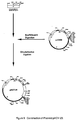
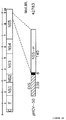
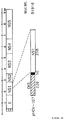
- Recombinant antigens which are derived from the molecular cloning and expression of synthetic DNA sequences in heterologous hosts are provided. Briefly, synthetic DNA sequences which encode the desired proteins representing distinct antigenic regions of the HCV genome are optimized for expression in E.coli by specific codon selection. Specifically, two recombinant proteins representing three distinct antigenic regions of the HCV genome, including immunogenic regions of the c1OO-3 antigen and two additional non-overlapping regions upstream from the c1OO-3 region are described. Both proteins are expressed as chimeric fusions with E.coli CMP-KDO synthetase (CKS) gene. The first protein, expressed by plasmid pHCV-34 represents amino acids 1-150 of the HCV sequence and, based on analogy to the genomic organization of other flaviviruses, has been named HCV CKS-Core. Note that the term pHCV-34 will also refer to the fusion protein itself and that pHCV-34' will be the designation for a polypeptide representing the core region from about amino acids 1-150 of the HCV sequence prepared using other recombinant or synthetic methodologies. Other recombinant methodologies would include the preparation of pHCV-34', utilizing different expression systems. The methodology for the preparation of synthetic peptides of HCV is described in U.S. Serial No. 456,162, filed December 22, 1989, which enjoys common ownership and is incorporated herein by reference. The other protein is expressed by plasmid pHCV-31 and is composed of two non-contiguous coding regions located in the putative non-structural regions of HCV designated NS-3 and NS-4. The first of the two regions represents amino acids 1192-1457 of the HCV sequence (known as Clone 33) and is expressed by the plasmid pHCV-29. The fusion protein itself will also be referred to as pHCV-29 and pHCV-29' shall be the designation for a polypeptide from the NS-3 region representing from about amino acids 1192-1457 of the HCV sequence prepared using other recombinant or synthetic methodologies. The second region represents amino acids 1676-1931 of the HCV sequence and is expressed by the plasmid pHCV-23. The fusion protein will be referred to as pHCV-23 and pHCV-23' shall be the designation for a polypeptide from the NS4 region representing from about amino acids 1676-1931 of the HCV sequence prepared using other recombinant or synthetic methodologies. It has been designated Clone BCD based on the strategy used in its construction. Clone BCD represents the carboxyl-
terminal 256 amino acids of c1OO-3: the amino terminal 108 amino acids of c1OO-3 are not represented in Clone BCD. The recombinant antigen produced by pHCV-31 is designated CKS-33c-BCD. The fusion protein is also designated by pHCV-31 and pHCV-31' refers to the polypeptide composed of two noncontiguous coding regions located in the putative nonstructural regions of HCV designated NS-3 and NS-4, representing from about amino acids 1192-1457 and from about 1676-1931 of the HCV sequence prepared using different recombinator synthetic methodologies. Figure 1 illustrates the position of the three HCV regions within the HCV genome. These antigens are used in the inventive immunoassays to detect the presence of HCV antibodies in samples. - One assay format according to the invention provides a screening assay for identifying the presence of an antibody that is immunologically reactive with an HCV antigen. Briefly, a fluid sample is incubated with a solid support containing the two commonly bound recombinant proteins HCV pHCV-34 and pHCV-31. Finally, the antibody-antigen complex is detected. In a modification of the screening assay the solid support additionally contains recombinant polypeptide c1OO-3.
- Another assay format provides a confirmatory assay for unequivocally identifying the presence of an antibody that is immunologically reactive with an HCV antigen. The confirmatory assay includes synthetic peptides or recombinant antigens representing major epitopes contained within the three distinct regions of the HCV genome, which are the same regions represented by the two recombinant proteins described in the screening assay. These regions include NS4 (the c100-3 region) represented by pHCV-23, NS3 (the 33c region) represented by pHCV-29, and together with pHCV-23 (the c100-3 region) represented by pHCV-31, and a region near the 5' end of the HCV genome believed to be the core structural protein of HCV (pHCV-34). Recombinant proteins used in the confirmatory assay should have a heterologous source of antigen to that used in the primary screening assay (i.e. should not be an E.coli-derived recombinant antigen nor a recombinant antigen composed in part, of CKS sequences). Briefly, specimens repeatedly reactive in the primary screening assay are retested in the confirmatory assay. Aliquots containing identical amounts of specimen are contacted with a synthetic peptide or recombinant antigen individually coated onto a solid support. Finally, the antibody-antigen complex is detected. Seroreactivity for epitopes within the c1OO-3 region of the HCV genome are confirmed by use of the synthetic peptides sp67 and sp65. The synthetic peptide sp117 can also be used to confirm seroreactivity within the c100-3 region. Seroreactivity for HCV epitopes within the putative core region of HCV are confirmed by the use of the synthetic peptide sp75. In order to confirm seroreactivity for HCV epitopes within the 33c region of HCV, a recombinant antigen is expressed as a chimeric protein with superoxide dismutase (SOD) in yeast. The synthetic peptide sp65 (representing amino acids p1866-1930 of the HCV sequence), sp67 (representing amino acids p1684-1750), sp75 (representing amino acids p1-75), and sp117 (representing amino acids p1689-1805) are described in U.S. Serial No. 456,162 entitled "Hepatitis C Assay", filed December 22, 1989, which enjoys common ownership and is incorporated herein by reference.
- Another assay format provides a competition assay or neutralization assay directed to the confirmation that positive results are not false by identifying the presence of an antibody that is immunologically reactive with an HCV antigen in a fluid sample where the sample is used to prepare first and second immunologically equivalent aliquots. The first aliquot is contacted with solid support containing a bound polypeptide which contains at least one epitope of an HCV antigen under conditions suitable for complexing with the antibody to form a detectable antibody-polypeptide complex and the second aliquot is first contacted with the same solid support containing bound polypeptide. The preferred recombinant polypeptide is derived from pHCV-23.
- Another assay format provides an immunodot assay for identifying the presence of an antibody that is immunologically reactive with an HCV antigen by concurrently contacting a sample with recombinant polypeptides each containing distinct epitopes of an HCV antigen under conditions suitable for complexing the antibody with at least one of the polypeptides and detecting the antibodypolypeptide complex by reacting the complex with color-producing reagents. The preferred recombinant polypeptides employed include those recombinant polypeptides derived from pHCV-23, pHCV-29, pHCV-31, pHCV-34, as well as c1OO-3 expressed as a chimeric protein with superoxide dismutase (SOD) in yeast.
- In all of the assays, the sample is preferably diluted before contacting the polypeptide absorbed on a solid support. Samples may be obtained from different biological samples such as whole blood, serum, plasma, cerebral spinal fluid, and lymphocyte or cell culture supernatants. Solid support materials may include cellulose materials, such as paper and nitrocellulose, natural and synthetic polymeric materials, such as polyacrylamide, polystyrene, and cotton, porous gels such as silica gel, agarose, dextran and gelatin, and inorganic materials such as deactivated alumina, magnesium sulfate and glass. Suitable solid support materials may be used in assays in a variety of well known physical configurations, including microtiter wells, test tubes, beads, strips, membranes, and microparticles. A preferred solid support for a non-immunodot assay is a polystyrene bead. A preferred solid support for an immnuodot assay is nitrocellulose.
- Suitable methods and reagents for dectecting an antibody-antigen complex in an assay of the present invention are commercially available or known in the relevant art. Representative methods may employ detection reagents such as enzymatic, radioisotopic, fluorescent, luminescent, or chemiluminescent reagents. These reagents may be used to prepare hapten-labelled antihapten detection systems according to known procedures, for example, a biotin-labelled antibiotin system may be used to detect an antibody-antigen complex.
- The present invention also encompasses assay kits including polypeptides which contain at least one epitope of an HCV antigen bound to a solid support as well as needed sample preparation reagents, wash reagents, detection reagents and signal producing reagents.
- Other aspects and advantages of the invention will be apparent to those skilled in the art upon consideration of the following detailed description which provides illustrations of the invention in its presently preferred embodiments.
- E.coli strains containing plasmids useful for constructs of the invention have been deposited at the American Type Culture Collection, Rockville, Maryland on August 10, 1990, under the accession Nos. ATCC 68380 (pHCV-23), ATCC 68381 (pHCV-29), ATCC 68382 (pHCV-31), ATCC 68383 (pHCV-34) and on November 6, 1990 for E. coli strains containing plasmids useful for constructs under the accession Nos. ATCC 68458 (pHCV-50), 68459 (pHCV-57), 68460 (pHCV-103), 68461 (pHCV-102), 68462 (pHCV-51), 68463 (pHCV-105), 68464 (pHCV-107), 68465 (pHCV-104), 68466 (pHCV-45), 68467 (pHCV-48), 68468 (pHCV-49), 68469 (pHCV-58), 68470 (pHCV-101).
- FIGURE 1 illustrates the HCV genome.
- FIGURE 2 illustrates the use of recombinant polypeptides to identify the presence of antibodies in a chimpanzee inoculated with HCV.
- FIGURE 3 illustrates the sensitivity and specificity increase in using the screening assay using pHCV-34 and pHCV-31 antigens.
- FIGURE 4 illustrates the construction of plasmid pHCV-34.
- FIGURE 5 illustrates the complete DNA and amino acid sequence of pHCV-34.
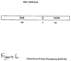
- FIGURE 6 illustrates fusion protein pHCV-34.
- FIGURE 7 illustrates the expression of pHCV-34 proteins in E.coli.
- FIGURE 8 illustrates the construction of plasmid pHCV-23.
- FIGURE 9 illustrates the construction of plasmid pHCV-29.
- FIGURE 10 illustrates the construction of plasmid pHCV-31.
- FIGURE 11 illustrates the complete DNA and amino acid sequence of pHCV-31.
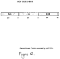
- FIGURE 12 illustrates the fusion protein pHCV-31.
- FIGURE 13 illustrates the expression of pHCV-29 in E.coli.
- FIGURE 14 illustrates the expression of pHCV-23 in E.coli.
- FIGURE 15 illustrates the expression of pHCV-31 in E.coli.
- FIGURE 16 illustrates the increased sensitivity using the screening assay utilizing the pHCV-34.
- FIGURE 17 illustrates the increased specificity with the screening assay utilizing pHCV-34 and pHCV-31.
- FIGURE 18 illustrates the results in hemodialysis patients using the screening and confirmatory assays.
- FIGURE 19 illustrates earlier detection of HCV in a hemodialysis patient using the screening assay.
- FIGURE 20 illustrates the results of the screening assay utilizing pHCV-34 and pHCV-31 on samples from individuals with acute NANBH.
- FIGURE 21 illustrates the results of the confirmatory assay of the same population group as in Figure 20.
- FIGURE 22 illustrates the results of the screening and confirmatory assays on individuals infected with chronic NANBH.
- FIGURE 23 illustrates preferred buffers, pH conditions, and spotting concentrations for the HCV immunodot assay.
- FIGURE 24 illustrates the results of the HCV immunodot assay.
- FIGURE 25 illustrates the fusion protein pHCV-45.
- FIGURE 26 illustrates the DNA and amino acid sequence of the recombinant antigen expressed by pHCV-45.
- FIGURE 27 illustrates the expression of pHCV-45 in E.coli.
- FIGURE 28 illustrates the fusion protein pHCV-48.
- FIGURE 29 illustrates the DNA and amino acid sequence of the recombinant antigen expressed by pHCV-48.
- FIGURE 30 illustrates the expression of pHCV-48 in E.coli.
- FIGURE 31 illustrates the fusion protein pHCV-51.
- FIGURE 32 illustrates the DNA and amino acid sequence of the recombinant antigen expressed by pHCV-51.
- FIGURE 33 illustrates the expression of pHCV-51 in E.coli.
- FIGURE 34 illustrates the fusion protein pHCV-50.
- FIGURE 35 illustrates the DNA and amino acid sequence of the recombinant antigen expressed by pHCV-50.
- FIGURE 36 illustrates the expression of pHCV-50 in E.coli.
- FIGURE 37 illustrates the fusion protein pHCV-49.
- FIGURE 38 illustrates the DNA and amino acid sequence of the recombinant antigen expressed by pHCV-49.
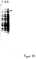
- FIGURE 39 illustrates the expression of pHCV-49 in E.coli.
- FIGURE 40 illustrates an immunoblot of pHCV-23, pHCV-45, pHCV-48, pHCV-51, pHCV-50 and pHCV-49.
- FIGURE 41 illustrates the fusion proteins pHCV-24, pHCV-57, pHCV-58.
- FIGURE 42 illustrates the DNA and amino acid sequence of the recombinant antigen expressed by pHCV-57.
- FIGURE 43 illustrates the DNA and amino acid sequence of the recombinant antigen expressed by pHCV-58.
- FIGURE 44 illustrates the expression of pHCV-24, pHCV-57, and pHCV-58 in E.coli.
- FIGURE 45 illustrates the fusion protein pHCV-105.
- FIGURE 46 illustrates the DNA and amino acid sequence of the recombinant antigen expressed by pHCV-105.
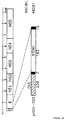
- FIGURE 47 illustrates the expression of pHCV-105 in E.coli.
- FIGURE 48 illustrates the fusion protein pHCV-103.
- FIGURE 49 illustrates the DNA and amino acid sequence of the recombinant antigen expressed by pHCV-103.
- FIGURE 50 illustrates the fusion protein pHCV-101.
- FIGURE 51 illustrates the DNA and amino acid sequence of the recombinant antigen expressed by pHCV-101.
- FIGURE 52 illustrates the fusion protein pHCV-102.
- FIGURE 53 illustrates the DNA and amino acid sequence of the recombinant antigen expressed by pHCV-102.
- FIGURE 54 illustrates the expression of pHCV-102 in E.coli.
- FIGURE 55 illustrates the fusion protein pHCV-107.
- FIGURE 56 illustrates the DNA and amino acid sequence of the recombinant antigen expressed by pHCV-107.
- FIGURE 57 illustrates the fusion protein pHCV-104.
- FIGURE 58 illustrates the DNA and amino acid sequence of the recombinant antigen expressed by pHCV-104.
- The present invention is directed to an assay to detect an antibody to an HCV antigen in a sample. Human serum or plasma is preferably diluted in a sample diluent and incubated with a polystyrene bead coated with a recombinant polypeptide that represents a distinct antigenic region of the HCV genome. If antibodies are present in the sample they will form a complex with the antigenic polypeptide and become affixed to the polystyrene bead. After the complex has formed, unbound materials and reagents are removed by washing the bead and the bead-antigen-antibody complex is reacted with a solution containing horseradish peroxidase labeled goat antibodies directed against human antibodies. This peroxidase enzyme then binds to the antigen-antibody complex already fixed to the bead. In a final reaction the horseradish peroxidase is contacted with o-phenylenediamine and hydrogen peroxide which results in a yellow-orange color. The intensity of the color is proportional to the amount of antibody which initially binds to the antigen fixed to the bead.
- The preferred recombinant polypeptides having HCV antigenic epitopes were selected from portions of the HCV genome which encoded polypeptides which possessed amino acid sequences similar to other known immunologically reactive agents and which were identified as having some immunological reactivity. (The immunological reactivity of a polypeptide was initially identified by reacting the cellular extract of E.coli clones which had been transformed with cDNA fragments of the HCV genome with HCV infected serum. Polypeptides expressed by clone containing the incorporated cDNA were immunologically reactive with serum known to contain antibody to HCV antigens.) An analysis of a given amino acid sequence, however, only provides rough guides to predicting immunological reactivity. There is no invariably predictable way to ensure immunological activity short of preparing a given amino acid sequence and testing the suspected sequence in an assay.
- The use of recombinant polypeptides representing distinct antigenic regions of the HCV genome to detect the presence of an antibody to an HCV antigen is illustrated in Figure 2. The course of HCV infection in the chimpanzee, Pan, was followed with one assay using recombinant c1OO-3 polypeptide and with another improved assay, using the two recombinant antigens CKS-Core (pHCV-34) and pHCV-33c-BCD (pHCV-31) expressed by the plasmids pHCV-34 and pHCV-31, respectively. The assay utilizing the recombinant pHCV-34 and pHCV-31 proteins detected plasma antibody three weeks prior to detection of antibody by the assay using c100-3.
- A summary of the results of a study which followed the course of HCV infection in Pan and six other chimpanzees using the two assays described above is summarized in Figure 3. Both assays gave negative results before inoculation and both assays detected the presence of antibodies after the animal had been infected with HCV. However, in the comparison of the two assays, the improved screening assay using pHCV-34 and pHCV-31 detected seroconversion to HCV antigens at an earlier or equivalent bleed date in six of the seven chimpanzees. Data from these chimpanzee studies clearly demonstrate that overall detection of HCV antibodies is greatly increased with the assay utilizing the pHCV-34 and pHCV-31 proteins. This test is sufficiently sensitive to detect seroconversion during the acute phase of this disease, as defined as an elevation in ALT levels, in most animals. Equally important is the high degree of specificity of the test as no pre-inoculation specimens were reactive.
- The polypeptides useful in the practice of this invention are produced using recombinant technologies. The DNA sequences which encode the desired polypeptides are preferably assembled from fragments of the total desired sequence. Synthetic DNA fragments of the HCV genome can be synthesized based on their corresponding amino acid sequences. Once the amino acid sequence is chosen, this is then reverse translated to determine the complementary DNA sequence using codons optimized to facilitate expression in the chosen system. The fragments are generally prepared using well known automated processes and apparatus. After the complete sequence has been prepared the desired sequence is incorporated into an expression vector which is transformed into a host cell. The DNA sequence is then expressed by the host cell to give the desired polypeptide which is harvested from the host cell or from the medium in which the host cell is cultured. When smaller peptides are to be made using recombinant technologies it may be advantageous to prepare a single DNA sequence which encodes several copies of the desired polypeptide in a connected chain. The long chain is then isolated and the chain is cleaved into the shorter, desired sequences.
- The methodology of polymerase chain reaction (PCR) may also be employed to develop PCR amplified genes from any portion of the HCV genome, which in turn may then be cloned and expressed in a manner similar to the synthetic genes.
- Vector systems which can be used include plant, bacterial, yeast, insect, and mammalian expression systems. It is preferred that the codons are optimized for expression in the system used.
- A preferred expression system utilizes a carrier gene for a fusion system where the recombinant HCV proteins are expressed as a fusion protein of an E.coli enzyme, CKS (CTP:CMP-3-deoxy-manno-octulosonate cytidylyl transferase or CMP-KDO synthetase). The CKS method of protein synthesis is disclosed in U.S. Patent Applications Serial Nos. 167,067 and 276,263 filed March 11, 1988 and November 23, 1988, respectively, by Bolling (EPO 891029282) which enjoy common ownership and are incorporated herein by reference.
- Other expression systems may be utilized including the lambda PL vector system whose features include a strong lambda pL promoter, a strong three-frame translation terminator rrnBtl, and translation starting at an ATG codon.
- In the present invention, the amino acid sequences encoding for the recombinant HCV antigens of interest were reverse translated using codons optimized to facilitate high level expression in E.coli. Individual oligonucleotides were synthesized by the method of oligonucleotide directed double-stranded break repair disclosed in U.S. Patent Application Serial No. 883,242, filed July 8, 1986 by Mandecki (EPO 87109357.1) which enjoys common ownership and is incorporated herein by reference. Alternatively, the individual oligonucleotides may be synthesized on the Applied Biosystem 380A DNA synthesizer using methods and reagents recommended by the manufacturer. The DNA sequences of the individual oligonucleotides were confirmed using the Sanger dideoxy chain termination method (Sanger et al., J. Mole. Biol., 162:729 (1982)). These individual gene fragments were then annealed and ligated together and cloned as EcoRl-BamHl subfragments in the CKS fusion vector pJO200. After subsequent DNA sequence confirmation by the Sanger dideoxy chain termination method, the subfragments were digested with appropriate restriction enzymes, gel purified, ligated and cloned again as an EcoRl-BamHl fragment in the CKS fusion vector pJO2OO. The resulting clones were mapped to identify a hybrid gene consisting of the EcoRl-BamHl HCV fragment inserted at the 3' end of the CKS (CMP-KDO synthetase) gene. The resultant fusion proteins, under control of the lac promoter, consist of 239 amino acids of the CKS protein fused to the various regions of HCV.
- The synthesis, cloning, and characterization of the recombinant polypeptides as well as the preferred formats for assays using these polypeptides are provided in the following examples. Examples 1 and 2 describe the synthesis and cloning of CKS-Core and CKS-33-BCD, respectively. Example 3 describes a screening assay. Example 4 describes a confirmatory assay. Example 5 describes a competition assay. Example 6 describes an immunodot assay.
- Media such as Luria-Bertani (LB) and Superbroth II (Dri Form) were obtained from Gibco Laboratories Life Technologies, Inc., Madison Wisconsin. Restriction enzymes, Klenow fragment of DNA polymerase I, T4 DNA ligase, T4 polynucleotide kinase, nucleic acid molecular weight standards, M13 sequencing system, X-gal (5-bromo-4-chloro-3-indonyl-β-D-galactoside), IPTG (isopropyl-β-D-thiogalactoside), glycerol, Dithiothreitol, 4-chloro-1-naphthol were purchased from Boehringer Mannheim Biochemicals, Indianapolis, Indiana; or New England Biolabs, Inc., Beverly, Massachusetts; or Bethesda Research Laboratories Life Technologies, Inc., Gaithersburg, Maryland. Prestained protein molecular weight standards, acrylamide (crystallized, electrophoretic grade 99%); N-N'-Methylene-bis-acrylamide (BIS); N,N,N',N',-Tetramethylethylenediamine (TEMED) and sodium dodecylsulfate (SDS) were purchased from BioRad Laboratories, Richmond, California. Lysozyme and ampicillin were obtained from Sigma Chemical Co., St. Louis, Missouri. Horseradish peroxidase (HRPO) labeled secondary antibodies were obtained from Kirkegaard & Perry Laboratories, Inc., Gaithersburg, Maryland. Seaplaque® agarose (low melting agarose) was purchased from FMC Bioproducts, Rockland, Maine.
- T50E10 contained 50mM Tris, pH 8.0, 1OmM EDTA; 1X TG contained 1OOmM Tris, pH 7.5 and 10% glycerol; 2X SDS/PAGE loading buffer consisted of 15% glycerol, 5% SDS, 1OOmM Tris base, 1M β-mercaptoethanol and 0.8% Bromophenol blue dye;
TBS container 50 mM Tris, pH 8.0, and 150 mM sodium chloride; Blocking solution consisted of 5% Carnation nonfat dry milk in TBS. - E.coli JM103 cells, pUC8, pUC18, pUC19 and M13 cloning vectors were purchased from Pharmacia LKB Biotechnology, Inc., Piscataway, New Jersey; Competent Epicurean™ coli stains XL1-Blue and JM109 were purchased from Stratagene Cloning Systems, LaJolla, California. RR1 cells were obtained from Coli Genetic Stock Center, Yale University, New Haven, Connecticut; and E.coli CAG456 cells from Dr. Carol Gross, University of Wisconsin, Madison, Wisconsin. Vector pRK248.clts was obtained from Dr. Donald R. Helinski, University of California, San Diego, California.
- All restriction enzyme digestion were performed according to suppliers' instructions. At least 5 units of enzyme were used per microgram of DNA, and sufficient incubation was allowed to complete digestion of DNA. Standard procedures were used for minicell lysate DNA preparation, phenol-chloroform extraction, ethanol precipitation of DNA, restriction analysis of DNA on agarose, and low melting agarose gel purification of DNA fragments (Maniatis et al., Molecular Cloning. A Laboratory Manual [New York: Cold Spring Harbor, 1982]). Plasmid isolations from E.coli strains used the alkali lysis procedure and cesium chloride-ethidium bromide density gradient method (Maniatis et al., supra). Standard buffers were used for T4 DNA ligase and T4 polynucleotide kinase (Maniatis et al., supra).
- The cloning vector pJO200 allows the fusion of recombinant proteins to the CKS protein. The plasmid consists of the plasmid pBR322 with a modified lac promoter fused to a KdsB gene fragment (encoding the first 239 of the entire 248 amino acids of the E.coli CMP-KDO synthetase of CKS protein), and a synthetic linker fused to the end of the KdsB gene fragment. The cloning vector pJO200 is a modification of vector pTB210. The synthetic linker includes: multiple restriction sites for insertion of genes; translational stop signals, and the trpA rho-independent transcriptional terminator. The CKS method of protein synthesis as well as CKS vectors including pTB210 are disclosed in U.S. Patent Application Serial Nos. 167,067 and 276,263, filed March 11, 1988 and November 23, 1988, respectively, by Bolling (EPO 891029282) which enjoy common ownership, and are herein incorporated by reference.
- Six individual nucleotides representing amino acids 1-150 of the HCV genome were ligated together and cloned as a 466 base pair EcoRl-BamHl fragment into the CKS fusion vector pJO200 as presented in Figure 4. The complete DNA sequence of this plasmid, designated pHCV-34, and the entire amino acid sequence of the pHCV-34 recombinant antigen produced is presented in Figure 5. The resultant fusion protein HCV CKS-Core, consists of 239 amino acids of CKS, seven amino acids contributed by linker DNA sequences, and the first 150 amino acids of HCV as illustrated in Figure 6.
- The pHCV-34 plasmid and the CKS plasmid pTB210 were transformed into E.coli K-12 strain xL-1 (recAl, endAl, gyrA96, thi-1, hsdRl7, supE44, relAl, lac/F', proAB, laclqZDM15, TN10) cells made competent by the calcium chloride method. In these constructions the expression of the CKS fusion proteins was under the control of the lac promoter and was induced by the addition of IPTG. These plasmids replicated as independent elements, were nonmobilizable and were maintained at approximately 10-30 copies per cell.
- In order to establish that clone pHCV-34 expressed the unique HCV-CKS Core protein, the pHCV-34/XL-1 culture was grown overnight at 37°C in growth media consisting of yeast extract, trytone, phosphate salts, glucose, and ampicillin. When the culture reached an OD600 of 1.0, IPTG was added to a final concentration of 1mM to induce expression. Samples (1.5 ml) were removed at 1 hour intervals, and cells were pelleted and resuspended to an OD600 of 1.0 in 2X SDS/PAGE loading buffer. Aliquots (15ul) of the prepared samples were separated on duplicate 12.5% SDS/PAGE gels.
- One gel was fixed in a solution of 50% methanol and 10% acetic acid for 20 minutes at room temperature, and then stained with 0.25% Coomassie blue dye in a solution of 50% methanol and 10% acetic acid for 30 minutes. Destaining was carried out using a solution of 10% methanol and 7% acetic acid for 3-4 hours, or until a clear background was obtained.
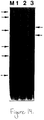
- Figure 7 presents the expression of pHCV-34 proteins in E.coli. Molecular weight standards were run in
Lane M. Lane 1 contains the plasmid pJ0200-the CKS vector without the HCV sequence. The arrows on the left indicate the mobilities of the molecular weight markers from top to bottom: 110,000; 84,000; 47,000; 33,000; 24,000; and 16,000 daltons. The arrows on the right indicate the mobilities of the recombinant HCV proteins.Lane 2 contains the E.coli lysate containing pHCV-34 expressing CKS-Core (amino acids 1 to 150) prior to induction; andLane 3 after 3 hours of induction. The results show that the recombinant protein pHCV-34 has an apparent mobility corresponding to a molecular size of 48,000 daltons. This compares acceptably with the predicted molecular mass of 43,750 daltons. - Proteins from the second 12.5% SDS/PAGE gel were electrophoretically transferred to nitrocellulose for immunoblotting. The nitrocellulose sheet containing the transferred proteins was incubated with Blocking Solution for one hour and incubated overnight at 4°C with HCV patients' sera diluted in TBS containing E.coli K-12 strain XL-1 lysate. The nitrocellulose sheet was washed three times in TBS, then incubated with HRPO-labeled goat anti-human IgG, diluted in TBS containing 10% fetal calf sera. The nitrocellulose was washed three times with TBS and the color was developed in TBS containing 2 mg/ml 4-chloro-1-napthol, 0.02% hydrogen peroxide and 17% methanol. Clone HCV-34 demonstrated a strong immunoreactive band at 48,000 daltons with the HCV patients' sera. Thus, the major protein in the Coomassie stained protein gel was immunoreactive. Normal human serum did not react with any component of pHCV-34.
- The construction of this recombinant clone expressing the HCV CKS-33-BCD antigen was carried out in three steps described below. First, a clone expressing the HCV CKS-BCD antigen was constructed, designated pHCV-23. Second, a clone expressing the HCV CKS-33 antigen was constructed, designated pHCV-29. Lastly, the HCV BCD region was excised from pHCV-23 and inserted into pHCV-29 to construct a clone expressing the HCV CKS-33-BCD antigen, designated pHCV-31.
- To construct the plasmid pHCV-23, thirteen individual oligonucleotides representing amino acids 1676-1931 of the HCV genome were ligated together and cloned as three separate EcoRl-BamHl subfragments into the CKS fusion vector pJO200. After subsequent DNA sequence confirmation, the three subfragments, designated B, C, and D respectively, were digested with the appropriate restriction enzymes, gel purified, ligated together, and cloned as a 781 base pair EcoRl-BamHl fragment in the CKS fusion vector pJO200, as illustrated in Figure 8. The resulting plasmid, designated pHCV-23, expresses the HCV CKS-BCD antigen under control of the lac promoter. The HCV CKS-BCD antigen consists of 239 amino acids of CKS, seven amino acids contributed by linker DNA sequences, 256 amino acids from the HCV NS4 region (amino acids 1676-1931, and 10 additional amino acids contributed by linker DNA sequences.
- To construct the plasmid pHCV-29 twelve individual oligonucleotides representing amino acids 1192-1457 of the HCV genome were ligated together and cloned as two separate EcoRl-BamHl subfragments in the CKS fusion vector pJO200. After subsequent DNA sequence confirmation, the two subfragments were digested with the appropriate restriction enzymes, gel purified, ligated together and cloned again as an 816 base pair EcoRl-BamHl fragment in the CKS fusion vector pJO200, as illustrated in Figure 9. The resulting plasmid, designated pHCV-29, expresses the CKS-33 antigen under control of the lac promoter. The HCV CKS-33 antigen consists of 239 amino acids of CKS, eight amino acids contributed by linker DNA sequences, and 266 amino acids from the HCV NS3 region (amino acids 1192-1457).
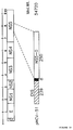
- To construct the plasmid pHCV-31, the 781 base pair EcoRl-BamHl fragment from pHCV-23 representing the HCV-BCD region was linker-adapted to produce a Cla1-BamH1 fragment which was then gel purified and ligated into pHCV-29 at the Cla1-BamH1 sites as illustrated in Figure 10. The resulting plasmid, designated pHCV-31, expresses the pHCV-31 antigen under control of the lac promoter. The complete DNA sequence of pHCV-31 and the entire amino acid sequence of the HCV CKS-33-BCD recombinant antigen produced is presented in Figure 11. The HCV CKS-33-BCD antigen consists of 239 amino acids of CKS, eight amino acids contributed by linker DNA sequences, 266 amino acids of the HCV NS3 region (amino acids 1192-1457), 2 amino acids contributed by linker DNA sequences, 256 amino acids of the HCV NS4 region (amino acids 1676-1931), and 10 additional amino acids contributed by linker DNA sequences. Figure 12 presents a schematic representation of the pHCV-31 antigen.
- The pHCV-31 plasmid was transformed into E.coli K-12 strain XL-1 in a manner similar to the pHCV-34 and CKS-pTB210 plasmids of Example 1.
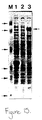
- Characterization of pHCV CKS-33-BCD was carried out in a manner similar to pHCV CKS-Core of Example 1. pHCV-23, pHCV SDS/PAGE gels were run for E.coli lysates containing the plasmids pHCV-29 (Figure 13), pHCV-23 (Figure 14), and pHCV-31 (Figure 15) expressing the recombinant fusion proteins CKS-33c, CKS-BCD, and CKS-33-BCD, respectively. For all three figures, molecular weight standards were run in Lane M, with the arrows on the left indicating mobilities of the molecular weight markers the from top to bottom: 110,000; 84,000; 47,000; 33,000; 24,000; and 16,000 daltons. In Figure 13,
Lane 1 contained the E.coli lysate containing pHCV-29 expressing HCV CKS-33c (amino acids 1192 to 1457) prior to induction andlane 2 after 4 hours induction. These results show that the recombinant pHCV-29 fusion protein has an apparent mobility corresponding to a molecular size of 60,000 daltons. This compares acceptably to the predicted molecular mass of 54,911. - In Figure 14,
Lane 1 contained the E.coli lysate containing pJO200- the CKS vector without the HCV sequence.Lane 2, contained pHCV-20 expressing the HCV CKS-B (amino acids 1676 to 1790).Lane 3, contained the fusion protein pHCV-23 (amino acids 1676-1931). These results show that the recombinant pHCV-23 fusion protein has an apparent mobility corresponding to a molecular size of 55,000 daltons. This compares acceptably to the predicted molecular mass of 55,070 daltons. - In Figure 15,
Lane 1 contained the E.coli lysate containing pJO200 the CKS vector without the HCV sequences.Lane 2 contained pHCV-31 expressing the CKS-33c-BCD fusion protein (amino acids 1192 to 1447 and 1676 to 1931) prior to induction andlane 3 after 2 hours induction. These results show that the recombinant pHCV-31 (CKS-33c-BCD) fusion protein has an apparent mobility corresponding to a molecular size of 90,000 daltons. This compares acceptably to the predicted molecular mass of 82,995 daltons. - An immunoblot was also run on one of the SDS/PAGE gels derived from the pHCV-31/X1-1 culture. Human serum from an HCV exposed individual reacted strongly with the major pHCV-31 band at 90,000 daltons. Normal human serum did not react with any component of the pHCV-31 (CKS-33-BCD) preparations.
- The use of recombinant polypeptides which contain epitopes within c100-3 as well as epitopes from other antigenic regions from the HCV genome, provide immunological assays which have increased sensitivity and may be more specific than HCV immunological assays using epitopes within c100-3 alone.
- In the presently preferred screening assay, the procedure uses two E.coli expressed recombinant proteins, CKS-Core (pHCV-34) and CKS-33-BCD (pHCV-31), representing three distinct regions of the HCV genome. These recombinant polypeptides were prepared following procedures described above. In the screening assay, both recombinant antigens are coated onto the same polystyrene bead. In a modification of the screening assay the polystyrene bead may also be coated with the SOD-fusion polypeptide c100-3.
- The polystyrene beads are first washed with distilled water and propanol and then incubated with a solution containing recombinant pHCV-31 diluted to 0.5 to 2.0 ug/ml and pHCV-34 diluted to 0.1 to 0.5 ug/ml in 0.1 M NaH₂PO₄·H₂0 with 0.4M NaC1 and 0.0022% Triton X-100, pH 6.5. The beads are incubated in the antigen solution for 2 hours (plus or minus 10 minutes) at 38-42°C, washed in PBS and soaked in 0.1% (w/v) Triton X-100 in PBS for 60 minutes at 38-42°C. The beads are then washed two times in phosphate buffered saline (PBS), overcoated with a solution of 5.0% (w/v) bovine serum albumin (BSA) in PBS for 60 minutes at 38-42°C and washed one time in PBS. Finally, the beads are overcoated with 5% (w/v) sucrose in PBS, and dried under nitrogen or air.
- The polystyrene beads coated with pHCV-31 and pHCV-34 are used in an antibody capture format. Ten microliters of sample are added to the wells of the reaction tray along with 400 ul of a sample diluent and the recombinant coated bead. The sample diluent consists of 10% (v/v) bovine serum and 20% (v/v) goat serum in 20 mM Tris phosphate buffer containing 0.15% (v/v) Triton X-100, 1%(w/v) BSA, 1% E.coli lysate and 500 ug/ml or less CKS lysate. When the recombinant yeast c100-3 polypeptide is used, antibodies to yeast antigens which may be present in a sample are reacted with yeast extracts which are added to the sample diluent (typically about 200 ug/ml). The addition of yeast extracts to the sample diluent is used to prevent false positive results. The final material is sterile filtered and filled in plastic bottles, and preserved with 0.1% sodium azide.
- After one hour of incubation at 40°C, the beads are washed and 200 ul of conjugate is added to the wells of the reaction tray.
- The preferred conjugate is goat anti-human IgG horseradish peroxidase conjugate. Concentrated conjugate is titered to determine a working concentration. A twenty-fold concentrate of the working conjugate solution is then prepared by diluting the concentrate in diluent. The 20X concentrate is sterile filtered and stored in plastic bottles.
- The conjugate diluent includes 10% (v/v) bovine serum, 10% (v/v) goat serum and 0.15% Triton-X100 in 20 mM Tris buffer, pH 7.5 with 0.01% gentamicin sulfate, 0.01% thimerosal and red dye. The conjugate is sterile filtered and filled in plastic bottles.
- Anti-HCV positive control is prepared from plasma units positive for antibodies to HCV. The pool of units used includes plasma with antibodies reactive to pHCV-31 and pHCV-34. The units are recalcified and heat inactivated at 59-61°C for 12 hours with constant stirring. The pool is aliquoted and stored at -20°C or at 2-8°C. For each lot of positive control, the stock solution is diluted with negative control containing 0.1% sodium azide as a preservative. The final material is sterile filtered and filled in plastic bottles.
- Anti-HCV negative control is prepared from recalcified human plasma, negative for antibodies to pHCV-31 and pHCV-34 proteins of HCV. The plasma is also negative for antibodies to human immunodeficiency virus (HIV) and negative for hepatitis B surface antigen (HBsAg). The units are pooled, and 0.1% sodium azide is added as a preservative. The final material is sterile filtered and filled in plastic bottles.
- After one hour of incubation with the conjugate at 40°C, the beads are washed, exposed to the OPD substrate for thirty minutes at room temperature and the reaction terminated by the addition of 1 N H₂SO₄. The absorbance is read at 492 nm.
- In order to maintain acceptable specificity, the cutoff for the assay should be at least 5-7 standard deviations above the absorbance value of the normal population mean. In addition, it has generally been observed that acceptable specificity is obtained when the population mean runs at a sample to cutoff (S/CO) value of 0.25 or less. Consistent with these criteria, a "preclinical" cutoff for the screening assay was selected which clearly separated most of the presumed "true negative" from "true positive" specimens. The cutoff value was calculated as the sum of the positive control mean absorbance value multiplied by 0.25 and the negative control mean absorbance value. The cutoff may be expressed algebraically as:

- Testing may be performed by two methods which differ primarily in the degree of automation and the mechanism for reading the resulting color development in the assay. One method is referred to as the manual or Quantumttm method because Quantum or Quantumatic is used to read absorbance at 492 nm. It is also called the manual method because sample pipetting, washing and reagent additions are generally done manually by the technician, using appropriately calibrated pipettes, dispensers and wash instruments. The second method is referred to as the PPC method and utilizes the automated Abbott CommanderR system. This system employs a pipetting device referred to as the Sample Management Center (SMC) and a wash/dispense/read device referred to as the Parallel Processing Center (PPC) disclosed in the Abbott Disclosure No. 17256 entitled "Simultaneous Assay for Detecting One Or More Analytes" the inventor of which is William E. Brown, III. The optical reader used in the PPC has dual wavelength capabilities that can measure differential absorbencies (peak band and side band) from the sample wells. These readings are converted into results by the processor's Control Center.
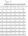
- As previously described, Table I summarizes the results of a study which followed the course of HCV infection in seven chimpanzees using a screening assay which utilized the c100-3 polypeptide, and the screening assay which utilized pHCV-31 and pHCV-34. Both assays gave negative results before inoculation and both assays detected the presence of antibodies after the animal had been infected with HCV. However, in the comparison of the two assays, the assay utilizing pHCV-31 and pHCV-34 detected seroconversion to HCV antigens at an earlier or equivalent bleed date in six of the seven chimpanzees. Data from these chimpanzee studies clearly demonstrate that overall detection of HCV antibodies is greatly increased with the assay utilizing the pHCV-31 and pHCV-34 proteins. This test is sufficiently sensitive to detect seroconversion during the acute phase of this disease, as defined as an elevation in ALT levels, in most animals. Equally important is the high degree of specificity of the test as no pre-inoculation specimens were reactive.
- A panel of highly pedigreed human sera from Dr. H. Alter, NIH, Bethesda, MD., containing infectious HCV sera, negative sera and other disease controls were tested. A total of 44 specimens were present in the panel.
- Six of seven sera which were "proven infectious" in chimpanzees were positive in both the screening assay using c100-3 as well as in the screening assay utilizing the recombinant proteins pHCV-31 and pHCV-34. These six reactive specimens were obtained from individuals with chronic hepatitis. All six of the reactive specimens were confirmed positive using synthetic peptide sp67. One specimen obtained during the acute phase of NANB post-transfusion hepatitis was non-reactive in both screening assays.
- In the group labeled "probable infectious" were three samples taken from the same post transfusion hepatitis patient. The first two acute phase samples were negative in both assays, but the third sample was reactive in both assay. The disease control samples and pedigreed negative controls were uniformly negative.
- All sixteen specimens detected as positive by both screening assays were confirmed by the spll7 confirmatory assay (Figure 16). In addition,
specimens Specimen 39 was initially reactive in the screening test utilizing pHCV-34 and pHCV-31, but upon retesting was negative and could not be confirmed by the confirmatory assays. - In summary, both screening tests identified 6 of 6 chronic NANBH carriers and 1 of 4 acute NANBH samples. Paired specimens from an implicated donor were non-reactive in the screening test utilizing c100-3 but were reactive in the screening test with pHCV-31 and pHCV-34. Thus, the screening test utilizing the recombinant antigens pHCV-31 and pHCV-34 appears to be more sensitive than the screening assay utilizing c100-3. None of the disease control specimens or pedigreed negative control specimens were reactive in either screening assay.
- A reference panel for antibody to Hepatitis C was received from the Center for Biologics Evaluation and Research (CBER). This 10 member panel consists of eight reactive samples diluted in normal human sera negative for antibody to HCV and two sera that contain no detectable antibody to HCV. This panel was run on the Ortho first generation HCV EIA assay, the screening assay utilizing c100-3 and the screening assay utilizing pHCV-31 and pHCV-34. The assay results are presented in Figure 17.
- The screening assay utilizing pHCV-31 and pHCV-34 detected all six of the HCV positive or borderline sample dilutions. The two non-reactive sample dilutions (709 and 710) appear to be diluted well beyond endpoint of antibody detectability for both screening assays. A marked increase was observed in the sample to cutoff values for three of the members on the screening assay utilizing pHCV-31 and pHCV-34 compared to the screening assay utilizing c100-3 or the Ortho first generation test. All repeatably reactive specimens were confirmed.
- The confirmatory assay provides a means for unequivocally identifying the presence of an antibody that is immunologically reactive with an HCV antigen. The confirmatory assay includes synthetic peptides or recombinant antigens representing major epitopes contained within the three distinct regions of the HCV genome, which are the same regions represented by the two recombinant antigens described in the screening assay. Recombinant proteins used in the confirmatory assay should have a heterologous source of antigen to that used in the primary screening assay (i.e. should not be an E.coli-derived recombinant antigen nor a recombinant antigen composed in part, of CKS sequences). Specimens repeatedly reactive in the primary screening assay are retested in the confirmatory assay. Aliquots containing identical amounts of specimen are contacted with a synthetic peptide or recombinant antigen individually coated onto a polystyrene bead. Seroreactivity for epitopes within the c100-3 region of the HCV genome are confirmed by use of the synthetic peptides sp67 and sp65. The synthetic peptide sp117 can also be used to confirm seroreactivity with the c100-3 region. Seroreactivity for HCV epitopes within the putative core region of HCV are confirmed by the use of the synthetic peptide sp75. In order to confirm seroreactivity for HCV epitopes within the 33c region of HCV, a recombinant antigen expressed as a chimeric protein with superoxide dismutase (SOD) in yeast is used. Finally, the antibody-antigen complex is detected.
- The assay protocols were similar to those described in Example 3 above. The peptides are each individually coated onto polystyrene beads and used in an antibody capture format similar to that described for the screening assay. Ten microliters of specimen are added to the wells of a reaction tray along with 400 ul of a specimen diluent and a peptide coated bead. After one hour of incubation at 40°C, the beads are washed and 200 ul of conjugate (identical to that described in Example 3) is added to the wells of the reaction tray. After one hour of incubation at 40°C, the beads are washed, exposed to the OPD substrate for 30 minutes at room temperature and the reaction terminated by the addition of 1 N H₂SO₄. The absorbance is read at 492 nm. The cutoff value for the peptide assay is 4 times the mean of the negative control absorbance value.
- A group of 233 specimens representing 23 hemodialysis patients all with clinically diagnosed NANBH were supplied by Gary Gitnick, M.D. at the University of California, Los Angeles Center for the Health Sciences. These samples which were tested in by the screening assay utilizing c100-3 were subsequently tested in the screening assay which uses pHCV-31 and pHCV-34. A total of 7/23 patients (30.44%) were reactive in the c100-3 screening assay, with a total of 36 repeat reactive specimens. Ten of 23 patients (43.48%) were reactive by the screening assay utilizing pHCV-31 and pHCV-34, with a total of 70 repeatable reactives among the available specimens (Figure 18). Two specimens were unavailable for testing. All of the 36 repeatedly reactive specimens detected in the c100-3 screening assay were confirmed by synthetic peptide confirmatory assays. A total of 34 of these 36 were repeatedly reactive on HCV EIA utilizing pHCV-34 and pHCV-31: two specimens were not available for testing. Of the 36 specimens additionally detected by the screening assay utilizing pHCV-34 and pHCV-31, 9 were confirmed by the core peptide confirmatory assay (sp75) and 27 were confirmed by the SOD-33c confirmatory assay.
- In summary these data indicate that detection of anti-HCV by the screening assay utilizing pHCV-31 and pHCV-34 may occur at an equivalent bleed date or as many as 9 months earlier, when compared to the c100-3 screening assay. Figure 19 depicts earlier detection by the screening assay utilizing pHCV-34 and pHCV-31 in a hemodialysis patient.
- A population of specimens was identified from individuals diagnosed as having acute or chronic NANBH. Specimens from individuals with acute cases of NANBH were received from Gary Gitnick, M.D. at the University of California, Los Angeles Center for Health Sciences. The diagnosis of acute hepatitis was based on the presence of a cytolytic syndrome (ALT levels greater than 2X the upper normal limit) on at least 2 serum samples for a duration of less than 6 months with or without other biological abnormalities and clinical symptoms. All specimens were also negative for IgM antibodies to Hepatitis A Virus (HAV) and were negative for Hepatitis B surface Ag when tested with commercially available tests. Specimens from cases of chronic NANBH were obtained from two clinical sites. Individuals were diagnosed as having chronic NANBH based on the following criteria: persistently elevated ALT levels, liver biopsy results, and/or the absence of detectable HBsAg. Specimens with biopsy results were further categorized as either chronic active NANBH, chronic persistent NANBH, or chronic NANBH with cirrhosis.
- These specimens were tested by both the c100-3 screening assay and the screening assay utilizing pHCV-34 and pHCV-31. The latter testing was performed in replicates of two by both the Quantum and PPC methods.
- The c100-3 screening assay detected 2 of 10 specimens (20.00%) as repeatedly reactive, both of which were confirmed. The screening assay utilizing pHCV-34 and pHCV-31 detected both of these specimens plus and additional 2 specimens (Figure 20). These 2 specimens were confirmed by sp75 (see Figure 21).
- The c100-3 assay detected 4 of 32 specimens (12.50%) as repeatedly reactive, all of which was confirmed. The screening assay utilizing pHCV-34 and pHCV-31 detected 3 out of these 4 specimens (75%) as reactive. The one sample that was missed had an S/CO of 0.95 by the latter screening test. This sample was confirmed by the sp67 peptide (Figure 20). In addition, the screening assay utilizing pHCV-34 and pHCV-31 detected 11 specimens not reactive in the c100-3 screening assay. Of the 9 specimens available for confirmation, 8 were confirmed by sp75 and 1 could not be confirmed but had an S/CO of 0.90 in the sp65 confirmatory test. (see Figure 21).
- A summary of the results on these populations is shown in Figure 22. Overall, 155 of 164 (94.5%) chronic NANBH samples were detected by the screening test utilizing pHCV-31 and pHCV-34 using either Quantum or PPC. The 155 reactive samples were all confirmed in alternate assays using synthetic peptides based on sequences from either the c100, 33c or core regions of the HCV genome. In contrast, only 138 of 164 (84.1%) specimens were positive by the c100-3 assay. All but one of the 138 c100-3 samples were detected as positive by the screening assay utilizing pHCV-31 and pHCV-34. The one discordant specimen was not confirmed by either synthetic or neutralization assays. Conversely, there were 17 confirmed specimens which were positive only by the screening assay utilizing pHCV-34 and pHCV-31.
- The results indicate that the screening assay utilizing pHCV-34 and pHCV-31 is more sensitive than the current test in detecting HCV positive individuals within chronically infected NANBH populations.
- The recombinant polypeptides containing antigenic HCV epitopes are useful for competition assays. To perform a neutralization assay, a recombinant polypeptide representing epitopes within the c100-3 region such as CKS-BCD (pHCV-23) is solubilized and mixed with a sample diluent to a final concentration of 0.5-50 ug/ml. Ten microliters of specimen or diluted specimen is added to a reaction well followed by 400 ul of the sample diluent containing the recombinant polypeptide and if desired, the mixture may be preincubated for about fifteen minutes to two hours. A bead coated with c100-3 antigen is then added to the reaction well and incubated for one hour at 40°C. After washing, 200 ul of a peroxidase labeled goat anti-human IgG in conjugate diluent is added and incubated for one hour at 40°C. After washing, OPD substrate is added and incubated at room temperature for thirty minutes. The reaction is terminated by the addition of 1 N sulfuric acid and the absorbance read at 492 nm.
- Samples containing antibodies to the c100-3 antigen generate a reduced signal caused by the competitive binding of the peptides to these antibodies in solution. The percentage of competitive binding may be calculated by comparing the absorbance value of the sample in the presence of a recombinant polypeptide to the absorbance value of the sample assayed in the absence of a recombinant polypeptide at the same dilution.
- The immunodot assay system uses a panel of purified recombinant polypeptides placed in an array on a nitrocellulose solid support. The prepared solid support is contacted with a sample and captures specific antibodies to HCV antigens. The captured antibodies are detected by a conjugate-specific reaction. Preferably, the conjugate specific reaction is quantified using a reflectance optics assembly within an instrument which has been described in U.S. Patent Applications Serial No. 07/227,408 filed August 2, 1988. The related U.S. Patent Applications Serial Nos. 07/227,272, 07/227,586 and 07/227,590 further describe specific methods and apparatus useful to perform an immunodot assay. The assay has also been described in U.S. Application Serial No. 07/532,489 filed June 6, 1990. Briefly, a nitrocellulose-base test cartridge is treated with multiple antigenic polypeptides. Each polypeptide is contained within a specific reaction zone on the test cartridge. After all the antigenic polypeptides have been placed on the nitrocellulose, excess binding sites on the nitrocellulose are blocked. The test cartridge is then contacted with a sample such that each antigenic polypeptide in each reaction zone will react if the sample contains the appropriate antibody. After reaction, the test cartridge is washed and any antigen-antibody reactions are identified using suitable well known reagents.
- As described in the patent applications listed above, the entire process is amenable to automation. The specifications of these applications related to the method and apparatus for performing an immunodot assay are incorporated by reference herein.
- In a preferred immunodot assay, the recombinant polypeptides pHCV-23, pHCV-29, pHCV-34, and c1OO-3 were diluted in the preferred buffers, pH conditions, and spotting concentrations as summarized in Figure 23 and applied to a preassembled nitrocellulose test cartridge. After drying the cartridge overnight at
room temperature 37°C, the non-specific binding capacity of the nitro-cellulose phase was blocked. The blocking solution contained 1% porcine gelatin, 1% casein enzymatic hydrolysate, 5% Tween-20, 0.1% sodium azide, 0.5 M sodium chloride and 20 mM Tris, pH 7.5. - Forty normal donors were assayed by following the method described above. The mean reflectance density value then was determined for each of the recombinant proteins. A cutoff value was calculated as the negative mean plus six standard deviations. Test cartridges were incubated with samples A00642 and 423 (see Figure 24). Sample A00642 was from a convalescent non-A, non-B hepatitis patient, diluted in negative human plasma from 1:100 to 1:12800. The other sample, 423, was from a paid plasma donor which tested positive in an assay using a recombinant c100-3 polypeptide, diluted in negative human plasma from 1:40 to 1:2560. After sample incubation, sequential incubations with a biotin-conjugated goat anti-human immunoglobulin-specific antibody, an alkaline phosphatase-conjugated rabbit anti-biotin specific antibody, and 5-bromo-4-chloro-3-indolyl phosphate produced a colored product at the site of the reaction. Sample to cutoff values (S/CO) were determined for all HCV recombinant proteins. Those S/CO values greater than or equal to 1.0 were considered reactive. The limiting dilution was defined as the lowest dilution at which the S/CO was greater than or equal to 1.0. As seen in Figure 24, each sample tested positive for all HCV recombinant proteins. The data demonstrate that reactivity for sample A00642 was greatest with pHCV-29, and decreased for the remaining antigens pHCV-23, c100-3, and pHCV-34.
Sample 423 most strongly reacted with the recombinant proteins expressing pHCV-29 and pHCV-34, and to a lesser extent with pHCV-23 and c100-3. - Eight individual oligonucleotides representing amino acids 1932-2191 of the HCV genome were ligated together and cloned as a 793 base pair EcoRl-BamHl fragment into the CKS fusion vector pJ0200. The resulting plasmid, designated pHCV-45, expresses the HCV CKS-NS5E antigen under control of the lac promoter. The HCV CKS-NS5E antigen consists of 239 amino acids of CKS, nine amino acids contributed by linker DNA sequences, and 260 amino acids from the HCV NS4/NS5 region (amino acids 1932-2191). Figure 25 presents a schematic representation of the recombinant antigen expressed by pHCV-45. Figure 26 presents the DNA and amino acid sequence of the HCV CKS-NS5E recombinant antigen produced by pHCV-45. Figure 27 presents the expression of pHCV-45 proteins in E.coli.
Lane 1 contained the E.coli lysate containing pHCV-45 expressing the HCV CKS-NS5E antigen (amino acids 1932-2191) prior to induction andlanes - Eleven individual oligonucleotides representing amino acids 2188-2481 of the HCV genome were ligated together and cloned as a 895 base pair EcoRl-BamHl fragment into the CKS fusion vector pJ0200. The resulting plasmid, designated pHCV-48, expresses the HCV CKS-NS5F antigen under control of the lac promoter. The HCV CKS-NS5F antigen consists of 239 amino acids of CKS, eight amino acids contributed by linker DNA sequences, and 294 amino acids from the HCV NS5 region (amino acids 2188-2481). Figure 28 presents a schematic representation of the recombinant antigen expressed by pHCV-48. Figure 29 presents the DNA and amino acid sequence of the HCV CKS-NS5F recombinant antigen produced by pHCV-48. Figure 30 presents the expression of pHCV-48 proteins in E.coli.
Lane 1 contained the E.coli lysate containing pHCV-48 expressing the HCV CKS-NS5F antigen (amino acids 2188-2481) prior to induction andlanes - Seven individual oligonucleotides representing amino acids 2480-2729 of the HCV genome were ligated together and cloned as a 769 base pair EcoRl-BamHl fragment into the CKS fusion vector pJ0200. The resulting plasmid, designated pHCV-51, expresses the HCV CKS-NS5G antigen under control of the lac promoter. The HCV CKS-NS5G antigen consists of 239 amino acids of CKS, eight amino acids contributed by linker DNA sequences, and 250 amino acids from the HCV NS5 region (amino acids 2480-2729). Figure 31 presents a schematic representation of the recombinant antigen expressed by pHCV-51. Figure 32 presents the DNA and amino acid sequence of the HCV CKS-NS5G recombinant antigen produced by pHCV-51. Figure 33 presents the expression of pHCV-51 proteins in E.coli.
Lane 1 contained the E.coli lysate containing pHCV-51 expressing the HCV CKS-NS5G antigen (amino acids 2480-2729) prior to induction andlanes - Six individual oligonucleotides representing amino acids 2728-2867 of the HCV genome were ligated together and cloned as a 439 base pair EcoRl-BamHl fragment into the CKS fusion vector pJ0200. The resulting plasmid, designated pHCV-50, expresses the HCV CKS-NS5H antigen under control of the lac promoter. The HCV CKS-NS5H antigen consists of 239 amino acids of CKS, eight amino acids contributed by linker DNA sequences, and 140 amino acids from the HCV NS5 region (amino acids 2728-2867). Figure 34 presents a schematic representation of the recombinant antigen expressed by pHCV-50. Figure 35 presents the DNA and amino acid sequence of the HCV CKS-NS5H recombinant antigen produced by pHCV-50. Figure 36 presents the expression of pHCV-50 proteins in E.coli.
Lane 1 contained the E.coli lysate containing pHCV-50 expressing the HCV CKS-NS5H antigen (amino acids 2728-2867) prior to induction andlanes - Six individual oligonucleotides representing amino acids 2866-3011 of the HCV genome were ligated together and cloned as a 460 base pair EcoRI-BamHI fragment into the CKS fusion vector pJ0200. The resulting plasmid, designated pHCV-49, expresses the HCV CKS-NS5I antigen under control of the lac promoter. The HCV CKS-NS5I antigen consists of 239 amino acids of CKS, eight amino acids contributed by linker DNA sequences, and 146 amino acids from the HCV NS5 region (amino acids 2866-3011). Figure 37 presents a schematic representation of the recombinant antigen expressed by pHCV-49. Figure 38 presents the DNA and amino acid sequence of the HCV CKS-NS5I recombinant antigen produced by pHCV-49. Figure 39 presents the expression of pHCV-49 proteins in E.coli.
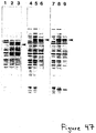
Lane 1 contained the E.coli lysate containing pHCV-49 expressing HCV CKS-NS5I antigen (amino acids 2866-3011) prior to induction andlanes - Induced E.coli lysates containing pHCV-23, pHCV-45, pHCV-48, pHCV-51, pHCV-50, or pHCV-49 were individually run on preparative SDS/PAGE gels to separate the various HCV CKS-NS5 or HCV CKS-BCD recombinant antigens assay from the majority of other E.coli proteins. Gel slices containing the separated individual HCV CKS-NS5 or HCV CKS-BCD recombinant antigens were then electropheretically transferred to nitrocellulose, and the nitrocellulose sheet cut into strips. Figure 40 presents the results of a Western Blot analysis of various serum or plasma samples using these nitrocellulose strips. The arrows on the right indicate the position of each HCV CKS-BCD or HCV CKS-NS5 recombinant antigen, from top to bottom pHCV-23 (HCV CKS-BCD), pHCV-45 (HCV CKS-NS5E), pHCV-48 (HCV CKS-NS5F), pHCV-51 (HCV CKS-NS5G), pHCV-50 (HCV CKS-NS5H), pHCV-49 (HCV CKS-NS5I), and pJO200 (CKS). Panel A contained five normal human plasma, panel B contained five normal human sera, panel C contained twenty human sera positive in the Abbott HCV EIA test, panel D contained two mouse sera directed against CKS, and panel E contained two normal mouse sera. Both the HCV CKS-NS5E antigen expressed by pHCV-45 and the HCV CKS-NS5F antigen expressed by pHCV-48 were immunoreactive when screened with human serum samples containing HCV antibodies.
- Eighteen individual oligonucleotides representing amino acids 1569-1931 of the HCV genome were ligated together and cloned as four separate EcoRI-BamHI subfragments into the CKS fusion vector pJ0200. After subsequent DNA sequences confirmation, the four subfragments were digested with the appropriate restriction enzymes, gel purified, ligated together, and cloned as an 1102 base pair EcoRI-BamHI fragment in the CKS fusion vector pJ0200. The resulting plasmid, designated pHCV-24, expresses the HCV CKS-C100 antigen under control of the lac promoter. The HCV CKS-c100 antigen consists of 239 amino acids of CKS, eight amino acids contributed by linker DNA sequences, 363 amino acids from the HCV NS4 region (amino acids 1569-1931) and 10 additional amino acids contributed by linker DNA sequences. The HCV CKS-c100 antigen was expressed at very low levels by pHCV-24.
- Poor expression levels of this HCV CKS-c100 recombinant antigen were overcome by constructing two additional clones containing deletions in the extreme amino terminal portion of the HCV c100 region. The first of these clones, designated pHCV-57, contains a 23 amino acid deletion (HCV amino acids 1575-1597) and was constructed by deleting a 69 base pair Ddel restriction fragment. The second of these clones, designated pHCV-58, contains a 21 amino acid deletion (HCV amino acids 1600-1620) and was constructed by deleting a 63 base pair Nlalv-Haelll restriction fragment. Figure 41 presents a schematic representation of the recombinant antigens expressed by pHCV-24, pHCV-57, and pHCV-58. Figure 42 presents the DNA and amino acid sequence of the HCV-C100D1 recombinant antigen produced by pHCV-57. Figure 43 presents the DNA and amino acid sequence of the HCV-C100D2 recombinant antigen produced by pHCV-58. Figure 44 presents the expression of pHCV-24, pHCV-57, and pHCV-58 proteins in E.coli.
Lane 1 contained the E.coli lysate containing pHCV-24 expressing the HCV CKS-c100 antigen (amino acids 1569-1931) prior to induction andlanes Lane 4 contained the E.coli lysate containing pHCV-57 expressing the HCV-CKS-C100D1 antigen (amino acids 1569-1574 and 1598-1931) prior to induction andlanes Lane 7 contained the E.coli lysate containing pHCV-58 expressing the HCV CKS-C100D2 antigen (amino acids 1569-1599 and 1621-1931) prior to induction, andlanes - RNA was extracted from the serum of various chimpanzees or humans infected with HCV by first subjecting the samples to digestion with Proteinase K and SDS for 1 hour at 37° centigrade followed by numerous phenol:chloroform extractions. The RNA was then concentrated by several ethanol precipitations and resuspended in water. RNA samples were then reverse transcribed according to supplier's instructions using a specific primer. A second primer was then added and PCR amplification was performed according to supplier's instructions. An aliquot of this PCR reaction was then subjected to an additional round of PCR using nested primers located internal to the first set of primers. In general, these primers also contained restriction endonuclease recognition sequences to be used for subsequent cloning. An aliquot of this second round nested PCR reaction was then subjected to agarose gel electrophoresis and Southern blot analysis to confirm the specificity of the PCR reaction. The remainder of the PCR reaction was then digested with the appropriate restriction enzymes, the HCV DNA fragment of interest gel purified, and ligated to an appropriate cloning vector. This ligation was then transformed into E.coli and single colonies were isolated and plasmid DNA prepared for DNA sequences analysis. The DNA sequences was then evaluated to confirm that the specific HCV coding region of interest was intact. HCV DNA fragments obtained in this manner were then cloned into appropriate vectors for expression analysis.
- Using the methods detailed above, a 474 base pair DNA fragment from the putative NS3 region of HCV was generated by PCR. This fragment represents HCV amino acids #1473-1629 and was cloned into the CKS expression vector pJ0201 by blunt-end ligation. The resulting clone, designated pHCV-105, expresses the HCV CKS-NS3 antigen under control of the lac promoter. The HCV CKS-NS3 antigen consists of 239 amino acids of CKS, 12 amino acids contributed by linker DNA sequences, 157 amino acids from the HCV NS3 region (amino acids 1473-1629), and 9 additional amino acids contributed by linker DNA sequences. Figure 45 presents a schematic representation of the pHCV-105 antigen. Figure 46 presents the DNA and amino acid sequence of the HCV CKS-NS3 recombinant antigen produced by pHCV-105. Figure 47 presents the expression of pHCV-105 proteins in E.coli.
Lane 1 contained the E.coli lysate containing pHCV-105 expressing the HCV CKS-NS3 antigen (amino acids 1472-1629) prior to induction andlanes - Using the methods detailed above, a 489 base pair DNA fragment from the putative envelope region of HCV was generated by PCR. This fragment represents the HCV amino acids 114-276 and was cloned into the CKS expression vector pJ0202 using EcoRI-BamHI restriction sites. The resulting clone, designated pHCV-103, expresses the HCV CKS-5'ENV antigen under control of the lac promoter. The HCV CKS-5'ENV antigen consists of 239 amino acids of CKS, 7 amino acids contributed by linker DNA sequences, 163 amino acids from the HCV envelope region (amino acids 114-276), and 16 additional amino acids contributed by linker DNA sequences. Figure 48 presents a schematic representation of the pHCV-103 antigen. Figure 49 presents the DNA and amino acid sequence of the HCV CKS-5'ENV recombinant antigen produced by pHCV-103. Figure 47 presents the expression of pHCV-103 proteins in E.coli.
Lane 1 contained the E.coli lysate containing pHCV-103 expressing the HCV CKS-5'ENV antigen (amino acids 114-276) prior to induction andlanes - Using the methods detailed above, a 621 base pair DNA fragment form the putative envelope region of HCV was generated by PCR. This fragment represents HCV amino acids 263-469 and was cloned into the CKS expression vector pJ0202 using EcoRI restriction sites. The resulting clone, designated pHCV-101, expresses the HCV CKS-3'ENV antigen under control of the lac promoter. The HCV CKS-3'ENV antigen consists of 239 amino acids of CKS, 7 amino acids contributed by linker DNA sequences, 207 amino acids from the HCV envelope region (amino acids 263-469), and 15 additional amino acids contributed by linker DNA sequences. Figure 50 presents a schematic representation of the pHCV-101 antigen. Figure 51 presents the DNA and amino acid sequence of the HCV CKS-3'ENV recombinant antigen produced by pHCV-101. Figure 47 presents the expression of pHCV-101 proteins in
E.coli Lane 7 contained the E.coli lysate containing pHCV-101 expressing the HCV CKS-3'ENV antigen (amino acids 263-469) prior to induction andlanes - Using the methods detailed above, a 636 base pair DNA fragment from the putative NS2 region of HCV was generated by PCR. This fragment represents the HCV amino acids 994-1205 and was cloned into the CKS expression vector pJ0201 using EcoRI restriction sites. The resulting clone, designated pHCV-102, expresses the HCV CKS-NS2 antigen under control of the lac promoter. The HCV CKS-NS2 antigen consists of 239 amino acids of CKS, 7 amino acids contributed by linker DNA sequences, 212 amino acids from the HCV NS2 region (amino acids 994-1205), and 16 additional amino acids contributed by linker DNA sequences. Figure 52 presents a schematic representation of the pHCV-102 antigen. Figure 53 presents the DNA and amino acid sequence of the HCV CKS-NS2 recombinant antigen produced by pHCV-102. Figure 54 presents the expression of pHCV-102 proteins in E.coli.
Lane 1 contained the E.coli lysate containing pHCV-102 expressing the HCV CKS-NS2 antigen (amino acids 994-1205) prior to induction andlanes - Using the methods detailed above, a 654 base pair DNA fragment from the putative NS1 region of HCV was generated by PCR. This fragment represents HCV amino acids 617-834 and was cloned into the CKS expression vector pJ0200 using EcoRI-BamHI restriction sites. The resulting clone, designated pHCV-107, expresses the HCV CKS-NS1 antigen under control of the lac promoter. The HCV CKS-NS1 antigen consists of 239 amino acids of CKS, 10 amino acids contributed by linker DNA sequences, and 218 amino acids from the HCV NS1 region (amino acids 617-834). Figure 55 presents a schematic representation of the pHCV-107 antigen. Figure 56 presents the DNA and amino acid sequence of the HCV CKS-NS1 recombinant antigen produced by pHCV-107.
- Using the methods detailed above, a 1068 base pair DNA fragment from the putative envelope region of HCV was generated by PCR. This fragment represents HCV amino acids #114-469 and was cloned into the CKS expression vector pJ0202 using EcoRI restriction sites. The resulting clone, designated pHCV-104, expresses the HCV CKS-ENV antigen under control of the lac promoter. The HCV CKS-ENV antigen consists of 239 amino acids of CKS, 7 amino acids contributed by linker DNA sequences, 356 amino acids from the HCV envelope region (amino acids 114-469), add 15 additional amino acids contributed by linker DNA sequences. Figure 57 presents a schematic representation of the pHCV-104 antigen. Figure 58 presents the DNA and amino acid sequence of the HCV CKS-ENV recombinant antigen produced by pHCV-104.
- The recombinant antigens, either alone or in combination, can be used in the assay formats provided herein and exemplified in the Examples. It also is contemplated that these recombinant antigens can be used to develop specific inhibitors of viral replication and used for therapeutic purposes, such as for vaccines. Other applications and modifications of the use of these antigens and the specific embodiments of this inventions as set forth herein, will be apparent to those skilled in the art. Accordingly, the invention is intended to be limited only in accordance with the appended claims.
Claims (15)
- A recombinant fusion protein selected from the group consisting of pHCV-23, pHCV-29, pHCV-31, pHCV-34, pHCV-45, pHCV-48, pHCV-49, pHCV-50, pHCV-51, pHCV-57, pHCV-58, pHCV-101, pHCV-102, pHCV-103, pHCV-104 pHCV-105 and pHCV-107.
- A polypeptide selected from the group consisting of pHCV-23', pHCV-29', pHCV-31', pHCV-34', pHCV-45', pHCV-48', pHCV-49', pHCV-50', pHCV-51', pHCV-57', pHCV-58', pHCV-101', pHCV-102', pHCV-103', pHCV-104', pHCV-105' and pHCV-107'.
- An assay for identifying the presence of an antibody immunologically reactive with an HCV antigen in a fluid sample comprising:
Contacting the sample with at least one polypeptide selected from the group consisting of pHCV-23, pHCV-29, pHCV-31, pHCV-34, pHCV-45, pHCV-48, pHCV-49, pHCV-50, pHCV-51, pHCV-57, pHCV-58, pHCV-101, pHCV-102, pHCV-103, pHCV-104, pHCV-105, pHCV-107, pHCV-45', pHCV-48', pHCV-49', pHCV-50', pHCV-51', pHCV-57', pHCV-58', pHCV-101', pHCV-102, pHCV-103', pHCV-104', pHCV-105', pHCV-107', pHCV-23', pHCV-29', pHCV-31', and pHCV-34' under conditions suitable for complexing the antibody with the polypeptide; and detecting the antibody-polypeptide complex. - The assay of claim 3 wherein the polypeptides are pHCV-31 and pHCV-34 or pHCV-31' and pHCV-34'.
- In a confirmatory assay for identifying the presence of an antibody in a fluid sample immunologically reactive with an HCV antigen wherein the sample is used to prepare first and second immunologically equivalent aliquots and the first aliquot is contacted with at least one polypeptide selected from the group consisting of pHCV-23, pHCV-29, pHCV-31, pHCV-34, pHCV-45, pHCV-48, pHCV-49, pHCV-50, pHCV-51, pHCV-57, pHCV-58, pHCV-101, pHCV-102, pHCV-103, pHCV-104, pHCV-105, pHCV-107, pHCV-45', pHCV-48', pHCV-49' pHCV-50', pHCV-51', pHCV-57', pHCV-58', pHCV-101', pHCV-102', pHCV-103', pHCV-104', pHCV-105', pHCV-107', pHCV-23', pHCV-29', pHCV-31', and pHCV-34' under conditions suitable for complexing the antibody with the polypeptide and wherein the first antibody-antigen complex is detected, and:
contacting the second aliquot with a polypeptide selected from the group consisting of sp65, sp67, sp75, spll7, SOD-33c, pHCV-23', pHCV-29', pHCV-31', pHCV-34', pHCV-45', pHCV-48', pHCV-49', pHCV-50', pHCV-51' pHCV-57', pHCV-58', pHCV-101', pHCV-102', pHCV-103', pHCV-104', pHCV-105', and pHCV-107' under conditions suitable to form a second antibody-antigen complex; and detecting the second antibody-antigen complex; wherein the polypeptide selected in the first aliquot is not the same as the polypeptide selected in the second aliquot. - The assay of claim 5 wherein the first aliquot is contacted with the polypeptides pHCV-31 and pHCV-34 or pHCV-31' and pHCV-34'.
- In an immunodot assay for identifying the presence of an antibody immunologically reactive with an HCV antigen in a fluid sample wherein the sample is concurrently contacted with at least two polypeptides separately bound to distinct regions of the solid support, each containing distinct epitopes of an HCV antigen under conditions suitable for complexing the antibody with the polypeptide; and detecting the antibody-polypeptide complex, and
wherein said polypeptides are selected from the group consisting of pHCV-23, pHCV-29, pHCV-31, pHCV-34, pHCV-23', pHCV-29', pHCV-31', pHCV-34', C100, pHCV-45, pHCV-48, pHCV-49, pHCV-50, pHCV-51, pHCV-57, pHCV-58, pHCV-101, pHCV-102, pHCV-103, pHCV-104, pHCV-105, pHCV-107, pHCV-45', pHCV-48', pHCV-49' pHCV-50', pHCV-51', pHCV-57', pHCV-58', pHCV-101', pHCV-102', pHCV-103', pHCV-104', pHCV-105', and pHCV-107'. - In a competition assay for identifying the presence of an antibody immunologically reactive with an HCV antigen in a fluid sample wherein the sample is used to prepare first and second immunologically equivalent aliquots wherein the first aliquot is contacted with a polypeptide bound to a solid support under conditions suitable for complexing the antibody with the polypeptide to form a detectable antibody-polypeptide complex, and wherein the second aliquot is first contacted with unbound polypeptide and then contacted with said bound polypeptide wherein the polypeptide is selected from the group consisting of pHCV-23, pHCV-29, pHCV-31, pHCV-34, pHCV-23', pHCV-29', pHCV-31', pHCV-34', pHCV-45, pHCV-48, pHCV-49, pHCV-50, pHCV-51, pHCV-57, pHCV-58, pHCV-101, pHCV-102, pHCV-103, pHCV-104, pHCV-105, pHCV-107, pHCV-45', pHCV-48', pHCV-49' pHCV-50', pHCV-51', pHCV-57', pHCV-58', pHCV-101', pHCV-102', pHCV-103', pHCV-104', pHCV-105', and pHCV-107'.
- In a competition assay for identifying the presence of an antibody immunologically reactive with an HCV antigen in a fluid sample wherein the sample is used to prepare first and second immunologically equivalent aliquots wherein the first aliquot is contacted with a polypeptide bound to a solid support under conditions suitable for complexing the antibody with the polypeptide to form a detectable antibody-polypeptide complex and wherein the second aliquot is first contacted with unbound polypeptide and then contacted with said bound polypeptide wherein the polypeptide is selected from the group consisting of pHCV-23, pHCV-29, pHCV-31, pHCV-34, pHCV-23', pHCV-29', pHCV-31', pHCV-34', pHCV-45, pHCV-48, pHCV-49, pHCV-50, pHCV-51, pHCV-57, pHCV-58, pHCV-101, pHCV-102, pHCV-103, pHCV-104, pHCV-105, pHCV-107, pHCV-45', pHCV-48', pHCV-49' pHCV-50', pHCV-51', pHCV-57', pHCV-58', pHCV-101', pHCV-102', pHCV-103', pHCV-104', pHCV-105', and pHCV-107'; wherein the second aliquot is contacted with unbound and bound polypeptide simultaneously.
- In a neutralization assay for identifying the presence of an antibody immunologically reactive with an HCV antigen in a fluid sample wherein the sample is used to prepare first and second immunologically equivalent aliquots wherein the first aliquot is contacted with a polypeptide bound to a solid support under conditions suitable for complexing the antibody with the polypeptide to form a detectable antibody-polypeptide complex wherein the bound polypeptide is selected from the group consisting of pHCV-23, pHCV-29, pHCV-31, pHCV-34, pHCV-23', pHCV-29', pHCV-31', pHCV-34', pHCV-45, pHCV-48, pHCV-49, pHCV-50, pHCV-51, pHCV-57, pHCV-58, pHCV-101, pHCV-102, pHCV-103, pHCV-104, pHCV-105, pHCV-107, pHCV-45', pHCV-48', pHCV-49' pHCV-50', pHCV-51', pHCV-57', pHCV-58', pHCV-101', pHCV-102', pHCV-103', pHCV-104', pHCV-105', and pHCV-107';
and wherein the second aliquot is first contacted with unbound polypeptide and then contacted with said bound polypeptide wherein the unbound polypeptide is selected from the group consisting of pHCV-23, pHCV-29, pHCV-31, pHCV-34, pHCV-23', pHCV-29', pHCV-31', pHCV-34', pHCV-45, pHCV-48, pHCV-49, pHCV-50, pHCV-51, pHCV-57, pHCV-58, pHCV-101, pHCV-102, pHCV-103, pHCV-104, pHCV-105, pHCV-107, pHCV-45', pHCV-48', pHCV-49' pHCV-50', pHCV-51', pHCV-57', pHCV-58', pHCV-101', pHCV-102', pHCV-103', pHCV-104', pHCV-105', and pHCV-107' and wherein the bound polypeptide selected is not the same as the same as the unbound polypeptide selected. - In a neutralization assay for identifying the presence of an antibody immunologically reactive with an HCV antigen in a fluid sample wherein the sample is used to prepare first and second immunologically equivalent aliquots wherein the first aliquot is contacted with a polypeptide bound to a solid support under conditions suitable for complexing the antibody with the polypeptide to form a detectable antibody-polypeptide complex wherein the bound polypeptide is selected from the group consisting of pHCV-23, pHCV-29, pHCV-31, pHCV-34, pHCV-23', pHCV29', pHCV-31', pHCV-34', pHCV-45, pHCV-48, pHCV-49, pHCV-50, pHCV-51, pHCV-57, pHCV-58, pHCV-101, pHCV-102, pHCV-103, pHCV-104, pHCV-105, pHCV-107, pHCV-45', pHCV-48', pHCV-49' pHCV-50', pHCV-51', pHCV-57', pHCV-58', pHCV-101', pHCV-102', pHCV-103', pHCV-104', pHCV-105', and pHCV-107';
and wherein the second aliquot is first contacted with unbound polypeptide and then contacted with said bound polypeptide wherein the unbound polypeptide is selected from the group consisting of pHCV-23, pHCV-29, pHCV-31, pHCV-34, pHCV-23', pHCV-29', pHCV-31', pHCV-34', pHCV-45, pHCV-48, pHCV-49, pHCV-50, pHCV-51, pHCV-57, pHCV-58, pHCV-101, pHCV-102, pHCV-103, pHCV-104, pHCV-105, pHCV-107, pHCV-45', pHCV-48', pHCV-49' pHCV-50', pHCV-51', pHCV-57', pHCV-58', pHCV-101', pHCV-102', pHCV-103', pHCV-104', pHCV-105', and pHCV-107';
and wherein the bound polypeptide selected is not the same as the unbound polypeptide selected;
and wherein the second aliquot is contacted with unbound and bound polypeptide simultaneously. - The assay of claim 11 wherein the polypeptide is pHCV-23 or pHCV-23'.
- An immunoassay kit comprising:
a polypeptide containing at least one HCV antigen selected from the group consisting of pHCV-23, pHCV-29, pHCV-31, pHCV-34, pHCV-23', pHCV-29', pHCV-31', pHCV-34', pHCV-45, pHCV-48, pHCV-49 pHCV-50, pHCV-51, pHCV-57, pHCV-58, pHCV-101, pHCV-102, pHCV-103, pHCV-104, pHCV-105, pHCV-107, pHCV-45', pHCV-48', pHCV-49' pHCV-50', pHCV-51', pHCV-57', pHCV-58', pHCV-101', pHCV-102', pHCV-103', pHCV-104', pHCV-105', and pHCV-107';
one or more sample preparation reagents;
and one or more detection and signal producing reagents. - A kit of claim 13 wherein the polypeptides are bound to a solid support.
- A plasmid selected from the group consisting of pHCV-23, pHCV-29, pHCV-31 and pHCV-34.
Applications Claiming Priority (4)
| Application Number | Priority Date | Filing Date | Title |
|---|---|---|---|
| US57282290A | 1990-08-24 | 1990-08-24 | |
| US572822 | 1990-08-24 | ||
| US61406990A | 1990-11-07 | 1990-11-07 | |
| US614069 | 1990-11-07 |
Publications (3)
| Publication Number | Publication Date |
|---|---|
| EP0472207A2 true EP0472207A2 (en) | 1992-02-26 |
| EP0472207A3 EP0472207A3 (en) | 1992-08-26 |
| EP0472207B1 EP0472207B1 (en) | 1999-10-13 |
Family
ID=27075963
Family Applications (1)
| Application Number | Title | Priority Date | Filing Date |
|---|---|---|---|
| EP91114161A Expired - Lifetime EP0472207B1 (en) | 1990-08-24 | 1991-08-23 | Hepatitis C assay utilizing recombinant antigens |
Country Status (7)
| Country | Link |
|---|---|
| EP (1) | EP0472207B1 (en) |
| JP (1) | JP3354579B2 (en) |
| AT (1) | ATE185605T1 (en) |
| AU (1) | AU655592B2 (en) |
| CA (1) | CA2049679C (en) |
| DE (1) | DE69131707T2 (en) |
| ES (1) | ES2139571T3 (en) |
Cited By (17)
| Publication number | Priority date | Publication date | Assignee | Title |
|---|---|---|---|---|
| EP0475182A2 (en) * | 1990-08-24 | 1992-03-18 | Abbott Laboratories | Method for protein synthesis using CKS fusion proteins |
| WO1993017110A2 (en) * | 1992-02-21 | 1993-09-02 | The Wellcome Foundation Limited | A recombinant hepatitis c virus polypeptide |
| EP0599913A1 (en) * | 1991-08-21 | 1994-06-08 | Abbott Laboratories | Monoclonal antibodies to putative hcv ns5 proteins and methos for using same |
| EP0603307A1 (en) * | 1991-08-21 | 1994-06-29 | Abbott Laboratories | Monoclonal antibodies to putative hcv e2/ns1 proteins and methods for using same |
| US5350671A (en) * | 1987-11-18 | 1994-09-27 | Chiron Corporation | HCV immunoassays employing C domain antigens |
| EP0625204A1 (en) * | 1992-02-04 | 1994-11-23 | Chiron Viagene, Inc. | Hepatitis therapeutics |
| EP0662082A1 (en) * | 1992-09-15 | 1995-07-12 | United Biomedical, Inc. | Novel branched hybrid and cluster peptides effective in diagnosing and detecting non-a, non-b hepatitis |
| WO1997001640A2 (en) * | 1995-06-29 | 1997-01-16 | Smithkline Beecham Biologicals S.A. | Vaccines against hepatitis c |
| US5683864A (en) * | 1987-11-18 | 1997-11-04 | Chiron Corporation | Combinations of hepatitis C virus (HCV) antigens for use in immunoassays for anti-HCV antibodies |
| US5712087A (en) * | 1990-04-04 | 1998-01-27 | Chiron Corporation | Immunoassays for anti-HCV antibodies employing combinations of hepatitis C virus (HCV) antigens |
| US5747241A (en) * | 1991-10-08 | 1998-05-05 | Japan As Represented By Director General Of Agency Of National Institute Of Health | Diagnostic reagent for hepatitis c |
| WO1999061906A2 (en) * | 1998-05-28 | 1999-12-02 | Abbott Laboratories | Toxoplasma gondii antigens, p35, and uses thereof |
| EP0979867A2 (en) * | 1993-11-04 | 2000-02-16 | Innogenetics N.V. | Immunodominant human T-cell epitopes of hepatitis C virus |
| US6150087A (en) * | 1991-06-24 | 2000-11-21 | Chiron Corporation | NANBV diagnostics and vaccines |
| US6221619B1 (en) | 1998-05-28 | 2001-04-24 | Abbott Laboratories | Method of using P35 antigen of toxoplasma gondii in distinguishing acute from chronic toxoplasmosis |
| US6630298B2 (en) * | 2000-06-15 | 2003-10-07 | Chiron Corporation | HCV antigen/antibody combination assay |
| US7166426B2 (en) | 2002-09-09 | 2007-01-23 | Novartis Vaccines And Diagnostics, Inc. | HCV assay |
Families Citing this family (3)
| Publication number | Priority date | Publication date | Assignee | Title |
|---|---|---|---|---|
| ATE167522T1 (en) * | 1991-01-31 | 1998-07-15 | Abbott Lab | MONOCLONAL ANTIBODIES AGAINST POSSIBLE HCV ENVELOPE REGIONS AND METHODS OF USING THEM |
| JP2778886B2 (en) * | 1992-10-16 | 1998-07-23 | エバーニュー バイオテック インコーポレイティド | Hepatitis C virus core antigen protein and diagnostic method and kit using the same |
| JPH06153959A (en) * | 1992-10-16 | 1994-06-03 | Evernew Biotec Inc | Nonstructural protein of hepatitis c virus and diagnostic method and kit using said protein |
Citations (9)
| Publication number | Priority date | Publication date | Assignee | Title |
|---|---|---|---|---|
| EP0253193A2 (en) * | 1986-07-08 | 1988-01-20 | Abbott Laboratories | Method for mutagenesis by oligonucleotide-directed repair of a strand break |
| EP0318216A1 (en) * | 1987-11-18 | 1989-05-31 | Chiron Corporation | NANBV diagnostics and vaccines |
| EP0331961A2 (en) * | 1988-03-11 | 1989-09-13 | Abbott Laboratories | Method for protein synthesis using CKS fusion proteins |
| EP0353590A2 (en) * | 1988-08-02 | 1990-02-07 | Abbott Laboratories | Test card for performing assays |
| EP0388232A1 (en) * | 1989-03-17 | 1990-09-19 | Chiron Corporation | NANBV diagnostics and vaccines |
| EP0442394A2 (en) * | 1990-02-16 | 1991-08-21 | United Biomedical, Inc. | Synthetic peptides specific for the detection of antibodies to HCV, diagnosis of HCV infection and prevention thereof as vaccines |
| EP0445423A2 (en) * | 1989-12-22 | 1991-09-11 | Abbott Laboratories | Hepatitis C assay |
| EP0450931A1 (en) * | 1990-04-04 | 1991-10-09 | Chiron Corporation | Combinations of hepatitis C virus (HCV) antigens for use in immunoassays for anti-HCV antibodies |
| EP0468527A2 (en) * | 1990-07-26 | 1992-01-29 | United Biomedical, Inc. | Synthetic peptides specific for the detection of antibodies to HCV, diagnosis of HCV infection and prevention thereof as vaccines |
Family Cites Families (3)
| Publication number | Priority date | Publication date | Assignee | Title |
|---|---|---|---|---|
| DE4209215A1 (en) * | 1991-07-04 | 1993-01-07 | Boehringer Mannheim Gmbh | HCV PEPTIDE ANTIGEN AND METHOD FOR DETERMINING HCV |
| EP0525910A1 (en) * | 1991-08-02 | 1993-02-03 | Akzo Nobel N.V. | Non-A, non-B peptide |
| ZA927837B (en) * | 1991-10-15 | 1994-03-11 | Akzo Nv | Monoclonal antibodiesto hepatitis C virus |
-
1991
- 1991-08-22 CA CA002049679A patent/CA2049679C/en not_active Expired - Lifetime
- 1991-08-23 AT AT91114161T patent/ATE185605T1/en not_active IP Right Cessation
- 1991-08-23 AU AU82774/91A patent/AU655592B2/en not_active Ceased
- 1991-08-23 DE DE69131707T patent/DE69131707T2/en not_active Expired - Lifetime
- 1991-08-23 ES ES91114161T patent/ES2139571T3/en not_active Expired - Lifetime
- 1991-08-23 EP EP91114161A patent/EP0472207B1/en not_active Expired - Lifetime
- 1991-08-26 JP JP24058791A patent/JP3354579B2/en not_active Expired - Lifetime
Patent Citations (9)
| Publication number | Priority date | Publication date | Assignee | Title |
|---|---|---|---|---|
| EP0253193A2 (en) * | 1986-07-08 | 1988-01-20 | Abbott Laboratories | Method for mutagenesis by oligonucleotide-directed repair of a strand break |
| EP0318216A1 (en) * | 1987-11-18 | 1989-05-31 | Chiron Corporation | NANBV diagnostics and vaccines |
| EP0331961A2 (en) * | 1988-03-11 | 1989-09-13 | Abbott Laboratories | Method for protein synthesis using CKS fusion proteins |
| EP0353590A2 (en) * | 1988-08-02 | 1990-02-07 | Abbott Laboratories | Test card for performing assays |
| EP0388232A1 (en) * | 1989-03-17 | 1990-09-19 | Chiron Corporation | NANBV diagnostics and vaccines |
| EP0445423A2 (en) * | 1989-12-22 | 1991-09-11 | Abbott Laboratories | Hepatitis C assay |
| EP0442394A2 (en) * | 1990-02-16 | 1991-08-21 | United Biomedical, Inc. | Synthetic peptides specific for the detection of antibodies to HCV, diagnosis of HCV infection and prevention thereof as vaccines |
| EP0450931A1 (en) * | 1990-04-04 | 1991-10-09 | Chiron Corporation | Combinations of hepatitis C virus (HCV) antigens for use in immunoassays for anti-HCV antibodies |
| EP0468527A2 (en) * | 1990-07-26 | 1992-01-29 | United Biomedical, Inc. | Synthetic peptides specific for the detection of antibodies to HCV, diagnosis of HCV infection and prevention thereof as vaccines |
Cited By (33)
| Publication number | Priority date | Publication date | Assignee | Title |
|---|---|---|---|---|
| US5683864A (en) * | 1987-11-18 | 1997-11-04 | Chiron Corporation | Combinations of hepatitis C virus (HCV) antigens for use in immunoassays for anti-HCV antibodies |
| US5350671A (en) * | 1987-11-18 | 1994-09-27 | Chiron Corporation | HCV immunoassays employing C domain antigens |
| US6312889B1 (en) | 1990-04-04 | 2001-11-06 | Chiron Corporation | Combinations of hepatitis c virus (HCV) antigens for use in immunoassays for anti-HCV antibodies |
| US5712087A (en) * | 1990-04-04 | 1998-01-27 | Chiron Corporation | Immunoassays for anti-HCV antibodies employing combinations of hepatitis C virus (HCV) antigens |
| EP0475182A3 (en) * | 1990-08-24 | 1992-10-21 | Abbott Laboratories | Method for protein synthesis using cks fusion proteins |
| EP0475182A2 (en) * | 1990-08-24 | 1992-03-18 | Abbott Laboratories | Method for protein synthesis using CKS fusion proteins |
| US6346375B1 (en) | 1991-06-24 | 2002-02-12 | Chiron Corporation | NANBV diagnostics and vaccines |
| US6150087A (en) * | 1991-06-24 | 2000-11-21 | Chiron Corporation | NANBV diagnostics and vaccines |
| EP0599913A1 (en) * | 1991-08-21 | 1994-06-08 | Abbott Laboratories | Monoclonal antibodies to putative hcv ns5 proteins and methos for using same |
| EP0603307A1 (en) * | 1991-08-21 | 1994-06-29 | Abbott Laboratories | Monoclonal antibodies to putative hcv e2/ns1 proteins and methods for using same |
| EP0603307A4 (en) * | 1991-08-21 | 1995-01-18 | Abbott Lab | Monoclonal antibodies to putative hcv e2/ns1 proteins and methods for using same. |
| EP0599913A4 (en) * | 1991-08-21 | 1995-01-18 | Abbott Lab | Monoclonal antibodies to putative hcv ns5 proteins and methos for using same. |
| US5747241A (en) * | 1991-10-08 | 1998-05-05 | Japan As Represented By Director General Of Agency Of National Institute Of Health | Diagnostic reagent for hepatitis c |
| US5750331A (en) * | 1991-10-08 | 1998-05-12 | Japan As Represented By Director General Of Agency Of National Institute Of Health | Diagnostic reagent for hepatitis C |
| EP0625204A1 (en) * | 1992-02-04 | 1994-11-23 | Chiron Viagene, Inc. | Hepatitis therapeutics |
| WO1993017110A3 (en) * | 1992-02-21 | 1993-10-14 | Wellcome Found | A recombinant hepatitis c virus polypeptide |
| WO1993017110A2 (en) * | 1992-02-21 | 1993-09-02 | The Wellcome Foundation Limited | A recombinant hepatitis c virus polypeptide |
| EP0662082A1 (en) * | 1992-09-15 | 1995-07-12 | United Biomedical, Inc. | Novel branched hybrid and cluster peptides effective in diagnosing and detecting non-a, non-b hepatitis |
| EP0662082A4 (en) * | 1992-09-15 | 1997-09-03 | United Biomedical Inc | Novel branched hybrid and cluster peptides effective in diagnosing and detecting non-a, non-b hepatitis. |
| EP0979867A2 (en) * | 1993-11-04 | 2000-02-16 | Innogenetics N.V. | Immunodominant human T-cell epitopes of hepatitis C virus |
| EP0979867A3 (en) * | 1993-11-04 | 2007-06-13 | Innogenetics N.V. | Immunodominant human T-cell epitopes of hepatitis C virus |
| WO1997001640A3 (en) * | 1995-06-29 | 1997-05-15 | Smithkline Beecham Biolog | Vaccines against hepatitis c |
| WO1997001640A2 (en) * | 1995-06-29 | 1997-01-16 | Smithkline Beecham Biologicals S.A. | Vaccines against hepatitis c |
| WO1999061906A2 (en) * | 1998-05-28 | 1999-12-02 | Abbott Laboratories | Toxoplasma gondii antigens, p35, and uses thereof |
| US6329157B1 (en) | 1998-05-28 | 2001-12-11 | Abbott Laboratories | Antigen cocktails and uses thereof |
| US6221619B1 (en) | 1998-05-28 | 2001-04-24 | Abbott Laboratories | Method of using P35 antigen of toxoplasma gondii in distinguishing acute from chronic toxoplasmosis |
| WO1999061906A3 (en) * | 1998-05-28 | 2000-05-04 | Abbott Lab | Toxoplasma gondii antigens, p35, and uses thereof |
| US6630298B2 (en) * | 2000-06-15 | 2003-10-07 | Chiron Corporation | HCV antigen/antibody combination assay |
| US6632601B2 (en) | 2000-06-15 | 2003-10-14 | Chiron Corporation | Immunoassays for anti-HCV antibodies |
| US6797809B2 (en) | 2000-06-15 | 2004-09-28 | Chiron Corporation | Multiple fusion antigens for use in immunoassays for anti-HCV antibodies |
| US7241879B2 (en) | 2000-06-15 | 2007-07-10 | Novartis Vaccines And Diagnostics, Inc. | Immunoassays for anti-HCV antibodies |
| US7319144B2 (en) | 2000-06-15 | 2008-01-15 | Novartis Vaccines And Diagnostics, Inc. | Polynucleotides encoding a multiple epitope fusion antigen for use in an HCV antigen/antibody combination assay |
| US7166426B2 (en) | 2002-09-09 | 2007-01-23 | Novartis Vaccines And Diagnostics, Inc. | HCV assay |
Also Published As
| Publication number | Publication date |
|---|---|
| JPH04281792A (en) | 1992-10-07 |
| ATE185605T1 (en) | 1999-10-15 |
| DE69131707T2 (en) | 2000-06-08 |
| EP0472207A3 (en) | 1992-08-26 |
| JP3354579B2 (en) | 2002-12-09 |
| ES2139571T3 (en) | 2000-02-16 |
| AU655592B2 (en) | 1995-01-05 |
| CA2049679A1 (en) | 1992-02-25 |
| EP0472207B1 (en) | 1999-10-13 |
| DE69131707D1 (en) | 1999-11-18 |
| CA2049679C (en) | 2005-06-21 |
| AU8277491A (en) | 1992-05-07 |
Similar Documents
| Publication | Publication Date | Title |
|---|---|---|
| EP0472207B1 (en) | Hepatitis C assay utilizing recombinant antigens | |
| KR100206150B1 (en) | Combinations of hepatitis c virus(hcv)antigens for use in immunoassays for anti-hcv antibodies | |
| US5683864A (en) | Combinations of hepatitis C virus (HCV) antigens for use in immunoassays for anti-HCV antibodies | |
| US6596476B1 (en) | Hepatitis C assay | |
| AU719929B2 (en) | Multiple epitope fusion protein | |
| US5712087A (en) | Immunoassays for anti-HCV antibodies employing combinations of hepatitis C virus (HCV) antigens | |
| EP0445423B1 (en) | Hepatitis C assay | |
| US6428792B1 (en) | Hepatitis C virus multiple copy epitope fusion antigens | |
| US5645983A (en) | Core antigen protein of hepatitis C virus, and diagnostic method and kit using the same | |
| EP0642666B1 (en) | Hepatitis c assay | |
| EP0600009A4 (en) | Hepatitis c assay utilizing recombinant antigens to ns1. | |
| EP0600018B1 (en) | Hepatitis c assay utilizing recombinant antigens to c-100 region | |
| CA2115925A1 (en) | Hepatitis c assay utilizing recombinant antigens from ns5 region | |
| EP0593291A2 (en) | Nonstructural protein of hepatitis C virus, and diagnostic method and kit using the same | |
| WO1994013700A1 (en) | Peptides from the c33 region of hcv, antibodies thereto and methods for the detection of hcv | |
| WO1998029747A1 (en) | Reagents and methods for detecting hepatitis gb virus antigen |
Legal Events
| Date | Code | Title | Description |
|---|---|---|---|
| PUAI | Public reference made under article 153(3) epc to a published international application that has entered the european phase |
Free format text: ORIGINAL CODE: 0009012 |
|
| AK | Designated contracting states |
Kind code of ref document: A2 Designated state(s): AT BE CH DE DK ES FR GB GR IT LI NL SE |
|
| PUAL | Search report despatched |
Free format text: ORIGINAL CODE: 0009013 |
|
| AK | Designated contracting states |
Kind code of ref document: A3 Designated state(s): AT BE CH DE DK ES FR GB GR IT LI NL SE |
|
| 17P | Request for examination filed |
Effective date: 19930216 |
|
| 17Q | First examination report despatched |
Effective date: 19951019 |
|
| GRAG | Despatch of communication of intention to grant |
Free format text: ORIGINAL CODE: EPIDOS AGRA |
|
| GRAG | Despatch of communication of intention to grant |
Free format text: ORIGINAL CODE: EPIDOS AGRA |
|
| GRAG | Despatch of communication of intention to grant |
Free format text: ORIGINAL CODE: EPIDOS AGRA |
|
| GRAH | Despatch of communication of intention to grant a patent |
Free format text: ORIGINAL CODE: EPIDOS IGRA |
|
| GRAH | Despatch of communication of intention to grant a patent |
Free format text: ORIGINAL CODE: EPIDOS IGRA |
|
| GRAA | (expected) grant |
Free format text: ORIGINAL CODE: 0009210 |
|
| AK | Designated contracting states |
Kind code of ref document: B1 Designated state(s): AT BE CH DE DK ES FR GB GR IT LI NL SE |
|
| PG25 | Lapsed in a contracting state [announced via postgrant information from national office to epo] |
Ref country code: GR Free format text: LAPSE BECAUSE OF NON-PAYMENT OF DUE FEES Effective date: 19991013 Ref country code: SE Free format text: THE PATENT HAS BEEN ANNULLED BY A DECISION OF A NATIONAL AUTHORITY Effective date: 19991013 |
|
| REF | Corresponds to: |
Ref document number: 185605 Country of ref document: AT Date of ref document: 19991015 Kind code of ref document: T |
|
| REG | Reference to a national code |
Ref country code: CH Ref legal event code: EP |
|
| REF | Corresponds to: |
Ref document number: 69131707 Country of ref document: DE Date of ref document: 19991118 |
|
| ITF | It: translation for a ep patent filed |
Owner name: MODIANO & ASSOCIATI S.R.L. |
|
| PG25 | Lapsed in a contracting state [announced via postgrant information from national office to epo] |
Ref country code: DK Free format text: LAPSE BECAUSE OF FAILURE TO SUBMIT A TRANSLATION OF THE DESCRIPTION OR TO PAY THE FEE WITHIN THE PRESCRIBED TIME-LIMIT Effective date: 20000113 |
|
| ET | Fr: translation filed | ||
| REG | Reference to a national code |
Ref country code: CH Ref legal event code: NV Representative=s name: A. BRAUN, BRAUN, HERITIER, ESCHMANN AG PATENTANWAE |
|
| REG | Reference to a national code |
Ref country code: ES Ref legal event code: FG2A Ref document number: 2139571 Country of ref document: ES Kind code of ref document: T3 |
|
| PLBQ | Unpublished change to opponent data |
Free format text: ORIGINAL CODE: EPIDOS OPPO |
|
| PLBI | Opposition filed |
Free format text: ORIGINAL CODE: 0009260 |
|
| PLBF | Reply of patent proprietor to notice(s) of opposition |
Free format text: ORIGINAL CODE: EPIDOS OBSO |
|
| 26 | Opposition filed |
Opponent name: INNOGENETICS NV Effective date: 20000713 |
|
| NLR1 | Nl: opposition has been filed with the epo |
Opponent name: INNOGENETICS NV |
|
| PLBF | Reply of patent proprietor to notice(s) of opposition |
Free format text: ORIGINAL CODE: EPIDOS OBSO |
|
| PLBF | Reply of patent proprietor to notice(s) of opposition |
Free format text: ORIGINAL CODE: EPIDOS OBSO |
|
| PLBF | Reply of patent proprietor to notice(s) of opposition |
Free format text: ORIGINAL CODE: EPIDOS OBSO |
|
| PLBQ | Unpublished change to opponent data |
Free format text: ORIGINAL CODE: EPIDOS OPPO |
|
| PLAB | Opposition data, opponent's data or that of the opponent's representative modified |
Free format text: ORIGINAL CODE: 0009299OPPO |
|
| R26 | Opposition filed (corrected) |
Opponent name: INNOGENETICS NV Effective date: 20000713 |
|
| NLR1 | Nl: opposition has been filed with the epo |
Opponent name: INNOGENETICS NV |
|
| REG | Reference to a national code |
Ref country code: GB Ref legal event code: IF02 |
|
| PLBP | Opposition withdrawn |
Free format text: ORIGINAL CODE: 0009264 |
|
| PLBD | Termination of opposition procedure: decision despatched |
Free format text: ORIGINAL CODE: EPIDOSNOPC1 |
|
| PLBM | Termination of opposition procedure: date of legal effect published |
Free format text: ORIGINAL CODE: 0009276 |
|
| STAA | Information on the status of an ep patent application or granted ep patent |
Free format text: STATUS: OPPOSITION PROCEDURE CLOSED |
|
| 27C | Opposition proceedings terminated |
Effective date: 20031012 |
|
| NLR2 | Nl: decision of opposition |
Effective date: 20031012 |
|
| PGFP | Annual fee paid to national office [announced via postgrant information from national office to epo] |
Ref country code: AT Payment date: 20040707 Year of fee payment: 14 |
|
| PGFP | Annual fee paid to national office [announced via postgrant information from national office to epo] |
Ref country code: NL Payment date: 20040713 Year of fee payment: 14 |
|
| PGFP | Annual fee paid to national office [announced via postgrant information from national office to epo] |
Ref country code: BE Payment date: 20040901 Year of fee payment: 14 |
|
| PGFP | Annual fee paid to national office [announced via postgrant information from national office to epo] |
Ref country code: CH Payment date: 20040914 Year of fee payment: 14 |
|
| PG25 | Lapsed in a contracting state [announced via postgrant information from national office to epo] |
Ref country code: AT Free format text: LAPSE BECAUSE OF NON-PAYMENT OF DUE FEES Effective date: 20050823 |
|
| PG25 | Lapsed in a contracting state [announced via postgrant information from national office to epo] |
Ref country code: CH Free format text: LAPSE BECAUSE OF NON-PAYMENT OF DUE FEES Effective date: 20050831 Ref country code: LI Free format text: LAPSE BECAUSE OF NON-PAYMENT OF DUE FEES Effective date: 20050831 Ref country code: BE Free format text: LAPSE BECAUSE OF NON-PAYMENT OF DUE FEES Effective date: 20050831 |
|
| PG25 | Lapsed in a contracting state [announced via postgrant information from national office to epo] |
Ref country code: NL Free format text: LAPSE BECAUSE OF NON-PAYMENT OF DUE FEES Effective date: 20060301 |
|
| REG | Reference to a national code |
Ref country code: CH Ref legal event code: PL |
|
| NLV4 | Nl: lapsed or anulled due to non-payment of the annual fee |
Effective date: 20060301 |
|
| BERE | Be: lapsed |
Owner name: *ABBOTT LABORATORIES Effective date: 20050831 |
|
| PGFP | Annual fee paid to national office [announced via postgrant information from national office to epo] |
Ref country code: ES Payment date: 20100805 Year of fee payment: 20 |
|
| PGFP | Annual fee paid to national office [announced via postgrant information from national office to epo] |
Ref country code: FR Payment date: 20100819 Year of fee payment: 20 Ref country code: DE Payment date: 20100831 Year of fee payment: 20 Ref country code: IT Payment date: 20100817 Year of fee payment: 20 |
|
| PGFP | Annual fee paid to national office [announced via postgrant information from national office to epo] |
Ref country code: GB Payment date: 20100708 Year of fee payment: 20 |
|
| REG | Reference to a national code |
Ref country code: DE Ref legal event code: R071 Ref document number: 69131707 Country of ref document: DE |
|
| REG | Reference to a national code |
Ref country code: DE Ref legal event code: R071 Ref document number: 69131707 Country of ref document: DE |
|
| PG25 | Lapsed in a contracting state [announced via postgrant information from national office to epo] |
Ref country code: GB Free format text: LAPSE BECAUSE OF EXPIRATION OF PROTECTION Effective date: 20110822 |
|
| REG | Reference to a national code |
Ref country code: ES Ref legal event code: FD2A Effective date: 20120220 |
|
| PG25 | Lapsed in a contracting state [announced via postgrant information from national office to epo] |
Ref country code: ES Free format text: LAPSE BECAUSE OF EXPIRATION OF PROTECTION Effective date: 20110824 |
|
| PG25 | Lapsed in a contracting state [announced via postgrant information from national office to epo] |
Ref country code: DE Free format text: LAPSE BECAUSE OF EXPIRATION OF PROTECTION Effective date: 20110824 |