EP0477020A1 - Polymeric endoscopic ligature - Google Patents
Polymeric endoscopic ligature Download PDFInfo
- Publication number
- EP0477020A1 EP0477020A1 EP91308566A EP91308566A EP0477020A1 EP 0477020 A1 EP0477020 A1 EP 0477020A1 EP 91308566 A EP91308566 A EP 91308566A EP 91308566 A EP91308566 A EP 91308566A EP 0477020 A1 EP0477020 A1 EP 0477020A1
- Authority
- EP
- European Patent Office
- Prior art keywords
- endoscopic
- knot
- ligature
- filament
- fiber
- Prior art date
- Legal status (The legal status is an assumption and is not a legal conclusion. Google has not performed a legal analysis and makes no representation as to the accuracy of the status listed.)
- Granted
Links
- XDTMQSROBMDMFD-UHFFFAOYSA-N C1CCCCC1 Chemical compound C1CCCCC1 XDTMQSROBMDMFD-UHFFFAOYSA-N 0.000 description 2
Images


Classifications
-
- A—HUMAN NECESSITIES
- A61—MEDICAL OR VETERINARY SCIENCE; HYGIENE
- A61L—METHODS OR APPARATUS FOR STERILISING MATERIALS OR OBJECTS IN GENERAL; DISINFECTION, STERILISATION OR DEODORISATION OF AIR; CHEMICAL ASPECTS OF BANDAGES, DRESSINGS, ABSORBENT PADS OR SURGICAL ARTICLES; MATERIALS FOR BANDAGES, DRESSINGS, ABSORBENT PADS OR SURGICAL ARTICLES
- A61L17/00—Materials for surgical sutures or for ligaturing blood vessels ; Materials for prostheses or catheters
- A61L17/04—Non-resorbable materials
-
- A—HUMAN NECESSITIES
- A61—MEDICAL OR VETERINARY SCIENCE; HYGIENE
- A61B—DIAGNOSIS; SURGERY; IDENTIFICATION
- A61B17/00—Surgical instruments, devices or methods, e.g. tourniquets
- A61B17/12—Surgical instruments, devices or methods, e.g. tourniquets for ligaturing or otherwise compressing tubular parts of the body, e.g. blood vessels, umbilical cord
- A61B17/12009—Implements for ligaturing other than by clamps or clips, e.g. using a loop with a slip knot
- A61B17/12013—Implements for ligaturing other than by clamps or clips, e.g. using a loop with a slip knot for use in minimally invasive surgery, e.g. endoscopic surgery
-
- A—HUMAN NECESSITIES
- A61—MEDICAL OR VETERINARY SCIENCE; HYGIENE
- A61L—METHODS OR APPARATUS FOR STERILISING MATERIALS OR OBJECTS IN GENERAL; DISINFECTION, STERILISATION OR DEODORISATION OF AIR; CHEMICAL ASPECTS OF BANDAGES, DRESSINGS, ABSORBENT PADS OR SURGICAL ARTICLES; MATERIALS FOR BANDAGES, DRESSINGS, ABSORBENT PADS OR SURGICAL ARTICLES
- A61L17/00—Materials for surgical sutures or for ligaturing blood vessels ; Materials for prostheses or catheters
- A61L17/06—At least partially resorbable materials
- A61L17/10—At least partially resorbable materials containing macromolecular materials
- A61L17/12—Homopolymers or copolymers of glycolic acid or lactic acid
-
- A—HUMAN NECESSITIES
- A61—MEDICAL OR VETERINARY SCIENCE; HYGIENE
- A61B—DIAGNOSIS; SURGERY; IDENTIFICATION
- A61B17/00—Surgical instruments, devices or methods, e.g. tourniquets
- A61B17/04—Surgical instruments, devices or methods, e.g. tourniquets for suturing wounds; Holders or packages for needles or suture materials
- A61B17/0469—Suturing instruments for use in minimally invasive surgery, e.g. endoscopic surgery
-
- A—HUMAN NECESSITIES
- A61—MEDICAL OR VETERINARY SCIENCE; HYGIENE
- A61B—DIAGNOSIS; SURGERY; IDENTIFICATION
- A61B17/00—Surgical instruments, devices or methods, e.g. tourniquets
- A61B17/04—Surgical instruments, devices or methods, e.g. tourniquets for suturing wounds; Holders or packages for needles or suture materials
- A61B17/06—Needles ; Sutures; Needle-suture combinations; Holders or packages for needles or suture materials
- A61B17/06166—Sutures
-
- A—HUMAN NECESSITIES
- A61—MEDICAL OR VETERINARY SCIENCE; HYGIENE
- A61B—DIAGNOSIS; SURGERY; IDENTIFICATION
- A61B17/00—Surgical instruments, devices or methods, e.g. tourniquets
- A61B17/04—Surgical instruments, devices or methods, e.g. tourniquets for suturing wounds; Holders or packages for needles or suture materials
- A61B17/0469—Suturing instruments for use in minimally invasive surgery, e.g. endoscopic surgery
- A61B2017/0474—Knot pushers
-
- A—HUMAN NECESSITIES
- A61—MEDICAL OR VETERINARY SCIENCE; HYGIENE
- A61B—DIAGNOSIS; SURGERY; IDENTIFICATION
- A61B17/00—Surgical instruments, devices or methods, e.g. tourniquets
- A61B17/04—Surgical instruments, devices or methods, e.g. tourniquets for suturing wounds; Holders or packages for needles or suture materials
- A61B17/0469—Suturing instruments for use in minimally invasive surgery, e.g. endoscopic surgery
- A61B2017/0477—Suturing instruments for use in minimally invasive surgery, e.g. endoscopic surgery with pre-tied sutures
-
- A—HUMAN NECESSITIES
- A61—MEDICAL OR VETERINARY SCIENCE; HYGIENE
- A61B—DIAGNOSIS; SURGERY; IDENTIFICATION
- A61B90/00—Instruments, implements or accessories specially adapted for surgery or diagnosis and not covered by any of the groups A61B1/00 - A61B50/00, e.g. for luxation treatment or for protecting wound edges
- A61B90/03—Automatic limiting or abutting means, e.g. for safety
- A61B2090/037—Automatic limiting or abutting means, e.g. for safety with a frangible part, e.g. by reduced diameter
Definitions
- Endoscopic surgery involves the use of an endoscope, which is an instrument permitting the visual inspection and magnification of any cavity of the body.
- the endoscope is inserted through a cannula after puncture through the wall of the body cavity with a trocar, which is a sharp-pointed instrument.
- the surgeon can then perform diagnostic and therapeutic procedures at the surgical site with the aid of specialized instrumentation designed to fit through additional cannulas providing openings into the desired body cavity as may be required.
- ENDOLOOPTM gut ligature is a device formed from a suture material of surgical catgut.
- the suture material is formed into a ligature loop at the distal end of the device with a knot which becomes secure after the knot is activated.
- ENDOLOOPTM gut ligature facilitates ligation of vessels through small incisions in bodily cavities, the surgical gut from which it is formed may elicit significant tissue reaction. Additionally, the reproducibility of the physical and biological properties of the surgical gut is difficult because it is derived from natural sources.
- the invention is a medical device comprising an endoscopic ligature composed of at least one continuous filament of a synthetic fiber-forming polymer.
- endoscopic ligatures fabricated from synthetic fiber-forming polymers exhibit excellent knot security and minimal knot slippage when ligating a vessel during endoscopic surgery. Additionally, numerous fiber-forming polymers can be used to prepare the devices which elicit minimal tissue reaction and demonstrate a desirable combination of physical and biological properties. These properties, unlike the properties obtained from the conventional gut endoscopic ligatures, are reproducible.
- the medical devices of this invention can be used not only to ligate vessels endoscopically during surgery, but also to perform other desirable surgical techniques when used in combination with other fabricated devices particularly adapted for surgery.
- an embodiment of the medical device of this invention for applying an endoscopic ligature composed of at least one polymeric filament is shown generally as 10.
- the device 10 is comprised of a longitudinal tubular shaft 12, a distal tapered end 14, a score line 16 and a frangible portion 18 located at the proximal end of the device for purposes of gripping.
- Extending from distal end 14 is an endoscopic ligature which has a loop portion 34 and a knot 40.
- the endoscopic ligature as that term is described herein, will be described in greater detail with respect to Figures 4 and 5.
- Knot 40 is restrained by distal end 14 and allows continuous filament 32 to pass through to shaft 12 so that the ligature may be tightened securely about vessel 20.
- a critical feature, one that requires a modified knot configuration for the synthetics, is that the knot must be absolutely “tight" in one direction and should slip in the other. In this manner, once the knot is tightened around the vessel it must remain tight and not loosen.
- a trocar cannula 50 can be positioned within body cavity 60 to receive medical device 10 for endoscopically ligating vessel 20.
- Trocar cannula 50 is affixed to main body portion 52 and is hollow throughout its entire length.
- Figures 4 and 5 show a method of securing a knot arrangement 40 to prepare an endoscopic ligature composed of at least one synthetic polymeric filament.
- a basic knot 30 is wrapped about continuous filament 32.
- This knot 30 is commonly referred to as a #19 knot to those skilled in the art.
- One would begin forming this knot by laying continuous filament 32 out and forming a loop region 34 which terminates at point 36 adjacent knot 30.
- the filament is then continued (see Figure 5) to form two additional throws 38 and 39.
- a sizing gauge is used to properly size loop 34 by adjusting the free end of filament 32.
- Continuous filament 32 is then passed through medical device 10 and proximal end 18 is secured with epoxy. The additional length of continuous filament 32 is then trimmed off.
- Figure 6 shows the prior art endoscopic ligature, which has a basic knot 130, suture material 132 composed of surgical gut, a loop 134 terminating at 136 and a final throw 137 completing the endoscopic ligature.
- this knot configuration exhibits adequate security during ligation with a ligature composed of surgical gut, it is inadequate for securely ligating when a ligature composed of at least one synthetic polymeric filament is used.
- Figure 7 depicts another knot configuration for an endoscopic ligature used to prepare the medical device of this invention. To form this configuration one would start by laying continuous filament 232 out and forming a loop 234. Throw 235 is formed and then a spiral wrap 237 continues downward and is brought up to form two throws 238 and 239 completing the knot shown generally as 240. Other knot configurations which exhibit the desired properties may also be possible.
- FIG. 8 shows a disc or button as an alternative to the endoscopic ligature knot.
- Button 300 has an opening 302 in which one end 336 of the filament 332 is secured by epoxy or any other suitable means. Also located on button 300 is an opening 304 for the filament 332 to pass through and form a loop 334. Button 300 acts similar to knots 40 and 240 to restrict the loops 34 and 234 respectively from passing through medical device 10.
- the synthetic fiber-forming polymer is fabricated into at least one filament which makes up the endoscopic ligature.
- Each filament is continuous, so therefore the filament extends substantially along the entire length of the ligature.
- the filament can be of a monofilament construction or alternatively the endoscopic ligature can be fabricated from a multifilament construction of a plurality of filamentary strands in a braided, twisted or covered form.
- the preferred synthetic fiber-forming polymers exhibit a weight average molecular weight which render the polymer suitable for extrusion into fibers.
- the molecular weight of the polymer as measured by gel permeation chromatography ranges from about 40,000 to about 120,000, preferably from about 60,000 to about 90,000.
- a polymer with a molecular weight below about 40,000 generally lacks sufficient viscosity to provide suitable melt strength for extrusion, and a polymer with a molecular weight above about 120,000 is generally too viscous for melt processing at the temperatures desired to avoid polymer degradation.
- the preferred fiber-forming polymers are polyesters and polyamides.
- the most preferred polyamide is nylon, for example, NUROLON Black Braided Nylon or ETHILON Black Monofilament Nylon.
- polyesters are polyethylene-terephthalate (PET).
- PET polyethylene-terephthalate
- examples of such polyesters include ETHIBOND braided polyester or MERSILENE braided polyester coated with polybutilate.
- the most preferred fiber-forming polymers are bioabsorbable polyesters containing recurring units derived from one or more hydroxy acids or polyalkylene carbonates, e.g. trimethylene carbonate.
- the preferred hydroxy acids include glycolic acid, glycolide, lactide, para-dioxanone and ⁇ -caprolactone.
- the most preferred hydroxy acid based polyesters for fabricating endoscopic ligatures include VICRYL poly(lactide-co-glycolide) and PDS II polydioxanone.
- the synthetic polymer can be fabricated into a filament suitable for the preparation of an endoscopic ligature using conventionally accepted methods well known in the art by first melt extruding the polymer through a spinneret to prepare fibers, drawing the fibers to create orientation, and then annealing the oriented fibers to enhance dimensional stability. Optimum annealing time and temperature for maximum physical and biological properties is readily determined by simple experimentation for each polymer composition.
- An annealed suture strand of each of the following polymeric filaments is obtained: PDSII monofilament polydioxanone, VICRYL braided poly(lactide-co-glycolide) ETHILON monofilament nylon, and NUROLON braided nylon.
- An endoscopic ligature from each of the strands is prepared by first forming a #19 knot (see Fig. 4) and then passing 2 additional throws on each side of the formed loop (see Fig. 5). The final loop size is adjusted by sliding the knot using a loop gauge.
- the straight end of the endoscopic ligature is then inserted into a cannula with the knot in contact with the pointed tip of the cannula.
- Epoxy is injected into the flat end of the cannula to adhere the continuous filamentary strand to the cannula. The excessive length of the strand is trimmed off.
- the finished medical device is then placed in a paper folder and foil package.
- the device is sterilized with ethylene oxide vapor or cobalt radiation, depending on the particular filamentary polymer.
- a knot is tightened on a rubber tube containing liquid using 4 lb. force.
- the liquid in the rubber tube is then pressurized to 5 and 10 psi.
- the liquid leakage through ligating site is monitored at 1 min., 2 hrs. and overnight intervals. The test is repeated 19 times for separately fabricated devices incorporating endoscopic ligatures composed of the specified fiber-forming polymers.
- a knot is tightened on a rubber tube pressurized to 10 lb. force while the knot is submersed in glycerol.
- the test is repeated 9 times for separately fabricated devices incorporating endoscopic ligatures composed of the specified fiber-forming polymers.
Abstract
Description
- As medical and hospital costs continue to increase, surgeons are constantly striving to develop advanced surgical techniques. Advances in the surgical field are often related to the development of operative techniques which involve less invasive surgical procedures and reduce overall patient trauma. In this manner, the length of hospital stays can be significantly reduced, and therefore the hospital and medical costs can be reduced as well.
- One of the truly great advances in recent years to reduce the invasiveness of surgical procedures is endoscopic surgery. Endoscopic surgery involves the use of an endoscope, which is an instrument permitting the visual inspection and magnification of any cavity of the body. The endoscope is inserted through a cannula after puncture through the wall of the body cavity with a trocar, which is a sharp-pointed instrument. The surgeon can then perform diagnostic and therapeutic procedures at the surgical site with the aid of specialized instrumentation designed to fit through additional cannulas providing openings into the desired body cavity as may be required.
- In many surgical procedures, including those involving endoscopic surgery, it is often necessary to ligate blood vessels which have been cut within the surgical site. The vessels may then be severed downstream of the ligated portion. The primary reason for ligating the vessels is to maintain the surgical site free of an excess of blood and to reduce blood loss in the patient.
- In the past, the surgeon closed blood vessels with conventional ligatures, which are long, relatively straight strands of suture material. The surgeon would manually tie the ligature around the vessel desired to be closed. Unfortunately, this is a very time-consuming process, and one certainly not well suited for endoscopic surgical applications where a surgeon's manual operative techniques within the surgical site are severely restricted.
- In more recent years, an endoscopic ligature has been developed especially well adapted for endoscopic surgery. ENDOLOOP™ gut ligature is a device formed from a suture material of surgical catgut. The suture material is formed into a ligature loop at the distal end of the device with a knot which becomes secure after the knot is activated. Although ENDOLOOP™ gut ligature facilitates ligation of vessels through small incisions in bodily cavities, the surgical gut from which it is formed may elicit significant tissue reaction. Additionally, the reproducibility of the physical and biological properties of the surgical gut is difficult because it is derived from natural sources.
- In view of the deficiencies of the prior art for preparing a suitable endoscopic ligature exhibiting minimal tissue reaction and outstanding physical and biological properties, such an endoscopic ligature would be highly desired within the medical community.
- The invention is a medical device comprising an endoscopic ligature composed of at least one continuous filament of a synthetic fiber-forming polymer.
- Surprisingly, endoscopic ligatures fabricated from synthetic fiber-forming polymers exhibit excellent knot security and minimal knot slippage when ligating a vessel during endoscopic surgery. Additionally, numerous fiber-forming polymers can be used to prepare the devices which elicit minimal tissue reaction and demonstrate a desirable combination of physical and biological properties. These properties, unlike the properties obtained from the conventional gut endoscopic ligatures, are reproducible.
- The medical devices of this invention can be used not only to ligate vessels endoscopically during surgery, but also to perform other desirable surgical techniques when used in combination with other fabricated devices particularly adapted for surgery.
-
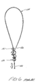
- Figure 1 is a perspective view of one embodiment of the medical device of the present invention incorporating an endoscopic ligature composed of at least one synthetic polymeric filament;
- Figure 2 is a perspective view similar to Figure 1 wherein the device is in the process of ligating a vessel;
- Figure 3 is a side elevational view of the device inserted through a cannula/trocar assembly;
- Figure 4 is a partial perspective view of the first step in forming a ligature knot for an endoscopic ligature composed of at least one synthetic polymeric filament;
- Figure 5 is a perspective view of a completed knot configuration for an endoscopic ligature composed of at least one synthetic polymeric filament in a preferred embodiment of the invention;
- Figure 6 is a perspective view of a knot configuration which is used in the prior art endoscopic ligature composed of surgical gut;
- Figure 7 is a perspective view of an alternate embodiment of a knot configuration for an endoscopic ligature particularly suitable for polymeric filaments;
- Figure 8 is a perspective view of an endoscopic ligature wherein a button or disc is used to form the ligature loop instead of a knot configuration for securely ligating a vessel.
- Referring to Figure 1, an embodiment of the medical device of this invention for applying an endoscopic ligature composed of at least one polymeric filament is shown generally as 10. The
device 10 is comprised of a longitudinaltubular shaft 12, a distaltapered end 14, ascore line 16 and afrangible portion 18 located at the proximal end of the device for purposes of gripping. Extending fromdistal end 14 is an endoscopic ligature which has aloop portion 34 and aknot 40. The endoscopic ligature, as that term is described herein, will be described in greater detail with respect to Figures 4 and 5. - In order to ligate
vessel 20, as seen in Figure 2, the user would first gripfrangible portion 18 with one hand and theshaft 12 with the other hand and snap apart the two pieces aboutscore line 16. This allows for thecontinuous filament 32 to be retracted through thehollow shaft 12.Continuous filament 32 is secured tofrangible portion 18 by means of adhesive, crimping or any other suitable attaching means. Secondly, the user would then placeloop 34 aroundvessel 20, positioning the ligature at the appropriate point on thevessel 20. To complete the procedure,continuous filament 32 is pulled proximally as shown by arrow "A", causing theloop 34 to ligatevessel 20 as shown by arrow "B".Knot 40 is restrained bydistal end 14 and allowscontinuous filament 32 to pass through toshaft 12 so that the ligature may be tightened securely aboutvessel 20. A critical feature, one that requires a modified knot configuration for the synthetics, is that the knot must be absolutely "tight" in one direction and should slip in the other. In this manner, once the knot is tightened around the vessel it must remain tight and not loosen. - Referring now to Figure 3, a trocar cannula 50 can be positioned within
body cavity 60 to receivemedical device 10 for endoscopically ligatingvessel 20. Trocar cannula 50 is affixed tomain body portion 52 and is hollow throughout its entire length. - Figures 4 and 5 show a method of securing a
knot arrangement 40 to prepare an endoscopic ligature composed of at least one synthetic polymeric filament. Abasic knot 30 is wrapped aboutcontinuous filament 32. Thisknot 30 is commonly referred to as a #19 knot to those skilled in the art. One would begin forming this knot by layingcontinuous filament 32 out and forming aloop region 34 which terminates atpoint 36adjacent knot 30. The filament is then continued (see Figure 5) to form twoadditional throws 38 and 39. Once thefinal knot 40 is completed a sizing gauge is used to properlysize loop 34 by adjusting the free end offilament 32.Continuous filament 32 is then passed throughmedical device 10 andproximal end 18 is secured with epoxy. The additional length ofcontinuous filament 32 is then trimmed off. - Figure 6 shows the prior art endoscopic ligature, which has a
basic knot 130,suture material 132 composed of surgical gut, aloop 134 terminating at 136 and afinal throw 137 completing the endoscopic ligature. Although this knot configuration exhibits adequate security during ligation with a ligature composed of surgical gut, it is inadequate for securely ligating when a ligature composed of at least one synthetic polymeric filament is used. - Figure 7 depicts another knot configuration for an endoscopic ligature used to prepare the medical device of this invention. To form this configuration one would start by laying
continuous filament 232 out and forming aloop 234.Throw 235 is formed and then aspiral wrap 237 continues downward and is brought up to form twothrows - Although the formation of various knot configurations is the preferred means for fabricating the endoscopic ligatures used for preparing medical devices of this invention, such knots are in no way the only way in which endoscopic ligatures composed of at least one polymeric filament can be fabricated. For example, figure 8 shows a disc or button as an alternative to the endoscopic ligature knot.
Button 300 has an opening 302 in which oneend 336 of thefilament 332 is secured by epoxy or any other suitable means. Also located onbutton 300 is anopening 304 for thefilament 332 to pass through and form aloop 334.Button 300 acts similar toknots loops medical device 10. - The synthetic fiber-forming polymer is fabricated into at least one filament which makes up the endoscopic ligature. Each filament is continuous, so therefore the filament extends substantially along the entire length of the ligature. The filament can be of a monofilament construction or alternatively the endoscopic ligature can be fabricated from a multifilament construction of a plurality of filamentary strands in a braided, twisted or covered form.
- The preferred synthetic fiber-forming polymers exhibit a weight average molecular weight which render the polymer suitable for extrusion into fibers. Advantageously, the molecular weight of the polymer as measured by gel permeation chromatography ranges from about 40,000 to about 120,000, preferably from about 60,000 to about 90,000. A polymer with a molecular weight below about 40,000 generally lacks sufficient viscosity to provide suitable melt strength for extrusion, and a polymer with a molecular weight above about 120,000 is generally too viscous for melt processing at the temperatures desired to avoid polymer degradation.
- The preferred fiber-forming polymers are polyesters and polyamides. The most preferred polyamide is nylon, for example, NUROLON Black Braided Nylon or ETHILON Black Monofilament Nylon.
- One of the preferred polyesters is polyethylene-terephthalate (PET). Examples of such polyesters include ETHIBOND braided polyester or MERSILENE braided polyester coated with polybutilate.
- The most preferred fiber-forming polymers are bioabsorbable polyesters containing recurring units derived from one or more hydroxy acids or polyalkylene carbonates, e.g. trimethylene carbonate. The preferred hydroxy acids include glycolic acid, glycolide, lactide, para-dioxanone and ε-caprolactone. The most preferred hydroxy acid based polyesters for fabricating endoscopic ligatures include VICRYL poly(lactide-co-glycolide) and PDS II polydioxanone.
- The synthetic polymer can be fabricated into a filament suitable for the preparation of an endoscopic ligature using conventionally accepted methods well known in the art by first melt extruding the polymer through a spinneret to prepare fibers, drawing the fibers to create orientation, and then annealing the oriented fibers to enhance dimensional stability. Optimum annealing time and temperature for maximum physical and biological properties is readily determined by simple experimentation for each polymer composition.
- While several embodiments have been depicted, it will be readily apparent to those skilled in the art that numerous modifications in the design or fabrication of the medical device, or in the selection of the particular synthetic fiber-forming polymer, can be made without departing from the spirit or scope of this invention.
- An annealed suture strand of each of the following polymeric filaments is obtained: PDSII monofilament polydioxanone, VICRYL braided poly(lactide-co-glycolide) ETHILON monofilament nylon, and NUROLON braided nylon. An endoscopic ligature from each of the strands is prepared by first forming a #19 knot (see Fig. 4) and then passing 2 additional throws on each side of the formed loop (see Fig. 5). The final loop size is adjusted by sliding the knot using a loop gauge.
- The straight end of the endoscopic ligature is then inserted into a cannula with the knot in contact with the pointed tip of the cannula. Epoxy is injected into the flat end of the cannula to adhere the continuous filamentary strand to the cannula. The excessive length of the strand is trimmed off.
- The finished medical device is then placed in a paper folder and foil package. The device is sterilized with ethylene oxide vapor or cobalt radiation, depending on the particular filamentary polymer.
- To evaluate the effectiveness of the knot of the endoscopic ligature of the device, the following tests are established:
- A knot is tightened on a rubber tube containing liquid using 4 lb. force. The liquid in the rubber tube is then pressurized to 5 and 10 psi. The liquid leakage through ligating site is monitored at 1 min., 2 hrs. and overnight intervals. The test is repeated 19 times for separately fabricated devices incorporating endoscopic ligatures composed of the specified fiber-forming polymers.
- A knot is tightened on a rubber tube pressurized to 10 lb. force while the knot is submersed in glycerol. The test is repeated 9 times for separately fabricated devices incorporating endoscopic ligatures composed of the specified fiber-forming polymers.
- While the knot is tightened, the cannula is parallel to the tube. The force required to slide a knot and any liquid leakage at 10 psi internal pressure are monitored. The test is repeated 9 times for separately fabricated endoscopic ligatures composed of the specified fiber-forming polymers.
- The following illustrates the testing results.

- The results show that the endoscopic ligatures composed of at least one polymeric filament from which the medical devices of this invention are prepared repeatedly demonstrate outstanding knot security and minimal knot slippage for a series of runs.
Claims (10)
- A medical device comprising an endoscopic ligature composed of at least one continuous filament of a synthetic fiber-forming polymer.
- The device of claim 1 wherein the fiber-forming polymer has a weight average molecular weight between about 40,000 and about 120,000.
- The device of claim 2 wherein the fiber-forming polymer is a polyester or a polyamide.
- The device of claim 3 wherein the polyamide is nylon.
- The device of claim 3 wherein the polyester is PET.
- The device of claim 3 wherein the polyester is a bioabsorbable polyester containing recurring units derived from one or more hydroxy acids or polyalkylene carbonates.
- The device of claim 6 wherein the hydroxy acid is glycolic acid, glycolide, lactide, para-dioxanone or ε-caprolactone.
- The device of claim 6 wherein the polyalkylene carbonate is trimethylene carbonate.
- The device of claim 7 wherein the synthetic fiber-forming polymer is VICRYL poly(lactide-co-glycolide) or PDS II polydioxanone.
- The device of claim 1 wherein the medical device is in a sterile condition.
Applications Claiming Priority (2)
| Application Number | Priority Date | Filing Date | Title |
|---|---|---|---|
| US58575790A | 1990-09-20 | 1990-09-20 | |
| US585757 | 1990-09-20 |
Publications (2)
| Publication Number | Publication Date |
|---|---|
| EP0477020A1 true EP0477020A1 (en) | 1992-03-25 |
| EP0477020B1 EP0477020B1 (en) | 1995-11-22 |
Family
ID=24342824
Family Applications (1)
| Application Number | Title | Priority Date | Filing Date |
|---|---|---|---|
| EP91308566A Expired - Lifetime EP0477020B1 (en) | 1990-09-20 | 1991-09-19 | Polymeric endoscopic ligature |
Country Status (11)
| Country | Link |
|---|---|
| EP (1) | EP0477020B1 (en) |
| JP (1) | JPH04226668A (en) |
| AU (1) | AU8452391A (en) |
| BR (1) | BR9104023A (en) |
| CA (1) | CA2051800A1 (en) |
| DE (1) | DE69114802T2 (en) |
| DK (1) | DK0477020T3 (en) |
| ES (1) | ES2080912T3 (en) |
| GR (1) | GR910100389A (en) |
| IE (1) | IE70599B1 (en) |
| ZA (1) | ZA917486B (en) |
Cited By (20)
| Publication number | Priority date | Publication date | Assignee | Title |
|---|---|---|---|---|
| US5282809A (en) * | 1992-11-16 | 1994-02-01 | Ethicon, Inc. | Endoscopic suturing device |
| EP0621007A1 (en) * | 1993-04-23 | 1994-10-26 | Ethnor | Ligaturing and/or suturing system for endoscopic sugery |
| EP0628286A2 (en) * | 1993-06-07 | 1994-12-14 | Ethicon, Inc. | Endoscopic suturing device |
| US5374278A (en) * | 1989-11-14 | 1994-12-20 | United States Surgical Corporation | Method and apparatus for heat tipping sutures |
| US5403331A (en) * | 1993-03-12 | 1995-04-04 | United States Surgical Corporation | Looped suture ligating device containing a heat-shrinkable element |
| US5437680A (en) * | 1987-05-14 | 1995-08-01 | Yoon; Inbae | Suturing method, apparatus and system for use in endoscopic procedures |
| US5454834A (en) * | 1992-03-12 | 1995-10-03 | Richard Wolf Gmbh | Surgical suture material |
| EP0676211A2 (en) * | 1994-04-11 | 1995-10-11 | Ethicon, Inc. | Process for producing a polyamide suture |
| WO1996022735A1 (en) | 1995-01-25 | 1996-08-01 | Inbae Yoon | Methods and apparatus for suturing tissue |
| US5571120A (en) * | 1992-08-17 | 1996-11-05 | Yoon; Inbae | Ligating instrument and methods of ligating tissue in endoscopic operative procedures |
| US5693059A (en) * | 1995-09-15 | 1997-12-02 | Yoon; Inbae | Ligating instrument with multiple loop ligature supply and methods therefor |
| US5704943A (en) * | 1995-09-25 | 1998-01-06 | Yoon; Inbae | Ligating instrument with multiple loop ligature supply and methods therefor |
| US5908429A (en) * | 1997-05-01 | 1999-06-01 | Yoon; Inbae | Methods of anatomical tissue ligation |
| US5921993A (en) * | 1997-05-01 | 1999-07-13 | Yoon; Inbae | Methods of endoscopic tubal ligation |
| US5957936A (en) * | 1997-05-01 | 1999-09-28 | Inbae Yoon | Instrument assemblies for performing anatomical tissue ligation |
| WO2000074745A1 (en) * | 1999-06-09 | 2000-12-14 | Scimed Life Systems, Inc. | Drug releasing elastic band and method |
| US6551335B1 (en) * | 1997-07-11 | 2003-04-22 | Astra Tech Ab | Methods and devices for stripping blood vessels |
| WO2005115257A1 (en) * | 2004-05-25 | 2005-12-08 | Boston Scientific Limited | Medical retrieval devices |
| US7951157B2 (en) | 2000-05-19 | 2011-05-31 | C.R. Bard, Inc. | Tissue capturing and suturing device and method |
| WO2013100671A3 (en) * | 2011-12-28 | 2013-08-22 | Kim Jin Sung | Endoloop having a screw-coupled rear end portion |
Families Citing this family (4)
| Publication number | Priority date | Publication date | Assignee | Title |
|---|---|---|---|---|
| WO1993019679A1 (en) | 1992-04-07 | 1993-10-14 | The Johns Hopkins University | A percutaneous mechanical fragmentation catheter system |
| US6235869B1 (en) * | 1998-10-20 | 2001-05-22 | United States Surgical Corporation | Absorbable polymers and surgical articles fabricated therefrom |
| CA2525275C (en) | 2003-05-16 | 2012-02-07 | C.R. Bard, Inc. | Single intubation, multi-stitch endoscopic suturing system |
| JP5034045B2 (en) * | 2006-11-30 | 2012-09-26 | 国立大学法人 東京大学 | Non-human animal model for intracranial vascular injury and method for producing the same |
Citations (11)
| Publication number | Priority date | Publication date | Assignee | Title |
|---|---|---|---|---|
| US1856721A (en) * | 1931-02-07 | 1932-05-03 | Clemens B Nagelmann | Suturing and ligating instrument |
| FR736756A (en) * | 1931-05-23 | 1932-11-28 | Method and device for tying surgical threads | |
| US3476115A (en) * | 1967-01-10 | 1969-11-04 | Amp Inc | Ligating implement with ligature severing means |
| GB2033411A (en) * | 1978-11-13 | 1980-05-21 | American Cyanamid Co | Copolymers and surgical articles formed therefrom |
| DE2900265A1 (en) * | 1979-01-04 | 1980-07-17 | Fritz Dr Sammer | Surgical probe loop - has loop filament wound into self-locking coils (OE 15.8.79) |
| US4300565A (en) * | 1977-05-23 | 1981-11-17 | American Cyanamid Company | Synthetic polyester surgical articles |
| EP0132131A1 (en) * | 1983-07-15 | 1985-01-23 | Ethicon, Inc. | Surgical filaments from block copolyetheramides |
| WO1986000020A1 (en) * | 1984-06-14 | 1986-01-03 | Bioresearch Inc. | Composite surgical sutures |
| US4735194A (en) * | 1987-01-13 | 1988-04-05 | University Patents, Inc. | Flexible endoscopic ligating instrument |
| US4838267A (en) * | 1988-02-12 | 1989-06-13 | Ethicon, Inc. | Glycolide/p-dioxanone block copolymers |
| WO1990006725A1 (en) * | 1988-12-12 | 1990-06-28 | Ethicon, Inc. | Ligature system for use in endoscopic surgery, ligature and handling instrument for said system |
-
1991
- 1991-09-16 AU AU84523/91A patent/AU8452391A/en not_active Abandoned
- 1991-09-18 CA CA002051800A patent/CA2051800A1/en not_active Abandoned
- 1991-09-18 JP JP3265438A patent/JPH04226668A/en active Pending
- 1991-09-19 DK DK91308566.8T patent/DK0477020T3/en active
- 1991-09-19 EP EP91308566A patent/EP0477020B1/en not_active Expired - Lifetime
- 1991-09-19 GR GR910100389A patent/GR910100389A/en unknown
- 1991-09-19 IE IE330191A patent/IE70599B1/en not_active IP Right Cessation
- 1991-09-19 ES ES91308566T patent/ES2080912T3/en not_active Expired - Lifetime
- 1991-09-19 DE DE69114802T patent/DE69114802T2/en not_active Expired - Fee Related
- 1991-09-19 BR BR919104023A patent/BR9104023A/en not_active Application Discontinuation
- 1991-09-19 ZA ZA917486A patent/ZA917486B/en unknown
Patent Citations (12)
| Publication number | Priority date | Publication date | Assignee | Title |
|---|---|---|---|---|
| US1856721A (en) * | 1931-02-07 | 1932-05-03 | Clemens B Nagelmann | Suturing and ligating instrument |
| FR736756A (en) * | 1931-05-23 | 1932-11-28 | Method and device for tying surgical threads | |
| US3476115A (en) * | 1967-01-10 | 1969-11-04 | Amp Inc | Ligating implement with ligature severing means |
| US4300565A (en) * | 1977-05-23 | 1981-11-17 | American Cyanamid Company | Synthetic polyester surgical articles |
| GB2033411A (en) * | 1978-11-13 | 1980-05-21 | American Cyanamid Co | Copolymers and surgical articles formed therefrom |
| DE2900265A1 (en) * | 1979-01-04 | 1980-07-17 | Fritz Dr Sammer | Surgical probe loop - has loop filament wound into self-locking coils (OE 15.8.79) |
| EP0132131A1 (en) * | 1983-07-15 | 1985-01-23 | Ethicon, Inc. | Surgical filaments from block copolyetheramides |
| WO1986000020A1 (en) * | 1984-06-14 | 1986-01-03 | Bioresearch Inc. | Composite surgical sutures |
| US4735194A (en) * | 1987-01-13 | 1988-04-05 | University Patents, Inc. | Flexible endoscopic ligating instrument |
| US4735194C1 (en) * | 1987-01-13 | 2001-05-08 | Dept Of Veterans Affairs The U | Flexile endoscopic ligating instrument |
| US4838267A (en) * | 1988-02-12 | 1989-06-13 | Ethicon, Inc. | Glycolide/p-dioxanone block copolymers |
| WO1990006725A1 (en) * | 1988-12-12 | 1990-06-28 | Ethicon, Inc. | Ligature system for use in endoscopic surgery, ligature and handling instrument for said system |
Cited By (32)
| Publication number | Priority date | Publication date | Assignee | Title |
|---|---|---|---|---|
| US5437680A (en) * | 1987-05-14 | 1995-08-01 | Yoon; Inbae | Suturing method, apparatus and system for use in endoscopic procedures |
| US5374278A (en) * | 1989-11-14 | 1994-12-20 | United States Surgical Corporation | Method and apparatus for heat tipping sutures |
| US5454834A (en) * | 1992-03-12 | 1995-10-03 | Richard Wolf Gmbh | Surgical suture material |
| US5571120A (en) * | 1992-08-17 | 1996-11-05 | Yoon; Inbae | Ligating instrument and methods of ligating tissue in endoscopic operative procedures |
| US5282809A (en) * | 1992-11-16 | 1994-02-01 | Ethicon, Inc. | Endoscopic suturing device |
| AU670461B2 (en) * | 1992-11-16 | 1996-07-18 | Ethicon Inc. | Endoscopic suturing device |
| US5403331A (en) * | 1993-03-12 | 1995-04-04 | United States Surgical Corporation | Looped suture ligating device containing a heat-shrinkable element |
| FR2704132A1 (en) * | 1993-04-23 | 1994-10-28 | Ethnor | System for ligature and / or suture for endoscopic surgery. |
| EP0621007A1 (en) * | 1993-04-23 | 1994-10-26 | Ethnor | Ligaturing and/or suturing system for endoscopic sugery |
| US5466241A (en) * | 1993-04-23 | 1995-11-14 | Leroy; Joel | Ligature and/or suture system for endoscopic surgery |
| EP0628286A3 (en) * | 1993-06-07 | 1995-09-13 | Ethicon Inc | Endoscopic suturing device. |
| EP0628286A2 (en) * | 1993-06-07 | 1994-12-14 | Ethicon, Inc. | Endoscopic suturing device |
| EP0676211A2 (en) * | 1994-04-11 | 1995-10-11 | Ethicon, Inc. | Process for producing a polyamide suture |
| EP0676211A3 (en) * | 1994-04-11 | 1996-07-31 | Ethicon Inc | Process for producing a polyamide suture. |
| US5843574A (en) * | 1994-04-11 | 1998-12-01 | Ethicon, Inc. | Polyamide suture having improved tensile strength |
| WO1996022735A1 (en) | 1995-01-25 | 1996-08-01 | Inbae Yoon | Methods and apparatus for suturing tissue |
| US5693059A (en) * | 1995-09-15 | 1997-12-02 | Yoon; Inbae | Ligating instrument with multiple loop ligature supply and methods therefor |
| US5704943A (en) * | 1995-09-25 | 1998-01-06 | Yoon; Inbae | Ligating instrument with multiple loop ligature supply and methods therefor |
| US5810845A (en) * | 1995-09-25 | 1998-09-22 | Yoon; Inbae | Ligating instrument with multiple loop ligature supply and methods therefor |
| US5908429A (en) * | 1997-05-01 | 1999-06-01 | Yoon; Inbae | Methods of anatomical tissue ligation |
| US5921993A (en) * | 1997-05-01 | 1999-07-13 | Yoon; Inbae | Methods of endoscopic tubal ligation |
| US5957936A (en) * | 1997-05-01 | 1999-09-28 | Inbae Yoon | Instrument assemblies for performing anatomical tissue ligation |
| US6551335B1 (en) * | 1997-07-11 | 2003-04-22 | Astra Tech Ab | Methods and devices for stripping blood vessels |
| US6471987B1 (en) | 1999-06-09 | 2002-10-29 | Scimed Life Systems, Inc. | Drug releasing elastic band and method |
| WO2000074745A1 (en) * | 1999-06-09 | 2000-12-14 | Scimed Life Systems, Inc. | Drug releasing elastic band and method |
| EP1462132A1 (en) * | 1999-06-09 | 2004-09-29 | Boston Scientific Limited | Drug releasing elastic band and method |
| EP1649881A1 (en) * | 1999-06-09 | 2006-04-26 | Boston Scientific Limited | Drug releasing elastic band and method |
| US7252838B2 (en) | 1999-06-09 | 2007-08-07 | Boston Scientific Scimed, Inc. | Drug releasing elastic band and method |
| US7951157B2 (en) | 2000-05-19 | 2011-05-31 | C.R. Bard, Inc. | Tissue capturing and suturing device and method |
| WO2005115257A1 (en) * | 2004-05-25 | 2005-12-08 | Boston Scientific Limited | Medical retrieval devices |
| US7491211B2 (en) | 2004-05-25 | 2009-02-17 | Boston Scientific Scimed, Inc. | Medical retrieval devices |
| WO2013100671A3 (en) * | 2011-12-28 | 2013-08-22 | Kim Jin Sung | Endoloop having a screw-coupled rear end portion |
Also Published As
| Publication number | Publication date |
|---|---|
| DE69114802D1 (en) | 1996-01-04 |
| IE70599B1 (en) | 1996-12-11 |
| DE69114802T2 (en) | 1996-04-25 |
| BR9104023A (en) | 1992-06-02 |
| ZA917486B (en) | 1993-03-19 |
| AU8452391A (en) | 1992-03-26 |
| JPH04226668A (en) | 1992-08-17 |
| ES2080912T3 (en) | 1996-02-16 |
| EP0477020B1 (en) | 1995-11-22 |
| GR910100389A (en) | 1992-09-11 |
| CA2051800A1 (en) | 1992-03-21 |
| DK0477020T3 (en) | 1995-12-18 |
| IE913301A1 (en) | 1992-02-25 |
Similar Documents
| Publication | Publication Date | Title |
|---|---|---|
| EP0477020A1 (en) | Polymeric endoscopic ligature | |
| US5144961A (en) | Endoscopic ligating device | |
| AU671666B2 (en) | Endoscopic knotting device | |
| US8864776B2 (en) | Deployment system for surgical suture | |
| JP5558540B2 (en) | Barbed suture | |
| EP0559429B1 (en) | Suture-needle combination | |
| JP4493501B2 (en) | Back-tipped suture | |
| JP5710679B2 (en) | Barbed construct for barbed suture | |
| US7147651B2 (en) | Stiff tipped suture | |
| EP3934547A1 (en) | Knotless sutures including integrated closures | |
| US11058414B1 (en) | Methods of tissue repair with direct pass cinch | |
| AU2021364390A1 (en) | Suturing device |
Legal Events
| Date | Code | Title | Description |
|---|---|---|---|
| PUAI | Public reference made under article 153(3) epc to a published international application that has entered the european phase |
Free format text: ORIGINAL CODE: 0009012 |
|
| AK | Designated contracting states |
Kind code of ref document: A1 Designated state(s): BE DE DK ES FR GB IT LU NL SE |
|
| 17P | Request for examination filed |
Effective date: 19920902 |
|
| 17Q | First examination report despatched |
Effective date: 19931004 |
|
| GRAA | (expected) grant |
Free format text: ORIGINAL CODE: 0009210 |
|
| AK | Designated contracting states |
Kind code of ref document: B1 Designated state(s): BE DE DK ES FR GB IT LU NL SE |
|
| REG | Reference to a national code |
Ref country code: DK Ref legal event code: T3 |
|
| REF | Corresponds to: |
Ref document number: 69114802 Country of ref document: DE Date of ref document: 19960104 |
|
| ET | Fr: translation filed | ||
| REG | Reference to a national code |
Ref country code: ES Ref legal event code: FG2A Ref document number: 2080912 Country of ref document: ES Kind code of ref document: T3 |
|
| ITF | It: translation for a ep patent filed |
Owner name: SOCIETA' ITALIANA BREVETTI S.P.A. |
|
| PLBE | No opposition filed within time limit |
Free format text: ORIGINAL CODE: 0009261 |
|
| STAA | Information on the status of an ep patent application or granted ep patent |
Free format text: STATUS: NO OPPOSITION FILED WITHIN TIME LIMIT |
|
| 26N | No opposition filed | ||
| REG | Reference to a national code |
Ref country code: GB Ref legal event code: IF02 |
|
| PGFP | Annual fee paid to national office [announced via postgrant information from national office to epo] |
Ref country code: DK Payment date: 20080912 Year of fee payment: 18 |
|
| PGFP | Annual fee paid to national office [announced via postgrant information from national office to epo] |
Ref country code: IT Payment date: 20080926 Year of fee payment: 18 Ref country code: NL Payment date: 20080903 Year of fee payment: 18 Ref country code: FR Payment date: 20080915 Year of fee payment: 18 |
|
| PGFP | Annual fee paid to national office [announced via postgrant information from national office to epo] |
Ref country code: GB Payment date: 20080924 Year of fee payment: 18 |
|
| PGFP | Annual fee paid to national office [announced via postgrant information from national office to epo] |
Ref country code: LU Payment date: 20081003 Year of fee payment: 18 Ref country code: DE Payment date: 20081002 Year of fee payment: 18 |
|
| PGFP | Annual fee paid to national office [announced via postgrant information from national office to epo] |
Ref country code: SE Payment date: 20080908 Year of fee payment: 18 Ref country code: ES Payment date: 20081021 Year of fee payment: 18 Ref country code: BE Payment date: 20080922 Year of fee payment: 18 |
|
| BERE | Be: lapsed |
Owner name: *ETHICON INC. Effective date: 20090930 |
|
| REG | Reference to a national code |
Ref country code: NL Ref legal event code: V1 Effective date: 20100401 |
|
| EUG | Se: european patent has lapsed | ||
| REG | Reference to a national code |
Ref country code: DK Ref legal event code: EBP |
|
| GBPC | Gb: european patent ceased through non-payment of renewal fee |
Effective date: 20090919 |
|
| REG | Reference to a national code |
Ref country code: FR Ref legal event code: ST Effective date: 20100531 |
|
| PG25 | Lapsed in a contracting state [announced via postgrant information from national office to epo] |
Ref country code: NL Free format text: LAPSE BECAUSE OF NON-PAYMENT OF DUE FEES Effective date: 20100401 Ref country code: FR Free format text: LAPSE BECAUSE OF NON-PAYMENT OF DUE FEES Effective date: 20090930 Ref country code: DE Free format text: LAPSE BECAUSE OF NON-PAYMENT OF DUE FEES Effective date: 20100401 |
|
| PG25 | Lapsed in a contracting state [announced via postgrant information from national office to epo] |
Ref country code: BE Free format text: LAPSE BECAUSE OF NON-PAYMENT OF DUE FEES Effective date: 20090930 |
|
| PG25 | Lapsed in a contracting state [announced via postgrant information from national office to epo] |
Ref country code: GB Free format text: LAPSE BECAUSE OF NON-PAYMENT OF DUE FEES Effective date: 20090919 |
|
| PG25 | Lapsed in a contracting state [announced via postgrant information from national office to epo] |
Ref country code: DK Free format text: LAPSE BECAUSE OF NON-PAYMENT OF DUE FEES Effective date: 20090930 |
|
| PG25 | Lapsed in a contracting state [announced via postgrant information from national office to epo] |
Ref country code: IT Free format text: LAPSE BECAUSE OF NON-PAYMENT OF DUE FEES Effective date: 20090919 |
|
| PG25 | Lapsed in a contracting state [announced via postgrant information from national office to epo] |
Ref country code: LU Free format text: LAPSE BECAUSE OF NON-PAYMENT OF DUE FEES Effective date: 20090919 |
|
| PG25 | Lapsed in a contracting state [announced via postgrant information from national office to epo] |
Ref country code: SE Free format text: LAPSE BECAUSE OF NON-PAYMENT OF DUE FEES Effective date: 20090920 |
|
| REG | Reference to a national code |
Ref country code: ES Ref legal event code: FD2A Effective date: 20110708 |
|
| PG25 | Lapsed in a contracting state [announced via postgrant information from national office to epo] |
Ref country code: ES Free format text: LAPSE BECAUSE OF NON-PAYMENT OF DUE FEES Effective date: 20110628 |
|
| PG25 | Lapsed in a contracting state [announced via postgrant information from national office to epo] |
Ref country code: ES Free format text: LAPSE BECAUSE OF NON-PAYMENT OF DUE FEES Effective date: 20090920 |