EP0502814A2 - Compositions for increasing the image contrast in imaging of the digestive tract of patients - Google Patents
Compositions for increasing the image contrast in imaging of the digestive tract of patients Download PDFInfo
- Publication number
- EP0502814A2 EP0502814A2 EP92810078A EP92810078A EP0502814A2 EP 0502814 A2 EP0502814 A2 EP 0502814A2 EP 92810078 A EP92810078 A EP 92810078A EP 92810078 A EP92810078 A EP 92810078A EP 0502814 A2 EP0502814 A2 EP 0502814A2
- Authority
- EP
- European Patent Office
- Prior art keywords
- compositions
- particles
- carrier
- carrier phase
- water
- Prior art date
- Legal status (The legal status is an assumption and is not a legal conclusion. Google has not performed a legal analysis and makes no representation as to the accuracy of the status listed.)
- Granted
Links
- 239000000203 mixture Substances 0.000 title claims abstract description 65
- 210000001035 gastrointestinal tract Anatomy 0.000 title claims abstract description 32
- 238000003384 imaging method Methods 0.000 title claims description 25
- 239000006249 magnetic particle Substances 0.000 claims abstract description 25
- 238000000034 method Methods 0.000 claims abstract description 18
- 239000000227 bioadhesive Substances 0.000 claims abstract description 16
- 238000012800 visualization Methods 0.000 claims abstract description 5
- 239000002245 particle Substances 0.000 claims description 52
- 229920000642 polymer Polymers 0.000 claims description 38
- XLYOFNOQVPJJNP-UHFFFAOYSA-N water Substances O XLYOFNOQVPJJNP-UHFFFAOYSA-N 0.000 claims description 36
- SZVJSHCCFOBDDC-UHFFFAOYSA-N iron(II,III) oxide Inorganic materials O=[Fe]O[Fe]O[Fe]=O SZVJSHCCFOBDDC-UHFFFAOYSA-N 0.000 claims description 21
- SMZOUWXMTYCWNB-UHFFFAOYSA-N 2-(2-methoxy-5-methylphenyl)ethanamine Chemical compound COC1=CC=C(C)C=C1CCN SMZOUWXMTYCWNB-UHFFFAOYSA-N 0.000 claims description 13
- NIXOWILDQLNWCW-UHFFFAOYSA-N 2-Propenoic acid Natural products OC(=O)C=C NIXOWILDQLNWCW-UHFFFAOYSA-N 0.000 claims description 13
- 239000002872 contrast media Substances 0.000 claims description 13
- 229920002307 Dextran Polymers 0.000 claims description 11
- 230000002708 enhancing effect Effects 0.000 claims description 11
- 241001465754 Metazoa Species 0.000 claims description 10
- 229920002125 Sokalan® Polymers 0.000 claims description 10
- GUJOJGAPFQRJSV-UHFFFAOYSA-N dialuminum;dioxosilane;oxygen(2-);hydrate Chemical compound O.[O-2].[O-2].[O-2].[Al+3].[Al+3].O=[Si]=O.O=[Si]=O.O=[Si]=O.O=[Si]=O GUJOJGAPFQRJSV-UHFFFAOYSA-N 0.000 claims description 10
- 239000000463 material Substances 0.000 claims description 10
- 239000007864 aqueous solution Substances 0.000 claims description 9
- 239000006185 dispersion Substances 0.000 claims description 9
- 239000003795 chemical substances by application Substances 0.000 claims description 8
- 229920001577 copolymer Polymers 0.000 claims description 8
- 230000004044 response Effects 0.000 claims description 8
- 238000000576 coating method Methods 0.000 claims description 7
- 239000012528 membrane Substances 0.000 claims description 7
- 239000000178 monomer Substances 0.000 claims description 7
- 210000004877 mucosa Anatomy 0.000 claims description 7
- 229920003169 water-soluble polymer Polymers 0.000 claims description 7
- 229920002134 Carboxymethyl cellulose Polymers 0.000 claims description 6
- 239000003513 alkali Substances 0.000 claims description 6
- 150000001875 compounds Chemical class 0.000 claims description 6
- 239000004005 microsphere Substances 0.000 claims description 6
- 229910052901 montmorillonite Inorganic materials 0.000 claims description 6
- 230000000694 effects Effects 0.000 claims description 5
- 230000002496 gastric effect Effects 0.000 claims description 5
- 239000000758 substrate Substances 0.000 claims description 5
- 150000001720 carbohydrates Chemical class 0.000 claims description 4
- 235000014633 carbohydrates Nutrition 0.000 claims description 4
- 239000011248 coating agent Substances 0.000 claims description 4
- 229910052500 inorganic mineral Inorganic materials 0.000 claims description 4
- 238000004519 manufacturing process Methods 0.000 claims description 4
- 239000011707 mineral Substances 0.000 claims description 4
- 239000001267 polyvinylpyrrolidone Substances 0.000 claims description 4
- 235000013855 polyvinylpyrrolidone Nutrition 0.000 claims description 4
- 150000003839 salts Chemical class 0.000 claims description 4
- 229960004291 sucralfate Drugs 0.000 claims description 4
- 239000012736 aqueous medium Substances 0.000 claims description 3
- 229960000892 attapulgite Drugs 0.000 claims description 3
- 230000015572 biosynthetic process Effects 0.000 claims description 3
- 150000004676 glycans Chemical class 0.000 claims description 3
- 230000033001 locomotion Effects 0.000 claims description 3
- 229920000620 organic polymer Polymers 0.000 claims description 3
- 229910052625 palygorskite Inorganic materials 0.000 claims description 3
- 229920001282 polysaccharide Polymers 0.000 claims description 3
- 239000005017 polysaccharide Substances 0.000 claims description 3
- 239000000126 substance Substances 0.000 claims description 3
- QAOWNCQODCNURD-UHFFFAOYSA-L sulfate group Chemical group S(=O)(=O)([O-])[O-] QAOWNCQODCNURD-UHFFFAOYSA-L 0.000 claims description 3
- FYYHWMGAXLPEAU-UHFFFAOYSA-N Magnesium Chemical compound [Mg] FYYHWMGAXLPEAU-UHFFFAOYSA-N 0.000 claims description 2
- 229910019142 PO4 Inorganic materials 0.000 claims description 2
- 235000010443 alginic acid Nutrition 0.000 claims description 2
- 229920000615 alginic acid Polymers 0.000 claims description 2
- 238000010790 dilution Methods 0.000 claims description 2
- 239000012895 dilution Substances 0.000 claims description 2
- 230000036571 hydration Effects 0.000 claims description 2
- 238000006703 hydration reaction Methods 0.000 claims description 2
- 229910052749 magnesium Inorganic materials 0.000 claims description 2
- 239000011777 magnesium Substances 0.000 claims description 2
- 230000014759 maintenance of location Effects 0.000 claims description 2
- 229910044991 metal oxide Inorganic materials 0.000 claims description 2
- 150000004706 metal oxides Chemical class 0.000 claims description 2
- 239000001814 pectin Substances 0.000 claims description 2
- 229920001277 pectin Polymers 0.000 claims description 2
- 235000010987 pectin Nutrition 0.000 claims description 2
- NBIIXXVUZAFLBC-UHFFFAOYSA-K phosphate Chemical compound [O-]P([O-])([O-])=O NBIIXXVUZAFLBC-UHFFFAOYSA-K 0.000 claims description 2
- 239000010452 phosphate Substances 0.000 claims description 2
- UEZVMMHDMIWARA-UHFFFAOYSA-M phosphonate Chemical compound [O-]P(=O)=O UEZVMMHDMIWARA-UHFFFAOYSA-M 0.000 claims description 2
- BDHFUVZGWQCTTF-UHFFFAOYSA-M sulfonate Chemical compound [O-]S(=O)=O BDHFUVZGWQCTTF-UHFFFAOYSA-M 0.000 claims description 2
- 125000001273 sulfonato group Chemical group [O-]S(*)(=O)=O 0.000 claims description 2
- 229910052784 alkaline earth metal Inorganic materials 0.000 claims 3
- 150000001342 alkaline earth metals Chemical class 0.000 claims 2
- FHVDTGUDJYJELY-UHFFFAOYSA-N 6-{[2-carboxy-4,5-dihydroxy-6-(phosphanyloxy)oxan-3-yl]oxy}-4,5-dihydroxy-3-phosphanyloxane-2-carboxylic acid Chemical compound O1C(C(O)=O)C(P)C(O)C(O)C1OC1C(C(O)=O)OC(OP)C(O)C1O FHVDTGUDJYJELY-UHFFFAOYSA-N 0.000 claims 1
- 229920002153 Hydroxypropyl cellulose Polymers 0.000 claims 1
- XUIMIQQOPSSXEZ-UHFFFAOYSA-N Silicon Chemical compound [Si] XUIMIQQOPSSXEZ-UHFFFAOYSA-N 0.000 claims 1
- 229940072056 alginate Drugs 0.000 claims 1
- 150000007942 carboxylates Chemical class 0.000 claims 1
- 239000001863 hydroxypropyl cellulose Substances 0.000 claims 1
- 235000010977 hydroxypropyl cellulose Nutrition 0.000 claims 1
- 229940071676 hydroxypropylcellulose Drugs 0.000 claims 1
- 238000000926 separation method Methods 0.000 claims 1
- 229910052710 silicon Inorganic materials 0.000 claims 1
- 239000010703 silicon Substances 0.000 claims 1
- MNQYNQBOVCBZIQ-JQOFMKNESA-A sucralfate Chemical compound O[Al](O)OS(=O)(=O)O[C@@H]1[C@@H](OS(=O)(=O)O[Al](O)O)[C@H](OS(=O)(=O)O[Al](O)O)[C@@H](COS(=O)(=O)O[Al](O)O)O[C@H]1O[C@@]1(COS(=O)(=O)O[Al](O)O)[C@@H](OS(=O)(=O)O[Al](O)O)[C@H](OS(=O)(=O)O[Al](O)O)[C@@H](OS(=O)(=O)O[Al](O)O)O1 MNQYNQBOVCBZIQ-JQOFMKNESA-A 0.000 claims 1
- QULKDBMYSOOKMH-UHFFFAOYSA-N sulfo hydrogen carbonate Chemical compound OC(=O)OS(O)(=O)=O QULKDBMYSOOKMH-UHFFFAOYSA-N 0.000 claims 1
- 239000003021 water soluble solvent Substances 0.000 claims 1
- 239000000725 suspension Substances 0.000 abstract description 26
- 239000000969 carrier Substances 0.000 abstract description 15
- 210000001156 gastric mucosa Anatomy 0.000 abstract description 3
- 239000012071 phase Substances 0.000 description 26
- 239000000243 solution Substances 0.000 description 23
- 238000013421 nuclear magnetic resonance imaging Methods 0.000 description 22
- HEMHJVSKTPXQMS-UHFFFAOYSA-M Sodium hydroxide Chemical compound [OH-].[Na+] HEMHJVSKTPXQMS-UHFFFAOYSA-M 0.000 description 18
- 230000005291 magnetic effect Effects 0.000 description 17
- XEEYBQQBJWHFJM-UHFFFAOYSA-N Iron Chemical compound [Fe] XEEYBQQBJWHFJM-UHFFFAOYSA-N 0.000 description 14
- LFQSCWFLJHTTHZ-UHFFFAOYSA-N Ethanol Chemical compound CCO LFQSCWFLJHTTHZ-UHFFFAOYSA-N 0.000 description 11
- 230000005294 ferromagnetic effect Effects 0.000 description 10
- 229910052751 metal Inorganic materials 0.000 description 10
- 239000002184 metal Substances 0.000 description 10
- 210000002784 stomach Anatomy 0.000 description 10
- 239000007789 gas Substances 0.000 description 9
- 238000002360 preparation method Methods 0.000 description 9
- 238000005259 measurement Methods 0.000 description 8
- 239000000499 gel Substances 0.000 description 7
- 238000011835 investigation Methods 0.000 description 7
- 239000000843 powder Substances 0.000 description 7
- PEDCQBHIVMGVHV-UHFFFAOYSA-N Glycerine Chemical compound OCC(O)CO PEDCQBHIVMGVHV-UHFFFAOYSA-N 0.000 description 6
- 229910052742 iron Inorganic materials 0.000 description 6
- 210000004379 membrane Anatomy 0.000 description 6
- 230000005298 paramagnetic effect Effects 0.000 description 6
- 239000002244 precipitate Substances 0.000 description 6
- 239000000700 radioactive tracer Substances 0.000 description 6
- -1 sucrose or maltose Chemical class 0.000 description 6
- 238000005481 NMR spectroscopy Methods 0.000 description 5
- BLRPTPMANUNPDV-UHFFFAOYSA-N Silane Chemical compound [SiH4] BLRPTPMANUNPDV-UHFFFAOYSA-N 0.000 description 5
- 238000002474 experimental method Methods 0.000 description 5
- 239000012530 fluid Substances 0.000 description 5
- 210000000936 intestine Anatomy 0.000 description 5
- WSSMOXHYUFMBLS-UHFFFAOYSA-L iron dichloride tetrahydrate Chemical compound O.O.O.O.[Cl-].[Cl-].[Fe+2] WSSMOXHYUFMBLS-UHFFFAOYSA-L 0.000 description 5
- NQXWGWZJXJUMQB-UHFFFAOYSA-K iron trichloride hexahydrate Chemical compound O.O.O.O.O.O.[Cl-].Cl[Fe+]Cl NQXWGWZJXJUMQB-UHFFFAOYSA-K 0.000 description 5
- 239000011159 matrix material Substances 0.000 description 5
- 150000002739 metals Chemical class 0.000 description 5
- 210000000056 organ Anatomy 0.000 description 5
- 229910000077 silane Inorganic materials 0.000 description 5
- FBPFZTCFMRRESA-KVTDHHQDSA-N D-Mannitol Chemical compound OC[C@@H](O)[C@@H](O)[C@H](O)[C@H](O)CO FBPFZTCFMRRESA-KVTDHHQDSA-N 0.000 description 4
- 229920001353 Dextrin Polymers 0.000 description 4
- 239000004375 Dextrin Substances 0.000 description 4
- 229930195725 Mannitol Natural products 0.000 description 4
- 241000700159 Rattus Species 0.000 description 4
- 238000013019 agitation Methods 0.000 description 4
- 239000001768 carboxy methyl cellulose Substances 0.000 description 4
- 235000010948 carboxy methyl cellulose Nutrition 0.000 description 4
- 239000008112 carboxymethyl-cellulose Substances 0.000 description 4
- 229940105329 carboxymethylcellulose Drugs 0.000 description 4
- 235000019425 dextrin Nutrition 0.000 description 4
- 230000000968 intestinal effect Effects 0.000 description 4
- 239000007788 liquid Substances 0.000 description 4
- 239000000594 mannitol Substances 0.000 description 4
- 235000010355 mannitol Nutrition 0.000 description 4
- 239000000047 product Substances 0.000 description 4
- 210000000813 small intestine Anatomy 0.000 description 4
- 239000004094 surface-active agent Substances 0.000 description 4
- 238000012360 testing method Methods 0.000 description 4
- 231100000419 toxicity Toxicity 0.000 description 4
- 230000001988 toxicity Effects 0.000 description 4
- QGZKDVFQNNGYKY-UHFFFAOYSA-N Ammonia Chemical compound N QGZKDVFQNNGYKY-UHFFFAOYSA-N 0.000 description 3
- WXOMTJVVIMOXJL-BOBFKVMVSA-A O.O.O.O.O.O.O.O.O.O.O.O.O.O.O.O.O.O.O.O.O.O.O[Al](O)O.O[Al](O)O.O[Al](O)O.O[Al](O)O.O[Al](O)O.O[Al](O)O.O[Al](O)O.O[Al](O)O.O[Al](O)OS(=O)(=O)OC[C@H]1O[C@@H](O[C@]2(COS(=O)(=O)O[Al](O)O)O[C@H](OS(=O)(=O)O[Al](O)O)[C@@H](OS(=O)(=O)O[Al](O)O)[C@@H]2OS(=O)(=O)O[Al](O)O)[C@H](OS(=O)(=O)O[Al](O)O)[C@@H](OS(=O)(=O)O[Al](O)O)[C@@H]1OS(=O)(=O)O[Al](O)O Chemical compound O.O.O.O.O.O.O.O.O.O.O.O.O.O.O.O.O.O.O.O.O.O.O[Al](O)O.O[Al](O)O.O[Al](O)O.O[Al](O)O.O[Al](O)O.O[Al](O)O.O[Al](O)O.O[Al](O)O.O[Al](O)OS(=O)(=O)OC[C@H]1O[C@@H](O[C@]2(COS(=O)(=O)O[Al](O)O)O[C@H](OS(=O)(=O)O[Al](O)O)[C@@H](OS(=O)(=O)O[Al](O)O)[C@@H]2OS(=O)(=O)O[Al](O)O)[C@H](OS(=O)(=O)O[Al](O)O)[C@@H](OS(=O)(=O)O[Al](O)O)[C@@H]1OS(=O)(=O)O[Al](O)O WXOMTJVVIMOXJL-BOBFKVMVSA-A 0.000 description 3
- 229930006000 Sucrose Natural products 0.000 description 3
- CZMRCDWAGMRECN-UGDNZRGBSA-N Sucrose Chemical compound O[C@H]1[C@H](O)[C@@H](CO)O[C@@]1(CO)O[C@@H]1[C@H](O)[C@@H](O)[C@H](O)[C@@H](CO)O1 CZMRCDWAGMRECN-UGDNZRGBSA-N 0.000 description 3
- 230000001070 adhesive effect Effects 0.000 description 3
- 239000007900 aqueous suspension Substances 0.000 description 3
- 229920002678 cellulose Polymers 0.000 description 3
- 239000001913 cellulose Substances 0.000 description 3
- 238000004581 coalescence Methods 0.000 description 3
- 238000003745 diagnosis Methods 0.000 description 3
- 239000005720 sucrose Substances 0.000 description 3
- 210000001835 viscera Anatomy 0.000 description 3
- QTBSBXVTEAMEQO-UHFFFAOYSA-N Acetic acid Chemical compound CC(O)=O QTBSBXVTEAMEQO-UHFFFAOYSA-N 0.000 description 2
- 241000252983 Caecum Species 0.000 description 2
- 229910052688 Gadolinium Inorganic materials 0.000 description 2
- 239000004372 Polyvinyl alcohol Substances 0.000 description 2
- CDBYLPFSWZWCQE-UHFFFAOYSA-L Sodium Carbonate Chemical compound [Na+].[Na+].[O-]C([O-])=O CDBYLPFSWZWCQE-UHFFFAOYSA-L 0.000 description 2
- 238000010521 absorption reaction Methods 0.000 description 2
- 239000002253 acid Substances 0.000 description 2
- 150000001252 acrylic acid derivatives Chemical class 0.000 description 2
- 238000005054 agglomeration Methods 0.000 description 2
- 230000002776 aggregation Effects 0.000 description 2
- ROOXNKNUYICQNP-UHFFFAOYSA-N ammonium persulfate Chemical compound [NH4+].[NH4+].[O-]S(=O)(=O)OOS([O-])(=O)=O ROOXNKNUYICQNP-UHFFFAOYSA-N 0.000 description 2
- 238000010171 animal model Methods 0.000 description 2
- 230000008901 benefit Effects 0.000 description 2
- 230000035587 bioadhesion Effects 0.000 description 2
- 210000004534 cecum Anatomy 0.000 description 2
- 230000000052 comparative effect Effects 0.000 description 2
- 230000001276 controlling effect Effects 0.000 description 2
- 210000001198 duodenum Anatomy 0.000 description 2
- 239000003623 enhancer Substances 0.000 description 2
- 238000010348 incorporation Methods 0.000 description 2
- NOESYZHRGYRDHS-UHFFFAOYSA-N insulin Chemical compound N1C(=O)C(NC(=O)C(CCC(N)=O)NC(=O)C(CCC(O)=O)NC(=O)C(C(C)C)NC(=O)C(NC(=O)CN)C(C)CC)CSSCC(C(NC(CO)C(=O)NC(CC(C)C)C(=O)NC(CC=2C=CC(O)=CC=2)C(=O)NC(CCC(N)=O)C(=O)NC(CC(C)C)C(=O)NC(CCC(O)=O)C(=O)NC(CC(N)=O)C(=O)NC(CC=2C=CC(O)=CC=2)C(=O)NC(CSSCC(NC(=O)C(C(C)C)NC(=O)C(CC(C)C)NC(=O)C(CC=2C=CC(O)=CC=2)NC(=O)C(CC(C)C)NC(=O)C(C)NC(=O)C(CCC(O)=O)NC(=O)C(C(C)C)NC(=O)C(CC(C)C)NC(=O)C(CC=2NC=NC=2)NC(=O)C(CO)NC(=O)CNC2=O)C(=O)NCC(=O)NC(CCC(O)=O)C(=O)NC(CCCNC(N)=N)C(=O)NCC(=O)NC(CC=3C=CC=CC=3)C(=O)NC(CC=3C=CC=CC=3)C(=O)NC(CC=3C=CC(O)=CC=3)C(=O)NC(C(C)O)C(=O)N3C(CCC3)C(=O)NC(CCCCN)C(=O)NC(C)C(O)=O)C(=O)NC(CC(N)=O)C(O)=O)=O)NC(=O)C(C(C)CC)NC(=O)C(CO)NC(=O)C(C(C)O)NC(=O)C1CSSCC2NC(=O)C(CC(C)C)NC(=O)C(NC(=O)C(CCC(N)=O)NC(=O)C(CC(N)=O)NC(=O)C(NC(=O)C(N)CC=1C=CC=CC=1)C(C)C)CC1=CN=CN1 NOESYZHRGYRDHS-UHFFFAOYSA-N 0.000 description 2
- UQSXHKLRYXJYBZ-UHFFFAOYSA-N iron oxide Inorganic materials [Fe]=O UQSXHKLRYXJYBZ-UHFFFAOYSA-N 0.000 description 2
- 238000002595 magnetic resonance imaging Methods 0.000 description 2
- 229910052748 manganese Inorganic materials 0.000 description 2
- 239000011572 manganese Substances 0.000 description 2
- 239000002609 medium Substances 0.000 description 2
- 210000002500 microbody Anatomy 0.000 description 2
- 235000010755 mineral Nutrition 0.000 description 2
- 210000004400 mucous membrane Anatomy 0.000 description 2
- 150000002482 oligosaccharides Chemical class 0.000 description 2
- 239000000546 pharmaceutical excipient Substances 0.000 description 2
- 229920005597 polymer membrane Polymers 0.000 description 2
- 238000006116 polymerization reaction Methods 0.000 description 2
- 229920002451 polyvinyl alcohol Polymers 0.000 description 2
- 229940068984 polyvinyl alcohol Drugs 0.000 description 2
- 235000019422 polyvinyl alcohol Nutrition 0.000 description 2
- 238000001556 precipitation Methods 0.000 description 2
- 230000002285 radioactive effect Effects 0.000 description 2
- 239000011347 resin Substances 0.000 description 2
- 229920005989 resin Polymers 0.000 description 2
- 238000000527 sonication Methods 0.000 description 2
- 210000001519 tissue Anatomy 0.000 description 2
- 231100000331 toxic Toxicity 0.000 description 2
- 230000002588 toxic effect Effects 0.000 description 2
- 229910000859 α-Fe Inorganic materials 0.000 description 2
- WQJPLCUCJXYHCV-UHFFFAOYSA-N (3-hydroxy-1,3,3-trimethoxypropyl) prop-2-enoate Chemical compound COC(O)(OC)CC(OC)OC(=O)C=C WQJPLCUCJXYHCV-UHFFFAOYSA-N 0.000 description 1
- WRIDQFICGBMAFQ-UHFFFAOYSA-N (E)-8-Octadecenoic acid Natural products CCCCCCCCCC=CCCCCCCC(O)=O WRIDQFICGBMAFQ-UHFFFAOYSA-N 0.000 description 1
- CHRJZRDFSQHIFI-UHFFFAOYSA-N 1,2-bis(ethenyl)benzene;styrene Chemical compound C=CC1=CC=CC=C1.C=CC1=CC=CC=C1C=C CHRJZRDFSQHIFI-UHFFFAOYSA-N 0.000 description 1
- IXPNQXFRVYWDDI-UHFFFAOYSA-N 1-methyl-2,4-dioxo-1,3-diazinane-5-carboximidamide Chemical compound CN1CC(C(N)=N)C(=O)NC1=O IXPNQXFRVYWDDI-UHFFFAOYSA-N 0.000 description 1
- IIZPXYDJLKNOIY-JXPKJXOSSA-N 1-palmitoyl-2-arachidonoyl-sn-glycero-3-phosphocholine Chemical compound CCCCCCCCCCCCCCCC(=O)OC[C@H](COP([O-])(=O)OCC[N+](C)(C)C)OC(=O)CCC\C=C/C\C=C/C\C=C/C\C=C/CCCCC IIZPXYDJLKNOIY-JXPKJXOSSA-N 0.000 description 1
- OWEGMIWEEQEYGQ-UHFFFAOYSA-N 100676-05-9 Natural products OC1C(O)C(O)C(CO)OC1OCC1C(O)C(O)C(O)C(OC2C(OC(O)C(O)C2O)CO)O1 OWEGMIWEEQEYGQ-UHFFFAOYSA-N 0.000 description 1
- RFVNOJDQRGSOEL-UHFFFAOYSA-N 2-hydroxyethyl octadecanoate Chemical compound CCCCCCCCCCCCCCCCCC(=O)OCCO RFVNOJDQRGSOEL-UHFFFAOYSA-N 0.000 description 1
- RLTIOXULHIEMRX-UHFFFAOYSA-N 2-methylprop-2-enoic acid;3-trimethoxysilylpropan-1-ol Chemical compound CC(=C)C(O)=O.CO[Si](OC)(OC)CCCO RLTIOXULHIEMRX-UHFFFAOYSA-N 0.000 description 1
- LQJBNNIYVWPHFW-UHFFFAOYSA-N 20:1omega9c fatty acid Natural products CCCCCCCCCCC=CCCCCCCCC(O)=O LQJBNNIYVWPHFW-UHFFFAOYSA-N 0.000 description 1
- ATVJXMYDOSMEPO-UHFFFAOYSA-N 3-prop-2-enoxyprop-1-ene Chemical compound C=CCOCC=C ATVJXMYDOSMEPO-UHFFFAOYSA-N 0.000 description 1
- QSBYPNXLFMSGKH-UHFFFAOYSA-N 9-Heptadecensaeure Natural products CCCCCCCC=CCCCCCCCC(O)=O QSBYPNXLFMSGKH-UHFFFAOYSA-N 0.000 description 1
- NIXOWILDQLNWCW-UHFFFAOYSA-M Acrylate Chemical compound [O-]C(=O)C=C NIXOWILDQLNWCW-UHFFFAOYSA-M 0.000 description 1
- 102000009027 Albumins Human genes 0.000 description 1
- 108010088751 Albumins Proteins 0.000 description 1
- GUBGYTABKSRVRQ-XLOQQCSPSA-N Alpha-Lactose Chemical compound O[C@@H]1[C@@H](O)[C@@H](O)[C@@H](CO)O[C@H]1O[C@@H]1[C@@H](CO)O[C@H](O)[C@H](O)[C@H]1O GUBGYTABKSRVRQ-XLOQQCSPSA-N 0.000 description 1
- VHUUQVKOLVNVRT-UHFFFAOYSA-N Ammonium hydroxide Chemical compound [NH4+].[OH-] VHUUQVKOLVNVRT-UHFFFAOYSA-N 0.000 description 1
- FBPFZTCFMRRESA-FSIIMWSLSA-N D-Glucitol Natural products OC[C@H](O)[C@H](O)[C@@H](O)[C@H](O)CO FBPFZTCFMRRESA-FSIIMWSLSA-N 0.000 description 1
- FBPFZTCFMRRESA-JGWLITMVSA-N D-glucitol Chemical compound OC[C@H](O)[C@@H](O)[C@H](O)[C@H](O)CO FBPFZTCFMRRESA-JGWLITMVSA-N 0.000 description 1
- KCXVZYZYPLLWCC-UHFFFAOYSA-N EDTA Chemical compound OC(=O)CN(CC(O)=O)CCN(CC(O)=O)CC(O)=O KCXVZYZYPLLWCC-UHFFFAOYSA-N 0.000 description 1
- 241000792859 Enema Species 0.000 description 1
- JOYRKODLDBILNP-UHFFFAOYSA-N Ethyl urethane Chemical compound CCOC(N)=O JOYRKODLDBILNP-UHFFFAOYSA-N 0.000 description 1
- 108010010803 Gelatin Proteins 0.000 description 1
- 102000006395 Globulins Human genes 0.000 description 1
- 108010044091 Globulins Proteins 0.000 description 1
- 108090001061 Insulin Proteins 0.000 description 1
- 102000004877 Insulin Human genes 0.000 description 1
- GUBGYTABKSRVRQ-QKKXKWKRSA-N Lactose Natural products OC[C@H]1O[C@@H](O[C@H]2[C@H](O)[C@@H](O)C(O)O[C@@H]2CO)[C@H](O)[C@@H](O)[C@H]1O GUBGYTABKSRVRQ-QKKXKWKRSA-N 0.000 description 1
- GUBGYTABKSRVRQ-PICCSMPSSA-N Maltose Natural products O[C@@H]1[C@@H](O)[C@H](O)[C@@H](CO)O[C@@H]1O[C@@H]1[C@@H](CO)OC(O)[C@H](O)[C@H]1O GUBGYTABKSRVRQ-PICCSMPSSA-N 0.000 description 1
- PWHULOQIROXLJO-UHFFFAOYSA-N Manganese Chemical compound [Mn] PWHULOQIROXLJO-UHFFFAOYSA-N 0.000 description 1
- CERQOIWHTDAKMF-UHFFFAOYSA-M Methacrylate Chemical compound CC(=C)C([O-])=O CERQOIWHTDAKMF-UHFFFAOYSA-M 0.000 description 1
- QPCDCPDFJACHGM-UHFFFAOYSA-N N,N-bis{2-[bis(carboxymethyl)amino]ethyl}glycine Chemical compound OC(=O)CN(CC(O)=O)CCN(CC(=O)O)CCN(CC(O)=O)CC(O)=O QPCDCPDFJACHGM-UHFFFAOYSA-N 0.000 description 1
- 239000005642 Oleic acid Substances 0.000 description 1
- ZQPPMHVWECSIRJ-UHFFFAOYSA-N Oleic acid Natural products CCCCCCCCC=CCCCCCCCC(O)=O ZQPPMHVWECSIRJ-UHFFFAOYSA-N 0.000 description 1
- 229920000148 Polycarbophil calcium Polymers 0.000 description 1
- 229920000388 Polyphosphate Polymers 0.000 description 1
- 229920002684 Sepharose Polymers 0.000 description 1
- 102000007562 Serum Albumin Human genes 0.000 description 1
- 108010071390 Serum Albumin Proteins 0.000 description 1
- 229920002472 Starch Polymers 0.000 description 1
- 238000005411 Van der Waals force Methods 0.000 description 1
- TVXBFESIOXBWNM-UHFFFAOYSA-N Xylitol Natural products OCCC(O)C(O)C(O)CCO TVXBFESIOXBWNM-UHFFFAOYSA-N 0.000 description 1
- 230000003187 abdominal effect Effects 0.000 description 1
- 150000007513 acids Chemical class 0.000 description 1
- 239000000853 adhesive Substances 0.000 description 1
- 125000003277 amino group Chemical group 0.000 description 1
- 229910021529 ammonia Inorganic materials 0.000 description 1
- 235000011114 ammonium hydroxide Nutrition 0.000 description 1
- 229910001870 ammonium persulfate Inorganic materials 0.000 description 1
- 150000003863 ammonium salts Chemical class 0.000 description 1
- 229910052586 apatite Inorganic materials 0.000 description 1
- 239000008346 aqueous phase Substances 0.000 description 1
- 125000004429 atom Chemical group 0.000 description 1
- GUBGYTABKSRVRQ-QUYVBRFLSA-N beta-maltose Chemical compound OC[C@H]1O[C@H](O[C@H]2[C@H](O)[C@@H](O)[C@H](O)O[C@@H]2CO)[C@H](O)[C@@H](O)[C@@H]1O GUBGYTABKSRVRQ-QUYVBRFLSA-N 0.000 description 1
- 238000006065 biodegradation reaction Methods 0.000 description 1
- 229910000416 bismuth oxide Inorganic materials 0.000 description 1
- 210000001124 body fluid Anatomy 0.000 description 1
- 239000010839 body fluid Substances 0.000 description 1
- 239000012876 carrier material Substances 0.000 description 1
- 230000008859 change Effects 0.000 description 1
- 229910052804 chromium Inorganic materials 0.000 description 1
- 239000011651 chromium Substances 0.000 description 1
- 239000007979 citrate buffer Substances 0.000 description 1
- 239000011362 coarse particle Substances 0.000 description 1
- 239000000084 colloidal system Substances 0.000 description 1
- 210000001072 colon Anatomy 0.000 description 1
- 239000003086 colorant Substances 0.000 description 1
- 239000000470 constituent Substances 0.000 description 1
- 229940039231 contrast media Drugs 0.000 description 1
- 229920006037 cross link polymer Polymers 0.000 description 1
- 238000010908 decantation Methods 0.000 description 1
- 230000003247 decreasing effect Effects 0.000 description 1
- 230000001419 dependent effect Effects 0.000 description 1
- 238000001514 detection method Methods 0.000 description 1
- 238000002059 diagnostic imaging Methods 0.000 description 1
- TYIXMATWDRGMPF-UHFFFAOYSA-N dibismuth;oxygen(2-) Chemical compound [O-2].[O-2].[O-2].[Bi+3].[Bi+3] TYIXMATWDRGMPF-UHFFFAOYSA-N 0.000 description 1
- 238000009792 diffusion process Methods 0.000 description 1
- 230000001079 digestive effect Effects 0.000 description 1
- 238000007865 diluting Methods 0.000 description 1
- 229940008099 dimethicone Drugs 0.000 description 1
- 239000004205 dimethyl polysiloxane Substances 0.000 description 1
- 235000013870 dimethyl polysiloxane Nutrition 0.000 description 1
- 201000010099 disease Diseases 0.000 description 1
- 208000037265 diseases, disorders, signs and symptoms Diseases 0.000 description 1
- 239000012153 distilled water Substances 0.000 description 1
- 239000003814 drug Substances 0.000 description 1
- 229940079593 drug Drugs 0.000 description 1
- 239000002961 echo contrast media Substances 0.000 description 1
- 230000002526 effect on cardiovascular system Effects 0.000 description 1
- 239000007920 enema Substances 0.000 description 1
- 229940095399 enema Drugs 0.000 description 1
- 230000005284 excitation Effects 0.000 description 1
- 238000001914 filtration Methods 0.000 description 1
- 238000009472 formulation Methods 0.000 description 1
- UIWYJDYFSGRHKR-UHFFFAOYSA-N gadolinium atom Chemical compound [Gd] UIWYJDYFSGRHKR-UHFFFAOYSA-N 0.000 description 1
- 210000005095 gastrointestinal system Anatomy 0.000 description 1
- 239000008273 gelatin Substances 0.000 description 1
- 229920000159 gelatin Polymers 0.000 description 1
- 235000019322 gelatine Nutrition 0.000 description 1
- 235000011852 gelatine desserts Nutrition 0.000 description 1
- 150000002334 glycols Chemical class 0.000 description 1
- 239000007970 homogeneous dispersion Substances 0.000 description 1
- 125000004435 hydrogen atom Chemical group [H]* 0.000 description 1
- 238000001727 in vivo Methods 0.000 description 1
- 239000004615 ingredient Substances 0.000 description 1
- 229940125396 insulin Drugs 0.000 description 1
- 210000004347 intestinal mucosa Anatomy 0.000 description 1
- 238000001990 intravenous administration Methods 0.000 description 1
- 238000005342 ion exchange Methods 0.000 description 1
- 235000013980 iron oxide Nutrition 0.000 description 1
- 159000000014 iron salts Chemical class 0.000 description 1
- VBMVTYDPPZVILR-UHFFFAOYSA-N iron(2+);oxygen(2-) Chemical class [O-2].[Fe+2] VBMVTYDPPZVILR-UHFFFAOYSA-N 0.000 description 1
- JEIPFZHSYJVQDO-UHFFFAOYSA-N iron(III) oxide Inorganic materials O=[Fe]O[Fe]=O JEIPFZHSYJVQDO-UHFFFAOYSA-N 0.000 description 1
- QXJSBBXBKPUZAA-UHFFFAOYSA-N isooleic acid Natural products CCCCCCCC=CCCCCCCCCC(O)=O QXJSBBXBKPUZAA-UHFFFAOYSA-N 0.000 description 1
- 210000003734 kidney Anatomy 0.000 description 1
- 239000008101 lactose Substances 0.000 description 1
- 210000002429 large intestine Anatomy 0.000 description 1
- 239000000787 lecithin Substances 0.000 description 1
- 235000010445 lecithin Nutrition 0.000 description 1
- 229940067606 lecithin Drugs 0.000 description 1
- 231100001231 less toxic Toxicity 0.000 description 1
- 150000002632 lipids Chemical class 0.000 description 1
- 239000002502 liposome Substances 0.000 description 1
- 230000005415 magnetization Effects 0.000 description 1
- 210000004779 membrane envelope Anatomy 0.000 description 1
- HEBKCHPVOIAQTA-UHFFFAOYSA-N meso ribitol Natural products OCC(O)C(O)C(O)CO HEBKCHPVOIAQTA-UHFFFAOYSA-N 0.000 description 1
- 229910021645 metal ion Inorganic materials 0.000 description 1
- 229920000609 methyl cellulose Polymers 0.000 description 1
- 239000001923 methylcellulose Substances 0.000 description 1
- 239000003094 microcapsule Substances 0.000 description 1
- 125000000896 monocarboxylic acid group Chemical group 0.000 description 1
- 210000003205 muscle Anatomy 0.000 description 1
- 230000007935 neutral effect Effects 0.000 description 1
- 229910052759 nickel Inorganic materials 0.000 description 1
- PXHVJJICTQNCMI-UHFFFAOYSA-N nickel Substances [Ni] PXHVJJICTQNCMI-UHFFFAOYSA-N 0.000 description 1
- ZQPPMHVWECSIRJ-KTKRTIGZSA-N oleic acid Chemical compound CCCCCCCC\C=C/CCCCCCCC(O)=O ZQPPMHVWECSIRJ-KTKRTIGZSA-N 0.000 description 1
- 235000021313 oleic acid Nutrition 0.000 description 1
- 229920001542 oligosaccharide Polymers 0.000 description 1
- 239000003960 organic solvent Substances 0.000 description 1
- 125000002524 organometallic group Chemical group 0.000 description 1
- TWNQGVIAIRXVLR-UHFFFAOYSA-N oxo(oxoalumanyloxy)alumane Chemical compound O=[Al]O[Al]=O TWNQGVIAIRXVLR-UHFFFAOYSA-N 0.000 description 1
- 230000021962 pH elevation Effects 0.000 description 1
- 239000002907 paramagnetic material Substances 0.000 description 1
- VSIIXMUUUJUKCM-UHFFFAOYSA-D pentacalcium;fluoride;triphosphate Chemical compound [F-].[Ca+2].[Ca+2].[Ca+2].[Ca+2].[Ca+2].[O-]P([O-])([O-])=O.[O-]P([O-])([O-])=O.[O-]P([O-])([O-])=O VSIIXMUUUJUKCM-UHFFFAOYSA-D 0.000 description 1
- 230000002572 peristaltic effect Effects 0.000 description 1
- 229920003023 plastic Polymers 0.000 description 1
- 239000004033 plastic Substances 0.000 description 1
- 229920001432 poly(L-lactide) Polymers 0.000 description 1
- 229920000435 poly(dimethylsiloxane) Polymers 0.000 description 1
- 229920000548 poly(silane) polymer Polymers 0.000 description 1
- 229950005134 polycarbophil Drugs 0.000 description 1
- 235000010482 polyoxyethylene sorbitan monooleate Nutrition 0.000 description 1
- 239000001205 polyphosphate Substances 0.000 description 1
- 235000011176 polyphosphates Nutrition 0.000 description 1
- 229920001296 polysiloxane Polymers 0.000 description 1
- 229920000136 polysorbate Polymers 0.000 description 1
- 229920000053 polysorbate 80 Polymers 0.000 description 1
- 230000001376 precipitating effect Effects 0.000 description 1
- 102000004196 processed proteins & peptides Human genes 0.000 description 1
- 108090000765 processed proteins & peptides Proteins 0.000 description 1
- 238000012545 processing Methods 0.000 description 1
- 238000004393 prognosis Methods 0.000 description 1
- 230000002035 prolonged effect Effects 0.000 description 1
- 230000001681 protective effect Effects 0.000 description 1
- 102000004169 proteins and genes Human genes 0.000 description 1
- 108090000623 proteins and genes Proteins 0.000 description 1
- 230000009467 reduction Effects 0.000 description 1
- 230000002829 reductive effect Effects 0.000 description 1
- 230000002040 relaxant effect Effects 0.000 description 1
- 230000000241 respiratory effect Effects 0.000 description 1
- 230000000717 retained effect Effects 0.000 description 1
- 239000013049 sediment Substances 0.000 description 1
- 150000004756 silanes Chemical class 0.000 description 1
- 235000010413 sodium alginate Nutrition 0.000 description 1
- 239000000661 sodium alginate Substances 0.000 description 1
- 229940005550 sodium alginate Drugs 0.000 description 1
- 235000017550 sodium carbonate Nutrition 0.000 description 1
- 229910000029 sodium carbonate Inorganic materials 0.000 description 1
- 159000000000 sodium salts Chemical class 0.000 description 1
- RPACBEVZENYWOL-XFULWGLBSA-M sodium;(2r)-2-[6-(4-chlorophenoxy)hexyl]oxirane-2-carboxylate Chemical compound [Na+].C=1C=C(Cl)C=CC=1OCCCCCC[C@]1(C(=O)[O-])CO1 RPACBEVZENYWOL-XFULWGLBSA-M 0.000 description 1
- 239000007787 solid Substances 0.000 description 1
- 239000000600 sorbitol Substances 0.000 description 1
- 235000010356 sorbitol Nutrition 0.000 description 1
- 241000894007 species Species 0.000 description 1
- 210000000952 spleen Anatomy 0.000 description 1
- 238000013222 sprague-dawley male rat Methods 0.000 description 1
- 239000008107 starch Substances 0.000 description 1
- 235000019698 starch Nutrition 0.000 description 1
- 230000003068 static effect Effects 0.000 description 1
- 238000003756 stirring Methods 0.000 description 1
- 125000001424 substituent group Chemical group 0.000 description 1
- WEPNHBQBLCNOBB-FZJVNAOYSA-N sucrose octasulfate Chemical group OS(=O)(=O)O[C@@H]1[C@H](OS(O)(=O)=O)[C@H](COS(=O)(=O)O)O[C@]1(COS(O)(=O)=O)O[C@@H]1[C@H](OS(O)(=O)=O)[C@@H](OS(O)(=O)=O)[C@@H](OS(O)(=O)=O)[C@@H](COS(O)(=O)=O)O1 WEPNHBQBLCNOBB-FZJVNAOYSA-N 0.000 description 1
- 235000000346 sugar Nutrition 0.000 description 1
- 150000008163 sugars Chemical class 0.000 description 1
- 239000013589 supplement Substances 0.000 description 1
- 238000001356 surgical procedure Methods 0.000 description 1
- 230000008961 swelling Effects 0.000 description 1
- 230000002522 swelling effect Effects 0.000 description 1
- 230000009974 thixotropic effect Effects 0.000 description 1
- 230000036962 time dependent Effects 0.000 description 1
- 238000003325 tomography Methods 0.000 description 1
- 238000002627 tracheal intubation Methods 0.000 description 1
- 238000012546 transfer Methods 0.000 description 1
- 230000007704 transition Effects 0.000 description 1
- 238000002604 ultrasonography Methods 0.000 description 1
- 230000003313 weakening effect Effects 0.000 description 1
- 239000000811 xylitol Substances 0.000 description 1
- HEBKCHPVOIAQTA-SCDXWVJYSA-N xylitol Chemical compound OC[C@H](O)[C@@H](O)[C@H](O)CO HEBKCHPVOIAQTA-SCDXWVJYSA-N 0.000 description 1
- 235000010447 xylitol Nutrition 0.000 description 1
- 229960002675 xylitol Drugs 0.000 description 1
- 229910006297 γ-Fe2O3 Inorganic materials 0.000 description 1
Images
Classifications
-
- G—PHYSICS
- G01—MEASURING; TESTING
- G01R—MEASURING ELECTRIC VARIABLES; MEASURING MAGNETIC VARIABLES
- G01R33/00—Arrangements or instruments for measuring magnetic variables
- G01R33/20—Arrangements or instruments for measuring magnetic variables involving magnetic resonance
- G01R33/44—Arrangements or instruments for measuring magnetic variables involving magnetic resonance using nuclear magnetic resonance [NMR]
- G01R33/48—NMR imaging systems
- G01R33/54—Signal processing systems, e.g. using pulse sequences ; Generation or control of pulse sequences; Operator console
- G01R33/56—Image enhancement or correction, e.g. subtraction or averaging techniques, e.g. improvement of signal-to-noise ratio and resolution
- G01R33/5601—Image enhancement or correction, e.g. subtraction or averaging techniques, e.g. improvement of signal-to-noise ratio and resolution involving use of a contrast agent for contrast manipulation, e.g. a paramagnetic, super-paramagnetic, ferromagnetic or hyperpolarised contrast agent
-
- A—HUMAN NECESSITIES
- A61—MEDICAL OR VETERINARY SCIENCE; HYGIENE
- A61K—PREPARATIONS FOR MEDICAL, DENTAL OR TOILETRY PURPOSES
- A61K49/00—Preparations for testing in vivo
- A61K49/06—Nuclear magnetic resonance [NMR] contrast preparations; Magnetic resonance imaging [MRI] contrast preparations
- A61K49/18—Nuclear magnetic resonance [NMR] contrast preparations; Magnetic resonance imaging [MRI] contrast preparations characterised by a special physical form, e.g. emulsions, microcapsules, liposomes
- A61K49/1818—Nuclear magnetic resonance [NMR] contrast preparations; Magnetic resonance imaging [MRI] contrast preparations characterised by a special physical form, e.g. emulsions, microcapsules, liposomes particles, e.g. uncoated or non-functionalised microparticles or nanoparticles
- A61K49/1821—Nuclear magnetic resonance [NMR] contrast preparations; Magnetic resonance imaging [MRI] contrast preparations characterised by a special physical form, e.g. emulsions, microcapsules, liposomes particles, e.g. uncoated or non-functionalised microparticles or nanoparticles coated or functionalised microparticles or nanoparticles
- A61K49/1824—Nuclear magnetic resonance [NMR] contrast preparations; Magnetic resonance imaging [MRI] contrast preparations characterised by a special physical form, e.g. emulsions, microcapsules, liposomes particles, e.g. uncoated or non-functionalised microparticles or nanoparticles coated or functionalised microparticles or nanoparticles coated or functionalised nanoparticles
- A61K49/1827—Nuclear magnetic resonance [NMR] contrast preparations; Magnetic resonance imaging [MRI] contrast preparations characterised by a special physical form, e.g. emulsions, microcapsules, liposomes particles, e.g. uncoated or non-functionalised microparticles or nanoparticles coated or functionalised microparticles or nanoparticles coated or functionalised nanoparticles having a (super)(para)magnetic core, being a solid MRI-active material, e.g. magnetite, or composed of a plurality of MRI-active, organic agents, e.g. Gd-chelates, or nuclei, e.g. Eu3+, encapsulated or entrapped in the core of the coated or functionalised nanoparticle
- A61K49/1833—Nuclear magnetic resonance [NMR] contrast preparations; Magnetic resonance imaging [MRI] contrast preparations characterised by a special physical form, e.g. emulsions, microcapsules, liposomes particles, e.g. uncoated or non-functionalised microparticles or nanoparticles coated or functionalised microparticles or nanoparticles coated or functionalised nanoparticles having a (super)(para)magnetic core, being a solid MRI-active material, e.g. magnetite, or composed of a plurality of MRI-active, organic agents, e.g. Gd-chelates, or nuclei, e.g. Eu3+, encapsulated or entrapped in the core of the coated or functionalised nanoparticle having a (super)(para)magnetic core coated or functionalised with a small organic molecule
- A61K49/1845—Nuclear magnetic resonance [NMR] contrast preparations; Magnetic resonance imaging [MRI] contrast preparations characterised by a special physical form, e.g. emulsions, microcapsules, liposomes particles, e.g. uncoated or non-functionalised microparticles or nanoparticles coated or functionalised microparticles or nanoparticles coated or functionalised nanoparticles having a (super)(para)magnetic core, being a solid MRI-active material, e.g. magnetite, or composed of a plurality of MRI-active, organic agents, e.g. Gd-chelates, or nuclei, e.g. Eu3+, encapsulated or entrapped in the core of the coated or functionalised nanoparticle having a (super)(para)magnetic core coated or functionalised with a small organic molecule the small organic molecule being a carbohydrate (monosaccharides, discacharides)
-
- A—HUMAN NECESSITIES
- A61—MEDICAL OR VETERINARY SCIENCE; HYGIENE
- A61K—PREPARATIONS FOR MEDICAL, DENTAL OR TOILETRY PURPOSES
- A61K49/00—Preparations for testing in vivo
- A61K49/06—Nuclear magnetic resonance [NMR] contrast preparations; Magnetic resonance imaging [MRI] contrast preparations
- A61K49/18—Nuclear magnetic resonance [NMR] contrast preparations; Magnetic resonance imaging [MRI] contrast preparations characterised by a special physical form, e.g. emulsions, microcapsules, liposomes
- A61K49/1818—Nuclear magnetic resonance [NMR] contrast preparations; Magnetic resonance imaging [MRI] contrast preparations characterised by a special physical form, e.g. emulsions, microcapsules, liposomes particles, e.g. uncoated or non-functionalised microparticles or nanoparticles
- A61K49/1821—Nuclear magnetic resonance [NMR] contrast preparations; Magnetic resonance imaging [MRI] contrast preparations characterised by a special physical form, e.g. emulsions, microcapsules, liposomes particles, e.g. uncoated or non-functionalised microparticles or nanoparticles coated or functionalised microparticles or nanoparticles
- A61K49/1824—Nuclear magnetic resonance [NMR] contrast preparations; Magnetic resonance imaging [MRI] contrast preparations characterised by a special physical form, e.g. emulsions, microcapsules, liposomes particles, e.g. uncoated or non-functionalised microparticles or nanoparticles coated or functionalised microparticles or nanoparticles coated or functionalised nanoparticles
- A61K49/1827—Nuclear magnetic resonance [NMR] contrast preparations; Magnetic resonance imaging [MRI] contrast preparations characterised by a special physical form, e.g. emulsions, microcapsules, liposomes particles, e.g. uncoated or non-functionalised microparticles or nanoparticles coated or functionalised microparticles or nanoparticles coated or functionalised nanoparticles having a (super)(para)magnetic core, being a solid MRI-active material, e.g. magnetite, or composed of a plurality of MRI-active, organic agents, e.g. Gd-chelates, or nuclei, e.g. Eu3+, encapsulated or entrapped in the core of the coated or functionalised nanoparticle
- A61K49/1851—Nuclear magnetic resonance [NMR] contrast preparations; Magnetic resonance imaging [MRI] contrast preparations characterised by a special physical form, e.g. emulsions, microcapsules, liposomes particles, e.g. uncoated or non-functionalised microparticles or nanoparticles coated or functionalised microparticles or nanoparticles coated or functionalised nanoparticles having a (super)(para)magnetic core, being a solid MRI-active material, e.g. magnetite, or composed of a plurality of MRI-active, organic agents, e.g. Gd-chelates, or nuclei, e.g. Eu3+, encapsulated or entrapped in the core of the coated or functionalised nanoparticle having a (super)(para)magnetic core coated or functionalised with an organic macromolecular compound, i.e. oligomeric, polymeric, dendrimeric organic molecule
- A61K49/1854—Nuclear magnetic resonance [NMR] contrast preparations; Magnetic resonance imaging [MRI] contrast preparations characterised by a special physical form, e.g. emulsions, microcapsules, liposomes particles, e.g. uncoated or non-functionalised microparticles or nanoparticles coated or functionalised microparticles or nanoparticles coated or functionalised nanoparticles having a (super)(para)magnetic core, being a solid MRI-active material, e.g. magnetite, or composed of a plurality of MRI-active, organic agents, e.g. Gd-chelates, or nuclei, e.g. Eu3+, encapsulated or entrapped in the core of the coated or functionalised nanoparticle having a (super)(para)magnetic core coated or functionalised with an organic macromolecular compound, i.e. oligomeric, polymeric, dendrimeric organic molecule the organic macromolecular compound being obtained by reactions only involving carbon-to-carbon unsaturated bonds, e.g. poly(meth)acrylate, polyacrylamide, polyvinylpyrrolidone, polyvinylalcohol
-
- A—HUMAN NECESSITIES
- A61—MEDICAL OR VETERINARY SCIENCE; HYGIENE
- A61K—PREPARATIONS FOR MEDICAL, DENTAL OR TOILETRY PURPOSES
- A61K49/00—Preparations for testing in vivo
- A61K49/06—Nuclear magnetic resonance [NMR] contrast preparations; Magnetic resonance imaging [MRI] contrast preparations
- A61K49/18—Nuclear magnetic resonance [NMR] contrast preparations; Magnetic resonance imaging [MRI] contrast preparations characterised by a special physical form, e.g. emulsions, microcapsules, liposomes
- A61K49/1818—Nuclear magnetic resonance [NMR] contrast preparations; Magnetic resonance imaging [MRI] contrast preparations characterised by a special physical form, e.g. emulsions, microcapsules, liposomes particles, e.g. uncoated or non-functionalised microparticles or nanoparticles
- A61K49/1821—Nuclear magnetic resonance [NMR] contrast preparations; Magnetic resonance imaging [MRI] contrast preparations characterised by a special physical form, e.g. emulsions, microcapsules, liposomes particles, e.g. uncoated or non-functionalised microparticles or nanoparticles coated or functionalised microparticles or nanoparticles
- A61K49/1824—Nuclear magnetic resonance [NMR] contrast preparations; Magnetic resonance imaging [MRI] contrast preparations characterised by a special physical form, e.g. emulsions, microcapsules, liposomes particles, e.g. uncoated or non-functionalised microparticles or nanoparticles coated or functionalised microparticles or nanoparticles coated or functionalised nanoparticles
- A61K49/1827—Nuclear magnetic resonance [NMR] contrast preparations; Magnetic resonance imaging [MRI] contrast preparations characterised by a special physical form, e.g. emulsions, microcapsules, liposomes particles, e.g. uncoated or non-functionalised microparticles or nanoparticles coated or functionalised microparticles or nanoparticles coated or functionalised nanoparticles having a (super)(para)magnetic core, being a solid MRI-active material, e.g. magnetite, or composed of a plurality of MRI-active, organic agents, e.g. Gd-chelates, or nuclei, e.g. Eu3+, encapsulated or entrapped in the core of the coated or functionalised nanoparticle
- A61K49/1851—Nuclear magnetic resonance [NMR] contrast preparations; Magnetic resonance imaging [MRI] contrast preparations characterised by a special physical form, e.g. emulsions, microcapsules, liposomes particles, e.g. uncoated or non-functionalised microparticles or nanoparticles coated or functionalised microparticles or nanoparticles coated or functionalised nanoparticles having a (super)(para)magnetic core, being a solid MRI-active material, e.g. magnetite, or composed of a plurality of MRI-active, organic agents, e.g. Gd-chelates, or nuclei, e.g. Eu3+, encapsulated or entrapped in the core of the coated or functionalised nanoparticle having a (super)(para)magnetic core coated or functionalised with an organic macromolecular compound, i.e. oligomeric, polymeric, dendrimeric organic molecule
- A61K49/1863—Nuclear magnetic resonance [NMR] contrast preparations; Magnetic resonance imaging [MRI] contrast preparations characterised by a special physical form, e.g. emulsions, microcapsules, liposomes particles, e.g. uncoated or non-functionalised microparticles or nanoparticles coated or functionalised microparticles or nanoparticles coated or functionalised nanoparticles having a (super)(para)magnetic core, being a solid MRI-active material, e.g. magnetite, or composed of a plurality of MRI-active, organic agents, e.g. Gd-chelates, or nuclei, e.g. Eu3+, encapsulated or entrapped in the core of the coated or functionalised nanoparticle having a (super)(para)magnetic core coated or functionalised with an organic macromolecular compound, i.e. oligomeric, polymeric, dendrimeric organic molecule the organic macromolecular compound being a polysaccharide or derivative thereof, e.g. chitosan, chitin, cellulose, pectin, starch
-
- A—HUMAN NECESSITIES
- A61—MEDICAL OR VETERINARY SCIENCE; HYGIENE
- A61K—PREPARATIONS FOR MEDICAL, DENTAL OR TOILETRY PURPOSES
- A61K49/00—Preparations for testing in vivo
- A61K49/06—Nuclear magnetic resonance [NMR] contrast preparations; Magnetic resonance imaging [MRI] contrast preparations
- A61K49/18—Nuclear magnetic resonance [NMR] contrast preparations; Magnetic resonance imaging [MRI] contrast preparations characterised by a special physical form, e.g. emulsions, microcapsules, liposomes
- A61K49/189—Host-guest complexes, e.g. cyclodextrins
- A61K49/1893—Molecular sieves
-
- A—HUMAN NECESSITIES
- A61—MEDICAL OR VETERINARY SCIENCE; HYGIENE
- A61K—PREPARATIONS FOR MEDICAL, DENTAL OR TOILETRY PURPOSES
- A61K49/00—Preparations for testing in vivo
- A61K49/22—Echographic preparations; Ultrasound imaging preparations ; Optoacoustic imaging preparations
- A61K49/222—Echographic preparations; Ultrasound imaging preparations ; Optoacoustic imaging preparations characterised by a special physical form, e.g. emulsions, liposomes
- A61K49/223—Microbubbles, hollow microspheres, free gas bubbles, gas microspheres
-
- A—HUMAN NECESSITIES
- A61—MEDICAL OR VETERINARY SCIENCE; HYGIENE
- A61K—PREPARATIONS FOR MEDICAL, DENTAL OR TOILETRY PURPOSES
- A61K49/00—Preparations for testing in vivo
- A61K49/22—Echographic preparations; Ultrasound imaging preparations ; Optoacoustic imaging preparations
- A61K49/222—Echographic preparations; Ultrasound imaging preparations ; Optoacoustic imaging preparations characterised by a special physical form, e.g. emulsions, liposomes
- A61K49/226—Solutes, emulsions, suspensions, dispersions, semi-solid forms, e.g. hydrogels
-
- Y—GENERAL TAGGING OF NEW TECHNOLOGICAL DEVELOPMENTS; GENERAL TAGGING OF CROSS-SECTIONAL TECHNOLOGIES SPANNING OVER SEVERAL SECTIONS OF THE IPC; TECHNICAL SUBJECTS COVERED BY FORMER USPC CROSS-REFERENCE ART COLLECTIONS [XRACs] AND DIGESTS
- Y10—TECHNICAL SUBJECTS COVERED BY FORMER USPC
- Y10T—TECHNICAL SUBJECTS COVERED BY FORMER US CLASSIFICATION
- Y10T436/00—Chemistry: analytical and immunological testing
- Y10T436/24—Nuclear magnetic resonance, electron spin resonance or other spin effects or mass spectrometry
Definitions
- the present invention concerns innocuous ingestible or enterally administrable compositions which, depending on the contrast agent incorporated thereto, can be used as contrast enhancer media for imaging, on the first hand in ultrasonic echography, and on the second hand, in nuclear magnetic resonance imaging (NMRI), both of the gastro-intestinal tract of animal and human patients.
- NMRI nuclear magnetic resonance imaging
- echography and NMRI are investigative diagnosis techniques which enable the direct electronic visualization of internal organs in living beings and are therefore powerful help and guide in prognosis, medical treatment and surgery. These techniques can often advantageously supplement or replace X-ray tomography as well as the use of radio-active tracer compounds which may have obvious undesirable side-effects.
- contrast echography relies on the administration to patients of dispersions or suspensions of microbodies containig air or a gas, in a medium, and thereafter applying ultrasonic waves which become reflected by said microbodies to provide desired echographic signals.
- air or gas-filled microspheres e.g. microbubbles or microballoons
- the term of "microbubble” specifically designates air or gas globules in suspension in a liquid which generally results from the introduction therein of air or a gas in divided form, the liquid preferably also containing surfactants or tensides to control the surface properties and the stability of the bubbles.
- microcapsule designates preferably air or gas bodies with a material boundary or envelope, e.g.a polymer membrane wall.
- a material boundary or envelope e.g. a polymer membrane wall.
- Both microbubbles and microballoons are useful as ultrasonic contrast agents. For instance injecting into the blood-stream of living bodies suspensions of gas microbubbles or microballoons (in the range of 0.5 to 10 ⁇ m) in a carrier liquid will strongly reinforce ultrasonic echography imaging, thus aiding in the visualization of internal organs. Imaging of vessels and internal organs can strongly help in medical diagnosis, for instance for the detection of cardiovascular and other diseases.
- NMRI techniques comprise subjecting a patient to a main static magnetic field combined with a linear gradient magnetic field, both being directed to some parts of the body to be investigated.
- the magnetic fields act on the nuclei of atoms with fractional spin quantum numbers and encode them into various degrees of statistical alignment with different resonant frequencies in a selected direction of orientation; the nuclei of concern here are mainly that of hydrogen atoms, i.e. protons, these being predominantly that of molecules present in relatively high concentration in or around the organs to be investigated, viz, the protons of water and lipids.
- protons i.e. protons
- the protons under consideration When the protons under consideration are excited by a pulse of resonant energy, they are raised to a higher energy state which causes them to flip from the average orientation direction controlled by the magnetic field. Thereafter, the protons will return to their original state by relaxation in an exponential time dependent fashion, the corresponding energy then reemitted (spin-echo) forming a response signal typical of the protons under consideration, i.e. depending on their immediate environment.
- NMRI techniques are actually based on the detecting, acquiring and electronically processing of this signal (according to Fourier transforms) and thereafter displaying it spatially on a screen, thus forming an image whose various patterns correspond to areas having protons in different environments, i.e. to protons belonging to organ tissues or body fluids being subjected to investigation.
- T1 spinlattice component along the axis of magnetization
- T2 perpendicular or transverse relaxation component
- the differences in relaxation time constants between protons in various parts of the organs are small and the image is of poor to bad quality.
- the contrast effect can however be enhanced by the presence, in the environment of the hydrated molecules under excitation, of a variety of magnetic Species, e.g. paramagnetic (which mainly affect T1) and ferromagnetic or superparamagnetic (which mainly affect the T2 response).
- the paramagnetic substances include some metals in the ionic or organo-metallic state (e.g. Fe+3, Mn+2, Gd+3 and the like, particularly in the form of chelates to decrease the intrinsic toxicity of the free metal ions).
- Ferromagnetic contrast substances preferably include magnetic aggregate particles of micronic or submicronic size, i.e. not smaller than about 100-200 nm, for instance particles of magnetite (Fe3O4), ⁇ -Fe2O3, ferrites and other magnetic mineral compounds of transition elements.
- Superparamagnetic materials are usually very small magnetic particles (below about 100-150 nm) which, because their size is under a critical value, do not behave any longer as small autonomous magnets, i.e they will align in a preferential direction only when subjected to an external magnetic field.
- the advantage of the superparamagnetic materials (also defined sometimes as superparamagnetic fluids) over the ferromagnetic particles is mainly of efficiency density, i.e. being smaller, the number of available magnetic particles for a given weight of metal is greater in the case of superparamagnetic particles than with ferromagnetic particles and the magnetic efficiency on the neighboring protons is further enhanced.
- the particulate contrast agents are usually administered orally or rectally, either neat or preferably with a carrier.
- EP-A-275.215 discloses NMRI contrast enhancers for the investigation of the digestive tract comprising complexes of paramagnetic metal species like gadolinium, iron, manganese and the like associated with mineral particulate carriers such as alkaline-earth polyphosphates and apatite.
- EP-A-83.760 W085/05534 (AMERSHAM) discloses EDTA, DTPA and NTA chelates of paramagnetic metals chemically bonded to organic polymer carriers such as sepharose, dextran, dextrin, starch and the like.
- paramagnetic contrast agents in which the metals are in the ionic state or in the form of petal-organic compounds are often metabolizable and toxic and, although this toxicity can be controlled to some extent by using very strong chelatants and non-metabolizable polymer carriers, it is desirable to further minimize possible hazards by using less toxic materials, e.g. non-metabolizable magnetic particles of sufficient size not to diffuse through the intestinal membrane; the micronic ferromagnetic and nanometric superparamagnetic aggregate particles typically fulfill such requirements.
- biodegradable sub-micron sized superparamagnetic metal oxide particles (1-50 nm) which may be used uncoated or coated with a polysaccharide (like dextran) or serum albumin. Coating is effected by precipitating the particles with alkali, starting with water solutions of metal salts in the presence of the polymer.
- These products are suitable for intravenous applications as well as for gastrointestinal applications, in which case they are administrable by intubation or enema, presumably because otherwise biodegradation by the stomach fluids would be too fast and toxicity might become a problem.
- ferromagnetic particles are bigger than superparamagnetic particles and behave as small permanent magnets which also achieve a significant reduction of T2.
- the ferromagnetic particles are preferably embedded in a cellulose matrix or coated with this matrix.
- Cellulose derivatives can also be added as viscosants but the reference indicates that contrast enhancement is not readily achieved beyond the stomach, presumably because the embedding cellulose matrix does not protect sufficiently the particles from attack by the stomach fluids.
- Non-biodegradable embedding or coating matrices are therefore recommended to minimize absorption of toxic materials by the body.
- EP-A-186.616 discloses the use of complexes of particles of magnetite (Fe3O4), ⁇ -iron oxide (Fe2O3) and metal ferrites as contrast agents for NMRI.
- the cited complexants include oligo- and polysaccharides, proteins, polycarboxylic acids, protective colloids and other compounds. Examples of such compounds comprise polyvinyl-alcohol (PVA), polysilanes, polyethylene-imine, dextran, dextrin, oleic acid, gelatin, globulin, albumin, insulin, peptides and antibodies.
- PVA polyvinyl-alcohol
- the particles can also be encapsulated in liposomes.
- the contrast agents are suspended in a water medium which may contain further ingredients such as salt or excipients like methylcellulose, viscosants, lactose, mannitol and surfactants like lecithin, Tween®, Myrj® and the like.
- compositions containing dextrin- or dextran-magnetite complexes the manufacturing of which is disclosed in US-A-4,101,435 (MEITO SANGYO).
- the present inventors noted that the contrast effect in NMR imaging is generally unstable and rapidly vanishes, presumably because despite the presence of the carrier phase the magnetic particles tend to coalesce or coacervate together under the influence of the external magnetic field which strongly reduces their controlling effect on the spin-relaxation of the neighbouring protons.
- the present inventors also found that for efficient and selective imaging of portions of the digestive tract, it is advantageous to select carrier phases that have essential particular selective affinity for the gastric mucosa, e.g. carrier phases which can form intestinal linings or coatings on all or selected portions of the internal lumen surfaces.
- carrier phases e.g. carrier phases which can form intestinal linings or coatings on all or selected portions of the internal lumen surfaces.
- the carrier phase has special affinity for the gastric and intestinal mucosa, it will tend to stay in immobilized layer form thereon, thus further reducing particle mobility and preventing coalescence.
- Similar considerations pertain to the incorporation into the carrier phase of echogenic particles, e.g. air or gas-filled microspheres which, upon application of ultrasonic waves, will generate a reflected signal which permits visualizing by echography selected portions of the digestive tract or bladder .
- the invention mainly concerns diagnostic compositions to be used as contrast enhancing agents, on a first hand in the imaging by echography, and on a second hand in the NMR imaging, of portions of the digestive tract of humans and animals, said compositions comprising, depending on the needs, i.e. on the kind of investigation, at least one member of a group consisting of two types of contrast enhancing particles, viz.
- the said carrier phase is substantially water insoluble, although swellable by hydration and gel forming, and, when hydrated, has differential affinity for the gastro-intestinal mucosa, i.e. it adheres preferably to some areas thereof, thus forming luminal linings or coatings having specific magnetic contrast response patterns which enable improved visualization of specific portions of the oeso-gastro-duodenal tract.
- the carrier phase of the present composition can be selected from water-soluble polymers which can form homogeneous solutions in aqueous media, in which case the proportion of signal generating particles therein preferably not exceeds 10% by weight of solution, more preferable 5%, for optimalized stability against coalescence when subjected to a steady magnetic field.
- the polymers which are convenient are for instance dextran, polyvinyl-pyrrolidone (PVP), carboxymethyl-cellulose (CMC) and the like.
- compositions with water-soluble polymers have no recognized specific affinity for the internal mucosa membrane of the digestive tract, they are useful for controlling the transit therethrough of the magnetic particles, either alone or when preferably used in association with bioadhesive carrier phases.
- Carrier phases having affinity for the membrane mucosa of the digestive tract can include most luminal coating materials in use for treatment, protection of medication of gastro-intestinal regions including, for instance, adhesives containing bismuth oxide, aluminum oxide and various clays such as montmorillonite and attapulgite designed for luminal applications. These materials also include cross-linked polymers such as polysiloxanes (Dimethicone®), magnesium and other metals alginates, bioadhesive pectins and carbohydrates polysubstituted with groups such as sulfate, phosphate, sulfonate and phosphonate.
- luminal coating materials in use for treatment, protection of medication of gastro-intestinal regions including, for instance, adhesives containing bismuth oxide, aluminum oxide and various clays such as montmorillonite and attapulgite designed for luminal applications. These materials also include cross-linked polymers such as polysiloxanes (Dimethicone®), magnesium and other metals alginates, bioadhesive
- sucrose octasulfate also named sucralfate
- montmorillonite clays of the Greek octasulfate can themselves function as useful contrast agents in the NMRI of the digestive tract of humans and animals, this being possibly due to some inherent magnetic properties of the montmorillonites.
- the contrast effect of Cetylene montmorillonite clays is further enhanced when used as a carrier according to the invention.
- compositions of the invention can be made to have controllable transit time through the gastrointestinal tract and particular adhesion to selected portions thereof, thus enabling efficient NMRI of selected organs.
- the compositions using polymers carriers having no or little bioadhesion will permit faster transit than the compositions using carriers with strong bioadhesive character, hence controlled transit can be achieved with compositions in which non-bioadhesive and bioadhesive carriers are admixed in suitable proportions.
- the particular selection of carriers used in the compositions of the invention i.e. the carriers with enhanced affinity for mucosa membranes at low pH and/or soluble carriers with high dispersive capacity for magnetic particles provide generally excellent transverse and longitudinal luminal imaging homogeneity.
- bioadhesive polymers to be used as the carrier phase in this invention is a polymer or copolymer of acrylic acid containing a proportion of other monomers (generally di- or polyfunctional allyl ethers or acrylates) to impart a degree of water-insolubility and swelling capacity to form gels.
- Suitable polymers of this type are available on the market under the names of Carbopol® or Polycarbophil®, for instance from the Goodrich Company.
- Other suitable polymers of comparable type are disclosed in EP-A-309.404 (included for reference) and comprise copolymers of acrylic acid with allyl ethers of glycols or sugars.
- these polymers Upon addition of water these polymers will form viscous dispersions of microgels which have strong affinity for internal mucous membranes. Furthermore, the gelling and swelling properties of such polymers are pH dependent; hence the volume, bulk and adhesive properties of the carrier can be controlled by adjusting the pH to a desired value
- the present ingestible NMRI contrast compositions are stable even at pH 1 and this is another advantage over similar products of the prior art as they are not attacked by stomach fluids which normally dissolve magnetite particles into Fe+2 and Fe+3 ions. This is obviously undesirable for both toxicity and imaging efficiency reasons.
- Bioadhesivity is particularly effective under acid or near neutral conditions; at high pH the bioadhesivity is decreased and may become negligible. Hence control of bioadhesivity by pH control is another asset of the present invention.
- the contrast compositions according to the invention one usually admixes the signal generating particles (the echogenic or magnetically responsive particles, or both) with the carrier phase.
- the signal generating particles the echogenic or magnetically responsive particles, or both
- the carrier phase the carrier phase.
- magnetic particles which can be ferromagnetic or superparamagnetic. The nature of both and the distinction between them has been stressed before in this specification. Hence, one prefers superparamagnetic particles for optimalized density efficiency, i.e. magnetic particles of size not exceeding about 150 nm and preferably in the 1-100 nm range (10-1000 A).
- the preparation of such magnetic particles is well known and disclosed in many references, e.g. the aforementioned references and further references, e.g.
- compositions of the invention will contain echogenic particles, preferably in the form of air- or gas-filled microspheres bounded by a material envelope, e.g. a polymer membrane.
- a material envelope e.g. a polymer membrane.
- the obtention of such echogenic particles is disclosed in many references among which one can cite EP-A-458 745.
- other types of ultrasonic echogenerating particles can be used as well, e.g. those disclosed in the following documents: WO-A-91/12 823, EP-A-327 490, EP-A-324 938.
- an aqueous suspension of the magnetic particles is treated with alkali to raise the pH to 13 or more, then the obtained alkaline suspension is admixed with an aqueous solution or dispersion of the carrier phase at the same pH and finally a water-compatible organic solvent in which the carrier phase is insoluble is added, whereby a precipitate of the desired composition is formed.
- the composition can thereafter be separated, for instance by filtration, and washed free from alkali, then it can be stored dry.
- the composition can be rehydrated with water or an aqueous solution of innocuous excipients, this being for oral or enteral administration.
- the magnetic particles are not covalently linked to the carrier phase, the latter being water-soluble, but still they are immobilized therein presumably due to the existence of electrostatic or Van der Waals forces between the carrier phase constituents and the magnetic particles or, if the carrier is insoluble in water but in hydrated gel form, to immobilization of the hydrated magnetic particles within the carrier gelled structure.
- the magnetic particles are chemically bonded to the carrier phase by the use of either reactive functions of the carrier itself or of a coupler grafted to the particles, said coupler possessing also functions which will bind to the carrier.
- the coupler can be a silane which will bind to the particles by silanation (see EP-A-125.995 incorporated for reference) and which bears a function which may subsequently react with the carrier phase.
- a useful function of the silane coupler can be an amine group which can further react with negative groups on the carrier material, e.g. COOH or S-OH, sulfate or sulfonate groups, to form ammonium salts.
- the carrier is an organic polymer obtained by polymerization of one or more monomers
- the silane coupler can comprise functions that will copolymerize with said one or more monomers.
- the carrier phase comprises a polymer or copolymer of acrylic acid
- this can be prepared by copolymerizing a mixture of acrylic acid (and optionally other olefinic monomers) and magnetic particles carrying grafted thereon silanes with substituents bearing functions copolymerizable with acrylic acid.
- An appropriate silane for achieving this is trimethoxy-hydroxypropyl acrylate or methacrylate which possesses a double bond copolymerizable with other acrylates.
- the grafting technique is disclosed for instance in EP-A-113.367 and EP-A-125.995.
- compositions for using the present compositions as an echogenic or NMRI contrast agent (or both) in the study of the digestive tract, the compositions (when stored dry) are admixed with an aqueous phase suitable for oral administration in order to provide a gel which will adhere to a predetermined extent to the mucous membrane of the digestive tract and therefore will carry the particles through said tract to the portion thereof to be visualized.
- the rate of transfer and the transit time can be adapted by properly selecting the nature and degree of bioadhesion of the carrier phase.
- variable proportions of contrast compositions including water-soluble carrier polymer can be admixed with the bioadhesive carrier, these proportions being selected to reach up to 99% by weight or more in the case where little restricted transit, unrestricted transit or accelerated transit is desired.
- the present contrast compositions may also advantageously comprise isoosmolarity agents which minimize loss or gain of water during intestinal transit, i.e. diffusion by osmosis; such agents may comprise carbohydrates such as sorbitol, mannitol, xylitol and the like.
- the patient having received a dose of the present compositions as NMRI or echogenic contrast agent is subjected to periodical or continuous investigations using conventional echography or NMRI equipment, whereby the obtained processed images can thereafter be used for diagnostic or other medical applications.
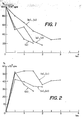
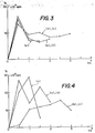
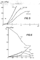
- Fig. 1 to 5 illustrate by graph obtained from radioactive tracer measurements the rate of transit (expressed at % radioactivity in function to time) of contrast compositions through the digestive tract of experimental animals.
- Fig. 1 refers to the stomach.
- Fig. 2 to 5 refer to successive parts of the intestine.
- Fig. 6 is a graph illustrating the rate of digestive transit of a sucralfate-magnetite composition according to the invention.
- a water solution (30 ml) of FeCl3.6H2O (185.3 mg; 0.685 mmol) and FeCl2.4H2O (80.5 mg; 0.405 mmol) labeled with 59Fe was brought slowly to pH 13.4 by the dropwise addition of 3% aqueous NH4 solution; then it was heated to 75°C for about 10 min. This resulted in the formation of a dark brown suspension of magnetite in large and coarse particles.
- Radioactive measurements showed that the yield was 94% by comparison with the radioactivity of the initial mixture.
- R2 1/T2/mmol Fe
- the suspension was acidified to pH 3.2 with glacial CH3COOH and sonicated for 2 min (Branson Sonifier, output mark 40 ). Then 10 ml of trimethoxy-3 -hydroxypropylsilane methacrylate were added and sonication was resumed for 2 min.
- a suspension was made containing 20 ml of acrylic acid 10 ml H2O and 10 ml of the silanized magnetite prepared as described above. This suspension was heated to 50°C and a 10% aqueous ammonium persulfate solution was added dropwise. After the polymerization was complete, the polymer was ground in 500 ml of water and dialyzed against running water. Then it was freeze-dried to give 20.1 g of silvery powder.
- a suspension of 1 g of this powder in 100 ml of water was prepared and 1 ml of this suspension was mixed with 9 ml of a 1% aqueous solution of Carbopol R at pH 7-8.
- Example 2 The procedure of Example 1, i.e. the preparation of dispersions of magnetite (1.1 mmol) in aqueous solutions of 5 g of polymer in 200 ml of H2O, was repeated using different polymers according to the Table below.
- the Table provides the names of the polymers, the yield of the preparation (calculated on the basis of the iron converted to magnetite) and the relaxivity R2 in terms of
- magnetite suspensions were prepared as disclosed in Examples 1 and 3, using a tracer amount of 59FeCl3 as label.
- the quantities of iron salts were selected so that the final concentration was about 1.46 mmol of Fe/l.
- the suspensions were further homogenized by adding a drop of surfactant (Tween-80) and sonication for a few minutes with a Branson sonifier (30 watt output).
- mice were sacrificed and stomach, small intestine, caecum and large intestine removed for examination.
- the small intestine was divided into four segments about equal in length and these were examined separately.
- the tests consisted in measuring the radioactive response of the various parts of the digestive tract and correlate the results with time.
- compositions of the invention enable to control the length of the periods during which NMRI of the digestive tract portions can be performed.
- a 6N aqueous sodium hydroxide solution was added dropwise under agitation to a 59Fe labeled aqueous solution of 153.1 mg of FeCl3.6H2O (0.566 mmol) and 64.3 mg of FeCl2.4H2O (0.323 mmol) until the pH was approximately 12.5; then the mixture was heated to 80°C and agitated for 10 min at this temperature.
- the magnetite suspension was allowed to come back to room temperature, the pH was lowered to 6.5 with 1N HCl and there were added two grams of aluminum-sucrose-octasulfate (sucralfate) sold under the name of Keal® by Laboratoires SINBIO, 75116 Paris (France).
- This powder retained the luminal adhesive properties of the Keal® product and adhered significantly to selected parts of the digestive tract of experimental animals, whereby satisfactory NMRI of these parts was experienced.
- Fig. 6 illustrates the results obtained in which the % residual radioactivity of the tracer is plotted against time for various section of the digestive tract.
- Curves i to v refer respectively to the following sections:
- a composition of magnetic particles suspended in aqueous Carbopol® was prepared as disclosed in Example 4 (item b). The quantities were adjusted to provide a mixture containing about 0.3 ⁇ mol of iron (0.0174 mg) per ml and 10 mg/ml of Carbopol R .
- the T2 weighted and intermediate scan images show a clear delineation of the darkened and expanded bowel loops. Especially the 350/50/2 image showed very clearly the single loops of the small intestine. The wall of the loops could be clearly observed. A cross section of the colon and of a kidney were also seen as well as abdominal and dorsal muscles. The contrast media was distributed evenly over the whole GI tract.
- a suspension of echogenic microballoons was prepared as described in Example 4 of reference EP-A-458 745 using poly-L-lactic acid (commercially availble unde the name of Resomer® R-207 from Boeringer Ingelheim, Germany). Ther was obtained a suspension microballoons in distilled water (concentration 109/ml; avrage size 5.2 ⁇ m).
- an aqueous suspension of gelled bioadhesive polymer was prepared by dispersing 1& by weight of Carbopol® resin (Goodrich Company) in water. The pH of this suspension was brought to 13 with concentrated NaOH and to 100 ml of the alkalinized solution were added 10 ml of the aforementioned microballoon suspension. After homogenizing the mixture under agitation, a quantity of ethanol was added sufficient to precipitate the solids; the polymer was drained on a filter under succion, washed with alcohol and dried under vacuum. The dry powder was then resuspended in 100 ml of a 0.3 M mannitol aqueous solution and the pH adjusted to 3 - 4 with HCl.
- This suspension was used to carry out echographic imaging experiments in-vivo: Laboratory rats were starved for 24 hrs before administration, then they were anesthesized with "Urethane” (1.4 g/kg) and a sample of the contrast suspension was administerd intragastrally in 10 sec (6.5 ml/rat). The imaging measurments were effected using an Acuson 128-XP/5 apparatus (ACUSON Corp. USA).
Abstract
Description
- The present invention concerns innocuous ingestible or enterally administrable compositions which, depending on the contrast agent incorporated thereto, can be used as contrast enhancer media for imaging, on the first hand in ultrasonic echography, and on the second hand, in nuclear magnetic resonance imaging (NMRI), both of the gastro-intestinal tract of animal and human patients.
- It is well known that echography and NMRI are investigative diagnosis techniques which enable the direct electronic visualization of internal organs in living beings and are therefore powerful help and guide in prognosis, medical treatment and surgery. These techniques can often advantageously supplement or replace X-ray tomography as well as the use of radio-active tracer compounds which may have obvious undesirable side-effects.
- It is also well known that contrast echography relies on the administration to patients of dispersions or suspensions of microbodies containig air or a gas, in a medium, and thereafter applying ultrasonic waves which become reflected by said microbodies to provide desired echographic signals. In this connection, it has been recognized that air or gas-filled microspheres, e.g. microbubbles or microballoons, suspended in a liquid are exceptionally efficient ultrasound reflectors for echography. In this disclosure the term of "microbubble" specifically designates air or gas globules in suspension in a liquid which generally results from the introduction therein of air or a gas in divided form, the liquid preferably also containing surfactants or tensides to control the surface properties and the stability of the bubbles. The term of "microcapsule" or "microballoon" designates preferably air or gas bodies with a material boundary or envelope, e.g.a polymer membrane wall. Both microbubbles and microballoons are useful as ultrasonic contrast agents. For instance injecting into the blood-stream of living bodies suspensions of gas microbubbles or microballoons (in the range of 0.5 to 10 µm) in a carrier liquid will strongly reinforce ultrasonic echography imaging, thus aiding in the visualization of internal organs. Imaging of vessels and internal organs can strongly help in medical diagnosis, for instance for the detection of cardiovascular and other diseases.
- It is also well known that NMRI techniques comprise subjecting a patient to a main static magnetic field combined with a linear gradient magnetic field, both being directed to some parts of the body to be investigated. The magnetic fields act on the nuclei of atoms with fractional spin quantum numbers and encode them into various degrees of statistical alignment with different resonant frequencies in a selected direction of orientation; the nuclei of concern here are mainly that of hydrogen atoms, i.e. protons, these being predominantly that of molecules present in relatively high concentration in or around the organs to be investigated, viz, the protons of water and lipids. For doing the measurements, one will apply to the parts of the body under investigation pulses of radio-frequency that matches with the resonance energy of the protons involved in the tissues or fluids of said parts of the body. When the protons under consideration are excited by a pulse of resonant energy, they are raised to a higher energy state which causes them to flip from the average orientation direction controlled by the magnetic field. Thereafter, the protons will return to their original state by relaxation in an exponential time dependent fashion, the corresponding energy then reemitted (spin-echo) forming a response signal typical of the protons under consideration, i.e. depending on their immediate environment.
- NMRI techniques are actually based on the detecting, acquiring and electronically processing of this signal (according to Fourier transforms) and thereafter displaying it spatially on a screen, thus forming an image whose various patterns correspond to areas having protons in different environments, i.e. to protons belonging to organ tissues or body fluids being subjected to investigation.
- Among the critical factors pertaining to MRI, one usually distinguishes two mutually perpendicular components of the proton-distinctive relaxation time parameter, namely the spinlattice component along the axis of magnetization (called T₁), which corresponds to the release of energy to the nuclear environment, and the perpendicular or transverse (spin-spin) relaxation component (called T₂), that corresponds to the returning of the nucleus to the initial statistical energy level. Either T₁ or T₂ can contribute to the definition of the NMR images depending on the kind of organ selected and the measurement conditions.
- It should be noted that when the measurements are carried out in the absence of agents added for increasing image contrasts, the differences in relaxation time constants between protons in various parts of the organs are small and the image is of poor to bad quality. The contrast effect can however be enhanced by the presence, in the environment of the hydrated molecules under excitation, of a variety of magnetic Species, e.g. paramagnetic (which mainly affect T₁) and ferromagnetic or superparamagnetic (which mainly affect the T₂ response). The paramagnetic substances include some metals in the ionic or organo-metallic state (e.g. Fe⁺³, Mn⁺², Gd⁺³ and the like, particularly in the form of chelates to decrease the intrinsic toxicity of the free metal ions). Ferromagnetic contrast substances preferably include magnetic aggregate particles of micronic or submicronic size, i.e. not smaller than about 100-200 nm, for instance particles of magnetite (Fe₃O₄), γ-Fe₂O₃, ferrites and other magnetic mineral compounds of transition elements. Superparamagnetic materials are usually very small magnetic particles (below about 100-150 nm) which, because their size is under a critical value, do not behave any longer as small autonomous magnets, i.e they will align in a preferential direction only when subjected to an external magnetic field. The advantage of the superparamagnetic materials (also defined sometimes as superparamagnetic fluids) over the ferromagnetic particles is mainly of efficiency density, i.e. being smaller, the number of available magnetic particles for a given weight of metal is greater in the case of superparamagnetic particles than with ferromagnetic particles and the magnetic efficiency on the neighboring protons is further enhanced.
- For ultrasonic or NMRI imaging of the digestive tract, the particulate contrast agents, whether in the form of gas-filled microspheres or particles of ferromagnetic, superparamagnetic, or paramagnetic materials, are usually administered orally or rectally, either neat or preferably with a carrier.
- For instance, EP-A-275.215 (AMERSHAM) discloses NMRI contrast enhancers for the investigation of the digestive tract comprising complexes of paramagnetic metal species like gadolinium, iron, manganese and the like associated with mineral particulate carriers such as alkaline-earth polyphosphates and apatite.
- EP-A-83.760 = W085/05534 (AMERSHAM) discloses EDTA, DTPA and NTA chelates of paramagnetic metals chemically bonded to organic polymer carriers such as sepharose, dextran, dextrin, starch and the like.
- Also in EP-A-299.920 (SCHERING), there are disclosed complexes between paramagnetic metals such as Cr, Mn, Fe, Ni, Co, Gd, etc. and polysulfated oligosaccharides like sucrose or maltose, these complexes being used for NMRI of the digestive tract.
- It has been indicated above that paramagnetic contrast agents in which the metals are in the ionic state or in the form of petal-organic compounds are often metabolizable and toxic and, although this toxicity can be controlled to some extent by using very strong chelatants and non-metabolizable polymer carriers, it is desirable to further minimize possible hazards by using less toxic materials, e.g. non-metabolizable magnetic particles of sufficient size not to diffuse through the intestinal membrane; the micronic ferromagnetic and nanometric superparamagnetic aggregate particles typically fulfill such requirements.
- For instance, in US-A-4,770,183 (ADVANCED MAGNETICS), there is recommended to use biodegradable sub-micron sized superparamagnetic metal oxide particles (1-50 nm) which may be used uncoated or coated with a polysaccharide (like dextran) or serum albumin. Coating is effected by precipitating the particles with alkali, starting with water solutions of metal salts in the presence of the polymer. These products are suitable for intravenous applications as well as for gastrointestinal applications, in which case they are administrable by intubation or enema, presumably because otherwise biodegradation by the stomach fluids would be too fast and toxicity might become a problem.
- In WO85/04330 (NYCOMED), there is disclosed the use of ferromagnetic particles as contrast agents for NMRI. As mentioned before, ferromagnetic particles are bigger than superparamagnetic particles and behave as small permanent magnets which also achieve a significant reduction of T₂. For direct administration into the digestive tract, the ferromagnetic particles are preferably embedded in a cellulose matrix or coated with this matrix. Cellulose derivatives can also be added as viscosants but the reference indicates that contrast enhancement is not readily achieved beyond the stomach, presumably because the embedding cellulose matrix does not protect sufficiently the particles from attack by the stomach fluids. Non-biodegradable embedding or coating matrices are therefore recommended to minimize absorption of toxic materials by the body.
- EP-A-186.616 (SCHERING) discloses the use of complexes of particles of magnetite (Fe₃O₄), γ-iron oxide (Fe₂O₃) and metal ferrites as contrast agents for NMRI. The cited complexants include oligo- and polysaccharides, proteins, polycarboxylic acids, protective colloids and other compounds. Examples of such compounds comprise polyvinyl-alcohol (PVA), polysilanes, polyethylene-imine, dextran, dextrin, oleic acid, gelatin, globulin, albumin, insulin, peptides and antibodies. The particles can also be encapsulated in liposomes. For enteral administration, the contrast agents are suspended in a water medium which may contain further ingredients such as salt or excipients like methylcellulose, viscosants, lactose, mannitol and surfactants like lecithin, Tween®, Myrj® and the like.
- For enteral use, this document particularly mentions compositions containing dextrin- or dextran-magnetite complexes, the manufacturing of which is disclosed in US-A-4,101,435 (MEITO SANGYO).
- There is also reported by J. KLAVENESS et al, in Diagnostic Imaging International, November 1988, p. 70, the use as contrast agents for the gastro-intestinal system of microspheres (3.5 µm) of sulfonated ion-exchange styrenedivinylbenzene resin coated with magnetite Fe₃O₄. The matrix is non-biodegradable and the particles with an iron content of about 20% are stable in the gastro-intestinal tract.
- After testing the compositions of the cited prior art consisting of coated or uncoated magnetic particles in admixtures with polymer carriers, the present inventors noted that the contrast effect in NMR imaging is generally unstable and rapidly vanishes, presumably because despite the presence of the carrier phase the magnetic particles tend to coalesce or coacervate together under the influence of the external magnetic field which strongly reduces their controlling effect on the spin-relaxation of the neighbouring protons. They however also found that such undesirable coalescence of the magnetic particles and vanishing of the T₂ relaxing effect can be prevented by either selecting as the carrier phase substantially water-insoluble hydrophilic water-swelling substrates which tend to form gels with water or, when using water-soluble polymer carriers, raising the pH of the aqueous medium containing the magnetic particles to at least 13 when admixing with the polymer solution, and keeping the dry weight ratio of said polymers to magnetic particles not below 5:1 and, preferably, in the range of 100:1 - 10:1.
- Although the exact reason of these findings is not definitely explained, it can be postulated that using carrier matrices which form nearly insoluble gels upon admixing with water (thixotropic or pseudo-plastic solutions) will locally raise the viscosity at the particle/carrier interface to such extent that the particle mobility is impeded and agglomeration is prevented.
- The present inventors also found that for efficient and selective imaging of portions of the digestive tract, it is advantageous to select carrier phases that have essential particular selective affinity for the gastric mucosa, e.g. carrier phases which can form intestinal linings or coatings on all or selected portions of the internal lumen surfaces. Hence when the carrier phase has special affinity for the gastric and intestinal mucosa, it will tend to stay in immobilized layer form thereon, thus further reducing particle mobility and preventing coalescence. Similar considerations pertain to the incorporation into the carrier phase of echogenic particles, e.g. air or gas-filled microspheres which, upon application of ultrasonic waves, will generate a reflected signal which permits visualizing by echography selected portions of the digestive tract or bladder .
- The foregoing findings form the basis of the present invention as recited below and in the annexed claims, i.e. the invention mainly concerns diagnostic compositions to be used as contrast enhancing agents, on a first hand in the imaging by echography, and on a second hand in the NMR imaging, of portions of the digestive tract of humans and animals, said compositions comprising, depending on the needs, i.e. on the kind of investigation, at least one member of a group consisting of two types of contrast enhancing particles, viz. magnetically and ultrasonically responsive particles, in admixture with, or chemically bonded to, at least one physiologically acceptable carrier phase The said carrier phase is substantially water insoluble, although swellable by hydration and gel forming, and, when hydrated, has differential affinity for the gastro-intestinal mucosa, i.e. it adheres preferably to some areas thereof, thus forming luminal linings or coatings having specific magnetic contrast response patterns which enable improved visualization of specific portions of the oeso-gastro-duodenal tract.
- The carrier phase of the present composition can be selected from water-soluble polymers which can form homogeneous solutions in aqueous media, in which case the proportion of signal generating particles therein preferably not exceeds 10% by weight of solution, more preferable 5%, for optimalized stability against coalescence when subjected to a steady magnetic field. The polymers which are convenient are for instance dextran, polyvinyl-pyrrolidone (PVP), carboxymethyl-cellulose (CMC) and the like. It is quite surprising that, contrary to products of the prior art with a high particle/carrier weight ratio, homogeneous dispersion of magnetic particles in relatively low concentrations in the carrier polymer remain stable in a magnetic field with virtually no or very little agglomeration of the magnetic particles, and hence no substantial weakening of the imaging response under the conditions of NMR imaging. Although the compositions with water-soluble polymers have no recognized specific affinity for the internal mucosa membrane of the digestive tract, they are useful for controlling the transit therethrough of the magnetic particles, either alone or when preferably used in association with bioadhesive carrier phases.
- Carrier phases having affinity for the membrane mucosa of the digestive tract (bioadhesive carriers) can include most luminal coating materials in use for treatment, protection of medication of gastro-intestinal regions including, for instance, adhesives containing bismuth oxide, aluminum oxide and various clays such as montmorillonite and attapulgite designed for luminal applications. These materials also include cross-linked polymers such as polysiloxanes (Dimethicone®), magnesium and other metals alginates, bioadhesive pectins and carbohydrates polysubstituted with groups such as sulfate, phosphate, sulfonate and phosphonate. One convenient polysulfated carbohydrate is sucrose octasulfate (also named sucralfate). It is already known that montmorillonite clays of the Beidel type can themselves function as useful contrast agents in the NMRI of the digestive tract of humans and animals, this being possibly due to some inherent magnetic properties of the montmorillonites. The contrast effect of Beidel montmorillonite clays is further enhanced when used as a carrier according to the invention.
- These carriers have the general property to get hydrated with water and form therewith substantially insoluble gels which strongly adhere to the membrane mucosa. Depending on the selected carrier phase, the compositions of the invention can be made to have controllable transit time through the gastrointestinal tract and particular adhesion to selected portions thereof, thus enabling efficient NMRI of selected organs. Normally, the compositions using polymers carriers having no or little bioadhesion will permit faster transit than the compositions using carriers with strong bioadhesive character, hence controlled transit can be achieved with compositions in which non-bioadhesive and bioadhesive carriers are admixed in suitable proportions. It has also been surprisingly found that the particular selection of carriers used in the compositions of the invention, i.e. the carriers with enhanced affinity for mucosa membranes at low pH and/or soluble carriers with high dispersive capacity for magnetic particles provide generally excellent transverse and longitudinal luminal imaging homogeneity.
- One preferred type of bioadhesive polymers to be used as the carrier phase in this invention is a polymer or copolymer of acrylic acid containing a proportion of other monomers (generally di- or polyfunctional allyl ethers or acrylates) to impart a degree of water-insolubility and swelling capacity to form gels. Suitable polymers of this type are available on the market under the names of Carbopol® or Polycarbophil®, for instance from the Goodrich Company. Other suitable polymers of comparable type are disclosed in EP-A-309.404 (included for reference) and comprise copolymers of acrylic acid with allyl ethers of glycols or sugars. Upon addition of water these polymers will form viscous dispersions of microgels which have strong affinity for internal mucous membranes. Furthermore, the gelling and swelling properties of such polymers are pH dependent; hence the volume, bulk and adhesive properties of the carrier can be controlled by adjusting the pH to a desired value
- The present ingestible NMRI contrast compositions are stable even at
pH 1 and this is another advantage over similar products of the prior art as they are not attacked by stomach fluids which normally dissolve magnetite particles into Fe+² and Fe⁺³ ions. This is obviously undesirable for both toxicity and imaging efficiency reasons. Bioadhesivity is particularly effective under acid or near neutral conditions; at high pH the bioadhesivity is decreased and may become negligible. Hence control of bioadhesivity by pH control is another asset of the present invention. - For manufacturing the contrast compositions according to the invention one usually admixes the signal generating particles (the echogenic or magnetically responsive particles, or both) with the carrier phase. In regard to the MRI aspect of the invention, one may use magnetic particles which can be ferromagnetic or superparamagnetic. The nature of both and the distinction between them has been stressed before in this specification. Hence, one prefers superparamagnetic particles for optimalized density efficiency, i.e. magnetic particles of size not exceeding about 150 nm and preferably in the 1-100 nm range (10-1000 A). The preparation of such magnetic particles is well known and disclosed in many references, e.g. the aforementioned references and further references, e.g. US-A-4,554,088; EP-A-125.995 (Advanced Magnetics); US-A-4,267,234; US-A-4,157,323 (California Institute); WO-A-78/00005 (Mosbach); WO-A-83/01738; WO-A-83/03426; WO-A-84/00294 (Schröder). They are also available commercially, e.g. from Ferrofluidics Corp., Burlington, Mass. They normally result from the alkalinization of an aqueous solution containing Fe⁺² and Fe⁺³ salts in correct proportions.
- It has been said that when it is wished to also use the compositions of the invention for ultrasonic echography diagnosis purposes, said compositions will contain echogenic particles, preferably in the form of air- or gas-filled microspheres bounded by a material envelope, e.g. a polymer membrane. The obtention of such echogenic particles is disclosed in many references among which one can cite EP-A-458 745. In this reference, there is disclosed the preparation of microballoons with a soft and resilient membrane envelope which are convenient as echogenic particles in the present invention as they resist pressure variations due to peristaltic movements in the bowels. Notwithstanding, other types of ultrasonic echogenerating particles can be used as well, e.g. those disclosed in the following documents: WO-A-91/12 823, EP-A-327 490, EP-A-324 938.
- In one embodiment of a method for preparing the compositions according to the present invention, an aqueous suspension of the magnetic particles is treated with alkali to raise the pH to 13 or more, then the obtained alkaline suspension is admixed with an aqueous solution or dispersion of the carrier phase at the same pH and finally a water-compatible organic solvent in which the carrier phase is insoluble is added, whereby a precipitate of the desired composition is formed. The composition can thereafter be separated, for instance by filtration, and washed free from alkali, then it can be stored dry. When used for internal NMRI investigation, the composition can be rehydrated with water or an aqueous solution of innocuous excipients, this being for oral or enteral administration.
- In the foregoing embodiment, the magnetic particles are not covalently linked to the carrier phase, the latter being water-soluble, but still they are immobilized therein presumably due to the existence of electrostatic or Van der Waals forces between the carrier phase constituents and the magnetic particles or, if the carrier is insoluble in water but in hydrated gel form, to immobilization of the hydrated magnetic particles within the carrier gelled structure.
- In another embodiment for preparing the compositions of the invention, the magnetic particles are chemically bonded to the carrier phase by the use of either reactive functions of the carrier itself or of a coupler grafted to the particles, said coupler possessing also functions which will bind to the carrier. For instance, the coupler can be a silane which will bind to the particles by silanation (see EP-A-125.995 incorporated for reference) and which bears a function which may subsequently react with the carrier phase. For example, a useful function of the silane coupler can be an amine group which can further react with negative groups on the carrier material, e.g. COOH or S-OH, sulfate or sulfonate groups, to form ammonium salts. Preferably, if the carrier is an organic polymer obtained by polymerization of one or more monomers, the silane coupler can comprise functions that will copolymerize with said one or more monomers.
- Thus for instance, in the case the carrier phase comprises a polymer or copolymer of acrylic acid, this can be prepared by copolymerizing a mixture of acrylic acid (and optionally other olefinic monomers) and magnetic particles carrying grafted thereon silanes with substituents bearing functions copolymerizable with acrylic acid. An appropriate silane for achieving this is trimethoxy-hydroxypropyl acrylate or methacrylate which possesses a double bond copolymerizable with other acrylates. The grafting technique is disclosed for instance in EP-A-113.367 and EP-A-125.995.
- For using the present compositions as an echogenic or NMRI contrast agent (or both) in the study of the digestive tract, the compositions (when stored dry) are admixed with an aqueous phase suitable for oral administration in order to provide a gel which will adhere to a predetermined extent to the mucous membrane of the digestive tract and therefore will carry the particles through said tract to the portion thereof to be visualized. The rate of transfer and the transit time can be adapted by properly selecting the nature and degree of bioadhesion of the carrier phase. For this, variable proportions of contrast compositions including water-soluble carrier polymer can be admixed with the bioadhesive carrier, these proportions being selected to reach up to 99% by weight or more in the case where little restricted transit, unrestricted transit or accelerated transit is desired. For instance, for NMR investigations, using mainly dextran as the carrier phase in rats gave a transit rate similar to that observed with no carrier, while using mainly CMC as carrier gave accelerated transit. Using Carbopol® as the carrier phase gave strongly retarded transit, particularly in the duodenum portion of the intestinal tract. The present contrast compositions may also advantageously comprise isoosmolarity agents which minimize loss or gain of water during intestinal transit, i.e. diffusion by osmosis; such agents may comprise carbohydrates such as sorbitol, mannitol, xylitol and the like.
- During transit time, the patient having received a dose of the present compositions as NMRI or echogenic contrast agent is subjected to periodical or continuous investigations using conventional echography or NMRI equipment, whereby the obtained processed images can thereafter be used for diagnostic or other medical applications.
- The invention is now illustrated by the following practical examples.
- In the drawing:
- Fig. 1 to 5 illustrate by graph obtained from radioactive tracer measurements the rate of transit (expressed at % radioactivity in function to time) of contrast compositions through the digestive tract of experimental animals.
- Fig. 1 refers to the stomach.
- Fig. 2 to 5 refer to successive parts of the intestine.
- Fig. 6 is a graph illustrating the rate of digestive transit of a sucralfate-magnetite composition according to the invention.
- A water solution (30 ml) of FeCl₃.6H₂O (185.3 mg; 0.685 mmol) and FeCl₂.4H₂O (80.5 mg; 0.405 mmol) labeled with ⁵⁹Fe was brought slowly to pH 13.4 by the dropwise addition of 3% aqueous NH₄ solution; then it was heated to 75°C for about 10 min. This resulted in the formation of a dark brown suspension of magnetite in large and coarse particles.
- To this was added under agitation a solution of 5 g of Dextran in 200 ml of water and the pH was reajusted to 13.4 by adding some more NaOH solution. After 15 min more stirring, alcohol was added, whereby a very fine precipitate of Dextran-magnetite was formed. This precipitate was drained under succion, washed with alcohol and dried at 50°C in air.
- Radioactive measurements showed that the yield was 94% by comparison with the radioactivity of the initial mixture.
- The proton magnetic resonance transverse relaxivity, expressed as R₂, i.e. R₂ = 1/T₂/mmol Fe) was measured on aqueous dispersions of the aforementioned precipitate by means of a 60 MHz RMN spectrometer. The measurements were effected at mid-height of the absorption peaks. Values of R₂ in the range of 500-550 (mM.s)⁻¹ were obtained which did not substantially change with time, thus showing that reagglomeration of the particles did not occur.
- A comparative test was performed as disclosed in Example 2 of EP-A-186.616.
- To a solution of Dextrin (80 g) in 180 ml of H₂O at 70°C were added a solution of 19 g of FeCl₃.6H₂O (70 mmol) and 7,01 g FeCl₂.4H₂O (35 mmol) in 105 ml of water. A tracer quantity of ⁵⁹FeCl₃ was also added. The pH was raised to 2.4 by first adding 20% aqueous Na₂CO₃, then it was brought to pH 11 with 10N NaOH added dropwise, whereby the color of iron oxides developed. The mixture was refluxed for 30 min, then it was cooled to room temperature and the pH was brought to 6.2 with 6N HCl. Then 0.8 liter of ethanol was added which caused the formation of a precipitate. This was collected, resuspended in water and the suspension was dialyzed for 24 hrs in running water (membrane cut-off = 10'000). Then the suspension was freeze-dried which gave a blackish powder.
- Measurements of R₂ as described above gave none or useless responses. Analogous results were obtained by replacing Dextrin by a comparable amount of Dextran. Obviously, the ratio of metal to polymer in this formulation is much too high to prevent reagglomeration of the particles subjected to a magnetic field.
- It was also found that when the experiment of Example 1 was repeated, but the magnetite precipitation was effected with the polymer already present in the solution, no useful R₂ measurements were obtained.
- To a solution of 13.56 g FeCl₃.6H₂O (50 mmol) and 5.3 g FeCl₂.4H₂O (27 mmol) in 60 ml of water labeled with ⁵⁹Fe (about 10⁶ cpm) were added dropwise 60 ml of 5M aqueous NaOH until the pH was above 13. The solution was left to stand until a blackish sediment accumulated at the bottom of the container. This was washed carefully by decantation ten times with portions of 300 ml H₂O. After the last portions, the pH of the water had dropped to 9.3.
- The suspension was acidified to pH 3.2 with glacial CH₃COOH and sonicated for 2 min (Branson Sonifier, output mark 40 ). Then 10 ml of trimethoxy-3 -hydroxypropylsilane methacrylate were added and sonication was resumed for 2 min.
- Sixty ml of glycerol were added and the mixture was heated under reduced pressure in a rotavapor until a black viscous residue has formed. This residue was diluted with 240 ml of water, agitated for 20 min and allowed to rest whereby it separated into two phases. This was dialyzed for 24 hrs against a citrate buffer (10 mM, pH 8.3 with ammonia) in order to remove low molecular material (glycerol, unreacted silane and acrylate oligomers).
- A suspension was made containing 20 ml of
acrylic acid 10 ml H₂O and 10 ml of the silanized magnetite prepared as described above. This suspension was heated to 50°C and a 10% aqueous ammonium persulfate solution was added dropwise. After the polymerization was complete, the polymer was ground in 500 ml of water and dialyzed against running water. Then it was freeze-dried to give 20.1 g of silvery powder. - A suspension of 1 g of this powder in 100 ml of water was prepared and 1 ml of this suspension was mixed with 9 ml of a 1% aqueous solution of Carbopol R at pH 7-8. The relaxation time T₂ was measured as described in Example 1, the value found being R₂ = 160 (s.mM)⁻¹.
- The procedure of Example 1, i.e. the preparation of dispersions of magnetite (1.1 mmol) in aqueous solutions of 5 g of polymer in 200 ml of H₂O, was repeated using different polymers according to the Table below.
- The Table provides the names of the polymers, the yield of the preparation (calculated on the basis of the iron converted to magnetite) and the relaxivity R₂ in terms of


- In the experiments reported below, magnetite suspensions were prepared as disclosed in Examples 1 and 3, using a tracer amount of ⁵⁹FeCl₃ as label. The quantities of iron salts were selected so that the final concentration was about 1.46 mmol of Fe/l. After precipitation with alkali the suspensions were further homogenized by adding a drop of surfactant (Tween-80) and sonication for a few minutes with a Branson sonifier (30 watt output).
- Samples of the suspensions (0.1 ml) were diluted with 9.9 ml (dilution 1:100) of 1% by weight aqueous polymer solutions also containing 0.3 mol/l of mannitol to preserve isotonicity. The polymers used were:
- a) Carboxymethylcellulose (CMC) Na salt (0.1 g/9.9 ml of water).
- b) "Carbosal"; this was the code name given to Carpobol (from the Goodrich Company) when in the form of sodium salt.
- c) "SA" polymer; this polymer is disclosed in EP-A-309.404 and is based on acrylic acid copolymerized with a mixture of sucrose mono- and di- allyl ethers with minor quantities of sucrose polyfunctional allyl ether.
- d) Dextran
- e) Control; this was made as above by diluting the magnetite preparation to 1/100 but using only water without polymer.
- Five ml of samples a) to e) were administered to Sprague-Dawly rats (about 180 g) which were kept fast except for water 24 hrs before administration. The samples were introduced intragastrically with a ball-point syringe. The tracer amount in each sample accounted for about 10⁶ cpm.
- At time intervals of 0.5, 1, 1.5, 2, 3 and 6 hrs, animals were sacrificed and stomach, small intestine, caecum and large intestine removed for examination. The small intestine was divided into four segments about equal in length and these were examined separately.
- The tests consisted in measuring the radioactive response of the various parts of the digestive tract and correlate the results with time. The global results were that sample (d) (Dextran) behaved about like the control, i.e there was no visible influence of the polymer on the intestinal transit time while (a) accelerated it and (b) and (c) retarded it significantly, thus allowing prolonged examination when visualized under NMRI.
- The results are recorded on the graphs of fig. 1-5 which refer, respectively, to the stomach and to the four successive portions of the small intestine. In the figures, the curves representing control (e) or Dextran (d) (they are practically similar) are marked with full dots; the curves for CMC (a) are marked with empty squares and the curves for the bioadhesive polymers (b) and (c) are marked with shaded squares. These results clearly support the foregoing findings.
- If in the experiments reported in this Example the carrier phases were replaced by mixtures of the component carriers, intermediate transit and retention rates were observed. Hence, the compositions of the invention enable to control the length of the periods during which NMRI of the digestive tract portions can be performed.
- A 3% aqueous solution of NH₄OH (about 25 ml) was slowly added dropwise under agitation to a 100 ml water solution of 619.2 mg of FeCl₃.6H₂O (2.29 mmol) and 262.1 mg of FeCl₂.4H₂O (1.32 mmol). The mixture was then heated to 75°C for 10 min. There were thereafter added 200 ml of an aqueous 2.5% by weight solution of sodium alginate and the pH was raised to 13.5 with 40% aqueous NaOH. After further agitating for 15 min at room temperature, alcohol was added to precipitate the magnetite-polymer composition; the powder was drained on a filter, washed with alcohol and dried; the yield was 4.96 g containing 0.216 mmol of Fe (theory = 0.22 mmol). The mmolar transverse relaxivity R₂ was measured at 60 MHz on a water suspension of the powder with a Perkin-Elmer/Hitachi NMR apparatus. The measured value was R₂ = 465 (s.mM)⁻¹.
- A 6N aqueous sodium hydroxide solution was added dropwise under agitation to a ⁵⁹Fe labeled aqueous solution of 153.1 mg of FeCl₃.6H₂O (0.566 mmol) and 64.3 mg of FeCl₂.4H₂O (0.323 mmol) until the pH was approximately 12.5; then the mixture was heated to 80°C and agitated for 10 min at this temperature.
- The magnetite suspension was allowed to come back to room temperature, the pH was lowered to 6.5 with 1N HCl and there were added two grams of aluminum-sucrose-octasulfate (sucralfate) sold under the name of Keal® by Laboratoires SINBIO, 75116 Paris (France). The mixture was diluted with 50 ml H₂O and further agitated for 10 min after which it was left to stand, whereby a precipitate of magnetite-sucralfate settled at the bottom of the vessel. This was drained on a filter, washed with water and dried, yield 2.03 g. R₂ (in water) = 283 (s.mM)⁻¹.
- This powder retained the luminal adhesive properties of the Keal® product and adhered significantly to selected parts of the digestive tract of experimental animals, whereby satisfactory NMRI of these parts was experienced.
- Fig. 6 illustrates the results obtained in which the % residual radioactivity of the tracer is plotted against time for various section of the digestive tract. Curves i to v refer respectively to the following sections:
- i
- = stomach
- ii
- = intestine,
portion 1 - iii
- = intestine,
portion 2 - iv
- = intestive,
portion 3 - v
- = intestine,
portion 4 + caecum - Montmorillonite clay of the Beidel type (BEDELIX®) available from Laboratoires Beaufour, 28100 Dreux (France) was tested for NMRI contrast response under the following conditions: three g of BEDELIX® were stirred in 100 ml of a 0.5% solution of Carbopol®. The 1/T₂ value of this preparation was measured as usual with a Perkin-Elmer/Hitachi NMR apparatus at 60 MHz and found to be 435.5 s⁻¹ without the addition of magnetite. This value was stable (due to the presence of the Carbopol®) and the preparation could be used as such for the NMRI of the digestive tract of animals.
- However the contrast performances were further enhanced by the incorporation of magnetite as follows:
- A suspension of magnetite was prepared as disclosed in Example 1, first paragraph. A 0.2 ml portion of this suspension was added to 99.8 ml of a 0.5% by weight solution of Carbopol® . The iron concentration in this preparation was 0.178 mmol/ml; the 1/T₂ value was 144.5 s⁻¹ , i.e. R₂ = 812 (s.mm)⁻¹.
- Aliquots of the magnetite preparation were taken and to each were added a different quantity of BEDELIX® . The R₂ values were measured as usual and reported below in function to the % by weight of added BEDELIX®.

- A composition of magnetic particles suspended in aqueous Carbopol® was prepared as disclosed in Example 4 (item b). The quantities were adjusted to provide a mixture containing about 0.3 µmol of iron (0.0174 mg) per ml and 10 mg/ml of CarbopolR.
- Male Sprague Dawley rats were used for the experiment after 24 hours of fasting. Eight ml of the Carbopol-magnetite solution were administrated orally to the conscious animals which were anesthetized 10 min later by 30 ml/kg of Pentotal. Transversal MR images were then taken on an Esaote ESATOM 5000 imager equipped with a special 8 cm i.d. RF receiver coil, using a slice of 2 mm, a F.O.V. of 15.9 x 15.9 cm, and a matrix size of 128 x 256. T2 weighted images (SE 200/70/1), were acquired as well as T1 weighted images (SE 500/16/2), and intermediate scan images (SE 350/50/2). For the T2 weighted and intermediate scan images gradient moment nulling techniques were used in order to minimize respiratory artifacts. The T2 weighted and intermediate scan images show a clear delineation of the darkened and expanded bowel loops. Especially the 350/50/2 image showed very clearly the single loops of the small intestine. The wall of the loops could be clearly observed. A cross section of the colon and of a kidney were also seen as well as abdominal and dorsal muscles. The contrast media was distributed evenly over the whole GI tract.
- A suspension of echogenic microballoons was prepared as described in Example 4 of reference EP-A-458 745 using poly-L-lactic acid (commercially availble unde the name of Resomer® R-207 from Boeringer Ingelheim, Germany). Ther was obtained a suspension microballoons in distilled water (concentration 10⁹/ml; avrage size 5.2 µm).
- On the other hand, an aqueous suspension of gelled bioadhesive polymer was prepared by dispersing 1& by weight of Carbopol® resin (Goodrich Company) in water. The pH of this suspension was brought to 13 with concentrated NaOH and to 100 ml of the alkalinized solution were added 10 ml of the aforementioned microballoon suspension. After homogenizing the mixture under agitation, a quantity of ethanol was added sufficient to precipitate the solids; the polymer was drained on a filter under succion, washed with alcohol and dried under vacuum. The dry powder was then resuspended in 100 ml of a 0.3 M mannitol aqueous solution and the pH adjusted to 3 - 4 with HCl.
- This suspension was used to carry out echographic imaging experiments in-vivo: Laboratory rats were starved for 24 hrs before administration, then they were anesthesized with "Urethane" (1.4 g/kg) and a sample of the contrast suspension was administerd intragastrally in 10 sec (6.5 ml/rat). The imaging measurments were effected using an Acuson 128-XP/5 apparatus (ACUSON Corp. USA).
- Immediately after administration, there was observed a dilatation of the stomach the image of which became entirely white in the two-dimensional imaging mode. In the color Doppler imaging mode, the stomach area showed multiple colors (iridescent yellow-red, blue-green) indicating that the scatterers were in motion. Ten minutes later, imaging of the upper part of the intestine (duodenum) showed a similar colored patterns which indicated that composition containing the reflector microballoons was moving down in the intestinal tract. Expanded bowel loops could be distinguished very clearly and the presence of possible abnomalities could be spotted easily. The spleen was also easily identified.
- If in the present example ther is used a suspension of microbubbles, for instance that disclosed in WO-A-91/15244, comparable results were noted.
Claims (21)
- Administrable diagnostic compositions for enhancing the image contrast in the ultrasonic and, respectively, NMR imaging of the digestive tract of humans and animals, said compositions respectively comprising, as contrast enhancing agent in admixture with or chemically bonded to at least one physiologically acceptable carrier phase, one or more members of the group consisting of signal enhancing ultrasonically and magnetically responsive particles, characterized in that said carrier phase is substantially water insoluble, although swellable by hydration and gel forming, and, when hydrated, has differential affinity for the gastro-intestinal mucosa, i.e. it adheres preferably to some areas thereof, thus forming luminal linings or coatings having specific imaging contrast response which enable improved visualization of specific portions, and relative motions, of the oeso-gastro-duodenal tract
- The compositions of claim 1, in which the carrier is a hydratable gel-forming bioadhesive carrying groups selected from one or more of carboxy, sulfate, sulfonate, phosphate and phosphonate, these groups being either free or in the form of salts with alkali of alkaline-earth metals.
- The compositions of claim 2, in which the carrier is selected from polysulfated and polyphosphated carbohydrates.
- The compositions of claim 1, in which the carrier is a luminal coating compound selected from hydratable clays, e.g. Attapulgite and Montmorillonite, and hydroxylated coatable polysaccharides.
- The compositions of claim 1, in which the carrier is a bioadhesive, hydratable, gel-forming polymer or copolymer of acrylic acid and, optionally, other monomers, this polymer or copolymer bearing carboxylate of sulfonate groups, said groups being free or in the form of salts with alkali or alkaline-earth metals.
- The compositions of claim 1, in which the magnetically responsive particles are uncoated particles of magnetite or superparamagnetic metal oxides and the ultrasonically responsive particles are air- or gas-filled microspheres surrounded by an envelope of polymeric material.
- The compositions of claim 1, further comprising dispersions of contrast enhancing particles in an aqueous solution of one or more water-soluble polymer carrier, the amount of said particles to said water-soluble polymer carrier, measured dry, not exceeding 5%.
- The compositions of claim 7, in which the proportion of said dispersions is up to 100% of the total of the compositions and the water-soluble polymer carrier is selected from dextran, polyvinyl-pyrrolidone (PVP), Carboxymethylcellulose (CMC), hydroxypropyl-cellulose and other water-soluble non-metabolizable polymers.
- The compositions of claim 1, in which the particles are bonded to the bioadhesive carrier substrate by means of a chemical coupler.
- The compositions of claim 9, in which said coupler has reactive functions of which one will bind to the particles and another will bind to the carrier.
- The compositions of claim 10, in which the carrier is a polymer or copolymer of acrylic acid and, optionally other unsaturated monomers, and in which said another function of the coupler comprises an ethylenically unsaturated double bond, said another function being copolymerized with said polymer or copolymer of acrylic acid.
- The compositions of claim 10, in which said one function of the coupler is a trialkoxy-silane function which binds to the magnetic particles through formation of a silicon-containing bond.
- A method for manufacturing ingestible compositions to be used as contrast enhancing agents in the NMR Imaging of the digestive tract of humans and animals, said compositions comprising image contrast enhancing particles and at least a carrier phase consisting of a water-soluble or water-insoluble mineral or organic substrate, characterized in adjusting the pH of a dispersion of the particles in an aqueous medium to a value of at least 13, admixing the carrier phase with said dispersion and separating the product of addition of the magnetic particles and the carrier phase.
- The method of claim 13, in which the carrier phase is an organic polymer or oligomer, optionally carrying carboxylic or sulfate groups, and separation is achieved by dilution with a water-soluble solvent in which said product of addition is not soluble.
- A method for manufacturing ingestible compositions to be used as contrast enhancing agents in the ultrasonic or NMR Imaging of the digestive tract of humans and animals, said compositions comprising image contrast enhancing particles bound to a carrier phase consisting of a water-insoluble mineral or organic substrate, characterized in selecting a coupler with at least two reactive functions, grafting the coupler to the particles through one of said reactive functions and binding said grafted particles to said carrier phase through the other of said reactive functions.
- The method of claim 15, in which the organic substrate is a polymer or copolymer of acrylic acid and, optionally, other copolymerizable monomers, characterized in copolymerizing said acrylic acid in the presence of said grafted particles, said other reactive function of the coupler having a double bond copolymerizable with acrylic acid.
- A method for the ultrasonic or NMR imaging the digestive tract of human and animal patients in which an imaging composition comprising an image contrast enhancing agent incorporated to a biocompatible carrier phase is orally or enterally administered to said patient and the successive sections of the digestive tract are then imaged as said carrier phase transits therethrough, characterized in that the nature of carrier phase and the concentration of contrast agent therein are selected to effect control on the rate of said transit and on the retention time of said contrast agent in each of said sections depending on the imaging needs.
- The method of claim 17, in which the nature of carrier phase is selected by its degree of bioadhesive capacity to the mucosa membrane of the digestive tract.
- The method of claim 18, in which the carrier phase is a water-insoluble, gel-forming luminal lining compound.
- The method of claim 19, in which the carrier phase is selected at least in part from Carbopol®, Sucralfate®, Attapulgite, Montmorillonite, magnesium alginate and pectin.
- The method of claim 18, in which the carrier phase contains also a water-soluble polymer for accelerating said rate of transit.
Priority Applications (3)
| Application Number | Priority Date | Filing Date | Title |
|---|---|---|---|
| EP92810078A EP0502814B1 (en) | 1991-02-15 | 1992-02-05 | Compositions for increasing the image contrast in imaging of the digestive tract of patients |
| EP94203065A EP0638318B1 (en) | 1991-02-15 | 1992-02-05 | Compositions for increasing the image contrast in diagnostic investigations of the digestive tract |
| EP94202222A EP0627632B1 (en) | 1991-02-15 | 1992-02-05 | Compositions for increasing the image contrast in diagnostic investigations of the digestive tract of patients |
Applications Claiming Priority (3)
| Application Number | Priority Date | Filing Date | Title |
|---|---|---|---|
| EP91810105 | 1991-02-15 | ||
| EP91810105 | 1991-02-15 | ||
| EP92810078A EP0502814B1 (en) | 1991-02-15 | 1992-02-05 | Compositions for increasing the image contrast in imaging of the digestive tract of patients |
Related Child Applications (4)
| Application Number | Title | Priority Date | Filing Date |
|---|---|---|---|
| EP94202222A Division EP0627632B1 (en) | 1991-02-15 | 1992-02-05 | Compositions for increasing the image contrast in diagnostic investigations of the digestive tract of patients |
| EP94203065A Division EP0638318B1 (en) | 1991-02-15 | 1992-02-05 | Compositions for increasing the image contrast in diagnostic investigations of the digestive tract |
| EP94202222.9 Division-Into | 1992-02-05 | ||
| EP94203065.1 Division-Into | 1992-02-05 |
Publications (3)
| Publication Number | Publication Date |
|---|---|
| EP0502814A2 true EP0502814A2 (en) | 1992-09-09 |
| EP0502814A3 EP0502814A3 (en) | 1992-10-07 |
| EP0502814B1 EP0502814B1 (en) | 1995-06-21 |
Family
ID=8208821
Family Applications (3)
| Application Number | Title | Priority Date | Filing Date |
|---|---|---|---|
| EP92810078A Expired - Lifetime EP0502814B1 (en) | 1991-02-15 | 1992-02-05 | Compositions for increasing the image contrast in imaging of the digestive tract of patients |
| EP94202222A Expired - Lifetime EP0627632B1 (en) | 1991-02-15 | 1992-02-05 | Compositions for increasing the image contrast in diagnostic investigations of the digestive tract of patients |
| EP94203065A Expired - Lifetime EP0638318B1 (en) | 1991-02-15 | 1992-02-05 | Compositions for increasing the image contrast in diagnostic investigations of the digestive tract |
Family Applications After (2)
| Application Number | Title | Priority Date | Filing Date |
|---|---|---|---|
| EP94202222A Expired - Lifetime EP0627632B1 (en) | 1991-02-15 | 1992-02-05 | Compositions for increasing the image contrast in diagnostic investigations of the digestive tract of patients |
| EP94203065A Expired - Lifetime EP0638318B1 (en) | 1991-02-15 | 1992-02-05 | Compositions for increasing the image contrast in diagnostic investigations of the digestive tract |
Country Status (8)
| Country | Link |
|---|---|
| US (4) | US5370901A (en) |
| EP (3) | EP0502814B1 (en) |
| JP (1) | JPH05148161A (en) |
| AT (3) | ATE180678T1 (en) |
| DE (3) | DE69229358T2 (en) |
| DK (1) | DK0502814T3 (en) |
| ES (1) | ES2074350T3 (en) |
| GR (1) | GR3017002T3 (en) |
Cited By (23)
| Publication number | Priority date | Publication date | Assignee | Title |
|---|---|---|---|---|
| WO1992021382A1 (en) * | 1991-06-03 | 1992-12-10 | Holmes, Michael, John | Improvements in or relating to contrast agents |
| US5393524A (en) * | 1991-09-17 | 1995-02-28 | Sonus Pharmaceuticals Inc. | Methods for selecting and using gases as ultrasound contrast media |
| WO1995020405A1 (en) * | 1994-01-28 | 1995-08-03 | Nycomed Imaging As | Oral magnetic particle formulation |
| DE4428589A1 (en) * | 1994-08-12 | 1996-02-15 | Byk Gulden Lomberg Chem Fab | Echo contrast agent for oral use |
| US5558855A (en) * | 1993-01-25 | 1996-09-24 | Sonus Pharmaceuticals | Phase shift colloids as ultrasound contrast agents |
| US5558854A (en) * | 1991-09-17 | 1996-09-24 | Sonus Pharmaceuticals | Ultrasound contrast media comprising perfluoropentane and perfluorohexane gas |
| US5558853A (en) * | 1993-01-25 | 1996-09-24 | Sonus Pharmaceuticals | Phase shift colloids as ultrasound contrast agents |
| US5558094A (en) * | 1991-09-17 | 1996-09-24 | Sonus Pharmaceuticals, Inc. | Methods for using persistent gases as ultrasound contrast media |
| US5853698A (en) * | 1996-03-05 | 1998-12-29 | Acusphere, Inc. | Method for making porous microparticles by spray drying |
| US6132699A (en) * | 1996-03-05 | 2000-10-17 | Acusphere, Inc. | Microencapsulated fluorinated gases for use as imaging agents |
| US6569404B1 (en) | 1993-01-25 | 2003-05-27 | Amersham Health A/S | Phase shift colloids as ultrasound contrast agents |
| US6723303B1 (en) | 1991-09-17 | 2004-04-20 | Amersham Health, As | Ultrasound contrast agents including protein stabilized microspheres of perfluoropropane, perfluorobutane or perfluoropentane |
| WO2005021557A2 (en) | 2003-08-29 | 2005-03-10 | Dyax Corp. | Poly-pegylated protease inhibitors |
| WO2009027690A1 (en) | 2007-08-30 | 2009-03-05 | United Arab Emirates University | Diagnostic agent |
| WO2010045315A1 (en) | 2008-10-14 | 2010-04-22 | Dyax Corp. | Use of igf-ii/igf-iie binding for the treatment and prevention of systemic sclerosis associated pulmonary fibrosis |
| WO2011085103A2 (en) | 2010-01-06 | 2011-07-14 | Dyax Corp. | Plasma kallikrein binding proteins |
| EP2460831A2 (en) | 2005-05-27 | 2012-06-06 | Biogen Idec MA Inc. | Tweak binding antibodies |
| WO2014093396A1 (en) | 2012-12-10 | 2014-06-19 | Biogen Idec Ma Inc. | Anti-blood dendritic cell antigen 2 antibodies and uses thereof |
| WO2014143739A2 (en) | 2013-03-15 | 2014-09-18 | Biogen Idec Ma Inc. | Anti-alpha v beta 6 antibodies and uses thereof |
| WO2014144466A1 (en) | 2013-03-15 | 2014-09-18 | Biogen Idec Ma Inc. | Anti-alpha v beta 6 antibodies and uses thereof |
| WO2015057939A1 (en) | 2013-10-18 | 2015-04-23 | Biogen Idec Ma Inc. | Anti-s1p4 antibodies and uses thereof |
| EP2966089A1 (en) | 2011-06-02 | 2016-01-13 | Dyax Corp. | Fc receptor binding proteins |
| US9260520B2 (en) | 2008-04-25 | 2016-02-16 | Dyax Corp. | Antibodies to FeRn and uses thereof |
Families Citing this family (147)
| Publication number | Priority date | Publication date | Assignee | Title |
|---|---|---|---|---|
| US5562099A (en) * | 1990-10-05 | 1996-10-08 | Massachusetts Institute Of Technology | Polymeric microparticles containing agents for imaging |
| US5370901A (en) * | 1991-02-15 | 1994-12-06 | Bracco International B.V. | Compositions for increasing the image contrast in diagnostic investigations of the digestive tract of patients |
| GB9106686D0 (en) * | 1991-03-28 | 1991-05-15 | Hafslund Nycomed As | Improvements in or relating to contrast agents |
| GB9200388D0 (en) * | 1992-01-09 | 1992-02-26 | Nycomed As | Improvements in or relating to contrast agents |
| DE69207589T2 (en) * | 1992-06-01 | 1996-05-23 | Basf Ag | Use of dispersions of magneto-ionic particles in MRI contrast media |
| US5439686A (en) * | 1993-02-22 | 1995-08-08 | Vivorx Pharmaceuticals, Inc. | Methods for in vivo delivery of substantially water insoluble pharmacologically active agents and compositions useful therefor |
| FR2703250A1 (en) * | 1993-03-30 | 1994-10-07 | Chicouri Marcel | New pharmaceutical compositions based on bismuth and process for their preparation |
| US6405732B1 (en) | 1994-06-24 | 2002-06-18 | Curon Medical, Inc. | Method to treat gastric reflux via the detection and ablation of gastro-esophageal nerves and receptors |
| US6044846A (en) | 1994-06-24 | 2000-04-04 | Edwards; Stuart D. | Method to treat esophageal sphincters |
| US6092528A (en) | 1994-06-24 | 2000-07-25 | Edwards; Stuart D. | Method to treat esophageal sphincters |
| US6733495B1 (en) | 1999-09-08 | 2004-05-11 | Curon Medical, Inc. | Systems and methods for monitoring and controlling use of medical devices |
| US6056744A (en) | 1994-06-24 | 2000-05-02 | Conway Stuart Medical, Inc. | Sphincter treatment apparatus |
| US6009877A (en) | 1994-06-24 | 2000-01-04 | Edwards; Stuart D. | Method for treating a sphincter |
| DE4424233C1 (en) * | 1994-07-09 | 1995-12-07 | Thomas Gieselmann | Oral echo contrast agent for ultra sound diagnoses |
| DE4428851C2 (en) * | 1994-08-04 | 2000-05-04 | Diagnostikforschung Inst | Nanoparticles containing iron, their production and application in diagnostics and therapy |
| US6992107B1 (en) * | 1995-03-10 | 2006-01-31 | Photocure Asa | Esters of 5-aminolevulinic acid and their use as photosensitizing compounds in photochemotherapy |
| US5804162A (en) * | 1995-06-07 | 1998-09-08 | Alliance Pharmaceutical Corp. | Gas emulsions stabilized with fluorinated ethers having low Ostwald coefficients |
| ES2112765B1 (en) * | 1995-08-02 | 1999-03-01 | Cantabria Ind Farmaceutica Sa | PROCEDURE FOR OBTAINING RADIOLOGICAL CONTRAST FORMULATIONS, FOR GASTROINTESTINAL EXPLORATIONS FOR EXTEMPORARY AND DIRECT USE. |
| WO1997016474A1 (en) * | 1995-11-01 | 1997-05-09 | Bracco Research S.A. | Targeted magnetically labeled molecular marker systems for the nmr imaging |
| US8353908B2 (en) | 1996-09-20 | 2013-01-15 | Novasys Medical, Inc. | Treatment of tissue in sphincters, sinuses, and orifices |
| US20070092563A1 (en) | 1996-10-01 | 2007-04-26 | Abraxis Bioscience, Inc. | Novel formulations of pharmacological agents, methods for the preparation thereof and methods for the use thereof |
| US6464697B1 (en) | 1998-02-19 | 2002-10-15 | Curon Medical, Inc. | Stomach and adjoining tissue regions in the esophagus |
| US6106473A (en) | 1996-11-06 | 2000-08-22 | Sts Biopolymers, Inc. | Echogenic coatings |
| US7229413B2 (en) * | 1996-11-06 | 2007-06-12 | Angiotech Biocoatings Corp. | Echogenic coatings with overcoat |
| IL121312A (en) * | 1997-07-14 | 2001-09-13 | Technion Res & Dev Foundation | Microelectronic components, their fabrication and electronic networks comprising them |
| US9023031B2 (en) | 1997-08-13 | 2015-05-05 | Verathon Inc. | Noninvasive devices, methods, and systems for modifying tissues |
| US7637948B2 (en) | 1997-10-10 | 2009-12-29 | Senorx, Inc. | Tissue marking implant |
| US8668737B2 (en) | 1997-10-10 | 2014-03-11 | Senorx, Inc. | Tissue marking implant |
| US6270464B1 (en) | 1998-06-22 | 2001-08-07 | Artemis Medical, Inc. | Biopsy localization method and device |
| AU2114299A (en) | 1998-01-14 | 1999-08-02 | Conway-Stuart Medical, Inc. | Electrosurgical device for sphincter treatment |
| US6440128B1 (en) | 1998-01-14 | 2002-08-27 | Curon Medical, Inc. | Actively cooled electrode assemblies for forming lesions to treat dysfunction in sphincters and adjoining tissue regions |
| IT1298269B1 (en) * | 1998-02-18 | 1999-12-20 | Promefarm S R L | USE OF A POLYETHYLENGLYCLE AS A MEANS OF CONTRAST IN ECHOGRAPHY |
| US6258087B1 (en) | 1998-02-19 | 2001-07-10 | Curon Medical, Inc. | Expandable electrode assemblies for forming lesions to treat dysfunction in sphincters and adjoining tissue regions |
| US8906010B2 (en) | 1998-02-19 | 2014-12-09 | Mederi Therapeutics, Inc. | Graphical user interface for association with an electrode structure deployed in contact with a tissue region |
| AU752243B2 (en) | 1998-02-19 | 2002-09-12 | Curon Medical, Inc. | Electrosurgical sphincter treatment apparatus |
| US6355031B1 (en) | 1998-02-19 | 2002-03-12 | Curon Medical, Inc. | Control systems for multiple electrode arrays to create lesions in tissue regions at or near a sphincter |
| US6273886B1 (en) | 1998-02-19 | 2001-08-14 | Curon Medical, Inc. | Integrated tissue heating and cooling apparatus |
| US6423058B1 (en) | 1998-02-19 | 2002-07-23 | Curon Medical, Inc. | Assemblies to visualize and treat sphincters and adjoining tissue regions |
| CA2320105A1 (en) | 1998-02-19 | 1999-08-26 | Curon Medical, Inc. | Apparatus to detect and electrosurgically treat aberrant myoelectric activity |
| US7165551B2 (en) | 1998-02-19 | 2007-01-23 | Curon Medical, Inc. | Apparatus to detect and treat aberrant myoelectric activity |
| US6402744B2 (en) | 1998-02-19 | 2002-06-11 | Curon Medical, Inc. | Systems and methods for forming composite lesions to treat dysfunction in sphincters and adjoining tissue regions |
| US6790207B2 (en) | 1998-06-04 | 2004-09-14 | Curon Medical, Inc. | Systems and methods for applying a selected treatment agent into contact with tissue to treat disorders of the gastrointestinal tract |
| US6325798B1 (en) | 1998-02-19 | 2001-12-04 | Curon Medical, Inc. | Vacuum-assisted systems and methods for treating sphincters and adjoining tissue regions |
| US6358245B1 (en) | 1998-02-19 | 2002-03-19 | Curon Medical, Inc. | Graphical user interface for association with an electrode structure deployed in contact with a tissue region |
| US20030135206A1 (en) | 1998-02-27 | 2003-07-17 | Curon Medical, Inc. | Method for treating a sphincter |
| AU754424B2 (en) | 1998-02-27 | 2002-11-14 | Curon Medical, Inc. | Apparatus to electrosurgically treat esophageal sphincters |
| US6497706B1 (en) | 1998-03-03 | 2002-12-24 | Senorx, Inc. | Biopsy device and method of use |
| US6758848B2 (en) | 1998-03-03 | 2004-07-06 | Senorx, Inc. | Apparatus and method for accessing a body site |
| US6638234B2 (en) | 1998-03-03 | 2003-10-28 | Senorx, Inc. | Sentinel node location and biopsy |
| AU753618B2 (en) | 1998-03-06 | 2002-10-24 | Curon Medical, Inc. | Apparatus to electrosurgically treat esophageal sphincters |
| US6347241B2 (en) * | 1999-02-02 | 2002-02-12 | Senorx, Inc. | Ultrasonic and x-ray detectable biopsy site marker and apparatus for applying it |
| US6161034A (en) * | 1999-02-02 | 2000-12-12 | Senorx, Inc. | Methods and chemical preparations for time-limited marking of biopsy sites |
| US6540695B1 (en) | 1998-04-08 | 2003-04-01 | Senorx, Inc. | Biopsy anchor device with cutter |
| WO1999055245A1 (en) | 1998-04-30 | 1999-11-04 | Edwards Stuart D | Electrosurgical sphincter treatment apparatus |
| US6802841B2 (en) | 1998-06-04 | 2004-10-12 | Curon Medical, Inc. | Systems and methods for applying a selected treatment agent into contact with tissue to treat sphincter dysfunction |
| US6679851B2 (en) | 1998-09-01 | 2004-01-20 | Senorx, Inc. | Tissue accessing and anchoring device and method |
| US7651505B2 (en) | 2002-06-17 | 2010-01-26 | Senorx, Inc. | Plugged tip delivery for marker placement |
| US7983734B2 (en) * | 2003-05-23 | 2011-07-19 | Senorx, Inc. | Fibrous marker and intracorporeal delivery thereof |
| US8498693B2 (en) | 1999-02-02 | 2013-07-30 | Senorx, Inc. | Intracorporeal marker and marker delivery device |
| US20090216118A1 (en) | 2007-07-26 | 2009-08-27 | Senorx, Inc. | Polysaccharide markers |
| US6862470B2 (en) | 1999-02-02 | 2005-03-01 | Senorx, Inc. | Cavity-filling biopsy site markers |
| US9820824B2 (en) | 1999-02-02 | 2017-11-21 | Senorx, Inc. | Deployment of polysaccharide markers for treating a site within a patent |
| US6725083B1 (en) * | 1999-02-02 | 2004-04-20 | Senorx, Inc. | Tissue site markers for in VIVO imaging |
| US8361082B2 (en) | 1999-02-02 | 2013-01-29 | Senorx, Inc. | Marker delivery device with releasable plug |
| AU4696100A (en) | 1999-05-04 | 2000-11-17 | Curon Medical, Inc. | Electrodes for creating lesions in tissue regions at or near a sphincter |
| US6575991B1 (en) | 1999-06-17 | 2003-06-10 | Inrad, Inc. | Apparatus for the percutaneous marking of a lesion |
| GB9915398D0 (en) * | 1999-07-02 | 1999-09-01 | Baker Matthew J | Magnetic particles |
| AU7480200A (en) | 1999-09-08 | 2001-04-10 | Curon Medical, Inc. | System for controlling a family of treatment devices |
| CA2384273A1 (en) | 1999-09-08 | 2001-03-15 | Curon Medical, Inc. | Systems and methods for monitoring and controlling use of medical devices |
| CA2825425C (en) | 1999-11-16 | 2016-03-22 | Covidien Lp | System and method of treating abnormal tissue in the human esophagus |
| US20040215235A1 (en) | 1999-11-16 | 2004-10-28 | Barrx, Inc. | Methods and systems for determining physiologic characteristics for treatment of the esophagus |
| US20060095032A1 (en) | 1999-11-16 | 2006-05-04 | Jerome Jackson | Methods and systems for determining physiologic characteristics for treatment of the esophagus |
| US6547776B1 (en) | 2000-01-03 | 2003-04-15 | Curon Medical, Inc. | Systems and methods for treating tissue in the crura |
| US8845632B2 (en) | 2000-05-18 | 2014-09-30 | Mederi Therapeutics, Inc. | Graphical user interface for monitoring and controlling use of medical devices |
| GB0018527D0 (en) * | 2000-07-27 | 2000-09-13 | Photocure Asa | Composition |
| US6911204B2 (en) * | 2000-08-11 | 2005-06-28 | Favrille, Inc. | Method and composition for altering a B cell mediated pathology |
| KR20030064377A (en) * | 2000-08-11 | 2003-07-31 | 파브릴, 인크. | Method and Composition for Altering a T cell Mediated Pathology |
| US7306591B2 (en) | 2000-10-02 | 2007-12-11 | Novasys Medical, Inc. | Apparatus and methods for treating female urinary incontinence |
| US6544185B2 (en) | 2000-10-23 | 2003-04-08 | Valentino Montegrande | Ultrasound imaging marker and method of use |
| WO2002041786A2 (en) | 2000-11-20 | 2002-05-30 | Senorx, Inc. | Tissue site markers for in vivo imaging |
| US7014610B2 (en) | 2001-02-09 | 2006-03-21 | Medtronic, Inc. | Echogenic devices and methods of making and using such devices |
| US7288574B2 (en) * | 2001-07-18 | 2007-10-30 | Eckert C Edward | Two-phase oxygenated solution and method of use |
| US20030083286A1 (en) * | 2001-08-22 | 2003-05-01 | Ching-Leou Teng | Bioadhesive compositions and methods for enhanced intestinal drug absorption |
| US20030124196A1 (en) * | 2001-08-22 | 2003-07-03 | Susan Weinbach | Pulsatile release compositions and methods for enhanced intestinal drug absorption |
| GB0120645D0 (en) | 2001-08-24 | 2001-10-17 | Smiths Group Plc | Medico-surgical devices |
| US6654629B2 (en) | 2002-01-23 | 2003-11-25 | Valentino Montegrande | Implantable biomarker and method of use |
| US20030185757A1 (en) * | 2002-03-27 | 2003-10-02 | Mayk Kresse | Iron-containing nanoparticles with double coating and their use in diagnosis and therapy |
| US20060036158A1 (en) | 2003-11-17 | 2006-02-16 | Inrad, Inc. | Self-contained, self-piercing, side-expelling marking apparatus |
| US20040175329A1 (en) * | 2003-03-07 | 2004-09-09 | Fisher John Steele | Method for continuous visualization of a body lumen |
| GB0307350D0 (en) | 2003-03-29 | 2003-05-07 | Smiths Group Plc | Catheters |
| EP1615557B1 (en) * | 2003-04-15 | 2012-09-19 | Philips Intellectual Property & Standards GmbH | Method and apparatus for improved determination of spatial non-agglomerated magnetic particle distribution in an area of examination |
| EP1615556B1 (en) * | 2003-04-15 | 2012-06-13 | Philips Intellectual Property & Standards GmbH | Method of determining state variables and changes in state variables |
| US20060248944A1 (en) * | 2003-04-15 | 2006-11-09 | Koninklijke Philips Electronics N.V. | Method to determine the spatial distribution of magnetic particles and magnetic particle administering compositions |
| US20050119562A1 (en) * | 2003-05-23 | 2005-06-02 | Senorx, Inc. | Fibrous marker formed of synthetic polymer strands |
| US7877133B2 (en) | 2003-05-23 | 2011-01-25 | Senorx, Inc. | Marker or filler forming fluid |
| US20050273002A1 (en) | 2004-06-04 | 2005-12-08 | Goosen Ryan L | Multi-mode imaging marker |
| US9408592B2 (en) | 2003-12-23 | 2016-08-09 | Senorx, Inc. | Biopsy device with aperture orientation and improved tip |
| US7150745B2 (en) | 2004-01-09 | 2006-12-19 | Barrx Medical, Inc. | Devices and methods for treatment of luminal tissue |
| US8012457B2 (en) | 2004-06-04 | 2011-09-06 | Acusphere, Inc. | Ultrasound contrast agent dosage formulation |
| US20060140871A1 (en) * | 2004-11-30 | 2006-06-29 | Sillerud Laurel O | Magnetic resonance imaging of prostate cancer |
| US8343071B2 (en) | 2004-12-16 | 2013-01-01 | Senorx, Inc. | Biopsy device with aperture orientation and improved tip |
| US10357328B2 (en) | 2005-04-20 | 2019-07-23 | Bard Peripheral Vascular, Inc. and Bard Shannon Limited | Marking device with retractable cannula |
| US9095325B2 (en) | 2005-05-23 | 2015-08-04 | Senorx, Inc. | Tissue cutting member for a biopsy device |
| US20100055040A1 (en) * | 2005-06-21 | 2010-03-04 | Periasamy Muthunadar P | Optical Imaging Contrast Agents |
| EP1738773A1 (en) * | 2005-06-29 | 2007-01-03 | Schering AG | Composition comprising magnetic iron oxide particles and use thereof in medical imaging |
| US8317725B2 (en) | 2005-08-05 | 2012-11-27 | Senorx, Inc. | Biopsy device with fluid delivery to tissue specimens |
| US7572236B2 (en) | 2005-08-05 | 2009-08-11 | Senorx, Inc. | Biopsy device with fluid delivery to tissue specimens |
| CA2562580C (en) | 2005-10-07 | 2014-04-29 | Inrad, Inc. | Drug-eluting tissue marker |
| US7959627B2 (en) | 2005-11-23 | 2011-06-14 | Barrx Medical, Inc. | Precision ablating device |
| US7997278B2 (en) | 2005-11-23 | 2011-08-16 | Barrx Medical, Inc. | Precision ablating method |
| US8702694B2 (en) | 2005-11-23 | 2014-04-22 | Covidien Lp | Auto-aligning ablating device and method of use |
| ES2443526T3 (en) | 2006-10-23 | 2014-02-19 | C.R. Bard, Inc. | Breast marker |
| US9579077B2 (en) | 2006-12-12 | 2017-02-28 | C.R. Bard, Inc. | Multiple imaging mode tissue marker |
| EP2101670B1 (en) | 2006-12-18 | 2013-07-31 | C.R.Bard, Inc. | Biopsy marker with in situ-generated imaging properties |
| WO2008137757A1 (en) | 2007-05-04 | 2008-11-13 | Barrx Medical, Inc. | Method and apparatus for gastrointestinal tract ablation for treatment of obesity |
| US8784338B2 (en) | 2007-06-22 | 2014-07-22 | Covidien Lp | Electrical means to normalize ablational energy transmission to a luminal tissue surface of varying size |
| US8251992B2 (en) | 2007-07-06 | 2012-08-28 | Tyco Healthcare Group Lp | Method and apparatus for gastrointestinal tract ablation to achieve loss of persistent and/or recurrent excess body weight following a weight-loss operation |
| KR101513926B1 (en) | 2007-07-06 | 2015-04-21 | 코비디엔 엘피 | Ablation in the gastrointestinal tract to achieve hemostasis and eradicate lesions with a propensity for bleeding |
| US8646460B2 (en) | 2007-07-30 | 2014-02-11 | Covidien Lp | Cleaning device and methods |
| US8273012B2 (en) | 2007-07-30 | 2012-09-25 | Tyco Healthcare Group, Lp | Cleaning device and methods |
| WO2009099767A2 (en) | 2008-01-31 | 2009-08-13 | C.R. Bard, Inc. | Biopsy tissue marker |
| GB0811856D0 (en) * | 2008-06-27 | 2008-07-30 | Ucl Business Plc | Magnetic microbubbles, methods of preparing them and their uses |
| US9327061B2 (en) | 2008-09-23 | 2016-05-03 | Senorx, Inc. | Porous bioabsorbable implant |
| US9782565B2 (en) | 2008-10-01 | 2017-10-10 | Covidien Lp | Endoscopic ultrasound-guided biliary access system |
| US11298113B2 (en) | 2008-10-01 | 2022-04-12 | Covidien Lp | Device for needle biopsy with integrated needle protection |
| US9186128B2 (en) | 2008-10-01 | 2015-11-17 | Covidien Lp | Needle biopsy device |
| US9332973B2 (en) | 2008-10-01 | 2016-05-10 | Covidien Lp | Needle biopsy device with exchangeable needle and integrated needle protection |
| US8968210B2 (en) | 2008-10-01 | 2015-03-03 | Covidien LLP | Device for needle biopsy with integrated needle protection |
| US20110190662A1 (en) * | 2008-10-01 | 2011-08-04 | Beacon Endoscopic Corporation | Rapid exchange fna biopsy device with diagnostic and therapeutic capabilities |
| WO2010077244A1 (en) | 2008-12-30 | 2010-07-08 | C.R. Bard Inc. | Marker delivery device for tissue marker placement |
| US9474565B2 (en) | 2009-09-22 | 2016-10-25 | Mederi Therapeutics, Inc. | Systems and methods for treating tissue with radiofrequency energy |
| US9750563B2 (en) | 2009-09-22 | 2017-09-05 | Mederi Therapeutics, Inc. | Systems and methods for treating tissue with radiofrequency energy |
| US20110112529A1 (en) | 2009-09-22 | 2011-05-12 | Mederi Therapeutics Inc. | Systems and methods for controlling use and operation of a family of different treatment devices |
| US10386990B2 (en) | 2009-09-22 | 2019-08-20 | Mederi Rf, Llc | Systems and methods for treating tissue with radiofrequency energy |
| US9775664B2 (en) | 2009-09-22 | 2017-10-03 | Mederi Therapeutics, Inc. | Systems and methods for treating tissue with radiofrequency energy |
| US10278774B2 (en) | 2011-03-18 | 2019-05-07 | Covidien Lp | Selectively expandable operative element support structure and methods of use |
| BR112014014412A2 (en) | 2011-12-16 | 2017-06-13 | Nanobiotix | nanoparticles comprising metal and hafnium oxide materials, their preparation and use |
| US8403927B1 (en) | 2012-04-05 | 2013-03-26 | William Bruce Shingleton | Vasectomy devices and methods |
| CN105431174B (en) * | 2013-06-20 | 2019-06-07 | 纳米生物技术公司 | Composition and method for oncology |
| USD715942S1 (en) | 2013-09-24 | 2014-10-21 | C. R. Bard, Inc. | Tissue marker for intracorporeal site identification |
| USD715442S1 (en) | 2013-09-24 | 2014-10-14 | C. R. Bard, Inc. | Tissue marker for intracorporeal site identification |
| USD716450S1 (en) | 2013-09-24 | 2014-10-28 | C. R. Bard, Inc. | Tissue marker for intracorporeal site identification |
| USD716451S1 (en) | 2013-09-24 | 2014-10-28 | C. R. Bard, Inc. | Tissue marker for intracorporeal site identification |
| AU2015243218B2 (en) * | 2014-04-11 | 2019-03-21 | Covidien Lp | Tagged surgical instruments and methods therefor |
| US11096962B2 (en) | 2015-05-28 | 2021-08-24 | Nanobiotix | Nanoparticles for use as a therapeutic vaccine |
| CN109261342B (en) * | 2018-08-02 | 2020-08-07 | 河南省核力科技发展有限公司 | Preparation method of dense medium for coal dressing based on irradiation grafting |
| US20210299284A1 (en) * | 2018-08-09 | 2021-09-30 | The Regents Of The University Of California | Method and material to acquire magnetic resonance imaging data |
Citations (7)
| Publication number | Priority date | Publication date | Assignee | Title |
|---|---|---|---|---|
| WO1988000060A1 (en) * | 1986-07-03 | 1988-01-14 | Advanced Magnetics, Inc. | Biodegradable superparamagnetic materials used in clinical applications |
| US4729892A (en) * | 1986-03-21 | 1988-03-08 | Ciba-Geigy Corporation | Use of cross-linked hydrogel materials as image contrast agents in proton nuclear magnetic resonance tomography and tissue phantom kits containing such materials |
| EP0284549A2 (en) * | 1987-03-24 | 1988-09-28 | Silica Gel Ges.mbH Absorptionstechnik, Apparatebau | Magnetic fluid compositions |
| US4927624A (en) * | 1987-11-19 | 1990-05-22 | The University Of Rochester | Clay magnetic resonance contrast agents for gastrointestinal comsumption or introduction |
| WO1991001148A1 (en) * | 1989-07-21 | 1991-02-07 | Nycomed As | Contrast medium composition |
| WO1991018612A1 (en) * | 1990-06-01 | 1991-12-12 | Unger Evan C | Contrast media for ultrasonic imaging |
| EP0467031A2 (en) * | 1990-07-10 | 1992-01-22 | Cavitation-Control Technology, Inc. | Medical-grade lipid-coated microbubbles, paramagnetic labelling thereof and their therapeutic use |
Family Cites Families (43)
| Publication number | Priority date | Publication date | Assignee | Title |
|---|---|---|---|---|
| US4335094A (en) * | 1979-01-26 | 1982-06-15 | Mosbach Klaus H | Magnetic polymer particles |
| EP0113367B1 (en) | 1982-07-14 | 1989-02-15 | Battelle Development Corporation | Method for producing cross-linked resin coatings on substrates |
| US5141738A (en) * | 1983-04-15 | 1992-08-25 | Schering Aktiengesellschaft | Ultrasonic contrast medium comprising gas bubbles and solid lipophilic surfactant-containing microparticles and use thereof |
| US4554088A (en) * | 1983-05-12 | 1985-11-19 | Advanced Magnetics Inc. | Magnetic particles for use in separations |
| GB8408127D0 (en) * | 1984-03-29 | 1984-05-10 | Nyegaard & Co As | Contrast agents |
| DE3577185D1 (en) * | 1984-11-01 | 1990-05-23 | Nycomed As | PARAMAGNETIC CONTRAST AGENTS FOR USE IN "IN VIVO" NMR DIAGNOSTIC METHODS AND THE PRODUCTION THEREOF. |
| US4795698A (en) * | 1985-10-04 | 1989-01-03 | Immunicon Corporation | Magnetic-polymer particles |
| IT1213029B (en) * | 1986-01-30 | 1989-12-07 | Bracco Ind Chimica Spa | PARAMAGNETIC METAL ION CHELATES. |
| US4871716A (en) * | 1986-02-04 | 1989-10-03 | University Of Florida | Magnetically responsive, hydrophilic microspheres for incorporation of therapeutic substances and methods of preparation thereof |
| US5262176A (en) * | 1986-07-03 | 1993-11-16 | Advanced Magnetics, Inc. | Synthesis of polysaccharide covered superparamagnetic oxide colloids |
| US4770183A (en) * | 1986-07-03 | 1988-09-13 | Advanced Magnetics Incorporated | Biologically degradable superparamagnetic particles for use as nuclear magnetic resonance imaging agents |
| US4863717A (en) * | 1986-11-10 | 1989-09-05 | The State Of Oregon Acting By And Through The State Board Of Higher Education On Behalf Of The University Of Oregon | Methods for circumventing the problem of free radial reduction associated with the use of stable nitroxide free radicals as contrast agents for magnetic reasonance imaging |
| GB8701054D0 (en) * | 1987-01-16 | 1987-02-18 | Amersham Int Plc | Contrast agent for nmr scanning |
| US4822594A (en) * | 1987-01-27 | 1989-04-18 | Gibby Wendell A | Contrast enhancing agents for magnetic resonance images |
| DE3724188C2 (en) * | 1987-07-17 | 1995-05-18 | Heinz Dr Gries | Metal-containing oligosaccharide polysulfates, process for their preparation and pharmaceutical compositions containing them |
| CH672922A5 (en) | 1987-09-25 | 1990-01-15 | Battelle Memorial Institute | |
| IE61591B1 (en) | 1987-12-29 | 1994-11-16 | Molecular Biosystems Inc | Concentrated stabilized microbubble-type ultrasonic imaging agent and method of production |
| GB8801646D0 (en) * | 1988-01-26 | 1988-02-24 | Nycomed As | Chemical compounds |
| KR0133132B1 (en) | 1988-02-05 | 1998-04-17 | 쉐링 아게, 베를린 운트 베르크카멘 | Ultrasonic contrast agents, process for producing them and their use as diagnostics |
| US5023072A (en) * | 1988-08-10 | 1991-06-11 | University Of New Mexico | Paramagnetic/superparamagnetic/ferromagnetic sucrose sulfate compositions for magnetic resonance imaging of the gastrointestinal tract |
| US5260050A (en) * | 1988-09-29 | 1993-11-09 | Ranney David F | Methods and compositions for magnetic resonance imaging comprising superparamagnetic ferromagnetically coupled chromium complexes |
| WO1990007380A2 (en) * | 1988-12-28 | 1990-07-12 | Stefan Miltenyi | Methods and materials for high gradient magnetic separation of biological materials |
| DE3908623A1 (en) * | 1989-03-16 | 1990-09-20 | Ulrich Dr Lang | Process for the steric stabilisation of iron ferrite particles |
| US5114703A (en) * | 1989-05-30 | 1992-05-19 | Alliance Pharmaceutical Corp. | Percutaneous lymphography using particulate fluorocarbon emulsions |
| GB8916782D0 (en) * | 1989-07-21 | 1989-09-06 | Nycomed As | Compositions |
| US5393525A (en) * | 1989-07-21 | 1995-02-28 | Nycomed Imaging As | Contrast medium comprising superparamagnetic or ferromagnetic particles capable of increasing viscosity after administration |
| US5120527A (en) * | 1989-10-19 | 1992-06-09 | King Chuen Peter Li | Paramagnetic oil emulsions as mri contrast agents |
| GB9003821D0 (en) | 1990-02-20 | 1990-04-18 | Danbiosyst Uk | Diagnostic aid |
| US5236694A (en) * | 1990-02-21 | 1993-08-17 | The Board Of Regents, The University Of Texas System | 19f labelled dextrans and antibodies as nmr imaging and spectroscopy agents |
| US5154914A (en) * | 1990-03-12 | 1992-10-13 | Research Corporation Technologies, Inc. | Methods of diagnostic image analysis using lipophilic contrast agents |
| IN172208B (en) | 1990-04-02 | 1993-05-01 | Sint Sa | |
| US5358702A (en) * | 1990-04-10 | 1994-10-25 | Unger Evan C | Methoxylated gel particle contrast media for improved diagnostic imaging |
| US5368840A (en) * | 1990-04-10 | 1994-11-29 | Imarx Pharmaceutical Corp. | Natural polymers as contrast media for magnetic resonance imaging |
| US5205287A (en) * | 1990-04-26 | 1993-04-27 | Hoechst Aktiengesellschaft | Ultrasonic contrast agents, processes for their preparation and the use thereof as diagnostic and therapeutic agents |
| AU636481B2 (en) | 1990-05-18 | 1993-04-29 | Bracco International B.V. | Polymeric gas or air filled microballoons usable as suspensions in liquid carriers for ultrasonic echography |
| US5122363A (en) * | 1990-12-07 | 1992-06-16 | Board Of Regents, The University Of Texas System | Zeolite-enclosed transistion and rare earth metal ions as contrast agents for the gastrointestinal tract |
| US5370901A (en) * | 1991-02-15 | 1994-12-06 | Bracco International B.V. | Compositions for increasing the image contrast in diagnostic investigations of the digestive tract of patients |
| US5205290A (en) * | 1991-04-05 | 1993-04-27 | Unger Evan C | Low density microspheres and their use as contrast agents for computed tomography |
| US5147631A (en) * | 1991-04-30 | 1992-09-15 | Du Pont Merck Pharmaceutical Company | Porous inorganic ultrasound contrast agents |
| US5302372A (en) * | 1992-07-27 | 1994-04-12 | National Science Council | Method to opacify left ventricle in echocardiography |
| US5362478A (en) * | 1993-03-26 | 1994-11-08 | Vivorx Pharmaceuticals, Inc. | Magnetic resonance imaging with fluorocarbons encapsulated in a cross-linked polymeric shell |
| BR9405798A (en) * | 1993-02-22 | 1995-12-12 | Vivorx Pharmaceuticals Inc | Methods for in vivo release of biological material and useful compositions thereof |
| US5547882A (en) * | 1995-10-11 | 1996-08-20 | Mosel Vitelic Inc. | Method for forming retrograde channel profile by phosphorus implantation through polysilicon gate |
-
1992
- 1992-01-23 US US07/824,311 patent/US5370901A/en not_active Expired - Lifetime
- 1992-02-04 JP JP4018867A patent/JPH05148161A/en active Pending
- 1992-02-05 EP EP92810078A patent/EP0502814B1/en not_active Expired - Lifetime
- 1992-02-05 DE DE69229358T patent/DE69229358T2/en not_active Expired - Lifetime
- 1992-02-05 DE DE69203004T patent/DE69203004T2/en not_active Expired - Lifetime
- 1992-02-05 EP EP94202222A patent/EP0627632B1/en not_active Expired - Lifetime
- 1992-02-05 AT AT94203065T patent/ATE180678T1/en active
- 1992-02-05 DE DE69232709T patent/DE69232709T2/en not_active Expired - Lifetime
- 1992-02-05 EP EP94203065A patent/EP0638318B1/en not_active Expired - Lifetime
- 1992-02-05 AT AT92810078T patent/ATE124147T1/en not_active IP Right Cessation
- 1992-02-05 DK DK92810078.3T patent/DK0502814T3/en active
- 1992-02-05 AT AT94202222T patent/ATE221668T1/en not_active IP Right Cessation
- 1992-02-05 ES ES92810078T patent/ES2074350T3/en not_active Expired - Lifetime
-
1994
- 1994-06-08 US US08/257,510 patent/US5653959A/en not_active Expired - Lifetime
-
1995
- 1995-08-02 GR GR950402121T patent/GR3017002T3/en unknown
-
1996
- 1996-02-07 US US08/598,226 patent/US5688490A/en not_active Expired - Lifetime
- 1996-02-07 US US08/598,225 patent/US5792445A/en not_active Expired - Lifetime
Patent Citations (7)
| Publication number | Priority date | Publication date | Assignee | Title |
|---|---|---|---|---|
| US4729892A (en) * | 1986-03-21 | 1988-03-08 | Ciba-Geigy Corporation | Use of cross-linked hydrogel materials as image contrast agents in proton nuclear magnetic resonance tomography and tissue phantom kits containing such materials |
| WO1988000060A1 (en) * | 1986-07-03 | 1988-01-14 | Advanced Magnetics, Inc. | Biodegradable superparamagnetic materials used in clinical applications |
| EP0284549A2 (en) * | 1987-03-24 | 1988-09-28 | Silica Gel Ges.mbH Absorptionstechnik, Apparatebau | Magnetic fluid compositions |
| US4927624A (en) * | 1987-11-19 | 1990-05-22 | The University Of Rochester | Clay magnetic resonance contrast agents for gastrointestinal comsumption or introduction |
| WO1991001148A1 (en) * | 1989-07-21 | 1991-02-07 | Nycomed As | Contrast medium composition |
| WO1991018612A1 (en) * | 1990-06-01 | 1991-12-12 | Unger Evan C | Contrast media for ultrasonic imaging |
| EP0467031A2 (en) * | 1990-07-10 | 1992-01-22 | Cavitation-Control Technology, Inc. | Medical-grade lipid-coated microbubbles, paramagnetic labelling thereof and their therapeutic use |
Cited By (40)
| Publication number | Priority date | Publication date | Assignee | Title |
|---|---|---|---|---|
| WO1992021382A1 (en) * | 1991-06-03 | 1992-12-10 | Holmes, Michael, John | Improvements in or relating to contrast agents |
| US5558857A (en) * | 1991-06-03 | 1996-09-24 | Nycomed Imaging As | Contrast agents |
| US5558094A (en) * | 1991-09-17 | 1996-09-24 | Sonus Pharmaceuticals, Inc. | Methods for using persistent gases as ultrasound contrast media |
| US5393524A (en) * | 1991-09-17 | 1995-02-28 | Sonus Pharmaceuticals Inc. | Methods for selecting and using gases as ultrasound contrast media |
| US5409688A (en) * | 1991-09-17 | 1995-04-25 | Sonus Pharmaceuticals, Inc. | Gaseous ultrasound contrast media |
| US6723303B1 (en) | 1991-09-17 | 2004-04-20 | Amersham Health, As | Ultrasound contrast agents including protein stabilized microspheres of perfluoropropane, perfluorobutane or perfluoropentane |
| US5573751A (en) * | 1991-09-17 | 1996-11-12 | Sonus Pharmaceuticals, Inc. | Persistent gaseous bubbles as ultrasound contrast media |
| US5558854A (en) * | 1991-09-17 | 1996-09-24 | Sonus Pharmaceuticals | Ultrasound contrast media comprising perfluoropentane and perfluorohexane gas |
| US5707607A (en) * | 1993-01-25 | 1998-01-13 | Sonus Pharmaceuticals, Inc. | Phase shift colloids as ultrasound contrast agents |
| US6569404B1 (en) | 1993-01-25 | 2003-05-27 | Amersham Health A/S | Phase shift colloids as ultrasound contrast agents |
| US5558855A (en) * | 1993-01-25 | 1996-09-24 | Sonus Pharmaceuticals | Phase shift colloids as ultrasound contrast agents |
| US5595723A (en) * | 1993-01-25 | 1997-01-21 | Sonus Pharmaceuticals | Method for preparing storage stable colloids |
| US5707606A (en) * | 1993-01-25 | 1998-01-13 | Sonus Pharmaceuticals, Inc. | Phase shift colloids as ultrasound contrast agents |
| US5558853A (en) * | 1993-01-25 | 1996-09-24 | Sonus Pharmaceuticals | Phase shift colloids as ultrasound contrast agents |
| WO1995020405A1 (en) * | 1994-01-28 | 1995-08-03 | Nycomed Imaging As | Oral magnetic particle formulation |
| DE4428589A1 (en) * | 1994-08-12 | 1996-02-15 | Byk Gulden Lomberg Chem Fab | Echo contrast agent for oral use |
| US6132699A (en) * | 1996-03-05 | 2000-10-17 | Acusphere, Inc. | Microencapsulated fluorinated gases for use as imaging agents |
| US5853698A (en) * | 1996-03-05 | 1998-12-29 | Acusphere, Inc. | Method for making porous microparticles by spray drying |
| WO2005021557A2 (en) | 2003-08-29 | 2005-03-10 | Dyax Corp. | Poly-pegylated protease inhibitors |
| EP2460831A2 (en) | 2005-05-27 | 2012-06-06 | Biogen Idec MA Inc. | Tweak binding antibodies |
| EP2460832A2 (en) | 2005-05-27 | 2012-06-06 | Biogen Idec MA Inc. | TWEAK binding antibodies |
| WO2009027690A1 (en) | 2007-08-30 | 2009-03-05 | United Arab Emirates University | Diagnostic agent |
| US9260520B2 (en) | 2008-04-25 | 2016-02-16 | Dyax Corp. | Antibodies to FeRn and uses thereof |
| EP3670538A1 (en) | 2008-04-25 | 2020-06-24 | Dyax Corp. | Antibodies against fcrn and use thereof |
| EP3348573A1 (en) | 2008-04-25 | 2018-07-18 | Dyax Corp. | Method of producing antibodies against fcrn and use thereof |
| WO2010045315A1 (en) | 2008-10-14 | 2010-04-22 | Dyax Corp. | Use of igf-ii/igf-iie binding for the treatment and prevention of systemic sclerosis associated pulmonary fibrosis |
| WO2011085103A2 (en) | 2010-01-06 | 2011-07-14 | Dyax Corp. | Plasma kallikrein binding proteins |
| EP4011917A1 (en) | 2010-01-06 | 2022-06-15 | Takeda Pharmaceutical Company Limited | Plasma kallikrein binding proteins |
| EP3459564A1 (en) | 2010-01-06 | 2019-03-27 | Dyax Corp. | Plasma kallikrein binding proteins |
| US11739152B2 (en) | 2011-06-02 | 2023-08-29 | Takeda Pharmaceutical Company Limited | Antibodies which bind Fc receptors (FcRn) |
| EP2966089A1 (en) | 2011-06-02 | 2016-01-13 | Dyax Corp. | Fc receptor binding proteins |
| US9359438B2 (en) | 2011-06-02 | 2016-06-07 | Dyax Corporation | Human neonatal Fc receptor antibodies and methods of use thereof |
| US9862768B2 (en) | 2011-06-02 | 2018-01-09 | Dyax Corp. | Methods of producing antibodies to neonatal Fc receptor (FcRn) |
| US11014988B2 (en) | 2011-06-02 | 2021-05-25 | Dyax Corp. | Nucleic acids encoding anti-neonatal Fc receptor (FcRn) antibodies |
| US10479834B2 (en) | 2011-06-02 | 2019-11-19 | Dyax Corp. | Fc receptor antibodies and methods of use thereof |
| WO2014093396A1 (en) | 2012-12-10 | 2014-06-19 | Biogen Idec Ma Inc. | Anti-blood dendritic cell antigen 2 antibodies and uses thereof |
| EP3686218A1 (en) | 2012-12-10 | 2020-07-29 | Biogen MA Inc. | Anti-blood dendritic cell antigen 2 antibodies and uses thereof |
| WO2014144466A1 (en) | 2013-03-15 | 2014-09-18 | Biogen Idec Ma Inc. | Anti-alpha v beta 6 antibodies and uses thereof |
| WO2014143739A2 (en) | 2013-03-15 | 2014-09-18 | Biogen Idec Ma Inc. | Anti-alpha v beta 6 antibodies and uses thereof |
| WO2015057939A1 (en) | 2013-10-18 | 2015-04-23 | Biogen Idec Ma Inc. | Anti-s1p4 antibodies and uses thereof |
Also Published As
| Publication number | Publication date |
|---|---|
| EP0638318B1 (en) | 1999-06-02 |
| EP0638318A3 (en) | 1995-11-29 |
| EP0502814B1 (en) | 1995-06-21 |
| DE69232709T2 (en) | 2003-04-10 |
| US5792445A (en) | 1998-08-11 |
| DE69232709D1 (en) | 2002-09-05 |
| JPH05148161A (en) | 1993-06-15 |
| DE69203004T2 (en) | 1995-10-26 |
| DE69229358T2 (en) | 2000-03-02 |
| ATE180678T1 (en) | 1999-06-15 |
| DE69229358D1 (en) | 1999-07-08 |
| EP0638318A2 (en) | 1995-02-15 |
| ES2074350T3 (en) | 1995-09-01 |
| EP0502814A3 (en) | 1992-10-07 |
| DK0502814T3 (en) | 1995-08-21 |
| US5688490A (en) | 1997-11-18 |
| EP0627632B1 (en) | 2002-07-31 |
| US5370901A (en) | 1994-12-06 |
| ATE221668T1 (en) | 2002-08-15 |
| GR3017002T3 (en) | 1995-11-30 |
| ATE124147T1 (en) | 1995-07-15 |
| DE69203004D1 (en) | 1995-07-27 |
| US5653959A (en) | 1997-08-05 |
| EP0627632A1 (en) | 1994-12-07 |
Similar Documents
| Publication | Publication Date | Title |
|---|---|---|
| US5688490A (en) | Mucoadhesive compositions for increasing the ultrasonic image contrast of the digestive tract | |
| US4863715A (en) | Method of NMK imaging using a contrast agent comprising particles of a ferromagnetic material | |
| US5069216A (en) | Silanized biodegradable super paramagnetic metal oxides as contrast agents for imaging the gastrointestinal tract | |
| US4951675A (en) | Biodegradable superparamagnetic metal oxides as contrast agents for MR imaging | |
| US4827945A (en) | Biologically degradable superparamagnetic materials for use in clinical applications | |
| US5219554A (en) | Hydrated biodegradable superparamagnetic metal oxides | |
| US4985233A (en) | A diagnostic agent containing a non-radioactive paramagnetic metal species in a macromolecular carrier | |
| AU655175B2 (en) | Diagnostic agents | |
| US5547682A (en) | Preparation and use of novel injectable RES avoiding inorganic particles for medical application | |
| WO1993000933A1 (en) | Ultrasmall non-aggregated porous particles entrapping gas-bubbles | |
| SK135093A3 (en) | Method of manufacturing very small particles from magnetite and dextrane | |
| CA2120130A1 (en) | Treated apatite particles for medical diagnostic imaging | |
| AU641677B2 (en) | Particulate contrast media | |
| EP1105162A1 (en) | Combination of a positive mri contrast agent with a negative mri contrast agent | |
| CN110882400B (en) | Developable embolism microsphere based on bioactive glass and preparation method thereof | |
| CN1951506A (en) | Ultrasound and MRI combined contrast medium and preparation method thereof | |
| CA2182213A1 (en) | Oral magnetic particle formulation | |
| EP0524239B1 (en) | Insoluble salts of lanthanides for the visual display, using nuclear magnetic resonance, of the gastro-intestinal tract |
Legal Events
| Date | Code | Title | Description |
|---|---|---|---|
| PUAI | Public reference made under article 153(3) epc to a published international application that has entered the european phase |
Free format text: ORIGINAL CODE: 0009012 |
|
| PUAL | Search report despatched |
Free format text: ORIGINAL CODE: 0009013 |
|
| AK | Designated contracting states |
Kind code of ref document: A2 Designated state(s): AT BE CH DE DK ES FR GB GR IT LI LU MC NL PT SE |
|
| 17P | Request for examination filed |
Effective date: 19920711 |
|
| AK | Designated contracting states |
Kind code of ref document: A3 Designated state(s): AT BE CH DE DK ES FR GB GR IT LI LU MC NL PT SE |
|
| 17Q | First examination report despatched |
Effective date: 19930514 |
|
| RAP1 | Party data changed (applicant data changed or rights of an application transferred) |
Owner name: BRACCO INTERNATIONAL B.V. |
|
| RIN1 | Information on inventor provided before grant (corrected) |
Inventor name: CAVAGNA, FRIEDRICH Inventor name: HYACINTHE, ROLAND Inventor name: TOURNIER, HERVE |
|
| GRAA | (expected) grant |
Free format text: ORIGINAL CODE: 0009210 |
|
| AK | Designated contracting states |
Kind code of ref document: B1 Designated state(s): AT BE CH DE DK ES FR GB GR IT LI LU MC NL PT SE |
|
| PG25 | Lapsed in a contracting state [announced via postgrant information from national office to epo] |
Ref country code: MC Free format text: LAPSE BECAUSE OF NON-PAYMENT OF DUE FEES Effective date: 19950621 |
|
| REF | Corresponds to: |
Ref document number: 124147 Country of ref document: AT Date of ref document: 19950715 Kind code of ref document: T |
|
| XX | Miscellaneous (additional remarks) |
Free format text: TEILANMELDUNG 94202222.9 EINGEREICHT AM 29/07/94. |
|
| REF | Corresponds to: |
Ref document number: 69203004 Country of ref document: DE Date of ref document: 19950727 |
|
| ET | Fr: translation filed | ||
| REG | Reference to a national code |
Ref country code: DK Ref legal event code: T3 |
|
| REG | Reference to a national code |
Ref country code: ES Ref legal event code: FG2A Ref document number: 2074350 Country of ref document: ES Kind code of ref document: T3 |
|
| ITF | It: translation for a ep patent filed |
Owner name: STUDIO CONS. BREVETTUALE S.R.L. |
|
| SC4A | Pt: translation is available |
Free format text: 950621 AVAILABILITY OF NATIONAL TRANSLATION |
|
| REG | Reference to a national code |
Ref country code: GR Ref legal event code: FG4A Free format text: 3017002 |
|
| PLBE | No opposition filed within time limit |
Free format text: ORIGINAL CODE: 0009261 |
|
| STAA | Information on the status of an ep patent application or granted ep patent |
Free format text: STATUS: NO OPPOSITION FILED WITHIN TIME LIMIT |
|
| 26N | No opposition filed | ||
| REG | Reference to a national code |
Ref country code: CH Ref legal event code: PFA Free format text: BRACCO INTERNATIONAL B.V.,7, DE BOELELAAN,AMSTERDAM (NL) TRANSFER- BRACCO INTERNATIONAL B.V.,STRAWINSKYLAAN 3051,1077 ZX AMSTERDAM (NL) |
|
| REG | Reference to a national code |
Ref country code: GB Ref legal event code: IF02 |
|
| PGFP | Annual fee paid to national office [announced via postgrant information from national office to epo] |
Ref country code: AT Payment date: 20030121 Year of fee payment: 12 |
|
| PGFP | Annual fee paid to national office [announced via postgrant information from national office to epo] |
Ref country code: NL Payment date: 20030124 Year of fee payment: 12 Ref country code: GR Payment date: 20030124 Year of fee payment: 12 |
|
| PGFP | Annual fee paid to national office [announced via postgrant information from national office to epo] |
Ref country code: CH Payment date: 20030127 Year of fee payment: 12 Ref country code: BE Payment date: 20030127 Year of fee payment: 12 |
|
| PGFP | Annual fee paid to national office [announced via postgrant information from national office to epo] |
Ref country code: PT Payment date: 20030128 Year of fee payment: 12 Ref country code: LU Payment date: 20030128 Year of fee payment: 12 Ref country code: DK Payment date: 20030128 Year of fee payment: 12 |
|
| PGFP | Annual fee paid to national office [announced via postgrant information from national office to epo] |
Ref country code: SE Payment date: 20030129 Year of fee payment: 12 |
|
| PG25 | Lapsed in a contracting state [announced via postgrant information from national office to epo] |
Ref country code: LU Free format text: LAPSE BECAUSE OF NON-PAYMENT OF DUE FEES Effective date: 20040205 Ref country code: AT Free format text: LAPSE BECAUSE OF NON-PAYMENT OF DUE FEES Effective date: 20040205 |
|
| PG25 | Lapsed in a contracting state [announced via postgrant information from national office to epo] |
Ref country code: SE Free format text: LAPSE BECAUSE OF NON-PAYMENT OF DUE FEES Effective date: 20040206 |
|
| PG25 | Lapsed in a contracting state [announced via postgrant information from national office to epo] |
Ref country code: BE Free format text: LAPSE BECAUSE OF NON-PAYMENT OF DUE FEES Effective date: 20040228 |
|
| PG25 | Lapsed in a contracting state [announced via postgrant information from national office to epo] |
Ref country code: LI Free format text: LAPSE BECAUSE OF NON-PAYMENT OF DUE FEES Effective date: 20040229 Ref country code: CH Free format text: LAPSE BECAUSE OF NON-PAYMENT OF DUE FEES Effective date: 20040229 |
|
| PG25 | Lapsed in a contracting state [announced via postgrant information from national office to epo] |
Ref country code: DK Free format text: LAPSE BECAUSE OF NON-PAYMENT OF DUE FEES Effective date: 20040301 |
|
| BERE | Be: lapsed |
Owner name: *BRACCO INTERNATIONAL B.V. Effective date: 20040228 |
|
| PG25 | Lapsed in a contracting state [announced via postgrant information from national office to epo] |
Ref country code: PT Free format text: LAPSE BECAUSE OF NON-PAYMENT OF DUE FEES Effective date: 20040831 |
|
| PG25 | Lapsed in a contracting state [announced via postgrant information from national office to epo] |
Ref country code: NL Free format text: LAPSE BECAUSE OF NON-PAYMENT OF DUE FEES Effective date: 20040901 |
|
| PG25 | Lapsed in a contracting state [announced via postgrant information from national office to epo] |
Ref country code: GR Free format text: LAPSE BECAUSE OF NON-PAYMENT OF DUE FEES Effective date: 20040903 |
|
| REG | Reference to a national code |
Ref country code: DK Ref legal event code: EBP |
|
| EUG | Se: european patent has lapsed | ||
| REG | Reference to a national code |
Ref country code: CH Ref legal event code: PL |
|
| NLV4 | Nl: lapsed or anulled due to non-payment of the annual fee |
Effective date: 20040901 |
|
| PGFP | Annual fee paid to national office [announced via postgrant information from national office to epo] |
Ref country code: ES Payment date: 20100225 Year of fee payment: 19 |
|
| PGFP | Annual fee paid to national office [announced via postgrant information from national office to epo] |
Ref country code: IT Payment date: 20100224 Year of fee payment: 19 Ref country code: FR Payment date: 20100303 Year of fee payment: 19 |
|
| PGFP | Annual fee paid to national office [announced via postgrant information from national office to epo] |
Ref country code: GB Payment date: 20100224 Year of fee payment: 19 Ref country code: DE Payment date: 20100226 Year of fee payment: 19 |
|
| GBPC | Gb: european patent ceased through non-payment of renewal fee |
Effective date: 20110205 |
|
| REG | Reference to a national code |
Ref country code: FR Ref legal event code: ST Effective date: 20111102 |
|
| PG25 | Lapsed in a contracting state [announced via postgrant information from national office to epo] |
Ref country code: IT Free format text: LAPSE BECAUSE OF NON-PAYMENT OF DUE FEES Effective date: 20110205 |
|
| REG | Reference to a national code |
Ref country code: DE Ref legal event code: R119 Ref document number: 69203004 Country of ref document: DE Effective date: 20110901 |
|
| PG25 | Lapsed in a contracting state [announced via postgrant information from national office to epo] |
Ref country code: FR Free format text: LAPSE BECAUSE OF NON-PAYMENT OF DUE FEES Effective date: 20110228 |
|
| PG25 | Lapsed in a contracting state [announced via postgrant information from national office to epo] |
Ref country code: GB Free format text: LAPSE BECAUSE OF NON-PAYMENT OF DUE FEES Effective date: 20110205 |
|
| REG | Reference to a national code |
Ref country code: ES Ref legal event code: FD2A Effective date: 20120411 |
|
| PG25 | Lapsed in a contracting state [announced via postgrant information from national office to epo] |
Ref country code: ES Free format text: LAPSE BECAUSE OF NON-PAYMENT OF DUE FEES Effective date: 20110206 |
|
| PG25 | Lapsed in a contracting state [announced via postgrant information from national office to epo] |
Ref country code: DE Free format text: LAPSE BECAUSE OF NON-PAYMENT OF DUE FEES Effective date: 20110901 |