EP0520177A1 - Resorbable tendon and bone augmentation device - Google Patents
Resorbable tendon and bone augmentation device Download PDFInfo
- Publication number
- EP0520177A1 EP0520177A1 EP92108135A EP92108135A EP0520177A1 EP 0520177 A1 EP0520177 A1 EP 0520177A1 EP 92108135 A EP92108135 A EP 92108135A EP 92108135 A EP92108135 A EP 92108135A EP 0520177 A1 EP0520177 A1 EP 0520177A1
- Authority
- EP
- European Patent Office
- Prior art keywords
- range
- membrane
- tendon
- polymeric
- resorbable
- Prior art date
- Legal status (The legal status is an assumption and is not a legal conclusion. Google has not performed a legal analysis and makes no representation as to the accuracy of the status listed.)
- Granted
Links
Images


Classifications
-
- A—HUMAN NECESSITIES
- A61—MEDICAL OR VETERINARY SCIENCE; HYGIENE
- A61L—METHODS OR APPARATUS FOR STERILISING MATERIALS OR OBJECTS IN GENERAL; DISINFECTION, STERILISATION OR DEODORISATION OF AIR; CHEMICAL ASPECTS OF BANDAGES, DRESSINGS, ABSORBENT PADS OR SURGICAL ARTICLES; MATERIALS FOR BANDAGES, DRESSINGS, ABSORBENT PADS OR SURGICAL ARTICLES
- A61L31/00—Materials for other surgical articles, e.g. stents, stent-grafts, shunts, surgical drapes, guide wires, materials for adhesion prevention, occluding devices, surgical gloves, tissue fixation devices
- A61L31/14—Materials characterised by their function or physical properties, e.g. injectable or lubricating compositions, shape-memory materials, surface modified materials
- A61L31/148—Materials at least partially resorbable by the body
-
- A—HUMAN NECESSITIES
- A61—MEDICAL OR VETERINARY SCIENCE; HYGIENE
- A61B—DIAGNOSIS; SURGERY; IDENTIFICATION
- A61B17/00—Surgical instruments, devices or methods, e.g. tourniquets
- A61B17/04—Surgical instruments, devices or methods, e.g. tourniquets for suturing wounds; Holders or packages for needles or suture materials
- A61B17/0401—Suture anchors, buttons or pledgets, i.e. means for attaching sutures to bone, cartilage or soft tissue; Instruments for applying or removing suture anchors
-
- A—HUMAN NECESSITIES
- A61—MEDICAL OR VETERINARY SCIENCE; HYGIENE
- A61B—DIAGNOSIS; SURGERY; IDENTIFICATION
- A61B17/00—Surgical instruments, devices or methods, e.g. tourniquets
- A61B17/064—Surgical staples, i.e. penetrating the tissue
- A61B17/0642—Surgical staples, i.e. penetrating the tissue for bones, e.g. for osteosynthesis or connecting tendon to bone
-
- A—HUMAN NECESSITIES
- A61—MEDICAL OR VETERINARY SCIENCE; HYGIENE
- A61B—DIAGNOSIS; SURGERY; IDENTIFICATION
- A61B17/00—Surgical instruments, devices or methods, e.g. tourniquets
- A61B17/11—Surgical instruments, devices or methods, e.g. tourniquets for performing anastomosis; Buttons for anastomosis
- A61B17/1146—Surgical instruments, devices or methods, e.g. tourniquets for performing anastomosis; Buttons for anastomosis of tendons
-
- A—HUMAN NECESSITIES
- A61—MEDICAL OR VETERINARY SCIENCE; HYGIENE
- A61B—DIAGNOSIS; SURGERY; IDENTIFICATION
- A61B17/00—Surgical instruments, devices or methods, e.g. tourniquets
- A61B17/56—Surgical instruments or methods for treatment of bones or joints; Devices specially adapted therefor
- A61B17/58—Surgical instruments or methods for treatment of bones or joints; Devices specially adapted therefor for osteosynthesis, e.g. bone plates, screws, setting implements or the like
-
- A—HUMAN NECESSITIES
- A61—MEDICAL OR VETERINARY SCIENCE; HYGIENE
- A61L—METHODS OR APPARATUS FOR STERILISING MATERIALS OR OBJECTS IN GENERAL; DISINFECTION, STERILISATION OR DEODORISATION OF AIR; CHEMICAL ASPECTS OF BANDAGES, DRESSINGS, ABSORBENT PADS OR SURGICAL ARTICLES; MATERIALS FOR BANDAGES, DRESSINGS, ABSORBENT PADS OR SURGICAL ARTICLES
- A61L31/00—Materials for other surgical articles, e.g. stents, stent-grafts, shunts, surgical drapes, guide wires, materials for adhesion prevention, occluding devices, surgical gloves, tissue fixation devices
- A61L31/04—Macromolecular materials
- A61L31/06—Macromolecular materials obtained otherwise than by reactions only involving carbon-to-carbon unsaturated bonds
-
- A—HUMAN NECESSITIES
- A61—MEDICAL OR VETERINARY SCIENCE; HYGIENE
- A61L—METHODS OR APPARATUS FOR STERILISING MATERIALS OR OBJECTS IN GENERAL; DISINFECTION, STERILISATION OR DEODORISATION OF AIR; CHEMICAL ASPECTS OF BANDAGES, DRESSINGS, ABSORBENT PADS OR SURGICAL ARTICLES; MATERIALS FOR BANDAGES, DRESSINGS, ABSORBENT PADS OR SURGICAL ARTICLES
- A61L31/00—Materials for other surgical articles, e.g. stents, stent-grafts, shunts, surgical drapes, guide wires, materials for adhesion prevention, occluding devices, surgical gloves, tissue fixation devices
- A61L31/12—Composite materials, i.e. containing one material dispersed in a matrix of the same or different material
- A61L31/125—Composite materials, i.e. containing one material dispersed in a matrix of the same or different material having a macromolecular matrix
-
- A—HUMAN NECESSITIES
- A61—MEDICAL OR VETERINARY SCIENCE; HYGIENE
- A61B—DIAGNOSIS; SURGERY; IDENTIFICATION
- A61B17/00—Surgical instruments, devices or methods, e.g. tourniquets
- A61B17/16—Bone cutting, breaking or removal means other than saws, e.g. Osteoclasts; Drills or chisels for bones; Trepans
- A61B17/17—Guides or aligning means for drills, mills, pins or wires
- A61B17/1739—Guides or aligning means for drills, mills, pins or wires specially adapted for particular parts of the body
- A61B17/1778—Guides or aligning means for drills, mills, pins or wires specially adapted for particular parts of the body for the shoulder
-
- A—HUMAN NECESSITIES
- A61—MEDICAL OR VETERINARY SCIENCE; HYGIENE
- A61B—DIAGNOSIS; SURGERY; IDENTIFICATION
- A61B17/00—Surgical instruments, devices or methods, e.g. tourniquets
- A61B17/16—Bone cutting, breaking or removal means other than saws, e.g. Osteoclasts; Drills or chisels for bones; Trepans
- A61B17/17—Guides or aligning means for drills, mills, pins or wires
- A61B17/1796—Guides or aligning means for drills, mills, pins or wires for holes for sutures or flexible wires
-
- A—HUMAN NECESSITIES
- A61—MEDICAL OR VETERINARY SCIENCE; HYGIENE
- A61B—DIAGNOSIS; SURGERY; IDENTIFICATION
- A61B17/00—Surgical instruments, devices or methods, e.g. tourniquets
- A61B2017/00004—(bio)absorbable, (bio)resorbable, resorptive
-
- A—HUMAN NECESSITIES
- A61—MEDICAL OR VETERINARY SCIENCE; HYGIENE
- A61B—DIAGNOSIS; SURGERY; IDENTIFICATION
- A61B17/00—Surgical instruments, devices or methods, e.g. tourniquets
- A61B17/04—Surgical instruments, devices or methods, e.g. tourniquets for suturing wounds; Holders or packages for needles or suture materials
- A61B17/0401—Suture anchors, buttons or pledgets, i.e. means for attaching sutures to bone, cartilage or soft tissue; Instruments for applying or removing suture anchors
- A61B2017/0406—Pledgets
-
- A—HUMAN NECESSITIES
- A61—MEDICAL OR VETERINARY SCIENCE; HYGIENE
- A61B—DIAGNOSIS; SURGERY; IDENTIFICATION
- A61B17/00—Surgical instruments, devices or methods, e.g. tourniquets
- A61B17/04—Surgical instruments, devices or methods, e.g. tourniquets for suturing wounds; Holders or packages for needles or suture materials
- A61B17/0401—Suture anchors, buttons or pledgets, i.e. means for attaching sutures to bone, cartilage or soft tissue; Instruments for applying or removing suture anchors
- A61B2017/0414—Suture anchors, buttons or pledgets, i.e. means for attaching sutures to bone, cartilage or soft tissue; Instruments for applying or removing suture anchors having a suture-receiving opening, e.g. lateral opening
-
- A—HUMAN NECESSITIES
- A61—MEDICAL OR VETERINARY SCIENCE; HYGIENE
- A61B—DIAGNOSIS; SURGERY; IDENTIFICATION
- A61B17/00—Surgical instruments, devices or methods, e.g. tourniquets
- A61B17/04—Surgical instruments, devices or methods, e.g. tourniquets for suturing wounds; Holders or packages for needles or suture materials
- A61B17/0401—Suture anchors, buttons or pledgets, i.e. means for attaching sutures to bone, cartilage or soft tissue; Instruments for applying or removing suture anchors
- A61B2017/0446—Means for attaching and blocking the suture in the suture anchor
- A61B2017/0459—Multiple holes in the anchor through which the suture extends and locking the suture when tension is applied
-
- A—HUMAN NECESSITIES
- A61—MEDICAL OR VETERINARY SCIENCE; HYGIENE
- A61B—DIAGNOSIS; SURGERY; IDENTIFICATION
- A61B17/00—Surgical instruments, devices or methods, e.g. tourniquets
- A61B17/04—Surgical instruments, devices or methods, e.g. tourniquets for suturing wounds; Holders or packages for needles or suture materials
- A61B17/0401—Suture anchors, buttons or pledgets, i.e. means for attaching sutures to bone, cartilage or soft tissue; Instruments for applying or removing suture anchors
- A61B2017/0464—Suture anchors, buttons or pledgets, i.e. means for attaching sutures to bone, cartilage or soft tissue; Instruments for applying or removing suture anchors for soft tissue
-
- A—HUMAN NECESSITIES
- A61—MEDICAL OR VETERINARY SCIENCE; HYGIENE
- A61B—DIAGNOSIS; SURGERY; IDENTIFICATION
- A61B17/00—Surgical instruments, devices or methods, e.g. tourniquets
- A61B17/064—Surgical staples, i.e. penetrating the tissue
- A61B2017/0641—Surgical staples, i.e. penetrating the tissue having at least three legs as part of one single body
-
- A—HUMAN NECESSITIES
- A61—MEDICAL OR VETERINARY SCIENCE; HYGIENE
- A61F—FILTERS IMPLANTABLE INTO BLOOD VESSELS; PROSTHESES; DEVICES PROVIDING PATENCY TO, OR PREVENTING COLLAPSING OF, TUBULAR STRUCTURES OF THE BODY, e.g. STENTS; ORTHOPAEDIC, NURSING OR CONTRACEPTIVE DEVICES; FOMENTATION; TREATMENT OR PROTECTION OF EYES OR EARS; BANDAGES, DRESSINGS OR ABSORBENT PADS; FIRST-AID KITS
- A61F2/00—Filters implantable into blood vessels; Prostheses, i.e. artificial substitutes or replacements for parts of the body; Appliances for connecting them with the body; Devices providing patency to, or preventing collapsing of, tubular structures of the body, e.g. stents
- A61F2/02—Prostheses implantable into the body
- A61F2/08—Muscles; Tendons; Ligaments
- A61F2/0811—Fixation devices for tendons or ligaments
Definitions
- This invention relates to a device implantable in the living body for attachment and augmentation of tendons and/or reinforcements of bones.
- the device according to the invention is particularly useful for the attachment and augmentation of the disrupted tendons of the rotator cuffs and the reinforcement of the proximal humeral bone.
- a bone plate consisting of a resorbable polymeric matrix reinforced with non-resorbable carbon fibres.
- the disadvantage of this known bone plate is therefore its limited resorbability due to the non-resorbable carbon fibres present in the polymeric matrix.
- the pure polymeric matrix of this known device without the reinforcing non-resorbable carbon fibres would have an unacceptable high Young's modulus whereas the reinforced bone plate has an unacceptable low tensile strength.
- the invention as claimed is intended to remedy these drawbacks. It solves the problem of how to design a device implantable in the living body for attachment and augmentation of tendons and reinforcement of bones having a superior tensile strength of the tendon repair and/or allowing a more secure fixation of transosseous sutures.
- the invention solves the problem with a device comprising the features of claim 1.
- the device according to the invention comprises a substantially flat membrane having a rounded outer shape and at least two perforations.
- the membrane contains resorbable or degradable polymeric material having a Young's modulus in the range of 1 to 50 GPa and a tensile strength in the range of 0.1 to 20.0 GPa and preferably of non-porous structure.
- the membrane may contain additionally resorbable or degradable polymeric-ceramic material. The advantage of this addition being an enhanced biocompatibility of the membrane. Young's modulus is preferably in the range of 5 to 15 GPa and most preferably in the range of 7 to 10 GPa.
- Tensile strength is in the range of 0.5 to 3.0 GPa and most preferably in the range of 0.7 to 2.5 GPa.
- Resorbable materials to be used for the device according to the invention can be resorbable polymers like highly purified polyhydroxyacids, polysaccharides, polyamines, polyaminoacids, polyorthoesters, polyanhydrides, polyamidoesters, polydioxanone, polyesteramides, copolyoxalates, polycarbonates or poly(glutamic-co-leucine).
- polylactides are used or their combinations with polyhydroxybutyrates or polyhydroxyvalerates and/or resorbable glasses.
- polyhydroxyacids comprise polycaprolactone, poly(L-lactide), poly(DL-lactide), polyglycolide, poly(DL-lactide-co-glycolide), poly(DL-lactide-co-caprolactone).
- At least 90 weight percent of the resorbable polymeric material should have a molecular weight in the range of 200,000 to 400,000, preferably in range of 300,000 to 350,000.
- the polymeric and/or polymeric-ceramic material should have a polydispersity ( as defined in "Textbook of Polymer Science” 3rd edition, Billmeyer, Wiley-Interscience) in the range of 1.2 to 100.0, preferably in the range of 1.5 to 3.0.
- the resorbability of said material should be set at a level allowing to maintain adequate mechanical properties in vivo for at least 6 months, and preferably 7 months.
- the resorption rate can be adjusted to a desired value by altering the polymer molecular weight, the polymer chain orientation and crystallinity, physical structure, chemical composition, presence and extent of voids, additives a.s.o.
- poly(L-dL lactide) with 5% of dL-units and a molecular weight of 400.000 Daltons resorbs at the rate which assures augmentation during the healing time.
- poly(L-lactide) with a molecular weight of 320.000 Daltons hot drawn to draw ratio 4.
- nonresorbable materials such as bioceramics (e.g. aluminium oxide ceramics), titanium, titanium alloys alone or coated with ceramics may be used to temporarily strengthen the device according to the invention.
- bioceramics e.g. aluminium oxide ceramics
- titanium, titanium alloys alone or coated with ceramics may be used to temporarily strengthen the device according to the invention.
- These nonresorbable materials have to be removed from the implantation site after the healing is completed.
- the resorbable materials to be used with the invention should be of non-porous structure to avoid tissue ingrowth, which would interfere with the gliding function of the tendon.
- the resorbable polymeric tendon and/or bone augmentation device according to the invention can be produced either from a thin nonporous film, membrane or plate or likewise from a film, membrane or plate with controlled porosity and in vivo resorption time.
- the thickness of the membrane can be controlled to meet structural demands of the proposed implantation site, but should range between 0.5 and 6.0 mm, preferably between 1.0 and 2.0 mm and most preferably between 1.4 and 1.5 mm.
- the surface of the membrane which faces the tendon is provided with spikes to prevent slippage of the device on the tendon.
- spikes it is possible to use any suitable irregularites or corrugations on said surface of the membrane to obtain the same effect.
- the augmentation device according to the invention can also be used as a drug delivery device, containing antibiotics and/or fibroblast growth factor.
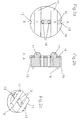
- Fig. 1 shows a device according to the invention consisting of a substantially flat membrane 1 having a rounded outer shape, with the approximate dimensions of 5.0 x 10.0 x 1.4 mm, having two pairs of lateral holes 2,3 and 4,5 with diameters in the range of 0.2 to 2.5 mm, and preferably 1 mm, to house the suture. All edges of the device are rounded to prevent irritation of the tissue and diminish the chance of suture damage due to friction.
- the surface 6 of the device which faces the tendon has 0.5 mm long spikes 7 to prevent slippage of the device on the tendon.
- the opposite surface 8 of the flat membrane 1 of the device is strengthened by incorporation of three bars 9 of the same material as the membrane 1.
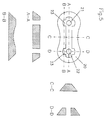
- Figs. 2a, 2b and 2c show a tendon augmentation device according to the invention which has a circular shape in the form of a disk 10.
- the diameter of the disk 10 is about 8 mm and its thickness about 2 mm.
- the disk 10 has two or three 0.5 mm thick reinforcing bars 11 of the same material as the disk to support the suture and two holes 12 with a diameter of 0.5 to 1.3 mm to house the suture and two cuts 13 at the edges of disk 10 to protect against slippage of the suture over the device.
- the surface 14 of the device facing the tendon has rounded edges 15 to diminish irritation and 0.5 to 1.0 mm long spikes 16 to prevent slippage of the device on the tendon.
- the tendon augmentation device 1 As illustrated in Fig. 3 the tendon augmentation device 1 according to Fig. 1 is used for repairing and augmenting a disrupted tendon in a living body.
- the surgical method comprises the following steps:
- the membrane 1 prevents the suture 19 from cutting through the tendon 18 at the point of highest stress (pulley).
- the same operative technique is illustrated in Fig. 4 when a pair of disk-shaped devices 10 according to Fig. 2 are used.
- the operative method comprises:
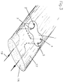
- Fig. 5 shows a bone augmentation device consisting of a membrane 20 similar to the one used for the tendon augmentation device according to Fig. 1. It is fashioned into a rectangle, preferably with dimensions of 5.0 x 10.0 x 1.4 mm, with rounded edges to prevent irritation of the surrounding tissue and diminish friction between the suture and the device.
- the membrane 20 has two holes 21,22 and three reinforcing bars 23.
- Intraoperatively the device according to Fig. 5 is placed onto the osteoporotic bone of e.g. the humeral head. The suture is pulled through the holes 21,22 while the device acts as a washer.
- In vivo tests in the sheep with the resorbable device according to Fig. 5 to augment the bone showed to increase its holding power to 398 N (520%), as compared with 76 N for the nonaugmented cancellous bone. It allows therefore a more secure fixation of transosseous sutures to the bone.
- a similar surgical technique is used for the bone augmentation device but care should be taken that the holes drilled into the bone have identical distances between them as in the device to be secured to it in order to prevent cutting the bone by the suture material and diastasis which can be formed when the bone is cut.
- a special drill guide 22 as shown in Fig. 6 with guiding holes 21 corresponding exactly to the holes 12 of the device 10 to be secured to the bone.
- both, the tendon and the bone augmentation devices according to the invention have several advantages as compared with existing repair techniques. Thus, it reduces diastasis between the tendon and the bone being the main problem in the rotator cuff tear repair, it protects against tendon strangulation and cutting through the tendon and the osteoporotic bone at the place of fixation.
- the use of resorbable augmentation devices according to the invention has also additional advantages, i.e. once the device is resorbed, it no longer affects the already critical blood supply.
- the tendon and bone augmentation device according to the invention can be prepared using one of the common techniques applied to polymer processing, e.g. injection-moulding, extrusion, compression-molding, solution-casting, etc.
- the devices can be produced as a composite device consisting of a resorbable polymer reinforced with resorbable polymeric and/or glass fibres.
Abstract
Description
- This invention relates to a device implantable in the living body for attachment and augmentation of tendons and/or reinforcements of bones.
- The device according to the invention is particularly useful for the attachment and augmentation of the disrupted tendons of the rotator cuffs and the reinforcement of the proximal humeral bone.
- Disorders of the rotator cuff are the most common cause of painful shoulder disability. The natural history of untreated ruptures of the rotator cuff are much less favourable than previously thought. Attempts of surgical repair of ruptures almost consistently yield satisfactory pain relief, in contrast to functional restoration which is less certain, particularly in large tears. This has been attributed to a variety of factors:
- the quality of muscle motoring the tendon may be irrevocably poor;
- the quality of the tendon (circulation, elasticity, tensile strength) may preclude repair;
- the quality of cancellous bone may not provide enough stability (e.g. in the case of osteoporosis); or
- the patients cooperation may defeat the surgical gaol.
- It has been established, that functional restoration of the shoulder does not depend on the cuff rotator tear size, but on the success of repair of the defect. Thus the anatomical and mechanical success of operative tendon-to-bone attachment is therefore of great importance.
- Several factors, which influence the quality of repair such as the suture material, the quality of initial fixation, the tension in the musculotendinous unit and the load applied during the postoperative course influence the success of the repair. One of the important problems is how to grasp the tendon with the suture material to achieve a strong and secure attachment of the tendon to the bone.
- Very few literature data concerning this problem are available and yet refer to the surgical attachment of the supraspinatus tendon, e.g. as described in an article of France, Paulos, Harner and Straight "Biomechanical evaluation of rotator cuff fixation methods" published in Am J Sports Med 17:176-181,1989.
- Different prior art methods of soft tissue-to-bone fixation have been evaluated, e.g. fixation to bone by spiked washers and screws, using spiked soft tissue plates, different kinds of staples and suturing. It has been found that independently on the suturing technique used, the suture material pulls through the tendon.
- It has been also observed that when using prior art non-augmented techniques for the tendon-to-bone attachment [e.g. according to the Kessler technique described in
ML Mason, HS Allen, "The rate of healing of tendons. An experimental study of tensile strength", Ann Surg 113-3, 424-59(1941);
AD Forward, J Cowan, "Tendon suture to bone", J Bone Joint Surg 45-A(4), 807-823(1963); and
LD Ketchum, NL Martin, DA Kappel, "Experimental evaluation of factors affecting the strength of tendon repairs", Plast Reconstr Surg 59(5), 708-719(1977)]
there is a diastasis formed between connected elements, while suture applied to the tendon strangulates and/or pulls out the tendinous tissue. - Attempts have been made (France, Paulos, Harner and Straight "Biomechanical evaluation of rotator cuff fixation methods" published in Am J Sports Med 17:176-181,1989) to strengthen tendon-suture interface by interposing a polytetrafluoroethylene (PTFE) membrane between suture and tendon. Although it was expected that the membrane will augment the holding power of the tendon, no significant improvement was observed.
- In addition it was observed in reoperations that shoulder function is usually not achieved, because of diastasis between the tendon and bone. While the suture material stays always intact, the tendon is connected to the bone through functionally insufficient scar tissue.
- From J.R.Parsons, H. Alexander, S.F. Corcoran and A.B. Weiss "Development of a variable stiffness, absorbable bone plate", 25th Annual ORS, San Francisco, California, February 20-22, 1979 a bone plate is known consisting of a resorbable polymeric matrix reinforced with non-resorbable carbon fibres. The disadvantage of this known bone plate is therefore its limited resorbability due to the non-resorbable carbon fibres present in the polymeric matrix. The pure polymeric matrix of this known device without the reinforcing non-resorbable carbon fibres would have an unacceptable high Young's modulus whereas the reinforced bone plate has an unacceptable low tensile strength.
- The invention as claimed is intended to remedy these drawbacks. It solves the problem of how to design a device implantable in the living body for attachment and augmentation of tendons and reinforcement of bones having a superior tensile strength of the tendon repair and/or allowing a more secure fixation of transosseous sutures.
- The invention solves the problem with a device comprising the features of claim 1.
- The device according to the invention comprises a substantially flat membrane having a rounded outer shape and at least two perforations.
- The membrane contains resorbable or degradable polymeric material having a Young's modulus in the range of 1 to 50 GPa and a tensile strength in the range of 0.1 to 20.0 GPa and preferably of non-porous structure. The membrane may contain additionally resorbable or degradable polymeric-ceramic material. The advantage of this addition being an enhanced biocompatibility of the membrane. Young's modulus is preferably in the range of 5 to 15 GPa and most preferably in the range of 7 to 10 GPa. Tensile strength is in the range of 0.5 to 3.0 GPa and most preferably in the range of 0.7 to 2.5 GPa.
- Resorbable materials to be used for the device according to the invention can be resorbable polymers like highly purified polyhydroxyacids, polysaccharides, polyamines, polyaminoacids, polyorthoesters, polyanhydrides, polyamidoesters, polydioxanone, polyesteramides, copolyoxalates, polycarbonates or poly(glutamic-co-leucine). Preferably polylactides are used or their combinations with polyhydroxybutyrates or polyhydroxyvalerates and/or resorbable glasses.
- Other useful polyhydroxyacids comprise polycaprolactone, poly(L-lactide), poly(DL-lactide), polyglycolide, poly(DL-lactide-co-glycolide), poly(DL-lactide-co-caprolactone).
- Purposefully at least 90 weight percent of the resorbable polymeric material should have a molecular weight in the range of 200,000 to 400,000, preferably in range of 300,000 to 350,000.
- In terms of molecular weight distribution the polymeric and/or polymeric-ceramic material should have a polydispersity ( as defined in "Textbook of Polymer Science" 3rd edition, Billmeyer, Wiley-Interscience) in the range of 1.2 to 100.0, preferably in the range of 1.5 to 3.0.
- The resorbability of said material should be set at a level allowing to maintain adequate mechanical properties in vivo for at least 6 months, and preferably 7 months. The resorption rate can be adjusted to a desired value by altering the polymer molecular weight, the polymer chain orientation and crystallinity, physical structure, chemical composition, presence and extent of voids, additives a.s.o.
By way of example poly(L-dL lactide) with 5% of dL-units and a molecular weight of 400.000 Daltons resorbs at the rate which assures augmentation during the healing time. Another example for a suitably resorbable material which maintains the required mechanical properties till the healing is complete is poly(L-lactide) with a molecular weight of 320.000 Daltons hot drawn to drawratio 4. Still another example is poly(L-lactide) with a porosity in the range of 0.1 to 0.5 µm and a molecular weight of 300.000 Daltons which maintains 70% of itsinitial tensile strength 6 months after implantation. - Additionally to these resorbable materials according to the invention, other nonresorbable materials such as bioceramics (e.g. aluminium oxide ceramics), titanium, titanium alloys alone or coated with ceramics may be used to temporarily strengthen the device according to the invention. These nonresorbable materials, however, have to be removed from the implantation site after the healing is completed.
- The resorbable materials to be used with the invention should be of non-porous structure to avoid tissue ingrowth, which would interfere with the gliding function of the tendon.
- The resorbable polymeric tendon and/or bone augmentation device according to the invention can be produced either from a thin nonporous film, membrane or plate or likewise from a film, membrane or plate with controlled porosity and in vivo resorption time.
- The thickness of the membrane can be controlled to meet structural demands of the proposed implantation site, but should range between 0.5 and 6.0 mm, preferably between 1.0 and 2.0 mm and most preferably between 1.4 and 1.5 mm.
- Preferably the surface of the membrane which faces the tendon is provided with spikes to prevent slippage of the device on the tendon. Instead of spikes it is possible to use any suitable irregularites or corrugations on said surface of the membrane to obtain the same effect.
- The augmentation device according to the invention can also be used as a drug delivery device, containing antibiotics and/or fibroblast growth factor.
- The various features of novelty which characterize the invention are pointed out with particularity in the claims annexed to and forming part of this disclosure. For the better understanding of the invention, its operating advantages and specific objects attained by its use, reference should be had to the accompanying drawings, examples and descriptive matter in which are illustrated and described preferred embodiments of the invention.
-
- Fig. 1 is an elevational view of a tendon augmentation device according to the invention having a longitudinal shape with sectional views at locations A-A, B-B, C-C and D-D;
- Fig. 2a is an elevational view of a device according to the invention having a circular shape;
- Fig. 2b is a section through the device according to Fig. 2a;
- Fig. 2c is a perspective view of the device according to Figs. 2a and 2b;
- Fig. 3 is a perspective view of the tendon augmentation device according to Fig. 1 applied on a tendon;
- Fig. 4 is a perspective view of a pair of tendon augmentation devices according to Figs. 2 applied on a tendon;
- Fig. 5 is an elevational view of a bone augmentation device according to the invention having a longitudinal shape with sectional views at locations A-A, B-B, C-C and D-D; and
- Fig. 6 is a perspective view of a drilling jig for effecting of the necessary bone perforations.
- Fig. 1 shows a device according to the invention consisting of a substantially flat membrane 1 having a rounded outer shape, with the approximate dimensions of 5.0 x 10.0 x 1.4 mm, having two pairs of
lateral holes surface 6 of the device which faces the tendon has 0.5 mmlong spikes 7 to prevent slippage of the device on the tendon. Theopposite surface 8 of the flat membrane 1 of the device is strengthened by incorporation of threebars 9 of the same material as the membrane 1. - Figs. 2a, 2b and 2c show a tendon augmentation device according to the invention which has a circular shape in the form of a
disk 10. The diameter of thedisk 10 is about 8 mm and its thickness about 2 mm. Thedisk 10 has two or three 0.5 mm thick reinforcingbars 11 of the same material as the disk to support the suture and twoholes 12 with a diameter of 0.5 to 1.3 mm to house the suture and twocuts 13 at the edges ofdisk 10 to protect against slippage of the suture over the device. Thesurface 14 of the device facing the tendon has roundededges 15 to diminish irritation and 0.5 to 1.0 mmlong spikes 16 to prevent slippage of the device on the tendon. - As illustrated in Fig. 3 the tendon augmentation device 1 according to Fig. 1 is used for repairing and augmenting a disrupted tendon in a living body. The surgical method comprises the following steps:
- A. Placing the augmentation device 1 intraoperatively on the
surface 17 of thetendon 18. - B. Pulling proximally, along the tendon direction, the
suture 19 from the cut end oftendon 18, towards its outer surface. - C. Pulling the
suture 19 through thetendon 18 and one pair of twolateral holes tendon 18. - D. Pulling the
suture 19 back through thetendon 18 and the other pair oflateral holes - E. Pulling the
suture 19 longitudinally through thetendon 18 to its distal cut end. - Once fixed to the tendon the membrane 1 prevents the
suture 19 from cutting through thetendon 18 at the point of highest stress (pulley). - The same operative technique is illustrated in Fig. 4 when a pair of disk-shaped
devices 10 according to Fig. 2 are used. The operative method comprises: - A. Placing a
first device 10 according toclaim 5 on thesurface 17 of thetendon 18. - B. Pulling proximally, along the tendon direction, a
suture 19 from thecut end 20 oftendon 18 towards thedevice 10 and through one of itsholes 12 to its outer surface. - C. Pulling the
suture 19 through one of thecuts 13 into thetendon 18 towards to outer surface of thetendon 18. - D. Pulling the
suture 19 back into thetendon 18 through theother cut 13 and theother hole 12 and again into thetendon 18. - E. Pulling the
suture 19 longitudinally through thetendon 18 to its distal cut end. - F. Placing a
second device 10 according toclaim 5 on thesurface 17 of thetendon 18 and performing steps B to E. - In vitro tests showed that the use of resorbable tendon augmentation devices according to the invention for the augmentation of the supraspinatus tendon, increased the pull out strength up to 469 N (126%), as compared with the nonaugmented suture technique (with a tensile strength of 371 N only).
- Fig. 5 shows a bone augmentation device consisting of a
membrane 20 similar to the one used for the tendon augmentation device according to Fig. 1. It is fashioned into a rectangle, preferably with dimensions of 5.0 x 10.0 x 1.4 mm, with rounded edges to prevent irritation of the surrounding tissue and diminish friction between the suture and the device. Themembrane 20 has twoholes bars 23.
Intraoperatively the device according to Fig. 5 is placed onto the osteoporotic bone of e.g. the humeral head. The suture is pulled through theholes - A similar surgical technique is used for the bone augmentation device but care should be taken that the holes drilled into the bone have identical distances between them as in the device to be secured to it in order to prevent cutting the bone by the suture material and diastasis which can be formed when the bone is cut. to achieve a high accuracy which is desirable it is recommended to use a
special drill guide 22 as shown in Fig. 6 with guidingholes 21 corresponding exactly to theholes 12 of thedevice 10 to be secured to the bone. - The use of both, the tendon and the bone augmentation devices according to the invention have several advantages as compared with existing repair techniques. Thus, it reduces diastasis between the tendon and the bone being the main problem in the rotator cuff tear repair, it protects against tendon strangulation and cutting through the tendon and the osteoporotic bone at the place of fixation. The use of resorbable augmentation devices according to the invention has also additional advantages, i.e. once the device is resorbed, it no longer affects the already critical blood supply.
- The tendon and bone augmentation device according to the invention can be prepared using one of the common techniques applied to polymer processing, e.g. injection-moulding, extrusion, compression-molding, solution-casting, etc.
The devices can be produced as a composite device consisting of a resorbable polymer reinforced with resorbable polymeric and/or glass fibres. -
- I. A nonporous membrane with dimensions of 5.0 x 10.0 x 1.4 mm was prepared by casting from 10 wt.-% solution of poly(L-lactide) in chloroform at room temperature. Poly(L-lactide) with viscosity-average molecular weight of 350.000 Daltons used for preparation of the membrane was purified twice by dissolution in chloroform followed by precipitation with a methanol/water mixture. Membranes were dried to constant weight in vacuum oven at 70°. Augmentation devices of the required size were cut out from the membranes using a steel stamp. When used to augment the supraspinatus tendon they increased the pull out strength of the tendon to 470 N as compared with 370 N for nonaugmented tendons.
- II. A nonporous membrane with dimensions of 5.0 x 10.0 x 0.7 mm was prepared by casting from 7 weight-percent solution of poly(L-lactide) with a viscosity-average molecular weight of 350.000 Daltons in chloroform. When used to augment osteoporotic bone it increased the holding strength of the bone from 70 N to 270 N.
- III. A nonporous membrane with dimensions of 5.0 x 10.0 x 1.4 mm was prepared by injection molding of highly purified poly(L-lactide) with a viscosity-average molecular weight of 340.000 Daltons. The polymer was dried and kept under vacuum prior to injection-moulding to diminish thermomechanical degradation. The augmentation device placed on osteoporotic bone of the humeral head increased the holding strength of the bone from 75 N to 400 N.
- IV. A porous membrane with porosity in the range in the range of 0.5 to 1.0 µm and dimensions of 5.0 x 10.0 x 2.0 mm was prepared by solution casting from poly(L-lactide) with molecular weight of 240.000 Daltons. The augmentation device was cut out from the membrane using a steel stamp with a suitable shape. Reinforcing bars pressed in the microporous device using a suitable mould/hydraulic press system. The augmentation device placed on osteoporotic bone of the humeral head increased the holding strength of the bone from 80 to 310 N.
- V. Several manufacturing processes can be used to include reinforcing bars into the augmentation devices:
- a) Injection molding:
The mould used for preparation of the augmentation device has a shape which allows formation of the reinforcing bars in one injection-molding operation; - b) Extrusion:
The polymer ribbon is extruded through a nozzle having a shape of a device with the reinforcing bars. The final device is cut out from the ribbon using a suitable stamp. - c) Solution casting:
A membrane is prepared by solution casting. Next the membrane is placed in a mould with a suitable shape and subsequently compression moulded at temperatures in the range of 80° to 110°C.
- a) Injection molding:
Claims (23)
- A device implantable in the living body for attachment and augmentation of tendons and/or reinforcement of bones, comprising
a substantially flat membrane having a rounded outer shape and at least two perforations;
the membrane containing resorbable or degradable polymeric material, having
a Young's modulus in the range of l to 50 GPa; and
a tensile strength in the range of 0.1 to 20.0 GPa. - A device according to claim l, wherein said membrane additionally contains resorbable or degradable polymericceramic material.
- A device according to claim l or 2, wherein said resorbable or degradable polymeric material and/or said polymeric-ceramic material is of nonporous structure.
- A device according claim l or 2, wherein said resorbable or degradable polymeric material and/or said polymeric-ceramic material is of porous structure with controlled porosity in the range of 0.05 to 200 rim, preferably in the range of 0.2 to 5.0 pm.
- A device according to one of the claims l to 4, wherein said membrane has a circular outer shape.
- A device according to one of the claims 1 to 4, wherein said membrane has the shape of a tape with rounded ends with at least two perforations at each of the two ends.
- A device according to one of the claims 1 to 6, wherein said membrane is deformable to the shape of a bone.
- A device according to one of the claims 1 to 7, wherein the thickness of said membrane is between 0.5 and 6.0 mm.
- A device according to claim 8, wherein the thickness of said membrane is between 1.0 and 2.0 mm, preferably between 1.4 and 1.5 mm.
- A device according to one of the claims 1 to 9, wherein said Young's modulus is in the range of 5 to 15 GPa, preferably in the range of 7 to 10 GPa.
- A device according to one of the claims 1 to 10, wherein said tensile strength is in the range of 0.5 to 3.0 GPa, preferably in the range of 0.7 to 2.5 GPa.
- A device according to one of the claims 1 to 11, wherein at least 90 weight percent of said polymeric material has a molecular weight in the range of 200,000 to 400,000, preferably in the range of 300,000 to 350'000.
- A device according to one of the claims 1 to 12, wherein said polymeric material has a polydispersity in the range of 1.2 to 100.0 , preferably in the range of 1.5 to 3.0.
- A device according to one of the claims 1 to 13, wherein said polymeric material comprises highly purified polyhydroxyacids, polysaccharides, polyamines, polyaminoacids, polyorthoesters, polyanhydrides, polyamidoesters, polydioxanone, polyesteramides, copolyoxalates, polycarbonates or poly(glutamic-co-leucine).
- A device according to claim 14, wherein said polyhydroxyacids comprise polycaprolactone, poly(L-lactide), poly(DL-lactide), polyglycolide, poly(DL-lactide-co-glycolide), poly(DL-lactide-co-caprolactone).
- A device according to one of the claims 1 to 15, wherein a fibroblast activating agent is incorporated in the polymeric matrix.
- A device according to one of the claims 1 to 15, wherein an osteoplastic agent is incorporated in the polymeric matrix.
- A device according to one of the claims 1 to 15, wherein an antibiotic agent is incorporated in the polymeric matrix.
- A device according to one of the claims 1 to 18, wherein the polymeric material has a degradation rate in situ in the range of 6 to 24 months.
- A device according to one of the claims 1 to 19, wherein the polymeric chains in the polymeric material are at least partially oriented.
- A device according to one of the claims 1 to 20, wherein said membrane is preshaped to conform to the curvature of the humeral head.
- A device according to one of the claims 1 to 21, wherein a surface (14) of said membrane facing the tendon and/or bone is provided with a three-dimensional structure, preferably in the form of spikes (16), corrugations or irregularities.
- A device according to one of the claims 1 to 22, wherein a surface (14) of said membrane facing the tendon is provided with rounded edges (15).
Applications Claiming Priority (2)
| Application Number | Priority Date | Filing Date | Title |
|---|---|---|---|
| US70538991A | 1991-05-24 | 1991-05-24 | |
| US705389 | 1991-05-24 |
Publications (2)
| Publication Number | Publication Date |
|---|---|
| EP0520177A1 true EP0520177A1 (en) | 1992-12-30 |
| EP0520177B1 EP0520177B1 (en) | 1995-12-13 |
Family
ID=24833246
Family Applications (1)
| Application Number | Title | Priority Date | Filing Date |
|---|---|---|---|
| EP92108135A Expired - Lifetime EP0520177B1 (en) | 1991-05-24 | 1992-05-14 | Resorbable tendon and bone augmentation device |
Country Status (6)
| Country | Link |
|---|---|
| US (1) | US5527341A (en) |
| EP (1) | EP0520177B1 (en) |
| JP (1) | JP3439487B2 (en) |
| AT (1) | ATE131373T1 (en) |
| CA (1) | CA2069223C (en) |
| DE (1) | DE69206693T2 (en) |
Cited By (66)
| Publication number | Priority date | Publication date | Assignee | Title |
|---|---|---|---|---|
| US5306301A (en) * | 1993-02-11 | 1994-04-26 | American Cyanamid Company | Graft attachment device and method of using same |
| US5352229A (en) * | 1993-05-12 | 1994-10-04 | Marlowe Goble E | Arbor press staple and washer and method for its use |
| US5611986A (en) * | 1994-07-05 | 1997-03-18 | Ethicon, Inc. | Medical devices containing high inherent viscosity poly(p-dioxanone) |
| US5800544A (en) * | 1994-12-02 | 1998-09-01 | Omeros Medical Systems, Inc. | Tendon and ligament repair system |
| EP0867193A2 (en) * | 1997-03-27 | 1998-09-30 | Friatec Aktiengesellschaft | Foil for medical use |
| GB2329342A (en) * | 1997-09-19 | 1999-03-24 | William Frits Stewart Gibson | Suture retaining device |
| WO1999058073A1 (en) * | 1998-05-12 | 1999-11-18 | Synthes Ag Chur | Bone augmentation device |
| US6056752A (en) * | 1997-10-24 | 2000-05-02 | Smith & Nephew, Inc. | Fixation of cruciate ligament grafts |
| WO2002078574A1 (en) * | 2001-03-30 | 2002-10-10 | Depuy Acromed, Inc. | Intervertebral connection system |
| EP1416879A2 (en) * | 2001-07-16 | 2004-05-12 | Depuy Products, Inc. | Unitary surgical device and method |
| US6740100B2 (en) | 1999-12-23 | 2004-05-25 | Omeros Corporation | Tendon repair using adhesive |
| WO2005110506A1 (en) * | 2004-05-13 | 2005-11-24 | Synthes Gmbh | Resorbable polymeric medical goods with improved mechanical properties and method for producing same |
| WO2009071984A1 (en) * | 2007-12-05 | 2009-06-11 | Politecnico Di Torino | Suturing device for a tendon or ligament |
| WO2011092262A1 (en) * | 2010-01-28 | 2011-08-04 | Universität Zürich | Method and device for modelling tendinous tissue into a desired shape |
| US8006700B2 (en) | 2000-02-07 | 2011-08-30 | Demopulos Gregory A | Soft tissue repair system |
| US8092528B2 (en) | 2005-05-27 | 2012-01-10 | Depuy Spine, Inc. | Intervertebral ligament having a helical bone fastener |
| WO2013123136A1 (en) * | 2012-02-16 | 2013-08-22 | DePuy Synthes Products, LLC | Drug eluting insert for implantable body |
| DE102012006454A1 (en) * | 2012-03-30 | 2013-10-02 | Heraeus Medical Gmbh | Anti-infective spacer for osteosynthesis plates |
| EP2448520A4 (en) * | 2009-07-02 | 2015-11-18 | Imds Llc | Systems and methods for zipknot acl fixation |
| US9414925B2 (en) | 2006-09-29 | 2016-08-16 | Biomet Manufacturing, Llc | Method of implanting a knee prosthesis assembly with a ligament link |
| US9433407B2 (en) | 2012-01-03 | 2016-09-06 | Biomet Manufacturing, Llc | Method of implanting a bone fixation assembly |
| US9468433B2 (en) | 2006-02-03 | 2016-10-18 | Biomet Sports Medicine, Llc | Method and apparatus for forming a self-locking adjustable loop |
| US9538998B2 (en) | 2006-02-03 | 2017-01-10 | Biomet Sports Medicine, Llc | Method and apparatus for fracture fixation |
| US9603591B2 (en) | 2006-02-03 | 2017-03-28 | Biomet Sports Medicine, Llc | Flexible anchors for tissue fixation |
| US9615822B2 (en) | 2014-05-30 | 2017-04-11 | Biomet Sports Medicine, Llc | Insertion tools and method for soft anchor |
| US9622851B2 (en) | 2004-06-09 | 2017-04-18 | Biomet Sports Medicine, Llc | Method and apparatus for soft tissue attachment |
| US9622736B2 (en) | 2006-02-03 | 2017-04-18 | Biomet Sports Medicine, Llc | Soft tissue repair device and associated methods |
| US9642661B2 (en) | 2006-02-03 | 2017-05-09 | Biomet Sports Medicine, Llc | Method and Apparatus for Sternal Closure |
| US9681940B2 (en) | 2006-09-29 | 2017-06-20 | Biomet Sports Medicine, Llc | Ligament system for knee joint |
| US9700291B2 (en) | 2014-06-03 | 2017-07-11 | Biomet Sports Medicine, Llc | Capsule retractor |
| US9724090B2 (en) | 2006-09-29 | 2017-08-08 | Biomet Manufacturing, Llc | Method and apparatus for attaching soft tissue to bone |
| US9757119B2 (en) | 2013-03-08 | 2017-09-12 | Biomet Sports Medicine, Llc | Visual aid for identifying suture limbs arthroscopically |
| US9763656B2 (en) | 2006-02-03 | 2017-09-19 | Biomet Sports Medicine, Llc | Method and apparatus for soft tissue fixation |
| US9788876B2 (en) | 2006-09-29 | 2017-10-17 | Biomet Sports Medicine, Llc | Fracture fixation device |
| US9801708B2 (en) | 2004-11-05 | 2017-10-31 | Biomet Sports Medicine, Llc | Method and apparatus for coupling soft tissue to a bone |
| US9801620B2 (en) | 2006-02-03 | 2017-10-31 | Biomet Sports Medicine, Llc | Method and apparatus for coupling soft tissue to bone |
| US9833230B2 (en) | 2006-09-29 | 2017-12-05 | Biomet Sports Medicine, Llc | Fracture fixation device |
| US9861351B2 (en) | 2007-04-10 | 2018-01-09 | Biomet Sports Medicine, Llc | Adjustable knotless loops |
| US9918827B2 (en) | 2013-03-14 | 2018-03-20 | Biomet Sports Medicine, Llc | Scaffold for spring ligament repair |
| US9918826B2 (en) | 2006-09-29 | 2018-03-20 | Biomet Sports Medicine, Llc | Scaffold for spring ligament repair |
| US9955980B2 (en) | 2015-02-24 | 2018-05-01 | Biomet Sports Medicine, Llc | Anatomic soft tissue repair |
| US10004588B2 (en) | 2006-02-03 | 2018-06-26 | Biomet Sports Medicine, Llc | Method and apparatus for fixation of an ACL graft |
| US10004493B2 (en) | 2006-09-29 | 2018-06-26 | Biomet Sports Medicine, Llc | Method for implanting soft tissue |
| US10004489B2 (en) | 2006-02-03 | 2018-06-26 | Biomet Sports Medicine, Llc | Method and apparatus for coupling soft tissue to a bone |
| US10022118B2 (en) | 2006-02-03 | 2018-07-17 | Biomet Sports Medicine, Llc | Method and apparatus for coupling soft tissue to a bone |
| US10039543B2 (en) | 2014-08-22 | 2018-08-07 | Biomet Sports Medicine, Llc | Non-sliding soft anchor |
| US10092288B2 (en) | 2006-02-03 | 2018-10-09 | Biomet Sports Medicine, Llc | Method and apparatus for coupling soft tissue to a bone |
| US10136886B2 (en) | 2013-12-20 | 2018-11-27 | Biomet Sports Medicine, Llc | Knotless soft tissue devices and techniques |
| US10149767B2 (en) | 2009-05-28 | 2018-12-11 | Biomet Manufacturing, Llc | Method of implanting knee prosthesis assembly with ligament link |
| US10154837B2 (en) | 2006-02-03 | 2018-12-18 | Biomet Sports Medicine, Llc | Method and apparatus for coupling soft tissue to a bone |
| US10251637B2 (en) | 2006-02-03 | 2019-04-09 | Biomet Sports Medicine, Llc | Soft tissue repair device and associated methods |
| US10265159B2 (en) | 2011-11-03 | 2019-04-23 | Biomet Sports Medicine, Llc | Method and apparatus for stitching tendons |
| US10265064B2 (en) | 2004-11-05 | 2019-04-23 | Biomet Sports Medicine, Llc | Soft tissue repair device and method |
| US10321906B2 (en) | 2006-02-03 | 2019-06-18 | Biomet Sports Medicine, Llc | Method for tissue fixation |
| US10363028B2 (en) | 2011-11-10 | 2019-07-30 | Biomet Sports Medicine, Llc | Method for coupling soft tissue to a bone |
| US10368856B2 (en) | 2011-11-10 | 2019-08-06 | Biomet Sports Medicine, Llc | Apparatus for coupling soft tissue to a bone |
| US10398428B2 (en) | 2006-02-03 | 2019-09-03 | Biomet Sports Medicine, Llc | Method and apparatus for coupling anatomical features |
| US10441264B2 (en) | 2006-02-03 | 2019-10-15 | Biomet Sports Medicine, Llc | Soft tissue repair assembly and associated method |
| US10517587B2 (en) | 2006-02-03 | 2019-12-31 | Biomet Sports Medicine, Llc | Method and apparatus for forming a self-locking adjustable loop |
| US10610217B2 (en) | 2006-09-29 | 2020-04-07 | Biomet Sports Medicine, Llc | Method and apparatus for forming a self-locking adjustable loop |
| US10765462B2 (en) | 2018-09-11 | 2020-09-08 | DePuy Synthes Products, Inc. | Patella bone plate and methods of fixation |
| US10905478B2 (en) | 2015-09-04 | 2021-02-02 | DePuy Synthes Products, Inc. | Patella bone plate and methods of fixation |
| US10912551B2 (en) | 2015-03-31 | 2021-02-09 | Biomet Sports Medicine, Llc | Suture anchor with soft anchor of electrospun fibers |
| US11259792B2 (en) | 2006-02-03 | 2022-03-01 | Biomet Sports Medicine, Llc | Method and apparatus for coupling anatomical features |
| US11259794B2 (en) | 2006-09-29 | 2022-03-01 | Biomet Sports Medicine, Llc | Method for implanting soft tissue |
| US11311287B2 (en) | 2006-02-03 | 2022-04-26 | Biomet Sports Medicine, Llc | Method for tissue fixation |
Families Citing this family (160)
| Publication number | Priority date | Publication date | Assignee | Title |
|---|---|---|---|---|
| JPH0614946B2 (en) * | 1986-01-20 | 1994-03-02 | ユニ・チヤ−ム株式会社 | Absorbent article surface material and method for producing the same |
| US5439467A (en) * | 1991-12-03 | 1995-08-08 | Vesica Medical, Inc. | Suture passer |
| US5716413A (en) * | 1995-10-11 | 1998-02-10 | Osteobiologics, Inc. | Moldable, hand-shapable biodegradable implant material |
| US6013083A (en) * | 1997-05-02 | 2000-01-11 | Bennett; William F. | Arthroscopic rotator cuff repair apparatus and method |
| US5718717A (en) | 1996-08-19 | 1998-02-17 | Bonutti; Peter M. | Suture anchor |
| CA2264672C (en) | 1996-10-24 | 2010-11-30 | Spinal Concepts, Inc. | Method and apparatus for spinal fixation |
| US6416515B1 (en) | 1996-10-24 | 2002-07-09 | Spinal Concepts, Inc. | Spinal fixation system |
| JP2001511685A (en) * | 1997-02-13 | 2001-08-14 | ボストン サイエンティフィック リミテッド | Stabilized sling for use in minimally invasive pelvic surgery |
| US6045579A (en) | 1997-05-01 | 2000-04-04 | Spinal Concepts, Inc. | Adjustable height fusion device |
| US5928243A (en) * | 1997-07-16 | 1999-07-27 | Spinal Concepts, Inc. | Pedicle probe and depth gage |
| US20050216059A1 (en) * | 2002-09-05 | 2005-09-29 | Bonutti Peter M | Method and apparatus for securing a suture |
| US6010525A (en) | 1997-08-01 | 2000-01-04 | Peter M. Bonutti | Method and apparatus for securing a suture |
| US6030389A (en) * | 1997-08-04 | 2000-02-29 | Spinal Concepts, Inc. | System and method for stabilizing the human spine with a bone plate |
| US6454769B2 (en) | 1997-08-04 | 2002-09-24 | Spinal Concepts, Inc. | System and method for stabilizing the human spine with a bone plate |
| US6053921A (en) * | 1997-08-26 | 2000-04-25 | Spinal Concepts, Inc. | Surgical cable system and method |
| US5964769A (en) | 1997-08-26 | 1999-10-12 | Spinal Concepts, Inc. | Surgical cable system and method |
| CA2304296C (en) | 1997-10-01 | 2005-02-15 | Boston Scientific Limited | Pelvic floor reconstruction |
| US6149669A (en) * | 1997-10-30 | 2000-11-21 | Li Medical Technologies, Inc. | Surgical fastener assembly method of use |
| US6045551A (en) | 1998-02-06 | 2000-04-04 | Bonutti; Peter M. | Bone suture |
| US6106545A (en) * | 1998-04-16 | 2000-08-22 | Axya Medical, Inc. | Suture tensioning and fixation device |
| US7758614B2 (en) | 1998-07-08 | 2010-07-20 | Tornier, Inc. | Coupling member for knotless sutures and ligatures |
| US6409743B1 (en) | 1998-07-08 | 2002-06-25 | Axya Medical, Inc. | Devices and methods for securing sutures and ligatures without knots |
| US6423088B1 (en) | 1998-07-08 | 2002-07-23 | Axya Medical, Inc. | Sharp edged device for closing wounds without knots |
| US6117139A (en) * | 1998-12-25 | 2000-09-12 | Nagoya Screw Mfg., Co., Ltd. | Ligament graft-securing device |
| US6241749B1 (en) * | 1999-04-12 | 2001-06-05 | Simon B. Rayhanabad | Adjustable tension device for sutures |
| US6447516B1 (en) | 1999-08-09 | 2002-09-10 | Peter M. Bonutti | Method of securing tissue |
| US6368343B1 (en) | 2000-03-13 | 2002-04-09 | Peter M. Bonutti | Method of using ultrasonic vibration to secure body tissue |
| DE19952359C1 (en) * | 1999-10-30 | 2001-03-22 | Aesculap Ag & Co Kg | Surgical connection has coupling element, two placement elements, bone plates, and holders |
| US7887551B2 (en) * | 1999-12-02 | 2011-02-15 | Smith & Nephew, Inc. | Soft tissue attachment and repair |
| US6331179B1 (en) | 2000-01-06 | 2001-12-18 | Spinal Concepts, Inc. | System and method for stabilizing the human spine with a bone plate |
| US6635073B2 (en) | 2000-05-03 | 2003-10-21 | Peter M. Bonutti | Method of securing body tissue |
| US6514274B1 (en) * | 2000-02-25 | 2003-02-04 | Arthrotek, Inc. | Method and apparatus for rotator cuff repair |
| TW580377B (en) | 2000-03-07 | 2004-03-21 | Metacardia Inc | Everting staple devices and methods |
| US8932330B2 (en) * | 2000-03-13 | 2015-01-13 | P Tech, Llc | Method and device for securing body tissue |
| US9138222B2 (en) | 2000-03-13 | 2015-09-22 | P Tech, Llc | Method and device for securing body tissue |
| US7094251B2 (en) | 2002-08-27 | 2006-08-22 | Marctec, Llc. | Apparatus and method for securing a suture |
| US7156862B2 (en) | 2000-05-19 | 2007-01-02 | Coapt Systems, Inc. | Multi-point tension distribution system device and method of tissue approximation using that device to improve wound healing |
| AU2002217880A1 (en) | 2000-11-15 | 2002-05-27 | Scimed Life Systems, Inc. | Device and method for treating female urinary incontinence |
| US8033983B2 (en) | 2001-03-09 | 2011-10-11 | Boston Scientific Scimed, Inc. | Medical implant |
| EP1365679B1 (en) | 2001-03-09 | 2007-11-14 | Boston Scientific Limited | Medical slings |
| US7670361B2 (en) * | 2001-06-15 | 2010-03-02 | Aesculap Ag | Implant for fixing bone plates |
| GB0116341D0 (en) * | 2001-07-04 | 2001-08-29 | Smith & Nephew | Biodegradable polymer systems |
| US6755781B2 (en) | 2001-07-27 | 2004-06-29 | Scimed Life Systems, Inc. | Medical slings |
| US7070558B2 (en) * | 2001-07-27 | 2006-07-04 | Boston Scientific Scimed, Inc. | Medical slings |
| WO2003026486A2 (en) * | 2001-09-27 | 2003-04-03 | Mayo Foundation For Medical Education And Research | Eyelet reinforcement at the tissue-suture interface |
| US7766947B2 (en) | 2001-10-31 | 2010-08-03 | Ortho Development Corporation | Cervical plate for stabilizing the human spine |
| US6719765B2 (en) | 2001-12-03 | 2004-04-13 | Bonutti 2003 Trust-A | Magnetic suturing system and method |
| GB0202233D0 (en) * | 2002-01-31 | 2002-03-20 | Smith & Nephew | Bioresorbable polymers |
| US9155544B2 (en) | 2002-03-20 | 2015-10-13 | P Tech, Llc | Robotic systems and methods |
| US7682392B2 (en) | 2002-10-30 | 2010-03-23 | Depuy Spine, Inc. | Regenerative implants for stabilizing the spine and devices for attachment of said implants |
| AU2003259834A1 (en) | 2002-12-17 | 2004-07-29 | Boston Scientific Limited | Spacer for sling delivery system |
| WO2004071356A2 (en) * | 2003-02-10 | 2004-08-26 | Smith & Nephew, Inc. | Resorbable devices |
| US7497864B2 (en) | 2003-04-30 | 2009-03-03 | Marctec, Llc. | Tissue fastener and methods for using same |
| US7150757B2 (en) * | 2003-06-11 | 2006-12-19 | Fallin T Wade | Adjustable line locks and methods |
| US7594923B2 (en) * | 2003-06-11 | 2009-09-29 | Medicine Lodge, Inc | Line lock suture attachment systems and methods |
| US7722644B2 (en) * | 2003-06-11 | 2010-05-25 | Medicine Lodge, Inc. | Compact line locks and methods |
| US7806909B2 (en) * | 2003-06-11 | 2010-10-05 | Medicine Lodge Inc. | Line lock threading systems and methods |
| GB0317192D0 (en) * | 2003-07-19 | 2003-08-27 | Smith & Nephew | High strength bioresorbable co-polymers |
| US7361138B2 (en) | 2003-07-31 | 2008-04-22 | Scimed Life Systems, Inc. | Bioabsorbable casing for surgical sling assembly |
| GB0329654D0 (en) | 2003-12-23 | 2004-01-28 | Smith & Nephew | Tunable segmented polyacetal |
| US20050197699A1 (en) * | 2004-02-10 | 2005-09-08 | Jacobs Daniel I. | Tissue repair apparatus and method |
| US20080039873A1 (en) | 2004-03-09 | 2008-02-14 | Marctec, Llc. | Method and device for securing body tissue |
| US20050251143A1 (en) * | 2004-05-05 | 2005-11-10 | Dillard David G | Surgical system and method of use for soft tissue fixation to bone |
| US7407511B2 (en) * | 2004-05-13 | 2008-08-05 | Wright Medical Technology Inc | Methods and materials for connective tissue repair |
| US7785348B2 (en) * | 2004-05-14 | 2010-08-31 | Ethicon Endo-Surgery, Inc. | Devices and methods of locking and cutting a suture in a medical procedure |
| RU2416371C2 (en) | 2004-06-02 | 2011-04-20 | КейЭфэкс МЕДИКАЛ КОРПОРЕЙШН | System and method of fastening soft tissue to bone |
| US8062334B2 (en) | 2004-06-02 | 2011-11-22 | Kfx Medical Corporation | Suture anchor |
| US7695503B1 (en) | 2004-06-09 | 2010-04-13 | Biomet Sports Medicine, Llc | Method and apparatus for soft tissue attachment |
| US7500983B1 (en) | 2004-06-09 | 2009-03-10 | Biomet Sports Medicine, Llc | Apparatus for soft tissue attachment |
| US7819898B2 (en) | 2004-06-09 | 2010-10-26 | Biomet Sports Medicine, Llc | Method and apparatus for soft tissue fixation |
| US7935136B2 (en) * | 2004-06-17 | 2011-05-03 | Alamin Todd F | Facet joint fusion devices and methods |
| DE102004038823B3 (en) | 2004-08-04 | 2006-03-30 | Aesculap Ag & Co. Kg | Implant for fixing adjacent bone plates |
| US9271766B2 (en) | 2004-10-26 | 2016-03-01 | P Tech, Llc | Devices and methods for stabilizing tissue and implants |
| US9173647B2 (en) * | 2004-10-26 | 2015-11-03 | P Tech, Llc | Tissue fixation system |
| US9463012B2 (en) | 2004-10-26 | 2016-10-11 | P Tech, Llc | Apparatus for guiding and positioning an implant |
| US20060089646A1 (en) | 2004-10-26 | 2006-04-27 | Bonutti Peter M | Devices and methods for stabilizing tissue and implants |
| US8840645B2 (en) | 2004-11-05 | 2014-09-23 | Biomet Sports Medicine, Llc | Method and apparatus for coupling soft tissue to a bone |
| US8998949B2 (en) | 2004-11-09 | 2015-04-07 | Biomet Sports Medicine, Llc | Soft tissue conduit device |
| US20060149266A1 (en) * | 2004-12-10 | 2006-07-06 | New York Society For The Ruptured And Crippled Maintaining The Hospital For Special Surgery | Anchor for screw fixation of soft tissue to bone |
| US7641694B1 (en) | 2005-01-06 | 2010-01-05 | IMDS, Inc. | Line lock graft retention system and method |
| US9089323B2 (en) | 2005-02-22 | 2015-07-28 | P Tech, Llc | Device and method for securing body tissue |
| US20060276896A1 (en) * | 2005-06-02 | 2006-12-07 | Medicinelodge, Inc. | Bone implants with integrated line locks |
| WO2007020432A2 (en) * | 2005-08-18 | 2007-02-22 | Smith & Nephew, Plc | High strength devices and composites |
| US7878970B2 (en) * | 2005-09-28 | 2011-02-01 | Boston Scientific Scimed, Inc. | Apparatus and method for suspending a uterus |
| US20070167950A1 (en) * | 2005-12-22 | 2007-07-19 | Tauro Joseph C | System and method for attaching soft tissue to bone |
| US9144483B2 (en) * | 2006-01-13 | 2015-09-29 | Boston Scientific Scimed, Inc. | Placing fixation devices |
| US8574235B2 (en) | 2006-02-03 | 2013-11-05 | Biomet Sports Medicine, Llc | Method for trochanteric reattachment |
| US8506597B2 (en) | 2011-10-25 | 2013-08-13 | Biomet Sports Medicine, Llc | Method and apparatus for interosseous membrane reconstruction |
| US9271713B2 (en) | 2006-02-03 | 2016-03-01 | Biomet Sports Medicine, Llc | Method and apparatus for tensioning a suture |
| US8771352B2 (en) | 2011-05-17 | 2014-07-08 | Biomet Sports Medicine, Llc | Method and apparatus for tibial fixation of an ACL graft |
| US11253296B2 (en) | 2006-02-07 | 2022-02-22 | P Tech, Llc | Methods and devices for intracorporeal bonding of implants with thermal energy |
| US8496657B2 (en) | 2006-02-07 | 2013-07-30 | P Tech, Llc. | Methods for utilizing vibratory energy to weld, stake and/or remove implants |
| ES2624589T3 (en) * | 2006-02-07 | 2017-07-17 | Tepha, Inc. | Methods and devices for rotator cuff repair |
| US7967820B2 (en) | 2006-02-07 | 2011-06-28 | P Tech, Llc. | Methods and devices for trauma welding |
| US11278331B2 (en) | 2006-02-07 | 2022-03-22 | P Tech Llc | Method and devices for intracorporeal bonding of implants with thermal energy |
| WO2007103276A2 (en) | 2006-03-03 | 2007-09-13 | Smith & Nephew, Inc. | Systems and methods for delivering a medicament |
| US9078727B2 (en) * | 2006-03-16 | 2015-07-14 | Boston Scientific Scimed, Inc. | System and method for treating tissue wall prolapse |
| US7828820B2 (en) | 2006-03-21 | 2010-11-09 | Biomet Sports Medicine, Llc | Method and apparatuses for securing suture |
| US11246638B2 (en) | 2006-05-03 | 2022-02-15 | P Tech, Llc | Methods and devices for utilizing bondable materials |
| US20080009900A1 (en) * | 2006-06-12 | 2008-01-10 | Kfx Medical Corporation | Surgical grasping device |
| CA2679365C (en) | 2006-11-30 | 2016-05-03 | Smith & Nephew, Inc. | Fiber reinforced composite material |
| US8617185B2 (en) | 2007-02-13 | 2013-12-31 | P Tech, Llc. | Fixation device |
| US8112846B2 (en) * | 2007-03-08 | 2012-02-14 | Mattel, Inc. | Cleat for securing packaging ties |
| JP5416090B2 (en) * | 2007-04-18 | 2014-02-12 | スミス アンド ネフュー ピーエルシー | Expansion molding of shape memory polymer |
| EP2142227B1 (en) * | 2007-04-19 | 2012-02-29 | Smith & Nephew, Inc. | Multi-modal shape memory polymers |
| US9770534B2 (en) * | 2007-04-19 | 2017-09-26 | Smith & Nephew, Inc. | Graft fixation |
| US8480715B2 (en) | 2007-05-22 | 2013-07-09 | Zimmer Spine, Inc. | Spinal implant system and method |
| US20090018655A1 (en) * | 2007-07-13 | 2009-01-15 | John Brunelle | Composite Implant for Surgical Repair |
| US7771455B2 (en) * | 2007-08-31 | 2010-08-10 | Ken Christopher G M | Closure medical device |
| US8287909B2 (en) * | 2007-12-19 | 2012-10-16 | Tepha, Inc. | Medical devices containing melt-blown non-wovens of poly-4-hydroxybutyrate and copolymers thereof |
| US8597336B2 (en) * | 2007-12-28 | 2013-12-03 | Howmedica Osteonics Corp. | Apparatus for discrete tissue anchoring for soft tissue repair and method of use |
| US8221453B2 (en) * | 2008-02-16 | 2012-07-17 | Aljizawi Hakim Mahmoud | Laparoscopic auto-knot sutures device |
| WO2009149455A1 (en) * | 2008-06-06 | 2009-12-10 | Synthes Usa, Llc | Suture based tissue repair |
| US8777990B2 (en) * | 2008-09-08 | 2014-07-15 | Howmedica Osteonics Corp. | Knotless suture anchor for soft tissue repair and method of use |
| US9451942B2 (en) * | 2008-11-12 | 2016-09-27 | Howmedica Osteonics Corp. | Insertion tool for knotless suture anchor for soft tissue repair and method of use |
| EP2400899A4 (en) | 2009-02-24 | 2015-03-18 | P Tech Llc | Methods and devices for utilizing bondable materials |
| US8858577B2 (en) | 2010-05-19 | 2014-10-14 | University Of Utah Research Foundation | Tissue stabilization system |
| US8911348B2 (en) | 2010-09-02 | 2014-12-16 | Boston Scientific Scimed, Inc. | Pelvic implants and methods of implanting the same |
| WO2012033977A1 (en) | 2010-09-09 | 2012-03-15 | Gore Enterprise Holdings, Inc. | Method of increasing film tear strength |
| US8795334B2 (en) | 2011-01-28 | 2014-08-05 | Smith & Nephew, Inc. | Tissue repair |
| US8852214B2 (en) | 2011-02-04 | 2014-10-07 | University Of Utah Research Foundation | System for tissue fixation to bone |
| US9168120B2 (en) | 2011-09-09 | 2015-10-27 | Boston Scientific Scimed, Inc. | Medical device and methods of delivering the medical device |
| US9370350B2 (en) | 2011-11-10 | 2016-06-21 | Biomet Sports Medicine, Llc | Apparatus for coupling soft tissue to a bone |
| US8926662B2 (en) | 2012-02-01 | 2015-01-06 | Smith & Nephew, Inc. | Tissue graft anchoring |
| US9084597B2 (en) | 2012-03-09 | 2015-07-21 | Smith & Nephew, Inc. | Suture-based knotless repair |
| US11944531B2 (en) | 2012-07-30 | 2024-04-02 | Conextions, Inc. | Devices, systems, and methods for repairing soft tissue and attaching soft tissue to bone |
| US10390935B2 (en) | 2012-07-30 | 2019-08-27 | Conextions, Inc. | Soft tissue to bone repair devices, systems, and methods |
| US10835241B2 (en) | 2012-07-30 | 2020-11-17 | Conextions, Inc. | Devices, systems, and methods for repairing soft tissue and attaching soft tissue to bone |
| US9629632B2 (en) | 2012-07-30 | 2017-04-25 | Conextions, Inc. | Soft tissue repair devices, systems, and methods |
| US9427309B2 (en) | 2012-07-30 | 2016-08-30 | Conextions, Inc. | Soft tissue repair devices, systems, and methods |
| US11253252B2 (en) | 2012-07-30 | 2022-02-22 | Conextions, Inc. | Devices, systems, and methods for repairing soft tissue and attaching soft tissue to bone |
| US10219804B2 (en) | 2012-07-30 | 2019-03-05 | Conextions, Inc. | Devices, systems, and methods for repairing soft tissue and attaching soft tissue to bone |
| US8986327B2 (en) | 2012-10-18 | 2015-03-24 | Smith & Nephew, Inc. | Flexible anchor delivery system |
| US10076377B2 (en) | 2013-01-05 | 2018-09-18 | P Tech, Llc | Fixation systems and methods |
| US9056003B2 (en) | 2013-01-25 | 2015-06-16 | Smith & Nephew, Inc. | Tissue graft fixation |
| MX367766B (en) | 2013-03-12 | 2019-09-05 | Ziptek Llc | APPARATUS and METHOD FOR SECURING TISSUE. |
| US9814555B2 (en) | 2013-03-12 | 2017-11-14 | Boston Scientific Scimed, Inc. | Medical device for pelvic floor repair and method of delivering the medical device |
| US9936940B2 (en) | 2013-06-07 | 2018-04-10 | Biomet Sports Medicine, Llc | Method and apparatus for coupling soft tissue to bone |
| ES2706759T3 (en) * | 2013-07-10 | 2019-04-01 | Tepha Inc | Soft suture anchors |
| USD819432S1 (en) | 2014-03-11 | 2018-06-05 | Ziptek LLC. | Screw |
| US11583384B2 (en) | 2014-03-12 | 2023-02-21 | Conextions, Inc. | Devices, systems, and methods for repairing soft tissue and attaching soft tissue to bone |
| US9993332B2 (en) | 2014-07-09 | 2018-06-12 | Medos International Sarl | Systems and methods for ligament graft preparation |
| US10034742B2 (en) | 2014-10-23 | 2018-07-31 | Medos International Sarl | Biceps tenodesis implants and delivery tools |
| US10729419B2 (en) | 2014-10-23 | 2020-08-04 | Medos International Sarl | Biceps tenodesis implants and delivery tools |
| US10076374B2 (en) | 2014-10-23 | 2018-09-18 | Medos International Sárl | Biceps tenodesis delivery tools |
| US10751161B2 (en) | 2014-10-23 | 2020-08-25 | Medos International Sárl | Biceps tenodesis anchor implants |
| US10856966B2 (en) | 2014-10-23 | 2020-12-08 | Medos International Sarl | Biceps tenodesis implants and delivery tools |
| US9693856B2 (en) | 2015-04-22 | 2017-07-04 | DePuy Synthes Products, LLC | Biceps repair device |
| US10182808B2 (en) | 2015-04-23 | 2019-01-22 | DePuy Synthes Products, Inc. | Knotless suture anchor guide |
| US10058393B2 (en) | 2015-10-21 | 2018-08-28 | P Tech, Llc | Systems and methods for navigation and visualization |
| US10383720B2 (en) | 2015-12-22 | 2019-08-20 | DePuy Synthes Products, Inc. | Graft preparation system |
| EP3184078B1 (en) | 2015-12-23 | 2020-10-07 | INOVEDIS GmbH | Tendon fixation plate |
| US10231824B2 (en) | 2016-04-08 | 2019-03-19 | Medos International Sárl | Tenodesis anchoring systems and tools |
| US10231823B2 (en) | 2016-04-08 | 2019-03-19 | Medos International Sarl | Tenodesis implants and tools |
| US11696822B2 (en) | 2016-09-28 | 2023-07-11 | Conextions, Inc. | Devices, systems, and methods for repairing soft tissue and attaching soft tissue to bone |
| US10299784B2 (en) * | 2017-07-06 | 2019-05-28 | Christian N. Anderson | Suture button construct for surgical procedures |
| US11547397B2 (en) | 2017-12-20 | 2023-01-10 | Conextions, Inc. | Devices, systems, and methods for repairing soft tissue and attaching soft tissue to bone |
| KR102006132B1 (en) * | 2018-02-08 | 2019-08-01 | 건양대학교 산학협력단 | Customized Bone Prosthesis to Prevent Nerve Compression |
| KR102006136B1 (en) * | 2018-02-08 | 2019-08-01 | 건양대학교 산학협력단 | Customized Bone Prosthesis With Soft Tissue Connecting Portion |
| CA3091800A1 (en) | 2018-02-20 | 2019-08-29 | Conextions, Inc. | Devices, systems, and methods for repairing soft tissue and attaching soft tissue to bone |
Citations (5)
| Publication number | Priority date | Publication date | Assignee | Title |
|---|---|---|---|---|
| FR2422386A1 (en) * | 1978-04-13 | 1979-11-09 | Christian Buscayret | Device for fastening suture into tissue - has rectangular body with two perforations accommodating suture and two protuberances engaging tissue |
| DE2947985A1 (en) * | 1979-11-28 | 1981-09-17 | Vsesojuznyj naučno-issledovatel'skij i ispytatel'nyj institut medicinskoj techniki, Moskva | Matrix material for fixing bone fractures - consisting of a copolymer of hydrophilic and hydrophobic monomers reinforced with resorbable non-non-toxic fibres |
| WO1988003417A1 (en) * | 1986-11-03 | 1988-05-19 | Material Consultants Oy | Surgical biocomposite material and a method for producing the material |
| WO1988006872A1 (en) * | 1987-03-09 | 1988-09-22 | Astra Meditec Ab | A resorbable prosthesis |
| DE9002844U1 (en) * | 1990-03-10 | 1990-12-06 | Giers, Roland, 4950 Minden, De |
Family Cites Families (19)
| Publication number | Priority date | Publication date | Assignee | Title |
|---|---|---|---|---|
| US2199025A (en) * | 1936-06-08 | 1940-04-30 | Carl E Conn | Means and method of closing surgical incisions |
| US3739773A (en) * | 1963-10-31 | 1973-06-19 | American Cyanamid Co | Polyglycolic acid prosthetic devices |
| US3931821A (en) * | 1972-11-24 | 1976-01-13 | Bio-Medicus, Inc. | Suture bridges |
| US3976079A (en) * | 1974-08-01 | 1976-08-24 | Samuels Peter B | Securing devices for sutures |
| US4210148A (en) * | 1978-11-03 | 1980-07-01 | Stivala Oscar G | Retention suture system |
| US4512038A (en) * | 1979-04-27 | 1985-04-23 | University Of Medicine And Dentistry Of New Jersey | Bio-absorbable composite tissue scaffold |
| US4789732A (en) * | 1980-08-04 | 1988-12-06 | Regents Of The University Of California | Bone morphogenetic protein composition |
| DE3206279A1 (en) * | 1982-02-20 | 1983-09-08 | B. Braun Melsungen Ag, 3508 Melsungen | RELIEF SEAMING CUTLERY FOR SURGERY |
| US4823794A (en) * | 1982-07-12 | 1989-04-25 | Pierce William S | Surgical pledget |
| US4990161A (en) * | 1984-03-16 | 1991-02-05 | Kampner Stanley L | Implant with resorbable stem |
| US4609551A (en) * | 1984-03-20 | 1986-09-02 | Arnold Caplan | Process of and material for stimulating growth of cartilage and bony tissue at anatomical sites |
| GB8514055D0 (en) * | 1985-06-04 | 1985-07-10 | Geistlich Soehne Ag | Chemical substance |
| US4905680A (en) * | 1986-10-27 | 1990-03-06 | Johnson & Johnson Orthopaedics, Inc. | Absorbable bone plate |
| FI81498C (en) * | 1987-01-13 | 1990-11-12 | Biocon Oy | SURGICAL MATERIAL OCH INSTRUMENT. |
| DE8714327U1 (en) * | 1987-10-28 | 1987-12-23 | Asys-Gmbh Gesellschaft Fuer Die Herstellung Und Den Vertrieb Von Systemen Fuer Die Automatisierung, 7060 Schorndorf, De | |
| US5013292A (en) * | 1989-02-24 | 1991-05-07 | R. Laborie Medical Corporation | Surgical correction of female urinary stress incontinence and kit therefor |
| GB9020379D0 (en) * | 1990-09-18 | 1990-10-31 | Femcare Ltd | Suture apparatus |
| US5366480A (en) * | 1990-12-24 | 1994-11-22 | American Cyanamid Company | Synthetic elastomeric buttressing pledget |
| JPH06114067A (en) * | 1992-09-30 | 1994-04-26 | Takiron Co Ltd | Binding button for medical suture |
-
1992
- 1992-05-14 AT AT92108135T patent/ATE131373T1/en not_active IP Right Cessation
- 1992-05-14 EP EP92108135A patent/EP0520177B1/en not_active Expired - Lifetime
- 1992-05-14 DE DE69206693T patent/DE69206693T2/en not_active Expired - Fee Related
- 1992-05-22 JP JP15421992A patent/JP3439487B2/en not_active Expired - Fee Related
- 1992-05-22 CA CA002069223A patent/CA2069223C/en not_active Expired - Fee Related
-
1994
- 1994-08-30 US US08/297,975 patent/US5527341A/en not_active Expired - Fee Related
Patent Citations (5)
| Publication number | Priority date | Publication date | Assignee | Title |
|---|---|---|---|---|
| FR2422386A1 (en) * | 1978-04-13 | 1979-11-09 | Christian Buscayret | Device for fastening suture into tissue - has rectangular body with two perforations accommodating suture and two protuberances engaging tissue |
| DE2947985A1 (en) * | 1979-11-28 | 1981-09-17 | Vsesojuznyj naučno-issledovatel'skij i ispytatel'nyj institut medicinskoj techniki, Moskva | Matrix material for fixing bone fractures - consisting of a copolymer of hydrophilic and hydrophobic monomers reinforced with resorbable non-non-toxic fibres |
| WO1988003417A1 (en) * | 1986-11-03 | 1988-05-19 | Material Consultants Oy | Surgical biocomposite material and a method for producing the material |
| WO1988006872A1 (en) * | 1987-03-09 | 1988-09-22 | Astra Meditec Ab | A resorbable prosthesis |
| DE9002844U1 (en) * | 1990-03-10 | 1990-12-06 | Giers, Roland, 4950 Minden, De |
Cited By (135)
| Publication number | Priority date | Publication date | Assignee | Title |
|---|---|---|---|---|
| US5645588A (en) * | 1993-02-11 | 1997-07-08 | Acufex Microsurgical, Inc. | Graft attachment device |
| US5306301A (en) * | 1993-02-11 | 1994-04-26 | American Cyanamid Company | Graft attachment device and method of using same |
| US5352229A (en) * | 1993-05-12 | 1994-10-04 | Marlowe Goble E | Arbor press staple and washer and method for its use |
| US5611986A (en) * | 1994-07-05 | 1997-03-18 | Ethicon, Inc. | Medical devices containing high inherent viscosity poly(p-dioxanone) |
| US5869597A (en) * | 1994-07-05 | 1999-02-09 | Ethicon, Inc. | Medical devices containing high inherent viscosity poly(p-dioxanone) |
| US5800544A (en) * | 1994-12-02 | 1998-09-01 | Omeros Medical Systems, Inc. | Tendon and ligament repair system |
| EP0867193A3 (en) * | 1997-03-27 | 1999-04-28 | Friatec Aktiengesellschaft | Foil for medical use |
| EP0867193A2 (en) * | 1997-03-27 | 1998-09-30 | Friatec Aktiengesellschaft | Foil for medical use |
| GB2329342B (en) * | 1997-09-19 | 2002-03-20 | William Frits Stewart Gibson | A surgical spacer |
| US6063106A (en) * | 1997-09-19 | 2000-05-16 | Gibson; William Frits Stewart | Surgical spacer |
| GB2329342A (en) * | 1997-09-19 | 1999-03-24 | William Frits Stewart Gibson | Suture retaining device |
| US6056752A (en) * | 1997-10-24 | 2000-05-02 | Smith & Nephew, Inc. | Fixation of cruciate ligament grafts |
| AU733432B2 (en) * | 1998-05-12 | 2001-05-17 | Synthes Gmbh | Bone augmentation device |
| US6344042B1 (en) | 1998-05-12 | 2002-02-05 | Synthes (Usa) | Bone augmentation device |
| WO1999058073A1 (en) * | 1998-05-12 | 1999-11-18 | Synthes Ag Chur | Bone augmentation device |
| US6740100B2 (en) | 1999-12-23 | 2004-05-25 | Omeros Corporation | Tendon repair using adhesive |
| US8006700B2 (en) | 2000-02-07 | 2011-08-30 | Demopulos Gregory A | Soft tissue repair system |
| AU2002254301B2 (en) * | 2001-03-30 | 2006-08-03 | Depuy Acromed, Inc. | Intervertebral connection system |
| WO2002078574A1 (en) * | 2001-03-30 | 2002-10-10 | Depuy Acromed, Inc. | Intervertebral connection system |
| EP1416879A2 (en) * | 2001-07-16 | 2004-05-12 | Depuy Products, Inc. | Unitary surgical device and method |
| EP1416879A4 (en) * | 2001-07-16 | 2007-04-25 | Depuy Products Inc | Unitary surgical device and method |
| WO2005110506A1 (en) * | 2004-05-13 | 2005-11-24 | Synthes Gmbh | Resorbable polymeric medical goods with improved mechanical properties and method for producing same |
| US8211351B2 (en) | 2004-05-13 | 2012-07-03 | Synthes Usa, Llc | Resorbable polymeric medical goods with improved mechanical properties and method for producing same |
| US9622851B2 (en) | 2004-06-09 | 2017-04-18 | Biomet Sports Medicine, Llc | Method and apparatus for soft tissue attachment |
| US11109857B2 (en) | 2004-11-05 | 2021-09-07 | Biomet Sports Medicine, Llc | Soft tissue repair device and method |
| US10265064B2 (en) | 2004-11-05 | 2019-04-23 | Biomet Sports Medicine, Llc | Soft tissue repair device and method |
| US9801708B2 (en) | 2004-11-05 | 2017-10-31 | Biomet Sports Medicine, Llc | Method and apparatus for coupling soft tissue to a bone |
| US8092528B2 (en) | 2005-05-27 | 2012-01-10 | Depuy Spine, Inc. | Intervertebral ligament having a helical bone fastener |
| US8591579B2 (en) | 2005-05-27 | 2013-11-26 | DePuy Synthes Products, LLC | Intervertebral ligament having a helical bone fastener |
| US10603029B2 (en) | 2006-02-03 | 2020-03-31 | Biomet Sports Medicine, Llc | Method and apparatus for coupling soft tissue to bone |
| US11589859B2 (en) | 2006-02-03 | 2023-02-28 | Biomet Sports Medicine, Llc | Method and apparatus for coupling soft tissue to bone |
| US11116495B2 (en) | 2006-02-03 | 2021-09-14 | Biomet Sports Medicine, Llc | Soft tissue repair assembly and associated method |
| US11065103B2 (en) | 2006-02-03 | 2021-07-20 | Biomet Sports Medicine, Llc | Method and apparatus for fixation of an ACL graft |
| US11730464B2 (en) | 2006-02-03 | 2023-08-22 | Biomet Sports Medicine, Llc | Soft tissue repair assembly and associated method |
| US9468433B2 (en) | 2006-02-03 | 2016-10-18 | Biomet Sports Medicine, Llc | Method and apparatus for forming a self-locking adjustable loop |
| US9538998B2 (en) | 2006-02-03 | 2017-01-10 | Biomet Sports Medicine, Llc | Method and apparatus for fracture fixation |
| US9603591B2 (en) | 2006-02-03 | 2017-03-28 | Biomet Sports Medicine, Llc | Flexible anchors for tissue fixation |
| US11723648B2 (en) | 2006-02-03 | 2023-08-15 | Biomet Sports Medicine, Llc | Method and apparatus for soft tissue fixation |
| US11819205B2 (en) | 2006-02-03 | 2023-11-21 | Biomet Sports Medicine, Llc | Soft tissue repair device and associated methods |
| US9622736B2 (en) | 2006-02-03 | 2017-04-18 | Biomet Sports Medicine, Llc | Soft tissue repair device and associated methods |
| US9642661B2 (en) | 2006-02-03 | 2017-05-09 | Biomet Sports Medicine, Llc | Method and Apparatus for Sternal Closure |
| US11039826B2 (en) | 2006-02-03 | 2021-06-22 | Biomet Sports Medicine, Llc | Method and apparatus for forming a self-locking adjustable loop |
| US10987099B2 (en) | 2006-02-03 | 2021-04-27 | Biomet Sports Medicine, Llc | Method for tissue fixation |
| US10973507B2 (en) | 2006-02-03 | 2021-04-13 | Biomet Sports Medicine, Llc | Method and apparatus for coupling soft tissue to a bone |
| US11617572B2 (en) | 2006-02-03 | 2023-04-04 | Biomet Sports Medicine, Llc | Soft tissue repair device and associated methods |
| US9763656B2 (en) | 2006-02-03 | 2017-09-19 | Biomet Sports Medicine, Llc | Method and apparatus for soft tissue fixation |
| US10932770B2 (en) | 2006-02-03 | 2021-03-02 | Biomet Sports Medicine, Llc | Soft tissue repair device and associated methods |
| US11896210B2 (en) | 2006-02-03 | 2024-02-13 | Biomet Sports Medicine, Llc | Method and apparatus for coupling soft tissue to a bone |
| US9801620B2 (en) | 2006-02-03 | 2017-10-31 | Biomet Sports Medicine, Llc | Method and apparatus for coupling soft tissue to bone |
| US11786236B2 (en) | 2006-02-03 | 2023-10-17 | Biomet Sports Medicine, Llc | Method and apparatus for coupling anatomical features |
| US11259792B2 (en) | 2006-02-03 | 2022-03-01 | Biomet Sports Medicine, Llc | Method and apparatus for coupling anatomical features |
| US11284884B2 (en) | 2006-02-03 | 2022-03-29 | Biomet Sports Medicine, Llc | Method and apparatus for coupling soft tissue to a bone |
| US10517587B2 (en) | 2006-02-03 | 2019-12-31 | Biomet Sports Medicine, Llc | Method and apparatus for forming a self-locking adjustable loop |
| US11311287B2 (en) | 2006-02-03 | 2022-04-26 | Biomet Sports Medicine, Llc | Method for tissue fixation |
| US11317907B2 (en) | 2006-02-03 | 2022-05-03 | Biomet Sports Medicine, Llc | Method and apparatus for forming a self-locking adjustable loop |
| US9993241B2 (en) | 2006-02-03 | 2018-06-12 | Biomet Sports Medicine, Llc | Method and apparatus for forming a self-locking adjustable loop |
| US10004588B2 (en) | 2006-02-03 | 2018-06-26 | Biomet Sports Medicine, Llc | Method and apparatus for fixation of an ACL graft |
| US10729430B2 (en) | 2006-02-03 | 2020-08-04 | Biomet Sports Medicine, Llc | Method and apparatus for coupling soft tissue to a bone |
| US10004489B2 (en) | 2006-02-03 | 2018-06-26 | Biomet Sports Medicine, Llc | Method and apparatus for coupling soft tissue to a bone |
| US10022118B2 (en) | 2006-02-03 | 2018-07-17 | Biomet Sports Medicine, Llc | Method and apparatus for coupling soft tissue to a bone |
| US10729421B2 (en) | 2006-02-03 | 2020-08-04 | Biomet Sports Medicine, Llc | Method and apparatus for soft tissue fixation |
| US10092288B2 (en) | 2006-02-03 | 2018-10-09 | Biomet Sports Medicine, Llc | Method and apparatus for coupling soft tissue to a bone |
| US10098629B2 (en) | 2006-02-03 | 2018-10-16 | Biomet Sports Medicine, Llc | Method and apparatus for coupling soft tissue to a bone |
| US11471147B2 (en) | 2006-02-03 | 2022-10-18 | Biomet Sports Medicine, Llc | Method and apparatus for coupling soft tissue to a bone |
| US10716557B2 (en) | 2006-02-03 | 2020-07-21 | Biomet Sports Medicine, Llc | Method and apparatus for coupling anatomical features |
| US10154837B2 (en) | 2006-02-03 | 2018-12-18 | Biomet Sports Medicine, Llc | Method and apparatus for coupling soft tissue to a bone |
| US10251637B2 (en) | 2006-02-03 | 2019-04-09 | Biomet Sports Medicine, Llc | Soft tissue repair device and associated methods |
| US10702259B2 (en) | 2006-02-03 | 2020-07-07 | Biomet Sports Medicine, Llc | Soft tissue repair assembly and associated method |
| US10695052B2 (en) | 2006-02-03 | 2020-06-30 | Biomet Sports Medicine, Llc | Method and apparatus for coupling soft tissue to a bone |
| US10321906B2 (en) | 2006-02-03 | 2019-06-18 | Biomet Sports Medicine, Llc | Method for tissue fixation |
| US10687803B2 (en) | 2006-02-03 | 2020-06-23 | Biomet Sports Medicine, Llc | Method and apparatus for coupling soft tissue to a bone |
| US10675073B2 (en) | 2006-02-03 | 2020-06-09 | Biomet Sports Medicine, Llc | Method and apparatus for sternal closure |
| US10595851B2 (en) | 2006-02-03 | 2020-03-24 | Biomet Sports Medicine, Llc | Method and apparatus for coupling soft tissue to a bone |
| US10398428B2 (en) | 2006-02-03 | 2019-09-03 | Biomet Sports Medicine, Llc | Method and apparatus for coupling anatomical features |
| US10542967B2 (en) | 2006-02-03 | 2020-01-28 | Biomet Sports Medicine, Llc | Method and apparatus for coupling soft tissue to a bone |
| US10441264B2 (en) | 2006-02-03 | 2019-10-15 | Biomet Sports Medicine, Llc | Soft tissue repair assembly and associated method |
| US11446019B2 (en) | 2006-02-03 | 2022-09-20 | Biomet Sports Medicine, Llc | Method and apparatus for coupling soft tissue to a bone |
| US9833230B2 (en) | 2006-09-29 | 2017-12-05 | Biomet Sports Medicine, Llc | Fracture fixation device |
| US10743925B2 (en) | 2006-09-29 | 2020-08-18 | Biomet Sports Medicine, Llc | Fracture fixation device |
| US10398430B2 (en) | 2006-09-29 | 2019-09-03 | Biomet Sports Medicine, Llc | Method for implanting soft tissue |
| US9414925B2 (en) | 2006-09-29 | 2016-08-16 | Biomet Manufacturing, Llc | Method of implanting a knee prosthesis assembly with a ligament link |
| US11096684B2 (en) | 2006-09-29 | 2021-08-24 | Biomet Sports Medicine, Llc | Method and apparatus for forming a self-locking adjustable loop |
| US10610217B2 (en) | 2006-09-29 | 2020-04-07 | Biomet Sports Medicine, Llc | Method and apparatus for forming a self-locking adjustable loop |
| US9681940B2 (en) | 2006-09-29 | 2017-06-20 | Biomet Sports Medicine, Llc | Ligament system for knee joint |
| US10349931B2 (en) | 2006-09-29 | 2019-07-16 | Biomet Sports Medicine, Llc | Fracture fixation device |
| US10695045B2 (en) | 2006-09-29 | 2020-06-30 | Biomet Sports Medicine, Llc | Method and apparatus for attaching soft tissue to bone |
| US11672527B2 (en) | 2006-09-29 | 2023-06-13 | Biomet Sports Medicine, Llc | Method for implanting soft tissue |
| US9724090B2 (en) | 2006-09-29 | 2017-08-08 | Biomet Manufacturing, Llc | Method and apparatus for attaching soft tissue to bone |
| US9788876B2 (en) | 2006-09-29 | 2017-10-17 | Biomet Sports Medicine, Llc | Fracture fixation device |
| US11259794B2 (en) | 2006-09-29 | 2022-03-01 | Biomet Sports Medicine, Llc | Method for implanting soft tissue |
| US10004493B2 (en) | 2006-09-29 | 2018-06-26 | Biomet Sports Medicine, Llc | Method for implanting soft tissue |
| US10835232B2 (en) | 2006-09-29 | 2020-11-17 | Biomet Sports Medicine, Llc | Fracture fixation device |
| US10517714B2 (en) | 2006-09-29 | 2019-12-31 | Biomet Sports Medicine, Llc | Ligament system for knee joint |
| US11376115B2 (en) | 2006-09-29 | 2022-07-05 | Biomet Sports Medicine, Llc | Prosthetic ligament system for knee joint |
| US9918826B2 (en) | 2006-09-29 | 2018-03-20 | Biomet Sports Medicine, Llc | Scaffold for spring ligament repair |
| US11612391B2 (en) | 2007-01-16 | 2023-03-28 | Biomet Sports Medicine, Llc | Soft tissue repair device and associated methods |
| US9861351B2 (en) | 2007-04-10 | 2018-01-09 | Biomet Sports Medicine, Llc | Adjustable knotless loops |
| US10729423B2 (en) | 2007-04-10 | 2020-08-04 | Biomet Sports Medicine, Llc | Adjustable knotless loops |
| US11185320B2 (en) | 2007-04-10 | 2021-11-30 | Biomet Sports Medicine, Llc | Adjustable knotless loops |
| WO2009071984A1 (en) * | 2007-12-05 | 2009-06-11 | Politecnico Di Torino | Suturing device for a tendon or ligament |
| US11534159B2 (en) | 2008-08-22 | 2022-12-27 | Biomet Sports Medicine, Llc | Method and apparatus for coupling soft tissue to a bone |
| US10149767B2 (en) | 2009-05-28 | 2018-12-11 | Biomet Manufacturing, Llc | Method of implanting knee prosthesis assembly with ligament link |
| EP2448520A4 (en) * | 2009-07-02 | 2015-11-18 | Imds Llc | Systems and methods for zipknot acl fixation |
| WO2011092262A1 (en) * | 2010-01-28 | 2011-08-04 | Universität Zürich | Method and device for modelling tendinous tissue into a desired shape |
| US10265159B2 (en) | 2011-11-03 | 2019-04-23 | Biomet Sports Medicine, Llc | Method and apparatus for stitching tendons |
| US11241305B2 (en) | 2011-11-03 | 2022-02-08 | Biomet Sports Medicine, Llc | Method and apparatus for stitching tendons |
| US10363028B2 (en) | 2011-11-10 | 2019-07-30 | Biomet Sports Medicine, Llc | Method for coupling soft tissue to a bone |
| US10368856B2 (en) | 2011-11-10 | 2019-08-06 | Biomet Sports Medicine, Llc | Apparatus for coupling soft tissue to a bone |
| US11534157B2 (en) | 2011-11-10 | 2022-12-27 | Biomet Sports Medicine, Llc | Method for coupling soft tissue to a bone |
| US9433407B2 (en) | 2012-01-03 | 2016-09-06 | Biomet Manufacturing, Llc | Method of implanting a bone fixation assembly |
| US11511019B2 (en) | 2012-02-16 | 2022-11-29 | DePuy Synthes Products, Inc. | Drug eluting insert for implantable body |
| WO2013123136A1 (en) * | 2012-02-16 | 2013-08-22 | DePuy Synthes Products, LLC | Drug eluting insert for implantable body |
| US9440009B2 (en) | 2012-02-16 | 2016-09-13 | DePuy Synthes Products, Inc. | Drug eluting insert for implantable body |
| EP3251705A1 (en) * | 2012-02-16 | 2017-12-06 | Synthes GmbH | Drug eluting insert for implantable body |
| US10485904B2 (en) | 2012-02-16 | 2019-11-26 | DePuy Synthes Products, Inc. | Drug eluting insert for implantable body |
| DE102012006454A1 (en) * | 2012-03-30 | 2013-10-02 | Heraeus Medical Gmbh | Anti-infective spacer for osteosynthesis plates |
| US9241746B2 (en) | 2012-03-30 | 2016-01-26 | Heraeus Medical Gmbh | Anti-infective spacer for osteosynthesis plates |
| US9757119B2 (en) | 2013-03-08 | 2017-09-12 | Biomet Sports Medicine, Llc | Visual aid for identifying suture limbs arthroscopically |
| US10758221B2 (en) | 2013-03-14 | 2020-09-01 | Biomet Sports Medicine, Llc | Scaffold for spring ligament repair |
| US9918827B2 (en) | 2013-03-14 | 2018-03-20 | Biomet Sports Medicine, Llc | Scaffold for spring ligament repair |
| US10806443B2 (en) | 2013-12-20 | 2020-10-20 | Biomet Sports Medicine, Llc | Knotless soft tissue devices and techniques |
| US11648004B2 (en) | 2013-12-20 | 2023-05-16 | Biomet Sports Medicine, Llc | Knotless soft tissue devices and techniques |
| US10136886B2 (en) | 2013-12-20 | 2018-11-27 | Biomet Sports Medicine, Llc | Knotless soft tissue devices and techniques |
| US9615822B2 (en) | 2014-05-30 | 2017-04-11 | Biomet Sports Medicine, Llc | Insertion tools and method for soft anchor |
| US9700291B2 (en) | 2014-06-03 | 2017-07-11 | Biomet Sports Medicine, Llc | Capsule retractor |
| US10743856B2 (en) | 2014-08-22 | 2020-08-18 | Biomet Sports Medicine, Llc | Non-sliding soft anchor |
| US10039543B2 (en) | 2014-08-22 | 2018-08-07 | Biomet Sports Medicine, Llc | Non-sliding soft anchor |
| US11219443B2 (en) | 2014-08-22 | 2022-01-11 | Biomet Sports Medicine, Llc | Non-sliding soft anchor |
| US9955980B2 (en) | 2015-02-24 | 2018-05-01 | Biomet Sports Medicine, Llc | Anatomic soft tissue repair |
| US10912551B2 (en) | 2015-03-31 | 2021-02-09 | Biomet Sports Medicine, Llc | Suture anchor with soft anchor of electrospun fibers |
| US11690658B2 (en) | 2015-09-04 | 2023-07-04 | DePuy Synthes Products, Inc. | Patella bone plate and methods of fixation |
| US10905478B2 (en) | 2015-09-04 | 2021-02-02 | DePuy Synthes Products, Inc. | Patella bone plate and methods of fixation |
| US11660132B2 (en) | 2018-09-11 | 2023-05-30 | DePuy Synthes Products, Inc. | Patella bone plate and methods of fixation |
| US11413078B2 (en) | 2018-09-11 | 2022-08-16 | DePuy Synthes Products, Inc. | Patella bone plate and methods of fixation |
| US10765462B2 (en) | 2018-09-11 | 2020-09-08 | DePuy Synthes Products, Inc. | Patella bone plate and methods of fixation |
Also Published As
| Publication number | Publication date |
|---|---|
| JPH0614946A (en) | 1994-01-25 |
| US5527341A (en) | 1996-06-18 |
| DE69206693T2 (en) | 1996-05-23 |
| JP3439487B2 (en) | 2003-08-25 |
| ATE131373T1 (en) | 1995-12-15 |
| DE69206693D1 (en) | 1996-01-25 |
| CA2069223C (en) | 2005-10-25 |
| EP0520177B1 (en) | 1995-12-13 |
| CA2069223A1 (en) | 1992-11-25 |
Similar Documents
| Publication | Publication Date | Title |
|---|---|---|
| EP0520177B1 (en) | Resorbable tendon and bone augmentation device | |
| US9044225B1 (en) | Composite self-retaining sutures and method | |
| US6503278B1 (en) | Under tissue conditions degradable material and a method for its manufacturing | |
| EP2150288B1 (en) | Graft fixation | |
| US6147135A (en) | Fabrication of biocompatible polymeric composites | |
| FI95537C (en) | Surgical implant | |
| US6015410A (en) | Bioabsorbable surgical implants for endoscopic soft tissue suspension procedure | |
| JP5518737B2 (en) | Indwelling suture with thermal contact mediator retainer | |
| EP2282681B1 (en) | Self-retaining sutures with bi-directional retainers or uni-directional retainers | |
| US20040138705A1 (en) | Surgical staple for tissue treatment | |
| JP3207255B2 (en) | Absorbable fixation device with controlled stiffness for treating body material in vivo and inserter of the device | |
| EP2127608B1 (en) | A medical device and its manufacture | |
| US20020169452A1 (en) | Minimally traumatic surgical device for tissue treatment | |
| US20010034520A1 (en) | Bioabsorbable pin for external bone fixation | |
| JP2002510521A (en) | Anatomical fixation implant | |
| AU2004204337A1 (en) | Suture anchor device and method of using | |
| JPH06114067A (en) | Binding button for medical suture | |
| EP1119317A1 (en) | Chute for endosteal ligament fixation | |
| Nambi Gowri et al. | Mechanical fabrication and evaluation of bioresorbable barbed sutures with different barb geometries |
Legal Events
| Date | Code | Title | Description |
|---|---|---|---|
| PUAI | Public reference made under article 153(3) epc to a published international application that has entered the european phase |
Free format text: ORIGINAL CODE: 0009012 |
|
| 17P | Request for examination filed |
Effective date: 19920514 |
|
| AK | Designated contracting states |
Kind code of ref document: A1 Designated state(s): AT CH DE FR GB LI NL |
|
| 17Q | First examination report despatched |
Effective date: 19940926 |
|
| GRAA | (expected) grant |
Free format text: ORIGINAL CODE: 0009210 |
|
| AK | Designated contracting states |
Kind code of ref document: B1 Designated state(s): AT CH DE FR GB LI NL |
|
| PG25 | Lapsed in a contracting state [announced via postgrant information from national office to epo] |
Ref country code: AT Effective date: 19951213 Ref country code: NL Free format text: LAPSE BECAUSE OF FAILURE TO SUBMIT A TRANSLATION OF THE DESCRIPTION OR TO PAY THE FEE WITHIN THE PRESCRIBED TIME-LIMIT Effective date: 19951213 |
|
| REF | Corresponds to: |
Ref document number: 131373 Country of ref document: AT Date of ref document: 19951215 Kind code of ref document: T |
|
| ET | Fr: translation filed | ||
| REF | Corresponds to: |
Ref document number: 69206693 Country of ref document: DE Date of ref document: 19960125 |
|
| REG | Reference to a national code |
Ref country code: CH Ref legal event code: NV Representative=s name: DR. LUSUARDI AG |
|
| NLV1 | Nl: lapsed or annulled due to failure to fulfill the requirements of art. 29p and 29m of the patents act | ||
| PLBE | No opposition filed within time limit |
Free format text: ORIGINAL CODE: 0009261 |
|
| STAA | Information on the status of an ep patent application or granted ep patent |
Free format text: STATUS: NO OPPOSITION FILED WITHIN TIME LIMIT |
|
| 26N | No opposition filed | ||
| REG | Reference to a national code |
Ref country code: GB Ref legal event code: IF02 |
|
| PGFP | Annual fee paid to national office [announced via postgrant information from national office to epo] |
Ref country code: CH Payment date: 20060503 Year of fee payment: 15 |
|
| REG | Reference to a national code |
Ref country code: CH Ref legal event code: PUE Owner name: SYNTHES GMBH Free format text: SYNTHES AG, CHUR#GRABENSTRASSE 15#CH-7002 CHUR (CH) -TRANSFER TO- SYNTHES GMBH#EIMATTSTRASSE 3#4436 OBERDORF (CH) |
|
| REG | Reference to a national code |
Ref country code: GB Ref legal event code: 732E |
|
| REG | Reference to a national code |
Ref country code: FR Ref legal event code: TP |
|
| PGFP | Annual fee paid to national office [announced via postgrant information from national office to epo] |
Ref country code: DE Payment date: 20070424 Year of fee payment: 16 |
|
| PGFP | Annual fee paid to national office [announced via postgrant information from national office to epo] |
Ref country code: GB Payment date: 20070424 Year of fee payment: 16 |
|
| REG | Reference to a national code |
Ref country code: CH Ref legal event code: PL |
|
| PG25 | Lapsed in a contracting state [announced via postgrant information from national office to epo] |
Ref country code: CH Free format text: LAPSE BECAUSE OF NON-PAYMENT OF DUE FEES Effective date: 20070531 Ref country code: LI Free format text: LAPSE BECAUSE OF NON-PAYMENT OF DUE FEES Effective date: 20070531 |
|
| PGFP | Annual fee paid to national office [announced via postgrant information from national office to epo] |
Ref country code: FR Payment date: 20070420 Year of fee payment: 16 |
|
| GBPC | Gb: european patent ceased through non-payment of renewal fee |
Effective date: 20080514 |
|
| REG | Reference to a national code |
Ref country code: FR Ref legal event code: ST Effective date: 20090119 |
|
| PG25 | Lapsed in a contracting state [announced via postgrant information from national office to epo] |
Ref country code: DE Free format text: LAPSE BECAUSE OF NON-PAYMENT OF DUE FEES Effective date: 20081202 Ref country code: FR Free format text: LAPSE BECAUSE OF NON-PAYMENT OF DUE FEES Effective date: 20080602 |
|
| PG25 | Lapsed in a contracting state [announced via postgrant information from national office to epo] |
Ref country code: GB Free format text: LAPSE BECAUSE OF NON-PAYMENT OF DUE FEES Effective date: 20080514 |