EP0560934B1 - Implantable bioabsorbable article - Google Patents
Implantable bioabsorbable article Download PDFInfo
- Publication number
- EP0560934B1 EP0560934B1 EP92902824A EP92902824A EP0560934B1 EP 0560934 B1 EP0560934 B1 EP 0560934B1 EP 92902824 A EP92902824 A EP 92902824A EP 92902824 A EP92902824 A EP 92902824A EP 0560934 B1 EP0560934 B1 EP 0560934B1
- Authority
- EP
- European Patent Office
- Prior art keywords
- cell
- article
- bioabsorbable
- fibrous matrix
- sheet material
- Prior art date
- Legal status (The legal status is an assumption and is not a legal conclusion. Google has not performed a legal analysis and makes no representation as to the accuracy of the status listed.)
- Expired - Lifetime
Links
Images


Classifications
-
- A—HUMAN NECESSITIES
- A61—MEDICAL OR VETERINARY SCIENCE; HYGIENE
- A61L—METHODS OR APPARATUS FOR STERILISING MATERIALS OR OBJECTS IN GENERAL; DISINFECTION, STERILISATION OR DEODORISATION OF AIR; CHEMICAL ASPECTS OF BANDAGES, DRESSINGS, ABSORBENT PADS OR SURGICAL ARTICLES; MATERIALS FOR BANDAGES, DRESSINGS, ABSORBENT PADS OR SURGICAL ARTICLES
- A61L31/00—Materials for other surgical articles, e.g. stents, stent-grafts, shunts, surgical drapes, guide wires, materials for adhesion prevention, occluding devices, surgical gloves, tissue fixation devices
- A61L31/14—Materials characterised by their function or physical properties, e.g. injectable or lubricating compositions, shape-memory materials, surface modified materials
- A61L31/148—Materials at least partially resorbable by the body
-
- A—HUMAN NECESSITIES
- A61—MEDICAL OR VETERINARY SCIENCE; HYGIENE
- A61C—DENTISTRY; APPARATUS OR METHODS FOR ORAL OR DENTAL HYGIENE
- A61C8/00—Means to be fixed to the jaw-bone for consolidating natural teeth or for fixing dental prostheses thereon; Dental implants; Implanting tools
- A61C8/0003—Not used, see subgroups
- A61C8/0004—Consolidating natural teeth
- A61C8/0006—Periodontal tissue or bone regeneration
-
- A—HUMAN NECESSITIES
- A61—MEDICAL OR VETERINARY SCIENCE; HYGIENE
- A61F—FILTERS IMPLANTABLE INTO BLOOD VESSELS; PROSTHESES; DEVICES PROVIDING PATENCY TO, OR PREVENTING COLLAPSING OF, TUBULAR STRUCTURES OF THE BODY, e.g. STENTS; ORTHOPAEDIC, NURSING OR CONTRACEPTIVE DEVICES; FOMENTATION; TREATMENT OR PROTECTION OF EYES OR EARS; BANDAGES, DRESSINGS OR ABSORBENT PADS; FIRST-AID KITS
- A61F2/00—Filters implantable into blood vessels; Prostheses, i.e. artificial substitutes or replacements for parts of the body; Appliances for connecting them with the body; Devices providing patency to, or preventing collapsing of, tubular structures of the body, e.g. stents
- A61F2/0063—Implantable repair or support meshes, e.g. hernia meshes
-
- A—HUMAN NECESSITIES
- A61—MEDICAL OR VETERINARY SCIENCE; HYGIENE
- A61F—FILTERS IMPLANTABLE INTO BLOOD VESSELS; PROSTHESES; DEVICES PROVIDING PATENCY TO, OR PREVENTING COLLAPSING OF, TUBULAR STRUCTURES OF THE BODY, e.g. STENTS; ORTHOPAEDIC, NURSING OR CONTRACEPTIVE DEVICES; FOMENTATION; TREATMENT OR PROTECTION OF EYES OR EARS; BANDAGES, DRESSINGS OR ABSORBENT PADS; FIRST-AID KITS
- A61F2/00—Filters implantable into blood vessels; Prostheses, i.e. artificial substitutes or replacements for parts of the body; Appliances for connecting them with the body; Devices providing patency to, or preventing collapsing of, tubular structures of the body, e.g. stents
- A61F2/0077—Special surfaces of prostheses, e.g. for improving ingrowth
-
- A—HUMAN NECESSITIES
- A61—MEDICAL OR VETERINARY SCIENCE; HYGIENE
- A61F—FILTERS IMPLANTABLE INTO BLOOD VESSELS; PROSTHESES; DEVICES PROVIDING PATENCY TO, OR PREVENTING COLLAPSING OF, TUBULAR STRUCTURES OF THE BODY, e.g. STENTS; ORTHOPAEDIC, NURSING OR CONTRACEPTIVE DEVICES; FOMENTATION; TREATMENT OR PROTECTION OF EYES OR EARS; BANDAGES, DRESSINGS OR ABSORBENT PADS; FIRST-AID KITS
- A61F2/00—Filters implantable into blood vessels; Prostheses, i.e. artificial substitutes or replacements for parts of the body; Appliances for connecting them with the body; Devices providing patency to, or preventing collapsing of, tubular structures of the body, e.g. stents
- A61F2/02—Prostheses implantable into the body
- A61F2/04—Hollow or tubular parts of organs, e.g. bladders, tracheae, bronchi or bile ducts
- A61F2/06—Blood vessels
-
- A—HUMAN NECESSITIES
- A61—MEDICAL OR VETERINARY SCIENCE; HYGIENE
- A61F—FILTERS IMPLANTABLE INTO BLOOD VESSELS; PROSTHESES; DEVICES PROVIDING PATENCY TO, OR PREVENTING COLLAPSING OF, TUBULAR STRUCTURES OF THE BODY, e.g. STENTS; ORTHOPAEDIC, NURSING OR CONTRACEPTIVE DEVICES; FOMENTATION; TREATMENT OR PROTECTION OF EYES OR EARS; BANDAGES, DRESSINGS OR ABSORBENT PADS; FIRST-AID KITS
- A61F2/00—Filters implantable into blood vessels; Prostheses, i.e. artificial substitutes or replacements for parts of the body; Appliances for connecting them with the body; Devices providing patency to, or preventing collapsing of, tubular structures of the body, e.g. stents
- A61F2/02—Prostheses implantable into the body
- A61F2/28—Bones
- A61F2/2846—Support means for bone substitute or for bone graft implants, e.g. membranes or plates for covering bone defects
-
- A—HUMAN NECESSITIES
- A61—MEDICAL OR VETERINARY SCIENCE; HYGIENE
- A61K—PREPARATIONS FOR MEDICAL, DENTAL OR TOILETRY PURPOSES
- A61K6/00—Preparations for dentistry
- A61K6/50—Preparations specially adapted for dental root treatment
-
- A—HUMAN NECESSITIES
- A61—MEDICAL OR VETERINARY SCIENCE; HYGIENE
- A61L—METHODS OR APPARATUS FOR STERILISING MATERIALS OR OBJECTS IN GENERAL; DISINFECTION, STERILISATION OR DEODORISATION OF AIR; CHEMICAL ASPECTS OF BANDAGES, DRESSINGS, ABSORBENT PADS OR SURGICAL ARTICLES; MATERIALS FOR BANDAGES, DRESSINGS, ABSORBENT PADS OR SURGICAL ARTICLES
- A61L27/00—Materials for grafts or prostheses or for coating grafts or prostheses
- A61L27/14—Macromolecular materials
- A61L27/18—Macromolecular materials obtained otherwise than by reactions only involving carbon-to-carbon unsaturated bonds
-
- A—HUMAN NECESSITIES
- A61—MEDICAL OR VETERINARY SCIENCE; HYGIENE
- A61L—METHODS OR APPARATUS FOR STERILISING MATERIALS OR OBJECTS IN GENERAL; DISINFECTION, STERILISATION OR DEODORISATION OF AIR; CHEMICAL ASPECTS OF BANDAGES, DRESSINGS, ABSORBENT PADS OR SURGICAL ARTICLES; MATERIALS FOR BANDAGES, DRESSINGS, ABSORBENT PADS OR SURGICAL ARTICLES
- A61L27/00—Materials for grafts or prostheses or for coating grafts or prostheses
- A61L27/28—Materials for coating prostheses
- A61L27/34—Macromolecular materials
-
- A—HUMAN NECESSITIES
- A61—MEDICAL OR VETERINARY SCIENCE; HYGIENE
- A61L—METHODS OR APPARATUS FOR STERILISING MATERIALS OR OBJECTS IN GENERAL; DISINFECTION, STERILISATION OR DEODORISATION OF AIR; CHEMICAL ASPECTS OF BANDAGES, DRESSINGS, ABSORBENT PADS OR SURGICAL ARTICLES; MATERIALS FOR BANDAGES, DRESSINGS, ABSORBENT PADS OR SURGICAL ARTICLES
- A61L27/00—Materials for grafts or prostheses or for coating grafts or prostheses
- A61L27/50—Materials characterised by their function or physical properties, e.g. injectable or lubricating compositions, shape-memory materials, surface modified materials
- A61L27/58—Materials at least partially resorbable by the body
-
- A—HUMAN NECESSITIES
- A61—MEDICAL OR VETERINARY SCIENCE; HYGIENE
- A61L—METHODS OR APPARATUS FOR STERILISING MATERIALS OR OBJECTS IN GENERAL; DISINFECTION, STERILISATION OR DEODORISATION OF AIR; CHEMICAL ASPECTS OF BANDAGES, DRESSINGS, ABSORBENT PADS OR SURGICAL ARTICLES; MATERIALS FOR BANDAGES, DRESSINGS, ABSORBENT PADS OR SURGICAL ARTICLES
- A61L31/00—Materials for other surgical articles, e.g. stents, stent-grafts, shunts, surgical drapes, guide wires, materials for adhesion prevention, occluding devices, surgical gloves, tissue fixation devices
- A61L31/04—Macromolecular materials
- A61L31/06—Macromolecular materials obtained otherwise than by reactions only involving carbon-to-carbon unsaturated bonds
-
- A—HUMAN NECESSITIES
- A61—MEDICAL OR VETERINARY SCIENCE; HYGIENE
- A61L—METHODS OR APPARATUS FOR STERILISING MATERIALS OR OBJECTS IN GENERAL; DISINFECTION, STERILISATION OR DEODORISATION OF AIR; CHEMICAL ASPECTS OF BANDAGES, DRESSINGS, ABSORBENT PADS OR SURGICAL ARTICLES; MATERIALS FOR BANDAGES, DRESSINGS, ABSORBENT PADS OR SURGICAL ARTICLES
- A61L31/00—Materials for other surgical articles, e.g. stents, stent-grafts, shunts, surgical drapes, guide wires, materials for adhesion prevention, occluding devices, surgical gloves, tissue fixation devices
- A61L31/08—Materials for coatings
- A61L31/10—Macromolecular materials
-
- A—HUMAN NECESSITIES
- A61—MEDICAL OR VETERINARY SCIENCE; HYGIENE
- A61B—DIAGNOSIS; SURGERY; IDENTIFICATION
- A61B17/00—Surgical instruments, devices or methods, e.g. tourniquets
- A61B17/04—Surgical instruments, devices or methods, e.g. tourniquets for suturing wounds; Holders or packages for needles or suture materials
- A61B17/06—Needles ; Sutures; Needle-suture combinations; Holders or packages for needles or suture materials
- A61B17/06166—Sutures
-
- A—HUMAN NECESSITIES
- A61—MEDICAL OR VETERINARY SCIENCE; HYGIENE
- A61B—DIAGNOSIS; SURGERY; IDENTIFICATION
- A61B17/00—Surgical instruments, devices or methods, e.g. tourniquets
- A61B2017/00004—(bio)absorbable, (bio)resorbable, resorptive
-
- A—HUMAN NECESSITIES
- A61—MEDICAL OR VETERINARY SCIENCE; HYGIENE
- A61B—DIAGNOSIS; SURGERY; IDENTIFICATION
- A61B90/00—Instruments, implements or accessories specially adapted for surgery or diagnosis and not covered by any of the groups A61B1/00 - A61B50/00, e.g. for luxation treatment or for protecting wound edges
- A61B90/08—Accessories or related features not otherwise provided for
- A61B2090/0815—Implantable devices for insertion in between organs or other soft tissues
- A61B2090/0816—Implantable devices for insertion in between organs or other soft tissues for preventing adhesion
-
- A—HUMAN NECESSITIES
- A61—MEDICAL OR VETERINARY SCIENCE; HYGIENE
- A61F—FILTERS IMPLANTABLE INTO BLOOD VESSELS; PROSTHESES; DEVICES PROVIDING PATENCY TO, OR PREVENTING COLLAPSING OF, TUBULAR STRUCTURES OF THE BODY, e.g. STENTS; ORTHOPAEDIC, NURSING OR CONTRACEPTIVE DEVICES; FOMENTATION; TREATMENT OR PROTECTION OF EYES OR EARS; BANDAGES, DRESSINGS OR ABSORBENT PADS; FIRST-AID KITS
- A61F2/00—Filters implantable into blood vessels; Prostheses, i.e. artificial substitutes or replacements for parts of the body; Appliances for connecting them with the body; Devices providing patency to, or preventing collapsing of, tubular structures of the body, e.g. stents
- A61F2/02—Prostheses implantable into the body
- A61F2/30—Joints
- A61F2/3094—Designing or manufacturing processes
-
- A—HUMAN NECESSITIES
- A61—MEDICAL OR VETERINARY SCIENCE; HYGIENE
- A61F—FILTERS IMPLANTABLE INTO BLOOD VESSELS; PROSTHESES; DEVICES PROVIDING PATENCY TO, OR PREVENTING COLLAPSING OF, TUBULAR STRUCTURES OF THE BODY, e.g. STENTS; ORTHOPAEDIC, NURSING OR CONTRACEPTIVE DEVICES; FOMENTATION; TREATMENT OR PROTECTION OF EYES OR EARS; BANDAGES, DRESSINGS OR ABSORBENT PADS; FIRST-AID KITS
- A61F2/00—Filters implantable into blood vessels; Prostheses, i.e. artificial substitutes or replacements for parts of the body; Appliances for connecting them with the body; Devices providing patency to, or preventing collapsing of, tubular structures of the body, e.g. stents
- A61F2/02—Prostheses implantable into the body
- A61F2/30—Joints
- A61F2002/30001—Additional features of subject-matter classified in A61F2/28, A61F2/30 and subgroups thereof
- A61F2002/30003—Material related properties of the prosthesis or of a coating on the prosthesis
- A61F2002/30004—Material related properties of the prosthesis or of a coating on the prosthesis the prosthesis being made from materials having different values of a given property at different locations within the same prosthesis
- A61F2002/30032—Material related properties of the prosthesis or of a coating on the prosthesis the prosthesis being made from materials having different values of a given property at different locations within the same prosthesis differing in absorbability or resorbability, i.e. in absorption or resorption time
-
- A—HUMAN NECESSITIES
- A61—MEDICAL OR VETERINARY SCIENCE; HYGIENE
- A61F—FILTERS IMPLANTABLE INTO BLOOD VESSELS; PROSTHESES; DEVICES PROVIDING PATENCY TO, OR PREVENTING COLLAPSING OF, TUBULAR STRUCTURES OF THE BODY, e.g. STENTS; ORTHOPAEDIC, NURSING OR CONTRACEPTIVE DEVICES; FOMENTATION; TREATMENT OR PROTECTION OF EYES OR EARS; BANDAGES, DRESSINGS OR ABSORBENT PADS; FIRST-AID KITS
- A61F2/00—Filters implantable into blood vessels; Prostheses, i.e. artificial substitutes or replacements for parts of the body; Appliances for connecting them with the body; Devices providing patency to, or preventing collapsing of, tubular structures of the body, e.g. stents
- A61F2/02—Prostheses implantable into the body
- A61F2/30—Joints
- A61F2002/30001—Additional features of subject-matter classified in A61F2/28, A61F2/30 and subgroups thereof
- A61F2002/30003—Material related properties of the prosthesis or of a coating on the prosthesis
- A61F2002/3006—Properties of materials and coating materials
- A61F2002/30062—(bio)absorbable, biodegradable, bioerodable, (bio)resorbable, resorptive
-
- A—HUMAN NECESSITIES
- A61—MEDICAL OR VETERINARY SCIENCE; HYGIENE
- A61F—FILTERS IMPLANTABLE INTO BLOOD VESSELS; PROSTHESES; DEVICES PROVIDING PATENCY TO, OR PREVENTING COLLAPSING OF, TUBULAR STRUCTURES OF THE BODY, e.g. STENTS; ORTHOPAEDIC, NURSING OR CONTRACEPTIVE DEVICES; FOMENTATION; TREATMENT OR PROTECTION OF EYES OR EARS; BANDAGES, DRESSINGS OR ABSORBENT PADS; FIRST-AID KITS
- A61F2/00—Filters implantable into blood vessels; Prostheses, i.e. artificial substitutes or replacements for parts of the body; Appliances for connecting them with the body; Devices providing patency to, or preventing collapsing of, tubular structures of the body, e.g. stents
- A61F2/02—Prostheses implantable into the body
- A61F2/30—Joints
- A61F2002/30001—Additional features of subject-matter classified in A61F2/28, A61F2/30 and subgroups thereof
- A61F2002/30108—Shapes
- A61F2002/30199—Three-dimensional shapes
- A61F2002/30224—Three-dimensional shapes cylindrical
- A61F2002/30235—Three-dimensional shapes cylindrical tubular, e.g. sleeves
-
- A—HUMAN NECESSITIES
- A61—MEDICAL OR VETERINARY SCIENCE; HYGIENE
- A61F—FILTERS IMPLANTABLE INTO BLOOD VESSELS; PROSTHESES; DEVICES PROVIDING PATENCY TO, OR PREVENTING COLLAPSING OF, TUBULAR STRUCTURES OF THE BODY, e.g. STENTS; ORTHOPAEDIC, NURSING OR CONTRACEPTIVE DEVICES; FOMENTATION; TREATMENT OR PROTECTION OF EYES OR EARS; BANDAGES, DRESSINGS OR ABSORBENT PADS; FIRST-AID KITS
- A61F2/00—Filters implantable into blood vessels; Prostheses, i.e. artificial substitutes or replacements for parts of the body; Appliances for connecting them with the body; Devices providing patency to, or preventing collapsing of, tubular structures of the body, e.g. stents
- A61F2/02—Prostheses implantable into the body
- A61F2/30—Joints
- A61F2002/30001—Additional features of subject-matter classified in A61F2/28, A61F2/30 and subgroups thereof
- A61F2002/30667—Features concerning an interaction with the environment or a particular use of the prosthesis
- A61F2002/30677—Means for introducing or releasing pharmaceutical products, e.g. antibiotics, into the body
-
- A—HUMAN NECESSITIES
- A61—MEDICAL OR VETERINARY SCIENCE; HYGIENE
- A61F—FILTERS IMPLANTABLE INTO BLOOD VESSELS; PROSTHESES; DEVICES PROVIDING PATENCY TO, OR PREVENTING COLLAPSING OF, TUBULAR STRUCTURES OF THE BODY, e.g. STENTS; ORTHOPAEDIC, NURSING OR CONTRACEPTIVE DEVICES; FOMENTATION; TREATMENT OR PROTECTION OF EYES OR EARS; BANDAGES, DRESSINGS OR ABSORBENT PADS; FIRST-AID KITS
- A61F2210/00—Particular material properties of prostheses classified in groups A61F2/00 - A61F2/26 or A61F2/82 or A61F9/00 or A61F11/00 or subgroups thereof
- A61F2210/0004—Particular material properties of prostheses classified in groups A61F2/00 - A61F2/26 or A61F2/82 or A61F9/00 or A61F11/00 or subgroups thereof bioabsorbable
-
- A—HUMAN NECESSITIES
- A61—MEDICAL OR VETERINARY SCIENCE; HYGIENE
- A61F—FILTERS IMPLANTABLE INTO BLOOD VESSELS; PROSTHESES; DEVICES PROVIDING PATENCY TO, OR PREVENTING COLLAPSING OF, TUBULAR STRUCTURES OF THE BODY, e.g. STENTS; ORTHOPAEDIC, NURSING OR CONTRACEPTIVE DEVICES; FOMENTATION; TREATMENT OR PROTECTION OF EYES OR EARS; BANDAGES, DRESSINGS OR ABSORBENT PADS; FIRST-AID KITS
- A61F2230/00—Geometry of prostheses classified in groups A61F2/00 - A61F2/26 or A61F2/82 or A61F9/00 or A61F11/00 or subgroups thereof
- A61F2230/0063—Three-dimensional shapes
- A61F2230/0069—Three-dimensional shapes cylindrical
-
- A—HUMAN NECESSITIES
- A61—MEDICAL OR VETERINARY SCIENCE; HYGIENE
- A61F—FILTERS IMPLANTABLE INTO BLOOD VESSELS; PROSTHESES; DEVICES PROVIDING PATENCY TO, OR PREVENTING COLLAPSING OF, TUBULAR STRUCTURES OF THE BODY, e.g. STENTS; ORTHOPAEDIC, NURSING OR CONTRACEPTIVE DEVICES; FOMENTATION; TREATMENT OR PROTECTION OF EYES OR EARS; BANDAGES, DRESSINGS OR ABSORBENT PADS; FIRST-AID KITS
- A61F2240/00—Manufacturing or designing of prostheses classified in groups A61F2/00 - A61F2/26 or A61F2/82 or A61F9/00 or A61F11/00 or subgroups thereof
- A61F2240/001—Designing or manufacturing processes
-
- A—HUMAN NECESSITIES
- A61—MEDICAL OR VETERINARY SCIENCE; HYGIENE
- A61F—FILTERS IMPLANTABLE INTO BLOOD VESSELS; PROSTHESES; DEVICES PROVIDING PATENCY TO, OR PREVENTING COLLAPSING OF, TUBULAR STRUCTURES OF THE BODY, e.g. STENTS; ORTHOPAEDIC, NURSING OR CONTRACEPTIVE DEVICES; FOMENTATION; TREATMENT OR PROTECTION OF EYES OR EARS; BANDAGES, DRESSINGS OR ABSORBENT PADS; FIRST-AID KITS
- A61F2250/00—Special features of prostheses classified in groups A61F2/00 - A61F2/26 or A61F2/82 or A61F9/00 or A61F11/00 or subgroups thereof
- A61F2250/0014—Special features of prostheses classified in groups A61F2/00 - A61F2/26 or A61F2/82 or A61F9/00 or A61F11/00 or subgroups thereof having different values of a given property or geometrical feature, e.g. mechanical property or material property, at different locations within the same prosthesis
- A61F2250/003—Special features of prostheses classified in groups A61F2/00 - A61F2/26 or A61F2/82 or A61F9/00 or A61F11/00 or subgroups thereof having different values of a given property or geometrical feature, e.g. mechanical property or material property, at different locations within the same prosthesis differing in adsorbability or resorbability, i.e. in adsorption or resorption time
-
- A—HUMAN NECESSITIES
- A61—MEDICAL OR VETERINARY SCIENCE; HYGIENE
- A61F—FILTERS IMPLANTABLE INTO BLOOD VESSELS; PROSTHESES; DEVICES PROVIDING PATENCY TO, OR PREVENTING COLLAPSING OF, TUBULAR STRUCTURES OF THE BODY, e.g. STENTS; ORTHOPAEDIC, NURSING OR CONTRACEPTIVE DEVICES; FOMENTATION; TREATMENT OR PROTECTION OF EYES OR EARS; BANDAGES, DRESSINGS OR ABSORBENT PADS; FIRST-AID KITS
- A61F2250/00—Special features of prostheses classified in groups A61F2/00 - A61F2/26 or A61F2/82 or A61F9/00 or A61F11/00 or subgroups thereof
- A61F2250/0058—Additional features; Implant or prostheses properties not otherwise provided for
- A61F2250/0067—Means for introducing or releasing pharmaceutical products into the body
Definitions
- This invention relates to an implantable bioabsorbable article for use as a mammalian-cell-barrier to aid in the regeneration of tissue.
- a preferred technique to promote the regeneration of mammalian tissue is accomplished by the separation and isolation of a particular type of tissue to be regenerated from other competing undesirable tissues through the use of a biocompatible barrier material.
- This concept is known as a guided tissue regeneration and was described in an article by J. Gottlow, et al., titled “New Attachment Formation in the Human Periodontium by Guided Tissue Regeneration” (Journal of Clinical Periodontology, 1986; Vol. 13, pp. 604-616).
- the function of the barrier material is to substantially preclude the movement of tissue cells through the thickness of the material and consequently limit the varieties of cell types at the treatment site. This function is combined with the requirement that the material maintain sufficient space adjacent to the defect so as to allow for the regeneration of the desirable tissue into that space.
- the preservation of space between the surface of the defect and the desired contours of the subsequently regenerated surface is necessary in order to allow for the regeneration of tissues into that space.
- Specific periodontal structures which may be regenerated in this fashion are the periodontal ligament, bone and cementum.
- the barrier material allows propagation of bone and periodontal ligament cells by precluding epithelial cells and gingival connective tissue cells which are believed to propagate at a greater rate. This concept may be useful for other applications where separation of specific cell varieties is desirable such as, for example, nerve repair and nerve guidance applications, bone regeneration and prevention of soft tissue adhesions, particularly those of the peritoneum.
- WO 90/00410 describes adsorbable polymeric films reinforced at least partially by absorbable oriented reinforcing elements such as, for example, fibers.
- the disclosed polymeric films are used to either join tissues or their parts to each other, or to separate them from each other, especially during regrowth of periodontal ligament and/or cementum tissue.
- GORE-TEX® Periodontal Material One commercially available material that provides a cell-barrier for periodontal tissue regeneration is the GORE-TEX® Periodontal Material.
- This polytetrafluoroethylene (PTFE) material serves as a cell-barrier between the gingiva and a periodontal defect and is intended to preserve necessary space between the surface of the defect and the desired contours of the subsequently regenerated surface.
- the GORE-TEX Periodontal Material is made of porous expanded PTFE having a microstructure of nodes interconnected by fine fibrils. This commercially available material is not of laminar construction and its porosity is generally uniform through the thickness of the material.
- One portion of the total surface area of the GORE-TEX Periodontal Material has a porous structural surface that becomes infiltrated with blood clot and ingrown with fibrous connective tissue, thereby inhibiting epithelial migration.
- the remaining portion of the surface area has a cell-barrier structure of low porosity for isolating the overlying gingival connective tissue from the underlying defect. It is not bioabsorbable and is typically removed in a subsequent surgical procedure.
- Vicryl® Periodontal Mesh Another material that is commercially available for use in guided tissue regeneration is Vicryl® Periodontal Mesh available from Johnson & Johnson.
- the Vicryl Periodontal Mesh is comprised of woven fibers made from a bioabsorbable copolymer of about 90% glycolide and 10% lactide. Studies have shown that the Vicryl Periodontal Mesh has had some success as a barrier material that provides for tissue regeneration (Fleisner et al., "Regeneration of lost attachment apparatus in the dog using Vicryl Absorbable Mesh", International Journal of Periodontics and Restorative Dentistry 2/1988 pp 45-55). Difficulties with this conventional woven construction include its inferior ability to maintain space adjacent to the defect and its marginal effectiveness as a tissue barrier because of the inherent porosity of the woven structure.
- This material is a single layer material of woven construction that is intended to both promote tissue ingrowth and simultaneously serve as a tissue barrier. As these are somewhat contradictory objectives for a single layer material of woven construction having a degree of inherent porosity, ingrowth can only be made to occur at the expense of the barrier function. The effectiveness of this material is therefore a compromise between the material's ability to allow for tissue ingrowth and the requirement to simultaneously function as a tissue barrier.
- bioabsorbable material for use as an effective cell-barrier that allows for tissue attachment on both surfaces, adds to the stability of the healing wound through blood infiltration and coagulation into the material, substantially precludes passage of tissue cells through the material, possesses adequate rigidity to ensure preservation of the desired space proximal to the defect and has acceptable surgical handling properties and strength.
- Bioabsorbable materials are herein defined as those materials of either synthetic or natural origin which when placed into a living body are degraded through either enzymatic, hydrolytic or other chemical reactions, into byproducts which are either integrated into or expelled from the body.
- Cells and tissue are herein defined as mammalian cells and mammalian tissue.
- This invention provides an implantable bioabsorbable article useful for the separation and regeneration of mammalian tissue comprising: a bioabsorbable cell-barrier sheet material having first and second opposing surfaces; and a bioabsorbable fibrous matrix laminarly affixed to the first and second opposing surfaces of said bioabsorbable cell-barrier sheet material.
- the invention may be made in either a flat planar form or alternatively in a tubular form for use in, for example, nerve repair and nerve guidance applications.
- Preferred bioabsorbable materials include polyglycolic acid fibers for the fibrous matrix and a copolymer of polylactic acid and polyglycolic acid for the cell-barrier sheet material.
- a method for making the implantable bioabsorbable article of the present invention is also provided.
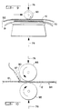
- FIG. 1 The basic structure of an article is shown in Figure 1, comprising a composite material having a fibrous matrix 10 laminarly affixed to a cell-barrier sheet material 12 .
- the fibrous matrix and cell-barrier sheet material are designed to be superimposed and affixed together in a laminar fashion so that the implantable bioabsorbable article provides an open structure of void spaces capable of accommodating tissue ingrowth while also providing a cell-barrier.
- the cross section of Figure 2A shows the implantable bioabsorbable article in further detail.
- the cell-barrier sheet material 12 has two opposing surfaces, designating the first opposing surface 13 and the second opposing surface 15 .
- the fibrous matrix 10 is laminarly affixed to the first opposing surface 13 of the cell-barrier sheet material 12 .
- laminarly affixed is meant that the fibrous matrix 10 is attached directly to the surface of the cell-barrier sheet material 12 as shown by Figure 2A, or extends through the first opposing surface 13 as shown by the cross section of Figure 2B, or extends through the first surface 13 to the second surface 15 as shown by the cross section of Figure 2C.
- Affixing is preferably accomplished by superimposing the fibrous matrix 10 on the first opposing surface 13 of the cell-barrier sheet material 12 while the cell-barrier sheet material 12 is in a softened or even liquid state as will be further described.
- a bioabsorbable adhesive may be used to bond and thereby laminarly affix the fibrous matrix 10 to the first opposing surface 13 of the cell-barrier sheet material 12 .
- the fibrous matrix 10 comprises a mesh of fibers in either a random, unorganized configuration 14 as shown by the cross sections of Figures 2A, 2B and 2C, or alternatively the fibrous matrix comprises an organized fabric configuration 16 such as a fabric capable of supporting tissue ingrowth as shown by the cross section of Figure 3.
- the organized configuration 16 is a fabric configuration which may be comprised of threads, yarns, nets, knits, weaves, laces, or felts of fibers.
- the fibrous matrix may be comprised of an open cell foam structure that will allow tissue ingrowth. Regardless of construction, the fibrous matrix must be sufficiently open to allow infiltration of blood and subsequent interconnection of ingrowing tissue through the open spaces existing between the fibers that comprise the matrix.
- any bioabsorbable material having an open structure capable of tissue ingrowth may be suitable as the fibrous matrix so long as the mechanical properties and rate of bioabsorption are also appropriate for the intended application.
- the thickness of these fibrous matrix materials may vary depending on the end use application.
- the cell-barrier sheet material is typically non-porous but may also be porous so long as the pore sizes are small enough to substantially preclude cell passage and ingrowth. Such a porous cell-barrier sheet material may be advantageous for certain applications, for example, where passage of nutrients or gasses across the material may be important.
- an article to allow tissue ingrowth or conversely to substantially preclude cell passage and ingrowth can be determined by implanting the article into a dog and allowing time for healing to occur.
- the article and adjacent tissue complex should be harvested and histologically evaluated after three weeks, before substantial bioabsorption of the article has occurred.
- the article is deemed capable of allowing tissue ingrowth and incapable of substantially precluding cell passage and ingrowth.
- the cell-barrier sheet material should be composed of a synthetic bioabsorbable material such as, for example, polycaprolactone, poly p-dioxanone, trimethylene carbonate, polyglycolic acid (PGA) or polylactic acid (PLA) or copolymers thereof.
- a preferred material is a copolymer of PLA and PGA, with preferred mixtures ranging from about 85% PLA and 15% PGA to about 50% PLA and 50% PGA.
- the equal ratio copolymer can be expected to bioabsorb at the fastest rate.
- Copolymers of PLA and PGA are polymerized from appropriate proportions of lactide and glycolide which are cyclic dimers based on lactic acid and glycolic acid respectively.
- the lactic acid components of the lactide dimer may be of either the d (dextrorotatory) or l (levorotatory) configuration or may be a mixture of the two configurational varieties (e.g., d,l lactide).
- Polymers containing mixtures of d,l lactides possess little or no polymeric crystallinity resulting in lessened rigidity and a relatively low glass transition temperature when compared with a more crystalline counterpart such as l lactide.
- the preferred periodontal repair embodiment for the cell-barrier sheet material component contains copolymers d,l lactide with glycolide which correspondingly provides minimal rigidity and a glass transition temperature conducive to subsequent thermal bonding of the embodiment incorporating a fibrous matrix on both sides of the barrier material.
- copolymers bioabsorb through hydrolysis returning them to their original components which are subsequently expelled from or assimilated into the body.
- the in vivo longevity of a particular copolymer is a function of its molecular weight combined with the ratio of its lactide and glycolide components. In general, the rate of hydrolysis is decreased with increasing lactic acid composition.
- the fibers from which the fibrous matrix is constructed are preferably made of PGA in order to provide for bioabsorption of the matrix in a suitable time frame.
- Different fibers of the matrix may be comprised of different bioabsorbable polymers so that different fibers may be bioabsorbed at different rates.
- the implantable bioabsorbable article should be mechanically capable of successfully retaining sutures for the amount of time required for enough healing to render the sutures unnecessary.
- the implantable bioabsorbable article should also be pliable and formable and not be brittle or thick so that it is hard to suture or difficult to conform to the ideal contours desired at the defect site.
- the article must not be so flexible that it collapses into the defect space but rather maintains and protects that space for an adequate time to allow healing to occur.
- the cross section of Figure 4A describes the present invention wherein a fibrous matrix 10 is affixed to both opposing sides of a cell-barrier sheet material 12 .
- one way to produce this embodiment is to laminarly affix together the exposed cell-barrier surfaces of two samples 41 and 42 of the implantable bioabsorbable article having a fibrous matrix attached to only one side of the cell-barrier sheet material.
- the laminar juncture of the two samples 41 and 42 is shown at 44 .
- the implantable bioabsorbable article of the present invention may also be made so that it incorporates a bioabsorbable suture 50 to make the securing of the article to adjacent anatomical structures as simple as possible.
- This embodiment is considered to be at the present time the best mode of the present invention for periodontal repair. This embodiment is made most simply by placing a suture into the fold of the two-sided article described in Figure 4C.
- the cross section of Figure 5B shows this in detail.
- Other embodiments incorporating an affixed bioabsorbable suture are also possible.
- the coronal edge of the article as implanted is indicated at 54 .
- Other methods of attaching a suture may be used, for example, interweaving the suture through the thickness of the article.
- the fibrous matrix, the fibers, the cell-barrier material, or any combination thereof may also be impregnated with any single or combination of substances such as antibiotics, antimicrobials, growth factors, differentiation factors, cell attachment factors or other such therapeutic agents. Impregnation of the implantable bioabsorbable article with such a therapeutic agent involves simply coating the article with the agent or alternatively incorporating the agent into the material from which either the fibrous matrix or the cell-barrier is constructed.
- the therapeutic agent may be included in the polymer and solvent solution from which the cell-barrier is subsequently made.
- a therapeutic agent into the material of the implantable bioabsorbable article in this fashion would result in the release of the therapeutic agent into the living body in which the article is implanted.
- the presence of such an impregnating therapeutic agent in the implantable bioabsorbable article can be determined by accepted analytical techniques involving extraction, solvation, or other isolation methods whereby the therapeutic agent may be separated from the material from which the implantable bioabsorbable article is constructed.
- a typical application of the implantable bioabsorbable article is shown in the cross section of Figure 6.
- This figure illustrates the relationship of the implantable bioabsorbable article and adjacent anatomical structures immediately after the surgical placement of the article.
- the enamel tooth crown is indicated at 61 and the tooth root (dentin) is shown at 62 .
- the periodontal ligament is indicated by 63
- the supporting alveolar bone is shown by 64
- the cementum is shown at 69 .
- the implantable bioabsorbable article is shown in cross section at 66 .
- the implantable bioabsorbable article 66 is shown serving as a barrier between the periodontal defect 65 and adjacent gingival connective tissue 67 and gingival epithelium 68 .
- This embodiment of the implantable bioabsorbable article is shown with an incorporated bioabsorbable suture 50 as previously described in Figures 5A and 5B.
- the suture 50 is shown attaching the implantable bioabsorbable article to the tooth root 62 by encircling the tooth root 62 .
- the process of laminarly affixing the fibrous matrix to the cell-barrier sheet material is accomplished by placing a fibrous matrix into a bioabsorbable polymer and solvent solution and applying pressure and heat to evaporate the solvent, the bioabsorbable polymer forming the cell-barrier sheet material as the solvent evaporates.
- the selection of the solvent and the fibrous matrix material should be coordinated so that the fibrous matrix material is not readily soluble in the solvent.
- a preferred polymer and solvent solution is that of a PLA and PGA copolymer in a solvent such as acetone.
- the use of PGA fibers for the fibrous matrix is a preferred combination as such fibers are not readily soluble in the acetone.
- the polymer and solvent solution 70 and fibrous matrix 10 are placed on a surface 72 capable of transferring heat at a temperature above the boiling point of the solvent in order to evaporate the solvent.
- the heating surface 72 must possess suitable release properties to allow removal of the finished implantable bioabsorbable article without damage.
- Suitable heating surface materials include polytetrafluoroethylene (PTFE), fluorinated ethylene-propylene (FEP), and other materials having adequate temperature capability and having release properties.
- the preferred process for producing an article with a fibrous matrix on one surface is as follows.
- the fibrous matrix 10 and the solution of bioabsorbable polymer and solvent 70 are introduced between two opposing release surfaces 72 and 74 which are then compressed together as indicated by arrows 76 while heat in excess of the boiling temperature of the solvent is applied from release surface 72 .
- the temperature should be higher than the boiling point of the solvent in order to assure rapid evaporation of the solvent within a few seconds.
- the fibrous matrix material is coated with the solvent carried polymer.
- points of contact between adjacent fibers of the matrix are adhesively bonded.
- the cell-barrier sheet material of the chosen bioabsorbable polymer or copolymer is deposited at the surface from which heat is applied. The resulting article is then removed from between the release surfaces.
- two articles are prepared as described above.
- the two articles are placed so that their cell-barrier surfaces face each other and are superimposed on each other laminarly as shown by Figure 8.
- Pressure and heat are applied at a temperature above the glass transition temperature of the barrier and below that of the fiber. This results in the two film surfaces adhering to one another to form a composite having a fibrous matrix on the two opposing surfaces of the cell-barrier sheet material.
- the cell-barrier sheet materials with a PLA/PGA copolymer for the two-sided article as the copolymer possesses a glass transition temperature between 40 and 60°C.
- the embodiment incorporating a suture is made as described in the previous paragraph with the addition of a length of bioabsorbable suture placed between the two sheets of implantable bioabsorbable material before the two sheets are affixed together, the ends of the suture extending beyond the affixed sheets.
- a single layer article is folded in half so that the suture is captured within the fold.
- Figure 9 describes a preferred process wherein compression 76 is applied by a traversing roller 93 instead of a flat compression surface. The process is essentially the same as described previously. Separate removable upper and lower FEP film release layers 91 and 92 may be used optionally to provide the necessary release characteristics.
- Figure 10 describes an alternative process useful for a continuous process of manufacturing an article wherein the bioabsorbable polymer and solvent solution are applied as a liquid 90 to the fibrous matrix 10 from a liquid dispensing means 97 .
- the fibrous matrix material 10 and applied bioabsorbable polymer solvent solution is fed between a heated roller 95 and an adjacent second roiler 93 .
- the article 94 emerges from between the two compressing rollers 95 and 93 .
- tubular constructions of the implantable bioabsorbable article are also possible. These are made by rolling the finished article, made as previously described, around a mandrel to form a tube 110 , and applying heat and pressure to the overlapping edge 111 of the tubular article in order to seal the overlapping edge to the underlying material 112 .
- Such a tubular article may have various implantable applications such as vascular repair and nerve repair.
- vascular repair it is believed that excluding other tissues from the nerve repair site and constraining the healing nerve ends to prevent uncontrolled proliferation of nervous tissue are important factors in successful repair. It is also believed that retaining neurotropic and neurotrophic factors released by the distal nerve end, keeping these factors in the near vicinity of the proximal nerve end, is important in effecting successful nerve repair.
- the tubular article of the present invention is capable of providing for the exclusion of adjacent tissues from the nerve repair site, the containment of proliferating nervous tissue, and the retention of neurotropic and neurotrophic factors in the vicinity of the proximal nerve end.
- a tube is made by rolling the implantable bioabsorbable article into a tubular form as previously described with the fibrous matrix on the inner surface and outer surface of the tube.
- the fibrous matrix on the outer surface aids in attachment and stabilization of the surrounding external tissue. It is preferred that the fibers be of an organized configuration in the form of parallel fibers on the inner surface of the tube oriented parallel to the longitudinal axis of the tube.
- this fiber orientation provides a directional aid to the healing of the nerve between exposed nerve ends.
- the proximal and distal ends of a transected nerve will be placed in respective ends of this tubular article. These nerve ends may be abutted and sutured together, simply abutted, or left with a gap between the nerve ends when the tubular article is finally sutured or affixed by any suitable means to the nerve sheaths of the two nerve ends.
- an alternative tubular embodiment for nerve repair incorporates a layer of randomly configured fibrous matrix 121 between the outer cell-barrier sheet material surface of the tube and the inner surface of parallel longitudinally oriented fibers 113 .
- This construction offers improved crush resistance.
- the longitudinally oriented fibers 113 are shown in exaggerated diameter for clarity.
- the implantable bioabsorbable article of the present invention may also be useful for repair of bone defects and bone regeneration. Specific applications include endochondral bone, intermembranous or round bone, and long bone.
- the article intended for bone repair should be sufficiently rigid to guarantee the maintenance of space adjacent to a bone defect.
- the implantable bioabsorbable article of the present invention may also be useful in the prevention of soft tissue adhesions, particularly those of the peritoneum including pelvic adhesions.
- the fibrous matrix is useful for stabilizing the implantable bioabsorbable article at the desired location to prevent the occurrence of an adhesion at that location.
- the fibrous matrix is effective to the extent that the implantable bioabsorbable article can simply be placed at the desired location without the necessity of further securing means.
- PGA polyglycolic acid
- the fibrous matrix was removed from the stainless steel screen on which it had previously been fabricated, and was placed onto the first sheet of FEP film 91 .
- Approximately 0.2 grams of 1:10 to 1:15 (w/w) solution of l-lactide polymer in methylene chloride (dichloromethane) solvent was poured onto the center of the fibrous matrix.
- a second sheet of FEP clear film 92 was laid over the top of the fiber matrix and polymer and solvent combination. The resulting sandwich was immediately compression-rolled between the aluminum plate and a rolling cylinder of about 3.5 inches length and 2 inch diameter.
- the roller effectively applied a force of approximately eight pounds across its width, as shown by arrow 75 .
- Each compression roll process consisted of an approximately 3 inch pass of the roller across the surface of the fibrous matrix at a rate of approximately one complete cycle (2 passes) per second for a total of 2 minutes.
- the application of heat and pressure resulted in a finished implantable bioabsorbable article having a fibrous matrix laminarly affixed to one side of a cell-barrier sheet.
- the two FEP film sheets 91 and 92 and the finished implantable bioabsorbable article contained between the two FEP sheets was removed from the heated aluminum plate and allowed to cool under passive pressure applied by a 4" x 4" x 1" aluminum block.
- the two sheets of FEP were peelably removed from the finished implantable bioabsorbable article.
- the finished article was trimmed to 15 mm width and a 30 mm length.
- the article was folded in half across its 15mm width with the cell-barrier surface on the inside.
- a 5-0 PGA suture obtained commercially was trimmed of its needle and placed into the fold of the material as shown by Figures 5A nad 5B.
- the folded article was placed between two FEP film sheets and compressed by the roller and heated plate apparatus described by Figure 8. The plate temperature was 100°C. Pressure was again applied by traversing the roller back and forth over the article in one second cycles for a total period of 1 minute.
- the finished implantable bioabsorbable article having an integrally attached suture was placed into a food grade pouch made of a polymer foil laminate.
- the pouch was sealed and gamma radiation sterilized at a total dosage of 2.0 Mrad.
- buccal and lingual full-thickness muco-periosteal flaps were elevated from the first mandibular premolar to the mid portion of the mandibular first molar.
- Class II furcation defects were surgically created with high speed and hand instrumentation on the buccal aspects of the second and fourth mandibular premolars and the lingual aspects of the third mandibular premolars in four beagle dogs.
- the defects were created such that the roots were exposed between the mesial and distal line angles of the teeth and into but not through the furcation. The depth of the defects was approximately 4-6 millimeters.
- the exposed root surfaces were aggressively instrumented with high speed and hand instruments to remove any trace of the periodontal ligament and cementum. Reference notches were placed in the roots at the margins of the bony defects.
- Each bioabsorbable article was first trimmed on the lateral and apical sides so that the material completely covered the defect and extended beyond the bony margins of the defect by at least 2 to 3 mm. The article was then positioned so that the coronal edge with the integrally attached suture was at the level of the cemento-enamel junction and completely covered the defect. The article was tied to the tooth with the integrally attached suture using a sling suture technique. The article was thus tightly adapted to the tooth surface along its coronal edge and draped over the surface of the bone. This created a protected space defined by the dimensions of the defect and the surface of the article which faced the defect. The muco-periosteal flaps were repositioned and the incisions closed using a combination of interrupted and vertical mattress sutures. Care was taken to make sure the membranes were completely covered by the soft tissue of the flaps.
- New Bone - the distance from the base of the instrumented root surface to the coronal extent of the new regenerated bone.
- New Cementum NC - distance from the base of the instrumented root surface to the coronal extent of regenerated new cementum.
- Defect Depth DD - the base of the instrumented root surface to the gingival margin (the area "at risk” for regeneration).
- the integrity of the implantable bioabsorbable article was substantially intact creating a barrier, as evidenced by a plane of separation between the flap tissue and the defect space.
- the granulation tissue in the defect space had an immature appearance with a limited amount of extracellular matrix deposition and minimal differentiation of the tissue types.
- Epithelial migration along the root surface can proceed at the rate of between 0.5 mm and 1.0 mm per day. If allowed to progress unimpeded, the epithelium would have migrated apically along either the outside of the implantable bioabsorbable articles or along the root surfaces. The data from this experiment show that the migration of the epithelium was limited by the fibrous connective tissue ingrowth and attachment to the implantable bioabsorbable articles. Concurrently, protection of the defect space from the epithelium and the gingival connective tissue by the implantable bioabsorbable articles allowed the regeneration of new bone in the defect space and new cementum on the root surfaces.
- the preferred embodiment currently utilizes d,l PLA:PGA copolymer ratios between 50:50 and 85:15 for the cell-occlusive barrier forming material to provide a desirable in vivo bioabsorption rate for the periodontal application.
- Examples of the preferred embodiment were fabricated in accordance with the description of Example 1 with the following exceptions:
Abstract
Description
- This invention relates to an implantable bioabsorbable article for use as a mammalian-cell-barrier to aid in the regeneration of tissue.
- A preferred technique to promote the regeneration of mammalian tissue is accomplished by the separation and isolation of a particular type of tissue to be regenerated from other competing undesirable tissues through the use of a biocompatible barrier material. This concept is known as a guided tissue regeneration and was described in an article by J. Gottlow, et al., titled "New Attachment Formation in the Human Periodontium by Guided Tissue Regeneration" (Journal of Clinical Periodontology, 1986; Vol. 13, pp. 604-616). The function of the barrier material is to substantially preclude the movement of tissue cells through the thickness of the material and consequently limit the varieties of cell types at the treatment site. This function is combined with the requirement that the material maintain sufficient space adjacent to the defect so as to allow for the regeneration of the desirable tissue into that space. The preservation of space between the surface of the defect and the desired contours of the subsequently regenerated surface is necessary in order to allow for the regeneration of tissues into that space. Specific periodontal structures which may be regenerated in this fashion are the periodontal ligament, bone and cementum. The barrier material allows propagation of bone and periodontal ligament cells by precluding epithelial cells and gingival connective tissue cells which are believed to propagate at a greater rate. This concept may be useful for other applications where separation of specific cell varieties is desirable such as, for example, nerve repair and nerve guidance applications, bone regeneration and prevention of soft tissue adhesions, particularly those of the peritoneum.
- WO 90/00410 describes adsorbable polymeric films reinforced at least partially by absorbable oriented reinforcing elements such as, for example, fibers. The disclosed polymeric films are used to either join tissues or their parts to each other, or to separate them from each other, especially during regrowth of periodontal ligament and/or cementum tissue.
- A description of a bioabsorbable tubular device useful for nerve repair is provided in U.S. Patent 4,870,966 to Dellon, et al.
- Additionally, it has been proposed that the mechanical stability of the blood clot which forms in the defect space adjacent to the tooth root after periodontal surgery may be important to the regeneration process (Wikesjo et al., 1990; J. Periodontal., Vol. 61, 559-563). Therefore a material which can become infiltrated with blood clot and thus form a connection between the material and the adjacent gingival flap may add to the mechanical stability of the wound.
- One commercially available material that provides a cell-barrier for periodontal tissue regeneration is the GORE-TEX® Periodontal Material. This polytetrafluoroethylene (PTFE) material serves as a cell-barrier between the gingiva and a periodontal defect and is intended to preserve necessary space between the surface of the defect and the desired contours of the subsequently regenerated surface. The GORE-TEX Periodontal Material is made of porous expanded PTFE having a microstructure of nodes interconnected by fine fibrils. This commercially available material is not of laminar construction and its porosity is generally uniform through the thickness of the material. One portion of the total surface area of the GORE-TEX Periodontal Material has a porous structural surface that becomes infiltrated with blood clot and ingrown with fibrous connective tissue, thereby inhibiting epithelial migration. The remaining portion of the surface area has a cell-barrier structure of low porosity for isolating the overlying gingival connective tissue from the underlying defect. It is not bioabsorbable and is typically removed in a subsequent surgical procedure.
- There have been previous attempts to produce suitable surgical barriers from bioabsorbable materials. A 70 micron thick membrane solvent-cast from bioabsorbable polylactic acid, having no inherent porosity or tissue cell permeability, was tested in periodontal applications as a cell-barrier material for exclusion of epithelium and gingival connective tissue during healing (I. Magnusson, et al., "New Attachment Formations Following Controlled Tissue Regeneration Using Biodegradable Membranes", J. Periodontal January, 1988 pp. 1-6). Tests showed some new formation of cementum and bone. Reproductions of this material demonstrated poor surgical handling characteristics due to its thin friable construction and also proved to be difficult to suture because of its brittleness. This material makes no provision for tissue ingrowth on either of its surfaces.
- Another material that is commercially available for use in guided tissue regeneration is Vicryl® Periodontal Mesh available from Johnson & Johnson. The Vicryl Periodontal Mesh is comprised of woven fibers made from a bioabsorbable copolymer of about 90% glycolide and 10% lactide. Studies have shown that the Vicryl Periodontal Mesh has had some success as a barrier material that provides for tissue regeneration (Fleisner et al., "Regeneration of lost attachment apparatus in the dog using Vicryl Absorbable Mesh", International Journal of Periodontics and Restorative Dentistry 2/1988 pp 45-55). Difficulties with this conventional woven construction include its inferior ability to maintain space adjacent to the defect and its marginal effectiveness as a tissue barrier because of the inherent porosity of the woven structure. This material is a single layer material of woven construction that is intended to both promote tissue ingrowth and simultaneously serve as a tissue barrier. As these are somewhat contradictory objectives for a single layer material of woven construction having a degree of inherent porosity, ingrowth can only be made to occur at the expense of the barrier function. The effectiveness of this material is therefore a compromise between the material's ability to allow for tissue ingrowth and the requirement to simultaneously function as a tissue barrier.
- There remains a need for a bioabsorbable material for use as an effective cell-barrier that allows for tissue attachment on both surfaces, adds to the stability of the healing wound through blood infiltration and coagulation into the material, substantially precludes passage of tissue cells through the material, possesses adequate rigidity to ensure preservation of the desired space proximal to the defect and has acceptable surgical handling properties and strength.
- Bioabsorbable materials are herein defined as those materials of either synthetic or natural origin which when placed into a living body are degraded through either enzymatic, hydrolytic or other chemical reactions, into byproducts which are either integrated into or expelled from the body.
- Cells and tissue are herein defined as mammalian cells and mammalian tissue.
- This invention provides an implantable bioabsorbable article useful for the separation and regeneration of mammalian tissue comprising: a bioabsorbable cell-barrier sheet material having first and second opposing surfaces; and a bioabsorbable fibrous matrix laminarly affixed to the first and second opposing surfaces of said bioabsorbable cell-barrier sheet material.
- The invention may be made in either a flat planar form or alternatively in a tubular form for use in, for example, nerve repair and nerve guidance applications. Preferred bioabsorbable materials include polyglycolic acid fibers for the fibrous matrix and a copolymer of polylactic acid and polyglycolic acid for the cell-barrier sheet material.
- A method for making the implantable bioabsorbable article of the present invention is also provided.
- The present invention will now be described in detail with reference to the following Figures, in which;
- Figure 1 is a perspective view of an implantable bioabsorbable article having a fibrous matrix laminarly affixed to one side of a cell-barrier sheet material.
- Figures 2A, 2B and 2C are cross sectional views of the article of Figure 1 wherein the fibrous matrix is of unorganized configuration.
- Figure 3 is a cross sectional view of the implantable bioabsorbable article of Figure 1 wherein the fibrous matrix is of organized configuration.
- Figures 4A, 4B and 4C are cross sectional views of an embodiment of the present invention having a fibrous matrix laminarly affixed to both sides of a cell-barrier sheet material.
- Figures 5A, 5B and show the preferred periodontal repair embodiment of the present invention incorporating a bioabsorbable suture into its construction.
- Figure 6 is a cross section of a periodontal defect showing the preferred periodontal repair embodiment of the present invention.
- Figure 7 shows a schematic drawing of the process of making an article having a fibrous matrix affixed to one side of the cell-barrier sheet material.
- Figure 8 shows a schematic drawing of a process of making an embodiment of the present invention having a fibrous matrix affixed to both sides of the cell-barrier sheet material.
- Figures 9 and 10 show a schematic drawings of two alternative processes of making an article having a fibrous matrix affixed to one side of the cell-barrier sheet material.
- Figure 11 shows the implantable bioabsorbable article of the present invention formed into a tube useful for repairs such as vascular and nerve guide repair.
- Figure 12 shows a cross section of an alternative tubular embodiment.
- It should be noted that the Figures No. 1, 2, 3, 7, 9, 10 and 11 disclose bioabsorbable articles, or methods, for making the bioabsorbable articles which fall outside the scope of the claims as filed and these figures are included for reference purposes only.
- Reference will now be made in detail to the present invention, examples of which are illustrated in the drawings.
- The basic structure of an article is shown in Figure 1, comprising a composite material having a
fibrous matrix 10 laminarly affixed to a cell-barrier sheet material 12. The fibrous matrix and cell-barrier sheet material are designed to be superimposed and affixed together in a laminar fashion so that the implantable bioabsorbable article provides an open structure of void spaces capable of accommodating tissue ingrowth while also providing a cell-barrier. - The cross section of Figure 2A shows the implantable bioabsorbable article in further detail. The cell-
barrier sheet material 12 has two opposing surfaces, designating the firstopposing surface 13 and the second opposingsurface 15. Thefibrous matrix 10 is laminarly affixed to the firstopposing surface 13 of the cell-barrier sheet material 12. By laminarly affixed is meant that thefibrous matrix 10 is attached directly to the surface of the cell-barrier sheet material 12 as shown by Figure 2A, or extends through the firstopposing surface 13 as shown by the cross section of Figure 2B, or extends through thefirst surface 13 to thesecond surface 15 as shown by the cross section of Figure 2C. Affixing is preferably accomplished by superimposing thefibrous matrix 10 on the firstopposing surface 13 of the cell-barrier sheet material 12 while the cell-barrier sheet material 12 is in a softened or even liquid state as will be further described. Alternatively a bioabsorbable adhesive may be used to bond and thereby laminarly affix thefibrous matrix 10 to the firstopposing surface 13 of the cell-barrier sheet material 12. - The
fibrous matrix 10 comprises a mesh of fibers in either a random, unorganized configuration 14 as shown by the cross sections of Figures 2A, 2B and 2C, or alternatively the fibrous matrix comprises anorganized fabric configuration 16 such as a fabric capable of supporting tissue ingrowth as shown by the cross section of Figure 3. Theorganized configuration 16 is a fabric configuration which may be comprised of threads, yarns, nets, knits, weaves, laces, or felts of fibers. Alternatively, the fibrous matrix may be comprised of an open cell foam structure that will allow tissue ingrowth. Regardless of construction, the fibrous matrix must be sufficiently open to allow infiltration of blood and subsequent interconnection of ingrowing tissue through the open spaces existing between the fibers that comprise the matrix. In general, any bioabsorbable material having an open structure capable of tissue ingrowth may be suitable as the fibrous matrix so long as the mechanical properties and rate of bioabsorption are also appropriate for the intended application. The thickness of these fibrous matrix materials may vary depending on the end use application. - The cell-barrier sheet material is typically non-porous but may also be porous so long as the pore sizes are small enough to substantially preclude cell passage and ingrowth. Such a porous cell-barrier sheet material may be advantageous for certain applications, for example, where passage of nutrients or gasses across the material may be important.
- The ability of an article to allow tissue ingrowth or conversely to substantially preclude cell passage and ingrowth can be determined by implanting the article into a dog and allowing time for healing to occur. The article and adjacent tissue complex should be harvested and histologically evaluated after three weeks, before substantial bioabsorption of the article has occurred. When evaluated, if mammalian cells, fibrous connective tissue cells or extracellular matrix, or collagen, have invaded the spaces between adjacent structures within the fibrous matrix, resulting in a connection between the article and adjacent tissue, the article is deemed capable of allowing tissue ingrowth and incapable of substantially precluding cell passage and ingrowth.
- The cell-barrier sheet material should be composed of a synthetic bioabsorbable material such as, for example, polycaprolactone, poly p-dioxanone, trimethylene carbonate, polyglycolic acid (PGA) or polylactic acid (PLA) or copolymers thereof. For periodontal applications, a preferred material is a copolymer of PLA and PGA, with preferred mixtures ranging from about 85% PLA and 15% PGA to about 50% PLA and 50% PGA. The equal ratio copolymer can be expected to bioabsorb at the fastest rate. For a comparison of bioabsorption rates of these materials, see Lewis, Danny H., Controlled Release of Bioactive Agents from Lactide/Glycolide Polymers, pp 1-41, Biodegradable Polymers As Drug Delivery Systems, Mark Chasin and Robert Langer (ed); Marcel Dekker, Inc., New York, NY, 1990.
- Copolymers of PLA and PGA are polymerized from appropriate proportions of lactide and glycolide which are cyclic dimers based on lactic acid and glycolic acid respectively. The lactic acid components of the lactide dimer may be of either the d (dextrorotatory) or l (levorotatory) configuration or may be a mixture of the two configurational varieties (e.g., d,l lactide). Polymers containing mixtures of d,l lactides possess little or no polymeric crystallinity resulting in lessened rigidity and a relatively low glass transition temperature when compared with a more crystalline counterpart such as l lactide. The preferred periodontal repair embodiment for the cell-barrier sheet material component contains copolymers d,l lactide with glycolide which correspondingly provides minimal rigidity and a glass transition temperature conducive to subsequent thermal bonding of the embodiment incorporating a fibrous matrix on both sides of the barrier material.
- These copolymers bioabsorb through hydrolysis returning them to their original components which are subsequently expelled from or assimilated into the body. The in vivo longevity of a particular copolymer is a function of its molecular weight combined with the ratio of its lactide and glycolide components. In general, the rate of hydrolysis is decreased with increasing lactic acid composition.
- For periodontal applications, the fibers from which the fibrous matrix is constructed are preferably made of PGA in order to provide for bioabsorption of the matrix in a suitable time frame. Different fibers of the matrix may be comprised of different bioabsorbable polymers so that different fibers may be bioabsorbed at different rates.
- The implantable bioabsorbable article should be mechanically capable of successfully retaining sutures for the amount of time required for enough healing to render the sutures unnecessary. The implantable bioabsorbable article should also be pliable and formable and not be brittle or thick so that it is hard to suture or difficult to conform to the ideal contours desired at the defect site. The article must not be so flexible that it collapses into the defect space but rather maintains and protects that space for an adequate time to allow healing to occur. These mechanical characteristics have been achieved with the implantable bioabsorbable article of the present invention and are the result of the structural combination of the fibrous matrix laminarly affixed to the cell-barrier sheet material.
- The cross section of Figure 4A describes the present invention wherein a
fibrous matrix 10 is affixed to both opposing sides of a cell-barrier sheet material 12. As shown by the cross section of Figure 4B, one way to produce this embodiment is to laminarly affix together the exposed cell-barrier surfaces of twosamples samples - Alternatively, as shown by the cross section of Figure 4C, it is possible to fold in half a single sample of the single-sided implantable bioabsorbable article with the surfaces of the cell-barrier material brought together and affixed. The fold is indicated at 43 and the laminarly affixed cell-barrier sheet material surface juncture is shown at 44. A method of affixing the cell-barrier sheet material surface will be described below.
- As shown by Figure 5A, the implantable bioabsorbable article of the present invention may also be made so that it incorporates a
bioabsorbable suture 50 to make the securing of the article to adjacent anatomical structures as simple as possible. This embodiment is considered to be at the present time the best mode of the present invention for periodontal repair. This embodiment is made most simply by placing a suture into the fold of the two-sided article described in Figure 4C. The cross section of Figure 5B shows this in detail. Other embodiments incorporating an affixed bioabsorbable suture are also possible. - The coronal edge of the article as implanted is indicated at 54. Other methods of attaching a suture may be used, for example, interweaving the suture through the thickness of the article.
- In an alternative embodiment, the fibrous matrix, the fibers, the cell-barrier material, or any combination thereof may also be impregnated with any single or combination of substances such as antibiotics, antimicrobials, growth factors, differentiation factors, cell attachment factors or other such therapeutic agents. Impregnation of the implantable bioabsorbable article with such a therapeutic agent involves simply coating the article with the agent or alternatively incorporating the agent into the material from which either the fibrous matrix or the cell-barrier is constructed. For example, the therapeutic agent may be included in the polymer and solvent solution from which the cell-barrier is subsequently made. The incorporation of a therapeutic agent into the material of the implantable bioabsorbable article in this fashion would result in the release of the therapeutic agent into the living body in which the article is implanted. The presence of such an impregnating therapeutic agent in the implantable bioabsorbable article can be determined by accepted analytical techniques involving extraction, solvation, or other isolation methods whereby the therapeutic agent may be separated from the material from which the implantable bioabsorbable article is constructed.
- A typical application of the implantable bioabsorbable article is shown in the cross section of Figure 6. This figure illustrates the relationship of the implantable bioabsorbable article and adjacent anatomical structures immediately after the surgical placement of the article. The enamel tooth crown is indicated at 61 and the tooth root (dentin) is shown at 62. The periodontal ligament is indicated by 63, the supporting alveolar bone is shown by 64 and the cementum is shown at 69. A periodontal defect exists at 65 wherein the cementum, the periodontal ligament and alveolar bone are missing from the void space also shown by 65. This
void space 65 will typically become filled with blood clot during or shortly after surgery. The implantable bioabsorbable article is shown in cross section at 66. - The implantable
bioabsorbable article 66 is shown serving as a barrier between theperiodontal defect 65 and adjacent gingivalconnective tissue 67 andgingival epithelium 68. This embodiment of the implantable bioabsorbable article is shown with an incorporatedbioabsorbable suture 50 as previously described in Figures 5A and 5B. Thesuture 50 is shown attaching the implantable bioabsorbable article to thetooth root 62 by encircling thetooth root 62. - According to the preferred method of making the implantable bioabsorbable article, the process of laminarly affixing the fibrous matrix to the cell-barrier sheet material is accomplished by placing a fibrous matrix into a bioabsorbable polymer and solvent solution and applying pressure and heat to evaporate the solvent, the bioabsorbable polymer forming the cell-barrier sheet material as the solvent evaporates. The selection of the solvent and the fibrous matrix material should be coordinated so that the fibrous matrix material is not readily soluble in the solvent. A preferred polymer and solvent solution is that of a PLA and PGA copolymer in a solvent such as acetone. The use of PGA fibers for the fibrous matrix is a preferred combination as such fibers are not readily soluble in the acetone.
- As shown by Figure 7, the polymer and
solvent solution 70 andfibrous matrix 10 are placed on asurface 72 capable of transferring heat at a temperature above the boiling point of the solvent in order to evaporate the solvent. Theheating surface 72 must possess suitable release properties to allow removal of the finished implantable bioabsorbable article without damage. Suitable heating surface materials include polytetrafluoroethylene (PTFE), fluorinated ethylene-propylene (FEP), and other materials having adequate temperature capability and having release properties. - The preferred process for producing an article with a fibrous matrix on one surface is as follows. The
fibrous matrix 10 and the solution of bioabsorbable polymer and solvent 70 are introduced between two opposing release surfaces 72 and 74 which are then compressed together as indicated byarrows 76 while heat in excess of the boiling temperature of the solvent is applied fromrelease surface 72. The temperature should be higher than the boiling point of the solvent in order to assure rapid evaporation of the solvent within a few seconds. As a result of the rapid evaporation of the solvent, the fibrous matrix material is coated with the solvent carried polymer. Upon evaporation of the solvent, points of contact between adjacent fibers of the matrix are adhesively bonded. Simultaneously, the cell-barrier sheet material of the chosen bioabsorbable polymer or copolymer is deposited at the surface from which heat is applied. The resulting article is then removed from between the release surfaces. - To construct the two-sided article of the present invention shown by Figure 4B having a fibrous matrix on two of the opposing surfaces of the cell-barrier sheet material, two articles are prepared as described above. The two articles are placed so that their cell-barrier surfaces face each other and are superimposed on each other laminarly as shown by Figure 8. Pressure and heat are applied at a temperature above the glass transition temperature of the barrier and below that of the fiber. This results in the two film surfaces adhering to one another to form a composite having a fibrous matrix on the two opposing surfaces of the cell-barrier sheet material. It is preferable to prepare the cell-barrier sheet materials with a PLA/PGA copolymer for the two-sided article as the copolymer possesses a glass transition temperature between 40 and 60°C.
- The embodiment incorporating a suture is made as described in the previous paragraph with the addition of a length of bioabsorbable suture placed between the two sheets of implantable bioabsorbable material before the two sheets are affixed together, the ends of the suture extending beyond the affixed sheets. Alternatively and preferably, as shown by the cross sections of Figures 5A, 5B and 5C, a single layer article is folded in half so that the suture is captured within the fold.
- Figure 9 describes a preferred process wherein
compression 76 is applied by a traversingroller 93 instead of a flat compression surface. The process is essentially the same as described previously. Separate removable upper and lower FEP film release layers 91 and 92 may be used optionally to provide the necessary release characteristics. - Figure 10 describes an alternative process useful for a continuous process of manufacturing an article wherein the bioabsorbable polymer and solvent solution are applied as a liquid 90 to the
fibrous matrix 10 from a liquid dispensing means 97. Thefibrous matrix material 10 and applied bioabsorbable polymer solvent solution is fed between aheated roller 95 and an adjacentsecond roiler 93. Thearticle 94 emerges from between the two compressingrollers - As shown by Figure 11, tubular constructions of the implantable bioabsorbable article are also possible. These are made by rolling the finished article, made as previously described, around a mandrel to form a
tube 110, and applying heat and pressure to the overlapping edge 111 of the tubular article in order to seal the overlapping edge to theunderlying material 112. Such a tubular article may have various implantable applications such as vascular repair and nerve repair. For nerve repair it is believed that excluding other tissues from the nerve repair site and constraining the healing nerve ends to prevent uncontrolled proliferation of nervous tissue are important factors in successful repair. It is also believed that retaining neurotropic and neurotrophic factors released by the distal nerve end, keeping these factors in the near vicinity of the proximal nerve end, is important in effecting successful nerve repair. The tubular article of the present invention is capable of providing for the exclusion of adjacent tissues from the nerve repair site, the containment of proliferating nervous tissue, and the retention of neurotropic and neurotrophic factors in the vicinity of the proximal nerve end. In a preferred embodiment for nerve repair, a tube is made by rolling the implantable bioabsorbable article into a tubular form as previously described with the fibrous matrix on the inner surface and outer surface of the tube. The fibrous matrix on the outer surface aids in attachment and stabilization of the surrounding external tissue. It is preferred that the fibers be of an organized configuration in the form of parallel fibers on the inner surface of the tube oriented parallel to the longitudinal axis of the tube. It is believed that this fiber orientation provides a directional aid to the healing of the nerve between exposed nerve ends. The proximal and distal ends of a transected nerve will be placed in respective ends of this tubular article. These nerve ends may be abutted and sutured together, simply abutted, or left with a gap between the nerve ends when the tubular article is finally sutured or affixed by any suitable means to the nerve sheaths of the two nerve ends. - As shown by the cross section of Figure 12, an alternative tubular embodiment for nerve repair incorporates a layer of randomly configured
fibrous matrix 121 between the outer cell-barrier sheet material surface of the tube and the inner surface of parallel longitudinally orientedfibers 113. This construction offers improved crush resistance. The longitudinally orientedfibers 113 are shown in exaggerated diameter for clarity. - The implantable bioabsorbable article of the present invention may also be useful for repair of bone defects and bone regeneration. Specific applications include endochondral bone, intermembranous or round bone, and long bone.
- The article intended for bone repair should be sufficiently rigid to guarantee the maintenance of space adjacent to a bone defect.
- The implantable bioabsorbable article of the present invention may also be useful in the prevention of soft tissue adhesions, particularly those of the peritoneum including pelvic adhesions.
- The fibrous matrix is useful for stabilizing the implantable bioabsorbable article at the desired location to prevent the occurrence of an adhesion at that location. The fibrous matrix is effective to the extent that the implantable bioabsorbable article can simply be placed at the desired location without the necessity of further securing means.
- The present invention will now be described by way of example only.
- Approximately 40 to 45 milligrams of individual 30 micron diameter polyglycolic acid (PGA) 1 to 2 inch long fibers were air laid in a random configuration on the surface of a stainless steel screen. The fibers were fabricated from PGA homopolymer with an average molecular weight in excess of 100,000 daltons.
- A first sheet of 0.020 inch thick FEP
clear film 91 as shown by Figure 9, was placed onto a 4 x 4 inch horizontally oriented aluminum plate that had been heated to approximately 100°C. The fibrous matrix was removed from the stainless steel screen on which it had previously been fabricated, and was placed onto the first sheet ofFEP film 91. Approximately 0.2 grams of 1:10 to 1:15 (w/w) solution of l-lactide polymer in methylene chloride (dichloromethane) solvent was poured onto the center of the fibrous matrix. A second sheet of FEPclear film 92 was laid over the top of the fiber matrix and polymer and solvent combination. The resulting sandwich was immediately compression-rolled between the aluminum plate and a rolling cylinder of about 3.5 inches length and 2 inch diameter. The roller effectively applied a force of approximately eight pounds across its width, as shown by arrow 75. Each compression roll process consisted of an approximately 3 inch pass of the roller across the surface of the fibrous matrix at a rate of approximately one complete cycle (2 passes) per second for a total of 2 minutes. The application of heat and pressure resulted in a finished implantable bioabsorbable article having a fibrous matrix laminarly affixed to one side of a cell-barrier sheet. Upon completion, the twoFEP film sheets - After cooling, the two sheets of FEP were peelably removed from the finished implantable bioabsorbable article. The finished article was trimmed to 15 mm width and a 30 mm length. The article was folded in half across its 15mm width with the cell-barrier surface on the inside. A 5-0 PGA suture obtained commercially was trimmed of its needle and placed into the fold of the material as shown by Figures 5A nad 5B. The folded article was placed between two FEP film sheets and compressed by the roller and heated plate apparatus described by Figure 8. The plate temperature was 100°C. Pressure was again applied by traversing the roller back and forth over the article in one second cycles for a total period of 1 minute.
- The finished implantable bioabsorbable article having an integrally attached suture was placed into a food grade pouch made of a polymer foil laminate. The pouch was sealed and gamma radiation sterilized at a total dosage of 2.0 Mrad.
- The implantable bioabsorbable articles made and sterilized according to Example 1, each article incorporating an integrally attached suture as shown by Figures 5A and 5B, were implanted in dogs. The dog model has been used extensively for the testing of guided tissue regeneration techniques and materials (Caffesse et al. 1988, J. Periodontal Vol 59, 589-584; Claffey et al. 1989, J. Clin. Periodontal,
Vol 16, 371-379). - At surgery, buccal and lingual full-thickness muco-periosteal flaps were elevated from the first mandibular premolar to the mid portion of the mandibular first molar. Class II furcation defects were surgically created with high speed and hand instrumentation on the buccal aspects of the second and fourth mandibular premolars and the lingual aspects of the third mandibular premolars in four beagle dogs. The defects were created such that the roots were exposed between the mesial and distal line angles of the teeth and into but not through the furcation. The depth of the defects was approximately 4-6 millimeters. After the bone was removed, the exposed root surfaces were aggressively instrumented with high speed and hand instruments to remove any trace of the periodontal ligament and cementum. Reference notches were placed in the roots at the margins of the bony defects.
- Defects were created in five separate mandibular premolar teeth per dog, the defects being intended to receive implantable bioabsorbable articles of the present invention. Each canine mandibular premolar tooth had two roots. The defects involved both roots of each of the five teeth for a total of ten roots potentially available for evaluation per dog. No sham operated controls were included in the experiment. It is well documented that surgical treatment of periodontal defect sites where no barrier material is placed will result in the formation of a long junctional epithelium and little or no regeneration of the periodontal attachment structures (Pfeifer et al., 1989, Int. J. Perio. Rest. Dent., Vol. 9, 263-273; Magnussen et al., 1988, J. Periodontal, Vol. 59, 1-6).
- Each bioabsorbable article was first trimmed on the lateral and apical sides so that the material completely covered the defect and extended beyond the bony margins of the defect by at least 2 to 3 mm. The article was then positioned so that the coronal edge with the integrally attached suture was at the level of the cemento-enamel junction and completely covered the defect. The article was tied to the tooth with the integrally attached suture using a sling suture technique. The article was thus tightly adapted to the tooth surface along its coronal edge and draped over the surface of the bone. This created a protected space defined by the dimensions of the defect and the surface of the article which faced the defect. The muco-periosteal flaps were repositioned and the incisions closed using a combination of interrupted and vertical mattress sutures. Care was taken to make sure the membranes were completely covered by the soft tissue of the flaps.
- The dogs received post operative oral hygiene care in the form of chlorhexidine (0.12%) flushes every other day starting seven days after surgery. Clinical parameters monitored during the healing period were gingival health, plaque index, exposure of material, gingival recession, and fragmentation or resorption of the exposed material.
- Sacrifice times for the animals were 2, 4, 8 and 12 weeks postoperative. At the time of sacrifice, the experimental teeth were removed en bloc and placed in 70% ethanol/30% distilled water fixative. After fixation, the teeth were infiltrated and embedded in poly(methylmethacrylate) and fifteen 6-8 micron thick bucco-lingual sections spanning the mesio-distal width of each tooth were cut for histological evaluation with a Reichart-Jung Polycut S sledge microtome. One section representing the greatest length of each root and located as close to the furcation as feasible was chosen for evaluation. The parameters measured on each root available for evaluation were:
Apical Epithelial Migration (EM) - the distance from the apical extent of the epithelium to the gingival margin.
New Bone (NB) - the distance from the base of the instrumented root surface to the coronal extent of the new regenerated bone.
New Cementum (NC) - distance from the base of the instrumented root surface to the coronal extent of regenerated new cementum.
Defect Depth (DD) - the base of the instrumented root surface to the gingival margin (the area "at risk" for regeneration). - For EM, NB and NC, the distances were divided by DD and multiplied by 100 to obtain a percentage of the defect depth. The values for all available roots for each dog were averaged to obtain a single value for each dog. No adequate sections were obtained for three roots of the 4 week dog, 1 root of the 8 week dog and three roots of the 12 week dog.
- Histological evaluation of the specimens revealed a mild chronic inflammatory response to the bioabsorbable membranes which was consistent with the response reported in the literature for this group of polymers (for example see Matlaga, B.F., and Salthouse, T.N., 1983; J. Biomed. Mat. Res. Vol. 19, 349-365). Fibrous connective tissue had infiltrated the fibrous matrix of the implantable bioabsorbable articles and there was clear interdigitation of collagen fibers between the ingrown tissue and the adjacent connective tissue of the flap. In all cases, the epithelium had migrated to a point at or very near the coronal border of the implantable bioabsorbable articles where the migration appeared to have been halted. In addition, at the two week time frame, the integrity of the implantable bioabsorbable article was substantially intact creating a barrier, as evidenced by a plane of separation between the flap tissue and the defect space. Also at the two week time frame the granulation tissue in the defect space had an immature appearance with a limited amount of extracellular matrix deposition and minimal differentiation of the tissue types.
- At the remaining time frames, the integrity of the implantable bioabsorbable article was somewhat fragmented due to the resorption process. However, maturation of the tissues had occurred allowing measurement of the amount of newly regenerated bone and cementum. The resulting values are shown in Table I.
TABLE I 4 wk (N = 7) 8 wk (N = 9) 12 wk (N = 7) EM = 31% ± 10% EM = 44% ± 13% EM = 52% ± 22% NB = 27% ± 13% NB = 30% ± 13% NB = 32% ± 18% NC = 65% ± 15% NC = 60% ± 11% NC = 54% ± 17% - Epithelial migration along the root surface can proceed at the rate of between 0.5 mm and 1.0 mm per day. If allowed to progress unimpeded, the epithelium would have migrated apically along either the outside of the implantable bioabsorbable articles or along the root surfaces. The data from this experiment show that the migration of the epithelium was limited by the fibrous connective tissue ingrowth and attachment to the implantable bioabsorbable articles. Concurrently, protection of the defect space from the epithelium and the gingival connective tissue by the implantable bioabsorbable articles allowed the regeneration of new bone in the defect space and new cementum on the root surfaces.
- The preferred embodiment currently utilizes d,l PLA:PGA copolymer ratios between 50:50 and 85:15 for the cell-occlusive barrier forming material to provide a desirable in vivo bioabsorption rate for the periodontal application. Examples of the preferred embodiment were fabricated in accordance with the description of Example 1 with the following exceptions:
- A. Approximately 38 to 42 milligrams of approximately 25 micrometer diameter PGA staple fibers were utilized in the fabrication of the fibrous matrix.
- B. Individual samples were made utilizing d,l PLA:PGA copolymer ratios of either 85:15, 75:25, 60:40, and 50:50 for the cell-occlusive barrier forming material.
- C. Each of the preferred embodiments utilizing d,l PLA:PGA copolymer ratios of either 85:15, 75:25, or 60:40 were fabricated with an approximately 1:3 to 1:5 (w/w) solution of the respective copolymer in acetone instead of in methylene chloride as described in Example 1.
Claims (17)
- An implantable bioabsorbable article useful for the separation and regeneration of mammalian tissue comprising:a) a bioabsorbable cell-barrier sheet material having first and second opposing surfaces; andb) a bioabsorbable fibrous matrix laminarly affixed to the first and second opposing surfaces of said bioabsorbable cell-barrier sheet material.
- An implantable bioabsorbable article of Claim 1 wherein both the cell-barrier sheet material and the fibrous matrix are comprised of a synthetic bioabsorbable material selected from the group consisting of polylactic acid, polyglycolic acid, poly-P-dioxanone, trimethylene carbonate, polycaprolactone and copolymers thereof.
- An implantable bioabsorbable article of Claim 2 wherein both the cell-barrier sheet material and the fibrous matrix are comprised of a copolymer of polylactic acid and polyglycolic acid.
- An implantable bioabsorbable article of Claim 2 wherein the cell-barrier sheet material is comprised of a copolymer of polylactic acid and polyglycolic acid and the fibrous matrix is comprised of polyglycolic acid fibers.
- An implantable bioabsorbable article of Claim 4 wherein the cell-barrier sheet material is comprised of a copolymer of about 50% polylactic acid and 50% polyglycolic acid.
- An implantable bioabsorbable article of Claim 4 wherein the cell-barrier sheet material is comprised of a copolymer of about 65% polylactic acid and 35% polyglycolic acid.
- An implantable bioabsorbable article of Claim 4 wherein the cell-barrier sheet material is comprised of a copolymer of about 85% polylactic acid and 15% polyglycolic acid.
- An implantable bioabsorbable article according to any preceding claim wherein the fibrous matrix comprises a mesh of fibers in random, unorganized configuration.
- An implantable bioabsorbable article according to any preceding claim wherein the bioabsorbable fibrous matrix comprises a mesh of fibers in an organized configuration selected from the group consisting of threads, yarns, nets, knits, weaves, laces and felts.
- An implantable bioabsorbable article according to any preceding claim wherein the bioabsorbable cell-barrier sheet material is porous having a pore size small enough to substantially preclude the passage and ingrowth of tissue.
- The implantable bioabsorbable article according to any preceding claim incorporating a suture affixed to the implantable bioabsorbable article.
- The implantable bioabsorbable article according to any preceding claim fashioned into a tube.
- The implantable bioabsorbable article of Claim 12 wherein the tube has a longitudinal axis and the fibrous matrix is of organized configuration with the fibers approximately parallel to the longitudinal axis of the tube.
- An implantable bioabsorbable article according to any preceding claim wherein the implantable bioabsorbable article is impregnated with a substance selected from the group consisting of antibiotics, antimicrobials, growth factors, differentiation factors and cell attachment factors.
- A process for making an implantable bioabsorbable article having a fibrous matrix laminarly affixed to both sides of a cell-barrier sheet material comprising steps of:(a) forming a solution of at least one bioabsorbable polymer with a solvent;(b) placing a fibrous matrix which is substantially insoluble in the solvent onto a first heated release surface;(c) introducing the solution onto the fibrous matrix;(d) compressing the fibrous matrix and solution between the first heated release surface and a second opposing release surface while heat is applied by the first heated release surface at a temperature above the boiling point of the solvent in order to evaporate the solvent; and(e) releasing the compression and removing the implantable bioabsorbable article from between the first heatable release surface and the second opposing release surface; and(f) folding the article in half with the cell-barrier sheet material contacting itself and laminating the contacting surfaces of the cell-barrier sheet material together.
- A process according to Claim 15 wherein the second opposing release surface is a roller.
- A process according to Claim 16 wherein the first heated release surface is a roller.
Applications Claiming Priority (3)
| Application Number | Priority Date | Filing Date | Title |
|---|---|---|---|
| US62286990A | 1990-12-06 | 1990-12-06 | |
| US622869 | 1990-12-06 | ||
| PCT/US1991/008972 WO1992010218A1 (en) | 1990-12-06 | 1991-12-02 | Implantable bioabsorbable article |
Publications (3)
| Publication Number | Publication Date |
|---|---|
| EP0560934A1 EP0560934A1 (en) | 1993-09-22 |
| EP0560934B1 true EP0560934B1 (en) | 1996-08-21 |
| EP0560934B2 EP0560934B2 (en) | 1999-11-10 |
Family
ID=24495826
Family Applications (1)
| Application Number | Title | Priority Date | Filing Date |
|---|---|---|---|
| EP92902824A Expired - Lifetime EP0560934B2 (en) | 1990-12-06 | 1991-12-02 | Implantable bioabsorbable article |
Country Status (6)
| Country | Link |
|---|---|
| US (1) | US6031148A (en) |
| EP (1) | EP0560934B2 (en) |
| JP (1) | JPH06506366A (en) |
| CA (1) | CA2094908C (en) |
| DE (1) | DE69121587T3 (en) |
| WO (1) | WO1992010218A1 (en) |
Families Citing this family (268)
| Publication number | Priority date | Publication date | Assignee | Title |
|---|---|---|---|---|
| WO1993017635A1 (en) * | 1992-03-04 | 1993-09-16 | C.R. Bard, Inc. | Composite prosthesis and method for limiting the incidence of postoperative adhesions |
| WO1993021858A1 (en) * | 1992-04-24 | 1993-11-11 | Osteotech, Inc. | Devices for preventing tissue adhesion |
| NL9201973A (en) * | 1992-06-11 | 1994-01-03 | Lolke Johan Van Dijk | BIO-RESORABLE BARRIER ELEMENT. |
| EP0604022A1 (en) * | 1992-12-22 | 1994-06-29 | Advanced Cardiovascular Systems, Inc. | Multilayered biodegradable stent and method for its manufacture |
| US5443458A (en) * | 1992-12-22 | 1995-08-22 | Advanced Cardiovascular Systems, Inc. | Multilayered biodegradable stent and method of manufacture |
| CA2114290C (en) * | 1993-01-27 | 2006-01-10 | Nagabushanam Totakura | Post-surgical anti-adhesion device |
| DE4302708C2 (en) * | 1993-02-01 | 1995-06-01 | Kirsch Axel | Covering membrane |
| US5433996A (en) * | 1993-02-18 | 1995-07-18 | W. L. Gore & Associates, Inc. | Laminated patch tissue repair sheet material |
| GB2280850B (en) * | 1993-07-28 | 1997-07-30 | Johnson & Johnson Medical | Absorbable composite materials for use in the treatment of periodontal disease |
| US5441515A (en) * | 1993-04-23 | 1995-08-15 | Advanced Cardiovascular Systems, Inc. | Ratcheting stent |
| JPH09503675A (en) * | 1993-07-02 | 1997-04-15 | マテリアル・エボルーション・アンド・デベロップメント・ユーエスエー、インコーポレーテッド | Implantable system for cell growth control |
| EP0708625B1 (en) * | 1993-07-12 | 2003-01-02 | The Regents of the University of California | Soft tissue augmentation kit |
| US5393594A (en) * | 1993-10-06 | 1995-02-28 | United States Surgical Corporation | Absorbable non-woven fabric |
| US5527353A (en) * | 1993-12-02 | 1996-06-18 | Meadox Medicals, Inc. | Implantable tubular prosthesis |
| EP0773755A4 (en) * | 1994-01-28 | 1997-10-08 | Jacob Labarre Jean T | Intraocular prostheses |
| GB9403135D0 (en) * | 1994-02-18 | 1994-04-06 | Univ Glasgow | Wound healing device |
| DE4414675C1 (en) * | 1994-04-27 | 1995-09-28 | Kirsch Axel | Covering device for bone defects and method for their production |
| US5629077A (en) * | 1994-06-27 | 1997-05-13 | Advanced Cardiovascular Systems, Inc. | Biodegradable mesh and film stent |
| CA2219995C (en) * | 1995-05-01 | 2004-04-13 | Sam Yang Co., Ltd. | Implantable bioresorbable membrane and method for the preparation thereof |
| GB9510637D0 (en) * | 1995-05-25 | 1995-07-19 | Ellis Dev Ltd | Device for the repair of the rotator cuff of the shoulder |
| DE19520866C2 (en) * | 1995-06-08 | 1998-04-16 | Kirsch Axel | Device for treating a bone recess |
| DE19520867C2 (en) * | 1995-06-08 | 1998-05-20 | Kirsch Axel | Covering membrane |
| US5782907A (en) * | 1995-07-13 | 1998-07-21 | Devices For Vascular Intervention, Inc. | Involuted spring stent and graft assembly and method of use |
| JP3874205B2 (en) * | 1995-12-22 | 2007-01-31 | 住友電気工業株式会社 | In vivo implant material and method for producing the same |
| DE19600095A1 (en) * | 1996-01-03 | 1997-07-10 | Christian Dr Med Juergens | Use of lactide polymers for adhesion prophylaxis |
| DE19654884C2 (en) * | 1996-03-04 | 1999-07-29 | Kirsch Axel | Molded body |
| WO1997035533A1 (en) | 1996-03-25 | 1997-10-02 | Enrico Nicolo | Surgical mesh prosthetic material and methods of use |
| AT403002B (en) * | 1996-05-29 | 1997-10-27 | Mke Metall Kunststoffwaren | FILM OR MEMBRANE FOR COVERING BONE DEFECTS, METHOD FOR PRODUCING THE FILM AND NAIL FOR FIXING THE POSITION OF SUCH A FILM |
| US6500777B1 (en) | 1996-06-28 | 2002-12-31 | Ethicon, Inc. | Bioresorbable oxidized cellulose composite material for prevention of postsurgical adhesions |
| US10028851B2 (en) | 1997-04-15 | 2018-07-24 | Advanced Cardiovascular Systems, Inc. | Coatings for controlling erosion of a substrate of an implantable medical device |
| US6120539A (en) * | 1997-05-01 | 2000-09-19 | C. R. Bard Inc. | Prosthetic repair fabric |
| US6165217A (en) | 1997-10-02 | 2000-12-26 | Gore Enterprise Holdings, Inc. | Self-cohering, continuous filament non-woven webs |
| GB9805214D0 (en) * | 1998-03-11 | 1998-05-06 | Univ Glasgow | Cell adhesion |
| DE19835251A1 (en) * | 1998-07-10 | 2000-03-23 | Eckhard Binder | Molded parts, especially foils, to promote the new formation of bone material in the jaw |
| DE19831699C1 (en) * | 1998-07-15 | 2000-04-27 | Caremed Medical Produkte Ag | Implant holder |
| US6350284B1 (en) * | 1998-09-14 | 2002-02-26 | Bionx Implants, Oy | Bioabsorbable, layered composite material for guided bone tissue regeneration |
| US6884427B1 (en) * | 1999-02-08 | 2005-04-26 | Aderans Research Institute, Inc. | Filamentary means for introducing agents into tissue of a living host |
| DE19912648A1 (en) * | 1999-03-20 | 2000-09-21 | Aesculap Ag & Co Kg | Flat implant, method for its production and use in surgery |
| US20030040809A1 (en) * | 1999-03-20 | 2003-02-27 | Helmut Goldmann | Flat implant for use in surgery |
| US6258124B1 (en) | 1999-05-10 | 2001-07-10 | C. R. Bard, Inc. | Prosthetic repair fabric |
| DE60018571T8 (en) | 1999-07-28 | 2006-04-27 | Davol Inc. | HERNIENPROTHESE |
| US6527801B1 (en) | 2000-04-13 | 2003-03-04 | Advanced Cardiovascular Systems, Inc. | Biodegradable drug delivery material for stent |
| ES2173796B1 (en) * | 2000-06-20 | 2003-12-16 | Caneiro Juan Manuel Bellon | WALL PROTESIS THAT STIMULATES AND MODULES THE CONJUNCTIVE FABRIC, IS INTEGRATED TO THE FABRIC AND ALLOWS THE MESOTELIAL DEPOSIT, AVOIDING ADHERENCES AND VISCERAL EROSIONS. |
| FR2811218B1 (en) | 2000-07-05 | 2003-02-28 | Patrice Suslian | IMPLANTABLE DEVICE FOR CORRECTING URINARY INCONTINENCE |
| US6638211B2 (en) * | 2000-07-05 | 2003-10-28 | Mentor Corporation | Method for treating urinary incontinence in women and implantable device intended to correct urinary incontinence |
| AU2001279209B2 (en) * | 2000-08-08 | 2006-08-24 | Aderans Research Institute, Inc. | Scaffolds for tissue engineered hair |
| US7404819B1 (en) * | 2000-09-14 | 2008-07-29 | C.R. Bard, Inc. | Implantable prosthesis |
| GB0025068D0 (en) | 2000-10-12 | 2000-11-29 | Browning Healthcare Ltd | Apparatus and method for treating female urinary incontinence |
| US20060205995A1 (en) | 2000-10-12 | 2006-09-14 | Gyne Ideas Limited | Apparatus and method for treating female urinary incontinence |
| US8167785B2 (en) * | 2000-10-12 | 2012-05-01 | Coloplast A/S | Urethral support system |
| US7192604B2 (en) | 2000-12-22 | 2007-03-20 | Ethicon, Inc. | Implantable biodegradable devices for musculoskeletal repair or regeneration |
| US6612977B2 (en) * | 2001-01-23 | 2003-09-02 | American Medical Systems Inc. | Sling delivery system and method of use |
| US20020147382A1 (en) * | 2001-01-23 | 2002-10-10 | Neisz Johann J. | Surgical articles and methods |
| US6802807B2 (en) * | 2001-01-23 | 2004-10-12 | American Medical Systems, Inc. | Surgical instrument and method |
| US6641525B2 (en) * | 2001-01-23 | 2003-11-04 | Ams Research Corporation | Sling assembly with secure and convenient attachment |
| US7229453B2 (en) * | 2001-01-23 | 2007-06-12 | Ams Research Corporation | Pelvic floor implant system and method of assembly |
| US7070556B2 (en) * | 2002-03-07 | 2006-07-04 | Ams Research Corporation | Transobturator surgical articles and methods |
| US6783554B2 (en) | 2001-02-20 | 2004-08-31 | Atrium Medical Corporation | Pile mesh prosthesis |
| US6652585B2 (en) * | 2001-02-28 | 2003-11-25 | Sdgi Holdings, Inc. | Flexible spine stabilization system |
| US6827743B2 (en) * | 2001-02-28 | 2004-12-07 | Sdgi Holdings, Inc. | Woven orthopedic implants |
| US7229441B2 (en) * | 2001-02-28 | 2007-06-12 | Warsaw Orthopedic, Inc. | Flexible systems for spinal stabilization and fixation |
| US7344539B2 (en) * | 2001-03-30 | 2008-03-18 | Depuy Acromed, Inc. | Intervertebral connection system |
| GB0108088D0 (en) | 2001-03-30 | 2001-05-23 | Browning Healthcare Ltd | Surgical implant |
| AU2002340749A1 (en) * | 2001-05-04 | 2002-11-18 | Concentric Medical | Coated combination vaso-occlusive device |
| EP1392182A1 (en) * | 2001-05-04 | 2004-03-03 | Concentric Medical | Hydrogel vaso-occlusive device |
| EP1392175A2 (en) * | 2001-05-04 | 2004-03-03 | Concentric Medical | Hydrogel filament vaso-occlusive device |
| EP1387670B1 (en) * | 2001-05-11 | 2008-10-15 | Ortho-McNeil-Janssen Pharmaceuticals, Inc. | Immune modulation device for use in animals |
| US7407480B2 (en) | 2001-07-27 | 2008-08-05 | Ams Research Corporation | Method and apparatus for correction of urinary and gynecological pathologies, including treatment of incontinence cystocele |
| US6790455B2 (en) * | 2001-09-14 | 2004-09-14 | The Research Foundation At State University Of New York | Cell delivery system comprising a fibrous matrix and cells |
| US6863683B2 (en) | 2001-09-19 | 2005-03-08 | Abbott Laboratoris Vascular Entities Limited | Cold-molding process for loading a stent onto a stent delivery system |
| US6648921B2 (en) * | 2001-10-03 | 2003-11-18 | Ams Research Corporation | Implantable article |
| US6800082B2 (en) | 2001-10-19 | 2004-10-05 | Ethicon, Inc. | Absorbable mesh device |
| US20030093111A1 (en) * | 2001-10-26 | 2003-05-15 | Concentric Medical | Device for vaso-occlusion and interventional therapy |
| US6790213B2 (en) | 2002-01-07 | 2004-09-14 | C.R. Bard, Inc. | Implantable prosthesis |
| US20040068284A1 (en) * | 2002-01-29 | 2004-04-08 | Barrows Thomas H. | Method for stimulating hair growth and kit for carrying out said method |
| US7357773B2 (en) | 2002-03-07 | 2008-04-15 | Ams Research Corporation | Handle and surgical article |
| US6911003B2 (en) * | 2002-03-07 | 2005-06-28 | Ams Research Corporation | Transobturator surgical articles and methods |
| US20030212456A1 (en) * | 2002-05-09 | 2003-11-13 | John Lipchitz | Implant for tissue repair |
| US8187326B2 (en) * | 2002-05-22 | 2012-05-29 | Advanced Technologies And Regenerative Medicine, Llc. | Attachment of absorbable tissue scaffolds to fixation devices |
| DE10222872B4 (en) * | 2002-05-23 | 2018-08-16 | Johnson & Johnson Medical Gmbh | Medical implant and method for manufacturing a medical implant |
| US6989034B2 (en) | 2002-05-31 | 2006-01-24 | Ethicon, Inc. | Attachment of absorbable tissue scaffolds to fixation devices |
| AU2003243755A1 (en) * | 2002-06-24 | 2004-01-06 | William R. Noyes | Fillers and methods for displacing tissues to improve radiological outcomes |
| US20040019360A1 (en) * | 2002-07-25 | 2004-01-29 | Farnsworth Ted R. | Tissue repair device with a removable support member |
| AU2003269934A1 (en) | 2002-08-02 | 2004-02-23 | C.R. Bard, Inc. | Self anchoring sling and introducer system |
| US20040062753A1 (en) * | 2002-09-27 | 2004-04-01 | Alireza Rezania | Composite scaffolds seeded with mammalian cells |
| US7682392B2 (en) * | 2002-10-30 | 2010-03-23 | Depuy Spine, Inc. | Regenerative implants for stabilizing the spine and devices for attachment of said implants |
| US6808486B1 (en) | 2002-12-03 | 2004-10-26 | Pat O'Donnell | Surgical instrument for treating female urinary stress incontinence |
| US7771345B1 (en) * | 2002-12-03 | 2010-08-10 | O'donnell Pat D | Surgical instrument for treating female urinary stress incontinence |
| US20040115164A1 (en) * | 2002-12-17 | 2004-06-17 | Pierce Ryan K. | Soft filament occlusive device delivery system |
| GB0307082D0 (en) | 2003-03-27 | 2003-04-30 | Gyne Ideas Ltd | Drug delivery device and method |
| EP1610714A2 (en) | 2003-03-28 | 2006-01-04 | Analytic Biosurgical Solutions - ABISS | Implant for treatment of a rectocele and device for placement of said implant |
| US7494495B2 (en) | 2003-03-28 | 2009-02-24 | Coloplast A/S | Method and implant for curing cystocele |
| US20040197375A1 (en) * | 2003-04-02 | 2004-10-07 | Alireza Rezania | Composite scaffolds seeded with mammalian cells |
| CN100586401C (en) * | 2003-04-16 | 2010-02-03 | 波雷克斯外科手术用品公司 | Craniofacial implant |
| US8298292B2 (en) * | 2003-04-16 | 2012-10-30 | Howmedica Osteonics Corp. | Craniofacial implant |
| DE20306635U1 (en) * | 2003-04-28 | 2003-06-26 | Gfe Medizintechnik Gmbh | Surgical surface insert |
| ITRM20030210A1 (en) * | 2003-04-30 | 2004-11-01 | Mauro Cervigni | PROSTHESIS TO BE USED IN THE PROLASSO SURGICAL THERAPY |
| US7497864B2 (en) * | 2003-04-30 | 2009-03-03 | Marctec, Llc. | Tissue fastener and methods for using same |
| US20070084897A1 (en) | 2003-05-20 | 2007-04-19 | Shelton Frederick E Iv | Articulating surgical stapling instrument incorporating a two-piece e-beam firing mechanism |
| US7976452B2 (en) * | 2003-06-11 | 2011-07-12 | L.Vad Technology, Inc. | Long term ambulatory intro-aortic balloon pump with percutaneous access device |
| US7704225B2 (en) * | 2005-07-29 | 2010-04-27 | L-Vad Technology, Inc. | Percutaneous access device system facilitating cell growth thereon |
| EP1644478A4 (en) * | 2003-06-30 | 2008-07-23 | Lifescan Inc | Multi-staged absorbable nonwoven structures for culturing pancreatic cells |
| US7931695B2 (en) * | 2003-07-15 | 2011-04-26 | Kensey Nash Corporation | Compliant osteosynthesis fixation plate |
| US7347812B2 (en) | 2003-09-22 | 2008-03-25 | Ams Research Corporation | Prolapse repair |
| US20050202067A1 (en) * | 2003-10-28 | 2005-09-15 | Gkss Forschungszentrum | Matrix structure and hybrid matrix system for inducing a neofacia, their use and method for generating a neofacia |
| US20050113849A1 (en) * | 2003-11-26 | 2005-05-26 | Nicholas Popadiuk | Prosthetic repair device |
| US7846201B2 (en) * | 2004-02-20 | 2010-12-07 | The Children's Hospital Of Philadelphia | Magnetically-driven biodegradable gene delivery nanoparticles formulated with surface-attached polycationic complex |
| US8562505B2 (en) | 2004-02-20 | 2013-10-22 | The Children's Hospital Of Philadelphia | Uniform field magnetization and targeting of therapeutic formulations |
| US9028829B2 (en) * | 2004-02-20 | 2015-05-12 | The Children's Hospital Of Philadelphia | Uniform field magnetization and targeting of therapeutic formulations |
| US7597885B2 (en) * | 2004-03-26 | 2009-10-06 | Aderans Research Institute, Inc. | Tissue engineered biomimetic hair follicle graft |
| US20050214339A1 (en) | 2004-03-29 | 2005-09-29 | Yiwen Tang | Biologically degradable compositions for medical applications |
| BRPI0510042B8 (en) * | 2004-04-20 | 2021-06-22 | Genzyme Corp | implant similar to surgical mesh |
| US7500945B2 (en) * | 2004-04-30 | 2009-03-10 | Ams Research Corporation | Method and apparatus for treating pelvic organ prolapse |
| US7811222B2 (en) * | 2004-04-30 | 2010-10-12 | Ams Research Corporation | Method and apparatus for treating pelvic organ prolapse |
| US7351197B2 (en) * | 2004-05-07 | 2008-04-01 | Ams Research Corporation | Method and apparatus for cystocele repair |
| US7758654B2 (en) * | 2004-05-20 | 2010-07-20 | Kensey Nash Corporation | Anti-adhesion device |
| GB0411360D0 (en) | 2004-05-21 | 2004-06-23 | Mpathy Medical Devices Ltd | Implant |
| US20050278037A1 (en) * | 2004-06-11 | 2005-12-15 | Analytic Biosurgical Solutions-Abiss | Implant for the treatment of cystocele and rectocele |
| EP1609491B1 (en) * | 2004-06-16 | 2008-02-06 | Straumann Holding AG | Barrier membrane |
| US7282584B2 (en) | 2004-06-16 | 2007-10-16 | Straumann Holding Ag | Methylene blue |
| US11890012B2 (en) | 2004-07-28 | 2024-02-06 | Cilag Gmbh International | Staple cartridge comprising cartridge body and attached support |
| AR050212A1 (en) * | 2004-08-13 | 2006-10-04 | Aderans Res Inst Inc | ORGANOGENESIS FROM DISCELLED CELLS |
| US9283099B2 (en) | 2004-08-25 | 2016-03-15 | Advanced Cardiovascular Systems, Inc. | Stent-catheter assembly with a releasable connection for stent retention |
| US20060173468A1 (en) * | 2005-01-28 | 2006-08-03 | Marc Simmon | Obturator introducer with snare |
| US20060222596A1 (en) | 2005-04-01 | 2006-10-05 | Trivascular, Inc. | Non-degradable, low swelling, water soluble radiopaque hydrogel polymer |
| US8414907B2 (en) | 2005-04-28 | 2013-04-09 | Warsaw Orthopedic, Inc. | Coatings on medical implants to guide soft tissue healing |
| US9119901B2 (en) * | 2005-04-28 | 2015-09-01 | Warsaw Orthopedic, Inc. | Surface treatments for promoting selective tissue attachment to medical impants |
| US9179996B2 (en) * | 2005-04-29 | 2015-11-10 | Dtherapeutics, Llc | Tissue engineering of blood vessels |
| CA2615130A1 (en) | 2005-07-26 | 2007-02-08 | Ams Research Corporation | Methods and systems for treatment of prolapse |
| US20070026039A1 (en) | 2005-07-29 | 2007-02-01 | Drumheller Paul D | Composite self-cohered web materials |
| US20070027551A1 (en) * | 2005-07-29 | 2007-02-01 | Farnsworth Ted R | Composite self-cohered web materials |
| US8048503B2 (en) | 2005-07-29 | 2011-11-01 | Gore Enterprise Holdings, Inc. | Highly porous self-cohered web materials |
| US7850810B2 (en) | 2005-07-29 | 2010-12-14 | Gore Enterprise Holdings, Inc. | Method of making porous self-cohered web materials |
| US7604668B2 (en) | 2005-07-29 | 2009-10-20 | Gore Enterprise Holdings, Inc. | Composite self-cohered web materials |
| FR2898502B1 (en) * | 2006-03-16 | 2012-06-15 | Sofradim Production | THREE DIMENSIONAL PROTHETIC FABRIC WITH RESORBABLE DENSE FACE |
| US8128552B2 (en) * | 2005-08-22 | 2012-03-06 | O'donnell Pat D | Surgical instrument for treating female pelvic prolapse |
| US9248034B2 (en) | 2005-08-23 | 2016-02-02 | Advanced Cardiovascular Systems, Inc. | Controlled disintegrating implantable medical devices |
| WO2007027592A1 (en) * | 2005-08-29 | 2007-03-08 | Ams Research Corporation | System for positioning support mesh in a patient |
| US11246590B2 (en) | 2005-08-31 | 2022-02-15 | Cilag Gmbh International | Staple cartridge including staple drivers having different unfired heights |
| WO2007059199A2 (en) | 2005-11-14 | 2007-05-24 | C.R. Bard, Inc. | Sling anchor system |
| US20070148138A1 (en) * | 2005-11-22 | 2007-06-28 | Aderans Research Institute, Inc. | Hair follicle graft from tissue engineered skin |
| WO2007062387A2 (en) * | 2005-11-22 | 2007-05-31 | Aderans Research Institute, Inc. | Hair grafts derived from plucked hair |
| US20070128243A1 (en) | 2005-12-02 | 2007-06-07 | Xylos Corporation | Implantable microbial cellulose materials for various medical applications |
| US20070156230A1 (en) | 2006-01-04 | 2007-07-05 | Dugan Stephen R | Stents with radiopaque markers |
| US7845537B2 (en) | 2006-01-31 | 2010-12-07 | Ethicon Endo-Surgery, Inc. | Surgical instrument having recording capabilities |
| US8708213B2 (en) | 2006-01-31 | 2014-04-29 | Ethicon Endo-Surgery, Inc. | Surgical instrument having a feedback system |
| US8186555B2 (en) | 2006-01-31 | 2012-05-29 | Ethicon Endo-Surgery, Inc. | Motor-driven surgical cutting and fastening instrument with mechanical closure system |
| US7709631B2 (en) | 2006-03-13 | 2010-05-04 | Xylos Corporation | Oxidized microbial cellulose and use thereof |
| JP2007289634A (en) * | 2006-03-30 | 2007-11-08 | Gc Corp | Bioabsorbable tube and its manufacturing method |
| EP2019648A4 (en) | 2006-05-19 | 2017-06-07 | AMS Research Corporation | Method and articles for treatment of stress urinary incontinence |
| US20130325107A1 (en) | 2006-05-26 | 2013-12-05 | Abbott Cardiovascular Systems Inc. | Stents With Radiopaque Markers |
| US7872068B2 (en) * | 2006-05-30 | 2011-01-18 | Incept Llc | Materials formable in situ within a medical device |
| US20070286884A1 (en) * | 2006-06-13 | 2007-12-13 | Xylos Corporation | Implantable microbial cellulose materials for hard tissue repair and regeneration |
| US8834350B2 (en) * | 2006-06-16 | 2014-09-16 | Ams Research Corporation | Surgical implants, tools, and methods for treating pelvic conditions |
| US7824383B2 (en) * | 2006-06-16 | 2010-11-02 | Family Health International | Vaginal drug delivery system and method |
| US8137327B2 (en) * | 2006-06-16 | 2012-03-20 | Family Health International | Vaginal drug delivery system and method |
| US8333000B2 (en) | 2006-06-19 | 2012-12-18 | Advanced Cardiovascular Systems, Inc. | Methods for improving stent retention on a balloon catheter |
| US20090259092A1 (en) * | 2006-06-22 | 2009-10-15 | Ogdahl Jason W | Adjustable Sling and Method of Treating Pelvic Conditions |
| AU2007261254B2 (en) * | 2006-06-22 | 2013-12-12 | Boston Scientific Scimed, Inc. | Adjustable tension incontinence sling assemblies |
| US9072820B2 (en) | 2006-06-26 | 2015-07-07 | Advanced Cardiovascular Systems, Inc. | Polymer composite stent with polymer particles |
| US7833284B2 (en) | 2006-06-28 | 2010-11-16 | The Cleveland Clinic Foundation | Anti-adhesion membrane |
| US7757543B2 (en) | 2006-07-13 | 2010-07-20 | Advanced Cardiovascular Systems, Inc. | Radio frequency identification monitoring of stents |
| US8529597B2 (en) | 2006-08-09 | 2013-09-10 | Coherex Medical, Inc. | Devices for reducing the size of an internal tissue opening |
| US20090270840A1 (en) * | 2008-03-28 | 2009-10-29 | Coherex Medical, Inc. | Delivery systems for a medical device and related methods |
| US20080039743A1 (en) * | 2006-08-09 | 2008-02-14 | Coherex Medical, Inc. | Methods for determining characteristics of an internal tissue opening |
| US9220487B2 (en) | 2006-08-09 | 2015-12-29 | Coherex Medical, Inc. | Devices for reducing the size of an internal tissue opening |
| US9173733B1 (en) | 2006-08-21 | 2015-11-03 | Abbott Cardiovascular Systems Inc. | Tracheobronchial implantable medical device and methods of use |
| WO2008033950A2 (en) | 2006-09-13 | 2008-03-20 | C. R. Bard, Inc. | Urethral support system |
| US8652154B2 (en) * | 2006-12-28 | 2014-02-18 | Orthovita, Inc. | Non-resorbable implantable guides |
| US8684253B2 (en) | 2007-01-10 | 2014-04-01 | Ethicon Endo-Surgery, Inc. | Surgical instrument with wireless communication between a control unit of a robotic system and remote sensor |
| US7942104B2 (en) | 2007-01-22 | 2011-05-17 | Nuvasive, Inc. | 3-dimensional embroidery structures via tension shaping |
| US7946236B2 (en) | 2007-01-31 | 2011-05-24 | Nuvasive, Inc. | Using zigzags to create three-dimensional embroidered structures |
| CA2678178C (en) * | 2007-02-14 | 2013-12-03 | Synthasome, Inc. | Synthethic structure for soft tissue repair |
| US11672531B2 (en) | 2007-06-04 | 2023-06-13 | Cilag Gmbh International | Rotary drive systems for surgical instruments |
| US8931682B2 (en) | 2007-06-04 | 2015-01-13 | Ethicon Endo-Surgery, Inc. | Robotically-controlled shaft based rotary drive systems for surgical instruments |
| US7985537B2 (en) * | 2007-06-12 | 2011-07-26 | Aderans Research Institute, Inc. | Methods for determining the hair follicle inductive properties of a composition |
| US11849941B2 (en) | 2007-06-29 | 2023-12-26 | Cilag Gmbh International | Staple cartridge having staple cavities extending at a transverse angle relative to a longitudinal cartridge axis |
| EP2014256A1 (en) | 2007-07-12 | 2009-01-14 | Straumann Holding AG | Composite bone repair material |
| CN101815483B (en) * | 2007-08-09 | 2012-12-19 | 郡是株式会社 | Prosthetic material for living organ |
| US8206280B2 (en) | 2007-11-13 | 2012-06-26 | C. R. Bard, Inc. | Adjustable tissue support member |
| EP2070557A1 (en) | 2007-12-12 | 2009-06-17 | Xylos Corporation | Implantable microbial cellulose materials for hard tissue repair and regeneration |
| US8206632B2 (en) * | 2007-12-18 | 2012-06-26 | Ethicon, Inc. | Methods of making composite prosthetic devices having improved bond strength |
| JP5702515B2 (en) * | 2007-12-28 | 2015-04-15 | 東洋紡株式会社 | Nerve regeneration induction tube |
| US8727963B2 (en) * | 2008-07-31 | 2014-05-20 | Ams Research Corporation | Methods and implants for treating urinary incontinence |
| JP2012500712A (en) | 2008-08-25 | 2012-01-12 | エーエムエス リサーチ コーポレイション | Implants and methods with minimal invasion |
| US9017243B2 (en) | 2008-08-25 | 2015-04-28 | Ams Research Corporation | Minimally invasive implant and method |
| US9005230B2 (en) | 2008-09-23 | 2015-04-14 | Ethicon Endo-Surgery, Inc. | Motorized surgical instrument |
| CA2966657C (en) | 2008-10-03 | 2019-07-02 | C.R. Bard, Inc. | Implantable prosthesis |
| US20100172889A1 (en) * | 2008-12-05 | 2010-07-08 | Catchmark Jeffrey M | Degradable biomolecule compositions |
| US9433707B2 (en) * | 2009-02-25 | 2016-09-06 | Orthovita, Inc. | Bone graft material containment structures |
| US8696759B2 (en) | 2009-04-15 | 2014-04-15 | DePuy Synthes Products, LLC | Methods and devices for implants with calcium phosphate |
| US20100268227A1 (en) * | 2009-04-15 | 2010-10-21 | Depuy Products, Inc. | Methods and Devices for Bone Attachment |
| WO2010128039A1 (en) * | 2009-05-07 | 2010-11-11 | Dsm Ip Assets B.V. | Biodegradable composite comprising a biodegradable polymer and a glass fiber |
| WO2011047047A2 (en) * | 2009-10-13 | 2011-04-21 | The Penn State Research Foundation | Composites containing polypeptides attached to polysaccharides and molecules |
| US9060837B2 (en) * | 2009-11-23 | 2015-06-23 | Ams Research Corporation | Patterned sling implant and method |
| CA2780992C (en) * | 2009-11-23 | 2017-10-10 | Ams Research Corporation | Patterned implant and method |
| AU2010340067B2 (en) | 2009-12-15 | 2015-03-19 | Incept, Llc | Implants and biodegradable fiducial markers |
| DE102009060377B4 (en) * | 2009-12-24 | 2016-10-20 | Richard Wolf Gmbh | trocar |
| US8475536B2 (en) * | 2010-01-29 | 2013-07-02 | DePuy Synthes Products, LLC | Methods and devices for implants with improved cement adhesion |
| US8568471B2 (en) | 2010-01-30 | 2013-10-29 | Abbott Cardiovascular Systems Inc. | Crush recoverable polymer scaffolds |
| US8808353B2 (en) | 2010-01-30 | 2014-08-19 | Abbott Cardiovascular Systems Inc. | Crush recoverable polymer scaffolds having a low crossing profile |
| US8550979B2 (en) | 2010-06-15 | 2013-10-08 | Coloplast A/S | Method of treating incontinence |
| AU2011268321B2 (en) | 2010-06-17 | 2015-10-01 | Washington University | Biomedical patches with aligned fibers |
| US9526600B2 (en) | 2010-07-20 | 2016-12-27 | Warsaw Orthopedic, Inc. | Biodegradable stents and methods for treating periodontal disease |
| US10028813B2 (en) | 2010-07-22 | 2018-07-24 | Boston Scientific Scimed, Inc. | Coated pelvic implant device and method |
| US9232941B2 (en) * | 2010-09-30 | 2016-01-12 | Ethicon Endo-Surgery, Inc. | Tissue thickness compensator comprising a reservoir |
| US9629814B2 (en) | 2010-09-30 | 2017-04-25 | Ethicon Endo-Surgery, Llc | Tissue thickness compensator configured to redistribute compressive forces |
| US11849952B2 (en) | 2010-09-30 | 2023-12-26 | Cilag Gmbh International | Staple cartridge comprising staples positioned within a compressible portion thereof |
| US9592050B2 (en) | 2010-09-30 | 2017-03-14 | Ethicon Endo-Surgery, Llc | End effector comprising a distal tissue abutment member |
| US10945731B2 (en) | 2010-09-30 | 2021-03-16 | Ethicon Llc | Tissue thickness compensator comprising controlled release and expansion |
| US9320523B2 (en) | 2012-03-28 | 2016-04-26 | Ethicon Endo-Surgery, Llc | Tissue thickness compensator comprising tissue ingrowth features |
| EP2624783A4 (en) | 2010-10-06 | 2018-01-24 | AMS Research Corporation | Implants with absorbable and non-absorbable features for the treatment of female pelvic conditions |
| US9572648B2 (en) | 2010-12-21 | 2017-02-21 | Justin M. Crank | Implantable slings and anchor systems |
| US8808162B2 (en) | 2011-03-28 | 2014-08-19 | Ams Research Corporation | Implants, tools, and methods for treatment of pelvic conditions |
| US9750590B2 (en) | 2011-03-28 | 2017-09-05 | Andrew P. VanDeWeghe | Implants, tools, and methods for treatment of pelvic conditions |
| US9492259B2 (en) | 2011-03-30 | 2016-11-15 | Astora Women's Health, Llc | Expandable implant system |
| US8579990B2 (en) * | 2011-03-30 | 2013-11-12 | Ethicon, Inc. | Tissue repair devices of rapid therapeutic absorbency |
| US10058240B2 (en) | 2011-06-29 | 2018-08-28 | Boston Scientific Scimed, Inc. | Systems, implants, tools, and methods for treatments of pelvic conditions |
| US9351723B2 (en) | 2011-06-30 | 2016-05-31 | Astora Women's Health, Llc | Implants, tools, and methods for treatments of pelvic conditions |
| EP2734148B1 (en) | 2011-07-22 | 2019-06-05 | Boston Scientific Scimed, Inc. | Pelvic implant system |
| US9414903B2 (en) | 2011-07-22 | 2016-08-16 | Astora Women's Health, Llc | Pelvic implant system and method |
| US8726483B2 (en) | 2011-07-29 | 2014-05-20 | Abbott Cardiovascular Systems Inc. | Methods for uniform crimping and deployment of a polymer scaffold |
| US8998059B2 (en) | 2011-08-01 | 2015-04-07 | Ethicon Endo-Surgery, Inc. | Adjunct therapy device having driver with cavity for hemostatic agent |
| US9492191B2 (en) | 2011-08-04 | 2016-11-15 | Astora Women's Health, Llc | Tools and methods for treatment of pelvic conditions |
| US20130035555A1 (en) | 2011-08-05 | 2013-02-07 | Alexander James A | Systems, implants, tools, and methods for treatment of pelvic conditions |
| US9492170B2 (en) | 2011-08-10 | 2016-11-15 | Ethicon Endo-Surgery, Inc. | Device for applying adjunct in endoscopic procedure |
| US9101359B2 (en) | 2011-09-13 | 2015-08-11 | Ethicon Endo-Surgery, Inc. | Surgical staple cartridge with self-dispensing staple buttress |
| US8998060B2 (en) | 2011-09-13 | 2015-04-07 | Ethicon Endo-Surgery, Inc. | Resistive heated surgical staple cartridge with phase change sealant |
| US9999408B2 (en) | 2011-09-14 | 2018-06-19 | Ethicon Endo-Surgery, Inc. | Surgical instrument with fluid fillable buttress |
| US9125649B2 (en) | 2011-09-15 | 2015-09-08 | Ethicon Endo-Surgery, Inc. | Surgical instrument with filled staple |
| US9254180B2 (en) | 2011-09-15 | 2016-02-09 | Ethicon Endo-Surgery, Inc. | Surgical instrument with staple reinforcement clip |
| US8814025B2 (en) | 2011-09-15 | 2014-08-26 | Ethicon Endo-Surgery, Inc. | Fibrin pad matrix with suspended heat activated beads of adhesive |
| US9198644B2 (en) | 2011-09-22 | 2015-12-01 | Ethicon Endo-Surgery, Inc. | Anvil cartridge for surgical fastening device |
| US9393018B2 (en) | 2011-09-22 | 2016-07-19 | Ethicon Endo-Surgery, Inc. | Surgical staple assembly with hemostatic feature |
| US8985429B2 (en) | 2011-09-23 | 2015-03-24 | Ethicon Endo-Surgery, Inc. | Surgical stapling device with adjunct material application feature |
| US8899464B2 (en) | 2011-10-03 | 2014-12-02 | Ethicon Endo-Surgery, Inc. | Attachment of surgical staple buttress to cartridge |
| US9089326B2 (en) | 2011-10-07 | 2015-07-28 | Ethicon Endo-Surgery, Inc. | Dual staple cartridge for surgical stapler |
| KR20190090048A (en) | 2011-12-05 | 2019-07-31 | 인셉트, 엘엘씨 | Medical organogel processes and compositions |
| WO2013119859A1 (en) * | 2012-02-08 | 2013-08-15 | Boston Scientific Scimed, Inc. | Porous surgical films |
| US9254193B2 (en) | 2012-02-10 | 2016-02-09 | DePuy Synthes Products, Inc. | Porous implant materials and related methods |
| RU2014143258A (en) | 2012-03-28 | 2016-05-20 | Этикон Эндо-Серджери, Инк. | FABRIC THICKNESS COMPENSATOR CONTAINING MANY LAYERS |
| AU2012390291B2 (en) | 2012-09-21 | 2017-09-07 | Washington University | Biomedical patches with spatially arranged fibers |
| TWI487542B (en) | 2012-12-06 | 2015-06-11 | Ind Tech Res Inst | Bioresorbable porous film |
| US20140205971A1 (en) * | 2013-01-18 | 2014-07-24 | The Trustees Of The Stevens Institute Of Technology | Modification of root form dental implants for accelerated tissue regeneration |
| WO2014179720A1 (en) | 2013-05-02 | 2014-11-06 | University Of South Florida | Implantable sonic windows |
| US10202517B2 (en) | 2013-07-26 | 2019-02-12 | The Penn State Research Foundation | Polymer compositions and coatings |
| JP6636452B2 (en) | 2014-04-16 | 2020-01-29 | エシコン エルエルシーEthicon LLC | Fastener cartridge including extension having different configurations |
| BR112016023807B1 (en) | 2014-04-16 | 2022-07-12 | Ethicon Endo-Surgery, Llc | CARTRIDGE SET OF FASTENERS FOR USE WITH A SURGICAL INSTRUMENT |
| BR102014011717A2 (en) * | 2014-05-15 | 2015-12-15 | Aguedo Aragones | resorbable hybrid device for guided tissue regeneration |
| EP3437589B1 (en) * | 2014-07-17 | 2023-03-01 | Poriferous, LLC | Orbital floor sheet |
| AT516116B1 (en) | 2014-08-05 | 2016-05-15 | Dietmar Dr Sonnleitner | Method for producing a multilayer film |
| US9924944B2 (en) | 2014-10-16 | 2018-03-27 | Ethicon Llc | Staple cartridge comprising an adjunct material |
| US9999527B2 (en) | 2015-02-11 | 2018-06-19 | Abbott Cardiovascular Systems Inc. | Scaffolds having radiopaque markers |
| US9700443B2 (en) | 2015-06-12 | 2017-07-11 | Abbott Cardiovascular Systems Inc. | Methods for attaching a radiopaque marker to a scaffold |
| US10105139B2 (en) | 2015-09-23 | 2018-10-23 | Ethicon Llc | Surgical stapler having downstream current-based motor control |
| US11890015B2 (en) | 2015-09-30 | 2024-02-06 | Cilag Gmbh International | Compressible adjunct with crossing spacer fibers |
| CA3007935A1 (en) * | 2015-12-11 | 2017-06-15 | Poly-Med, Inc. | Synthetic implant device replicating natural tissue structure and methods of making same |
| US10596660B2 (en) | 2015-12-15 | 2020-03-24 | Howmedica Osteonics Corp. | Porous structures produced by additive layer manufacturing |
| US10357247B2 (en) | 2016-04-15 | 2019-07-23 | Ethicon Llc | Surgical instrument with multiple program responses during a firing motion |
| US10632228B2 (en) | 2016-05-12 | 2020-04-28 | Acera Surgical, Inc. | Tissue substitute materials and methods for tissue repair |
| JP7010956B2 (en) | 2016-12-21 | 2022-01-26 | エシコン エルエルシー | How to staple tissue |
| US11628517B2 (en) | 2017-06-15 | 2023-04-18 | Howmedica Osteonics Corp. | Porous structures produced by additive layer manufacturing |
| US10307170B2 (en) | 2017-06-20 | 2019-06-04 | Ethicon Llc | Method for closed loop control of motor velocity of a surgical stapling and cutting instrument |
| EP3479798B1 (en) | 2017-11-03 | 2023-06-21 | Howmedica Osteonics Corp. | Flexible construct for femoral reconstruction |
| US10779826B2 (en) | 2017-12-15 | 2020-09-22 | Ethicon Llc | Methods of operating surgical end effectors |
| FR3099361A1 (en) * | 2019-07-30 | 2021-02-05 | Les Laboratoires Osteal Medical | Textile barrier membrane |
| US20210186672A1 (en) * | 2019-12-02 | 2021-06-24 | Angela Wandera | Apparatus and method for applying medicants |
| USD1013170S1 (en) | 2020-10-29 | 2024-01-30 | Cilag Gmbh International | Surgical instrument assembly |
Family Cites Families (67)
| Publication number | Priority date | Publication date | Assignee | Title |
|---|---|---|---|---|
| FR560934A (en) * | 1923-01-12 | 1923-10-12 | Improved method and apparatus for the destruction by distillation of household refuse and other substances and the recovery of useful materials contained therein | |
| US2748781A (en) * | 1953-08-24 | 1956-06-05 | Collat Edgar | Means for dental hygiene |
| US2991224A (en) * | 1958-08-18 | 1961-07-04 | Dale V Bell | Gingival pack |
| US3386440A (en) * | 1965-10-19 | 1968-06-04 | Milton J. Cohen | Dental pad |
| FR1499912A (en) * | 1966-03-10 | 1967-11-03 | Root protection tray | |
| US3589011A (en) * | 1969-07-10 | 1971-06-29 | Meer Sneer | Endosseous pin dental implant method and dental devices used therein |
| CA962021A (en) * | 1970-05-21 | 1975-02-04 | Robert W. Gore | Porous products and process therefor |
| CA962806A (en) * | 1970-06-04 | 1975-02-18 | Ontario Research Foundation | Surgical prosthetic device |
| US3754332A (en) * | 1970-09-09 | 1973-08-28 | L Warren | Treatment member |
| SE393290B (en) * | 1971-05-20 | 1977-05-09 | Vitek Inc | MATERIAL COMPOSITION FOR USE FOR IMPLANTATION IN VIVO, WHICH COMPOSITION CONTAINS A FIBROS, POROS STRUCTURE AND PROCEDURES FOR THE PRODUCTION OF SUCH A COMPOSITION |
| FR2155073A5 (en) * | 1971-10-06 | 1973-05-18 | Rhone Poulenc Sa | |
| US3879767A (en) * | 1972-01-26 | 1975-04-29 | Cutter Lab | Prosthesis for articulating body structures |
| US3909852A (en) * | 1973-12-17 | 1975-10-07 | Charles A Homsy | Implantable substitute structure for at least part of the middle ear bony chain |
| US4039653A (en) * | 1974-01-23 | 1977-08-02 | Defoney, Brenman, Mayes & Baron | Long-acting articles for oral delivery and process |
| US4136162A (en) * | 1974-07-05 | 1979-01-23 | Schering Aktiengesellschaft | Medicament carriers in the form of film having active substance incorporated therein |
| US3971670A (en) * | 1974-07-17 | 1976-07-27 | Homsy Charles A | Implantable structure and method of making same |
| FR2278317A1 (en) * | 1974-07-19 | 1976-02-13 | Commissariat Energie Atomique | ORAL IMPLANT FOR ADMINISTERING SOLUBILIZABLE PRODUCTS |
| US3971134A (en) * | 1975-01-31 | 1976-07-27 | General Atomic Company | Carbon dental implant with artificial periodontal ligament |
| US4007494A (en) * | 1975-04-11 | 1977-02-15 | Glasrock Products, Inc. | Bone cap |
| US4164794A (en) * | 1977-04-14 | 1979-08-21 | Union Carbide Corporation | Prosthetic devices having coatings of selected porous bioengineering thermoplastics |
| US4175326A (en) * | 1977-10-26 | 1979-11-27 | Forsyth Dental Infirmary For Children | Hollow-fiber devices for and a method of the treament and diagnosis of oral diseases |
| US4244689A (en) * | 1978-06-27 | 1981-01-13 | Arthur Ashman | Endosseous plastic implant |
| US4303712A (en) * | 1979-01-22 | 1981-12-01 | Woodroof E Aubrey | Fabric silicone elastomer composite |
| US4195405A (en) * | 1979-03-15 | 1980-04-01 | Paris Glove Of Canada Ltd. | Combined mitten and glove |
| US4252525A (en) * | 1979-12-17 | 1981-02-24 | Child Frank W | Dental implant |
| US4321914A (en) * | 1980-04-22 | 1982-03-30 | W. L. Gore & Associates, Inc. | Percutaneous conduit having PTFE skirt |
| US4400833A (en) * | 1981-06-10 | 1983-08-30 | Kurland Kenneth Z | Means and method of implanting bioprosthetics |
| US4535485A (en) * | 1982-03-12 | 1985-08-20 | Medical Biological Sciences, Inc. | Polymeric acrylic prothesis |
| US4520821A (en) * | 1982-04-30 | 1985-06-04 | The Regents Of The University Of California | Growing of long-term biological tissue correction structures in vivo |
| US4492577A (en) * | 1982-10-25 | 1985-01-08 | Farris Edward T | Surgical implants with solid interiors and porous surfaces |
| US4883618A (en) * | 1983-02-02 | 1989-11-28 | Minnesota Mining And Manufacturing Company | Absorbable nerve repair device and method |
| US4534349A (en) * | 1983-02-02 | 1985-08-13 | Minnesota Mining And Manufacturing Company | Absorbable sutureless nerve repair device |
| JPS59202401A (en) * | 1983-05-02 | 1984-11-16 | Sumitomo Electric Ind Ltd | Optical fiber and its manufacture |
| US4643734A (en) * | 1983-05-05 | 1987-02-17 | Hexcel Corporation | Lactide/caprolactone polymer, method of making the same, composites thereof, and prostheses produced therefrom |
| US4531916A (en) * | 1983-07-08 | 1985-07-30 | W. L. Gore & Associates, Inc. | Dental implant with expanded PTFE gingival interface |
| US4655777A (en) * | 1983-12-19 | 1987-04-07 | Southern Research Institute | Method of producing biodegradable prosthesis and products therefrom |
| US4837285A (en) * | 1984-03-27 | 1989-06-06 | Medimatrix | Collagen matrix beads for soft tissue repair |
| US4886870A (en) * | 1984-05-21 | 1989-12-12 | Massachusetts Institute Of Technology | Bioerodible articles useful as implants and prostheses having predictable degradation rates |
| IN164495B (en) * | 1984-07-06 | 1989-03-25 | Gore & Ass | |
| US5032445A (en) * | 1984-07-06 | 1991-07-16 | W. L. Gore & Associates | Methods and articles for treating periodontal disease and bone defects |
| SE450623B (en) * | 1984-11-07 | 1987-07-13 | Dan Lundgren | ELEMENTS OF CONTROLLED TISSUE INVESTMENT IN SURGICAL INTERVENTIONED AREAS |
| US4595713A (en) * | 1985-01-22 | 1986-06-17 | Hexcel Corporation | Medical putty for tissue augmentation |
| US4923470A (en) * | 1985-04-25 | 1990-05-08 | American Cyanamid Company | Prosthetic tubular article made with four chemically distinct fibers |
| US5061281A (en) * | 1985-12-17 | 1991-10-29 | Allied-Signal Inc. | Bioresorbable polymers and implantation devices thereof |
| US4743252A (en) * | 1986-01-13 | 1988-05-10 | Corvita Corporation | Composite grafts |
| US5002572A (en) * | 1986-09-11 | 1991-03-26 | Picha George J | Biological implant with textured surface |
| FI80605C (en) * | 1986-11-03 | 1990-07-10 | Biocon Oy | Bone surgical biocomposite material |
| DE3644588C1 (en) * | 1986-12-27 | 1988-03-10 | Ethicon Gmbh | Implant and process for its manufacture |
| FI81498C (en) * | 1987-01-13 | 1990-11-12 | Biocon Oy | SURGICAL MATERIAL OCH INSTRUMENT. |
| EP0303496A1 (en) * | 1987-08-12 | 1989-02-15 | Ethicon, Inc. | Resorbable surgical meshwork, prostheses of this meshwork and surgical uses thereof |
| US4916193A (en) * | 1987-12-17 | 1990-04-10 | Allied-Signal Inc. | Medical devices fabricated totally or in part from copolymers of recurring units derived from cyclic carbonates and lactides |
| US4961707A (en) * | 1987-12-22 | 1990-10-09 | University Of Florida | Guided periodontal tissue regeneration |
| US4870966A (en) * | 1988-02-01 | 1989-10-03 | American Cyanamid Company | Bioabsorbable surgical device for treating nerve defects |
| FI84137C (en) * | 1988-07-05 | 1991-10-25 | Biocon Oy | BIODEGRADERBAR OCH / ELLER LOESLIG POLYMERMEMBRAN. |
| US4864176A (en) * | 1988-07-29 | 1989-09-05 | Rem Technologies, Inc. | Stator support structure with stamped end plates |
| EP0408673A4 (en) * | 1988-08-22 | 1992-03-11 | The Commonwealth Scientific And Industrial Research Organisation | Acid treated polyacrylic acid grafted fluorocarbon polymer surface for cell attachment |
| GB2222954B (en) * | 1988-08-31 | 1991-11-13 | Ethicon Inc | Tubular implant and process for the production thereof |
| DE3830481A1 (en) * | 1988-09-08 | 1990-03-22 | Ethicon Gmbh | Tubular implant and method of producing it |
| CA1323959C (en) * | 1988-09-16 | 1993-11-09 | Andreas F. Von Recum | Soft tissue implant with micron-scale surface texture to optimize anchorage |
| US5024671A (en) * | 1988-09-19 | 1991-06-18 | Baxter International Inc. | Microporous vascular graft |
| US5011486A (en) * | 1988-11-18 | 1991-04-30 | Brown University Research Foundation | Composite nerve guidance channels |
| SE8804641D0 (en) * | 1988-12-23 | 1988-12-23 | Procordia Oratech Ab | SURGICAL BARRIER |
| WO1990011730A1 (en) * | 1989-04-05 | 1990-10-18 | W.L. Gore & Associates, Inc. | Methods and articles for treating periodontal disease and bone defects |
| AU5654990A (en) * | 1989-04-28 | 1990-11-29 | Brigham And Women's Hospital | Novel materials and methods for guided tissue regeneration |
| US5077049A (en) * | 1989-07-24 | 1991-12-31 | Vipont Pharmaceutical, Inc. | Biodegradable system for regenerating the periodontium |
| EP0475077B1 (en) * | 1990-09-10 | 1996-06-12 | Synthes AG, Chur | Bone regeneration membrane |
| SE502566C2 (en) * | 1992-12-04 | 1995-11-13 | Cementa Ab | Methods for relaying cement |
-
1991
- 1991-12-02 JP JP4502947A patent/JPH06506366A/en active Pending
- 1991-12-02 EP EP92902824A patent/EP0560934B2/en not_active Expired - Lifetime
- 1991-12-02 CA CA002094908A patent/CA2094908C/en not_active Expired - Lifetime
- 1991-12-02 DE DE69121587T patent/DE69121587T3/en not_active Expired - Lifetime
- 1991-12-02 WO PCT/US1991/008972 patent/WO1992010218A1/en active IP Right Grant
-
1993
- 1993-04-02 US US08/042,293 patent/US6031148A/en not_active Expired - Lifetime
Also Published As
| Publication number | Publication date |
|---|---|
| WO1992010218A1 (en) | 1992-06-25 |
| EP0560934B2 (en) | 1999-11-10 |
| DE69121587T2 (en) | 1997-03-27 |
| CA2094908A1 (en) | 1992-06-07 |
| EP0560934A1 (en) | 1993-09-22 |
| CA2094908C (en) | 2000-02-08 |
| DE69121587D1 (en) | 1996-09-26 |
| JPH06506366A (en) | 1994-07-21 |
| US6031148A (en) | 2000-02-29 |
| DE69121587T3 (en) | 2000-05-31 |
Similar Documents
| Publication | Publication Date | Title |
|---|---|---|
| EP0560934B1 (en) | Implantable bioabsorbable article | |
| RU2662557C2 (en) | Randomly uniform three dimensional tissue scaffold of absorbable and non-absorbable material | |
| EP0466820B1 (en) | Articles for treating periodontal disease and bone defects | |
| US9788930B2 (en) | Soft tissue implants and methods for making same | |
| US9750594B2 (en) | Soft tissue implants and methods for making same | |
| JP3565890B2 (en) | Multi-layer graft | |
| AU768132B2 (en) | A bioabsorbable, layered composite material for guided bone tissue regeneration | |
| JP2950922B2 (en) | Improved dental insert for the treatment of periodontal disease | |
| JP4738794B2 (en) | Artificial repair equipment | |
| RU2662556C2 (en) | Randomly uniform three dimensional tissue scaffold of absorbable and non-absorbable material | |
| Bartee | Polytetrafluoroethylene Guided Tissue Regeneration Membrane | |
| KR20130101872A (en) | Barrier membrane for guided tissue regeneration | |
| KR100738476B1 (en) | Membrane for guided bone regerneration and preparation method thereof | |
| DE102007063214B4 (en) | Flat implant, especially for hernia care | |
| Kinoshita et al. | Study on the efficacy of biodegradable polyf (l-lactide) mesh for supporting transplanted particulate cancellous bone and marrow: experiment involving subcutaneous implantation in dogs | |
| KR0141988B1 (en) | Biodegradable film and process for preparing thereof | |
| US11872293B2 (en) | Implant pellets and methods for performing bone augmentation and preservation | |
| CN108283732A (en) | A kind of bone grafting composite material for Level of Alveolar Bone increment | |
| JP7220852B2 (en) | Membrane for GBR | |
| RU2257232C1 (en) | Polytetrafluoroethylene membrane for oriented regeneration of periodontal tissues | |
| Merino | Treatment of bone defects in apical endodontic microsurgery. | |
| Rajpal et al. | Membrane Guided Regeneration in Periodontal Tissues |
Legal Events
| Date | Code | Title | Description |
|---|---|---|---|
| PUAI | Public reference made under article 153(3) epc to a published international application that has entered the european phase |
Free format text: ORIGINAL CODE: 0009012 |
|
| 17P | Request for examination filed |
Effective date: 19930601 |
|
| AK | Designated contracting states |
Kind code of ref document: A1 Designated state(s): DE FR GB IT SE |
|
| RAP1 | Party data changed (applicant data changed or rights of an application transferred) |
Owner name: W.L. GORE & ASSOCIATES, INC. |
|
| 17Q | First examination report despatched |
Effective date: 19950426 |
|
| GRAH | Despatch of communication of intention to grant a patent |
Free format text: ORIGINAL CODE: EPIDOS IGRA |
|
| GRAH | Despatch of communication of intention to grant a patent |
Free format text: ORIGINAL CODE: EPIDOS IGRA |
|
| GRAA | (expected) grant |
Free format text: ORIGINAL CODE: 0009210 |
|
| AK | Designated contracting states |
Kind code of ref document: B1 Designated state(s): DE FR GB IT SE |
|
| REF | Corresponds to: |
Ref document number: 69121587 Country of ref document: DE Date of ref document: 19960926 |
|
| ITF | It: translation for a ep patent filed |
Owner name: PORTA CHECCACCI E BOTTI S.R.L. |
|
| ET | Fr: translation filed |
Free format text: CORRECTIONS |
|
| PLBQ | Unpublished change to opponent data |
Free format text: ORIGINAL CODE: EPIDOS OPPO |
|
| PLBI | Opposition filed |
Free format text: ORIGINAL CODE: 0009260 |
|
| PLBQ | Unpublished change to opponent data |
Free format text: ORIGINAL CODE: EPIDOS OPPO |
|
| PLBI | Opposition filed |
Free format text: ORIGINAL CODE: 0009260 |
|
| 26 | Opposition filed |
Opponent name: ETHICON GMBH & CO. KG Effective date: 19970428 |
|
| PLBF | Reply of patent proprietor to notice(s) of opposition |
Free format text: ORIGINAL CODE: EPIDOS OBSO |
|
| 26 | Opposition filed |
Opponent name: BIOCON OY Effective date: 19970521 Opponent name: ETHICON GMBH & CO. KG Effective date: 19970428 |
|
| PLBF | Reply of patent proprietor to notice(s) of opposition |
Free format text: ORIGINAL CODE: EPIDOS OBSO |
|
| PLBF | Reply of patent proprietor to notice(s) of opposition |
Free format text: ORIGINAL CODE: EPIDOS OBSO |
|
| PLAW | Interlocutory decision in opposition |
Free format text: ORIGINAL CODE: EPIDOS IDOP |
|
| PLAW | Interlocutory decision in opposition |
Free format text: ORIGINAL CODE: EPIDOS IDOP |
|
| PUAH | Patent maintained in amended form |
Free format text: ORIGINAL CODE: 0009272 |
|
| STAA | Information on the status of an ep patent application or granted ep patent |
Free format text: STATUS: PATENT MAINTAINED AS AMENDED |
|
| 27A | Patent maintained in amended form |
Effective date: 19991110 |
|
| AK | Designated contracting states |
Kind code of ref document: B2 Designated state(s): DE FR GB IT SE |
|
| ITF | It: translation for a ep patent filed |
Owner name: PORTA CHECCACCI & ASSOCIATI S.P.A. |
|
| ET3 | Fr: translation filed ** decision concerning opposition | ||
| REG | Reference to a national code |
Ref country code: GB Ref legal event code: IF02 |
|
| PGFP | Annual fee paid to national office [announced via postgrant information from national office to epo] |
Ref country code: FR Payment date: 20110107 Year of fee payment: 20 |
|
| PGFP | Annual fee paid to national office [announced via postgrant information from national office to epo] |
Ref country code: GB Payment date: 20101229 Year of fee payment: 20 Ref country code: IT Payment date: 20101227 Year of fee payment: 20 Ref country code: SE Payment date: 20101229 Year of fee payment: 20 |
|
| PGFP | Annual fee paid to national office [announced via postgrant information from national office to epo] |
Ref country code: DE Payment date: 20101229 Year of fee payment: 20 |
|
| REG | Reference to a national code |
Ref country code: DE Ref legal event code: R071 Ref document number: 69121587 Country of ref document: DE |
|
| REG | Reference to a national code |
Ref country code: DE Ref legal event code: R071 Ref document number: 69121587 Country of ref document: DE |
|
| REG | Reference to a national code |
Ref country code: GB Ref legal event code: PE20 Expiry date: 20111201 |
|
| REG | Reference to a national code |
Ref country code: SE Ref legal event code: EUG |
|
| PG25 | Lapsed in a contracting state [announced via postgrant information from national office to epo] |
Ref country code: GB Free format text: LAPSE BECAUSE OF EXPIRATION OF PROTECTION Effective date: 20111201 |
|
| PG25 | Lapsed in a contracting state [announced via postgrant information from national office to epo] |
Ref country code: DE Free format text: LAPSE BECAUSE OF EXPIRATION OF PROTECTION Effective date: 20111203 |