EP0588659B1 - Endoscopic suturing device - Google Patents
Endoscopic suturing device Download PDFInfo
- Publication number
- EP0588659B1 EP0588659B1 EP93307359A EP93307359A EP0588659B1 EP 0588659 B1 EP0588659 B1 EP 0588659B1 EP 93307359 A EP93307359 A EP 93307359A EP 93307359 A EP93307359 A EP 93307359A EP 0588659 B1 EP0588659 B1 EP 0588659B1
- Authority
- EP
- European Patent Office
- Prior art keywords
- cannula
- suture
- channel
- knot
- loop
- Prior art date
- Legal status (The legal status is an assumption and is not a legal conclusion. Google has not performed a legal analysis and makes no representation as to the accuracy of the status listed.)
- Expired - Lifetime
Links
Images


Classifications
-
- A—HUMAN NECESSITIES
- A61—MEDICAL OR VETERINARY SCIENCE; HYGIENE
- A61B—DIAGNOSIS; SURGERY; IDENTIFICATION
- A61B17/00—Surgical instruments, devices or methods, e.g. tourniquets
- A61B17/12—Surgical instruments, devices or methods, e.g. tourniquets for ligaturing or otherwise compressing tubular parts of the body, e.g. blood vessels, umbilical cord
- A61B17/12009—Implements for ligaturing other than by clamps or clips, e.g. using a loop with a slip knot
- A61B17/12013—Implements for ligaturing other than by clamps or clips, e.g. using a loop with a slip knot for use in minimally invasive surgery, e.g. endoscopic surgery
-
- A—HUMAN NECESSITIES
- A61—MEDICAL OR VETERINARY SCIENCE; HYGIENE
- A61B—DIAGNOSIS; SURGERY; IDENTIFICATION
- A61B17/00—Surgical instruments, devices or methods, e.g. tourniquets
- A61B17/04—Surgical instruments, devices or methods, e.g. tourniquets for suturing wounds; Holders or packages for needles or suture materials
- A61B17/0469—Suturing instruments for use in minimally invasive surgery, e.g. endoscopic surgery
- A61B2017/0474—Knot pushers
-
- A—HUMAN NECESSITIES
- A61—MEDICAL OR VETERINARY SCIENCE; HYGIENE
- A61B—DIAGNOSIS; SURGERY; IDENTIFICATION
- A61B17/00—Surgical instruments, devices or methods, e.g. tourniquets
- A61B17/04—Surgical instruments, devices or methods, e.g. tourniquets for suturing wounds; Holders or packages for needles or suture materials
- A61B17/0469—Suturing instruments for use in minimally invasive surgery, e.g. endoscopic surgery
- A61B2017/0475—Suturing instruments for use in minimally invasive surgery, e.g. endoscopic surgery using sutures having a slip knot
-
- A—HUMAN NECESSITIES
- A61—MEDICAL OR VETERINARY SCIENCE; HYGIENE
- A61B—DIAGNOSIS; SURGERY; IDENTIFICATION
- A61B17/00—Surgical instruments, devices or methods, e.g. tourniquets
- A61B17/04—Surgical instruments, devices or methods, e.g. tourniquets for suturing wounds; Holders or packages for needles or suture materials
- A61B17/0469—Suturing instruments for use in minimally invasive surgery, e.g. endoscopic surgery
- A61B2017/0477—Suturing instruments for use in minimally invasive surgery, e.g. endoscopic surgery with pre-tied sutures
Definitions
- This invention relates to a medical device, particularly an endoscopic medical device, which is designed to facilitate the surgical procedures of tissue approximation and vessel ligation.
- Endoscopic surgery involves the use of an endoscope, which is an instrument permitting the visual inspection and magnification of any cavity of the body.
- the endoscope is inserted through a tube, conventionally referred to as a cannula, after puncture of a hole into the soft tissue protecting the body cavity.
- the hole is made with a trocar, which is a sharp-pointed instrument.
- the trocar includes an obturator, or cutting implement, which is slideably and removably disposed within a trocar cannula. The obturator will puncture a hole in the tissue equal in size to the inner diameter of the trocar cannula.
- the obturator can be slideably withdrawn from the trocar cannula.
- the surgeon can then perform diagnostic and therapeutic procedures at the surgical site with the aid of specialized instrumentation designed to fit through the trocar cannula and additional trocar cannulas providing openings into the desired body cavity as may be required.
- suturing and ligating are required to close wounds.
- suturing to approximate tissue which requires the formation of a suture knot for placement of a stitch, is often required for proper healing of lacerations and surgical incisions.
- ligating blood vessels to be cut within the surgical site is often necessary in numerous surgical procedures. The vessels may then be severed between the ligations. The primary reason for ligating the vessels is to maintain the surgical site free of an excess of blood and to reduce blood loss in the patient.
- ENDOKNOT gut ligature is a device formed from a suture material of surgical catgut.
- the catgut ligature is securely fastened within a cannula at one end and attached to a needle at the other end.
- This device is described in the 1991 package insert for the product sold by Ethicon, Inc., which insert is entitled "ENDOKNOT Suture Made With Chromic Gut".
- ENDOKNOT gut ligature facilitates ligation of vessels through small incisions in bodily cavities, the surgeon is required to manually tie the ligature knot extracorporeally, i.e. outside the body, to ligate a vessel. This is a time consuming and laborious process, especially for inexperienced surgeons, and represents a significant disadvantage to the use of the ENDOKNOT ligature device during endoscopic surgery.
- an endoscopic suturing device having a suture loop which avoids manually tying knots extracorporeally, and is designed to easily manipulate and position the suture loop during use, would be highly desired within the medical community. Additionally, it would be highly desirable if such a design were designed which offered the flexibility for use to not only ligate vessels but also approximate tissue.
- the invention is a medical device which comprises a cannula and a suture.
- the cannula has a proximal end and a distal end, and the distal end has a beveled surface thereon.
- the cannula also has a first channel extending axially through it, and a second channel extending from the beveled surface to the first channel.
- the suture has a slide end, a distal loop, and a slip knot securing the loop to the slide end.
- the slide end is threaded through the first and second channels of the cannula, and protrudes from the proximal end of the cannula.
- the device has means for securing the positioning of the slide end of the suture in the first channel of the cannula.
- the suture has a stay end extending from the slip knot, and a needle attached to the stay end.
- the device can be used for tissue approximation, or if desired, the needle can be detached and the device would then be especially well adapted for vessel ligation.
- the distal loop of the suture is easily manipulated and positioned during use. Therefore, the device is particularly suited for endoscopic surgery.
- the threading of the slide end of the suture through the first and second channels of the cannula helps to lock the distal loop of the suture in place, thus preventing or reducing any rotation of the loop during usage. This in turn greatly improves maneuverability in general of the device.
- the distal loop of the suture can be easily placed at the desired surgical site.
- the beveled surface at the distal end of the cannula makes the distal end more visible and easy to locate during endoscopic surgery.
- the beveled surface serves as a platform to support the slip knot and to prevent any unwanted shifting when the device is used.
- the design of this device permits greater visibility of the slip knot as well when the device is being used.
- Fig. 1 is a perspective view of the preferred medical device of this invention.
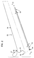
- Fig. 2 is an exploded perspective view of the preferred medical device.
- Fig. 3 is a cross-sectional view of the distal end of the cannula of the preferred medical device, taken along line 3-3 of Fig. 2, prior to threading the slide end of the suture through the first and second channels of the cannula.
- Fig. 4 is a cross-sectional view of the proximal end of the cannula of the preferred device, taken along line 4-4 of Fig. 2, prior to threading the slide end of the suture through the first and second channels of the cannula.
- Figs. 5A and 5B are views similar to that of Fig. 4 showing the preferred means for securing the positioning of the slide end of the suture in the first channel of the cannula of the preferred device.
- Figs. 7-12 are perspective views showing the recommended sequence of steps for endoscopically approximating tissue using the preferred medical device of this invention.
- distal is used to describe that portion of the device which extends away from the user during use
- proximal is used to describe that portion of the device that extends toward the user during use.
- distal refers to movement extending away from the user during use of the device
- proximally refers to movement extending toward the user.
- the device has a cannula 21 with proximal and distal ends 22 and 23, respectively.
- the distal end 23 of the cannula terminates with a beveled surface 24.
- the device also includes a suture 25.
- the suture has a slide end 26, a distal loop 27 and a slip knot 28.
- the slip knot secures the distal loop to the slide end of the suture. Extending from the slip knot is stay end 29 to which a surgical needle 30 is attached.
- the incorporation of the suture 25 into cannula 21 to form the preferred device can be seen at Fig 2.
- the slide end 26 of the suture is threaded completely through the cannula until the slip knot 28 makes contact with the beveled surface 24 of the cannula.
- the slide end is long enough so that it protrudes from the proximal end 22 of the cannula when the slip knot makes contact with the cannula.
- the secure positioning of the slide end of the suture in the first channel of the cannula is provided so that the portion of the slide end protruding from the proximal end of the cannula does not slide back into the cannula, as will be explained in more detail with respect to Figs 4 and 5.
- the suture 25 can be composed of any surgical suture material.
- Suture materials can be composed of synthetic and nonsynthetic filaments, as well as absorbable and nonabsorbable fibers.
- suitable nonabsorbable suture materials include, but are not limited to, nylon, polypropylene, steel, and polyethyleneterephthalate (PET).
- PET polyethyleneterephthalate
- the preferred sutures are synthetic bioabsorbable sutures.
- suitable bioabsorbable sutures are those which are derived from the polymerization of lactone monomers, e.g., lactide, para-dioxanone and ⁇ -caprolactone.
- the stay end 29 of the suture extending from slip knot 28 can be attached to the surgical needle 30 using standard needle attachment. Alternatively, it can be attached using removable or detachable needle attachment. In the case of standard needle attachment, the suture is securely attached to the needle and is not intended to be separated from the needle except by cutting or severing the strand. Removable needle attachment, by contrast, is such that the needle is separable from the strand in response to a force exerted by the surgeon, as illustrated, for example, in European Patent Application 0420605 and U.S. Patent 3,926,194.
- slip knot 28 of the suture can be any configuration which allows for sliding movement of the slide end 26 of the suture proximally and prevents such sliding movement distally.
- the slip knot serves to function as a knot which will allow for the continual reduction in the size of the distal loop 27 of the suture, yet prevents the enlargement of this distal loop, so that a secure and strong suture knot for approximating or ligating tissue can be formed.
- the material of construction of the cannula 21 of the preferred device is not critical, so long as whatever material is chosen is sufficiently biocompatible with bodily tissue.
- a preferred material of choice for the cannula is nylon because of the ease with which it can be fabricated and its biocompatability.
- the slide end of the suture is threaded through first and second channels in the cannula.
- the slide end of the suture would first be threaded through second channel 32 of the cannula, and then subsequently through first channel 33.
- the first channel 33 is the primary channel of the cannula, and extends axially completely through the cannula from the distal to the proximal end.
- the second channel 32 extends from the beveled surface 24 of the cannula to the first channel so that what in effect is formed is a continuous opening in the cannula from the beveled surface to the proximal end of the cannula.
- the angle between the first and second channels is preferably no less than about 20°, but ideally it is about 30°.
- the angle between the beveled surface and the first channel is preferably no less than 20°, but ideally it is about 30°.
- a metallic blunt-tipped pin 31 is heated to a temperature above the melting temperature of the cannula, and the heated pin is pressed against the proximal end 22 of the cannula.
- the pressure of the heated pin is applied in a direction substantially perpendicular to the longitudinal axis of the cannula.
- the heated pin upon contact with the cannula, causes the contacted area of the cannula to melt, thus forming a hole in the outer surface of the cannula through which the pin can be inserted into the first channel.
- the pin is then inserted partially into the first channel to cause a protrusion 37 extending into the first channel.
- the cross-sectional inner diameter at this region of the first channel of the cannula is less than that of the remainder of the first channel.
- This protrusion in turn creates a partial obstruction in the pathway that the slide end of the suture must take if it were pulled through the first channel of the cannula. In this manner, the slide end is prevented from sliding back inside the cannula.
- the partial obstruction caused by the protrusion does not prohibit the surgeon from pulling the slide end protruding from the cannula in the proximal direction when the device is used during surgery. Once the protrusion is created with the pin, the pin is then removed from the cannula.
- the depth to which the pin is inserted may create minimal contact between the blunt-tipped surface of the pin and the suture, although this contact may be unnecessary to provide for the proper securement of the positioning of the slide end inside the cannula.
- the protrusion which the pin creates stabilizes the suture so that it is prevented from sliding freely upon routine handling, yet only creates a slight drag when the user pulls the suture to close the loop and tighten the knot, as will be explained in more detail with reference to Figs. 7-12.
- the protrusion created from the contact with the pin will solidify upon cooling, and therefore cause a permanent obstruction in the first channel of the cannula.
- the surgeon would ideally first backload the cannula of the device into and through an appropriate introducer for a trocar.
- the introducer with the device loaded into it, is then placed into an appropriately sized trocar.
- the trocar provides access into a body cavity where the desired tissue approximation is to occur.
- the cannula 21 of the medical device 20 is pushed into the field of surgery through trocar 34 and trocar cannula 35, which have penetrated bodily tissue 33.
- the surgeon would use graspers 36 to hold and orient the needle, and to pass the surgical needle through the two segments of tissue to be joined.
- the surgeon positions distal loop 27 of the suture by rotating the cannula 21 of the device so that the graspers can be easily passed through the distal loop.
- the surgeon grasps the stay end 29 of the suture just behind the needle, and pulls the stay end and needle of the suture through the loop.
- the cannula 21 is simultaneously advanced distally until the base of the slip knot lies against the tissue to be joined.
- the distal loop 27 of the suture can now be closed to form the suture knot.
- That portion of the slide end of the suture protruding from the proximal end of the cannula is held firmly and pulled proximally.
- the distal loop subsequently becomes smaller and smaller until it is cinched about the suture to form a knot joining the severed tissue.
- the method of using the device for ligation is substantially the same as the method described above for tissue approximation. The only significant difference is that the surgeon would first remove the needle from the suture prior to inserting the device into and through the trocar.
- the needle is generally unnecessary for ligation because the suture can merely be looped around the vessel desired to be ligated. This looping procedure can be preformed without penetrating tissue, so that the needle can be removed without affecting the ease with which the ligation can be preformed.
Description
- This invention relates to a medical device, particularly an endoscopic medical device, which is designed to facilitate the surgical procedures of tissue approximation and vessel ligation.
- As medical and hospital costs continue to increase, surgeons are constantly striving to develop advanced surgical techniques. Advances in the surgical field are often related to the development of operative techniques which involve less invasive surgical procedures and reduce overall patient trauma. In this manner, the length of hospital stays can be significantly reduced, and therefore the hospital and medical costs can be reduced as well.
- One of the truly great advances in recent years to reduce the invasiveness of surgical procedures is endoscopic surgery. Endoscopic surgery involves the use of an endoscope, which is an instrument permitting the visual inspection and magnification of any cavity of the body. The endoscope is inserted through a tube, conventionally referred to as a cannula, after puncture of a hole into the soft tissue protecting the body cavity. The hole is made with a trocar, which is a sharp-pointed instrument. The trocar includes an obturator, or cutting implement, which is slideably and removably disposed within a trocar cannula. The obturator will puncture a hole in the tissue equal in size to the inner diameter of the trocar cannula. After puncture, the obturator can be slideably withdrawn from the trocar cannula. The surgeon can then perform diagnostic and therapeutic procedures at the surgical site with the aid of specialized instrumentation designed to fit through the trocar cannula and additional trocar cannulas providing openings into the desired body cavity as may be required.
- In many surgical procedures, suturing and ligating are required to close wounds. For example, suturing to approximate tissue, which requires the formation of a suture knot for placement of a stitch, is often required for proper healing of lacerations and surgical incisions. Additionally, ligating blood vessels to be cut within the surgical site is often necessary in numerous surgical procedures. The vessels may then be severed between the ligations. The primary reason for ligating the vessels is to maintain the surgical site free of an excess of blood and to reduce blood loss in the patient.
- Conventionally, surgeons have closed blood vessels with ligatures, which are long, relatively straight strands of suture material. As is the case with the formation of a suture knot to place a stitch, the surgeon must manually tie a knot on the ligature after the ligature is looped around the vessel desired to be closed. Unfortunately, the formation of a knot on sutures and conventional ligatures is tedious and time-consuming during endoscopic surgical applications where a surgeon's manual operative techniques within the surgical site are severely restricted.
- In more recent years, a ligature has been developed which is better adapted for endoscopic surgery. ENDOKNOT gut ligature is a device formed from a suture material of surgical catgut. The catgut ligature is securely fastened within a cannula at one end and attached to a needle at the other end. This device is described in the 1991 package insert for the product sold by Ethicon, Inc., which insert is entitled "ENDOKNOT Suture Made With Chromic Gut". Although ENDOKNOT gut ligature facilitates ligation of vessels through small incisions in bodily cavities, the surgeon is required to manually tie the ligature knot extracorporeally, i.e. outside the body, to ligate a vessel. This is a time consuming and laborious process, especially for inexperienced surgeons, and represents a significant disadvantage to the use of the ENDOKNOT ligature device during endoscopic surgery.
- An improved device for ligation is described in European Patent Application 477,020, published March 25, 1992. This device has a suture loop extending from the cannula. The loop is secured to the suture strand with a slip knot. This device eliminates the need to tie a knot extracorporeally, but the loop is extremely difficult to maneuver and position during endoscopy. In addition, it is difficult to avoid undesirably rotating the loop during use. Furthermore, the device is designed only for ligation, and not for tissue approximation as well.
- A similar device to that described in the European patent application discussed above is disclosed in U.S. Patent 5,129,912. This device has a pre-looped suture and needle for tissue approximation, and means for applying tension on the suture knot. Unfortunately, this device suffers from similar deficiencies because it too is difficult to maneuver and position during endoscopy.
- Another attempt to avoid the need for manually tying the knot extracorporeally for ligation or tissue approximation is described in U.S. Serial No. 728,443, filed July 11, 1991 corresponding to US-A-5144961, published September 8, 1992. This reference describes a device which has a partially tightened knot secured around a cannula. The knot can be slid down the cannula onto a suture strand to make a loop which can then be subsequently tightened into a secure knot. Unfortunately, in operation the user must thread the suture strand up through the cannula before sliding the partially tightened knot down the cannula to make the loop. This threading operation is undesirable because it is time consuming and tedious. A similar device is shown in U.S. Patent 2,012,776.
- In view of the significant deficiencies of the prior art, an endoscopic suturing device having a suture loop which avoids manually tying knots extracorporeally, and is designed to easily manipulate and position the suture loop during use, would be highly desired within the medical community. Additionally, it would be highly desirable if such a design were designed which offered the flexibility for use to not only ligate vessels but also approximate tissue.
- The invention is a medical device which comprises a cannula and a suture. The cannula has a proximal end and a distal end, and the distal end has a beveled surface thereon. The cannula also has a first channel extending axially through it, and a second channel extending from the beveled surface to the first channel.
- The suture has a slide end, a distal loop, and a slip knot securing the loop to the slide end. The slide end is threaded through the first and second channels of the cannula, and protrudes from the proximal end of the cannula.
- The device has means for securing the positioning of the slide end of the suture in the first channel of the cannula.
- In a preferred embodiment, the suture has a stay end extending from the slip knot, and a needle attached to the stay end. In this manner, the device can be used for tissue approximation, or if desired, the needle can be detached and the device would then be especially well adapted for vessel ligation.
- Surprisingly, the distal loop of the suture is easily manipulated and positioned during use. Therefore, the device is particularly suited for endoscopic surgery. The threading of the slide end of the suture through the first and second channels of the cannula helps to lock the distal loop of the suture in place, thus preventing or reducing any rotation of the loop during usage. This in turn greatly improves maneuverability in general of the device. Also, the distal loop of the suture can be easily placed at the desired surgical site.
- Additionally, the beveled surface at the distal end of the cannula makes the distal end more visible and easy to locate during endoscopic surgery. The beveled surface serves as a platform to support the slip knot and to prevent any unwanted shifting when the device is used. Furthermore, the design of this device permits greater visibility of the slip knot as well when the device is being used.
- Fig. 1 is a perspective view of the preferred medical device of this invention.
- Fig. 2 is an exploded perspective view of the preferred medical device.
- Fig. 3 is a cross-sectional view of the distal end of the cannula of the preferred medical device, taken along line 3-3 of Fig. 2, prior to threading the slide end of the suture through the first and second channels of the cannula.
- Fig. 4 is a cross-sectional view of the proximal end of the cannula of the preferred device, taken along line 4-4 of Fig. 2, prior to threading the slide end of the suture through the first and second channels of the cannula.
- Figs. 5A and 5B are views similar to that of Fig. 4 showing the preferred means for securing the positioning of the slide end of the suture in the first channel of the cannula of the preferred device.
- Figs. 7-12 are perspective views showing the recommended sequence of steps for endoscopically approximating tissue using the preferred medical device of this invention.
- As used in this specification, the word "distal" is used to describe that portion of the device which extends away from the user during use, and the word "proximal" is used to describe that portion of the device that extends toward the user during use. Similarly, "distally" refers to movement extending away from the user during use of the device, and "proximally" refers to movement extending toward the user.
- Referring to Fig. 1, there is shown the preferred
medical device 20. The device has acannula 21 with proximal anddistal ends distal end 23 of the cannula terminates with abeveled surface 24. The device also includes asuture 25. The suture has aslide end 26, adistal loop 27 and aslip knot 28. The slip knot secures the distal loop to the slide end of the suture. Extending from the slip knot is stayend 29 to which asurgical needle 30 is attached. - The incorporation of the
suture 25 intocannula 21 to form the preferred device can be seen at Fig 2. Theslide end 26 of the suture is threaded completely through the cannula until theslip knot 28 makes contact with thebeveled surface 24 of the cannula. The slide end is long enough so that it protrudes from theproximal end 22 of the cannula when the slip knot makes contact with the cannula. The secure positioning of the slide end of the suture in the first channel of the cannula is provided so that the portion of the slide end protruding from the proximal end of the cannula does not slide back into the cannula, as will be explained in more detail with respect to Figs 4 and 5. - The
suture 25 can be composed of any surgical suture material. Suture materials can be composed of synthetic and nonsynthetic filaments, as well as absorbable and nonabsorbable fibers. Examples of suitable nonabsorbable suture materials include, but are not limited to, nylon, polypropylene, steel, and polyethyleneterephthalate (PET). The preferred sutures are synthetic bioabsorbable sutures. Examples of suitable bioabsorbable sutures are those which are derived from the polymerization of lactone monomers, e.g., lactide, para-dioxanone and ε-caprolactone. - The
stay end 29 of the suture extending fromslip knot 28 can be attached to thesurgical needle 30 using standard needle attachment. Alternatively, it can be attached using removable or detachable needle attachment. In the case of standard needle attachment, the suture is securely attached to the needle and is not intended to be separated from the needle except by cutting or severing the strand. Removable needle attachment, by contrast, is such that the needle is separable from the strand in response to a force exerted by the surgeon, as illustrated, for example, in European Patent Application 0420605 and U.S. Patent 3,926,194. - The configuration of
slip knot 28 of the suture can be any configuration which allows for sliding movement of theslide end 26 of the suture proximally and prevents such sliding movement distally. In this manner, the slip knot serves to function as a knot which will allow for the continual reduction in the size of thedistal loop 27 of the suture, yet prevents the enlargement of this distal loop, so that a secure and strong suture knot for approximating or ligating tissue can be formed. - The material of construction of the
cannula 21 of the preferred device is not critical, so long as whatever material is chosen is sufficiently biocompatible with bodily tissue. A preferred material of choice for the cannula is nylon because of the ease with which it can be fabricated and its biocompatability. - The slide end of the suture is threaded through first and second channels in the cannula. Referring now to Fig. 3, the slide end of the suture would first be threaded through
second channel 32 of the cannula, and then subsequently throughfirst channel 33. Thefirst channel 33 is the primary channel of the cannula, and extends axially completely through the cannula from the distal to the proximal end. Thesecond channel 32 extends from thebeveled surface 24 of the cannula to the first channel so that what in effect is formed is a continuous opening in the cannula from the beveled surface to the proximal end of the cannula. The angle between the first and second channels is preferably no less than about 20°, but ideally it is about 30°. Likewise, the angle between the beveled surface and the first channel is preferably no less than 20°, but ideally it is about 30°. When the slide end of the suture is threaded through the first and second channels of the cannula, the slip knot of the suture abuts the opening of the second channel at the beveled surface, and the slide end protrudes from the proximal end of the cannula. - The creation of a partial obstruction in the first channel of the cannula at its proximal end to provide for secure positioning of the slide end of the suture during packaging and routine handling is illustrated in Figs. 4 and 5. After the slide end of the suture is threaded through the first and second channels of the cannula, a metallic blunt-tipped
pin 31 is heated to a temperature above the melting temperature of the cannula, and the heated pin is pressed against theproximal end 22 of the cannula. The pressure of the heated pin is applied in a direction substantially perpendicular to the longitudinal axis of the cannula. The heated pin, upon contact with the cannula, causes the contacted area of the cannula to melt, thus forming a hole in the outer surface of the cannula through which the pin can be inserted into the first channel. The pin is then inserted partially into the first channel to cause aprotrusion 37 extending into the first channel. The cross-sectional inner diameter at this region of the first channel of the cannula is less than that of the remainder of the first channel. This protrusion in turn creates a partial obstruction in the pathway that the slide end of the suture must take if it were pulled through the first channel of the cannula. In this manner, the slide end is prevented from sliding back inside the cannula. However, the partial obstruction caused by the protrusion does not prohibit the surgeon from pulling the slide end protruding from the cannula in the proximal direction when the device is used during surgery. Once the protrusion is created with the pin, the pin is then removed from the cannula. - The depth to which the pin is inserted may create minimal contact between the blunt-tipped surface of the pin and the suture, although this contact may be unnecessary to provide for the proper securement of the positioning of the slide end inside the cannula. The protrusion which the pin creates stabilizes the suture so that it is prevented from sliding freely upon routine handling, yet only creates a slight drag when the user pulls the suture to close the loop and tighten the knot, as will be explained in more detail with reference to Figs. 7-12. When the pin is removed from the cannula, the protrusion created from the contact with the pin will solidify upon cooling, and therefore cause a permanent obstruction in the first channel of the cannula.
- To approximate tissue using the device, the surgeon would ideally first backload the cannula of the device into and through an appropriate introducer for a trocar. The introducer, with the device loaded into it, is then placed into an appropriately sized trocar. The trocar provides access into a body cavity where the desired tissue approximation is to occur. As illustrated in Fig. 6, the
cannula 21 of themedical device 20 is pushed into the field of surgery throughtrocar 34 andtrocar cannula 35, which have penetratedbodily tissue 33. - Referring now to Figs. 7-12 in sequence, the surgeon would use
graspers 36 to hold and orient the needle, and to pass the surgical needle through the two segments of tissue to be joined. The surgeon then positionsdistal loop 27 of the suture by rotating thecannula 21 of the device so that the graspers can be easily passed through the distal loop. Once the graspers have been passed through the distal loop, the surgeon then grasps thestay end 29 of the suture just behind the needle, and pulls the stay end and needle of the suture through the loop. As the stay end of the suture is pulled, thecannula 21 is simultaneously advanced distally until the base of the slip knot lies against the tissue to be joined. While holding the stay end of the suture firmly, thedistal loop 27 of the suture can now be closed to form the suture knot. To close the loop, that portion of the slide end of the suture protruding from the proximal end of the cannula is held firmly and pulled proximally. As the slide end is pulled proximally, the distal loop subsequently becomes smaller and smaller until it is cinched about the suture to form a knot joining the severed tissue. Once the knot is formed, excess suture at the stay end and the slide end can be easily removed, and the needle, excess suture and cannula can be removed from the surgical site. - The method of using the device for ligation is substantially the same as the method described above for tissue approximation. The only significant difference is that the surgeon would first remove the needle from the suture prior to inserting the device into and through the trocar. The needle is generally unnecessary for ligation because the suture can merely be looped around the vessel desired to be ligated. This looping procedure can be preformed without penetrating tissue, so that the needle can be removed without affecting the ease with which the ligation can be preformed.
- Although this description has focused on the preferred embodiment of the device, it is readily apparent that numerous additional embodiments of the device can be envisioned without departing from the scope of the claimed invention.
Claims (6)
- A medical device comprising:a) a cannula (21), said cannula having a proximal end (22) and a distal end (23), said distal end having a beveled surface (24) thereon, said cannula having a first channel (33) extending axially therethrough and a second channel (32) extending from said beveled surface to said first channel;b) a suture (25), said suture having a slide end (26), a distal loop (27), and a slip knot (28) securing said loop to said slide end, said slide end threaded through said first and second channels, and protruding from the proximal end of said cannula; andc) means for securing the positioning of said slide end (26) of said suture in said first channel (33) of said cannula.
- The device of claim 1 wherein said suture has a stay end (29) extending from said slip knot (28), and a needle (30) attached to said stay end.
- The device of claim 2 wherein said slip knot (28) abuts the opening of the second channel (32) at the beveled surface (24).
- The device of claim 3 wherein said slip knot (28) is configured to allow sliding movement of said slide end (26) proximally so as to continually reduce the size of said distal loop (27).
- The device of claim 4 wherein said means is a protrusion (37) extending partially into said first channel (33).
- The device of claim 5 wherein the angle between said first and second channels (33,32) is about 30°.
Applications Claiming Priority (2)
| Application Number | Priority Date | Filing Date | Title |
|---|---|---|---|
| US947662 | 1992-09-18 | ||
| US07/947,662 US5234445A (en) | 1992-09-18 | 1992-09-18 | Endoscopic suturing device |
Publications (2)
| Publication Number | Publication Date |
|---|---|
| EP0588659A1 EP0588659A1 (en) | 1994-03-23 |
| EP0588659B1 true EP0588659B1 (en) | 1995-11-15 |
Family
ID=25486522
Family Applications (1)
| Application Number | Title | Priority Date | Filing Date |
|---|---|---|---|
| EP93307359A Expired - Lifetime EP0588659B1 (en) | 1992-09-18 | 1993-09-17 | Endoscopic suturing device |
Country Status (7)
| Country | Link |
|---|---|
| US (1) | US5234445A (en) |
| EP (1) | EP0588659B1 (en) |
| JP (1) | JP3305064B2 (en) |
| AU (1) | AU668585B2 (en) |
| BR (1) | BR9303822A (en) |
| CA (1) | CA2106327A1 (en) |
| DE (1) | DE69300810T2 (en) |
Cited By (2)
| Publication number | Priority date | Publication date | Assignee | Title |
|---|---|---|---|---|
| US11298122B2 (en) | 2018-06-07 | 2022-04-12 | EnVision Endoscopy, Inc. | Endoscopic suturing device with circular needle |
| US11534158B2 (en) | 2020-10-23 | 2022-12-27 | EnVision Endoscopy, Inc. | Endoscopic suture cinch |
Families Citing this family (96)
| Publication number | Priority date | Publication date | Assignee | Title |
|---|---|---|---|---|
| US5320629B1 (en) * | 1991-01-07 | 2000-05-02 | Advanced Surgical Inc | Device and method for applying suture |
| US5330491A (en) * | 1992-09-18 | 1994-07-19 | Ethicon, Inc. | Endoscopic suturing device |
| US5282809A (en) * | 1992-11-16 | 1994-02-01 | Ethicon, Inc. | Endoscopic suturing device |
| US5334200A (en) * | 1993-03-02 | 1994-08-02 | Lanny L. Johnson | Suture knot making device and method for use |
| US5496331A (en) * | 1993-07-28 | 1996-03-05 | Terumo Kabushiki Kaisha | Knot-forming instrument and method of forming knots |
| US5395382A (en) * | 1993-10-22 | 1995-03-07 | Ethicon, Inc. | Device for tying intracorporeal knot |
| US5643293A (en) * | 1993-12-29 | 1997-07-01 | Olympus Optical Co., Ltd. | Suturing instrument |
| US5741280A (en) | 1994-01-18 | 1998-04-21 | Coral Medical | Knot tying method and apparatus |
| US5522827A (en) * | 1994-04-22 | 1996-06-04 | Zimmer, Inc. | Apparatus and method for harvesting a tendon graft |
| WO1995032671A1 (en) * | 1994-06-01 | 1995-12-07 | Perclose, Inc. | Method and device for providing vascular hemostasis |
| US5562684A (en) | 1994-10-11 | 1996-10-08 | Ethicon, Inc. | Surgical knot pusher device and improved method of forming knots |
| US5643292A (en) * | 1995-01-10 | 1997-07-01 | Applied Medical Resources Corporation | Percutaneous suturing device |
| US5709694A (en) * | 1995-03-22 | 1998-01-20 | Human Factors Industrial Design, Inc. | Endoscopic intracorporeal suture tying aid |
| USD386583S (en) * | 1996-01-02 | 1997-11-18 | Acufex Microsurgical, Inc. | Proximal end of a surgical suture slotted knot pusher |
| USD387161S (en) * | 1996-01-02 | 1997-12-02 | Acufex Microsurgical, Inc. | Surgical suture knot pusher with hooks |
| US5741276A (en) * | 1996-03-28 | 1998-04-21 | Innovative Surgical Instruments | Apparatus for facilitating the performance of surgical procedures such as the placement of sutures, ligatures and the like |
| US5759189A (en) * | 1997-02-25 | 1998-06-02 | Smith & Nephew Inc. | Knot pusher |
| US6155264A (en) * | 1997-03-06 | 2000-12-05 | Scimed Life Systems, Inc. | Percutaneous bypass by tunneling through vessel wall |
| US6035856A (en) | 1997-03-06 | 2000-03-14 | Scimed Life Systems | Percutaneous bypass with branching vessel |
| US6026814A (en) | 1997-03-06 | 2000-02-22 | Scimed Life Systems, Inc. | System and method for percutaneous coronary artery bypass |
| US5893592A (en) * | 1997-04-08 | 1999-04-13 | Ethicon Endo-Surgery, Inc. | Partially tied surgical knot |
| US5728109A (en) * | 1997-04-08 | 1998-03-17 | Ethicon Endo-Surgery, Inc. | Surgical knot and method for its formation |
| US6092526A (en) * | 1997-06-19 | 2000-07-25 | Scimed Life Systems, Inc. | Percutaneous chamber-to-artery bypass |
| US6443158B1 (en) | 1997-06-19 | 2002-09-03 | Scimed Life Systems, Inc. | Percutaneous coronary artery bypass through a venous vessel |
| US6213126B1 (en) | 1997-06-19 | 2001-04-10 | Scimed Life Systems, Inc. | Percutaneous artery to artery bypass using heart tissue as a portion of a bypass conduit |
| EP1105049A4 (en) * | 1998-07-22 | 2007-10-17 | Angiolink Corp | Vascular suction cannula, dilator and surgical stapler |
| US6325813B1 (en) | 1998-08-18 | 2001-12-04 | Scimed Life Systems, Inc. | Method and apparatus for stabilizing vascular wall |
| US6132439A (en) * | 1999-02-17 | 2000-10-17 | X-Site, L.L.C. | Knot pusher |
| US7842048B2 (en) | 2006-08-18 | 2010-11-30 | Abbott Laboratories | Articulating suture device and method |
| US8137364B2 (en) | 2003-09-11 | 2012-03-20 | Abbott Laboratories | Articulating suturing device and method |
| US7235087B2 (en) | 1999-03-04 | 2007-06-26 | Abbott Park | Articulating suturing device and method |
| US7001400B1 (en) | 1999-03-04 | 2006-02-21 | Abbott Laboratories | Articulating suturing device and method |
| US6964668B2 (en) * | 1999-03-04 | 2005-11-15 | Abbott Laboratories | Articulating suturing device and method |
| US20040092964A1 (en) * | 1999-03-04 | 2004-05-13 | Modesitt D. Bruce | Articulating suturing device and method |
| US6152934A (en) * | 1999-06-14 | 2000-11-28 | Ethicon Endo-Surgery, Inc. | Surgical knot tying instrument |
| US7618426B2 (en) | 2002-12-11 | 2009-11-17 | Usgi Medical, Inc. | Apparatus and methods for forming gastrointestinal tissue approximations |
| US7955340B2 (en) | 1999-06-25 | 2011-06-07 | Usgi Medical, Inc. | Apparatus and methods for forming and securing gastrointestinal tissue folds |
| US7416554B2 (en) | 2002-12-11 | 2008-08-26 | Usgi Medical Inc | Apparatus and methods for forming and securing gastrointestinal tissue folds |
| US7637905B2 (en) | 2003-01-15 | 2009-12-29 | Usgi Medical, Inc. | Endoluminal tool deployment system |
| US6716224B2 (en) | 2000-08-28 | 2004-04-06 | Linvatec Corporation | Intracorporeal knot tier |
| US6884249B2 (en) * | 2001-02-16 | 2005-04-26 | Depuy Mitek, Inc. | Surgical knot pusher and method of use |
| US7402166B2 (en) * | 2002-02-15 | 2008-07-22 | A&P Feigl Family Limited Partnership | Devices and methods for positioning sutures |
| US7942884B2 (en) | 2002-12-11 | 2011-05-17 | Usgi Medical, Inc. | Methods for reduction of a gastric lumen |
| US7942898B2 (en) | 2002-12-11 | 2011-05-17 | Usgi Medical, Inc. | Delivery systems and methods for gastric reduction |
| US20040116943A1 (en) * | 2002-12-13 | 2004-06-17 | Brandt C. Phillip | Method and apparatus for endoscopically ligating an elongate tissue structure at multiple sites |
| US7160309B2 (en) | 2002-12-31 | 2007-01-09 | Laveille Kao Voss | Systems for anchoring a medical device in a body lumen |
| US8308765B2 (en) | 2004-05-07 | 2012-11-13 | Usgi Medical, Inc. | Apparatus and methods for positioning and securing anchors |
| US8216252B2 (en) | 2004-05-07 | 2012-07-10 | Usgi Medical, Inc. | Tissue manipulation and securement system |
| US7462188B2 (en) | 2003-09-26 | 2008-12-09 | Abbott Laboratories | Device and method for suturing intracardiac defects |
| US7361180B2 (en) | 2004-05-07 | 2008-04-22 | Usgi Medical, Inc. | Apparatus for manipulating and securing tissue |
| US20050251189A1 (en) | 2004-05-07 | 2005-11-10 | Usgi Medical Inc. | Multi-position tissue manipulation assembly |
| US7347863B2 (en) | 2004-05-07 | 2008-03-25 | Usgi Medical, Inc. | Apparatus and methods for manipulating and securing tissue |
| US7449024B2 (en) | 2003-12-23 | 2008-11-11 | Abbott Laboratories | Suturing device with split arm and method of suturing tissue |
| US7703459B2 (en) | 2004-03-09 | 2010-04-27 | Usgi Medical, Inc. | Apparatus and methods for mapping out endoluminal gastrointestinal surgery |
| KR100603633B1 (en) * | 2004-04-30 | 2006-07-24 | 문화숙 | Endo-loop for an operation using endoscope |
| US7918869B2 (en) | 2004-05-07 | 2011-04-05 | Usgi Medical, Inc. | Methods and apparatus for performing endoluminal gastroplasty |
| US7736374B2 (en) | 2004-05-07 | 2010-06-15 | Usgi Medical, Inc. | Tissue manipulation and securement system |
| US7390329B2 (en) | 2004-05-07 | 2008-06-24 | Usgi Medical, Inc. | Methods for grasping and cinching tissue anchors |
| US8257394B2 (en) | 2004-05-07 | 2012-09-04 | Usgi Medical, Inc. | Apparatus and methods for positioning and securing anchors |
| US8444657B2 (en) | 2004-05-07 | 2013-05-21 | Usgi Medical, Inc. | Apparatus and methods for rapid deployment of tissue anchors |
| US8206417B2 (en) | 2004-06-09 | 2012-06-26 | Usgi Medical Inc. | Apparatus and methods for optimizing anchoring force |
| US7736379B2 (en) | 2004-06-09 | 2010-06-15 | Usgi Medical, Inc. | Compressible tissue anchor assemblies |
| US7695493B2 (en) | 2004-06-09 | 2010-04-13 | Usgi Medical, Inc. | System for optimizing anchoring force |
| US7678135B2 (en) | 2004-06-09 | 2010-03-16 | Usgi Medical, Inc. | Compressible tissue anchor assemblies |
| US9585651B2 (en) | 2005-05-26 | 2017-03-07 | Usgi Medical, Inc. | Methods and apparatus for securing and deploying tissue anchors |
| US8298291B2 (en) | 2005-05-26 | 2012-10-30 | Usgi Medical, Inc. | Methods and apparatus for securing and deploying tissue anchors |
| US8083754B2 (en) | 2005-08-08 | 2011-12-27 | Abbott Laboratories | Vascular suturing device with needle capture |
| US8267947B2 (en) | 2005-08-08 | 2012-09-18 | Abbott Laboratories | Vascular suturing device |
| US7883517B2 (en) | 2005-08-08 | 2011-02-08 | Abbott Laboratories | Vascular suturing device |
| US9456811B2 (en) | 2005-08-24 | 2016-10-04 | Abbott Vascular Inc. | Vascular closure methods and apparatuses |
| US20070060895A1 (en) | 2005-08-24 | 2007-03-15 | Sibbitt Wilmer L Jr | Vascular closure methods and apparatuses |
| US8920442B2 (en) | 2005-08-24 | 2014-12-30 | Abbott Vascular Inc. | Vascular opening edge eversion methods and apparatuses |
| US8726909B2 (en) | 2006-01-27 | 2014-05-20 | Usgi Medical, Inc. | Methods and apparatus for revision of obesity procedures |
| GB0607608D0 (en) * | 2006-04-18 | 2006-05-31 | Royal Brompton & Harefield Nhs | Sternal closure device |
| JP2009536083A (en) * | 2006-05-03 | 2009-10-08 | インディアナ・ユニバーシティ・リサーチ・アンド・テクノロジー・コーポレーション | Method and apparatus for reconstructing the esophagus and other body lumens |
| US8870916B2 (en) | 2006-07-07 | 2014-10-28 | USGI Medical, Inc | Low profile tissue anchors, tissue anchor systems, and methods for their delivery and use |
| US8574244B2 (en) | 2007-06-25 | 2013-11-05 | Abbott Laboratories | System for closing a puncture in a vessel wall |
| US8808313B2 (en) * | 2007-07-12 | 2014-08-19 | Linvatec Corporation | Suture passing and retrieval device |
| US8449478B2 (en) * | 2008-05-16 | 2013-05-28 | Conquest Medical Technologies | Biopsy device |
| US8317827B2 (en) * | 2009-01-07 | 2012-11-27 | Ethicon Endo-Surgery, Inc. | Suturing devices and methods |
| US8663252B2 (en) | 2010-09-01 | 2014-03-04 | Abbott Cardiovascular Systems, Inc. | Suturing devices and methods |
| US9370353B2 (en) | 2010-09-01 | 2016-06-21 | Abbott Cardiovascular Systems, Inc. | Suturing devices and methods |
| US8795334B2 (en) | 2011-01-28 | 2014-08-05 | Smith & Nephew, Inc. | Tissue repair |
| JP6125488B2 (en) | 2011-03-23 | 2017-05-10 | エシコン・エルエルシーEthicon LLC | Self-holding variable loop suture |
| US8926662B2 (en) | 2012-02-01 | 2015-01-06 | Smith & Nephew, Inc. | Tissue graft anchoring |
| US9084597B2 (en) | 2012-03-09 | 2015-07-21 | Smith & Nephew, Inc. | Suture-based knotless repair |
| US8858573B2 (en) | 2012-04-10 | 2014-10-14 | Abbott Cardiovascular Systems, Inc. | Apparatus and method for suturing body lumens |
| US8864778B2 (en) | 2012-04-10 | 2014-10-21 | Abbott Cardiovascular Systems, Inc. | Apparatus and method for suturing body lumens |
| US9265514B2 (en) | 2012-04-17 | 2016-02-23 | Miteas Ltd. | Manipulator for grasping tissue |
| KR20130124792A (en) * | 2012-05-07 | 2013-11-15 | 정창욱 | Ligation device |
| US9241707B2 (en) | 2012-05-31 | 2016-01-26 | Abbott Cardiovascular Systems, Inc. | Systems, methods, and devices for closing holes in body lumens |
| US8986327B2 (en) | 2012-10-18 | 2015-03-24 | Smith & Nephew, Inc. | Flexible anchor delivery system |
| WO2015137269A1 (en) * | 2014-03-11 | 2015-09-17 | 国立大学法人九州大学 | Suture needle |
| US10426449B2 (en) | 2017-02-16 | 2019-10-01 | Abbott Cardiovascular Systems, Inc. | Articulating suturing device with improved actuation and alignment mechanisms |
| US20210038255A1 (en) * | 2017-12-01 | 2021-02-11 | The Regents Of The University Of Colorado, A Body Corporate | Slotted canulla for arthroscopic surgery |
| US11413034B2 (en) | 2020-10-13 | 2022-08-16 | Olympus Corporation | Method for closing wound |
Family Cites Families (9)
| Publication number | Priority date | Publication date | Assignee | Title |
|---|---|---|---|---|
| US2012776A (en) * | 1931-05-23 | 1935-08-27 | Roeder Hans Albert | Ligator |
| NL133643C (en) * | 1967-01-10 | 1900-01-01 | ||
| US4177813A (en) * | 1978-01-09 | 1979-12-11 | Med General, Inc. | Vessel occluder |
| US4602635A (en) * | 1983-11-14 | 1986-07-29 | Mulhollan James S | Remote surgical knot tier and method of use |
| US5032897A (en) * | 1990-02-28 | 1991-07-16 | International Business Machines Corp. | Integrated thermoelectric cooling |
| US5129912B2 (en) * | 1991-01-07 | 2000-01-11 | Urohealth Systems Inc | Device and method for applying suture |
| DE9100162U1 (en) * | 1991-01-09 | 1991-05-08 | Scarfi, Andrea, Dr.Med., 7750 Konstanz, De | |
| US5144961A (en) * | 1991-07-11 | 1992-09-08 | Ethicon, Inc. | Endoscopic ligating device |
| US5152769A (en) * | 1991-11-04 | 1992-10-06 | Will Baber | Apparatus for laparoscopic suturing with improved suture needle |
-
1992
- 1992-09-18 US US07/947,662 patent/US5234445A/en not_active Expired - Lifetime
-
1993
- 1993-09-16 CA CA002106327A patent/CA2106327A1/en not_active Abandoned
- 1993-09-16 AU AU47419/93A patent/AU668585B2/en not_active Expired
- 1993-09-17 DE DE69300810T patent/DE69300810T2/en not_active Expired - Lifetime
- 1993-09-17 BR BR9303822A patent/BR9303822A/en not_active Application Discontinuation
- 1993-09-17 EP EP93307359A patent/EP0588659B1/en not_active Expired - Lifetime
- 1993-09-20 JP JP25651293A patent/JP3305064B2/en not_active Expired - Lifetime
Cited By (2)
| Publication number | Priority date | Publication date | Assignee | Title |
|---|---|---|---|---|
| US11298122B2 (en) | 2018-06-07 | 2022-04-12 | EnVision Endoscopy, Inc. | Endoscopic suturing device with circular needle |
| US11534158B2 (en) | 2020-10-23 | 2022-12-27 | EnVision Endoscopy, Inc. | Endoscopic suture cinch |
Also Published As
| Publication number | Publication date |
|---|---|
| US5234445A (en) | 1993-08-10 |
| AU668585B2 (en) | 1996-05-09 |
| JPH06189968A (en) | 1994-07-12 |
| DE69300810D1 (en) | 1995-12-21 |
| JP3305064B2 (en) | 2002-07-22 |
| CA2106327A1 (en) | 1994-03-19 |
| BR9303822A (en) | 1994-03-29 |
| AU4741993A (en) | 1994-03-24 |
| DE69300810T2 (en) | 1996-04-25 |
| EP0588659A1 (en) | 1994-03-23 |
Similar Documents
| Publication | Publication Date | Title |
|---|---|---|
| EP0588659B1 (en) | Endoscopic suturing device | |
| EP0628286B1 (en) | Endoscopic suturing device | |
| EP0598588B1 (en) | Endoscopic suturing device | |
| EP0588606B1 (en) | Endoscopic knotting device | |
| US5144961A (en) | Endoscopic ligating device | |
| US5129912A (en) | Device and method for applying suture | |
| US5320629A (en) | Device and method for applying suture | |
| EP0706779B1 (en) | Surgical knot pusher device | |
| US5449367A (en) | Pre-tied knot for surgical use and method of using same | |
| US5207694A (en) | Method for performing a surgical occlusion, and kit and applicator for carrying out the method | |
| US5181919A (en) | Suture ligating device for use with an endoscope | |
| AU716663B2 (en) | Knotting element for use in suturing anatomical tissue and methods therefor | |
| US5683417A (en) | Suture and method for endoscopic surgery | |
| US4796626A (en) | Tourniquet tube | |
| US6221084B1 (en) | Knot tying apparatus having a notched thread cover and method for using same | |
| JPH03503251A (en) | Ligation system for use in endoscopic surgery and ligation instrument for the system | |
| US5653719A (en) | Knot pushing instrument for endoscopic surgery | |
| JPH07136177A (en) | Biologically compatible suture clip | |
| US10743862B1 (en) | Laparoscopic suturing device and methods of use |
Legal Events
| Date | Code | Title | Description |
|---|---|---|---|
| PUAI | Public reference made under article 153(3) epc to a published international application that has entered the european phase |
Free format text: ORIGINAL CODE: 0009012 |
|
| AK | Designated contracting states |
Kind code of ref document: A1 Designated state(s): DE FR GB |
|
| 17P | Request for examination filed |
Effective date: 19940826 |
|
| D17Q | First examination report despatched (deleted) | ||
| GRAA | (expected) grant |
Free format text: ORIGINAL CODE: 0009210 |
|
| RIN1 | Information on inventor provided before grant (corrected) |
Inventor name: BRITT, CRAWFORD R. Inventor name: ROYCE, FREDERICK L. Inventor name: WALKER, EDWARD E. |
|
| AK | Designated contracting states |
Kind code of ref document: B1 Designated state(s): DE FR GB |
|
| REF | Corresponds to: |
Ref document number: 69300810 Country of ref document: DE Date of ref document: 19951221 |
|
| ET | Fr: translation filed | ||
| PLBE | No opposition filed within time limit |
Free format text: ORIGINAL CODE: 0009261 |
|
| STAA | Information on the status of an ep patent application or granted ep patent |
Free format text: STATUS: NO OPPOSITION FILED WITHIN TIME LIMIT |
|
| 26N | No opposition filed | ||
| REG | Reference to a national code |
Ref country code: GB Ref legal event code: IF02 |
|
| PGFP | Annual fee paid to national office [announced via postgrant information from national office to epo] |
Ref country code: GB Payment date: 20120912 Year of fee payment: 20 |
|
| PGFP | Annual fee paid to national office [announced via postgrant information from national office to epo] |
Ref country code: DE Payment date: 20120912 Year of fee payment: 20 Ref country code: FR Payment date: 20120926 Year of fee payment: 20 |
|
| REG | Reference to a national code |
Ref country code: DE Ref legal event code: R071 Ref document number: 69300810 Country of ref document: DE |
|
| REG | Reference to a national code |
Ref country code: GB Ref legal event code: PE20 Expiry date: 20130916 |
|
| PG25 | Lapsed in a contracting state [announced via postgrant information from national office to epo] |
Ref country code: DE Free format text: LAPSE BECAUSE OF EXPIRATION OF PROTECTION Effective date: 20130918 |
|
| PG25 | Lapsed in a contracting state [announced via postgrant information from national office to epo] |
Ref country code: GB Free format text: LAPSE BECAUSE OF EXPIRATION OF PROTECTION Effective date: 20130916 |