EP0591914B1 - Retrovirus of the HIV-group and its application - Google Patents
Retrovirus of the HIV-group and its application Download PDFInfo
- Publication number
- EP0591914B1 EP0591914B1 EP93116058A EP93116058A EP0591914B1 EP 0591914 B1 EP0591914 B1 EP 0591914B1 EP 93116058 A EP93116058 A EP 93116058A EP 93116058 A EP93116058 A EP 93116058A EP 0591914 B1 EP0591914 B1 EP 0591914B1
- Authority
- EP
- European Patent Office
- Prior art keywords
- hiv
- sequence
- virus
- antigen
- dna
- Prior art date
- Legal status (The legal status is an assumption and is not a legal conclusion. Google has not performed a legal analysis and makes no representation as to the accuracy of the status listed.)
- Expired - Lifetime
Links
- 241001430294 unidentified retrovirus Species 0.000 title claims abstract description 24
- 125000003275 alpha amino acid group Chemical group 0.000 claims abstract description 37
- 239000000427 antigen Substances 0.000 claims abstract description 25
- 108091007433 antigens Proteins 0.000 claims abstract description 25
- 102000036639 antigens Human genes 0.000 claims abstract description 25
- 239000002299 complementary DNA Substances 0.000 claims abstract description 12
- 108020004511 Recombinant DNA Proteins 0.000 claims abstract 3
- 230000000295 complement effect Effects 0.000 claims abstract 2
- 241000700605 Viruses Species 0.000 claims description 85
- 239000002773 nucleotide Substances 0.000 claims description 29
- 208000029462 Immunodeficiency disease Diseases 0.000 claims description 28
- 230000007813 immunodeficiency Effects 0.000 claims description 28
- 125000003729 nucleotide group Chemical group 0.000 claims description 28
- 238000012360 testing method Methods 0.000 claims description 28
- 206010061598 Immunodeficiency Diseases 0.000 claims description 27
- 150000001413 amino acids Chemical class 0.000 claims description 20
- 238000001262 western blot Methods 0.000 claims description 9
- 238000001514 detection method Methods 0.000 claims description 6
- 230000007812 deficiency Effects 0.000 claims description 4
- 210000004102 animal cell Anatomy 0.000 claims description 3
- 238000004113 cell culture Methods 0.000 claims description 3
- 238000002965 ELISA Methods 0.000 claims description 2
- 239000000470 constituent Substances 0.000 claims 10
- 108091028026 C-DNA Proteins 0.000 claims 1
- 241000725303 Human immunodeficiency virus Species 0.000 description 66
- 108020004414 DNA Proteins 0.000 description 56
- 241000713772 Human immunodeficiency virus 1 Species 0.000 description 55
- 108090000623 proteins and genes Proteins 0.000 description 49
- 241000713340 Human immunodeficiency virus 2 Species 0.000 description 41
- 102000004169 proteins and genes Human genes 0.000 description 39
- 208000031886 HIV Infections Diseases 0.000 description 37
- 239000013615 primer Substances 0.000 description 33
- 238000003752 polymerase chain reaction Methods 0.000 description 27
- 238000000034 method Methods 0.000 description 21
- 102100034343 Integrase Human genes 0.000 description 20
- 108091028043 Nucleic acid sequence Proteins 0.000 description 18
- 108010092799 RNA-directed DNA polymerase Proteins 0.000 description 18
- 101710132601 Capsid protein Proteins 0.000 description 17
- 101710094648 Coat protein Proteins 0.000 description 17
- 102100021181 Golgi phosphoprotein 3 Human genes 0.000 description 17
- 101710125418 Major capsid protein Proteins 0.000 description 17
- 101710141454 Nucleoprotein Proteins 0.000 description 17
- 101710083689 Probable capsid protein Proteins 0.000 description 17
- 210000004027 cell Anatomy 0.000 description 17
- 238000006243 chemical reaction Methods 0.000 description 15
- 238000012163 sequencing technique Methods 0.000 description 14
- LFQSCWFLJHTTHZ-UHFFFAOYSA-N Ethanol Chemical compound CCO LFQSCWFLJHTTHZ-UHFFFAOYSA-N 0.000 description 12
- 108090000765 processed proteins & peptides Proteins 0.000 description 12
- 239000012634 fragment Substances 0.000 description 10
- 102000004196 processed proteins & peptides Human genes 0.000 description 10
- 150000007523 nucleic acids Chemical group 0.000 description 9
- FAPWRFPIFSIZLT-UHFFFAOYSA-M Sodium chloride Chemical compound [Na+].[Cl-] FAPWRFPIFSIZLT-UHFFFAOYSA-M 0.000 description 8
- 230000003321 amplification Effects 0.000 description 8
- 239000000872 buffer Substances 0.000 description 8
- 238000003199 nucleic acid amplification method Methods 0.000 description 8
- 208000030507 AIDS Diseases 0.000 description 7
- 208000015181 infectious disease Diseases 0.000 description 7
- 238000012216 screening Methods 0.000 description 7
- 238000009396 hybridization Methods 0.000 description 6
- 238000007852 inverse PCR Methods 0.000 description 6
- 238000004519 manufacturing process Methods 0.000 description 6
- 108020004707 nucleic acids Proteins 0.000 description 6
- 102000039446 nucleic acids Human genes 0.000 description 6
- 210000002966 serum Anatomy 0.000 description 6
- 239000000243 solution Substances 0.000 description 6
- 229920000936 Agarose Polymers 0.000 description 5
- 241000282577 Pan troglodytes Species 0.000 description 5
- 241000288906 Primates Species 0.000 description 5
- 108010067390 Viral Proteins Proteins 0.000 description 5
- 230000036436 anti-hiv Effects 0.000 description 5
- 238000002474 experimental method Methods 0.000 description 5
- 238000002955 isolation Methods 0.000 description 5
- 210000005105 peripheral blood lymphocyte Anatomy 0.000 description 5
- 108091008146 restriction endonucleases Proteins 0.000 description 5
- XLYOFNOQVPJJNP-UHFFFAOYSA-N water Substances O XLYOFNOQVPJJNP-UHFFFAOYSA-N 0.000 description 5
- 108091032973 (ribonucleotides)n+m Proteins 0.000 description 4
- 108091035707 Consensus sequence Proteins 0.000 description 4
- 102000004190 Enzymes Human genes 0.000 description 4
- 108090000790 Enzymes Proteins 0.000 description 4
- ISWSIDIOOBJBQZ-UHFFFAOYSA-N Phenol Chemical compound OC1=CC=CC=C1 ISWSIDIOOBJBQZ-UHFFFAOYSA-N 0.000 description 4
- 239000011543 agarose gel Substances 0.000 description 4
- 230000015572 biosynthetic process Effects 0.000 description 4
- 210000004369 blood Anatomy 0.000 description 4
- 239000008280 blood Substances 0.000 description 4
- 238000005119 centrifugation Methods 0.000 description 4
- 238000010367 cloning Methods 0.000 description 4
- 238000004590 computer program Methods 0.000 description 4
- 239000011780 sodium chloride Substances 0.000 description 4
- 239000013598 vector Substances 0.000 description 4
- QKNYBSVHEMOAJP-UHFFFAOYSA-N 2-amino-2-(hydroxymethyl)propane-1,3-diol;hydron;chloride Chemical compound Cl.OCC(N)(CO)CO QKNYBSVHEMOAJP-UHFFFAOYSA-N 0.000 description 3
- 108091093088 Amplicon Proteins 0.000 description 3
- 108020004705 Codon Proteins 0.000 description 3
- 108010014303 DNA-directed DNA polymerase Proteins 0.000 description 3
- 102000016928 DNA-directed DNA polymerase Human genes 0.000 description 3
- 108010010803 Gelatin Proteins 0.000 description 3
- 108090000288 Glycoproteins Proteins 0.000 description 3
- 102000003886 Glycoproteins Human genes 0.000 description 3
- 108091034117 Oligonucleotide Proteins 0.000 description 3
- 108010038807 Oligopeptides Proteins 0.000 description 3
- 102000015636 Oligopeptides Human genes 0.000 description 3
- VMHLLURERBWHNL-UHFFFAOYSA-M Sodium acetate Chemical compound [Na+].CC([O-])=O VMHLLURERBWHNL-UHFFFAOYSA-M 0.000 description 3
- HEMHJVSKTPXQMS-UHFFFAOYSA-M Sodium hydroxide Chemical compound [OH-].[Na+] HEMHJVSKTPXQMS-UHFFFAOYSA-M 0.000 description 3
- 238000002835 absorbance Methods 0.000 description 3
- AIYUHDOJVYHVIT-UHFFFAOYSA-M caesium chloride Chemical compound [Cl-].[Cs+] AIYUHDOJVYHVIT-UHFFFAOYSA-M 0.000 description 3
- 238000012512 characterization method Methods 0.000 description 3
- 238000004925 denaturation Methods 0.000 description 3
- 230000036425 denaturation Effects 0.000 description 3
- 238000005516 engineering process Methods 0.000 description 3
- 238000000605 extraction Methods 0.000 description 3
- 108700004026 gag Genes Proteins 0.000 description 3
- 239000008273 gelatin Substances 0.000 description 3
- 229920000159 gelatin Polymers 0.000 description 3
- 235000019322 gelatine Nutrition 0.000 description 3
- 235000011852 gelatine desserts Nutrition 0.000 description 3
- 230000002068 genetic effect Effects 0.000 description 3
- 238000003119 immunoblot Methods 0.000 description 3
- 238000011534 incubation Methods 0.000 description 3
- 238000002372 labelling Methods 0.000 description 3
- 210000004698 lymphocyte Anatomy 0.000 description 3
- 239000000203 mixture Substances 0.000 description 3
- 230000002093 peripheral effect Effects 0.000 description 3
- 229920001184 polypeptide Polymers 0.000 description 3
- 239000011541 reaction mixture Substances 0.000 description 3
- 239000001632 sodium acetate Substances 0.000 description 3
- 235000017281 sodium acetate Nutrition 0.000 description 3
- 208000024891 symptom Diseases 0.000 description 3
- 238000003786 synthesis reaction Methods 0.000 description 3
- 230000003612 virological effect Effects 0.000 description 3
- YBJHBAHKTGYVGT-ZKWXMUAHSA-N (+)-Biotin Chemical compound N1C(=O)N[C@@H]2[C@H](CCCCC(=O)O)SC[C@@H]21 YBJHBAHKTGYVGT-ZKWXMUAHSA-N 0.000 description 2
- 102000002260 Alkaline Phosphatase Human genes 0.000 description 2
- 108020004774 Alkaline Phosphatase Proteins 0.000 description 2
- 108091003079 Bovine Serum Albumin Proteins 0.000 description 2
- HEDRZPFGACZZDS-UHFFFAOYSA-N Chloroform Chemical compound ClC(Cl)Cl HEDRZPFGACZZDS-UHFFFAOYSA-N 0.000 description 2
- 238000007399 DNA isolation Methods 0.000 description 2
- KCXVZYZYPLLWCC-UHFFFAOYSA-N EDTA Chemical compound OC(=O)CN(CC(O)=O)CCN(CC(O)=O)CC(O)=O KCXVZYZYPLLWCC-UHFFFAOYSA-N 0.000 description 2
- 241000588724 Escherichia coli Species 0.000 description 2
- ZHNUHDYFZUAESO-UHFFFAOYSA-N Formamide Chemical compound NC=O ZHNUHDYFZUAESO-UHFFFAOYSA-N 0.000 description 2
- 206010020164 HIV infection CDC Group III Diseases 0.000 description 2
- 102000003960 Ligases Human genes 0.000 description 2
- 108090000364 Ligases Proteins 0.000 description 2
- 241000713311 Simian immunodeficiency virus Species 0.000 description 2
- 108010006785 Taq Polymerase Proteins 0.000 description 2
- 239000007983 Tris buffer Substances 0.000 description 2
- 108020000999 Viral RNA Proteins 0.000 description 2
- 239000008346 aqueous phase Substances 0.000 description 2
- 238000001816 cooling Methods 0.000 description 2
- 230000001419 dependent effect Effects 0.000 description 2
- 238000002405 diagnostic procedure Methods 0.000 description 2
- 238000010790 dilution Methods 0.000 description 2
- 239000012895 dilution Substances 0.000 description 2
- 108700004025 env Genes Proteins 0.000 description 2
- 101150030339 env gene Proteins 0.000 description 2
- 101150098622 gag gene Proteins 0.000 description 2
- 238000001502 gel electrophoresis Methods 0.000 description 2
- 238000010353 genetic engineering Methods 0.000 description 2
- KWGKDLIKAYFUFQ-UHFFFAOYSA-M lithium chloride Chemical compound [Li+].[Cl-] KWGKDLIKAYFUFQ-UHFFFAOYSA-M 0.000 description 2
- 239000002609 medium Substances 0.000 description 2
- 239000012528 membrane Substances 0.000 description 2
- 238000012986 modification Methods 0.000 description 2
- 230000004048 modification Effects 0.000 description 2
- 230000035772 mutation Effects 0.000 description 2
- 239000008188 pellet Substances 0.000 description 2
- 238000006116 polymerization reaction Methods 0.000 description 2
- 238000002360 preparation method Methods 0.000 description 2
- 239000000047 product Substances 0.000 description 2
- 239000007790 solid phase Substances 0.000 description 2
- 238000010561 standard procedure Methods 0.000 description 2
- 239000006228 supernatant Substances 0.000 description 2
- 208000011580 syndromic disease Diseases 0.000 description 2
- 238000010998 test method Methods 0.000 description 2
- LENZDBCJOHFCAS-UHFFFAOYSA-N tris Chemical compound OCC(N)(CO)CO LENZDBCJOHFCAS-UHFFFAOYSA-N 0.000 description 2
- 229960005486 vaccine Drugs 0.000 description 2
- 230000029812 viral genome replication Effects 0.000 description 2
- OWEGMIWEEQEYGQ-UHFFFAOYSA-N 100676-05-9 Natural products OC1C(O)C(O)C(CO)OC1OCC1C(O)C(O)C(O)C(OC2C(OC(O)C(O)C2O)CO)O1 OWEGMIWEEQEYGQ-UHFFFAOYSA-N 0.000 description 1
- 229920001817 Agar Polymers 0.000 description 1
- 241000972773 Aulopiformes Species 0.000 description 1
- 241000894006 Bacteria Species 0.000 description 1
- 108010041397 CD4 Antigens Proteins 0.000 description 1
- 101710205625 Capsid protein p24 Proteins 0.000 description 1
- 241000282693 Cercopithecidae Species 0.000 description 1
- 241000867610 Chlorocebus pygerythrus Species 0.000 description 1
- 108020004635 Complementary DNA Proteins 0.000 description 1
- 102000012410 DNA Ligases Human genes 0.000 description 1
- 108010061982 DNA Ligases Proteins 0.000 description 1
- 108010017826 DNA Polymerase I Proteins 0.000 description 1
- 102000004594 DNA Polymerase I Human genes 0.000 description 1
- 108020001019 DNA Primers Proteins 0.000 description 1
- 239000003155 DNA primer Substances 0.000 description 1
- 230000006820 DNA synthesis Effects 0.000 description 1
- 102000004163 DNA-directed RNA polymerases Human genes 0.000 description 1
- 108090000626 DNA-directed RNA polymerases Proteins 0.000 description 1
- SHIBSTMRCDJXLN-UHFFFAOYSA-N Digoxigenin Natural products C1CC(C2C(C3(C)CCC(O)CC3CC2)CC2O)(O)C2(C)C1C1=CC(=O)OC1 SHIBSTMRCDJXLN-UHFFFAOYSA-N 0.000 description 1
- 101710091045 Envelope protein Proteins 0.000 description 1
- 101710177291 Gag polyprotein Proteins 0.000 description 1
- 208000037357 HIV infectious disease Diseases 0.000 description 1
- 108010001336 Horseradish Peroxidase Proteins 0.000 description 1
- 108060003951 Immunoglobulin Proteins 0.000 description 1
- 102100034349 Integrase Human genes 0.000 description 1
- 101710203526 Integrase Proteins 0.000 description 1
- 108010061833 Integrases Proteins 0.000 description 1
- ROHFNLRQFUQHCH-YFKPBYRVSA-N L-leucine Chemical compound CC(C)C[C@H](N)C(O)=O ROHFNLRQFUQHCH-YFKPBYRVSA-N 0.000 description 1
- FFEARJCKVFRZRR-BYPYZUCNSA-N L-methionine Chemical compound CSCC[C@H](N)C(O)=O FFEARJCKVFRZRR-BYPYZUCNSA-N 0.000 description 1
- ROHFNLRQFUQHCH-UHFFFAOYSA-N Leucine Natural products CC(C)CC(N)C(O)=O ROHFNLRQFUQHCH-UHFFFAOYSA-N 0.000 description 1
- FYYHWMGAXLPEAU-UHFFFAOYSA-N Magnesium Chemical compound [Mg] FYYHWMGAXLPEAU-UHFFFAOYSA-N 0.000 description 1
- GUBGYTABKSRVRQ-PICCSMPSSA-N Maltose Natural products O[C@@H]1[C@@H](O)[C@H](O)[C@@H](CO)O[C@@H]1O[C@@H]1[C@@H](CO)OC(O)[C@H](O)[C@H]1O GUBGYTABKSRVRQ-PICCSMPSSA-N 0.000 description 1
- PWHULOQIROXLJO-UHFFFAOYSA-N Manganese Chemical compound [Mn] PWHULOQIROXLJO-UHFFFAOYSA-N 0.000 description 1
- 239000000020 Nitrocellulose Substances 0.000 description 1
- 108091005461 Nucleic proteins Chemical group 0.000 description 1
- 101100025355 Oryza sativa subsp. japonica MYB4 gene Proteins 0.000 description 1
- 238000012408 PCR amplification Methods 0.000 description 1
- 101710177166 Phosphoprotein Proteins 0.000 description 1
- 102000004160 Phosphoric Monoester Hydrolases Human genes 0.000 description 1
- 108090000608 Phosphoric Monoester Hydrolases Proteins 0.000 description 1
- 108010047620 Phytohemagglutinins Proteins 0.000 description 1
- 239000004146 Propane-1,2-diol Substances 0.000 description 1
- 108010001267 Protein Subunits Proteins 0.000 description 1
- 102000002067 Protein Subunits Human genes 0.000 description 1
- 101710188315 Protein X Proteins 0.000 description 1
- 239000012980 RPMI-1640 medium Substances 0.000 description 1
- 238000012300 Sequence Analysis Methods 0.000 description 1
- 101710149279 Small delta antigen Proteins 0.000 description 1
- 108091081024 Start codon Proteins 0.000 description 1
- 208000037065 Subacute sclerosing leukoencephalitis Diseases 0.000 description 1
- 206010042297 Subacute sclerosing panencephalitis Diseases 0.000 description 1
- 229930006000 Sucrose Natural products 0.000 description 1
- CZMRCDWAGMRECN-UGDNZRGBSA-N Sucrose Chemical compound O[C@H]1[C@H](O)[C@@H](CO)O[C@@]1(CO)O[C@@H]1[C@H](O)[C@@H](O)[C@H](O)[C@@H](CO)O1 CZMRCDWAGMRECN-UGDNZRGBSA-N 0.000 description 1
- 101800001690 Transmembrane protein gp41 Proteins 0.000 description 1
- 102100022563 Tubulin polymerization-promoting protein Human genes 0.000 description 1
- 108010003533 Viral Envelope Proteins Proteins 0.000 description 1
- 208000036142 Viral infection Diseases 0.000 description 1
- JLCPHMBAVCMARE-UHFFFAOYSA-N [3-[[3-[[3-[[3-[[3-[[3-[[3-[[3-[[3-[[3-[[3-[[5-(2-amino-6-oxo-1H-purin-9-yl)-3-[[3-[[3-[[3-[[3-[[3-[[5-(2-amino-6-oxo-1H-purin-9-yl)-3-[[5-(2-amino-6-oxo-1H-purin-9-yl)-3-hydroxyoxolan-2-yl]methoxy-hydroxyphosphoryl]oxyoxolan-2-yl]methoxy-hydroxyphosphoryl]oxy-5-(5-methyl-2,4-dioxopyrimidin-1-yl)oxolan-2-yl]methoxy-hydroxyphosphoryl]oxy-5-(6-aminopurin-9-yl)oxolan-2-yl]methoxy-hydroxyphosphoryl]oxy-5-(6-aminopurin-9-yl)oxolan-2-yl]methoxy-hydroxyphosphoryl]oxy-5-(6-aminopurin-9-yl)oxolan-2-yl]methoxy-hydroxyphosphoryl]oxy-5-(6-aminopurin-9-yl)oxolan-2-yl]methoxy-hydroxyphosphoryl]oxyoxolan-2-yl]methoxy-hydroxyphosphoryl]oxy-5-(5-methyl-2,4-dioxopyrimidin-1-yl)oxolan-2-yl]methoxy-hydroxyphosphoryl]oxy-5-(4-amino-2-oxopyrimidin-1-yl)oxolan-2-yl]methoxy-hydroxyphosphoryl]oxy-5-(5-methyl-2,4-dioxopyrimidin-1-yl)oxolan-2-yl]methoxy-hydroxyphosphoryl]oxy-5-(5-methyl-2,4-dioxopyrimidin-1-yl)oxolan-2-yl]methoxy-hydroxyphosphoryl]oxy-5-(6-aminopurin-9-yl)oxolan-2-yl]methoxy-hydroxyphosphoryl]oxy-5-(6-aminopurin-9-yl)oxolan-2-yl]methoxy-hydroxyphosphoryl]oxy-5-(4-amino-2-oxopyrimidin-1-yl)oxolan-2-yl]methoxy-hydroxyphosphoryl]oxy-5-(4-amino-2-oxopyrimidin-1-yl)oxolan-2-yl]methoxy-hydroxyphosphoryl]oxy-5-(4-amino-2-oxopyrimidin-1-yl)oxolan-2-yl]methoxy-hydroxyphosphoryl]oxy-5-(6-aminopurin-9-yl)oxolan-2-yl]methoxy-hydroxyphosphoryl]oxy-5-(4-amino-2-oxopyrimidin-1-yl)oxolan-2-yl]methyl [5-(6-aminopurin-9-yl)-2-(hydroxymethyl)oxolan-3-yl] hydrogen phosphate Polymers Cc1cn(C2CC(OP(O)(=O)OCC3OC(CC3OP(O)(=O)OCC3OC(CC3O)n3cnc4c3nc(N)[nH]c4=O)n3cnc4c3nc(N)[nH]c4=O)C(COP(O)(=O)OC3CC(OC3COP(O)(=O)OC3CC(OC3COP(O)(=O)OC3CC(OC3COP(O)(=O)OC3CC(OC3COP(O)(=O)OC3CC(OC3COP(O)(=O)OC3CC(OC3COP(O)(=O)OC3CC(OC3COP(O)(=O)OC3CC(OC3COP(O)(=O)OC3CC(OC3COP(O)(=O)OC3CC(OC3COP(O)(=O)OC3CC(OC3COP(O)(=O)OC3CC(OC3COP(O)(=O)OC3CC(OC3COP(O)(=O)OC3CC(OC3COP(O)(=O)OC3CC(OC3COP(O)(=O)OC3CC(OC3COP(O)(=O)OC3CC(OC3CO)n3cnc4c(N)ncnc34)n3ccc(N)nc3=O)n3cnc4c(N)ncnc34)n3ccc(N)nc3=O)n3ccc(N)nc3=O)n3ccc(N)nc3=O)n3cnc4c(N)ncnc34)n3cnc4c(N)ncnc34)n3cc(C)c(=O)[nH]c3=O)n3cc(C)c(=O)[nH]c3=O)n3ccc(N)nc3=O)n3cc(C)c(=O)[nH]c3=O)n3cnc4c3nc(N)[nH]c4=O)n3cnc4c(N)ncnc34)n3cnc4c(N)ncnc34)n3cnc4c(N)ncnc34)n3cnc4c(N)ncnc34)O2)c(=O)[nH]c1=O JLCPHMBAVCMARE-UHFFFAOYSA-N 0.000 description 1
- 239000000654 additive Substances 0.000 description 1
- 239000008272 agar Substances 0.000 description 1
- 238000004458 analytical method Methods 0.000 description 1
- 238000003149 assay kit Methods 0.000 description 1
- 230000008033 biological extinction Effects 0.000 description 1
- 229960002685 biotin Drugs 0.000 description 1
- 235000020958 biotin Nutrition 0.000 description 1
- 239000011616 biotin Substances 0.000 description 1
- 238000009835 boiling Methods 0.000 description 1
- 229940098773 bovine serum albumin Drugs 0.000 description 1
- 238000010804 cDNA synthesis Methods 0.000 description 1
- 230000007969 cellular immunity Effects 0.000 description 1
- 239000001913 cellulose Substances 0.000 description 1
- 229920002678 cellulose Polymers 0.000 description 1
- 239000003795 chemical substances by application Substances 0.000 description 1
- 238000011109 contamination Methods 0.000 description 1
- 239000012228 culture supernatant Substances 0.000 description 1
- 229960003067 cystine Drugs 0.000 description 1
- 230000009089 cytolysis Effects 0.000 description 1
- 238000011161 development Methods 0.000 description 1
- 230000029087 digestion Effects 0.000 description 1
- QONQRTHLHBTMGP-UHFFFAOYSA-N digitoxigenin Natural products CC12CCC(C3(CCC(O)CC3CC3)C)C3C11OC1CC2C1=CC(=O)OC1 QONQRTHLHBTMGP-UHFFFAOYSA-N 0.000 description 1
- SHIBSTMRCDJXLN-KCZCNTNESA-N digoxigenin Chemical compound C1([C@@H]2[C@@]3([C@@](CC2)(O)[C@H]2[C@@H]([C@@]4(C)CC[C@H](O)C[C@H]4CC2)C[C@H]3O)C)=CC(=O)OC1 SHIBSTMRCDJXLN-KCZCNTNESA-N 0.000 description 1
- LOKCTEFSRHRXRJ-UHFFFAOYSA-I dipotassium trisodium dihydrogen phosphate hydrogen phosphate dichloride Chemical compound P(=O)(O)(O)[O-].[K+].P(=O)(O)([O-])[O-].[Na+].[Na+].[Cl-].[K+].[Cl-].[Na+] LOKCTEFSRHRXRJ-UHFFFAOYSA-I 0.000 description 1
- 239000012153 distilled water Substances 0.000 description 1
- 238000009826 distribution Methods 0.000 description 1
- VHJLVAABSRFDPM-QWWZWVQMSA-N dithiothreitol Chemical compound SC[C@@H](O)[C@H](O)CS VHJLVAABSRFDPM-QWWZWVQMSA-N 0.000 description 1
- 238000001035 drying Methods 0.000 description 1
- 230000000694 effects Effects 0.000 description 1
- 238000010828 elution Methods 0.000 description 1
- 230000002255 enzymatic effect Effects 0.000 description 1
- 210000003527 eukaryotic cell Anatomy 0.000 description 1
- 239000013604 expression vector Substances 0.000 description 1
- 239000000284 extract Substances 0.000 description 1
- 239000012894 fetal calf serum Substances 0.000 description 1
- 102000034356 gene-regulatory proteins Human genes 0.000 description 1
- 108091006104 gene-regulatory proteins Proteins 0.000 description 1
- PJJJBBJSCAKJQF-UHFFFAOYSA-N guanidinium chloride Chemical compound [Cl-].NC(N)=[NH2+] PJJJBBJSCAKJQF-UHFFFAOYSA-N 0.000 description 1
- 238000010438 heat treatment Methods 0.000 description 1
- 208000033519 human immunodeficiency virus infectious disease Diseases 0.000 description 1
- 239000005457 ice water Substances 0.000 description 1
- 230000001900 immune effect Effects 0.000 description 1
- 238000010166 immunofluorescence Methods 0.000 description 1
- 102000018358 immunoglobulin Human genes 0.000 description 1
- 229940072221 immunoglobulins Drugs 0.000 description 1
- 208000033065 inborn errors of immunity Diseases 0.000 description 1
- 239000000411 inducer Substances 0.000 description 1
- 238000003780 insertion Methods 0.000 description 1
- 230000037431 insertion Effects 0.000 description 1
- 230000016507 interphase Effects 0.000 description 1
- 150000002632 lipids Chemical class 0.000 description 1
- 239000011777 magnesium Substances 0.000 description 1
- 229910052749 magnesium Inorganic materials 0.000 description 1
- 229910052748 manganese Inorganic materials 0.000 description 1
- 239000011572 manganese Substances 0.000 description 1
- 238000005259 measurement Methods 0.000 description 1
- 238000002844 melting Methods 0.000 description 1
- 230000008018 melting Effects 0.000 description 1
- MYWUZJCMWCOHBA-VIFPVBQESA-N methamphetamine Chemical compound CN[C@@H](C)CC1=CC=CC=C1 MYWUZJCMWCOHBA-VIFPVBQESA-N 0.000 description 1
- 229930182817 methionine Natural products 0.000 description 1
- 210000005087 mononuclear cell Anatomy 0.000 description 1
- 230000003472 neutralizing effect Effects 0.000 description 1
- 229920001220 nitrocellulos Polymers 0.000 description 1
- -1 nucleotide triphosphates Chemical class 0.000 description 1
- 238000004806 packaging method and process Methods 0.000 description 1
- 239000002245 particle Substances 0.000 description 1
- 231100000915 pathological change Toxicity 0.000 description 1
- 230000036285 pathological change Effects 0.000 description 1
- 238000010647 peptide synthesis reaction Methods 0.000 description 1
- 239000002953 phosphate buffered saline Substances 0.000 description 1
- 125000002467 phosphate group Chemical group [H]OP(=O)(O[H])O[*] 0.000 description 1
- 230000001885 phytohemagglutinin Effects 0.000 description 1
- 239000013612 plasmid Substances 0.000 description 1
- 108010089520 pol Gene Products Proteins 0.000 description 1
- 108700004029 pol Genes Proteins 0.000 description 1
- 238000001556 precipitation Methods 0.000 description 1
- 208000028529 primary immunodeficiency disease Diseases 0.000 description 1
- 238000000163 radioactive labelling Methods 0.000 description 1
- 230000009711 regulatory function Effects 0.000 description 1
- 235000019515 salmon Nutrition 0.000 description 1
- 238000010532 solid phase synthesis reaction Methods 0.000 description 1
- 230000002269 spontaneous effect Effects 0.000 description 1
- 239000000758 substrate Substances 0.000 description 1
- 239000005720 sucrose Substances 0.000 description 1
- 238000010189 synthetic method Methods 0.000 description 1
- 230000007704 transition Effects 0.000 description 1
- 239000001226 triphosphate Substances 0.000 description 1
- 235000011178 triphosphate Nutrition 0.000 description 1
- 230000009385 viral infection Effects 0.000 description 1
- 238000005406 washing Methods 0.000 description 1
Images


Classifications
-
- G—PHYSICS
- G01—MEASURING; TESTING
- G01N—INVESTIGATING OR ANALYSING MATERIALS BY DETERMINING THEIR CHEMICAL OR PHYSICAL PROPERTIES
- G01N33/00—Investigating or analysing materials by specific methods not covered by groups G01N1/00 - G01N31/00
- G01N33/48—Biological material, e.g. blood, urine; Haemocytometers
- G01N33/50—Chemical analysis of biological material, e.g. blood, urine; Testing involving biospecific ligand binding methods; Immunological testing
- G01N33/53—Immunoassay; Biospecific binding assay; Materials therefor
- G01N33/569—Immunoassay; Biospecific binding assay; Materials therefor for microorganisms, e.g. protozoa, bacteria, viruses
- G01N33/56983—Viruses
- G01N33/56988—HIV or HTLV
-
- A—HUMAN NECESSITIES
- A61—MEDICAL OR VETERINARY SCIENCE; HYGIENE
- A61P—SPECIFIC THERAPEUTIC ACTIVITY OF CHEMICAL COMPOUNDS OR MEDICINAL PREPARATIONS
- A61P31/00—Antiinfectives, i.e. antibiotics, antiseptics, chemotherapeutics
- A61P31/12—Antivirals
-
- A—HUMAN NECESSITIES
- A61—MEDICAL OR VETERINARY SCIENCE; HYGIENE
- A61P—SPECIFIC THERAPEUTIC ACTIVITY OF CHEMICAL COMPOUNDS OR MEDICINAL PREPARATIONS
- A61P31/00—Antiinfectives, i.e. antibiotics, antiseptics, chemotherapeutics
- A61P31/12—Antivirals
- A61P31/14—Antivirals for RNA viruses
- A61P31/18—Antivirals for RNA viruses for HIV
-
- C—CHEMISTRY; METALLURGY
- C07—ORGANIC CHEMISTRY
- C07K—PEPTIDES
- C07K14/00—Peptides having more than 20 amino acids; Gastrins; Somatostatins; Melanotropins; Derivatives thereof
- C07K14/005—Peptides having more than 20 amino acids; Gastrins; Somatostatins; Melanotropins; Derivatives thereof from viruses
-
- C—CHEMISTRY; METALLURGY
- C12—BIOCHEMISTRY; BEER; SPIRITS; WINE; VINEGAR; MICROBIOLOGY; ENZYMOLOGY; MUTATION OR GENETIC ENGINEERING
- C12Q—MEASURING OR TESTING PROCESSES INVOLVING ENZYMES, NUCLEIC ACIDS OR MICROORGANISMS; COMPOSITIONS OR TEST PAPERS THEREFOR; PROCESSES OF PREPARING SUCH COMPOSITIONS; CONDITION-RESPONSIVE CONTROL IN MICROBIOLOGICAL OR ENZYMOLOGICAL PROCESSES
- C12Q1/00—Measuring or testing processes involving enzymes, nucleic acids or microorganisms; Compositions therefor; Processes of preparing such compositions
- C12Q1/70—Measuring or testing processes involving enzymes, nucleic acids or microorganisms; Compositions therefor; Processes of preparing such compositions involving virus or bacteriophage
- C12Q1/701—Specific hybridization probes
- C12Q1/702—Specific hybridization probes for retroviruses
- C12Q1/703—Viruses associated with AIDS
-
- A—HUMAN NECESSITIES
- A61—MEDICAL OR VETERINARY SCIENCE; HYGIENE
- A61K—PREPARATIONS FOR MEDICAL, DENTAL OR TOILETRY PURPOSES
- A61K39/00—Medicinal preparations containing antigens or antibodies
-
- C—CHEMISTRY; METALLURGY
- C12—BIOCHEMISTRY; BEER; SPIRITS; WINE; VINEGAR; MICROBIOLOGY; ENZYMOLOGY; MUTATION OR GENETIC ENGINEERING
- C12N—MICROORGANISMS OR ENZYMES; COMPOSITIONS THEREOF; PROPAGATING, PRESERVING, OR MAINTAINING MICROORGANISMS; MUTATION OR GENETIC ENGINEERING; CULTURE MEDIA
- C12N2740/00—Reverse transcribing RNA viruses
- C12N2740/00011—Details
- C12N2740/10011—Retroviridae
- C12N2740/16011—Human Immunodeficiency Virus, HIV
- C12N2740/16021—Viruses as such, e.g. new isolates, mutants or their genomic sequences
-
- C—CHEMISTRY; METALLURGY
- C12—BIOCHEMISTRY; BEER; SPIRITS; WINE; VINEGAR; MICROBIOLOGY; ENZYMOLOGY; MUTATION OR GENETIC ENGINEERING
- C12N—MICROORGANISMS OR ENZYMES; COMPOSITIONS THEREOF; PROPAGATING, PRESERVING, OR MAINTAINING MICROORGANISMS; MUTATION OR GENETIC ENGINEERING; CULTURE MEDIA
- C12N2740/00—Reverse transcribing RNA viruses
- C12N2740/00011—Details
- C12N2740/10011—Retroviridae
- C12N2740/16011—Human Immunodeficiency Virus, HIV
- C12N2740/16022—New viral proteins or individual genes, new structural or functional aspects of known viral proteins or genes
-
- C—CHEMISTRY; METALLURGY
- C12—BIOCHEMISTRY; BEER; SPIRITS; WINE; VINEGAR; MICROBIOLOGY; ENZYMOLOGY; MUTATION OR GENETIC ENGINEERING
- C12N—MICROORGANISMS OR ENZYMES; COMPOSITIONS THEREOF; PROPAGATING, PRESERVING, OR MAINTAINING MICROORGANISMS; MUTATION OR GENETIC ENGINEERING; CULTURE MEDIA
- C12N2740/00—Reverse transcribing RNA viruses
- C12N2740/00011—Details
- C12N2740/10011—Retroviridae
- C12N2740/16011—Human Immunodeficiency Virus, HIV
- C12N2740/16111—Human Immunodeficiency Virus, HIV concerning HIV env
- C12N2740/16122—New viral proteins or individual genes, new structural or functional aspects of known viral proteins or genes
-
- C—CHEMISTRY; METALLURGY
- C12—BIOCHEMISTRY; BEER; SPIRITS; WINE; VINEGAR; MICROBIOLOGY; ENZYMOLOGY; MUTATION OR GENETIC ENGINEERING
- C12N—MICROORGANISMS OR ENZYMES; COMPOSITIONS THEREOF; PROPAGATING, PRESERVING, OR MAINTAINING MICROORGANISMS; MUTATION OR GENETIC ENGINEERING; CULTURE MEDIA
- C12N2740/00—Reverse transcribing RNA viruses
- C12N2740/00011—Details
- C12N2740/10011—Retroviridae
- C12N2740/16011—Human Immunodeficiency Virus, HIV
- C12N2740/16211—Human Immunodeficiency Virus, HIV concerning HIV gagpol
- C12N2740/16222—New viral proteins or individual genes, new structural or functional aspects of known viral proteins or genes
Definitions
- the present invention relates to a new retrovirus from the HIV group and variants or parts thereof, which the essential properties of the virus included. Is described a method of growing the retrovirus. The invention relates continue to recover and use this retrovirus the virus, its parts or extracts for medical purposes and for diagnostics.
- Retrovirus from the HIV group is a in certain cases such evidence is possible.
- MVP-5180/91 Human immunodeficiency virus
- This retrovirus originates geographically a region in Africa that is between West Africa with endemic HIV-2 and HIV-1 viral infections and East Central Africa with almost exclusive HIV-1 spread is localized.
- object the present invention is a new HIV group retrovirus, which is designated as MVP-5180/91 and its Variants and derived DNA sequences and test kits that amino acid sequences or partial sequences derived therefrom contain.
- the retrovirus MVP-5180/91 was developed by the European Collection of Animal Cell Cultures (ECACC) under number V 920 92 318 according to the terms of the Budapest Treaty deposited.
- the MVP-5180/91 grows in the following cell lines HUT 78, Jurkat cells, C8166 cells and MT-2 cells. Isolation and multiplication of viruses is described in the book "Viral Quantitation in HIV Infection, Editor Jean-Marie Andrieu, John Libbey Eurotext, 1991 "in detail described. The working methods described there are by Reference to the subject matter of the disclosure of the present Registration made.
- the virus according to the invention has one magnesium-dependent reverse transcriptase, but not is dependent on manganese. This represents another commonality to the Viruses represent HIV-1 and HIV-2.
- RNA is located inside the virus a conical core made up of protein subunits composed of the designation p 24 (p for protein) carry.
- This inner core is surrounded by a protein shell, which is composed of the protein p 17 (outer core) and of is surrounded by a glycoprotein shell, which, in addition to lipids, consists of of the host cell, the transmembrane protein gp 41, and that Contains coat protein 120 (gp 120).
- This gp 120 can then be used with the Host cell CD-4 receptors bind.
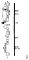
- RNA of the HIV virus - simplified shown - the following gene areas: At both ends so-called long terminal repeats (LTR), and the following Gene ranges gag, pol, env and nef.
- LTR long terminal repeats
- the gene gag codes under other for the core (core) proteins, p 24 and p 17, the gene pol coded i.a. for the reverse transcriptase, the RNAse H and the integrase and the env gene encode for the Glycoproteins gp 41 and gp 120 of the virus envelope.
- the gene nef encodes a protein with regulatory function.
- a schematic arrangement of the genome of retroviruses of the HIV type is shown in Figure 1.
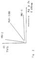
- HIV-1 and HIV-2 are among others possible by testing viral antigen is made with a monoclonal antibody that is commercially available as Test kit is available from Abbott (HIVAG-1 Monoclonal), and against which (HIV-1) p 24 is directed. It is known that the level of reverse transcriptase in the HIV-1 virus types and HIV-2 is about the same. So if you dilute it of the disrupted viruses the absorbance (E 490 nm), obtained by the antigen-antibody reaction against the activity of reverse transcriptase, then receives you get a graphic that corresponds approximately to Figure 2.
- PCR polymerase chain reaction
- the Reverse transcriptase of the MVP-5180/91 a molecular weight that is between about 3 and about 7 kilodaltons smaller than that of reverse transcriptase from HIV-1 or HIV-2.
- the Reverse transcriptase from MPV-5180 thus shows Molecular weight that is between about 4,500 daltons and is about 5,500 daltons smaller than reverse transcriptase of HIV-1 or HIV-2.
- the virus is made from a correspondingly large amount of culture (about 1 l) precipitated and taken up in phosphate buffered saline. Then pelleting is carried out by a (20%) Sucrose pillow.
- the virus pellet can be in 6 M Guanidinium chloride in 20 mM dithiothreitol and 0.5% nonidet P 40 can be suspended. CsCl except for one Concentration of 2 molar added and that the solution containing broken virus is placed on a Cesium chloride cushion applied. Then the viral RNA pelleted by centrifugation, dissolved, with phenol extracted and precipitated with ethanol and lithium chloride.
- the present invention furthermore relates to such Viruses that have a homology of more than 66%, preferably 75% and particularly preferably have 85% partial sequences the nucleotide sequence shown in Table 3, at least Are 50, preferably 100 nucleotides long. This corresponds to one Length of the peptides of at least 16 and preferably at least 33 amino acids.
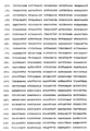
- the (almost) complete sequence, given as the DNA sequence of the Virus according to the invention is shown in FIG. 4.
- object The present invention includes viruses that have the sequence 4 and variants thereof that have a high 4 and homology to the sequence diagnostic use of proteins derived therefrom, Polypeptides and oligopeptides.
- the antigens or peptides can be relatively short Have partial sequences of an amino acid sequence, which are in Table 3 is reproduced or can be derived from Figure 4.
- This Amino acid sequence is at least 6 amino acids, preferred at least 10 and particularly preferably at least 15 amino acids long. These peptides cannot only be produced with the help of recombinant technology but also synthetic Methods.
- a suitable manufacturing route is Solid phase synthesis from the Merrifield type. Another description this technique and others known in the art Methods can be found in the literature, e.g. M. Bodansky, et al., Peptide Synthesis, John Weeley & Sons, 2nd edition 1976.
- a serum sample is taken of the investigator matched with the protein chains of one or more proteins or glycoproteins (which in eukaryotic cell lines can be expressed) or parts of which are from MVP-5180/91.
- Preferred test methods include immunofluorescence or immunoenzymatic Test procedures (e.g. Elisa, Immunoblot).
- antigen derived from MVP-5180/91 or a variant thereof can be bound to the walls of microtiter plates.
- the dosage used depends essentially on the test system and the treatment of the microtiter plates.
- serum or serum dilutions which come from the person to be examined, are added to the holes in the microtiter plates.
- specific immune complexes are detected by antibodies which bind specifically to human immunoglobulins and which have previously been linked with an enzyme, for example horseradish peroxidase, alkaline phosphatase etc. or with enzyme-labeled antigen.
- viruses according to the invention can convert a colorless substrate into a strongly colored product and the presence of specific anti-HIV antibodies can then be read from the strength of the color.
- Another possibility of using the virus according to the invention in test systems is the use in Western blots. Even if the production of vaccines against immunodeficiency disorders proves to be extremely difficult, this virus or parts thereof, ie immunodominant epitopes and inducers of cellular immunity, or genetically engineered antigens can also be used for the development and production of vaccines.
- the immunodeficiency virus MVP-5180/91 was isolated from the blood of a patient with signs of immunodeficiency.
- peripheral mononuclear cells peripheral blood lymphocytes, PBL
- peripheral lymphocytes from the blood PBL
- the usual medium RPMI 1640 with 10% fetal calf serum was used for this.
- the culture conditions are described in Landay A. et al., J. Inf. Dis., 161 (1990) pp. 706-710. The formation of giant cells was then observed under the microscope.
- HIV viruses The production of HIV viruses was determined by determining the p24 antigen using Abbott's commercially available test. Another test to determine the growth of the virus was the test using particle-bound reverse transcriptase (Eberle J., Seibl R., J. Virol. Methods 40, 1992, pp. 347-356). The growth of the viruses was determined once or twice a week based on the enzymatic activities in the culture supernatant in order to monitor the virus production. New donor lymphocytes were added once a week.
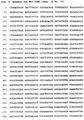
- Example 2 further Western blots carried out.
- the results are in the attached Figure 3 shown.
- This Filter strips were made with the sera from various Patients were incubated and then the specific ones Antibodies made visible by a color reaction.
- the right half the figure shows a virus isolated from a German donor (MVP-899), which is an HIV-1 virus.
- Figure 3 clearly shows that sera from German Patients with the immunodeficiency virus according to the invention in Western blot with the gp 41 is very weak. Sera African patients react with the immunodeficiency virus according to the invention very strong. Figure 3 therefore makes it clear that using the immunodeficiency virus according to the invention such immunodeficiency infections can be demonstrated when using of an HIV-1 or HIV-2 virus is only questionable, so not deliver clearly positive results.
- This Evidence can be far-reaching diagnostic Have meaning because in cases where only questionable Results are achieved in the Western blot, not with definite certainty can be established whether it is an infection with an immunodeficiency virus. But if with the help of the immunodeficiency virus according to the invention such questionable results of infection with be assigned to a virus of the type according to the invention then this presents a significant diagnostic Progress.
- Genomic DNA from MVP-5180/91 infected HUT 78 cells were isolated using standard methods.
- PCR polymerase chain reaction
- 5 ⁇ l genomic DNA from MVP-5180/91 infected HUT 78 cells in a 100 ⁇ l reaction mixture Pipette 0.25 mM dNTP, 1 ⁇ M each of Primer 1 and Primer 2, 10 mM Tris HCl pH 8.3, 50 mM KCl, 1.5 mM MgCl 2 , 0.001% gelatin, 2.5 units Taq polymerase (Perkin Elmer) and amplified according to the following temperature program: 1st initial denaturation: 3 '95 ° C, 2nd amplification: 90''94 ° C, 60''56 ° C
- the primers used for the PCR and the nucleotide sequencing were synthesized on the 8750 oligonucleotide synthesizer from Biosearch (Seq. ID No. 35 + 36).
- the amplified DNA was over a 3% "Nusieve” agarose gel (Biozyme), the amplified Cut out fragment and apply the same volume Buffer (1xTBE (0.09 M TrisBorat, 0.002 M EDTA pH8.0) transferred.
- Buffer (1xTBE 0.09 M TrisBorat, 0.002 M EDTA pH8.0
- the pelleted DNA was dried taken up in water and after the photometric Determination of the DNA concentration at 260 nm in Spectrophotometer (Beckman) using the Sanger method (F. Sanger, Proc. Natl.Adac.Sci., 74: 5463, 1977) sequenced. Instead of sequencing with Klenow DNA Polymerase, the sequencing reaction was performed using a kit by Applied Biosystems ("Taq Dye Deoxy Terminator Cycle Sequencing ", order no .: 401150). As a primer were in separate sequencing reactions primer 1 or Primer 2 (1 ⁇ M each) used. The analysis of the Sequencing reaction was done on the DNA sequencer 373A (Applied Biosystems) according to the specifications of Device manufacturer.
- nucleotide sequence of the amplified DNA region and the amino acid sequence derived therefrom is shown in Table 1 (Sequ. ID No. 37-39).
- nucleotide sequence from Table 1 has one 66% homology to a chimpanzee isolate.
- About HIV 1 isolates is MVP 5180/91 in the investigated DNA sequence at best 64% homologous.
- DNA is about HIV 2 isolates from Table 1 to 56% homolog.
- Primate isolates SIV: Simian Immunodeficiency Virus
- the homology is 61.5%.
- the amino acid region of the MVP-5180/91 coat protein (Table 1) could be serodiagnostically important due to the overlap with the immunodiagnostically important region from gp 41. This would be particularly the case if antisera from HIV-infected patients did not react positively with any of the commercially available antibody screening tests. In these cases, an infection with an MVP-5180/91 closely related virus could be present.
- Genomic DNA from MVP-5180/91 infected HUT78 cells were isolated as described.
- PCR polymerase chain reaction
- the 5 'region of gp 41 (N-terminus) and the 3' sequence of gp 120 were amplified by means of "inverse PCR".
- 100 ⁇ l of a genomic DNA preparation (218 ⁇ g / ml) from MVP-5180/91-infected HUT78 cells in a final volume of 200 ⁇ l was digested with 10 units of the restriction endonuclease Sau3a for 1 hour at 37 ° C.
- the DNA was then phenolized and precipitated with sodium acetate (final concentration 300 mM) and 2.5 volumes of ethanol for 10 min at -70 ° C., centrifuged in the Eppendorf centrifuge and the pellet dried and in 890 ⁇ l distilled water. resuspended.
- the primers 163env, 168i and 169i have been removed from the determined partial sequence of the HIV isolate MVP-5180 selected (Example 4).
- the amplified DNA was over a 3% "Nusieve” agarose gel (Biozyme), the amplified fragment cut out and with the same volume of buffer (1xTBE (0.09 M TrisBorat, 0.002 M EDTA, pH 8.0)) was added. After Incubate the DNA agarose mixture for 10 minutes at 70 ° C and subsequent phenol extraction, the DNA was extracted from the aqueous phase by adding 1/10 vol 3M NaAc, pH 5.5 and 2 vol ethanol at -20 ° C 15 'and then in pelleted in an Eppendorf centrifuge (13000rpm, 10 ', 4 ° C).
- nucleotide sequence found from Table 3 was found on homologous sequences in the GENEBANK database (release 72, June 1992) with the help of the GCG computer program (Genetic Computer Group, Inc. Wisconsin USA, version 7.1, March 1992) examined. In this database most of the are up July 1992 known nucleotide sequences from Immunodeficiency viruses of human origin and isolates Primates included.
- the nucleotide sequence from Table 3 has at best one 62% homology to an HIV 1 isolate.
- the peptide of amino acid 584-618 of the HIV 1 coat protein region is of particular serodiagnostic interest (numbering according to Wain Hobson et al., Cell 40: 9-17, 1985; Gnann et al., J. Inf. Dis. 156: 261-267, 1987; Norrby et al., Nature, 329: 248-250, 1987).
- Corresponding amino acid regions from the envelope proteins of HIV 2 and SIV are also preserved immunodiagnostically (Gnann et al., Science, pp. 1346-1349, 1987).
- Peptides from this coat protein range of HIV 1 and HIV 2 are used as solid phase antigens in many commercially available HIV 1/2 antibody screening tests. Approximately 99% of the anti-HIV 1 and anti-HIV 2 positive sera can be detected with it.
- the corresponding amino acid region of the MVP-5180/91 coat protein (Table 4) as well as the entire gp 41 of this isolate could be of serodiagnostic importance, in particular if antisera from HIV-infected patients are weak or at all in commercially available antibody screening tests would not respond. In these cases, an infection with an MVP-5180/91 closely related virus could be present.
- the peptide obtained using the information from MvP 5180 the amino acid sequence has: RLQALETLIQNQQRLNLWGCKGKLICYTSVKWNTS.
- the present invention therefore also relates to diagnostic use of peptides that are recombinant or can be produced synthetically and the above Have sequence or partial sequence, the partial sequences at least 6 consecutive amino acids, preferably 9 and particularly preferably have 12 consecutive amino acids.
- Genomic DNA from MvP5180 infected HUT78 cells was isolated as described. 300 ⁇ g of this DNA was incubated in a volume of 770 ⁇ l with 0.24 U of the restriction enzyme Sau3A for 45 min.
- the DNA that was only partially cut was then size-fractionated using a 0.7% agarose gel (low melting agarose, Nusieve) and fragments between 10 and 21 kb were cut out.
- the agarose was melted at 70 ° C. for 10 min and the same volume of buffer (1 ⁇ TBE, 0.2 M NaCl) was added.
- phenol and one chloroform extraction followed the precipitation of the DNA by adding 1/10 vol 3 M sodium acetate solution (pH 5.9) and 2.5 vol ethanol at -70 ° C for 10 min.
- the precipitated DNA was centrifuged off, dried and dissolved in water at a concentration of 1 ⁇ g / ⁇ l.
- the yield of size-fractionated DNA was approximately 60 ⁇ g.
- the "Random Primed DNA Labeling Kit” from Boehringer Mannheim (No. 713 023) was used for the labeling.
- the PCR product which was obtained as described in Example 3 with the primers sk68 and envb was labeled. 1 ⁇ g of this DNA was denatured by boiling for 2 x 5 min and then cooling in ice water. 50 mCi [ ⁇ - 32 P] -dCTP (NEN, no .: NEX-053H) were added for labeling. Other additives were pipetted according to the manufacturer's instructions. After an incubation of 30 minutes at 37 ° C., the now radioactively labeled DNA was precipitated.
- the filters were placed in 15 ml hybridization solution (50% formamide, 0.5% SDS, 5 x SSPE, 5 x Denhardt's solution and 0.1 mg / ml salmon sperm DNA) per filter at 42 ° C with shaking 2-3 h incubated.
- the [ 32 P] -labeled DNA samples were denatured for 2-5 min at 100 ° C, cooled on ice, added to the prehybridization solution and hybridized at 42 ° C for 12 hours.
- the filters were then washed at 60 ° C. first with 2 x SSC / 0.1% SDS, then with 0.2 x SSC / 0.1% SDS.
- the phage DNA was then precipitated, cut with the restriction enzyme EcoRI and the EcoRI fragments obtained therefrom were subcloned into the vector Bluescript KS - (Strategene, no .: 212208). A total of 4 clones were obtained: Plasmid Beginning The End pSP1 1 1785 pSP2 1786 5833 pSP3 5834 7415 pSP4 7660 9793
- the missing piece between base 7416 and 7659 was obtained by PCR with primers 157 (CCA TAA TAT TCA GCA GAA CTA G) and 226 (GCT GAT TCT GTA TAA GGG).
- the phage DNA of the clone was used as the DNA template.
- the conditions for the PCR were: 1.) Initial denaturation: 94 ° C, 3 min, 2.) Amplification: 1.5 min 94 ° C, 1 min 56 ° C and 1 min 72 ° C for 30 cycles.
- the DNA was sequenced as described in Example 4. Both the strand and the counter strand were sequenced from the entire genome.
- the gag sequence was cloned from the LTR ("long terminal repeat", LTR1 primer) of the left end of the MvP 5180 genome overlapping into the pol (polymerase gene, pol3.5i primer).
- the cloning strategy is shown schematically in FIG. 5.
- the PCR reactions were carried out with the ug DNA primers, the sequences of which were derived from the HIV-1 consensus sequence.
- the sequencing was carried out using the dideoxy chain termination method.
- the sequence coding for the MvP 5180 gag gene extends from nucleotide 817 (A of the ATG start codon) to nucleotide 2300 (A of the last codon) (Sequ. ID No. 47-53).
- the DNA sequence obtained in the PCR technique was the in Figure 4 compared DNA sequence shown.
- a Comparison of the two DNA sequences is shown in FIG. 6 shown. It was found that the Nucleotides differ from each other in about 2%, although it does is the same virus. 6 represents the top line represents the DNA sequence shown in Fig. 4 and the bottom line represents the one with PCR technique DNA sequence obtained.
- amino acid sequence was determined using the PCR technique determined protein gag the amino acid sequence of the Fig. 4 derived corresponding protein juxtaposed. There was a difference in the Amino acid of about 2.2% determined. The comparison is in Fig. 7 shown, the lower line each Represents amino acid sequence by using the PCR technique sequence obtained was derived.
- the homology values in% given in Table 9 mean that when the sequence according to FIG. 4 is compared with a sequence of another virus at most one of the above corresponding percentage of the sequence indicated may be different.
Abstract
Description
Die vorliegende Erfindung betrifft ein neues Retrovirus aus der HIV-Gruppe sowie Varianten oder Teile davon, welche die wesentlichen Eigenschaften des Virus enthalten. Beschrieben wird ein Verfahren zur Züchtung des Retrovirus. Die Erfindung betrifft weiterhin die Gewinnung dieses Retrovirus sowie die Verwendung des Virus, seiner Teile oder Extrakte für medizinische Zwecke und für die Diagnostik.The present invention relates to a new retrovirus from the HIV group and variants or parts thereof, which the essential properties of the virus included. Is described a method of growing the retrovirus. The invention relates continue to recover and use this retrovirus the virus, its parts or extracts for medical purposes and for diagnostics.
Retroviren, die zur sogenannten HIV-Gruppe gehören, führen bei damit infizierten Menschen zu Krankheitserscheinungen, die unter dem Sammelbegriff Immunschwäche bzw. AIDS (Acquired Immune Deficiency Syndrome) zusammengefaßt werden.Retroviruses, which belong to the so-called HIV group, lead to people infected with it to symptoms that are under the collective term immunodeficiency or AIDS (Acquired Immune Deficiency Syndrome).
Epidemiologische Studien belegen, daá das Humane Immunschwäche Virus (HIV) das aetiologische Agens für die überwiegende Mehrheit der AIDS (Acquired Immune Deficiency Syndrome)-Fälle darstellt. Ein 1983 aus einem Patienten isoliertes und charakterisiertes Retrovirus erhielt die Bezeichnung HIV-1 (Barré-Sinoussi, F. et al., Science 220, 868-871 [1983]). Eine Variante von HIV-1 wird in WO 86/02383 beschrieben. Epidemiological studies show that the human immunodeficiency virus (HIV) is the aetiological agent for the vast majority of AIDS (Acquired Immune Deficiency Syndrome) cases. A retrovirus isolated and characterized in 1983 from a patient was given the designation HIV-1 (Barré-Sinoussi, F. et al., Science 220 , 868-871 [1983]). A variant of HIV-1 is described in WO 86/02383.
Eine zweite Gruppe von Humanen Immunschwäche Viren wurde 1985 in Westafrika identifiziert (Clavel, F. et al., Science 233, 343-346 [1986]) und als Humanes Immunschwäche Virus Typ 2 (HIV-2) bezeichnet (EP-A-0 239 425). HIV-2 Retroviren unterscheiden sich deutlich von HIV-1, weisen jedoch auch eine Verwandtschaft zu Affen Immunschwäche Viren (SIV-2) auf. Wie HIV-1 führt auch HIV-2 zu einer AIDS-Symptomatik.A second group of human immunodeficiency viruses was identified in West Africa in 1985 (Clavel, F. et al., Science 233 , 343-346 [1986]) and designated as human immunodeficiency virus type 2 (HIV-2) (EP-A-0 239 425). HIV-2 retroviruses differ significantly from HIV-1, but are also related to monkey immunodeficiency viruses (SIV-2). Like HIV-1, HIV-2 leads to symptoms of AIDS.
Eine weitere Variante eines Immunschwäche Retrovirus wird in der EP-A-0 345 375 beschrieben und dort als HIV-3 Retrovirus bezeichnet (ANT 70).Another variant of an immune deficiency retrovirus is in EP-A-0 345 375 and there as HIV-3 retrovirus designated (ANT 70).
Auch in Lancet Vol. 340, Sept. 1992, S. 681-682 wird die Isolierung eines weiteren, varianten Immunschwächevirus beschrieben.Also in Lancet Vol. 340, Sept. 1992, pp. 681-682 the Isolation of another variant immunodeficiency virus described.
Es ist ein Charakteristikum der Humanen Immunschwäche Viren, daß sie eine hohe Variabilität aufweisen, die die Vergleichbarkeit der verschiedenen Isolate deutlich kompliziert. Beim Vergleich diverser HIV-1-Isolate treten z.B. in einigen Regionen des Genoms hohe Variabilitäten auf, während andere Genombereiche vergleichsweise konserviert vorliegen (Benn, S. et al. Science 230, 949-951 [1985)). Ein wesentlich größerer Polymorphismus konnte auch für HIV-2 beobachtet werden (Clavel, F. et al., Nature 324, 691-695 [1986]). Die größte genetische Stabilität besitzen Bereiche in den gag und pol Genen, die für strukturell und enzymatisch essentielle Proteine codieren; einige Regionen im env-Gen sowie die Gene (vif, vpr, tat, rev, nef), die für regulatorische Proteine codieren, zeigen einen hohen Grad an Variabilität. Es konnte weiterhin gezeigt werden, daß Antisera gegen HIV-1 auch mit gag und pol Genprodukten von HIV-2 kreuzreagieren, obwohl nur geringe Sequenzhomologie vorlag. Ebenfalls war die Hybridisierung zwischen diesen beiden Viren wenig signifikant, wenn nicht sehr wenig stringente Konditionen verwandt wurden (Clavel, F. et al., Nature 324, 691-695 [1986]).It is a characteristic of the human immunodeficiency virus that they have a high variability, which significantly complicates the comparability of the different isolates. When comparing various HIV-1 isolates, for example, high variability occurs in some regions of the genome, while other genome areas are comparatively conserved (Benn, S. et al. Science 230 , 949-951 [1985)). A much larger polymorphism could also be observed for HIV-2 (Clavel, F. et al., Nature 324 , 691-695 [1986]). Areas in the gag and pol genes that code for structurally and enzymatically essential proteins have the greatest genetic stability; some regions in the env gene as well as the genes (vif, vpr, tat, rev, nef) that code for regulatory proteins show a high degree of variability. It was also possible to show that antisera against HIV-1 also cross-react with gag and pol gene products of HIV-2, even though the sequence homology was poor. The hybridization between these two viruses was also of little significance if very little stringent conditions were used (Clavel, F. et al., Nature 324 , 691-695 [1986]).
Aufgrund der weiten Verbreitung der Retroviren aus der HIV-Gruppe und der Tatsache, daß zwischen dem Zeitpunkt der Infektion und dem Zeitpunkt, zu dem eindeutige Symptome für pathologische Veränderungen erkennbar sind, ein Zeitraum von einigen bis vielen Jahren (2-20) liegt, ist es epidemiologisch von großer Bedeutung, die Infektion mit Retroviren der HIV-Gruppe möglichst frühzeitig und vor allem zuverlässig zu bestimmen. Dies spielt nicht nur eine Rolle bei der Diagnose von Patienten, die Zeichen von Immunschwäche aufweisen, sondern auch bei der Überprüfung von Blutspendern. Es hat sich herausgestellt, daß bei der Verwendung von Retroviren oder Bestandteilen davon des Types HIV-1 oder HIV-2 in Nachweissystemen bei manchen Seren kein oder nur ein schwacher Nachweis von Antikörpern geführt werden kann, obwohl bei den Patienten, von denen die Seren stammen, Zeichen von Immunschwäche auftreten. Mit Hilfe des erfindungsgemäßen Retrovirus aus der HIV-Gruppe ist in bestimmten Fällen ein derartiger Nachweis möglich.Because of the wide distribution of retroviruses from the HIV group and the fact that between the time of infection and the time when clear symptoms of pathological Changes are noticeable, a period of some to many Years (2-20), it is epidemiologically of great importance infection with retroviruses from the HIV group as early as possible and above all to determine reliably. This doesn't just play a role in diagnosing patients who have signs of Have immunodeficiency, but also when checking Blood donors. It has been found that when used of retroviruses or components thereof of the HIV-1 or HIV-2 type no or only one in some sera in detection systems weak detection of antibodies can be done, though in the patients from whom the sera are derived, signs of Immunodeficiency occur. With the help of the invention Retrovirus from the HIV group is a in certain cases such evidence is possible.
Beschrieben wird die Isolation und Charakterisierung eines neuen Humanen Immunschwäche Virus, im folgenden als MVP-5180/91 bezeichnet, das aus peripheren Lymphozyten einer 1991 34-jährigen Patientin aus Kamerun isoliert wurde, die Zeichen von Immunschwäche aufwies. Geographisch stammt dieses Retrovirus aus einer Region in Afrika, die zwischen Westafrika mit endemischer HIV-2 und HIV-1 Virusinfektion und Ostzentralafrika mit fast ausschließlicher HIV-1-Verbreitung lokalisiert ist. Gegenstand der vorliegenden Erfindung ist also ein neues Retrovirus der HIV-Gruppe, welches als MVP-5180/91 bezeichnet wird und dessen Varianten sowie davon abgeleitete DNS-Sequenzen und Testkits, die davon abgeleitete Aminosäuresequenzen bzw. Teilsequenzen enthalten. Das Retrovirus MVP-5180/91 wurde bei der European Collection of Animal Cell Cultures (ECACC) unter der Nummer V 920 92 318 gemäß den Bedingungen des Budapester Vertrages hinterlegt.The isolation and characterization of a new one is described Human immunodeficiency virus, hereinafter referred to as MVP-5180/91 referred to that from peripheral lymphocytes of a 1991 34-year-old Patient from Cameroon was isolated, the sign of Showed immune deficiency. This retrovirus originates geographically a region in Africa that is between West Africa with endemic HIV-2 and HIV-1 viral infections and East Central Africa with almost exclusive HIV-1 spread is localized. object the present invention is a new HIV group retrovirus, which is designated as MVP-5180/91 and its Variants and derived DNA sequences and test kits that amino acid sequences or partial sequences derived therefrom contain. The retrovirus MVP-5180/91 was developed by the European Collection of Animal Cell Cultures (ECACC) under number V 920 92 318 according to the terms of the Budapest Treaty deposited.
Ebenso wie HIV-1 und HIV-2 wächst das erfindungsgemäße MVP-5180/91 in folgenden Zellinien HUT 78, Jurkat-Zellen, C8166-Zellen und MT-2-Zellen. Die Isolierung und Vermehrung von Viren wird in dem Buch "Viral Quantitation in HIV Infection, Editor Jean-Marie Andrieu, John Libbey Eurotext, 1991" ausführlich beschrieben. Die dort beschriebenen Arbeitsmethoden werden durch Bezugnahme zum Gegenstand der Offenbarung der vorliegenden Anmeldung gemacht.Like HIV-1 and HIV-2, the MVP-5180/91 according to the invention grows in the following cell lines HUT 78, Jurkat cells, C8166 cells and MT-2 cells. Isolation and multiplication of viruses is described in the book "Viral Quantitation in HIV Infection, Editor Jean-Marie Andrieu, John Libbey Eurotext, 1991 "in detail described. The working methods described there are by Reference to the subject matter of the disclosure of the present Registration made.
Weiterhin besitzt das erfindungsgemäße Virus eine magnesiumabhängige Reverse Transkriptase, die aber nicht manganabhängig ist. Dies stellt eine weitere Gemeinsamkeit zu den Viren HIV-1 und HIV-2 dar.Furthermore, the virus according to the invention has one magnesium-dependent reverse transcriptase, but not is dependent on manganese. This represents another commonality to the Viruses represent HIV-1 and HIV-2.
Zum besseren Verständnis der Unterschiede des erfindungsgemäßen
MVP-5180/91 Virus zu den Retroviren HIV-1 und HIV-2 soll zunächst
kurz der Aufbau der Immunschwäche verursachenden Retroviren
erläutert werden. Im Inneren des Virus befindet sich die RNA in
einem kegelförmigen Core, das aus Proteinuntereinheiten
zusammengesetzt ist, die die Bezeichnung p 24 (p für Protein)
tragen. Dieses innere Core wird von einer Proteinhülle umgeben,
die aus dem Protein p 17 aufgebaut ist (äußeres Core) und von
einer Glykoproteinhülle umgeben ist, die neben Lipiden, die aus
der Wirtszelle stammen, das Transmembranprotein gp 41, und das
Hüllprotein 120 (gp 120) enthält. Dieses gp 120 kann dann mit den
CD-4-Rezeptoren der Wirtszellen eine Bindung eingehen. For a better understanding of the differences between the invention
MVP-5180/91 virus to the retroviruses HIV-1 and HIV-2 is said to be first
briefly the structure of the retroviruses causing immune deficiency
are explained. The RNA is located inside the virus
a conical core made up of protein subunits
composed of the designation p 24 (p for protein)
carry. This inner core is surrounded by a protein shell,
which is composed of the protein p 17 (outer core) and of
is surrounded by a glycoprotein shell, which, in addition to lipids, consists of
of the host cell, the
Soweit bekannt, weist die RNA der HIV-Viren - vereinfacht
dargestellt - folgende Genbereiche auf: An den beiden Enden
sogenannte long terminal repeats (LTR), und die folgenden
Genbereiche gag, pol, env und nef. Das Gen gag codiert unter
anderem für die Kern(Core)-Proteine, p 24 und p 17, das Gen
pol codiert u.a. für die Reverse Transkriptase, die RNAse H
und die Integrase und das Gen env codiert für die
Glykoproteine gp 41 und gp 120 der Virushülle. Das Gen nef
codiert für ein Protein mit Regulatorfunktion. Eine
schematische Anordnung des Genomes von Retroviren des HIV-Typs
ist in Figur 1 gezeigt.As far as is known, the RNA of the HIV virus - simplified
shown - the following gene areas: At both ends
so-called long terminal repeats (LTR), and the following
Gene ranges gag, pol, env and nef. The gene gag codes under
other for the core (core) proteins,
Eine Unterscheidung zwischen den Retroviren HIV-1 und HIV-2
ist u.a. dadurch möglich, daß virales Antigen ausgetestet
wird mit einem monoklonalen Antikörper, der kommerziell als
Testkit von Abbott (HIVAG-1 Monoclonal) erhältlich ist, und
gegen das (HIV-1) p 24 gerichtet ist. Es ist bekannt, daß
der Gehalt an Reverser Transkriptase in den Virustypen HIV-1
und HIV-2 etwa gleich ist. Wenn man deshalb in Verdünnungen
der aufgeschlossenen Viren die Extinktion (E 490 nm),
erhalten durch die Antigen-Antikörper-Reaktion, aufträgt
gegen die Aktivität der Reversen Transkriptase, dann erhält
man eine Graphik, die etwa der Figur 2 entspricht. Hierbei
stellt man fest, daß im Verhältnis zu dem Gehalt an Reverser
Transkriptase bei HIV-1 eine sehr hohe Bindungsaffinität für
p 24 mit dem eingesetzten monoklonalen Antikörper vorhanden
ist. Für HIV-2 tritt dagegen nur eine sehr geringe
Bindungsaffinität für p 24 bei Einsatz des monoklonalen
Antikörpers wiederum bezogen auf den Gehalt an Reverser
Transkriptase auf. Werden diese Messungen durchgeführt für
MVP-5180/91, dann befindet sich die Kurve ziemlich genau in
der Mitte zwischen der Kurve von HIV-1 und HIV-2, d.h. die
Bindungsaffinität des monoklonalen Antikörpers gegen MVP-5180/91
p 24 ist gegenüber HIV-1 reduziert. Figur 2 zeigt
schematisch diesen Sachverhalt, wobei RT Reverse
Transkriptase bedeutet und als Antigen (Ag) das Protein p 24
eingesetzt wird, gegen das der monoklonale Antikörper, der
in dem von Abbott käuflich erwerblichen Testkit vorhanden
ist, gerichtet ist.A distinction between the retroviruses HIV-1 and HIV-2
is among others possible by testing viral antigen
is made with a monoclonal antibody that is commercially available as
Test kit is available from Abbott (HIVAG-1 Monoclonal), and
against which (HIV-1)
Ein sehr vielseitig verwendbares System der Gentechnologie
ist die sogenannte PCR (polymerase chain reaction) geworden,
wobei die zur Durchführung des Verfahrens benötigten
Komponenten käuflich erworben werden können. Mit diesem
Verfahren ist es möglich, DNA-Sequenzen zu amplifizieren,
wenn DNA-Bereiche der zu amplifizierenden Sequenz bekannt
sind. Es müssen dann kurze komplementäre DNA-Fragmente
(Oligonucleotide = Primer) synthetisiert werden, die sich an
einen kurzen Bereich der zu amplifizierenden
Nukleinsäuresequenz anlagern. Für die Testdurchführung
werden HIV-Nukleinsäuren mit den Primern zusammengebracht in
einer Reaktionsmischung, die zusätzlich eine Polymerase und
Nukleotidtriphosphate enthält. Die Polymerisation (DNA-Synthese)
wird für eine bestimmte Zeit durchgeführt und dann
werden die Nukleinsäurestränge durch Erwärmen getrennt. Nach
Abkühlen läuft dann die Polymerisation erneut an. Wenn es
sich also bei dem erfindungsgemäßen Retrovirus um ein HIV-1
oder HIV-2 Virus handelt, dann müßte die Nukleinsäure
amplifiziert werden können, indem Primer verwendet werden,
die konserviert sind innerhalb der bekannten Sequenzen der
Viren HIV-1 und HIV-2. Derartige Primer sind zum Teil
vorbeschrieben (Lauré, F. et al., Lancet ii, (1988) 538-541
für pol 3 und pol 4 bzw. Ou C.Y. et al., Science 239 (1988)
295-297 für sk 38/39, sk 68/69).The so-called PCR (polymerase chain reaction) has become a very versatile system of genetic engineering, whereby the components required to carry out the method can be purchased. With this method it is possible to amplify DNA sequences if DNA regions of the sequence to be amplified are known. Short complementary DNA fragments (oligonucleotides = primers) must then be synthesized, which attach to a short region of the nucleic acid sequence to be amplified. To carry out the test, HIV nucleic acids are brought together with the primers in a reaction mixture which additionally contains a polymerase and nucleotide triphosphates. The polymerization (DNA synthesis) is carried out for a certain time and then the nucleic acid strands are separated by heating. After cooling, the polymerization then starts again. If the retrovirus according to the invention is an HIV-1 or HIV-2 virus, then the nucleic acid should be able to be amplified by using primers which are conserved within the known sequences of the viruses HIV-1 and HIV-2. Some of these primers have been previously described (Lauré, F. et al., Lancet ii, (1988) 538-541 for
Es wurde nun herausgefunden, daß bei Verwendung von
bestimmten Primerpaaren, die folgende Sequenz aufweisen:
![]()

![]()
Keine oder nur schwache Amplifikate im Vergleich zum HIV-1,
die evtl. auf Verunreinigungen zurückzuführen sind, wurden
erhalten mit folgenden Primer-Sequenzen:
![]()

![]()
Im Vergleich zum HIV-1 schwache Amplifikate, die jedoch die
gleiche Intensität wie das verwendete HIV-2 Isolat
(MVP-11971/87) aufwiesen, wurden erhalten mit

Eine weitverbreitete Methode zum Nachweis von HIV-Antikörpern ist der sogenannte Western Blot (Immunoblot). Dabei werden die viralen Proteine gelelektrophoretisch aufgetrennt und dann auf eine Membran überführt. Die mit den überführten Proteinen versehenen Membranen werden dann mit Seren der zu untersuchenden Patienten in Verbindung gebracht. Sofern Antikörper gegen die viralen Proteine vorhanden sind, binden diese an die Proteine. Nach Waschen verbleiben lediglich spezifische Antikörper gegen virale Proteine. Die Antikörper werden dann mit Antiantikörpern sichtbar gemacht, die regelmäßig mit einem Enzym gekoppelt sind, das eine Farbreaktion katalysiert. Auf diese Weise können die Banden der viralen Proteine sichtbar gemacht werden.A widely used method for the detection of HIV antibodies is the so-called Western blot (immunoblot). The viral proteins become gel electrophoretic separated and then transferred to a membrane. The one with the Proteins are then transferred to proteins Sera related to the patient to be examined brought. Provided antibodies against the viral proteins are present, they bind to the proteins. After washing only specific antibodies against viral remain Proteins. The antibodies are then combined with anti-antibodies visualized that are regularly coupled with an enzyme that catalyzes a color reaction. In this way can visualize the bands of viral proteins will.
Das erfindungsgemäße Virus MVP-5180/91 weist gegenüber den
Viren HIV-1 und HIV-2 im Western Blot zwei signifikante
wesentliche Unterschiede auf. Das HIV-1 zeigt regelmäßig
eine starke Bande, die dem Protein p 24 zuzuordnen ist und
eine sehr schwache, oft kaum sichtbare Bande, die dem
Protein p 23 zuzuordnen ist. HIV-2 weist eine kräftige Bande
auf, die dem Protein p 25 zuzuordnen ist und manchmal eine
schwache Bande, die dem Protein p 23 zuzuordnen ist. Im
Unterschied dazu weist das erfindungsgemäße MVP-5180/91
Virus zwei etwa gleich starke Banden auf, die den Proteinen
p 24 und p 25 entsprechen.The virus MVP-5180/91 according to the invention shows the
Viruses HIV-1 and HIV-2 two significant in Western blot
significant differences. The HIV-1 shows regularly
a strong band which can be assigned to the
Ein weiterer signifikanter Unterschied besteht bei den
Banden, die der Reversen Transkriptase zuzuordnen sind. HIV-1
zeigt eine Bande (p 53), die der Reversen Transkriptase
entspricht und eine Bande (p 66), die der Reversen
Transkriptase verbunden mit der RNAse H entspricht. Bei HIV-2
entspricht die Reverse Transkriptase dem Protein p 55 und,
wenn sie mit der RNAse H verbunden ist, dem Protein p 68.
Das erfindungsgemäße MPV-5180/91 weist dagegen eine Bande
auf bei dem Protein p 48, die der Reversen Transkriptase
entspricht, und eine Bande, bei dem Protein p 60, die der
Reversen Transkriptase in Verbindung mit RNAse H entspricht. Another significant difference is in the
Bands that are assigned to the reverse transcriptase. HIV-1
shows a band (p 53) that of the reverse transcriptase
corresponds and a band (p 66) that of the reverse
Transcriptase associated with the RNAse H corresponds. With HIV-2
the reverse transcriptase corresponds to the protein p 55 and,
when linked to RNAse H, protein p 68.
In contrast, the MPV-5180/91 according to the invention has a band
on the protein p 48, that of the reverse transcriptase
and a band at the
Aus diesen Ergebnissen kann geschlossen werden, daß die Reverse Transkriptase des MVP-5180/91 ein Molekulargewicht hat, das zwischen etwa 3 und etwa 7 Kilodalton kleiner ist als das der Reversen Transkriptase von HIV-1 bzw. HIV-2. Die Reverse Transkriptase von MPV-5180 weist also ein Molekulargewicht auf, das zwischen etwa 4.500 Dalton und etwa 5.500 Dalton kleiner ist als die Reverse Transkriptase von HIV-1 bzw. HIV-2.From these results it can be concluded that the Reverse transcriptase of the MVP-5180/91 a molecular weight that is between about 3 and about 7 kilodaltons smaller than that of reverse transcriptase from HIV-1 or HIV-2. The Reverse transcriptase from MPV-5180 thus shows Molecular weight that is between about 4,500 daltons and is about 5,500 daltons smaller than reverse transcriptase of HIV-1 or HIV-2.
Es wurde herausgefunden, daß mit Hilfe des erfindungsgemäßen
Virus MVP-5180/91 anti-env-Antikörper in Seren von deutschen
Patienten, die Zeichen von Immunschwäche zeigen, nur schwach
nachgewiesen werden können, wobei die Seren aber stark
reagieren, wenn anstelle des erfindungsgemäßen Virus ein
HIV-1 Virus verwendet wird. Diese stärkere Nachweisreaktion
wurde vor allem lokalisiert in dem gp 41 Protein. Bei den
Versuchen wurden Serumpanels gegenübergestellt, die einmal
von deutschen Patienten stammen und zum anderen von
afrikanischen Patienten mit Zeichen von Immunschwäche
stammen.It was found that with the help of the invention
Virus MVP-5180/91 anti-env antibodies in sera from German
Patients who show signs of immunodeficiency are weak
can be detected, but the sera are strong
react if instead of the virus of the invention
HIV-1 virus is used. This stronger detection reaction
was mainly localized in the
Die oben angegebenen Charakteristika kennzeichnen solche Virus-Varianten, die dem erfindungsgemäßen MVP-5180/91 entsprechen. Wenn also aus heparinisiertem Spenderblut, das von Personen stammt, die Immunschwächeanzeichen aufweisen und vorzugsweise aus Afrika stammen, Immunschwächeviren isoliert werden, dann kann auf diese Weise das erfindungsgemäße Virus oder Varianten davon erhalten werden.The characteristics indicated above characterize such Virus variants, the MVP-5180/91 according to the invention correspond. So if from heparinized donor blood, that comes from people who show signs of immunodeficiency and preferably come from Africa, immunodeficiency viruses can be isolated, then this way virus according to the invention or variants thereof are obtained.
Da das Virus isoliert wurde, welches die oben erwähnten Eigenschaften aufweist, kann die Klonierung einer cDNA auf folgendem Weg durchgeführt werden: Das Virus wird aus einer entsprechend großen Kulturmenge (etwa 1 l) präzipitiert und in phosphatgepufferter Kochsalzlösung aufgenommen. Dann erfolgt eine Pelletierung durch ein (20 %iges) Saccharose-Kissen. Das Viruspellet kann in 6 M Guanidiniumchlorid in 20 mM Dithiothreitol und 0,5 % Nonidet P 40 suspendiert werden. CsCl wird bis auf eine Konzentration von 2 molar zugegeben und die das aufgebrochene Virus enthaltende Lösung wird auf ein Cäsiumchlorid-Kissen aufgebracht. Dann wird die virale RNA durch Zentrifugation pelletiert, gelöst, mit Phenol extrahiert und mit Ethanol und Lithiumchlorid präzipitiert. Mit Hilfe eines Oligo(dT)-Primers wird die Synthese des ersten cDNA-Stranges an der viralen RNA oder Teilen davon durchgeführt. Die Synthese unter Zugabe von Reverser Transkriptase kann durchgeführt werden unter Verwendung eines käuflich erhältlichen Kits. Für die Synthese des zweiten Stranges wird der RNA-Strang des RNA/DNA-Hybrids mit RNase H verdaut und der zweite Strang unter Einsatz von E.coli DNA Polymerase I synthetisiert. Mit Hilfe von T4 DNA Polymerase können dann stumpfe Enden erzeugt werden und diese mit geeigneten Linkern für Restriktionsschnittstellen verbunden werden. Nach Restriktionsverdau mit der geeigneten Restriktionsendonuklease wird das cDNA-Fragment aus einem Agarosegel isoliert und mit einem vorher in geeigneter Weise geschnittenen Vektor ligiert. Der Vektor mit dem cDNA-Insert kann dann zur Transformation von kompetenten E.coli-Zellen verwendet werden. Die erhaltenen Kolonien werden dann auf Membranen übertragen, lysiert und denaturiert und schließlich durch Hybridisierung mit Digoxigenin oder Biotin markierter Nukleinsäure aufgefunden. Nach gentechnologischer Herstellung der entsprechenden cDNA ist eine Isolierung der gewünschten DNA-Fragmente, die aus dem Retrovirus stammen, möglich. Durch Einbau dieser Fragmente in geeignete Expressionsvektoren kann dann das gewünschte Protein bzw. Proteinfragment exprimiert werden und für die diagnostischen Tests eingesetzt werden. Since the virus was isolated, which mentioned the above Has properties, the cloning of a cDNA the following way: The virus is made from a correspondingly large amount of culture (about 1 l) precipitated and taken up in phosphate buffered saline. Then pelleting is carried out by a (20%) Sucrose pillow. The virus pellet can be in 6 M Guanidinium chloride in 20 mM dithiothreitol and 0.5% nonidet P 40 can be suspended. CsCl except for one Concentration of 2 molar added and that the solution containing broken virus is placed on a Cesium chloride cushion applied. Then the viral RNA pelleted by centrifugation, dissolved, with phenol extracted and precipitated with ethanol and lithium chloride. With the help of an oligo (dT) primer, the synthesis of the first cDNA strand on the viral RNA or parts thereof carried out. The synthesis with the addition of reverse Transcriptase can be performed using of a commercially available kit. For the synthesis of the second strand is the RNA strand of the RNA / DNA hybrid RNase H digested and the second strand using E.coli DNA polymerase I synthesized. With the help of T4 DNA Polymerase can then be generated and blunt ends these with suitable linkers for restriction interfaces get connected. After restriction digestion with the appropriate Restriction endonuclease is the cDNA fragment from a Agarose gel isolated and with a previously appropriate cut vector ligated. The vector with the cDNA insert can then be used to transform competent E. coli cells be used. The colonies obtained are then expanded Membranes transferred, lysed and denatured and finally by hybridization with digoxigenin or biotin labeled nucleic acid found. After genetic engineering Preparation of the corresponding cDNA is an isolation of the desired DNA fragments that originate from the retrovirus, possible. By incorporating these fragments into suitable ones Expression vectors can then be the desired protein or Protein fragment can be expressed and used for diagnostic Tests are used.
Alternativ zu der angegebenen Methode kann das Immunschwächevirus mit Hilfe der PCR-Technologie kloniert werden, wobei die oben angegebenen Primer Verwendung finden können.As an alternative to the specified method, this can Immunodeficiency virus cloned using PCR technology using the primers given above can.
Zwischen verschiedenen Virusisolaten kann die Ähnlichkeit ausgedrückt werden durch den Grad der Homologie der Nucleinsäure- oder Proteinsequenzen. Eine 50 %ige Homologie bedeutet beispielsweise, daß 50 von 100 Nucleotid- oder Aminosäure-Positionen in den Sequenzen übereinstimmen. Die Homologie von Proteinen wird durch Sequenzanalyse bestimmt. Homologe DNA-Sequenzen lassen sich auch durch die Hybridisierungs-Technik ermitteln.The similarity can exist between different virus isolates are expressed by the degree of homology of the Nucleic acid or protein sequences. A 50% homology means, for example, that 50 out of 100 nucleotide or Amino acid positions in the sequences match. The Homology of proteins is determined by sequence analysis. Homologous DNA sequences can also be identified by the Determine hybridization technique.
Erfindungsgemäß wurde zunächst ein Teil aus dem Hüllprotein
sequenziert und festgestellt, daß diese Sequenz nur eine
verhältnismäßig geringe Homologie zu den entsprechenden
Sequenzen von Viren vom HIV-Typ aufweist. Insbesondere
bezogen auf den gp 41-Bereich wurde durch einen Vergleich
mit HIV-Sequenzen, der mit Hilfe von Datenbanken
durchgeführt wurde, eine Homologie von höchstens 66 %
(Nucleotidsequenz) ermittelt.According to the invention, part of the coat protein first became
sequenced and found that this sequence is only one
relatively low homology to the corresponding
Has sequences of HIV-type viruses. Especially
based on the
Es wurde weiterhin der Bereich sequenziert, der für gp 41
kodiert. Diese Sequenz ist in Tabellen 1 bzw. 3 dargestellt.The area was further sequenced for
Gegenstand der vorliegenden Erfindung sind daher solche
Viren, die eine Homologie von mehr als 66 %, bevorzugt 75 %
und besonders bevorzugt 85 % aufweisen zu dem
erfindungsgemäßen HIV-Virus, MVP 5180/91, bezogen auf die
Nucleotidsequenz der Tabelle 1 und/oder der Tabelle 3.The present invention therefore relates to such
Viruses that have a homology of more than 66%, preferably 75%
and particularly preferably have 85% of that
HIV virus according to the invention,
Weiterhin sind Gegenstand der vorliegenden Erfindung solche Viren, die eine Homologie von mehr als 66 %, bevorzugt 75 % und besonders bevorzugt 85 % aufweisen zu Partialsequenzen der in Tabelle 3 dargestellten Nucleotidsequenz, die wenigstens 50, bevorzugt 100 Nucleotide lang sind. Dies entspricht einer Länge der Peptide von wenigstens 16 und bevorzugt von wenigstens 33 Aminosäuren.The present invention furthermore relates to such Viruses that have a homology of more than 66%, preferably 75% and particularly preferably have 85% partial sequences the nucleotide sequence shown in Table 3, at least Are 50, preferably 100 nucleotides long. This corresponds to one Length of the peptides of at least 16 and preferably at least 33 amino acids.
Das erfindungsgemäße Virus unterscheidet sich durch seine Sequenz von vorbekannten Viren. Gegenstand der vorliegenden Erfindung sind daher solche Viren sowie entsprechende DNS- bzw. Aminosäuresequenzen, die der Sequenz des erfindungsgemäßen Virus weitgehend entsprechen, wobei der Grad der Abweichung durch den Grad der Homologie festgelegt wird. Eine Homologie von beispielsweise mehr als 85 % bedeutet daher, daß solche Sequenzen umfaßt werden, die in wenigstens 85 von 100 Nucleotiden bzw. Aminosäuren dieselben Nucleotide bzw. Aminosäuren aufweisen, während der Rest unterschiedlich sein kann. Bei der Feststellung der Homologie werden die beiden Sequenzen derart gegenübergestellt, daß möglichst viele einander entsprechende Nucleotide bzw. Aminosäuren miteinander zur Deckung kommen.The virus according to the invention differs in its sequence of known viruses. Subject of the present invention are such viruses and corresponding DNS or Amino acid sequences that correspond to the sequence of the virus according to the invention largely correspond, with the degree of deviation due to the Degree of homology is established. A homology of for example, more than 85% therefore means that such sequences which are contained in at least 85 out of 100 nucleotides or Amino acids have the same nucleotides or amino acids, while the rest can be different. When determining In homology, the two sequences become like this juxtaposed that as many as possible correspond to each other Nucleotides or amino acids coincide with each other.
Die (nahezu) vollständige Sequenz, angegeben als DNS-Sequenz des erfindungsgemäßen Virus ist in Fig. 4 wiedergegeben. Gegenstand der vorliegenden Erfindung sind dabei Viren, die die Sequenz gemäß Fig. 4 aufweisen sowie Varianten davon, die eine hohe Homologie zu der Sequenz von Fig. 4 aufweisen sowie die diagnostische Verwendung von davon abgeleiteten Proteinen, Polypeptiden und Oligopeptiden.The (almost) complete sequence, given as the DNA sequence of the Virus according to the invention is shown in FIG. 4. object The present invention includes viruses that have the sequence 4 and variants thereof that have a high 4 and homology to the sequence diagnostic use of proteins derived therefrom, Polypeptides and oligopeptides.
Anhand der isolierten Sequenz können immundominante Epitope (Peptide) konfektioniert und synthetisiert werden. Da die Nucleinsäuresequenz des Virus bekannt ist, kann der Fachmann hieraus die Aminosäuresequenz ableiten. Ein Teilbereich der Aminosäuresequenz ist in Tabelle 3 angegeben. Gegenstand der vorliegenden Erfindung ist daher auch die diagnostische Verwendung von Antigenen, d.h. Proteine, Oligo- oder Polypeptide, die mit Hilfe der in Figur 4 bzw. Tabelle 3 offenbarten Information hergestellt werden können. Diese Antigene, Proteine, Polypeptide und Oligopeptide weisen Aminosäuresequenzen auf, die von Figur 4 abgeleitet werden können bzw. in Tabelle 3 angegeben sind. Die Antigene bzw. Peptide können verhältnismäßig kurze Teilsequenzen einer Aminosäuresequenz aufweisen, die in Tabelle 3 wiedergegeben ist oder aus Figur 4 abgeleitet werden kann. Diese Aminosäuresequenz ist wenigstens 6 Aminosäuren, bevorzugt wenigstens 10 und besonders bevorzugt wenigstens 15 Aminosäuren lang. Hergestellt werden können diese Peptide nicht nur mit Hilfe der rekombinanten Technologie sondern auch durch synthetische Methoden. Ein geeigneter Herstellungsweg ist die Festphasensynthese vom Merrifield-Typ. Eine weitere Beschreibung dieser Technik und anderer im Stand der Technik bekannter Verfahren kann in der Literatur gefunden werden, z.B. M. Bodansky, et al., Peptide Synthesis, John Weeley & Sons, 2nd Edition 1976.Using the isolated sequence, immunodominant epitopes (Peptides) are assembled and synthesized. Since the Nucleic acid sequence of the virus is known to the person skilled in the art derive the amino acid sequence from this. Part of the Amino acid sequence is given in Table 3. Subject of The present invention is therefore also the diagnostic one Use of antigens, i.e. Proteins, oligo- or polypeptides, that with the help of those disclosed in Figure 4 and Table 3 Information can be produced. These antigens, proteins, Polypeptides and oligopeptides have amino acid sequences that can be derived from Figure 4 or specified in Table 3 are. The antigens or peptides can be relatively short Have partial sequences of an amino acid sequence, which are in Table 3 is reproduced or can be derived from Figure 4. This Amino acid sequence is at least 6 amino acids, preferred at least 10 and particularly preferably at least 15 amino acids long. These peptides cannot only be produced with the help of recombinant technology but also synthetic Methods. A suitable manufacturing route is Solid phase synthesis from the Merrifield type. Another description this technique and others known in the art Methods can be found in the literature, e.g. M. Bodansky, et al., Peptide Synthesis, John Weeley & Sons, 2nd edition 1976.
Bei den diagnostischen Tests wird eine Serumprobe der zu untersuchenden Person zusammengebracht mit den Proteinketten von einem oder mehreren Proteinen oder Glykoproteinen (die in eukaryontischen Zellinien exprimiert werden können) oder Teilen davon, die von MVP-5180/91 stammen. Bevorzugte Testverfahren schließen die Immunfluoreszenz oder immunenzymatische Testverfahren (z.B. Elisa, Immunoblot) ein.In the diagnostic tests, a serum sample is taken of the investigator matched with the protein chains of one or more proteins or glycoproteins (which in eukaryotic cell lines can be expressed) or parts of which are from MVP-5180/91. Preferred test methods include immunofluorescence or immunoenzymatic Test procedures (e.g. Elisa, Immunoblot).
Bei den immunenzymatischen Tests (ELISA) kann beispielsweise
Antigen, das von MVP-5180/91 oder einer Variante davon stammt, an
den Wänden von Mikrotiterplatten gebunden werden. Die dabei
verwendete Dosierung hängt von dem Testsystem und der Behandlung
der Mikrotiterplatten wesentlich ab. Dann wird Serum bzw.
Serumverdünnungen, die von der zu
untersuchenden Person stammen, in die Löcher der
Mikrotiterplatten gegeben. Nach einer bestimmten
Inkubationszeit wird die Platte gewaschen und spezifische
Immunkomplexe werden nachgewiesen durch Antikörper, die
spezifisch an menschliche Immunglobuline binden und die
vorher mit einem Enzym, beispielsweise
Meerrettichperoxidase, alkalischer Phosphatase usw.
verbunden wurden oder mit enzymmarkiertem Antigen. Diese
Enzyme können ein farbloses Substrat in ein stark gefärbtes
Produkt umwandeln und an der Stärke der Färbung kann dann
das Vorhandensein von spezifischen Anti-HIV-Antikörpern
abgelesen werden. Eine weitere Möglichkeit der Verwendung
des erfindungsgemäßen Virus in Testsystemen ist die
Verwendung in Western Blots.
Auch wenn die Herstellung von Impfstoffen gegen
Immunschwächeerkrankungen sich als äußerst schwierig
erweist, kann doch auch dieses Virus bzw. Teile davon, d.h.
immundominante Epitope und Induktoren der zellulären
Immunität, oder gentechnologisch hergestellte Antigene zur
Entwicklung und Herstellung von Impfstoffen verwendet
werden.In immunoenzymatic tests (ELISA), for example, antigen derived from MVP-5180/91 or a variant thereof can be bound to the walls of microtiter plates. The dosage used depends essentially on the test system and the treatment of the microtiter plates. Then serum or serum dilutions, which come from the person to be examined, are added to the holes in the microtiter plates. After a certain incubation period, the plate is washed and specific immune complexes are detected by antibodies which bind specifically to human immunoglobulins and which have previously been linked with an enzyme, for example horseradish peroxidase, alkaline phosphatase etc. or with enzyme-labeled antigen. These enzymes can convert a colorless substrate into a strongly colored product and the presence of specific anti-HIV antibodies can then be read from the strength of the color. Another possibility of using the virus according to the invention in test systems is the use in Western blots.
Even if the production of vaccines against immunodeficiency disorders proves to be extremely difficult, this virus or parts thereof, ie immunodominant epitopes and inducers of cellular immunity, or genetically engineered antigens can also be used for the development and production of vaccines.
Das erfindungsgemäße Immunschwäche-Virus MVP-5180/91 wurde aus dem Blut einer Patientin mit Zeichen von Immunschwäche isoliert. Hierzu wurden periphere mononukleäre Zellen (peripheral blood lymphocytes, PBL) und periphere Lymphozyten aus dem Blut (PBL) eines nicht HIV-infizierten Spenders mit Phytohämagglutinin stimuliert und in Kultur gehalten. Verwendet wurde hierzu das übliche Medium RPMI 1640 mit 10 % fötalem Kälberserum. Die Kulturbedingungen sind beschrieben in Landay A. et al., J. Inf. Dis., 161 (1990) S. 706-710. Beobachtet wurde dann die Bildung von Riesenzellen unter dem Mikroskop. Die Produktion von HIV-Viren wurde über die Bestimmung des p 24-Antigens mit Hilfe des käuflich erwerbbaren Tests von Abbott bestimmt. Ein weiterer Test zur Bestimmung des Wachstums der Viren war der Test unter Verwendung von partikelgebundener Reverser Transkriptase (Eberle J., Seibl R., J. Virol. Methods 40, 1992, S. 347-356). Das Wachstum der Viren wurde also anhand der enzymatischen Aktivitäten im Kulturüberstand ein- bis zweimal pro Woche bestimmt, um die Virusproduktion zu überwachen. Wöchentlich einmal wurden neue Spenderlymphozyten zugegeben.The immunodeficiency virus MVP-5180/91 according to the invention was isolated from the blood of a patient with signs of immunodeficiency. For this purpose, peripheral mononuclear cells (peripheral blood lymphocytes, PBL) and peripheral lymphocytes from the blood (PBL) of a non-HIV-infected donor were stimulated with phytohemagglutinin and kept in culture. The usual medium RPMI 1640 with 10% fetal calf serum was used for this. The culture conditions are described in Landay A. et al., J. Inf. Dis., 161 (1990) pp. 706-710. The formation of giant cells was then observed under the microscope. The production of HIV viruses was determined by determining the p24 antigen using Abbott's commercially available test. Another test to determine the growth of the virus was the test using particle-bound reverse transcriptase (Eberle J., Seibl R., J. Virol. Methods 40, 1992, pp. 347-356). The growth of the viruses was determined once or twice a week based on the enzymatic activities in the culture supernatant in order to monitor the virus production. New donor lymphocytes were added once a week.
Nachdem eine HIV-Virenvermehrung festgestellt werden konnte, wurden frische periphere Lymphozyten aus dem Blut (PBL) nicht HIV-infizierter, gesunder Spender mit dem Überstand der ersten Kultur infiziert. Dieser Schritt wurde wiederholt und dann wurden mit dem Überstand H 9 bzw. HUT 78 Zellen infiziert. Auf diese Weise war eine permanente Produktion des Immunschwäche-Virus möglich. Das Virus wurde bei der ECACC unter der Nr. V 920 92 318 hinterlegt.After HIV virus replication was found, fresh peripheral blood lymphocytes (PBL) healthy donor not infected with HIV with the supernatant the first culture infected. This step was repeated and then with the supernatant H 9 and HUT became 78 cells infected. In this way it was a permanent production of the immunodeficiency virus possible. The virus was found in the ECACC deposited under number V 920 92 318.
Zum Nachweis von HIV-Infektionen ist derzeit der sogenannte
Western Blot oder Immunoblot ein Standardverfahren. Gemäß
der in J. Virol. Meth. 15 (1987) S. 11-23 von Gürtler et al.
beschriebenen Vorgehensweise wurden verschiedene Seren
untersucht. Hierbei wurden Seren von deutschen Patienten
solchen Seren gegenübergestellt, die von afrikanischen
Patienten erhalten wurden. Es wurden hierbei folgende
Ergebnisse erhalten:
Die oben dargestellten Ergebnisse zeigen, daß ein aus deutschen Patienten isoliertes Virus vom HIV-1 Typ, wenn es zum Nachweis von HIV-Infektionen verwendt wird, möglicherweise keine eindeutigen Ergebnisse liefert, wenn der Patient infiziert wurde mit einem Virus, das dem erfindungsgemäßen MVP-5180/91 entspricht. Es wird dabei davon ausgegangen, daß mit Hilfe des erfindungsgemäßen Virus solche Viren nachgewiesen werden können, die wenigstens etwa 85 % Homologie, bezogen auf das Gesamtgenom, zu dem erfindungsgemäßen Virus aufweisen.The results presented above show that an out German patients isolated HIV-1 type virus if it is used to detect HIV infections, may not produce clear results if the patient has been infected with a virus that MVP-5180/91 according to the invention corresponds. It will be there assumed that with the help of the virus according to the invention such viruses can be detected that at least approximately 85% homology, based on the total genome, to the exhibit virus according to the invention.
Gemäß der in Beispiel 2 angegebenen Vorgehensweise wurden weitere Western Blots durchgeführt. Die Ergebnisse sind in der anliegenden Figur 3 dargestellt. Bei diesem Test wurde einmal das virale Protein des erfindungsgemäßen Immunschwäche-Virus MVP-5180/91 und einmal das virale Protein eines HIV-1-Typ Virus (MVP-899) gelelektrophoretisch aufgetrennt und dann auf Zellulosefilter transferiert. Diese Filterstreifen wurden mit den Seren von verschiedenen Patienten inkubiert und dann wurden die spezifischen Antikörper durch eine Farbreaktion sichtbar gemacht. Die linke Hälfte der Figur mit der Überschrift MVP-5180 zeigt das erfindungsgemäße Immunschwäche-Virus. Die rechte Hälfte der Figur zeigt ein aus deutschem Spender isoliertes Virus (MVP-899), bei dem es sich um ein HIV-1-Virus handelt. According to the procedure given in Example 2 further Western blots carried out. The results are in the attached Figure 3 shown. In this test was once the viral protein of the invention Immunodeficiency virus MVP-5180/91 and once the viral Protein of an HIV-1 type virus (MVP-899) gel electrophoresis separated and then transferred to cellulose filters. This Filter strips were made with the sera from various Patients were incubated and then the specific ones Antibodies made visible by a color reaction. The shows left half of the figure with the heading MVP-5180 the immunodeficiency virus according to the invention. The right half the figure shows a virus isolated from a German donor (MVP-899), which is an HIV-1 virus.
Die einzelnen Filterstreifen wurden nun mit den Seren von
verschiedenen Patienten inkubiert. Betrachtet man die
Figur 3, so wurden jeweils dieselben Seren (von deutschen
Patienten) umgesetzt mit je zwei Filterstreifen, wobei die
Nummern 8 und 26; 9 und 27; 10 und 28; 11 und 29; 12 und 30;
13 und 31; 14 und 32; 15 und 33 sowie 16 und 34 die gleichen
Seren bezeichnen. Bei den Western Blots mit den Nummern 17
und 18 wurden Seren von afrikanischen Patienten eingesetzt.
Die Zahlen an den rechten Seitenrändern geben die
annähernden Molekulargewichte in Tausend (KD) an.The individual filter strips were now covered with the sera from
incubated in different patients. If you look at that
Figure 3, the same sera (from German
Patients) implemented with two filter strips, the
Die Figur 3 zeigt deutlich, daß Seren von deutschen
Patienten mit dem erfindungsgemäßen Immunschwäche-Virus im
Western Blot mit dem gp 41 nur sehr schwach reagieren. Seren
von afrikanischen Patienten dagegen reagieren mit dem
erfindungsgemäßen Immunschwäche-Virus sehr stark. Figur 3
macht daher deutlich, daß unter Verwendung des
erfindungsgemäßen Immunschwäche-Virus solche Immunschwäche-Infektionen
nachgewiesen werden können, die bei Verwendung
eines HIV-1 oder HIV-2-Virus nur fragliche, also nicht
eindeutig positive Ergebnisse liefern. Diese
Nachweismöglichkeit kann weitreichende diagnostische
Bedeutung haben, da in den Fällen, in denen nur fragliche
Ergebnisse im Western Blot erzielt werden, nicht mit
eindeutiger Sicherheit festgestellt werden kann, ob es sich
um eine Infektion mit einem Immunschwäche-Virus handelt.
Wenn aber mit Hilfe des erfindungsgemäßen Immunschwäche-Virus
derartige fragliche Ergebnisse einer Infektion mit
einem Virus des erfindungsgemäßen Typs zugeordnet werden
können, dann stellt dies einen erheblichen diagnostischen
Fortschritt dar. Figure 3 clearly shows that sera from German
Patients with the immunodeficiency virus according to the invention in
Western blot with the
Genomische DNA aus MVP-5180/91-infizierten HUT 78-Zellen wurden nach Standardmethoden isoliert.Genomic DNA from MVP-5180/91 infected HUT 78 cells were isolated using standard methods.
Zur Charakterisierung von Genombereichen des Isolates
MVP-5180/91 wurden PCR (Polymerase Chain Reaction)Experimente
mit einem Primerpaar aus dem Hüllproteinbereich
gp 41 durchgeführt. Die Durchführung der PCR-Experimente
erfolgte nach der Methode von Saiki et al. (Saiki et al.,
Science 239: 487-491, 1988) mit folgenden Modifikationen:
Für die Amplifikation HIV-spezifischer DNA-Bereiche wurden
5 µl genomische DNA aus MVP-5180/91 infizierten HUT 78-Zellen
in einem 100 µl Reaktionsansatz (0,25 mM dNTP, je
1 µM Primer 1 und Primer 2, 10 mM Tris HCl pH 8,3, 50 mM
KCl, 1,5 mM MgCl2, 0,001 % Gelatine, 2,5 units Taq
Polymerase (Perkin Elmer) pipettiert und nach folgendem
Temperatur-Programm amplifiziert: 1. Initiale Denaturierung:
3' 95°C, 2. Amplifikation: 90'' 94°C, 60'' 56°C, 90'' 72°C
(30 Cyclen).To characterize genome areas of the isolate MVP-5180/91, PCR (polymerase chain reaction) experiments were carried out with a primer pair from the coat
Die für die PCR und die Nucleotidsequenzierung verwendeten
Primer wurden auf dem Oligonucletidsynthesizer 8750 der
Firma Biosearch synthetisiert (Seq. ID No. 35 + 36).

Die amplifizierte DNA wurde über ein 3 % "Nusieve"-Agarosegel
(Fa. Biozyme) aufgetrennt, das amplifizierte
Fragment ausgeschnitten und mit dem gleichen Volumen an
Puffer (1xTBE (0,09 M TrisBorat, 0,002 M EDTA pH8,0)
versetzt. Nach Inkubation des DNA-Agarosegemisches für
10 Minuten bei 70°C und nachfolgender Phenolextraktion wurde
die DNA aus der wässrigen Phase durch Zugabe von 1/10 Vol 3M
NaAc pH 5,5 und 2 Vol Ethanol bei -20°C 15' gefällt und
anschließend in einer Zentrifuge (Eppendorf) pelletiert
(13000 rpm, 10', 4°C). Die pelletierte DNA wurde getrocknet,
in Wasser aufgenommen und nach der photometrischen
Bestimmung der DNA-Konzentration bei 260 nm im
Spektralphotometer (Beckman) nach der Methode von Sanger
(F. Sanger, Proc. Natl.Adac.Sci., 74: 5463, 1977)
sequenziert. Anstelle der Sequenzierung mit Klenow DNA
Polymerase, wurde die Sequenzierungsreaktion mit einem Kit
von Applied Biosystems ("Taq Dye Deoxy Terminator Cycle
Sequencing", Best.-Nr.: 401150) durchgeführt. Als Primer
wurden in getrennten Sequenzierungsreaktionen Primer 1 oder
Primer 2 (jeweils 1 µM) eingesetzt. Die Analyse der
Sequenzierungsreaktion erfolgte auf dem DNA-Sequenziergerät
373A (Applied Biosystems) nach den Vorgaben des
Geräteherstellers.The amplified DNA was over a 3% "Nusieve" agarose gel
(Biozyme), the amplified
Cut out fragment and apply the same volume
Buffer (1xTBE (0.09 M TrisBorat, 0.002 M EDTA pH8.0)
transferred. After incubation of the DNA agarose mixture for
10 minutes at 70 ° C and subsequent phenol extraction
the DNA from the aqueous phase by adding 1/10 vol 3M
NaAc pH 5.5 and 2 vol ethanol at -20 ° C 15 'and
then pelleted in a centrifuge (Eppendorf)
(13000 rpm, 10 ', 4 ° C). The pelleted DNA was dried
taken up in water and after the photometric
Determination of the DNA concentration at 260 nm in
Spectrophotometer (Beckman) using the Sanger method
(F. Sanger, Proc. Natl.Adac.Sci., 74: 5463, 1977)
sequenced. Instead of sequencing with Klenow DNA
Polymerase, the sequencing reaction was performed using a kit
by Applied Biosystems ("Taq Dye Deoxy Terminator Cycle
Sequencing ", order no .: 401150). As a primer
were in separate
Die Nucleotidsequenz des amplifizierten DNA-Bereichs und die
davon abgeleitete Aminosäuresequenz ist in der Tabelle 1
dargestellt (Sequ. ID No. 37-39).

Die gefundene Nucleotidsequenz aus Tabelle 1 wurde auf homologe Sequenzen in der GENEBANK-Datenbank (Release 72, Juni 1992) mit Hilfe des GCG-Computerprogramms (Genetic Computer Group, Inc., Wisconsin USA, Version 7.1, März 1992) untersucht. In dieser Datenbank sind die meisten der bis Juli 1992 bekannten Nucleotidsequenzen von Immundefizienzviren humanen Ursprungs und von Isolaten aus Primaten enthalten.The nucleotide sequence found from Table 1 was found on homologous sequences in the GENEBANK database (release 72, June 1992) with the help of the GCG computer program (Genetic Computer Group, Inc., Wisconsin USA, version 7.1, March 1992) examined. In this database most of the are up July 1992 known nucleotide sequences from Immunodeficiency viruses of human origin and isolates Primates included.
Die Nucleotidsequenz aus Tabelle 1 weist im besten Fall eine
66 %ige Homologie zu einem Schimpansen-Isolat auf. Zu HIV 1-Isolaten
ist MVP 5180/91 in der untersuchten DNA-Sequenz im
besten Fall zu 64 % homolog. Zu HIV 2-Isolaten ist die DNA
aus Tabelle 1 zu 56 % homolog. Außer der Sequenz des
Schimpansenisolates besteht die beste Homologie zwischen der
Nucleotidsequenz aus Tabelle 1 und DNA-Abschnitten aus
Primatenisolaten (SIV: Simian Immunodeficiency Virus) in
einer DNA-Sequenz, die für einen Teilbereich des
Hüllproteins des Isolates SIV (Afrikanische Meerkatze) TYO-1
kodiert. Die Homologie beträgt 61,5 %.At best, the nucleotide sequence from Table 1 has one
66% homology to a chimpanzee isolate. About
Die gefundene Aminosäuresequenz aus Tabelle 1 wurde auf homologe Sequenzen in der SWISSPROT Protein-Datenbank (Release 22, Juni 1992) mit Hilfe des GCG-Computerprogramms untersucht. In dieser Datenbank sind die meisten der bis Juni 1992 bekannten Proteinsequenzen von Immundefizienz-Viren humanen Ursprungs und von Isolaten aus Primaten enthalten.The amino acid sequence found from Table 1 was found on homologous sequences in the SWISSPROT protein database (Release 22, June 1992) using the GCG computer program examined. In this database most of the are up June 1992 known protein sequences of immunodeficiency viruses of human origin and isolates from primates contain.
Die Aminosäuresequenz aus Tabelle 1 ist im besten Fall mit
62,5 % zu einem Hüllproteinabschnitt des oben genannten
Schimpansenisolates homolog. Unter HIV 1-Hüllproteinen
findet man die beste Homologie mit der Aminosäuresequenz aus
Tabelle 1, in dem Isolat HIV 1 Mal. Die Homologie beträgt
59 %. Zu HIV 2-Hüllproteinen beträgt die Homologie mit der
Aminosäuresequenz aus Tabelle 1 im besten Fall 52 % (Isolat
HIV 2 Rod). Da auch HIV 1 und HIV 2-Isolate im besten Fall
im korrespondierenden Proteinabschnitt nur zu 64 % identisch
sind, scheint es sich bei dem Isolat MVP-5180/91 um eine
HIV-Variante zu handeln, die sich deutlich von HIV 1 und von
HIV 2 strukturell abgrenzt und somit einen Vertreter einer
davon unabhängigen Gruppe von HIV-Viren repräsentiert.The amino acid sequence from Table 1 is in the best case with
62.5% to a coat protein portion of the above
Chimpanzee isolates homolog. Among
Die Aminosäuresequenz des amplifizierten DNA-Bereiches
(Tabelle 1) des HIV-Isolates MVP 5180/91 überlappt mit einem
immundiagnostisch wichtigen Bereich aus dem Hüllprotein
gp 41 von HIV 1 (Aminosäure 584-618*) (Tabelle 2) (Gnann et
al., J. Inf. Dis. 156: 261-267, 1987; Norrby et al., Nature,
329: 248-250, 1987). The amino acid sequence of the amplified DNA region
(Table 1) of the HIV isolate
Korrespondierende Aminosäurebereiche aus den Hüllproteinen
von HIV 2 und SIV sind ebenfalls immundiagnostisch
konserviert (Gnann et al., Science, S. 1346-1349, 1987). So
werden Peptide aus diesem Hüllproteinbereich von HIV 1 und
HIV 2 in vielen kommerziell erhältlichen HIV 1/2 Antikörper-Screeningtests
als Festphasenantigene eingesetzt. Ungefähr
99 % der anti HIV 1 und anti HIV 2 positiven Seren können
damit erfaßt werden.Corresponding amino acid regions from the
Der Aminosäurebereich des MVP-5180/91-Hüllproteins
(Tabelle 1) könnte aufgrund der Überlappung mit dem
immundiagnostisch wichtigen Bereich aus gp 41
serodiagnostisch von Bedeutung sein. Dies wäre insbesondere
dann der Fall, wenn Antiseren von HIV-infizierten Patienten
mit keinem der kommerziell erhältlichen Antikörper-Screeningtests
positiv reagieren würden. In diesen Fällen
könnte eine Infektion mit einem MVP-5180/91 eng verwandten
Virus vorliegen.
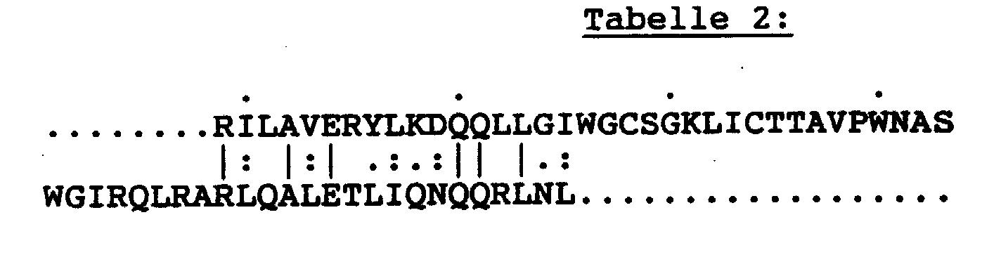
Genomische DNA aus MVP-5180/91-infizierten HUT78-Zellen wurden wie beschrieben isoliert.Genomic DNA from MVP-5180/91 infected HUT78 cells were isolated as described.
Zur Charakterisierung von Genombereichen des Isolates MVP-5180/91
wurden PCR (Polymerase Chain Reaction)-Experimente
mit Primerpaaren aus dem Hüllproteinbereich gp 41
durchgeführt. Die PCR (Saiki et al., Science 239: 487-491,
1988) und inverse PCR (Triglia et al., Nucl. Asids, Res. 16:
8186, 1988) wurden mit folgenden Modifikationen
durchgeführt:To characterize genome areas of the isolate MVP-5180/91, PCR (polymerase chain reaction) experiments were carried out with primer pairs from the
Für die Amplifikation HIV-spezifischer DNA Bereiche wurden 5 µl (218 µg/ml) genomische DNA aus MVP-5180/91 infizierten HUT78 Zellen in einem 100 µl Reaktionsansatz (0,25 mM dNTP, je 1 µM Primer 163env und Primer envend, 10 mM Tris HCl pH 8.3, 50 mM KCl, 1.5 mM MgCl2, 0,001 % Gelatine, 2,5 units Taq Polymerase (Perkin Elmer)) pipettiert und nach folgendem Temperaturprogramm amplifiziert: 1. Initiale Denaturierung: 3 min. 95°C, 2. Amplifikation: 90 sec. 94°C, 60 sec. 56°C, 90 sec. 72°C (30 Cyclen).For the amplification of HIV-specific DNA areas, 5 µl (218 µg / ml) genomic DNA from MVP-5180/91 infected HUT78 cells in a 100 µl reaction mixture (0.25 mM dNTP, 1 µM primer 163env and primer envend, 10 pipetted mM Tris HCl pH 8.3, 50 mM KCl, 1.5 mM MgCl 2 , 0.001% gelatin, 2.5 units Taq Polymerase (Perkin Elmer)) and amplified according to the following temperature program: 1. Initial denaturation: 3 min. 95 ° C, 2nd amplification: 90 sec. 94 ° C, 60 sec. 56 ° C, 90 sec. 72 ° C (30 cycles).
Der 5' Bereich von gp 41 (N-Terminus) und die 3' Sequenz von
gp 120 wurden mittels "inverser PCR" amplifiziert. Hierzu
wurden 100 µl einer genomischen DNA Präparation (218 µg/ml)
aus MVP-5180/91-infizierten HUT78-Zellen in einem Endvolumen
von 200 µl mit 10 units der Restriktionsendonuklease Sau3a
1 Stunde bei 37°C verdaut. Die DNA wurde anschließend
phenolisiert und mit Natriumazetat (Endkonzentration 300 mM)
und 2.5 Volumen Ethanol 10 min bei -70°C gefällt, in der
Eppendorfzentrifuge abzentrifugiert und das Pellet
getrocknet und in 890 µl Aqua dest. resuspendiert. Nach
Zugabe von 100 µl Ligasepuffer (50 mM Tris HCl, pH 7,8,
10 mM MgCl2, 10 mM DTT, 1 mM ATP, 25 µg/ml Rinderserumalbumin)
und 10 µl T4 DNA-Ligase (Fa. Boehringer,
Mannheim) wurden die DNA-Fragmente 3 Stunden bei
Raumtemperatur ligiert, erneut phenolisiert und mit
Natriumazetat und Ethanol wie oben gefällt. Nach dem
Abzentrifugieren und Trocknen wurde die DNA in 40 µl Aqua
dest. resuspendiert und mit 10 units der
Restriktionsendonuklease SacI (Fa. Boehringer, Mannheim) für
1 Stunde verdaut. Anschließend wurden 5 µl dieses Ansatzes
in einem PCR-Experiment wie unter "1. PCR" dargestellt,
eingesetzt. Anstelle der Primer 163env und envend wurden für
die inverse PCR die Primer 168i und 169i verwendet.The 5 'region of gp 41 (N-terminus) and the 3' sequence of
Die Primer 163env, 168i und 169i wurden aus der bereits ermittelten Teilsequenz des HIV-Isolates MVP-5180 ausgewählt (Beispiel 4).The primers 163env, 168i and 169i have been removed from the determined partial sequence of the HIV isolate MVP-5180 selected (Example 4).
Die für die PCR/inverse PCR und die Nucleotidsequenzierung
verwendeten Primer wurden auf dem Oligonucleotidsynthesizer
8750 der Firma Biosearch synthetisiert, wobei die Primer
folgende Sequenzen aufwiesen (Sequ. ID No. 40-43):

Die amplifizierte DNA wurde über ein 3 % "Nusieve"-Agarosegel (Fa. Biozyme) aufgetrennt, das amplifizierte Fragment ausgeschnitten und mit dem gleichen Volumen an Puffer (1xTBE (0.09 M TrisBorat, 0.002 M EDTA, pH 8.0)) versetzt. Nach Inkubation des DNA-Agarosegemisches für 10 Minuten bei 70°C und nachfolgender Phenolextraktion wurde die DNA aus der wässrigen Phase durch Zugabe von 1/10 Vol 3M NaAc, pH 5,5 und 2 Vol Ethanol bei -20°C 15' gefällt und anschließend in einer Eppendorfzentrifuge pelletiert (13000rpm, 10', 4°C). Die pelletierte DNA wurde getrocknet, in Wasser aufgenommen und nach der photometrischen Bestimmung der DNA-Konzentration bei 260nm im Spektralphotometer (Fa. Beckman) nach der Methode von Sanger (F. Sanger, Proc. Natl. Acad. Sci., 74:5463, 1977) sequenziert. Anstelle der Sequenzierung mit Klenow DNA Polymerase wurde die Sequenzierungsreaktion mit einem Kit der Fa. Applied Biosystems ("Taq Dye Deoxy Terminator Cycle Sequencing", Best. Nr.: 401150) durchgeführt. Als Primer wurden in getrennten Sequenzierungsreaktionen Primer 163env oder Primer envend (jeweils 1µM) eingesetzt. Die amplifizierte DNA aus dem inversen PCR-Experiment wurde mit den Primern 168i und 169i sequenziert. Die Analyse der Sequenzierungsreaktion erfolgte auf dem DNA-Sequenziergerät 373A (Applied Biosystems) nach den Vorgaben des Geräteherstellers.The amplified DNA was over a 3% "Nusieve" agarose gel (Biozyme), the amplified fragment cut out and with the same volume of buffer (1xTBE (0.09 M TrisBorat, 0.002 M EDTA, pH 8.0)) was added. After Incubate the DNA agarose mixture for 10 minutes at 70 ° C and subsequent phenol extraction, the DNA was extracted from the aqueous phase by adding 1/10 vol 3M NaAc, pH 5.5 and 2 vol ethanol at -20 ° C 15 'and then in pelleted in an Eppendorf centrifuge (13000rpm, 10 ', 4 ° C). The pelleted DNA was dried, taken up in water and after the photometric determination of the DNA concentration at 260nm in a spectrophotometer (Beckman) according to the Sanger method (F. Sanger, Proc. Natl. Acad. Sci., 74: 5463, 1977). Instead of sequencing with Klenow DNA polymerase the sequencing reaction with a kit from Applied Biosystems ("Taq Dye Deoxy Terminator Cycle Sequencing ", Order No .: 401150) carried out. As primers were in separate Sequencing reactions Primer 163env or Primer envend (1µM each) used. The amplified DNA from the Inverse PCR experiment was performed with primers 168i and 169i sequenced. The sequencing reaction was analyzed on the DNA sequencer 373A (Applied Biosystems) the specifications of the device manufacturer.
Die Nucleotidsequenz des amplifizierten DNA-Bereichs und die
davon abgeleitete Aminosäuresequenz sind in der Tabelle 3
dargestellt (Sequ. ID No. 44-46).

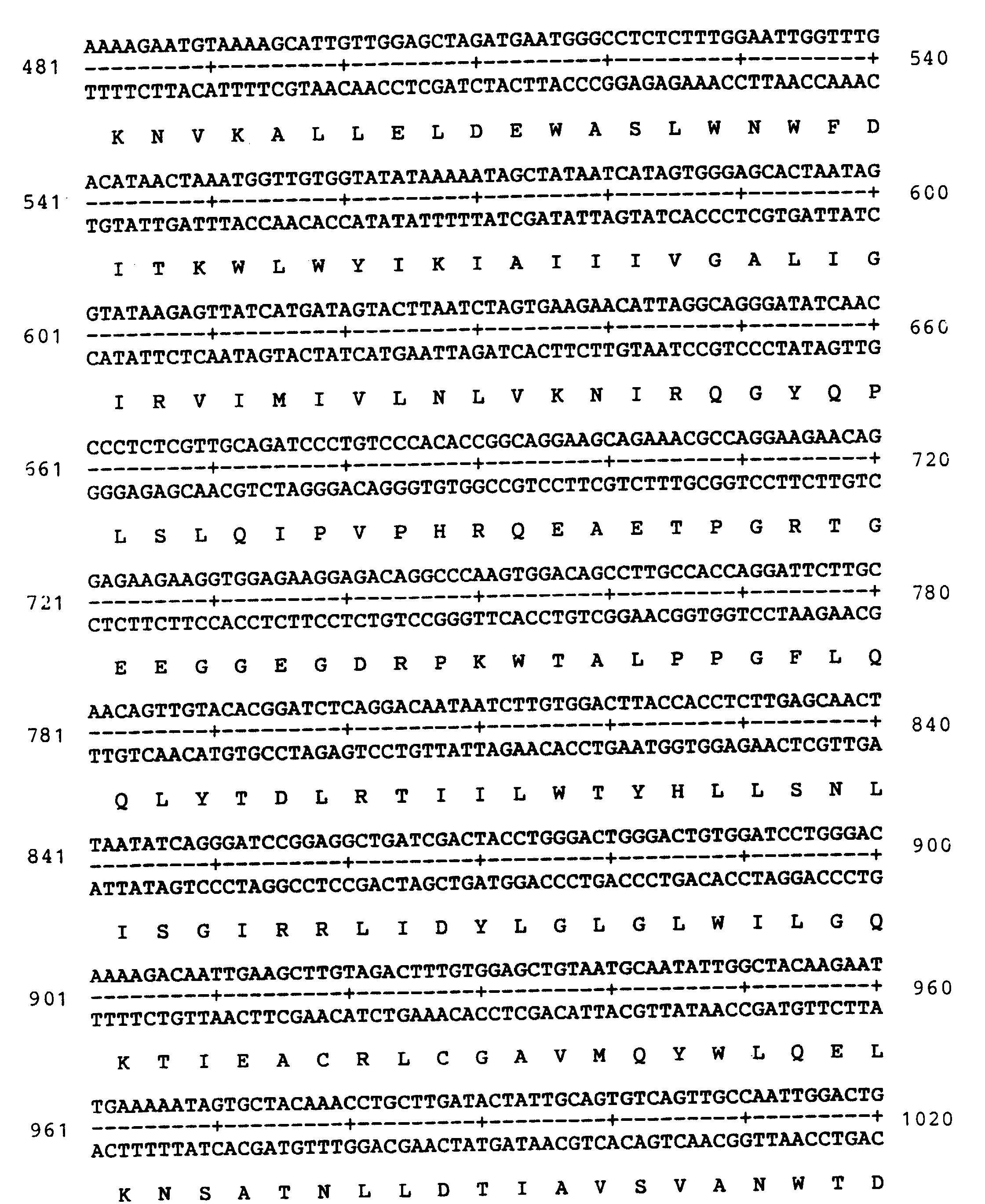

Die gefundene Nucleotidsequenz aus Tabelle 3 wurde auf homologe Sequenzen in der GENEBANK-Datenbank (Release 72, Juni 1992) mit Hilfe des GCG-Computerprogramms (Genetic Computer Group, Inc. Wisconsin USA, Version 7.1, März 1992) untersucht. In dieser Datenbank sind die meisten der bis Juli 1992 bekannten Nucleotidsequenzen von Immundefizienzviren humanen Ursprungs und von Isolaten aus Primaten enthalten.The nucleotide sequence found from Table 3 was found on homologous sequences in the GENEBANK database (release 72, June 1992) with the help of the GCG computer program (Genetic Computer Group, Inc. Wisconsin USA, version 7.1, March 1992) examined. In this database most of the are up July 1992 known nucleotide sequences from Immunodeficiency viruses of human origin and isolates Primates included.
Die Nucleotidsequenz aus Tabelle 3 weist im besten Fall eine
62 %ige Homologie zu einem HIV 1-Isolat auf. Zu HIV 2
Isolaten ist die DNA aus Tabelle 5 zu 50 % homolog.The nucleotide sequence from Table 3 has at best one
62% homology to an
Die aus der Nucleotidsequenz aus Tabelle 3 abgeleitete Aminosäuresequenz wurde auf homologe Sequenzen in der SWISSPROT Protein-Datenbank (Release 22, Juni 1992) mit Hilfe des GCG-Computerprogramms untersucht. In dieser Datenbank sind die meisten der bis Juni 1992 bekannten Proteinsequenzen von Immundefizienzviren humanen Ursprungs und von Isolaten aus Primaten enthalten.The one derived from the nucleotide sequence of Table 3 Amino acid sequence was based on homologous sequences in the SWISSPROT protein database (Release 22, June 1992) with Examined with the help of the GCG computer program. In this Database are most of those known until June 1992 Protein sequences from immunodeficiency viruses of human origin and contain isolates from primates.
Die Aminosäuresequenz aus Tabelle 3 ist im besten Fall mit
54 % zu dem korrespondierenden Hüllproteinabschnitt eines
Schimpansen-Isolates CIV (SIVcpz) homolog und zu 54,5 % zu
dem HIV 1-Isolat Mal. Zu HIV 2-Hüllproteinen beträgt die
Homologie mit der Aminosäuresequenz aus Tabelle 3 im besten
Fall 34 % (Isolat HIV 2 D194). The amino acid sequence from Table 3 is in the best case with
54% to the corresponding coat protein section
Chimpanzee isolates CIV (SIVcpz) homologous and 54.5% too
the
Vergleicht man demgegenüber die gp 41-Aminosäuresequenz von
HIV 1 mit den in der SWISSPROT-Datenbank vorhandenen HIV 1
gp 41-Sequenz, ergeben sich wie erwartet im besten Fall eine
fast 100 %ige Homologie und im schlechtesten Fall eine
78 %ige Homologie.In comparison, the
Aufgrund dieser deutlichen strukturellen Unterschiede
zwischen dem Sequenzbereich aus Tabelle 3 und dem
korrespondierenden Abschnitt aus HIV 1 und HIV 2 scheint es
sich bei dem Isolat MVP-5180/91 um eine HIV-Variante zu
handeln, die sich von HIV 1 und HIV 2 deutlich strukturell
abgrenzt. Möglicherweise ist MVP-5180/91 einer eigenen, von
HIV 1 und HIV 2 sich abgrenzenden Gruppe von HIV-Viren
zuzuordnen.Because of these clear structural differences
between the sequence area from Table 3 and the
corresponding section from
Das Peptid von Aminosäure 584-618 des HIV 1-Hüllproteinbereichs
ist von besonderem serodiagnostischem
Interesse (Numerierung nach Wain Hobson et al.,
Cell 40:9-17, 1985; Gnann et al., J. Inf. Dis. 156:261-267,
1987; Norrby et al., Nature, 329:248-250, 1987).
Korrespondierende Aminosäurebereiche aus den Hüllproteinen
von HIV 2 und SIV sind ebenfalls immundiagnostisch
konserviert (Gnann et al., Science, S. 1346-1349, 1987). So
werden Peptide aus diesem Hüllproteinbereich von HIV 1 und
HIV 2 in vielen kommerziell erhältlichen HIV 1/2 Antikörper-Screeningtests
als Festphasenantigene eingesetzt. Ungefähr
99 % der Anti-HIV 1 und Anti-HIV 2 positiven Seren können
damit erfaßt werden.The peptide of amino acid 584-618 of the
Cell 40: 9-17, 1985; Gnann et al., J. Inf. Dis. 156: 261-267, 1987; Norrby et al., Nature, 329: 248-250, 1987). Corresponding amino acid regions from the envelope proteins of
Der korrespondierende Aminosäurebereich des MVP-5180/91-Hüllproteins
(Tabelle 4) als auch das gesamte gp 41 dieses
Isolates könnten serodiagnostisch insbesondere dann von
Bedeutung sein, wenn Antiseren von HIV-infizierten Patienten
in kommerziell erhältlichen Antikörper-Screening-Tests nur
schwach oder überhaupt nicht reagieren würden. In diesen Fällen
könnte eine Infektion mit einem MVP-5180/91 eng verwandten Virus
vorliegen.
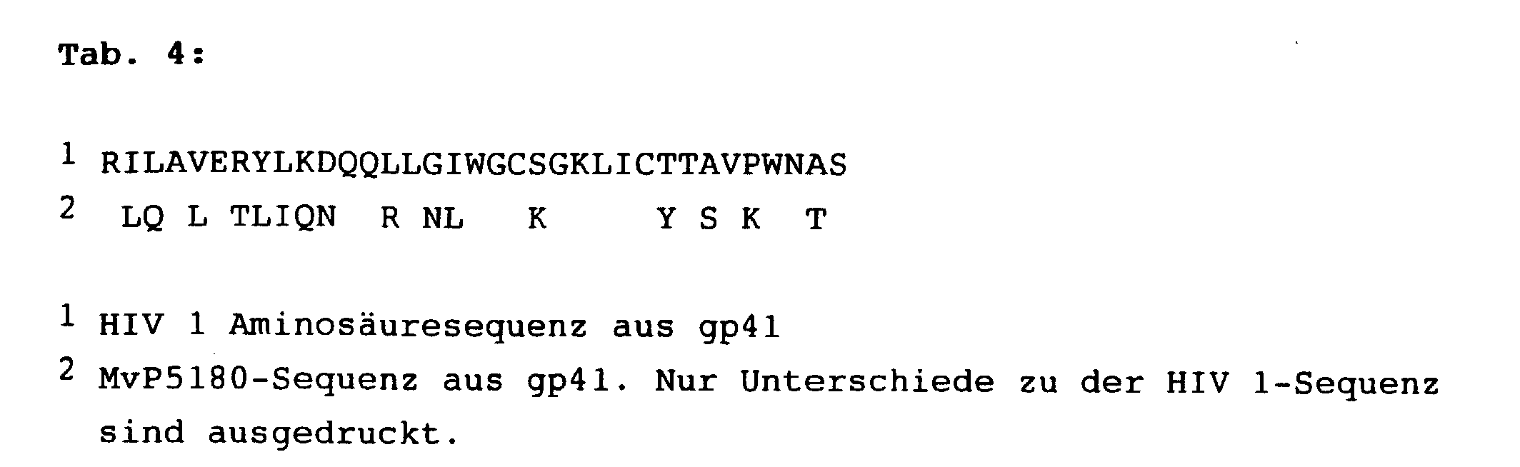
Das Peptid, das mit Hilfe der von MvP 5180 stammenden Information
aufgefunden wurde, hat also die Aminosäuresequenz:
RLQALETLIQNQQRLNLWGCKGKLICYTSVKWNTS.The peptide obtained using the information from
Gegenstand der vorliegenden Erfindung ist daher auch die diagnostische Verwendung von Peptiden, die rekombinant oder synthetisch hergestellt werden können und die oben angegebene Sequenz oder Teilsequenz aufweisen, wobei die Teilsequenzen wenigstens 6 aufeinanderfolgende Aminosäuren, bevorzugt 9 und besonders bevorzugt 12 aufeinanderfolgende Aminosäuren aufweisen.The present invention therefore also relates to diagnostic use of peptides that are recombinant or can be produced synthetically and the above Have sequence or partial sequence, the partial sequences at least 6 consecutive amino acids, preferably 9 and particularly preferably have 12 consecutive amino acids.
Genomische DNA aus MvP5180-infizierten HUT78-Zellen wurde wie
beschrieben isoliert.
300 µg dieser DNA wurde in einem Volumen von 770 µl mit 0,24 U
des Restriktionsenzyms Sau3A für 45 min inkubiert. Genomic DNA from MvP5180 infected HUT78 cells was isolated as described.
300 µg of this DNA was incubated in a volume of 770 µl with 0.24 U of the restriction enzyme Sau3A for 45 min.
Die dadurch nur partiell geschnittene DNA wurde anschließend
über ein 0,7 %iges Agarosegel (low melting agarose, Nusieve)
größenfraktioniert und Fragmente zwischen 10 und 21 kb
ausgeschnitten. Die Agarose wurde für 10 min bei 70°C
geschmolzen und mit demselben Volumen Puffer (1 x TBE, 0,2 M
NaCl) versetzt. Anschließend folgte nach zweimaliger Phenol- und
einmaliger Chloroformextraktion die Fällung der DNA
durch Zugabe von 1/10 Vol 3 M Natriumacetatlösung (pH 5,9)
und 2,5 Vol Ethanol bei -70°C für 10 min. Die gefällte DNA
wurde abzentrifugiert, getrocknet und in einer Konzentration
von 1 µg/µl in Wasser gelöst.
Die Ausbeute an größenfraktionierter DNA betrug etwa 60 µg.
5 µg dieser DNA wurden mit 1 U Alkalische Phosphatase im
entsprechenden Puffer für 20 min bei 37°C inkubiert. Durch
Abspaltung des 5'-terminalen Phosphatrestes wurden so
multiple Insertionen von größenfraktionierter DNA
vermindert. Die Phosphatasebehandlung wurde durch
Phenolisierung gestoppt, die DNA wie oben gefällt und
zusammen mit 1 µg des Vektors (2 DASH, BamHI geschnitten,
Stratagene Nr.: 247611) in 6 µl Gesamtvolumen mit 2 Weiss-Units
Lambda T4 Ligase für 12 Stunden bei 15°C ligiert.
Nach erfolgter Ligation wurde die DNA mit Hilfe eines
Verpackungskits (Gigapack II Gold, Stratagene Nr.: 247611)
genau nach Angaben des Herstellers in Phagenhüllen verpackt.The DNA that was only partially cut was then size-fractionated using a 0.7% agarose gel (low melting agarose, Nusieve) and fragments between 10 and 21 kb were cut out. The agarose was melted at 70 ° C. for 10 min and the same volume of buffer (1 × TBE, 0.2 M NaCl) was added. After two times phenol and one chloroform extraction followed the precipitation of the DNA by adding 1/10 vol 3 M sodium acetate solution (pH 5.9) and 2.5 vol ethanol at -70 ° C for 10 min. The precipitated DNA was centrifuged off, dried and dissolved in water at a concentration of 1 µg / µl.
The yield of size-fractionated DNA was approximately 60 µg. 5 µg of this DNA were incubated with 1 U alkaline phosphatase in the appropriate buffer for 20 min at 37 ° C. By splitting off the 5'-terminal phosphate residue, multiple insertions of size-fractionated DNA were reduced. The phosphatase treatment was stopped by phenolization, the DNA was precipitated as above and together with 1 μg of the vector (2 DASH, BamHI cut, Stratagene No .: 247611) in 6 μl total volume with 2 Weiss units Lambda T4 ligase for 12 hours at 15 ° C ligated. After the ligation, the DNA was packed in phage casings exactly according to the manufacturer's instructions using a packaging kit (Gigapack II Gold, Stratagene No .: 247611).
Für die Markierung wurde der "Random Primed DNA Labeling Kit" von Boehringer Mannheim (Nr.: 713 023) eingesetzt. Markiert wurde das PCR-Produkt, welches wie in Beispiel 3 beschrieben mit den Primern sk68 und envb erhalten wurde. 1 µg dieser DNA wurde durch 2 x 5 min Kochen und anschließender Abkühlung in Eiswasser denaturiert. Zur Markierung wurden 50 mCi [α-32P]-dCTP (NEN, Nr.: NEX-053H) zugegeben. Sonstige Zusätze wurden nach Herstellerangaben pipettiert. Nach einer Inkubation von 30 min bei 37°C erfolgte eine Fällung der nun radioaktiv markierten DNA.The "Random Primed DNA Labeling Kit" from Boehringer Mannheim (No. 713 023) was used for the labeling. The PCR product which was obtained as described in Example 3 with the primers sk68 and envb was labeled. 1 µg of this DNA was denatured by boiling for 2 x 5 min and then cooling in ice water. 50 mCi [α- 32 P] -dCTP (NEN, no .: NEX-053H) were added for labeling. Other additives were pipetted according to the manufacturer's instructions. After an incubation of 30 minutes at 37 ° C., the now radioactively labeled DNA was precipitated.
Zu 200 µl einer bei 30°C über Nacht angezogenen Kultur
(Stamm SRB(P2) [Stratagene, Nr.: 247611] in LB-Medium,
welches 10 mM MgSO4, sowie 0,2 % Maltose enthielt) wurden
20000 pfu (Plaque forming units) der Bibliothek in 100 µl
SM-Puffer (5,8 g NaCl, 2 g MgSO4 50 ml 1 M Tris. pH 7,5 und
5 ml einer 2 % Gelatinelösung in 1 l H2O gelöst) gegeben,
die Phagen 20 min bei 37°C an die Bakterien adsorbiert, mit
7,5 ml auf 55°C abgekühlter Top-Agarose gemischt und auf
einer vorgewärmten Lb-Agarplatte von 14 cm Durchmesser
verteilt. Nach etwa 8 Stunden erreichten die Plaques
Konfluenz. Daraufhin wurden Nitrocellulosefilter für wenige
Minuten auf die Platten gelegt und asymmetrische
Markierungen angebracht. Nach vorsichtigem Abheben wurden
die Filter für 2 min denaturiert (0,5 M NaOH, 1,5 M NaCl)
und dann 5 min neutralisiert (0,5 M Tris, pH 8, 1,5 M NaCl).
Nach anschließendem Backen der Filter bei 80°C für 60 min,
konnten die Filter mit der Probe hybridisiert werden.
Zur Vorhybridisierung wurden die Filter in 15 ml
Hybridisierungslösung (50 % Formamid, 0,5 % SDS, 5 x SSPE,
5 x Denhardt's Lösung und 0,1 mg/ml Lachssperma-DNA) pro
Filter bei 42°C unter Schütteln 2-3 h inkubiert. Die [32P]-markierten
DNA-Proben wurden 2-5 min bei 100°C denaturiert,
auf Eis abgekühlt, der Vorhybridisierungslösung zugesetzt
und 12 Stunden bei 42°C hybridisiert. Anschließend wurden
die Filter bei 60°C zunächst mit 2 x SSC/0,1 % SDS, dann mit
0,2 x SSC/0,1 % SDS gewaschen. Nach Trocknen der Filter
wurden Hybridisierungssignale mit Hilfe des Röntgenfilms X-OMAT™AR
(Kodak) detektiert.
Die Plaques, denen ein Signal zugeordnet werden konnte,
wurden nach Elution in SM-Puffer in weiteren
Verdünnungsschritten vereinzelt.
Der unten beschriebene Klon konnte nach dem Screening von
2 x 10⁶ Plaques identifiziert werden.20000 pfu (plaque.) Were added to 200 ul of a culture grown overnight at 30 ° C. (strain SRB (P2) [Stratagene, no .: 247611] in LB medium which contained 10 mM MgSO 4 and 0.2% maltose) forming units) of the library in 100 ul SM buffer (5.8 g NaCl, 2
For pre-hybridization, the filters were placed in 15 ml hybridization solution (50% formamide, 0.5% SDS, 5 x SSPE, 5 x Denhardt's solution and 0.1 mg / ml salmon sperm DNA) per filter at 42 ° C with shaking 2-3 h incubated. The [ 32 P] -labeled DNA samples were denatured for 2-5 min at 100 ° C, cooled on ice, added to the prehybridization solution and hybridized at 42 ° C for 12 hours. The filters were then washed at 60 ° C. first with 2 x SSC / 0.1% SDS, then with 0.2 x SSC / 0.1% SDS. After the filters had dried, hybridization signals were detected using the X-OMAT ™ AR X-ray film (Kodak).
The plaques to which a signal could be assigned were separated in further dilution steps after elution in SM buffer.
The clone described below could be identified after screening 2 x 10 6 plaques.
Mit 10 µl eines Phageneluats in SM-Puffer, wurde eine
Übernachtkultur des Wirtsstammes SRB (P2) so infiziert, daß
nach zunächst dichtem Wachstum der Kultur nach etwa 6-8 h
Lyse erfolgte. Von der lysierten Kultur wurden Zellreste
durch zweimalige Zentrifugation bei 9000 g für 10 min
abgetrennt. Anschließend wurden die Phagen durch
Zentrifugation pelletiert (35000 g, l h), in 700 µl 10 mM
MgSO4 aufgenommen und solange phenolisiert, bis keine
Proteininterphase mehr zu sehen war. Daraufhin wurde die
Phagen-DNA gefällt, mit dem Restriktionsenzym EcoRI
geschnitten und die daraus erhaltenen EcoRI-Fragmente in den
Vektor Bluescript KS- (Strategene, Nr.: 212208) subkloniert.
Insgesamt wurden 4 Klone erhalten:
Das fehlende Stück zwischen Base 7416 und 7659 wurde durch
PCR mit den Primern 157 (CCA TAA TAT TCA GCA GAA CTA G) und
226 (GCT GAT TCT GTA TAA GGG) erhalten. Als DNA-Template
wurde die Phagen-DNA des Klons verwendet. Die Bedingungen
für die PCR waren: 1.) Initiale Denaturierung: 94°C, 3 min,
2.) Amplifikation: 1,5 min 94°C, 1 min 56°C und 1 min 72°C
für 30 Zyklen.
Die Sequenzierung der DNA erfolgte wie in Beispiel 4
beschrieben. Vom gesamten Genom wurde sowohl der Strang- als
auch der Gegenstrang sequenziert. Bei allen EcoRI
Schnittstellen wurde durch PCR mit Phagen-DNA des Klons als
DNA-Template verifiziert, daß es sich an den
Subklonübergängen jeweils um singuläre EcoRI-Schnittstellen
handelt.
The DNA was sequenced as described in Example 4. Both the strand and the counter strand were sequenced from the entire genome. For all EcoRI interfaces, it was verified by PCR with phage DNA of the clone as a DNA template that the subclone transitions are each unique EcoRI interfaces.
Die Gesamtsequenz von MvP5180/91 ist in Fig. 4 dargestellt.The overall sequence of MvP5180 / 91 is shown in Fig. 4.
Grundlage für die folgenden Sequenzvergleiche war die
Datenbanken Genbank Release 75 von 2.93, EMBL 33 von 12.92
und Swissprot 24 von 1.93. Homologievergleiche erfolgten mit
der GCG-Software (Version 7.2, 10.92. der Genetics-Computer-Group,
Wisconsin).The basis for the following sequence comparisons was the
Databases Genbank Release 75 from 2.93, EMBL 33 from 12.92
and
Zunächst wurden auf Aminosäureebene die Sequenzen von GAG,
POL und ENV mit dem Programm "Wordsearch" mit der Datenbank
verglichen. Die 50 besten Homologen wurden mit dem Programm
"Pileup" jeweils untereinander verglichen. Daraus geht
deutlich hervor, daß MvP5180/91 in den HIV1-Stammbaum fällt,
dort aber sehr früh, sogar noch vor dem Schimpansenvirus
SIVcpz abzweigt, also eine neue Subfamilie von HIV1
repräsentiert. Um Zahlenwerte für die Homologien zu
erhalten; wurde mit dem Programm "Gap" MvP5180 mit den
jeweils am besten passenden HIV1, HIV2 und SIV-Sequenzen und
zusätzlich mit der SIVcpz-Sequenz verglichen.
Der obere Zahlenwert drückt die Identität, der untere die
Ähnlichkeit beider Sequenzen aus.
Weiter wurde die Datenbank mit "Wordsearch" und "Gap" auf
Nukleotidebene durchsucht. Die Homologiewerte für die
jeweils besten "matches" sind in Tabelle 7 zusammengefaßt.
The database was also searched with "Wordsearch" and "Gap" at the nucleotide level. The homology values for the best "matches" are summarized in Table 7.
Um die im Laufe der Virusvermehrung auftretenden Spontanmutationen darzustellen, wurde ein Teil des Virusgenoms mit der PCR-Technik kloniert und die so erhaltene DNS-Sequenz verglichen mit der Sequenz gemäß Fig. 4.To those that occur in the course of virus replication Representing spontaneous mutations became part of the Virus genome cloned with the PCR technique and so DNA sequence obtained compared with the sequence according to Fig. 4.
Die gag Sequenz wurde vom LTR ("long terminal repeat", LTR1
primer) des linken Endes des MvP 5180 Genomes bis in das pol
(Polymerase Gen, pol3.5i primer) hinein überlappend
kloniert. Die Klonierungsstrategie ist in Fig. 5 schematisch
dargestellt.
Die PCR Reaktionen wurden mit den u.g. DNA Primern, deren
Sequenzen von der HIV-1 Konsensus-Sequenz abgeleitet wurden,
durchgeführt. Die Sequenzierungen erfolgten mit Hilfe der
Dideoxykettenabbruchmethode.
Die für das MvP 5180 gag Gen kodierende Sequenz erstreckt
sich von Nukleotid 817 (A des ATG Startkodons) bis Nukleotid
2300 (A des letzten Kodons) (Sequ. ID No. 47-53).

The PCR reactions were carried out with the ug DNA primers, the sequences of which were derived from the HIV-1 consensus sequence. The sequencing was carried out using the dideoxy chain termination method.
The sequence coding for the
Die bei der PCR-Technik erhaltene DNS-Sequenz wurde der in Figur 4 dargestellten DNS-Sequenz gegenübergestellt. Ein Vergleich der beiden DNS-Sequenzen ist in Figur 6 dargestellt. Hierbei wurde festgestellt, daß sich die Nukleotide in ca. 2 % voneinander unterscheiden, obwohl es sich um dasselbe Virus handelt. In Fig. 6 stellt jeweils die obere Zeile die DNS-Sequenz dar, die in Fig. 4 dargestellt ist und die untere Zeile stellt die mit PCR-Technik erhaltene DNS-Sequenz dar.The DNA sequence obtained in the PCR technique was the in Figure 4 compared DNA sequence shown. A Comparison of the two DNA sequences is shown in FIG. 6 shown. It was found that the Nucleotides differ from each other in about 2%, although it does is the same virus. 6 represents the top line represents the DNA sequence shown in Fig. 4 and the bottom line represents the one with PCR technique DNA sequence obtained.
Weiterhin wurde die Aminosäuresequenz des mit PCR-Technik ermittelten Proteins gag der Aminosäuresequenz des aus Fig. 4 abgeleiteten entsprechenden Proteins gegenübergestellt. Dabei wurde ein Unterschied der Aminosäure von ca. 2,2 % ermittelt. Der Vergleich ist in Fig. 7 dargestellt, wobei die untere Zeile jeweils die Aminosäuresequenz darstellt, die von der mit PCR-Technik erhaltenen Sequenz abgeleitet wurde.Furthermore, the amino acid sequence was determined using the PCR technique determined protein gag the amino acid sequence of the Fig. 4 derived corresponding protein juxtaposed. There was a difference in the Amino acid of about 2.2% determined. The comparison is in Fig. 7 shown, the lower line each Represents amino acid sequence by using the PCR technique sequence obtained was derived.
Es wurde die Sequenz des erfindungsgemäßen Virus MvP 5180
verglichen mit den Konsensus-Sequenzen von HIV1 und HIV2 und
soweit bekannt mit der Sequenz von ANT-70 (WO 89/12094).It became the sequence of the
Dabei wurden folgende Ergebnisse erhalten:
In der obigen Tabelle bedeuten "HIV-1" Konsensus-Sequenzen von HIV-1 Viren; "HIV-2" Konsensus-Sequenzen von HIV-2 Viren; ANT-70 aus der WO 89/12094 bekannte Teilsequenz eines als HIV-3 bezeichneten Virus.In the table above, "HIV-1" means consensus sequences of HIV-1 viruses; "HIV-2" consensus sequences of HIV-2 viruses; ANT-70 from WO 89/12094 a partial sequence known as HIV-3 designated virus.
Gegenstand der vorliegenden Erfindung sind daher Viren, DNS-Sequenzen,
und die diagnostische Verwendung von
Aminosäuresequenzen sowie Teilsequenzen davon, die eine solche
Homologie mit der in Fig. 4 dargestellten Sequenz aufweisen,
bezogen auf die Genorte, daß höchstens die in Tabelle 9
angegebenen Anteile, ausgedrückt in %-Werten, unterschiedlich
sind.
Die in Tabelle 9 angegebenen Homologiewerte in % bedeuten, daß bei einer Gegenüberstellung der Sequenz gemäß Fig. 4 mit einer Sequenz eines anderen Virus höchstens ein den oben angegebenen Prozentwerten entsprechender Anteil der Sequenz unterschiedlich sein darf.The homology values in% given in Table 9 mean that when the sequence according to FIG. 4 is compared with a sequence of another virus at most one of the above corresponding percentage of the sequence indicated may be different.
Diese Schlaufe ist die hauptneutralisierende Region im HIV
und die Dokumentation der immunologischen Spezifitäten der
Region sind in der Figur 8 zusammengefaßt. Dies ist eine
Kopie aus AIDS aus einer Arbeit von Peter Nara (1990).
Die V3-Schlaufe ist dann in Aminosäuren-Ebene aufgezeichnet
und mit dem IIIB Virus jetzt LAI und dem ersten HIV-2 Isolat
(ROD) verglichen. Einzelne Aminosäuren an der Cystin-Brücke
sind konserviert. Während die Krone von HIV-1 GPGR oder GPGQ
ist und die von HIV-2 GHVF ist die Krone des MvP5180/91
gebildet aus den Aminosäuren GPMR. Das Motiv mit dem
Methionin ist bisher nicht beschrieben worden und
unterstreicht die Individualität des MvP 5180/91. This loop is the main neutralizing region in HIV and the documentation of the immunological specificities of the region are summarized in FIG. 8. This is a copy of AIDS from a work by Peter Nara (1990).
The V3 loop is then recorded at the amino acid level and compared with the IIIB virus now LAI and the first HIV-2 isolate (ROD). Individual amino acids on the cystine bridge are preserved. While the crown of HIV-1 is GPGR or GPGQ and that of HIV-2 GHVF, the crown of MvP5180 / 91 is formed from the amino acids GPMR. The motif with the methionine has not been described so far and underlines the individuality of the
Nachdem die Nukleinsäuresequenz des Virus ermittelt war, wurde der V3-Loop-Bereich mit Hilfe der PCR-Technik unter Verwendung geeigneter Primer amplifiziert. Hierbei konnten Mutationen beobachtet werden, insbesondere eine Veränderung des Metheonin-Kodons (ATG) zu einem Leucin-Kodon (CTG).After the nucleic acid sequence of the virus was determined, the V3 loop area was created using the PCR technique Amplified using suitable primers. Here could Mutations are observed, especially a change of the metheonin codon (ATG) to a leucine codon (CTG).
Nachfolgend wird eine Gegenüberstellung der von der
klonierten Nukleinsäure abgeleiteten Aminosäuresequenz und
der Sequenz gegeben, die nach Amplifizierung mittels PCR-Technologie
erhalten wurde (Sequ. ID No. 54-55):


Um zu zeigen, daß mit Hilfe des erfindungsgemäßen Virus
MvP 5180 bzw. davon abgeleiteten Antigenen auch solche Seren
als HIV-1 positiv nachgewiesen werden können, die bei
Verwendung eines normalen HIV-1+2 Screening-Tests nicht
erfaßt werden können, wurden verschiedene Seren von
Patienten aus Kamerun im EIA-Test überprüft.To show that with the help of the virus according to the
Bei einer Studie in Kamerun wurden 156 Anti-HIV-1-positive
Seren überprüft. Bei zwei dieser Seren wurden erhebliche,
diagnostisch relevante Unterschiede beobachtet. Bei der
nachfolgenden Tabelle 10 werden die gemessenen Extinktionen
angegeben. CAM-A bzw. CAM-B stehen für die Seren
verschiedener Patienten.
Der Cutoff für beide Tests betrug 0.300.The cutoff for both tests was 0.300.
In einer weiteren Studie mit 47 Anti-HIV-1-positiven Seren
aus Kamerun fielen zwei Seren besonders auf. Eines davon
(93-1000) stammt von einem wenig symptomatischen, das andere
(93-1001) von einem AIDS-kranken Patienten. In der folgenden
Tabelle 11 werden die Extinktionswerte der beiden EIA-Tests
einander gegenübergestellt:
Auch hier betrug der Cutoff 0.3. Die Extinktionswerte des Patienten 93-1001 zeigen, daß der normale HIV-1 + HIV-2 EIA versagen kann, wohingegen durch Einsatz des erfindungsgemäßen Antigens ein klarer Nachweis möglich ist.Here, too, the cutoff was 0.3. The absorbance values of the Patients 93-1001 show normal HIV-1 + HIV-2 EIA can fail, whereas by using the antigen according to the invention a clear detection is possible.
Claims (16)
- An immunodeficiency virus of the HIV group, which has been deposited with the European Collection of Animal Cell Cultures (ECACC) under No. V 920 92 318 with the designation MVP-5180/91, as well as variants of this virus, wherein one variant has a sequence corresponding to table 3 or being homologous to this sequence, wherein the homology is higher than 66% based on the nucleotide sequence.
- The virus as claimed in claim 1, wherein the homology is higher than 75%.
- The virus as claimed in claim 1, wherein the homology is higher than 85%.
- c-DNA which is complementary to the RNA, or parts thereof, of the immunodeficiency virus MVP-5180/91 deposited with the European Collection of Animal Cell Cultures (ECACC) under No. V 920 92 318, or of a virus as claimed in one of claims 1 to 3, wherein the constituent sequence is at least 50 nucleotides long.
- cDNA as claimed in claim 4, wherein the constituent sequence is at least 100 nucleotides long.
- Recombinant DNA which contains cDNA as claimed in claim 4 or 5.
- A test kit for detecting antibodies against viruses which cause immunodeficiency, belonging to an independent group of HIV viruses as claimed in claims 1 to 3, wherein an antigen is used having an amino acid sequence or a constituent amino acid sequence which is deduced from such a virus and has a constituent sequence of at least 6 amino acids.
- The test kit as claimed in claim 7, wherein the antigen has an amino acid sequence corresponding to table 3 or a constituent sequence thereof and the constituent sequence has at least 6 amino acids.
- The test kit as claimed in claim 7 or 8, wherein the antigen has the amino acid sequence RLQALETLIQNQQRLNLWGCKGKLICYTSVKWNTS or a constituent sequence thereof having at least 6 consecutive amino acids.
- The test kit as claimed in any one of claims 7 to 9, wherein the antigen was prepared recombinantly.
- The test kit as claimed in any one of claims 7 to 9, wherein the antigen was prepared synthetically.
- The test kit as claimed in any one of claims 7 to 11, which is a Western blot.
- The test kit as claimed in any one of claims 7 to 11, which is an ELISA test or a fluorescence-antibody detection test.
- Use of an antigen deduced from a virus as claimed in any one of claims 1 to 3 for detecting such retroviruses which cause immune deficiency and which belong to an independent group of HIV viruses according to claim 1.
- Use of the antigen as claimed in claim 14, wherein the antigen has an amino acid sequence corresponding to table 3 or a constituent sequence thereof and the constituent sequence has at least 6 amino acids.
- Use of an antigen as claimed in claim 14 or 15, wherein the antigen has the amino acid sequence RLQALETLIQNQQRLNLWGCKGKLICYTSVXWNTS or a constituent sequence thereof having at least 6 consecutive amino acids.
Priority Applications (1)
| Application Number | Priority Date | Filing Date | Title |
|---|---|---|---|
| EP98114623A EP0890642A3 (en) | 1992-10-06 | 1993-10-05 | Retrovirus of the HIV-group and its application |
Applications Claiming Priority (8)
| Application Number | Priority Date | Filing Date | Title |
|---|---|---|---|
| DE4233646 | 1992-10-06 | ||
| DE4233646A DE4233646C2 (en) | 1992-10-06 | 1992-10-06 | Retrovirus from the HIV group and its use |
| DE4235718 | 1992-10-22 | ||
| DE4235718A DE4235718A1 (en) | 1992-10-22 | 1992-10-22 | New HIV-type immune deficiency virus ECACC V 92092318 |
| DE4244541 | 1992-12-30 | ||
| DE4244541A DE4244541A1 (en) | 1992-12-30 | 1992-12-30 | New HIV-type immune deficiency virus ECACC V 92092318 |
| DE4318186A DE4318186C2 (en) | 1993-06-01 | 1993-06-01 | Retrovirus from the HIV group and its use |
| DE4318186 | 1993-06-01 |
Related Child Applications (2)
| Application Number | Title | Priority Date | Filing Date |
|---|---|---|---|
| EP98114623A Division EP0890642A3 (en) | 1992-10-06 | 1993-10-05 | Retrovirus of the HIV-group and its application |
| EP98114623A Division-Into EP0890642A3 (en) | 1992-10-06 | 1993-10-05 | Retrovirus of the HIV-group and its application |
Publications (4)
| Publication Number | Publication Date |
|---|---|
| EP0591914A2 EP0591914A2 (en) | 1994-04-13 |
| EP0591914A3 EP0591914A3 (en) | 1994-08-10 |
| EP0591914B1 true EP0591914B1 (en) | 1998-12-09 |
| EP0591914B2 EP0591914B2 (en) | 2010-02-10 |
Family
ID=27435590
Family Applications (2)
| Application Number | Title | Priority Date | Filing Date |
|---|---|---|---|
| EP98114623A Withdrawn EP0890642A3 (en) | 1992-10-06 | 1993-10-05 | Retrovirus of the HIV-group and its application |
| EP93116058A Expired - Lifetime EP0591914B2 (en) | 1992-10-06 | 1993-10-05 | Retrovirus of the HIV-group and its application |
Family Applications Before (1)
| Application Number | Title | Priority Date | Filing Date |
|---|---|---|---|
| EP98114623A Withdrawn EP0890642A3 (en) | 1992-10-06 | 1993-10-05 | Retrovirus of the HIV-group and its application |
Country Status (9)
| Country | Link |
|---|---|
| US (12) | US5770427A (en) |
| EP (2) | EP0890642A3 (en) |
| JP (4) | JP3277045B2 (en) |
| AT (1) | ATE174382T1 (en) |
| AU (1) | AU677225B2 (en) |
| CA (1) | CA2107732C (en) |
| DE (1) | DE59309207D1 (en) |
| DK (1) | DK0591914T4 (en) |
| ES (1) | ES2126620T5 (en) |
Cited By (2)
| Publication number | Priority date | Publication date | Assignee | Title |
|---|---|---|---|---|
| EP0891982A2 (en) | 1992-03-06 | 1999-01-20 | Innogenetics N.V. | HIV peptides |
| US7723040B2 (en) | 1999-07-09 | 2010-05-25 | Gen-Probe Incorporated | Detection of HIV-1 by nucleic acid amplification |
Families Citing this family (62)
| Publication number | Priority date | Publication date | Assignee | Title |
|---|---|---|---|---|
| US6589734B1 (en) | 1989-07-11 | 2003-07-08 | Gen-Probe Incorporated | Detection of HIV |
| US5856088A (en) * | 1989-07-11 | 1999-01-05 | Gen-Probe Incorporated | Detection of human immunodeficiency virus type 1 |
| DK0591914T4 (en) * | 1992-10-06 | 2010-05-25 | Siemens Healthcare Diagnostics | Retrovirus from the HIV group and its use |
| KR100325554B1 (en) | 1993-03-26 | 2002-11-02 | 젠-프로브 인코포레이티드 | Detection of Human Immunodeficiency Virus Type 1 |
| DE4405810A1 (en) * | 1994-02-23 | 1995-08-24 | Behringwerke Ag | Peptides derived from a retrovirus from the HIV group and their use |
| GB9410044D0 (en) * | 1994-05-19 | 1994-07-06 | Int Murex Tech Corp | Assay |
| FR2731225B1 (en) * | 1995-03-03 | 2003-10-31 | Pasteur Institut | PEPTIDES OF TRANSMEMBRANE ENVELOPE GLYCOPROTEIN AND CAPSID PROTEIN OF HUMAN RETROVIRUS TYPE HIV-1 AND PEPTIDES WITH AN IMMUNOLOGICAL PARENT |
| DK0787191T4 (en) | 1994-10-20 | 2012-01-30 | Pasteur Institut | Nucleotide sequences of HIV-1 type (or subtype) O retrovirus antigens |
| FR2726006B1 (en) * | 1994-10-20 | 1996-12-27 | Pasteur Institut | NUCLEOTIDE SEQUENCES ENCODING ANTIGENS OF A GROUP O HIV-1 RETROVIRUS |
| US6548635B1 (en) | 1995-02-16 | 2003-04-15 | Dade Behring Marburg Gmbh | Retrovirus from the HIV type O and its use (MVP-2901/94) |
| DE19505262C2 (en) * | 1995-02-16 | 1998-06-18 | Behring Diagnostics Gmbh | Retrovirus from the HIV group and its use |
| US5599662A (en) * | 1995-02-17 | 1997-02-04 | Hoffmann-La Roche Inc. | Oliconucleotide primers and probes for the detection of HIV-1 |
| WO1996027012A1 (en) * | 1995-03-02 | 1996-09-06 | Akzo Nobel N.V. | Specific hiv-1 group o antigens |
| CA2222994A1 (en) * | 1995-06-07 | 1996-12-19 | Abbott Laboratories | Peptides for hiv-1 detection |
| US5733732A (en) * | 1996-01-03 | 1998-03-31 | University Of Iowa Research Foundation | Methods for detecting primary adhalinopathy |
| CA2215073A1 (en) * | 1996-01-26 | 1997-07-31 | Innogenetics N.V. | Method for detection of drug-induced mutations in the reverse transcriptase gene |
| DE19644248A1 (en) * | 1996-10-24 | 1998-04-30 | Boehringer Mannheim Gmbh | Primers and probes for the detection of HIV |
| US6309827B1 (en) | 1997-03-28 | 2001-10-30 | Orasure Technologies, Inc. | Simultaneous collection of DNA and non-nucleic analytes |
| CA2286344C (en) | 1997-04-09 | 2012-08-07 | Pasteur Sanofi Diagnostics | Synthetic peptides useful in biological assays for detecting infections caused by group o hiv-1 viruses |
| US6846905B2 (en) | 1997-08-15 | 2005-01-25 | Abbott Laboratories | Antigen constructs useful in the detection and differentiation of antibodies to HIV |
| US5922533A (en) * | 1997-08-15 | 1999-07-13 | Abbott Laboratories | Rapid assay for simultaneous detection and differentiation of antibodies to HIV groups |
| DE19737442C2 (en) * | 1997-08-22 | 1999-06-24 | Olfert Landt Tib Molbiol Synth | Protein-coated polyribonucleic acids, a process for their preparation and their use |
| US6228128B1 (en) | 1997-11-10 | 2001-05-08 | Charlotte Johansen | Antimicrobial activity of laccases |
| AU2345399A (en) * | 1998-01-28 | 1999-08-16 | Universal Health-Watch, Inc. | Divergent hiv-1 peptides |
| CA2288869A1 (en) * | 1998-03-04 | 1999-09-10 | Mary Ann Childs | Rapid confirmatory test for microbial infections |
| WO1999062945A2 (en) * | 1998-06-05 | 1999-12-09 | Peptide Solutions, Inc. | Peptide antigens for detection of hiv, hcv and other microbial infections |
| CA2676762C (en) * | 1998-11-30 | 2015-12-29 | Ortho-Clinical Diagnostics, Inc. | Peptides for the detection of hiv-1 group o |
| US6303293B1 (en) | 1999-02-02 | 2001-10-16 | Ortho-Clinical Diagnostics, Inc. | Oligonucleotide reverse transcription primers for efficient detection of HIV-1 and HIV-2 and methods of use thereof |
| JP4805511B2 (en) * | 1999-12-23 | 2011-11-02 | メディカル リサーチ カウンシル | Improvement in immune response to HIV or improvement in immune response |
| WO2001057245A2 (en) * | 2000-02-04 | 2001-08-09 | K.U.Leuven Research & Development | Hiv-1 resistance assay |
| US6582920B2 (en) | 2000-09-01 | 2003-06-24 | Gen-Probe Incorporated | Amplification of HIV-1 RT sequences for detection of sequences associated with drug-resistance mutations |
| JP2002125687A (en) * | 2000-10-30 | 2002-05-08 | Tosoh Corp | Oligonucleotide for detecting hiv-1 and method for detecting the same |
| DE602004028862D1 (en) * | 2003-12-19 | 2010-10-07 | Gen Probe Inc | THE NUCLEIC ACIDS OF HIV-1 AND HIV-2 |
| US7935479B2 (en) * | 2004-07-19 | 2011-05-03 | Cell Biosciences, Inc. | Methods and devices for analyte detection |
| US20060292558A1 (en) * | 2004-07-19 | 2006-12-28 | Cell Biosciences Inc. | Methods and apparatus for protein assay diagnostics |
| US7846676B2 (en) * | 2004-07-19 | 2010-12-07 | Cell Biosciences, Inc. | Methods and devices for analyte detection |
| WO2006014680A1 (en) * | 2004-07-19 | 2006-02-09 | Cell Biosciences, Inc. | Methods and devices for analyte detection |
| US8945361B2 (en) | 2005-09-20 | 2015-02-03 | ProteinSimple | Electrophoresis standards, methods and kits |
| CA2637600A1 (en) | 2006-01-17 | 2007-07-26 | Health Research, Inc. | Heteroduplex tracking assay |
| US20080086118A1 (en) * | 2006-05-17 | 2008-04-10 | Applied Harmonics Corporation | Apparatus and method for diode-pumped laser ablation of soft tissue |
| US20080017512A1 (en) * | 2006-07-24 | 2008-01-24 | Bordunov Andrei V | Coatings for capillaries capable of capturing analytes |
| GB0701253D0 (en) | 2007-01-23 | 2007-02-28 | Diagnostics For The Real World | Nucleic acid amplification and testing |
| US10107782B2 (en) | 2008-01-25 | 2018-10-23 | ProteinSimple | Method to perform limited two dimensional separation of proteins and other biologicals |
| JPWO2009122591A1 (en) * | 2008-04-04 | 2011-07-28 | 国立大学法人 琉球大学 | Virus antigen and virus antibody detection kit capable of detecting virus antigen and virus antibody at one time, and virus antigen and virus antibody detection method |
| WO2009122592A1 (en) * | 2008-04-04 | 2009-10-08 | 国立大学法人琉球大学 | Kit for detecting viral antigen and viral antibody including reagent storable at room temperature and method for detecting viral antigen and viral antibody |
| US20090258342A1 (en) * | 2008-04-09 | 2009-10-15 | Intelligent Medical Devices, Inc. | Optimized probes and primers and methods of using same for the detection, quantification and grouping of hiv-1 |
| WO2011005220A1 (en) * | 2009-07-07 | 2011-01-13 | Masafumi Inoue | Human immunodeficiency virus type 1 (hiv-1) detection method and kit therefor |
| WO2011119920A2 (en) | 2010-03-25 | 2011-09-29 | Oregon Health & Science University | Cmv glycoproteins and recombinant vectors |
| SG184833A1 (en) | 2010-04-14 | 2012-11-29 | Emd Millipore Corp | Methods of producing high titer, high purity virus stocks and methods of use thereof |
| WO2012153153A1 (en) | 2011-05-11 | 2012-11-15 | Diagon Kft. | Procedure for rapid determination of viruses using nucleic acid-based molecular diagnostics, and a kit for this purpose |
| CA2832109C (en) | 2011-06-10 | 2021-07-06 | Oregon Health & Science University | Cmv glycoproteins and recombinant vectors |
| CA2789539A1 (en) | 2011-09-12 | 2013-03-12 | International Aids Vaccine Initiative | Immunoselection of recombinant vesicular stomatitis virus expressing hiv-1 proteins by broadly neutralizing antibodies |
| EP2586461A1 (en) | 2011-10-27 | 2013-05-01 | Christopher L. Parks | Viral particles derived from an enveloped virus |
| EP2839276B1 (en) | 2012-04-19 | 2018-06-27 | ProteinSimple | Dual wavelength isoelectric focusing for determining drug load in antibody drug conjugates |
| ES2631608T3 (en) | 2012-06-27 | 2017-09-01 | International Aids Vaccine Initiative | Env-glycoprotein variant of HIV-1 |
| US20150065381A1 (en) | 2013-09-05 | 2015-03-05 | International Aids Vaccine Initiative | Methods of identifying novel hiv-1 immunogens |
| US9766206B2 (en) | 2013-09-27 | 2017-09-19 | ProteinSimple | Apparatus, systems, and methods for capillary electrophoresis |
| EP2873423B1 (en) | 2013-10-07 | 2017-05-31 | International Aids Vaccine Initiative | Soluble hiv-1 envelope glycoprotein trimers |
| US10174292B2 (en) | 2015-03-20 | 2019-01-08 | International Aids Vaccine Initiative | Soluble HIV-1 envelope glycoprotein trimers |
| EP3072901A1 (en) | 2015-03-23 | 2016-09-28 | International Aids Vaccine Initiative | Soluble hiv-1 envelope glycoprotein trimers |
| EP3286335A4 (en) * | 2015-04-24 | 2018-10-17 | The Johns Hopkins University | Compositions and methods related to characterizing proviral reservoirs |
| CN116064923A (en) * | 2022-01-27 | 2023-05-05 | 圣湘生物科技股份有限公司 | Compositions, kits, methods and uses for detecting HIV-1 |
Family Cites Families (26)
| Publication number | Priority date | Publication date | Assignee | Title |
|---|---|---|---|---|
| US5705612A (en) * | 1984-10-18 | 1998-01-06 | Institut Pasteur And Centre National De La Recherche Scientifique | NEF peptide encoded by human immunodefiency virus type 1 (HIV-1) |
| CA1341482C (en) * | 1984-10-31 | 2005-05-10 | Paul A. Luciw | Process for preparing fragments of aids-associated retroviruses |
| US4683202A (en) * | 1985-03-28 | 1987-07-28 | Cetus Corporation | Process for amplifying nucleic acid sequences |
| US4683195A (en) * | 1986-01-30 | 1987-07-28 | Cetus Corporation | Process for amplifying, detecting, and/or-cloning nucleic acid sequences |
| US4629783A (en) * | 1985-04-29 | 1986-12-16 | Genetic Systems Corporation | Synthetic antigen for the detection of AIDS-related disease |
| US4803156A (en) * | 1985-09-11 | 1989-02-07 | New York Blood Center | Peptide-beta-lactamase conjugates for enzyme-linked immunoassays |
| ES2150897T3 (en) * | 1986-01-22 | 2000-12-16 | Pasteur Institut | RECOMBINANT MANUFACTURING PROCEDURE OF PROTEINS DERIVED FROM HIV-2 AND CELL CULTURE THAT EXPRESSES HIV-2 PROTEINS. |
| US5079342A (en) * | 1986-01-22 | 1992-01-07 | Institut Pasteur | Cloned DNA sequences related to the entire genomic RNA of human immunodeficiency virus II (HIV-2), polypeptides encoded by these DNA sequences and use of these DNA clones and polypeptides in diagnostic kits |
| US6265149B1 (en) * | 1986-01-22 | 2001-07-24 | Institut Pasteur | In vitro diagnostic methods and kits for the detection of HIV-2-specific antibodies |
| US5030714A (en) * | 1986-06-23 | 1991-07-09 | Institut Pasteur | Variant of LAV viruses |
| US5034511A (en) * | 1987-04-13 | 1991-07-23 | Institut Pasteur | Variant of LAV viruses |
| US5030449A (en) * | 1988-07-21 | 1991-07-09 | The United States Of America As Represented By The Secretary Of The Department Of Health And Human Services | Synthetic vaccine against AIDS virus |
| US5141867A (en) * | 1987-02-02 | 1992-08-25 | E. I. Du Pont De Nemours And Company | Nucleotide sequence encoding a human immunodeficiency virus antigen |
| DE3887053D1 (en) * | 1987-04-16 | 1994-02-24 | Johnson & Johnson | STLV-III RELATED POLYPEPTIDES, DIAGNOSTIC SYSTEMS AND TEST METHODS. |
| US5013548A (en) * | 1987-09-08 | 1991-05-07 | Duke University | Production of antibodies to HIV |
| US5019510A (en) * | 1987-10-28 | 1991-05-28 | Institut Pasteur | Isolation, molecular cloning and sequencing of an HIV-1 isolate from a Gabonese donor |
| US5312737A (en) * | 1988-03-11 | 1994-05-17 | Abbott Laboratories | CKS method of HCV protein synthesis |
| EP1369427A3 (en) * | 1988-06-09 | 2004-05-12 | Innogenetics N.V. | HIV-3 retrovirus strains and their use |
| US5223423A (en) * | 1989-03-31 | 1993-06-29 | United States Of America | Characterization of replication competent human immunodeficiency type 2 proviral clone hiv-2sbl/isy |
| DE4002636A1 (en) * | 1990-01-30 | 1991-08-01 | Boehringer Mannheim Gmbh | EXPRESSION OF HIV1 AND 2 POLYPEPTIDES AND THEIR USE |
| US5013458A (en) * | 1990-04-06 | 1991-05-07 | Rdp Company | Process and apparatus for pathogen reduction in waste |
| EP0554389B2 (en) * | 1990-10-17 | 2002-08-14 | The United States Of America | Molecular clones of hiv-1 and uses thereof |
| FR2676071B1 (en) * | 1991-05-02 | 1994-11-18 | Transgene Sa | NEW NON-CLeavABLE, SOLUBLE, HYBRID-FORMING GP160 VARIANT. |
| DK0591914T4 (en) * | 1992-10-06 | 2010-05-25 | Siemens Healthcare Diagnostics | Retrovirus from the HIV group and its use |
| US5304046A (en) * | 1993-06-14 | 1994-04-19 | Yang Gene Huang | Fluid transport device |
| DE4405810A1 (en) * | 1994-02-23 | 1995-08-24 | Behringwerke Ag | Peptides derived from a retrovirus from the HIV group and their use |
-
1993
- 1993-10-05 DK DK93116058.4T patent/DK0591914T4/en active
- 1993-10-05 AU AU48800/93A patent/AU677225B2/en not_active Expired
- 1993-10-05 JP JP24960593A patent/JP3277045B2/en not_active Expired - Lifetime
- 1993-10-05 ES ES93116058T patent/ES2126620T5/en not_active Expired - Lifetime
- 1993-10-05 AT AT93116058T patent/ATE174382T1/en active
- 1993-10-05 CA CA002107732A patent/CA2107732C/en not_active Expired - Lifetime
- 1993-10-05 DE DE59309207T patent/DE59309207D1/en not_active Expired - Lifetime
- 1993-10-05 EP EP98114623A patent/EP0890642A3/en not_active Withdrawn
- 1993-10-05 EP EP93116058A patent/EP0591914B2/en not_active Expired - Lifetime
-
1995
- 1995-06-06 US US08/471,770 patent/US5770427A/en not_active Expired - Lifetime
- 1995-06-06 US US08/470,202 patent/US5759770A/en not_active Expired - Lifetime
- 1995-06-06 US US08/468,059 patent/US5840480A/en not_active Expired - Lifetime
-
1998
- 1998-07-02 US US09/109,916 patent/US6277561B1/en not_active Expired - Lifetime
-
2000
- 2000-02-23 JP JP2000045662A patent/JP4113318B2/en not_active Expired - Lifetime
-
2001
- 2001-06-22 US US09/886,156 patent/US6528626B2/en not_active Expired - Fee Related
- 2001-06-22 US US09/886,149 patent/US6531137B2/en not_active Expired - Fee Related
- 2001-06-22 US US09/886,159 patent/US6551824B2/en not_active Expired - Fee Related
- 2001-06-22 US US09/886,150 patent/US6531587B2/en not_active Expired - Fee Related
-
2002
- 2002-12-23 US US10/326,090 patent/US6773915B2/en not_active Expired - Fee Related
-
2004
- 2004-01-23 JP JP2004015239A patent/JP3854604B2/en not_active Expired - Lifetime
- 2004-04-22 US US10/829,442 patent/US20050053920A1/en not_active Abandoned
-
2006
- 2006-04-04 JP JP2006103080A patent/JP4029107B2/en not_active Expired - Lifetime
- 2006-12-06 US US11/634,254 patent/US20070105219A1/en not_active Abandoned
- 2006-12-06 US US11/634,155 patent/US20070264627A1/en not_active Abandoned
Cited By (2)
| Publication number | Priority date | Publication date | Assignee | Title |
|---|---|---|---|---|
| EP0891982A2 (en) | 1992-03-06 | 1999-01-20 | Innogenetics N.V. | HIV peptides |
| US7723040B2 (en) | 1999-07-09 | 2010-05-25 | Gen-Probe Incorporated | Detection of HIV-1 by nucleic acid amplification |
Also Published As
Similar Documents
| Publication | Publication Date | Title |
|---|---|---|
| EP0591914B1 (en) | Retrovirus of the HIV-group and its application | |
| EP0673948B1 (en) | Pepticles derived from a retrovirus of the HIV-group and their use | |
| DE3588251T2 (en) | DNA fragments of the GAG gene of LAV | |
| DE68929467T2 (en) | HIV-2 variants | |
| EP0495896B1 (en) | Vaccines for protecting against infections by hiv virus;process for producing the same and their use as diagnostic and immunotherapy means | |
| DE69637264T2 (en) | Viral material and multiple sclerosis associated nucleotide fragments with diagnostic, prophylactic and therapeutic uses | |
| DE69535601T3 (en) | NUCLEOTIDE SEQUENCES FROM RETROVIRUS ANTIGENIC HIV-I GROUP (OR SUB-GROUP) O | |
| DE69836206T2 (en) | SYNTHETIC HIV GAG GENES | |
| DE4040339C2 (en) | Viral agent | |
| EP0727483B1 (en) | Retrovirus of the HIV-group and its application | |
| DD283935A5 (en) | METHOD FOR PRODUCING AN AIDS VIRUS VACCINE | |
| JPH07233196A (en) | Active ingredient of vaccine against aids | |
| DE69333636T2 (en) | Nucleotide and peptide sequences of the immunodeficiency virus of the cat, isolate WO and applications of the sequences in the diagnosis and for the prevention of the infection | |
| DE69830325T2 (en) | Peptides derived from the gene env of FIV (feline immunoefficiency virus) and their immunoprotective applications | |
| DE4318186C2 (en) | Retrovirus from the HIV group and its use | |
| DE4233646C2 (en) | Retrovirus from the HIV group and its use | |
| DE4244541A1 (en) | New HIV-type immune deficiency virus ECACC V 92092318 | |
| DE60034243T2 (en) | RECOMBINANT BIFUNCTIONAL FUSION PROTEINS FOR ABORING VARIABLE VIRUSES | |
| DE4235718A1 (en) | New HIV-type immune deficiency virus ECACC V 92092318 | |
| DE69831412T2 (en) | MULTIPLE SCLEROSIS AND / OR RHEUMATIC POLYARTHRITIS ASSOCIATED RETROVIRAL NUCLEIC ACIDS AND CORRESPONDING NUCLEOTIDE FRAGMENTS FOR DIAGNOSTICS, PROPHYLAXIS AND THERAPY | |
| DE3727137A1 (en) | PRODUCTION OF ANTIGENS OF THE HUMAN IMMUNODEFICIENCY VIRUS TYPE 1 AND THEIR USE AS A DIAGNOSTIC AGENT AND THERAPEUTIC | |
| DE3724016A1 (en) | Methods for the genetic engineering production of antigens and their use in an approach to confirmatory tests for antibodies in the example of human immunodeficiency virus (HIV) | |
| WO1998052969A1 (en) | Method for detecting hiv antibodies and antigens used to this end | |
| EP1754715A1 (en) | Vaccine on the basis of virus-neutralising antibodies | |
| DE3856564T2 (en) | HIV-3 strains of retrovirus and their use |
Legal Events
| Date | Code | Title | Description |
|---|---|---|---|
| PUAI | Public reference made under article 153(3) epc to a published international application that has entered the european phase |
Free format text: ORIGINAL CODE: 0009012 |
|
| AK | Designated contracting states |
Kind code of ref document: A2 Designated state(s): AT BE CH DE DK ES FR GB IT LI LU NL PT SE |
|
| PUAL | Search report despatched |
Free format text: ORIGINAL CODE: 0009013 |
|
| AK | Designated contracting states |
Kind code of ref document: A3 Designated state(s): AT BE CH DE DK ES FR GB IT LI LU NL PT SE |
|
| 17P | Request for examination filed |
Effective date: 19941011 |
|
| 17Q | First examination report despatched |
Effective date: 19950125 |
|
| RAP1 | Party data changed (applicant data changed or rights of an application transferred) |
Owner name: BEHRING DIAGNOSTICS GMBH |
|
| RAP1 | Party data changed (applicant data changed or rights of an application transferred) |
Owner name: DADE BEHRING MARBURG GMBH |
|
| GRAG | Despatch of communication of intention to grant |
Free format text: ORIGINAL CODE: EPIDOS AGRA |
|
| GRAG | Despatch of communication of intention to grant |
Free format text: ORIGINAL CODE: EPIDOS AGRA |
|
| GRAH | Despatch of communication of intention to grant a patent |
Free format text: ORIGINAL CODE: EPIDOS IGRA |
|
| GRAH | Despatch of communication of intention to grant a patent |
Free format text: ORIGINAL CODE: EPIDOS IGRA |
|
| GRAA | (expected) grant |
Free format text: ORIGINAL CODE: 0009210 |
|
| AK | Designated contracting states |
Kind code of ref document: B1 Designated state(s): AT BE CH DE DK ES FR GB IT LI LU NL PT SE |
|
| REF | Corresponds to: |
Ref document number: 174382 Country of ref document: AT Date of ref document: 19981215 Kind code of ref document: T |
|
| REG | Reference to a national code |
Ref country code: CH Ref legal event code: EP |
|
| REG | Reference to a national code |
Ref country code: CH Ref legal event code: NV Representative=s name: PATENTANWALTSBUERO EDER AG |
|
| REF | Corresponds to: |
Ref document number: 59309207 Country of ref document: DE Date of ref document: 19990121 |
|
| ITF | It: translation for a ep patent filed |
Owner name: ST. DR. CAVATTONI ING. A. RAIMONDI |
|
| ET | Fr: translation filed | ||
| GBT | Gb: translation of ep patent filed (gb section 77(6)(a)/1977) |
Effective date: 19990225 |
|
| REG | Reference to a national code |
Ref country code: ES Ref legal event code: FG2A Ref document number: 2126620 Country of ref document: ES Kind code of ref document: T3 |
|
| REG | Reference to a national code |
Ref country code: PT Ref legal event code: SC4A Free format text: AVAILABILITY OF NATIONAL TRANSLATION Effective date: 19990305 |
|
| K2C3 | Correction of patent specification (complete document) published |
Effective date: 19981209 |
|
| REG | Reference to a national code |
Ref country code: DK Ref legal event code: T3 |
|
| PLBQ | Unpublished change to opponent data |
Free format text: ORIGINAL CODE: EPIDOS OPPO |
|
| PLBQ | Unpublished change to opponent data |
Free format text: ORIGINAL CODE: EPIDOS OPPO |
|
| PLBI | Opposition filed |
Free format text: ORIGINAL CODE: 0009260 |
|
| PLAB | Opposition data, opponent's data or that of the opponent's representative modified |
Free format text: ORIGINAL CODE: 0009299OPPO |
|
| 26 | Opposition filed |
Opponent name: BIOTEST AG Effective date: 19990909 Opponent name: DIPL. ING. H. HORSTMANN GMBH Effective date: 19990909 Opponent name: ORTHO CLINICAL DIAGNOSTICS, INC. Effective date: 19990909 Opponent name: PASTEUR SANOFI DIAGNOSTICS Effective date: 19990909 Opponent name: INSTITUT PASTEUR Effective date: 19990909 Opponent name: CHIRON CORPORATION Effective date: 19990909 |
|
| PLBF | Reply of patent proprietor to notice(s) of opposition |
Free format text: ORIGINAL CODE: EPIDOS OBSO |
|
| R26 | Opposition filed (corrected) |
Opponent name: CHIRON CORPORATION * 19990909 INSTITUT PASTEUR * 1 Effective date: 19990909 |
|
| NLR1 | Nl: opposition has been filed with the epo |
Opponent name: BIOTEST AG Opponent name: DIPL. ING. H. HORSTMANN GMBH Opponent name: ORTHO CLINICAL DIAGNOSTICS, INC. Opponent name: PASTEUR SANOFI DIAGNOSTICS Opponent name: INSTITUT PASTEUR Opponent name: CHIRON CORPORATION |
|
| PLBF | Reply of patent proprietor to notice(s) of opposition |
Free format text: ORIGINAL CODE: EPIDOS OBSO |
|
| PLBF | Reply of patent proprietor to notice(s) of opposition |
Free format text: ORIGINAL CODE: EPIDOS OBSO |
|
| PLBF | Reply of patent proprietor to notice(s) of opposition |
Free format text: ORIGINAL CODE: EPIDOS OBSO |
|
| PLBF | Reply of patent proprietor to notice(s) of opposition |
Free format text: ORIGINAL CODE: EPIDOS OBSO |
|
| REG | Reference to a national code |
Ref country code: GB Ref legal event code: IF02 |
|
| PLAB | Opposition data, opponent's data or that of the opponent's representative modified |
Free format text: ORIGINAL CODE: 0009299OPPO |
|
| R26 | Opposition filed (corrected) |
Opponent name: BIOTEST AG Effective date: 19990909 Opponent name: ORTHO CLINICAL DIAGNOSTICS, INC. Effective date: 19990909 Opponent name: BIO-RAD PASTEUR Effective date: 19990909 Opponent name: INSTITUT PASTEUR Effective date: 19990909 Opponent name: CHIRON CORPORATION Effective date: 19990909 |
|
| NLR1 | Nl: opposition has been filed with the epo |
Opponent name: BIOTEST AG Opponent name: ORTHO CLINICAL DIAGNOSTICS, INC. Opponent name: BIO-RAD PASTEUR Opponent name: INSTITUT PASTEUR Opponent name: CHIRON CORPORATION |
|
| PLBP | Opposition withdrawn |
Free format text: ORIGINAL CODE: 0009264 |
|
| APBP | Date of receipt of notice of appeal recorded |
Free format text: ORIGINAL CODE: EPIDOSNNOA2O |
|
| APBP | Date of receipt of notice of appeal recorded |
Free format text: ORIGINAL CODE: EPIDOSNNOA2O |
|
| APBP | Date of receipt of notice of appeal recorded |
Free format text: ORIGINAL CODE: EPIDOSNNOA2O |
|
| APBP | Date of receipt of notice of appeal recorded |
Free format text: ORIGINAL CODE: EPIDOSNNOA2O |
|
| APBQ | Date of receipt of statement of grounds of appeal recorded |
Free format text: ORIGINAL CODE: EPIDOSNNOA3O |
|
| APBQ | Date of receipt of statement of grounds of appeal recorded |
Free format text: ORIGINAL CODE: EPIDOSNNOA3O |
|
| RIC2 | Information provided on ipc code assigned after grant |
Ipc: 7G 01N 33/68 B Ipc: 7C 12Q 1/68 B Ipc: 7G 01N 33/569 B Ipc: 7C 07K 15/00 B Ipc: 7C 12N 15/48 B Ipc: 7C 12N 7/00 A |
|
| APAA | Appeal reference recorded |
Free format text: ORIGINAL CODE: EPIDOS REFN |
|
| APAH | Appeal reference modified |
Free format text: ORIGINAL CODE: EPIDOSCREFNO |
|
| PLAB | Opposition data, opponent's data or that of the opponent's representative modified |
Free format text: ORIGINAL CODE: 0009299OPPO |
|
| R26 | Opposition filed (corrected) |
Opponent name: BIOTEST AG Effective date: 19990909 Opponent name: BIO-RAD PASTEUR Effective date: 19990909 Opponent name: INSTITUT PASTEUR Effective date: 19990909 |
|
| NLR1 | Nl: opposition has been filed with the epo |
Opponent name: BIOTEST AG Opponent name: BIO-RAD PASTEUR Opponent name: INSTITUT PASTEUR |
|
| PLAB | Opposition data, opponent's data or that of the opponent's representative modified |
Free format text: ORIGINAL CODE: 0009299OPPO |
|
| RAP2 | Party data changed (patent owner data changed or rights of a patent transferred) |
Owner name: SIEMENS HEALTHCARE DIAGNOSTICS PRODUCTS GMBH |
|
| NLT2 | Nl: modifications (of names), taken from the european patent patent bulletin |
Owner name: SIEMENS HEALTHCARE DIAGNOSTICS PRODUCTS GMBH Effective date: 20081015 |
|
| REG | Reference to a national code |
Ref country code: CH Ref legal event code: PFA Owner name: SIEMENS HEALTHCARE DIAGNOSTICS PRODUCTS GMBH Free format text: DADE BEHRING MARBURG GMBH#POSTFACH 11 49#35001 MARBURG (DE) -TRANSFER TO- SIEMENS HEALTHCARE DIAGNOSTICS PRODUCTS GMBH#EMIL-VON-BEHRING-STRASSE 76#35041 MARBURG (DE) Ref country code: CH Ref legal event code: NV Representative=s name: SIEMENS SCHWEIZ AG |
|
| NLT1 | Nl: modifications of names registered in virtue of documents presented to the patent office pursuant to art. 16 a, paragraph 1 |
Owner name: SIEMENS HEALTHCARE DIAGNOSTICS PRODUCTS GMBH |
|
| APBU | Appeal procedure closed |
Free format text: ORIGINAL CODE: EPIDOSNNOA9O |
|
| REG | Reference to a national code |
Ref country code: FR Ref legal event code: CD |
|
| PUAH | Patent maintained in amended form |
Free format text: ORIGINAL CODE: 0009272 |
|
| STAA | Information on the status of an ep patent application or granted ep patent |
Free format text: STATUS: PATENT MAINTAINED AS AMENDED |
|
| 27A | Patent maintained in amended form |
Effective date: 20100210 |
|
| AK | Designated contracting states |
Kind code of ref document: B2 Designated state(s): AT BE CH DE DK ES FR GB IT LI LU NL PT SE |
|
| REG | Reference to a national code |
Ref country code: CH Ref legal event code: AEN Free format text: AUFRECHTERHALTUNG DES PATENTES IN GEAENDERTER FORM |
|
| NLR2 | Nl: decision of opposition |
Effective date: 20100210 |
|
| REG | Reference to a national code |
Ref country code: NL Ref legal event code: T3 |
|
| REG | Reference to a national code |
Ref country code: SE Ref legal event code: RPEO |
|
| REG | Reference to a national code |
Ref country code: ES Ref legal event code: DC2A Date of ref document: 20100408 Kind code of ref document: T5 |
|
| REG | Reference to a national code |
Ref country code: DK Ref legal event code: T4 |
|
| PGFP | Annual fee paid to national office [announced via postgrant information from national office to epo] |
Ref country code: LU Payment date: 20121024 Year of fee payment: 20 |
|
| PGFP | Annual fee paid to national office [announced via postgrant information from national office to epo] |
Ref country code: DK Payment date: 20121025 Year of fee payment: 20 |
|
| PGFP | Annual fee paid to national office [announced via postgrant information from national office to epo] |
Ref country code: FR Payment date: 20121113 Year of fee payment: 20 Ref country code: CH Payment date: 20121024 Year of fee payment: 20 Ref country code: DE Payment date: 20120928 Year of fee payment: 20 Ref country code: BE Payment date: 20121022 Year of fee payment: 20 |
|
| PGFP | Annual fee paid to national office [announced via postgrant information from national office to epo] |
Ref country code: PT Payment date: 20120405 Year of fee payment: 20 Ref country code: IT Payment date: 20121024 Year of fee payment: 20 Ref country code: SE Payment date: 20121024 Year of fee payment: 20 Ref country code: GB Payment date: 20121023 Year of fee payment: 20 Ref country code: ES Payment date: 20121023 Year of fee payment: 20 |
|
| PGFP | Annual fee paid to national office [announced via postgrant information from national office to epo] |
Ref country code: AT Payment date: 20121022 Year of fee payment: 20 Ref country code: NL Payment date: 20121018 Year of fee payment: 20 |
|
| REG | Reference to a national code |
Ref country code: DE Ref legal event code: R071 Ref document number: 59309207 Country of ref document: DE |
|
| REG | Reference to a national code |
Ref country code: DK Ref legal event code: EUP Effective date: 20131005 Ref country code: DK Ref legal event code: EUP |
|
| REG | Reference to a national code |
Ref country code: DE Ref legal event code: R071 Ref document number: 59309207 Country of ref document: DE |
|
| REG | Reference to a national code |
Ref country code: CH Ref legal event code: PL Ref country code: PT Ref legal event code: MM4A Free format text: MAXIMUM VALIDITY LIMIT REACHED Effective date: 20131005 |
|
| REG | Reference to a national code |
Ref country code: NL Ref legal event code: V4 Effective date: 20131005 |
|
| REG | Reference to a national code |
Ref country code: GB Ref legal event code: PE20 Expiry date: 20131004 |
|
| BE20 | Be: patent expired |
Owner name: SIEMENS HEALTHCARE DIAGNOSTICS PRODUCTS G.M.B.H. Effective date: 20131005 |
|
| REG | Reference to a national code |
Ref country code: AT Ref legal event code: MK07 Ref document number: 174382 Country of ref document: AT Kind code of ref document: T Effective date: 20131005 |
|
| REG | Reference to a national code |
Ref country code: SE Ref legal event code: EUG |
|
| REG | Reference to a national code |
Ref country code: ES Ref legal event code: FD2A Effective date: 20131219 |
|
| PG25 | Lapsed in a contracting state [announced via postgrant information from national office to epo] |
Ref country code: PT Free format text: LAPSE BECAUSE OF EXPIRATION OF PROTECTION Effective date: 20131015 Ref country code: DE Free format text: LAPSE BECAUSE OF EXPIRATION OF PROTECTION Effective date: 20131008 Ref country code: GB Free format text: LAPSE BECAUSE OF EXPIRATION OF PROTECTION Effective date: 20131004 |
|
| PG25 | Lapsed in a contracting state [announced via postgrant information from national office to epo] |
Ref country code: ES Free format text: LAPSE BECAUSE OF EXPIRATION OF PROTECTION Effective date: 20131006 |