EP0701801A1 - Method for making an intraluminal stent - Google Patents
Method for making an intraluminal stent Download PDFInfo
- Publication number
- EP0701801A1 EP0701801A1 EP95306528A EP95306528A EP0701801A1 EP 0701801 A1 EP0701801 A1 EP 0701801A1 EP 95306528 A EP95306528 A EP 95306528A EP 95306528 A EP95306528 A EP 95306528A EP 0701801 A1 EP0701801 A1 EP 0701801A1
- Authority
- EP
- European Patent Office
- Prior art keywords
- fibrin
- stent
- preform
- fibrinogen
- mold cavity
- Prior art date
- Legal status (The legal status is an assumption and is not a legal conclusion. Google has not performed a legal analysis and makes no representation as to the accuracy of the status listed.)
- Granted
Links
Images


Classifications
-
- A—HUMAN NECESSITIES
- A61—MEDICAL OR VETERINARY SCIENCE; HYGIENE
- A61F—FILTERS IMPLANTABLE INTO BLOOD VESSELS; PROSTHESES; DEVICES PROVIDING PATENCY TO, OR PREVENTING COLLAPSING OF, TUBULAR STRUCTURES OF THE BODY, e.g. STENTS; ORTHOPAEDIC, NURSING OR CONTRACEPTIVE DEVICES; FOMENTATION; TREATMENT OR PROTECTION OF EYES OR EARS; BANDAGES, DRESSINGS OR ABSORBENT PADS; FIRST-AID KITS
- A61F2/00—Filters implantable into blood vessels; Prostheses, i.e. artificial substitutes or replacements for parts of the body; Appliances for connecting them with the body; Devices providing patency to, or preventing collapsing of, tubular structures of the body, e.g. stents
- A61F2/82—Devices providing patency to, or preventing collapsing of, tubular structures of the body, e.g. stents
-
- A—HUMAN NECESSITIES
- A61—MEDICAL OR VETERINARY SCIENCE; HYGIENE
- A61L—METHODS OR APPARATUS FOR STERILISING MATERIALS OR OBJECTS IN GENERAL; DISINFECTION, STERILISATION OR DEODORISATION OF AIR; CHEMICAL ASPECTS OF BANDAGES, DRESSINGS, ABSORBENT PADS OR SURGICAL ARTICLES; MATERIALS FOR BANDAGES, DRESSINGS, ABSORBENT PADS OR SURGICAL ARTICLES
- A61L31/00—Materials for other surgical articles, e.g. stents, stent-grafts, shunts, surgical drapes, guide wires, materials for adhesion prevention, occluding devices, surgical gloves, tissue fixation devices
- A61L31/04—Macromolecular materials
- A61L31/041—Mixtures of macromolecular compounds
-
- A—HUMAN NECESSITIES
- A61—MEDICAL OR VETERINARY SCIENCE; HYGIENE
- A61L—METHODS OR APPARATUS FOR STERILISING MATERIALS OR OBJECTS IN GENERAL; DISINFECTION, STERILISATION OR DEODORISATION OF AIR; CHEMICAL ASPECTS OF BANDAGES, DRESSINGS, ABSORBENT PADS OR SURGICAL ARTICLES; MATERIALS FOR BANDAGES, DRESSINGS, ABSORBENT PADS OR SURGICAL ARTICLES
- A61L31/00—Materials for other surgical articles, e.g. stents, stent-grafts, shunts, surgical drapes, guide wires, materials for adhesion prevention, occluding devices, surgical gloves, tissue fixation devices
- A61L31/04—Macromolecular materials
- A61L31/043—Proteins; Polypeptides; Degradation products thereof
- A61L31/046—Fibrin; Fibrinogen
-
- A—HUMAN NECESSITIES
- A61—MEDICAL OR VETERINARY SCIENCE; HYGIENE
- A61L—METHODS OR APPARATUS FOR STERILISING MATERIALS OR OBJECTS IN GENERAL; DISINFECTION, STERILISATION OR DEODORISATION OF AIR; CHEMICAL ASPECTS OF BANDAGES, DRESSINGS, ABSORBENT PADS OR SURGICAL ARTICLES; MATERIALS FOR BANDAGES, DRESSINGS, ABSORBENT PADS OR SURGICAL ARTICLES
- A61L31/00—Materials for other surgical articles, e.g. stents, stent-grafts, shunts, surgical drapes, guide wires, materials for adhesion prevention, occluding devices, surgical gloves, tissue fixation devices
- A61L31/04—Macromolecular materials
- A61L31/043—Proteins; Polypeptides; Degradation products thereof
- A61L31/047—Other specific proteins or polypeptides not covered by A61L31/044 - A61L31/046
-
- A—HUMAN NECESSITIES
- A61—MEDICAL OR VETERINARY SCIENCE; HYGIENE
- A61L—METHODS OR APPARATUS FOR STERILISING MATERIALS OR OBJECTS IN GENERAL; DISINFECTION, STERILISATION OR DEODORISATION OF AIR; CHEMICAL ASPECTS OF BANDAGES, DRESSINGS, ABSORBENT PADS OR SURGICAL ARTICLES; MATERIALS FOR BANDAGES, DRESSINGS, ABSORBENT PADS OR SURGICAL ARTICLES
- A61L31/00—Materials for other surgical articles, e.g. stents, stent-grafts, shunts, surgical drapes, guide wires, materials for adhesion prevention, occluding devices, surgical gloves, tissue fixation devices
- A61L31/08—Materials for coatings
- A61L31/10—Macromolecular materials
-
- A—HUMAN NECESSITIES
- A61—MEDICAL OR VETERINARY SCIENCE; HYGIENE
- A61L—METHODS OR APPARATUS FOR STERILISING MATERIALS OR OBJECTS IN GENERAL; DISINFECTION, STERILISATION OR DEODORISATION OF AIR; CHEMICAL ASPECTS OF BANDAGES, DRESSINGS, ABSORBENT PADS OR SURGICAL ARTICLES; MATERIALS FOR BANDAGES, DRESSINGS, ABSORBENT PADS OR SURGICAL ARTICLES
- A61L31/00—Materials for other surgical articles, e.g. stents, stent-grafts, shunts, surgical drapes, guide wires, materials for adhesion prevention, occluding devices, surgical gloves, tissue fixation devices
- A61L31/12—Composite materials, i.e. containing one material dispersed in a matrix of the same or different material
- A61L31/125—Composite materials, i.e. containing one material dispersed in a matrix of the same or different material having a macromolecular matrix
- A61L31/128—Composite materials, i.e. containing one material dispersed in a matrix of the same or different material having a macromolecular matrix containing other specific inorganic fillers not covered by A61L31/126 or A61L31/127
-
- A—HUMAN NECESSITIES
- A61—MEDICAL OR VETERINARY SCIENCE; HYGIENE
- A61L—METHODS OR APPARATUS FOR STERILISING MATERIALS OR OBJECTS IN GENERAL; DISINFECTION, STERILISATION OR DEODORISATION OF AIR; CHEMICAL ASPECTS OF BANDAGES, DRESSINGS, ABSORBENT PADS OR SURGICAL ARTICLES; MATERIALS FOR BANDAGES, DRESSINGS, ABSORBENT PADS OR SURGICAL ARTICLES
- A61L31/00—Materials for other surgical articles, e.g. stents, stent-grafts, shunts, surgical drapes, guide wires, materials for adhesion prevention, occluding devices, surgical gloves, tissue fixation devices
- A61L31/14—Materials characterised by their function or physical properties, e.g. injectable or lubricating compositions, shape-memory materials, surface modified materials
- A61L31/16—Biologically active materials, e.g. therapeutic substances
-
- A—HUMAN NECESSITIES
- A61—MEDICAL OR VETERINARY SCIENCE; HYGIENE
- A61F—FILTERS IMPLANTABLE INTO BLOOD VESSELS; PROSTHESES; DEVICES PROVIDING PATENCY TO, OR PREVENTING COLLAPSING OF, TUBULAR STRUCTURES OF THE BODY, e.g. STENTS; ORTHOPAEDIC, NURSING OR CONTRACEPTIVE DEVICES; FOMENTATION; TREATMENT OR PROTECTION OF EYES OR EARS; BANDAGES, DRESSINGS OR ABSORBENT PADS; FIRST-AID KITS
- A61F2/00—Filters implantable into blood vessels; Prostheses, i.e. artificial substitutes or replacements for parts of the body; Appliances for connecting them with the body; Devices providing patency to, or preventing collapsing of, tubular structures of the body, e.g. stents
- A61F2/0077—Special surfaces of prostheses, e.g. for improving ingrowth
-
- A—HUMAN NECESSITIES
- A61—MEDICAL OR VETERINARY SCIENCE; HYGIENE
- A61F—FILTERS IMPLANTABLE INTO BLOOD VESSELS; PROSTHESES; DEVICES PROVIDING PATENCY TO, OR PREVENTING COLLAPSING OF, TUBULAR STRUCTURES OF THE BODY, e.g. STENTS; ORTHOPAEDIC, NURSING OR CONTRACEPTIVE DEVICES; FOMENTATION; TREATMENT OR PROTECTION OF EYES OR EARS; BANDAGES, DRESSINGS OR ABSORBENT PADS; FIRST-AID KITS
- A61F2240/00—Manufacturing or designing of prostheses classified in groups A61F2/00 - A61F2/26 or A61F2/82 or A61F9/00 or A61F11/00 or subgroups thereof
- A61F2240/001—Designing or manufacturing processes
-
- A—HUMAN NECESSITIES
- A61—MEDICAL OR VETERINARY SCIENCE; HYGIENE
- A61F—FILTERS IMPLANTABLE INTO BLOOD VESSELS; PROSTHESES; DEVICES PROVIDING PATENCY TO, OR PREVENTING COLLAPSING OF, TUBULAR STRUCTURES OF THE BODY, e.g. STENTS; ORTHOPAEDIC, NURSING OR CONTRACEPTIVE DEVICES; FOMENTATION; TREATMENT OR PROTECTION OF EYES OR EARS; BANDAGES, DRESSINGS OR ABSORBENT PADS; FIRST-AID KITS
- A61F2250/00—Special features of prostheses classified in groups A61F2/00 - A61F2/26 or A61F2/82 or A61F9/00 or A61F11/00 or subgroups thereof
- A61F2250/0058—Additional features; Implant or prostheses properties not otherwise provided for
- A61F2250/0067—Means for introducing or releasing pharmaceutical products into the body
-
- A—HUMAN NECESSITIES
- A61—MEDICAL OR VETERINARY SCIENCE; HYGIENE
- A61L—METHODS OR APPARATUS FOR STERILISING MATERIALS OR OBJECTS IN GENERAL; DISINFECTION, STERILISATION OR DEODORISATION OF AIR; CHEMICAL ASPECTS OF BANDAGES, DRESSINGS, ABSORBENT PADS OR SURGICAL ARTICLES; MATERIALS FOR BANDAGES, DRESSINGS, ABSORBENT PADS OR SURGICAL ARTICLES
- A61L2300/00—Biologically active materials used in bandages, wound dressings, absorbent pads or medical devices
- A61L2300/20—Biologically active materials used in bandages, wound dressings, absorbent pads or medical devices containing or releasing organic materials
- A61L2300/252—Polypeptides, proteins, e.g. glycoproteins, lipoproteins, cytokines
-
- A—HUMAN NECESSITIES
- A61—MEDICAL OR VETERINARY SCIENCE; HYGIENE
- A61L—METHODS OR APPARATUS FOR STERILISING MATERIALS OR OBJECTS IN GENERAL; DISINFECTION, STERILISATION OR DEODORISATION OF AIR; CHEMICAL ASPECTS OF BANDAGES, DRESSINGS, ABSORBENT PADS OR SURGICAL ARTICLES; MATERIALS FOR BANDAGES, DRESSINGS, ABSORBENT PADS OR SURGICAL ARTICLES
- A61L2300/00—Biologically active materials used in bandages, wound dressings, absorbent pads or medical devices
- A61L2300/40—Biologically active materials used in bandages, wound dressings, absorbent pads or medical devices characterised by a specific therapeutic activity or mode of action
- A61L2300/416—Anti-neoplastic or anti-proliferative or anti-restenosis or anti-angiogenic agents, e.g. paclitaxel, sirolimus
Landscapes
- Health & Medical Sciences (AREA)
- Life Sciences & Earth Sciences (AREA)
- General Health & Medical Sciences (AREA)
- Public Health (AREA)
- Heart & Thoracic Surgery (AREA)
- Vascular Medicine (AREA)
- Veterinary Medicine (AREA)
- Animal Behavior & Ethology (AREA)
- Surgery (AREA)
- Epidemiology (AREA)
- Engineering & Computer Science (AREA)
- Biomedical Technology (AREA)
- Chemical & Material Sciences (AREA)
- Materials Engineering (AREA)
- Inorganic Chemistry (AREA)
- Composite Materials (AREA)
- Cardiology (AREA)
- Oral & Maxillofacial Surgery (AREA)
- Transplantation (AREA)
- Medicinal Chemistry (AREA)
- Molecular Biology (AREA)
- Media Introduction/Drainage Providing Device (AREA)
- Materials For Medical Uses (AREA)
- Moulds For Moulding Plastics Or The Like (AREA)
Abstract
- (a) forming a fibrin stent preform in a first mold cavity by polymerizing fibrinogen in said cavity;
- (b) placing said preform into a second mold cavity having a molding surface; and
- (c) compressing said preform against the molding surface of the second mold cavity.
Description
- This invention relates to a method for making intraluminal stents having anti-thrombosis and anti-restenosis properties.
- Restenosis is the reclosure of a peripheral or coronary artery following trauma to that artery caused by efforts to open a stenosed portion of the artery, such as, for example, by balloon dilation, ablation, atherectomy or laser treatment of the artery. For these angioplasty procedures, restenosis occurs at a rate of about 20-50% depending on the definition, vessel location, lesion length and a number of other morphological and clinical variables. Restenosis is believed to be a natural healing reaction to the injury of the arterial wall that is caused by angioplasty procedures. The healing reaction begins with the thrombotic mechanism at the site of the injury. The final result of the complex steps of the healing process can be intimal hyperplasia, the uncontrolled migration and proliferation of medial smooth muscle cells, combined with their extracellular matrix production, until the artery is again stenosed or occluded.
- In an attempt to prevent restenosis, metallic intravascular stents have been permanently implanted in coronary or peripheral vessels. The stent is typically inserted by catheter into a vascular lumen and expanded into contact with the diseased portion of the arterial wall, thereby providing mechanical support for the lumen. However, it has been found that restenosis can still occur with such stents in place. Also, the stent itself can cause undesirable local thrombosis. To address the problem of thrombosis, persons receiving stents also receive extensive systemic treatment with anticoagulant and antiplatelet drugs.
- To address the restenosis problem, it has been proposed to provide stents which are seeded with endothelial cells (see Dichek et al., Circulation 80: 1347-1353 (1989)). As reported by Dichek et al., sheep endothelial cells that had undergone retrovirus-mediated gene transfer for either bacterial beta-galactosidase or human tissue-type plasminogen activator were seeded onto stainless steel stents and grown until the stents were covered. The cells were therefore able to be delivered to the vascular wall where they could provide therapeutic proteins. Other methods of providing therapeutic substances to the vascular wall by means of stents have also been proposed, eg in international patent applications WO 91/12779 and WO 90/13332. In those applications, it is suggested that antiplatelet agents, anticoagulant agents, antimicrobial agents, anti-inflammatory agents, antimetabolic agents and other drugs could be supplied in stents to reduce the incidence of restenosis. Furthermore, other vasoreactive agents such as nitric oxide releasing agents could also be used.
- In the vascular graft art, it has been noted that fibrin can be used to produce a biocompatible surface. For example, in an article by Soldani et al., "Bioartificial Polymeric Materials Obtained from Blends of Synthetic Polymers with Fibrin and Collagen" in International Journal of Artificial Organs, Vol. 14, No. 5, 1991, polyurethane is combined with fibrinogen and cross-linked with thrombin and then made into vascular grafts. In vivo tests of the vascular grafts reported in the Soldani et al. article indicated that the fibrin facilitated tissue ingrowth and was rapidly degraded and reabsorbed. Also, EP-A-366564 (Terumo Kabushiki Kaisha) discloses a medical device such as an artificial blood vessel, catheter or artificial internal organ which is made from a polymerized protein such as fibrin. The fibrin is said to be highly nonthrombogenic and tissue compatible and promotes the uniform propagation of cells that regenerate the intima. Also, in an article by Gusti et al., "New Biolized Polymers for Cardiovascular Applications", in Life Support Systems, Vol. 3, Suppl. 1, 1986, "biolized" polymers were made by mixing synthetic polymers with fibrinogen and cross-linking them with thrombin to improve tissue ingrowth and neointima formation as the fibrin biodegrades. Also, in an article by Haverich et al., "Evaluation of Fibrin Seal in Animal Experiments", Thoracic Cardiovascular Surgeon, Vol. 30, No. 4, pp. 215-22, 1982, the authors report the successful sealing of vascular grafts with fibrin. In EP-A-566245 (corresponding to USSN 08/079222), it is disclosed that the problem of restenosis can be addressed by the use of fibrin in an intravascular stent. However, it would be desirable to provide a fibrin-based stent in which the stent also has a drug delivery capability that would allow drugs to be delivered locally to the site of a potential restenosis and then elute over a period of days to treat the blood vessel in the initial stages of restenosis and thereby prevent or limit restenosis.
- An intraluminal stent comprising fibrin can provide a suitable device to administer drugs for the treatment of restenosis. Fibrin is a naturally occurring bioabsorbable polymer of fibrinogen that arises during blood coagulation. As set forth above, providing fibrin at the site of treatment can provide a readily tolerated, bioabsorbable surface which will interact in a natural manner with the body's healing mechanism and reduce the prospect for the intimal hyperplasia that causes restenosis.
- The present invention provides an improved method for making a fibrin containing stent.
- Thus viewed from one aspect the invention provides a method for making an intraluminal stent, comprising the steps of: (a) forming a fibrin stent preform in a first mold cavity by polymerizing fibrinogen in said cavity; (b) placing said preform into a second mold cavity having a molding surface; and (c) compressing said preform against the molding surface of the second mold cavity.
- A stent can be made according to the present invention in many configurations and can be delivered conventionally by catheter to the site of the angioplasty, luminal closure or restriction. Since it is desirable to provide such stents in consistent strength and uniform configuration, a two stage molding process is employed. In the first molding stage of the two stage process, the fibrin is polymerized in a mold cavity in which the shape of the cavity defines the shape of a fibrin stent preform. This may be accomplished by charging a solution of fibrinogen and a fibrinogen-coagulating protein into the mold cavity in liquid form while avoiding the introduction of bubbles. Preferably, a multi-cavity mold is used so that piece-to-piece uniformity is achieved. Also, preferably, the mold cavity accommodates a stent framework so that the fibrin can be formed uniformly over the framework thereby making the fibrin and stent framework part of the fibrin stent preform. Once the fibrin has been cured in the mold cavity, the fibrin stent preform is removed from the cavity. In a second molding stage, the fibrin stent preform is then provided with its final form by compressing the fibrin stent against a molding surface in a second mold cavity. In this stage the mold cavity can simply be a tube into which the fibrin stent preform is inserted. The stent preform is then compressed against the interior sides of the mold cavity. This has the effect of initiating syneresis and compacting the fibrin fibrils. A suitable uniform compressive force or pressure can be provided for example by a balloon such as that used in a balloon catheter. Thus, the fibrin stent preform can be mounted on a balloon which is expanded after the fibrin stent preform and balloon have been inserted in the second mold cavity. This second molding stage preferably also radially expands the fibrin stent preform and therefore has the effect of stretching the fibrin and thinning it because of the viscoelastic properties of the fibrin. This is accomplished by having a second mold cavity with a greater diameter at its molding surface than the first mold cavity. Because fibrin is such a fragile material, it is important to control the expansion by slow expansion to prevent the fibrin from tearing and also to provide by the second molding stage a stent with the proper dimensions for expansion in vivo without tearing.
- Fibrin is a naturally occurring polymer of fibrinogen that arises during blood coagulation.
- Blood coagulation generally requires the participation of several plasma protein coagulation factors: factors XII, XI, IX, X, VIII, VII, V, XIII, prothrombin, and fibrinogen, in addition to tissue factor (factor III), kallikrein, high molecular weight kininogen, Ca⁺, and phospholipid. The final event is the formation of an insoluble, cross-linked polymer, fibrin, generated by the action of thrombin on fibrinogen. Fibrinogen has three pairs of polypeptide chains (ALPHA 2 - BETA 2 - GAMMA 2) covalently linked by disulfide bonds with a total molecular weight of about 340000. Fibrinogen is converted to fibrin through proteolysis by thrombin. An activation peptide, fibrinopeptide A (human) is cleaved from the amino-terminus of each ALPHA chain; fibrinopeptide B (human) from the amino-terminus of each BETA chain. The resulting monomer spontaneously polymerizes to a fibrin gel. Further stabilization of the fibrin polymer to an insoluble, mechanically strong form, requires cross-linking by factor XIII. Factor XIII is converted to XIIIa by thrombin in the presence of Ca⁺. XIIIa cross-links the GAMMA chains of fibrin by transglutaminase activity, forming EPSILON - (GAMMA -glutamyl) lysine cross-links. The ALPHA chains of fibrin also may be secondarily cross-linked by transamidation.
- Since fibrin blood clots are naturally subject to fibrinolysis as part of the body's repair mechanism, implanted fibrin can be rapidly biodegraded. Plasminogen is a circulating plasma protein that is adsorbed onto the surface of the fibrin polymer. The adsorbed plasminogen is converted to plasmin by plasminogen activator released from the vascular endothelium. The plasmin will then break down the fibrin into a collection of soluble peptide fragments.
- Methods for making fibrin and forming it into implantable devices are well known, as described in the following patent publications. In US-A-4548736 (Muller et al.), fibrin is clotted by contacting fibrinogen with a fibrinogen-coagulating protein such as thrombin, reptilase or ancrod. Preferably, the fibrin in the fibrin-containing stent of the present invention has Factor XIII and calcium present during clotting, as described in US-A-3523807 (Gerendas), or as described in EP-A-366564, in order to improve the mechanical properties and biostability of the implanted device. Also preferably, the fibrinogen and thrombin used to make fibrin in the present invention are from the same animal or human species as that in which the stent of the present invention will be implanted in order to avoid cross-species immune reactions. The resulting fibrin can also be subjected to heat treatment at about 150°C for 2 hours in order to reduce or eliminate antigenicity. In US-A-4548736 (Muller et al.) the fibrin product is in the form of a fine fibrin film produced by casting the combined fibrinogen and thrombin in a film and then removing moisture from the film osmotically through a moisture permeable membrane. In EP-A-366564, a substrate (preferably having high porosity or high affinity for either thrombin or fibrinogen) is contacted with a fibrinogen solution and with a thrombin solution. The result is a fibrin layer formed by polymerization of fibrinogen on the surface of the device. Multiple layers of fibrin applied by this method could provide a fibrin layer of any desired thickness. Or, as in US-A-3523807 (Gerendas), the fibrin can first be clotted and then ground into a powder which is mixed with water and stamped into a desired shape in a heated mold. Increased stability can also be achieved in the shaped fibrin by contacting the fibrin with a fixing agent such as glutaraldehyde or formaldehyde. These and other methods known by those skilled in the art for making and forming fibrin may be used in the present invention.
- Preferably, the fibrinogen used to make the fibrin is a bacteria-free and virus-free fibrinogen such as that described in US-A-4540573 (Neurath et al.). The fibrinogen is used in solution with a concentration between 10 and 50 mg/ml and with a pH of 5.8-9.0 and with an ionic strength of 0.05 to 0.45. The fibrinogen solution also typically contains proteins and enzymes such as albumin, fibronectin (0-300 µg per ml fibrinogen), Factor XIII (0-20 µg per ml fibrinogen), plasminogen (0-210 µg per ml fibrinogen), antiplasmin (0-61 µg per ml fibrinogen) and Antithrombin III (0-150 µg per ml fibrinogen). The thrombin solution added to make the fibrin is typically at a concentration of 1 to 120 NIH units/ml with a preferred concentration of calcium ions between 0.02 and 0.2 M.
- Preferably the coagulating effect of any residual coagulation protein in the fibrin should be neutralized before employing it in the stent of the present invention in order to prevent clotting at the fibrin interface with blood after stent implantation. This can be accomplished, for example, by treating the fibrin with irreversible coagulation inhibitor compounds or heat after polymerization. For example, hirudin or D-phenylalanyl-propyl-arginine chloromethyl ketone (PPACK) could be used. Anti-coagulants such as heparin can also be added to reduce the possibility of further coagulation. To ensure the effectiveness of the treatment with coagulation inhibitor or anti-coagulant it may be desirable to apply such materials within 30 minutes before implantation of the device.
- Polymeric materials can also be intermixed in a blend or co-polymer with the fibrin to produce a material with the desired properties of fibrin with improved structural strength. For example, the polyurethane material described in the article by Soldani et al., "Bioartificial Polymeric Materials Obtained from Blends of Synthetic Polymers with Fibrin and Collagen" in International Journal of Artificial Organs, Vol. 14, No. 5, 1991, could be sprayed onto a suitable stent structure. Suitable polymers could also be biodegradable polymers such as polyphosphate ester, polyhydroxybutyrate valerate, polyhydroxybutyrate-cohydroxyvalerate and the like.
- Also, the stent could be made with a porous polymeric sheet material into which fibrin is incorporated. Such a sheet material could be made, for example, as a polyurethane by dissolving a polyether urethane in an organic solvent such as methyl-2-pyrrolidone; mixing into the resulting polyurethane solution a crystalline, particulate material like salt or sugar that is not soluble in the solvent; casting the solution with particulate material into a thin film; and then applying a second solvent, such as water, to dissolve and remove the particulate material, thereby leaving a porous sheet. The porous sheet could then be placed into a fibrinogen solution in order to fill the pores with fibrinogen followed by application of a solution of thrombin and fibrinogen to the surface of the sheet to establish a fibrin matrix that occupies both the surface of the sheet and the pores of the sheet. Preferably, a vacuum would be pulled on the sheet to insure that the fibrinogen applied to the sheet is received into the pores.
- The shape for the fibrin can be provided by molding processes. For example, the mixture can be formed into a stent having essentially the same shape as the stent shown in US-A-4886062 (Wiktor). Unlike the method for making the stent disclosed in Wiktor which is wound from a wire, the stent made with fibrin can be directly molded into the desired open-ended tubular shape.
- In US-A-4548736 (Muller et al.), a dense fibrin composition is disclosed which can be a bioabsorbable matrix for delivery of drugs to a patient. Such a fibrin composition can also be used in the present invention by incorporating a drug or other therapeutic substance useful in diagnosis or treatment of body lumens to the fibrin provided on the stent. The drug, fibrin and stent can then be delivered to the portion of the body lumen to be treated where the drug may elute to affect the course of restenosis in surrounding luminal tissue. Examples of drugs that are thought to be useful in the treatment of restenosis are disclosed in international patent application WO 91/12779. Useful drugs for treatment of restenosis and drugs that can be incorporated in the fibrin and used in the present invention can include drugs such as anticoagulant drugs, antiplatelet drugs, antimetabolite drugs, anti-inflammatory drugs and antimitotic drugs. Further, other vasoreactive agents such as nitric oxide releasing agents could also be used. Such therapeutic substances can also be microencapsulated prior to their inclusion in the fibrin. The micro-capsules then control the rate at which the therapeutic substance is provided to the blood stream or the body lumen. This avoids the necessity for dehydrating the fibrin as set forth in US-A-4548736 (Muller et al.), since a dense fibrin structure would not be required to contain the therapeutic substance and limit the rate of delivery from the fibrin. For example, a suitable fibrin matrix for drug delivery can be made by adjusting the pH of the fibrinogen to below about pH 6.7 in a saline solution to prevent precipitation (e.g. NaCl, CaCl, etc.), adding the microcapsules, treating the fibrinogen with thrombin and mechanically compressing the resulting fibrin into a thin film. The microcapsules which are suitable for use in this invention are well known. For example, the materials and techniques disclosed in US-A-4897268, US-A-4675189; US-A-4542025; US-A-4530840; US-A-4389330; US-A-4622244; US-A-4464317; and US-A-4943449 could be used. Alternatively, in a method similar to that disclosed in US-A-4548736 (Muller et al.), a dense fibrin composition suitable for drug delivery can be made without the use of microcapsules by adding the drug directly to the fibrin followed by compression of the fibrin into a sufficiently dense matrix that a desired elution rate for the drug is achieved. In yet another method for incorporating drugs which allows the drug to elute at a controlled rate, a solution which includes a solvent, a polymer dissolved in the solvent and a therapeutic drug dispersed in the solvent is applied to the structural elements of the stent and then the solvent is evaporated. Fibrin can then be added over the coated structural elements in an adherent layer. The inclusion of a polymer in intimate contact with a drug on the underlying stent structure allows the drug to be retained on the stent in a resilient matrix during expansion of the stent and also slows the administration of drug following implantation. The method can be applied whether the stent has a metallic or polymeric surface. The method is also an extremely simple method since it can be applied by simply immersing the stent into the solution or by spraying the solution onto the stent. The amount of drug to be included on the stent can be readily controlled by applying multiple thin coats of the solution while allowing it to dry between coats. The overall coating should be thin enough so that it will not significantly increase the profile of the stent for intravascular delivery by catheter. It is therefore preferably less than about 50.8 µm (0.002 inch) thick and most preferably less than 25.4 µm (0.001 inch) thick. The adhesion of the coating and the rate at which the drug is delivered can be controlled by the selection of an appropriate bioabsorbable or biostable polymer and by the ratio of drug to polymer in the solution. By this method, drugs such as glucocorticoids (e.g. dexamethasone, betamethasone), heparin, hirudin, tocopherol, angiopeptin, aspirin, ACE inhibitors, growth factors, oligonucleotides, and, more generally, antiplatelet agents, anticoagulant agents, antimitotic agents, antioxidants, antimetabolite agents, and anti-inflammatory agents can be applied to a stent, retained on a stent during expansion of the stent and elute the drug at a controlled rate. The release rate can be further controlled by varying the ratio of drug to polymer in the multiple layers. For example, a higher drug-to-polymer ratio in the outer layers than in the inner layers would result in a higher early dose which would decrease over time. Examples of some suitable combinations of polymer, solvent and therapeutic substance are set forth in Table 1 below.
TABLE 1 POLYMER SOLVENT THERAPEUTIC SUBSTANCE poly(L-lactic acid) chloroform dexamethasone poly(lactic acid-co-glycolic acid) acetone dexamethasone polyether urethane N-methyl pyrrolidone tocopherol (vitamin E) silicone adhesive xylene dexamethasone phosphate poly(hydroxybutyrate-co-hydroxyvalerate) dichloromethane aspirin - The term "stent" herein means any device which when placed into contact with a site in the wall of a lumen to be treated, will also place fibrin at the lumen wall and retain it at the lumen wall. This can include especially devices delivered percutaneously to treat coronary artery occlusions and to seal dissections or aneurysms of splenic, carotid, iliac and popliteal vessels. The stent can also have underlying polymeric or metallic structural elements onto which the fibrin is applied or the stent can be a composite of fibrin intermixed with a polymer. For example, a deformable metal wire stent such as that disclosed in US-A-4886062 (Wiktor) could be coated with fibrin as set forth above in one or more coats (i.e. polymerization of fibrin on the metal framework by application of a fibrinogen solution and a solution of a fibrinogen-coagulating protein) or provided with an attached fibrin preform such as an encircling film of fibrin made as set forth above (i.e. a cast film as set forth in US-A-4548736 (Muller et al.)). The stent and fibrin could then be placed onto the balloon at a distal end of a balloon catheter and delivered by conventional percutaneous means (e.g. as in an angioplasty procedure) to the site of the restriction or closure to be treated where it would then be expanded into contact with the body lumen by inflating the balloon. The catheter can then be withdrawn, leaving the fibrin stent of the present invention in place at the treatment site. The stent may therefore provide both a supporting structure for the lumen at the site of treatment and also a structure supporting the secure placement of fibrin at the lumen wall.
- The invention will now be described further by way of example and with reference to the accompanying drawings in which:
- Fig. 1 is an elevational view of a balloon catheter with a metallic stent including a fibrin coating produced according to the present invention;
- Fig. 2 is an elevational view of a balloon catheter with a metallic stent including a fibrin film produced according to the present invention;
- Fig. 3 is an elevational view of a polymeric stent incorporating fibrin produced according to the present invention;
- Figs. 4-10 illustrate a method of making a stent according to the present invention (Fig. 4 is an elevational view of a stent and rigid tube into which the stent is inserted,
- Fig. 5 is an elevational view of the tube of Fig. 4 into which a catheter balloon is inserted,
- Fig. 6 is a partial sectional view of the tube of Fig. 5 with included stent and catheter,
- Fig. 7 is a partial sectional view of the tube of Fig. 6 to which fibrin has been added,
- Fig. 8 is a partial sectional view of the tube of Fig. 7 in which the balloon has been expanded,
- Fig. 9 is an elevational view of the resulting stent being removed from the tube of Fig. 8, and
- Fig. 10 is an elevational view of the completed stent mounted on the balloon of a catheter);
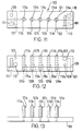
- Fig. 11 is a top plan view of a multi-cavity mold for making a stent according to the invention;
- Fig. 12 is an elevational view of the mold of Fig. 11;
- Fig. 13 is an elevational view of the mold of Fig. 12 with the top mold halves removed; and
- Fig. 14 is a flowchart of a process for making a fibrin stent using the multi-cavity mold of Figs. 11 and 12.
- Referring to Fig. 1 of the accompanying drawings there is shown a stent produced according to the invention in place on a balloon catheter. A
catheter 10 has aballoon 15 upon which astent 20 has been placed, thestent 20 having adeformable metal portion 22 and afibrin coating 24 thereon. Fig. 2 shows analternative stent 30 in which afibrin film 32 has been affixed to the underlyingmetallic framework 34. - A preferred method of making a stent according to the present invention is as set forth in Figs. 4-10. A
stent 50 of the type disclosed in US-A-4886062 (Wiktor) is inserted into atube 55 which is preferably made from a rigid material and which has an inside diameter which is large enough to accommodate an unexpanded PTCA balloon but which is smaller than a fully inflated PTCA balloon. APTCA balloon 60 attached to acatheter 62 and inflation device (not shown) is inserted into thestent 50 andtube 55. Fibrinogen at a pH of about 6.5, suspended in a saline solution, and thrombin are inserted into thetube 55 around the deflatedballoon 60 andstent 50. The amount of thrombin added is not critical but preferably will polymerize the fibrinogen tofibrin 65 in about 5 minutes. After polymerization, the fibrin is allowed to crosslink for at least an hour, preferably several hours. Theballoon 60 is then placed in a second mold cavity (tube 56) of greater diameter thantube 55 and inflated to compress thefibrin 65 between theballoon 60 andtube 56. Theballoon 60 is then deflated and removed from thetube 56. The resultingfibrin stent 70 includes thestent 50 embedded in a very thin elastic film offibrin 65. Thefibrin stent 70 may then be removed from thetube 56 and washed in a buffered saline solution. - Further processing of the fibrin stent can also be undertaken to neutralize thrombin with PPACK or hirudin; to add anticoagulants such as heparin; to further facilitate crosslinking by incubation at body temperature in a biological buffer such as a solution of blood serum buffered by 4-(2-Hydroxyethyl)-1-piperazineethanesulfonic acid (HEPES); or to add plasticizers such as glycerol. The resulting fibrin stent can then be placed over a balloon, and secured onto the balloon by crimping. The stent can then be delivered transluminally and expanded into place in the body lumen by conventional procedures.
- Preferably, heparin is incorporated into the stent prior to implantation in an amount effective to prevent or limit thrombosis. For example, the fibrin stent can be immersed in a solution of heparin within 10-30 minutes prior to implantation. The heparin immersion procedure can be conducted in a heparin solution having a concentration of 1000-25000 heparin units/ml. It may also be desirable to incorporate heparin into the fibrin matrix before it is completely polymerized. For example, after the fibrinogen and thrombin have been combined and the resulting fibrin has been shaped but within two hours of combining the fibrinogen and thrombin, the fibrin is immersed in a solution of heparin. Since the fibrin polymerization is largely complete at this point, the fibrin can be immersed in heparin solution containing up to about 20000 units/ml of heparin without damaging the integrity of the fibrin structure. Immersion times will depend on the concentration of the heparin solution and the concentration of heparin desired in the fibrin. However, preferably, in a solution of heparin having a concentration of 10000-20000 units/ml of heparin, an immersion time of 12-24 hours may be used. In yet another method for incorporation of heparin in the fibrin, the heparin can be included in the fibrinogen or in the initial mixture of fibrinogen and thrombin so long as the ratio of heparin to fibrinogen is such that the presence of the heparin does not lead to a weak fibrin film. Typically, less than 50-500 units of heparin can be used in a stent which includes 0.003-0.006 grams of fibrin. In yet another method for incorporating heparin into the fibrin, powdered heparin can be dusted onto the stent during the polymerization process and additional thrombin and fibrinogen can then be applied as a coating over the heparin.
- The metal stent portion mentioned above may be eliminated to make a fibrin tube which can be placed on a balloon catheter and expanded into place in a body lumen. The absence of permanently implanted metal elements would allow the entire stent to biodegrade as healing is completed in the body lumen. In order to achieve sufficient structural support for a stent without a metal structure, it may be desirable to form supporting elements from elastin or elastin/fibrin/collagen/fibronectin as replacements for the metal supporting elements. If desired, fibrin glue or fibrinogen can also be applied to the exterior of the fibrin tube immediately prior to placing it into the blood vessel in order to improve its adhesion to the vessel wall.
- In yet another method for making the fibrin stent, the fibrin can be polymerized in a multi-cavity mold such as that shown in Figs. 11 and 12. The
mold 100 is a three piece mold consisting of first and second mold halves 101, 102 andmold base 103. A series of pins 105-108 and screws 110-113 secure the mold pieces 101-103 together. As assembled, the mold halves 101, 102 define fivemold cavities 115a-e. Centrally located within each of themold cavities 115a-e is acorresponding pin 117a-e which is retained in themold base 103. In themold base 103 is a series of laterally extending air passageways 120a-e which communicate with thecavities 115a-e to allow complete filling of the mold cavity. The molding surfaces are coated with a polymeric slip coating such as PTFE to permit the piece parts to be removed from the mold cavities after curing. Fig. 13 shows themold base 103 of Fig. 12 after the mold halves 101, 102 have been removed following the molding operation.Pins 117a-e are shown surrounded by the moldedfibrin 121a-e. - Now referring also to Fig. 14, in operation, a
stent 125 is placed into themold 100 into one of themold cavities 115a such that thepin 117a occupies the hollow center of thestent 125. A fibrinogen mixture withthrombin 130 is made by metering the fibrinogen solution and thrombin solution into a sterile syringe and then moving the plunger of the syringe to mix the solutions. For example, 0.5 ml of a fibrinogen solution having a concentration of 26 mg/ml can be mixed with 0.125 ml of a thrombin solution having a concentration of 12 NIH units/ml. Themixture 130 is then injected into the bottom of thecavity 115a of themold 100 to fill thecavity 115a and encompass thestent 125. Themixture 130 is then allowed to cure. With the mixture indicated above, the curing interval should be at least two hours. Once themixture 130 has clotted, sterile water may be applied by spraying onto themold 100 to prevent the fibrin from drying out. When cured, the moldedpreform 140 comprising thestent 125 and the curedmixture 130 is removed from themold 100 by removing themold base 103 and pulling the moldedpreform 140 from thepin 117a. Since the pin 117 is coated with PTFE, the curedmixture 130 does not adhere to thepin 117a and the moldedpreform 140 can be removed by carefully pushing the preform from the bottom ofpin 117a using a plastic tweezers. If required, excess fibrin can be trimmed from thepreform 140 at this point or trimming can take place at a later stage of processing where the fibrin is stronger. Thepreform 140 can then be further crosslinked by treating it in abuffer solution 145 which may optionally contain crosslinking agents such as Factor XIIIa. For example, thebuffer 145 could be a tris buffer with a pH of 7.4 with thepreform 140 immersed in the tris buffer for at least five hours. Preferably, a solution ofheparin 135 is also included in themixture 130. Themixture 130 withheparin 135 is then injected into thecavity 115a of themold 100 and allowed to cure. Alternatively, immediately after thepreform 140 is removed from themold 100, thepreform 140 can be immersed in a heparin solution. After crosslinking, thepreform 140 undergoes an additional molding stage in a cavity of asecond mold 150 in whichpressure 155 is applied to provide the final form of thefibrin stent 160. For example, the mold can simply be a polycarbonate tube and thepreform 140 can be placed over the balloon of a balloon catheter and into the tube. The balloon is then slowly inflated causing thepreform 140 to be pressed against the sides of the tube. The effect of the expansion and pressure on the fibrin is to stretch it and thin it because of the viscoelastic properties of the fibrin. Because fibrin is such a fragile material, it is important to control the expansion by slow expansion to prevent the fibrin from tearing and also to provide a stent with the proper dimensions for expansion in vivo without tearing. For example, thepreform 140 may have an internal diameter of about 2.7 mm and may be placed on a 3.5 mm balloon and into amold 150 having a 3.4 mm internal diameter. The balloon can then be expanded slowly at one atmosphere increments until a pressure of about six atmospheres is achieved.Pressure 155 is then typically maintained on thefibrin stent 160 inside thesecond mold 150 for a short period of time in order to set thefibrin stent 160 into its final shape. Typically thirty minutes at six atmospheres of pressure is sufficient. Upon release of pressure in the balloon, the balloon andfibrin stent 160 can be withdrawn from themold 150. If thefibrin stent 160 is to be packaged and shipped dry, it can then be dehydrated 170 by well known methods such as air drying, ethanol dehydration or lyophilization and packaged 180 for storage and use. Typically, after packaging 180 , thefibrin stent 160 is sterilized 190 by gamma or electron beam sterilization. It will be readily appreciated that a fibrin stent with an attached metallic framework can be readily provided by this molding method. - Sterilization of the fibrin stent can be accomplished by starting with sterile, virus-free materials and manufacturing the device under sterile processing conditions. The sterile processing conditions include manufacturing the device under standard clean room conditions and ending the manufacturing process with a final sterilization step. The final sterilization step would expose the packaged device to radiation, preferably gamma radiation, at a level sufficient to cause disruption of microorganism DNA. This can be accomplished at an approximately 2.5 MRad gamma ray dosage. A suitable gamma ray source can be e.g. cobalt-60 or cesium-137. Another suitable form of radiation can be electron beam radiation. The packaged device configuration at irradiation can be either dry or wet i.e. with the fibrin stent in the package in a dehydrated state or in a wet pack package where the fibrin is maintained in a 100% relative humidity environment until end use.
Claims (11)
- A method for making an intraluminal stent, comprising the steps of:(a) forming a fibrin stent preform in a first mold cavity by polymerizing fibrinogen in said cavity;(b) placing said preform into a second mold cavity having a molding surface; and(c) compressing said preform against the molding surface of the second mold cavity.
- A method as claimed in claim 1 comprising the steps of:(a) polymerizing fibrinogen into a fibrin stent preform in said first mold cavity;(b) placing a balloon into said preform;(c) placing said balloon and preform into said second mold cavity; and(c) compressing said preform against the molding surface of said second mold cavity by expanding the balloon.
- A method as claimed in claim 1 or claim 2 comprising the steps of:(a) placing a stent framework into said first mold cavity;(b) injecting fibrinogen and a fibrinogen-coagulating protein into said first mold cavity around the stent framework and allowing the fibrinogen to cure in said first mold cavity, thereby producing a fibrin stent preform comprising fibrin and said stent framework;(c) placing said preform into said second mold cavity; and(d) compressing said preform against the molding surface of said second mold cavity.
- A method as claimed in either of claims 1 and 2 wherein step (a) comprises placing a mixture of fibrinogen and a fibrinogen-coagulating protein into said first mold cavity and curing the mixture to produce said preform.
- A method as claimed in either of claims 3 and 4 wherein water is applied to said stent preform as the fibrinogen cures.
- A method as claimed in any one of claims 1 to 5 further comprising applying a buffer solution to said preform.
- A method as claimed in claim 6 wherein said buffer solution comprises a crosslinking agent.
- A method as claimed in any one of claims 1 to 7 further comprising applying heparin to said preform.
- A method as claimed in any one of claims 1 to 8 wherein compression of said preform in said second mold cavity includes a slow outward expansion of said preform.
- A method as claimed in any one of claims 1 to 9 further comprising the step of mounting the compressed preform onto a balloon catheter balloon.
- A method as claimed in any one of claims 1 to 10 further comprising the step of applying a sterilizing dose of radiation to the compressed preform.
Priority Applications (1)
| Application Number | Priority Date | Filing Date | Title |
|---|---|---|---|
| EP02000867A EP1208818A3 (en) | 1994-09-15 | 1995-09-15 | Mold for making an intralumimal stent |
Applications Claiming Priority (2)
| Application Number | Priority Date | Filing Date | Title |
|---|---|---|---|
| US306806 | 1994-09-15 | ||
| US08/306,806 US5510077A (en) | 1992-03-19 | 1994-09-15 | Method of making an intraluminal stent |
Related Child Applications (1)
| Application Number | Title | Priority Date | Filing Date |
|---|---|---|---|
| EP02000867A Division EP1208818A3 (en) | 1994-09-15 | 1995-09-15 | Mold for making an intralumimal stent |
Publications (2)
| Publication Number | Publication Date |
|---|---|
| EP0701801A1 true EP0701801A1 (en) | 1996-03-20 |
| EP0701801B1 EP0701801B1 (en) | 2002-08-28 |
Family
ID=23186932
Family Applications (2)
| Application Number | Title | Priority Date | Filing Date |
|---|---|---|---|
| EP95306528A Expired - Lifetime EP0701801B1 (en) | 1994-09-15 | 1995-09-15 | Method for making an intraluminal stent |
| EP02000867A Withdrawn EP1208818A3 (en) | 1994-09-15 | 1995-09-15 | Mold for making an intralumimal stent |
Family Applications After (1)
| Application Number | Title | Priority Date | Filing Date |
|---|---|---|---|
| EP02000867A Withdrawn EP1208818A3 (en) | 1994-09-15 | 1995-09-15 | Mold for making an intralumimal stent |
Country Status (4)
| Country | Link |
|---|---|
| US (1) | US5510077A (en) |
| EP (2) | EP0701801B1 (en) |
| JP (1) | JPH0889584A (en) |
| DE (1) | DE69527899T2 (en) |
Cited By (4)
| Publication number | Priority date | Publication date | Assignee | Title |
|---|---|---|---|---|
| EP0841040A1 (en) * | 1996-11-08 | 1998-05-13 | Medtronic, Inc. | Therapeutic intraluminal stents |
| WO2009014776A1 (en) * | 2007-07-23 | 2009-01-29 | Carnegie Mellon University | Methods and apparatus for manufacturing plasma based plastics and bioplastics produced therefrom |
| US8215149B1 (en) | 2004-07-26 | 2012-07-10 | Abbott Laboratories | Stent crimping system and method |
| CN104203151A (en) * | 2012-02-14 | 2014-12-10 | 尼奥格拉夫特科技公司 | Kink resistant graft devices and related systems and methods |
Families Citing this family (138)
| Publication number | Priority date | Publication date | Assignee | Title |
|---|---|---|---|---|
| US5811447A (en) | 1993-01-28 | 1998-09-22 | Neorx Corporation | Therapeutic inhibitor of vascular smooth muscle cells |
| US6515009B1 (en) | 1991-09-27 | 2003-02-04 | Neorx Corporation | Therapeutic inhibitor of vascular smooth muscle cells |
| US20020055710A1 (en) * | 1998-04-30 | 2002-05-09 | Ronald J. Tuch | Medical device for delivering a therapeutic agent and method of preparation |
| US5464650A (en) * | 1993-04-26 | 1995-11-07 | Medtronic, Inc. | Intravascular stent and method |
| US5660873A (en) * | 1994-09-09 | 1997-08-26 | Bioseal, Limited Liability Corporaton | Coating intraluminal stents |
| US6099562A (en) * | 1996-06-13 | 2000-08-08 | Schneider (Usa) Inc. | Drug coating with topcoat |
| US5820917A (en) * | 1995-06-07 | 1998-10-13 | Medtronic, Inc. | Blood-contacting medical device and method |
| US8728143B2 (en) * | 1996-06-06 | 2014-05-20 | Biosensors International Group, Ltd. | Endoprosthesis deployment system for treating vascular bifurcations |
| US7686846B2 (en) * | 1996-06-06 | 2010-03-30 | Devax, Inc. | Bifurcation stent and method of positioning in a body lumen |
| US7238197B2 (en) * | 2000-05-30 | 2007-07-03 | Devax, Inc. | Endoprosthesis deployment system for treating vascular bifurcations |
| US6511477B2 (en) * | 1997-03-13 | 2003-01-28 | Biocardia, Inc. | Method of drug delivery to interstitial regions of the myocardium |
| US6273913B1 (en) | 1997-04-18 | 2001-08-14 | Cordis Corporation | Modified stent useful for delivery of drugs along stent strut |
| US6106454A (en) * | 1997-06-17 | 2000-08-22 | Medtronic, Inc. | Medical device for delivering localized radiation |
| US6203536B1 (en) * | 1997-06-17 | 2001-03-20 | Medtronic, Inc. | Medical device for delivering a therapeutic substance and method therefor |
| US6775574B1 (en) | 1997-11-07 | 2004-08-10 | Medtronic, Inc. | Method and system for myocardial infarction repair |
| US7031775B2 (en) * | 1997-11-07 | 2006-04-18 | Medtronic, Inc. | Method and system for myocardial infarction repair |
| US6151525A (en) | 1997-11-07 | 2000-11-21 | Medtronic, Inc. | Method and system for myocardial identifier repair |
| US6241762B1 (en) | 1998-03-30 | 2001-06-05 | Conor Medsystems, Inc. | Expandable medical device with ductile hinges |
| US7208010B2 (en) | 2000-10-16 | 2007-04-24 | Conor Medsystems, Inc. | Expandable medical device for delivery of beneficial agent |
| US8029561B1 (en) | 2000-05-12 | 2011-10-04 | Cordis Corporation | Drug combination useful for prevention of restenosis |
| US20070087028A1 (en) * | 1998-04-16 | 2007-04-19 | Robert Falotico | Intraluminal devices for the prevention and treatment of vascular disease |
| US7658727B1 (en) | 1998-04-20 | 2010-02-09 | Medtronic, Inc | Implantable medical device with enhanced biocompatibility and biostability |
| US6013099A (en) | 1998-04-29 | 2000-01-11 | Medtronic, Inc. | Medical device for delivering a water-insoluble therapeutic salt or substance |
| US6206914B1 (en) | 1998-04-30 | 2001-03-27 | Medtronic, Inc. | Implantable system with drug-eluting cells for on-demand local drug delivery |
| JP2002523136A (en) | 1998-08-21 | 2002-07-30 | プロビデンス ヘルス システム−オレゴン | Insertable stent and method of making and using the stent |
| US7662409B2 (en) * | 1998-09-25 | 2010-02-16 | Gel-Del Technologies, Inc. | Protein matrix materials, devices and methods of making and using thereof |
| US20030007991A1 (en) * | 1998-09-25 | 2003-01-09 | Masters David B. | Devices including protein matrix materials and methods of making and using thereof |
| US20070219642A1 (en) * | 1998-12-03 | 2007-09-20 | Jacob Richter | Hybrid stent having a fiber or wire backbone |
| US20060178727A1 (en) * | 1998-12-03 | 2006-08-10 | Jacob Richter | Hybrid amorphous metal alloy stent |
| US20050033399A1 (en) * | 1998-12-03 | 2005-02-10 | Jacob Richter | Hybrid stent |
| US8382821B2 (en) * | 1998-12-03 | 2013-02-26 | Medinol Ltd. | Helical hybrid stent |
| US20060122691A1 (en) * | 1998-12-03 | 2006-06-08 | Jacob Richter | Hybrid stent |
| US6620170B1 (en) * | 1999-04-26 | 2003-09-16 | C. R. Bard, Inc. | Devices and methods for treating ischemia by creating a fibrin plug |
| US6383171B1 (en) | 1999-10-12 | 2002-05-07 | Allan Will | Methods and devices for protecting a passageway in a body when advancing devices through the passageway |
| US8920487B1 (en) | 2000-03-01 | 2014-12-30 | Medinol Ltd. | Longitudinally flexible stent |
| US8202312B2 (en) * | 2000-03-01 | 2012-06-19 | Medinol Ltd. | Longitudinally flexible stent |
| US8496699B2 (en) * | 2000-03-01 | 2013-07-30 | Medinol Ltd. | Longitudinally flexible stent |
| US6723119B2 (en) | 2000-03-01 | 2004-04-20 | Medinol Ltd. | Longitudinally flexible stent |
| US7828835B2 (en) | 2000-03-01 | 2010-11-09 | Medinol Ltd. | Longitudinally flexible stent |
| US7141062B1 (en) * | 2000-03-01 | 2006-11-28 | Medinol, Ltd. | Longitudinally flexible stent |
| SG86458A1 (en) | 2000-03-01 | 2002-02-19 | Medinol Ltd | Longitudinally flexible stent |
| US7621947B2 (en) | 2000-03-01 | 2009-11-24 | Medinol, Ltd. | Longitudinally flexible stent |
| US7758627B2 (en) * | 2000-03-01 | 2010-07-20 | Medinol, Ltd. | Longitudinally flexible stent |
| US8088060B2 (en) | 2000-03-15 | 2012-01-03 | Orbusneich Medical, Inc. | Progenitor endothelial cell capturing with a drug eluting implantable medical device |
| US9522217B2 (en) | 2000-03-15 | 2016-12-20 | Orbusneich Medical, Inc. | Medical device with coating for capturing genetically-altered cells and methods for using same |
| US6736838B1 (en) | 2000-03-22 | 2004-05-18 | Zuli Holdings Ltd. | Method and apparatus for covering a stent |
| US6776796B2 (en) | 2000-05-12 | 2004-08-17 | Cordis Corportation | Antiinflammatory drug and delivery device |
| US8236048B2 (en) * | 2000-05-12 | 2012-08-07 | Cordis Corporation | Drug/drug delivery systems for the prevention and treatment of vascular disease |
| US20040243097A1 (en) * | 2000-05-12 | 2004-12-02 | Robert Falotico | Antiproliferative drug and delivery device |
| US20030139803A1 (en) * | 2000-05-30 | 2003-07-24 | Jacques Sequin | Method of stenting a vessel with stent lumenal diameter increasing distally |
| IL137090A (en) * | 2000-06-29 | 2010-04-15 | Pentech Medical Devices Ltd | Polymeric stent |
| US20060222756A1 (en) * | 2000-09-29 | 2006-10-05 | Cordis Corporation | Medical devices, drug coatings and methods of maintaining the drug coatings thereon |
| WO2002026117A2 (en) * | 2000-09-29 | 2002-04-04 | New Health Sciences, Inc. | Systems and methods for assessing vascular health |
| CA2424029C (en) | 2000-09-29 | 2008-01-29 | Cordis Corporation | Coated medical devices |
| US20020111590A1 (en) * | 2000-09-29 | 2002-08-15 | Davila Luis A. | Medical devices, drug coatings and methods for maintaining the drug coatings thereon |
| US7604599B2 (en) * | 2000-09-29 | 2009-10-20 | New Health Sciences, Inc. | Systems and methods for using dynamic vascular assessment to improve vascular stent placement, application, design and marketing |
| US20020051730A1 (en) * | 2000-09-29 | 2002-05-02 | Stanko Bodnar | Coated medical devices and sterilization thereof |
| DE20122506U1 (en) | 2000-10-16 | 2005-12-08 | Conor Medsystems, Inc., Menlo Park | Expandable medical device for delivering a beneficial agent |
| US20020082620A1 (en) * | 2000-12-27 | 2002-06-27 | Elaine Lee | Bioactive materials for aneurysm repair |
| US8182527B2 (en) * | 2001-05-07 | 2012-05-22 | Cordis Corporation | Heparin barrier coating for controlled drug release |
| US6656506B1 (en) * | 2001-05-09 | 2003-12-02 | Advanced Cardiovascular Systems, Inc. | Microparticle coated medical device |
| DE10130968B4 (en) * | 2001-06-27 | 2009-08-20 | Envisiontec Gmbh | Coated polymeric material, its use and process for its preparation |
| US7842083B2 (en) | 2001-08-20 | 2010-11-30 | Innovational Holdings, Llc. | Expandable medical device with improved spatial distribution |
| US7135189B2 (en) * | 2001-08-23 | 2006-11-14 | Boston Scientific Scimed, Inc. | Compositions and techniques for localized therapy |
| US7195640B2 (en) * | 2001-09-25 | 2007-03-27 | Cordis Corporation | Coated medical devices for the treatment of vulnerable plaque |
| US7108701B2 (en) * | 2001-09-28 | 2006-09-19 | Ethicon, Inc. | Drug releasing anastomosis devices and methods for treating anastomotic sites |
| US20030065382A1 (en) * | 2001-10-02 | 2003-04-03 | Fischell Robert E. | Means and method for the treatment of coronary artery obstructions |
| EP1451306A1 (en) * | 2001-11-06 | 2004-09-01 | Medtronic, Inc. | Method and system for myocardial infarction repair |
| US20030175410A1 (en) * | 2002-03-18 | 2003-09-18 | Campbell Phil G. | Method and apparatus for preparing biomimetic scaffold |
| US8529956B2 (en) | 2002-03-18 | 2013-09-10 | Carnell Therapeutics Corporation | Methods and apparatus for manufacturing plasma based plastics and bioplastics produced therefrom |
| US20100254900A1 (en) * | 2002-03-18 | 2010-10-07 | Campbell Phil G | Biocompatible polymers and Methods of use |
| US7261734B2 (en) * | 2002-04-23 | 2007-08-28 | Boston Scientific Scimed, Inc. | Resorption-controllable medical implants |
| WO2003092468A2 (en) * | 2002-04-29 | 2003-11-13 | Gel-Del Technologies, Inc. | Biomatrix structural containment and fixation systems and methods of use thereof |
| NZ536331A (en) * | 2002-05-09 | 2007-08-31 | Hemoteq Ag | Compounds and method for coating surfaces of medical devices such as stents in a haemocompatible manner |
| JP3887588B2 (en) * | 2002-08-30 | 2007-02-28 | 株式会社リガク | Stress measurement method by X-ray diffraction |
| DE60231843D1 (en) | 2002-11-08 | 2009-05-14 | Jacques Seguin | ENDOPROTHESIS FOR VESSEL FORKING |
| US20040236415A1 (en) * | 2003-01-02 | 2004-11-25 | Richard Thomas | Medical devices having drug releasing polymer reservoirs |
| US20040243224A1 (en) * | 2003-04-03 | 2004-12-02 | Medtronic Vascular, Inc. | Methods and compositions for inhibiting narrowing in mammalian vascular pathways |
| US8465537B2 (en) * | 2003-06-17 | 2013-06-18 | Gel-Del Technologies, Inc. | Encapsulated or coated stent systems |
| US9039755B2 (en) | 2003-06-27 | 2015-05-26 | Medinol Ltd. | Helical hybrid stent |
| US9155639B2 (en) * | 2009-04-22 | 2015-10-13 | Medinol Ltd. | Helical hybrid stent |
| AU2004296851A1 (en) * | 2003-12-08 | 2005-06-23 | Gel-Del Technologies, Inc. | Mucoadhesive drug delivery devices and methods of making and using thereof |
| US20050154455A1 (en) * | 2003-12-18 | 2005-07-14 | Medtronic Vascular, Inc. | Medical devices to treat or inhibit restenosis |
| US20050149174A1 (en) * | 2003-12-18 | 2005-07-07 | Medtronic Vascular, Inc. | Medical devices to treat or inhibit restenosis |
| US20050154451A1 (en) * | 2003-12-18 | 2005-07-14 | Medtronic Vascular, Inc. | Medical devices to treat or inhibit restenosis |
| US20050137683A1 (en) * | 2003-12-19 | 2005-06-23 | Medtronic Vascular, Inc. | Medical devices to treat or inhibit restenosis |
| US20050152942A1 (en) * | 2003-12-23 | 2005-07-14 | Medtronic Vascular, Inc. | Medical devices to treat or inhibit restenosis |
| US20050152940A1 (en) * | 2003-12-23 | 2005-07-14 | Medtronic Vascular, Inc. | Medical devices to treat or inhibit restenosis |
| US20050152943A1 (en) * | 2003-12-23 | 2005-07-14 | Medtronic Vascular, Inc. | Medical devices to treat or inhibit restenosis |
| US20050154452A1 (en) * | 2003-12-23 | 2005-07-14 | Medtronic Vascular, Inc. | Medical devices to treat or inhibit restenosis |
| US20050159809A1 (en) * | 2004-01-21 | 2005-07-21 | Medtronic Vascular, Inc. | Implantable medical devices for treating or preventing restenosis |
| US20050197691A1 (en) * | 2004-02-18 | 2005-09-08 | Medtronic Vascular, Inc. | Medical devices to treat or inhibit restenosis |
| US20050228490A1 (en) * | 2004-04-09 | 2005-10-13 | Medtronic Vascular, Inc. | Medical devices to treat or inhibit restenosis |
| US7654997B2 (en) * | 2004-04-21 | 2010-02-02 | Acclarent, Inc. | Devices, systems and methods for diagnosing and treating sinusitus and other disorders of the ears, nose and/or throat |
| DE102004022606A1 (en) * | 2004-05-07 | 2005-12-15 | Envisiontec Gmbh | Method for producing a three-dimensional object with improved separation of hardened material layers from a building level |
| DE102004022961B4 (en) * | 2004-05-10 | 2008-11-20 | Envisiontec Gmbh | Method for producing a three-dimensional object with resolution improvement by means of pixel shift |
| JP5184080B2 (en) | 2004-05-10 | 2013-04-17 | エンビジョンテク・ゲゼルシャフト・ミット・ベシュレンクテル・ハフツング | 3D object manufacturing process with resolution improvement by pixel shift |
| US20050261762A1 (en) * | 2004-05-21 | 2005-11-24 | Medtronic Vascular, Inc. | Medical devices to prevent or inhibit restenosis |
| WO2005118016A1 (en) * | 2004-05-27 | 2005-12-15 | Medtronic, Inc. | Medical device comprising a biologically active agent |
| US7763064B2 (en) | 2004-06-08 | 2010-07-27 | Medinol, Ltd. | Stent having struts with reverse direction curvature |
| US20060062822A1 (en) * | 2004-09-21 | 2006-03-23 | Medtronic Vascular, Inc. | Medical devices to treat or inhibit restenosis |
| US20060088571A1 (en) * | 2004-10-21 | 2006-04-27 | Medtronic Vascular, Inc. | Biocompatible and hemocompatible polymer compositions |
| US9554803B2 (en) | 2005-07-26 | 2017-01-31 | Ethicon Endo-Surgery, Llc | Electrically self-powered surgical instrument with manual release |
| US10314583B2 (en) | 2005-07-26 | 2019-06-11 | Ethicon Llc | Electrically self-powered surgical instrument with manual release |
| US11751873B2 (en) | 2005-07-26 | 2023-09-12 | Cilag Gmbh International | Electrically powered surgical instrument with manual release |
| US8627995B2 (en) | 2006-05-19 | 2014-01-14 | Ethicon Endo-Sugery, Inc. | Electrically self-powered surgical instrument with cryptographic identification of interchangeable part |
| US20070027530A1 (en) * | 2005-07-26 | 2007-02-01 | Medtronic Vascular, Inc. | Intraluminal device, catheter assembly, and method of use thereof |
| US8573462B2 (en) | 2006-05-19 | 2013-11-05 | Ethicon Endo-Surgery, Inc. | Electrical surgical instrument with optimized power supply and drive |
| US9662116B2 (en) | 2006-05-19 | 2017-05-30 | Ethicon, Llc | Electrically self-powered surgical instrument with cryptographic identification of interchangeable part |
| US20070067020A1 (en) * | 2005-09-22 | 2007-03-22 | Medtronic Vasular, Inc. | Intraluminal stent, delivery system, and a method of treating a vascular condition |
| US8876763B2 (en) * | 2005-11-01 | 2014-11-04 | Boston Scientific Scimed, Inc. | Composite balloon |
| US20070231361A1 (en) * | 2006-03-28 | 2007-10-04 | Medtronic Vascular, Inc. | Use of Fatty Acids to Inhibit the Growth of Aneurysms |
| DE102006019963B4 (en) * | 2006-04-28 | 2023-12-07 | Envisiontec Gmbh | Device and method for producing a three-dimensional object by layer-by-layer solidifying a material that can be solidified under the influence of electromagnetic radiation using mask exposure |
| DE102006019964C5 (en) * | 2006-04-28 | 2021-08-26 | Envisiontec Gmbh | Device and method for producing a three-dimensional object by means of mask exposure |
| WO2007148201A2 (en) * | 2006-06-19 | 2007-12-27 | Arterial Remodeling Technologies, S.A. | Improved stent manufacturing methods |
| US20080001330A1 (en) * | 2006-06-28 | 2008-01-03 | Bin Huang | Fabricating polymer stents with injection molding |
| US7998404B2 (en) | 2006-07-13 | 2011-08-16 | Advanced Cardiovascular Systems, Inc. | Reduced temperature sterilization of stents |
| US7636610B2 (en) * | 2006-07-19 | 2009-12-22 | Envisiontec Gmbh | Method and device for producing a three-dimensional object, and computer and data carrier useful therefor |
| US8952123B1 (en) * | 2006-08-02 | 2015-02-10 | Abbott Cardiovascular Systems Inc. | Dioxanone-based copolymers for implantable devices |
| US20080085293A1 (en) * | 2006-08-22 | 2008-04-10 | Jenchen Yang | Drug eluting stent and therapeutic methods using c-Jun N-terminal kinase inhibitor |
| US8529959B2 (en) | 2006-10-17 | 2013-09-10 | Carmell Therapeutics Corporation | Methods and apparatus for manufacturing plasma based plastics and bioplastics produced therefrom |
| US7892474B2 (en) * | 2006-11-15 | 2011-02-22 | Envisiontec Gmbh | Continuous generative process for producing a three-dimensional object |
| US8003039B2 (en) | 2007-01-17 | 2011-08-23 | 3D Systems, Inc. | Method for tilting solid image build platform for reducing air entrainment and for build release |
| EP2011631B1 (en) | 2007-07-04 | 2012-04-18 | Envisiontec GmbH | Process and device for producing a three-dimensional object |
| US8821861B2 (en) * | 2007-10-05 | 2014-09-02 | The Board Of Trustees Of The University Of Illinois | Fibrin sealant |
| DK2052693T4 (en) | 2007-10-26 | 2021-03-15 | Envisiontec Gmbh | Process and free-form manufacturing system to produce a three-dimensional object |
| CA2711001A1 (en) * | 2007-12-26 | 2009-07-09 | Gel-Del Technologies, Inc. | Biocompatible protein-based particles and methods thereof |
| KR20110056539A (en) * | 2008-09-10 | 2011-05-30 | 이브이쓰리 인크. | Stents and catheters having improved stent deployment |
| WO2011035020A1 (en) * | 2009-09-18 | 2011-03-24 | Bioinspire Technologies, Inc. | Free-standing biodegradable patch |
| US9271925B2 (en) | 2013-03-11 | 2016-03-01 | Bioinspire Technologies, Inc. | Multi-layer biodegradable device having adjustable drug release profile |
| US8372330B2 (en) | 2009-10-19 | 2013-02-12 | Global Filtration Systems | Resin solidification substrate and assembly |
| US20140081659A1 (en) | 2012-09-17 | 2014-03-20 | Depuy Orthopaedics, Inc. | Systems and methods for surgical and interventional planning, support, post-operative follow-up, and functional recovery tracking |
| WO2014077854A1 (en) | 2012-11-19 | 2014-05-22 | Washington State University Research Foundation | Nanocrystalline cellulose materials and methods for their preparation |
| US9527244B2 (en) | 2014-02-10 | 2016-12-27 | Global Filtration Systems | Apparatus and method for forming three-dimensional objects from solidifiable paste |
| CN104552727B (en) * | 2014-11-07 | 2016-11-30 | 威海云阳碳素科技有限公司 | A kind of preparation method of carbon fiber product |
| DE102015111126A1 (en) * | 2015-07-09 | 2017-01-12 | Medizinische Hochschule Hannover | A method for producing a bioartificial, primary acellular fibrin-based construct and this construct itself |
| US10501715B1 (en) | 2015-09-11 | 2019-12-10 | Mark H. Widick | System for the formation of fibrin foam |
| US10737479B2 (en) | 2017-01-12 | 2020-08-11 | Global Filtration Systems | Method of making three-dimensional objects using both continuous and discontinuous solidification |
Citations (4)
| Publication number | Priority date | Publication date | Assignee | Title |
|---|---|---|---|---|
| US3523807A (en) * | 1966-11-25 | 1970-08-11 | Mihaly Gerendas | Method of making a cross-linked fibrin prosthesis |
| US3723244A (en) * | 1971-01-18 | 1973-03-27 | Atomic Energy Commission | Fibrous fibrin sheet and method for producing same |
| US3918099A (en) * | 1973-05-29 | 1975-11-11 | Jurgen Fuhr | Artificial organ or part or section thereof for implanation into the human body and method of producing the same |
| EP0566245A1 (en) * | 1992-03-19 | 1993-10-20 | Medtronic, Inc. | Intraluminal stent |
Family Cites Families (27)
| Publication number | Priority date | Publication date | Assignee | Title |
|---|---|---|---|---|
| CH305554A (en) * | 1952-01-30 | 1955-02-28 | Safco Trevoux | Electric capacitor. |
| US4622244A (en) * | 1979-09-04 | 1986-11-11 | The Washington University | Process for preparation of microcapsules |
| US4464317A (en) * | 1980-01-28 | 1984-08-07 | The Washington University | Method of encapsulating active agents with inorganic coatings |
| US4389330A (en) * | 1980-10-06 | 1983-06-21 | Stolle Research And Development Corporation | Microencapsulation process |
| US4675189A (en) * | 1980-11-18 | 1987-06-23 | Syntex (U.S.A.) Inc. | Microencapsulation of water soluble active polypeptides |
| FR2502539A1 (en) * | 1981-03-25 | 1982-10-01 | Seb Sa | Silicone rubber moulds for making cast sticks of wax - to facilitate ejection of elongated profiles |
| US4542025A (en) * | 1982-07-29 | 1985-09-17 | The Stolle Research And Development Corporation | Injectable, long-acting microparticle formulation for the delivery of anti-inflammatory agents |
| US4530840A (en) * | 1982-07-29 | 1985-07-23 | The Stolle Research And Development Corporation | Injectable, long-acting microparticle formulation for the delivery of anti-inflammatory agents |
| US4540573A (en) * | 1983-07-14 | 1985-09-10 | New York Blood Center, Inc. | Undenatured virus-free biologically active protein derivatives |
| US4548736A (en) * | 1983-08-29 | 1985-10-22 | Wisconsin Alumni Research Foundation | Preparation of protein films |
| US5223420A (en) * | 1985-03-01 | 1993-06-29 | Institut National De La Sante Et De La Recherche Medicale | Elastin-based product, a procedure for its preparation and its biological applications; in particular as biomaterials and artificial supports |
| US4600652A (en) * | 1985-04-01 | 1986-07-15 | Warner-Lambert Company | Permanently bonded antithrombogenic polyurethane surface |
| US4642242A (en) * | 1985-04-01 | 1987-02-10 | Becton, Dickinson And Company | Permanently bonded antithrombogenic polyurethane surface |
| DE3519011A1 (en) * | 1985-05-25 | 1986-11-27 | Behringwerke Ag, 3550 Marburg | METHOD FOR PRODUCING A MATERIAL FOR AFFINITY CHROMATOGRAPHY |
| US4720512A (en) * | 1986-03-24 | 1988-01-19 | Becton, Dickinson And Company | Polymeric articles having enhanced antithrombogenic activity |
| JPS63151353A (en) * | 1986-12-15 | 1988-06-23 | Oogawara Kakoki Kk | Preparation of wax-coated microcapsule |
| US4897268A (en) * | 1987-08-03 | 1990-01-30 | Southern Research Institute | Drug delivery system and method of making the same |
| US4886062A (en) * | 1987-10-19 | 1989-12-12 | Medtronic, Inc. | Intravascular radially expandable stent and method of implant |
| US4824628A (en) * | 1988-01-20 | 1989-04-25 | General Motors Corporation | Method of making an epoxy mold |
| CA1322628C (en) * | 1988-10-04 | 1993-10-05 | Richard A. Schatz | Expandable intraluminal graft |
| DE68923423T2 (en) * | 1988-10-28 | 1996-01-25 | Terumo Corp | Antithromotic medical material, artificial internal organ and method for producing an antithrombotic medical material. |
| WO1990013332A1 (en) * | 1989-05-11 | 1990-11-15 | Cedars-Sinai Medical Center | Stent with sustained drug delivery |
| US5141516A (en) * | 1989-07-26 | 1992-08-25 | Detweiler Mark B | Dissolvable anastomosis stent and method for using the same |
| CA2049973C (en) * | 1990-02-28 | 2002-12-24 | Rodney G. Wolff | Intralumenal drug eluting prosthesis |
| US5092841A (en) * | 1990-05-17 | 1992-03-03 | Wayne State University | Method for treating an arterial wall injured during angioplasty |
| US5246451A (en) * | 1991-04-30 | 1993-09-21 | Medtronic, Inc. | Vascular prosthesis and method |
| SE9101853D0 (en) * | 1991-06-17 | 1991-06-17 | Jonas Wadstroem | IMPROVED TISSUE ASHESIVE |
-
1994
- 1994-09-15 US US08/306,806 patent/US5510077A/en not_active Expired - Fee Related
-
1995
- 1995-09-15 EP EP95306528A patent/EP0701801B1/en not_active Expired - Lifetime
- 1995-09-15 EP EP02000867A patent/EP1208818A3/en not_active Withdrawn
- 1995-09-15 DE DE69527899T patent/DE69527899T2/en not_active Expired - Fee Related
- 1995-09-18 JP JP26197795A patent/JPH0889584A/en active Pending
Patent Citations (4)
| Publication number | Priority date | Publication date | Assignee | Title |
|---|---|---|---|---|
| US3523807A (en) * | 1966-11-25 | 1970-08-11 | Mihaly Gerendas | Method of making a cross-linked fibrin prosthesis |
| US3723244A (en) * | 1971-01-18 | 1973-03-27 | Atomic Energy Commission | Fibrous fibrin sheet and method for producing same |
| US3918099A (en) * | 1973-05-29 | 1975-11-11 | Jurgen Fuhr | Artificial organ or part or section thereof for implanation into the human body and method of producing the same |
| EP0566245A1 (en) * | 1992-03-19 | 1993-10-20 | Medtronic, Inc. | Intraluminal stent |
Cited By (6)
| Publication number | Priority date | Publication date | Assignee | Title |
|---|---|---|---|---|
| EP0841040A1 (en) * | 1996-11-08 | 1998-05-13 | Medtronic, Inc. | Therapeutic intraluminal stents |
| US5833651A (en) * | 1996-11-08 | 1998-11-10 | Medtronic, Inc. | Therapeutic intraluminal stents |
| US6228845B1 (en) | 1996-11-08 | 2001-05-08 | Medtronic, Inc. | Therapeutic intraluminal stents |
| US8215149B1 (en) | 2004-07-26 | 2012-07-10 | Abbott Laboratories | Stent crimping system and method |
| WO2009014776A1 (en) * | 2007-07-23 | 2009-01-29 | Carnegie Mellon University | Methods and apparatus for manufacturing plasma based plastics and bioplastics produced therefrom |
| CN104203151A (en) * | 2012-02-14 | 2014-12-10 | 尼奥格拉夫特科技公司 | Kink resistant graft devices and related systems and methods |
Also Published As
| Publication number | Publication date |
|---|---|
| DE69527899D1 (en) | 2002-10-02 |
| US5510077A (en) | 1996-04-23 |
| EP0701801B1 (en) | 2002-08-28 |
| EP1208818A2 (en) | 2002-05-29 |
| EP1208818A3 (en) | 2003-05-07 |
| JPH0889584A (en) | 1996-04-09 |
| DE69527899T2 (en) | 2003-03-20 |
Similar Documents
| Publication | Publication Date | Title |
|---|---|---|
| EP0701801B1 (en) | Method for making an intraluminal stent | |
| EP0701802B1 (en) | Drug eluting stent | |
| US5628785A (en) | Bioelastomeric stent | |
| US5554182A (en) | Method for preventing restenosis | |
| US6080190A (en) | Intraluminal stent | |
| Barbucci | Integrated biomaterials science | |
| US6187370B1 (en) | Medical device for delivering a therapeutic substance and method therefor | |
| EP0920342B1 (en) | Medical device for delivering a therapeutic substance | |
| US8048042B2 (en) | Medical articles incorporating surface capillary fiber | |
| EP1416946B1 (en) | Expandable foam-like biomaterials and methods | |
| EP0226061B1 (en) | High molecular weight bioresorbable polymers and implantation devices thereof | |
| US9364587B2 (en) | Bone regeneration using biodegradable polymeric nanocomposite materials and applications of the same | |
| US20020131933A1 (en) | Biopolymer membrane and methods for its preparation | |
| JP2001505114A (en) | Biodegradable polymer membrane | |
| CN105283207B (en) | Use the osteanagenesis and its application of degradable polymer based nano composite material | |
| CZ308556B6 (en) | Composite vascular replacement and manufacturing method | |
| Marconi et al. | Structure and properties of polymeric materials | |
| Crowley et al. | Biomaterials in Dentistry and Medicine | |
| Ducheyne | Garth W. Hastings, D. Sc., Ph. D., C. Chern., FRSC |
Legal Events
| Date | Code | Title | Description |
|---|---|---|---|
| PUAI | Public reference made under article 153(3) epc to a published international application that has entered the european phase |
Free format text: ORIGINAL CODE: 0009012 |
|
| AK | Designated contracting states |
Kind code of ref document: A1 Designated state(s): DE FR GB IT NL SE |
|
| 17P | Request for examination filed |
Effective date: 19960410 |
|
| 17Q | First examination report despatched |
Effective date: 19980928 |
|
| GRAG | Despatch of communication of intention to grant |
Free format text: ORIGINAL CODE: EPIDOS AGRA |
|
| GRAG | Despatch of communication of intention to grant |
Free format text: ORIGINAL CODE: EPIDOS AGRA |
|
| GRAH | Despatch of communication of intention to grant a patent |
Free format text: ORIGINAL CODE: EPIDOS IGRA |
|
| GRAH | Despatch of communication of intention to grant a patent |
Free format text: ORIGINAL CODE: EPIDOS IGRA |
|
| GRAA | (expected) grant |
Free format text: ORIGINAL CODE: 0009210 |
|
| AK | Designated contracting states |
Kind code of ref document: B1 Designated state(s): DE FR GB IT NL SE |
|
| PG25 | Lapsed in a contracting state [announced via postgrant information from national office to epo] |
Ref country code: NL Free format text: LAPSE BECAUSE OF FAILURE TO SUBMIT A TRANSLATION OF THE DESCRIPTION OR TO PAY THE FEE WITHIN THE PRESCRIBED TIME-LIMIT Effective date: 20020828 |
|
| REG | Reference to a national code |
Ref country code: GB Ref legal event code: FG4D |
|
| REF | Corresponds to: |
Ref document number: 69527899 Country of ref document: DE Date of ref document: 20021002 |
|
| PGFP | Annual fee paid to national office [announced via postgrant information from national office to epo] |
Ref country code: SE Payment date: 20021025 Year of fee payment: 8 |
|
| PGFP | Annual fee paid to national office [announced via postgrant information from national office to epo] |
Ref country code: NL Payment date: 20021029 Year of fee payment: 8 |
|
| PG25 | Lapsed in a contracting state [announced via postgrant information from national office to epo] |
Ref country code: SE Free format text: LAPSE BECAUSE OF FAILURE TO SUBMIT A TRANSLATION OF THE DESCRIPTION OR TO PAY THE FEE WITHIN THE PRESCRIBED TIME-LIMIT Effective date: 20021128 |
|
| ET | Fr: translation filed | ||
| NLV1 | Nl: lapsed or annulled due to failure to fulfill the requirements of art. 29p and 29m of the patents act | ||
| PLBE | No opposition filed within time limit |
Free format text: ORIGINAL CODE: 0009261 |
|
| STAA | Information on the status of an ep patent application or granted ep patent |
Free format text: STATUS: NO OPPOSITION FILED WITHIN TIME LIMIT |
|
| 26N | No opposition filed |
Effective date: 20030530 |
|
| PGFP | Annual fee paid to national office [announced via postgrant information from national office to epo] |
Ref country code: GB Payment date: 20060804 Year of fee payment: 12 |
|
| PGFP | Annual fee paid to national office [announced via postgrant information from national office to epo] |
Ref country code: FR Payment date: 20060906 Year of fee payment: 12 |
|
| PGFP | Annual fee paid to national office [announced via postgrant information from national office to epo] |
Ref country code: DE Payment date: 20060929 Year of fee payment: 12 |
|
| PGFP | Annual fee paid to national office [announced via postgrant information from national office to epo] |
Ref country code: IT Payment date: 20070913 Year of fee payment: 13 |
|
| GBPC | Gb: european patent ceased through non-payment of renewal fee |
Effective date: 20070915 |
|
| PG25 | Lapsed in a contracting state [announced via postgrant information from national office to epo] |
Ref country code: DE Free format text: LAPSE BECAUSE OF NON-PAYMENT OF DUE FEES Effective date: 20080401 |
|
| REG | Reference to a national code |
Ref country code: FR Ref legal event code: ST Effective date: 20080531 |
|
| PG25 | Lapsed in a contracting state [announced via postgrant information from national office to epo] |
Ref country code: FR Free format text: LAPSE BECAUSE OF NON-PAYMENT OF DUE FEES Effective date: 20071001 |
|
| PG25 | Lapsed in a contracting state [announced via postgrant information from national office to epo] |
Ref country code: GB Free format text: LAPSE BECAUSE OF NON-PAYMENT OF DUE FEES Effective date: 20070915 |
|
| PG25 | Lapsed in a contracting state [announced via postgrant information from national office to epo] |
Ref country code: IT Free format text: LAPSE BECAUSE OF NON-PAYMENT OF DUE FEES Effective date: 20080915 |