EP0701803A1 - Medical device for implantation into living bodies - Google Patents
Medical device for implantation into living bodies Download PDFInfo
- Publication number
- EP0701803A1 EP0701803A1 EP95100829A EP95100829A EP0701803A1 EP 0701803 A1 EP0701803 A1 EP 0701803A1 EP 95100829 A EP95100829 A EP 95100829A EP 95100829 A EP95100829 A EP 95100829A EP 0701803 A1 EP0701803 A1 EP 0701803A1
- Authority
- EP
- European Patent Office
- Prior art keywords
- type
- surface area
- roughness
- medical device
- implant
- Prior art date
- Legal status (The legal status is an assumption and is not a legal conclusion. Google has not performed a legal analysis and makes no representation as to the accuracy of the status listed.)
- Granted
Links
Images


Classifications
-
- A—HUMAN NECESSITIES
- A61—MEDICAL OR VETERINARY SCIENCE; HYGIENE
- A61F—FILTERS IMPLANTABLE INTO BLOOD VESSELS; PROSTHESES; DEVICES PROVIDING PATENCY TO, OR PREVENTING COLLAPSING OF, TUBULAR STRUCTURES OF THE BODY, e.g. STENTS; ORTHOPAEDIC, NURSING OR CONTRACEPTIVE DEVICES; FOMENTATION; TREATMENT OR PROTECTION OF EYES OR EARS; BANDAGES, DRESSINGS OR ABSORBENT PADS; FIRST-AID KITS
- A61F2/00—Filters implantable into blood vessels; Prostheses, i.e. artificial substitutes or replacements for parts of the body; Appliances for connecting them with the body; Devices providing patency to, or preventing collapsing of, tubular structures of the body, e.g. stents
- A61F2/02—Prostheses implantable into the body
- A61F2/30—Joints
- A61F2/30767—Special external or bone-contacting surface, e.g. coating for improving bone ingrowth
- A61F2/30771—Special external or bone-contacting surface, e.g. coating for improving bone ingrowth applied in original prostheses, e.g. holes or grooves
-
- A—HUMAN NECESSITIES
- A61—MEDICAL OR VETERINARY SCIENCE; HYGIENE
- A61B—DIAGNOSIS; SURGERY; IDENTIFICATION
- A61B17/00—Surgical instruments, devices or methods, e.g. tourniquets
- A61B17/56—Surgical instruments or methods for treatment of bones or joints; Devices specially adapted therefor
- A61B17/58—Surgical instruments or methods for treatment of bones or joints; Devices specially adapted therefor for osteosynthesis, e.g. bone plates, screws, setting implements or the like
-
- A—HUMAN NECESSITIES
- A61—MEDICAL OR VETERINARY SCIENCE; HYGIENE
- A61F—FILTERS IMPLANTABLE INTO BLOOD VESSELS; PROSTHESES; DEVICES PROVIDING PATENCY TO, OR PREVENTING COLLAPSING OF, TUBULAR STRUCTURES OF THE BODY, e.g. STENTS; ORTHOPAEDIC, NURSING OR CONTRACEPTIVE DEVICES; FOMENTATION; TREATMENT OR PROTECTION OF EYES OR EARS; BANDAGES, DRESSINGS OR ABSORBENT PADS; FIRST-AID KITS
- A61F2/00—Filters implantable into blood vessels; Prostheses, i.e. artificial substitutes or replacements for parts of the body; Appliances for connecting them with the body; Devices providing patency to, or preventing collapsing of, tubular structures of the body, e.g. stents
- A61F2/02—Prostheses implantable into the body
- A61F2/30—Joints
- A61F2/30767—Special external or bone-contacting surface, e.g. coating for improving bone ingrowth
-
- A—HUMAN NECESSITIES
- A61—MEDICAL OR VETERINARY SCIENCE; HYGIENE
- A61F—FILTERS IMPLANTABLE INTO BLOOD VESSELS; PROSTHESES; DEVICES PROVIDING PATENCY TO, OR PREVENTING COLLAPSING OF, TUBULAR STRUCTURES OF THE BODY, e.g. STENTS; ORTHOPAEDIC, NURSING OR CONTRACEPTIVE DEVICES; FOMENTATION; TREATMENT OR PROTECTION OF EYES OR EARS; BANDAGES, DRESSINGS OR ABSORBENT PADS; FIRST-AID KITS
- A61F2/00—Filters implantable into blood vessels; Prostheses, i.e. artificial substitutes or replacements for parts of the body; Appliances for connecting them with the body; Devices providing patency to, or preventing collapsing of, tubular structures of the body, e.g. stents
- A61F2/02—Prostheses implantable into the body
- A61F2/30—Joints
- A61F2002/30001—Additional features of subject-matter classified in A61F2/28, A61F2/30 and subgroups thereof
- A61F2002/30108—Shapes
- A61F2002/3011—Cross-sections or two-dimensional shapes
- A61F2002/30138—Convex polygonal shapes
- A61F2002/30143—Convex polygonal shapes hexagonal
-
- A—HUMAN NECESSITIES
- A61—MEDICAL OR VETERINARY SCIENCE; HYGIENE
- A61F—FILTERS IMPLANTABLE INTO BLOOD VESSELS; PROSTHESES; DEVICES PROVIDING PATENCY TO, OR PREVENTING COLLAPSING OF, TUBULAR STRUCTURES OF THE BODY, e.g. STENTS; ORTHOPAEDIC, NURSING OR CONTRACEPTIVE DEVICES; FOMENTATION; TREATMENT OR PROTECTION OF EYES OR EARS; BANDAGES, DRESSINGS OR ABSORBENT PADS; FIRST-AID KITS
- A61F2/00—Filters implantable into blood vessels; Prostheses, i.e. artificial substitutes or replacements for parts of the body; Appliances for connecting them with the body; Devices providing patency to, or preventing collapsing of, tubular structures of the body, e.g. stents
- A61F2/02—Prostheses implantable into the body
- A61F2/30—Joints
- A61F2002/30001—Additional features of subject-matter classified in A61F2/28, A61F2/30 and subgroups thereof
- A61F2002/30316—The prosthesis having different structural features at different locations within the same prosthesis; Connections between prosthetic parts; Special structural features of bone or joint prostheses not otherwise provided for
- A61F2002/30317—The prosthesis having different structural features at different locations within the same prosthesis
- A61F2002/30321—The prosthesis having different structural features at different locations within the same prosthesis differing in roughness
-
- A—HUMAN NECESSITIES
- A61—MEDICAL OR VETERINARY SCIENCE; HYGIENE
- A61F—FILTERS IMPLANTABLE INTO BLOOD VESSELS; PROSTHESES; DEVICES PROVIDING PATENCY TO, OR PREVENTING COLLAPSING OF, TUBULAR STRUCTURES OF THE BODY, e.g. STENTS; ORTHOPAEDIC, NURSING OR CONTRACEPTIVE DEVICES; FOMENTATION; TREATMENT OR PROTECTION OF EYES OR EARS; BANDAGES, DRESSINGS OR ABSORBENT PADS; FIRST-AID KITS
- A61F2/00—Filters implantable into blood vessels; Prostheses, i.e. artificial substitutes or replacements for parts of the body; Appliances for connecting them with the body; Devices providing patency to, or preventing collapsing of, tubular structures of the body, e.g. stents
- A61F2/02—Prostheses implantable into the body
- A61F2/30—Joints
- A61F2/30767—Special external or bone-contacting surface, e.g. coating for improving bone ingrowth
- A61F2/30771—Special external or bone-contacting surface, e.g. coating for improving bone ingrowth applied in original prostheses, e.g. holes or grooves
- A61F2002/30878—Special external or bone-contacting surface, e.g. coating for improving bone ingrowth applied in original prostheses, e.g. holes or grooves with non-sharp protrusions, for instance contacting the bone for anchoring, e.g. keels, pegs, pins, posts, shanks, stems, struts
- A61F2002/30879—Ribs
-
- A—HUMAN NECESSITIES
- A61—MEDICAL OR VETERINARY SCIENCE; HYGIENE
- A61F—FILTERS IMPLANTABLE INTO BLOOD VESSELS; PROSTHESES; DEVICES PROVIDING PATENCY TO, OR PREVENTING COLLAPSING OF, TUBULAR STRUCTURES OF THE BODY, e.g. STENTS; ORTHOPAEDIC, NURSING OR CONTRACEPTIVE DEVICES; FOMENTATION; TREATMENT OR PROTECTION OF EYES OR EARS; BANDAGES, DRESSINGS OR ABSORBENT PADS; FIRST-AID KITS
- A61F2230/00—Geometry of prostheses classified in groups A61F2/00 - A61F2/26 or A61F2/82 or A61F9/00 or A61F11/00 or subgroups thereof
- A61F2230/0002—Two-dimensional shapes, e.g. cross-sections
- A61F2230/0017—Angular shapes
-
- A—HUMAN NECESSITIES
- A61—MEDICAL OR VETERINARY SCIENCE; HYGIENE
- A61F—FILTERS IMPLANTABLE INTO BLOOD VESSELS; PROSTHESES; DEVICES PROVIDING PATENCY TO, OR PREVENTING COLLAPSING OF, TUBULAR STRUCTURES OF THE BODY, e.g. STENTS; ORTHOPAEDIC, NURSING OR CONTRACEPTIVE DEVICES; FOMENTATION; TREATMENT OR PROTECTION OF EYES OR EARS; BANDAGES, DRESSINGS OR ABSORBENT PADS; FIRST-AID KITS
- A61F2250/00—Special features of prostheses classified in groups A61F2/00 - A61F2/26 or A61F2/82 or A61F9/00 or A61F11/00 or subgroups thereof
- A61F2250/0014—Special features of prostheses classified in groups A61F2/00 - A61F2/26 or A61F2/82 or A61F9/00 or A61F11/00 or subgroups thereof having different values of a given property or geometrical feature, e.g. mechanical property or material property, at different locations within the same prosthesis
- A61F2250/0025—Special features of prostheses classified in groups A61F2/00 - A61F2/26 or A61F2/82 or A61F9/00 or A61F11/00 or subgroups thereof having different values of a given property or geometrical feature, e.g. mechanical property or material property, at different locations within the same prosthesis differing in roughness
Definitions
- This invention relates to a medical device for implantation into living bodies according to the preamble of claim 1.
- Postoperative infections still play an important role in jeopardising the result of surgical treatment of bone fractures and joint disease.
- the relative incidence of infections is 1 - 2 % at least. It is evident that resistance to infection (provided good implant materials are used) at the implant/soft tissue interface is determined by the strength of adhesion of the connective tissue to the implant. The extremities of the situation are:
- the invention as claimed is intended to cure these problems of too much or too little adhesion of the soft tissues to the implant. Varying the degree of rough/smooth surface area will allow the problems to be solved in different parts of the body.
- the invention will have numerous applications in the body for all implant plates and most prosthesis. Variations of the invention include the following:
- the design contains numerous additional advantages to those stated in the initial problem for minimising possibility of bacterial infection.
- the fine rough surface protects the initial invading cells, such as fibroblastics cells, from shear forces from body fluid motion during the important initial stages of cell attachment and proliferation.
- the smooth surface promotes cell spreading and early attachment of the cells therefore accelerating the cell cycle with the consequence of increasing proliferation of cells.
- the raised network also separates compartments of smooth surface which prevents bacteria from spreading from one compartment to the next.
- the rough surface will enable optimal adhesion of the tissue to the implant via the tissue's extracellular matrix and in the case of raised rough surface the compartments which will have been filled by invading cells will act as plugs of tissue which will attach to the overlying soft tissue and prevent motion of the soft tissue over the implant or prosthesis, preventing tissue trauma.
- Examples of the use of the rough and smooth surface would include plates with this combination surface on the uppersurface of the plate where it is in contact with the soft tissue.
- the rough and smooth surface would be beneficial to all metallic plates and also to biodegradable ones.
- the amount of rough surface compared to the amount of smooth surface could be increased since the implant or prosthesis should degrade into the body and would not need to be taken out. Therefore the amount of adhesion could be allowed to be much greater than for an implant or prosthesis that would have to be taken out.
- a pacemaker prosthesis could have the combination rough and smooth surface on all of it's area in contact with living tissue to promote good adhesion and minimise the chance of infection.
- Figure 1 shows a prior art bone plate 1 having an uppersurface 2 of uniform roughness.
- Roughness can be either very low as with polished plates or can be significant as in the case of sandblasted or etched bone plates.
- the cell, e.g. fibroblasts 3 will adhere to the uppersurface 2 of the bone plate 1 according to its roughness.
- the uppersurface 2 of the bone plate 1 according to the invention is divided into first and second types of surface areas with different roughness.
- the first type of surface area consists of a plurality of isle-like surface portions 2a of lower roughness surrounded by the second type of surface area consisting of a continuously interconnected surface portion 2b of higher roughness, in the form of a network, for example of hexagonal structure.
- the total surface of the network of higher roughness is equal to A2.
- the control of the tissue adhesion is possible by changing the ratio of A1/A2 which typically is in the region of 1 - 10, preferably 2 - 5.
- the smooth surface portions 2a promote spreading and early attachment of the fibroblasts 3.
- the function of the rougher surfaces 2b of the network is to provide for stronger attachment of the extracellular matrix (collagen).
- Fig. 3 shows a preferred embodiment of the invention in which the uppersurface 2 of the bone plate 1 is divided in the same manner as in the example of Fig. 2 but where the rough surface portion 2b is raised with respect to the surface portions 2a of lower roughness, thereby separating the latter to form compartments of smooth surface in between the rough surface portion 2b.
- the surface portion 2b of higher roughness protects the fibroblastic cells 3 initially invading the lower smooth compartments of surface portions 2a.
- the preferred method of producing such a surface is firstly acid etching of the complete surface, e.g. with hydrofluoric acid in the case of titanium which will lead to about 1 ⁇ RMS (i.e. root mean square) roughness and secondly stamping the etched surface with a patterned tool.
- the stamping of the lowered regions will lead to a significant decrease in roughness, e.g. stamping to a depth of about 100 ⁇ reduced the RMS value of the roughness to about 0.4 ⁇ .
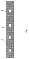
- Fig. 4 shows the total upperside 2 of a bone plate 1 with holes 4 having the inventive combination of portions of lower roughness 2a surrounded by a surface portion of higher roughness 2b as shown in detail in Fig. 3.
Abstract
Description
- This invention relates to a medical device for implantation into living bodies according to the preamble of
claim 1. - Postoperative infections still play an important role in jeopardising the result of surgical treatment of bone fractures and joint disease. The relative incidence of infections is 1 - 2 % at least.
It is evident that resistance to infection (provided good implant materials are used) at the implant/soft tissue interface is determined by the strength of adhesion of the connective tissue to the implant. The extremities of the situation are: - 1. Interface motion causing the formation of a fibrous capsule, surrounding the implant with a continuous liquid phase favouring the spread of bacterial infection and impeding the mobile cellular defense of the body.
- 2. Strong adhesion of the soft tissue to the implant allows minimal fibrous tissue formation, thus confining a bacterial contamination. Close contact of blood vessels to the implant improves the body's defense. The draw back of strong adhesion of the soft tissue to the implant is that it can cause tissue trauma during removal of the implant after fracture healing.
- The invention as claimed is intended to cure these problems of too much or too little adhesion of the soft tissues to the implant. Varying the degree of rough/smooth surface area will allow the problems to be solved in different parts of the body.
- The invention will have numerous applications in the body for all implant plates and most prosthesis. Variations of the invention include the following:
- 1. A network of fine rough surface with smooth compartments in between covering at least partially the surface of the prosthesis or implant.
- 2. A raised network of fine rough surface with smooth compartments below in between the network covering at least partially the surface of the prosthesis or implant.
- The design contains numerous additional advantages to those stated in the initial problem for minimising possibility of bacterial infection.
In the example of the raised network, the fine rough surface protects the initial invading cells, such as fibroblastics cells, from shear forces from body fluid motion during the important initial stages of cell attachment and proliferation. The smooth surface promotes cell spreading and early attachment of the cells therefore accelerating the cell cycle with the consequence of increasing proliferation of cells. The raised network also separates compartments of smooth surface which prevents bacteria from spreading from one compartment to the next. When the soft tissue comes to rest on the implant the rough surface will enable optimal adhesion of the tissue to the implant via the tissue's extracellular matrix and in the case of raised rough surface the compartments which will have been filled by invading cells will act as plugs of tissue which will attach to the overlying soft tissue and prevent motion of the soft tissue over the implant or prosthesis, preventing tissue trauma. - Examples of the use of the rough and smooth surface would include plates with this combination surface on the uppersurface of the plate where it is in contact with the soft tissue. The rough and smooth surface would be beneficial to all metallic plates and also to biodegradable ones. In the case of any biodegradable plates and other implants or prosthesis' the amount of rough surface compared to the amount of smooth surface could be increased since the implant or prosthesis should degrade into the body and would not need to be taken out. Therefore the amount of adhesion could be allowed to be much greater than for an implant or prosthesis that would have to be taken out. A pacemaker prosthesis could have the combination rough and smooth surface on all of it's area in contact with living tissue to promote good adhesion and minimise the chance of infection.
- The various features of novelty which characterize the invention are pointed out with particularity in the claims annexed to and forming part of this disclosure. For the better understanding of the invention, its operating advantages and specific objects attained by its use, reference should be had to the accompanying drawings, examples and descriptive matter in which are illustrated and described preferred embodiments of the invention.
- In the drawings:
- Fig. 1 is a cross section of a conventional implant or prosthesis having either a rough or smooth surface, but not a combination of both;
- Fig. 2 is a cross section of an implant of prosthesis with the invention of a combination of rough and smooth areas both on the same surface of the implant or prosthesis;
- Fig. 3 is a cross section of an implant or prosthesis with the invention of a combination of a raised rough surface separating compartments of smooth surface in between; and
- Fig. 4 is an example of the rough and smooth surface combined as applied to an implant plate from an uppersurface view.
- Figure 1 shows a prior
art bone plate 1 having anuppersurface 2 of uniform roughness. Roughness can be either very low as with polished plates or can be significant as in the case of sandblasted or etched bone plates. The cell,e.g. fibroblasts 3 will adhere to theuppersurface 2 of thebone plate 1 according to its roughness. - In Fig. 2 the
uppersurface 2 of thebone plate 1 according to the invention is divided into first and second types of surface areas with different roughness. The first type of surface area consists of a plurality of isle-like surface portions 2a of lower roughness surrounded by the second type of surface area consisting of a continuously interconnectedsurface portion 2b of higher roughness, in the form of a network, for example of hexagonal structure. The total surface area of lower roughness is equal to
Thesmooth surface portions 2a promote spreading and early attachment of thefibroblasts 3.
The function of therougher surfaces 2b of the network is to provide for stronger attachment of the extracellular matrix (collagen). - Fig. 3 shows a preferred embodiment of the invention in which the
uppersurface 2 of thebone plate 1 is divided in the same manner as in the example of Fig. 2 but where therough surface portion 2b is raised with respect to thesurface portions 2a of lower roughness, thereby separating the latter to form compartments of smooth surface in between therough surface portion 2b. Thesurface portion 2b of higher roughness protects thefibroblastic cells 3 initially invading the lower smooth compartments ofsurface portions 2a.
The preferred method of producing such a surface is firstly acid etching of the complete surface, e.g. with hydrofluoric acid in the case of titanium which will lead to about 1 µ RMS (i.e. root mean square) roughness and secondly stamping the etched surface with a patterned tool. The stamping of the lowered regions will lead to a significant decrease in roughness, e.g. stamping to a depth of about 100 µ reduced the RMS value of the roughness to about 0.4 µ. - Fig. 4 shows the
total upperside 2 of abone plate 1 withholes 4 having the inventive combination of portions oflower roughness 2a surrounded by a surface portion ofhigher roughness 2b as shown in detail in Fig. 3. - While the foregoing description and drawings represent the preferred embodiments of the present invention, it will be obvious for those skilled in the art that various changes and modifications may be made therein without departing from the true spirit and scope of the present invention.
Claims (10)
- Medical device for implantation into living bodies having first and second types of surface areas with different roughness, wherein the first type of surface area consists of a plurality of isle-like surface portions of lower roughness surrounded by the second type of surface area consisting of a continuously interconnected surface portion of higher roughness.
- Medical device according to claim 1, wherein the ratio A₁/A₂ between the total surface area of the first type

- Medical device according to claim 1 or 2, wherein the RMS (root mean square) roughness of the said first type of surface area is less than 1.0 µ, preferably less than 0.5 µ.
- Medical device according to claim 1 or 2, wherein the RMS (root mean square) roughness of the said second type of surface area is larger than 0.5 µ, preferably larger than 1.0 µ.
- Medical device according to one of the claims 1 - 4, wherein the roughness of the said second type of surface area is at least two times higher than the roughness of the said first type of surface area.
- Medical device according to one of the claims 1 - 5, wherein said plurality of discontinuous surface portions of the first type of surface area are in the form of regular polygons, preferably squares or hexagons.
- Medical device according to one of the claims 1 - 6, wherein first type of surface area is located at a lower level than second type of surface area, said plurality of discontinuous surface portions of lower roughness forming depressions in said continuous surface portion of higher roughness.
- Medical device according to one of the claims 1 - 7, wherein the distance between the levels of said first and second types of surface areas is in the range of 5 - 200 µm, preferably 30 - 80 µm.
- Medical device according to one of the claims 1 - 8, wherein the surface aj of one of said discontinuous surface portions of said first type of surface area is larger than 0.1 mm².
- Medical device according to one of the claims 1 - 8, wherein the surface aj of one of said discontinuous surface portions of said first type of surface area is smaller than 10 mm².
Applications Claiming Priority (2)
| Application Number | Priority Date | Filing Date | Title |
|---|---|---|---|
| US306968 | 1994-02-03 | ||
| US30696894A | 1994-09-07 | 1994-09-07 |
Publications (2)
| Publication Number | Publication Date |
|---|---|
| EP0701803A1 true EP0701803A1 (en) | 1996-03-20 |
| EP0701803B1 EP0701803B1 (en) | 1999-10-06 |
Family
ID=23187678
Family Applications (1)
| Application Number | Title | Priority Date | Filing Date |
|---|---|---|---|
| EP19950100829 Expired - Lifetime EP0701803B1 (en) | 1994-02-03 | 1995-01-23 | Medical device for implantation into living bodies |
Country Status (3)
| Country | Link |
|---|---|
| EP (1) | EP0701803B1 (en) |
| DE (1) | DE69512593T2 (en) |
| ES (1) | ES2138094T3 (en) |
Cited By (13)
| Publication number | Priority date | Publication date | Assignee | Title |
|---|---|---|---|---|
| WO1997028760A1 (en) * | 1996-02-09 | 1997-08-14 | Institut Straumann Ag | Pin or screw-like securing device for osteosynthesis |
| WO1999023977A1 (en) * | 1997-11-07 | 1999-05-20 | Expandable Grafts Partnership | Intravascular stent and method for manufacturing an intravascular stent |
| EP0850604A3 (en) * | 1996-12-30 | 1999-07-21 | SORIN BIOMEDICA S.p.A. | A stent for angioplasty and associated production process |
| WO2001021105A1 (en) * | 1999-09-23 | 2001-03-29 | Intratherapeutics, Inc. | Stent with enhanced friction |
| EP1159935A1 (en) * | 2000-05-31 | 2001-12-05 | SAY, Wen-Ching | Orthopedic implant having a porous surface and method of making same |
| WO2002083039A1 (en) * | 2001-04-12 | 2002-10-24 | Advanced Cardiovascular Systems, Inc. | Variable surface area stent |
| US8147859B2 (en) | 2002-09-26 | 2012-04-03 | Advanced Bio Prosthetic Surfaces, Ltd. | Implantable material having patterned surface of raised elements and photochemically altered elements and method of making same |
| US8268340B2 (en) | 2002-09-26 | 2012-09-18 | Advanced Bio Prosthetic Surfaces, Ltd. | Implantable materials having engineered surfaces and method of making same |
| WO2012154862A3 (en) * | 2011-05-09 | 2013-01-17 | Palmaz Scientific, Inc. | Implantable medical device having enhanced endothelial migration features and methods of making the same |
| US8679517B2 (en) | 2002-09-26 | 2014-03-25 | Palmaz Scientific, Inc. | Implantable materials having engineered surfaces made by vacuum deposition and method of making same |
| US8728563B2 (en) | 2011-05-03 | 2014-05-20 | Palmaz Scientific, Inc. | Endoluminal implantable surfaces, stents, and grafts and method of making same |
| US9592083B2 (en) | 2013-08-30 | 2017-03-14 | New South Innovations Pty Limited | Spine stabilization device |
| US9931143B2 (en) | 2012-08-31 | 2018-04-03 | New South Innovations Pty Limited | Bone stabilization device and methods of use |
Families Citing this family (10)
| Publication number | Priority date | Publication date | Assignee | Title |
|---|---|---|---|---|
| US7862495B2 (en) | 2001-05-31 | 2011-01-04 | Advanced Cardiovascular Systems, Inc. | Radiation or drug delivery source with activity gradient to minimize edge effects |
| US6656216B1 (en) | 2001-06-29 | 2003-12-02 | Advanced Cardiovascular Systems, Inc. | Composite stent with regioselective material |
| US7169178B1 (en) | 2002-11-12 | 2007-01-30 | Advanced Cardiovascular Systems, Inc. | Stent with drug coating |
| US7198675B2 (en) | 2003-09-30 | 2007-04-03 | Advanced Cardiovascular Systems | Stent mandrel fixture and method for selectively coating surfaces of a stent |
| US7867547B2 (en) | 2005-12-19 | 2011-01-11 | Advanced Cardiovascular Systems, Inc. | Selectively coating luminal surfaces of stents |
| US8069814B2 (en) | 2006-05-04 | 2011-12-06 | Advanced Cardiovascular Systems, Inc. | Stent support devices |
| US8603530B2 (en) | 2006-06-14 | 2013-12-10 | Abbott Cardiovascular Systems Inc. | Nanoshell therapy |
| US8048448B2 (en) | 2006-06-15 | 2011-11-01 | Abbott Cardiovascular Systems Inc. | Nanoshells for drug delivery |
| US8017237B2 (en) | 2006-06-23 | 2011-09-13 | Abbott Cardiovascular Systems, Inc. | Nanoshells on polymers |
| US8048441B2 (en) | 2007-06-25 | 2011-11-01 | Abbott Cardiovascular Systems, Inc. | Nanobead releasing medical devices |
Citations (7)
| Publication number | Priority date | Publication date | Assignee | Title |
|---|---|---|---|---|
| DE3116040A1 (en) * | 1981-04-22 | 1982-11-04 | Fraunhofer-Gesellschaft zur Förderung der angewandten Forschung e.V., 8000 München | Biocompatible carbon layers for coating flexible materials, and process for applying the layers |
| US4767418A (en) * | 1986-02-13 | 1988-08-30 | California Institute Of Technology | Luminal surface fabrication for cardiovascular prostheses |
| US4865603A (en) * | 1988-02-04 | 1989-09-12 | Joint Medical Products Corporation | Metallic prosthetic devices having micro-textured outer surfaces |
| EP0359575A2 (en) * | 1988-09-16 | 1990-03-21 | Clemson University | Soft tissue implant with micron-scale surface texture to optimize anchorage |
| EP0388576A1 (en) * | 1989-03-23 | 1990-09-26 | Institut Straumann Ag | Metallic implant |
| WO1993000870A1 (en) * | 1991-07-08 | 1993-01-21 | The Trustees Of The University Of Pennsylvania | Porous coated implants having improved fatigue behavior |
| EP0610837A1 (en) * | 1993-02-09 | 1994-08-17 | Acromed Corporation | Spine disc |
-
1995
- 1995-01-23 ES ES95100829T patent/ES2138094T3/en not_active Expired - Lifetime
- 1995-01-23 EP EP19950100829 patent/EP0701803B1/en not_active Expired - Lifetime
- 1995-01-23 DE DE1995612593 patent/DE69512593T2/en not_active Expired - Lifetime
Patent Citations (7)
| Publication number | Priority date | Publication date | Assignee | Title |
|---|---|---|---|---|
| DE3116040A1 (en) * | 1981-04-22 | 1982-11-04 | Fraunhofer-Gesellschaft zur Förderung der angewandten Forschung e.V., 8000 München | Biocompatible carbon layers for coating flexible materials, and process for applying the layers |
| US4767418A (en) * | 1986-02-13 | 1988-08-30 | California Institute Of Technology | Luminal surface fabrication for cardiovascular prostheses |
| US4865603A (en) * | 1988-02-04 | 1989-09-12 | Joint Medical Products Corporation | Metallic prosthetic devices having micro-textured outer surfaces |
| EP0359575A2 (en) * | 1988-09-16 | 1990-03-21 | Clemson University | Soft tissue implant with micron-scale surface texture to optimize anchorage |
| EP0388576A1 (en) * | 1989-03-23 | 1990-09-26 | Institut Straumann Ag | Metallic implant |
| WO1993000870A1 (en) * | 1991-07-08 | 1993-01-21 | The Trustees Of The University Of Pennsylvania | Porous coated implants having improved fatigue behavior |
| EP0610837A1 (en) * | 1993-02-09 | 1994-08-17 | Acromed Corporation | Spine disc |
Cited By (38)
| Publication number | Priority date | Publication date | Assignee | Title |
|---|---|---|---|---|
| WO1997028760A1 (en) * | 1996-02-09 | 1997-08-14 | Institut Straumann Ag | Pin or screw-like securing device for osteosynthesis |
| US6638302B1 (en) | 1996-12-30 | 2003-10-28 | Sorin Biomedica Cardio S.P.A. | Stent for angioplasty and associated production process |
| EP0850604A3 (en) * | 1996-12-30 | 1999-07-21 | SORIN BIOMEDICA S.p.A. | A stent for angioplasty and associated production process |
| US7946019B2 (en) | 1996-12-30 | 2011-05-24 | Sorin Biomedica Cardio S.R.L. | Process for producing a stent for angioplasty |
| US7739781B2 (en) | 1996-12-30 | 2010-06-22 | Sorin Biomedica Cardio S.R.L | Process for producing a stent for angioplasty |
| EP1181903A3 (en) * | 1996-12-30 | 2002-04-03 | SORIN BIOMEDICA CARDIO S.p.A. | A stent for angioplasty and associated production process |
| US7607208B2 (en) | 1996-12-30 | 2009-10-27 | Sorin Biomedica Cardio S.R.L. | Method of making a medicated stent |
| WO1999023977A1 (en) * | 1997-11-07 | 1999-05-20 | Expandable Grafts Partnership | Intravascular stent and method for manufacturing an intravascular stent |
| US6190404B1 (en) | 1997-11-07 | 2001-02-20 | Advanced Bio Prosthetic Surfaces, Ltd. | Intravascular stent and method for manufacturing an intravascular stent |
| WO2001021105A1 (en) * | 1999-09-23 | 2001-03-29 | Intratherapeutics, Inc. | Stent with enhanced friction |
| US6827732B2 (en) | 1999-09-23 | 2004-12-07 | Ev3 Peripheral, Inc. | Stent with enhanced friction |
| US6254631B1 (en) | 1999-09-23 | 2001-07-03 | Intratherapeutics, Inc. | Stent with enhanced friction |
| US9642728B2 (en) | 1999-09-23 | 2017-05-09 | Covidien Lp | Stent with enhanced friction |
| US8236047B2 (en) | 1999-09-23 | 2012-08-07 | Tyco Healthcare Group Lp | Stent with enhanced friction |
| US9011518B2 (en) | 1999-09-23 | 2015-04-21 | Covidien Lp | Stent with enhanced friction |
| EP1159935A1 (en) * | 2000-05-31 | 2001-12-05 | SAY, Wen-Ching | Orthopedic implant having a porous surface and method of making same |
| WO2002083039A1 (en) * | 2001-04-12 | 2002-10-24 | Advanced Cardiovascular Systems, Inc. | Variable surface area stent |
| US8147859B2 (en) | 2002-09-26 | 2012-04-03 | Advanced Bio Prosthetic Surfaces, Ltd. | Implantable material having patterned surface of raised elements and photochemically altered elements and method of making same |
| US20120305529A1 (en) * | 2002-09-26 | 2012-12-06 | Advanced Bio Prosthetic Surfaces, A Wholly Owned Subsidiary Of Palmaz Scientific, Inc. | Implantable materials having engineered surfaces and method of making same |
| US10729824B2 (en) | 2002-09-26 | 2020-08-04 | Vactronix Scientific, Llc. | Implantable materials having engineered surfaces and method of making same |
| US8679517B2 (en) | 2002-09-26 | 2014-03-25 | Palmaz Scientific, Inc. | Implantable materials having engineered surfaces made by vacuum deposition and method of making same |
| US8709066B2 (en) | 2002-09-26 | 2014-04-29 | Advanced Bio Prosthetic Surfaces, Ltd. | Implantable materials having engineered surfaces comprising a pattern of features and method of making same |
| US8932347B2 (en) | 2002-09-26 | 2015-01-13 | Advanced Bio Prosthetic Surfaces, Ltd. | Implantable materials having engineered surfaces and method of making same |
| US8268340B2 (en) | 2002-09-26 | 2012-09-18 | Advanced Bio Prosthetic Surfaces, Ltd. | Implantable materials having engineered surfaces and method of making same |
| US9272077B2 (en) | 2002-09-26 | 2016-03-01 | Palmaz Scientific, Inc. | Implantable materials having engineered surfaces and method of making same |
| US10682443B2 (en) | 2002-09-26 | 2020-06-16 | Vactronix Scientific, Llc | Implantable biomaterials having functional surfaces |
| US10314949B2 (en) | 2002-09-26 | 2019-06-11 | Vactronix Scientific, Llc | Implantable biomaterials having functional surfaces |
| US10034967B2 (en) | 2002-09-26 | 2018-07-31 | Vactronix Scientific, Llc | Implantable biomaterials having engineered functional surfaces |
| US10039866B2 (en) | 2002-09-26 | 2018-08-07 | Vactronix Scientific, Llc | Implantable materials having engineered surfaces and method of making same |
| US8728563B2 (en) | 2011-05-03 | 2014-05-20 | Palmaz Scientific, Inc. | Endoluminal implantable surfaces, stents, and grafts and method of making same |
| WO2012154862A3 (en) * | 2011-05-09 | 2013-01-17 | Palmaz Scientific, Inc. | Implantable medical device having enhanced endothelial migration features and methods of making the same |
| US10206772B2 (en) | 2011-05-09 | 2019-02-19 | Vactronix Scientific, Llc | Implantable medical device having enhanced endothelial migration features and methods of making the same |
| US9439789B2 (en) | 2011-05-09 | 2016-09-13 | Palmaz Scientific, Inc. | Implantable medical device having enhanced endothelial migration features and methods of making the same |
| US8632583B2 (en) | 2011-05-09 | 2014-01-21 | Palmaz Scientific, Inc. | Implantable medical device having enhanced endothelial migration features and methods of making the same |
| US10786343B2 (en) | 2011-05-09 | 2020-09-29 | Vactronix Scientific, Llc | Implantable medical device having enhanced endothelial migration features and methods of making the same |
| US9931143B2 (en) | 2012-08-31 | 2018-04-03 | New South Innovations Pty Limited | Bone stabilization device and methods of use |
| US9592083B2 (en) | 2013-08-30 | 2017-03-14 | New South Innovations Pty Limited | Spine stabilization device |
| US10441323B2 (en) | 2013-08-30 | 2019-10-15 | New South Innovations Pty Limited | Spine stabilization device |
Also Published As
| Publication number | Publication date |
|---|---|
| ES2138094T3 (en) | 2000-01-01 |
| DE69512593D1 (en) | 1999-11-11 |
| EP0701803B1 (en) | 1999-10-06 |
| DE69512593T2 (en) | 2000-01-20 |
Similar Documents
| Publication | Publication Date | Title |
|---|---|---|
| EP0701803B1 (en) | Medical device for implantation into living bodies | |
| US5545226A (en) | Implants for cranioplasty | |
| ES2546329T3 (en) | Assembled non-random foams | |
| EP2773294B1 (en) | Microstructured implant surfaces | |
| AU774347B2 (en) | Device for protecting nerves after surgical procedure | |
| KR100673676B1 (en) | Dental implant having a dual bio-affinity collar | |
| CA2402578A1 (en) | Method for surface treatment of implants or prosthesis made of titanium or other materials | |
| WO2006022878A1 (en) | Surgical implant for promotion of osseo-integration | |
| US20150026942A1 (en) | Implant, and method and system for producing such an implant | |
| JP2002524199A (en) | Bioabsorbable layered composites for inductive bone tissue regeneration | |
| EP1516602A3 (en) | Implant with bioactive particles stuck and method of manufacturing the same | |
| CA2164922A1 (en) | Conically-shaped fusion cage and method of implantation | |
| US4813960A (en) | Endoprosthesis for a hip joint socket | |
| JP2001187074A (en) | Prosthesis transplanting constituting element | |
| CA2491428A1 (en) | An implant and a method for treating an implant surface | |
| WO1992002194A1 (en) | Reconstructive surgery method and implant | |
| JP2003535643A (en) | Transcutaneous prosthesis | |
| US5009665A (en) | Acetabular cup | |
| AU2676795A (en) | Soft implantable surgical prosthesis for increasing or restoring the volume of soft tissues, in particular a breast prosthesis and method of manufacture | |
| EP0927546A3 (en) | Oblong acetabular cup | |
| WO2003051240A3 (en) | Spinal implants | |
| EP0860152A3 (en) | Spinal implant | |
| CA2014542A1 (en) | Medical implant and method for the manufacture thereof | |
| Schultz | Reconstruction of facial deformities with alloplastic material | |
| CN2386790Y (en) | Artificial vertebral lamina |
Legal Events
| Date | Code | Title | Description |
|---|---|---|---|
| PUAI | Public reference made under article 153(3) epc to a published international application that has entered the european phase |
Free format text: ORIGINAL CODE: 0009012 |
|
| 17P | Request for examination filed |
Effective date: 19950123 |
|
| AK | Designated contracting states |
Kind code of ref document: A1 Designated state(s): CH DE ES FR GB IT LI |
|
| RAP1 | Party data changed (applicant data changed or rights of an application transferred) |
Owner name: SYNTHES AG CHUR |
|
| GRAG | Despatch of communication of intention to grant |
Free format text: ORIGINAL CODE: EPIDOS AGRA |
|
| 17Q | First examination report despatched |
Effective date: 19990201 |
|
| GRAG | Despatch of communication of intention to grant |
Free format text: ORIGINAL CODE: EPIDOS AGRA |
|
| GRAH | Despatch of communication of intention to grant a patent |
Free format text: ORIGINAL CODE: EPIDOS IGRA |
|
| GRAH | Despatch of communication of intention to grant a patent |
Free format text: ORIGINAL CODE: EPIDOS IGRA |
|
| GRAA | (expected) grant |
Free format text: ORIGINAL CODE: 0009210 |
|
| AK | Designated contracting states |
Kind code of ref document: B1 Designated state(s): CH DE ES FR GB IT LI |
|
| REG | Reference to a national code |
Ref country code: CH Ref legal event code: NV Representative=s name: DR. LUSUARDI AG Ref country code: CH Ref legal event code: EP |
|
| ET | Fr: translation filed | ||
| REF | Corresponds to: |
Ref document number: 69512593 Country of ref document: DE Date of ref document: 19991111 |
|
| ITF | It: translation for a ep patent filed |
Owner name: STUDIO ING. ALFREDO RAIMONDI |
|
| REG | Reference to a national code |
Ref country code: ES Ref legal event code: FG2A Ref document number: 2138094 Country of ref document: ES Kind code of ref document: T3 |
|
| PLBE | No opposition filed within time limit |
Free format text: ORIGINAL CODE: 0009261 |
|
| STAA | Information on the status of an ep patent application or granted ep patent |
Free format text: STATUS: NO OPPOSITION FILED WITHIN TIME LIMIT |
|
| 26N | No opposition filed | ||
| REG | Reference to a national code |
Ref country code: GB Ref legal event code: IF02 |
|
| REG | Reference to a national code |
Ref country code: CH Ref legal event code: PUE Owner name: SYNTHES GMBH Free format text: SYNTHES AG CHUR#GRABENSTRASSE 15#7002 CHUR (CH) -TRANSFER TO- SYNTHES GMBH#EIMATTSTRASSE 3#4436 OBERDORF (CH) |
|
| REG | Reference to a national code |
Ref country code: GB Ref legal event code: 732E |
|
| REG | Reference to a national code |
Ref country code: FR Ref legal event code: TP |
|
| PGFP | Annual fee paid to national office [announced via postgrant information from national office to epo] |
Ref country code: CH Payment date: 20120126 Year of fee payment: 18 Ref country code: FR Payment date: 20120216 Year of fee payment: 18 |
|
| PGFP | Annual fee paid to national office [announced via postgrant information from national office to epo] |
Ref country code: DE Payment date: 20120125 Year of fee payment: 18 |
|
| PGFP | Annual fee paid to national office [announced via postgrant information from national office to epo] |
Ref country code: IT Payment date: 20120127 Year of fee payment: 18 Ref country code: GB Payment date: 20120118 Year of fee payment: 18 |
|
| PGFP | Annual fee paid to national office [announced via postgrant information from national office to epo] |
Ref country code: ES Payment date: 20120130 Year of fee payment: 18 |
|
| REG | Reference to a national code |
Ref country code: CH Ref legal event code: PL |
|
| GBPC | Gb: european patent ceased through non-payment of renewal fee |
Effective date: 20130123 |
|
| REG | Reference to a national code |
Ref country code: FR Ref legal event code: ST Effective date: 20130930 |
|
| PG25 | Lapsed in a contracting state [announced via postgrant information from national office to epo] |
Ref country code: CH Free format text: LAPSE BECAUSE OF NON-PAYMENT OF DUE FEES Effective date: 20130131 Ref country code: LI Free format text: LAPSE BECAUSE OF NON-PAYMENT OF DUE FEES Effective date: 20130131 Ref country code: DE Free format text: LAPSE BECAUSE OF NON-PAYMENT OF DUE FEES Effective date: 20130801 |
|
| REG | Reference to a national code |
Ref country code: DE Ref legal event code: R119 Ref document number: 69512593 Country of ref document: DE Effective date: 20130801 |
|
| PG25 | Lapsed in a contracting state [announced via postgrant information from national office to epo] |
Ref country code: FR Free format text: LAPSE BECAUSE OF NON-PAYMENT OF DUE FEES Effective date: 20130131 Ref country code: GB Free format text: LAPSE BECAUSE OF NON-PAYMENT OF DUE FEES Effective date: 20130123 |
|
| PG25 | Lapsed in a contracting state [announced via postgrant information from national office to epo] |
Ref country code: IT Free format text: LAPSE BECAUSE OF NON-PAYMENT OF DUE FEES Effective date: 20130123 |
|
| REG | Reference to a national code |
Ref country code: ES Ref legal event code: FD2A Effective date: 20140324 |
|
| PG25 | Lapsed in a contracting state [announced via postgrant information from national office to epo] |
Ref country code: ES Free format text: LAPSE BECAUSE OF NON-PAYMENT OF DUE FEES Effective date: 20130124 |