EP0723013A2 - Biosynthetic osteogenic proteins and osteogenic devices containing them - Google Patents
Biosynthetic osteogenic proteins and osteogenic devices containing them Download PDFInfo
- Publication number
- EP0723013A2 EP0723013A2 EP96200044A EP96200044A EP0723013A2 EP 0723013 A2 EP0723013 A2 EP 0723013A2 EP 96200044 A EP96200044 A EP 96200044A EP 96200044 A EP96200044 A EP 96200044A EP 0723013 A2 EP0723013 A2 EP 0723013A2
- Authority
- EP
- European Patent Office
- Prior art keywords
- protein
- synthetic
- bone
- osteogenic
- amino acid
- Prior art date
- Legal status (The legal status is an assumption and is not a legal conclusion. Google has not performed a legal analysis and makes no representation as to the accuracy of the status listed.)
- Granted
Links
Images


Classifications
-
- A—HUMAN NECESSITIES
- A61—MEDICAL OR VETERINARY SCIENCE; HYGIENE
- A61K—PREPARATIONS FOR MEDICAL, DENTAL OR TOILETRY PURPOSES
- A61K9/00—Medicinal preparations characterised by special physical form
- A61K9/0012—Galenical forms characterised by the site of application
- A61K9/0019—Injectable compositions; Intramuscular, intravenous, arterial, subcutaneous administration; Compositions to be administered through the skin in an invasive manner
- A61K9/0024—Solid, semi-solid or solidifying implants, which are implanted or injected in body tissue
-
- A—HUMAN NECESSITIES
- A61—MEDICAL OR VETERINARY SCIENCE; HYGIENE
- A61L—METHODS OR APPARATUS FOR STERILISING MATERIALS OR OBJECTS IN GENERAL; DISINFECTION, STERILISATION OR DEODORISATION OF AIR; CHEMICAL ASPECTS OF BANDAGES, DRESSINGS, ABSORBENT PADS OR SURGICAL ARTICLES; MATERIALS FOR BANDAGES, DRESSINGS, ABSORBENT PADS OR SURGICAL ARTICLES
- A61L27/00—Materials for grafts or prostheses or for coating grafts or prostheses
- A61L27/14—Macromolecular materials
- A61L27/22—Polypeptides or derivatives thereof, e.g. degradation products
- A61L27/227—Other specific proteins or polypeptides not covered by A61L27/222, A61L27/225 or A61L27/24
-
- A—HUMAN NECESSITIES
- A61—MEDICAL OR VETERINARY SCIENCE; HYGIENE
- A61L—METHODS OR APPARATUS FOR STERILISING MATERIALS OR OBJECTS IN GENERAL; DISINFECTION, STERILISATION OR DEODORISATION OF AIR; CHEMICAL ASPECTS OF BANDAGES, DRESSINGS, ABSORBENT PADS OR SURGICAL ARTICLES; MATERIALS FOR BANDAGES, DRESSINGS, ABSORBENT PADS OR SURGICAL ARTICLES
- A61L27/00—Materials for grafts or prostheses or for coating grafts or prostheses
- A61L27/14—Macromolecular materials
- A61L27/22—Polypeptides or derivatives thereof, e.g. degradation products
- A61L27/24—Collagen
-
- A—HUMAN NECESSITIES
- A61—MEDICAL OR VETERINARY SCIENCE; HYGIENE
- A61L—METHODS OR APPARATUS FOR STERILISING MATERIALS OR OBJECTS IN GENERAL; DISINFECTION, STERILISATION OR DEODORISATION OF AIR; CHEMICAL ASPECTS OF BANDAGES, DRESSINGS, ABSORBENT PADS OR SURGICAL ARTICLES; MATERIALS FOR BANDAGES, DRESSINGS, ABSORBENT PADS OR SURGICAL ARTICLES
- A61L27/00—Materials for grafts or prostheses or for coating grafts or prostheses
- A61L27/28—Materials for coating prostheses
- A61L27/34—Macromolecular materials
-
- A—HUMAN NECESSITIES
- A61—MEDICAL OR VETERINARY SCIENCE; HYGIENE
- A61L—METHODS OR APPARATUS FOR STERILISING MATERIALS OR OBJECTS IN GENERAL; DISINFECTION, STERILISATION OR DEODORISATION OF AIR; CHEMICAL ASPECTS OF BANDAGES, DRESSINGS, ABSORBENT PADS OR SURGICAL ARTICLES; MATERIALS FOR BANDAGES, DRESSINGS, ABSORBENT PADS OR SURGICAL ARTICLES
- A61L27/00—Materials for grafts or prostheses or for coating grafts or prostheses
- A61L27/36—Materials for grafts or prostheses or for coating grafts or prostheses containing ingredients of undetermined constitution or reaction products thereof, e.g. transplant tissue, natural bone, extracellular matrix
- A61L27/3604—Materials for grafts or prostheses or for coating grafts or prostheses containing ingredients of undetermined constitution or reaction products thereof, e.g. transplant tissue, natural bone, extracellular matrix characterised by the human or animal origin of the biological material, e.g. hair, fascia, fish scales, silk, shellac, pericardium, pleura, renal tissue, amniotic membrane, parenchymal tissue, fetal tissue, muscle tissue, fat tissue, enamel
- A61L27/3608—Bone, e.g. demineralised bone matrix [DBM], bone powder
-
- A—HUMAN NECESSITIES
- A61—MEDICAL OR VETERINARY SCIENCE; HYGIENE
- A61L—METHODS OR APPARATUS FOR STERILISING MATERIALS OR OBJECTS IN GENERAL; DISINFECTION, STERILISATION OR DEODORISATION OF AIR; CHEMICAL ASPECTS OF BANDAGES, DRESSINGS, ABSORBENT PADS OR SURGICAL ARTICLES; MATERIALS FOR BANDAGES, DRESSINGS, ABSORBENT PADS OR SURGICAL ARTICLES
- A61L27/00—Materials for grafts or prostheses or for coating grafts or prostheses
- A61L27/36—Materials for grafts or prostheses or for coating grafts or prostheses containing ingredients of undetermined constitution or reaction products thereof, e.g. transplant tissue, natural bone, extracellular matrix
- A61L27/3641—Materials for grafts or prostheses or for coating grafts or prostheses containing ingredients of undetermined constitution or reaction products thereof, e.g. transplant tissue, natural bone, extracellular matrix characterised by the site of application in the body
- A61L27/3645—Connective tissue
- A61L27/365—Bones
-
- A—HUMAN NECESSITIES
- A61—MEDICAL OR VETERINARY SCIENCE; HYGIENE
- A61L—METHODS OR APPARATUS FOR STERILISING MATERIALS OR OBJECTS IN GENERAL; DISINFECTION, STERILISATION OR DEODORISATION OF AIR; CHEMICAL ASPECTS OF BANDAGES, DRESSINGS, ABSORBENT PADS OR SURGICAL ARTICLES; MATERIALS FOR BANDAGES, DRESSINGS, ABSORBENT PADS OR SURGICAL ARTICLES
- A61L27/00—Materials for grafts or prostheses or for coating grafts or prostheses
- A61L27/36—Materials for grafts or prostheses or for coating grafts or prostheses containing ingredients of undetermined constitution or reaction products thereof, e.g. transplant tissue, natural bone, extracellular matrix
- A61L27/3641—Materials for grafts or prostheses or for coating grafts or prostheses containing ingredients of undetermined constitution or reaction products thereof, e.g. transplant tissue, natural bone, extracellular matrix characterised by the site of application in the body
- A61L27/3645—Connective tissue
- A61L27/3654—Cartilage, e.g. meniscus
-
- A—HUMAN NECESSITIES
- A61—MEDICAL OR VETERINARY SCIENCE; HYGIENE
- A61L—METHODS OR APPARATUS FOR STERILISING MATERIALS OR OBJECTS IN GENERAL; DISINFECTION, STERILISATION OR DEODORISATION OF AIR; CHEMICAL ASPECTS OF BANDAGES, DRESSINGS, ABSORBENT PADS OR SURGICAL ARTICLES; MATERIALS FOR BANDAGES, DRESSINGS, ABSORBENT PADS OR SURGICAL ARTICLES
- A61L27/00—Materials for grafts or prostheses or for coating grafts or prostheses
- A61L27/50—Materials characterised by their function or physical properties, e.g. injectable or lubricating compositions, shape-memory materials, surface modified materials
- A61L27/56—Porous materials, e.g. foams or sponges
-
- A—HUMAN NECESSITIES
- A61—MEDICAL OR VETERINARY SCIENCE; HYGIENE
- A61P—SPECIFIC THERAPEUTIC ACTIVITY OF CHEMICAL COMPOUNDS OR MEDICINAL PREPARATIONS
- A61P1/00—Drugs for disorders of the alimentary tract or the digestive system
- A61P1/02—Stomatological preparations, e.g. drugs for caries, aphtae, periodontitis
-
- A—HUMAN NECESSITIES
- A61—MEDICAL OR VETERINARY SCIENCE; HYGIENE
- A61P—SPECIFIC THERAPEUTIC ACTIVITY OF CHEMICAL COMPOUNDS OR MEDICINAL PREPARATIONS
- A61P43/00—Drugs for specific purposes, not provided for in groups A61P1/00-A61P41/00
-
- C—CHEMISTRY; METALLURGY
- C07—ORGANIC CHEMISTRY
- C07K—PEPTIDES
- C07K14/00—Peptides having more than 20 amino acids; Gastrins; Somatostatins; Melanotropins; Derivatives thereof
- C07K14/435—Peptides having more than 20 amino acids; Gastrins; Somatostatins; Melanotropins; Derivatives thereof from animals; from humans
- C07K14/475—Growth factors; Growth regulators
- C07K14/51—Bone morphogenetic factor; Osteogenins; Osteogenic factor; Bone-inducing factor
-
- A—HUMAN NECESSITIES
- A61—MEDICAL OR VETERINARY SCIENCE; HYGIENE
- A61C—DENTISTRY; APPARATUS OR METHODS FOR ORAL OR DENTAL HYGIENE
- A61C8/00—Means to be fixed to the jaw-bone for consolidating natural teeth or for fixing dental prostheses thereon; Dental implants; Implanting tools
- A61C8/0003—Not used, see subgroups
- A61C8/0004—Consolidating natural teeth
- A61C8/0006—Periodontal tissue or bone regeneration
-
- A—HUMAN NECESSITIES
- A61—MEDICAL OR VETERINARY SCIENCE; HYGIENE
- A61F—FILTERS IMPLANTABLE INTO BLOOD VESSELS; PROSTHESES; DEVICES PROVIDING PATENCY TO, OR PREVENTING COLLAPSING OF, TUBULAR STRUCTURES OF THE BODY, e.g. STENTS; ORTHOPAEDIC, NURSING OR CONTRACEPTIVE DEVICES; FOMENTATION; TREATMENT OR PROTECTION OF EYES OR EARS; BANDAGES, DRESSINGS OR ABSORBENT PADS; FIRST-AID KITS
- A61F2310/00—Prostheses classified in A61F2/28 or A61F2/30 - A61F2/44 being constructed from or coated with a particular material
- A61F2310/00005—The prosthesis being constructed from a particular material
- A61F2310/00365—Proteins; Polypeptides; Degradation products thereof
-
- A—HUMAN NECESSITIES
- A61—MEDICAL OR VETERINARY SCIENCE; HYGIENE
- A61K—PREPARATIONS FOR MEDICAL, DENTAL OR TOILETRY PURPOSES
- A61K38/00—Medicinal preparations containing peptides
-
- A—HUMAN NECESSITIES
- A61—MEDICAL OR VETERINARY SCIENCE; HYGIENE
- A61L—METHODS OR APPARATUS FOR STERILISING MATERIALS OR OBJECTS IN GENERAL; DISINFECTION, STERILISATION OR DEODORISATION OF AIR; CHEMICAL ASPECTS OF BANDAGES, DRESSINGS, ABSORBENT PADS OR SURGICAL ARTICLES; MATERIALS FOR BANDAGES, DRESSINGS, ABSORBENT PADS OR SURGICAL ARTICLES
- A61L2430/00—Materials or treatment for tissue regeneration
- A61L2430/02—Materials or treatment for tissue regeneration for reconstruction of bones; weight-bearing implants
Definitions
- This invention relates to osteogenic devices, to synthetic genes encoding proteins which can induce osteogenesis in mammals and methods for their production using recombinant DNA techniques, to synthetic forms of osteogenic protein, and to bone and cartilage repair procedures using osteogenic device comprising the synthetic proteins.
- Mammalian bone tissue is known to contain one or more proteinaceous materials, presumably active during growth and natural bone healing, which can induce a developmental cascade of cellular events resulting in endochondral bone formation.
- This active factor (or factors) has variously been referred to in the literature as bone morphogenetic or morphogenic protein, bone inductive protein, osteogenic protein, osteogenin, or osteoinductive protein.
- the developmental cascade of bone differentiation consists of recruitment of mesenchymal cells, proliferation of progenitor cells, calcification of cartilage, vascular invasion, bone formation, remodeling, and finally marrow differentiation (Reddi (1981) Collagen Rel. Res. 1 :209-226).
- This putative bone inductive protein has been shown to have a molecular mass of less than 50 kilodaltons (kD).
- kD kilodaltons
- Several species of mammals produce closely related protein as demonstrated by cross species implant experiments (Sampath and Reddi (1983) Proc. Natl. Acad. Sci. USA 80 :6591-6595).
- Urist et al. (Proc. Natl. Acad. Sci. USA (1984) 81 :371-375) disclose a bovine bone morphogenetic protein extract having the properties of an acidic polypeptide and a molecular weight of approximately 18 kD. The authors reported that the protein was present in a fraction separated by hydroxyapatite chromatography, and that it induced bone formation in mouse hindquarter muscle and bone regeneration in trephine defects in rat and dog skulls. Their method of obtaining the extract from bone results in ill-defined and impure preparations.
- Wozney et al. discloses the isolation of full-length cDNA's encoding the human equivalents of three polypeptides originally purified from bovine bone. The authors report that each of the three recombinantly expressed human proteins are independently or in combination capable of inducing cartilage formation. No evidence of bone formation is reported.
- It is an object of this invention to provide osteogenic devices comprising matrices containing dispersed osteogenic protein capable of bone induction in allogenic and xenogenic implants. Another object is to provide synthetic osteogenic proteins capable of inducing endochondral bone formation in mammals, including humans. Yet another object is to provide genes encoding non-native osteogenic proteins and methods for their production using recombinant DNA techniques. Another object is to provide novel biosynthetic forms of osteogenic proteins and a structural design for novel, functional osteogenic proteins. Another object is to provide methods for inducing cartilage formation.
- This invention involves osteogenic devices which, when implanted in a mammalian body, can induce at the locus of the implant the full developmental cascade of endochondral bone formation and bone marrow differentiation. Suitably modified as disclosed herein, the devices also may be used to induce cartilage formation.
- the devices comprise a carrier material, referred to herein as a matrix, having the characteristics disclosed below, containing dispersed osteogenic protein in the form of a biosynthetic construct.
- a series of consensus DNA sequences were designed with the goal of producing an active osteogenic protein.
- the sequences were based on partial amino acid sequence data obtained from the naturally sourced product and on observed homologies with unrelated genes reported in the literature, or the sequences they encode, having a presumed or demonstrated developmental function.
- biosynthetic consensus sequences have been expressed as fusion proteins in procaryotes, purified, cleaved, refolded, combined with a matrix, implanted in an established animal model, and shown to have endochondral bone-inducing activity.
- the currently preferred active proteins comprise sequences designated COP5, COP7, COP16, and OP1. The amino acid sequences of these proteins are set forth below.
- the dashes (-) are used as fillers only to line up comparable sequences in related proteins, and have no other function.
- amino acids 45-50 of COP7 for example, are NHAVV.
- the numbering of amino acids is selected solely for purposes of facilitating comparisons between sequences.
- the DF residues numbered at 9 and 10 of COP5 and COP7 may comprise residues, e.g., 35 and 36, of an osteogenic protein embodying the invention.
- Various leader or trailer sequences may be attached to the operative active region provided the osteogenic or chondrogenic activity of the protein is not destroyed.
- the invention comprises a protein comprising an amino acid sequence sufficiently duplicative of the sequence of COP5, COP7, COP16, or OP1 such that it is capable of inducing endochondral bone formation when properly folded and implanted in a mammal in association with a matrix.
- Some of these sequences induce cartilage, but not bone.
- the bone forming materials may be used to produce cartilage if implanted in an avascular locus, or if an inhibitor to full bone development is implanted together with the active protein.
- the invention comprises a protein less than about 200 amino acids long (for each chain) including a sequence sufficiently duplicative of the sequence of COP5, COP7, COP16, or OP1 such that it is capable at least of cartilage formation when properly folded and implanted in a mammal in association with a matrix.
- the phrase "sufficiently duplicative", as used herein, is used to describe proteins having a degree of homology with the specific sequences disclosed herein and other, different amino acids but which nevertheless exhibit osteogenic or chondrogenic activity.
- these proteins comprise species of the generic amino acid sequences: where the letters indicate the amino acid residues of standard single letter code, and the Xs each represent any one of the 22 naturally occurring amino acid residues.
- Preferred amino acid sequences within the foregoing generic sequences are: wherein each of the amino acids arranged vertically at each position in the sequence may be used alternatively in various combinations. Note that these generic sequences have 6 and preferably 7 cysteine residues where inter- or intramolecular disulfide bonds can form, and contain other critical amino acids which influence the tertiary structure of the proteins.
- Vg1 is a known Xenopus sequence heretofore not associated with bone formation.
- DPP is an amino acid sequence encoded by a Drosophila gene responsible for development of the dorsoventral pattern.
- OP1 is a region of a natural sequence encoded by exons of a genomic DNA sequence retrieved by applicants.
- the CBMPs are amino acid sequences comprising subparts of mammalian proteins encoded by genomic DNAs and cDNAs retrieved by applicants.
- the COPs are totally biosynthetic protein sequences expressed by novel consensus gene constructs, designed using the criteria set forth herein, and not yet found in nature.
- COP5, COP7, COP16, and OP1 constructs constitute the first instances of the design of a bioactive protein without preexisting knowledge of the active region of a native form nucleotide or amino acid sequence.
- the invention thus provides synthetic osteogenic protein produced using recombinant DNA techniques.
- the protein may include forms having varying glycosylation patterns, varying N-termini, a family of related proteins having regions of amino acid sequence homology, and active truncated or mutated forms of native protein, no matter how derived.
- skilled genetic engineers can design and synthesize genes which encode appropriate amino acid sequences, and then can express them in various types of host cells, including both procaryotes and eucaryotes, to produce large quantities of active synthetic proteins comprising truncated analogs, muteins, fusion proteins, and other constructs mimicking the biological activity of the native forms and capable of inducing bone formation in mammals including humans.
- the synthetic proteins are useful in clinical applications in conjunction with a suitable delivery or support system (matrix).
- the matrix is made up of particles or porous materials.
- the pores must be of a dimension to permit progenitor cell migration and subsequent differentiation and proliferation.
- the particle size should be within the range of 70 - 850 ⁇ m, preferably 70 - 420 ⁇ m. It may be fabricated by close packing particulate material into a shape spanning the bone defect, or by otherwise structuring as desired a material that is biocompatible (non-inflammatory) and, biodegradable in vivo to serve as a "temporary scaffold” and substratum for recruitment of migratory progenitor cells, and as a base for their subsequent anchoring and proliferation.
- preferred carriers include particulate, demineralized, guanidine extracted, species-specific (allogenic) bone, and particulate, deglycosglated (or HF treated), protein extracted, demineralized, xenogenic bone.
- xenogenic bone powder matrices also may be treated with proteases such as trypsin.
- Other useful matrix materials comprise collagen, homopolymers and copolymers of glycolic acid and lactic acid, hydroxyapatite, tricalcium phosphate and other calcium phosphates.
- the osteogenic proteins and implantable osteogenic devices enabled and disclosed herein will permit the physician to obtain optimal predictable bone formation to correct, for example, acquired and congenital craniofacial and other skeletal or dental anomalies (Glowacki et al. (1981) Lancet 1 :959-963).
- the devices may be used to induce local endochondral bone formation in non-union fractures, and in other clinical applications including periodontal applications where bone formation is required.
- Another potential clinical application is in cartilage repair, for example, in the treatment of osteoarthritis.
- BOP has been characterized significantly; its ability to induce cartilage and ultimately endochondral bone growth in cat, rabbit, and rat have been studied; it has been shown to be able to induce the full developmental cascade of bone formation previously ascribed to unknown protein or proteins in heterogeneous bone extracts; and it may be used to induce formation of endochondral bone in orthopedic defects including non-union fractures.
- DNAs encoding totally novel, non-native protein constructs which individually and combined are capable of producing true endochondral bone.
- the DNAs may be expressed using well established recombinant DNA technologies in procaryotic or eucaryotic host cells, and the expressed proteins may be oxidized and refolded in vitro if necessary for biological activity.
- a synthetic consensus gene shown in FIGURE 4 was designed to encode a consensus protein based on amino acid predictions from homology with the TGF-beta gene family.
- the designed concensus sequence was then constructed using known techniques involving assembly of oligonucleotides manufactured in a DNA synthesizer.
- Tryptic peptides derived from Bovine Osteogenic Protein isolated by applicants and sequenced by Edman degradation provided amino acid sequences that showed strong homology with the Drosophila DPP protein sequence (as inferred from the gene), the Xenopus VG1 protein, and somewhat less homology to inhibin and TGF-beta, as demonstrated below in TABLE 1.
- an appropriate amino acid sequence of an osteogenic protein (from which the nucleic acid sequence can be determined), the following points were considered: (1) the amino acid sequence determined by Edman degradation of natural source osteogenic protein tryptic fragments is ranked highest as long as it has a strong signal and shows homology or conservative changes when aligned with the other members of the gene family; (2) where the sequence matches for all four proteins, it is used in the synthetic gene sequence; (3) matching amino acids in DPP and Vg1 are used; (4) If Vg1 or DPP diverged but either one were matched by inhibin or by TGF-beta, this matched amino acid is chosen; (5) where all sequences diverged, the DPP sequence is initially chosen, with a later plan of creating the Vg1 sequence by mutagenesis kept as a possibility.
- the consensus sequence is designed to preserve the disulfide crosslinking and the apparent structural homology among the related proteins.
- the recombinant osteogenic/chondrogenic proteins are "novel", in the sense that, as far as applicants are aware, they do not exist in nature or, if they do exist, have never before been associated with bone or cartilage formation.
- the approach to design of these proteins is to employ amino acid sequences, found in the native OP isolates, in polypeptide structures are patterned after certain proteins reported in the literature, or the amino acid sequences inferred from DNAs reported in the literature.
- Expression experiments of two of these constructs demonstrate activity.
- Expression experiments with constructs patterned after this template amino acid sequence with a shorter sequence having only six cysteines also show activity: wherein each X independently represents an amino acid.
- Within these generic structures are a multiplicity of specific sequences which have osteogenic or chondrogenic activity.
- Preferred structures are those having the amino acid sequence: wherein, in each position where more than one amino acid is shown, any one of the amino acids shown may be used.
- Novel active proteins also are defined by amino acid sequences comprising an active domain beginning at residue number 6 of this sequence, i.e, omitting the N terminal CXXXX, or omitting any of the preferred specific combinations such as CKRHP, CRRKQ, CKRHE, etc, resulting in a construct having only six cysteine residues.
- PCT 87/01537 was published, and it was observed that the proteins there identified as BMPII a and b and BMPIII each included a region embodying this generic structure. These proteins were not demonstrated to be osteogenic in the published application. However, applicants discovered that a subpart of the amino acid sequence of these proteins, properly folded, and implanted as set forth herein, is active.
- CBMPIIa CBMPIIb
- CBMPIII CBMPIIa
- OP1 OP1
- the preferred osteogenic proteins are expressed from recombinant DNA and comprise amino acid sequences including any of the following sequences:
- these sequences have considerable homology with the alpha and beta inhibins, three forms of TGF beta, and MIS.
- the synthetic genes designed to express the proteins as described above preferably are produced by assembly of chemically synthesized oligonucleotides.
- 15-100mer oligonucleotides may be synthesized on a Biosearch DNA Model 8600 Synthesizer, and purified by polyacrylamide gel electrophoresis (PAGE) in Tris-Borate-EDTA buffer (TBE). The DNA is then electroeluted from the gel.
- Overlapping oligomers may be phosphorylated by T4 polynucleotide kinase and ligated into larger blocks which may also be purifed by PAGE. Natural gene sequences and cDNAs also may be used for expression.
- the genes can be expressed in appropriate prokaryotic hosts such as various strains of E. coli and also in bacillus, yeasts, and various animal cells such as CHO, myeloma, etc. Generally, expression may be achieved using many cell types and expression systems well known to those skilled in the art. If the gene is to be expressed in E. coli , it must first be cloned into an expression vector.
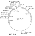
- FIGURE 2A An expression vector (FIGURE 2A) based on pBR322 and containing a synthetic trp promoter operator and the modified trp LE leader can be opened at the EcoRI and PSTI restriction sites, and a FB-FB COP1, COP3, COP5, and COP7 gene fragments (FIGURE 2B) can be inserted between these sites, where FB is fragment B of Staphylococcal Protein A.
- the expressed fusion protein results from attachment of the COP gene to a fragment encoding FB.
- the COP protein is joined to the leader protein via a hinge region having the sequence Asp-Pro-Asn-Gly.
- This hinge permits chemical cleavage of the fusion protein with dilute acid at the asp-pro site or cleavage at Asn-Gly with hydroxylamine, resulting in release of the COP protein.
- the proteins are expressed as fusion products, using the modified trp-LE as a leader,
- the following procedure was followed for production of active recombinant proteins, E. coli cells containing the fusion proteins were lysed.
- the fusion proteins were purified by differential solubilization, In the case of the COP1, 3, 4, 5, and 7 fusion proteins, cleavage was with dilute acid, and the resulting cleavage products were passed through a Sephacryl-200HR column. The Sephacryl column separated most of the uncleaved fusion products from the COP1, 3, 4, 5, and 7 analogs. In the case of the COP16 or OP1 fusion protein, cleavage was with a more concentrated acid, and an SP-Trisacryl column was used as an additional purification step. The COP or OP fractions were then subjected to HPLC on a semi-prep C-18 column.
- Antisera to COP7 and COP5 were produced in New Zealand white rabbits. Western blots demonstrate that the antisera react with COP7 and COP5 preparations. Antisera to COP7 has been tested for reactivity to naturally sourced bovine osteogenic protein samples. Western blots show a clear reaction with the 30 kD protein and, when reduced, with the 16 kD subunit. The immunoreactive species appears as a closely-spaced doublet in the 16 kD subunit region, similar to the 16 kD doublet seen in Con A blots.
- the carrier described in the bioassay section, infra may be replaced by either a biodegradable-synthetic or synthetic-inorganic matrix (e.g., HAP, collagen, tricalcium phosphate, or polylactic acid, polyglycolic acid and various copolymers thereof). Also xenogeneic bone may be used if pretreated as described below.
- a biodegradable-synthetic or synthetic-inorganic matrix e.g., HAP, collagen, tricalcium phosphate, or polylactic acid, polyglycolic acid and various copolymers thereof.
- xenogeneic bone may be used if pretreated as described below.
- the sequential cellular reactions at the interface of the bone matrix/OP implants are complex.
- the multistep cascade includes: binding of fibrin and fibronectin to implanted matrix, chemotaxis of cells, proliferation of fibroblasts, differentiation into chondroblasts, cartilage formation, vascular invasion, bone formation, remodeling, and bone marrow differentiation.
- a successful carrier for osteogenic protein must perform several important functions. It must bind osteogenic protein and act as a slow release delivery system, accommodate each step of the cellular response during bone development, and protect the osteogenic protein from nonspecific proteolysis.
- selected materials must be biocompatible in vivo and biodegradable; the carrier must act as a temporary scaffold until replaced completely by new bone. Biocompatibility requires that the matrix not induce significant inflammation when implanted and not be rejected by the host animal. Biodegradability requires that the matrix be slowly absorbed by the body of the host during development of new bone or cartilage.
- Polylactic acid (PLA), polyglycolic acid (PGA), and various combinations have different dissolution rates in vivo . In bones, the dissolution rates can vary according to whether the implant is placed in cortical or trabecular bone,
- Matrix geometry, particle size, the presence of surface charge, and porosity or the presence of interstices among the particles of a size sufficient to permit cell infiltration, are all important to successful matrix performance. It is preferred to shape the matrix to the desired form of the new bone and to have dimensions which span non-union defects. Rat studies show that the new bone is formed essentially having the dimensions of the device implanted.
- the matrix may comprise a shape-retaining solid made of loosely adhered particulate material, e.g., with collagen. It may also comprise a molded, porous solid, or simply an aggregation of close-packed particles held in place by surrounding tissue. Masticated muscle or other tissue may also be used. Large allogeneic bone implants can act as a carrier for the matrix if their marrow cavities are cleaned and packed with particles and the dispersed osteogenic protein.
- Demineralized bone matrix is prepared from the dehydrated diaphyseal shafts of rat femur and tibia as described herein to produce a bone particle size which pass through a 420 ⁇ m sieve.
- the bone particles are subjected to dissociative extraction with 4 M guanidine-HCl.
- Such treatment results in a complete loss of the inherent ability of the bone matrix to induce endochondral bone differentiation.
- the remaining insoluble material is used to fabricate the matrix.
- the material is mostly collagenous in nature, and upon implantation, does not induce cartilage and bone. All new preparations are tested for mineral content and false positives before use.
- the total loss of biological activity of bone matrix is restored when an active osteoinductive protein fraction or a pure protein is reconstituted with the biologically inactive insoluble collagenous matrix.
- the osteoinductive protein can be obtained from any vertebrate, e.g., bovine, porcine, monkey, or human, or produced using recombinant DNA techniques.
- osteogenic protein When osteogenic protein is reconstituted with collagenous bone matrix from other species and implanted in rat, no bone is formed. This suggests that while the osteogenic protein is xenogenic (not species specific), while the matrix is species specific and cannot be implanted cross species perhaps due to intrinsic immunogenic or inhibitory Components. Thus, heretofore, for bone-based matrices, in order for the osteogenic protein to exhibit its full bone inducing activity, a species specific collagenous bone matrix was required.
- Type I collagen The major component of all bone matrices is Type I collagen.
- extracted bone includes non-collagenous proteins which may account for 5% of its mass.
- non-collagenous components of bone matrix are glycoproteins. Although the biological significance of the glycoproteins in bone formation is not known, they may present themselves as potent antigens by virtue of their carbohydrate content and may constitute immunogenic and/or inhibitory components that are present in xenogenic matrix.
- a collagenous bone matrix may be used as a carrier to effect bone inducing activity in xenogenic implants, if one first removes the immunogenic and inhibitory components from the matrix.
- the matrix is deglycosglated chemically using, for example, hydrogen fluoride to achieve this purpose.
- Bovine bone residue prepared as described above is sieved, and particles of the 74-420 ⁇ M are collected.
- the sample is dried in vacuo over P 2 0 5 , transferred to the reaction vessel and anhydrous hydrogen fluoride (HF) (10-20 ml/g of matrix) is then distilled onto the sample at -70°C.
- HF hydrous hydrogen fluoride
- the vessel is allowed to warm to 0°C. and the reaction mixture is stirred at this temperature for 120 min. After evaporation of the HF in vacuo , the residue is dried thoroughly in vacuo over KOH pellets to remove any remaining traces of acid.
- Extent of deglycosylation can be determined from carbohydrate analysis of matrix samples taken before and after treatment with HF, after washing the samples appropriately to remove non-covalently bound carbohydrates.
- the deglycosylated bone matrix is next treated as set forth below:
- Fabrication of osteogenic devices using any of the matrices set forth above with any of the osteogenic proteins described above may be performed as follows.
- matrix is added to osteogenic protein in guanidine-HCl. Samples are vortexed and incubated at a low temperature. Samples are then further vortexed. Cold absolute ethanol is added to the mixture which is then stirred and incubated. After centrifugation (microfuge high speed) the supernatant is discarded, The reconstituted matrix is washed with cold concentrated ethanol in water and then lyophilized,
- osteogenic protein in an acetonitrile trifluroacetic acid (ACN/TFA) solution is added to the carrier.
- Samples are vigorously vortexed many times and then lyophilized.
- the protein is mixed with the matrix, vortexed many times, and then lyophilized.
- the lyophilized material may be used "as is" for implants.
- osteogenic devices have been incorporated in matrices to produce osteogenic devices, and assayed in rat for endochondral bone. Studies in rats show the osteogenic effect to be dependent on the dose of osteogenic protein dispersed in the osteogenic device. No activity is observed if the matrix is implanted alone. The following sets forth guidelines for how the osteogenic devices disclosed herein can be assayed for evaluating protein constructs and matrices for biological activity.
- This assay consists of implanting the test samples in subcutaneous sites in allogenic recipient rats under ether anesthesia. Male Long-Evans rats, aged 28-32 days, were used. A vertical incision (1 cm) is made under sterile conditions in the skin over the thoraic region, and a pocket is prepared by blunt dissection. Approximately 25 mg of the test sample is implanted deep into the pocket and the incision is closed with a metallic skin clip. The day of implantation is designated as day one of the experiment. Implants were removed on day 12. The heterotropic site allows for the study of bone induction without the possible ambiguities resulting from the use of orthotopic sites.
- the implant model in rats exhibits a controlled progression through the stages of matrix induced endochondral bone development including: (1) transient infiltration by polymorphonuclear leukocytes on day one; (2) mesenchymal cell migration and proliferation on days two and three; (3) chondrocyte appearance on days five and six; (4) cartilage matrix formation on day seven; (5) cartiliage calcification on day eight; (6) vascular invasion, appearance of osteoblasts, and formation of new bone on days nine and ten; (7) appearance of osteoblastic and bone remodeling and dissolution of the implanted matrix on days twelve to eighteen; and (8) hematopoietic bone marrow differentiation in the ossicle on day twenty-one.
- the results show that the shape of the new bone conforms to the shape of the implanted matrix.
- Implants are fixed in Bouins Solution, embedded in parafilm, cut into 6-8 mm sections. Staining with toluidine blue or hemotoxylin/eosin demonstrates clearly the ultimate development of endochondrial bone. Twelve day implants are usually sufficient to determine whether the implants show bone inducing activity.
- Alkaline phosphatase activity may be used as a marker for osteogenesis.
- the enzyme activity may be determined Spectrophotometrically after homogenization of the implant. The activity peaks at 9-10 days in vivo and thereafter slowly declines. Implants showing no bone development by histology should have little or no alkaline phosphatase activity under these assay conditions.
- the assay is useful for quantitation and obtaining an estimate of bone formation very quickly after the implants are removed from the rat. Alternatively the amount of bone formation can be determined by measuring the calcium content of the implant.
- the osteogenic activity due to osteogenic protein is represented by "bone forming units".
- One bone forming unit represents the amount of protein that is needed for half maximal bone forming activity as compared to rat demineralized bone matrix as control and determined by calcium content of the implant on day 12.
- rat carrier Devices that contained only rat carrier show complete absence of new bone formation.
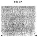
- the implant consists of carrier rat matrix and surrounding mesenchymal cells. Again, the devices that contained rat carrier and not correctly folded (or biologically inactive) recombinant protein also showed complete absence of bone formation. These implants are scored as cartilage formation (-) and bone formation (-). The endochondral bone formation activity is scored as zero percent (0%) (FIGURE 3A).
- Implants included biologically active recombinant protein, however, showed evidence of endochondral bone formation. Histologically they showed new cartilage and bone formation.
- the cartilage formation is scored as (+) by the presence of metachromatically stained chondrocytes in the center of the implant, as (++) by the presence of numerous chondrocytes in many areas of the implant and as (+++) by the presence of abundant chondrocytes forming cartilage matrix and the appearance of hypertrophied chondrocytes accompanying cartilage calcification (FIGURE 3B).
- the bone formation is scored as (+) by the presence of osteoblast surrounding vascular endothelium forming new matrix, as (++) by the formation of bone due to osteoblasts (as indicated by arrows) and further bone remodeling by the appearance of osteoclasts in opposition to the newly formed bone matrix.
- Formation is scored as (+++) by the presence of extensive remodeled bone which results in the formation of ossicles.
- the invention relates to an osteogenic device for implantation in a mammal, said device comprising a biocompatible, in vivo biodegradable matrix defining pores of a dimension sufficient to permit influx, proliferation and differentiation of migratory progenitor cells from the body of said mammal; and a protein, produced by expression of recombinant DNA in a host cell, comprising one or more polypeptide chains, each of which comprises an amino acid sequence sufficiently duplicative of the sequence of COP5, COP7, COP16, or OP1 such that said protein is capable of inducing endochondral bone formation in association with said matrix when implanted in a mammal.
- a device for implantation in a mammal comprising a biocompatible, in vivo biodegradable matrix defining pores of a dimension sufficient to permit influx, proliferation and differentiation of migratory progenitor cells from the body of said mammal; and a protein, produced by expression of recombinant DNA in a host cell, comprising one or more polypeptide chains, each of which has less than about 200 amino acids, and each of which comprises a sequence sufficiently duplicative of the sequence of COP5, COP7, COP16, or OP1 such that said protein is capable of inducing cartilage formation in association with said matrix when implanted in a mammal.
- the sequence may comprise wherein each X independently represents an amino acid; or wherein each X independently represents an amino acid; or wherein, in each position where more than one amino acid is shown, any one of the amino acids shown may be in that position; or wherein, in each position where more than one amino acid is shown, any one of the amino acids shown may be in that position; or
- the device of the invention may comprise an osteogenic protein comprising a pair of separate polypeptide chains.
- the invention also relates to an osteogenic protein, produced by expression of recombinant DNA in a host cell, comprising one or more polypeptide chains each of which comprises a sequence sufficiently duplicative of the sequence of COP5, COP7, COP16, or OP1 such that said protein is capable of inducing endochondral bone formation in association with a matrix when implanted in a mammal.
- the invention relates to a protein, produced by expression of recombinant DNA in a host cell, comprising one or more polypeptide chains less than about 200 amino acids long and comprising a sequence sufficiently duplicative of the sequence of COP5, COP7, COP16, or OP1 such that said protein is capable of inducing cartilage formation in association with a matrix when implanted in a mammal.
- the protein may be unglycosylated, and may comprise the amino acid sequences: wherein each X independently represents an amino acid.
- the protein of the invention may comprise the amino acid sequences: wherein, in each position where more than one amino acid is shown, any one of the amino acids shown may be in that position.
- the protein of the invention may comprise the amino acid sequences: wherein, in each position where more than one amino acid is shown, any one of the amino acids shown may be in that position.
- the protein of the invention may comprise the amino acid sequences:
- the protein of the invention may comprise the product of expression of a DNA in a procaryotic cell.
- Also contemplated by the invcention is a cell line engineered to express the protein of the invention.
- the protein has a half maximum bone forming activity of about 20-25 ng per 25 mg of implant.
- the invention also contemplates antibodies reactive with an epitope of the protein of the invention.
- the device of the invention preferably includes a matrix comprising demineralized, deglycosylated, protein extracted, particulate, xenogenic bone, or demineralized, protein extracted, particulate, xenogenic bone treated with HF.
Abstract
Description
- This invention relates to osteogenic devices, to synthetic genes encoding proteins which can induce osteogenesis in mammals and methods for their production using recombinant DNA techniques, to synthetic forms of osteogenic protein, and to bone and cartilage repair procedures using osteogenic device comprising the synthetic proteins.
- Mammalian bone tissue is known to contain one or more proteinaceous materials, presumably active during growth and natural bone healing, which can induce a developmental cascade of cellular events resulting in endochondral bone formation. This active factor (or factors) has variously been referred to in the literature as bone morphogenetic or morphogenic protein, bone inductive protein, osteogenic protein, osteogenin, or osteoinductive protein.
- The developmental cascade of bone differentiation consists of recruitment of mesenchymal cells, proliferation of progenitor cells, calcification of cartilage, vascular invasion, bone formation, remodeling, and finally marrow differentiation (Reddi (1981) Collagen Rel. Res. 1:209-226).
- Though the precise mechanisms underlying these phenotypic transformations are unclear, it has been shown that the natural endochondral bone differentiation activity of bone matrix can be dissociatively extracted and reconstituted with inactive residual collagenous matrix to restore full bone induction activity (Sampath and Reddi, (1981) Proc. Natl. Acad. Sci. USA 78:7599-7603). This provides an experimental method for assaying protein extracts for their ability to induce endochondral bone in vivo.
- This putative bone inductive protein has been shown to have a molecular mass of less than 50 kilodaltons (kD). Several species of mammals produce closely related protein as demonstrated by cross species implant experiments (Sampath and Reddi (1983) Proc. Natl. Acad. Sci. USA 80:6591-6595).
- The potential utility of these proteins has been widely recognized. It is contemplated that the availability of the pure protein would revolutionize orthopedic medicine, certain types of plastic surgery, and various periodontal and craniofacial reconstructive procedures.
- The observed properties of these protein fractions have induced an intense research effort in various laboratories directed to isolating and identifying the pure factor or factors responsible for osteogenic activity. The current state of the art of purification of osteogenic protein from mammalian bone is disclosed by Sampath et al. (Proc. Natl. Acad. Sci. USA (1987) 80). Urist et al. (Proc. Soc. Exp. Biol. Med. (1984) 173:194-199) disclose a human osteogenic protein fraction which was extracted from demineralized cortical bone by means of a calcium chloride-urea inorganic-organic solvent mixture, and retrieved by differential precipitation in guanidine-hydrochloride and preparative gel electrophoresis. The authors report that the protein fraction has an amino acid composition of an acidic polypeptide and a molecular weight in a range of 17-18 kD.
- Urist et al. (Proc. Natl. Acad. Sci. USA (1984) 81:371-375) disclose a bovine bone morphogenetic protein extract having the properties of an acidic polypeptide and a molecular weight of approximately 18 kD. The authors reported that the protein was present in a fraction separated by hydroxyapatite chromatography, and that it induced bone formation in mouse hindquarter muscle and bone regeneration in trephine defects in rat and dog skulls. Their method of obtaining the extract from bone results in ill-defined and impure preparations.
- European Patent Application Serial No. 148,155, published October 7, 1985, purports to disclose osteogenic proteins derived from bovine, porcine, and human origin. One of the proteins, designated by the inventors as a P3 protein having a molecular weight of 22-24 kD, is said to have been purified to an essentially homogeneous state. This material is reported to induce bone formation when implanted into animals.
- International Application No. PCT/087/01537, published January 14, 1988, discloses an impure fraction from bovine bone which has bone induction qualities. The named applicants also disclose putative bone inductive factors produced by recombinant DNA techniques. Four DNA sequences were retrieved from human or bovine genomic or cDNA libraries and apparently expressed in recombinant host cells. While the applicants stated that the expressed proteins may be bone morphogenic proteins, bone induction was not demonstrated. See also Urist et al., EP 0,212,474 entitled Bone Morphogenic Agents.
- Wang et al. (Proc. Nat. Acad. Sci. USA (1988) 85: 9484-9488) discloses the purification of a bovine bone morphogenetic protein from guanidine extracts of demineralized bone having cartilage and bone formation activity as a basic protein corresponding to a molecular weight of 30 kD determined from gel elution. Purification of the protein yielded proteins of 30, 18 and 16 kD which, upon separation, were inactive. In view of this result, the authors acknowledged that the exact identity of the active material had not been determined.
- Wozney et al. (Science (1988) 242: 1528-1534) discloses the isolation of full-length cDNA's encoding the human equivalents of three polypeptides originally purified from bovine bone. The authors report that each of the three recombinantly expressed human proteins are independently or in combination capable of inducing cartilage formation. No evidence of bone formation is reported.
- It is an object of this invention to provide osteogenic devices comprising matrices containing dispersed osteogenic protein capable of bone induction in allogenic and xenogenic implants. Another object is to provide synthetic osteogenic proteins capable of inducing endochondral bone formation in mammals, including humans. Yet another object is to provide genes encoding non-native osteogenic proteins and methods for their production using recombinant DNA techniques. Another object is to provide novel biosynthetic forms of osteogenic proteins and a structural design for novel, functional osteogenic proteins. Another object is to provide methods for inducing cartilage formation.
- These and other objects and features of the invention will be apparent from the description, drawings, and claims which follow.
- This invention involves osteogenic devices which, when implanted in a mammalian body, can induce at the locus of the implant the full developmental cascade of endochondral bone formation and bone marrow differentiation. Suitably modified as disclosed herein, the devices also may be used to induce cartilage formation. The devices comprise a carrier material, referred to herein as a matrix, having the characteristics disclosed below, containing dispersed osteogenic protein in the form of a biosynthetic construct.
- Key to these developments was the successful preparation of substantially pure osteogenic protein by purification from bone, the elucidation of amino acid sequence and structure data of the native osteogenic protein, and insights involving study of the DNA and amino acid sequences of the natural source product. A protocol was developed which results in retrieval of active, substantially pure osteogenic protein from mammalian bone. Investigation of the properties and structure of the native form osteogenic protein then permitted the inventors to develop a rational design for non-native forms, i.e., forms never before known in nature, capable of inducing bone formation. As far as applicants are aware, the constructs disclosed herein constitute the first instance of the design of a functional, active protein without preexisting knowledge of the active region of a native form nucleotide or amino acid sequence.
- A series of consensus DNA sequences were designed with the goal of producing an active osteogenic protein. The sequences were based on partial amino acid sequence data obtained from the naturally sourced product and on observed homologies with unrelated genes reported in the literature, or the sequences they encode, having a presumed or demonstrated developmental function. Several of the biosynthetic consensus sequences have been expressed as fusion proteins in procaryotes, purified, cleaved, refolded, combined with a matrix, implanted in an established animal model, and shown to have endochondral bone-inducing activity. The currently preferred active proteins comprise sequences designated COP5, COP7, COP16, and OP1. The amino acid sequences of these proteins are set forth below.


- In these sequences and all other amino acid sequences disclosed herein, the dashes (-) are used as fillers only to line up comparable sequences in related proteins, and have no other function. Thus, amino acids 45-50 of COP7, for example, are NHAVV. Also, the numbering of amino acids is selected solely for purposes of facilitating comparisons between sequences. Thus, for example, the DF residues numbered at 9 and 10 of COP5 and COP7 may comprise residues, e.g., 35 and 36, of an osteogenic protein embodying the invention. Various leader or trailer sequences may be attached to the operative active region provided the osteogenic or chondrogenic activity of the protein is not destroyed.
- Thus, in one aspect, the invention comprises a protein comprising an amino acid sequence sufficiently duplicative of the sequence of COP5, COP7, COP16, or OP1 such that it is capable of inducing endochondral bone formation when properly folded and implanted in a mammal in association with a matrix. Some of these sequences induce cartilage, but not bone. Also, the bone forming materials may be used to produce cartilage if implanted in an avascular locus, or if an inhibitor to full bone development is implanted together with the active protein. Thus, in another aspect, the invention comprises a protein less than about 200 amino acids long (for each chain) including a sequence sufficiently duplicative of the sequence of COP5, COP7, COP16, or OP1 such that it is capable at least of cartilage formation when properly folded and implanted in a mammal in association with a matrix. The phrase "sufficiently duplicative", as used herein, is used to describe proteins having a degree of homology with the specific sequences disclosed herein and other, different amino acids but which nevertheless exhibit osteogenic or chondrogenic activity.
- In one preferred aspect, these proteins comprise species of the generic amino acid sequences:

where the letters indicate the amino acid residues of standard single letter code, and the Xs each represent any one of the 22 naturally occurring amino acid residues. Preferred amino acid sequences within the foregoing generic sequences are:
wherein each of the amino acids arranged vertically at each position in the sequence may be used alternatively in various combinations. Note that these generic sequences have 6 and preferably 7 cysteine residues where inter- or intramolecular disulfide bonds can form, and contain other critical amino acids which influence the tertiary structure of the proteins. These generic structural features are found in previously published sequences, none of which have been described as capable of osteogenic activity, and most of which never have been linked with such activity. - Particular useful sequences include:


Vg1 is a known Xenopus sequence heretofore not associated with bone formation. DPP is an amino acid sequence encoded by a Drosophila gene responsible for development of the dorsoventral pattern. OP1 is a region of a natural sequence encoded by exons of a genomic DNA sequence retrieved by applicants, The CBMPs are amino acid sequences comprising subparts of mammalian proteins encoded by genomic DNAs and cDNAs retrieved by applicants. The COPs are totally biosynthetic protein sequences expressed by novel consensus gene constructs, designed using the criteria set forth herein, and not yet found in nature. - These proteins are believed to be dimers. They appear not to be active when reduced. Various combinations of species of the proteins may exist as heterodimers or homodimers. As far as applicants are aware, the COP5, COP7, COP16, and OP1 constructs constitute the first instances of the design of a bioactive protein without preexisting knowledge of the active region of a native form nucleotide or amino acid sequence.
- The invention thus provides synthetic osteogenic protein produced using recombinant DNA techniques. The protein may include forms having varying glycosylation patterns, varying N-termini, a family of related proteins having regions of amino acid sequence homology, and active truncated or mutated forms of native protein, no matter how derived. In view of this disclosure, skilled genetic engineers can design and synthesize genes which encode appropriate amino acid sequences, and then can express them in various types of host cells, including both procaryotes and eucaryotes, to produce large quantities of active synthetic proteins comprising truncated analogs, muteins, fusion proteins, and other constructs mimicking the biological activity of the native forms and capable of inducing bone formation in mammals including humans.
- The synthetic proteins are useful in clinical applications in conjunction with a suitable delivery or support system (matrix). The matrix is made up of particles or porous materials. The pores must be of a dimension to permit progenitor cell migration and subsequent differentiation and proliferation. The particle size should be within the range of 70 - 850 µm, preferably 70 - 420 µm. It may be fabricated by close packing particulate material into a shape spanning the bone defect, or by otherwise structuring as desired a material that is biocompatible (non-inflammatory) and, biodegradable in vivo to serve as a "temporary scaffold" and substratum for recruitment of migratory progenitor cells, and as a base for their subsequent anchoring and proliferation. Currently preferred carriers include particulate, demineralized, guanidine extracted, species-specific (allogenic) bone, and particulate, deglycosglated (or HF treated), protein extracted, demineralized, xenogenic bone. Optionally, such xenogenic bone powder matrices also may be treated with proteases such as trypsin. Other useful matrix materials comprise collagen, homopolymers and copolymers of glycolic acid and lactic acid, hydroxyapatite, tricalcium phosphate and other calcium phosphates.
- The osteogenic proteins and implantable osteogenic devices enabled and disclosed herein will permit the physician to obtain optimal predictable bone formation to correct, for example, acquired and congenital craniofacial and other skeletal or dental anomalies (Glowacki et al. (1981) Lancet 1:959-963). The devices may be used to induce local endochondral bone formation in non-union fractures, and in other clinical applications including periodontal applications where bone formation is required. Another potential clinical application is in cartilage repair, for example, in the treatment of osteoarthritis.
- The foregoing and other objects of this invention, the various features thereof, as well as the invention itself, may be more fully understood from the following description, when read together with the accompanying drawings, in which:
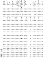
- FIGURE 1 is a comparison of the amino acid sequence of various osteogenic proteins to those of the TGF-beta family. COP1, COP3, COP4, COP5, and COP7 are a family of analogs of synthetic osteogenic proteins developed from the consensus gene that was joined to a leader protein via a hinge region having the sequence D-P-N-G that permitted chemical cleavage at the D-P site (by acid) or N-G (by hydroxylamine) resulting in the release of the analog protein; VGI is a Xenopus protein, DPP is a Drosophila protein; OP1 is a native osteogenic protein; CBMP2a and 2b, and CBMP3 are subparts of proteins disclosed in PCT application 087/01537; MIS is Mullerian inhibitory substance; and "consensus choices" represent various substitutions of amino acids that may be made at various positions in osteogenic proteins;
- FIGURE 2A is an E. coli expression vector containing a gene of an osteogenic protein fused to a leader protein;
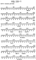
- FIGURE 2B is the DNA sequence comprising a modified trp-LE leader, two Fb domains of protein A, an ASP-PRO cleavage site, and the COP5 sequence;
- FIGURES 3A and 3B are photomicrographs of implants showing the histology (day 12) of COP5 active recombinant protein. A is a control (rat matrix alone, 25 mg). B is rat matrix plus COP5, showing +++ cartilage formation and ++ bone formation (see key infra). Similar results are achieved with COP7; and
- FIGURE 4 is a schematic representation of the DNA sequence and corresponding amino acid sequence of a consensus gene for osteogenic protein (COPO).
- Purification protocols have been developed which enable isolation of the osteogenic protein present in crude protein extracts from mammalian bone. The isolation procedure enables the production of significant quantities of substantially pure osteogenic protein from any mammalian species, provided sufficient amounts of fresh bone from the species is available. The empirical development of the procedure, coupled with the availability of fresh calf bone, enabled isolation of substantially pure bovine osteogenic protein (BOP). BOP has been characterized significantly; its ability to induce cartilage and ultimately endochondral bone growth in cat, rabbit, and rat have been studied; it has been shown to be able to induce the full developmental cascade of bone formation previously ascribed to unknown protein or proteins in heterogeneous bone extracts; and it may be used to induce formation of endochondral bone in orthopedic defects including non-union fractures. In its native form it is a glycosylated, dimeric protein. However, it is active in deglycosylated form. It has been partially sequenced.
- Elucidation of the amino acid sequence of BOP enabled the construction of a consensus nucleic acid sequence designed as disclosed herein based on the sequence data, inferred codons for the sequences, and observation of partial homology with known genes.
- These consensus sequences have been refined by comparison with the sequences present in certain regulatory genes from drosophila, xenopus, and human followed by point mutation, expression, and assay for activity. This approach has been successful in producing several active totally synthetic constructs not found in nature (as far as applicants are aware) which have true osteogenic activity.
- These discoveries enable the construction of DNAs encoding totally novel, non-native protein constructs which individually and combined are capable of producing true endochondral bone. The DNAs may be expressed using well established recombinant DNA technologies in procaryotic or eucaryotic host cells, and the expressed proteins may be oxidized and refolded in vitro if necessary for biological activity.
- The design and production of such biosynthetic proteins, the nature of the matrix, and other material aspects concerning the nature, utility, how to make, and how to use the subject matter claimed herein will be further understood from the following, which constitutes the best method currently known for practicing the various aspects of the invention.
- A synthetic consensus gene shown in FIGURE 4 was designed to encode a consensus protein based on amino acid predictions from homology with the TGF-beta gene family. The designed concensus sequence was then constructed using known techniques involving assembly of oligonucleotides manufactured in a DNA synthesizer.
- Tryptic peptides derived from Bovine Osteogenic Protein isolated by applicants and sequenced by Edman degradation provided amino acid sequences that showed strong homology with the Drosophila DPP protein sequence (as inferred from the gene), the Xenopus VG1 protein, and somewhat less homology to inhibin and TGF-beta, as demonstrated below in TABLE 1.


- In determining an appropriate amino acid sequence of an osteogenic protein (from which the nucleic acid sequence can be determined), the following points were considered: (1) the amino acid sequence determined by Edman degradation of natural source osteogenic protein tryptic fragments is ranked highest as long as it has a strong signal and shows homology or conservative changes when aligned with the other members of the gene family; (2) where the sequence matches for all four proteins, it is used in the synthetic gene sequence; (3) matching amino acids in DPP and Vg1 are used; (4) If Vg1 or DPP diverged but either one were matched by inhibin or by TGF-beta, this matched amino acid is chosen; (5) where all sequences diverged, the DPP sequence is initially chosen, with a later plan of creating the Vg1 sequence by mutagenesis kept as a possibility. In addition, the consensus sequence is designed to preserve the disulfide crosslinking and the apparent structural homology among the related proteins.
- This approach resulted in the production of novel recombinant proteins capable of inducing formation of cartilage and endochondral bone comprising a protein structure analogous to or duplicative of the functional domain of the naturally sourced material. The amino acid sequences encoded by the consensus DNA sequences were derived from a family of natural proteins implicated in tissue development. These gene products/proteins are known to exist in active form as dimers and are, in general, processed from a precursor protein to produce an active C-terminal domain of the precursor.
- The recombinant osteogenic/chondrogenic proteins are "novel", in the sense that, as far as applicants are aware, they do not exist in nature or, if they do exist, have never before been associated with bone or cartilage formation. The approach to design of these proteins is to employ amino acid sequences, found in the native OP isolates, in polypeptide structures are patterned after certain proteins reported in the literature, or the amino acid sequences inferred from DNAs reported in the literature. Thus, using the design criteria set forth above, and refining the amino acid sequence as more protein sequence information was learned, a series of synthetic proteins were designed with the hope and intent that they might have osteogenic or chondrogenic activity when tested in the bioassay system disclosed below.
- It was noted, for example, that DPP from drosophila, VG1 from Xenopus, the TGF beta family of proteins, and to a lesser extent, alpha and beta inhibins, had significant homologies with certain of the sequences derived from the naturally sourced OP product. (FIGURE 1.) Study of these proteins led to the realization that a portion of the sequence of each had a structural similarity observable by analysis of the positional relationship of cysteines and other amino acids which have an important influence on three dimensional protein conformation. It was noted that a region of these sequences had a series of seven cysteines, placed very nearly in the same relative positions, and certain other amino acids in sequence as set forth below:

wherein each X independently represents an amino acid. Expression experiments of two of these constructs demonstrate activity. Expression experiments with constructs patterned after this template amino acid sequence with a shorter sequence having only six cysteines also show activity:
wherein each X independently represents an amino acid. Within these generic structures are a multiplicity of specific sequences which have osteogenic or chondrogenic activity. Preferred structures are those having the amino acid sequence:
wherein, in each position where more than one amino acid is shown, any one of the amino acids shown may be used. Novel active proteins also are defined by amino acid sequences comprising an active domain beginning at residue number 6 of this sequence, i.e, omitting the N terminal CXXXX, or omitting any of the preferred specific combinations such as CKRHP, CRRKQ, CKRHE, etc, resulting in a construct having only six cysteine residues. After this work, PCT 87/01537 was published, and it was observed that the proteins there identified as BMPII a and b and BMPIII each included a region embodying this generic structure. These proteins were not demonstrated to be osteogenic in the published application. However, applicants discovered that a subpart of the amino acid sequence of these proteins, properly folded, and implanted as set forth herein, is active. These are disclosed herein as CBMPIIa, CBMPIIb, and CBMPIII. Also, applicants retrieved a previously unreported gene by probing a human genomic DNA library with COPO. This protein was designated OP1. It comprises a region exhibiting the same generic structure. - Thus, the preferred osteogenic proteins are expressed from recombinant DNA and comprise amino acid sequences including any of the following sequences:


- As shown in FIGURE 1, these sequences have considerable homology with the alpha and beta inhibins, three forms of TGF beta, and MIS.
- The synthetic genes designed to express the proteins as described above preferably are produced by assembly of chemically synthesized oligonucleotides. 15-100mer oligonucleotides may be synthesized on a Biosearch DNA Model 8600 Synthesizer, and purified by polyacrylamide gel electrophoresis (PAGE) in Tris-Borate-EDTA buffer (TBE). The DNA is then electroeluted from the gel. Overlapping oligomers may be phosphorylated by T4 polynucleotide kinase and ligated into larger blocks which may also be purifed by PAGE. Natural gene sequences and cDNAs also may be used for expression.
- The genes can be expressed in appropriate prokaryotic hosts such as various strains of E. coli and also in bacillus, yeasts, and various animal cells such as CHO, myeloma, etc. Generally, expression may be achieved using many cell types and expression systems well known to those skilled in the art. If the gene is to be expressed in E. coli, it must first be cloned into an expression vector. An expression vector (FIGURE 2A) based on pBR322 and containing a synthetic trp promoter operator and the modified trp LE leader can be opened at the EcoRI and PSTI restriction sites, and a FB-FB COP1, COP3, COP5, and COP7 gene fragments (FIGURE 2B) can be inserted between these sites, where FB is fragment B of Staphylococcal Protein A. The expressed fusion protein results from attachment of the COP gene to a fragment encoding FB. The COP protein is joined to the leader protein via a hinge region having the sequence Asp-Pro-Asn-Gly. This hinge permits chemical cleavage of the fusion protein with dilute acid at the asp-pro site or cleavage at Asn-Gly with hydroxylamine, resulting in release of the COP protein. For COP16 and OP1, the proteins are expressed as fusion products, using the modified trp-LE as a leader,
- The following procedure was followed for production of active recombinant proteins, E. coli cells containing the fusion proteins were lysed. The fusion proteins were purified by differential solubilization, In the case of the COP1, 3, 4, 5, and 7 fusion proteins, cleavage was with dilute acid, and the resulting cleavage products were passed through a Sephacryl-200HR column. The Sephacryl column separated most of the uncleaved fusion products from the COP1, 3, 4, 5, and 7 analogs. In the case of the COP16 or OP1 fusion protein, cleavage was with a more concentrated acid, and an SP-Trisacryl column was used as an additional purification step. The COP or OP fractions were then subjected to HPLC on a semi-prep C-18 column.
- Initial conditions for refolding of COP analogs or OP1 were at pH 8.0 using Tris, Gu-HCl, dithiothreitol. Final conditions for refolding of COP analogs were at pH 8.0 using Tris, oxidized glutathione, and lower amounts of Gu-HCl and dithiothreitol. Alternatively, the COP or OP1 proteins are suspended in 50 mM HCl, 6 M guanidine-HCl, pH 8.0, for 18 hours at 4°C. Refolding may not be required if the proteins are expressed in animal cells.
- Antisera to COP7 and COP5 were produced in New Zealand white rabbits. Western blots demonstrate that the antisera react with COP7 and COP5 preparations. Antisera to COP7 has been tested for reactivity to naturally sourced bovine osteogenic protein samples. Western blots show a clear reaction with the 30 kD protein and, when reduced, with the 16 kD subunit. The immunoreactive species appears as a closely-spaced doublet in the 16 kD subunit region, similar to the 16 kD doublet seen in Con A blots.
- The carrier described in the bioassay section, infra, may be replaced by either a biodegradable-synthetic or synthetic-inorganic matrix (e.g., HAP, collagen, tricalcium phosphate, or polylactic acid, polyglycolic acid and various copolymers thereof). Also xenogeneic bone may be used if pretreated as described below.
- Studies have shown that surface charge, particle size, the presence of mineral, and the methodology for combining matrix and osteogenic protein all play a role in achieving successful bone induction, Perturbation of the charge by chemical modification abolishes the inductive response. Particle size influences the quantitative response of new bone; particles between 75 and 420 µm elicit the maximum response. Contamination of the matrix with bone mineral will inhibit bone formation. Most importantly, the procedures used to formulate osteogenic protein onto the matrix are extremely sensitive to the physical and chemical state of both the osteogenic protein and the matrix.
- The sequential cellular reactions at the interface of the bone matrix/OP implants are complex. The multistep cascade includes: binding of fibrin and fibronectin to implanted matrix, chemotaxis of cells, proliferation of fibroblasts, differentiation into chondroblasts, cartilage formation, vascular invasion, bone formation, remodeling, and bone marrow differentiation.
- A successful carrier for osteogenic protein must perform several important functions. It must bind osteogenic protein and act as a slow release delivery system, accommodate each step of the cellular response during bone development, and protect the osteogenic protein from nonspecific proteolysis. In addition, selected materials must be biocompatible in vivo and biodegradable; the carrier must act as a temporary scaffold until replaced completely by new bone. Biocompatibility requires that the matrix not induce significant inflammation when implanted and not be rejected by the host animal. Biodegradability requires that the matrix be slowly absorbed by the body of the host during development of new bone or cartilage. Polylactic acid (PLA), polyglycolic acid (PGA), and various combinations have different dissolution rates in vivo. In bones, the dissolution rates can vary according to whether the implant is placed in cortical or trabecular bone,
- Matrix geometry, particle size, the presence of surface charge, and porosity or the presence of interstices among the particles of a size sufficient to permit cell infiltration, are all important to successful matrix performance. It is preferred to shape the matrix to the desired form of the new bone and to have dimensions which span non-union defects. Rat studies show that the new bone is formed essentially having the dimensions of the device implanted.
- The matrix may comprise a shape-retaining solid made of loosely adhered particulate material, e.g., with collagen. It may also comprise a molded, porous solid, or simply an aggregation of close-packed particles held in place by surrounding tissue. Masticated muscle or other tissue may also be used. Large allogeneic bone implants can act as a carrier for the matrix if their marrow cavities are cleaned and packed with particles and the dispersed osteogenic protein.
- Demineralized bone matrix is prepared from the dehydrated diaphyseal shafts of rat femur and tibia as described herein to produce a bone particle size which pass through a 420 µm sieve. The bone particles are subjected to dissociative extraction with 4 M guanidine-HCl. Such treatment results in a complete loss of the inherent ability of the bone matrix to induce endochondral bone differentiation. The remaining insoluble material is used to fabricate the matrix. The material is mostly collagenous in nature, and upon implantation, does not induce cartilage and bone. All new preparations are tested for mineral content and false positives before use. The total loss of biological activity of bone matrix is restored when an active osteoinductive protein fraction or a pure protein is reconstituted with the biologically inactive insoluble collagenous matrix. The osteoinductive protein can be obtained from any vertebrate, e.g., bovine, porcine, monkey, or human, or produced using recombinant DNA techniques.
- When osteogenic protein is reconstituted with collagenous bone matrix from other species and implanted in rat, no bone is formed. This suggests that while the osteogenic protein is xenogenic (not species specific), while the matrix is species specific and cannot be implanted cross species perhaps due to intrinsic immunogenic or inhibitory Components. Thus, heretofore, for bone-based matrices, in order for the osteogenic protein to exhibit its full bone inducing activity, a species specific collagenous bone matrix was required.
- The major component of all bone matrices is Type I collagen. In addition to collagen, extracted bone includes non-collagenous proteins which may account for 5% of its mass. Many non-collagenous components of bone matrix are glycoproteins. Although the biological significance of the glycoproteins in bone formation is not known, they may present themselves as potent antigens by virtue of their carbohydrate content and may constitute immunogenic and/or inhibitory components that are present in xenogenic matrix.
- It has now been discovered that a collagenous bone matrix may be used as a carrier to effect bone inducing activity in xenogenic implants, if one first removes the immunogenic and inhibitory components from the matrix. The matrix is deglycosglated chemically using, for example, hydrogen fluoride to achieve this purpose.
- Bovine bone residue prepared as described above is sieved, and particles of the 74-420 µM are collected. The sample is dried in vacuo over
P 205, transferred to the reaction vessel and anhydrous hydrogen fluoride (HF) (10-20 ml/g of matrix) is then distilled onto the sample at -70°C. The vessel is allowed to warm to 0°C. and the reaction mixture is stirred at this temperature for 120 min. After evaporation of the HF in vacuo, the residue is dried thoroughly in vacuo over KOH pellets to remove any remaining traces of acid. - Extent of deglycosylation can be determined from carbohydrate analysis of matrix samples taken before and after treatment with HF, after washing the samples appropriately to remove non-covalently bound carbohydrates.
- The deglycosylated bone matrix is next treated as set forth below:
- 1) suspend in TBS (Tris-buffered Saline) 1g/200 ml and stir at 4°C for 2 hrs; or in 6 M urea, 50 mM Tris-HCl, 500 mM NaCl, pH 7.0 (UTBS) and stir at RT for 30 min;
- 2) centrifuge and wash with TBS or UTBS as in
step 1; and - 3) centrifuge; discard supernatant; water wash residue; and then lyophilize.
- Fabrication of osteogenic devices using any of the matrices set forth above with any of the osteogenic proteins described above may be performed as follows.
- In this procedure, matrix is added to osteogenic protein in guanidine-HCl. Samples are vortexed and incubated at a low temperature. Samples are then further vortexed. Cold absolute ethanol is added to the mixture which is then stirred and incubated. After centrifugation (microfuge high speed) the supernatant is discarded, The reconstituted matrix is washed with cold concentrated ethanol in water and then lyophilized,
- In this procedure, osteogenic protein in an acetonitrile trifluroacetic acid (ACN/TFA) solution is added to the carrier. Samples are vigorously vortexed many times and then lyophilized.
- For those proteins that are prepared in urea buffer, the protein is mixed with the matrix, vortexed many times, and then lyophilized. The lyophilized material may be used "as is" for implants.
- Several of the synthetic proteins have been incorporated in matrices to produce osteogenic devices, and assayed in rat for endochondral bone. Studies in rats show the osteogenic effect to be dependent on the dose of osteogenic protein dispersed in the osteogenic device. No activity is observed if the matrix is implanted alone. The following sets forth guidelines for how the osteogenic devices disclosed herein can be assayed for evaluating protein constructs and matrices for biological activity.
- The bioassay for bone induction as described by Sampath and Reddi (Proc. Natl. Acad. Sci. USA (1983) 80: 6591-6595), herein incorporated by reference, is used to assess endochondral bone differentiation activity. This assay consists of implanting the test samples in subcutaneous sites in allogenic recipient rats under ether anesthesia. Male Long-Evans rats, aged 28-32 days, were used. A vertical incision (1 cm) is made under sterile conditions in the skin over the thoraic region, and a pocket is prepared by blunt dissection. Approximately 25 mg of the test sample is implanted deep into the pocket and the incision is closed with a metallic skin clip. The day of implantation is designated as day one of the experiment. Implants were removed on day 12. The heterotropic site allows for the study of bone induction without the possible ambiguities resulting from the use of orthotopic sites.
- The implant model in rats exhibits a controlled progression through the stages of matrix induced endochondral bone development including: (1) transient infiltration by polymorphonuclear leukocytes on day one; (2) mesenchymal cell migration and proliferation on days two and three; (3) chondrocyte appearance on days five and six; (4) cartilage matrix formation on day seven; (5) cartiliage calcification on day eight; (6) vascular invasion, appearance of osteoblasts, and formation of new bone on days nine and ten; (7) appearance of osteoblastic and bone remodeling and dissolution of the implanted matrix on days twelve to eighteen; and (8) hematopoietic bone marrow differentiation in the ossicle on day twenty-one. The results show that the shape of the new bone conforms to the shape of the implanted matrix.
- Histological sectioning and staining is preferred to determine the extent of osteogenesis in the implants. Implants are fixed in Bouins Solution, embedded in parafilm, cut into 6-8 mm sections. Staining with toluidine blue or hemotoxylin/eosin demonstrates clearly the ultimate development of endochondrial bone. Twelve day implants are usually sufficient to determine whether the implants show bone inducing activity.
- Alkaline phosphatase activity may be used as a marker for osteogenesis. The enzyme activity may be determined Spectrophotometrically after homogenization of the implant. The activity peaks at 9-10 days in vivo and thereafter slowly declines. Implants showing no bone development by histology should have little or no alkaline phosphatase activity under these assay conditions. The assay is useful for quantitation and obtaining an estimate of bone formation very quickly after the implants are removed from the rat. Alternatively the amount of bone formation can be determined by measuring the calcium content of the implant.
- The osteogenic activity due to osteogenic protein is represented by "bone forming units". One bone forming unit represents the amount of protein that is needed for half maximal bone forming activity as compared to rat demineralized bone matrix as control and determined by calcium content of the implant on day 12.
- Devices that contained only rat carrier show complete absence of new bone formation. The implant consists of carrier rat matrix and surrounding mesenchymal cells. Again, the devices that contained rat carrier and not correctly folded (or biologically inactive) recombinant protein also showed complete absence of bone formation. These implants are scored as cartilage formation (-) and bone formation (-). The endochondral bone formation activity is scored as zero percent (0%) (FIGURE 3A).
- Implants included biologically active recombinant protein, however, showed evidence of endochondral bone formation. Histologically they showed new cartilage and bone formation.
- The cartilage formation is scored as (+) by the presence of metachromatically stained chondrocytes in the center of the implant, as (++) by the presence of numerous chondrocytes in many areas of the implant and as (+++) by the presence of abundant chondrocytes forming cartilage matrix and the appearance of hypertrophied chondrocytes accompanying cartilage calcification (FIGURE 3B).
- The bone formation is scored as (+) by the presence of osteoblast surrounding vascular endothelium forming new matrix, as (++) by the formation of bone due to osteoblasts (as indicated by arrows) and further bone remodeling by the appearance of osteoclasts in opposition to the newly formed bone matrix. Vascular invasion is evident in these implants (FIGURE 3B). Formation is scored as (+++) by the presence of extensive remodeled bone which results in the formation of ossicles.
- The overall bone inducing activity due to recombinant protein is represented as percent response of endochondral bone formation (see TABLE 2 below).
TABLE 2 HISTOLOGICAL EVALUATION OF RECOMBINANT BONE INDUCTIVE PROTEINS Sample No. Implanted Protein Cartilage Formation Bone Formation 260-54 COP-5 +++ ++ 279-5 COP-5 ++ + 285-13 COP-5 +++ ++ 277-7 COP-7 +++ ++ 277-8 COP-7 +++ ++ 277-9 COP-7 ++ + 285-14 COP-7 +++ ++ 285-24 COP-7 ++ + 285-25 COP-7 ++ ++ 314-6 COP-16 +++ +++ 314-15 COP-16 ++ + 314-16 COP-16 ++ + 314-12 OP-1 ++ + - The invention may be embodied in other specific forms without departing from the spirit or essential characteristics thereof. The present embodiments are therefore to be considered in all respects as illustrative and not restrictive, the scope of the invention being indicated by the appended claims rather than by the foregoing description, and all changes which come within the meaning and range of equivalency of the claims are therefore intended to be embraced therein.
- The invention relates to an osteogenic device for implantation in a mammal, said device comprising a biocompatible, in vivo biodegradable matrix defining pores of a dimension sufficient to permit influx, proliferation and differentiation of migratory progenitor cells from the body of said mammal; and a protein, produced by expression of recombinant DNA in a host cell, comprising one or more polypeptide chains, each of which comprises an amino acid sequence sufficiently duplicative of the sequence of COP5, COP7, COP16, or OP1 such that said protein is capable of inducing endochondral bone formation in association with said matrix when implanted in a mammal.
- Also covered by the invention is a device for implantation in a mammal, said device comprising a biocompatible, in vivo biodegradable matrix defining pores of a dimension sufficient to permit influx, proliferation and differentiation of migratory progenitor cells from the body of said mammal; and a protein, produced by expression of recombinant DNA in a host cell, comprising one or more polypeptide chains, each of which has less than about 200 amino acids, and each of which comprises a sequence sufficiently duplicative of the sequence of COP5, COP7, COP16, or OP1 such that said protein is capable of inducing cartilage formation in association with said matrix when implanted in a mammal.
- The sequence may comprise

wherein each X independently represents an amino acid; or
wherein each X independently represents an amino acid; or
wherein, in each position where more than one amino acid is shown, any one of the amino acids shown may be in that position; or
wherein, in each position where more than one amino acid is shown, any one of the amino acids shown may be in that position; or


- The device of the invention may comprise an osteogenic protein comprising a pair of separate polypeptide chains.
- The invention also relates to an osteogenic protein, produced by expression of recombinant DNA in a host cell, comprising one or more polypeptide chains each of which comprises a sequence sufficiently duplicative of the sequence of COP5, COP7, COP16, or OP1 such that said protein is capable of inducing endochondral bone formation in association with a matrix when implanted in a mammal.
- In another embodiment, the invention relates to a protein, produced by expression of recombinant DNA in a host cell, comprising one or more polypeptide chains less than about 200 amino acids long and comprising a sequence sufficiently duplicative of the sequence of COP5, COP7, COP16, or OP1 such that said protein is capable of inducing cartilage formation in association with a matrix when implanted in a mammal.
- The protein may be unglycosylated, and may comprise the amino acid sequences:

wherein each X independently represents an amino acid. - The protein of the invention may comprise the amino acid sequences:

wherein, in each position where more than one amino acid is shown, any one of the amino acids shown may be in that position. - The protein of the invention may comprise the amino acid sequences:

wherein, in each position where more than one amino acid is shown, any one of the amino acids shown may be in that position. - The protein of the invention may comprise the amino acid sequences:


- The protein of the invention may comprise the product of expression of a DNA in a procaryotic cell.
- Also contemplated by the invcention is a cell line engineered to express the protein of the invention.
- Preferably, the protein has a half maximum bone forming activity of about 20-25 ng per 25 mg of implant.
- The invention also contemplates antibodies reactive with an epitope of the protein of the invention.
- The device of the invention preferably includes a matrix comprising demineralized, deglycosylated, protein extracted, particulate, xenogenic bone, or demineralized, protein extracted, particulate, xenogenic bone treated with HF.
Claims (21)
- A process for producing active synthetic osteogenic protein, said protein comprising a pair of polypeptide chains disulphide bonded to produce a dimeric species, the process comprising the steps of:(a) producing a consensus amino acid sequence by comparing amino acid sequences of proteins of the TGF-beta gene family;(b) constructing a synthetic gene encoding the consensus amino acid sequence of step (a), for example by assembling chemically synthesised oligonucleotides;(c) introducing the synthetic gene of step (b) into a prokaryotic or eukaryotic host cell;(d) expressing the synthetic gene to produce a putative synthetic osteogenic protein;(e) assaying the putative synthetic osteogenic protein to determine whether it is active in inducing endochondral bone formation when implanted in a mammal, and(f) repeating steps (a) to (e) (or mutagenizing the synthetic gene and repeating steps (c) to (e)) if the putative synthetic osteogenic protein is inactive when so assayed.
- The process of claim 1 wherein in step (a) the consensus nucleic acid sequence substantially preserves the disulphide crosslinking pattern present in the sequence:(a)
 (b)wherein the letters indicate the amino acid residues of standard single letter code and each X represents any amino acid.
- The process of any one of the preceding claims wherein the consensus nucleic acid sequence comprises the sequence:(a)
 (b)wherein the letters indicate the amino acid residues of standard single letter code and each X represents any amino acid.
- The process of any one of the preceding claims wherein the synthetic gene of step (b) comprises a nucleotide sequence which encodes a synthetic polypeptide chain having the same number of cysteine residues in the same relative positions as COP5, COP7, COP16 or OP1.
- The process of any one of the preceding claims wherein the synthetic gene of step (b) comprises a nucleotide sequence which encodes a synthetic polypeptide chain sharing one or more amino acids at corresponding positions in any of the sequences: Vg1, DPP, OP1, CBMP-2a, CBMP-2b, CBMP-3, COP1, COP3, COP4, COP5, COP7 or COP16.
- The process of any one of the preceding claims wherein the synthetic gene of step (b) comprises a nucleotide sequence which encodes a synthetic polypeptide chain comprising the amino acid sequence:(a)wherein, in each position where more than one amino acid is shown, any one of the amino acids shown may be in that position; or
 (b)wherein, in each position where more than one amino acid is shown, any one of the amino acids shown may be in that position; or
 (c)
(c) (d)
(d) (e)
(e) (f)
(f) (g)
(g) (h)
(h) (i)
(i) (j)
(j) (k)
(k) (l)
(l) (m)
(m) (n)
(n) (o)
(o)
- The process of any one of the preceding claims wherein the protein comprises a synthetic polypeptide chain of less than 200 amino acids.
- The process of any one of the preceding claims wherein the host cell of step (c) is a prokaryotic or eukaryotic host cell, for example E. coli, bacillus, yeasts or animal cells (e.g. CHO or myeloma).
- The process of any one of the preceding claims wherein the synthetic osteogenic protein of step (d) is unglycosylated.
- A composition comprising a synthetic osteogenic protein, said protein comprising a pair of polypeptide chains disulphide bonded to produce a dimeric species, producible by the process of any one of the preceding claims.
- Antibodies reactive with an epitope of the synthetic osteogenic protein as defined in claim 10.
- An osteogenic device comprising a matrix in which is disposed the synthetic osteogenic protein as defined in claim 10.
- An osteogenic device for implantation in a mammal, the device comprising: (a) a biocompatible, in vivo biodegradable matrix defining a scaffold of dimensions sufficient to permit the attachment, proliferation and differentiation of migratory progenitor cells from the body of said mammal, and (b) the synthetic osteogenic protein as defined in claim 10 disposed in said matrix and accessible to said cells.
- The device of claim 12 or claim 13 wherein said matrix comprises close-packed particulate matter having a particle size within the range of 70 to 850 um (e.g. 70 to 420 um).
- The device of any one of claims 12 to 14 wherein said matrix comprises: (a) allogenic bone, e.g. demineralized, protein extracted, particulate, allogenic bone, (b) demineralized, protein extracted, particulate, deglycosylated xenogenic bone, (c) demineralized, protein extracted, particulate xenogenic bone treated with HF or a protease, (d) materials selected from collagen, hydroxyapatite, calcium phosphates (e.g. tricalcium phosphate) and polymers comprising glycolic acid and/or lactic acid monomers, (e) a shape-retaining solid of loosely adhered particulate material e.g. collagen, (f) a porous solid or (g) masticated tissue, e.g. muscle.
- The device of any one of claims 12 to 15 disposed within the marrow cavity of allogenic bone.
- The device of any of claims 12 to 16 for use in therapy, e.g. for inducing local cartilage and/or endochondral bone formation in a mammal by implanting the device in a mammal at a locus accessible to migratory progenitor cells.
- Use of the synthetic osteogenic protein as defined in claim 10 for the manufacture of: (a) a device according to any of claims 12 to 17, or (b) a medicament, the medicament or device being for inducing local cartilage and/or endochondral bone formation in a mammal by implanting the device in a mammal at a locus accessible to migratory progenitor cells for periodontal treatment, cartilage repair and for the treatment of osteoarthritis or to correct non-union fractures, acquired or congenital craniofacial and other skeletal or dental anomalies.
- A synthetic nucleic acid molecule for use in the process of any one of claims 1 to 9.
- A host cell for use in the process of any one of claims 1 to 9 which contains the synthetic nucleic acid molecule of claim 19.
- The host cell of claim 20 which is a prokaryotic or eukaryotic host cell, for example E. coli, bacillus, yeasts or animal cells (e.g. CHO or myeloma).
Priority Applications (1)
| Application Number | Priority Date | Filing Date | Title |
|---|---|---|---|
| EP01201555A EP1221484A3 (en) | 1988-04-08 | 1989-04-07 | Biosynthetic osteogenic proteins and osteogenic devices containing them |
Applications Claiming Priority (7)
| Application Number | Priority Date | Filing Date | Title |
|---|---|---|---|
| US179406 | 1988-04-08 | ||
| US07/179,406 US4968590A (en) | 1988-04-08 | 1988-04-08 | Osteogenic proteins and polypeptides |
| US23263088A | 1988-08-15 | 1988-08-15 | |
| US232630 | 1988-08-15 | ||
| US315342 | 1989-02-23 | ||
| US07/315,342 US5011691A (en) | 1988-08-15 | 1989-02-23 | Osteogenic devices |
| EP89904959A EP0372031B1 (en) | 1988-04-08 | 1989-04-07 | Biosynthetic osteogenic proteins and osteogenic devices containing them |
Related Parent Applications (2)
| Application Number | Title | Priority Date | Filing Date |
|---|---|---|---|
| EP89904959.7 Division | 1989-04-07 | ||
| EP89904959A Division EP0372031B1 (en) | 1988-04-08 | 1989-04-07 | Biosynthetic osteogenic proteins and osteogenic devices containing them |
Related Child Applications (1)
| Application Number | Title | Priority Date | Filing Date |
|---|---|---|---|
| EP01201555A Division EP1221484A3 (en) | 1988-04-08 | 1989-04-07 | Biosynthetic osteogenic proteins and osteogenic devices containing them |
Publications (3)
| Publication Number | Publication Date |
|---|---|
| EP0723013A2 true EP0723013A2 (en) | 1996-07-24 |
| EP0723013A3 EP0723013A3 (en) | 1998-01-14 |
| EP0723013B1 EP0723013B1 (en) | 2003-06-18 |
Family
ID=27391116
Family Applications (6)
| Application Number | Title | Priority Date | Filing Date |
|---|---|---|---|
| EP89904986A Expired - Lifetime EP0362367B1 (en) | 1988-04-08 | 1989-04-07 | Osteogenic devices |
| EP01201555A Withdrawn EP1221484A3 (en) | 1988-04-08 | 1989-04-07 | Biosynthetic osteogenic proteins and osteogenic devices containing them |
| EP95201872A Expired - Lifetime EP0714665B1 (en) | 1988-04-08 | 1989-04-07 | Osteogenic devices |
| EP89904959A Expired - Lifetime EP0372031B1 (en) | 1988-04-08 | 1989-04-07 | Biosynthetic osteogenic proteins and osteogenic devices containing them |
| EP01201546A Withdrawn EP1225225A3 (en) | 1988-04-08 | 1989-04-07 | Osteogenic devices |
| EP96200044A Revoked EP0723013B1 (en) | 1988-04-08 | 1989-04-07 | Biosynthetic osteogenic proteins and osteogenic devices containing them |
Family Applications Before (5)
| Application Number | Title | Priority Date | Filing Date |
|---|---|---|---|
| EP89904986A Expired - Lifetime EP0362367B1 (en) | 1988-04-08 | 1989-04-07 | Osteogenic devices |
| EP01201555A Withdrawn EP1221484A3 (en) | 1988-04-08 | 1989-04-07 | Biosynthetic osteogenic proteins and osteogenic devices containing them |
| EP95201872A Expired - Lifetime EP0714665B1 (en) | 1988-04-08 | 1989-04-07 | Osteogenic devices |
| EP89904959A Expired - Lifetime EP0372031B1 (en) | 1988-04-08 | 1989-04-07 | Biosynthetic osteogenic proteins and osteogenic devices containing them |
| EP01201546A Withdrawn EP1225225A3 (en) | 1988-04-08 | 1989-04-07 | Osteogenic devices |
Country Status (8)
| Country | Link |
|---|---|
| US (4) | US5814604A (en) |
| EP (6) | EP0362367B1 (en) |
| JP (5) | JP2548414B2 (en) |
| AT (4) | ATE142644T1 (en) |
| AU (2) | AU628050B2 (en) |
| CA (2) | CA1341610C (en) |
| DE (6) | DE01201546T1 (en) |
| WO (2) | WO1989009788A2 (en) |
Cited By (8)
| Publication number | Priority date | Publication date | Assignee | Title |
|---|---|---|---|---|
| WO1998041246A2 (en) * | 1997-03-20 | 1998-09-24 | Stryker Corporation | Osteogenic devices and methods of use thereof for repair of bone |
| WO1998054572A1 (en) * | 1997-05-30 | 1998-12-03 | Creative Biomolecules, Inc. | Methods for evaluating tissue morphogenesis and activity |
| WO1998056433A1 (en) * | 1997-06-11 | 1998-12-17 | Sdgi Holdings, Inc. | Bone graft composites and spacers |
| WO2000020591A2 (en) * | 1998-10-07 | 2000-04-13 | Stryker Corporation | Tgf-beta superfamily mutant members, including morphogenic proteins |
| DE10026713A1 (en) * | 2000-05-30 | 2001-12-06 | Walter Sebald | Mutein a chain of a protein from the superfamily of the growth factor TGF-beta |
| US7763270B2 (en) | 2002-09-10 | 2010-07-27 | Scil Technology Gmbh | Metal implant coated under reduced oxygen concentration with osteoinductive protein |
| US8372805B1 (en) | 1997-03-20 | 2013-02-12 | Stryker Corporation | Osteogenic devices and methods of use thereof for repair of endochondral bone, osteochondral and chondral defects |
| US8546334B2 (en) | 2001-11-19 | 2013-10-01 | Scil Technology Gmbh | Device having osteoinductive and osteoconductive properties |
Families Citing this family (113)
| Publication number | Priority date | Publication date | Assignee | Title |
|---|---|---|---|---|
| US5902741A (en) * | 1986-04-18 | 1999-05-11 | Advanced Tissue Sciences, Inc. | Three-dimensional cartilage cultures |
| US5187076A (en) * | 1986-07-01 | 1993-02-16 | Genetics Institute, Inc. | DNA sequences encoding BMP-6 proteins |
| US5106748A (en) * | 1986-07-01 | 1992-04-21 | Genetics Institute, Inc. | Dna sequences encoding 5 proteins |
| US5459047A (en) | 1986-07-01 | 1995-10-17 | Genetics Institute, Inc. | BMP-6 proteins |
| US5366875A (en) * | 1986-07-01 | 1994-11-22 | Genetics Institute, Inc. | Methods for producing BMP-7 proteins |
| US5939388A (en) * | 1986-07-01 | 1999-08-17 | Rosen; Vicki A. | Methods of administering BMP-5 compositions |
| US5543394A (en) * | 1986-07-01 | 1996-08-06 | Genetics Institute, Inc. | Bone morphogenetic protein 5(BMP-5) compositions |
| US6150328A (en) * | 1986-07-01 | 2000-11-21 | Genetics Institute, Inc. | BMP products |
| US5013649A (en) * | 1986-07-01 | 1991-05-07 | Genetics Institute, Inc. | DNA sequences encoding osteoinductive products |
| US6432919B1 (en) | 1986-07-01 | 2002-08-13 | Genetics Institute, Inc. | Bone morphogenetic protein-3 and compositions |
| US6919308B2 (en) * | 1988-04-08 | 2005-07-19 | Stryker Corporation | Osteogenic devices |
| AU645244B2 (en) * | 1988-04-08 | 1994-01-13 | Genetics Institute, Llc | Bone and cartilage inductive compositions |
| US20070166353A1 (en) * | 1988-04-08 | 2007-07-19 | Stryker Corporation | Osteogenic proteins |
| ATE142644T1 (en) * | 1988-04-08 | 1996-09-15 | Stryker Corp | BIOSYNTHETIC OSTEOGENIC PROTEINS AND OSTEOGENIC DEVICES CONTAINING THEM |
| US5266683A (en) * | 1988-04-08 | 1993-11-30 | Stryker Corporation | Osteogenic proteins |
| US6586388B2 (en) | 1988-04-08 | 2003-07-01 | Stryker Corporation | Method of using recombinant osteogenic protein to repair bone or cartilage defects |
| US5108436A (en) * | 1988-09-29 | 1992-04-28 | Collagen Corporation | Implant fixation |
| US5284756A (en) * | 1988-10-11 | 1994-02-08 | Lynn Grinna | Heterodimeric osteogenic factor |
| US5106626A (en) * | 1988-10-11 | 1992-04-21 | International Genetic Engineering, Inc. | Osteogenic factors |
| CA2030518C (en) * | 1989-03-28 | 2000-03-21 | Elizabeth A. Wang | Osteoinductive compositions |
| JP3045398B2 (en) * | 1989-09-06 | 2000-05-29 | 武田薬品工業株式会社 | Proteins, DNAs and their uses |
| DK0448704T3 (en) * | 1989-10-17 | 1999-04-06 | Stryker Corp | Osteogenic devices |
| US5688678A (en) * | 1990-05-16 | 1997-11-18 | Genetics Institute, Inc. | DNA encoding and methods for producing BMP-8 proteins |
| US7378392B1 (en) | 1990-05-16 | 2008-05-27 | Genetics Institute, Llc | Bone and cartilage inductive proteins |
| US5645591A (en) | 1990-05-29 | 1997-07-08 | Stryker Corporation | Synthetic bone matrix |
| ES2120964T3 (en) * | 1990-10-18 | 1998-11-16 | Stryker Corp | OSTEOGENIC PEPTIDES. |
| AU8941791A (en) * | 1990-10-18 | 1992-05-20 | Creative Biomolecules, Inc. | Osteogenic protein |
| WO1992009697A1 (en) * | 1990-11-30 | 1992-06-11 | Celtrix Laboratories, Inc. | USE OF A BONE MORPHOGENETIC PROTEIN IN SYNERGISTIC COMBINATION WITH TGF-β FOR BONE REPAIR |
| US5206023A (en) * | 1991-01-31 | 1993-04-27 | Robert F. Shaw | Method and compositions for the treatment and repair of defects or lesions in cartilage |
| US6495513B1 (en) | 1991-03-11 | 2002-12-17 | Curis, Inc. | Morphogen-enhanced survival and repair of neural cells |
| US6399569B1 (en) | 1991-03-11 | 2002-06-04 | Curis, Inc. | Morphogen treatments for limiting proliferation of epithelial cells |
| US5652337A (en) * | 1991-03-11 | 1997-07-29 | Creative Biomolecules, Inc. | OP-3-induced morphogenesis |
| US5650276A (en) * | 1991-03-11 | 1997-07-22 | Creative Biomolecules, Inc. | Morphogenic protein screening method |
| US7056882B2 (en) | 1991-03-11 | 2006-06-06 | Curis, Inc. | Treatment to prevent loss of and/or increase bone mass in metabolic bone diseases |
| US6395883B1 (en) * | 1991-03-11 | 2002-05-28 | Curis, Inc. | Soluble morphogenic protein complex compositions of matter |
| US6077823A (en) * | 1991-03-11 | 2000-06-20 | Creative Biomolecules, Inc. | Method for reducing tissue damage associated with ischemia-reperfusion or hypoxia injury |
| US5652118A (en) * | 1991-03-11 | 1997-07-29 | Creative Biomolecules, Inc. | Nucleic acid encoding a novel morphogenic protein, OP-3 |
| US5707810A (en) * | 1991-03-11 | 1998-01-13 | Creative Biomolecules, Inc. | Method of diagnosing renal tissue damage or disease |
| US5972884A (en) * | 1991-03-11 | 1999-10-26 | Creative Biomolecules, Inc. | Morphogen treatment of gastrointestinal ulcers |
| ATE203166T1 (en) * | 1991-03-11 | 2001-08-15 | Curis Inc | PROTEIN-INDUCING MORPHOGENESIS |
| US6194376B1 (en) | 1991-03-11 | 2001-02-27 | Creative Biomolecules, Inc. | Method for modulating inflammatory response comprising administering morphogen |
| US6800603B2 (en) | 1991-03-11 | 2004-10-05 | Curis, Inc. | Morphogen-induced neural cell adhesion |
| US6211146B1 (en) * | 1991-03-11 | 2001-04-03 | Curis, Inc. | 60A protein-induced morphogenesis |
| US6090776A (en) * | 1991-03-11 | 2000-07-18 | Creative Bio Molecules, Inc. | Morphogen treatment of organ implants |
| US5674844A (en) * | 1991-03-11 | 1997-10-07 | Creative Biomolecules, Inc. | Treatment to prevent loss of and/or increase bone mass in metabolic bone diseases |
| US5656593A (en) * | 1991-03-11 | 1997-08-12 | Creative Biomolecules, Inc. | Morphogen induced periodontal tissue regeneration |
| US5849686A (en) * | 1991-03-11 | 1998-12-15 | Creative Biomolecules, Inc. | Morphogen-induced liver regeneration |
| CA2363965C (en) | 1991-03-11 | 2010-05-18 | Curis, Inc. | Protein-induced morphogenesis |
| US5118667A (en) * | 1991-05-03 | 1992-06-02 | Celtrix Pharmaceuticals, Inc. | Bone growth factors and inhibitors of bone resorption for promoting bone formation |
| WO1992020371A1 (en) | 1991-05-10 | 1992-11-26 | Celtrix Pharmaceuticals, Inc. | Targeted delivery of bone growth factors |
| US5693615A (en) * | 1991-06-05 | 1997-12-02 | The Procter & Gamble Company | Therapeutic compositions for osteoinduction |
| ATE142460T1 (en) * | 1991-06-21 | 1996-09-15 | Genetics Inst | MEDICINAL PRODUCTS CONTAINING OSTEOGENIC PROTEINS |
| US6287816B1 (en) * | 1991-06-25 | 2001-09-11 | Genetics Institute, Inc. | BMP-9 compositions |
| ATE308336T1 (en) * | 1991-08-30 | 2005-11-15 | Curis Inc | OSTEOGENIC PROTEINS IN THE TREATMENT OF METABOLIC BONE DISEASES |
| US6022853A (en) * | 1991-08-30 | 2000-02-08 | Creative Biomolecules, Inc. | Morphogen-enriched dietary composition |
| ES2149776T5 (en) * | 1991-08-30 | 2004-07-01 | Curis, Inc. | MODULATION, INDUCED BY A MORPHOGEN, OF THE INFLAMMATORY RESPONSE. |
| US5270300A (en) * | 1991-09-06 | 1993-12-14 | Robert Francis Shaw | Methods and compositions for the treatment and repair of defects or lesions in cartilage or bone |
| JP3504263B2 (en) | 1991-11-04 | 2004-03-08 | ジェネティックス・インスチチュート・リミテッド・ライアビリティ・カンパニー | Recombinant bone morphogenetic protein heterodimers, compositions and uses |
| US20080233170A1 (en) * | 1992-02-21 | 2008-09-25 | Stryker Corporation | Osteogenic Proteins |
| ES2201059T5 (en) * | 1992-07-31 | 2007-11-01 | Curis, Inc. | REGENERATION AND REPAIR OF NERVES INDUCED BY MORPHOGENS. |
| EP0672064A1 (en) * | 1992-11-03 | 1995-09-20 | Creative Biomolecules, Inc. | Op-3-induced morphogenesis |
| CA2157387C (en) * | 1993-03-04 | 1998-12-15 | David C. Rueger | Method and compositions for recombinant osteogenic protein production |
| US5906827A (en) * | 1994-06-03 | 1999-05-25 | Creative Biomolecules, Inc. | Matrix for the manufacture of autogenous replacement body parts |
| US5674292A (en) | 1995-06-07 | 1997-10-07 | Stryker Corporation | Terminally sterilized osteogenic devices and preparation thereof |
| US6040431A (en) * | 1995-06-07 | 2000-03-21 | Stryker Corporation | Single chain analogs of the TGF-β superfamily (morphons) |
| AU725295B2 (en) * | 1996-01-22 | 2000-10-12 | Brandeis University | Morphogen analogs and methods for producing them |
| US6492508B1 (en) | 1996-06-03 | 2002-12-10 | United States Surgical Corp. A Division Of Tyco Healthcare Group | Nucleic acids encoding extracellular matrix proteins |
| ZA9711580B (en) * | 1996-12-25 | 1999-09-23 | Hoechst Marion Roussel Ltd | Process for the production of purified dimeric bone morphogenetic factors. |
| US6034062A (en) * | 1997-03-13 | 2000-03-07 | Genetics Institute, Inc. | Bone morphogenetic protein (BMP)-9 compositions and their uses |
| US7147839B2 (en) | 1998-05-29 | 2006-12-12 | Curis, Inc. | Methods for evaluating tissue morphogenesis and activity |
| JP3786533B2 (en) * | 1998-11-12 | 2006-06-14 | 善彦 西村 | Peptide and osteogenesis promoter |
| US6231528B1 (en) | 1999-01-15 | 2001-05-15 | Jonathan J. Kaufman | Ultrasonic and growth factor bone-therapy: apparatus and method |
| DE19906096A1 (en) | 1999-02-13 | 2000-08-17 | Walter Sebald | Protein with a heparin-binding epitope |
| US20020114795A1 (en) | 2000-12-22 | 2002-08-22 | Thorne Kevin J. | Composition and process for bone growth and repair |
| EP1406678B1 (en) | 2001-04-25 | 2009-01-14 | Eidgenössische Technische Hochschule Zürich | Drug delivery matrices to enhance wound healing |
| US7160725B2 (en) | 2001-11-13 | 2007-01-09 | Curis, Inc. | Hedgehog signaling promotes the formation of three dimensional cartilage matrices |
| US20050008629A1 (en) * | 2002-05-08 | 2005-01-13 | Interpore Orthopaedics, A Delaware Corporation | Encapsulated AGF cells |
| US7166133B2 (en) | 2002-06-13 | 2007-01-23 | Kensey Nash Corporation | Devices and methods for treating defects in the tissue of a living being |
| US20050020506A1 (en) * | 2003-07-25 | 2005-01-27 | Drapeau Susan J. | Crosslinked compositions comprising collagen and demineralized bone matrix, methods of making and methods of use |
| US7534264B2 (en) * | 2004-01-28 | 2009-05-19 | Ultradent Products, Inc. | Delivery system for bone growth promoting material |
| JP4944010B2 (en) | 2004-03-10 | 2012-05-30 | サイル テクノロジー ゲーエムベーハー | Coated implant, its manufacture and use |
| US20060233849A1 (en) * | 2005-04-13 | 2006-10-19 | Simon Bruce J | Composite bone graft material |
| US7621963B2 (en) * | 2005-04-13 | 2009-11-24 | Ebi, Llc | Composite bone graft material |
| US8506983B2 (en) * | 2006-05-01 | 2013-08-13 | Warsaw Orthopedic, Inc. | Bone filler material |
| US7838022B2 (en) * | 2006-05-01 | 2010-11-23 | Warsaw Orthopedic, Inc | Malleable implants containing demineralized bone matrix |
| US7771741B2 (en) * | 2006-05-01 | 2010-08-10 | Warsaw Orthopedic, Inc | Demineralized bone matrix devices |
| US20100209470A1 (en) * | 2006-05-01 | 2010-08-19 | Warsaw Orthopedic, Inc. An Indiana Corporation | Demineralized bone matrix devices |
| US7718616B2 (en) | 2006-12-21 | 2010-05-18 | Zimmer Orthobiologics, Inc. | Bone growth particles and osteoinductive composition thereof |
| US9056150B2 (en) * | 2007-12-04 | 2015-06-16 | Warsaw Orthopedic, Inc. | Compositions for treating bone defects |
| CA2710798A1 (en) | 2007-12-28 | 2009-07-09 | Kuros Biosurgery Ag | Pdgf fusion proteins incorporated into fibrin foams |
| US8840913B2 (en) * | 2008-03-27 | 2014-09-23 | Warsaw Orthopedic, Inc. | Malleable multi-component implants and materials therefor |
| JP5644503B2 (en) | 2008-12-26 | 2014-12-24 | 旭硝子株式会社 | Granulation method of ethylene / tetrafluoroethylene copolymer |
| US11078248B2 (en) | 2010-03-19 | 2021-08-03 | Lifenet Health | BMP peptides and methods of use |
| US9951112B2 (en) | 2010-03-24 | 2018-04-24 | Lifenet Health | BMP-4 peptides and methods of use |
| US9725689B2 (en) | 2010-10-08 | 2017-08-08 | Terumo Bct, Inc. | Configurable methods and systems of growing and harvesting cells in a hollow fiber bioreactor system |
| US8613938B2 (en) | 2010-11-15 | 2013-12-24 | Zimmer Orthobiologics, Inc. | Bone void fillers |
| EP2529764A1 (en) | 2011-05-31 | 2012-12-05 | Curasan AG | Biodegradable composite material |
| US10207027B2 (en) | 2012-06-11 | 2019-02-19 | Globus Medical, Inc. | Bioactive bone graft substitutes |
| US9539286B2 (en) | 2013-10-18 | 2017-01-10 | Globus Medical, Inc. | Bone grafts including osteogenic stem cells, and methods relating to the same |
| US9486483B2 (en) | 2013-10-18 | 2016-11-08 | Globus Medical, Inc. | Bone grafts including osteogenic stem cells, and methods relating to the same |
| CN105992816B (en) | 2013-11-16 | 2018-04-17 | 泰尔茂比司特公司 | Cell amplification in bioreactor |
| US9579421B2 (en) | 2014-02-07 | 2017-02-28 | Globus Medical Inc. | Bone grafts and methods of making and using bone grafts |
| US9463264B2 (en) | 2014-02-11 | 2016-10-11 | Globus Medical, Inc. | Bone grafts and methods of making and using bone grafts |
| EP3122866B1 (en) | 2014-03-25 | 2019-11-20 | Terumo BCT, Inc. | Passive replacement of media |
| EP3198006B1 (en) | 2014-09-26 | 2021-03-24 | Terumo BCT, Inc. | Scheduled feed |
| US11426489B2 (en) | 2015-06-10 | 2022-08-30 | Globus Medical, Inc. | Biomaterial compositions, implants, and methods of making the same |
| US10016529B2 (en) | 2015-06-10 | 2018-07-10 | Globus Medical, Inc. | Biomaterial compositions, implants, and methods of making the same |
| WO2017004592A1 (en) | 2015-07-02 | 2017-01-05 | Terumo Bct, Inc. | Cell growth with mechanical stimuli |
| US11685883B2 (en) | 2016-06-07 | 2023-06-27 | Terumo Bct, Inc. | Methods and systems for coating a cell growth surface |
| US11104874B2 (en) | 2016-06-07 | 2021-08-31 | Terumo Bct, Inc. | Coating a bioreactor |
| US11702634B2 (en) | 2017-03-31 | 2023-07-18 | Terumo Bct, Inc. | Expanding cells in a bioreactor |
| US11624046B2 (en) | 2017-03-31 | 2023-04-11 | Terumo Bct, Inc. | Cell expansion |
| US11896736B2 (en) | 2020-07-13 | 2024-02-13 | Globus Medical, Inc | Biomaterial implants and methods of making the same |
Citations (3)
| Publication number | Priority date | Publication date | Assignee | Title |
|---|---|---|---|---|
| EP0169016A2 (en) * | 1984-07-16 | 1986-01-22 | Celtrix Pharmaceuticals, Inc. | Polypeptide cartilage-inducing factors found in bone |
| WO1988000205A1 (en) * | 1986-07-01 | 1988-01-14 | Genetics Institute, Inc. | Novel osteoinductive compositions |
| WO1990011366A1 (en) * | 1989-03-28 | 1990-10-04 | Genetics Institute, Inc. | Osteoinductive compositions |
Family Cites Families (53)
| Publication number | Priority date | Publication date | Assignee | Title |
|---|---|---|---|---|
| US621146A (en) * | 1899-03-14 | Presser-foot for sewing-machines | ||
| CH595293A5 (en) * | 1975-02-20 | 1978-02-15 | Battelle Memorial Institute | |
| US4455256A (en) * | 1981-05-05 | 1984-06-19 | The Regents Of The University Of California | Bone morphogenetic protein |
| US4294753A (en) * | 1980-08-04 | 1981-10-13 | The Regents Of The University Of California | Bone morphogenetic protein process |
| US4394370A (en) * | 1981-09-21 | 1983-07-19 | Jefferies Steven R | Bone graft material for osseous defects and method of making same |
| US4434094A (en) * | 1983-04-12 | 1984-02-28 | Collagen Corporation | Partially purified osteogenic factor and process for preparing same from demineralized bone |
| CA1229789A (en) * | 1983-06-06 | 1987-12-01 | David Baylink | Polypeptides exhibiting skeletal growth factor activity |
| US4795804A (en) * | 1983-08-16 | 1989-01-03 | The Regents Of The University Of California | Bone morphogenetic agents |
| US4804744A (en) * | 1984-01-04 | 1989-02-14 | International Genetic Engineering, Inc. | Osteogenic factors |
| NZ210699A (en) * | 1984-01-04 | 1989-06-28 | Int Genetic Eng | Isolation of an osteogenic protein of the p3 immunologically related family |
| US4563489A (en) * | 1984-02-10 | 1986-01-07 | University Of California | Biodegradable organic polymer delivery system for bone morphogenetic protein |
| GB8413319D0 (en) * | 1984-05-24 | 1984-06-27 | Oliver Roy Frederick | Biological material |
| US4620327A (en) * | 1984-07-05 | 1986-11-04 | Caplan Arnold I | Process of adapting soluble bone protein for use in stimulating osteoinduction |
| US4843063A (en) * | 1984-07-16 | 1989-06-27 | Collagen Corporation | Polypeptide cartilage-inducing factors found in bone |
| US4563350A (en) * | 1984-10-24 | 1986-01-07 | Collagen Corporation | Inductive collagen based bone repair preparations |
| US4817190A (en) | 1985-08-27 | 1989-03-28 | Motorola, Inc. | Method for dynamically regrouping subscribers on a communications system |
| US5108922A (en) * | 1986-07-01 | 1992-04-28 | Genetics Institute, Inc. | DNA sequences encoding BMP-1 products |
| US5013649A (en) * | 1986-07-01 | 1991-05-07 | Genetics Institute, Inc. | DNA sequences encoding osteoinductive products |
| US4877864A (en) * | 1987-03-26 | 1989-10-31 | Genetics Institute, Inc. | Osteoinductive factors |
| ZA874681B (en) * | 1986-07-01 | 1988-04-27 | Genetics Inst | Novel osteoinductive factors |
| US5106748A (en) * | 1986-07-01 | 1992-04-21 | Genetics Institute, Inc. | Dna sequences encoding 5 proteins |
| US5187076A (en) * | 1986-07-01 | 1993-02-16 | Genetics Institute, Inc. | DNA sequences encoding BMP-6 proteins |
| DE3775363D1 (en) * | 1986-10-22 | 1992-01-30 | Biotechnolog Forschung Gmbh | GROWTH-STIMULATING MATERIAL, MANUFACTURING PROCESS AND THERAPEUTIC COMPOSITION. |
| KR900700116A (en) * | 1988-04-06 | 1990-08-11 | 원본미기재 | Bone-induced protein |
| US5324819A (en) * | 1988-04-08 | 1994-06-28 | Stryker Corporation | Osteogenic proteins |
| US5258494A (en) * | 1988-04-08 | 1993-11-02 | Stryker Corporation | Osteogenic proteins |
| US5162114A (en) * | 1989-02-23 | 1992-11-10 | Stryker Corporation | Bone collagen matrix for xenogenic implants |
| US6586388B2 (en) * | 1988-04-08 | 2003-07-01 | Stryker Corporation | Method of using recombinant osteogenic protein to repair bone or cartilage defects |
| US5250302A (en) * | 1988-04-08 | 1993-10-05 | Stryker Corporation | Osteogenic devices |
| US5354557A (en) * | 1988-04-08 | 1994-10-11 | Stryker Corporation | Osteogenic devices |
| US6919308B2 (en) * | 1988-04-08 | 2005-07-19 | Stryker Corporation | Osteogenic devices |
| US5266683A (en) * | 1988-04-08 | 1993-11-30 | Stryker Corporation | Osteogenic proteins |
| US4975526A (en) * | 1989-02-23 | 1990-12-04 | Creative Biomolecules, Inc. | Bone collagen matrix for zenogenic implants |
| ATE142644T1 (en) * | 1988-04-08 | 1996-09-15 | Stryker Corp | BIOSYNTHETIC OSTEOGENIC PROTEINS AND OSTEOGENIC DEVICES CONTAINING THEM |
| US5011691A (en) * | 1988-08-15 | 1991-04-30 | Stryker Corporation | Osteogenic devices |
| US5344654A (en) * | 1988-04-08 | 1994-09-06 | Stryker Corporation | Prosthetic devices having enhanced osteogenic properties |
| AU645244B2 (en) * | 1988-04-08 | 1994-01-13 | Genetics Institute, Llc | Bone and cartilage inductive compositions |
| US5108753A (en) * | 1988-04-08 | 1992-04-28 | Creative Biomolecules | Osteogenic devices |
| US5106626A (en) * | 1988-10-11 | 1992-04-21 | International Genetic Engineering, Inc. | Osteogenic factors |
| US5284756A (en) * | 1988-10-11 | 1994-02-08 | Lynn Grinna | Heterodimeric osteogenic factor |
| WO1991002744A1 (en) * | 1989-08-21 | 1991-03-07 | Celtrix Laboratories, Inc. | Bone-specific protein |
| US5645591A (en) * | 1990-05-29 | 1997-07-08 | Stryker Corporation | Synthetic bone matrix |
| US5674844A (en) * | 1991-03-11 | 1997-10-07 | Creative Biomolecules, Inc. | Treatment to prevent loss of and/or increase bone mass in metabolic bone diseases |
| US5656593A (en) * | 1991-03-11 | 1997-08-12 | Creative Biomolecules, Inc. | Morphogen induced periodontal tissue regeneration |
| US5652337A (en) * | 1991-03-11 | 1997-07-29 | Creative Biomolecules, Inc. | OP-3-induced morphogenesis |
| US6022853A (en) * | 1991-08-30 | 2000-02-08 | Creative Biomolecules, Inc. | Morphogen-enriched dietary composition |
| US5906827A (en) * | 1994-06-03 | 1999-05-25 | Creative Biomolecules, Inc. | Matrix for the manufacture of autogenous replacement body parts |
| US5674292A (en) * | 1995-06-07 | 1997-10-07 | Stryker Corporation | Terminally sterilized osteogenic devices and preparation thereof |
| JP4388602B2 (en) * | 1997-02-07 | 2009-12-24 | ストライカー コーポレイション | Bone-forming device not containing matrix, graft, and method of use thereof |
| US20010016646A1 (en) * | 1998-03-20 | 2001-08-23 | David C. Rueger | Osteogenic devices and methods of use thereof for repair of endochondral bone, osteochondral and chondral defects |
| US20030032586A1 (en) * | 1997-05-15 | 2003-02-13 | David C. Rueger | Compositions for morphogen-induced osteogenesis |
| US20020015985A1 (en) * | 1998-02-18 | 2002-02-07 | Haruo Takahashi | Immobilized enzymes |
| US6677432B1 (en) * | 1998-10-07 | 2004-01-13 | Stryker Corporation | Mutations of the C-terminal portion of TGF-β superfamily proteins |
-
1989
- 1989-04-07 AT AT89904959T patent/ATE142644T1/en not_active IP Right Cessation
- 1989-04-07 CA CA596143A patent/CA1341610C/en not_active Expired - Fee Related
- 1989-04-07 EP EP89904986A patent/EP0362367B1/en not_active Expired - Lifetime
- 1989-04-07 AT AT95201872T patent/ATE231523T1/en not_active IP Right Cessation
- 1989-04-07 EP EP01201555A patent/EP1221484A3/en not_active Withdrawn
- 1989-04-07 DE DE0001225225T patent/DE01201546T1/en active Pending
- 1989-04-07 WO PCT/US1989/001469 patent/WO1989009788A2/en active IP Right Grant
- 1989-04-07 CA CA000596144A patent/CA1338663C/en not_active Expired - Lifetime
- 1989-04-07 DE DE68929472T patent/DE68929472T2/en not_active Revoked
- 1989-04-07 DE DE68929453T patent/DE68929453T2/en not_active Expired - Lifetime
- 1989-04-07 AT AT96200044T patent/ATE243254T1/en not_active IP Right Cessation
- 1989-04-07 EP EP95201872A patent/EP0714665B1/en not_active Expired - Lifetime
- 1989-04-07 JP JP1504777A patent/JP2548414B2/en not_active Expired - Lifetime
- 1989-04-07 EP EP89904959A patent/EP0372031B1/en not_active Expired - Lifetime
- 1989-04-07 JP JP1504771A patent/JP2522568B2/en not_active Expired - Lifetime
- 1989-04-07 DE DE0001221484T patent/DE01201555T1/en active Pending
- 1989-04-07 EP EP01201546A patent/EP1225225A3/en not_active Withdrawn
- 1989-04-07 WO PCT/US1989/001453 patent/WO1989009787A2/en active IP Right Grant
- 1989-04-07 DE DE68925773T patent/DE68925773T2/en not_active Expired - Lifetime
- 1989-04-07 AU AU34449/89A patent/AU628050B2/en not_active Expired
- 1989-04-07 DE DE68927153T patent/DE68927153T2/en not_active Expired - Lifetime
- 1989-04-07 EP EP96200044A patent/EP0723013B1/en not_active Revoked
- 1989-04-07 AU AU35305/89A patent/AU618357B2/en not_active Expired
- 1989-04-07 AT AT89904986T patent/ATE134647T1/en not_active IP Right Cessation
-
1995
- 1995-04-04 US US08/417,071 patent/US5814604A/en not_active Expired - Lifetime
- 1995-10-11 JP JP7263371A patent/JPH08187084A/en active Pending
-
1996
- 1996-01-11 JP JP8003435A patent/JP2933867B2/en not_active Expired - Lifetime
- 1996-04-05 JP JP8084325A patent/JP2869381B2/en not_active Expired - Lifetime
-
1998
- 1998-05-07 US US09/074,299 patent/US6297213B1/en not_active Expired - Fee Related
-
2001
- 2001-09-19 US US09/956,582 patent/US20030124169A1/en not_active Abandoned
-
2003
- 2003-09-25 US US10/671,317 patent/US20050054825A1/en not_active Abandoned
Patent Citations (3)
| Publication number | Priority date | Publication date | Assignee | Title |
|---|---|---|---|---|
| EP0169016A2 (en) * | 1984-07-16 | 1986-01-22 | Celtrix Pharmaceuticals, Inc. | Polypeptide cartilage-inducing factors found in bone |
| WO1988000205A1 (en) * | 1986-07-01 | 1988-01-14 | Genetics Institute, Inc. | Novel osteoinductive compositions |
| WO1990011366A1 (en) * | 1989-03-28 | 1990-10-04 | Genetics Institute, Inc. | Osteoinductive compositions |
Non-Patent Citations (3)
| Title |
|---|
| \ZKAYNAK, E. ET AL.: "OP-1 cDNA encodes an osteogenic protein in the TGF-beta family" EMBO J., vol. 9, no. 7, 1990, page 2085-2093 XP000611252 * |
| CELESTE, A.J ET AL.: "Identification of transforming growth factor beta family members ..." PROC. NATL. ACAD. SCI. USA, vol. 87, no. 24, 1990, pages 9843-9847, XP000168624 * |
| PADGETT, R.W. ET AL.: "A transcript from drosophila pattern gene predicts a protein homologous to the transforming growth factor-beta family" NATURE, vol. 325, 1987, page 81-84 XP002036812 * |
Cited By (21)
| Publication number | Priority date | Publication date | Assignee | Title |
|---|---|---|---|---|
| US7041641B2 (en) | 1997-03-20 | 2006-05-09 | Stryker Corporation | Osteogenic devices and methods of use thereof for repair of endochondral bone and osteochondral defects |
| WO1998041246A3 (en) * | 1997-03-20 | 1998-10-22 | Creative Biomolecules Inc | Osteogenic devices and methods of use thereof for repair of bone |
| US8802626B2 (en) | 1997-03-20 | 2014-08-12 | Stryker Corporation | Osteogenic devices and methods of use thereof for repair of endochondral bone, osteochondral and chondral defects |
| WO1998041246A2 (en) * | 1997-03-20 | 1998-09-24 | Stryker Corporation | Osteogenic devices and methods of use thereof for repair of bone |
| US7410947B2 (en) | 1997-03-20 | 2008-08-12 | Stryker Corporation | Osteogenic devices and methods of use thereof for repair of endochondral bone and osteochondral defects |
| US8372805B1 (en) | 1997-03-20 | 2013-02-12 | Stryker Corporation | Osteogenic devices and methods of use thereof for repair of endochondral bone, osteochondral and chondral defects |
| AU751451B2 (en) * | 1997-03-20 | 2002-08-15 | Stryker Corporation | Osteogenic devices and methods of use thereof for repair of bone |
| US8354376B2 (en) | 1997-03-20 | 2013-01-15 | Stryker Corporation | Osteogenic devices and methods of use thereof for repair of endochondral bone, osteochondral and chondral defects |
| WO1998054572A1 (en) * | 1997-05-30 | 1998-12-03 | Creative Biomolecules, Inc. | Methods for evaluating tissue morphogenesis and activity |
| EP1988395A1 (en) * | 1997-05-30 | 2008-11-05 | Curis, Inc. | Methods for evaluating tissue morphogenesis and morphogenic activity |
| EP2309261A1 (en) * | 1997-05-30 | 2011-04-13 | Stryker Corporation | Methods for evaluating tissue morphogenesis and morphogenic activity |
| WO1998056433A1 (en) * | 1997-06-11 | 1998-12-17 | Sdgi Holdings, Inc. | Bone graft composites and spacers |
| US6261586B1 (en) | 1997-06-11 | 2001-07-17 | Sdgi Holdings, Inc. | Bone graft composites and spacers |
| WO2000020591A3 (en) * | 1998-10-07 | 2000-07-06 | Stryker Corp | Tgf-beta superfamily mutant members, including morphogenic proteins |
| US6846906B1 (en) | 1998-10-07 | 2005-01-25 | Stryker Corporation | Modified proteins of the TGF-β superfamily, including morphogenic proteins |
| AU772934B2 (en) * | 1998-10-07 | 2004-05-13 | Stryker Corporation | TGF-beta superfamily mutant members, including morphogenic proteins |
| WO2000020591A2 (en) * | 1998-10-07 | 2000-04-13 | Stryker Corporation | Tgf-beta superfamily mutant members, including morphogenic proteins |
| DE10026713A1 (en) * | 2000-05-30 | 2001-12-06 | Walter Sebald | Mutein a chain of a protein from the superfamily of the growth factor TGF-beta |
| US8546334B2 (en) | 2001-11-19 | 2013-10-01 | Scil Technology Gmbh | Device having osteoinductive and osteoconductive properties |
| US7763270B2 (en) | 2002-09-10 | 2010-07-27 | Scil Technology Gmbh | Metal implant coated under reduced oxygen concentration with osteoinductive protein |
| US8257728B2 (en) | 2002-09-10 | 2012-09-04 | Scil Technology Gmbh | Metal implant coated under reduced oxygen concentration with osteoinductive protein |
Also Published As
Similar Documents
| Publication | Publication Date | Title |
|---|---|---|
| EP0723013B1 (en) | Biosynthetic osteogenic proteins and osteogenic devices containing them | |
| US6261835B1 (en) | Nucleotide sequences encoding osteogenic proteins | |
| US5011691A (en) | Osteogenic devices | |
| US5324819A (en) | Osteogenic proteins | |
| CA2094027C (en) | Osteogenic peptides | |
| US5840325A (en) | Osteogenic devices | |
| EP0448704B1 (en) | Osteogenic devices | |
| US5108753A (en) | Osteogenic devices | |
| US4968590A (en) | Osteogenic proteins and polypeptides | |
| US5670336A (en) | Method for recombinant production of osteogenic protein | |
| WO1992007004A1 (en) | Osteogenic protein | |
| US7429484B2 (en) | Nucleic acid molecules encoding osteogenic proteins | |
| AU648997C (en) | Osteogenic devices |
Legal Events
| Date | Code | Title | Description |
|---|---|---|---|
| PUAI | Public reference made under article 153(3) epc to a published international application that has entered the european phase |
Free format text: ORIGINAL CODE: 0009012 |
|
| AC | Divisional application: reference to earlier application |
Ref document number: 372031 Country of ref document: EP |
|
| AK | Designated contracting states |
Kind code of ref document: A2 Designated state(s): AT BE CH DE FR GB IT LI LU NL SE |
|
| PUAL | Search report despatched |
Free format text: ORIGINAL CODE: 0009013 |
|
| 17P | Request for examination filed |
Effective date: 19971023 |
|
| AK | Designated contracting states |
Kind code of ref document: A3 Designated state(s): AT BE CH DE FR GB IT LI LU NL SE |
|
| 17Q | First examination report despatched |
Effective date: 19980127 |
|
| GRAG | Despatch of communication of intention to grant |
Free format text: ORIGINAL CODE: EPIDOS AGRA |
|
| GRAG | Despatch of communication of intention to grant |
Free format text: ORIGINAL CODE: EPIDOS AGRA |
|
| GRAG | Despatch of communication of intention to grant |
Free format text: ORIGINAL CODE: EPIDOS AGRA |
|
| GRAG | Despatch of communication of intention to grant |
Free format text: ORIGINAL CODE: EPIDOS AGRA |
|
| GRAG | Despatch of communication of intention to grant |
Free format text: ORIGINAL CODE: EPIDOS AGRA |
|
| GRAH | Despatch of communication of intention to grant a patent |
Free format text: ORIGINAL CODE: EPIDOS IGRA |
|
| GRAH | Despatch of communication of intention to grant a patent |
Free format text: ORIGINAL CODE: EPIDOS IGRA |
|
| GRAA | (expected) grant |
Free format text: ORIGINAL CODE: 0009210 |
|
| AC | Divisional application: reference to earlier application |
Ref document number: 0372031 Country of ref document: EP Kind code of ref document: P |
|
| AK | Designated contracting states |
Designated state(s): AT BE CH DE FR GB IT LI LU NL SE |
|
| PG25 | Lapsed in a contracting state [announced via postgrant information from national office to epo] |
Ref country code: NL Free format text: LAPSE BECAUSE OF FAILURE TO SUBMIT A TRANSLATION OF THE DESCRIPTION OR TO PAY THE FEE WITHIN THE PRESCRIBED TIME-LIMIT Effective date: 20030618 Ref country code: LI Free format text: LAPSE BECAUSE OF FAILURE TO SUBMIT A TRANSLATION OF THE DESCRIPTION OR TO PAY THE FEE WITHIN THE PRESCRIBED TIME-LIMIT Effective date: 20030618 Ref country code: CH Free format text: LAPSE BECAUSE OF FAILURE TO SUBMIT A TRANSLATION OF THE DESCRIPTION OR TO PAY THE FEE WITHIN THE PRESCRIBED TIME-LIMIT Effective date: 20030618 Ref country code: BE Free format text: LAPSE BECAUSE OF FAILURE TO SUBMIT A TRANSLATION OF THE DESCRIPTION OR TO PAY THE FEE WITHIN THE PRESCRIBED TIME-LIMIT Effective date: 20030618 Ref country code: AT Free format text: LAPSE BECAUSE OF FAILURE TO SUBMIT A TRANSLATION OF THE DESCRIPTION OR TO PAY THE FEE WITHIN THE PRESCRIBED TIME-LIMIT Effective date: 20030618 |
|
| REG | Reference to a national code |
Ref country code: GB Ref legal event code: FG4D |
|
| REG | Reference to a national code |
Ref country code: CH Ref legal event code: EP |
|
| REF | Corresponds to: |
Ref document number: 68929472 Country of ref document: DE Date of ref document: 20030724 Kind code of ref document: P |
|
| PG25 | Lapsed in a contracting state [announced via postgrant information from national office to epo] |
Ref country code: SE Free format text: LAPSE BECAUSE OF FAILURE TO SUBMIT A TRANSLATION OF THE DESCRIPTION OR TO PAY THE FEE WITHIN THE PRESCRIBED TIME-LIMIT Effective date: 20030918 |
|
| NLV1 | Nl: lapsed or annulled due to failure to fulfill the requirements of art. 29p and 29m of the patents act | ||
| REG | Reference to a national code |
Ref country code: CH Ref legal event code: PL |
|
| PLBQ | Unpublished change to opponent data |
Free format text: ORIGINAL CODE: EPIDOS OPPO |
|
| PLBI | Opposition filed |
Free format text: ORIGINAL CODE: 0009260 |
|
| PG25 | Lapsed in a contracting state [announced via postgrant information from national office to epo] |
Ref country code: LU Free format text: LAPSE BECAUSE OF NON-PAYMENT OF DUE FEES Effective date: 20040407 |
|
| ET | Fr: translation filed | ||
| PLAX | Notice of opposition and request to file observation + time limit sent |
Free format text: ORIGINAL CODE: EPIDOSNOBS2 |
|
| 26 | Opposition filed |
Opponent name: BIOPHARM GMBH Effective date: 20040318 |
|
| PLAX | Notice of opposition and request to file observation + time limit sent |
Free format text: ORIGINAL CODE: EPIDOSNOBS2 |
|
| PLAX | Notice of opposition and request to file observation + time limit sent |
Free format text: ORIGINAL CODE: EPIDOSNOBS2 |
|
| PLAX | Notice of opposition and request to file observation + time limit sent |
Free format text: ORIGINAL CODE: EPIDOSNOBS2 |
|
| PLBB | Reply of patent proprietor to notice(s) of opposition received |
Free format text: ORIGINAL CODE: EPIDOSNOBS3 |
|
| RDAF | Communication despatched that patent is revoked |
Free format text: ORIGINAL CODE: EPIDOSNREV1 |
|
| APBP | Date of receipt of notice of appeal recorded |
Free format text: ORIGINAL CODE: EPIDOSNNOA2O |
|
| APAH | Appeal reference modified |
Free format text: ORIGINAL CODE: EPIDOSCREFNO |
|
| APBQ | Date of receipt of statement of grounds of appeal recorded |
Free format text: ORIGINAL CODE: EPIDOSNNOA3O |
|
| PGFP | Annual fee paid to national office [announced via postgrant information from national office to epo] |
Ref country code: DE Payment date: 20070531 Year of fee payment: 19 |
|
| APBU | Appeal procedure closed |
Free format text: ORIGINAL CODE: EPIDOSNNOA9O |
|
| RDAG | Patent revoked |
Free format text: ORIGINAL CODE: 0009271 |
|
| STAA | Information on the status of an ep patent application or granted ep patent |
Free format text: STATUS: PATENT REVOKED |
|
| 27W | Patent revoked |
Effective date: 20070926 |
|
| GBPR | Gb: patent revoked under art. 102 of the ep convention designating the uk as contracting state |
Free format text: 20070926 |
|
| PGFP | Annual fee paid to national office [announced via postgrant information from national office to epo] |
Ref country code: GB Payment date: 20070425 Year of fee payment: 19 |
|
| PGFP | Annual fee paid to national office [announced via postgrant information from national office to epo] |
Ref country code: IT Payment date: 20070608 Year of fee payment: 19 |
|
| PGFP | Annual fee paid to national office [announced via postgrant information from national office to epo] |
Ref country code: FR Payment date: 20070417 Year of fee payment: 19 |