EP0847237B1 - Unactivated oocytes as cytoplast recipients for nuclear transfer - Google Patents
Unactivated oocytes as cytoplast recipients for nuclear transfer Download PDFInfo
- Publication number
- EP0847237B1 EP0847237B1 EP96928586A EP96928586A EP0847237B1 EP 0847237 B1 EP0847237 B1 EP 0847237B1 EP 96928586 A EP96928586 A EP 96928586A EP 96928586 A EP96928586 A EP 96928586A EP 0847237 B1 EP0847237 B1 EP 0847237B1
- Authority
- EP
- European Patent Office
- Prior art keywords
- human
- embryo
- nucleus
- human mammal
- oocytes
- Prior art date
- Legal status (The legal status is an assumption and is not a legal conclusion. Google has not performed a legal analysis and makes no representation as to the accuracy of the status listed.)
- Expired - Lifetime
Links
Images
Classifications
-
- A—HUMAN NECESSITIES
- A01—AGRICULTURE; FORESTRY; ANIMAL HUSBANDRY; HUNTING; TRAPPING; FISHING
- A01K—ANIMAL HUSBANDRY; CARE OF BIRDS, FISHES, INSECTS; FISHING; REARING OR BREEDING ANIMALS, NOT OTHERWISE PROVIDED FOR; NEW BREEDS OF ANIMALS
- A01K67/00—Rearing or breeding animals, not otherwise provided for; New breeds of animals
- A01K67/027—New breeds of vertebrates
-
- C—CHEMISTRY; METALLURGY
- C12—BIOCHEMISTRY; BEER; SPIRITS; WINE; VINEGAR; MICROBIOLOGY; ENZYMOLOGY; MUTATION OR GENETIC ENGINEERING
- C12N—MICROORGANISMS OR ENZYMES; COMPOSITIONS THEREOF; PROPAGATING, PRESERVING, OR MAINTAINING MICROORGANISMS; MUTATION OR GENETIC ENGINEERING; CULTURE MEDIA
- C12N15/00—Mutation or genetic engineering; DNA or RNA concerning genetic engineering, vectors, e.g. plasmids, or their isolation, preparation or purification; Use of hosts therefor
- C12N15/09—Recombinant DNA-technology
- C12N15/87—Introduction of foreign genetic material using processes not otherwise provided for, e.g. co-transformation
- C12N15/873—Techniques for producing new embryos, e.g. nuclear transfer, manipulation of totipotent cells or production of chimeric embryos
- C12N15/877—Techniques for producing new mammalian cloned embryos
- C12N15/8773—Ovine embryos
-
- A—HUMAN NECESSITIES
- A61—MEDICAL OR VETERINARY SCIENCE; HYGIENE
- A61K—PREPARATIONS FOR MEDICAL, DENTAL OR TOILETRY PURPOSES
- A61K35/00—Medicinal preparations containing materials or reaction products thereof with undetermined constitution
- A61K35/12—Materials from mammals; Compositions comprising non-specified tissues or cells; Compositions comprising non-embryonic stem cells; Genetically modified cells
- A61K35/48—Reproductive organs
- A61K35/54—Ovaries; Ova; Ovules; Embryos; Foetal cells; Germ cells
-
- C—CHEMISTRY; METALLURGY
- C12—BIOCHEMISTRY; BEER; SPIRITS; WINE; VINEGAR; MICROBIOLOGY; ENZYMOLOGY; MUTATION OR GENETIC ENGINEERING
- C12N—MICROORGANISMS OR ENZYMES; COMPOSITIONS THEREOF; PROPAGATING, PRESERVING, OR MAINTAINING MICROORGANISMS; MUTATION OR GENETIC ENGINEERING; CULTURE MEDIA
- C12N15/00—Mutation or genetic engineering; DNA or RNA concerning genetic engineering, vectors, e.g. plasmids, or their isolation, preparation or purification; Use of hosts therefor
- C12N15/09—Recombinant DNA-technology
- C12N15/87—Introduction of foreign genetic material using processes not otherwise provided for, e.g. co-transformation
- C12N15/873—Techniques for producing new embryos, e.g. nuclear transfer, manipulation of totipotent cells or production of chimeric embryos
- C12N15/877—Techniques for producing new mammalian cloned embryos
- C12N15/8771—Bovine embryos
Definitions
- This invention relates to the generation of non-human mammals including but not being limited to genetically selected and/or modified non-human mammals, and to cells useful in their generation.
- non-human mammalian embryos by the transfer of a non-human donor nucleus to a non-human enucleated oocyte or one non-human cell zygote allows the production of genetically identical individuals. This has clear advantages for both research (i.e. as biological controls) and also in commercial applications (i.e. multiplication of genetically valuable livestock, uniformity of meat products, animal management).
- Transfer of the donor nucleus into the oocyte cytoplasm is generally achieved by inducing cell fusion.
- In ungulates fusion is induced by application of a DC electrical pulse across the contact/fusion plane of the couplet.
- the same pulse which induces cell fusion also activates the recipient oocyte.
- embryo reconstruction further development is dependent on a large number of factors including the ability of the nucleus to direct development i.e. totipotency, developmental competence of the recipient cytoplast (i.e. oocyte maturation), oocyte activation, embryo culture (reviewed Campbell and Wilmut in Vth World Congress on Genetics as Applied to Livestock 20 180-187 (1994 )).
- MPF maturation/mitosis/ meiosis promoting factor
- a method of reconstituting a non-human mammal embryo comprising transferring a non-human differentiated diploid nucleus into a non-human enucleated oocyte which is arrested in the metaphase of the second meiotic division without concomitantly activating the oocyte, keeping the nucleus exposed to the cytoplasm of the non-human recipient for a period of time sufficient for the embryo to become capable of giving rise to a live birth and subsequently activating the reconstituted embryo while maintaining correct ploidy.
- the non-human reconstituted embryo is a single cell.
- the invention is applicable to all non-human animals, especially non-human mammals, particularly placental mammals, that the greatest commercially useful applicability is presently envisaged. It is with ungulates, particularly economically important ungulates such as cattle, sheep, goats, water buffalo, camels and pigs that the invention is likely to be most useful, both as a means for cloning animals and as a means for generating transgenic animals. It should also be noted that the invention is also likely to be applicable to other economically important non-human animal species such as, for example, horses, llamas or rodents, e.g. rats or mice, or rabbits.
- Non-human transgenic animals may be produced from genetically altered non-human donor cells.
- the overall procedure has a number of advantages over conventional procedures for the production of transgenic (i.e, genetically modified) non-human mammals which may be summarised as follows:
- transgenic in relation to non-human mammals, should not be taken to be limited to referring to non-human mammals containing in their germ line one or more genes from another species, although many transgenic non-human mammals will contain such a gene or genes. Rather, the term refers more broadly to any non-human mammal whose germ line has been the subject of technical intervention by recombinant DNA technology. So, for example, a non-human mammal in whose germ line an endogenous gene has been deleted, duplicated, activated or modified is a transgenic non-human mammal for the purposes of this invention as much as a non-human mammal to whose germ line an exogenous DNA sequence has been added.
- the non human donor nucleus is genetically modified.
- the non human donor nucleus may contain one or more transgenes and the genetic modification may take place prior to nuclear transfer and non human embryo reconstitution.
- micro-injection analogous to injection into the non human male of non human female pronucleus of a non human zygote, may be used as a method of genetic modification, the invention is not limited to that methodology: mass transformation or transfection techniques can also be used e.g. electroporation, viral transfection or lipofection.
- a non human diploid nucleus is transferred from a non human donor into the non human enucleated recipient oocyte.
- Donors which are diploid at the time of transfer are necessary in order to maintain the correct ploidy of the reconstituted embryo; therefore donors may be either in the G1 phase or preferably, as is the subject of our co-pending PCT patent application No. PCT/GB96/02099 filed today (claiming priority from GB 9517780.4 ), in the G0 phase of the cell cycle.
- the mitotic cell cycle has four distinct phases, G, S, G2 and M.
- the beginning event in the cell cycle, called start takes place in the G1 phase and has a unique function.
- the decision or commitment to undergo another cell cycle is made at start.
- Once a cell has passed through start it passes through the remainder of the G1 phase, which is the pre-DNA synthesis phase.
- the second stage, the S phase is when DNA synthesis takes place.
- Quiescent cells which include cells in which quiescence has been induced as well as those cells which are naturally quiescent, such as certain fully differentiated cells
- the nuclei of quiescent G0 cells like the nuclei of G1 cells, have a diploid DNA content; both of such diploid nuclei can be used in the present invention.
- non-human cells that can be used in nuclear donors: fully or partially differentiated cells or undifferentiated cells can be used as can cells which are cultured in vitro or abstracted ex vivo.
- the only limitation is that the non-human donor cells have normal DNA content and be karyotypically normal.
- a preferred source of cells is disclosed in our co-pending PCT patent application No. PCT/GB95/02095 , published as WO 96/07732 . It is believed that all such normal non-human cells contain all of the genetic information required for the production of an adult non-human mammal.
- the present invention allows this information to be provided to the developing non-human embryo by altering chromatin structure such that the genetic material can re-direct development.
- Non-human recipient cells useful in the invention are enucleated non-human oocytes which are arrested in the metaphase of the second meiotic division. In most vertebrates, oocyte maturation proceeds in vivo to this fairly late stage of the egg maturation process and then arrests. At ovulation, the arrested oocyte is released from the ovary (and, if fertilisation occurs, the oocyte is naturally stimulated to complete meiosis). In the practice of the invention, oocytes can be matured either in vitro or in vivo and are collected on appearance of the lst polar body or as soon as possible after ovulation, respectively.
- enucleation may be achieved physically, by actual removal of the non human nucleus, pro-nuclei or metaphase plate (depending on the recipient cell), or functionally, such as by the application of ultraviolet radiation or another enucleating influence.
- the non-human donor nucleus is introduced either by fusion to non-human donor cells under conditions which do not induce oocyte activation or by injection under non-activating conditions.
- the non-human donor nucleus In order to maintain the correct ploidy of the reconstructed non-human embryo, the non-human donor nucleus must be diploid (i.e. in the G0 or G1 phase of the cell cycle) at the time of fusion.
- non-human donor and non-human recipient cells have been prepared, it is necessary for the non human nucleus of the former to be transferred to the latter. Most conveniently, nuclear transfer is effected by fusion. Activation should not take place at the time of fusion.
- fusion is commonly achieved by the same electrical stimulation that is used to induce parthogenetic activation ( Willadsen Nature 320 (6) 63-65 (1986 ), Prather et al., Biol. Reprod. 37 859-866 (1987 )).
- Sendai virus induces fusion in a proportion of cases, but is not sufficiently reliable for routine application ( Willadsen Nature 320 (6) 63-65 (1986 )).
- cell-cell fusion is a preferred method of effecting nuclear transfer, it is not the only method that can be used.
- Other suitable techniques include microinjection ( Ritchie and Campbell, J. Reproduction and Fertility Abstract Series No. 15, p60 ).
- fusion of the non-human oocyte karyoplast couplet is accomplished in the absence of activation by electropulsing in 0.3M mannitol solution or 0.27M sucrose solution; alternatively the non human nucleus may be introduced by injection in a calcium free medium.
- the age of the non human oocytes at the time of fusion/injection and the absence of calcium ions from the fusion/injection medium prevent activation of the recipient oocyte.
- the fused non-human reconstructed embryo which is generally returned to the maturation medium, is maintained without being activated so that the non human donor nucleus is exposed to the recipient cytoplasm for a period of time sufficient to allow the non human reconstructed embryo to become capable, eventually, of giving rise to a live birth (preferably of a fertile offspring).
- the optimum period of time before activation varies from species to species and can readily be determined by experimentation. For cattle, a period of from 6 to 20 hours is appropriate. The time period should probably not be less than that which will allow chromosome formation, and it should not be so long either that the couplet activates spontaneously or, in extreme cases that it dies.
- any conventional or other suitable activation protocol can be used.
- Recent experiments have shown that the requirements for parthogenetic activation are more complicated than had been imagined. It had been assumed that activation is an all-or-none phenomenon and that the large number of treatments able to induce formation of a pronucleus were all causing "activation".
- exposure of rabbit oocytes to repeated electrical pulses revealed that only selection of an appropriate series of pulses and control of the Ca 2+ was able to promote development of diploidized oocytes to mid-gestation ( Ozil Development 109 117-127 (1990 )).
- the interval between pulses in the rabbit is approximately 4 minutes ( Ozil Development 109 117-127 (1990 )), and in the mouse 10 to 20 minutes ( Cutbertson & Cobbold Nature 316 541-542 (1985 )), while there are preliminary observations in the cow that the interval is approximately 20 to 30 minutes ( Robl et al., in Symposium on Cloning Mammals by Nuclear Transplantation (Seidel ed.), Colorado State University, 24-27 (1992 )). In most published experiments activation was induced with a single electrical pulse, but new observations suggest that the proportion of reconstituted embryos that develop is increased by exposure to several pulses ( Collas & Robl Biol. Reprod. 43 877-884 (1990 )). In any individual case, routine adjustments may be made to optimise the number of pulses, the field strength and duration of the pulses and the calcium concentration of the medium.
- correct ploidy must be maintained during activation. It is desirable to inhibit or stabilise microtubule polymerisation in order to prevent the production of multiple pronuclei, thereby to maintain correct ploidy.
- a microtubule inhibitor such as nocodazole at an effective concentration (such as about 5 ⁇ g/ml).
- Colchecine and colcemid are other microtubule inhibitors.
- a microtubule stabiliser such as, for example, taxol could be used.
- microtubules The molecular component of microtubules (tubulin) is in a state of dynamic equilibrium between the polymerised and non-polymerised states.
- Microtubule inhibitors such as nocodazole prevent the addition of tubulin molecules to microtubules, thereby disturbing the equilibrium and leading to microtubule depolymerisation and destruction of the spindle. It is preferred to add the microtubule inhibitor a sufficient time before activation to ensure complete, or almost complete, depolymerisation of the microtubules. Twenty to thirty minutes is likely to be sufficient in most cases.
- a microtubule stabiliser such as taxol prevents the breakdown of the spindle and may also therefore prevent the production of multiple pronuclei. Use of a microtubule stabiliser is preferably under similar conditions to those used for microtubule inhibitors.
- microtubule inhibitor or stabiliser should remain present after activation until pronuclei formation. It should be removed thereafter, and in any event before the first division takes place.
- the reconstructed non-human oocytes are placed into medium containing nocodazole non-human (5 ⁇ g/ml) and activated using conventional protocols. Incubation in nocodazole may be continued for 4-6 hours following the activation stimulus (dependent upon species and oocyte age).
- a method of preparing a non-human mammal comprising:
- Step (a) has been described in depth above.
- step (b) in the method of this aspect of the invention is to cause a non-human mammal to develop to term from the embryo. This may be done directly or indirectly. In direct development, the reconstituted non human embryo from step (a) is simply allowed to develop without further intervention beyond any that may be necessary to allow the development to take place. In indirect development, however, the non human embryo may be further manipulated before full development takes place. For example, the non human embryo may be split and the cells clonally expanded, for the purpose of improving yield.
- non-human embryos may be achieved by means of the present invention by clonal expansion of non human donors and/or if use is made of the process of serial (nuclear) transfer.
- a limitation in the presently achieved rate of non human blastocyst formation may be due to the fact that a majority of the non-human embryos do not "reprogram" (although an acceptable number do). If this is the case, then the rate may be enhanced as follows.
- Each non-human embryo that does develop itself can be used as a non-human nuclear donor at the 32-64 cell stage; alternatively, non-human inner cell mass cells can be used at the blastocyst stage.
- each developing non human embryo may be multiplied in this way by the efficiency of the nuclear transfer process.
- the degree of enhancement likely to be achieved depends upon the cell type. In sheep, it is readily possible to obtain 55% blastocyst stage embryos by transfer of a single blastomere from a 16 cell embryo to a preactivated "Universal Recipient" non human oocyte. So it is reasonable to hypothesise that each non human embryo developed from a single cell could give rise to eight at the 16 cell stage.
- the reconstituted non-human embryo may be cultured, in vivo or in vitro to blastocyst.
- the non-human embryo in order to protect the non-human embryo it should preferably be embedded in a protective medium such as agar before transfer and then dissected from the agar after recovery from the temporary non human recipient.
- a protective medium such as agar
- the function of the protective agar or other medium is twofold: first, it acts as a structural aid for the non-human embryo by holding the zona pellucida together; and secondly it acts as barrier to non-human cells of the recipient animal's immune system.
- the non-human embryo may be screened for suitability for development to term. Typically, this will be done where the non-human embryo is transgenic and screening and selection for stable integrants has been carried out. Screening for non-transgenic genetic markers may also be carried out at this stage. However, because the method of the invention allows for screening of non human donors at an earlier stage, that will generally be preferred.
- the non human blastocyst embryo is allowed to develop to term. This will generally be in vivo . If development up to blastocyst has taken place in vitro , then transfer into the final non-human recipient animal takes place at this stage. If blastocyst development has taken place in vivo, although in principle the non human blastocyst can be allowed to develop to term in the non human pre-blastocyst host, in practice the blastocyst will usually be removed from the (temporary) pre-blastocyst recipient and, after dissection from the protective medium, will be transferred to the (permanent) post-blastocyst recipient.
- non-human mammals may be bred from the non-human mammal prepared by the preceding steps.
- a non-human mammal may be used to establish a herd or flock of a non-human mammals having the desired genetic characteristic(s).
- Non-human mammals produced by transfer of non human nuclei from a source of genetically identical non human cells share the same nucleus, but are not strictly identical as they are derived from different non human oocytes. The significance of this different origin is not clear, but may affect commercial traits. Recent analyses of the mitochondrial DNA of dairy cattle in the Iowa State University Breeding Herd revealed associated with milk and reproductive performance ( Freeman & Beitz, In symposium on Cloning Mammals by Nuclear Transplantation (Seidel, G. E. Jr., ed.) 17-20, Colorado State University, Colorado (1992 )). It remains to be confirmed that similar effects are present throughout the cattle population and to consider whether it is possible or necessary in specific situations to consider the selection of non human oocytes.
- Recipient oocytes the subject of this experimental procedure are designated MAGIC ( M etaphase A rrested G 1/G0 Accept I ng C ytoplast) Recipients.
- FIG. 1 ovaries were obtained from a local abattoir and maintained at 28-32°C during transport to the laboratory. Cumulus oocyte complexes (COC's) were aspirated from follicles 3-10mm in diameter using a hypodermic needle (1.2mm internal diameter) and placed into sterile plastic universal containers. The universal containers were placed into a warmed chamber (35°C) and the follicular material allowed to settle for 10-15 minutes before pouring off three quarters of the supernatant.
- COC's Cumulus oocyte complexes
- the remaining follicular material was diluted with an equal volume of dissection medium (TCM 199 with Earles salts (Gibco), 75.0 mg/l kanamycin, 30.0mM Hepes, pH 7.4, osmolarity 280 mOsmols/Kg H 2 O) supplemented with 10% bovine serum, transferred into an 85mm petri dish and searched for COC's under a dissecting microscope.
- TCM 199 with Earles salts (Gibco) 75.0 mg/l kanamycin, 30.0mM Hepes, pH 7.4, osmolarity 280 mOsmols/Kg H 2 O
- Table la shows activation of bovine follicular oocytes matured in vitro for different periods. Oocytes were removed from the maturation medium, washed once in activation medium, placed into the activation chamber and given a single electrical pulse of 1.25kV/cm for 80 ⁇ s. Table 1a No. of oocytes (N) Hours post onset of maturation (hpm) [age (hrs)] Pronuclear formation (% activation) 73 24 24.6 99 30 84.8 55 45 92.7* *many 2 or more pronuclei
- Table 1b shows activation response of in vitro matured bovine oocytes sham enucleated at approximately 22 hours post onset of maturation (hpm). Oocytes were treated exactly as for enucleation, a small volume of cytoplasm was aspirated not containing the metaphase plate. After manipulation the oocytes were given a single DC pulse of 1.25 KV/cm and returned to the maturation medium, at 30 hpm and 42 hpm groups of oocytes were mounted, fixed and stained with aceto-orcein. The results show the number of oocytes at each time point from five individual experiments as the number of cells having pronuclei with respect to the total number of cells. Table 1b EXPERIMENT No.
- Table 2 shows pronuclear formation in enucleated oocytes fused to primary bovine fibroblasts (24 hpm) and subsequently activated (42hpm). The results represent five separate experiments. Oocytes were divided into two groups, group A were incubated in nocodazole for 1 hour prior to activation and for 6 hours following activation. Group B were not treated with nocodazole. Activated oocytes were fixed and stained with aceto-orcein 12 hours post activation. The number of pronuclei (PN) in each parthenote was then scored under phase contrast. The results are expressed as the percentage of activated oocytes containing 1 or more pronuclei. Table 2 TOTAL _1 PN 2 PN 3 PN 4 PN >4 PN GROUP A 52 100 0 0 0 0 GROUP B 33 45.2 25.8 16.1 3.2 9.7
- Table 3 shows development of bovine embryos reconstructed by nuclear transfer of serum starved (G0) bovine primary fibroblasts into enucleated unactivated MII oocytes. Embryos were reconstructed at 24 hpm and the fused couplets activated at 42 hpm. Fused couplets were incubated in nocodazole (5 ⁇ g/ml) in M2 medium for 1 hour prior to activation and 5 hours post activation. Couplets were activated with a single DC pulse of 1.25 KV/cm for 80 ⁇ sec. Table 3 EXPERIMENT NUMBER NUMBER OF BLASTOCYSTS/ TOTAL NUMBER OF FUSED COUPLETS % BLASTOCYSTS 1 1/30 3.3 2 4/31 12.9
- Table 4 shows development of ovine embryos reconstructed by transfer of an embryo derived established cell line to unactivated enucleated in vivo matured ovine oocytes.
- Oocytes were obtained from superstimulated Scottish blackface ewes, the cell line was established from the embryonic disc of a day 9 embryo obtained from a Welsh mountain ewe.
- Reconstructed embryos were cultured in the ligated oviduct of a temporary recipient ewe for 6 days, recovered and assessed for development.
- Table 5 shows induction of pregnancy following transfer of all morula/blastocyst stage reconstructed embryos to the uterine horn of synchronised final recipient blackface ewes.
- the table shows the total number of embryos from each group transferred the frequency of pregnancy in terms of ewes and embryos, in the majority of cases 2 embryos were transferred to each ewe.
- a single twin pregnancy was established which resulted in the birth of a single live lamb.
- Table 5 PASSAGE NUMBER "MAGIC" P6 4 P7 1 PH 2 P12 0 P13 3 TOTAL MOR/BL 10 TOTAL NUMBER EWES 6 PREGNANT EWES % 1 (16.7) FOETUSES/ TOTAL TRANSFERRED (%) 2/10 (20.0)
Abstract
Description
- This invention relates to the generation of non-human mammals including but not being limited to genetically selected and/or modified non-human mammals, and to cells useful in their generation.
- The reconstruction of non-human mammalian embryos by the transfer of a non-human donor nucleus to a non-human enucleated oocyte or one non-human cell zygote allows the production of genetically identical individuals. This has clear advantages for both research (i.e. as biological controls) and also in commercial applications (i.e. multiplication of genetically valuable livestock, uniformity of meat products, animal management).
- Embryo reconstruction by nuclear transfer was first proposed (Spemann, Embryonic Development and Induction 210-211 Hofner Publishing Co., New York (1938)) in order to answer the question of nuclear equivalence or 'do nuclei change during development?'. By transferring nuclei from increasingly advanced embryonic stages these experiments were designed to determine at which point nuclei became restricted in their developmental potential. Due to technical limitations and the unfortunate death of Spemann these studies were not completed until 1952, when it was demonstrated in the frog that certain nuclei could direct development to a sexually mature adult (Briggs and King, Proc. Natl. Acad. Sci. USA 38 455-461 (1952)). Their findings led to the current concept that equivalent totipotent nuclei from a single individual could, when transferred to an enucleated egg, give rise to "genetically identical" individuals. In the true sense of the meaning these individuals would not be clones as unknown cytoplasmic contributions in each may vary and also the absence of any chromosomal rearrangements would have to be demonstrated.
- Since the demonstration of embryo cloning in amphibians, similar techniques have been applied to mammalian species. These techniques fall into two categories:
- 1) transfer of a donor nucleus to a matured metaphase II oocyte which has had its chromosomal DNA removed and
- 2) transfer of a donor nucleus to a fertilised one cell zygote which has had both pronuclei removed. In ungulates the former procedure has become the method of choice as no development has been reported using the latter other than when pronuclei are exchanged.
- Transfer of the donor nucleus into the oocyte cytoplasm is generally achieved by inducing cell fusion. In ungulates fusion is induced by application of a DC electrical pulse across the contact/fusion plane of the couplet. The same pulse which induces cell fusion also activates the recipient oocyte. Following embryo reconstruction further development is dependent on a large number of factors including the ability of the nucleus to direct development i.e. totipotency, developmental competence of the recipient cytoplast (i.e. oocyte maturation), oocyte activation, embryo culture (reviewed Campbell and Wilmut in Vth World Congress on Genetics as Applied to Livestock 20 180-187 (1994)).
- In addition to the above we have shown that maintenance of correct ploidy during the first cell cycle of the reconstructed embryo is of major importance (Campbell et al., Biol. Reprod. 49 933-942 (1993); Campbell et al., Biol. Reprod. 50 1385-1393 (1994)). During a single cell cycle all genomic DNA must be replicated once and only once prior to mitosis. If any of the DNA either fails to replicate or is replicated more than once then the ploidy of that nucleus at the time of mitosis will be incorrect. The mechanisms by which replication is restricted to a single round during each cell cycle are unclear, however, several lines of evidence have implicated that maintenance of an intact nuclear membrane is crucial to this control. The morphological events which occur in the donor nucleus after transfer into an enucleated metaphase II oocyte have been studied in a number of species including mouse (Czolowiska et al., J. Cell Sci. 69 19-34 (1984)), rabbit (Collas and Robl, Biol. Reprod. 45 455-465 (1991)), pig (Prather et al., J. Exp. Zool. 225 355-358 (1990)), cow (Kanka et al., Mol. Reprod. Dev. 29 110-116 (1991)). Immediately upon fusion the donor nuclear envelope breaks down (NEBD), and the chromosomes prematurely condense (PCC). These effects are catalysed by a cytoplasmic activity termed maturation/mitosis/ meiosis promoting factor (MPF). This activity is found in all mitotic and meiotic cells reaching a maximal activity at metaphase. Matured mammalian oocytes are arrested at metaphase of the 2nd meiotic division (metaphase II) and have high MPF activity. Upon fertilisation or activation MPF activity declines, the second meiotic division is completed and the second polar body extruded, the chromatin then decondenses and pronuclear formation occurs. In nuclear transfer embryos reconstructed when MPF levels are high NEBD and PCC occur; these events are followed, when MPF activity declines, by chromatin decondensation and nuclear reformation and subsequent DNA replication. In reconstructed embryos correct ploidy can be maintained in one of two ways; firstly by transferring nuclei at a defined cell cycle stage, e.g. diploid nuclei of cells in G1, into metaphase II oocytes at the time of activation; or secondly by activating the recipient oocyte and transferring the donor nucleus after the disappearance of MPF activity. In sheep this latter approach has yielded an increase in the frequency of development to the blastocyst stage from 21% to 55% of reconstructed embryos when using blastomeres from 16 cell embryos as nuclear donors (Campbell et al., Biol. Reprod. 50 1385-1393 (1994)).
- These improvements in the frequency of development of non-human reconstructed embryos have as yet not addressed the question of nuclear reprogramming. During development certain genes become "imprinted" i.e. are altered such that they are no longer transcribed. Studies on imprinting have shown that this "imprinting" is removed during germ cell formation (i.e. reprogramming). One possibility is that this reprogramming is affected by exposure of the chromatin to cytoplasmic factors which are present in cells undergoing meiosis. This raises the question of how we may mimic this situation during the reconstruction of embryos by nuclear transfer in order to reprogram the developmental clock of the donor nucleus.
- It has now been found that nuclear transfer into a non-human oocyte arrested in metaphase II can give rise to a viable non-human embryo if normal ploidy (i.e. diploidy) is maintained and if the non-human embryo is not activated at the time of nuclear transfer. The delay in activation allows the non-human nucleus to remain exposed to the non-human recipient cytoplasm.
- According to a first aspect of the present invention there is provided a method of reconstituting a non-human mammal embryo, the method comprising transferring a non-human differentiated diploid nucleus into a non-human enucleated oocyte which is arrested in the metaphase of the second meiotic division without concomitantly activating the oocyte, keeping the nucleus exposed to the cytoplasm of the non-human recipient for a period of time sufficient for the embryo to become capable of giving rise to a live birth and subsequently activating the reconstituted embryo while maintaining correct ploidy. At this stage, the non-human reconstituted embryo is a single cell.
- In principle, the invention is applicable to all non-human animals, especially non-human mammals, particularly placental mammals, that the greatest commercially useful applicability is presently envisaged. It is with ungulates, particularly economically important ungulates such as cattle, sheep, goats, water buffalo, camels and pigs that the invention is likely to be most useful, both as a means for cloning animals and as a means for generating transgenic animals. It should also be noted that the invention is also likely to be applicable to other economically important non-human animal species such as, for example, horses, llamas or rodents, e.g. rats or mice, or rabbits.
- The invention is equally applicable in the production of transgenic, as well as non-transgenic non-human mammals. Non-human transgenic animals may be produced from genetically altered non-human donor cells. The overall procedure has a number of advantages over conventional procedures for the production of transgenic (i.e, genetically modified) non-human mammals which may be summarised as follows:
- (1) fewer non human recipients will be required;
- (2)multiple syngeneic founders may be generated using non human clonal donor cells;
- (3)subtle genetic alteration by gene targeting is permitted;
- (4) all non human animals produced from embryos prepared by the invention should transmit the relevant genetic modification through the germ line as each non human animal is derived from a single nucleus; in contrast, production of non human transgenic animals by pronuclear injection or chimerism after inclusion of modified non human stem cell populations by blastocyst injection produces a proportion of non human mosaic animals in which all cells do not contain the modification and may not transmit the modification through the germ line; and
- (5) non human cells can be selected for the site of genetic modification (e.g. integration) prior to the generation of the whole animal.
- It should be noted that the term "transgenic", in relation to non-human mammals, should not be taken to be limited to referring to non-human mammals containing in their germ line one or more genes from another species, although many transgenic non-human mammals will contain such a gene or genes. Rather, the term refers more broadly to any non-human mammal whose germ line has been the subject of technical intervention by recombinant DNA technology. So, for example, a non-human mammal in whose germ line an endogenous gene has been deleted, duplicated, activated or modified is a transgenic non-human mammal for the purposes of this invention as much as a non-human mammal to whose germ line an exogenous DNA sequence has been added.
- In embodiments of the invention in which the non-human mammal is transgenic, the non human donor nucleus is genetically modified. The non human donor nucleus may contain one or more transgenes and the genetic modification may take place prior to nuclear transfer and non human embryo reconstitution. Although micro-injection, analogous to injection into the non human male of non human female pronucleus of a non human zygote, may be used as a method of genetic modification, the invention is not limited to that methodology: mass transformation or transfection techniques can also be used e.g. electroporation, viral transfection or lipofection.
- In the method of the invention described above, a non human diploid nucleus is transferred from a non human donor into the non human enucleated recipient oocyte. Donors which are diploid at the time of transfer are necessary in order to maintain the correct ploidy of the reconstituted embryo; therefore donors may be either in the G1 phase or preferably, as is the subject of our co-pending PCT patent application No.
PCT/GB96/02099 GB 9517780.4 - The mitotic cell cycle has four distinct phases, G, S, G2 and M. The beginning event in the cell cycle, called start, takes place in the G1 phase and has a unique function. The decision or commitment to undergo another cell cycle is made at start. Once a cell has passed through start, it passes through the remainder of the G1 phase, which is the pre-DNA synthesis phase. The second stage, the S phase, is when DNA synthesis takes place. This is followed by the G2 phase, which is the period between DNA synthesis and mitosis. Mitosis itself occurs at the M phase. Quiescent cells (which include cells in which quiescence has been induced as well as those cells which are naturally quiescent, such as certain fully differentiated cells) are generally regarded as not being in any of these four phases of the cycle; they are usually described as being in a G0 state, so as to indicate that they would not normally progress through the cycle. The nuclei of quiescent G0 cells, like the nuclei of G1 cells, have a diploid DNA content; both of such diploid nuclei can be used in the present invention.
- Subject to the above, it is believed that there is no significant limitation on the non-human cells that can be used in nuclear donors: fully or partially differentiated cells or undifferentiated cells can be used as can cells which are cultured in vitro or abstracted ex vivo. The only limitation is that the non-human donor cells have normal DNA content and be karyotypically normal. A preferred source of cells is disclosed in our co-pending PCT patent application No.
PCT/GB95/02095 WO 96/07732 - Non-human recipient cells useful in the invention are enucleated non-human oocytes which are arrested in the metaphase of the second meiotic division. In most vertebrates, oocyte maturation proceeds in vivo to this fairly late stage of the egg maturation process and then arrests. At ovulation, the arrested oocyte is released from the ovary (and, if fertilisation occurs, the oocyte is naturally stimulated to complete meiosis). In the practice of the invention, oocytes can be matured either in vitro or in vivo and are collected on appearance of the lst polar body or as soon as possible after ovulation, respectively.
- More recently, different procedures have been used in attempts to remove the chromosomes with a minimum of cytoplasm. Aspiration of the first polar body and neighbouring cytoplasm was found to remove the metaphase II apparatus in 67% of sheep oocytes (Smith & Wilmut Biol. Reprod. 40 1027-1035 (1989)). Only with the use of DNA-specific fluorochrome (Hoechst 33342) was a method provided by which enucleation would be guaranteed with the minimum reduction in cytoplasmic volume (Tsunoda et al., J. Reprod. Fertil. 82 173 (1988)). In livestock species, this is probably the method of routine use at present (Prather & First J. Reprod. Fertil. Suppl. 41 125 (1990), Westhusin et al., Biol. Reprod. (Suppl.) 42 176 (1990)).
- There have been very few reports of non-invasive approaches to enucleation in mammals, whereas in amphibians, irradiation with ultraviolet light is used as a routine procedure (Gurdon Q. J. Microsc. Soc. 101 299-311 (1960)). There are no detailed reports of the use of this approach in mammals, although during the use of DNA-specific fluorochrome it was noted that exposure of mouse oocytes to ultraviolet light for more than 30 seconds reduced the developmental potential of the cell (Tsunoda et al., J. Reprod. Fertil. 82 173 (1988)).
- As described above enucleation may be achieved physically, by actual removal of the non human nucleus, pro-nuclei or metaphase plate (depending on the recipient cell), or functionally, such as by the application of ultraviolet radiation or another enucleating influence.
- After enucleation, the non-human donor nucleus is introduced either by fusion to non-human donor cells under conditions which do not induce oocyte activation or by injection under non-activating conditions. In order to maintain the correct ploidy of the reconstructed non-human embryo, the non-human donor nucleus must be diploid (i.e. in the G0 or G1 phase of the cell cycle) at the time of fusion.
- Once suitable non-human donor and non-human recipient cells have been prepared, it is necessary for the non human nucleus of the former to be transferred to the latter. Most conveniently, nuclear transfer is effected by fusion. Activation should not take place at the time of fusion.
- Three established methods which have been used to induce fusion are:
- (1) exposure of cells to fusion-promoting chemicals, such as polyethylene glycol;
- (2) the use of inactivated virus, such as Sendai virus; and
- (3) the use of electrical stimulation.
- Exposure of cells to fusion-promoting chemicals such as polyethylene glycol or other glycols is a routine procedure for the fusion of somatic cells, but it has not been widely used with embryos. As polyethylene glycol is toxic it is necessary to expose the cells for a minimum period and the need to be able to remove the chemical quickly may necessitate the removal of the zona pellucida (Kanka et al., Mol. Reprod. Dev. 29 110-116 (1991)). In experiments with mouse embryos, inactivated Sendai virus provides an efficient means for the fusion of cells from cleavage-stage embryos (Graham Wistar Inst. Symp. Monogr. 9 19 (1969)), with the additional experimental advantage that activation is not induced. In ungulates, fusion is commonly achieved by the same electrical stimulation that is used to induce parthogenetic activation (Willadsen Nature 320 (6) 63-65 (1986), Prather et al., Biol. Reprod. 37 859-866 (1987)). In these species, Sendai virus induces fusion in a proportion of cases, but is not sufficiently reliable for routine application (Willadsen Nature 320 (6) 63-65 (1986)).
- While cell-cell fusion is a preferred method of effecting nuclear transfer, it is not the only method that can be used. Other suitable techniques include microinjection (Ritchie and Campbell, J. Reproduction and Fertility Abstract Series No. 15, p60).
- In a preferred embodiment of the invention, fusion of the non-human oocyte karyoplast couplet is accomplished in the absence of activation by electropulsing in 0.3M mannitol solution or 0.27M sucrose solution; alternatively the non human nucleus may be introduced by injection in a calcium free medium. The age of the non human oocytes at the time of fusion/injection and the absence of calcium ions from the fusion/injection medium prevent activation of the recipient oocyte.
- In practice, it is best to enucleate and conduct the transfer as soon as possible after the non human oocyte reaches metaphase II. The time that this will be post onset of maturation (in vitro) or hormone treatment (in vivo) will depend on the species. For cattle or sheep, nuclear transfer should preferably take place within 24 hours; for pigs, within 48 hours; mice, within 12 hours; and rabbits within 20-24 hours. although transfer can take place later, it becomes progressively more difficult to achieve as the oocyte ages. High MPF activity is desirable.
- Subsequently, the fused non-human reconstructed embryo, which is generally returned to the maturation medium, is maintained without being activated so that the non human donor nucleus is exposed to the recipient cytoplasm for a period of time sufficient to allow the non human reconstructed embryo to become capable, eventually, of giving rise to a live birth (preferably of a fertile offspring).
- The optimum period of time before activation varies from species to species and can readily be determined by experimentation. For cattle, a period of from 6 to 20 hours is appropriate. The time period should probably not be less than that which will allow chromosome formation, and it should not be so long either that the couplet activates spontaneously or, in extreme cases that it dies.
- When it is time for activation, any conventional or other suitable activation protocol can be used. Recent experiments have shown that the requirements for parthogenetic activation are more complicated than had been imagined. It had been assumed that activation is an all-or-none phenomenon and that the large number of treatments able to induce formation of a pronucleus were all causing "activation". However, exposure of rabbit oocytes to repeated electrical pulses revealed that only selection of an appropriate series of pulses and control of the Ca2+ was able to promote development of diploidized oocytes to mid-gestation (Ozil Development 109 117-127 (1990)). During fertilization there are repeated, transient increases in intracellular calcium concentration (Cutbertson & Cobbold Nature 316 541-542 (1985)) and electrical pulses are believed to cause analogous increases in calcium concentration. There is evidence that the pattern of calcium transients varies with species and it can be anticipated that the optimal pattern of electrical pulses will vary in a similar manner. The interval between pulses in the rabbit is approximately 4 minutes (Ozil Development 109 117-127 (1990)), and in the
mouse 10 to 20 minutes (Cutbertson & Cobbold Nature 316 541-542 (1985)), while there are preliminary observations in the cow that the interval is approximately 20 to 30 minutes (Robl et al., in Symposium on Cloning Mammals by Nuclear Transplantation (Seidel ed.), Colorado State University, 24-27 (1992)). In most published experiments activation was induced with a single electrical pulse, but new observations suggest that the proportion of reconstituted embryos that develop is increased by exposure to several pulses (Collas & Robl Biol. Reprod. 43 877-884 (1990)). In any individual case, routine adjustments may be made to optimise the number of pulses, the field strength and duration of the pulses and the calcium concentration of the medium. - In the practice of the invention, correct ploidy must be maintained during activation. It is desirable to inhibit or stabilise microtubule polymerisation in order to prevent the production of multiple pronuclei, thereby to maintain correct ploidy. This can be achieved by the application of a microtubule inhibitor such as nocodazole at an effective concentration (such as about 5µg/ml). Colchecine and colcemid are other microtubule inhibitors. Alternatively, a microtubule stabiliser, such as, for example, taxol could be used.
- The molecular component of microtubules (tubulin) is in a state of dynamic equilibrium between the polymerised and non-polymerised states. Microtubule inhibitors such as nocodazole prevent the addition of tubulin molecules to microtubules, thereby disturbing the equilibrium and leading to microtubule depolymerisation and destruction of the spindle. It is preferred to add the microtubule inhibitor a sufficient time before activation to ensure complete, or almost complete, depolymerisation of the microtubules. Twenty to thirty minutes is likely to be sufficient in most cases. A microtubule stabiliser such as taxol prevents the breakdown of the spindle and may also therefore prevent the production of multiple pronuclei. Use of a microtubule stabiliser is preferably under similar conditions to those used for microtubule inhibitors.
- The microtubule inhibitor or stabiliser should remain present after activation until pronuclei formation. It should be removed thereafter, and in any event before the first division takes place.
- In a preferred embodiment of the invention at 30-42 hours post onset of maturation (bovine and ovine, i.e. 6-18 hours post nuclear transfer) the reconstructed non-human oocytes are placed into medium containing nocodazole non-human (5µg/ml) and activated using conventional protocols. Incubation in nocodazole may be continued for 4-6 hours following the activation stimulus (dependent upon species and oocyte age).
- According to a second aspect of the invention, there is provided a method of preparing a non-human mammal, the method comprising:
- (a)reconstituting a non-human mammalian embryo as described above; and
- (b)causing a non-human mammal to develop to term from the embryo; and
- (c)optionally, breeding from the non-human mammal so formed.
- Step (a) has been described in depth above.
- The second step, step (b) in the method of this aspect of the invention is to cause a non-human mammal to develop to term from the embryo. This may be done directly or indirectly. In direct development, the reconstituted non human embryo from step (a) is simply allowed to develop without further intervention beyond any that may be necessary to allow the development to take place. In indirect development, however, the non human embryo may be further manipulated before full development takes place. For example, the non human embryo may be split and the cells clonally expanded, for the purpose of improving yield.
- Alternatively or additionally, it may be possible for increased yields of viable non-human embryos to be achieved by means of the present invention by clonal expansion of non human donors and/or if use is made of the process of serial (nuclear) transfer. A limitation in the presently achieved rate of non human blastocyst formation may be due to the fact that a majority of the non-human embryos do not "reprogram" (although an acceptable number do). If this is the case, then the rate may be enhanced as follows. Each non-human embryo that does develop itself can be used as a non-human nuclear donor at the 32-64 cell stage; alternatively, non-human inner cell mass cells can be used at the blastocyst stage. If these non human embryos do indeed reflect those which have reprogrammed gene expression and those nuclei are in fact reprogrammed (as seems likely), then each developing non human embryo may be multiplied in this way by the efficiency of the nuclear transfer process. The degree of enhancement likely to be achieved depends upon the cell type. In sheep, it is readily possible to obtain 55% blastocyst stage embryos by transfer of a single blastomere from a 16 cell embryo to a preactivated "Universal Recipient" non human oocyte. So it is reasonable to hypothesise that each non human embryo developed from a single cell could give rise to eight at the 16 cell stage. Although these figures are just a rough guide, it is clear that at later developmental stages the extent of benefit would depend on the efficiency of the process at that stage.
- Aside from the issue of yield-improving expediencies, the reconstituted non-human embryo may be cultured, in vivo or in vitro to blastocyst.
- Experience suggests that non-human embryos derived by nuclear transfer are different from normal embryos and sometimes benefit from or even require culture conditions in vivo other than those in which embryos are usually cultured (at least in vivo). The reason for this is not known. In routine multiplication of bovine embryos, reconstituted embryos (many of them at once) have been cultured in sheep oviducts for 5 to 6 days (as described by Willadsen, In Mammalian Egg Transfer (Adams, E.E., ed.) 185 CRC Press, Boca Raton, Florida (1982)). In the practice of the present invention, though, in order to protect the non-human embryo it should preferably be embedded in a protective medium such as agar before transfer and then dissected from the agar after recovery from the temporary non human recipient. The function of the protective agar or other medium is twofold: first, it acts as a structural aid for the non-human embryo by holding the zona pellucida together; and secondly it acts as barrier to non-human cells of the recipient animal's immune system. Although this approach increases the proportion of non-human embryos that form blastocysts, there is the disadvantage that a number of embryos may be lost.
- If in vitro conditions are used, those routinely employed in the art are quite acceptable.
- At the blastocyst stage, the non-human embryo may be screened for suitability for development to term. Typically, this will be done where the non-human embryo is transgenic and screening and selection for stable integrants has been carried out. Screening for non-transgenic genetic markers may also be carried out at this stage. However, because the method of the invention allows for screening of non human donors at an earlier stage, that will generally be preferred.
- After screening, if screening has taken place, the non human blastocyst embryo is allowed to develop to term. This will generally be in vivo. If development up to blastocyst has taken place in vitro, then transfer into the final non-human recipient animal takes place at this stage. If blastocyst development has taken place in vivo, although in principle the non human blastocyst can be allowed to develop to term in the non human pre-blastocyst host, in practice the blastocyst will usually be removed from the (temporary) pre-blastocyst recipient and, after dissection from the protective medium, will be transferred to the (permanent) post-blastocyst recipient.
- In optional step (c) of this aspect of the invention, non-human mammals may be bred from the non-human mammal prepared by the preceding steps. In this way, a non-human mammal may be used to establish a herd or flock of a non-human mammals having the desired genetic characteristic(s).
- Non-human mammals produced by transfer of non human nuclei from a source of genetically identical non human cells share the same nucleus, but are not strictly identical as they are derived from different non human oocytes. The significance of this different origin is not clear, but may affect commercial traits. Recent analyses of the mitochondrial DNA of dairy cattle in the Iowa State University Breeding Herd revealed associated with milk and reproductive performance (Freeman & Beitz, In symposium on Cloning Mammals by Nuclear Transplantation (Seidel, G. E. Jr., ed.) 17-20, Colorado State University, Colorado (1992)). It remains to be confirmed that similar effects are present throughout the cattle population and to consider whether it is possible or necessary in specific situations to consider the selection of non human oocytes. In the area of cattle breeding the ability to produce large numbers of embryos from donors of high genetic merit may have considerable potential value in disseminating genetic improvement through the national herd. The scale of application will depend upon the cost of each embryo and the proportion of transferred embryos able to develop to term.
- By way of illustration and summary, the following scheme sets out a typical process by which transgenic and non-transgenic non-human mammals may be prepared. The process can be regarded as involving five steps:
- (1) isolation of non human diploid donor cells;
- (2)optionally, transgenesis, for example by transfection with suitable constructs, with or without selectable markers;
- (2a)optionally screen and select for stable integrants - skip for micro-injection;
- (3) non human embryo reconstitution by nuclear transfer;
- (4) non human culture, in vivo or in vitro, to blastocyst;
- (4a)optionally screen and select for stable integrants - omit if done at 2a - or other desired characteristics;
- (5) transfer if necessary to final non human recipient.
- This protocol has a number of advantages over previously published methods of nuclear transfer:
- 1) The chromatin of the non human donor nucleus can be exposed to the meiotic cytoplasm of the non human recipient oocyte in the absence of activation for appropriate periods of time. This may increase the "reprogramming" of the non-human donor nucleus by altering the chromatin structure.
- 2) Correct ploidy of the non human reconstructed embryo is maintained when G0/G1 nuclei are transferred.
- 3) Previous studies have shown that activation responsiveness of bovine/ovine oocytes increases with age.
- Preferred features of each aspect of the invention are as for each other aspect, mutatis mutandis.
- The invention will now be described by reference to the accompanying Examples which are provided for the purposes of illustration and are not to be construed as being limiting on the present invention. In the following description, reference is made to the accompanying drawing, in which:
-
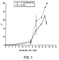
FIGURE 1 shows the rate of maturation of bovine oocytes in vitro. - Recipient oocytes the subject of this experimental procedure are designated MAGIC (Metaphase Arrested G1/G0 AcceptIng Cytoplast) Recipients.
- The nuclear and cytoplasmic events during in vitro oocyte maturation were studied. In addition the roles of fusion and activation in embryos reconstructed at different ages were also investigated. The studies have shown that oocyte maturation is asynchronous; however, a population of matured oocytes can be morphologically selected at 18 hours (
Figure 1 ). - In
Figure 1 ovaries were obtained from a local abattoir and maintained at 28-32°C during transport to the laboratory. Cumulus oocyte complexes (COC's) were aspirated from follicles 3-10mm in diameter using a hypodermic needle (1.2mm internal diameter) and placed into sterile plastic universal containers. The universal containers were placed into a warmed chamber (35°C) and the follicular material allowed to settle for 10-15 minutes before pouring off three quarters of the supernatant. The remaining follicular material was diluted with an equal volume of dissection medium (TCM 199 with Earles salts (Gibco), 75.0 mg/l kanamycin, 30.0mM Hepes, pH 7.4, osmolarity 280 mOsmols/Kg H2O) supplemented with 10% bovine serum, transferred into an 85mm petri dish and searched for COC's under a dissecting microscope. - Complexes with at least 2-3 compact layers of cumulus cells were selected washed three times in dissection medium and transferred into maturation medium (TC medium 199 with Earles salts (Gibco), 75mg/l kanamycin, 30.0mM Hepes, 7.69mM NaHCO3, pH 7.8, osmolarity 280 mOsmols/Kg H2O) supplemented with 10% bovine serum and 1x10⁶ granulosa cells/ml and cultured on a rocking table at 39°C in an atmosphere of 5% CO2 in air. Oocytes were removed from the maturation dish and wet mounted on ethanol cleaned glass slides under coverslips which were attached using a mixture of 5% petroleum jelly 95% wax. Mounted embryos were then fixed for 24 hours in freshly prepared methanol: glacial acetic acid (3:1), stained with 45% aceto-orcein (Sigma) and examined by phase contrast and DIC microscopy using a Nikon Microphot-SA, the graph in
Figure 1 shows the percentage of oocytes at MII and those with-a visible polar body. - If maturation is then continued until 24 hours these oocytes activate at a very low rate (24%) in mannitol containing calcium (Table 1a). However, removal of calcium and magnesium from the electropulsing medium prevents any activation.
- Table la shows activation of bovine follicular oocytes matured in vitro for different periods. Oocytes were removed from the maturation medium, washed once in activation medium, placed into the activation chamber and given a single electrical pulse of 1.25kV/cm for 80µs.
Table 1a No. of oocytes (N) Hours post onset of maturation (hpm) [age (hrs)] Pronuclear formation (% activation) 73 24 24.6 99 30 84.8 55 45 92.7* *many 2 or more pronuclei - Table 1b shows activation response of in vitro matured bovine oocytes sham enucleated at approximately 22 hours post onset of maturation (hpm). Oocytes were treated exactly as for enucleation, a small volume of cytoplasm was aspirated not containing the metaphase plate. After manipulation the oocytes were given a single DC pulse of 1.25 KV/cm and returned to the maturation medium, at 30 hpm and 42 hpm groups of oocytes were mounted, fixed and stained with aceto-orcein. The results show the number of oocytes at each time point from five individual experiments as the number of cells having pronuclei with respect to the total number of cells.
Table 1b EXPERIMENT No. cells having pronuclei/ Total no. of cells 30 hpmNo. cells having pronuclei/ Total no. of cells 42 hpm 1 1/8 - 2 0/24 0/30 3 0/21 0/22 4 0/27 0/25 5 0/19 0/1 hpm = hours post onset of maturation - Table 2 shows pronuclear formation in enucleated oocytes fused to primary bovine fibroblasts (24 hpm) and subsequently activated (42hpm). The results represent five separate experiments. Oocytes were divided into two groups, group A were incubated in nocodazole for 1 hour prior to activation and for 6 hours following activation. Group B were not treated with nocodazole. Activated oocytes were fixed and stained with aceto-
orcein 12 hours post activation. The number of pronuclei (PN) in each parthenote was then scored under phase contrast. The results are expressed as the percentage of activated oocytes containing 1 or more pronuclei.Table 2 TOTAL _1 PN 2 PN 3 PN 4 PN >4 PN GROUP A 52 100 0 0 0 0 GROUP B 33 45.2 25.8 16.1 3.2 9.7 - The absence of an organised spindle and the absence of a polar body suggests that in order to maintain ploidy in the reconstructed embryo then only a diploid i.e. G0/G1 nucleus should be transferred into this cytoplasmic situation. Incubation of activated oocytes in the presence of the microtubule inhibitor nocodazole for 5 hours, 1 hour prior to and following the activation stimulus prevents the formation of micronuclei (Table 2) and thus when the donor nucleus is in the G0/G1 phase of the cell cycle the correct ploidy of the reconstructed embryo is maintained.
- These results show that:
- i) these oocytes can be enucleated at 18 hours post onset of maturation (
Figure 1 ); - ii) enucleated oocytes can be fused to donor blastomeres/cells in either 0.3M mannitol or 0.27M sucrose alternatively the donor the cells or nuclei can be injected in calcium free medium in the absence of any activation response;
- iii) reconstructed embryos or enucleated pulsed oocytes can be cultured in maturation medium and do not undergo spontaneous activation;
- iv) the transferred nucleus is seen to undergo nuclear envelope breakdown (NEBD) and chromosome condensation. No organised meiotic/mitotic spindle is observed regardless of the cell cycle stage of the transferred nucleus;
- v) such manipulated couplets will activate at 30 hours and 42 hours with a frequency equal to unmanipulated control oocytes;
- vi) no polar body is observed following subsequent activation, regardless of the cell cycle stage of the transferred nucleus;
- viii) upon subsequent activation 1-5 micronuclei are formed per reconstructed zygote (Table 2).
- In preliminary experiments this technique has been applied to the reconstruction of bovine embryos using primary fibroblasts synchronised in the G0 phase of the cell cycle by serum starvation for five days. The results are summarised in Table 3.
- Table 3 shows development of bovine embryos reconstructed by nuclear transfer of serum starved (G0) bovine primary fibroblasts into enucleated unactivated MII oocytes. Embryos were reconstructed at 24 hpm and the fused couplets activated at 42 hpm. Fused couplets were incubated in nocodazole (5µg/ml) in M2 medium for 1 hour prior to activation and 5 hours post activation. Couplets were activated with a single DC pulse of 1.25 KV/cm for 80µsec.
Table 3 EXPERIMENT NUMBER NUMBER OF BLASTOCYSTS/ TOTAL NUMBER OF FUSED COUPLETS % BLASTOCYSTS 1 1/30 3.3 2 4/31 12.9 - Similar observations to those in Example 1 have also been made in ovine oocytes which have been matured in vivo. Freshly ovulated oocytes can be retrieved by flushing from the oviducts of
superstimulated ewes 24 hours after prostaglandin treatment. The use of calcium magnesium free PHS/1.0% FCS as a flushing medium prevents oocyte activation. Oocytes can be enucleated in calcium free medium and donor cells introduced as above in the absence of activation. No organised spindle is observed, multiple nuclei are formed upon subsequent activation and this may be suppressed by nocodazole treatment. - In preliminary experiments in sheep, a single pregnancy has resulted in the birth of a single live lamb. The results are summarised in Tables 4 and 5.
- Table 4 shows development of ovine embryos reconstructed by transfer of an embryo derived established cell line to unactivated enucleated in vivo matured ovine oocytes. Oocytes were obtained from superstimulated Scottish blackface ewes, the cell line was established from the embryonic disc of a day 9 embryo obtained from a Welsh mountain ewe. Reconstructed embryos were cultured in the ligated oviduct of a temporary recipient ewe for 6 days, recovered and assessed for development.
Table 4 DATE OF NUCLEAR TRANSFER PASSAGE NUMBER NUMBER OF MORULA, BLA STOCYSTS / TOTAL NUMBER 17.1.95 6 4/28 19.1.95 7 1/10 31.1.95 13 0/2 2.2.95 13 0/14 7.2.95 11 1/9 9.2.95 11 1/2 14.2.95 12 16.2.95 13 3/13 TOTAL 10/78 (12.8%) - Table 5 shows induction of pregnancy following transfer of all morula/blastocyst stage reconstructed embryos to the uterine horn of synchronised final recipient blackface ewes. The table shows the total number of embryos from each group transferred the frequency of pregnancy in terms of ewes and embryos, in the majority of
cases 2 embryos were transferred to each ewe. A single twin pregnancy was established which resulted in the birth of a single live lamb.Table 5 PASSAGE NUMBER "MAGIC" P6 4 P7 1 PH 2 P12 0 P13 3 TOTAL MOR/ BL 10 TOTAL NUMBER EWES 6 PREGNANT EWES % 1 (16.7) FOETUSES/ TOTAL TRANSFERRED (%) 2/10 (20.0)
Claims (11)
- A method of reconstituting a non-human mammal embryo, the process comprising transferring a non-human differentiated diploid nucleus into a non-human enucleated oocyte which is arrested in the metaphase of the second meiotic division without concomitantly activating the oocyte, keeping the non-human nucleus exposed to the cytoplasm of the non-human recipient for a period of time sufficient for the non-human embryo to become capable of giving rise to a live birth and subsequently activating the reconstituted embryo while maintaining correct ploidy.
- A method as claimed in claim 1, in which the non-human mammal is an ungulate species.
- A method as claimed in claim 2, in which the non-human mammal is a cow or bull, pig, goat, sheep, or horse.
- A method as claimed in claim 1, in which the non-human mammal is a rodent species.
- A method as claimed in claim 4, in which the rodent is a rat or a mouse.
- A method as claimed in any one of claims 1 to 5, in which the differentiated cell is obtained ex vivo or from an in vitro culture.
- A method as claimed in any one of claims 1 to 6, in which the donor nucleus is genetically modified.
- A method as claimed in any one of claims 1 to 7, wherein the diploid nucleus is donated by a quiescent cell.
- A method as claimed in any one of claims 1 to 8, wherein nuclear transfer is achieved by cell fusion.
- A method as claimed in any one of claims 1 to 9, wherein the non-human mammal is a cow or bull and wherein the donor nucleus is kept exposed to the recipient cytoplasm for a period of time of from 6 to 20 hours prior to activation.
- A method of preparing a non-human mammal, the method comprising:(a) reconstituting a non-human mammal embryo as claimed in any preceding claim;(b) causing a non-human mammal to develop to term from the embryo; and(c) optionally, breeding from the non-human mammal so formed.
Applications Claiming Priority (3)
| Application Number | Priority Date | Filing Date | Title |
|---|---|---|---|
| GB9517779 | 1995-08-31 | ||
| GBGB9517779.6A GB9517779D0 (en) | 1995-08-31 | 1995-08-31 | Biological manipulation |
| PCT/GB1996/002098 WO1997007668A1 (en) | 1995-08-31 | 1996-08-30 | Unactivated oocytes as cytoplast recipients for nuclear transfer |
Publications (2)
| Publication Number | Publication Date |
|---|---|
| EP0847237A1 EP0847237A1 (en) | 1998-06-17 |
| EP0847237B1 true EP0847237B1 (en) | 2008-07-09 |
Family
ID=10779997
Family Applications (1)
| Application Number | Title | Priority Date | Filing Date |
|---|---|---|---|
| EP96928586A Expired - Lifetime EP0847237B1 (en) | 1995-08-31 | 1996-08-30 | Unactivated oocytes as cytoplast recipients for nuclear transfer |
Country Status (23)
| Country | Link |
|---|---|
| US (12) | US6252133B1 (en) |
| EP (1) | EP0847237B1 (en) |
| JP (3) | JP2000506721A (en) |
| KR (4) | KR100743006B1 (en) |
| CN (3) | CN1772891A (en) |
| AT (1) | ATE400176T1 (en) |
| AU (1) | AU728809B2 (en) |
| BR (1) | BR9610013A (en) |
| CA (1) | CA2229657A1 (en) |
| CZ (1) | CZ60498A3 (en) |
| DE (1) | DE69637591D1 (en) |
| DK (1) | DK0847237T3 (en) |
| ES (1) | ES2309989T3 (en) |
| GB (1) | GB9517779D0 (en) |
| HK (1) | HK1004937A1 (en) |
| HU (1) | HUP9802485A3 (en) |
| IL (1) | IL123417A0 (en) |
| NO (1) | NO980846L (en) |
| NZ (2) | NZ335407A (en) |
| PL (1) | PL325336A1 (en) |
| SG (1) | SG75919A1 (en) |
| WO (1) | WO1997007668A1 (en) |
| ZA (1) | ZA967383B (en) |
Families Citing this family (190)
| Publication number | Priority date | Publication date | Assignee | Title |
|---|---|---|---|---|
| US5480772A (en) | 1993-02-03 | 1996-01-02 | Brandeis University | In vitro activation of a nucleus |
| US7304204B2 (en) | 1995-08-31 | 2007-12-04 | Roslin Institute | Ungulates produced by nuclear transfer of G1 cells |
| GB9517780D0 (en) * | 1995-08-31 | 1995-11-01 | Roslin Inst Edinburgh | Biological manipulation |
| GB9517779D0 (en) * | 1995-08-31 | 1995-11-01 | Roslin Inst Edinburgh | Biological manipulation |
| US6077697A (en) * | 1996-04-10 | 2000-06-20 | Chromos Molecular Systems, Inc. | Artificial chromosomes, uses thereof and methods for preparing artificial chromosomes |
| US6025155A (en) * | 1996-04-10 | 2000-02-15 | Chromos Molecular Systems, Inc. | Artificial chromosomes, uses thereof and methods for preparing artificial chromosomes |
| CA2267220A1 (en) | 1996-10-11 | 1998-04-23 | The Texas A & M University System | Methods for the generation of primordial germ cells and transgenic animal species |
| US20090092588A1 (en) * | 1997-01-10 | 2009-04-09 | University Of Massachusetts As Represented By Its Amherst Campus | Cloned ungulate embryos and animals, use of cells, tissues and organs thereof for transplantation therapies including parkinson's disease |
| US5945577A (en) * | 1997-01-10 | 1999-08-31 | University Of Massachusetts As Represented By Its Amherst Campus | Cloning using donor nuclei from proliferating somatic cells |
| US6235969B1 (en) * | 1997-01-10 | 2001-05-22 | University Of Massachusetts | Cloning pigs using donor nuclei from non-quiescent differentiated cells |
| US6011197A (en) | 1997-03-06 | 2000-01-04 | Infigen, Inc. | Method of cloning bovines using reprogrammed non-embryonic bovine cells |
| ATE318316T1 (en) | 1997-03-26 | 2006-03-15 | Imp College Innovations Ltd | ANTICOAGULANT FUSION PROTEIN ANCHORED IN THE CELL MEMBRANE |
| BR9811657A (en) | 1997-07-03 | 2000-09-05 | Univ Massachusetts Public Inst | Cloning using donor nuclei from differentiated serum-free cells |
| US7923015B2 (en) | 1997-08-14 | 2011-04-12 | Institut Pasteur | Methods for direct visualization of active synapses |
| US7923216B2 (en) | 1997-08-14 | 2011-04-12 | Institut Pasteur | In vivo modulation of neuronal transport |
| JPH11127852A (en) * | 1997-10-28 | 1999-05-18 | Snow Brand Milk Prod Co Ltd | Go phase synchronization of epithelial cell |
| CA2318117A1 (en) * | 1998-01-16 | 1999-07-22 | Agrobiogen Gmbh | Efficient nuclear transfer using primordial germ cells |
| US7351876B2 (en) | 1998-01-16 | 2008-04-01 | Agrobiogen Gmbh Biotechnologie | Efficient nuclear transfer with primordial gametes |
| US6331659B1 (en) * | 1998-01-21 | 2001-12-18 | University Of Hawaii | Cumulus cells as nuclear donors |
| WO1999046982A1 (en) * | 1998-03-16 | 1999-09-23 | Relag Pty Ltd | Porcine nuclear transfer |
| AUPP294898A0 (en) * | 1998-04-15 | 1998-05-07 | Monash University | Method of nuclear transfer |
| GB9808325D0 (en) * | 1998-04-20 | 1998-06-17 | Ltr Ciz Di Associazione Italia | Source of nuclei for nuclear transfer |
| US6143564A (en) * | 1998-07-07 | 2000-11-07 | University Of Hawaii | Use of the polar body chromosomes for the production of embryos and normal offspring |
| GB9820988D0 (en) * | 1998-09-25 | 1998-11-18 | Biotech & Biolog Scien Res | Screening method |
| WO2000022098A1 (en) * | 1998-10-12 | 2000-04-20 | Geron Bio-Med Limited | Porcine oocytes with improved developmental competence |
| EP1818397A1 (en) * | 1998-11-02 | 2007-08-15 | Trustees Of Tufts College | Methods for cloning animals |
| US6781030B1 (en) | 1998-11-02 | 2004-08-24 | Trustee Of Tufts College, Ballou Hall | Methods for cloning mammals using telophase oocytes |
| NZ511299A (en) | 1998-11-19 | 2003-02-28 | Ppl Therapeutics Scotland Ltd | Stabilisation of milk from transgenic animals through expression of a serine proteinase inhibitor and optionally fibrinogen in the mammary gland |
| US6700037B2 (en) | 1998-11-24 | 2004-03-02 | Infigen, Inc. | Method of cloning porcine animals |
| US6258998B1 (en) | 1998-11-24 | 2001-07-10 | Infigen, Inc. | Method of cloning porcine animals |
| US7531715B1 (en) * | 1999-01-13 | 2009-05-12 | Ppl Therapeutics (Scotland) | Double nuclear transfer method and results thereof |
| ATE407203T1 (en) * | 1999-01-13 | 2008-09-15 | Revivicor Inc | DOUBLE CORE TRANSFER METHOD AND ITS RESULTS |
| AU776150B2 (en) | 1999-01-28 | 2004-08-26 | Medical College Of Georgia Research Institute, Inc. | Composition and method for (in vivo) and (in vitro) attenuation of gene expression using double stranded RNA |
| EP1179000A4 (en) | 1999-02-26 | 2005-10-12 | Millennium Pharm Inc | Secreted proteins and uses thereof |
| CA2361943C (en) * | 1999-03-04 | 2011-05-17 | Ppl Therapeutics (Scotland) Limited | Genetic modification of somatic cells and uses thereof |
| US20020012660A1 (en) * | 1999-03-04 | 2002-01-31 | Alan Colman | Method of preparing a somatic cells for nuclear transfer |
| EP1194570A1 (en) | 1999-06-23 | 2002-04-10 | PPL Therapeutics (Scotland) Limited | Fusion proteins incorporating lysozyme |
| KR100368435B1 (en) * | 1999-06-30 | 2003-01-24 | 황우석 | Method of cell fusion for producing a clone animal using somatic cell |
| EP1117763A4 (en) * | 1999-06-30 | 2004-12-01 | Hwang Woo Suk | Method for producing cloned tigers by employing inter-species nuclear transplantation technique |
| KR100414043B1 (en) * | 1999-06-30 | 2004-01-13 | 황우석 | Clone animal reproduced from somatic cell and method for production thereof |
| CA2334953A1 (en) * | 1999-06-30 | 2001-01-04 | Woo-Suk Hwang | Method for producing human cloned embryos by employing inter-species nuclear transplantation technique |
| US7291714B1 (en) | 1999-06-30 | 2007-11-06 | Millennium Pharmaceuticals, Inc. | Glycoprotein VI and uses thereof |
| EP1109891A4 (en) | 1999-06-30 | 2004-11-17 | Hwang Woo Suk | Method for producing cloned cows |
| EP2360254A1 (en) | 1999-08-23 | 2011-08-24 | Dana-Farber Cancer Institute, Inc. | Assays for screening anti-pd-1 antibodies and uses thereof |
| EP2275559A3 (en) | 1999-09-28 | 2011-03-23 | Shire Human Genetic Therapies, Inc. | Optimized messenger RNA |
| WO2002064799A2 (en) | 1999-09-28 | 2002-08-22 | Transkaryotic Therapies, Inc. | Optimized messenger rna |
| AU1777301A (en) * | 1999-11-19 | 2001-05-30 | Hematech, Llc | Production of ungulates, preferably bovines that produce human immunoglobulins |
| KR100417566B1 (en) * | 1999-11-19 | 2004-02-05 | 한국생명공학연구원 | A mass production method of embryos by nuclear transfer using somatic cells |
| US7074983B2 (en) | 1999-11-19 | 2006-07-11 | Kirin Beer Kabushiki Kaisha | Transgenic bovine comprising human immunoglobulin loci and producing human immunoglobulin |
| US7820878B2 (en) * | 1999-11-19 | 2010-10-26 | Kyowa Hakko Kirin Co., Ltd. | Production of ungulates, preferably bovines that produce human immunoglobulins |
| US7414170B2 (en) * | 1999-11-19 | 2008-08-19 | Kirin Beer Kabushiki Kaisha | Transgenic bovines capable of human antibody production |
| GB9927444D0 (en) * | 1999-11-19 | 2000-01-19 | Cancer Res Campaign Tech | Inhibiting gene expression |
| CA2396210A1 (en) | 2000-01-04 | 2001-07-12 | Chikara Kubota | Method for cloning animals with targetted genetic alterations by transfer_of long-term cultured male or female somatic cell nuclei, comprising artificially-induced genetic alterations, to enucleated recipient cells |
| US6635802B1 (en) * | 2000-01-10 | 2003-10-21 | The Texas A&M University System | Nuclear transfer using cells cultured in serum starvation media containing apoptosis inhibitors |
| US7122177B2 (en) | 2000-01-20 | 2006-10-17 | Diabcell Pty Ltd. | Preparation and xenotransplantation of porcine islets |
| WO2001059076A2 (en) * | 2000-02-09 | 2001-08-16 | The Government Of The United States Of America, As Represented By The Secretary, Department Of Health And Human Services | Premeiotic and postmeiotic origin of teratomas: isolated teratoma stem cells for therapeutic uses |
| WO2001068831A2 (en) | 2000-03-15 | 2001-09-20 | The University Of Georgia Research Foundation, Inc. | Effective nuclear reprogramming in mammals |
| GB2360522A (en) | 2000-03-24 | 2001-09-26 | Geron Corp | A strategy for maintaining pregnancy |
| AU2001261650A1 (en) * | 2000-05-15 | 2001-11-26 | Geron Corporation | Ovine tissue for xenotransplantation |
| US20020039736A1 (en) | 2000-06-05 | 2002-04-04 | Byrne Michael C. | Compositions, kits, and methods for identification and modulation of type I diabetes |
| AU2001273096B8 (en) | 2000-06-28 | 2005-10-13 | Dana-Farber Cancer Institute, Inc. | PD-L2 molecules: novel PD-1 ligands and uses therefor |
| MXPA03001915A (en) | 2000-08-03 | 2004-09-10 | Therapeutic Human Polyclonals | Production of humanized antibodies in transgenic animals. |
| US6677500B2 (en) | 2000-08-15 | 2004-01-13 | Board Of Trustees Of The University Of Illinois | Ungulates expressing exogenous IGF-I in their milk |
| FR2814642B1 (en) | 2000-10-03 | 2005-07-01 | Ass Pour Le Dev De La Rech En | TRANSGENIC MOUSE FOR THE TARGETED RECOMBINATION MEDIATED BY THE MODIFIED CRE-ER |
| WO2002028164A2 (en) * | 2000-10-06 | 2002-04-11 | Advanced Cell Technology, Inc. | Cloning endangered and extinct species |
| US6821731B2 (en) | 2000-11-28 | 2004-11-23 | Wyeth | Expression analysis of FKBP nucleic acids and polypeptides useful in the diagnosis of prostate cancer |
| WO2002044419A2 (en) | 2000-11-28 | 2002-06-06 | Wyeth | Expression analysis of kiaa nucleic acids and polypeptides useful in the diagnosis and treatment of prostate cancer |
| IL156234A0 (en) * | 2000-11-30 | 2004-01-04 | Stemron Inc | Isolated homozygous stem cells, differentiated cells derived therefrom, and materials and methods for making and using same |
| US7491534B2 (en) * | 2000-12-22 | 2009-02-17 | Kirin Holdings Kabushiki Kaisha | Methods for altering cell fate to generate T-cells specific for an antigen of interest |
| ATE466089T1 (en) | 2000-12-22 | 2010-05-15 | Agronomique Inst Nat Rech | POSITION-INDEPENDENT AND TISSUE-SPECIFIC EXPRESSION OF A TRANSGENE IN THE MILK OF A TRANSGENIC ANIMAL |
| US20020142397A1 (en) * | 2000-12-22 | 2002-10-03 | Philippe Collas | Methods for altering cell fate |
| MXPA03005624A (en) * | 2000-12-22 | 2004-12-03 | Vaurox Llc | Methods for cloning mammals using reprogrammed donor chromatin or donor cells. |
| EP1860199B1 (en) | 2001-01-12 | 2014-03-05 | Exelixis, Inc. | Modulating insulin receptor signaling |
| MXPA03006751A (en) | 2001-01-30 | 2005-04-08 | Amy K Rebholtz | Systems of transferring embryos and managing recipients. |
| CA2369944A1 (en) | 2001-01-31 | 2002-07-31 | Nucleonics Inc. | Use of post-transcriptional gene silencing for identifying nucleic acid sequences that modulate the function of a cell |
| JP2002262716A (en) * | 2001-03-07 | 2002-09-17 | National Agricultural Research Organization | Method for creating livestock individual using established cell line |
| US8981061B2 (en) | 2001-03-20 | 2015-03-17 | Novo Nordisk A/S | Receptor TREM (triggering receptor expressed on myeloid cells) and uses thereof |
| US7126039B2 (en) * | 2001-03-21 | 2006-10-24 | Geron Corporation | Animal tissue with carbohydrate antigens compatible for human transplantation |
| US7265262B2 (en) * | 2001-03-21 | 2007-09-04 | Roslin Institute (Edinburgh) | Telomerizing nuclear donor cells and improving the efficiency on nuclear transfer |
| MXPA03008959A (en) | 2001-04-02 | 2004-10-15 | Wyeth Corp | Pd-1, a receptor for b7-4, and uses therefor. |
| EP1572868A4 (en) | 2001-04-16 | 2007-04-04 | Wyeth Corp | Novel streptococcus pneumoniae open reading frames encoding polypeptide antigens and uses thereof |
| CA2450652A1 (en) * | 2001-06-14 | 2002-12-27 | Infigen, Inc. | Methods for cloning mammals using remodeling factors |
| EP1406917A4 (en) * | 2001-06-15 | 2007-11-14 | New Century Pharmaceuticals | Human albumin animal models for drug evaluation, toxicology and immunogenicity studies |
| IL159756A0 (en) | 2001-07-12 | 2004-06-20 | Univ Massachusetts | IN VIVO PRODUCTION OF SMALL INTERFERING RNAs THAT MEDIATE GENE SILENCING |
| EP1900815B1 (en) | 2001-07-12 | 2016-09-07 | University of Massachusetts | In vivo production of small interfering RNAs that mediate gene silencing |
| US20030211603A1 (en) * | 2001-08-14 | 2003-11-13 | Earp David J. | Reprogramming cells for enhanced differentiation capacity using pluripotent stem cells |
| US20030224380A1 (en) * | 2001-10-25 | 2003-12-04 | The General Hospital Corporation | Genes and polymorphisms on chromosome 10 associated with Alzheimer's disease and other neurodegenerative diseases |
| AU2002364945A1 (en) * | 2001-10-25 | 2003-07-09 | Neurogenetics, Inc. | Genes and polymorphisms on chromosome 10 associated with alzheimer's disease and other neurodegenerative diseases |
| US20030170678A1 (en) * | 2001-10-25 | 2003-09-11 | Neurogenetics, Inc. | Genetic markers for Alzheimer's disease and methods using the same |
| WO2003046129A2 (en) * | 2001-11-09 | 2003-06-05 | Trustees Of The University Of Pennsylvania | Compositions for the derivation of germ cells from stem cells and methods of use thereof |
| WO2003042362A2 (en) | 2001-11-09 | 2003-05-22 | Dana-Farber Cancer Institute, Inc. | PGC-1β, A NOVEL PGC-1 HOMOLOGUE AND USES THEREFOR |
| CA2473909A1 (en) * | 2002-01-15 | 2003-07-24 | Advanced Cell Technology, Inc. | Cloning b and t lymphocytes |
| US20050090004A1 (en) * | 2003-01-16 | 2005-04-28 | Sayre Chauncey B. | Stem cell maturation for all tissue lines |
| US20030134422A1 (en) * | 2002-01-16 | 2003-07-17 | Sayre Chauncey Bigelow | Stem cell maturation for all tissue lines |
| US20050170506A1 (en) * | 2002-01-16 | 2005-08-04 | Primegen Biotech Llc | Therapeutic reprogramming, hybrid stem cells and maturation |
| WO2003085105A1 (en) * | 2002-04-01 | 2003-10-16 | Gtc Biotherapeutics, Inc. | A method for selecting cell lines to be used for nuclear transfer in mammalian species |
| US20030217378A1 (en) * | 2002-04-05 | 2003-11-20 | The University Of Georgia Research Foundation, Inc | Cloning using rapidly matured oocytes |
| US7612250B2 (en) * | 2002-07-29 | 2009-11-03 | Trustees Of Tufts College | Nuclear transfer embryo formation method |
| KR20050058407A (en) | 2002-08-20 | 2005-06-16 | 밀레니엄 파머슈티컬스 인코퍼레이티드 | Compositions, kits, and methods for identification, assessment, prevention, and therapy of cervical cancer |
| WO2004032877A2 (en) | 2002-10-10 | 2004-04-22 | Wyeth | Compositions, organisms and methodologies employing a novel human kinase |
| AU2003286442A1 (en) * | 2002-10-16 | 2004-05-04 | Advanced Cell Technology, Inc. | Methods using gene trapped stem cells for marking pathways of stem cell differentiation and making and isolating differentiated cells |
| CA2503491A1 (en) | 2002-10-24 | 2004-05-06 | Wyeth | Calcineurin-like human phosphoesterase |
| EP1563084A4 (en) * | 2002-11-08 | 2006-03-01 | Hematech Llc | Transgenic ungulates having reduced prion protein activity and uses thereof |
| CA2505416A1 (en) | 2002-11-21 | 2004-06-10 | Wyeth | Methods for diagnosing rcc and other solid tumors |
| WO2004050831A2 (en) | 2002-11-27 | 2004-06-17 | Wyeth | Compositions, organisms and methodologies employing a novel human kinase |
| CA2511012C (en) | 2002-12-20 | 2018-02-27 | Applera Corporation | Genetic polymorphisms associated with myocardial infarction, methods of detection and uses thereof |
| PT1581812E (en) | 2003-01-06 | 2008-09-22 | Wyeth Corp | Compositions and methods for diagnosing and treating colon cancers |
| EP2314692B1 (en) | 2003-06-02 | 2021-02-24 | University of Massachusetts | Methods and compositions for enhancing the efficacy and specificity of RNAi |
| CA2528012C (en) | 2003-06-02 | 2015-11-24 | University Of Massachusetts | Methods and compositions for controlling efficacy of rna silencing |
| CA2535901A1 (en) | 2003-08-14 | 2005-02-24 | Exelixis, Inc. | Ups as modifiers of the beta catenin pathway and methods of use |
| US7799518B2 (en) | 2003-10-07 | 2010-09-21 | Millennium Pharmaceuticals, Inc. | Nucleic acid molecules and proteins for the identification, assessment, prevention, and therapy of ovarian cancer |
| US7951532B2 (en) | 2003-11-12 | 2011-05-31 | Children's Hospital Medical Center | Method of screening a midkine modulating agent |
| US7682828B2 (en) * | 2003-11-26 | 2010-03-23 | Whitehead Institute For Biomedical Research | Methods for reprogramming somatic cells |
| CA3050151C (en) | 2003-11-26 | 2023-03-07 | Celera Corporation | Single nucleotide polymorphisms associated with cardiovascular disorders and statin response, methods of detection and uses thereof |
| US7420099B2 (en) * | 2004-04-22 | 2008-09-02 | Kirin Holdings Kabushiki Kaisha | Transgenic animals and uses thereof |
| EP1745154B1 (en) | 2004-05-07 | 2012-08-01 | Celera Corporation | Genetic polymorphisms associated with liver fibrosis methods of detection and uses thereof |
| EP1812041A2 (en) | 2004-10-22 | 2007-08-01 | Dana-Farber Cancer Institute, Inc. | Compositions and methods for modulating pgc-1 to treat lipid-related diseases and disorders |
| GB0426146D0 (en) | 2004-11-29 | 2004-12-29 | Bioxell Spa | Therapeutic peptides and method |
| CN1274829C (en) | 2004-12-10 | 2006-09-13 | 四川大学华西医院 | Anti EB virus resulted tumour polypeptide, and its use and preparing method |
| EP1856296A2 (en) | 2005-03-11 | 2007-11-21 | Applera Corporation | Genetic polymorphisms associated with coronary heart disease, methods of detection and uses thereof |
| US20070011603A1 (en) * | 2005-07-06 | 2007-01-11 | Mikko Makela | Method, system, device and software product for showing tooltips for page segments and generating content for the page segments |
| CA2617930A1 (en) | 2005-08-09 | 2007-03-29 | Revivicor, Inc. | Transgenic ungulates expressing ctla4-ig and uses thereof |
| WO2007030820A2 (en) | 2005-09-09 | 2007-03-15 | The Johns Hopkins University | Manipulation of regulatory t cell and dc function by targeting neuritin gene using antibodies, agonists and antagonists |
| WO2007035778A2 (en) | 2005-09-19 | 2007-03-29 | Histogenics Corporation | Cell-support matrix and a method for preparation thereof |
| US7799530B2 (en) | 2005-09-23 | 2010-09-21 | Celera Corporation | Genetic polymorphisms associated with cardiovascular disorders and drug response, methods of detection and uses thereof |
| US7695911B2 (en) | 2005-10-26 | 2010-04-13 | Celera Corporation | Genetic polymorphisms associated with Alzheimer's Disease, methods of detection and uses thereof |
| PE20081216A1 (en) | 2006-09-01 | 2008-09-04 | Therapeutic Human Polyclonals Inc | ENHANCED EXPRESSION OF HUMAN OR HUMANIZED IMMUNOGLOBULIN IN NON-HUMAN TRANSGENIC ANIMALS |
| US7993833B2 (en) | 2006-09-11 | 2011-08-09 | Celera Corporation | Genetic polymorphisms associated with psoriasis, methods of detection and uses thereof |
| CA2666346C (en) | 2006-10-20 | 2016-02-23 | Celera Corporation | Genetic polymorphisms associated with venous thrombosis, methods of detection and uses thereof |
| KR101398220B1 (en) * | 2006-10-30 | 2014-05-22 | 건국대학교 산학협력단 | Methods for producing transgenic embryos |
| WO2008085891A1 (en) * | 2007-01-05 | 2008-07-17 | Worcester Polytechnic Institute | Oocyte spindle-associated factors improve somatic cell cloning |
| CA3071055A1 (en) | 2007-04-07 | 2008-10-16 | Whitehead Institute For Biomedical Research | Reprogramming of somatic cells |
| WO2008134522A1 (en) * | 2007-04-26 | 2008-11-06 | President And Fellows Of Harvard College | Deriving embryonic stem cells |
| WO2008144580A2 (en) * | 2007-05-17 | 2008-11-27 | Oregon Health & Science University | Primate stem cells produced by somatic cell nuclear transfer |
| US20090111764A1 (en) * | 2007-10-25 | 2009-04-30 | Searete Llc, A Limited Liability Corporation Of The State Of Delaware | Mitochondrial selection |
| US8663980B2 (en) | 2007-10-26 | 2014-03-04 | Janssen Biotech, Inc. | Vectors, host cells, and methods of production and uses |
| US8039212B2 (en) | 2007-11-05 | 2011-10-18 | Celera Corporation | Genetic polymorphisms associated with liver fibrosis, methods of detection and uses thereof |
| US20090221620A1 (en) | 2008-02-20 | 2009-09-03 | Celera Corporation | Gentic polymorphisms associated with stroke, methods of detection and uses thereof |
| KR100915397B1 (en) * | 2008-02-22 | 2009-09-03 | 삼성전자주식회사 | Cover member, developing cartridge and developing unit for image forming apparatus |
| US20110071049A1 (en) | 2008-03-12 | 2011-03-24 | Nathaniel Heintz | Methods and compositions for translational profiling and molecular phenotyping |
| US8288094B2 (en) | 2008-04-09 | 2012-10-16 | Institut Pasteur | APOBEC3 mediated DNA editing |
| EP2300611B1 (en) | 2008-06-13 | 2017-08-09 | Whitehead Institute for Biomedical Research | Programming and reprogramming of cells |
| EP2878680B1 (en) | 2008-07-09 | 2016-06-08 | Celera Corporation | Genetic polymorphisms associated with cardiovascular diseases, methods of detection and uses thereof |
| US9181315B2 (en) | 2009-01-08 | 2015-11-10 | Dana-Farber Cancer Institute, Inc. | Compositions and methods for induced brown fat differentiation |
| US8722964B2 (en) | 2009-04-23 | 2014-05-13 | Transposagen Biopharmaceuticals, Inc. | Genetically engineered or transgenic rats exhibiting a cancer phenotype due to a disruption of germline tumor suppressor genes |
| CA2759608A1 (en) | 2009-04-24 | 2010-10-28 | Oregon Health & Science University | Methods for mitochondrial dna replacement in oocytes |
| EP2267153A1 (en) | 2009-05-26 | 2010-12-29 | Université Claude Bernard Lyon 1 | Identification of netrin-1 receptor unc5c gene mutation in solid cancers |
| WO2011002988A1 (en) | 2009-07-01 | 2011-01-06 | Transposagen Biopharmaceuticals, Inc. | Genetically modified rat models for severe combined immunodeficiency (scid) |
| WO2011011678A2 (en) | 2009-07-24 | 2011-01-27 | Transposagen Biopharmaceuticals, Inc. | Genetically modified rat models for cytokine-cytokine signaling pathways |
| US20110145936A1 (en) | 2009-07-30 | 2011-06-16 | Ostertag Eric M | Genetically Modified Rat Models for Pharmacokinetics |
| WO2011017518A2 (en) | 2009-08-05 | 2011-02-10 | Transposagen Biopharmaceuticals, Inc. | Genetically modified rat models for drug metabolism |
| WO2011022634A2 (en) | 2009-08-20 | 2011-02-24 | Transposagen Biopharmaceuticals, Inc. | Genetically modified rat models for pain |
| US9420770B2 (en) | 2009-12-01 | 2016-08-23 | Indiana University Research & Technology Corporation | Methods of modulating thrombocytopenia and modified transgenic pigs |
| US10429384B2 (en) | 2010-01-22 | 2019-10-01 | Dana-Farber Cancer Institute, Inc. | Compositions, kits, and methods for identification, assessment, prevention, and therapy of metabolic disorders |
| TWI518325B (en) | 2010-02-04 | 2016-01-21 | 自治醫科大學 | Identification, assessment, and therapy of cancers with innate or acquired resistance to alk inhibitors |
| WO2011120013A2 (en) | 2010-03-26 | 2011-09-29 | Trustees Of Dartmouth College | Vista regulatory t cell mediator protein, vista binding agents and use thereof |
| US20150231215A1 (en) | 2012-06-22 | 2015-08-20 | Randolph J. Noelle | VISTA Antagonist and Methods of Use |
| US10745467B2 (en) | 2010-03-26 | 2020-08-18 | The Trustees Of Dartmouth College | VISTA-Ig for treatment of autoimmune, allergic and inflammatory disorders |
| US8383793B2 (en) | 2010-04-15 | 2013-02-26 | St. Jude Children's Research Hospital | Methods and compositions for the diagnosis and treatment of cancer resistant to anaplastic lymphoma kinase (ALK) kinase inhibitors |
| US20110269735A1 (en) | 2010-04-19 | 2011-11-03 | Celera Corporation | Genetic polymorphisms associated with statin response and cardiovascular diseases, methods of detection and uses thereof |
| WO2012051699A1 (en) | 2010-10-22 | 2012-04-26 | Agri-Neo Inc. | Synergistic activity of peracetic acid and at least one sar inducer for the control of pathogens in and onto growing plants |
| US20120108651A1 (en) | 2010-11-02 | 2012-05-03 | Leiden University Medical Center (LUMC) Acting on Behalf of Academic Hospital Leiden (AZL) | Genetic polymorphisms associated with venous thrombosis and statin response, methods of detection and uses thereof |
| KR101352641B1 (en) | 2010-11-08 | 2014-01-17 | 광주과학기술원 | Chemically-Defined Methods for Preparing Multipotent Cells Derived from Terminally Differentiated Skeletal Muscle and Uses Thereof |
| WO2012112586A1 (en) | 2011-02-14 | 2012-08-23 | Revivicor, Inc. | Genetically modified pigs for xenotransplantation of vascularized xenografts and derivatives thereof |
| WO2012129198A1 (en) | 2011-03-23 | 2012-09-27 | Transposagen Biopharmaceuticals, Inc. | Genetically modified rat models for obesity and diabetes |
| CA2836288A1 (en) | 2011-05-16 | 2012-11-22 | The Curators Of The University Of Missouri | Porcine reproductive and respiratory syndrome virus resistant animals |
| CA2848368C (en) | 2011-09-13 | 2023-02-14 | Dana-Farber Cancer Institute, Inc. | Compositions and methods for brown fat induction and activity using fndc5 |
| CN108103069B (en) | 2012-02-15 | 2021-08-10 | 诺和诺德股份有限公司 | Antibodies that bind to and block trigger receptor-1 (TREM-1) expressed by myeloid cells |
| PL2814842T3 (en) | 2012-02-15 | 2018-12-31 | Novo Nordisk A/S | Antibodies that bind peptidoglycan recognition protein 1 |
| US9550830B2 (en) | 2012-02-15 | 2017-01-24 | Novo Nordisk A/S | Antibodies that bind and block triggering receptor expressed on myeloid cells-1 (TREM-1) |
| AU2013277051B2 (en) | 2012-06-22 | 2018-06-07 | King's College London | Novel VISTA-Ig constructs and the use of VISTA-Ig for treatment of autoimmune, allergic and inflammatory disorders |
| US9890215B2 (en) | 2012-06-22 | 2018-02-13 | King's College London | Vista modulators for diagnosis and treatment of cancer |
| CN109793893B (en) | 2012-09-07 | 2023-05-26 | 达特茅斯大学理事会 | VISTA modulators for diagnosis and treatment of cancer |
| CA2935378C (en) | 2013-12-24 | 2023-04-18 | Janssen Pharmaceutica Nv | Anti-vista antibodies and fragments |
| US11014987B2 (en) | 2013-12-24 | 2021-05-25 | Janssen Pharmaceutics Nv | Anti-vista antibodies and fragments, uses thereof, and methods of identifying same |
| MX2016016310A (en) | 2014-06-11 | 2017-10-20 | A Green Kathy | Use of vista agonists and antagonists to suppress or enhance humoral immunity. |
| TN2017000008A1 (en) | 2014-07-17 | 2018-07-04 | Novo Nordisk As | Site directed mutagenesis of trem-1 antibodies for decreasing viscosity. |
| US10077420B2 (en) | 2014-12-02 | 2018-09-18 | Histogenics Corporation | Cell and tissue culture container |
| JP2018505911A (en) | 2014-12-05 | 2018-03-01 | イミュネクスト,インコーポレーテッド | Identification of VSIG8 as a putative VISTA receptor and its use to produce a VISTA / VSIG8 modulator |
| JP2018500896A (en) | 2014-12-09 | 2018-01-18 | リジェネロン・ファーマシューティカルズ・インコーポレイテッドRegeneron Pharmaceuticals, Inc. | Non-human animal having humanized differentiation antigen group 274 gene |
| US10136642B2 (en) | 2015-02-19 | 2018-11-27 | Agri-Neo, Inc. | Composition of peracetic acid and at least one organic fungicide for the control and/or the treatment of diseases associated with the presence of pathogens, and method, use and kit involving said composition |
| CN107922497B (en) | 2015-06-24 | 2022-04-12 | 詹森药业有限公司 | anti-VISTA antibodies and fragments |
| US10091975B2 (en) | 2015-08-06 | 2018-10-09 | The Curators Of The University Of Missouri | Pathogen-resistant animals having modified CD163 genes |
| US20210106661A1 (en) | 2015-10-29 | 2021-04-15 | Dana-Farber Cancer Institute, Inc. | Methods for identification, assessment, prevention, and treatment of metabolic disorders using pm20d1 and n-lipidated amino acids |
| TWI756204B (en) | 2016-02-12 | 2022-03-01 | 比利時商楊森製藥公司 | Anti-vista antibodies and fragments, uses thereof, and methods of identifying same |
| SG11201808633RA (en) | 2016-04-15 | 2018-10-30 | Immunext Inc | Anti-human vista antibodies and use thereof |
| JOP20200240A1 (en) | 2018-04-02 | 2020-09-27 | Bristol Myers Squibb Co | Anti-trem-1 antibodies and uses thereof reference to sequence listing submitted electronically via efs-web |
| US11160260B2 (en) | 2018-04-17 | 2021-11-02 | The Curators Of The University Of Missouri | Methods for protecting porcine fetuses from infection with porcine reproductive and respiratory syndrome virus (PRRSV) |
| US20220211018A1 (en) | 2020-11-20 | 2022-07-07 | Revivicor, Inc. | Multi-transgenic pigs with growth hormone receptor knockout for xenotransplantation |
| US20230255185A1 (en) | 2021-09-20 | 2023-08-17 | Revivicor, Inc. | Multitransgenic pigs comprising ten genetic modifications for xenotransplantation |
Family Cites Families (28)
| Publication number | Priority date | Publication date | Assignee | Title |
|---|---|---|---|---|
| CN1030255A (en) * | 1987-04-14 | 1989-01-11 | 路明尼斯有限公司 | The animal of metastatic gene |
| US5057420A (en) | 1987-06-05 | 1991-10-15 | Granada Biosciences, Inc. | Bovine nuclear transplantation |
| CA2059199A1 (en) | 1992-01-10 | 1993-07-11 | Steen Willadsen | Method for producing mature reconstituted mammalian eggs by nuclear transplantation |
| GB2265909B (en) | 1992-04-01 | 1996-03-20 | Inst Of Animal Physiology And | Fusion of donor nucleus with recipient cytoplast in the absence of calcium ion |
| WO1994006422A1 (en) * | 1992-09-22 | 1994-03-31 | The Government Of The United States Of America As Represented By The Secretary, Department Of Health And Human Services | Use of taxol for treating lymphomas and breast cancer |
| CA2092258A1 (en) | 1992-12-30 | 1994-07-01 | Steven L. Stice | Synchronized donor cells for bovine nuclear transfer |
| US5480772A (en) | 1993-02-03 | 1996-01-02 | Brandeis University | In vitro activation of a nucleus |
| US5496720A (en) | 1993-02-10 | 1996-03-05 | Susko-Parrish; Joan L. | Parthenogenic oocyte activation |
| US5523226A (en) | 1993-05-14 | 1996-06-04 | Biotechnology Research And Development Corp. | Transgenic swine compositions and methods |
| US5333948A (en) | 1993-06-22 | 1994-08-02 | Tekonsha Engineering Company | Multiple-gain electronic brake actuator with trigger point inertial sensor |
| EP0711158B2 (en) * | 1993-07-29 | 2008-07-23 | THE GOVERNMENT OF THE UNITED STATES OF AMERICA, as represented by THE SECRETARY, DEPARTMENT OF HEALTH AND HUMAN SERVICES | Method of treating atherosclerosis or restenosis using microtubule stabilizing agent |
| WO1995017500A1 (en) | 1993-12-23 | 1995-06-29 | Abs Global, Inc. | Embryonic stem cells as nuclear donors and nuclear transfer techniques to produce chimeric and transgenic animals |
| GB9417831D0 (en) | 1994-09-05 | 1994-10-26 | Biotech & Biolog Scien Res | Biological manipulation |
| GB9517779D0 (en) | 1995-08-31 | 1995-11-01 | Roslin Inst Edinburgh | Biological manipulation |
| GB9517780D0 (en) | 1995-08-31 | 1995-11-01 | Roslin Inst Edinburgh | Biological manipulation |
| US7304204B2 (en) | 1995-08-31 | 2007-12-04 | Roslin Institute | Ungulates produced by nuclear transfer of G1 cells |
| US6215041B1 (en) | 1997-01-10 | 2001-04-10 | University Of Mmassachusetts | Cloning using donor nuclei from a non-quiesecent somatic cells |
| US6235969B1 (en) | 1997-01-10 | 2001-05-22 | University Of Massachusetts | Cloning pigs using donor nuclei from non-quiescent differentiated cells |
| US5945577A (en) | 1997-01-10 | 1999-08-31 | University Of Massachusetts As Represented By Its Amherst Campus | Cloning using donor nuclei from proliferating somatic cells |
| US7291764B1 (en) | 1997-01-10 | 2007-11-06 | University of Massachusetts, a Public Institution of Higher Education of the Commonwealth of Massachusetts, as Represented by its Amherst Campus, Office of Vice Chancellor for Research at Amherst | Cloning pigs using non-quiescent differentiated donor cells or nuclei |
| US7361804B1 (en) * | 1997-02-19 | 2008-04-22 | Roslin Institute (Edinburgh) | Unactivated oocytes in nuclear transfer to produce ungulates |
| US6011197A (en) | 1997-03-06 | 2000-01-04 | Infigen, Inc. | Method of cloning bovines using reprogrammed non-embryonic bovine cells |
| WO1999036510A1 (en) | 1998-01-16 | 1999-07-22 | Agrobiogen Gmbh | Efficient nuclear transfer using fetal fibroblasts |
| JP3573964B2 (en) * | 1998-06-17 | 2004-10-06 | 三洋電機株式会社 | Method of manufacturing hydrogen storage alloy electrode for alkaline battery and hydrogen storage alloy electrode for alkaline storage battery |
| WO2000022098A1 (en) * | 1998-10-12 | 2000-04-20 | Geron Bio-Med Limited | Porcine oocytes with improved developmental competence |
| US6700037B2 (en) | 1998-11-24 | 2004-03-02 | Infigen, Inc. | Method of cloning porcine animals |
| US20050156424A1 (en) * | 2000-02-29 | 2005-07-21 | Thyssenkrupp Presta Ag | Steering column and manufacturing method thereof |
| US8302202B2 (en) * | 2005-08-03 | 2012-10-30 | International Business Machines Corporation | Transportable computing environment apparatus system and method |
-
1995
- 1995-08-31 GB GBGB9517779.6A patent/GB9517779D0/en active Pending
-
1996
- 1996-08-30 CN CNA2005101270278A patent/CN1772891A/en active Pending
- 1996-08-30 ES ES96928586T patent/ES2309989T3/en not_active Expired - Lifetime
- 1996-08-30 SG SG1999000941A patent/SG75919A1/en unknown
- 1996-08-30 ZA ZA9607383A patent/ZA967383B/en unknown
- 1996-08-30 NZ NZ335407A patent/NZ335407A/en not_active IP Right Cessation
- 1996-08-30 CA CA002229657A patent/CA2229657A1/en not_active Abandoned
- 1996-08-30 KR KR1020057014691A patent/KR100743006B1/en active IP Right Grant
- 1996-08-30 KR KR1019980701382A patent/KR100533477B1/en active IP Right Grant
- 1996-08-30 CN CNB961978929A patent/CN1250715C/en not_active Expired - Lifetime
- 1996-08-30 WO PCT/GB1996/002098 patent/WO1997007668A1/en active IP Right Grant
- 1996-08-30 NZ NZ316148A patent/NZ316148A/en not_active IP Right Cessation
- 1996-08-30 BR BR9610013-3A patent/BR9610013A/en not_active Application Discontinuation
- 1996-08-30 AU AU68309/96A patent/AU728809B2/en not_active Expired
- 1996-08-30 JP JP9509995A patent/JP2000506721A/en active Pending
- 1996-08-30 KR KR1020067006296A patent/KR20060057638A/en not_active Application Discontinuation
- 1996-08-30 CZ CZ98604A patent/CZ60498A3/en unknown
- 1996-08-30 EP EP96928586A patent/EP0847237B1/en not_active Expired - Lifetime
- 1996-08-30 AT AT96928586T patent/ATE400176T1/en not_active IP Right Cessation
- 1996-08-30 IL IL12341796A patent/IL123417A0/en unknown
- 1996-08-30 CN CN2006100037660A patent/CN1813642B/en not_active Expired - Lifetime
- 1996-08-30 DE DE69637591T patent/DE69637591D1/en not_active Expired - Lifetime
- 1996-08-30 KR KR1020077007531A patent/KR20070041793A/en not_active Application Discontinuation
- 1996-08-30 HU HU9802485A patent/HUP9802485A3/en unknown
- 1996-08-30 DK DK96928586T patent/DK0847237T3/en active
- 1996-08-30 PL PL96325336A patent/PL325336A1/en unknown
-
1997
- 1997-02-19 US US08/803,165 patent/US6252133B1/en not_active Expired - Lifetime
-
1998
- 1998-02-27 NO NO980846A patent/NO980846L/en unknown
- 1998-05-13 HK HK98104135A patent/HK1004937A1/en not_active IP Right Cessation
-
2000
- 2000-08-29 US US09/650,285 patent/US6525243B1/en not_active Expired - Lifetime
-
2001
- 2001-10-11 US US09/973,701 patent/US20030037352A1/en not_active Abandoned
-
2002
- 2002-07-09 US US10/190,617 patent/US7326824B2/en not_active Expired - Fee Related
- 2002-09-05 US US10/234,854 patent/US7321075B2/en not_active Expired - Fee Related
-
2004
- 2004-08-03 US US10/909,845 patent/US7329796B2/en not_active Expired - Fee Related
- 2004-08-10 US US10/914,161 patent/US7321076B2/en not_active Expired - Fee Related
-
2005
- 2005-03-02 US US11/068,903 patent/US7432415B2/en not_active Expired - Fee Related
- 2005-11-03 US US11/265,114 patent/US7524677B2/en not_active Expired - Fee Related
- 2005-11-30 JP JP2005345998A patent/JP2006340710A/en active Pending
-
2006
- 2006-10-06 US US11/544,038 patent/US20070028312A1/en not_active Abandoned
- 2006-10-06 US US11/543,881 patent/US7326825B2/en not_active Expired - Fee Related
-
2008
- 2008-04-22 JP JP2008111492A patent/JP2008194053A/en active Pending
-
2009
- 2009-12-24 US US12/647,316 patent/US20100146654A1/en not_active Abandoned
Also Published As
Similar Documents
| Publication | Publication Date | Title |
|---|---|---|
| EP0847237B1 (en) | Unactivated oocytes as cytoplast recipients for nuclear transfer | |
| AU716956B2 (en) | Quiescent cell populations for nuclear transfer | |
| US7307198B2 (en) | Ungulates produced by nuclear transfer of G1 cells | |
| US7361804B1 (en) | Unactivated oocytes in nuclear transfer to produce ungulates | |
| AU2005202808B2 (en) | Unactivated oocytes as cytoplast recipients for nuclear transfer | |
| AU2005202805B2 (en) | Unactivated oocytes as cytoplast recipients for nuclear transfer | |
| AU2003271356B2 (en) | Quiescent cell population for nuclear transfer | |
| NZ504101A (en) | A method of preparing a non-human mammal comprising reconstituting a non-human mammalian embryo | |
| GB2340493A (en) | Unactivated oocytes as cytoplast recipients for nuclear transfer | |
| MXPA98001645A (en) | Oocytes inactivated as receptors of cytopllasto for nuclear transfer | |
| AU2005246962A1 (en) | Quiescent cell population for nuclear transfer |
Legal Events
| Date | Code | Title | Description |
|---|---|---|---|
| PUAI | Public reference made under article 153(3) epc to a published international application that has entered the european phase |
Free format text: ORIGINAL CODE: 0009012 |
|
| 17P | Request for examination filed |
Effective date: 19980220 |
|
| AK | Designated contracting states |
Kind code of ref document: A1 Designated state(s): AT BE CH DE DK ES FI FR GB GR IE IT LI LU MC NL PT SE |
|
| AX | Request for extension of the european patent |
Free format text: SI PAYMENT 980220 |
|
| RAP1 | Party data changed (applicant data changed or rights of an application transferred) |
Owner name: BIOTECHNOLOGY AND BIOLOGICAL SCIENCES RESEARCH CO Owner name: THE MINISTER OF AGRICULTURE FISHERIES AND FOOD Owner name: ROSLIN INSTITUTE (EDINBURGH) |
|
| 17Q | First examination report despatched |
Effective date: 20030808 |
|
| 17Q | First examination report despatched |
Effective date: 20030808 |
|
| GRAP | Despatch of communication of intention to grant a patent |
Free format text: ORIGINAL CODE: EPIDOSNIGR1 |
|
| RAP1 | Party data changed (applicant data changed or rights of an application transferred) |
Owner name: ROSLIN INSTITUTE (EDINBURGH) |
|
| RBV | Designated contracting states (corrected) |
Designated state(s): AT BE CH DE DK ES FI FR GR IE IT LI LU MC NL PT SE |
|
| GRAS | Grant fee paid |
Free format text: ORIGINAL CODE: EPIDOSNIGR3 |
|
| GRAA | (expected) grant |
Free format text: ORIGINAL CODE: 0009210 |
|
| AK | Designated contracting states |
Kind code of ref document: B1 Designated state(s): AT BE CH DE DK ES FI FR GR IE IT LI LU MC NL PT SE |
|
| AX | Request for extension of the european patent |
Extension state: SI |
|
| REG | Reference to a national code |
Ref country code: CH Ref legal event code: EP |
|
| REF | Corresponds to: |
Ref document number: 69637591 Country of ref document: DE Date of ref document: 20080821 Kind code of ref document: P |
|
| REG | Reference to a national code |
Ref country code: IE Ref legal event code: FG4D |
|
| REG | Reference to a national code |
Ref country code: CH Ref legal event code: NV Representative=s name: KIRKER & CIE S.A. |
|
| REG | Reference to a national code |
Ref country code: DK Ref legal event code: T3 |
|
| REG | Reference to a national code |
Ref country code: ES Ref legal event code: FG2A Ref document number: 2309989 Country of ref document: ES Kind code of ref document: T3 |
|
| PG25 | Lapsed in a contracting state [announced via postgrant information from national office to epo] |
Ref country code: PT Free format text: LAPSE BECAUSE OF FAILURE TO SUBMIT A TRANSLATION OF THE DESCRIPTION OR TO PAY THE FEE WITHIN THE PRESCRIBED TIME-LIMIT Effective date: 20081209 |
|
| PG25 | Lapsed in a contracting state [announced via postgrant information from national office to epo] |
Ref country code: FI Free format text: LAPSE BECAUSE OF FAILURE TO SUBMIT A TRANSLATION OF THE DESCRIPTION OR TO PAY THE FEE WITHIN THE PRESCRIBED TIME-LIMIT Effective date: 20080709 Ref country code: AT Free format text: LAPSE BECAUSE OF FAILURE TO SUBMIT A TRANSLATION OF THE DESCRIPTION OR TO PAY THE FEE WITHIN THE PRESCRIBED TIME-LIMIT Effective date: 20080709 |
|
| PG25 | Lapsed in a contracting state [announced via postgrant information from national office to epo] |
Ref country code: MC Free format text: LAPSE BECAUSE OF NON-PAYMENT OF DUE FEES Effective date: 20080831 |
|
| PLBE | No opposition filed within time limit |
Free format text: ORIGINAL CODE: 0009261 |
|
| STAA | Information on the status of an ep patent application or granted ep patent |
Free format text: STATUS: NO OPPOSITION FILED WITHIN TIME LIMIT |
|
| 26N | No opposition filed |
Effective date: 20090414 |
|
| PG25 | Lapsed in a contracting state [announced via postgrant information from national office to epo] |
Ref country code: SE Free format text: LAPSE BECAUSE OF FAILURE TO SUBMIT A TRANSLATION OF THE DESCRIPTION OR TO PAY THE FEE WITHIN THE PRESCRIBED TIME-LIMIT Effective date: 20081009 |
|
| PG25 | Lapsed in a contracting state [announced via postgrant information from national office to epo] |
Ref country code: LU Free format text: LAPSE BECAUSE OF NON-PAYMENT OF DUE FEES Effective date: 20080830 |
|
| PG25 | Lapsed in a contracting state [announced via postgrant information from national office to epo] |
Ref country code: GR Free format text: LAPSE BECAUSE OF FAILURE TO SUBMIT A TRANSLATION OF THE DESCRIPTION OR TO PAY THE FEE WITHIN THE PRESCRIBED TIME-LIMIT Effective date: 20081010 |
|
| PGFP | Annual fee paid to national office [announced via postgrant information from national office to epo] |
Ref country code: NL Payment date: 20100810 Year of fee payment: 15 Ref country code: IE Payment date: 20100812 Year of fee payment: 15 Ref country code: CH Payment date: 20100812 Year of fee payment: 15 |
|
| PGFP | Annual fee paid to national office [announced via postgrant information from national office to epo] |
Ref country code: IT Payment date: 20100814 Year of fee payment: 15 Ref country code: FR Payment date: 20100824 Year of fee payment: 15 |
|
| PGFP | Annual fee paid to national office [announced via postgrant information from national office to epo] |
Ref country code: DK Payment date: 20100812 Year of fee payment: 15 |
|
| PGFP | Annual fee paid to national office [announced via postgrant information from national office to epo] |
Ref country code: BE Payment date: 20100818 Year of fee payment: 15 |
|
| PGFP | Annual fee paid to national office [announced via postgrant information from national office to epo] |
Ref country code: ES Payment date: 20110915 Year of fee payment: 16 |
|
| BERE | Be: lapsed |
Owner name: ROSLIN INSTITUTE (EDINBURGH) Effective date: 20110831 |
|
| REG | Reference to a national code |
Ref country code: NL Ref legal event code: V1 Effective date: 20120301 |
|
| REG | Reference to a national code |
Ref country code: CH Ref legal event code: PL |
|
| PG25 | Lapsed in a contracting state [announced via postgrant information from national office to epo] |
Ref country code: LI Free format text: LAPSE BECAUSE OF NON-PAYMENT OF DUE FEES Effective date: 20110831 Ref country code: CH Free format text: LAPSE BECAUSE OF NON-PAYMENT OF DUE FEES Effective date: 20110831 |
|
| REG | Reference to a national code |
Ref country code: DK Ref legal event code: EBP |
|
| REG | Reference to a national code |
Ref country code: FR Ref legal event code: ST Effective date: 20120430 |
|
| REG | Reference to a national code |
Ref country code: IE Ref legal event code: MM4A |
|
| PG25 | Lapsed in a contracting state [announced via postgrant information from national office to epo] |
Ref country code: BE Free format text: LAPSE BECAUSE OF NON-PAYMENT OF DUE FEES Effective date: 20110831 Ref country code: NL Free format text: LAPSE BECAUSE OF NON-PAYMENT OF DUE FEES Effective date: 20120301 Ref country code: IT Free format text: LAPSE BECAUSE OF NON-PAYMENT OF DUE FEES Effective date: 20110830 |
|
| PG25 | Lapsed in a contracting state [announced via postgrant information from national office to epo] |
Ref country code: IE Free format text: LAPSE BECAUSE OF NON-PAYMENT OF DUE FEES Effective date: 20110830 Ref country code: DK Free format text: LAPSE BECAUSE OF NON-PAYMENT OF DUE FEES Effective date: 20110831 |
|
| PG25 | Lapsed in a contracting state [announced via postgrant information from national office to epo] |
Ref country code: FR Free format text: LAPSE BECAUSE OF NON-PAYMENT OF DUE FEES Effective date: 20110831 |
|
| PGFP | Annual fee paid to national office [announced via postgrant information from national office to epo] |
Ref country code: DE Payment date: 20120822 Year of fee payment: 17 |
|
| REG | Reference to a national code |
Ref country code: ES Ref legal event code: FD2A Effective date: 20131021 |
|
| PG25 | Lapsed in a contracting state [announced via postgrant information from national office to epo] |
Ref country code: ES Free format text: LAPSE BECAUSE OF NON-PAYMENT OF DUE FEES Effective date: 20120831 |
|
| PG25 | Lapsed in a contracting state [announced via postgrant information from national office to epo] |
Ref country code: DE Free format text: LAPSE BECAUSE OF NON-PAYMENT OF DUE FEES Effective date: 20140301 |
|
| REG | Reference to a national code |
Ref country code: DE Ref legal event code: R119 Ref document number: 69637591 Country of ref document: DE Effective date: 20140301 |