EP0915968B2 - Isolation, propagation, and directed differentiation of stem cells from central nervous system of mammals - Google Patents
Isolation, propagation, and directed differentiation of stem cells from central nervous system of mammals Download PDFInfo
- Publication number
- EP0915968B2 EP0915968B2 EP97923569.4A EP97923569A EP0915968B2 EP 0915968 B2 EP0915968 B2 EP 0915968B2 EP 97923569 A EP97923569 A EP 97923569A EP 0915968 B2 EP0915968 B2 EP 0915968B2
- Authority
- EP
- European Patent Office
- Prior art keywords
- cells
- stem cells
- neurons
- cell
- culture
- Prior art date
- Legal status (The legal status is an assumption and is not a legal conclusion. Google has not performed a legal analysis and makes no representation as to the accuracy of the status listed.)
- Expired - Lifetime
Links
Images


Classifications
-
- G—PHYSICS
- G01—MEASURING; TESTING
- G01N—INVESTIGATING OR ANALYSING MATERIALS BY DETERMINING THEIR CHEMICAL OR PHYSICAL PROPERTIES
- G01N33/00—Investigating or analysing materials by specific methods not covered by groups G01N1/00 - G01N31/00
- G01N33/48—Biological material, e.g. blood, urine; Haemocytometers
- G01N33/50—Chemical analysis of biological material, e.g. blood, urine; Testing involving biospecific ligand binding methods; Immunological testing
- G01N33/5005—Chemical analysis of biological material, e.g. blood, urine; Testing involving biospecific ligand binding methods; Immunological testing involving human or animal cells
- G01N33/5008—Chemical analysis of biological material, e.g. blood, urine; Testing involving biospecific ligand binding methods; Immunological testing involving human or animal cells for testing or evaluating the effect of chemical or biological compounds, e.g. drugs, cosmetics
- G01N33/5044—Chemical analysis of biological material, e.g. blood, urine; Testing involving biospecific ligand binding methods; Immunological testing involving human or animal cells for testing or evaluating the effect of chemical or biological compounds, e.g. drugs, cosmetics involving specific cell types
- G01N33/5073—Stem cells
-
- C—CHEMISTRY; METALLURGY
- C12—BIOCHEMISTRY; BEER; SPIRITS; WINE; VINEGAR; MICROBIOLOGY; ENZYMOLOGY; MUTATION OR GENETIC ENGINEERING
- C12N—MICROORGANISMS OR ENZYMES; COMPOSITIONS THEREOF; PROPAGATING, PRESERVING, OR MAINTAINING MICROORGANISMS; MUTATION OR GENETIC ENGINEERING; CULTURE MEDIA
- C12N5/00—Undifferentiated human, animal or plant cells, e.g. cell lines; Tissues; Cultivation or maintenance thereof; Culture media therefor
- C12N5/06—Animal cells or tissues; Human cells or tissues
- C12N5/0602—Vertebrate cells
- C12N5/0618—Cells of the nervous system
- C12N5/0623—Stem cells
-
- G—PHYSICS
- G01—MEASURING; TESTING
- G01N—INVESTIGATING OR ANALYSING MATERIALS BY DETERMINING THEIR CHEMICAL OR PHYSICAL PROPERTIES
- G01N33/00—Investigating or analysing materials by specific methods not covered by groups G01N1/00 - G01N31/00
- G01N33/48—Biological material, e.g. blood, urine; Haemocytometers
- G01N33/50—Chemical analysis of biological material, e.g. blood, urine; Testing involving biospecific ligand binding methods; Immunological testing
- G01N33/5005—Chemical analysis of biological material, e.g. blood, urine; Testing involving biospecific ligand binding methods; Immunological testing involving human or animal cells
- G01N33/5008—Chemical analysis of biological material, e.g. blood, urine; Testing involving biospecific ligand binding methods; Immunological testing involving human or animal cells for testing or evaluating the effect of chemical or biological compounds, e.g. drugs, cosmetics
- G01N33/502—Chemical analysis of biological material, e.g. blood, urine; Testing involving biospecific ligand binding methods; Immunological testing involving human or animal cells for testing or evaluating the effect of chemical or biological compounds, e.g. drugs, cosmetics for testing non-proliferative effects
-
- G—PHYSICS
- G01—MEASURING; TESTING
- G01N—INVESTIGATING OR ANALYSING MATERIALS BY DETERMINING THEIR CHEMICAL OR PHYSICAL PROPERTIES
- G01N33/00—Investigating or analysing materials by specific methods not covered by groups G01N1/00 - G01N31/00
- G01N33/48—Biological material, e.g. blood, urine; Haemocytometers
- G01N33/50—Chemical analysis of biological material, e.g. blood, urine; Testing involving biospecific ligand binding methods; Immunological testing
- G01N33/5005—Chemical analysis of biological material, e.g. blood, urine; Testing involving biospecific ligand binding methods; Immunological testing involving human or animal cells
- G01N33/5008—Chemical analysis of biological material, e.g. blood, urine; Testing involving biospecific ligand binding methods; Immunological testing involving human or animal cells for testing or evaluating the effect of chemical or biological compounds, e.g. drugs, cosmetics
- G01N33/5044—Chemical analysis of biological material, e.g. blood, urine; Testing involving biospecific ligand binding methods; Immunological testing involving human or animal cells for testing or evaluating the effect of chemical or biological compounds, e.g. drugs, cosmetics involving specific cell types
- G01N33/5058—Neurological cells
-
- G—PHYSICS
- G01—MEASURING; TESTING
- G01N—INVESTIGATING OR ANALYSING MATERIALS BY DETERMINING THEIR CHEMICAL OR PHYSICAL PROPERTIES
- G01N33/00—Investigating or analysing materials by specific methods not covered by groups G01N1/00 - G01N31/00
- G01N33/48—Biological material, e.g. blood, urine; Haemocytometers
- G01N33/50—Chemical analysis of biological material, e.g. blood, urine; Testing involving biospecific ligand binding methods; Immunological testing
- G01N33/94—Chemical analysis of biological material, e.g. blood, urine; Testing involving biospecific ligand binding methods; Immunological testing involving narcotics or drugs or pharmaceuticals, neurotransmitters or associated receptors
- G01N33/9406—Neurotransmitters
- G01N33/9413—Dopamine
-
- A—HUMAN NECESSITIES
- A61—MEDICAL OR VETERINARY SCIENCE; HYGIENE
- A61K—PREPARATIONS FOR MEDICAL, DENTAL OR TOILETRY PURPOSES
- A61K35/00—Medicinal preparations containing materials or reaction products thereof with undetermined constitution
- A61K35/12—Materials from mammals; Compositions comprising non-specified tissues or cells; Compositions comprising non-embryonic stem cells; Genetically modified cells
-
- C—CHEMISTRY; METALLURGY
- C12—BIOCHEMISTRY; BEER; SPIRITS; WINE; VINEGAR; MICROBIOLOGY; ENZYMOLOGY; MUTATION OR GENETIC ENGINEERING
- C12N—MICROORGANISMS OR ENZYMES; COMPOSITIONS THEREOF; PROPAGATING, PRESERVING, OR MAINTAINING MICROORGANISMS; MUTATION OR GENETIC ENGINEERING; CULTURE MEDIA
- C12N2500/00—Specific components of cell culture medium
- C12N2500/90—Serum-free medium, which may still contain naturally-sourced components
-
- C—CHEMISTRY; METALLURGY
- C12—BIOCHEMISTRY; BEER; SPIRITS; WINE; VINEGAR; MICROBIOLOGY; ENZYMOLOGY; MUTATION OR GENETIC ENGINEERING
- C12N—MICROORGANISMS OR ENZYMES; COMPOSITIONS THEREOF; PROPAGATING, PRESERVING, OR MAINTAINING MICROORGANISMS; MUTATION OR GENETIC ENGINEERING; CULTURE MEDIA
- C12N2501/00—Active agents used in cell culture processes, e.g. differentation
- C12N2501/10—Growth factors
- C12N2501/11—Epidermal growth factor [EGF]
-
- C—CHEMISTRY; METALLURGY
- C12—BIOCHEMISTRY; BEER; SPIRITS; WINE; VINEGAR; MICROBIOLOGY; ENZYMOLOGY; MUTATION OR GENETIC ENGINEERING
- C12N—MICROORGANISMS OR ENZYMES; COMPOSITIONS THEREOF; PROPAGATING, PRESERVING, OR MAINTAINING MICROORGANISMS; MUTATION OR GENETIC ENGINEERING; CULTURE MEDIA
- C12N2501/00—Active agents used in cell culture processes, e.g. differentation
- C12N2501/10—Growth factors
- C12N2501/115—Basic fibroblast growth factor (bFGF, FGF-2)
-
- C—CHEMISTRY; METALLURGY
- C12—BIOCHEMISTRY; BEER; SPIRITS; WINE; VINEGAR; MICROBIOLOGY; ENZYMOLOGY; MUTATION OR GENETIC ENGINEERING
- C12N—MICROORGANISMS OR ENZYMES; COMPOSITIONS THEREOF; PROPAGATING, PRESERVING, OR MAINTAINING MICROORGANISMS; MUTATION OR GENETIC ENGINEERING; CULTURE MEDIA
- C12N2501/00—Active agents used in cell culture processes, e.g. differentation
- C12N2501/10—Growth factors
- C12N2501/13—Nerve growth factor [NGF]; Brain-derived neurotrophic factor [BDNF]; Cilliary neurotrophic factor [CNTF]; Glial-derived neurotrophic factor [GDNF]; Neurotrophins [NT]; Neuregulins
-
- C—CHEMISTRY; METALLURGY
- C12—BIOCHEMISTRY; BEER; SPIRITS; WINE; VINEGAR; MICROBIOLOGY; ENZYMOLOGY; MUTATION OR GENETIC ENGINEERING
- C12N—MICROORGANISMS OR ENZYMES; COMPOSITIONS THEREOF; PROPAGATING, PRESERVING, OR MAINTAINING MICROORGANISMS; MUTATION OR GENETIC ENGINEERING; CULTURE MEDIA
- C12N2501/00—Active agents used in cell culture processes, e.g. differentation
- C12N2501/10—Growth factors
- C12N2501/148—Transforming growth factor alpha [TGF-a]
-
- C—CHEMISTRY; METALLURGY
- C12—BIOCHEMISTRY; BEER; SPIRITS; WINE; VINEGAR; MICROBIOLOGY; ENZYMOLOGY; MUTATION OR GENETIC ENGINEERING
- C12N—MICROORGANISMS OR ENZYMES; COMPOSITIONS THEREOF; PROPAGATING, PRESERVING, OR MAINTAINING MICROORGANISMS; MUTATION OR GENETIC ENGINEERING; CULTURE MEDIA
- C12N2501/00—Active agents used in cell culture processes, e.g. differentation
- C12N2501/30—Hormones
- C12N2501/38—Hormones with nuclear receptors
- C12N2501/395—Thyroid hormones
-
- C—CHEMISTRY; METALLURGY
- C12—BIOCHEMISTRY; BEER; SPIRITS; WINE; VINEGAR; MICROBIOLOGY; ENZYMOLOGY; MUTATION OR GENETIC ENGINEERING
- C12N—MICROORGANISMS OR ENZYMES; COMPOSITIONS THEREOF; PROPAGATING, PRESERVING, OR MAINTAINING MICROORGANISMS; MUTATION OR GENETIC ENGINEERING; CULTURE MEDIA
- C12N2501/00—Active agents used in cell culture processes, e.g. differentation
- C12N2501/90—Polysaccharides
- C12N2501/91—Heparin
-
- C—CHEMISTRY; METALLURGY
- C12—BIOCHEMISTRY; BEER; SPIRITS; WINE; VINEGAR; MICROBIOLOGY; ENZYMOLOGY; MUTATION OR GENETIC ENGINEERING
- C12N—MICROORGANISMS OR ENZYMES; COMPOSITIONS THEREOF; PROPAGATING, PRESERVING, OR MAINTAINING MICROORGANISMS; MUTATION OR GENETIC ENGINEERING; CULTURE MEDIA
- C12N2506/00—Differentiation of animal cells from one lineage to another; Differentiation of pluripotent cells
- C12N2506/02—Differentiation of animal cells from one lineage to another; Differentiation of pluripotent cells from embryonic cells
-
- C—CHEMISTRY; METALLURGY
- C12—BIOCHEMISTRY; BEER; SPIRITS; WINE; VINEGAR; MICROBIOLOGY; ENZYMOLOGY; MUTATION OR GENETIC ENGINEERING
- C12N—MICROORGANISMS OR ENZYMES; COMPOSITIONS THEREOF; PROPAGATING, PRESERVING, OR MAINTAINING MICROORGANISMS; MUTATION OR GENETIC ENGINEERING; CULTURE MEDIA
- C12N2510/00—Genetically modified cells
- C12N2510/02—Cells for production
Definitions
- the present invention relates to a technology where stem cells from embryonic and adult brain are isolated, propagated, and differentiated efficiently in culture to generate large numbers of nerve cells.
- This technology for the first time, enables one to generate large numbers of many different kinds of neurons found in a normal brain and provides a new foundation for gene therapy, cell therapy, novel growth factor screening, and drug screening for nervous system disorders.
- the brain is composed of highly diverse nerve cell types making specific interconnections and, once destroyed, the nerve cells (neurons) do not regenerate.
- the brain is protected by a blood-brain barrier that effectively blocks the flow of large molecules into the brain, rendering peripheral injection of potential growth factor drugs ineffective.
- a degenerative disease like Parkinson's
- the most comprehensive approach to regain a lost neural function may be to replace the damaged cells with healthy cells, rather than just a single gene product.
- a disease gene i.e., a normal gene
- This development ideally requires cells of neuronal origin that (1) proliferate in culture to a large number, (2) are amenable to various methods of gene transfer, and (3) integrate and behave as the cells of a normal brain.
- neurons do not divide and therefore cannot be propagated in culture.
- various transformed cells of neural and non-neural origins such as glias, fibroblasts, and even muscle cells, which can be proliferated in culture, have been used as possible vehicles for delivering a gene of interest into brain cells.
- such cells do not and cannot be expected to provide neuronal functions.
- Another alternative approach has been to force a neural cell of unknown origin to divide in culture by genetically modifying some of its properties, while still retaining some of its ability to become and function as a neuron. Although some "immortalized" cells can display certain features of a neuron, it is unclear whether these altered cells are truly a viable alternative for clinical purposes.
- a developing fetal brain contains all of the cells germinal to the cells of an adult brain as well as all of the programs necessary to orchestrate them toward the final network of neurons.
- the nervous system is populated by germinal cells from which all other cells, mainly neurons, astrocytes, and oligodendrocytes, derive during subsequent stages of development.
- germinal cells that are precursors of the normal brain development would be ideal for all gene-based and cell-based therapies if these germinal cells could be isolated, propagated, and differentiated into mature cell types.
- the usefulness of the isolated primary cells for both basic research and for therapeutic application depends upon the extent to which the isolated cells resemble those in the brain. Just how many different kinds of precursor cells there are in the developing brain is unknown. However, several distinct cell types may exist:
- Vicario-Abejon et al. used the following culture conditions which differ from the those described in the present invention:
- Gage et al. report isolation, propagation, and transplantation of cells from adult hippocampus . These mixtures of cells were maintained in culture for one year through multiple passages. 80% of them exhibit rather unusual properties such as co-expressing glial and neuronal antigens while remaining mitotic. These properties are not exhibited by stem cells isolated from the adult striatal subventricular zone.
- the procedure in the present invention permits propagation of stem cells throughout the developing CNS as well as the striatum of the adult brain. It also uses adherent culture and actively avoids cell-cell contact and high cell density. As a result, it permits much more efficient expansion of the cells in an undifferentiated multipotential state and much more precise and efficient control over differentiation of the expanded cells.
- bFGF regulates the proliferative fate of unipotent (neuronal) and bipotent (neuronal/ astroglial) EGF-generated CNS progenitor cells, Neuron 11, 951-966 (1993 ) 41 .
- EGF epidermal growth factor
- BDNF brain-derived growth factor
- B27 This study utilizes the sphere culture with EGF as described above to test a commercially available medium supplement called "B27". The study simply reports that use of B27 enhances cell survival (not neuronal survival) in a mixed culture containing neurons, astrocytes, and oligodendrocytes.
- the authors utilize the clonal culture system reported in the above-described reference 43 to test mitogenic efficacy of bFGF and EGF on cortical cells from E10 and E17 embryos. Again, the culture condition applies strictly to microculture in serum containing medium to demonstrate existence of different precursor cells in developing brain. There is no mass expansion, long-term culture, or systematic differentiation protocol.
- the present invention provides a method for efficiently propagating the undifferentiated germinal cells, i.e., stem cells of the central nervous system (CNS), in culture and defines conditions to effectively turn the undifferentiated cells into mature cell types.
- CNS stem cells display the multipotential capacity to differentiate into all three major cell types of a mature brain -- neurons, astrocytes, and oligodendrocytes.
- the same culture conditions enable isolation, expansion, and differentiation of equivalent multipotential cells from the adult brain.
- Schinstine and Iacovitti 56 reported that some of the astrocytes derived from EGF-generated neural precursor cells expressed neuronal antigens such as tau and MAP2.
- Qian et al. 57 reported that different concentrations of bFGF proliferate stem-like cells of E10 mouse cortex with varying differentiation potentials ranging from only neuronal to multipotential.
- Finley et al. 66 reported that the mouse embryonic carcinoma cells line, P19, can form neuronal polarity and be eletrophysiologically active when induced by retinoic acid and serum.
- Strubing et al. 67 reported that embryonic stem cells grown in serum-containing medium could differentiate into electrophysiologically active neurons in vitro.
- Okabe et al. 68 also reported differentiation of some of embryonic stem cells into neurons in vitro.
- Gritti et al. 40 reported that multipotential stem cells could be isolated from adult mouse subependyme by EGF and bFGF, which when differentiated, could be eletrophysiologically active and express GABA-, gluatamate-, and ChAT-immunoreactivities, but not others. The frequency of such neurons, however, was not documented and thus it is difficult to ascertain how efficient neuronal maturation was. Moreover, these neuronal phenotypes derived from dividing stem cells were not directly demonstrated by BrdU labeling. This is particularly relevant since aggregate cultures are extremely prone to be contaminated by primary neurons from the tissue, which carry over for several passages. Weiss et al. 49 , in fact, stated that only GABA-positive cells could be obtained from their cultures. Most of the GABA-positive cells may be oligodendrocytes.
- Feldman et al. 69 reported electrophysiological studies of EGF-generated rat neural precursors. They found that most, if not all, electrophysiologically active cells are in fact non-neuronal, and that glial cells do contain voltage-sensitive Na channels that evoke action potential-like conductances.
- Results such as these illustrate that identifying CNS stem cells, defining conditions that stably maintain CNS stem cell properties for long-term, and controlling their differentiation into mature cell types are neither obvious nor predictable to those skilled in this art.
- the present specification describes an in vitro culture of stem cells of the central nervous system of a mammal, a method for the in vitro culture of the stem cells, and a method for the differentiation of the stem cells.
- the stem cells maintain the multipotential capacity to differentiate into neurons, astrocytes, and oligodendrocytes.
- the stem cells can be derived from central nervous system tissue from a human, fetus or adult.
- the central nervous system tissue may be hippocampus, cerebral cortex, striatum, septum, diencephalon, mesencephalon, hindbrain, or spinal cord.
- the stem cells can differentiate to mature neurons exhibiting axon-dendrite polarity, synaptic terminals, and localization of proteins involved in synaptogenesis and synaptic activity including neurotransmitter receptors, transporters, and processing enzymes.
- the stem cells retain their capacity to generate subtypes of neurons having molecular differences among the subtypes.
- the method is applicable with stems cells derived from central nervous system tissue from a human, fetus or adult.
- the central nervous system tissue may be hippocampus, cerebral cortex, striatum, septum, diencephalon, mesencephalon, hindbrain, or spinal cord.
- the application also discloses a method for the differentiation of an in vitro culture of stem cells of the central nervous system of a mammal, where the stem cells maintain the multipotential capacity to differentiate into neurons, astrocytes, and oligo-dendrocytes, wherein said method is as set forth in the claims.
- differentiation may be specifically directed by adding a second growth factor to the cultured cells either before or after removing the first growth factor from the cultured cells.
- the second or added growth factor may be platelet-derived growth factor (PDGF), ciliary neurotropic factor (CNTF), leukemia inhibitory factor (LIF), or thyroid hormone, iodothyronine (T3).
- the present application also describes an in vitro culture of region-specific, terminally differentiated, mature neurons derived from cultures of mammalian multipotential CNS stem cells and an in vitro culture method for generation of the differentiated neurons.
- multipotential CNS stem cells from a specific region are cultured in a chemically defined serum-free culture medium containing a growth factor; the medium is replaced with growth factor-free medium; the stem cells are harvested by trypsinization; plated at a density of between 100,000 to 250,000 cells per square centimeter; and cultured in a glutamic acid-free chemically defined serum-free culture medium.
- the specific region of the CNS from which the multipotential stems cells are derived are selected from the group consisting of cortex, olfactory tubercle, retina, septum, lateral ganglionic eminence, medial ganglionic eminence, amygdala, hippocampus, thalamus, hypothalamus, ventral and dorsal mesencephalon, brain stem, cerebellum, and spinal cord.
- the chemically defined serum-free culture medium may be selected from N2 (DMEM/F12, glucose, glutamine, sodium bicarbonate, 25 ug/ml insulin, 100 ⁇ g/ml human apotransferrin, 25 nM progesterone, 100 ⁇ M putrescine, 30 nM sodium selenite, pH 7.2 8 ) or N2-modified media.
- the growth factor may be selected from the group consisting of bFGF, EGF, TGF-alpha and aFGF.
- the glutamic acid-free chemically defined serum-free culture medium may be supplemented with between 10-100 ng/ml of brain-derived neurotropic factor.
- the method is applicable to multipotential CNS stem cells derived from central nervous system tissue from any mammal, including rat and human.
- the present application also describes in vitro cultures of region-specific, terminally differentiated, mature neurons derived from cultures of mammalian multipotential CNS stem cells from a specific region of the CNS.
- the specific region from which the multipotential stems cells are derived are selected from the group consisting of cortex, olfactory tubercle, retina, septum, lateral ganglionic eminence, medial ganglionic eminence, amygdala, hippocampus, thalamus, hypothalamus, ventral and dorsal mesencephalon, brain stem, cerebellum, and spinal cord.
- the in vitro culture of region-specific differentiated neurons may be derived from any mammalian multipotential CNS stem cell, including rat and human.
- conditions are defined which permit mass expansion up to 10 9 fold in culture and controlled differentiation of multipotential CNS stem cells from the embryonic and adult brain of mammals.
- clones derived from single cells differentiate into neurons, astrocytes, and oligodendrocytes. Addition of single factors can dramatically shift the proportion of cell types within a clone.
- the procedure for isolating, propagating, and differentiating the CNS stem cells are given in detail below.
- the procedure contains four essential steps that must be followed in concert for successful isolation and differentiation of the CNS stem cells.
- the four essential steps are as follows:
- the cells are treated with Hank's buffered saline solution (HBSS) to remove divalent cations in the culture which disrupts the ionic interactions between the cadherins and the integrins on the cell surface and extracellular matrix proteins on the culture plate, causing the cells to round up.
- HBSS Hank's buffered saline solution
- Differentiation of the CNS stem cells is achieved by simply removing the mitogen, bFGF or other selected growth factor, from the medium.
- Specification of the cell types i.e., neurons, oligodendrocytes, and astrocytes, occurs constitutively.
- the cells In order for the effective controlled differentiation, the cells must be in a homogeneous state which can be achieved by following steps 1-4, above.
- the isolation of the CNS stem cells in the above-described manner further permits directed differentiation of the cells by treating them with specific growth factors.
- One practical significance of this directed differentiation to biotechnology is that a single cell type can be enriched in vitro .
- PDGF 37 platelet-derived growth factor
- CNTF ciliary neurotrophic factor
- T3 thyroid hormone, tri-iodothyronine
- PDGF-induced neurons appear to be actually neuronal progenitors that can further proliferate and expand in culture by PDGF. These cells differentiate only to neurons or to neurons and oligodendrocytes and differ from the stem cells. Isolation of neuronal progenitors from mammalian CNS by PDGF has not been described previously.
- Rat embryonic hippocampus (gestation day 16; day of conception is day 1, Taconic Farm) were dissected in Hank's buffered saline solution (HBSS) and dissociated by brief mechanical trituration in HBSS.
- the cells were collected by centrifugation and resuspended in a serum-free medium containing DMEM/F12, glucose, glutamine, sodium bicarbonate, 25 ⁇ g/ml insulin, 100 ⁇ g/ml human apotransferrin, 25 nM progesterone, 100 ⁇ M putrescine, 30 nM sodium selenite, pH 7.2 8 , plus 10 ng/ml recombinant human basic fibroblast growth factor 12 (bFGF; R&D Inc.).
- bFGF basic fibroblast growth factor 12
- Cells with multipotential capacity were found throughout the developing neuroepithelium. Under identical culture conditions, similar cells could be prepared from other regions of the developing CNS including cerebral cortex, striatum, septum, diencephalon, mesencephalon, hindbrain, and spinal cord. From E14 cortex and striatum and E16 hippocampus, approximately 70% of acutely dissociated cells responded to bFGF within 2 days of plating by undergoing mitosis.
- Hippocampal cells isolated from embryonic rat brains were expanded by daily addition of basic fibroblast growth factor (bFGF) in serum-free medium. Continuous supply of bFGF was important to repress differentiation and to maintain a homogeneous population of rapidly dividing cells expressing nestin, an intermediate filament protein characteristic for CNS precursor cells 13,14 . Less than 1% of the cells expressed the astroglial marker GFAP or the oligodendroglial markers, 04 and GalC.
- bFGF basic fibroblast growth factor
- the cells were passaged 4 days after plating during which time cell number increased rapidly with an average cell doubling time of approximately 24 hours. Passaged cells were replated at 0.5 x 10 6 cells per 10 cm plate and were allowed to propagate further. Cells could be passaged up to five times in this manner for a total of 20 days in vitro during which time a yield of 2 20 cells could be ideally expected. After this time period, the mitotic rate of the cells declined rapidly and the cells gradually lost their multipotential capacity, exhibiting glial characteristics and unable to differentiate into neurons.
- Differentiation capacity of the cells expanded in mass culture was assessed at each passage by plating 200 cells per 10 cm plate and cultured under conditions as described above. Within 24 hours of plating, well isolated single cells were marked with a 3 mm ring (Nikon) on the bottom of the plate. Initial viability of the marked single cells was 5-10% and each plate typically yielded 10-20 marked clones. Only a single cell resided in each circle. The subsequent population of cells within each circle are progeny of that single cell. Clones were expanded for up to 10 days (500-2000 cells). Average double time was approximately 24 hours.
- Immunopositive cells were counted under 400x magnification. At least five fields with a total cell number greater than 1,000 per sample were counted. Results shown ( Fig. 1A ) are cell counts averaged from two experiments.
- Antibody reagents used were: anti-nestin antiserum; monoclonal anti-MAP2 (clone HM-2, Sigma) and anti-tau antiserum (Sigma), monoclonal anti-neurofilament L and M (clones NR4 and NN18, Boehringer-Manheim), anti-beta tubulin type III (TuJl), monoclonal anti-GFAP (ICN), A2B5 (ATCC), 04, and anti-galactocerebroside (GalC).
- clonal density 200 cells per 10 cm plate
- well-isolated single cells were marked with 3 mm diameter circles. 5-10% of the marked single cells survived and proliferated with a doubling time of 24 hours to generate clones. After various periods of expansion (clone sizes ranging from 2 4 to 2 10 cells), differentiation of clones was initiated by washing the plates once with HBSS and culturing in the same medium but in the absence of bFGF ( Fig. 2 ).
- Double staining was done sequentially using a commercial kit (Zymed) according to the manufacturer instructions.
- oligodendrocyte staining cells fixed with 4% paraformaldehyde were stained first for the cell-surface antigens 04 or GalC without permeabilization. The first antibody was developed with alkaline phosphatase reaction (blue) and the second with peroxidase reaction (red) (Zymed).
- Neurofilament expression was delayed under these conditions. On average, 8% of the cells in a clone were GalC+ and had typical oligodendrocyte morphology. An additional 8% expressed GFAP and displayed a characteristic astrocytic morphology. The remaining cells were unstained by any of the antibodies specific for differentiated cell types but reacted with A2B5 and/or anti-nestin antibodies. A maximum of 20% of the cells died during differentiation. Identical results were obtained whether clones were obtained from acutely dissociated cells with no prior passage or from cells after 4 passages (26 days in vitro ).
- Proliferating clones of the multipotential cells contained uniform morphology and patterns of antigen expression. Yet, the separation of neuronal and non-neuronal morphologies occurred rapidly within 24 hours and only after the mitogen withdrawal. The early neurons were evenly distributed throughout the clone without obvious polarity or localization, suggesting the absence of committed neuronal progenitors during clonal expansion. Moreover, the number of neurons increased linearly with increasing clone size and reproducibly constituted 50% of the clone ( Fig. 1B ).
- the subependymal layer of adult rat brain contains mitotic nestin positive cells that could be expanded in aggregate culture in the presence of epidermal growth factor (EGF) but not bFGF 15 . Some of the cells in aggregates showed neuronal and astrocytic properties.
- EGF epidermal growth factor
- the mitotic population 1% of 1 x 10 5 cells/brain
- bFGF bFGF-like growth factor
- the cells were dissociated by incubating minced tissues at room temperature for 10 minutes with trypsin (1 mg/ml), hyaluronidase (0.7 mg/ml), and kynurenic acid (0.2 mg/ml) in oxygenated HBSS. They were washed once in HBSS with 0.7 mg/ml ovomucoid and 0.2 mg/ml kynurenic acid, resuspended, and mechanically triturated in the same solution. Dissociated cells were recovered by centrifugation and cultured in the serum-free medium plus bFGF (10 ng/ml) as described for the embryonic cells.
- the morphology and growth characteristics of the nestin-positive adult cells were similar to those of embryonic cells. Following bFGF withdrawal, marked clones differentiated into multiple cell types expressing MAP2, TuJ1, GFAP, and GalC ( Fig. 3B and D ). Strikingly, the same high proportion of neurons were found in differentiated clones of adult cells as in the embryonic clones (Table III). More specifically, Table III shows the cell type composition of differentiated clones derived from adult subependymal cells. 23 clones from three independent experiments were quantified.
- Acutely dissociated cells from various regions of embryonic brain were cultured in the presence of either EGF (20 ng/ml) or bFGF (10 ng/ml) under identical conditions as described above.
- Acutely dissociated adult cells were prepared as described above and cultured under identical condition as the embryonic cells.
- the possible effect of initial cell density on the mitogenic response was tested by varying the initial cell density from 1 x 10 4 to 2.5 x 10 6 per plate. At low density, efficiency of colony formation was measured; at high density, BrdU+/nestin+ mitotic cells per field were counted. EGF- and bFGF-expanded colonies were also differentiated by withdrawing the mitogens and cell types analyzed as described above.
- EGF was an equally effective mitogen as bFGF for adult cells ( Fig. 1C ) and, when clones were differentiated, they gave rise to all three cell types. EGF-expanded embryonic clones, with and without passage, also differentiated into all three cell types. Unlike the adult cells, however, EGF was at least 10-fold less effective than bFGF as a mitogen for the embryonic cells from several different regions, regardless of initial cell density ( Fig. 1C ). Thus, with the exception of the proliferative effects of EGF, these data reveal that the multipotential cells from embryonic and adult CNS are remarkably similar.
- TGF ⁇ (10 ng/ml) was also a mitogen for the multipotential cells and was indistinguishable from EGF, while aFGF (10 ng/ml) in the presence of heparin (1 ⁇ g/ml) mimicked the effects of bFGF.
- the clonal analysis suggests that the multipotential precursors are not committed prior to mitogen withdrawal and thus extracellular signals may regulate cell type determination.
- Influence of growth factors on cell type specification was tested by adding them to the culture two days before the withdrawal of bFGF and during the 6 days of differentiation. Factors were added daily and medium was changed every 2 days. At the end of the 6 days, the clones were analyzed for cell type composition by double-staining as described above. Final concentrations of the factors were 10 ng/ml PDGF-AA, -AB, or -BB, 10 ng/ml CNTF, and 3 ng/ml T3.
- the cells expressing the neuronal antigens showed a less mature morphology under these conditions.
- ciliary neurotrophic factor CNTF
- clones gave rise almost exclusively to astrocytes ( Fig. 3G and H , Table IV).
- astrocytes Fig. 3G and H , Table IV.
- MAP2-positive less than 1% of the cells were MAP2-positive in this condition.
- the CNTF-treated cells were intensely GFAP-positive and all showed a flat, astrocytic morphology. LIF showed identical effects as CNTF.
- Thyroid hormone, tri-iodothyronine (T3) influenced the differentiation of the multipotential precursors toward a mixed glial fate ( Fig. 3I and J , Table IV).
- Astrocytes and oligodendrocytes were both increased 3-fold and there was a marked decrease in the proportion of neurons.
- GalC- and O4-positive cells showed characteristic oligodendrocyte morphologies.
- the clones were of similar size in all the experiments and numerical analysis of dead cells showed that selective cell death cannot account for the changes in the proportion of cell types.
- Similar results were obtained with multipotential stem cells from embryonic cortex and striatum.
- the multipotential cells derived from subependymal layer of the adult brain showed quantitatively similar differentiation responses to PDGF, CNTF, and T3 ( Fig. 3 , Table IV). This emphasizes the general nature of these pathways.
- NGF neurotrophic factor
- NT-3 BDNF
- TGFb1 TGFb1
- IL2-11 G-CSF
- M-CSF M-CSF
- GM-CSF oncostatin M
- stem cell factor erythropoietin
- interferon gamma 9-cis and all-trans retinoic acid
- retinyl acetate dexamethasone
- corticosterone corticosterone
- Tissues from various regions of human fetal brains were obtained from fetuses. The tissues were dissociated in HBSS by mechanical trituration as described above. Cells were collected by centrifugation, resuspended, plated at 1 x 10 6 cells per 10 cm plate, and expanded in the serum-free medium plus 10 ng/ml bFGF under conditions identical to those described for rodent fetal CNS stem cells above.
- the remaining cells were of large elongated glial morphology. Approximately 10% of the cells expressed mature astrocytic antigen, GFAP, and about 2% expressed oligodendrocytic antigens 04 or galactocerebroside (GalC) (Table V). This clonal analysis thus demonstrates that the culture system described here permits efficient isolation, mass-expansion, and differentiation of multipotential stem cells from human fetal CNS.
- T3 increased 04- or GalC-positive oligodendroglial cells as well as GFAP-positive astroglial cells, while MAP2-positive neurons decreased (Table V).
- CNS stem cells Multipotentiality of CNS stem cells and their directed differentiation by defined extracellular signals unequivocally establish that neurons derive from stem cells directly.
- the origin of neuronal diversity seen in mature brain starts from CNS stem cells.
- stem cells derived from embryonic rat hippocampus were allowed to differentiate for up to 21 days at high density.
- snapsin proteins found in synaptic vesicles of mature neurons at axon terminals and are involved in exocytosis of neurotransmitters. All four proteins were highly co-localized in the stem cell-derived neurons, in punctate pattern, most likely delineating the axon terminals.
- the processes bearing the synaptic vesicle proteins were thin, highly elaborate, traveled long distance, and decorated the perimeter of neighboring neurons ( Fig. 7G ). They contained axon-specific proteins such as tau and neurofilament and were devoid of dendrite specific proteins such as MAP2a and MAP2b ( Fig. 7H and 7I ).
- the stem cell-derived neurons display proper axon-dendrite polarity and exhibit synaptic activity.
- Stem cell-derived neurons also expressed major neurotransmitter receptors, transporters, and processing enzymes important for neurotransmitter functions. These included members of glutamate receptors, GABA receptors, and dopamine receptors ( Fig. 8 ). Furthermore, the stem cells retain their capacity to generate subtypes of neurons having molecular differences among the subtypes.
- CNS stem cells To answer these questions, we have successfully isolated from early rat neuroepithelium multipotential precursor cells, CNS stem cells, and examined quantitatively their differentiation capacity in vitro 64 (see also Examples 1-3, 5 and 6). Under constitutive conditions with no exogenous influence, CNS stem cell clones differentiated into all three major cell types-- neurons, astrocytes, and oligodendrocytes. In the presence of single extracellular factors, however, their fate choice could be directed toward single cell types. Moreover, such multipotential stem cells were by far the majority of expandable populations in culture suggesting that they are abundant in the neuroepithelium. These properties are also shared by CNS stem cells from human fetal brain. Thus, these are the defining properties of mammalian CNS stem cells, which constitute the majority of embryonic CNS and are the direct precursors to neurons of the adult brain.
- Examples, 1-3, 5 and 6 we limited the differentiation of CNS stem cell clones only to the earliest time point of maturation at which all three cellular phenotypes, i.e., neurons, astrocytes and oligodendrocytes, could be sampled without encountering significant cell death. Hence, neuronal differentiation was limited only to early stages of differentiation. We decided to examine to what extent CNS stem cell-derived neurons could differentiate in vitro under constitutive conditions, that is, in serum-free, defined minimal medium in the absence of exogenous factors.
- Neuronal differentiation encompasses many distinct phases of cellular maturation.
- One of the earliest characteristics of a functional neuron to be expected is the polarization of a neuron into distinct compartments, i.e., soma, dendrite and axon.
- addition of various commercially available neurotrophic factors including NGF and FGF families could not overcome this barrier.
- BDNF brain-derived neurotrophic factor
- CNS stem cells were isolated and expanded under defined conditions as previously described above in Examples 1-3, 5 and 6. Different neurons were derived by isolating CNS stem cells from different regions of the central nervous system and from different stages of the CNS development. Differentiation conditions for obtaining all neuronal phenotypes were identical and different neurons derived only from allowing expression of inherent information already embedded in the expanded CNS stem cells.
- differentiation was overtly triggered by withdrawal of mitogen, e.g., bFGF, by replacing the growth medium with mitogen-free medium.
- mitogen e.g., bFGF
- the cells were harvested by trypsinization and centrifugation according to conventional procedures. Trypsin was inactivated by adding trypsin inhibitor.
- the resulting cell pellet was resuspended in the same N2 growth medium without bFGF or any other factor and plated at high cell density, optimally at 125,000 cells per square centimeter, onto tissue culture plates precoated with poly-L-ornithine (15 ⁇ g/ml) and fibronectin (1 ⁇ g/ml) or laminin (1 ⁇ g/ml). Two to four days later, the N2 medium was replaced by N2 medium without glutamic acid. The high cell density was necessary for efficient neuronal differentiation and the absence of glutamic acid was necessary to permit long-term survival of mature neurons.
- Neurons were maintained for long periods (up to 30 days) under these conditions with the medium changed every 3-4 days. Supplementing the medium with 20 ng/ml of recombinant human BDNF further facilitated neuronal survival and maturation. After 12-30 days of differentiation, cells were fixed with 4% paraformaldehyde and neuronal phenotypes were identified by immunocytochemistry against marker proteins.
- CNS stem cells from various embryonic brain regions which had been expanded in vitro for long-term (approximately 16 days and 16 cell divisions through 4 passages) were overtly differentiated for 21 days total as described above.
- Mitotic CNS stem cells were pulse-labeled with bromodeoxyuridine (BrdU) for the last 24 to 48 hours prior to differentiation. From all regions, up to 86% of MAP2ab-positive neurons were also positive for BrdU. By 24 hour-BrdU labeling, approximately 50%-75% of the neurons immunopositive for the antigens specific for different neuronal subtypes were also positive for BrdU.
- Neurons thus obtained contained distinct localization of dendritic proteins such as MAP2ab from axonal proteins such as tau, neurofilaments, and several synaptic vesicle proteins.
- Fig. 9 are typical neurons derived from rat embryonic hippocampal stem cells. Mature neurons were triple-immunostained with antibodies against BrdU ( Fig. 9A ), MAP2ab ( Fig. 9B ), and synapsin ( Fig. 9C ). Combined staining is shown in Fig. 9D .
- Figures 9E and F show another typical example of hippocampal stem cell-derived neurons double-stained for synaptophysin ( Fig. 9E ), a synaptic vesicle protein labeling axon terminals, and MAP2ab ( Fig. 9F ) labeling dendritic process.
- Fig. 9E a synaptic vesicle protein labeling axon terminals
- MAP2ab Fig. 9F
- These immunostaining results demonstrate polarization of neurons into axons and dendrites and significantly suggest numerous synaptic junctions. Further examination of these morphologies by electron microscopy confirmed the abundant presence of synapses containing synaptic vesicles and synaptic densities ( Fig. 10 ).
- CNS stem cells Long-term expanded CNS stem cells derived from several different regions of the neuroepithelium gave rise to distinct subpopulations of neurons.
- CNS stem cells were isolated from several different regions of rat embryonic CNS at times known to be at the beginning or in the midst of neurogenesis-- embryonic gestation day 15.5 (E 15.5) cortex (CTX), septum (SEP), lateral ganglionic eminence (LGE), medial ganglionic eminence (MGE), hippocampus, E13.5 thalamus, hypothalamus, E12.5 ventral and dorsal mesencephalon, and E11.5-E13.5 spinal cords. From each of these regions, almost homogeneous cultures of CNS stem cells could be expanded for long term (typically for 16 days with average doubling time of 24 hours) according to the culture conditions described previously 64 .
- Lateral and medial ganglionic eminence are two closely adjacent structures that develop in parallel into striatum and globus pallidus in the adult brain.
- Dopamine receptors, D1 and D2 are expressed in striatum, but only D2 is present in pallidus.
- the expression of D1 and D2 receptors from CNS stem cells isolated from E16 lateral and medial ganglionic eminence was examined by RT-PCR ( Fig. 12 ). Prior to differentiation, CNS stem cells from either region expressed no dopamine receptors. After 9 days of differentiation, D1 and D2 receptors were expressed in LGE-derived stem cells, but only D2 receptor was expressed in cells from MGE. -This differential pattern was stable throughout the differentiation course up to 21 days examined ( Fig. 12 ).
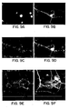
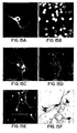
- FIG. 13A shows a septal CNS stem cell-derived cholinergic neuron immuno-stained for acetylcholine transferase.
- Figure 13B shows the same field of view as Fig. 13A stained for the mitotic label BrdU.
- Figure 13C and D show another example of a cholinergic neuron double-stained for vesicular acetylcholine transporter and BrdU, respectively.
- Table VII summarizes the number of MAP2ab-positive neurons per square centimeter and the proportions of different neuronal phenotypes relative to the total MAP2ab-positive neurons derived from CNS stem cells of several different regions and ages. Approximately 4-5% of the MAP2 positive neurons were cholinergic. In contrast, hippocampal and cortical CNS stem cells gave rise to no cholinergic neurons.
- LGE and MGE CNS stem cells also expressed vesicular acetylcholine transporter, a specific marker of cholinergic neurons (Table VII).
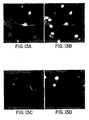
- Figures 14A , C, and D show typical LGE CNS stem cell-derived neurons stained for neuropeptide Y, met-enkephalin, and leu-enkephalin, respectively.
- Figures 14B , D, and F show the immunostaining for BrdU of the same fields as in Figs. 14A , C, and E, respectively.
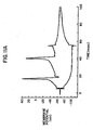
- FIG. 15A shows a typical CNS stem cell derived TH-positive neuron and Figure 15B shows the corresponding BrdU staining of the same field. All TH-positive cells are neurons as shown by double-staining for TH and MAP2ab ( Figs. 15C and D , respectively). Most of the remaining neurons were positive for the marker of GABAergic neurons, glutamic acid decarboxylase (GAD) as well as for GABA itself ( Fig. 15E ) and/or for acetylcholine esterase ( Fig. 15F ; Table VII) which is known to be expressed in monoaminergic neurons in this area.
- GABAergic neurons glutamic acid decarboxylase
- Fig. 15E glutamic acid decarboxylase
- Fig. 15F acetylcholine esterase
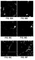
- CNS stem cells derived from dorsal mesencephalon in contrast, generated no TH-positive neurons (Table VII). Almost all neurons of this area (100.9 ⁇ 9.1%) expressed acetylcholine esterase (Table VII). They are most likely monoaminergic neurons, consistent with the in vivo pattern. Significantly, no TH-positive neurons arose from CNS stem cells derived from cortex, septum, hippocampus, striatum, and spinal cord (Table VII). Thus, in parallel with the known in vivo expression pattern, generation of TH-positive neurons were unique to ventral mesencephalon CNS stem cells in vitro .
- CNS stem cells from E13.5 cervical and thoracic spinal cords were expanded and differentiated.
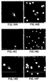
- 1.2 ⁇ 0.1% of MAP2 positive neurons were cholinergic containing vesicular acetylcholine transporter (Table VII).
- Cholinergic neurons also expressing acetylcholine transferase and BrdU-positive are shown in Figures 16C and D , respectively.
- a typical acetylcholine esterase-positive and BrdU-positive neuron is shown in Figure 16A and B , respectively.
- Neurons derived from E15.5 hippocampal and cortical CNS stem cells did not express tyrosine hydroxylase, acetylcholine esterase, acetylcholine transferase, and vesicular acetylcholine transporter (Table VII). This is appropriate for known absence of these markers in hippocampus in vivo .
- About 30% of MAP2ab-positive neurons were GABAergic, indicated by expression of GAD and GABA.
- Figures 17A and B show typical examples of GAD- and GABA-positive staining, respectively, which completely overlap.
- a typical hippocampal calretinin- and MAP2ab-positive neuron is shown in Figures 17C and D , respectively.
- Mature neurons can be also be derived with equal efficiency from E13.5 thalamus and hypothalamus. These neurons contain exceptionally long axonal processes.
- a typical thalamic neuron stained for the axonal protein, tau, and BrdU is shown in Figures 18A and B , respectively.
- a typical hypothalamic neuron stained for tau and BrdU is shown in Figures 18C and D , respectively.
- Synapsin staining of thalamic and hypothalamic neurons is shown in Figures 18E and F , respectively.
- neurons and the CNS stem cells capable of differentiating into such neurons provide the key element for gene therapy, cell therapy, and identification of novel therapeutic molecules (proteins, peptides, DNA, oligonucleotides, synthetic and natural organic compounds) directed to nervous system disorders.
- the multipotential cells could be efficiently isolated from many regions of the developing CNS, indicating that they are abundant throughout the neuroepithelium. This contrasts with the widely-held notion that stem cells are rare. Differentiation of the stem cells can be effectively directed by extracellular factors that are known to be present during CNS development 16-23 . This suggests that different extracellular factors can act on a single class of stem cells to generate different cell types. A similar instructive mechanism has also been observed in vitro with stem cells isolated from the peripheral nervous system 24 .
- Another mechanism of fate choice regulation in vivo may involve intermediate stages of differentiation. Identification of the bipotential oligodendrocyte precursor cell, O-2A, from postnatal optic nerve directly demonstrated that restricted progenitors are produced during development 25,26 .
- the stem cells are distinct from the 0-2A cells. Their origins, properties, and developmental capacities differ. Given that the stem cells differentiate into oligodendrocytes, the differentiation pathway may involve an obligatory intermediate stage, a committed progenitor state like the O-2A cell. The similar responses of both cells to T3 and CNTF 27,28 may reflect this common step.
- this CNS stem cell technology permits large scale culture of homogeneous stem cells in an undifferentiated state. The longer that the cells can be maintained in the stem cell state, the higher the yield of neurons that can be derived from the culture, thereby enabling more efficient gene transfer and large scale selection of those cells carrying the gene of interest.
- this culture system permits controlled differentiation of the stem cells where 50% of the expanded cells now turn into neurons. This efficient differentiation, combined with efficient proliferation, routinely yields more than 100 million neurons from the neocortex of one rat fetal brain in a two-week period.
- the differentiation of the stem cells into neurons, astrocytes, and oligodendrocytes occurs constitutively where all three cell types continue to mature in culture, most likely due to nurturing interactions with each other, as during normal brain development. Many different types of neurons arise, which respond to many growth factors and contain neurotransmitters and their receptors. Thus, a significant portion of the brain development can be recapitulated in a manipulable environment, thereby highlighting the potential to extract and test novel neurotropic factors normally secreted by these cells.
- the stem cell technology of the present invention can be developed for direct application to many different aspects of therapy and drug discovery for nervous system disorders.
- Outlined below are four examples for potential commercial applications, i.e., gene therapy for Parkinson's disease, cell therapy, search for novel growth factors, and assays for drug screening.
- the CNS stem cells more than meet the technical criteria as vehicles for gene therapies and cell therapies in general.
- the stem cells can be expanded rapidly under precisely controlled, reproducible conditions. Furthermore, these cells are readily accessible to all standard gene transfer protocols such as via retroviruses, adenoviruses, liposomes, and calcium phosphate treatment, as well as subsequent selection and expansion protocols.
- the expanded stem cells efficiently differentiate into neurons en masse.
- stem cells make them unique as the fundamental basis of therapeutic development directed at the human nervous system.
- stem cells once stem cells are triggered to differentiate into mature cell types, all of the molecular interactions are in place within the culture system to generate, to mature, and to survive a variety of different cell types and neuronal subtypes. These interactions recapitulate a significant portion of the natural brain development process. Therefore, the stem cells, as vehicles of gene therapy and cell therapy, refurnish not only a single potential gene or factor to be delivered but also the whole infrastructure for nerve regeneration.
- the stem cells in culture are expanded from the multipotential germinal precursors of the normal brain development. Hence, these stem cells retain the capacity to become not only three different cell types but also many different types of neurons depending upon the environmental cues to which they are exposed. This broad plasticity, which is the inherent property of the stem cells, distinctly suggests that, once transplanted, the cells may retain the capacity to conform to many different host brain regions and to differentiate into neurons specific for that particular host region.
- These intrinsic properties of the primary stem cells are far different from the existing tumorigenic cell lines where some neuronal differentiation can be induced under artificial conditions. Therefore, with these unique properties, the expandable human CNS stem cells contain significant commercial potential by themselves with little further development.
- Parkinson's Disease results mainly from degeneration of dopamine releasing neurons in the substantia nigra of the brain and the resulting depletion of dopamine neurotransmitter in the striatum.
- the cause of this degeneration is unknown but the motor degeneration symptoms of the disease can be alleviated by peripherally administering the dopamine precursor, L-dopa, at the early onset of the disease.
- L-dopa is no longer effective and currently no further treatment is available.
- One promising treatment being developed is to transplant dopamine-rich substantia nigra neurons from fetal brain into the striatum of the brain of the patient. Results obtained from various clinical centers look extremely optimistic.
- Tyrosine hydroxylase is the key enzyme for dopamine synthesis.
- Human CNS stem cells derived from fetal basal ganglia can be produced which express the tyrosine hydroxylase (TH) gene. These cells can be expanded, differentiated, and transplanted into the patient's striatum. Since the cells are originally derived from the primordial striatum, they would have the best chance of integrating into this region of the brain. Production of such cells and their successful transplantation into animal models will result in the most promising application of gene therapy to date.
- CNS stem cells are the natural germinal cells of the developing brain with the capacity to become the cells of the mature brain, the stem cells from the spinal cord and different regions of the brain may be used directly to repopulate degenerated nerves in various neuropathies.
- Drug discovery by traditional pharmacology had been performed without the knowledge of such complexity using whole brain homogenate and animals, and mostly produced analogs of neurotransmitters with broad actions and side effects.
- the next generation of pharmaceutical drugs aimed to modify specific brain functions may be obtained by screening potential chemicals against neurons displaying a specific profile of neurotransmitters, receptors complexes, and ion channels.
- CNS stem cells expanded and differentiated into neurons in culture express several neurotransmitters and receptor complexes.
- Many cell lines derived from stem cells and neuronal progenitors of different regions of the brain can be developed which, when differentiated into mature neurons, would display a unique profile of neurotransmitter receptor complexes.
- Such neuronal cell lines will be valuable tools for designing and screening potential drugs.
- the CNS stem cell technology of this application offers broad and significant potentials for treating nervous system disorders.
- MAP2 Embryonic Neuron
- MAP2 45.9 81.0 0.9 11.5 Neuron
- TuJ1 9.9 72.4 N.D. N.D.
- Neuron NF-M
- Oligodendrocyte GalC 7.4 2.8 4.5 21.2 Astrocyte (GFAP) 6.3 2.0 97.3 20.7
- MAP2 36.8 73.9 11.8 35.2 Neuron
- TuJ1 47.9 72.4 N.D. N.D.
- Oligodendrocyte GalC
- MAP2 microtubule associated protein a and b
- TH tyrosine hydroxylase
- AchE acetylcholine esterase
- VAT vesicular acetylcholine transporter
- ChAT choline acetyl transferase
- GAD glutamic acid decarboxylase
- NPY neuropeptide Y
- L-Enk leu-enkephalin
- M-Enk met-enkephalin. 2 Regions from where CNS stem cells were derived.
- SEPT E15.5 septum
- LGE E15.5 lateral ganglionic eminence
- MGE E15.5 medial ganglionic eminence
- HI E15.5 hippocampus
- VM E12.5 ventral mesencephalon
- DM E12.5 dorsal mesencephalon
- SPC E13.5 spinal cord. 3 Shown are the average number of MAP2ab-positve cells per square centimeter. Initial number of cells plated for all regions was 125,000 cells per square centimeter. ⁇ standard mean error.
Abstract
Description
- The present invention relates to a technology where stem cells from embryonic and adult brain are isolated, propagated, and differentiated efficiently in culture to generate large numbers of nerve cells. This technology, for the first time, enables one to generate large numbers of many different kinds of neurons found in a normal brain and provides a new foundation for gene therapy, cell therapy, novel growth factor screening, and drug screening for nervous system disorders.
- The brain is composed of highly diverse nerve cell types making specific interconnections and, once destroyed, the nerve cells (neurons) do not regenerate. In addition, the brain is protected by a blood-brain barrier that effectively blocks the flow of large molecules into the brain, rendering peripheral injection of potential growth factor drugs ineffective. Thus, a major challenge currently facing the biotechnology industry is to find an efficient mechanism for delivering potential gene therapy products directly into the brain in order to treat nervous system disorders.
- Moreover, for a degenerative disease like Parkinson's, the most comprehensive approach to regain a lost neural function may be to replace the damaged cells with healthy cells, rather than just a single gene product. Thus, current and future success of gene therapy and cell therapy depends upon development of suitable cells that can (1) carry a healthy copy of a disease gene (i.e., a normal gene), (2) be transplanted into the brain, and (3) be integrated into the host's neural network. This development ideally requires cells of neuronal origin that (1) proliferate in culture to a large number, (2) are amenable to various methods of gene transfer, and (3) integrate and behave as the cells of a normal brain. However, there have been no such cells for therapeutic purposes since neurons do not divide and therefore cannot be propagated in culture.
- As alternatives, various transformed cells of neural and non-neural origins such as glias, fibroblasts, and even muscle cells, which can be proliferated in culture, have been used as possible vehicles for delivering a gene of interest into brain cells. However, such cells do not and cannot be expected to provide neuronal functions. Another alternative approach has been to force a neural cell of unknown origin to divide in culture by genetically modifying some of its properties, while still retaining some of its ability to become and function as a neuron. Although some "immortalized" cells can display certain features of a neuron, it is unclear whether these altered cells are truly a viable alternative for clinical purposes.
- A developing fetal brain contains all of the cells germinal to the cells of an adult brain as well as all of the programs necessary to orchestrate them toward the final network of neurons. At early stages of development, the nervous system is populated by germinal cells from which all other cells, mainly neurons, astrocytes, and oligodendrocytes, derive during subsequent stages of development. Clearly, such germinal cells that are precursors of the normal brain development would be ideal for all gene-based and cell-based therapies if these germinal cells could be isolated, propagated, and differentiated into mature cell types.
- The usefulness of the isolated primary cells for both basic research and for therapeutic application depends upon the extent to which the isolated cells resemble those in the brain. Just how many different kinds of precursor cells there are in the developing brain is unknown. However, several distinct cell types may exist:
- a precursor to neuron only ("committed neuronal progenitor" or "neuroblast"),
- a precursor to oligodendrocyte only ("oligodendroblast"),
- a precursor to astrocyte only ("astroblast"),
- a bipotential precursor that can become either neuron or oligodendrocyte, neuron or astrocyte, and oligodendrocyte or astrocyte, and
- a multipotential precursor that maintains the capacity to differentiate into any one of the three cell types.
- Fate mapping analysis and transplantation studies in vivo have shown that different neuronal types and non-neuronal cells can be derived from the same precursor cells1-5. In vitro analyses have also suggested that multipotential cells are present in the developing brain6,7. Lineage analysis alone, however, does not directly identify the multipotential cells; nor does it define the mechanisms that drive them to different fates. Precursor cells from the central nervous system (CNS) have been expanded in vitro and differentiation into neurons and glia has been observed8-12 and, as detailed below, markedly different cell types have been obtained even when the culture conditions used were seemingly the same.
- Because of the current lack of understanding of histogenesis during brain development, many investigators have used various terms loosely to describe the cells that they have studied, e.g., neuronal progenitor, neural precursor, neuroepithelial precursor, multipotential stem cell, etc. Thus, the nature of the cells so far described in the literature and culture conditions for obtaining them can only be compared to each other by their reported differentiation capacity. The entire subject of the isolation, characterization, and use of stem cells from the CNS has recently been reviewed 33,34,38.
- In summary, conditions have not been found to date, despite many reports, to successfully identify, propagate, and differentiate multipotential stem cells. A useful compilation of studies reporting culture of CNS precursor cells is found in Table 3, p. 172, of a recent review34 and further extended below.
- Vicario-Abejon, C., Johe, K., Hazel, T., Collazo, D. & McKay, R., Functions of basic fibroblast growth factor and neurotrophins in the differentiation of hippocampal neurons, Neuron 15, 105-114 (1995 )12 .
- Cells expanded by Vicario-Abejon et al. are significantly different from those described in the present invention although the starting tissue (embryonic hippocampus), the mitogen (basic fibroblast growth factor, bFGF), and the basal medium (N2) are similar in both reports. Almost all of the cells expanded by Vicario-Abejon et al. failed to differentiate into any cell types but died in the absence of bFGF (as stated in the paper, pg. 106). This is also reflected in
Fig. 3 of the paper where the number of MAP2 positive neurons is exceedingly low (50-100 cells out of an initial cell number of approximately 80,000 per well; i.e., far less than 1% in all reported conditions). Thus, differences in culture conditions, subtle as they may be, can yield cells with significantly different properties and this is, in fact, consistent with the main observation of the present invention that the extracellular environment can shift the developmental properties of the CNS stem cells. - Vicario-Abejon et al. used the following culture conditions which differ from the those described in the present invention:
- 1. Used enzymatic dissociation, 0.1-0.25% trypsin + 0.4% DNAse I for the initial tissue dissociation as well as subsequent passaging. In the present invention, enzymatic dissociation effectively causes proteolyses of FGF receptors and causes cells to become unresponsive to bFGF and leads to differentiation.
- 2. Used 10% fetal bovine serum to stop the trypsin activity and to prime the cells from 4 hours to overnight before switching to serum free medium. In the present invention, serum even at less than 1% concentration shifts stem cells to astrocytic fate.
- 3. Cells were seeded at much higher density of 45,000 cells per cm2 and then grown to confluence before passaging by trypsin and serum. In the present invention, high cell density inhibits proliferation and causes spontaneous differentiation even in the presence of bFGF.
- 4. bFGF was given only intermittently every 2-3 days, and at 5 ng/ml, less than the optimal concentration disclosed in the present invention. This condition leads to partial differentiation of cells and subsequent heterogeneity of cell types in culture.
- 5. Basal medium consisting of "N2" components consisted of 5 ng/ml insulin, less than the optimal concentration disclosed in the present invention.
- Ray, J., Peterson, D. Schinstine, M. & Gage, F., Proliferation, differentiation, and long-term culture of primary hippocampal neurons, Proc. Natl. Acad. Sci. USA 90, 3602-3606 (1993 )10 .
- This study used culture conditions that are very similar to those described by Vicario-Abejon et al.--bFGF as the primary mitogen, serum-free medium, and E16 hippocampus. However, it reports isolation and expansion of a precursor population (neuroblasts) quite different from the cells of Vicario-Abejon et al. (undefined) as well as the multipotential stem cells described in the present invention. The reported cells had the following properties which markedly contrast from those of CNS stem cells:
- 1. The expanded cells under the reported condition are mitotic neurons with antigenic expressions of neurofilament, nestin, neuron-specific enolase, galactocerebroside, and MAP2 (Table I, p. 3604). The expanding CNS stem cells reported in the present invention express nestin, only, are negative for the above antigens, and are, therefore, a molecularly distinct population of cells from those described by Ray et al.
- 2. Ultrastructural analysis of the expanded cells in culture "demonstrated their histotypic neuronal morphology". The expanding CNS stem cells exhibit entirely different, non-neuronal morphology.
- 3. The mitotic "neurons" had a doubling time of 4 days and could be passaged and grown as continuous cell lines. The CNS stem cells double at every 20-24 hours and exhibit a characteristic regression of mitotic and differentiative capacity over time so that they cannot be maintained as stable cell lines indefinitely.
- 4. The culture system by Ray et al. generates "nearly pure neuronal cell cultures". The culture system in the present invention generates multipotential stem cells that can differentiate into all three major cell types of the brain, i.e., neurons, oligodendrocytes, and astrocytes.
- Ray et al. used the following culture conditions which differ from those of the present invention.
- 1. Embryonic hippocampi were mechanically triturated without the use of an enzyme; however, cells were plated approximately 100,000 cells per cm2, optimal for neuronal survival, but almost 10 times higher cell density than optimal for expansion of CNS stem cells.
- 2. bFGF was given at 20 ng/ml, intermittently, at every 3-4 days.
- 3. Basal "N2" medium contained 5 µg/ml insulin, less than optimal. Medium change was also prolonged at every 3-4 days.
- 4. Cells were passaged by using trypsin.
- In conclusion, even seemingly small differences in culture conditions can result in isolation of vastly different cell types.
- Ray, J. and Gage, F.H., Spinal cord neuroblasts proliferate in response to basic fibroblast growth factor, J. Neurosci. 14, 3548-3564 (1994 )39 .
- Ray and Gage report isolation and propagation of cells "that have already committed to a neuronal pathway are and expressing neuronal phenotypes (neuroblasts)" from spinal cord using bFGF. Again, although the primary mitogen is bFGF, their culture conditions are different and obtained cells markedly different from CNS stem cells.
- 1. E14-E16 spinal cord was used, a much later stage of development than optimal for stem cells.
- 2. The tissue was dissociated enzymatically by papain and DNase.
- 3. Initial plating was done in 10% fetal bovine serum.
- 4. There was a preliminary enrichment for a non-adherent cell population.
- 5. There was intermittent medium change and bFGF supplement, every 3-4 days.
- Gage, F.H., Coates, P.W., Palmer, T.D., Kuhn, H.G., Fisher, L.J., Suhonen, J.O., Peterson, D.A., Suhr, S.T. & Ray, J., Survival and differentiation of adult neuronal progenitor cells transplanted to the adult brain, Proc. Natl. Acad. Sci. USA 92, 11879-11883 (1995 )35 .
- Gage et al. report isolation, propagation, and transplantation of cells from adult hippocampus. These mixtures of cells were maintained in culture for one year through multiple passages. 80% of them exhibit rather unusual properties such as co-expressing glial and neuronal antigens while remaining mitotic. These properties are not exhibited by stem cells isolated from the adult striatal subventricular zone.
- Again, using bFGF as a primary mitogen, the authors derived markedly different cells than CNS stem cells reported in the present invention.
- Gritti, A. et al., Multipotential stem cells from the adult mouse brain proliferate and self-renew in response to basic fibroblast growth factor. J. Neurosci. 16, 1091-1100 (1996 )40 .
- These authors report isolation and propagation of multipotential stem cells from the subventricular zone of adult brain by using bFGF. A significant difference in culture conditions used by Gritti et al. is that the cells are propagated as aggregated spheres without attachment to plate surface. Culture conditions by Gritti et al. require this aggregation of cells into spheres, using either bFGF or epidermal growth factor (EGF), as an essential step for propagating multipotential cells. This aggregation step alone essentially distinguishes the reported culture system from that of the present invention. The aggregation promotes undefined cell-cell interactions and results in uncontrollable differentiation/fate-shifts and overall in much less expansion and differentiation. Furthermore, this culture system and the result obtained by Gritti et al. are limited to adult brain where extremely small number of cells were obtained (105 cells per brain) and have not been extended to various regions of embryonic brain.
- The procedure in the present invention permits propagation of stem cells throughout the developing CNS as well as the striatum of the adult brain. It also uses adherent culture and actively avoids cell-cell contact and high cell density. As a result, it permits much more efficient expansion of the cells in an undifferentiated multipotential state and much more precise and efficient control over differentiation of the expanded cells.
- Reynolds, B. & Weiss, S., Generation of neurons and astrocytes from isolated cells of the adult mammalian central nervous system, Science 255, 1707-1710 (1992 )15 .
- Reynolds, B., Tetzlaff, W. & Weiss, S., A multipotent EGF-responsive striatal embryonic progenitor cell produces neurons and astrocytes, J. Neurosci. 12, 4565-4574 (1992 )9 .
- Vescovi, A.L., Reynolds, B.A., Fraser, D.D., and Weiss, S., bFGF regulates the proliferative fate of unipotent (neuronal) and bipotent (neuronal/ astroglial) EGF-generated CNS progenitor cells, Neuron 11, 951-966 (1993 )41 .
- These three studies describe the original sphere cultures of neural precursor cells from adult and embryonic brain using EGF (epidermal growth factor). The expanded cells differentiate into neurons and astrocytes, but not into oligodendrocytes, and thus are thought to be a bipotential population, rather than multipotential. Another distinguishing property of the cells is that they respond only to EGF and not to bFGF in particular, whereas CNS stem cells respond similarly to both EGF and bFGF. Again, the sphere culture conditions are not comparable to those employed in the present invention because they require cell aggregation in which many additional undefined interactions are expected to occur.
- Ahmed, S., Reynolds, B.A., and Weiss, S., BDNF enhances the differentiation but not the survival of CNS stem cell-derived neuronal precursors. J. Neurosci. 15, 5765-5778 (1995 ) 42 .
- This paper reports the effects of brain-derived growth factor (BDNF) on sphere cultures of embryonic neural precursor cells propagated with EGF. There is no further enhancement of the culture system per se.
- Svendsen, C.N., Fawcett, J.W., Bentlage, C. & Dunnett, S.B., Increased survival of rat EGF-generated CNS precursor cells using B27 supplemented medium, Exp. Brain Res. 102, 407-414 (1995 )36.
- This study utilizes the sphere culture with EGF as described above to test a commercially available medium supplement called "B27". The study simply reports that use of B27 enhances cell survival (not neuronal survival) in a mixed culture containing neurons, astrocytes, and oligodendrocytes.
- Kilpatrick, T.J. and Bartlett, P.F., Cloning and growth of multipotential neural precursors: requirements for proliferation and differentiation, ) 43.
- The authors report existence of multipotential precursor cells in E10 mouse telencephalon by culturing single cells from the brain in bFGF plus serum. The results were based on 700 cells expanded clonally for 10 days, some of which, when differentiated in the presence of bFGF, serum, and astrocyte conditioned medium, could give rise to neurons. There was no mass expansion of the cells.
- Kilpatrick, T.J. and Bartlett. P.F., Cloned multipotential precursors from the mouse cerebrum require FGF-2, whereas glial restricted precursors are stimulated with either FGF-2 or EGF, J. Neurosci. 15, 3653-3661 (1995 )44 .
- The authors utilize the clonal culture system reported in the above-described reference43 to test mitogenic efficacy of bFGF and EGF on cortical cells from E10 and E17 embryos. Again, the culture condition applies strictly to microculture in serum containing medium to demonstrate existence of different precursor cells in developing brain. There is no mass expansion, long-term culture, or systematic differentiation protocol.
- Baetge, E.E., Neural stem cells for CNS transplantation, Ann. N.Y. Acad. Sci. 695, 285 (1993 ) 45 .
- This is a brief review paper summarizing various studies directed to isolating precursor cells and their derivatives in culture. It is somewhat outdated and most of the relevant original studies cited have been discussed above.
- Bartlett, P.F. et al., Regulation of neural precursor differentiation in the embryonic and adult forebrain. Clin. Exp. Pharm. Physiol. 22, 559-562 (1995 )46 .
- This is also a brief review paper summarizing mostly previous works from the authors' laboratory in regard to their microculture studies where differentiation potentials of certain-clones of precursors are tested in the presence of acidic FGF (aFGF), bFGF, serum, and/or astrocyte conditioned medium.
- In addition, Sabate et al.32 reported the culturing of a human neural progenitor with undefined differentiation capacity. Davis and Temple6 demonstrated the existence of multipotential stem cells in cortex by co-culturing with epithelial cells for short term (less than 100 cells altogether).
- However, cell differentiation could not be controlled in any of the reported studies which precluded analysis of their lineage relations and the mechanisms regulating fate choice.
- The present invention provides a method for efficiently propagating the undifferentiated germinal cells, i.e., stem cells of the central nervous system (CNS), in culture and defines conditions to effectively turn the undifferentiated cells into mature cell types. These undifferentiated cells or "CNS stem cells" display the multipotential capacity to differentiate into all three major cell types of a mature brain -- neurons, astrocytes, and oligodendrocytes. Moreover, the same culture conditions enable isolation, expansion, and differentiation of equivalent multipotential cells from the adult brain.
- Since the initial disclosure, additional reports have appeared. Most recent research on and use of CNS stem cells and neural progenitors have been further reviewed47-52. In addition, Reynolds and Weiss53 reported that embryonic striatal progenitors generated as spheres using EGF were able to differentiate into all three cell types including oligodendrocytes, astrocytes, and neurons. The frequency of EGF-responsive cells was limited to only 1% of the initial primary culture. Subcloning to establish self-renewal was questionable since up to 500 cells/well were used to generate the secondary "clones". Differentiation of the cells was induced by incorporating 1% serum in the medium. However, no data demonstrating all three cell types were presented from single-cell derived clones.
- Weiss et al.54 reported that multipotential CNS stem cells could be isolated from adult spinal cord and third and fourth ventricles by using a combination of EGF and bFGF but not with either alone.
- Svendsen et al.55 reported that neural precursor cells isolated from striatum and mesencephalon of 16 day old rat embryos (E16), when grafted into lesioned adult rat brains, failed to differentiate into neurons. They also reported that-EGF-generated mesencephalon cells but not striatal cells differentiated into tyrosine hydroxylase (TH)-positive neurons, albeit in very low number (0.002%). There were no characterization of cells in vitro to ensure that the primary culture used contained no post-mitotic neurons carrying over from the tissue, especially given that the result could only be obtained with E16 tissue when most TH cells are already born.
- Schinstine and Iacovitti56 reported that some of the astrocytes derived from EGF-generated neural precursor cells expressed neuronal antigens such as tau and MAP2. Qian et al.57 reported that different concentrations of bFGF proliferate stem-like cells of E10 mouse cortex with varying differentiation potentials ranging from only neuronal to multipotential.
- Palmer et al.65 reported that multipotential CNS stem cells could be isolated from adult rat hippocampus. 84% of the cells they expanded, however, co-expressed MAP2c and 04, immature neuronal and oligodendroglial markers. Only 0.2% were MAP2ab positive and less than 0.01% were positive for other neuronal markers such as tau and neurofilament 200. Such properties are quite different from the properties described in the Examples in the present application.
- Finley et al.66 reported that the mouse embryonic carcinoma cells line, P19, can form neuronal polarity and be eletrophysiologically active when induced by retinoic acid and serum. Strubing et al.67 reported that embryonic stem cells grown in serum-containing medium could differentiate into electrophysiologically active neurons in vitro. Okabe et al.68 also reported differentiation of some of embryonic stem cells into neurons in vitro.
- Gritti et al.40 reported that multipotential stem cells could be isolated from adult mouse subependyme by EGF and bFGF, which when differentiated, could be eletrophysiologically active and express GABA-, gluatamate-, and ChAT-immunoreactivities, but not others. The frequency of such neurons, however, was not documented and thus it is difficult to ascertain how efficient neuronal maturation was. Moreover, these neuronal phenotypes derived from dividing stem cells were not directly demonstrated by BrdU labeling. This is particularly relevant since aggregate cultures are extremely prone to be contaminated by primary neurons from the tissue, which carry over for several passages. Weiss et al.49, in fact, stated that only GABA-positive cells could be obtained from their cultures. Most of the GABA-positive cells may be oligodendrocytes.
- Feldman et al.69 reported electrophysiological studies of EGF-generated rat neural precursors. They found that most, if not all, electrophysiologically active cells are in fact non-neuronal, and that glial cells do contain voltage-sensitive Na channels that evoke action potential-like conductances.
- Results such as these illustrate that identifying CNS stem cells, defining conditions that stably maintain CNS stem cell properties for long-term, and controlling their differentiation into mature cell types are neither obvious nor predictable to those skilled in this art.
- The present specification describes an in vitro culture of stem cells of the central nervous system of a mammal, a method for the in vitro culture of the stem cells, and a method for the differentiation of the stem cells.
- In the described in vitro culture of the stem cells of the central nervous system of a mammal, the stem cells maintain the multipotential capacity to differentiate into neurons, astrocytes, and oligodendrocytes. The stem cells can be derived from central nervous system tissue from a human, fetus or adult. Furthermore, the central nervous system tissue may be hippocampus, cerebral cortex, striatum, septum, diencephalon, mesencephalon, hindbrain, or spinal cord.
- Furthermore, the stem cells can differentiate to mature neurons exhibiting axon-dendrite polarity, synaptic terminals, and localization of proteins involved in synaptogenesis and synaptic activity including neurotransmitter receptors, transporters, and processing enzymes. In addition, the stem cells retain their capacity to generate subtypes of neurons having molecular differences among the subtypes.
- A method for the in vitro culture of the stem cells, where the stem cells maintain the multipotential capacity to differentiate into neurons, astrocytes, and oligodendrocytes, wherein said method is as set forth in the claims.
- The method is applicable with stems cells derived from central nervous system tissue from a human, fetus or adult. Again, the central nervous system tissue may be hippocampus, cerebral cortex, striatum, septum, diencephalon, mesencephalon, hindbrain, or spinal cord.
- The application also discloses a method for the differentiation of an in vitro culture of stem cells of the central nervous system of a mammal, where the stem cells maintain the multipotential capacity to differentiate into neurons, astrocytes, and oligo-dendrocytes, wherein said method is as set forth in the claims.
- Furthermore, differentiation may be specifically directed by adding a second growth factor to the cultured cells either before or after removing the first growth factor from the cultured cells. The second or added growth factor may be platelet-derived growth factor (PDGF), ciliary neurotropic factor (CNTF), leukemia inhibitory factor (LIF), or thyroid hormone, iodothyronine (T3).
- The present application also describes an in vitro culture of region-specific, terminally differentiated, mature neurons derived from cultures of mammalian multipotential CNS stem cells and an in vitro culture method for generation of the differentiated neurons.
- In the method for in vitro generation of region-specific, terminally differentiated, mature neurons from cultures of mammalian multipotential CNS stem cells, multipotential CNS stem cells from a specific region are cultured in a chemically defined serum-free culture medium containing a growth factor; the medium is replaced with growth factor-free medium; the stem cells are harvested by trypsinization; plated at a density of between 100,000 to 250,000 cells per square centimeter; and cultured in a glutamic acid-free chemically defined serum-free culture medium. The specific region of the CNS from which the multipotential stems cells are derived are selected from the group consisting of cortex, olfactory tubercle, retina, septum, lateral ganglionic eminence, medial ganglionic eminence, amygdala, hippocampus, thalamus, hypothalamus, ventral and dorsal mesencephalon, brain stem, cerebellum, and spinal cord.
- In addition, the chemically defined serum-free culture medium may be selected from N2 (DMEM/F12, glucose, glutamine, sodium bicarbonate, 25 ug/ml insulin, 100 µg/ml human apotransferrin, 25 nM progesterone, 100 µM putrescine, 30 nM sodium selenite, pH 7.28) or N2-modified media. The growth factor may be selected from the group consisting of bFGF, EGF, TGF-alpha and aFGF.
- Furthermore, the glutamic acid-free chemically defined serum-free culture medium may be supplemented with between 10-100 ng/ml of brain-derived neurotropic factor. The method is applicable to multipotential CNS stem cells derived from central nervous system tissue from any mammal, including rat and human.
- The present application also describes in vitro cultures of region-specific, terminally differentiated, mature neurons derived from cultures of mammalian multipotential CNS stem cells from a specific region of the CNS. The specific region from which the multipotential stems cells are derived are selected from the group consisting of cortex, olfactory tubercle, retina, septum, lateral ganglionic eminence, medial ganglionic eminence, amygdala, hippocampus, thalamus, hypothalamus, ventral and dorsal mesencephalon, brain stem, cerebellum, and spinal cord. Likewise, the in vitro culture of region-specific differentiated neurons may be derived from any mammalian multipotential CNS stem cell, including rat and human.
-
-
Figure 1A shows the controlled differentiation of CNS stem cells at high density. Rapidly dividing nestin-positive precursor cells were labelled with BrdU during the last 24 hours of proliferation. Differentiation was then initiated by withdrawal of bFGF (day 0) and continued for up to 6 days. At indicated times, cells were fixed and stained for BrdU and neuronal antigens. Ratios of cells double-stained for BrdU and each neuronal antigen to total BrdU positive (BrdU+) cells are shown. Up to 50% of BrdU+ cells expressed neuronal antigens and their expression was time-dependent. MAP2 positive (MAP2+), filled circle; TuJ1 positive (TuJl+), grey diamond; neurofilament L positive (neurofilament L+), open square; neurofilament M positive (neurofilament M+), filled triangle. -
Figure 1B shows proportions of MAP2+ neurons (●), GalC+ oligodendrocytes (+), and GFAP+ astrocytes (o) in differentiated clones. Clones of various sizes ranging from 39 cells to 2800 cells were differentiated for 6 days and analyzed for two cell types per clone by double immunohistochemistry. A partial list is given in Table I and immunostaining shown inFig. 3 . The number of neurons increased with increasing clone size, constituting 50% of the clone. -
Figure 1C shows a comparison of the mitogenic efficacies of epidermal growth factor (EGF) and basic fibroblast growth factor (bFGF). Cells (initial density of 1 x 10⁴ per plate) acutely dissociated from E16 hippocampus (HI), E14 cortex (CTX) and striatum (ST), and adult subependymal layer (Adult) were expanded with either EGF (20 ng/ml) or bFGF (10 ng/ml). Colonies arising after 10 days of expansion were stained for nestin, an intermediate filament protein characteristic for CNS precursor cells13,14. Relative number of colonies averaged from at least 2 experiments for each region are shown (bFGF = 1). Twenty- to fifty-fold more nestin+ colonies per plate were present when embryonic cells were grown in bFGF (dotted bar) than in EGF (striped bar). At high densities (1 x 10⁶ and 2.5 x 10⁶), bFGF condition gave 10-fold higher BrdU+/nestin+ cells than EGF. Both growth factors were equally mitogenic for the adult cells. When EGF- and bFGF-expanded clones were differentiated, neurons and oligodendrocytes were found in similar quantities; however, EGF-expanded clones gave rise to significantly higher number of GFAP+ astrocytes. -
Figures 2A - D show a typical clone of CNS stem cells. Cells were marked by a circle on the plate within 24 hours of plating before the first mitosis and then expanded up to 10 days (Fig. 2A ). Higher magnification view of another clone before differentiation, immunostained with anti-nestin antibody, is shown inFigure 2B . Note the homogeneous radial morphology of the nestin-positive cells consistent with the nestin-positive morphology in neuroepithelium in vivo.Figure 2C shows a sister clone at low magnification, which has been differentiated for 6 days and immunostained with a neuron-specific antibody, TuJ1. Note the widespread and non-localized presence of TuJl-positive neurons across the entire clone. A higher magnification view of the same cells is shown inFigure 2D . The TuJ1-positive cells assume typical neuronal morphology. Heterogeneous morphologies in the non-neuronal TuJl-negative cells are apparent. -
Figures 3A-J show examples of representative clones of embryonic hippocampal cells (3A, 3C, 3E, 3G, 3I) and adult subependymal cells (3B, 3D, 3F, 3H, 3J) double-stained with combinations of antibodies to reveal different cell types within individual clones: anti-MAP2, neuronal; anti-GalC, oligodendrocytic; anti-GFAP, astrocytic. The two immunoreactions were developed sequentially and distinguished by using two distinct chromogens via alkaline phosphatase reaction (blue, indicated by arrows) versus horse radish peroxidase reaction (red, indicated by arrow heads).
The cells inFigures 3A and3B were double-stained with anti-MAP2 (neuronal, arrows) and anti-GFAP (astrocytic, arrow heads) and show that bFGF-expanded clones derived from embryonic or adult brain differentiate into both neurons and astrocytes. (Oligodendrocytes are unstained in this staining.)
The cells inFigures 3C and 3D were double-stained with anti-GalC (oligodendrocytic, arrows) and anti-GFAP (astrocytic, arrow heads) and show that bFGF-expanded clones derived from embryonic or adult brain differentiate into both oligodendrocytes and astrocytes. (Neurons are unstained in this staining.) -
Figures 3E and 3F show clones differentiated in the presence of platelet-derived growth factor (PDGF). The cells were double stained with anti-MAP2 (neuronal, arrows) and anti-GFAP (astrocytic). Most cells were MAP2+ and only a few were GFAP+. -
Figures 3G and 3H show clones differentiated in the presence of ciliary neurotrophic factor (CNTF). The cells were double-stained with anti-MAP2 (neuronal) and anti-GFAP (astrocytic, arrow heads). All cells were intensely GFAP+. -
Figures 3I and 3J show clones differentiated in the presence of thyroid hormone, tri-iodothyronine (T3). The cells were double-stained with anti-GalC (oligodendrocytic, arrows) and anti-GFAP (astrocytic, arrow heads). GFAP+ and, particularly, GalC+ cells increased. MAP2+ cells decreased (Table IV). -
Figure 4 shows the differentiation of human CNS stem cells into neuron in high density culture. bFGF-expanded CNS cells at high density were differentiated by withdrawal of bFGF ("WD"). The number of neurons expressing tau protein was determined by immunocytochemistry in culture during the expansion phase ("Before WD") versus after differentiation ("After WD"). The dramatic increase in postmitotic neurons only after the withdrawal of bFGF indicates that they were generated from the dividing stem cells. -
Figures 5A-F show human stem cells stained with human-specific anti-tau antiserum (Chemicon) which identify neurons. Proliferating human CNS stem cells in high density culture do not express tau protein, a neuronal marker (Figure 5A ). After 6 days of differentiation, however, many cells with typical neuronal morphology express high level of tau protein (Figure 5B ). In order to further demonstrate that these neurons have indeed derived from dividing stem cells, the stem cells were labeled with 10 µM bromodeoxyuridine (BrdU), an indicator of mitosis, for 24 hours just prior to the bFGF withdrawal. They were then differentiated for 6 days, double stained with human specific anti-tau antiserum (FITC, green) and anti-BrdU antibody (Rhodamine, red).Figure 5C shows a high magnification view of subsequent tau-positive neurons as seen through FITC fluorescence.Figure 5D shows the same field of view as inFigure 5D but seen through rhodamine fluorescence to reveal BrdU-positive nuclei. Most tau-positive neurons are also positive for BrdU, demonstrating that they were derived from mitotic stem cells before the bFGF withdrawal.
In order to further demonstrate multipotentiality of human CNS stem cells, they were cultured at clonal density as described for rodent cells.Figure 5E shows a typical clone at low magnification, which has been expanded from a single cell for 20 days, subsequently differentiated for 12 days, and immunostained with the neuron specific, anti-MAP2 antibody. Neurons are abundant in the clone.Figure 5F shows a higher magnification view of the clone inFigure 5E to indicate that the MAP2-positive cell are of typical neuronal morphology. -
Figures 6A-D demonstrate directed differentiation of human CNS stem cells. Human CNS stem cells after 16 days of expansion were grown clonally for an additional 20 days and then differentiated in the presence or absence of single factors, PDGF (10 ng/ml), CNTF (10 ng/ml), or T3 (3 ng/ml).Figure 6A shows a untreated control clone, with approximately 50% MAP2-positive neurons (arrows) and 2-10% GFAP-positive astrocytes (arrow heads).Figure 6B shows a PDGF-treated clone, where 75% of cells are MAP2-positive neurons (arrows) and 2-10% GFAP-positive astrocytes (arrow heads).Figure 6C shows a CNTF-treated clone, where 85% are GFAP-positive astrocytes (arrow heads) and only 9% MAP2-positive neurons (arrows).Figure 6D shows a T3-treated clone with increased number of O4- and/or GalC-positive oligodendrocytes (arrows) and of GFAP -positive astrocytes (arrow heads). -
Figures 7A-7I show that a large number of mature neurons with correct axon-dendritic polarity and synaptic activity can be obtained routinely from long-term expanded CNS stem cells. Hippocampal stem cells from E16 rat embryos were expanded in culture for 16 days and through 4 passages. Just before the last passage, rapidly dividing cells were labeled with 10 µM BrdU for overnight, passaged by using trypsin, and plated onto chamber slides. Cells were maintained for 21 days to allow constitutive differentiation and maturation of neurons. Subsequently, the extent of maturation and neuronal subtypes generated were analyzed by immunocytochemistry. -
Fig. 7A : Neurons stained with TuJ1 antibody viewed at low magnification (100x) to illustrate that production of neurons is efficient. -
Fig. 7B : Typical morphology of neurons revealed by TuJ1 antibodies (400x). -
Fig. 7C : Typical morphology of neurons revealed by MAP2 antibodies (400x). -
Fig. 7D : Neurons stained with synapsin antibody. Only mature neurons containing synaptic vesicles are stained. -
Fig. 7E : BrdU staining. All cells in the culture, neurons and glia, are labeled with BrdU. -
Fig. 7F : Synapsin and BrdU double staining. Mature synapsin-positive cells are also BrdU-positive, demonstrating that they are derived from mitotic stem cells in culture. -
Fig. 7G : Punctate anti-synapsin antibody staining marks the presynaptic axon terminals specifically in large mature neurons. -
Fig. 7H : The synapsin-positive structures are closely apposed to dendritic processes revealed by MAP2 antibody staining. -
Fig. 7I : MAP2 and synapsin proteins are closely associated but not co-localized, suggesting presynaptic-postsynaptic interaction. -
Figure 8 shows that stem cell-derived neurons express various neurotransmitter receptors and transporters expected to be involved in synaptic transmission as detected by RT-PCR. Long-term expanded stem cells derived from E16 rodent cortex were differentiated for 14 days and harvested to prepare RNA. Undifferentiated stem cells were also prepared in order to compare differentiation-specific induction. RNA from a whole brain of adult rat was used as a positive control. Primers specific for NMDA (N-methyl-D-aspartate) and AMPA (α-amino-3- hydroxy-5-methyl-4-isoxazole proprionic acid) families of glutamate receptor subtypes as well as for various GABA transporters were used. Note especially the specific induction of NMDA R1, AMPA R1 and AMPA R2 receptors in differentiated cells. -
Figures 9A-F show examples of typical neurons derived from rat embryonic hippocampal stem cells which had been expanded in vitro for 16 days (approximately 16 cell divisions through 4 passages) and differentiated for 21 days total. Mitotic CNS stem cells were pulse-labeled with bromodeoxyuridine (BrdU) for the last 24 hours prior to differentiation. Resulting neurons were triple-immunostained with antibodies against BrdU (Fig. 9A ), MAP2ab (Fig. 9B ), and synapsin (Fig. 9C ). The composite view of the triple stained cell is shown inFig. 9D . The BrdU-labeling demonstrates that the differentiated neuron derived from a mitotic precursor in the culture and that it is a terminally differentiated neuron since it retained the mitotic label during the prolonged differentiation phase. MAP2ab is a well-established neuron-specific protein present only in mature neurons and localized mostly in dendrites. Synapsin is a well-established synaptic vesicle protein and thus localizes synaptic terminals in axons. The triple-labeled neurons as shown inFig. 9D established that long-term expanded mitotic CNS stem cells terminally differentiate into mature neurons with proper subcellular polarization containing distinct dendritic (post-synaptic) and axonal (presynaptic) structures expected of fully functional neurons. Other synaptic vesicle proteins also localize in the same pattern of punctate axon terminals apposed to soma and dendrites.Fig. 9E and Fig. 9F show another hippocampal CNS stem cell derived mature neuron double-stained for synaptophysin and MAP2ab, respectively. -
Figure 10 shows a field of neurons from hippocampal CNS stem cells viewed by transmission electron microscopy. The abundant presence of synapses containing synaptic vesicles and post-synaptic densities are evident. -
Figures 11A-D show intracellular electrophysiological recordings from single neurons obtained from rat E15.5 septal CNS stem cells. Consistent with the morphology, these recordings show that the CNS stem cell-derived neuronal networks are also electrophysiologically active. Thus, when individual cells were stimulated with electrode, they conducted action potentials (Fig. 11A ), demonstrated presence of various voltage-sensitive ion channels (Fig. 11B ), and evoked excitatory and inhibitory postsynaptic potentials in response to bath application of the excitatory neurotransmitter, glutamate (Fig. 11 C and D). These examples establish beyond doubt that CNS stem cells give rise to terminally differentiated, electrophysiologically functional, neuronal networks.
Diverse neuronal phenotypes seen in vivo are obtained from the CNS stem cell cultures.
Examples of some of these neuronal phenotypes are shown inFigures 12-18 and Table VII. -
Figure 12 shows the expression of dopamine receptors D1 and D2 from CNS stem cells isolated from E15.5 lateral and medial ganglionic eminence. Total RNAs were isolated from respective CNS stem cell cultures differentiated for varying periods (0-20 days). Shown is the electrophoresis pattern of the DNA amplified by RT-PCR (reverse transcription-polymerase chain reaction). Results from five independent culture preparations, run in parallel in a single gel, are shown. The numbers above the lanes indicate the days of differentiation. D1 = dopamine receptor, D1; D2 = dopamine receptor, D2. -
Figures 13 A-D show cholinergic neurons from septal CNS stem cells. CNS stem cells derived from E16 septum were differentiated for 18-21 days. The cholinergic neurons were assessed by acetylcholine esterase histochemistry (not shown), by immunostaining for acetylcholine transferase (Fig. 13A ) and for acetylcholine transporter (Fig. 13C ). In each case, CNS stem cells were incubated with the mitotic label, BrdU (10 µM), for 24 hours just before switching to the differentiation condition (Fig. 13B andD ). -
Figures 14A-F show neuropeptide-containing neurons obtained from rat 15.5 lateral ganglionic eminence (striatum) CNS stem cells. They are a neuropeptide Y-positive (Fig. 14A ), BrdU-positive (Fig. 14B ) neuron, a met-enkephalin-positive (Fig. 14C ), BrdU-positive (Fig. 14D ) neuron, and a leu-enkephalin-positive (Fig. 14E ), BrdU-positive (Fig. 14F ) neuron. -
Figures 15A-F show typical morphologies of several subtypes of neurons derived from CNS stem cell of rat E12.5 ventral mesencephalon.Figure 15A andB show a TH-positive and BrdU-positive neuron (Fig. 15A -TH staining;Fig. 15B -BrdU staining).Figure 15C andD show another TH-positive and MAP2ab-positive neuron (Fig. 15C -TH staining;Fig. 15D -MAP2ab staining).Figure 15E shows neurons stained with anti-GABA antibody.Figure 15F shows neurons stained by acetylcholine esterase histochemistry. -
Figures 16A-D show examples of neurons from spinal cord stem cells.Figure 16A shows an acetylcholine esterase-positive neuron derived from rat E13.5 spinal cord CNS stem cells, which is also BrdU-positive (Fig. 16B ). Cholinergic neurons are shown by acetylcholine transferase staining (Fig. 16 C) , which are also BrdU-positive (Fig. 16D ). -
Figures 17A and B show GABAergic neurons derived from rat E15.5 hippocampal CNS stem cells, which have been double-stained for glutamic acid decarboxylase (Fig. 17A ) and GABA (Fig. 17B). Figure 17C and D show a hippocampal calretinin-positive (Fig. 17C ), MAP2ab-positive (Fig. 17D ) neuron. -
Figures 18A-F show neurons derived from rat E13.5 thalamus and hypothalamus CNS stem cells.Figure 18A shows thalamic neurons stained for tau;Figure 18B shows the same field of view stained for BrdU.Figure 18C shows a hypothalamic neuron stained for tau;Figure 18D shows the same field of view stained for BrdU.Figure 18E and F show synapsin-positive neurons from thalamus and hypothalamus CNS stem cells, respectively. - In this application, conditions are defined which permit mass expansion up to 109 fold in culture and controlled differentiation of multipotential CNS stem cells from the embryonic and adult brain of mammals. In both cases, clones derived from single cells differentiate into neurons, astrocytes, and oligodendrocytes. Addition of single factors can dramatically shift the proportion of cell types within a clone.
- The procedure for isolating, propagating, and differentiating the CNS stem cells are given in detail below. The procedure contains four essential steps that must be followed in concert for successful isolation and differentiation of the CNS stem cells. The four essential steps are as follows:
- (1) The initial dissociation of cells from tissue is done by mechanical trituration and not by enzymatic digestion. With adult tissue, it is necessary to first enzymatically digest the tissue and then dissociate the cells from the tissue by mechanical trituration.
Trituration means gentle agitation of cell aggregates caused by fluid movement occurring during repetitive pipetting action by which individual cells become loose and dissociated from neighboring cells. Trituration is done in a saline solution free of divalent cations whose absence aids break-up of interactions among cell-adhesion proteins on cell surface. Rapidly dividing stem cells in the ventricular zone are only weakly adherent and simply removing the divalent cations from the medium and gentle agitation by pipetting are sufficient to dissociate the tissue into mostly single cells. The cells are then cultured in the complete absence of serum. Even a brief exposure to serum deleteriously affects the differentiation capacity of the stem cells so that they are no longer able to differentiate into neurons and oligodendrocytes. Precoating the plates with poly-L-ornithine and fibronectin facilitates the adhesion of the cells to the plates. - (2) The CNS stem cells display an innate property to differentiate spontaneously, which reflects a regulatory mechanism controlling cell cycle depending upon the free concentration of growth factor, the mitogen. In order to suppress the differentiation of the stem cells into other cell types and to maintain homogeneity, the growth factor must be supplied daily at a concentration of 10 ng/ml or higher. The growth factor can be selected from (1) basic fibroblast growth factor (bFGF), (2) EGF, (3) TGF-alpha, or (4) acidic FGF (aFGF). If acidic fibroblast growth factor is selected, heparin at a concentration of 1 µg/ml must also be supplied.
- (3) Even a continuous supply of bFGF or other selected growth factor is insufficient to inhibit differentiation if the culture is allowed to reach a critical density of greater than approximately 50%. This is most likely because of yet undefined endogenous factor(s) secreted by the dividing cells themselves which antagonize the action of bFGF. Thus, in order to remove such factors from the culture and to reduce cell-cell interaction as much as possible, the cells must be passaged frequently at every 4 days after plating, and replating should be done at low density of approximately 0.5 x 10⁶ per 10 cm plate, i.e., in the range of 1 x 10² to 1 x 10⁶ cells per 10 cm plate, precoated with poly-ornithine and fibronectin.
- (4) Passaging the cells by trypsin results in proteolytic removal of a bFGF receptor component and disables the mitogenic effect of bFGF. The turn-over rate of the receptor is sufficiently slow during which period the cells fail to recognize the mitogen and activate the differentiation pathway.
- In order to circumvent this process, the cells are treated with Hank's buffered saline solution (HBSS) to remove divalent cations in the culture which disrupts the ionic interactions between the cadherins and the integrins on the cell surface and extracellular matrix proteins on the culture plate, causing the cells to round up. At this point the stem cells can be scraped from the plate with a scraper without damaging the cells.
- Other cells in culture maintain tightly bound to the plate and scraping eliminates them, thus allowing effective selection of rapidly dividing undifferentiated stem cells.
- Differentiation of the CNS stem cells is achieved by simply removing the mitogen, bFGF or other selected growth factor, from the medium. Specification of the cell types, i.e., neurons, oligodendrocytes, and astrocytes, occurs constitutively. In order for the effective controlled differentiation, the cells must be in a homogeneous state which can be achieved by following steps 1-4, above.
- These procedures yield a culture system for obtaining a homogeneous population of the CNS stem cells that can be differentiated into neurons, oligodendrocytes, and astrocytes with control and efficiency. The highlights of the features of this system are:
- (1) production of a large number of the CNS stem cells with the potential to form many different neuronal subtypes, oligodendrocytes, and astrocytes that can be transplanted into a brain;
- (2) controlled differentiation in vitro under serum-free conditions which allows the search for novel growth factors and cytokines;
- (3) rapidly dividing cells accessible to genetic manipulation for introduction of foreign genes;
- (4) generation of mature neurons in vitro suitable for genetic and pharmacological screening; and
- (5) direct derivation of intermediate precursor cells from the stem cells for enrichment of a single population of cells.
- The isolation of the CNS stem cells in the above-described manner further permits directed differentiation of the cells by treating them with specific growth factors. One practical significance of this directed differentiation to biotechnology is that a single cell type can be enriched in vitro. Thus, a novel application of previously discovered growth factors PDGF37 (platelet-derived growth factor), CNTF (ciliary neurotrophic factor), and T3 (thyroid hormone, tri-iodothyronine) would be to direct the CNS stem cells to generate neurons, astrocytes, and oligodendrocytes, respectively.
- Another practical significance, especially for PDGF, is that PDGF-induced neurons appear to be actually neuronal progenitors that can further proliferate and expand in culture by PDGF. These cells differentiate only to neurons or to neurons and oligodendrocytes and differ from the stem cells. Isolation of neuronal progenitors from mammalian CNS by PDGF has not been described previously.
- Rat embryonic hippocampus (gestation day 16; day of conception is
day 1, Taconic Farm) were dissected in Hank's buffered saline solution (HBSS) and dissociated by brief mechanical trituration in HBSS. The cells were collected by centrifugation and resuspended in a serum-free medium containing DMEM/F12, glucose, glutamine, sodium bicarbonate, 25 µg/ml insulin, 100 µg/ml human apotransferrin, 25 nM progesterone, 100 µM putrescine, 30 nM sodium selenite, pH 7.28, plus 10 ng/ml recombinant human basic fibroblast growth factor12 (bFGF; R&D Inc.). - 1 x 10⁶ cells were plated per 10 cm plastic tissue culture plate precoated with 15 µg/ml poly-L-ornithine and 1 µg/ml bovine plasma fibronectin (Gibco). bFGF was added daily and media change was every 2 days. Cells were passaged at 50% confluence (4 days after initial plating) by briefly incubating them in HBSS and scraping with a cell scraper.
- Cells with multipotential capacity were found throughout the developing neuroepithelium. Under identical culture conditions, similar cells could be prepared from other regions of the developing CNS including cerebral cortex, striatum, septum, diencephalon, mesencephalon, hindbrain, and spinal cord. From E14 cortex and striatum and E16 hippocampus, approximately 70% of acutely dissociated cells responded to bFGF within 2 days of plating by undergoing mitosis.
- Hippocampal cells isolated from embryonic rat brains were expanded by daily addition of basic fibroblast growth factor (bFGF) in serum-free medium. Continuous supply of bFGF was important to repress differentiation and to maintain a homogeneous population of rapidly dividing cells expressing nestin, an intermediate filament protein characteristic for CNS precursor cells13,14. Less than 1% of the cells expressed the astroglial marker GFAP or the oligodendroglial markers, 04 and GalC.
- The cells were passaged 4 days after plating during which time cell number increased rapidly with an average cell doubling time of approximately 24 hours. Passaged cells were replated at 0.5 x 10⁶ cells per 10 cm plate and were allowed to propagate further. Cells could be passaged up to five times in this manner for a total of 20 days in vitro during which time a yield of 220 cells could be ideally expected. After this time period, the mitotic rate of the cells declined rapidly and the cells gradually lost their multipotential capacity, exhibiting glial characteristics and unable to differentiate into neurons.
- Large numbers of cells from cortex, striatum, and septum isolated from embryos of 12-18 days of gestation could also be expanded in mass culture in the same manner. The time course of the expansion was similar to that of hippocampal cells. Continuous expansion was again limited by the constitutive loss of multipotentiality after about 20 days of cell division. Thus, this regression appears to be a characteristic property of CNS stem cells.
- No simple antigenic marker is available which uniquely identifies multipotential stem cells from other precursors in vitro. Identity of a precursor population can only be ascertained by the cell's differentiation capacity. The conditions defined for mass culture in this application also permitted clonal expansion where cells were plated at extremely low cell density so that single cells were well isolated.
- Differentiation capacity of the cells expanded in mass culture was assessed at each passage by plating 200 cells per 10 cm plate and cultured under conditions as described above. Within 24 hours of plating, well isolated single cells were marked with a 3 mm ring (Nikon) on the bottom of the plate. Initial viability of the marked single cells was 5-10% and each plate typically yielded 10-20 marked clones. Only a single cell resided in each circle. The subsequent population of cells within each circle are progeny of that single cell. Clones were expanded for up to 10 days (500-2000 cells). Average double time was approximately 24 hours.
- Developmental potential of expanded cells was tested by directly differentiating the cells. Withdrawal of bFGF initiated differentiation within 24 hours. To initiate differentiation of high density cells, rapidly dividing cells, which had been in culture for 12 days and passaged three times, were incubated for the last 24 hours with 10 µM BrdU (bromodeoxyuridine) prior to passaging. 80-85% of the cells incorporated BrdU. The cells were harvested either by scraping or by using trypsin followed by soybean trypsin inhibitor in the serum-free medium. They were plated in duplicate at 40,000 cells/cm2 into multi-well chamber slides (LabTek) precoated with poly-L-ornithine and fibronectin, and cultured in the serum-free medium without bFGF. At indicated times, the cells were fixed and stained with various antibodies according to standard procedure.
- Immunopositive cells were counted under 400x magnification. At least five fields with a total cell number greater than 1,000 per sample were counted. Results shown (
Fig. 1A ) are cell counts averaged from two experiments. Antibody reagents used were: anti-nestin antiserum; monoclonal anti-MAP2 (clone HM-2, Sigma) and anti-tau antiserum (Sigma), monoclonal anti-neurofilament L and M (clones NR4 and NN18, Boehringer-Manheim), anti-beta tubulin type III (TuJl), monoclonal anti-GFAP (ICN), A2B5 (ATCC), 04, and anti-galactocerebroside (GalC). - Over a 6 day period, there was a progressive increase in the number of cells expressing several well established neuron-specific antigens, including MAP2a, b and c, beta tubulin type 3 (TuJ1), tau, and neurofilaments L, M, and H (
Fig. 1A ). Up to 50% of the cells expressed the neuronal antigens and exhibited complex neuronal morphology. The remaining cells expressed GFAP, GalC/O4, or nestin. While neurons and glia have been observed previously in expanded culture, these examples are the first to establish that the differentiation of proliferating precursor cells can be initiated at a precise time point and that multiple cell types arise rapidly. These conditions permit large scale lineage analysis in vitro. - To determine if the precursor population contains separate committed progenitors that independently give rise to neurons and glia, rapidly dividing cells were plated at clonal density (200 cells per 10 cm plate) and well-isolated single cells were marked with 3 mm diameter circles. 5-10% of the marked single cells survived and proliferated with a doubling time of 24 hours to generate clones. After various periods of expansion (clone sizes ranging from 24 to 210 cells), differentiation of clones was initiated by washing the plates once with HBSS and culturing in the same medium but in the absence of bFGF (
Fig. 2 ). - For subcloning (data shown in Table II), the clonal plates were washed and briefly left in HBSS until the cells rounded up. Clones of 500-2000 cells were picked in 50 µl volume with an adjustable pipetter while viewing through a microscope. Each clone was replated in a 10 cm plate and single cells were marked and cultured as before.
- Cell types within clones were analyzed during the first six days of differentiation by double-staining with combinations of cell-type specific antibodies that react with mutually exclusive cells:
- neurons = MAP2+, tau+, TuJl+, neurofilament L+, or neurofilament M+;
- astrocytes = GFAP+; and
- oligodendrocytes = O4+ or GalC+.
- Double staining was done sequentially using a commercial kit (Zymed) according to the manufacturer instructions. For oligodendrocyte staining, cells fixed with 4% paraformaldehyde were stained first for the cell-surface antigens 04 or GalC without permeabilization. The first antibody was developed with alkaline phosphatase reaction (blue) and the second with peroxidase reaction (red) (Zymed).
- As in high density culture, by six days, 50% of the cells in a clone expressed neuronal antigens including MAP2, tau, and beta tubulin type III (Table I,
Fig. 3A and C ). In Table I, clones of hippocampal precursor cells were expanded, differentiated, and analyzed as described above. A partial list of typical clones are presented above. Total number of cells (clone size) and cells stained positive for cell-type specific antigens are shown. Their relative proportion is given in percentage in parenthesis. A total of 48 clones was quantified from 4 different passages in 6 separate experiments. Total Average indicates the average composition of each cell types in the 48 clones. - Neurofilament expression was delayed under these conditions. On average, 8% of the cells in a clone were GalC+ and had typical oligodendrocyte morphology. An additional 8% expressed GFAP and displayed a characteristic astrocytic morphology. The remaining cells were unstained by any of the antibodies specific for differentiated cell types but reacted with A2B5 and/or anti-nestin antibodies. A maximum of 20% of the cells died during differentiation. Identical results were obtained whether clones were obtained from acutely dissociated cells with no prior passage or from cells after 4 passages (26 days in vitro).
- Cells with multipotential capacity were found throughout the developing neuroepithelium. Under identical culture conditions, similar cells could be prepared from other regions of the developing CNS including cerebral cortex, striatum, septum, diencephalon, mesencephalon, hindbrain, and spinal cord. When clonally expanded, almost all of the clones contained multiple cell types defined by both morphology and antigen expression with neurons constituting 50% of the clone.
- Proliferating clones of the multipotential cells contained uniform morphology and patterns of antigen expression. Yet, the separation of neuronal and non-neuronal morphologies occurred rapidly within 24 hours and only after the mitogen withdrawal. The early neurons were evenly distributed throughout the clone without obvious polarity or localization, suggesting the absence of committed neuronal progenitors during clonal expansion. Moreover, the number of neurons increased linearly with increasing clone size and reproducibly constituted 50% of the clone (
Fig. 1B ). - In order to further determine if expanding clones consisted of proliferating committed progenitors, clones were picked and replated again. 10-15% of the cells gave rise to second generation clones. Again, all of the subclones contained neurons, astrocytes, oligodendrocytes, and unstained cells (Table II). More specifically, cell type composition of subclones obtained from three independent clones, HI6, HI8, HI19, are shown in Table II. A total of 84 subclones was quantified from 13 independent parental clones in two separate experiments. Only a partial list is presented. Total Average indicates the average composition of each cell type from the 84 subclones. No subclone consisted of only one cell type. These data indicate that the multipotential precursors undergo symmetric divisions to generate daughter cells with multipotential capacity.
- The subependymal layer of adult rat brain contains mitotic nestin positive cells that could be expanded in aggregate culture in the presence of epidermal growth factor (EGF) but not bFGF15. Some of the cells in aggregates showed neuronal and astrocytic properties. To more fully define their developmental capacity, the mitotic population (1% of 1 x 10⁵ cells/brain) lining the lateral ventricle of adult rat striatum was expanded in the presence of bFGF and compared to the embryonic precursors. Forebrain slices from 250 g adult rat brains (10-20 per experiment) were prepared and the subependymal region of striatum lining the lateral ventricles were cut out under microscope in oxygenated HBSS. The cells were dissociated by incubating minced tissues at room temperature for 10 minutes with trypsin (1 mg/ml), hyaluronidase (0.7 mg/ml), and kynurenic acid (0.2 mg/ml) in oxygenated HBSS. They were washed once in HBSS with 0.7 mg/ml ovomucoid and 0.2 mg/ml kynurenic acid, resuspended, and mechanically triturated in the same solution. Dissociated cells were recovered by centrifugation and cultured in the serum-free medium plus bFGF (10 ng/ml) as described for the embryonic cells.
- The morphology and growth characteristics of the nestin-positive adult cells were similar to those of embryonic cells. Following bFGF withdrawal, marked clones differentiated into multiple cell types expressing MAP2, TuJ1, GFAP, and GalC (
Fig. 3B and D ). Strikingly, the same high proportion of neurons were found in differentiated clones of adult cells as in the embryonic clones (Table III). More specifically, Table III shows the cell type composition of differentiated clones derived from adult subependymal cells. 23 clones from three independent experiments were quantified. - Acutely dissociated cells from various regions of embryonic brain were cultured in the presence of either EGF (20 ng/ml) or bFGF (10 ng/ml) under identical conditions as described above. Acutely dissociated adult cells were prepared as described above and cultured under identical condition as the embryonic cells. The possible effect of initial cell density on the mitogenic response was tested by varying the initial cell density from 1 x 10⁴ to 2.5 x 10⁶ per plate. At low density, efficiency of colony formation was measured; at high density, BrdU+/nestin+ mitotic cells per field were counted. EGF- and bFGF-expanded colonies were also differentiated by withdrawing the mitogens and cell types analyzed as described above.
- Under the culture conditions of these examples, EGF was an equally effective mitogen as bFGF for adult cells (
Fig. 1C ) and, when clones were differentiated, they gave rise to all three cell types. EGF-expanded embryonic clones, with and without passage, also differentiated into all three cell types. Unlike the adult cells, however, EGF was at least 10-fold less effective than bFGF as a mitogen for the embryonic cells from several different regions, regardless of initial cell density (Fig. 1C ). Thus, with the exception of the proliferative effects of EGF, these data reveal that the multipotential cells from embryonic and adult CNS are remarkably similar. TGFα (10 ng/ml) was also a mitogen for the multipotential cells and was indistinguishable from EGF, while aFGF (10 ng/ml) in the presence of heparin (1 µg/ml) mimicked the effects of bFGF. - The clonal analysis suggests that the multipotential precursors are not committed prior to mitogen withdrawal and thus extracellular signals may regulate cell type determination. We tested whether the proportion of the cell types generated within a clone could be influenced by growth factors and cytokines either during proliferation or differentiation.
- Influence of growth factors on cell type specification was tested by adding them to the culture two days before the withdrawal of bFGF and during the 6 days of differentiation. Factors were added daily and medium was changed every 2 days. At the end of the 6 days, the clones were analyzed for cell type composition by double-staining as described above. Final concentrations of the factors were 10 ng/ml PDGF-AA, -AB, or -BB, 10 ng/ml CNTF, and 3 ng/ml T3.
- In embryonic clones, the proportion of neurons increased significantly in the presence of PDGF (10 ng/ml, -AA, -AB, or -BB) during the differentiation. Up to 80% of the cells were neuronal with MAP2, tau, TuJ1, or NF-M expression, and fewer cells expressed 04, GalC, and GFAP (
Fig. 3E and F , Table IV). More specifically, Table IV shows the average clonal composition of each cell type obtained when clones were differentiated for 6 days either in the absence (Untreated) or presence of different factors. Clonal plates were prepared from cells after 0-3 passages. Clone size ranged from 17 to 5336 cells. Differentiated cell types were analyzed as described above. - The cells expressing the neuronal antigens showed a less mature morphology under these conditions. When treated with ciliary neurotrophic factor (CNTF), clones gave rise almost exclusively to astrocytes (
Fig. 3G and H , Table IV). Remarkably, less than 1% of the cells were MAP2-positive in this condition. The CNTF-treated cells were intensely GFAP-positive and all showed a flat, astrocytic morphology. LIF showed identical effects as CNTF. - Thyroid hormone, tri-iodothyronine (T3), influenced the differentiation of the multipotential precursors toward a mixed glial fate (
Fig. 3I and J , Table IV). Astrocytes and oligodendrocytes were both increased 3-fold and there was a marked decrease in the proportion of neurons. As in the untreated clones, GalC- and O4-positive cells showed characteristic oligodendrocyte morphologies. The clones were of similar size in all the experiments and numerical analysis of dead cells showed that selective cell death cannot account for the changes in the proportion of cell types. Similar results were obtained with multipotential stem cells from embryonic cortex and striatum. Furthermore, the multipotential cells derived from subependymal layer of the adult brain showed quantitatively similar differentiation responses to PDGF, CNTF, and T3 (Fig. 3 , Table IV). This emphasizes the general nature of these pathways. - Other factors that were tested during this study and showed no significant instructive effect on cell type determination were: NGF, NT-3, BDNF, TGFb1, IL1b, IL2-11, G-CSF, M-CSF, GM-CSF, oncostatin M, stem cell factor, erythropoietin, interferon gamma, 9-cis and all-trans retinoic acid, retinyl acetate, dexamethasone, and corticosterone.
- Tissues from various regions of human fetal brains were obtained from fetuses. The tissues were dissociated in HBSS by mechanical trituration as described above. Cells were collected by centrifugation, resuspended, plated at 1 x 10⁶ cells per 10 cm plate, and expanded in the serum-free medium plus 10 ng/ml bFGF under conditions identical to those described for rodent fetal CNS stem cells above.
- Approximately 25-50% of the human cells depending upon the age and the region had CNS stem cell morphology and responded to bFGF by rapid cell division. Human CNS stem cells were expanded in culture for up to 36 days. Average doubling time was approximately 48 hours, which contrasts with the rodent counterpart with 24 hour doubling time. Upon withdrawal of bFGF, the differentiation of human fetal CNS stem cells occurred rapidly and multiple cell types arose. In high density culture, the cells were differentiated for up to 13 days and subsequent cell types present were analyzed by immunocytochemistry as described for rodent cell culture. Before differentiation by bFGF withdrawal, few tau-positive neurons were present in the culture (
Fig. 4 ). In contrast, after the bFGF withdrawal, up to 40% of the bFGF-expanded human fetal brain cells in mass culture were neurons immunoreactive with human-specific anti-tau antiserum. A majority of the tau-positive neurons in culture could be labeled with BrdU (bromodeoxyuridine), the indicator of mitosis, within 24 hours prior to the bFGF withdrawal (Fig. 5C and D ). This result demonstrates that the culture conditions defined for rodent CNS stem cells applies equally well for efficient expansion and differentiation of human CNS stem cells to generate large numbers of neurons in culture. - In order to further analyze the multipotentiality of the human fetal CNS stem cells in mass culture, dividing cells were plated at clonal density (100-200 cells per 10 cm plate) and further expanded for 20 days (clone size = 210). Subsequently, clones were differentiated by withdrawing bFGF and analyzed for cell types immunoreactive for neuron-, astrocyte-, or oligodendrocyte-specific antibodies. Almost all of clonally expanded human fetal cells differentiated to give rise to all three cell types--neurons, astrocytes, and oligdendrocytes (
Fig. 5E and F ). As with rodent stem cells, MAP2-positive neurons comprised approximately 50% of the clone (Table V). The remaining cells were of large elongated glial morphology. Approximately 10% of the cells expressed mature astrocytic antigen, GFAP, and about 2% expressed oligodendrocytic antigens 04 or galactocerebroside (GalC) (Table V). This clonal analysis thus demonstrates that the culture system described here permits efficient isolation, mass-expansion, and differentiation of multipotential stem cells from human fetal CNS. - In addition to the mulitpotentiality and self-renewing properties of CNS stem cells, the capacity to differentiate into one cell type in response to an extracellular signal is the key defining property of rodent CNS stem cells as demonstrated above. The three extracellular factors, PDGF, CNTF, and T3, also directed the differentiation of the human CNS cell clones in an identical manner (Table V;
Fig. 6A-D ). Thus, in the presence of PDGF, MAP2-positive neuronal cells increased to 71% of a clone, significantly higher than the 46% in the untreated control culture. In contrast, in the presence of CNTF, MAP2-positive cells decreased and GFAP-positive astrocytes increased dramatically to 85% of the clones. T3 increased 04- or GalC-positive oligodendroglial cells as well as GFAP-positive astroglial cells, while MAP2-positive neurons decreased (Table V). These results demonstrate the similarities quantitatively between the human and the rodent CNS stem cells and the universal applicability of the present culture system for efficient expansion and differentiation of mammalian CNS stem cells. - Multipotentiality of CNS stem cells and their directed differentiation by defined extracellular signals unequivocally establish that neurons derive from stem cells directly. Thus, the origin of neuronal diversity seen in mature brain starts from CNS stem cells. Can the CNS stem cells expanded in culture for long-term retain the ability to mature to form axonal-dendritic polarity, to interact with other cells and form synapses? In order to investigate the extent to which CNS stem cell-derived neurons can mature in vitro under serum-free condition, stem cells derived from embryonic rat hippocampus were allowed to differentiate for up to 21 days at high density.
- Subsequently, neurons were stained with various antibodies recognizing either axon- or dendrite- specific proteins. Synapsin, synaptophysin, synaptobrevin, and syntaxin are proteins found in synaptic vesicles of mature neurons at axon terminals and are involved in exocytosis of neurotransmitters. All four proteins were highly co-localized in the stem cell-derived neurons, in punctate pattern, most likely delineating the axon terminals. The processes bearing the synaptic vesicle proteins were thin, highly elaborate, traveled long distance, and decorated the perimeter of neighboring neurons (
Fig. 7G ). They contained axon-specific proteins such as tau and neurofilament and were devoid of dendrite specific proteins such as MAP2a and MAP2b (Fig. 7H and 7I ). - These results indicate that, similar to neurons generated in vivo, the stem cell-derived neurons display proper axon-dendrite polarity and exhibit synaptic activity. Stem cell-derived neurons also expressed major neurotransmitter receptors, transporters, and processing enzymes important for neurotransmitter functions. These included members of glutamate receptors, GABA receptors, and dopamine receptors (
Fig. 8 ). Furthermore, the stem cells retain their capacity to generate subtypes of neurons having molecular differences among the subtypes. - Understanding the molecular programs that govern the organization of complex neuronal diversity in the mammalian adult brain is a major goal of developmental neurobiology. Most of the structural domains in the adult brain and subpopulations of postmitotic neurons comprising them are generated during embryonic development. The developmental properties of the immediate precursor cells that give rise to specific neurons, however, are largely unknown. Also unclear are the precise stage of differentiation and the general molecular principle by which neurons acquire their neurotransmitter phenotypes.
- One emerging line of evidences is that from early stages of development, neural tube and brain vesicles are patterned by spatial and temporal expression of a number of nuclear and secreted proteins58,59. This evidence is consistent with the hypothesis that the early neuroepithelium is composed of predetermined precursor cells and that mature cortical organization, for example, derives from a predetermined early "proto-cortex."60
- The idea of predetermined neuroepithelium, however, is at odds with other observations from in vivo fate mapping studies and transplantation studies5,61-63. One main conclusion from these experiments is that certain precursor population(s) are multipotential and/or widely plastic in respect to the neuronal versus glial lineages as well as neuronal phenotypes such as neurotransmitter phenotypes and laminar or regional destination. In order to reconcile these two sets of seemingly contradicting observations, several major issues must be addressed. What is the differentiation capacity of the precursor cell that directly gives rise to terminally differentiated neurons? What information, if any, does that precursor cell contain in respect to specific phenotype of the neurons?
- To answer these questions, we have successfully isolated from early rat neuroepithelium multipotential precursor cells, CNS stem cells, and examined quantitatively their differentiation capacity in vitro 64 (see also Examples 1-3, 5 and 6). Under constitutive conditions with no exogenous influence, CNS stem cell clones differentiated into all three major cell types-- neurons, astrocytes, and oligodendrocytes. In the presence of single extracellular factors, however, their fate choice could be directed toward single cell types. Moreover, such multipotential stem cells were by far the majority of expandable populations in culture suggesting that they are abundant in the neuroepithelium. These properties are also shared by CNS stem cells from human fetal brain. Thus, these are the defining properties of mammalian CNS stem cells, which constitute the majority of embryonic CNS and are the direct precursors to neurons of the adult brain.
- What, then, is the developmental capacity of multipotential CNS stem cells in respect to neuronal phenotypes? In this Example, we examined the extent of information embedded in the isolated CNS stem cells to guide terminal differentiation and maturation of neurons and for generation of specific subpopulations of neurons. We found that although CNS stem cells are widely distributed in large numbers throughout the neuroepithelium and are equally multipotential in respect to the three major cell types, CNS stem cells derived from a distinct region give rise to neuronal phenotypes appropriate for that region only. We conclude that the information specifying region-specific neuronal phenotypes is present in the multipotential stem cell state, that this information is stably inherited through many cell divisions in vitro, and that, when differentiated under constitutive conditions in the absence of external influence, CNS stem cells are non-equivalent and each gives rise to only restricted sets of neurons appropriate for the region from where the CNS stem cells originated.
- In Examples, 1-3, 5 and 6, we limited the differentiation of CNS stem cell clones only to the earliest time point of maturation at which all three cellular phenotypes, i.e., neurons, astrocytes and oligodendrocytes, could be sampled without encountering significant cell death. Hence, neuronal differentiation was limited only to early stages of differentiation. We decided to examine to what extent CNS stem cell-derived neurons could differentiate in vitro under constitutive conditions, that is, in serum-free, defined minimal medium in the absence of exogenous factors.
- Neuronal differentiation encompasses many distinct phases of cellular maturation. One of the earliest characteristics of a functional neuron to be expected is the polarization of a neuron into distinct compartments, i.e., soma, dendrite and axon. We examined various defined culture conditions to promote differentiation of clones of multipotential CNS stem cells into polarized neurons. Under clonal conditions, we found that neuronal survival is too limited to permit systematic characterization of late phenotypes of neuronal differentiation. Interestingly, addition of various commercially available neurotrophic factors including NGF and FGF families could not overcome this barrier.
- However, simply increasing the cell density of differentiating CNS stem cells was sufficient for effective neuronal survival, and polarized neurons with mature morphology could be reproducibly obtained after 14-21 days in N2 medium in the absence of glutamate and any other exogenous factors. Although not essential, occasional supplementation of the culture with brain-derived neurotrophic factor (BDNF) further facilitated long-term neuronal survival generally and was, therefore, used in this Example.
- Specifically, CNS stem cells were isolated and expanded under defined conditions as previously described above in Examples 1-3, 5 and 6. Different neurons were derived by isolating CNS stem cells from different regions of the central nervous system and from different stages of the CNS development. Differentiation conditions for obtaining all neuronal phenotypes were identical and different neurons derived only from allowing expression of inherent information already embedded in the expanded CNS stem cells.
- More specifically, at the last mitotic cycle, differentiation was overtly triggered by withdrawal of mitogen, e.g., bFGF, by replacing the growth medium with mitogen-free medium. At the same time, or some days later without any differential consequence, the cells were harvested by trypsinization and centrifugation according to conventional procedures. Trypsin was inactivated by adding trypsin inhibitor. The resulting cell pellet was resuspended in the same N2 growth medium without bFGF or any other factor and plated at high cell density, optimally at 125,000 cells per square centimeter, onto tissue culture plates precoated with poly-L-ornithine (15 µg/ml) and fibronectin (1 µg/ml) or laminin (1 µg/ml). Two to four days later, the N2 medium was replaced by N2 medium without glutamic acid. The high cell density was necessary for efficient neuronal differentiation and the absence of glutamic acid was necessary to permit long-term survival of mature neurons.
- Neurons were maintained for long periods (up to 30 days) under these conditions with the medium changed every 3-4 days. Supplementing the medium with 20 ng/ml of recombinant human BDNF further facilitated neuronal survival and maturation. After 12-30 days of differentiation, cells were fixed with 4% paraformaldehyde and neuronal phenotypes were identified by immunocytochemistry against marker proteins.
- In order to directly demonstrate that the mature neurons and various subtypes of neurons in culture were directly produced from the mitotic CNS stem cells, CNS stem cells from various embryonic brain regions (see below for examples) which had been expanded in vitro for long-term (approximately 16 days and 16 cell divisions through 4 passages) were overtly differentiated for 21 days total as described above. Mitotic CNS stem cells were pulse-labeled with bromodeoxyuridine (BrdU) for the last 24 to 48 hours prior to differentiation. From all regions, up to 86% of MAP2ab-positive neurons were also positive for BrdU. By 24 hour-BrdU labeling, approximately 50%-75% of the neurons immunopositive for the antigens specific for different neuronal subtypes were also positive for BrdU. Prior to the overt differentiation step, no neurons expressing MAP2ab or any other subtype-specific antigens were observed in any of the CNS stem cell cultures. Thus, consistent with the previous examples (Examples 1-8), all of the neurons and neuronal subtypes reported below were produced exclusively from long-term expanded, mitotic, CNS stem cells.
- Neurons thus obtained contained distinct localization of dendritic proteins such as MAP2ab from axonal proteins such as tau, neurofilaments, and several synaptic vesicle proteins. Shown in
Fig. 9 are typical neurons derived from rat embryonic hippocampal stem cells. Mature neurons were triple-immunostained with antibodies against BrdU (Fig. 9A ), MAP2ab (Fig. 9B ), and synapsin (Fig. 9C ). Combined staining is shown inFig. 9D . -
Figures 9E and F show another typical example of hippocampal stem cell-derived neurons double-stained for synaptophysin (Fig. 9E ), a synaptic vesicle protein labeling axon terminals, and MAP2ab (Fig. 9F ) labeling dendritic process. These immunostaining results demonstrate polarization of neurons into axons and dendrites and significantly suggest numerous synaptic junctions. Further examination of these morphologies by electron microscopy confirmed the abundant presence of synapses containing synaptic vesicles and synaptic densities (Fig. 10 ). - These neuronal networks were also functional electrophysiologically. They conducted action potentials (
Fig. 11A ), contained various voltage-sensitive ion channels (Fig. 11B ), and transmitted excitatory and inhibitory postsynaptic potentials when evoked by bath application of the excitatory neurotransmitter, glutamate (Fig. 11C and D ). Thus, these results unequivocally demonstrate that all of the information necessary and sufficient to form mature neurons and synaptogenesis from the mitotic state is self-contained and stable within the long-term expanded CNS stem cells. - Long-term expanded CNS stem cells derived from several different regions of the neuroepithelium gave rise to distinct subpopulations of neurons. Thus, CNS stem cells were isolated from several different regions of rat embryonic CNS at times known to be at the beginning or in the midst of neurogenesis-- embryonic gestation day 15.5 (E 15.5) cortex (CTX), septum (SEP), lateral ganglionic eminence (LGE), medial ganglionic eminence (MGE), hippocampus, E13.5 thalamus, hypothalamus, E12.5 ventral and dorsal mesencephalon, and E11.5-E13.5 spinal cords. From each of these regions, almost homogeneous cultures of CNS stem cells could be expanded for long term (typically for 16 days with average doubling time of 24 hours) according to the culture conditions described previously64.
- General properties of the expanding CNS stem cells such as morphology, mitotic rate, and differentiation characteristics were indistinguishable among different regions including hippocampus which has been described above in detail64. In clonal analysis, each of these regions contain many CNS stem cell clones with multipotential capacity to differentiate into neurons, astrocytes, and oligodendrocytes in relative proportions identical to the previously detailed hippocampal stem cell clones64.
- The compelling question then is whether there is only one kind of stem cell that constitutes the entire neuroepithelium and that regional specification and neuronal diversity occur at subsequent stages of development or whether the CNS stem cells at the multipotential stage contain the information for regionally distinct neuronal phenotypes.
- Lateral and medial ganglionic eminence are two closely adjacent structures that develop in parallel into striatum and globus pallidus in the adult brain. Dopamine receptors, D1 and D2 are expressed in striatum, but only D2 is present in pallidus. The expression of D1 and D2 receptors from CNS stem cells isolated from E16 lateral and medial ganglionic eminence was examined by RT-PCR (
Fig. 12 ). Prior to differentiation, CNS stem cells from either region expressed no dopamine receptors. After 9 days of differentiation, D1 and D2 receptors were expressed in LGE-derived stem cells, but only D2 receptor was expressed in cells from MGE. -This differential pattern was stable throughout the differentiation course up to 21 days examined (Fig. 12 ). - Cholinergic neurons of septum have been critically attributed to the onset of Alzheimer's disease. These neurons appear during E15-E18 in rats. CNS stem cells derived from E16 septum were differentiated for 18-21 days under the defined conditions as described above and the presence of cholinergic neurons were assessed by acetylcholine esterase histochemistry and by immunostaining for acetylcholine transferase and for vesicular acetylcholine transporter.
Figure 13A shows a septal CNS stem cell-derived cholinergic neuron immuno-stained for acetylcholine transferase.Figure 13B shows the same field of view asFig. 13A stained for the mitotic label BrdU.Figure 13C and D show another example of a cholinergic neuron double-stained for vesicular acetylcholine transporter and BrdU, respectively. - Table VII summarizes the number of MAP2ab-positive neurons per square centimeter and the proportions of different neuronal phenotypes relative to the total MAP2ab-positive neurons derived from CNS stem cells of several different regions and ages. Approximately 4-5% of the MAP2 positive neurons were cholinergic. In contrast, hippocampal and cortical CNS stem cells gave rise to no cholinergic neurons.
- About 0.4% of neurons derived from LGE and MGE CNS stem cells also expressed vesicular acetylcholine transporter, a specific marker of cholinergic neurons (Table VII). Approximately 2.8% to 10.7% of LGE and MGE-derived neurons contained several different neuropeptides such as neuropeptide Y, met-enkephalin, and leu-enkephalin (
Fig. 14 ; Table VII).Figures 14A , C, and D show typical LGE CNS stem cell-derived neurons stained for neuropeptide Y, met-enkephalin, and leu-enkephalin, respectively.Figures 14B , D, and F show the immunostaining for BrdU of the same fields as inFigs. 14A , C, and E, respectively. - When CNS stem cells expanded from E12.5 ventral mesencephalon were differentiated, approximately 2.6 ± 0.3% of MAP2 positive neurons expressed tyrosine hydroxylase, the key enzyme for dopamine synthesis and a well-established marker of dopaminergic neurons (Table VII).
Figure 15A shows a typical CNS stem cell derived TH-positive neuron andFigure 15B shows the corresponding BrdU staining of the same field. All TH-positive cells are neurons as shown by double-staining for TH and MAP2ab (Figs. 15C and D , respectively). Most of the remaining neurons were positive for the marker of GABAergic neurons, glutamic acid decarboxylase (GAD) as well as for GABA itself (Fig. 15E ) and/or for acetylcholine esterase (Fig. 15F ; Table VII) which is known to be expressed in monoaminergic neurons in this area. - CNS stem cells derived from dorsal mesencephalon, in contrast, generated no TH-positive neurons (Table VII). Almost all neurons of this area (100.9 ± 9.1%) expressed acetylcholine esterase (Table VII). They are most likely monoaminergic neurons, consistent with the in vivo pattern. Significantly, no TH-positive neurons arose from CNS stem cells derived from cortex, septum, hippocampus, striatum, and spinal cord (Table VII). Thus, in parallel with the known in vivo expression pattern, generation of TH-positive neurons were unique to ventral mesencephalon CNS stem cells in vitro.
- CNS stem cells from E13.5 cervical and thoracic spinal cords were expanded and differentiated. 1.2 ± 0.1% of MAP2 positive neurons were cholinergic containing vesicular acetylcholine transporter (Table VII).
Cholinergic neurons also expressing acetylcholine transferase and BrdU-positive are shown inFigures 16C and D , respectively. 39.3 ± 2.5% of the neurons expressed acetylcholine esterase (Table VII), most of which are expected to be monoaminergic. A typical acetylcholine esterase-positive and BrdU-positive neuron is shown inFigure 16A and B , respectively. - Neurons derived from E15.5 hippocampal and cortical CNS stem cells did not express tyrosine hydroxylase, acetylcholine esterase, acetylcholine transferase, and vesicular acetylcholine transporter (Table VII). This is appropriate for known absence of these markers in hippocampus in vivo. About 30% of MAP2ab-positive neurons were GABAergic, indicated by expression of GAD and GABA.
Figures 17A and B show typical examples of GAD- and GABA-positive staining, respectively, which completely overlap. A typical hippocampal calretinin- and MAP2ab-positive neuron is shown inFigures 17C and D , respectively. - Mature neurons can be also be derived with equal efficiency from E13.5 thalamus and hypothalamus. These neurons contain exceptionally long axonal processes. A typical thalamic neuron stained for the axonal protein, tau, and BrdU is shown in
Figures 18A and B , respectively. A typical hypothalamic neuron stained for tau and BrdU is shown inFigures 18C and D , respectively. Synapsin staining of thalamic and hypothalamic neurons is shown inFigures 18E and F , respectively. - The examples given above have been selected based upon only well-established in vivo populations available in the literature and also upon the well-defined markers commercially available. A summary of the proportions of different neuronal phenotypes from various regional CNS stem cells are shown in Table VII. These examples are only a part of the neuronal diversity present in the CNS stem cell-derived cultures.
- In summary, these results conclusively demonstrate that distinct subpopulations of neurons are generated in culture from expanded CNS stem cells and that the types of neurons generated are restricted in a region-specific manner approximately corresponding to in vivo patterns of expression. The information which specifies neuronal phenotype is therefore embedded in the multipotential stem cell state. Moreover, this specifying information is heritably stable through many cell divisions and enacted during the subsequent differentiation process. These results directly demonstrate that the mammalian neuroepithelium is indeed divided in a mosaic pattern of different kinds of multipotential CNS stem cells with heritable, restricted information which specify neuronal phenotypes in the absence of any other interactions. Thus, all neuronal subtypes found in the mature mammalian brain can be generated in vitro by differentiating appropriate CNS stem cells.
- These neurons and the CNS stem cells capable of differentiating into such neurons provide the key element for gene therapy, cell therapy, and identification of novel therapeutic molecules (proteins, peptides, DNA, oligonucleotides, synthetic and natural organic compounds) directed to nervous system disorders.
- Behavior of the stem cells in vitro provides important insights on the CNS development. Efficient proliferation and controlled differentiation of the precursor cells permitted a quantitative analysis of their developmental capacity. They display properties expected of stem cells: rapid proliferation, multipotentiality, and self-regeneration. Moreover, the cells from adult brain were quantitatively equivalent to the embryonic cells, indicating that stem cells persist in the adult.
- The multipotential cells could be efficiently isolated from many regions of the developing CNS, indicating that they are abundant throughout the neuroepithelium. This contrasts with the widely-held notion that stem cells are rare. Differentiation of the stem cells can be effectively directed by extracellular factors that are known to be present during CNS development16-23. This suggests that different extracellular factors can act on a single class of stem cells to generate different cell types. A similar instructive mechanism has also been observed in vitro with stem cells isolated from the peripheral nervous system24.
- Multiple cell types appear rapidly when the stem cells are differentiated in vitro. In contrast, neurons, astrocytes, and oligodendrocytes appear at distinct times in vivo. Clearly, additional mechanisms must regulate the fate choice in vivo. Temporal and spatial expression of extracellular factors and their receptors may be a part of the mechanism.
- Another mechanism of fate choice regulation in vivo may involve intermediate stages of differentiation. Identification of the bipotential oligodendrocyte precursor cell, O-2A, from postnatal optic nerve directly demonstrated that restricted progenitors are produced during development25,26. The stem cells are distinct from the 0-2A cells. Their origins, properties, and developmental capacities differ. Given that the stem cells differentiate into oligodendrocytes, the differentiation pathway may involve an obligatory intermediate stage, a committed progenitor state like the O-2A cell. The similar responses of both cells to T3 and CNTF27,28 may reflect this common step.
- There is also evidence from lineage analysis in vivo and in vitro for the presence of other lineage restrictions, including bipotential neuronal and oligodendrocyte precursors and committed neuronal progenitors6, 7, 29, 30. These may also arise from differentiating stem cells. The clonal assay described here will permit the relative contributions of lineage commitment versus instructive and selective factors on these intermediate cells to be quantitatively defined.
- In summary, the present application reveals that:
- (1) most regions of the fetal brain and spinal cord can be made to multiply in culture under completely defined culture conditions to yield up to a 1,000,000,000-fold increase in cell number;
- (2) the homogeneous stem cell culture can be triggered to differentiate under precisely controlled conditions where up to 50% of the cells differentiate into neurons while the remaining cells become astrocytes and oligodendrocytes;
- (3) many different kinds of neurons are generated in culture;
- (4) growth factors have been identified that effectively direct the stem cells to differentiate into a single cell type, i.e., neuron, astrocyte, or oligodendrocyte; and
- (5) equivalent stem cells have been isolated and expanded from adult subependymal layer by using a similar procedure.
- These enumerated results provide the following advantages over the current state of the art. First, this CNS stem cell technology permits large scale culture of homogeneous stem cells in an undifferentiated state. The longer that the cells can be maintained in the stem cell state, the higher the yield of neurons that can be derived from the culture, thereby enabling more efficient gene transfer and large scale selection of those cells carrying the gene of interest.
- Second, this culture system permits controlled differentiation of the stem cells where 50% of the expanded cells now turn into neurons. This efficient differentiation, combined with efficient proliferation, routinely yields more than 100 million neurons from the neocortex of one rat fetal brain in a two-week period.
- Third, the differentiation of the stem cells into neurons, astrocytes, and oligodendrocytes occurs constitutively where all three cell types continue to mature in culture, most likely due to nurturing interactions with each other, as during normal brain development. Many different types of neurons arise, which respond to many growth factors and contain neurotransmitters and their receptors. Thus, a significant portion of the brain development can be recapitulated in a manipulable environment, thereby highlighting the potential to extract and test novel neurotropic factors normally secreted by these cells.
- Finally, these results permit the establishment of conditions by which dividing immature neurons can be derived directly from the stem cells and expanded further to allow large scale isolation of specific kinds of neurons in culture.
- The stem cell technology of the present invention can be developed for direct application to many different aspects of therapy and drug discovery for nervous system disorders. Outlined below are four examples for potential commercial applications, i.e., gene therapy for Parkinson's disease, cell therapy, search for novel growth factors, and assays for drug screening.
- The CNS stem cells more than meet the technical criteria as vehicles for gene therapies and cell therapies in general. The stem cells can be expanded rapidly under precisely controlled, reproducible conditions. Furthermore, these cells are readily accessible to all standard gene transfer protocols such as via retroviruses, adenoviruses, liposomes, and calcium phosphate treatment, as well as subsequent selection and expansion protocols. The expanded stem cells efficiently differentiate into neurons en masse.
- In addition, it should be emphasized that two additional properties of the stem cells make them unique as the fundamental basis of therapeutic development directed at the human nervous system. First, once stem cells are triggered to differentiate into mature cell types, all of the molecular interactions are in place within the culture system to generate, to mature, and to survive a variety of different cell types and neuronal subtypes. These interactions recapitulate a significant portion of the natural brain development process. Therefore, the stem cells, as vehicles of gene therapy and cell therapy, refurnish not only a single potential gene or factor to be delivered but also the whole infrastructure for nerve regeneration.
- Second, the stem cells in culture are expanded from the multipotential germinal precursors of the normal brain development. Hence, these stem cells retain the capacity to become not only three different cell types but also many different types of neurons depending upon the environmental cues to which they are exposed. This broad plasticity, which is the inherent property of the stem cells, distinctly suggests that, once transplanted, the cells may retain the capacity to conform to many different host brain regions and to differentiate into neurons specific for that particular host region. These intrinsic properties of the primary stem cells are far different from the existing tumorigenic cell lines where some neuronal differentiation can be induced under artificial conditions. Therefore, with these unique properties, the expandable human CNS stem cells contain significant commercial potential by themselves with little further development.
- Parkinson's Disease results mainly from degeneration of dopamine releasing neurons in the substantia nigra of the brain and the resulting depletion of dopamine neurotransmitter in the striatum. The cause of this degeneration is unknown but the motor degeneration symptoms of the disease can be alleviated by peripherally administering the dopamine precursor, L-dopa, at the early onset of the disease. As the disease continues to worsen, L-dopa is no longer effective and currently no further treatment is available. One promising treatment being developed is to transplant dopamine-rich substantia nigra neurons from fetal brain into the striatum of the brain of the patient. Results obtained from various clinical centers look extremely optimistic. However, it is estimated that up to 10 fetal brains are needed in order to obtain enough cells for one transplant operation. This requirement renders unfeasible the wide application of the transplantation of primary neurons as a therapeutic reality. This is exactly the type of problem solved by the CNS stem cell technology of the present application, whereby a small number of cells can be expanded in culture up to a 1,000,000,000 fold.
- It is now widely recognized that transplantation of dopamine producing cells is the most promising therapy of severe Parkinson's Disease and that a stable cell population or cell line genetically engineered to produce dopamine is essential to effective therapy. Tyrosine hydroxylase (TH) is the key enzyme for dopamine synthesis. Human CNS stem cells derived from fetal basal ganglia can be produced which express the tyrosine hydroxylase (TH) gene. These cells can be expanded, differentiated, and transplanted into the patient's striatum. Since the cells are originally derived from the primordial striatum, they would have the best chance of integrating into this region of the brain. Production of such cells and their successful transplantation into animal models will result in the most promising application of gene therapy to date.
- In most neurological diseases, unlike Parkinson's Disease, the underlying cause of symptoms cannot be attributed to a single factor. This condition renders the therapeutic approach of introducing a single gene by gene therapy ineffective. Rather, replacement of the host neuronal complex by healthy cells will be required. Since CNS stem cells are the natural germinal cells of the developing brain with the capacity to become the cells of the mature brain, the stem cells from the spinal cord and different regions of the brain may be used directly to repopulate degenerated nerves in various neuropathies.
- Several specific stem cell lines that over express various growth factors that are currently in clinical trials are being developed. This application combines the unique plasticity of the stem cells and growth factor-mediated gene therapy to provide not only the benefit of the targeted delivery of the protein but also more broad neuronal regeneration in specific areas.
- Primary examples of growth factors currently in clinical trials or under full development by various companies are listed below in Table VI31. So far, tests of growth factors have been limited to direct peripheral injection of large doses, which brings significant risks of side effects since most growth factors affect many different populations of neurons and non-neural tissues and with a short half-life. These problems can be overcome by generating from the CNS stem cells several cell populations or cell lines stably expressing these growth factors and demonstrating their capacity to differentiate into neurons and to secrete the growth factors in specific peripheral and central regions.
- One of the central principles of modern neurobiology is that each of the major projection neurons, if not all neurons, requires specific signals (trophic factors) to reach their target cells and survive. Neuropathies in many diseases may be caused by or involve lack of such growth factors. These growth factors represent the next generation of preventive and therapeutic drugs for nervous system disorders, and hence the enormous capitalization invested in the search and development of novel growth factors by the biotechnology industry.
- Implicit in the observation that a variety of mature neurons can be produced from CNS stem cell culture is that various growth factors are secreted by the differentiating cells for determination of cell types, maturation, and continued support for their survival and that the cells contain the necessary receptor machinery to respond to those growth factors and probably others. Most of the growth factors known so far in the nervous system were discovered by their effects on peripheral nerves and they most likely represent a very minor fraction of existing growth factors in the brain.
- Search for growth factors from the brain has been difficult mainly because particular neuronal cell types are difficult to isolate from the brain and maintain in defined culture conditions. Differentiation of the stem cells into neurons overcomes this problem and opens new assays to screen potential growth factors.
- As more and more neurotransmitter receptors and signal transducing proteins are being identified from the brain, it is becoming clear that the dogma of one neurotransmitter activating one receptor is an oversimplification. Most receptor complexes in neurons are composed of protein subunits encoded by several genes and each gene synthesizes many different variations of the protein. These variations result in a wide range of possible receptor combinations, and not a single receptor, that can interact with a neurotransmitter. Consequently, a range of signal output may be produced by a single neurotransmitter action. The specific signal effected by a neurotransmitter on a neuron, then, depends on which receptor complex is produced by the cell. Thus, cellular diversity must parallel the molecular diversity and constitute a major structural element underlying the complexity of brain function.
- Drug discovery by traditional pharmacology had been performed without the knowledge of such complexity using whole brain homogenate and animals, and mostly produced analogs of neurotransmitters with broad actions and side effects. The next generation of pharmaceutical drugs aimed to modify specific brain functions may be obtained by screening potential chemicals against neurons displaying a specific profile of neurotransmitters, receptors complexes, and ion channels.
- CNS stem cells expanded and differentiated into neurons in culture express several neurotransmitters and receptor complexes. Many cell lines derived from stem cells and neuronal progenitors of different regions of the brain can be developed which, when differentiated into mature neurons, would display a unique profile of neurotransmitter receptor complexes. Such neuronal cell lines will be valuable tools for designing and screening potential drugs.
- In summary, the CNS stem cell technology of this application offers broad and significant potentials for treating nervous system disorders.
- The following scientific articles have been cited throughout this application.
- 1. Turner, D.L. & Cepko, C.L., Nature 328, 131-136 (1987).
- 2. Gray, G., Glover, J., Majors, J. & Sanes, J., Proc. Natl. Acad. Sci. USA 85, 7356-7360 (1988).
- 3. Wetts, R. & Fraser, S., Science 239, 1142-1145 (1988).
- 4. McConnell, S., Curr. Opin. Neurobiol. 2, 23-27 (1992).
- 5. Walsh, C. & Cepko, C.L., Nature 362, 632-635 (1993).
- 6. Davis, A.A. & Temple, S., Nature 372, 263-266 (1994).
- 7. Williams, B. & Price, J., ).
- 8. Cattaneo, E. & McKay, R.D.G., Nature 347, 762-765 (1990).
- 9. Reynolds, B., Tetzlaff, W. & Weiss, S., J. Neurosci. 12, 4565-4574 (1992).
- 10. Ray, J., Peterson, D., Schinstine, M. & Gage, F., Proc. Natl. Acad. Sci. USA 90, 3602-3606 (1993).
- 11. Ghosh, A. & Greenberg, M., Neuron 15, 89-103 (1995).
- 12. Vicario-Abejon, C., Johe, K., Hazel, T., Collazo, D. & McKay, R., Neuron 15, 105-114 (1995).
- 13. Frederiksen, K. & McKay, R.D.G., J. Neurosci. 8, 1144-1151 (1988).
- 14. Lendahl, U., Zimmermann, L.B. & McKay, R.D.G., ).
- 15. Reynolds, B. & Weiss, S., Science 255, 1707-1710 (1992).
- 16. Ip, N. et al., ).
- 17. Davis, S. et al., Science 260, 1805-1808 (1993).
- 18. Ware, C. et al., Development 121, 1283-1299 (1995).
- 19. Yeh, H.-J., Ruit, K. G., Wang, Y-X., Parks, W.C., Snider, W. D. & Deuel, T.F., Cell 64, 209-216 (1991).
- 20. Yeh, H.-J., Silos-Santiago, I., Wang, Y.-X., George, R.J., Snider, W.D. & Deuel, T.F., Proc. Natl. Acad. Sci., U.S.A. 90, 1952-1956 (1993).
- 21. Orr-Urtregger, A., Bedford, M.T., Do, M.S., Eisenbach, L. & Lonai, P., Development 115, 289-303 (1992).
- 22. Reddy, U.R. & Pleasure, D., J. Neurosci. Res. 31, 670-677 (1992).
- 23. Barres, B. & Raff, M., Neuron 12, 935-942 (1994).
- 24. Shah, N.M., Marchionni, M.A., Isaacs, I., Stroobant, P. & Anderson, D.J., Cell 77, 349-360 (1994).
- 25. Raff, M., Miller, R. & Noble, M., Nature 303, 390-396 (1983).
- 26. Raff, M., Science 243, 1450-1455 (1989).
- 27. Barres, B., Lazar, M. & Raff, M., Development 120, 1097-1108 (1994).
- 28. Hughes, S., Lillien, L., Raff, M., Rohrer, H. & Sendtner, M., Nature 335, 70-73 (1988).
- 29. Luskin, M., Parnavelas, J. & Barfield, J., J. Neurosci. 13, 1730-1750 (1993).
- 30. Luskin, M., FASEB J. 8, 722-730 (1994).
- 31. Schatzle, H.M., Trends in Neuroscience 18, 463-464 (1995).
- 32. Sabate, O., Horellou, P., Vigne, E., Colin, P., Perricaudet, M., Buc-Caron, M.-H. & Mallet, J., ).
- 33. Svendsen, C.N. & Rosser, A.E., Trends in Neuroscience 18, 465-466 (1995).
- 34. Gage, F.H., Ray, J. & Fisher, L.J., Annu. Rev. Neurosci. 18, 159-192 (1995).
- 35. Gage, F.H., Coates, P.W., Palmer, T.D., Kuhn, H.G., Fisher, L.J., Suhonen, J.O., Peterson, D.A., Suhr, S.T. & Ray, J., Proc. Natl. Acad. Sci. USA 92, 11879-11883 (1995).
- 36. Svendsen, C.N., Fawcett, J.W., Bentlage, C. & Dunnett, S.B., Exp. Brain Res. 102, 407-414 (1995).
- 37. Hermanson, M., Olsson, T., Westermark, B. & Funa K., Exp. Brain Res. 102, 415-422 (1995).
- 38. Kilpatrick, T.J., Richards, L.J., and Bartlett, P.F., Mol. Cell. Neurosci. 6, 2-15 (1995).
- 39. Ray, J. and Gage, F.H., J. Neurosci. 14, 3548-3564 (1994).
- 40. Gritti, A. et al., J. Neurosci. 16, 1091-1100 (1996).
- 41. Vescovi, A.L., Reynolds, B.A., Fraser, D.D., and Weiss, S., Neuron 11, 951-966 (1993).
- 42. Ahmed, S., Reynolds, B.A., and Weiss, S., J. Neurosci. 15, 5765-5778 (1995).10.11.
- 43. Kilpatrick, T.J. and Bartlett, P.F., ).
- 44. Kilpatrick, T.J. and Bartlett, P.F., J. Neurosci. 15, 3653-3661 (1995).
- 45. Baetge, E.E., Ann. N.Y. Acad. Sci. 695, 285 (1993).
- 46. Bartlett, P.F. et al., Clin. Exp. Pharm. Physiol. 22, 559-562 (1995).
- 47. Temple, S. and Qian, X., Curr. Opin. Neurobiol. 6, 11-17 (1996).
- 48. Brustle, O. And McKay, R.D.G., Curr. Opin. Neurobiol. 6, 688-695 (1996).
- 49. Weiss, S., Reynolds, B.A., Vescovi, A.L., Morshead, C., Craig, C.G., van der Kooy, D., Trends Neurosci. 19, 387-393 (1996).
- 50. Stemple, D.L. And Mahanthappa, N.K., Neuron 18, 1-4 (1997).
- 51. Morrison, S.J., Shah, N.M., and Anderson, D.J., Cell 88, 287-298 (1997).
- 52. McKay, R., Science 276, 66-71 (1997).
- 53. Reynolds, B.A. and Weiss, S., Dev. Biol. 175, 1-13 (1996).
- 54. Weiss, S., Dunne, C., Hewson, J., Wohl, C., Wheatley, M., Peterson, A.C., and Reynolds, B.A., J. Neurosci. 16, 7599-7609 (1996).
- 55. Svendsen, C.N., Clarke, D.J., Rosser, A.E., and Dunnett, S.B., Exp. Neurol. 137, 376-388 (1996).
- 56. Schinstine, M. and Iacovitti, L., Exp. Neurol. 141, 67-78 (1996).
- 57. Qian, X., Davis, A.A., Goderie, S.K., and Temple, S., Neuron 18, 81-93 (1997).
- 58. Lumsden, A. And Krumlauf, R., Science 274, 1109-1115 (1996).
- 59. McConnell, S.K., Neuron 15, 761-768 (1995).
- 60. Rakic, P., Proc. Natl. Acad. Sci. USA 92, 11323-11327 (1995).
- 61. Schlaggar, B.L. and O'Leary, D.D.M., Science 252, 1556-1560 (1991).
- 62. Brustle, O., Maskos, U., and McKay, R.D.G., Neuron 15, 1275-1285 (1995).
- 63. Vicario-Abejon, C., Cunningham, M.G., and McKay, R.D., J. Neurosci. 15, 6351-6363 (1995).
- 64. Johe, K.K., Hazel, T.G., Muller, T., Dugich-Djordjevic, M.M., and Mckay, R.D.G., Genes Dev. 10, 3129-3140 (1996).
- 65. Palmer, T.D., Takahashi, J., and Gage, F.H., Mol. Cell. Neurosci. 8, 389-404 (1997).
- 66. Finley, M.F.A., Kulkarni, N., and Huettner, J.E., J. Neurosci. 16, 1056-1065 (1996).
- 67. Strubing, C., Ahnert-Hilger, G., Shan, J., Wiedenmann, B., Hescheler, J., and Wobus, A.M., Mech. Dev. 53, 275-287 (1995).
- 68. Okabe, S., Forsberg-Nilsson, K., Spiro, A.C., Segal, M., and McKay, R.D., Mech. Dev. 59, 89 (1996).
- 69. Feldman, D.H., Thinschmidt, J.S., Peel, A.L., Papke, R.L., and Reier, P.J., Exp. Neurology 140, 206-217 (1996).
- While the invention has been described in connection with what is presently considered to be the most practical and preferred embodiments, it is to be understood that the invention is not limited to the disclosed embodiments,
TABLE I: CELL TYPE COMPOSITION OF DIFFERENTIATED CLONES Clones of Embryonic Hippocampal Precursor Cells Passage Clone Size MAP2+ (%) GalC+ (%) GFAP+ (%) 1 319 145 (45) 41 (13) 1 451 245 (54) 0 (0) 1 1237 634 (51) 9 (1) 1 2197 956 (44) 42 (2) 1 2779 1617 (5.8) 336 (12) 4 71 10 (14) 5 (7) 4 139 14 (10) 4 (3) 4 296 21 (7) 139 (47) 4 341 54 (16) 38 (11) 4 420 39 (9) 25 (6) 4 600 35 (6) 60 (10) 4 662 66 (10) 62 (9) 4 141 42 (30) 4 (3) 4 427 220 (52) 15 (4) 4 610 306 (50) 29 (5) Total Average: 48.6±1.6% 8.4±1.0% 7.8±2.3% TABLE II: CELL TYPE COMPOSITION OF DIFFERENTIATED CLONES Subclones from Clones of Embryonic Hippocampal Precursor Cells Subclone Clone Size MAP2+ (%) GalC+ (%) GFAP+ (%) HI6.1 337 22 (7) 99 (29) HI6.2 338 13 (4) 157 (46) HI6.3 537 132 (25) 48 (9) HI6.4 565 98 (17) 28 (5) HI6.5 831 96 (12) 107 (13) HI6.6 886 158 (18) 134 (15) HI6.7 893 135 (15) 66 (7) HI6.8 950 154 (16) 53 (6) HI6.9 951 112 (12) 120 (13) HI6.10 970 105 (11) 95 (10) HI19.1 84 11 (13) 0 (0) HI19.2 211 45 (21) 0 (0) HI19.3 363 61 (17) 18 (5) HI19.4 697 172 (25) 5 (1) HI19.5 861 135 (16) 57 (7) HI19.6 1469 401 (27) 123 (8) HI19.7 1841 486 (26) 179 (10) HI8.1 88 4 (5) 0 (0) HI8.2 104 3 (3) 0 (0) HI8.3 193 16 (8) 28 (15) HI8.4 237 14 (6) 39 (16) HI8.5 384 65 (17) 119 (31) HI8.6 402 26 (6) 75 (19) HI8.7 554 49 (9) 45 (8) HI8.8 571 23 (4) 49 (9) HI8.9 662 41 (6) 118 (18) HI8.10 669 46 (7) 46 (7) HI8.11 827 57 (7) 18 (2) HI8.12 836 92 (11) 97 (12) HI8.13 1084 104 (10) 53 (5) HI8.14 1268 124 (10) 163 (13) HI8.15 1284 75 (6) 193 (15) Total Average: 20.1±1.4% 8.9±1.1% 10.0±0.7% TABLE III: CELL TYPE COMPOSITION OF DIFFERENTIATED CLONES Clones of Adult Subependymal Cells Passage Clone Size MAP2+ (%) GalC+ (%) GFAP+ (%) 1 73 6 (8) 37 (51) 1 159 56 (35) 42 (26) 1 173 57 (33) 26 (15) 1 185 71 (38) 32 (17) 1 230 97 (42) 39 (17) 1 273 139 (51) 56 (21) 1 387 117 (30) 45 (12) 1 554 237 (43) 84 (15) 1 675 280 (41) 74 (11) 1 847 399 (47) 155 (18) 1 496 23 (5) 92 (19) 1 526 7 (1) 115 (22) 1 644 19 (3) 26 (4) 1 713 22 (3) 179 (25) 1 1112 56 (5) 235 (21) 0 278 153 (55) 6 (2) 0 305 145 (48) 19 (6) 1 411 156 (38) 68 (17) 0 513 242 (47) 3 (1) 0 532 246 (46) 26 (5) 0 538 283 (53) 10 (2) 0 584 277 (47) 32 (5) 0 1012 498 (49) 5 (0) Total Average: 41.7±2.6% 4.2±1.2% 19.6±2.7% TABLE IV: EFFECT OF EXTRACELLULAR FACTORS ON CELL TYPE DETERMINATION (Antibody) Untreated (%) +PDGF (%) +CNTF (%) +T3 (%) A. Embryonic Neuron (MAP2) 45.9 81.0 0.9 11.5 Neuron (TuJ1) 9.9 72.4 N.D. N.D. Neuron (NF-M) 1.0 53.0 N.D. N.D. Oligodendrocyte (GalC) 7.4 2.8 4.5 21.2 Astrocyte (GFAP) 6.3 2.0 97.3 20.7 B. Adult Neuron (MAP2) 36.8 73.9 11.8 35.2 Neuron (TuJ1) 47.9 72.4 N.D. N.D. Oligodendrocyte (GalC) 4.8 N.D. N.D. 47.4 Astrocyte (GFAP) 20.3 2.2 72.9 32.4 TABLE V: DIRECTED DIFFERENTIATION OF bFGF-EXPANDED HUMAN CNS STEM CELLS (Antibody) Untreated (%) +PDGF (%) +CNTF (%) +T3 (%) Neuron (MAP2+) 45.9±2.3 71.4±1.9 9.4±1.6 16.9±2.4 Oligodendrocytes (04+/Galc+) 2.6±0.8 0.8±0.3 0.9±0.1 25.3±2.8 Astrocytes (GFAP+) 10.5±1.8 7.1±1.2 85.2±1.9 37.1±3.4 Dead 5.9±0.7 3.5±0.4 2.0±0.5 8.2±0.6 TABLE VI: NEUROTROPIC FACTORS AND DISEASES Neurotropic factor: Disease Nerve growth factor: Alzheimer's Disease Diabetic neuropathy Taxol neuropathy Compressive neuropathy AIDS-related neuropathy Brain-derived growth factor: Amyotrophic lateral sclerosis Neurotrophin 3: Large fiber neuropathy Insulin-like growth factor: Amyotrophic lateral sclerosis Vincristine neuropathy Taxol neuropathy Ciliary neurotrophic factor: Amyotrophic lateral sclerosis Glia-derived neurotrophic factor: Parkinson's Disease TABLE VII: Proportions of Neuronal Phenotypes from Regionally Derived CNS Stem Cells1 Regions2 MAP2+3 % TH+ % AchE+ % VAT+ % ChAT+ % GAD+ % NPY+ % L-Enk+ % M-Enk+ SEPT 976±153 0.0 11.5.±3.1 4.3±0.7 5.4±2.0 N.D. 0.0 0.0 0.0 LGE 2324±571 0.0 4.7±1.4 0.4±0.1 N.D. N.D. 5.6±0.3 3.8±0.7 10.7±3.8 MGE 2414±518 0.0 6.0±1.8 0.4±0.1 N.D. N.D. 2.8±0.1 5.1±2.5 5.0±0.6 HI 4286±1695 0.0 0.0 0.0 0.0 53.1±2.3 0.0 0.0 0.0 VM 2072±264 2.6±0.3 76.1±6.8 0.0 N.D. 84.4±6.5 N.D. N.D. N.D. DM 1385±95 0.0 100.9±9.1 0.0 N.D. N.D. N.D. N.D. N.D. SPC 1529±18 0.0 39.3±2.5 1.2±0.1 N.D. N.D. N.D. N.D. N.D TABLE VII LEGEND:
1Numbers for different neuronal phenotypes are given as the percentage of MAP2ab-positive neurons per square centimeter for each region. MAP2, microtubule associated protein a and b; TH, tyrosine hydroxylase; AchE, acetylcholine esterase; VAT, vesicular acetylcholine transporter; ChAT, choline acetyl transferase; GAD, glutamic acid decarboxylase; NPY, neuropeptide Y; L-Enk, leu-enkephalin; M-Enk, met-enkephalin.
2Regions from where CNS stem cells were derived. SEPT, E15.5 septum; LGE, E15.5 lateral ganglionic eminence; MGE, E15.5 medial ganglionic eminence; HI, E15.5 hippocampus; VM, E12.5 ventral mesencephalon; DM, E12.5 dorsal mesencephalon; SPC, E13.5 spinal cord.
3Shown are the average number of MAP2ab-positve cells per square centimeter. Initial number of cells plated for all regions was 125,000 cells per square centimeter. ± standard mean error.
Claims (9)
- A method for expansion and culture in vitro of stem cells of the central nervous system of a mammal, wherein the stem cells maintain the multipotential capacity to differentiate into neurons, astrocytes and oligodendrocytes, comprising the steps of:a) dissociating cells from central nervous system tissue by mechanical trituration in a saline solution in the absence of divalent cations;b) culturing the dissociated cells adhered onto a plate in a chemically defined serum-free culture medium;c) plating dissociated cells at a density not exceeding 20,000 cells/cm2 and, in subsequent passages, replating the cultured cells at a density not exceeding 10,000 cells/cm2 and a density of approximately 0.5 x 10⁶ per 10 cm plated) adding daily to the culture cells a growth factor selected from the group consisting ofi) bFGF at a concentration of at least 10 ng/ml,ii) EGF at a concentration of at least 10 ng/ml,iii) TGF-alpha at a concentration of at least 10 ng/ml, andiv) aFGF at a concentration of at least 10 ng/ml plus 1 µg/ml heparin;e) replacing this culture medium with fresh medium every two days;f) passaging the cultured cells every four days after plating so as not to exceed 50% confluence; andg) passaging the cultured cells by treating the cultured cells with Hank's buffered saline solution.
- A method for differentiation of an in vitro culture of stem cells of the central nervous system of a mammal, wherein the stem cells maintain the multipotential capacity to differentiate into neurons, astrocytes and oligodendrocytes, comprising the steps of:a) dissociating cells from central nervous system tissue by mechanical trituration in a saline solution in the absence of divalent cations;b) culturing the dissociated cells adhered onto a plate in a serum-free culture medium;c) plating dissociated cells at a density not exceeding 20,000 cells/cm2 and, in subsequent passages, replating the cultured cells at a density not exceeding 10,000 cells/cm2 and a density of approximately 0.5 x 10⁶ per 10 cm plated) adding daily to the cultured cells a first growth factor selected from the group consisting ofi) bFGF at a concentration of at least 10 ng/ml,ii) EGF at a concentration of at least 10 ng/ml,iii) TGF-alpha at a concentration of at least 10 ng/ml, andiv) aFGF at a concentration of at least 10 ng/ml plus 1 µg/ml heparin;e) passaging the cultured cells every four days after plating so as not to exceed 50% confluence;f) passaging the cultured cells by treating the cultured cells with Hank's buffered saline solution; andg) removing the first growth factor from the cultured cells, wherein said removal results in differentiation of the cultured cells into neurons, astrocytes and/or oligodendrocytes.
- The method of claim 1 or claim 2 wherein in step (b), the culture medium is chemically defined, the culture medium is replaced with fresh medium every two days and wherein in step (f), the saline solution is free of divalent cations and the cells are scraped from the plate.
- The method of any of claims 1 to 3, wherein the stem cells are derived from central nervous system tissue from a human.
- The method of any one of claims 1 to 3, wherein the stem cells are derived from central nervous system tissue from a non-human fetus.
- The method of claim 4, wherein the stem cells are derived from central nervous system tissue from an adult.
- The method of claims 4, 5 or 6, wherein the stem cells are derived from central nervous system tissue selected from the group consisting of hippocampus, cerebral cortex, striatum, septum, diencephalon, mesencephalon, hindbrain and spinal cord.
- The method of claim 2, further comprising adding a second growth factor to the cultured cells after removing the first growth factor from the cultured cells, wherein the second growth factor is different from the first growth factor.
- The method of claim 8, wherein the second growth factor is selected from the group consisting of platelet-derived growth factor (PDGF), ciliary neurotropic factor (CNTF), leukemia inhibitory factor (LIF) and thyroid hormone, iodothyronine (T3).
Priority Applications (1)
| Application Number | Priority Date | Filing Date | Title |
|---|---|---|---|
| DE69737949.3T DE69737949T3 (en) | 1996-05-23 | 1997-05-07 | ISOLATION, REPRODUCTION AND TARGETED DIFFERENTIATION OF MAMMALIAN CELLS OF THE CENTRAL NERVOUS SYSTEM |
Applications Claiming Priority (5)
| Application Number | Priority Date | Filing Date | Title |
|---|---|---|---|
| US719450 | 1991-06-24 | ||
| US1820696P | 1996-05-23 | 1996-05-23 | |
| US18206P | 1996-05-23 | ||
| US08/719,450 US5753506A (en) | 1996-05-23 | 1996-09-25 | Isolation propagation and directed differentiation of stem cells from embryonic and adult central nervous system of mammals |
| PCT/US1997/007669 WO1997044442A1 (en) | 1996-05-23 | 1997-05-07 | Isolation, propagation, and directed differentiation of stem cells from central nervous system of mammals |
Publications (3)
| Publication Number | Publication Date |
|---|---|
| EP0915968A1 EP0915968A1 (en) | 1999-05-19 |
| EP0915968B1 EP0915968B1 (en) | 2007-07-25 |
| EP0915968B2 true EP0915968B2 (en) | 2014-10-22 |
Family
ID=26690851
Family Applications (1)
| Application Number | Title | Priority Date | Filing Date |
|---|---|---|---|
| EP97923569.4A Expired - Lifetime EP0915968B2 (en) | 1996-05-23 | 1997-05-07 | Isolation, propagation, and directed differentiation of stem cells from central nervous system of mammals |
Country Status (8)
| Country | Link |
|---|---|
| US (2) | US5753506A (en) |
| EP (1) | EP0915968B2 (en) |
| AT (1) | ATE368103T1 (en) |
| AU (1) | AU2934197A (en) |
| CA (1) | CA2257068C (en) |
| DE (1) | DE69737949T3 (en) |
| ES (1) | ES2292187T5 (en) |
| WO (1) | WO1997044442A1 (en) |
Families Citing this family (274)
| Publication number | Priority date | Publication date | Assignee | Title |
|---|---|---|---|---|
| US7037721B1 (en) * | 1990-01-29 | 2006-05-02 | Hy-Gene Biomedical, Inc. | Protein-free defined media for the growth of normal human keratinocytes |
| US7166277B1 (en) | 1991-07-08 | 2007-01-23 | Neurospheres Holdings Ltd. | Remyelination of neurons using multipotent neural stem cell progeny |
| US6399369B1 (en) | 1991-07-08 | 2002-06-04 | Neurospheres Holdings Ltd. | Multipotent neural stem cell cDNA libraries |
| US7361505B1 (en) | 1991-07-08 | 2008-04-22 | Neurospheres Holdings Ltd. | Multipotent neural stem cell compositions |
| US6497872B1 (en) * | 1991-07-08 | 2002-12-24 | Neurospheres Holdings Ltd. | Neural transplantation using proliferated multipotent neural stem cells and their progeny |
| US5910161A (en) * | 1994-09-20 | 1999-06-08 | Fujita; Makoto | Refrigerating apparatus |
| US7544511B2 (en) * | 1996-09-25 | 2009-06-09 | Neuralstem Biopharmaceuticals Ltd. | Stable neural stem cell line methods |
| US6787355B1 (en) | 1996-08-26 | 2004-09-07 | Mcgill University | Multipotent neural stem cells from peripheral tissues and uses thereof |
| US6969608B1 (en) | 1996-08-26 | 2005-11-29 | Mcgill University | Pharmaceuticals containing multipotential precursor cells from tissues containing sensory receptors |
| WO1998030678A1 (en) * | 1997-01-07 | 1998-07-16 | Steindler Dennis A | Isolated mammalian neural stem cells, methods of making such cells, and methods of using such cells |
| US6245564B1 (en) | 1997-01-23 | 2001-06-12 | Cornell Research Foundation, Inc. | Method for separating cells |
| US6361996B1 (en) | 1997-05-07 | 2002-03-26 | University Of Utah Research Foundation | Neuroepithelial stem cells and glial-restricted intermediate precursors |
| AU7963998A (en) | 1997-06-13 | 1998-12-30 | University Technology Corporation | Transplantation of immortalized dopamine cells to improve neurological deficits |
| US6235527B1 (en) * | 1997-11-29 | 2001-05-22 | University Of Utah Research Foundation | Lineage restricted glial precursors from the central nervous system |
| US6734015B1 (en) * | 1997-07-04 | 2004-05-11 | University Of Utah Research Foundation | Isolation of lineage-restricted neuronal precursors |
| US20020123143A1 (en) * | 1997-08-22 | 2002-09-05 | Jean Toma | Multipotent stem cells from peripheral tissues and uses thereof |
| US20020016002A1 (en) * | 2000-01-24 | 2002-02-07 | Jean Toma | Multipotent neural stem cells from peripheral tissues and uses thereof |
| US5968829A (en) * | 1997-09-05 | 1999-10-19 | Cytotherapeutics, Inc. | Human CNS neural stem cells |
| US6126955A (en) * | 1998-04-16 | 2000-10-03 | University Of Utah Research Foundation | Apo-transferrin as a potent inhibitor of bacterial adhesion to biomaterials |
| US6171610B1 (en) | 1998-04-24 | 2001-01-09 | University Of Massachusetts | Guided development and support of hydrogel-cell compositions |
| US6027744A (en) * | 1998-04-24 | 2000-02-22 | University Of Massachusetts Medical Center | Guided development and support of hydrogel-cell compositions |
| JP2002513545A (en) | 1998-05-07 | 2002-05-14 | ザ ユニヴァーシティー オブ サウス フロリダ | Bone marrow cells as a source of neurons for brain and spinal cord repair |
| US20080206206A1 (en) | 1998-05-07 | 2008-08-28 | University Of South Florida | Bone marrow-derived neuronal cells |
| SE9802264D0 (en) * | 1998-06-25 | 1998-06-25 | Neuronova Ab | A method of isolating cells |
| US6541247B1 (en) * | 1998-06-25 | 2003-04-01 | Neuronova Ab | Method of isolating ependymal neural stem cells |
| AU773012B2 (en) * | 1998-07-24 | 2004-05-13 | Carnegie Institution Of Washington | Method for maintenance and propagation of germline stem cells using members of the TGF-beta family of growth factors |
| US7186409B2 (en) * | 1998-08-14 | 2007-03-06 | The Children's Medical Center Corporation | Neural stem cells and use thereof for brain tumor therapy |
| US20020115213A1 (en) * | 1998-08-14 | 2002-08-22 | Northeastern Ohio Universities Of Medicine | Engraftable neural progenitor & stem cells for brain tumor therapy |
| US5958767A (en) * | 1998-08-14 | 1999-09-28 | The Children's Medical Center Corp. | Engraftable human neural stem cells |
| EP1115841B1 (en) * | 1998-09-22 | 2016-05-18 | Neuralstem, Inc. | Stable neural stem cell lines |
| EP1036162B1 (en) * | 1998-10-07 | 2013-03-06 | The Children's Medical Center Corporation | Engraftable neural stem cells for brain tumor therapy |
| US8263402B1 (en) | 1998-10-19 | 2012-09-11 | Cornell Research Foundation, Inc. | Method for isolating and purifying oligodendrocytes and oligodendrocyte progenitor cells |
| US20020168766A1 (en) * | 2000-01-11 | 2002-11-14 | Gold Joseph D. | Genetically altered human pluripotent stem cells |
| US7410798B2 (en) * | 2001-01-10 | 2008-08-12 | Geron Corporation | Culture system for rapid expansion of human embryonic stem cells |
| US6667176B1 (en) * | 2000-01-11 | 2003-12-23 | Geron Corporation | cDNA libraries reflecting gene expression during growth and differentiation of human pluripotent stem cells |
| SE9900134D0 (en) * | 1999-01-15 | 1999-01-15 | A & Science Invest Ab | A method for introducing substances into cells, and using said method |
| US6087168A (en) | 1999-01-20 | 2000-07-11 | Cedars Sinai Medical Center | Conversion of non-neuronal cells into neurons: transdifferentiation of epidermal cells |
| US7041507B1 (en) | 1999-01-20 | 2006-05-09 | Cedars-Sinai Medical Center | Transdiffentiation of transfected epidermal basal cells into neural progenitor cells, neuronal cells and/or glial cells |
| AU3594400A (en) * | 1999-02-11 | 2000-08-29 | Salk Institute For Biological Studies, The | Isolation of stem cells and methods of use thereof |
| US6468794B1 (en) * | 1999-02-12 | 2002-10-22 | Stemcells, Inc. | Enriched central nervous system stem cell and progenitor cell populations, and methods for identifying, isolating and enriching for such populations |
| US7105150B2 (en) * | 1999-02-12 | 2006-09-12 | Stemcells California, Inc. | In vivo screening methods using enriched neural stem cells |
| ES2351170T3 (en) | 1999-02-12 | 2011-02-01 | Stemcells California, Inc. | ENRICHED POPULATIONS OF CENTRAL NERVOUS SYSTEM CELLS. |
| US7037719B1 (en) | 1999-02-12 | 2006-05-02 | Stemcells California, Inc. | Enriched central nervous system stem cell and progenitor cell populations, and methods for identifying, isolating and enriching for such populations |
| US7217565B2 (en) * | 1999-02-12 | 2007-05-15 | Stemcells California, Inc. | Enriched central nervous system stem cell and progenitor cell populations, and methods for identifying, isolating and enriching for such populations |
| US6238922B1 (en) * | 1999-02-26 | 2001-05-29 | Stemcells, Inc. | Use of collagenase in the preparation of neural stem cell cultures |
| GB9907243D0 (en) * | 1999-03-29 | 1999-05-26 | Reneuron Ltd | Therapy |
| EP1179046B1 (en) * | 1999-05-07 | 2006-03-08 | University Of Utah Research Foundation | Lineage-restricted precursor cells isolated from mouse neural tube and mouse embryonic stem cells |
| EP1218018A4 (en) * | 1999-08-17 | 2005-06-01 | Univ Texas | Use of colostrinin, constituent peptides thereof, and analogs thereof as oxidative stress regulators |
| US6903068B1 (en) | 1999-08-17 | 2005-06-07 | Board Of Regents, The University Of Texas System | Use of colostrinin, constituent peptides thereof, and analogs thereof for inducing cytokines |
| US7119064B2 (en) * | 1999-08-17 | 2006-10-10 | Board Of Regents, The University Of Texas System | Use of colostrinin, constituent peptides thereof, and analogs thereof as modulators of intracellular signaling molecules |
| US6852685B1 (en) * | 1999-08-17 | 2005-02-08 | Board Of Regents, The University Of Texas System | Use of colostrinin, constituent peptides thereof, and analogs thereof to promote neuronal cell differentiation |
| US6749850B1 (en) | 1999-08-18 | 2004-06-15 | The General Hospital Corporation | Methods, compositions and kits for promoting recovery from damage to the central nervous system |
| US20050118143A1 (en) * | 1999-10-01 | 2005-06-02 | Steindler Dennis A. | Isolated mammalian neural stem cells, methods of making such cells, and methods of using such cells |
| ES2183665B1 (en) * | 1999-10-14 | 2004-08-01 | Universidad Miguel Hernandez De Elche | USEFUL DNA SEGMENTS TO SELECT DIFFERENTIATED CELLULAR TYPES AND THEIR APPLICATIONS. |
| US6878543B1 (en) * | 1999-10-25 | 2005-04-12 | Nsgene Sa | Cultures of GFAP+ nestin+ cells that differentiate to neurons |
| CA2393071A1 (en) * | 1999-12-07 | 2001-06-14 | Monash University | Long-term cell culture compositions and genetically modified animals derived therefrom |
| AUPQ463799A0 (en) * | 1999-12-14 | 2000-01-13 | Novio Phenolic Foam Pty Ltd | Fire resistant compositions |
| US7468277B2 (en) * | 1999-12-23 | 2008-12-23 | Cornell Research Foundation, Inc. | Enriched preparation of human fetal multipotential neural stem cells |
| US7575921B2 (en) * | 1999-12-30 | 2009-08-18 | Vbi Technologies, L.L.C. | Spore-like cells and uses thereof |
| US7560275B2 (en) * | 1999-12-30 | 2009-07-14 | Vbi Technologies, L.L.C. | Compositions and methods for generating skin |
| US20050042749A1 (en) * | 2001-05-16 | 2005-02-24 | Carpenter Melissa K. | Dopaminergic neurons and proliferation-competent precursor cells for treating Parkinson's disease |
| US7455983B2 (en) * | 2000-01-11 | 2008-11-25 | Geron Corporation | Medium for growing human embryonic stem cells |
| WO2001053503A1 (en) * | 2000-01-18 | 2001-07-26 | Cornell Research Foundation, Inc. | Neural progenitor cells from hippocampal tissue and a method for isolating and purifying them |
| US6395546B1 (en) * | 2000-02-01 | 2002-05-28 | Neurogeneration, Inc. | Generation of dopaminergic neurons from human nervous system stem cells |
| JP2003521910A (en) * | 2000-02-11 | 2003-07-22 | ザ スキーペンズ アイ リサーチ インスティテュート インコーポレイテッド | Isolation and transplantation of retinal stem cells |
| AU2001247442B2 (en) * | 2000-03-15 | 2006-07-27 | Children's Medical Center Corporation | Systemic gene delivery vehicles for the treatment of tumors |
| US6673606B1 (en) | 2000-04-12 | 2004-01-06 | The Children's Hospital Of Philadelphia | Therapeutic uses for mesenchymal stromal cells |
| US7037493B2 (en) | 2000-05-01 | 2006-05-02 | Cornell Research Foundation, Inc. | Method of inducing neuronal production in the brain and spinal cord |
| US6828145B2 (en) | 2000-05-10 | 2004-12-07 | Cedars-Sinai Medical Center | Method for the isolation of stem cells by immuno-labeling with HLA/MHC gene product marker |
| WO2001088100A1 (en) * | 2000-05-16 | 2001-11-22 | Kyowa Hakko Kogyo Co., Ltd. | Novel method of inducing the differentiation of embryonic stem cells into ectodermal cells and use thereof |
| US8273570B2 (en) * | 2000-05-16 | 2012-09-25 | Riken | Process of inducing differentiation of embryonic cell to cell expressing neural surface marker using OP9 or PA6 cells |
| AU6319901A (en) * | 2000-05-17 | 2001-11-26 | Geron Corp | Neural progenitor cell populations |
| US7250294B2 (en) * | 2000-05-17 | 2007-07-31 | Geron Corporation | Screening small molecule drugs using neural cells differentiated from human embryonic stem cells |
| WO2001091548A2 (en) * | 2000-06-01 | 2001-12-06 | Pharmacia & Upjohn Company | Mice heterozygous for wfs1 gene as mouse models for depression |
| US6897061B1 (en) * | 2000-06-16 | 2005-05-24 | Spinal Cord Society | Transdifferentiation of glial cells |
| US7756584B2 (en) * | 2000-07-13 | 2010-07-13 | Advanced Neuromodulation Systems, Inc. | Methods and apparatus for effectuating a lasting change in a neural-function of a patient |
| US7146217B2 (en) * | 2000-07-13 | 2006-12-05 | Northstar Neuroscience, Inc. | Methods and apparatus for effectuating a change in a neural-function of a patient |
| US7024247B2 (en) | 2001-10-15 | 2006-04-04 | Northstar Neuroscience, Inc. | Systems and methods for reducing the likelihood of inducing collateral neural activity during neural stimulation threshold test procedures |
| US20030125786A1 (en) * | 2000-07-13 | 2003-07-03 | Gliner Bradford Evan | Methods and apparatus for effectuating a lasting change in a neural-function of a patient |
| US7305268B2 (en) * | 2000-07-13 | 2007-12-04 | Northstar Neurscience, Inc. | Systems and methods for automatically optimizing stimulus parameters and electrode configurations for neuro-stimulators |
| US7010351B2 (en) * | 2000-07-13 | 2006-03-07 | Northstar Neuroscience, Inc. | Methods and apparatus for effectuating a lasting change in a neural-function of a patient |
| US7831305B2 (en) * | 2001-10-15 | 2010-11-09 | Advanced Neuromodulation Systems, Inc. | Neural stimulation system and method responsive to collateral neural activity |
| US7672730B2 (en) | 2001-03-08 | 2010-03-02 | Advanced Neuromodulation Systems, Inc. | Methods and apparatus for effectuating a lasting change in a neural-function of a patient |
| JP4031901B2 (en) * | 2000-07-19 | 2008-01-09 | 株式会社東芝 | Solid-state imaging device |
| AU8617301A (en) * | 2000-08-01 | 2002-02-13 | Yissum Res Dev Co | Directed differentiation of embryonic cells |
| US6984522B2 (en) * | 2000-08-03 | 2006-01-10 | Regents Of The University Of Michigan | Isolation and use of solid tumor stem cells |
| US8044259B2 (en) | 2000-08-03 | 2011-10-25 | The Regents Of The University Of Michigan | Determining the capability of a test compound to affect solid tumor stem cells |
| AUPR095200A0 (en) * | 2000-10-24 | 2000-11-16 | Bresagen Limited | Cell production |
| WO2002057428A1 (en) * | 2000-10-30 | 2002-07-25 | University Of Massachusetts | Isolation of spore-like cells from tissues exposed to extreme conditions |
| AU2002251681A1 (en) * | 2000-12-05 | 2002-08-28 | Layton Bioscience Inc. | Production and use of dopaminergic cells to treat dopaminergic deficiencies |
| WO2002059278A2 (en) * | 2001-01-24 | 2002-08-01 | The Government Of The United States Of America, As Represented By The Secretary Of Department Of Health & Human Services | Differentiation of stem cells to pancreatic endocrine cells |
| US7299096B2 (en) * | 2001-03-08 | 2007-11-20 | Northstar Neuroscience, Inc. | System and method for treating Parkinson's Disease and other movement disorders |
| US20020132346A1 (en) * | 2001-03-08 | 2002-09-19 | Jose Cibelli | Use of RNA interference for the creation of lineage specific ES and other undifferentiated cells and production of differentiated cells in vitro by co-culture |
| FR2821947B1 (en) * | 2001-03-12 | 2003-05-16 | Canon Kk | METHOD AND DEVICE FOR VALIDATING IMAGE DEFINING PARAMETERS |
| US6506576B2 (en) | 2001-03-14 | 2003-01-14 | Board Of Trustees Of The University Of Arkansas | Serum-and steroid-free culture media for cerebellar granule neurons |
| US7838292B1 (en) * | 2001-03-29 | 2010-11-23 | University Of Louisville Research Foundation, Inc. | Methods for obtaining adult human olfactory progenitor cells |
| WO2002083877A1 (en) * | 2001-04-11 | 2002-10-24 | Stem Cell Therapeutics Inc. | Production of tyrosine hydroxylase positive neurons |
| KR100449141B1 (en) * | 2001-04-19 | 2004-09-21 | (주)라이프코드 | Method for differentiating a mesenchymal stem cell into neural cells |
| WO2002086073A2 (en) * | 2001-04-20 | 2002-10-31 | Memorial Sloan-Kettering Cancer Center | Generation of differentiated tissue from nuclear transfer embryonic stem cells and methods of use |
| EP1385938A1 (en) * | 2001-04-23 | 2004-02-04 | Nsgene A/S | Method and culture medium for producing neural cells expressing tyrosine hydroxylase |
| AU2007251905B2 (en) * | 2001-04-27 | 2012-04-26 | Stem Cell Therapeutics Inc. | Neural progenitor cells |
| US7943376B2 (en) * | 2001-04-27 | 2011-05-17 | Stem Cell Therapuetics Inc. | Platelet derived growth factor (PDGF)-derived neurospheres define a novel class of progenitor cells |
| WO2002087662A1 (en) * | 2001-04-27 | 2002-11-07 | Nexell Therapeutics Inc. | Cell processing and fluid transfer apparatus and method of use |
| US20030211605A1 (en) * | 2001-05-01 | 2003-11-13 | Lee Sang-Hun | Derivation of midbrain dopaminergic neurons from embryonic stem cells |
| WO2002099087A2 (en) * | 2001-06-05 | 2002-12-12 | Thomas Rohrmeier | Cell populations for detecting neuronal targets and potential active ingredients |
| WO2002102988A2 (en) * | 2001-06-18 | 2002-12-27 | Psychiatric Genomics, Inc. | Method for neural stem cell differentiation using 5ht-1a agonists |
| AU2002315337A1 (en) * | 2001-06-18 | 2003-01-02 | Psychiatric Genomics, Inc. | Method for neural stem cell differentiation using valproate |
| CA2364095C (en) * | 2001-07-20 | 2011-07-12 | Neurostasis, Inc | Production of radial glial cells |
| EP1423504B1 (en) * | 2001-08-08 | 2009-09-23 | Levesque Biosciences, Inc. | Compositions and methods for isolation, propagation, and differentiation of non-embryonic human stem cells and uses thereof |
| WO2003014320A2 (en) | 2001-08-10 | 2003-02-20 | Cornell Research Foundation, Inc. | Telomerase immortalized human neutral stem cells and phenotypically-restricted progenitor cells |
| US20040092013A1 (en) * | 2001-08-14 | 2004-05-13 | Snyder Evan Y. | Method of treating alzheimer's disease with cell therapy |
| AU2002325712C1 (en) * | 2001-08-30 | 2008-07-31 | Stem Cell Therapeutics Inc. | Differentiation of neural stem cells and therapeutic use theeof |
| WO2003024472A2 (en) | 2001-09-14 | 2003-03-27 | Stem Cell Therapeutics Inc. | Prolactin induced increase in neutral stem cell numbers and therapeutical use thereof |
| US20030134341A1 (en) * | 2001-09-19 | 2003-07-17 | Medcell Biologics, Llc. | Th1 cell adoptive immunotherapy |
| AU2002341717A1 (en) * | 2001-09-17 | 2003-04-01 | Medcell Biologics, Llc. | Cell therapy system |
| US20030134415A1 (en) * | 2001-09-19 | 2003-07-17 | Gruenberg Micheal L. | Th1 cell adoptive immunotherapy |
| EP1430113A2 (en) * | 2001-09-18 | 2004-06-23 | Stem Cell Therapeutics Inc. | Effect of growth hormone and igf-1 on neural stem cells and therapeutic application |
| WO2003026738A1 (en) * | 2001-09-28 | 2003-04-03 | Northstar Neuroscience, Inc. | Methods and apparatus for electrically stimulating cells implanted in the nervous system |
| US20030119080A1 (en) * | 2001-10-15 | 2003-06-26 | Mangano Joseph A. | Strategies for the identification and isolation of cancer stem cells and non-cancerous stem cells |
| US7129034B2 (en) | 2001-10-25 | 2006-10-31 | Cedars-Sinai Medical Center | Differentiation of whole bone marrow |
| CA2469204A1 (en) * | 2001-12-07 | 2003-06-19 | Regents Of The University Of Michigan | Prospective identification and characterization of breast cancer stem cells |
| JP2005514926A (en) * | 2002-01-14 | 2005-05-26 | ザ・ボード・オブ・トラスティーズ・オブ・ザ・ユニバーシティ・オブ・イリノイ | Novel mammalian multipotent stem cells and compositions, methods for their preparation and administration |
| AU2003208580A1 (en) * | 2002-02-05 | 2003-09-02 | Technion Research And Development Foundation Ltd | Lineage committed stem cells selected for telomerase promoter activity |
| WO2003068357A2 (en) * | 2002-02-12 | 2003-08-21 | Cornell Research Foundation, Inc. | Identification and high-yield isolation of human pancreatic islet progenitor and stem cells |
| WO2003070189A2 (en) * | 2002-02-15 | 2003-08-28 | Cornell Research Foundation, Inc. | Enhancing neurotrophin-induced neurogenesis by endogenous neural progenitor cells by concurrent overexpression of brain derived neurotrophic factor and an inhibitor of a pro-gliogenic bone morphogenetic protein |
| US20030175272A1 (en) * | 2002-03-07 | 2003-09-18 | Medcell Biologics, Inc. | Re-activated T-cells for adoptive immunotherapy |
| US7221981B2 (en) * | 2002-03-28 | 2007-05-22 | Northstar Neuroscience, Inc. | Electrode geometries for efficient neural stimulation |
| US20050101014A1 (en) * | 2002-07-11 | 2005-05-12 | Keirstead Hans S. | Oligodendrocytes derived from human embryonic stem cells for remyelination and treatment of spinal cord injury |
| US7285415B2 (en) | 2002-07-11 | 2007-10-23 | The Regents Of The University Of California | Oligodendrocytes derived from human embryonic stem cells for remyelination and treatment of spinal cord injury |
| JP2006506970A (en) * | 2002-07-12 | 2006-03-02 | ユニバーシティ オブ ワシントン | Methods and systems for extended in vitro culture of neurons |
| US20040224302A1 (en) * | 2002-07-16 | 2004-11-11 | Thomas Jessel | Systems and methods for screening for modulators of neural differentiation |
| US7390659B2 (en) * | 2002-07-16 | 2008-06-24 | The Trustees Of Columbia University In The City Of New York | Methods for inducing differentiation of embryonic stem cells and uses thereof |
| US7632679B2 (en) * | 2002-07-16 | 2009-12-15 | The Trustees Of Columbia University In The City Of New York | Systems and methods for screening for modulators of neural differentiation |
| AU2003250705A1 (en) | 2002-07-31 | 2004-02-16 | Stem Cell Therapeutics Inc. | Method of enhancing neural stem cell proliferation, differentiation, and survival using pituitary adenylate cyclase activating polypeptide (pacap) |
| US20060121607A1 (en) * | 2002-08-08 | 2006-06-08 | Thomas Schulz | Compositions and methods for neural differentiation of embryonic stem cells |
| WO2004020597A2 (en) * | 2002-08-27 | 2004-03-11 | Stemcells, Inc. | Enriched central nervous system stem cell and progenitor cell populations, and methods for identifying, isolating and enriching for such populations |
| US20040043009A1 (en) * | 2002-09-03 | 2004-03-04 | Donnie Rudd | Method of repairing primate mammalian tissue |
| US20040076605A1 (en) * | 2002-09-03 | 2004-04-22 | Donnie Rudd | Method of regenerating human tissue |
| US20040042997A1 (en) * | 2002-09-03 | 2004-03-04 | Donnie Rudd | Method of regenerating human tissue |
| US20040044300A1 (en) * | 2002-09-03 | 2004-03-04 | Donnie Rudd | Method of replenishing cells damaged by treatment for cancer |
| US20040076620A1 (en) * | 2002-09-03 | 2004-04-22 | Donnie Rudd | Method of repairing primate mammalian tissue |
| US20040077985A1 (en) * | 2002-09-03 | 2004-04-22 | Donnie Rudd | Method of replenishing cells damaged by treatment for cancer |
| KR100495532B1 (en) * | 2002-09-18 | 2005-06-14 | 에프씨비파미셀 주식회사 | Method of differentiating mesenchymal stem cell into neural cell |
| AU2003276924A1 (en) * | 2002-09-25 | 2004-04-19 | Bresagen, Inc. | Compositions and methods for enrichment of neural stem cells using ceramide analogs |
| US20050075679A1 (en) * | 2002-09-30 | 2005-04-07 | Gliner Bradford E. | Methods and apparatuses for treating neurological disorders by electrically stimulating cells implanted in the nervous system |
| US20050042300A1 (en) * | 2002-10-22 | 2005-02-24 | Istvan Boldogh | Use of colostrinin, constituent peptides thereof, and analogs thereof as inhibitors of apoptosis and other cellular damage |
| AU2003286808A1 (en) * | 2002-11-01 | 2004-06-07 | The Regents Of The University Of Colorado | Dopamine neurons from human embryonic stem cells |
| US7236830B2 (en) | 2002-12-10 | 2007-06-26 | Northstar Neuroscience, Inc. | Systems and methods for enhancing or optimizing neural stimulation therapy for treating symptoms of Parkinson's disease and/or other movement disorders |
| US20050075680A1 (en) | 2003-04-18 | 2005-04-07 | Lowry David Warren | Methods and systems for intracranial neurostimulation and/or sensing |
| US7302298B2 (en) * | 2002-11-27 | 2007-11-27 | Northstar Neuroscience, Inc | Methods and systems employing intracranial electrodes for neurostimulation and/or electroencephalography |
| US6959215B2 (en) * | 2002-12-09 | 2005-10-25 | Northstar Neuroscience, Inc. | Methods for treating essential tremor |
| US20040185429A1 (en) | 2002-12-09 | 2004-09-23 | Judith Kelleher-Andersson | Method for discovering neurogenic agents |
| US8293488B2 (en) | 2002-12-09 | 2012-10-23 | Neuralstem, Inc. | Method for screening neurogenic agents |
| US7565199B2 (en) * | 2002-12-09 | 2009-07-21 | Advanced Neuromodulation Systems, Inc. | Methods for treating and/or collecting information regarding neurological disorders, including language disorders |
| US20060211111A1 (en) * | 2002-12-18 | 2006-09-21 | Maisam Mitalipova | Compositions and methods for neural cell production and stabilization |
| JP2006517586A (en) * | 2003-02-04 | 2006-07-27 | ラドウィグ インスティテュート フォー キャンサー リサーチ | VEGF-B and PDGF regulation of stem cells |
| CA2548580A1 (en) * | 2003-04-03 | 2004-10-21 | Neostem, Inc. | Elective collection and banking of autologous peripheral blood stem cells |
| WO2004091663A1 (en) * | 2003-04-18 | 2004-10-28 | Kyowa Hakko Kogyo Co., Ltd. | Drug for nerve regeneration |
| US7459152B2 (en) * | 2003-04-23 | 2008-12-02 | Rush University Medical Center | Erythropoietin administration to improve graft survival |
| IL155783A (en) | 2003-05-05 | 2010-11-30 | Technion Res & Dev Foundation | Multicellular systems of pluripotent human embryonic stem cells and cancer cells and uses thereof |
| CA2524883C (en) | 2003-05-16 | 2014-07-22 | Universite Laval | Potassium-chloride cotransporter kcc2 modulation for treatment of pain |
| CN1820069A (en) * | 2003-06-03 | 2006-08-16 | 器官恢复体系股份有限公司 | Selection and propagation of progenitor cells |
| EP2481814A3 (en) * | 2003-06-09 | 2012-10-10 | The Regents of the University of Michigan | Compositions and methods for treating and diagnosing cancer |
| AR046076A1 (en) * | 2003-07-18 | 2005-11-23 | Otsuka Pharma Co Ltd | PROCEDURE FOR OBTAINING A HOMOGENOUS POPULATION OF OLIGODENDROCIT PRECURSOR CELLS AND OBTAINED POPULATION |
| US20070111932A1 (en) * | 2003-07-31 | 2007-05-17 | Stem Cell Therapeutics Inc. | Method of enhancing and/or inducing neuronal migration using erythropoietin |
| US7684866B2 (en) * | 2003-08-01 | 2010-03-23 | Advanced Neuromodulation Systems, Inc. | Apparatus and methods for applying neural stimulation to a patient |
| WO2005017189A2 (en) * | 2003-08-05 | 2005-02-24 | University Of Florida Research Foundation, Inc. | Neural cell assay |
| WO2005033297A1 (en) * | 2003-09-19 | 2005-04-14 | The Rockefeller University | Compositions, methods and kits relating to reprogramming adult differentiated cells and production of embryonic stem cell-like cells |
| US20070269412A1 (en) | 2003-12-02 | 2007-11-22 | Celavie Biosciences, Llc | Pluripotent cells |
| US7632681B2 (en) * | 2003-12-02 | 2009-12-15 | Celavie Biosciences, Llc | Compositions and methods for propagation of neural progenitor cells |
| JP5019883B2 (en) | 2004-01-16 | 2012-09-05 | カーネギー メロン ユニバーシティ | Cell labeling for nuclear magnetic resonance technology |
| CA2554779A1 (en) * | 2004-02-03 | 2005-08-18 | The Regents Of The University Of Michigan | Compositions and methods for characterizing, regulating, diagnosing, and treating cancer |
| WO2005077404A1 (en) * | 2004-02-13 | 2005-08-25 | Stem Cell Therapeutics Corp. | Use of luteinizing hormone (lh) and chorionic gonadotropin (hcg) for proliferation of neural stem cells and neurogenesis |
| WO2005079250A2 (en) * | 2004-02-13 | 2005-09-01 | Cornell Research Foundation, Inc. | Purines are self-renewal signals for neural stem cells, and purine receptor antagonists promote neuronal and glial differentiation therefrom |
| WO2005089420A2 (en) * | 2004-03-16 | 2005-09-29 | Theradigm, Inc. | Expansion of neural stem cells with lif |
| NL1026137C2 (en) * | 2004-05-07 | 2005-11-08 | Vanderlande Ind Nederland | Device for sorting products. |
| US8785187B2 (en) | 2004-06-09 | 2014-07-22 | The University Court Of The University Of Edinburgh | Neural stem cells |
| KR100563297B1 (en) * | 2004-06-16 | 2006-03-27 | 시 명 김 | Transfer device for golf ball |
| US7456019B2 (en) | 2004-06-30 | 2008-11-25 | Regenetech, Inc. | Three-dimensional cell to tissue development process |
| US20060228795A1 (en) * | 2004-06-30 | 2006-10-12 | Parker Clayton R | Apparatus for enhancing proliferation of cells in a small-scale cell culturing container |
| US20080044890A1 (en) * | 2004-06-30 | 2008-02-21 | Parker Clayton R | Interchangable sleeve for enhancing proliferation of cells in a rotating bioreactor |
| EP1786510A4 (en) * | 2004-07-15 | 2009-12-02 | Northstar Neuroscience Inc | Systems and methods for enhancing or affecting neural stimulation efficiency and/or efficacy |
| AU2005286097A1 (en) * | 2004-09-21 | 2006-03-30 | Nvr Labs. Ltd. | Compositions and methods for stem cell expansion and differentiation |
| US7565200B2 (en) | 2004-11-12 | 2009-07-21 | Advanced Neuromodulation Systems, Inc. | Systems and methods for selecting stimulation sites and applying treatment, including treatment of symptoms of Parkinson's disease, other movement disorders, and/or drug side effects |
| US20060106430A1 (en) * | 2004-11-12 | 2006-05-18 | Brad Fowler | Electrode configurations for reducing invasiveness and/or enhancing neural stimulation efficacy, and associated methods |
| CN104042629A (en) * | 2004-11-17 | 2014-09-17 | 神经干公司 | Transplantation of human neural cells for treatment of neurodegenerative conditions |
| WO2006056998A2 (en) * | 2004-11-29 | 2006-06-01 | Yeda Research And Development Co. Ltd. | Methods of cell therapy, neurogenesis and oligodendrogenesis |
| WO2006058427A1 (en) * | 2004-12-01 | 2006-06-08 | Stem Cell Therapeutics Corporation | Platelet-derived growth factor-responsive neural precursor cells and progeny thereof |
| US20060252073A1 (en) * | 2005-04-18 | 2006-11-09 | Regents Of The University Of Michigan | Compositions and methods for the treatment of cancer |
| WO2006116717A2 (en) * | 2005-04-28 | 2006-11-02 | The Board Of Regents Of The University Of Texas System | Autologous somatic cells from peripheral blood and uses thereof |
| JP2008546387A (en) * | 2005-06-13 | 2008-12-25 | ザ リージェンツ オブ ザ ユニバーシティ オブ ミシガン | Compositions and methods for treating and diagnosing cancer |
| US20070099209A1 (en) * | 2005-06-13 | 2007-05-03 | The Regents Of The University Of Michigan | Compositions and methods for treating and diagnosing cancer |
| CA2614289A1 (en) * | 2005-07-08 | 2007-01-18 | Braincells, Inc. | Methods for identifying agents and conditions that modulate neurogenesis |
| US20070054397A1 (en) * | 2005-08-26 | 2007-03-08 | Harald Ott | Adult cardiac uncommitted progenitor cells |
| KR20080074108A (en) * | 2005-09-27 | 2008-08-12 | 스템 셀 테라퓨틱스 코포레이션 | Oligodendrocyte precursor cell proliferation regulated by prolactin |
| US7729773B2 (en) | 2005-10-19 | 2010-06-01 | Advanced Neuromodualation Systems, Inc. | Neural stimulation and optical monitoring systems and methods |
| US8929991B2 (en) | 2005-10-19 | 2015-01-06 | Advanced Neuromodulation Systems, Inc. | Methods for establishing parameters for neural stimulation, including via performance of working memory tasks, and associated kits |
| US20070088403A1 (en) * | 2005-10-19 | 2007-04-19 | Allen Wyler | Methods and systems for establishing parameters for neural stimulation |
| CA2628255C (en) * | 2005-10-31 | 2016-04-19 | The Regents Of The University Of Michigan | Compositions and methods for treating and diagnosing cancer |
| ES2618785T3 (en) | 2005-10-31 | 2017-06-22 | Oncomed Pharmaceuticals, Inc. | Compositions and methods for treating cancer based on human FZD receptors |
| US7723477B2 (en) | 2005-10-31 | 2010-05-25 | Oncomed Pharmaceuticals, Inc. | Compositions and methods for inhibiting Wnt-dependent solid tumor cell growth |
| WO2007106987A1 (en) * | 2006-03-17 | 2007-09-27 | Stem Cell Therapeutics Corp. | Continuous dosing regimens for neural stem cell proliferating agents and neural stem cell differentiating agents |
| CA2649333C (en) * | 2006-04-14 | 2016-10-04 | Carnegie Mellon University | Cellular labeling and quantification for nuclear magnetic resonance techniques |
| AU2007314614A1 (en) * | 2006-04-14 | 2008-05-08 | Celsense, Inc. | Methods for assessing cell labeling |
| US20080038231A1 (en) | 2006-06-15 | 2008-02-14 | Rodgerson Denis O | Processing procedure for peripheral blood stem cells |
| US20080003676A1 (en) * | 2006-06-26 | 2008-01-03 | Millipore Corporation | Growth of embryonic stem cells |
| AU2007285057B2 (en) | 2006-08-15 | 2013-08-01 | Agency For Science, Technology And Research | Mesenchymal stem cell conditioned medium |
| CA2661232A1 (en) * | 2006-08-31 | 2008-03-06 | The University Of Louisville Research Foundation, Inc. | Transcription factors for differentiation of adult human olfactory progenitor cells |
| EP2106439B1 (en) | 2007-01-24 | 2014-11-12 | The Regents of the University of Michigan | Compositions and methods for treating and diagnosing pancreatic cancer |
| US20080249591A1 (en) * | 2007-04-06 | 2008-10-09 | Northstar Neuroscience, Inc. | Controllers for implantable medical devices, and associated methods |
| US9765297B2 (en) * | 2007-04-13 | 2017-09-19 | The Trustees Of Columbia University In The City Of New York | Stem cell-based culture system for drug development |
| WO2008134406A1 (en) * | 2007-04-25 | 2008-11-06 | The Board Of Trustees Of The Leland Stanford Junior University | Ischemia-induced neovascularization is enhanced by hcns-sc transplantation |
| EP2607477B1 (en) * | 2007-05-03 | 2020-09-23 | The Brigham and Women's Hospital, Inc. | Multipotent stem cells and uses thereof |
| US8574567B2 (en) * | 2007-05-03 | 2013-11-05 | The Brigham And Women's Hospital, Inc. | Multipotent stem cells and uses thereof |
| WO2008148218A1 (en) * | 2007-06-06 | 2008-12-11 | The Hospital For Sick Children | Skin-derived precursor cells and uses thereof |
| CA2693678C (en) * | 2007-07-10 | 2017-02-14 | Carnegie Mellon University | Compositions and methods for producing cellular labels for nuclear magnetic resonance techniques |
| US20090136461A1 (en) * | 2007-11-28 | 2009-05-28 | Moon Suk Kim | Neuronal differentiation method of adult stem cells using small molecules |
| EP2240536B1 (en) | 2008-01-30 | 2019-09-04 | Asterias Biotherapeutics, Inc. | Synthetic surfaces for culturing stem cell derived oligodendrocyte progenitor cells |
| JP2011514169A (en) | 2008-03-17 | 2011-05-06 | エージェンシー フォー サイエンス,テクノロジー アンド リサーチ | Microcarrier for stem cell culture |
| US20110212062A1 (en) * | 2008-03-27 | 2011-09-01 | Neostem, Inc. | Compositions and methods using stem cells in cutaneous wound healing |
| EP2280736B1 (en) * | 2008-05-02 | 2016-11-02 | Celsense Inc. | Compositions and methods for producing emulsions for nuclear magnetic resonance techniques and other applications |
| WO2009139921A2 (en) * | 2008-05-16 | 2009-11-19 | Genelux Corporation | Microorganisms for preventing and treating neoplasms accompanying cellular therapy |
| WO2010008486A2 (en) | 2008-06-24 | 2010-01-21 | Parkinsons Institute | Pluripotent cell lines and methods of use thereof |
| CN105079805A (en) | 2008-09-26 | 2015-11-25 | 昂考梅德药品有限公司 | Frizzled-binding agents and uses thereof |
| US8748177B2 (en) | 2008-09-30 | 2014-06-10 | The Hospital For Sick Children | Compositions for proliferation of cells and related methods |
| US8969081B2 (en) * | 2008-12-10 | 2015-03-03 | The Trustees Of Columbia University In The City Of New York | Caudal motor neuron derived from embryonic stem cells under conditions essentially free of any retinoid |
| US20100158878A1 (en) * | 2008-12-23 | 2010-06-24 | Alexandra Capela | Target populations of oligodendrocyte precursor cells and methods of making and using same |
| US20100209399A1 (en) * | 2009-02-13 | 2010-08-19 | Celavie Biosciences, Llc | Brain-derived stem cells for repair of musculoskeletal system in vertebrate subjects |
| US20110020293A1 (en) | 2009-07-21 | 2011-01-27 | Abt Holding Company | Use of Stem Cells to Reduce Leukocyte Extravasation |
| ES2690199T3 (en) | 2009-07-21 | 2018-11-19 | Abt Holding Company | Use of stem cells to reduce leukocyte extravasation |
| US8551775B2 (en) | 2009-11-12 | 2013-10-08 | Vbi Technologies, L.L.C. | Subpopulations of spore-like cells and uses thereof |
| TWI535445B (en) | 2010-01-12 | 2016-06-01 | 安可美德藥物股份有限公司 | Wnt antagonists and methods of treatment and screening |
| AU2011220721B2 (en) | 2010-02-25 | 2015-02-05 | Abt Holding Company | Modulation of macrophage activation |
| CN102971337B (en) | 2010-04-01 | 2016-09-21 | 昂考梅德药品有限公司 | FZ combines medicament and application thereof |
| WO2012016049A1 (en) | 2010-07-28 | 2012-02-02 | Neuralstem, Inc. | Methods for treating and/or reversing neurodegenerative diseases and/or disorders |
| EP2625577B1 (en) | 2010-10-08 | 2019-06-26 | Terumo BCT, Inc. | Customizable methods and systems of growing and harvesting cells in a hollow fiber bioreactor system |
| WO2012061146A1 (en) | 2010-10-25 | 2012-05-10 | The Children's Hospital Of Philadelphia | Compositions and methods for the generation of platelets and methods of use thereof |
| EP2670426B1 (en) | 2011-01-31 | 2017-05-10 | The General Hospital Corporation | Multimodal trail molecules and uses in cellular therapies |
| BR112013028257B1 (en) | 2011-05-03 | 2020-05-12 | Immunovative Therapies, Ltd. | BIOLOGICAL PHARMACEUTICAL COMPOSITION UNDERSTANDING LIVING CELLS AND ITS MANIPULATION METHOD, METHOD OF PROVIDING LIVING CELL COMPOSITIONS FOR USE IN IMMUNOTHERAPY AND USE OF LIVING CELLS. |
| CA2837419C (en) | 2011-06-06 | 2021-07-13 | ReGenesys BVBA | Expansion of stem cells in hollow fiber bioreactors |
| US8450107B1 (en) | 2011-11-30 | 2013-05-28 | The Broad Institute Inc. | Nucleotide-specific recognition sequences for designer TAL effectors |
| ES2700980T3 (en) | 2012-02-17 | 2019-02-20 | Schepens Eye Res Inst | Phenotypic profile of human retinal progenitor cells |
| GB201302468D0 (en) | 2013-02-12 | 2013-03-27 | Reneuron Ltd | Stem cell product |
| AU2013245406B2 (en) | 2012-04-03 | 2017-07-20 | Reneuron Limited | Stem cell microparticles |
| CN104703609B (en) | 2012-07-19 | 2019-02-19 | 兰诺龙有限公司 | Stem cell particle |
| WO2014035474A1 (en) | 2012-08-30 | 2014-03-06 | The General Hospital Corporation | Compositions and methods for treating cancer |
| WO2014066328A1 (en) | 2012-10-23 | 2014-05-01 | Oncomed Pharmaceuticals, Inc. | Methods of treating neuroendocrine tumors using wnt pathway-binding agents |
| CN105073195A (en) | 2013-02-04 | 2015-11-18 | 昂科梅德制药有限公司 | Methods and monitoring of treatment with a Wnt pathway inhibitor |
| SG11201506284RA (en) | 2013-02-12 | 2015-09-29 | Reneuron Ltd | Method of producing microparticles |
| US9168300B2 (en) | 2013-03-14 | 2015-10-27 | Oncomed Pharmaceuticals, Inc. | MET-binding agents and uses thereof |
| CA2907327C (en) | 2013-03-15 | 2020-04-07 | Wake Forest University Health Sciences | Neural progenitor cell differentiation |
| SG10201710638UA (en) | 2013-04-12 | 2018-01-30 | Saverio Lafrancesca | Improving organs for transplantation |
| WO2014184666A2 (en) | 2013-04-30 | 2014-11-20 | Katholieke Universiteit Leuven | Cell therapy for myelodysplastic syndromes |
| GB201317887D0 (en) | 2013-10-09 | 2013-11-20 | Reneuron Ltd | Product |
| GB201317889D0 (en) | 2013-10-09 | 2013-11-20 | Reneuron Ltd | Product and use |
| US8916339B1 (en) | 2013-10-31 | 2014-12-23 | Vivex Biomedical, Inc. | Spinal cord tissue dehydrated and micronized |
| WO2015073913A1 (en) | 2013-11-16 | 2015-05-21 | Terumo Bct, Inc. | Expanding cells in a bioreactor |
| EP3122866B1 (en) | 2014-03-25 | 2019-11-20 | Terumo BCT, Inc. | Passive replacement of media |
| US20160090569A1 (en) | 2014-09-26 | 2016-03-31 | Terumo Bct, Inc. | Scheduled Feed |
| AU2015336194B2 (en) | 2014-10-20 | 2019-01-03 | Neuralstem, Inc. | Stable neural stem cells comprising an exogenous polynucleotide coding for a growth factor and methods of use thereof |
| US9402869B1 (en) | 2015-03-27 | 2016-08-02 | Vivex Biomedical, Inc. | Treated neural tissue composition |
| US20180112006A1 (en) | 2015-04-17 | 2018-04-26 | The General Hospital Corporation | Agents, systems and methods for treating cancer |
| WO2016176652A2 (en) | 2015-04-29 | 2016-11-03 | Fred Hutchinson Cancer Research Center | Modified stem cells and uses thereof |
| WO2017004592A1 (en) | 2015-07-02 | 2017-01-05 | Terumo Bct, Inc. | Cell growth with mechanical stimuli |
| US11406721B2 (en) | 2016-02-22 | 2022-08-09 | The Regents Of The University Of California | Compositions and methods for imaging cell populations |
| US11104874B2 (en) | 2016-06-07 | 2021-08-31 | Terumo Bct, Inc. | Coating a bioreactor |
| US11685883B2 (en) | 2016-06-07 | 2023-06-27 | Terumo Bct, Inc. | Methods and systems for coating a cell growth surface |
| US11624046B2 (en) | 2017-03-31 | 2023-04-11 | Terumo Bct, Inc. | Cell expansion |
| JP7393945B2 (en) | 2017-03-31 | 2023-12-07 | テルモ ビーシーティー、インコーポレーテッド | cell proliferation |
| US20220364059A1 (en) | 2019-07-05 | 2022-11-17 | Case Western Reserve University | Priming media and methods for stem cell culture and therapy |
| GB201919021D0 (en) | 2019-12-20 | 2020-02-05 | Reneuron Ltd | Nucleic acid constructs for delivering polynucleotides into exosomes |
| CN112226411A (en) * | 2020-10-16 | 2021-01-15 | 武汉睿健医药科技有限公司 | Chemical culture system and application thereof |
| GB202114441D0 (en) | 2021-10-08 | 2021-11-24 | Reneuron Ltd | Proteins and extracellular vesicles |
| CN113881634A (en) * | 2021-10-18 | 2022-01-04 | 中山大学中山眼科中心 | Method for culturing primary adult primate hippocampal neural stem cell mass |
Family Cites Families (6)
| Publication number | Priority date | Publication date | Assignee | Title |
|---|---|---|---|---|
| NZ226750A (en) * | 1987-10-29 | 1990-09-26 | Amrad Corp Ltd | Immortalisation of neural precursor cells by introducing a retrovirus vector containing a myc-oncogene |
| US5851832A (en) * | 1991-07-08 | 1998-12-22 | Neurospheres, Ltd. | In vitro growth and proliferation of multipotent neural stem cells and their progeny |
| JPH08500245A (en) * | 1992-07-27 | 1996-01-16 | カリフォルニア インスティテュート オブ テクノロジー | Mammalian pluripotent neural stem cells |
| US5589376A (en) * | 1992-07-27 | 1996-12-31 | California Institute Of Technology | Mammalian neural crest stem cells |
| PT669973E (en) * | 1992-10-28 | 2003-06-30 | Neurospheres Holdings Ltd | BIOLOGICAL FACTORS AND NEURAL ESTAMINAL CELLS |
| AU714837B2 (en) * | 1994-09-23 | 2000-01-13 | Neurospheres Holdings Ltd | In vitro models of CNS function and dysfunction |
-
1996
- 1996-09-25 US US08/719,450 patent/US5753506A/en not_active Expired - Lifetime
-
1997
- 1997-05-07 ES ES97923569.4T patent/ES2292187T5/en not_active Expired - Lifetime
- 1997-05-07 AT AT97923569T patent/ATE368103T1/en not_active IP Right Cessation
- 1997-05-07 US US08/919,580 patent/US6040180A/en not_active Expired - Lifetime
- 1997-05-07 DE DE69737949.3T patent/DE69737949T3/en not_active Expired - Lifetime
- 1997-05-07 AU AU29341/97A patent/AU2934197A/en not_active Abandoned
- 1997-05-07 EP EP97923569.4A patent/EP0915968B2/en not_active Expired - Lifetime
- 1997-05-07 WO PCT/US1997/007669 patent/WO1997044442A1/en active IP Right Grant
- 1997-05-07 CA CA2257068A patent/CA2257068C/en not_active Expired - Lifetime
Also Published As
| Publication number | Publication date |
|---|---|
| ATE368103T1 (en) | 2007-08-15 |
| CA2257068A1 (en) | 1997-11-27 |
| CA2257068C (en) | 2013-04-30 |
| WO1997044442A1 (en) | 1997-11-27 |
| DE69737949T2 (en) | 2008-04-10 |
| EP0915968B1 (en) | 2007-07-25 |
| EP0915968A1 (en) | 1999-05-19 |
| AU2934197A (en) | 1997-12-09 |
| ES2292187T5 (en) | 2015-02-10 |
| US6040180A (en) | 2000-03-21 |
| DE69737949D1 (en) | 2007-09-06 |
| DE69737949T3 (en) | 2015-01-08 |
| ES2292187T3 (en) | 2008-03-01 |
| US5753506A (en) | 1998-05-19 |
Similar Documents
| Publication | Publication Date | Title |
|---|---|---|
| EP0915968B2 (en) | Isolation, propagation, and directed differentiation of stem cells from central nervous system of mammals | |
| Johe et al. | Single factors direct the differentiation of stem cells from the fetal and adult central nervous system. | |
| Vicario‐Abejón et al. | Hippocampal stem cells differentiate into excitatory and inhibitory neurons | |
| Imura et al. | Phenotypic and functional heterogeneity of GFAP‐expressing cells in vitro: Differential expression of LeX/CD15 by GFAP‐expressing multipotent neural stem cells and non‐neurogenic astrocytes | |
| Liu et al. | Olfactory bulb core is a rich source of neural progenitor and stem cells in adult rodent and human | |
| Chambers et al. | Spatiotemporal selectivity of response to Notch1 signals in mammalian forebrain precursors | |
| US7651853B2 (en) | Cultures of GFAP+ nestin+ cells that differentiate to neurons | |
| Avellana‐Adalid et al. | Expansion of rat oligodendrocyte progenitors into proliferative “oligospheres” that retain differentiation potential | |
| Vescovi et al. | Establishment and properties of neural stem cell clones: plasticity in vitro and in vivo | |
| Babu et al. | Enriched monolayer precursor cell cultures from micro-dissected adult mouse dentate gyrus yield functional granule cell-like neurons | |
| Duval et al. | Cell coupling and Cx43 expression in embryonic mouse neural progenitor cells | |
| Morizane et al. | Optimal conditions for in vivo induction of dopaminergic neurons from embryonic stem cells through stromal cell‐derived inducing activity | |
| Sawamoto et al. | Direct isolation of committed neuronal progenitor cells from transgenic mice coexpressing spectrally distinct fluorescent proteins regulated by stage‐specific neural promoters | |
| US20180298333A1 (en) | Personalized 3d neural culture system for generating human oligodendrocytes and studying myelination in vitro | |
| CA2532216A1 (en) | Oligodendrocyte precursor cells and methods of obtaining and culturing the same | |
| US6734015B1 (en) | Isolation of lineage-restricted neuronal precursors | |
| Uchida et al. | Potential functional neural repair with grafted neural stem cells of early embryonic neuroepithelial origin | |
| Niijima et al. | Enhanced survival and neuronal differentiation of adrenal chromaffin cells cografted into the striatum with NGF-producing fibroblasts | |
| US20210261924A1 (en) | Functional cortico-spinal-muscle assembled spheroids | |
| Chen et al. | Oligodendrocyte progenitor cells derived from mouse embryonic stem cells give rise to type-1 and type-2 astrocytes in vitro | |
| EP0923640A1 (en) | Embryonic and adult cns stem cells | |
| Otsu et al. | Generation of multipotential NG2 progenitors from mouse embryonic stem cell-derived neural stem cells | |
| Sen | The development of media for the in vitro expansion of mammalian neural stem cells | |
| Kleiderman | The Generation of Astrocytes from Stem Cells and Their Potency to Regain Stem Cell Potential | |
| Kirner | The Generation of Astrocytes from Stem Cells and their Potency to Regain Stem Cell Potential |
Legal Events
| Date | Code | Title | Description |
|---|---|---|---|
| PUAI | Public reference made under article 153(3) epc to a published international application that has entered the european phase |
Free format text: ORIGINAL CODE: 0009012 |
|
| 17P | Request for examination filed |
Effective date: 19981222 |
|
| AK | Designated contracting states |
Kind code of ref document: A1 Designated state(s): AT BE CH DE DK ES FI FR GB GR IE IT LI LU MC NL PT SE |
|
| 17Q | First examination report despatched |
Effective date: 20030821 |
|
| GRAP | Despatch of communication of intention to grant a patent |
Free format text: ORIGINAL CODE: EPIDOSNIGR1 |
|
| GRAS | Grant fee paid |
Free format text: ORIGINAL CODE: EPIDOSNIGR3 |
|
| GRAA | (expected) grant |
Free format text: ORIGINAL CODE: 0009210 |
|
| AK | Designated contracting states |
Kind code of ref document: B1 Designated state(s): AT BE CH DE DK ES FI FR GB GR IE IT LI LU MC NL PT SE |
|
| REG | Reference to a national code |
Ref country code: GB Ref legal event code: FG4D |
|
| REG | Reference to a national code |
Ref country code: CH Ref legal event code: EP |
|
| REG | Reference to a national code |
Ref country code: IE Ref legal event code: FG4D |
|
| REF | Corresponds to: |
Ref document number: 69737949 Country of ref document: DE Date of ref document: 20070906 Kind code of ref document: P |
|
| REG | Reference to a national code |
Ref country code: SE Ref legal event code: TRGR |
|
| ET | Fr: translation filed | ||
| REG | Reference to a national code |
Ref country code: CH Ref legal event code: NV Representative=s name: A. BRAUN, BRAUN, HERITIER, ESCHMANN AG PATENTANWAE |
|
| PG25 | Lapsed in a contracting state [announced via postgrant information from national office to epo] |
Ref country code: PT Free format text: LAPSE BECAUSE OF FAILURE TO SUBMIT A TRANSLATION OF THE DESCRIPTION OR TO PAY THE FEE WITHIN THE PRESCRIBED TIME-LIMIT Effective date: 20071226 Ref country code: NL Free format text: LAPSE BECAUSE OF FAILURE TO SUBMIT A TRANSLATION OF THE DESCRIPTION OR TO PAY THE FEE WITHIN THE PRESCRIBED TIME-LIMIT Effective date: 20070725 Ref country code: FI Free format text: LAPSE BECAUSE OF FAILURE TO SUBMIT A TRANSLATION OF THE DESCRIPTION OR TO PAY THE FEE WITHIN THE PRESCRIBED TIME-LIMIT Effective date: 20070725 |
|
| NLV1 | Nl: lapsed or annulled due to failure to fulfill the requirements of art. 29p and 29m of the patents act | ||
| PG25 | Lapsed in a contracting state [announced via postgrant information from national office to epo] |
Ref country code: AT Free format text: LAPSE BECAUSE OF FAILURE TO SUBMIT A TRANSLATION OF THE DESCRIPTION OR TO PAY THE FEE WITHIN THE PRESCRIBED TIME-LIMIT Effective date: 20070725 |
|
| REG | Reference to a national code |
Ref country code: ES Ref legal event code: FG2A Ref document number: 2292187 Country of ref document: ES Kind code of ref document: T3 |
|
| PG25 | Lapsed in a contracting state [announced via postgrant information from national office to epo] |
Ref country code: BE Free format text: LAPSE BECAUSE OF FAILURE TO SUBMIT A TRANSLATION OF THE DESCRIPTION OR TO PAY THE FEE WITHIN THE PRESCRIBED TIME-LIMIT Effective date: 20070725 |
|
| PLBI | Opposition filed |
Free format text: ORIGINAL CODE: 0009260 |
|
| PG25 | Lapsed in a contracting state [announced via postgrant information from national office to epo] |
Ref country code: GR Free format text: LAPSE BECAUSE OF FAILURE TO SUBMIT A TRANSLATION OF THE DESCRIPTION OR TO PAY THE FEE WITHIN THE PRESCRIBED TIME-LIMIT Effective date: 20071026 Ref country code: DK Free format text: LAPSE BECAUSE OF FAILURE TO SUBMIT A TRANSLATION OF THE DESCRIPTION OR TO PAY THE FEE WITHIN THE PRESCRIBED TIME-LIMIT Effective date: 20070725 |
|
| 26 | Opposition filed |
Opponent name: STEMCELLS, INC. Effective date: 20080421 |
|
| PLAX | Notice of opposition and request to file observation + time limit sent |
Free format text: ORIGINAL CODE: EPIDOSNOBS2 |
|
| REG | Reference to a national code |
Ref country code: CH Ref legal event code: PFA Owner name: NEURALSTEM BIOPHARMACEUTICALS Free format text: NEURALSTEM BIOPHARMACEUTICALS#BUILDING 335, PAINT BRANCH DRIVE#COLLEGE PARK, MD 20742 (US) -TRANSFER TO- NEURALSTEM BIOPHARMACEUTICALS#BUILDING 335, PAINT BRANCH DRIVE#COLLEGE PARK, MD 20742 (US) |
|
| PLAF | Information modified related to communication of a notice of opposition and request to file observations + time limit |
Free format text: ORIGINAL CODE: EPIDOSCOBS2 |
|
| PLBB | Reply of patent proprietor to notice(s) of opposition received |
Free format text: ORIGINAL CODE: EPIDOSNOBS3 |
|
| PG25 | Lapsed in a contracting state [announced via postgrant information from national office to epo] |
Ref country code: MC Free format text: LAPSE BECAUSE OF NON-PAYMENT OF DUE FEES Effective date: 20080531 |
|
| PLAB | Opposition data, opponent's data or that of the opponent's representative modified |
Free format text: ORIGINAL CODE: 0009299OPPO |
|
| R26 | Opposition filed (corrected) |
Opponent name: STEMCELLS, INC. Effective date: 20080421 |
|
| PG25 | Lapsed in a contracting state [announced via postgrant information from national office to epo] |
Ref country code: LU Free format text: LAPSE BECAUSE OF NON-PAYMENT OF DUE FEES Effective date: 20080507 |
|
| PG25 | Lapsed in a contracting state [announced via postgrant information from national office to epo] |
Ref country code: IT Free format text: LAPSE BECAUSE OF NON-PAYMENT OF DUE FEES Effective date: 20080531 |
|
| APBM | Appeal reference recorded |
Free format text: ORIGINAL CODE: EPIDOSNREFNO |
|
| APBP | Date of receipt of notice of appeal recorded |
Free format text: ORIGINAL CODE: EPIDOSNNOA2O |
|
| APAH | Appeal reference modified |
Free format text: ORIGINAL CODE: EPIDOSCREFNO |
|
| APBQ | Date of receipt of statement of grounds of appeal recorded |
Free format text: ORIGINAL CODE: EPIDOSNNOA3O |
|
| PLAB | Opposition data, opponent's data or that of the opponent's representative modified |
Free format text: ORIGINAL CODE: 0009299OPPO |
|
| R26 | Opposition filed (corrected) |
Opponent name: STEMCELLS, INC. Effective date: 20080421 |
|
| APBU | Appeal procedure closed |
Free format text: ORIGINAL CODE: EPIDOSNNOA9O |
|
| REG | Reference to a national code |
Ref country code: CH Ref legal event code: PCAR Free format text: NEW ADDRESS: HOLBEINSTRASSE 36-38, 4051 BASEL (CH) |
|
| PUAH | Patent maintained in amended form |
Free format text: ORIGINAL CODE: 0009272 |
|
| STAA | Information on the status of an ep patent application or granted ep patent |
Free format text: STATUS: PATENT MAINTAINED AS AMENDED |
|
| 27A | Patent maintained in amended form |
Effective date: 20141022 |
|
| AK | Designated contracting states |
Kind code of ref document: B2 Designated state(s): AT BE CH DE DK ES FI FR GB GR IE IT LI LU MC NL PT SE |
|
| REG | Reference to a national code |
Ref country code: DE Ref legal event code: R102 Ref document number: 69737949 Country of ref document: DE Ref country code: DE Ref legal event code: R079 Ref document number: 69737949 Country of ref document: DE Free format text: PREVIOUS MAIN CLASS: C12N0005080000 Ipc: C12N0005074000 |
|
| RIC2 | Information provided on ipc code assigned after grant |
Ipc: C12N 5/07 20100101ALI20140916BHEP Ipc: C12N 5/079 20100101AFI20140916BHEP |
|
| REG | Reference to a national code |
Ref country code: CH Ref legal event code: AELC |
|
| REG | Reference to a national code |
Ref country code: DE Ref legal event code: R102 Ref document number: 69737949 Country of ref document: DE Effective date: 20141022 Ref country code: DE Ref legal event code: R079 Ref document number: 69737949 Country of ref document: DE Free format text: PREVIOUS MAIN CLASS: C12N0005080000 Ipc: C12N0005074000 Effective date: 20141022 |
|
| REG | Reference to a national code |
Ref country code: SE Ref legal event code: RPEO |
|
| REG | Reference to a national code |
Ref country code: ES Ref legal event code: DC2A Ref document number: 2292187 Country of ref document: ES Kind code of ref document: T5 Effective date: 20150210 |
|
| REG | Reference to a national code |
Ref country code: FR Ref legal event code: PLFP Year of fee payment: 20 |
|
| PGFP | Annual fee paid to national office [announced via postgrant information from national office to epo] |
Ref country code: GB Payment date: 20160504 Year of fee payment: 20 Ref country code: ES Payment date: 20160412 Year of fee payment: 20 Ref country code: IE Payment date: 20160509 Year of fee payment: 20 Ref country code: DE Payment date: 20160504 Year of fee payment: 20 Ref country code: CH Payment date: 20160511 Year of fee payment: 20 |
|
| PGFP | Annual fee paid to national office [announced via postgrant information from national office to epo] |
Ref country code: FR Payment date: 20160412 Year of fee payment: 20 Ref country code: SE Payment date: 20160511 Year of fee payment: 20 |
|
| REG | Reference to a national code |
Ref country code: DE Ref legal event code: R071 Ref document number: 69737949 Country of ref document: DE |
|
| REG | Reference to a national code |
Ref country code: CH Ref legal event code: PL |
|
| REG | Reference to a national code |
Ref country code: GB Ref legal event code: PE20 Expiry date: 20170506 |
|
| REG | Reference to a national code |
Ref country code: IE Ref legal event code: MK9A |
|
| REG | Reference to a national code |
Ref country code: SE Ref legal event code: EUG |
|
| PG25 | Lapsed in a contracting state [announced via postgrant information from national office to epo] |
Ref country code: IE Free format text: LAPSE BECAUSE OF EXPIRATION OF PROTECTION Effective date: 20170507 Ref country code: GB Free format text: LAPSE BECAUSE OF EXPIRATION OF PROTECTION Effective date: 20170506 |
|
| REG | Reference to a national code |
Ref country code: ES Ref legal event code: FD2A Effective date: 20170825 |
|
| PG25 | Lapsed in a contracting state [announced via postgrant information from national office to epo] |
Ref country code: ES Free format text: LAPSE BECAUSE OF EXPIRATION OF PROTECTION Effective date: 20170508 |