BACKGROUND
Field of the Invention
This invention relates to novel human gene
sequences and proteins encoded by the gene sequences.
More specifically, the invention concerns a novel gene,
termed "ART", that is expressed in selected tissues, and
increases food uptake.
Description of Related Art
1. Acrouti Gene
The agouti gene is present in most mammals,
although its function in mammals other than rodents is
unclear. The agouti gene product regulates the relative
production of black or yellow pigment in the hair of
many animals, including mice, squirrels, and wolves (A.G
Searle, Comparative Genetics of Coat Color in Mammals,
Academic Press, New York, NY [1968]).
The mouse agouti gene has been cloned and
sequenced (Bultman et al., Cell, 71:1195-1204 [1992]),
and it encodes a 131 amino acid protein that is
secreted. The agouti protein appears to act as an
antagonist to the melanocortin-1 receptor ("MC1r") which
is expressed on melanocytes (see Takeuchi, J. Invest.
Dermatol., 92:239S-242S [1989]; Jackson, Nature,
362:587-588 [1993]). MC1r, when occupied by melanocyte
stimulating hormone (a-MSH), causes the melanocyte to
synthesize black pigment (see Jackson, supra). and
therefore, it appears that agouti blocks the action of
a-MSH, thereby resulting in hairs with yellow pigment
(Lu et al., Nature, 371:799-802 [1994]).
Similarly, Willard et al. (Biochemistry,
34:12341-12346 [1995]) have shown that partially
purified mouse agouti protein acts as a potent
antagonist of a-MSH at the MC1 receptor in B16F10 mouse
melanoma cell cultures. Proteolytic cleavage of agouti
protein at amino acid 83 generates a C-terminal fragment
that is comparable in activity to full length agouti
protein, suggesting that the active domain of agouti
protein lies within its C-terminus (Willard et al.,
supra). This C-terminal fragment has 10 cysteines (the
full length molecule has 11 cysteines).
In humans, the agouti gene is expressed in
skin, heart, testes, ovary, and adipose tissue. This
diverse tissue expression suggests that agouti may be
involved in physiological processes other than
pigmentation production (Wilson et al., Human Mol. Gen.,
4:233-230 [1995]; Kwon et al., Proc. Natl. Acad. Sci
USA, 91:9760-9764 [1994]).
Several dominant phenotypes that result from
agouti over-expression in transgenic mice have been
identified. These include, for example, obesity,
hyperinsulinemia, diabetes, and increased tumor
susceptibility (see Manne et al., Proc. Natl. Acad. Sci
USA, 92:4721-4724 [1995]). The degree and time of onset
of obesity and hyperinsulinemia appear to be related to
the level of agouti gene expression (Manne et al.,
supra). Further, these phenotypes do not seem to be
related to the excess production of yellow pigment,
since mice which have an inactive MC1 receptor show the
same phenotype.
Mutant mice that over-express the agouti gene
product have increased levels of intracellular calcium
in the skeletal muscle (Zemel et al., Proc. Natl. Acad.
Sci USA, 92:4728-4732 [1995]). Although the mechanism
by which agouti produces this effect is not known, it
does not appear to result either from release of
intracellular stores of calcium or from a decreased
efflux rate of calcium. Since skeletal muscle is
important in the uptake of insulin, and this process is
regulated at least in part by calcium levels, this
increased intracellular calcium may explain in part the
hyperinsulinemia observed in agouti mutant mice.
Interestingly, mouse agouti shares some amino
acid sequence homology with certain spider and snail
toxins that target specific neurotransmitter receptors
or ion channels (Manne et al., supra; Ichida et al.,
Neurochem. Res., 18:1137-1144 [1993]; Figueiredo et al.,
Toxicon, 33:83-93 [1995]). This homology is primarily
confined to the C-terminus of the agouti protein, where
the toxins and agouti share 8 cysteine residues. In the
toxins, these cysteine residues form 4 disulfide bonds
that are critical for toxin activity. Structural
activity relationships using 3-dimensional NMR predicts
that the disulfide bonds are required to form the
tertiary structure needed to block calcium channels (Kim
et al., J. Mol. Biol., 250:659-671 [1995]).
In view of the amino acid sequence homologies
of agouti with the spider and snail toxins, and the
results obtained from mutant mice that over-express
agouti, it has been suggested that agouti may be a
member of a new class of molecules that regulate the
activity of melanocortin receptors or certain types of
calcium channel proteins (Manne et al., supra).
2. Melanocortin Receptors
In humans, there are currently five known
melanocortin receptors and they are known as MC1r-MC5r.
Two of these, MC1r and MC2r, show relative specificity
for the ligands a-MSH and ACTH, respectively. MC1r and
MC2r are expressed in melanocytes and the adrenal gland,
respectively (Mountjoy et al., Science, 257:1248-1251
[1992]). MC3r is expressed in specific brain regions,
while MC4r is expressed more widely throughout the
brain, and MC5r is expressed in numerous peripheral
tissues (Roselli-Reyfuss et al., Proc. Natl. Acad. Sci.
USA, 90:8856-8860 [1993]; Mountjoy et al., Science,
supra; Labbe et al., Biochemistry, 33:4543-4549 [1994]).
The ligands and biological functions of MC3r, MC4r, and
MC5r are presently unknown.
A role for melanocortin receptors in the
central control of obesity has recently been suggested
by the observation that injection of melanin
concentrating hormone (MCH) into the brain of rats
stimulates a feeding response (Qu et al., Nature,
380:243-247 [1996]). Although MCH does not have amino
acid sequence homology with agouti, antibodies against
MCH also recognize epitopes on agouti, and MCH also
displays antagonistic activity at the MC1 receptor.
In view of the variety of physiological
disorders and diseases (obesity, insulinemia, diabetes)
that agouti and MCH have been implicated in, and in view
of the fact that agouti and MCH antagonize MC receptors,
there is a need in the art to identify and analyze
related genes and proteins that may be involved in these
same disorders.
Accordingly, it is an object to provide a
compound that can modulate, either directly or
indirectly, melanocortin receptor signaling, intra-cellular
calcium levels, and/or body fat composition
(such as adipose tissue level and/or distribution,
circulating glucose levels, and/or insulin levels).
It is a further object to provide a compound
that can increase food uptake.
These and other objectives will readily be
apparent to one of ordinary skill in the art.
SUMMARY OF THE INVENTION
In one embodiment, the invention provides a nucleic acid molecule encoding
a polypeptide selected from the group consisting of:
In another embodiment, the invention provides a vector comprising a nucleic
acid molecule selected from the group set forth above, and a host cell comprising
the vector.
In yet another embodiment, the invention provides a process for producing
an agouti-related (ART) polypeptide comprising the steps of:
The invention further provides an agouti-related (ART) polypeptide selected from the group
consisting of:
Also disclosed is a method of increasing food uptake in a mammal
comprising administering an agouti-related (ART) polypeptide to the mammal.
BRIEF DESCRIPTION OF THE DRAWINGS
Figure 1A and 1B depicts the genomic DNA sequence of human ART (SEQ
ID NO:4).
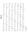
Figure 2 depicts the ART cDNA from human brain tissue (SEQ ID NO:5).
Figure 3 depicts the ART cDNA from human peripheral tissues (SEQ ID
NO:6).
Figure 4 depicts the full length translated amino acid sequence of human
ART cDNA (SEQ ID NO:7).
Figure 5 depicts a truncated human ART polypeptide (SEQ ID NO:8).
Figure 6 depicts a Northern blot of various
human tissues as indicated. The blot was probed with an
ART cDNA as described in the Examples.
Figure 7 depicts the mouse genomic DNA
starting with exon 2 (the first coding exon) and also
contains exons 3 and 4, as well as the corresponding
introns (SEQ ID NO:9).
Figure 8 depicts the full length translated
amino acid sequence of mouse ART cDNA (SEQ ID NO:10).
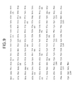
Figure 9 depicts the amino acid sequence of a
human ART gene polymorphism. As is apparent, the amino
acid at position 45 (Leu in Figure 4) is Pro in this
polymorphic seuqence (SEQ ID NO:11).
Figure 10 is a graph of the feeding behavior
pattern of rats injected with human ART polypeptide.
The X axis represents the time after injection of ART at
which food intake was measured; the Y axis represents
the cumulative amount of food consumed in grams. Rats
were injected with either PBS alone (control),
"unfolded" ART (control), 0.075, 0.3, 3.0, or 7.5 nmol
of folded ART in about a 2 µl volume. Standard error
bars are indicated. Statistical analysis of the data
where appropriate is indicated as: *=ps<0.006-0.0001 vs
PBS, and #=ps<0.01-0.0001 vs unfolded ART.
DETAILED DESCRIPTION OF THE INVENTION
As used herein, the term "ART" when used to
describe a nucleic acid molecule refers to a nucleic
acid molecule or fragment thereof that (a) has the
nucleotide sequence as set forth in SEQ ID NO: 4, SEQ ID
NO:5, or SEQ ID NO:6; (b) has a nucleic acid sequence
encoding a polypeptide that is at least 70 percent
identical, preferably at least 80 percent identical, and
more preferably at least 90 percent identical to the
polypeptide encoded by any of SEQ ID NOS:4, 5, or 6; (c)
is a naturally occurring allelic variant of (a) or (b);
(d) is a nucleic acid variant of (a)-(c) produced as
provided for herein; and/or (e) is complementary to
(a)-(d).
Percent sequence identity can be determined by
standard methods that are commonly used to compare the
similarity in position of the amino acids of two
polypeptides. Using a computer program such as BLAST or
FASTA, two polypeptides are aligned for optimal matching
of their respective amino acids (either along the full
length of one or both sequences, or along a predetermined
portion of one or both sequences). The
programs provide a "default" opening penalty and a
"default" gap penalty, and a scoring matrix such as PAM
250 (a standard scoring matrix; see Dayhoff
et al., in:
Atlas of Protein Sequence and Structure, vol. 5, supp.3
[1978]) can be used in conjunction with the computer
program. The percent identity can then be calculated
as:
Polypeptides that are at least 70 percent identical will
typically have one or more amino acid substitutions,
deletions, and/or insertions. Usually, the substitutions
will be conservative so as to have little or no effect on
the overall net charge, polarity, or hydrophobicity of
the protein. Conservative substitutions are set forth in
Table I below.
| Conservative amino acid substitutions |
| Basic | arginine
lysine
histidine |
| Acidic | glutamic acid
aspartic acid |
| Polar | glutamine
asparagine |
| Hydrophobic | leucine
isoleucine
valine |
| Aromatic | phenylalanine
tryptophan
tyrosine |
| Small | glycine
alanine
serine
threonine
methionine |
The term "stringent conditions" refers,to
hybridization and washing under conditions that permit
only binding of a nucleic acid molecule such as an
oligonucleotide or cDNA molecule probe to highly
homologous sequences. One stringent wash solution is
0.015 M NaCl, 0.005 M NaCitrate, and 0.1 percent SDS
used at a temperature of 55°C-65°C. Another stringent
wash solution is 0.2 X SSC and 0.1 percent SDS used at a
temperature of between 50°C-65°C. Where oligonucleotide
probes are used to screen cDNA or genomic libraries, the
following stringent washing conditions may be used. One
protocol uses 6 X SSC with 0.05 percent sodium
pyrophosphate at a temperature of 35°C-62°C, depending
on the length of the oligonucleotide probe. For
example, 14 base pair probes are washed at 35-40°C, 17
base pair probes are washed at 45-50°C, 20 base pair
probes are washed at 52-57°C, and 23 base pair probes
are washed at 57-63°C. The temperature can be increased
2-3°C where the background non-specific binding appears
high. A second protocol utilizes tetramethylammonium
chloride (TMAC) for washing oligonucleotide probes. One
stringent washing solution is 3 M TMAC, 50 mM Tris-HCl,
pH 8.0, and 0.2 percent SDS. The washing temperature
using this solution is a function of the length of the
probe. For example, a 17 base pair probe is washed at
about 45-50°C.
The term "ART protein" or "ART polypeptide" as
used herein refers to any protein or polypeptide having
the properties described herein for ART. The ART
polypeptide may or may not have an amino terminal
methionine, depending on the manner in which it is
prepared. By way of illustration, ART protein or ART
polypeptide includes, an amino acid sequence encoded by
the nucleic acid molecule set forth in any of items (a)-(e)
above and peptide or polypeptide fragments derived
therefrom, to the amino acid sequence set forth in SEQ
ID NOs:7 or 8, and/or to chemically modified derivatives
as well as nucleic acid and or amino acid sequence
variants thereof as provided for herein.
As used herein, the term "ART fragment" refers
to a peptide or polypeptide that is less than the full
length amino acid sequence of naturally occurring ART
protein but has substantially the same biological
activity as ART polypeptide or ART protein described
above. Such a fragment may be truncated at the amino
terminus, the carboxy terminus, and/or internally, and
may be chemically modified. Preferably, the ART
fragment will be a carboxy terminal fragment which
retains at least all 10 C-terminal cysteine residues.
Such ART fragments may be prepared with or without an
amino terminal methionine. A preferred ART fragment is
set forth in SEQ ID NO:8.
As used herein, the term "ART derivative" or
"ART variant" refers to a ART polypeptide or ART protein
that has 1) been chemically modified, as for example, by
addition of polyethylene glycol or other compound,
and/or 2) contains one or more nucleic acid or amino
acid sequence substitutions, deletions, and/or
insertions.
As used herein, the terms "biologically active
polypeptide" and "biologically active fragment" refer to
a peptide or polypeptide that has ART activity (i.e., is
capable of modulating the signaling activity of a
melanocortin receptor, is capable of modulating
intracellular calcium levels, and/or is capable of
modulating lipid metabolism).
As used herein, the terms "effective amount"
and "therapeutically effective amount" refer to the
amount of ART necessary to support one or more
biological activities of ART as set forth above.
The ART polypeptides that have use in
practicing the present invention may be naturally
occurring full length polypeptides, or truncated
polypeptides or peptides (i.e, "fragments"). The
polypeptides or fragments may be chemically modified,
i.e., glycosylated, phosphorylated, and/or linked to a
polymer, as described below, and they may have an amino
terminal methionine, depending on how they are prepared.
In addition, the polypeptides or fragments may be
variants of the naturally occurring ART polypeptide
(i.e., may contain one or more amino acid deletions,
insertions, and/or substitutions as compared with
naturally occurring ART).
The full length ART polypeptide or fragment
thereof can be prepared using well known recombinant DNA
technology methods such as those set forth in Sambrook
et al. (Molecular Cloning: A Laboratory Manual, Cold
Spring Harbor Laboratory Press, Cold Spring Harbor, NY
[1989]) and/or Ausubel et al., eds, (Current Protocols
in Molecular Biology, Green Publishers Inc. and Wiley
and Sons, NY [1994]). A gene or cDNA encoding the ART
protein or fragment thereof may be obtained for example
by screening a genomic or cDNA library, or by PCR
amplification. Alternatively, a gene encoding the ART
polypeptide or fragment may be prepared by chemical
synthesis using methods well known to the skilled
artisan such as those described by Engels et al. (Angew.
Chem. Intl. Ed., 28:716-734 [1989]). These methods
include, inter alia, the phosphotriester,
phosphoramidite, and H-phosphonate methods for nucleic
acid synthesis. A preferred method for such chemical
synthesis is polymer-supported synthesis using standard
phosphoramidite chemistry. Typically, the DNA encoding
the ART polypeptide will be several hundred nucleotides
in length. Nucleic acids larger than about 100
nucleotides can be synthesized as several fragments
using these methods. The fragments can then be ligated
together to form the full length ART polypeptide.
Usually, the DNA fragment encoding the amino terminus of
the polypeptide will have an ATG, which encodes a
methionine residue. This methionine may or may not be
present on the mature form of the ART polypeptide,
depending on whether the polypeptide produced in the
host cell is secreted from that cell.
In some cases, it may be desirable to prepare
nucleic acid and/or amino acid variants of naturally
occurring ART. Nucleic acid variants (wherein one or
more nucleotides are designed to differ from the wild-type
or naturally occurring ART) may be produced using
site directed mutagenesis or PCR amplification where the
primer(s) have the desired point mutations (see Sambrook
et al., supra, and Ausubel et al., supra, for
descriptions of mutagenesis techniques). Chemical
synthesis using methods described by Engels et al.,
supra, may also be used to prepare such variants.
Other methods known to the skilled artisan may be used
as well. Preferred nucleic acid variants are those
containing nucleotide substitutions accounting for codon
preference in the host cell that is to be used to
produce ART. Other preferred variants are those
encoding conservative amino acid changes (e.g., wherein
the charge or polarity of the naturally occurring amino
acid side chain is not altered substantially by
substitution with a different amino acid) as compared to
wild type, and/or those designed to either generate a
novel glycosylation and/or phosphorylation site(s) on
ART, or those designed to delete an existing
glycosylation and/or phosphorylation site(s) on ART.
The ART gene or cDNA can be inserted into an
appropriate expression vector for expression in a host
cell. The vector is selected to be functional in the
particular host cell employed (i.e., the vector is
compatible with the host cell machinery such that
amplification of the ART gene and/or expression of the
gene can occur). The ART polypeptide or fragment
thereof may be amplified/expressed in prokaryotic,
yeast, insect (baculovirus systems) and/or eukaryotic
host cells. Selection of the host cell will depend at
least in part on whether the ART polypeptide or
fragment thereof is to be glycosylated. If so, yeast,
insect, or mammalian host cells are preferable; yeast
cells will glycosylate the polypeptide, and insect and
mammalian cells can glycosylate and/or phosphorylate the
polypeptide as it naturally occurs on the ART
polypeptide (i.e., "native" glycosylation and/or
phosphorylation).
Typically, the vectors used in any of the host
cells will contain 5' flanking sequence (also referred
to as a "promoter") and other regulatory elements as
well such as an enhancer(s), an origin of replication
element, a transcriptional termination element, a
complete intron sequence containing a donor and acceptor
splice site, a signal peptide sequence, a ribosome
binding site element, a polyadenylation sequence, a
polylinker region for inserting the nucleic acid
encoding the polypeptide to be expressed, and a
selectable marker element. Each of these elements is
discussed below. Optionally, the vector may contain a
"tag" sequence, i.e., an oligonucleotide sequence
located at the 5' or 3' end of the ART coding sequence
that encodes polyHis (such as hexaHis) or another small
immunogenic sequence. This tag will be expressed along
with the protein, and can serve as an affinity tag for
purification of the ART polypeptide from the host cell.
Optionally, the tag can subsequently be removed from the
purified ART polypeptide by various means such as using
a selected peptidase for example.
The 5' flanking sequence may be homologous
(i.e., from the same species and/or strain as the host
cell), heterologous (i.e., from a species other than the
host cell species or strain), hybrid (i.e., a
combination of 5' flanking sequences from more than one
source), synthetic, or it may be the native ART 5'
flanking sequence. As such, the source of the 5'
flanking sequence may be any unicellular prokaryotic or
eukaryotic organism, any vertebrate or invertebrate
organism, or any plant, provided that the 5' flanking
sequence is functional in, and can be activated by, the
host cell machinery.
The 5' flanking sequences useful in the
vectors of this invention may be obtained by any of
several methods well known in the art. Typically, 5'
flanking sequences useful herein other than the ART 5'
flanking sequence will have been previously identified
by mapping and/or by restriction endonuclease digestion
and can thus be isolated from the proper tissue source
using the appropriate restriction endonucleases. In
some cases, the full nucleotide sequence of the 5'
flanking sequence may be known. Here, the 5' flanking
sequence may be synthesized using the methods described
above for nucleic acid synthesis or cloning.
Where all or only a portion of the 5' flanking
sequence is known, it may be obtained using PCR and/or
by screening a genomic library with suitable
oligonucleotide and/or 5' flanking sequence fragments
from the same or another species.
Where the 5' flanking sequence is not known, a
fragment of DNA containing a 5' flanking sequence may be
isolated from a larger piece of DNA that may contain,
for example, a coding sequence or even another gene or
genes. Isolation may be accomplished by restriction
endonuclease digestion using one or more carefully
selected enzymes to isolate the proper DNA fragment.
After digestion, the desired fragment may be isolated by
agarose gel purification, Qiagen® column or other
methods known to the skilled artisan. Selection of
suitable enzymes to accomplish this purpose will be
readily apparent to one of ordinary skill in the art.
The origin of replication element is typically a
part of prokaryotic expression vectors purchased
commercially, and aids in the amplification of the
vector in a host cell. Amplification of the vector to a
certain copy number can, in some cases, be important for
optimal expression of the ART polypeptide. If the
vector of choice does not contain an origin of
replication site, one may be chemically synthesized
based on a known sequence, and ligated into the vector.
The transcription termination element is
typically located 3' to the end of the ART polypeptide
coding sequence and serves to terminate transcription of
the ART polypeptide. Usually, the transcription
termination element in prokaryotic cells is a G-C rich
fragment followed by a poly T sequence. While the
element is easily cloned from a library or even
purchased commercially as part of a vector, it can also
be readily synthesized using methods for nucleic acid
synthesis such as those described above.
A selectable marker gene element encodes a
protein necessary for the survival and growth of a host
cell grown in a selective culture medium. Typical
selection marker genes encode proteins that (a) confer
resistance to antibiotics or other toxins, e.g.,
ampicillin, tetracycline, or kanamycin for prokaryotic
host cells, (b) complement auxotrophic deficiencies of
the cell; or (c) supply critical nutrients not available
from complex media. Preferred selectable markers are
the kanamycin resistance gene, the ampicillin resistance
gene, and the tetracycline resistance gene.
The ribosome binding element, commonly called
the Shine-Dalgarno sequence (prokaryotes) or the Kozak
sequence (eukaryotes), is necessary for translation
initiation of mRNA. The element is typically located 3'
to the promoter and 5' to the coding sequence of the ART
polypeptide to be synthesized. The Shine-Dalgarno
sequence is varied but is typically a polypurine (i.e.,
having a high A-G content). Many Shine-Dalgarno
sequences have been identified, each of which can be
readily synthesized using methods set forth above and
used in a prokaryotic vector.
In those cases where it is desirable for ART
to be secreted from the host cell, a signal sequence may
be used to direct the ART polypeptide out of the host
cell where it is synthesized. Typically, the signal
sequence is positioned in the coding region of ART
nucleic acid sequence, or directly at the 5' end of the
ART coding region. Many signal sequences have been
identified, and any of them that are functional in the
selected host cell may be used in conjunction with the
ART gene. Therefore, the signal sequence may be
homologous or heterologous to the ART polypeptide, and
may be homologous or heterologous to the ART
polypeptide. Additionally, the signal sequence may be
chemically synthesized using methods set forth above.
In most cases, secretion of the polypeptide from the
host cell via the presence of a signal peptide will
result in the removal of the amino terminal methionine
from the polypeptide.
In many cases, transcription of the ART
polypeptide is increased by the presence of one or more
introns on the vector; this is particularly true for
eukaryotic host cells, especially mammalian host cells.
The intron may be naturally occurring within the ART
nucleic acid sequence, especially where the ART sequence
used is a full length genomic sequence or a fragment
thereof. Where the intron is not naturally occurring
within the ART DNA sequence (as for most cDNAs), the
intron(s) may be obtained from another source. The
position of the intron with respect to the 5' flanking
sequence and the ART coding sequence is important, as
the intron must be transcribed to be effective. As
such, where the ART nucleic acid sequence is a cDNA
sequence, the preferred position for the intron is 3' to
the transcription start site, and 5' to the polyA
transcription termination sequence. Preferably for ART
cDNAs, the intron will be located on one side or the
other (i.e., 5' or 3') of the ART coding sequence such
that it does not interrupt the this coding sequence.
Any intron from any source, including any viral,
prokaryotic and eukaryotic (plant or animal) organisms,
may be used to practice this invention, provided that it
is compatible with the host cell(s) into which it is
inserted. Also included herein are synthetic introns.
Optionally, more than one intron may be used in the
vector.
Where one or more of the elements set forth
above are not already present in the vector to be used,
they may be individually obtained and ligated into the
vector. Methods used for obtaining each of the elements
are well known to the skilled artisan and are comparable
to the methods set forth above (i.e., synthesis of the
DNA, library screening, and the like).
The final vectors used to practice this
invention are typically constructed from a starting
vectors such as a commercially available vector. Such
vectors may or may not contain some of the elements to
be included in the completed vector. If none of the
desired elements are present in the starting vector,
each element may be individually ligated into the vector
by cutting the vector with the appropriate restriction
endonuclease(s) such that the ends of the element to be
ligated in and the ends of the vector are compatible for
ligation. In some cases, it may be necessary to "blunt"
the ends to be ligated together in order to obtain a
satisfactory ligation. Blunting is accomplished by
first filling in "sticky ends" using Klenow DNA
polymerase or T4 DNA polymerase in the presence of all
four nucleotides. This procedure is well known in the
art and is described for example in Sambrook et al.,
supra.
Alternatively, two or more of the elements to
be inserted into the vector may first be ligated
together (if they are to be positioned adjacent to each
other) and then ligated into the vector.
One other method for constructing the vector
to conduct all ligations of the various elements
simultaneously in one reaction mixture. Here, many
nonsense or nonfunctional vectors will be generated due
to improper ligation or insertion of the elements,
however the functional vector may be identified and
selected by restriction endonuclease digestion.
Preferred vectors for practicing this
invention are those which are compatible with bacterial,
insect, and mammalian host cells. Such vectors include,
inter alia, pCRII (Invitrogen Company, San Diego, CA),
pBSII (Stratagene Company, LaJolla, CA), and pETL
(BlueBacII; Invitrogen).
After the vector has been constructed and a
ART nucleic acid has been inserted into the proper site
of the vector, the completed vector may be inserted into
a suitable host cell for amplification and/or ART
polypeptide expression.
Host cells may be prokaryotic host cells (such
as E. coli) or eukaryotic host cells (such as a yeast
cell, an insect cell, or a vertebrate cell). The host
cell, when cultured under appropriate conditions, can
synthesize ART protein which can subsequently be
collected from the culture medium (if the host cell
secretes it into the medium) or directly from the host
cell producing it (if it is not secreted). After
collection, the ART protein can be purified using
methods such as molecular sieve chromatography, affinity
chromatography, and the like.
Selection of the host cell will depend in part
on whether the ART protein is to be glycosylated or
phosphorylated (in which case eukaryotic host cells are
preferred), and the manner in which the host cell is
able to "fold" the protein into its native tertiary
structure (e.g., proper orientation of disulfide
bridges, etc.) such that biologically active protein is
prepared by the cell. However, where the host cell does
not synthesize biologically active ART, the ART may be
"folded" after synthesis using appropriate chemical
conditions as discussed below.
Suitable cells or cell lines may be mammalian
cells, such as Chinese hamster ovary cells (CHO) or 3T3
cells. The selection of suitable mammalian host cells
and methods for transformation, culture, amplification,
screening and product production and purification are
known in the art. Other suitable mammalian cell lines,
are the monkey COS-1 and COS-7 cell lines, and the CV-1
cell line. Further exemplary mammalian host cells
include primate cell lines and rodent cell lines,
including transformed cell lines. Normal diploid cells,
cell strains derived from in vitro culture of primary
tissue, as well as primary explants, are also suitable.
Candidate cells may be genotypically deficient in the
selection gene, or may contain a dominantly acting
selection gene. Other suitable mammalian cell lines
include but are not limited to, HeLa, mouse L-929 cells,
3T3 lines derived from Swiss, Balb-c or NIH mice, BHK or
HaK hamster cell lines.
Similarly useful as host cells suitable for
the present invention are bacterial cells. For example,
the various strains of E. coli (e.g., HB101, DH5α,DH10,
and MC1061) are well-known as host cells in the field of
biotechnology. Various strains of B. subtilis,
Pseudomonas spp., other Bacillus spp., Streptomyces
spp., and the like may also be employed in this method.
Many strains of yeast cells known to those
skilled in the art are also available as host cells for
expression of the polypeptides of the present invention.
Additionally, where desired, insect cells may be
utilized as host cells in the method of the present
invention (Miller et al., Genetic Engineering 8: 277-298
[1986]).
Insertion (also referred to as
"transformation" or "transfection") of the vector into
the selected host cell may be accomplished using such
methods as calcium chloride, electroporation,
microinjection, lipofection or the DEAE-dextran method.
The method selected will in part be a function of the
type of host cell to be used. These methods and other
suitable methods are well known to the skilled artisan,
and are set forth, for example, in Sambrook et al.,
supra.
The host cells containing the vector (i.e.,
transformed or transfected) may be cultured using
standard media well known to the skilled artisan. The
media will usually contain all nutrients necessary for
the growth and survival of the cells. Suitable media
for culturing E. coli cells are for example, Luria Broth
(LB) and/or Terrific Broth (TB). Suitable media for
culturing eukaryotic cells are RPMI 1640, MEM, DMEM, all
of which may be supplemented with serum and/or growth
factors as required by the particular cell line being
cultured. A suitable medium for insect cultures is
Grace's medium supplemented with yeastolate, lactalbumin
hydrolysate, and/or fetal calf serum as necessary.
Typically, an antibiotic or other compound
useful for selective growth of the transformed cells
only is added as a supplement to the media. The
compound to be used will be dictated by the selectable
marker element present on the plasmid with which the
host cell was transformed. For example, where the
selectable marker element is kanamycin resistance, the
compound added to the culture medium will be kanamycin.
The amount of ART polypeptide produced in the
host cell can be evaluated using standard methods known
in the art. Such methods include, without limitation,
Western blot analysis, SDS-polyacrylamide gel
electrophoresis, non-denaturing gel electrophoresis,
HPLC separation, immunoprecipitation, and/or activity
assays such as DNA binding gel shift assays.
If the ART polypeptide has been designed to be
secreted from the host cells, the majority of
polypeptide will likely be found in the cell culture
medium. Polypeptides prepared in this way will
typically not possess an amino terminal methionine, as
it is removed during secretion from the cell. If
however, the ART polypeptide is not secreted from the
host cells, it will be present in the cytoplasm (for
eukaryotic, gram positive bacteria, and insect host
cells) or in the periplasm (for gram negative bacteria
host cells) and may have an amino terminal methionine.
For intracellular ART protein, the host cells
are typically first disrupted mechanically or
osmotically to release the cytoplasmic contents into a
buffered solution. ART polypeptide can then be isolated
from this solution.
Purification of ART polypeptide from solution
can be accomplished using a variety of techniques. If
the polypeptide has been synthesized such that it
contains a tag such as Hexahistidine (ART/hexaHis) or
other small peptide at either its carboxyl or amino
terminus, it may essentially be purified in a one-step
process by passing the solution through an affinity
column where the column matrix has a high affinity for
the tag or for the polypeptide directly (i.e., a
monoclonal antibody specifically recognizing ART). For
example, polyhistidine binds with great affinity and
specificity to nickel, thus an affinity column of nickel
(such as the Qiagen nickel columns) can be used for
purification of ART/polyHis. (See for example, Ausubel
et al., eds., Current Protocols in Molecular Biology,
Section 10.11.8, John Wiley & Sons, New York [1993]).
Where the ART polypeptide has no tag and no
antibodies are available, other well known procedures
for purification can be used. Such procedures include,
without limitation, ion exchange chromatography,
molecular sieve chromatography, HPLC, native gel
electrophoresis in combination with gel elution, and
preparative isoelectric focusing ("Isoprime"
machine/technique, Hoefer Scientific). In some cases,
two or more of these techniques may be combined to
achieve increased purity. Preferred methods for
purification include polyHistidine tagging and ion
exchange chromatography in combination with preparative
isoelectric focusing.
If it is anticipated that the ART polypeptide
will be found primarily in the periplasmic space of the
bacteria or the cytoplasm of eukaryotic cells, the
contents of the periplasm or cytoplasm, including
inclusion bodies (e.g., gram-negative bacteria) if the
processed polypeptide has formed such complexes, can be
extracted from the host cell using any standard
technique known to the skilled artisan. For example,
the host cells can be lysed to release the contents of
the periplasm by French press, homogenization, and/or
sonication. The homogenate can then be centrifuged.
If the ART polypeptide has formed inclusion
bodies in the periplasm, the inclusion bodies can often
bind to the inner and/or outer cellular membranes and
thus will be found primarily in the pellet material
after centrifugation. The pellet material can then be
treated with a chaotropic agent such as guanidine or
urea to release, break apart, and solubilize the
inclusion bodies. The ART polypeptide in its now
soluble form can then be analyzed using gel
electrophoresis, immunoprecipitation or the like. If it
is desired to isolate the ART polypeptide, isolation may
be accomplished using standard methods such as those set
forth below and in Marston et al. (Meth. Enz., 182:264-275
[1990]).
If ART polypeptide inclusion bodies are not
formed to a significant degree in the periplasm of the
host cell, the ART polypeptide will be found primarily
in the supernatant after centrifugation of the cell
homogenate, and the ART polypeptide can be isolated from
the supernatant using methods such as those set forth
below.
In those situations where it is preferable to
partially or completely isolate the ART polypeptide,
purification can be accomplished using standard methods
well known to the skilled artisan. Such methods
include, without limitation, separation by
electrophoresis followed by electroelution, various
types of chromatography (immunoaffinity, molecular
sieve, and/or ion exchange), and/or high pressure liquid
chromatography. In some cases, it may be preferable to
use more than one of these methods for complete
purification.
In addition to preparing and purifying ART
polypeptide using recombinant DNA techniques, the ART
polypeptides, fragments, and/or derivatives thereof may
be prepared by chemical synthesis methods (such as solid
phase peptide synthesis) using methods known in the art
such as those set forth by Merrifield et al., (J. Am.
Chem. Soc., 85:2149 [1964]), Houghten et al. (Proc Natl
Acad. Sci. USA, 82:5132 [1985]), and Stewart and Young
(Solid Phase Peptide Synthesis, Pierce Chem Co,
Rockford, IL [1984]). Such polypeptides may be
synthesized with or without a methionine on the amino
terminus. Chemically synthesized ART polypeptides or
fragments may be oxidized using methods set forth in
these references to form disulfide bridges. The ART
polypeptides or fragments may be employed as
biologically active or immunological substitutes for
natural, purified ART polypeptides in therapeutic and
immunological processes.
Chemically modified ART compositions (i.e.,
"derivatives") where the ART polypeptide is linked to a
polymer ("ART-polymers") are included within the scope
of the present invention. The polymer selected is
typically water soluble so that the protein to which it
is attached does not precipitate in an aqueous
environment, such as a physiological environment. The
polymer selected is usually modified to have a single
reactive group, such as an active ester for acylation or
an aldehyde for alkylation, so that the degree of
polymerization may be controlled as provided for in the
present methods. A preferred reactive aldehyde is
polyethylene glycol propionaldehyde, which is water
stable, or mono C1-C10 alkoxy or aryloxy derivatives
thereof (see U.S. Patent 5,252,714). The polymer may be
branched or unbranched. Included within the scope of
ART-polymers is a mixture of polymers. Preferably, for
therapeutic use of the end-product preparation, the
polymer will be pharmaceutically acceptable. The water
soluble polymer or mixture thereof may be selected from
the group consisting of, for example, polyethylene
glycol (PEG), monomethoxy-polyethylene glycol, dextran,
cellulose, or other carbohydrate based polymers, poly-(N-vinyl
pyrrolidone) polyethylene glycol, propylene
glycol homopolymers, a polypropylene oxide/ethylene
oxide co-polymer, polyoxyethylated polyols (e.g.,
glycerol) and polyvinyl alcohol. For the acylation
reactions, the polymer(s) selected should have a single
reactive ester group. For reductive alkylation, the
polymer(s) selected should have a single reactive
aldehyde group. The polymer may be of any molecular
weight, and may be branched or unbranched.
Pegylation of ART may be carried out by any of
the pegylation reactions known in the art, as described
for example in the following references: Focus on Growth
Factors 3 (2): 4-10 (1992); EP 0 154 316; and EP 0 401
384. Preferably, the pegylation is carried out via an
acylation reaction or an alkylation reaction with a
reactive polyethylene glycol molecule (or an analogous
reactive water-soluble polymer) as described below.
Pegylation by acylation generally involves reacting
an active ester derivative of polyethylene glycol (PEG)
with an ART protein. Any known or subsequently
discovered reactive PEG molecule may be used to carry
out the pegylation of ART. A preferred activated PEG
ester is PEG esterified to N-hydroxysuccinimide ("NHS").
As used herein, "acylation" is contemplated to include
without limitation the following types of linkages
between ART and a water soluble polymer such as PEG:
amide, carbamate, urethane, and the like, as described
in Bioconjugate Chem. 5: 133-140 (1994). Reaction
conditions may be selected from any of those known in
the pegylation art or those subsequently developed,
provided that conditions such as temperature, solvent,
and pH that would inactivate the ART species to be
modified are avoided.
Pegylation by acylation usually results in a
poly-pegylated ART product, wherein the lysine ε-amino
groups are pegylated via an acyl linking group.
Preferably, the connecting linkage will be an amide.
Also preferably, the resulting product will be at least
about 95 percent mono, di- or tri- pegylated. However,
some species with higher degrees of pegylation (up to
the maximum number of lysine ε-amino acid groups of ART
plus one α-amino group at the amino terminus of ART)
will normally be formed in amounts depending on the
specific reaction conditions used. If desired, more
purified pegylated species may be separated from the
mixture, particularly unreacted species, by standard
purification techniques, including, among others,
dialysis, salting-out, ultrafiltration, ion-exchange
chromatography, gel filtration chromatography and
electrophoresis.
Pegylation by alkylation generally involves
reacting a terminal aldehyde derivative of PEG with a
protein such as ART in the presence of a reducing agent.
Regardless of the degree of pegylation, the PEG groups
are preferably attached to the protein via a -CH2-NH-
group. With particular reference to the -CH2- group,
this type of linkage is referred to herein as an "alkyl"
linkage.
Derivatization via reductive alkylation to
produce a monopegylated product exploits the
differential reactivity of different types of primary
amino groups (lysine versus the N-terminal) available
for derivatization in ART. Typically, the reaction is
performed at a pH (see below) which allows one to take
advantage of the pKa differences between the ε-amino
groups of the lysine residues and that of the α-amino
group of the N-terminal residue of the protein. By such
selective derivatization, attachment of a water soluble
polymer that contains a reactive group such as an
aldehyde, to a protein is controlled: the conjugation
with the polymer occurs predominantly at the N-terminus
of the protein without significant modification of other
reactive groups such as the lysine side chain amino
groups. The present invention provides for a
substantially homogeneous preparation of ART-monopolymer
protein conjugate molecules (meaning ART protein to
which a polymer molecule has been attached substantially
only (i.e., at least about 95%) in a single location on
the ART protein. More specifically, if polyethylene
glycol is used, the present invention also provides for
pegylated ART protein lacking possibly antigenic linking
groups, and having the polyethylene glycol molecule
directly coupled to the ART protein.
A particularly preferred water-soluble polymer
for use herein is polyethylene glycol, abbreviated PEG.
As used herein, polyethylene glycol is meant to
encompass any of the forms of PEG that have been used to
derivatize other proteins, such as mono-(C1-C10) alkoxy-
or aryloxy-polyethylene glycol.
In general, chemical derivatization may be
performed under any suitable conditions used to react a
biologically active substance with an activated polymer
molecule. Methods for preparing pegylated ART will
generally comprise the steps of (a) reacting an ART
polypeptide with polyethylene glycol (such as a reactive
ester or aldehyde derivative of PEG) under conditions
whereby ART becomes attached to one or more PEG groups,
and (b) obtaining the reaction product(s). In general,
the optimal reaction conditions for the acylation
reactions will be determined based on known parameters
and the desired result. For example, the larger the
ratio of PEG: protein, the greater the percentage of
poly-pegylated product.
Reductive alkylation to produce a
substantially homogeneous population of mono-polymer/ART
protein conjugate molecule will generally comprise the
steps of: (a) reacting a ART protein with a reactive PEG
molecule under reductive alkylation conditions, at a pH
suitable to permit selective modification of the α-amino
group at the amino terminus of said ART protein; and (b)
obtaining the reaction product(s).
For a substantially homogeneous population of
mono-polymer/ART protein conjugate molecules, the
reductive alkylation reaction conditions are those which
permit the selective attachment of the water soluble
polymer moiety to the N-terminus of ART. Such reaction
conditions generally provide for pKa differences between
the lysine amino groups and the α-amino group at the
N-terminus (the pKa being the pH at which 50% of the
amino groups are protonated and 50% are not). The pH
also affects the ratio of polymer to protein to be used.
In general, if the pH is lower, a larger excess of
polymer to protein will be desired (i.e., the less
reactive the N-terminal α-amino group, the more polymer
needed to achieve optimal conditions). If the pH is
higher, the polymer:protein ratio need not be as large
(i.e., more reactive groups are available, so fewer
polymer molecules are needed). For purposes of the
present invention, the pH will generally fall within the
range of 3-9, preferably 3-6.
Another important consideration is the
molecular weight of the polymer. In general, the higher
the molecular weight of the polymer, the fewer number of
polymer molecules which may be attached to the protein.
Similarly, branching of the polymer should be taken into
account when optimizing these parameters. Generally,
the higher the molecular weight (or the more branches)
the higher the polymer:protein ratio. In general, for
the pegylation reactions contemplated herein, the
preferred average molecular weight is about 2kDa to
about 100kDa (the term "about" indicating ± 1kDa). The
preferred average molecular weight is about 5kDa to
about 50kDa, particularly preferably about 12kDa to
about 25kDa. The ratio of water-soluble polymer to ART
protein will generally range from 1:1 to 100:1,
preferably (for polypegylation) 1:1 to 20:1 and (for
monopegylation) 1:1 to 5:1.
Using the conditions indicated above,
reductive alkylation will provide for selective
attachment of the polymer to any ART protein having an
α-amino group at the amino terminus, and provide for a
substantially homogenous preparation of monopolymer/ART
protein conjugate. The term "monopolymer/ART protein
conjugate" is used here to mean a composition comprised
of a single polymer molecule attached to an ART protein
molecule. The monopolymer/ART protein conjugate
preferably will have a polymer molecule located at the
N-terminus, but not on lysine amino side groups. The
preparation will preferably be greater than 90%
monopolymer/ART protein conjugate, and more preferably
greater than 95% monopolymer ART protein conjugate, with
the remainder of observable molecules being unreacted
(i.e., protein lacking the polymer moiety). The
examples below provide for a preparation which is at
least about 90% monopolymer/ protein conjugate, and
about 10% unreacted protein. The monopolymer/protein
conjugate has biological activity.
For the present reductive alkylation, the
reducing agent should be stable in aqueous solution and
preferably be able to reduce only the Schiff base formed
in the initial process of reductive alkylation.
Preferred reducing agents may be selected from the group
consisting of sodium borohydride, sodium
cyanoborohydride, dimethylamine borane, trimethylamine
borane and pyridine borane. A particularly preferred
reducing agent is sodium cyanoborohydride.
Other reaction parameters, such as solvent,
reaction times, temperatures, etc., and means of
purification of products, can be determined based on
the published information relating to derivatization of
proteins with water soluble polymers.
A mixture of polymer-ART protein conjugate
molecules may be prepared by acylation and/or alkylation
methods, as described above,and one may select the
proportion of monopolymer/ protein conjugate to include
in the mixture. Thus, where desired, a mixture of
various protein with various numbers of polymer
molecules attached (i.e., di-, tri-, tetra-, etc.) may
be prepared and combined with the monopolymer/ART
protein conjugate material prepared using the present
methods.
Generally, conditions which may be alleviated
or modulated by administration of the present
polymer/ART include those described herein for ART
molecules in general. However, the polymer/ART
molecules disclosed herein may have additional
activities, enhanced or reduced activities, or other
characteristics, as compared to the non-derivatized
molecules.
ART nucleic acid molecules, fragments, and/or
derivatives that do not themselves encode polypeptides
that are active in activity assays may be useful as
hybridization probes in diagnostic assays to test,
either qualitatively or quantitatively, for the presence
of ART DNA or RNA in mammalian tissue or bodily fluid
samples.
ART polypeptide fragments and/or derivatives
that are not themselves active in activity assays may be
useful as modulators (e.g., inhibitors or stimulants) of
the ART receptors in vitro or in vivo, or to prepare
antibodies to ART polypeptides.
The ART polypeptides and fragments thereof,
whether or not chemically modified, may be employed
alone, or in combination with other pharmaceutical
compositions such as, for example, neurotrophic factors,
cytokines, interferons, interleukins, growth factors,
antibiotics, anti-inflammatories, neurotransmitter
receptor agonists or antagonists and/or antibodies, in
the treatment of endocrine system disorders.
The ART polypeptides and/or fragments thereof
may be used to prepare antibodies generated by standard
methods. Thus, antibodies that react with the ART
polypeptides, as well as reactive fragments of such
antibodies, are also contemplated as within the scope of
the present invention. The antibodies may be
polyclonal, monoclonal, recombinant, chimeric, single-chain
and/or bispecific, etc. The antibody fragments
may be any fragment that is reactive with the ART of the
present invention, such as, Fab, Fab', etc. Also
provided by this invention are the hybridomas generated
by presenting ART or a fragment thereof as an antigen to
a selected mammal, followed by fusing cells (e.g.,
spleen cells) of the animal with certain cancer cells to
create immortalized cell lines by known techniques. The
methods employed to generate such cell lines and
antibodies directed against all or portions of a human
ART polypeptide of the present invention are also
encompassed by this invention.
The antibodies may be used therapeutically,
such as to inhibit binding of the ART to its receptor.
The antibodies may further be used for in vivo and in
vitro diagnostic purposes, such as in labeled form to
detect the presence of the ART in a body fluid.
Therapeutic Compositions and Administration
Therapeutic compositions for treating various
endocrine and/or neuro-endocrine system disorders such
as glucocorticoid resistance, Cushing's syndrome (either
genetic or caued by ectopic ACTH production due to
pituitary tumors, small lung carcinomas, or adrenal
tumors), congenital adrenal hyperplasia, other disorders
of the hypothalamic-pitutary axis (HPA), and/or obesity
are within the scope of the present invention. Such
compositions may comprise a therapeutically effective
amount of a ART polypeptide or fragment thereof (either
of which may be chemically modified) in admixture with a
pharmaceutically acceptable carrier. The carrier
material may be water for injection, preferably
supplemented with other materials common in solutions
for administration to mammals. Typically, a ART
therapeutic compound will be administered in the form of
a composition comprising purified protein (which may be
chemically modified) in conjunction with one or more
physiologically acceptable carriers, excipients, or
diluents. Neutral buffered saline or saline mixed with
serum albumin are exemplary appropriate carriers.
Preferably, the product is formulated as a lyophilizate
using appropriate excipients (e.g., sucrose). Other
standard carriers, diluents, and excipients may be
included as desired. Other exemplary compositions
comprise Tris buffer of about pH 7.0-8.5, or acetate
buffer of about pH 4.0-5.5, which may further include
sorbitol or a suitable substitute therefor.
The ART compositions can be systemically
administered parenterally. Alternatively, the
compositions may be administered intravenously or
subcutaneously. When systemically administered, the
therapeutic compositions for use in this invention may
be in the form of a pyrogen-free, parenterally
acceptable aqueous solution. The preparation of such
pharmaceutically acceptable protein solutions, with due
regard to pH, isotonicity, stability and the like, is
within the skill of the art.
Therapeutic formulations of ART compositions
useful for practicing the present invention may be
prepared for storage by mixing the selected composition
having the desired degree of purity with optional
physiologically acceptable carriers, excipients, or
stabilizers (Remington's Pharmaceutical Sciences, 18th
edition, A.R. Gennaro, ed., Mack Publishing Company
[1990]) in the form of a lyophilized cake or an aqueous
solution. Acceptable carriers, excipients or stabilizers
are nontoxic to recipients and are preferably inert at
the dosages and concentrations employed, and include
buffers such as phosphate, citrate, or other organic
acids; antioxidants such as ascorbic acid; low molecular
weight polypeptides; proteins, such as serum albumin,
gelatin, or immunoglobulins; hydrophilic polymers such
as polyvinylpyrrolidone; amino acids such as glycine,
glutamine, asparagine, arginine or lysine;
monosaccharides, disaccharides, and other carbohydrates
including glucose, mannose, or dextrins; chelating
agents such as EDTA; sugar alcohols such as mannitol or
sorbitol; salt-forming counterions such as sodium;
and/or nonionic surfactants such as Tween, Pluronics or
polyethylene glycol (PEG).
The ART composition to be used for in vivo
administration must be sterile. This is readily
accomplished by filtration through sterile filtration
membranes. Where the ART composition is lyophilized,
sterilization using these methods may be conducted
either prior to, or following, lyophilization and
reconstitution. The composition for parenteral
administration ordinarily will be stored in lyophilized
form or in solution.
Therapeutic compositions generally are placed
into a container having a sterile access port, for
example, an intravenous solution bag or vial having a
stopper pierceable by a hypodermic injection needle.
The route of administration of the composition
is in accord with known methods, e.g. oral, injection or
infusion by intravenous, intraperitoneal, intracerebral
(intraparenchymal), intracerebroventricular,
intramuscular, intraocular, intraarterial, or
intralesional routes, or by sustained release systems or
implantation device which may optionally involve the use
of a catheter. Where desired, the compositions may be
administered continuously by infusion, bolus injection
or by implantation device. Alternatively or
additionally, ART may be administered locally via
implantation into the affected area of a membrane,
sponge, or other appropriate material on to which ART
polypeptide has been absorbed.
Where an implantation device is used, the
device may be implanted any suitable tissue or organ,
such as, for example, into a cerebral ventricle or into
brain parenchyma, and delivery of ART may be directly
through the device via bolus or continuous
administration, or via a catheter using continuous
infusion.
ART polypeptide may be administered in a
sustained release formulation or preparation. Suitable
examples of sustained-release preparations include
semipermeable polymer matrices in the form of shaped
articles, e.g. films, or microcapsules. Sustained
release matrices include polyesters, hydrogels,
polylactides (U.S. 3,773,919, EP 58,481), copolymers of
L-glutamic acid and gamma ethyl-L-glutamate (Sidman et
al, Biopolymers, 22: 547-556 [1983]), poly (2-hydroxyethyl-methacrylate)
(Langer et al., J. Biomed.
Mater. Res., 15: 167-277 [1981] and Langer, Chem. Tech.,
12: 98-105 [1982]), ethylene vinyl acetate (Langer et
al., supra) or poly-D(-)-3-hydroxybutyric acid (EP
133,988). Sustained-release compositions also may
include liposomes, which can be prepared by any of
several methods known in the art (e.g., DE 3,218,121;
Epstein et al., Proc. Natl. Acad. Sci. USA, 82: 3688-3692
[1985]; Hwang et al., Proc. Natl. Acad. Sci. USA,
77: 4030-4034 [1980]; EP 52,322; EP 36,676; EP 88,046;
EP 143,949).
In some cases, it may be desirable to use ART
compositions in an ex vivo manner, i.e., to treat cells
or tissues that have been removed from the patient and
are then subsequently implanted back into the patient.
In other cases, ART may be delivered through
implanting into patients certain cells that have been
genetically engineered (using methods described above)
to express and secrete ART polypeptide. Such cells may
be human cells, and may be derived from the patient's
own tissue or from another source, either human or non-human.
Optionally, the cells may be immortalized. The
cells may be implanted into the brain, adrenal gland or
into other body tissues or organs.
In certain situations, it may be desirable to
use gene therapy methods for administration of ART to
patients suffering from certain endocrine and/or neuro-endocrine
system disorders or diseases such as
glucocorticoid resistance, Cushing's syndrome (either
genetic or caued by ectopic ACTH production due to
pituitary tumors, small lung carcinomas, or adrenal
tumors), congenital adrenal hyperplasia, other disorders
of the hypothalamic-pitutary axis (HPA), and/or obesity.
In these situations, genomic DNA, cDNA, and/or synthetic
DNA encoding ART or a fragment or variant thereof may be
operably linked to a constitutive or inducible promoter
that is active in the tissue into which the composition
will be injected. This ART DNA construct can be
injected directly into brain or other neuronal tissue to
be treated.
Alternatively, the ART DNA construct may be
injected into muscle tissue where it can be taken up
into the cells and expressed in the cells, provided that
the ART DNA is operably linked to a promoter that is
active in muscle tissue such as cytomegalovirus (CMV)
promoter, Rous sarcoma virus (RSV) promoter, or muscle
creatine kinase promoter. Typically, the DNA construct
may include (in addition to the ART DNA and a promoter),
vector sequence obtained from vectors such as adenovirus
vector, adeno-associated virus vector, a retroviral
vector, and/or a herpes virus vector. The vector/DNA
construct may be admixed with a pharmaceutically
acceptable carrier(s) for injection.
An effective amount of the ART composition(s)
to be employed therapeutically will depend, for example,
upon the therapeutic objectives such as the indication
for which ART is being used, the route of
administration, and the condition of the patient.
Accordingly, it will be necessary for the therapist to
titer the dosage and modify the route of administration
as required to obtain the optimal therapeutic effect. A
typical daily dosage may range from about 0.1 µg/kg to
up to 100 mg/kg or more, depending on the factors
mentioned above. Typically, a clinician will administer
the ART composition until a dosage is reached that
achieves the desired effect. The ART composition may
therefore be administered as a single dose, or as two or
more doses (which may or may not contain the same amount
of ART) over time, or as a continuous infusion via
implantation device or catheter.
As further studies are conducted, information
will emerge regarding appropriate dosage levels for
treatment of various conditions in various patients, and
the ordinary skilled worker, considering the therapeutic
context, the type of disorder under treatment, the age
and general health of the recipient, will be able to
ascertain proper dosing. Generally, the dosage will be
between 0.01 µg/kg body weight (calculating the mass of
the protein alone, without chemical modification) and
300 µg/kg (based on the same).
The ART proteins, fragments and/or derivatives
thereof may be utilized to treat diseases and disorders
of the endocrine system which may be associated with
alterations in the pattern of ART expression or which
may benefit from exposure to ART or anti-ART antibodies.
ART protein, and/or fragments or derivatives
thereof, may be used to treat patients in whom various
cells of the endocrine and/or nervous system have
degenerated and/or have been damaged by congenital
disease, trauma, surgery, stroke, ischemia, infection,
metabolic disease, nutritional deficiency, malignancy,
and/or toxic agents.
In other embodiments of the invention, ART
protein and/or fragments or derivatives thereof can be
used to treat endocrine and/or neuro-endocrine system
disorders or diseases such as glucocorticoid resistance,
Cushing's syndrome (either genetic or caued by ectopic
ACTH production due to pituitary tumors, small lung
carcinomas, or adrenal tumors), congenital adrenal
hyperplasia, other disorders of the hypothalamic-pitutary
axis (HPA), and/or obesity. In addition, ART
compositions may be useful in modulating intra-cellular
calcium levels.
In addition, ART protein or peptide fragments
or derivatives thereof can be used in conjunction with
surgical implantation of tissue in the treatment of
diseases in which tissue implantation is indicated.
EXAMPLES
Example I: Identification of Human ART cDNA
The publicly available Washington
University/Merck DNA sequence database referred to as
the EST (Expressed Sequence Tag) database was searched
with a sequence profile (Gribskov et al., Proc. Natl.
Acad. Sci, USA, 84:4355 [1987] and Luethy et al.,
Protein Science, 3:139-146 [1994]) using a sequence
alignment of the human and mouse agouti genes (starting
at amino acid 22 of both mouse and human agouti), along
with the PAM250 amino acid substitution table (Dayhoff
et al., in: Atlas of Protein Sequence and Structure, vol
5, supp. 3 [1978]).
In order to search at the database for
homologous amino acid sequences, each entry in the EST
database was first translated by computer from DNA to
amino acid sequence prior to searching. One EST
database submission cDNA clone, H63735, was found to
have homology to this profile sequence. The submission
containing the sequence of the opposite end of this cDNA
clone, H63298, was examined but did not show any
homology to the profile sequence.
The E. coli stock containing the cDNA clone
corresponding to H63735 and H63298 (stock number 208641)
was obtained from Genome Systems Inc., St. Louis, MO.
The DNA from this clone was prepared using standard
miniprep methods (Sambrook et al., Molecular Cloning: A
Laboratoy Manual, Cold Spring Harbor Laboratory Press,
Cold Spring Harbor, NY [1989]). The DNA was purified by
passage through a Qiagen column (Qiagen, Chatsworth, CA)
and following the manufacturer's protocol. After
purification, the DNA was sequenced using the standard
dideoxy chain termination method. When this purified
DNA was digested with the restriction endonucleases
EcoRI and HindIII, two fragments of about 1.2 and 0.3 kb
were obtained, indicating the clone contained an,insert
of approximately 1.5 kb. Sequence from the T3 and T7
primers of the sequencing vector yielded sequence which
was nearly identical to the submitted sequences,
indicating that clone 208641 contained the DNA used to
generate submissions H63735 and H63298.
Analysis of the full cDNA sequence of clone
208641 confirmed the presence of homology with the
agouti gene in the cysteine-rich carboxy terminus.
Comparison of the cDNA sequence of clone 208641 with the
original sequence submitted in the database (H63735)
revealed an error in the submitted sequence.
Specifically, an extra guanine nucleotide was present at
position 164 of H63735, resulting in a frameshift
mutation and a premature in-frame termination codon when
H63735 was translated. This error, when corrected,
revealed increased homology between the profile sequence
and H63735. Correction of this error resulted in
additional sequence homology between clone 208641 and
the agouti gene as well. However, even with the
correction of this frame shift, the predicted protein
sequence of 208641 from the open reading frame resulted
in a protein of 94 amino acids, compared to 132 amino
acids for human agouti. In addition, the predicted
protein homology decreased dramatically towards the
amino terminus. This suggested that 208641 was actually
not a genuine cDNA, but rather a partially spliced
genomic intron DNA-cDNA hybrid, and this was confirmed
when the sequence for the human genomic clone (SEQ ID
NO:4) was obtained, as described below.
To assess the gene expression pattern of the
clone, nylon Northern blots containing about 2 µg per
lane of polyA RNA from various human tissues (Clontech
Labs, Palo Alto, CA) were screened for the presence of
ART by probing the blots with an approximately 600 base
pair probe (obtained by digesting clone 208641 with NcoI
and NotI and isolating the 600 base pair fragment using
the Qiagen Gel Purification Kit [Qiagen, Chatsworth,
CA]) and following the manufacturer's protocol. This
isolated 600 bp fragment was radioactively labelled with
a-32P-dCTP using standard methods (RediVue, Amersham,
Arlington Heights, IL) in a random primed reaction
(RediPrime, Amersham). Unincorporated radioactivity was
excluded by size exclusion chromotography (QuickSpin
columns, Boehringer-Mannheim). The Northern filters
were hybridized overnight at about 42°C in buffer
containing 50% formamide, 2% SDS, 10X Denhardts, 100
mg/ml salmon sperm DNA, and 5X SSPE. The filters were
then washed in 2X SSC, 0.05% SDS at room temperature for
about 40 minutes with three changes of wash solution,
followed by 30 minutes at about 50°C in 0.1XSSC,
0.1%SDS. Hybridization signals were detected by placing
the filters in a phosphoimager cassette overnight.
Hybridization of the Northern filters with the
600 bp NcoI-NotI probe revealed a striking and
relatively specific pattern of expression of ART, as is
shown in Figure 6. The most abundant site of expression
was the adrenal cortex, followed by the adrenal medulla,
hypothalamus, subthalamic nucleus, and testis. A weak
hybridization signal was detected in lung. When the
relative intensities of the hybridization signals were
quantitated on a phosphoimager and expressed relative to
adrenal cortex, the following values were obtained;
adrenal cortex, 100; adrenal medulla, 46; hypothalamus,
23; testis, 15; subthalamic nucleus, 11; and lung, 3.6.
The filters were then probed with a beta-actin probe to
verify equal loading of RNA and accurate placement of
RNA size markers.
Examination of the Northern blot with
reference to the size markers revealed an interesting
difference in transcript length of ART between brain and
peripheral tissues, which could be due to alternative
exon splicing. The transcript size was approximately 0.8
kb for the brain tissues, while the peripheral tissues
had a smaller transcript of approximately 0.5 kb. To
resolve whether this represented the alternative
splicing of coding and/or untranslated exons, the cDNA
from both subthalamic nucleus and adrenal gland was
cloned as described below.
Initial attempts to clone the full length cDNA
using standard phage libraries were unsuccessful, which
was most likely due to the small transcript size being
excluded during the preparation of such libraries.
Accordingly, a more sophisticated and technically
challenging cloning method utilizing PCR was attempted.
To obtain the full-length human cDNA clone corresponding
to clone 208641, human polyA RNA from adrenal gland,
subthalamic nucleus, and lung (Clontech, Palo Alto, CA;
catalog numbers 6571-1, 6581-1, and 6524-1,
respectively) was reverse transcribed, second strand
cDNA was synthesized, and ligated to adaptor primers
using the Marathon cDNA amplification kit (Clontech,
Palo Alto, CA), following the manufacturer's protocol.
The final cDNA products were purified from unligated
adaptor primers (PCR Clean-up kit, Qiagen, Chatsworth,
CA), and used as templates for subsequent RACE reactions
using PCR. PCR was performed for each cDNA using the
following primers:


and using the Advantage PCR kit components (Clontech,
Palo Alto, CA). Following an initial denaturation step
(94°C for 3 minutes), the reactions were cycled 5 times
at 94°C for 15 seconds and then 72°C for 2 minutes; 5
times at 94°C for 15 seconds and then 70°C for 2
minutes; and 25 cycles at 94°C for 15 seconds and,then
68°C for 2 minutes. All reactions were conducted on a
Perkin Elmer 2400 PCR machine.
An aliquot of each PCR reaction mix was
electrophoresed on an agarose gel, and the bands
migrating at approximately 600 base pairs were excised
and purified (Gel Extraction kit, Qiagen, Chatsworth,
CA) and used as a template for subsequent PCR using the
primer SEQ ID NO:2 and the primer:
The PCR conditions were the same as described above.
An aliquot of this second PCR reaction was
electrophoresed on agarose, and the bands migrating at
approximately 600 base pairs were excised, purified, and
cloned into a plasmid (TA Cloning kit, Invitrogen, San
Diego, CA). Bacterial host cells were then transformed
with the plasmid, and grown overnight for DNA
purification. The plasmid DNA was then isolated from
the bacteria host cells using the Qiagen miniprep
protocol, digested with EcoRI, and electrophoresed to
confirm the presence and size of the inserts. Clones
containing a variety of insert sizes were sequenced
using various T7 and M13 primers. Sequencing of the
clones indicated a polymorphism in the second position
of codon 135 corresponding to the predicted amino acids
Leu (CTG) or Pro (CCG; see Figures 4 and 9). The
sequences obtained were used to determine which clones
had inserts that contained ART cDNA, and to design
oligonucleotide primers to the 5' portion of the ART
cDNA. When a number of these inserts were sequenced,
only the larger insert sizes of 700 bp and 500 bp for
the subthalamic nucleus and adrenal gland, respectively,
contained ART transcripts. Both of these inserts
contained the same open reading frame (ORF), but
differed in the amount of 5' untranslated region. This
ORF matched the seqence from 208641 in the 3' region.
For the 3' RACE reaction, an oligonucleotide
on the forward strand that overlapped the 5' RACE
product by about 180 bp was used with SEQ ID NO:1. This
resulted in the same sized amplicon (about 300 bp) from
all three tissues. The sequence from this amplicon was
the same from all three tissues, and also matched the
sequence from clone 208641. The sequence of the adrenal
gland and lung ("peripheral tissues") ART cDNA is shown
in Figure 3 (SEQ ID NO:6).
The combined sequence from the subthalamic
nucleus RACE reactions is shown in Figure 2 (SEQ ID
NO:5). As mentioned above, the sequence from the
adrenal gland and lung was identical to this sequence
except for the length of the 5' untranslated region.
This cDNA sequence contains in-frame termination codons
from the presumed translation start site, a
polyadenylation signal, and a polyA tail. The protein
predicted from this ORF contains 132 amino acids, a
signal peptide sequence and 11 cysteines, and this
sequence is shown in Figure 4 (SEQ ID NO:7). The signal
peptide consist of the first 20 amino acids, and the
mature polypeptide starts at amino acid 21 (Ala).
Example II: Identification of Human ART Genomic DNA
High density filters spotted with DNA from
human genomic DNA (obtained from Genome Systems Inc.,
St. Louis, MO) were hybridized with the 600 bp a-32P-dCTP
labelled NcoI-NotI cDNA probe (see Example I) in
RapidHyb buffer (Amersham, Arlington Heights, IL;
catalog number RPN 1636) at about 65°C for about 4
hours. The filters were then washed in 2X SSC
containing 0.2% SDS at room temperature for 30 minutes,
and then in 0.2X SSC containing 0.2% SDS at 65°C for 30
minutes. The filters were placed into autoradiography
cassettes with Hyperfilm (Amersham) and placed at -80°C
overnight. The film was then developed, and the
coordinates of P1 clones which hybridized to the probe
were recorded. Bacterial stocks containing these
positive P1 clones were obtained from Genome Systems
Inc. and the DNA from these stocks was isolated (Qiagen
Miniprep System, Qiagen, Chatsworth, CA).
An aliquot of DNA was digested with EcoRI,
electrophoresed on a 0.9% agarose gel, and the bands
migrating at approximately 2-3 kb were excised,
purified, and subcloned into a plasmid (Bluescript-KSII,
Stratagene) previously digested with EcoRI. DNA was
isolated from bacteria containing inserts (Qiagen
Miniprep System), digested with EcoRI, electrophoresed,
transferred to nylon filters (Turboblotter, S&S, Keene,
NH), and UV cross-linked (Stratagene, La Jolla, CA).
These filters were then hybridized with the NcoI-NotI
probe as described above to identify clones which
contained ART sequences. A clone containing an
approximately 2.3 kb EcoRI fragment was found to
hybridize to the ART probe. DNA from this clone was
then sequenced, and the nucleic acid sequence of this
ART genomic DNA is shown in Figure 1 (SEQ ID NO:4).
When the sequence from this genomic clone was compared
to the cDNA sequence obtained from adrenal gland and
brain, the ART coding sequence was found to be divided
into 3 exons. Furthermore, the 5' untranslated sequence
present in the brain cDNA was found to be a separate
exon, located 5' to these 3 coding exons. Therefore,
the ART gene appears to be composed of three coding
exons and a variably spliced untranslated exon.
It is possible that the smaller ART
transcripts that were identified in Northern blots of
peripheral tissues are due to the absence of this non-coding
exon. Interestingly, mouse agouti is known to
use alternatively-spliced non-coding exons during
different phases of the hair-growth cycle.
Example III: Preparation of ART Peptides
A synthetic peptide containing amino acids 79-132
of ART was prepared using standard solid phase FMOC
protection chemistry. The sequence of this peptide is
set forth in Figure 5 (SEQ ID NO:8). To refold the ART
peptide, about 5.0 mg of lyophilized powder was
dissolved in 25 ml of 20 mM Tris-HCl and 4 M urea (pH
7.0). This mixture was stirred slowly overnight at room
temperature. After stirring, the sample was
concentrated in an Amicon (Beverly, MA) stirred cell
using a cutoff membrane of 3 kDa. The final volume
after concentration was about 1 ml. This sample was
then diluted with about 15 ml of sterile 1 X D-PBS
(Gibco/BRL, Grand Island, NY) , and was then
reconcentrated to a final volume of about 1 ml. The
stirred cell was rinsed twice with about 2 ml of D-PBS,
and this 4 ml of solution was added to the sample. This
sample solution, now about 5 ml, was concentrated
further in an Amicon Centricon 3 device to a final
volume of about 0.5 ml (equivalent to about 10 mg/ml).
The sample was then sterile filtered in a Costar
(Cambridge, MA) 0.22 µm Spinex filter device and stored
at 4°C.
This peptide sample was administered to rats
as described in Example V below.
Example IV: Cloning of Mouse ART Genomic DNA
A mouse liver tissue genomic library
(Stratagene, La Jolla, CA) was screened for the mouse
ART genomic DNA using the 600 bp a-32P-dCTP labelled
NcoI-NotI cDNA probe (see Example I) in RapidHyb buffer
(Amersham, Arlington Heights, IL; catalog number RPN
1636) at about 65°C for about 4 hours. The filters were
then washed in 2X SSC containing 0.2% SDS at room
temperature for 30 minutes, and then in 0.2X SSC
containing 0.2% SDS at 65°C for 30 minutes. The filters
were placed into autoradiography cassettes with
Hyperfilm (Amersham, Arlington Heights, IL) and placed
at -80°C overnight. The film was then developed, and
one clone was identified as binding to the probe.
This clone, termed m-ARTg, was plaque purified
using standard methods, the bacteria were lysed, and the
DNA was then isolated using a Qiagen (Chatsworth, CA)
Maxiprep column. The purified DNA was digested with
XbaI, and an approximaately 2.8 kb fragment was found to
hybridize with the human ART cDNA probe. This 2.8 kb
fragment was subcloned into the vector pBlueScript
(Stratagene, La Jolla, CA) and sequenced. The coding
region of this sequence is set forth in Figure 7. The
splice donor/acceptor sites in this gene were found to
be comparable to those in the human ART genomic DNA,
indicating that the mouse ART gene also has three coding
exons (2, 3, and 4) and one non-coding exon (exon 1).
The predicted amino acid sequence of mouse ART is shown
in Figure 8. This sequence is about 81 percent
identical to the human ART polypeptide sequence.
Example V: Feeding Behavior of Rats Treated With ART
Long-Evans male rats weighing 300-500 grams
were chronically implanted in the brain with a 22 gauge
cannula aimed at the lateral ventricle. The steriotaxic
coordinates for the canulas were approximately: 0.8 mm
anterior/posterior; 1.4 mm medial/lateral; and 3.5 mm
dorsal/ventral. A 3.5 mm, 28 gauge stylet remained
inside the implanted cannula until the animal was ready
for an injection. ART peptide or control solutions were
administered with a 28 gauge injector that extended
about 1 mm beyond the tip of the cannula.
ART peptide was dissolved in PBS (pH about
7.0) and injected into the lateral ventricle at doses
ranging from about 0.075 nmol to about 7.5 nmol in a
volume of about 2 µl. Controls were PBS and an unfolded
version of ART at about 7.5 nmol. Feeding measurements
were taken from pre-weighed dishes containing a mixture
of ground rodent chow, sugar, and condensed milk.
(45%:28%:27%). Rats were offered this mixture along
with their regular chow about 24 hours prior to
injection. About one and one half hours prior to
infusion, the regular chow was removed, but the rats
were allowed to continue feeding on the sweetened
mixture. Injections were done at 8:30 am or at 8:30 pm.
Food intake was assesed by weighing the dishes over time
at 90 minutes, 4 hours, 8 hours, 12 hours, and 24 hours
after injection. 10-12 rats were used per group.
The results are shown in Figure 10. As can be
seen, those rats receiving folded ART increased their
food intake as compared to controls. Further, there is
a correlation between the amount of ART injected and the
amount of food eaten by the rats.
SEQUENCE LISTING
(1) GENERAL INFORMATION:
(i) APPLICANT: AMGEN INC. (ii) TITLE OF INVENTION: NOVEL AGOUTI-RELATED GENE (iii) NUMBER OF SEQUENCES: 11 (iv) CORRESPONDENCE ADDRESS:
(A) ADDRESSEE: AMGEN INC. (B) STREET: 1840 DEHAVILLAND DRIVE (C) CITY: THOUSAND OAKS (D) STATE: CALIFORNIA (E) COUNTRY: USA (F) ZIP: 91320-1789 (v) COMPUTER READABLE FORM:
(A) MEDIUM TYPE: Floppy disk (B) COMPUTER: IBM PC compatible (C) OPERATING SYSTEM: PC-DOS/MS-DOS (D) SOFTWARE: Patent In Release #1.0, Version #1.30 (vi) CURRENT APPLICATION DATA:
(A) APPLICATION NUMBER: (B) FILING DATE: (C) CLASSIFICATION: (viii) ATTORNEY/AGENT INFORMATION:
(A) NAME: OLESKI, NANCY A (B) REGISTRATION NUMBER: 34,688 (C) REFERENCE/DOCKET NUMBER: A-402A
(2) INFORMATION FOR SEQ ID NO:1:
(i) SEQUENCE CHARACTERISTICS:
(A) LENGTH: 27 base pairs (B) TYPE: nucleic acid (C) STRANDEDNESS: single (D) TOPOLOGY: linear (ii) MOLECULE TYPE: other nucleic acid
(A) DESCRIPTION: /desc = "Oligonucleotide" (xi) SEQUENCE DESCRIPTION: SEQ ID NO:1:
(2) INFORMATION FOR SEQ ID NO:2:
(i) SEQUENCE CHARACTERISTICS:
(A) LENGTH: 22 base pairs (B) TYPE: nucleic acid (C) STRANDEDNESS: single (D) TOPOLOGY: linear (ii) MOLECULE TYPE: other nucleic acid
(A) DESCRIPTION: /desc = "Oligonucleotide" (xi) SEQUENCE DESCRIPTION: SEQ ID NO:2:
(2) INFORMATION FOR SEQ ID NO:3:
(i) SEQUENCE CHARACTERISTICS:
(A) LENGTH: 23 base pairs (B) TYPE: nucleic acid (C) STRANDEDNESS: single (D) TOPOLOGY: linear (ii) MOLECULE TYPE: other nucleic acid
(A) DESCRIPTION: /desc = "Oligonucleotide" (xi) SEQUENCE DESCRIPTION: SEQ ID NO:3:
(2) INFORMATION FOR SEQ ID NO:4:
(i) SEQUENCE CHARACTERISTICS:
(A) LENGTH: 2371 base pairs (B) TYPE: nucleic acid (C) STRANDEDNESS: single (D) TOPOLOGY: linear (ii) MOLECULE TYPE: DNA (genomic) (xi) SEQUENCE DESCRIPTION: SEQ ID NO:4:
(2) INFORMATION FOR SEQ ID NO:5:
(i) SEQUENCE CHARACTERISTICS:
(A) LENGTH: 830 base pairs (B) TYPE: nucleic acid (C) STRANDEDNESS: single (D) TOPOLOGY: linear (ii) MOLECULE TYPE: cDNA (xi) SEQUENCE DESCRIPTION: SEQ ID NO:5:
(2) INFORMATION FOR SEQ ID NO:6:
(i) SEQUENCE CHARACTERISTICS:
(A) LENGTH: 479 base pairs (B) TYPE: nucleic acid (C) STRANDEDNESS: single (D) TOPOLOGY: linear (ii) MOLECULE TYPE: cDNA (xi) SEQUENCE DESCRIPTION: SEQ ID NO:6:
(2) INFORMATION FOR SEQ ID NO:7:
(i) SEQUENCE CHARACTERISTICS:
(A) LENGTH: 132 amino acids (B) TYPE: amino acid (C) STRANDEDNESS: single (D) TOPOLOGY: linear (ii) MOLECULE TYPE: protein (xi) SEQUENCE DESCRIPTION: SEQ ID NO:7:
(2) INFORMATION FOR SEQ ID NO:8:
(i) SEQUENCE CHARACTERISTICS:
(A) LENGTH: 54 amino acids (B) TYPE: amino acid (C) STRANDEDNESS: single (D) TOPOLOGY: linear (ii) MOLECULE TYPE: protein (xi) SEQUENCE DESCRIPTION: SEQ ID NO:8:
(2) INFORMATION FOR SEQ ID NO:9:
(i) SEQUENCE CHARACTERISTICS:
(A) LENGTH: 734 base pairs (B) TYPE: nucleic acid (C) STRANDEDNESS: single (D) TOPOLOGY: linear (ii) MOLECULE TYPE: DNA (genomic) (xi) SEQUENCE DESCRIPTION: SEQ ID NO:9:
(2) INFORMATION FOR SEQ ID NO:10:
(i) SEQUENCE CHARACTERISTICS:
(A) LENGTH: 131 amino acids (B) TYPE: amino acid (C) STRANDEDNESS: single (D) TOPOLOGY: linear (ii) MOLECULE TYPE: protein (xi) SEQUENCE DESCRIPTION: SEQ ID NO:10:
(2) INFORMATION FOR SEQ ID NO:11:
(i) SEQUENCE CHARACTERISTICS:
(A) LENGTH: 132 amino acids (B) TYPE: amino acid (C) STRANDEDNESS: single (D) TOPOLOGY: linear (ii) MOLECULE TYPE: protein (xi) SEQUENCE DESCRIPTION: SEQ ID NO:11: