TECHNICAL FIELD
This invention relates generally to the field of pharmacotherapeutics and
the use of photodynamic therapy ("PDT") to reduce or prevent inflammation due
to injured tissue, whether by intestinal injury, such as surgery, or by accidental
injury, such as skin lacerations, injuries to joints and tendons, and the treatment of
burn victims. In a preferred embodiment, the invention relates to the use of "low
dose" PDT to treat ocular tissue where inflammation is due to the manipulation of
eye tissue, especially when the inflammation presents a complicating factor in the
patient's recovery from a necessary procedure. Common applications include
inflammatory eye disease and various types of ocular surgery or laser treatment,
such as transplantation and the filtration ocular surgery commonly used to treat
glaucoma. In a particularly preferred embodiment, the invention relates to the
extension of filtration bleb survival to improve the outcome of filtration surgery.
BACKGROUND ART
Inflammation in General
The four cardinal signs commonly associated with inflammation are: (1)
redness, (2) swelling, (3) heat and (4) pain, with an optional fifth cardinal sign
being loss of function of the affected part. While injury triggers a complex series
of events, many of which occur simultaneously and are interrelated in a variety of
ways, it is known that small blood vessels participate in an important way in the
induction of inflammation. In fact, inflammation is one of the body's valuable
defense mechanisms and is generally thought of as having three phases: the
degenerative phase, the vascular phase, and the healing phase. See Klein,
"Defense Reactions in Action", Immunology, The Science of Self-Nonself
Discrimination, Chapter 14, 577-84 (1982).
.
Specifically, in the degenerative phase, the affected cells, primarily
epidermal cells and fibroblasts become swollen, with their cytoplasms becoming
vacuolized and their nuclei enlarging and fragmenting. Some of the platelets in
the damaged blood vessels disintegrate and release serotonin and other mediators
acting on sympathetic nerve endings.
The vascular phase is characterized by changes in the blood vessels,
extensive migration and activity of the so-called inflammatory cells (granulocytes--
particularly neutrophils, lymphocytes, monocytes and macrophages), and the
clearing of degenerated cells and cellular debris. The capillary network and the
postcapillary venules become flooded, congested and engorged by blood in active
hyperemia. Because the number of capillaries also proliferate, one observes the
reddish appearance of inflamed tissue, sometimes called "flare." The increased
blood flow also causes the temperature of the inflamed area to approach that of
warmer aortic blood, as compared with the surrounding normal tissue, giving the
sensation of heat.
Upon injury, the damaged tissue releases substances known to be related to
histamine called H substances, which are a mixture of histamine and serotonin
released by disrupted tissue mast cells. The H substances cause an active dilation
of blood vessels, and the endothelial cells of the dilated vessel separate from one
another, causing the gaps between them to enlarge. The endothelium lining the
blood vessels gradually becomes paved with leukocytes, forcing some of the fluid
out into the surrounding tissue. The protein-rich fluid that leaks out of the vessel
into the surrounding tissue causes tissue swelling. The leakage of fluid also
contains substances that neutralize bacterial toxins and aid in the destruction of the
agent causing the inflammation.
The leukocytes, particularly neutrophils and monocytes, move about on
the blood vessel wall until they find a suitable gap through which they can
emigrate into the perivascular structures and tissue spaces. The leukocytes attack
the dead and dying cells, digesting them intracellularly by phagocytosis or
extracellularly by proteolytic enzymes released from their lysosomes when they
themselves die. The stimuli for leukocyte emigration is believed to come from the
injured tissue in the form of chemotactic factors.
Platelets are another cell type profoundly affected by tissue injury. Shortly
after the injury, platelets, singly or in clumps, adhere to the vessel walls.
Simultaneously, fibrin fibers begin to appear, forming a fine mesh that helps to
trap cells. The resulting clot pulls the edges of the disrupted tissue together.
The intra- and extracellular digestion of necrotic tissue by neutrophils and
monocytes produces a fluid that combines with the serous material being extruded
from the blood vessels. If an abscess forms, the cavity is lined by a pyrogenic
membrane that, in wounds infected with bacteria, prevents the dissemination and
multiplication of pathogenic microorganisms into the blood.
In the first two phases of the inflammatory process, the foreign body is
either destroyed, for example, if the foreign body is an organism, or the tissue
around it is loosened, for example, if it is a splinter. In the healing phase, the
inflammation begins to subside; individual blood vessels and vascular patterns
become normal once again; and repair of the wound commences. The three main
events in the repair process are (1) formation of new connective tissue by
proliferating fibroblasts; (2) regeneration of epithelium; and (3) outgrowth of new
capillaries.
Even before the inflammation subsides, fibroblasts begin moving into the
injured area from the surrounding normal tissue, where they usually exist in a
dormant state. They migrate by an ameboid movement along strands of fibrin and
distribute themselves throughout the healing area. Once fixed into position in the
injured tissue, they begin to synthesize collagen and secrete this protein, which
arranges itself into fibers. The fibers orient themselves with their longitudinal
axes in the direction of the greatest stress. As the collagen bundles grow in
firmness, the fibroblasts gradually degenerate and attach closely to the bundles,
and the injured area transforms into scar tissue.
Simultaneously with scar tissue formation, the intact epidermal cells on the
edge of the wound begin to proliferate and move, as one sheet, toward the center
of the injured area. As the inflammation subsides, a need for a direct supply of
blood arises, and new vessels begin to grow into the wound.
It is known that, looking at inflammation on a molecular basis, a number
of active compounds interact with one another in a complex manner. Among the
cells damaged by injury are mast cells, which release mediators that trigger an
early phase of vasodilation, accompanied by the separation of endothelial cells
and exposure of collagen fibers in the subendothelial layer. Fibers in the
intercellular gaps that form in blood vessels trap platelets and trigger the release of
mediators from these cells.
In addition to platelets, the exposed collagen fibers also interact with
proteins of the plasma that filter through the pores of the dilated vessel wall,
including the triggering factor of the blood-clotting cascade. These proteins also
initiate the kinin-bradykinin cascade, producing bradykinin, which becomes
involved in vasodilation, the increase of blood vessel permeability, and
chemotaxis.
A fourth molecular system, the complement cascade, can be activated by
several stimuli: the injured blood vessels, the proteolytic enzymes released by the
damaged cells, the membrane components of any participating bacteria, and
antigen-antibody complexes. Some of the activated complement components act
as chemotactic factors, responsible for the influx of leukocytes into the inflamed
area. Others facilitate phagocytosis and participate in cell lysis.
Ocular Inflammation
Glaucoma is a disease of the eye in which high intraocular pressure causes
damage to the individual's vision. In a normal eye, fluid is produced by the
epithelial cells of the ciliary body, which is located around the inner
circumference of the iris (toward the inside of the eyeball). The functions of this
fluid include nourishing the cells in the eye and keeping a positive pressure within
the eyeball, which is necessary for maintaining the correct spatial distribution of
the visual parts needed for image formation, similar to the supporting structure of
a camera body in a photographic camera.
The fluid is normally removed from the eye by filtration through the
trabecular meshwork, a circular body placed circumferentially in the angle
between the iris and the cornea in the anterior portion of the eye. The fluid
typically drains through microscopic holes in the trabecular meshwork into
Schlemm's canal, and then through connector channels that lead the fluid into the
episcleral veins and out of the eye. In the pathology of glaucoma, the outflow of
the fluid from the eye is reduced, resulting in a sharp increase in intraocular
pressure, damage to the inner eye tissues and, eventually, the complete loss of
vision.
The therapeutic objective in treating glaucoma is always the same, i.e., to
lower the intraocular pressure, either by decreasing fluid production or by
increasing the drainage or "filtration" of the fluid out of the eye. While there are
many means to accomplish this objective, medication is always tried first. If
medication is not successful in controlling the elevated intraocular pressure, other
more invasive techniques are used, such as laser treatment or surgical
intervention.
Laser procedures include trabeculoplasty, in which the laser is used to burn
holes in the trabecular meshwork. Surgical techniques include (1) a
trabeculotomy, which uses a metal probe or "trabeculotom" to create an opening
between Schlemm's canal and the anterior chamber of the eye for roughly one
third of the circumference of the normal drainage angle; (2) a trabeculectomy,
which involves cutting through the trabecular meshwork; and (3) an iridectomy
which refers to the cutting out of portions of the iris. A sclerostomy involves
cutting through the sclera with either a laser or a surgical instrument.
Trabeculectomy, iridectomy and sclerostomy are all associated with the formation
of a filtration "bleb", a small bladder into which excess ocular fluid is shunted to
expedite drainage away from the eye.
Glaucoma filtering surgery is usually recommended for patients who have
progressive glaucomatous damage and those who, at their current level of ocular
pressure, are at a significant risk for progression of the disease. For patients with
severe damage, the long-term prognosis is improved when the intraocular pressure
("IOP") can be reduced to less than 20 mm Hg and maintained below this level.
Thus, in patients with advanced damage and ocular pressures above 18-20 mm
Hg, filtering surgery is usually strongly recommended.
The surgery generally falls into one of two categories: (1) full thickness
procedures or (2) guarded fistula procedures. The more basic, guarded fistula
procedure typically involves the following trabeculectomy steps:
The full thickness procedure differs in that a direct opening, without the
scleral flap, is created to connect the anterior chamber to the subconjunctival
space through the limbus. After the outer layers of the conjunctiva have been
peeled back, the fistula is created by sclerectomy (cutting a lip of tissue out of
sclera at the limbus), thermal sclerostomy (cutting a shallow groove in the sclera
parallel to the limbal surface), laser sclerostomy, or trephination. Stewart,
"Filtering Surgery--Techniques and Operative Complications", Clinical Practice
of Glaucoma, Chapter 10, 333-61 (1990).
Following surgery, the condition of the filtration bleb is carefully observed
on a regular basis. Initially, the bleb is usually well-elevated off from the sclera.
Many eyes show a beginning area of avascularity in the conjunctiva the first day
postoperative, usually around the fistula site. The avascular area is identified by
noting a localized loss of capillaries and venules. However, when examining the
anterior chamber, a small amount of redness or flare may be present, indicating
inflammation. The IOP in the first postoperative week is usually less than 5 mm
Hg, although it may be in the 6-10 mm Hg range. After the initial examination,
the patient is typically started on an antibiotic-steroid combination. Stewart,
"Postoperative Complications of Filtering Surgery", Clinical Practice of
Glaucoma, Chapter 11, 363-90 (1990).
In the second to fourth postoperative week, the conjunctiva and the bleb
become less inflamed, and the anterior chamber becomes "quiet" as the amount of
flare subsides. Also, as a result of scarring, the bleb usually becomes a little
smaller. Additionally, the bleb generally continues to show an avascular area that
may increase in size. The IOP in the second to fourth postoperative week usually
rises to 10 mm Hg or above. Stewart, "Postoperative Complications of Filtering
Surgery", Clinical Practice of Glaucoma, Chapter 11, 363-90 (1990).
After four weeks of an uncomplicated post-operative course, the
conjunctiva usually has little or no inflammation. The well-functioning bleb
typically maintains an avascular area and may either be minimally or well-elevated
off the sclera. Additionally, the IOP should stabilize during this period,
ideally between 10 and 15 mm Hg. Topical postoperative steroids are tapered
slowly, according to the amount of inflammation in the filtering bleb and the
anterior chamber. If the bleb remains vascular and inflamed, steroids are
commonly maintained, and sometimes even increased, to hasten the resolution of
any anterior segment inflammation, thus limiting scar formation. Stewart,
"Postoperative Complications of Filtering Surgery", Clinical Practice of
Glaucoma, Chapter 11, 363-90 (1990).
Unfortunately, during the early postoperative period after filtration
surgery, a patient may suffer a variety of different complications, one of which is
bleb failure. In many patients, bleb failure occurs between 1-6 months
postoperatively, and the bleb ultimately fails to control the ocular pressure
Clinically, filtering blebs that are functioning poorly are usually small in extent,
are poorly elevated, and become at least partially vascularized, and the IOP again
becomes elevated above the normal range. Stewart, "Postoperative Complications
of Filtering Surgery", Clinical Practice of Glaucoma, Chapter 11, 363-90 (1990).
The success of filtering surgery depends upon how long after the surgery
the bleb remains functional. Patients typically develop bleb failure from either a
blockage at the fistula site or from scarring at the interface between the
conjunctiva and the sclera. If the fistula site is blocked, one of several laser
therapy techniques or conventional surgical techniques may be used.
Unfortunately, however, even if the fistula is thus opened, the aqueous outflow
may be limited due to previous bleb scarring to the sclera. If these procedures fail
and the patient's IOP is uncontrolled on maximal medical therapy, performing
another filtering procedure at a different location may be necessary. If the fistula
remains open but the bleb is small, the increased IOP probably has resulted from
scarring between the conjunctiva and the sclera, which remains the most common
cause of bleb failure. Stewart, "Postoperative Complications of Filtering
Surgery", Clinical Practice of Glaucoma, Chapter 11, 363-90 (1990).
The manipulation of the eye tissues, especially conjunctiva, in filtering
surgery necessarily causes inflammation and, eventually, scarring. In general, the
more the manipulation, the shorter the bleb survival time. Because filtering
surgery in patients at high risk for glaucoma often results in failure as a result of
postoperative scarring, fibroblasts appear to play a critical role in this process.
Katz et al., "Mitomycin C versus 5-Fluorouracil in High-risk Glaucoma Filtering
Surgery", Ophthalmology, 102:9, 1263-68 (1995). One of the primary reasons for
failure in glaucoma filtration surgery is the presence of fibroblasts in
subconjunctival tissue, (Berlin et al, "The Role of Laser Sclerostomy in Glaucoma
Surgery", Current Opinion in Ophthalmology, 6:102-114 (1995)), and when the
operation fails, it is usually because there has been fibroblast proliferation and
scarring at the filtration site (Mora et al., "Trabeculectomy with Intraoperative
Sponge 5-Fluorouracil", Ophthalmology, 103:963-70 (1996)).
In so-called "high risk" patients, where there is a high percentage of bleb
failures due to fibrosis, sometimes treatment concomitant with the surgery to
extend the survival of the filtering bleb is helpful. Many techniques have been
devised to reduce inflammation and scarring, thus prolonging the function of the
filtering bleb created in filtering surgery, such as simple digital massage of the eye
on a periodic basis for about four weeks following surgery.
Pharmacologic techniques to limit scarring by inhibiting the inflammatory
response and preventing the formation of collagen at specific steps along its
synthetic pathway have also been tried. Corticosteroids are often used, either
topically as drops or injected subconjunctivally, to help prevent scarring of the
bleb by inhibiting the inflammatory response and fibroblast proliferation.
Stewart, "Filtering Surgery--Techniques and Operative Complications", Clinical
Practice of Glaucoma, Chapter 10, 333-61 (1990). Usually, topical steroids are
continued to minimize scarring until the anterior segment inflammation resolves.
Stewart, "Postoperative Complications of Filtering Surgery", Clinical Practice of
Glaucoma, Chapter 11, 363-90 (1990). A typical treatment program might
indicate post-operative use topically every three hours with rapid tapering over 20
or so days. Araujo et al., "A Ten-year Follow-up on a Prospective, Randomized
Trial of Postoperative Corticosteroids after Traveculectomy", Ophthalmology,
102:1753-59 (1995).
5-Fluorouracil ("5-FU") is a fluorinated pyrimidine analog with
antimetabolic activity (a competitive inhibitor of thymidylate synthase), which
also exerts an anti-fibrotic effect by decreasing fibroblast proliferation, thus
preventing the scarring of the filtering bleb. Typically, 5-FU has been used in
cases with poor surgical prognoses. At the two-year point, 5-FU has shown a
success rate for filtering surgery between 60 and 70%. 5-FU is usually
administered by a series of subconjunctival injections.
However, in addition to the inconvenience and discomfort of frequent and
repeated postoperative injections, a number of serious complications have been
reported with subconjunctival 5-FU, including epithelial defects, subepithelial
scarring, corneal ulcerations, conjunctival wound leaks, bleb leaks, suprachoroidal
hemorrhage, retinal detachment and endophthalmitis. Thus, although 5-FU can
prolong bleb life, the incidence of corneal epithelial defects, scarring, and
vascularization is also high due to the general toxicity of this agent. Stewart,
"Filtering Surgery--Techniques and Operative Complications", Clinical Practice
of Glaucoma, Chapter 10, 333-61 (1990). See also Khaw et al., "Five-minute
Treatments with Fluorouracil, Floxuridine, and Mitomycin Have Long-term
Effects on Human Tenon's Capsule Fibroblasts", Arch. Ophthalmol., 110: 1150-54
(1992); Kupin et al., "Adjunctive Mitomycin C in Primary Trabeculectomy in
Phakic Eyes", Am. J. of Ophthalmology, 119:30-39 (1995); and Katz et al.,
"Mitomycin C versus 5-Fluorouracil in High-risk Glaucoma Filtering Surgery",
Ophthalmology, 102:9, 1263-69 (1995). See also Kay et al. "Delivery of
Antifibroblast Agents as Adjuncts to Filtration Surgery-Part II: Delivery of 5-Fluorouracil
and Bleomycin in a Collagen Implant: Pilot Study in the Rabbit",
Ophthalmic Surg., 17:796-801 (1986); and Khaw et al, "Effects of Inoperative 5-Fluorouracil
or Mitomycin C on Glaucoma Filtration Surgery in the Rabbit",
Ophthalmology, 100:367-72 (1993).
Some writers have reported that corneal edema due to inadvertent
intraocular exposure could be prevented by the use of a lower concentration, e.g.,
0.5 mL of 10 mg/mL 5-FU, in the usual subconjunctival injections. Chalfin et al.,
"Corneal Endothelial Toxic Effect Secondary to Fluorouracil Needle Bleb
Revision", Arch. Ophthalmol., 113:1093-94 (1993). Others have noted that the
use of 5-FU can be made safer and more effective by intraoperative administration
using a sponge soaked with 50 mg/mL of the compound and leaving the sponge in
contact with the bleb site for a short period of time. Mora et al., "Trabeculectomy
with Intraoperative Sponge 5-Fluorouracil", Ophthalmology, 103:963-970 (1996).
However, even then, supplemental postoperative injections are needed in some
cases, and their injections are still associated with an undesirably high incidence
of corneal epithelial damage.
The deoxyribose sugar of fluorouracil, floxuridine, is about 100 times as
potent as fluorouracil in long-term inhibition of ocular fibroblasts, and so can be
given as a single dose. However, the difference between causing cell death, rather
than inhibition, is relatively small. Therefore, the use of floxuridine is susceptible
to the danger of exposing normal tissues to relatively high doses of potentially
cytotoxic materials. Khaw et al., "Five-minute Treatments with Fluorouracil,
Floxuridine, and Mitomycin Have Long-term Effects on Human Tenon's Capsule
Fibroblasts", Arch. Ophthalmol., 110:1150-54 (1992).
Similar effects have been noted with mitomycin or mitomycin C
("MMC"). Because it is much more potent than 5-FU, MMC can also be
administered in a single intraoperative application, typically with a contact time of
about one to five minutes, followed by copious irrigation. MMC is an alkylating
antiproliferative agent isolated from the fermentation filtrate of a particular
species of Streptomyces. It is an anti-fibrotic, anti-neoplastic antibiotic that
prevents the scarring of filtration blebs by inhibiting the proliferation of
fibroblasts. It is usually effective in reducing postoperative subconjunctival
fibrosis and, thus, tends to lengthen the survival time of filtration blebs and to
reduce the IOP.
However, MMC is also cytocidal at high concentrations and produces
undesirable ocular hypotony (IOP less than 5 or 6 mm Hg) in as much as 1/3 of
the patients treated with it. Other undesirable side effects include conjunctival
wound leaks, choroidal detachments, and hypotony maculopathy, with a
probability of late-onset bleb leaks of around 25%. See Khaw et al., "Five-minute
Treatments with Fluorouracil, Floxuridine, and Mitomycin Have Long-term
Effects on Human Tenon's Capsule Fibroblasts", Arch. Ophthalmol., 110:1150-54
(1992); Zacharia et al., "Ocular Hypotony after Trabeculectomy with Mitomycin
C", Am. J. of Ophthalmology, 116:314-26 (1993); Kupin et al., "Adjunctive
Mitomycin C in Primary Trabeculectomy in Phakic Eyes", Am. J. of
Ophthalmology, 119:30-39 (1995); Katz et al., "Mitomycin C versus 5-Fluorouracil
in High-risk Glaucoma Filtering Surgery", Ophthalmology, 102:9,
1263-69 (1995); Shin et al., "Adjunctive Subconjunctival Mitomycin C in
Glaucoma Triple Procedure", Ophthalmology, 102:10, 1550-58 (1995); Nouri-Mahdavi
et al., "Outcomes of Trabeculectomy for Primary Open-angle
Glaucoma", Ophthalmology, 102:12, 1760-69 (1995); and Mora et al.,
"Trabeculectomy with Intraoperative Sponge 5-Fluorouracil", Ophthalmology,
103:963-970 (1996). One group of investigators even reported an increased
incidence of scleritis, involving severe pain and redness of the sclera, following
topical treatment with MMC during trabeculectomy. Fourman, "Scleritis after
Glaucoma Filtering Surgery with mitomycin C", Ophthalmology, 102:10, 1569-71
(1995). See also Liang et al. "Comparison of Mitomycin C and 5-Fluorouracil
on Filtration Surgery Success in Rabbit Eyes ", J. Glaucoma, 1:87-93 (1992).
Other scientists have reported the use of laser sclerostomy along with
either 5-FU or MMC. While the use of 5-FU administered after laser treatment
over a two-week period was described as successful by Berlin et al., "The Role of
Laser Sclerostomy in Glaucoma Surgery", Current Opinion in Ophthalmology,
6:11, 102-114 (1995), it was suggested that the use of MMC administered by a
number of different channels (subconjunctival injection, subconjunctival gel foam,
topical drops or by absorbent sponges), along with laser treatment, could be even
more effective. However, the usual complications were also noted, i.e., corneal
toxicity, wound leak, chronic hypotony, choroidal detachment, and hyotonous
maculopathy.
Beta-aminopropionitrite ("BAPN") and D-penicillamine have been used to
inhibit the cross-linking of collagen fibers, which may help to keep collagen in an
immature state after filtration surgery and, consequently, limit bleb scarring. An
initial report using topical BAPN ointment postoperatively found that it kept the
IOP below 22 mm Hg in 74% of the patients. However, animal studies using both
BAPN and D-penicillamine showed only limited potency. Stewart, "Filtering
Surgery--Techniques and Operative Complications", Clinical Practice of
Glaucoma, Chapter 10, 333-61 (1990).
For a discussion of bleomycin, see Khaw et al., "Effects of Inoperative 5-Fluorouracil
or Mitomycin C on Glaucoma Filtration Surgery in the Rabbit",
Ophthalmology, 100:367-72 (1993). For a discussion of cytosine arabinocide-impregnated
polymers, see Lee et al., "Effects of Cytosine Arabinoside-impregnated
Bioerodible Polymers on Glaucoma Filtration Surgery in Rabbits", J.
Glaucoma, 2:96-100 (1993).
Photodynamic Therapy
Photodynamic therapy ("PDT") is known as an approved cancer treatment
that can be used for many purposes, such as the treatment of solid tumors (e.g.,
U.S. Patent No. 4,932,934 and 5,283,255); the impairment of blood-borne targets
such as leukemic cells, immunoreactive cells (copending Application Serial Nos.
07/889,707; 08/309,509, 08/374,158 and 08/174,211), and unwanted
microorganisms (U.S. Patent No. 5,360,734); the prevention of restenosis (U.S.
Patent No. 5,422,362); the diagnosis and treatment of certain neovascular ocular
disorders (co-pending Application Serial Nos. 08/209,473, 08/390,591 and
08/613,420); the removal of atherosclerotic plaque (co-pending Application Serial
No. 08/663,890); and the prevention of transplant rejection (co-pending
Application Serial No. 08/371,707).
PDT involves the local or systemic application of a light-absorbing
photosensitive agent, usually a porphyrin derivative, which accumulates
selectively in target tissues. Upon irradiation with visible light of an activating
wavelength, reactive oxygen species are produced in cells containing the
photosensitizer, which promote cell death. For example, in the treatment of
tumors, the photosensitization process is thought to give rise to singlet oxygen, an
activated derivative of molecular oxygen, which may oxidatively react with a
number of specific sites in cells and tissues. As a consequence, the tumor cells
undergo irreversible damage at a subcellular levels, especially in the cell
membrane and mitochondria. In vivo, tumor destruction is the result of a complex
interplay of multiple factors affecting the framework of connective tissue that
physically supports the stroma of a tumor and the vascular tissue that nourishes
the tumor. Zhou, "Mechanisms of Tumor Necrosis Induced by Photodynamic
Therapy", J. of Photochem. and Photobiol., B: Biology, 3, 299-318 (1989).
It is clear that photosensitizers are preferentially taken up and accumulate
in tumor tissue and that some tumor stroma cell necrosis is selectively and directly
caused by PDT. However, vascular injury and the subsequent anoxia of tumor
cells are also involved in the tumor necrotizing process induced by PDT.
Particularly in this latter event, PDT-induced tumor necrosis has been considered
the result of an acute inflammatory reaction to the physicochemical changes in the
vascular wall. The rapid reduction in blood supply, as well as the onset of
inflammatory edema in the tumor, leads to hypoxia or even anoxia of the
photoinjured neoplastic cells, which eventually undergo necrosis. The overall
damaging process is multiplied by the release of vasoactive or tissue-lysing
substances such as histamine, proteases and acid phosphatases from
photodamaged mast cells and neutrophils in the tumor stroma, which are also
associated with inflammatory processes. Zhou, "Mechanisms of Tumor Necrosis
Induced by Photodynamic Therapy", J. of Photochem, and Photobiol., B: Biology,
3, 299-318 (1989).
It has been recognized that the acute inflammatory phase usually induced
by PDT in approved cancer treating protocols is a double-edged sword. The study
of experimental tumor models has shown that, after PDT is administered, a
protein- and neutral lipid-rich exudate infiltrates into the extracellular space and
accumulates against a "wall" of perinecrotic vital cells ("hypoxic cells"), which
are stuck against the "ghosts" of necrotic cells. From a positive cancer treatment
perspective, the inflammatory exudate may help to deliver protein-bound
photosensitizers to the inner areas of the tumor that would otherwise be difficult to
reach. On the other hand, this flow of inflammatory exudate may also bring
oxygen and nutrients and thus help to nourish cells engaged in wound repair
processes. Therefore, the occurrence of an inflammatory state associated with
PDT has been recognized a fact of life that often complicates the treatment of
cancerous tumors. Freitas, "Inflammation and Photodynamic Therapy", J.
Photochem, and Photobiol., B: Biology, 8: 340-41 (1991).
Some work has been done with PDT to achieve an antifibrosis effect in
connection with glaucoma filtering surgery using tin ethyl etiopurpurin ("SnET2")
as the photosensitive agent. Specifically, rabbits that received subconjunctival
injections of SnET2 underwent filtering surgery followed by post-operative light
irradiation. Hill et al., "Photodynamic Therapy with Tin Ethyl Etiopurpurin as an
Alternative Anti-fribrotic Treatment Following Glaucoma Filtering Surgery",
Photochem. Photobiol., 61 Suppl., 68S, TPM-E9 (1995); and Hill et al.,
"Photodynamic Therapy (PDT) for Antifibrosis in a Rabbit Model of Filtration
Surgery", Investigative Ophthalmology and Visual Science, 36:4, S877 (1995).
However, in this preliminary work, the authors do not report any control data and,
therefore, it is difficult to determine how much the Hill et al. treatment actually
prolonged the survival of the filtration bleb over untreated blebs.
Further, Hill et al. disclose that more than three hours elapsed after the
injection of the photosensitive agent before the surgery and the irradiation step
took place, which would have allowed sufficient time for the photosensitizer to be
absorbed by the tissues associated with injury, but would also have allowed the
photosensitizing agent to spread to other non-target areas of the eye. Because the
authors report that large, transient areas of avascular conjunctiva were produced,
with the avascular region not being limited to the filtration bleb until a full four
weeks after the surgery, it is clear that undesirably large areas of the eye were
affected by the treatment. In view of the well-known potentially destructive,
necrotic effect of PDT in other applications, there is a need for the reduction or
prevention of inflammation in such a way that the degree and extent of
pharmacological activity can be reliably controlled.
DISCLOSURE OF THE INVENTION
Surprisingly, it has now been found that, with the appropriate choice of a
photosensitizing agent that is rapidly absorbed by injured tissues, but non-toxic in
the absence of light, PDT can have a predictable and beneficial anti-inflammatory
effect that is useful even for delicate tissues, such as the eye area. This is a
particularly surprising discovery in view of teachings in the past that PDT has
been responsible for actually causing inflammatory responses, rather than having
the ability to reduce or prevent them.
Specifically, it has now been discovered that the effects of inflammation
arising from injured tissue can be reduced or prevented by low-dose PDT.
Specifically, the invention relates to the use of a
photosensitizing agent capable of penetrating into tissue, in the
manufacture of a medicament for reducing or preventing such
inflammation, in a method comprising the steps of:
The method of the invention is particularly advantageous when the injured tissue
is highly sensitive to further injury or inflammation, such as in ocular tissue,
because appropriate photosensitizers are not, in themselves, antiproliferative in
effect or cytotoxic to delicate tissues in the absence of activating irradiation.
Further, because most photosensitizing agents are non-toxic to human tissue
unless activated by light and because the photosensitizing agent of the invention is
capable of penetrating into injured tissue relatively quickly, the degree of
pharmacologic activity is easily controlled both by the extent of the irradiation
and either the extent of physical contact with the photosensitizer or its
concentration, e.g., in the bloodstream, at the time of irradiation. Consequently,
the therapeutic effect of the invention is more easily regulated than known
pharmacologic anti-fibrotic techniques.
In another embodiment, the invention relates to the use of a composition
comprising:
BRIEF DESCRIPTION OF THE DRAWINGS
Figure 1 is a graph showing the percentage of filtration bleb survival of
rabbits in each of four groups.
Figure 2 is a graph showing the differences between groups with respect to
bleb extent at each examination day up to 12 days postoperatively.
Figure 3 is a graph showing the differences among groups with respect to
bleb height on each examination day up to 12 days postoperatively.
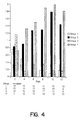
Figure 4 is a graph showing the differences in conjunctival erythema over
the filtration bleb at each examination day up to 12 days postoperatively.
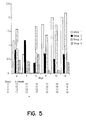
Figure 5 shows the formulas of typical green porphyrins useful in the
methods, compositions, and articles of the invention.
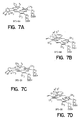
Figure 6 shows the structure of four BPD-type compounds particularly
useful as photosensitizing agents in the invention.
DETAILED DESCRIPTION OF THE INVENTION
The term "inflammation" in this application refers to the series of changes
that occurs in a living body following an injury. The injury may be caused by
physical agents, such as excessive heat or cold, pressure, ultraviolet or ionizing
irradiation, cuts or abrasions; by a wide variety of inorganic or organic chemical
substances; or by biological agents such as viruses, bacteria, and other parasites.
Photosensitizing Agent
A "photosensitizing agent" is a chemical compound that, when exposed to
light of a wavelength capable of being absorbed by the photosensitizer, absorbs
light energy to result in the desired physiological effect, e.g., a controlled anti-inflammatory
effect. The photosensitizing agents for use according to the present invention
preferably have an absorption spectrum that is within the range of wavelengths
between 350 nm and 1200 nm, which absorption spectrum may be tailored to the
desired penetration in a manner known per se, preferably between about 400 and
900 nm and, most preferably, between 600 and 800 nm. Typically, the photosensitizing
agent absorbs light of at least some of the wavelengths in the visible
portion of the electromagnetic spectrum.
Another property of photosensitizers in general that is of particular
significance in the practice of the present invention is a relative absence of
toxicity to cells in the absence of the photochemical effect and the ready clearance
from tissues in the absence of a target-specific interaction between particular cells
and the photosensitizer.
The photosensitizer for use according to the invention can be any photosensitizing agent
suitable for photodynamic therapy ("PDT") that is capable of penetrating into the
injured tissue to be treated and causing the desired degree of biodistribution in less
than one hour. Whether this criterion is met by a potential photosensitizer
candidate can be easily and quickly determined by the following simple test:
A particularly potent group of photosensitizers includes green porphyrins,
which are described in detail in Levy et al., U.S. Patent No. 5,171,749 issued 15
December 1992 . The term "green
porphyrins" refers to porphyrin derivatives obtained by reacting a porphyrin
nucleus with an alkyne in a Diels-Alder type reaction to obtain a monohydrobenzoporphyrin.
Typically, green porphyrins are selected from a group of
porphyrin derivatives obtained by Diels-Alder reactions of acetylene derivatives
with protoporphyrin under conditions that promote reaction at only one of the two
available conjugated, nonaromatic diene structures present in the protoporphyrin-IX
ring systems (rings A and B).
Several structures of typical green porphyrins are shown in Figure 6. The
Diels-Alder reaction initially results in the formation of a cyclohexadiene--referred
to herein as "hydrobenzo"--fused to the A or B pyrrolic ring, as shown in
formulas 1 and 2 of Figure 6. Rearrangement of the π system in the hexadiene
ring results in the formation of compounds of formulas 3 and 4, and reduction
would provide compounds of formulas 5 and 6. For practical reasons, however,
the compounds of formulas 5 and 6 are preferably made by performing the
previously discussed Diels-Alder reaction with the corresponding olefin being
substituted for the usual acetylene compound, thus producing a more reduced
version of the resulting porphyrin ring structure. These compounds are shown in
formulas 1-6 with hydrogen occupying the internal ring nitrogens. However, it is
to be understood that the metalated forms, in which a cation replaces one or both
of these hydrogens, can also be used. The preparation of the green porphyrin
compounds useful in this invention is described in detail in U.S. Patent No.
5,095,030.
For convenience, an abbreviation of the term hydromonobenzoporphyrin
derivative-"BPD"-is generally used to refer to compounds of formulas 3 and 4 of
Figure 5. Compounds of the formulas 3 and 4 and mixtures thereof are
particularly preferred.
As shown in Figure 5, R1, R2, R3 and R4 are non-interfering substituents
that do not appreciably affect the activity of the compound in the
invention. More specifically, the term "non-interfering
substituents" is used to mean substituents that do not destroy the ability of the
green porphyrin to act as a photosensitizer capable of be absorbed by injured
tissue to exert a pharmacological effect in less than one hour. For the compounds
of Figures and 6, generally, R1 and R2 are each, independently, electron-withdrawing
substituents or any other activating substituents that are sufficiently
electron-withdrawing to increase the rate of the Diels-Alder reaction, which can
proceed with both A and B rings but, preferably, occurs in only one ring.
Examples of suitable R1 and R2 groups include carbalkoxy (2-6C), alkyl (1-6C)
sulfonyl or aryl (6-10C) sulfonyl, aryl (6-10C), cyano, and -CONR5CO- where R5
is aryl (6-10C) or alkyl (1-6). One of R1 and R2 may also be hydrogen, so long as
the other is an electron-withdrawing substituent of sufficient strength to facilitate
the Diels-Alder reaction. Most commonly, R1 and R2 are carbalkoxy groups,
preferably methyl or ethyl carboxy esters. Preferred compounds are those in
which R1 and R2 are the same and are carbalkoxy, particularly carboethoxy.
As used herein, the term "carboxy" is, as conventionally defined, -COOH,
while "carbalkoxy" represents -COOR where R is alkyl. "Carboxyalkyl" refers to
the substituent -R'-COOH where R' is alkylene. "Carbalkoxyalkyl" refers to -R'-COOR
where R' is alkylene and R is alkyl or alkanol. "Alkyl" generally
represents a saturated straight or branched chain hydrocarbyl moiety of 1-6 carbon
atoms, such as methyl, n-hexyl, 2-methylpentyl, t-butyl, n-propyl, and so forth.
"Alkylene" is the same as "alkyl" except that the group is divalent rather than
monovalent "Aryl" represents an aromatic cyclic group, such as phenyl,
naphthyl, pyridyl, and the like. The aryl group in compounds for use according to the invention is optionally
substituted with 1-3 substituents, which may be independently selected from the
group consisting of halo, such as fluoro, chloro, bromo or iodo; lower alkyl (1-4C);
and lower alkoxy (I-4C). "Aryl" or "alkyl sulfonyl" groups have the formula
-SO2R where R is alkyl or aryl as defined above.
R3 is independently a ω-carboxyalkyl group (2-6C) or a salt, amide, ester
or acylhydrazone thereof, or is alkyl (I-6C). Preferably, R3 is 2-carboxyethyl or
the alkyl or alkanol ester thereof, and R4 is vinyl. Most of these embodiments,
however, are preferred because of the availability of native porphyrins, rather than
being mandated by considerations of biological efficacy. As shown in Figure 5,
adducts formed by the reaction of R1-C≡C-R2 with a protoporphyrin-LX ring
system (where R3 is a protected form of 2-carboxyethyl, such as 2-carbomethoxyethyl
or 2-carboethoxyethyl, and R4 is -CH=CH2) are compounds of
the formulas I and 2. Compounds of formula 1 result from the addition to the A
ring, and compounds of formula 2 result from the addition to the B ring.
Convenient starting materials for the green porphyrin compounds for use according to the
invention include the naturally-occurring porphyrins where R3 is either
-CH2CH2COOH, -CH2CHRCONR2 or -CH2CHRCOOR where R is alkyl (1-6C)
or alkanol (1-6C). However, the exact nature of R3, unless it contains a π-bond
conjugated to ring π-bond, is ordinarily not relevant to the progress of the Diels-Alder
reaction or to the effectiveness of the resulting product. R3 can thus be any
one of a wide variety of groups such as, for example, lower alkyl (1-4C); and ω-carboxyalkyl
(2-6C) and the esters and amides thereof. The R3 substituent may
also be substituted with a hydroxy group; halogen, such as fluoro, chloro, bromo
or iodo; or with other nonreactive substituents.
When R3 is -CH2CHR-COOR, it has been found advantageous to
hydrolyze, or partially hydrolyze, the esterified carboxy group. Typically, the
hydrolysis at the R3-position conveniently occurs at a much faster rate than that of
the ester groups of R1 or R2. Further, the solubility and biodistribution
characteristics of the resulting compounds are more desirable than those of the
unhydrolyzed form. Hydrolysis results in the diacid or monoacid products (or
their salts).
In compounds of
formulas 1 and 2, R
4 is usually -CH=CH
2, at least
initially, but this vinyl group is readily derivatized to other embodiments of R
4 by
the addition to, or oxidation of, the vinyl ring substituent of ring B or A in
formula
1 or 2 respectively. Thus, R
4 can be any one of a wide variety of substituents that
are consistent with that formed by a facile addition reaction. For example, an
exemplary addition reagent may be of the form HX where H is added to the
carbon adjacent to the ring to provide an R
4-position having the formula:
Thus, in one embodiment, one of the added substituents is a hydrogen, and the
other one is selected from the group consisting of hydrogen; halo such as fluoro,
chloro, bromo or iodo; hydroxy; lower alkoxy; amino; amide; sulfhydryl; or an
organosulfide. For example, the Markovnikov addition of water provides a
substituent structure analogous to a hematoporphyrin ring system at the relevant
ring. The vinyl group can also be oxidized to obtain, as a substituent in the R
4-position,
-CH
2OH, -CHO, or -COOH or its salts or esters. The addition or
oxidation products can themselves also be substituted if the added substituents are
functional leaving groups. For example, when Br is a substituent, it may be
replaced by such moieties as -OH, -OR where R is alkyl (1-6C) as described
above, halo, -NH
2, -NHR, -NR
2 and the like.
Thus, in general, R4 represents any substituents to which the vinyl group
- CH=CH2 is readily converted by cleavage or addition, and further substituents
formed by the reaction of leaving good groups with additional moieties.
Preferably, however, R4 is vinyl (-CH=CH2); -CHOR4' where R4' is H or alkyl
(1-6C), optionally substituted with a hydrophilic substituent such as -CH2OH;
-CHO; -COOR4' such as COOH or -COOCH3; -CH(OR4')CH3 such as
-CH(OH)CH3 or -CH(OCH3)CH3; -CH(OR4')CH2OR4'; -CH(OH)CH2OH;
- CH(SR4')CH3 such as -CH(SCH3)CH3 and the disulfide thereof; -CH(NR4')CH3;
- CH(CN)CH3; -CH(pyridinium bromide)CH3; -CH(COOR4')CH3;
- CH(COOCR4')CH3; -CH2(halo)CH3 such as -CHBrCH3; or -CH(halo)CH2(halo).
Alternatively, R4 can be an organic group of less than 12 carbon atoms resulting
from the direct or indirect derivatization of vinyl. Or R4 can provide additional
porphyrin or porphyrin-related ring systems, such as a group containing from 1-3
tetrapyrrole-type nuclei of the formula -L-P, as defined below. Those compounds
in which R4 is -CH=CH2, -CH(OH)CH3, -CH(halo)CH3, or a group containing 1-3
tetrapyrrole-type nuclei of the formula -L-P, as defined below, are preferred.
As used herein, the term "tetrapyrrole-type nucleus" represents a four-ring
system of the skeleton:
or a salt, ester, amide, or acylhydrazone thereof, which is highly conjugated. It
includes the porphyrin system, which is in effect a completely conjugated system;
the chlorin system, which is in effect a dihydro form of the porphyrin; and the
reduced chlorin system, which is a tetrahydro form of the conjugated porphyrin
system. When "porphyrin" is specified, the completely conjugated system is
indicated. Green porphyrins are effectively a dihydro form of the porphyrin
system.
In one embodiment, the substituent R4 includes at least one additional
tetrapyrrole-type nucleus. The resulting compounds for use according to the invention are dimers or
oligomers in which at least one of the tetrapyrrole-type ring systems is a green
porphyrin. Linkage between the green porphyrin moiety at the R4-position to an
additional tetrapyrrole-type ring system may be by an ether, amine or vinyl
linkage. Porphyrin ring systems having two available substituent positions (in
both A and B rings) corresponding to R4 can be additionally derivatized, as
explained below.
When R4 is "-L-P," -L- is selected from the group consisting of:
and
and P is a porphyrin structure or a second green porphyrin of the formulas 1-6
shown in Figure 5, except that any second R
4 group is replaced by L above.
(It is also understood that, when -L- is of the formula (e) or (f) shown
above, the ring system to which the double bond is attached will have a resonance
system corresponding to
in the ring to which the double bond is attached, as shown.)
The hydro-monobenzoporphyrins that directly result from the Diels-Alder
reaction described above can also be isomerized to the BPD compounds of
formulas 3 and 4 of Figure 5. The depictions of compounds 3 and 4 in Figure 5
do not show the relative position of the exocyclic methyl group (ring A of formula
3 and ring B of formula 4) with respect to the R2 substituent. Either isomer is
available. Compounds of formulas 3 and 4 are particularly preferred in the
methods and compositions of the invention.
In addition, the Diels-Alder products could be selectively reduced by
treating with hydrogen in the presence of a catalyst, such as palladium on
charcoal, to give the saturated ring analogs, shown as formulas 5 and 6 in Figure
5, which correspond to the respective Diels-Alder products of rings A and B.
However, as explained above, the more common practice is to perform the Diels-Alder
reaction starting with an olefin starting material, in the place of the usual
acetylene starting material, to achieve a more reduced form of the resulting
porphyrin ring system. The description set forth above with respect to the
compounds of formulas 1 and 2 concerning derivatization by conversion of the
remaining vinyl substituent (R4) and with respect to the variability of R3 applies as
well to the compounds of formulas 3, 4, 5 and 6.
Preferred embodiments of the green porphyrins for use according to the invention are those
in which the Diels-Alder product is rearranged and partially hydrolyzed. Even
more preferred are the compounds of formulas 3 and 4 (BPD's) in which the
carbalkoxy groups in the R3-positions have also been hydrolyzed or partially
hydrolyzed. Compounds for use according to the invention that contain -COOH may be prepared as
either the free acid or in the form of salts with organic or inorganic bases.
Figure 6 shows four particularly preferred compounds for use according to the invention
covered by formulas 3 and 4, which are collectively designated as benzoporphyrin
derivatives, i.e., BPD-DA, BPD-DB, BPD-MA and BPD-MB. These are
hydrolyzed or partially hydrolyzed forms of the rearranged products of formula 3
and 4, wherein one or both of the protected carboxyl groups of R3 have been
hydrolyzed. The ester groups at R1 and R2 hydrolyze relatively slowly, so that
conversion to the forms shown in Figure 6 is easily effected. The most preferred
of these green porphyrin compounds is BPD-MA.
In Figure 6, R3 is -CH2CH2COOR3' where R3' varies by individual
compound. Specifically, in BPD-DA, R1 and R2 are carbalkoxy, R3' is hydrogen,
and derivatization is at ring A. BPD-DB is the corresponding compound with
derivatization at ring B. BPD-MA represents the partially hydrolyzed form of
BPD-DA, and BPD-NEB represents the partially hydrolyzed form of BPD-DB.
Thus, in these latter compounds, R1 and R2 are carbalkoxy; one R3' is hydrogen,
and the other R3' is alkyl (1-6C).
The compounds of formulas BPD-MA and BPD-MB may be
homogeneous, in which only the C ring carbalkoxyethyl or only the D ring
carbalkoxyethyl would be hydrolyzed, or may be mixtures of the C and D ring
substituent hydrolyzates. In addition, mixtures of any two or more of BPD-MA,
- MB, -DA and -DB may be used in the
invention.
It should be noted that many of the compounds of Figure 5 contain at least
one chiral center and, thus, may exist as optical isomers. The method of the
invention can use compounds having both configurations of the chiral carbons,
whether the compounds are supplied as isolates of a single stereoisomer or are
mixtures of enantiomers and/or diastereomers. Separation of mixtures of
diastereomers may be effected by any conventional means. Mixtures of
enantiomers may be separated by any of the usual techniques, such as by reacting
them with optically active preparations and separating the resulting diastereomers.
It should further be noted that the reaction products may be unseparated
mixtures of A and B ring additions, e.g., mixtures of formulas 1 and 2 or 3 and 4
or 5 and 6. Either the separated forms, e.g., formula 3 alone or 4 alone, or
mixtures in any ratio, may be used in the
invention.
Further still, dimeric forms of the green porphyrin and dimeric or
multimeric forms of green porphyrin/porphyrin combinations can be used to
absorb more light on a per mole basis. The dimers and oligomeric compounds for use according to
the invention can be prepared using reactions analogous to those for dimerization
and oligomerization of porphyrins per se. The green porphyrins or green
porphyrin/porphyrin linkages can be made directly, or porphyrins may be coupled,
followed by a Diels-Alder reaction of either or both terminal porphyrins to convert
them to the corresponding green porphyrins.
Pharmaceutical Composition
Typically, the photosensitizing agent for use according to the invention is formulated into a
pharmaceutical composition by mixing the photosensitizing agent, typically at
ambient temperatures, appropriate pH's, and the desired degree of purity, with one
or more physiologically acceptable carriers, i.e., carriers that are non-toxic to
recipients at the dosages and concentrations employed. Suitable compositions
include those appropriate for systemic or topical administration, including
preparations for injection, transmucosal administration, or transdermal
administration.
The composition for use according to the invention preferably comprises about 1 µg/ml to
about 2 mg/ml of the photosensitizing agent, depending primarily on the mode of
administration. For topical administration, from about 0.1 to about 2.0 mg/mL are
preferably used. For systemic administration, e.g., intravenous injection, the
concentration of the photosensitizing agent preferably varies from about 0.3 to
about 0.5 mg/mL.
Preferably, the photosensitizing agent is administered in a liquid, gel, or
gelatinous solid pharmaceutical composition, either alone with water, or together
with other pharmaceutically acceptable excipients, such as are disclosed in
Remington's Pharmaceutical Sciences, Mack Publishing Co., Easton
Pennsylvania (Gennaro, ed. 1990).
When a liquid, the pharmaceutical composition containing the photosensitizer can
be a suspension or an emulsion. In particular, liposomal or lipophilic
formulations are often desirable. The photosensitizing agent for use according to the invention may
be included within liposomes, attached to their surface, or both. Suitable methods
for preparing liposomes are well-known in the art. The inclusion of green
porphyrin compounds in such preparation is described, for example, in Allison et
al., U.S. Patent No. 5,214,036 issued 25 May 1993 and Desai et al., co-pending
application Serial No. 08/489,850 filed 13 June 1995
. If suspensions or emulsions are used, suitable
excipients include water, saline, dextrose, glycerol, and the like. These
pharmaceutical compositions may also contain minor amounts of nontoxic
auxiliary substances, such as wetting or emulsifying agents, antioxidants, pH
buffering agents, and the like.
The pH of the formulation depends mainly on the particular use and the
concentration of the photosensitizer, but preferably ranges from about 3 to about
8. Preferably, the photosensitizer is maintained at a neutral pH (e.g., about 6.5 to
about 7.5) to prevent its adhering to the contains in which it is placed, as occurs at
pH values approaching physiological levels, and to ensure activation of the
photosensitizer. Thus, the formulation of a photosensitizer in an electrolyte
solution containing a balanced salt buffer at pH 6.5, but containing no fetal bovine
serum ("FBS"), is a suitable embodiment. The reason the FBS is omitted is
because it contains antigenic components that could exacerbate an inflammatory
reaction. If the photosensitizing agent adheres to the containers in which the
pharmaceutical composition containing it is being kept, an appropriate non-antigenic
ingredient, such as human serum albumin, may optionally be added in
an amount that does not interfere with the photosensitizing agent adhering to the
injured tissue being treated.
The photosensitizing agent may be combined with one or more
immunosuppressive agents to enhance the anti-inflammatory effect on the injured
tissue. The term "immunosuppressive agent" as used herein refers to substances
that act to suppress or mask T-lymphocyte responses. This would include
substances that suppress cytokine production, down-regulate or suppress self-antigen
expression, or mask the MHC antigens.
Examples of such agents include 2-amino-6-aryl-5-substituted
pyrimidines; azathioprine or cyclophosphamide; bromocryptine; glutaraldehyde;
anti-idiotypic antibodies for MHC antigens; cyclosporin A; one or more steroids,
preferably corticosteroids and glucocorticosteroids such as prednisone, methyl
prednisolone, and dexamethasone; anti-interferon-gamma antibodies; anti-tumor
necrosis factor-alpha antibodies; anti-tumor necrosis factor-beta antibodies; anti-interleukin-2
antibodies; anticytokine receptor antibodies such as anti-IL-2
receptor antibodies; heterologous anti-lymphocyte globulin; pan-T antibodies,
preferably OKT-3 monoclonal antibodies; antibodies to CD4; streptokinase;
streptodomase; or RNA or DNA from the host.
This immunosuppressive agent may supplement or be used in combination
in the same dosage as the photosensitizing agent or a reduced dosage, and may be
administered simultaneously or separately, systemically or locally. The effective
amount of such other agents is subject to a great deal of therapeutic discretion and
depends on the amount of the photosensitizing agent present in the formulation,
the type of injury, the type of immunosuppressive agent, the site of delivery, the
method of administration, the scheduling of administration, other factors
discussed above, and other factors known to practitioners. However, the amount
of immunosuppressive agent appropriate for use with the invention is typically
lower than that normally advisable for the treatment of like injured tissues.
When an immunosuppressive agent is used, it may be administered by any
suitable means, including parenteral and, if desired for local immunosuppressive
treatment, intralesionally, i.e. topically to the injured tissues. Parenteral infusions
include intramuscular, intravenous, intraarterial, intraperitoneal, subcutaneous,
and subconjunctival administration.
If the pharmaceutical composition for use according to the invention is to be applied
topically, for example, if it is to be painted onto the injured tissue, it may be
preferable to use a viscous solution, such as a gel, rather than a non-viscous
solution. The gel may be prepared, for example, by mixing a solution of the
desired photosensitizing agent with a gelling agent, such as a polysaccharide,
preferably a water-soluble polysaccharide, e.g., hyaluronic acid, starches, and
cellulose derivatives (such as methylcellulose, hydroxyethyl cellulose, and
carboxy methyl cellulose). When a polysaccharide is present in a gel formulation,
the amount usually present is in the range of about 1-90% by weight of the gel,
more preferably about 1-20%. Examples of other suitable polysaccharides for this
purpose and a determination of the solubility of the polysaccharides are found in
EP 267,017 published 11 May 1988.
Examples of suitable surfactants include the poloxamer surfactants, which
represent a series of molecules that are block copolymers of ethylene oxide and
propylene oxide, either alone or taken in admixture with a phospholipid such as
egg lecithin. Another example of an emulsion commercially available from Green
Cross is Fluosol-DA 20%, which contains perfluorodecalin and
perfluorotripropylamine emulsified with the poloxamer surfactant, Pluronic F-68.
The perfluorochemical emulsions and their effects in mammals are described
more fully in Bollands et al., J. Pharm. Pharmacol., 39:1021-24 (1987).
The pharmaceutical composition for use according to the invention is preferably sterile.
Sterility is readily accomplished by sterile filtration through 0.2 micron
membranes. Once formulated and sterilized, the composition may not be stable to
oxidative denaturation. However, lyophilized formulations for reconstitution, for
example, containing BPD, are suitable for storage.
Modes of Bringing Tissue into Contact with Photosensitizer
The reduction or prevention of inflammation in accordance with the
present invention is effected in a relatively straightforward manner by bringing the
injured tissue (or the tissue to be injured or being injured) into contact with the
photosensitizing agent under conditions that enable the formation of a strong
association between the photosensitizing agent and the target tissue, while
minimizing the concentration of the photosensitizer and, so far as is practicable,
localizing the area of contact to the target injured tissue. Preferably, the contact
of step a) is for less than five minutes.
When the cells to be protected from inflammation are contained within a
live, intact animal, the photosensitizer may be administered locally or
systemically. The photosensitizing agent may be administered by injection so
long as the particular mode of injection allows for rapid clearance of the
photosensitizer from the body. For example, intravenous injection would be
suitable. Alternatively, the photosensitizer may be topically or enterally applied,
e.g., by painting or spraying onto the surface of the tissue to be treated, or via
patches or implants, which are typically removable at the conclusion of a predetermined
photosensitizer contact time.
When the target tissues to be protected from inflammation are delicate
ocular tissues, topical external administration is preferred due to the localized
nature of contact with the eye achievable with topical administration, which
results in a greater margin of safety. In an especially preferred embodiment, the
photosensitizer for use according to the invention is applied with the article of the invention, which
comprises the photosensitizer and an absorbent applicator. The absorbent
applicator comprises any absorbent material that is sterile or is capable of being
sterilized, that easily releases the photosensitizes on contact with injured tissues,
and that does not chemically react with the photosensitizing agent. Preferably, the
absorbent material is also inexpensive and disposable. Examples of suitable
absorbent applicators include drug-soak sponges and non-lint-producing flexible
webs. A drug-soak sponge, such as a Weck cell, is the preferred absorbent
applicator. When such an applicator is used, it is preferably saturated with the
pharmaceutical composition for use according to the invention and topically applied to the target
tissues during or shortly after the occurrence of injury, e.g., during a surgical
procedure.
The contacting step can take place over a wide variety of temperatures,
avoiding only those temperatures great enough to denature or otherwise
deleteriously affect the injured tissue and those temperatures low enough to
minimize the cellular uptake of the photosensitizer. Preferably, the contacting
step takes place at a temperature in the range from 5°C to 40°C,
preferably, from 15°C to 37°C and, most preferably, at ambient
temperature.
Dosing
In the method of the invention, the subject is administered an amount of
the photosensitizing agent, or a mixture of photosensitizing agents, in one or
several dosages. The photosensitizing agents for use according to the invention are dosed in a
fashion consistent with good medical practice, taking into account the nature of
the inflammation being prevented or reduced, the species and medical condition of
the subject, the presence of any other drug in the subject's body, the purity and
chemical form of the photosensitizer, the mode of administration, the rate and
degree of absorption expected, and other factors known to practitioners. A
therapeutically effective amount of photosensitizer is an amount that is effective
to reduce significantly, upon exposure to light, the proliferation of fibroblasts,
thus ameliorating the inflammatory response and the undesirable effects that may
be associated with inflammation, such as increased vascularity and/or scar tissue
formation.
The dose of the photosensitizing agent will vary with the target tissue and,
if administered intravenously or systemically, will be limited by the weight and
optimal blood level of the animal. Suitable systemic amounts per dose are
typically less than about 1.0 mg/kg of body weight, preferably in the range of
from about 0.25 to 0.75 mg/kg per dose and, most preferably, 0.15 to
0.50 mg/kg per dose.
Typically, the dose of the photosensitizing agent is less than 0.50mg/kg.
A systemic dose of BPD as the photosensitizer would
exceed 0.3 mg/kg only under unusual circumstances. These dosage ranges are
intended to be suggestive and should not necessarily be considered as limiting,
since the individual reactions of particular subjects will also vary.
Depending on the photosensitizing agent and the mode of administration,
an equivalent optimal systemic blood level can be established, but it is difficult to
do because the photosensitizer preferably clears very rapidly. Thus, there can be a
dramatic difference between the concentration of the photosensitizer in the
bloodstream at the moment of injection and the concentration at the time of
treatment with light. For example, the concentration of BPD at the moment of
intravenous injection may range from about 1-10 mg/mL, while, at the time of
light exposure, may only be in the range of from 0.5-0.05 ug/mL. If by topical
administration, no photosensitizer at all is typically detectable in the blood.
When administered topically or systemically, the dose is best described in
terms of the concentration of the composition and the length of the time of contact
with the target tissue. A generally effective range of concentrations for the
photosensitizing agent is from 0.1 to 10 mg/mL, preferably from
0.1 to 5 mg/mL and, most preferably; from 0.25 to 2.0
mg/ml. Typically, the concentration of the photosensitizing agent is mg/mL of less.
The contact suitably involves applying the composition to one or more
surfaces of the injured tissue with the pharmaceutical composition of the
invention. Topical contact with the photosensitizer generally takes place for at
least one minute, preferably under five minutes, and even more preferably from
about one to two minutes. The time of contact depends on such factors as the
concentration of the photosensitizing agent in the composition, the tissue to be
treated, and the particular type of composition.
After a predetermined contact time with the photosensitizer, the excess
photosensitizer is preferably removed from the area of treatment
prior to step (b) of the method as defined above. If the
photosensitizer is being systemically administered, the photosensitizer is selected
to have, not only rapid pharmacokinetic characteristics, but also susceptibility to
rapid clearance from the body. If the photosensitizer is being topically
administered, the excess is preferably removed by irrigating or flushing away with
a physiologically acceptable, chemically inert fluid, such as normal saline or BSS
(balanced salt solution), or washing off with water or some other solvent. Again,
these protocols are not intended to be limiting in view of the wide variation
permitted in protocol design.
Following the step of bringing the injured tissue, or pre-injured tissue, into
contact with a composition containing the photosensitizer for use according to the invention, the
tissue is subjected to exposure with light having a wavelength that is absorbed by
the photosensitizing agent and leads to the reduction or prevention of
inflammation. The term "low-dose PDT" in this application refers to a dose that
does not cause evident cell damage, necrosis or erythema, but exhibits only an
anti-inflammatory effect. Because the total PDT dose depends on a combination
of the dose of the photosensitizing agent and the dose of the irradiating light, low-dose
PDT may be administered in combinations of relatively high photosensitizer
doses and low light doses or, on the other hand, combinations of relatively low
photosensitizer doses and high light doses. The latter low photosensitizer/high
light combination can also be achieved by administering a relatively high dose of
photosensitizer, followed by an unusually long "incubation" time before being
irradiated with light. Therefore, a wide variety of conditions, all producing a
relatively low dose of PDT overall, would be suitable for use according to the invention.
Likewise, a wide variety of different combinations of photosensitizer
doses, contact times, and modes of administration are suitable. However, the
following rough guidelines may be useful. Short contact (less than one hour) with
high doses of the photosensitizer e.g., 2 mg/mL applied topically, would generally
be equivalent to a low photosensitizer dose, e.g., 0.15 mg/kg administered
intravenously. However, even after a high dose of photosensitizer administered
intravenously, delaying irradiation with light to a later time, e.g., more than three
hours, after administration of the photosensitizing agent can also result in low-dose
PDT because, if the photosensitizer is capable of rapid clearance, very little
of it may still be present in the tissues after three hours.
Specific examples of "low-dose PDT" would include:
- topical application or localized injection of less than 2 mg/mL of a
benzoporphyrin derivative ("BPD") photosensitizer, which is left in
contact with the target tissue for less than ten minutes;
- intravenous administration of less than 0.15 mg/kg of a BPD with
irradiation at any time after administration of the BPD; or
- intravenous administration of 0.15-0.50 mg/kg BPD with irradiation
more than six hours after BPD administration;
coupled with irradiation under the following conditions:
- less than 15 J/cm2 applied between 0-3 hours after administration of
the photosensitizer, preferably about 7 - 12 J/cm2; or
- up to 100 J/cm2 applied later than six hours after photosensitizer
administration.
Preferably, the dose of light in the exposure step b) as
defined above is less than 100 J/cm2. More preferably, the
time between step a) and the exposure step b) as defined
above is greater than six hours, and the dose of the
light during said exposure step b) is from 15 to 100 J/cm2.
During the irradiation step, any light that the photosensitizer absorbs and
that is appropriate for use with the injured tissue may be used, e.g., from
380 to 850 nm, depending upon the photosensitizer and upon the depth of
tissue penetration desired, preferably from 400 to 700 nm. For
general anti-inflammatory applications, light in the visible portion of the
electromagnetic spectrum, e.g., red light, blue light or even UVA light, may be
used. Light having a wavelength shorter than 400 nm is acceptable, but not
preferred because of the potentially damaging effects of UVA light. Light having
a wavelength longer than 700 nm is also acceptable, but not particularly preferred
because it is difficult to see, thus making the visual control of irradiation almost
impossible. For ocular applications, red light is preferred because this eliminates
any potentially harmful effects from the blue and UVA spectral ranges on the
sensitive retina of the eye.
An example of a particularly preferred procedure which is used during
filtering surgery, is as follows:
No single protocol appears to be desirable for all cases at this time.
However, typical protocols will include either a single treatment or an initial
treatment followed optionally by 1-4 additional treatments. Local treatments with
topical photosensitizer administration can be repeated every 3 or 4 days.
However, with systemic administration of the photosensitizer, repeated treatments
are generally spaced about a week apart, or longer, to avoid any undesirable
effects from the accumulation of excess photosensitizer.
The following examples are intended to illustrate, but not to limit, the
invention.
EXAMPLES
Example 1 - Light Dosing
Filtration surgery was performed on one eye in six normal rabbits. A
Weck cell sponge was saturated with a 2 mg/mL aqueous solution of the
photosensitizer benzoporphyrin derivative monoacid ring A (BPD-MA, also
known as "BPD-verteporfin"). During surgery, the saturated Weck cell was used
to apply BPD-MA topically for two minutes to the sclera and conjunctiva in the
surgical field. After washing out the excess drug with BSS, both the sclera and
conjunctiva were exposed to red light having a wavelength of about 690 nm,
which was delivered by a light emitting diode ("LED") placed at a distance of
about 1 cm from the tissue to be irradiated. Each of the six rabbits used in this
experiment received a different dose of light, specifically, 0, 3, 6, 12, 18 and 24
J/cm
2 over a 30-second to 4-minute time period. The treated rabbits were
followed for 11-12 days after surgery by determining filtration bleb height, bleb
vascularity (indicative of inflammation), and reduction in intraocular pressure
("IOP"). The data obtained on
day 5 and
day 11 are shown below in Tables 1A
and 1B respectively.
| Results of Pilot BPD-MA for Light Dosing at Postoperative Day 5 |
| Rabbit No. | Irradiation Time | IOP Decrease
(mm Hg) | Bleb Height | Bleb Vascularity | |
| 1 | 0 (No BPD) | 2-3 | Moderate | Vascular | |
| 2 | 30 seconds | Minimal | Small | Vascular | |
| 3 | 1 minute | > 20 | Maximum | Avascular | |
| 4 | 2 minutes | 20 | Maximum | Avascular' |
| 5 | 3 minutes | 2.5 | Small | Vascular | |
| 6 | 4 minutes | 0-4 | Small to Moderate | Vascular |
| Results of Pilot BPD-MA for Light Dosing at Postoperative Day 11 |
| Rabbit No. | Irradiation Time | IOP Decrease
(mm Hg) | Bleb Height | Bleb Vascularity | |
| 1 | 0 (No BPD) | 3-4 | Moderate | Vascular | |
| 2 | 30 seconds | 2.3 | Minimal | Vascular | |
| 3 | 1 minute | > 20 | Maximum | Avascular | |
| 4 | 2 minutes | > 20 | Maximum | Avascular' |
| 5 | 3 minutes | 3-4 | Low | Vascular | |
| 6 | 4 minutes | Sacrificed - bleb failure |
The results indicated that the survival of the filtration bleb was the longest
in eyes treated with light at a medium range of doses, i.e., relatively low doses of
drug and light ("Low-dose PDT"). The data indicated that a certain level of PDT
was required, but that higher doses were generally less effective than lower ones.
The combination of the short incubation time with BPD and the low light dosage
of 12 J/cm2 was not expected to cause much damage to treated cells. Nevertheless,
the treatment had a definite pharmacological action. Bleb survival was associated
with the lack of inflammation, as indicated by avascularity and a pale-colored
bleb.
On the other hand, with too low or too high light doses, the bleb height
and the amount of the lowered intraocular pressure were reduced. Bleb failure
was associated with inflammation.
The corresponding filtration blebs are shown in Figure 1.
Example 2 - Times of PDT Administration
The photosensitizer used in the example was prepared as follows: A
liposomally formulated benzoporphyrin derivative, monoacid ring A, BPD-MA or
BPD-verteporfin was supplied by QLT PhotoTherapeutics, Inc. as a lyophilized
powder and was reconstituted with sterile distilled water shortly before use. The
BPD was reconstituted to 1.98 mg/mL was used to saturate the 3 mm cut end of a
Weck cell. In control groups, the Weck cell was saturated with basic salt solution
("BSS").
On day 0, full thickness filtration surgery was performed on one randomly
selected eye in 48 rabbits, 12 in each of four groups. In each rabbit, the untreated
other eye served as a control. The filtration procedure was performed in the
following manner: Each animal was anesthetized with a mixture of ketamine and
xylazine. A wire speculum was used to separate the eyelids. A fornix-based
conjunctival flap was created either in the superior nasal or superior temporal
quadrant. Following creation of the fornix-based flap, the Week cell saturated
with BPD-MA (or BSS placebo) was placed on the sclera posterior to the limbus
where the fistula was to be created. The conjunctiva was draped over the Weck
Cell. The Weck cell thus rested between the conjunctiva and sclera, also coming
into contact with the episclera and Tenon's capsule, for two minutes. The Weck
cell was then removed and the area was irrigated with BSS. Instruments and
gloves were also rinsed before entering the eye, and a 1.0 mm trephine was used
to enter the anterior chamber.
The conjunctival flap was then secured to the limbus using two 7-0 vicryl
sutures. Immediately following surgery, each rabbit had a two-minute exposure
of light having a wavelength of 690 nm, with the light source used in Example 1
(Quantum Devices, Inc.) being placed 1 cm from the eye. The light source was
used at a maximum output (100 W/cm2), which provided a total dosage of
approximately 7.2 J.
A drop of tobramycin was then placed into each eye following surgery.
Tobramycin and prednisolone acetate were instilled in both eyes four times a day
for one week following surgery.
The control eye received the same photosensitizer and irradiation
subconjunctivally as the surgical eye, but without a fistula being created. The
control eye was used to test toxicity and as a basis for detecting a decrease in IOP
for the surgical eye. The time at which the BPD-MA was applied was varied, as
follows:
48 hours post surgery, placebo treatment; Group 2: During surgery, BPD treatment;
48 hours post surgery, placebo treatment; Group 3: During surgery, placebo treatment;
48 hours post surgery, BPD treatment; Group 4: During surgery, BPD treatment; and
48 hours post surgery, BPD treatment.
BPD treatment at 48 hours post surgery consisted of the application of the 3 mm
cut end of a Weck cell saturated with a 2 mg/mL aqueous solution of BPD-MA
(or placebo) laid on the conjunctiva over the filtration bleb for two minutes,
followed by washing out the excess photosensitizer and exposure to red LED light
having a wavelength of 688 nm for one minute.
Postoperatively at
day 0 and every two days following surgery, the rabbits
were examined with slit lamp biomicroscopy to evaluate filtration bleb extent,
filtration bleb height, conjuctival erythema over the bleb, anterior chamber cell
flare, and anterior chamber depth. Applanation tonometry to measure IOP
following topical anesthesia. A subjective ocular discomfort assessment was also
performed by determining animal comfort and eating habits, in accordance with
the grading scale shown below:
Bleb survival was assessed by the extent and height of the bleb and by IOP
as compared with the control eye. The degree of the inflammatory response was
determined by erythema over the filtration bleb and was scored on a scale of 0-3.
Rabbits were terminated when bleb failure was noted, i.e., when the intraocular
pressure in the surgical eye equaled that of the control eye and the filtration bleb
was flat.
The graph in Figure 2 shows the percentage of filtration bleb survival of
rabbits in each of the four groups evaluated in this study. Mean (± SD) survival
times were: Group 1: 10.3 ± 8 days;
A statistical difference was observed between most groups (P < 0.001), but not
between
Groups 2 and 4 (P < 0.05). Treatment with BPD and light during surgery
(
Groups 2 and 4) resulted in prolonged bleb survival in comparison with the
placebo control (Group 1) or treatment only at 48 hours post surgery (Group 3).
The second treatment with BPD and light at 48 hours (Group 4) did not appear to
have any additional effect. A Kruskal-Wallis test for non-parametric analysis was
used to evaluate differences in the survival time of filtration blebs between
groups.
IOP was evaluated by an ANOVA test, and a trend toward lower IOP was
observed when BPD was given intraoperatively (P = 0.057). However, due to
problems with the tonometer used to measure IOP in the rabbits, the most reliable
parameters were the extent (Figure 3) and height (Figure 4) of the bleb, both
which indicated the effectiveness of low-dose PDT treatment during surgery. The
measurement of erythema over the bleb (shown in Figure 5) indicated a higher
inflammatory response in Groups 1 and 3, in which blebs failed early post
surgery. In Figures 3, 4 and 5, a statistical difference was observed between
groups (P ,0.001). The bleb height and extent, as well as other slit lamp
characteristics, were analyzed by a chi-square test. Adverse events also were
analyzed by a chi-square test. A Cox Proportional Hazard Model was used to
evaluate for parameters that most accurately predicted filtration bleb survival on
each examination day.
When compared to previous rabbit studies that have investigated
adjunctive medicines for trabeculectomy, as shown below in Table 2, the results
showed that BPD prolonged the filtration bleb survival compared to normal
controls, Ara-A and 5-fluorouracil.
| Antifibrotic Therapy | Filtration Bleb Survival
(mean days ± SD [range]) | Documents |
| No drug | 12.06 ± 6.2 (6-23.2) | Kay et al., "Delivery of Antifibroblast Agents as Adjuncts to Filtration Surgery-Part II: Delivery of 5-Fluorouracil and Bleomycin in a Collagen Implant: Pilot Study in the Rabbit", Ophthalmic Surg., 17:796-801 (1986); Khaw et al., "Effects of Inoperative 5-Fluorouracil or mitomycin C on Glaucoma Filtration Surgery in the Rabbit", Ophthalmology, 100:367-72 (1993); Bergstrom et al., "The Effects of Subconjunctival Mitomycin-C on Glaucoma Filtration Surgery in Rabbits", Arch. Ophthalmol., 109:1725-30 (1991); and Liang et al., "Comparison of Mitomycin C and 5-Fluorouracil on Filtration Surgery Success in Rabbit Eyes", J. Glaucoma, 1:87-93 (1992). |
| 5-Fluorouracil | 19.5 ± 5.5 (14-25) | Kay et al., "Delivery of Antifibroblast Agents as Adjuncts to Filtration Surgery-Part II: Delivery of 5-Fluorouracil and Bleomycin in a Collagen Implant: Pilot Study in the Rabbit", Ophthalmic Surg., 17:796-801 (1986); Khaw et al., "Effects of Inoperative 5-Fluorouracil or mitomycin C on Glaucoma Filtration Surgery in the Rabbit", Ophthalmology, 100:367-72 (1993). |
| Bleomycin | 25.5 ± 14.5 (21-50) | Khaw et al., "Effects of Inoperative 5-Fluorouracil or Mitomycin C on Glaucoma Filtration Surgery in the Rabbit", Ophthalmology, 100:367-72 (1993). |
| Mitomycin-C | 34.8 ± 28.8 (6-23.2) | Khaw et al., "Effects of Inoperative 5-Fluorouracil or mitomycin C on Glaucoma Filtration Surgery in the Rabbit", Ophthalmology, 100:367-72 (1993); Liang et al., "Comparison of Mitomycin C and 5-Fluorouracil on Filtration Surgery Success in Rabbit Eyes", J. Glaucoma, 1:87-93 (1992). |
| Ara-A | 15.2 ± 1.8 | Lee et al., "Effects of Cytosine Arabinoside-impregnated Bioerodible Polymers on Glaucoma Filtration Surgery in Rabbits", J. Glaucoma, 2:96-100 (1993). |
The results also showed that BPD prolonged the life of the filtration bleb
in rabbits longer that in some rabbits who received mitomycin C. The mean
survival time of studies with mitomycin C were longer, and this is consistent with
the hypothesis that PDT clinically would cause less over-filtration than mitomycin
C but have a similar ease of application with greater safety.
The data indicated clearly that longer bleb survival was associated with no
erythema or only minimal erythema and, thus, with the reduction of the
inflammatory response. The data also suggested that the treatment with BPD and
light, at relatively low ranges of doses, prevented the development of
inflammation, especially when administered during surgery.
Adverse events were few and not related specifically to the use of the
photosensitizer. A fibrin clot within the first four days was observed in six
rabbits, three of which were in Group 2 and three in Group 3. In each case, the
fibrin was resolved without sequelae by the end of the first week. One rabbit died
at day 0, which was felt to be a complication of anesthesia. No other adverse
events were reported.
At day 7, one rabbit from each group and, following bleb failure, two
rabbits from each group, were sacrificed to undergo histologic and transmission
electron microscopic ("TEM") analysis. Histologic evaluation was performed
after first fixing the cadaver eyes with 10% buffered neutral formalin. The eyes
were processed, sectioned, and then stained with hematoxylin and eosin, as well
as Masson's Trichrome. The specimens were examined in a masked fashion by an
independent observer.
Transmission electron microscopy was performed by fixing tissue samples
in 2.5% glutaraldehyde buffered with 0.1 M cacodylate containing 7% sucrose.
Tissues were post fixed for one hour with 2% osmium tetroxide and dehydrated
through graded alcohol concentrations to 100% ethanol. The 2 x 5 mm tissue
blocks were then infiltrated with catalyzed epoxy resin. Thick sections (0.5 µm)
were cut and stained with toluidine blue and examined by light microscopy to
determine appropriate areas. Thin section (80 nm) were then cut, picked up on
copper grids, and stained with uranyl acetate and lead citrate for TEM evaluation.
A Hitachi H7000 transmission electron microscope was used to examine these
sections.
A light microscopy at day 7 postoperatively in filtering blebs that received
BPD rather than the placebo at surgery showed that fibroblasts and a mild
lymphocytic response were present in the rabbit that received a placebo at both
day 0 and day 2. In addition, at day 7, these eyes demonstrated some vascular
proliferation and new collagen deposition.
In contrast, the rabbit that received BPD only at day 2 (Group 3), showed
increased lymph channels in addition to the fibroblasts, but no vascular
proliferation. In both eyes that received BPD at surgery, the filtration bleb was
noted to have a mild lymphocytic response. However, no fibroblasts, vascular
increase or lymphocytic channel increase were noted. At the end of the study
(three weeks postoperatively), in eye fistulas that had received BPD, only a few
lymphocytes and no proliferation of blood vessels at the fistula were noted.
In Groups 2 and 4, both at day 7 and at sacrifice, a thinned epithelium was
observed. However, this was felt to be due to the elevated bleb and the associated
breakdown of the tear fill, as opposed to a toxic effect from the PDT treatment.
In the control eye, no differences between the placebo and BPD-treated
eyes were observed in the anterior segment ocular exam, indicating a lack of
toxicity of the invention clinically (P > 0.05).
Additionally, histologic and transmission electron microscopic analysis showed
no evidence of toxicity or inflammation apart from the filtration site, either in the
surgical or control eye. Therefore, no toxicity from BPD was observed clinically,
histologically, or by transmission electron microscopy.