EP1007065B1 - Preparation of collagen - Google Patents
Preparation of collagen Download PDFInfo
- Publication number
- EP1007065B1 EP1007065B1 EP98902442A EP98902442A EP1007065B1 EP 1007065 B1 EP1007065 B1 EP 1007065B1 EP 98902442 A EP98902442 A EP 98902442A EP 98902442 A EP98902442 A EP 98902442A EP 1007065 B1 EP1007065 B1 EP 1007065B1
- Authority
- EP
- European Patent Office
- Prior art keywords
- collagen
- sample
- produce
- sodium
- agent
- Prior art date
- Legal status (The legal status is an assumption and is not a legal conclusion. Google has not performed a legal analysis and makes no representation as to the accuracy of the status listed.)
- Expired - Lifetime
Links
Images


Classifications
-
- C—CHEMISTRY; METALLURGY
- C07—ORGANIC CHEMISTRY
- C07K—PEPTIDES
- C07K14/00—Peptides having more than 20 amino acids; Gastrins; Somatostatins; Melanotropins; Derivatives thereof
- C07K14/435—Peptides having more than 20 amino acids; Gastrins; Somatostatins; Melanotropins; Derivatives thereof from animals; from humans
- C07K14/78—Connective tissue peptides, e.g. collagen, elastin, laminin, fibronectin, vitronectin, cold insoluble globulin [CIG]
-
- A—HUMAN NECESSITIES
- A61—MEDICAL OR VETERINARY SCIENCE; HYGIENE
- A61P—SPECIFIC THERAPEUTIC ACTIVITY OF CHEMICAL COMPOUNDS OR MEDICINAL PREPARATIONS
- A61P17/00—Drugs for dermatological disorders
- A61P17/02—Drugs for dermatological disorders for treating wounds, ulcers, burns, scars, keloids, or the like
-
- A—HUMAN NECESSITIES
- A61—MEDICAL OR VETERINARY SCIENCE; HYGIENE
- A61P—SPECIFIC THERAPEUTIC ACTIVITY OF CHEMICAL COMPOUNDS OR MEDICINAL PREPARATIONS
- A61P7/00—Drugs for disorders of the blood or the extracellular fluid
- A61P7/04—Antihaemorrhagics; Procoagulants; Haemostatic agents; Antifibrinolytic agents
Definitions
- the present invention relates to methods for preparing collagen from tissues of humans and other animals.
- the present invention provides methods for the preparation of collagen suitable for biomedical, veterinary, and other applications.
- Collagen is the most abundant protein in mammals. ( See , U.S. Patent No. 5,043,426 to Goldstein). Indeed, it represents 30% of the dry weight of the human body. ( See , L.C. Junqueira and J. Carneiro, Basic Histology , 4th ed.. Lange Medical Publications, Los Altos, CA, [1983], pp. 89-119). Vertebrate collagen is actually a family of proteins produced by several cell types. Within this protein family, the collagen types are distinguishable by their chemical compositions. different morphological and pathological features, distributions within tissues, and their functions. Although many types of collagen have been described, five major types have been recognized.
- Collagen type I is the most abundant form of collagen, with widespread distribution within the body. It is present in tissues in structures classically referred to as "collagen fibers" that form bones, dentin, tendons, fascias, sclera, organ capsules, dermis, fibrous cartilage, etc.
- the primary function of type I collagen is to resist tension. Microscopically, type I collagen appears as closely packed, thick, non-argyrophilic, strongly birefringent red or yellow fibers. Its ultrastructure is characterized as being densely packed, thick fibrils with marked variation in diameter. It is produced by fibroblasts, osteoblasts, odontoblasts, and chondroblasts.
- Collagen type II is primarily found in cartilage ( e . g ., hyaline and elastic cartilages).
- the primary function of type II collagen is to resist intermittent pressure. Microscopically, it appears as a loose, collagenous network, that is visible only with picrosirius stain and polarization microscopy. Ultrastructurally, it is characterized as appearing to have no fibers. but with very thin fibrils embedded in abundant ground substance. It is produced by chondroblasts.
- Collagen type III is commonly associated with type I collagen in tissues, and may be the collagenous component of reticular fibers. It is present in smooth muscles, endoneurium, arteries, uterus, liver, spleen, kidney, an lung tissue. The primary function of type III collagen is to maintain the structure of expansible organs. Microscopically, it appears as a loose network of thin, argyrophilic, and weakly birefringent greenish fibers. Ultrastructurally, it is characterized as being loosely packed thin fibrils with fairly uniform diameters. It is produced by smooth muscle fibroblasts, reticular cells, Schwann cells, and hepatocytes.
- Collagen type IV is found in the epithelial and endothelial basal lamina and basement membranes.
- the primary function of type IV collagen involves support and filtration. Microscopically, it appears as a thin, amorphous, weakly birefringent membrane. Ultrastructurally, it appears to have neither fibers nor fibrils.
- Collagen type V is found in fetal membranes, blood vessels, placental basement membrane, and in small amounts in other tissues. This type of collagen remains largely uncharacterized.
- the principal amino acids found in collagen are glycine, proline and hydroxyproline. Hydroxylysine is also characteristic of collagen. These hydroxy amino acids are the result of hydroxylation of proline and lysine present in nascent collagen polypeptides during collagen synthesis.
- the collagen content in a tissue can be determined by measurement of its hydroxyproline content.
- Collagen is comprised of polypeptide chains, designated as " ⁇ .” There are two types of ⁇ chains, referred to as “alpha-1" (" ⁇ 1") and “alpha-2 (" ⁇ -2"). The most important types of a 1 chains are ⁇ 1(I), ⁇ 1(II), ⁇ 1(III), and ⁇ 1(IV), which aggregate in different combinations to produce the triple helices of types I, II, III, IV, and V.
- Type I collagen is composed of two ⁇ 1 and one ⁇ 2 chains. It's formula is ( ⁇ 1[I]) 2 ⁇ 2. The formula for type II collagen is ( ⁇ 1[II]) 3 , while the formula for type III collagen is ( ⁇ 1[III]) 3 , and type IV is ( ⁇ 1[IV]) 3 .
- Tropocollagen is the protein unit that polymerizes into aggregations of microtibrillar subunits packed together to form “collagen fibrils.” Hydrogen bonds and hydrophobic interactions are critical in this aggregation and packing. Covalent crosslinks reinforce the structure of the collagen fibrils. Collagen fibrils are thin and elongated, of variable diameter. and have transverse striations with a characteristic periodicity of 64 nm. The transverse striations is produced by the overlapping organization of the subunit tropocollagen molecules. In type I and III collagen, these fibrils associate to produce collagen "fibers.” In collagen type I, collagen "bundles" may be formed by association of the fibers. Collagen type II is observed as fibrils. but does not form fibers, while types IV and V do not form fibrils or fibers.
- Collagen fibers are the most abundant fiber found in connective tissue. Their inctasticity and molecular configuration provide collagen fibers with a tensile strength that is greater than steel. Thus, collagen provides a combination of flexibility and strength to the tissues in which it resides. In many parts of the body, collagen fibers are organized in parallel arrays to form collagen "bundles.”
- collagen fibers When fresh, collagen fibers appear as colorless strands, although when a large number of fibers are present, they cause the tissues in which they reside to be white (e . g ., tendons and aponeuroses). The organization of the elongated tropocollagen in the fibers cause them to be birefringent. Staining with certain acidic dyes ( e . g ., Sirius red) enhances this birefringency. As this increase in birefringency us only observed in oriented collagen structures, it is useful as a method to detect the presence of collagen in a tissue.
- acidic dyes e . g ., Sirius red
- collagen is a relatively weak immunogen, at least partially due to masking of potential antigenic determinants within the collagen structure. Also, it is resistant to proteolysis due to its helical structure. In addition, it is a natural substance for cell adhesion and the major tensile load-bearing component of the musculoskeletal system. Thus, extensive efforts have been devoted to the production of collagen fibers and membranes suitable for use in medical, as well as veterinary applications.
- Collagen has been used in the area of soft tissue augmentation, as a replacement for paraffin, petrolatum. vegetable oils. lanolin, bees wax, and silicone previously used. ( See e . g ., U.S. Patent No. 5,002,071.).
- problems have been associated with the use of collagen in implants.
- the non-collagenous proteins present in impure collagen preparations are more potent immunogens than the collagen, and can stimulate the inflammatory response, it is critical that highly pure collagen be used. If the inflammatory cascade is stimulated, the resorption of collagen occurs by the infiltrating inflammatory cells ( e . g ., macrophages. and granulocytes) that contain collagenase, resulting in thee digestion of the collagen.
- collagen itself is chemotactic. and becomes increasingly chemotactic as it is degraded into smaller peptide fragments.
- non-human collagen a repeatedly documented problem associated with the use of bovine collagen as a biomaterial is the consistent, chronic cellular inflammatory reaction that is evident following its implantation or use. This inflammation may result in residual scar tissue formation, adhesion formation. interference with heating of skin edges, pseudointima formation pseudodiaphragm formation, disruption of anastomoses, transient low grade fever, aneurysms, or other problems.
- Collagen preparations are typically prepared from skin, tendons ( e . g ., bovine Achilles, tail, and extensor tendons), hide or other animal parts, by procedures involving acid and/or enzyme extraction.
- collagen preparation methods involve purification of collagen by extraction with diluted organic acids, precipitation with salts, optional gelation and/or lyophilization, tangential filtration etc.
- the tissue is subjected to moderate digestion with proteolytic enzymes, such as pepsin, then the collagen is precipitated at a neutral pH, redissolved and the residual impurities precipitated at an acid pH.
- proteolytic enzymes such as pepsin
- the collagen fibers are then precipitated with salts or organic solvents, and dehydrating the collagen fibers.
- the extracted collagen can be converted into a finely divided fibrous collagen by treating water-wet collagen with acetone to remove water, centrifuging to obtain the solid mass of collagen and deaggregating the collagen during drying.
- the collagen preparation can then be brought back to a neutral pH and dried in the form of fibers. Completely transparent physiological and hemocompatible gels, collagen films, and solutions can be prepared. These forms of collagen may then be used in the fabrication of contact lenses and implants.
- One disadvantage of treatment with pepsin is that the collagen preparation may be partially degraded (i . e ., the extraction enzymes cleave the collagen molecule at the terminal non-helical regions, which contain the inter-collagenous cross-linkages). Indeed, it has been found that collagen extracted with pepsin results in preparations that are too weak for certain applications, especially those for which substantial mechanical handling of the collagen preparation is required.
- denatured collagen or collagen that has undergone treatment to reform the physical and biological characteristics to approximate collagen in vivo is often not satisfactory. It often lacks the mechanical properties required for wet dressings, as it lacks the in vivo organized structure ( i . e .,collagen fibers are not present in this artificial collagen).
- GB 1 204 048 describes a method for purifying collagen free of antigenic substances, by one single proteolysis step with elastase (i.e. ficin) and a Reduction step before or after the proteolysis step.
- elastase i.e. ficin
- the present invention relates to methods for preparing collagen from humans and other animals.
- the present invention provides methods for the preparation of collagen suitable for biomedical applications.
- the present invention provides numerous embodiments for the purification of collagen. It is particularly preferred that the collagen purified according to one embodiment of the present invention be type I collagen.
- the present invention provides a method for purifying collagen comprising: providing a sample comprising collagen, first and second proteolytic enzyme preparations, and a reducing agent: exposing the collagen sample to the first proteolytic enzyme preparation to produce a first collagen solution; exposing the first collagen solution to the reducing agent to produce a second collagen solution; exposing the second collagen solution to the second proteolytic enzyme preparation to produce purified collagen.
- the first and second proteolytic enzyme preparation comprise papain.
- the method of the present invention further comprises the step of de-epithelializing the sample prior to exposing the sample to the first proteolytic enzyme preparation.
- the reducing agent is selected from the group consisting of sodium sulfide, dithiothreitol, glutathionine, and sodium horohydride.
- the reducing agent comprises sodium borohydride.
- the method of the present invention comprises the further step of exposing the purified collagen to a delipidation agent to produce dilipidated collagen.
- the delipidation agent comprises a mixture comprising chloroform and methanol.
- the method of the present invention further comprises the steps of compressing the delipidated collagen to produce compressed collagen: dehydrating the compressed collagen to produce dehydrated collagen; and dispersing and drying the dehydrating collagen to form collagen fibers.
- the collagen fibers are dried.
- the method of the present invention comprises the step of exposing the delipidated collagen to a phosphorylating agent to produce phosphorylated collagen.
- the phosphorylation agent is selected from the group consisting of sodium trimetaphosphate, sodium hexametaphosphate sodium ultraphosphate, sodium tetrametaphosphate, phosphoric anhydride and phosphoryl trichloride.
- the phosphorylation agent comprises sodium trimetaphosphate.
- the purified collagen comprises CollagenPROTM.
- the present invention provides purified collagen purified by the steps of: providing a sample comprising collagen, first and second proteolytic enzyme preparations wherein both of said proteolytic enzyme preparations comprise papain, and a reducing agent; exposing the collagen sample to the first proteolytic enzyme preparation to produce a first collagen solution; exposing the first collagen solution to the reducing agent to produce a second collagen solution: exposing the second collagen solution to the second proteolytic enzyme preparation to produce purified collagen.
- the purified collagen be comprised of additional compounds, including but not limited to antimicrobials, antivirals, growth factors, anti-dehydration compounds, antiseptics, or other compounds suitable for biomedical and/or veterinary uses.
- the present invention also provides an alternative embodiment comprising methods for production of biocompatible collagen, in which the method comprises: providing a sample comprising collagen, first and second proteolytic enzyme preparations wherein both of said proteolytic enzyme preparation comprise papain, a reducing agent, a delipidation agent, and a phosphorylation agent: exposing the collagen sample to the first proteolytic enzyme preparation to produce a first collagen solution: exposing the first collagen solution to the reducing agent to produce a second collagen solution: exposing the second collagen solution to the second proteolytic enzyme preparation to produce to produce a proteolyzed collagen solution; exposing the proteolyzed collagen solution to the delipidation agent to produce delipidated collagen; and exposing the delipidated collagen to the phosphorylation agent to produce phosphorylated collagen.
- the method of the present invention further comprises the step of de-epithelializing the sample prior to exposing the sample to the first proteolytic enzyme preparation.
- the reducing agent is selected from the group consisting of sodium sulfide, dithiothreitol, glutathionine, and sodium borohydride.
- the reducing agent comprises sodium borohydride.
- the delipidation agent comprises a mixture of chloroform and methanol.
- the phosphorylation agent is selected from the group consisting of sodium trimetaphosphate. sodium hexametaphosphate. sodium ultraphosphate, sodium tetrametaphosphate, phosphoric anhydride, and phosphoryl trichloride.
- the phosphorylation agent comprises sodium trimetaphosphate.
- the method of the present invention further comprises the steps of compressing the delipidated collagen to produce compressed collagen; dehydrating the compressed collagen to produce dehydrated collagen; and dispersing and drying the dehydrating collagen to form collagen fibers.
- the collagen fibers are dried.
- the method further comprises the step of filter-sterilizing the delipidated collagen prior to exposing the delipidated collagen to the phosphorylation agent to produce phosphorylated collagen.
- the present invention also provides purified such that the collagen is biocompatible.
- the biocompatible collagen is produced by the method of: providing a sample comprising collagen, first and second proteolytic enzyme preparations wherein both of said proteolytic enzyme preparations comprise papain a reducing agent, a delipidation agent, and a phosphorylation agent; exposing the collagen sample to the first proteolytic enzyme preparation to produce a first collagen solution; exposing the first collagen solution to the reducing agent to produce a second collagen solution; exposing the second collagen solution to the second proteolytic enzyme preparation to produce to produce a proteolyzed collagen solution; exposing the proteolyzed collagen solution to the delipidation agent to produce delipidated collagen; and exposing the delipidated collagen to the phosphorylation agent to produce phosphorylated collagen.
- the phosphorylated collagen comprises CollagenPROTM.
- the biocompatible collagen comprises a film or membrane, while in alternate preferred embodiments, the biocompatible collagen comprises a solid, while in other alternately preferred embodiments, the biocompatible collagen comprises a solution. In other embodiments, the biocompatible collagen is a dried film that is hydrated prior to its application.
- biocompatible collagen be comprised of additional compounds, including but not limited to antimicrobials, antivirals, growth factors, anti-dehydration compounds, antiseptics, or other compounds suitable for biomedical and/or veterinary uses.
- the present invention also provides the use of collagen purified by the present method for the manufacture of a medicament for achieving hemostasis in a bleeding wound.
- the present invention also provides the use of collagen purified as described above, for the manufacture of a medicament for the treatment of a transplantation site.
- the present invention relates to methods for preparing collagen from tissues of humans and other animals.
- the present invention provides methods for the preparation of collagen suitable for biomedical, veterinary, and other applications.
- the present invention was developed in order to address the problems presented by commonly used collagen preparations.
- the present invention is predicated in part on the discovery that collagen may be prepared in a manner in which all non-collagenous materials are removed, while retaining the native molecular quaternary structure and other characteristic features of collagen (e . g ., length, diameter, and periodicity of collagen type I fibrils, as described above).
- the methods of the present invention facilitate the enzymatic removal of all extraneous materials while preserving the native collagen molecules in their original fiber configuration.
- the processes of the present invention may be used to prepare highly purified collagen from various animal sources (including humans), as most if not all, conjugated proteolipids and phospholipid contaminating the source collagen are removed through use of a specific mixture of organic solvents.
- the prepared collagen of the present invention has better wound healing and hemostatic properties than collagen preparations previously developed.
- the methods of the present invention utilize a two-step enzyme treatment process.
- the two-step treatment process (“Twice Treatment ProcessTM” or "TTPTM") of the present invention renders collagen polymers non-inflammatory through the processes in which papain is used in conjunction with oxidative and reducing agents.
- the "twice-treatment” refers to the use of papain in two steps: a first proteolytic treatment is conducted, followed by treatment with a reducing agent, which is then followed by a second proteolytic treatment. In particularly preferred embodiments, this process is followed by removal of proteolipids and phospholipids using a Solution of chloroform and methanol.
- the collagen is bioactivated by varied degrees or controlled phosphorylation.
- papain an enzyme extracted from papaya, is known to break the disulfide bonds or cysteine. As many immunogenic molecules contain cystine disulfide bonds, papain may be used to degrade these molecules and render them non-immunogenic. For example, papain is capable of digesting numerous naturally occurring proteins and peptides, as well as benzoyl amino acid esters ( See e .
- a delipidation process is contemplated. in which solvents are used to remove proteolipids and phospholipids.
- solvents are used to remove proteolipids and phospholipids.
- solvents such as ether. acetone, ethanol, isopropyl alcohol, etc. were not effective in removing all the proteolipids or conjugated lipids like proteolipids. ( See , Example 4). Therefore. a unique technique was developed in which a 3:1 v/v mixture of chloroform : methanol was used to remove all the proteolipids and phospholipids.
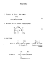
- purified collagen may be chemically-modified by eovalently binding phosphates to hydroxyl groups of hydroxylated amino acids, as shown in Example 6.
- this reaction likely involves covalent bonding of phosphate to hydroxyl group of serine, tyrosine and/or threonine, hydroxylysine and hydroxyproline.
- the reaction is controlled, in order to limit the degree of reaction.
- any of the potentially remaining reactive groups present in the collagen may be fully converted to phosphoryl, hydroxyl, or sulfonyl groups by exposure to trimetaphosphate or other active agents.
- the purified product can be customized to particular uses.
- bioactive responses may be favored by reacting the collagen with a linear or cyclic polyphosphates at alkaline pH.
- the final collagen product may be used in solution or as a solid, and has been shown to be useful for a variety of purposes, including but not limited to, biological implants, grafts, transplants, and drug delivery. It is, moreover, contemplated that it is useful as a surgical adjunct during transplant surgery and to prevent post-operative graft dislocation, as a hemostatic agent, to augment soft tissues, and as a support for in vitro cell growth ( i . e ., in cell cultures).
- the collagen prepared according to the methods of the present invention is particularly applicable to the control of bleeding from surfaces. especially large surfaces.
- the collagen of the present invention may be used as a hemostatic agent on cut or severed bone, organs ( e . g ., spleen, liver or kidney which has been cut surgically or traumatically), the central nervous system with its predominant collection of small blood vessels, prosthetic surgery, oozing surfaces resuming from the surgical removal of necrotic tissue, cosmetic surgery, and any surfaces with oozing of blood from one or more small sources ( e . g . facial cuts).
- This hemostatic collagen preparation may be applied in a variety of forms ( e .
- hemostatic collagen preparation as a powder applied directly to the surface: as a styptic in pencil form: as a gel, a sponge or in fabric form).
- the amount of hemostatic collagen preparation utilized will vary with the extent of the bleeding, the surface area to be treated, and severity of the blood flow: the only requirement being that the achievement of the desired control.
- sample are used interchangeably in the present specification and claims and are used in their broadest sense. On the one hand they are meant to include a clinical specimen (i . e ., sample) or culture ( e . g ., microbiological cultures). On the other hand it is meant to include both biological and environmental samples. Biological samples may be animal, including human, fluid or tissue, food products and ingredients. However, these examples are not to be construed as limiting the sample types applicable to the present invention.
- non-human animals comprise any animal other than humans.
- Such non-human animals include vertebrates such as rodents, non-human primates, ovines, bovines. ruminants, lagomorphs, porcines, caprines, equines, canines, felines, aves, etc.
- preferred non-human animals are selected from livestock, such as bovines and ovines.
- procollagen is used in reference to the precursor protein that is cleaved in the extracellular matrix to form collagen.
- procollagen is used in reference to "pro- ⁇ chains” that are precursors of the collagen ⁇ chains.
- Pro- ⁇ chains are characterized as comprising "pro-peptides” that are important in the formation of the triple-helix formation of procollagen.
- collagen is used in reference to the extracellular family of fibrous proteins that are characterized by their stiff, triple-stranded helical structure. Three collagen polypeptide chains (“ ⁇ -chains”) are wound around each other to form this helical molecule. The term is also intended to encompass the various types of collagen, although the preferred form is type I collagen.
- collagen sample refers to any source of collagen, including, but not limited to hide, skin, tendons, blood vessels, intestine, liver, spleen, heart valve, bone, etc. It is not intended that the source of collagen sample be limited to any particular tissue source or type. The only requirement is that the collagen sample contain the type of collagen of interest to be purified (e . g ., type I collagen). However, it is not intended that the present invention be limited to any particular type of purified collagen.
- collagen fibrils is used in reference to the long, thin polymers of collagen that are grouped into bundles referred to as "collagen fibers.”
- the "collagen matrix” refers to an extracellular matrix of collagen, with the characteristic periodic cross-striations that may be visible through electron microscopy. Characteristically, the collagen matrix is formed when normal collagen molecules are assembled in a quarter stagger array in a three-dimensional structure appearing by electron microscopy as a 67 nanometer striation.
- the term "compressed collagen” refers to a collagen sample that has been compressed due to the application of pressure.
- the collagen to be compressed is wet. TTPTM-treated, and delipidated collagen. It is intended that this compression be accomplished by any means ( i . e ., hand presses or machine presses). It is intended that this compression encompass a range of pressures, ranging from 5,000 to 50,000 pounds per square inch (psi) of sample (34474 - 344740 kPa).
- the pressure applied is approximately 9,000 (62052 kPa), while in alternative preferred embodiments, the applied pressure is approximately 30,000 (206841 kPa).
- the compressed collagen is in the form of a cake.
- dehydrated collagen refers to a collagen sample that has been dehydrated using any method commonly known in the art. In preferred embodiments. dehydrated collagen is produced by lyophilization or desiccation.
- drying refers to any method for the removal of water from a sample. It is intended that the term encompass methods including, but not limited to, airdrying and heating.
- the term "dispersing” refers to the mechanical separation of a sample.
- the term encompass the separation of the components contained within a cake of compressed collagen.
- the components of the compressed cake are dispersed into fibers.
- the collagen is agitated and suspended within a liquid.
- These fibers comprise collagen fibers as described above ( i . e ., bundles of collagen fibrils).
- native, as well as dried collagen fibers be encompassed by this definition.
- hemostatic refers to an agent that stops or slows the flow of blood.
- proteolytic agent refers to papain that is capable of degrading proteins associated with collagen, while leaving collagen type I in its native configuration. However, it is contemplated that in some embodiments, the proteolytic agent breaks the cross-links associated with collagen fibers in collagen. In this manner, the cross-links between the strands within the fibers are loosened, such that the fibers retain their basic shape, but sufficient space is allowed between the fibers that molecules that are normally trapped within the collagen fiber structure may be removed.
- proteolyzed collagen refers to collagen that has been treated with at least one proteolytic enzyme.
- the term "purified” refers to the removal of contaminants from a sample. For example, it is intended to encompass proteolyzed collagen. It is not intended that the term be limited to any particular level of purity. However, in preferred embodiments, the purified sample is treated with proteolytic enzymes, In particularly preferred embodiments, the purified sample has undergone subsequent treatments, including but not limited to, delipidation and/or phosphorylation.
- reducing agent refers to any compound or mixture of compounds that is capable of reducing another compound. In this process the proportion of hydrogen is increased while the proportion of oxygen is decreased and the number of multiple bonds is decreased. In particular. it is intended that the term encompass any agent that is capable of unfolding proteins from their native configurations by breaking disulfide bonds. Such agents include, but are not limited to compounds such as sodium sulfide, dithiothreitol, glutathionine, and sodium borohydride.
- reduced collagen refers to collagen that has been exposed to or treated with at least one reducing agent.
- phosphorylation agent refers to any compound or mixture of compounds that is capable of phosphorylating another compound.
- Such compounds include, but are not limited to sodium trimetaphosphate sodium hexametaphosphate, sodium ultraphosphate, sodium tetrametaphosphate, phosphoric anhydride, and phosphoryl trichloride,
- phosphorylated collagen refers to collagen that has been treated so as to increase the number of phosphorylated amino acids in the collagen molecules. It is intended that the term encompass a range of phosphorylation. from a minimal degree of phosphorylation (i . e ., only one amino acid has been phosphorylated) to a maximal degree of phosphorylation ( i . e ., all of the amino acids available and suitable for phosphorylation have been phosphorylated), or any degree of phosphorylation within this range.
- the term "delipidation agent” refers to any compound or mixture of compounds that is capable of removing lipid moieties ( i . e ., delipidating). In particular it is intended that the term encompass substances that are capable of removing molecules including, but not limited to, phospholipids, neutral lipids. proteolipids, and glycolipids from collagen. Such substances include organic solvents such as chloroform and methanol.
- delipidated collagen refers to collagen that has been exposed to or treated with at least one delipidation agent. In particularly preferred embodiments, the delipidation agent is chloroform and methanol in a 3:1 mixture.
- alkali refers to a hydroxide of one of the alkali metals (e . g ., lithium, sodium, potassium, rubidium, cesium, and francium). It also encompasses substances which provide an alkaline solution (pH > 7) in water (e . g ., CaO, Ca(OH) 2 , and Na 2 CO 3 .
- modified collagen refers to collagen that has been chemically modified by such means as phosphorylation. fluorination, halogenation ( e . g ., chlorination), sulfonation, and/or hydroxylation.
- filter-sterilized sample refers to a sample that has been applied to a filter to remove unwanted contaminants such as bacteria, from the sample.
- epithelialization refers to the process of removing epithelial tissues and/or cells from a sample.
- epithelial pertains to epithelium, the cellular covering of internal and external body surfaces.
- biocompatible collagen refers to collagen that is suitable for biomedical or veterinary applications, including but not limited to grafts, and implants.
- biomolecule refers to any molecule that has biomedical or veterinary applications. For example, it is intended to encompass various compounds and substances. including collagen, synthetic polymers, or other materials useful for implantation, transplantation, and/or grafting.
- graft refers to any tissue or organ for implantation or transplantation, as well as the process of implantation or transplantation of such tissue.
- a skin graft is a piece of skin or other tissue or material that is transplanted in order to replace a lost porton of the skin. It is intended that the term encompass materials such as purified collagen or other suitable biocompatible substances useful in the replacement of lost or damaged tissues or organs.
- graft is used in reference to the replacement of skin or other tissues or organs exposed to the environment.
- the term may be used in reference to such tissues as skin. cutaneous tissues. mucous membranes, conjunctival membranes, etc.
- the term be limited to any particular type of tissue.
- transplant refers to tissue used in grafting, implanting, or transplanting, as well as the transfer of tissues from one part of the body to another, or the transfer of tissues from one individual to another. or the introduction of biocompatible materials into or onto the body.
- transplantation refers to the grafting of tissues from one part of the body to another part, or to another individual.
- tissue or organ used for transplantation or grafting be comprised of materials such as collagen purified according to the methods of the present invention.
- the graft or transplant be limited to naturally-occurring tissues.
- the term "implantation” refers to the insertion of an organ or tissue in a new site in the body, as well as the insertion of any biocompatible, biological, living, inert, or radioactive material into the body.
- implant refers to the process of inserting or grafting of tissue, inert, living, radioactive, or biocompatible material into intact tissues or a body cavity, as well as the material to be so inserted or grafted.
- overlap refers to a mass of tissue for grafting, which usually includes skin, that is only partially removed from one part of the body, so that it retains its own blood supply during transfer to another site of the body or to another individual. It is also intended that the term encompass masses of tissue for grafting or implantation that have been cultivated in vitro prior to their grafting or implantation.
- Medchem Products Medchem Products, Woburn, MA: Collatek (Collatek, Inc., Plainsboro, NJ): Boehringer Mannheim (Boehringer Mannheim. Indianapolis, IN): J & J Medical (J & J Medical. Inc.. Arlington, TX); Axygen (Axygen Inc., Hayward. CA); Papain Products (Papain Products, Tamilnadu, India); Beckman (Beckman Instruments, Inc.. Fullerton, CA); and Brinkmann (Brinkmann Instruments. Inc.. Westbury, NY).
- collagen was purified from dehaired bovine hide, using a one-step enzyme treatment process.
- 100 g of clean, dehaired bovine hide obtained from a local slaughterhouse was chopped or sliced into pieces of approximately 1 cm thickness and approximately 10 cm long.
- the cut slices were washed in three liters of a wash buffer containing double distilled water containing 30 g NaCl, 3 g ZnCl, 4.5 g methyl paraben. 0.9 g propyl paraben.
- the wash buffer contained 30.0 g NaCl, 3 g ZnCl, 4.5 g methyl paraben, and 1.5 g trichlorophenate or 3 g hypochlorite. No differences were observed in the purified collagen preparations using these different wash buffers.
- Washing was conducted at 5-15°C for 6-8 hours under constant agitation using a Rotator (Model #4140. American). The wash procedure was repeated twice, with the fluid being decanted from the solids after each wash. The repeated wash procedure removed most of the albumins and globulins that usually constitute approximately 1-5% of the dry weight of the sample.
- de-epithelialization of the sample was conducted. In this process. 1 liter of cold de-epithelialization buffer containing cold deionized water containing 24 g NaOH and 56 g calcium oxide was added to the previously washed sample. and centrifuged at 5.000 xg for 15 minutes to obtain solid chunks of material. The sample was agitated on a rotator at 80-100 rpm, for 48 hours at 15°C. The de-epithelialization solution was replaced every 16 hours.
- the chunks were soaked in 3 liters of deionized water for 2 hours, with agitation at 80-100 rpm, and the solids were recovered by centrifugation at 5.000 xg for 15 min.
- One hundred grams of boric acid solubilized in 3 liters of deionized water pre-chilled to 4°C. was added and the sample was agitated for 16 hours at 4°C.
- the pH was measured and adjusted to approximately 8.3.
- the sample was washed twice with 3 liters of deionized water for 2 hours each wash, and the solids were recovered by centrifugation as described above.
- the sample was treated with papain in a one-step enzymatic treatment.
- the sample was further washed in 3 liters of pyrogen-free deionized water for 2 hours at room temperature.
- the sample was centrifuged for 15 minutes at 5.000 xg, and decanted.
- One liter of normal saline solution was then added to the sample.
- Papain Sigma or Papain Products
- Papain was added at the rate of 1-2 mg per ml of the total volume ( i.e.. 1 liter), with continual stirring.
- the solution was incubated under agitation using a rotator at 80-100 rpm, at 37°C for approximately 16 hours.
- the excess water was drained and the solids were washed three times with 2 liters of deionized water.
- One liter of 0.2 M sodium borohydride was then added to the washed solids.
- the solids were incubated for 3 hours under agitation with a rotator at 80-100 rpm.
- the excess water was drained, and the solids were washed three times with 2 liters of deionized water.
- a "Robust Treatment” was also tested. In this treatment, a multifold increase in time of incubation and enzyme concentration were used, as compared to the "normal” treatment. Although the rest of the steps were the same as described for the normal treatment (i . e ., the collagen used in the "Robust Treatment” was prepared using the same bovine hide. cutting and washing steps), in the Robust Treatment, 4 mg of papain was added per ml of solution and 24 hours of incubation were used in the enzymatic treatment step, rather than the 1 mg of papain per ml of solution and 16 hours of incubation.
- TTPTM Twice-Treatment ProcessTM
- TTPTM Twice-Treatment ProcessTM
- type I collagen was purified from dehaircd bovine hide.
- 100 g of clean, dehaired bovine hide obtained from a local slaughterhouse was chopped or sliced into pieces of approximately 1 cm thickness and approximately 10 cm long. The cut slices were washed in three liters of a wash buffer containing double distilled water containing 30 g NaCl, 3 g ZnCl, 4.5 g methyl paraben, 0.9 g propyl paraben.
- the wash buffer contained 30.0 g NaCl, 3 g ZnCl, 4.5 g methyl paraben, and 1.5 g trichlorophenate or 3 g hypochlorite. No differences were observed in the purified collagen preparations using these different wash buffers. Washing was conducted at 5-15°C for 6-8 hours under constant agitation using a rotator (Model #4140, American) at 80-100 rpm. The wash procedure was repeated twice, with centrifugation at 5,000 x g for 15 minutes, and decanting of the supernatant each time. The repeated wash procedure removed most of the albumins and globulins that usually constitute approximately 1-5% of the dry weight of the sample.
- de-epithelialiration of the sample was conducted.
- 1 liter of cold de-epithelialization buffer containing cold deionized water containing 24 g NaOH and 56 g calcium oxide was added to the previously washed sample, and centrifuged at 5,000 xg for 15 minutes to obtain solid chunks of material.
- the sample was agitated on a rotator at 80-100 rpm, for 48 hours at 15°C.
- the deepithelialization solution was replaced every 16 hours.
- the chunks were soaked in 3 liters of deionized water for 2 hours, with agitation at 80-100 rpm, and the solids were recovered by centrifugation at 5,000 xg for 15 min.
- the pH was measured and adjusted to approximately 8.3.
- the sample was washed twice with 3 liters of deionized water for 2 hours each wash, and the solids were recovered by centrifugation as described above.
- the sample was treated with the TTPTM.
- the sample was further washed in 3 liters of pyrogen-free deionized water for 2 hours at room temperature, and the water was decanted.
- One liter of normal saline solution was then added to the sample.
- Papain was added at the rate of 1-2 mg per ml of the total volume ( i . e ., 1 liter), with continual stirring.
- the solution was incubated under agitation on a rotator at 80-100 rpm, at 37°C for approximately 16 hours.
- This step also reduced the amount of aldehyde potentially involved in aldol condensation reactions (i . e ., reactions between two aldehyde groups) or Schiff-base reactions (i . e ., reactions between an aldehyde group and an amino group ( See e . g .,Nimni et al .,"Bioprosthesis derived from crosslinked and chemically modified collagenous tissues.” in Collagen , vol. 3. M.E. Nimni et al ., CRC Press. Inc.. Boca Raton. FL [1988], p. 5). It may also reduce the interchain disulfide bonds in the helical region of the same molecule, present only in type III collagen ( See e . g ., D. Amiel et al ., "Biochemistry of tendon and ligament," in Collagen , vol. 3. p. 229, supra ).
- the first enzyme treatment liberates the collagen in the preparation to an extent that is susceptible for further attack by a reducing agent (e . g ., sodium sulfide. thiothreitol, dithiothreitol, glutathione or sodium borohydride), with or without alkali compounds to swell the collagen.
- a reducing agent e . g ., sodium sulfide. thiothreitol, dithiothreitol, glutathione or sodium borohydride
- a second treatment with papain was conducted, in order to thoroughly eliminate all of the residual non-collagenous contaminants (e . g ., interstitial elastin).
- Collagen prepared according to the two-step enzymatic treatment, as well as collagen prepared with the one-step enzymatic treatment described in Example 1 was tested to determine the degree of removal of non-collagenous fibrous proteins (e . g ., elastin) as described in Example 3.
- non-collagenous fibrous proteins e . g ., elastin
- the "Enzyme ⁇ Reducing Agent” sample refers to a sample that was tested following one enzyme treatment and one reduction step ( i . e ., the first enzyme treatment of Example 2, followed by the reducing step of Example 2).
- the sample indicated as "Reducing Agent ⁇ Enzyme” refers to sample that was tested following one reduction step followed by one enzyme treatment.
- This "Reducing Agent ⁇ Enzyme” sample was prepared from the same source of collagen, cut, and washed as described in Examples 1 and 2. The test sample was first reduced using the reduction method described in Example 2, but without the first enzyme treatment. Following this reduction step, the methods described for the first enzyme treatment were conducted on the reduced sample.
- TTPTM-Treated The sample indicated as "TTPTM-Treated” was a sample prepared according to the methods of Example 2. As no insoluble non-collagenous protein was detected using the normal methods of "TTPTM-treatment, the robust version of this process was not conducted. The characteristics of this TTPTM-treated sample are discussed in more detail in Example 5 below.
- the untreated control contained 1.24 ⁇ 0.190 percent dry weight of non-collagenous insoluble protein, while the sample prepared by the TTP process, had no detectable dry weight non-collagenous insoluble protein.
- the TTPTM-treated type I collagen was separated by fractional NaCl salt precipitation as described by Hill and Harper (R.J. Hill and E. Harper, Anal. Biochem., 141:83-93 [1984]). Briefly, the enzyme-treated collagen was dissolved in 0.5 M NaCl in Tris (hydroxymethyl amino methane buffer), at pH 7.4. The NaCl concentration was then increased to 1.7 M. The solution was mixed and centrifuged at 10.000 xg for 1 hour at 0-4°C. The dewatered precipitate was mixed and solubilized in 0.5 M acetic acid, and the pH was adjusted to 7.0. Sodium chloride was added to a final concentration of 2.5 M, and the precipitate was collected by another centrifugation step as above.
- Tris hydroxymethyl amino methane buffer
- the precipitated collagen was then dialyzed against deionized water and lyophilized prior to delipidation for quantitative analysis of the lipid content in the collagen sample.
- the collagen preparation was then dilipidated using the method of Folch et al ., (Folch et al ., J. Biol. Chem., 226:497-509 [1957]), with modifications.
- This step involved solvent extraction of total lipids, including gangliosides and phospho-inosities. Briefly, the collagen preparation was stirred in a mixture of chloroform and methanol (2:1, v/v). The volume of the chloroform: methanol was 20 times that of the collagen sample ( i . e ., 20 parts of the chloroform:methanol mixture were added to 1 part of collagen sample). The sample was stirred well during this addition, and vigorously shaken for one hour at 40-45°C.
- the homogenate was transferred to a vial, and 0.2 volumes of 0.88% KCl were added.
- the two phases of the mixture were allowed to separate in a separating funnel.
- the lower phase containing major lipids was removed, and the upper phase, which contained the collagen was washed twice with a mixture of 3:48:47 (v/v) chloroform:methanol:water.
- the collagen preparation was then washed with a mixture of 5:80:15 (v/v) of the same components ( i . e ., chloroform:methanol:water), and dewatered using a pressure of 9.000 ⁇ 1000 psi, using a hand press. This squeezed the water from the collagen preparation to produce a dewatered mass.
- pressures ranging from approximately 8000 psi to 30.000 psi are suitable for use in this step.
- Application of pressure at 8000 psi removes all of the extraneous water from outside of the molecules. while application of approximately 30,000 psi removes even the intramolecular water.
- the compressed cake was then dispersed, using a coffee grinder, and air dried in a ventilated drying oven at 40-45 °C.
- the purity of the delipidated sample was then tested for phospholipids, neutral lipids and glycolipids as described below.
- TLC Thin layer chromatography
- the presence of neutral lipids and glycolipids in the TTPTM-treated collagen was determined using a silicic acid column chromatographic method. as described by Rouser et al . (G. Rouser et al ., Lipids 2:37-40 [1967]). Briefly, a column (10 mm x 20 mm) containing silicic acid (Spectrum) was prepared and 10 g of the delipidated collagen was applied in sequential portions of 2 grams each, to the column. Thus, the total 10 g sample was divided into five runs on the column; all of the eluted samples were pooled.
- the sample was eluted with 160 ml of chloroform:acetone (1:1, v/v), followed by a series of acetone and acetone:water solutions.
- the eluted neutral and glycolipids were collected from the column, air dried, and estimated gravimetrically. The results are shown in Table 2.
- the cysteine. hydroxyproline, hexosamine, and elastin content of delipiciated and TTPTM-treated collagen was determined.
- the hydroxyproline content is recognized as a measure of the amount of collagen present in a sample.
- the delipidated and dehydrated type I collagen sample was reconstituted in 0.1 M acetic acid, to a concentration of 0.3% (w/v).
- the purity of the collagen was evaluated by HPLC to determine its amino acid content using a Beckman 6300 amino acid analyzer, according the manufacturer's instructions.
- the cysteine content of the sample was determined, as was the cysteine content of a collagen preparation that was "less phosphorylated,” as described in Example 6.
- the insoluble non-collagenous protein (i . e ., elastin) content of TTPTM-treated collagen was determined using the methods described in Example 3 for testing the insoluble non-collagenous protein content of collagen.
- elastin was the only non-collagenous protein potentially remaining in the TTPTM-treated type I collagen preparation, it was considered important to determine the elastin content of this preparation, in comparison with collagen treated using other methods ( e . g ., the phosphorylated collagen of Example 6).
- Table 3 below. These results are expressed as a percentage (wt/wt) on a dry weight basis.
- the 0.03 M chloramine T used in this Example was prepared by added 0.845 g chloramine T to 100 ml of a buffer comprised of 20 ml water. 30 ml propanol, and 50 ml citrate-acetate buffer, pH 6.
- the citrate-acetate buffer (pH 6) was prepared by adding 50 g citric acid (1 H 2 O), 12 ml glacial acetic acid, 120 g sodium acetate (3 H 2 O), and 34 g NaOH to 500 ml water. The volume was adjusted to 1 liter, and the pH was adjusted to 6. Perchloric acid (3 M) was prepared in 25.5 ml 70% HClO 4 , and the volume adjusted to 100 ml in water. The hydroxyproline stock was comprised of 5 ⁇ g hydroxyproline per ml water. The hydroxyproline standards were prepared by adding 20 mg hydroxyproline per 100 ml water and 0.05 ml concentrated HCl.
- hydroxyproline concentrations were determined as follows. One ml of each sample and 1 ml of standard solution containing 2-5 ⁇ g hydroxyproline were added to 1 ml chloramine solution in separate tubes, and mixed for 20 minutes at room temperature. One ml perchloric acid was then added to the tubes, and mixed at room temperature for 5 minutes. One ml of 5% p-dimethylaminobenzaldehyde in propanol was added to each tube, and mixed for 18 minutes in a 60°C water bath. The tubes were cooled to 20°C, and optical density was read at 550 nm. The results are shown in Table 3, below.
- the trisodium phosphate-potassium tetraborate solution used in this experiment was prepared by mixing 98 ml of 1 N tribasic sodium phosphate and 0.5 N potassium tetraborate. This trisodium phosphate-potassium tetraborate solution was then used as the solvent for the acetylacetone mixtures for determination of hexosamine in the samples.
- Acetylacetone I was prepared as a 3.5% (v/v) solution in sodium phosphate-potassium borate buffer.
- Acetylacetone II was prepared as a 3.5% (v/v) solution in 0.5 N potassium tetraborate.
- the Erlich's reagent used in these Examples was prepared by adding 3.2 g p-dimethylaminobenzaldehyde in 30 ml 12 N HCl, diluted with 210 ml 2-propanol.
- the delipidated sample was placed into a tube. 6 N HCl was mixed into the sample. and the sample was allowed to hydrolyze for 30 minutes at 120°C (or 12-14 hours at 100°C), the sample was then evaporated to dryness. Concurrently, standards of glucosamine and galactosamine (2.5 to 40 ⁇ g for each) were hydrolyzed and evaporated to dryness using the same methods. The samples and standards were resuspended in double distilled water.
- TTPTM- treated collagen prepared according to the methods described in Example 5 were further treated to produce "CollagenPROTM.”
- CollagenPROTM refers to purified collagen that has been phosphorylated.
- CollagenPROTM was prepared from TTPTM- treated collagen prepared according to the methods described in Example 5. First, TTPTM- treated collagen was filter sterilized by passing the preparations through a 0.22 ⁇ filter attached to a 30 ml syringe. The filter-sterilized collagen suspensions were then stored until use at 4°C. These tilter-sterilized collagen preparations were chemically modified in order to determine the optimum modifications for production of purified collagen suitable for biomedical applications. The entire procedure was conducted under sterile conditions, using pre-sterilized ingredients and pyrogen-free, sterile water.
- EGF growth factors
- interleukins e . g ., IL1
- other biologically active molecules e.g ., phosphorylation of biomolecules, such as growth factors (e . g ., EGF), interleukins (e . g ., IL1), and other biologically active molecules.
- EGF growth factors
- interleukins e . g ., IL1
- other biologically active molecules is beneficial.
- EGF EGF
- 1 ml of 0.1% EGF adjusted to pH 11.5 is added to 0.2 ml sodium trimetaphosphate, at the same concentrations and conditions ( e . g ., agitation and temperature), as described above.
- the sample is then dialyzed to remove salts, and the phosphorylated EGF may be used as appropriate.
- washed collagen sample was tested for the presence of covalently hound phosphorous, using the procedure described by Fiske and Subbarow (C.H. Fiske and Y. Subbarow. J. Biol. Chem., 66:375-400 [1925]). Briefly, 1 mg of dry modified collagen prepared and lyophilized according to Example 6 was mixed with 1 ml of 72% perchloric acid. and then digested at 200°C, for 20 minutes. Deionized water was added to the digested sample to adjust the volume to 4 ml. Then, the following reagents were added in succession. and mixed well. First. 0.4 ml of 2.5% ammonium molybdate in 3 N sulfuric acid was added.
- the phosphorous content was estimated by graphing the results obtained with the potassium dihydrogen monophosphate standards. In this experiment, no bound phosphorous was detected in the TTPTM-treated samples. For samples with more phosphorylated collagen ( i . e ., where 40% of the modifiable sites available in the collagen were phosphorylated), 2.5% (wt/wt; dry weight basis) bound phosphorous was detected. For samples with less phosphorylation, 0.8% (wt/wt: dry weight basis) bound phosphorous was detected.
- This modified collagen was shown to possess improved biological characteristics, as described in Examples 10-15. However. it is recognized that care must be taken in the purification of collagen, as impurities present in impure collagen preparations may engender unfavorable biological reactions due to the phosphorylation of contaminant proteins ( e . g ., elastin).
- the amount of modification can be controlled, ranging from none to a maximum of approximately 40% of the modifiable amino acids present in collagen.
- the pH conditions under which the reaction occurs may vary from 8 to 13, depending upon the amount of modification desired.
- the reaction temperature may range from 4-45°C. It was observed in other experiments, that the samples tend to freeze at temperatures less than 4°C, while at temperatures above 45°C, the collagen may become denatured.
- the chemical used to react with the collagen may be selected from the group comprising, but not limited to. various polyphosphates (e . g ., sodium hexametaphosphate. sodium ultraphosphate, and sodium tetrametaphosphate), phosphoric anhydride, and phosphoryl trichloride solubilized in organic non-polar solvents such as hexane, etc.
- various polyphosphates e . g ., sodium hexametaphosphate. sodium ultraphosphate, and sodium tetrametaphosphate
- phosphoric anhydride e.g phosphoric anhydride
- phosphoryl trichloride solubilized in organic non-polar solvents such as hexane, etc.
- non-collagenous biological materials for various biological applications will be chemically modified in a like manner. It is also contemplated that other modifications useful for preparing collagen to be used in cell culture or in vivo applications, including, but not limited to fluorination, halogenation ( e . g ., chlorination), sulfonation, and/or hydroxylation may be performed on collagen prepared according to the methods of Example 2, as well as phosphorylated collagen prepared according to the above described methods. It is contemplated that these modifications be conducted in conjunction ( i . e ., multiple modification procedures may be conducted on the same preparation), as well as separately.
- Example 6 the solubility of purified type I collagen, modified as described in Example 6 was determined, and compared with the solubility of native collagen.
- films i . e ., membranes
- filter-sterilized collagen i . e ., CollagenPROTM
- films prepared according to the methods described in Example 6 were prepared for use in in vivo and in vitro experiments.
- the collagen was prepared from bovine Achilles tendons, rather than bovine hide. Both collagen sources were found to be satisfactory for use in the following Examples.
- the suspension was poured into sterile 15 x 2 cm petri dishes to a depth of approximately 8 mm.
- the petri dishes were covered, and stored at a constant temperature of 37°C for 48 hours, in order to promote uniform initial gelation. This often required 3-5 days for the films to visually appear to be dry.
- the petri dishes were uncovered, and drying was allowed to continue at room temperature in a chemical fume hood.
- the collagen films were sterilized with ethylene oxide gas and used for animal implantation, transplantation. grafting, or other experiments. These films were also used in subsequent experiments.
- Example 9 the suitability and biocompatibility for in vivo applications of the chemically-modified collagen type I produced as described in Example 6, unmodified collagen prepared as described in Example 2, were determined. Purified, washed collagen samples obtained as described in Example 9, were dried in the form of thin films of 0.3 mm thickness.
- the entire skin flap, including the grafted collagen was removed from each of the animals from the surgical site, and fixed in 70% alcohol, dehydrated, and embedded in wax. using methods known in the art. Embedded samples were cut into sections 5 microns in thickness, and stained with Goldner's one-step trichrome stain (OST). and hematoxylin and eosin (H & E). using methods known in the art.
- OST Goldner's one-step trichrome stain
- H & E hematoxylin and eosin
- Example 2 type I collagen obtained from tissue samples of bovine intestine (obtained from a local market) was purified and chemically modified using the methods of Example 6. These samples were then implanted into rabbits, in order to determine the suitability and biocompatibility of collagen prepared according to the methods of the present invention. In these experiments, control collagen prepared according to the methods described in Example 2 was also used.
- the animals were observed for three weeks for gross indications of inflammation (e . g ., redness, swotting, etc.). No adverse responses were observed for any of the animals.
- the animals were sacrificed and the implants were surgically removed. Fixed in 70% alcohol, dehydrated and embedded in paraffin, using methods commonly known in the art. Sections of 5 micron in thickness were cut. and stained with OST and H & E. using methods commonly known in the art.
- the results showed more vascularization and fibroblastic ingrowth in both experimental groups.
- the control samples had relatively poor vascularization, as well as a prevalence of multi-nucleated giant cells, reflecting the lesser biocompatibility of these samples.
- Example 9 the collagen implants tested as described in Example 9 were analyzed for their alkaline phosphatase activity.
- Alkaline phosphatase activity was of interest, as it has been hypothesized that this enzyme is involved in tissue formation and calcification.
- the assay used in this Example is based on the methods of Bradenberger and Hanson (H. Bradenberger and R. Hanson, Helv. Chim. Acta 36:900 [1953]; and Hofstee (B.H. Hofstee, Arch. Biochem. Biophys.. 51:139 [1954]). In this colorimetric method. the effect of alkaline phosphatase on the hydrolysis of o-carboxy-phenyl phosphate is measured.
- samples of approximately 10 mg from each of the harvested collagen implants from Example 10 were dispersed at a rate of 1 mg in 1 ml of Tris buffer (0.1 M Tris, pH 8.5) for five minutes. Care was taken to maintain the temperature of the dispersed samples at 0-5°C. in order to prevent enzymatic reactions from occurring prior to the assay procedure. A small amount of detergent (to a final concentration of 0.1 M sodium deoxycholate) was added to the dispersed samples, in order to facilitate the release of membrane-bound enzymes. The dispersed samples were then mixed in a Polytron homogenizer (Brinkmann), for 30 seconds. white they were chilled in an ice bath.
- Tris buffer 0.1 M Tris, pH 8.5
- test solution comprised of 2 ml glycine (0.2 M. pH 8.8). 1 ml of 3.65 mM o-carboxy phenyl phosphate (OCCP), and 0.5 ml of 0.05 M MgCl 2 was placed into a cuvette, and allowed to incubate for 3-4 minutes at 25°C. Collagen test samples (0.1 ml, prefiltered using a 0.22 micron filter attached to a 5 ml syringe) were added to the solutions in the cuvettes. The optical density of the cuvette contents was then determined at 300 nm, at room temperature.
- OCCP o-carboxy phenyl phosphate
- the activity is expressed in units per mg of tissue, as based on the number of micromoles of OCCP hydrolyzed per minute at 25°C, at pH, under the conditions described above.
- Example 9 the suitability of type I collagen was extracted from bovine Achilles tendons (obtained from a local market), and made into uniformly thin dry films as described in Example 9.
- a control film was prepared using the standard pepsin enzyme treatment method described in U.S. Patent No. 5,374,539.
- native type I bovine collagen prepared as described in U.S. Patent No. 5,374,539, and bovine collagen cross-linked with glutaraldehyde (as described in this patent) were tested. All of these samples were tested in the presence or 0.5 ml of reconstituted epidermal growth factor (EGF; Collaborative Research Inc.)(1 ⁇ g/3 ml PBS). as well as without the addition of EGF.
- EGF epidermal growth factor
- Epithelial cell cultures were chosen for this Example. as they provide a model system for investigating the events that govern the closure of superficial epithelial defects and for assaying the actions of exogenous agents on these events. These methods provide some advantages, as compared to in vivo models of wound closure. For example, it is much easier to manipulate the extracellular milieu in cell culture systems.
- Prior reports indicate that the in vitro epithelial response is similar to the response observed in vivo , and can be readily quantified ( See e . g ., S. Gunasekaran et al ., Proc. Soc. Biomater., 20:311 [1994]; and S. Simmons et al ., Toxicol. Appl. Pharrnacol., 88:13-23 [1987]).
- This experiment was designed to demonstrate the comparative suitability of various collagen substrata for wound closure. In particular, the interaction of the collagen substrata and EGF were investigated.
- DMSO dimethyl sulfoxide
- the full thickness of the corneal epithelial sheets were gently peeled off the corneal samples and placed in trypsin, gently mixed. and centrifuged at 2,000 xg for 5 minutes at 4°C. The supernatants were removed and the cells from six corneas were combined and resuspended in fresh primary culture medium and plated in six 35 mm culture dishes. The cultures were placed at 37°C, in a humidified CO 2 incubator. The culture medium was changed three times per week.
- the primary cultures had grown to confluency and were trypsinized for 5-10 minutes.
- the cells were pipetted from the culture dishes and centrifuged at 2000 x g for 5 minutes. The supernatant were removed and the control culture medium composed of the Dulbecco's modified Eagle's medium described above, but exclusive of the EGF. The cells were pipetted to yield a suspension of single cells and counted using a hemacytometer. Twenty-four well multiplates were seeded at 3-4 x 10 4 cells/well in I ml medium per well. The subcultured cells were grown for 7 days, with the medium changed three times during this period.
- the cells contained in four wells were counted. in order to ensure that each well had at least 1.6 x 10 5 cells.
- the cell layers were injured ("wounded") using filter disks (7 mm diameter), as described by Jumblatt and Neufeld ( See , M. Jumblatt and A. Neufeld, Invest. Ophthamol. Visual Sci.. 15:4-14 [1986]). Briefly, the cell culture medium was removed from the wells and the filters were placed on top of the cell layer and pressed down with a plunger (7 mm diameter). A pre-chilled steel probe 6 mm in diameter was held for 5 second on the bottom of the wells, directly below each disk. Disks with adherent dead cells were then removed from each well.
- test materials (collagen prepared according to the methods described in Example 9 (CollagenProTM), as well as native type I bovine collagen and cross-linked type I bovine collagen (prepared according to U.S. Patent No. 5,374,539), were added to each well.
- the multipiates containing the "wounded" cell layers and test collagens were re-incubated for 24 hours, to allow the growth of cells into the wounded area.
- the wells were observed at intervals during this 24 hour growth period, to measure the area of cell layer wounded by the disk. These measurements were taken microscopically, at 0. 12, 18, and 24 hour time intervals.
- Closure of the injured area on the cell layer within the wells was stopped by fixation of the cell layers and collagen test samples, with 10% neutral buffered formalin, for at least 10 minutes. The cells were then stained with Giemsa stock solution for at least 10 minutes, rinsed in 10% neutral buffered formalin, and allowed to air dry. The stained wells contained in the microplate were observed microscopically and the wound areas were measured.
- Figure 3 is a bar chart that shows the in vitro wound closure results obtained in this Example.
- SC represents CollagenProTM
- NC represents native collagen
- XC indicates cross-linked type I bovine collagen
- GF indicates EGF.
- collagen prepared as described in Example 6. was used in additional tests to determine its utility in in vivo as a hemostatic agent.
- Hemostasis i . e ., blood clotting
- Collagen is a natural hemostatic agent that is present in almost every tissue, and which also appears to have specific affinity for cells and growth factors essential for normal wound healing.
- Other materials may assist hemostasis, it is possible that, unlike collagen, they will not optimize the rate of wound heating.
- the object of this Example was to determine the hemostatic and wound healing abilities of various compounds and substances.
- collagen fibers obtained from Medchem Products
- sponges obtained from Collatek
- gelatin obtained from Arizona Health Sciences
- Additional substances previously used as hemostatic agents such as thrombin (e . g ., Thrombinar and Thromostat), and cellulose ( e . g ., Surgicel and Oxycel, J & J Medical) were not included in these experiments.
- Thrombin was not included as it may have immunological and local delivery problems.
- Native type I bovine collagen was prepared as described in Example 2. Avitene, Helistat, and Gelfoam were purchased from their respective suppliers. Gelatin fragments were prepared by limited. non-specific protease treatment of gelatin (Sigma). The gelatin fragments were then purified by size exclusion column chromatography on Sephadex G-50 and G-100 gels in Multi-Spin columns (Axygen), as described by the manufacturer. Two groups of fragments were isolated and purified. One group was designated as "high molecular weight gelatin peptide.” This group contained peptides of molecular weights in the range of 50-100 kD. The other fragment group was designated "low molecular weight gelatin peptide.” This group contained peptides with molecular weights in the range of 30-50 kD.
- the standard Lee-White clotting method was used in this Example, as described by Brown (B.A. Brown. Hematology: Principles and Practice , 3d ed., Lea & Febiger, Philadelphia. [1980], pgs 125-126), commonly known to those in the hematology art. Multiple experiments were conducted using this method. in which the volume of blood tested was either 0.5 ml or 1 ml (and the other reagents maintained in the proper proportions). As no differences were observed between the tests run with these differing volumes, in most experiments. 0.5 ml blood was used. Briefly, 3 mg of each sample previously stored in a desiccator was carefully measured, and each was separately placed into 10 cm long clean polypropylene vials.
- Example 9 This Example, a continuation of the previous example. was conducted in order to assess the hemostatic capabilities of the collagen and other preparations used in Example 9 in vivo .
- Example 13 adult male Sprague-Dawley rats were used. Circular regions (2 cm) of full-thickness skin were surgically removed from the backs of anesthetized the animals, as described in Example 10. The exposed areas were aseptically covered with the collagen preparations ( i . e ., grafted) described in Example 13. After 3 weeks, the animals were sacrificed and the grafts were surgically removed, fixed in 70% alcohol. dehydrated and embedded in paraffin, using methods commonly known in the art. Sections of 5 micron in thickness were cut. and stained with OST and H & E. using methods commonly known in the art.
- collagen prepared according to the methods of the present invention are suitable for various biomedical applications that require non-inflammatory collagen. It is contemplated that the non-inflammatory collagen of the present invention be used in multiple settings and for numerous applications, including but not limited to, non-inflammatory collagen sutures. collagen soft tissue replacements including wound and burn coverings, arterial vessel replacements, hemostatic agents, drug delivery matrices, vitreous replacement for ophthalmologic therapy, endodontic therapy, cell culture supports, etc. It is further contemplated that various embodiments of the present invention will find use in any form, including, but not limited to fibrous or membrane films, bags, sponges, suture threads. and aqueous suspensions, as well as composite materials. In addition, collagen prepared according to the present invention may be further modified as necessary for the desired application and to provide an improved bioactive response. It is also contemplated that the methods of the present invention will be applicable to the preparation of other biomolecules as well as collagen.
Abstract
Description
| Non-Collagenous Insoluble Protein Concentration | ||
| Sample | Normal Treatment | Robust Treatment |
| Enzyme Only | 0.31 ± 0.020 | 0.27 ± 0.016 |
| Enzyme→Reducing Agent | 0.30 ± 0.027 | 0.25 ± 0.014 |
| Reducing Agent→Enzyme | 0.19 ± 0.013 | 0.21 ± 0.029 |
| TTP™-Treatment | Not Detected | Not Conducted |
| Evaluation Of Delipidation Methods | ||
| Delipidation Method | % Phospholipids | % Neutral Lipids & Glycolipids |
| Chloroform:Methanol (2:1) | <0.0001 | <0.0001 |
| Ether | 0.0210 | 0.0037 |
| Hexane | 0.0260 | 0.0031 |
| Methanol | 0.0190 | 0.0039 |
| Ethanol | 0.0200 | 0.0030 |
| Isopropyl Alcohol | 0.0190 | 0.0030 |
| Benzene | 0.0230 | 0.0029 |
| Chloroform | 0.0240 | 0.0030 |
| Acetone | 0.0170 | 0.0028 |
| Elastin, Hydroxyproline And Hexosamine Concentration Determinations | |||
| Sample | Elastin (% wt/wt) | Hydroxyproline (% wt/wt) | Hexosamine (% wt/wt) |
| Raw. Untreated Bovine Tissue Control | 0.95 | 3.27 | 1.24 |
| TTP™-Treated Bovine Collagen | Not Detected | 9.83 | Not Detected |
| U.S. Patent No. 5,374,539 Collagen | 0.02 | 9.45 | Not Conducted |
| Less Phosphorylated Collagen | Not Detected | 9.76 | Not Detected |
| More Phosphorylated Collagen | Not Detected | 9.75 | Not Detected |
| Solubility Of Modified And Unmodified Collagen At pH 3.5 And 7 | ||
| Sample | Optical Density At pH 3.5 | Optical Density At pH 7 |
| Unmodified Collagen | 0.001 | 0.06 |
| Modified Collagen | 0.001 | 0.01 |
The high molecular weight gelatin peptide fragments ("HMW Gel. Pept.") time was almost as high as the low molecular weight gelatin peptides. The collagen fibers obtained from Avitene ("Avitene") had the second-most rapid clotting time, while the Helistat ("Helistat") and Gelfoam ("Gelfoam") preparations had the next most rapid times, respectively. As shown in Figure 4, the Gelfoam clotting time was only slightly faster than the high molecular weight gelatin peptide fragments.
Claims (22)
- A method for purifying collagen comprising:a) providing:i) a sample comprising collagen,ii) first and second proteolytic enzyme preparations, wherein both of said proteolytic enzyme preparations comprise papain, andiii) a reducing agent;b) exposing said collagen sample to said first proteolytic enzyme preparation to produce a first collagen solution;c) exposing said first collagen solution to said reducing agent to produce a second collagen solution;d) exposing said second collagen solution to said second proteolytic enzyme preparation to produce purified collagen.
- The method of Claim 1, wherein said reducing agent is selected from the group consisting of sodium sulfide. dithiothreitol, glutathionine, and sodium borohydride.
- The method of Claim 2, wherein said reducing agent further comprises an alkali selected from the group consisting of sodium hydroxide, ammonium hydroxide, potassium hydroxide, and calcium hydroxide.
- The method of Claim 1, further comprising the step of e) exposing said purified collagen to a delipidation agent to produce dilipidated collagen.
- The method of Claim 4, wherein said delipidation agent comprises a mixture comprising chloroform and methanol.
- The method of Claim 5, further comprising the steps of f) compressing said delipidated collagen to produce compressed collagen; g) dehydrating said compressed collagen to produce dehydrated collagen; and h) dispersing and drying said dehydrating collagen to form collagen fibers.
- The method of Claim 5, wherein said delipidated collagen is exposed to a phosphorylating agent to produce phosphorylated collagen.
- The method of Claim 7, wherein said phosphorylation agent is selected from the group consisting of sodium trimetaphosphate, sodium hexametaphosphate, sodium ultraphosphate, sodium tetrametaphosphate, phosphoric anhydride, and phosphoryl trichloride.
- The method of Claim 7, wherein said phosphorylation agent comprises sodium trimetaphosphate.
- The method of Claim 1, further comprising the step of de-epithelializing said sample prior to exposing said sample to said first proteolytic enzyme preparation.
- Purified collagen prepared according to the method of Claim 1.
- A method for production of biocompatible collagen comprising:a) providing:i) a sample comprising collagen,ii) first and second proteolytic enzyme preparations, wherein both of said proteolytic enzyme preparations comprise papain,iii) a reducing agent,iv) a delipidation agent, andv) a phosphorylation agent;b) exposing said collagen sample to said first proteolytic enzyme preparation to produce a first collagen solution;c) exposing said first collagen solution to said reducing agent to produce a second collagen solution;d) exposing said second collagen solution to said second proteolytic enzyme preparation to produce a proteolyzed collagen solution;e) exposing said proteolyzed collagen solution to said delipidation agent to produce delipidated collagen; andf) exposing said delipidated collagen to said phosphorylation agent to produce phosphorylated collagen.
- The method of Claim 12, wherein said reducing agent is selected from the group consisting of sodium sulfide, dithiothreitol, glutathionine, and sodium borohydride.
- The method of Claim 12, wherein said delipidation agent comprises a mixture of chloroform and methanol.
- The method of Claim 12, wherein said phosphorylation agent is selected from the group consisting of sodium trimetaphosphate, sodium hexametaphosphate, sodium ultraphosphate, sodium tetrametaphosphate, phosphoric anhydride, and phosphoryl trichloride.
- The method of Claim 15, wherein said phosphorylation agent comprises sodium trimetaphosphate.
- The method of Claim 12, further comprising the step of de-epithelialization of said sample prior to exposing said sample to said first proteolytic enzyme preparation.
- The method of Claim 12, further comprising the step of filter-sterilizing said delipidated collagen prior to exposing said delipidated collagen to said phosphorylation agent to produce phosphorylated collagen.
- The method of Claim 12, further comprising the steps of compressing said delipidated collagen to produce compressed collagen; dehydrating said compressed collagen to produce dehydrated collagen; and dispersing and drying said dehydrating collagen to form collagen fibers, prior to phosphorylating said delipidated collagen.
- Purified collagen prepared according to the method of Claim 12.
- Use of the purified collagen of claim 1 for the manufacture of a medicament for achieving hemostasies in a bleeding wound.
- Use of the purified collagen of claim 1 for the manufacture of a medicament for the treatment of a transplantation site.
Applications Claiming Priority (3)
| Application Number | Priority Date | Filing Date | Title |
|---|---|---|---|
| US782138 | 1997-01-13 | ||
| US08/782,138 US5814328A (en) | 1997-01-13 | 1997-01-13 | Preparation of collagen using papain and a reducing agent |
| PCT/US1998/000371 WO1998030225A1 (en) | 1997-01-13 | 1998-01-12 | Preparation of collagen |
Publications (3)
| Publication Number | Publication Date |
|---|---|
| EP1007065A1 EP1007065A1 (en) | 2000-06-14 |
| EP1007065A4 EP1007065A4 (en) | 2002-11-06 |
| EP1007065B1 true EP1007065B1 (en) | 2004-04-21 |
Family
ID=25125107
Family Applications (1)
| Application Number | Title | Priority Date | Filing Date |
|---|---|---|---|
| EP98902442A Expired - Lifetime EP1007065B1 (en) | 1997-01-13 | 1998-01-12 | Preparation of collagen |
Country Status (8)
| Country | Link |
|---|---|
| US (3) | US5814328A (en) |
| EP (1) | EP1007065B1 (en) |
| JP (1) | JP4025897B2 (en) |
| AT (1) | ATE264689T1 (en) |
| AU (1) | AU740950B2 (en) |
| CA (1) | CA2277896C (en) |
| DE (1) | DE69823361T2 (en) |
| WO (1) | WO1998030225A1 (en) |
Families Citing this family (106)
| Publication number | Priority date | Publication date | Assignee | Title |
|---|---|---|---|---|
| ES2208974T3 (en) | 1996-08-23 | 2004-06-16 | Cook Biotech, Inc. | PROTESIS OF GRAFT, MATERIALS AND METHODS. |
| US20030203008A1 (en) * | 1997-01-13 | 2003-10-30 | Subramanian Gunasekaran | Preparation of collagen |
| US5814328A (en) * | 1997-01-13 | 1998-09-29 | Gunasekaran; Subramanian | Preparation of collagen using papain and a reducing agent |
| JP3869142B2 (en) * | 1999-01-29 | 2007-01-17 | 独立行政法人科学技術振興機構 | Hollow fiber silica and method for producing the same |
| US6372494B1 (en) | 1999-05-14 | 2002-04-16 | Advanced Tissue Sciences, Inc. | Methods of making conditioned cell culture medium compositions |
| AU2002320189B2 (en) * | 2001-06-28 | 2007-04-26 | Cook Biotech Incorporated | Graft prosthesis devices containing renal capsule collagen |
| US6478825B1 (en) | 2001-11-28 | 2002-11-12 | Osteotech, Inc. | Implant, method of making same and use of the implant for the treatment of bone defects |
| US20040002444A1 (en) * | 2002-04-08 | 2004-01-01 | Toshikazu Shiba | Polyphosphate-water soluble collagen complexes and process for preparation thereof |
| JP4698932B2 (en) * | 2002-04-08 | 2011-06-08 | 肇一 柴 | Composite material of polyphosphoric acid and water-soluble collagen and method for producing the same |
| NZ536564A (en) * | 2002-05-02 | 2008-05-30 | Purdue Research Foundation | Vascularization enhanced graft constructs which comprise a matrix composition consisting of liver basement membrane |
| AUPS242702A0 (en) * | 2002-05-21 | 2002-06-13 | Colltech Australia Limited | Improved method for the extraction and purification of collagen |
| JP4638735B2 (en) | 2002-06-24 | 2011-02-23 | タフツ ユニバーシティー | Silk biomaterial and method of using the same |
| JP2006515765A (en) * | 2002-11-15 | 2006-06-08 | エスディージーアイ・ホールディングス・インコーポレーテッド | Collagen-based materials and methods for treating synovial joints |
| JP4698596B2 (en) | 2003-04-10 | 2011-06-08 | タフツ ユニバーシティー | Concentrated aqueous silk fibroin solutions and their use |
| US7993578B2 (en) | 2004-10-29 | 2011-08-09 | Depuy Spine, Inc. | Methods and kits for aseptic filling of products |
| WO2007040574A2 (en) * | 2004-12-16 | 2007-04-12 | Regents Of The University Of Minnesota | Biomimetic nanocomposite |
| JP4738005B2 (en) * | 2005-02-02 | 2011-08-03 | 株式会社高研 | Fish skin pretreatment method |
| KR100676285B1 (en) * | 2005-03-11 | 2007-01-30 | 세원셀론텍(주) | Manufactured product using and collagen solution manufacturing method and collagen separation method of animal tissue |
| US7485323B2 (en) * | 2005-05-31 | 2009-02-03 | Gelita Ag | Process for making a low molecular weight gelatine hydrolysate and gelatine hydrolysate compositions |
| EP1901789A2 (en) * | 2005-06-30 | 2008-03-26 | Anthrogenesis Corporation | Repair of tympanic membrane using placenta derived collagen biofabric |
| WO2007009062A2 (en) * | 2005-07-13 | 2007-01-18 | Anthrogenesis Corporation | Treatment of leg ulcers using placenta derived collagen biofabric |
| EP1919365A2 (en) * | 2005-07-13 | 2008-05-14 | Anthrogenesis Corporation | Ocular plug formed from placenta derived collagen biofabric |
| CA2518298A1 (en) * | 2005-09-06 | 2007-03-06 | Chaimed Technologies Inc. | Biodegradable polymers, their preparation and their use for the manufacture of bandages |
| US20070190165A1 (en) * | 2005-10-21 | 2007-08-16 | Brey Eric M | Tissue-specific basement membrane gels |
| US20070117176A1 (en) * | 2005-11-18 | 2007-05-24 | Srinivasan Aishwarya | Process for the extraction of atelopeptide collagen from a collagenous source by microbial treatment |
| US8690957B2 (en) | 2005-12-21 | 2014-04-08 | Warsaw Orthopedic, Inc. | Bone graft composition, method and implant |
| US8105634B2 (en) * | 2006-08-15 | 2012-01-31 | Anthrogenesis Corporation | Umbilical cord biomaterial for medical use |
| US7491541B2 (en) * | 2006-09-21 | 2009-02-17 | Kci Licensing, Inc. | Method for quantitation of collagen in tissue |
| US20100028451A1 (en) | 2006-09-26 | 2010-02-04 | Trustees Of Tufts College | Silk microspheres for encapsulation and controlled release |
| US20080131522A1 (en) * | 2006-10-03 | 2008-06-05 | Qing Liu | Use of placental biomaterial for ocular surgery |
| US8071135B2 (en) | 2006-10-04 | 2011-12-06 | Anthrogenesis Corporation | Placental tissue compositions |
| CN101622007A (en) | 2006-10-06 | 2010-01-06 | 人类起源公司 | Natural (end peptide) placental collagen compositions |
| US20080260794A1 (en) * | 2007-02-12 | 2008-10-23 | Lauritzen Nels J | Collagen products and methods for producing collagen products |
| US9056151B2 (en) * | 2007-02-12 | 2015-06-16 | Warsaw Orthopedic, Inc. | Methods for collagen processing and products using processed collagen |
| CA2678821C (en) * | 2007-02-20 | 2015-12-01 | Shriners Hospitals For Children | In vivo hydraulic fixation including bio-rivets using biocompatible expandable fibers |
| WO2008106485A2 (en) | 2007-02-27 | 2008-09-04 | Trustees Of Tufts College | Tissue-engineered silk organs |
| US20090004455A1 (en) * | 2007-06-27 | 2009-01-01 | Philippe Gravagna | Reinforced composite implant |
| JP2010533048A (en) * | 2007-07-12 | 2010-10-21 | ザ ユニバーシティ オブ ノース カロライナ アット チャペル ヒル | Formable bioceramics |
| US9808557B2 (en) | 2007-08-10 | 2017-11-07 | Trustees Of Tufts College | Tubular silk compositions and methods of use thereof |
| US9308068B2 (en) | 2007-12-03 | 2016-04-12 | Sofradim Production | Implant for parastomal hernia |
| US9242026B2 (en) | 2008-06-27 | 2016-01-26 | Sofradim Production | Biosynthetic implant for soft tissue repair |
| MY160866A (en) * | 2008-12-23 | 2017-03-31 | Univ Putra Malaysia | Collagen extraction from aquatic animals |
| FR2949688B1 (en) | 2009-09-04 | 2012-08-24 | Sofradim Production | FABRIC WITH PICOTS COATED WITH A BIORESORBABLE MICROPOROUS LAYER |
| FR2951549B1 (en) | 2009-10-15 | 2013-08-23 | Olivier Schussler | PROCESS FOR OBTAINING IMPLANTABLE MEDICAL BIOPROTHESES |
| US8460691B2 (en) | 2010-04-23 | 2013-06-11 | Warsaw Orthopedic, Inc. | Fenestrated wound repair scaffold |
| US8790699B2 (en) | 2010-04-23 | 2014-07-29 | Warsaw Orthpedic, Inc. | Foam-formed collagen strand |
| CN105671018A (en) | 2010-08-31 | 2016-06-15 | 天野酶制品株式会社 | Eggshell membrane solubilization method using enzymes |
| CN101948900B (en) * | 2010-10-09 | 2013-06-12 | 上海阿敏生物技术有限公司 | Method for extracting hydrolyzed collagen from bovine cartilage |
| FR2972626B1 (en) | 2011-03-16 | 2014-04-11 | Sofradim Production | PROSTHETIC COMPRISING A THREE-DIMENSIONAL KNIT AND ADJUSTED |
| US10335519B2 (en) | 2011-04-20 | 2019-07-02 | Trustees Of Tufts College | Dynamic silk coatings for implantable devices |
| US9694106B2 (en) | 2011-07-11 | 2017-07-04 | Mimedx Group, Inc. | Synthetic collagen threads for cosmetic uses including skin wrinkle treatments and associated methods |
| FR2977790B1 (en) | 2011-07-13 | 2013-07-19 | Sofradim Production | PROSTHETIC FOR UMBILIC HERNIA |
| FR2977789B1 (en) | 2011-07-13 | 2013-07-19 | Sofradim Production | PROSTHETIC FOR UMBILIC HERNIA |
| CN102357259A (en) | 2011-07-28 | 2012-02-22 | 王珊珊 | Bioprotein sponge and preparation method thereof |
| WO2013046058A2 (en) | 2011-09-30 | 2013-04-04 | Sofradim Production | Reversible stiffening of light weight mesh |
| FR2985170B1 (en) | 2011-12-29 | 2014-01-24 | Sofradim Production | PROSTHESIS FOR INGUINAL HERNIA |
| FR2985271B1 (en) | 2011-12-29 | 2014-01-24 | Sofradim Production | KNITTED PICOTS |
| CA2867464A1 (en) | 2012-03-20 | 2013-09-26 | Trustees Of Tufts College | Silk reservoirs for drug delivery |
| CA2869967A1 (en) | 2012-04-13 | 2013-10-17 | Trustees Of Tufts College | Methods and compositions for preparing a silk microsphere |
| WO2014012101A1 (en) | 2012-07-13 | 2014-01-16 | Trustees Of Tufts College | Silk powder compaction for production of constructs with high mechanical strength and stiffness |
| FR2994185B1 (en) | 2012-08-02 | 2015-07-31 | Sofradim Production | PROCESS FOR THE PREPARATION OF A POROUS CHITOSAN LAYER |
| CA2881796A1 (en) * | 2012-08-17 | 2014-02-20 | Allosource | Adipose composition systems and methods |
| FR2995788B1 (en) | 2012-09-25 | 2014-09-26 | Sofradim Production | HEMOSTATIC PATCH AND PREPARATION METHOD |
| FR2995778B1 (en) | 2012-09-25 | 2015-06-26 | Sofradim Production | ABDOMINAL WALL REINFORCING PROSTHESIS AND METHOD FOR MANUFACTURING THE SAME |
| FR2995779B1 (en) | 2012-09-25 | 2015-09-25 | Sofradim Production | PROSTHETIC COMPRISING A TREILLIS AND A MEANS OF CONSOLIDATION |
| AU2013322268B2 (en) | 2012-09-28 | 2017-08-31 | Sofradim Production | Packaging for a hernia repair device |
| RU2019101641A (en) | 2012-12-07 | 2019-03-20 | Антродженезис Корпорейшн | TREATMENT OF INJURIES OF THE ORAL CAVITY BY USING THE EXTRACELLULAR MATRIX OF THE PLACENTA |
| JP2014166217A (en) * | 2013-02-28 | 2014-09-11 | Tokyo Institute Of Technology | Collagen fiber inorganic polymer composite and method for producing the same |
| US11376329B2 (en) | 2013-03-15 | 2022-07-05 | Trustees Of Tufts College | Low molecular weight silk compositions and stabilizing silk compositions |
| JP6457482B2 (en) | 2013-03-15 | 2019-01-23 | トラスティーズ オブ タフツ カレッジ | Low molecular weight silk composition and stabilization of silk composition |
| NO2975611T3 (en) | 2013-03-15 | 2018-06-09 | ||
| US10285702B2 (en) | 2013-04-24 | 2019-05-14 | Trustees Of Tufts College | Bioresorbable biopolymer anastomosis devices |
| FR3006578B1 (en) | 2013-06-07 | 2015-05-29 | Sofradim Production | PROSTHESIS BASED ON TEXTILE FOR LAPAROSCOPIC PATHWAY |
| FR3006581B1 (en) | 2013-06-07 | 2016-07-22 | Sofradim Production | PROSTHESIS BASED ON TEXTILE FOR LAPAROSCOPIC PATHWAY |
| US20140363418A1 (en) * | 2013-06-10 | 2014-12-11 | Molecular Biology International, Inc. | Method of producing collagen from hydrolyzed egg membrane |
| WO2014209856A1 (en) | 2013-06-24 | 2014-12-31 | Anthrogenesis Corporation | Extracellular matrix composition beads for cell culture |
| EP3027235A1 (en) | 2013-07-30 | 2016-06-08 | Musculoskeletal Transplant Foundation | Acellular soft tissue-derived matrices and methods for preparing same |
| EP3000432B1 (en) | 2014-09-29 | 2022-05-04 | Sofradim Production | Textile-based prosthesis for treatment of inguinal hernia |
| EP3000433B1 (en) | 2014-09-29 | 2022-09-21 | Sofradim Production | Device for introducing a prosthesis for hernia treatment into an incision and flexible textile based prosthesis |
| EP3029189B1 (en) | 2014-12-05 | 2021-08-11 | Sofradim Production | Prosthetic porous knit, method of making same and hernia prosthesis |
| WO2016100721A1 (en) | 2014-12-17 | 2016-06-23 | Tufts University | Injectable, flexible hydroxyapatite-silk foams for osteochondral and dental repair |
| EP3059255B1 (en) | 2015-02-17 | 2020-05-13 | Sofradim Production | Method for preparing a chitosan-based matrix comprising a fiber reinforcement member |
| EP3085337B1 (en) | 2015-04-24 | 2022-09-14 | Sofradim Production | Prosthesis for supporting a breast structure |
| ES2676072T3 (en) | 2015-06-19 | 2018-07-16 | Sofradim Production | Synthetic prosthesis comprising a knitted fabric and a non-porous film and method of forming it |
| AU2016296984B2 (en) | 2015-07-20 | 2021-08-26 | Massachusetts Eye And Ear Infirmary | Biodegradable silk ear tubes |
| US10912864B2 (en) | 2015-07-24 | 2021-02-09 | Musculoskeletal Transplant Foundation | Acellular soft tissue-derived matrices and methods for preparing same |
| US11052175B2 (en) | 2015-08-19 | 2021-07-06 | Musculoskeletal Transplant Foundation | Cartilage-derived implants and methods of making and using same |
| CN105316382A (en) * | 2015-12-15 | 2016-02-10 | 李国深 | Preparation method of fishbone collagen |
| EP3195830B1 (en) | 2016-01-25 | 2020-11-18 | Sofradim Production | Prosthesis for hernia repair |
| CA3056759C (en) | 2016-03-22 | 2024-01-23 | Avicenna Nutraceutical, Llc | Hydrolyzed collagen compositions and methods of making thereof |
| US10864299B2 (en) | 2016-04-29 | 2020-12-15 | Trustees Of Tufts College | Artificial silk based innervated cornea |
| WO2018006037A1 (en) | 2016-07-01 | 2018-01-04 | Trustees Of Tufts College | Innervated artificial skin |
| US11248313B2 (en) | 2016-08-01 | 2022-02-15 | Trustees Of Tufts College | Biomimetic mechanical tension driven fabrication of nanofibrillar architecture |
| EP3312325B1 (en) | 2016-10-21 | 2021-09-22 | Sofradim Production | Method for forming a mesh having a barbed suture attached thereto and the mesh thus obtained |
| US20200000713A1 (en) | 2017-01-20 | 2020-01-02 | Massachusetts Institute Of Technology | Injectable polymer micro-depots for controlled local drug delivery |
| EP3398554A1 (en) | 2017-05-02 | 2018-11-07 | Sofradim Production | Prosthesis for inguinal hernia repair |
| WO2019089567A1 (en) | 2017-10-30 | 2019-05-09 | Massachusetts Institute Of Technology | Layer-by-layer nanoparticles for cytokine therapy in cancer treatment |
| US20210236644A1 (en) | 2017-11-10 | 2021-08-05 | Cocoon Biotech Inc. | Ocular applications of silk-based products |
| CN108410935A (en) * | 2018-04-09 | 2018-08-17 | 陕西科技大学 | A method of collagen aggregate and collagen are prepared from the slag of scrap leather containing chromium based on level controlled degradation technology |
| WO2019211854A1 (en) | 2018-05-03 | 2019-11-07 | Collplant Holdings Ltd. | Dermal fillers and applications thereof |
| US11583609B2 (en) | 2018-08-08 | 2023-02-21 | Mimedx Group, Inc. | Collagen constructs and methods of making the same |
| EP3653171A1 (en) | 2018-11-16 | 2020-05-20 | Sofradim Production | Implants suitable for soft tissue repair |
| US20220023499A1 (en) * | 2018-12-12 | 2022-01-27 | Kinki University | Collagen solid, method for producing collagen solid, biomaterial, and ex vivo material |
| CN109602896A (en) * | 2019-01-11 | 2019-04-12 | 无限极(中国)有限公司 | A kind of collagen peptide and the composition of Elastin peptide and its preparation method and application |
| CA3142608A1 (en) | 2019-06-04 | 2020-12-10 | Cocoon Biotech Inc. | Silk-based products, formulations, and methods of use |
| CN113957111B (en) * | 2021-10-15 | 2024-01-05 | 海南华研胶原科技股份有限公司 | Preparation method of phosphorylation modified cod collagen peptide |
Family Cites Families (55)
| Publication number | Priority date | Publication date | Assignee | Title |
|---|---|---|---|---|
| US2202566A (en) * | 1936-02-07 | 1940-05-28 | Koninklijke Pharma Fab Nv | Tampons and other porous articles and process for producing same |
| US2973302A (en) * | 1955-05-18 | 1961-02-28 | Ethicon Inc | Purification of collagen |
| US3157524A (en) * | 1960-10-25 | 1964-11-17 | Ethicon Inc | Preparation of collagen sponge |
| US3113568A (en) * | 1961-12-26 | 1963-12-10 | Eric K Erskine | Styptic bandage |
| GB1147072A (en) * | 1965-07-09 | 1969-04-02 | Braun Internat Gmbh B | Process for preparing a wound covering |
| US3520402A (en) * | 1967-08-30 | 1970-07-14 | Ethicon Inc | Purified collagen fibrils |
| US3529530A (en) * | 1968-12-11 | 1970-09-22 | Devro Inc | Edible water soluble collagen film |
| US3664844A (en) * | 1970-02-10 | 1972-05-23 | Devro Inc | Edible collagen film |
| US3800792A (en) * | 1972-04-17 | 1974-04-02 | Johnson & Johnson | Laminated collagen film dressing |
| US4488911A (en) * | 1975-10-22 | 1984-12-18 | Luck Edward E | Non-antigenic collagen and articles of manufacture |
| US4148664A (en) * | 1976-05-10 | 1979-04-10 | Avicon, Inc. | Preparation of fibrous collagen product having hemostatic and wound sealing properties |
| US4412947A (en) * | 1979-09-12 | 1983-11-01 | Seton Company | Collagen sponge |
| DE3037513C2 (en) * | 1980-10-03 | 1983-05-05 | Steffan, Wolfgang, 8425 Neustadt | Collagen wound dressing |
| DE3042860A1 (en) * | 1980-11-13 | 1982-06-09 | Heyl & Co Chemisch-Pharmazeutische Fabrik, 1000 Berlin | COLLAGEN PREPARATIONS, METHODS FOR THEIR PRODUCTION AND THEIR USE IN HUMAN AND VETERINE MEDICINE |
| US4539716A (en) * | 1981-03-19 | 1985-09-10 | Massachusetts Institute Of Technology | Fabrication of living blood vessels and glandular tissues |
| US4546500A (en) * | 1981-05-08 | 1985-10-15 | Massachusetts Institute Of Technology | Fabrication of living blood vessels and glandular tissues |
| US4350629A (en) * | 1981-07-29 | 1982-09-21 | Massachusetts Institute Of Technology | Procedures for preparing composite materials from collagen and glycosaminoglycan |
| JPS5826822A (en) * | 1981-08-10 | 1983-02-17 | Kaken Pharmaceut Co Ltd | Preventing and pemedy for liver and kidney diseases |
| FR2516927B1 (en) * | 1981-11-26 | 1986-05-23 | Merieux Fond | PROCESS FOR THE INDUSTRIAL PREPARATION OF COLLAGENIC MATERIALS FROM HUMAN PLACENTARY TISSUES, HUMAN COLLAGENIC MATERIALS OBTAINED, THEIR APPLICATION AS BIOMATERIALS |
| US4424208A (en) * | 1982-01-11 | 1984-01-03 | Collagen Corporation | Collagen implant material and method for augmenting soft tissue |
| US4409332A (en) * | 1982-01-12 | 1983-10-11 | Jefferies Steven R | Collagen-enzyme conjugates that exhibit no inflammatory response and method for making the same |
| US4485097A (en) * | 1982-05-26 | 1984-11-27 | Massachusetts Institute Of Technology | Bone-equivalent and method for preparation thereof |
| US4837285A (en) * | 1984-03-27 | 1989-06-06 | Medimatrix | Collagen matrix beads for soft tissue repair |
| CA1295796C (en) * | 1984-03-27 | 1992-02-18 | Conrad Whyne | Biodegradable matrix and methods for producing same |
| US4557764A (en) * | 1984-09-05 | 1985-12-10 | Collagen Corporation | Process for preparing malleable collagen and the product thereof |
| US4604346A (en) * | 1984-10-09 | 1986-08-05 | Massachusetts Institute Of Technology | Skin-equivalent prepared by the use of punch biopsy |
| US4600533A (en) * | 1984-12-24 | 1986-07-15 | Collagen Corporation | Collagen membranes for medical use |
| JPH0662679B2 (en) * | 1985-06-21 | 1994-08-17 | 新田ゼラチン株式会社 | Tissue-friendly collagen and its manufacturing method |
| DE3522626A1 (en) * | 1985-06-25 | 1987-01-08 | Merz & Co Gmbh & Co | SOLUBLE COLLAGEN SPONGE |
| US4883864A (en) * | 1985-09-06 | 1989-11-28 | Minnesota Mining And Manufacturing Company | Modified collagen compound and method of preparation |
| US4687518A (en) * | 1985-11-06 | 1987-08-18 | Optical Corp. | Method for manufacturing pyrogen-free collagen gels useful as contact lenses |
| DE3778195D1 (en) * | 1986-04-07 | 1992-05-21 | Agency Ind Science Techn | ANTITHROMOGENIC MATERIAL. |
| FR2601371B1 (en) * | 1986-07-11 | 1989-05-12 | Merieux Inst | PROCESS FOR TREATING COLLAGEN WITH A VIEW TO, IN PARTICULAR, FACILITATING CROSS-LINKING AND COLLAGEN OBTAINED BY APPLICATION OF SAID PROCESS |
| IT1214505B (en) * | 1987-03-12 | 1990-01-18 | Gentili Ist Spa | PROCEDURE FOR THE PREPARATION OF COLLAGEN AND PRODUCTS SO OBTAINED. |
| US4835102A (en) * | 1987-03-31 | 1989-05-30 | Eugene Bell | Tissue equivalent test systems |
| GB8708009D0 (en) * | 1987-04-03 | 1987-05-07 | Clayton Found Res | Injectable soft tissue augmentation materials |
| US4772288A (en) * | 1987-06-15 | 1988-09-20 | Borner William H | Method for producing implantable ligament and tendon prostheses and prostheses produced thereby |
| FR2617488B1 (en) * | 1987-07-01 | 1990-03-09 | Tab | PROCESS FOR THE MANUFACTURE OF ORGANIZED COLLAGEN STRUCTURES, PARTICULARLY OF HUMAN ORIGIN, AND ORGANIZED COLLAGEN STRUCTURES THEREOF |
| US4969912A (en) * | 1988-02-18 | 1990-11-13 | Kelman Charles D | Human collagen processing and autoimplant use |
| DE3850956D1 (en) * | 1988-03-11 | 1994-09-08 | Chemokol G B R Ing Buero Fuer | Process for the production of collagen membranes for hemostasis, wound treatment and implants. |
| GB8812993D0 (en) * | 1988-06-01 | 1988-07-06 | Natural Resources Mfg Ltd | Improvements in/relating to production of protein products |
| US4837379A (en) * | 1988-06-02 | 1989-06-06 | Organogenesis Inc. | Fibrin-collagen tissue equivalents and methods for preparation thereof |
| US4950483A (en) * | 1988-06-30 | 1990-08-21 | Collagen Corporation | Collagen wound healing matrices and process for their production |
| US4948540A (en) * | 1988-08-01 | 1990-08-14 | Semex Medical, Inc. | Method of preparing collagen dressing sheet material |
| US5064941A (en) * | 1988-11-25 | 1991-11-12 | Boston Biomedical Research Institute | Collagen extracted from collagen-containing tissue |
| FR2641289B1 (en) * | 1989-01-04 | 1991-03-15 | Mitz Vladimir | PROCESS FOR OBTAINING HUMAN ORIGINAL COLLAGEN FIBERS, FIBERS THUS OBTAINED AND COMPOSITION CONTAINING THEM |
| US5106949A (en) * | 1989-09-15 | 1992-04-21 | Organogenesis, Inc. | Collagen compositions and methods for preparation thereof |
| US5104660A (en) * | 1989-11-21 | 1992-04-14 | Bruce A. Barber | Method of preparing an antimicrobial wound dressing |
| US5104957A (en) * | 1990-02-28 | 1992-04-14 | Autogenesis Technologies, Inc. | Biologically compatible collagenous reaction product and articles useful as medical implants produced therefrom |
| US5081106A (en) * | 1990-07-16 | 1992-01-14 | The Oregon Health Sciences University | Wound dressing protocol utilizing collagen gelatin formed with iodine |
| US5229497A (en) * | 1990-10-03 | 1993-07-20 | Teepak, Inc. | Process for recovering collagen |
| WO1992017131A1 (en) * | 1991-03-29 | 1992-10-15 | Collagen Corporation | Device and method for treating facial lines |
| US5374539A (en) * | 1991-06-17 | 1994-12-20 | Nimni; Marcel E. | Process for purifying collagen and generating bioprosthesis |
| US5316942A (en) * | 1993-06-16 | 1994-05-31 | Battelle Memorial Institute | Process for the production of low-cost soluble high-molecular weight collagen |
| US5814328A (en) * | 1997-01-13 | 1998-09-29 | Gunasekaran; Subramanian | Preparation of collagen using papain and a reducing agent |
-
1997
- 1997-01-13 US US08/782,138 patent/US5814328A/en not_active Expired - Lifetime
-
1998
- 1998-01-12 CA CA002277896A patent/CA2277896C/en not_active Expired - Fee Related
- 1998-01-12 AT AT98902442T patent/ATE264689T1/en not_active IP Right Cessation
- 1998-01-12 WO PCT/US1998/000371 patent/WO1998030225A1/en active IP Right Grant
- 1998-01-12 DE DE69823361T patent/DE69823361T2/en not_active Expired - Fee Related
- 1998-01-12 AU AU59107/98A patent/AU740950B2/en not_active Ceased
- 1998-01-12 EP EP98902442A patent/EP1007065B1/en not_active Expired - Lifetime
- 1998-01-12 JP JP53110798A patent/JP4025897B2/en not_active Expired - Fee Related
- 1998-09-28 US US09/162,319 patent/US6127143A/en not_active Expired - Lifetime
-
2000
- 2000-10-03 US US09/677,646 patent/US6548077B1/en not_active Expired - Lifetime
Also Published As
| Publication number | Publication date |
|---|---|
| AU5910798A (en) | 1998-08-03 |
| CA2277896A1 (en) | 1998-07-16 |
| DE69823361D1 (en) | 2004-05-27 |
| US5814328A (en) | 1998-09-29 |
| US6127143A (en) | 2000-10-03 |
| CA2277896C (en) | 2003-08-26 |
| WO1998030225A1 (en) | 1998-07-16 |
| EP1007065A4 (en) | 2002-11-06 |
| US6548077B1 (en) | 2003-04-15 |
| JP4025897B2 (en) | 2007-12-26 |
| DE69823361T2 (en) | 2005-05-04 |
| JP2001508301A (en) | 2001-06-26 |
| ATE264689T1 (en) | 2004-05-15 |
| AU740950B2 (en) | 2001-11-15 |
| EP1007065A1 (en) | 2000-06-14 |
Similar Documents
| Publication | Publication Date | Title |
|---|---|---|
| EP1007065B1 (en) | Preparation of collagen | |
| US20030203008A1 (en) | Preparation of collagen | |
| US5223420A (en) | Elastin-based product, a procedure for its preparation and its biological applications; in particular as biomaterials and artificial supports | |
| CA2213533C (en) | Resorbable extracellular matrix for reconstruction of cartilage tissue | |
| KR100676285B1 (en) | Manufactured product using and collagen solution manufacturing method and collagen separation method of animal tissue | |
| Woodley et al. | Collagen telopeptides (cross-linking sites) play a role in collagen gel lattice contraction | |
| Gorham et al. | Effect of chemical modifications on the susceptibility of collagen to proteolysis. II. Dehydrothermal crosslinking | |
| RU2104703C1 (en) | Method of preparing material for osteoplastics and material prepared by this method | |
| Harper et al. | Lipoid proteinosis: an inherited disorder of collagen metabolism? | |
| US8273372B2 (en) | Extracellular matrix composition | |
| AU617327B2 (en) | Process for manufacturing organised collagen structures, particularly of human origin, and organised collagen structures corresponding thereto | |
| JPS6237020B2 (en) | ||
| US11369714B2 (en) | Method for producing collagen hydrogels | |
| JP4490268B2 (en) | Method for producing cysteine protease-treated collagen and cysteine protease-treated collagen | |
| JPH08501558A (en) | Morphogen-induced liver regeneration | |
| Speranza et al. | Influence of fibronectin on the fibrillogenesis of type I and type III collagen | |
| US7232886B2 (en) | Method for the manufacture of recombinant unhydroxylated collagen polypeptide fibres, and recombinant unhydroxylated collagen polypeptide fibres obtained thereby | |
| EP3498309A1 (en) | Interpenetrating polymer networks, and their methods of manufacturing | |
| Ino et al. | Improved physical and biochemical features of a collagen membrane by conjugating with soluble egg shell membrane protein | |
| JPH0829160B2 (en) | Elastin-based product, process for its production and biological application in particular as a bioprosthetic substance and artificial support | |
| Abedin et al. | Collagen heterogeneity and its functional significance | |
| US20220305168A1 (en) | Method for producing collagen hydrogels | |
| US8685092B2 (en) | Material for osteoplasty and tissue engineering | |
| Stapelfeldt | Nanofibrous Fibrinogen Scaffolds: Towards an Understanding of the Selfassembly Mechanisms and an Application as a Biomaterial | |
| BG109565A (en) | Multilayer bioresorbable membrane for guided tissue regeneration |
Legal Events
| Date | Code | Title | Description |
|---|---|---|---|
| PUAI | Public reference made under article 153(3) epc to a published international application that has entered the european phase |
Free format text: ORIGINAL CODE: 0009012 |
|
| 17P | Request for examination filed |
Effective date: 19990721 |
|
| AK | Designated contracting states |
Kind code of ref document: A1 Designated state(s): AT BE CH DE DK ES FI FR GB GR IE IT LI LU MC NL PT SE |
|
| RIC1 | Information provided on ipc code assigned before grant |
Free format text: 7A 61K 38/17 A, 7A 61F 2/00 B, 7C 12P 21/06 B, 7C 07K 14/78 B, 7A 61L 27/00 B, 7A 61L 15/40 B |
|
| A4 | Supplementary search report drawn up and despatched |
Effective date: 20020919 |
|
| AK | Designated contracting states |
Kind code of ref document: A4 Designated state(s): AT BE CH DE DK ES FI FR GB GR IE IT LI LU MC NL PT SE |
|
| RA4 | Supplementary search report drawn up and despatched (corrected) |
Effective date: 20021113 |
|
| 17Q | First examination report despatched |
Effective date: 20030122 |
|
| GRAP | Despatch of communication of intention to grant a patent |
Free format text: ORIGINAL CODE: EPIDOSNIGR1 |
|
| RIC1 | Information provided on ipc code assigned before grant |
Ipc: 7A 61P 7/04 B Ipc: 7A 61L 15/40 B Ipc: 7A 61L 27/00 B Ipc: 7C 07K 14/78 B Ipc: 7C 12P 21/06 B Ipc: 7A 61F 2/00 B Ipc: 7A 61K 38/17 A |
|
| GRAS | Grant fee paid |
Free format text: ORIGINAL CODE: EPIDOSNIGR3 |
|
| GRAA | (expected) grant |
Free format text: ORIGINAL CODE: 0009210 |
|
| AK | Designated contracting states |
Kind code of ref document: B1 Designated state(s): AT BE CH DE DK ES FI FR GB GR IE IT LI LU MC NL PT SE |
|
| PG25 | Lapsed in a contracting state [announced via postgrant information from national office to epo] |
Ref country code: NL Free format text: LAPSE BECAUSE OF FAILURE TO SUBMIT A TRANSLATION OF THE DESCRIPTION OR TO PAY THE FEE WITHIN THE PRESCRIBED TIME-LIMIT Effective date: 20040421 Ref country code: LI Free format text: LAPSE BECAUSE OF FAILURE TO SUBMIT A TRANSLATION OF THE DESCRIPTION OR TO PAY THE FEE WITHIN THE PRESCRIBED TIME-LIMIT Effective date: 20040421 Ref country code: FI Free format text: LAPSE BECAUSE OF FAILURE TO SUBMIT A TRANSLATION OF THE DESCRIPTION OR TO PAY THE FEE WITHIN THE PRESCRIBED TIME-LIMIT Effective date: 20040421 Ref country code: CH Free format text: LAPSE BECAUSE OF FAILURE TO SUBMIT A TRANSLATION OF THE DESCRIPTION OR TO PAY THE FEE WITHIN THE PRESCRIBED TIME-LIMIT Effective date: 20040421 Ref country code: BE Free format text: LAPSE BECAUSE OF FAILURE TO SUBMIT A TRANSLATION OF THE DESCRIPTION OR TO PAY THE FEE WITHIN THE PRESCRIBED TIME-LIMIT Effective date: 20040421 Ref country code: AT Free format text: LAPSE BECAUSE OF FAILURE TO SUBMIT A TRANSLATION OF THE DESCRIPTION OR TO PAY THE FEE WITHIN THE PRESCRIBED TIME-LIMIT Effective date: 20040421 |
|
| REG | Reference to a national code |
Ref country code: GB Ref legal event code: FG4D |
|
| REG | Reference to a national code |
Ref country code: CH Ref legal event code: EP |
|
| REG | Reference to a national code |
Ref country code: IE Ref legal event code: FG4D |
|
| REF | Corresponds to: |
Ref document number: 69823361 Country of ref document: DE Date of ref document: 20040527 Kind code of ref document: P |
|
| PG25 | Lapsed in a contracting state [announced via postgrant information from national office to epo] |
Ref country code: SE Free format text: LAPSE BECAUSE OF FAILURE TO SUBMIT A TRANSLATION OF THE DESCRIPTION OR TO PAY THE FEE WITHIN THE PRESCRIBED TIME-LIMIT Effective date: 20040721 Ref country code: GR Free format text: LAPSE BECAUSE OF FAILURE TO SUBMIT A TRANSLATION OF THE DESCRIPTION OR TO PAY THE FEE WITHIN THE PRESCRIBED TIME-LIMIT Effective date: 20040721 Ref country code: DK Free format text: LAPSE BECAUSE OF FAILURE TO SUBMIT A TRANSLATION OF THE DESCRIPTION OR TO PAY THE FEE WITHIN THE PRESCRIBED TIME-LIMIT Effective date: 20040721 |
|
| PG25 | Lapsed in a contracting state [announced via postgrant information from national office to epo] |
Ref country code: ES Free format text: LAPSE BECAUSE OF FAILURE TO SUBMIT A TRANSLATION OF THE DESCRIPTION OR TO PAY THE FEE WITHIN THE PRESCRIBED TIME-LIMIT Effective date: 20040801 |
|
| NLV1 | Nl: lapsed or annulled due to failure to fulfill the requirements of art. 29p and 29m of the patents act | ||
| REG | Reference to a national code |
Ref country code: CH Ref legal event code: PL |
|
| ET | Fr: translation filed | ||
| PG25 | Lapsed in a contracting state [announced via postgrant information from national office to epo] |
Ref country code: LU Free format text: LAPSE BECAUSE OF NON-PAYMENT OF DUE FEES Effective date: 20050112 |
|
| PG25 | Lapsed in a contracting state [announced via postgrant information from national office to epo] |
Ref country code: MC Free format text: LAPSE BECAUSE OF NON-PAYMENT OF DUE FEES Effective date: 20050131 |
|
| PLBE | No opposition filed within time limit |
Free format text: ORIGINAL CODE: 0009261 |
|
| STAA | Information on the status of an ep patent application or granted ep patent |
Free format text: STATUS: NO OPPOSITION FILED WITHIN TIME LIMIT |
|
| 26N | No opposition filed |
Effective date: 20050124 |
|
| PG25 | Lapsed in a contracting state [announced via postgrant information from national office to epo] |
Ref country code: PT Free format text: LAPSE BECAUSE OF NON-PAYMENT OF DUE FEES Effective date: 20040921 |
|
| PGFP | Annual fee paid to national office [announced via postgrant information from national office to epo] |
Ref country code: IT Payment date: 20080227 Year of fee payment: 11 Ref country code: IE Payment date: 20080220 Year of fee payment: 11 Ref country code: GB Payment date: 20080222 Year of fee payment: 11 Ref country code: DE Payment date: 20080320 Year of fee payment: 11 |
|
| PGFP | Annual fee paid to national office [announced via postgrant information from national office to epo] |
Ref country code: FR Payment date: 20080219 Year of fee payment: 11 |
|
| GBPC | Gb: european patent ceased through non-payment of renewal fee |
Effective date: 20090112 |
|
| REG | Reference to a national code |
Ref country code: IE Ref legal event code: MM4A |
|
| PG25 | Lapsed in a contracting state [announced via postgrant information from national office to epo] |
Ref country code: DE Free format text: LAPSE BECAUSE OF NON-PAYMENT OF DUE FEES Effective date: 20090801 |
|
| REG | Reference to a national code |
Ref country code: FR Ref legal event code: ST Effective date: 20091030 |
|
| PG25 | Lapsed in a contracting state [announced via postgrant information from national office to epo] |
Ref country code: GB Free format text: LAPSE BECAUSE OF NON-PAYMENT OF DUE FEES Effective date: 20090112 |
|
| PG25 | Lapsed in a contracting state [announced via postgrant information from national office to epo] |
Ref country code: IE Free format text: LAPSE BECAUSE OF NON-PAYMENT OF DUE FEES Effective date: 20090112 |
|
| PG25 | Lapsed in a contracting state [announced via postgrant information from national office to epo] |
Ref country code: FR Free format text: LAPSE BECAUSE OF NON-PAYMENT OF DUE FEES Effective date: 20090202 |
|
| PG25 | Lapsed in a contracting state [announced via postgrant information from national office to epo] |
Ref country code: IT Free format text: LAPSE BECAUSE OF NON-PAYMENT OF DUE FEES Effective date: 20090112 |