EP1007631B2 - Cardiac muscle regeneration using mesenchymal stem cells - Google Patents
Cardiac muscle regeneration using mesenchymal stem cells Download PDFInfo
- Publication number
- EP1007631B2 EP1007631B2 EP98934507A EP98934507A EP1007631B2 EP 1007631 B2 EP1007631 B2 EP 1007631B2 EP 98934507 A EP98934507 A EP 98934507A EP 98934507 A EP98934507 A EP 98934507A EP 1007631 B2 EP1007631 B2 EP 1007631B2
- Authority
- EP
- European Patent Office
- Prior art keywords
- pharmaceutical preparation
- stem cells
- mesenchymal stem
- administered
- heart
- Prior art date
- Legal status (The legal status is an assumption and is not a legal conclusion. Google has not performed a legal analysis and makes no representation as to the accuracy of the status listed.)
- Expired - Lifetime
Links
Images
Classifications
-
- C—CHEMISTRY; METALLURGY
- C12—BIOCHEMISTRY; BEER; SPIRITS; WINE; VINEGAR; MICROBIOLOGY; ENZYMOLOGY; MUTATION OR GENETIC ENGINEERING
- C12N—MICROORGANISMS OR ENZYMES; COMPOSITIONS THEREOF; PROPAGATING, PRESERVING, OR MAINTAINING MICROORGANISMS; MUTATION OR GENETIC ENGINEERING; CULTURE MEDIA
- C12N5/00—Undifferentiated human, animal or plant cells, e.g. cell lines; Tissues; Cultivation or maintenance thereof; Culture media therefor
- C12N5/06—Animal cells or tissues; Human cells or tissues
- C12N5/0602—Vertebrate cells
- C12N5/0652—Cells of skeletal and connective tissues; Mesenchyme
- C12N5/0662—Stem cells
- C12N5/0663—Bone marrow mesenchymal stem cells (BM-MSC)
-
- A—HUMAN NECESSITIES
- A61—MEDICAL OR VETERINARY SCIENCE; HYGIENE
- A61P—SPECIFIC THERAPEUTIC ACTIVITY OF CHEMICAL COMPOUNDS OR MEDICINAL PREPARATIONS
- A61P9/00—Drugs for disorders of the cardiovascular system
-
- A—HUMAN NECESSITIES
- A61—MEDICAL OR VETERINARY SCIENCE; HYGIENE
- A61P—SPECIFIC THERAPEUTIC ACTIVITY OF CHEMICAL COMPOUNDS OR MEDICINAL PREPARATIONS
- A61P9/00—Drugs for disorders of the cardiovascular system
- A61P9/04—Inotropic agents, i.e. stimulants of cardiac contraction; Drugs for heart failure
-
- A—HUMAN NECESSITIES
- A61—MEDICAL OR VETERINARY SCIENCE; HYGIENE
- A61P—SPECIFIC THERAPEUTIC ACTIVITY OF CHEMICAL COMPOUNDS OR MEDICINAL PREPARATIONS
- A61P9/00—Drugs for disorders of the cardiovascular system
- A61P9/10—Drugs for disorders of the cardiovascular system for treating ischaemic or atherosclerotic diseases, e.g. antianginal drugs, coronary vasodilators, drugs for myocardial infarction, retinopathy, cerebrovascula insufficiency, renal arteriosclerosis
-
- A—HUMAN NECESSITIES
- A61—MEDICAL OR VETERINARY SCIENCE; HYGIENE
- A61K—PREPARATIONS FOR MEDICAL, DENTAL OR TOILETRY PURPOSES
- A61K35/00—Medicinal preparations containing materials or reaction products thereof with undetermined constitution
- A61K35/12—Materials from mammals; Compositions comprising non-specified tissues or cells; Compositions comprising non-embryonic stem cells; Genetically modified cells
- A61K2035/124—Materials from mammals; Compositions comprising non-specified tissues or cells; Compositions comprising non-embryonic stem cells; Genetically modified cells the cells being hematopoietic, bone marrow derived or blood cells
-
- A—HUMAN NECESSITIES
- A61—MEDICAL OR VETERINARY SCIENCE; HYGIENE
- A61K—PREPARATIONS FOR MEDICAL, DENTAL OR TOILETRY PURPOSES
- A61K48/00—Medicinal preparations containing genetic material which is inserted into cells of the living body to treat genetic diseases; Gene therapy
-
- C—CHEMISTRY; METALLURGY
- C12—BIOCHEMISTRY; BEER; SPIRITS; WINE; VINEGAR; MICROBIOLOGY; ENZYMOLOGY; MUTATION OR GENETIC ENGINEERING
- C12N—MICROORGANISMS OR ENZYMES; COMPOSITIONS THEREOF; PROPAGATING, PRESERVING, OR MAINTAINING MICROORGANISMS; MUTATION OR GENETIC ENGINEERING; CULTURE MEDIA
- C12N2510/00—Genetically modified cells
- C12N2510/02—Cells for production
Definitions
- a common heart ailment in the aging population is improper heart valve function, particularly the aortic valve.
- Mechanical replacement valves are widely used but require the patient to continually take blood thinners.
- Valves obtained from cadavers and xenographs (porcine) are also frequently used to replace a patient's own tissue. Valves are freeze-dried or chemically cross-linked using e.g., glutaraldehyde to stabilize the collagen fibrils and decrease antigenicity and proteolytic degradation.
- glutaraldehyde to stabilize the collagen fibrils and decrease antigenicity and proteolytic degradation.
- these valves remain acellular and often fail after several years due to mechanical strain or calcification.
- a replacement valve derived from biocompatible material that would allow ingrowth of the appropriate host cells and renewal of tissue over time would be preferred.
- MSCs Mesenchymal stem cells
- Mesenchymal stem cells have been identified and cultured from avian and mammalian species including mouse, rat, rabbit, dog and human ( See Caplan, 1991, Caplan et al . 1993 and U.S. Patent No. 5,486,359 ). Isolation, purification and culture expansion of hMSCs is described in detail therein.
- mesenchymal stem cells are used to regenerate or repair striated cardiac muscle that has been damaged through disease or degeneration.
- the MSCs differentiate into cardiac muscle cells and integrate with the healthy tissue of the recipient to replace the function of the dead or damaged cells, thereby regenerating the cardiac muscle as a whole. Cardiac muscle does not normally have reparative potential.
- the MSCs are used, for example, in cardiac muscle regeneration for a number of principal indications: (i) ischemic heart implantations, (ii) therapy for congestive heart failure patients, (iii) prevention of further disease for patients undergoing coronary artery bypass graft, (iv) conductive tissue regeneration, (v) vessel smooth muscle regeneration and (vi) valve regeneration.
- the MSCs are also used to integrate with tissue of a replacement heart valve to be placed into a recipient.
- the MSCs preferably autologous, repopulate the valve tissue, enabling proper valve function.
- MSC cardiac muscle therapy is based, for example, on the following sequence: harvest of MSC-containing tissue, isolation/expansion of MSCs, implantation into the damaged heart (with or without a stabilizing matrix and biochemical manipulation), and in situ formation of myocardium.
- This approach is different from traditional tissue engineering, in which the tissues are grown ex vivo and implanted in their final differentiated form.
- Biological, bioelectrical and/or biomechanical triggers from the host environment may be sufficient, or under certain circumstances, may be augmented as part of the therapeutic regimen to establish a fully integrated and functional tissue.
- one aspect of the present invention provides the use of mesenchymal stem cells for the manufacture of a pharmaceutical preparation for producing cardiomyocytes in an individual in need thereof.
- the mesenchymal stem cells that are employed may be a homogeneous composition or may be a mixed cell population enriched in MSCs.
- Homogeneous human mesenchymal stem cell compositions are obtained by culturing adherent marrow or periosteal cells; the mesenchymal stem cells may be identified by specific cell surface markers which are identified with unique monoclonal antibodies.
- a method for obtaining a cell population enriched in mesenchymal stem cells is described, for example, in U.S. Patent No. 5,486,359 .
- the administration of the cells can be directed to the heart, by a variety of procedures. Localized administration is preferred.
- the mesenchymal stem cells can be from a spectrum of sources including, in order of preference: autologous, allogeneic or xenogeneic. There are several embodiments to this aspect, including the following.
- the MSCs are to be administered as a cell suspension in a pharmaceutically acceptable liquid medium for injection.
- Injection in this embodiment, can be local, i.e . directly into the damaged portion of the myocardium, or systemic.
- localized administration is preferred.
- the MSCs are to be administered in a biocompatible medium which is, or becomes in situ at the site of myocardial damage, a semi-solid or solid matrix.
- the matrix may be (i) an injectible liquid which "sets up” (or polymerizes) to a semi-solid gel at the site of the damaged myocardium, such as collagen and its derivatives, polylactic acid or polyglycolic acid, or (ii) one or more layers of a flexible, solid matrix that is implanted in its final form, such as impregnated fibrous matrices.
- the matrix can be, for example, Gelfoam (Upjohn, Kalamazoo, MI).
- the matrix holds the MSCs in place at the site of injury, i.e. serves the function of "scaffolding". This, in turn, enhances the opportunity for the administered MSCs to proliferate, differentiate and eventually become fully developed cardiomyocytes. As a result of their localization in the myocardial environment they then integrate with the recipient's surrounding myocardium. These events likewise occur in the above liquid injectible embodiment, but this embodiment may be preferred where more rigorous therapy is indicated.
- the MSCs are genetically modified or engineered to contain genes which express proteins of importance for the differentiation and/or maintenance of striated muscle cells.
- genes which express proteins of importance for the differentiation and/or maintenance of striated muscle cells include growth factors (TGF- ⁇ , IGF-1, FGF), myogenic factors (myoD, myogenin, Myf5, MRF), transcription factors (GATA-4), cytokines (cardiotrophin-1), members of the neuregulin family (neuregulin 1, 2 and 3) and homeobox genes (Csx, tinman, NKx family).
- TGF- ⁇ , IGF-1, FGF myogenic factors
- myogenin myogenin, Myf5, MRF
- transcription factors GATA-4
- cytokines cardiotrophin-1
- members of the neuregulin family members of the neuregulin family (neuregulin 1, 2 and 3)
- homeobox genes Csx, tinman, NKx family
- this invention also provides novel genetically engineered mesenchymal stem cells and tissue compositions to treat the above indications.
- the compositions can include genetically modified MSCs and unmodified MSCs in various proportions to regulate the amount of expressed exogenous material in relationship to the total number of MSCs to be affected.
- the invention also relates to the potential of MSCs to partially differentiate to the cardiomyocyte phenotype using in vitro methods. This technique can under certain circumstances optimize conversion of MSCs to the cardiac lineage by predisposing them thereto. This also has the potential to shorten the time required for complete differentiation once the cells have been administered.
- MSCs into cardiac myocytes. Differentiation of mesenchymal stem cells to the cardiac lineage is controlled by factors present in the cardiac environment. Exposure of MSCs to a simulated cardiac environment directs these cells to cardiac differentiation as detected by expression of specific cardiac muscle lineage markers. Local chemical, electrical and mechanical environmental influences alter pluripotent MSCs and convert the cells grafted into the heart into the cardiac lineage.
- a series of specific treatments applicable to MSCs to induce expression of cardiac specific genes are disclosed herein.
- the conditions are effective on rat, canine and human MSCs.
- Treatments of MSCs include (1) co-culturing MSCs with fetal, neonatal and adult rat cardiac cells, (2) use of chemical fusigens ( e .
- MSCs that progress towards cardiomyocytes first express proteins found in fetal cardiac tissue and then proceed to adult forms.
- Detection of expression of cardiomyocyte specific proteins is achieved using antibodies to, for example, myosin heavy chain monoclonal antibody MF 20 (MF20), sarcoplasmic reticulum calcium ATPase (SERCA1) (mAb 10D1) or gap junctions using antibodies to connexin 43.
- MF20 myosin heavy chain monoclonal antibody MF 20
- SERCA1 sarcoplasmic reticulum calcium ATPase
- gap junctions using antibodies to connexin 43.
- MSCs Cardiac injury promotes tissue responses which enhance myogenesis using implanted MSCs.
- MSCs are introduced to the infarct zone to reduce the degree of scar formation and to augment ventricular function. New muscle is thereby created within an infarcted myocardial segment. MSCs are directly infiltrated into the zone of infarcted tissue. The integration and subsequent differentiation of these cells is characterized, as described above. Timing of intervention is designed to mimic the clinical setting where patients with acute myocardial infarction would first come to medical attention, receive first-line therapy, followed by stabilization, and then intervention with myocardial replacement therapy if necessary.
- the left ventricle is primarily responsible for pumping blood under pressure through the body's circulatory system. It has the thickest myocardial walls and is the most frequent site of myocardial injury resulting from congestive heart failure.
- the degree of advance or severity of the congestive heart failure ranges from those cases where heart transplantation is indicated as soon as a suitable donor organ becomes available to those where little or no permanent injury is observed and treatment is primarily prophylactic.
- the severity of resulting myocardial infarction i.e. the percentage of muscle mass of the left ventricle that is involved can range from about 5 to about 40 percent. This represents affected tissue areas, whether as one contiguous ischemia or the sum of smaller ischemic lesions, having horizontal affected areas from about 2 cm 2 to about 6 cm 2 and a thickness of from 1-2 mm to 1-1.5 cm.
- the severity of the infarction is significantly affected by which vessel(s) is involved and how much time has passed before treatment intervention is begun.
- the mesenchymal stem cells used in accordance with the invention are, in order of preference, autologous, allogeneic or xenogeneic, and the choice can largely depend on the urgency of the need for treatment.
- a patient presenting an imminently life threatening condition may be maintained on a heart/lung machine while sufficient numbers of autologous MSCs are cultured or initial treatment can be provided using other than autologous MSCs.
- the MSC therapy of the invention can be provided by several routes of administration, including the following.
- intracardiac muscle injection which avoids the need for an open surgical procedure, can be used where the MSCs are in an injectible liquid suspension preparation or where they are in a biocompatible medium which is injectible in liquid form and becomes semi-solid at the site of damaged myocardium.
- a conventional intracardiac syringe or a controllable arthroscopic delivery device can be used so long as the needle lumen or bore is of sufficient diameter ( e.g. 30 gauge or larger) that shear forces will not damage the MSCs.
- the injectible liquid suspension MSC preparations can also be administered intravenously, either by continuous drip or as a bolus.
- all of the described forms of MSC delivery preparations are available options.
- a dose range is a volume of about 20 to about 50 ⁇ l of injectible suspension containing 10-40 x 10 6 MSCs/ml.
- concentration of cells per unit volume, whether the carrier medium is liquid or solid remains within substantially the same range.
- the amount of MSCs delivered will usually be greater when a solid, "patch" type application is made during an open procedure, but follow-up therapy by injection will be as described above.
- the frequency and duration of therapy will, however, vary depending on the degree (percentage) of tissue involvement, as already described (e.g. 5-40% left ventricular mass).
- the injection medium can be any pharmaceutically acceptable isotonic liquid.
- examples include phosphate buffered saline (PBS), culture media such as DMEM (preferably serum-free), physiological saline or 5 % dextrose in water (D5W).
- the present invention is further illustrated, but not limited, by the following example.
- MSCs In using MSCs, it is desirable to maintain cell-cell contact in vivo for the conversion of MSCs to the muscle lineage.
- Environmental signals identified above act in concert with mechanical and electrical signaling in vivo to lead to cardiac differentiation.
- hMSCs Primary human MSCs
- Rat MSCs are grafted into the heart muscles of rats. To analyze the injected cells over several weeks and to minimize the possibility of immune system rejection, MSCs are harvested from Fisher 344 rats, the same inbred strain (identical genotype) as the intended MSC recipients.
- the MSCs can be marked in a variety of ways prior to their introduction into the recipient. This makes it possible to trace the fate of the MSCs as they proliferate and differentiate in the weeks following the MSC implant.
- Several methods are utilized to positively identify the injected cells: membrane lipid dyes PKH26 or CM-DI I and genetic marking with adeno-associated virus (AAV) or retroviruses, such as Maloney murine leukemia virus expressing green fluorescent protein (GFP) or galactosidase.
- PCR is also used to detect the Y chromosome marker of male cells implanted into female animals.
- the dye-labeled cells are readily detected and offer the simplest method to directly follow the injected cells. This method is reliable for times out to at least 4 weeks.
- MSCs are trypsinized and labeled with CM-DI I according to the recommendations of the manufacturer (Molecular Probes). Subconfluent monolayer cultures of MSCs are incubated with 5mM CM-DI I in serum-free medium for 20 minutes, trypsinized, washed twice in excess dye-free medium, and utilized for injection.
- MSCs are genetically marked prior to injections, such as by using AAV-GFP vector.
- AAV-GFP vector lacks a selectable marker but mediates high-level expression of the transduced genes in a variety of post-mitotic and stem cell types.
- Recombinant AAV-GFP is added to low density monolayers of MSCs in low serum. Following a four hour incubation at 37°C, the supernatant is removed and replaced with fresh media. At 96 hours after transduction, cells are assayed for green fluorescent protein (GFP) activity. Typically 50% of the cells express the transduced gene. Unselected MSCs on a clonal line, isolated by limiting dilution, are utilized for injection. Cells are collected following trypsin treatment, washed and used at high concentrations for injection (10 to 100 million cells per ml).
- GFP green fluorescent protein
- the hearts of ten week old athymic rats were injected with dye labeled or GFP-labeled human MSCs. All procedures were performed under strict sterile conditions. The animals were placed in a glass jar containing a methoxyflurane anesthesia soaked sponge. Under sterile conditions, a 20 mm anterior thoracotomy was performed, and following visualization of the left ventricle, 10 ⁇ l of the cell suspension, containing 10,000 to 100,000 MSCs in serum-free medium were injected into the left ventricular apex using a 30 gauge needle. The procedure was performed rapidly with endotracheal intubation and mechanical ventilation assist. The incision was closed with sutures.
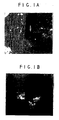
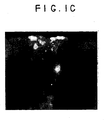
- FIG. 1A shows the low magnification image of a rat heart which was injected with dye labeled cells and later, a T-incision had been made at the site to reveal the injected cells in the ventrical wall.
- Figure 1A is a gross photo of the incised heart.
- Figures 1B and 1C reveal the labeled MSCs in the ventricle wall.
- Figure 1C shows that the cells were present in the outer 1-2 mm of the 3 mm thick rat cardiac muscle.
- the heart When sacrificed, the heart is removed, examined by light microscopy for the presence of vascular thrombi or emboli, paraffin-embedded, and sectioned. The histology of serial sections is examined to determine the fate of dye-stained cells. Sections are then tested for immunohistochemical markers of cardiac muscle in the areas of the introduced MSCs to ascertain whether donor MSCs have differentiated into cardiomyocytes in vivo . Implantation surgeries are carried out on animals to be sacrificed at 1, 2, 4, and 6 weeks (4 animals at each time point) and the hearts which received implants are analyzed histologically and immunologically.
- the hearts are removed and processed for histology by immunofluorescence microscopy. Differentiation of MSCs is determined by the immunofluorescence localization of sacomeric myosin heavy chain, SERCA1 and phospholamban.
- SERCA1 sacomeric myosin heavy chain
- phospholamban phospholamban
- MSCs are also implanted in biomatrix materials to determine if enhanced grafting would be observed, such as type I collagen.
- the MSCs are rapidly mixed with the matrix in a small volume and injected into the ventricle wall.
- the biomatrices are used at concentrations of 0.1 mg/ml or greater.
- the biomatrices may be used at concentrations of 1 to 3 mg/ml containing 10 to 100 million cells/ml.
- the tissue is analyzed at times of 1, 2, 4, and 6 weeks as described above.
- Xenograft or homograft valves are made acellular by freeze-drying, which leads to cellular death, or by enzymatic treatment followed by detergent extraction of cells and cell debris. This latter approach was taken by Vesely and coworkers with porcine valves to be repopulated with dermal or aortic fibroblasts. Curtil et al. 1997 used a freeze-dried porcine valve and attempted repopulation of the valve with human fibroblasts and endothelial cells. These studies were preliminary and limited to short term studies in vitro.
- the acellular valve to be populated by autologous hMSCs is incubated with culture expanded hMSCs in a tumbling vessel to ensure loading of cells to all valve surfaces.
- the valve is then cultured with the hMSCs for 1-2 weeks to allow the hMSCs to infiltrate and repopulate the valve.
- the valve is then attached to a pump to allow the actuation of the valve leaflets and simulate the pumping motion present in the body.
- the valve is maintained in the pumping mode for 1-2 weeks to allow cellular remodeling associated with the stresses of the pumping action. Once sufficient cellular remodeling has occurred, the valve is implanted into the body of the patient.
- Another embodiment of this aspect of the invention is to first repopulate the valve with hMSCs and to later incubate the valve tissue during the pumping stage with autologous smooth muscle cells isolated from a vascular graft which will line the lumen of the valve.
Abstract
Description
- This application claims priority of
U.S. provisional application serial no. 60/052,910, filed July 14, 1997 . This invention relates to the replacement and regeneration of cardiac tissue and muscle. - This year over 300,000 Americans will die from congestive heart failure. The ability to augment weakened cardiac muscle would be a major advance in the treatment of cardiomyopathy and heart failure. Despite advances in the medical therapy of heart failure, the mortality due to this disorder remains high, where most patients die within one to five years after diagnosis.
- A common heart ailment in the aging population is improper heart valve function, particularly the aortic valve. Mechanical replacement valves are widely used but require the patient to continually take blood thinners. Valves obtained from cadavers and xenographs (porcine) are also frequently used to replace a patient's own tissue. Valves are freeze-dried or chemically cross-linked using e.g., glutaraldehyde to stabilize the collagen fibrils and decrease antigenicity and proteolytic degradation. However, these valves remain acellular and often fail after several years due to mechanical strain or calcification. A replacement valve derived from biocompatible material that would allow ingrowth of the appropriate host cells and renewal of tissue over time would be preferred.
- Mesenchymal stem cells (MSCs) are cells which are capable of differentiating into more than one type of mesenchymal cell lineage. Mesenchymal stem cells (MSCs) have been identified and cultured from avian and mammalian species including mouse, rat, rabbit, dog and human (See Caplan, 1991, Caplan et al. 1993 and
U.S. Patent No. 5,486,359 ). Isolation, purification and culture expansion of hMSCs is described in detail therein. - In accordance with the present invention mesenchymal stem cells (MSCs) are used to regenerate or repair striated cardiac muscle that has been damaged through disease or degeneration. The MSCs differentiate into cardiac muscle cells and integrate with the healthy tissue of the recipient to replace the function of the dead or damaged cells, thereby regenerating the cardiac muscle as a whole. Cardiac muscle does not normally have reparative potential. The MSCs are used, for example, in cardiac muscle regeneration for a number of principal indications: (i) ischemic heart implantations, (ii) therapy for congestive heart failure patients, (iii) prevention of further disease for patients undergoing coronary artery bypass graft, (iv) conductive tissue regeneration, (v) vessel smooth muscle regeneration and (vi) valve regeneration. Thus the MSCs are also used to integrate with tissue of a replacement heart valve to be placed into a recipient. The MSCs, preferably autologous, repopulate the valve tissue, enabling proper valve function.
- MSC cardiac muscle therapy is based, for example, on the following sequence: harvest of MSC-containing tissue, isolation/expansion of MSCs, implantation into the damaged heart (with or without a stabilizing matrix and biochemical manipulation), and in situ formation of myocardium. This approach is different from traditional tissue engineering, in which the tissues are grown ex vivo and implanted in their final differentiated form. Biological, bioelectrical and/or biomechanical triggers from the host environment may be sufficient, or under certain circumstances, may be augmented as part of the therapeutic regimen to establish a fully integrated and functional tissue.
- Accordingly, one aspect of the present invention provides the use of mesenchymal stem cells for the manufacture of a pharmaceutical preparation for producing cardiomyocytes in an individual in need thereof. The mesenchymal stem cells that are employed may be a homogeneous composition or may be a mixed cell population enriched in MSCs. Homogeneous human mesenchymal stem cell compositions are obtained by culturing adherent marrow or periosteal cells; the mesenchymal stem cells may be identified by specific cell surface markers which are identified with unique monoclonal antibodies. A method for obtaining a cell population enriched in mesenchymal stem cells is described, for example, in
U.S. Patent No. 5,486,359 . - The administration of the cells can be directed to the heart, by a variety of procedures. Localized administration is preferred. The mesenchymal stem cells can be from a spectrum of sources including, in order of preference: autologous, allogeneic or xenogeneic. There are several embodiments to this aspect, including the following.
- In one embodiment of this aspect, the MSCs are to be administered as a cell suspension in a pharmaceutically acceptable liquid medium for injection. Injection, in this embodiment, can be local, i.e. directly into the damaged portion of the myocardium, or systemic. Here, again, localized administration is preferred.
- In another embodiment of this aspect, the MSCs are to be administered in a biocompatible medium which is, or becomes in situ at the site of myocardial damage, a semi-solid or solid matrix. For example, the matrix may be (i) an injectible liquid which "sets up" (or polymerizes) to a semi-solid gel at the site of the damaged myocardium, such as collagen and its derivatives, polylactic acid or polyglycolic acid, or (ii) one or more layers of a flexible, solid matrix that is implanted in its final form, such as impregnated fibrous matrices. The matrix can be, for example, Gelfoam (Upjohn, Kalamazoo, MI). The matrix holds the MSCs in place at the site of injury, i.e. serves the function of "scaffolding". This, in turn, enhances the opportunity for the administered MSCs to proliferate, differentiate and eventually become fully developed cardiomyocytes. As a result of their localization in the myocardial environment they then integrate with the recipient's surrounding myocardium. These events likewise occur in the above liquid injectible embodiment, but this embodiment may be preferred where more rigorous therapy is indicated.
- In another embodiment of this aspect, the MSCs are genetically modified or engineered to contain genes which express proteins of importance for the differentiation and/or maintenance of striated muscle cells. Examples include growth factors (TGF-β, IGF-1, FGF), myogenic factors (myoD, myogenin, Myf5, MRF), transcription factors (GATA-4), cytokines (cardiotrophin-1), members of the neuregulin family (neuregulin 1, 2 and 3) and homeobox genes (Csx, tinman, NKx family). Also contemplated are genes that code for factors that stimulate angiogenesis and revascularization (e.g. vascular endothelial growth factor (VEGF)). Any of the known methods for introducing DNA are suitable, however electroporation, retroviral vectors and adeno-associated virus (AAV) vectors are currently preferred.
- Thus, in association with the embodiment of the above aspect using genetically engineered MSCs, this invention also provides novel genetically engineered mesenchymal stem cells and tissue compositions to treat the above indications. The compositions can include genetically modified MSCs and unmodified MSCs in various proportions to regulate the amount of expressed exogenous material in relationship to the total number of MSCs to be affected.
- The invention also relates to the potential of MSCs to partially differentiate to the cardiomyocyte phenotype using in vitro methods. This technique can under certain circumstances optimize conversion of MSCs to the cardiac lineage by predisposing them thereto. This also has the potential to shorten the time required for complete differentiation once the cells have been administered.
-
-
Figures 1A - 1C show cardiac muscle injected, using a fine needle, with in vitro dye-labeled MSCs. The lipophilic dyes PKH26 (Sigma Chemical) or CM-Di I (Molecular Probes) were utilized to label MSCs prior to being introduced into animals. These dyes remain visible when the tissue site is harvested 1-2 months later. We have also shown that such dyes do not interfere with the differentiation of MSCs in in vitro assays.Figure 1A shows the low magnification image of a rat heart which has been injected with dye labeled cells and later, a T-incision has been made at the site.Figures 1A and 1B reveal the labeled MSCs in the ventricle wall viewed from the outer surface.Figure 1C shows a cross-section of the ventricle wall and that the cells are present in the outer 1-2 mm of the 3mm thick cardiac muscle. - The proper environmental stimuli convert MSCs into cardiac myocytes. Differentiation of mesenchymal stem cells to the cardiac lineage is controlled by factors present in the cardiac environment. Exposure of MSCs to a simulated cardiac environment directs these cells to cardiac differentiation as detected by expression of specific cardiac muscle lineage markers. Local chemical, electrical and mechanical environmental influences alter pluripotent MSCs and convert the cells grafted into the heart into the cardiac lineage.
- Early in embryonic development following the epithelia-mesenchyme transition, the presumptive heart mesenchyme from the left and right sides of the body migrate to the ventral midline. Here, interaction with other cell types induces continued cardiogenesis. In vitro conversion of MSCs to cardiomyocytes is tested by co-culture or fusion with murine embryonic stem cells or cardiomyocytes, treatment of MSCs with cardiac cell lysates, incubation with specific soluble growth factors, or exposure of MSCs to mechanical stimuli and electrical stimulation.
- A series of specific treatments applicable to MSCs to induce expression of cardiac specific genes are disclosed herein. The conditions are effective on rat, canine and human MSCs. Treatments of MSCs include (1) co-culturing MSCs with fetal, neonatal and adult rat cardiac cells, (2) use of chemical fusigens (e.g., polyethylene glycol or sendai virus) to create heterokaryons of MSCs with fetal, neonatal and adult cardiomyocytes, (3) incubating MSCs with extracts of mammalian hearts, including the extracellular matrix and related molecules found in heart tissue, (4) treatment of MSCs with growth factors and differentiating agents, (5) mechanical and/or electrical stimulation of MSCs, and (6) mechanically and/or electrically coupling MSCs with cardiomyocytes. MSCs that progress towards cardiomyocytes first express proteins found in fetal cardiac tissue and then proceed to adult forms. Detection of expression of cardiomyocyte specific proteins is achieved using antibodies to, for example, myosin heavy chain monoclonal antibody MF 20 (MF20), sarcoplasmic reticulum calcium ATPase (SERCA1) (mAb 10D1) or gap junctions using antibodies to connexin 43.
- Cardiac injury promotes tissue responses which enhance myogenesis using implanted MSCs. Thus, MSCs are introduced to the infarct zone to reduce the degree of scar formation and to augment ventricular function. New muscle is thereby created within an infarcted myocardial segment. MSCs are directly infiltrated into the zone of infarcted tissue. The integration and subsequent differentiation of these cells is characterized, as described above. Timing of intervention is designed to mimic the clinical setting where patients with acute myocardial infarction would first come to medical attention, receive first-line therapy, followed by stabilization, and then intervention with myocardial replacement therapy if necessary.
- Of the four chambers of the heart, the left ventricle is primarily responsible for pumping blood under pressure through the body's circulatory system. It has the thickest myocardial walls and is the most frequent site of myocardial injury resulting from congestive heart failure. The degree of advance or severity of the congestive heart failure ranges from those cases where heart transplantation is indicated as soon as a suitable donor organ becomes available to those where little or no permanent injury is observed and treatment is primarily prophylactic.
- The severity of resulting myocardial infarction, i.e. the percentage of muscle mass of the left ventricle that is involved can range from about 5 to about 40 percent. This represents affected tissue areas, whether as one contiguous ischemia or the sum of smaller ischemic lesions, having horizontal affected areas from about 2 cm2 to about 6 cm2 and a thickness of from 1-2 mm to 1-1.5 cm. The severity of the infarction is significantly affected by which vessel(s) is involved and how much time has passed before treatment intervention is begun.
- The mesenchymal stem cells used in accordance with the invention are, in order of preference, autologous, allogeneic or xenogeneic, and the choice can largely depend on the urgency of the need for treatment. A patient presenting an imminently life threatening condition may be maintained on a heart/lung machine while sufficient numbers of autologous MSCs are cultured or initial treatment can be provided using other than autologous MSCs.
- The MSC therapy of the invention can be provided by several routes of administration, including the following. First, intracardiac muscle injection, which avoids the need for an open surgical procedure, can be used where the MSCs are in an injectible liquid suspension preparation or where they are in a biocompatible medium which is injectible in liquid form and becomes semi-solid at the site of damaged myocardium. A conventional intracardiac syringe or a controllable arthroscopic delivery device can be used so long as the needle lumen or bore is of sufficient diameter (e.g. 30 gauge or larger) that shear forces will not damage the MSCs. The injectible liquid suspension MSC preparations can also be administered intravenously, either by continuous drip or as a bolus. During open surgical procedures, involving direct physical access to the heart, all of the described forms of MSC delivery preparations are available options.
- As a representative example of a dose range is a volume of about 20 to about 50 µl of injectible suspension containing 10-40 x 106 MSCs/ml. The concentration of cells per unit volume, whether the carrier medium is liquid or solid remains within substantially the same range. The amount of MSCs delivered will usually be greater when a solid, "patch" type application is made during an open procedure, but follow-up therapy by injection will be as described above. The frequency and duration of therapy will, however, vary depending on the degree (percentage) of tissue involvement, as already described (e.g. 5-40% left ventricular mass).
- In cases having in the 5-10% range of tissue involvement, it is possible to treat with as little as a single administration of one million MSCs in 20-50 µl of injection preparation. The injection medium can be any pharmaceutically acceptable isotonic liquid. Examples include phosphate buffered saline (PBS), culture media such as DMEM (preferably serum-free), physiological saline or 5 % dextrose in water (D5W).
- In cases having more in a range around the 20% tissue involvement severity level, multiple injections of 20-50 µl (10-40 x 106 MSCs/ml) are envisioned. Follow-up therapy may involve additional dosings.
- In very severe cases, e.g. in a range around the 40% tissue involvement severity level, multiple equivalent doses for a more extended duration with long term (up to several months) maintenance dose aftercare may well be indicated.
- The present invention is further illustrated, but not limited, by the following example.
- In using MSCs, it is desirable to maintain cell-cell contact in vivo for the conversion of MSCs to the muscle lineage. Environmental signals identified above act in concert with mechanical and electrical signaling in vivo to lead to cardiac differentiation.
- Primary human MSCs (hMSCs) are introduced into athymic rat myocardial tissue by direct injection. The integration of implanted cells, their subsequent differentiation, formation of junctions with cardiac cells, and their long-term survival are characterized with light microscopy, histology, confocal immunofluorescence microscopy, electron microscopy and in situ hybridization.
- Whether human MSCs are appropriately grafted into cardiac muscle of athymic rats (strain HSD:RH-RNU/RNU), which lack the immune responses necessary to destroy many foreign cells, is also examined.
- Rat MSCs are grafted into the heart muscles of rats. To analyze the injected cells over several weeks and to minimize the possibility of immune system rejection, MSCs are harvested from Fisher 344 rats, the same inbred strain (identical genotype) as the intended MSC recipients.
- The MSCs can be marked in a variety of ways prior to their introduction into the recipient. This makes it possible to trace the fate of the MSCs as they proliferate and differentiate in the weeks following the MSC implant. Several methods are utilized to positively identify the injected cells: membrane lipid dyes PKH26 or CM-DI I and genetic marking with adeno-associated virus (AAV) or retroviruses, such as Maloney murine leukemia virus expressing green fluorescent protein (GFP) or galactosidase. PCR is also used to detect the Y chromosome marker of male cells implanted into female animals. The dye-labeled cells are readily detected and offer the simplest method to directly follow the injected cells. This method is reliable for times out to at least 4 weeks. On the day of introduction to recipient animals, MSCs are trypsinized and labeled with CM-DI I according to the recommendations of the manufacturer (Molecular Probes). Subconfluent monolayer cultures of MSCs are incubated with 5mM CM-DI I in serum-free medium for 20 minutes, trypsinized, washed twice in excess dye-free medium, and utilized for injection.
- Alternatively, MSCs are genetically marked prior to injections, such as by using AAV-GFP vector. This vector lacks a selectable marker but mediates high-level expression of the transduced genes in a variety of post-mitotic and stem cell types. Recombinant AAV-GFP is added to low density monolayers of MSCs in low serum. Following a four hour incubation at 37°C, the supernatant is removed and replaced with fresh media. At 96 hours after transduction, cells are assayed for green fluorescent protein (GFP) activity. Typically 50% of the cells express the transduced gene. Unselected MSCs on a clonal line, isolated by limiting dilution, are utilized for injection. Cells are collected following trypsin treatment, washed and used at high concentrations for injection (10 to 100 million cells per ml).
- To test whether the hMSCs became cardiomyocytes in the heart environment, the hearts of ten week old athymic rats were injected with dye labeled or GFP-labeled human MSCs. All procedures were performed under strict sterile conditions. The animals were placed in a glass jar containing a methoxyflurane anesthesia soaked sponge. Under sterile conditions, a 20 mm anterior thoracotomy was performed, and following visualization of the left ventricle, 10 µl of the cell suspension, containing 10,000 to 100,000 MSCs in serum-free medium were injected into the left ventricular apex using a 30 gauge needle. The procedure was performed rapidly with endotracheal intubation and mechanical ventilation assist. The incision was closed with sutures. Ventilation assist was normally unnecessary after a short period following chest closure.
Figure 1A shows the low magnification image of a rat heart which was injected with dye labeled cells and later, a T-incision had been made at the site to reveal the injected cells in the ventrical wall.Figure 1A is a gross photo of the incised heart.Figures 1B and1C reveal the labeled MSCs in the ventricle wall.Figure 1C shows that the cells were present in the outer 1-2 mm of the 3 mm thick rat cardiac muscle. - When sacrificed, the heart is removed, examined by light microscopy for the presence of vascular thrombi or emboli, paraffin-embedded, and sectioned. The histology of serial sections is examined to determine the fate of dye-stained cells. Sections are then tested for immunohistochemical markers of cardiac muscle in the areas of the introduced MSCs to ascertain whether donor MSCs have differentiated into cardiomyocytes in vivo. Implantation surgeries are carried out on animals to be sacrificed at 1, 2, 4, and 6 weeks (4 animals at each time point) and the hearts which received implants are analyzed histologically and immunologically.
- For phenotypic characterization, the hearts are removed and processed for histology by immunofluorescence microscopy. Differentiation of MSCs is determined by the immunofluorescence localization of sacomeric myosin heavy chain, SERCA1 and phospholamban. The sequence-specific antibody to gap junction protein connexin 43, which is commercially available (Zymed) and detects gap junctions in cardiac tissue is used.
- MSCs are also implanted in biomatrix materials to determine if enhanced grafting would be observed, such as type I collagen. The MSCs are rapidly mixed with the matrix in a small volume and injected into the ventricle wall. The biomatrices are used at concentrations of 0.1 mg/ml or greater. For example, the biomatrices may be used at concentrations of 1 to 3 mg/ml containing 10 to 100 million cells/ml. The tissue is analyzed at times of 1, 2, 4, and 6 weeks as described above.
- Xenograft or homograft valves are made acellular by freeze-drying, which leads to cellular death, or by enzymatic treatment followed by detergent extraction of cells and cell debris. This latter approach was taken by Vesely and coworkers with porcine valves to be repopulated with dermal or aortic fibroblasts. Curtil et al. 1997 used a freeze-dried porcine valve and attempted repopulation of the valve with human fibroblasts and endothelial cells. These studies were preliminary and limited to short term studies in vitro.
- The acellular valve to be populated by autologous hMSCs is incubated with culture expanded hMSCs in a tumbling vessel to ensure loading of cells to all valve surfaces. The valve is then cultured with the hMSCs for 1-2 weeks to allow the hMSCs to infiltrate and repopulate the valve. Within the culture vessel, the valve is then attached to a pump to allow the actuation of the valve leaflets and simulate the pumping motion present in the body. The valve is maintained in the pumping mode for 1-2 weeks to allow cellular remodeling associated with the stresses of the pumping action. Once sufficient cellular remodeling has occurred, the valve is implanted into the body of the patient.
- Another embodiment of this aspect of the invention is to first repopulate the valve with hMSCs and to later incubate the valve tissue during the pumping stage with autologous smooth muscle cells isolated from a vascular graft which will line the lumen of the valve.
Claims (43)
- Use of mesenchymal stem cells for the manufacture of a pharmaceutical preparation for treating a patient with damaged heart muscle to improve heart function.
- The use of claim 1 wherein the pharmaceutical preparation is to be administered directly to the heart.
- The use of claim 2 wherein the pharmaceutical preparation is to be administered by injection.
- The use of claim 3 wherein the pharmaceutical preparation is to be administered in a pharmaceutically acceptable liquid injectable carrier.
- The use of claim 2 wherein the pharmaceutical preparation is to be administered during an open surgical procedure.
- The use of claim 1 wherein the pharmaceutical preparation is to be administered systemically.
- The use of claim 6 wherein the pharmaceutical preparation is to be administered intravenously.
- The use of claim 1 wherein the mesenchymal stem cells are autologous to the patient being treated.
- The use of claim 1 wherein the mesenchymal stem cells are allogeneic to the patient being treated.
- The use of claim 1 wherein said mesenchymal stem cells are human mesenchymal stem cells.
- The use of claim 10 wherein said mesenchymal stem cells are human allogeneic mesenchymal stem cells.
- The use of claim 1 wherein at least a portion of the mesenchymal stem cells have been modified to contain exogenous genetic material.
- The use of claim 12 wherein the exogenous genetic material codes for an expression product selected from the group consisting of growth factors, myogenic factors, transcription factors, cytokines, homeobox genes, angiogenesis stimulating factors, and revascularization enhancing factors.
- Use of mesenchymal stem cells for the manufacture of a pharmaceutical preparation for treating a patient with congestive heart failure.
- The use of claim 14 wherein the pharmaceutical preparation is to be administered directly to the heart.
- The use of claim 15 wherein the pharmaceutical preparation is to be administered by injection.
- The use of claim 16 wherein the pharmaceutical preparation is to be administered in a pharmaceutically acceptable liquid injectable carrier.
- The use of claim 15 wherein the pharmaceutical preparation is to be administered during an open surgical procedure.
- The use of claim 14 wherein the pharmaceutical preparation is to be administered systemically.
- The use of claim 19 wherein the pharmaceutical preparation is to be administered intravenously.
- The use of claim 14 wherein the mesenchymal stem cells are autologous to the patient being treated.
- The use of claim 14 wherein the mesenchymal stem cells are allogeneic to the patient being treated.
- The use of claim 14 wherein the mesenchymal stem cells are human mesenchymal stem cells.
- The use of claim 23 wherein the mesenchymal stem cells are human allogeneic mesenchymal stem cells.
- The use of claim 14 wherein at least a portion of the mesenchymal stem cells have been modified to contain exogenous genetic material.
- The use of claim 25 wherein the exogenous genetic material codes for an expression product selected from the group consisting of growth factors, myogenic factors, transcription factors, cytokines, homeobox genes, angiogenesis stimulating factors, and revascularization enhancing factors.
- Use of autologous or allogeneic mesenchymal stem cells for the manufacture of a pharmaceutical preparation for producing cardiac muscle cells in the heart of an individual in need thereof.
- The use of claim 27 wherein said pharmaceutical preparation is to be administered to an individual to regenerate or repair cardiac muscle that has been damaged through disease.
- The use of claim 28 wherein said pharmaceutical preparation is to be administered to an individual who has suffered myocardial infarction.
- The use of claim 28 wherein said pharmaceutical preparation is to be administered directly to the heart.
- The use of claim 28 wherein said pharmaceutical preparation is to be administered systemically.
- The use of claim 31 wherein said pharmaceutical preparation is to be administered by injection.
- The use of claim 28 wherein said mesenchymal stem cells are human.
- The use of claim 33 wherein said pharmaceutical preparation is to be administered to an individual who has suffered myocardial infarction.
- The use of claim 34 wherein said pharmaceutical preparation is to be administered directly to the heart.
- The use of claim 34 wherein said the pharmaceutical preparation is to be administered systemically.
- Use of autologous or allogeneic mesenchymal stem cells for the manufacture of a pharmaceutical preparation for reducing scar formation in infarcted heart tissue.
- The use of claim 37 wherein said pharmaceutical preparation is to be administered systemically.
- The use of claim 38 wherein said pharmaceutical preparation is to be administered by injection.
- The use of claim 37 wherein said mesenchymal stem cells are human.
- The use of claim 40 wherein said pharmaceutical preparation is to be administered directly to the heart.
- The use of claim 40 wherein said pharmaceutical preparation is to be administered systemically.
- The use of claim 42 wherein said pharmaceutical preparation is to be administered by injection.
Applications Claiming Priority (3)
| Application Number | Priority Date | Filing Date | Title |
|---|---|---|---|
| US5291097P | 1997-07-14 | 1997-07-14 | |
| US52910P | 1997-07-14 | ||
| PCT/US1998/014520 WO1999003973A1 (en) | 1997-07-14 | 1998-07-14 | Cardiac muscle regeneration using mesenchymal stem cells |
Publications (4)
| Publication Number | Publication Date |
|---|---|
| EP1007631A1 EP1007631A1 (en) | 2000-06-14 |
| EP1007631A4 EP1007631A4 (en) | 2003-05-21 |
| EP1007631B1 EP1007631B1 (en) | 2005-10-19 |
| EP1007631B2 true EP1007631B2 (en) | 2009-02-18 |
Family
ID=21980718
Family Applications (1)
| Application Number | Title | Priority Date | Filing Date |
|---|---|---|---|
| EP98934507A Expired - Lifetime EP1007631B2 (en) | 1997-07-14 | 1998-07-14 | Cardiac muscle regeneration using mesenchymal stem cells |
Country Status (10)
| Country | Link |
|---|---|
| US (1) | US6387369B1 (en) |
| EP (1) | EP1007631B2 (en) |
| JP (1) | JP4562816B2 (en) |
| AT (1) | ATE307195T1 (en) |
| AU (1) | AU8401498A (en) |
| CA (1) | CA2296704C (en) |
| DE (1) | DE69831957T3 (en) |
| DK (1) | DK1007631T4 (en) |
| ES (1) | ES2251773T5 (en) |
| WO (1) | WO1999003973A1 (en) |
Cited By (1)
| Publication number | Priority date | Publication date | Assignee | Title |
|---|---|---|---|---|
| US8309343B2 (en) | 2008-12-01 | 2012-11-13 | Baxter International Inc. | Apparatus and method for processing biological material |
Families Citing this family (264)
| Publication number | Priority date | Publication date | Assignee | Title |
|---|---|---|---|---|
| US6974571B2 (en) * | 1995-03-28 | 2005-12-13 | Thomas Jefferson University | Isolated stromal cells and methods of using the same |
| US7514074B2 (en) | 1997-07-14 | 2009-04-07 | Osiris Therapeutics, Inc. | Cardiac muscle regeneration using mesenchymal stem cells |
| US20030103951A1 (en) * | 1997-07-14 | 2003-06-05 | Osiris Therapeutics, Inc. | Cardiac muscle regeneration using mesenchymal stem cells |
| CA2323073C (en) * | 1998-03-13 | 2010-06-22 | Osiris Therapeutics, Inc. | Uses for human non-autologous mesenchymal stem cells |
| DE69937888T2 (en) * | 1998-07-31 | 2009-01-02 | Genzyme Corp., Cambridge | Process for the preparation of mesenchymal stem cells |
| EP1894997A1 (en) * | 1998-07-31 | 2008-03-05 | Genzyme Corporation | Improvement of cardiac function by mesenchymal stem cell transplantation |
| AU6056299A (en) * | 1998-09-21 | 2000-04-10 | Musc Foundation For Research Development | Non-hematopoietic cells, including cardiomyocytes and skeletal muscle cells, derived from hematopoietic stem cells and methods of making and using them |
| US7410798B2 (en) | 2001-01-10 | 2008-08-12 | Geron Corporation | Culture system for rapid expansion of human embryonic stem cells |
| US6667176B1 (en) | 2000-01-11 | 2003-12-23 | Geron Corporation | cDNA libraries reflecting gene expression during growth and differentiation of human pluripotent stem cells |
| US6777231B1 (en) | 1999-03-10 | 2004-08-17 | The Regents Of The University Of California | Adipose-derived stem cells and lattices |
| US20030082152A1 (en) | 1999-03-10 | 2003-05-01 | Hedrick Marc H. | Adipose-derived stem cells and lattices |
| US6635249B1 (en) * | 1999-04-23 | 2003-10-21 | Cenes Pharmaceuticals, Inc. | Methods for treating congestive heart failure |
| US7670628B2 (en) * | 1999-07-07 | 2010-03-02 | Angioblast Systems, Inc. | Mesenchymal precursor cell |
| AU2003901668A0 (en) * | 2003-03-28 | 2003-05-01 | Medvet Science Pty. Ltd. | Non-haemopoietic precursor cells |
| AUPQ147799A0 (en) | 1999-07-07 | 1999-07-29 | Medvet Science Pty. Ltd. | Mesenchymal precursor cell |
| US20050158289A1 (en) * | 1999-07-07 | 2005-07-21 | Simmons Paul J. | Mesenchymal precursor cell and use thereof in the repair of bone defects and fractures in mammals |
| US8062675B2 (en) * | 1999-07-07 | 2011-11-22 | Angioblast Systems, Inc. | Mesenchymal precursor cell |
| US8147824B2 (en) | 1999-08-05 | 2012-04-03 | Athersys, Inc. | Immunomodulatory properties of multipotent adult progenitor cells and uses thereof |
| US8075881B2 (en) * | 1999-08-05 | 2011-12-13 | Regents Of The University Of Minnesota | Use of multipotent adult stem cells in treatment of myocardial infarction and congestive heart failure |
| US8252280B1 (en) | 1999-08-05 | 2012-08-28 | Regents Of The University Of Minnesota | MAPC generation of muscle |
| US7015037B1 (en) * | 1999-08-05 | 2006-03-21 | Regents Of The University Of Minnesota | Multiponent adult stem cells and methods for isolation |
| DE60041821D1 (en) * | 1999-09-24 | 2009-04-30 | Cybios Llc | PLURIPOTENT EMBRYONAL STEM CELL-SIMILAR CELLS, COMPOSITIONS, AND ITS CONDITIONS |
| AU7500400A (en) * | 1999-09-30 | 2001-04-30 | Mcgill University | Autologous marrow stem cell (msc) transplantation for myocardial regeneration |
| AU784618B2 (en) * | 1999-12-28 | 2006-05-18 | Kyowa Hakko Kogyo Co. Ltd. | Cells capable of differentiating into heart muscle cells |
| US7166280B2 (en) * | 2000-04-06 | 2007-01-23 | Franco Wayne P | Combination growth factor therapy and cell therapy for treatment of acute and chronic heart disease |
| US20100303769A1 (en) * | 2000-04-06 | 2010-12-02 | Franco Wayne P | Combination growth factor therapy and cell therapy for treatment of acute and chronic heart disease |
| US7862810B2 (en) * | 2000-07-31 | 2011-01-04 | New York Medical College | Methods and compositions for the repair and/or regeneration of damaged myocardium |
| US20110091428A1 (en) * | 2000-07-31 | 2011-04-21 | New York Medical College | Compositions of adult organ stem cells and uses thereof |
| AU2001284695A1 (en) * | 2000-07-31 | 2002-02-13 | New York Medical College | Methods and compositions for the repair and/or regeneration of damaged myocardium |
| US7214371B1 (en) | 2000-09-01 | 2007-05-08 | Ben-Gurion University Of The Negev Research & Development Authority | Tissue engineered biografts for repair of damaged myocardium |
| US7560280B2 (en) * | 2000-11-03 | 2009-07-14 | Kourion Therapeutics Gmbh | Human cord blood derived unrestricted somatic stem cells (USSC) |
| DE10056465A1 (en) * | 2000-11-14 | 2002-07-18 | Rosemarie Daig | Cell constructs obtainable from mesenchymal stem cells and cells derived therefrom and their use |
| US7311905B2 (en) * | 2002-02-13 | 2007-12-25 | Anthrogenesis Corporation | Embryonic-like stem cells derived from post-partum mammalian placenta, and uses and methods of treatment using said cells |
| KR100915482B1 (en) | 2000-12-06 | 2009-09-03 | 하리리 로버트 제이 | Method of collecting placental stem cells |
| JP2005501802A (en) * | 2001-01-23 | 2005-01-20 | ボストン サイエンティフィック コーポレイション | Localized myocardial injection method for treating ischemic myocardium |
| NZ528035A (en) * | 2001-02-14 | 2005-07-29 | Robert J Hariri | Renovation and repopulation of decellularized tissues and cadaveric organs by stem cells |
| EP1362095B1 (en) * | 2001-02-14 | 2015-05-27 | Anthrogenesis Corporation | Post-partum mammalian placenta, its use and placental stem cells therefrom |
| EP1393067A4 (en) | 2001-04-13 | 2005-01-05 | Anterogen Co Ltd | Encapsulated cell indicator system |
| US7732199B2 (en) | 2001-07-12 | 2010-06-08 | Geron Corporation | Process for making transplantable cardiomyocytes from human embryonic stem cells |
| JP2004535199A (en) * | 2001-07-12 | 2004-11-25 | ジェロン コーポレイション | Cardiomyocyte lineage cells produced from human pluripotent stem cells |
| ITRM20010550A1 (en) * | 2001-09-11 | 2003-03-11 | Giulio Cossu | METHOD FOR INDUCING THE DIFFERENTIATION OF ENDOTHELIAL CELLS IN CARDIOMYOCYTES. |
| EP1435977A4 (en) * | 2001-09-19 | 2005-06-08 | Ford Henry Health System | Cardiac transplantation of stem cells for the treatment of heart failure |
| US20030082153A1 (en) | 2001-10-22 | 2003-05-01 | The Government Of The United States Of America | Stem cells that transform to beating cardiomyocytes |
| AU2002359371A1 (en) * | 2001-11-08 | 2003-05-19 | The Regents Of The University Of California | Methods and compositions for correction of cardiac conduction disturbances |
| US8404229B2 (en) | 2001-12-07 | 2013-03-26 | Cytori Therapeutics, Inc. | Methods of using adipose derived stem cells to treat acute tubular necrosis |
| US7585670B2 (en) | 2001-12-07 | 2009-09-08 | Cytori Therapeutics, Inc. | Automated methods for isolating and using clinically safe adipose derived regenerative cells |
| US8105580B2 (en) | 2001-12-07 | 2012-01-31 | Cytori Therapeutics, Inc. | Methods of using adipose derived stem cells to promote wound healing |
| US20050095228A1 (en) | 2001-12-07 | 2005-05-05 | Fraser John K. | Methods of using regenerative cells in the treatment of peripheral vascular disease and related disorders |
| US7595043B2 (en) * | 2001-12-07 | 2009-09-29 | Cytori Therapeutics, Inc. | Method for processing and using adipose-derived stem cells |
| US9597395B2 (en) | 2001-12-07 | 2017-03-21 | Cytori Therapeutics, Inc. | Methods of using adipose tissue-derived cells in the treatment of cardiovascular conditions |
| US7651684B2 (en) | 2001-12-07 | 2010-01-26 | Cytori Therapeutics, Inc. | Methods of using adipose tissue-derived cells in augmenting autologous fat transfer |
| US7771716B2 (en) | 2001-12-07 | 2010-08-10 | Cytori Therapeutics, Inc. | Methods of using regenerative cells in the treatment of musculoskeletal disorders |
| US7514075B2 (en) | 2001-12-07 | 2009-04-07 | Cytori Therapeutics, Inc. | Systems and methods for separating and concentrating adipose derived stem cells from tissue |
| CN1630526B (en) | 2001-12-07 | 2010-05-05 | 马克罗珀尔生物外科公司 | Systems and methods for treating patients with processed lipoaspirate cells |
| AU2002226683A1 (en) * | 2002-01-17 | 2003-07-30 | Cardio Incorporated | Complex therapy for tissue regeneration |
| CA2477411A1 (en) * | 2002-03-02 | 2003-09-12 | Board Of Regents, The University Of Texas | Local production and/or delivery of anti-cancer agents by stromal cell precursors |
| WO2003093433A2 (en) | 2002-05-02 | 2003-11-13 | Regents Of The University Of Minnesota | Fibrin-based biomatrix |
| WO2003094697A2 (en) * | 2002-05-08 | 2003-11-20 | The Regents Of The University Of California | Methods and compositions for correction of cardiac conduction disturbances |
| US20040067221A1 (en) * | 2002-05-22 | 2004-04-08 | Medtronic, Inc. | Cell delivery fluid for prevention of cell settling in delivery system |
| WO2004009767A2 (en) * | 2002-07-23 | 2004-01-29 | Boston Scientific Limited | Cell therapy for regeneration |
| US20050271639A1 (en) * | 2002-08-22 | 2005-12-08 | Penn Marc S | Genetically engineered cells for therapeutic applications |
| AR047712A1 (en) | 2002-09-07 | 2006-02-15 | Royal Veterinary College | TREATMENT METHOD OF A NATURAL SOFT SKELETTIC TISSUE INJURY MANAGING A COMPOSITION OF MESENQUIMATOSE MOTHER CELLS |
| WO2004044142A2 (en) * | 2002-11-05 | 2004-05-27 | The Brigham And Women's Hospital, Inc. | Mesenchymal stem cells and methods of use thereof |
| EP1583422B1 (en) * | 2002-12-05 | 2016-03-30 | Case Western Reserve University | Cell-based therapies for ischemia |
| US7470538B2 (en) * | 2002-12-05 | 2008-12-30 | Case Western Reserve University | Cell-based therapies for ischemia |
| US20050002914A1 (en) * | 2003-01-15 | 2005-01-06 | Rosen Michael R. | Mesenchymal stem cells as a vehicle for ion channel transfer in syncytial structures |
| US7794702B2 (en) * | 2003-01-15 | 2010-09-14 | The Trustees Of Columbia University In The City Of New York | Mesenchymal stem cells as a vehicle for ion channel transfer in syncytial structures |
| US20040197310A1 (en) * | 2003-02-12 | 2004-10-07 | Sanberg Paul R. | Compositions and methods for using umbilical cord progenitor cells in the treatment of myocardial infarction |
| AU2011253681B2 (en) * | 2003-03-28 | 2014-04-10 | Mesoblast, Inc. | Perivascular mesenchymal precursor cell induced blood vessel formation |
| ATE482725T1 (en) * | 2003-04-01 | 2010-10-15 | Us Dept Of Veteran S Affaires | STEM CELL, PROGRESSOR CELL OR TARGET CELL BASED TREATMENT OF MULTI-ORGAN FAILURE. |
| US20040213770A1 (en) * | 2003-04-22 | 2004-10-28 | Endobionics, Inc. | Methods and systems for treating ischemic cardiac and other tissues |
| WO2004110270A1 (en) * | 2003-06-12 | 2004-12-23 | Regents Of The University Of Minnesota | Directing cells to target tissues or organs |
| US8367410B2 (en) * | 2003-06-20 | 2013-02-05 | Massachusetts Institute Of Technology | Application of electrical stimulation for functional tissue engineering in vitro and in vivo |
| NZ544828A (en) * | 2003-06-25 | 2008-10-31 | Ottawa Health Research Inst | Use of cardiotrophin to modulate stem cell proliferation |
| WO2005038012A2 (en) | 2003-06-27 | 2005-04-28 | Ethicon Incorporated | Cartilage and bone repair and regeneration using postpartum-derived cells |
| US9592258B2 (en) | 2003-06-27 | 2017-03-14 | DePuy Synthes Products, Inc. | Treatment of neurological injury by administration of human umbilical cord tissue-derived cells |
| US8790637B2 (en) | 2003-06-27 | 2014-07-29 | DePuy Synthes Products, LLC | Repair and regeneration of ocular tissue using postpartum-derived cells |
| US9572840B2 (en) | 2003-06-27 | 2017-02-21 | DePuy Synthes Products, Inc. | Regeneration and repair of neural tissue using postpartum-derived cells |
| US20050013870A1 (en) * | 2003-07-17 | 2005-01-20 | Toby Freyman | Decellularized extracellular matrix of conditioned body tissues and uses thereof |
| ITRM20030376A1 (en) | 2003-07-31 | 2005-02-01 | Univ Roma | PROCEDURE FOR THE ISOLATION AND EXPANSION OF CARDIOC STAMIN CELLS FROM BIOPSIA. |
| CA2443423A1 (en) * | 2003-09-30 | 2005-03-30 | Darren H. Freed | Use of cardiotrophon-1 to promote wound healing and counteract overt fibrosis |
| EP1685237A4 (en) * | 2003-10-28 | 2007-12-26 | Caritas St Elizabeths Boston | Novel multipotent stem cells and use thereof |
| WO2005062857A2 (en) * | 2003-12-24 | 2005-07-14 | The Trustees Of Columbia University In The City Of New York | Creation of a biological atrioventricular bypass to compensate for atrioventricular block |
| US7840263B2 (en) * | 2004-02-27 | 2010-11-23 | Cardiac Pacemakers, Inc. | Method and apparatus for device controlled gene expression |
| US7452718B2 (en) * | 2004-03-26 | 2008-11-18 | Geron Corporation | Direct differentiation method for making cardiomyocytes from human embryonic stem cells |
| US20050214938A1 (en) * | 2004-03-26 | 2005-09-29 | Gold Joseph D | Cardiac bodies: clusters of spontaneously contracting cells for regenerating cardiac function |
| WO2005097147A1 (en) * | 2004-03-30 | 2005-10-20 | Boston Scientific Limited (Incorporated In Ireland) | Restenosis therapy using mesenchymal stem cells |
| WO2005099758A2 (en) * | 2004-04-17 | 2005-10-27 | The Board Of Trustees The Leland Standford Junior University | Injectable bioartificial tissue matrix |
| WO2005113751A1 (en) * | 2004-05-14 | 2005-12-01 | Becton, Dickinson And Company | Cell culture environments for the serum-free expansion of mesenchymal stem cells |
| US20050277124A1 (en) * | 2004-06-10 | 2005-12-15 | White Steven M | Cardiac conduction system cells and uses thereof |
| ATE551066T1 (en) * | 2004-06-21 | 2012-04-15 | Cleveland Clinic Foundation | CCR LIGANDS FOR STEM CELL HOMING |
| WO2006020322A2 (en) * | 2004-07-19 | 2006-02-23 | The Trustees Of Columbia University In The City Of New York | Assay system for monitoring the effects of genetically engineered cells to alter function of a syncytium |
| US8173118B2 (en) | 2004-07-30 | 2012-05-08 | Mayo Foundation For Medical Education And Research | Compositions consisting essentially of TGF-β, BMP-2 FGF-4, leukemia inhibitory factor, IGF-1, IL-6 and H-α-thrombin |
| US7828711B2 (en) * | 2004-08-16 | 2010-11-09 | Cardiac Pacemakers, Inc. | Method and apparatus for modulating cellular growth and regeneration using ventricular assist device |
| US8513011B2 (en) * | 2004-08-26 | 2013-08-20 | Biotech Research Ventures Pte Limited | Methods and compositions for culturing cardiomyocyte-like cells |
| US8182806B2 (en) * | 2004-09-07 | 2012-05-22 | Johnson Lanny L | Synovial villi for use with tissue engineering |
| US20060051328A1 (en) * | 2004-09-07 | 2006-03-09 | Johnson Lanny L | Mobilization of cells via physical means |
| US7785582B2 (en) * | 2004-09-07 | 2010-08-31 | Johnson Lanny L | Use of synovium and omentum for tissue engineering |
| US20090029912A1 (en) * | 2004-09-24 | 2009-01-29 | Stan Gronthos | Method of enhancing proliferation and/or survival of mesenchymal precursor cells (mpc) |
| US20060234375A1 (en) * | 2004-09-30 | 2006-10-19 | Doronin Sergey V | Use of human stem cells and/or factors they produce to promote adult mammalian cardiac repair through cardiomyocyte cell division |
| US20070072294A1 (en) * | 2004-09-30 | 2007-03-29 | Doronin Sergey V | Use of human stem cells and/or factors they produce to promote adult mammalian cardiac repair through cardiomyocyte cell division |
| WO2006037103A2 (en) * | 2004-09-30 | 2006-04-06 | The Trustees Of Columbia University In The City Of New York | The use of human stem cells and/or factors they produce to promote adult mammalian cardiac repair through cardiomyocyte cell division |
| US11660317B2 (en) | 2004-11-08 | 2023-05-30 | The Johns Hopkins University | Compositions comprising cardiosphere-derived cells for use in cell therapy |
| CN101087563A (en) * | 2004-11-08 | 2007-12-12 | 约翰霍普金斯大学 | Cardiac stem cells |
| US7981065B2 (en) | 2004-12-20 | 2011-07-19 | Cardiac Pacemakers, Inc. | Lead electrode incorporating extracellular matrix |
| US8060219B2 (en) | 2004-12-20 | 2011-11-15 | Cardiac Pacemakers, Inc. | Epicardial patch including isolated extracellular matrix with pacing electrodes |
| GB0428328D0 (en) * | 2004-12-24 | 2005-02-02 | Astrazeneca Uk Ltd | Chemical process |
| CA2512667A1 (en) * | 2005-01-07 | 2006-07-07 | Takahiro Ochiya | Human hepatocyte-like cells and uses thereof |
| US20060180187A1 (en) * | 2005-02-14 | 2006-08-17 | Squeegit, Inc. | Window cleaning apparatus |
| US20060263338A1 (en) * | 2005-03-04 | 2006-11-23 | Jacoby Douglas B | Catheter-based delivery of Skeletal Myoblasts to the Myocardium of Damaged Hearts |
| WO2006102643A2 (en) * | 2005-03-24 | 2006-09-28 | Caritas St. Elizabeth Medical Center Boston, Inc. | Stably transformed bone marrow-derived cells and uses thereof |
| CN103361303B (en) | 2005-03-31 | 2015-09-30 | 斯丹姆涅恩有限公司 | Prepare a kind of method of the highly purified cell mass from amnion |
| CA2608048A1 (en) * | 2005-05-10 | 2006-11-16 | United States Of America Department Of Veteran's Affairs | Therapy of kidney diseases and multiorgan failure with mesenchymal stem cells and mesenchymal stem cell conditioned media |
| DK1885382T3 (en) * | 2005-05-25 | 2011-06-20 | Cytori Therapeutics Inc | Methods for using adipose tissue-derived cells in the treatment of cardiovascular conditions |
| US9062289B2 (en) | 2005-06-22 | 2015-06-23 | Asterias Biotherapeutics, Inc. | Differentiation of primate pluripotent stem cells to cardiomyocyte-lineage cells |
| US8568761B2 (en) * | 2005-07-15 | 2013-10-29 | Cormatrix Cardiovascular, Inc. | Compositions for regenerating defective or absent myocardium |
| US7531355B2 (en) | 2005-07-29 | 2009-05-12 | The Regents Of The University Of California | Methods and compositions for smooth muscle reconstruction |
| US8518349B2 (en) | 2005-09-12 | 2013-08-27 | Lanny Johnson | Use of autologous sediment from fluid aspirates as vehicles for drug delivery |
| US7927630B2 (en) * | 2005-09-12 | 2011-04-19 | Johnson Lanny L | Use of autologous sediment from fluid aspirates as vehicles for drug delivery |
| ES2452595T3 (en) | 2005-10-13 | 2014-04-02 | Anthrogenesis Corporation | Immunomodulation using placental stem cells |
| WO2007047963A2 (en) * | 2005-10-20 | 2007-04-26 | Caritas St. Elizabeth Medical Center Of Boston, Inc. | Use of bone-marrow derived stem cells to treat ischemia |
| ES2386295T3 (en) * | 2005-10-28 | 2012-08-16 | Universität Zürich | Tissue genetic engineering using pure populations of isolated non-embryoblastic fetal cells |
| US7713232B2 (en) * | 2005-11-04 | 2010-05-11 | Medrad, Inc. | System for washing and processing of cells for delivery thereof to tissue |
| US8182444B2 (en) * | 2005-11-04 | 2012-05-22 | Medrad, Inc. | Delivery of agents such as cells to tissue |
| US10117900B2 (en) | 2005-11-09 | 2018-11-06 | Athersys, Inc. | MAPC treatment of brain injuries and diseases |
| US11000546B2 (en) | 2005-11-09 | 2021-05-11 | Athersys, Inc. | Immunomodulatory properties of MAPCs and uses thereof |
| WO2007070870A1 (en) | 2005-12-16 | 2007-06-21 | Ethicon, Inc. | Compositions and methods for inhibiting adverse immune response in histocompatibility-mismatched transplantation |
| US7534607B1 (en) | 2005-12-27 | 2009-05-19 | Industrial Technology Research Institute | Method of producing cardiomyocytes from mesenchymal stem cells |
| US9125906B2 (en) | 2005-12-28 | 2015-09-08 | DePuy Synthes Products, Inc. | Treatment of peripheral vascular disease using umbilical cord tissue-derived cells |
| US8455250B2 (en) | 2005-12-29 | 2013-06-04 | Anthrogenesis Corporation | Co-culture of placental stem cells and stem cells from a second source |
| PL2471904T3 (en) | 2005-12-29 | 2019-08-30 | Celularity, Inc. | Placental stem cell populations |
| WO2007087293A2 (en) | 2006-01-23 | 2007-08-02 | Athersys, Inc. | Mapc therapeutics without adjunctive immunosuppressive treatment |
| US20070178137A1 (en) * | 2006-02-01 | 2007-08-02 | Toby Freyman | Local control of inflammation |
| JP2009531021A (en) | 2006-02-16 | 2009-09-03 | フォンダッチォーネ・セントロ・サン・ラファエル・デル・モンテ・タボール | Peri-skeletal muscle hemangioblasts and myocardial mesoderm hemangioblasts, methods for their isolation and use |
| ITPA20060008A1 (en) * | 2006-03-14 | 2007-09-15 | Luciano Ciuro | SYSTEM FOR INSTALLATION OF INTRACARDIACO STEM CELLS ACCORDING TO THE CURE METHOD; PSEUDONIMO OF THE "SPYDER" SYSTEM. |
| US20070253937A1 (en) * | 2006-04-26 | 2007-11-01 | Young-Sup Yoon | Novel multipotent stem cells and use thereof |
| WO2007139551A1 (en) * | 2006-05-30 | 2007-12-06 | Cytori Therapeutics, Inc. | Systems and methods for manipulation of regenerative cells from adipose tissue |
| US20130122108A1 (en) * | 2006-06-06 | 2013-05-16 | Robert G Matheny | Compositions for Regenerating Defective or Absent Myocardium |
| ZA200810412B (en) * | 2006-06-09 | 2010-03-31 | Anthrogenesis Corp | Placental niche and use thereof to culture stem cells |
| US20070293893A1 (en) * | 2006-06-14 | 2007-12-20 | Craig Stolen | Method and apparatus for preconditioning of cells |
| US9765298B2 (en) | 2006-07-24 | 2017-09-19 | Mayo Foundation For Medical Education And Research | Methods and materials for providing cardiac cells |
| US20100015104A1 (en) * | 2006-07-26 | 2010-01-21 | Cytori Therapeutics, Inc | Generation of adipose tissue and adipocytes |
| US7993918B2 (en) * | 2006-08-04 | 2011-08-09 | Anthrogenesis Corporation | Tumor suppression using placental stem cells |
| US20090035286A1 (en) * | 2006-08-08 | 2009-02-05 | Minguell Jose J | Intracoronary, intracardiac, or intravenous infusion of a mixture of autologous bone marrow derived mononuclear cells and autologous bone marrow derived mesenchymal stem cells for utilization and rescue of infarcted myocardium |
| US20110044950A1 (en) * | 2006-08-08 | 2011-02-24 | Minguell Jose J | Infusion of a Mixture of Autologous Bone Marrow-Derived Mononuclear Cells and Autologous or Allogeneic Bone Marrow-Derived Mesenchymal Stem Cells for Treating Myocardial and/or Cardiovascular Disorders |
| US20080038229A1 (en) * | 2006-08-08 | 2008-02-14 | Minguell Jose J | Intracoronary injection of a mixture of autologous bone marrow derived mononuclear cells and autologous bone marrow derived mesenchymal stem cells for utilization and rescue of infarcted myocardium |
| WO2008040027A2 (en) * | 2006-09-28 | 2008-04-03 | The Regents Of The University Of California | Directed differentiation and maturation of stem cell-derived cardiomyocytes |
| US20080086111A1 (en) * | 2006-10-09 | 2008-04-10 | Medrad, Inc. | Fluid delivery systems and volume metering in cell delivery |
| WO2008054819A2 (en) * | 2006-11-02 | 2008-05-08 | The General Hospital Corporation | Cardiovascular stem cells, methods for stem cell isolation, and uses thereof |
| DK2086556T3 (en) * | 2006-11-03 | 2011-05-09 | Aastrom Biosciences Inc | Mixed cell populations for tissue repair and cell separation techniques |
| JP2010508846A (en) * | 2006-11-09 | 2010-03-25 | ザ ジェイ.ディヴィッド グラッドストン インスティテューツ | Method for inducing cardiomyogenesis |
| WO2008058273A2 (en) * | 2006-11-09 | 2008-05-15 | The Johns Hopkins University | Dedifferentiation of adult mammalian cardiomyocytes into cardiac stem cells |
| WO2008085229A2 (en) * | 2006-11-15 | 2008-07-17 | Arteriocyte Inc. | Cell-based therapies for treating liver disease |
| WO2008063675A2 (en) * | 2006-11-24 | 2008-05-29 | Regents Of The University Of Minnesota | Endodermal progenitor cells |
| AU2008216749B2 (en) | 2007-02-12 | 2014-03-13 | Celularity Inc. | Treatment of inflammatory diseases using placental stem cells |
| WO2008132722A1 (en) | 2007-04-26 | 2008-11-06 | Ramot At Tel-Aviv University Ltd. | Pluripotent autologous stem cells from oral mucosa and methods of use |
| TWM322542U (en) * | 2007-05-23 | 2007-11-21 | Universal Scient Ind Co Ltd | Testing machine |
| US20080300642A1 (en) * | 2007-05-30 | 2008-12-04 | Japan As Represented By President Of National Cardiovascular Center | Regeneration treatment apparatus, operating method thereof, and regeneration treatment method |
| WO2009036220A2 (en) | 2007-09-12 | 2009-03-19 | The Regents Of The University Of California | Compositions and methods for improving the functional efficacy of stem cell-derived cardiomyocytes |
| EP2783692B1 (en) | 2007-09-28 | 2019-01-02 | Celularity, Inc. | Tumor suppression using human placental perfusate and human placenta-derived intermediate natural killer cells |
| US9962409B2 (en) | 2007-10-01 | 2018-05-08 | Vestion, Inc. | Therapy using cardiac stem cells and mesenchymal stem cells |
| WO2009046346A2 (en) | 2007-10-04 | 2009-04-09 | Medistem Laboratories, Inc. | Stem cell therapy for weight loss |
| EP2210608B1 (en) | 2007-11-02 | 2016-08-31 | JCR Pharmaceuticals CO., LTD. | Pharmaceutical composition containing human mesenchymal stem cell |
| AU2008323719B2 (en) * | 2007-11-09 | 2013-04-04 | New York Medical College | Methods for the repair and/or regeneration of damaged myocardium using variants of hepatocyte growth factor |
| US8512696B2 (en) | 2007-11-30 | 2013-08-20 | Autologous, Llc | Methods of isolating non-senescent cardiac stem cells and uses thereof |
| CA2743682A1 (en) * | 2007-11-30 | 2009-06-11 | New York Medical College | Methods of reducing transplant rejection and cardiac allograft vasculopathy by implanting autologous stem cells |
| CA2743701A1 (en) * | 2007-11-30 | 2009-06-11 | New York Medical College | Compositions comprising hdac inhibitors and methods of their use in restoring stem cell function and preventing heart failure |
| AU2008334058B2 (en) | 2007-11-30 | 2014-07-10 | New York Medical College | Compositions comprising vascular and myocyte progenitor cells and methods of their use |
| WO2009073518A1 (en) * | 2007-11-30 | 2009-06-11 | New York Medical College | Methods of isolating non-senescent cardiac stem cells and uses thereof |
| US20100272679A1 (en) * | 2007-12-14 | 2010-10-28 | Penn Marc S | Compositions and methods of promoting wound healing |
| US20110110897A1 (en) * | 2008-01-11 | 2011-05-12 | Schwarz Richard P | Adult Human Cardiac-Derived Progenitor Cells |
| JP2011510658A (en) * | 2008-01-30 | 2011-04-07 | ジェロン・コーポレーション | Synthetic surface for culturing stem cell-derived cardiomyocytes |
| WO2009116951A2 (en) | 2008-03-17 | 2009-09-24 | Agency For Science, Technology And Research | Microcarriers for stem cell culture |
| US9458431B2 (en) | 2008-03-17 | 2016-10-04 | Agency For Science, Technology And Research | Microcarriers for stem cell culture |
| US8828720B2 (en) * | 2008-03-17 | 2014-09-09 | Agency For Science, Technology And Research | Microcarriers for stem cell culture |
| US8691569B2 (en) * | 2008-03-17 | 2014-04-08 | Agency For Science, Technology And Research | Microcarriers for stem cell culture |
| US20110143433A1 (en) * | 2008-03-17 | 2011-06-16 | Agency For Science, Technology And Research | Microcarriers for Stem Cell Culture |
| WO2009145761A1 (en) * | 2008-05-27 | 2009-12-03 | Mayo Foundation For Medical Education And Research | Methods and materials for using cells to treat heart tissue |
| US10035834B2 (en) * | 2008-06-18 | 2018-07-31 | The Texas A&M University System | Mesenchymal stem cells, compositions, and methods for treatment of cardiac tissue damage |
| US20110135748A1 (en) * | 2008-07-29 | 2011-06-09 | Hebei Yiling Medicine Research Institute Co., Ltd. | Use of a traditional chinese medicinal composition for preparing medicine for promoting bone marrow-derived mesenchymal stem cell survival in vivo and differentiation into cardiomyocytes |
| CA2733985C (en) * | 2008-08-14 | 2016-07-12 | Osiris Therapeutics, Inc. | Purified mesenchymal stem cell compositions and methods of purifying mesenchymal stem cell compositions |
| WO2010021993A1 (en) | 2008-08-19 | 2010-02-25 | Cytori Therapeutics, Inc. | Methods of using adipose tissue-derived cells in the treatment of the lymphatic system and malignant disease |
| MX2011001990A (en) | 2008-08-20 | 2011-03-29 | Anthrogenesis Corp | Improved cell composition and methods of making the same. |
| CN105796602A (en) | 2008-08-20 | 2016-07-27 | 人类起源公司 | Treatment of stroke using isolated placental cells |
| CN102176919A (en) | 2008-08-22 | 2011-09-07 | 人类起源公司 | Methods and compositions for treatment of bone defects with placental cell populations |
| KR20110086176A (en) | 2008-11-19 | 2011-07-27 | 안트로제네시스 코포레이션 | Amnion derived adherent cells |
| US10179900B2 (en) | 2008-12-19 | 2019-01-15 | DePuy Synthes Products, Inc. | Conditioned media and methods of making a conditioned media |
| PL2379088T3 (en) | 2008-12-19 | 2018-07-31 | DePuy Synthes Products, Inc. | Treatment of lung and pulmonary diseases and disorders |
| EP2403430A4 (en) * | 2009-03-02 | 2013-12-18 | Childrens Mercy Hospital | Tissue engineered human pulmonary valves with cyclic pressure bioreactor accelerated seeding strategies and methods for assessing inflammatory potential of putative scaffolds for tissue engineered heart valves |
| SG174471A1 (en) | 2009-03-20 | 2011-10-28 | Agency Science Tech & Res | Culture of pluripotent and multipotent cells on microcarriers |
| AU2010229651B2 (en) | 2009-03-26 | 2014-05-08 | Advanced Technologies And Regenerative Medicine, Llc | Human umbilical cord tissue cells as therapy for Alzheimer' s disease |
| US9301975B2 (en) | 2009-05-01 | 2016-04-05 | Biocardia, Inc. | Method of preparing autologous cells and method of use for therapy |
| WO2010127310A1 (en) * | 2009-05-01 | 2010-11-04 | Cytori Therapeutics, Inc. | Systems, methods and compositions for optimizing tissue and cell enriched grafts |
| EP2258833B1 (en) | 2009-05-29 | 2014-03-12 | Virga Jesse Hospital | Isolation of a new cardiac stem cell population |
| AU2010276201B2 (en) * | 2009-07-21 | 2013-10-17 | Abt Holding Company | Use of stem cells to reduce leukocyte extravasation |
| AU2010303469A1 (en) * | 2009-10-06 | 2012-05-03 | The Cohen Mcniece Foundation | Preparation and use of stromal cells for treatment of cardiac diseases |
| AU2010324536B2 (en) | 2009-11-27 | 2016-03-03 | Stephen Anthony Livesey | A method of inducing cellular growth and materials for use therewith |
| US9234179B2 (en) | 2009-12-18 | 2016-01-12 | Shanghai Icell Biotechnology Co., Ltd. | Materials and methods for generating pluripotent stem cells |
| ES2646750T3 (en) | 2010-01-26 | 2017-12-15 | Anthrogenesis Corporation | Treatment of bone-related cancers using placental stem cells |
| SI2556145T1 (en) | 2010-04-07 | 2017-01-31 | Anthrogenesis Corporation | Angiogenesis using placental stem cells |
| KR20130092394A (en) | 2010-04-08 | 2013-08-20 | 안트로제네시스 코포레이션 | Treatment of sarcoidosis using placental stem cells |
| US9845457B2 (en) | 2010-04-30 | 2017-12-19 | Cedars-Sinai Medical Center | Maintenance of genomic stability in cultured stem cells |
| US9249392B2 (en) | 2010-04-30 | 2016-02-02 | Cedars-Sinai Medical Center | Methods and compositions for maintaining genomic stability in cultured stem cells |
| SG10201503700WA (en) | 2010-05-12 | 2015-06-29 | Abt Holding Co | Modulation of splenocytes in cell therapy |
| NZ605505A (en) | 2010-07-13 | 2015-02-27 | Anthrogenesis Corp | Methods of generating natural killer cells |
| EP2625264B1 (en) | 2010-10-08 | 2022-12-07 | Terumo BCT, Inc. | Methods and systems of growing and harvesting cells in a hollow fiber bioreactor system with control conditions |
| AR093183A1 (en) | 2010-12-31 | 2015-05-27 | Anthrogenesis Corp | INCREASE IN THE POWER OF PLACENTA MOTHER CELLS USING MODULATING RNA MOLECULES |
| EP3311826B1 (en) | 2011-02-14 | 2020-04-29 | MiMedx Group, Inc. | Micronized placental tissue compositions and methods for making and using the same |
| EP3443968A1 (en) | 2011-06-01 | 2019-02-20 | Celularity, Inc. | Treatment of pain using placental stem cells |
| US9925221B2 (en) | 2011-09-09 | 2018-03-27 | Celularity, Inc. | Treatment of amyotrophic lateral sclerosis using placental stem cells |
| US20140316369A1 (en) * | 2011-11-14 | 2014-10-23 | Regenerative Sciences, Llc | Suspended particle delivery systems and methods |
| KR101981450B1 (en) | 2011-12-23 | 2019-08-28 | 디퍼이 신테스 프로덕츠, 인코포레이티드 | Detection of human umbilical cord tissue-derived cells |
| EP2832850B1 (en) | 2012-03-29 | 2017-09-06 | JCR Pharmaceuticals Co., Ltd. | Method for producing pluripotent stem cells derived from dental pulp |
| EP2861238A4 (en) | 2012-06-05 | 2016-03-16 | Capricor Inc | Optimized methods for generation of cardiac stem cells from cardiac tissue and their use in cardiac therapy |
| CN104936603A (en) | 2012-08-01 | 2015-09-23 | 联合治疗公司 | Treatment of pulmonary arterial hypertension with prostacyclin-treated endothelial progenitor cells |
| CN111803523A (en) | 2012-08-01 | 2020-10-23 | 联合治疗公司 | Treatment of pulmonary hypertension using mesenchymal stem cells |
| CA2881394A1 (en) | 2012-08-13 | 2014-02-20 | Cedars-Sinai Medical Center | Exosomes and micro-ribonucleic acids for tissue regeneration |
| CA2880157C (en) | 2012-08-15 | 2020-07-21 | Mimedx Group, Inc. | Reinforced placental tissue grafts and methods of making and using the same |
| US8904664B2 (en) | 2012-08-15 | 2014-12-09 | Mimedx Group, Inc. | Dehydration device and methods for drying biological materials |
| US9943551B2 (en) | 2012-08-15 | 2018-04-17 | Mimedx Group, Inc. | Tissue grafts composed of micronized placental tissue and methods of making and using the same |
| WO2014027474A1 (en) * | 2012-08-17 | 2014-02-20 | 株式会社Clio | Pluripotent stem cell that induces repair and regeneration after myocardial infarction |
| US20140065110A1 (en) * | 2012-08-31 | 2014-03-06 | The Regents Of The University Of California | Genetically modified msc and therapeutic methods |
| US9663564B2 (en) | 2013-03-15 | 2017-05-30 | The Regents Of The University Of California | Vectors and methods to treat ischemia |
| US9180145B2 (en) | 2012-10-12 | 2015-11-10 | Mimedx Group, Inc. | Compositions and methods for recruiting and localizing stem cells |
| US8946163B2 (en) | 2012-11-19 | 2015-02-03 | Mimedx Group, Inc. | Cross-linked collagen comprising metallic anticancer agents |
| EP2925863B1 (en) | 2012-11-30 | 2020-07-15 | Vestion Inc. | Cardiac stem cells and methods of identifying and using the same |
| ES2697573T3 (en) * | 2013-01-09 | 2019-01-25 | United Therapeutics Corp | Treatment of vasculopathy with prostacyclin and mesenchymal stem cells |
| JP6545102B2 (en) * | 2013-01-18 | 2019-07-17 | ミメディクス グループ インコーポレイテッド | How to treat a cardiac condition |
| US10206977B1 (en) | 2013-01-18 | 2019-02-19 | Mimedx Group, Inc. | Isolated placental stem cell recruiting factors |
| AU2014215458A1 (en) | 2013-02-05 | 2015-08-13 | Anthrogenesis Corporation | Natural killer cells from placenta |
| EP3613847A1 (en) | 2013-03-13 | 2020-02-26 | The University of Queensland | A method of isolating cells for therapy and prophylaxis |
| US10029030B2 (en) | 2013-03-15 | 2018-07-24 | Mimedx Group, Inc. | Molded placental tissue compositions and methods of making and using the same |
| HUE052516T2 (en) | 2013-04-12 | 2021-05-28 | Houston Methodist Hospital | Improving organs for transplantation |
| EP3000878A4 (en) * | 2013-05-22 | 2017-08-09 | National Center of Neurology and Psychiatry | Stem cells for transplantation and manufacturing method therefor |
| JP2016537414A (en) | 2013-10-29 | 2016-12-01 | ベスティオン、インク. | Cardiac neural crest cells and methods of use thereof |
| WO2015073913A1 (en) | 2013-11-16 | 2015-05-21 | Terumo Bct, Inc. | Expanding cells in a bioreactor |
| US10087436B2 (en) | 2014-02-06 | 2018-10-02 | The Regents Of The University Of California | Electrophysiologically mature cardiomyocytes and methods for making same |
| WO2015140005A1 (en) | 2014-03-19 | 2015-09-24 | Ifom Fondazione Istituto Firc Di Oncologia Molecolare | Method of generation of pluripotent cells |
| US11008547B2 (en) | 2014-03-25 | 2021-05-18 | Terumo Bct, Inc. | Passive replacement of media |
| US11058106B2 (en) | 2014-05-12 | 2021-07-13 | Roosterbio, Inc. | Ready-to-print cells and integrated devices |
| US10918669B2 (en) | 2014-07-25 | 2021-02-16 | Recellerate, Inc. | Methods of treating exercise-induced pulmonary hemorrhage |
| EP3185918B1 (en) | 2014-08-28 | 2021-08-18 | MiMedx Group, Inc. | Collagen reinforced tissue grafts |
| US20160090569A1 (en) | 2014-09-26 | 2016-03-31 | Terumo Bct, Inc. | Scheduled Feed |
| US11357799B2 (en) | 2014-10-03 | 2022-06-14 | Cedars-Sinai Medical Center | Cardiosphere-derived cells and exosomes secreted by such cells in the treatment of muscular dystrophy |
| EP3236977B1 (en) * | 2014-12-23 | 2023-06-21 | Mesoblast International Sàrl | Method for treating heart failure |
| WO2017004592A1 (en) | 2015-07-02 | 2017-01-05 | Terumo Bct, Inc. | Cell growth with mechanical stimuli |
| US11253551B2 (en) | 2016-01-11 | 2022-02-22 | Cedars-Sinai Medical Center | Cardiosphere-derived cells and exosomes secreted by such cells in the treatment of heart failure with preserved ejection fraction |
| EP3464565A4 (en) | 2016-05-25 | 2020-01-01 | Terumo BCT, Inc. | Cell expansion |
| US11351200B2 (en) | 2016-06-03 | 2022-06-07 | Cedars-Sinai Medical Center | CDC-derived exosomes for treatment of ventricular tachyarrythmias |
| US11685883B2 (en) | 2016-06-07 | 2023-06-27 | Terumo Bct, Inc. | Methods and systems for coating a cell growth surface |
| US11104874B2 (en) | 2016-06-07 | 2021-08-31 | Terumo Bct, Inc. | Coating a bioreactor |
| EP3515459A4 (en) | 2016-09-20 | 2020-08-05 | Cedars-Sinai Medical Center | Cardiosphere-derived cells and their extracellular vesicles to retard or reverse aging and age-related disorders |
| KR102606101B1 (en) | 2016-10-24 | 2023-11-27 | 유나이티드 쎄러퓨틱스 코포레이션 | Enhancement of MSC immunomodulatory properties by treprostinil |
| CN109952371A (en) | 2016-11-15 | 2019-06-28 | 株式会社钟化 | Cell mass, preparation method and medical composition comprising the mesenchyma lineage stem cells from fetal appendage |
| US11624046B2 (en) | 2017-03-31 | 2023-04-11 | Terumo Bct, Inc. | Cell expansion |
| US11629332B2 (en) | 2017-03-31 | 2023-04-18 | Terumo Bct, Inc. | Cell expansion |
| AU2018255346A1 (en) | 2017-04-19 | 2019-11-07 | Capricor, Inc. | Methods and compositions for treating skeletal muscular dystrophy |
| WO2019079195A1 (en) | 2017-10-16 | 2019-04-25 | University Of Pittsburgh - Of The Commonwealth System Of Higher Education | Genetically modified mesenchymal stem cells for use in cardiovascular prosthetics |
| EP3727351A4 (en) | 2017-12-20 | 2021-10-06 | Cedars-Sinai Medical Center | Engineered extracellular vesicles for enhanced tissue delivery |
| US20200368291A1 (en) | 2017-12-28 | 2020-11-26 | Kaneka Corporation | Cell population including adhesive stem cells, production method therefor and pharmaceutical composition |
| AU2019316431A1 (en) | 2018-07-31 | 2021-01-21 | Jcr Pharmaceuticals Co., Ltd. | Method for producing dental pulp-derived cells |
| CN112955161A (en) * | 2018-09-06 | 2021-06-11 | 泰克年研究发展基金会公司 | Tissue repair by activated cells |
| WO2020251020A1 (en) | 2019-06-14 | 2020-12-17 | 株式会社カネカ | Cell population including mesenchymal cells, pharmaceutical composition including same, and method for producing same |
| US20230086254A1 (en) | 2020-01-30 | 2023-03-23 | Jcr Pharmaceuticals Co., Ltd. | Medicinal composition comprising dental pulp-derived cells |
Family Cites Families (5)
| Publication number | Priority date | Publication date | Assignee | Title |
|---|---|---|---|---|
| US5226914A (en) | 1990-11-16 | 1993-07-13 | Caplan Arnold I | Method for treating connective tissue disorders |
| US5591625A (en) | 1993-11-24 | 1997-01-07 | Case Western Reserve University | Transduced mesenchymal stem cells |
| EP0852463B1 (en) * | 1995-06-06 | 2008-09-03 | Case Western Reserve University | Myogenic differentiation of human mesenchymal stem cells |
| AU6836596A (en) * | 1995-09-08 | 1997-03-27 | Takara Shuzo Co., Ltd. | Transformant |
| US6099832A (en) | 1997-05-28 | 2000-08-08 | Genzyme Corporation | Transplants for myocardial scars |
-
1998
- 1998-07-14 CA CA2296704A patent/CA2296704C/en not_active Expired - Lifetime
- 1998-07-14 JP JP50760299A patent/JP4562816B2/en not_active Expired - Lifetime
- 1998-07-14 AU AU84014/98A patent/AU8401498A/en not_active Abandoned
- 1998-07-14 ES ES98934507T patent/ES2251773T5/en not_active Expired - Lifetime
- 1998-07-14 EP EP98934507A patent/EP1007631B2/en not_active Expired - Lifetime
- 1998-07-14 WO PCT/US1998/014520 patent/WO1999003973A1/en active IP Right Grant
- 1998-07-14 AT AT98934507T patent/ATE307195T1/en active
- 1998-07-14 DE DE69831957T patent/DE69831957T3/en not_active Expired - Lifetime
- 1998-07-14 US US09/446,952 patent/US6387369B1/en not_active Expired - Lifetime
- 1998-07-14 DK DK98934507T patent/DK1007631T4/en active
Cited By (5)
| Publication number | Priority date | Publication date | Assignee | Title |
|---|---|---|---|---|
| US8309343B2 (en) | 2008-12-01 | 2012-11-13 | Baxter International Inc. | Apparatus and method for processing biological material |
| US9097631B2 (en) | 2008-12-01 | 2015-08-04 | Baxter International Inc. | Apparatus and method for processing biological material |
| US9176038B2 (en) | 2008-12-01 | 2015-11-03 | Baxalta Incorporated | Apparatus and method for processing biological material |
| US9182328B2 (en) | 2008-12-01 | 2015-11-10 | Baxalta Incorporated | Apparatus and method for processing biological material |
| US9423327B2 (en) | 2008-12-01 | 2016-08-23 | Baxalta GmbH | Apparatus and method for processing biological material |
Also Published As
| Publication number | Publication date |
|---|---|
| DE69831957T3 (en) | 2009-07-23 |
| CA2296704A1 (en) | 1999-01-28 |
| AU8401498A (en) | 1999-02-10 |
| EP1007631A4 (en) | 2003-05-21 |
| JP4562816B2 (en) | 2010-10-13 |
| CA2296704C (en) | 2010-10-19 |
| DE69831957T2 (en) | 2006-07-27 |
| EP1007631A1 (en) | 2000-06-14 |
| JP2002511094A (en) | 2002-04-09 |
| DE69831957D1 (en) | 2006-03-02 |
| WO1999003973A1 (en) | 1999-01-28 |
| ES2251773T3 (en) | 2006-05-01 |
| ATE307195T1 (en) | 2005-11-15 |
| DK1007631T3 (en) | 2006-03-06 |
| US6387369B1 (en) | 2002-05-14 |
| ES2251773T5 (en) | 2009-05-12 |
| DK1007631T4 (en) | 2009-04-27 |
| EP1007631B1 (en) | 2005-10-19 |
Similar Documents
| Publication | Publication Date | Title |
|---|---|---|
| EP1007631B2 (en) | Cardiac muscle regeneration using mesenchymal stem cells | |
| US8158121B2 (en) | Cardiac muscle regeneration using mesenchymal stem cells | |
| US20070003530A1 (en) | Cardiac muscle regeneration using mesenchymal stem cells | |
| EP1444329A2 (en) | Stem cells that transform to beating cardiomyocytes | |
| US20080145860A1 (en) | Encapsulated cell indicator system |
Legal Events
| Date | Code | Title | Description |
|---|---|---|---|
| PUAI | Public reference made under article 153(3) epc to a published international application that has entered the european phase |
Free format text: ORIGINAL CODE: 0009012 |
|
| 17P | Request for examination filed |
Effective date: 19991231 |
|
| AK | Designated contracting states |
Kind code of ref document: A1 Designated state(s): AT BE CH CY DE DK ES FI FR GB GR IE IT LI LU MC NL PT SE |
|
| A4 | Supplementary search report drawn up and despatched |
Effective date: 20030408 |
|
| 17Q | First examination report despatched |
Effective date: 20030718 |
|
| GRAP | Despatch of communication of intention to grant a patent |
Free format text: ORIGINAL CODE: EPIDOSNIGR1 |
|
| GRAS | Grant fee paid |
Free format text: ORIGINAL CODE: EPIDOSNIGR3 |
|
| GRAA | (expected) grant |
Free format text: ORIGINAL CODE: 0009210 |
|
| AK | Designated contracting states |
Kind code of ref document: B1 Designated state(s): AT BE CH CY DE DK ES FI FR GB GR IE IT LI LU MC NL PT SE |
|
| REG | Reference to a national code |
Ref country code: GB Ref legal event code: FG4D |
|
| REG | Reference to a national code |
Ref country code: CH Ref legal event code: EP |
|
| REG | Reference to a national code |
Ref country code: IE Ref legal event code: FG4D |
|
| REG | Reference to a national code |
Ref country code: SE Ref legal event code: TRGR |
|
| REG | Reference to a national code |
Ref country code: CH Ref legal event code: NV Representative=s name: FIAMMENGHI-FIAMMENGHI |
|
| REG | Reference to a national code |
Ref country code: GR Ref legal event code: EP Ref document number: 20060400037 Country of ref document: GR |
|
| REF | Corresponds to: |
Ref document number: 69831957 Country of ref document: DE Date of ref document: 20060302 Kind code of ref document: P |
|
| REG | Reference to a national code |
Ref country code: DK Ref legal event code: T3 |
|
| REG | Reference to a national code |
Ref country code: ES Ref legal event code: FG2A Ref document number: 2251773 Country of ref document: ES Kind code of ref document: T3 |
|
| ET | Fr: translation filed | ||
| PLBI | Opposition filed |
Free format text: ORIGINAL CODE: 0009260 |
|
| PLAX | Notice of opposition and request to file observation + time limit sent |
Free format text: ORIGINAL CODE: EPIDOSNOBS2 |
|
| 26 | Opposition filed |
Opponent name: ADAMS, HARVEY VAUGHAN JOHN MATHYS & SQUIRE Effective date: 20060719 Opponent name: COGNATE THERAPEUTICS INC. Effective date: 20060719 |
|
| NLR1 | Nl: opposition has been filed with the epo |
Opponent name: ADAMS, HARVEY VAUGHAN JOHN MATHYS & SQUIRE Opponent name: COGNATE THERAPEUTICS INC. |
|
| PLAB | Opposition data, opponent's data or that of the opponent's representative modified |
Free format text: ORIGINAL CODE: 0009299OPPO |
|
| PLAF | Information modified related to communication of a notice of opposition and request to file observations + time limit |
Free format text: ORIGINAL CODE: EPIDOSCOBS2 |
|
| R26 | Opposition filed (corrected) |
Opponent name: ADAMS, HARVEY VAUGHAN JOHN Effective date: 20060719 Opponent name: COGNATE THERAPEUTICS INC. Effective date: 20060719 |
|
| PLAF | Information modified related to communication of a notice of opposition and request to file observations + time limit |
Free format text: ORIGINAL CODE: EPIDOSCOBS2 |
|
| NLR1 | Nl: opposition has been filed with the epo |
Opponent name: ADAMS, HARVEY VAUGHAN JOHN Opponent name: COGNATE THERAPEUTICS INC. |
|
| PLBB | Reply of patent proprietor to notice(s) of opposition received |
Free format text: ORIGINAL CODE: EPIDOSNOBS3 |
|
| PUAH | Patent maintained in amended form |
Free format text: ORIGINAL CODE: 0009272 |
|
| STAA | Information on the status of an ep patent application or granted ep patent |
Free format text: STATUS: PATENT MAINTAINED AS AMENDED |
|
| 27A | Patent maintained in amended form |
Effective date: 20090218 |
|
| AK | Designated contracting states |
Kind code of ref document: B2 Designated state(s): AT BE CH CY DE DK ES FI FR GB GR IE IT LI LU MC NL PT SE |
|
| REG | Reference to a national code |
Ref country code: CH Ref legal event code: AEN Free format text: AUFRECHTERHALTUNG DES PATENTES IN GEAENDERTER FORM |
|
| REG | Reference to a national code |
Ref country code: DK Ref legal event code: T4 |
|
| REG | Reference to a national code |
Ref country code: SE Ref legal event code: RPEO |
|
| NLR2 | Nl: decision of opposition |
Effective date: 20090218 |
|
| NLR3 | Nl: receipt of modified translations in the netherlands language after an opposition procedure | ||
| REG | Reference to a national code |
Ref country code: ES Ref legal event code: DC2A Date of ref document: 20090316 Kind code of ref document: T5 |
|
| REG | Reference to a national code |
Ref country code: GR Ref legal event code: EP Ref document number: 20090401030 Country of ref document: GR |
|
| REG | Reference to a national code |
Ref country code: FR Ref legal event code: PLFP Year of fee payment: 19 |
|
| REG | Reference to a national code |
Ref country code: FR Ref legal event code: PLFP Year of fee payment: 20 |
|
| PGFP | Annual fee paid to national office [announced via postgrant information from national office to epo] |
Ref country code: GR Payment date: 20170628 Year of fee payment: 20 Ref country code: FR Payment date: 20170628 Year of fee payment: 20 Ref country code: MC Payment date: 20170630 Year of fee payment: 20 |
|
| PGFP | Annual fee paid to national office [announced via postgrant information from national office to epo] |
Ref country code: BE Payment date: 20170626 Year of fee payment: 20 Ref country code: LU Payment date: 20170626 Year of fee payment: 20 |
|
| PGFP | Annual fee paid to national office [announced via postgrant information from national office to epo] |
Ref country code: NL Payment date: 20170712 Year of fee payment: 20 |
|
| PGFP | Annual fee paid to national office [announced via postgrant information from national office to epo] |
Ref country code: IT Payment date: 20170720 Year of fee payment: 20 Ref country code: ES Payment date: 20170901 Year of fee payment: 20 Ref country code: GB Payment date: 20170712 Year of fee payment: 20 Ref country code: DE Payment date: 20170822 Year of fee payment: 20 Ref country code: CH Payment date: 20170712 Year of fee payment: 20 Ref country code: FI Payment date: 20170710 Year of fee payment: 20 Ref country code: CY Payment date: 20170620 Year of fee payment: 20 |
|
| PGFP | Annual fee paid to national office [announced via postgrant information from national office to epo] |
Ref country code: DK Payment date: 20170711 Year of fee payment: 20 Ref country code: AT Payment date: 20170626 Year of fee payment: 20 Ref country code: SE Payment date: 20170829 Year of fee payment: 20 Ref country code: IE Payment date: 20170710 Year of fee payment: 20 Ref country code: PT Payment date: 20170710 Year of fee payment: 20 |
|
| REG | Reference to a national code |
Ref country code: DE Ref legal event code: R082 Ref document number: 69831957 Country of ref document: DE Representative=s name: ZSP PATENTANWAELTE PARTG MBB, DE Ref country code: DE Ref legal event code: R081 Ref document number: 69831957 Country of ref document: DE Owner name: MESOBLAST INTERNATIONAL SARL, CH Free format text: FORMER OWNER: OSIRIS THERAPEUTICS, INC., BALTIMORE, MD., US |
|
| REG | Reference to a national code |
Ref country code: CH Ref legal event code: PVP Ref country code: CH Ref legal event code: PUE Owner name: MESOBLAST INTERNATIONAL SARL, CH Free format text: FORMER OWNER: OSIRIS THERAPEUTICS, INC., US |
|
| REG | Reference to a national code |
Ref country code: LU Ref legal event code: PD Owner name: MESOBLAST INTERNATIONAL SARL; CH Free format text: FORMER OWNER: OSIRIS THERAPEUTICS, INC. Effective date: 20180606 Ref country code: LU Ref legal event code: HC Owner name: OSIRIS THERAPEUTICS, INC.; US Free format text: FORMER OWNER: OSIRIS THERAPEUTICS INC. Effective date: 20180606 |
|
| REG | Reference to a national code |
Ref country code: ES Ref legal event code: PC2A Owner name: MESOBLAST INTERNATIONAL SARL Effective date: 20180704 Ref country code: ES Ref legal event code: PC2A Effective date: 20180704 |
|
| REG | Reference to a national code |
Ref country code: DE Ref legal event code: R071 Ref document number: 69831957 Country of ref document: DE |
|
| REG | Reference to a national code |
Ref country code: DK Ref legal event code: EUP Effective date: 20180714 |
|
| REG | Reference to a national code |
Ref country code: NL Ref legal event code: MK Effective date: 20180713 |
|
| REG | Reference to a national code |
Ref country code: CH Ref legal event code: PL |
|
| REG | Reference to a national code |
Ref country code: GB Ref legal event code: PE20 Expiry date: 20180713 |
|
| REG | Reference to a national code |
Ref country code: IE Ref legal event code: MK9A |
|
| REG | Reference to a national code |
Ref country code: SE Ref legal event code: EUG |
|
| REG | Reference to a national code |
Ref country code: BE Ref legal event code: PD Owner name: MESOBLAST INTERNATIONAL SARL; CH Free format text: DETAILS ASSIGNMENT: CHANGE OF OWNER(S), CESSION; FORMER OWNER NAME: OSIRIS THERAPEUTICS, INC. Effective date: 20180802 Ref country code: NL Ref legal event code: PD Owner name: MESOBLAST INTERNATIONAL SARL; CH Free format text: DETAILS ASSIGNMENT: CHANGE OF OWNER(S), ASSIGNMENT; FORMER OWNER NAME: OSIRIS THERAPEUTICS, INC. Effective date: 20180702 Ref country code: BE Ref legal event code: MK Effective date: 20180714 |
|
| REG | Reference to a national code |
Ref country code: GB Ref legal event code: 732E Free format text: REGISTERED BETWEEN 20180816 AND 20180822 |
|
| REG | Reference to a national code |
Ref country code: AT Ref legal event code: MK07 Ref document number: 307195 Country of ref document: AT Kind code of ref document: T Effective date: 20180714 |
|
| PG25 | Lapsed in a contracting state [announced via postgrant information from national office to epo] |
Ref country code: PT Free format text: LAPSE BECAUSE OF EXPIRATION OF PROTECTION Effective date: 20180724 Ref country code: IE Free format text: LAPSE BECAUSE OF EXPIRATION OF PROTECTION Effective date: 20180714 |
|
| REG | Reference to a national code |
Ref country code: FR Ref legal event code: TP Owner name: MESOBLAST INTERNATIONAL SARL, CH Effective date: 20181016 Ref country code: FR Ref legal event code: CA Effective date: 20181016 |
|
| PG25 | Lapsed in a contracting state [announced via postgrant information from national office to epo] |
Ref country code: GB Free format text: LAPSE BECAUSE OF EXPIRATION OF PROTECTION Effective date: 20180713 |
|
| REG | Reference to a national code |
Ref country code: ES Ref legal event code: FD2A Effective date: 20220128 |
|
| PG25 | Lapsed in a contracting state [announced via postgrant information from national office to epo] |
Ref country code: ES Free format text: LAPSE BECAUSE OF EXPIRATION OF PROTECTION Effective date: 20180715 |