EP1180938B1 - Agents, compositions and methods utilizing the same useful in treating or preventing alzheimer disease - Google Patents
Agents, compositions and methods utilizing the same useful in treating or preventing alzheimer disease Download PDFInfo
- Publication number
- EP1180938B1 EP1180938B1 EP00954883A EP00954883A EP1180938B1 EP 1180938 B1 EP1180938 B1 EP 1180938B1 EP 00954883 A EP00954883 A EP 00954883A EP 00954883 A EP00954883 A EP 00954883A EP 1180938 B1 EP1180938 B1 EP 1180938B1
- Authority
- EP
- European Patent Office
- Prior art keywords
- propagatable
- seq
- vivo non
- virus particle
- epitope
- Prior art date
- Legal status (The legal status is an assumption and is not a legal conclusion. Google has not performed a legal analysis and makes no representation as to the accuracy of the status listed.)
- Expired - Lifetime
Links
Images


Classifications
-
- A—HUMAN NECESSITIES
- A61—MEDICAL OR VETERINARY SCIENCE; HYGIENE
- A61K—PREPARATIONS FOR MEDICAL, DENTAL OR TOILETRY PURPOSES
- A61K39/00—Medicinal preparations containing antigens or antibodies
- A61K39/0005—Vertebrate antigens
- A61K39/0007—Nervous system antigens; Prions
-
- A—HUMAN NECESSITIES
- A61—MEDICAL OR VETERINARY SCIENCE; HYGIENE
- A61K—PREPARATIONS FOR MEDICAL, DENTAL OR TOILETRY PURPOSES
- A61K38/00—Medicinal preparations containing peptides
- A61K38/16—Peptides having more than 20 amino acids; Gastrins; Somatostatins; Melanotropins; Derivatives thereof
- A61K38/17—Peptides having more than 20 amino acids; Gastrins; Somatostatins; Melanotropins; Derivatives thereof from animals; from humans
- A61K38/1703—Peptides having more than 20 amino acids; Gastrins; Somatostatins; Melanotropins; Derivatives thereof from animals; from humans from vertebrates
- A61K38/1709—Peptides having more than 20 amino acids; Gastrins; Somatostatins; Melanotropins; Derivatives thereof from animals; from humans from vertebrates from mammals
-
- A—HUMAN NECESSITIES
- A61—MEDICAL OR VETERINARY SCIENCE; HYGIENE
- A61K—PREPARATIONS FOR MEDICAL, DENTAL OR TOILETRY PURPOSES
- A61K49/00—Preparations for testing in vivo
- A61K49/0004—Screening or testing of compounds for diagnosis of disorders, assessment of conditions, e.g. renal clearance, gastric emptying, testing for diabetes, allergy, rheuma, pancreas functions
-
- A—HUMAN NECESSITIES
- A61—MEDICAL OR VETERINARY SCIENCE; HYGIENE
- A61P—SPECIFIC THERAPEUTIC ACTIVITY OF CHEMICAL COMPOUNDS OR MEDICINAL PREPARATIONS
- A61P19/00—Drugs for skeletal disorders
- A61P19/08—Drugs for skeletal disorders for bone diseases, e.g. rachitism, Paget's disease
-
- A—HUMAN NECESSITIES
- A61—MEDICAL OR VETERINARY SCIENCE; HYGIENE
- A61P—SPECIFIC THERAPEUTIC ACTIVITY OF CHEMICAL COMPOUNDS OR MEDICINAL PREPARATIONS
- A61P25/00—Drugs for disorders of the nervous system
- A61P25/28—Drugs for disorders of the nervous system for treating neurodegenerative disorders of the central nervous system, e.g. nootropic agents, cognition enhancers, drugs for treating Alzheimer's disease or other forms of dementia
-
- A—HUMAN NECESSITIES
- A61—MEDICAL OR VETERINARY SCIENCE; HYGIENE
- A61P—SPECIFIC THERAPEUTIC ACTIVITY OF CHEMICAL COMPOUNDS OR MEDICINAL PREPARATIONS
- A61P35/00—Antineoplastic agents
-
- A—HUMAN NECESSITIES
- A61—MEDICAL OR VETERINARY SCIENCE; HYGIENE
- A61P—SPECIFIC THERAPEUTIC ACTIVITY OF CHEMICAL COMPOUNDS OR MEDICINAL PREPARATIONS
- A61P43/00—Drugs for specific purposes, not provided for in groups A61P1/00-A61P41/00
-
- C—CHEMISTRY; METALLURGY
- C07—ORGANIC CHEMISTRY
- C07K—PEPTIDES
- C07K14/00—Peptides having more than 20 amino acids; Gastrins; Somatostatins; Melanotropins; Derivatives thereof
- C07K14/435—Peptides having more than 20 amino acids; Gastrins; Somatostatins; Melanotropins; Derivatives thereof from animals; from humans
- C07K14/46—Peptides having more than 20 amino acids; Gastrins; Somatostatins; Melanotropins; Derivatives thereof from animals; from humans from vertebrates
- C07K14/47—Peptides having more than 20 amino acids; Gastrins; Somatostatins; Melanotropins; Derivatives thereof from animals; from humans from vertebrates from mammals
- C07K14/4701—Peptides having more than 20 amino acids; Gastrins; Somatostatins; Melanotropins; Derivatives thereof from animals; from humans from vertebrates from mammals not used
- C07K14/4711—Alzheimer's disease; Amyloid plaque core protein
-
- C—CHEMISTRY; METALLURGY
- C07—ORGANIC CHEMISTRY
- C07K—PEPTIDES
- C07K16/00—Immunoglobulins [IGs], e.g. monoclonal or polyclonal antibodies
- C07K16/18—Immunoglobulins [IGs], e.g. monoclonal or polyclonal antibodies against material from animals or humans
-
- A—HUMAN NECESSITIES
- A61—MEDICAL OR VETERINARY SCIENCE; HYGIENE
- A61K—PREPARATIONS FOR MEDICAL, DENTAL OR TOILETRY PURPOSES
- A61K39/00—Medicinal preparations containing antigens or antibodies
- A61K2039/60—Medicinal preparations containing antigens or antibodies characteristics by the carrier linked to the antigen
- A61K2039/6031—Proteins
- A61K2039/6075—Viral proteins
-
- C—CHEMISTRY; METALLURGY
- C07—ORGANIC CHEMISTRY
- C07K—PEPTIDES
- C07K2317/00—Immunoglobulins specific features
- C07K2317/60—Immunoglobulins specific features characterized by non-natural combinations of immunoglobulin fragments
- C07K2317/62—Immunoglobulins specific features characterized by non-natural combinations of immunoglobulin fragments comprising only variable region components
- C07K2317/622—Single chain antibody (scFv)
-
- C—CHEMISTRY; METALLURGY
- C07—ORGANIC CHEMISTRY
- C07K—PEPTIDES
- C07K2317/00—Immunoglobulins specific features
- C07K2317/70—Immunoglobulins specific features characterized by effect upon binding to a cell or to an antigen
- C07K2317/74—Inducing cell proliferation
-
- C—CHEMISTRY; METALLURGY
- C07—ORGANIC CHEMISTRY
- C07K—PEPTIDES
- C07K2319/00—Fusion polypeptide
Landscapes
- Health & Medical Sciences (AREA)
- Life Sciences & Earth Sciences (AREA)
- Chemical & Material Sciences (AREA)
- General Health & Medical Sciences (AREA)
- Organic Chemistry (AREA)
- Medicinal Chemistry (AREA)
- Engineering & Computer Science (AREA)
- Animal Behavior & Ethology (AREA)
- Public Health (AREA)
- Veterinary Medicine (AREA)
- Pharmacology & Pharmacy (AREA)
- Biomedical Technology (AREA)
- Proteomics, Peptides & Aminoacids (AREA)
- Gastroenterology & Hepatology (AREA)
- Biophysics (AREA)
- Molecular Biology (AREA)
- Zoology (AREA)
- Neurology (AREA)
- Toxicology (AREA)
- Epidemiology (AREA)
- Immunology (AREA)
- Biochemistry (AREA)
- Bioinformatics & Cheminformatics (AREA)
- Genetics & Genomics (AREA)
- Nuclear Medicine, Radiotherapy & Molecular Imaging (AREA)
- General Chemical & Material Sciences (AREA)
- Neurosurgery (AREA)
- Chemical Kinetics & Catalysis (AREA)
- Rheumatology (AREA)
- Marine Sciences & Fisheries (AREA)
- Mycology (AREA)
- Microbiology (AREA)
- Physical Education & Sports Medicine (AREA)
- Urology & Nephrology (AREA)
- Diabetes (AREA)
- Endocrinology (AREA)
- Pathology (AREA)
- Hospice & Palliative Care (AREA)
- Orthopedic Medicine & Surgery (AREA)
- Psychiatry (AREA)
Abstract
Description
- The present invention relates to compositions comprising a pharmaceutically acceptable carrier, and, as an active ingredient, a viral display vehicle that is an in vivo non-propagatable virus particle, which display vehicle displays a polypeptide, wherein said polypeptide comprises at least one epitope of beta-amyloid (Aβ), and wherein said at least one epitope elicits Aβ-binding antibodies against said epitope when administered to a subject, and wherein said antibodies inhibit aggregation of said beta-amyloid.
- Plaques forming diseases are characterized by the presence of amyloid plaques deposits in the brain as well as neuronal degeneration. Amyloid deposits are formed by peptide aggregated to an insoluble mass.
The nature of the peptide varies in different diseases but in most cases, the aggregate has a beta-pleated sheet structure and stains with Congo Red dye. In addition to Alzheimer's disease (AD), early onset Alzheimer's disease, late onset Alzheimer's disease, presymptomatic Alzheimer's disease, other diseases characterized by amyloid deposits are, for example, SAA. amyloidosis, hereditary Icelandic syndrome, multiple myeloma, and prion diseases. The most common prion diseases in animals are scrapie of sheep and goats and bovine spongiform encephalopathy (BSE) of cattle (Wilesmith and Wells (1991) Curr Top Microbiol Immunol 172: 21-38).
Four prion diseases have been identified in humans: (i) kuru, (ii) Creutzfeldt-Jakob Disease (CJD), (iii) Gerstmann-Streussler-Sheinker Disease (GSS), and (iv) fatal familial insomnia (FFI) (Gajdusek (1977) Science 197: 943-960; Medori, Tritschler et al. (1992) N Engl J Med 326: 444-449). - Prion diseases involve conversion of the normal cellular prion protein (PrPC) into the corresponding scrapie isoform (PrPSc). Spectroscopic measurements demonstrate that the conversion of PrPC into the scrapie isoform (PrPSc) involves a major conformational transition, implying that prion diseases, like other amyloidogenic diseases, are disorders of protein conformation. The transition from PrPC to PrPSc is accompanied by a decrease in α-helical secondary structure (from 42% to 30%) and a remarkable increase in β-sheet content (from 3% to 43%) (Caughey et. al. 1991, Pan et. al. 1993). This rearrangement is associated with abnormal physiochemical properties, including insolubility in non-denaturing detergents and partial resistance to proteolysis. Previous studies have shown that a synthetic peptide homologous with residues 106-126 of human PrP (PrP106-126) exhibits some of the pathogenic and physicochemical properties of PrPSc (Selvaggini et. al. 1993, Tagliavini et. al. 1993, Forloni, et. al. 1993). The peptide shows a remarkable conformational polymorphism, acquiring different secondary structures in various environments (De Gioia et al. 1994). It tends to adopt a β-sheet conformation in buffered solutions, and aggregates into amyloid fibrils that are partly resistant to digestion with protease. Recently, the x-ray crystallographic studies of a complex of antibody 3F4 and its peptide epitope (PrP 104-113) provided a structural view of this flexible region that is thought to be a component of the conformational rearrangement essential to the development of prion disease (Kanyo et al. 1999). The identification of classes of sequences that participate in folding-unfolding and/or solubilization-aggregation processes may open new direction for the treatment of plaque forming disease, based on the prevention of aggregation and/or the induction of dissaggregation (Silen and Agard, 1989, Frenkel et al. 1998, Horiuchi and Caughey, 1999).
- Alzheimer's disease (AD) is a progressive disease resulting in senile dementia. Broadly speaking, the disease falls into two categories: late onset, which occurs in old age (typically above 65 years) and early onset, which develops well before the senile period, e.g., between 35 and 60 years. In both types of the disease, the pathology is similar, but the abnormalities tend to be more severe and widespread in cases beginning at an earlier age. The disease is characterized by two types of lesions in the brain, senile plaques and neurofibrillary tangles. Senile plaques are areas of disorganized neutrophils up to 150 mm across with extracellular amyloid deposits at the center, visible by microscopic analysis of sections of brain tissue. Neurofibrillary tangles are intracellular deposits of tau protein consisting of two filaments twisted about each other in pairs.
- The principal constituent of the senile plaques is a peptide termed Aβ or beta-amyloid peptide (βAP). The amyloid beta peptide is an internal fragment of 39-43 amino acids of a precursor protein termed amyloid precursor protein (APP). Several mutations within the APP protein have been correlated with the presence of Alzheimer's disease (See, e.g., Goate et al., Nature 349,704, 1991, valine717 to isoleucine; Chartier Harlan et al. Nature 353, 844, 1991, valine717 to glycine; Murrell et al., Science 254, 97, 1991, valine717 to phenylalanine; Mullan et al., Nature Genet. 1, 345, 1992, a double mutation ,changing lysine595 -methionine596 to asparagine595-leucine596).
- Such mutations are thought to cause Alzheimer's disease by increased or altered processing of APP to beta-amyloid, particularly processing of APP to increased amounts of the long form of beta-amyloid (i.e., Aβ1-42 and Aβ1-43). Mutations in other genes, such as the presenilin genes, PS1 and PS2, are thought indirectly to affect processing of APP to generate increased amounts of long form beta-amyloid (see Hardy, TINS 20, 154, 1997). These observations indicate that beta-amyloid, and particularly its long form, is a causative element in Alzheimer's disease.
- Other peptides or proteins with evidence of self aggregation are also known, such as, but not limited to, amylin (Young AA. et al., 1994, FEBS Lett, 343(3);237-41); bombesin, caerulein, cholecystokinin octapeptide, eledoisin, gastrin-related pentapeptide, gastrin tetrapeptide, somatostatin (reduced), substance P; and peptide, luteinizing hormone releasing hormone, somatostatin N-Tyr (Banks and Kastin, Prog Brain Res., 91:139-4, 1992).
- Binding of high affinity monoclonal antibodies (mAbs) to such regions may alter the molecular dynamics of the whole protein chain or assembly. By appropriate selection, mAbs have been found to recognize incompletely folded epitopes and to induce native conformation in partially or wrongly folded protein (Frauenfelder et al. 1979, Blond and Goldberg 1987, Karplus and Petsko 1990, Carlson and Yarmush 1992, Solomon and Schwartz 1995).
-
U.S. Pat. No. 5,688,561 to Solomon teaches methods of identifying monoclonal antibodies effective in disaggregating protein aggregates and preventing aggregation of such proteins. Specifically,U.S. Pat. No. 5,688,561 demonstrates anti-beta-amyloid monoclonal antibodies effective in disaggregating beta-amyloid plaques and preventing beta-amyloid plaque formation in vitro.U.S. Pat. No. 5,688,561 stipulates the in vivo use of such antibodies to prevent plaque formation by aggregation of beta-amyloid or to disaggregate beta-amyloid plaques which have already formed. These teachings do not, however, identify an epitope to be employed to generate such antibodies. In addition, these teachings do not provide means with which to enable the penetration of such antibodies into the brain through the blood brain barrier (BBB). Furthermore, this patent fails to teach the use of phage display technology as a delivery method for antigens or antibodies. Yet furthermore, no experimental results demonstrating the in vivo effectiveness of such antibodies are demonstrated byU.S. Pat. No. 5,688,561 . -
EP 526511 by McMichael EP 526511 -
PCT/US98/25386 by Schenk - It is also important to note that these teachings are typically restricted to the use of "...any of the naturally occurring forms of beta-amyloid peptide, and particularly the human forms (i.e., Aβ39, Aβ40, Aβ41, Aβ42 or Aβ43)" or "...longer polypeptides that include, for example, a beta-amyloid peptide, active fragment or analog together with other amino acids", or "multimers of monomeric immunogenic agents".
- These teachings ignore, however, earlier data teaching that the first 28 amino acids of beta-amyloid are sufficient to elicit antibodies which both disaggregate and inhibit aggregation of beta-amyloid plaques in vitro (Hanan and Solomon, Amyloid: Int. J. Exp. Clin. Invest. 3:130-133, 1996; Solomon et al., Proc. Natl. Acad. Sci. U.S.A. 93:452-455, 1996; Solomon et al., Proc. Natl. Acad. Sci. U.S.A. 94:4109-4112, 1997).
- Schenk and Schenk et al. both fail to teach the use of the N-terminal epitope of beta-amyloid plaques which is known to be a sequential epitope composed of only four amino acid residues (EFRH, SEQ ID NO:1) located at positions 3-6 of the beta-amyloid peptide (Frenkel D., J. Neuroimmunol., 88:85-90,1998). Antibodies against this epitope have subsequently been shown to disaggregate beta-amyloid fibrils, restore beta-amyloid plaques solubilization and prevent neurotoxic effects on
PC 12 cells (Solomon, B. et al., Proc. Natl. Acad. Sci. USA. 94:4109-4112, 1997; and Solomon, B., et al., Proc. Natl. Acad. Sci. USA., 93:452-455,1996). - This epitope has been independently confirmed as the epitope bound by anti-aggregating antibodies using random combinatorial hexapeptide phage display (Frenkel and Solomon, J. of Neuroimmunol. 88:85-90, 1998).
- The EFRH (SEQ ID NO:1) epitope is available for antibody binding when beta-amyloid peptide is either in solution or in aggregates. Blocking of this epitope by a monoclonal antibody prevents self-aggregation and enables resolubilization of already formed aggregates.
- These findings suggest that the teachings of Schenk and colleagues are inefficient at best. Since, as has already been mentioned hereinabove, the normal concentration of beta-amyloid in human serum is 50-200 pg/ml, immunization with that peptide could be expected to produce either low antibody titers or high toxicity if strong adjuvants are used and as such it is not applicable for therapy. Indeed, in order to achieve significant serum titers of antibody against beta-amyloid a series of 11 monthly injections was required (Schenk et al., Nature, 400:173-177, 1999). The degree to which these serum titers will persist over time is not yet known, and this point is especially crucial with respect to early onset Alzheimer's disease.
- Schenk and colleagues further teach that an immunogenic peptide such as beta-amyloid may be displayed upon the surface of a virus or bacteria. However, they fail to teach use of an antigen so displayed to effect immunization. No mention is made of defining an epitope in this context and no experimental data is provided either. In addition, delivery of antibody displayed on a display vehicle is not taught by Schenk or Schenk et al. altogether.
WO01/15655 A - Collectively, the prior art fails to teach means with which an effective titer of anti-aggregation antibodies can be generated in vivo in a short time and/or be introduced into the brains of patients suffering a plaque-forming diseases. In addition, the persistence of titers generated via prior art teachings has not been established.
- There is thus a widely recognized need for, and it would be highly advantageous to have, effective means of disaggregating amyloid plaques in vivo which would have lasting effect, high efficiency, rapid onset, no adverse effect on the treated subject and which is readily amenable to large scale production.
- According to one aspect of the present invention there is provided a pharmaceutical composition, comprising a pharmaceutically acceptable carrier, and, as an active ingredient, a viral display vehicle that is an in vivo non-propagatable virus particle, which display vehicle displays a polypeptide, wherein said polypeptide comprises at least one epitope of beta-amyloid (Aβ), and wherein said at least one epitope elicits Aβ-binding antibodies against said epitope when administered to a subject, and wherein said antibodies inhibit aggregation of said beta-amyloid.
- According to yet another aspect of the present invention there is provided the use of the above mentioned pharmaceutical composition in the manufacture of a medicament for treating Alzheimer disease
- According to still further features in the invention the Alzheimer's disease is selected from the group consisting of early onset Alzheimer's disease, late onset Alzheimer's disease, presymptomatic Alzheimer's disease.
- According to still further features in the invention the display vehicle is an in vivo non propagatable virus particle.
- According to still further features in the described preferred embodiments the virus is selected from the group consisting of a double stranded DNA virus, a single stranded DNA virus, a positive strand RNA virus and a negative strand RNA virus.
- According to still further features in the described preferred embodiments the display vehicle is a bacteriophage.
- According to still further features in the described preferred embodiments the display vehicle is a filamentous bacteriophage.
- According to still further features in the described preferred embodiments the bacteriophage display vehicle is fd.
- According to further features in the invention, the display vehicle is incapable of propagation in vivo.
- According to still further features in the described preferred embodiments a triple dose of 10 10 units of the chosen display vehicle induces an antibody titer of at least 1:50,000 within 30 days of administration, as measured by ELISA.
- According to still further features in the invention the immunological portion of an antibody serves for binding at least one epitope of beta-amyloid (Aβ) said immunological portion of said antibody being capable of disaggregating and/or of preventing aggregation of said Aβ.
- In the drawings:
-
FIG. 1a is a schematic depiction of an IgM antibody. -
FIG. 1b is a photograph of an ethidium bromide stained 1.5 % agarose gel showing cDNA fragments of the heavy and the light chains of IgM508. Lane 1: Kb (Ladder);Lanes 2 and 3 VH and VL fragments, respectively, as indicated by arrows. -
FIG. 1c is a photograph of an ethidium bromide stained 1.5 % agarose gel showing scFv DNA fragment derived fromantibody IgM 508. Lane 1: Kb (Ladder); Lane 2: scFv 508 DNA (750 bp). -
FIG. 1d is a schematic depiction of filamentous phage displaying an scFv. -
FIG. 1e is a schematic depiction of a soluble scFv. -
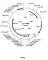
FIG. 2 is a physical map of plasmid pCC-508F which is used for the production of scFv-508-CBD fusion protein (also referred to herein as 508(Fv)-CBD) under control of lac promoter. Amp res - a gene encoding β-lactamase; VH and VL - sequences coding for the variable domains of the heavy and light chains of scFv-508, respectively; Lin - a gene coding for a (Gly4Ser)3 (SEQ ID NO:2) linker present between the variable domains VH and VL. Restriction sites and positions thereof are also shown. -
FIG. 3 is a physical map of plasmid pfFEKCA-508 which is used according to the present invention for cytoplasmic expression of the scFv-508-CBD fusion protein under the control of a T7-promoter. Amp res - a gene encoding β-lactamase; VH and VL - sequences coding for the variable domains of the heavy and light chains of scFv-508, respectively; Lin - a gene coding for a (Gly4Ser)3 (SEQ ID NO:2) linker present between the variable domains VH and VL. T7-promoter and T7 term - T7 promoter and T7 terminator sequences, respectively. Restriction sites and positions thereof are also shown. -
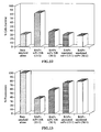
FIG. 4 shows an analysis of βAP binding by antibody 508(Fv)-CBD in an ELISA assay. The analyzed antibodies were added to βAP coated wells. Bound antibodies were detected with HRP conjugated secondary antibodies. The parental 508 IgM antibody was used as a positive control. The unrelated anti-β-galactosidase antibody Gal6(Fv)-CBD was used as a negative control. -
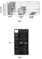
FIG. 5 shows PCR analysis of phage DNA inserts. DNA isolated from pCC-508(Fv),lane 2, and pCC-Gal6(Fv),Lane 3, were PCR amplified and separated on a 1.5 % agarose gel. Ethidium bromide staining and UV illumination were used to visualize the bands.Lane 1 contains a DNA size marker. The arrow marks the position of an intact scFv migrating at about 750 bp. -
FIG. 6 demonstrates expression and purification of 508(Fv)-CBD. 5-10 µg protein were loaded in each lane of a 14 % SDS polyacrylamide gel. Proteins were visualized by Coomassie brilliant blue staining. The arrow marks the position of the scFv-CBD fusion protein. Lane 1 - total cell extract from non-induced BL21(DE3) cells carrying 508((Fv)-CBD expression vector. Lane 2 - total cell extract from BL21(DE3) cells carrying 508((Fv)-CBD expression vector induced for 3 hours with IPTG. Lane 3 - washed, solubilized and reduced inclusion bodies that were used in refolding. Lane 4 - protein that did not bind to cellulose during cellulose-assisted refolding. Lane 5 - protein washed away from cellulose with TBS. Lane 6 - protein washed away from crystalline cellulose with distilled water. Lane 7 - soluble 508(Fv)-CBD recovered from cellulose by high-pH elution and neutralization. -
FIG. 7 demonstrates the stability of 508(Fv)-CBD. Purified 508(Fv)-CBD protein was stored at 4 °C for one day (dark squares) or one week (dark circles), and then analyzed for βAP binding in an ELISA assay, as described in the legend toFigure 4 . The unrelated antibody Gal6(Fv)-CBD served as a negative control (open squares). -
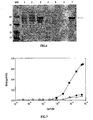
FIG. 8 demonstrates quantitation of 508(Fv) mutants affinity-enrichment by PCR and DNA restriction analysis. The DNA of 19 508(Fv)-mutant micro library clones before (Figure 8a ) and of 11 clones picked up after one cycle of affinity selection (Figure 8b ) were analyzed. The DNA was digested with PvuI and separated on a 1.5 % agarose gel. A non-mutated scFv-CBD appears as an intact 1250 bp fragment (upper arrow). A mutated clone is indicated by the appearance of both 700 bp (middle arrow) and 550 bp (lower arrow) fragments. A DNA size marker is shown inlane 1. -
FIG. 9 shows an analysis of βAP binding (Figure 9a ) and stability (Figure 9b ) of mutated 508(Fv) derivatives in an ELISA assay. The analyzed antibodies were added to βAP coated wells. Bound antibodies stored at 4 °C for one day or for one week were detected as described in the legend toFigure 7 . 508(Fv) wild type (open squares), C96F (dark squares), C96Y (dark circles), C96S (dark triangles). The unrelated anti-β-galactosidase antibody Gal6(Fv)-CBD was used as a negative control (open squares). -
FIG. 10 shows an analysis of the specific inhibition of βAP binding byantibody 508F(Fv) in a competitive ELISA assay. The antibody was pre-incubated with varying concentrations of the competing peptides: βAP (acids 1-16 of SEQ ID NO:3) (dark squares) or the unrelated peptide WVLD (SEQ ID NO:4) (open squares), before being added to βAP coated wells. Bound antibodies were detected as described in the legend toFigure 7 . -
FIGs. 11a and11b show nucleotide (SEQ ID NO:5) and deuced amino acid (SEQ ID NO:6) sequences ofscFv 508F heavy chain (Figure 11a ); and the linker and the variable region of the light chain (Figure 11b ). The amino acid sequence is presented by a three-letter code; CDRs and the linker are underlined. -
FIG. 12 demonstrates the prevention of βAP mediated toxic effect on PC12 cells by 508F(Fv). Cells were incubated with fibrillar βA alone, or with fibrillar βA that had been incubated with antibodies at different molar ratio of antibody/βAP, as indicated. An 3-(4,5-dimethylthiazol-2-yl)-2,5-diphenyl tetrazolium bromide (MTT) assay was used to estimate cell survival. -
FIG. 13 demonstrates the disaggregation of fibrillar βA by 508F(Fv). The fibrillar state of pre-formed βA fibrils were measured with or without incubation with antibodies at different molar ratio of antibody/βAP, as indicated. The fluorescence of thioflavin-T (ThT) reagent in a ThT assay which is proportional to fibril βA was used to assess the fibril morphology. -
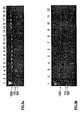
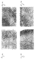
FIGs. 14a-d demonstrate the detection of filamentous phage (f88-EFRH) in brain sections via immunofluorescence one day following a single dose applied intranasally. Appearance of filamentous phage in mouse olfactory bulb and hippocampus sections using fluorescent rabbit anti-phage antibody (Figures 14a and 14c , respectively) as is compare to an untreated mouse brain (Figures 14b and 14d , respectively). The sections were observed using a fluorescence microscope at a final magnification ofx 10. -
FIGs. 15a-d demonstrate the disappearance of filamentous phage (f88-EFRH) from mouse brain 28 days following a single intranasal administration. Disappearance of filamentous phage from mouse olfactory bulb and hippocampus is demonstrated in sections of these organs using fluorescent rabbit anti-phage antibody (Figures 15a and 15c , respectively), as is compared to an untreated mouse brain (Figures 15b and 15d , respectively). The sections were observed using a fluorescence microscope at a final magnification ofx 10. -
FIGs. 16a-d show histology of mouse brain sections after phage f88-EFRH clearance. Brain sections of olfactory organ (Figure 16a ) and hipocampous (Figure 16c ) after 28 days following phage f88-EFRH administration were stained with hematoxylin and eosin, and compared to sections of an untreated brain (Figures 16b and 16d , respectively). The stained sections were examined and photographed at afinal magnification ofx 40. -
FIGs. 17a-d show fluorescence detection of biotin of phage pCC-508F coupled to biotinylated βAP (acids 1-16 of SEQ ID NO:3) in mouse brain sections following a single intranasal administration. Appearance of β AP (acids 1-16, SEQ ID NO:3) coupled to filamentous phage displaying scFv508F in mice olfactory bulb and hippocampus sections using streptavidin coupled to PE (Figures 17a and 17c , respectively) as is compare to an untreated mouse brain (Figures 17b and 17d , respectively). The sections were observed using a fluorescence microscope at a final magnification ofx 20. -
FIGs. 18a-d show histology of mouse brain after phage pCC-508F coupled to biotinylated βAP (acids 1-16 of SEQ ID NO:3) administration. Olfactory organ (Figure 18a ) and hippocampus (Figure 18b ) sections one day following phage administration were stained with hematoxylin and eosin, and were compared to untreated mouse brain sections (Figures 18c and 18d , respectively). The stained sections were examined and photographed at a final magnification ofx 40. -
FIG 19 is a diagram of immunization schedule with filamentous phage displaying the EFRH (SEQ ID NO:1) epitope of β-amyloid peptide. -
FIGs. 20a and 20b show immunization with f3 filamentous phage displaying EFRH (SEQ ID NO:1) epitope of β-amyloid peptide as a fusion of phage glycoprotein III (gpIII). Serum IgG titer of different bleeds from mice immunized with the EFRH-phage according to the schedule ofFigure 19 against wild type filamentous phage coat proteins (Figure 20a ) and the N-terminal epitope (acids 1-16, SEQ ID NO:3) of β-amyloid (Figure 20b ). -
FIG. 21 demonstrates long lasting immunization with f3 filamentous phage. Serum IgG titer of different bleeds from mice immunized with EFRH-phage against wild type filamentous phage coat proteins and the N-terminal (acids 1-16, SEQ ID NO:3) of β-amyloid. -
FIG. 22 show binding of anti-aggregating βAP monoclonal antibody (mAb 10D5) to peptide-presenting phage selected from an f88 phage library. Unrelated mAb 5.5 raised against acetylcholine receptor was used as a negative control. Antibodies were added to phage-coated wells and ELISA was used to detect binding. -
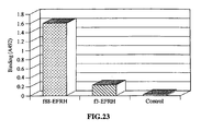
FIG. 23 show binding of anti-aggregating βAP mAb (10D5) to a YYEFRH (SEQ ID NO:7)-phage and VHEPHEFRHVALNPV (SEQ ID NO:8)-phage. Antibody in concentration of 1 µg/ml was added to phage-coated wells and binding was analyzed by ELISA. Filamentous phage without insert was used as a control. -
FIGs. 24a-b show immunization with f88 filamentous phage displaying EFRH (SEQ ID NO:1) epitope of β-amyloid peptide as a fusion of phage glycoprotein VIII (gpVIII). Serum IgG titer of different bleeds from mice immunized with EFRH-phage against wild type filamentous phage coat proteins (Figure 24a ) and the N-terminal epitope (acids 1-16, SEQ ID NO:3) of β-amyloid peptide (Figure 24b ). -
FIG. 25 shows inhibition of serum of an immunized mice in binding to βAP by synthetic peptides derived from the N-terminal of β-amyloid peptide. The assay was done with 1:3000 dilution of serum after a third immunization with f88-EFRH reacted with the various peptides in various concentrations per well, as indicated. The peptide WVLD (SEQ ID NO:4) was used as a negative control. -
FIG. 26 demonstrates prevention of βAP mediated toxic effect onPC 12 cells by serum antibodies raised against f88-EFRH-phage. Cells were incubated with fibrillar βA alone, or with fibrillar βA that has been incubated with serum from the third bleeding at different concentration. The negative control was serum from a non-immunized mouse. The MTT assay was used to estimate cell survival. -
FIG. 27 demonstrates interference with fibrillar β-amyloid formation by serum antibodies raised against the f88-EFRH-phage. Estimation of the fluorescence of ThT which correlates with the amount of fibrillar β-amyloid formed after incubation for a week at 37 °C in the presence of serum samples diluted as indicated. The negative control was serum from a non-immunized mouse. The positive control was without serum. Fibril formation was measured by the ThT assay. -
FIG. 28 illustrates the amino acids sequence corresponding to the human prion protein 106-126 (SEQ ID NO:25) and to the mouse homologue. -
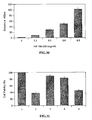
FIG. 29 demonstrates the neurotoxicity effect of the PrP peptide as measured by MTT assay.PC 12 cells were seeded in 96 well plates in a DMEM medium supplemented with 2mM insulin, 2µM L-glutamine and 100 units penicillin/streptomycin. Cell viability was assessed by the MTT assay following incubation with PrP 106-126, (at different concentrations). PrP 106-126 was either preincubated for 4 days at 37 °C and then added to the cells for 3 days (grey bars), or was preincubated for 4 days at 37 °C and then added to the cells for 5 days (white bars) or was preincubated for 7 days at 37 °C and was then added to the cells for 5 days (black bars). -
FIG. 30 illustrates the extent of aggregation of the PrP peptide, using ThT binding assay. PrP 106-126 (0-0.8 mg/ml) was incubated for 7 days at 37 °C and emmision at 482 nm was measured to determine the extent of aggregation. -
FIG. 31 demonstrates the protective effect of mAbs 3-11, 2-40 on PrP peptide neurotoxicity. PC12 cells were seeded in a 96 wells plate in a DMEM medium supplemented with 2mM insulin 2mM L-glutamine and 100 units penicillin/streptomycin and were incubated for three days. The following treatments were conducted: (1) Positive Control, untreated cells; (2) 100 µM PrP 106-126 that was preincubated for 7 days at 37°C; (3,4,5) an aggregated peptide that was preincubated for 1 hour before exposure to the cells together with the mAbs 3-11 (treatment 3), 2-40 (treatment 4) and 3F4 (treatment 5). Cell viability was assessed using the MTT assay. -
FIG. 32 illustrates the modulation of PrP conformation by the mAbs. PrP 106-126 (0.3 mg/ml) was incubated for 7 days at 37°C (1) and with mAbs 2-40, 3-11 and 3F4 (treatments -
FIG. 33 shows the concentration dependent protective effect of mAb 3-11 against PrP fibrillar aggregate formation. PrP 106-126 (0.3 mg/ml) was incubated for 7 days at 37°C with diluted mAb 3-11 (1:1, 1:10, 1:50, corresponding totreatments - The present invention is of pharmaceutical agents and compositions, which can be used for treating Alzheimer's disease. Specifically the present invention can be used to (i) induce active immunity to plaque derived antigens in a recipient by immunizing with at least one epitope of beta-amyloid (Aβ) on a display vehicle, so that antibodies elicited in response to immunization are capable of preventing plaque formation and/or of disaggregating existing plaques; and (ii) induce passive immunity by administering at least an immunological portion of an antibody which can bind to at least one epitope of Aβ raised against plaque derived antigens, cloned and displayed on a display vehicle, capable of preventing plaque formation and of disaggregating existing plaques. This passive immunity may be of exceptionally long duration if the display vehicle employed is capable of replicating within the recipient.
- The principles and operation of the present invention may be better understood with reference to the drawings and accompanying descriptions.
- As it is further exemplified in Examples 1-15 of the Examples section that follows, antigens derived from beta amyloid peptide were displayed on the surface of a filamentous phage which was used for immunization of experimental animals. All of the peptides employed contained the EFRH epitope (SEQ ID NO:1, residues 3-6, SEQ ID NO:3) of beta amyloid peptide (SEQ ID NO:3). The epitope was presented as a fusion protein of fd phage coat glycoprotein III or VIII. Doses ranging from 1010 to 1012 phages per injection were employed on 8 week old female BALB/c mice. A typical immunization schedule included three injections at 14-day intervals, administered either intraperitoneally or intranasally.
- During and after the immunization process, the antibody serum titer of subject mice was tested for the production of Aβ specific antibodies by enzyme linked immunosorbent assay (ELISA) as detailed in methods and materials hereinbelow. Serum titers were subsequently shown to persist for 11 months in response to a protocol including only 3 immunizations. While all tested epitopes containing EFRH produced a titer, displaying the epitope on the surface of a display vehicle produced far highest and unexpected titers. These high titers are believed to be a result of the great number of copies presented to the immune system using this method, and this idea is supported by results of binding assays using controlled amounts of sera.
- The anti-aggregating properties of the obtained polyclonal antibody raised against EFRH epitopes with respect to beta-amyloid fibril formation was measured by the ThT binding assay. Serum, at dilution of 1:10 and 1:100, disrupted formation of fibril structure of β-amyloid with extensive deterioration of fibril morphology, as indicated by a substantial decrease in ThT fluorescence. The unrelated serum used as control (serum from unimmunized mouse) did not significantly inhibit fibril formation.
- The effect of the same serum on disruption of already formed βA fibril (the toxic form of βAP) was also determined. Serum of EFRH immunized mice incubated with pre-formed βA fibrils disrupted the fibril structure. The unrelated control antibody had no significant effect on fibril morphology. Together, these results confirm the ability of EFRH epitope presented by suitable display vehicles to evoke production of anti-aggregation antibodies, which can inhibit or reverse the process of fibril formation.
- Diluted serum produced according to the present invention prevented the neurotoxicity of beta amyloid peptide. This result implies potential clinical utility in preventing brain deterioration of patients suffering from amyloid plaque diseases.
- As it is further exemplified in Examples 15-21 of the Examples section which follows, it was uncovered that site-directed antibodies (designated mAbs 3-11 and 2-40), which were generated against a prion derived peptide, are useful in preventing or disaggregating prion generated plaques.
- Binding of the prion derived peptide (PrP 106-126) to these mAbs led to a significant protective effect against aggregation as was measured by the ThT and MTT assays. The mAbs generated by the preset invention also significantly decrease the peptide fibrillar aggregation and reverse the aggregated form to a disaggregated conformation as assayed by the ThT binding assay.
- The binding of mAbs 3-11 and 2-40 to the PrP peptide either in solution or to the aggregate suggests that this epitope is involved in aggregation process and may act as a regulatory site controlling both the solubilization and disaggregation process of PrP peptide and perhaps the whole PrP protein.
- According to one aspect of the present there is provided an agent for treating Alzheimer's disease. The agent according to this aspect of the present invention comprising a display vehicle displaying a polypeptide, the display vehicle being an in vivo non propagatable virus particle, the polypeptide representing at least one epitope of beta-Amyloid (Aβ), the at least one epitope being capable of eliciting antibodies capable of disaggregating and/or of preventing aggregation of Aβ.
- According to still another aspect of the present invention there is provided a pharmaceutical composition for treating Alzheimer's disease. The composition according to this aspect of the present invention comprising an effective amount of a display vehicle displaying a polypeptide, the display vehicle being an in vivo non propagatable virus particle, the polypeptide representing at least one epitope of Aβ the at least one epitope being capable of eliciting an effective amount of antibodies capable of disaggregating and/or of preventing aggregation of Aβ, and a pharmaceutically acceptable carrier.
- Use of beta amyloid peptide antigens in conjunction with adjuvants to effect immunization has previously been difficult due to a combination of high toxicity and low titers which result. Using prior art methods as a starting point, immunization of a mouse with a 16 amino acids peptide of beta-amyloid conjugated to KLH (SEQ ID NO:9) was carried out. This immunization produced a low but measurable antibody titer against beta-amyloid.
- Splenectomy of the immunized mouse facilitated preparation of IgM hybridoma 508 expressing scFvAb with specificity to beta-amyloid. RNA was subsequently extracted from this hybridoma and was employed for antibody cloning.
IgM 508 hybridoma showed specific activity to Aβ in preventing its toxic affect onPC 12 cells (Anavi, S. 1998, M. Sc. thesis from the department of Molecular Microbiology and Biotechnology of the Tel-Aviv University, Israel). VH and VL sequences ofIgM 508 were cloned separately and linked using a commercially available vector to form a single chain antibody with anti-beta amyloid specificity. This single chain antibody was subsequently expressed as a fusion protein in a phage display library and clones with anti beta amyloid activity were selected for propagation in E. coli. - Further reduction to practice was demonstrated by determining the apparent binding constants of the purified antibody presenting phage to amyloid beta were measured by ELISA test, and half-maximal binding was obtained at an antibody concentration of 340 ng/ml, corresponding to 8x10⁻⁶ M. This result anticipatesthat the prepared single chain antibody will be effective under in vivo conditions. This phage was also able to disrupt already formed fibril structures confirming that the purified single chain antibody is biologically active, as suggested by the binding constant determination.
- While reducing another aspect of the present invention to practice, it was uncovered that monoclonal antibodies raised against a peptide sequence of a prion protein were effective in disaggregating, or preventing the formation, of prion plaques.
- A model for assessing PrP 106-126 toxicity was established by the present inventors and utilized to test the effectiveness of two immunoglobulin clones [designated mAb 3-11 (IgM) and mAb 2-40 (IgG1)] for neuroprotective and disaggregative capabilities.
- As is further detailed in Examples 15-21, both mAb 3-11 and mAb 2-40 significantly reduced the dose dependent toxic effects of PrP 106-126 on PC-12 cells. Co-incubation of mAb 3-11 with PrP 106-126 prevented fibrillar aggregation, while administration of mAb 3-11 to already formed aggregates, resulted in disaggregation of 50% of the amyloid fibrils (Example 21).
- For purposes of this specification and the accompanying claims the terms "patient", "subject" and "recipient" are used interchangeably. They include humans and other mammals which are the object of either prophylactic, experimental, or therapeutic treatment.
- For purposes of this specification and the accompanying claims, the terms "beta amyloid peptide" is synonymous with "β-amyloid peptide", "β AP", "βA", and "Aβ". All of these terms refer to a plaque forming peptide derived from amyloid precursor protein.
- As used herein, "PrP protein", "PrP", "prion" and "prion protein" refer to polypeptides which are capable, under appropriate conditions, of inducing the formation of aggregates responsible for plaque forming diseases. For example, normal cellular prion protein (PrPC) is converted under such conditions into the corresponding scrapie isoform (PrPSc) which is responsible for plaque forming diseases such as, but not limited to, bovine spongiform encephalopathy (BSE), or mad cow disease, feline spongiform encephalopathy of cats, kuru, Creutzfeldt-Jakob Disease (CJD), Gerstmann-Straussler-Scheinker disease (GSS), and fatal familial insomnia (FFI).
- As used herein in the specification and in the claims the term "disaggregating" refers to solubilization of aggregated proteins typically held together by non covalent bonds.
- For purposes of this specification and the accompanying claims the terms "comprising" refers to inclusion of one or more recited element but does not exclude other elements not specifically recited. For example, a composition that comprises Aβ peptide encompasses both an isolated Aβ peptide and Aβ peptide as a component of a larger polypeptide sequence. Similarly, an immunological portion of an antibody may be included as a part of a larger of the antibody, say the entire antibody.
- As used herein in the specification and in the claims section that follows, the term "treating" includes substantially inhibiting, slowing or reversing the progression of a disease, substantially ameliorating clinical symptoms of a disease or substantially preventing the appearance of clinical symptoms of a disease.
- Because amyloid plaques associated with the diseases described hereinabove are located within the brain, any proposed treatment modality must demonstrate an ability to cross the blood brain barrier (BBB) as well as an ability to dissolve amyloid plaques. Normally, the average size of molecules capable of penetrating the BBB is approximately 2 kDa. Monoclonal antibodies are typically in the range 135-900 kDa. Therefore, future therapeutic use of antibodies in treating amyloid plaque diseases must rely on either reduction of their size concurrent with retention of activity, or on development of novel delivery strategies.
- Small synthetic peptides consisting of antigen epitopes, such as the EFRH (SEQ ID NO:1) epitope of Aβ or the PrP 106-126 peptide (SEQ ID NO:25) described herein, are in general poor antigens and need to be coupled to a larger carrier. Even after coupling they may induce only a low affinity immune response. For example, injection of Aβ-KLH or Aβ-fibril leads to very slow immune response (Anavi, S., 1998, M. Sc. thesis from the Department of Molecular Microbiology and Biotechnology of the Tel-Aviv University) and many efforts have been made to circumvent low affinity response, with limited success.
- Since the pathological effects of plaque forming polypeptides are maintained only in the central nervous system (CNS), the capability of highly specific Abs in preventing such plaques in vivo is dependent on the permeability of the blood brain barrier (BBB). For example, in the progressive stage of AD, evidence shows alteration in the permeability of the BBB, which may lead to direct delivery of such antibody from the periphery to the CNS to disaggregate already formed plaques and minimize further toxic effects (Schenk et al. , Nature 1999, 100:173-177). Preferred embodiments of the present invention include direct delivery of monoclonal Abs presented on display vehicles to the brain across the blood brain barrier.
- An increasing body of evidence shows that olfactory deficits and degenerative changes in the central olfactory pathways are affected early in the clinical course of AD. Moreover, the anatomic patterns involved in AD suggest that the olfactory pathway may be the initial stage in the development of AD.
- Olfactory receptor neurons are bipolar cells that reside in the epithelial lining of the nasal cavity. Their axons traverse the cribriform plate and project to the first synapse of the olfactory pathway in the olfactory pathway in the olfactory bulb of the brain. This configuration makes them a highway by which viruses or other transported substances may gain access to the CNS across the BBB.
- In the early stages of AD, the BBB may limit the entry of antibody circulating in the periphery to the CNS. In contrast Aβ anti-aggregating antibodies displayed on a phage surface have the potential not only be delivered directly to the CNS by intranasal administration but also to prevent olfactory permanent damage by βA in the patients. As previously shown, intranasal administration (Mathison et al., J. Drug Target, 1998) 5(6), 415-441; Chou et al., Biopharm Drug Dispos. 1997) 18(4):335-46; et al., Gene Therapy 1995) 2:418-423) enables the direct entry of viruses and macromolecules into the CSF or CNS.
- Use of olfactory receptor neurons as a point of delivery for an adenovirus vector to the brain is reported in the literature. This method reportedly causes expression of a reporter gene in the brain for 12 days without apparent toxicity (Draghia et al., Gene Therapy 2:418-423, 1995).
- Thus, according to the present invention, a vehicle displaying an immunological portion of an antibody capable of disaggregating, or preventing the formation of, a polypeptide aggregate associated with Alzheimer's disease is delivered via this route to the brain wherein the display vehicle is an in vivo non-propagatable virus particle.
- As Aβ is produced continuously by cells in peripheral tissues which cross the blood brain barrier (BBB) leading to localized toxic effects in specific neuronal populations, intranasal administration of such a vehicle may also prevent the progression of the disease by minimizing the amount of peripheral Aβ available to form plaques.
- The use of display vehicles such as filamentous phages as a drug delivery system to the CNS opens new horizons for therapeutic approaches for Alzheimer's disease, as well as for other neurodegenerative diseases involving toxic extracellular aggregation of human peptides such as for example, prion generated diseases.
- The display vehicle according to the present invention is of viral type (e.g., bacteriophage, such as filamentous bacteriophage, fd, for example). Thus, for example, the display vehicle can be a double stranded DNA virus, a single stranded DNA virus, an RNA virus (positive or negative strand). The display vehicle is an in vivo non-propagateable particle.
- The phage or virus vehicle has promise as a targetable in vivo therapy approach. Although concerns about the potential infection of the natural intestinal flora (Delmastro et al., Vaccine 1997, 15: 1276-1285; Willis et al., Gene. 1993, 128:79-83; Poul et al., J. Mol. Biol. 1999, 288, 203-211) have been expressed, UV inactivation of phage showed (Delmastro et al., Vaccine 1997, 15: 1276-1285) that they are as immunogenic as their infective counterparts. Use of inactivated phage may preclude incorporation of phage encoded transgenes into the nucleus for subsequent expression in host cells (Larocca et al., 1998, Hum. Gene Ther. 9:2393-2399), an important practical consideration. Therefore, the display vehicles employed in the present invention are non-replicating.
- Phage or virus display involves the expression of cDNA clones as fusion proteins with phage or virus coat proteins. If the cDNAs selected for expression encode antigens, the phage or virus may then be employed as an antigen presenting vehicle, which can optionally replicate within a recipient.
- As described above, according to preferred embodiments of the present invention, antigens displayed by a phage or virus may be used directly for vaccination, without antigen purification. In this case, the bulk of the coat proteins serve to stimulate a general immune response because they are "non-self" with respect to the vaccinated subject. The antigen-coat protein fusion elicits a specific antibody against epitopes in the displayed cDNA gene product.
- Antibody phage or virus display is accomplished, for example, by fusing the coding sequence of the antibody variable regions to a phage or virus coat protein. To this end, the variable (V) regions (VH and VL) mRNA isolated from antibody-producing cells is reverse-transcribed into cDNA, and heavy and light chains assembled randomly to encode single chain Fv (scFv). These cassettes are cloned directly into a suitable vector such as a phagemid vector for expression and display on the phage or virus surface. This linkage between antibody genotype and phenotype allows the enrichment of antigen specific phage or virus antibodies, using immobilized or labeled antigen. Phage or virus that display a relevant antibody will be retained on a surface coated with antigen, while non-adherent phages or viruses will be washed away. Bound phages or viruses can be recovered from the surface, re-infected into suitable host cells and re-grown for further enrichment and, eventually for binding analysis.
- The success of antibody phage or virus display hinges on the combination of this display and enrichment method. Phage or virus antibody genes can be sequenced, mutated and screened to improve antigen binding.
- It is possible to rearrange the genes which code for the various regions of an antibody molecule such that its specificity and affinity for an antigen are altered. The antibody can be maintained on the surface of the phage or virus for further manipulation or be released as soluble scFv (∼25 kDa) fragment.
- Since its invention at the beginning of the 1990's, antibody phage display has revolutionized the generation of monoclonal antibodies and their engineering. This is because phage display allows antibodies to be made completely in vitro, bypassing the immune system and the immunization procedure, and allowing in vitro tailoring of the affinity and specificity of the antibody. It is therefore anticipated that the most efficient new vaccine development strategies will employ this technology.
- Additional features can be added to the vector to ensure its safety and/or enhance its therapeutic efficacy. Such features include, for example, markers that can be used to negatively select against cells infected with the recombinant virus such as antibiotic sensitivity. Negative selection is therefore a means by which infection can be controlled because it provides inducible suicide through the addition of antibiotic. Such protection ensures that if, for example, mutations arise that produce altered forms of the viral vector or recombinant sequence, cellular transformation will not occur. Features that limit expression to particular cell types can also be included. Such features include, for example, promoter and regulatory elements that are specific for the desired cell type.
- Viruses are very specialized infectious agents that have evolved, in many cases, to elude host defense mechanisms. Typically, viruses infect and propagate in specific cell types. The targeting specificity of viral vectors utilizes its natural specificity to specifically target predetermined cell types and thereby introduce a recombinant gene into the infected cell.
- As used herein in the specification and in the claims section that follows, the term polypeptide refers to a stretch of amino acids covalently linked there amongst via peptide bonds. Different polypeptides have different functionalities according to the present invention. While according to one aspect a polypeptide is derived from an immunogen designed to induce an active immune response in a recipient, according to another aspect of the invention, a polypeptide is derived from an antibody which results following the elicitation of an active immune response, in, for example, an animal, and which can serve to induce a passive immune response in the recipient. In both cases, however, the polypeptide is encoded by a polynucleotide according to any possible codon usage.
- As used herein the phrase "immune response" or its equivalent "immunological response" refers to the development of a beneficial humoral (antibody mediated) and/or a cellular (mediated by antigen-specific T cells or their secretion products) response directed against an aggregating protein (plaque forming peptide) in a recipient patient. Such a response can be an active response induced by administration of immunogen or a passive response induced by administration of antibody or primed T-cells. A cellular immune response is elicited by the presentation of polypeptide epitopes in association with Class I or Class II MHC molecules, to activate antigen-specific CD4+ T helper cells and/or CD8+ cytotoxic T cells. The response may also involve activation of monocytes, macrophages, NK, cells, basophils, dendritic cells, astrocytes, microglia cells, eosinophils or other components of innate immunity.
- As used herein "active immunity" refers to any immunity conferred upon a subject by administration of an antigen.
- As used herein "passive immunity" refers to any immunity conferred upon a subject without administration of an antigen. "Passive immunity" therefore includes, administration of a replicating display vehicle which includes an immunological portion of an antibody presented on its surface to a recipient. Although replication of such a vehicle is active, the immune response is passive from the standpoint of the recipient.
- For purposes of this specification and the accompanying claims the terms "epitope" and "antigenic determinant" are used interchangeably to refer to a site on an antigen to which B and/or T cells respond. B-cell epitopes can be formed both from contiguous amino acids or noncontiguous amino acids juxtaposed by tertiary folding of a protein. Epitopes formed from contiguous amino acids are typically retained on exposure to denaturing solvents whereas epitopes formed by tertiary folding are typically lost on treatment with denaturing solvents. An epitope typically includes at least 3, and more usually, at least 5 or 8-10 amino acids in a unique spatial conformation. Methods of determining spatial conformation of epitopes include, for example, x-ray crystallography and 2-dimensional nuclear magnetic resonance. See, e.g., Epitope Mapping Protocols in Methods in Molecular Biology, Vol. 66, Glenn E. Morris, Ed. (1996). Antibodies that recognize the same epitope can be identified in a simple immunoassay showing the ability of one antibody to block the binding of another antibody to a target antigen. T-cells recognize continuous epitopes of about nine amino acids for CD8 cells or about 13-15 amino acids for CD4 cells. T cells that recognize the epitope can be identified by in vitro assays that measure antigen-dependent proliferation, as determined by 3H-thymidine incorporation by primed T cells in response to an epitope (Burke et al., J. Inf. Dis. 170, 1110-19, 1994), by antigen-dependent killing (cytotoxic T lymphocyte assay, Tigges et al., J. Immunol. 156, 3901-3910) or by cytokine secretion.
- The presence of a cell-mediated immunological response can be determined by proliferation assays (CD4+ T cells) or CTL (cytotoxic T lymphocyte) assays. The relative contributions of humoral and cellular responses to the protective or therapeutic effect of an immunogen can be distinguished by separately isolating IgG and T-cells from an immunized syngeneic animal and measuring protective or therapeutic effect in a second subject.
- As used herein and in the claims, the terms "antibody" or "immunoglobulin" are used interchangeably and refer to any of several classes of structurally related proteins that function as part of the immune response of an animal or recipient, which proteins include IgG, IgD, IgE, IgA, IgM and related proteins.
- Under normal physiological conditions antibodies are found in plasma and other body fluids and in the membrane of certain cells and are produced by lymphocytes of the type denoted B cells or their functional equivalent. Antibodies of the IgG class are made up of four polypeptide chains linked together by disulfide bonds. The four chains of intact IgG molecules are two identical heavy chains referred to as H-chains and two identical light chains referred to as L-chains.
- In order to produce polyclonal antibodies, a host, such as a rabbit or goat, is immunized with the antigen or antigen fragment, generally with an adjuvant and, if necessary, coupled to a carrier. Antibodies to the antigen are subsequently collected from the sera of the host. The polyclonal antibody can be affinity purified against the antigen rendering it monospecific. Previous experience has shown that standard production of polyclonal antibodies is not the method of choice for preparation of disaggregating antibodies for plaque forming peptides due to problems of poor titer and toxicity.
- In order to produce monoclonal antibodies hyperimmunization of an appropriate donor, generally a mouse, with the antigen is undertaken. Isolation of splenic antibody producing cells is then carried out. These cells are fused to a cell characterized by immortality, such as a myeloma cell, to provide a fused cell hybrid (hybridoma) which can be maintained in culture and which secretes the required monoclonal antibody. The cells are then be cultured, in bulk, and the monoclonal antibodies harvested from the culture media for use. By definition, monoclonal antibodies are specific to a single epitope. Monoclonal antibodies often have lower affinity constants than polyclonal antibodies raised against similar antigens for this reason.
- Monoclonal antibodies may also be produced ex-vivo by use of primary cultures of splenic cells or cell lines derived from spleen (Anavi, S., 1998. Locking the N-terminal of the Alzheimer β-amyloid peptide prevents the neurotoxicity in cell cultures, M. Sc. thesis. In order to produce recombinant antibody (see generally Huston et al, 1991; Johnson and Bird, 1991; Mernaugh and Mernaugh, 1995), messenger RNAs from antibody producing B-lymphocytes of animals, or hybridoma are reverse-transcribed to obtain complementary DNAs (cDNAs). Antibody cDNA, which can be full length or partial length, is amplified and cloned into a phage or a plasmid. The cDNA can be a partial length of heavy and light chain cDNA, separated or connected by a linker. The antibody, or antibody fragment, is expressed using a suitable expression system to obtain recombinant antibody. Antibody cDNA can also be obtained by screening pertinent expression libraries.
- The antibody can be bound to a solid support substrate or conjugated with a detectable moiety or be both bound and conjugated as is well known in the art. For a general discussion of conjugation of fluorescent or enzymatic moieties see Johnstone & Thorpe, Immunochemistry in Practice, Blackwell Scientific Publications, Oxford, 1982. The binding of antibodies to a solid support substrate is also well known in the art. See for a general discussion Harlow & Lane Antibodies: A Laboratory Manual, Cold Spring Harbor Laboratory Publications, New York, 1988 and Borrebaeck, Antibody Engineering - A Practical Guide, W.H. Freeman and Co., 1992.
- As used herein and in the claims, the phrase "an immunological portion of an antibody" include an F(ab')2 fragment of an antibody, an Fab fragment of an antibody, an Fv fragment of an antibody, a heavy chain of an antibody, a light chain of an antibody, an unassociated mixture of a heavy chain and a light chain of an antibody, a heterodimer consisting of a heavy chain and a light chain of an antibody, a catalytic domain of a heavy chain of an antibody, a catalytic domain of a light chain of an antibody, a variable fragment of a light chain of an antibody, a variable fragment of a heavy chain of an antibody, and a single chain variant of an antibody, which is also known as scFv. In addition, the term includes chimeric immunoglobulins which are the expression products of fused genes derived from different species, one of the species can be a human, in which case a chimeric immunoglobulin is said to be humanized. Typically, an immunological portion of an antibody competes with the intact antibody from which it was derived for specific binding to an antigen.
- Optionally, an antibody or preferably an immunological portion of an antibody, can be chemically conjugated to, or expressed as, a fusion protein with other proteins. For purposes of this specification and the accompanying claims, all such fused proteins are included in the definition of antibodies or an immunological portion of an antibody.
- As used herein the terms "immunogenic agent" or "immunogen" or "antigen" are used interchangeably to describe a molecule capable of inducing an immunological response against itself on administration to a recipient, either alone, in conjunction with an adjuvant, or presented on a display vehicle.
- As used herein the term "adjuvant" refers to a compound that, when administered in conjunction with an antigen, augments the immune response to the antigen, but when administered alone does not generate an immune response to the antigen. Adjuvants can augment an immune response by several mechanisms including lymphocyte recruitment, stimulation of B and/or T cells, and stimulation of macrophages.
- The preparation according to the present invention can be administered to an organism per se, or in a pharmaceutical composition where it is mixed with suitable carriers or excipients.
- As used herein a "pharmaceutical composition" refers to a preparation of one or more of the active ingredients described herein with other chemical components such as physiologically suitable carriers and excipients. The purpose of a pharmaceutical composition is to facilitate administration of a compound to an organism.
- Herein the term "active ingredient" refers to the preparation accountable for the biological effect.
- Hereinafter, the phrases "physiologically acceptable carrier" and "pharmaceutically acceptable carrier" which may be interchangeably used refer to a carrier or a diluent that does not cause significant irritation to an organism and does not abrogate the biological activity and properties of the administered compound. An adjuvant is included under these phrases.
- Herein the term "excipient" refers to an inert substance added to a pharmaceutical composition to further facilitate administration of an active ingredient. Examples of excipients include calcium carbonate, calcium phosphate, various sugars and types of starch, cellulose derivatives, gelatin, vegetable oils and polyethylene glycols.
- Techniques for formulation and administration of drugs may be found in "Remington's Pharmaceutical Sciences," Mack Publishing Co., Easton, PA, latest edition.
- Suitable routes of administration may, for example, include oral, rectal, transmucosal, especially transnasal, intestinal or parenteral delivery, including intramuscular, subcutaneous and intramedullary injections as well as intrathecal, direct intraventricular, intravenous, inrtaperitoneal, intranasal, or intraocular injections.
- Alternately, one may administer a preparation in a local rather than systemic manner, for example, via injection of the preparation directly into the brain of a patient.
- Pharmaceutical compositions of the present invention may be manufactured by processes well known in the art, e.g., by means of conventional mixing, dissolving, granulating, dragee-making, levigating, emulsifying, encapsulating, entrapping or lyophilizing processes.
- Pharmaceutical compositions for use in accordance with the present invention thus may be formulated in conventional manner using one or more physiologically acceptable carriers comprising excipients and auxiliaries, which facilitate processing of the active ingredients into preparations which, can be used pharmaceutically. Proper formulation is dependent upon the route of administration chosen.
- For injection, the active ingredients of the invention may be formulated in aqueous solutions, preferably in physiologically compatible buffers such as Hank's solution, Ringer's solution, or physiological salt buffer. For transmucosal administration, penetrants appropriate to the barrier to be permeated are used in the formulation. Such penetrants are generally known in the art.
- For oral administration, the compounds can be formulated readily by combining the active compounds with pharmaceutically acceptable carriers well known in the art. Such carriers enable the compounds of the invention to be formulated as tablets, pills, dragees, capsules, liquids, gels, syrups, slurries, suspensions for oral ingestion by a patient. Pharmacological preparations for oral use can be made using a solid excipient, optionally grinding the resulting mixture, and processing the mixture of granules, after adding suitable auxiliaries if desired, to obtain tablets or dragee cores. Suitable excipients are, in particular, fillers such as sugars, including lactose, sucrose, mannitol, or sorbitol; cellulose preparations such as, for example, maize starch, wheat starch, rice starch, potato starch, gelatin, gum tragacanth, methyl cellulose, hydroxypropylmethyl-cellulose, sodium carbomethylcellulose; and/or physiologically acceptable polymers such as polyvinylpyrrolidone (PVP). If desired, disintegrating agents may be added, such as cross-linked polyvinyl pyrrolidone, agar, or alginic acid or a salt thereof such as sodium alginate.
- Dragee cores are provided with suitable coatings. For this purpose, concentrated sugar solutions may be used which may optionally contain gum arabic, talc, polyvinyl pyrrolidone, carbopol gel, polyethylene glycol, titanium dioxide, lacquer solutions and suitable organic solvents or solvent mixtures. Dyestuffs or pigments may be added to the tablets or dragee coatings for identification or to characterize different combinations of active compound doses.
- Pharmaceutical compositions, which can be used orally, include push-fit capsules made of gelatin as well as soft, sealed capsules made of gelatin and a plasticizer, such as glycerol or sorbitol. The push-fit capsules may contain the active ingredients in admixture with filler such as lactose, binders such as starches, lubricants such as talc or magnesium stearate and, optionally, stabilizers. In soft capsules, the active ingredients may be dissolved or suspended in suitable liquids, such as fatty oils, liquid paraffin, or liquid polyethylene glycols. In addition, stabilizers may be added. All formulations for oral administration should be in dosages suitable for the chosen route of administration.
- For buccal administration, the compositions may take the form of tablets or lozenges formulated in conventional manner.
- For administration by nasal inhalation, the active ingredients for use according to the present invention are conveniently delivered in the form of an aerosol spray presentation from a pressurized pack or a nebulizer with the use of a suitable propellant, e.g., dichlorodifluoromethane, trichlorofluoromethane, dichloro-tetrafluoroethane or carbon dioxide. In the case of a pressurized aerosol, the dosage unit may be determined by providing a valve to deliver a metered amount. Capsules and cartridges of, e.g., gelatin for use in a dispenser may be formulated containing a powder mix of the compound and a suitable powder base such as lactose or starch.
- The preparations described herein may be formulated for parenteral administration, e.g., by bolus injection or continuos infusion. Formulations for injection may be presented in unit dosage form, e.g., in ampoules or in multidose containers with optionally, an added preservative. The compositions may be suspensions, solutions or emulsions in oily or aqueous vehicles, and may contain formulatory agents such as suspending, stabilizing and/or dispersing agents.
- Pharmaceutical compositions for parenteral administration include aqueous solutions of the active preparation in water-soluble form. Additionally, suspensions of the active ingredients may be prepared as appropriate oily or water based injection suspensions. Suitable lipophilic solvents or vehicles include fatty oils such as sesame oil, or synthetic fatty acids esters such as ethyl oleate, triglycerides or liposomes. Aqueous injection suspensions may contain substances, which increase the viscosity of the suspension, such as sodium carboxymethyl cellulose, sorbitol or dextran. Optionally, the suspension may also contain suitable stabilizers or agents which increase the solubility of the active ingredients to allow for the preparation of highly concentrated solutions.
- Alternatively, the active ingredient may be in powder form for constitution with a suitable vehicle, e.g., sterile, pyrogen-free water based solution, before use.
- The preparation of the present invention may also be formulated in rectal compositions such as suppositories or retention enemas, using, e.g., conventional suppository bases such as cocoa butter or other glycerides.
- Pharmaceutical compositions suitable for use in context of the present invention include compositions wherein the active ingredients are contained in an amount effective to achieve the intended purpose. More specifically, a therapeutically effective amount means an amount of active ingredients effective to prevent, alleviate or ameliorate symptoms of disease or prolong the survival of the subject being treated.
- Determination of a therapeutically effective amount is well within the capability of those skilled in the art, especially in light of the detailed disclosure provided herein.
- For any preparation used in the methods of the invention, the therapeutically effective amount or dose can be estimated initially from in vitro and cell culture assays. For example, a dose can be formulated in animal models to achieve a desired circulating antibody concentration or titer. Such information can be used to more accurately determine useful doses in humans.
- Toxicity and therapeutic efficacy of the active ingredients described herein can be determined by standard pharmaceutical procedures in vitro, in cell cultures or experimental animals. The data obtained from these in vitro and cell culture assays and animal studies can be used in formulating a range of dosage for use in human. The dosage may vary depending upon the dosage form employed and the route of administration utilized. The exact formulation, route of administration and dosage can be chosen by the individual physician in view of the patient's condition. (See e.g., Fingl, et al., 1975, in "The Pharmacological Basis of Therapeutics", Ch. 1 p.1).
- Dosage amount and interval may be adjusted individually to provide plasma or brain levels of antibodies which are sufficient to prevent aggregation or disaggregate existing aggregates (minimal effective concentration, MEC). The MEC will vary for each preparation, but can be estimated from in vitro data. Dosages necessary to achieve the MEC will depend on individual characteristics and route of administration. Binding assays can be used to determine plasma concentrations.
- Dosage intervals can also be determined using the MEC value. Preparations should be administered using a regimen, which maintains plasma levels above the MEC for 10-90 % of the time, preferable between 30-90 % and most preferably 50-90 %.
- Depending on the severity and responsiveness of the condition to be treated, dosing can be of a single or a plurality of administrations, with course of treatment lasting from several days to several weeks or until cure is effected or diminution of the disease state is achieved.
- The amount of a composition to be administered will, of course, be dependent on the subject being treated, the severity of the affliction, the manner of administration, the judgment of the prescribing physician, etc.
- Compositions of the present invention may, if desired, be presented in a pack or dispenser device, such as an FDA approved kit, which may contain one or more unit dosage forms containing the active ingredient. The pack may, for example, comprise metal or plastic foil, such as a blister pack. The pack or dispenser device may be accompanied by instructions for administration. The pack or dispenser may also be accommodated by a notice associated with the container in a form prescribed by a governmental agency regulating the manufacture, use or sale of pharmaceuticals, which notice is reflective of approval by the agency of the form of the compositions or human or veterinary administration. Such notice, for example, may be of labeling approved by the U.S. Food and Drug Administration for prescription drugs or of an approved product insert. Compositions comprising a preparation of the invention formulated in a compatible pharmaceutical carrier may also be prepared, placed in an appropriate container, and labeled for treatment of an indicated condition, as if further detailed above.
- Generally, the nomenclature used herein and the laboratory procedures utilized in the present invention include molecular, biochemical, microbiological and recombinant DNA techniques. Such techniques are thoroughly explained in the literature. See, for example, "Molecular Cloning: A laboratory Manual" Sambrook et al., (1989); "Current Protocols in Molecular Biology" Volumes I-III Ausubel, R. M., ed. (1994); Ausubel et al., "Current Protocols in Molecular Biology", John Wiley and Sons, Baltimore, Maryland (1989); Perbal, "A Practical Guide to Molecular Cloning", John Wiley & Sons, New York (1988); Watson et al., "Recombinant DNA", Scientific American Books, New York; Birren et al. (eds) "Genome Analysis: A Laboratory Manual Series", Vols. 1-4, Cold Spring Harbor Laboratory Press, New York (1998); methodologies as set forth in
U.S. Pat. Nos. 4,666,828 ;4,683,202 ;4,801,531 ;5,192,659 and5,272,057 ; "Cell Biology: A Laboratory Handbook", Volumes I-III Cellis, J. E., ed. (1994); "Current Protocols in Immunology" Volumes I-III Coligan J. E., ed. (1994); Stites et al. (eds), "Basic and Clinical Immunology" (8th Edition), Appleton & Lange, Norwalk, CT (1994); Mishell and Shiigi (eds), "Selected Methods in Cellular Immunology", W. H. Freeman and Co., New York (1980); available immunoassays are extensively described in the patent and scientific literature, see, for example,U.S. Pat. Nos. 3,791,932 ;3,839,153 ;3,850,752 ;3,850,578 ;3,853,987 ;3,867,517 ;3,879,262 ;3,901,654 ;3,935,074 ;3,984,533 ;3,996,345 ;4,034,074 ;4,098,876 ;4,879,219 ;5,011,771 and5,281,521 ; "Oligonucleotide Synthesis" Gait, M. J., ed. (1984); "Nucleic Acid Hybridization" Hames, B. D., and Higgins S. J., eds. (1985); "Transcription and Translation" Hames, B. D., and Higgins S. J., eds. (1984); "Animal Cell Culture" Freshney, R. I., ed. (1986); "Immobilized Cells and Enzymes" IRL Press, (1986); "A Practical Guide to Molecular Cloning" Perbal, B., (1984) and "Methods in Enzymology" Vol. 1-317, Academic Press; "PCR Protocols: A Guide To Methods And Applications", Academic Press, San Diego, CA (1990); Marshak et al., "Strategies for Protein Purification and Characterization - A Laboratory Course Manual" CSHL Press (1996) Other general references are provided throughout this document. The procedures therein are believed to be well known in the art and are provided for the convenience of the reader. - Reference is made to the following materials and methods, which were employed in experiments described in the following examples.
- The following materials and experimental methods were employed while reducing the present invention to practice as is further demonstrated in the Examples that follow:
- Standard recombinant DNA techniques were performed essentially as described (Sambrook et al., 1989). General protocols for antibody-phage display technology are from the Pharmacia Biotech (Uppsala, Sweden) Recombinant Phage Antibody System (RPAS).
- The 508 IgM hybridoma used as the source for antibody variable-region sequences was generated from splenocytes of a mouse that had been immunized with a peptide corresponding to the 16 amino terminal residues of βAP conjugated to keyhole limpet hemocyanin, used as a carrier. mRNA extraction, first strand cDNA synthesis, PCR amplification of variable heavy (VH) and variable light (VL) sequences, and assembly of scFv cassettes, were done according to protocols essentially as described (Pharmacia Biotech RPAS manual). Assembled 508 scFv DNA was digested with SfiI and NotI, and 100 ng were ligated with 150 ng of vector DNA prepared by digestion of phagemid pCC-Gal6(Fv) (Berdichevsky Y et al., J Immunol Methods., 31;228(1-2):151-62,1999) with SfiI and NotI . This phage-display system is designed to express the scFv in frame fusion protein with cellulose binding domain (CBD) derived from Clostridium thermocellum (Morag E et al. Appl Environ Microbiol., 61(5):1980-6, 1995). Ligated DNA was introduced into XL-1 Blue cells (Stratagene, La Jolla, CA) by transformation and transformants were plated onto 2 X YT Agar plates containing 100 µg/ml ampicillin and 1 % glucose for overnight growth at 37 °C.
- Individual clones were picked and grown, each in 5 ml 2 X YT, 1 % glucose, 100 µg/ml Ampicillin overnight at 30 °C. IPTG was added at 1 mM for a 3 hr induction period. Soluble scFv-CBD fusion proteins were isolated from each clone by sonication of induced cell pellets. In order to identify functional soluble 508(Fv) from non-functional ones, 250 ng/well β -amyloid peptide were covalently bound to epoxy-coated microtiter plates for 16 hr at 4 °C (Solomon, B., et al, Proc. Natl. Acad. Sci. USA., 93:452-455,1996). The plates were washed with PBS/0.05 % - Tween 20 (PBST), and blocked with a mixture of 3 % bovine serum albumin and milk powder in PBS for 16 hr at 4 °C. The plates were then washed and incubated with the soluble scFv-CBD recovered from the clones for 1 hr at 37 °C. The bound antibody was detected with a rabbit anti CBD antiserum followed by HRP-conjugated goat anti rabbit antibodies. Plates were developed with the peroxidase chromogenic substrate ABTS and the signal was recorded with an ELISA microtiter plate reader at 405 nm. Positive phage clones (pCC-508(Fv)) were propagated and their DNA was sequenced using an automated model 373A DNA sequencer (Applied Biosystems, USA).
- For high level expression in E. coli, wild type (wt) and mutated 508(Fv) derivatives were cloned into the pFEKCA3 vector as described (Berdichevsky Y et al, Protein Expr Purif., 17(2):249-59, 1999). This vector utilizes the strong T7 promoter for expression, where the T7 RNA polymerase gene is carried as a lac repressor controlled-IPTG inducible gene in E. coli BL21 (DE3) (Studier, F.W., et al., Methods Enzymol., 85, 60-89, 1990). Upon IPTG induction, 508(Fv)-CBD proteins accumulated as insoluble inclusion bodies. They were recovered by the cellulose-assisted refolding method as previously described (Berdichevsky Y et al, Protein Expr Purif., 17(2):249-59, 1999). SDS polyacrylamide gel electrophoresis (SDS/PAGE) was used to separate proteins according to their molecular weight under denaturing conditions (Laemmli, U.K., Nature 227:263-270,1970).
- The activity of purified 508(Fv)-CBD protein was checked before and after storage at 4 °C for 7 days. 250 ng/well β-amyloid peptide was covalently bound to epoxy-coated wells of microtiter plates for 16 hr at 4 °C (Solomon B. et al., Proc. Natl. Acad. Sci. USA. 94, 4109-4112, 1997). Wells were blocked with a mixture of 3 % bovine serum albumin and bovine hemoglobin in PBS for 2 hr at 37 °C, then washed and incubated with the 508(Fv)-CBD protein (0.5 µg/ml or as otherwise specified) for 1 hr at 37 °C. Bound antibody fragments were detected by incubation with HRP-conjugated rabbit anti-mouse antibodies (BioMakor, Rehovot, Israel), diluted 1:5,000 and rabbit anti CBD diluted 1:10,000 in PBST for 1 hr at 37 °C. The bound antibody fragments were monitored as described above.
- Splicing overlap extension (SOE) PCR technique (Lefenbrve, B., et al., Biotechniques, 19:186-188, 1995) was used to replace VL cysteine codon 96 of 508(Fv) with other codons. pCC-508(Fv) DNA was used as template. In a first step, the template DNA was amplified with the following primers:
- The antisense primer 508-mut-FOR: 5'-CCCCCCTCCGAAC GTSNATGGGTAACTcgatcgCTGATGGCAGTA-3' (SEQ ID NO:10) inserts a PvuI restriction site (underlined), where S represents nucleotides C or G and N represents A, C, T or G. This primer was used for the replacement of cysteine codon 96 with phenylalanine (F), leucine (L), serine (S), tyrosine (Y) or tryptophan codons. The primer SfiI 5'BACK: 5'-ATCTATGCggcccagccggccATG-3' (SEQ ID NO:11) inserts an SfiI site at the 5' end of the scFv. The resulting PCR product (SfiI-508mut) corresponds to the 5' half of 508(Fv)-CBD. In the second PCR step, the complete 508(Fv)-CBD was re-assembled by amplifying pCC-508(Fv) DNA with the SfiI-508mut PCR product from
step 1 serving as the 5' end primer and CBD(BX): 5'-GTGGTGCTGAGTggatccta TACTACACTGCCACCGGG-3' (SEQ ID NO:12) as the 3' end primer. The final PCR product (SfiI-508mut-BX) is a complete 508(Fv)-CBD cassette with replacements at VL codon 96 and an engineered PvuI restriction site as a silent mutation for analysis. SfiI-508mut-BX DNA was digested with SfiI, PvuI and NotI and ligated in a three fragment ligation with SfiI and NotI linearized pCC-Gal6(Fv) DNA which is a phagemid vector used to display an anti E. coli β-galactosidase scFv (Berdichevsky Y et at, J Immunol Methods., 31;228(1-2):151-62, 1999). The resulting ligated phagemid DNA was introduced into E. coli XL-1-Blue cells by electroporation. Cultures of E. coli were used to produce displaying phage by rescue with M13KO7 helper phage (Pharmacia Biotech, Uppsala, Sweden). - A sample containing rescued phage particles was subjected to one round of affinity selection (biopanning) and amplification. For the selection cycle, 0.5 µg/ml biotinylated β-amyloid 1-16 amino acid peptide (βAP(1-16), acids 1-16 OF SEQ ID NO:3) in a total volume of 1 ml were used. The phages were pre-incubated with the biotinylated peptide for 2 hr at room temperature, and the reaction mixture was then layered on streptavidin-coated 30 mm polystyrene Petri dishes and incubated for 20 min at room temperature. Unbound phages were removed by extensive washing with PBST. The bound phages were eluted with 0.3 ml of 0.1 M HCI titrated to pH 2.2 with glycine. The eluate was neutralized with 80 µl of 0.5 M Tris (HCI)
pH 10, and used to infect E. coli XL-1-Blue cells. Individual bacterial colonies containing amplified phage particles were used as a template for colony PCR (Novagen Madison, USA) with primers SfiI5'Back and CBD(BX). The PCR product of about the size of an intact scFv-CBD fragment (about 1250 bp) was digested with the restriction enzyme PvuI and analyzed by agarose gel electrophoresis. - Binding of scFv to βAP(1-16) was analyzed by ELISA. Coated plates with 50 µl of 1 µg/ml streptavidin in 0.1 M NaHCO3, pH 9.6, were washed three times with PBST and 50 µl of 6 ng/µl biotinylated βAP(1-16) were then added to the wells and incubated for 30 min at 37 °C. Wells were blocked with a mixture of 3 % bovine serum albumin and bovine hemoglobin in PBS for 2 hr at 37 °C, then washed and incubated with the scFv (0.5 µg/ml or as otherwise specified) for 1 hr at 37 °C. For inhibition experiments, peptides were pre-incubated with the antibody for 30 min at 37 °C before their addition to peptide coated wells. After washing, bound antibody fragments were detected as described above. βAP specific binding phage clones were propagated and their DNA was isolated and sequenced as described above.
- Rat phenochromocytoma PC12 cells were cultured in DMEM supplemented with 5 % horse serum, 10 % fetal calf serum, 2 mM L-glutamine, and 100 units/ml penicillin/streptomycin and incubated at 37 °C under 5 % CO2. For the neurotoxicity assay,
cultured PC 12 cells were seeded into a 96-well plate at a density of 104 cells/100 µl/well in a serum-free medium supplemented with 2 M of insulin. The effect on the prevention of the neurotoxicity of βA was measured as follows: 0.12 mM β-amyloid that was incubated for a week at 37 °C for the generation of fibrils, and further incubated in the presence of 508F(Fv)-CBD or with the unrelated Gal6(Fv)-CBD at a molar ratio of βAP to scFv of 15:1 or 30:1 for 24 hr. The βA/antibody mixture was added to thewells containing PC 12 cells. The plates were incubated at 37 °C for 2 days, after which cell viability was assessed by measuring cellular redox activity with 3-(4,5-dimethylthiazol-2-yl)-2,5-diphenyl tetrazolium bromide (MTT), as described (Sladowski, D. et al., J. Immunol. Methods., 157:203-207,1993). The plates were incubated overnight at 37 °C. MTT reduction was determined colorimetrically using an ELISA microtiter plate reader set at 550 nm. - Aggregation of β-amyloid peptide was measured by the Thioflavin T (ThT) binding assay, in which the fluorescence intensity reflects the degree of β-amyloid fibril formation. ThT characteristically stains amyloid-like deposits (Levine, H. III, Protein Sci., 2: 404-410,1993) and exhibits enhanced fluorescence emission at 485 nm upon excitation at 435 nm when added to the suspension of aggregated β-sheet preparations. Aqueous solutions of 0.12 mM βAP in 0.1 M Tris (HCI) pH 7.1 were incubated at 37 °C for 1 week and further incubated in the presence of 508F(Fv)-CBD or with the unrelated Gal6(Fv)-CBD at a molar ratio of βAP to scFv of 15:1 or 30:1 for 24 hr. The fluorescence was measured after addition of 1 ml of ThT (2 µM in 50 mM Glycine, pH 9) with an LSB-50 Perkin Elmer Ltd., UK, spectrofluorimeter.
- 12 Balb/c-female mice were divided to four groups of 3 mice per group. One group was used as control. Following a single dose of 1011 phage particles (fd phage, taken from a 15-mer phage peptide library which was provided by George P. Smith, University of Missouri, Columbia, MO) administered intranasally, mice were sacrificed in intervals of 1, 14 and 28 days in each group and their brains were taken for further analysis.
- ScFv-508F fusion to filamentous minor coat gpIII were used in order to investigate the ability of βAP anti-aggregating scFv to be carries by a fillamentous phage display system directly into the CNS.
- This scFv was prepared from
anti-aggregation hybridoma 508 as described above and preserved its specific binding activity. Nine Balb/c mice divided into three groups were treated as follows: Mice of a first group were treated with 0.2 ml of 10-3 M biotin βA(1-16) alone. Mice of a second group were treated with a mix of 1010 phage carrying 508F-scFv which were pre incubated with 0.2 ml of 10-3 M biotin βA(1-16) for 1 hr. Mice of a third group were used as control. Following a single dose applied intranasally, mice were sacrificed in each group in intervals of 1, 14 and 28 days and their brains were taken for further analysis. - Immediately following decapitation, brains were removed and cut into two halves along the mid-sagittal sinus. Randomly, one half-brain was fixed by immersion in 4 % paraformaldehyde solution in 0.1 M phosphate buffer for two hours in 4 °C and then immersed for cold protection in 4.5 % sucrose in 0.1 M PBS over night. The sections were then moved to 30 % sucrose for 2 hr in 4 °C. Sections of coronal blocks containing the olfactory and hippocampus were put in OCT and cut with thick-nesses of 6 µm with a cryostat at - 20 °C, and then taken up on glass slides. Slides were kept at - 70 °C. These slides were used for phage detection using an immunofluorescence technique.
- The other mid-sagittal half-brain was used for preparing paraffin tissue section for histology. The section were fixed in 4 % paraformaldehyde for 2 hr, then transferred to 10 % formalin saline for 2 days in room temperature, followed by embedding in paraffin, and cut with thick-nesses of 4 µm on a microtom and then taken up on glass slides. The slides were kept at room temperature until used.
- Immunofluorescence: Sections were blocked with 3 % bovine serum albumin in PBS for 30 min and then incubated with rabbit polyclonal serum anti fd (1:100) or Streptavidin coupled with PE (sigma) for 1 hr at 37 °C. Slides were then washed three times, 5 min each, in PBS, treated again with the blocking buffer for 5 minutes at room temperature, and then reacted with
secondary r Cy ™ 3 donkey anti-rabbit IgG (for phage detection) at 1:400 (sigma) or with streptavidin coupled to PE, 1:50 dilution, for 1 hr at room temperature. Finally, the preparations were washed three times in PBS, observed using a fluorescence microscope at a final magnification ofx 10, and recorded on film or using a Hamamatsu digital camera (C4742) and Metamorph (Universal Imaging; West Chester, PA) computer software. - Histology: Six-micrometer sections were stained with hematoxylin and eosin. The stained sections were examined and photographed at a final magnification of
x 40. Finally, the preparations were washed three times in PBS, observed on a microscope, and recorded on film. - Immunizations were performed with a genetically engineered fd phage carrying the peptide YYEFRH (SEQ ID NO:7) fused to its minor coat gpIII. Doses of 1010 phages per injection were used to immunize at 14-day intervals, through intraperitoneal injections. Mice were injected the phages with or without Freund's complete adjuvant (Difco) for the first injection and Freund's incomplete adjuvant (Difco) for the second injection. Following 7 days of each injections, the mice were bled and their serum were tested by ELISA for antibody IgG reactivity for both phage coat proteins and for βA.
- The 15-mer phage-peptide library used in this study was provided by George P. Smith (University of Missouri, Columbia, MO). The library consists of about 1.9 x 10⁹ phage particles and comprises a random peptide repertoire of 15 amino acid residues fused to coat glycoprotein VIII of the fd phage. Experiments with this library were carried out according to instructions of the provider (George P. Smith University of Missouri, Columbia, MO).
- For antibody biotinylation, 100 µg of each antibody in 0.1 M NaHCO3, pH 8.6, was incubated for 2 hr at room temperature with 5 µg of biotinamidocoproate N-hydroxysuccinimide ester (Sigma, B 2643) from a stock solution of 1 mg/ml in dimethylformamide and dialyzed at 4 °C against phosphate-buffered saline (PBS; 0.14M NaCl/0.01 M phosphate buffer, pH 7.4) overnight.
- A library sample containing 109 infectious phage particles was subjected to three rounds of selection (biopanning) and amplification. For each selection cycle a biotinylated monoclonal antibody (1 µg/µl) in a total volume of 25 µl was used. The phage clones were pre incubated with the biotinylated antibody overnight at 4 °C, and the reaction mixtures were then layered in 1 ml of PBS containing 0.5
% Tween 20 on streptavidin-coated 30 mm polystyrene Petri dishes and incubated for 20 min at room temperature. Unbound phages were removed by extensive washing (10 times for 10 min each) in PBS/0.05% Tween 20. The bound phages were eluted with 0.3 ml of 0.1 M HCI titrated to pH 2.2 with glycine. The eluate was neutralized and used to infect E. coli K91 cells. After three rounds of panning individual bacterial colonies containing amplified particles were grown on a microtiter plate and the selected phages were tested by ELISA for their ability to bind to the studied antibody, as described below. - Binding of antibodies to phage was analyzed by ELISA. Wells of microtiter plates (Maxisorb, Nunc) were coated with 50 µl (at dilution of 1:1000 in 0.1 M NaHCO3, pH 8.6) of rabbit anti-phage serum and incubated overnight at 4 °C. The wells were blocked with a mixture of 3 % bovine serum albumin and hemoglobin at a ratio of 1:1 (in PBS) for 2 hr at 37 °C. Coated plates were washed three times with PBS/0.05
% Tween - Coat glycoprotein VIII of filamentous phage is presented in approximately 2700 copies on the phage coat. The following oligonucleotides were prepared: sense - 5'-agctccGATGCTGAATTCGG TGATAGCGGCTACGAAGTGCATCATCAGAAAcctgcag-3' (SEQ ID NO:13); and antisense - 5'-ggTTTCTGATGATGCACTTCGTAGC CGCTATCATGACGAAATTCAGCATCgg-3' (SEQ ID NO:14). These oligonucleotides were used to form a duplex (68-70 ° C, 10 minutes, followed by slow cool to room temperature) which encodes for amino acids 1-16 of human βAP and contain a silent mutation of a specific restriction site (EcoRI) which is useful for further analysis. The duplex was phosphorylated and lygated into HindIII/PstI linearized f88-4 phagemid, which is a vector used to display fusion peptides on gpVIII of filamentous phage. The resulting ligated phagemid DNA was introduce into E. coli K91K cells by transformation and transformants were plated onto 2 x YT Agar plates containing 10 µg/ml tetracycline and 1 % glucose for overnight growth at 37 °C. Individual bacterial colonies containing phage particles were used to inoculate 2YT medium containing 10 µg/ml tetracycline for overnight growth at 37 °C for amplification. The DNA phagemid product obtained from each colony was analyzed by EcoRI. Positive clones were further amplified for antigen preparation.
- Immunizations were performed with a genetically engineered fd phage carrying the peptide VHEPHEFRHVALNPV (SEQ ID NO:8) fused to its major coat glycoprotein VIII. Doses of 1010 phages per injection were used to immunize at 14-day intervals, through intraperitoneal injections. Mice were injected the phages with or without Freund's complete adjuvant (Difco) for the first injection and Freund's incomplete adjuvant (Difco) for the second injection. Following 7 days of each injections, the mice were bled and their serum were tested by ELISA for antibody IgG reactivity for both phage coat proteins and for βA.
- The inhibition of antibody binding to βAP(1-16) by various small peptides was performed using 250 ng/well biotinylated β-amyloid peptides (1-16) bound covalently to ELISA plates as previously described. The plates were washed with PBS/0.05
% Tween 20 and blocked with a mixture of 3 % bovine serum albumin and hemoglobin, ratio 1:1 (in PBS) for 2 hr at 37 °C. The peptides were pre incubated with 1:3000 dilution of serum after third immunization with f88-EFRH for 30 min at 37 °C before their addition to βAP-coated wells and were left overnight at 4 °C therein. After washing, bound antibody was detected by incubation with HRP-conjugated rabbit anti-mouse immunoglobulin, as described above. The results were used to derive the IC50, which is the half molar concentration of peptide that fully inhibits antibody binding. Peptides were synthesis by Applied Biosystems Synergy Model 430A in the Unit for Chemical Services of The Weizmann Institute of Science by solid-phase using Fmoc chemistry. -
Rat phenochromocytoma PC 12 cells were cultured in DMEM supplemented with 5 % horse serum, 10 % fetal calf serum, 2 mM L-glutamine, and 100 units/ml penicillin/streptomycin and incubated at 37 °C under 5 % CO2. For the neurotoxicity assay, cultured PC12 cells were seeded into a 96-well plate at a density of 104 cells/100 µl/well in a serum-free medium supplemented with 2 M of insulin. The effect on the prevention of the neurotoxicity of βA was measured as follows: 0.12 mM β-amyloid that was incubated for a week at 37 °C for the generation of fibrils, and further incubated in the presence of serum of EFRH-phage immunized mice and serum of a non-relevant phage immunized mice at dilutions of 5:1 and 10:1 for 24 hr. The βA antibody mixture was added to thewells containing PC 12 cells. The plates were incubated at 37 °C for 2 days, after which cell viability was assessed by measuring cellular redox activity with 3-(4,5-dimethylthiazol-2-yl)-2,5-diphenyl tetrazolium bromide (MTT), as described (Sladowski, D. et al., J. Immunol. Methods., 157:203-207,1993). The plates were incubated overnight at 37 °C. MTT reduction was determined colorimetrically using an ELISA microtiter plate reader set at 550 nm. - Mice immunized with synthetic peptide corresponding to the sequence of human PrP 106-126 (SEQ ID NO:25)(obtained from Chiron Technologies, Claton Victoria, Australia) coupled to the larger carrier KLH were used for generating monoclonal antibodies following the fusion techniques of Kohler and Milstein (Kohler and Milstein 1975).
Hybridomas were tested for the production of peptide-specific antibodies by ELISA, as follows: Peptide PrP 106-126 was covalently attached to the epoxy groups of Eupergit-C coated 96 well plates (Solomon et al. 1992, 1993). The residual epoxy groups were blocked by incubating the plates with 3% skim milk (blocking solution). Undiluted hybridoma supernatants were applied for 1 hour at 37°C. Wells were excessively rinsed (as in each step of the procedure) and further incubated with horseradish peroxidase (HRP) labeled goat-anti-mouse antibodies specific for mouse IgG or IgM (diluted in blocking solution). After washing, antibody binding was visualized using ortho-phenyldiamine as substrate for HRP. Optical density was measured at 492nm. Selected monoclonal antibodies were scaled-up and purified according to published procedures: IgG molecules on a protein A column (Harlow et al. 1988). and IgM on KaptiveM coloumn. Two mAbs, namely 2-40 and 3-11, were used for further studies. The mAb 3F4 was purchased from Senetek, Ca. USA. - The antibodies 3-11 (IgM) and 2-40 (IgG) were biotinylated. The following libraries (provided by G.P. Smith) were searched to find the epitope of the antibodies studied, as previously described (Frenkel et al. 1998).
- 1. fUSE5/15-mer library where foreign 15-mer are displayed on all 5 copies of pIII.
- 2. f88-4/6-mer library where foreign 6-mer are displayed on up to ∼300 copies of pVIII (recombinant gene VIII encoding the peptide is inducible with IPTG)
- The biopanning of the phages to find the antibodies' epitope was performed as previously described (Frenkel et al. 1998).
- The ELISA competitive binding of the above antibodies to covalently bound PrP peptide was performed as described above. The antibodies were preincubated with peptide NMKH at equimolar ratio before adding to the wells.
- In vitro aggregation of peptide 106-126 was induced by incubation of an aqueous solution of PrP 106-126 (10 mg/ml) for various time intervals at 37°C. The aggregated peptide was incubated either with monoclonal antibodies 2-40, 3-11 or 3F4 at conditions specified later.
- Rat
pheochromocytoma PC 12 cells were cultured in DMEM supplemented with 8% horse serum, 8% fetal calf serum, 2 mM L-glutamine and 100 units/ml penicillin/streptomycin and incubated at 37°C under 5% CO2.
For the neurotoxicity assay,cultured PC 12 cells were seeded on 96-well plates at a density of 2 x 10⁴ cells/100 µl/well in a serum-free medium supplemented with 2 µM of insulin. Cells were treated for 3-5 days with 100µM PrP 106-126 preincubated 4-7 days at 37°C. The cell viability was assessed by the MTT assay which measures the activity of mitochondrial enzymes responsible for the conversion of the tetrazolium salt, 3-(4,5-dimethylthiazol-2-yl)-2,5-diphenyl-tetrazolium bromide (MTT) to a formazan product in viable cells (Hansen et al. 1989). MTT was added to the wells to a final concentration of 1 mg/ml and incubated with the cells for an additional 3 hours at 37°C. Cell lysis buffer (20% wt/vol SDS in a solution of 50% dimethylformamide, pH 4.7) was added, and the plate was incubated overnight at 37°C. MTT reduction was determined colorimetrically by measuring the optical density (OD) at 550 nm. - The effect of mAbs on the inhibition of PrP 106-126 neurotoxicity was determined as follows: PrP 106-126 (10 mg/ml) was incubated for 7 days at 37°C to induce maximal aggregation of the peptide. Monoclonal antibodies 3-11, 2-40 and 3F4 were added for 1 hour to samples of 1mM of the already aggregated peptide. The antibody-peptide mixtures, as well as the aggregated peptide alone, were applied to the cells to a peptide final concentration of 100 µM. Cell viability following a 3 day incubation at 37° C with the aforementioned reaction mixture, was assessed as described above. 100% viability was defined as the value of MTT assay for untreated cells.
- Increasing amounts of PrP 106-126 peptide (0-0.8 mg/ml) were incubated for 7 days at 37°C. Prion amyloid fibril formation was measured by the Thioflavine T (ThT) binding assay. The binding of ThT to amyloid fibrils of certain origins generates a specific fluorescent signal: a 114nm red shift in the excitation peak from 336nm of excitation spectrum of the free dye in solution to a new excitation peak at 450nm of the bound dye. Additionally the bound dye has an enhanced emission at 482nm (Naiki et al. 1989, LeVine 1993). The aggregation of the prion peptide was followed using samples of PrP 106-126 (0.3 mg/ml) in 0.1M Tris/HCI pH-7.1 incubated for 7 days at 37°C, either with or without mAbs 3-11, 2-40 and 3F4 at various dilutions. Disaggregation of already formed prion amyloid fibrils was measured using samples of PrP 106-126 that were incubated for 7 days at 37°C and then supplemented with the mAbs for an additional 24 hours. Fluorescence (emission at 482 nm after excitation at 435 nm) was measured after an addition of the samples to ThT (2µM in 50mM glycine, pH-9).
- Examples 1-6 below relate to the production of a single chain version of the anti aggregating monoclonal antibody. Examples 7-8 below relate to delivery of peptide or antibody displaying phage to the brain. Examples 9-14 below relate to the production of high titers of anti-aggregating polyclonal antibodies by direct immunization with beta amyloid antigens displayed on a phage, and to characterization of these antibodies.
- Immunization of a mouse with a 16 amino acid peptide of beta-amyloid (acids 1-16 of SEQ ID NO:3) conjugated to KLH (SEQ ID NO:9) was carried out as described hereinabove. Repetitious immunization eventually produced a low but measurable antibody titer against beta-amyloid. Subsequent splenectomy of the immunized mouse facilitated preparation of IgM hybridoma 508 expressing scFvAb with specificity to beta-amyloid. RNA was subsequently extracted from this hybridoma. The
IgM 508 hybridoma showed specific activity to Aβ in preventing its toxic affects onPC 12 cells (Anavi, S. 1998, M. Sc. thesis from the department of Molecular Microbiology and Biotechnology of the Tel-Aviv University, I). -
MAb 508 showed specific recognition of β-amyloid and prevented its toxic affects onPC 12 cells (Anavi S., 1998, ibid). For cloning the 508 antibody as a scFv in a phage display vector, RNA was extracted from 108 508 hybridoma cells and was used as a source for antibody variable region coding sequences. RT-PCR was used to amplify the variable domains that were cloned into the phage display vector pCC-Gal6(Fv), as described in Materials and Methods. When hybridoma derived antibodies are cloned as scFvs, some of the clones may contain aberrant sequences that are not functional. Therefore, to identify phagemid clones carrying functional β-amyloid binders from the generated clones, 10 individual clones were picked at random and soluble scFv-CBD fusion protein was produced thereby.Figure 2 shows a physical map of plasmid pCC-508 which was used to express the 508-scFv. The CBD domain serves as an immunological detection of soluble scFv protein or as a novel approach in refolding of soluble scFv protein inclusion bodies of overexpressed protein (Berdichevsky Y et al, Protein Expr Purif., 17(2):249-59, 1999). The plasmid used for 508-scFv over expression is shown inFigure 3 . The soluble scFv-CBD from the selected clones was incubated in wells of an ELISA plate that has been coated with β-amyloid peptide. Of the analyzed clones, 50 % showed specific binding to βAP. Figures la-e demonstrate and illustrate the preparation of 508 scFv from IgM antibody.Figure 4 shows βAP binding by the scFv-CBD produced by a positive clone that was chosen for further analysis. PCR analysis was used to characterize its DNA insert. It was found that the positive clone (designated pCC-508(Fv)) contained an intact DNA insert (Figure 5 ). DNA sequencing of pCC-508(Fv) confirmed that the clone expresses an intact scFv fragment (see,Figures 11a-b and SEQ ID NOs:5 and 6, for nucleic and amino acid sequences, respectively, modified as further described below). - The DNA sequencing analysis of pCC508-(Fv) revealed the unusual appearance of a cysteine residue at the position 96 of VL CDR3 (Kabat, E. A. et al., Sequences of proteins of immunological interest, 5th Ed., 1991). The deduced amino acid sequence of VL CDR3 is: H89QRSSYPCT97 (SEQ ID NO:15). The presence of an unpaired cysteine residue in a scFv may reduce its folding yield and also decrease its stability in solution and its storage half life. Therefore, 508(Fv) was subcloned into an expression vector and produced in E. coli as described in Materials and Methods.
Figure 6 summarizes the production process of 508(Fv)-CBD by the cellulose-assisted refolding method (Berdichevsky Y et al, Protein Expr Purif., 17(2):249-59, 1999). Although 508(Fv)-CBD could be purified to near homogeneity (Figure 6 lane 7) by this method, it refolded relatively poorly and was unstable upon storage at 4 °C (Figure 7 ). It was assumed that substitution of the cysteine with a different residue may increase the production yield and stability of the soluble scFv without having an adverse affect on its affinity (Kirpriyanov, M.S. et al., Protein Engineering, 10:445-453, 1997). - For the replacement of the 508 VL cysteine 96 codon SOE PCR was used, which enabled the replacement of Cys 96 with phenylalanine, leucine, serine, tyrosine or tryptophan codons. In addition, the PCR scheme employed permits the persistence of the cysteine residue at that position. 508(Fv) mutants were cloned into the pCC-Gal6(Fv) phage vector, resulting in the generation of a micro library (having 6 potential variant). The replacing residues chosen are generally acceptable at that CDR3 position, as they are found in various antibodies in the Kabat database (Kabat, E. A. et al., Sequences of proteins of immunological interest, 5th Ed., 1991). However, different replacements may vary in their effect on βAP binding. To test which replacement maintains βAP binding, a single cycle of affinity selection was performed on the 508(Fv)-Mut micro phage library using biotinylated βAP(1-16) as a capturing antigen. PCR amplification and restriction analysis was used to monitor the enrichment of library clones after the affinity selection cycle. When the 508Mut-(Fv)-CBD DNA is digested with PvuI, a typical restriction pattern is obtained upon agarose-gel electrophoresis and ethidium-bromide staining. The lower 750 and 500 bp fragments represent the 508Mut-(FV)-CBD DNA, while an intact 1250 bp fragment represent scFv-CBD from the pCC-Gal6(Fv) DNA which was used as a vector. It was found that before affinity selection the library was heavily contaminated with the pCC-Gal(Fv) vector DNA. This is evident from the fact that the DNA of 18/19 randomly picked library clones was not cleaved at the PvuI site engineered adjacent to 508 VL position 96. Only one of the 19 analyzed clones showed the expected restriction pattern associated with a mutation (
Figure 8a ). However, after affinity selection, 5/11 randomly selected clones showed the expected restriction pattern (Figure 8b ). This indicates an enrichment factor of about 10 fold, which demonstrates the ability of 508 scFv mutants to bind the βAP(1-16) epitope. The DNA sequences of the 5 mutants were determined and are shown in Table 1 below. Suitable replacements of 508 LL cysteine 96 codon were found to be phenylalanine, serine or tyrosine.TABLE 1 Lane ( Figure 8b )Amino Acid Sequence SEQ ID NO: 4 89HQRSSYP-C96- T 16 5 89HQRSSYP-F96- T 17 6 89HQRSSYP-Y96- T 18 8 89HQRSSYP-F96- T 19 11 89HQRSSYP-S96- T 20 - For further examination of mutated 508 scFv derivatives, the mutated genes were subcloned into an expression vector and overexpressed in E. coli, as described for the wild type protein above. The interactions of the various mutated 508-(Fv) proteins (Table 1) with βAP(1-16) were tested in an ELISA assay.
Figures 9a-b show that while the wild type 508-(Fv)-CBD binds at a half maximum binding (HMB) of 10-5 M, all the mutants showed improved binding to βAP: the HMB of C96S and C96Y is 5 X 10⁻⁶ M and the HMB of C96F is 10-7 M. For further examination the 508-scFv mutant that carries the C96F mutation (508F(Fv)) was chosen, which show the higher affinity and shelf stability (Figures 9a-b ). The specificity of βAP(1-16) binding by 508F(Fv) was tested in a competitive ELISA. As is shown inFigure 10 , binding of the purified 508F(Fv)-CBD to βAP was inhibited by soluble βAP(1-16) peptide serving as the competitor in the liquid phase in a dose-dependent manner. Inhibition of 50 % binding was obtained at about 1 µM competitor. Binding was not affected by an irrelevant peptide (WVLD, SEQ ID NO:4). - In order to find out whether 508F(Fv) exhibits a protective effect similar to the parental IgM antibody in preventing βA mediated neurotoxicity toward cultured cells, an in vitro test was applied using rat phenochromocytoma PC12 as described (Solomon B. et al., Proc. Natl. Acad. Sci. USA., 94: 4109-4112,1997). Viability of the cells exposed to βA with or without antibody was measured. As shown in
Figure 12 , 508F(Fv) prevented the neurotoxicity of βA (90 % cell viability) at a molar ratio β AP:scFv of 15:1, while the unrelated scFv showed no effect. Purified CBD or the scFv alone had no affect on the cells. - To examine the effect of 508F(Fv) on disruption of the βA fibril (the toxic form of βAP) the ThT reagent that binds specifically to fibrillar structures (Levine, H. III, Protein Sci., 2: 404-410,1993) was used. The interference with the already formed βA fibril was measured at the same molar ratio of βAP:scFv as in the βA neurotoxic assay and was quantitated by ThT fluorimetry.
Figure 13 shows that 508F(Fv) incubated with pre-formed βA fibrils disrupted the fibril structure indicating extensive deterioration of fibril morphology, as is evidenced by a substantial (62 %) decrease in ThT fluorescence. - Female Balb/c mice were treated with phage vector f88-EFRH via intranasal administration. The propose of this experiment was to check the ability of filamentous phage to reach the hippocampous region via olfactory tract. Since the phage is not carrying any specific molecule for targeting neuron cells, it should be vanished without causing any harm after several day following the administration. In order to investigate the appearance of phage in the olfactory bulb and the hippocampous region double labeling of antibodies was used as follows: Rabbit polyclonal antibody anti filamentous phage and mouse monoclonal antibody against EFRH epitope fused to glycoprotein VIII of the phage surface. One day following a single intranasal administration of 1011 phages animals showed such phages in their olfactory bulb and hippocampous (
Figures 14a-d ). Seven days after the administration phages were detected in the olfactory bulb of only one mouse of the three tested, whereas no phages were revealed in the hippocampous. No evidence of phages was detectable 28 days following administration (Figures 15a-d ). As shown inFigures 16a-d , no evidence of change in the neuron population of the brain of treated mice was evident. - To check whether a filamentous phage can carry an antibody to the CNS via the olfactory track and still preserve the activity against β-amyloid a filamentous phage displaying 508F was incubated with 10-3 M biotinlated βAP(1-16), in order to form antibody antigen immunocomplex. Balb/c mice were divided into two groups and were administrated intranasally with two different antigens: 508F-βAP(1-16) immunocomplex and for comparison biotinlated βAP(1-16) alone. Following a single dose the mice were sacrificed and brain sections thereof prepared and reacted with streptavidin coupled to a fluorescent agent. Fluorescence was detected only in brain sections of mice that were administered with 508F-βAP(1-16), but not, or to a much lesser extent, in brain sections of mice that were administered with βAP(1-16) alone (
Figures 17a-d ). No histological findings characterized treated mice (Figures 18a-d ). - It is therefore assumed that the phage act as an inert vehicle of antibody to the brain, carrying the βAP(1-16) molecule into the brain.
- The anti-aggregating epitope within βAP (EFRH, SEQ ID NO:1) map to positions 3-6 of the amino acid sequence of βAP. In order to generate specific immune response against βAP, mice were immunized with genetically engineered fd phage carrying the peptide YYEFRH (SEQ ID NO:7) fused to its minor coat gpIII according to the immunization schedule shown in
Figure 19 . Doses of 1010 phage particles per injection were used to immunize, at 14-day intervals, through intraperitoneal injection. Following 7 days of each injection, mice were bled and their sera tested by ELISA for IgG antibody reactivity against wild type phage (not bearing the peptide YYEFRH on its surface) and against βAP (Figures 20a-b ). This route of administration a very high response against βAP (1:750) following the third injection. Furthermore, it was found that injection through phage carrying epitope is long lasting (Figure 21 ), it is non-toxic and may be given without adjuvant. The phage vector is found to be an immunogenic tool to raise a high affinity immune response within 14 days from the first injection. The immune response against the peptide YYEFRH (SEQ ID NO:7) is low, compared to the immune response against the entire phage and could be explained by the low copy number of the fusion gpIII on the phage envelope. Therefore, for further analysis phages displaying the epitope through glycoprotein VIII were employed. - To identify a disagreggating EFRH peptide epitope a phage-epitope library was screened with biotinylated antibody. After three cycles of panning and phage amplification, 90 individually isolated bacterial colonies were grown in microtiter plates and their phages were assayed for antibody binding. ELISA analysis revealed that of the phage-clones which were selected followed by three biopanning cycles, most (above 80%) bound specifically to anti-aggregating mAb, respectively. DNA from 6 positive clones was sequenced (Table 2). The sequence EFRH (SEQ ID NO:1) appeared in 4 clones, one additional clone had the sequence EPRH (SEQ ID NO:1), with only one residue replacement of proline with phenylalanine. In one additional clone, the inserted peptide bears the sequence of the three residues FRH (acids 2-4 of SEQ ID NO:1), lacking the glutamate residue.
TABLE 2 Amino acid Sequence (name) SEQ ID NO: No. of Phages VHEPHEFRHVALNPV (C3-II) 8 2 DTEFRHSSNNFSAVR(C7-II) 21 1 STEFRHQTTPLHPNS (C11-I) 22 1 KEPRHHIQHHERVIR (F8-II) 23 1 SAADFRHGSPPISAF (D3-I) 24 1 - Binding of anti-aggregating mAb to the EFRH-bearing phage was concentration dependent; half-maximal binding was obtained at an antibody concentration of 100 ng/ml, corresponding to 10-9 M (
Figure 4 ) which resembles the level of binding of these antibodies to the whole peptide. One specific f88-EFRH phage (termed C3-II, table 2) showed higher level of avidity as is compared to the others (Figure 22 ). It may be due to higher level of EFRH epitope exposure on its surface. Binding tests of f88-EFRH or f3-EFRH with the same concentration of anti-aggregating antibody (1µ /ml) demonstrated a higher number of EFRH epitope copies per phage which may lead to higher serum titer via phage immune response (Figure 23 ). - In order to generate the same specific immune response against βAP, mice were immunized with genetically engineered fd phage carrying the peptide VHEPHEFRHVALNPV (SEQ ID NO:8) fused to its major coat gpVIII. This phage was selected from a 15-mer phage peptide library by an anti aggregating βAP antibody and is presenting the mAb epitope (underlined) within βAP. This phage was used to immunize mice as described. Following 7 days of each injection with 1010 phage particles (without adjuvant) the mice were bled and their sera tested by ELISA for IgG antibody reactivity against wild type phage and against βAP. The results are summarized in
Figures 24a-b . All animals showed a measurable response of IgG antibody against the wild type phage, and titers increased with the second and the third injection. This route also gave the highest titer measurable responses against βAP (1:50,000) after the third injection (Figure 24b ). - The interaction of mouse serum immunized by phage f88-EFRH with βAP was further assayed by competitive inhibition experiments.
Figure 25 shows inhibition of mice serum antibody with synthesized peptides derived from βAP, each of which includes the sequence EFRH) such as: DAEFRH (positions 1-6, SEQ ID NO:3), DAEFRHD (positions 1-7, SEQ ID NO:3), DAEFRHDSG (positions 1-9, SEQ ID NO:3), and βAP itself, DAEFRHDSGYEVHHQKLVFFAEDVGSNKGAIIGLMVG GVV (positions 1-40, SEQ ID NO:3). -
Figure 25 shows that all of the synthetic peptides which bear the motive EFRH (SEQ ID NO:1) similarly inhibited binding of mouse serum antibody to the βAP with IC50 values of about 5x10⁻⁶ M. These data indicate that the epitope of mouse serum antibody within the βAP molecule is composed of four amino acid residues corresponding to positions 3-6 in the βAP which was found to act as a regulatory site controlling both the solubilization and the disaggregation process of the βA molecule. - In order to find out whether serum of f88-EFRH immunized mice exhibits a protective effect similar to the parental IgG antibody in preventing βA mediated neurotoxicity toward cultured cells, the in vitro test using
rat phenochromocytoma PC 12 was applied as described (Solomon B. et al., Proc. Natl. Acad. Sci. USA., 94: 4109-4112,1997). Viability of the cells exposed to βA with or without antibody was measured. As shown inFigure 26 , diluted serum of 1:5 prevented the neurotoxicity of βA (80 % cell viability), while the unrelated serum showed no effect. - To examine the effect of serum of f88-EFRH immunized mice on disruption of the βA fibril (the toxic form of βAP) the ThT reagent that binds specifically to fibrillar structures (Levine, H. III, Protein Sci., 2: 404-410,1993) was used. βAP samples were incubated for a week at 37 °C and then were exposed to different dilutions mouse serum antibody. Fibril formation was quantitated by the ThT fluorometry binding assay.
Figure 27 shows that mouse serum, at dilution of 1:5 and 1:20, disrupted the fibril structure of βA with extensive deterioration of fibril morphology, as indicated by a substantial 75 % (1:5 dilution) and 50 % (1:20 dilution) decrease in ThT fluorescence. The unrelated serum used as control (serum from non-immunized mouse), did not significantly inhibit fibril formation as is compared to the immunized serum. This result strongly emphasizes the ability of the EFRH epitope displayed by a filamentous phage vector to evoke an immune response resulting in anti-agreggation antibody. - The possible involvement of the PrP protein in the pathogenesis of nerve cell degeneration and glial cell reaction led to the identification of a PrP sequences that play a role in the amyloid formation. A fragment of PrP consisting of amino acids 106-126 was demonstrated to be toxic to rat hippocampal neurons (Forloni et al. 1993), to mouse cortical and cerebellar cells (Brown et al., 1994; 1997), and to be particularly highly fibrillogenic (Selvaggini et al. 1993). The formed fibrils were partially resistant to proteases digestion and exhibited properties of in situ amyloid (Selvaggini et al. 1993, Tagliavini et al. 1993). Synthetic peptides corresponding to this region of PrP exhibit conformational flexibility from α-helix to a β-sheet conformation (Selvaggini et al. 1993) which was similar to conformational changes from PrPC to PrPSc. The conformational plasticity of this region is further emphasized by the findings that two distinct prion strains which exhibit different sites of proteolytic cleavage within this region (Bessen and Marsh, 1994; Telling et al., 1996).
- As is further described below, the inventors of the present invention have generated mAbs specific to epitopes formed by amino acids 106-126 of the PrP protein. Such antibodies are usefull in studying plaque formation and morphology and as possible active agents for treating or preventing prion generated plaque diseases.
- Mice immunized with a synthetic peptide corresponding to the amino acid sequence of human PrP 106-126 coupled to the larger carrier KLH were used for generating monoclonal antibodies reactive against epitopes on this peptide. Sera derived from the immunized mice was subjected to ELISA and several positive clones composed mainly of immunoglobulin M (IgM) molecules and to a lesser extent IgG molecules were detected and isolated.
- Two immunoglobulin clones that were isolated as described above and designated mAbs 3-11 (IgM) and mAb 2-40 (IgG1) were utilized in further studies.
- Phage display libraries displaying various peptide fragments of the human PrP 106-126 polypeptide were generated as described hereinabove in the method section. Clones reactive to mAbs 3-11 or mAb 2-40 (for each library) were not detected following 6 cycles of library biopanning (368 clones screened) raising the possibility that epitopes that are recognized by these antibodies are of a conformational nature.
- The above described anti-PrP 106-126 antibodies were preincubated with a peptide of an amino acid sequence NMKH (SEQ ID NO:26) at equimolar ratio before reacting with the PrP 106-126 polypeptide in order to determine the ability of NMKH to compete with the human PrP 106-126 for antibody binding. As shown in
Figure 28 , this sequence is highly conserved between mice and human sequences (and others) with the exception of two different amino acids, M109 instead of L and M 112 instead of V. These two differences probably contribute to antigenic differeces between the mouse and human sequence, since the antibodies which were derived from mice immunized with the human sequence corresponding to PrP 106-126 did not cross react with the mouse sequence corresponding to peptide PrP 106-126. - Competition (as analyzed by ELISA) between the NMKH and whole peptide was not detected, supporting the suggestion that the epitope may depend upon three dimensional conformation and not primary structure. In addition, co-reacting mAb 3F4 (recognizing the sequence MKHM) and mAb 3-11 with PrP 106-126 did not produce an additive response, suggesting that their epitopes may be adjacent or overlapping (Solomon and Balas, 1991).
- Incubation of PrP 106-126 at 37 °C at different concentrations led to a dose dependent fibrillar aggregation as measured by the Thioflavin T binding fluorescence assay (
Figure 30 ). A PrP 106-126 concentration of 0.3 mg/ml was selected for further studies. - Since the major target organ for PrPSc is the nervous system, in vitro neuronal model systems were used to analyze PrP toxicity. A clonal cell line which exhibits neuronal properties has been established from rat central nervous system tumors (Schubert et al. 1974). The rat pheochromocytoma PC12 cells - a neuron-like cloned tumor cell line (Greene and Tischler, 1976) - has been shown to enable in vitro replication of PrPSc (Rubenstein et al. 1984). As such, this cell line was utilized by the present study as a model for detecting molecules with the ability to prevent toxicity induced by PrP fibrils.
- Using this model system, it was uncovered that PrP 106-126 is toxic to
PC 12 cells in a dose dependant manner, which toxicity is related to the conformational state of the peptide and to the exposure time of the cells to aggregated peptide. Cell viability considerably decreased (as detected by MTT assay), when cells were incubated with PrP 106-126 for 5 days in comparison to a 3 day exposure. Furthermore, preincubation of PrP 106-126 at 37 °C prior to its addition to the cells increased its toxicity probably by increasing the amount of amyloid fibrils. Preincubation of the peptide for 7 days resulted in a significantly higher toxicity which was concentration dependent (Figure 29 ). Under the described conditions, cell viability was reduced to 10% at a concentration of 100µM of PrP 106-126 as was measured by MTT assay. - The cytoprotective effect of mAb 3-11 and 2-40 is shown in
Figure 31 . Mabs 3-11 and 2-40 inhibited cell death which was induced by 100µM PrP 106-126. The viability of cells treated with a mixture of either antibody and the peptide was 85-89%, as compared to a 40% survival rate in cells treated with the peptide alone (The antibodies without the peptide had no affect on cell viability). The antibodies' protective effect was apparently related to the specific epitope on the PrP molecule, since such protection was not demonstrated with mAb 3F4 (44% viability). - The amyloid fibril formation of PrP 106-126 was detected by a thioflavin-T (ThT) binding assay. Both mAbs 3-11 and 2-40 prevent PrP 106-126 fibrillar aggregation and disaggregate the aggregated form of this peptide. A significant decrease in amyloid fibril formation (about 80% prevention) was observed when PrP 106-126 was incubated at 37 °C in the presence of mAbs 3-11 and 2-40. When these antibodies were added to already formed aggregates of PrP 106-126, more than 50% of the fibrils were disaggregated. In contrast, mAb 3F4 had lower efficacy both in inhibiting amyloid fibril formation and in inducing their disaggregation (
Figure 32 ). Both the inhibition of amyloid fibril formation and the induction of disaggregation were dependent on the antibody's concentration and the epitope location (Figures 32-33 ). - The protective effect of mAbs 3-11 and 2-40 of the present invention and of mAb 3F4 was calculated as shown in
Equation 1 below.
-
- Bessen, R.A. and Marsh, R.F. (1994). Distinct PrP properties suggest the molecular basis of strain variation in transmissible mink encephalopathy. J. Virol.68, 7859-7868.
- Blaney, J.M., Jorgensen, E.C., Connolly, M.L., Ferrin, T.E., Langridge, R., Oatley, S.J., Burridge, J.M. and Blake, C.C.F. (1982) Computer graphics in drug design: molecular modeling of thyroid hormone-prealbumin interactions. J. Med. Chem. 25, 785-790
- Blond, S. and Goldberg, M. (1987) Partly native epitopes are already present on early intermediates in the folding of tryptophan synthase. Proc. Natl. Acad. Sci. USA 84, 1147-1151
- Brown, D.R., Hernis, J. and Kretzschmar, H.A. (1994) Mouse cortical cells lacking cellular PrP survive in culture with aneurotoxic PrP fragment.
- Brown, D. R., Herms, J.W., Schmidt, B. and Kretzschmar, H.A. (1997) Different requirements for the neurotoxicity of fragments of PrP and a-amyloid. Eur. J. Neurosci. 9, 1162-1169.
- Carlson, J.D. and Yarmush, M.L. (1992) Antibody assisted protein refolding.
- Caughey, B.W., Dong, A., Bhat, K.S., Ernst, D., Hayes, S.F., Caughey, W.S. (1991) Secondary structure analysis of the scrapie-associated protein PrP 27-30 in water by infrared spectroscopy. Biochemistry. 30, 7672-7680.
- De Gioia, L., Selvaggini, C., Ghibaudi, E., Diomede, L., Bugiani, O., Forloni, G., Tagliavini, F. and Salmona, M. (1994) Conformational polymorphism of the amyloidogenic and neurotoxic peptide homologous to residues 106-126 of the prion protein. J Biol Chem. 269, 7859-7862.
- Donne, D.G., Viles, J.H., Groth, D., Mehlhorn, I., James, T.L., Cohen, F.E., Prusiner, S.B., Wright, P.E. and Dyson, H.1997) ) Structure of the recombinant full-length hamster prion protein PrP(29-231): the N terminus is highly flexible. Proc. Natl. Acad. Sci. USA 94, 13452-13457
- Forloni, G., Angeretti, N., Chiesa R., Monzani, E., Salmona, M., Bugiani, O., and Tagliavini, F. (1993) Neurotoxicity of a prion protein fragment. Nature 362, 543-546
- Frauenfelder, H., Petsko, G.A. and Tsernoglou, D. (1979) Temperature-dependent X-ray diffraction as a probe of prote Nature 1989 in structural dynamics. Nature 280, 558-563.
- Frenkel, D., Ballas, M. and Solomon, B. (1998) N-terminal EFRH sequence of Alzheimer's β-amyloid peptide represents the epitope of its anti-aggregating antibodies. J. Neuroimmunology 88, 85-90.
- Greene, L.A. and Tischler, A.S. (1976) Establishment of a noradrenergic clonal line of rat adrenal pheochromocytoma cells which respond to nerve growth factor. Proc Natl Acad Sci U S A 73, 2424-2428
- Hansen, M.B., Nielsen, S.E., Berg, K. (1989) Re-examination and further development of a precise and rapid dye method for measuring cell growth/cell kill. J Immunol Methods 119, 203-210
- Harlow, E. and Lane, D. (1988) Antibodies, a laboratory manual (Cold Spring Harbor Laboratory), .
- Horiuchi, M. and Caughey B. (1999) Specific binding of normal prion protein to the scrapie form via a localized domain initiates its conversion to the protease-resistant state EMBO J. 18, 3193-3203
- Kanyo, Z.F., Pan, K.M., Williamson, R.A., Burton, D.R., Prusiner, S.B., Fletterick, R.J. and Cohen, F.E. (1999) Antibody binding defines a structure for an epitope that participates in thePrPC-->PrPSc conformational change. J Mol Biol. 293, 855-863
- Karplus, M. and Petsko, G.A. (1990) Molecular dynamics simulations in biology. Nature 347, 631-639
- Kohler, G. and Milstein, C. (1975) Continuous cultures of fused cells secreting antibody of predefined specificity. Nature 256, 495-497
- LeVine, H. III (1993) Thioflavine T interaction with synthetic Alzheimer's disease beta-amyloid peptides: detection of amyloid aggregation in solution. Protein Sci. 2, 404-410
- Miroy, G.J., Lai, Z., Lashuel, H.A., Peterson, S.A., Strang, C. and Kelly, J.W. (1996) Inhibiting transthyretin amyloid fibril formation via protein stabilization. Proc. Natl. Acad. Sci. USA 93, 15051-15056
- Muramoto, T., Scott, M., Cohen, F.E. and Prusiner, S.B. (1996) Recombinant scrapie-like prion protein of 106 amino acids is soluble. Proc. Natl. Acad. Sci. USA 93, 15457-15462.
- Naiki, H., Higuchi, K., Hosakawa, M. and Takeda, T. (1989) Fluorometric determination of amyloid fibrils in vitro using the fluorescent dye, thioflavin T1. Anal. Biochem., 177, 244-249.
- Pan, K.M., Baldwin, M., Nguyen, J., Gasset, M., Serban, A., Groth, D., Mehlhorn, I., Huang, Z., Fletterick, R.J., Cohen, F.E. and Prusiner, S.B. (1993) Conversion of alpha-helices into beta-sheets features in the formation of the scrapie prion proteins. Proc Natl Acad Sci USA. 90, 10962-10966.
- Peretz, D., Williamson, R.A., Matsunaga, Y., Serban, H., Pinilla, C., Bastidas, R.B., Rozenshteyn, R., James, T.L., Houghten, R.A., Cohen, F.E., Prusiner, S.B. and Burton, D.R. (1997) A conformational transition at the N terminus of the prion protein features in formation of the scrapie isoform. J. Mol. Biol. 273, 614-622.
- Riek, R., Hornemann, S., Wider, G., Glockshuber, R., Wüthrich, K (1997) NMR characterization of the full-length recombinant murine prion protein, mPrP(23-231). FEBS Lett. 413, 282-288
- Rubenstein, R, Carp, R.I. and Callahan, S.M. (1984) In vitro replication of scrapie agent in a neuronal model: infection of PC12 cells.
- Schubert, D., Heinemann, S., Carlisle, W., Tarikas, H., Kimes, B., Patrick, J., Steinbach, J.H., Culp, W. and Brandt, B.L. (1974) Clonal cell lines from the rat central nervous system. Nature 249, 224-227
- Selvaggini, C., De Gioia, L., Cantu, L., Ghibaudi, E., Diomede, L., Passerini, F., Forloni, G., Bugiani, O., Tagliavini, F. and Salmona, M. (1993) Molecular characteristics of a protease-resistant, amyloidogenic and neurotoxic peptide homologous to residues 106-126 of the prion protein. Biochem. Biophys. Res. Commun. 194, 1380-1386.
- Silen, J.L. and Agard, D.A. (1989) The alpha-lytic protease pro-region does not require a physical linkage to activate the protease domain in vivo. Nature 341, 462-464.
- Solomon, B. and Balas N. (1991) Thermostabilization of carboxypeptidase A by interaction with its monoclonal antibodies. Biotechnol Appl Biochem. 14, 202-211.
- Solomon, B., Fleminger, G., Schwartz, F., Doolman, R. and Sela, B.A. (1992) Microalbuminuria immunoassay based on antibodies covalently conjugated to Eupergit C-coated beads.
- Solomon, B., Schmitt, S., Schwartz, F., Levi, A. and Fleminger, G. (1993) Eupergit C-coated membranes as solid support for a sensitive immunoassay of human albumin. J. Immunol. Methods 157, 209-215
- Solomon, B., and Schwartz, F. (1995) Chaperone-like effect of monoclonal antibodies on refolding of heat-denatured carboxypeptidase A. J. Mol. Recognit. 8, 72-76
- Solomon, B., Koppel, R., Hanan, E. and Katzav, T. (1996) Monoclonal antibodies inhibit in vitro fibrillar aggregation of the Alzheimer beta-amyloid peptide. Proc. Natl. Acad. Sci. USA 93, 452-455
- Solomon, B., Koppel, R., Frankel, D., Hanan-Aharon, E. (1997) Disaggregation of Alzheimer beta-amyloid by site-directed mAb. Proc. Natl. Acad. Sci. USA 94, 4109-4112.
- Tagliavini, F., Prelli, F., Verga, L., Giaccone, G., Sarma, R., Gorevic, P., Ghetti, B., Passerini, F., Ghibaudi, E., Forloni, G., Salmona, M., Bugiani, O. and Frangione, B. (1993) Synthetic peptides homologous to prion protein residues 106-147 form amyloid-like fibrils in vitro. Proc. Natl. .
- Telling, G.C., Parchi, P., DeArmond, S.J., Cortelli, P., Montagna, P., Gabizon, R., Mastrianni, J., Lugaresi, E., Gambetti, P. and Prusiner, S.B. (1996) Evidence for the conformation of the pathologic isoform of the prion protein enciphering and propagating prion diversity. Science, 274, 2079-2082.
- Zahn, R., Liu, A., Luhrs, T., Riek, R., von Schroetter, C., Lopez Garcia, F., Billeter, M., Calzolai, L., Wider, G. and Wuthrich, K. (2000) NMR solution structure of the human prion protein. Proc Natl Acad Sci USA. 97, 145-150.
-
- <110> Beka Solomon et al.
- <120> Agents and compositions and methods utilizing same useful in treating or preventing plaque forming diseases.
- <130> 20100
- <150> 60/152,417
<151> September 3, 1999 - <160> 26
- <170> PatentIn Ver. 2.1
- <210> 1
<211> 4
<212> PRT
<213> Artificial sequence - <220>
<223> Description of the Artificial sequence: synthetic peptide - <400>1
Glu Phe Arg His - <210> 2
<211> 15
<212> PRT
<213> Artificial sequence - <220>
<223> Description of the Artificial sequence: synthetic peptide - <400>2

- <210> 3
<211> 43
<212> PRT
<213> Artificial sequence - <220>
<223> Description of the Artificial sequence: synthetic peptide - <400> 3

- <210> 4
<211> 4
<212> PRT
<213> Artificial sequence - <220>
<223> Description of the Artificial sequence: synthetic peptide - <400> 4
Trp Val Leu Asp - <210> 5
<211> 717
<212> DNA
<213> Homo sapien - <400> 5


- <210> 6
<211> 244
<212> PRT
<213> Homo sapien - <400> 6

- <210> 7
<211> 6
<212> PRT
<213> Artificial sequence - <220>
<223> Description of the Artificial sequence: synthetic peptide - <400> 7

- <210> 8
<211> 15
<212> amino acid
<213> Artificial sequence - <220>
<223> - <400> 8

- <210> 10
<211> 45
<212> DNA
<213> Artificial sequence - <220>
<223> Description of the Artificial sequence: primer - <400>10
CCCCCCTCCG AACGTSNATG GGTAACTCGA TCGCTGATGG CAGTA 45 - <210> 11
<211> 24
<212> DNA
<213> Artificial sequence - <220>
<223> Description of the Artificial sequence: primer - <400> 11
ATCTATGCGG CCCAGCCGGC CATG 24 - <210> 12
<211> DNA
<212> 38
<213> Artificial sequence - <220>
<223> Description of the Artificial sequence: primer - <400> 12
GTGGTGCTGA GTGGATCCTA TACTACACTG CCACCGGG 38 - <210> 13
<211> 58
<212> DNA
<213> Artificial sequence - <220>
<223> Description of the Artificial sequence: primer - <400>13
AGCTCCGATG CTGAATTCGG TGATAGCGGC TACGAAGTGC ATCATCAGAA ACCTGCAG 58 - <210> 14
<211> 52
<212> DNA
<213> Artificial sequence - <220>
<223> Description of the Artificial sequence: primer - <400> 14
GGTTTCTGAT GATGCACTTC GTAGCCGCTA TCATGACGAA ATTCAGCATC GG 52 - <210> 15
<211> 9
<212> PRT
<213> Artificial sequence - <220>
<223> Description of the Artificial sequence: synthetic peptide - <400> 15

- <210> 16
<211> 9
<212> PRT
<213> Artificial sequence - <220>
<223> Description of the Artificial sequence: synthetic peptide - <400> 16

- <210> 17
<211> 9
<212> PRT
<213> Artificial sequence - <220>
<223> Description of the Artificial sequence: synthetic peptide - <400> 17

- <210> 18
<211> 9
<212> PRT
<213> Artificial sequence - <220>
<223> Description of the Artificial sequence: synthetic peptide - <400> 18

- <210> 19
<211> 9
<212> PRT
<213> Artificial sequence - <220> Description of the Artificial sequence: synthetic peptide
<223> - <400> 19

- <210> 20
<211> 9
<212> PRT
<213> - <220>
<223> Description of the Artificial sequence: synthetic peptide - <400> 20

- <210> 21
<211> 15
<212> PRT
<213> Artificial sequence - <220>
<223> Description of the Artificial sequence: synthetic peptide - <400> 21

- <210> 22
<211> 15
<212> PRT
<213> Artificial sequence - <220>
<223> Description of the Artificial sequence: synthetic peptide - <400> 22

- <210> 23
<211> 15
<212> PRT
<213> Artificial sequnce - <220>
<223> Description of the Artificial sequence: synthetic peptide - <400> 23

- <210> 24
<211> 15
<212> PRT
<213> Artificial sequence - <220>
<223> Description of the Artificial sequence: synthetic peptide - <400> 24

- <210> 25
<211> 21
<212> PRT
<213> Artificial sequence - <220>
<223> Description of the Artificial sequence: synthetic peptide - <400> 25

- <210> 26
<211> 4
<212> PRT
<213> Artificial sequence - <220>
<223> Description of the Artificial sequence: synthetic peptide - <400> 26
Asn Met Lys His
- <210> 6
<211> 244
<212> PRT
<213> Homo sapien - <400> 6

- <210> 7
<211> 6
<212> PRT
<213> Artificial sequence - <220>
<223> Description of the Artificial sequence: synthetic peptide - <400> 7

- <210> 8
<211> 15
<212> amino acid
<213> Artificial sequence - <220>
<223> - <400> 8

- <210> 10
<211> 45
<212> DNA
<213> Artificial sequence - <400> 23

- <210> 24
<211> 15
<212> PRT
<213> Artificial sequence - <220>
<223> Description of the Artificial sequence: synthetic peptide - <400> 24

- <210> 25
<211> 21
<212> PRT
<213> Artificial sequence - <220>
<223> Description of the Artificial sequence: synthetic peptide - <400> 25

- <210> 26
<211> 4
<212> PRT
<213> Artificial sequence - <220>
<223> Description of the Artificial sequence: synthetic peptide - <400> 26
Asn Met Lys His
Claims (30)
- A pharmaceutical composition, comprising a pharmaceutically acceptable carrier, and, as an active ingredient, a viral display vehicle that is an in vivo non-propagatable virus particle, which display vehicle displays a polypeptide, wherein said polypeptide comprises at least one epitope of beta-amyloid (Aβ), and wherein said at least one epitope elicits Aβ-binding antibodies against said epitope when administered to a subject, and wherein said antibodies inhibit aggregation of said beta-amyloid.
- The pharmaceutical composition of claim 1, wherein said in vivo non-propagatable virus particle is selected from the group consisting of an in vivo non-propagatable double stranded DNA virus particle, an in vivo non-propagatable single stranded DNA virus particle, an in vivo non-propagatable positive strand RNA virus particle, and an in vivo non-propagatable negative strand RNA virus particle.
- The pharmaceutical composition of claim 1 or claim 2, wherein said in vivo non-propagatable virus particle is an in vivo non-propagatable bacteriophage particle.
- The pharmaceutical composition of any of the preceding claims, wherein said in vivo non-propagatable virus particle is selected such that less than 30 days following an introduction of a triple dose of 1010 units thereof to the recipient, a titer of said antibodies is above 1:50,000, as is determined by ELISA.
- A pharmaceutical composition in accordance with any one of the preceding claims, wherein said epitope is EFRH (SEQ ID NO:1), DAEFRH (residues 1-6 of SEQ ID NO:3), DAEFRHD (residues 1-7 of SEQ ID NO:3), or DAEFRHDSG (residues 1-9 of SEQ ID NO:3).
- A pharmaceutical composition in accordance with any one of the preceding claims, wherein said epitope comprises EFRH (SEQ ID NO:1).
- Use of a pharmaceutical composition in accordance with any of the preceding claims in the manufacture of a medicament for treating Alzheimer's disease.
- Use according to claim 7, wherein said in vivo non-propagatable virus particle is selected from the group consisting of an in vivo non-propagatable double stranded DNA virus particle, an in vivo non-propagatable single stranded DNA virus particle, an in vivo non-propagatable positive strand RNA virus particle, and an in vivo non-propagatable negative strand RNA virus particle.
- Use according to claim 7, wherein said in vivo non-propagatable virus particle is an in vivo non-propagatable bacteriophage particle.
- Use according to any of claims 7-9, wherein the Alzheimer's disease is early onset Alzheimer's disease.
- Use according to any of claims 7-9, wherein the Alzheimer's disease is late onset Alzheimer's disease.
- Use according to any of claims 7-9, wherein the Alzheimer's disease is presymptomatic Alzheimer's disease.
- Use according to any of claims 7-12, wherein said medicament is for introduction into the body of the recipient so as to inhibit aggregation of beta-amyloid by applying said in vivo non-propagatable virus particle to an olfactory system of the recipient.
- Use according to any of claims 7-13, wherein said epitope is EFRH (SEQ ID NO:1), DAEFRH (residues 1-6 of SEQ ID NO:3), DAEFRHD (residues 1-7 of SEQ ID NO:3), or DAEFRHDSG (residues 1-9 of SEQ ID NO:3).
- Use according to any of claims 7-14, wherein said epitope comprises EFRH (SEQ ID NO:1).
- An agent comprising a viral display vehicle that is an in vivo non-propagatable virus particle, which display vehicle displays a polypeptide, wherein said polypeptide comprises at least one epitope of beta-amyloid (Aβ), and wherein said at least one epitope elicits Aβ-binding antibodies against said epitope when administered to a subject, and wherein said antibodies inhibit aggregation of said beta-amyloid.
- An agent of claim 16, wherein said in vivo non-propagatable virus particle is selected from the group consisting of an in vivo non-propagatable double stranded DNA virus particle, an in vivo non-propagatable single stranded DNA virus particle, an in vivo non-propagatable positive strand RNA virus particle, and an in vivo non-propagatable negative strand RNA virus particle.
- An agent of claim 16 or claim 17, wherein said in vivo non-propagatable virus particle is an in vivo non-propagatable bacteriophage particle.
- An agent of any of claims 16-18, wherein said in vivo non-propagatable virus particle is selected such that less than 30 days following an introduction of a triple dose of 1010 units thereof to the recipient, a titer of said antibodies is above 1:50,000, as is determined by ELISA.
- An agent in accordance with any of claims 16-19, wherein said epitope is EFRH (SEQ ID NO:1), DAEFRH (residues 1-6 of SEQ ID NO:3), DAEFRHD (residues 1-7 of SEQ ID NO:3), or DAEFRHDSG (residues 1-9 of SEQ ID NO:3).
- An agent in accordance with any of claims 16-20, wherein said epitope comprises EFRH (SEQ ID NO:1).
- Use of an agent in accordance with any of claims 16-21 in the manufacture of a medicament for treating Alzheimer's disease.
- Use according to claim 22, wherein said in vivo non-propagatable virus particle is selected from the group consisting of an in vivo non-propagatable double stranded DNA virus particle, an in vivo non-propagatable single stranded DNA virus particle, an in vivo non-propagatable positive strand RNA virus particle, and an in vivo non-propagatable negative strand RNA virus particle.
- Use according to claim 22, wherein said in vivo non-propagatable virus particle is an in vivo non-propagatable bacteriophage particle.
- Use according to any of claims 22-24, wherein the Alzheimer's disease is early onset Alzheimer's disease.
- Use according to any of claims 22-24, wherein the Alzheimer's disease is late onset Alzheimer's disease.
- Use according to any of claims 22-24, wherein the Alzheimer's disease is presymptomatic Alzheimer's disease.
- Use according to any of claims 22-27, wherein said medicament is for introduction into the body of the recipient so as to inhibit aggregation of beta-amyloid by applying said in vivo non-propagatable particle to an olfactory system of the recipient.
- Use according to any of claims 22-28, wherein said epitope is EFRH (SEQ ID NO:1), DAEFRH (residues 1-6 of SEQ ID NO:3), DAEFRHD (residues 1-7 of SEQ ID NO:3), or DAEFRHDSG (residues 1-9 of SEQ ID NO:3).
- Use according to any of claims 22-29, wherein said epitope comprises EFRH (SEQ ID NO:1).
Applications Claiming Priority (7)
| Application Number | Priority Date | Filing Date | Title |
|---|---|---|---|
| US15241799P | 1999-09-03 | 1999-09-03 | |
| US152417P | 1999-09-03 | ||
| US473653 | 1999-12-29 | ||
| US09/473,653 US6703015B1 (en) | 1999-09-03 | 1999-12-29 | Filamentous bacteriophage displaying an β-amyloid epitope |
| US62997100A | 2000-07-31 | 2000-07-31 | |
| US629971 | 2000-07-31 | ||
| PCT/IL2000/000518 WO2001018169A2 (en) | 1999-09-03 | 2000-08-31 | Agents and compositions and methods for diagnostic and/or treating or preventing plaque forming diseases |
Publications (3)
| Publication Number | Publication Date |
|---|---|
| EP1180938A2 EP1180938A2 (en) | 2002-02-27 |
| EP1180938A4 EP1180938A4 (en) | 2005-03-16 |
| EP1180938B1 true EP1180938B1 (en) | 2010-03-24 |
Family
ID=27387248
Family Applications (1)
| Application Number | Title | Priority Date | Filing Date |
|---|---|---|---|
| EP00954883A Expired - Lifetime EP1180938B1 (en) | 1999-09-03 | 2000-08-31 | Agents, compositions and methods utilizing the same useful in treating or preventing alzheimer disease |
Country Status (11)
| Country | Link |
|---|---|
| US (4) | US6919075B1 (en) |
| EP (1) | EP1180938B1 (en) |
| JP (1) | JP2003509020A (en) |
| AT (1) | ATE461996T1 (en) |
| AU (1) | AU784568B2 (en) |
| CA (1) | CA2349434A1 (en) |
| DE (1) | DE60044057D1 (en) |
| ES (1) | ES2344189T3 (en) |
| HK (1) | HK1044564B (en) |
| IL (1) | IL142948A0 (en) |
| WO (1) | WO2001018169A2 (en) |
Families Citing this family (78)
| Publication number | Priority date | Publication date | Assignee | Title |
|---|---|---|---|---|
| TWI239847B (en) | 1997-12-02 | 2005-09-21 | Elan Pharm Inc | N-terminal fragment of Abeta peptide and an adjuvant for preventing and treating amyloidogenic disease |
| US6787523B1 (en) * | 1997-12-02 | 2004-09-07 | Neuralab Limited | Prevention and treatment of amyloidogenic disease |
| US20080050367A1 (en) | 1998-04-07 | 2008-02-28 | Guriq Basi | Humanized antibodies that recognize beta amyloid peptide |
| US7790856B2 (en) | 1998-04-07 | 2010-09-07 | Janssen Alzheimer Immunotherapy | Humanized antibodies that recognize beta amyloid peptide |
| US7964192B1 (en) * | 1997-12-02 | 2011-06-21 | Janssen Alzheimer Immunotherapy | Prevention and treatment of amyloidgenic disease |
| US20050059802A1 (en) * | 1998-04-07 | 2005-03-17 | Neuralab Ltd | Prevention and treatment of amyloidogenic disease |
| US20020052311A1 (en) * | 1999-09-03 | 2002-05-02 | Beka Solomon | Methods and compostions for the treatment and/or diagnosis of neurological diseases and disorders |
| TR200202799T3 (en) | 2000-02-24 | 2003-03-21 | Washington University St.Louis | Humanized antibodies that sequence the AB peptide |
| JP2003516929A (en) * | 2000-06-01 | 2003-05-20 | ニユーララブ・リミテツド | Prevention and treatment of amyloidogenic diseases |
| US7700751B2 (en) | 2000-12-06 | 2010-04-20 | Janssen Alzheimer Immunotherapy | Humanized antibodies that recognize β-amyloid peptide |
| US7264810B2 (en) * | 2001-01-19 | 2007-09-04 | Cytos Biotechnology Ag | Molecular antigen array |
| ES2437875T3 (en) | 2001-04-30 | 2014-01-14 | Eli Lilly And Company | Humanized antibodies that recognize the beta-amyloid peptide |
| DE60229051D1 (en) | 2001-04-30 | 2008-11-06 | Lilly Co Eli | HUMANIZED ANTIBODIES |
| US6906169B2 (en) | 2001-05-25 | 2005-06-14 | United Biomedical, Inc. | Immunogenic peptide composition comprising measles virus Fprotein Thelper cell epitope (MUFThl-16) and N-terminus of β-amyloid peptide |
| DK1944040T3 (en) | 2001-08-17 | 2012-10-29 | Univ Washington | Method of analysis for Alzheimer's disease |
| US7494655B2 (en) | 2002-03-05 | 2009-02-24 | Ramot At Tel-Aviv University Ltd. | Immunizing composition and method for inducing an immune response against the β-secretase cleavage site of amyloid precursor protein |
| MY139983A (en) | 2002-03-12 | 2009-11-30 | Janssen Alzheimer Immunotherap | Humanized antibodies that recognize beta amyloid peptide |
| CA2492930C (en) * | 2002-07-19 | 2013-01-08 | Cytos Biotechnology Ag | Vaccine compositions containing amyloid beta1-6 antigen arrays |
| AT413945B (en) * | 2003-01-14 | 2006-07-15 | Mattner Frank Dr | Use of a compound having a binding capacity to an antibody specific for the natural N-terminal AB42 sequence, for preparing a vaccine for preventing and treating Alzheimer's disease |
| DE10303974A1 (en) | 2003-01-31 | 2004-08-05 | Abbott Gmbh & Co. Kg | Amyloid β (1-42) oligomers, process for their preparation and their use |
| KR20050118669A (en) * | 2003-02-01 | 2005-12-19 | 뉴랄랩 리미티드 | Active immunization to generate antibodies to soluble a-beta |
| ATE468886T1 (en) | 2003-02-10 | 2010-06-15 | Applied Molecular Evolution | ABETA-BINDING MOLECULES |
| US8663650B2 (en) | 2003-02-21 | 2014-03-04 | Ac Immune Sa | Methods and compositions comprising supramolecular constructs |
| TWI374893B (en) | 2003-05-30 | 2012-10-21 | Janssen Alzheimer Immunotherap | Humanized antibodies that recognize beta amyloid peptide |
| US7807171B2 (en) | 2003-07-25 | 2010-10-05 | Ac Immune Sa | Therapeutic vaccine targeted against P-glycoprotein 170 for inhibiting multidrug resistance in the treatment of cancers |
| US20050225165A1 (en) * | 2004-04-13 | 2005-10-13 | Naik Sanjeev M | Brake by-wire control system |
| RU2387453C2 (en) * | 2004-06-25 | 2010-04-27 | АйДи БАЙОМЕДИКАЛ КОРПОРЕЙШН ОФ КВЕБЕК | Compositions and methods of treating neurological disturbances |
| AT413946B (en) * | 2004-07-13 | 2006-07-15 | Mattner Frank Dr | VACCINE AGAINST THE ALZHEIMER DISEASE |
| EP1787998A4 (en) | 2004-08-11 | 2008-08-27 | Mitsubishi Chem Corp | Antibody and utilization of the same |
| US20070269898A1 (en) * | 2004-09-15 | 2007-11-22 | Yokohama City University | Amino Acid Sequences Essential to Neuron Specific Gene Expression |
| US20060160161A1 (en) * | 2004-10-26 | 2006-07-20 | Elan Pharmaceuticals, Inc. | Methods for assessing antibodies to neurodegenerative disease-associated antigens |
| GB0424563D0 (en) | 2004-11-05 | 2004-12-08 | Novartis Ag | Organic compounds |
| TW200635607A (en) | 2004-12-15 | 2006-10-16 | Elan Pharm Inc | Humanized Aβ antibodies for use in improving cognition |
| EP1853285B1 (en) * | 2005-02-01 | 2011-03-16 | Ramot at Tel-Aviv University Ltd. | Method for treating inflammation associated with amyloid deposits and brain inflammation involving activated microglia |
| AU2006319358B2 (en) | 2005-11-30 | 2012-01-19 | AbbVie Deutschland GmbH & Co. KG | Anti-Abeta globulomer antibodies, antigen-binding moieties thereof, corresponding hybridomas, nucleic acids, vectors, host cells, methods of producing said antibodies, compositions comprising said antibodies, uses of said antibodies and methods of using said antibodies |
| KR101439828B1 (en) | 2005-11-30 | 2014-09-17 | 애브비 인코포레이티드 | Monoclonal antibodies against amyloid beta protein and uses thereof |
| RU2015111675A (en) * | 2005-12-12 | 2015-08-10 | Ац Иммуне Са | SPECIFIC IN RESPECT TO AMYLOID BETA (A BETA) 1-42 MONOCLONAL ANTIBODIES WITH THERAPEUTIC PROPERTIES |
| WO2007094003A2 (en) * | 2006-02-15 | 2007-08-23 | Ramot At Tel Aviv University Ltd. | Viral display vehicles for treating multiple sclerosis |
| US8784810B2 (en) | 2006-04-18 | 2014-07-22 | Janssen Alzheimer Immunotherapy | Treatment of amyloidogenic diseases |
| KR101605207B1 (en) | 2006-07-14 | 2016-03-22 | 에이씨 이뮨 에스.에이. | Humanized antibody against amyloid beta |
| ATE541038T1 (en) * | 2006-07-21 | 2012-01-15 | Univ Ramot | USE OF A PHAGE DISPLAYING A PEPTIDE OF AN ADHESION PROTEIN FOR THE TREATMENT OF A DISEASE ASSOCIATED WITH INTRACELLULAR FORMATION OF FIBRILLARY PROTEIN INCLUSIONS OR AGGREGATES |
| US20100129354A1 (en) * | 2006-10-27 | 2010-05-27 | Ablynx N.V. | Intranasal delivery of polypeptides and proteins |
| US8455626B2 (en) | 2006-11-30 | 2013-06-04 | Abbott Laboratories | Aβ conformer selective anti-aβ globulomer monoclonal antibodies |
| CN103408661B (en) | 2007-01-05 | 2016-04-06 | 苏黎世大学 | The method of disease-specific binding molecules and target is provided |
| IL199534A (en) | 2007-01-05 | 2013-01-31 | Univ Zuerich | Isolated human antibody which is capable of selectively recognizing a neoepitope of a disorder-associated protein, a polynucleotide encoding the antibody, a vector comprising the polynucleotide, a host cell comprising the polynucleotide or the vector, a composition comprising the antibody and methods and uses associated therewith |
| PT2842967T (en) | 2007-01-18 | 2017-01-13 | Lilly Co Eli | Pegylated amyloid beta fab |
| WO2008104386A2 (en) | 2007-02-27 | 2008-09-04 | Abbott Gmbh & Co. Kg | Method for the treatment of amyloidoses |
| US8003097B2 (en) | 2007-04-18 | 2011-08-23 | Janssen Alzheimer Immunotherapy | Treatment of cerebral amyloid angiopathy |
| US8613923B2 (en) | 2007-06-12 | 2013-12-24 | Ac Immune S.A. | Monoclonal antibody |
| US8048420B2 (en) | 2007-06-12 | 2011-11-01 | Ac Immune S.A. | Monoclonal antibody |
| DK2182983T3 (en) | 2007-07-27 | 2014-07-14 | Janssen Alzheimer Immunotherap | TREATMENT OF AMYLOIDOGENIC DISEASES WITH HUMANIZED ANTI-ABETA ANTIBODIES |
| EP2238166B1 (en) | 2007-10-05 | 2013-11-27 | Genentech, Inc. | Use of anti-amyloid beta antibody in ocular diseases |
| JO3076B1 (en) | 2007-10-17 | 2017-03-15 | Janssen Alzheimer Immunotherap | Immunotherapy regimes dependent on apoe status |
| ME01026B (en) * | 2007-11-16 | 2012-10-20 | Univ Rockefeller | Antibodies specific for the protofibril form of beta-amyloid protein |
| ITNA20080006A1 (en) * | 2008-01-28 | 2009-07-29 | Consiglio Nazionale Ricerche | CHEMICAL PROTEINS ABLE TO FORM VIRUS-LIKE PARTICLES, WHICH CONTAIN PEPTIDIC SEQUENCES OF BETA-AMYLOID AND THE E2 COMPONENT OF "GEOBACILLUS STEAROTHERMOPHILUS" ALFACHETO ACID DEIDROGENASIS, USEFUL FOR THE INDUCTION OF AN ANTICORP RESPONSE |
| WO2010004434A2 (en) * | 2008-07-09 | 2010-01-14 | University Of Zurich | Method of promoting neurogenesis |
| US9067981B1 (en) | 2008-10-30 | 2015-06-30 | Janssen Sciences Ireland Uc | Hybrid amyloid-beta antibodies |
| WO2010060073A2 (en) * | 2008-11-24 | 2010-05-27 | Ramot At Tel Aviv University Ltd. | Method for treating parkinson's disease |
| SI2949666T1 (en) | 2008-12-19 | 2019-03-29 | Biogen International Neuroscience Gmbh | Human anti-alpha-synuclein antibodies |
| CA2796339C (en) | 2010-04-15 | 2020-03-31 | Abbott Laboratories | Amyloid-beta binding proteins |
| WO2012001004A1 (en) * | 2010-07-02 | 2012-01-05 | Sigma-Tau Industrie Farmaceutiche Riunite S.P.A. | Fimh vaccine against urinary tract infections (uti) |
| SG187173A1 (en) | 2010-07-30 | 2013-02-28 | Ac Immune Sa | Safe and functional humanized anti beta-amyloid antibody |
| WO2012020124A1 (en) | 2010-08-12 | 2012-02-16 | Ac Immune S.A. | Vaccine engineering |
| EP3533803B1 (en) | 2010-08-14 | 2021-10-27 | AbbVie Inc. | Anti-amyloid-beta antibodies |
| TW201223561A (en) | 2010-10-26 | 2012-06-16 | Ac Immune Sa | Preparation of an antigenic construct |
| CA2839563C (en) | 2011-06-23 | 2019-10-29 | Biogen Idec International Neuroscience Gmbh | Anti-alpha synuclein binding molecules |
| US8877446B2 (en) * | 2011-08-26 | 2014-11-04 | Korea Research Institute Of Bioscience And Biotechnology | Method for detecting protein-protein interactions in cells |
| JP2012102131A (en) * | 2012-01-05 | 2012-05-31 | Elan Pharma Internatl Ltd | Prophylaxis and treatment for amyloidogenic disease |
| JP6077569B2 (en) * | 2012-12-27 | 2017-02-08 | Spiber株式会社 | Extraction method of hydrophilic recombinant protein |
| JP6077570B2 (en) * | 2012-12-27 | 2017-02-08 | Spiber株式会社 | Crude purification method for hydrophilic recombinant protein |
| US9102752B2 (en) | 2013-03-15 | 2015-08-11 | United Biomedical, Inc. | Peptide vaccine for prevention and immunotherapy of dementia of the Alzheimer's type |
| WO2014150870A2 (en) | 2013-03-15 | 2014-09-25 | Altria Client Services Inc. | Bacteriophage and methods of making and using |
| EP2990413B1 (en) | 2013-04-25 | 2019-05-29 | Spiber Inc. | Polypeptide porous body and method for producing same |
| JP5782580B2 (en) | 2013-04-25 | 2015-09-24 | Spiber株式会社 | Polypeptide hydrogel and method for producing the same |
| MA41115A (en) | 2014-12-02 | 2017-10-10 | Biogen Int Neuroscience Gmbh | ALZHEIMER'S DISEASE TREATMENT PROCESS |
| US10357056B2 (en) | 2015-01-29 | 2019-07-23 | Altria Client Services Llc | Endolysin from bacteriophage against Geobacillus and methods of using |
| WO2016123427A1 (en) | 2015-01-29 | 2016-08-04 | Altria Client Services Llc | Bacteriophage and methods of using |
| JP7263320B2 (en) | 2017-08-22 | 2023-04-24 | バイオジェン・エムエイ・インコーポレイテッド | Pharmaceutical composition containing anti-beta-amyloid antibody |
Citations (1)
| Publication number | Priority date | Publication date | Assignee | Title |
|---|---|---|---|---|
| US5688651A (en) * | 1994-12-16 | 1997-11-18 | Ramot University Authority For Applied Research And Development Ltd. | Prevention of protein aggregation |
Family Cites Families (17)
| Publication number | Priority date | Publication date | Assignee | Title |
|---|---|---|---|---|
| GB9022190D0 (en) | 1990-10-12 | 1990-11-28 | Perham Richard N | Immunogenic material |
| US5811093A (en) * | 1994-04-05 | 1998-09-22 | Exponential Biotherapies, Inc. | Bacteriophage genotypically modified to delay inactivations by the host defense system |
| US5622699A (en) * | 1995-09-11 | 1997-04-22 | La Jolla Cancer Research Foundation | Method of identifying molecules that home to a selected organ in vivo |
| JP3495738B2 (en) * | 1995-09-14 | 2004-02-09 | ザ レジェンツ オブ ザ ユニバーシティー オブ カリフォルニア | Natural PrPsc-specific antibody |
| GB9626675D0 (en) * | 1996-12-23 | 1997-02-12 | Procter & Gamble | Hair coloring compositions |
| US6703015B1 (en) * | 1999-09-03 | 2004-03-09 | Ramot At Tel-Aviv University Ltd. | Filamentous bacteriophage displaying an β-amyloid epitope |
| US6905686B1 (en) * | 1997-12-02 | 2005-06-14 | Neuralab Limited | Active immunization for treatment of alzheimer's disease |
| TWI239847B (en) * | 1997-12-02 | 2005-09-21 | Elan Pharm Inc | N-terminal fragment of Abeta peptide and an adjuvant for preventing and treating amyloidogenic disease |
| US7964192B1 (en) * | 1997-12-02 | 2011-06-21 | Janssen Alzheimer Immunotherapy | Prevention and treatment of amyloidgenic disease |
| US6211149B1 (en) * | 1998-08-03 | 2001-04-03 | The United States Of America As Represented By The Department Of Health And Human Services | Inhibitors of formation of protease resistant prion protein |
| US20020140675A1 (en) * | 1999-01-25 | 2002-10-03 | Ali Ammar Al | System and method for altering a display mode based on a gravity-responsive sensor |
| EP1209969A2 (en) * | 1999-08-31 | 2002-06-05 | RAMOT UNIVERSITY AUTHORITY FOR APPLIED RESEARCH & INDUSTRIAL DEVELOPMENT LTD. | Peptides and substances, methods and devices using same for diagnosing and treating neurodegenerative disorders |
| US20040013647A1 (en) * | 1999-09-03 | 2004-01-22 | Ramot At Tel-Aviv University Ltd. | Methods and compositions for treating a plaque-forming disease |
| US20020052311A1 (en) * | 1999-09-03 | 2002-05-02 | Beka Solomon | Methods and compostions for the treatment and/or diagnosis of neurological diseases and disorders |
| US6597384B1 (en) * | 1999-12-22 | 2003-07-22 | Intel Corporation | Automatic reorienting of screen orientation using touch sensitive system |
| US6621508B1 (en) * | 2000-01-18 | 2003-09-16 | Seiko Epson Corporation | Information processing system |
| US20030148404A1 (en) * | 2000-07-27 | 2003-08-07 | Ramot University Authority For Applied Research & Industrial Development Ltd. | Peptides and substances, methods and devices using same for diagnosing and treating neurodegenerative disorders |
-
2000
- 2000-08-31 EP EP00954883A patent/EP1180938B1/en not_active Expired - Lifetime
- 2000-08-31 IL IL14294800A patent/IL142948A0/en unknown
- 2000-08-31 ES ES00954883T patent/ES2344189T3/en not_active Expired - Lifetime
- 2000-08-31 AU AU67232/00A patent/AU784568B2/en not_active Ceased
- 2000-08-31 WO PCT/IL2000/000518 patent/WO2001018169A2/en active Application Filing
- 2000-08-31 AT AT00954883T patent/ATE461996T1/en not_active IP Right Cessation
- 2000-08-31 CA CA002349434A patent/CA2349434A1/en not_active Abandoned
- 2000-08-31 JP JP2001522381A patent/JP2003509020A/en active Pending
- 2000-08-31 US US09/830,954 patent/US6919075B1/en not_active Expired - Fee Related
- 2000-08-31 DE DE60044057T patent/DE60044057D1/en not_active Expired - Lifetime
-
2002
- 2002-06-06 US US10/162,889 patent/US20030077252A1/en not_active Abandoned
- 2002-08-21 HK HK02106123.5A patent/HK1044564B/en not_active IP Right Cessation
-
2004
- 2004-01-02 US US10/749,522 patent/US20050089510A1/en not_active Abandoned
-
2005
- 2005-03-08 US US11/073,526 patent/US20050152878A1/en not_active Abandoned
Patent Citations (1)
| Publication number | Priority date | Publication date | Assignee | Title |
|---|---|---|---|---|
| US5688651A (en) * | 1994-12-16 | 1997-11-18 | Ramot University Authority For Applied Research And Development Ltd. | Prevention of protein aggregation |
Non-Patent Citations (1)
| Title |
|---|
| CRUSE J. M. AND LEWIS R. E.: "Illustrated Dictionary of Immunology", 1995, CRC PRESS, NEW YORK * |
Also Published As
| Publication number | Publication date |
|---|---|
| WO2001018169A3 (en) | 2001-11-22 |
| AU784568B2 (en) | 2006-05-04 |
| HK1044564B (en) | 2010-05-28 |
| DE60044057D1 (en) | 2010-05-06 |
| WO2001018169A2 (en) | 2001-03-15 |
| ATE461996T1 (en) | 2010-04-15 |
| JP2003509020A (en) | 2003-03-11 |
| CA2349434A1 (en) | 2001-03-15 |
| US6919075B1 (en) | 2005-07-19 |
| ES2344189T3 (en) | 2010-08-20 |
| EP1180938A2 (en) | 2002-02-27 |
| US20030077252A1 (en) | 2003-04-24 |
| US20050089510A1 (en) | 2005-04-28 |
| HK1044564A1 (en) | 2002-10-25 |
| AU6723200A (en) | 2001-04-10 |
| US20050152878A1 (en) | 2005-07-14 |
| EP1180938A4 (en) | 2005-03-16 |
| IL142948A0 (en) | 2002-04-21 |
Similar Documents
| Publication | Publication Date | Title |
|---|---|---|
| EP1180938B1 (en) | Agents, compositions and methods utilizing the same useful in treating or preventing alzheimer disease | |
| US6703015B1 (en) | Filamentous bacteriophage displaying an β-amyloid epitope | |
| US20040013647A1 (en) | Methods and compositions for treating a plaque-forming disease | |
| US20020052311A1 (en) | Methods and compostions for the treatment and/or diagnosis of neurological diseases and disorders | |
| EP1910829B1 (en) | Prevention and treatment of synucleinopathic and amyloidogenic disease | |
| JP5211290B2 (en) | Prevention and treatment of synucleinopathic and amyloidogenic diseases | |
| Frenkel et al. | Modulation of Alzheimer’s β-amyloid neurotoxicity by site-directed single-chain antibody | |
| JP5159841B2 (en) | Novel Fab fragment libraries and methods for their use | |
| EP2583978B1 (en) | Prevention and treatment of synucleinopathic and amyloidogenic disease | |
| US8361458B2 (en) | Method and filamentous phage for treating inflammation associated with amyloid deposits and brain inflammation involving activated microglia | |
| JP2007522119A (en) | Conjugates of amyloid protein as vaccines for amyloid-related diseases | |
| CA2771429A1 (en) | Use of mimotopes of alpha-synuclein epitopes for treating lewy body diseases | |
| JP6196336B2 (en) | Prevention and treatment of synucleinopathies and amyloidogenic diseases | |
| IL142948A (en) | Agents and compositions and methods utilizing same useful in diagnosing and/or treating or preventing plaque forming diseases | |
| EP2300499B1 (en) | Anti-prion protein antibody fragment | |
| Gevorkian et al. | Mimotopes of conformational epitopes in fibrillar β-amyloid | |
| CN110759987B (en) | Conformation-specific recombinant Abeta 1-42-like oligomer antigen, preparation method and application thereof | |
| CN110684122B (en) | Recombinant Tau epitope chimeric polymer antigen, preparation method and application thereof | |
| Solomon | Peptide Vaccine for Alzheimer's Disease |
Legal Events
| Date | Code | Title | Description |
|---|---|---|---|
| PUAI | Public reference made under article 153(3) epc to a published international application that has entered the european phase |
Free format text: ORIGINAL CODE: 0009012 |
|
| 17P | Request for examination filed |
Effective date: 20010601 |
|
| AK | Designated contracting states |
Kind code of ref document: A2 Designated state(s): AT BE CH CY DE DK ES FI FR GB GR IE IT LI LU MC NL |
|
| AX | Request for extension of the european patent |
Free format text: AL;LT;LV;MK;RO;SI |
|
| RIC1 | Information provided on ipc code assigned before grant |
Ipc: 7A 61K 47/48 B Ipc: 7C 12N 15/85 B Ipc: 7C 07K 16/00 B Ipc: 7C 12N 15/87 B Ipc: 7G 01N 33/53 B Ipc: 7G 01N 33/68 B Ipc: 7A 01N 65/00 B Ipc: 7A 01N 63/00 A Ipc: 7C 12N 15/63 B Ipc: 7C 12N 7/00 B |
|
| A4 | Supplementary search report drawn up and despatched |
Effective date: 20050127 |
|
| RBV | Designated contracting states (corrected) |
Designated state(s): AT BE CH CY DE DK ES FI FR GB GR IE IT LI LU MC NL PT SE |
|
| 17Q | First examination report despatched |
Effective date: 20060203 |
|
| GRAP | Despatch of communication of intention to grant a patent |
Free format text: ORIGINAL CODE: EPIDOSNIGR1 |
|
| RIC1 | Information provided on ipc code assigned before grant |
Ipc: C12N 15/10 20060101ALN20090824BHEP Ipc: C12N 7/00 20060101AFI20090824BHEP Ipc: A61K 39/00 20060101ALI20090824BHEP Ipc: A61P 25/28 20060101ALI20090824BHEP |
|
| RTI1 | Title (correction) |
Free format text: AGENTS, COMPOSITIONS AND METHODS UTILIZING THE SAME USEFUL IN TREATING OR PREVENTING ALZHEIMER DISEASE |
|
| GRAS | Grant fee paid |
Free format text: ORIGINAL CODE: EPIDOSNIGR3 |
|
| GRAA | (expected) grant |
Free format text: ORIGINAL CODE: 0009210 |
|
| AK | Designated contracting states |
Kind code of ref document: B1 Designated state(s): AT BE CH CY DE DK ES FI FR GB GR IE IT LI LU MC NL PT SE |
|
| REG | Reference to a national code |
Ref country code: GB Ref legal event code: FG4D |
|
| REG | Reference to a national code |
Ref country code: CH Ref legal event code: EP |
|
| REG | Reference to a national code |
Ref country code: IE Ref legal event code: FG4D |
|
| REF | Corresponds to: |
Ref document number: 60044057 Country of ref document: DE Date of ref document: 20100506 Kind code of ref document: P |
|
| REG | Reference to a national code |
Ref country code: NL Ref legal event code: T3 |
|
| REG | Reference to a national code |
Ref country code: HK Ref legal event code: GR Ref document number: 1044564 Country of ref document: HK |
|
| REG | Reference to a national code |
Ref country code: ES Ref legal event code: FG2A Ref document number: 2344189 Country of ref document: ES Kind code of ref document: T3 |
|
| PG25 | Lapsed in a contracting state [announced via postgrant information from national office to epo] |
Ref country code: FI Free format text: LAPSE BECAUSE OF FAILURE TO SUBMIT A TRANSLATION OF THE DESCRIPTION OR TO PAY THE FEE WITHIN THE PRESCRIBED TIME-LIMIT Effective date: 20100324 Ref country code: AT Free format text: LAPSE BECAUSE OF FAILURE TO SUBMIT A TRANSLATION OF THE DESCRIPTION OR TO PAY THE FEE WITHIN THE PRESCRIBED TIME-LIMIT Effective date: 20100324 |
|
| PG25 | Lapsed in a contracting state [announced via postgrant information from national office to epo] |
Ref country code: GR Free format text: LAPSE BECAUSE OF FAILURE TO SUBMIT A TRANSLATION OF THE DESCRIPTION OR TO PAY THE FEE WITHIN THE PRESCRIBED TIME-LIMIT Effective date: 20100625 Ref country code: SE Free format text: LAPSE BECAUSE OF FAILURE TO SUBMIT A TRANSLATION OF THE DESCRIPTION OR TO PAY THE FEE WITHIN THE PRESCRIBED TIME-LIMIT Effective date: 20100324 |
|
| PLBE | No opposition filed within time limit |
Free format text: ORIGINAL CODE: 0009261 |
|
| STAA | Information on the status of an ep patent application or granted ep patent |
Free format text: STATUS: NO OPPOSITION FILED WITHIN TIME LIMIT |
|
| PG25 | Lapsed in a contracting state [announced via postgrant information from national office to epo] |
Ref country code: DK Free format text: LAPSE BECAUSE OF FAILURE TO SUBMIT A TRANSLATION OF THE DESCRIPTION OR TO PAY THE FEE WITHIN THE PRESCRIBED TIME-LIMIT Effective date: 20100324 |
|
| 26N | No opposition filed |
Effective date: 20101228 |
|
| PG25 | Lapsed in a contracting state [announced via postgrant information from national office to epo] |
Ref country code: MC Free format text: LAPSE BECAUSE OF NON-PAYMENT OF DUE FEES Effective date: 20100831 Ref country code: IT Free format text: LAPSE BECAUSE OF FAILURE TO SUBMIT A TRANSLATION OF THE DESCRIPTION OR TO PAY THE FEE WITHIN THE PRESCRIBED TIME-LIMIT Effective date: 20100324 |
|
| REG | Reference to a national code |
Ref country code: CH Ref legal event code: PL |
|
| PG25 | Lapsed in a contracting state [announced via postgrant information from national office to epo] |
Ref country code: LI Free format text: LAPSE BECAUSE OF NON-PAYMENT OF DUE FEES Effective date: 20100831 Ref country code: CH Free format text: LAPSE BECAUSE OF NON-PAYMENT OF DUE FEES Effective date: 20100831 |
|
| PG25 | Lapsed in a contracting state [announced via postgrant information from national office to epo] |
Ref country code: CY Free format text: LAPSE BECAUSE OF FAILURE TO SUBMIT A TRANSLATION OF THE DESCRIPTION OR TO PAY THE FEE WITHIN THE PRESCRIBED TIME-LIMIT Effective date: 20100324 |
|
| PG25 | Lapsed in a contracting state [announced via postgrant information from national office to epo] |
Ref country code: LU Free format text: LAPSE BECAUSE OF NON-PAYMENT OF DUE FEES Effective date: 20100831 |
|
| PG25 | Lapsed in a contracting state [announced via postgrant information from national office to epo] |
Ref country code: PT Free format text: LAPSE BECAUSE OF NON-PAYMENT OF DUE FEES Effective date: 20100324 |
|
| PGFP | Annual fee paid to national office [announced via postgrant information from national office to epo] |
Ref country code: IE Payment date: 20130829 Year of fee payment: 14 Ref country code: NL Payment date: 20130815 Year of fee payment: 14 Ref country code: ES Payment date: 20130829 Year of fee payment: 14 |
|
| PGFP | Annual fee paid to national office [announced via postgrant information from national office to epo] |
Ref country code: IT Payment date: 20130826 Year of fee payment: 14 |
|
| PGFP | Annual fee paid to national office [announced via postgrant information from national office to epo] |
Ref country code: BE Payment date: 20130823 Year of fee payment: 14 |
|
| REG | Reference to a national code |
Ref country code: NL Ref legal event code: V1 Effective date: 20150301 |
|
| PG25 | Lapsed in a contracting state [announced via postgrant information from national office to epo] |
Ref country code: BE Free format text: LAPSE BECAUSE OF NON-PAYMENT OF DUE FEES Effective date: 20140831 Ref country code: IT Free format text: LAPSE BECAUSE OF NON-PAYMENT OF DUE FEES Effective date: 20140831 Ref country code: NL Free format text: LAPSE BECAUSE OF NON-PAYMENT OF DUE FEES Effective date: 20150301 |
|
| REG | Reference to a national code |
Ref country code: IE Ref legal event code: MM4A |
|
| PG25 | Lapsed in a contracting state [announced via postgrant information from national office to epo] |
Ref country code: IE Free format text: LAPSE BECAUSE OF NON-PAYMENT OF DUE FEES Effective date: 20140831 |
|
| REG | Reference to a national code |
Ref country code: ES Ref legal event code: FD2A Effective date: 20150925 |
|
| PG25 | Lapsed in a contracting state [announced via postgrant information from national office to epo] |
Ref country code: ES Free format text: LAPSE BECAUSE OF NON-PAYMENT OF DUE FEES Effective date: 20140901 |
|
| REG | Reference to a national code |
Ref country code: FR Ref legal event code: PLFP Year of fee payment: 17 |
|
| REG | Reference to a national code |
Ref country code: FR Ref legal event code: PLFP Year of fee payment: 18 |
|
| PGFP | Annual fee paid to national office [announced via postgrant information from national office to epo] |
Ref country code: DE Payment date: 20170822 Year of fee payment: 18 Ref country code: GB Payment date: 20170822 Year of fee payment: 18 Ref country code: FR Payment date: 20170822 Year of fee payment: 18 |
|
| REG | Reference to a national code |
Ref country code: DE Ref legal event code: R119 Ref document number: 60044057 Country of ref document: DE |
|
| GBPC | Gb: european patent ceased through non-payment of renewal fee |
Effective date: 20180831 |
|
| PG25 | Lapsed in a contracting state [announced via postgrant information from national office to epo] |
Ref country code: DE Free format text: LAPSE BECAUSE OF NON-PAYMENT OF DUE FEES Effective date: 20190301 |
|
| PG25 | Lapsed in a contracting state [announced via postgrant information from national office to epo] |
Ref country code: FR Free format text: LAPSE BECAUSE OF NON-PAYMENT OF DUE FEES Effective date: 20180831 |
|
| PG25 | Lapsed in a contracting state [announced via postgrant information from national office to epo] |
Ref country code: GB Free format text: LAPSE BECAUSE OF NON-PAYMENT OF DUE FEES Effective date: 20180831 |