EP1197568A2 - Detection and typing of HCV using 5'UTR and NS5 nucleic acid sequences - Google Patents
Detection and typing of HCV using 5'UTR and NS5 nucleic acid sequences Download PDFInfo
- Publication number
- EP1197568A2 EP1197568A2 EP01121347A EP01121347A EP1197568A2 EP 1197568 A2 EP1197568 A2 EP 1197568A2 EP 01121347 A EP01121347 A EP 01121347A EP 01121347 A EP01121347 A EP 01121347A EP 1197568 A2 EP1197568 A2 EP 1197568A2
- Authority
- EP
- European Patent Office
- Prior art keywords
- hcv
- seq
- nucleic acid
- type
- probe
- Prior art date
- Legal status (The legal status is an assumption and is not a legal conclusion. Google has not performed a legal analysis and makes no representation as to the accuracy of the status listed.)
- Granted
Links
Images


Classifications
-
- C—CHEMISTRY; METALLURGY
- C12—BIOCHEMISTRY; BEER; SPIRITS; WINE; VINEGAR; MICROBIOLOGY; ENZYMOLOGY; MUTATION OR GENETIC ENGINEERING
- C12Q—MEASURING OR TESTING PROCESSES INVOLVING ENZYMES, NUCLEIC ACIDS OR MICROORGANISMS; COMPOSITIONS OR TEST PAPERS THEREFOR; PROCESSES OF PREPARING SUCH COMPOSITIONS; CONDITION-RESPONSIVE CONTROL IN MICROBIOLOGICAL OR ENZYMOLOGICAL PROCESSES
- C12Q1/00—Measuring or testing processes involving enzymes, nucleic acids or microorganisms; Compositions therefor; Processes of preparing such compositions
- C12Q1/70—Measuring or testing processes involving enzymes, nucleic acids or microorganisms; Compositions therefor; Processes of preparing such compositions involving virus or bacteriophage
- C12Q1/701—Specific hybridization probes
- C12Q1/706—Specific hybridization probes for hepatitis
- C12Q1/707—Specific hybridization probes for hepatitis non-A, non-B Hepatitis, excluding hepatitis D
-
- C—CHEMISTRY; METALLURGY
- C12—BIOCHEMISTRY; BEER; SPIRITS; WINE; VINEGAR; MICROBIOLOGY; ENZYMOLOGY; MUTATION OR GENETIC ENGINEERING
- C12Q—MEASURING OR TESTING PROCESSES INVOLVING ENZYMES, NUCLEIC ACIDS OR MICROORGANISMS; COMPOSITIONS OR TEST PAPERS THEREFOR; PROCESSES OF PREPARING SUCH COMPOSITIONS; CONDITION-RESPONSIVE CONTROL IN MICROBIOLOGICAL OR ENZYMOLOGICAL PROCESSES
- C12Q1/00—Measuring or testing processes involving enzymes, nucleic acids or microorganisms; Compositions therefor; Processes of preparing such compositions
- C12Q1/68—Measuring or testing processes involving enzymes, nucleic acids or microorganisms; Compositions therefor; Processes of preparing such compositions involving nucleic acids
- C12Q1/6813—Hybridisation assays
- C12Q1/6834—Enzymatic or biochemical coupling of nucleic acids to a solid phase
Definitions
- the present invention relates to the detection of target sequences in a biological sample.
- It is an aim of the present invention is to provide a method for the rapid and indisputable determination of the presence of one or several target sequences present in a biological sample.
- the present invention relates more particularly to the LiPA (line probe assay) technology which is in essence a method for detecting target sequences present in a biological sample comprising at least the following steps:
- a sensitive PCR protocol has been used for the highly conserved 5' untranslated region of Hepatitis C virus (HCV) with sets of nested, universal primers.
- HCV Hepatitis C virus
- the obtained amplification product was hybridized to oligonucleotides directed against the variable regions of the 5' UR, immobilized as parallel lines on membrane strips (reverse-hybridization principle).
- This hybridization assay called line probe assay (LiPA)
- LiPA line probe assay
- This LiPA technology allows an easy and fast determination of HCV types and their subtypes present in patient serum. This technology also forms an easy and reliable general typing system for parenterally transmitted human viral diseases. If analyte strand amplification is necessary, a set of primers can be provided per viral organism to be differentiated and classified.
- the present invention relates more particularly to a method as defined above wherein the nucleic acids present in said sample are first amplified by means of a set of primers.
- Amplifying the target region for instance via a polymerase chain reaction by means of the above-mentioned set of primers can be accompanied by the incorporation of a label such as digoxigenin or biotin into the amplified target sequence.
- the amplification can be repeated between 20 and 80 times, advantageously between 30 and 50 times.
- Amplification may be performed by any technique known in the art such as by the polymerase chain reaction (PCR; Saiki et al ., 1988), ligase chain reaction (LCR; Landegren et al.
- NASBA nucleic acid sequence-based amplification
- TAS transcription-based amplification system
- SDA strand displacement amplification
- Duck, 1990; Walker et al ., 1992 amplification by means of Qb replicase
- the cDNA amplification step is preferably achieved by means of PCR technology and may consist of steps:
- appropriate label molecule may include the use of labeled nucleotides incorporated during the polymerase step of the amplification such as illustrated in Saiki et al. (1988) and Bej et al. (1990) and or any other method known to the person skilled in the art.
- the assays as described in this invention may be improved in several ways obvious for the person skilled in the art.
- the cPCR reactions can be preceded by an RNA-capture step.
- the present invention also relates to a method as defined above wherein biotinylated dUTP is incorporated during step a).
- the present invention also relates to a method as defined above wherein said solid support is a membrane strip, preferably a nitrocellulose membrane strip.
- nucleic acid probe Prior to application to the membrane or fixation it may be convenient to modify the nucleic acid probe in order to facilitate fixation or improve the hybridization efficiency. Such modifications may encompass homopolymer tailing, coupling with different reactive groups such as aliphatic groups, NH 2 groups, SH groups, carboxylic groups, or coupling with biotin or haptens.
- the present invention also relates to a method as defined above wherein one of said probes present on said solid support is a control to determine if there is hybridization.
- the present invention relates also to a method as defined above wherein one of said probes present on said solid support is a control to determine if there is conjugate binding.
- step d) is accomplished by means of streptavidin labelled alkaline phosphatase.
- the present invention relates also to a method as defined above wherein said probes are immobilized to said solid support by applying 1 pmol of a poly-(dT) probe solution.
- the present invention relates also to a method as defined above wherein said probes were fixed to said solid support by baking at 80°C for two hours.
- the present invention relates also to a method as defined above wherein the PCR amplification product, containing incorporated Biotin dUTP, is denatured and hybridized to the probes on the solid support at 42°C in 3 M tetramethylammonium chloride and consequently washed at 51°C for 30 minutes.
- the LiPA allows a rapid determination of the type of HCV infection.
- This assay has the ability to discriminate between 4 different HCV types and 8 subtypes, and is a good means for determining new types.
- this assay can be further improved by replacing the cPCR reactions by the RNA-capture PCR...this assay could prove to be instrumental in further establishing the relation between genotypes, future serotypes, and the clinical status or outcome of the disease.
- the LiPA also allows to determine the type and/or subtype of HIV, HBV and/or HTLV infection.
- the invention also relates to a solid support, particularly a membrane strip containing, on known locations of its surface, a selection of probes applied in the form of parallel lines.
- the present invention also relates to a diagnostic kit comprising a solid support as defined above and possibly also buffers enabling the hybridization as defined above and/or primers enabling the amplification of sample nucleic acids as defined above.
- the present invention also relates to 13.
- probe corresponds to any polynucleotide which forms a hybrid with a target sequence present in a sample on the basis of complementarity.
- a probe may be composed of DNA, RNA, or synthetic nucleotide analogs.
- the probes of the invention can be incubated with an analyte strand immobilized to a solid substrate.
- the probes themselves can be immobilized to a solid substrate.
- These probes may further include capture probes, characterized as being coupled to a binding molecule which in turn is directly or indirectly bound to a solid substrate, or may also include label probes, characterized in that they carry a detectable label.
- analyte strand corresponds to a single- or double-stranded nucleic acid molecule which is suspected to contain sequences which may be present in a biological sample, with said analyte strand being directly detected or detected after amplification.
- This analyte strand is preferentially positive- or negative-stranded RNA, cDNA, or amplified cDNA.
- biological sample may refer to any biological sample (tissue or fluid) containing HCV sequences and refers more particularly to blood serum or plasma samples.
- the detection of hybrids formed between the target region, if present, and the probes as mentioned above depends on the nature of the reporter molecule used (either present on the probe or on the analyte strand to be targeted) and may be determined by means of colorimetric, fluorescent, radiometric detection or any other method comprised in the state of the art.
- (HCV) isolates refers to any biological fluid containing hepatitis C virus genetic material obtained from naturally infected humans or experimentally infected animals, and also refers to fluids containing hepatitis C virus genetic material which has been obtained from in vitro experiments. For instance, from in vitro cultivation experiments, both cells and growth medium can be employed as a source of genetic HCV material.
- hybridize or “target” refers to a hybridization experiment carried out according to any method known in the art, and allowing the detection of homologous targets (including one or few mismatches) or preferably completely homologous targets (no mismatches allowed).
- genotyping in the context of the present invention refers to either typing and/or subtyping.
- solid substrate or solid support can refer to any substrate to which an oligonucleotide probe can be coupled, provided that it retains its hybridization characteristics and provided that the background level of hybridization remains low.
- the solid substrate will be a microtiter plate or a membrane (e.g. nylon or nitrocellulose)
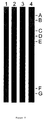
- the strip contains 19 parallel probe lines: A: Probe 5 (SEQ ID NO 5); B: Probe 6 (SEQ ID NO 6); C: Probe 7 (SEQ ID NO 7); D: Probe 8 (SEQ ID NO 8); E: Probe 26 (SEQ ID NO 26); F: Probe 22 (SEQ ID NO 22) and Probe 24 (SEQ ID NO 24); G: Probe 10 (SEQ ID NO 10); H: Probe 13 (SEQ ID NO 13); I: Probe 14 (SEQ ID NO 14); J: Probe 21 (SEQ ID NO 21); K: Probe 15 (SEQ ID NO 15); L: Probe 16 (SEQ ID NO 16); M: Probe 17 (SEQ ID NO 17); N: Probe 19 (SEQ ID NO 19); O: Probe 18 (SEQ ID NO 18); P: Probe 155 (antisense probe: 5'-GGGGGCCTGGAGGCTG-3'); Q: Probe 27 (SEQ ID NO 27); R: Probe 20 (SEQ ID NO 20); S: control line for conjugate binding.
- Strip 1 serum BR5
- Strip 2 serum BR12
- Strip 3 serum BR18
- Strip 4 serum BR22
- Strip 5 serum BR19
- Strip 6 serum BE95
- Strip 7 serum BU79
- Strip 8 serum BR23
- Strip 9 serum JP63.
- sequences used to construct this alignment are taken from the EMBL database and have the following accession number: 1 M62321, 2 D10749, 3 D00944, 4 D01221, 5 D13448, 6 D11443, 7 M84838, and 8 L08156.
- accession number 1 M62321, 2 D10749, 3 D00944, 4 D01221, 5 D13448, 6 D11443, 7 M84838, and 8 L08156.
- the sequences between nucleotides -220 and -180 are not shown, they are identical to HCV-1 in all isolates.
- nucleotide is identical to the corresponding nucleotide in HCV-1; '..', gap created between -145 and -144 to allow alignment with type 6 sequences which have a CA insertion; '.', gap created between -138 and -137 in most of the sequences to preserve alignment with sequences which have an extra nucleotide at that position.
- * refers to the conserved HCV sequence between resuidues -220 and -180 as shown in Figure 2.
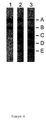
- Line probe assays including probes with SEQ ID NO 32, tested with type 1 and 2 sera. 1, type 1b serum BE82, 2, type 2a serum JP62, 3, type 2b serum BE91, A, conjugated control, B, probes 20 and 27, C, probe 8, D, probe 26, E, probe 32 (SEQ ID NO 32).
- Line probe assays including probes with SEQ ID NO33 and 34, tested with type 2a, 2b, and 2c sera.
- Line probe assays including probes with SEQ ID NO 31, 37 and 38, tested with type 4 sera. 1, type 4a serum GB116, 2, serum GB113, 3, type 4f serum GB438, A, conjugate control, B, probes 20 and 27, C, probe 37 (SEQ ID NO 37), D, probe 38 (SEQ ID NO 38), E, probe 19, F, probe 31 (SEQ ID NO 31), G, probe 7.
- Line probe assays including probes with SEQ ID NO 44, 45 and 46, tested with type 4a and 5a sera. 1, type 5a serum BE95, 2, type 4a serum GB116, A, conjugate control, B, probes 20 and 27, C, probe 44 (SEQ ID 44), D, probe 45 (SEQ ID NO 45), E, probe 46 (SEQ ID NO 46), F, probe 31 (SEQ ID NO 31), G, probe 7.
- Line probe assays including probes with SEQ ID NO 93, 94, 95, and 96, tested with type 4a and 5a sera. 1, type 4a serum GB116, 2, type 5a serum BE95.
- A conjugate control
- B probes 20 and 27,
- C probe 93 (SEQ ID NO 93) applied at a concentration of 0.4 pmol/ ⁇ l
- D probe 94 (SEQ ID NO 94) applied at a concentration of 2.5 pmol/ ⁇ l
- E probe 94 (SEQ ID NO 94) applied at a concentration of 1.0 pmol/ ⁇ l
- F probe 94 (SEQ ID NO 94) applied at a concentration of 0.4 pmol/ ⁇ l
- G probe 95 (SEQ ID NO 95) applied at a concentration of 2.5 pmol/ ⁇ l
- H probe 95 (SEQ ID NO 95) applied at a concentration of 1.0 pmol/ ⁇ l
- I probe 95 (SEQ ID NO 95) applied at a concentration of 0.4 pmol/ ⁇ l
- the signal-to-noise ratio of the sera tested in the Innotest HCV Ab is also given for some of the sera.
- LiPA Line Probe Assay
- a cPCR fragment containing incorporated biotinylated dUTP is hybridized to oligonucleotides which are immobilized on a nitrocellulose membrane.
- the stable hybridization duplex is then revealed by streptavidin-labelled alkaline phosphatase, and subsequent color development with NBT (nitro 42 blue tetrazolium) and BCIP (bromochloro-indolyl phosphate).
- NBT nitro 42 blue tetrazolium
- BCIP bromochloro-indolyl phosphate
- variable regions between positions -170 and -155, and between -132 and-117 in the linear sequence may be part of a stem in the folded viral RNA, and mutations in the first region may be complemented by another mutation in the second region to allow or disallow RNA duplex formation.
- Variation and conservation is expected to occur at the same positions in other new types of HCV as well and, therefore, this variable region might remain instrumental for the discrimination between all current and yet-to-be discovered types of HCV.
- NS3, and NS5 regions typing in these regions employing universal sets of primers might no longer be tenable.
- subtype 4a might be changed into another type 4 subtype, like 4c or 4e, and type 4 might be changed into type 5 or 6, in which case type 4a might become 6c, for example.
- new classification systems will not hamper classification of a certain group of isolates classified into a type or subtype by means of the proposed probes of the invention.
- Example 1 Serum samples used for typing and subtyping
- the primers used for the PCR reactions were complementary to the conserved areas of the 5' UR of the different HCV types. Degeneration was included to allow annealing to type 1 and type 2 sequences (Kato et al . , 1990; Nakao et al ., 1991; Okamoto et al ., 1991) and to the sequence of our type 3 clone (BR56; accession number D13448, DDJJB/EMBL/GenBank DNA data base deposited on 21/10/1992).
- the sequences of the outer PCR primers (HcPr98, SEQ ID NO 1 and HcPr29, SEQ ID NO 2) and of the nested PCR primers (HcPr95, SEQ ID NO 3 and HcPr96, SEQ ID NO 4) are listed in Table 4.
- the probes used for the detection of the different serum types are also listed in Table 4. All oligonucleotides were synthesized on a 392 DNA/RNA Synthesizer (Applied Biosystems).
- Viral RNA was extracted from serum essentially as described by Chomczynski and Sacchi (1987) with minor modifications. The RNA was coprecipitated with 20 ⁇ g Dextran T500 (Pharmacia). The RNA pellet was briefly dried and resuspended in 10 ⁇ l DEPC-treated H 2 O.
- the first strand cDNA synthesis was carried out in 20 ⁇ l at 42°C in the presence of 25 U HPRI (Amersham), 500 ⁇ M dATP, dCTP, dTTP and dGTP, 1 x AMV buffer (Stratagene) and 2.5 U AMV-RT (Stratagene). Seven ⁇ l of the resulting cDNA was amplified in an outer PCR over 40 cycles each consisting of 1 min 95°C, 1 min 55°C and 1 min 72°C in a total volume of 50 ⁇ l.
- the solution was adjusted to a final concentration of 200 ⁇ M of dATP, dCTP, dTTP and dGTP, 1 x Taq buffer (Stratagene), 0.2 ⁇ M of each primer, and 1 U Taq polymerase (Stratagene).
- One ⁇ l of the first round amplification product was amplified with the nested primers again for 40 cycles in a buffer with the same composition.
- the nested PCR contained 40 ⁇ M Bio-11-dUTP (Sigma) and 160 ⁇ M of dTTP.
- Plasmid DNA preparation was as described in the alkaline lysis method (Maniatis et al ., 1982). Sequencing reactions were carried out on double-stranded plasmid DNA with T7 and T3 primers by using the Deaza G/A T7 sequencing mixes (Pharmacia).
- Serum RNA from HCV-infected patients was used as template for cDNA synthesis, which in turn was a template for nested PCR.
- Two sets of PCR primers were designed: HcPr98 (SEQ ID NO 1) and HcPr29 (SEQ ID NO 2) for the outer reaction, HcPr95 (SEQ ID NO 3) and HcPr96 (SEQ ID NO 4) for the nested reaction (Table 4). These four primers were chosen to match the published sequences (Kato et al ., 1990; Nakao et al ., 1991; Okamoto et al ., 1991) and the sequence of a clone obtained from the untranslated region of isolate BR56. (see Figure 2).
- the resulting amplification product of the nested PCR is 235 base pairs (bp) long. Due to the incorporation of Bio-11-dUTP, there is a decrease in mobility which is clearly visible after agarose gel electrophoresis (Fig. 1). The size of the DNA fragments is the same for all the different HCV types, suggesting that a second experiment, like restriction enzyme digestion or hybridization, is necessary for classification. A membrane strip containing immobilized HCV-specific oligonucleotide probes applied as parallel lines was therefore developed. These strips are hybridized with PCR amplified DNA fragments of the 5' UR into which biotinylated nucleotides were incorporated during synthesis. After hybridization, streptavidin labelled with alkaline phosphatase is added and becomes bound to the biotinylated hybrids formed during the hybridization. After incubation with NBT/BCIP, a purple precipitate appears.
- the 16-mer oligonucleotides specific for the different types or subtypes of HCV (Table 4, number 5 to 27), were provided with a poly-(dT) tail at their 3' end as follows: 20 pmol of primer was incubated in 25 ⁇ l buffer containing 3.2 mM dTTP, 25 mM Tris.HCl (pH 7.5), 0.1 M sodium cacodylate, 1 mM CoCl 2 , 0.1 mM dithiothreitol, and 60 U Terminal deoxynucleotidyl Transferase (Pharmacia) for 1 hour at 37°C.
- the reaction was stopped by adding 2.5 ⁇ l 0.5 M EDTA (pH 8.0) and diluted with 20 x SSC (Maniatis et al ., 1982) until a final concentration of 6 x SSC and 2.5 pmol oligonucleotide/ ⁇ l was reached.
- Conjugate Diluent phosphate buffer containing NaCl, Triton, protein stabilizers, 0.1% NaN 3 ; Inno-Lia, Innogenetics, Antwerp, Belgium
- Conjugate Diluent containing 4000 x diluted streptavidin labelled with alkaline phosphatase (Gibco BRL) for another 30 minutes at RT.
- the strips are washed again 3 times with Rinse Solution and once with Substrate Diluent (Tris buffer containing NaCl and MgCl 2 ; Inno-Lia, Innogenetics, Antwerp, Belgium). Color development is achieved by adding BCIP and NBT to the Substrate diluent and incubation of the strips for 30 minutes at RT. The color development is stopped by replacing the buffer with distilled water.
- Conjugate Diluent phosphate buffer containing NaCl, Triton, protein stabilizers, 0.1% NaN 3 ; Inno-L
- Example 5 A LiPA for discrimination between HCV types 1, 2 and 3
- sequences for the probes against type 3 were derived from a cPCR clone from serum BR56 (accession number D13448).
- D13448 serum BR56
- two regions of 16 nucleotides containing 4 to 6 mutations could be observed each time.
- type 2 sequences became available, variation was again maintained in these two regions. Therefore, the position of the typing probes was chosen in those regions with a relatively low degree of homology between types, but good conservation within one type.
- a total of eight separately immobilized oligonucleotides were applied.
- HcPr124 SEQ ID NO 5 and HcPr125, SEQ ID NO 6
- HcPr136 SEQ ID NO 9 and HcPrl37
- HcPr126 SEQ ID NO 11
- HcPr127 SEQ ID NO 12
- HCV HCV
- cPCR products were synthesized from 23 Brazilian sera (BR1 to BR23) and, after hybridization, 17 of them recognized the 16-mers of type 1.
- Four type 3 sera were found, as well as one type 2a serum.
- Serum BR23 was co-infected with type 1 and type 3.
- Two pools of Japanese sera were subsequently tested: JP63 reacted with the type 1 and type 2a probes, and the majority of the JP62 pool contained type 2a sequences.
- HcPr95 SEQ ID NO 3
- HcPr96 SEQ ID NO 4
- the type 2b probes HcPr126 (SEQ ID NO 11) and HcPr127 (SEQ ID NO 12), to which JP62 did not react, differed by only one and two nucleotides, respectively, from the sequence of JP62 (accession number D13453). Therefore, the chosen hybridization and washing conditions were very stringent and that even single mismatches abolish hybridization in this assay.
- the isolates When comparing all available 5'UR sequences of type 3 (present invention; Bukh et al ., 1992; Chan et al ., 1992; Lee et al ., 1992), the isolates could be divided into two groups according to the presence of a common G (type 3a; HcPr140, SEQ ID NO 15) or a more rare A (type 3b; HcPr139, SEQ ID NO 16) at position -139. Discrimination between types 2a and type 2b (or K2a and K2b) could be made in the variable regions as reported above.
- Example 7 Identification of type 4 isolates and incorporation of type 4-specific probes in the LiPA
- PCR fragments amplified from 6 Burundian sera failed to react with any of the 16-mers on the strips.
- Three PCR fragments from these Burundian samples (BU74: accession number D13449, which was identical to BU76, and BU79: accession number D13450; Figure 2) were cloned and sequenced. Sequences that were clearly different from most of the previously described types were obtained.
- the Burundian samples are related to each other, and to Z6 (Bukh et al., 1992) and show higher homologies to type 1 than to type 3 or type 2. However, most of the differences with type 1 were again located in the variable regions.
- oligonucleotide HcPr142 (SEQ ID NO 20), carrying one degeneration, was chosen from a highly conserved region as universal HCV probe for the confirmation of the presence of the PCR product (Table 4).
- three oligonucleotides were synthesized for identification of the type 4 sequences (HcPr144, SEQ ID NO 17 with one degeneration, HcPr145, SEQ ID NO 18 and HcPr146, SEQ ID NO 19; Table 4).
- a universal type 2 probe was selected outside the variable regions (HcPr147, SEQ ID NO 8, Table 4), since a universal probe for the detection of type 2 could not be chosen from the regions between positions -170 and -155 and between positions - 132 to -117.
- the fourth NE serum which showed good reactivities in both Ortho RIBA and INNO-LIA, contained a type 1a isolate.
- BE64 to BE67 were infected with type 1b strains.
- One patient of Italian origin (BE68) had a type 2a infection, and BE69 contained type 3a sequences. The latter was obtained from a case of chronic, viral-like NANB hepatitis, but was negative in all second generation assays and anti-NS3, anti-E1, and anti-E2 research assays.
- This serum had a very low virus titer and became weakly positive only after the second round of PCR in four different samples taken during 2 years, showing the need for nested cPCR in HCV diagnosis.
- the sequence of the nested PCR fragment was identical with BR56. This was not surprising, since type 3 strains show very little sequence variation.
- a type 1b PCR fragment recognized the 3b subtype probe HcPr139 (SEQ ID NO 16). This can be explained by assuming that the 1b sequence of serum BE67 has an A instead of a T at position -139.
- Epitopes for the NS4 and NS5 region are located in highly variable regions, disabling most of the immunological cross-reaction. As the current antibody assays contain type 1 epitopes, it is possible that a few percent of type 2, type 3, and type 4 infected sera will show a negative result. However, the conclusion of lack of cross-reaction of the type 3 Brazilian sera with type 1 NS4 and NS5 antigens cannot be drawn from our results (Table 3). For the 14 randomly chosen sera (BR24 to BR37; Table 3), there was 100% correlation between the LIA reactivity and the 9 type 1 viruses. From four type 3 sera, two (BR34 and BR36) reacted with NS4 and three (BR33, BR34 and BR35) with NS5.
- BR37 was not taken into account because of the coinfection.
- 58% and 44% of the type 1 sera recognized the NS4, and NS5 epitopes, respectively. These percentages are rather low and due to the selection criteria.
- 37% and 53% were reactive with the NS4 and NS5 epitopes, respectively. It is possible that higher cross-reactivities are observed in high-risk groups, such as in those samples obtained from Brazil, as compared with results in European blood donors (present invention and Chan et al ., 1992). Such cross-reacting sera could be induced by multiple infections, some of which occur simultaneously, but others might occur after one another.
- a previous anti-HCV memory could be boosted by new HCV infections and result in co-circulation of viruses of one type with antibodies mainly directed against another type.
- serum BR56 which has been typed as HCV type 3, but contained antibodies to type 1 core, E1, E2, NS3, NS4, and NS5 (data not shown). It remains to be determined whether anti-type 3 antibodies are present in this serum.
- HCV types could also show different progression to long-term liver disease, as has already been reported (Okamoto et al., 1992a).
- the LiPA allows a rapid determination of the type of HCV infection.
- This assay has the ability to discriminate between 4 different HCV types and 8 subtypes, and is a good means for determining new types.
- this assay can be further improved by, for example, replacing the cPCR reactions by the RNA-capture PCR.
- this assay could prove to be instrumental in further establishing the relation between genotypes, future serotypes, and the clinical status or outcome of the disease.
- Example 8 Identification of new types and subtypes and probes useful for their classification .
- the sequences of the 5'untranslated region were obtained after nested PCR by means of primers with SEQ ID NO. 1, 2, 3 and 4, cloning, and sequencing as described in example 2.
- probe 30 including a degeneration of T and C at position -94, should enable better genotyping of subtype 1b.
- Isolate BE92 reacted only with probes 8 and 26 in addition to the universal probes 20 and 27.
- this isolate could be classified as type 2, but could not be subtyped because no reactivity with probes 23, 24, 25, or 26 could be observed.
- two new motifs could indeed be observed: GGACCCAGTCTTCCTG, covered by probe 33, and TGCCTGGTCATTTGGG, covered by probe 34. Sequencing of the NS5 region indeed revealed homologies with type 2a and 2b isolates compatible with classification within the same type, but in another subtype which is the proposed subtype 2c.
- Isolates BE93 and BE94 did not show any reactivity with the subtyping probe 14. After sequencing the 5'untranslated region and the NS5 region, it was concluded that these isolates belonged to the 3a subtype. Therefore, a probe containing a C and A degeneration at position -118 like probe 35, should allow better genotyping of subtype 3a.
- Isolates GB48, GB116, GB358, and GB569 showed positive hybridization signals on probe 17 and 19 in LiPA, indicating similarity to the previously reported type 4 isolates, but isolates GB549 and GB809 only reacted with the universal probes.
- the sequences of parts of the 5'untranslated region and NS5 were obtained. From Figure 5 and 6 and Table 6, it can be concluded that the isolates represented by GB358 belong to the same subtype of type 4, which is the proposed subtype 4a. However, both GB549 and GB809 show lower homologies to the subtype 4a, 4b and 4d isolates, and also to each other, but GB809 seems to belong to the same subtype as Z4.
- probes 38 and 19 are useful for detection and classification of subtype 4a.
- Probe 38 is specific for subtype 4a, 4b, 4d, 4f and 3b, while probe 19 recognizes subtypes 3b, 4a and 4d, but also hybridizes to the new types 3c and 4f.
- the new subtype 3b sequence HCV-TR should cross-react with these probes.
- 3b can still be classified as type 3 because of the reactivity with the type 3-specific probe 21.
- GB549 also shows characteristic motifs. Motif AATCGCCGGGACGACC can be detected by probe 40 and the sequence AATGCCCGGCAATTTG is detectable with probe 41. Thus, probes 40 and 41 are useful for subtyping of subtype 4b.
- Type 4 isolates usually show a T at position -238 and a A at -235. Therefore, probes 37, 38, and 51 should enable better genotyping of type 4.
- BE95 which only hybridized to probes 7 and 17 in the LiPA shows low homologies in the coding region of about 68% with all other isolates, except BE96 which shows an homology to BE95 compatible with classification into the same subtype, which is the proposed subtype 5a.
- BE95, BE96, and SA1 all show the same motifs GAGTGTCGAACAGCCT, detected with probe 44; AATTGCCGGGAYGACC, detectable with probes 45 and 47; and TCTCCGGGCATTGAGC, detectable with probe-46.
- probes 44, 45, 46 and 47 are useful for genotyping of type 5a.
- Isolate GB438 contains sequence motifs which are typical for subtype 4a, detectable with probes 38 and 19, but still shows a different sequence in the E1 region, representing a new subtype within type 4, which was designated subtype 4f. Discrimination from subtype 4a may be performed by means of probes with SEQ ID NO 51 and 7.
- Probes 29, derived from the sequence of BE90, and probes 51 and 52, derived from the sequence of GB724, may be useful to improve genotyping of certain HCV types or subtypes.
- variable regions in the 5'UR are expected tot contain genotype-specific sequences also in newly discovered genotypes, as examplified in example 8, and consequently, such new genotype-specific motifs should again be detectable by means of the genotype-specific probes as described in example 8.
- probes 32, and as described in example 8 probes 31, 33, 34, 37, 38, 44, 45, and 46 were synthesized and applied to nitrocellulose membranes and line probe assays with biotin-labelled PCR fragments was performed as described in example 3 and 4, except for the labelling of the PCR product with biotin which was not incorporated form bio-11-dUTP, but from of 5'-biotinylated primers with SEQ ID NO 3 and 4 or 5'-biotinylated primers with SEQ ID NO 1 and 2, during the synthesis of the PCR fragment.
- Figure 6 shows the type-specific hybridization of HCV type 2, but not type 1, 5'UR fragments to the probe with SEQ ID NO 32. Both subtype 2a and 2b isolates hybridized specifically to probe 32.
- the probes with SEQ ID NO 33 and 34 could be shown to hybridize specifically to the genotype 2c PCR product derived from serum BE92, while genotype 2a and 2b sera did not react to these probes although a specific hybridization with the respective 2a and 2b genotype-specific probes could be observed. It is to be understood that the new genotype 2c may differ from other genotype 2c subtypes discovered recently, and therefore, the alternative names may be proposed for denomination of this subtype.
- Figure 8 shows the line probe performed with type 4 sera to show specific hybridization of the most common type 4 sera to probes 37 and 38.
- Figure 9 depicts specific hybridization of type 5a sera to probes 44, 45, 46, and probe 7, while reactivity of type 4 sera is usually confined to probe 31 and absent on probes 44 to 46. Therefore, the promiscuity of the probe with SEQ ID NO 18 for both type 4 and type 5 isolates, can now conveniently be overcome by employing, in addition to the probe with SEQ ID NO 19, probes with SEQ ID NO 37, 38, 44, 45, 46, 7, 30, and 31 for discrimination of genotypes 4 and 5.
- FIG. 10 shows line probe assays with the type 4a serum GB116 and the type 5a serum BE95, as described in example 4, except for the following: After denaturation of the PCR fragment in NaOH/SDS, 1.0 ml hybridization solution (prewarmed at 50°C) consisting of 3x SSC (Maniatis et al., (1982)) and 1 % sodium dodecyl sulphate (SDS), was added to the denatured PCR product and hybridization was performed in a shaking water bath at 50°C for 2 hours.
- 3x SSC Maniatis et al., (1982)
- SDS sodium dodecyl sulphate
- probes to retain specificity in other hybridization conditions it may also be preferable to elongate or shorten the contiguous HCV sequence and/or to reverse the sense or the probes to allow genotype-specific hybridization at a certain preferred temperature or salt concentration.
- probe with SEQ ID NO 37 which was able to discriminate between type 4 and 5 isolates in tertramethylammoniumchloride buffer as described in example 10, was now changed into probe with SEQ ID NO 93 (5'-GAGTGTTGTACAGCCTCC-3') by elongation of the contiguous HCV sequence at the 3'end with 2 nucleotides, and probe 93 showed a specific reactivity in SSC/SDS hybridization buffer ( Figure 10).
- probe with SEQ ID NO 44 which was able to discriminate between type 4 and 5 isolates in tertramethylammoniumchloride buffer as described in example 10, was now changed into probe with SEQ ID NO 96 (5'-GAGTGTCGAACAGCCTC-3') by elongation FO the contiguous HCV sequence at the 3' end with 1 nucleotide, and probe 96 showed a specific reactivity in SSC/SDS hybridization buffer ( Figure 10).
- the antisense probe with SEQ ID NO 46 which targets positions -132 tot -117 was able to discriminate between type 4 and 5 isolates in tertramethylammoniumchloride buffer as described in example 10, was now changed into probe with SEQ ID NO 95 (5'-TGCCCGGAGATTTGGG-3'), a sense probe which targets positions -126 to -111, and probe 95 showed a specific reactivity in SSC/SDS hybridization buffer ( Figure 10).
- This example illustrates the numerous possibilities of developing probes to those skilled in the art for targetting the genotype-specific mutations that are present in other new or still to be discovered genotypes.
Abstract
Description
- The present invention relates to the detection of target sequences in a biological sample.
- It is an aim of the present invention is to provide a method for the rapid and indisputable determination of the presence of one or several target sequences present in a biological sample.
- The present invention relates more particularly to the LiPA (line probe assay) technology which is in essence a method for detecting target sequences present in a biological sample comprising at least the following steps:
- a) possibly amplifying nucleic acids present in the biological sample by means of a set of primers,
- b) providing a solid support onto which nucleotide probes have been immobilized in the form of parallel lines,
- c) incubating the nucleic acids present in said biological sample with said solid support under conditions enabling hybridization between the probes and the target sequence possibly present in the analyte strand of said biological sample,
- d) detecting the hybrids formed in step c).
-
- in the examples section of the present invention, a sensitive PCR protocol has been used for the highly conserved 5' untranslated region of Hepatitis C virus (HCV) with sets of nested, universal primers. The obtained amplification product was hybridized to oligonucleotides directed against the variable regions of the 5' UR, immobilized as parallel lines on membrane strips (reverse-hybridization principle). This hybridization assay, called line probe assay (LiPA), is a rapid assay, by means of which previously poorly described isolates were detected. This LiPA technology allows an easy and fast determination of HCV types and their subtypes present in patient serum. This technology also forms an easy and reliable general typing system for parenterally transmitted human viral diseases. If analyte strand amplification is necessary, a set of primers can be provided per viral organism to be differentiated and classified.
- The present invention relates more particularly to a method as defined above wherein the nucleic acids present in said sample are first amplified by means of a set of primers. Amplifying the target region, for instance via a polymerase chain reaction by means of the above-mentioned set of primers can be accompanied by the incorporation of a label such as digoxigenin or biotin into the amplified target sequence. The amplification can be repeated between 20 and 80 times, advantageously between 30 and 50 times. Amplification may be performed by any technique known in the art such as by the polymerase chain reaction (PCR; Saiki et al., 1988), ligase chain reaction (LCR; Landegren et al., 1988; Wu & Wallace, 1989; Barany, 1991), nucleic acid sequence-based amplification (NASBA; Guatelli et al., 1990; Compton, 1991), transcription-based amplification system (TAS; Kwoh et al., 1989), strand displacement amplification (SDA; Duck, 1990; Walker et al., 1992) or amplification by means of Qb replicase (Lizardi et al., 1988; Lomeli et al., 1989).
- The cDNA amplification step is preferably achieved by means of PCR technology and may consist of steps:
- (a) providing a set of primers for a polymerase chain reaction method which flank the target sequence to be detected;
- (b) amplifying the target region via a polymerase chain reaction method by means of the primers of (a); and in the same step an appropriate label molecule can be incorporated into the amplified targetth said label molecule being preferably digoxigenin or biotin.
-
- The term "appropriate label molecule" may include the use of labeled nucleotides incorporated during the polymerase step of the amplification such as illustrated in Saiki et al. (1988) and Bej et al. (1990) and or any other method known to the person skilled in the art.
- The assays as described in this invention may be improved in several ways obvious for the person skilled in the art. For example the cPCR reactions can be preceded by an RNA-capture step.
- The present invention also relates to a method as defined above wherein biotinylated dUTP is incorporated during step a).
- The present invention also relates to a method as defined above wherein said solid support is a membrane strip, preferably a nitrocellulose membrane strip.
- Prior to application to the membrane or fixation it may be convenient to modify the nucleic acid probe in order to facilitate fixation or improve the hybridization efficiency. Such modifications may encompass homopolymer tailing, coupling with different reactive groups such as aliphatic groups, NH2 groups, SH groups, carboxylic groups, or coupling with biotin or haptens.
- The present invention also relates to a method as defined above wherein one of said probes present on said solid support is a control to determine if there is hybridization.
- The present invention relates also to a method as defined above wherein one of said probes present on said solid support is a control to determine if there is conjugate binding.
- The present invention relates also to a method as defined above wherein step d) is accomplished by means of streptavidin labelled alkaline phosphatase.
- The present invention relates also to a method as defined above wherein said probes are immobilized to said solid support by applying 1 pmol of a poly-(dT) probe solution.
- The present invention relates also to a method as defined above wherein said probes were fixed to said solid support by baking at 80°C for two hours.
- The present invention relates also to a method as defined above wherein the PCR amplification product, containing incorporated Biotin dUTP, is denatured and hybridized to the probes on the solid support at 42°C in 3 M tetramethylammonium chloride and consequently washed at 51°C for 30 minutes.
- As demonstrated in the Examples section, the LiPA allows a rapid determination of the type of HCV infection. This assay has the ability to discriminate between 4 different HCV types and 8 subtypes, and is a good means for determining new types.. Moreover this assay can be further improved by replacing the cPCR reactions by the RNA-capture PCR...this assay could prove to be instrumental in further establishing the relation between genotypes, future serotypes, and the clinical status or outcome of the disease.
- The LiPA also allows to determine the type and/or subtype of HIV, HBV and/or HTLV infection.
- Other hybridization conditions are specified in Example 10.
- The invention also relates to a solid support, particularly a membrane strip containing, on known locations of its surface, a selection of probes applied in the form of parallel lines.
- The present invention also relates to a diagnostic kit comprising a solid support as defined above and possibly also buffers enabling the hybridization as defined above and/or primers enabling the amplification of sample nucleic acids as defined above.
- The present invention also relates to 13. A diagnostic kit according to claim 12 for genotyping HCV, and/or HIV, and/or HBV, and/or HTLV present in a biological sample.
- The expression "probe" corresponds to any polynucleotide which forms a hybrid with a target sequence present in a sample on the basis of complementarity. Such a probe may be composed of DNA, RNA, or synthetic nucleotide analogs. The probes of the invention can be incubated with an analyte strand immobilized to a solid substrate. In a preferred embodiment of the invention, the probes themselves can be immobilized to a solid substrate. These probes may further include capture probes, characterized as being coupled to a binding molecule which in turn is directly or indirectly bound to a solid substrate, or may also include label probes, characterized in that they carry a detectable label.
- The term "analyte strand" corresponds to a single- or double-stranded nucleic acid molecule which is suspected to contain sequences which may be present in a biological sample, with said analyte strand being directly detected or detected after amplification. This analyte strand is preferentially positive- or negative-stranded RNA, cDNA, or amplified cDNA.
- The expression "biological sample" may refer to any biological sample (tissue or fluid) containing HCV sequences and refers more particularly to blood serum or plasma samples.
- The detection of hybrids formed between the target region, if present, and the probes as mentioned above depends on the nature of the reporter molecule used (either present on the probe or on the analyte strand to be targeted) and may be determined by means of colorimetric, fluorescent, radiometric detection or any other method comprised in the state of the art.
- The term "(HCV) isolates" refers to any biological fluid containing hepatitis C virus genetic material obtained from naturally infected humans or experimentally infected animals, and also refers to fluids containing hepatitis C virus genetic material which has been obtained from in vitro experiments. For instance, from in vitro cultivation experiments, both cells and growth medium can be employed as a source of genetic HCV material.
- The expression "hybridize" or "target" refers to a hybridization experiment carried out according to any method known in the art, and allowing the detection of homologous targets (including one or few mismatches) or preferably completely homologous targets (no mismatches allowed).
- The term "genotyping" in the context of the present invention refers to either typing and/or subtyping.
- The term "solid substrate or solid support" can refer to any substrate to which an oligonucleotide probe can be coupled, provided that it retains its hybridization characteristics and provided that the background level of hybridization remains low. Usually the solid substrate will be a microtiter plate or a membrane (e.g. nylon or nitrocellulose)
- Ethidium bromide-stained agarose gel showing the length of the nested PCR fragments. Lane A of each pair shows the PCR fragment with incorporation of Bio-11-dUTP. Lane B is the PCR fragment without Bio-11-dUTP. 1: Serum BR28, 2: Serum BR24, 3: Serum BR29, 4: Serum BR33, 5: Serum BR36, 6 and 7: negative control sera, 8: Serum JP62, 9: Serum BR23, 10: cPCR control without template, M: molecular weight markers.
- Alignment of the 5' UR nucleotide sequences of isolates from four different types of HCV. Boxed nucleotides indicate the positions of probes used for typing of the four different groups. The underlined nucleotides are used for subtyping within each group. The period between nucleotide -140 and -139 in most sequences corresponds to the insertion in some of the
type 4 isolates. The numbering of the probes corresponds with the numbers used in Table 4. - HCV LiPA Typing results of some representative sera. The strip contains 19 parallel probe lines:
A: Probe 5 (SEQ ID NO 5); B: Probe 6 (SEQ ID NO 6); C: Probe 7 (SEQ ID NO 7); D: Probe 8 (SEQ ID NO 8); E: Probe 26 (SEQ ID NO 26); F: Probe 22 (SEQ ID NO 22) and Probe 24 (SEQ ID NO 24); G: Probe 10 (SEQ ID NO 10); H: Probe 13 (SEQ ID NO 13); I: Probe 14 (SEQ ID NO 14); J: Probe 21 (SEQ ID NO 21); K: Probe 15 (SEQ ID NO 15); L: Probe 16 (SEQ ID NO 16); M: Probe 17 (SEQ ID NO 17); N: Probe 19 (SEQ ID NO 19); O: Probe 18 (SEQ ID NO 18); P: Probe 155 (antisense probe: 5'-GGGGGCCTGGAGGCTG-3'); Q: Probe 27 (SEQ ID NO 27); R: Probe 20 (SEQ ID NO 20); S: control line for conjugate binding. - The strips were hybridized with cPCR products of the following sera: Strip 1: serum BR5, Strip 2: serum BR12, Strip 3: serum BR18, Strip 4: serum BR22, Strip 5: serum BR19, Strip 6: serum BE95, Strip 7: serum BU79, Strip 8: serum BR23, Strip 9: serum JP63.
- Nucleotide sequence alignment of the HCV 5'untranslated regions of new isolates BE90, BE91, BE92, BE93, BE94, BE95, BE96, BE97, BE98, BE99, GB48, GB116, GB358, GB569, GB549, GB809, CAM600, CAM736, GB478, GB724, and GB438, with sequences of
HCV type 1a (HCV-1), 1b (HCV-J), 2a (HC-J6), 2b (HC-J8), 3a (BR56), 3b (HCV-TR), 5 (SA1), 6 (HK1). The sequences used to construct this alignment are taken from the EMBL database and have the following accession number: 1 M62321, 2 D10749, 3 D00944, 4 D01221, 5 D13448, 6 D11443, 7 M84838, and 8 L08156. The sequences between nucleotides -220 and -180 are not shown, they are identical to HCV-1 in all isolates. '-', nucleotide is identical to the corresponding nucleotide in HCV-1; '..', gap created between -145 and -144 to allow alignment withtype 6 sequences which have a CA insertion; '.', gap created between -138 and -137 in most of the sequences to preserve alignment with sequences which have an extra nucleotide at that position. * refers to the conserved HCV sequence between resuidues -220 and -180 as shown in Figure 2. - Amino acid sequence alignment of the NS5 sequences of isolates BE90, BE91, BE92, BE93, BE95, GB358, GB549, and GB809 with known sequences as described in Table 6.
- Line probe assays including probes with
SEQ ID NO 32, tested withtype probe 8, D,probe 26, E, probe 32 (SEQ ID NO 32). - Line probe assays including probes with SEQ ID NO33 and 34, tested with
type probe 8, D,probe 26, E,probe 32, F,probe 22, G, probe 24, H,probe 23, I, probe 25, J, probe 33 (SEQ 33), K, probe 34 (SEQ ID NO 34). - Line probe assays including probes with
SEQ ID NO type 4 sera. 1, type 4a serum GB116, 2, serum GB113, 3,type 4f serum GB438, A, conjugate control, B, probes 20 and 27, C, probe 37 (SEQ ID NO 37), D, probe 38 (SEQ ID NO 38), E,probe 19, F, probe 31 (SEQ ID NO 31), G,probe 7. - Line probe assays including probes with
SEQ ID NO type probe 7. - Line probe assays including probes with SEQ ID NO 93, 94, 95, and 96, tested with
type - Overview of the different classification systems.
- Interpretation of the results shown in Figure 3.
- Final results of HCV LiPA typing and HCV antibody assays.
- A summary of the typing in relation to the serology is presented. The INNO-LIA HCV Ab assay contains one line with NS4 epitopes, one line with NS5 epitopes, and 4 lines with core epitopes. Only the highest score for the core lines is given. The intensity of the signal is given by a number: 0 = negative; 9 = indeterminate; 1 to 3 = positive. The final interpretation of the antibody test is given in the LIA column: 1 = positive; 0 = negative; 9 = indeterminate.
- The signal-to-noise ratio of the sera tested in the Innotest HCV Ab is also given for some of the sera.
- Nucleotide sequence, position, and orientation of the primers and probes.
- Overview of new probes designed from new types and subtypes of HCV. The type or subtype for which classification is possible or improved, the sequence, and the SEQ ID NO. are shown. * represents a probe which does not type or subtype all isolates found representing said type or subtype. The underlined letters indicate provisional divisions into subtypes.
- Sequence homology between BE90, BE91, BE92, BE93, BE95, GB358, GB549, and GB809 and published sequences in the HCV NS5 region from nucleotide 7935 to 8274, according to the numbering used in the present invention. Homology scores within the same subtype are in bold. Published sequences used to perform homology calculations were taken from the EMBL database and have the following accession numbers: 1M62321, 2D10749, M67463, D90208, X61596, L02836, 7 M84754, 8 D10750, 9 D11168; D01171, 10 S38204, 11 M58335, 12 D10078, 13 D10079, 14 D10080, 15 D10081, 16 D00944, and 17 D01221. All of them represent complete genomes, except 12, 13, 14, 15 and 18 for which NS5 sequences were published. 18was published in the Chiron patent WO 92/19743,
SEQ ID NO 18. - In order to study the natural variation of HCV isolates obtained from different geographical areas throughout the world, a rapid means for typing and subtyping of HCV isolates in the form of a Line Probe Assay (LiPA) was developed.
- Essentially, a cPCR fragment containing incorporated biotinylated dUTP is hybridized to oligonucleotides which are immobilized on a nitrocellulose membrane. The stable hybridization duplex is then revealed by streptavidin-labelled alkaline phosphatase, and subsequent color development with NBT (
nitro 42 blue tetrazolium) and BCIP (bromochloro-indolyl phosphate). The cPCR fragment is synthesized from the 5' UR of any HCV RNA using highly conserved sets of primers. The oligonucleotides used for typing are directed against the internal type-specific variable parts of the cPCR fragment. In fact, the 2 variable regions between positions -170 and -155, and between -132 and-117 in the linear sequence may be part of a stem in the folded viral RNA, and mutations in the first region may be complemented by another mutation in the second region to allow or disallow RNA duplex formation. Variation and conservation is expected to occur at the same positions in other new types of HCV as well and, therefore, this variable region might remain instrumental for the discrimination between all current and yet-to-be discovered types of HCV. Moreover, since higher variabilities compared to the 5' UR are observed in the core, NS3, and NS5 regions, typing in these regions employing universal sets of primers might no longer be tenable. - The proposed nomenclature of this invention is provisional and could still be subject to amendments according to new guidelines that may be set forward by international committees. For example,
subtype 4a might be changed into anothertype 4 subtype, like 4c or 4e, andtype 4 might be changed intotype case type 4a might become 6c, for example. However, new classification systems will not hamper classification of a certain group of isolates classified into a type or subtype by means of the proposed probes of the invention. - Sixty-one Brazilian samples (BR1 to BR61) were tested in the HCV Antibody ELISA assay (Innotest HCV Ab, Innogenetics) as well as in the Inno-LIA HCV Ab test (Innogenetics). The first 23 serum samples (BR1 to BR23, Table 3) were taken from hemodialysis patients with either high ALT levels or positive Inno-LIA results, or from blood donors from which the recipient developed NANB hepatitis liver disease. Fourteen (BR24 to BR37) of the other serum samples were randomly chosen; the 24 remaining sera (BR38 to BR61) were selected on the basis of their LIA pattern. Most of the latter showed weakly positive, indeterminate, or negative reactivity with the NS4 and NS5 synthetic peptides on the LIA. The following sera were also included in this typing effort: two pools of Japanese sera (JP62 and J63), six Belgian sera (BE64 to BE69), four sera from the Netherlands (NE70 to NE73), six sera from Burundi (BU74 to BU79) and two sera from Gabon (GB80 and GB81). They were all tested with the Inno-LIA HCV Ab assay system. The sera BU74 to BU78 were only positive for anti-core antibodies, while the serum BU79 reacted only with the NS5 line. Both Gabonese sera were LIA HCV negative (Inno-LIA HCV), HIV negative (Innotest HIV), but HTLV positive (Innotest HTLV). One serum from Belgium (BE69) and one from the Netherlands (NE73) were completely negative. Three of the NE-sera (NE71 to NE73) were selected because they were negative in the second generation RIBA test (Ortho Diagnostics Inc.).
- The primers used for the PCR reactions were complementary to the conserved areas of the 5' UR of the different HCV types. Degeneration was included to allow annealing to type 1 and
type 2 sequences (Kato et al., 1990; Nakao et al., 1991; Okamoto et al., 1991) and to the sequence of ourtype 3 clone (BR56; accession number D13448, DDJJB/EMBL/GenBank DNA data base deposited on 21/10/1992). The sequences of the outer PCR primers (HcPr98,SEQ ID NO 1 and HcPr29, SEQ ID NO 2) and of the nested PCR primers (HcPr95,SEQ ID NO 3 and HcPr96, SEQ ID NO 4) are listed in Table 4. The probes used for the detection of the different serum types are also listed in Table 4. All oligonucleotides were synthesized on a 392 DNA/RNA Synthesizer (Applied Biosystems). - Viral RNA was extracted from serum essentially as described by Chomczynski and Sacchi (1987) with minor modifications. The RNA was coprecipitated with 20 µg Dextran T500 (Pharmacia). The RNA pellet was briefly dried and resuspended in 10 µl DEPC-treated H2O. After adding 2
µl 150 ng/µl random primers (Pharmacia) and denaturating for 10 minutes at 65°C, the first strand cDNA synthesis was carried out in 20 µl at 42°C in the presence of 25 U HPRI (Amersham), 500 µM dATP, dCTP, dTTP and dGTP, 1 x AMV buffer (Stratagene) and 2.5 U AMV-RT (Stratagene). Seven µl of the resulting cDNA was amplified in an outer PCR over 40 cycles each consisting of 1 min 95°C, 1 min 55°C and 1min 72°C in a total volume of 50 µl. The solution was adjusted to a final concentration of 200 µM of dATP, dCTP, dTTP and dGTP, 1 x Taq buffer (Stratagene), 0.2 µM of each primer, and 1 U Taq polymerase (Stratagene). One µl of the first round amplification product was amplified with the nested primers again for 40 cycles in a buffer with the same composition. For HCV typing, the nested PCR contained 40 µM Bio-11-dUTP (Sigma) and 160 µM of dTTP. Both the outer and the nested PCR product were then subjected to electrophoresis in a 2% low melting point (NuSieve GTG, FMC)/1% Ultra Pure (Gibco BRL) agarose gel. After ethidium bromide staining, PCR fragments were cut out from the agarose gel, the DNA was recovered by centrifugation through a 0.45 µm HV membrane (Millipore), purified by two phenol/ chloroform and two ether extractions, precipitated, and subsequently polished with T4 DNA polymerase (Boehringer), kinated with T4 kinase (Boehringer), and finally ligated in the dephosphorylated Eco RV site of pBluescript KS(-) (Stratagene). Plasmid DNA preparation was as described in the alkaline lysis method (Maniatis et al., 1982). Sequencing reactions were carried out on double-stranded plasmid DNA with T7 and T3 primers by using the Deaza G/A T7 sequencing mixes (Pharmacia). - The results of these sequencing reactions are shown in Figure 2. The following sequences were deposited in DNA databases (BR56: DDBJ/EMBL/Genbank accession number D13448; BU74: DDBJ/EML/GenBank, accession number D13449; BU79: accession number D13450; GB80: accession number D13451; GB81: accession number D13452; GP62: accession number D13453).
- Serum RNA from HCV-infected patients was used as template for cDNA synthesis, which in turn was a template for nested PCR. Two sets of PCR primers were designed: HcPr98 (SEQ ID NO 1) and HcPr29 (SEQ ID NO 2) for the outer reaction, HcPr95 (SEQ ID NO 3) and HcPr96 (SEQ ID NO 4) for the nested reaction (Table 4). These four primers were chosen to match the published sequences (Kato et al., 1990; Nakao et al., 1991; Okamoto et al., 1991) and the sequence of a clone obtained from the untranslated region of isolate BR56. (see Figure 2). The resulting amplification product of the nested PCR is 235 base pairs (bp) long. Due to the incorporation of Bio-11-dUTP, there is a decrease in mobility which is clearly visible after agarose gel electrophoresis (Fig. 1). The size of the DNA fragments is the same for all the different HCV types, suggesting that a second experiment, like restriction enzyme digestion or hybridization, is necessary for classification. A membrane strip containing immobilized HCV-specific oligonucleotide probes applied as parallel lines was therefore developed. These strips are hybridized with PCR amplified DNA fragments of the 5' UR into which biotinylated nucleotides were incorporated during synthesis. After hybridization, streptavidin labelled with alkaline phosphatase is added and becomes bound to the biotinylated hybrids formed during the hybridization. After incubation with NBT/BCIP, a purple precipitate appears.
- The 16-mer oligonucleotides, specific for the different types or subtypes of HCV (Table 4,
number 5 to 27), were provided with a poly-(dT) tail at their 3' end as follows: 20 pmol of primer was incubated in 25 µl buffer containing 3.2 mM dTTP, 25 mM Tris.HCl (pH 7.5), 0.1 M sodium cacodylate, 1 mM CoCl2, 0.1 mM dithiothreitol, and 60 U Terminal deoxynucleotidyl Transferase (Pharmacia) for 1 hour at 37°C. The reaction was stopped by adding 2.5 µl 0.5 M EDTA (pH 8.0) and diluted with 20 x SSC (Maniatis et al., 1982) until a final concentration of 6 x SSC and 2.5 pmol oligonucleotide/µl was reached. - One pmol of this solution was applied over a distance of 4 mm on a nitrocellulose membrane. As control for the conjugate, biotinylated DNA was applied alongside. The oligonucleotides were fixed to the membrane by baking at 80°C for two hours. The membrane was then sliced in 4-mm strips.
- Ten µl of the nested PCR amplification product, containing incorporated Bio-11-dUTP, is mixed with 10 µl of 400 mM NaOH/10 mM EDTA and incubated at room temperature (RT) for 10 minutes. Then, 1 ml prewarmed (37°C) hybridization buffer containing 3 M tetramethylammonium chloride (TMACl, Merck), 50 mM sodium phosphate (pH 6.8), 1 mM EDTA, 5 x Denhardts (Maniatis et al., 1982), 0.6% (w/v) SDS and 100 µg/ml sheared salmon sperm DNA is added and the hybridization is carried out in a shaking water bath at 42°C for 2 hours (Jacobs et al., 1988). The strips are washed 2 times at RT for 5 minutes with 1 ml prewarmed (37°C) wash buffer (3 M TMACl, 0.2% SDS, 50 mM Tris.HCl, pH 8.0), followed by a stringent wash at 51°C for 30 minutes and two brief washing steps at RT. At this moment, the wash buffer is replaced by Rinse Solution (phosphate buffer containing NaCl, Triton, 0.5% NaN3) and the strips are rinsed twice with 1 ml at RT. Finally, the strips are rinsed with Conjugate Diluent (phosphate buffer containing NaCl, Triton, protein stabilizers, 0.1% NaN3; Inno-Lia, Innogenetics, Antwerp, Belgium) and incubated with Conjugate Diluent containing 4000 x diluted streptavidin, labelled with alkaline phosphatase (Gibco BRL) for another 30 minutes at RT. The strips are washed again 3 times with Rinse Solution and once with Substrate Diluent (Tris buffer containing NaCl and MgCl2; Inno-Lia, Innogenetics, Antwerp, Belgium). Color development is achieved by adding BCIP and NBT to the Substrate diluent and incubation of the strips for 30 minutes at RT. The color development is stopped by replacing the buffer with distilled water.
- The sequences for the probes against
type 3 were derived from a cPCR clone from serum BR56 (accession number D13448). When comparing the publishedtype 1 sequences with BR56, two regions of 16 nucleotides containing 4 to 6 mutations could be observed each time. Surprisingly, whentype 2 sequences became available, variation was again maintained in these two regions. Therefore, the position of the typing probes was chosen in those regions with a relatively low degree of homology between types, but good conservation within one type. In a first version of the strips, a total of eight separately immobilized oligonucleotides were applied. Two of them were directed against type 1 (HcPr124,SEQ ID NO 5 and HcPr125, SEQ ID NO 6), four against type 2 (HcPr136,SEQ ID NO 9 and HcPrl37 (SEQ ID NO 10) fortype 2a, HcPr126 (SEQ ID NO 11) and HcPr127 (SEQ ID NO 12) fortype 2b) and two against type 3 (HcPr128,SEQ ID NO 13 and HcPr129, SEQ ID NO 14) HCV (Table 4). - cPCR products were synthesized from 23 Brazilian sera (BR1 to BR23) and, after hybridization, 17 of them recognized the 16-mers of
type 1. Fourtype 3 sera were found, as well as onetype 2a serum. Serum BR23 was co-infected withtype 1 andtype 3. Two pools of Japanese sera were subsequently tested: JP63 reacted with thetype 1 andtype 2a probes, and the majority of the JP62 pool containedtype 2a sequences. After cPCR cloning and sequencing the region between the primers HcPr95 (SEQ ID NO 3) and HcPr96 (SEQ ID NO 4), the sequence of JP62 (Figure 2) was confirmed astype 2a. Thetype 2b probes HcPr126 (SEQ ID NO 11) and HcPr127 (SEQ ID NO 12), to which JP62 did not react, differed by only one and two nucleotides, respectively, from the sequence of JP62 (accession number D13453). Therefore, the chosen hybridization and washing conditions were very stringent and that even single mismatches abolish hybridization in this assay. - After careful comparison of all
available type 1 coding sequences, two subtypes (1a and 1b) can clearly be distinguished, with an average genome homology of 79%. In the 5' UR, only 2 mutations were observed between HCV-J and HCV-1 in the region of the nested PCR product, resulting in 98.8% homology. Although only 2 mutations were present between HCV-1 (1a) and HCV-J (1b), the A-to-G transition observed at position -99 occurs in alltype 1b isolates studied so far. Therefore, hybridization to probe HcPr138 (SEQ ID NO 7), which spans the region of the G substitution, is indicative of atype 1b isolate. - When comparing all available 5'UR sequences of type 3 (present invention; Bukh et al., 1992; Chan et al., 1992; Lee et al., 1992), the isolates could be divided into two groups according to the presence of a common G (
type 3a; HcPr140, SEQ ID NO 15) or a more rare A (type 3b; HcPr139, SEQ ID NO 16) at position -139. Discrimination betweentypes 2a andtype 2b (or K2a and K2b) could be made in the variable regions as reported above. - The combination of all these type- and subtype-specific probes for
type 1 and 3 (Table 4) allowed us to separate the 17 Brazilian sera which previously had been characterized astype 1 into 8type type 1b sera. Three of the fourtype 3 sera formed hybrids with thetype 3a line. Different molecules in the cPCR fragment of the co-infected serum BR23 hybridized with the lines fortype 1b andtype 3a (Figure 3, strip 8). - Another 38 Brazilian sera (BR24 to BR61) were tested in this new LiPA. The most dominant criterium for the selection of these sera was the absence of antibodies for NS4 and NS5 epitopes, since earlier reports showed that there was a low degree of cross-reactivity between
type 2 andtype 3 anti-NS4 antibodies withtype 1 NS4 antigens (Chan et al., 1991). Of the 38 Brazilian sera, 12 could be typed astype type type type 3b and a coinfection oftype type 3 subtypes is relevant. As no sequence data from the 5' UR of the Ta and Tb isolates (Mori et al., 1992) has been published, our division intotype type 3 andtype 4 subtyping. - PCR fragments amplified from 6 Burundian sera (BU74 to BU79) failed to react with any of the 16-mers on the strips. Three PCR fragments from these Burundian samples (BU74: accession number D13449, which was identical to BU76, and BU79: accession number D13450; Figure 2) were cloned and sequenced. Sequences that were clearly different from most of the previously described types were obtained. The Burundian samples are related to each other, and to Z6 (Bukh et al., 1992) and show higher homologies to type 1 than to type 3 or
type 2. However, most of the differences withtype 1 were again located in the variable regions. The most surprising finding was the presence of one extra nucleotide in BU74 and BU76 between the positions -139 and -140. These results argue in favor of the existence of new HCV type(s) or subtype(s), which will be provisionally calledtype 4. The sequences of the 5' UR of the virus that could be amplified from these African sera were strongly divergent from the previously described types. Therefore, these isolates have been tentatively designated astype 4. Similar sequences communicated in the study of Bukh et al. (1992), also originated from Africa, although one was from Denmark. Figure 2 shows that in the region between nucleotides -291 and -55, as many as 8 nucleotide variations are possible within this group. It is likely thattype 4 is further composed of several subtypes, or that these subtypes are divergent subtypes oftype 1. - After obtaining these data, the LiPA was improved in three ways. First, oligonucleotide HcPr142 (SEQ ID NO 20), carrying one degeneration, was chosen from a highly conserved region as universal HCV probe for the confirmation of the presence of the PCR product (Table 4). Secondly, three oligonucleotides were synthesized for identification of the
type 4 sequences (HcPr144,SEQ ID NO 17 with one degeneration, HcPr145,SEQ ID NO 18 and HcPr146,SEQ ID NO 19; Table 4). Thirdly, auniversal type 2 probe was selected outside the variable regions (HcPr147,SEQ ID NO 8, Table 4), since a universal probe for the detection oftype 2 could not be chosen from the regions between positions -170 and -155 and between positions - 132 to -117. - With tms last version of the LiPA, the 6 PCR fragments from the Burundian sera (Table 3) were classified as
type 4 as expected (Fig. 3,strip 6 and 7). Two Gabonese sera, 4 sera from the Netherlands and 6 Belgian sera were also included in the screening. From GB80 atype 4 HCV 5' UR could be amplified, which was cloned and sequenced (Fig. 2). The other Gabonese serum GB81 showed a coinfection of a variant of type 2 (cloned and sequenced, Fig. 2) andtype 4. The latter gave the same typing pattern as BU79 (Fig. 3, strip 7). To establish whether reaction of GB81 with thetype 2 andtype 4 probes was caused by unexpected cross-reactivity between typing probes, or merely the result of a coinfection, the cPCR product was cloned and 17 individual colonies were subjected to PCR and HCV LiPA. Ten (59%) colonies containedtype 4 inserts and seven were type 2 (41%), clearly indicating the co-circulation of 2 types of HCV in the same serum. For the three NE-sera which were negative in the Ortho RIBA test and positive (NE71), indeterminate (NE72) or negative (NE73) in the Inno-LIA HCV Ab test, it could be shown thattype 3a isolates were present. The fourth NE serum, which showed good reactivities in both Ortho RIBA and INNO-LIA, contained atype 1a isolate. Finally, from the 6 Belgian sera analyzed, BE64 to BE67 were infected withtype 1b strains. One patient of Italian origin (BE68) had atype 2a infection, and BE69 containedtype 3a sequences. The latter was obtained from a case of chronic, viral-like NANB hepatitis, but was negative in all second generation assays and anti-NS3, anti-E1, and anti-E2 research assays. This serum had a very low virus titer and became weakly positive only after the second round of PCR in four different samples taken during 2 years, showing the need for nested cPCR in HCV diagnosis. The sequence of the nested PCR fragment was identical with BR56. This was not surprising, sincetype 3 strains show very little sequence variation. - In total, 19 different oligonucleotides were used for the final version of the LiPA strips as shown in Figure 3. Because some of the oligonucleotides are directed against the same HCV subtype, probe HcPr156 (SEQ ID NO 22) was pooled with HcPr158 (SEQ ID NO 24) for
type 2a. The oligonucleotides againsttype 4 were applied separately because too little sequence information from the coding region is known at this moment and hence, no division into subtypes (if any) can be made as yet. The presence of an extra base in some of thetype 4 sequences can form the basis for further attempts to subtype this group. The results obtained with some representative sera are shown in Figure 3. The interpretation of these strips is given in Table 2. - In this study, 61 PCR-positive Brazilian HCV sera were typed. Twenty (33%) sera had a
type 1a HCV infection, 23 (38%) weretype 1b, one (1.5%)type 2a, 15 (24.5%)type 3, and two (3%) sera with coinfections were found. The recognition of coinfected sera is illustrated by BR23 (Fig. 1,lane 9; Fig. 3, strip 8). The remaining 20 sera were collected from 5 different countries; 8 of the sera originated from two African countries. - In a minority of the cases such as was the case for BE67, a
type 1b PCR fragment recognized the 3b subtype probe HcPr139 (SEQ ID NO 16). This can be explained by assuming that the 1b sequence of serum BE67 has an A instead of a T at position -139. The results obtained with the JP62 (accession number D 13453) sequence, where one mismatch in the oligonucleotide abolishes the hybridization signal, further supports this assumption. Since isolate-specific mutations are scattered throughout the 5' UR, it is possible that an isolate of a given type also hybridizes to a subtyping probe of another type (see Fig. 3, strips 6 and 7). Such reactivities merely indicate the presence of the sequence of the subtyping probe in the isolate studied. However, reactivities with multiple typing probes were never observed, unless a serum was coinfected, as investigated for GB81. - In general, when a
type 1a cPCR product hybridized on the LiPA, the sequence of the probes HcPr124 (SEQ ID NO 5), HcPr125 (SEQ ID NO 6) and HcPr142 (SEQ ID NO 20) must be present in the nested cPCR fragment. Consequently, 48 (26%) bp of 184 bp (Figure 2) are immediately known. Following the same reasoning, it can be calculated that for isolates similar to the HCV J type 33%, to the HC J6 type 35%, to theBR56 type 34%, to the Z6 and BU77 type 26%, to the BU74 type 41% and to theBU79 type 32% of the sequence is known. However, it must be taken into account that due to the degeneration of some of the 16-mers, some information is lost and, hence, these percentages are maximum scores. Nevertheless, this approach supports the idea of the sequencing by the hybridization principle (Strezoska et al., 1991). - When comparing LiPA with antibody reactivity of these sera in our Inno-LIA HCV Ab assay (Table 3) some correlations between genotypes and their phenotypes (serotype) emerge. The
type type 1 epitopes, it is possible that a few percent oftype 2,type 3, andtype 4 infected sera will show a negative result. However, the conclusion of lack of cross-reaction of thetype 3 Brazilian sera withtype 1 NS4 and NS5 antigens cannot be drawn from our results (Table 3). For the 14 randomly chosen sera (BR24 to BR37; Table 3), there was 100% correlation between the LIA reactivity and the 9type 1 viruses. From fourtype 3 sera, two (BR34 and BR36) reacted with NS4 and three (BR33, BR34 and BR35) with NS5. BR37 was not taken into account because of the coinfection. When all serological data of the 77 sera infected by a single type were analyzed, 58% and 44% of thetype 1 sera recognized the NS4, and NS5 epitopes, respectively. These percentages are rather low and due to the selection criteria. For thetype 3 sera, 37% and 53% were reactive with the NS4 and NS5 epitopes, respectively. It is possible that higher cross-reactivities are observed in high-risk groups, such as in those samples obtained from Brazil, as compared with results in European blood donors (present invention and Chan et al., 1992). Such cross-reacting sera could be induced by multiple infections, some of which occur simultaneously, but others might occur after one another. A previous anti-HCV memory could be boosted by new HCV infections and result in co-circulation of viruses of one type with antibodies mainly directed against another type. Such an explanation is plausible for serum BR56, which has been typed asHCV type 3, but contained antibodies to type 1 core, E1, E2, NS3, NS4, and NS5 (data not shown). It remains to be determined whetheranti-type 3 antibodies are present in this serum. - Besides the differences in immune response, different HCV types could also show different progression to long-term liver disease, as has already been reported (Okamoto et al., 1992a).
- In conclusion, the LiPA allows a rapid determination of the type of HCV infection. This assay has the ability to discriminate between 4 different HCV types and 8 subtypes, and is a good means for determining new types.
- Moreover, this assay can be further improved by, for example, replacing the cPCR reactions by the RNA-capture PCR. Finally, this assay could prove to be instrumental in further establishing the relation between genotypes, future serotypes, and the clinical status or outcome of the disease.
- Isolates BE82, BE90, BE91, BE92, BE93, BE94, BE95, BE96, BE97, BE98, obtained from Belgium; GB48, GB116, GB358, GB569, GB549, GB809, GB487, GB724, and GB438, obtained from Gabon; CAM600 and CAM736, obtained from Cameroun; were retained for further study because aberrant reactivities were observed after genotyping by means of a
LiPA including probes 5 to 27 according to examples 3 and 4. The sequences of the 5'untranslated region were obtained after nested PCR by means of primers with SEQ ID NO. 1, 2, 3 and 4, cloning, and sequencing as described in example 2. Sequence information was obtained in the NS5 coding region for most of these isolates, and an alignment with known sequences is presented in Figure 5 and 6. The homologies of NS5 nucleic acid and amino acid sequences of representative isolates for each subtype with the sequences of published isolates is presented in Table 6. This calculation allows classification into types and subtypes, as presented in Figure 4. Nucleotide sequence alignment of the 5'untranslated regions of these new isolates with some prototype sequences is also presented in Figure 4. Several mutations can be observed compared to the HCV-1 sequence. As identical mutations in the 5'untranslated region correlate with similar sequences in the coding region, such mutations are employed in the present invention to design new type and subtype-specific probes. - BE82, a
subtype 1b isolate, showed a C mutation at position -94, and therefore could not react withprobe 7. After sequencing of the NS5 region, it could be concluded that this isolate belonged tosubtype 1b. Therefore,probe 30, including a degeneration of T and C at position -94, should enable better genotyping ofsubtype 1b. - BE90, another
subtype 1b isolate, showed a T mutation at position -159 and a G mutation at position -126, and therefore only reacted with theuniversal probes subtyping probe 7. Sequencing of the NS5 region taught that the isolate belonged tosubtype 1b.Probe 28, including a degeneration of T and C at position -126 should enable better genotyping oftypes - Isolate BE92 reacted only with
probes universal probes type 2, but could not be subtyped because no reactivity withprobes probe 33, and TGCCTGGTCATTTGGG, covered byprobe 34. Sequencing of the NS5 region indeed revealed homologies withtype subtype 2c. - Isolates BE93 and BE94 did not show any reactivity with the
subtyping probe 14. After sequencing the 5'untranslated region and the NS5 region, it was concluded that these isolates belonged to the 3a subtype. Therefore, a probe containing a C and A degeneration at position -118 likeprobe 35, should allow better genotyping ofsubtype 3a. - Isolates GB48, GB116, GB358, and GB569 showed positive hybridization signals on
probe type 4 isolates, but isolates GB549 and GB809 only reacted with the universal probes. The sequences of parts of the 5'untranslated region and NS5 were obtained. From Figure 5 and 6 and Table 6, it can be concluded that the isolates represented by GB358 belong to the same subtype oftype 4, which is the proposedsubtype 4a. However, both GB549 and GB809 show lower homologies to thesubtype same type 4, but into a different subtype of type 4:subtype 4e for GB549 and subtype 4c for GB809 are proposed. Sequences obtained from isolates GB116, GB358, and GB569 all showed the motifs AATCGCCGGGATGACC, detectable withprobe 38 and TTTCCGGGCATTGAGC, detectable withprobe 19. Thus, probes 38 and 19 are useful for detection and classification ofsubtype 4a.Probe 38 is specific forsubtype probe 19 recognizessubtypes new types new subtype 3b sequence HCV-TR should cross-react with these probes. However, 3b can still be classified astype 3 because of the reactivity with the type 3-specific probe 21. - GB549 also shows characteristic motifs. Motif AATCGCCGGGACGACC can be detected by
probe 40 and the sequence AATGCCCGGCAATTTG is detectable withprobe 41. Thus, probes 40 and 41 are useful for subtyping ofsubtype 4b. - Reactivities identical to GB809 were obtained on LiPA with two samples obtained from Cameroun: CAM600 and CAM736. After sequencing the NS5 region, it could be concluded that these samples belong to the same subtype as GB809, and after sequencing the 5'untranslated region, two identical motifs were again detected as those already present in GB809. Thus, it appears that the motif AATCGCCGAGATGACC, detectable with
probe 42, and AATGCTCGGAAATTTG, detectable withprobe 43, are characteristic for subtype 4c, and that probes 42 and 43 are useful for detection and classification of subtype 4c. However Z4, which shows homology in the E1 region compatible with classification into the same subtype 4c, shows 5'UR sequences which are again unique and may be detected byprobes - New sequences were detected in the 5'untranslated region of isolates GB487, GB724 and BE97. A new subtype classification, not based on sequence information of the coding region, is tentatively proposed for these isolates. All three isolates show the sequence TGCCTGGAAATTTGGG, detectable with
probe 50. GB487 shows the unique sequence AATCGCCAGGATGACC, detectable withprobe 49, and is tentatively classified assubtype 4h. GB724 and BE97 both contain the sequence GGAATCGCCAGGACGA, detectable withprobe 53, and are tentatively classified assubtype 4g. -
Type 4 isolates usually show a T at position -238 and a A at -235. Therefore, probes 37, 38, and 51 should enable better genotyping oftype 4. - In another example, BE95, which only hybridized to
probes subtype 5a. BE95, BE96, and SA1 all show the same motifs GAGTGTCGAACAGCCT, detected withprobe 44; AATTGCCGGGAYGACC, detectable withprobes type 5a. - Sequences have been published by Bukh et al. (1992), which contain a unique CA insertion between positions -144 and -145. These isolates are tentatively classified as
type 6 and can be detected by means ofprobe 48. - A new type of hepatitis C virus was discovered in isolate BE98, which only reacted with
probe 19 on LiPA. The sequence of the 5'untranslated region contains the new motif GAATCGCCGGGTTGAC that can be detected by means ofprobe 54. Sequencing of the core region revealed sequences showing about equally distant homologies togenotypes new type 3c is proposed for this prototype sequence. - Isolate GB438 contains sequence motifs which are typical for
subtype 4a, detectable withprobes type 4, which was designatedsubtype 4f. Discrimination fromsubtype 4a may be performed by means of probes withSEQ ID NO -
Probes 29, derived from the sequence of BE90, and probes 51 and 52, derived from the sequence of GB724, may be useful to improve genotyping of certain HCV types or subtypes. - As described in the introduction to the examples and in previous examples, variable regions in the 5'UR are expected tot contain genotype-specific sequences also in newly discovered genotypes, as examplified in example 8, and consequently, such new genotype-specific motifs should again be detectable by means of the genotype-specific probes as described in example 8. Therefore, probes 32, and as described in example 8, probes 31, 33, 34, 37, 38, 44, 45, and 46 were synthesized and applied to nitrocellulose membranes and line probe assays with biotin-labelled PCR fragments was performed as described in example 3 and 4, except for the labelling of the PCR product with biotin which was not incorporated form bio-11-dUTP, but from of 5'-biotinylated primers with
SEQ ID NO SEQ ID NO
Figure 6 shows the type-specific hybridization ofHCV type 2, but nottype 1, 5'UR fragments to the probe withSEQ ID NO 32. Bothsubtype - In figure 7, the probes with
SEQ ID NO genotype 2c PCR product derived from serum BE92, whilegenotype new genotype 2c may differ fromother genotype 2c subtypes discovered recently, and therefore, the alternative names may be proposed for denomination of this subtype.
Figure 8 shows the line probe performed withtype 4 sera to show specific hybridization of the mostcommon type 4 sera toprobes
Figure 9 depicts specific hybridization oftype 5a sera toprobes probe 7, while reactivity oftype 4 sera is usually confined to probe 31 and absent onprobes 44 to 46. Therefore, the promiscuity of the probe withSEQ ID NO 18 for bothtype 4 andtype 5 isolates, can now conveniently be overcome by employing, in addition to the probe withSEQ ID NO 19, probes withSEQ ID NO genotypes - It may be preferable to use other hybridization conditions (temperature, buffers) as those outlined in examples 3 and 4. Therefore, probes with SEQ ID NO 93, 94, 95, and 96 were applied to nitrocellulose membranes. Figure 10 shows line probe assays with the
type 4a serum GB116 and thetype 5a serum BE95, as described in example 4, except for the following: After denaturation of the PCR fragment in NaOH/SDS, 1.0 ml hybridization solution (prewarmed at 50°C) consisting of 3x SSC (Maniatis et al., (1982)) and 1 % sodium dodecyl sulphate (SDS), was added to the denatured PCR product and hybridization was performed in a shaking water bath at 50°C for 2 hours. The strips were washed with the same hybridization solution at 50°C for 30 min, after which the strips were washed and color development was performed as described in example 4. In Figure 10, a clear type-specific reaction can be observed. Therefore, type-specific hybridization has been obtained in other hybridization conditions as described in examples 4 and 10 after having introduced minor modifications to the hybridization probes. For example, the position of the probes can be changed in order to achieve more specific hybridization in a certain hybridization condition, for example, the probe can be positioned in such a way that the type-specific nucleotides are located in the middle part of the probe. For certain probes to retain specificity in other hybridization conditions, it may also be preferable to elongate or shorten the contiguous HCV sequence and/or to reverse the sense or the probes to allow genotype-specific hybridization at a certain preferred temperature or salt concentration. However, in some cases, it may be preferable to include inosines or mismatching nucleotides to allow genotype-specific hybridization at a certain preferred temperature or salt concentration. For example, the probe withSEQ ID NO 37, which was able to discriminate betweentype SEQ ID NO 44, which was able to discriminate betweentype SEQ ID NO 46, which targets positions -132 tot -117 was able to discriminate betweentype
This example illustrates the numerous possibilities of developing probes to those skilled in the art for targetting the genotype-specific mutations that are present in other new or still to be discovered genotypes.


Final Result of the LiPA typing and HCV antibody assays Serum ns4 ns5 Core LIA EIA type BR1 3 3 3 1 1a BR2 3 1 2 1 1a BR3 0 0 0 0 1a BR4 1 0 0 9 1a BR5 3 3 2 1 1a BR6 3 3 2 1 1a BR7 9 0 1 1 1a BR8 0 9 1 9 1a BR9 0 0 0 0 1b BR10 0 0 0 0 1b BR11 3 0 3 1 1b BR12 1 1 0 1 1b BR13 1 0 0 9 1b BR14 1 0 0 9 1b BR15 0 0 1 1 1b BR16 1 0 0 9 1b BR17 1 0 0 9 1b BR18 0 0 3 1 2a BR19 3 3 2 1 3b BR20 9 0 1 9 3a BR21 2 3 2 1 3a BR22 0 0 0 0 3a BR23 3 3 3 1 1b+3a BR24 2 2 2 1 6.7 1a BR25 3 2 3 1 6.9 1a BR26 1 2 3 1 6.4 1a BR27 9 0 2 1 7.1 1b BR28 2 3 2 1 6.9 1b BR29 3 3 9 1 6.1 1b BR30 3 3 2 1 6.6 1b BR31 1 0 2 1 6.8 1b BR32 2 3 2 1 6.8 1b BR33 0 2 0 1 1.1 3b BR34 1 3 3 1 6.6 3a BR35 9 1 1 1 5.2 3a BR36 2 0 2 1 6.2 3a BR37 1 0 3 1 6.8 1b+3a BR38 1 3 0 1 7.1 1a BR39 9 0 1 9 2.9 1a BR40 0 0 2 1 0.9 1a BR41 0 0 3 1 0.3 1a BR42 0 0 1 9 6.0 1a BR43 0 0 2 1 4.7 1a BR44 0 0 2 1 6.7 1a BR45 0 0 1 1 4.7 1a BR46 0 0 9 9 2.2 1a BR47 3 3 2 1 0.5 1b BR48 0 1 2 1 0.2 1b BR49 0 1 1 1 5.4 1b BR50 1 0 1 1 7.4 1b BR51 0 0 3 1 7.4 1b BR52 2 3 2 1 4.7 1b BR53 0 0 1 1 7.9 1b BR54 2 2 3 1 1b BR55 0 9 1 9 1.4 3b BR56 2 1 2 1 2.9 3a BR57 1 3 2 1 1.5 3a BR58 1 1 2 1 7.3 3a BR59 0 1 0 9 0.4 3a BR60 0 3 1 1 1.6 3a BR61 0 0 3 1 2.6 3a JP62 1 2 2 1 2a JP63 3 3 2 1 1b+2a BE64 3 3 3 1 1b BE65 0 0 3 1 1b BE66 1 0 2 1 1b BE67 3 3 2 1 1b BE68 2 1 3 1 2a BE69 0 0 0 0 3a NE70 3 1 3 1 1a NE71 9 9 1 1 3a NE72 0 0 1 9 3a NE73 0 0 0 0 3a BU74 0 0 1 1 4 BU75 0 0 1 1 4 BU76 0 0 1 1 4 BU77 0 0 2 1 4 BU78 0 0 1 1 4 BU79 0 1 0 9 4 GB80 0 0 0 0 0.4 4 GB81 0 0 0 0 3.4 2a+4 
Type Sequence 5' to 3' SEQ ID NO 1 AATTGCCAGGACGACC 5 1*/6 TCTCCAGGCATTGAGC 6 1/6 AATTGCCAGGAYGACC 28 1a/2 CCCCGCAAGACTGCTA 31 1b GCTCAGTGCCTGGAGA 29 1b/3c/5 CCGCGAGACYGCTAGC 30 2/6 CGTACAGCCTCCAGGC 32 2a CCGGGAAGACTGGGTC 22 2a ACCCACTCTATGCCCG 24 2b ACCCACTCTATGTCCG 25 2c GGACCCAGTCTTCCTG 33 2c TGCCTGGTCATTTGGG 34 3a* CCGCAAGATCACTAGC 36 3a TKTCTGGGTATTGAGC 35 3c GAATCGCCGGGTTGAC 54 4/5 AATYGCCGGGATGACC 17 4a/4b/4c/4d/4g/4h GAGTGTTGTACAGCCT 37 4e GAGTGTTGTGCAGCCT 39 3b/4a/4b/4d/4f AATCGCCGGGATGACC 38 3b/4a/4d/3c/4f TTTCCGGGCATTGAGC 19 4e AATCGCCGGGACGACC 40 4e AATGCCCGGCAATTTG 41 4c* AATCGCCGAGATGACC 42 4c AATGCTCGGAAATTTG 43 4h AATCGCCAGGATGACC 49 4h/4g TGCCTGGAAATTTGGG 50 4g GGAATCGCCAGGACGA 53 4f/4e AGTYCACCGGAATCGC 52 4f/4g/6/2a/2b/2c GAGTGTCGTACAGCCT 51 5a/5 GAGTGTCGAACAGCCT 44 5a* AATTGCCGGGATGACC 45 5a/5 AATTGCCGGGACGACC 47 5a* TCTCCGGGCATTGAGC 46 6a/6 GGGTCCTTTCCATTGG 48 

- Barany F (1991). Genetic disease detection and DNA amplification using cloned thermostable ligase. Proc Natl Acad Sci USA 88: 189-193.
- Bej A, Mahbubani M, Miller R, Di Cesare J, Haff L, Atlas R (1990) Mutiplex PCR amplification and immobilized capture probes for detection of bacterial pathogens and indicators in water. Mol Cell Probes 4: 353-365.
- Bukh J, Purcell R, Miller R (1992). Sequence analysis of the 5' noncoding region of hepatitis C virus. Proc Natl Acad Sci USA 89: 4942-4946.
- Chan S, Simmonds P, McOmish F, Yap P, Mitchell R, Dow B, Follett E (1991) Serological responses to infection with three different types of hepatitis C virus. Lancet 338:1991.
- Chan S, McOmish F, Holmes E, Dow B, Peutherer J, Follett E, Yap P, Simmonds P (1992a) Analysis of a new hepatitis C virus type and its phylogenetic relationship to existing variants. J Gen Virol 73: 1131-1141.
- Chan S, Holmes E, McOmish F, Follett E, Yap P, Simmonds P (1992b) Phylogenetic analysis of a new, highly divergent HCV type (type 3): effect of sequence variability on serological responces to infection. In: Hepatitis C virus and related viruses, Molecular Virology and pathogenesis. First Annual Meeting, Venice, Italy. Abstract book D5, 73.
- Chomczynski P, Sacchi N (1987) Single step method of RNA isolation by acid guanidinium thiocyanate-phenol-chloroform extraction. Anal Biochem 162: 156-159.
- Compton J (1991). Nucleic acid sequence-based amplification. Nature, 350: 91-92.
- Duck P (1990). Probe amplifier system based on chimeric cycling oligonucleotides.
Biotechniques 9, 142-147. - Guatelli J, Whitfield K, Kwoh D, Barringer K, Richman D, Gengeras T (1990) Isothermal, in vitro amplification of nucleic acids by a multienzyme reaction modeled after retroviral replication. Proc Natl Acad Sci USA 87: 1874-1878.
- Jacobs K, Rudersdorf R, Neill S, Dougherty J, Brown E, Fritsch E (1988) The thermal stability of oligonucleotide duplexes is sequence independent in tedraalkylammonium salt solutions: application to identifying recombinant DNA clones. Nucl Acids Res 16: 4637-4650.
- Kato N, Hijikata M, Ootsuyama Y, Nakagawa M, Ohkoshi S, Sugimura T, Shimotohno K (1990) Molecular cloning of the human hepatitis C virus genome from Japanese patients with non-A, non-B hepatitis. Proc Natl Acad Sci USA 87: 9524-9528.
- Kwoh D, Davis G, Whitfield K, Chappelle H, Dimichele L, Gingeras T (1989). Transcription-based amplification system and detection of amplified human
immunodeficiency virus type 1 with a bead-based sandwich hybridization format. Proc Natl Acad Sci USA, 86: 1173-1177. - Landegren U, Kaiser R, Sanders J, Hood L (1988). A ligase-mediated gene detection technique. Science 241: 1077-1080.
- Lee C, Cheng C, Wang J, Lumeng L (1992) Identification of hepatitis C viruses with a nonconserved sequence of the 5' untranslated region. J Clin Microbiol 30: 1602-1604.
- Lizardi P, Guerra C, Lomeli H, Tussie-Luna I, Kramer F (1988) Exponential amplification of recombinant RNA hybridization probes.
Bio/Technology 6: 1197-1202. - Lomeli H, Tyagi S, Printchard C, Lisardi P, Kramer F (1989) Quantitative assays based on the use of replicatable hybridization probes.
Clin Chem 35: 1826-1831. - Maniatis T, Fritsch E, Sambrook J (1982) Molecular cloning: a laboratory manual. Cold Spring Harbor Laboratory Press, Cold Spring Harbor, NY.
- Mori S, Kato N, Yagyu A, Tanaka T, Ikeda Y, Petchclai B, Chiewsilp P, Kurimura T, Shimotohno K (1992) A new type of hepatitis C virus in patients in Thailand. Biochem Biophys Res Comm 183: 334-342.
- Nakao T, Enomoto N, Takada N, Takada A, Date T (1991) Typing of hepatitis C virus genomes by restriction length polymorphism. J Gen Virol 72: 2105-2112.
- Okamoto H, Okada S, Sugiyama Y, Yotsumoto S, Tanaka T, Yoshizawa H, Tsuda F, Miyakawa Y, Mayumi M (1990) The 5' terminal sequence of the hepatitis C virus genome. Japan J Exp Med 60: 167-177.
- Okamoto H, Okada S, Sugiyama Y, Kurai K, Iizuka H, Machida A, Miyakawa Y, Mayumi M (1991) Nucleotide sequence of the genomic RNA of hepatitis C virus isolated from a human carrier: comparison with reported isolates for conserved and divergent regions. J Gen Virol 72: 2697-2704.
- Okamoto H, Sugiuama Y, Okada S, Kurai K, Akahane Y, Sugai Y, Tanaka T, Sato K, Tsuda F, Miyakawa Y, Mayumi M (1992a) Typing hepatitis C virus by polymerase chain reaction with type-specific primers: application to clinical surveys and tracing infectious sources. J Gen Virol 73: 673-679.
- Okamoto H, Kurai K, Okada S, Yamamoto K, Lizuka H, Tanaka T, Fukuda S, Tsuda F, Mishiro S (1992b) Full-length sequences of a hepatitis C virus genome having poor homology to reported isolates: comparative study of four distinct genotypes. Virology 188: 331-341.
- Saiki r, Gelfand D, Stoffel S, Scharf S, Higuchi R, Horn G, Mullis K, Erlich H (1988). Primer-directed enzymatic amplification of DNA with a thermostable DNA polymerase. Science 239: 487-491.
- Strezoska Z, Paunesku T, Radosavljevic D, Labat I, Drmanac R, Crkvenjakov R (1991) DNA sequencing by hybridization: 100 bases read by a non-gel-based method. Proc Natl Acad Sci USA 88: 10089-10093.human carriers. J Virol 65: 1105-111.
- Walker G, Little M, Nadeau J, Shank D (1992). Isothermal in vitro amplification of DNA by a restriction enzyme/DNA polymerase system. Proc Natl Acad Sci USA 89: 392-396.
- Wu D, Wallace B (1989). The ligation amplification reaction (LAR) - amplification of specific DNA sequences using sequential rounds of template-dependent ligation. Genomics 4: 560-569.


Claims (56)
- An HCV virus characterized by any of the following 5'UTR nucleotide sequences: SEQ ID NO 55, 56, 57, 58, 59, 60, 61, 62, 63, 64, 65, 66, 67, 68, 69, 70, 71, 72, 73, 74, 75, 76, 77, 78, 79, 80 or 81.
- A method for detecting the presence of an infection with a virus of claim 1 in a biological sample.
- A method for detecting the presence of an infection with a virus of claim 1 in a biological sample on the basis of the presence of a genotype-specific sequence or an isolate-specific mutation present in any of SEQ ID NO 55, 56, 57, 58, 59, 60, 61, 62, 63, 64, 65, 66, 67, 68, 69, 70, 71, 72, 73, 74, 75, 76, 77, 78, 79, 80 or 81.
- A method for detecting the presence of an infection with a virus of claim 1 in a biological sample on the basis of the presence of at least part of any of SEQ ID NO 55, 56, 57, 58, 59, 60, 61, 62, 63, 64, 65, 66, 67, 68, 69, 70, 71, 72, 73, 74, 75, 76, 77, 78, 79, 80 or 81.
- The method according to any of claims 2 to 4, wherein said method comprises a sequencing reaction, a hybridization reaction or an amplification reaction.
- A method comprising the determination of the presence in a biological sample of at least one of the following variable nucleotide regions or positions of any HCV 5'UTR sequence by means of a sequencing, hybridization or amplification reaction, wherein said method detects any nucleotide variation present between HCV strains in at least one of the following regions of HCV:(a) the type-specific variable region between positions -170 and -155,(b) the type-specific variable region between positions -132 and -117,(c) the type-specific variable region amplified with the primers represented by SEQ ID NO 3 and 4 and/or,(d) the type-specific variable region between positions -291 and -55.
- The method according to claim 6 wherein said method comprises the determination of said nucleotide variations present in said regions of any of SEQ ID NO 55, 56, 57, 58, 59, 60, 61, 62, 63, 64, 65, 66, 67, 68, 69, 70, 71, 72, 73, 74, 75, 76, 77, 78, 79, 80 or 81.
- The method according to claim 7 wherein said method comprises the determination of at least one of the following nucleotides of any of the following sequences:(a) C at position -94 in SEQ ID NO 61,(b) T at position -159 or G at position -126 in SEQ ID NO 62, or,(c) any of SEQ ID NO 5-19 or 21-93.
- A method according to any of claims 2 to 8 for identifying HCV type 4 sequences.
- An HCV genotyping method comprising the steps of a method according to any of claims 2 to 9.
- A method according to any of claims 5 or 6 wherein said hybridization reaction is carried out with hybridization probes which are coupled to a solid support and wherein said probes are optionally capture probes.
- A method for amplification of an HCV genomic sequence isolated from an HCV virus according to claim 1.
- The method according to claim 12 wherein said amplification method is PCR, LCR, NASBA, TAS, or amplification by means of Qb replicase.
- The method according to claim 12 or 13 wherein an appropriate label is incorporated during said amplification reaction.
- An isolated HCV 5'UTR nucleic acid as represented by any of SEQ ID NO 55, 56, 57, 58, 59, 60, 61, 62, 63, 64, 65, 66, 67, 68, 69, 70, 71, 72, 73, 74, 75, 76, 77, 78, 79, 80 or 81.
- An isolated HCV 5'UTR nucleic acid comprising a region comprised in any of SEQ ID NO 55, 56, 57, 58, 59, 60, 61, 62, 63, 64, 65, 66, 67, 68, 69, 70, 71, 72, 73, 74, 75, 76, 77, 78, 79, 80 or 81, or the complement thereof, wherein said region contains a strain-, isolate- or genotype-specific nucleotide sequence.
- A nucleic acid according to claim 15 or 16 which is a genomic RNA or cDNA.
- A nucleic acid according to claim 16 chosen from any of the following nucleic acids characterized by SEQ ID NO 1, 2, 3, 4, 5, 6, 7, 8, 9, 10, 11, 12, 13, 14, 21, 15, 16, 17, 18, 19, 20, 21, 22, 23, 24, 25, 26, 27, 28, 29, 30, 31, 32, 33, 34, 35, 36, 37, 38, 39, 40, 41, 42, 43, 44, 45, 46, 47, 48, 49, 50, 51, 52, 53, 54, 93, 94, 95, 96, 97, or 98, or the complement thereof.
- A nucleic acid sequence of any of claim 15 to 18 which acts as a hybridization probe.
- A nucleic acid according to any of claim 15 to 19 which acts as a type or subtype-specific probe.
- A nucleic acid according to claim 15 to 20 which targets at least part of any of the following genotype-specific motifs characterized by SEQ ID NO 5, 6, 7, 8, 9, 10, 11, 12, 13, 14, 15, 16, 17, 18, 19, 21, 22, 23, 24, 25, 26, 28, 29, 30, 31, 32, 33, 34, 35, 36, 37, 38, 39, 40, 41, 42, 43, 44, 45, 46, 47, 48, 49, 50, 51, 52, 53, 54, 93, 94, 95, 96, or 98, or the complement thereof.
- A nucleic acid according to claim 15 to 20 which targets at least part of any of the following universal motifs characterized by SEQ ID NO 1, 2, 3, 4, 20, 27 or 97, or the complement thereof.
- A nucleic acid according to any of claim 15 to 22 which comprises a label and/or which is coupled to a solid support.
- A nucleic acid of claim 16 which acts as a specific amplification primer.
- A nucleic acid according to claim 24 chosen from SEQ ID NO 1, 2, 3 or 4.
- A nucleic acid according to any of claim 15 to 23 or 25 which is a capture probe.
- An HCV virus characterized by at least one of the following NS5 region amino acid sequences: SEQ ID NO 83, 84, 85, 86, 87, 88, 89, 90, 91 or 92.
- An HCV genotype comprising at least one virus according to claim 1 or 27.
- A method for detecting the presence of an infection with an HCV virus or genotype of claim 27 or 28 in a biological sample.
- A method for detecting the presence of an infection with a virus of claim 27 in a biological sample on the basis of the presence of a genotype-specific sequence or an isolate-specific mutation present in a nucleic acid encoding any of SEQ ID NO 83, 84, 85, 86, 87, 88, 89, 90, 91 or 92.
- A method for detecting the presence of an infection with a virus of claim 27 in a biological sample on the basis of the presence of at least part of a nucleic acid sequence encoding any of SEQ ID NO 83, 84, 85, 86, 87, 88, 89, 90, 91 or 92.
- The method according to any of claim 29 to 31, wherein said method comprises a sequencing reaction, a hybridization reaction or an amplification reaction.
- A method according to any of claim 29 to 32 for identifying HCV type 4 or 5 sequences.
- An HCV genotyping method comprising the steps of a method according to any of claims 29 to 32.
- A method according to any of claims 29 to 32 wherein said hybridization reaction is carried out with hybridization probes which are coupled to a solid support, preferably a membrane, and wherein said probes are optionally capture probes.
- A method for amplification of an HCV genomic sequence isolated from an HCV virus according to claim 27.
- The method according to claim 36 wherein said amplification method is PCR, LCR, NASBA, TAS, or amplification by means of Qb replicase.
- The method according to claim 36 or 37 wherein an appropriate label is incorporated during said amplification reaction.
- An isolated HCV NS5 nucleic acid encoding an amino acid sequence as represented by any of SEQ ID NO 83, 84, 85, 86, 87, 88, 89, 90, 91 or 92.
- An isolated HCV NS5 nucleic acid encoding a region comprised in an amino acid sequence selected from any of SEQ ID 83, 84, 85, 86, 87, 88, 89, 90, 91 or 92, wherein said region contains a strain-, isolate- or genotype-specific nucleotide sequence.
- A nucleic acid according to claim 39 or 40 which is a genomic RNA or cDNA.
- A nucleic acid sequence of any of claim 39 to 41 which acts as a hybridization probe.
- A nucleic acid according to any of claim 39 to 42 which acts as a type or subtype-specific probe.
- A nucleic acid according to any of claim 39 to 41 which comprises a label and/or which is coupled to a solid support.
- A nucleic acid according to any of claim 39 to 44 which is a capture probe.
- A nucleic acid according to claim 40 which acts as a specific amplification primer.
- A polypeptide or peptide comprising at least part of any of SEQ ID NO 83, 84, 85, 86, 87, 88, 89, 90, 91 or 92, wherein said part is specific for a virus according to claim 27.
- A polypeptide having a sequence characterized by any of SEQ ID NO 83, 84, 85, 86, 87, 88, 89, 90, 91 or 92.
- A method for detecting the presence of an infection with an HCV virus of claim 27 comprising detection of the presence of an amino acid sequence, polypeptide or peptide according to claim 46 or 47.
- Use of a nucleic acid according to any of claims 16 to 26 or 39 to 46 for the detection of HCV.
- Use of a nucleic acid according to any of claims 16 to 26 or 39 to 46 for the determination of an HCV genotype.
- A diagnostic kit for the detection of HCV in a biological sample comprising an HCV 5'UTR nucleic acid of any of claims 15 to 26.
- A diagnostic kit for the detection of HCV in a biological sample comprising an HCV NS5 nucleic acid of any of claims 39 to 45.
- A diagnostic kit for the detection of HCV in a biological sample comprising an HCV NS5 amino acid sequence, a peptide or a polypeptide of claim 46 or 47.
- A diagnostic kit allowing the detection of an HCV virus according to claim 1 or 27.
- A diagnostic kit for use with a method according to claims 2-14, 29-38, or 49.
Priority Applications (1)
| Application Number | Priority Date | Filing Date | Title |
|---|---|---|---|
| EP01121347A EP1197568B1 (en) | 1992-11-27 | 1993-11-26 | Detection and typing of HCV using 5'UTR and NS5 nucleic acid sequences |
Applications Claiming Priority (7)
| Application Number | Priority Date | Filing Date | Title |
|---|---|---|---|
| EP92403222 | 1992-11-27 | ||
| EP92403222 | 1992-11-27 | ||
| EP93402129 | 1993-08-31 | ||
| EP93402129 | 1993-08-31 | ||
| EP94901891A EP0637342B1 (en) | 1992-11-27 | 1993-11-26 | Process for typing of hcv isolates |
| EP98117538A EP0905258A3 (en) | 1992-11-27 | 1993-11-26 | Method for detecting nucleic acid sequences based on the use of solid phase immobilised nucleotide probes (line probe assay) |
| EP01121347A EP1197568B1 (en) | 1992-11-27 | 1993-11-26 | Detection and typing of HCV using 5'UTR and NS5 nucleic acid sequences |
Related Parent Applications (3)
| Application Number | Title | Priority Date | Filing Date |
|---|---|---|---|
| EP98117538A Division EP0905258A3 (en) | 1992-11-27 | 1993-11-26 | Method for detecting nucleic acid sequences based on the use of solid phase immobilised nucleotide probes (line probe assay) |
| EP94901891.5 Division | 1994-06-09 | ||
| EP98117538.3 Division | 1998-09-16 |
Publications (3)
| Publication Number | Publication Date |
|---|---|
| EP1197568A2 true EP1197568A2 (en) | 2002-04-17 |
| EP1197568A3 EP1197568A3 (en) | 2004-04-14 |
| EP1197568B1 EP1197568B1 (en) | 2011-04-27 |
Family
ID=26132445
Family Applications (3)
| Application Number | Title | Priority Date | Filing Date |
|---|---|---|---|
| EP94901891A Expired - Lifetime EP0637342B1 (en) | 1992-11-27 | 1993-11-26 | Process for typing of hcv isolates |
| EP01121347A Expired - Lifetime EP1197568B1 (en) | 1992-11-27 | 1993-11-26 | Detection and typing of HCV using 5'UTR and NS5 nucleic acid sequences |
| EP98117538A Withdrawn EP0905258A3 (en) | 1992-11-27 | 1993-11-26 | Method for detecting nucleic acid sequences based on the use of solid phase immobilised nucleotide probes (line probe assay) |
Family Applications Before (1)
| Application Number | Title | Priority Date | Filing Date |
|---|---|---|---|
| EP94901891A Expired - Lifetime EP0637342B1 (en) | 1992-11-27 | 1993-11-26 | Process for typing of hcv isolates |
Family Applications After (1)
| Application Number | Title | Priority Date | Filing Date |
|---|---|---|---|
| EP98117538A Withdrawn EP0905258A3 (en) | 1992-11-27 | 1993-11-26 | Method for detecting nucleic acid sequences based on the use of solid phase immobilised nucleotide probes (line probe assay) |
Country Status (13)
| Country | Link |
|---|---|
| US (8) | US5846704A (en) |
| EP (3) | EP0637342B1 (en) |
| JP (2) | JP4251407B2 (en) |
| AT (2) | ATE179459T1 (en) |
| AU (1) | AU681612B2 (en) |
| CA (1) | CA2128528C (en) |
| DE (2) | DE69324678T2 (en) |
| DK (1) | DK0637342T3 (en) |
| ES (1) | ES2133529T3 (en) |
| GR (1) | GR3030641T3 (en) |
| HK (1) | HK1047961A1 (en) |
| SG (1) | SG46456A1 (en) |
| WO (1) | WO1994012670A2 (en) |
Families Citing this family (59)
| Publication number | Priority date | Publication date | Assignee | Title |
|---|---|---|---|---|
| SG46456A1 (en) * | 1992-11-27 | 1998-02-20 | Innogenetics Nv | Process for typing of hcv isolates |
| US7258977B1 (en) * | 1992-11-27 | 2007-08-21 | Innogenetics N.V. | Process for typing of HCV isolates |
| EP0688488A1 (en) * | 1993-03-05 | 1995-12-27 | MANKOVITZ, Roy J. | Apparatus and method using compressed codes for television program record scheduling |
| US7255997B1 (en) | 1993-04-27 | 2007-08-14 | N.V. Innogenetics S.A. | Sequences of hepatitis C virus genotypes and their use as therapeutic and diagnostic agents |
| US6762024B2 (en) | 1993-04-27 | 2004-07-13 | Innogenetics, S.A. | Sequences of hepatitis C virus genotypes and their use as therapeutic and diagnostic agents |
| US5830635A (en) * | 1995-03-31 | 1998-11-03 | Agnello; Vincent | Method of detecting hepatitis C virus in tissues |
| US20020081577A1 (en) * | 1995-06-06 | 2002-06-27 | Robert L. Kilkuskie | Oligonucleotides speciific for hepatitis c virus |
| JPH11502727A (en) * | 1996-01-26 | 1999-03-09 | イノジェネティックス・ナムローゼ・フェンノートシャップ | Methods for detecting drug-induced mutations in the reverse transcriptase gene |
| ZA973367B (en) | 1996-04-19 | 1997-11-18 | Innogenetics Nv | Method for typing and detecting HBV. |
| FR2750505B1 (en) | 1996-06-27 | 1998-10-16 | Appligene Oncor | PROCESS FOR THE PREPARATION OF ANALYSIS MEDIA FOR CHEMICAL OR BIOLOGICAL SUBSTANCES HAVING ORDERED SET OF REACTION AREAS |
| EP1249705A3 (en) | 1996-12-31 | 2003-11-05 | Genometrix Genomics Incorporated | Multiplexed molecular analysis apparatus and its fabrication method |
| US7101672B2 (en) * | 1998-05-05 | 2006-09-05 | Third Wave Technologies, Inc. | Target-dependent reactions using structure-bridging oligonucleotides |
| IL122299A (en) * | 1997-11-25 | 2003-11-23 | Broadcom Corp | Video encoding device |
| US20030096232A1 (en) | 1997-12-19 | 2003-05-22 | Kris Richard M. | High throughput assay system |
| EP1008658A4 (en) * | 1998-05-19 | 2002-08-28 | Lab Molecular Biophotonics | Solid phase for detecting nucleic acid and method for detecting nucleic acid |
| AR020608A1 (en) | 1998-07-17 | 2002-05-22 | United Video Properties Inc | A METHOD AND A PROVISION TO SUPPLY A USER REMOTE ACCESS TO AN INTERACTIVE PROGRAMMING GUIDE BY A REMOTE ACCESS LINK |
| EP1099339B1 (en) | 1998-07-17 | 2002-10-09 | United Video Properties, Inc. | Interactive television program guide having multiple devices at one location |
| AU2006203092B2 (en) * | 1999-02-03 | 2009-11-12 | Ortho-Clinical Diagnostics, Inc. | Oligonucleotide primers for efficient detection of hepatitis C virus (HCV) and methods of use thereof |
| US6638714B1 (en) * | 1999-02-03 | 2003-10-28 | Ortho-Clinical Diagnostics, Inc. | Oligonucleotide primers for efficient detection of hepatitis C virus (HCV) and methods of use thereof |
| US6623919B1 (en) * | 1999-02-03 | 2003-09-23 | Ortho-Clinical Diagnostics, Inc | Oligonucleotide primers for efficient multiplex detection of hepatitis C virus (HCV) and human immunodeficiency virus (HIV) and methods of use thereof |
| GB0016836D0 (en) | 2000-07-07 | 2000-08-30 | Lee Helen | Improved dipstick assays (1) |
| US6586584B2 (en) | 2001-01-29 | 2003-07-01 | Becton, Dickinson And Company | Sequences and methods for detection of Hepatitis C virus |
| US20040043379A1 (en) * | 2001-03-27 | 2004-03-04 | Koji Hashimoto | Method of detectingnucleic acid relating to disease |
| AU2002311897A1 (en) * | 2001-05-09 | 2002-11-18 | Third Wave Technologies, Inc. | Nucleic acid detection in pooled samples |
| US7196183B2 (en) * | 2001-08-31 | 2007-03-27 | Innogenetics N.V. | Hepatitis C virus genotype, and its use as prophylactic, therapeutic and diagnostic agent |
| US7877273B2 (en) * | 2002-01-08 | 2011-01-25 | Fredric David Abramson | System and method for evaluating and providing nutrigenomic data, information and advice |
| GB0200526D0 (en) * | 2002-01-11 | 2002-02-27 | Science And Technology The | Methods |
| FR2855832B1 (en) | 2003-06-03 | 2007-09-14 | Biomerieux Sa | DIAGNOSTIC AND / OR PROGNOSTIC METHOD OF SEPTIC SYNDROME |
| FR2857375B1 (en) | 2003-07-10 | 2007-11-09 | Biomerieux Sa | PROCESS FOR THE DETECTION AND / OR IDENTIFICATION OF STAPHYLOCOCCUS GENE BACTERIA |
| EP1536024B1 (en) * | 2003-08-29 | 2016-08-17 | Fujirebio Europe N.V. | New hepatitis c virus clade and phototype sequences thereof |
| US8124747B2 (en) * | 2003-08-29 | 2012-02-28 | Innogenetics | HCV clade and prototype sequences thereof |
| JP5138230B2 (en) * | 2004-01-07 | 2013-02-06 | サード・ウェーブ・テクノロジーズ・インク | Method for determining the genotype of hepatitis C virus |
| WO2005098021A1 (en) * | 2004-04-06 | 2005-10-20 | Flinders Technologies Pty. Ltd. | Detecting targets using nucleic acids having both a variable region and a conserved region |
| US8806533B1 (en) | 2004-10-08 | 2014-08-12 | United Video Properties, Inc. | System and method for using television information codes |
| FR2881437B1 (en) | 2005-01-31 | 2010-11-19 | Biomerieux Sa | METHOD FOR THE DIAGNOSIS / PROGNOSIS OF A SEPTIC SYNDROME |
| WO2006104381A1 (en) | 2005-03-30 | 2006-10-05 | Labo Bio-Medical Investments | Identification of beta-papillomavirus dna by type-specific reverse hybridization |
| US8067208B2 (en) * | 2005-06-30 | 2011-11-29 | Roche Molecular Systems, Inc. | Probes and methods for hepatitis C virus typing using multidimensional probe analysis |
| WO2007059348A2 (en) * | 2005-11-17 | 2007-05-24 | Third Wave Technologies, Inc. | Compositions and methods for detecting an hcv-1 subtype |
| US7544473B2 (en) * | 2006-01-23 | 2009-06-09 | Population Genetics Technologies Ltd. | Nucleic acid analysis using sequence tokens |
| FR2906537A1 (en) | 2006-09-28 | 2008-04-04 | Biomerieux Sa | METHOD FOR IN VITRO DIAGNOSIS OF BRONCHO-PULMONARY CANCER BY DETECTION OF ALTERNATIVE MAJORITY TRANSCRIPTS OF KLK8 GENE ENCODING KALLICREIN 8 AND USE THEREOF FOR THE PROGNOSIS OF SURVIVAL |
| EP2121991A2 (en) | 2006-10-20 | 2009-11-25 | Innogenetics N.V. | Methodology for analysis of sequence variations within the hcv ns3/4a genomic region |
| EP2463389A1 (en) | 2006-10-20 | 2012-06-13 | Innogenetics N.V. | Methodology for analysis of sequence variations within the HCV NS5B genomic region |
| US8418206B2 (en) | 2007-03-22 | 2013-04-09 | United Video Properties, Inc. | User defined rules for assigning destinations of content |
| EP2236624B1 (en) * | 2007-08-09 | 2012-11-07 | Federalnoe Gosudarstvennoe Byudzhetnoe Uchrezhdenie Nauki Institut Molekulyarnoi Biologi Im. V.A. Engelgardta Rossiiskoi Akademii Nauk | Method for identifying the genotype and subtype of hepatitis c virus on a biological microchip |
| US8601526B2 (en) | 2008-06-13 | 2013-12-03 | United Video Properties, Inc. | Systems and methods for displaying media content and media guidance information |
| KR101755037B1 (en) | 2008-12-29 | 2017-07-19 | 주식회사 엘지화학 | Method for detection of HCV at the real time PCR with intercalating dye |
| US20100240049A1 (en) | 2009-01-16 | 2010-09-23 | Cepheid | Methods of Detecting Cervical Cancer |
| EP2379752B1 (en) | 2009-01-19 | 2015-09-30 | Biomérieux S.A. | Methods for determining the susceptibility of a subject to contract a nosocomial infection and for establishing a prognose of evolution of septic syndrome |
| US20100323344A1 (en) * | 2009-06-18 | 2010-12-23 | Ferguson Monique R | Detection of hepatitis C virus RNA |
| US9204193B2 (en) | 2010-05-14 | 2015-12-01 | Rovi Guides, Inc. | Systems and methods for media detection and filtering using a parental control logging application |
| WO2011150976A1 (en) | 2010-06-04 | 2011-12-08 | bioMérieux | Method and kit for the prognosis of colorectal cancer |
| US9110079B2 (en) | 2010-09-29 | 2015-08-18 | Biomerieux | Method and kit for establishing an in vitro prognosis on a patient exhibiting SIRS |
| FR2970975B1 (en) | 2011-01-27 | 2016-11-04 | Biomerieux Sa | METHOD AND KIT FOR DETERMINING IN VITRO THE IMMUNE STATUS OF AN INDIVIDUAL |
| WO2012129758A1 (en) | 2011-03-25 | 2012-10-04 | Biomerieux | Method and kit for determining in vitro probability for individual to suffer from colorectal cancer |
| ES2640570T3 (en) * | 2012-10-18 | 2017-11-03 | F. Hoffmann-La Roche Ag | Double probe assay for HCV detection |
| FR3000966B1 (en) | 2013-01-11 | 2016-10-28 | Biomerieux Sa | METHOD FOR ESTABLISHING IN VITRO A PROGNOSIS OF SEVERITY IN A SEPTIC SHOCK PATIENT |
| WO2014187308A2 (en) | 2013-05-21 | 2014-11-27 | 生物梅里埃股份公司 | Colorectal cancer prognosis agent kit |
| WO2018075633A2 (en) * | 2016-10-19 | 2018-04-26 | Gen-Probe Incorporated | Compositions and methods for detecting or quantifying hepatitis c virus |
| EP3643792B1 (en) | 2018-10-24 | 2021-09-15 | Fujirebio Europe N.V. | Detection and analysis of sequence variations in a target nucleic acid |
Citations (1)
| Publication number | Priority date | Publication date | Assignee | Title |
|---|---|---|---|---|
| WO1992019743A2 (en) | 1991-05-08 | 1992-11-12 | Chiron Corporation | Hcv genomic sequences for diagnostics and therapeutics |
Family Cites Families (26)
| Publication number | Priority date | Publication date | Assignee | Title |
|---|---|---|---|---|
| GB8810400D0 (en) * | 1988-05-03 | 1988-06-08 | Southern E | Analysing polynucleotide sequences |
| JPH02167081A (en) * | 1988-12-21 | 1990-06-27 | Tetsuo Nakamura | Genome rna of hepatitis virus of non-a, non-b type, cdna and viral antigen protein |
| US5176994A (en) * | 1988-12-21 | 1993-01-05 | Immuno Japan Inc. | Non-A, Non-B hepatitis virus genome RNA, cDNA and virus antigen protein |
| US5629158A (en) | 1989-03-22 | 1997-05-13 | Cemu Bitecknik Ab | Solid phase diagnosis of medical conditions |
| US5043272A (en) * | 1989-04-27 | 1991-08-27 | Life Technologies, Incorporated | Amplification of nucleic acid sequences using oligonucleotides of random sequence as primers |
| JP2802125B2 (en) * | 1989-06-23 | 1998-09-24 | キヤノン株式会社 | Nucleic acid detection method |
| US5372928A (en) * | 1989-09-15 | 1994-12-13 | Chiron Corporation | Hepatitis C virus isolates |
| GB8924989D0 (en) * | 1989-11-06 | 1989-12-28 | Scotgen Ltd | Method and device for the detection of antibiotic resistance |
| US5252743A (en) * | 1989-11-13 | 1993-10-12 | Affymax Technologies N.V. | Spatially-addressable immobilization of anti-ligands on surfaces |
| US5629153A (en) * | 1990-01-10 | 1997-05-13 | Chiron Corporation | Use of DNA-dependent RNA polymerase transcripts as reporter molecules for signal amplification in nucleic acid hybridization assays |
| EP0461863A1 (en) * | 1990-06-12 | 1991-12-18 | Immuno Japan Inc. | Oligonucleotide primers, and their application for high-fidelity detection of non-A, non-B hepatitis virus |
| ATE150086T1 (en) * | 1990-07-11 | 1997-03-15 | Shionogi & Co | CDNS SEQUENCE AND DETECTION OF HEPATITIS C VIRUS |
| CZ282573B6 (en) * | 1990-08-10 | 1997-08-13 | Chiron Corporation | Method of detecting selected target region of hcv polynucleotide in an analyte chain, set and a reaction agent for making the method and in vitro detection method of blood polluted with infectious matters |
| US5620852A (en) | 1990-11-14 | 1997-04-15 | Hri Research, Inc. | Nucleic acid preparation methods |
| EP0834576B1 (en) * | 1990-12-06 | 2002-01-16 | Affymetrix, Inc. (a Delaware Corporation) | Detection of nucleic acid sequences |
| EP0511559A1 (en) * | 1991-04-30 | 1992-11-04 | F.Hoffmann-La Roche & Co. Aktiengesellschaft | Oligonucleotide probe reagent |
| US6190864B1 (en) * | 1991-05-08 | 2001-02-20 | Chiron Corporation | HCV genomic sequences for diagnostics and therapeutics |
| US5428145A (en) * | 1991-08-09 | 1995-06-27 | Immuno Japan, Inc. | Non-A, non-B, hepatitis virus genome, polynucleotides, polypeptides, antigen, antibody and detection systems |
| ES2198514T3 (en) * | 1991-08-27 | 2004-02-01 | F. Hoffmann-La Roche Ag | PRIMERS AND PROBES FOR THE DETECTION OF HEPATITIS C. |
| US5427909A (en) * | 1991-09-09 | 1995-06-27 | Immuno Japan Inc. | Oligonucleotides and determination system of HCV genotypes |
| JPH06502770A (en) | 1991-09-12 | 1994-03-31 | シーダーズ−サイナイ・メディカル・センター | Direct detection method for hepatitis C virus RNA |
| US5173994A (en) * | 1992-01-14 | 1992-12-29 | The United States Of America As Represented By The Secretary Of Agriculture | Fiber cleaning apparatus with air flow deflector |
| WO1993023569A1 (en) * | 1992-05-11 | 1993-11-25 | Ribozyme Pharmaceuticals, Inc. | Method and reagent for inhibiting viral replication |
| JPH06153961A (en) | 1992-11-27 | 1994-06-03 | Imuno Japan:Kk | Partial gene of hepatitis c virus and method for detecting gene of hepatitis c virus in high sensitivity |
| SG46456A1 (en) * | 1992-11-27 | 1998-02-20 | Innogenetics Nv | Process for typing of hcv isolates |
| US5820852A (en) * | 1996-11-26 | 1998-10-13 | The Proctor & Gamble Company | Oral compositions containing fluoride, pyrophosphate, and peroxide |
-
1993
- 1993-11-26 SG SG1996004862A patent/SG46456A1/en unknown
- 1993-11-26 DK DK94901891T patent/DK0637342T3/en active
- 1993-11-26 JP JP51276794A patent/JP4251407B2/en not_active Expired - Lifetime
- 1993-11-26 WO PCT/EP1993/003325 patent/WO1994012670A2/en active IP Right Grant
- 1993-11-26 AT AT94901891T patent/ATE179459T1/en active
- 1993-11-26 AT AT01121347T patent/ATE507312T1/en not_active IP Right Cessation
- 1993-11-26 EP EP94901891A patent/EP0637342B1/en not_active Expired - Lifetime
- 1993-11-26 AU AU56282/94A patent/AU681612B2/en not_active Expired
- 1993-11-26 EP EP01121347A patent/EP1197568B1/en not_active Expired - Lifetime
- 1993-11-26 US US08/256,568 patent/US5846704A/en not_active Expired - Lifetime
- 1993-11-26 ES ES94901891T patent/ES2133529T3/en not_active Expired - Lifetime
- 1993-11-26 DE DE69324678T patent/DE69324678T2/en not_active Expired - Lifetime
- 1993-11-26 EP EP98117538A patent/EP0905258A3/en not_active Withdrawn
- 1993-11-26 CA CA2128528A patent/CA2128528C/en not_active Expired - Lifetime
- 1993-11-26 DE DE69334357T patent/DE69334357D1/en not_active Expired - Lifetime
-
1998
- 1998-03-10 US US09/038,369 patent/US6171784B1/en not_active Expired - Lifetime
- 1998-03-19 US US09/044,665 patent/US6051696A/en not_active Expired - Lifetime
-
1999
- 1999-06-30 GR GR990401727T patent/GR3030641T3/en unknown
- 1999-08-23 US US09/378,900 patent/US6495670B1/en not_active Expired - Fee Related
-
2001
- 2001-07-06 US US09/899,082 patent/US6891026B2/en not_active Expired - Fee Related
- 2001-07-06 US US09/899,044 patent/US6548244B2/en not_active Expired - Fee Related
- 2001-07-06 US US09/899,302 patent/US6887985B2/en not_active Expired - Fee Related
-
2002
- 2002-10-17 HK HK02107543.5A patent/HK1047961A1/en unknown
-
2004
- 2004-04-13 US US10/822,711 patent/US7258982B2/en not_active Expired - Fee Related
- 2004-11-11 JP JP2004328242A patent/JP2005040145A/en active Pending
Patent Citations (1)
| Publication number | Priority date | Publication date | Assignee | Title |
|---|---|---|---|---|
| WO1992019743A2 (en) | 1991-05-08 | 1992-11-12 | Chiron Corporation | Hcv genomic sequences for diagnostics and therapeutics |
Non-Patent Citations (1)
| Title |
|---|
| CHA ET AL., PNAS, vol. 89, no. 15, 1992, pages 7144 - 7148 |
Also Published As
Similar Documents
| Publication | Publication Date | Title |
|---|---|---|
| EP1197568B1 (en) | Detection and typing of HCV using 5'UTR and NS5 nucleic acid sequences | |
| ES2354937T3 (en) | METHOD FOR TYPING AND DETECTING HBV. | |
| EP0984067B1 (en) | Sequences of hepatitis C virus genotypes and their use as therapeutic and diagnostic agents | |
| AU638719B2 (en) | Nanbv diagnostics: polynucleotides useful for screening for hepatitis c virus | |
| US8101351B2 (en) | Process for typing of HCV isolates | |
| ES2231822T3 (en) | DETECTION OF THE VIRUS OF HEPATITIS GB THROUGH NUCLEIC ACIDS. |
Legal Events
| Date | Code | Title | Description |
|---|---|---|---|
| PUAI | Public reference made under article 153(3) epc to a published international application that has entered the european phase |
Free format text: ORIGINAL CODE: 0009012 |
|
| 17P | Request for examination filed |
Effective date: 20010928 |
|
| AC | Divisional application: reference to earlier application |
Ref document number: 637342 Country of ref document: EP Ref document number: 905258 Country of ref document: EP |
|
| AK | Designated contracting states |
Kind code of ref document: A2 Designated state(s): AT BE CH DE DK ES FR GB GR IE IT LI LU MC NL PT SE |
|
| PUAL | Search report despatched |
Free format text: ORIGINAL CODE: 0009013 |
|
| AK | Designated contracting states |
Kind code of ref document: A3 Designated state(s): AT BE CH DE DK ES FR GB GR IE IT LI LU MC NL PT SE |
|
| AKX | Designation fees paid |
Designated state(s): AT BE CH DE DK ES FR GB GR IE IT LI LU MC NL PT SE |
|
| 17Q | First examination report despatched |
Effective date: 20041214 |
|
| GRAP | Despatch of communication of intention to grant a patent |
Free format text: ORIGINAL CODE: EPIDOSNIGR1 |
|
| GRAS | Grant fee paid |
Free format text: ORIGINAL CODE: EPIDOSNIGR3 |
|
| GRAA | (expected) grant |
Free format text: ORIGINAL CODE: 0009210 |
|
| AC | Divisional application: reference to earlier application |
Ref document number: 0637342 Country of ref document: EP Kind code of ref document: P Ref document number: 0905258 Country of ref document: EP Kind code of ref document: P |
|
| AK | Designated contracting states |
Kind code of ref document: B1 Designated state(s): AT BE CH DE DK ES FR GB GR IE IT LI LU MC NL PT SE |
|
| REG | Reference to a national code |
Ref country code: GB Ref legal event code: FG4D |
|
| REG | Reference to a national code |
Ref country code: CH Ref legal event code: EP |
|
| REG | Reference to a national code |
Ref country code: IE Ref legal event code: FG4D |
|
| REF | Corresponds to: |
Ref document number: 69334357 Country of ref document: DE Date of ref document: 20110609 Kind code of ref document: P |
|
| REG | Reference to a national code |
Ref country code: DE Ref legal event code: R096 Ref document number: 69334357 Country of ref document: DE Effective date: 20110609 |
|
| REG | Reference to a national code |
Ref country code: NL Ref legal event code: VDEP Effective date: 20110427 |
|
| REG | Reference to a national code |
Ref country code: HK Ref legal event code: WD Ref document number: 1047961 Country of ref document: HK |
|
| PG25 | Lapsed in a contracting state [announced via postgrant information from national office to epo] |
Ref country code: PT Free format text: LAPSE BECAUSE OF FAILURE TO SUBMIT A TRANSLATION OF THE DESCRIPTION OR TO PAY THE FEE WITHIN THE PRESCRIBED TIME-LIMIT Effective date: 20110829 Ref country code: SE Free format text: LAPSE BECAUSE OF FAILURE TO SUBMIT A TRANSLATION OF THE DESCRIPTION OR TO PAY THE FEE WITHIN THE PRESCRIBED TIME-LIMIT Effective date: 20110427 |
|
| PG25 | Lapsed in a contracting state [announced via postgrant information from national office to epo] |
Ref country code: AT Free format text: LAPSE BECAUSE OF FAILURE TO SUBMIT A TRANSLATION OF THE DESCRIPTION OR TO PAY THE FEE WITHIN THE PRESCRIBED TIME-LIMIT Effective date: 20110427 Ref country code: BE Free format text: LAPSE BECAUSE OF FAILURE TO SUBMIT A TRANSLATION OF THE DESCRIPTION OR TO PAY THE FEE WITHIN THE PRESCRIBED TIME-LIMIT Effective date: 20110427 Ref country code: ES Free format text: LAPSE BECAUSE OF FAILURE TO SUBMIT A TRANSLATION OF THE DESCRIPTION OR TO PAY THE FEE WITHIN THE PRESCRIBED TIME-LIMIT Effective date: 20110807 Ref country code: GR Free format text: LAPSE BECAUSE OF FAILURE TO SUBMIT A TRANSLATION OF THE DESCRIPTION OR TO PAY THE FEE WITHIN THE PRESCRIBED TIME-LIMIT Effective date: 20110728 |
|
| PG25 | Lapsed in a contracting state [announced via postgrant information from national office to epo] |
Ref country code: NL Free format text: LAPSE BECAUSE OF FAILURE TO SUBMIT A TRANSLATION OF THE DESCRIPTION OR TO PAY THE FEE WITHIN THE PRESCRIBED TIME-LIMIT Effective date: 20110427 |
|
| PLBI | Opposition filed |
Free format text: ORIGINAL CODE: 0009260 |
|
| PLAX | Notice of opposition and request to file observation + time limit sent |
Free format text: ORIGINAL CODE: EPIDOSNOBS2 |
|
| 26 | Opposition filed |
Opponent name: KOENIG SZYNKA TILMANN VON RENESSE Effective date: 20120127 |
|
| REG | Reference to a national code |
Ref country code: DE Ref legal event code: R026 Ref document number: 69334357 Country of ref document: DE Effective date: 20120127 |
|
| PG25 | Lapsed in a contracting state [announced via postgrant information from national office to epo] |
Ref country code: IT Free format text: LAPSE BECAUSE OF FAILURE TO SUBMIT A TRANSLATION OF THE DESCRIPTION OR TO PAY THE FEE WITHIN THE PRESCRIBED TIME-LIMIT Effective date: 20110427 |
|
| PLAF | Information modified related to communication of a notice of opposition and request to file observations + time limit |
Free format text: ORIGINAL CODE: EPIDOSCOBS2 |
|
| PG25 | Lapsed in a contracting state [announced via postgrant information from national office to epo] |
Ref country code: MC Free format text: LAPSE BECAUSE OF NON-PAYMENT OF DUE FEES Effective date: 20111130 |
|
| REG | Reference to a national code |
Ref country code: CH Ref legal event code: PL |
|
| GBPC | Gb: european patent ceased through non-payment of renewal fee |
Effective date: 20111126 |
|
| PG25 | Lapsed in a contracting state [announced via postgrant information from national office to epo] |
Ref country code: LI Free format text: LAPSE BECAUSE OF NON-PAYMENT OF DUE FEES Effective date: 20111130 Ref country code: CH Free format text: LAPSE BECAUSE OF NON-PAYMENT OF DUE FEES Effective date: 20111130 |
|
| REG | Reference to a national code |
Ref country code: FR Ref legal event code: ST Effective date: 20120731 |
|
| REG | Reference to a national code |
Ref country code: IE Ref legal event code: MM4A |
|
| REG | Reference to a national code |
Ref country code: DE Ref legal event code: R119 Ref document number: 69334357 Country of ref document: DE Effective date: 20120601 |
|
| PG25 | Lapsed in a contracting state [announced via postgrant information from national office to epo] |
Ref country code: IE Free format text: LAPSE BECAUSE OF NON-PAYMENT OF DUE FEES Effective date: 20111126 Ref country code: GB Free format text: LAPSE BECAUSE OF NON-PAYMENT OF DUE FEES Effective date: 20111126 |
|
| PG25 | Lapsed in a contracting state [announced via postgrant information from national office to epo] |
Ref country code: FR Free format text: LAPSE BECAUSE OF NON-PAYMENT OF DUE FEES Effective date: 20111130 |
|
| PLBB | Reply of patent proprietor to notice(s) of opposition received |
Free format text: ORIGINAL CODE: EPIDOSNOBS3 |
|
| PLAS | Information related to reply of patent proprietor to notice(s) of opposition deleted |
Free format text: ORIGINAL CODE: EPIDOSDOBS3 |
|
| PLBB | Reply of patent proprietor to notice(s) of opposition received |
Free format text: ORIGINAL CODE: EPIDOSNOBS3 |
|
| PLAS | Information related to reply of patent proprietor to notice(s) of opposition deleted |
Free format text: ORIGINAL CODE: EPIDOSDOBS3 |
|
| PLBB | Reply of patent proprietor to notice(s) of opposition received |
Free format text: ORIGINAL CODE: EPIDOSNOBS3 |
|
| PLAS | Information related to reply of patent proprietor to notice(s) of opposition deleted |
Free format text: ORIGINAL CODE: EPIDOSDOBS3 |
|
| PLAN | Information deleted related to communication of a notice of opposition and request to file observations + time limit |
Free format text: ORIGINAL CODE: EPIDOSDOBS2 |
|
| PLAX | Notice of opposition and request to file observation + time limit sent |
Free format text: ORIGINAL CODE: EPIDOSNOBS2 |
|
| PG25 | Lapsed in a contracting state [announced via postgrant information from national office to epo] |
Ref country code: LU Free format text: LAPSE BECAUSE OF NON-PAYMENT OF DUE FEES Effective date: 20111126 |
|
| PLBD | Termination of opposition procedure: decision despatched |
Free format text: ORIGINAL CODE: EPIDOSNOPC1 |
|
| PG25 | Lapsed in a contracting state [announced via postgrant information from national office to epo] |
Ref country code: DE Free format text: LAPSE BECAUSE OF NON-PAYMENT OF DUE FEES Effective date: 20120601 |
|
| PLBM | Termination of opposition procedure: date of legal effect published |
Free format text: ORIGINAL CODE: 0009276 |
|
| STAA | Information on the status of an ep patent application or granted ep patent |
Free format text: STATUS: OPPOSITION PROCEDURE CLOSED |
|
| 27C | Opposition proceedings terminated |
Effective date: 20130707 |