EP1210916A2 - System for indicating a location within a body of a patient - Google Patents
System for indicating a location within a body of a patient Download PDFInfo
- Publication number
- EP1210916A2 EP1210916A2 EP02004099A EP02004099A EP1210916A2 EP 1210916 A2 EP1210916 A2 EP 1210916A2 EP 02004099 A EP02004099 A EP 02004099A EP 02004099 A EP02004099 A EP 02004099A EP 1210916 A2 EP1210916 A2 EP 1210916A2
- Authority
- EP
- European Patent Office
- Prior art keywords
- probe
- array
- tip
- emitters
- relative
- Prior art date
- Legal status (The legal status is an assumption and is not a legal conclusion. Google has not performed a legal analysis and makes no representation as to the accuracy of the status listed.)
- Granted
Links
Images


Classifications
-
- A—HUMAN NECESSITIES
- A61—MEDICAL OR VETERINARY SCIENCE; HYGIENE
- A61B—DIAGNOSIS; SURGERY; IDENTIFICATION
- A61B90/00—Instruments, implements or accessories specially adapted for surgery or diagnosis and not covered by any of the groups A61B1/00 - A61B50/00, e.g. for luxation treatment or for protecting wound edges
- A61B90/10—Instruments, implements or accessories specially adapted for surgery or diagnosis and not covered by any of the groups A61B1/00 - A61B50/00, e.g. for luxation treatment or for protecting wound edges for stereotaxic surgery, e.g. frame-based stereotaxis
-
- A—HUMAN NECESSITIES
- A61—MEDICAL OR VETERINARY SCIENCE; HYGIENE
- A61B—DIAGNOSIS; SURGERY; IDENTIFICATION
- A61B5/00—Measuring for diagnostic purposes; Identification of persons
- A61B5/0059—Measuring for diagnostic purposes; Identification of persons using light, e.g. diagnosis by transillumination, diascopy, fluorescence
- A61B5/0073—Measuring for diagnostic purposes; Identification of persons using light, e.g. diagnosis by transillumination, diascopy, fluorescence by tomography, i.e. reconstruction of 3D images from 2D projections
-
- A—HUMAN NECESSITIES
- A61—MEDICAL OR VETERINARY SCIENCE; HYGIENE
- A61B—DIAGNOSIS; SURGERY; IDENTIFICATION
- A61B34/00—Computer-aided surgery; Manipulators or robots specially adapted for use in surgery
- A61B34/20—Surgical navigation systems; Devices for tracking or guiding surgical instruments, e.g. for frameless stereotaxis
-
- A—HUMAN NECESSITIES
- A61—MEDICAL OR VETERINARY SCIENCE; HYGIENE
- A61B—DIAGNOSIS; SURGERY; IDENTIFICATION
- A61B5/00—Measuring for diagnostic purposes; Identification of persons
- A61B5/0059—Measuring for diagnostic purposes; Identification of persons using light, e.g. diagnosis by transillumination, diascopy, fluorescence
- A61B5/0062—Arrangements for scanning
- A61B5/0064—Body surface scanning
-
- A—HUMAN NECESSITIES
- A61—MEDICAL OR VETERINARY SCIENCE; HYGIENE
- A61B—DIAGNOSIS; SURGERY; IDENTIFICATION
- A61B5/00—Measuring for diagnostic purposes; Identification of persons
- A61B5/06—Devices, other than using radiation, for detecting or locating foreign bodies ; determining position of probes within or on the body of the patient
-
- A—HUMAN NECESSITIES
- A61—MEDICAL OR VETERINARY SCIENCE; HYGIENE
- A61B—DIAGNOSIS; SURGERY; IDENTIFICATION
- A61B5/00—Measuring for diagnostic purposes; Identification of persons
- A61B5/103—Detecting, measuring or recording devices for testing the shape, pattern, colour, size or movement of the body or parts thereof, for diagnostic purposes
- A61B5/107—Measuring physical dimensions, e.g. size of the entire body or parts thereof
- A61B5/1077—Measuring of profiles
-
- A—HUMAN NECESSITIES
- A61—MEDICAL OR VETERINARY SCIENCE; HYGIENE
- A61B—DIAGNOSIS; SURGERY; IDENTIFICATION
- A61B6/00—Apparatus for radiation diagnosis, e.g. combined with radiation therapy equipment
- A61B6/12—Devices for detecting or locating foreign bodies
-
- A—HUMAN NECESSITIES
- A61—MEDICAL OR VETERINARY SCIENCE; HYGIENE
- A61B—DIAGNOSIS; SURGERY; IDENTIFICATION
- A61B6/00—Apparatus for radiation diagnosis, e.g. combined with radiation therapy equipment
- A61B6/50—Clinical applications
- A61B6/501—Clinical applications involving diagnosis of head, e.g. neuroimaging, craniography
-
- A—HUMAN NECESSITIES
- A61—MEDICAL OR VETERINARY SCIENCE; HYGIENE
- A61B—DIAGNOSIS; SURGERY; IDENTIFICATION
- A61B6/00—Apparatus for radiation diagnosis, e.g. combined with radiation therapy equipment
- A61B6/52—Devices using data or image processing specially adapted for radiation diagnosis
- A61B6/5211—Devices using data or image processing specially adapted for radiation diagnosis involving processing of medical diagnostic data
- A61B6/5229—Devices using data or image processing specially adapted for radiation diagnosis involving processing of medical diagnostic data combining image data of a patient, e.g. combining a functional image with an anatomical image
- A61B6/5247—Devices using data or image processing specially adapted for radiation diagnosis involving processing of medical diagnostic data combining image data of a patient, e.g. combining a functional image with an anatomical image combining images from an ionising-radiation diagnostic technique and a non-ionising radiation diagnostic technique, e.g. X-ray and ultrasound
-
- A—HUMAN NECESSITIES
- A61—MEDICAL OR VETERINARY SCIENCE; HYGIENE
- A61B—DIAGNOSIS; SURGERY; IDENTIFICATION
- A61B8/00—Diagnosis using ultrasonic, sonic or infrasonic waves
-
- A—HUMAN NECESSITIES
- A61—MEDICAL OR VETERINARY SCIENCE; HYGIENE
- A61B—DIAGNOSIS; SURGERY; IDENTIFICATION
- A61B8/00—Diagnosis using ultrasonic, sonic or infrasonic waves
- A61B8/52—Devices using data or image processing specially adapted for diagnosis using ultrasonic, sonic or infrasonic waves
- A61B8/5215—Devices using data or image processing specially adapted for diagnosis using ultrasonic, sonic or infrasonic waves involving processing of medical diagnostic data
- A61B8/5238—Devices using data or image processing specially adapted for diagnosis using ultrasonic, sonic or infrasonic waves involving processing of medical diagnostic data for combining image data of patient, e.g. merging several images from different acquisition modes into one image
-
- A—HUMAN NECESSITIES
- A61—MEDICAL OR VETERINARY SCIENCE; HYGIENE
- A61B—DIAGNOSIS; SURGERY; IDENTIFICATION
- A61B90/00—Instruments, implements or accessories specially adapted for surgery or diagnosis and not covered by any of the groups A61B1/00 - A61B50/00, e.g. for luxation treatment or for protecting wound edges
- A61B90/36—Image-producing devices or illumination devices not otherwise provided for
-
- B—PERFORMING OPERATIONS; TRANSPORTING
- B82—NANOTECHNOLOGY
- B82Y—SPECIFIC USES OR APPLICATIONS OF NANOSTRUCTURES; MEASUREMENT OR ANALYSIS OF NANOSTRUCTURES; MANUFACTURE OR TREATMENT OF NANOSTRUCTURES
- B82Y15/00—Nanotechnology for interacting, sensing or actuating, e.g. quantum dots as markers in protein assays or molecular motors
-
- A—HUMAN NECESSITIES
- A61—MEDICAL OR VETERINARY SCIENCE; HYGIENE
- A61B—DIAGNOSIS; SURGERY; IDENTIFICATION
- A61B17/00—Surgical instruments, devices or methods, e.g. tourniquets
- A61B2017/00681—Aspects not otherwise provided for
- A61B2017/00738—Aspects not otherwise provided for part of the tool being offset with respect to a main axis, e.g. for better view for the surgeon
-
- A—HUMAN NECESSITIES
- A61—MEDICAL OR VETERINARY SCIENCE; HYGIENE
- A61B—DIAGNOSIS; SURGERY; IDENTIFICATION
- A61B34/00—Computer-aided surgery; Manipulators or robots specially adapted for use in surgery
- A61B34/10—Computer-aided planning, simulation or modelling of surgical operations
- A61B2034/101—Computer-aided simulation of surgical operations
- A61B2034/105—Modelling of the patient, e.g. for ligaments or bones
-
- A—HUMAN NECESSITIES
- A61—MEDICAL OR VETERINARY SCIENCE; HYGIENE
- A61B—DIAGNOSIS; SURGERY; IDENTIFICATION
- A61B34/00—Computer-aided surgery; Manipulators or robots specially adapted for use in surgery
- A61B34/20—Surgical navigation systems; Devices for tracking or guiding surgical instruments, e.g. for frameless stereotaxis
- A61B2034/2046—Tracking techniques
- A61B2034/2055—Optical tracking systems
-
- A—HUMAN NECESSITIES
- A61—MEDICAL OR VETERINARY SCIENCE; HYGIENE
- A61B—DIAGNOSIS; SURGERY; IDENTIFICATION
- A61B34/00—Computer-aided surgery; Manipulators or robots specially adapted for use in surgery
- A61B34/20—Surgical navigation systems; Devices for tracking or guiding surgical instruments, e.g. for frameless stereotaxis
- A61B2034/2068—Surgical navigation systems; Devices for tracking or guiding surgical instruments, e.g. for frameless stereotaxis using pointers, e.g. pointers having reference marks for determining coordinates of body points
-
- A—HUMAN NECESSITIES
- A61—MEDICAL OR VETERINARY SCIENCE; HYGIENE
- A61B—DIAGNOSIS; SURGERY; IDENTIFICATION
- A61B34/00—Computer-aided surgery; Manipulators or robots specially adapted for use in surgery
- A61B34/20—Surgical navigation systems; Devices for tracking or guiding surgical instruments, e.g. for frameless stereotaxis
- A61B2034/2072—Reference field transducer attached to an instrument or patient
-
- A—HUMAN NECESSITIES
- A61—MEDICAL OR VETERINARY SCIENCE; HYGIENE
- A61B—DIAGNOSIS; SURGERY; IDENTIFICATION
- A61B90/00—Instruments, implements or accessories specially adapted for surgery or diagnosis and not covered by any of the groups A61B1/00 - A61B50/00, e.g. for luxation treatment or for protecting wound edges
- A61B90/36—Image-producing devices or illumination devices not otherwise provided for
- A61B2090/363—Use of fiducial points
-
- A—HUMAN NECESSITIES
- A61—MEDICAL OR VETERINARY SCIENCE; HYGIENE
- A61B—DIAGNOSIS; SURGERY; IDENTIFICATION
- A61B90/00—Instruments, implements or accessories specially adapted for surgery or diagnosis and not covered by any of the groups A61B1/00 - A61B50/00, e.g. for luxation treatment or for protecting wound edges
- A61B90/36—Image-producing devices or illumination devices not otherwise provided for
- A61B2090/364—Correlation of different images or relation of image positions in respect to the body
-
- A—HUMAN NECESSITIES
- A61—MEDICAL OR VETERINARY SCIENCE; HYGIENE
- A61B—DIAGNOSIS; SURGERY; IDENTIFICATION
- A61B90/00—Instruments, implements or accessories specially adapted for surgery or diagnosis and not covered by any of the groups A61B1/00 - A61B50/00, e.g. for luxation treatment or for protecting wound edges
- A61B90/36—Image-producing devices or illumination devices not otherwise provided for
- A61B90/37—Surgical systems with images on a monitor during operation
- A61B2090/378—Surgical systems with images on a monitor during operation using ultrasound
- A61B2090/3782—Surgical systems with images on a monitor during operation using ultrasound transmitter or receiver in catheter or minimal invasive instrument
- A61B2090/3788—Surgical systems with images on a monitor during operation using ultrasound transmitter or receiver in catheter or minimal invasive instrument transmitter only
-
- A—HUMAN NECESSITIES
- A61—MEDICAL OR VETERINARY SCIENCE; HYGIENE
- A61B—DIAGNOSIS; SURGERY; IDENTIFICATION
- A61B90/00—Instruments, implements or accessories specially adapted for surgery or diagnosis and not covered by any of the groups A61B1/00 - A61B50/00, e.g. for luxation treatment or for protecting wound edges
- A61B90/39—Markers, e.g. radio-opaque or breast lesions markers
- A61B2090/3937—Visible markers
- A61B2090/3945—Active visible markers, e.g. light emitting diodes
-
- A—HUMAN NECESSITIES
- A61—MEDICAL OR VETERINARY SCIENCE; HYGIENE
- A61B—DIAGNOSIS; SURGERY; IDENTIFICATION
- A61B90/00—Instruments, implements or accessories specially adapted for surgery or diagnosis and not covered by any of the groups A61B1/00 - A61B50/00, e.g. for luxation treatment or for protecting wound edges
- A61B90/39—Markers, e.g. radio-opaque or breast lesions markers
- A61B2090/3954—Markers, e.g. radio-opaque or breast lesions markers magnetic, e.g. NMR or MRI
-
- A—HUMAN NECESSITIES
- A61—MEDICAL OR VETERINARY SCIENCE; HYGIENE
- A61B—DIAGNOSIS; SURGERY; IDENTIFICATION
- A61B90/00—Instruments, implements or accessories specially adapted for surgery or diagnosis and not covered by any of the groups A61B1/00 - A61B50/00, e.g. for luxation treatment or for protecting wound edges
- A61B90/39—Markers, e.g. radio-opaque or breast lesions markers
- A61B2090/3983—Reference marker arrangements for use with image guided surgery
-
- A—HUMAN NECESSITIES
- A61—MEDICAL OR VETERINARY SCIENCE; HYGIENE
- A61B—DIAGNOSIS; SURGERY; IDENTIFICATION
- A61B5/00—Measuring for diagnostic purposes; Identification of persons
- A61B5/05—Detecting, measuring or recording for diagnosis by means of electric currents or magnetic fields; Measuring using microwaves or radio waves
- A61B5/055—Detecting, measuring or recording for diagnosis by means of electric currents or magnetic fields; Measuring using microwaves or radio waves involving electronic [EMR] or nuclear [NMR] magnetic resonance, e.g. magnetic resonance imaging
-
- A—HUMAN NECESSITIES
- A61—MEDICAL OR VETERINARY SCIENCE; HYGIENE
- A61B—DIAGNOSIS; SURGERY; IDENTIFICATION
- A61B8/00—Diagnosis using ultrasonic, sonic or infrasonic waves
- A61B8/08—Detecting organic movements or changes, e.g. tumours, cysts, swellings
- A61B8/0808—Detecting organic movements or changes, e.g. tumours, cysts, swellings for diagnosis of the brain
-
- A—HUMAN NECESSITIES
- A61—MEDICAL OR VETERINARY SCIENCE; HYGIENE
- A61B—DIAGNOSIS; SURGERY; IDENTIFICATION
- A61B90/00—Instruments, implements or accessories specially adapted for surgery or diagnosis and not covered by any of the groups A61B1/00 - A61B50/00, e.g. for luxation treatment or for protecting wound edges
- A61B90/10—Instruments, implements or accessories specially adapted for surgery or diagnosis and not covered by any of the groups A61B1/00 - A61B50/00, e.g. for luxation treatment or for protecting wound edges for stereotaxic surgery, e.g. frame-based stereotaxis
- A61B90/14—Fixators for body parts, e.g. skull clamps; Constructional details of fixators, e.g. pins
-
- A—HUMAN NECESSITIES
- A61—MEDICAL OR VETERINARY SCIENCE; HYGIENE
- A61B—DIAGNOSIS; SURGERY; IDENTIFICATION
- A61B90/00—Instruments, implements or accessories specially adapted for surgery or diagnosis and not covered by any of the groups A61B1/00 - A61B50/00, e.g. for luxation treatment or for protecting wound edges
- A61B90/39—Markers, e.g. radio-opaque or breast lesions markers
Abstract

Description
- Precise localization of position has always been critical to neurosurgery. Knowledge of the anatomy of the brain and specific functions relegated to local areas of the brain are critical in planning any neurosurgical procedure. Recent diagnostic advances such as computerized tomographic (CT) scans, magnetic resonance imaging (MRI) scanning, and positron emission tomographic (PET) scanning have greatly facilitated preoperative diagnosis and surgical planning. However, the precision and accuracy of the scanning technologies have not become fully available to the neurosurgeon in the operating room. Relating specific structures and locations within the brain during surgery to preoperative scanning technologies has previously been cumbersome, if not impossible.
- Stereotactic surgery, first developed 100 years ago, consists of the use of a guiding device which channels the surgery through specific parts of the brain as localized by preoperative radiographic techniques. Stereotactic surgery was not widely used prior to the advent of modern scanning technologies as the injection of air into the brain was required to localize the ventricles, fluid containing chambers within the brain. Ventriculography carried a significant complication rate and accuracy in localization was marginal.
- It is an object of this invention to provide a system which can determine the position of a probe within a head and display an image corresponding to the determined position.
- The invention comprises a system for determining a position of a tip of a probe, which is positioned within an object, relative to cross sectional images of the object. The system comprises measuring means, translating means and selecting and displaying means. The measuring means measures the position of the tip of the probe relative to the object. The translating means translates the position of the tip of the probe relative to the object into a coordinate system corresponding to the cross sectional images of the object. The selecting and displaying means selects the image of the object which corresponds to the measured position of the tip of the probe relative to the object and displays the selected image.
- The invention also comprises a system for determining a position of a tip of a surgical probe, which is positioned within a head of a body of a patient, relative to cross sectional images of the head. Means measures the position of the tip of the surgical probe relative to the head. Means translates the position of the tip of the surgical probe relative to the head into a coordinate system corresponding to the cross sectional images of the head. Means selects the image of the head which corresponds to the measured position of the tip of the surgical probe realtive to the head and displays the selected image.
- The invention also comprises a method for determining a position of a tip of a surgical probe, which is positioned within a head of a body of a patient, relative to cross sectional images of the head, said method comprising the steps of: measuring the position of the tip of the surgical probe relative to the head; translating the position of the tip of the surgical probe relative to the head into a coordinate system corresponding to the cross sectional images of the head; selecting the image of the head which corresponds to the measured position of the tip of the surgical probe relative to the head; and displaying the selected image.
- The invention also comprises a system for determining a position of an ultrasound probe relative to a head of a body of a patient when the probe is positioned adjacent to the head. An array is positioned adjacent the probe. First means determines the position of the ultrasound probe relative to the array. Second means determines the position of the head relative to the array. Means translates the position of the ultrasound probe into a coordinate system corresponding to the position of the head.
- Other objects and features will be in part apparent and in part pointed out hereinafter.
-
- Figure 1A is a perspective illustration of a cylindrical frame structure which is mounted around a patient's head during the scanning process.
- Figure 1B is a plan view of the rods of the cylindrical frame structure of Figure 1A taken along a plane midway between the upper and lower rings.
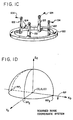
- Figure 1C is a perspective illustration of a reference ring which is mounted by uprights to a patient's head to support the cylindrical frame structure of Figure 1A.
- Figure 1D is a perspective illustration of the coordinate system of a three dimensional scanned image.
- Figure 2A is a perspective view of the caliper frame used to determine the relative position between a position in the head and the phantom base.
- Figure 2B is a perspective view of the caliper frame of Figure 2A illustrating its angles of adjustment.
- Figure 2C is a block diagram of the steps involved in the prior art process of determining the position of surgical probe relative to the scanned images so that the image corresponding to the probe position can be identified and viewed by the surgeon.
- Figure 2D is a perspective illustration of a three dimensional coordinate system of a surgical probe.
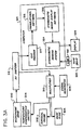
- Figure 3A is a block diagram of a system according to the invention for indicating the position of a surgical probe within a head on an image of the head.
- Figure 3B is a perspective schematic diagram of the microphone array, surgical probe and base ring according to the invention.
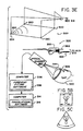
- Figure 3C is a block diagram of the steps involved in the process according to the invention for determining the position of a surgical probe relative to the scanned images so that the image corresponding to the probe position can be identified and viewed by the surgeon.
- Figure 3D is a perspective schematic diagram of an optical scanner used in combination with a cradle.
- Figure 3E is a perspective schematic diagram of the microphone array, surgical probe, base ring and optical scanner according to the invention.
- Figure 4 is a flow chart of the translational software for translating coordinates from the surgical probe coordinate system to the scanned image coordinate system according to the invention.
- Figure 5A is a perspective schematic diagram of an ultrasound probe system according to the invention;
- Figures 5B and 5C illustrate ultrasound and scanned images, respectively.
-
- Corresponding reference characters indicate corresponding parts throughout the several views of the drawings.
- With the advent of modern scanning equipment and techniques, several stereotactic systems have been developed and are presently available. These stereotactic systems allow a surgeon to localize specific points detected on CT, MRI or PET scans which have been previously generated prior to the surgical procedure being performed. In particular, the stereotactic systems allow the selection of specific points detected on the scans to be localized within the brain by the surgeon during the surgical procedure using a mechanical device.
- Initially, prior to the operative procedure, some form of localizing device, such as a frame, is attached to the patient's skull using sharp pins. The particular scan or scans which are to be performed are then generated with the head of the patient encircled by the frame. For example, the frame may be comprised of a
cylindrical structure 100 as illustrated in perspective in Figure 1A.Structure 100 includes an uppercircular ring 102 and a lowercircular ring 104 which are interconnected by sixvertical rods 106 and threediagonal rods 108. The threediagonal rods 108 diagonallyinterconnect rings cylindrical structure 100 and orthogonally intersects itsaxis 108 will intersect each of thediagonal rods 108 at a particular point. The resultant spacing between the diagonal and upright rods defines a unique plane within thecylindrical structure 100. For example, as shown in Figure 1B, a scan in a particular plane would show a pattern of nine cross sectional views of therods 106. The unique spacing of these views of the rods, as shown inplane 112 of Figure 1B, would necessarily indicate that the position of thescan plane 112 was parallel to and midway betweenrings cylindrical structure 100. - As a result of the scanning process, the images obtained are analyzed and the position within the images of the
specific marking rods 106, called fudicels, are identified and measured. By measuring the distance between therods 106, the specific location of a scan with reference to a base plane can be identified. Generally, thelower ring 104 of thecylindrical structure 100 is attached to a reference ring 120 (also known as a BRW head ring) as illustrated in Figure 1C. As noted above, thisring 120 is supported on the patient's head viauprights 122 attached to the head by the use ofsharp pins 124 so that thering 120 is held firmly in place with respect to the head. Thelower ring 104 of thecylindrical structure 100 is mounted to thereference ring 120 attached to the patient's head so that these two rings are in parallel planes. - As shown in Figure 1D, the scanning system (e.g., CT, MRI, PET) which is performing the scanning has a scanned image coordinate system (Xo, Yo, Zo) within which a reference plane RP can be defined by at least three reference points RP1, RP2 and RP3 located on the
head 124 of the patient. A computer is then used to calculate a specific position within the brain and a target picked out on the specific image can be approached with a fair degree of accuracy during the surgical procedure. - Although stereotactic surgery allows a surgeon to be guided to a specific point with accuracy, it has not been particularly useful in allowing the surgeon to identify the particular location of a surgical probe within the brain at any point during the surgical process. Frequently in neurosurgery, brain tumors or other target points within the brain are indistinguishable from surrounding normal tissue and may not be detected even with the use of frozen sections. Moreover, with modern microsurgical techniques, it is essential that the neurosurgeon identify specific structures within the brain which are of critical functional importance to the patient. In addition, the boundaries of these structures must be accurately defined and specifically known to the surgeon during the surgical process. In this way, these tissues will not be disturbed or otherwise damaged during the surgical process resulting in injury to the patient.
- In the past, the surgeon has been able to use the stereotactic system in reverse in order to permit the determination of the position of a surgical probe relative to the scanned images so the image corresponding to the probe position can be identified and viewed. However, going in reverse from the patient's brain backwards to find the position of the surgical probe relative to the scan is a cumbersome and time-consuming process. Usually, a specially designed
caliper frame 200, as illustrated in Figure 2A, has to be attached to thering 120 affixed to the patient's head to determine the position of the surgical probe in the head. For example, suppose the surgeon desires to know the position of atip 201 of aprobe 202 in the patient's head. First, thecaliper frame 200 is fitted to thereference ring 120 affixed to the patient's head. Next, the position ofprobe 202 is positioned onarch 206 and theframe 200 is set to indicate the alpha, beta, gamma and delta angles onscales probe 202 defines with respect to theframe 200, as shown in Figure 2B. Next, thedistance 216 from the tip of theprobe 202 to the arch 206 is determined. - The
caliper frame 200 is then transferred and mounted to aphantom base 250 in a manner as illustrated in Figure 2A. Thephantom base 216 has a coordinate system (X1, Y1, Z1). Generally, thecaliper frame 200 identifies apoint 201 over thephantom base 250. Apointing device 252 is positioned to have itstip 254 atpoint 201. The X1 - Y1 plane of thephantom base 200 corresponds to a plane parallel to the plane in which the reference points RP1, RP2 and RP3 are located. The (X1, Y1, Z1) coordinates define the position ofpoint 201. As a result, the position ofpoint 254 with respect to the X1 - Y1 plane and, therefore, with respect to the reference plane RP is now known. A computer can now be used to calculate the specific position within the brain and the particular scan which corresponds to the calculated position can now be accessed and viewed on a scanning system. - In summary, this prior art process as shown in Figure 2C identifies the location of the
tip 201 of thesurgical probe 202 for the surgeon. Initally, the surgeon positions theprobe 202 on thecaliper frame 200, which is attached to the head, at the position desired within the head. Thecaliper frame 200 is then removed from the patient's head and transferred to thephantom base 250. Thepointing device 252 is then positioned atpoint 254 which is essentially coaxial withpoint 201 of the tip of the probe. Thepointing device 252 then indicates the position of the tip of the probe in the phantom base coordinate system (X1, Y1, Z1). Finally, these coordinates are used to determine the scanned image coordinates (Xo, Yo, Zo) so that the image corresponding to the probe position can be displayed. - After this cumbersome and time-consuming process, the surgeon has now determined the position of the
tip 201 of theprobe 202 with respect to the scanned images and can now view the image corresponding to the probe position to decide the next step in the surgical procedure. This entire process takes approximately ten to fifteen minutes and increases the risks of intraoperative contamination as the base of the calipers are nonsterile. Because of these considerations, stereotactic surgery is not commmonly employed in most procedures. Furthermore, the minimal accuracy it affords is generally insufficient for modern microsurgical techniques. Consequently, stereotactic surgery is not generally available to the majority of certain patients undergoing surgery. - Comparing Figures 1D and 2A, it can be seen that it is necessary for the surgeon to know the specific location of the
tipe 201 of thesurgical probe 202 with respect to the scanned image coordinate system (Xo, Yo, Zo) of the particular scans that were preoperatively performed. In other words, thesurgical probe 202 has a particular coordinate system (X2, Y2, Z2) which is illustrated in Figure 2D. Ideally, the surgical probe coordinate system (X2, Y2, Z2) must be related to the scanned image coordinate system (Xo, Yo, Zo). The prior art as illustrated in Figure 2B has suggested relating these coordinate systems via the phantom base coordinate system (X1, Y1, Z1). However, as noted above, this relational process is inaccurate, time-consuming and cumbe some. The invention uses a 3D digitizer system to locate the position of thetip 201 of thesurgical probe 202 and to directly relate the surgical probe coordinate system (X2, Y2, Z2) to the scanned image coordinate system (Xo, Yo, Zo). - In particular, an off-the-shelf, three dimensional sonic digitizer such as Model GP-8-3D produced by Scientific Accessories Corporation is used to determine the position of the probe. As shown in Figure 3A, the 3D digitizer system includes a
microphone array 300 which is generally mounted in the operating room on the ceiling or in some other position so that it is in a line of sight with thesurgical probe 302 that is being used. As will be described in greater detail below, theprobe 302 includes transmitters such as sound emitters thereon which interact with themicrophone array 300 so that the position of the tip ofsurgical probe 302 is known at any particular instant in time. The 3D digitizer system also includes atemperature compensation emitter 304 associated with themicrophone array 300. Furthermore, mounted to the ring 120 (Figure 1C) affixed to the patient's head is abase ring 306 which is coaxial and parallel with the plane defined byreference ring 120. Thisbase ring 306 includes a plurality of transmitters as will be described below which interact with themicrophone array 300 so that the relative position of thebase ring 306 can be determined any particular instant in time.Signal generator 308 generates a signal which is provided through amultiplexer 310 to thetemperature compensation emitter 304,surgical probe 302, andbase ring 306. Usually,temperature compensation emitter 304 is activated by thesignal generator 308 viamultiplexer 310 to emit a signal which is received by themicrophone array 300. Each of the signals received by each of the microphones of thearray 300 is provided to adigitizer 312 which digitizes the signals and provides the digitized signals tocomputer 314 which includes a spatial acquisition and recording (SAR)program 316 which acquires and records spatial coordinates based on the digitized signals. For example,program 316 may be the SACDAC program licensed by PIXSYS of Boulder, Colorado. This program evaluates the digitized signals emitted by thetemperature compensation emitter 304 to determine the reference standards. i.e., the velocity of the radiation through the air. For example, depending on the temperature of the air in the operating room, the period of time that it takes from the instant that thetemperature compensation emitter 304 is actuated to radiate a signal until the instant that each of the microphones of thearray 300 receives the emitted signal will vary. TheSAR program 316 knows, through calibration, the distance between thetemperature compensation emitter 304 and each of the microphones of thearray 300. Therefore, theSAR program 316 can immediately calculate the velocity of the signals being transmitted. This velocity establishes a reference for determining the position of thesurgical probe 302 and thebase ring 306. - Next, the emitters of the
base ring 306 are activated so that the position of thebase ring 306 can be determined. At this point, the emitters of thebase ring 306 are successively energized and the radiation transmitted by these emitters is detected by themicrophone array 300. The signal generated by the microphones from this radiation is digitized and evaluated by theSAR program 316 to determine the position of each of the emitters of thebase ring 306. Once the positions of the base ring emitters have been determined by theSAR program 316, standard geometrical computations are performed by the SAR program to determine the plane defined by thebase ring 306 with respect to themicrophone array 300. -
Digitizer 312 then signalsmultiplexer 310 to provide the signal generated bysignal generator 308 to thesurgical probe 302. At this point, the emitters of thesurgical probe 302 are successively energized and the radiation transmitted by these emitters is detected by themicrophone array 300. The signal generated by the microphones from this radiation is digitized and evaluated by theSAR program 316 to determine the position of each of the emitters of thesurgical probe 302. Once the positions of the probe emitters have been determined by theSAR program 316, standard geometrical triangulation is performed by the SAR program to determine the location of the tip of the surgical probe with respect to themicrophone array 300. - Therefore, by using the 3D digitizer system, the position of the
base ring 306 and the position of thesurgical probe 302 relative to thebase ring 306 can be determined by theSAR program 316. As noted above, thebase ring 306 is mounted to the reference ring 120 (Figure 1C) and is essentially coplanar therewith so that thebase ring 306 defines the reference plane RP of the scanned image coordinate system illustrated in Figure 1D. -
Computer 314 includestranslational software 318 which then translates the coordinates of surgical probe coordinate system illustrated in Figure 2D into the scanned image coordinate system illustrated in Figure 1D. As a result of this translation,computer 314 has now determined the particular scanned image of the preoperative scan on which the tip of thesurgical probe 302 would be located. The system includes atape drive 320, accessed through a local area network (LAN) 321, in which each of the images of the preoperative scan are stored. The translated coordinates generated bytranslational software 318 are provided to the stereotacticimage display software 322, also resident withincomputer 314, and identify the particular scanned image which is to be viewed by the surgeon. The identified image is selected by thestereotactic imaging system 324 which recreates the image from the data stored intape drive 320 and displays it on ahigh resolution display 326. Stereotacticimage display software 322 andstereotactic image system 324 may be any off-the-shelf system such as manufactured by Stereotactic Image Systems, Inc. of Salt Lake City, Utah. - Referring to Figure 3B, a perspective illustration of the
microphone array 300,temperature compensation emitter 304,surgical probe 302 andbase ring 306 are illustrated.Microphone array 300 includes a plurality ofmicrophones 350, the outputs of which are connected to3D digitizer 312. Adjacent to themicrophone array 300 is atemperature compensating emitter 304 which selectively emits signals used by the SAR program in calibration to determine the velocity of the radiation. For example, in the Scientific Accessories Corporation Model GP-8-3D, a sonic digitizer is used. In this case, the speed of sound being transmitted from thetemperature compensation emitter 304 to themicrophones 350 is calculated by the SAR program to determine the speed at which the sound is being transmitted through the air. Since this system is very accurate and the speed of sound varies fairly significantly with respect to the temperature of the air, thetemperature compensation emitter 304 allows the 3D digitizer system to compensate for changes in the air temperature in the operating room.Surgical probe 302 comprises a bayonet surgical forceps modified to carry at least two sound emitters thereon which are essentially coaxial onaxis 362 with the tip of the forceps. The emitters are in line and immediately below the surgeon's line of sight through the forceps so that the line of sight is not blocked. In general, themicrophone array 350 is attached to the operating light above the patient's head so that it is in direct line of sight with the forceps as they are being used by the surgeon. Themicrophones 350 listen to the sound emitted from the sequential energization of theemitters 360 on the forceps. TheSAR software 316 measures the time of transmission from each of thesound emitters 360 on the forceps to themicrophones 350. By comparing these times, the position of bothemitters 360 and, therefore, the tip of the forceps can be calculated by theSAR program 316. -
Base ring 306 is affixed to thereference ring 120 attached to the patient's head and is essentially coplanar with the reference points RP1, RP2 and RP3.Base ring 306 includes a plurality ofemitters 370 thereon which are connected to multiplexer 310 and energized bysignal generator 308. Each one of theseemitters 370 is sequentially energized so that the radiation emitter thereby is received by themicrophones 350 ofarray 300. Theemitters 370 are preferably positioned 90° apart with the center emitter being located at the anterior of the head. This permitsbase ring 306 to be mounted around the head so that all three emitters are in line of sight with the array. The resulting signals are digitized bydigitizer 312 so that theSAR program 316 is able to determine the plane in which theemitters 370 are located. This plane essentially defines the reference plane because it is coplanar with the reference points RP1, RP2 and RP3. By determining the position of the reference plane,translational software 318 is now able to take the coordinate position of theprobe 302 and translate it from the surgical probe coordinate system of Figure 2D into the scanned image coordinate system as illustrated in Figure 1D. As a result, the particular scanned image which corresponds to the position of the probe can be identified and displayed for viewing by the surgeon. - The
surgical probe 302 is generally a bayonet cauterizing device which has a bundle ofwire 364 attached thereto. Therefore, the wires required to connect theemitters 360 to themultiplexer 310 are part of the bundle ofwires 364 which connect the forceps to its electrical power source and the surgeon is familiar with handling such forceps connected to a wire bundle. Therefore, there is no inconvenience to the surgeon in using such a probe and the surgeon is familiar with handling such a forceps connected to a wire bundle. -
Base ring 306 is one apparatus for determining and positioning the reference points RP1, RP2 and RP3 with respect to themicrophone array 300. An advantage of thebase ring 306 is that each time the patient's head is moved thebase ring 306 is energized to define the reference plane. This allows the surgeon to move the patient's head during surgery. Alternatively, the reference points RP1, RP2 and RP3 can be established by using a reference mode of the3D digitizer 312. In particular, the tip ofprobe 302 is positioned on each of the reference points RP1, RP2 and RP3 and actuated to emit a signal to themicrophone array 300 so that the position of the tip can be determined at each of these points. This is performed during a reference mode of operation of the3D digitizer 312 so that theSAR program 316 calculates, at the end of the execution of this mode, the position of the reference points RP1, RP2 and RP3. This requires that the reference points have to be reestablished before the position of the surgical probe is determined to avoid changes in the reference plane due to movement of the head. On the other hand, one advantage of this approach is that the use of thereference ring 120 may be eliminated. In particular, it is possible that the reference pins 122 can be permanently affixed to the skull of the patient. For example, these pins may be radiolucent surgical screws which are embedded in the patient's skull and which have radiopaque tips. These screws would be affixed to the patient's skull before surgery and before the preoperative scanning so the radiopaque tips would provide a constant reference during scanning and throughout the stereotactic surgical procedure. During the actual surgery, the probe would be used to indicate the position of each of the radiopague tips before the probe position was determined. By eliminating the need for thereference ring 120, other advantages are also achieved. For example, generally the preoperative scanning must be done under anesthetic because thereference ring 120 interferes with intubation. Therefore, intubation must occur before the reference ring is affixed to the skull. By eliminating the need for thereference ring 120 and using surgical screws to identify the reference points RP1, RP2 and RP3, the preoperative scanning can be performed without the need for intubation and the anesthesia accompanying it. In one alternative embodiment, it is contemplated that theemitters 370 may each be separately mounted to a screw or other fixed structure positioned at one of the reference points. - In summary, this process according to the invention is illustrated in Figure 3C and identifies the location of the tip of the
surgical probe 202 for the surgeon. Initially, the reference plane is determined by energizing thebase ring 306 or by positioning theprobe 302 at the reference points (as described herein). Next, the surgeon positions the probe in the position desired within the head. The emitters of the probe are then energized so that the probe position is measured and determined in the surgical probe coordinate system (X2,Y2,Z2). Next, thetranslational software 318 converts the surgical probe coordinate system into the scanned image coordinate system (Xo,Yo,Zo) so that the image corresponding to the probe position can be displayed. - Referring to Figure 3D, a perspective illustration of a patient's
head 390 in acradle 392 during the scanning process is shown. As will be described below,optical scanner 380, having emitters 381 thereon, is employed to determine the position of thehead 390 relative to acradle 392 positioned on the head. - Referring to Figure 3E, a perspective illustration of the
microphone array 300,temperature compensation emitter 304,surgical probe 302 andoptical scanner 380 are illustrated.Microphone array 300 includes a plurality ofmicrophones 350, the outputs of which are connected to3D digitizer 312. Themicrophone array 300 provides a fixed frame of reference to which the position ofprobe 302 is measured and to which the position of thehead 390, relative to thecradle 392, is measured. As a result, the position of theprobe 302 relative to thehead 390 at any instant in time can be determined. - Adjacent to the
microphone array 300 is atemperature compensating emitter 304 which selectively emits signals used by the SAR program in calibration to determine the velocity of the radiation. For example, in the Scientific Accessories Corporation Model GP-8-3D, a sonic digitizer is used. In this case, the speed of sound being transmitted from thetemperature compensation emitter 304 to themicrophones 350 is calculated by the SAR program to determine the speed at which the sound is being transmitted through the air. Since this system is very accurate and the speed of sound varies fairly significantly with respect to the temperature of the air, thetemperature compensation emitter 304 allows the 3D digitizer system to compensate for changes in the air temperature in the operating room. -
Surgical probe 302 comprises a bayonet surgical forceps modified to carry at least twosound emitters 360 thereon which are essentially coaxial onaxis 362 with the tip of the forceps. The emitters are in line and immediately below the surgeon's line of sight through the forceps so that the line of sight is not blocked. In general, themicrophone array 350 is attached to the operating room light above the patient's head so that it is in direct line of sight with the forceps as they are being used by the surgeon. Themicrophones 350 listen to the sound emitted from the sequential energization of theemitters 360 on the forceps. TheSAR software 316 measures the time of transmission from each of thesound emitters 360 on the forceps to themicrophones 350. By comparing these times, the position of bothemitters 360 and, therefore, the tip of the forceps can be calculated by theSAR program 316. -
Optical scanner 380 is generally located over the patient'shead 390 and is used during scanning to establish the position of thehead 390 relative to thecradle 392 thereby to relate the frame of reference of the cross sectional scans to theforehead 394.Scanner 380 is also used during surgery to establish the position of thehead 390 relative to thecradle 392 thereby to relate the frame of reference of theprobe 302 to theforehead 394. - During the preoperative scanning process as shown in Figure 3D, when the cross sectional images of the head are created, the patient's head lies temporarily in
cradle 392. The cradle includes anarc 393 of radiopaque material so that it appears in at least some of the cross sectional scans. As a result, thearc 393 defines a plane relative to thehead 390. During scanning, this plane can be defined as the 0,0,0 plane for convenience. After the head is placed in the cradle,optical scanner 380 is used to establish the position of thecradle 392 and its attachedarc 393 relative to theforehead 394. In particular, theoptical scanner 380 scans both the forehead and thearc 393 of thecradle 392 and, viacomputer 396 employing foreheadfitting software 398, determines the position of thearc 393 of thecradle 392 relative to theforehead 394. The forehead fitting software may be any off-the-shelf or custom software which graphs a set of points so that a curve defining the contour of the forehead can be calculated, a curve defining the arc can be calculated, and a curve defining the relative position of the forehead and the arc can be calculated. Since the position of the cross sectional scans relative to theradioopaque arc 393 is known (because the cradle arc defines the 0,0,0 plane) and since the position of thearc 393 of thecradle 392 relative to theforehead 394 is known (because of the scanning by the optical scanner), then the position of the cross sectional scans relative to the forehead is known and can be calculated bytranslational software 316. - During surgery, a
base ring 306 is firmly affixed to the head. Thebase ring 306 does not have to be positioned in the same location relative to the head as the arc was during the scanning process when the cross sectional images were created. Thebase ring 306 used during surgery includesemitters 370 which communicate with thearray 300 to establish the position of thebase ring 306. As a result, thebase ring 306 defines a plane relative to thehead 390. After affixing the base ring to the head,optical scanner 380 is used prior to or during the surgery to establish the position of thebase ring 306 relative to theforehead 394. In particular, theoptical scanner 380 scans both the forehead and thebase ring 306 and, viacomputer 396 employing foreheadfitting software 398, determines the position of thebase ring 306 relative to theforehead 392. Since the position of the probe relative to the base ring is known (because of communication via the array) and since the position of the base ring relative to the forehead is known (because of the scanning by the optical scanner), then the position of the probe relative to the forehead is known and can be calculated bytranslational software 316. Since the position of the cross sectional images relative to the forehead is also known (from the preoperative scanning process), the end result is that the position of the probe relative to the cross sectional images is known so that the position of the tip of the probe on the closest cross sectional image can be displayed. -
Optical scanner 380 andcomputer 396 are a standard, off the shelf scanner used to scan an object to determine its three-dimensional shape. For example, a limb scanner such as PIXSYS Optical Scanner used to develop three-dimensional models for artificial limbs may be used. Thescanner 380 emits a laser beam or other optical beam toward thearc 393 and theforehead 394 and receives the light reflected there through an array of linear chip cameras such as CCD (charge coupled device) cameras. By evaluating the position of the reflected light using the camera array, theoptical scanner 380, including acomputer 396, determines the shape and, thus, the contour of theforehead 394, the shape of thearc 393 ofcradle 392 and the relative position of the forehead and thearc 393.Computer 396 indicates to thetranslational software 316 ofcomputer 314, which is a part of the system as illustrated in Fig. 3A, the position of theprobe 302 relative to theforehead 392. Thetranslational software 316 then coverts this indicated position into the coordinate system of the cross sectional scanned images. As a result, the particular scanned image which corresponds to the position of the probe can be identified and displayed on display 326 (Fig. 3A) for viewing by the surgeon. - The
surgical probe 302 is generally a bayonet cauterizing device which has a bundle ofwire 364 attached thereto. Therefore, the wires required to connect theemitters 360 to themultiplexer 310 are part of the bundle ofwires 364 which connect the forceps to its electrical power source. Surgeons are generally familiar with handling such forceps connected to a wire bundle. Therefore, there is no inconvenience to the surgeon in using such a probe and the surgeon is experienced with handling such a forceps connected to a wire bundle. - One advantage of the
optical scanner 380 is that it eliminates the need for a ring or pins to be attached to the patient's head during the preoperative scanning process. Each time the patient's head is placed in a cradle, theoptical scanner 380 can be used to scan the head and cradle to redefine their relative position without the need for any contact. The reference ring (i.e., arc) on the head is, therefore, temporary. By eliminating the need for apermanent reference ring 120 or reference pins RP1-RP3, other advantages are also achieved. For example, generally the preoperative scanning must be done under anesthetic because thereference ring 120 interferes with intubation or it must be done after pins are affixed to the head. Therefore, intubation must occur before the reference ring is affixed to the skull. By eliminating the need for thepermanent reference ring 120 and/or reference pins, and by using the contour of the forehead to define a reference point, the preoperative scanning can be performed without the need for intubation and the anesthesia accompanying it. - In summary, during the preoperative scanning process the patient simply lies in a U-shaped cradle attached to the end of a CT or MRI table. Above the patient's face is an arc providing the reference plane. All scans are obtained with reference to and preferably parallel to this arc defining the reference or base plane. The optical scanner relates the forehead contour to this arc so that the relation of the forehead to the scans is known.
- In the operating room, the patient's head is again scanned with the optical scanner but this time the arc over the patient's head is
base ring 306. The reference emitters attached to the base ring define the operative reference system. Therefore, the forehead is again related to the base ring by the optical scanner to define a new reference system; this time the new reference system is the operating room. The computer then matches the forehead contours obtained in the operating room and the scanning room to relate the two reference systems. In effect, the forehead is a "bridge" between the reference system of the preoperative scanner and the reference system of the operating room. - The cradle does not have to appear in the actual scans. The primary purpose of the cradle is to keep the patient's head from moving so that all scans are obtained with the same relationship to the arc.
- Referring to Figure 4, a flow chart of the operation of the
translational software 318 is illustrated. Initially, the surgeon locates theprobe 302 in the position which is to be determined. (If abase ring 306 is not being used to identify the location of the reference plane, the initial step is for the surgeon to use the reference mode of the3D digitizer 312 to identify the reference plane by locating the surgical probe tip at several points in the plane.) - The system initializes at
step 400 so that translational software opens a window menu atstep 402 of a multitasking program such as DESQ VIEW distributed by Quarterdeck Office Systems of Santa Monica, California. Such software permits simultaneous execution of multiple software programs. In general, once a program is selected for actuation, it continues to run either in the foreground or in the background until deactuated. - The translational software continues initializing by selecting the stereotactic imaging system and actuating the stereotactic imaging system in the foreground by opening the stereotactic window at
step 404. Thereafter, the translational software returns to the window menu atstep 406 moving the stereotactic image display software to the background and selects the digitizer window atstep 408 to actuate the digitizer in the foreground. The computer is then ready to be actuated by the foot switch. - The surgeon then actuates a foot pedal or other switch which indicates that the system should perform a computation. Actuation of the foot switch is essentially the beginning of the
start step 410. Upon actuation, the digitizer energizes calibration by thetemperature compensation emitter 304 to determine the velocity of the sound waves, energizes the emitters of thebase ring 306 to locate the reference plane and energizes the emitters of thesurgical probe 302 to locate the position of the tip of theprobe 302. The signals generated by the microphone array are digitized so that theSAR program 316 determines the coordinates of the tip of the surgical probe. At step 412, thetranslational software 318 selects the coordinates from the SAR program. - Next, the window menu is again accessed at
step 414 and the window menu switches the stereotactic image system software to the foreground at step 416 to specifically control the operation of thestereotactic imaging system 324. At this point, thetranslational software 318 issues an FI command to the stereotacticimage display software 322 which in turn prepares thestereotactic imaging system 324 to accept coordinates. Atstep 420, the window menu is again selected so that atstep 422 the computer switches the digitizer window into the foreground. Atstep 424, the digitizer window menu is accessed and coordinate translation is selected. Atstep 426, the digitizer begins calculating the coordinates and atstep 428 the coordinate calculation is ended. The translational software then returns to the digitizer window menu at step 430, switches windows to place the stereotactic image system software in the foreground at 432 to prepare it for receiving the coordinates and again returns to the main window menu atstep 434. Finally, the coordinate information is translated, including any necessary manipulation, and transferred to the stereotacticimage display software 322 atstep 436 which actuates thestereotactic imaging system 324 to select the particular image from thetape drive 320 and display it onhigh resolution display 326. The stereotacticimage display software 322 instructs thestereotactic imaging system 324 to display the image closest to transferred coordinates and to display a cursor on thedisplay 326 at the coordinates which corresponds to the position of the tip of the probe. Thereafter, thecomputer 314 is in a standby mode until the foot switch of the surgeon is again actuated to execute the translational software beginning with thestart step 410. - The translation that occurs in
step 436 depends on the position of the surgical probe coordinate system relative to the scanned image coordinate system and the units of measure. In the preferred embodiment, the systems are coaxial and the units of measure are the same so that algebraic adjustment is unnecessary. However, it is contemplated that the coordinates systems may not be coaxial, in which case translation would require arithmetic and/or trigonometric calculations. Also, the sequence, e.g., (X2, Y2, Z2), in which the coodrinates are generated by the digitizer may be different than the sequence, e.g., (Xo, Yo, Zo), in which stereotactic image system software receives coordinates. Therefore, the sequence in which the coordinates are transferred may have to be reordered. - Referring to Figure 5A, a system employing an ultrasound localizer is illustrated.
Reference character 500 refers to an ultrasound probe which may be used in the operating room to scan the brain. Theultrasound probe 500 includes a plurality of at least threeemitters 502 which communicate with thearray 300 to define the plane in which the ultrasound probe is scanning.Emitters 502 are energized via line 504 bymultiplexer 310 as in the other systems illustrated above. The radiation emitted byemitters 502 is received byarray 300 to determine the plane in which theultrasound probe 500 is positioned. The ultrasound probe is also connected vialine 506 to acomputer 508 which analyzes the ultrasound scanning and provides the analyzed information to awork station 510 which displays the scanned image. Since thearray 300 can determine the position of theultrasound probe 500 at any point in time, viadigitizer 312, the particular plane of the image displayed onwork station 510 is known. The position of thehead 394 of the patient can be determined by attaching abase ring 306 withemitters 370 to the head, as noted above, or by scanning the forehead with an optical scanner having emitters thereon, as noted below. - For example, such an ultrasound image is illustrated in Figure 5B. The surgeon can then call up the similar image on the
display 326 of thestereotactic imaging system 324 such as illustrated in Figure 5C. Alternatively,computer 508 may be linked to thestereotactic imaging system 324 directly to define the particular image plane illustrated onwork station 510 so thatdisplay 326 can display the corresponding scanned image. As a result, the image from the ultrasound system, as illustrated onwork station 510, is shown on one monitor and may be compared to a cross section to the images obtained either by CT, MRI or PET scanning. The cross section through the three dimensional data set as developed by the ultrasound system is determined by a high speed graphics work station, such as manufactured by Silicon Graphics. This allows the interpretation of the ultrasound scans as the anatomy from the MRI, CT or PET scans can be seen directly. Furthermore, the ultrasound system allows scanning in the operating room. Since the brain tissue is elastic and the position of various tissue may change from time to time, use of an ultrasound scan in the operating room permits a more definite localization of various brain tissues. - Alternatively, the system may be used for determining a position of the ultrasound probe relative to a head of a body of a patient. The
probe 500 is positioned to scan thehead 394 with anarray 300 positioned adjacent the probe. At least threeemitters 502 permit determination of the position of the ultrasound probe relative to the array.Optical scanner 380, having emitters 381 (Fig. 3D) permit determination of the position of the head relative to the array.Computer 396 translates the position of the ultrasound probe into a coordinate system corresponding to the position of the head. - In view of the above, it will be seen that the several objects of the invention are achieved and other advantageous results attained.
- As various changes could be made in the above constructions without departing from the scope of the invention, it is intended that all matter contained in the above description or shown in the accompanying drawings shall be interpreted as illustrative and not in a limiting sense.
Claims (20)
- A system for indicating a location within a body of a patient, said system comprising:a reference contour (394) having a position in fixed relation to the body for providing reference points;means (320, 324) for generating images of the body, said images including reference images corresponding to the reference contour;reference means (300) having a location outside the body for providing a reference;a surgical probe (302) including a tip (301, 541) having a position;first means (360) for determining the position of the tip of the surgical probe relative to the reference means;second means (306) for determining the position of the reference contour of the body relative to the reference means so that the position of the tip relative to the reference contour of the body is a known position;means (314) for translating the known position of the surgical probe to provide a translated position within a coordinate system corresponding to the images of the body; andmeans (326) for displaying an image of the body to provide a displayed image that corresponds to the translated position of the tip of the surgical probe.
- A system according to claim 1, wherein the reference contour comprises a contour of the patient's forehead (394).
- A system according to claim 1 or claim 2, wherein the means (326) for displaying an image of the body provides a plurality of displayed images that correspond to the translated position of the tip of the surgical probe.
- A system according to any one of claims 1 to 3, wherein the displaying means (326) comprises means for displaying an image representing the tip (301) of the probe on the displayed image of the body.
- A system according to claim 4, wherein the second means comprisesa base (306) adapted to be mounted on the body in a position having a fixed relationship to the reference contour (390) of the body; andmeans (396, 380) for measuring the position of the base with respect to the reference means.
- A system according to any one of claims 1 to 5, wherein the reference means comprises an array of sensors (300) and wherein the probe (302) comprises a bayonet forceps having emitters (360) in line with the tip of the forceps and below a surgeon's line of sight when using the forceps; and
wherein the emitters communicate with the sensors of the array to indicate a position of the probe relative to the array. - A system according to claim 6, further comprising additional emitters (370) on the base for communicating with the sensors of the array to indicate the position of the base relative to the array.
- A system according to claim 6 or claim 7, further comprising a three dimensional digitizer means (312) for digitizing signals generated by the sensors in response to signals from the emitters.
- A system according to any one of claims 1 to 8, wherein the translating means comprises a computer (314) connected between the second determining means and the displaying means and a translational software program (318) for controlling the operation of the computer so that coordinates supplied to the computer by the second determining means are converted into corresponding coordinates supplied to the displaying means.
- A system according to any one of claims 1 to 9, wherein the displaying means comprises an imaging system.
- A system according to any one of claims 1 to 10, further comprising means (304) for compensating for temperature changes which affect the operation of the first and second determining means.
- A system according to any one of claims 1 to 11, wherein the displaying means comprises means for displaying a cursor representing the tip of the probe on the displayed image of the body.
- A system according to any one of claims 1 to 12, wherein the reference points define a reference plane and wherein the coordinate system (X0, Y0, Z0) of the images includes an X-Y plane parallel to the reference plane.
- A system according to any one of claims 1 to 13, wherein the reference means comprises an array of sensors (300) and wherein the probe (302)comprises a bayonet forceps having a line of sight through the forceps and having two emitters (360) mounted on the forceps which are in line with the tip of the forceps and below the line of sight through the forceps, said emitters for communicating with the sensors of the array to indicate the position of the probe relative to the array.
- A system according to any one of claims 1 to 14, wherein the means for generating images of the body comprises at least two of a CAT, PET, MRI and MEG scanner.
- A system according to any one of claims 1 to 15, wherein the reference means comprises an array of sensors (300), and the first determining means comprises at least three emitters (360) on the surgical probe (302) and means (308) for activating the emitters to generate a signal communicated to the array.
- A system according to any one of claims 1 to 16, wherein the reference means comprises an array of sensors (300) and the second determining means (306) comprises means mounted on the body for generating a signal received by the array to indicate the position of the body relative to the array.
- A system according to any one of claims 1 to 17, wherein the image generating means includes a first scanning means (320, 324) adapted to be used prior to an operation and a second scanning means (500) adapted to be used during the operation.
- A system according to any one of claims 1 to 18, wherein the reference means comprises an array of sensors (300), and the second determining means comprises a base (306) adapted to be mounted on the body part in a position having a fixed relationship with the reference contour (394) of the body and means (396, 380) for measuring the position of the base with respect to the reference means.
- A system according to claim 19, further comprising emitters (370) on the base for communicating with the sensors of the array to indicate the position of the base relative to the array.
Priority Applications (1)
| Application Number | Priority Date | Filing Date | Title |
|---|---|---|---|
| EP06100735A EP1690511B1 (en) | 1990-10-19 | 1991-10-17 | Surgical probe locating system for head use |
Applications Claiming Priority (4)
| Application Number | Priority Date | Filing Date | Title |
|---|---|---|---|
| US60075390A | 1990-10-19 | 1990-10-19 | |
| US600753 | 1990-10-19 | ||
| EP91919611A EP0553246B1 (en) | 1990-10-19 | 1991-10-17 | Surgical probe locating system for head use |
| EP99108026A EP0931516B1 (en) | 1990-10-19 | 1991-10-17 | Surgical probe locating system for head use |
Related Parent Applications (1)
| Application Number | Title | Priority Date | Filing Date |
|---|---|---|---|
| EP99108026A Division EP0931516B1 (en) | 1990-10-19 | 1991-10-17 | Surgical probe locating system for head use |
Related Child Applications (1)
| Application Number | Title | Priority Date | Filing Date |
|---|---|---|---|
| EP06100735A Division EP1690511B1 (en) | 1990-10-19 | 1991-10-17 | Surgical probe locating system for head use |
Publications (3)
| Publication Number | Publication Date |
|---|---|
| EP1210916A2 true EP1210916A2 (en) | 2002-06-05 |
| EP1210916A3 EP1210916A3 (en) | 2002-06-12 |
| EP1210916B1 EP1210916B1 (en) | 2006-09-20 |
Family
ID=24404917
Family Applications (4)
| Application Number | Title | Priority Date | Filing Date |
|---|---|---|---|
| EP06100735A Expired - Lifetime EP1690511B1 (en) | 1990-10-19 | 1991-10-17 | Surgical probe locating system for head use |
| EP91919611A Expired - Lifetime EP0553246B1 (en) | 1990-10-19 | 1991-10-17 | Surgical probe locating system for head use |
| EP02004099A Expired - Lifetime EP1210916B1 (en) | 1990-10-19 | 1991-10-17 | System for indicating a location within a body of a patient |
| EP99108026A Expired - Lifetime EP0931516B1 (en) | 1990-10-19 | 1991-10-17 | Surgical probe locating system for head use |
Family Applications Before (2)
| Application Number | Title | Priority Date | Filing Date |
|---|---|---|---|
| EP06100735A Expired - Lifetime EP1690511B1 (en) | 1990-10-19 | 1991-10-17 | Surgical probe locating system for head use |
| EP91919611A Expired - Lifetime EP0553246B1 (en) | 1990-10-19 | 1991-10-17 | Surgical probe locating system for head use |
Family Applications After (1)
| Application Number | Title | Priority Date | Filing Date |
|---|---|---|---|
| EP99108026A Expired - Lifetime EP0931516B1 (en) | 1990-10-19 | 1991-10-17 | Surgical probe locating system for head use |
Country Status (8)
| Country | Link |
|---|---|
| US (4) | US5383454B1 (en) |
| EP (4) | EP1690511B1 (en) |
| AT (2) | ATE405223T1 (en) |
| AU (1) | AU8876391A (en) |
| CA (1) | CA2094251C (en) |
| DE (5) | DE69133603D1 (en) |
| SE (1) | SE9301262L (en) |
| WO (1) | WO1992006645A1 (en) |
Families Citing this family (561)
| Publication number | Priority date | Publication date | Assignee | Title |
|---|---|---|---|---|
| US6331180B1 (en) | 1988-05-03 | 2001-12-18 | Sherwood Services Ag | Target-centered stereotaxtic surgical arc system with reorientatable arc axis |
| FR2652928B1 (en) | 1989-10-05 | 1994-07-29 | Diadix Sa | INTERACTIVE LOCAL INTERVENTION SYSTEM WITHIN A AREA OF A NON-HOMOGENEOUS STRUCTURE. |
| EP1690511B1 (en) | 1990-10-19 | 2010-07-14 | St. Louis University | Surgical probe locating system for head use |
| US6347240B1 (en) | 1990-10-19 | 2002-02-12 | St. Louis University | System and method for use in displaying images of a body part |
| US6167295A (en) | 1991-01-28 | 2000-12-26 | Radionics, Inc. | Optical and computer graphic stereotactic localizer |
| US6405072B1 (en) | 1991-01-28 | 2002-06-11 | Sherwood Services Ag | Apparatus and method for determining a location of an anatomical target with reference to a medical apparatus |
| US5662111A (en) * | 1991-01-28 | 1997-09-02 | Cosman; Eric R. | Process of stereotactic optical navigation |
| US6675040B1 (en) | 1991-01-28 | 2004-01-06 | Sherwood Services Ag | Optical object tracking system |
| US6006126A (en) | 1991-01-28 | 1999-12-21 | Cosman; Eric R. | System and method for stereotactic registration of image scan data |
| US5603318A (en) * | 1992-04-21 | 1997-02-18 | University Of Utah Research Foundation | Apparatus and method for photogrammetric surgical localization |
| SE470440B (en) * | 1992-08-12 | 1994-03-14 | Jan Erik Juto | Method and apparatus for rhino-osteometric measurement |
| US6757557B1 (en) | 1992-08-14 | 2004-06-29 | British Telecommunications | Position location system |
| DE69318304T2 (en) | 1992-08-14 | 1998-08-20 | British Telecomm | LOCATION SYSTEM |
| US5732703A (en) * | 1992-11-30 | 1998-03-31 | The Cleveland Clinic Foundation | Stereotaxy wand and tool guide |
| US5517990A (en) | 1992-11-30 | 1996-05-21 | The Cleveland Clinic Foundation | Stereotaxy wand and tool guide |
| US5730130A (en) * | 1993-02-12 | 1998-03-24 | Johnson & Johnson Professional, Inc. | Localization cap for fiducial markers |
| US5787886A (en) * | 1993-03-19 | 1998-08-04 | Compass International Incorporated | Magnetic field digitizer for stereotatic surgery |
| US5483961A (en) * | 1993-03-19 | 1996-01-16 | Kelly; Patrick J. | Magnetic field digitizer for stereotactic surgery |
| ZA942812B (en) * | 1993-04-22 | 1995-11-22 | Pixsys Inc | System for locating the relative positions of objects in three dimensional space |
| AU6818694A (en) * | 1993-04-26 | 1994-11-21 | St. Louis University | Indicating the position of a surgical probe |
| US5558091A (en) * | 1993-10-06 | 1996-09-24 | Biosense, Inc. | Magnetic determination of position and orientation |
| JP3594980B2 (en) * | 1993-12-10 | 2004-12-02 | 株式会社東芝 | File management method |
| US20040015176A1 (en) * | 1994-06-20 | 2004-01-22 | Cosman Eric R. | Stereotactic localizer system with dental impression |
| US5829444A (en) * | 1994-09-15 | 1998-11-03 | Visualization Technology, Inc. | Position tracking and imaging system for use in medical applications |
| US5803089A (en) * | 1994-09-15 | 1998-09-08 | Visualization Technology, Inc. | Position tracking and imaging system for use in medical applications |
| US5695501A (en) | 1994-09-30 | 1997-12-09 | Ohio Medical Instrument Company, Inc. | Apparatus for neurosurgical stereotactic procedures |
| DE29521895U1 (en) | 1994-10-07 | 1998-09-10 | Univ St Louis | Surgical navigation system comprising reference and localization frames |
| US6978166B2 (en) * | 1994-10-07 | 2005-12-20 | Saint Louis University | System for use in displaying images of a body part |
| US5765561A (en) | 1994-10-07 | 1998-06-16 | Medical Media Systems | Video-based surgical targeting system |
| EP0714636B1 (en) * | 1994-11-28 | 2003-04-16 | The Ohio State University | Interventional medicine apparatus |
| US5665095A (en) * | 1994-12-15 | 1997-09-09 | Jacobson; Robert E. | Stereotactic guidance device |
| US5588430A (en) * | 1995-02-14 | 1996-12-31 | University Of Florida Research Foundation, Inc. | Repeat fixation for frameless stereotactic procedure |
| US6019724A (en) * | 1995-02-22 | 2000-02-01 | Gronningsaeter; Aage | Method for ultrasound guidance during clinical procedures |
| DE19506197A1 (en) * | 1995-02-23 | 1996-09-05 | Aesculap Ag | Method and device for determining the location of a body part |
| US6246898B1 (en) * | 1995-03-28 | 2001-06-12 | Sonometrics Corporation | Method for carrying out a medical procedure using a three-dimensional tracking and imaging system |
| US5566681A (en) * | 1995-05-02 | 1996-10-22 | Manwaring; Kim H. | Apparatus and method for stabilizing a body part |
| US6122541A (en) * | 1995-05-04 | 2000-09-19 | Radionics, Inc. | Head band for frameless stereotactic registration |
| US5737506A (en) * | 1995-06-01 | 1998-04-07 | Medical Media Systems | Anatomical visualization system |
| US6151404A (en) * | 1995-06-01 | 2000-11-21 | Medical Media Systems | Anatomical visualization system |
| US5617857A (en) * | 1995-06-06 | 1997-04-08 | Image Guided Technologies, Inc. | Imaging system having interactive medical instruments and methods |
| US5752513A (en) * | 1995-06-07 | 1998-05-19 | Biosense, Inc. | Method and apparatus for determining position of object |
| US5814038A (en) * | 1995-06-07 | 1998-09-29 | Sri International | Surgical manipulator for a telerobotic system |
| US5592939A (en) | 1995-06-14 | 1997-01-14 | Martinelli; Michael A. | Method and system for navigating a catheter probe |
| US6702736B2 (en) * | 1995-07-24 | 2004-03-09 | David T. Chen | Anatomical visualization system |
| US5776050A (en) * | 1995-07-24 | 1998-07-07 | Medical Media Systems | Anatomical visualization system |
| US6256529B1 (en) | 1995-07-26 | 2001-07-03 | Burdette Medical Systems, Inc. | Virtual reality 3D visualization for surgical procedures |
| JPH11505356A (en) * | 1995-08-18 | 1999-05-18 | ブリガーム・アンド・ウーメンズ・ホスピタル・インコーポレーテッド | Versatile localization device and its use |
| US5638819A (en) * | 1995-08-29 | 1997-06-17 | Manwaring; Kim H. | Method and apparatus for guiding an instrument to a target |
| US5806518A (en) | 1995-09-11 | 1998-09-15 | Integrated Surgical Systems | Method and system for positioning surgical robot |
| US6351659B1 (en) | 1995-09-28 | 2002-02-26 | Brainlab Med. Computersysteme Gmbh | Neuro-navigation system |
| CH691569A5 (en) * | 1995-10-12 | 2001-08-31 | Zeiss Carl | Medical therapy and / or diagnostic device with sterilizable Positionserfassungsaufsetzteil. |
| US5772594A (en) * | 1995-10-17 | 1998-06-30 | Barrick; Earl F. | Fluoroscopic image guided orthopaedic surgery system with intraoperative registration |
| US5697377A (en) * | 1995-11-22 | 1997-12-16 | Medtronic, Inc. | Catheter mapping system and method |
| US5682886A (en) * | 1995-12-26 | 1997-11-04 | Musculographics Inc | Computer-assisted surgical system |
| US5825908A (en) * | 1995-12-29 | 1998-10-20 | Medical Media Systems | Anatomical visualization and measurement system |
| WO1997029710A1 (en) | 1996-02-15 | 1997-08-21 | Biosense, Inc. | Medical probes with field transducers |
| CA2246284C (en) | 1996-02-15 | 2008-01-29 | Biosense, Inc. | Catheter with lumen |
| DE69733249T8 (en) | 1996-02-15 | 2006-04-27 | Biosense Webster, Inc., Diamond Bar | DETERMINATION OF THE EXACT POSITION OF ENDOSCOPES |
| EP0883374B1 (en) | 1996-02-15 | 2005-06-22 | Biosense Webster, Inc. | Movable transmit or receive coils for location system |
| IL125756A (en) | 1996-02-15 | 2003-05-29 | Biosense Inc | Catheter for use in surgery |
| AU709081B2 (en) | 1996-02-15 | 1999-08-19 | Biosense, Inc. | Medical procedures and apparatus using intrabody probes |
| IL125761A (en) | 1996-02-15 | 2005-05-17 | Biosense Inc | Independently positionable transducers for location system |
| DE69732362T2 (en) | 1996-02-15 | 2006-03-23 | Biosense Webster, Inc., Diamond Bar | Method for calibrating a probe |
| EP0886757B1 (en) | 1996-02-27 | 2005-05-25 | Biosense Webster, Inc. | Location system with field actuation sequences |
| SG64340A1 (en) | 1996-02-27 | 1999-04-27 | Inst Of Systems Science Nation | Curved surgical instruments and methods of mapping a curved path for stereotactic surgery |
| US6167145A (en) | 1996-03-29 | 2000-12-26 | Surgical Navigation Technologies, Inc. | Bone navigation system |
| US5782765A (en) * | 1996-04-25 | 1998-07-21 | Medtronic, Inc. | Medical positioning system |
| US6226418B1 (en) | 1997-11-07 | 2001-05-01 | Washington University | Rapid convolution based large deformation image matching via landmark and volume imagery |
| US6009212A (en) | 1996-07-10 | 1999-12-28 | Washington University | Method and apparatus for image registration |
| US6408107B1 (en) | 1996-07-10 | 2002-06-18 | Michael I. Miller | Rapid convolution based large deformation image matching via landmark and volume imagery |
| US6611630B1 (en) | 1996-07-10 | 2003-08-26 | Washington University | Method and apparatus for automatic shape characterization |
| US6684098B2 (en) | 1996-08-16 | 2004-01-27 | Brigham And Women's Hospital, Inc. | Versatile stereotactic device and methods of use |
| US6296613B1 (en) | 1997-08-22 | 2001-10-02 | Synthes (U.S.A.) | 3D ultrasound recording device |
| WO1998011405A1 (en) * | 1996-09-16 | 1998-03-19 | Brewco | Measuring device primarily for use with vehicles |
| US7245958B1 (en) * | 1996-09-30 | 2007-07-17 | Siemens Corporate Research, Inc. | Trigonometric depth gauge for biopsy needle |
| US5980535A (en) | 1996-09-30 | 1999-11-09 | Picker International, Inc. | Apparatus for anatomical tracking |
| US5824085A (en) | 1996-09-30 | 1998-10-20 | Integrated Surgical Systems, Inc. | System and method for cavity generation for surgical planning and initial placement of a bone prosthesis |
| US5776136A (en) * | 1996-09-30 | 1998-07-07 | Integrated Surgical Systems, Inc. | Method and system for finish cutting bone cavities |
| US6097994A (en) * | 1996-09-30 | 2000-08-01 | Siemens Corporate Research, Inc. | Apparatus and method for determining the correct insertion depth for a biopsy needle |
| US6142937A (en) * | 1996-10-11 | 2000-11-07 | Carl-Zeiss-Stiftung | Medical therapeutic and/or diagnostic appliance with a sterilizable position sensing attachment |
| US6016439A (en) * | 1996-10-15 | 2000-01-18 | Biosense, Inc. | Method and apparatus for synthetic viewpoint imaging |
| US7302288B1 (en) | 1996-11-25 | 2007-11-27 | Z-Kat, Inc. | Tool position indicator |
| US5810008A (en) * | 1996-12-03 | 1998-09-22 | Isg Technologies Inc. | Apparatus and method for visualizing ultrasonic images |
| WO1998036371A1 (en) | 1997-02-13 | 1998-08-20 | Integrated Surgical Systems, Inc. | Method and system for registering the position of a surgical system with a preoperative bone image |
| US6314310B1 (en) | 1997-02-14 | 2001-11-06 | Biosense, Inc. | X-ray guided surgical location system with extended mapping volume |
| JP4265698B2 (en) * | 1997-02-14 | 2009-05-20 | バイオセンス・ウェブスター・インコーポレイテッド | X-ray guided surgical positioning system using extended mapping space |
| US5880976A (en) * | 1997-02-21 | 1999-03-09 | Carnegie Mellon University | Apparatus and method for facilitating the implantation of artificial components in joints |
| US6205411B1 (en) | 1997-02-21 | 2001-03-20 | Carnegie Mellon University | Computer-assisted surgery planner and intra-operative guidance system |
| US6006127A (en) * | 1997-02-28 | 1999-12-21 | U.S. Philips Corporation | Image-guided surgery system |
| US5921992A (en) * | 1997-04-11 | 1999-07-13 | Radionics, Inc. | Method and system for frameless tool calibration |
| US6708184B2 (en) | 1997-04-11 | 2004-03-16 | Medtronic/Surgical Navigation Technologies | Method and apparatus for producing and accessing composite data using a device having a distributed communication controller interface |
| US5970499A (en) * | 1997-04-11 | 1999-10-19 | Smith; Kurt R. | Method and apparatus for producing and accessing composite data |
| WO1998046120A2 (en) | 1997-04-16 | 1998-10-22 | Karl Storz Gmbh & Co. | Endoscopic system |
| USD422706S (en) * | 1997-04-30 | 2000-04-11 | Surgical Navigation Technologies | Biopsy guide tube |
| US6537232B1 (en) | 1997-05-15 | 2003-03-25 | Regents Of The University Of Minnesota | Intracranial pressure monitoring device and method for use in MR-guided drug delivery |
| US6267769B1 (en) | 1997-05-15 | 2001-07-31 | Regents Of The Universitiy Of Minnesota | Trajectory guide method and apparatus for use in magnetic resonance and computerized tomographic scanners |
| US6752812B1 (en) | 1997-05-15 | 2004-06-22 | Regent Of The University Of Minnesota | Remote actuation of trajectory guide |
| US6434507B1 (en) | 1997-09-05 | 2002-08-13 | Surgical Navigation Technologies, Inc. | Medical instrument and method for use with computer-assisted image guided surgery |
| US6055449A (en) * | 1997-09-22 | 2000-04-25 | Siemens Corporate Research, Inc. | Method for localization of a biopsy needle or similar surgical tool in a radiographic image |
| US6226548B1 (en) | 1997-09-24 | 2001-05-01 | Surgical Navigation Technologies, Inc. | Percutaneous registration apparatus and method for use in computer-assisted surgical navigation |
| US5987960A (en) * | 1997-09-26 | 1999-11-23 | Picker International, Inc. | Tool calibrator |
| US5999837A (en) * | 1997-09-26 | 1999-12-07 | Picker International, Inc. | Localizing and orienting probe for view devices |
| US6081336A (en) * | 1997-09-26 | 2000-06-27 | Picker International, Inc. | Microscope calibrator |
| WO1999016352A1 (en) * | 1997-09-29 | 1999-04-08 | Medsim Advanced Radiology Medical Simulation Ltd. | Interventional radiology guidance system |
| US5978696A (en) * | 1997-10-06 | 1999-11-02 | General Electric Company | Real-time image-guided placement of anchor devices |
| US5891158A (en) * | 1997-10-23 | 1999-04-06 | Manwaring; Kim H. | Method and system for directing an instrument to a target |
| USD420132S (en) * | 1997-11-03 | 2000-02-01 | Surgical Navigation Technologies | Drill guide |
| AUPP031097A0 (en) * | 1997-11-10 | 1997-12-04 | Clift, Vaughan | Skin impedance imaging system |
| US6161080A (en) * | 1997-11-17 | 2000-12-12 | The Trustees Of Columbia University In The City Of New York | Three dimensional multibody modeling of anatomical joints |
| US6021343A (en) | 1997-11-20 | 2000-02-01 | Surgical Navigation Technologies | Image guided awl/tap/screwdriver |
| CA2311316C (en) | 1997-11-21 | 2007-01-16 | Synthes (U.S.A.) | Device for simulating implants located under the skin |
| CA2333583C (en) | 1997-11-24 | 2005-11-08 | Everette C. Burdette | Real time brachytherapy spatial registration and visualization system |
| US6348058B1 (en) | 1997-12-12 | 2002-02-19 | Surgical Navigation Technologies, Inc. | Image guided spinal surgery guide, system, and method for use thereof |
| US5957934A (en) * | 1997-12-22 | 1999-09-28 | Uri Rapoport | Method and apparatus for guiding a penetrating tool into a three-dimensional object |
| WO1999033406A1 (en) | 1997-12-31 | 1999-07-08 | Surgical Navigation Technologies, Inc. | Wireless probe system for use with a stereotactic surgical device |
| US6122539A (en) * | 1997-12-31 | 2000-09-19 | General Electric Company | Method for verifying accuracy during intra-operative MR imaging |
| DE19807884C2 (en) * | 1998-02-25 | 2003-07-24 | Achim Schweikard | Method for calibrating a recording device for determining spatial coordinates of anatomical target objects and device for carrying out the method |
| US6358260B1 (en) | 1998-04-20 | 2002-03-19 | Med-Logics, Inc. | Automatic corneal shaper with two separate drive mechanisms |
| US6298262B1 (en) | 1998-04-21 | 2001-10-02 | Neutar, Llc | Instrument guidance for stereotactic surgery |
| US6273896B1 (en) | 1998-04-21 | 2001-08-14 | Neutar, Llc | Removable frames for stereotactic localization |
| US6529765B1 (en) | 1998-04-21 | 2003-03-04 | Neutar L.L.C. | Instrumented and actuated guidance fixture for sterotactic surgery |
| US6546277B1 (en) * | 1998-04-21 | 2003-04-08 | Neutar L.L.C. | Instrument guidance system for spinal and other surgery |
| CA2296274A1 (en) * | 1998-05-13 | 1999-11-18 | Acuscape International, Inc. | Method and apparatus for generating 3d models from medical images |
| JP4132009B2 (en) | 1998-06-22 | 2008-08-13 | アーオー テクノロジー アクチエンゲゼルシャフト | Fiducial matching using fiducial screws |
| US6118845A (en) * | 1998-06-29 | 2000-09-12 | Surgical Navigation Technologies, Inc. | System and methods for the reduction and elimination of image artifacts in the calibration of X-ray imagers |
| US6282437B1 (en) | 1998-08-12 | 2001-08-28 | Neutar, Llc | Body-mounted sensing system for stereotactic surgery |
| US6351662B1 (en) | 1998-08-12 | 2002-02-26 | Neutar L.L.C. | Movable arm locator for stereotactic surgery |
| US6477400B1 (en) | 1998-08-20 | 2002-11-05 | Sofamor Danek Holdings, Inc. | Fluoroscopic image guided orthopaedic surgery system with intraoperative registration |
| US6482182B1 (en) | 1998-09-03 | 2002-11-19 | Surgical Navigation Technologies, Inc. | Anchoring system for a brain lead |
| US6033415A (en) * | 1998-09-14 | 2000-03-07 | Integrated Surgical Systems | System and method for performing image directed robotic orthopaedic procedures without a fiducial reference system |
| US6195577B1 (en) | 1998-10-08 | 2001-02-27 | Regents Of The University Of Minnesota | Method and apparatus for positioning a device in a body |
| US6340363B1 (en) | 1998-10-09 | 2002-01-22 | Surgical Navigation Technologies, Inc. | Image guided vertebral distractor and method for tracking the position of vertebrae |
| JP4443672B2 (en) * | 1998-10-14 | 2010-03-31 | 株式会社東芝 | Ultrasonic diagnostic equipment |
| US6298259B1 (en) | 1998-10-16 | 2001-10-02 | Univ Minnesota | Combined magnetic resonance imaging and magnetic stereotaxis surgical apparatus and processes |
| US6633686B1 (en) | 1998-11-05 | 2003-10-14 | Washington University | Method and apparatus for image registration using large deformation diffeomorphisms on a sphere |
| AU1525400A (en) | 1998-11-18 | 2000-06-05 | Microdexterity Systems, Inc. | Medical manipulator for use with an imaging device |
| US6723106B1 (en) | 1998-11-23 | 2004-04-20 | Microdexterity Systems, Inc. | Surgical manipulator |
| US6430434B1 (en) | 1998-12-14 | 2002-08-06 | Integrated Surgical Systems, Inc. | Method for determining the location and orientation of a bone for computer-assisted orthopedic procedures using intraoperatively attached markers |
| US6322567B1 (en) | 1998-12-14 | 2001-11-27 | Integrated Surgical Systems, Inc. | Bone motion tracking system |
| JP4612194B2 (en) | 1998-12-23 | 2011-01-12 | イメージ・ガイディッド・テクノロジーズ・インコーポレイテッド | Hybrid 3D probe tracked by multiple sensors |
| US6285902B1 (en) | 1999-02-10 | 2001-09-04 | Surgical Insights, Inc. | Computer assisted targeting device for use in orthopaedic surgery |
| US6106464A (en) * | 1999-02-22 | 2000-08-22 | Vanderbilt University | Apparatus and method for bone surface-based registration of physical space with tomographic images and for guiding an instrument relative to anatomical sites in the image |
| ES2260901T3 (en) | 1999-03-17 | 2006-11-01 | Synthes Ag Chur | IN SITU PLANNING AND GUIDE DEVICE OF A LIGAMENT INJERTO. |
| US6775404B1 (en) * | 1999-03-18 | 2004-08-10 | University Of Washington | Apparatus and method for interactive 3D registration of ultrasound and magnetic resonance images based on a magnetic position sensor |
| US6470207B1 (en) | 1999-03-23 | 2002-10-22 | Surgical Navigation Technologies, Inc. | Navigational guidance via computer-assisted fluoroscopic imaging |
| CA2370960C (en) | 1999-04-20 | 2006-06-13 | Synthes (U.S.A.) | Device for the percutaneous obtainment of 3d-coordinates on the surface of a human or animal organ |
| US6491699B1 (en) | 1999-04-20 | 2002-12-10 | Surgical Navigation Technologies, Inc. | Instrument guidance method and system for image guided surgery |
| DE19917867B4 (en) | 1999-04-20 | 2005-04-21 | Brainlab Ag | Method and device for image support in the treatment of treatment objectives with integration of X-ray detection and navigation system |
| EP1173105B1 (en) | 1999-04-22 | 2004-10-27 | Medtronic Surgical Navigation Technologies | Apparatus and method for image guided surgery |
| US6689142B1 (en) | 1999-04-26 | 2004-02-10 | Scimed Life Systems, Inc. | Apparatus and methods for guiding a needle |
| DE19919907C2 (en) * | 1999-04-30 | 2003-10-16 | Siemens Ag | Method and device for catheter navigation in three-dimensional vascular tree images |
| JP4693246B2 (en) * | 1999-05-03 | 2011-06-01 | アーオー テクノロジー アクチエンゲゼルシャフト | Position detecting device having gravity vector direction calculation auxiliary means |
| AU778448B2 (en) | 1999-05-07 | 2004-12-02 | University Of Virginia Patent Foundation | Method and system for fusing a spinal region |
| US6928490B1 (en) | 1999-05-20 | 2005-08-09 | St. Louis University | Networking infrastructure for an operating room |
| US6702832B2 (en) | 1999-07-08 | 2004-03-09 | Med Logics, Inc. | Medical device for cutting a cornea that has a vacuum ring with a slitted vacuum opening |
| US6314311B1 (en) | 1999-07-28 | 2001-11-06 | Picker International, Inc. | Movable mirror laser registration system |
| EP1204369A1 (en) | 1999-08-16 | 2002-05-15 | Super Dimension Ltd. | Method and system for displaying cross-sectional images of a body |
| DE10040498A1 (en) | 1999-09-07 | 2001-03-15 | Zeiss Carl Fa | Device for image-supported processing of working object has display unit for simultaneous visual acquisition of current working object and working object data, freedom of head movement |
| US6406482B1 (en) | 1999-09-13 | 2002-06-18 | The Ohio State University | Stereotactic apparatus and methods |
| US6699285B2 (en) | 1999-09-24 | 2004-03-02 | Scieran Technologies, Inc. | Eye endoplant for the reattachment of a retina |
| DE19946429A1 (en) | 1999-09-28 | 2001-04-05 | Stefan Vilsmeier | Continuous detection and analysis of tissue changes |
| US6544041B1 (en) | 1999-10-06 | 2003-04-08 | Fonar Corporation | Simulator for surgical procedures |
| US6530929B1 (en) | 1999-10-20 | 2003-03-11 | Sdgi Holdings, Inc. | Instruments for stabilization of bony structures |
| US6474341B1 (en) | 1999-10-28 | 2002-11-05 | Surgical Navigation Technologies, Inc. | Surgical communication and power system |
| US6288785B1 (en) * | 1999-10-28 | 2001-09-11 | Northern Digital, Inc. | System for determining spatial position and/or orientation of one or more objects |
| US7366562B2 (en) | 2003-10-17 | 2008-04-29 | Medtronic Navigation, Inc. | Method and apparatus for surgical navigation |
| US8239001B2 (en) * | 2003-10-17 | 2012-08-07 | Medtronic Navigation, Inc. | Method and apparatus for surgical navigation |
| US6701179B1 (en) | 1999-10-28 | 2004-03-02 | Michael A. Martinelli | Coil structures and methods for generating magnetic fields |
| US8644907B2 (en) | 1999-10-28 | 2014-02-04 | Medtronic Navigaton, Inc. | Method and apparatus for surgical navigation |
| US6235038B1 (en) | 1999-10-28 | 2001-05-22 | Medtronic Surgical Navigation Technologies | System for translation of electromagnetic and optical localization systems |
| US6747539B1 (en) | 1999-10-28 | 2004-06-08 | Michael A. Martinelli | Patient-shielding and coil system |
| US6381485B1 (en) * | 1999-10-28 | 2002-04-30 | Surgical Navigation Technologies, Inc. | Registration of human anatomy integrated for electromagnetic localization |
| US11331150B2 (en) | 1999-10-28 | 2022-05-17 | Medtronic Navigation, Inc. | Method and apparatus for surgical navigation |
| US6379302B1 (en) | 1999-10-28 | 2002-04-30 | Surgical Navigation Technologies Inc. | Navigation information overlay onto ultrasound imagery |
| US6499488B1 (en) | 1999-10-28 | 2002-12-31 | Winchester Development Associates | Surgical sensor |
| US6493573B1 (en) * | 1999-10-28 | 2002-12-10 | Winchester Development Associates | Method and system for navigating a catheter probe in the presence of field-influencing objects |
| US6702805B1 (en) | 1999-11-12 | 2004-03-09 | Microdexterity Systems, Inc. | Manipulator |
| US6261299B1 (en) | 1999-11-26 | 2001-07-17 | The Ohio State University | Stereotactic apparatus and methods |
| DE19960020A1 (en) * | 1999-12-13 | 2001-06-21 | Ruediger Marmulla | Device for optical detection and referencing between data set, surgical site and 3D marker system for instrument and bone segment navigation |
| US6290649B1 (en) * | 1999-12-21 | 2001-09-18 | General Electric Company | Ultrasound position sensing probe |
| AU2001224721A1 (en) * | 2000-01-10 | 2001-08-07 | Super Dimension Ltd. | Methods and systems for performing medical procedures with reference to projective images and with respect to pre-stored images |
| DE10000937B4 (en) * | 2000-01-12 | 2006-02-23 | Brainlab Ag | Intraoperative navigation update |
| US7635390B1 (en) | 2000-01-14 | 2009-12-22 | Marctec, Llc | Joint replacement component having a modular articulating surface |
| US6702821B2 (en) | 2000-01-14 | 2004-03-09 | The Bonutti 2003 Trust A | Instrumentation for minimally invasive joint replacement and methods for using same |
| US20010034530A1 (en) | 2000-01-27 | 2001-10-25 | Malackowski Donald W. | Surgery system |
| US6428508B1 (en) | 2000-02-01 | 2002-08-06 | Enlighten Technologies, Inc. | Pulsed vacuum cataract removal system |
| DE20002604U1 (en) | 2000-02-15 | 2001-06-21 | Ao Entwicklungsinstitut Davos | Laser pointer |
| WO2001064124A1 (en) * | 2000-03-01 | 2001-09-07 | Surgical Navigation Technologies, Inc. | Multiple cannula image guided tool for image guided procedures |
| US6497134B1 (en) | 2000-03-15 | 2002-12-24 | Image Guided Technologies, Inc. | Calibration of an instrument |
| ES2180481T3 (en) * | 2000-04-05 | 2003-02-16 | Brainlab Ag | REFERENCING A PATIENT IN A MEDICAL NAVIGATION SYSTEM, USING PROJECTED LIGHT POINTS. |
| US6535756B1 (en) | 2000-04-07 | 2003-03-18 | Surgical Navigation Technologies, Inc. | Trajectory storage apparatus and method for surgical navigation system |
| US7660621B2 (en) * | 2000-04-07 | 2010-02-09 | Medtronic, Inc. | Medical device introducer |
| US7366561B2 (en) * | 2000-04-07 | 2008-04-29 | Medtronic, Inc. | Robotic trajectory guide |
| CA2304978C (en) * | 2000-04-13 | 2008-02-12 | Giuceppe Milioto | Limb extremity positioning device and measurement method |
| US6676706B1 (en) | 2000-04-26 | 2004-01-13 | Zimmer Technology, Inc. | Method and apparatus for performing a minimally invasive total hip arthroplasty |
| US20050043810A1 (en) * | 2000-04-26 | 2005-02-24 | Dana Mears | Method and apparatus for performing a minimally invasive total hip arthroplasty |
| US6991656B2 (en) * | 2000-04-26 | 2006-01-31 | Dana Mears | Method and apparatus for performing a minimally invasive total hip arthroplasty |
| EP1153572B1 (en) * | 2000-05-09 | 2002-08-07 | BrainLAB AG | Method of registering of patient data of an imaging method for navigation supported surgical operations by means of X-ray image correspondance |
| EP1292235B1 (en) * | 2000-05-31 | 2004-10-20 | Stratec Medical AG | Device for positioning a surgical instrument |
| US6663644B1 (en) | 2000-06-02 | 2003-12-16 | Med-Logics, Inc. | Cutting blade assembly for a microkeratome |
| US6478802B2 (en) | 2000-06-09 | 2002-11-12 | Ge Medical Systems Global Technology Company, Llc | Method and apparatus for display of an image guided drill bit |
| US7085400B1 (en) * | 2000-06-14 | 2006-08-01 | Surgical Navigation Technologies, Inc. | System and method for image based sensor calibration |
| JP2004507288A (en) | 2000-07-06 | 2004-03-11 | ジンテーズ アクチエンゲゼルシャフト クール | Collision detection method and collision detection device |
| US6837892B2 (en) * | 2000-07-24 | 2005-01-04 | Mazor Surgical Technologies Ltd. | Miniature bone-mounted surgical robot |
| CA2314794A1 (en) * | 2000-08-01 | 2002-02-01 | Dimitre Hristov | Apparatus for lesion or organ localization |
| WO2002013714A1 (en) * | 2000-08-17 | 2002-02-21 | Image Guided Neurologies, Inc. | Trajectory guide with instrument immobilizer |
| US6823207B1 (en) | 2000-08-26 | 2004-11-23 | Ge Medical Systems Global Technology Company, Llc | Integrated fluoroscopic surgical navigation and imaging workstation with command protocol |
| DE10047698A1 (en) | 2000-09-25 | 2002-04-11 | Zeiss Carl | Medical therapy and / or diagnostic device with a position detection device |
| WO2002036018A1 (en) * | 2000-11-03 | 2002-05-10 | Synthes Ag Chur | Determination of deformation of surgical tools |
| EP1208808B1 (en) * | 2000-11-24 | 2003-06-18 | BrainLAB AG | Naviation device and method |
| US6425905B1 (en) | 2000-11-29 | 2002-07-30 | Med-Logics, Inc. | Method and apparatus for facilitating removal of a corneal graft |
| US6690960B2 (en) * | 2000-12-21 | 2004-02-10 | David T. Chen | Video-based surgical targeting system |
| EP1216651A1 (en) | 2000-12-21 | 2002-06-26 | BrainLAB AG | Wireless medical acquisition and treatment system |
| US6676669B2 (en) | 2001-01-16 | 2004-01-13 | Microdexterity Systems, Inc. | Surgical manipulator |
| US7892243B2 (en) * | 2001-01-16 | 2011-02-22 | Microdexterity Systems, Inc. | Surgical manipulator |
| AU2001228255B2 (en) * | 2001-02-07 | 2005-05-19 | Ao Technology Ag | Method for establishing a three-dimensional representation of bone X-ray images |
| US20050113846A1 (en) * | 2001-02-27 | 2005-05-26 | Carson Christopher P. | Surgical navigation systems and processes for unicompartmental knee arthroplasty |
| WO2002067800A2 (en) * | 2001-02-27 | 2002-09-06 | Smith & Nephew, Inc. | Surgical navigation systems and processes for high tibial osteotomy |
| US7547307B2 (en) * | 2001-02-27 | 2009-06-16 | Smith & Nephew, Inc. | Computer assisted knee arthroplasty instrumentation, systems, and processes |
| US6533794B2 (en) | 2001-04-19 | 2003-03-18 | The Ohio State University | Simplified stereotactic apparatus and methods |
| US6921406B1 (en) | 2001-04-19 | 2005-07-26 | The Ohio State University | Stereotactic apparatus and methods |
| US7862587B2 (en) | 2004-02-27 | 2011-01-04 | Jackson Roger P | Dynamic stabilization assemblies, tool set and method |
| FR2824466A1 (en) * | 2001-05-11 | 2002-11-15 | Innothera Lab Sa | Positioning accessory for use with an ultrasonic Doppler transducer device for vein cartography enables improved marking of the position of the vein as determined from the ultrasonic transducer position |
| EP1401323A4 (en) * | 2001-05-31 | 2009-06-03 | Image Navigation Ltd | Image guided implantology methods |
| US6636757B1 (en) | 2001-06-04 | 2003-10-21 | Surgical Navigation Technologies, Inc. | Method and apparatus for electromagnetic navigation of a surgical probe near a metal object |
| US6887245B2 (en) * | 2001-06-11 | 2005-05-03 | Ge Medical Systems Global Technology Company, Llc | Surgical drill for use with a computer assisted surgery system |
| US6723102B2 (en) | 2001-06-14 | 2004-04-20 | Alexandria Research Technologies, Llc | Apparatus and method for minimally invasive total joint replacement |
| US6482209B1 (en) | 2001-06-14 | 2002-11-19 | Gerard A. Engh | Apparatus and method for sculpting the surface of a joint |
| US6584339B2 (en) | 2001-06-27 | 2003-06-24 | Vanderbilt University | Method and apparatus for collecting and processing physical space data for use while performing image-guided surgery |
| US7063705B2 (en) | 2001-06-29 | 2006-06-20 | Sdgi Holdings, Inc. | Fluoroscopic locator and registration device |
| US20040243147A1 (en) * | 2001-07-03 | 2004-12-02 | Lipow Kenneth I. | Surgical robot and robotic controller |
| US7198630B2 (en) * | 2002-12-17 | 2007-04-03 | Kenneth I. Lipow | Method and apparatus for controlling a surgical robot to mimic, harmonize and enhance the natural neurophysiological behavior of a surgeon |
| DE10136709B4 (en) * | 2001-07-27 | 2004-09-02 | Siemens Ag | Device for performing surgical interventions and method for displaying image information during such an intervention on a patient |
| US7708741B1 (en) | 2001-08-28 | 2010-05-04 | Marctec, Llc | Method of preparing bones for knee replacement surgery |
| WO2003034705A2 (en) * | 2001-10-19 | 2003-04-24 | University Of North Carolina At Chapel Hill | Methods and systems for dynamic virtual convergence and head mountable display |
| EP1460938A4 (en) * | 2001-11-05 | 2006-07-26 | Computerized Med Syst Inc | Apparatus and method for registration, guidance, and targeting of external beam radiation therapy |
| US20070255169A1 (en) * | 2001-11-19 | 2007-11-01 | Dune Medical Devices Ltd. | Clean margin assessment tool |
| US7505811B2 (en) * | 2001-11-19 | 2009-03-17 | Dune Medical Devices Ltd. | Method and apparatus for examining tissue for predefined target cells, particularly cancerous cells, and a probe useful in such method and apparatus |
| US6758828B2 (en) | 2001-12-10 | 2004-07-06 | Regents Of The University Of Minnesota | Catheter for cell delivery in tissue |
| US20060177852A1 (en) * | 2001-12-12 | 2006-08-10 | Do-Coop Technologies Ltd. | Solid-fluid composition |
| US7169155B2 (en) | 2001-12-14 | 2007-01-30 | Scimed Life Systems, Inc. | Methods and apparatus for guiding a needle |
| US8019411B2 (en) * | 2002-01-04 | 2011-09-13 | Dune Medical Devices Ltd. | Probes, systems, and methods for examining tissue according to the dielectric properties thereof |
| US8721565B2 (en) * | 2005-08-04 | 2014-05-13 | Dune Medical Devices Ltd. | Device for forming an effective sensor-to-tissue contact |
| WO2005089065A2 (en) * | 2004-03-23 | 2005-09-29 | Dune Medical Devices Ltd. | Clean margin assessment tool |
| US8116845B2 (en) * | 2005-08-04 | 2012-02-14 | Dune Medical Devices Ltd. | Tissue-characterization probe with effective sensor-to-tissue contact |
| US8032211B2 (en) * | 2002-01-04 | 2011-10-04 | Dune Medical Devices Ltd. | Probes, systems, and methods for examining tissue according to the dielectric properties thereof |
| US7809425B2 (en) * | 2003-07-24 | 2010-10-05 | Dune Medical Devices Ltd. | Method and apparatus for examining a substance, particularly tissue, to characterize its type |
| WO2006103665A2 (en) * | 2005-03-29 | 2006-10-05 | Dune Medical Devices Ltd. | Electromagnetic sensors for tissue characterization |
| US20080287750A1 (en) * | 2002-01-04 | 2008-11-20 | Dune Medical Devices Ltd. | Ergonomic probes |
| US20080154090A1 (en) * | 2005-01-04 | 2008-06-26 | Dune Medical Devices Ltd. | Endoscopic System for In-Vivo Procedures |
| SE520858C2 (en) * | 2002-01-15 | 2003-09-02 | Ultrazonix Dnt Ab | Device with both therapeutic and diagnostic sensors for non-invasive ultrasound treatment of an object |
| SE520857C2 (en) * | 2002-01-15 | 2003-09-02 | Ultrazonix Dnt Ab | Device with both therapeutic and diagnostic sensors for mini-invasive ultrasound treatment of an object, where the therapeutic sensor is thermally insulated |
| US7237556B2 (en) | 2002-02-11 | 2007-07-03 | Smith & Nephew, Inc. | Image-guided fracture reduction |
| US6947786B2 (en) * | 2002-02-28 | 2005-09-20 | Surgical Navigation Technologies, Inc. | Method and apparatus for perspective inversion |
| US6990368B2 (en) * | 2002-04-04 | 2006-01-24 | Surgical Navigation Technologies, Inc. | Method and apparatus for virtual digital subtraction angiography |
| US7998062B2 (en) | 2004-03-29 | 2011-08-16 | Superdimension, Ltd. | Endoscope structures and techniques for navigating to a target in branched structure |
| AR039475A1 (en) * | 2002-05-01 | 2005-02-23 | Wyeth Corp | 6-ALQUILIDEN-PENEMS TRICICLICOS AS BETA-LACTAMASA INHIBITORS |
| US20040002649A1 (en) * | 2002-05-13 | 2004-01-01 | The Ohio State University Research Foundation | Instrument-holding projection imaging vector guide and method of use |
| US7030384B2 (en) * | 2002-07-03 | 2006-04-18 | Siemens Medical Solutions Usa, Inc. | Adaptive opto-emission imaging device and method thereof |
| US7107091B2 (en) * | 2002-07-25 | 2006-09-12 | Orthosoft Inc. | Multiple bone tracking |
| US7787934B2 (en) * | 2002-07-29 | 2010-08-31 | Medtronic, Inc. | Fiducial marker devices, tools, and methods |
| US7720522B2 (en) * | 2003-02-25 | 2010-05-18 | Medtronic, Inc. | Fiducial marker devices, tools, and methods |
| US20040019265A1 (en) * | 2002-07-29 | 2004-01-29 | Mazzocchi Rudy A. | Fiducial marker devices, tools, and methods |
| US20040030237A1 (en) * | 2002-07-29 | 2004-02-12 | Lee David M. | Fiducial marker devices and methods |
| US7187800B2 (en) | 2002-08-02 | 2007-03-06 | Computerized Medical Systems, Inc. | Method and apparatus for image segmentation using Jensen-Shannon divergence and Jensen-Renyi divergence |
| EP2151215B1 (en) * | 2002-08-09 | 2012-09-19 | Kinamed, Inc. | Non-imaging tracking tools for hip replacement surgery |
| CA2633137C (en) * | 2002-08-13 | 2012-10-23 | The Governors Of The University Of Calgary | Microsurgical robot system |
| US6892090B2 (en) * | 2002-08-19 | 2005-05-10 | Surgical Navigation Technologies, Inc. | Method and apparatus for virtual endoscopy |
| US7704260B2 (en) * | 2002-09-17 | 2010-04-27 | Medtronic, Inc. | Low profile instrument immobilizer |
| US7166114B2 (en) * | 2002-09-18 | 2007-01-23 | Stryker Leibinger Gmbh & Co Kg | Method and system for calibrating a surgical tool and adapter thereof |
| ES2204322B1 (en) * | 2002-10-01 | 2005-07-16 | Consejo Sup. De Invest. Cientificas | FUNCTIONAL BROWSER. |
| US20040077940A1 (en) * | 2002-10-11 | 2004-04-22 | Kienzle Thomas C. | Instrument guide for use with a tracking system |
| US7869861B2 (en) * | 2002-10-25 | 2011-01-11 | Howmedica Leibinger Inc. | Flexible tracking article and method of using the same |
| US7933640B2 (en) * | 2002-11-14 | 2011-04-26 | General Electric Company | Interchangeable localizing devices for use with tracking systems |
| US7599730B2 (en) * | 2002-11-19 | 2009-10-06 | Medtronic Navigation, Inc. | Navigation system for cardiac therapies |
| US7697972B2 (en) | 2002-11-19 | 2010-04-13 | Medtronic Navigation, Inc. | Navigation system for cardiac therapies |
| US7636596B2 (en) * | 2002-12-20 | 2009-12-22 | Medtronic, Inc. | Organ access device and method |
| JP2004208858A (en) * | 2002-12-27 | 2004-07-29 | Toshiba Corp | Ultrasonograph and ultrasonic image processing apparatus |
| US7542791B2 (en) * | 2003-01-30 | 2009-06-02 | Medtronic Navigation, Inc. | Method and apparatus for preplanning a surgical procedure |
| US7660623B2 (en) * | 2003-01-30 | 2010-02-09 | Medtronic Navigation, Inc. | Six degree of freedom alignment display for medical procedures |
| US7008430B2 (en) * | 2003-01-31 | 2006-03-07 | Howmedica Osteonics Corp. | Adjustable reamer with tip tracker linkage |
| US7111401B2 (en) * | 2003-02-04 | 2006-09-26 | Eveready Battery Company, Inc. | Razor head having skin controlling means |
| EP1605810A2 (en) * | 2003-02-04 | 2005-12-21 | Z-Kat, Inc. | Computer-assisted knee replacement apparatus and method |
| WO2004069040A2 (en) * | 2003-02-04 | 2004-08-19 | Z-Kat, Inc. | Method and apparatus for computer assistance with intramedullary nail procedure |
| US7896889B2 (en) * | 2003-02-20 | 2011-03-01 | Medtronic, Inc. | Trajectory guide with angled or patterned lumens or height adjustment |
| ES2242118T3 (en) | 2003-04-04 | 2005-11-01 | Brainlab Ag | REGISTRATION IN PERSPECTIVE AND VISUALIZATION OF INTERNAL BODY AREAS. |
| US7570791B2 (en) * | 2003-04-25 | 2009-08-04 | Medtronic Navigation, Inc. | Method and apparatus for performing 2D to 3D registration |
| US6955722B2 (en) * | 2003-06-27 | 2005-10-18 | S.C. Johnson Home Storage, Inc. | Method and apparatus for application of a material to a substrate |
| US20050038343A1 (en) * | 2003-07-10 | 2005-02-17 | Alfred E. Mann Institute For Biomedical Research At The University Of Southern California | Apparatus and method for locating a bifurcation in an artery |
| US7313430B2 (en) * | 2003-08-28 | 2007-12-25 | Medtronic Navigation, Inc. | Method and apparatus for performing stereotactic surgery |
| EP1667749B1 (en) | 2003-09-15 | 2009-08-05 | Super Dimension Ltd. | System of accessories for use with bronchoscopes |
| EP2316328B1 (en) | 2003-09-15 | 2012-05-09 | Super Dimension Ltd. | Wrap-around holding device for use with bronchoscopes |
| US7455685B2 (en) * | 2003-09-29 | 2008-11-25 | Warsaw Orthopedic, Inc. | Instruments and methods for securing a connecting element along a bony segment |
| US7862570B2 (en) | 2003-10-03 | 2011-01-04 | Smith & Nephew, Inc. | Surgical positioners |
| US7835778B2 (en) | 2003-10-16 | 2010-11-16 | Medtronic Navigation, Inc. | Method and apparatus for surgical navigation of a multiple piece construct for implantation |
| US7840253B2 (en) * | 2003-10-17 | 2010-11-23 | Medtronic Navigation, Inc. | Method and apparatus for surgical navigation |
| US7764985B2 (en) * | 2003-10-20 | 2010-07-27 | Smith & Nephew, Inc. | Surgical navigation system component fault interfaces and related processes |
| US20050085822A1 (en) * | 2003-10-20 | 2005-04-21 | Thornberry Robert C. | Surgical navigation system component fault interfaces and related processes |
| US7392076B2 (en) * | 2003-11-04 | 2008-06-24 | Stryker Leibinger Gmbh & Co. Kg | System and method of registering image data to intra-operatively digitized landmarks |
| US7197170B2 (en) | 2003-11-10 | 2007-03-27 | M2S, Inc. | Anatomical visualization and measurement system |
| US7794467B2 (en) | 2003-11-14 | 2010-09-14 | Smith & Nephew, Inc. | Adjustable surgical cutting systems |
| US20050109855A1 (en) * | 2003-11-25 | 2005-05-26 | Mccombs Daniel | Methods and apparatuses for providing a navigational array |
| US20050113659A1 (en) * | 2003-11-26 | 2005-05-26 | Albert Pothier | Device for data input for surgical navigation system |
| US7376492B2 (en) | 2003-12-04 | 2008-05-20 | Matrix Electronic Measuring, L.P. | System for measuring points on a vehicle during damage repair |
| US7120524B2 (en) * | 2003-12-04 | 2006-10-10 | Matrix Electronic Measuring, L.P. | System for measuring points on a vehicle during damage repair |
| US7873400B2 (en) | 2003-12-10 | 2011-01-18 | Stryker Leibinger Gmbh & Co. Kg. | Adapter for surgical navigation trackers |
| US7771436B2 (en) | 2003-12-10 | 2010-08-10 | Stryker Leibinger Gmbh & Co. Kg. | Surgical navigation tracker, system and method |
| US11419642B2 (en) | 2003-12-16 | 2022-08-23 | Medos International Sarl | Percutaneous access devices and bone anchor assemblies |
| US7179261B2 (en) | 2003-12-16 | 2007-02-20 | Depuy Spine, Inc. | Percutaneous access devices and bone anchor assemblies |
| WO2005072629A1 (en) * | 2004-01-16 | 2005-08-11 | Smith & Nephew, Inc. | Computer-assisted ligament balancing in total knee arthroplasty |
| US20050159759A1 (en) * | 2004-01-20 | 2005-07-21 | Mark Harbaugh | Systems and methods for performing minimally invasive incisions |
| WO2005070319A1 (en) * | 2004-01-22 | 2005-08-04 | Smith & Nephew, Inc. | Methods, systems, and apparatuses for providing patient-mounted surgical navigational sensors |
| US20050267353A1 (en) * | 2004-02-04 | 2005-12-01 | Joel Marquart | Computer-assisted knee replacement apparatus and method |
| US8764725B2 (en) | 2004-02-09 | 2014-07-01 | Covidien Lp | Directional anchoring mechanism, method and applications thereof |
| US20050182422A1 (en) | 2004-02-13 | 2005-08-18 | Schulte Gregory T. | Apparatus for securing a therapy delivery device within a burr hole and method for making same |
| EP1715788B1 (en) | 2004-02-17 | 2011-09-07 | Philips Electronics LTD | Method and apparatus for registration, verification, and referencing of internal organs |
| US7160300B2 (en) | 2004-02-27 | 2007-01-09 | Jackson Roger P | Orthopedic implant rod reduction tool set and method |
| JP2007525274A (en) | 2004-02-27 | 2007-09-06 | ロジャー・ピー・ジャクソン | Orthopedic implant rod reduction instrument set and method |
| US20070073306A1 (en) * | 2004-03-08 | 2007-03-29 | Ryan Lakin | Cutting block for surgical navigation |
| US20060098010A1 (en) * | 2004-03-09 | 2006-05-11 | Jeff Dwyer | Anatomical visualization and measurement system |
| US7904145B2 (en) | 2004-03-23 | 2011-03-08 | Dune Medical Devices Ltd. | Clean margin assessment tool |
| US9750425B2 (en) | 2004-03-23 | 2017-09-05 | Dune Medical Devices Ltd. | Graphical user interfaces (GUI), methods and apparatus for data presentation |
| US20050234466A1 (en) * | 2004-03-31 | 2005-10-20 | Jody Stallings | TLS adjustable block |
| US7477926B2 (en) * | 2004-03-31 | 2009-01-13 | Smith & Nephew, Inc. | Methods and apparatuses for providing a reference array input device |
| US20050234465A1 (en) * | 2004-03-31 | 2005-10-20 | Mccombs Daniel L | Guided saw with pins |
| US20050228404A1 (en) * | 2004-04-12 | 2005-10-13 | Dirk Vandevelde | Surgical navigation system component automated imaging navigation and related processes |
| EP1744676B1 (en) * | 2004-04-15 | 2015-07-22 | The Johns Hopkins University | Ultrasound calibration and real-time quality assurance based on closed form formulation |
| EP1737375B1 (en) * | 2004-04-21 | 2021-08-11 | Smith & Nephew, Inc | Computer-aided navigation systems for shoulder arthroplasty |
| US7567834B2 (en) * | 2004-05-03 | 2009-07-28 | Medtronic Navigation, Inc. | Method and apparatus for implantation between two vertebral bodies |
| US7746321B2 (en) | 2004-05-28 | 2010-06-29 | Erik Jan Banning | Easily deployable interactive direct-pointing system and presentation control system and calibration method therefor |
| US20050267373A1 (en) * | 2004-05-28 | 2005-12-01 | Doohi Lee | Tool insertion guidance device for use with a medical imaging system |
| US7776055B2 (en) * | 2004-07-19 | 2010-08-17 | General Electric Company | System and method for tracking progress of insertion of a rod in a bone |
| US7465306B2 (en) * | 2004-08-13 | 2008-12-16 | Warsaw Orthopedic, Inc. | System and method for positioning a connecting member adjacent the spinal column in minimally invasive procedures |
| US7634122B2 (en) * | 2004-08-25 | 2009-12-15 | Brainlab Ag | Registering intraoperative scans |
| US8290570B2 (en) * | 2004-09-10 | 2012-10-16 | Stryker Leibinger Gmbh & Co., Kg | System for ad hoc tracking of an object |
| EP1645241B1 (en) | 2004-10-05 | 2011-12-28 | BrainLAB AG | Position marker system with point light sources |
| US8446473B2 (en) * | 2004-10-05 | 2013-05-21 | Brainlab Ag | Tracking system with scattering effect utilization, in particular with star effect and/or cross effect utilization |
| US8007448B2 (en) * | 2004-10-08 | 2011-08-30 | Stryker Leibinger Gmbh & Co. Kg. | System and method for performing arthroplasty of a joint and tracking a plumb line plane |
| US7636595B2 (en) * | 2004-10-28 | 2009-12-22 | Medtronic Navigation, Inc. | Method and apparatus for calibrating non-linear instruments |
| WO2006057786A1 (en) * | 2004-11-05 | 2006-06-01 | The Government Of The United States Of America As Represented By The Secretary, Department Of Health And Human Services | Access system |
| US7702137B2 (en) | 2004-11-10 | 2010-04-20 | M2S, Inc. | Anatomical visualization and measurement system |
| KR100689707B1 (en) * | 2004-11-12 | 2007-03-08 | 삼성전자주식회사 | Bank selection signal control circuit, semiconductor memory device having the same and method for control bank selection signal |
| US7805269B2 (en) * | 2004-11-12 | 2010-09-28 | Philips Electronics Ltd | Device and method for ensuring the accuracy of a tracking device in a volume |
| US7751868B2 (en) * | 2004-11-12 | 2010-07-06 | Philips Electronics Ltd | Integrated skin-mounted multifunction device for use in image-guided surgery |
| US7472576B1 (en) | 2004-11-17 | 2009-01-06 | State Of Oregon Acting By And Through The State Board Of Higher Education On Behalf Of Portland State University | Nanometrology device standards for scanning probe microscopes and processes for their fabrication and use |
| US20060190011A1 (en) * | 2004-12-02 | 2006-08-24 | Michael Ries | Systems and methods for providing a reference plane for mounting an acetabular cup during a computer-aided surgery |
| DE102004058122A1 (en) * | 2004-12-02 | 2006-07-13 | Siemens Ag | Medical image registration aid for landmarks by computerized and photon emission tomographies, comprises permeable radioactive substance is filled with the emission tomography as radiation permeable containers, a belt and patient body bowl |
| JP2008521573A (en) * | 2004-12-02 | 2008-06-26 | スミス アンド ネフュー インコーポレーテッド | System, method and apparatus for automated software flow using instrument detection during computer assisted surgery |
| US7497863B2 (en) | 2004-12-04 | 2009-03-03 | Medtronic, Inc. | Instrument guiding stage apparatus and method for using same |
| US7744606B2 (en) * | 2004-12-04 | 2010-06-29 | Medtronic, Inc. | Multi-lumen instrument guide |
| US7621874B2 (en) * | 2004-12-14 | 2009-11-24 | Scimed Life Systems, Inc. | Systems and methods for improved three-dimensional imaging of a body lumen |
| JP4634134B2 (en) * | 2004-12-24 | 2011-02-16 | エスアイアイ・ナノテクノロジー株式会社 | Observation position display method and apparatus, and sample processing method and apparatus |
| WO2006075331A2 (en) * | 2005-01-13 | 2006-07-20 | Mazor Surgical Technologies Ltd. | Image-guided robotic system for keyhole neurosurgery |
| WO2006078678A2 (en) * | 2005-01-18 | 2006-07-27 | Traxtal Inc. | Method and apparatus for guiding an instrument to a target in the lung |
| US20060161051A1 (en) * | 2005-01-18 | 2006-07-20 | Lauralan Terrill-Grisoni | Method of computer-assisted ligament balancing and component placement in total knee arthroplasty |
| EP1838215B1 (en) * | 2005-01-18 | 2012-08-01 | Philips Electronics LTD | Electromagnetically tracked k-wire device |
| US7623250B2 (en) * | 2005-02-04 | 2009-11-24 | Stryker Leibinger Gmbh & Co. Kg. | Enhanced shape characterization device and method |
| US8177788B2 (en) | 2005-02-22 | 2012-05-15 | Smith & Nephew, Inc. | In-line milling system |
| FR2882596B1 (en) * | 2005-02-25 | 2009-07-10 | Eads Ccr Groupement D Interet | LOCATION OF A NON-DESTRUCTIVE CONTROL PROBE |
| EP3135238B1 (en) * | 2005-04-18 | 2019-05-29 | Image Navigation Ltd | Methods and apparatus for dental implantation |
| US7491208B2 (en) | 2005-04-28 | 2009-02-17 | Warsaw Orthopedic, Inc. | Instrument and method for guiding surgical implants and instruments during surgery |
| US8002704B2 (en) * | 2005-05-25 | 2011-08-23 | General Electric Company | Method and system for determining contact along a surface of an ultrasound probe |
| CN101193603B (en) | 2005-06-06 | 2010-11-03 | 直观外科手术公司 | Laparoscopic ultrasound robotic surgical system |
| US11259870B2 (en) | 2005-06-06 | 2022-03-01 | Intuitive Surgical Operations, Inc. | Interactive user interfaces for minimally invasive telesurgical systems |
| US8632461B2 (en) * | 2005-06-21 | 2014-01-21 | Koninklijke Philips N.V. | System, method and apparatus for navigated therapy and diagnosis |
| EP1937153B1 (en) * | 2005-06-21 | 2010-12-22 | Traxtal Inc. | Device and method for a trackable ultrasound |
| US20070016008A1 (en) * | 2005-06-23 | 2007-01-18 | Ryan Schoenefeld | Selective gesturing input to a surgical navigation system |
| US7840256B2 (en) | 2005-06-27 | 2010-11-23 | Biomet Manufacturing Corporation | Image guided tracking array and method |
| US20070003118A1 (en) * | 2005-06-30 | 2007-01-04 | Wheeler Frederick W | Method and system for projective comparative image analysis and diagnosis |
| US20070014448A1 (en) * | 2005-06-30 | 2007-01-18 | Wheeler Frederick W | Method and system for lateral comparative image analysis and diagnosis |
| US9285897B2 (en) | 2005-07-13 | 2016-03-15 | Ultimate Pointer, L.L.C. | Easily deployable interactive direct-pointing system and calibration method therefor |
| US9661991B2 (en) * | 2005-08-24 | 2017-05-30 | Koninklijke Philips N.V. | System, method and devices for navigated flexible endoscopy |
| US7695475B2 (en) * | 2005-08-26 | 2010-04-13 | Warsaw Orthopedic, Inc. | Instruments for minimally invasive stabilization of bony structures |
| EP1940515A4 (en) * | 2005-09-06 | 2010-05-26 | Resonant Medical Inc | System and method for patient setup for radiotherapy treatment |
| US7643862B2 (en) | 2005-09-15 | 2010-01-05 | Biomet Manufacturing Corporation | Virtual mouse for use in surgical navigation |
| US20070073133A1 (en) * | 2005-09-15 | 2007-03-29 | Schoenefeld Ryan J | Virtual mouse for use in surgical navigation |
| US7835784B2 (en) | 2005-09-21 | 2010-11-16 | Medtronic Navigation, Inc. | Method and apparatus for positioning a reference frame |
| US20070078678A1 (en) * | 2005-09-30 | 2007-04-05 | Disilvestro Mark R | System and method for performing a computer assisted orthopaedic surgical procedure |
| US8357181B2 (en) | 2005-10-27 | 2013-01-22 | Warsaw Orthopedic, Inc. | Intervertebral prosthetic device for spinal stabilization and method of implanting same |
| US7713471B2 (en) * | 2005-10-31 | 2010-05-11 | Codman Neuro Sciences Sarl | System for protecting circuitry in high-temperature environments |
| US20070118055A1 (en) * | 2005-11-04 | 2007-05-24 | Smith & Nephew, Inc. | Systems and methods for facilitating surgical procedures involving custom medical implants |
| US20090318935A1 (en) * | 2005-11-10 | 2009-12-24 | Satish Sundar | Percutaneous medical devices and methods |
| US20070179626A1 (en) * | 2005-11-30 | 2007-08-02 | De La Barrera Jose L M | Functional joint arthroplasty method |
| US8862200B2 (en) * | 2005-12-30 | 2014-10-14 | DePuy Synthes Products, LLC | Method for determining a position of a magnetic source |
| US20070161888A1 (en) * | 2005-12-30 | 2007-07-12 | Sherman Jason T | System and method for registering a bone of a patient with a computer assisted orthopaedic surgery system |
| US7525309B2 (en) * | 2005-12-30 | 2009-04-28 | Depuy Products, Inc. | Magnetic sensor array |
| US20070167741A1 (en) * | 2005-12-30 | 2007-07-19 | Sherman Jason T | Apparatus and method for registering a bone of a patient with a computer assisted orthopaedic surgery system |
| US9168102B2 (en) | 2006-01-18 | 2015-10-27 | Medtronic Navigation, Inc. | Method and apparatus for providing a container to a sterile environment |
| US8083795B2 (en) | 2006-01-18 | 2011-12-27 | Warsaw Orthopedic, Inc. | Intervertebral prosthetic device for spinal stabilization and method of manufacturing same |
| US8626953B2 (en) * | 2006-03-03 | 2014-01-07 | St. Louis University | System and method of communicating data for a hospital |
| US8112292B2 (en) * | 2006-04-21 | 2012-02-07 | Medtronic Navigation, Inc. | Method and apparatus for optimizing a therapy |
| EP1857070A1 (en) * | 2006-05-18 | 2007-11-21 | BrainLAB AG | Contactless medical registration with distance measurement |
| US8425418B2 (en) * | 2006-05-18 | 2013-04-23 | Eigen, Llc | Method of ultrasonic imaging and biopsy of the prostate |
| US8635082B2 (en) | 2006-05-25 | 2014-01-21 | DePuy Synthes Products, LLC | Method and system for managing inventories of orthopaedic implants |
| WO2007138930A1 (en) * | 2006-05-29 | 2007-12-06 | Sharp Kabushiki Kaisha | Fatigue estimation device and electronic apparatus having the fatigue estimation device mounted thereon |
| US8560047B2 (en) | 2006-06-16 | 2013-10-15 | Board Of Regents Of The University Of Nebraska | Method and apparatus for computer aided surgery |
| JP4816281B2 (en) * | 2006-06-22 | 2011-11-16 | 富士ゼロックス株式会社 | Document use management system, document management server and program thereof |
| US20110057930A1 (en) * | 2006-07-26 | 2011-03-10 | Inneroptic Technology Inc. | System and method of using high-speed, high-resolution depth extraction to provide three-dimensional imagery for endoscopy |
| US7728868B2 (en) | 2006-08-02 | 2010-06-01 | Inneroptic Technology, Inc. | System and method of providing real-time dynamic imagery of a medical procedure site using multiple modalities |
| US8092461B2 (en) * | 2006-08-04 | 2012-01-10 | Magrod, Llc | Method and apparatus for facilitating navigation of an implant |
| US8333771B2 (en) | 2006-08-04 | 2012-12-18 | Magrod, Llc | System for pushing and pulling surgical implants into position in vivo via a tether |
| US8092458B2 (en) | 2006-08-04 | 2012-01-10 | Magrod, Llc | Magnetic targeting system and method of using the same |
| US7976546B2 (en) * | 2006-08-04 | 2011-07-12 | Magrod, Llc | Magnetic targeting system for facilitating navigation |
| US20080051677A1 (en) * | 2006-08-23 | 2008-02-28 | Warsaw Orthopedic, Inc. | Method and apparatus for osteochondral autograft transplantation |
| US7747306B2 (en) | 2006-09-01 | 2010-06-29 | Warsaw Orthopedic, Inc. | Osteochondral implant procedure |
| US9451928B2 (en) | 2006-09-13 | 2016-09-27 | Elekta Ltd. | Incorporating internal anatomy in clinical radiotherapy setups |
| US20080071141A1 (en) * | 2006-09-18 | 2008-03-20 | Abhisuek Gattani | Method and apparatus for measuring attributes of an anatomical feature during a medical procedure |
| US7945310B2 (en) * | 2006-09-18 | 2011-05-17 | Stryker Corporation | Surgical instrument path computation and display for endoluminal surgery |
| US8248414B2 (en) * | 2006-09-18 | 2012-08-21 | Stryker Corporation | Multi-dimensional navigation of endoscopic video |
| US8248413B2 (en) * | 2006-09-18 | 2012-08-21 | Stryker Corporation | Visual navigation system for endoscopic surgery |
| US7824328B2 (en) * | 2006-09-18 | 2010-11-02 | Stryker Corporation | Method and apparatus for tracking a surgical instrument during surgery |
| US20080123910A1 (en) * | 2006-09-19 | 2008-05-29 | Bracco Imaging Spa | Method and system for providing accuracy evaluation of image guided surgery |
| US8660635B2 (en) * | 2006-09-29 | 2014-02-25 | Medtronic, Inc. | Method and apparatus for optimizing a computer assisted surgical procedure |
| US8064664B2 (en) * | 2006-10-18 | 2011-11-22 | Eigen, Inc. | Alignment method for registering medical images |
| US7879040B2 (en) * | 2006-10-23 | 2011-02-01 | Warsaw Orthopedic, IN | Method and apparatus for osteochondral autograft transplantation |
| US7804989B2 (en) * | 2006-10-30 | 2010-09-28 | Eigen, Inc. | Object recognition system for medical imaging |
| US8852192B2 (en) * | 2006-11-13 | 2014-10-07 | Warsaw Orthopedic, Inc. | Method and apparatus for osteochondral autograft transplantation |
| US8682413B2 (en) * | 2006-11-15 | 2014-03-25 | General Electric Company | Systems and methods for automated tracker-driven image selection |
| US20080140180A1 (en) * | 2006-12-07 | 2008-06-12 | Medtronic Vascular, Inc. | Vascular Position Locating Apparatus and Method |
| US20080139915A1 (en) * | 2006-12-07 | 2008-06-12 | Medtronic Vascular, Inc. | Vascular Position Locating and/or Mapping Apparatus and Methods |
| US20080147173A1 (en) * | 2006-12-18 | 2008-06-19 | Medtronic Vascular, Inc. | Prosthesis Deployment Apparatus and Methods |
| US8068648B2 (en) * | 2006-12-21 | 2011-11-29 | Depuy Products, Inc. | Method and system for registering a bone of a patient with a computer assisted orthopaedic surgery system |
| US20080161687A1 (en) * | 2006-12-29 | 2008-07-03 | Suri Jasjit S | Repeat biopsy system |
| US8473030B2 (en) * | 2007-01-12 | 2013-06-25 | Medtronic Vascular, Inc. | Vessel position and configuration imaging apparatus and methods |
| US20080172119A1 (en) * | 2007-01-12 | 2008-07-17 | Medtronic Vascular, Inc. | Prosthesis Deployment Apparatus and Methods |
| US8175350B2 (en) * | 2007-01-15 | 2012-05-08 | Eigen, Inc. | Method for tissue culture extraction |
| US8374673B2 (en) * | 2007-01-25 | 2013-02-12 | Warsaw Orthopedic, Inc. | Integrated surgical navigational and neuromonitoring system having automated surgical assistance and control |
| US20080183188A1 (en) * | 2007-01-25 | 2008-07-31 | Warsaw Orthopedic, Inc. | Integrated Surgical Navigational and Neuromonitoring System |
| US20080183074A1 (en) * | 2007-01-25 | 2008-07-31 | Warsaw Orthopedic, Inc. | Method and apparatus for coordinated display of anatomical and neuromonitoring information |
| US7987001B2 (en) * | 2007-01-25 | 2011-07-26 | Warsaw Orthopedic, Inc. | Surgical navigational and neuromonitoring instrument |
| US20080183068A1 (en) * | 2007-01-25 | 2008-07-31 | Warsaw Orthopedic, Inc. | Integrated Visualization of Surgical Navigational and Neural Monitoring Information |
| US20080188921A1 (en) * | 2007-02-02 | 2008-08-07 | Medtronic Vascular, Inc. | Prosthesis Deployment Apparatus and Methods |
| US20080186378A1 (en) * | 2007-02-06 | 2008-08-07 | Feimo Shen | Method and apparatus for guiding towards targets during motion |
| WO2008103383A1 (en) * | 2007-02-20 | 2008-08-28 | Gildenberg Philip L | Videotactic and audiotactic assisted surgical methods and procedures |
| US7950306B2 (en) | 2007-02-23 | 2011-05-31 | Microdexterity Systems, Inc. | Manipulator |
| US20080249531A1 (en) * | 2007-02-27 | 2008-10-09 | Warsaw Orthopedic, Inc. | Instruments and methods for minimally invasive insertion of dynamic implants |
| US7856130B2 (en) * | 2007-03-28 | 2010-12-21 | Eigen, Inc. | Object recognition system for medical imaging |
| US20080260095A1 (en) * | 2007-04-16 | 2008-10-23 | Predrag Sukovic | Method and apparatus to repeatably align a ct scanner |
| US8428690B2 (en) * | 2007-05-16 | 2013-04-23 | General Electric Company | Intracardiac echocardiography image reconstruction in combination with position tracking system |
| US8989842B2 (en) | 2007-05-16 | 2015-03-24 | General Electric Company | System and method to register a tracking system with intracardiac echocardiography (ICE) imaging system |
| US8527032B2 (en) * | 2007-05-16 | 2013-09-03 | General Electric Company | Imaging system and method of delivery of an instrument to an imaged subject |
| US8364242B2 (en) * | 2007-05-17 | 2013-01-29 | General Electric Company | System and method of combining ultrasound image acquisition with fluoroscopic image acquisition |
| US8934961B2 (en) | 2007-05-18 | 2015-01-13 | Biomet Manufacturing, Llc | Trackable diagnostic scope apparatus and methods of use |
| WO2008144232A2 (en) | 2007-05-18 | 2008-11-27 | The Johns Hopkins University | A treatment simulator for brain diseases and method of use thereof |
| WO2008151446A1 (en) | 2007-06-15 | 2008-12-18 | Orthosoft Inc. | Computer-assisted surgery system and method |
| US8249317B2 (en) | 2007-07-20 | 2012-08-21 | Eleckta Ltd. | Methods and systems for compensating for changes in anatomy of radiotherapy patients |
| US10531858B2 (en) | 2007-07-20 | 2020-01-14 | Elekta, LTD | Methods and systems for guiding the acquisition of ultrasound images |
| DE102007035932A1 (en) * | 2007-07-31 | 2009-02-05 | Inficon Gmbh | Leak Detector |
| JP2009056299A (en) | 2007-08-07 | 2009-03-19 | Stryker Leibinger Gmbh & Co Kg | Method of and system for planning surgery |
| US8135198B2 (en) | 2007-08-08 | 2012-03-13 | Resonant Medical, Inc. | Systems and methods for constructing images |
| US20090048515A1 (en) * | 2007-08-14 | 2009-02-19 | Suri Jasjit S | Biopsy planning system |
| US8905920B2 (en) | 2007-09-27 | 2014-12-09 | Covidien Lp | Bronchoscope adapter and method |
| US8265949B2 (en) | 2007-09-27 | 2012-09-11 | Depuy Products, Inc. | Customized patient surgical plan |
| CN102652687B (en) | 2007-09-30 | 2015-08-19 | 德普伊产品公司 | The patient-specific orthopaedic surgical instrumentation of customization |
| US8571277B2 (en) * | 2007-10-18 | 2013-10-29 | Eigen, Llc | Image interpolation for medical imaging |
| US7942829B2 (en) * | 2007-11-06 | 2011-05-17 | Eigen, Inc. | Biopsy planning and display apparatus |
| US8571637B2 (en) * | 2008-01-21 | 2013-10-29 | Biomet Manufacturing, Llc | Patella tracking method and apparatus for use in surgical navigation |
| US20090324041A1 (en) * | 2008-01-23 | 2009-12-31 | Eigen, Llc | Apparatus for real-time 3d biopsy |
| WO2009094646A2 (en) * | 2008-01-24 | 2009-07-30 | The University Of North Carolina At Chapel Hill | Methods, systems, and computer readable media for image guided ablation |
| US20100001996A1 (en) * | 2008-02-28 | 2010-01-07 | Eigen, Llc | Apparatus for guiding towards targets during motion using gpu processing |
| US8340379B2 (en) | 2008-03-07 | 2012-12-25 | Inneroptic Technology, Inc. | Systems and methods for displaying guidance data based on updated deformable imaging data |
| US9575140B2 (en) | 2008-04-03 | 2017-02-21 | Covidien Lp | Magnetic interference detection system and method |
| US20090259284A1 (en) * | 2008-04-10 | 2009-10-15 | Medtronic Vascular, Inc. | Resonating Stent or Stent Element |
| US20090259296A1 (en) * | 2008-04-10 | 2009-10-15 | Medtronic Vascular, Inc. | Gate Cannulation Apparatus and Methods |
| US8249332B2 (en) * | 2008-05-22 | 2012-08-21 | Matrix Electronic Measuring Properties Llc | Stereoscopic measurement system and method |
| US8345953B2 (en) | 2008-05-22 | 2013-01-01 | Matrix Electronic Measuring Properties, Llc | Stereoscopic measurement system and method |
| US9449378B2 (en) | 2008-05-22 | 2016-09-20 | Matrix Electronic Measuring Properties, Llc | System and method for processing stereoscopic vehicle information |
| US8326022B2 (en) * | 2008-05-22 | 2012-12-04 | Matrix Electronic Measuring Properties, Llc | Stereoscopic measurement system and method |
| US8189738B2 (en) | 2008-06-02 | 2012-05-29 | Elekta Ltd. | Methods and systems for guiding clinical radiotherapy setups |
| EP2297673B1 (en) | 2008-06-03 | 2020-04-22 | Covidien LP | Feature-based registration method |
| US8218847B2 (en) | 2008-06-06 | 2012-07-10 | Superdimension, Ltd. | Hybrid registration method |
| US8932207B2 (en) | 2008-07-10 | 2015-01-13 | Covidien Lp | Integrated multi-functional endoscopic tool |
| EP2326253B1 (en) * | 2008-08-18 | 2016-11-23 | The Brigham and Women's Hospital, Inc. | Integrated surgical sampling probe |
| US8551074B2 (en) | 2008-09-08 | 2013-10-08 | Bayer Pharma AG | Connector system having a compressible sealing element and a flared fluid path element |
| GB2464092A (en) * | 2008-09-25 | 2010-04-07 | Prosurgics Ltd | Surgical mechanism control system |
| JP2010075316A (en) * | 2008-09-25 | 2010-04-08 | Fujifilm Corp | Stereo biopsy apparatus with automatic calibration function, and method for controlling the same |
| JP2010075317A (en) * | 2008-09-25 | 2010-04-08 | Fujifilm Corp | Stereo biopsy apparatus, method for controlling the same and phantom |
| US8165658B2 (en) | 2008-09-26 | 2012-04-24 | Medtronic, Inc. | Method and apparatus for positioning a guide relative to a base |
| US9332973B2 (en) | 2008-10-01 | 2016-05-10 | Covidien Lp | Needle biopsy device with exchangeable needle and integrated needle protection |
| US9782565B2 (en) | 2008-10-01 | 2017-10-10 | Covidien Lp | Endoscopic ultrasound-guided biliary access system |
| US9186128B2 (en) | 2008-10-01 | 2015-11-17 | Covidien Lp | Needle biopsy device |
| US8968210B2 (en) | 2008-10-01 | 2015-03-03 | Covidien LLP | Device for needle biopsy with integrated needle protection |
| US11298113B2 (en) | 2008-10-01 | 2022-04-12 | Covidien Lp | Device for needle biopsy with integrated needle protection |
| US8175681B2 (en) | 2008-12-16 | 2012-05-08 | Medtronic Navigation Inc. | Combination of electromagnetic and electropotential localization |
| US8690776B2 (en) | 2009-02-17 | 2014-04-08 | Inneroptic Technology, Inc. | Systems, methods, apparatuses, and computer-readable media for image guided surgery |
| US11464578B2 (en) | 2009-02-17 | 2022-10-11 | Inneroptic Technology, Inc. | Systems, methods, apparatuses, and computer-readable media for image management in image-guided medical procedures |
| US8554307B2 (en) | 2010-04-12 | 2013-10-08 | Inneroptic Technology, Inc. | Image annotation in image-guided medical procedures |
| US8641621B2 (en) | 2009-02-17 | 2014-02-04 | Inneroptic Technology, Inc. | Systems, methods, apparatuses, and computer-readable media for image management in image-guided medical procedures |
| US8611984B2 (en) | 2009-04-08 | 2013-12-17 | Covidien Lp | Locatable catheter |
| US20100275718A1 (en) * | 2009-04-29 | 2010-11-04 | Microdexterity Systems, Inc. | Manipulator |
| CA2761432C (en) | 2009-05-08 | 2015-01-20 | The Gillette Company | Personal care systems, products, and methods |
| US7898353B2 (en) | 2009-05-15 | 2011-03-01 | Freescale Semiconductor, Inc. | Clock conditioning circuit |
| US20100312094A1 (en) * | 2009-06-08 | 2010-12-09 | Michael Guttman | Mri-guided surgical systems with preset scan planes |
| CN102625670B (en) | 2009-06-16 | 2015-07-15 | 核磁共振成像介入技术有限公司 | MRI-guided devices and MRI-guided interventional systems that can track and generate dynamic visualizations of the devices in near real time |
| US10542962B2 (en) | 2009-07-10 | 2020-01-28 | Elekta, LTD | Adaptive radiotherapy treatment using ultrasound |
| WO2011014687A2 (en) * | 2009-07-31 | 2011-02-03 | Inneroptic Technology, Inc. | Dual-tube stereoscope |
| US8494613B2 (en) * | 2009-08-31 | 2013-07-23 | Medtronic, Inc. | Combination localization system |
| US8494614B2 (en) * | 2009-08-31 | 2013-07-23 | Regents Of The University Of Minnesota | Combination localization system |
| US20110082351A1 (en) * | 2009-10-07 | 2011-04-07 | Inneroptic Technology, Inc. | Representing measurement information during a medical procedure |
| US9282947B2 (en) | 2009-12-01 | 2016-03-15 | Inneroptic Technology, Inc. | Imager focusing based on intraoperative data |
| US9248316B2 (en) | 2010-01-12 | 2016-02-02 | Elekta Ltd. | Feature tracking using ultrasound |
| US20110172526A1 (en) | 2010-01-12 | 2011-07-14 | Martin Lachaine | Feature Tracking Using Ultrasound |
| US8428328B2 (en) | 2010-02-01 | 2013-04-23 | Superdimension, Ltd | Region-growing algorithm |
| JP5553672B2 (en) * | 2010-04-26 | 2014-07-16 | キヤノン株式会社 | Acoustic wave measuring apparatus and acoustic wave measuring method |
| WO2011159834A1 (en) | 2010-06-15 | 2011-12-22 | Superdimension, Ltd. | Locatable expandable working channel and method |
| FR2963693B1 (en) | 2010-08-04 | 2013-05-03 | Medtech | PROCESS FOR AUTOMATED ACQUISITION AND ASSISTED ANATOMICAL SURFACES |
| US8425425B2 (en) | 2010-09-20 | 2013-04-23 | M. Dexter Hagy | Virtual image formation method for an ultrasound device |
| US9572539B2 (en) * | 2011-04-08 | 2017-02-21 | Imactis | Device and method for determining the position of an instrument in relation to medical images |
| CA2863602A1 (en) | 2011-05-04 | 2013-11-08 | The Regents Of The University Of Michigan | Intubation device |
| US11911117B2 (en) | 2011-06-27 | 2024-02-27 | Board Of Regents Of The University Of Nebraska | On-board tool tracking system and methods of computer assisted surgery |
| US10219811B2 (en) | 2011-06-27 | 2019-03-05 | Board Of Regents Of The University Of Nebraska | On-board tool tracking system and methods of computer assisted surgery |
| US9498231B2 (en) | 2011-06-27 | 2016-11-22 | Board Of Regents Of The University Of Nebraska | On-board tool tracking system and methods of computer assisted surgery |
| US20140323851A1 (en) * | 2011-11-26 | 2014-10-30 | Xlr Imaging Inc. | Subject placement and head positioning device |
| FR2983059B1 (en) | 2011-11-30 | 2014-11-28 | Medtech | ROBOTIC-ASSISTED METHOD OF POSITIONING A SURGICAL INSTRUMENT IN RELATION TO THE BODY OF A PATIENT AND DEVICE FOR CARRYING OUT SAID METHOD |
| WO2013116240A1 (en) | 2012-01-30 | 2013-08-08 | Inneroptic Technology, Inc. | Multiple medical device guidance |
| WO2013134623A1 (en) | 2012-03-08 | 2013-09-12 | Neutar, Llc | Patient and procedure customized fixation and targeting devices for stereotactic frames |
| US10799298B2 (en) * | 2012-06-21 | 2020-10-13 | Globus Medical Inc. | Robotic fluoroscopic navigation |
| US9339309B1 (en) | 2012-10-11 | 2016-05-17 | Nuvasive, Inc. | Systems and methods for inserting cross-connectors |
| JP2014121594A (en) * | 2012-11-22 | 2014-07-03 | Toshiba Corp | Ultrasonic diagnostic device, image processor and image processing method |
| US10314559B2 (en) | 2013-03-14 | 2019-06-11 | Inneroptic Technology, Inc. | Medical device guidance |
| US9545288B2 (en) | 2013-03-14 | 2017-01-17 | Think Surgical, Inc. | Systems and devices for a counter balanced surgical robot |
| KR102364736B1 (en) | 2013-03-14 | 2022-02-17 | 씽크 써지컬, 인크. | Systems and methods for monitoring a surgical procedure with critical regions |
| US10105149B2 (en) | 2013-03-15 | 2018-10-23 | Board Of Regents Of The University Of Nebraska | On-board tool tracking system and methods of computer assisted surgery |
| US10952593B2 (en) | 2014-06-10 | 2021-03-23 | Covidien Lp | Bronchoscope adapter |
| CN106659425B (en) | 2014-07-10 | 2020-09-18 | 基文影像公司 | Sensor strip configured to position an in vivo device and method of positioning |
| US9901406B2 (en) | 2014-10-02 | 2018-02-27 | Inneroptic Technology, Inc. | Affected region display associated with a medical device |
| AU2015360839B2 (en) | 2014-12-08 | 2019-09-12 | Think Surgical, Inc. | Implant based planning, digitizing, and registration for total joint arthroplasty |
| US10932866B1 (en) | 2014-12-08 | 2021-03-02 | Think Surgical, Inc. | Implant based planning, digitizing, and registration for total joint arthroplasty |
| US10188467B2 (en) | 2014-12-12 | 2019-01-29 | Inneroptic Technology, Inc. | Surgical guidance intersection display |
| US10426555B2 (en) | 2015-06-03 | 2019-10-01 | Covidien Lp | Medical instrument with sensor for use in a system and method for electromagnetic navigation |
| WO2016207092A1 (en) | 2015-06-26 | 2016-12-29 | Koninklijke Philips N.V. | System and method for generating an ultrasonic image |
| US9949700B2 (en) | 2015-07-22 | 2018-04-24 | Inneroptic Technology, Inc. | Medical device approaches |
| US10716544B2 (en) | 2015-10-08 | 2020-07-21 | Zmk Medical Technologies Inc. | System for 3D multi-parametric ultrasound imaging |
| US9962134B2 (en) | 2015-10-28 | 2018-05-08 | Medtronic Navigation, Inc. | Apparatus and method for maintaining image quality while minimizing X-ray dosage of a patient |
| US9675319B1 (en) | 2016-02-17 | 2017-06-13 | Inneroptic Technology, Inc. | Loupe display |
| US10478254B2 (en) | 2016-05-16 | 2019-11-19 | Covidien Lp | System and method to access lung tissue |
| DE102016209074A1 (en) * | 2016-05-25 | 2017-11-30 | Medical Templates Ag | A method of planning an intracorporeal location of a medical needle unit to be percutaneously introduced to a patient |
| AU2017340607B2 (en) | 2016-10-05 | 2022-10-27 | Nuvasive, Inc. | Surgical navigation system and related methods |
| US10278778B2 (en) | 2016-10-27 | 2019-05-07 | Inneroptic Technology, Inc. | Medical device navigation using a virtual 3D space |
| US10418705B2 (en) | 2016-10-28 | 2019-09-17 | Covidien Lp | Electromagnetic navigation antenna assembly and electromagnetic navigation system including the same |
| US10615500B2 (en) | 2016-10-28 | 2020-04-07 | Covidien Lp | System and method for designing electromagnetic navigation antenna assemblies |
| US10792106B2 (en) | 2016-10-28 | 2020-10-06 | Covidien Lp | System for calibrating an electromagnetic navigation system |
| US10446931B2 (en) | 2016-10-28 | 2019-10-15 | Covidien Lp | Electromagnetic navigation antenna assembly and electromagnetic navigation system including the same |
| US10638952B2 (en) | 2016-10-28 | 2020-05-05 | Covidien Lp | Methods, systems, and computer-readable media for calibrating an electromagnetic navigation system |
| US10517505B2 (en) | 2016-10-28 | 2019-12-31 | Covidien Lp | Systems, methods, and computer-readable media for optimizing an electromagnetic navigation system |
| US10722311B2 (en) | 2016-10-28 | 2020-07-28 | Covidien Lp | System and method for identifying a location and/or an orientation of an electromagnetic sensor based on a map |
| US10751126B2 (en) | 2016-10-28 | 2020-08-25 | Covidien Lp | System and method for generating a map for electromagnetic navigation |
| US10699448B2 (en) | 2017-06-29 | 2020-06-30 | Covidien Lp | System and method for identifying, marking and navigating to a target using real time two dimensional fluoroscopic data |
| US10535160B2 (en) | 2017-07-24 | 2020-01-14 | Visom Technology, Inc. | Markerless augmented reality (AR) system |
| US10282913B2 (en) | 2017-07-24 | 2019-05-07 | Visom Technology, Inc. | Markerless augmented reality (AR) system |
| US11259879B2 (en) | 2017-08-01 | 2022-03-01 | Inneroptic Technology, Inc. | Selective transparency to assist medical device navigation |
| US11219489B2 (en) | 2017-10-31 | 2022-01-11 | Covidien Lp | Devices and systems for providing sensors in parallel with medical tools |
| WO2019140291A1 (en) | 2018-01-12 | 2019-07-18 | Bono Peter L | Surgical sensor anchor system |
| US11484365B2 (en) | 2018-01-23 | 2022-11-01 | Inneroptic Technology, Inc. | Medical image guidance |
| US11550026B2 (en) * | 2018-03-23 | 2023-01-10 | Structural Integrity Associates, Inc. | Method, apparatus, and system for tracking arbitrary motion of an inspection probe in multiple dimensions |
| US11051829B2 (en) | 2018-06-26 | 2021-07-06 | DePuy Synthes Products, Inc. | Customized patient-specific orthopaedic surgical instrument |
| US11744655B2 (en) | 2018-12-04 | 2023-09-05 | Globus Medical, Inc. | Drill guide fixtures, cranial insertion fixtures, and related methods and robotic systems |
| US11602402B2 (en) | 2018-12-04 | 2023-03-14 | Globus Medical, Inc. | Drill guide fixtures, cranial insertion fixtures, and related methods and robotic systems |
| US11382549B2 (en) | 2019-03-22 | 2022-07-12 | Globus Medical, Inc. | System for neuronavigation registration and robotic trajectory guidance, and related methods and devices |
| US11045179B2 (en) | 2019-05-20 | 2021-06-29 | Global Medical Inc | Robot-mounted retractor system |
| US11612440B2 (en) | 2019-09-05 | 2023-03-28 | Nuvasive, Inc. | Surgical instrument tracking devices and related methods |
Citations (7)
| Publication number | Priority date | Publication date | Assignee | Title |
|---|---|---|---|---|
| EP0062941A1 (en) * | 1981-04-08 | 1982-10-20 | Koninklijke Philips Electronics N.V. | Contour recording device |
| DE3508730A1 (en) * | 1985-03-12 | 1986-09-18 | Siemens AG, 1000 Berlin und 8000 München | Measuring device for medical purposes |
| US4722056A (en) * | 1986-02-18 | 1988-01-26 | Trustees Of Dartmouth College | Reference display systems for superimposing a tomagraphic image onto the focal plane of an operating microscope |
| WO1988009151A1 (en) * | 1987-05-27 | 1988-12-01 | Schloendorff Georg | Process and device for optical representation of surgical operations |
| US4791934A (en) * | 1986-08-07 | 1988-12-20 | Picker International, Inc. | Computer tomography assisted stereotactic surgery system and method |
| US4896673A (en) * | 1988-07-15 | 1990-01-30 | Medstone International, Inc. | Method and apparatus for stone localization using ultrasound imaging |
| WO1990005494A1 (en) * | 1988-11-18 | 1990-05-31 | Istituto Neurologico 'carlo Besta' | Process and apparatus particularly for guiding neurosurgical operations |
Family Cites Families (149)
| Publication number | Priority date | Publication date | Assignee | Title |
|---|---|---|---|---|
| US3821469A (en) * | 1972-05-15 | 1974-06-28 | Amperex Electronic Corp | Graphical data device |
| US3868565A (en) * | 1973-07-30 | 1975-02-25 | Jack Kuipers | Object tracking and orientation determination means, system and process |
| JPS5531539Y2 (en) | 1974-08-02 | 1980-07-28 | ||
| DE2443558B2 (en) * | 1974-09-11 | 1979-01-04 | Siemens Ag, 1000 Berlin Und 8000 Muenchen | Device for puncturing internal organs and vessels |
| US3963028A (en) * | 1975-02-06 | 1976-06-15 | Texas Medical Products, Inc. | Suction wand |
| US3983474A (en) * | 1975-02-21 | 1976-09-28 | Polhemus Navigation Sciences, Inc. | Tracking and determining orientation of object using coordinate transformation means, system and process |
| US4068156A (en) * | 1977-03-01 | 1978-01-10 | Martin Marietta Corporation | Rate control system for manipulator arms |
| DE2718804C3 (en) * | 1977-04-27 | 1979-10-31 | Karlheinz Prof. Dr. 3000 Hannover Renner | Device for positioning control of patients and / or radiation sources |
| US4182312A (en) * | 1977-05-20 | 1980-01-08 | Mushabac David R | Dental probe |
| US4117337A (en) * | 1977-11-03 | 1978-09-26 | General Electric Company | Patient positioning indication arrangement for a computed tomography system |
| FR2416480A1 (en) * | 1978-02-03 | 1979-08-31 | Thomson Csf | RADIANT SOURCE LOCATION DEVICE AND STEERING TRACKING SYSTEM INCLUDING SUCH A DEVICE |
| DE7805301U1 (en) | 1978-02-22 | 1978-07-06 | Howmedica International, Inc. Zweigniederlassung Kiel, 2300 Kiel | Distal aiming device for locking nailing |
| DE2852949A1 (en) | 1978-12-05 | 1980-06-19 | Theo Dipl Ing Rieger | Cassette or container coding - has mechanical or magnetic code contents related for mechanical or opto-electronic sensing |
| US4259725A (en) * | 1979-03-01 | 1981-03-31 | General Electric Company | Cursor generator for use in computerized tomography and other image display systems |
| US4341220A (en) | 1979-04-13 | 1982-07-27 | Pfizer Inc. | Stereotactic surgery apparatus and method |
| US4608977A (en) * | 1979-08-29 | 1986-09-02 | Brown Russell A | System using computed tomography as for selective body treatment |
| US4419012A (en) * | 1979-09-11 | 1983-12-06 | Elliott Brothers (London) Limited | Position measuring system |
| US4398540A (en) * | 1979-11-05 | 1983-08-16 | Tokyo Shibaura Denki Kabushiki Kaisha | Compound mode ultrasound diagnosis apparatus |
| DE2948986C2 (en) * | 1979-12-05 | 1982-10-28 | Siemens AG, 1000 Berlin und 8000 München | Medical examination facility |
| DE8006965U1 (en) * | 1980-03-14 | 1981-08-27 | Robert Bosch Gmbh, 7000 Stuttgart | ADDITIONAL HANDLE FOR A HAND MACHINE TOOL |
| US4638798A (en) * | 1980-09-10 | 1987-01-27 | Shelden C Hunter | Stereotactic method and apparatus for locating and treating or removing lesions |
| US4358856A (en) * | 1980-10-31 | 1982-11-09 | General Electric Company | Multiaxial x-ray apparatus |
| AU7986682A (en) * | 1981-02-12 | 1982-08-19 | New York University | Apparatus for stereotactic surgery |
| US4465069A (en) * | 1981-06-04 | 1984-08-14 | Barbier Jean Y | Cranial insertion of surgical needle utilizing computer-assisted tomography |
| FI64282C (en) * | 1981-06-04 | 1983-11-10 | Instrumentarium Oy | DIAGNOSISPARATUR FOER BESTAEMMANDE AV VAEVNADERNAS STRUKTUR OC SAMMANSAETTNING |
| US4407298A (en) * | 1981-07-16 | 1983-10-04 | Critikon Inc. | Connector for thermodilution catheter |
| US4396945A (en) * | 1981-08-19 | 1983-08-02 | Solid Photography Inc. | Method of sensing the position and orientation of elements in space |
| US4473074A (en) * | 1981-09-28 | 1984-09-25 | Xanar, Inc. | Microsurgical laser device |
| DE3205915A1 (en) | 1982-02-19 | 1983-09-15 | Fred Dr. 6907 Nußloch Wunschik | Device for puncturing intracorporeal organs |
| US4457311A (en) * | 1982-09-03 | 1984-07-03 | Medtronic, Inc. | Ultrasound imaging system for scanning the human back |
| US4506676A (en) * | 1982-09-10 | 1985-03-26 | Duska Alois A | Radiographic localization technique |
| US4701407A (en) * | 1982-11-24 | 1987-10-20 | Baylor College Of Medicine | Diagnosis of Alzheimer disease |
| US4961422A (en) * | 1983-01-21 | 1990-10-09 | Marchosky J Alexander | Method and apparatus for volumetric interstitial conductive hyperthermia |
| US4585350A (en) * | 1983-01-28 | 1986-04-29 | Pryor Timothy R | Pulsed robotic inspection |
| US4651732A (en) * | 1983-03-17 | 1987-03-24 | Frederick Philip R | Three-dimensional light guidance system for invasive procedures |
| JPS59218513A (en) * | 1983-05-26 | 1984-12-08 | Fanuc Ltd | Arc control method of industrial robot |
| NL8302228A (en) * | 1983-06-22 | 1985-01-16 | Optische Ind De Oude Delft Nv | MEASURING SYSTEM FOR USING A TRIANGULAR PRINCIPLE, CONTACT-FREE MEASURING A DISTANCE GIVEN BY A SURFACE CONTOUR TO AN OBJECTIVE LEVEL. |
| SE8306243L (en) * | 1983-11-14 | 1985-05-15 | Cytex Medicinteknik Ab | LOCATION METHODOLOGY |
| DE3342675A1 (en) * | 1983-11-25 | 1985-06-05 | Fa. Carl Zeiss, 7920 Heidenheim | METHOD AND DEVICE FOR CONTACTLESS MEASUREMENT OF OBJECTS |
| US4753528A (en) * | 1983-12-13 | 1988-06-28 | Quantime, Inc. | Laser archery distance device |
| US4549555A (en) | 1984-02-17 | 1985-10-29 | Orthothronics Limited Partnership | Knee laxity evaluator and motion module/digitizer arrangement |
| US4841967A (en) * | 1984-01-30 | 1989-06-27 | Chang Ming Z | Positioning device for percutaneous needle insertion |
| US4674057A (en) * | 1984-02-14 | 1987-06-16 | Lockheed Corporation | Ultrasonic ranging control system for industrial robots |
| US4571834A (en) * | 1984-02-17 | 1986-02-25 | Orthotronics Limited Partnership | Knee laxity evaluator and motion module/digitizer arrangement |
| US4583538A (en) * | 1984-05-04 | 1986-04-22 | Onik Gary M | Method and apparatus for stereotaxic placement of probes in the body utilizing CT scanner localization |
| US4649504A (en) * | 1984-05-22 | 1987-03-10 | Cae Electronics, Ltd. | Optical position and orientation measurement techniques |
| US4775235A (en) * | 1984-06-08 | 1988-10-04 | Robotic Vision Systems, Inc. | Optical spot scanning system for use in three-dimensional object inspection |
| DE3423135A1 (en) * | 1984-06-22 | 1986-01-02 | Dornier Gmbh, 7990 Friedrichshafen | METHOD FOR READING A DISTANCE IMAGE LINE |
| JPS6149205A (en) * | 1984-08-16 | 1986-03-11 | Seiko Instr & Electronics Ltd | Robot control system |
| US4705395A (en) * | 1984-10-03 | 1987-11-10 | Diffracto Ltd. | Triangulation data integrity |
| JPS6186606A (en) * | 1984-10-05 | 1986-05-02 | Hitachi Ltd | Configuration measuring method without contact |
| US4821206A (en) * | 1984-11-27 | 1989-04-11 | Photo Acoustic Technology, Inc. | Ultrasonic apparatus for positioning a robot hand |
| US4592352A (en) * | 1984-11-30 | 1986-06-03 | Patil Arun A | Computer-assisted tomography stereotactic system |
| US4706665A (en) * | 1984-12-17 | 1987-11-17 | Gouda Kasim I | Frame for stereotactic surgery |
| DE3500605A1 (en) * | 1985-01-10 | 1986-07-10 | Markus Dr. 5300 Bonn Hansen | DEVICE FOR MEASURING THE POSITIONS AND MOVEMENTS OF THE LOWER JAW RELATIVE TO THE UPPER JAW |
| DE3502634A1 (en) * | 1985-01-26 | 1985-06-20 | Deutsche Forschungs- und Versuchsanstalt für Luft- und Raumfahrt e.V., 5000 Köln | OPTICAL-ELECTRONIC DISTANCE METER |
| USD291246S (en) | 1985-03-05 | 1987-08-04 | Zimmer, Inc. | Drill guide instrument for surgical use or the like |
| US4782239A (en) * | 1985-04-05 | 1988-11-01 | Nippon Kogaku K. K. | Optical position measuring apparatus |
| US4672306A (en) * | 1985-04-08 | 1987-06-09 | Tektronix, Inc. | Electronic probe having automatic readout of identification and status |
| GB2175581A (en) * | 1985-05-24 | 1986-12-03 | Still & Sons Ltd W M | Water dispensing apparatus |
| SE447848B (en) * | 1985-06-14 | 1986-12-15 | Anders Bengtsson | INSTRUMENTS FOR SEATING SURFACE TOPOGRAPHY |
| US4743771A (en) * | 1985-06-17 | 1988-05-10 | View Engineering, Inc. | Z-axis height measurement system |
| JPS62327A (en) * | 1985-06-26 | 1987-01-06 | 間中 信也 | Apparatus for detecting position of operation region in head |
| US4805615A (en) * | 1985-07-02 | 1989-02-21 | Carol Mark P | Method and apparatus for performing stereotactic surgery |
| US4686997A (en) * | 1985-07-03 | 1987-08-18 | The United States Of America As Represented By The Secretary Of The Air Force | Skeletal bone remodeling studies using guided trephine sample |
| US4705401A (en) * | 1985-08-12 | 1987-11-10 | Cyberware Laboratory Inc. | Rapid three-dimensional surface digitizer |
| US4737032A (en) * | 1985-08-26 | 1988-04-12 | Cyberware Laboratory, Inc. | Surface mensuration sensor |
| JPH0619243B2 (en) * | 1985-09-19 | 1994-03-16 | 株式会社トプコン | Coordinate measuring method and apparatus thereof |
| IL76517A (en) * | 1985-09-27 | 1989-02-28 | Nessim Igal Levy | Distance measuring device |
| US4709156A (en) * | 1985-11-27 | 1987-11-24 | Ex-Cell-O Corporation | Method and apparatus for inspecting a surface |
| US4794262A (en) * | 1985-12-03 | 1988-12-27 | Yukio Sato | Method and apparatus for measuring profile of three-dimensional object |
| DE3543867C3 (en) * | 1985-12-12 | 1994-10-06 | Wolf Gmbh Richard | Device for the spatial location and destruction of concrements in body cavities |
| JP2685071B2 (en) | 1986-03-10 | 1997-12-03 | 三菱電機株式会社 | Numerical control unit |
| US4776749A (en) * | 1986-03-25 | 1988-10-11 | Northrop Corporation | Robotic device |
| EP0239409A1 (en) * | 1986-03-28 | 1987-09-30 | Life Technology Research Foundation | Robot for surgical operation |
| SE469321B (en) * | 1986-04-14 | 1993-06-21 | Joenkoepings Laens Landsting | SET AND DEVICE TO MAKE A MODIFIED THREE-DIMENSIONAL IMAGE OF AN ELASTIC DEFORMABLE PURPOSE |
| US5078140A (en) | 1986-05-08 | 1992-01-07 | Kwoh Yik S | Imaging device - aided robotic stereotaxis system |
| US4822163A (en) * | 1986-06-26 | 1989-04-18 | Robotic Vision Systems, Inc. | Tracking vision sensor |
| US4767934A (en) * | 1986-07-02 | 1988-08-30 | Honeywell Inc. | Active ranging system |
| US4723544A (en) * | 1986-07-09 | 1988-02-09 | Moore Robert R | Hemispherical vectoring needle guide for discolysis |
| US4733969A (en) * | 1986-09-08 | 1988-03-29 | Cyberoptics Corporation | Laser probe for determining distance |
| US4743770A (en) * | 1986-09-22 | 1988-05-10 | Mitutoyo Mfg. Co., Ltd. | Profile-measuring light probe using a change in reflection factor in the proximity of a critical angle of light |
| US4761072A (en) * | 1986-09-30 | 1988-08-02 | Diffracto Ltd. | Electro-optical sensors for manual control |
| US4933843A (en) * | 1986-11-06 | 1990-06-12 | Storz Instrument Company | Control system for ophthalmic surgical instruments |
| US4750487A (en) * | 1986-11-24 | 1988-06-14 | Zanetti Paul H | Stereotactic frame |
| US4764015A (en) * | 1986-12-31 | 1988-08-16 | Owens-Illinois Television Products Inc. | Method and apparatus for non-contact spatial measurement |
| US4733662A (en) * | 1987-01-20 | 1988-03-29 | Minnesota Mining And Manufacturing Company | Tissue gripping and cutting assembly for surgical instrument |
| US4837669A (en) * | 1987-01-28 | 1989-06-06 | Manville Corporation | Low profile industrial luminaire |
| US5005142A (en) * | 1987-01-30 | 1991-04-02 | Westinghouse Electric Corp. | Smart sensor system for diagnostic monitoring |
| DE8701668U1 (en) | 1987-02-04 | 1987-04-02 | Aesculap-Werke Ag Vormals Jetter & Scheerer, 7200 Tuttlingen, De | |
| DE3703422A1 (en) * | 1987-02-05 | 1988-08-18 | Zeiss Carl Fa | OPTOELECTRONIC DISTANCE SENSOR |
| US4745290A (en) * | 1987-03-19 | 1988-05-17 | David Frankel | Method and apparatus for use in making custom shoes |
| US4762016A (en) * | 1987-03-27 | 1988-08-09 | The Regents Of The University Of California | Robotic manipulator having three degrees of freedom |
| US4875478A (en) * | 1987-04-10 | 1989-10-24 | Chen Harry H | Portable compression grid & needle holder |
| US4793355A (en) * | 1987-04-17 | 1988-12-27 | Biomagnetic Technologies, Inc. | Apparatus for process for making biomagnetic measurements |
| US4733661A (en) * | 1987-04-27 | 1988-03-29 | Palestrant Aubrey M | Guidance device for C.T. guided drainage and biopsy procedures |
| US4809694A (en) * | 1987-05-19 | 1989-03-07 | Ferrara Vincent L | Biopsy guide |
| US4836778A (en) * | 1987-05-26 | 1989-06-06 | Vexcel Corporation | Mandibular motion monitoring system |
| DE3884800D1 (en) | 1987-05-27 | 1993-11-11 | Schloendorff Georg Prof Dr | METHOD AND DEVICE FOR REPRODUCIBLE OPTICAL PRESENTATION OF A SURGICAL OPERATION. |
| US4835710A (en) * | 1987-07-17 | 1989-05-30 | Cincinnati Milacron Inc. | Method of moving and orienting a tool along a curved path |
| US4829373A (en) * | 1987-08-03 | 1989-05-09 | Vexcel Corporation | Stereo mensuration apparatus |
| DE3844716C2 (en) | 1987-08-24 | 2001-02-22 | Mitsubishi Electric Corp | Ionised particle beam therapy device |
| US4931056A (en) * | 1987-09-04 | 1990-06-05 | Neurodynamics, Inc. | Catheter guide apparatus for perpendicular insertion into a cranium orifice |
| US4991579A (en) * | 1987-11-10 | 1991-02-12 | Allen George S | Method and apparatus for providing related images over time of a portion of the anatomy using fiducial implants |
| US5079699A (en) | 1987-11-27 | 1992-01-07 | Picker International, Inc. | Quick three-dimensional display |
| US5027818A (en) * | 1987-12-03 | 1991-07-02 | University Of Florida | Dosimetric technique for stereotactic radiosurgery same |
| US4938762A (en) | 1987-12-16 | 1990-07-03 | Protek Ag | Reference system for implantation of condylar total knee prostheses |
| US5251127A (en) | 1988-02-01 | 1993-10-05 | Faro Medical Technologies Inc. | Computer-aided surgery apparatus |
| EP0326768A3 (en) | 1988-02-01 | 1991-01-23 | Faro Medical Technologies Inc. | Computer-aided surgery apparatus |
| FR2634130B1 (en) | 1988-07-12 | 1991-07-05 | Bertin & Cie | SELF-CONTAINED BREATHING APPARATUS |
| US5050608A (en) | 1988-07-12 | 1991-09-24 | Medirand, Inc. | System for indicating a position to be operated in a patient's body |
| US4982188A (en) * | 1988-09-20 | 1991-01-01 | Grumman Aerospace Corporation | System for measuring positional characteristics of an ejected object |
| US5099846A (en) * | 1988-12-23 | 1992-03-31 | Hardy Tyrone L | Method and apparatus for video presentation from a variety of scanner imaging sources |
| DE3902249A1 (en) | 1989-01-26 | 1990-08-02 | Bodenseewerk Geraetetech | Method of fixing the position of predetermined sites in the human body |
| US5197476A (en) * | 1989-03-16 | 1993-03-30 | Christopher Nowacki | Locating target in human body |
| US5257998A (en) | 1989-09-20 | 1993-11-02 | Mitaka Kohki Co., Ltd. | Medical three-dimensional locating apparatus |
| ES2085885T3 (en) | 1989-11-08 | 1996-06-16 | George S Allen | MECHANICAL ARM FOR INTERACTIVE SURGERY SYSTEM DIRECTED BY IMAGES. |
| US5222499A (en) | 1989-11-15 | 1993-06-29 | Allen George S | Method and apparatus for imaging the anatomy |
| US5047036A (en) | 1989-11-17 | 1991-09-10 | Koutrouvelis Panos G | Stereotactic device |
| US5078142A (en) | 1989-11-21 | 1992-01-07 | Fischer Imaging Corporation | Precision mammographic needle biopsy system |
| CA2003497C (en) * | 1989-11-21 | 1999-04-06 | Michael M. Greenberg | Probe-correlated viewing of anatomical image data |
| US5080662A (en) | 1989-11-27 | 1992-01-14 | Paul Kamaljit S | Spinal stereotaxic device and method |
| US5224049A (en) * | 1990-04-10 | 1993-06-29 | Mushabac David R | Method, system and mold assembly for use in preparing a dental prosthesis |
| US5107839A (en) * | 1990-05-04 | 1992-04-28 | Pavel V. Houdek | Computer controlled stereotaxic radiotherapy system and method |
| US5086401A (en) | 1990-05-11 | 1992-02-04 | International Business Machines Corporation | Image-directed robotic system for precise robotic surgery including redundant consistency checking |
| US5295483A (en) | 1990-05-11 | 1994-03-22 | Christopher Nowacki | Locating target in human body |
| US5017139A (en) * | 1990-07-05 | 1991-05-21 | Mushabac David R | Mechanical support for hand-held dental/medical instrument |
| DE4024402C1 (en) | 1990-08-01 | 1991-10-31 | Dr.Ing.H.C. F. Porsche Ag, 7000 Stuttgart, De | |
| US5193106A (en) | 1990-08-28 | 1993-03-09 | Desena Danforth | X-ray identification marker |
| US5198877A (en) | 1990-10-15 | 1993-03-30 | Pixsys, Inc. | Method and apparatus for three-dimensional non-contact shape sensing |
| EP1690511B1 (en) | 1990-10-19 | 2010-07-14 | St. Louis University | Surgical probe locating system for head use |
| US5207223A (en) | 1990-10-19 | 1993-05-04 | Accuray, Inc. | Apparatus for and method of performing stereotaxic surgery |
| US5059789A (en) | 1990-10-22 | 1991-10-22 | International Business Machines Corp. | Optical position and orientation sensor |
| US5295200A (en) | 1991-01-09 | 1994-03-15 | Board Of Regents, The University Of Texas System | Method and apparatus for determining the alignment of an object |
| US5662111A (en) | 1991-01-28 | 1997-09-02 | Cosman; Eric R. | Process of stereotactic optical navigation |
| US5291889A (en) | 1991-05-23 | 1994-03-08 | Vanguard Imaging Ltd. | Apparatus and method for spatially positioning images |
| US5279309A (en) | 1991-06-13 | 1994-01-18 | International Business Machines Corporation | Signaling device and method for monitoring positions in a surgical operation |
| US5261404A (en) | 1991-07-08 | 1993-11-16 | Mick Peter R | Three-dimensional mammal anatomy imaging system and method |
| US5249581A (en) | 1991-07-15 | 1993-10-05 | Horbal Mark T | Precision bone alignment |
| USD349573S (en) | 1992-08-17 | 1994-08-09 | Flexbar Machine Corp. | Quick-lock holder for laparoscopic instrument |
| US5309913A (en) | 1992-11-30 | 1994-05-10 | The Cleveland Clinic Foundation | Frameless stereotaxy system |
| US5305091A (en) | 1992-12-07 | 1994-04-19 | Oreo Products Inc. | Optical coordinate measuring system for large objects |
| ZA942812B (en) | 1993-04-22 | 1995-11-22 | Pixsys Inc | System for locating the relative positions of objects in three dimensional space |
| USD353668S (en) | 1993-05-24 | 1994-12-20 | Minnesota Mining And Manufacturing Company | Pneumoneedle |
| USD357534S (en) | 1993-12-15 | 1995-04-18 | Zimmer, Inc. | Surgical parallel drill guide instrument |
| USD359557S (en) | 1994-02-09 | 1995-06-20 | Zimmer, Inc. | Orthopaedic drill guide |
| US5531520A (en) | 1994-09-01 | 1996-07-02 | Massachusetts Institute Of Technology | System and method of registration of three-dimensional data sets including anatomical body data |
| US5638819A (en) | 1995-08-29 | 1997-06-17 | Manwaring; Kim H. | Method and apparatus for guiding an instrument to a target |
| US5682886A (en) | 1995-12-26 | 1997-11-04 | Musculographics Inc | Computer-assisted surgical system |
-
1991
- 1991-10-17 EP EP06100735A patent/EP1690511B1/en not_active Expired - Lifetime
- 1991-10-17 CA CA002094251A patent/CA2094251C/en not_active Expired - Lifetime
- 1991-10-17 EP EP91919611A patent/EP0553246B1/en not_active Expired - Lifetime
- 1991-10-17 AT AT99108026T patent/ATE405223T1/en not_active IP Right Cessation
- 1991-10-17 AT AT91919611T patent/ATE196234T1/en not_active IP Right Cessation
- 1991-10-17 AU AU88763/91A patent/AU8876391A/en not_active Abandoned
- 1991-10-17 DE DE69133603T patent/DE69133603D1/en not_active Expired - Lifetime
- 1991-10-17 EP EP02004099A patent/EP1210916B1/en not_active Expired - Lifetime
- 1991-10-17 WO PCT/US1991/007755 patent/WO1992006645A1/en active IP Right Grant
- 1991-10-17 DE DE69133548T patent/DE69133548T2/en not_active Expired - Lifetime
- 1991-10-17 EP EP99108026A patent/EP0931516B1/en not_active Expired - Lifetime
- 1991-10-17 DE DE9117261U patent/DE9117261U1/en not_active Expired - Lifetime
- 1991-10-17 DE DE69133634T patent/DE69133634D1/en not_active Expired - Lifetime
- 1991-10-17 DE DE69132412T patent/DE69132412T2/en not_active Expired - Lifetime
-
1992
- 1992-07-02 US US07909097 patent/US5383454B1/en not_active Expired - Lifetime
-
1993
- 1993-04-16 SE SE9301262A patent/SE9301262L/en not_active Application Discontinuation
-
1995
- 1995-10-16 US US08/543,516 patent/US5851183A/en not_active Expired - Lifetime
-
1998
- 1998-09-22 US US09/151,798 patent/US6463319B1/en not_active Expired - Fee Related
-
2002
- 2002-07-16 US US10/197,337 patent/US6678545B2/en not_active Expired - Fee Related
Patent Citations (7)
| Publication number | Priority date | Publication date | Assignee | Title |
|---|---|---|---|---|
| EP0062941A1 (en) * | 1981-04-08 | 1982-10-20 | Koninklijke Philips Electronics N.V. | Contour recording device |
| DE3508730A1 (en) * | 1985-03-12 | 1986-09-18 | Siemens AG, 1000 Berlin und 8000 München | Measuring device for medical purposes |
| US4722056A (en) * | 1986-02-18 | 1988-01-26 | Trustees Of Dartmouth College | Reference display systems for superimposing a tomagraphic image onto the focal plane of an operating microscope |
| US4791934A (en) * | 1986-08-07 | 1988-12-20 | Picker International, Inc. | Computer tomography assisted stereotactic surgery system and method |
| WO1988009151A1 (en) * | 1987-05-27 | 1988-12-01 | Schloendorff Georg | Process and device for optical representation of surgical operations |
| US4896673A (en) * | 1988-07-15 | 1990-01-30 | Medstone International, Inc. | Method and apparatus for stone localization using ultrasound imaging |
| WO1990005494A1 (en) * | 1988-11-18 | 1990-05-31 | Istituto Neurologico 'carlo Besta' | Process and apparatus particularly for guiding neurosurgical operations |
Non-Patent Citations (1)
| Title |
|---|
| FRIETS ET AL.: "A frameless Stereotaxic Operating Microscope for Neurosurgery" IEEE TRANSACTIONS ON BIOMEDICAL ENGINEERING, vol. 36, no. 6, June 1989 (1989-06), pages 608-617, XP000099027 NEW YORK * |
Also Published As
| Publication number | Publication date |
|---|---|
| CA2094251C (en) | 1999-01-19 |
| EP1210916A3 (en) | 2002-06-12 |
| US6678545B2 (en) | 2004-01-13 |
| DE69132412T2 (en) | 2001-03-01 |
| EP0553246A1 (en) | 1993-08-04 |
| DE69133548T2 (en) | 2007-01-11 |
| DE69133548D1 (en) | 2006-11-02 |
| DE69133603D1 (en) | 2008-10-02 |
| ATE405223T1 (en) | 2008-09-15 |
| US5851183A (en) | 1998-12-22 |
| US20020183615A1 (en) | 2002-12-05 |
| US6463319B1 (en) | 2002-10-08 |
| SE9301262D0 (en) | 1993-04-16 |
| WO1992006645A1 (en) | 1992-04-30 |
| EP0931516A1 (en) | 1999-07-28 |
| SE9301262L (en) | 1993-06-18 |
| EP1210916B1 (en) | 2006-09-20 |
| EP1690511A1 (en) | 2006-08-16 |
| AU8876391A (en) | 1992-05-20 |
| EP0553246A4 (en) | 1994-08-31 |
| EP0931516B1 (en) | 2008-08-20 |
| DE9117261U1 (en) | 1998-08-06 |
| EP1690511B1 (en) | 2010-07-14 |
| CA2094251A1 (en) | 1992-04-20 |
| US5383454A (en) | 1995-01-24 |
| US5383454B1 (en) | 1996-12-31 |
| DE69133634D1 (en) | 2010-08-26 |
| DE69132412D1 (en) | 2000-10-19 |
| ATE196234T1 (en) | 2000-09-15 |
| EP0553246B1 (en) | 2000-09-13 |
Similar Documents
| Publication | Publication Date | Title |
|---|---|---|
| US5851183A (en) | System for indicating the position of a surgical probe within a head on an image of the head | |
| EP0997109B1 (en) | Indicating the position of a surgical probe | |
| US5394875A (en) | Automatic ultrasonic localization of targets implanted in a portion of the anatomy | |
| EP0501993B1 (en) | Probe-correlated viewing of anatomical image data | |
| EP0700269B1 (en) | System for locating relative positions of objects | |
| US5309913A (en) | Frameless stereotaxy system | |
| US6259943B1 (en) | Frameless to frame-based registration system | |
| EP0728446B1 (en) | Stereotaxy systems | |
| US6533455B2 (en) | Method for determining a coordinate transformation for use in navigating an object | |
| US6490473B1 (en) | System and method of interactive positioning | |
| JPH09507131A (en) | Equipment for computer-assisted microscopic surgery and use of said equipment | |
| EP1259160A2 (en) | Apparatus and methods for medical interventions |
Legal Events
| Date | Code | Title | Description |
|---|---|---|---|
| PUAI | Public reference made under article 153(3) epc to a published international application that has entered the european phase |
Free format text: ORIGINAL CODE: 0009012 |
|
| PUAL | Search report despatched |
Free format text: ORIGINAL CODE: 0009013 |
|
| 17P | Request for examination filed |
Effective date: 20020225 |
|
| AC | Divisional application: reference to earlier application |
Ref document number: 553246 Country of ref document: EP Ref document number: 931516 Country of ref document: EP |
|
| AK | Designated contracting states |
Kind code of ref document: A2 Designated state(s): AT BE CH DE DK ES FR GB GR IT LI LU NL SE |
|
| AK | Designated contracting states |
Kind code of ref document: A3 Designated state(s): AT BE CH DE DK ES FR GB GR IT LI LU NL SE |
|
| RIN1 | Information on inventor provided before grant (corrected) |
Inventor name: BUCHOLZ, RICHARD D. |
|
| AKX | Designation fees paid |
Designated state(s): DE FR GB IT NL |
|
| 17Q | First examination report despatched |
Effective date: 20030220 |
|
| GRAP | Despatch of communication of intention to grant a patent |
Free format text: ORIGINAL CODE: EPIDOSNIGR1 |
|
| GRAS | Grant fee paid |
Free format text: ORIGINAL CODE: EPIDOSNIGR3 |
|
| GRAA | (expected) grant |
Free format text: ORIGINAL CODE: 0009210 |
|
| AC | Divisional application: reference to earlier application |
Ref document number: 0931516 Country of ref document: EP Kind code of ref document: P Ref document number: 0553246 Country of ref document: EP Kind code of ref document: P |
|
| AK | Designated contracting states |
Kind code of ref document: B1 Designated state(s): DE FR GB IT NL |
|
| PG25 | Lapsed in a contracting state [announced via postgrant information from national office to epo] |
Ref country code: IT Free format text: LAPSE BECAUSE OF FAILURE TO SUBMIT A TRANSLATION OF THE DESCRIPTION OR TO PAY THE FEE WITHIN THE PRESCRIBED TIME-LIMIT;WARNING: LAPSES OF ITALIAN PATENTS WITH EFFECTIVE DATE BEFORE 2007 MAY HAVE OCCURRED AT ANY TIME BEFORE 2007. THE CORRECT EFFECTIVE DATE MAY BE DIFFERENT FROM THE ONE RECORDED. Effective date: 20060920 |
|
| REG | Reference to a national code |
Ref country code: GB Ref legal event code: FG4D |
|
| REF | Corresponds to: |
Ref document number: 69133548 Country of ref document: DE Date of ref document: 20061102 Kind code of ref document: P |
|
| ET | Fr: translation filed | ||
| PLBE | No opposition filed within time limit |
Free format text: ORIGINAL CODE: 0009261 |
|
| STAA | Information on the status of an ep patent application or granted ep patent |
Free format text: STATUS: NO OPPOSITION FILED WITHIN TIME LIMIT |
|
| 26N | No opposition filed |
Effective date: 20070621 |
|
| PGFP | Annual fee paid to national office [announced via postgrant information from national office to epo] |
Ref country code: FR Payment date: 20101004 Year of fee payment: 20 |
|
| PGFP | Annual fee paid to national office [announced via postgrant information from national office to epo] |
Ref country code: GB Payment date: 20100923 Year of fee payment: 20 |
|
| PGFP | Annual fee paid to national office [announced via postgrant information from national office to epo] |
Ref country code: NL Payment date: 20100912 Year of fee payment: 20 |
|
| PGFP | Annual fee paid to national office [announced via postgrant information from national office to epo] |
Ref country code: DE Payment date: 20101029 Year of fee payment: 20 |
|
| PGFP | Annual fee paid to national office [announced via postgrant information from national office to epo] |
Ref country code: IT Payment date: 20101021 Year of fee payment: 20 |
|
| REG | Reference to a national code |
Ref country code: DE Ref legal event code: R071 Ref document number: 69133548 Country of ref document: DE |
|
| REG | Reference to a national code |
Ref country code: DE Ref legal event code: R071 Ref document number: 69133548 Country of ref document: DE |
|
| REG | Reference to a national code |
Ref country code: NL Ref legal event code: V4 Effective date: 20111017 |
|
| REG | Reference to a national code |
Ref country code: GB Ref legal event code: PE20 Expiry date: 20111016 |
|
| PG25 | Lapsed in a contracting state [announced via postgrant information from national office to epo] |
Ref country code: NL Free format text: LAPSE BECAUSE OF EXPIRATION OF PROTECTION Effective date: 20111017 |
|
| PG25 | Lapsed in a contracting state [announced via postgrant information from national office to epo] |
Ref country code: GB Free format text: LAPSE BECAUSE OF EXPIRATION OF PROTECTION Effective date: 20111016 |
|
| PG25 | Lapsed in a contracting state [announced via postgrant information from national office to epo] |
Ref country code: DE Free format text: LAPSE BECAUSE OF EXPIRATION OF PROTECTION Effective date: 20111018 |