EP1289576B1 - Drug combinations useful for prevention of restenosis - Google Patents
Drug combinations useful for prevention of restenosis Download PDFInfo
- Publication number
- EP1289576B1 EP1289576B1 EP01937196A EP01937196A EP1289576B1 EP 1289576 B1 EP1289576 B1 EP 1289576B1 EP 01937196 A EP01937196 A EP 01937196A EP 01937196 A EP01937196 A EP 01937196A EP 1289576 B1 EP1289576 B1 EP 1289576B1
- Authority
- EP
- European Patent Office
- Prior art keywords
- stent
- restenosis
- rapamycin
- dexamethasone
- coating
- Prior art date
- Legal status (The legal status is an assumption and is not a legal conclusion. Google has not performed a legal analysis and makes no representation as to the accuracy of the status listed.)
- Revoked
Links
Images


Classifications
-
- A—HUMAN NECESSITIES
- A61—MEDICAL OR VETERINARY SCIENCE; HYGIENE
- A61F—FILTERS IMPLANTABLE INTO BLOOD VESSELS; PROSTHESES; DEVICES PROVIDING PATENCY TO, OR PREVENTING COLLAPSING OF, TUBULAR STRUCTURES OF THE BODY, e.g. STENTS; ORTHOPAEDIC, NURSING OR CONTRACEPTIVE DEVICES; FOMENTATION; TREATMENT OR PROTECTION OF EYES OR EARS; BANDAGES, DRESSINGS OR ABSORBENT PADS; FIRST-AID KITS
- A61F2/00—Filters implantable into blood vessels; Prostheses, i.e. artificial substitutes or replacements for parts of the body; Appliances for connecting them with the body; Devices providing patency to, or preventing collapsing of, tubular structures of the body, e.g. stents
- A61F2/82—Devices providing patency to, or preventing collapsing of, tubular structures of the body, e.g. stents
- A61F2/86—Stents in a form characterised by the wire-like elements; Stents in the form characterised by a net-like or mesh-like structure
- A61F2/90—Stents in a form characterised by the wire-like elements; Stents in the form characterised by a net-like or mesh-like structure characterised by a net-like or mesh-like structure
- A61F2/91—Stents in a form characterised by the wire-like elements; Stents in the form characterised by a net-like or mesh-like structure characterised by a net-like or mesh-like structure made from perforated sheet material or tubes, e.g. perforated by laser cuts or etched holes
- A61F2/915—Stents in a form characterised by the wire-like elements; Stents in the form characterised by a net-like or mesh-like structure characterised by a net-like or mesh-like structure made from perforated sheet material or tubes, e.g. perforated by laser cuts or etched holes with bands having a meander structure, adjacent bands being connected to each other
-
- A—HUMAN NECESSITIES
- A61—MEDICAL OR VETERINARY SCIENCE; HYGIENE
- A61K—PREPARATIONS FOR MEDICAL, DENTAL OR TOILETRY PURPOSES
- A61K31/00—Medicinal preparations containing organic active ingredients
- A61K31/33—Heterocyclic compounds
- A61K31/395—Heterocyclic compounds having nitrogen as a ring hetero atom, e.g. guanethidine or rifamycins
- A61K31/435—Heterocyclic compounds having nitrogen as a ring hetero atom, e.g. guanethidine or rifamycins having six-membered rings with one nitrogen as the only ring hetero atom
- A61K31/4353—Heterocyclic compounds having nitrogen as a ring hetero atom, e.g. guanethidine or rifamycins having six-membered rings with one nitrogen as the only ring hetero atom ortho- or peri-condensed with heterocyclic ring systems
- A61K31/436—Heterocyclic compounds having nitrogen as a ring hetero atom, e.g. guanethidine or rifamycins having six-membered rings with one nitrogen as the only ring hetero atom ortho- or peri-condensed with heterocyclic ring systems the heterocyclic ring system containing a six-membered ring having oxygen as a ring hetero atom, e.g. rapamycin
-
- A—HUMAN NECESSITIES
- A61—MEDICAL OR VETERINARY SCIENCE; HYGIENE
- A61K—PREPARATIONS FOR MEDICAL, DENTAL OR TOILETRY PURPOSES
- A61K31/00—Medicinal preparations containing organic active ingredients
- A61K31/70—Carbohydrates; Sugars; Derivatives thereof
- A61K31/715—Polysaccharides, i.e. having more than five saccharide radicals attached to each other by glycosidic linkages; Derivatives thereof, e.g. ethers, esters
- A61K31/726—Glycosaminoglycans, i.e. mucopolysaccharides
- A61K31/727—Heparin; Heparan
-
- A—HUMAN NECESSITIES
- A61—MEDICAL OR VETERINARY SCIENCE; HYGIENE
- A61K—PREPARATIONS FOR MEDICAL, DENTAL OR TOILETRY PURPOSES
- A61K45/00—Medicinal preparations containing active ingredients not provided for in groups A61K31/00 - A61K41/00
- A61K45/06—Mixtures of active ingredients without chemical characterisation, e.g. antiphlogistics and cardiaca
-
- A—HUMAN NECESSITIES
- A61—MEDICAL OR VETERINARY SCIENCE; HYGIENE
- A61L—METHODS OR APPARATUS FOR STERILISING MATERIALS OR OBJECTS IN GENERAL; DISINFECTION, STERILISATION OR DEODORISATION OF AIR; CHEMICAL ASPECTS OF BANDAGES, DRESSINGS, ABSORBENT PADS OR SURGICAL ARTICLES; MATERIALS FOR BANDAGES, DRESSINGS, ABSORBENT PADS OR SURGICAL ARTICLES
- A61L31/00—Materials for other surgical articles, e.g. stents, stent-grafts, shunts, surgical drapes, guide wires, materials for adhesion prevention, occluding devices, surgical gloves, tissue fixation devices
- A61L31/14—Materials characterised by their function or physical properties, e.g. injectable or lubricating compositions, shape-memory materials, surface modified materials
- A61L31/16—Biologically active materials, e.g. therapeutic substances
-
- A—HUMAN NECESSITIES
- A61—MEDICAL OR VETERINARY SCIENCE; HYGIENE
- A61P—SPECIFIC THERAPEUTIC ACTIVITY OF CHEMICAL COMPOUNDS OR MEDICINAL PREPARATIONS
- A61P9/00—Drugs for disorders of the cardiovascular system
-
- A—HUMAN NECESSITIES
- A61—MEDICAL OR VETERINARY SCIENCE; HYGIENE
- A61F—FILTERS IMPLANTABLE INTO BLOOD VESSELS; PROSTHESES; DEVICES PROVIDING PATENCY TO, OR PREVENTING COLLAPSING OF, TUBULAR STRUCTURES OF THE BODY, e.g. STENTS; ORTHOPAEDIC, NURSING OR CONTRACEPTIVE DEVICES; FOMENTATION; TREATMENT OR PROTECTION OF EYES OR EARS; BANDAGES, DRESSINGS OR ABSORBENT PADS; FIRST-AID KITS
- A61F2/00—Filters implantable into blood vessels; Prostheses, i.e. artificial substitutes or replacements for parts of the body; Appliances for connecting them with the body; Devices providing patency to, or preventing collapsing of, tubular structures of the body, e.g. stents
- A61F2/82—Devices providing patency to, or preventing collapsing of, tubular structures of the body, e.g. stents
- A61F2/86—Stents in a form characterised by the wire-like elements; Stents in the form characterised by a net-like or mesh-like structure
- A61F2/90—Stents in a form characterised by the wire-like elements; Stents in the form characterised by a net-like or mesh-like structure characterised by a net-like or mesh-like structure
- A61F2/91—Stents in a form characterised by the wire-like elements; Stents in the form characterised by a net-like or mesh-like structure characterised by a net-like or mesh-like structure made from perforated sheet material or tubes, e.g. perforated by laser cuts or etched holes
- A61F2/915—Stents in a form characterised by the wire-like elements; Stents in the form characterised by a net-like or mesh-like structure characterised by a net-like or mesh-like structure made from perforated sheet material or tubes, e.g. perforated by laser cuts or etched holes with bands having a meander structure, adjacent bands being connected to each other
- A61F2002/91533—Stents in a form characterised by the wire-like elements; Stents in the form characterised by a net-like or mesh-like structure characterised by a net-like or mesh-like structure made from perforated sheet material or tubes, e.g. perforated by laser cuts or etched holes with bands having a meander structure, adjacent bands being connected to each other characterised by the phase between adjacent bands
- A61F2002/91541—Adjacent bands are arranged out of phase
-
- A—HUMAN NECESSITIES
- A61—MEDICAL OR VETERINARY SCIENCE; HYGIENE
- A61F—FILTERS IMPLANTABLE INTO BLOOD VESSELS; PROSTHESES; DEVICES PROVIDING PATENCY TO, OR PREVENTING COLLAPSING OF, TUBULAR STRUCTURES OF THE BODY, e.g. STENTS; ORTHOPAEDIC, NURSING OR CONTRACEPTIVE DEVICES; FOMENTATION; TREATMENT OR PROTECTION OF EYES OR EARS; BANDAGES, DRESSINGS OR ABSORBENT PADS; FIRST-AID KITS
- A61F2250/00—Special features of prostheses classified in groups A61F2/00 - A61F2/26 or A61F2/82 or A61F9/00 or A61F11/00 or subgroups thereof
- A61F2250/0058—Additional features; Implant or prostheses properties not otherwise provided for
- A61F2250/0067—Means for introducing or releasing pharmaceutical products into the body
- A61F2250/0068—Means for introducing or releasing pharmaceutical products into the body the pharmaceutical product being in a reservoir
-
- A—HUMAN NECESSITIES
- A61—MEDICAL OR VETERINARY SCIENCE; HYGIENE
- A61F—FILTERS IMPLANTABLE INTO BLOOD VESSELS; PROSTHESES; DEVICES PROVIDING PATENCY TO, OR PREVENTING COLLAPSING OF, TUBULAR STRUCTURES OF THE BODY, e.g. STENTS; ORTHOPAEDIC, NURSING OR CONTRACEPTIVE DEVICES; FOMENTATION; TREATMENT OR PROTECTION OF EYES OR EARS; BANDAGES, DRESSINGS OR ABSORBENT PADS; FIRST-AID KITS
- A61F2310/00—Prostheses classified in A61F2/28 or A61F2/30 - A61F2/44 being constructed from or coated with a particular material
- A61F2310/00389—The prosthesis being coated or covered with a particular material
- A61F2310/0097—Coating or prosthesis-covering structure made of pharmaceutical products, e.g. antibiotics
-
- A—HUMAN NECESSITIES
- A61—MEDICAL OR VETERINARY SCIENCE; HYGIENE
- A61L—METHODS OR APPARATUS FOR STERILISING MATERIALS OR OBJECTS IN GENERAL; DISINFECTION, STERILISATION OR DEODORISATION OF AIR; CHEMICAL ASPECTS OF BANDAGES, DRESSINGS, ABSORBENT PADS OR SURGICAL ARTICLES; MATERIALS FOR BANDAGES, DRESSINGS, ABSORBENT PADS OR SURGICAL ARTICLES
- A61L2300/00—Biologically active materials used in bandages, wound dressings, absorbent pads or medical devices
- A61L2300/40—Biologically active materials used in bandages, wound dressings, absorbent pads or medical devices characterised by a specific therapeutic activity or mode of action
- A61L2300/41—Anti-inflammatory agents, e.g. NSAIDs
-
- A—HUMAN NECESSITIES
- A61—MEDICAL OR VETERINARY SCIENCE; HYGIENE
- A61L—METHODS OR APPARATUS FOR STERILISING MATERIALS OR OBJECTS IN GENERAL; DISINFECTION, STERILISATION OR DEODORISATION OF AIR; CHEMICAL ASPECTS OF BANDAGES, DRESSINGS, ABSORBENT PADS OR SURGICAL ARTICLES; MATERIALS FOR BANDAGES, DRESSINGS, ABSORBENT PADS OR SURGICAL ARTICLES
- A61L2300/00—Biologically active materials used in bandages, wound dressings, absorbent pads or medical devices
- A61L2300/40—Biologically active materials used in bandages, wound dressings, absorbent pads or medical devices characterised by a specific therapeutic activity or mode of action
- A61L2300/416—Anti-neoplastic or anti-proliferative or anti-restenosis or anti-angiogenic agents, e.g. paclitaxel, sirolimus
-
- A—HUMAN NECESSITIES
- A61—MEDICAL OR VETERINARY SCIENCE; HYGIENE
- A61L—METHODS OR APPARATUS FOR STERILISING MATERIALS OR OBJECTS IN GENERAL; DISINFECTION, STERILISATION OR DEODORISATION OF AIR; CHEMICAL ASPECTS OF BANDAGES, DRESSINGS, ABSORBENT PADS OR SURGICAL ARTICLES; MATERIALS FOR BANDAGES, DRESSINGS, ABSORBENT PADS OR SURGICAL ARTICLES
- A61L2300/00—Biologically active materials used in bandages, wound dressings, absorbent pads or medical devices
- A61L2300/40—Biologically active materials used in bandages, wound dressings, absorbent pads or medical devices characterised by a specific therapeutic activity or mode of action
- A61L2300/43—Hormones, e.g. dexamethasone
-
- A—HUMAN NECESSITIES
- A61—MEDICAL OR VETERINARY SCIENCE; HYGIENE
- A61L—METHODS OR APPARATUS FOR STERILISING MATERIALS OR OBJECTS IN GENERAL; DISINFECTION, STERILISATION OR DEODORISATION OF AIR; CHEMICAL ASPECTS OF BANDAGES, DRESSINGS, ABSORBENT PADS OR SURGICAL ARTICLES; MATERIALS FOR BANDAGES, DRESSINGS, ABSORBENT PADS OR SURGICAL ARTICLES
- A61L2300/00—Biologically active materials used in bandages, wound dressings, absorbent pads or medical devices
- A61L2300/40—Biologically active materials used in bandages, wound dressings, absorbent pads or medical devices characterised by a specific therapeutic activity or mode of action
- A61L2300/45—Mixtures of two or more drugs, e.g. synergistic mixtures
-
- A—HUMAN NECESSITIES
- A61—MEDICAL OR VETERINARY SCIENCE; HYGIENE
- A61L—METHODS OR APPARATUS FOR STERILISING MATERIALS OR OBJECTS IN GENERAL; DISINFECTION, STERILISATION OR DEODORISATION OF AIR; CHEMICAL ASPECTS OF BANDAGES, DRESSINGS, ABSORBENT PADS OR SURGICAL ARTICLES; MATERIALS FOR BANDAGES, DRESSINGS, ABSORBENT PADS OR SURGICAL ARTICLES
- A61L2300/00—Biologically active materials used in bandages, wound dressings, absorbent pads or medical devices
- A61L2300/60—Biologically active materials used in bandages, wound dressings, absorbent pads or medical devices characterised by a special physical form
- A61L2300/606—Coatings
Definitions
- This invention describes the delivery of different drug combinations, either systemically or locally, particularly from an intravascular stent, directly from micropores in the stent body or mixed or bound to a polymer coating applied on stent, to inhibit neointimal tissue proliferation and thereby prevent restenosis.
- This invention given either systemically or locally also facilitates the performance of the stent in inhibiting restenosis.
- Atherosclerotic lesions which limit or obstruct coronary blood flow, are the major cause of ischemic heart disease related mortality, resulting in 500,000-600,000 deaths annually.
- Percutaneous transluminal coronary angioplasty (PTCA) to open the obstructed artery was performed in over 550,000 patients in the U.S. and 945,000+ patients worldwide in 1996 (Lemaitre et al., 1996).
- PTCA Percutaneous transluminal coronary angioplasty
- a major limitation of this technique is the problem of post-PTCA closure of the vessel, both immediately after PTCA (acute occlusion) and in the long term (restenosis): 30% of patients with subtotal lesions and 50% of patients with chronic total lesions will go on to restenosis after angioplasty.
- restenosis is a significant problem in patients undergoing saphenous vein bypass graft.
- the mechanism of acute occlusion appears to involve several factors and may result from vascular recoil with resultant closure of the artery and/or deposition of blood platelets along the damaged length of the newly opened blood vessel followed by formation of a fibrin/red blood cell thrombus.
- Restenosis after angioplasty is a more gradual process and involves initial formation of a subcritical thrombosis with release from adherent platelets of cell derived growth factors with subsequent proliferation of intimal smooth muscle cells and local infiltration of inflammatory cells contributing to vascular hyperplasia. It is important to note that multiple processes, among those including thrombosis, cell proliferation, cell migration and inflammation each seem to contribute to the restenotic process.
- heparin and heparin fragments include: heparin and heparin fragments (Clowes, A.W. and Karnovsky M., Nature, 265 : 25-26, 1977; Guyton, J.R. et al., Circ. Res., 46 : 625-634, 1980; Clowes, A.W. and Clowes, M.M., Lab. Invest. 52 : 611-616, 1985; Clowes, A.W. and Clowes, M.M., Circ. Res.
- angiopeptin (Lundergan, C.F. et al., Am. J. Cardiol. 17 (Suppl. B): 132B-136B, 1991), cyclosporin A (Jonasson, L. et. al., Proc. Natl., Acad. Sci., 85 : 2303, 1988), goat-anti-rabbit PDGF antibody (Ferns, G.A.A., et al., Science 253 : 1129-1132, 1991), terbinafine (Nemecek, G.M. et al., J. Pharmacol. Exp. Thera.
- agents with diverse mechanisms of SMC inhibition may have therapeutic utility in reducing intimal hyperplasia.
- the 7E3 humanized monoclonal antibody fragment to the platelet GP IIb/IIIa receptor is still under study but has not shown promising results for the reduction in restenosis following angioplasty and stenting.
- Other agents which have also been unsuccessful in the prevention of restenosis, include the calcium channel antagonists, prostacyclin mimetics, angiotensin converting enzyme inhibitors, serotonin receptor antagonists, and antiproliferative agents.
- antiproliferative (or antirestenosis) concentrations may exceed the known toxic concentrations of these agents so that levels sufficient to produce smooth muscle inhibition may not be reached (Mak and Topol, 1997; Lang et al., 1991; Popma et al., 1991).
- stents Unlike systemic pharmacologic therapy, stents have proven useful in partially preventing restenosis. Stents, are balloon-expandable slotted metal tubes (usually, but not limited to, stainless steel), which, when expanded within the lumen of an angioplastied coronary artery, provide structural support to the arterial wall. This support is helpful in maintaining vessel lumen patency. In two randomized clinical trials, stents increased angiographic success after PTCA, by increasing minimal lumen diameter and reducing, (but not eliminating,) the incidence of restenosis at 6 months (Serruys et al., 1994; Fischman et al., 1994).
- heparin coated stents appear to possess the same benefit of reduction in stenosis diameter at follow-up as was observed with non-heparin coated stents. Heparin coating also appears to have the added benefit of producing a reduction in sub-acute thrombosis after stent implantation (Serruys et al., 1996).
- sustained mechanical expansion of stenosed coronary artery with a stent has been shown to provide some measure of restenosis prevention
- coating of stents with heparin has demonstrated both the feasibility and the clinical usefulness of delivering drugs locally, at the site of injured tissue.
- WO98/36784 discloses an implantable medical device which can be introduced into the vascular system or other tract of human body.
- the device comprises a coating(s) which can include one or more different bioactive materials.
- the bioactive layer may include an anti-inflammatory steroid such as dexamethasone, antiproliferatives, non-steroidal anti-inflammatory drugs, or heparin for use as an antithrombotic agent.
- Post-angioplasty restenosis is a multifactorial process that involves numerous interactive mechanisms. This means that effective prevention of restenosis may not be feasible with agents possessing a single mechanism of action; positive therapeutic results may be best achieved through application of several agents with differing therapeutic targets. Thus, potential therapeutic benefit could be found with the co-delivery of agents with different mechanisms of action targeting different components of the restenosis process.
- the current invention comprises an approach to solving the clinical problem of restenosis, which involves the administration of drug combinations, either locally or systemically.
- drug combinations either locally or systemically.
- One example of such a combination would be the addition of the antiinflammatory corticosteroid, dexamethasone, with an antiproliferative agent such as rapamycin.
- Such combination therapies might result in a better therapeutic effect (less proliferation as well as less inflammation, a stimulus for proliferation) than would occur with either agent alone.
- agents could be bound to the surface of a stent by means of incorporation within either a biodegradable or biostable polymeric coating. Alternatively, these agents could be incorporated into a stent constructed with a grooved reservoir.
- delivery of a stent containing both an antiproliferative agent and an antiinflammatory agent to a coronary artery injured during the process of angioplasty would provide the added therapeutic benefit of 1) limiting the degree of local smooth muscle cell proliferation, 2) reducing a stimulus for proliferation, i.e., inflammation, and thus enhance the restenosis-limiting action of the stent.
- a catheter for the delivery of drugs as defined by appended claim 1.
- a stent for the delivery of drugs as defined by appended claim 2.
- implantation of a coronary stent in conjunction with balloon angioplasty is highly effective in treating acute vessel closure and may reduce the risk of restenosis.
- Intravascular ultrasound studies suggest that coronary stenting effectively prevents vessel constriction and that most of the late luminal loss after stent implantation is due to plaque growth, probably related to neointimal hyperplasia.
- the late luminal loss after coronary stenting is almost two times higher than that observed after conventional balloon angioplasty.
- a combination of agents, which prevent inflammation and proliferation, or prevents proliferation by multiple mechanisms, combined with a stent may provide the most efficacious treatment for post-angioplasty restenosis.
- a stent in conjunction with systemic treatment with the drug combinations suggested above or local delivery of such drug combinations is an attractive treatment. Either systemic or local delivery of multiple drugs from a stent has the following advantages:
- Combination therapy is therefore a means of improving the therapeutic ratio (efficacy/toxicity) of an antirestenosis agent.
- any stent strut 10, 20, 30 can be modified to have a certain reservoir 11, 21, 31. Each of these reservoirs can be open or closed as desired. These reservoirs can hold the drug to be delivered.
- Figure 4 shows a stent 40 with a reservoir 45 created at the apex of a flexible connector. Of course, this reservoir 45 is intended to be useful to deliver any drug at a specific point of flexibility of the stent. Accordingly, this concept can be useful for "second generation" type stents. Processes for coating such stents are described, for instance, in US-6,273,913 filed 16 Apr 1998, and EP-A-1127582 filed 25 Feb 2000.
- the reservoir size in the stent struts must be kept at a size of about 0.1 mm to about 1 mm depth, and 7 mm to 15 mm length, or enough to hold at least a therapeutic amount of the drug. Then, it should be possible to adequately apply the drug dosage at the desired location and in the desired amount.
- human smooth muscle cells (Clonetics, Walkersville, MD) were seeded at a density of 10,000 cells/well) into each well of 24-well plates and cultured in growth medium containing heparin, EGF (epidermal growth factor), FGF fibroblast growth factor) and serum. After 24 hours, the growth medium was changed and fresh medium containing various concentrations of test agents (0.01 - 10 mcg/mL) were added to triplicate wells. Medium was replaced with fresh medium (plus test agents) after 3 days. On day five, cells were detached by trypsin/EDTA and counted using a hemacytometer. Cell viability was assessed by trypan blue exclusion.
- Table 1 provides the percent of control growth of the various tested concentrations of the antiinflammatory agent, dexamethasone, on human smooth muscle cells, either in the absence or presence of 2 concentrations of the antiproliferative/antiimmune agent, rapamcyin.
- Dexamethasone produced a concentration-related decrease in the proliferation of smooth muscle cells in this model system.
- the IC 50 value concentration required to produce a reduction in proliferation to 50% of the control cell count
- the IC 50 value concentration required to produce a reduction in proliferation to 50% of the control cell count
- This example describes the preparation of a base coating that contains rapamycin
- Stents were coated with Parylene CTM using a vapor deposition method provided by the manufacturer of the parylene-coating instrument (SCS Madison, Wisconsin). The stent is weighed and then mounted for coating. While the stent is rotating a solution of 1.75mg/ml Poly (ethylene-covinyl acetate)(PEVA), 1.75mg/ml polybutyl methacrylate, and 1.5mg/ml rapamycin dissolved in tetrahydrofuran is sprayed onto it. The coated stent is removed from the spray and allowed to air-dry. After a final weighing the amount of coating on the stent is determined.
- PEVA poly (ethylene-covinyl acetate)
- This example describes the preparation of a base coating that contains dexamethasone
- Stents were coated with Parylene CTM using a vapor deposition method provided by the manufacturer of the parylene-coating instrument (SCS Madison, Wisconsin). The stent is weighed and then mounted for coating. While the stent is rotating a solution of 1.75mg/ml Poly (ethylene-co-vinyl acetate)(PEVA), 1.75 mg/ml polybutyl methacrylate, and 1.5 mg/ml dexamethasone dissolved in tetrahydrofuran is sprayed onto it. The coated stent is removed from the spray and allowed to air-dry. After a final weighing the amount of coating on the stent is determined.
- PEVA poly (ethylene-co-vinyl acetate)
- This example describes the preparation of a base coating that contains rapamycin and dexamethasone
- Stents were coated with Parylene CTM using a vapor deposition method provided by the manufacturer of the parylene-coating instrument (SCS Madison, Wisconsin). The stent is weighed and then mounted for coating. While the stent is rotating a solution of 1.75 mg/ml Poly (ethylene-co-vinyl acetate)(PEVA), 1.75 mg/ml polybutyl methacrylate, 0.75 mg/ml rapamycin and 0.75 mg/ml dexamethasone dissolved in tetrahydrofuran is sprayed onto it. The coated stent is removed from the spray and allowed to air-dry. After a final weighing the amount of coating on the stent is determined.
- PEVA poly (ethylene-co-vinyl acetate)
- This example describes a stent coating that consists of a base coat containing rapamycin and dexamethasone and a drug-free barrier overcoat
- a stent is coated as in Example 4. After the coating is thoroughly dried a solution of 2.5 mg/ml polybutyl methacrylate dissolved in tetrahydrofuran is sprayed onto it. It is then air-dried for a final overcoat weight of 150 ⁇ g.
- This example describes a stent coating, which consists of a base containing rapamycin and an overlayer with dexamethasone
- a stent is coated as in Example 2.
- a solution of 1.75 mg/ml Poly (ethylene-covinyl acetate)(PEVA), 1.75 mg/ml polybutyl methacrylate, and 1.5 mg/ml dexamethasone dissolved in tetrahydrofuran is sprayed onto it.
- the coated stent is removed from the spray and allowed to air-dry.
- the final weight of each layer is typically 250 ⁇ g for a total coating weight of 500 ⁇ g.
- This example describes a stent coating, which consists of a base containing dexamethasone and an overlayer with rapamycin
- a stent is coated as in Example 3.
- a solution of 1.75 mg/ml Poly (ethylene-covinyl acetate)(PEVA), 1.75 mg/ml polybutyl methacrylate, and 1.5 mg/ml rapamycin dissolved in tetrahydrofuran is sprayed onto it.
- the coated stent is removed from the spray and allowed to air-dry.
- the final weight of each layer is typically 250 ⁇ g for a total coating weight of 500 ⁇ g.
- This example describes the method for performing the in vitro release of rapamycin and dexamethasone from coated stent.
- Each stent was placed in a 2.5mL of release medium (aqueous ethanol, 25 percent by volume at room temperature) contained in a 13 X 100 mm culture tube with a screw cap. The tube was shaken in a water bath (INNOVATM 3100, New Brunswick Scientific) at 200 rpm while maintaining ambient conditions. After a given time interval (ranging from 15 minutes to one day) the tubes were removed from the shaker and the respective stents carefully transferred to a fresh 2.5 ml Aliquot of release medium. The new tube was placed on the shaker and agitation resumed. A sample was removed from the aliquot, which had previously contained the stent and placed in a HPLC vial for determination of the rapamycin content and dexamethasone, by HPLC.
- release medium aqueous ethanol, 25 percent by volume at room temperature
- This example describes the method for analyzing the release medium for rapamycin.
- the HPLC system used to analyze the samples was a Waters Alliance with a PDA 996. This system is equipped with a photodiode array detector. 20 ⁇ L of each sample was withdrawn and analyzed on a C 18 -reverse phase column (Waters SymmetryTM Column: 4.6mm X 100mm RP 18, 3.5 ⁇ m with a matching guard column) using a mobile phase consisting of acetonitrile/methanol/water (38:34:28 v/v) delivered at a flow rate of 1.2 mL/min. The column was maintained at 60°C through the analysis. Under these analytical conditions rapamycin had a retention time of 4.75 ⁇ 0.1 minutes. The concentration was determined from a standard curve of concentration versus response (area-under the curve) generated from rapamycin standards in the range of from 50ng/mL to 50 ⁇ g/mL.
- This example describes the method for analyzing the release medium for dexamethasone.
- the HPLC system used to analyze the samples was a Shimadzu Class-VP Chromatography Laboratory System. This system is equipped with a photodiode array detector. 20 ⁇ L of each sample was withdrawn and analyzed on a C 18 -reverse phase column (Waters SymmetryTM Column: 4.6mm X 100mm RP 18 3.5 ⁇ ). An isocratic mobile phase consisting of methanol/water (55:45 v/v) delivered at a flow rate of 0.8 mL/min. was used for the first 6.5 mins of analysis followed by 100% methanol for 2 minutes; the latter was to ensure removal of rapamycin which is retained on the column. The column was maintained at 25°C throughout the analysis.
- dexamthasone had a retention time of 5.9 ⁇ 0.1 minutes.
- the concentration was determined from a standard curve of concentration versus response (area-under the curve) generated from dexamethasone standards in the range of from 40ng/mL to 4.0 ⁇ g/mL.
Abstract
Description
- This invention describes the delivery of different drug combinations, either systemically or locally, particularly from an intravascular stent, directly from micropores in the stent body or mixed or bound to a polymer coating applied on stent, to inhibit neointimal tissue proliferation and thereby prevent restenosis. This invention given either systemically or locally also facilitates the performance of the stent in inhibiting restenosis.
- Atherosclerotic lesions, which limit or obstruct coronary blood flow, are the major cause of ischemic heart disease related mortality, resulting in 500,000-600,000 deaths annually. Percutaneous transluminal coronary angioplasty (PTCA) to open the obstructed artery was performed in over 550,000 patients in the U.S. and 945,000+ patients worldwide in 1996 (Lemaitre et al., 1996). A major limitation of this technique is the problem of post-PTCA closure of the vessel, both immediately after PTCA (acute occlusion) and in the long term (restenosis): 30% of patients with subtotal lesions and 50% of patients with chronic total lesions will go on to restenosis after angioplasty. Additionally, restenosis is a significant problem in patients undergoing saphenous vein bypass graft. The mechanism of acute occlusion appears to involve several factors and may result from vascular recoil with resultant closure of the artery and/or deposition of blood platelets along the damaged length of the newly opened blood vessel followed by formation of a fibrin/red blood cell thrombus.
- Restenosis after angioplasty is a more gradual process and involves initial formation of a subcritical thrombosis with release from adherent platelets of cell derived growth factors with subsequent proliferation of intimal smooth muscle cells and local infiltration of inflammatory cells contributing to vascular hyperplasia. It is important to note that multiple processes, among those including thrombosis, cell proliferation, cell migration and inflammation each seem to contribute to the restenotic process.
- In the U.S., a 30 - 50% restenosis rate translates to 120,000 - 200,000 U.S. patients at risk from restenosis. If only 80% of such patients elect repeat angioplasty (with the remaining 20% electing coronary artery bypass graft) is added to the cost of coronary artery bypass graft for the remaining 20%, the total cost for restenosis easily reaches into billions of dollars. Thus, successful prevention of restenosis could result not only in significant therapeutic benefit but also in significant health care savings.
- While the exact mechanism for restenosis is still uncertain, the general aspects of the restenosis process have been identified:
- 1) In the normal arterial wall, smooth muscle cells (SMC) proliferate at a low rate (<0.1%/day). SMC in vessel wall exists in a 'contractile' phenotype characterized by 80-90% of the cell cytoplasmic volume occupied with the contractile apparatus. Endoplasmic reticulum, Golgi, and free ribosomes are few and located in the perinuclear region. Extracellular matrix surrounds SMC and is rich in heparin-like glycosylaminoglycans which are believed to be responsible for maintaining SMC in the contractile phenotypic state (Campbell and Campbell, 1985).
- 2) Upon pressure expansion of an intracoronary balloon catheter during angioplasty, smooth muscle cells within the arterial wall become injured, initiating a thrombotic and inflammatory response. Cell derived growth factors such as platelet derived growth factor (PDGF), basic fibroblast growth factor (bFGF), epidermal growth factor (EGF), thrombin, etc., released from platelets (i.e., PDGF) adhering to the damaged arterial luminal surface, invading macrophages and/or leukocytes, or directly from SMC (i.e., bFGF) provoke a proliferation and migratory response in medial SMC. These cells undergo a phenotypic change from the contractile phenotyope to a 'synthetic' phenotype characterized by only few contractile filament bundles but extensive rough endoplasmic reticulum, Golgi and free ribosomes. Proliferation/migration usually begins within 1-2 days post-injury and peaks at 2 days in the media, declining thereafter (Campbell and Campbell, 1987; Clowes and Schwartz, 1985).
- 3) Daughter synthetic cells migrate to the intimal layer of arterial smooth muscle and continue to proliferate and begin to secrete significant amounts of extracellular matrix proteins. Proliferation, migration and inflammation continue until the damaged luminal endothelial layer regenerates at which time proliferation slows within the intima, usually within 7-14 days postinjury. The further increase in intimal thickening that occurs over the next 3-6 months is due primarily to an increase in extracellular matrix rather than cell number. Thus, SMC migration and proliferation is an acute response to vessel injury while intimal hyperplasia is a more chronic response. (Liu et al., 1989).
- 4) Simultaneous with local proliferation and migration, inflammatory cells adhere to the site of vascular injury. Within 3 - 7 days post injury, luminal adherent cells decline due to migration of inflammatory to the deeper layers of the vessel wall. In animal models employing either balloon injury or stent implantation, inflammatory cells may persist at the site of vascular injury for at least 30 days (Tanaka et al., 1993; Edelman et al., 1998). Inflammatory cells therefore are present and may contribute to both the acute and chronic phases of restenosis.
-
- Numerous agents have been examined for presumed antiproliferative actions in restenosis and have shown some activity in experimental animal models. Some of the agents which have been shown to successfully reduce the extent of intimal hyperplasia in animal models include: heparin and heparin fragments (Clowes, A.W. and Karnovsky M., Nature, 265: 25-26, 1977; Guyton, J.R. et al., Circ. Res., 46: 625-634, 1980; Clowes, A.W. and Clowes, M.M., Lab. Invest. 52: 611-616, 1985; Clowes, A.W. and Clowes, M.M., Circ. Res. 58: 839-845, 1986; Majesky et al., Circ Res. 61: 296-300, 1987; Snow et al., Am. J. Pathol. 137: 313-330, 1990; Okada, T. et al., Neurosurgery 25: 92-98, 1989), colchicine (Currier, J.W. et al., Circulation 80: II-66, 1989, taxol (Sollott, S.J. et al., J. Clin. Invest. 95: 1869-1876, 1995), angiotensin converting enzyme (ACE) inhibitors (Powell, J.S. et al., Science, 245: 186-188, 1989), angiopeptin (Lundergan, C.F. et al., Am. J. Cardiol. 17(Suppl. B): 132B-136B, 1991), cyclosporin A (Jonasson, L. et. al., Proc. Natl., Acad. Sci., 85: 2303, 1988), goat-anti-rabbit PDGF antibody (Ferns, G.A.A., et al., Science 253: 1129-1132, 1991), terbinafine (Nemecek, G.M. et al., J. Pharmacol. Exp. Thera. 248: 1167-1174, 1989), trapidil (Liu, M.W. et al., Circulation 81: 1089-1093, 1990), tranilast (Fukuyama, J. et al., Eur. J. Pharmacol. 318: 327-332, 1996), interferon-gamma (Hansson, G.K. and Holm, J., Circulation 84: 1266-1272,1991), rapamycin (Marx, S.O. et al., Circ. Res. 76: 412-417, 1995), steroids (Colburn, M.D. et al., J. Vasc. Surg. 15: 510-518, 1992), see also Berk, B.C. et al., J. Am. Coll. Cardiol. 17: 111 B-117B 1991, ionizing radiation (Weinberger, J. et al., Int. J. Rad. Onc. Biol. Phys. 36: 767-775, 1996), fusion toxins (Farb, A. et al., Circ. Res. 80: 542-550, 1997) antisense oligonucleotides (Simons, M. et al., Nature 359: 67-70, 1992) and gene vectors (Chang, M.W. et al., J. Clin. Invest. 96: 2260-2268, 1995). Antiproliferative action on SMC in vitro has been demonstrated for many of these agents, including heparin and heparin conjugates, taxol, tranilast, colchicine, ACE inhibitors, fusion toxins, antisense oligonucleotides, rapamycin and ionizing radiation. Thus, agents with diverse mechanisms of SMC inhibition may have therapeutic utility in reducing intimal hyperplasia.
- However, unlike animal models, attempts in human angioplasty patients to prevent restenosis by systemic pharmacologic means have thus far been unsuccessful. Neither aspirin-dipyridamole, ticlopidine, anticoagulant therapy (acute heparin, chronic warfarin, hirudin or hirulog), thromboxane receptor antagonism nor steroids have been effective in preventing restenosis, although platelet inhibitors have been effective in preventing acute reocclusion after angioplasty (Mak and Topol, 1997; Lang et al., 1991; Popma et al., 1991). Additionally, the 7E3 humanized monoclonal antibody fragment to the platelet GP IIb/IIIa receptor is still under study but has not shown promising results for the reduction in restenosis following angioplasty and stenting. Other agents, which have also been unsuccessful in the prevention of restenosis, include the calcium channel antagonists, prostacyclin mimetics, angiotensin converting enzyme inhibitors, serotonin receptor antagonists, and antiproliferative agents. These agents must be given systemically, however, and attainment of a therapeutically effective dose may not be possible; antiproliferative (or antirestenosis) concentrations may exceed the known toxic concentrations of these agents so that levels sufficient to produce smooth muscle inhibition may not be reached (Mak and Topol, 1997; Lang et al., 1991; Popma et al., 1991).
- Additional clinical trials in which the effectiveness for preventing restenosis of dietary fish oil supplements or cholesterol lowering agents has been examined have shown either conflicting or negative results so that no pharmacological agents are as yet clinically available to prevent post-angioplasty restenosis (Mak and Topol, 1997; Franklin and Faxon, 1993; Serruys, P.W. et al., 1993). Recent observations suggest that the antilipid/antioxident agent, probucol may be useful in preventing restenosis but this work requires confirmation (Tardif et al., 1997; Yokoi, et al., 1997). Probucol is presently not approved for use in the United States and a 30-day pretreatment period would preclude its use in emergency angioplasty. Additionally, application of ionizing radiation has shown significant promise in reducing or preventing restenosis after angioplasty in patients with stents (Teirstein et al., 1997). Currently, however, the most effective treatments for restenosis are repeat angioplasty, atherectomy or coronary artery bypass grafting, because no therapeutic agents currently have US Federal Regulatory Agency (FDA) approval for use for the prevention of post-angioplasty restenosis.
- Unlike systemic pharmacologic therapy, stents have proven useful in partially preventing restenosis. Stents, are balloon-expandable slotted metal tubes (usually, but not limited to, stainless steel), which, when expanded within the lumen of an angioplastied coronary artery, provide structural support to the arterial wall. This support is helpful in maintaining vessel lumen patency. In two randomized clinical trials, stents increased angiographic success after PTCA, by increasing minimal lumen diameter and reducing, (but not eliminating,) the incidence of restenosis at 6 months (Serruys et al., 1994; Fischman et al., 1994).
- Additionally, in a preliminary trial, heparin coated stents appear to possess the same benefit of reduction in stenosis diameter at follow-up as was observed with non-heparin coated stents. Heparin coating also appears to have the added benefit of producing a reduction in sub-acute thrombosis after stent implantation (Serruys et al., 1996). Thus, 1) sustained mechanical expansion of stenosed coronary artery with a stent has been shown to provide some measure of restenosis prevention, and 2) coating of stents with heparin has demonstrated both the feasibility and the clinical usefulness of delivering drugs locally, at the site of injured tissue.
- WO98/36784 discloses an implantable medical device which can be introduced into the vascular system or other tract of human body. The device comprises a coating(s) which can include one or more different bioactive materials. The bioactive layer may include an anti-inflammatory steroid such as dexamethasone, antiproliferatives, non-steroidal anti-inflammatory drugs, or heparin for use as an antithrombotic agent.
- Post-angioplasty restenosis is a multifactorial process that involves numerous interactive mechanisms. This means that effective prevention of restenosis may not be feasible with agents possessing a single mechanism of action; positive therapeutic results may be best achieved through application of several agents with differing therapeutic targets. Thus, potential therapeutic benefit could be found with the co-delivery of agents with different mechanisms of action targeting different components of the restenosis process.
- The current invention comprises an approach to solving the clinical problem of restenosis, which involves the administration of drug combinations, either locally or systemically. One example of such a combination would be the addition of the antiinflammatory corticosteroid, dexamethasone, with an antiproliferative agent such as rapamycin.
- Such combination therapies might result in a better therapeutic effect (less proliferation as well as less inflammation, a stimulus for proliferation) than would occur with either agent alone. Such agents could be bound to the surface of a stent by means of incorporation within either a biodegradable or biostable polymeric coating. Alternatively, these agents could be incorporated into a stent constructed with a grooved reservoir. Thus, delivery of a stent containing both an antiproliferative agent and an antiinflammatory agent to a coronary artery injured during the process of angioplasty would provide the added therapeutic benefit of 1) limiting the degree of local smooth muscle cell proliferation, 2) reducing a stimulus for proliferation, i.e., inflammation, and thus enhance the restenosis-limiting action of the stent.
- According to a first aspect of the present invention, there is provided a catheter for the delivery of drugs as defined by appended claim 1.
- According to a second aspect of the present invention, there is provided a stent for the delivery of drugs as defined by appended claim 2.
- The invention will be better understood in connection with the following figures in which:
- Figures 1 and 1a are top views and section views of a stent containing reservoirs as described in the present invention;
- Figures 2a and 2b are similar views of an alternate embodiment of the stent with open ends;
- Figures 3a and 3b are further alternate figures of a device containing a grooved reservoir;
- Figure 4 is a layout view of a device containing a reservoir as in Figure 3; and
- Figures 5, 6, 7, 8 and 9 are a graph of the performance characteristics of stents coated according to this invention.
-
- As stated previously, implantation of a coronary stent in conjunction with balloon angioplasty is highly effective in treating acute vessel closure and may reduce the risk of restenosis. Intravascular ultrasound studies (Mintz et al., 1996) suggest that coronary stenting effectively prevents vessel constriction and that most of the late luminal loss after stent implantation is due to plaque growth, probably related to neointimal hyperplasia. The late luminal loss after coronary stenting is almost two times higher than that observed after conventional balloon angioplasty. Thus, inasmuch as stents prevent at least a portion of the restenosis process, a combination of agents, which prevent inflammation and proliferation, or prevents proliferation by multiple mechanisms, combined with a stent may provide the most efficacious treatment for post-angioplasty restenosis. In this regard, a stent in conjunction with systemic treatment with the drug combinations suggested above or local delivery of such drug combinations is an attractive treatment. Either systemic or local delivery of multiple drugs from a stent has the following advantages:
- 1. Prevention of vessel recoil and remodeling through the scaffolding action of the stent;
- 2. Prevention of multiple components of neointimal hyperplasia, the vascular response to injury
-
- Local administration of drug combinations to stented coronary arteries might have additional therapeutic benefit:
- 1) higher tissue concentrations would be achievable than would occur with systemic administration;
- 2) reduced systemic toxicity; and
- 3) single treatment/ease of administration
-
- An additional benefit of combination drug therapy may be to reduce the dose of each of the therapeutic components and thus limiting their toxicity, while still achieving a reduction in restenosis. Combination therapy is therefore a means of improving the therapeutic ratio (efficacy/toxicity) of an antirestenosis agent.
- As seen in the accompanying Figures 1-4, it is possible to modify currently manufactured stents in order to provide adequate drug delivery. As seen in Figures 1a, 2a and 3a, any
stent strut certain reservoir stent 40 with areservoir 45 created at the apex of a flexible connector. Of course, thisreservoir 45 is intended to be useful to deliver any drug at a specific point of flexibility of the stent. Accordingly, this concept can be useful for "second generation" type stents. Processes for coating such stents are described, for instance, in US-6,273,913 filed 16 Apr 1998, and EP-A-1127582 filed 25 Feb 2000. - In any of the foregoing devices, however, it is useful to have the drug dosage applied with enough specificity and a sufficient concentration to provide an effective dosage in the lesion area. In this regard, the reservoir size in the stent struts must be kept at a size of about 0.1 mm to about 1 mm depth, and 7 mm to 15 mm length, or enough to hold at least a therapeutic amount of the drug. Then, it should be possible to adequately apply the drug dosage at the desired location and in the desired amount.
- To assess the ability of a drug combination to prevent cell proliferation, human smooth muscle cells (Clonetics, Walkersville, MD) were seeded at a density of 10,000 cells/well) into each well of 24-well plates and cultured in growth medium containing heparin, EGF (epidermal growth factor), FGF fibroblast growth factor) and serum. After 24 hours, the growth medium was changed and fresh medium containing various concentrations of test agents (0.01 - 10 mcg/mL) were added to triplicate wells. Medium was replaced with fresh medium (plus test agents) after 3 days. On day five, cells were detached by trypsin/EDTA and counted using a hemacytometer. Cell viability was assessed by trypan blue exclusion.
- Table 1 provides the percent of control growth of the various tested concentrations of the antiinflammatory agent, dexamethasone, on human smooth muscle cells, either in the absence or presence of 2 concentrations of the antiproliferative/antiimmune agent, rapamcyin. Dexamethasone produced a concentration-related decrease in the proliferation of smooth muscle cells in this model system. The IC50 value (concentration required to produce a reduction in proliferation to 50% of the control cell count) for the inhibition of smooth muscle cells with dexamethasone alone estimated from Table 1 is 5 µg/mL. Addition of 0.2 µg/mL of rapamycin to the incubation media was found to reduce the IC50 estimate of dexamethasone to 0.05 µg/mL. A greater added concentration of rapamycin (2 µg/mL) further reduced the IC50 estimate for dexamethasone to less than 0.01 µg/mL.
- Thus, as the rapamycin concentration was increased in the incubation media, less dexamethasone was required to produce a 50% inhibition of cell growth. Since the amounts of rapamycin employed did not achieve a 50% inhibition of cell growth, Table 1 demonstrates that concentrations of both rapamycin or dexamethasone below their respective IC50 amounts may combine to produce an effect on cell growth greater than either agent individually. Such a drug combination may be therapeutically useful for inhibition of the intimal smooth muscle cell proliferation that accompanies stent implantation. While efficacy could be maintained at these lower doses, toxicities associated with each of these agents might be ameliorated.
Inhibition of human vascular smooth muscle cell proliferation with dexamethasone or dexamethasone + rapamycin. Concentration of Dexamethasone (µg / ml) 0 0.01 0.05 0.1 0.5 1 5 10 50 100 % of Control Growth Rapamycin 0 ug/ml 100.0 - - 75.2 76.5 72.2 50.0 36.1 18.3 11.7 Standard Deviation 4.2 0.8 16.3 9.3 7.6 5.9 6.0 1.3 Rapamycin 0.2 ug/ml 85.7 63.4 57.6 49.7 48.9 48.2 41.2 31.1 31.2 29.0 Standard Deviation 6.6 3.2 2.1 4.6 2.2 1.7 3.0 2.7 1.0 1.8 Rapamycin 1 ug/ml 67.4 48.3 45.1 38.1 39.2 37.8 33.9 25.8 20.7 18.5 Standard Deviation 2.6 3.3 13.3 9.5 4.4 4.5 3.1 8.1 6.4 3.7 - The following examples are used to demonstrate the various configurations of medicated stent coatings containing one or more drugs. These are summarized in Table 2.


- This example describes the preparation of a base coating that contains rapamycin
- Stents were coated with Parylene C™ using a vapor deposition method provided by the manufacturer of the parylene-coating instrument (SCS Madison, Wisconsin). The stent is weighed and then mounted for coating. While the stent is rotating a solution of 1.75mg/ml Poly (ethylene-covinyl acetate)(PEVA), 1.75mg/ml polybutyl methacrylate, and 1.5mg/ml rapamycin dissolved in tetrahydrofuran is sprayed onto it. The coated stent is removed from the spray and allowed to air-dry. After a final weighing the amount of coating on the stent is determined.
- This example describes the preparation of a base coating that contains dexamethasone
- Stents were coated with Parylene C™ using a vapor deposition method provided by the manufacturer of the parylene-coating instrument (SCS Madison, Wisconsin). The stent is weighed and then mounted for coating. While the stent is rotating a solution of 1.75mg/ml Poly (ethylene-co-vinyl acetate)(PEVA), 1.75 mg/ml polybutyl methacrylate, and 1.5 mg/ml dexamethasone dissolved in tetrahydrofuran is sprayed onto it. The coated stent is removed from the spray and allowed to air-dry. After a final weighing the amount of coating on the stent is determined.
- This example describes the preparation of a base coating that contains rapamycin and dexamethasone
- Stents were coated with Parylene C™ using a vapor deposition method provided by the manufacturer of the parylene-coating instrument (SCS Madison, Wisconsin). The stent is weighed and then mounted for coating. While the stent is rotating a solution of 1.75 mg/ml Poly (ethylene-co-vinyl acetate)(PEVA), 1.75 mg/ml polybutyl methacrylate, 0.75 mg/ml rapamycin and 0.75 mg/ml dexamethasone dissolved in tetrahydrofuran is sprayed onto it. The coated stent is removed from the spray and allowed to air-dry. After a final weighing the amount of coating on the stent is determined.
- This example describes a stent coating that consists of a base coat containing rapamycin and dexamethasone and a drug-free barrier overcoat
- A stent is coated as in Example 4. After the coating is thoroughly dried a solution of 2.5 mg/ml polybutyl methacrylate dissolved in tetrahydrofuran is sprayed onto it. It is then air-dried for a final overcoat weight of 150 µg.
- This example describes a stent coating, which consists of a base containing rapamycin and an overlayer with dexamethasone
- A stent is coated as in Example 2. A solution of 1.75 mg/ml Poly (ethylene-covinyl acetate)(PEVA), 1.75 mg/ml polybutyl methacrylate, and 1.5 mg/ml dexamethasone dissolved in tetrahydrofuran is sprayed onto it. The coated stent is removed from the spray and allowed to air-dry. The final weight of each layer is typically 250 µg for a total coating weight of 500µg.
- This example describes a stent coating, which consists of a base containing dexamethasone and an overlayer with rapamycin
- A stent is coated as in Example 3. A solution of 1.75 mg/ml Poly (ethylene-covinyl acetate)(PEVA), 1.75 mg/ml polybutyl methacrylate, and 1.5 mg/ml rapamycin dissolved in tetrahydrofuran is sprayed onto it. The coated stent is removed from the spray and allowed to air-dry. The final weight of each layer is typically 250 µg for a total coating weight of 500µg.
- The following examples describe the method and results for testing the in vitro release of rapamycin and dexamethasone from coated stent.
- This example describes the method for performing the in vitro release of rapamycin and dexamethasone from coated stent.
- Each stent was placed in a 2.5mL of release medium (aqueous ethanol, 25 percent by volume at room temperature) contained in a 13 X 100 mm culture tube with a screw cap. The tube was shaken in a water bath (INNOVA™ 3100, New Brunswick Scientific) at 200 rpm while maintaining ambient conditions. After a given time interval (ranging from 15 minutes to one day) the tubes were removed from the shaker and the respective stents carefully transferred to a fresh 2.5 ml Aliquot of release medium. The new tube was placed on the shaker and agitation resumed. A sample was removed from the aliquot, which had previously contained the stent and placed in a HPLC vial for determination of the rapamycin content and dexamethasone, by HPLC.
- This example describes the method for analyzing the release medium for rapamycin.
- The HPLC system used to analyze the samples was a Waters Alliance with a PDA 996. This system is equipped with a photodiode array detector. 20µL of each sample was withdrawn and analyzed on a C18 -reverse phase column (Waters Symmetry™ Column: 4.6mm X 100mm RP18, 3.5 µm with a matching guard column) using a mobile phase consisting of acetonitrile/methanol/water (38:34:28 v/v) delivered at a flow rate of 1.2 mL/min. The column was maintained at 60°C through the analysis. Under these analytical conditions rapamycin had a retention time of 4.75 ± 0.1 minutes. The concentration was determined from a standard curve of concentration versus response (area-under the curve) generated from rapamycin standards in the range of from 50ng/mL to 50µg/mL.
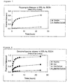
- The results from testing the coated stents for their rapamycin release described above are shown in Figures 5, 7 and 9.
- This example describes the method for analyzing the release medium for dexamethasone.
- The HPLC system used to analyze the samples was a Shimadzu Class-VP Chromatography Laboratory System. This system is equipped with a photodiode array detector. 20µL of each sample was withdrawn and analyzed on a C18 -reverse phase column (Waters Symmetry™ Column: 4.6mm X 100mm RP18 3.5 µ). An isocratic mobile phase consisting of methanol/water (55:45 v/v) delivered at a flow rate of 0.8 mL/min. was used for the first 6.5 mins of analysis followed by 100% methanol for 2 minutes; the latter was to ensure removal of rapamycin which is retained on the column. The column was maintained at 25°C throughout the analysis. Under these analytical conditions dexamthasone had a retention time of 5.9 ± 0.1 minutes. The concentration was determined from a standard curve of concentration versus response (area-under the curve) generated from dexamethasone standards in the range of from 40ng/mL to 4.0µg/mL.
- The results from testing the coated stents for the dexamethasone release described above are shown in Figures 6, 8 and 9.
- These and other concepts will are disclosed herein. It would be apparent to the reader that modifications are possible to the stent or the drug dosage applied. In any event, however, the any obvious modifications should be perceived to fall within the scope of the invention, which is to be realized from the attached daims and their equivalents.
- The terms "Rapamycin", "Taxol", "Agiopeptin", "Symmetry" and "Hirulog" are hereby acknowledged as trade marks.
Claims (3)
- A catheter for the delivery of drugs to a blood vessel lumen of a patient; wherein a therapeutic dosage amount of the combination of rapamycin and dexamethasone is coated on the said catheter.
- A stent for the delivery of drugs to a blood vessel lumen of a patient; wherein a therapeutic dosage amount of rapamycin and dexamethasone is coated on the said stent.
- A stent as claimed in claim 2, in which the stent comprises a plurality of struts, said struts expansible within the patient's lumen, in which at least one of said struts contains a reservoir therein.
Priority Applications (1)
| Application Number | Priority Date | Filing Date | Title |
|---|---|---|---|
| DK01937196T DK1289576T3 (en) | 2000-05-12 | 2001-04-25 | Drug combinations to prevent restenosis |
Applications Claiming Priority (5)
| Application Number | Priority Date | Filing Date | Title |
|---|---|---|---|
| US20441700P | 2000-05-12 | 2000-05-12 | |
| US204417P | 2000-05-12 | ||
| US575480P | 2000-05-19 | ||
| US09/575,480 US8029561B1 (en) | 2000-05-12 | 2000-05-19 | Drug combination useful for prevention of restenosis |
| PCT/US2001/013780 WO2001087372A1 (en) | 2000-05-12 | 2001-04-25 | Drug combinations useful for prevention of restenosis |
Publications (2)
| Publication Number | Publication Date |
|---|---|
| EP1289576A1 EP1289576A1 (en) | 2003-03-12 |
| EP1289576B1 true EP1289576B1 (en) | 2005-06-29 |
Family
ID=26899464
Family Applications (1)
| Application Number | Title | Priority Date | Filing Date |
|---|---|---|---|
| EP01937196A Revoked EP1289576B1 (en) | 2000-05-12 | 2001-04-25 | Drug combinations useful for prevention of restenosis |
Country Status (12)
| Country | Link |
|---|---|
| US (2) | US8029561B1 (en) |
| EP (1) | EP1289576B1 (en) |
| JP (1) | JP2003533493A (en) |
| AT (1) | ATE298592T1 (en) |
| AU (3) | AU6295701A (en) |
| BR (1) | BR0110778A (en) |
| CA (1) | CA2408606A1 (en) |
| DE (1) | DE60111743T2 (en) |
| ES (1) | ES2244622T3 (en) |
| MX (2) | MXPA02011186A (en) |
| PT (1) | PT1289576E (en) |
| WO (1) | WO2001087372A1 (en) |
Cited By (2)
| Publication number | Priority date | Publication date | Assignee | Title |
|---|---|---|---|---|
| WO2006102359A3 (en) * | 2005-03-23 | 2007-05-10 | Abbott Lab | Delivery of highly lipophilic agents via medical devices |
| WO2007111885A3 (en) * | 2006-03-22 | 2007-12-06 | Abbott Lab | Delivery of highly lipophilic agents via medical devices |
Families Citing this family (118)
| Publication number | Priority date | Publication date | Assignee | Title |
|---|---|---|---|---|
| US20030129215A1 (en) * | 1998-09-24 | 2003-07-10 | T-Ram, Inc. | Medical devices containing rapamycin analogs |
| US7399480B2 (en) * | 1997-09-26 | 2008-07-15 | Abbott Laboratories | Methods of administering tetrazole-containing rapamycin analogs with other therapeutic substances using medical devices |
| US7445792B2 (en) | 2003-03-10 | 2008-11-04 | Abbott Laboratories | Medical device having a hydration inhibitor |
| US6890546B2 (en) | 1998-09-24 | 2005-05-10 | Abbott Laboratories | Medical devices containing rapamycin analogs |
| US8257725B2 (en) * | 1997-09-26 | 2012-09-04 | Abbott Laboratories | Delivery of highly lipophilic agents via medical devices |
| US8057816B2 (en) | 1997-09-26 | 2011-11-15 | Abbott Laboratories | Compositions and methods of administering paclitaxel with other drugs using medical devices |
| US8394398B2 (en) | 1997-09-26 | 2013-03-12 | Abbott Laboratories | Methods of administering rapamycin analogs with anti-inflammatories using medical devices |
| US8257726B2 (en) | 1997-09-26 | 2012-09-04 | Abbott Laboratories | Compositions, systems, kits, and methods of administering rapamycin analogs with paclitaxel using medical devices |
| US7357942B2 (en) | 1997-09-26 | 2008-04-15 | Abbott Laboratories | Compositions, systems, and kits for administering zotarolimus and paclitaxel to blood vessel lumens |
| US7378105B2 (en) | 1997-09-26 | 2008-05-27 | Abbott Laboratories | Drug delivery systems, kits, and methods for administering zotarolimus and paclitaxel to blood vessel lumens |
| US7208010B2 (en) * | 2000-10-16 | 2007-04-24 | Conor Medsystems, Inc. | Expandable medical device for delivery of beneficial agent |
| US7455853B2 (en) | 1998-09-24 | 2008-11-25 | Abbott Cardiovascular Systems Inc. | Medical devices containing rapamycin analogs |
| US8257724B2 (en) | 1998-09-24 | 2012-09-04 | Abbott Laboratories | Delivery of highly lipophilic agents via medical devices |
| US7960405B2 (en) | 1998-09-24 | 2011-06-14 | Abbott Laboratories | Compounds and methods for treatment and prevention of diseases |
| US6790228B2 (en) | 1999-12-23 | 2004-09-14 | Advanced Cardiovascular Systems, Inc. | Coating for implantable devices and a method of forming the same |
| US7265199B2 (en) | 2000-04-11 | 2007-09-04 | Celonova Biosciences Germany Gmbh | Poly-tri-fluoro-ethoxypolyphosphazene coverings and films |
| US7419678B2 (en) * | 2000-05-12 | 2008-09-02 | Cordis Corporation | Coated medical devices for the prevention and treatment of vascular disease |
| US6534693B2 (en) | 2000-11-06 | 2003-03-18 | Afmedica, Inc. | Surgically implanted devices having reduced scar tissue formation |
| US9080146B2 (en) | 2001-01-11 | 2015-07-14 | Celonova Biosciences, Inc. | Substrates containing polyphosphazene as matrices and substrates containing polyphosphazene with a micro-structured surface |
| US6752829B2 (en) * | 2001-01-30 | 2004-06-22 | Scimed Life Systems, Inc. | Stent with channel(s) for containing and delivering a biologically active material and method for manufacturing the same |
| AU2002250968C1 (en) | 2001-02-19 | 2018-01-04 | Novartis Ag | Cancer treatment |
| US7247313B2 (en) * | 2001-06-27 | 2007-07-24 | Advanced Cardiovascular Systems, Inc. | Polyacrylates coatings for implantable medical devices |
| PT1432380E (en) | 2001-08-17 | 2007-01-31 | Polyzenix Gmbh | Device based on nitinol with a polyphosphazene coating |
| IL147416A (en) * | 2001-12-31 | 2008-11-26 | Israel State | Combined modalities for improved cancer treatment |
| TW200730152A (en) * | 2002-01-10 | 2007-08-16 | Novartis Ag | Drug delivery systems for the prevention and treatment of vascular diseases |
| US20030220297A1 (en) | 2002-02-01 | 2003-11-27 | Berstein David L. | Phosphorus-containing compounds and uses thereof |
| US20030207856A1 (en) * | 2002-03-18 | 2003-11-06 | Patrice Tremble | Medical devices and compositions for delivering anti-proliferatives to anatomical sites at risk for restenosis |
| AU2003252047A1 (en) * | 2002-07-18 | 2004-02-09 | Medtronic Ave Inc. | Medical devices comprising a protein-tyrosine kinase inhibitor to inhibit restonosis |
| US7491233B1 (en) * | 2002-07-19 | 2009-02-17 | Advanced Cardiovascular Systems Inc. | Purified polymers for coatings of implantable medical devices |
| DE10237571A1 (en) * | 2002-08-13 | 2004-02-26 | Biotronik Meß- und Therapiegeräte GmbH & Co. Ingenieurbüro Berlin | Endovascular implant with active coating |
| WO2004017939A1 (en) * | 2002-08-20 | 2004-03-04 | Terumo Kabushiki Kaisha | Medical instrument to be implanted in the body |
| JP4588986B2 (en) * | 2002-08-20 | 2010-12-01 | テルモ株式会社 | Implantable medical device |
| WO2004022124A1 (en) | 2002-09-06 | 2004-03-18 | Abbott Laboratories | Medical device having hydration inhibitor |
| DE10244847A1 (en) * | 2002-09-20 | 2004-04-01 | Ulrich Prof. Dr. Speck | Medical device for drug delivery |
| WO2004032947A1 (en) * | 2002-10-09 | 2004-04-22 | Unibioscreen S.A. | Extract with anti-tumor and anti-poisonous activity |
| WO2004037443A1 (en) * | 2002-10-22 | 2004-05-06 | Medtronic Vascular Inc. | Stent with intermittent coating |
| AU2003293529A1 (en) | 2002-12-16 | 2004-07-29 | Nitromed, Inc. | Nitrosated and nitrosylated rapamycin compounds, compositions and methods of use |
| JP2004222953A (en) * | 2003-01-22 | 2004-08-12 | Kanegafuchi Chem Ind Co Ltd | Indwelling stent |
| MXPA05010388A (en) * | 2003-03-28 | 2006-03-08 | Kosan Biosciences Inc | Devices, methods, and compositions to prevent restenosis. |
| US7279002B2 (en) * | 2003-04-25 | 2007-10-09 | Boston Scientific Scimed, Inc. | Cutting stent and balloon |
| US20050118344A1 (en) | 2003-12-01 | 2005-06-02 | Pacetti Stephen D. | Temperature controlled crimping |
| US20050033417A1 (en) * | 2003-07-31 | 2005-02-10 | John Borges | Coating for controlled release of a therapeutic agent |
| JP4732346B2 (en) * | 2003-08-19 | 2011-07-27 | バイエル・マテリアルサイエンス・アクチェンゲゼルシャフト | Polymeric drug elution system for medical devices |
| US8747881B2 (en) | 2003-12-19 | 2014-06-10 | Cordis Corporation | Intraluminal medical devices in combination with therapeutic agents |
| US8652502B2 (en) | 2003-12-19 | 2014-02-18 | Cordis Corporation | Local vascular delivery of trichostatin A alone or in combination with sirolimus to prevent restenosis following vascular injury |
| US7303758B2 (en) * | 2004-01-20 | 2007-12-04 | Cordis Corporation | Local vascular delivery of mycophenolic acid in combination with rapamycin to prevent restenosis following vascular injury |
| US7806924B2 (en) * | 2004-02-18 | 2010-10-05 | Cordis Corporation | Implantable structures for local vascular delivery of cladribine in combination with rapamycin for restenosis |
| US8431145B2 (en) | 2004-03-19 | 2013-04-30 | Abbott Laboratories | Multiple drug delivery from a balloon and a prosthesis |
| US7875282B2 (en) * | 2004-03-22 | 2011-01-25 | Cordis Corporation | Coated medical device for local vascular delivery of Panzem® in combination with rapamycin to prevent restenosis following vascular injury |
| US7695731B2 (en) * | 2004-03-22 | 2010-04-13 | Cordis Corporation | Local vascular delivery of etoposide in combination with rapamycin to prevent restenosis following vascular injury |
| US20050220836A1 (en) * | 2004-03-31 | 2005-10-06 | Robert Falotico | Drug delivery device |
| CN1964748A (en) * | 2004-04-06 | 2007-05-16 | 苏莫迪克斯公司 | Coating compositions for bioactive agents |
| DE102004024552B3 (en) * | 2004-05-18 | 2005-12-08 | Infineon Technologies Ag | Memory cell arrangement with a double memory cell |
| US7976557B2 (en) | 2004-06-23 | 2011-07-12 | Boston Scientific Scimed, Inc. | Cutting balloon and process |
| US8709469B2 (en) | 2004-06-30 | 2014-04-29 | Abbott Cardiovascular Systems Inc. | Anti-proliferative and anti-inflammatory agent combination for treatment of vascular disorders with an implantable medical device |
| US20080171729A1 (en) * | 2004-07-16 | 2008-07-17 | Novartis Ag | Use Of A Steroid For Enhancement Of Skin Permeability |
| US20070250157A1 (en) * | 2004-09-08 | 2007-10-25 | Kaneka Corporation | Stent For Placement In Body |
| US20210299056A9 (en) | 2004-10-25 | 2021-09-30 | Varian Medical Systems, Inc. | Color-Coded Polymeric Particles of Predetermined Size for Therapeutic and/or Diagnostic Applications and Related Methods |
| US9114162B2 (en) | 2004-10-25 | 2015-08-25 | Celonova Biosciences, Inc. | Loadable polymeric particles for enhanced imaging in clinical applications and methods of preparing and using the same |
| US9107850B2 (en) | 2004-10-25 | 2015-08-18 | Celonova Biosciences, Inc. | Color-coded and sized loadable polymeric particles for therapeutic and/or diagnostic applications and methods of preparing and using the same |
| US8066726B2 (en) | 2004-11-23 | 2011-11-29 | Boston Scientific Scimed, Inc. | Serpentine cutting blade for cutting balloon |
| WO2007032777A2 (en) | 2005-03-23 | 2007-03-22 | Abbott Laboratories | Compositions and methods of administering rapamycin analogs using medical devices for long-term efficacy |
| CA2615452C (en) * | 2005-07-15 | 2015-03-31 | Micell Technologies, Inc. | Polymer coatings containing drug powder of controlled morphology |
| KR101492545B1 (en) * | 2005-07-15 | 2015-02-12 | 미셀 테크놀로지즈, 인코포레이티드 | Polymer coatings containing drug powder of controlled morphology |
| US20090062909A1 (en) | 2005-07-15 | 2009-03-05 | Micell Technologies, Inc. | Stent with polymer coating containing amorphous rapamycin |
| JP5285426B2 (en) * | 2005-10-14 | 2013-09-11 | アボット ラボラトリーズ | Compositions, systems, kits and methods for administering a rapamycin analog with paclitaxel using a medical device |
| TWI469771B (en) * | 2005-10-14 | 2015-01-21 | Abbott Lab | System for providing controlled release delivery of drugs for treatment of neointimal hyperplasia and pharmaceutical composition for reducing neointimal hyperplasia |
| KR100778020B1 (en) | 2005-10-24 | 2007-11-28 | 사회복지법인 삼성생명공익재단 | Vascular stent which is specially designed for the multiple drug loading and better drug elution |
| US20070134163A1 (en) | 2005-12-13 | 2007-06-14 | Zhao Jonathon Z | Radiographic contrasting agents and radio-opaque polymeric materials for medical devices |
| US10029034B2 (en) * | 2005-12-15 | 2018-07-24 | CARDINAL HEALTH SWITZERLAND 515 GmbH | Drug-eluting articles with improved drug release profiles |
| BRPI0600275A (en) * | 2006-01-03 | 2007-10-02 | Brz Biotecnologia Ltda | Coronary prosthesis releasing drug composition for prevention and treatment of restenosis and manufacturing process |
| US8043358B2 (en) | 2006-03-29 | 2011-10-25 | Boston Scientific Scimed, Inc. | Stent with overlap and high extension |
| CA2996768C (en) | 2006-04-26 | 2020-12-08 | Micell Technologies, Inc. | Coatings containing multiple drugs |
| US7985441B1 (en) | 2006-05-04 | 2011-07-26 | Yiwen Tang | Purification of polymers for coating applications |
| US9028859B2 (en) | 2006-07-07 | 2015-05-12 | Advanced Cardiovascular Systems, Inc. | Phase-separated block copolymer coatings for implantable medical devices |
| EP2702993B1 (en) * | 2006-09-13 | 2017-12-06 | Elixir Medical Corporation | Macrocyclic lactone compounds and methods for their use |
| US10695327B2 (en) | 2006-09-13 | 2020-06-30 | Elixir Medical Corporation | Macrocyclic lactone compounds and methods for their use |
| US8088789B2 (en) | 2006-09-13 | 2012-01-03 | Elixir Medical Corporation | Macrocyclic lactone compounds and methods for their use |
| ES2378905T3 (en) | 2006-10-10 | 2012-04-19 | Celonova Biosciences, Inc. | Bioprotics heart valve with polyphosphazene |
| CA2667228C (en) | 2006-10-23 | 2015-07-14 | Micell Technologies, Inc. | Holder for electrically charging a substrate during coating |
| CA2679712C (en) | 2007-01-08 | 2016-11-15 | Micell Technologies, Inc. | Stents having biodegradable layers |
| US11426494B2 (en) | 2007-01-08 | 2022-08-30 | MT Acquisition Holdings LLC | Stents having biodegradable layers |
| AU2008256684B2 (en) | 2007-05-25 | 2012-06-14 | Micell Technologies, Inc. | Polymer films for medical device coating |
| US20090104240A1 (en) * | 2007-10-19 | 2009-04-23 | Abbott Cardiovascular Systems Inc. | Dual Drug Formulations For Implantable Medical Devices For Treatment of Vascular Diseases |
| US8992471B2 (en) * | 2007-11-05 | 2015-03-31 | Nanocopoeia, Inc. | Coated devices and method of making coated devices that reduce smooth muscle cell proliferation and platelet activity |
| US8216600B2 (en) | 2007-11-14 | 2012-07-10 | Cordis Corporation | Polymeric materials for medical devices |
| FR2927814B1 (en) * | 2008-02-21 | 2011-01-14 | Hexacath | IMPLANTABLE MEDICAL DEVICE COMPRISING THE OCTREOTIDE AND A PROTECTIVE AND / OR RETENTIVE LAYER THEREOF |
| FR2927815B1 (en) * | 2008-02-21 | 2011-01-14 | Hexacath | MEDICAL DEVICE COMPRISING A NO PRECURSOR AGENT, SUCH AS L-ARGININE OR L-LYSINE, AND A PROTECTIVE AND / OR RETENTIVE LAYER THEREOF |
| FR2927813B1 (en) * | 2008-02-21 | 2017-07-21 | Hexacath | MEDICAL DEVICE IMPLANTABLE ON A PROTECTIVE LAYER / RETENTION OF AN ACTIVE AGENT OR MEDICAMENT, ESPECIALLY WATER SOLUBLE |
| ES2658101T3 (en) * | 2008-02-21 | 2018-03-08 | Hexacath | Implantable medical device with a protective / retention layer of an active agent or medication, specifically water soluble |
| FR2927812B1 (en) * | 2008-02-21 | 2011-09-23 | Hexacath | IMPLANTABLE MEDICAL DEVICE WITH DRUG AND PROTECTIVE OR RETENTIVE LAYER THEREOF |
| EP2271294B1 (en) | 2008-04-17 | 2018-03-28 | Micell Technologies, Inc. | Stents having bioabsorbable layers |
| KR101104901B1 (en) | 2008-05-23 | 2012-01-12 | 김영곤 | manufacturing method of thermo-rod for active drug release the thermo-rod |
| JP2011528275A (en) | 2008-07-17 | 2011-11-17 | ミセル テクノロジーズ,インク. | Drug delivery medical device |
| US20100092534A1 (en) * | 2008-10-10 | 2010-04-15 | Medtronic Vascular, Inc. | Combination Local Delivery Using a Stent |
| US20100161039A1 (en) * | 2008-12-23 | 2010-06-24 | Vipul Dave | Adhesion promoting temporary mask for coated surfaces |
| US8834913B2 (en) | 2008-12-26 | 2014-09-16 | Battelle Memorial Institute | Medical implants and methods of making medical implants |
| CN102481195B (en) | 2009-04-01 | 2015-03-25 | 米歇尔技术公司 | Drug delivery medical device |
| WO2010121187A2 (en) | 2009-04-17 | 2010-10-21 | Micell Techologies, Inc. | Stents having controlled elution |
| EP2453834A4 (en) | 2009-07-16 | 2014-04-16 | Micell Technologies Inc | Drug delivery medical device |
| US8951595B2 (en) | 2009-12-11 | 2015-02-10 | Abbott Cardiovascular Systems Inc. | Coatings with tunable molecular architecture for drug-coated balloon |
| US11369498B2 (en) | 2010-02-02 | 2022-06-28 | MT Acquisition Holdings LLC | Stent and stent delivery system with improved deliverability |
| US8795762B2 (en) | 2010-03-26 | 2014-08-05 | Battelle Memorial Institute | System and method for enhanced electrostatic deposition and surface coatings |
| WO2011133655A1 (en) | 2010-04-22 | 2011-10-27 | Micell Technologies, Inc. | Stents and other devices having extracellular matrix coating |
| CA2805631C (en) | 2010-07-16 | 2018-07-31 | Micell Technologies, Inc. | Drug delivery medical device |
| WO2012017449A1 (en) * | 2010-08-04 | 2012-02-09 | Meril Life Sciences Pvt. Ltd | Process for preparation of novel 42-0-(heteroalkoxyalkyl) rapamycin compounds with anti-proliferative properties" |
| WO2012166819A1 (en) | 2011-05-31 | 2012-12-06 | Micell Technologies, Inc. | System and process for formation of a time-released, drug-eluting transferable coating |
| CA2841360A1 (en) | 2011-07-15 | 2013-01-24 | Micell Technologies, Inc. | Drug delivery medical device |
| US10188772B2 (en) | 2011-10-18 | 2019-01-29 | Micell Technologies, Inc. | Drug delivery medical device |
| DE102012001188A1 (en) * | 2012-01-24 | 2013-07-25 | Qualimed Innovative Medizinprodukte Gmbh | balloon catheter |
| US20130259921A1 (en) * | 2012-03-30 | 2013-10-03 | Abbott Cardiovascular Systems Inc. | Treatment Of Diabetic Patients With A Stent And An Adjunctive Drug Formulation |
| US9220584B2 (en) | 2012-03-30 | 2015-12-29 | Abbott Cardiovascular Systems Inc. | Treatment of diabetic patients with a stent and locally administered adjunctive therapy |
| US20130303496A1 (en) * | 2012-05-08 | 2013-11-14 | Abbott Cardiovascular Systems Inc. | Method Of Treating Vascular Lesions |
| KR20150143476A (en) | 2013-03-12 | 2015-12-23 | 미셀 테크놀로지즈, 인코포레이티드 | Bioabsorbable biomedical implants |
| JP6689605B2 (en) | 2013-04-16 | 2020-04-28 | 株式会社カネカ | Medical tubular body |
| KR102079613B1 (en) | 2013-05-15 | 2020-02-20 | 미셀 테크놀로지즈, 인코포레이티드 | Bioabsorbable biomedical implants |
| EP2997977A1 (en) * | 2014-09-19 | 2016-03-23 | Fundación de la Comunidad Valenciana Centro de Investigación Principe Felipe | Specific mtor inhibitors in the treatment of x-linked adrenoleukodystrophy |
| CN110996687A (en) * | 2017-05-26 | 2020-04-10 | 墨卡托医疗系统公司 | Combination therapy for the treatment of restenosis |
Family Cites Families (165)
| Publication number | Priority date | Publication date | Assignee | Title |
|---|---|---|---|---|
| GB1205743A (en) | 1966-07-15 | 1970-09-16 | Nat Res Dev | Surgical dilator |
| US3657744A (en) | 1970-05-08 | 1972-04-25 | Univ Minnesota | Method for fixing prosthetic implants in a living body |
| US4388735A (en) | 1980-11-03 | 1983-06-21 | Shiley Inc. | Low profile prosthetic xenograft heart valve |
| CA1204643A (en) | 1981-09-16 | 1986-05-20 | Hans I. Wallsten | Device for application in blood vessels or other difficulty accessible locations and its use |
| DE3205942A1 (en) | 1982-02-19 | 1983-09-08 | Ljubomir Dr. Skopje Vasilev | Balloon-tipped catheter with a mobile tip which permits complete voiding of the bladder |
| US4503569A (en) | 1983-03-03 | 1985-03-12 | Dotter Charles T | Transluminally placed expandable graft prosthesis |
| US5275622A (en) | 1983-12-09 | 1994-01-04 | Harrison Medical Technologies, Inc. | Endovascular grafting apparatus, system and method and devices for use therewith |
| US4580568A (en) | 1984-10-01 | 1986-04-08 | Cook, Incorporated | Percutaneous endovascular stent and method for insertion thereof |
| US4733665C2 (en) | 1985-11-07 | 2002-01-29 | Expandable Grafts Partnership | Expandable intraluminal graft and method and apparatus for implanting an expandable intraluminal graft |
| US4800882A (en) | 1987-03-13 | 1989-01-31 | Cook Incorporated | Endovascular stent and delivery system |
| US5041126A (en) | 1987-03-13 | 1991-08-20 | Cook Incorporated | Endovascular stent and delivery system |
| US4907336A (en) | 1987-03-13 | 1990-03-13 | Cook Incorporated | Method of making an endovascular stent and delivery system |
| US4969458A (en) | 1987-07-06 | 1990-11-13 | Medtronic, Inc. | Intracoronary stent and method of simultaneous angioplasty and stent implant |
| US5131908A (en) | 1987-09-01 | 1992-07-21 | Herbert Dardik | Tubular prosthesis for vascular reconstructive surgery and process for preparing same |
| US4990131A (en) | 1987-09-01 | 1991-02-05 | Herbert Dardik | Tubular prostheses for vascular reconstructive surgery and process for preparing same |
| US4886062A (en) | 1987-10-19 | 1989-12-12 | Medtronic, Inc. | Intravascular radially expandable stent and method of implant |
| US5133732A (en) | 1987-10-19 | 1992-07-28 | Medtronic, Inc. | Intravascular stent |
| US5266073A (en) | 1987-12-08 | 1993-11-30 | Wall W Henry | Angioplasty stent |
| US5192307A (en) | 1987-12-08 | 1993-03-09 | Wall W Henry | Angioplasty stent |
| US4856516A (en) | 1989-01-09 | 1989-08-15 | Cordis Corporation | Endovascular stent apparatus and method |
| CH678393A5 (en) | 1989-01-26 | 1991-09-13 | Ulrich Prof Dr Med Sigwart | |
| US5163958A (en) | 1989-02-02 | 1992-11-17 | Cordis Corporation | Carbon coated tubular endoprosthesis |
| US4990155A (en) | 1989-05-19 | 1991-02-05 | Wilkoff Howard M | Surgical stent method and apparatus |
| US4994071A (en) | 1989-05-22 | 1991-02-19 | Cordis Corporation | Bifurcating stent apparatus and method |
| US5015253A (en) | 1989-06-15 | 1991-05-14 | Cordis Corporation | Non-woven endoprosthesis |
| US5171262A (en) | 1989-06-15 | 1992-12-15 | Cordis Corporation | Non-woven endoprosthesis |
| US5292331A (en) | 1989-08-24 | 1994-03-08 | Applied Vascular Engineering, Inc. | Endovascular support device |
| CA2026604A1 (en) | 1989-10-02 | 1991-04-03 | Rodney G. Wolff | Articulated stent |
| US5035706A (en) | 1989-10-17 | 1991-07-30 | Cook Incorporated | Percutaneous stent and method for retrieval thereof |
| US5176660A (en) | 1989-10-23 | 1993-01-05 | Cordis Corporation | Catheter having reinforcing strands |
| DK0441516T3 (en) | 1990-02-08 | 1995-06-12 | Howmedica | Inflatable catheter |
| US6004346A (en) | 1990-02-28 | 1999-12-21 | Medtronic, Inc. | Intralumenal drug eluting prosthesis |
| US5545208A (en) | 1990-02-28 | 1996-08-13 | Medtronic, Inc. | Intralumenal drug eluting prosthesis |
| IL94138A (en) | 1990-04-19 | 1997-03-18 | Instent Inc | Device for the treatment of constricted fluid conducting ducts |
| US5064435A (en) | 1990-06-28 | 1991-11-12 | Schneider (Usa) Inc. | Self-expanding prosthesis having stable axial length |
| US5122154A (en) | 1990-08-15 | 1992-06-16 | Rhodes Valentine J | Endovascular bypass graft |
| US5449372A (en) | 1990-10-09 | 1995-09-12 | Scimed Lifesystems, Inc. | Temporary stent and methods for use and manufacture |
| DK0480667T3 (en) | 1990-10-09 | 1996-06-10 | Cook Inc | Percutaneous stent construction |
| US5222971A (en) | 1990-10-09 | 1993-06-29 | Scimed Life Systems, Inc. | Temporary stent and methods for use and manufacture |
| US5217483A (en) | 1990-11-28 | 1993-06-08 | Numed, Inc. | Intravascular radially expandable stent |
| US5178618A (en) | 1991-01-16 | 1993-01-12 | Brigham And Womens Hospital | Method and device for recanalization of a body passageway |
| US5354257A (en) | 1991-01-29 | 1994-10-11 | Med Institute, Inc. | Minimally invasive medical device for providing a radiation treatment |
| US5135536A (en) | 1991-02-05 | 1992-08-04 | Cordis Corporation | Endovascular stent and method |
| US5116365A (en) | 1991-02-22 | 1992-05-26 | Cordis Corporation | Stent apparatus and method for making |
| US5304200A (en) | 1991-05-29 | 1994-04-19 | Cordis Corporation | Welded radially expandable endoprosthesis and the like |
| USD359802S (en) | 1991-06-28 | 1995-06-27 | Cook Incorporated | Vascular stent |
| US5314472A (en) | 1991-10-01 | 1994-05-24 | Cook Incorporated | Vascular stent |
| US5443498A (en) | 1991-10-01 | 1995-08-22 | Cook Incorporated | Vascular stent and method of making and implanting a vacsular stent |
| WO1993006792A1 (en) | 1991-10-04 | 1993-04-15 | Scimed Life Systems, Inc. | Biodegradable drug delivery vascular stent |
| US5500013A (en) | 1991-10-04 | 1996-03-19 | Scimed Life Systems, Inc. | Biodegradable drug delivery vascular stent |
| US5464450A (en) | 1991-10-04 | 1995-11-07 | Scimed Lifesystems Inc. | Biodegradable drug delivery vascular stent |
| US5366504A (en) | 1992-05-20 | 1994-11-22 | Boston Scientific Corporation | Tubular medical prosthesis |
| US5387235A (en) | 1991-10-25 | 1995-02-07 | Cook Incorporated | Expandable transluminal graft prosthesis for repair of aneurysm |
| CA2079417C (en) | 1991-10-28 | 2003-01-07 | Lilip Lau | Expandable stents and method of making same |
| US5516781A (en) * | 1992-01-09 | 1996-05-14 | American Home Products Corporation | Method of treating restenosis with rapamycin |
| US5258021A (en) | 1992-01-27 | 1993-11-02 | Duran Carlos G | Sigmoid valve annuloplasty ring |
| CA2087132A1 (en) | 1992-01-31 | 1993-08-01 | Michael S. Williams | Stent capable of attachment within a body lumen |
| US5405377A (en) | 1992-02-21 | 1995-04-11 | Endotech Ltd. | Intraluminal stent |
| DE4206843C2 (en) | 1992-03-04 | 1994-03-24 | Heraeus Elektrochemie | Electrochemical cells for performing electrochemical processes |
| US5591224A (en) | 1992-03-19 | 1997-01-07 | Medtronic, Inc. | Bioelastomeric stent |
| EP0566245B1 (en) | 1992-03-19 | 1999-10-06 | Medtronic, Inc. | Intraluminal stent |
| US5571166A (en) | 1992-03-19 | 1996-11-05 | Medtronic, Inc. | Method of making an intraluminal stent |
| US5599352A (en) | 1992-03-19 | 1997-02-04 | Medtronic, Inc. | Method of making a drug eluting stent |
| US5510077A (en) | 1992-03-19 | 1996-04-23 | Dinh; Thomas Q. | Method of making an intraluminal stent |
| US5282823A (en) | 1992-03-19 | 1994-02-01 | Medtronic, Inc. | Intravascular radially expandable stent |
| US5370683A (en) | 1992-03-25 | 1994-12-06 | Cook Incorporated | Vascular stent |
| FR2689388B1 (en) | 1992-04-07 | 1999-07-16 | Celsa Lg | PERFECTIONALLY RESORBABLE BLOOD FILTER. |
| AU670937B2 (en) | 1992-04-28 | 1996-08-08 | Wyeth | Method of treating hyperproliferative vascular disease |
| US5354308A (en) | 1992-05-01 | 1994-10-11 | Beth Israel Hospital Association | Metal wire stent |
| WO1995014500A1 (en) | 1992-05-01 | 1995-06-01 | Beth Israel Hospital | A stent |
| US5383928A (en) | 1992-06-10 | 1995-01-24 | Emory University | Stent sheath for local drug delivery |
| US5342387A (en) | 1992-06-18 | 1994-08-30 | American Biomed, Inc. | Artificial support for a blood vessel |
| DE4222380A1 (en) * | 1992-07-08 | 1994-01-13 | Ernst Peter Prof Dr M Strecker | Endoprosthesis implantable percutaneously in a patient's body |
| US5382261A (en) | 1992-09-01 | 1995-01-17 | Expandable Grafts Partnership | Method and apparatus for occluding vessels |
| US5449382A (en) | 1992-11-04 | 1995-09-12 | Dayton; Michael P. | Minimally invasive bioactivated endoprosthesis for vessel repair |
| US5578075B1 (en) | 1992-11-04 | 2000-02-08 | Daynke Res Inc | Minimally invasive bioactivated endoprosthesis for vessel repair |
| US5342348A (en) | 1992-12-04 | 1994-08-30 | Kaplan Aaron V | Method and device for treating and enlarging body lumens |
| BE1006440A3 (en) | 1992-12-21 | 1994-08-30 | Dereume Jean Pierre Georges Em | Luminal endoprosthesis AND METHOD OF PREPARATION. |
| US5370691A (en) | 1993-01-26 | 1994-12-06 | Target Therapeutics, Inc. | Intravascular inflatable stent |
| US5981568A (en) | 1993-01-28 | 1999-11-09 | Neorx Corporation | Therapeutic inhibitor of vascular smooth muscle cells |
| WO1994021308A1 (en) | 1993-03-18 | 1994-09-29 | Cedars-Sinai Medical Center | Drug incorporating and releasing polymeric coating for bioprosthesis |
| US5607463A (en) | 1993-03-30 | 1997-03-04 | Medtronic, Inc. | Intravascular medical device |
| US5441515A (en) | 1993-04-23 | 1995-08-15 | Advanced Cardiovascular Systems, Inc. | Ratcheting stent |
| US5824048A (en) | 1993-04-26 | 1998-10-20 | Medtronic, Inc. | Method for delivering a therapeutic substance to a body lumen |
| US5464650A (en) | 1993-04-26 | 1995-11-07 | Medtronic, Inc. | Intravascular stent and method |
| US5411549A (en) | 1993-07-13 | 1995-05-02 | Scimed Life Systems, Inc. | Selectively expandable, retractable and removable stent |
| EP0711158B2 (en) * | 1993-07-29 | 2008-07-23 | THE GOVERNMENT OF THE UNITED STATES OF AMERICA, as represented by THE SECRETARY, DEPARTMENT OF HEALTH AND HUMAN SERVICES | Method of treating atherosclerosis or restenosis using microtubule stabilizing agent |
| JPH08503715A (en) * | 1993-09-24 | 1996-04-23 | バクスター、インターナショナル、インコーポレイテッド | Method for promoting vascularization of implantable devices |
| WO1995010989A1 (en) | 1993-10-19 | 1995-04-27 | Scimed Life Systems, Inc. | Intravascular stent pump |
| US5389106A (en) | 1993-10-29 | 1995-02-14 | Numed, Inc. | Impermeable expandable intravascular stent |
| JP2703510B2 (en) | 1993-12-28 | 1998-01-26 | アドヴァンスド カーディオヴァスキュラー システムズ インコーポレーテッド | Expandable stent and method of manufacturing the same |
| US5519042A (en) * | 1994-01-13 | 1996-05-21 | Hoechst Aktiengesellschaft | Method of treating hyperproliferative vascular disease |
| US5403341A (en) | 1994-01-24 | 1995-04-04 | Solar; Ronald J. | Parallel flow endovascular stent and deployment apparatus therefore |
| US5609627A (en) | 1994-02-09 | 1997-03-11 | Boston Scientific Technology, Inc. | Method for delivering a bifurcated endoluminal prosthesis |
| US5443477A (en) | 1994-02-10 | 1995-08-22 | Stentco, Inc. | Apparatus and method for deployment of radially expandable stents by a mechanical linkage |
| US5643312A (en) | 1994-02-25 | 1997-07-01 | Fischell Robert | Stent having a multiplicity of closed circular structures |
| US5441516A (en) | 1994-03-03 | 1995-08-15 | Scimed Lifesystems Inc. | Temporary stent |
| US5449373A (en) | 1994-03-17 | 1995-09-12 | Medinol Ltd. | Articulated stent |
| ATE310839T1 (en) | 1994-04-29 | 2005-12-15 | Scimed Life Systems Inc | STENT WITH COLLAGEN |
| US5629077A (en) | 1994-06-27 | 1997-05-13 | Advanced Cardiovascular Systems, Inc. | Biodegradable mesh and film stent |
| US5397355A (en) | 1994-07-19 | 1995-03-14 | Stentco, Inc. | Intraluminal stent |
| US5788979A (en) | 1994-07-22 | 1998-08-04 | Inflow Dynamics Inc. | Biodegradable coating with inhibitory properties for application to biocompatible materials |
| US5891108A (en) | 1994-09-12 | 1999-04-06 | Cordis Corporation | Drug delivery stent |
| US5649977A (en) | 1994-09-22 | 1997-07-22 | Advanced Cardiovascular Systems, Inc. | Metal reinforced polymer stent |
| EP0761251B1 (en) | 1994-10-17 | 2004-12-29 | Kabushikikaisha Igaki Iryo Sekkei | Drug-releasing stent |
| US5707385A (en) | 1994-11-16 | 1998-01-13 | Advanced Cardiovascular Systems, Inc. | Drug loaded elastic membrane and method for delivery |
| US5637113A (en) | 1994-12-13 | 1997-06-10 | Advanced Cardiovascular Systems, Inc. | Polymer film for wrapping a stent structure |
| NL9500094A (en) | 1995-01-19 | 1996-09-02 | Industrial Res Bv | Y-shaped stent and method of deployment. |
| US5605696A (en) | 1995-03-30 | 1997-02-25 | Advanced Cardiovascular Systems, Inc. | Drug loaded polymeric material and method of manufacture |
| US5709713A (en) | 1995-03-31 | 1998-01-20 | Cardiovascular Concepts, Inc. | Radially expansible vascular prosthesis having reversible and other locking structures |
| EP0734698B9 (en) | 1995-04-01 | 2006-07-05 | Variomed AG | Stent for transluminal implantation into hollow organs |
| US5837313A (en) | 1995-04-19 | 1998-11-17 | Schneider (Usa) Inc | Drug release stent coating process |
| US6099562A (en) * | 1996-06-13 | 2000-08-08 | Schneider (Usa) Inc. | Drug coating with topcoat |
| US5674242A (en) | 1995-06-06 | 1997-10-07 | Quanam Medical Corporation | Endoprosthetic device with therapeutic compound |
| US5603722A (en) | 1995-06-06 | 1997-02-18 | Quanam Medical Corporation | Intravascular stent |
| AU716005B2 (en) | 1995-06-07 | 2000-02-17 | Cook Medical Technologies Llc | Implantable medical device |
| US5609629A (en) | 1995-06-07 | 1997-03-11 | Med Institute, Inc. | Coated implantable medical device |
| US5820917A (en) | 1995-06-07 | 1998-10-13 | Medtronic, Inc. | Blood-contacting medical device and method |
| US5607475A (en) | 1995-08-22 | 1997-03-04 | Medtronic, Inc. | Biocompatible medical article and method |
| US5672638A (en) | 1995-08-22 | 1997-09-30 | Medtronic, Inc. | Biocompatability for solid surfaces |
| US5679659A (en) | 1995-08-22 | 1997-10-21 | Medtronic, Inc. | Method for making heparinized biomaterials |
| US5669924A (en) | 1995-10-26 | 1997-09-23 | Shaknovich; Alexander | Y-shuttle stent assembly for bifurcating vessels and method of using the same |
| DE19614160A1 (en) | 1996-04-10 | 1997-10-16 | Variomed Ag | Stent for transluminal implantation in hollow organs |
| NZ331269A (en) | 1996-04-10 | 2000-01-28 | Advanced Cardiovascular System | Expandable stent, its structural strength varying along its length |
| US5728420A (en) | 1996-08-09 | 1998-03-17 | Medtronic, Inc. | Oxidative method for attachment of glycoproteins to surfaces of medical devices |
| UA58485C2 (en) | 1996-05-03 | 2003-08-15 | Медінол Лтд. | Method for manufacture of bifurcated stent (variants) and bifurcated stent (variants) |
| US5697971A (en) | 1996-06-11 | 1997-12-16 | Fischell; Robert E. | Multi-cell stent with cells having differing characteristics |
| US5820918A (en) | 1996-07-11 | 1998-10-13 | Hercules Incorporated | Medical devices containing in-situ generated medical compounds |
| CN1126534C (en) | 1996-08-30 | 2003-11-05 | 以色列农业部农业研究组织 | Intracoronary stents fixing mould containing quinazolinone derivatives |
| US5807404A (en) | 1996-09-19 | 1998-09-15 | Medinol Ltd. | Stent with variable features to optimize support and method of making such stent |
| US6387121B1 (en) * | 1996-10-21 | 2002-05-14 | Inflow Dynamics Inc. | Vascular and endoluminal stents with improved coatings |
| US5833651A (en) | 1996-11-08 | 1998-11-10 | Medtronic, Inc. | Therapeutic intraluminal stents |
| KR100526913B1 (en) | 1997-02-20 | 2005-11-09 | 쿡 인코포레이티드 | Coated implantable medical device |
| US20020133222A1 (en) * | 1997-03-05 | 2002-09-19 | Das Gladwin S. | Expandable stent having a plurality of interconnected expansion modules |
| US5843172A (en) | 1997-04-15 | 1998-12-01 | Advanced Cardiovascular Systems, Inc. | Porous medicated stent |
| US6273913B1 (en) | 1997-04-18 | 2001-08-14 | Cordis Corporation | Modified stent useful for delivery of drugs along stent strut |
| US5879697A (en) * | 1997-04-30 | 1999-03-09 | Schneider Usa Inc | Drug-releasing coatings for medical devices |
| US6562829B1 (en) * | 1997-05-23 | 2003-05-13 | Hadasit Medical Research Services & Development Co., Ltd. | Treatment of hepatic cirrhosis |
| US5916910A (en) * | 1997-06-04 | 1999-06-29 | Medinox, Inc. | Conjugates of dithiocarbamates with pharmacologically active agents and uses therefore |
| US6306166B1 (en) | 1997-08-13 | 2001-10-23 | Scimed Life Systems, Inc. | Loading and release of water-insoluble drugs |
| US6159488A (en) * | 1997-08-14 | 2000-12-12 | Agricultural Research Org. Ministry Of Agriculture (Gov.) | Intracoronary stents containing quinazolinone derivatives |
| US6890546B2 (en) * | 1998-09-24 | 2005-05-10 | Abbott Laboratories | Medical devices containing rapamycin analogs |
| US6225346B1 (en) * | 1997-10-24 | 2001-05-01 | Sugen, Inc. | Tyrphostin like compounds |
| US5932580A (en) * | 1997-12-01 | 1999-08-03 | Yissum Research And Development Company Of The Hebrew University Of Jerusalem | PDGF receptor kinase inhibitory compounds their preparation and compositions |
| US7208010B2 (en) * | 2000-10-16 | 2007-04-24 | Conor Medsystems, Inc. | Expandable medical device for delivery of beneficial agent |
| US20020099438A1 (en) * | 1998-04-15 | 2002-07-25 | Furst Joseph G. | Irradiated stent coating |
| US6369039B1 (en) * | 1998-06-30 | 2002-04-09 | Scimed Life Sytems, Inc. | High efficiency local drug delivery |
| JP4898991B2 (en) * | 1998-08-20 | 2012-03-21 | クック メディカル テクノロジーズ エルエルシー | Sheathed medical device |
| US6335029B1 (en) * | 1998-08-28 | 2002-01-01 | Scimed Life Systems, Inc. | Polymeric coatings for controlled delivery of active agents |
| US6187024B1 (en) | 1998-11-10 | 2001-02-13 | Target Therapeutics, Inc. | Bioactive coating for vaso-occlusive devices |
| US6730349B2 (en) * | 1999-04-19 | 2004-05-04 | Scimed Life Systems, Inc. | Mechanical and acoustical suspension coating of medical implants |
| US6368658B1 (en) * | 1999-04-19 | 2002-04-09 | Scimed Life Systems, Inc. | Coating medical devices using air suspension |
| US6287628B1 (en) * | 1999-09-03 | 2001-09-11 | Advanced Cardiovascular Systems, Inc. | Porous prosthesis and a method of depositing substances into the pores |
| US6899731B2 (en) * | 1999-12-30 | 2005-05-31 | Boston Scientific Scimed, Inc. | Controlled delivery of therapeutic agents by insertable medical devices |
| US6379382B1 (en) * | 2000-03-13 | 2002-04-30 | Jun Yang | Stent having cover with drug delivery capability |
| US7419678B2 (en) | 2000-05-12 | 2008-09-02 | Cordis Corporation | Coated medical devices for the prevention and treatment of vascular disease |
| US6776796B2 (en) | 2000-05-12 | 2004-08-17 | Cordis Corportation | Antiinflammatory drug and delivery device |
| US7077836B2 (en) * | 2000-07-21 | 2006-07-18 | Vein Rx, Inc. | Methods and apparatus for sclerosing the wall of a varicose vein |
| US6545097B2 (en) * | 2000-12-12 | 2003-04-08 | Scimed Life Systems, Inc. | Drug delivery compositions and medical devices containing block copolymer |
| US20020103526A1 (en) * | 2000-12-15 | 2002-08-01 | Tom Steinke | Protective coating for stent |
| US6939375B2 (en) * | 2000-12-22 | 2005-09-06 | Avantac Vascular Corporation | Apparatus and methods for controlled substance delivery from implanted prostheses |
| US7179251B2 (en) * | 2001-01-17 | 2007-02-20 | Boston Scientific Scimed, Inc. | Therapeutic delivery balloon |
| WO2002066092A2 (en) * | 2001-02-23 | 2002-08-29 | Angiogene Inc. | Drug eluting device for treating vascular diseases |
| WO2002072167A1 (en) * | 2001-03-13 | 2002-09-19 | Implant Sciences Corporation. | Drug eluting encapsulated stent |
| US8017237B2 (en) * | 2006-06-23 | 2011-09-13 | Abbott Cardiovascular Systems, Inc. | Nanoshells on polymers |
-
2000
- 2000-05-19 US US09/575,480 patent/US8029561B1/en not_active Expired - Fee Related
-
2001
- 2001-04-25 AT AT01937196T patent/ATE298592T1/en not_active IP Right Cessation
- 2001-04-25 MX MXPA02011186A patent/MXPA02011186A/en active IP Right Grant
- 2001-04-25 EP EP01937196A patent/EP1289576B1/en not_active Revoked
- 2001-04-25 PT PT01937196T patent/PT1289576E/en unknown
- 2001-04-25 ES ES01937196T patent/ES2244622T3/en not_active Expired - Lifetime
- 2001-04-25 AU AU6295701A patent/AU6295701A/en active Pending
- 2001-04-25 JP JP2001583836A patent/JP2003533493A/en not_active Withdrawn
- 2001-04-25 BR BRPI0110778-0A patent/BR0110778A/en not_active Application Discontinuation
- 2001-04-25 CA CA002408606A patent/CA2408606A1/en not_active Abandoned
- 2001-04-25 WO PCT/US2001/013780 patent/WO2001087372A1/en not_active Application Discontinuation
- 2001-04-25 AU AU2001262957A patent/AU2001262957B2/en not_active Ceased
- 2001-04-25 DE DE60111743T patent/DE60111743T2/en not_active Revoked
- 2001-05-14 MX MXPA02011099A patent/MXPA02011099A/en active IP Right Grant
- 2001-05-14 AU AU6158101A patent/AU6158101A/en active Pending
-
2011
- 2011-09-07 US US13/227,002 patent/US20120029475A1/en not_active Abandoned
Cited By (3)
| Publication number | Priority date | Publication date | Assignee | Title |
|---|---|---|---|---|
| WO2006102359A3 (en) * | 2005-03-23 | 2007-05-10 | Abbott Lab | Delivery of highly lipophilic agents via medical devices |
| EP2327429A1 (en) * | 2005-03-23 | 2011-06-01 | Abbott Laboratories | Delivery of highly lipophilic agents via medical devices |
| WO2007111885A3 (en) * | 2006-03-22 | 2007-12-06 | Abbott Lab | Delivery of highly lipophilic agents via medical devices |
Also Published As
| Publication number | Publication date |
|---|---|
| BR0110778A (en) | 2007-05-08 |
| PT1289576E (en) | 2005-10-31 |
| US20120029475A1 (en) | 2012-02-02 |
| AU6158101A (en) | 2001-11-26 |
| EP1289576A1 (en) | 2003-03-12 |
| CA2408606A1 (en) | 2001-11-22 |
| US8029561B1 (en) | 2011-10-04 |
| ATE298592T1 (en) | 2005-07-15 |
| WO2001087372A1 (en) | 2001-11-22 |
| MXPA02011186A (en) | 2004-09-09 |
| MXPA02011099A (en) | 2004-08-19 |
| DE60111743D1 (en) | 2005-08-04 |
| ES2244622T3 (en) | 2005-12-16 |
| AU6295701A (en) | 2001-11-26 |
| AU2001262957B2 (en) | 2004-12-02 |
| JP2003533493A (en) | 2003-11-11 |
| DE60111743T2 (en) | 2005-12-15 |
Similar Documents
| Publication | Publication Date | Title |
|---|---|---|
| EP1289576B1 (en) | Drug combinations useful for prevention of restenosis | |
| AU2001262957A1 (en) | Drug combinations useful for prevention of restenosis | |
| AU2001263113B2 (en) | Drug/drug delivery systems for the prevention and treatment of vascular disease | |
| US7300662B2 (en) | Drug/drug delivery systems for the prevention and treatment of vascular disease | |
| US20020007213A1 (en) | Drug/drug delivery systems for the prevention and treatment of vascular disease | |
| US20020005206A1 (en) | Antiproliferative drug and delivery device | |
| US20020007215A1 (en) | Drug/drug delivery systems for the prevention and treatment of vascular disease | |
| US20020007214A1 (en) | Drug/drug delivery systems for the prevention and treatment of vascular disease | |
| US20090204204A1 (en) | Drug/Drug Deliver Systems For The Prevention And Treatment Of Vascular Disease | |
| AU2001261580A1 (en) | Delivery devices for treatment of vascular disease | |
| JP2005334646A (en) | Antiproliferative drug and delivery device | |
| EP1591135A1 (en) | Drug/drug delivery systems for the prevention and treatment of vascular disease |
Legal Events
| Date | Code | Title | Description |
|---|---|---|---|
| PUAI | Public reference made under article 153(3) epc to a published international application that has entered the european phase |
Free format text: ORIGINAL CODE: 0009012 |
|
| 17P | Request for examination filed |
Effective date: 20021126 |
|
| AK | Designated contracting states |
Kind code of ref document: A1 Designated state(s): AT BE CH CY DE DK ES FI FR GB GR IE IT LI LU MC NL PT SE TR |
|
| AX | Request for extension of the european patent |
Extension state: AL LT LV MK RO SI |
|
| 17Q | First examination report despatched |
Effective date: 20030422 |
|
| GRAP | Despatch of communication of intention to grant a patent |
Free format text: ORIGINAL CODE: EPIDOSNIGR1 |
|
| GRAS | Grant fee paid |
Free format text: ORIGINAL CODE: EPIDOSNIGR3 |
|
| GRAA | (expected) grant |
Free format text: ORIGINAL CODE: 0009210 |
|
| AK | Designated contracting states |
Kind code of ref document: B1 Designated state(s): AT BE CH CY DE DK ES FI FR GB GR IE IT LI LU MC NL PT SE TR |
|
| REG | Reference to a national code |
Ref country code: GB Ref legal event code: FG4D |
|
| REG | Reference to a national code |
Ref country code: CH Ref legal event code: EP |
|
| REF | Corresponds to: |
Ref document number: 60111743 Country of ref document: DE Date of ref document: 20050804 Kind code of ref document: P |
|
| REG | Reference to a national code |
Ref country code: IE Ref legal event code: FG4D |
|
| REG | Reference to a national code |
Ref country code: CH Ref legal event code: NV Representative=s name: E. BLUM & CO. PATENTANWAELTE |
|
| REG | Reference to a national code |
Ref country code: GR Ref legal event code: EP Ref document number: 20050402565 Country of ref document: GR |
|
| REG | Reference to a national code |
Ref country code: DK Ref legal event code: T3 |
|
| REG | Reference to a national code |
Ref country code: SE Ref legal event code: TRGR |
|
| REG | Reference to a national code |
Ref country code: PT Ref legal event code: SC4A Effective date: 20050913 |
|
| REG | Reference to a national code |
Ref country code: ES Ref legal event code: FG2A Ref document number: 2244622 Country of ref document: ES Kind code of ref document: T3 |
|
| PGFP | Annual fee paid to national office [announced via postgrant information from national office to epo] |
Ref country code: AT Payment date: 20060412 Year of fee payment: 6 |
|
| ET | Fr: translation filed | ||
| PLBI | Opposition filed |
Free format text: ORIGINAL CODE: 0009260 |
|
| PGFP | Annual fee paid to national office [announced via postgrant information from national office to epo] |
Ref country code: DE Payment date: 20060420 Year of fee payment: 6 |
|
| PGFP | Annual fee paid to national office [announced via postgrant information from national office to epo] |
Ref country code: ES Payment date: 20060523 Year of fee payment: 6 |
|
| 26 | Opposition filed |
Opponent name: ABBOTT LABORATORIES Effective date: 20060329 |
|
| PLAX | Notice of opposition and request to file observation + time limit sent |
Free format text: ORIGINAL CODE: EPIDOSNOBS2 |
|
| PGFP | Annual fee paid to national office [announced via postgrant information from national office to epo] |
Ref country code: BE Payment date: 20060614 Year of fee payment: 6 |
|
| NLR1 | Nl: opposition has been filed with the epo |
Opponent name: ABBOTT LABORATORIES |
|
| PLAF | Information modified related to communication of a notice of opposition and request to file observations + time limit |
Free format text: ORIGINAL CODE: EPIDOSCOBS2 |
|
| RDAF | Communication despatched that patent is revoked |
Free format text: ORIGINAL CODE: EPIDOSNREV1 |
|
| PGFP | Annual fee paid to national office [announced via postgrant information from national office to epo] |
Ref country code: CY Payment date: 20070316 Year of fee payment: 7 |
|
| PGFP | Annual fee paid to national office [announced via postgrant information from national office to epo] |
Ref country code: MC Payment date: 20070328 Year of fee payment: 7 |
|
| PGFP | Annual fee paid to national office [announced via postgrant information from national office to epo] |
Ref country code: NL Payment date: 20070403 Year of fee payment: 7 |
|
| PGFP | Annual fee paid to national office [announced via postgrant information from national office to epo] |
Ref country code: SE Payment date: 20070404 Year of fee payment: 7 |
|
| PGFP | Annual fee paid to national office [announced via postgrant information from national office to epo] |
Ref country code: IE Payment date: 20070412 Year of fee payment: 7 |
|
| PGFP | Annual fee paid to national office [announced via postgrant information from national office to epo] |
Ref country code: FI Payment date: 20070413 Year of fee payment: 7 |
|
| PGFP | Annual fee paid to national office [announced via postgrant information from national office to epo] |
Ref country code: DK Payment date: 20070416 Year of fee payment: 7 |
|
| PGFP | Annual fee paid to national office [announced via postgrant information from national office to epo] |
Ref country code: PT Payment date: 20070423 Year of fee payment: 7 |
|
| PGFP | Annual fee paid to national office [announced via postgrant information from national office to epo] |
Ref country code: CH Payment date: 20070427 Year of fee payment: 7 |
|
| PGFP | Annual fee paid to national office [announced via postgrant information from national office to epo] |
Ref country code: LU Payment date: 20070430 Year of fee payment: 7 |
|
| RDAG | Patent revoked |
Free format text: ORIGINAL CODE: 0009271 |
|
| STAA | Information on the status of an ep patent application or granted ep patent |
Free format text: STATUS: PATENT REVOKED |
|
| 27W | Patent revoked |
Effective date: 20070205 |
|
| GBPR | Gb: patent revoked under art. 102 of the ep convention designating the uk as contracting state |
Free format text: 20070205 |
|
| REG | Reference to a national code |
Ref country code: CH Ref legal event code: PL |
|
| REG | Reference to a national code |
Ref country code: PT Ref legal event code: MP4A Effective date: 20070705 |
|
| NLR2 | Nl: decision of opposition |
Effective date: 20070205 |
|
| REG | Reference to a national code |
Ref country code: SE Ref legal event code: ECNC |
|
| PGFP | Annual fee paid to national office [announced via postgrant information from national office to epo] |
Ref country code: GB Payment date: 20070425 Year of fee payment: 7 Ref country code: TR Payment date: 20070418 Year of fee payment: 7 |
|
| PGFP | Annual fee paid to national office [announced via postgrant information from national office to epo] |
Ref country code: IT Payment date: 20070516 Year of fee payment: 7 |
|
| PGFP | Annual fee paid to national office [announced via postgrant information from national office to epo] |
Ref country code: GR Payment date: 20070315 Year of fee payment: 7 |
|
| PGFP | Annual fee paid to national office [announced via postgrant information from national office to epo] |
Ref country code: FR Payment date: 20070411 Year of fee payment: 7 |