EP1297794B1 - Self-tapping resorbable two-piece bone screw - Google Patents
Self-tapping resorbable two-piece bone screw Download PDFInfo
- Publication number
- EP1297794B1 EP1297794B1 EP02256761A EP02256761A EP1297794B1 EP 1297794 B1 EP1297794 B1 EP 1297794B1 EP 02256761 A EP02256761 A EP 02256761A EP 02256761 A EP02256761 A EP 02256761A EP 1297794 B1 EP1297794 B1 EP 1297794B1
- Authority
- EP
- European Patent Office
- Prior art keywords
- section
- screw
- bone
- threads
- bioabsorbable
- Prior art date
- Legal status (The legal status is an assumption and is not a legal conclusion. Google has not performed a legal analysis and makes no representation as to the accuracy of the status listed.)
- Expired - Fee Related
Links
Images


Classifications
-
- A—HUMAN NECESSITIES
- A61—MEDICAL OR VETERINARY SCIENCE; HYGIENE
- A61F—FILTERS IMPLANTABLE INTO BLOOD VESSELS; PROSTHESES; DEVICES PROVIDING PATENCY TO, OR PREVENTING COLLAPSING OF, TUBULAR STRUCTURES OF THE BODY, e.g. STENTS; ORTHOPAEDIC, NURSING OR CONTRACEPTIVE DEVICES; FOMENTATION; TREATMENT OR PROTECTION OF EYES OR EARS; BANDAGES, DRESSINGS OR ABSORBENT PADS; FIRST-AID KITS
- A61F2/00—Filters implantable into blood vessels; Prostheses, i.e. artificial substitutes or replacements for parts of the body; Appliances for connecting them with the body; Devices providing patency to, or preventing collapsing of, tubular structures of the body, e.g. stents
- A61F2/02—Prostheses implantable into the body
- A61F2/08—Muscles; Tendons; Ligaments
- A61F2/0811—Fixation devices for tendons or ligaments
-
- A—HUMAN NECESSITIES
- A61—MEDICAL OR VETERINARY SCIENCE; HYGIENE
- A61B—DIAGNOSIS; SURGERY; IDENTIFICATION
- A61B17/00—Surgical instruments, devices or methods, e.g. tourniquets
- A61B17/56—Surgical instruments or methods for treatment of bones or joints; Devices specially adapted therefor
- A61B17/58—Surgical instruments or methods for treatment of bones or joints; Devices specially adapted therefor for osteosynthesis, e.g. bone plates, screws, setting implements or the like
- A61B17/68—Internal fixation devices, including fasteners and spinal fixators, even if a part thereof projects from the skin
- A61B17/84—Fasteners therefor or fasteners being internal fixation devices
- A61B17/86—Pins or screws or threaded wires; nuts therefor
- A61B17/8685—Pins or screws or threaded wires; nuts therefor comprising multiple separate parts
-
- A—HUMAN NECESSITIES
- A61—MEDICAL OR VETERINARY SCIENCE; HYGIENE
- A61B—DIAGNOSIS; SURGERY; IDENTIFICATION
- A61B17/00—Surgical instruments, devices or methods, e.g. tourniquets
- A61B17/56—Surgical instruments or methods for treatment of bones or joints; Devices specially adapted therefor
- A61B17/58—Surgical instruments or methods for treatment of bones or joints; Devices specially adapted therefor for osteosynthesis, e.g. bone plates, screws, setting implements or the like
- A61B17/68—Internal fixation devices, including fasteners and spinal fixators, even if a part thereof projects from the skin
- A61B17/84—Fasteners therefor or fasteners being internal fixation devices
- A61B17/86—Pins or screws or threaded wires; nuts therefor
- A61B17/8625—Shanks, i.e. parts contacting bone tissue
- A61B17/8635—Tips of screws
-
- A—HUMAN NECESSITIES
- A61—MEDICAL OR VETERINARY SCIENCE; HYGIENE
- A61B—DIAGNOSIS; SURGERY; IDENTIFICATION
- A61B17/00—Surgical instruments, devices or methods, e.g. tourniquets
- A61B17/56—Surgical instruments or methods for treatment of bones or joints; Devices specially adapted therefor
- A61B17/58—Surgical instruments or methods for treatment of bones or joints; Devices specially adapted therefor for osteosynthesis, e.g. bone plates, screws, setting implements or the like
- A61B17/68—Internal fixation devices, including fasteners and spinal fixators, even if a part thereof projects from the skin
- A61B17/84—Fasteners therefor or fasteners being internal fixation devices
- A61B17/86—Pins or screws or threaded wires; nuts therefor
- A61B17/864—Pins or screws or threaded wires; nuts therefor hollow, e.g. with socket or cannulated
-
- A—HUMAN NECESSITIES
- A61—MEDICAL OR VETERINARY SCIENCE; HYGIENE
- A61B—DIAGNOSIS; SURGERY; IDENTIFICATION
- A61B17/00—Surgical instruments, devices or methods, e.g. tourniquets
- A61B17/56—Surgical instruments or methods for treatment of bones or joints; Devices specially adapted therefor
- A61B17/58—Surgical instruments or methods for treatment of bones or joints; Devices specially adapted therefor for osteosynthesis, e.g. bone plates, screws, setting implements or the like
- A61B17/68—Internal fixation devices, including fasteners and spinal fixators, even if a part thereof projects from the skin
- A61B17/84—Fasteners therefor or fasteners being internal fixation devices
- A61B17/86—Pins or screws or threaded wires; nuts therefor
- A61B17/8645—Headless screws, e.g. ligament interference screws
-
- A—HUMAN NECESSITIES
- A61—MEDICAL OR VETERINARY SCIENCE; HYGIENE
- A61B—DIAGNOSIS; SURGERY; IDENTIFICATION
- A61B17/00—Surgical instruments, devices or methods, e.g. tourniquets
- A61B17/56—Surgical instruments or methods for treatment of bones or joints; Devices specially adapted therefor
- A61B17/58—Surgical instruments or methods for treatment of bones or joints; Devices specially adapted therefor for osteosynthesis, e.g. bone plates, screws, setting implements or the like
- A61B17/68—Internal fixation devices, including fasteners and spinal fixators, even if a part thereof projects from the skin
- A61B17/84—Fasteners therefor or fasteners being internal fixation devices
- A61B17/86—Pins or screws or threaded wires; nuts therefor
- A61B17/866—Material or manufacture
-
- A—HUMAN NECESSITIES
- A61—MEDICAL OR VETERINARY SCIENCE; HYGIENE
- A61B—DIAGNOSIS; SURGERY; IDENTIFICATION
- A61B17/00—Surgical instruments, devices or methods, e.g. tourniquets
- A61B2017/00004—(bio)absorbable, (bio)resorbable, resorptive
-
- A—HUMAN NECESSITIES
- A61—MEDICAL OR VETERINARY SCIENCE; HYGIENE
- A61F—FILTERS IMPLANTABLE INTO BLOOD VESSELS; PROSTHESES; DEVICES PROVIDING PATENCY TO, OR PREVENTING COLLAPSING OF, TUBULAR STRUCTURES OF THE BODY, e.g. STENTS; ORTHOPAEDIC, NURSING OR CONTRACEPTIVE DEVICES; FOMENTATION; TREATMENT OR PROTECTION OF EYES OR EARS; BANDAGES, DRESSINGS OR ABSORBENT PADS; FIRST-AID KITS
- A61F2/00—Filters implantable into blood vessels; Prostheses, i.e. artificial substitutes or replacements for parts of the body; Appliances for connecting them with the body; Devices providing patency to, or preventing collapsing of, tubular structures of the body, e.g. stents
- A61F2/02—Prostheses implantable into the body
- A61F2/08—Muscles; Tendons; Ligaments
- A61F2/0805—Implements for inserting tendons or ligaments
-
- A—HUMAN NECESSITIES
- A61—MEDICAL OR VETERINARY SCIENCE; HYGIENE
- A61F—FILTERS IMPLANTABLE INTO BLOOD VESSELS; PROSTHESES; DEVICES PROVIDING PATENCY TO, OR PREVENTING COLLAPSING OF, TUBULAR STRUCTURES OF THE BODY, e.g. STENTS; ORTHOPAEDIC, NURSING OR CONTRACEPTIVE DEVICES; FOMENTATION; TREATMENT OR PROTECTION OF EYES OR EARS; BANDAGES, DRESSINGS OR ABSORBENT PADS; FIRST-AID KITS
- A61F2/00—Filters implantable into blood vessels; Prostheses, i.e. artificial substitutes or replacements for parts of the body; Appliances for connecting them with the body; Devices providing patency to, or preventing collapsing of, tubular structures of the body, e.g. stents
- A61F2/02—Prostheses implantable into the body
- A61F2/08—Muscles; Tendons; Ligaments
- A61F2/0811—Fixation devices for tendons or ligaments
- A61F2002/0817—Structure of the anchor
- A61F2002/0823—Modular anchors comprising a plurality of separate parts
- A61F2002/0829—Modular anchors comprising a plurality of separate parts without deformation of anchor parts, e.g. fixation screws on bone surface, extending barbs, cams, butterflies, spring-loaded pins
-
- A—HUMAN NECESSITIES
- A61—MEDICAL OR VETERINARY SCIENCE; HYGIENE
- A61F—FILTERS IMPLANTABLE INTO BLOOD VESSELS; PROSTHESES; DEVICES PROVIDING PATENCY TO, OR PREVENTING COLLAPSING OF, TUBULAR STRUCTURES OF THE BODY, e.g. STENTS; ORTHOPAEDIC, NURSING OR CONTRACEPTIVE DEVICES; FOMENTATION; TREATMENT OR PROTECTION OF EYES OR EARS; BANDAGES, DRESSINGS OR ABSORBENT PADS; FIRST-AID KITS
- A61F2/00—Filters implantable into blood vessels; Prostheses, i.e. artificial substitutes or replacements for parts of the body; Appliances for connecting them with the body; Devices providing patency to, or preventing collapsing of, tubular structures of the body, e.g. stents
- A61F2/02—Prostheses implantable into the body
- A61F2/08—Muscles; Tendons; Ligaments
- A61F2/0811—Fixation devices for tendons or ligaments
- A61F2002/0817—Structure of the anchor
- A61F2002/0841—Longitudinal channel for insertion tool running through the whole tendon anchor, e.g. for accommodating bone drill, guidewire
-
- A—HUMAN NECESSITIES
- A61—MEDICAL OR VETERINARY SCIENCE; HYGIENE
- A61F—FILTERS IMPLANTABLE INTO BLOOD VESSELS; PROSTHESES; DEVICES PROVIDING PATENCY TO, OR PREVENTING COLLAPSING OF, TUBULAR STRUCTURES OF THE BODY, e.g. STENTS; ORTHOPAEDIC, NURSING OR CONTRACEPTIVE DEVICES; FOMENTATION; TREATMENT OR PROTECTION OF EYES OR EARS; BANDAGES, DRESSINGS OR ABSORBENT PADS; FIRST-AID KITS
- A61F2/00—Filters implantable into blood vessels; Prostheses, i.e. artificial substitutes or replacements for parts of the body; Appliances for connecting them with the body; Devices providing patency to, or preventing collapsing of, tubular structures of the body, e.g. stents
- A61F2/02—Prostheses implantable into the body
- A61F2/08—Muscles; Tendons; Ligaments
- A61F2/0811—Fixation devices for tendons or ligaments
- A61F2002/0847—Mode of fixation of anchor to tendon or ligament
- A61F2002/0858—Fixation of tendon or ligament between anchor and bone, e.g. interference screws, wedges
-
- A—HUMAN NECESSITIES
- A61—MEDICAL OR VETERINARY SCIENCE; HYGIENE
- A61F—FILTERS IMPLANTABLE INTO BLOOD VESSELS; PROSTHESES; DEVICES PROVIDING PATENCY TO, OR PREVENTING COLLAPSING OF, TUBULAR STRUCTURES OF THE BODY, e.g. STENTS; ORTHOPAEDIC, NURSING OR CONTRACEPTIVE DEVICES; FOMENTATION; TREATMENT OR PROTECTION OF EYES OR EARS; BANDAGES, DRESSINGS OR ABSORBENT PADS; FIRST-AID KITS
- A61F2/00—Filters implantable into blood vessels; Prostheses, i.e. artificial substitutes or replacements for parts of the body; Appliances for connecting them with the body; Devices providing patency to, or preventing collapsing of, tubular structures of the body, e.g. stents
- A61F2/02—Prostheses implantable into the body
- A61F2/08—Muscles; Tendons; Ligaments
- A61F2/0811—Fixation devices for tendons or ligaments
- A61F2002/0876—Position of anchor in respect to the bone
- A61F2002/0882—Anchor in or on top of a bone tunnel, i.e. a hole running through the entire bone
-
- A—HUMAN NECESSITIES
- A61—MEDICAL OR VETERINARY SCIENCE; HYGIENE
- A61F—FILTERS IMPLANTABLE INTO BLOOD VESSELS; PROSTHESES; DEVICES PROVIDING PATENCY TO, OR PREVENTING COLLAPSING OF, TUBULAR STRUCTURES OF THE BODY, e.g. STENTS; ORTHOPAEDIC, NURSING OR CONTRACEPTIVE DEVICES; FOMENTATION; TREATMENT OR PROTECTION OF EYES OR EARS; BANDAGES, DRESSINGS OR ABSORBENT PADS; FIRST-AID KITS
- A61F2250/00—Special features of prostheses classified in groups A61F2/00 - A61F2/26 or A61F2/82 or A61F9/00 or A61F11/00 or subgroups thereof
- A61F2250/0014—Special features of prostheses classified in groups A61F2/00 - A61F2/26 or A61F2/82 or A61F9/00 or A61F11/00 or subgroups thereof having different values of a given property or geometrical feature, e.g. mechanical property or material property, at different locations within the same prosthesis
- A61F2250/0019—Special features of prostheses classified in groups A61F2/00 - A61F2/26 or A61F2/82 or A61F9/00 or A61F11/00 or subgroups thereof having different values of a given property or geometrical feature, e.g. mechanical property or material property, at different locations within the same prosthesis differing in hardness, e.g. Vickers, Shore, Brinell
-
- A—HUMAN NECESSITIES
- A61—MEDICAL OR VETERINARY SCIENCE; HYGIENE
- A61F—FILTERS IMPLANTABLE INTO BLOOD VESSELS; PROSTHESES; DEVICES PROVIDING PATENCY TO, OR PREVENTING COLLAPSING OF, TUBULAR STRUCTURES OF THE BODY, e.g. STENTS; ORTHOPAEDIC, NURSING OR CONTRACEPTIVE DEVICES; FOMENTATION; TREATMENT OR PROTECTION OF EYES OR EARS; BANDAGES, DRESSINGS OR ABSORBENT PADS; FIRST-AID KITS
- A61F2250/00—Special features of prostheses classified in groups A61F2/00 - A61F2/26 or A61F2/82 or A61F9/00 or A61F11/00 or subgroups thereof
- A61F2250/0014—Special features of prostheses classified in groups A61F2/00 - A61F2/26 or A61F2/82 or A61F9/00 or A61F11/00 or subgroups thereof having different values of a given property or geometrical feature, e.g. mechanical property or material property, at different locations within the same prosthesis
- A61F2250/003—Special features of prostheses classified in groups A61F2/00 - A61F2/26 or A61F2/82 or A61F9/00 or A61F11/00 or subgroups thereof having different values of a given property or geometrical feature, e.g. mechanical property or material property, at different locations within the same prosthesis differing in adsorbability or resorbability, i.e. in adsorption or resorption time
-
- Y—GENERAL TAGGING OF NEW TECHNOLOGICAL DEVELOPMENTS; GENERAL TAGGING OF CROSS-SECTIONAL TECHNOLOGIES SPANNING OVER SEVERAL SECTIONS OF THE IPC; TECHNICAL SUBJECTS COVERED BY FORMER USPC CROSS-REFERENCE ART COLLECTIONS [XRACs] AND DIGESTS
- Y10—TECHNICAL SUBJECTS COVERED BY FORMER USPC
- Y10S—TECHNICAL SUBJECTS COVERED BY FORMER USPC CROSS-REFERENCE ART COLLECTIONS [XRACs] AND DIGESTS
- Y10S606/00—Surgery
- Y10S606/907—Composed of particular material or coated
- Y10S606/909—Bone
Definitions
- This invention relates to orthopedic screws and related surgical procedures using same, and, more particularly, to interference screws for securing synthetic or biological connective tissue to bone.
- the knee joint is one of the strongest joints in the body because of the powerful ligaments that bind the femur and tibia together. Although the structure of the knee provides one of the strongest joints of the body, the knee is usually one of the most frequently injured joints, e.g., athletes frequently stress and tear knee ligaments.
- the large number of ligament injuries has given rise to considerable innovative surgical procedures and devices for replacing and reconstructing torn or dislocated ligaments, typically involving grafting autografts, allografts, or a synthetic construct, to the site of a torn or dislocated ligament.
- an anterior cruciate ligament may involve transplanting a portion of the patellar tendon, looped together portions of semitendinosus-gracilis (hamstring) tendons, or donor achilles tendons, to attachment sites in the region of the knee joint.
- ACL anterior cruciate ligament
- the most widely used technique for the reconstruction of the ACL is known as the Jones procedure.
- the basic steps in the procedure include: harvesting a graft made from a portion of the patellar tendon with attached bone blocks; preparing the graft attachment site (e.g., drilling holes in opposing bones of the joint in which the graft will be placed); placing the graft in the graft attachment site; and rigidly fixing the bone blocks in place within the graft site, i.e., the holes or "bone tunnels".
- the screws used to fix the graft in place are called “interference screws” because they are wedged between the bone block and the wall of the hole into which the bone block fits. Typically, there is very little space between the bone block and the hole in the bone at the fixation site.
- Interference screws for anchoring ligaments to bone are typically fabricated from medically approved metallic materials that are not naturally absorbed by the body.
- a disadvantage of such screws is that once healing is complete, an additional surgical procedure may be required to remove the screw from the patient.
- Metallic screws may include a threaded shank joined to an enlarged head having a transverse slot or hexagonal socket formed therein to engage, respectively, a similarly configured, single blade or hexagonal rotatable driver for turning the screw into the bone.
- the enlarged heads on such screws can protrude from the bone tunnel and can cause chronic irritation and inflammation of surrounding body tissue.
- Permanent metallic medical screws in movable joints can, in certain instances, cause abrading of ligaments during normal motion of the joint. Screws occasionally back out after insertion, protruding into surrounding tissue and causing discomfort. Furthermore, permanent metallic screws and fixation devices may shield the bone from beneficial stresses after healing. It has been shown that moderate periodic stress on bone tissue, such as the stress produced by exercise, helps to prevent decalcification of the bone. Under some conditions, the stress shielding which results from the long term use of metal bone fixation devices can lead to osteoporosis.
- Biodegradable or bioabsorbable interference screws have been proposed to avoid the necessity of surgical removal after healing. Because the degradation of a biodegradable screw occurs over a period of time, support load is transferred gradually to the bone as it heals. This reduces potential stress shielding effects. Conventional bioabsorbable interference screws are softer and weaker than metallic compositions, such that they are not self-tapping, requiring the holes drilled into the bone to be tapped. The necessity to tap holes in the injured bone adds to the complexity of the surgical procedure and lengthens the time required to complete the operation.
- Reinforced bioabsorbable composite screws have also been made by adding an organic bioabsorbable reinforcing fiber to a bioabsorbable polymer matrix.
- highly drawn fibers of polylactide (PLA) or polyglycolide (PGA) can be fused to form a bioabsorbable polymeric screw with increased stiffness and strength.
- PLA polylactide
- PGA polyglycolide
- the consolidation or the melting temperature of the matrix usually causes the biocompatible organic fibers to partially relax their molecular orientation, thereby losing their strength and stiffness and adversely affecting the properties of the composite.
- the efforts to utilize bioabsorbable materials for orthopedic load bearing applications has not been entirely successful.
- the limitations of prior art interference screws are remedied by the present invention as defined in claim 1 which includes an orthopedic screw for introduction into a bone tunnel.
- the screw has a first section having a first end forming a tip of the screw.
- the first section extends from the first end to an intermediate point along the length of the screw.
- the first section is formed from a first material and has external threads along at least a portion of its length.
- a second section of the screw extends from the intermediate point to a second end of the screw distal to the tip.
- the second section is formed from a bioabsorbable material and has external threads along at least a portion of the length thereof.
- the first section has a hardness such that the first section is self-threading when the first section is screwed into the bone tunnel.
- the two materials are different materials.
- the present invention relates to an orthopedic surgical interference screw 10 for securing synthetic or biological connective tissue to a bone surface, such as, in the process of attaching and retaining a replacement anterior cruciate ligament (ACL) within a bone tunnel.
- the screw 10 has a front component/section 12 fabricated from a material with strength and hardness at least equivalent to that of bone, and a rear component/section 14 fabricated from a bioabsorbable polymer. Both front and rear components 12, 14 are externally threaded, i.e., with threads 16, 18, respectively, that are substantially continuous when the front and rear components 12, 14 are abutted together.
- the front component 12 has a tapered tip 20 with at least one and preferably a plurality of longitudinal flutes 22 to allow the front portion 12 to tap threads while being inserted into a hole in the bone.
- a tapered tip and flutes of this type are conventional on taps and self-threading screws.
- the threads 18 of the rear component 18 hold the screw 10 in place in the bone tunnel, as well as hold the bone blocks of the replacement anterior cruciate ligament (ACL) against the walls of the bone tunnels.
- ACL replacement anterior cruciate ligament
- Both the front and rear components 12, 14 have axial bores 24, 26, respectively.
- the axial bore 26 of the rear component 14 is shaped to matingly receive a tool 28, such as a hex wrench.
- a tool 28 such as a hex wrench.
- the tool 28 shown is hexagonal in shape, one skilled in the art could envision other axial bore 24, 26 shapes for receiving another tool shape. These include, but are not limited to, polygonal, cross, star, or oval shapes.
- the axial bore 24 of the front component 12 is stepped, with tool-shaped portion 30 proximate to the rear component 14 and a guide pin portion 32 proximate the tapered tip 20.
- the tool 28 may be inserted through the axial bore 26 and extend into the tool-shaped portion 30 of the axial bore 24.
- a guide pin 34 may be inserted into the guide pin portion 32 of the axial bore 24 in order to guide the screw 10 into the bone tunnel during an ACL reconstruction procedure.
- the taper of tip 20 extends over the threads 16a forming lead-in threads which cooperate with the flutes 22 to aid in self-threading.
- threads 16 gradually transition to threads 18, i.e., they are of uniform pitch and are like-handed; that is, they share the same rotational direction of advancement.
- Figure 2 shows that the screw 10 is provided with means for orienting and retaining the front component 12 in association with the rear portion 14.
- a hollow boss/projection 36 extends from the rear component 14 and is matingly received in a recess 38 provided in the front component 12.
- the external shape (hexagonal) of the boss 36 mimics the hexagonal shape of the axial bore 26.
- the recess 38 has a hexagonal shape of somewhat greater dimensions than the tool-shaped portion 30 of the axial bore 24.
- the boss 36 may exhibit a friction fit relative to the recess 38 to retain the front and rear components 12, 14 together during handling and while being threaded into bone.
- boss 36 and recess 38 may be provided with detents and mating depressions, threads, or other standard features used to fasten two pieces together in threaded, snap-fit, welded or glued relationship.
- the boss 36 could extend from the front component 12 and the recess be provided in the rear component 14.
- boss 36 Besides functioning as a means for attaching the front and rear components 12, 14, boss 36 also prevents relative rotation between front component 12 and rear component 14 in the event tool 28 is not inserted into tool-shaped portion 30 of the axial bore 24.
- boss 36 is shown as hexagonal in shape, one skilled in the art could envision boss 36 being other shapes, which prevent relative rotation between front component 12 and rear component 14. These include, but are not limited to, polygonal, cross, star, or oval shapes.
- Figures 3 and 4 show an alternative embodiment 110 of the present invention wherein the boss 136 is in the form of a generally cylindrical collar having an alignment key 140.
- Elements illustrated in Figures 3 and 4, which correspond to elements described above with respect to Figures 1 and 2, have been designated by corresponding reference numerals increased by 100.
- the boss 136 inserts within a mating recess 138 in front component 112 ( Figure 3) with the alignment key 140 being received in a mating alignment slot 142. Due to the asymmetry of the alignment key 140, the front and rear components 112, 114 have a predetermined relative assembly orientation.
- the axial bore 124 of the second embodiment 110 is not stepped, such that wrench portion 128 of tool 129 may extend through the axial bore 124 to the full length of the front component 112 to the tip 120 thereof.
- Wrench portion 128 preferably extends from an abutment surface 144 against which the rear component 114 abuts when the tool 129 is used to screw the screw 110 into bone.
- the insertion tool e.g., 129 is pushed toward the substrate into which the fastener, e.g., 110 is to be inserted. Simultaneously, the tool, e.g.
- the threads e.g., 116a
- the material e.g., bone
- the insertion pressure exerted on the tool 129 pushes the rear component 114 into engagement with the front component 112 insuring relative alignment and continuity of threads 116, 118.
- a relatively hard front component 12, 112 with thread cutting features, viz., flutes 22, 122 and lead-in threads 16a, 116a does the thread cutting that permits a relatively soft bioabsorbable rear component 14, 114 conjoined to the front component 12, 112 to threadedly follow the front component 12, 112 into bone.
- the tool 28, 128 has a friction-fit relative to the axial bores 24, 26, 124, 126, respectively.
- the screws 10, 110 of the present invention can be used in the Jones procedure for the reconstruction of the ACL as follows. After the steps of harvesting and preparing the patellar tendon graft, preparing the graft site by drilling holes through the tibia and femur, and placing the graft within the drilled holes, the screws of the present invention are used to rigidly fix the upper and lower bone blocks in place within the holes. The screws of the present invention are placed between the bone blocks and the holes drilled in the femur and tibia. The screws 10, 110 of the present invention wedge themselves between the bone block and the wall of the hole at the graft attachment site. In the Jones procedure, first the femoral bone block is fixed with an interference screw.
- the screw 10, 110 of the present invention includes a front component 12, 112 formed from a material with strength and hardness at least equivalent to that of bone, and a rear component 14, 114 comprised of a bioabsorbable polymer.
- Suitable materials from which the rear component 14, 114 of the screw 10, 110 may be formed include biocompatible polymers selected from the group consisting of aliphatic polyesters, polyorthoesters, polyanhydrides, polycarbonates, polyurethanes, polyamides and polyalkylene oxides.
- the rear component 14, 114 of the screw 10, 110 is formed from aliphatic polymers and copolymer polyesters and blends thereof.
- the aliphatic polyesters are typically synthesized in a ring opening polymerization.
- Suitable monomers include but are not limited to lactic acid, lactide (including L-, D-, meso and D,L mixtures), glycolic acid, glycolide, ⁇ -caprolactone, p-dioxanone (1,4-dioxan-2-one), trimethylene carbonate (1,3-dioxan-2-one), ⁇ -valerolactone, ⁇ -butyrolactone, ⁇ -decalactone, 2,5-diketomorpholine, pivalolactone, ⁇ , ⁇ -diethylpropiolactone, ethylene carbonate, ethylene oxalate, 3-methyl-1,4-dioxane-2,5-dione, 3,3-diethyl-1,4-dioxan
- the organometallic catalyst is preferably tin based, e.g., stannous octoate, and is present in the monomer mixture at a molar ratio of monomer to catalyst ranging from about 10,000/1 to about 100,000/1.
- the initiator is typically an alkanol (including diols and polyols), a glycol, a hydroxyacid, or an amine, and is present in the monomer mixture at a molar ratio of monomer to initiator ranging from about 100/1 to about 5000/1.
- the polymerization typically is carried out at a temperature range from about 80°C to about 240°C, preferably from about 100°C to about 220°C, until the desired molecular weight and viscosity are achieved.
- Suitable materials for forming the front component 12, 112 include, but are not limited to, biocompatible metals such as stainless steel and cobalt-chrome alloys, glasses or ceramics.
- the materials comprising the front component 12, 112 of the screw 10, 110 will be absorbable.
- Suitable absorbable materials comprising the front component 12, 112 include, but are not limited to, glasses or ceramics comprising mono-, di-, tri-, ⁇ -tri-, ⁇ -tri-, and tetra-calcium phosphate, hydroxyapatite, calcium sulfates, calcium oxides, calcium carbonates, magnesium calcium phosphates, phospate glasses, bioglasses, mixtures thereof or a stiff, strong polymer, such as a polyglycolic acid polymer.
- the front component of the present invention can also be formed from autograft, allograft, or xenograft bone tissues.
- the front component 12, 112 of the screw 10, 110 further can be made from combinations of metals, absorbable ceramics, glasses and polymers.
- the front component of the screw may be comprised of composites prepared by incorporating bioabsorbable glass or ceramic reinforcements such as fibers or particles in a bioabsorbable polymer matrix.
- the polymers, polymer blends, or composites can be used as a therapeutic agent release matrix.
- the polymer would be mixed with a therapeutic agent prior to forming the front or rear components 12, 14 of the screw.
- therapeutic agents which may be administered via the pharmaceutical compositions of the invention include, without limitation: antiinfectives such as antibiotics and antiviral agents; chemotherapeutic agents (i.e. anticancer agents); antirejection agents; analgesics and analgesic combinations; anti-inflammatory agents; hormones such as steroids; growth factors, including bone morphogenic proteins (i.e.
- BMP's 1-7) bone morphogenic-like proteins (i.e. GFD-5, GFD-7 and GFD-8), epidermal growth factor (EGF), fibroblast growth factor (i.e. FGF 1-9), platelet derived growth factor (PDGF), insulin like growth factor (IGF-I and IGF-II), transforming growth factors (i.e. TGF- ⁇ I-III), vascular endothelial growth factor (VEGF); and other naturally derived or genetically engineered proteins, polysaccharides, glycoproteins, or lipoproteins.
- GFD-5, GFD-7 and GFD-8 epidermal growth factor
- FGF 1-9 fibroblast growth factor
- PDGF platelet derived growth factor
- IGF-I and IGF-II insulin like growth factor
- TGF- ⁇ I-III transforming growth factors
- VEGF vascular endothelial growth factor
- VEGF vascular endothelial growth factor
- Matrix materials for the present invention may be formulated by mixing one or more therapeutic agents with the polymer.
- a therapeutic agent could be coated on to the polymer, preferably with a pharmaceutically acceptable carrier. Any pharmaceutical carrier can be used that does not dissolve the polymer.
- the therapeutic agent may be present as a liquid, a finely divided solid, or any other appropriate physical form.
- the matrix will include one or more additives, such as diluents, carriers, excipients, stabilizers or the like.
- the amount of therapeutic agent will depend on the particular drug being employed and medical condition being treated. Typically, the amount of drug represents about 0.001 percent to about 70 percent, more typically about 0.001 percent to about 50 percent, most typically about 0.001 percent to about 20 percent by weight of the matrix.
- the quantity and type of polymer incorporated into the drug delivery matrix will vary depending on the release profile desired and the amount of drug employed.
- the polymer Upon contact with body fluids, the polymer undergoes gradual degradation (mainly through hydrolysis) with concomitant release of the dispersed drug for a sustained or extended period. This can result in prolonged delivery (over, say I to 5,000 hours, preferably 2 to 800 hours) of effective amounts (say, 0.0001 mg/kg/hour to 10 mg/kg/hour) of the drug.
- This dosage form can be administered as is necessary depending on the subject being treated, the severity of the affliction, the judgment of the prescribing physician, and the like. Following this or similar procedures, those skilled in the art will be able to prepare a variety of formulations.
- the controls were monolithic in nature, i.e. composed of a single material, and of a one-piece design which incorporated all of the features of the present invention except for the means of connecting a front and rear component.
- the first control was composed of poly(lactic acid), or PLA, machined from billets of PLA previously formed by injection molding.
- the second control was composed of a composite of 20/80 (volume percent) ⁇ -tricalcium phosphate (TCP) particles (10-micron average diameter) in PLA. These screws were also machined from billets of TCP/PLA composites previously formed by injection molding.
- the third screw included a front and rear component.
- the rear component was composed of PLA machined from the same billets of PLA formed by injection molding as mentioned above.
- the front component was composed of metal machined from the rods of cold rolled stainless steel. The front and rear components were press fit to form a two-piece screw.
- Fresh frozen porcine knees were used for simulation of a bone-tendon-bone ACL repair. After thawing, the femur was securely clamped and the patella was dissected free from its proximal attachment. An 11-mm diameter patellar bone plug, attached to the patellar tendon, was harvested. The plug was fashioned with a Stryker 11-mm bone plug cutter and the patella tendon was dissected free at its tibial attachment.
- a 13-mm diameter bone tunnel was prepared by over-drilling a guide pin placed at the ACL origin and exteriorizing on the side of the femoral condyle.
- the bone plug was placed into the tunnel until its end protruded two to three millimeters out of its proximal end. Since the bone plug was 11-mm in diameter and the tunnel was 13-mm in diameter, there existed a 2-mm gap into which attempts were made to drive each screw into the gap.
- a standard hexagonal driver was used to attempt driving the screws through the cortical side of the bone plug.
- the PLA control screw could not be driven into the bone tunnel/bone plug gap.
- the composite control screw was successfully driven into the bone tunnel/bone plug gap. During the insertion process, this screw exerted considerable resistance to torsion and a "biting" sound could be heard, suggesting that the entire length of the threads were cutting into bone.
- the two-piece prototype easily self-tapped, with the relatively small number of threads on the stainless steel front component biting into and cutting the bone, and the softer, polymer rear component following the front component with minimal friction resistance.
Description
- This invention relates to orthopedic screws and related surgical procedures using same, and, more particularly, to interference screws for securing synthetic or biological connective tissue to bone.
- The knee joint is one of the strongest joints in the body because of the powerful ligaments that bind the femur and tibia together. Although the structure of the knee provides one of the strongest joints of the body, the knee is usually one of the most frequently injured joints, e.g., athletes frequently stress and tear knee ligaments. The large number of ligament injuries has given rise to considerable innovative surgical procedures and devices for replacing and reconstructing torn or dislocated ligaments, typically involving grafting autografts, allografts, or a synthetic construct, to the site of a torn or dislocated ligament. For example, the replacement of an anterior cruciate ligament (ACL) may involve transplanting a portion of the patellar tendon, looped together portions of semitendinosus-gracilis (hamstring) tendons, or donor achilles tendons, to attachment sites in the region of the knee joint.
- The most widely used technique for the reconstruction of the ACL is known as the Jones procedure. The basic steps in the procedure include: harvesting a graft made from a portion of the patellar tendon with attached bone blocks; preparing the graft attachment site (e.g., drilling holes in opposing bones of the joint in which the graft will be placed); placing the graft in the graft attachment site; and rigidly fixing the bone blocks in place within the graft site, i.e., the holes or "bone tunnels". The screws used to fix the graft in place are called "interference screws" because they are wedged between the bone block and the wall of the hole into which the bone block fits. Typically, there is very little space between the bone block and the hole in the bone at the fixation site.
- Interference screws for anchoring ligaments to bone are typically fabricated from medically approved metallic materials that are not naturally absorbed by the body. A disadvantage of such screws is that once healing is complete, an additional surgical procedure may be required to remove the screw from the patient. Metallic screws may include a threaded shank joined to an enlarged head having a transverse slot or hexagonal socket formed therein to engage, respectively, a similarly configured, single blade or hexagonal rotatable driver for turning the screw into the bone. The enlarged heads on such screws can protrude from the bone tunnel and can cause chronic irritation and inflammation of surrounding body tissue.
- Permanent metallic medical screws in movable joints can, in certain instances, cause abrading of ligaments during normal motion of the joint. Screws occasionally back out after insertion, protruding into surrounding tissue and causing discomfort. Furthermore, permanent metallic screws and fixation devices may shield the bone from beneficial stresses after healing. It has been shown that moderate periodic stress on bone tissue, such as the stress produced by exercise, helps to prevent decalcification of the bone. Under some conditions, the stress shielding which results from the long term use of metal bone fixation devices can lead to osteoporosis.
- Biodegradable or bioabsorbable interference screws have been proposed to avoid the necessity of surgical removal after healing. Because the degradation of a biodegradable screw occurs over a period of time, support load is transferred gradually to the bone as it heals. This reduces potential stress shielding effects. Conventional bioabsorbable interference screws are softer and weaker than metallic compositions, such that they are not self-tapping, requiring the holes drilled into the bone to be tapped. The necessity to tap holes in the injured bone adds to the complexity of the surgical procedure and lengthens the time required to complete the operation.
- Considerable effort has been expended to increase the stiffness and strength of bioabsorbable materials through various composite technologies, such as incorporating strong, stiff, non-absorbable, inorganic structural fibers or particles made from carbon or glass, as reinforcing agents in a bioabsorbable polymeric matrix. The disadvantage of this approach is that the non-absorbable fibers remain in the body tissue after the bioabsorbable polymer has been absorbed and may migrate or cause tissue inflammation. Composite bioabsorbable screws may also be prepared by incorporating inorganic, bioabsorbable glass or ceramic reinforcement fibers or particles in a bioabsorbable polymer matrix. However, lack of reinforcement-to-matrix interfacial bonding leads to poor load transfer between the reinforcement and the matrix. The weakness of the interface is accentuated when the implants are placed in the human body and may result in compromised long-term performance.
- Reinforced bioabsorbable composite screws have also been made by adding an organic bioabsorbable reinforcing fiber to a bioabsorbable polymer matrix. Similarly, highly drawn fibers of polylactide (PLA) or polyglycolide (PGA) can be fused to form a bioabsorbable polymeric screw with increased stiffness and strength. Unfortunately, the consolidation or the melting temperature of the matrix usually causes the biocompatible organic fibers to partially relax their molecular orientation, thereby losing their strength and stiffness and adversely affecting the properties of the composite. Thus the efforts to utilize bioabsorbable materials for orthopedic load bearing applications has not been entirely successful.
- Another screw is disclosed in EP0502698. The features of the present invention that are known from that screw have been placed in the preamble of claim 1 appended hereto. That screw has a one piece, bioabsorbable, structure. Yet another screw is disclosed in US5522817. US5522817 primarily is concerned with staples. Those staples have bone penetrating tips on biodegradable legs. Nevertheless, there is still a need for improved interference screws composed mainly of bioabsorbable materials that do not require tapped holes for insertion into bone.
- The limitations of prior art interference screws are remedied by the present invention as defined in claim 1 which includes an orthopedic screw for introduction into a bone tunnel. The screw has a first section having a first end forming a tip of the screw. The first section extends from the first end to an intermediate point along the length of the screw. The first section is formed from a first material and has external threads along at least a portion of its length. A second section of the screw extends from the intermediate point to a second end of the screw distal to the tip. The second section is formed from a bioabsorbable material and has external threads along at least a portion of the length thereof. The first section has a hardness such that the first section is self-threading when the first section is screwed into the bone tunnel. The two materials are different materials.
-
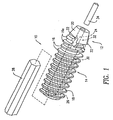
- Figure 1 is a perspective view of the device of an interference screw in accordance with a first exemplary embodiment of the present invention.
- Figure 2 is an exploded, perspective view of the screw of Figure 1.
- Figure 3 is a perspective view of a front component of an interference screw of an alternative embodiment of the present invention.
- Figure 4 is an exploded, perspective view of the screw having the front component shown in Figure 3.
- The present invention relates to an orthopedic surgical interference screw 10 for securing synthetic or biological connective tissue to a bone surface, such as, in the process of attaching and retaining a replacement anterior cruciate ligament (ACL) within a bone tunnel. The
screw 10 has a front component/section 12 fabricated from a material with strength and hardness at least equivalent to that of bone, and a rear component/section 14 fabricated from a bioabsorbable polymer. Both front andrear components threads rear components front component 12 has atapered tip 20 with at least one and preferably a plurality oflongitudinal flutes 22 to allow thefront portion 12 to tap threads while being inserted into a hole in the bone. A tapered tip and flutes of this type are conventional on taps and self-threading screws. Thethreads 18 of therear component 18 hold thescrew 10 in place in the bone tunnel, as well as hold the bone blocks of the replacement anterior cruciate ligament (ACL) against the walls of the bone tunnels. - Both the front and
rear components axial bores axial bore 26 of therear component 14 is shaped to matingly receive atool 28, such as a hex wrench. Although thetool 28 shown is hexagonal in shape, one skilled in the art could envision otheraxial bore front component 12 is stepped, with tool-shapedportion 30 proximate to therear component 14 and a guide pin portion 32 proximate the taperedtip 20. When the front andrear components tool 28 may be inserted through theaxial bore 26 and extend into the tool-shapedportion 30 of theaxial bore 24. Aguide pin 34 may be inserted into the guide pin portion 32 of theaxial bore 24 in order to guide thescrew 10 into the bone tunnel during an ACL reconstruction procedure. The taper oftip 20 extends over the threads 16a forming lead-in threads which cooperate with theflutes 22 to aid in self-threading. As noted above,threads 16 gradually transition tothreads 18, i.e., they are of uniform pitch and are like-handed; that is, they share the same rotational direction of advancement. - Figure 2 shows that the
screw 10 is provided with means for orienting and retaining thefront component 12 in association with therear portion 14. Namely, a hollow boss/projection 36 extends from therear component 14 and is matingly received in arecess 38 provided in thefront component 12. In Figure 2, the external shape (hexagonal) of theboss 36 mimics the hexagonal shape of theaxial bore 26. Similarly, therecess 38 has a hexagonal shape of somewhat greater dimensions than the tool-shapedportion 30 of theaxial bore 24. Theboss 36 may exhibit a friction fit relative to therecess 38 to retain the front andrear components boss 36 andrecess 38 may be provided with detents and mating depressions, threads, or other standard features used to fasten two pieces together in threaded, snap-fit, welded or glued relationship. Theboss 36 could extend from thefront component 12 and the recess be provided in therear component 14. - Besides functioning as a means for attaching the front and
rear components boss 36 also prevents relative rotation betweenfront component 12 andrear component 14 in theevent tool 28 is not inserted into tool-shapedportion 30 of theaxial bore 24. Althoughboss 36 is shown as hexagonal in shape, one skilled in the art could envisionboss 36 being other shapes, which prevent relative rotation betweenfront component 12 andrear component 14. These include, but are not limited to, polygonal, cross, star, or oval shapes. - Figures 3 and 4 show an
alternative embodiment 110 of the present invention wherein theboss 136 is in the form of a generally cylindrical collar having analignment key 140. Elements illustrated in Figures 3 and 4, which correspond to elements described above with respect to Figures 1 and 2, have been designated by corresponding reference numerals increased by 100. Theboss 136 inserts within amating recess 138 in front component 112 (Figure 3) with thealignment key 140 being received in amating alignment slot 142. Due to the asymmetry of thealignment key 140, the front andrear components - Unlike the
axial bore 24 of thefirst embodiment 10, theaxial bore 124 of thesecond embodiment 110 is not stepped, such thatwrench portion 128 oftool 129 may extend through theaxial bore 124 to the full length of thefront component 112 to thetip 120 thereof.Wrench portion 128 preferably extends from anabutment surface 144 against which therear component 114 abuts when thetool 129 is used to screw thescrew 110 into bone. As with the insertion of self-threading fasteners generally, the insertion tool, e.g., 129 is pushed toward the substrate into which the fastener, e.g., 110 is to be inserted. Simultaneously, the tool, e.g. 129, is turned such that the threads, e.g., 116a, bite into the material, e.g., bone, and advance down into the material. With respect to thescrew 110, the insertion pressure exerted on thetool 129 pushes therear component 114 into engagement with thefront component 112 insuring relative alignment and continuity ofthreads - In both of the foregoing
embodiments hard front component rear component front component front component screw insertion tool tool - The
screws screws - As mentioned above, the
screw front component rear component rear component screw - In the preferred embodiment, the
rear component screw - Suitable materials for forming the
front component - Preferably, the materials comprising the
front component screw front component - The
front component screw front component rear component - In another embodiment of the present invention, the polymers, polymer blends, or composites can be used as a therapeutic agent release matrix. To form this matrix, the polymer would be mixed with a therapeutic agent prior to forming the front or
rear components - Matrix materials for the present invention may be formulated by mixing one or more therapeutic agents with the polymer. Alternatively, a therapeutic agent could be coated on to the polymer, preferably with a pharmaceutically acceptable carrier. Any pharmaceutical carrier can be used that does not dissolve the polymer.
- The therapeutic agent may be present as a liquid, a finely divided solid, or any other appropriate physical form. Typically, but optionally, the matrix will include one or more additives, such as diluents, carriers, excipients, stabilizers or the like. The amount of therapeutic agent will depend on the particular drug being employed and medical condition being treated. Typically, the amount of drug represents about 0.001 percent to about 70 percent, more typically about 0.001 percent to about 50 percent, most typically about 0.001 percent to about 20 percent by weight of the matrix. The quantity and type of polymer incorporated into the drug delivery matrix will vary depending on the release profile desired and the amount of drug employed.
- Upon contact with body fluids, the polymer undergoes gradual degradation (mainly through hydrolysis) with concomitant release of the dispersed drug for a sustained or extended period. This can result in prolonged delivery (over, say I to 5,000 hours, preferably 2 to 800 hours) of effective amounts (say, 0.0001 mg/kg/hour to 10 mg/kg/hour) of the drug. This dosage form can be administered as is necessary depending on the subject being treated, the severity of the affliction, the judgment of the prescribing physician, and the like. Following this or similar procedures, those skilled in the art will be able to prepare a variety of formulations.
- The following example is illustrative of the principles and practice of this invention, although not limited thereto. Numerous additional embodiments within the scope and spirit of the invention will become apparent to those skilled in the art.
- Three screws were made for simulation of a bone-tendon-bone ACL repair. Two of the screws were controls. The controls were monolithic in nature, i.e. composed of a single material, and of a one-piece design which incorporated all of the features of the present invention except for the means of connecting a front and rear component.
- The first control was composed of poly(lactic acid), or PLA, machined from billets of PLA previously formed by injection molding. The second control was composed of a composite of 20/80 (volume percent) β-tricalcium phosphate (TCP) particles (10-micron average diameter) in PLA. These screws were also machined from billets of TCP/PLA composites previously formed by injection molding.
- The third screw included a front and rear component. The rear component was composed of PLA machined from the same billets of PLA formed by injection molding as mentioned above. The front component was composed of metal machined from the rods of cold rolled stainless steel. The front and rear components were press fit to form a two-piece screw.
- Fresh frozen porcine knees were used for simulation of a bone-tendon-bone ACL repair. After thawing, the femur was securely clamped and the patella was dissected free from its proximal attachment. An 11-mm diameter patellar bone plug, attached to the patellar tendon, was harvested. The plug was fashioned with a Stryker 11-mm bone plug cutter and the patella tendon was dissected free at its tibial attachment.
- A 13-mm diameter bone tunnel was prepared by over-drilling a guide pin placed at the ACL origin and exteriorizing on the side of the femoral condyle. The bone plug was placed into the tunnel until its end protruded two to three millimeters out of its proximal end. Since the bone plug was 11-mm in diameter and the tunnel was 13-mm in diameter, there existed a 2-mm gap into which attempts were made to drive each screw into the gap. A standard hexagonal driver was used to attempt driving the screws through the cortical side of the bone plug.
- The PLA control screw could not be driven into the bone tunnel/bone plug gap. The composite control screw was successfully driven into the bone tunnel/bone plug gap. During the insertion process, this screw exerted considerable resistance to torsion and a "biting" sound could be heard, suggesting that the entire length of the threads were cutting into bone. The two-piece prototype easily self-tapped, with the relatively small number of threads on the stainless steel front component biting into and cutting the bone, and the softer, polymer rear component following the front component with minimal friction resistance.
Claims (20)
- An orthopedic screw (10, 110) for introduction into a bone tunnel, comprising:a first section (12, 112) having a first end forming a tip (20, 120) of said screw (10, 110), said first section (12, 112) extending from said first end to an intermediate point along the length of said screw (10, 110), said first section (12, 112) including external threads (16, 116) along at least a portion of the length thereof, and said first section being made of a first material having a hardness such that said first section (12, 112) is self-threading when said first section (12, 112) is screwed into the bone tunnel; and characterised by:a second section (14, 1 14) coupled to said first section (12, 112) and extending from said intermediate point to a second end of said screw (10, 110) distal to said tip (20, 120), said second section (14, 114) being formed from a bioabsorbable material which is different than said first material and said second section (14, 114) having external threads (18, 118) along at least a portion of the length thereof, the screw further comprising:an axial bore (26, 126) extending through said second section (14, 114) to said first section, said bore (26, 126) being for matingly receiving a tool therethrough to extend to said first section of said screw, whereby said tool can apply a turning force to both sections of said screw (10, 110).
- The screw (10, 110) of Claim 1, further including a projection (36, 136) extending from one of said first section (12, 112) and said second section (14, 114) to the other of said first section (12, 112) and said second section (14, 114) to be received in a mating recess (38, 138) provided therein to conjoin said first section and said second section (12, 112; 14, 114).
- The screw (10, 110) of Claim 2, wherein said projection (36, 136) is a hexagonal boss and said recess (38, 138) is a mating hexagonal recess.
- The screw (10, 110) of Claim 2, wherein said projection (36, 136) is asymmetric and said recess (38, 138) is matingly asymmetric establishing a single assembly orientation of said first section (12, 112) relative to said second section (14, 114).
- The screw (10, 110) of Claim 4, wherein said projection (36, 136) is substantially cylindrical with at least one radially extending side prominence (140).
- The screw (10, 110) of any one of the preceding Claims, wherein said axial bore (26, 126) is hexagonal along a portion of its length, said hexagonal portion being a tool receiving portion for receiving a hex wrench (28, 128) therein.
- The screw (10, 110) of any one of the preceding Claims, wherein said axial bore (26, 126) extends through the entire length of said screw (10, 110) from said first end to said second end.
- The screw (10, 110) of Claim 7, wherein said axial bore (26, 126) proximate said first end (20, 120) has a shape for matingly receiving a guide pin (34).
- The screw (10, 110) of any one of the preceding Claims, wherein said threads (16, 116) of said first section (12, 112) and said threads (18, 118) of said second section (14, 114) are continuous relative to one another when said first section (12, 112) is conjoined to said second section (14, 114) and have the same direction of rotational advancement.
- The screw (10, 110) of Claim 9, wherein said first end (20, 120) is tapered to aid in introducing said screw into a hole in bone.
- The screw (10, 110) of Claim 10, wherein said first end (20, 120) has at least one longitudinal flute (22, 122) cutting across at least one of said threads (16, 116) in said first section (12, 112).
- The screw (10, 110) of Claim 11, wherein at least one of said threads (16, 116) on said first section (12, 112) is a starter thread.
- The screw (10, 110) of any one of the preceding Claims, wherein said first section (12, 112) is bioabsorbable.
- The screw (10, 110) of Claim 13, wherein said first section (12, 112) is made from a material selected from the group consisting of mono-, di-, tri-, α-tri, β-tri, and tetracalcium phosphate, hydroxyapatite, calcium sulfates, calcium oxides, calcium carbonates, magnesium calcium phosphates, phosphate glasses, bioglasses, mixtures thereof, polyglycolic acid polymers and bone tissues.
- The screw (10, 110) of any one of Claims 1 to 12, wherein said first section (12, 112) is made from a material selected from the group consisting of stainless steel, cobalt-chrome alloys, glasses, ceramics and polymers.
- The screw (10, 110) of any one of the preceding Claims, wherein said second section (14, 114) is made from a material selected from the group consisting of aliphatic polyesters, polyorthoesters, polyanhydrides, polycarbonates, polyurethanes, polyamides and polyalkylene oxides.
- The screw (10, 110) of any one of the preceding Claims, wherein said first section (12, 112) is conjoined to said second section (14, 114) by gluing.
- The screw (10, 110) of any one of Claims 1 to 16, wherein said first section (12, 112) is conjoined to said second section (14, 114) by welding.
- The screw (10, 110) of any one of the preceding claims, wherein:said first section (12, 112) is made from a biocompatible material with a strength and hardness at least equivalent to that of bone andsaid second section (14, 114) is made from a bioabsorbable polymer and is coupled to said first section (12, 112).
- The screw of claim 19, wherein said first section (12, 112) is bioabsorbable.
Applications Claiming Priority (2)
| Application Number | Priority Date | Filing Date | Title |
|---|---|---|---|
| US969779 | 2001-09-28 | ||
| US09/969,779 US6916321B2 (en) | 2001-09-28 | 2001-09-28 | Self-tapping resorbable two-piece bone screw |
Publications (3)
| Publication Number | Publication Date |
|---|---|
| EP1297794A2 EP1297794A2 (en) | 2003-04-02 |
| EP1297794A3 EP1297794A3 (en) | 2003-12-03 |
| EP1297794B1 true EP1297794B1 (en) | 2006-11-02 |
Family
ID=25515989
Family Applications (1)
| Application Number | Title | Priority Date | Filing Date |
|---|---|---|---|
| EP02256761A Expired - Fee Related EP1297794B1 (en) | 2001-09-28 | 2002-09-27 | Self-tapping resorbable two-piece bone screw |
Country Status (6)
| Country | Link |
|---|---|
| US (1) | US6916321B2 (en) |
| EP (1) | EP1297794B1 (en) |
| JP (1) | JP4298248B2 (en) |
| AU (1) | AU2002300976B2 (en) |
| CA (1) | CA2405601C (en) |
| DE (1) | DE60215744T2 (en) |
Families Citing this family (242)
| Publication number | Priority date | Publication date | Assignee | Title |
|---|---|---|---|---|
| US5713921A (en) * | 1996-03-29 | 1998-02-03 | Bonutti; Peter M. | Suture anchor |
| US6045551A (en) | 1998-02-06 | 2000-04-04 | Bonutti; Peter M. | Bone suture |
| US6447516B1 (en) | 1999-08-09 | 2002-09-10 | Peter M. Bonutti | Method of securing tissue |
| US6368343B1 (en) | 2000-03-13 | 2002-04-09 | Peter M. Bonutti | Method of using ultrasonic vibration to secure body tissue |
| US6592609B1 (en) * | 1999-08-09 | 2003-07-15 | Bonutti 2003 Trust-A | Method and apparatus for securing tissue |
| US6635073B2 (en) | 2000-05-03 | 2003-10-21 | Peter M. Bonutti | Method of securing body tissue |
| US7094251B2 (en) | 2002-08-27 | 2006-08-22 | Marctec, Llc. | Apparatus and method for securing a suture |
| US9138222B2 (en) | 2000-03-13 | 2015-09-22 | P Tech, Llc | Method and device for securing body tissue |
| US7993369B2 (en) | 2000-06-22 | 2011-08-09 | Arthrex, Inc. | Graft fixation using a plug against suture |
| DE10038016A1 (en) * | 2000-08-04 | 2002-02-14 | Wuerth Adolf Gmbh & Co Kg | Dowels for lightweight materials |
| US6878166B2 (en) * | 2000-08-28 | 2005-04-12 | Ron Clark | Method and implant for securing ligament replacement into the knee |
| US7235079B2 (en) * | 2004-11-18 | 2007-06-26 | Acumed Llc | Composite bone fasteners |
| GB0116341D0 (en) * | 2001-07-04 | 2001-08-29 | Smith & Nephew | Biodegradable polymer systems |
| US6719765B2 (en) | 2001-12-03 | 2004-04-13 | Bonutti 2003 Trust-A | Magnetic suturing system and method |
| US6921402B2 (en) | 2001-12-27 | 2005-07-26 | Ethicon, Inc. | Polymer-based orthopedic screw and driver system with increased insertion torque tolerance and associated method for making and using same |
| GB0202233D0 (en) * | 2002-01-31 | 2002-03-20 | Smith & Nephew | Bioresorbable polymers |
| US9155544B2 (en) | 2002-03-20 | 2015-10-13 | P Tech, Llc | Robotic systems and methods |
| US20050101961A1 (en) * | 2003-11-12 | 2005-05-12 | Huebner Randall J. | Bone screws |
| US7955388B2 (en) * | 2006-11-01 | 2011-06-07 | Acumed Llc | Orthopedic connector system |
| JP3938343B2 (en) * | 2002-08-09 | 2007-06-27 | インターナショナル・ビジネス・マシーンズ・コーポレーション | Task management system, program, and control method |
| US7235078B2 (en) * | 2002-11-26 | 2007-06-26 | Hs West Investments Llc | Protective devices for use with angled interference screws |
| US20040260398A1 (en) * | 2003-02-10 | 2004-12-23 | Kelman David C. | Resorbable devices |
| US8388624B2 (en) | 2003-02-24 | 2013-03-05 | Arthrosurface Incorporated | Trochlear resurfacing system and method |
| US7497864B2 (en) | 2003-04-30 | 2009-03-03 | Marctec, Llc. | Tissue fastener and methods for using same |
| AU2004237774B2 (en) * | 2003-05-02 | 2009-09-10 | Surmodics, Inc. | Implantable controlled release bioactive agent delivery device |
| US8246974B2 (en) * | 2003-05-02 | 2012-08-21 | Surmodics, Inc. | Medical devices and methods for producing the same |
| US8016865B2 (en) | 2003-09-29 | 2011-09-13 | Depuy Mitek, Inc. | Method of performing anterior cruciate ligament reconstruction using biodegradable interference screw |
| US7141354B2 (en) * | 2003-09-30 | 2006-11-28 | Dai Nippon Printing Co., Ltd. | Photo radical generator, photo sensitive resin composition and article |
| US7896917B2 (en) * | 2003-10-15 | 2011-03-01 | Biomet Sports Medicine, Llc | Method and apparatus for graft fixation |
| AU2004283727A1 (en) * | 2003-10-23 | 2005-05-06 | Trans1 Inc. | Tools and tool kits for performing minimally invasive procedures on the spine |
| GB0329654D0 (en) | 2003-12-23 | 2004-01-28 | Smith & Nephew | Tunable segmented polyacetal |
| US7608092B1 (en) | 2004-02-20 | 2009-10-27 | Biomet Sports Medicince, LLC | Method and apparatus for performing meniscus repair |
| US20080039873A1 (en) | 2004-03-09 | 2008-02-14 | Marctec, Llc. | Method and device for securing body tissue |
| US8109965B2 (en) | 2004-06-09 | 2012-02-07 | Biomet Sports Medicine, LLP | Method and apparatus for soft tissue fixation |
| US7500983B1 (en) | 2004-06-09 | 2009-03-10 | Biomet Sports Medicine, Llc | Apparatus for soft tissue attachment |
| US7819898B2 (en) | 2004-06-09 | 2010-10-26 | Biomet Sports Medicine, Llc | Method and apparatus for soft tissue fixation |
| US7695503B1 (en) | 2004-06-09 | 2010-04-13 | Biomet Sports Medicine, Llc | Method and apparatus for soft tissue attachment |
| US8114127B2 (en) | 2004-06-22 | 2012-02-14 | Hs West Investments, Llc | Bone anchors for use in attaching soft tissue to bone |
| US7322978B2 (en) * | 2004-06-22 | 2008-01-29 | Hs West Investments, Llc | Bone anchors for use in attaching soft tissue to a bone |
| US20060024350A1 (en) * | 2004-06-24 | 2006-02-02 | Varner Signe E | Biodegradable ocular devices, methods and systems |
| CA2572584A1 (en) | 2004-06-28 | 2006-01-12 | Arthrosurface, Inc. | System for articular surface replacement |
| US8002778B1 (en) | 2004-06-28 | 2011-08-23 | Biomet Sports Medicine, Llc | Crosspin and method for inserting the same during soft ligament repair |
| US9949843B2 (en) | 2004-08-09 | 2018-04-24 | Si-Bone Inc. | Apparatus, systems, and methods for the fixation or fusion of bone |
| US8470004B2 (en) | 2004-08-09 | 2013-06-25 | Si-Bone Inc. | Apparatus, systems, and methods for stabilizing a spondylolisthesis |
| US8425570B2 (en) | 2004-08-09 | 2013-04-23 | Si-Bone Inc. | Apparatus, systems, and methods for achieving anterior lumbar interbody fusion |
| US8414648B2 (en) | 2004-08-09 | 2013-04-09 | Si-Bone Inc. | Apparatus, systems, and methods for achieving trans-iliac lumbar fusion |
| US8444693B2 (en) * | 2004-08-09 | 2013-05-21 | Si-Bone Inc. | Apparatus, systems, and methods for achieving lumbar facet fusion |
| US8388667B2 (en) | 2004-08-09 | 2013-03-05 | Si-Bone, Inc. | Systems and methods for the fixation or fusion of bone using compressive implants |
| US20060036251A1 (en) | 2004-08-09 | 2006-02-16 | Reiley Mark A | Systems and methods for the fixation or fusion of bone |
| US9662158B2 (en) | 2004-08-09 | 2017-05-30 | Si-Bone Inc. | Systems and methods for the fixation or fusion of bone at or near a sacroiliac joint |
| US20180228621A1 (en) | 2004-08-09 | 2018-08-16 | Mark A. Reiley | Apparatus, systems, and methods for the fixation or fusion of bone |
| US20070156241A1 (en) | 2004-08-09 | 2007-07-05 | Reiley Mark A | Systems and methods for the fixation or fusion of bone |
| US20060034891A1 (en) * | 2004-08-12 | 2006-02-16 | Laurie Lawin | Biodegradable controlled release bioactive agent delivery device |
| JPWO2006022018A1 (en) * | 2004-08-27 | 2008-05-08 | グンゼ株式会社 | Bone treatment tool manufacturing method and bone treatment tool |
| GB2417536B (en) * | 2004-08-28 | 2006-09-06 | Adam James | A bioabsorable screw |
| US9173647B2 (en) | 2004-10-26 | 2015-11-03 | P Tech, Llc | Tissue fixation system |
| US9463012B2 (en) | 2004-10-26 | 2016-10-11 | P Tech, Llc | Apparatus for guiding and positioning an implant |
| US20060089646A1 (en) | 2004-10-26 | 2006-04-27 | Bonutti Peter M | Devices and methods for stabilizing tissue and implants |
| US9271766B2 (en) | 2004-10-26 | 2016-03-01 | P Tech, Llc | Devices and methods for stabilizing tissue and implants |
| US7857830B2 (en) | 2006-02-03 | 2010-12-28 | Biomet Sports Medicine, Llc | Soft tissue repair and conduit device |
| US7749250B2 (en) | 2006-02-03 | 2010-07-06 | Biomet Sports Medicine, Llc | Soft tissue repair assembly and associated method |
| US7905903B2 (en) | 2006-02-03 | 2011-03-15 | Biomet Sports Medicine, Llc | Method for tissue fixation |
| US20060189993A1 (en) | 2004-11-09 | 2006-08-24 | Arthrotek, Inc. | Soft tissue conduit device |
| US7905904B2 (en) | 2006-02-03 | 2011-03-15 | Biomet Sports Medicine, Llc | Soft tissue repair device and associated methods |
| US8840645B2 (en) | 2004-11-05 | 2014-09-23 | Biomet Sports Medicine, Llc | Method and apparatus for coupling soft tissue to a bone |
| US7909851B2 (en) | 2006-02-03 | 2011-03-22 | Biomet Sports Medicine, Llc | Soft tissue repair device and associated methods |
| US8118836B2 (en) | 2004-11-05 | 2012-02-21 | Biomet Sports Medicine, Llc | Method and apparatus for coupling soft tissue to a bone |
| US8303604B2 (en) | 2004-11-05 | 2012-11-06 | Biomet Sports Medicine, Llc | Soft tissue repair device and method |
| US9017381B2 (en) | 2007-04-10 | 2015-04-28 | Biomet Sports Medicine, Llc | Adjustable knotless loops |
| US9801708B2 (en) | 2004-11-05 | 2017-10-31 | Biomet Sports Medicine, Llc | Method and apparatus for coupling soft tissue to a bone |
| US8088130B2 (en) | 2006-02-03 | 2012-01-03 | Biomet Sports Medicine, Llc | Method and apparatus for coupling soft tissue to a bone |
| US8128658B2 (en) | 2004-11-05 | 2012-03-06 | Biomet Sports Medicine, Llc | Method and apparatus for coupling soft tissue to bone |
| US8298262B2 (en) | 2006-02-03 | 2012-10-30 | Biomet Sports Medicine, Llc | Method for tissue fixation |
| US7658751B2 (en) | 2006-09-29 | 2010-02-09 | Biomet Sports Medicine, Llc | Method for implanting soft tissue |
| US8137382B2 (en) | 2004-11-05 | 2012-03-20 | Biomet Sports Medicine, Llc | Method and apparatus for coupling anatomical features |
| US8361113B2 (en) | 2006-02-03 | 2013-01-29 | Biomet Sports Medicine, Llc | Method and apparatus for coupling soft tissue to a bone |
| US8998949B2 (en) | 2004-11-09 | 2015-04-07 | Biomet Sports Medicine, Llc | Soft tissue conduit device |
| US20070038303A1 (en) * | 2006-08-15 | 2007-02-15 | Ebi, L.P. | Foot/ankle implant and associated method |
| US7879109B2 (en) | 2004-12-08 | 2011-02-01 | Biomet Manufacturing Corp. | Continuous phase composite for musculoskeletal repair |
| US8535357B2 (en) * | 2004-12-09 | 2013-09-17 | Biomet Sports Medicine, Llc | Continuous phase compositions for ACL repair |
| US20060142772A1 (en) * | 2004-12-29 | 2006-06-29 | Ralph James D | Surgical fasteners and related implant devices having bioabsorbable components |
| SE528545C2 (en) * | 2005-02-16 | 2006-12-12 | Swemac Orthopaedics Ab | Articulated prosthesis and screw tools to apply parts of the same |
| US9089323B2 (en) | 2005-02-22 | 2015-07-28 | P Tech, Llc | Device and method for securing body tissue |
| US20090187216A1 (en) | 2006-05-18 | 2009-07-23 | Arthrex, Inc. | Fenestrated swivel anchor for knotless fixation of tissue |
| US11801043B2 (en) | 2005-03-30 | 2023-10-31 | Arthrex, Inc. | Suture anchor for knotless fixation of tissue |
| US7828828B2 (en) * | 2005-04-14 | 2010-11-09 | Warsaw Orthopedic, Inc | Intervertebral joint |
| US20060247638A1 (en) * | 2005-04-29 | 2006-11-02 | Sdgi Holdings, Inc. | Composite spinal fixation systems |
| US7951198B2 (en) * | 2005-05-10 | 2011-05-31 | Acumed Llc | Bone connector with pivotable joint |
| JP2009504929A (en) * | 2005-08-18 | 2009-02-05 | スミス アンド ネフュー ピーエルシー | High-strength devices and composite materials |
| US20070233145A1 (en) * | 2005-08-18 | 2007-10-04 | Biocomposites Ltd. | Tensegrity osteotomy system |
| CN101296663B (en) * | 2005-10-25 | 2011-05-25 | 圣歌整形外科有限责任公司 | Bone fastening assembly and bushing and screw for use therewith |
| US8100952B2 (en) * | 2005-12-22 | 2012-01-24 | Anthem Orthopaedics Llc | Drug delivering bone plate and method and targeting device for use therewith |
| US9149267B2 (en) | 2006-02-03 | 2015-10-06 | Biomet Sports Medicine, Llc | Method and apparatus for coupling soft tissue to a bone |
| US9538998B2 (en) | 2006-02-03 | 2017-01-10 | Biomet Sports Medicine, Llc | Method and apparatus for fracture fixation |
| US8562645B2 (en) | 2006-09-29 | 2013-10-22 | Biomet Sports Medicine, Llc | Method and apparatus for forming a self-locking adjustable loop |
| US11259792B2 (en) | 2006-02-03 | 2022-03-01 | Biomet Sports Medicine, Llc | Method and apparatus for coupling anatomical features |
| US8506597B2 (en) | 2011-10-25 | 2013-08-13 | Biomet Sports Medicine, Llc | Method and apparatus for interosseous membrane reconstruction |
| US11311287B2 (en) | 2006-02-03 | 2022-04-26 | Biomet Sports Medicine, Llc | Method for tissue fixation |
| US9078644B2 (en) | 2006-09-29 | 2015-07-14 | Biomet Sports Medicine, Llc | Fracture fixation device |
| US8968364B2 (en) | 2006-02-03 | 2015-03-03 | Biomet Sports Medicine, Llc | Method and apparatus for fixation of an ACL graft |
| US8652172B2 (en) | 2006-02-03 | 2014-02-18 | Biomet Sports Medicine, Llc | Flexible anchors for tissue fixation |
| US8801783B2 (en) | 2006-09-29 | 2014-08-12 | Biomet Sports Medicine, Llc | Prosthetic ligament system for knee joint |
| US8597327B2 (en) | 2006-02-03 | 2013-12-03 | Biomet Manufacturing, Llc | Method and apparatus for sternal closure |
| US8652171B2 (en) | 2006-02-03 | 2014-02-18 | Biomet Sports Medicine, Llc | Method and apparatus for soft tissue fixation |
| US8771352B2 (en) | 2011-05-17 | 2014-07-08 | Biomet Sports Medicine, Llc | Method and apparatus for tibial fixation of an ACL graft |
| US9271713B2 (en) | 2006-02-03 | 2016-03-01 | Biomet Sports Medicine, Llc | Method and apparatus for tensioning a suture |
| US8251998B2 (en) | 2006-08-16 | 2012-08-28 | Biomet Sports Medicine, Llc | Chondral defect repair |
| US8936621B2 (en) | 2006-02-03 | 2015-01-20 | Biomet Sports Medicine, Llc | Method and apparatus for forming a self-locking adjustable loop |
| US10517587B2 (en) | 2006-02-03 | 2019-12-31 | Biomet Sports Medicine, Llc | Method and apparatus for forming a self-locking adjustable loop |
| US8574235B2 (en) | 2006-02-03 | 2013-11-05 | Biomet Sports Medicine, Llc | Method for trochanteric reattachment |
| US7959650B2 (en) | 2006-09-29 | 2011-06-14 | Biomet Sports Medicine, Llc | Adjustable knotless loops |
| US8562647B2 (en) | 2006-09-29 | 2013-10-22 | Biomet Sports Medicine, Llc | Method and apparatus for securing soft tissue to bone |
| US11253296B2 (en) | 2006-02-07 | 2022-02-22 | P Tech, Llc | Methods and devices for intracorporeal bonding of implants with thermal energy |
| US7967820B2 (en) | 2006-02-07 | 2011-06-28 | P Tech, Llc. | Methods and devices for trauma welding |
| US11278331B2 (en) | 2006-02-07 | 2022-03-22 | P Tech Llc | Method and devices for intracorporeal bonding of implants with thermal energy |
| US8496657B2 (en) | 2006-02-07 | 2013-07-30 | P Tech, Llc. | Methods for utilizing vibratory energy to weld, stake and/or remove implants |
| US7850717B2 (en) * | 2006-03-01 | 2010-12-14 | Warsaw Orthopedic, Inc. | Bone anchors having two or more portions exhibiting different performance characteristics and method of forming the same |
| US9849216B2 (en) | 2006-03-03 | 2017-12-26 | Smith & Nephew, Inc. | Systems and methods for delivering a medicament |
| US7828820B2 (en) | 2006-03-21 | 2010-11-09 | Biomet Sports Medicine, Llc | Method and apparatuses for securing suture |
| GB2436293A (en) | 2006-03-24 | 2007-09-26 | Galley Geoffrey H | Spinous processes insertion device |
| US8361129B2 (en) * | 2006-04-28 | 2013-01-29 | Depuy Spine, Inc. | Large diameter bone anchor assembly |
| US11246638B2 (en) | 2006-05-03 | 2022-02-15 | P Tech, Llc | Methods and devices for utilizing bondable materials |
| US8894661B2 (en) * | 2007-08-16 | 2014-11-25 | Smith & Nephew, Inc. | Helicoil interference fixation system for attaching a graft ligament to a bone |
| US11259794B2 (en) | 2006-09-29 | 2022-03-01 | Biomet Sports Medicine, Llc | Method for implanting soft tissue |
| US8672969B2 (en) | 2006-09-29 | 2014-03-18 | Biomet Sports Medicine, Llc | Fracture fixation device |
| US9918826B2 (en) | 2006-09-29 | 2018-03-20 | Biomet Sports Medicine, Llc | Scaffold for spring ligament repair |
| US8500818B2 (en) | 2006-09-29 | 2013-08-06 | Biomet Manufacturing, Llc | Knee prosthesis assembly with ligament link |
| US8114128B2 (en) * | 2006-11-01 | 2012-02-14 | Depuy Mitek, Inc. | Cannulated suture anchor |
| CA2679365C (en) | 2006-11-30 | 2016-05-03 | Smith & Nephew, Inc. | Fiber reinforced composite material |
| AU2007332787A1 (en) | 2006-12-11 | 2008-06-19 | Arthrosurface Incorporated | Retrograde resection apparatus and method |
| US8617185B2 (en) | 2007-02-13 | 2013-12-31 | P Tech, Llc. | Fixation device |
| US20080234762A1 (en) * | 2007-03-06 | 2008-09-25 | Zimmer Technology, Inc. | Self-tapping screw with resorbable tip |
| US8147546B2 (en) * | 2007-03-13 | 2012-04-03 | Biomet Sports Medicine, Llc | Method and apparatus for graft fixation |
| WO2008129245A1 (en) | 2007-04-18 | 2008-10-30 | Smith & Nephew Plc | Expansion moulding of shape memory polymers |
| AU2008243035B2 (en) | 2007-04-19 | 2013-09-12 | Smith & Nephew, Inc. | Graft fixation |
| WO2008131197A1 (en) | 2007-04-19 | 2008-10-30 | Smith & Nephew, Inc. | Multi-modal shape memory polymers |
| US9072548B2 (en) * | 2007-06-07 | 2015-07-07 | Anthem Orthopaedics Llc | Spine repair assembly |
| US8075562B2 (en) * | 2007-06-25 | 2011-12-13 | Wisconsin Alumni Research Foundation | Controlled release of biopharmaceutical growth factors from hydroxyapatite coating on bioresorbable interference screws used in cruciate ligament reconstruction surgery |
| US20090024174A1 (en) | 2007-07-17 | 2009-01-22 | Stark John G | Bone screws and particular applications to sacroiliac joint fusion |
| US8702754B2 (en) * | 2007-09-14 | 2014-04-22 | Depuy Mitek, Llc | Methods for anchoring suture to bone |
| US8882801B2 (en) * | 2007-09-14 | 2014-11-11 | Depuy Mitek, Llc | Dual thread cannulated suture anchor |
| US20090075236A1 (en) * | 2007-09-18 | 2009-03-19 | Towse Ross W | Dental implant having improved stability |
| US20090125071A1 (en) * | 2007-10-23 | 2009-05-14 | Skinlo David M | Shape-changing anatomical anchor |
| US20090281581A1 (en) | 2008-05-06 | 2009-11-12 | Berg Jeffery H | Method and device for securing sutures to bones |
| JP5735414B2 (en) * | 2008-05-14 | 2015-06-17 | スミス アンド ネフュー インコーポレーテッドSmith & Nephew,Inc. | Joint affinity biceps tendon fixation treatment |
| US9616205B2 (en) | 2008-08-13 | 2017-04-11 | Smed-Ta/Td, Llc | Drug delivery implants |
| WO2010019781A1 (en) | 2008-08-13 | 2010-02-18 | Smed-Ta/Td, Llc | Drug delivery implants |
| ES2647919T3 (en) | 2008-08-13 | 2017-12-27 | Smed-Ta/Td, Llc | Drug supply implants |
| US9700431B2 (en) | 2008-08-13 | 2017-07-11 | Smed-Ta/Td, Llc | Orthopaedic implant with porous structural member |
| US10842645B2 (en) | 2008-08-13 | 2020-11-24 | Smed-Ta/Td, Llc | Orthopaedic implant with porous structural member |
| US20100049249A1 (en) * | 2008-08-25 | 2010-02-25 | Linvatec Corporation | Suture anchor extension |
| EP2341852B1 (en) * | 2008-08-29 | 2018-08-15 | SMed-TA/TD, LLC | Orthopaedic implant |
| US20100082071A1 (en) * | 2008-09-26 | 2010-04-01 | Missoum Moumene | Composite Screw Having A Metallic Pin and a Polymeric Thread |
| US8702808B2 (en) * | 2008-10-17 | 2014-04-22 | Osteopore International Pte Ltd | Resorbable scaffolds for bone repair and long bone tissue engineering |
| JP5904794B2 (en) | 2008-12-09 | 2016-04-20 | スミス アンド ネフュー インコーポレーテッドSmith & Nephew,Inc. | Tissue repair assembly |
| US8858606B2 (en) | 2008-12-09 | 2014-10-14 | Smith & Nephew, Inc. | Tissue repair assembly |
| WO2010071742A1 (en) * | 2008-12-15 | 2010-06-24 | Smith & Nephew, Inc. | Composite anchor |
| EP2198796A1 (en) * | 2008-12-19 | 2010-06-23 | Sepitec Foundation | Bone screw |
| CN104188708B (en) | 2009-01-16 | 2017-07-14 | 卡波菲克斯整形有限公司 | Composite material bone implant |
| WO2010099222A1 (en) | 2009-02-24 | 2010-09-02 | P Tech, Llc | Methods and devices for utilizing bondable materials |
| WO2010111350A1 (en) | 2009-03-24 | 2010-09-30 | Stabiliz Orthopedics, LLC | Orthopedic fixation device with bioresorbable layer |
| US10945743B2 (en) | 2009-04-17 | 2021-03-16 | Arthrosurface Incorporated | Glenoid repair system and methods of use thereof |
| WO2010121250A1 (en) | 2009-04-17 | 2010-10-21 | Arthrosurface Incorporated | Glenoid resurfacing system and method |
| AU2010236136B2 (en) | 2009-04-17 | 2014-07-10 | Shane Barwood | Tenodesis system |
| US20100305710A1 (en) | 2009-05-28 | 2010-12-02 | Biomet Manufacturing Corp. | Knee Prosthesis |
| US8361123B2 (en) | 2009-10-16 | 2013-01-29 | Depuy Spine, Inc. | Bone anchor assemblies and methods of manufacturing and use thereof |
| ES2456317T3 (en) * | 2010-02-26 | 2014-04-22 | Biedermann Technologies Gmbh & Co. Kg | Bone screw |
| AU2011222404A1 (en) | 2010-03-05 | 2012-09-27 | Arthrosurface Incorporated | Tibial resurfacing system and method |
| WO2011154891A2 (en) * | 2010-06-07 | 2011-12-15 | Carbofix Orthopedics Ltd. | Composite material bone implant and methods |
| US10154867B2 (en) | 2010-06-07 | 2018-12-18 | Carbofix In Orthopedics Llc | Multi-layer composite material bone screw |
| US8470046B2 (en) | 2011-04-25 | 2013-06-25 | Warsaw Orthopedic, Inc. | Bone augmentation device and method |
| US9089416B2 (en) | 2011-06-20 | 2015-07-28 | Anatomacl, Llc | Apparatus and method for ligament reconstruction |
| US10939992B2 (en) | 2011-06-20 | 2021-03-09 | Anatomacl, Llc | Apparatus and method for ligament reconstruction |
| WO2013013218A2 (en) | 2011-07-20 | 2013-01-24 | Horwitz Michael H | Minimal incision removable bone screw, driver, and method of use |
| US9357991B2 (en) | 2011-11-03 | 2016-06-07 | Biomet Sports Medicine, Llc | Method and apparatus for stitching tendons |
| US9370350B2 (en) | 2011-11-10 | 2016-06-21 | Biomet Sports Medicine, Llc | Apparatus for coupling soft tissue to a bone |
| US9357992B2 (en) | 2011-11-10 | 2016-06-07 | Biomet Sports Medicine, Llc | Method for coupling soft tissue to a bone |
| US9381013B2 (en) | 2011-11-10 | 2016-07-05 | Biomet Sports Medicine, Llc | Method for coupling soft tissue to a bone |
| WO2013096746A1 (en) | 2011-12-22 | 2013-06-27 | Arthrosurface Incorporated | System and method for bone fixation |
| US9259217B2 (en) | 2012-01-03 | 2016-02-16 | Biomet Manufacturing, Llc | Suture Button |
| US9526549B2 (en) * | 2012-01-16 | 2016-12-27 | Carbofix Orthopedics Ltd. | Bone screw with insert |
| US8778026B2 (en) | 2012-03-09 | 2014-07-15 | Si-Bone Inc. | Artificial SI joint |
| US10363140B2 (en) | 2012-03-09 | 2019-07-30 | Si-Bone Inc. | Systems, device, and methods for joint fusion |
| WO2013134670A1 (en) | 2012-03-09 | 2013-09-12 | Si-Bone Inc. | Integrated implant |
| DE102012008376A1 (en) | 2012-04-23 | 2013-10-24 | Primed Halberstadt Medizintechnik Gmbh | Bone Screw Assembly (Transplant Shuttle System TSS) |
| EP2841016B1 (en) * | 2012-04-26 | 2019-09-18 | Jortek Surgical Inc. | Apparatus for ligament reconstruction |
| CN104334092A (en) | 2012-05-04 | 2015-02-04 | 西-博恩公司 | Fenestrated implant |
| JP6549482B2 (en) | 2012-06-01 | 2019-07-24 | サーモディクス,インコーポレイテッド | Device and method for coating a balloon catheter |
| US9827401B2 (en) | 2012-06-01 | 2017-11-28 | Surmodics, Inc. | Apparatus and methods for coating medical devices |
| US9468448B2 (en) | 2012-07-03 | 2016-10-18 | Arthrosurface Incorporated | System and method for joint resurfacing and repair |
| US11090468B2 (en) | 2012-10-25 | 2021-08-17 | Surmodics, Inc. | Apparatus and methods for coating medical devices |
| US10076377B2 (en) | 2013-01-05 | 2018-09-18 | P Tech, Llc | Fixation systems and methods |
| US9320527B2 (en) * | 2013-01-18 | 2016-04-26 | Biomet Manufacturing, Llc | Quick-connect anti-rotation peg/drill bit component |
| JP6382237B2 (en) | 2013-03-06 | 2018-08-29 | スミス アンド ネフュー インコーポレーテッドSmith & Nephew,Inc. | Micro anchor |
| US9757119B2 (en) | 2013-03-08 | 2017-09-12 | Biomet Sports Medicine, Llc | Visual aid for identifying suture limbs arthroscopically |
| US9918827B2 (en) | 2013-03-14 | 2018-03-20 | Biomet Sports Medicine, Llc | Scaffold for spring ligament repair |
| US9936983B2 (en) | 2013-03-15 | 2018-04-10 | Si-Bone Inc. | Implants for spinal fixation or fusion |
| US9492200B2 (en) | 2013-04-16 | 2016-11-15 | Arthrosurface Incorporated | Suture system and method |
| US9839448B2 (en) | 2013-10-15 | 2017-12-12 | Si-Bone Inc. | Implant placement |
| US11147688B2 (en) | 2013-10-15 | 2021-10-19 | Si-Bone Inc. | Implant placement |
| US10136886B2 (en) | 2013-12-20 | 2018-11-27 | Biomet Sports Medicine, Llc | Knotless soft tissue devices and techniques |
| US11607319B2 (en) | 2014-03-07 | 2023-03-21 | Arthrosurface Incorporated | System and method for repairing articular surfaces |
| US20150250472A1 (en) | 2014-03-07 | 2015-09-10 | Arthrosurface Incorporated | Delivery System for Articular Surface Implant |
| US10624748B2 (en) | 2014-03-07 | 2020-04-21 | Arthrosurface Incorporated | System and method for repairing articular surfaces |
| US9615822B2 (en) | 2014-05-30 | 2017-04-11 | Biomet Sports Medicine, Llc | Insertion tools and method for soft anchor |
| US9700291B2 (en) | 2014-06-03 | 2017-07-11 | Biomet Sports Medicine, Llc | Capsule retractor |
| EP3160392A4 (en) | 2014-06-27 | 2018-07-11 | Anatomacl, LLC | Apparatus and method for anatomic acl reconstruction |
| US10039543B2 (en) | 2014-08-22 | 2018-08-07 | Biomet Sports Medicine, Llc | Non-sliding soft anchor |
| CA2955392C (en) | 2014-09-07 | 2024-01-30 | Ossio Ltd. | Anisotropic biocomposite material, medical implants comprising same and methods of treatment thereof |
| US10166033B2 (en) | 2014-09-18 | 2019-01-01 | Si-Bone Inc. | Implants for bone fixation or fusion |
| JP6542362B2 (en) | 2014-09-18 | 2019-07-10 | エスアイ−ボーン・インコーポレイテッドSi−Bone, Inc. | Matrix implant |
| DE102014115457A1 (en) * | 2014-10-23 | 2016-04-28 | Universität Bremen | fixation implant |
| DE102014116776A1 (en) * | 2014-11-17 | 2016-05-19 | Aesculap Ag | Bone screw and bone fixation system |
| EP3628249B1 (en) | 2014-12-26 | 2023-12-06 | Ossio Ltd | Continuous-fiber reinforced biocomposite medical implants |
| EP3042632A1 (en) * | 2015-01-12 | 2016-07-13 | Universität Zürich | Device for tendon and ligament reconstruction |
| US9955980B2 (en) | 2015-02-24 | 2018-05-01 | Biomet Sports Medicine, Llc | Anatomic soft tissue repair |
| US9974534B2 (en) | 2015-03-31 | 2018-05-22 | Biomet Sports Medicine, Llc | Suture anchor with soft anchor of electrospun fibers |
| US10376206B2 (en) | 2015-04-01 | 2019-08-13 | Si-Bone Inc. | Neuromonitoring systems and methods for bone fixation or fusion procedures |
| US10058393B2 (en) | 2015-10-21 | 2018-08-28 | P Tech, Llc | Systems and methods for navigation and visualization |
| US10617458B2 (en) | 2015-12-23 | 2020-04-14 | Carbofix In Orthopedics Llc | Multi-layer composite material bone screw |
| US10869954B2 (en) | 2016-03-07 | 2020-12-22 | Ossio, Ltd. | Surface treated biocomposite material, medical implants comprising same and methods of treatment thereof |
| TWI604819B (en) * | 2016-04-13 | 2017-11-11 | Bioabsorbable bone nail capable of developing under x-ray and its making method | |
| KR102456201B1 (en) | 2016-06-27 | 2022-10-19 | 오씨오 리미티드 | Fiber-reinforced biocomposite medical implants with high mineral content |
| US10893933B2 (en) | 2017-02-24 | 2021-01-19 | James Jastifer | Tissue anchors, kits, and associated methods |
| US11160663B2 (en) | 2017-08-04 | 2021-11-02 | Arthrosurface Incorporated | Multicomponent articular surface implant |
| WO2019067584A1 (en) | 2017-09-26 | 2019-04-04 | Si-Bone Inc. | Systems and methods for decorticating the sacroiliac joint |
| US11000325B2 (en) | 2018-02-27 | 2021-05-11 | Acumed Llc | Bone fastener with partially overlapping threads and a varying lead |
| US10888311B2 (en) * | 2018-09-21 | 2021-01-12 | Arthrex, Inc. | Suture anchor with biologic proximal end |
| US11937802B2 (en) | 2018-09-21 | 2024-03-26 | Arthrex, Inc. | Suture anchor with proximal end for promoting tissue in-growth |
| AU2019377125A1 (en) * | 2018-11-08 | 2021-05-27 | Nextremity Solutions, Inc. | Bone fixation device and method |
| WO2020100167A1 (en) * | 2018-11-16 | 2020-05-22 | Indian Institute Of Technology Delhi | Orthopedic screw |
| WO2020112816A1 (en) | 2018-11-29 | 2020-06-04 | Surmodics, Inc. | Apparatus and methods for coating medical devices |
| US11369419B2 (en) | 2019-02-14 | 2022-06-28 | Si-Bone Inc. | Implants for spinal fixation and or fusion |
| WO2020168269A1 (en) | 2019-02-14 | 2020-08-20 | Si-Bone Inc. | Implants for spinal fixation and or fusion |
| WO2020186099A1 (en) | 2019-03-12 | 2020-09-17 | Arthrosurface Incorporated | Humeral and glenoid articular surface implant systems and methods |
| US11819590B2 (en) | 2019-05-13 | 2023-11-21 | Surmodics, Inc. | Apparatus and methods for coating medical devices |
| US11278334B2 (en) | 2019-05-22 | 2022-03-22 | DePuy Synthes Products, Inc. | Variable angle bone screw having a hardened head |
| US11672570B2 (en) | 2019-11-27 | 2023-06-13 | Si-Bone Inc. | Bone stabilizing implants and methods of placement across SI Joints |
| AU2021200320A1 (en) * | 2020-01-22 | 2021-08-05 | Howmedica Osteonics Corp. | Femoral implant |
| WO2022125619A1 (en) | 2020-12-09 | 2022-06-16 | Si-Bone Inc. | Sacro-iliac joint stabilizing implants and methods of implantation |
| US20220280197A1 (en) * | 2021-03-02 | 2022-09-08 | Medshape, Inc. | Resorption-controlled compression devices and processes for making and using the same |
| CN114041864B (en) * | 2021-11-20 | 2023-01-20 | 南京医科大学康达学院 | Fracture internal fixation device with locking and fastening structure |
Family Cites Families (61)
| Publication number | Priority date | Publication date | Assignee | Title |
|---|---|---|---|---|
| US4141087A (en) * | 1977-01-19 | 1979-02-27 | Ethicon, Inc. | Isomorphic copolyoxalates and sutures thereof |
| US4105034A (en) * | 1977-06-10 | 1978-08-08 | Ethicon, Inc. | Poly(alkylene oxalate) absorbable coating for sutures |
| US4205399A (en) * | 1977-06-13 | 1980-06-03 | Ethicon, Inc. | Synthetic absorbable surgical devices of poly(alkylene oxalates) |
| US4130639A (en) * | 1977-09-28 | 1978-12-19 | Ethicon, Inc. | Absorbable pharmaceutical compositions based on isomorphic copolyoxalates |
| US4519713A (en) * | 1983-03-21 | 1985-05-28 | The Quaker Oats Company | Apparatus and method for relining extruder barrels |
| US4537185A (en) * | 1983-06-10 | 1985-08-27 | Denis P. Stednitz | Cannulated fixation screw |
| CH667832A5 (en) * | 1985-04-10 | 1988-11-15 | Metoxit Ag | METHOD FOR FORCE-CONNECTING A CYLINDRICAL CERAMIC PART TO A FLANGE MADE OF IRON MATERIAL. |
| DE3811345C1 (en) * | 1988-04-02 | 1989-09-07 | Aesculap Ag, 7200 Tuttlingen, De | |
| DE3840798A1 (en) * | 1988-12-01 | 1990-06-21 | Mecron Med Prod Gmbh | MARKING NAIL |
| US4950270A (en) * | 1989-02-03 | 1990-08-21 | Boehringer Mannheim Corporation | Cannulated self-tapping bone screw |
| CA2013539C (en) | 1989-03-31 | 1999-07-06 | Thomas W. Sander | Absorbable surgical fastener with bone penetrating elements |
| US5522817A (en) * | 1989-03-31 | 1996-06-04 | United States Surgical Corporation | Absorbable surgical fastener with bone penetrating elements |
| US4927421A (en) * | 1989-05-15 | 1990-05-22 | Marlowe Goble E | Process of endosteal fixation of a ligament |
| US5062843A (en) * | 1990-02-07 | 1991-11-05 | Mahony Iii Thomas H | Interference fixation screw with integral instrumentation |
| CA2048414C (en) | 1990-08-13 | 1999-05-25 | Thomas W. Sander | Absorbable surgical fastener with bone penetrating elements |
| US5030048A (en) * | 1990-09-20 | 1991-07-09 | Kennametal Inc. | Expandable tool holding device using a fusible alloy |
| CA2062012C (en) * | 1991-03-05 | 2003-04-29 | Randall D. Ross | Bioabsorbable interference bone fixation screw |
| US5152790A (en) * | 1991-03-21 | 1992-10-06 | American Cyanamid Company | Ligament reconstruction graft anchor apparatus |
| US5156616A (en) * | 1992-02-10 | 1992-10-20 | Meadows Bruce F | Apparatus and method for suture attachment |
| ES2125329T3 (en) * | 1992-02-14 | 1999-03-01 | Smith & Nephew Inc | SCREWS OF POLYMER MATERIALS AND COATINGS FOR SURGICAL USES. |
| US5348026A (en) | 1992-09-29 | 1994-09-20 | Smith & Nephew Richards Inc. | Osteoinductive bone screw |
| US5354299A (en) * | 1992-12-07 | 1994-10-11 | Linvatec Corporation | Method of revising a screw in a tunnel |
| US5632748A (en) * | 1993-06-14 | 1997-05-27 | Linvatec Corporation | Endosteal anchoring device for urging a ligament against a bone surface |
| US5824011A (en) * | 1993-06-23 | 1998-10-20 | Kevin R. Stone | Suture anchor assembly |
| US5370662A (en) * | 1993-06-23 | 1994-12-06 | Kevin R. Stone | Suture anchor assembly |
| US5417692A (en) | 1994-01-04 | 1995-05-23 | Goble; E. Marlowe | Bone fixation and fusion system |
| JPH07213534A (en) | 1994-02-07 | 1995-08-15 | Terumo Corp | Bone fixing screw |
| AU689846B2 (en) * | 1994-03-29 | 1998-04-09 | Zimmer Gmbh | Screw made of biodegradable material for bone surgery purposes, and screwdriver suitable therefor |
| US5584836A (en) * | 1994-04-07 | 1996-12-17 | Smith & Nephew Richards, Inc. | Cannulated medical suture anchor |
| FR2720627B1 (en) | 1994-06-06 | 1996-10-31 | Jacques Hummer | Orthopedic fixation element. |
| FR2721819B1 (en) | 1994-07-04 | 1996-10-04 | Amp Dev | SELF-DRILLING AND SELF-TAPPING ANKLE DEVICE WITH A SHRINKABLE END CAP, FOR LOCKING AN OSTEOSYNTHESIS PLATE OR COAPTING TWO BONE FRAGMENTS |
| US6001101A (en) * | 1994-07-05 | 1999-12-14 | Depuy France | Screw device with threaded head for permitting the coaptation of two bone fragments |
| US5529736A (en) * | 1994-08-10 | 1996-06-25 | Clemson University | Process of making a bone healing device |
| AU3635095A (en) | 1994-09-20 | 1996-04-09 | Smith & Nephew Richards Inc. | Composite threaded component and method of manufacture |
| EP0809467A4 (en) | 1995-02-15 | 1998-06-03 | Kevin R Stone | Improved suture anchor assembly |
| US5464929A (en) * | 1995-03-06 | 1995-11-07 | Ethicon, Inc. | Absorbable polyoxaesters |
| US5859150A (en) * | 1995-03-06 | 1999-01-12 | Ethicon, Inc. | Prepolymers of absorbable polyoxaesters |
| US5698213A (en) * | 1995-03-06 | 1997-12-16 | Ethicon, Inc. | Hydrogels of absorbable polyoxaesters |
| US5618552A (en) * | 1995-03-06 | 1997-04-08 | Ethicon, Inc. | Absorbable polyoxaesters |
| US5597579A (en) * | 1995-03-06 | 1997-01-28 | Ethicon, Inc. | Blends of absorbable polyoxaamides |
| US5571139A (en) * | 1995-05-19 | 1996-11-05 | Jenkins, Jr.; Joseph R. | Bidirectional suture anchor |
| US5743914A (en) * | 1996-06-06 | 1998-04-28 | Skiba; Jeffry B. | Bone screw |
| US5827287A (en) * | 1996-06-10 | 1998-10-27 | Howmedica Inc. | High strength internal bone fixation devices and process for forming same |
| US5711669A (en) * | 1996-11-12 | 1998-01-27 | Hurson; Steven M. | High load factor titanium dental implant screw |
| US5810821A (en) * | 1997-03-28 | 1998-09-22 | Biomet Inc. | Bone fixation screw system |
| JP3418323B2 (en) | 1997-08-29 | 2003-06-23 | タキロン株式会社 | Osteosynthesis screw |
| WO1999011177A2 (en) | 1997-09-05 | 1999-03-11 | Deslauriers Richard J | Self-retaining anchor track and method of making and using same |
| US5968045A (en) * | 1997-10-14 | 1999-10-19 | Frazier; John K. | Intra-articular tendon sling fixation screw |
| US5964783A (en) * | 1997-11-07 | 1999-10-12 | Arthrex, Inc. | Suture anchor with insert-molded suture |
| AU2546699A (en) * | 1998-02-13 | 1999-08-30 | Chugai Seiyaku Kabushiki Kaisha | Bone fixing pin |
| US6162225A (en) * | 1998-10-26 | 2000-12-19 | Musculoskeletal Transplant Foundation | Allograft bone fixation screw method and apparatus |
| US6099529A (en) * | 1998-10-26 | 2000-08-08 | Musculoskeletal Transplant Foundation | Allograft bone fixation screw method and apparatus |
| US6368322B1 (en) | 1999-04-02 | 2002-04-09 | Osteotech, Inc. | Surgical bone screw |
| US6096060A (en) * | 1999-05-20 | 2000-08-01 | Linvatec Corporation | Bioabsorbable threaded soft tissue anchor system |
| US6187008B1 (en) * | 1999-07-07 | 2001-02-13 | Bristol-Myers Squibb | Device for temporarily fixing bones |
| WO2001012054A2 (en) * | 1999-08-17 | 2001-02-22 | Pioneer Laboratories | Bone connector system |
| US6458134B1 (en) * | 1999-08-17 | 2002-10-01 | Pioneer Laboratories, Inc. | Bone connector system with anti-rotational feature |
| DE60015383D1 (en) * | 1999-12-08 | 2004-12-02 | Ericsson Telefon Ab L M | Portable communication device and method |
| US6423062B2 (en) * | 2000-02-18 | 2002-07-23 | Albert Enayati | Bioabsorbable pin for external bone fixation |
| US6565291B2 (en) * | 2000-08-18 | 2003-05-20 | Iscar, Ltd. | Cutting tool assembly |
| US6471707B1 (en) * | 2001-05-11 | 2002-10-29 | Biomet | Bone screw having bioresorbable proximal shaft portion |
-
2001
- 2001-09-28 US US09/969,779 patent/US6916321B2/en not_active Expired - Lifetime
-
2002
- 2002-09-11 AU AU2002300976A patent/AU2002300976B2/en not_active Ceased
- 2002-09-26 CA CA002405601A patent/CA2405601C/en not_active Expired - Fee Related
- 2002-09-27 EP EP02256761A patent/EP1297794B1/en not_active Expired - Fee Related
- 2002-09-27 DE DE60215744T patent/DE60215744T2/en not_active Expired - Lifetime
- 2002-09-27 JP JP2002283733A patent/JP4298248B2/en not_active Expired - Fee Related
Also Published As
| Publication number | Publication date |
|---|---|
| CA2405601C (en) | 2008-12-23 |
| DE60215744T2 (en) | 2007-09-06 |
| DE60215744D1 (en) | 2006-12-14 |
| EP1297794A3 (en) | 2003-12-03 |
| JP2003144449A (en) | 2003-05-20 |
| AU2002300976B2 (en) | 2008-01-10 |
| US20030065332A1 (en) | 2003-04-03 |
| EP1297794A2 (en) | 2003-04-02 |
| CA2405601A1 (en) | 2003-03-28 |
| US6916321B2 (en) | 2005-07-12 |
| JP4298248B2 (en) | 2009-07-15 |
Similar Documents
| Publication | Publication Date | Title |
|---|---|---|
| EP1297794B1 (en) | Self-tapping resorbable two-piece bone screw | |
| US9211184B2 (en) | Continuous phase compositions for ACL repair | |
| US9848978B2 (en) | Method of performing anterior cruciate ligament reconstruction using biodegradable interference screw | |
| EP1003445B1 (en) | Bone block fixation implant | |
| US10213195B2 (en) | Medical fixation devices with improved torsional drive head | |
| US6368322B1 (en) | Surgical bone screw | |
| US6632224B2 (en) | Bone fixation system | |
| US20030125749A1 (en) | Cannulated screw and associated driver system | |
| US20100274355A1 (en) | Bone-tendon-bone assembly with cancellous allograft bone block having cortical end portion | |
| WO2003075800A1 (en) | Improved bone-tendon-bone assembly with cancellous allograft bone block | |
| JP2011019900A (en) | Composite fixation device | |
| US20080312700A1 (en) | Bioabsorbable Screw | |
| Maitra et al. | Biodegradable implants | |
| AU2011202536B2 (en) | Method of performing anterior cruciate ligament reconstruction using biodegradable interference screw | |
| CA2536547C (en) | Improved bone-tendon-bone assembly with cancellous allograft bone block |
Legal Events
| Date | Code | Title | Description |
|---|---|---|---|
| PUAI | Public reference made under article 153(3) epc to a published international application that has entered the european phase |
Free format text: ORIGINAL CODE: 0009012 |
|
| AK | Designated contracting states |
Kind code of ref document: A2 Designated state(s): AT BE BG CH CY CZ DE DK EE ES FI FR GB GR IE IT LI LU MC NL PT SE SK TR |
|
| AX | Request for extension of the european patent |
Extension state: AL LT LV MK RO SI |
|
| PUAL | Search report despatched |
Free format text: ORIGINAL CODE: 0009013 |
|
| AK | Designated contracting states |
Kind code of ref document: A3 Designated state(s): AT BE BG CH CY CZ DE DK EE ES FI FR GB GR IE IT LI LU MC NL PT SE SK TR |
|
| AX | Request for extension of the european patent |
Extension state: AL LT LV MK RO SI |
|
| 17P | Request for examination filed |
Effective date: 20040521 |
|
| AKX | Designation fees paid |
Designated state(s): DE FR GB IT |
|
| 17Q | First examination report despatched |
Effective date: 20050302 |
|
| GRAP | Despatch of communication of intention to grant a patent |
Free format text: ORIGINAL CODE: EPIDOSNIGR1 |
|
| GRAS | Grant fee paid |
Free format text: ORIGINAL CODE: EPIDOSNIGR3 |
|
| GRAA | (expected) grant |
Free format text: ORIGINAL CODE: 0009210 |
|
| AK | Designated contracting states |
Kind code of ref document: B1 Designated state(s): DE FR GB IT |
|
| REG | Reference to a national code |
Ref country code: GB Ref legal event code: FG4D |
|
| REF | Corresponds to: |
Ref document number: 60215744 Country of ref document: DE Date of ref document: 20061214 Kind code of ref document: P |
|
| ET | Fr: translation filed | ||
| PLBE | No opposition filed within time limit |
Free format text: ORIGINAL CODE: 0009261 |
|
| STAA | Information on the status of an ep patent application or granted ep patent |
Free format text: STATUS: NO OPPOSITION FILED WITHIN TIME LIMIT |
|
| 26N | No opposition filed |
Effective date: 20070803 |
|
| REG | Reference to a national code |
Ref country code: FR Ref legal event code: PLFP Year of fee payment: 15 |
|
| PGFP | Annual fee paid to national office [announced via postgrant information from national office to epo] |
Ref country code: GB Payment date: 20160921 Year of fee payment: 15 |
|
| REG | Reference to a national code |
Ref country code: FR Ref legal event code: PLFP Year of fee payment: 16 |
|
| PGFP | Annual fee paid to national office [announced via postgrant information from national office to epo] |
Ref country code: DE Payment date: 20170920 Year of fee payment: 16 Ref country code: IT Payment date: 20170925 Year of fee payment: 16 Ref country code: FR Payment date: 20170810 Year of fee payment: 16 |
|
| GBPC | Gb: european patent ceased through non-payment of renewal fee |
Effective date: 20170927 |
|
| PG25 | Lapsed in a contracting state [announced via postgrant information from national office to epo] |
Ref country code: GB Free format text: LAPSE BECAUSE OF NON-PAYMENT OF DUE FEES Effective date: 20170927 |
|
| REG | Reference to a national code |
Ref country code: DE Ref legal event code: R119 Ref document number: 60215744 Country of ref document: DE |
|
| PG25 | Lapsed in a contracting state [announced via postgrant information from national office to epo] |
Ref country code: IT Free format text: LAPSE BECAUSE OF NON-PAYMENT OF DUE FEES Effective date: 20180927 Ref country code: DE Free format text: LAPSE BECAUSE OF NON-PAYMENT OF DUE FEES Effective date: 20190402 |
|
| PG25 | Lapsed in a contracting state [announced via postgrant information from national office to epo] |
Ref country code: FR Free format text: LAPSE BECAUSE OF NON-PAYMENT OF DUE FEES Effective date: 20180930 |