EP1328196B1 - Absorbable fastener - Google Patents
Absorbable fastener Download PDFInfo
- Publication number
- EP1328196B1 EP1328196B1 EP01988554A EP01988554A EP1328196B1 EP 1328196 B1 EP1328196 B1 EP 1328196B1 EP 01988554 A EP01988554 A EP 01988554A EP 01988554 A EP01988554 A EP 01988554A EP 1328196 B1 EP1328196 B1 EP 1328196B1
- Authority
- EP
- European Patent Office
- Prior art keywords
- anchors
- surgical
- surgical fastener
- mesh
- fastener
- Prior art date
- Legal status (The legal status is an assumption and is not a legal conclusion. Google has not performed a legal analysis and makes no representation as to the accuracy of the status listed.)
- Expired - Lifetime
Links
Images

Classifications
-
- A—HUMAN NECESSITIES
- A61—MEDICAL OR VETERINARY SCIENCE; HYGIENE
- A61F—FILTERS IMPLANTABLE INTO BLOOD VESSELS; PROSTHESES; DEVICES PROVIDING PATENCY TO, OR PREVENTING COLLAPSING OF, TUBULAR STRUCTURES OF THE BODY, e.g. STENTS; ORTHOPAEDIC, NURSING OR CONTRACEPTIVE DEVICES; FOMENTATION; TREATMENT OR PROTECTION OF EYES OR EARS; BANDAGES, DRESSINGS OR ABSORBENT PADS; FIRST-AID KITS
- A61F2/00—Filters implantable into blood vessels; Prostheses, i.e. artificial substitutes or replacements for parts of the body; Appliances for connecting them with the body; Devices providing patency to, or preventing collapsing of, tubular structures of the body, e.g. stents
- A61F2/0063—Implantable repair or support meshes, e.g. hernia meshes
-
- A—HUMAN NECESSITIES
- A61—MEDICAL OR VETERINARY SCIENCE; HYGIENE
- A61B—DIAGNOSIS; SURGERY; IDENTIFICATION
- A61B17/00—Surgical instruments, devices or methods, e.g. tourniquets
- A61B17/064—Surgical staples, i.e. penetrating the tissue
-
- A—HUMAN NECESSITIES
- A61—MEDICAL OR VETERINARY SCIENCE; HYGIENE
- A61B—DIAGNOSIS; SURGERY; IDENTIFICATION
- A61B17/00—Surgical instruments, devices or methods, e.g. tourniquets
- A61B17/064—Surgical staples, i.e. penetrating the tissue
- A61B17/0642—Surgical staples, i.e. penetrating the tissue for bones, e.g. for osteosynthesis or connecting tendon to bone
-
- A—HUMAN NECESSITIES
- A61—MEDICAL OR VETERINARY SCIENCE; HYGIENE
- A61B—DIAGNOSIS; SURGERY; IDENTIFICATION
- A61B17/00—Surgical instruments, devices or methods, e.g. tourniquets
- A61B17/12—Surgical instruments, devices or methods, e.g. tourniquets for ligaturing or otherwise compressing tubular parts of the body, e.g. blood vessels, umbilical cord
- A61B17/122—Clamps or clips, e.g. for the umbilical cord
-
- A—HUMAN NECESSITIES
- A61—MEDICAL OR VETERINARY SCIENCE; HYGIENE
- A61B—DIAGNOSIS; SURGERY; IDENTIFICATION
- A61B17/00—Surgical instruments, devices or methods, e.g. tourniquets
- A61B17/00234—Surgical instruments, devices or methods, e.g. tourniquets for minimally invasive surgery
-
- A—HUMAN NECESSITIES
- A61—MEDICAL OR VETERINARY SCIENCE; HYGIENE
- A61B—DIAGNOSIS; SURGERY; IDENTIFICATION
- A61B17/00—Surgical instruments, devices or methods, e.g. tourniquets
- A61B17/04—Surgical instruments, devices or methods, e.g. tourniquets for suturing wounds; Holders or packages for needles or suture materials
- A61B17/0401—Suture anchors, buttons or pledgets, i.e. means for attaching sutures to bone, cartilage or soft tissue; Instruments for applying or removing suture anchors
-
- A—HUMAN NECESSITIES
- A61—MEDICAL OR VETERINARY SCIENCE; HYGIENE
- A61B—DIAGNOSIS; SURGERY; IDENTIFICATION
- A61B17/00—Surgical instruments, devices or methods, e.g. tourniquets
- A61B17/068—Surgical staplers, e.g. containing multiple staples or clamps
- A61B17/0682—Surgical staplers, e.g. containing multiple staples or clamps for applying U-shaped staples or clamps, e.g. without a forming anvil
-
- A—HUMAN NECESSITIES
- A61—MEDICAL OR VETERINARY SCIENCE; HYGIENE
- A61B—DIAGNOSIS; SURGERY; IDENTIFICATION
- A61B17/00—Surgical instruments, devices or methods, e.g. tourniquets
- A61B17/56—Surgical instruments or methods for treatment of bones or joints; Devices specially adapted therefor
- A61B17/58—Surgical instruments or methods for treatment of bones or joints; Devices specially adapted therefor for osteosynthesis, e.g. bone plates, screws, setting implements or the like
- A61B17/68—Internal fixation devices, including fasteners and spinal fixators, even if a part thereof projects from the skin
- A61B17/84—Fasteners therefor or fasteners being internal fixation devices
- A61B17/86—Pins or screws or threaded wires; nuts therefor
- A61B17/8625—Shanks, i.e. parts contacting bone tissue
-
- A—HUMAN NECESSITIES
- A61—MEDICAL OR VETERINARY SCIENCE; HYGIENE
- A61B—DIAGNOSIS; SURGERY; IDENTIFICATION
- A61B17/00—Surgical instruments, devices or methods, e.g. tourniquets
- A61B2017/00004—(bio)absorbable, (bio)resorbable, resorptive
-
- A—HUMAN NECESSITIES
- A61—MEDICAL OR VETERINARY SCIENCE; HYGIENE
- A61B—DIAGNOSIS; SURGERY; IDENTIFICATION
- A61B17/00—Surgical instruments, devices or methods, e.g. tourniquets
- A61B17/04—Surgical instruments, devices or methods, e.g. tourniquets for suturing wounds; Holders or packages for needles or suture materials
- A61B17/0401—Suture anchors, buttons or pledgets, i.e. means for attaching sutures to bone, cartilage or soft tissue; Instruments for applying or removing suture anchors
- A61B2017/0412—Suture anchors, buttons or pledgets, i.e. means for attaching sutures to bone, cartilage or soft tissue; Instruments for applying or removing suture anchors having anchoring barbs or pins extending outwardly from suture anchor body
-
- A—HUMAN NECESSITIES
- A61—MEDICAL OR VETERINARY SCIENCE; HYGIENE
- A61B—DIAGNOSIS; SURGERY; IDENTIFICATION
- A61B17/00—Surgical instruments, devices or methods, e.g. tourniquets
- A61B17/04—Surgical instruments, devices or methods, e.g. tourniquets for suturing wounds; Holders or packages for needles or suture materials
- A61B17/0401—Suture anchors, buttons or pledgets, i.e. means for attaching sutures to bone, cartilage or soft tissue; Instruments for applying or removing suture anchors
- A61B2017/0427—Suture anchors, buttons or pledgets, i.e. means for attaching sutures to bone, cartilage or soft tissue; Instruments for applying or removing suture anchors having anchoring barbs or pins extending outwardly from the anchor body
-
- A—HUMAN NECESSITIES
- A61—MEDICAL OR VETERINARY SCIENCE; HYGIENE
- A61B—DIAGNOSIS; SURGERY; IDENTIFICATION
- A61B17/00—Surgical instruments, devices or methods, e.g. tourniquets
- A61B17/04—Surgical instruments, devices or methods, e.g. tourniquets for suturing wounds; Holders or packages for needles or suture materials
- A61B17/0401—Suture anchors, buttons or pledgets, i.e. means for attaching sutures to bone, cartilage or soft tissue; Instruments for applying or removing suture anchors
- A61B2017/044—Suture anchors, buttons or pledgets, i.e. means for attaching sutures to bone, cartilage or soft tissue; Instruments for applying or removing suture anchors with a threaded shaft, e.g. screws
-
- A—HUMAN NECESSITIES
- A61—MEDICAL OR VETERINARY SCIENCE; HYGIENE
- A61B—DIAGNOSIS; SURGERY; IDENTIFICATION
- A61B17/00—Surgical instruments, devices or methods, e.g. tourniquets
- A61B17/04—Surgical instruments, devices or methods, e.g. tourniquets for suturing wounds; Holders or packages for needles or suture materials
- A61B17/0401—Suture anchors, buttons or pledgets, i.e. means for attaching sutures to bone, cartilage or soft tissue; Instruments for applying or removing suture anchors
- A61B2017/0464—Suture anchors, buttons or pledgets, i.e. means for attaching sutures to bone, cartilage or soft tissue; Instruments for applying or removing suture anchors for soft tissue
-
- A—HUMAN NECESSITIES
- A61—MEDICAL OR VETERINARY SCIENCE; HYGIENE
- A61B—DIAGNOSIS; SURGERY; IDENTIFICATION
- A61B17/00—Surgical instruments, devices or methods, e.g. tourniquets
- A61B17/064—Surgical staples, i.e. penetrating the tissue
- A61B2017/0646—Surgical staples, i.e. penetrating the tissue for insertion into cartillege, e.g. meniscus
-
- A—HUMAN NECESSITIES
- A61—MEDICAL OR VETERINARY SCIENCE; HYGIENE
- A61B—DIAGNOSIS; SURGERY; IDENTIFICATION
- A61B17/00—Surgical instruments, devices or methods, e.g. tourniquets
- A61B17/064—Surgical staples, i.e. penetrating the tissue
- A61B2017/0647—Surgical staples, i.e. penetrating the tissue having one single leg, e.g. tacks
- A61B2017/0648—Surgical staples, i.e. penetrating the tissue having one single leg, e.g. tacks threaded, e.g. tacks with a screw thread
Definitions
- the present invention relates to a surgical fastener for fastening objects to body tissue. More particularly, the present disclosure relates to an absorbable surgical fastener
- a hernia is a general term referring to a protrusion of tissue through a wall or a cavity in which the tissue is normally contained, also called rupture.
- An inguinal hernia is a condition in which a loop of intestine enters the inguinal canal (i.e., a tubular passage through the lower layers of the abdominal wall).
- a direct inguinal hernia creates a bulge in the groin area, and an indirect hernia descends into the scrotum.
- a hernia In men, a hernia can develop at the point where the spermatic cord passes out of the abdomen into the scrotum.
- An inguinal hernia is a condition in males which occurs in approximately 2% of the male population. Often, an inguinal hernia can be pushed back into the abdominal cavity. However, if the inguinal hernia cannot be forced back through the abdominal wall, the herniated bowel may become trapped in the inguinal ring and/or strangulated. If the flow of blood is restricted (strangulated hernia) or the intestine is blocked (obstructed), emergency surgery is necessary. Without treatment, the strangulated loop of intestine dies as a result of a lack of blood to the loop of intestine.
- hernia repair In order to treat tho inguinal hernia, surgery is often required to reposition the loop of intestine and secure the weakened muscles in the abdomen.
- hernia repair There are two primarily practiced open surgical procedures for hernia repair which procedures use reinforcing synthetic mesh.
- One procedure is the Lichtenstein anterior repair method and the other is the Stoppa preperitoneal repair method. Modifications of these procedures exist, as do additional open surgical procedures that do not require the placement of reinforcing mesh over the hernia defect.
- the Lichtenstein, repair method is a "tension-free hernioplasty" based on two important facts, namely, inguinal hernias are caused by a metabolic disorder, which leads to a progressive destruction of the fibroconnective tissue of the groin, making the tissue unsuitable for use in hernia repair and the fact that traditional tissue repairs are associated with undue tension at the suture line, which leads to more postoperative pain, longer recovery time, and a higher rate of
- the Lichtenstein repair method includes the following steps. First, a transverse incision is made within a Langer's line, beginning from the pubic tubercle. The external oblique aponeurosis is opened and the spermatic cord with its cremasteric covering, external spermatic vessels, and the genital nerve are freed from the inguinal floor and lifted with a Penrose drain. The spermatic cord is then dissected free from the pubic bone area medial to the pubic tubercle in order to make room for extending the mesh beyond the pubic tubercle.
- the external oblique aponeurosis is dissected from the underlying internal oblique muscle and aponeurosis high enough to make room for a prosthesis.
- the sac is then dissected from the cord beyond its neck and inverted into the properitoneal space without ligation or excision.
- the proximal end is closed, dissected away from the cord structures, and inverted into the preperitoneal space.
- the medial side of the mesh is then shaped to the patient's anatomy.
- the first anchoring suture of the mesh fixes the mesh to the anterior rectus sheath where it inserts into the pubic bone.
- the lower edge of the mesh is sutured to the inguinal ligament using the same suture in a continuous fashion and ends at the lateral border of the internal ring.
- a slit is next made on the lateral end of the mesh, creating 2 tails.
- the upper tail is then passed under the cord and pulled toward the head of the patient, placing the spermatic cord in between the 2 tails.
- the upper tail is then crossed over the lower one and held with a pair of hemostats.
- the tails are later sutured together and tucked under the external oblique aponeurosis.
- the Stoppa method of hernia repair places a single sheet of prosthetic material (i.e., surgical mesh) between the peritoneum and the musculopectineal orifice.
- the surgical mesh is then anchored to Cooper's ligaments using nonabsorbable sutures.
- TAPP TransAbdominal PrePeritoneal Laparoscopic Hernia repair method
- a pneunmoperitoneum is created in the abdomen and an intra-abdominal pressure is maintained.
- the repair is then initiated.
- a laparoscope then inserted and is pointed toward the afflicted inguinal canal.
- the peritoneal defect or hernia is identified.
- a peritoneal incision is made, which incision is extended from the lateral aspect of the inguinal region to the lateral umbilical ligament.
- the Cooper's ligament is then exposed as well as the inferior epigastric vessels and the spermatic Cord.
- the indirect inguinal hernia sac is then dissected carefully from the spermatic cord.
- a surgical mesh is then inserted into the intra-abdominal cavity and deployed over the inguinal region. There are three methods to place and secure the mesh over the inguinal region.
- the mesh is then secured in place with a surgical stapler. It is first stapled on to Cooper's ligament followed by placing several staples perpendicular to the ligament followed by a row more lateral and parallel to Cooper's Ligament.
- the graft is also anchored around the inferior epigastric vessels and lateral to them. If the mesh is wrapped around the spermatic cord, both limbs of the mesh are stapled closed. The peritoneum is then closed using additional staples and homeostasis is checked.
- TEP Total ExtraPeritoneal Laparoscopic Hernia repair method. This method is identical to the TAPP repair method, however, it entirely takes place in the preperitoneal space.
- the TEP method includes the following steps. Unlike the TAPP repair method, no pneumoperitoneum is created in the TEP repair method. Instead, a small incision is made below the umbilicus (midline) and the midline exposed. An incision is made slightly lateral to the midline aponevrosis and the anterior and posterior rectus muscle sheaths are exposed. The anatomy must first be clearly identified. Cooper's ligament should be first visualized as well as the inferior epigastric vessels.
- the indirect hernia sac should be bluntly pulled away from the spermatic cord and the inguinal canal.
- the hernia sac should then be dissected as medially as possible to allow a surgical mesh to cover the entire inguinal region.
- the mesh is then inserted and stapled into place as in the TAPP repair method. With the repair completed the small incisions can be closed.
- TBP Total ExtraPeritoneal
- TAPP TransAbdominal PrePeritoneal
- U.S. Patent No. 5,997,552 discloses a surgical fastener according to the preamble of claim 1.
- the tether provides the advantage of holding the mesh in place loosely thus minimizing the tension in the surrounding tissue and reducing fastener pull-out occurrences.
- the surgical fastener apparatus serves the function of previously used staples to secure objects to body tissue as performed in previous methods, for example, mesh being fixed to specific anatomic landmarks surrounding the hernia repair, or attaching mesh to tissue, or attaching tissue to tissue or attaching tissue to ligaments.
- the absorbable fastener apparatus has the uniquely advantageous feature that the absorbable fasteners are utilized to attach the mesh to the tissue for a sufficient time to allow tissue in-growth to occur on the mesh material. In this manner, the absorbable fasteners help prevent early mesh migration and then after sufficient tissue in-growth are absorbed in the body.
- a hernial repair method utilizing the fasteners in which a surgical mesh is secured in place over the hernia repair site by imbedding the surgical fasteners in to body tissue through the surgical mesh.
- the presently disclosed absorbable surgical fastener apparatus and method is shown and described herein in connection with open and laparoscopic inguinal fernoral and ventral hernia repairs. Although not described in detail herein, the absorbable fastener can also be applied to other procedures which require that objects be attached to body tissue.
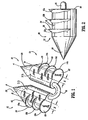
- an absorbable surgical fastener apparatus for connecting objects to body tissue such as absorbable fastener 10 includes dual fastener anchors 12 and 14 secured to one another by a suture tether 16 extending therebetween.
- Each anchor 12 and 14 has a substantially cylindrical body portion 18 having a conically tapered distal end 20 and a substantially planar proximal end surface 22.
- Anchors 12 and 14 are provided with conically shaped distal ends 20 for easier penetration of anchors 12 and 14 into hard tissues, such as, for example, Cooper's ligament.
- Each anchors 12 and 14 is provided with a pair of opposed flattened side surfaces 23 extending longitudinally along the length thereof.
- Anchors 12 and 14 are provided with barbs 12a,12b,12c, and 14a,14b,14c respectively to inhibit fastener pull-out occurrences in either bard or soft tissue.
- Each barb is semi-circular having a planar proximal surface 24 which is orthogonal to a central longitudinal axis "A" of each anchor 12 and 14, and a tapered lower surface 26.
- Barbs 12a-12c and 14a-14c share a common central axis "B". Central axis "B” is spaced a distance "X" from longitudinal axis "A”.
- the center of barbs 12a-12c and 14a-14c can be positioned to reveal a greater amount of planar proximal surface 24 in order to provide an anchor with predetermined anchoring and securing characteristics for body tissue without compromising the strength of body portion 18 of each anchor 12 and 14.
- distance "X" between central axis "B" and longitudinal axis "A” is increased thereby forming a barb with a larger planar proximal surface 24.
- a fastener having a larger projecting barb is desired in order to better anchor the fastener into the soft tissue of the patient.
- each barb 12a-12c and 14a-14c is orthogonal to longitudinal axis "A" of each anchor 12 and 14 respectively, it is envisioned that each barb can be angled relative to longitudinal axis "A" such that barbs 12a, 12b and 12c from a partial thread around body portion 18 of each anchor 12 and 14. In this manner, as each anchor 12 and 14 is pressed into body tissue, each anchor 12 and 14 will rotate into the body tissue.
- each anchor 12 and 14 can be pmvided with a notch or detent (not shown) formed in the proximal end thereof. In this manner, when a series of absorbable fasteners 10 are joined together, the tapered distal ends 18 are received in the detents to thereby maintain the absorbable fasteners 10 and anchors 12 and 14 longitudinally aligned with one another.
- Absorbable fasteners 10 are preferably made of medical grade absorbable materials, for example, Polyglycolic Acid (PGA) and polylactic Acid (PLA).
- PGA Polyglycolic Acid
- PLA polylactic Acid
- a critical feature of the presently disclosed absorbable fastener is that the absorbable fastener provide sufficient strength to retain a mesh material in place for a desired period of time. For example, in the case of applying a hernia repair mesh material, it is recommended that absorbable fasteners 10 remain in place retaining the mesh material for approximately 2 - 3 weeks and be absorbed into the body tissue anytime after that.
- absorbable fasteners 10 are approximately 3 mm long, from the tip of distal end 18 to proximal end 20, by approximately 1.5 mm in diameter. Other suitably consented and dimensioned fasteners may also be utilized depending on the particular application. It is also preferred that absorbable fasteners 10 are configured and dimensioned to avoid penetrating too far into the tissue. An example of a similar fastener structure and no instrument for applying such fasteners are disclosed in U.S. Patent No.
- the barbs of fastener 10 have a center "B" which is spaced a distance "X" from the longitudinal axis "A" of the anchor as opposed to the body of the anchor being cut away to reveal a barb in the '552 patent. In this manner, a barb having a larger height is achieved without altering the dimensions of the body portion, which larger height more firmly secures the anchor of fastener 10 into body tissue as compared to the fastener disclosed in the '552 patent.
- an application is preferably adapted to fire 20 to 30 fasteners per instrument for use in either open and/or laparoscopic procedures. It is envisioned that the applicator instrument be either entirely disposable after use or be provided with a replaceable cartridge of fasteners which can be coupled to the end of a reusable applicator and replaced within a given procedure while the handle of such an applicator would still be disposable but would be reusable within a single procedure. While an alternate applier may be used, the fastener applier disclosed in the '552 reference can be used to apply the surgical fasteners disclosed herein.
- the general procedure of applying a mesh material during a hernia repair procedure is to first create an access to the location of the hernia. (i.e., incision and dissection) thereby exposing the hernia; then place a prosthetic mesh over the hernia defect; next fasten the mesh to the surrounding tissue by firing a plurality of absorbable fasteners 10 through the mesh and into tissue to thereby secure the mesh into place; and finally close the access wound.
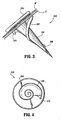
- fastener 200 includes a substantially conical body portion 202 depending from a circular head portion 204.
- Conical body portion 202 includes a helical thread 206 commencing at a pointed distal tip 208 of body portion 202 and terminating at a distance spaced from head portion 204.
- the helical thread 206 advances from distal tip 206 toward head portion 204, the radial projection of the helical thread 206 from the body portion 202 increases.
- helical thread 206 is disclosed as commencing at the distal tip 208, it is envisioned that the helical thread 206 can commence at a distance spaced from the distal tip 208.
- head portion 204 can be provided with a recess or notch (not shown) formed in the center of the proximal surface thereof. The recess being configured to receive the distal tip 208 of an adjacent fastener 200 therein. In this manner, a aeries of fasteners 200 can be aligned in a tip-to-tail fashion with one another and share a common axis.
- fastener 200 is pressed into body tissue "r”, through surgical mesh “M”, until the entire body portion 202 of fastener 200 has passed through mesh “M” and has been buried in tissue “T". Head portion 204 ensures that fastener 200 does not completely pass through mesh “M” thereby ensuring that mesh "M” is in contact with tissue "T”.
- Fastener 200 includes an arcuate tooth 210 projecting radially outwardly from the proximal end of helical thread 206 and oriented such that the curve of the arcuate tooth 210 is oriented to inhibit a rotation of fastener 200 which would remove fastener 200 from body tissue "T”.
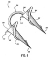
- surgical fastener apparatus 300 Similar to fastener 200, surgical fastener apparatus 300 includes a pair of substantially conical anchors 302 each having a helical thread 304 commencing at a distal tip 306 of each anchor 302 and terminating at a proximal end surface 308 of each anchor 302.
- the radial projection of the helical thread 304 on anchor 302 increases as the helical thread 304 advances from the distal tip 306 to the proximal end of the anchor 302.
- the helical thread 304 is disclosed as commencing at the distal tip 306, it is envisioned that the helical thread 304 can commence at a distance spaced from the distal tip 306 just as well.
- Anchors 302 are connected to one another by a suture tether 310 extending therebetween.
- the orientation of helical thread 304 on each anchor 302 is in the same direction.
- suture tether 31.0 on each anchor 302 will rotate in the same direction and not become tightened.
- the proximal end of each helical thread is provided with an arcuate tooth 312, which arcuate tooth 312 is oriented such that after anchors 302 have been completely imbedded into the body tissue the tooth 312 will dig into the body tissue if the anchor is rotated in a direction which would remove the anchor from the body tissue.
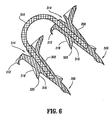
- surgical fastener apparatus 300 can be provided with a detent or recess 314 (see FIG. 6 ) formed in the proximal end surface thereof. In this manner, a series of fasteners 300 can be aligned in tip-to-tail fashion with one another in a fastener applier so that fasteners 300 share a common axis.
- helical thread 304 is made up of a distal surface 316 and a proximal surface 318 joined together to form a sharp edge 320.
- suture tether 310 is fixedly retained within anchors 302, however, it is envisioned that suture tether 310 can be rotatably amounted to proximal surface 308.
- Fasteners 200 and 300 are also preferably made of medical grade absorbable materials, for example, Polyglycolic Acid (PGA) and Polylactic Acid (PLA).
- PGA Polyglycolic Acid
- PLA Polylactic Acid
- a critical feature of the presently disclosed absorbable fastener is that the absorbable fastener provide sufficient strength to retain a mesh material in place for a desired period of time. For example, in the case of applying a hernia repair mesh material, it is recommended that absorbable fasteners 200 or 300 remain in place retaining the mesh material for approximately 2-3 weeks and be absorbed into the body tissue anytime after that.
- absorbable fastener 200 and that each anchor of fastener apparatus 300 are approximately 3 mm long, from the tip of distal end to the proximal end, and wherein the proximal end is approximately 1.5 mm in diameter. It is preferred that fastener 200 and fastener apparatus 300 are configured and dimensioned to avoid penetrating too far into the body tissue.
- Fasteners 200 and 300 can be imbedded into body tissue by either pressing the fastener in to the tissue and allowing the thread on the fastener to automatically twist the fastener into the tissue, by providing a twisting applier which turns the fastener and thus the helical threads draw the fastener into the body tissue or by a combination of pressing and twisting.
- a Lichtenstein repair method will now be described as performed using any of the absorbable surgical fasteners in accordance with the present disclosure.
- a 5cm to 6cm transverse incision is made within a Langer's line, beginning from the pubic tubercle.
- the external oblique aponeurosis is opened.
- the spermatic cord with its cremasteric covering, external spermatic vessels, and the genital nerve are freed from the inguinal floor and lifted with a Penrose drain.
- the spermatic cord is dissected free from the pubic bone area for approximately 2 cm medial to the pubic tubercle in order to make room for extending a prosthetic mesh beyond the pubic tubercle.
- the external oblique aponeurosis is dissected from the underlying internal oblique muscle and aponeurosis high enough to make room for a prosthesis that is 6 cm to 7 cm in height.
- the sac is then dissected from the cord beyond its neck and inverted into the properitoneal space without ligation or excision.
- a medial side of the mesh is shaped to the patient's anatomy.
- a first absorbable fastener 10, 200 or 300 is applied to the mesh to fix the mesh to the anterior rectus sheath where it inserts into the pubic bone.
- the absorbable fastener is placed approximately 2 cm medial to the pubic tubercle in order to be sure that the area is covered by the mesh.
- Additional absorbable fasteners 10, 200 or 300 are placed therearound and end at the lateral border of the internal ring.
- the lower edge of the mesh is then anchored to the inguinal ligament using the same fasteners 10, 200 or 300 around the surgical mesh and ending at the lateral border of the internal ring.
- the absorbable fastener is secured into place by firing the fastener into the body tissue such that a first anchor of the surgical fastener penetrates through the surgical mesh and into the body tissue and such that a second anchor of the surgical fastener is implanted directly into the body tissue.
- the suture tether of the fastener apparatus extends partially across the surgical mesh and partially across body tissue.
- an absorbable fastener in accordance with the present disclosure can be secured into place such that both anchors are imbedded into the body tissue and pass through the surgical mesh. If fastener 200 is used in the method, fasteners 200 are anchored into the body tissue solely through the mesh.
- a slit is then made on the lateral end of the mesh, as seen in FIG. 9 , creating 2 tails-2/3 above and 1/3 below.
- the upper tail is then passed under the cord and pulled toward the head of the patient, placing the spermatic cord in between the 2 tails.
- the upper tail is then crossed over the lower one and held with a pair of hemostat, as seen in FIG. 10 .
- the tails are later sutured together and tucked under the external oblique aponourosis, leaving 5 cm to 6 cm of mesh lateral to the internal ring.
- a Total ExtraPeritoneal Laparoscopic Hernia repair using fasteners 10 or 300 will now be described.
- a skin incision is created and the fascia is incised.
- a balloon type distractor is then placed therein and distended in order to create an operative extraperitoneal space.
- Umbilical trocars and secondary trocars are then inserted into the extraperitoneal space and the space explored.
- the medial and lateral structures are then dissected and the spermatic chord identified.
- a surgical mesh is then cut to the desired size and shape such that a slit is provided in order to wrap the mesh on either side of the spermatic chord.
- the mesh is then laterally fixed in place using surgical fasteners 10, 200 or 300 in order to anchor the mesh to the tranversus arch and the iliopubic tract. This step is repeated for the opposite side.
- the mesh is then medially fixed in place using additional fasteners 10, 200 or 300 to the transversus arch and to Cooper's ligament or in the alternative, fix the mesh to the medial iliopubic tract. Finally, the incision is closed.
- fasteners 10, 200 or 300 can be used in a TAPP repair method to replace suturing of the mesh both laterally and medially.
- fastener 10, 200 or 300 are used to secure the mesh in to place by penetrating the mesh and being anchored into the body tissue.
- fasteners having a similar structure can be used in surgical procedures for fastening items to bone or cartilage.
- the surgical fasteners can be made from surgical grade stainless steel, titanium or any other surgical grade material having sufficient strength to penetrate bone.
Abstract
Description
- The present application claims priority to
U.S. provisional application Serial No. 60/242,647 filed October 23, 2000 - The present invention relates to a surgical fastener for fastening objects to body tissue. More particularly, the present disclosure relates to an absorbable surgical fastener
- Fastening objects to body tissues is a commonly required task in many different surgical applications. One illustrative example of such an application is in hernia repair procedures wherein a reinforcing synthetic mesh material is attached to the tissue. A hernia is a general term referring to a protrusion of tissue through a wall or a cavity in which the tissue is normally contained, also called rupture. An inguinal hernia is a condition in which a loop of intestine enters the inguinal canal (i.e., a tubular passage through the lower layers of the abdominal wall). A direct inguinal hernia creates a bulge in the groin area, and an indirect hernia descends into the scrotum. In men, a hernia can develop at the point where the spermatic cord passes out of the abdomen into the scrotum. An inguinal hernia is a condition in males which occurs in approximately 2% of the male population. Often, an inguinal hernia can be pushed back into the abdominal cavity. However, if the inguinal hernia cannot be forced back through the abdominal wall, the herniated bowel may become trapped in the inguinal ring and/or strangulated. If the flow of blood is restricted (strangulated hernia) or the intestine is blocked (obstructed), emergency surgery is necessary. Without treatment, the strangulated loop of intestine dies as a result of a lack of blood to the loop of intestine.
- In order to treat tho inguinal hernia, surgery is often required to reposition the loop of intestine and secure the weakened muscles in the abdomen. There are two primarily practiced open surgical procedures for hernia repair which procedures use reinforcing synthetic mesh. One procedure, is the Lichtenstein anterior repair method and the other is the Stoppa preperitoneal repair method. Modifications of these procedures exist, as do additional open surgical procedures that do not require the placement of reinforcing mesh over the hernia defect.
- The Lichtenstein, repair method is a "tension-free hernioplasty" based on two important facts, namely, inguinal hernias are caused by a metabolic disorder, which leads to a progressive destruction of the fibroconnective tissue of the groin, making the tissue unsuitable for use in hernia repair and the fact that traditional tissue repairs are associated with undue tension at the suture line, which leads to more postoperative pain, longer recovery time, and a higher rate of
- The Lichtenstein repair method includes the following steps. First, a transverse incision is made within a Langer's line, beginning from the pubic tubercle. The external oblique aponeurosis is opened and the spermatic cord with its cremasteric covering, external spermatic vessels, and the genital nerve are freed from the inguinal floor and lifted with a Penrose drain. The spermatic cord is then dissected free from the pubic bone area medial to the pubic tubercle in order to make room for extending the mesh beyond the pubic tubercle.
- Next, the external oblique aponeurosis is dissected from the underlying internal oblique muscle and aponeurosis high enough to make room for a prosthesis. The sac is then dissected from the cord beyond its neck and inverted into the properitoneal space without ligation or excision. The proximal end is closed, dissected away from the cord structures, and inverted into the preperitoneal space. The medial side of the mesh is then shaped to the patient's anatomy. The first anchoring suture of the mesh fixes the mesh to the anterior rectus sheath where it inserts into the pubic bone. The lower edge of the mesh is sutured to the inguinal ligament using the same suture in a continuous fashion and ends at the lateral border of the internal ring. A slit is next made on the lateral end of the mesh, creating 2 tails. The upper tail is then passed under the cord and pulled toward the head of the patient, placing the spermatic cord in between the 2 tails. The upper tail is then crossed over the lower one and held with a pair of hemostats. The tails are later sutured together and tucked under the external oblique aponeurosis.
- The Stoppa method of hernia repair places a single sheet of prosthetic material (i.e., surgical mesh) between the peritoneum and the musculopectineal orifice. The surgical mesh is then anchored to Cooper's ligaments using nonabsorbable sutures.
- Yet another hernia repair method, known as TransAbdominal PrePeritoneal (TAPP) Laparoscopic Hernia repair method, generally includes the following steps. A pneunmoperitoneum is created in the abdomen and an intra-abdominal pressure is maintained. The repair is then initiated. A laparoscope then inserted and is pointed toward the afflicted inguinal canal. The peritoneal defect or hernia is identified. A peritoneal incision is made, which incision is extended from the lateral aspect of the inguinal region to the lateral umbilical ligament. The Cooper's ligament is then exposed as well as the inferior epigastric vessels and the spermatic Cord. The indirect inguinal hernia sac is then dissected carefully from the spermatic cord. A surgical mesh is then inserted into the intra-abdominal cavity and deployed over the inguinal region. There are three methods to place and secure the mesh over the inguinal region. The mesh is then secured in place with a surgical stapler. It is first stapled on to Cooper's ligament followed by placing several staples perpendicular to the ligament followed by a row more lateral and parallel to Cooper's Ligament. The graft is also anchored around the inferior epigastric vessels and lateral to them. If the mesh is wrapped around the spermatic cord, both limbs of the mesh are stapled closed. The peritoneum is then closed using additional staples and homeostasis is checked.
- Yet another hernia repair method is known as Total ExtraPeritoneal (TEP) Laparoscopic Hernia repair method. This method is identical to the TAPP repair method, however, it entirely takes place in the preperitoneal space. The TEP method includes the following steps. Unlike the TAPP repair method, no pneumoperitoneum is created in the TEP repair method. Instead, a small incision is made below the umbilicus (midline) and the midline exposed. An incision is made slightly lateral to the midline aponevrosis and the anterior and posterior rectus muscle sheaths are exposed. The anatomy must first be clearly identified. Cooper's ligament should be first visualized as well as the inferior epigastric vessels. The indirect hernia sac should be bluntly pulled away from the spermatic cord and the inguinal canal. The hernia sac should then be dissected as medially as possible to allow a surgical mesh to cover the entire inguinal region. The mesh is then inserted and stapled into place as in the TAPP repair method. With the repair completed the small incisions can be closed.
- The two most common of these methods are the Total ExtraPeritoneal (TBP) repair method and the TransAbdominal PrePeritoneal (TAPP) repair method. As discussed above, each of these methods utilizes a reinforcing synthetic mesh that must be fixed to the tissue to prevent early migration of the mesh away from the hernia site. However, the mesh must be anchored into place at first in order to prevent is movement from the hernia repair site. Only after 7-10 days, does the mesh have sufficient tissue in-growth to prevent its motion away from the hernia repair site.
- Each of the above disclosed procedures utilizes titanium staples to retain the mesh in place. These staples become permanent residents in the body cavity. A disadvantage of permanent metal staples is the possibility of the formation of excessive scar tissue (adhesions) which can in turn cause further patient complications and hinder future surgical procedures. In addition, these permanent staples may be associated with increased long-term discomfort to the patient as a result of the hernia repair procedure.
- Accordingly, a heed exists for an improved surgical fastener and applying apparatus as well as for methods in securing objects to body tissue, for example such as in attaching a mesh material for a sufficient time to a hernia repair site until sufficient tissue in-growth occurs to retain the mesh in place.
-
U.S. Patent No. 5,997,552 discloses a surgical fastener according to the preamble of claim 1. - It is an object of the present disclosure to provide an absorbable surgical fastener apparatus and methods in which the amount of foreign material in the patients body is reduced, thereby minimizing adhesion formation and reducing fastener-associated long-term discomfort to the patient.
- It is another object of the present disclosure to provide an absorbable surgical fastener and method which is easier and faster to use than traditional suturing techniques in open procedures. Further, the relatively high firing force that can be applied to the absorbable fasteners of the present disclosure facilitate more reliable penetration of tougher tissue materials, such as for example, Cooper's ligament.
- It is yet another object of the present disclosure to provide in absorbable surgical fastener which is radiolucent and provides greater peace of mind for the patient.
- It is still a further object of the present disclosure to provide an absorbable surgical fastener apparatus having a suture tether disposed between anchoring barbs. The tether provides the advantage of holding the mesh in place loosely thus minimizing the tension in the surrounding tissue and reducing fastener pull-out occurrences.
- It is another object of the present disclosure to provide a surgical fastener apparatus which is dimensioned to not penetrate the abdominal wall of the patient and which is provided with a series of barbs having a relatively larger surface than standard fastener barbs to thereby better retain the fastener in soft tissue.
- Aspects of the present invention are described in the following numbered paragraphs:
- "1. A surgical fastener apparatus (10) for securing a surgical mesh material to body tissue, comprising:
- a pair of substantially cylindrical anchors (12, 14), wherein:
- each anchor (12, 14) has a longitudinal central axis (A),
- each anchors (12, 14) is provided with a substantially cylindrical body portion (18) having a conically tapered distal end (20) and a substantially planar proximal end (22),
- each anchor (12, 14) is further provided with a series of semi-circular angled projections (12a-12c, 14a-14c) having a proximal surface (24) and a tapered distal end (26), and
- the angled projections of a respective series of angled projections (12a-12c, 14a-14c) share a common central axis (B); and
- a tether (16) interconnecting said proximal ends (22) of said pair of anchors (12, 14),
- characterized in that:
- the common central axis (B) is spaced a radial distance (X) from the longitudinal central axis (A) of a respective anchor (12, 14) in order to provide an anchor with predetermined anchoring and securing characteristics for body tissue."
- a pair of substantially cylindrical anchors (12, 14), wherein:
- 2. The surgical fastener apparatus according to paragraph 1, wherein said pair of anchors are made from a bioabsorbable material.
- 3. The surgical fastener apparatus according to paragraph 2, wherein said bioabsorbable material is selected from a material which reabsorbs into said body tissue at an appropriate rate.
- 4. The surgical fastener apparatus according to paragraph 2, wherein said bioabsorbable material is selected from the group consisting of polyglycolic acid and polylactic acid.
- 5. The surgical fastener apparatus according to paragraph 1, where said pair of anchors and said tether are dimensioned to be partially absorbed into said body tissue for at least a period of approximately 2 to 3 weeks immediately after implanting and are fully absorbed into said body tissue at any time thereafter.
- 6. The surgical fastener apparatus according to paragraph 1, wherein said pair of anchors have a length of approximately 3 mm from a distal tip of said tapered distal end to said substantially planar proximal end.
- 7. The surgical fastener apparatus according to paragraph 6, wherein said pair of anchors have a diameter of approximately 1.5 mm.
- 8. The surgical fastener apparatus according to paragraph 1, wherein said tether is made from a bioabsorbable material.
- 9. The surgical fastener apparatus according to paragraph 8, wherein said bioabsorbable material is selected from the group consisting of polyglycolic acid and polylactic acid.
- 10. The surgical fastener apparatus according to paragraph 1, wherein said tether is non-rigid.
- The surgical fastener apparatus serves the function of previously used staples to secure objects to body tissue as performed in previous methods, for example, mesh being fixed to specific anatomic landmarks surrounding the hernia repair, or attaching mesh to tissue, or attaching tissue to tissue or attaching tissue to ligaments. However, the absorbable fastener apparatus has the uniquely advantageous feature that the absorbable fasteners are utilized to attach the mesh to the tissue for a sufficient time to allow tissue in-growth to occur on the mesh material. In this manner, the absorbable fasteners help prevent early mesh migration and then after sufficient tissue in-growth are absorbed in the body.
- In addition, a hernial repair method utilizing the fasteners is described in which a surgical mesh is secured in place over the hernia repair site by imbedding the surgical fasteners in to body tissue through the surgical mesh.
- Various embodiments of tho presently disclosed surgical fastener will be described herein with reference to the accompanying drawing figures wherein only the fastener shown in
Figs. 1 and 2 is an embodiment of the invention , whereas the fasteners shown inFigs. 3-6 do not fall under the scope of the claims: -
FIG. 1 is an enlarged perspective view of one embodiment of an absorbable surgical fastener apparatus constructed in accordance with the present invention; -
FIG. 2 is a side view of a barb portion of the absorbable surgical fastener apparatus ofFIG. 1 ; -
FIG. 3 is an enlarged perspective view of an alternative absorbable surgical fastener; -
FIG. 4 is an end view of the surgical fastener ofFIG. 3 ; -
FIG. 5 is an enlarged perspective view of an absorbable surgical fastener apparatus including a pair of fasteners as shown inFIG. 3 ; -
FIG. 6 is a cross-sectional view of the absorbable fastener shown inFIG. 5 , taken along the longitudinal axis; -
FIG. 7 is an illustration of a sequential step in attaching an object to body tissue using the presently disclosed absorbable surgical fasteners; -
FIG. 8 is a further sequential step according to the method ofFIG. 7 ; -
FIG. 9 is a still further sequential step of the method ofFIG. 7 ; -
FIG. 10 is another sequential step of the method ofFIG. 7 ; and -
FIG. 11 is another illustration of a step of the method ofFIG. 7 . - Preferred embodiments of the presently disclosed absorbable surgical fastener apparatus and method of applying same will now be described in detail with reference to the drawing figures wherein live reference numerals identify similar or identical structural element.
- The presently disclosed absorbable surgical fastener apparatus and method is shown and described herein in connection with open and laparoscopic inguinal fernoral and ventral hernia repairs. Although not described in detail herein, the absorbable fastener can also be applied to other procedures which require that objects be attached to body tissue.
- Referring initially to
FIGs. 1 and 2 , an absorbable surgical fastener apparatus for connecting objects to body tissue, such asabsorbable fastener 10 includes dual fastener anchors 12 and 14 secured to one another by asuture tether 16 extending therebetween. Eachanchor cylindrical body portion 18 having a conically tapereddistal end 20 and a substantially planarproximal end surface 22.Anchors anchors -
Anchors barbs proximal surface 24 which is orthogonal to a central longitudinal axis "A" of eachanchor lower surface 26.Barbs 12a-12c and 14a-14c share a common central axis "B". Central axis "B" is spaced a distance "X" from longitudinal axis "A". In this manner, the center ofbarbs 12a-12c and 14a-14c can be positioned to reveal a greater amount of planarproximal surface 24 in order to provide an anchor with predetermined anchoring and securing characteristics for body tissue without compromising the strength ofbody portion 18 of eachanchor proximal surface 24. In the case of securing a fastener to soft tissue, as is the case in hernia repair, a fastener having a larger projecting barb is desired in order to better anchor the fastener into the soft tissue of the patient. - While each
barb 12a-12c and 14a-14c is orthogonal to longitudinal axis "A" of eachanchor barbs body portion 18 of eachanchor anchor anchor - While a planar
proximal end 20 has been shown, it is envisioned that eachanchor absorbable fasteners 10 are joined together, the tapered distal ends 18 are received in the detents to thereby maintain theabsorbable fasteners 10 and anchors 12 and 14 longitudinally aligned with one another. -
Absorbable fasteners 10 are preferably made of medical grade absorbable materials, for example, Polyglycolic Acid (PGA) and polylactic Acid (PLA). A critical feature of the presently disclosed absorbable fastener is that the absorbable fastener provide sufficient strength to retain a mesh material in place for a desired period of time. For example, in the case of applying a hernia repair mesh material, it is recommended thatabsorbable fasteners 10 remain in place retaining the mesh material for approximately 2 - 3 weeks and be absorbed into the body tissue anytime after that. - It is preferred that
absorbable fasteners 10 are approximately 3 mm long, from the tip ofdistal end 18 toproximal end 20, by approximately 1.5 mm in diameter. Other suitably consented and dimensioned fasteners may also be utilized depending on the particular application. It is also preferred thatabsorbable fasteners 10 are configured and dimensioned to avoid penetrating too far into the tissue. An example of a similar fastener structure and no instrument for applying such fasteners are disclosed inU.S. Patent No. 5,997,552 (hereinafter "the '552 patent"), issued to Person et als and entitled Meniscle Fastener Applying Device, Unlike the fastener in the '5S2 patent, the barbs offastener 10 according to the present invention have a center "B" which is spaced a distance "X" from the longitudinal axis "A" of the anchor as opposed to the body of the anchor being cut away to reveal a barb in the '552 patent. In this manner, a barb having a larger height is achieved without altering the dimensions of the body portion, which larger height more firmly secures the anchor offastener 10 into body tissue as compared to the fastener disclosed in the '552 patent. - Unlike the applicator instrument for placing
absorbable fasteners 10 disclosed inU.S. Patent No. 5,997,552 , an application is preferably adapted to fire 20 to 30 fasteners per instrument for use in either open and/or laparoscopic procedures. It is envisioned that the applicator instrument be either entirely disposable after use or be provided with a replaceable cartridge of fasteners which can be coupled to the end of a reusable applicator and replaced within a given procedure while the handle of such an applicator would still be disposable but would be reusable within a single procedure. While an alternate applier may be used, the fastener applier disclosed in the '552 reference can be used to apply the surgical fasteners disclosed herein. - By way of example, the general procedure of applying a mesh material during a hernia repair procedure, is to first create an access to the location of the hernia. (i.e., incision and dissection) thereby exposing the hernia; then place a prosthetic mesh over the hernia defect; next fasten the mesh to the surrounding tissue by firing a plurality of
absorbable fasteners 10 through the mesh and into tissue to thereby secure the mesh into place; and finally close the access wound. - Turning now to
FIGs. 3-5 , an alternative surgical fastener is shown generally as 200. As seen inFIGs. 3 and 4 ,fastener 200 includes a substantiallyconical body portion 202 depending from acircular head portion 204.Conical body portion 202 includes ahelical thread 206 commencing at a pointeddistal tip 208 ofbody portion 202 and terminating at a distance spaced fromhead portion 204. As thehelical thread 206 advances fromdistal tip 206 towardhead portion 204, the radial projection of thehelical thread 206 from thebody portion 202 increases. While thehelical thread 206 is disclosed as commencing at thedistal tip 208, it is envisioned that thehelical thread 206 can commence at a distance spaced from thedistal tip 208. in addition,head portion 204 can be provided with a recess or notch (not shown) formed in the center of the proximal surface thereof. The recess being configured to receive thedistal tip 208 of anadjacent fastener 200 therein. In this manner, a aeries offasteners 200 can be aligned in a tip-to-tail fashion with one another and share a common axis. - In use,
fastener 200 is pressed into body tissue "r", through surgical mesh "M", until theentire body portion 202 offastener 200 has passed through mesh "M" and has been buried in tissue "T".Head portion 204 ensures thatfastener 200 does not completely pass through mesh "M" thereby ensuring that mesh "M" is in contact with tissue "T".Fastener 200 includes anarcuate tooth 210 projecting radially outwardly from the proximal end ofhelical thread 206 and oriented such that the curve of thearcuate tooth 210 is oriented to inhibit a rotation offastener 200 which would removefastener 200 from body tissue "T". - Turning now to
FIG. 5 , a surgical fastener apparatus is shown generally as 300. Similar tofastener 200,surgical fastener apparatus 300 includes a pair of substantiallyconical anchors 302 each having ahelical thread 304 commencing at adistal tip 306 of eachanchor 302 and terminating at aproximal end surface 308 of eachanchor 302. Once again, the radial projection of thehelical thread 304 onanchor 302 increases as thehelical thread 304 advances from thedistal tip 306 to the proximal end of theanchor 302. While thehelical thread 304 is disclosed as commencing at thedistal tip 306, it is envisioned that thehelical thread 304 can commence at a distance spaced from thedistal tip 306 just as well.Anchors 302 are connected to one another by asuture tether 310 extending therebetween. As seen inFIG. 5 , the orientation ofhelical thread 304 on eachanchor 302 is in the same direction. In this manner, asanchors 302 are being imbedded into body tissue and commence rotating in the direction ofhelical thread 304, suture tether 31.0 on eachanchor 302 will rotate in the same direction and not become tightened. The proximal end of each helical thread is provided with anarcuate tooth 312, whicharcuate tooth 312 is oriented such that afteranchors 302 have been completely imbedded into the body tissue thetooth 312 will dig into the body tissue if the anchor is rotated in a direction which would remove the anchor from the body tissue. Similar tofastener 10,surgical fastener apparatus 300 can be provided with a detent or recess 314 (seeFIG. 6 ) formed in the proximal end surface thereof. In this manner, a series offasteners 300 can be aligned in tip-to-tail fashion with one another in a fastener applier so thatfasteners 300 share a common axis. - As seen in
FIG. 6 ,helical thread 304 is made up of adistal surface 316 and aproximal surface 318 joined together to form asharp edge 320. In addition,suture tether 310 is fixedly retained withinanchors 302, however, it is envisioned thatsuture tether 310 can be rotatably amounted toproximal surface 308. -
Fasteners absorbable fasteners absorbable fastener 200 and that each anchor offastener apparatus 300 are approximately 3 mm long, from the tip of distal end to the proximal end, and wherein the proximal end is approximately 1.5 mm in diameter. It is preferred thatfastener 200 andfastener apparatus 300 are configured and dimensioned to avoid penetrating too far into the body tissue. -
Fasteners - By way of example only, and not to be considered limiting in any way, with reference to
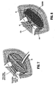
FIGs. 7-11 , a Lichtenstein repair method will now be described as performed using any of the absorbable surgical fasteners in accordance with the present disclosure. First, a 5cm to 6cm transverse incision is made within a Langer's line, beginning from the pubic tubercle. The external oblique aponeurosis is opened. As shown inFIG. 7 , the spermatic cord with its cremasteric covering, external spermatic vessels, and the genital nerve are freed from the inguinal floor and lifted with a Penrose drain. The spermatic cord is dissected free from the pubic bone area for approximately 2 cm medial to the pubic tubercle in order to make room for extending a prosthetic mesh beyond the pubic tubercle. The external oblique aponeurosis is dissected from the underlying internal oblique muscle and aponeurosis high enough to make room for a prosthesis that is 6 cm to 7 cm in height. The sac is then dissected from the cord beyond its neck and inverted into the properitoneal space without ligation or excision. - Referring to
FIG. 8 , a medial side of the mesh, is shaped to the patient's anatomy. A firstabsorbable fastener absorbable fasteners same fasteners - According to one method, the absorbable fastener is secured into place by firing the fastener into the body tissue such that a first anchor of the surgical fastener penetrates through the surgical mesh and into the body tissue and such that a second anchor of the surgical fastener is implanted directly into the body tissue. In this manner, the suture tether of the fastener apparatus extends partially across the surgical mesh and partially across body tissue. In an alternative method, an absorbable fastener in accordance with the present disclosure, can be secured into place such that both anchors are imbedded into the body tissue and pass through the surgical mesh. If
fastener 200 is used in the method,fasteners 200 are anchored into the body tissue solely through the mesh. - A slit is then made on the lateral end of the mesh, as seen in
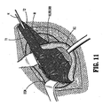
FIG. 9 , creating 2 tails-2/3 above and 1/3 below. The upper tail is then passed under the cord and pulled toward the head of the patient, placing the spermatic cord in between the 2 tails. The upper tail is then crossed over the lower one and held with a pair of hemostat, as seen inFIG. 10 . The tails are later sutured together and tucked under the external oblique aponourosis, leaving 5 cm to 6 cm of mesh lateral to the internal ring. - While the upper edge of the mesh is fixed in place, care is taken to keep the mesh slightly relaxed. This laxity produces a dome-like ripple in the mesh to compensate for increased intra-abdominal pressure when the patient stands up from his or her recumbent position during the operation. If the mesh is kept completely flat, it becomes subject to tension when the patient stands up. This pulling affect on the mesh and the tissue, is illustrated when the mesh is kept flat as shown in
FIG 11 . The use of a surgical fastener in accordance with the present disclosure effectively eliminates the tension of the surgical mesh as the anchors of then surgical fastener are relatively movable with respect to one another. In other words, one anchor is able to move with respect to the other anchor if needed as the patients body tissue shifts. In so doing, the tension on the mesh is reduced since the suture tether provides the mesh with some room to move since the individual anchors are moveable with respect to one another. - A Total ExtraPeritoneal Laparoscopic Hernia
repair using fasteners surgical fasteners additional fasteners - It is envisioned that
fasteners fastener - While each of the above described fasteners have been described as being used in connection with hernial repair surgery, it is envisioned that fasteners having a similar structure can be used in surgical procedures for fastening items to bone or cartilage. In such surgical procedures, the surgical fasteners can be made from surgical grade stainless steel, titanium or any other surgical grade material having sufficient strength to penetrate bone.
- It will be understood that various modifications may be made to the embodiments of the presently disclosed, surgical absorbable fastener apparatus disclosed herein. Therefore, the above description should not be construed as limiting, but merely as exemplifications of preferred embodiments. Those skilled in the art will envision other modifications within the scope of the present disclosure.
Claims (10)
- A surgical fastener apparatus (10) for securing a surgical mesh material to body tissue, comprising:a pair of substantially cylindrical anchors (12, 14), wherein:each anchor (12, 14) has a longitudinal central axis (A),each anchor (12, 14) is provided with a substantially cylindrical body portion (18) having a conically tapered distal end (20) and a substantially planar proximal end (22),each anchor (12, 14) is further provided with a series of semi-circular angled projections (12a-12c, 14a-14c) having a proximal surface (24) and a tapered distal end (26), andthe angled projections of a respective series of angled projections (12a-12c, 14a-14c) share a common central axis (B): anda tether (16) interconnecting said proximal ends (22) of said pair of anchors (12, 14),characterized in that:the common central axis (B) is spaced a radial distance (X) from the longitudinal central axis (A) of a respective anchor (12, 14) in order to provide an anchor with predetermined anchoring and securing characteristics for body tissue.
- The surgical fastener apparatus (10) according to claim 1, wherein said pair of anchors (12,14) are made from a bioabsorbable material.
- The surgical fastener apparatus (10) according to claim 2, wherein said bioabsorbable material is selected from a material which reabsorbs into said body tissue at an appropriate rate.
- The surgical fastener apparatus (10) according to claim 2, wherein said bioabsorbable material is selected from the group consisting of polyglycolic acid and polylactic acid.
- The surgical fastener apparatus (10) according to claim 1, where said pair of anchors (12, 14) and said tether (16) are dimensioned to be partially absorbed into said body tissue for at least a period of approximately 2 to 3 weeks immediately after implanting and are fully absorbed into said body tissue at any time thereafter.
- The surgical fastener apparatus (10) according to claim 1, wherein said pair of anchors (12, 14) have a length of approximately 3 mm from a distal tip of said tapered distal end (26) to said substantially planar proximal end (22).
- The surgical fasteners apparatus (10) according to claim 6, wherein said pair of anchors (12, 14) have a diameter of approximately 1.5 mm.
- The surgical fastener apparatus (10) according to claim 1, wherein said tether (16) is made from a bioabsorbable material.
- The surgical fastener apparatus (10) according to claim 8, wherein said bioabsorbable material is selected from the group consisting of polyglycolic acid and polylactic acid.
- The surgical fastener apparatus (10) according to claim 1, wherein said tether (16) is non-rigid.
Priority Applications (1)
| Application Number | Priority Date | Filing Date | Title |
|---|---|---|---|
| EP10008788A EP2298183B1 (en) | 2000-10-23 | 2001-10-23 | Absorbable fastener |
Applications Claiming Priority (3)
| Application Number | Priority Date | Filing Date | Title |
|---|---|---|---|
| US24264700P | 2000-10-23 | 2000-10-23 | |
| US242647P | 2000-10-23 | ||
| PCT/US2001/050165 WO2002034140A2 (en) | 2000-10-23 | 2001-10-23 | Absorbable fastener and applying apparatus |
Related Child Applications (2)
| Application Number | Title | Priority Date | Filing Date |
|---|---|---|---|
| EP10008606.5 Division-Into | 2010-08-18 | ||
| EP10008788.1 Division-Into | 2010-08-24 |
Publications (2)
| Publication Number | Publication Date |
|---|---|
| EP1328196A2 EP1328196A2 (en) | 2003-07-23 |
| EP1328196B1 true EP1328196B1 (en) | 2011-06-29 |
Family
ID=22915629
Family Applications (2)
| Application Number | Title | Priority Date | Filing Date |
|---|---|---|---|
| EP01988554A Expired - Lifetime EP1328196B1 (en) | 2000-10-23 | 2001-10-23 | Absorbable fastener |
| EP10008788A Expired - Lifetime EP2298183B1 (en) | 2000-10-23 | 2001-10-23 | Absorbable fastener |
Family Applications After (1)
| Application Number | Title | Priority Date | Filing Date |
|---|---|---|---|
| EP10008788A Expired - Lifetime EP2298183B1 (en) | 2000-10-23 | 2001-10-23 | Absorbable fastener |
Country Status (7)
| Country | Link |
|---|---|
| US (7) | US20040092937A1 (en) |
| EP (2) | EP1328196B1 (en) |
| JP (2) | JP4092196B2 (en) |
| AU (2) | AU2002232798B2 (en) |
| CA (2) | CA2426474C (en) |
| ES (1) | ES2365129T3 (en) |
| WO (1) | WO2002034140A2 (en) |
Families Citing this family (188)
| Publication number | Priority date | Publication date | Assignee | Title |
|---|---|---|---|---|
| DE69931018T2 (en) | 1998-12-30 | 2006-11-23 | Ethicon, Inc. | Thread belay device |
| US7153312B1 (en) | 1999-12-02 | 2006-12-26 | Smith & Nephew Inc. | Closure device and method for tissue repair |
| US7887551B2 (en) | 1999-12-02 | 2011-02-15 | Smith & Nephew, Inc. | Soft tissue attachment and repair |
| EP1437968B1 (en) | 2001-10-23 | 2013-04-10 | Covidien LP | Surgical fasteners |
| EP1511427B1 (en) | 2002-06-11 | 2007-02-14 | Tyco Healthcare Group Lp | Hernia mesh tacks |
| US20040138683A1 (en) * | 2003-01-09 | 2004-07-15 | Walter Shelton | Suture arrow device and method of using |
| US20040138705A1 (en) * | 2003-01-09 | 2004-07-15 | Harri Heino | Surgical staple for tissue treatment |
| EP1594569B1 (en) * | 2003-01-27 | 2014-05-14 | Corassist Cardiovascular Ltd. | In vivo device for improving diastolic ventricular function |
| US9314235B2 (en) | 2003-02-05 | 2016-04-19 | Smith & Nephew, Inc. | Tissue anchor and insertion tool |
| US8926637B2 (en) | 2003-06-13 | 2015-01-06 | Covidien Lp | Multiple member interconnect for surgical instrument and absorbable screw fastener |
| EP2298184A1 (en) * | 2003-06-13 | 2011-03-23 | Tyco Healthcare Group LP | Multiple member interconnect for surgical instrument and absorbable screw fastener |
| US20060052825A1 (en) * | 2003-06-16 | 2006-03-09 | Ransick Mark H | Surgical implant alloy |
| US7905902B2 (en) * | 2003-06-16 | 2011-03-15 | Ethicon Endo-Surgery, Inc. | Surgical implant with preferential corrosion zone |
| US10478179B2 (en) | 2004-04-27 | 2019-11-19 | Covidien Lp | Absorbable fastener for hernia mesh fixation |
| ES1057646Y (en) * | 2004-05-19 | 2004-12-16 | Ayet Felix Checa | PERFECTED PROTESIS FOR THE SURGICAL TREATMENT OF HERNIAS DE LA INGLE. |
| US8128670B2 (en) * | 2005-04-15 | 2012-03-06 | Biodynamics Llc | Surgical expansion fasteners |
| EP1937157B1 (en) | 2005-06-03 | 2020-08-05 | Covidien LP | Surgical stapler with timer and feedback display |
| US20060293709A1 (en) | 2005-06-24 | 2006-12-28 | Bojarski Raymond A | Tissue repair device |
| US20070162030A1 (en) * | 2006-01-06 | 2007-07-12 | Ernest Aranyi | Multi-pronged compressive absorbable tack |
| US7862573B2 (en) * | 2006-04-21 | 2011-01-04 | Darois Roger E | Method and apparatus for surgical fastening |
| US8454662B2 (en) * | 2006-12-08 | 2013-06-04 | Warsaw Orthopedic, Inc. | Tethers with strength limits for treating vertebral members |
| US7823760B2 (en) | 2007-05-01 | 2010-11-02 | Tyco Healthcare Group Lp | Powered surgical stapling device platform |
| US7931660B2 (en) | 2007-05-10 | 2011-04-26 | Tyco Healthcare Group Lp | Powered tacker instrument |
| US7959640B2 (en) | 2008-02-13 | 2011-06-14 | Apollo Endosurgery, Inc. | Method of performing transgastric ventral hernia repair and tissue anchors and deployment devices therefor |
| WO2009104182A2 (en) | 2008-02-18 | 2009-08-27 | Polytouch Medical Ltd | A device and method for deploying and attaching a patch to a biological tissue |
| US8317808B2 (en) | 2008-02-18 | 2012-11-27 | Covidien Lp | Device and method for rolling and inserting a prosthetic patch into a body cavity |
| US9044235B2 (en) | 2008-02-18 | 2015-06-02 | Covidien Lp | Magnetic clip for implant deployment device |
| US9034002B2 (en) | 2008-02-18 | 2015-05-19 | Covidien Lp | Lock bar spring and clip for implant deployment device |
| US8758373B2 (en) | 2008-02-18 | 2014-06-24 | Covidien Lp | Means and method for reversibly connecting a patch to a patch deployment device |
| US8808314B2 (en) | 2008-02-18 | 2014-08-19 | Covidien Lp | Device and method for deploying and attaching an implant to a biological tissue |
| US9301826B2 (en) | 2008-02-18 | 2016-04-05 | Covidien Lp | Lock bar spring and clip for implant deployment device |
| US9393093B2 (en) | 2008-02-18 | 2016-07-19 | Covidien Lp | Clip for implant deployment device |
| US9833240B2 (en) | 2008-02-18 | 2017-12-05 | Covidien Lp | Lock bar spring and clip for implant deployment device |
| US9393002B2 (en) | 2008-02-18 | 2016-07-19 | Covidien Lp | Clip for implant deployment device |
| US9398944B2 (en) | 2008-02-18 | 2016-07-26 | Covidien Lp | Lock bar spring and clip for implant deployment device |
| US20090287045A1 (en) | 2008-05-15 | 2009-11-19 | Vladimir Mitelberg | Access Systems and Methods of Intra-Abdominal Surgery |
| US20100076487A1 (en) * | 2008-09-23 | 2010-03-25 | Ilahi Omer A | Kit Containing Combination Absorbable Staple and Non-absorbable Suture, And Method Of Using Same |
| CA2730547C (en) | 2008-10-20 | 2013-08-06 | Polytouch Medical Ltd. | A device for attaching a patch to a biological tissue |
| WO2010081029A1 (en) | 2009-01-08 | 2010-07-15 | Rotation Medical, Inc. | Implantable tendon protection systems and related kits and methods |
| US8821555B2 (en) * | 2009-02-11 | 2014-09-02 | Howmedica Osteonics Corp. | Intervertebral implant with integrated fixation |
| US9179910B2 (en) | 2009-03-20 | 2015-11-10 | Rotation Medical, Inc. | Medical device delivery system and method |
| JP5457544B2 (en) * | 2009-03-31 | 2014-04-02 | アイエムディーエス コーポレイション | Double bundle ACL repair |
| US8535377B2 (en) * | 2009-03-31 | 2013-09-17 | Imds Corporation | Double bundle ACL repair system |
| US20100256675A1 (en) * | 2009-04-03 | 2010-10-07 | Romans Matthew L | Absorbable surgical staple |
| CA2761312C (en) * | 2009-05-07 | 2018-07-24 | Tyco Healthcare Group Lp | Surgical patch cover and method of use |
| US8894669B2 (en) * | 2009-05-12 | 2014-11-25 | Ethicon, Inc. | Surgical fasteners, applicator instruments, and methods for deploying surgical fasteners |
| CA2763794C (en) | 2009-06-04 | 2017-01-17 | Rotation Medical, Inc. | Apparatus having bowstring-like staple delivery to a target tissue |
| EP2437686B1 (en) | 2009-06-04 | 2017-12-13 | Rotation Medical, Inc. | Apparatus for deploying sheet-like materials |
| WO2011019699A2 (en) | 2009-08-10 | 2011-02-17 | Howmedica Osteonics Corp | Intervertebral implant with integrated fixation |
| US9999424B2 (en) | 2009-08-17 | 2018-06-19 | Covidien Lp | Means and method for reversibly connecting an implant to a deployment device |
| WO2011021083A1 (en) | 2009-08-17 | 2011-02-24 | PolyTouch Medical, Inc. | Articulating patch deployment device and method of use |
| US9033993B2 (en) | 2009-11-03 | 2015-05-19 | Howmedica Osteonics Corp. | Intervertebral implant with integrated fixation |
| US9480511B2 (en) | 2009-12-17 | 2016-11-01 | Engage Medical Holdings, Llc | Blade fixation for ankle fusion and arthroplasty |
| US9198750B2 (en) | 2010-03-11 | 2015-12-01 | Rotation Medical, Inc. | Tendon repair implant and method of arthroscopic implantation |
| EP3178448B1 (en) | 2010-12-16 | 2018-08-01 | Engage Medical Holdings, LLC | Arthroplasty systems |
| US10952783B2 (en) | 2011-12-29 | 2021-03-23 | Rotation Medical, Inc. | Guidewire having a distal fixation member for delivering and positioning sheet-like materials in surgery |
| WO2012145059A1 (en) | 2011-02-15 | 2012-10-26 | Rotation Medical, Inc. | Methods and apparatus for fixing sheet-like materials to a target tissue |
| US9198751B2 (en) | 2011-02-15 | 2015-12-01 | Rotation Medical, Inc. | Methods and apparatus for delivering and positioning sheet-like materials in surgery |
| US9314314B2 (en) | 2011-02-15 | 2016-04-19 | Rotation Medical, Inc. | Anatomical location markers and methods of use in positioning sheet-like materials during surgery |
| CA2825918C (en) | 2011-02-15 | 2018-08-07 | Rotation Medical, Inc. | Methods and apparatus for delivering and positioning sheet-like materials |
| US8777965B2 (en) * | 2011-04-15 | 2014-07-15 | Usgi Medical, Inc. | Devices and methods for laparoscopic hernia repair |
| US8968402B2 (en) | 2011-10-18 | 2015-03-03 | Arthrocare Corporation | ACL implants, instruments, and methods |
| US9615856B2 (en) | 2011-11-01 | 2017-04-11 | Imds Llc | Sacroiliac fusion cage |
| US9254130B2 (en) | 2011-11-01 | 2016-02-09 | Hyun Bae | Blade anchor systems for bone fusion |
| US8535339B2 (en) | 2011-12-18 | 2013-09-17 | Via Surgical Ltd. | Apparatus and method for suturing |
| US9107661B2 (en) | 2011-12-19 | 2015-08-18 | Rotation Medical, Inc. | Fasteners and fastener delivery devices for affixing sheet-like materials to bone or tissue |
| EP3403601A1 (en) | 2011-12-19 | 2018-11-21 | Rotation Medical, Inc. | Apparatus for forming pilot holes in bone and delivering fasteners therein for retaining an implant |
| US9271726B2 (en) | 2011-12-19 | 2016-03-01 | Rotation Medical, Inc. | Fasteners and fastener delivery devices for affixing sheet-like materials to bone or tissue |
| EP2793712B1 (en) | 2011-12-19 | 2018-03-28 | Rotation Medical, Inc. | Fasteners for affixing sheet -like materials to bone or tissue |
| US9204959B2 (en) | 2012-02-02 | 2015-12-08 | Smith & Nephew, Inc. | Implantable biologic holder |
| US10238382B2 (en) | 2012-03-26 | 2019-03-26 | Engage Medical Holdings, Llc | Blade anchor for foot and ankle |
| US9888913B2 (en) | 2012-05-31 | 2018-02-13 | Via Surgical Ltd. | Variable depth surgical fixation |
| ES2733939T3 (en) | 2012-05-31 | 2019-12-03 | Via Surgical Ltd | Surgical fixation of variable depth |
| US20140046348A1 (en) * | 2012-08-07 | 2014-02-13 | University Hospitals Of Cleveland | Wound healing system |
| WO2014107503A1 (en) | 2013-01-04 | 2014-07-10 | The Feinstein Institute For Medical Research | Fastener |
| US9351733B2 (en) | 2013-01-18 | 2016-05-31 | Covidien Lp | Surgical fastener applier |
| US9993245B2 (en) | 2013-03-11 | 2018-06-12 | Via Surgical Ltd. | Surgical tacker with quantity indicator |
| US9358010B2 (en) | 2013-03-12 | 2016-06-07 | Covidien Lp | Flex cable and spring-loaded tube for tacking device |
| US9867620B2 (en) | 2013-03-14 | 2018-01-16 | Covidien Lp | Articulation joint for apparatus for endoscopic procedures |
| CA2906203A1 (en) * | 2013-03-15 | 2014-09-18 | Micro Interventional Devices, Inc. | Tissue closure device and method |
| US9655621B2 (en) | 2013-03-15 | 2017-05-23 | Covidien Lp | Surgical instrument for dispensing tacks and solution |
| WO2014197633A1 (en) * | 2013-06-05 | 2014-12-11 | Lc Therapeutics, Inc. | Tissue anchor and deployment device for same |
| US9358004B2 (en) | 2013-06-28 | 2016-06-07 | Covidien Lp | Articulating apparatus for endoscopic procedures |
| US9351728B2 (en) | 2013-06-28 | 2016-05-31 | Covidien Lp | Articulating apparatus for endoscopic procedures |
| US9668730B2 (en) | 2013-06-28 | 2017-06-06 | Covidien Lp | Articulating apparatus for endoscopic procedures with timing system |
| US10085746B2 (en) | 2013-06-28 | 2018-10-02 | Covidien Lp | Surgical instrument including rotating end effector and rotation-limiting structure |
| US20150032130A1 (en) | 2013-07-24 | 2015-01-29 | Covidien Lp | Expanding absorbable tack |
| US9526498B2 (en) | 2013-09-17 | 2016-12-27 | Covidien Lp | Surgical device with a trigger lockout mechanism device |
| US10335146B2 (en) | 2014-04-02 | 2019-07-02 | Coviden Lp | Surgical fastener applying apparatus, kits and methods for endoscopic procedures |
| WO2015172052A1 (en) | 2014-05-09 | 2015-11-12 | Rotation Medical, Inc. | Medical implant delivery system for sheet-like implant |
| CN104055550B (en) * | 2014-05-27 | 2016-06-08 | 苏州瑞华医院有限公司 | A kind of inclined-plane band wire holdfast |
| US10166022B2 (en) * | 2014-09-29 | 2019-01-01 | Biomet C.V. | Method and apparatus for bone fixation |
| AU2015343273B2 (en) | 2014-11-04 | 2017-12-14 | Rotation Medical, Inc. | Medical implant delivery system and related methods |
| US10123796B2 (en) | 2014-11-04 | 2018-11-13 | Rotation Medical, Inc. | Medical implant delivery system and related methods |
| EP3215025B1 (en) | 2014-11-04 | 2020-12-23 | Rotation Medical, Inc. | Medical implant delivery system |
| US10568616B2 (en) | 2014-12-17 | 2020-02-25 | Howmedica Osteonics Corp. | Instruments and methods of soft tissue fixation |
| AU2016200195B2 (en) | 2015-01-14 | 2020-07-02 | Stryker European Operations Holdings Llc | Spinal implant with fluid delivery capabilities |
| AU2016200179B2 (en) | 2015-01-14 | 2020-09-17 | Stryker European Operations Holdings Llc | Spinal implant with porous and solid surfaces |
| US11090097B2 (en) | 2015-03-17 | 2021-08-17 | Covidien Lp | Connecting end effectors to surgical devices |
| US9763800B2 (en) | 2015-03-18 | 2017-09-19 | Biomet C. V. | Implant configured for hammertoe and small bone fixation |
| WO2016170423A1 (en) | 2015-04-23 | 2016-10-27 | Via Surgical Ltd. | Surgical fastener delivery and locking mechanism |
| US10898228B2 (en) | 2015-05-06 | 2021-01-26 | Rotation Medical, Inc. | Medical implant delivery system and related methods |
| CA2930123A1 (en) | 2015-05-18 | 2016-11-18 | Stryker European Holdings I, Llc | Partially resorbable implants and methods |
| WO2016205186A1 (en) | 2015-06-15 | 2016-12-22 | Rotation Medical, Inc. | Tendon repair implant and method of implantation |
| US20170000533A1 (en) | 2015-07-02 | 2017-01-05 | First Ray, LLC | Compression implants, instruments and methods |
| US10357260B2 (en) | 2015-11-02 | 2019-07-23 | First Ray, LLC | Orthopedic fastener, retainer, and guide methods |
| AU2016381936B2 (en) | 2015-12-31 | 2019-02-28 | Rotation Medical, Inc. | Medical implant delivery system and related methods |
| WO2017117437A1 (en) | 2015-12-31 | 2017-07-06 | Rotation Medical, Inc. | Fastener delivery system and related methods |
| US10390955B2 (en) | 2016-09-22 | 2019-08-27 | Engage Medical Holdings, Llc | Bone implants |
| US11298123B2 (en) | 2016-10-21 | 2022-04-12 | Covidien Lp | Surgical end effectors |
| US10617409B2 (en) | 2016-10-21 | 2020-04-14 | Covidien Lp | Surgical end effectors |
| US10743859B2 (en) | 2016-10-21 | 2020-08-18 | Covidien Lp | Surgical end effectors |
| US10888309B2 (en) | 2017-01-31 | 2021-01-12 | Covidien Lp | Surgical fastener devices with geometric tubes |
| US10456272B2 (en) | 2017-03-03 | 2019-10-29 | Engage Uni Llc | Unicompartmental knee arthroplasty |
| US11540928B2 (en) | 2017-03-03 | 2023-01-03 | Engage Uni Llc | Unicompartmental knee arthroplasty |
| EP3459502A1 (en) | 2017-09-20 | 2019-03-27 | Stryker European Holdings I, LLC | Spinal implants |
| US10842603B1 (en) * | 2017-10-16 | 2020-11-24 | David Lee Street | Sutureless ventral hernia meshing system and method of fixation |
| US11207066B2 (en) | 2017-10-30 | 2021-12-28 | Covidien Lp | Apparatus for endoscopic procedures |
| US10987104B2 (en) | 2017-10-30 | 2021-04-27 | Covidien Lp | Apparatus for endoscopic procedures |
| CN110225726A (en) | 2017-12-07 | 2019-09-10 | 罗特迅医疗有限公司 | Medical implant transportation system and correlation technique |
| CN108066041B (en) * | 2018-01-05 | 2024-04-16 | 北京博辉瑞进生物科技有限公司 | Absorbable nail and repair fixer |
| USD902405S1 (en) | 2018-02-22 | 2020-11-17 | Stryker Corporation | Self-punching bone anchor inserter |
| CA3090020A1 (en) | 2018-03-02 | 2019-09-06 | Covidien Lp | Surgical stapling instrument |
| US11298126B2 (en) | 2018-05-02 | 2022-04-12 | Covidien Lp | Shipping wedge for end effector installation onto surgical devices |
| US11116500B2 (en) | 2018-06-28 | 2021-09-14 | Covidien Lp | Surgical fastener applying device, kits and methods for endoscopic procedures |
| US11497490B2 (en) | 2018-07-09 | 2022-11-15 | Covidien Lp | Powered surgical devices including predictive motor control |
| US11071547B2 (en) | 2018-09-12 | 2021-07-27 | Absolutions Med, Inc. | Abdominal closure method and device for ventral hernia |
| EP3836852A4 (en) | 2018-10-03 | 2022-05-04 | Absolutions Med, Inc. | Abdominal closure method and device variations |
| US11197734B2 (en) | 2018-10-30 | 2021-12-14 | Covidien Lp | Load sensing devices for use in surgical instruments |
| US11369372B2 (en) | 2018-11-28 | 2022-06-28 | Covidien Lp | Surgical stapler adapter with flexible cable assembly, flexible fingers, and contact clips |
| US11202635B2 (en) | 2019-02-04 | 2021-12-21 | Covidien Lp | Programmable distal tilt position of end effector for powered surgical devices |
| US11376006B2 (en) | 2019-02-06 | 2022-07-05 | Covidien Lp | End effector force measurement with digital drive circuit |
| US11219461B2 (en) | 2019-03-08 | 2022-01-11 | Covidien Lp | Strain gauge stabilization in a surgical device |
| CN114126506A (en) | 2019-04-10 | 2022-03-01 | 腹腔解决方案医疗公司 | Variations of abdominal closure methods and devices to close abdominal hernias and reduce recurrence |
| US11523817B2 (en) | 2019-06-27 | 2022-12-13 | Covidien Lp | Endoluminal pursestring device |
| US11707274B2 (en) | 2019-12-06 | 2023-07-25 | Covidien Lp | Articulating mechanism for surgical instrument |
| US11197675B2 (en) | 2019-12-19 | 2021-12-14 | Covidien Lp | Positioning guide for surgical instruments and surgical instrument systems |
| USD944984S1 (en) | 2019-12-19 | 2022-03-01 | Covidien Lp | Tubular positioning guide |
| USD944985S1 (en) | 2019-12-19 | 2022-03-01 | Covidien Lp | Positioning guide cuff |
| US11452524B2 (en) | 2020-01-31 | 2022-09-27 | Covidien Lp | Surgical stapling device with lockout |
| US11458244B2 (en) | 2020-02-07 | 2022-10-04 | Covidien Lp | Irrigating surgical apparatus with positive pressure fluid |
| US11553913B2 (en) | 2020-02-11 | 2023-01-17 | Covidien Lp | Electrically-determining tissue cut with surgical stapling apparatus |
| EP4103070A4 (en) | 2020-02-14 | 2023-11-08 | Covidien LP | Cartridge holder for surgical staples and having ridges in peripheral walls for gripping tissue |
| US11406383B2 (en) | 2020-03-17 | 2022-08-09 | Covidien Lp | Fire assisted powered EGIA handle |
| US11937794B2 (en) | 2020-05-11 | 2024-03-26 | Covidien Lp | Powered handle assembly for surgical devices |
| US11622768B2 (en) | 2020-07-13 | 2023-04-11 | Covidien Lp | Methods and structure for confirming proper assembly of powered surgical stapling systems |
| US11602342B2 (en) | 2020-08-27 | 2023-03-14 | Covidien Lp | Surgical stapling device with laser probe |
| US11678878B2 (en) | 2020-09-16 | 2023-06-20 | Covidien Lp | Articulation mechanism for surgical stapling device |
| US11660092B2 (en) | 2020-09-29 | 2023-05-30 | Covidien Lp | Adapter for securing loading units to handle assemblies of surgical stapling instruments |
| US11406384B2 (en) | 2020-10-05 | 2022-08-09 | Covidien Lp | Stapling device with drive assembly stop member |
| US11576674B2 (en) | 2020-10-06 | 2023-02-14 | Covidien Lp | Surgical stapling device with articulation lock assembly |
| US11890007B2 (en) | 2020-11-18 | 2024-02-06 | Covidien Lp | Stapling device with flex cable and tensioning mechanism |
| US11744580B2 (en) | 2020-11-24 | 2023-09-05 | Covidien Lp | Long stapler reloads with continuous cartridge |
| US11653919B2 (en) | 2020-11-24 | 2023-05-23 | Covidien Lp | Stapler line reinforcement continuity |
| US11737774B2 (en) | 2020-12-04 | 2023-08-29 | Covidien Lp | Surgical instrument with articulation assembly |
| US11819200B2 (en) | 2020-12-15 | 2023-11-21 | Covidien Lp | Surgical instrument with articulation assembly |
| US11553914B2 (en) | 2020-12-22 | 2023-01-17 | Covidien Lp | Surgical stapling device with parallel jaw closure |
| US11759206B2 (en) | 2021-01-05 | 2023-09-19 | Covidien Lp | Surgical stapling device with firing lockout mechanism |
| US11744582B2 (en) | 2021-01-05 | 2023-09-05 | Covidien Lp | Surgical stapling device with firing lockout mechanism |
| US11759207B2 (en) | 2021-01-27 | 2023-09-19 | Covidien Lp | Surgical stapling apparatus with adjustable height clamping member |
| US11517313B2 (en) | 2021-01-27 | 2022-12-06 | Covidien Lp | Surgical stapling device with laminated drive member |
| US11717300B2 (en) | 2021-03-11 | 2023-08-08 | Covidien Lp | Surgical stapling apparatus with integrated visualization |
| CN113069241A (en) * | 2021-03-15 | 2021-07-06 | 宁波迪创医疗科技有限公司 | Myocardial patch with microneedle |
| US11497495B2 (en) | 2021-03-31 | 2022-11-15 | Covidien Lp | Continuous stapler strip for use with a surgical stapling device |
| US11666330B2 (en) | 2021-04-05 | 2023-06-06 | Covidien Lp | Surgical stapling device with lockout mechanism |
| US20220347035A1 (en) * | 2021-04-29 | 2022-11-03 | Chris Coleman | Mandible Clamping Device |
| US11576670B2 (en) | 2021-05-06 | 2023-02-14 | Covidien Lp | Surgical stapling device with optimized drive assembly |
| US11812956B2 (en) | 2021-05-18 | 2023-11-14 | Covidien Lp | Dual firing radial stapling device |
| US11696755B2 (en) | 2021-05-19 | 2023-07-11 | Covidien Lp | Surgical stapling device with reload assembly removal lockout |
| US11510673B1 (en) | 2021-05-25 | 2022-11-29 | Covidien Lp | Powered stapling device with manual retraction |
| US11771423B2 (en) | 2021-05-25 | 2023-10-03 | Covidien Lp | Powered stapling device with manual retraction |
| US11701119B2 (en) | 2021-05-26 | 2023-07-18 | Covidien Lp | Powered stapling device with rack release |
| US11576675B2 (en) | 2021-06-07 | 2023-02-14 | Covidien Lp | Staple cartridge with knife |
| US11684362B2 (en) | 2021-06-07 | 2023-06-27 | Covidien Lp | Handheld electromechanical surgical system |
| WO2022272089A1 (en) * | 2021-06-24 | 2022-12-29 | Tack Surgical, LLC | Flexible surgical stapler and staple insertion device |
| US11771432B2 (en) | 2021-06-29 | 2023-10-03 | Covidien Lp | Stapling and cutting to default values in the event of strain gauge data integrity loss |
| US11617579B2 (en) | 2021-06-29 | 2023-04-04 | Covidien Lp | Ultra low profile surgical stapling instrument for tissue resections |
| US11707275B2 (en) | 2021-06-29 | 2023-07-25 | Covidien Lp | Asymmetrical surgical stapling device |
| US11602344B2 (en) | 2021-06-30 | 2023-03-14 | Covidien Lp | Surgical stapling apparatus with firing lockout assembly |
| US11540831B1 (en) | 2021-08-12 | 2023-01-03 | Covidien Lp | Staple cartridge with actuation sled detection |
| US11779334B2 (en) | 2021-08-19 | 2023-10-10 | Covidien Lp | Surgical stapling device including a manual retraction assembly |
| US11576671B1 (en) | 2021-08-20 | 2023-02-14 | Covidien Lp | Small diameter linear surgical stapling apparatus |
| US11707277B2 (en) | 2021-08-20 | 2023-07-25 | Covidien Lp | Articulating surgical stapling apparatus with pivotable knife bar guide assembly |
| US11864761B2 (en) | 2021-09-14 | 2024-01-09 | Covidien Lp | Surgical instrument with illumination mechanism |
| US11653922B2 (en) | 2021-09-29 | 2023-05-23 | Covidien Lp | Surgical stapling device with firing lockout mechanism |
| US11660094B2 (en) | 2021-09-29 | 2023-05-30 | Covidien Lp | Surgical fastening instrument with two-part surgical fasteners |
| US11849949B2 (en) | 2021-09-30 | 2023-12-26 | Covidien Lp | Surgical stapling device with firing lockout member |
| US11832823B2 (en) | 2022-02-08 | 2023-12-05 | Covidien Lp | Determination of anvil release during anastomosis |
Family Cites Families (78)
| Publication number | Priority date | Publication date | Assignee | Title |
|---|---|---|---|---|
| US3123077A (en) * | 1964-03-03 | Surgical suture | ||
| US454875A (en) * | 1891-06-30 | Gridiron-valve | ||
| SU1034734A1 (en) | 1981-03-16 | 1983-08-15 | Центральный научно-исследовательский институт травматологии и ортопедии им.Н.Н.Приорова | Tooth implantat |
| US4403895A (en) * | 1981-06-01 | 1983-09-13 | General Motors Corporation | Vehicle floor mat retainer |
| AU548370B2 (en) * | 1981-10-08 | 1985-12-05 | United States Surgical Corporation | Surgical fastener |
| US4454875A (en) * | 1982-04-15 | 1984-06-19 | Techmedica, Inc. | Osteal medical staple |
| AT376021B (en) * | 1982-09-23 | 1984-10-10 | Rehau Plastiks In Austria Ges | CONNECTING ELEMENT FOR PACKING LAYERS |
| US4570623A (en) | 1983-06-02 | 1986-02-18 | Pfizer Hospital Products Group Inc. | Arched bridge staple |
| US4548202A (en) * | 1983-06-20 | 1985-10-22 | Ethicon, Inc. | Mesh tissue fasteners |
| US4889119A (en) | 1985-07-17 | 1989-12-26 | Ethicon, Inc. | Surgical fastener made from glycolide-rich polymer blends |
| SU1311721A1 (en) | 1985-10-29 | 1987-05-23 | Всесоюзный научно-исследовательский и испытательный институт медицинской техники | Staple for suturing bone tissue |
| US4848328A (en) * | 1986-05-20 | 1989-07-18 | Laboureau Jacques P | Agraffe for osteosynthesis |
| US4924865A (en) | 1986-05-20 | 1990-05-15 | Concept, Inc. | Repair tack for bodily tissue |
| US4723540A (en) * | 1986-07-15 | 1988-02-09 | Gilmer Jr Raymond E | Apparatus and method for exerting and maintaining a force between two bone members |
| FR2603794B1 (en) | 1986-09-12 | 1988-12-09 | Labourrau Jacques Philippe | SURGICAL STAPLE AND STAPLE HOLDER FOR ITS IMPLEMENTATION |
| US5261914A (en) * | 1987-09-02 | 1993-11-16 | Russell Warren | Surgical fastener |
| US4805617A (en) * | 1987-11-05 | 1989-02-21 | Ethicon, Inc. | Surgical fastening systems made from polymeric materials |
| DE3811345C1 (en) * | 1988-04-02 | 1989-09-07 | Aesculap Ag, 7200 Tuttlingen, De | |
| US5002562A (en) * | 1988-06-03 | 1991-03-26 | Oberlander Michael A | Surgical clip |
| FR2642641A1 (en) | 1989-02-09 | 1990-08-10 | France Implant | Surgical clip |
| US4932960A (en) * | 1989-09-01 | 1990-06-12 | United States Surgical Corporation | Absorbable surgical fastener |
| US5041129A (en) | 1990-07-02 | 1991-08-20 | Acufex Microsurgical, Inc. | Slotted suture anchor and method of anchoring a suture |
| US5258016A (en) | 1990-07-13 | 1993-11-02 | American Cyanamid Company | Suture anchor and driver assembly |
| US5593423A (en) * | 1990-10-22 | 1997-01-14 | United States Surgical Corporation | Skin fastener |
| US5108422A (en) * | 1990-10-22 | 1992-04-28 | United States Surgical Corporation | Skin fastener |
| US5492442A (en) * | 1990-11-27 | 1996-02-20 | National Medical Specialty, Inc. | Bone screw with improved threads |
| US5203864A (en) | 1991-04-05 | 1993-04-20 | Phillips Edward H | Surgical fastener system |
| FR2684927B1 (en) * | 1991-12-11 | 1994-02-04 | Briare Technologies Sa Emaux | IMPROVEMENTS TO THE PACKAGING OF TILE PLATES OR DECORATIVE PATTERNS. |
| FR2693899B1 (en) * | 1992-07-24 | 1994-09-23 | Laboureau Jacques | Osteosynthesis plate clip. |
| GB9217578D0 (en) * | 1992-08-19 | 1992-09-30 | Surgicarft Ltd | Surgical implants,etc |
| FR2695027B1 (en) * | 1992-09-02 | 1994-10-28 | Georges Comte | Surgical clip and apparatus for its impaction. |
| CA2100532C (en) * | 1992-09-21 | 2004-04-20 | David T. Green | Device for applying a meniscal staple |
| US5972000A (en) * | 1992-11-13 | 1999-10-26 | Influence Medical Technologies, Ltd. | Non-linear anchor inserter device and bone anchors |
| US6030162A (en) * | 1998-12-18 | 2000-02-29 | Acumed, Inc. | Axial tension screw |
| US5964768A (en) * | 1993-01-21 | 1999-10-12 | Acumed, Inc. | Tapered bone screw with continuously varying pitch |
| US5354292A (en) * | 1993-03-02 | 1994-10-11 | Braeuer Harry L | Surgical mesh introduce with bone screw applicator for the repair of an inguinal hernia |
| US5500000A (en) * | 1993-07-01 | 1996-03-19 | United States Surgical Corporation | Soft tissue repair system and method |
| AU1011595A (en) * | 1994-01-13 | 1995-07-20 | Ethicon Inc. | Spiral surgical tack |
| US5730744A (en) | 1994-09-27 | 1998-03-24 | Justin; Daniel F. | Soft tissue screw, delivery device, and method |
| DE19504627A1 (en) * | 1995-02-13 | 1996-08-14 | Bayer Ag | Process and new intermediates for the production of triazolinones |
| AU6899996A (en) * | 1995-08-25 | 1997-03-19 | R. Thomas Grotz | Stabilizer for human joints |
| US5997552A (en) | 1995-10-20 | 1999-12-07 | United States Surgical Corporation | Meniscal fastener applying device |
| JPH09149906A (en) | 1995-11-29 | 1997-06-10 | Nagoya Rashi Seisakusho:Kk | Tool for curing bone disease |
| US5868749A (en) | 1996-04-05 | 1999-02-09 | Reed; Thomas M. | Fixation devices |
| US6319270B1 (en) * | 1996-08-05 | 2001-11-20 | Arthrex, Inc. | Headed bioabsorbable tissue anchor |
| EP0934024B1 (en) | 1996-09-20 | 2006-08-30 | United States Surgical Corporation | Coil fastener applier |
| US5947999A (en) * | 1996-12-03 | 1999-09-07 | Groiso; Jorge A. | Surgical clip and method |
| DE59710739D1 (en) * | 1997-02-28 | 2003-10-16 | Synthes Ag | Implantat für die osteosynthese |
| US5991146A (en) * | 1997-09-04 | 1999-11-23 | Bokhary; Tario I. | Method and device to reduce electrical insulator flashover |
| US5971985A (en) * | 1997-09-12 | 1999-10-26 | Ace Surgical Supply Co., Inc. | Bone attachment device for use with tissue grafts and membranes |
| US5891146A (en) * | 1997-10-15 | 1999-04-06 | Applied Biological Concepts, Inc. | Wedge orthopedic screw |
| US6264677B1 (en) * | 1997-10-15 | 2001-07-24 | Applied Biological Concepts, Inc. | Wedge screw suture anchor |
| US5961524A (en) * | 1998-03-11 | 1999-10-05 | Stryker Technologies Corporation | Screw and method of attachment to a substrate |
| US6398785B2 (en) * | 1998-04-14 | 2002-06-04 | Joseph Edward Carchidi | Apparatus for rigidly fixing craniomaxillofacial tissue grafts and bone plates |
| US6045573A (en) * | 1999-01-21 | 2000-04-04 | Ethicon, Inc. | Suture anchor having multiple sutures |
| US6387113B1 (en) * | 1999-02-02 | 2002-05-14 | Biomet, Inc. | Method and apparatus for repairing a torn meniscus |
| US6325805B1 (en) * | 1999-04-23 | 2001-12-04 | Sdgi Holdings, Inc. | Shape memory alloy staple |
| US6096060A (en) | 1999-05-20 | 2000-08-01 | Linvatec Corporation | Bioabsorbable threaded soft tissue anchor system |
| US6179840B1 (en) * | 1999-07-23 | 2001-01-30 | Ethicon, Inc. | Graft fixation device and method |
| US6554852B1 (en) * | 1999-08-25 | 2003-04-29 | Michael A. Oberlander | Multi-anchor suture |
| DE59901090D1 (en) | 1999-12-23 | 2002-05-02 | Storz Karl Gmbh & Co Kg | Decentralized drive screw |
| US6290701B1 (en) * | 2000-01-11 | 2001-09-18 | Albert Enayati | Bioabsorbable rivet bone fastener |
| ES2379929T3 (en) | 2000-02-24 | 2012-05-07 | Stryker Instruments | Device for heating bioabsorbable plates |
| ATE338519T1 (en) * | 2000-06-14 | 2006-09-15 | Teppo Jaervinen | FIXATION ANCHORS |
| AU6994201A (en) * | 2000-06-22 | 2002-01-02 | Arthrex Inc | Graft fixation using a screw or plug against suture or tissue |
| US6817508B1 (en) | 2000-10-13 | 2004-11-16 | Tyco Healthcare Group, Lp | Surgical stapling device |
| US20040267310A1 (en) | 2000-10-20 | 2004-12-30 | Racenet David C | Directionally biased staple and anvil assembly for forming the staple |
| ITBO20010263A1 (en) * | 2001-05-02 | 2002-11-02 | Citieffe Srl | DEVICE FOR ANCHORING AN ELEMENT TENSILE AND FLEXIBLE ELEMENT FOR THE RECONSTRUCTION OF A TEARED LIGAMENT |
| US7122037B2 (en) * | 2001-05-17 | 2006-10-17 | Inion Ltd. | Bone fracture fastener and material for production thereof |
| WO2003007839A2 (en) * | 2001-07-16 | 2003-01-30 | Depuy Products, Inc. | Devices form naturally occurring biologically derived |
| US6645205B2 (en) | 2001-08-15 | 2003-11-11 | Core Medical, Inc. | Apparatus and methods for reducing lung volume |
| US6974460B2 (en) | 2001-09-14 | 2005-12-13 | Stryker Spine | Biased angulation bone fixation assembly |
| EP1437968B1 (en) * | 2001-10-23 | 2013-04-10 | Covidien LP | Surgical fasteners |
| US6671185B2 (en) | 2001-11-28 | 2003-12-30 | Landon Duval | Intelligent fasteners |
| US7011242B2 (en) | 2001-12-07 | 2006-03-14 | Acme Staple Company, Inc. | Coated staple and fastening tool for the same |
| US7905902B2 (en) * | 2003-06-16 | 2011-03-15 | Ethicon Endo-Surgery, Inc. | Surgical implant with preferential corrosion zone |
| CA2549224A1 (en) * | 2005-06-02 | 2006-12-02 | Tyco Healthcare Group Lp | Expandable backspan staple |
| US7717312B2 (en) * | 2005-06-03 | 2010-05-18 | Tyco Healthcare Group Lp | Surgical instruments employing sensors |
-
2001
- 2001-10-23 EP EP01988554A patent/EP1328196B1/en not_active Expired - Lifetime
- 2001-10-23 EP EP10008788A patent/EP2298183B1/en not_active Expired - Lifetime
- 2001-10-23 US US10/399,486 patent/US20040092937A1/en not_active Abandoned
- 2001-10-23 AU AU2002232798A patent/AU2002232798B2/en not_active Ceased
- 2001-10-23 CA CA2426474A patent/CA2426474C/en not_active Expired - Fee Related
- 2001-10-23 JP JP2002537196A patent/JP4092196B2/en not_active Expired - Fee Related
- 2001-10-23 AU AU3279802A patent/AU3279802A/en active Pending
- 2001-10-23 WO PCT/US2001/050165 patent/WO2002034140A2/en active IP Right Grant
- 2001-10-23 CA CA2692564A patent/CA2692564C/en not_active Expired - Fee Related
- 2001-10-23 ES ES01988554T patent/ES2365129T3/en not_active Expired - Lifetime
-
2008
- 2008-01-11 JP JP2008005035A patent/JP4748813B2/en not_active Expired - Fee Related
- 2008-03-20 US US12/052,077 patent/US8034076B2/en not_active Expired - Fee Related
- 2008-03-20 US US12/052,071 patent/US8114101B2/en not_active Expired - Fee Related
-
2011
- 2011-09-13 US US13/231,093 patent/US8597311B2/en not_active Expired - Fee Related
- 2011-11-11 US US13/294,442 patent/US20120059397A1/en not_active Abandoned
- 2011-12-06 US US13/312,031 patent/US8728102B2/en not_active Expired - Fee Related
-
2012
- 2012-01-09 US US13/345,786 patent/US20120109157A1/en not_active Abandoned
Also Published As
| Publication number | Publication date |
|---|---|
| US20040092937A1 (en) | 2004-05-13 |
| US8728102B2 (en) | 2014-05-20 |
| US20120004674A1 (en) | 2012-01-05 |
| US8114101B2 (en) | 2012-02-14 |
| EP2298183A1 (en) | 2011-03-23 |
| EP2298183B1 (en) | 2013-03-27 |
| JP4092196B2 (en) | 2008-05-28 |
| JP2008136876A (en) | 2008-06-19 |
| US20080167668A1 (en) | 2008-07-10 |
| US8034076B2 (en) | 2011-10-11 |
| CA2692564C (en) | 2013-08-27 |
| US8597311B2 (en) | 2013-12-03 |
| JP2004530450A (en) | 2004-10-07 |
| US20120109157A1 (en) | 2012-05-03 |
| JP4748813B2 (en) | 2011-08-17 |
| ES2365129T3 (en) | 2011-09-22 |
| AU3279802A (en) | 2002-05-06 |
| US20120059397A1 (en) | 2012-03-08 |
| CA2692564A1 (en) | 2002-05-02 |
| CA2426474C (en) | 2010-05-04 |
| EP1328196A2 (en) | 2003-07-23 |
| CA2426474A1 (en) | 2002-05-02 |
| US20120078275A1 (en) | 2012-03-29 |
| WO2002034140A2 (en) | 2002-05-02 |
| US20080167667A1 (en) | 2008-07-10 |
| WO2002034140A3 (en) | 2002-08-08 |
| AU2002232798B2 (en) | 2006-01-05 |
Similar Documents
| Publication | Publication Date | Title |
|---|---|---|
| EP1328196B1 (en) | Absorbable fastener | |
| AU2002232798A1 (en) | Absorbable fastener and applying apparatus | |
| US10470864B2 (en) | Multi-plane curved surgical fasteners for securing prosthetic devices to tissue | |
| US9439746B2 (en) | Methods and apparatus for treating ventral wall hernia | |
| US20040092969A1 (en) | Device and method for surgical repair of abdominal wall hernias | |
| AU2015321668B2 (en) | Method and means to attach anchor sutures onto mesh implants | |
| WO2006112877A2 (en) | Article, system, and method for securing medical device to tissue or organ | |
| WO2009075933A1 (en) | Methods and apparatus for treating ventral wall hernia | |
| JP2012523272A (en) | Apparatus and method for pelvic floor repair in human women | |
| WO2016100866A1 (en) | Port closure device and methods of use | |
| BR202020005136U2 (en) | DEVICE FOR FIXING SURGICAL SCREENS IN LAPAROSCOPY | |
| WO2023250167A1 (en) | Mesh implant device, system, kit and methods of use |
Legal Events
| Date | Code | Title | Description |
|---|---|---|---|
| PUAI | Public reference made under article 153(3) epc to a published international application that has entered the european phase |
Free format text: ORIGINAL CODE: 0009012 |
|
| 17P | Request for examination filed |
Effective date: 20030506 |
|
| AK | Designated contracting states |
Designated state(s): AT BE CH CY DE DK ES FI FR GB GR IE IT LI LU MC NL PT SE TR |
|
| AX | Request for extension of the european patent |
Extension state: AL LT LV MK RO SI |
|
| RBV | Designated contracting states (corrected) |
Designated state(s): AT BE CH CY DE DK ES FR GB IE IT LI |
|
| 17Q | First examination report despatched |
Effective date: 20061024 |
|
| RBV | Designated contracting states (corrected) |
Designated state(s): DE ES FR GB IE IT |
|
| GRAP | Despatch of communication of intention to grant a patent |
Free format text: ORIGINAL CODE: EPIDOSNIGR1 |
|
| RTI1 | Title (correction) |
Free format text: ABSORBABLE FASTENER |
|
| GRAS | Grant fee paid |
Free format text: ORIGINAL CODE: EPIDOSNIGR3 |
|
| GRAA | (expected) grant |
Free format text: ORIGINAL CODE: 0009210 |
|
| RAP1 | Party data changed (applicant data changed or rights of an application transferred) |
Owner name: TYCO HEALTHCARE GROUP LP |
|
| AK | Designated contracting states |
Kind code of ref document: B1 Designated state(s): DE ES FR GB IE IT |
|
| REG | Reference to a national code |
Ref country code: GB Ref legal event code: FG4D |
|
| REG | Reference to a national code |
Ref country code: IE Ref legal event code: FG4D |
|
| REG | Reference to a national code |
Ref country code: DE Ref legal event code: R096 Ref document number: 60144890 Country of ref document: DE Effective date: 20110818 |
|
| REG | Reference to a national code |
Ref country code: ES Ref legal event code: FG2A Ref document number: 2365129 Country of ref document: ES Kind code of ref document: T3 Effective date: 20110922 |
|
| PGFP | Annual fee paid to national office [announced via postgrant information from national office to epo] |
Ref country code: ES Payment date: 20111026 Year of fee payment: 11 |
|
| PLBE | No opposition filed within time limit |
Free format text: ORIGINAL CODE: 0009261 |
|
| STAA | Information on the status of an ep patent application or granted ep patent |
Free format text: STATUS: NO OPPOSITION FILED WITHIN TIME LIMIT |
|
| 26N | No opposition filed |
Effective date: 20120330 |
|
| REG | Reference to a national code |
Ref country code: DE Ref legal event code: R097 Ref document number: 60144890 Country of ref document: DE Effective date: 20120330 |
|
| PG25 | Lapsed in a contracting state [announced via postgrant information from national office to epo] |
Ref country code: IT Free format text: LAPSE BECAUSE OF NON-PAYMENT OF DUE FEES Effective date: 20121023 |
|
| REG | Reference to a national code |
Ref country code: ES Ref legal event code: FD2A Effective date: 20140116 |
|
| PG25 | Lapsed in a contracting state [announced via postgrant information from national office to epo] |
Ref country code: ES Free format text: LAPSE BECAUSE OF NON-PAYMENT OF DUE FEES Effective date: 20121024 |
|
| REG | Reference to a national code |
Ref country code: FR Ref legal event code: PLFP Year of fee payment: 16 |
|
| PGFP | Annual fee paid to national office [announced via postgrant information from national office to epo] |
Ref country code: IE Payment date: 20160922 Year of fee payment: 16 Ref country code: GB Payment date: 20160928 Year of fee payment: 16 |
|
| PGFP | Annual fee paid to national office [announced via postgrant information from national office to epo] |
Ref country code: FR Payment date: 20160921 Year of fee payment: 16 |
|
| PGFP | Annual fee paid to national office [announced via postgrant information from national office to epo] |
Ref country code: DE Payment date: 20160922 Year of fee payment: 16 |
|
| REG | Reference to a national code |
Ref country code: DE Ref legal event code: R119 Ref document number: 60144890 Country of ref document: DE |
|
| GBPC | Gb: european patent ceased through non-payment of renewal fee |
Effective date: 20171023 |
|
| REG | Reference to a national code |
Ref country code: IE Ref legal event code: MM4A |
|
| REG | Reference to a national code |
Ref country code: FR Ref legal event code: ST Effective date: 20180629 |
|
| PG25 | Lapsed in a contracting state [announced via postgrant information from national office to epo] |
Ref country code: GB Free format text: LAPSE BECAUSE OF NON-PAYMENT OF DUE FEES Effective date: 20171023 Ref country code: DE Free format text: LAPSE BECAUSE OF NON-PAYMENT OF DUE FEES Effective date: 20180501 |
|
| PG25 | Lapsed in a contracting state [announced via postgrant information from national office to epo] |
Ref country code: FR Free format text: LAPSE BECAUSE OF NON-PAYMENT OF DUE FEES Effective date: 20171031 |
|
| PG25 | Lapsed in a contracting state [announced via postgrant information from national office to epo] |
Ref country code: IE Free format text: LAPSE BECAUSE OF NON-PAYMENT OF DUE FEES Effective date: 20171023 |