-
The present invention relates to a method for culturing the Langerhans islets suitable for
transplantation. More particularly, the present invention relates to a culturing method by which the
Langerhans islets can be proliferated in volume, and the fact that proliferated islet autotransplantation
can stimulate islet regeneration via islet replication and neogenesis, leads to a perfect diabetes cure.
-
Diabetes mellitus (usually referred to simply as diabetes) is a complex disease characterized
by a grossly abnormal pattern of carbohydrate metabolism resulting from impaired insulin secretion
and/or effectiveness. The incidence of diabetes in industrialized countries is about 10%. Indeed,
diabetes is the most common serious metabolic disease in the world; it affects hundreds of millions.
-
Diabetes may be classified as insulin-dependent diabetes or noninsulin-dependent diabetes.
An absence of or insufficient intrinsic insulin is a characteristic of insulin-dependent diabetes. Some
diabetics have a normal or even higher than normal level of insulin in their blood, but they are quite
unresponsive to the hormone. This form of the disease, known as non-insulin-dependent diabetes,
typically develops later in life than does the insulin-dependent form. However, the diabetes-causing
mechanism with which these two types can be discriminated has yet to be revealed.
-
For treatment, insulin-dependent diabetics should continue to receive exogenous insulin
because their capacity of producing insulin is greatly lowered. However, it is virtually impossible to
continuously and properly provide insulin in response to patient's physiological demands. What is
more difficult, the body has an insulin concentration gradient such that the insulin concentration is
decreased in the order of the hepatic portal vein, the liver, the hepatic vein, the aorta and the muscle,
but an injection of exogenous insulin does not result in such a concentration gradient, which then
causes side effects.
-
The β- cells of the Langerhans islets secrete insulin and 11 other materials. Thus, an injection
of only insulin can decrease the blood glucose level, but cannot prevent glucopenia and other
complications. Since one of the objectives in the treatment of diabetes is to lower the blood glucose
level, blood glucose lowering agents are often employed. These lowering agents, however, should
not be prescribed for an extended period of time because they result in resistance. Moreover, blood
glucose lowering agents were found to cause serious side effects.
-
Insulin, as mentioned above, is able to lower blood glucose level as well as gives much lower
resistance than do blood glucose lowering agents. However, the necessary amount of insulin varies
with a patient's conditions so that it is very difficult to timely administrate proper dosage of insulin.
Upon improper administration of insulin, anti-insulin antibodies may be formed, making diabetes
worse.
-
For curing diabetes, tissue transplantation has recently been of great interest. For example,
the pancreas or Langerhans islets are transplanted into a patient who suffers from diabetes to provide
a controlled amount of insulin which is necessary for the patient.
-
In such cases, however, immune rejection is always problematic and must be considered.
When the immune rejection occurs, immune suppressors are administered to the patients. In addition,
the number of donors are not sufficient relative to the demand.
-
The treatment of diabetes by insulin administration was first conducted in 1921 by Banting
and Beat, but they failed to cure the disease because of a diabetic complication. In 1966, Lillihei of
Minnesota University first transplanted a portion of the pancreas into a diabetic patient. By 1977,
57 patients had been subjected to the transplantation. However, less than 10% of them survived for
one year or more. Recent development of immune suppressors has increased the survival rate of
pancreas or kidney transplant recipients up to 70%. For Langernans islet transplantation, the survival
rate amounts up to 90%.
-
The first thing into which account is taken is the histocomparibility between donor and
recipient. If tissue transplantation is performed between two persons who have different
histocompatibility, an immune rejection occurs, leading to the destruction of the transplanted islets
at the worst. Generally, 50 donors are needed to discover the necessary histocompatibility for one
recipient. If fresh islets are transplanted, a large quantity of fibrous tissues grow out from freshly
isolated Langerhans islets and divide and surround them if transplanted, so that the ability of the β-cells
to secrete insulin in response to a stimulus declines greatly.
-
In approaching the present invention, the present inventors considered the following:
-
First, in order for cells or cell groups to proliferate in vitro, they must contain stem cells or
progenitor cells therein and be in undifferentiated states. Fortunately, since many stem cells or
progenitor cells exist in Langerhans islets, it is highly possible to proliferate undifferentiated
Langerhans islets in vitro.
-
Next, MHC class II antigens, which cause immune rejection, must be absent in the proliferated
Langerhans islets and thus, if the blood cells, rich in MHC class II antigens, are eliminated from
Langerhans islets, the immune rejection can be greatly reduced in the islet allotransplantation. The
longer the islets remain in the culture and proliferate, decreases the immune rejection response.
-
Finally, fibrous tissues are developed from the crude islets and must be able to be easily
removed from the in vitro proliferated islet.
-
Taking advantage of the above three points, the present inventors tried to proliferate in vitro
the Langerhans islets with the aim of preparing them so as to be easily and successfully transplanted
in the host for the long term treatment of diabetes.
-
When the Langerhans islets were grown in a monolayer culture method, they proliferated at
a rate of, at most, 80%. However, they poorly secreted insulin so that it was impossible to control
the level of the blood glucose. The islets should proliferate at least 5-fold for successful
transplantation.
-
Intensive and thorough research repeated by the present inventors resulted in the discovering
that upon in vitro culture in media containing various biochemical materials, the Langerhans islets
isolated from rats proliferate at high rates sufficient to be applied for transplantation, release the blood
cells from themselves so as to greatly reduce the immune rejection, and function well enough so as
to successfully continue to secrete insulin after transplantation according to the present invention
-
Therefore, it is an object of the present invention to provide a method for proliferating the
Langerhans islets in a suitable state for transplantation, whereby a greatly enhanced treatment effect
for diabetes can be brought about.
-
In accordance with the present invention, there is provided a method for proliferating the
Langerhans islets, in which a culture medium is supplemented with radical scavengers, growth factors,
a matrix material, nerve growth factor, cell migrating/scattering factors (such as HGF) antinecrosis
factors or antiapopotosis factors (such as IGF 1, IGF 2, VeGF) and a cytoskeleton activator (anti-integrin
β1 antibody) at proper culture times and the proliferation is conduced for an extended period
of time, so that the Langerhans islets are depleted of the blood cells and also proliferate sufficiently
in order to be suitable for transplantation.
-
The present inventions are directed to a method for in vitro culturing and proliferating isolated
Langerhans islets endocrine cells so as to be suitable for transplantation. To initiate
the proliferation viable Langerhans islets endocrine cells including cells capable of
differentiating into insulin producing cells are collected and placed in a first culturing
medium comprising a basal medium supplemented with serum, at least one radical
scavenger selected from the group consisting of nicotinamide, mannitol or
superoxide dismutase; at least one growth factor selected from the group consisting
of: insulin transferrin selenite-complex (ITS-complex), epidermal growth factor
(EGF), platelet derived growth factor (PDGF), thrombin, Linoleic Acid-BSA,
hydrocortisone and progesterone; and at least one antinecrosis or antiapoptosis factor
selected from the group consisting of: IGF 1, IGF2, VeGF and culturing the
Langerhans islets endocrine cells including cells capable of differentiating into
insulin producing cells for a period of about one day, for example 12 to 36 hours, in
the first culturing medium to form a first culture growth. The first culture growth is
collected and incubated at 15 to 35 °C, typically room temperature, in fresh DMEM
or serum free basal medium with a cytoskeleton activator such as anti-integrin β1
antibody for about 45 to 120 minutes to form a second culture growth. The second
culture growth is then suspended in a matrix material to provide a 3-dimensional
culture growth environment and a second culturing medium comprising the
supplemented basal medium and further including, at least, another growth factor is
added thereto and the culturing proceeds for about 1 or 2 days, for example 12 to 60
hours, to provide a third culture growth dispersed in the matrix material. A third
culturing medium for culturing the third culture growth in the matrix material is
provided and comprises the supplemented basal medium but without VeGF, and
optionally adding to the third culturing medium NGF and HGF if the islets of the
third culture growth appeared thick and the center of the islets appeared dark, or
optionally adding to the third culturing medium NGF and anti-integrin β1 antibody if
the islets appeared too spread out and then culturing for a period of about one or two
days, for example 12 to 60 hours, to form a fourth culture growth. The islets are then
collected from the matrix material placed in a suitable vessel and an enzyme such as
loosen or enable removal of any adhering gel and then incubated for about 10
minutes to provide an incubated product. The incubated product is then aspirated
back and forth numerous times, for example at least 3 times such as 5 to 20 times,
causing the gel acted on by the diapase to be removed from the islets thereby
exposing the fibroblasts to the force created during the back and forth aspiration
which appears to cause the fibroblasts to become separated from the surface of the
islets to prepare fibroblast free islets.
-
The above process takes about 7 days and can be repeated as follows. A
fourth culturing medium comprising the basal medium supplemented with, typically,
serum, insulin-transferrin-sodium selenite (ITS), Linoleic Acid-BSA, thrombin, EGF,
nicotinamide, VeGF, IGF-1, IGF-2, superoxide dismutase and mannitol is provided
and then the fibroblast free islets are cultured for about 8 to 12 hours to provide a
fifth culture growth. The fifth culture growth is collected and cultured in a fifth
culturing medium comprising DMEM with a cytoskeleton activator such as anti-integrin
β1 antibody for 45 to 120 minutes at 15 to 35°C, e.g. room temperature, to
form a sixth culture growth. The sixth culture growth is then suspended in a matrix
material to provide a 3-dimensional culture growth environment and a sixth culturing
medium comprising the supplemented basal medium and further including, at least,
another growth factor is added thereto and then cultured for about 1 or 2 days, for
example 12 to 60 hours, to provide a seventh culture growth dispersed in the matrix
material. A seventh culturing medium which comprises the supplemented basal
medium without VeGF, and optionally adding to the third culturing medium NGF
and HGF if the islets of the third culture growth appeared thick and the center of the
islets appeared dark, or optionally adding to the third culturing medium NGF and
anti-integrin β1 antibody if the islets appeared too spread out is provided for
culturing the seventh culture growth dispersed in the matrix material for a period of
about one or two days, for example 12 to 60 hours, to form an eighth culture growth.
The islets are then collected from the matrix material and an enzyme, such as
diapase, is added to the collected islets and to any adhering gel and incubated for
about 10 min. to provide an incubated product. The incubated product is
aspirated back and forth numerous times causing the gel acted on by the enzyme to be removed from
the islets thereby exposing the fibroblasts, if any, to the force created during the back and forth
aspiration causing the fibroblasts to become separated from the surface of the islets to prepare an
increased number of fibroblast free islets.
-
In the present method it is preferred that the serum used in the medium is obtained from the
same species as that of the langerhans islets to be proliferated. Thus, where the Langerhans islets are
from a rat or human, the serum used is also rat, human serum, respectively, preferably the percent of
serum in the medium is about 10%. The growth factor added to the second and sixth culturing
medium is preferably pituitary extract.
-
The method of the present invention also includes using viable Langerhans islets endocrine
cells including cells capable of differentiating into insulin producing cells for proliferation which are
derived from a patient for proliferation according to the present invention which are then used for
autotransplantation back into the same patient. This results in regeneration of the islets in the patient
as described below.
-
The present invention also includes a method for removing fibroblasts growing from the
surface of in vitro proliferated Langerhans islets by providing a plurality of proliferated islets having
fibroblasts growing therewith in a gel matrix. The islets are collected from the gel matrix, along with
the fibroblasts which are growing with the islets. An enzyme, such as diapase, is added to the
collected islets and to any gel adhering to the surface of the collected islets and then this is incubated
to provide an incubated product. The incubated product is then aspirated back and forth numerous
times causing the gel acted on by the diapase to be removed from the islets thereby exposing the
fibroblasts to the force being created during the back and forth aspiration which causes the fibroblasts
to become separated from the surface of the islets to prepare fibroblast free islets.
-
The present invention also includes the culture product of proliferated fibroblast free islets
produced by the method according to the present invention and the use of the proliferated fibroblast
free islets produced by the method according to the present invention in treating diabetes mellitis by
transplanting the proliferated Langerhans islets endocrine cells into a patient suffering from diabetes
mellitis. In addition, the present invention further includes the use of the proliferated fibroblast free
islets from a patient produced by the method according to the present invention to treat diabetes
mellitis and to regenerate islets in the patent by autotransplanting the proliferated Langerhans islets
endocrine cells into the patient suffering from diabetes mellitis.
-
The above and other objects and aspects of the invention will become apparent from the
following description of embodiments with reference to the accompanying drawings in which:
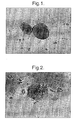
- Figure 1 is a photograph magnified 100 times (x100) showing blood cells released from islet
after 24 hour incubation at 37°C;
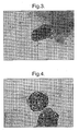
- Figure 2 is a photograph (x100) showing the Langerhans islets which are cultured in the
absence of radical scavengers;
- Figure 3 is a photograph (x100) showing the Langerhans islets which are cultured in the
presence of a radical scavenger;
- Figure 4 is a photograph (x200) showing islets which were incubated in a supplemented
medium (VeGF, IGF-1, IGF-1);
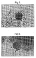
- Figure 5 is a photograph showing the islets on the second day of proliferation;
- Figure 6 is a photograph (x100) showing the lateral growth of the Langerhans islets which
are proliferated in the medium containing anti-integrin β1 antibody on the 3rd or 4th day;
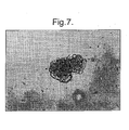
- Figure 7 is a photograph (x100) showing an islet cultured in the presence of
migrating factors, for example HGF, showing horizontal growth or spreading of the
islet, on the 3rd or 4th day;
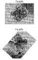
- Figures 8A and 8B are photographs (x320) showing the islet growing in all
directions after proliferating for 10 days according to the method of the present
invention;
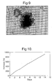
- Figure 9 is a photograph showing a human islet proliferated in vitro for a
period of 8 days in the presence of human serum;
- Figure 10 is a graph showing the change in the DNA amount of the
Langerhans islets with culturing time in days;
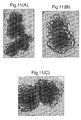
- Figures 11A, 11B and 11C show the process of fibroblast development and
its depletion from the islets. Many fibroblasts grew out from islet during islet
proliferation (Fig. 11A) even though the islets looked pure when they were collected.
The fibroblasts were almost all removed (Fig. 11B) and completely removed (Fig.
11C) from the islets;
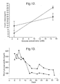
- Figure 12 shows the in vitro functions (glucose-response) of fresh (square)
and proliferated (circle) islets;
- Figure 13 is a graph in which the levels of glucose in the blood of the diabetic
rats which were transplanted with 1350 and 1650 islets just isolated, were measured
and were plotted against time in days;
- Figure 14 shows blood glucose level profile of STZ-induced diabetic rat
transplanted with 500 proliferated rat islets with days;
- Figure 15 is a graph of blood glucose (mg/dl) levels against time in days
comparing 500, 600, 800, 1000 and 1200 fresh rat islets transplanted into the spleen
of STZ-induced diabetic mice, indicating that 800 islets are the minimum islet
number required to recover and maintain normoglycaemia;
- Figure 16 is a photograph (x200) of a fresh islet transplanted into the spleen
of an SZT-induced diabetic nude mouse;
- Figure 17 shows the blood glucose profile for 150 to 200 proliferated islets
transplanted into the spleens of STZ-induced diabetic mice against time in days; and
- Figure 18 is a photograph (x400) showing a proliferated islet transplanted
into the spleen of STZ-induced diabetic nude mouse.
-
-
To better illustrate the present invention Figs. 1 through 18 are presented and
described below. Fig. 1 shows the islet cells with released red blood cells after being
incubated for 24 hours at 37°C in a medium of rat serum. Figs. 2 and 3 show the
difference when culturing in the absence of and the presence of radical scavengers,
respectively. Many islets cells were dead on the surface of the islet in the absence of
a radical scavenger (Fig. 2). The islets look intact without dead cells on the surface
in the presence of a radical scavenger (Fig. 3). Fig. 4 shows islets which were
incubated in a medium supplemented with VeGF, IGF-1 AND IGF-2 to increase islet
viability. The supplements function as anti-apoptotic and anti-necrotic factors. The
islets are intact without dead cells on the surface. Fig. 5 is a photograph showing the
islets on the second day of proliferation with the islets having a smooth surface and
high viability. The medium used in Fig. 6 included anti-integrin β1 antibody and
shows growing cells appearing on the surface of the islet, which then looks like a
mulberry. Fig. 7 shows the effect of adding a migrating factor(s), for example
hepatocyte growth factor (HGF), to the medium which appears to cause the islet to
spread horizontally. Figs. 8A and 8B show that the medium used in the present
invention enables the islets to grow in all directions. The front shape (Fig. 8A) and
the lateral shape (Fig. 8B) show thick and long buds from the islet. Fig. 9 shows a
human islet proliferated in vitro for 8 days using rat islet proliferation methods of the
present invention. Fig. 10 shows the increase in DNA content of in vitro
proliferating Sprague-Dawley (SD) rat islets against the number of days incubated.
Figs. 11A shows fibroblasts growing out from pure islets during proliferation (x100
magnification), and Figs. 11B and 11C show the fibroblasts removed prior to
transplantation (x200 magnification). Fig. 12 shows glucose-stimulated insulin
secretion from freshly isolated
(square) and proliferated (circle) islets. Fig. 13 is a graph of blood glucose (mg/dl) plotted against
time in days for 1650 (circle) and 1350 (triangle) fresh rat islets transplanted into the liver of a STZ
induced diabetic rat via the hepatic portal vein. Fig. 14 is a graph of blood glucose levels (mg/dl)
plotted against time in days, 0 being the day of transplant of 500 proliferated rat islets into four (4)
rats. Fig. 15 is a graph of blood glucose (mg/dl) levels plotted against time comparing 500, 600, 800,
1000 and 1200 fresh rat islets, respectively, transplanted into the spleen of STZ-induced diabetic
mice. The numbers represent the number of islets transplanted islet. 800 islets are the minimum islet
number required to recover and maintain normoglycaemia. The blood glucose levels of the diabetic
rats which were transplanted with the Langerhans islets just isolated, were plotted against time in
days.
-
Fig. 16 is a photograph (x 200) of a fresh islet transplanted into the spleen of an SZT-induced
diabetic nude mouse. The blood glucose concentration recovered and was maintained for 30 days.
Then the mouse was sacrificed to collect transplanted islet. The islet was sectioned and stained in
an insulin-specific staining method to identify the islet.
-
Fig. 17 is a graph of blood glucose levels (mg/dl) plotted against time in days comparing
different numbers (200, 180, 150) of proliferated islets transplanted into spleens of STZ-induced
diabetic mice. Blood glucose levels were measure every other day for several months. The mice used
in this experiment had no working immune system; however, in two mice, one with an implanted islet
number of 150 and 180, respectively, it is believed that the immune system functioned to the extent
that it attacked the implanted islets resulting in the sudden increase in blood glucose level at around
day 25.
-
Fig. 18 is a photograph (x400) showing a proliferated islet transplanted into the spleen of
STZ-induced diabetic nude mouse. The mouse recovered and remained normoglycaemia for 3
months. Then the mouse was sacrificed to remove the spleen from the mouse. The spleen was
sectioned and stained in insulin-specific staining method to identify the transplanted islet.
-
Before culturing, the Langerhans islets isolated from the pancreas may be stored. Also, after
culturing the proliferated Langerhans islets must be properly stored unless they all are used
immediately. To this end, they are preferably frozen in liquid nitrogen.
-
Dimethyl sulfoxide (DMSO) is useful to protect the Langerhans islets upon freezing. At a
convenient or proper time, the frozen stock of the Langerhans islets is thawed for culturing.
-
In accordance with the present invention, a culture system contains a matrix material in order
to provide a three dimensional environment for Langerhans islets. Under this circumstance, the
Langerhans islets show a high degree of proliferation. For the matrix material, collagen, complex
collagen, tail complex collagen or other biogels (e.g. Matrigel®), or the like, may be used.
-
The culture medium of the present invention (rat serum for rat islets and human serum would
be used for human islets) also contains a radical scavenger which plays the role of protecting the
proliferated cells from radical damage. Nicotinamide, mannitol or a superoxide dismutase is added
to the culture medium as a radical schvenger.
-
As mentioned above, the proliferation rate necessary for transplantation must amount to at
least 500%. For this, the isolated Langerhans islets are cultured in the presence of growth factors,
and cell migrating/scattering factors. Suitable growth factors are selected from the group consisting
of insulin transferrin selenite (ITS), epidermal growth factor (EGF), platelet derived growth factor
(PDGF), thrombin, progesterone, Linoleic Acid-BSA, pituitary extract and hydrocortisone.
Examples of cell migrating/scattering factors include hepatic growth factor (HFG) and tumor
promoting activator (TPA).
-
During culturing, the blood cells are go from the Langerhans islets into the medium. As the
culturing goes on, the blood cells are subjected to necrosis. Since the blood cells have MHC class
II, a major factor which causes the immune rejection upon tissue transplantation, the cultured
Langerhans islets can be transplanted in a host with little immune rejection, if any. In the present
invention, a cytoskeleton activator is added in the culture medium to enhance islet proliferation.
Preferably, anti-integrin β1 antibody is used for this purpose.
-
A better understanding of the present invention may be obtained in light of the following
examples which are set forth to illustrate, but should not be construed to limit the present invention.
EXAMPLE I : FREEZE STORAGE AND THAW OF LANGERHANS ISLETS
-
1 ml of 10% fetal calf (bovine) serum (FCS) medium which contained 2,000 (rat) Langerhans
islets was added to 0.5 ml of 2M DMSO and allowed to stand at room temperature for 5 min. The
procedure of adding DMSO and standing at room temperature was further repeated twice: for the
first time, with 0.5 ml of 2M DMSO and a period of 25 min., and for the second time, with 0.5 ml
of 3M DMSO and a period of 15 min. Thereafter, the medium was allowed to stand for 5 min on ice
and then, for 15 min at -7.5 C. This medium was subjected to nucleation using previously chilled
forceps and allowed to stand for 15 min at -7.5°C. The temperature was lowered down to -35 - -40
° C at a rate of 0.2 -0.3 per min. Once reaching the temperature, the vial containing the medium was
stored in a liquid nitrogen tank.
-
For thawing, the vial stored in the liquid nitrogen tank was transferred to a cryovial in a water
bath of 37°C until the ice crystals started to thaw. Then, the vial was placed on ice and allowed to
stand in order that the Langerhans islets may settle on the bottom. The supernatant was drained out
using a Pasteur pipette. The Langerhans islets were added with 10% FCS supplemented with 1 ml
of 0.75 M sucrose and allowed to stand on ice for 30 min. Then, the Langerhans islets were added
with 1 ml, 2 ml, 4 ml and 8 ml of 10% FCS, successively. After every addition, the Langerhans islets
were allowed to stand for 5 min at room temperature. The resulting supernatant was removed and
the remaining Langerhans islets were re-suspended in a culture medium and cultured at 37°C. This
procedure may be used for rat, mouse, human, and the like, Langerhans islets.
EXAMPLE II: PROLIFERATION OF THE LANGERHANS ISLETS
-
In this Example, the Langerhans islets which were frozen and thawed as in Example I were
used. However, freshly isolated langerhans islets could also be used as is appreciated by one skilled
in the art. During incubation 5% CO2 is added to ambient air.
Serum preparation
-
Rat serum was added to medium for proliferating the rat islets. 2-5 ml blood was obtained
from a rat and transferred into sterile 15 ml Falcon tube. The blood was left for 2-4 hours at room
temperature and then centrifuged at 3000g for 10 minutes. The supernatant was transferred to 1.5-2ml
tubes, which were left overnight at 4°C and then centrifuged again in the same way. The
supernatant was stored at -20°C until use. Human islets were proliferated in the medium containing
10% human serum instead of rat serum that was made in the same way.
-
The basal medium (50ml) is prepared by mixing together:
- 1.1 mg (100µl) pyrubate, (Gibcobrl); 025µg (100µl) hydrocortisone (Sigma);
100units/(100µl) (1ml) pencillin/streptomycin; 4.456mg (4µl) B-mercaptoethanol (Sigma); 14.6mg
L-glutamate (Gibco); 238.3 (1ml) Hepes Buffer (Gibco); 100mg (11mM) glucose (Sigma) plus
DMEM to make a volume of 50 ml.
-
Experiment Example II-I: Culturing of Langerhans islets under basic conditions
-
First Day: 500 islets were cultured under basic conditions. The freshly isolated islets were
incubated overnight at 37°C in 6 ml basal medium supplemented with 600 µl rat serum (10%), 30µg
of insulin-transferrin-sodium selenite (ITS) (5µg/ml, I-1884, Sigma USA), 6mg of Linoleic Acid-BSA
(1mg/ml, L-8384, Sigma USA), 60ng (platelet-derived growth factor) PDGF (10ngm/ml P8147
Sigma USA), 600ng thrombin (100ngm/ml T-4393 Sigma USA), 60ngm (epidermal growth factor)
EGF (10ngm/ml E-1264 Sigma USA), 187.2mg of 10-4 M superoxide dismutase (product no. S-9636,
Sigma, USA), 109.32 µg of 10-4 M mannitol (M-9546 Sigma USA), 61mg (3mM) nicotinamide (N-0636
Sigma USA), 12ng (vascular endothelial growth factor) VeGF (2ngm/ml V-7259 Sigma USA),
600ngm (insulin-like growth factor-I and -II) IGF-1 and IGF-2 (I-3769 and I-213P, respectively,
Sigma USA).
Experiment Example II-II
Second Day:
-
- 1. The 500 islets were incubated for 45 ∼ 120 minutes at room temperature with 50ng
(25ng/ml) of anti-itegrin B1 antibody (cat. No. I-41720 Transduction Labs. USA) in 2ml DMEM
medium or serum free basal medium.
- 2. The islets were then suspended in 200µl of 80 to 100% Matrigel® (Collaborative
Biomedical Product, USA) in order to provide a three-dimensional growth environment.
- 3. Added 2ml of the above supplemented basal medium and which was also supplemented
with 100µg pituitary extract (50µg/ml P-1167 Sigma, USA) and cultured for 1-2 days at 37°C.
-
Experiment Example II-III
Third or Fourth Day:
-
The culture medium was replaced with fresh supplemented basal medium having the same
composition as that of the first day except that VeGF is not added and, depending on the islet shape,
other factors were added to the medium, as follows:
- 1. 20ng (nerve growth factor) NGF (10ng/ml N-6009 Sigma, USA), 50ng HGF (25ng/ml
Collaborative Biomedical Product, USA Lot 902287), were added to 2ml of the supplemented basal
medium if the islets became thick and dark in the center of the islet and then the islets were cultured
in the medium for 1 or 2 days, or
- 2. 20ng NGF and 100ng anti-integrin β1 antibody (50ng/ml)) were added to 2 ml of the
supplemented basal medium if the islets looked spread out and then cultured for 1 - 2 days.
-
-
Observation with a microscope at x200 magnification was shown in Figure 9.
-
After the Sixth or Seventh day, the islets culturing sequence was performed as follows:
Seventh Day
-
The islets were collected from the 3-dimensional gel. The fibroblasts were removed using
diapase by the process described below. The collected islets were cultured overnight in a floating
culture, i.e. the islets were not fixed in a medium such as a gel medium. The culturing medium was
6 ml of the basal supplemented medium, as described above, supplemented with 600 µl of rat serum
(10%), 30µg of insulin-transferrin-sodium selenite (ITS), 6mg of Linoleic Acid-BSA (1mg/ml),
600ng thrombin (100ngm/ml T-4393 Sigma USA), 60ng EGF, 101ng nicotinamide, 12ng VeGF,
600ng IGF-1, 600ng IGF-2, , 187.2mg of 10-4 M superoxide dismutase, (Sigma), 109.32 µg of 10-4
M mannitol.
Eight Day
-
- 1. The islets are incubated in fresh DMEM supplemented with 50ng of anti-itegrin B1
antibody for 45 ∼ 120 minutes at room temperature.
- 2. The islets were suspended in Matrigel® to grow in a 3-dimensional environment.
- 3. 2ml of the basal supplemented medium was added plus 100µg pituitary extract and were
cultured for 1-2 days at 37°C.
-
Ninth or Tenth Day
-
- 1. Basal supplemented medium added as first prepared, except that VeGF is not used.
- 2. Depending on the islet shape, the islets were cultured for 2 - 3 days in the above medium
plus the added factors as described in the Third or Fourth Day culture medium.
-
Fourteenth Day
-
- 1. The islets were collected from the gel by using dispase as described below and as done at
around the Seventh day. This step is mainly to release the islets from the gel. However, if any
fibroblasts were to remain, they also would be removed from the surface of the islet.
- 2. The islets were incubated at 37°C in a floating culture (islets not fixed as in a matrix) in
a medium having the same constitutional amounts and components as in the 1st day. These islets may
be transplanted but are preferably subjected to removal as described below.
-
Experiment Example III:
-
During culturing the amounts of DNA of the Langerhans islets were measured every other
day and their relative amounts were plotted for culture times on the basis of the day on which the
Langerhans islets were isolated. As seen in Fig. 10, the Langerhans islets proliferated up to about
1,000% on the 14th day after culturing.
EXAMPLE IV : Test for in vivo function of the Langerhans Islets
-
Experiment Example IV-1: Blood glucose level of diabetic rats transplanted with freshly
isolated Langerhans islets.
-
For this, streptozotocin (STZ) was intra peritoneally injected to the rats at a dosage of 53-55
mg per kg of body weight and the level of glucose in the blood was checked everyday for two weeks.
If the blood glucose level reached 250 ng/mg, the Sprague-Dawley (SD) rats were judged to be
diabetic.
-
5.5 ml collagenase X1 (0.7mg/ml type X1 Sigma, USA) was introduced to the pancreases of
healthy rats by injection via the common bile duct to distend the pancreases and to digest them for
17 min. at 370C.
-
After the digested pancreas was washed three times with Dulbeco modified Eagle medium
(DMEM), the Langerhans islets were isolated therefrom and collected in a discontinuous bovine
serum albumin (BSA) concentration gradient method. The collected Langerhans islets were added
with DMEM to a final volume of 100-150 µl.
-
With the aid of 1 ml syringes, the resulting solution was injected into the hepatic portal vein
of the diabetic rats anesthetized by intra peritoneal injection of Entobal (Hanlim Pharmacy Co. Ltd,
Korea) at a dosage of 60 mg per kg body weight.
-
1650 (islet equivalents (IEQ) 2750) and 1350 (IEQ 2400) fresh Wistarkyoto (WK) rat islets
were transplanted into the livers of 175g streptozotocin-induced diabetic rats via hepatic portal vein.
1650 fresh islets are enough mass to recover and maintain normoglycaemia but 1350 fresh islets are
not enough mass to recover and maintain normolgycaemia, see Figure 13. Therefore, 1650 fresh
islets are minimum required islet number.
Experiment Example IV-II: Blood glucose level after transplantation of in vitro proliferated
rat islets
-
The procedure of Experiment Example III-1 was repeated using the same Langerhans islets as
those of Example II, which were proliferated in a medium supplemented with collagen, radical
scavengers, cell migrating/scattering factors, growth factors and anti-integrin β 1 antibody.
-
The blood glucose levels of the rat hosts transplanted with 500 proliferated rat islets were
measured and plotted with times. The measurements of the blood glucose level are shown in Figure
14. As seen, 500 proliferated Langerhans islets completely functioned to recover and maintain
normoglycaemia, so that enough insulin was secreted in proper response to the blood glucose levels
of the hosts.
Experiment Example IV-III:
-
Blood glucose concentration profile of STZ-induced diabetic mice transplanted with different
number of fresh rat islets.
-
Different numbers of fresh rat islets were transplanted into spleen of STZ- induced diabetic
nude mice to determine minimum number of islets required to recover and maintain normoglycaemia.
The blood glucose levels of the most mice were measured and plotted wisth respect to time. The
results are given in Figure 15. The figure shows the profile of blood glucose levels against time in
days. At least 800 fresh islets are required to recover and maintain normoglycaemia.
-
The blood glucose level profile of STZ-induced diabetic mice of Figure 15 is based on STZ-induced
diabetic mice transplanted with proliferated rat islets.
-
The Langerhans islets isolated from Sprague-Dawley (SD) rats were proliferated in the same
medium as in Example II.
-
About 150 - 200 proliferated rat islets were transplanted into the spleen of STZ induce
diabetic nude mice. The 150 - 200 proliferated rat islets are sufficient in number to recover and
maintain normoglycaemia.
-
The islets were transplanted into the spleen of STZ-induced diabetic nude mice. The blood
glucose levels of the host mice were measured and plotted against time in days. The results are given
in Figure 17. As compared with Figure 15, in vitro proliferated (for 5-6 days) islets showed 3-5 fold
higher in vivo function than fresh islets.
-
As described hereinbefore, the Langerhans islets, whether they are just isolated from
pancreas or thawed from a frozen state, can be proliferated in volume upon culturing in a species
similar serum (i.e. rat serum or human serum for rat or human Langerhans islets, respectively)
medium further supplemented with radical scavenger, collagen, growth factors and cell
migrating/scattering factors. Further, the Langerhans islets which are cultured for a long time in
the supplemented medium, are depleted of blood cells, so that they can be transplanted and function
well enough to recover and maintain normoglycaemia.
Experiment Example V: In vitro function (glucose response) of proliferated islets.
-
Fresh and proliferated rat islets were incubated with 2.7 and 16.7 mM glucose for 1 hour
and secreted insulin concentration were measured by using 1251 -insulin KIT.
-
Proliferated islets respond nearly null to low glucose concentration and very strongly to high
glucose concentration; whereas, fresh islets respond higher to low glucose, lower to high glucose
concentration than proliferated islets (as see Fig 12). The results indicated that in vitro proliferated
islets have more desirable functional properties than fresh islets, therefore proliferated islets can be
used as autotransplantation material.
Experiment Example VI: Fibroblast removal
-
Fresh islets are likely to be contaminated with fibroblasts even though they are collected in
a pure state as can be seen by the fact that many fibroblasts grow out from the islets during the
proliferation. The fibroblasts are removed completely prior to islet transplantation, as seen in Fig.
11C. Islets are dispersed in a gel (eg. Matrigel®) during proliferation. At about the seventh or
fourteenth day, the islets are collected from the gel and 400 µl of dispase (Collaborative Biomedical
Products, USA, Cat. 40235) is added and incubated for about 10 minutes at 37°C. Then the islets
are aspirated back and forth several times causing the gel acted on by the diapase to be removed from
the islets which exposes the fibroblasts to the force created during the back and forth aspiration
causing the fibroblasts to become separated from the surface of the islets to prepare fibroblast free
islets.
-
Islets proliferated up to 5- and 10-fold for the first and second week during in vitro
culturing, respectively. These proliferated islets showed a more desirable in vitro glucose-response
pattern than fresh islets. Their transplantation results showed that those islets have a 3∼4-fold higher
capability to recover and maintain normoglycaemia than fresh islets. These data suggest that in vitro
proliferated islets are a better source than fresh islets for transplantation to treat diabetes.
Consequently, the present invention can produce a large quantity of the Langerhans islets and, thus,
transplantation using the proliferated islets according to the present invention is a promising
therapeutic means for the treatment of diabetes.
Experiment Example VII: Islet autotransplantation-stimulated islet regeneration.
-
Islet autotransplantation recovered and maintained normoglycaemia. In the diabetic rats,
islet neogenesis did appear. Exogenous insulin injection also did not stimulate islet neogenesis.
However, islet autotransplanation stimulates islet neogenesis.
-
The following table shows insulin content, number, size of islets collected from rats and mice
which were either pancreatectomized (
columns 2 and 3) or transplanted into diabetic mice/rats after
transplantation (
columns 4, 5 and 6). The islets used were either fresh (
columns 4 and 5) or
cultured/proliferated for 6 days (column 6) according to the process of the present invention. The
transplantation (TX) was islet auto-transplantation. The results in
columns 5 and 6 indicate that the
transplanted islets, either fresh or proliferated according to the present invention result in islet
regeneration in the pancreas.
The following experimental results prove this (see Table, above)
- 1. 1350 fresh rat islets were transplanted into the liver of a STZ-induced diabetic rat via
hepatic portal vein. Blood glucose concentration declined gradually to be normoglycaemia
35 days later. The values lingered in the 200's for 20 days and finally normoglycaemia for
1-2 days, as seen in Fig. 13. Islets were collected from the pancreas and stained deeply with
dithizone.
- 2. 1650 fresh rat islets were transplanted syngeneically into the liver of a STZ-induced diabetic
rat. The rat recovered normoglycaemia and maintained it for 25 days, as seen in Fig. 13.
After sacrificing, 226 (IEQ 168) small islets were collected and were not stained with
dithizone. The pancreas islets were immature and could be produced by neogenesis. In
conclusion, on comparing both cases, islet autotransplantation-recovered normoglycaemia
seems to provide a favorable environment for islet neogenesis and replication by secreting
various natural substances.
- 3. The rats transplanted syngeneically with 500 proliferated islets maintained normoglycaemia
for 13 months, as see Fig. 14. 164 large islets (IEQ 264) were collected and stained deeply
with dithizone. The results indicates that neogenic (pancreas) islets grew up to large mature
islets over a long period of time and islet autotransplantation enhances islet regeneration and
stimulates islet maturation.
- 4. 90% pancreatectomy rat maintained normoglycaemia for 2 months. 235 large islets were
collected from the remaining pancreas. The islets were stained deeply with dithizone. They
seemed to originate from islet regeneration and grew for 2 months.
- 5. 90% pancreatectomy rat maintained normoglycaemia for 2 days. Islets were collected from
the remaining pancreas.
- 6. In 90% pancreatectomy rat, 113 (IEQ 223) islets ere collected from the remaining head part
of the pancreas on the day of the pancretectomy. On the basis of the pancreatectomy
results, islet regeneration rate and size depends on the magnitude of islet deficiency.
-
-
Islet proliferation research is performed for the ultimate purpose of treating a diabetic patient
by islet autotransplantation by providing a sufficient number of islets for transplant. The present
invention provides the required number of islet since it enables successful islet proliferation in vitro.
That is, one of the major reasons for the failure of transplantation is an insufficient number of islets
are available for transplant into a diabetic patient. This problem is overcome by the present invention.
-
While the present invention was developed using rat and mice islets, the application of the
present invention to proliferate human islets is within the scope of the teachings of the present
invention, as can be appreciated by those skilled in the art.
-
The present invention has been described in an illustrative manner so as to be easily
understood by one skilled in the art. This description, examples and illustrations are intended to be
in the nature of description rather than of a limitation, as appreciated by those skilled in this art. Many
modifications and variations of the present invention are possible in light of the above teachings.
Therefore, it is to be understood that within the scope of the appended claims, the invention may be
practiced otherwise than as specifically described.