FIELD OF THE INVENTION
-
The present invention relates to methods of testing for bronchial
asthma or chronic obstructive pulmonary disease (COPD).
BACKGROUND OF THE INVENTION
-
Currently, there are more than one hundred million bronchial
asthma patients in the world. The rapid increase in the number of
asthma patients is a social problem in Japan as well. In advanced
countries, the number has increased by 20-50% in the past decade.
Thus, asthma is thought to be one of the diseases that would pose
a major health threat in the 21st century.
-
Pharmaceuticals used today for treating asthma and candidate
pharmaceuticals for that purpose, include: inhaled steroids and oral
steroids; agents that suppress the release of inflammatory mediators;
anti-allergy agents such as histamine H1 antagonists; β2 agonists that
act as bronchodilators; and immunosuppressive agents. According to
a report describing clinical cases in New Zealand, the widespread
use of inhaled steroids and β2 agonists has decreased the mortality
rate of patients by 30% compared to 10 years ago. However, both
inhaled steroids and β2 agonists have been reported to have side
effects. The side effects of inhaled steroids include oral and
esophageal candidiasis, olfactory disorders, adrenal suppression,
osteoporosis, cataract, glaucoma, skin thinning, and growth
inhibition in children. Side effects of β2 agonists include ischemic
diseases, hyperthyroidism, and diabetes mellitus. In addition,
regular use of β2 agonists has been known to reduce the efficacy of
these drugs.
-
Bronchial asthma is characterized by respiratory inflammation
and airflow obstruction resulting from various degrees of respiratory
stenosis. Representative symptoms include paroxysmal cough and
difficulty in breathing. The degree of airflow obstruction in
bronchial asthma ranges from relatively mild to life-threatening
obstructions. Furthermore, it has been reported that allergic
reactions in the mucous membrane of the respiratory tract and
bronchial smooth muscles are closely involved in bronchial asthma
development.
-
Specifically, an atopic disposition accompanied by
hyperproduction of IgE antibodies is seen in many bronchial asthma
patients. Many causes are thought to lead to bronchial asthma, but
there is no doubt that an atopic disposition is one cause of
hypersensitivity in many patients. It is predicted that contraction
of bronchial smooth muscles, edema of the respiratory tract mucous
membrane, or respiratory tract hypersecretion is involved in the
mechanism of respiratory obstruction in an asthma attack. Type-I
allergic reactions in the respiratory tract due to exposure to
pathogenic allergens play an important role in such changes in the
respiratory tract.
-
In bronchial asthma patients, the activity of Th2 helper T cells
is enhanced, and so is the production of Th2 cytokines such as
interleukin-3 (hereinafter abbreviated as "IL-3"; similarly,
interleukin is abbreviated as "IL" ) , IL-4, IL-5 , IL-13 and granulocyte
macrophage colony stimulating factor (GM-CSF), and chemokines such
as eotaxin and RANTES. IL-4 and IL-13 have the activity of inducing
IgE production, and IL-3 and IL-4 have the activity of inducing the
proliferation of mast cells. Eosinophils that differentiate and
proliferate by IL-5 and GM-CSF infiltrate into the respiratory tract
by the action of eotaxin and RANTES (Allergy Asthma. Proc. 20: 141
(1999)).
-
Eosinophils that infiltrate into the respiratory tract release
intracellular granule proteins such as activated major basic protein
(MBP) and eosinophil cationic protein (ECP) as a result of
degranulation (Compr. Ther. 20: 651 (1994)). These granule proteins
exhibit cytotoxic activity, and thus, ablate and damage epithelial
cells. The ablation of epithelial cells results in the exposure of
sensory nerve endings, enhances the permeability of the epithelium,
and causes the loss of the epithelium-derived smooth muscle relaxing
factor. Furthermore, eosinophils are known to secrete leukotriene
C4 (LTC4) and Platelet activation factor (PAF), which have the
activity of enhancing bronchial smooth muscle constriction, and
platelet activating factor (PAF). It has been suggested that these
reactions are repeated in the body and become chronic resulting in
bronchial wall thickening and respiratory hypersensitivity.
-
Specifically, several reports have suggested the deep
involvement of IL-4 and IL-13 in allergic reactions. For example,
it is known that respiratory hypersensitivity disappears in
IL-4-knockout mice (Yssel, H. and Groux, H., Int. Arch. Allergy
Immunol., 121: 10-18, 2000). In a mouse model, IL-13 has been shown
to be involved in forming an asthma-like pathology regardless of IgE
production and the Th2 type (Wills-Karp, M. et al., Science, 2822:
2258-2261, 1998; Grunig, G. et al., Science, 282: 2261-2263, 1998;
Zhu, Z. et al. , J. Clin. Invest., 103: 779-788, 1999). In addition,
IL-4 receptors and IL-13 receptors are highly expressed in human
respiratory epithelial cells and bronchial smooth muscles (Heinzmann,
A. et al. , Hum. Mol. Genet., 9: 549-559, 2000). Accordingly, these
tissues are thought to be the targets of IL-4 and IL-13. On the other
hand, SNPs present in IL-4 receptor α and IL-13 have been shown to
be one of the genetic causes of allergic diseases (Mitsuyasu, H. et
al., Nature Genet., 19: 119-120, 1998; Mitsuyasu, H. et al., J.
Immunol. , 162: 1227-1231, 1999; Kruse, S. et al . , Immnol., 96: 365-371,
1999; Heinzmann, A. et al., Hum. Mol. Genet., 9: 549-559, 2000).
-
Furthermore, IL-4 and IL-13 have been reported to suppress the
expression of the β and γ subunits of amiloride-sensitive epithelial
sodium channel (ENaC) and increase the expression of cystic fibrosis
transmembrane conductance regulator (CFTR) in tracheal epithelial
cells. This suppresses Na+ release and enhances Cl- secretion. As
a result, water secretion is assumed to increase in the bronchial
lumen (Galietta L. J. V. et al., J. Immunol. 168: 839-45 (2002)).
Therapeutic agents that target the signaling molecules of IL-4 or
IL-13, such as IL-4 agonists, soluble IL-4 receptor α (Borish L. C.
et al. , Am. J. Respir. Crit. Care Med. 160: 912-22 (1999) ) , soluble
IL-13 receptor α2, anti-IL-13 antibodies, and anti-IL-4 antibodies,
have already been clinically applied and are expected to be effective
in treating bronchial asthma.
-
Inflammation in the respiratory tract is known to elevate the
expression levels of cytokines and adhesion molecules. Genes
encoding such cytokines and adhesion molecules, which participate
in the onset of allergic diseases such as bronchial asthma, can be
targets in drug discovery. Specifically, patients can be diagnosed
for the onset of symptoms, seriousness, response to medical treatments,
or such, by detecting variations in the expression levels of these
genes. Furthermore, patients can be treated using a substance that
controls the expression level of such genes or regulates protein
activity.
-
There are several commercially available expectorants for
removing sputum, the cause of death by suffocation in asthma. However,
until recently, available expectorant types were restricted to those
that contain an active SH group, and those that hydrolyze or lubricate
the mucus. However, "fudosteine" (a low-molecular-weight oral drug),
which was jointly developed by two Japanese pharmaceutical companies,
SS Pharmaceutical Co. Ltd.,and Mitsubishi Pharma Corporation, and
released last December, is a pharmaceutical agent having an activity
to suppress goblet cell hyperplasia.
-
In addition, Genaera Corporation in the United States has
reported that the hCLCA1 gene is closely associated with the
production of IL-9 and mucus in the mucosal epithelia in asthma
patients (J. Allergy Clin. Immunol. 109: 246-50 (2002) ) ; the hCLCA1
gene is the human counterpart of Gob-5 reported by Takeda Chemical
Industries LTD. , Japan (Proc. Natl. Acad. Sci. USA 98: 5175-80 (2001)).
Furthermore, clinical trials have already been launched for the
low-molecular-weight oral drug "LOMUCIN" that inhibits the function
of this gene.
-
In the bronchia of asthma patients, the aggravation of the
disease state induces differentiation of respiratory epithelial cells
into goblet cells and proliferation of these cells. Goblet cells
produce a huge glycoprotein called mucin. This protein contributes
to the production of sputum, which causes breathing difficulties and
is a leading cause of death in chronic bronchial asthma. The increase
in the number of goblet cells, which are secretory cells, enhances
secretions in the respiratory tract. Thus, such secreted material
enhances the obstruction of the respiratory tract and largely
contributes to the worsening of asthma symptoms. However, the
mechanism underlying goblet cell differentiation in the respiratory
epithelium is still unknown.
-
The term "chronic obstructive pulmonary disease" refers to
mainly pulmonary emphysema and chronic bronchitis. Shortness of
breath is a main symptom of pulmonary emphysema; cough and sputum
are main symptoms of chronic bronchitis. These are the major
subjective symptoms of respiratory diseases in aged patients. In
addition to aging, smoking is deeply involved in the onset of chronic
obstructive pulmonary diseases. In pulmonary emphysema, the walls
of pulmonary alveoli at the end of bronchioles are damaged and greatly
swollen; the elasticity and contractility of the walls are impaired,
and thus, the lungs have difficulty contracting during exhalation.
This often causes shortness of breath. In addition, bronchial
disorders result in bronchial obstruction, which is caused by swollen
mucous membranes, sputum, and such. In chronic bronchitis, chronic
inflammation and edema in the bronchia induce differentiation of
bronchial epithelial cells into goblet cells, which results in the
overproduction of secretory material. This results in coughs that
produce sputum. In chronic obstructive pulmonary diseases, narrowed
bronchia and damaged lungs cannot be restored to the original state.
Furthermore, there are about 220,000 and 1,400,00 patients with
chronic obstructive pulmonary diseases in Japan and the United States,
respectively, and the diseases are the fourth leading cause of death
in both countries. Thus, chronic obstructive pulmonary diseases are
quite serious.
-
There is a report suggesting the correlation between chronic
obstructive pulmonary diseases and IL-13 (Zheng T. et al, J Clin.
Invest.; 106, 1081-1093, 2000). According to this report, transgenic
mice in which respiratory epithelial cells were allowed to express
IL-13, developed pulmonary emphysema, inflammation, and goblet cell
hyperplasia.
SUMMARY OF THE INVENTION
-
As described above, in bronchial asthma or chronic obstructive
pulmonary diseases, changes in respiratory epithelial cells are
crucial factors constituting the disease states. One of the morbid
changes of respiratory epithelial cells is the differentiation into
goblet cells. An objective of the present invention is to identify
genes associated with the differentiation into goblet cells. Another
objective of the present invention is to provide diagnostic markers
for bronchial asthma and drug discovery targets.
-
Drugs suppressing the differentiation into goblet cells in
respiratory epithelial tissues were developed only recently. This
is a new approach in drug discovery. Once the mechanism underlying
the differentiation into goblet cells is elucidated, it may be
possible to establish a basic treatment for bronchial asthma.
Furthermore, agents that affect the process of goblet cell
differentiation are predicted to be useful in the treatment of
diseases involving inflammation and overproduction of mucus, such
as chronic obstructive pulmonary diseases, cystic fibrosis, chronic
sinusitis, bronchiectasis, diffuse panbronchiolitis, as well as
asthma.
-
A culture method (called the "air interface (AI) method") for
differentiating human respiratory epithelial cells into goblet cells
in the presence of IL-13 has been established by researchers of the
Department of Geriatric and Respiratory Medicine, Tohoku University
School of Medicine, Japan, who are collaborators in the present
invention. Using this method, the present inventors predicted that
goblet cell differentiation-associated genes can be identified by
elucidating which gene expression varies in respiratory epithelial
cells when stimulated by IL-13.
-
Conventionally, bronchial epithelial cells played a vital role
in studies concerning the transport of water and electrolytes in
humans and other animals. Moreover, particularly in humans, these
cells have been significant in clarifying disease states of
respiratory tract infections in cystic fibrosis and in establishing
therapeutic methods. Over the past two decades, methods for
culturing (in vitro) respiratory epithelial cells obtained from
protease-treated trachea tissues have been improved by improving
culture media and using growth-promoting substances. In addition,
the AI method has been established, in which cilia and secretory
granules can be produced in vitro by culturing cells under conditions
similar to the environment around respiratory epithelial cells in
vivo. In the AI method, the culture medium facing the mucous membrane
side (apical side) of the cells is removed exposing cells to air while
water and nutrients are supplied from the chorionic membrane side
(basolateral side) (Van Scott MR., Exp Lung Res, 11: 75-94, 1986,
Widdicombe JH., Am J Physiol, 258:L13-L18, 1990, Kim KC, J Biol Chem,
260: 4021-4027, 1985, Adler KB, Am J Respir Cell Mol Biol, 2:145-154,
1990).
-
Human bronchial epithelial cells cultured in the presence of
human IL-13 using the air interface method were reported to express
TGF-α (Booth BW, Adler KB, Bonner JC, Tournier F, Martin LD.
Interleukin-13 induces proliferation of human airway epithelial cells
in vitro via a mechanism mediated by transforming growth factor-α.
Am J Respir Cell Mol Biol . 2001 Dec; 25(6): 739-743). In addition,
the ion transport ability of human bronchial epithelial cells has
been evaluated in a previous report, in which cells were cultured
by the air interface method in the presence of IL-13 (Danahay H, Am
J Physiol Lung Cell Mol Physiol, 282:L226-L236, 2002). However,
these reports make no reference to goblet cell differentiation, and
have not conducted any exhaustive gene expression analyses.
-
Furthermore, bronchial epithelial cells of guinea pigs has been
reported to differentiate into goblet cells when cultured in the
presence of human IL-13 for 14 days using the air-liquid interface
method (Kondo, M., Tamaoki, J., Takeyama, K., Nakata, J. and Nagai,
A. Interleukin-13 induces goblet cell differentiation in a primary
cell culture from Guinea pig tracheal epithelium. Am J Respir Cell
Mol Biol 27,536-541, 2002). However, there are no reports on
exhaustive analyses of genes expressed in human bronchial epithelial
cells cultured by the method described above.
-
On the other hand, the present applicants have identified eight
types of allergy-associated genes whose expression levels decrease
upon IL-4 or IL-13 stimulation in several lots of primary human
respiratory epithelial cell cultures (Unexamined Published Japanese
Patent Application No. (JP-A) 2002-191398). The applicants have also
identified six types of allergy-associated genes whose expression
levels greatly increase in several lots under the same conditions
as described above (WO 02/052006 A1). The gene expression analyses
in these two previous patent applications were carried out using a
conventional culture method which induces no goblet cell
differentiation.
-
Using oligonucleotide microarrays (GeneChip®, Affymetrix,
Inc.) and air interface method, the present inventors compared the
expression profiles of genes expressed in respiratory epithelial
cells stimulated with IL-13 for goblet cell differentiation, with
those of cells not stimulated with IL-13. The inventors selected
genes whose expression levels increased by two folds or more or
decreased by half or more of the initial levels as a result of the
differentiation, and determined the expression levels of the genes.
Then, the inventors confirmed the variation of the expression level
of marker genes selected from the group described below in (a) or
(b).
-
Furthermore, with respect to the mouse homologs of the human
genes selected by the method described above, the inventors detected
variations in the expression levels in respiratory hypersensitivity
model mice. As a result, the variation pattern of expression levels
of the mouse homologs coincided well with that of human genes.
-
The nucleotide sequences of the respective marker genes listed
in (a) and (b) are known. The functions of the proteins encoded by
each marker gene are described in the references listed in the
"References" section in Tables 3-19 (increased) and Tables 20-36
(decreased) below. The nucleotide sequences of the mouse homologs
of the marker genes of the present invention are also known. The
functions of the proteins encoded by the mouse homologues of the
respective marker genes are described in the references listed in
the "References" section in Tables 40-62 (increased) and Tables 63-83
(decreased) below.
-
Among these groups of genes, some genes have been reported to
be directly related to bronchial asthma. However, most of the genes
have not been shown to be associated with an allergic disease.
Furthermore, even for genes that are reported to be associated with
bronchial asthma, there are no reports that focus on the aspect of
combinations with other co-expressing genes whose expression levels
vary at the same timing that the asthma-related genes do.
-
A close relationship between bronchial asthma symptoms and the
marker genes of the present invention is suggested by the finding
that the expression levels of marker genes vary in the differentiation
process of respiratory epithelial cells into goblet cells. The
relationship between the allergic response of the respiratory
epithelium and the marker genes of the present invention was verified
by the fact that the variation pattern of the expression levels of
mouse homologs in the respiratory hypersensitivity mouse model is
consistent with that in humans. Based on the findings described above,
the present inventors revealed that tests for bronchial asthma or
chronic obstructive pulmonary disease and screenings for therapeutic
agents can be achieved by using as a marker the expression level of
each marker gene or the activity of the protein encoded by each marker
gene.
-
Specifically, the present invention relates to the following
methods of testing for bronchial asthma or chronic obstructive
pulmonary disease and the following methods of screening for candidate
compounds for treating bronchial asthma or chronic obstructive
pulmonary disease:
- [1] a method of testing for bronchial asthma or chronic
obstructive pulmonary disease, which comprises the steps of:
- (1) determining the expression level of a marker gene in a
biological sample from a subject;
- (2) comparing the expression level determined in step (1) with
the expression level of the marker gene in a biological sample from
a healthy subject; and
- (3) judging the subject to have bronchial asthma or chronic
obstructive pulmonary disease when the result of the comparison in
step (2) indicates that (i) the expression level of the marker gene
in the subject is higher than that in the control when the marker
gene is a gene according to (a) or (ii) the expression level of the
marker gene in the subject is lower than that in the control when
said marker gene is a gene according to (b);
wherein the marker gene is any one selected from the group
according to (a) or (b) :
- (a) a group of genes whose expression levels increase when
respiratory epithelial cells are stimulated with interleukin-13, and
comprise any one of the nucleotide sequences of SEQ ID NOs: 25 to
310;
- (b) a group of genes whose expression levels decrease when
respiratory epithelial cells are stimulated with interleukin-13 and
comprise any one of the nucleotide sequences of SEQ ID NOs: 311 to
547;
- [2] the testing method according to [1], wherein the biological
sample is a respiratory epithelial cell;
- [3] the testing method according to [1], wherein the gene
expression level is measured by PCR analysis of the cDNA;
- [4] the testing method according to [1], wherein the gene
expression level is measured by detecting the protein encoded by the
marker gene;
- [5] a reagent for testing for bronchial asthma or chronic
obstructive pulmonary disease, wherein the reagent comprises a
polynucleotide comprising the nucleotide sequence of a marker gene,
or an oligonucleotide having at least 15 nucleotides and comprising
a nucleotide sequence complementary to the complementary strand of
the nucleotide sequence of the marker gene, and wherein, the marker
gene is any one selected from the group according to (a) or (b) in
[1];
- [6] a reagent for testing for bronchial asthma or chronic
obstructive pulmonary disease, wherein the reagent comprises an
antibody that recognizes a protein encoded by a marker gene, and
wherein the marker gene is any one selected from the group according
to (a) or (b) in [1];
- [7] a method of screening for a therapeutic agent for bronchial
asthma or chronic obstructive pulmonary disease, wherein the marker
gene is any one selected from the group according to (a) or (b) in
[1], and wherein the method comprises the steps of:
- (1) contacting a candidate compound with a cell expressing the
marker gene;
- (2) measuring the expression level of said gene; and
- (3) selecting a compound that decreases the expression level
of a marker gene belonging to group (a) or increases the expression
level of a marker gene belonging to group (b), as compared to that
in a control with which the compound has not been contacted;
- [8] the method according to [7], wherein the cell is a respiratory
epithelial cell or a goblet cell;
- [9] the method according to [8], which comprises the step of
culturing the respiratory epithelial cells under conditions in which
culture medium is removed from the apical side of said cells and the
culture medium is supplied from the basolateral side of the cells;
- [10] a kit for screening for a candidate compound for a
therapeutic agent to treat bronchial asthma or chronic obstructive
pulmonary disease, wherein the kit comprises (i) a polynucleotide
comprising the nucleotide sequence of a marker gene, or an
oligonucleotide having at least 15 nucleotides and comprising a
nucleotide sequence that is complementary to the complementary strand
of the polynucleotide, and (ii) a cell expressing the marker gene,
and wherein the marker gene is any one selected from the group
according to (a) or (b) in [1];
- [11] a kit for screening for a candidate compound for a
therapeutic agent to treat bronchial asthma or chronic obstructive
pulmonary disease, wherein the kit comprises (i) an antibody that
recognizes a protein encoded by a marker gene, and (ii) a cell
expressing the marker gene, wherein the marker gene is selected from
the group according to (a) or (b) in [1];
- [12] the kit according to [10] or [11], which further comprises
a cell-supporting material to culture respiratory epithelial cells
under conditions in which the culture medium is supplied from the
basolateral side of the cells;
- [13] the kit according to [12], which further comprises
respiratory epithelial cells;
- [14] an animal model for bronchial asthma or chronic obstructive
pulmonary disease, wherein the animal is a transgenic nonhuman
vertebrate wherein the expression level of a marker gene, or a gene
functionally equivalent to the marker gene, has been increased in
the respiratory tissue, wherein the marker gene is any one selected
from the group according to (a) in [1] or the following (A):
- (A) a group of genes whose expression levels increase in the
lung of an animal model for bronchial hypersensitivity induced
by an exposure to the ovalbumin antigen, wherein the genes
comprise any one of the nucleotide sequences of SEQ ID NOs: 954
to 1174;
- [15] the animal model according to [14], wherein the nonhuman
vertebrate is a mouse;
- [16] an animal model for bronchial asthma or chronic obstructive
pulmonary disease, wherein the animal is a transgenic nonhuman
vertebrate wherein the expression level of a marker gene, or a gene
functionally equivalent to the marker gene, has been decreased in
the respiratory tissue, wherein the marker gene is any one selected
from the group according to (b) in [1] or the following (B):
- (B) a group of genes whose expression levels decrease in the
lung of an animal model for bronchial hypersensitivity induced
by an exposure to the ovalbumin antigen, wherein the genes
comprise any one of the nucleotide sequences of SEQ ID NOs: 1376
to 1515;
- [17] the animal model according to [16], wherein the nonhuman
vertebrate is a mouse;
- [18] a method for producing an animal model for bronchial asthma
or chronic obstructive pulmonary disease, which comprises the step
of administering to a mouse any one of (i) to (iv):
- (i) a polynucleotide comprising the nucleotide sequence
constituting any one of the genes selected from the gene group
according to (A) in [14];
- (ii) a protein encoded by a polynucleotide comprising the
nucleotide sequence constituting any one of the genes selected from
the gene group according to [A] in [14];
- (iii) an antisense nucleic acid of a polynucleotide comprising
the nucleotide sequence constituting any one of the genes selected
from the gene group according to (B) in [16], a ribozyme, or a
polynucleotide that suppresses the expression of a gene through an
RNAi (RNA interference) effect; and,
- (iv) an antibody that binds to a protein encoded by a
polynucleotide comprising the nucleotide sequence constituting any
one of the genes selected from the gene group according to (B) in
[16], or a fragment comprising an antigen-binding region thereof;
- [19] an inducer that induces bronchial asthma in a mouse, wherein
said inducer comprises as an active ingredient any one of (i) to (iv)
in [18];
- [20] a method of screening for a therapeutic agent for bronchial
asthma or chronic obstructive pulmonary disease comprising the steps
of:
- (1) administering a candidate compound to an animal subject,
- (2) assaying the expression level of the marker gene in a
biological sample obtained from the animal subject, and
- (3) selecting a compound that decreases the expression level
of a marker gene belonging to group (a) or (A) , or a compound
that increases the expression level of a marker gene
belonging to group (b) or (B) , as compared to that in a control
with which the candidate compound has not been contacted,
wherein the marker gene is any one selected from the group
consisting of (a) or (b) in [1], (A) in [14], and (B) in [16], or
a gene functionally equivalent to said marker gene; - [21] a method of screening for a therapeutic agent for bronchial
asthma or chronic obstructive pulmonary disease comprising the steps
of:
- (1) contacting a candidate compound with a cell into which a
vector has been introduced, wherein the vector comprises a
transcriptional regulatory region of a marker gene and a
reporter gene that is expressed under the control of the
transcriptional regulatory region,
- (2) measuring the activity of the reporter gene, and
- (3) selecting a compound that decreases the expression level
of the reporter gene when the marker gene belongs to group
(a), or a compound that increases the expression level of
the reporter gene when the marker gene belongs to group (b) ,
as compared to that in a control with which the candidate
compound has not been contacted,
wherein the marker gene is any one selected from the group
according to (a) or (b)in [1], or a gene functionally equivalent to
the marker gene; - [22] a method of screening for a therapeutic agent for bronchial
asthma or chronic obstructive pulmonary disease comprising the steps
of:
- (1) contacting a candidate compound with a protein encoded by
a marker gene,
- (2) measuring the activity of the protein, and
- (3) selecting a compound that decreases the activity when the
marker gene belongs to group (a) , or a compound that increases
the activity when the marker gene belongs to the group (b) ,
as compared to that in a control where the candidate compound
has not been contacted,
wherein the marker gene is any one selected from the group
according to (a) or (b)in [1], or a gene functionally equivalent to
the marker gene; - [23] a therapeutic agent for bronchial asthma or chronic
obstructive pulmonary disease, which comprises as an active
ingredient a compound obtainable by any one of the screening methods
according to [7], [20], [21], and [22];
- [24] a therapeutic agent for bronchial asthma or chronic
obstructive pulmonary disease, which comprises as an active
ingredient a marker gene or an antisense nucleic acid corresponding
to a portion of the marker gene, a ribozyme, or a polynucleotide that
suppresses the expression of the gene through an RNAi effect, wherein
the marker gene is any one selected from the group according to (a)
in [1] ;
- [25] a therapeutic agent for bronchial asthma or chronic
obstructive pulmonary disease, which comprises as an active
ingredient an antibody recognizing a protein encoded by a marker gene,
wherein the marker gene is any one selected from the group according
to (a) in [1];
- [26] a therapeutic agent for bronchial asthma or chronic
obstructive pulmonary disease, which comprises as an active
ingredient a marker gene, or a protein encoded by a marker gene,
wherein the marker gene is any one selected from the group according
to (b) in [1]; and
- [27] a DNA chip for testing for bronchial asthma or a chronic
obstructive pulmonary disease, on which a probe has been immobilized
to assay a marker gene, and wherein the marker gene comprises at least
a single type of gene selected from group (a) and (b) in [1].
-
-
The present invention also relates to a method for treating
bronchial asthma or a chronic obstructive pulmonary disease, which
comprises the step of administering a compound obtainable by any one
of the screening methods according to [7], [20], [21], and [22]. The
present invention further relates to the use of a compound obtainable
by any one of the screening methods according to [7], [20], [21],
and [22] in producing pharmaceutical compositions to treat bronchial
asthma or chronic obstructive pulmonary diseases.
-
In addition, the present invention relates to a method for
treating bronchial asthma or chronic obstructive pulmonary disease,
wherein the method comprises administering (i) or (ii) described below.
Alternatively, the present invention relates to the use of (i) or
(ii) described below, in producing pharmaceutical compositions for
treating bronchial asthma or chronic obstructive pulmonary disease:
- (i) a gene according to (a) described above or an antisense
nucleic acid corresponding to a portion of the gene, a ribozyme, or
a polynucleotide that suppresses the expression of the gene through
an RNAi effect; and
- (ii) an antibody recognizing a protein encoded by a gene
according to (a) described above.
Furthermore, the present invention relates to a method for
treating bronchial asthma or a chronic obstructive pulmonary disease,
which comprises administering (iii) or (iv) described below.
Alternatively, the present invention relates to the use of (iii) or
(iv) described below, in producing pharmaceutical compositions to
treat bronchial asthma or chronic obstructive pulmonary diseases:
- (iii) a gene according to (b) described above; and
- (iv) a protein encoded by a gene according to (b) described above.
-
BRIEF DESCRIPTION OF THE DRAWINGS
-
- Fig. 1 is a schematic diagram of the air interface (AI) method.
- Fig. 2 is a schematic diagram showing the differences in the
culture procedure between the air interface (AI) method and the
immersed feeding (IMM) method.
- Fig. 3 is a graph showing variations in the expression level
of the pendrin gene during goblet cell differentiation when cultured
by the AI method or the IMM method. The expression level (copy
number/ng RNA) is indicated in the vertical axis, and the culture
conditions and duration (in days) are indicated in the horizontal
axis.
- Fig. 4 is a graph showing the expression levels of the pendrin
(PDS) gene in the lung of the mouse asthma model. The expression level
(copy number/ng RNA) is indicated in the vertical axis, and the
conditions used to treat mice and the number of individuals in each
treated group are indicated in the horizontal axis.
naive: untreated group; S-sal: OVA antigen-sensitized,
physiological saline-inhaled group; S-OVA: OVA
antigen-sensitized, OVA antigen-inhaled group; Pred: OVA
antigen-sensitized, OVA antigen-inhaled, Prednisolone-treated
group
- Fig. 5 shows micrographs (x 400) to determine the localization
of the PDS mRNA in the lung tissues of the mouse asthma model using
in situ hybridization.
- Fig. 6 shows micrographs (x 400) of the lung tissues of the mouse
asthma model. The tissues were subjected to hematoxylin-eosin (HE)
staining, periodic acid-Schiff (PAS) staining, or Alcian Blue
staining.
- Figs 7-31 show the results of quantitative PCR assay analyses
of genes whose expression levels varied in both humans and mice. The
assays were carried out with ABI 7700 using cDNA of differentiated
human goblet cells (human goblet cell differentiation model) or cDNA
of the mouse OVA antigen-exposed bronchial hypersensitivity model.
The vertical axis indicates the copy number of mRNA (copy number/ng
total RNA). In the left panel, the horizontal axis indicates the
culture conditions (AI method or IMM method) and duration (in days).
In the right panel, the horizontal axis indicates the conditions used
to treat mice and the number of antigen inhalation before collecting
lung tissues.
naive: untreated group; S-sal: OVA antigen-sensitized,
physiological saline-inhaled group;
S-OVA: OVA antigen-sensitized, OVA antigen-inhaled group; Pred:
OVA antigen-sensitized, OVA antigen-inhaled, Prednisolone-treated
group
- Fig. 7 shows the assay result for the gene SCYB11. Likewise,
the following Figures show the assay results for the respective genes.
The symbols for the genes shown in the respective Figures are listed
below.
- Fig. 8: FBP1
- Fig. 9: IL1RL1
- Fig. 10: ALOX15
- Fig. 11: ADAM8
- Fig. 12: diubiquitin
- Fig. 13: EPHX1
- Fig. 14: RDC1
- Fig. 15: IGFBP3
- Fig. 16: IGFBP6
- Fig. 17: S100A8
- Fig. 18: CNTN1
- Fig. 19: cig5
- Fig. 20: SECTM1
- Fig. 21: CP
- Fig. 22: HEY1
- Fig. 23: MGC14597
- Fig. 24: UCP2
- Fig. 25: STEAP
- Fig. 26: LOC51297
- Fig. 27: SLC34A2
- Fig. 28: AQP5
- Fig. 29: SLC26A4
- Fig. 30: SCNN1B
- Fig. 31: IL-13Ra2
- Figs 32-69 show the results of quantitative PCR assays for genes
whose expression levels varied in humans. The assays were carried
out with ABI 7700 using cDNA of differentiated human goblet cells
(human goblet cell differentiation model) or cDNA of the mouse OVA
antigen-exposed bronchial hypersensitivity model. The vertical axis
indicates the copy number of mRNA (copy number/ng total RNA). In the
left panel, the horizontal axis indicates the culture conditions (the
AI method or the IMM method) and duration (in days). In the right
panel, the horizontal axis indicates the conditions used to treat
mice and the number of antigen inhalation before collecting lung
tissues.
naive: untreated group; S-sal: OVA antigen-sensitized,
physiological saline-inhaled group;S-OVA: OVA antigen-sensitized, OVA antigen-inhaled group; Pred:
OVA antigen-sensitized, OVA antigen-inhaled, Prednisolone-treated
group
- Figs 32-69 (varies in human)
- Fig. 32 shows the assay result for the gene NOS2A. Likewise,
the following figures show the assay results for the respective genes.
The symbols for the genes shown in the respective figures are listed
below.
- Fig. 33: ISG15 (only the result for the cDNA of human goblet
cell differentiation model)
- Fig. 34: CH25H (only the result for the cDNA of human goblet
cell differentiation model)
- Fig. 35: SERPINB4
- Fig. 36: SERPINB2
- Fig. 37: NCF2
- Fig. 38: NOTCH3 (only the result for the cDNA of human goblet
cell differentiation model)
- Fig. 39: MDA5
- Fig. 40: GBF5
- Fig. 41: PRO1489 (only the result for the cDNA of human goblet
cell differentiation model)
- Fig. 42: MGC13102
- Fig. 43: TGFB2
- Fig. 44: DNAJA1
- Fig. 45: SIAT1
- Fig. 46: CISH
- Fig. 47: AGR2 (only the result for the cDNA of human goblet cell
differentiation model)
- Fig. 48: MSMB (only the result for the cDNA of human goblet cell
differentiation model)
- Fig. 49: FLJ23516
- Fig. 50: KCNMA1
- Fig. 51: FLJ10298
- Fig. 52: THBS1
- Fig. 53: ABCC5
- Fig. 54: SLC21A12 (only the result for the cDNA of human goblet
cell differentiation model)
- Fig. 55: SLC17A5 (only the result for the cDNA of human goblet
cell differentiation model)
- Fig. 56: connexin43
- Fig. 57: BST2 (only the result for the cDNA of human goblet cell
differentiation model)
- Fig. 58: IFI9-27
- Fig. 59: ICAM1
- Fig. 60: periostin
- Fig. 61: CDH-6
- Fig. 62: DD96
- Fig. 63: CTSC
- Fig. 64: BENE (only the result for the cDNA of human goblet cell
differentiation model)
- Fig. 65: FLJ10261
- Fig. 66: OAS2 (only the result for the cDNA of human goblet cell
differentiation model)
- Fig. 67: Odz2
- Fig. 68: E48
- Fig. 69: KRT16
-
DETAILED DESCRIPTION OF THE INVENTION
-
In the present invention, the term "allergic disease" is a
general term used for a disease in which an allergic reaction is
involved. More specifically, for a disease to be considered allergic,
the allergen must be identified, a strong correlation between exposure
to the allergen and the onset of a pathological change must be
demonstrated, and it should have been proven that an immunological
mechanism is behind the pathological change. Herein, the term
"immunological mechanism" means that leukocytes show an immune
response to allergen stimulation. Examples of allergens are dust
mite antigens, pollen antigens, etc.
-
Representative allergic diseases are bronchial asthma,
allergic rhinitis, pollinosis, insect allergy, etc. Allergic
diathesis is a genetic factor that is inherited from allergic parents
to children. Familial allergic diseases are also called atopic
diseases, and their causative factor that can be inherited is atopic
diathesis.
-
Bronchial asthma is characterized by respiratory tract
inflammation and varying degrees of airflow obstruction, and shows
paroxysmal cough, wheezing, and difficulty in breathing. The degree
of airflow obstruction ranges from mild to life-threatening
obstructions. Such airway obstructions can be reversed at least in
part either through natural healing or by treatment. Various types
of cells infiltrating into the respiratory tract, such as eosinophils ,
T cells (Th2) , and mast cells, are involved in the inflammation and
the damaging of the mucosal epithelium of the respiratory tract. The
reversibility of airway obstruction tends to decrease in adult
patients affected by the disease for a long time. In such cases,
"remodelings" such as thickening of the basement membrane under the
respiratory epithelium is often seen. In sensitive patients,
respiratory remodeling accompanies bronchial hypersensitivity.
-
Herein, a gene that can be used as a marker for bronchial asthma
is referred to as "marker gene". A protein comprising an amino acid
sequence encoded by a marker gene is referred to as a "marker protein".
Unless otherwise stated, the term "marker gene" is used as a
terminology that refers to one or more arbitrary gene(s) selected
from the genes according to (a) or (b):
- (a) a group of genes whose expression levels increase when
respiratory epithelial cells are stimulated with interleukin-13, and
comprise any one of the nucleotide sequences of SEQ ID NOs: 25 to
310;
- (b) a group of genes whose expression levels decrease when a
respiratory epithelial cell is stimulated with interleukin-13 and
comprise any one of the nucleotide sequences of SEQ ID NOs: 311 to
547;
-
-
The nucleotide sequences of the marker genes of the present
invention or portions of the genes are known in the art. Some of the
amino acid sequences encoded by the nucleotide sequences of the marker
genes of the present invention have already been identified. The
GenBank accession numbers for obtaining the data of partial nucleotide
sequences of the marker genes, together with names of the marker genes,
are listed below. In addition, the amino acid sequences of the marker
proteins are shown in Tables 84-113.
-
When a partial nucleotide sequence of a marker gene has been
identified, one skilled in the art can determine the full-length
nucleotide sequence of the marker gene based on the information of
the partial nucleotide sequence. Such a full-length nucleotide
sequence can be obtained, for example, through in-silico cloning.
Specifically, an EST nucleotide sequence constituting a portion of
a marker gene (query sequence) is compared with massive amounts of
expressed sequence tag (EST) information accumulated in public
databases. Based on the comparison result, information of other ESTs
that share a nucleotide sequence that coincides with the query
sequence over a certain length is selected. The newly selected EST
information is used as a new query sequence to gain other EST
information, and this is repeated. A set of multiple ESTs sharing
a partial nucleotide sequence can thus be obtained by this repetition.
A set of ESTs is referred to as a "cluster". The nucleotide sequence
of a gene of interest can be identified by assembling the nucleotide
sequences of ESTs constituting a cluster into a single nucleotide
sequence.
-
Furthermore, one skilled in the art can design PCR primers based
on the nucleotide sequence determined through in-silico cloning. The
presence of a gene comprising the determined nucleotide sequence can
be verified by determining whether a gene fragment whose size is as
expected is amplified by RT-PCR using such primers.
-
Alternatively, the result of in-silico cloning can be assessed
by Northern blotting. Northern blotting is carried out using a probe
designed based on the information of the determined nucleotide
sequence. As a result, if a band that agrees with the above nucleotide
sequence information is obtained, the presence of a gene comprising
the determined nucleotide sequence can be verified.
-
A gene of interest can be isolated empirically, in addition to
in-silico cloning. First, a cDNA clone that provided nucleotide
sequence information deposited as an EST is obtained. Then, the
entire nucleotide sequences of the cDNA in that clone are determined.
As a result, it may be possible to determine the full-length sequence
of the cDNA. At least it is possible to determine a longer nucleotide
sequence. The length of the cDNA in the clone can be pre-determined
empirically when the vector structure is known.
-
Even if the clone that provided nucleotide sequence information
of an EST is unavailable, there is a method known in the art by which
an unknown part of a nucleotide sequence of a gene can be obtained
based on a partial nucleotide sequence of the gene. For example, in
some cases, a longer nucleotide sequence can be identified by
screening a cDNA library using an EST as a probe. When a cDNA library
comprising many full-length cDNA is used in the screening, a
full-length cDNA clone can be readily isolated. For example, a cDNA
library synthesized by the oligo-capping method is known to contain
many full-length cDNA.
-
Furthermore, there is a technique known in the art to synthesize
an unknown portion of a gene, based on the information of a partial
nucleotide sequence of the gene. For example, RACE is a
representative technique for isolating a gene comprising an unknown
nucleotide sequence. In RACE, an oligonucleotide linker is
artificially ligated to one end of a cDNA. The oligonucleotide linker
consists of a known nucleotide sequence. Thus, PCR primers can be
designed based on the information of a portion whose nucleotide
sequence is already known as an EST and the nucleotide sequence of
the oligonucleotide linker. The nucleotide sequence of the unknown
region can be synthesized specifically by PCR using the primers
designed as described above.
-
The method of testing for allergic diseases of the present
invention comprises measuring the expression level of each marker
gene in a biological sample from a subject and comparing the level
with that of the marker gene in a control biological sample. When
the marker gene is one of the genes according to (a) described above
and the expression level is higher than that in the control, the
subject is judged to be affected with bronchial asthma or a chronic
obstructive pulmonary disease. Alternatively, when the marker gene
is one of the genes according to (b) described above and the expression
level is lower than that in the control, the subject is judged to
be affected with bronchial asthma or a chronic obstructive pulmonary
disease. In the present invention, a respiratory epithelial cell
which has not been stimulated with IL-13, can be used as a control.
Preferably, the control respiratory epithelial cell has been cultured
by the AI method.
-
The standard value for the control may be pre-determined by
measuring the expression level of the marker gene in the control,
in order to compare the expression levels. Typically, for example,
the standard value is determined based on the expression level of
the above-mentioned marker gene in the control. For example, the
permissible range is taken as ±2S.D. based on the standard value.
A technique for determining the permissible range and the standard
value based on a measured value for the marker gene is known in the
art. Once the standard value is determined, the testing method of
the present invention may be performed by measuring only the
expression level in a biological sample from a subject and comparing
the value with the determined standard value for the control.
-
When the marker gene is one of the genes according to (a)
described above and the expression level in a subject is higher than
the permissible range in comparison to that in the control, the subject
is judged to be affected with bronchial asthma or a chronic obstructive
pulmonary disease. Likewise, when the marker gene is one of the genes
according to (b) described above and the expression level in a subject
is lower than the permissible range in comparison to that in the
control, the subject is judged to be affected with bronchial asthma
or a chronic obstructive pulmonary disease. When the expression
level of the marker gene falls within the permissible range, the
subject is unlikely to be affected with bronchial asthma or a chronic
obstructive pulmonary disease.
-
In this invention, expression levels of marker genes include
transcription of the marker genes to mRNA, and translation into
proteins. Therefore, the method of testing for bronchial asthma or
a chronic obstructive pulmonary disease of this invention is performed
based on a comparison of the intensity of expression of mRNA
corresponding to the marker genes, or the expression level of proteins
encoded by the marker genes.
-
The measurement of the expression levels of marker genes in the
testing for bronchial asthma or a chronic obstructive pulmonary
disease of this invention can be carried out according to known gene
analysis methods. Specifically, one can use, for example, a
hybridization technique using nucleic acids that hybridize to these
genes as probes, or a gene amplification technique using DNA that
hybridize to the marker genes of this invention as primers.
-
The probes or primers used for the testing of this invention
can be designed based on the nucleotide sequences of the marker genes.
The nucleotide sequences of the marker genes and a portion of amino
acid sequences encoded by the genes are known. The GenBank accession
numbers for the known nucleotide sequences of the respective marker
genes of the present invention are shown below in Tables 3-19 (genes
showing increased expression) and Tables 20-36 (genes showing
decreased expression). When a gene has a number beginning with NM
in the column of RefSeq in Tables, the full-length nucleotide sequence
of the gene is known in the art. When a gene does not have a number
beginning with NM in the column of RefSeq, a partial nucleotide
sequence can be obtained based on the GenBank Accession number of
the gene. As described above, the full-length nucleotide sequence
of a gene can be obtained based on the information of a known partial
nucleotide sequence. In addition, with respect to some of the marker
genes of the present invention, the nucleotide sequences and the amino
acid sequences encoded by them are shown in the Tables.
-
Genes of higher animals generally accompany polymorphism in a
high frequency. There are also many molecules that produce isoforms
comprising mutually different amino acid sequences during the
splicing process. Any gene associated with bronchial asthma or a
chronic obstructive pulmonary disease that has an activity similar
to that of a marker gene is included in the marker genes of the present
invention, even if it has nucleotide sequence differences due to
polymorphism or being an isoform.
-
Herein, the marker genes include homologs of other species in
addition to humans. Thus, unless otherwise specified, the expression
"marker gene in a species other than human" refers to a homolog of
the marker gene unique to the species or a foreign marker gene which
has been introduced into an individual.
-
As used herein, the expression "homolog of a human marker gene"
refers to a gene derived from a species other than a human, which
can hybridize to the human marker gene as a probe under stringent
conditions. Stringent conditions typically mean hybridization in 4x
SSC at 65°C followed by washing with 0.1x SSC at 65°C for 1 hour.
Temperature conditions for hybridization and washing that greatly
influence stringency can be adjusted according to the melting
temperature (Tm). Tm varies with the ratio of constitutive
nucleotides in the hybridizing base pairs, and the composition of
the hybridization solution (concentrations of salts, formamide, and
sodium dodecyl sulfate). Therefore, considering these conditions,
one skilled in the art can select an appropriate condition to produce
an equal stringency experimentally or empirically.
-
An example of a homolog of the marker genes of the present
invention, which is derived from another species, is the mouse homolog.
Using the mouse model of bronchial hypersensitivity, the present
inventors confirmed that the mouse genes according to (A) or (B)
exhibit variation patterns of expression levels similar to that of
human marker genes. This finding supports the fact that there is a
close relationship between the human marker genes identified in the
present invention and the allergic responses of tissues in the
respiratory tract. This finding also supports the fact that homologs
of various species can be used as marker genes of the present
invention.
-
A polynucleotide comprising the nucleotide sequence of a marker
gene or a nucleotide sequence that is complementary to the
complementary strand of the nucleotide sequence of a marker gene and
has at least 15 nucleotides , can be used as a primer or probe. Herein,
the expression "complementary strand" means one strand of a double
stranded DNA with respect to the other strand and which is composed
of A: T (U for RNA) and G:C base pairs. In addition, "complementary"
means not only those that are completely complementary to a region
of at least 15 continuous nucleotides, but also those that have a
nucleotide sequence homology of at least 70%, preferably at least
80%, more preferably 90%, and even more preferably 95% or higher.
The degree of homology between nucleotide sequences can be determined
by an algorithm, BLAST, etc.
-
Such polynucleotides are useful as a probe to detect a marker
gene, or as a primer to amplify a marker gene. When used as a primer,
the polynucleotide comprises usually 15 bp to 100 bp, preferably 15
bp to 35 bp of nucleotides. When used as a probe, a DNA comprises
the whole nucleotide sequence of the marker gene (or the complementary
strand thereof) , or a partial sequence thereof that has at least 15-bp
nucleotides. When used as a primer, the 3' region must be
complementary to the marker gene, while the 5' region can be linked
to a restriction enzyme-recognition sequence or a tag.
-
"Polynucleotides" in the present invention may be either DNA
or RNA. These polynucleotides may be either synthetic or
naturally-occurring. Also, DNA used as a probe for hybridization is
usually labeled. Examples of labeling methods are those as described
below. Herein, the term "oligonucleotide" means a polynucleotide
with a relatively low degree of polymerization. Oligonucleotides are
included in polynucleotides. The labeling methods are as follows:
- nick translation labeling using DNA polymerase I;
- end labeling using polynucleotide kinase;
- fill-in end labeling using Klenow fragment (Berger, SL, Kimmel,
AR. (1987) Guide to Molecular Cloning Techniques, Method in Enzymology,
Academic Press; Hames, BD, Higgins, SJ. (1985) Genes Probes: A
Practical Approach. IRL Press; Sambrook, J., Fritsch, EF, Maniatis,
T. (1989) Molecular Cloning: a Laboratory Manual, 2nd Edn. Cold Spring
Harbor Laboratory Press);
- transcription labeling using RNA polymerase (Melton, DA, Krieg,
PA, Rebagkiati, MR, Maniatis, T, Zinn, K, Green, MR. (1984) Nucleic
Acid Res., 12, 7035-7056); and
- non-isotopic labeling of DNA by incorporating modified
nucleotides (Kricka, LJ. (1992) Non-isotopic DNA Probing Techniques.
Academic Press).
-
Tests for bronchial asthma or a chronic obstructive pulmonary
disease using hybridization techniques, can be performed using, for
example, Northern hybridization, dot blot hybridization, or the DNA
microarray technique. Furthermore, gene amplification techniques,
such as the RT-PCR method may be used. By using the PCR amplification
monitoring method during the gene amplification step in RT-PCR, one
can achieve a more quantitative analysis of the expression of a marker
gene of the present invention.
-
In the PCR gene amplification monitoring method, the detection
target (DNA or reverse transcript of RNA) is hybridized to probes
that are labeled with a fluorescent dye and a quencher which absorbs
the fluorescence. When the PCR proceeds and Taq polymerase degrades
the probe with its 5'-3' exonuclease activity, the fluorescent dye
and the quencher draw away from each other and the fluorescence is
detected. The fluorescence is detected in real time. By
simultaneously measuring a standard sample in which the copy number
of a target is known, it is possible to determine the copy number
of the target in the subject sample with the cycle number where PCR
amplification is linear (Holland, P. M. et al., 1991, Proc. Natl.
Acad. Sci. USA 88: 7276-7280; Livak, K. J. et al. , 1995, PCR Methods
and Applications 4(6): 357-362; Heid, C. A. et al., 1996, Genome
Research 6: 986-994; Gibson, E. M. U. et al., 1996, Genome Research
6: 995-1001). For the PCR amplification monitoring method, for
example, ABI PRISM7700 (Applied Biosystems) may be used.
-
The method of testing for bronchial asthma or a chronic
obstructive pulmonary disease of the present invention can be also
carried out by detecting a protein encoded by a marker gene.
Hereinafter, a protein encoded by a marker gene is described as a
"marker protein". For such test methods, for example, the Western
blotting method, the immunoprecipitation method, and the ELISA method
may be employed using an antibody that binds to each marker protein.
-
Antibodies used in the detection that bind to the marker protein
may be produced by techniques known to those skilled in the art.
Antibodies used in the present invention may be polyclonal or
monoclonal (Milstein, C. et al., 1983, Nature 305 (5934): 537-40).
For example, a polyclonal antibody against a marker protein may be
produced by collecting blood from mammals sensitized with the antigen,
and separating the serum from this blood using known methods. As a
polyclonal antibody, serum containing a polyclonal antibody may be
used. If necessary, a fraction containing the polyclonal antibody
can be further isolated from this serum. Also, a monoclonal antibody
may be obtained by isolating immune cells from mammals sensitized
with the antigen, fusing these cells with myeloma cells and such,
cloning the resulting hybridomas, and then collecting the antibody
from the hybridoma culture.
-
In order to detect a marker protein, such an antibody may be
appropriately labeled. Alternatively, instead of labeling the
antibody, a substance that specifically binds to the antibody, for
example, protein A or protein G, may be labeled to detect the marker
protein indirectly. More specifically, such a detection method
includes the ELISA method.
-
A protein or a partial peptide thereof used as an antigen may
be obtained, for example, by inserting a marker gene or a portion
thereof into an expression vector, introducing the construct into
an appropriate host cell to produce a transformant, culturing the
transformant to express the recombinant protein, and purifying the
expressed recombinant protein from the culture or the culture
supernatant. Alternatively, the amino acid sequence encoded by a
gene or an oligopeptide comprising a portion of the amino acid sequence
encoded by a full-length cDNA are chemically synthesized to be used
as an immunogen.
-
Furthermore, in the present invention, a test for an allergic
disease can be performed using as an index not only the expression
level of a marker gene but also the activity of a marker protein in
a biological sample. Activity of a marker protein means the
biological activity intrinsic to the protein. Typical methods for
measuring the activity of each protein are described below.
[Protease]
-
A protease sample is electrophoresed under a non-reducing
condition in an SDS polyacrylamide gel co-polymerized with a substrate
such as gelatin. After electrophoresis, the gel is allowed to stand
still in an appropriate buffer at 37°C for 16 hours. The gel is stained
with Coomassie Brilliant Blue R250 after 16 hours. The protease
activity can be assessed by verifying that the electrophoretic
position corresponding to the protease is not stained on the gel,
i.e., gelatin at that position has been hydrolyzed.
Chen, J. M. et al., J. Biol. Chem. 266, 5113-5121 (1991)
[Protease inhibitor]
-
A protease inhibitor is electrophoresed under a non-reducing
condition in an SDS polyacrylamide gel co-polymerized with a protease
substrate such as gelatin. After electrophoresis, the gel is allowed
to stand still in an appropriate buffer containing a protease at 37°C
for 16 hours. After 16 hours, the gel is stained with Coomassie
Brilliant Blue R250. The activity of the protease inhibitor can be
assessed by verifying that the electrophoretic position corresponding
to the protease inhibitor is not stained on the gel, i.e., gelatin
has not been hydrolyzed at that position.
Greene J. et al., J. Biol. Chem. 271, 30375-30380 (1996)
[Transcription factor]
-
A transcription factor is incubated at room temperature with
a double-stranded oligo DNA, which has been labeled with 32p or such
and contains a target sequence of the transcription factor. The
incubation allows the transcription factor to bind to the oligo DNA.
After incubation, the sample is electrophoresed in a native
polyacrylamide gel without SDS. The mobility of the labeled oligo
DNA is determined using the radioactivity of 32P or such as an index.
When the transcription factor has the activity of binding to the oligo
DNA, the mobility of the labeled oligo DNA decreases and thus the
band shifts to a higher-molecular-weight position. The binding
specificity for the target sequence can be assessed by verifying that
an excess amount of non-labeled double-stranded oligo DNA inhibits
the binding between the transcription factor and the labeled oligo
DNA.
-
In addition, the ability to activate transcription by a
transcription factor can be estimated by a procedure which comprises
the steps of: co-introducing into cells of a cell line such as HeLa
or HEK293, an expression vector comprising a reporter gene such as
chloramphenicol acetyltransferase (CAT) downstream of a target
sequence and another expression vector comprising the transcription
factor gene downstream of a promoter from human cytomegalovirus (CMV) ,
and after 48 hours, preparing a cell lysate and determining the
expression level of CAT in the lysate.
Zhao F. et al., J. Biol. Chem. 276, 40755-40760 (2001)
[Kinase]
-
A kinase is added to a buffer (20 mM HEPES, pH7.5, 10 mM MgCl2,
2 mM MnCl2, 2 mM dithiothreitol, and 25 µM ATP) containing myelin basic
protein as a substrate, and then [γ-32P]ATP is added thereto. The
resulting mixture is incubated at 37°C for 10 minutes. After 10
minutes, Laemmli buffer is added to stop the reaction, and the reaction
solution is subjected to SDS polyacrylamide gel electrophoresis.
After electrophoresis, the gel is dried and the radioactivity of the
phosphorylated myelin basic protein is detected on X-ray film.
Park SY. et al., J. Biol. Chem. 275, 19768-19777 (2000)
[Phosphatase]
-
A phosphatase is added to a buffer (25 mM MES (pH 5.5) , 1.6 mM
dithiothreitol, and 10 mM pNPP) containing p-nitrophenyl phosphate
(pNPP) as a substrate. The resulting mixture is incubated at 37°C
for 30 minutes. After 30 minutes, 1N NaOH is added to stop the reaction,
and the absorbance at 405 nm, a result of pNpp hydrolysis, is measured.
Aoyama K. et al., J. Biol. Chem. 276, 27575-27583 (2001)
[Chemokine and chemokine receptor]
-
Cells overexpressing a chemokine receptor are suspended in
Hank's balanced salt solution containing the calcium-sensitive
fluorescent dye fura-2. The cells are stimulated with the chemokine.
An increase in the intracellular calcium level that resulted from
the chemokine stimulation is measured with a fluorescence detector
such as LS50B (Perkin Elmer).
Zhou N. et al., J. Biol. Chem. 276, 42826-42833 (2001)
[Cytokine and cytokine receptor]
-
Cells expressing a cytokine receptor are stimulated with a
cytokine. The resulting cell proliferation is assessed by thymidine
uptake.
-
Alternatively, it is possible to assess the cytokine-mediated
activation of a transcription factor downstream of the cytokine
receptor based on the expression of a reporter gene such as luciferase.
Piek E. et al., J. Biol. Chem. 276, 19945-19953 (2001)
[Ion channel]
-
An ion channel-containing cell membrane is attached to the open
end, the area of which is a few µm2, of a glass pipette. The ion channel
activity can be determined by the patch-clamp method which comprises
measuring the electric current passing through the channel when a
potential difference is generated between the inside and outside of
the pipette.
Hamill, O. P. et al., Pfluegers Arch. 391, 85-100 (1981)
[Cell adhesion molecule]
-
Cells expressing an adhesion molecule on the cell surface are
incubated in a plate coated with the ligand of the molecule. The
number of cells adhering to the plate is determined.
Fujiwara H. et al., J. Biol. Chem. 276, 17550-17558 (2001)
[Extracellular matrix protein]
-
A suspension of cells expressing a receptor of an extracellular
matrix protein such as integrin, is added to a plate coated with an
extracellular matrix protein. The plate is incubated at 37°C for 1
hour. After incubation, the cells are fixed and a DNA-binding
fluorescent dye such as Hoechst 33342, is added thereto. After the
reaction, the fluorescence intensity is determined using a
fluorometer. The number of adhered cells quantified based on the
fluorescence intensity is used to assess the activity of the
extracellular matrix protein.
Miyazaki K. et al., Proc. Natl. Acad. Sci. U. S. A. 90, 11767
(1993)
-
Normally, a biological material collected from a subject is used
as a sample in the testing method of the present invention. A
preferred biological sample is blood. Blood samples include whole
blood, and plasma and serum prepared from whole blood. The biological
sample of the present invention includes sputum, secretions from the
nasal mucous membrane, bronchoalveolar lavage fluid, exfoliated
airway epithelial cells, in addition to blood. Methods for
collecting biological samples are known in the art.
-
When the biological sample is cells such as respiratory tract
epithelial cells, samples for immunological measurements of the
aforementioned proteins can be made by preparing a lysate.
Alternatively, samples for measuring mRNA corresponding to the
aforementioned genes can be prepared by extracting mRNA from this
lysate. A commercially available kit is useful when extracting a
lysate or mRNA from a biological sample. Alternatively, biological
samples in the liquid form such as blood, nasal mucous secretions,
and bronchoalveolar lavage fluids can be made into samples for
measurement of proteins and genes by diluting with a buffer and such,
as necessary.
-
A lysate prepared from an above-mentioned biological sample can
be used as a sample in immunological assays for marker proteins.
Alternatively, mRNA extracted from the lysate can be used as a sample
in assays for mRNA corresponding to marker genes. A commercially
available kit can be used to prepare a lysate or to extract mRNA from
a biological sample. When a marker protein is secreted into blood,
the expression level of the encoding gene can be compared by
determining the amount of the protein of interest in a sample of a
subject's body fluid such as blood or serum. The sample can be diluted
with a buffer or such, as required, to be used in the method of the
present invention.
-
When mRNA is measured, the measured value of the expression
levels of marker genes in the present invention can be corrected by
known methods. As a result of correction, variations in gene
expression levels in cells can be compared. Based on the measured
values of the expression levels of genes that do not show great
variations in each cell in the above biological samples (for example,
housekeeping genes), the correction of the measured values is done
by correcting the measured values of the expression levels of marker
genes in this invention. Genes whose expression level does not
greatly vary include β-actin and GAPDH.
-
Furthermore, the present invention provides reagents for the
testing methods of the present invention. Specifically, the present
invention relates to a reagent for testing bronchial asthma or a
chronic obstructive pulmonary disease, which comprise a
polynucleotide comprising the nucleotide sequence of a marker gene,
or an oligonucleotide having at least 15 nucleotides and comprising
a nucleotide sequence complementary to the complementary strand of
the nucleotide sequence of the marker gene. The present invention
also relates to a reagent for testing bronchial asthma or a chronic
obstructive pulmonary disease, which comprises an antibody
recognizing a marker protein.
-
The oligonucleotide or antibody constituting the reagents of
the present invention can be pre-labeled with an appropriate labeling
substance depending on the assay. Alternatively, the
oligonucleotide or antibody constituting the reagents of the present
invention can be pre-immobilized on an appropriate support depending
on the assay. Furthermore, the reagents of the present invention can
be prepared as test kits in combination with an additive necessary
for the testing and storage, in addition to the oligonucleotide or
antibody described above. Exemplary additives constituting such a
kit are listed below. If required, these may be added in advance.
A preservative may also be added to each.
-
A buffer for diluting the reagent or biological sample;
positive control;
negative control;
substrate to be used for detecting a label;
reaction vessel; and
instruction manual describing assay protocols.
-
The expression level of a marker gene of the present invention
has been confirmed to change in respiratory epithelial cells upon
IL-13 stimulation in comparison to that in non-stimulated respiratory
epithelial cells. Thus, bronchial asthma or a chronic obstructive
pulmonary disease can be tested using as an index the expression level
of a marker gene.
-
Tests for bronchial asthma or a chronic obstructive pulmonary
disease according to the present invention include, for example, the
following. Even if a patient is not diagnosed as being affected with
bronchial asthma or a chronic obstructive pulmonary disease in a
routine test in spite of symptoms suggesting these diseases, whether
or not such a patient is suffering from bronchial asthma or a chronic
obstructive pulmonary disease can be easily determined by performing
a test according to the present invention. More specifically, when
the marker gene is one of the genes according to (a) mentioned above,
an increase in the expression level of the marker gene in a patient
whose symptoms suggest bronchial asthma or chronic obstructive
pulmonary disease, implies that the symptoms are caused by bronchial
asthma or a chronic obstructive pulmonary disease. Alternatively,
when the marker gene is one of the genes according to (b) mentioned
above, likewise, a decrease in the expression level of a marker gene
in a patient whose symptoms suggest bronchial asthma or a chronic
obstructive pulmonary disease, implies that the symptoms are caused
by bronchial asthma or a chronic obstructive pulmonary disease.
-
In addition, the present invention facilitates tests to
determine whether bronchial asthma or a chronic obstructive pulmonary
disease is improving in a patient. In other words, the present
invention can be used to judge the therapeutic effect on bronchial
asthma or a chronic obstructive pulmonary disease. Furthermore, when
the marker gene is one of the genes according to (a), an increase
in the expression level of the marker gene in a patient, who has been
diagnosed as being affected by bronchial asthma or a chronic
obstructive pulmonary disease, implies that the disease has
progressed more. Alternatively, when the marker gene is one of the
genes according to (b) , likewise a decrease in the expression level
of the marker gene in a patient, who has been diagnosed as being
affected by bronchial asthma or a chronic obstructive pulmonary
disease, implies that the disease has progressed more.
-
Furthermore, the severity of bronchial asthma or a chronic
obstructive pulmonary disease may also be determined based on the
difference in expression levels. In other words, when the marker gene
is one of the genes according to (a), the degree of increase in the
expression level of the marker gene is correlated with the severity
of bronchial asthma or chronic obstructive pulmonary disease.
Alternatively, when the marker gene is one of the genes according
to (b) , the degree of decrease in the expression level of the marker
gene is correlated with the severity of bronchial asthma or chronic
obstructive pulmonary disease.
-
The present invention also relates to animal models for bronchial
asthma or chronic obstructive pulmonary disease, comprising a
nonhuman transgenic animal in which the expression level of a marker
gene according to (a) or a gene functionally equivalent to the marker
gene has been elevated in the respiratory epithelium.
-
The present invention revealed that stimulation with IL-13
increased the expression level of a marker gene according to (a) in
respiratory epithelial cells. Thus, an animal in which the
expression level of a marker gene according to (a) or a gene
functionally equivalent to the marker gene in respiratory epithelial
cells has been artificially increased, can be used as an animal model
for bronchial asthma or chronic obstructive pulmonary diseases.
-
The present invention also relates to an animal model for
bronchial asthma or chronic obstructive pulmonary disease, which is
a nonhuman transgenic animal in which the expression level of a marker
gene according to (b) , or a gene functionally equivalent to the marker
gene, has been decreased in respiratory epithelial cells.
-
The present invention revealed that stimulation with IL-13
decreased the expression level of a marker gene according to (b) in
respiratory epithelial cells. Thus, an animal in which the
expression level of a marker gene according to (b) or a gene
functionally equivalent to the marker gene in respiratory epithelial
cells has been artificially decreased can be used as an animal model
for bronchial asthma or chronic obstructive pulmonary disease.
-
A "functionally equivalent gene" as used in this invention
is a gene that encodes a protein having an activity similar to a known
activity of a protein encoded by the marker gene. A representative
example of a functionally equivalent gene includes a counterpart of
a marker gene of a subject animal, which is intrinsic to the animal.
-
For example, genes according to group (A) and group (B) described
above are functionally equivalent mouse genes. The genes according
to group (A) and group (B) described above are used as preferred marker
genes in performing the screenings according to the present invention
using mice.
-
In addition, the present invention identified the mouse
counterpart genes of the marker genes according to (a) and (b). Such
counterpart genes are shown in (A) and (B) , respectively. These
counterparts are genes whose expression levels in respiratory
epithelial cells showed a twofold or more difference between the mouse
model for bronchial asthma and normal mice. Thus, an animal model
for bronchial asthma can be created by controlling the expression
level of a counterpart gene or administering a counterpart gene.
Namely, the present invention relates to a method for creating an
animal model for bronchial asthma or a chronic obstructive pulmonary
disease by controlling the expression level of a gene selected from
the group of genes according to (A) or (B). Alternatively, the present
invention relates to a method for creating an animal model for
bronchial asthma or a chronic obstructive pulmonary disease by
administering the protein encoded by a gene selected from the group
of genes according to (A) or (B) , or administering an antibody against
the protein.
-
First, similarly to the group of genes according to (a), the
group of genes according to (A) can induce bronchial asthma or a
chronic obstructive pulmonary disease by the increase in their
expression levels. Alternatively, an animal model for bronchial
asthma or chronic obstructive pulmonary disease can be created by
introducing a gene selected from such groups of genes, or by
administering a protein encoded by such a gene. Such counterpart
genes or proteins are preferably introduced/administered to mice,
because they derive from mice.
-
In addition, similarly to the group of genes according to (b),
the group of genes according to (B) can induce bronchial asthma or
chronic obstructive pulmonary disease by the suppression of their
expression levels. Alternatively, bronchial asthma or chronic
obstructive pulmonary disease can be induced by suppressing the
expression of a gene selected from such groups of genes or the activity
of a protein encoded by such a gene. An antisense nucleic acid, a
ribozyme, or an RNAi can be used to suppress the expression. The
activity of a protein can be controlled effectively by administering
a substance that inhibits the activity, such as an antibody. Namely,
in an animal inherently having a gene selected from the group of genes
according to (B) , i.e. , mice, bronchial asthma or chronic obstructive
pulmonary disease is induced by administering such a substance.
-
The animal model for bronchial asthma or chronic obstructive
pulmonary disease is useful for detecting physiological changes due
to bronchial asthma or chronic obstructive pulmonary disease.
Furthermore, the use of the animal model for bronchial asthma or
chronic obstructive pulmonary disease to reveal additional functions
of marker genes and evaluate drugs whose targets are the marker genes,
also have a great significance.
-
In addition, the animal model for bronchial asthma or chronic
obstructive pulmonary disease of the present invention can be used
to elucidate the mechanism underlying bronchial asthma or chronic
obstructive pulmonary disease and also to test the safety of compounds
obtained by screening. For example, when an animal model for
bronchial asthma or chronic obstructive pulmonary disease according
to the present invention develops the symptoms of asthma or chronic
obstructive pulmonary disease, or when a measured value involved in
a certain allergic disease alters in the animal, a screening system
can be constructed to explore compounds having activity to alleviate
the disease.
-
As used herein, the expression "an increase in the expression
level" refers to any one of the following: where a marker gene
introduced as a foreign gene is expressed artificially; where the
transcription of a marker gene intrinsic to the subject animal and
the translation thereof into the protein are enhanced; or where the
hydrolysis of the protein, which is the translation product, is
suppressed.
-
As used herein, the expression "a decrease in the expression
level" refers to either the state in which the transcription of a
marker gene of the subject animal and the translation thereof into
the protein are inhibited, or the state in which the hydrolysis of
the protein, which is the translation product, is enhanced. The
expression level of a gene can be determined, for example, by a
difference in signal intensity on a DNA chip as shown below in the
Example. Furthermore, the activity of the translation product -the
protein- can be determined by comparing with that in the normal state.
-
Representative transgenic animals include: animals to which a
marker gene has been introduced and expressed artificially; marker
gene knockout animals; and knock-in animals in which another gene
has been substituted for a marker gene. A transgenic animal, into
which an antisense nucleic acid of a marker gene, a ribozyme, a
polynucleotide having an RNAi effect, or a DNA functioning as a decoy
nucleic acid or such has been introduced, can be used as the transgenic
animal of the present invention. Such transgenic animals also
include, for example, animals in which the activity of a marker protein
has been enhanced or suppressed by introducing a mutation(s) into
the coding region of the gene, or the amino acid sequence has been
modified to become resistant or susceptible to hydrolysis. Mutations
in an amino acid sequence include substitutions, deletions,
insertions, and additions. In addition, the expression itself of a
marker gene of the present invention can be controlled by introducing
a mutation (s) into the transcriptional regulatory region of the gene.
-
An amino acid substitution is preferably a "conservative amino
acid substitution" -a mutation of an amino acid into a different amino
acid that conserves the properties of the amino acid side-chain-.
A "conservative amino acid substitution" is a replacement of one amino
acid residue belonging to one of the following groups having a
chemically similar side chain with another amino acid in the same
group. Groups of amino acid residues having similar side chains have
been defined in the art. These groups include amino acids with basic
side chains (e.g., lysine, arginine, histidine) , acidic side chains
(e.g., aspartic acid, glutamic acid), uncharged polar side chains
(e.g., glycine, asparagine, glutamine, serine, threonine, tyrosine,
cysteine), nonpolar side chains (e.g. , alanine, valine, leucine,
isoleucine, proline, phenylalanine, methionine, tryptophan),
beta-branched side chains (e.g., threonine, valine, isoleucine) and
aromatic side chains (e.g., tyrosine, phenylalanine, tryptophan,
histidine).
-
The number of amino acids that are mutated is not particularly
restricted, as long as the activity is maintained. Normally, it is
within 50 amino acids, preferably within 30 amino acids, more
preferably within 10 amino acids, and even more preferably within
3 amino acids. The site of mutation may be any site, as long as the
activity is maintained.
-
Methods for obtaining transgenic animals by targeting a
particular gene are known. That is, a transgenic animal can be
obtained by any of the following methods: mixing a gene and ovum and
treating with calcium phosphate; introducing a gene directly into
the nucleus of an oocyte in a pronuclei with a micropipette under
a phase contrast microscope (microinjection method, US Patent No.
4873191) ; or using embryonic stem cells (ES cells). Furthermore, a
method for infecting ovum with a gene-inserted retroviral vector,
the sperm vector technique for transducing a gene into ovum via sperm,
or such, have also been developed. The sperm vector technique is a
gene recombination technique for introducing a foreign gene by
fertilizing ovum with sperm after a foreign gene has been incorporated
into sperm by adhesion or the electroporation method, etc. (M.
Lavitrano, et al., Cell, 57, 717, 1989).
-
When a promoter whose transcription activity is controlled by
a substance such as an appropriate drug is used in the expression
vector, the expression level of a foreign marker gene can be regulated
by administering the substance to the transgenic animal.
-
Transgenic animals used as the animal model for bronchial asthma
or chronic obstructive pulmonary disease of the present invention
can be produced using all vertebrates except humans. More
specifically, transgenic animals having various transgenes or
modified gene expression levels are being produced using vertebrates
such as mice, rats, rabbits, miniature pigs, goats, sheep, monkeys,
dogs, cats, or cattle.
-
In addition, the present invention relates to screening methods
for candidate compounds for therapeutic agents to treat bronchial
asthma or chronic obstructive pulmonary disease. According to the
present invention, a marker gene is selected from the group according
to the above (a) or (b). When the gene is selected from the group
according to (a) , the expression level is significantly elevated in
respiratory epithelial cells stimulated with IL-13 in comparison with
unstimulated respiratory epithelial cells. When the gene is selected
from the group according to (b) , the expression level is significantly
decreased in respiratory epithelial cells stimulated with IL-13 in
comparison with unstimulated respiratory epithelial cells.
-
Thus, when the marker gene belongs to group (a), a therapeutic
agent for bronchial asthma or chronic obstructive pulmonary disease
can be obtained by selecting a compound capable of decreasing the
expression level of the marker gene. On the other hand, when the
marker gene belongs to group (b) , a therapeutic agent for bronchial
asthma or chronic obstructive pulmonary disease can be obtained by
selecting a compound capable of increasing the expression level of
the marker gene.
-
As used herein, the expression "a compound that increases the
expression level of a gene" refers to a compound that promotes any
one of the steps of gene transcription, gene translation, or
expression of a protein activity. On the other hand, the expression
"a compound that decreases the expression level of a gene", as used
herein, refers to a compound that inhibits any one of these steps.
-
A method of screening for a therapeutic agent for an allergic
disease of this invention can be carried out either
in vivo or
in
vitro. This screening method can be performed, for example,
according to the steps as described below:
- (1) administering a candidate compound to an animal subject;
- (2) measuring the expression level of a marker gene in a
biological sample from the animal subject;
- (3) selecting a compound that decreases the expression level
of a marker gene belonging to group (a) , or a compound that increases
the expression level of a marker gene belonging to group (b) , as
compared to that in a control with which the candidate compound has
not been contacted;
-
-
In the screening methods of the present invention, a gene
functionally equivalent to any one of the genes selected from the
group according to (a) or (b) described above, can be used as a marker
gene. A representative example of a functionally equivalent gene
includes a counterpart marker gene of a subject animal, which is
intrinsic to the animal.
-
An animal used in the screening method of the present invention
includes, for example, an animal model for bronchial asthma known
in the art. For example, the animal model for ovalbumin (hereinafter
abbreviated as "OVA") antigen-exposed bronchial hypersensitivity has
been reported as an animal model for bronchial asthma. Bronchial
hypersensitivity can be induced as follows: 50 µg OVA and 1 mg aluminum
hydroxide as an adjuvant are injected into the peritoneal cavity of
Balb/c mice (male, seven-week old), and after 10 days, the mice are
sensitized with OVA by the same procedure. Then, after 10 days, 1%
OVA is given to the mice by inhalation using Ultra-nebulizer model
UN701 (Azwell, Inc.) for 30 minutes every four days three times in
total. The enhanced bronchial hypersensitivity is monitored by
detecting respiratory constriction caused by acetylcholine
(6.25-2000 mg/kg) using a respirator (model 131, New England Medical
Instruments Inc.) 24 hours after the final antigen inhalation (Nagai
H. et al, Int Arch Allergy Immunol; 108: 189-195, 1995).
-
Furthermore, an animal model for chronic obstructive pulmonary
disease is also known in the art. The animal model can be created
using mice, rats, rabbits, miniature pigs, dogs, horses, etc. For
example, an animal model for chronic obstructive pulmonary disease,
which develops symptoms such as pulmonary emphysema, can be created
by giving erastase to a New Zealand white rabbit three times by
inhalation (Brenner M. et al., Chest, 121, 201-209, 2002). The
screening according to the present invention can be practiced by
administering a candidate compound to such an animal model and then
monitoring variations in the expression level of a marker gene of
the present invention.
-
A screening method using an animal model typically comprises
monitoring the expression level of a marker gene that is inherently
contained in the animal model. Thus, for example, the expression
level of the mouse homolog of a marker gene is measured when the
screening method uses a mouse model. Mouse genes according to (A)
are genes whose expression levels are elevated in respiratory tissues
of an OVA antigen-exposed bronchial hypersensitivity mouse model.
On the other hand, mouse genes according to (B) are genes whose
expression levels are decreased in respiratory tissue of the same
mouse model. These mouse homolog genes can be used as marker genes
in the screening methods of the present invention.
-
In addition to mouse homologs, one skilled in the art can identify
similar homologs of various animal species based on the disclosure
of the present invention. For example, various genes (or proteins)
exhibiting a high homology to the nucleotide sequence or the amino
acid sequence of a human marker gene or a mouse homolog can be
identified by using homology searches. Alternatively, such homologs
derived from other species can be isolated by hybridization to the
marker gene.
-
However, with respect to screening methods comprising an animal
model to which a human gene has been introduced, not only animal
homologs but also human genes may be measured as marker genes.
-
Thus, the influence of a candidate compound for a pharmaceutical
agent on the expression level of a marker gene can be assessed by
contacting an animal subject with the candidate compound and
monitoring the effect of the compound on the expression level of the
marker gene in a biological sample derived from the animal subject.
The variation in the expression level of the marker gene in a
biological sample derived from the animal subject can be monitored
using the same technique as used in the testing method of the present
invention described above. Furthermore, based on the evaluation, a
candidate compound for a pharmaceutical agent can be selected by
screening. A compound that decreases the expression level is
selected as a candidate compound for a pharmaceutical agent, when
the marker gene is any one of the genes according to group (a); a
compound that increases the expression level is selected as a
candidate compound for a pharmaceutical agent, when the marker gene
is any one of the genes according to group (b).
-
More specifically, a screening according to the present
invention can be achieved by collecting respiratory epithelial cells
as a sample from an animal subject, and comparing the expression level
of a marker gene between the sample and a control with which the
candidate compound has not been contacted. Methods for collecting
and preparing respiratory epithelial cells are known in the art.
-
An animal subject may be stimulated with an allergen or IL-13
in a screening method of the present invention using an animal subj ect.
The screening can be conducted by administering the candidate compound
before or after the stimulation, or simultaneously, and comparing
the expression level of a marker gene with that in a control. As a
result, an effect of the candidate compound on the expression of a
marker gene that responds to such stimulation can be evaluated. A
compound having an activity to regulate the response of a marker gene
to a stimulation with an allergen or IL-13 can be obtained through
the screening.
-
These screening methods enable the selection of drugs involved
in the expression of marker genes in various ways. More specifically,
for example, drug candidate compounds having the following actions
can be found:
-
When a marker gene belongs to group (a):
- suppression of a signal transduction pathway to induce the
expression of the marker gene;
- suppression of the transcription activity of the marker
gene; and
- inhibition of the stabilization of the transcription product
of the marker gene or promotion of the decomposition thereof,
etc;
-
When a marker gene belongs to group (b):
- activation of a signal transduction pathway to induce the
expression of a marker gene;
- promotion of the transcription activity of the marker gene;
and
- stabilization of the transcription product of the marker
gene or inhibition of the decomposition thereof, etc;
-
Furthermore, methods of
in vitro screening include, for example,
a method that comprises contacting cells expressing a marker gene
with a candidate compound and selecting a compound that decreases
the expression level of a gene when the gene belongs to group (a),
or alternatively selecting a compound that increases the expression
level of a gene when the gene belongs to group (b). The screening
can be conducted, for example, according to a method comprising the
steps of:
- (1) contacting a candidate compound with a cell expressing the
marker gene;
- (2) measuring the expression level of said gene; and
- (3) selecting a compound that decreases the expression level
of a marker gene belonging to group (a) or increases the expression
level of a marker gene belonging to group (b), as compared to that
in a control with which the compound has not been contacted;
-
-
In the present invention, cells expressing a marker gene can
be obtained by inserting the marker gene to an appropriate expression
vector, and introducing said vector into a suitable host cell. Any
vector and host cell may be used as long as it is able to express
a marker gene of this invention. Examples of host cells in the
host-vector system are Escherichia coli, yeast, insect cells, animal
cells, and such, and vectors that can be used for respective host
cells can be appropriately selected.
-
Vectors may be introduced into hosts by a biological, physical,
or chemical method, or such. Examples of biological methods are
methods using viral vectors, methods using specific receptors, and
cell-fusion methods (HVJ (Sendai virus) method, polyethylene glycol
(PEG) method, electric cell fusion method, microcell-mediated
chromosome transfer). Examples of physical methods are the
microinjection method, electroporation method, and the method using
the gene particle gun (gene gun). Examples of chemical methods are
the calcium phosphate precipitation method, liposome method,
DEAE-dextran method, protoplast method, erythrocyte ghost method,
erythrocyte membrane ghost method, and microcapsule method.
-
In a screening method of the present invention, cells
constituting respiratory tissues, such as epithelial cells and goblet
cells can be used as cells expressing a marker gene. More specifically,
epithelial cells, goblet cells, endothelial cells, smooth muscle
cells, fibroblast cells, mucosal cells, and so on can be used.
-
Cells constituting respiratory tissues include a cell line
established from the respiratory epithelium. Such a cell line can
be used preferably in practicing a screening method of the present
invention, because homogeneous cells can be prepared on a large scale
and the cells can be cultured by a simple method. Such a respiratory
epithelial cell line can be established, for example, by the following
procedure. Namely, cells are collected from the lung, trachea, or
mucous membrane by protease treatment or such. In some cases, cells
can be immortalized and established as cell lines through infection
of a virus such as Hepatitis B virus (HBV). A previously established
cell line can be used in a screening according to the present invention.
Cell lines from the respiratory epithelium, which can be used in the
present invention, are listed below. The corresponding accession
numbers in the ATCC cell bank are shown within parentheses.
- Human lung cancer cell A549 (ATCC No. CCL-185)
SHP-77 (ATCC No. CRL-2195) - Human bronchial epithelial cell BEAS-2B (ATCC No. CRL-9609)
HBE4-E6/E7 (ATCC No. CRL-2078)
NL20 (ATCC No. CRL-2503)
NCI-H727 (ATCC No. CRL-5815)
MeT-5A (ATCC No. CRL-9444)
BBM (ATCC No. CRL-9482)
BZR (ATCC No. CRL-9483) - Human mucosal endothelial cell NCI-H292 (ATCC No. CRL-1848)
-
-
A screening method of the present invention can be practiced
by contacting a candidate compound with cells of a respiratory
epithelial cell line described above and measuring the expression
level of a marker gene within the cells. Based on the assay result,
a compound that decreases the expression level of the gene is selected
when the marker gene belongs to group (a) , or a compound that increases
the expression level of the gene is selected when the marker gene
belongs to group (b), in comparison with a control with which the
candidate compound has not been contacted.
-
When used in a screening method of the present invention,
respiratory epithelial cells can be cultured by using a method known
in the art. It is preferable to use the AI method described above
to culture respiratory epithelial cells. As used herein, the term
the "AI method" refers to a culture method in which respiratory
epithelial cells are in contact with air on the apical side and the
culture medium is supplied from the basolateral membrane side. The
term "air" in the AI method refers to air containing 5% CO2 gas, which
is typically used in culturing mammalian cells. In the AI method,
the air is used after being sterilized with a filter.
-
Animal cells are typically cultured in a culture medium under
a constant concentration of CO2. However, in the AI method,
respiratory epithelial cells are cultured in contact with air. The
difference between the AI method and the IMM method, which is a
conventional culture method for respiratory epithelial cells, is
schematically illustrated in Fig. 2.
-
When cultured by the AI method, respiratory epithelial cells
differentiate into goblet cells upon IL-13 stimulation. Thus, the
possibility of selecting a compound having an effect on the process
of goblet cell differentiation can be increased by pre-culturing
respiratory epithelial cells using the AI method. In a screening
method of the present invention, respiratory epithelial cells can
be treated with IL-13. Specifically, respiratory epithelial cells
may be treated with IL-13 before or after contacting a candidate
compound with the respiratory epithelial cells, or simultaneously.
-
When cultured by the AI method, respiratory epithelial cells
differentiate into goblet cells upon IL-13 stimulation. Thus, an
influence of a candidate compound on the expression level of a marker
gene that is expressed in the process of goblet cell differentiation
can be determined by monitoring as an index, the effect of the
candidate compound on respiratory epithelial cells stimulated with
IL-13.
-
The culture method for respiratory epithelial cells according
to the AI method is known in the art. For example, respiratory
epithelial cells can be cultured by the AI method based on disclosures
in the reports indicated below.
Yamaya M.; Kokyu Vol. 12 No. 10, pp. 1238-1243 (1993);
Yamaya et al., Am. J. Physiol. 262 (Lung Cell Mol. Physiol. 6):
L713-L724 (1992)
-
More specifically, first, tissues of the respiratory epithelium
are collected from a living body, and a suspension of respiratory
epithelial cells is prepared by protease treatment. A respiratory
epithelial cell line may also be used. Respiratory epithelial cells
from any mammalian species including humans can be used for the
screening methods of the present invention. The resulting
respiratory epithelial cells are cultured on a support. A preferred
cell density of respiratory epithelial cells on the support falls
within about 104-108 cells/cm2, preferably within about 106 cells/cm2.
Excess cells flowing out of the support are removed and the remaining
is further cultured.
-
A material that can hold respiratory epithelial cells and supply
components of the culture medium to the cells from the bottom of the
cell layer, is used as a support. For example, a filter with pores
whose size is too small for cells to pass through is preferably used
as a support in the AI method. The filter used as a support may be
coated with a material having affinity for the cells. Such materials
include, for example, collagen gel. In the Examples, a commercially
available filter (Millipore; Millicell-HA) coated with Vitrogen gel
(CELTRIX; Vitrogen was used after gelation) is used in the AI method.
The filter is attached to the bottom of an appropriate cuvette. When
a suspension of respiratory epithelial cells is added to the cuvette,
a cell layer is formed on the filter. Then, the culture according
to the AI method can be done by floating the collagen gel-coated
cuvette in a well filled with a medium.
-
A typical culture medium for respiratory epithelial cells may
be used in the culture according to the present invention.
Specifically, such a medium includes a culture medium comprising a
1:1 mixture of Dulbecco's MEM and Ham F12, which contains 2% Ultroser
G, and the following antibiotics: penicillin, streptomycin,
gentamycin, and amphotericin B.
-
Thus, the culture according to the AI method can be practiced
by adhering cells to the above-mentioned filter, continuing culture
in a state in which the filter side contacts the medium and the cell
side contacts air. A test compound or IL-13 can be contacted with
respiratory epithelial cells by adding it to the medium. In the AI
method, IL-13 is added to the medium typically at the concentration
of 5-100 ng/mL, preferably of 30-80 ng/mL, for example, of 50 ng/mL
in order to stimulate respiratory epithelial cells. It is preferable
to use IL-13 derived from the same species from which the respiratory
epithelial cells are derived.
-
In the screening method of this invention, expression levels
of marker genes can be compared not only based on the expression levels
of proteins encoded by the genes, but also based on the corresponding
mRNAs detected. For performing the comparison of expression levels
using mRNA, the process for preparing an mRNA sample as described
above is carried out in place of the process for preparing a protein
sample. Detection of mRNA and protein can be performed by known
methods as described above.
-
Furthermore, based on the disclosure of this invention, it is
possible to obtain a transcriptional regulatory region for a marker
gene of this invention and construct a reporter assay system. A
reporter assay system is a system for screening for a transcriptional
regulatory factor that acts on a transcriptional regulatory region
using as an index the expression level of a reporter gene localized
downstream of the transcriptional regulatory region.
-
Specifically, the present invention relates to a method of
screening for therapeutic agents for bronchial asthma or chronic
obstructive pulmonary disease, in which a marker gene is any one
selected from the group according to (a) or (b) , or a gene functionally
equivalent to the marker gene, which method comprises the steps of:
- (1) contacting a candidate compound with a cell into which a
vector containing a transcriptional regulatory region of a marker
gene and a reporter gene under the control of the transcriptional
regulatory region have been introduced;
- (2) measuring the activity of said reporter gene; and
- (3) selecting a compound that decreases the expression level
of said reporter gene when the marker gene belongs to group (a), or
a compound that increases the expression level of said reporter gene
when the marker gene belongs to group (b), as compared to that in
a control with which the candidate compound has not been contacted;
-
-
Examples of transcription regulatory regions are promoters,
enhancers, and furthermore, CAAT box and TATA box, which are normally
seen in the promoter region.
-
Also, as reporter genes, CAT (chloramphenicol
acetyltransferase) gene, luciferase gene, growth hormone genes, and
such may be used.
-
Alternatively, a transcription regulatory region of each marker
gene of this invention can be obtained as follows. That is, first,
a screening is performed by a method that uses PCR or hybridization
based on the nucleotide sequences of marker gene cDNA disclosed in
this invention, and a genomic DNA clone containing the cDNA sequence
is obtained from a human genome DNA library such as the BAC library
or YAC library. Based on the obtained genomic DNA sequence, the
transcription regulatory region of a cDNA disclosed in this invention
is estimated, and the transcription regulatory region is obtained.
A reporter construct is constructed by cloning the obtained
transcription regulatory region so that it is positioned upstream
of the reporter gene. The obtained reporter construct is transfected
into a cultured cell strain and is made into a transformant for
screening. A candidate compound is contacted with this transformant.
The screening of this invention can be performed by selecting a
compound capable of decreasing the expression level of a marker gene
when the gene belongs to group (a) ; or selecting a compound capable
of increasing the expression level of a marker gene when the marker
gene belongs to group (b) .
-
A screening method based on the activity of a marker gene can
be used as an
in vitro screening method of the present invention.
Specifically, the present invention relates to a method of screening
for a therapeutic agent for bronchial asthma or chronic obstructive
pulmonary disease, in which the marker gene is any one selected from
the group according to (a) or (b) , or a gene functionally equivalent
to the marker gene, which method comprises the steps of:
- (1) contacting a candidate compound with the protein encoded
by a marker gene;
- (2) measuring the activity of said protein; and
- (3) selecting a compound that decreases said activity when the
marker gene belongs to group (a), or a compound that increases said
activity when the marker gene belongs to group (b), as compared to
that in a control with which the candidate compound has not been
contacted.
-
-
A compound having the activity of inhibiting the activity of
a marker protein of the present invention can be selected through
screening using the activity as an index, when the marker gene belongs
to group (a). Such a compound that can be obtained as described above
suppresses the activity of the respective marker gene belonging to
group (a). Thus, the compound can control bronchial asthma or chronic
obstructive pulmonary disease by inhibiting the marker protein whose
expression has been induced in respiratory epithelial cells.
-
A compound having the activity of enhancing the activity of a
marker protein can be selected through screening using the activity
as an index, when the marker gene belongs to group (b). Such a compound
that can be obtained as described above enhances the activity of the
respective marker gene belonging to group (b). Thus, the compound
can control bronchial asthma or chronic obstructive pulmonary disease
by activating the marker protein whose expression has been inhibited
in respiratory epithelial cells.
-
In addition to compound preparations synthesized by existing
chemical methods, such as steroid derivatives and compound
preparations synthesized by combinatorial chemistry, candidate test
compounds used in such screenings include, mixtures of multiple
compounds such as extracts from animal or plant tissues, or microbial
cultures, and their purified preparations.
-
A polynucleotide, antibody, cell strain, or model animal
necessary for various screening methods according to this invention
can be combined in advance into a kit. A substrate compound used for
the detection of a marker, a medium and vessel for cell culturing,
positive and negative standard samples, and furthermore, a manual
describing how to use the kit, may also be packaged in the kit. For
example, such a kit may have a combination of a filter or a
filter-attached cuvette to be used in the culture of respiratory
epithelial cells according to the AI method, a culture well in which
the cuvette is installed and the culture is maintained, a culture
medium, and such.
-
A compound selected by a screening method of the present
invention can be used as a therapeutic agent for bronchial asthma
or chronic obstructive pulmonary disease. An antisense nucleic acid
or a ribozyme capable of suppressing the expression level of a marker
gene according to (a), or a polynucleotide that suppresses the
expression of the gene through an RNAi effect can also be used as
a therapeutic agent for bronchial asthma or chronic obstructive
pulmonary disease.
-
Furthermore, an antibody recognizing a peptide comprising the
amino acid sequence of a protein encoded by any one of the genes
according to (a) can also be used as a therapeutic agent for bronchial
asthma or chronic obstructive pulmonary disease. Each marker gene
according to (a) is a gene whose expression level is increased in
respiratory epithelial cells stimulated with IL-13. Thus, a
therapeutic effect on bronchial asthma or chronic obstructive
pulmonary disease can be achieved by suppressing the expression of
the genes or the function of proteins encoded by the genes.
-
In addition, any marker gene according to (b) and the protein
encoded by the gene can be used as a therapeutic agent for bronchial
asthma or chronic obstructive pulmonary disease.
-
A therapeutic agent for an allergic disease according to this
invention can be formulated by including a compound selected by a
screening method of the present invention as an active ingredient,
and mixing it with a physiologically acceptable carrier, excipient,
diluent, or such. The therapeutic agent can be administered orally
or parenterally to ameliorate the allergy symptoms.
-
Oral drugs can take any dosage form selected from the group of
granules, powders, tablets, capsules, solutions, emulsions,
suspensions, etc. Injections can include subcutaneous injections,
intramuscular injections, or intraperitoneal injections.
-
Furthermore, when the compound to be administered comprises a
protein, a therapeutic effect can be achieved by introducing a gene
encoding the protein into the living body using gene therapy
techniques. Techniques for treating diseases by introducing a gene
encoding a therapeutically effective protein into the living body
and expressing it therein are known.
-
Alternatively, an antisense nucleic acid, a ribozyme, or a
polynucleotide that suppresses the expression of a corresponding gene
by an RNAi effect can be incorporated downstream of an appropriate
promoter sequence to be administered as an expression vector of an
antisense RNA, a ribozyme, or an RNA having the RNAi effect. When
this expression vector is introduced into mononuclear cells of an
allergy patient, the therapeutic effect on the allergy can be achieved
by reducing the expression level of the gene by expressing a
corresponding antisense nucleic acid, ribozyme, or polynucleotide
that suppresses the expression of a corresponding gene by an RNAi
effect. In vivo or ex vivo methods are known for introducing the
expression vector into mononuclear cells.
-
The expression "antisense RNA" refers to an RNA comprising a
nucleotide sequence complementary to the sense sequence of a gene.
When an antisense RNA is used to suppress gene expression, such an
RNA typically comprises a nucleotide sequence of 15 or more
consecutive nucleotides, for example, 20 or more consecutive
nucleotides, or 30 or more consecutive nucleotides. For example, an
antisense nucleic acid capable of hybridizing to a region comprising
an initiation codon is thought to be highly effective in suppressing
the expression of the corresponding gene.
-
The term "ribozyme" refers to an RNA that has the catalytic
activity of digesting RNA in a nucleotide sequence-specific manner.
There are two types of ribozymes: hammerhead ribozymes and hairpin
ribozymes. Both ribozymes are composed of a nucleotide sequence
portion complementary to the region to be digested and a nucleotide
sequence portion that maintains the structure required for the
catalytic activity. The nucleotide sequence complementary to the
region to be digested can be arbitrary. Therefore, when the
nucleotide sequence of this region is set to be complementary to the
nucleotide sequence of a target gene, a ribozyme can be designed to
control the expression of a marker gene.
-
The expression "RNAi (RNA interference) effect" refers to the
phenomenon where a double-stranded RNA comprising a nucleotide
sequence identical to that of an mRNA strongly suppresses the
expression of the mRNA. Thus, such a double-stranded RNA comprising
a nucleotide sequence identical to that of the mRNA of a marker gene
can be used to suppress the expression of the marker gene. A
double-stranded RNA comprising a nucleotide sequence having at least
20 or more consecutive nucleotides is preferably used to exert an
RNAi effect. The double strand may be composed of separate strands
or a stem-and-loop structure of a single RNA chain.
-
With respect to an antisense nucleic acid, a ribozyme, or a
polynucleotide exerting the RNAi effect, a complementary nucleotide
sequence and an identical nucleotide sequence are not limited to a
perfectly complementary nucleotide sequence and a perfectly identical
nucleotide sequence, respectively. When having a high sequence
complementarity or identity, the RNAs exhibit the activity of
suppressing expression. When having typically 70% or higher,
preferably 80% or higher, more preferably, 90% or higher, still more
preferably 95% or higher, for example, 98% or higher identity to a
nucleotide sequence or a nucleotide sequence complementary to a
nucleotide sequence, an RNA can be deemed to have a high identity
or complementarity.
-
Although the dosage may vary depending on the age, sex, body
weight, and symptoms of a patient, and also treatment effects, method
for administration, treatment duration, type of active ingredient
contained in the drug composition, or such, it can be usually
administered in the range of 0.1 mg to 500 mg, preferably 0.5 mg to
20 mg per dose for an adult. However, since the dosage varies
according to various conditions, an amount less than the
above-described dosage may be sufficient in some cases, whereas in
others, a dosage exceeding the above-described range may be required.
-
The present invention also provides a DNA chip for diagnosing
bronchial asthma or chronic obstructive pulmonary disease, on which
a probe has been immobilized. The probe is used to detect a marker
gene that is at least a single gene selected from group (a) or group
(b). There is no limitation on the type of the marker gene. The more
the marker gene number, the more are the markers that can be used
for the diagnosis. In general, the accuracy of diagnosis is high if
more markers are used. When multiple marker genes are detected, it
is advantageous to select genes having different properties. Genes
that are assumed to be different with respect to the mechanism of
expression level variation or and the function of the encoded proteins
may be defined as "genes having different properties".
-
Exemplary combinations of marker genes are shown below. These
combinations can enhance the accuracy of allergy testing.
[Two or more genes selected from the group consisting of marker genes
for proteases and protease inhibitors]
-
Proteases and protease inhibitors can serve as markers for the
balance between tissue disruption and construction. Specifically,
a chip for testing allergic bronchial asthma or chronic obstructive
pulmonary disease can be prepared by accumulating probes for detecting
genes selected from genes belonging to the protease group and protease
inhibitor group among the marker genes of the present invention.
Marker genes belonging to each group are listed at the end of this
specification.
[Two or more genes selected from the group consisting of marker genes
for cytokines, cytokine receptors, chemokines, chemokine receptors,
CD antigens, antibodies, and antibody receptors]
-
Any combination of the genes listed above contains a pair of
substances that are mutually related as a ligand-and-receptor. An
immune response may be viewed as a result of the interaction between
these substances. Accordingly, the immunological state of
respiratory epithelial tissues may be determined by using these marker
genes in combination. A pair of molecules in a ligand-and-receptor
relationship may be selected as marker genes. Alternatively, one of
the molecules in the pair may be selected as a marker gene when only
that molecule has been shown to be a marker gene of the present
invention.
[Two or more genes selected from the group consisting of marker genes
for cytokines, extracellular matrix proteins, cytoskeletal proteins,
cell adhesion molecules, and transcription factors]
-
Extracellular matrix proteins include collagen. Cytoskeletal
proteins include keratin, small proline-rich protein and involucrin.
Cell adhesion molecules include cadherin and desmocollin.
Transcription factors include jun, fos, and myc. The degree of the
differentiation of respiratory epithelial tissues or remodeling
(repair) of inflammatory lesions can be assessed by monitoring the
expression levels of marker genes.
[Two or more genes selected from marker genes encoding enzymes]
-
Once a gene is selected from marker genes encoding enzymes, then
it is possible to know which metabolic processes occur in respiratory
epithelial cells. For example, the metabolism of lipid mediators and
lipid molecules participating in the barrier function of the
respiratory epithelium can be determined based on the expression
levels of lipid-metabolizing enzymes. Such lipid-metabolizing
enzymes include, for example, phospholipase A2, cyclooxygenase-2,
prostaglandin D2 synthase, and fatty acid desaturases 1 and 2.
-
Alternatively, a chip for testing for bronchial asthma or chronic
obstructive pulmonary disease, which contains densely immobilized
probes capable of detecting genes selected from those constituting
groups (a) and (b) , is effective in order to achieve a more accurate
diagnosis. The selected genes are a combination of any multiple genes.
Specifically, typically 10 or more, for example, 30 or more,
preferably 50 or more, more preferably 60 or more, still more
preferably 80 or more, or 100 or more genes can be selected from group
(a). Likewise, typically 10 or more, for example, 30 or more,
preferably 50 or more, more preferably 60 or more, still more
preferably 80 or more, or 100 or more genes can be selected from group
(b). Much more genes, for example, 150 or more, preferably 180 or
more, more preferably 200 or more genes may be selected from each
of the groups (a) and (b).
-
The present invention provides marker genes belonging to groups
(a) and (b) described below for bronchial asthma or chronic
obstructive pulmonary disease:
- (a) group of genes whose expression levels are increased in
respiratory epithelial cells upon stimulation with IL-13; and
- (b) group of genes whose expression levels are decreased in
respiratory epithelial cells upon stimulation with IL-13.
-
-
The use of the expression level of each gene as a marker makes
it possible to establish a method of testing for bronchial asthma
or chronic obstructive pulmonary disease; create animal models for
bronchial asthma or chronic obstructive pulmonary disease; and screen
for candidate compounds for therapeutic agents for treating the
diseases. All marker genes of the present invention are genes whose
expression levels vary upon stimulation with IL-13 in respiratory
epithelial cells cultured by the AI method. The AI method enables
the culture of respiratory epithelial cells under conditions similar
to the original conditions in the body. Thus, there is a high
possibility that the expression levels of marker genes found
throughout the present invention are indeed altered upon stimulation
with IL-13 in tissues of the respiratory tract. As described herein
in Examples, the expression levels of the marker genes of the present
invention are indeed increased in the mouse asthma model. Thus, all
the marker genes of the present invention can be used as markers for
bronchial asthma or chronic obstructive pulmonary disease, and as
targets in treating bronchial asthma or chronic obstructive pulmonary
disease.
-
The variation in the expression level of each marker gene of
the present invention correlates to the disease state. Thus,
bronchial asthma or chronic obstructive pulmonary disease can be
treated by controlling the expression levels of the marker genes and
the activities of the proteins encoded by the marker genes. For
example, when the expression level of a gene of interest is increased
in respiratory epithelial cells accompanied by the differentiation
of the cells into goblet cells, the expression of the gene or the
activity of the encoded protein is inhibited in a therapeutic strategy
for treating bronchial asthma or chronic obstructive pulmonary
disease. In contrast, when the expression level of a gene of interest
is decreased in respiratory epithelial cells, the expression of the
gene or the activity of the encoded protein is enhanced in a
therapeutic strategy for treating bronchial asthma or chronic
obstructive pulmonary disease. Furthermore, the marker genes can be
used as novel clinical diagnostic markers to monitor bronchial asthma
or chronic obstructive pulmonary disease in the treatment of the
diseases.
-
The expression level of each marker gene provided by this
invention can be easily determined, regardless of the type of allergen.
Therefore, the overall pathology of an allergic reaction can be
understood.
-
Additionally, the methods of testing for bronchial asthma or
chronic obstructive pulmonary disease of this invention have low
invasiveness towards patients since analysis of expression levels
can be carried out using a biological sample. Furthermore, gene
expression analysis has enabled highly sensitive measurements using
small amounts of samples. Year after year in gene analysis technology,
high throughput methods are being improved and costs are being
decreased. Therefore, in the near future, the methods of testing for
bronchial asthma or chronic obstructive pulmonary disease of this
invention are expected to become important bedside diagnostic methods
(methods that can be performed outside labs). In this sense,
diagnostic value of the marker genes of this invention is high.
-
Furthermore, the present invention reveals that the expression
level of pendrin in respiratory epithelial cells is increased upon
IL-13 stimulation and that the PDS gene encoding pendrin is one of
genes participating in the differentiation of respiratory epithelium
cells into goblet cells. The expression level of pendrin is also
increased in the lung of the asthma model mouse, and thus the present
invention shows that the PDS gene encoding pendrin is closely
associated with bronchial asthma or chronic obstructive pulmonary
disease. The development of drugs for suppressing goblet cell
differentiation did not start until recently. Thus, the present
invention provides a new approach in drug discovery. In addition,
the present invention reveals genes participating in goblet cell
differentiation, enabling a more fundamental therapy that uses the
genes. Furthermore, agents that control the expression level of
genes participating in goblet cell differentiation or the activity
of proteins participating in goblet cell differentiation can be used
in the treatment of diseases characterized by inflammation and
overproduction of mucus, such as chronic obstructive pulmonary
disease, cystic fibrosis, chronic sinusitis, bronchiectasis, and
diffuse panbronchiolitis, as well as asthma.
-
Any patents, published patent applications, and any prior art
references cited herein are incorporated by reference. Hereinafter,
the present invention is described more specifically based on Examples,
but it is not to be construed as being limited thereto.
EXAMPLE 1
The air interface (AI) method and the immersed feeding (IMM) method
1. The air interface method:
-
Approval for this study was obtained from the Ethical Committee
of the Faculty of Medicine, The Tohoku University, Japan. Tracheal
tissues derived from anatomical specimens were stretched on plates.
The epithelia were removed and allowed to stand still in phosphate
buffer containing protease (0.05%) at 4°C overnight. The following
day, a culture medium containing fetal calf serum was added to the
samples to neutralize enzyme activity, and respiratory epithelial
cells were isolated by shaking the samples.
-
After the cell count was determined, cells were plated at the
cell density of 10
6 cells/cm
2 on a filter membrane with 0.45-µm pores,
being attached to the bottom of a Millicell-HA Culture Plate Insert
(Millipore Corp.). At the time of plating, Vitrogen gel (Vitrogen
from Celtrix Pharmaceuticals, Inc. was used after gelation) was placed
on the filter membrane as a growth-supporting material, and the
epithelial cells were placed thereon. The Millicell inserts were
placed in a 24-well plate (Falcon) containing a culture medium, which
was a 1: 1 mixture of Dulbecco's MEM and Ham F12 containing 2% Ultroser
G and the antibiotics, penicillin, streptomycin, gentamycin, and
amphotericin B. The cells were incubated overnight. Then, cells
that had not adhered to the collagen gel were removed, and the
remaining cells were cultured while the cell side was in contact with
air (air interface) for approximately two weeks (See Fig. 1). The
basic procedures of the AI method by which respiratory epithelial
cells were cultured were the same as those described in the following
reports:
- Yamaya M; Kokyu, Vol. 12, No. 10, pp. 1238-1243 (1993); and
- Yamaya et al., Am. J. Physiol. 262 (Lung Cell Mol. Physiol. 6):
L713-L724, 1992.
-
2. The immersed feeding method (IMM method):
-
As basically done in the AI method, Vitrogen gel was placed on
a filter membrane, and epithelial cells were placed thereon. The IMM
method is different from the AI method in the point that the IMM method
comprises adding a medium to cover the epithelial cells. Then, the
filter membrane was placed in a 24-well plate (Falcon) containing
the same medium as that used in the AI method. The cells were incubated
for approximately two weeks (See Fig. 2). The basic procedures of
the IMM method by which respiratory epithelial cells were cultured
were the same as those described in the following reports:
- Yamaya M; Kokyu, Vol. 12, No. 10, pp. 1238-1243 (1993); and
- Yamaya et al., Am. J. Physiol. 262 (Lung Cell Mol. Physiol. 6):
L713-L724, 1992.
-
EXAMPLE 2
Stimulation of bronchial epithelial cells with IL-13
-
In the AI method in Example 1, human IL-13 (Peprotech, Inc.)
was added to the medium at the concentration of 50 ng/mL when changing
the medium, every day for 7 days. After 7 days, human IL-13 was added
to the medium when the medium was changed, every two days. After 14
days of incubation, cells were treated by PAS staining for acidic
sugar chains and Alcian blue staining for basic sugar chains. The
result showed that the cells had differentiated into goblet cells
comprising a huge glycoprotein, mucin.
-
Human IL-13 was also added in the IMM method. However, goblet
cell differentiation was not observed. The objective of this study
is to screen genes associated with the differentiation of respiratory
epithelial cells into goblet cells upon IL-13 stimulation by the AI
method. Therefore, instead of completely differentiated day-14
cells, cells that were in the process of undergoing cell
differentiation were harvested at
day 3 and
day 7. Furthermore, cells
from two different lots were used in the culture. The culture
conditions used are described below.
| Lot 1 |
| Culture method | Stimulation with IL-13 | Day 3 | Day 7 |
| AI | + | 1 | 5 |
| IMM | + | 2 | 6 |
| AI | - | 3 | 7 |
| IMM | - | 4 | 8 |
| Lot 2 |
| Culture method | Stimulation with IL-13 | Day 3 | Day 7 |
| AI | + | 9 | 11 |
| AI | - | 10 | 12 |
EXAMPLE 3
Preparation of RNA for GeneChips
-
Respiratory epithelial cells treated by the procedure described
above were lysed with ISOGEN (Nippon Gene Co. , Ltd.). RNA was isolated
from the solution according to the protocol attached to ISOGEN.
Chloroform was added to the solution. After the mixture was stirred
and centrifuged, the aqueous layer was collected. Then, isopropanol
was added to the aqueous solution. After stirring and centrifuging
the solution, the precipitated total RNA was collected.
Approximately 5 µg to 15 µg total RNAs were extracted from sample Nos.
1 to 12. The total RNAs were analyzed for gene expression using
HG-U95A to HG-U95E from Affymetrix. The type A gene chip comprises
about 12,000 probes designed based on the information on the
nucleotide sequences of full-length cDNAs. Each of the type B, C,
D, and E gene chips comprises about 50,000 probes designed based on
the information on the nucleotide sequences of ESTs.
EXAMPLE 4
Synthesis of cRNA for GeneChips
-
Single stranded cDNA was prepared from 5 µg of total RNA by
reverse transcription using Superscript II Reverse Transcriptase
(Life Technologies) following the method of Expression Analysis
Technical Manual by Affymetrix, and by using T7-(dT)
24 (Amersham
Pharmacia) as a primer. The T7-(dT)
24 primer comprises a nucleotide
sequence in which d (T)
24 is added to a T7 promoter nucleotide sequence,
as shown below.
-
Next, according to Expression Analysis Technical Manual, DNA
ligase, DNA polymerase I, and RNase H were added to synthesize double
stranded cDNA. After phenol-chloroform extraction of cDNA, the
extract was passed through Phase Lock Gels, and was purified by ethanol
precipitation.
-
Furthermore, using BioArray High Yield RNA Transcription
Labeling Kit, biotin-labeled cRNA was synthesized. Approximately
20-50 µg of biotinated cRNA was synthesized from Sample Nos. 1 to 12.
Using RNeasy Spin column (QIAGEN), cRNA was purified and then
fragmented by heat treatment.
-
15 µg of this cRNA was added to a hybridization cocktail,
according to the Expression Analysis Technical Manual. This was
placed in an array and was hybridized for 16 hours at 45°C.
-
After the array was washed, streptavidin phycoerythrin was
added for staining. After washing, a mixed antibody solution of
normal goat IgG and biotinylated goat IgG was added to the array.
Furthermore, in order to enhance fluorescence intensity, streptavidin
phycoerythrin was added again for staining. After washing, this was
set in a scanner and was analyzed by the GeneChip software Suite 4.0.
EXAMPLE 5
GeneChip analysis
-
Data analysis was performed using the GeneChip analysis
software Suite 4.0. Average Intensity (1) and Background Average (2)
were determined by Absolute Analysis, and four average values were
obtained (AI method, no stimulation; AI method, IL-13 stimulation;
IMM method, no stimulation; and IMM method, IL-13 stimulation) by
subtracting (2) from (1). These four values were used as scale factors
for comparison analysis.
-
First, absolute analysis was performed to analyze one chip data.
Positives and negatives were determined by comparing the fluorescence
intensity of perfect matches and mismatches of a probe set.
Determination of the three categories of Absolute Calls, i.e., P
(present) , A (absent), and M (marginal), were made by values of Pos
Fraction, Log Avg, and Pos/Neg:
- Pos Fraction; ratio of positive pairs.
- Log Avg; average of the log of fluorescence intensity ratio
between probe cells of perfect match and mismatch.
- Pos/Neg; ratio of the number of positive pairs and negative
pairs.
-
-
Additionally, Average Difference (Avg Diff), which is the
average value of the difference in fluorescence intensities between
perfect matching and mismatching probe cells, was calculated for each
gene.
-
Next, Comparison Analysis was performed on two sets of data.
For example, comparison was made between the AI method, no stimulation
of day 3 and the AI method, IL-13 stimulation of day 3, and the
difference in expression levels was ranked as follows. Determination
of the 5 categories of difference calls, which are I, D, MI, MD, and
NC, were made from values of Inc/Dec, Inc Ratio, Dpos-Dneg Ratio,
and Log Avg Ratio Change.
Inc: Number of probe pairs that corresponded to IL-13 stimulation
and no stimulation and that were judged to have increased expression
levels when stimulated by IL-13.
Dec: Number of pairs judged to have decreased expression levels when
stimulated by IL-13.
Inc/Dec: Ratio of the number of pairs judged to be Inc and number
of pairs judged to be Dec.
Inc Ratio: Number of pairs judged to be Inc/number of pairs actually
used.
Dpos/Dneg Ratio: Ratio between the number of Neg Change subtracted
from that of Pos Change, and the number of pairs actually used.
Pos Change: Difference between the number of positive pairs in
Absolute Analysis of IL-13 stimulation, and the number of positive
pairs in Absolute Analysis of no stimulation.
Neg Change: Difference between the number of negative pairs in
Absolute Analysis of IL-13 stimulation, and the number of negative
pairs in Absolute Analysis of no stimulation.
Log Avg Ratio Change: Difference between Log Avg in Absolute Analysis
of IL-13 stimulation and no stimulation.
Increased: I,
Decreased: D,
Marginally Increased: MI,
Marginally Decreased: MD, and
No Change: NC
-
1. A group of genes associated with goblet cell differentiation, which
had been narrowed down from the genes on the gene chips of HG-U95A
to HG-U95E (group (a)/ a group of genes whose expression levels were
increased; and group (b)/ a group of genes whose expression levels
were decreased)
-
The sequences and the number of genes in gene chips A to E, whose
expression levels were found to increase by two folds or more or
decrease by half or less upon IL-13 stimulation in both
Lots 1 and
2 under the culture conditions of the AI method, are shown in each
category in Table 2. The column labeled "Increased" contains the
sequences and the numbers of genes whose expression levels increased
upon IL-13 stimulation. The column labeled "Decreased" contains the
sequences and the numbers of genes whose expression levels decreased
upon IL-13 stimulation. The annotations on the genes selected using
EST chips of B to E are described according to the database NetAffx (TM)
of the June/2002 version provided by Affymetrix.
-
Tables 3 to 19 (a group of genes whose expression levels increased
upon IL-13 stimulation) and Tables 20 to 36 (a group of genes whose
expression levels decreased upon IL-13 stimulation) include lists
of categorized genes on the chips of HG-U95A to HG-U95E . The Tables
also include values of fold changes upon IL-13 stimulation in
lot
1 and 2 when the AI method or the IMM method was used.
-
RefSeq gene sequences on the chips of HG-U95A to HG-U95E and
the amino acid sequences thereof, and, if RefSeq genes are unavailable,
EST sequences, are shown in the Sequence Listing.
2. Pendrin gene
-
Among the sequences whose expression levels change in response
to IL-13 stimulation in both Lots 1 and 2 in the respiratory epithelial
cells cultured by the AI method, the pendrin gene (RefSeq: NM_000441
and NM_000432; SEQ ID NOs: 2 and 3) was selected by the analysis
described above, as a gene whose expression level was increased on
day 3 and day 7 by a factor of ten or more. The Pendrin gene belongs
to the category of transporters. In respiratory epithelial cells
cultured with the IMM method, the expression level of the pendrin
gene was also found to be increased by a factor of 20 or more in response
to IL-13 stimulation on day 3 and day 7 in both Lots 1 and 2.
-
This gene is closely associated with allergies induced by IL-13
stimulation. The analysis result for the pendrin gene obtained using
HG-U95A chip is shown in Table 37.
-
The PDS gene is a causative gene of the hereditary disease
Pendred's syndrome, which is characterized by congenital deafness
and goiters (Everett L. A. et al., Nat. Genet. 17: 411-22 (1997)).
The gene was reported as a sulfuric acid transporter, because of the
presence of a sulfuric acid transporter domain. However, after the
report, the protein has been studied as a protein that transports
other anions such as Cl- and I- (Scott D. A. et al . , Nat. Genet. 21(4):
440-3 (1999); Scott D.A. and Karniski L. P., Am. J. Physiol. 278:
C207-11 (2000)). Pendrin is an 86-kDa transmembrane protein that
consists of 780 amino acid residues and has a 12 transmembrane domain.
In humans, the gene has been found to be expressed in the inner ear
and thyroid gland at high levels, and in the kidney, endometrium,
and placenta at lower levels (Rayaux I.E. et al., Endocrinology 141:
839-45 (2000) ; Bidart J. M. et al. , J. Clin. Endocrinol. Metab. 85:
2028-33 (2000)). On the other hand, in mice and rats, the gene is
expressed in the kidney at a high level, and the expression is also
detectable in the endometrium and placenta. The PDS gene encoding
pendrin has been mapped on chromosome 7q31 , the location of the DFNB4
locus. The causative gene of congenital colon disorder, DRA
(SLC26A3; down-regulated in colonic adenoma), has been mapped
immediately downstream of the PDS gene in an inverse configuration.
-
The DRA gene encodes a sulfur transporter that is expressed at
high levels in the colon and mucous membranes, and the transporter
is structurally very similar to pendrin. Another gene exhibiting a
high similarity to the PDS gene is DTDST (SLC26A2; diastrophic
dysplasia) that is a causative gene of diastrophic dysplasia, which
has been mapped on chromosome 5q32-q33.1. DTDST is also known to
encode a protein functioning as a sulfur transporter. PDS gene
knockout mice are deaf and are affected with vestibular function
disorders. The inner ears are normal in 15-day olds or younger fetuses,
but enlargement, sensory cell deformities , and otocranial deformities
are developed after that (Everett L. A. et al., Hum. Mol. Genet. 10(2):
153-61 (2001) ) .
EXAMPLE 6
Determination of the expression levels of candidate genes in bronchial
epithelial cells cultured by the AI method or the IMM method
-
Quantitative PCR assays were further performed with ABI 7700
using two batches of epithelial cells cultured respectively by the
AI method and the IMM method described in Example 1 to quantitatively
determine the expression level of the pendrin gene selected in Example
5. The primers and TaqMan probe used in the assays with ABI 7700 were
designed based on the information on the sequence of the pendrin gene
utilizing Primer Express (PE Biosystems). The 5' and 3' ends of the
TaqMan probe were labeled with FAM (6-carboxy-fluorescein) and TAMRA
(6-carboxy-N,N,N',N'-tetramethylrhodamine), respectively. The
sequences of oligonucleotides of the forward primer (F), reverse
primer (R) , and TaqMan probe (TP) for the pendrin gene are shown below.
The GenBank accession number corresponding to the nucleotide sequence
of each marker gene is shown in parenthesis after the name.
Pendrin (AF030880)
-
Total RNA extracted by the aforementioned method was treated
with DNase (Nippon Gene). Then, cDNA, which was reverse transcribed
using random hexamer (GIBCO BRL) as primer, was used as a template.
For a standard curve to calculate the number of copies, a plasmid
clone containing a nucleotide sequence region that is amplified by
both primers was prepared for each of the genes, and this was diluted
stepwise to be used as template for carrying out the reaction. The
composition of reaction solution for monitoring PCR amplification
is shown in Table 38.
| Composition of reaction in ABI-PRISM 7700 (Amount per well) |
| Sterilized distilled water | 23.75 (µL) |
| 10x TaqMan buffer A | 5 |
| 25mM MgCl 2 | 7 |
| dATP(10 mM) | 1.0 |
| dCTP(10 mM) | 1.0 |
| dGTP(10 mM) | 1.0 |
| dUTP (20 mM) | 1.0 |
| Forward Primer (10 µM) | 1.0 |
| Reverse Primer (10 µM) | 1.0 |
| TaqMan probe (2.0 µM) | 2.5 |
| AmpliTaq Gold (5 U/µL) | 0.25 |
| AmpErase UNG (1 U/µL) | 0.5 |
| Template solution | 5 |
| Total | 50 |
-
Additionally, to correct the differences of cDNA concentration
in the sample, a similar quantitative analysis was performed for
β-actin gene and glyceraldehyde-3-phosphate dehydrogenase (GAPDH)
gene as internal standards for correction. By correcting based on
the number of copies of these genes, the number of copies of the genes
of interest was calculated.
-
Primers and probes for measuring β-actin or GAPDH were
designed from Primer Express (Applied Biosystems) based on the genetic
information of each gene. The nucleotide sequences are as shown below.
The β-actin-corrected expression levels (copy/5 ng RNA) for marker
genes are shown in Figs. 3.
FAM: 6-carboxy-fluorescein
TAMRA: 6-carboxy-N,N,N',N'-tetramethylrhodamine
-
As a result of quantitative PCR, the expression level of the
pendrin gene (selected in Example 5) in the respiratory tract
epithelial cells was elevated by hundred folds or more as a result
of IL-13 stimulation in respiratory tract epithelial cells when
cultured according to the AI method or IMM method. Based on these
results, it was presumed that the expression level of the marker gene
was elevated in respiratory tract epithelial cells in response to
IL-13.
-
The marker genes of this invention show common behavior among
different lots of bronchial epithelial cells by IL-13 stimulation
known to have a close relationship to allergic reactions. Therefore,
the marker genes of this invention are thought to be important genes
that regulate the progression of allergic reactions.
EXAMPLE 7
RNA recovery from the lung of OVA antigen-exposed bronchial
hypersensitivity mouse model
-
The OVA antigen-exposed bronchial hypersensitivity model has
been reported as a bronchial asthma model. 50 µg OVA and 1 mg aluminum
hydroxide (an adjuvant) were injected into the peritoneal cavity of
Balb/c mice (male, seven-week old), and after 10 days the mice was
sensitized with OVA under the same conditions. Then, after 10 days,
1% OVA was given by inhalation using the Ultra-nebulizer model UN701
(Azwell(Co., Ltd.)) for 30 minutes every four days three times in
total. Enhanced bronchial hypersensitivity was monitored by
detecting the respiratory constriction caused by acetylcholine
(6.25-2000 µg/kg) using an artificial respirator (model 131, New
England Medical Instruments Inc.) 24 hours after the final antigen
inhalation (Nagai H. et al, Int Arch Allergy Immunol; 108: 189-195,
1995) . Bronchial hypersensitivity can be induced by this treatment.
-
Variations in the expression level of the mouse pendrin gene
were studied using RNA from the lungs of this model.
-
The test was conducted using the following four groups: OVA
antigen-exposed bronchial hypersensitivity group (called the "S-OVA
group"; N=7)); and three control groups: untreated group (called the
"naive group";(N=6)); physiological saline-inhaled group to which
the OVA antigen was given twice for immunization and physiological
saline was given by inhalation (called the "S-Sal group"; (N=6)); and
the Prednisolone-administered group, to which Prednisolone was given
by inhalation 10 times in total from the day before antigen inhalation
until the final antigen inhalation, and the development of bronchial
hypersensitivity was suppressed by giving 5 mg/kg Prednisolone orally
(called the "Pred-group"; (N=7)).
-
The left lungs were removed 24 hours after the antigen was inhaled
three times, by which time, the symptoms of bronchial hypersensitivity
can be seen. The lung tissues were dissolved in 2 ml of Isogen (Nippon
Gene; Wako Pure Chemical Industries) and immediately crushed with
the homogenizer DIAX100 (Heidolph). RNA was isolated from 1 ml of
this solution according to the protocol attached to Isogen.
Chloroform was added to the solution. After the mixture was stirred
and centrifuged, the aqueous layer was recovered. Then, isopropanol
was added. After the mixture was stirred and centrifuged, the
precipitated total RNA was collected. Total RNAs (approximately
20-60 µg) were extracted from the samples of the four groups (N=26)
described above.
EXAMPLE 8
Determination of the expression level of pendrin gene in the lung
of OVA antigen-exposed bronchial hypersensitivity model
-
Quantitative PCR assay was performed with ABI 7700 using the
lung RNAs described in Example 8 to quantitatively determine the
expression level of the mouse pendrin gene (RefSeq: NM_011867,
NM_035997, SEQ ID NO: 13/DNA, and SEQ ID NO: 14/amino acid sequence).
The primers and TaqMan probe used in the assay with ABI 7700 were
designed based on the information on the sequence of the pendrin gene
utilizing Primer Express (Applied Bio Systems). The 5' and 3' ends
of the TaqMan probe were labeled with FAM (6-carboxy-fluorescein)
and TAMRA (6-carboxy-N,N,N',N'-tetramethylrhodamine), respectively.
The sequences of oligonucleotides of the forward primer (F) , reverse
primer (R) and TaqMan probe (TP) for the pendrin gene are shown below.
The GenBank accession number corresponding to the nucleotide sequence
of the mouse pendrin gene is shown in parenthesis after the name.
-
Total RNA extracted by the aforementioned method was treated
with DNase (Nippon Gene). Then, cDNA, which was reverse transcribed
using random hexamer (GIBCO BRL) as primer, was used as a template.
For a standard curve to calculate the number of copies, a plasmid
clone comprising a nucleotide sequence region that is amplified by
both primers was prepared for each of the genes, and this was diluted
stepwise to be used as a template for carrying out the reaction. The
composition of the reaction solution for monitoring PCR amplification
is shown in Table 39.
| Composition of the reaction solution in ABI-PRISM 7700 (Amount per well) |
| Sterilized distilled water | 23.75 (µL) |
| 10x TaqMan buffer A | 5 |
| 25mM MgCl 2 | 7 |
| dATP(10 mM) | 1.0 |
| dCTP(10 mM) | 1.0 |
| dGTP(10 mM) | 1.0 |
| dUTP (20 mM) | 1.0 |
| Forward Primer (10 µM) | 1.0 |
| Reverse Primer (10 µM) | 1.0 |
| TaqMan probe (2.0 µM) | 2.5 |
| AmpliTaq Gold (5 U/µL) | 0.25 |
| AmpErase UNG (1 U/µL) | 0.5 |
| Template solution | 5 |
| Total | 50 |
-
Additionally, to correct the differences of cDNA
concentration in the sample, a similar quantitative analysis was
performed for mouse β-actin gene and mouse glyceraldehyde-3-phosphate
dehydrogenase (GAPDH) gene as internal standards for correction. By
correcting based on the number of copies of these genes, the number
of copies of the genes of interest was calculated.
-
Primers and probes for measuring mouse β-actin or mouse GAPDH
were designed from Primer Express (Applied Biosystems) based on the
genetic information of each gene. The nucleotide sequences are as
shown below. The mouse β-actin-corrected expression levels (copy/5
ng RNA) for each of the genes are shown in Fig. 4.
FAM: 6-carboxy-fluorescein
TAMRA: 6-carboxy-N,N,N',N'-tetramethylrhodamine
-
According to the result of quantitative PCR, the expression level
in the lung of OVA antigen-exposed bronchial hypersensitivity mice
was about 50 times higher than that in the lung of physiological
saline-inhaled mice. This finding suggests that the pendrin gene may
be an important gene that controls the progression of allergic
reactions, particularly asthma because the gene is expressed at a
higher level in the lung of OVA antigen-exposed bronchial
hypersensitivity model mouse that mimics human asthma.
EXAMPLE 9
Determination of the localization of pendrin mRNA in the lung of OVA
antigen-exposed bronchial hypersensitivity model by in situ hybridization (hereinafter referred to as "ISH")
-
After perfusion fixation with 10% buffered neutral formalin,
the pulmonary tissues were collected from three mice each of the four
groups (the untreated group; the physiological saline-inhaled group;
the Prednisolone-administered group; and the OVA antigen-inhaled
group) used in Example 9. The tissues were fixed with 10% buffered
neutral formalin, and then embedded in paraffin to prepare tissue
blocks.
-
All paraffin blocks from the mouse lung samples were sliced into
7 µm sections. Then, the sections were treated with hematoxylin for
nuclear staining. Among the sections, sections exhibiting good
tissue morphology were selected from a single individual each of the
physiological saline-inhaled group and OVA antigen-inhaled group.
The sections were tested by ISH. The nucleotide sequence of the ISH
probe is shown in SEQ ID NO: 24.
-
The paraffin sections of mouse lung tissues from the
physiological-saline-inhalation group and the
OVA-antigen-inhalation group were rehydrated by deparaffinization
(washed with water after treatment with xylene, 100%, 90%, 80%, and
70% alcohol). Then, the sections were treated with the above probe.
After the staining, the sections were treated for nuclear staining.
The condition used for the ISH experiments is described below. The
result of ISH is shown in Fig. 5.
Probe concentration: 250 ng/ml
hybridization temperature: 60°C
Duration of hybridization: 6 hours
Post-hybridization wash: 0.1x SSC/70°C /6 minutes/3 times
Coloring reagents: NBT/BCIP
Duration of color development: 7 hours
-
The ISH result showed that the mouse lung sections from the OVA
antigen inhalation group gave a specific staining pattern with the
antisense probe. Blue deposits were detectable in the bronchia,
bronchiole and macrophages in the pulmonary alveoli. Blue deposits
with similar intensity were also found on the epithelial cells of
bronchial mucosa. The sense probe resulted in no deposits.
EXAMPLE 10
PAS staining and Alcian Blue staining of lung tissues of OVA
antigen-exposed bronchial hypersensitivity model
-
The localization of the huge glycoprotein mucin in the lung
tissue of OVA antigen-exposed bronchial hypersensitivity model was
confirmed by PAS staining for acidic sugar chains and Alcian Blue
staining for basic sugar chains. The paraffin blocks of mouse lung
tissues from the physiological-saline-inhalation group and the
OVA-antigen-inhalation group used in Example 10 were sliced into 3-µm
sections. After being rehydrated by deparaffinization (washed with
water after treatment with xylene, 100%, 90%, 80% and 70% alcohol) ,
the sections were treated by PAS staining and Alcian Blue staining.
The result obtained by the staining is shown in Fig. 6. The reaction
conditions used are as follows:
- PAS staining:
- 1% periodate solution for 10 minutes
- washing with water for 5 minutes
- cold Schiff's reagent for 15 minutes
- sulfuric water for 2 minutes 3 times
- washing with water
- Alcian Blue staining:
- 3% acetic acid for 1 minute
- Alcian Blue staining solution (pH 2.5) for 30 minutes
- 3% acetic acid; washing five times
- washing with water
- dehydration, clearing and mounting
- 70% alcohol for 5 minutes
- 80% alcohol for 5 minutes
- 90% alcohol for 5 minutes
- 100% alcohol for 5 minutes twice
- xylene for 5 minutes twice
- xylene type mounting agent; mounting with cover glasses
-
-
Both PAS staining and Alcian Blue staining resulted in positive
reactions in the cytoplasmic granules in epithelial cells and goblet
cells of bronchial mucosal membrane. This indicates that the
epithelial cells and goblet cells of bronchial mucosal membrane
contain mucin. According to the results obtained in Examples 12 and
13, the pendrin mRNA are localized in the epithelial cells and goblet
cells of bronchial mucosal membrane.
EXAMPLE 11
Variations in the expression levels of marker genes in bronchial
hypersensitivity model mouse
1. RNA recovery from the lung of OVA antigen-exposed bronchial
hypersensitivity model mouse
-
As mentioned above, the OVA antigen-exposed bronchial
hypersensitivity model using 7-week old male Balb/c mice has been
reported to mimic human asthma. This mouse model is prepared as
described in Example 7. In such mice, bronchial hypersensitivity is
enhanced after the final antigen inhalation. Thus, symptoms quite
similar to those of asthma can be induced in this model.
-
In this Example, RNAs were isolated from the lung and trachea
24 hours after the first, second or third exposure to OVA antigen,
and cDNA and cRNA were synthesized from the RNAs. The respective
samples were analyzed using a mouse GeneChip (MG-U74A-C), and the
result obtained was compared to that from the human goblet cell
differentiation model.
-
RNAs were isolated from the lung and trachea 24 hours after the
first, second and third exposure to OVA antigen. The test was
conducted using the following four groups: OVA antigen-inhaled
bronchial hypersensitivity group (S-OVA); the three control groups:
untreated group (naive) ; physiological saline-inhaled group in which
OVA antigen was given twice for immunization and physiological saline
was given by inhalation (S-Sal); and Prednisolone-treated group, in
which Prednisolone was given by inhalation 10 times in total from
the day before antigen inhalation until the final antigen inhalation,
and the development of bronchial hypersensitivity was suppressed by
giving 5 mg/kg Prednisolone orally (Pred).
-
The lung and trachea were resected 24 hours after the first,
second and third exposure to OVA antigen. Each tissue was crushed
with a homogenizer called Polytrone immediately after dissolving in
Isogen (Nippon Gene; Wako Pure Chemical Industries). RNA was
isolated from 1 ml of this solution according to the protocol attached
to Isogen. Chloroform was added to the solution. After the mixture
was stirred and centrifuged, the aqueous layer was recovered. Then,
isopropanol was added to the aqueous solution obtained. After the
mixture was stirred and centrifuged, the precipitated total RNA was
collected. Total RNAs (approximately 20-60 µg) were extracted from
the samples of the twelve groups described above.
2. Synthesis of cRNA for GeneChip
-
Biotinylated cRNA was synthesized by the same method as described
in Example 4. About 20-50 µg biotinylated cRNAs were synthesized from
the cDNAs obtained from the twelve groups described above. The cRNAs
were purified using RNeasy Spin column (QIAGEN) , and then converted
into fragments by heat treatment. A 15-µg aliquot of each cRNA was
added to a Hybridization Cocktail according to the Expression Analysis
Technical Manual. The cocktail is added to an array chip, followed
by incubation for hybridization at 45°C for 16 hours. After
hybridization, the chip was stained and analyzed by the same procedure
as described in Example 4.
3. GeneChip analysis
-
Data analysis was performed using Suite 4.0, which is a
GeneChip analysis software. Average Intensity (1) and Background
Average (2) were determined by Absolute Analysis, and four average
values obtained (naive group, S-Sal group, S-OVA group, and Pred
group) by subtracting (2) from (1). These four values were used as
scale factors for comparison analysis.
-
First, absolute analysis was performed to analyze one chip
data. Positives and negatives were determined by comparing the
fluorescence intensity of perfect match and mismatch of a probe set.
Determination of the three categories of Absolute Calls, i.e., P
(present) , A (absent) , and M (marginal) , were made by values of Pos
Fraction, Log Avg, and Pos/Neg:
- Pos Fraction; ratio of positive pairs.
- Log Avg; average of the log of fluorescence intensity ratio
between probe cells of perfect match and mismatch.
- Pos/Neg; ratio of the number of positive pairs and negative
pairs.
-
-
Additionally, Average Difference (Avg Diff), which is the
average value of the difference in fluorescence intensities between
perfect matching and mismatching probe cells, was calculated for each
gene.
-
Next, Comparison Analysis was performed on two sets of data.
For example, comparison was made between S-Sal group and S-OVA group,
and the difference in expression levels was ranked as follows.
-
Determination of the 5 categories of difference calls, which
are I, D, MI, MD, and NC, were made from values of Inc/Dec, Inc Ratio,
Dpos-Dneg Ratio, and Log Avg Ratio Change.
Inc: Number of probe pairs that corresponded to S-Sal group and S-OVA
group and that were judged to have increased expression levels in
S-OVA group.
Dec: Number of pairs judged to have decreased expression levels in
S-OVA group.
Inc/Dec: Ratio of the number of pairs judged to be Inc and number
of pairs judged to be Dec.
Inc Ratio: Number of pairs judged to be Inc/number of pairs actually
used.
Dpos/Dneg Ratio: Ratio between the number of Neg Change subtracted
from that of Pos Change, and the number of pairs actually used.
Pos Change: Difference between the number of positive pairs in
Absolute Analysis of S-Sal group, and the number of positive pairs
in Absolute Analysis of S-OVA group.
Neg Change: Difference between the number of negative pairs in
Absolute Analysis of S-Sal group, and the number of negative pairs
in Absolute Analysis of S-OVA group.
Log Avg Ratio Change: Difference between Log Avg in Absolute Analysis
of S-Sal group and S-OVA group.
Increased: I,
Decreased: D,
Marginally Increased: MI,
Marginally Decreased: MD, and
No Change: NC
4. Comparison of a group of genes associated with goblet cell
differentiation, which was narrowed down using the chips of HG-U95A
to HG-U95E, with a group of genes derived from the OVA antigen-exposed
bronchial hypersensitivity model, which was narrowed down using the
chips of MG-U74A, MG-U74B, and MG-U74C
-
NetAffx database (Affymetrix) was searched for the mouse
counterparts of the genes narrowed down using HG-U95A to HG-U95E chips
as described above. The Fold Change values are shown in Tables 40
to 83, which were obtained by further analyzing the counterpart genes
contained in mouse GeneChip MG-U74A to MG-U74C comparatively between
S-Sal group and S-OVA group using Suite4.0 (Affymetrix).
-
Based on the expression levels in the mouse asthma model, the
genes categorized are shown in Tables 40 to 62 (mouse counterpart
genes of the human genes whose expression levels were found to increase
by IL-13 under the culture conditions according to the AI method)
and Tables 63 to 83 (mouse counterpart genes of the human genes whose
expression levels were found to be decreased by IL-13 under the culture
condition according to the AI method).
-
In addition, the nucleotide sequences and the amino acid
sequences of the mouse counterparts are shown in SEQ ID NOs: 954 to
1635. The details are as follows.
The mouse counterparts of the human genes whose expression levels
were increased by IL-13 (AI method):
- 954 to 1174 (nucleotide sequence)
- 1175 to 1375 (amino acid sequence)
The mouse counterparts of the human genes whose expression levels
were decreased by IL-13 (IMM method):
- 1376 to 1505 (nucleotide sequence)
- 1506 to 1635 (amino acid sequence)
With respect to each mouse counterpart, Probe ID, GenBank Accession
No. , Ref SEQ NO, and the corresponding SEQ ID NO in the Sequence Listing
are shown in Tables 84 to 113.
-
5. Determination of the expression levels of the genes narrowed down
in Section 4 in the human goblet cell differentiation model and the
mouse OVA antigen-exposed bronchial hypersensitivity model
-
Eighty-eight genes, most of which were recognized as genes whose
expression levels were altered in human and mouse, were selected from
the genes narrowed down in Section 4. A quantitative PCR assay was
carried out with ABI 7700 using cDNA from the human goblet cell
differentiation model and using cDNA from the mouse OVA
antigen-exposed bronchial hypersensitivity model.
-
The primers and TaqMan probe used in the assay with ABI 7700
were designed based on the information on the sequence of each gene
utilizing Primer Express (PE Biosystems). The 5' and 3' ends of the
TaqMan probe were labeled with FAM (6-carboxy-fluorescein) and TAMRA
(6-carboxy-N,N,N',N'-tetramethylrhodamine), respectively. The
nucleotide sequences of oligonucleotides for the forward primer (F) ,
reverse primer (R), and TaqMan probe (TP) for each gene are shown
below. The nucleotide sequences of the forward primer, TaqMan probe,
and reverse primer used in the detection of each gene are indicated
after probe ID, Accession No., symbol for each gene, and gene name,
each of which are separated by //. The number in the parenthesis after
each nucleotide sequence refers to the corresponding SEQ ID NO. The
5' and 3' ends of the TaqMan probe were labeled with FAM
(6-carboxy-fluorescein) and TAMRA
(6-carboxy-N,N,N',N'-tetramethylrhodamine), respectively.
Genes whose expression levels varied in both humans and mice:
Mouse genes;
Genes whose expression levels tend to vary in both humans and mice:
Human genes;
Mouse genes;
Genes whose expression levels varied in humans:
Human genes;
Mouse genes;
Genes whose expression levels tend to vary in humans:
Human genes;
Mouse genes;
Genes whose expression levels varied in mice:
Human genes;
Mouse genes;
-
The total RNAs extracted by the method described above were
treated with DNase (Nippon Gene Co. , Ltd.). Then, the cDNAs prepared
by reverse transcription were used as templates. The primer used was
random hexamer (GIBCO BRL). A plasmid clone for each gene, which
contained the nucleotide sequence region amplified with the pair of
primers, was prepared for a standard curve to determine the copy number.
A dilution series of the plasmid was used as templates in the PCR
assay. The composition of the reaction solution used to monitor PCR
amplification was the same as that shown in Table 39.
-
Furthermore, similar quantitative analyses for the β-actin gene
and the glyceraldehyde-3-phosphate dehydrogenase (GAPDH) gene as
internal standards for correction were carried out to correct the
difference of cDNA concentration in a sample. The copy number of the
gene of interest was determined by correcting based on the determined
copy numbers for the genes.
-
The nucleotide sequences of primers and probes used in the assays
for human and mouse β-actin, and human and mouse GAPDH, are the same
as shown in Example 6 (human: SEQ ID Nos: 7 to 12) and Example 9 (mouse:
SEQ ID NOs: 18 to 23). The expression levels (copy/ng RNA) of the
respective genes corrected with the level of β-actin are shown in Figs
7 to 31 (altered in both human and mouse) and Figs 32 to 69 (altered
in human). In the OVA-administered group, the respective genes
showed significant variations in expression levels. Specifically,
the expression levels of genes belonging to groups (A) and (B) were
confirmed to be increased and decreased, respectively.
6. Determination of the localization of each mRNA in the lung of OVA
antigen-exposed bronchial hypersensitivity model by in situ
hybridization (hereinafter referred to as "ISH")
-
A32/IL-1R-1, A36/ADAM 8, A37/diubiqutin, A42/SDC1, A50/IGFBP3,
and A129/CTSC were analyzed for the localization pattern. After
perfusion fixation with 10% buffered neutral formalin, the pulmonary
tissues were removed from three mice from the naive group and each
of the other three groups (S-Sal group, Pred group and S-OVA group)
24 hours after the final exposure to the antigen. The tissues were
fixed with 10% buffered neutral formalin, and then embedded in
paraffin to prepare tissue blocks.
-
All paraffin blocks from the mouse lung samples were sliced into
3 µm sections. Then, the sections were treated with hematoxylin for
nuclear staining. Among them, sections exhibiting good tissue
morphology were selected from a single individual each of the S-Sal
group and S-OVA group for carrying out ISH. The nucleotide sequences
of the ISH probes are shown in the following SEQ ID NOs:
and
-
The paraffin sections of mouse lung tissues from the S-Sal group
and the S-OVA group were rehydrated by deparaffinization (washed with
water after treatment with xylene, 100%, 90%, 80%, and 70% alcohol).
Then, the sections were treated with the ISH probe described above.
After the staining, the sections were treated for nuclear staining.
The conditions used for the ISH experiments are described below. The
ISH result is shown in Table 158.
Probe concentration: 250 ng/ml
Hybridization temperature: 60°C
Duration of hybridization: 6 hours
Post-hybridization wash: 0.1x SSC/70°C /6 minutes/3 times
Coloring reagents: NBT/BCIP
Duration of color development: 7 hours


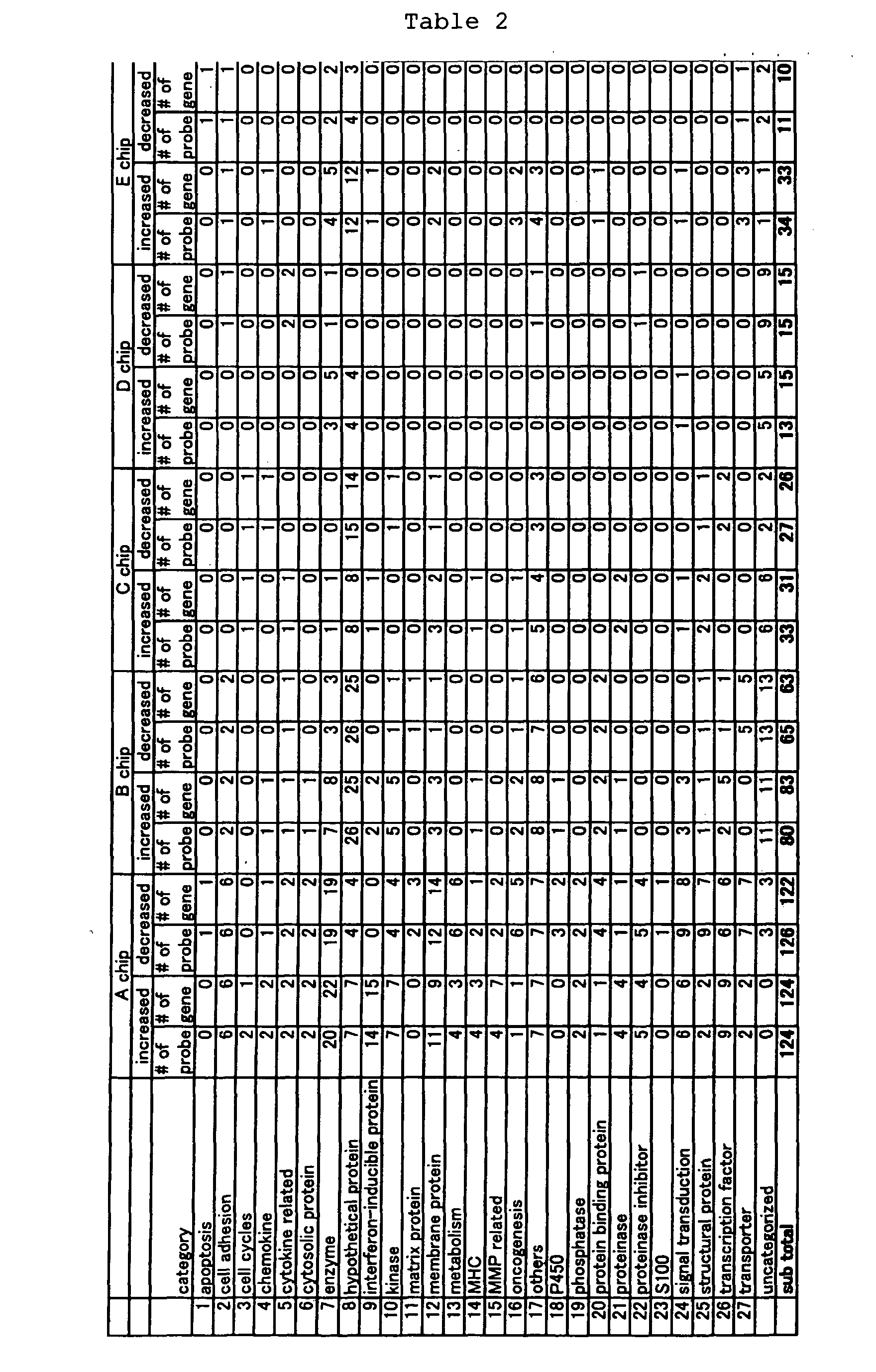
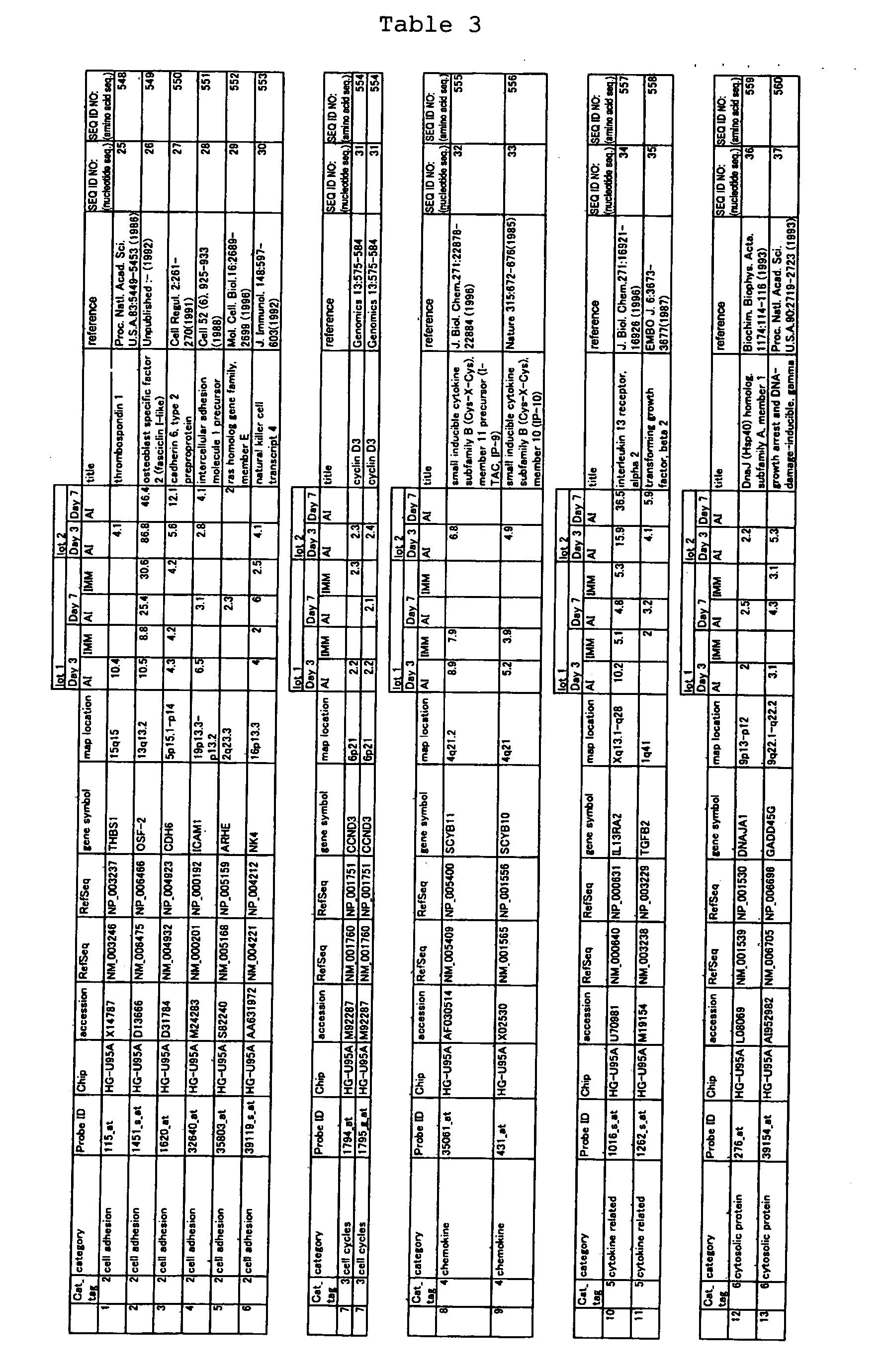


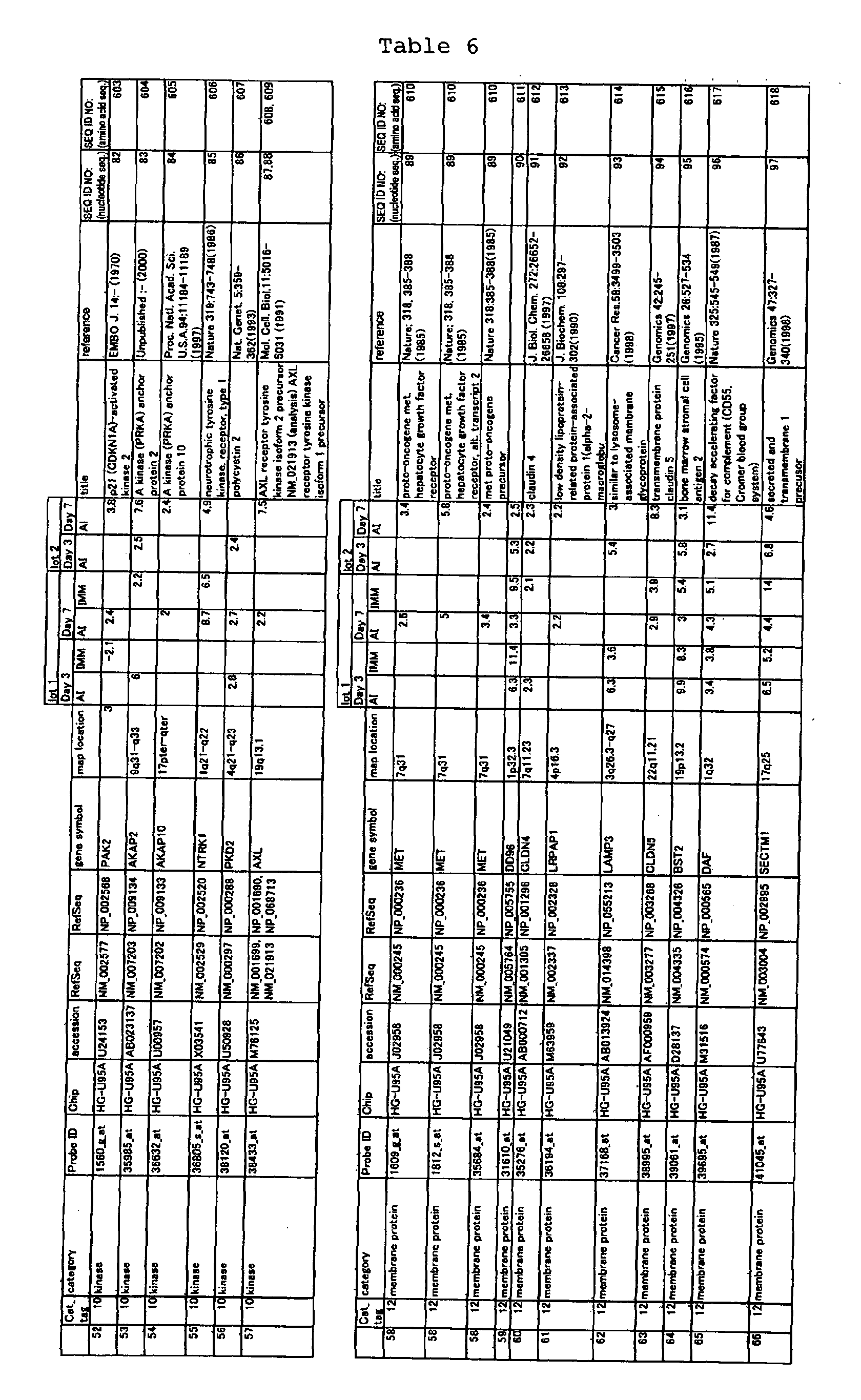


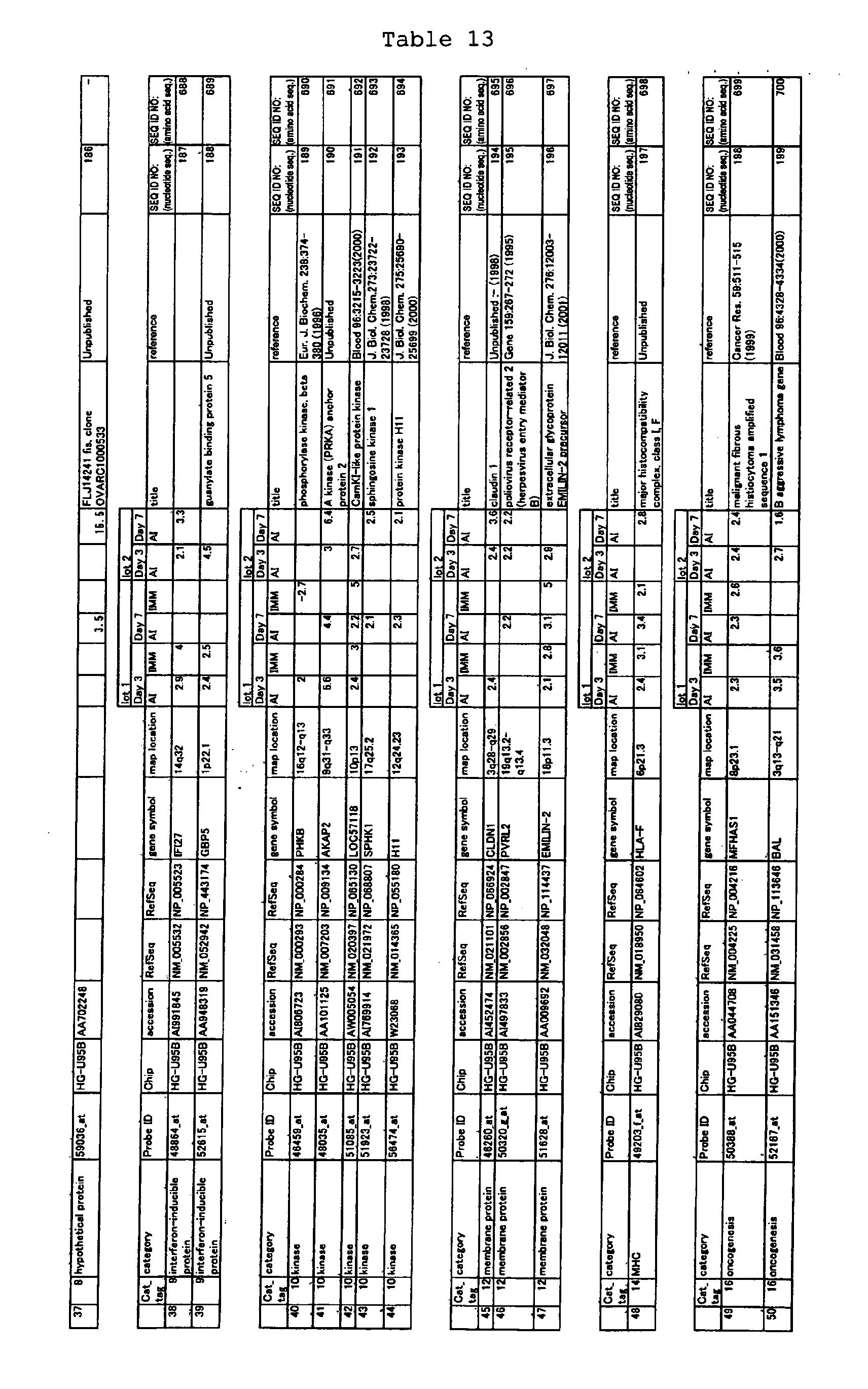


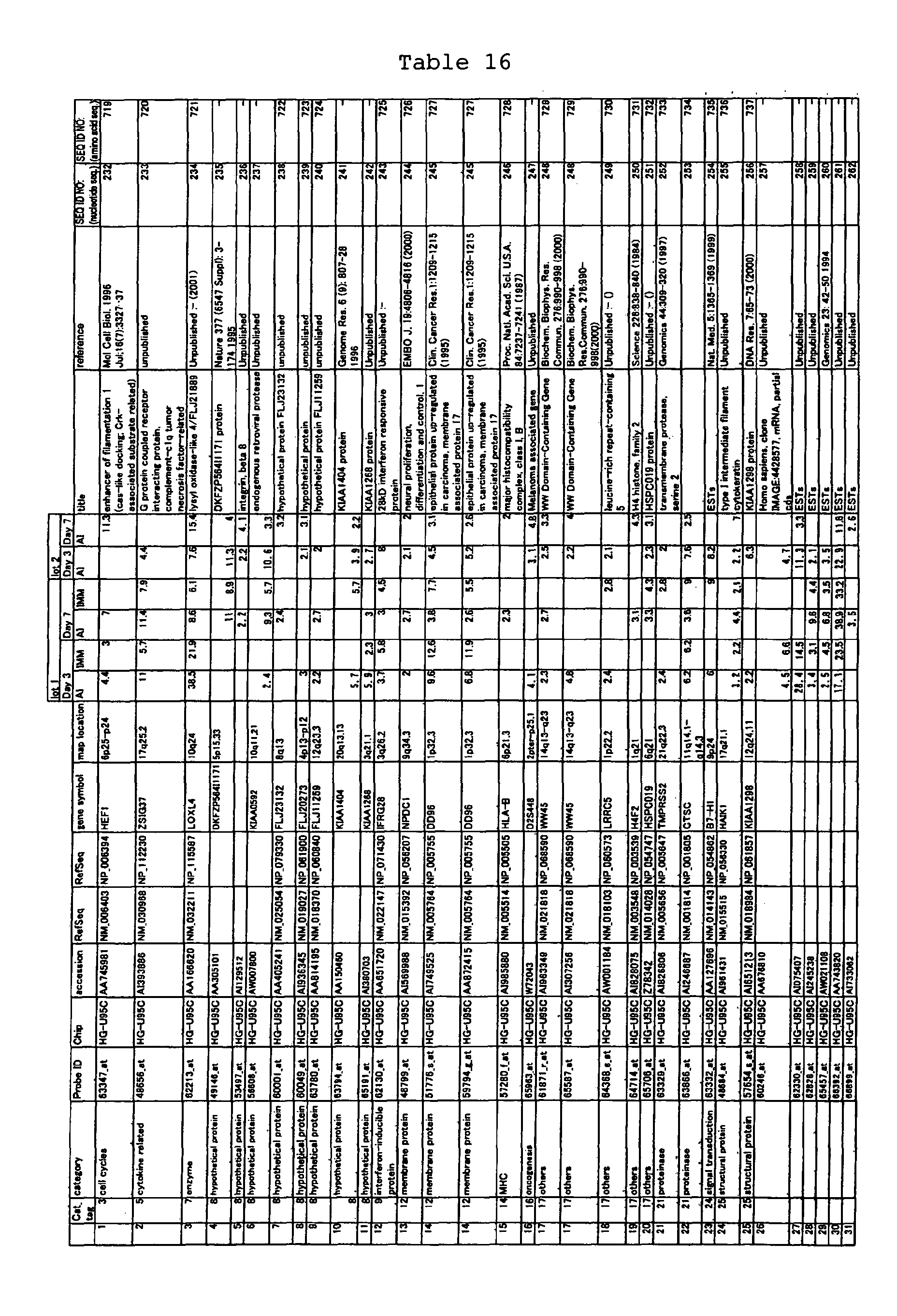


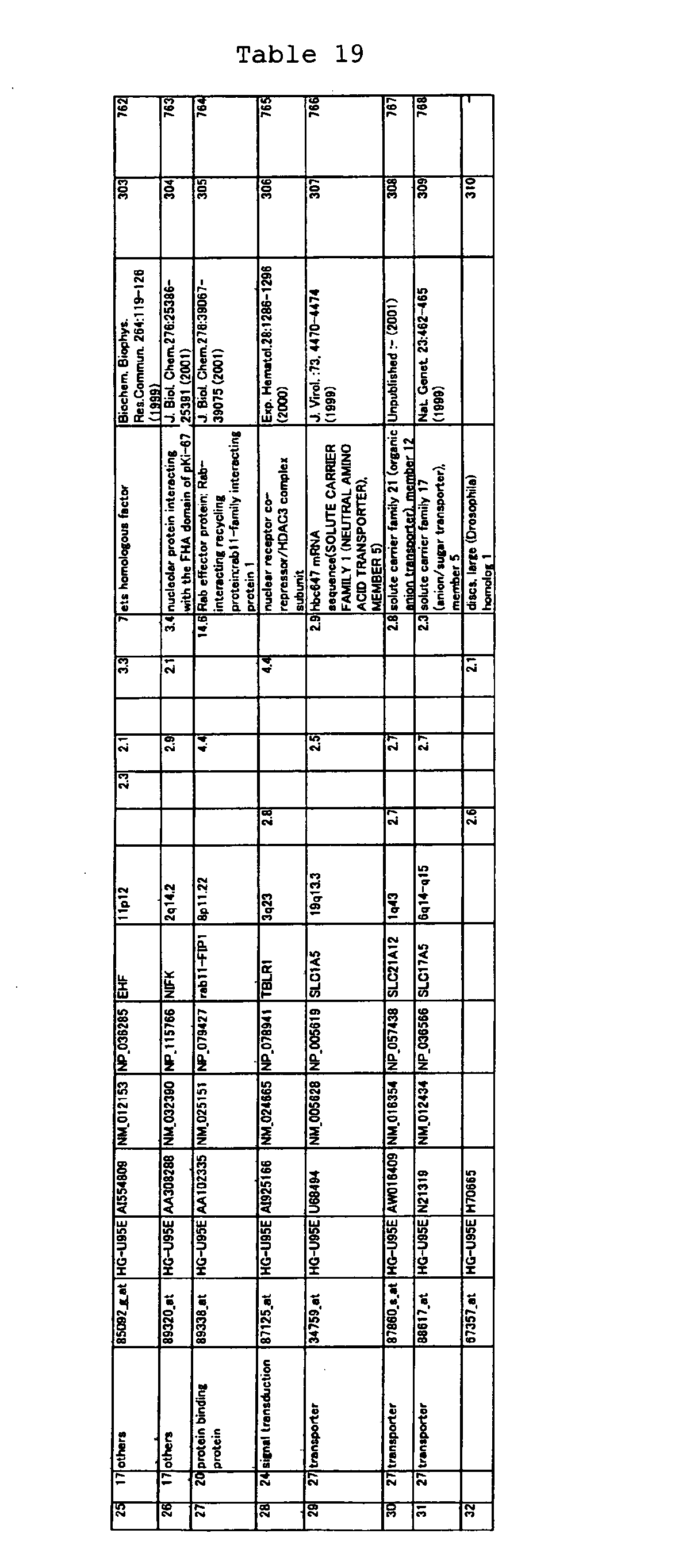
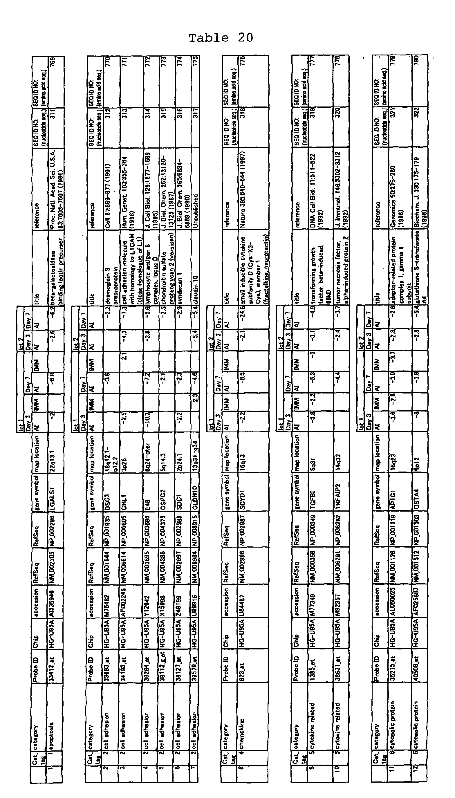

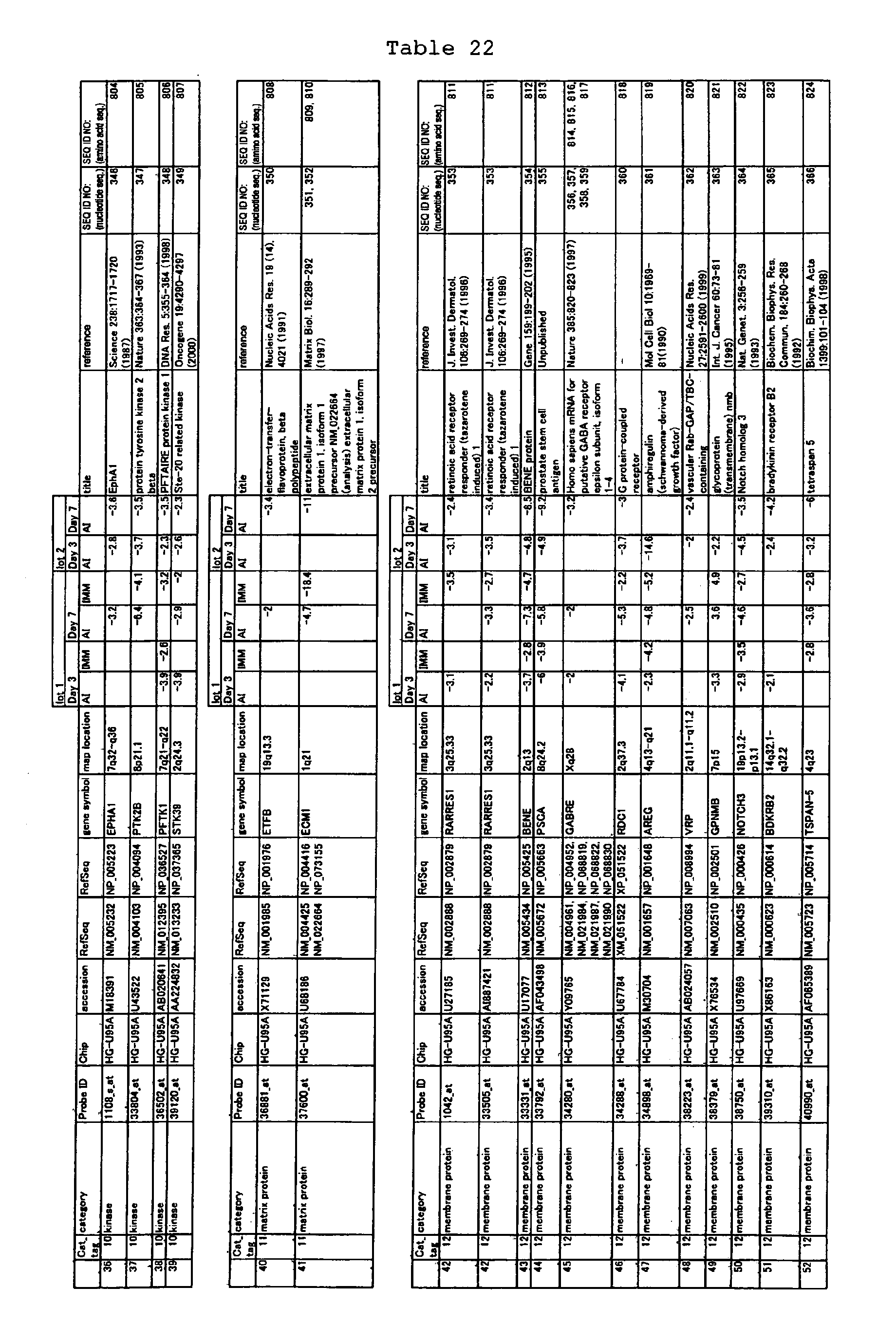


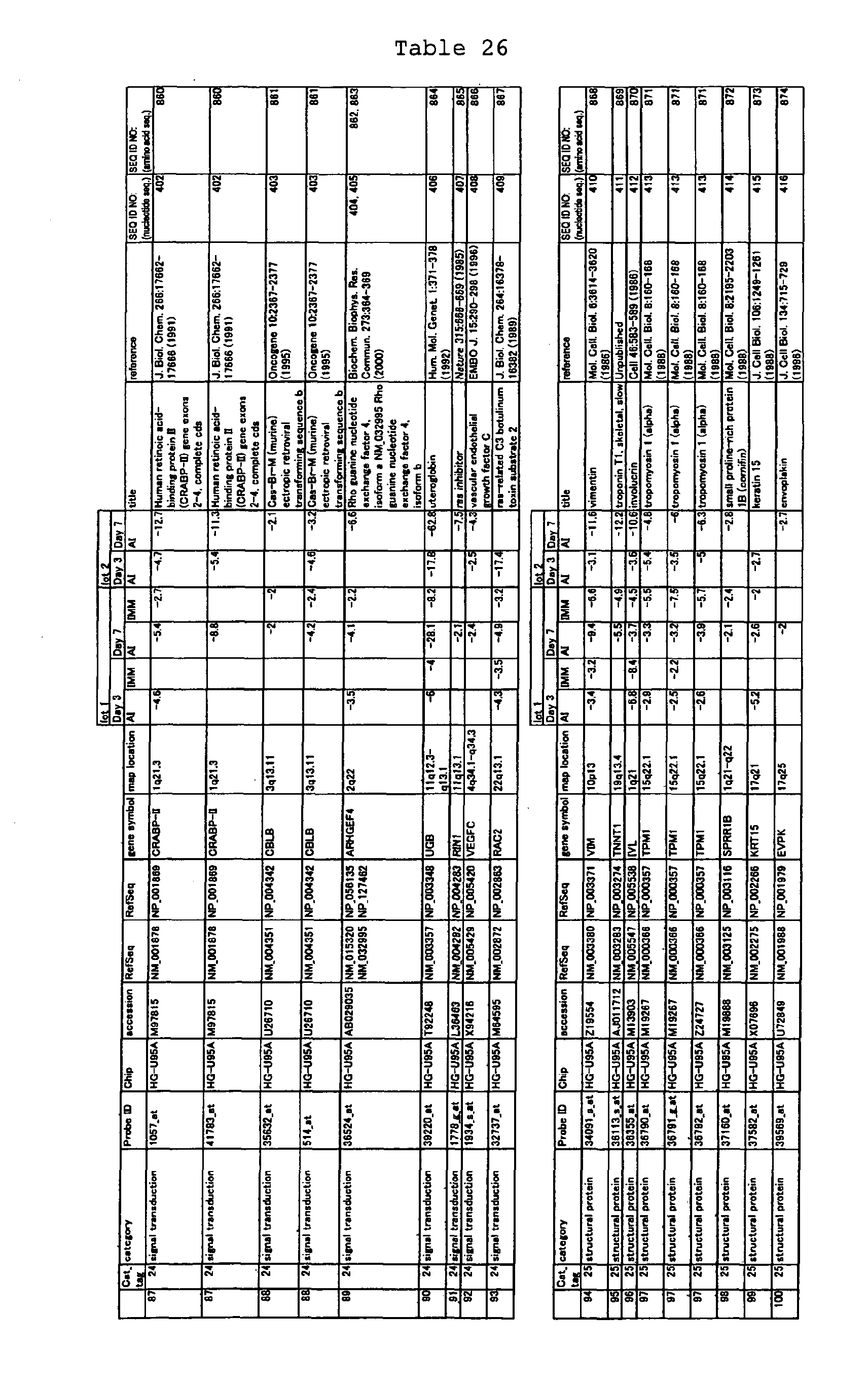
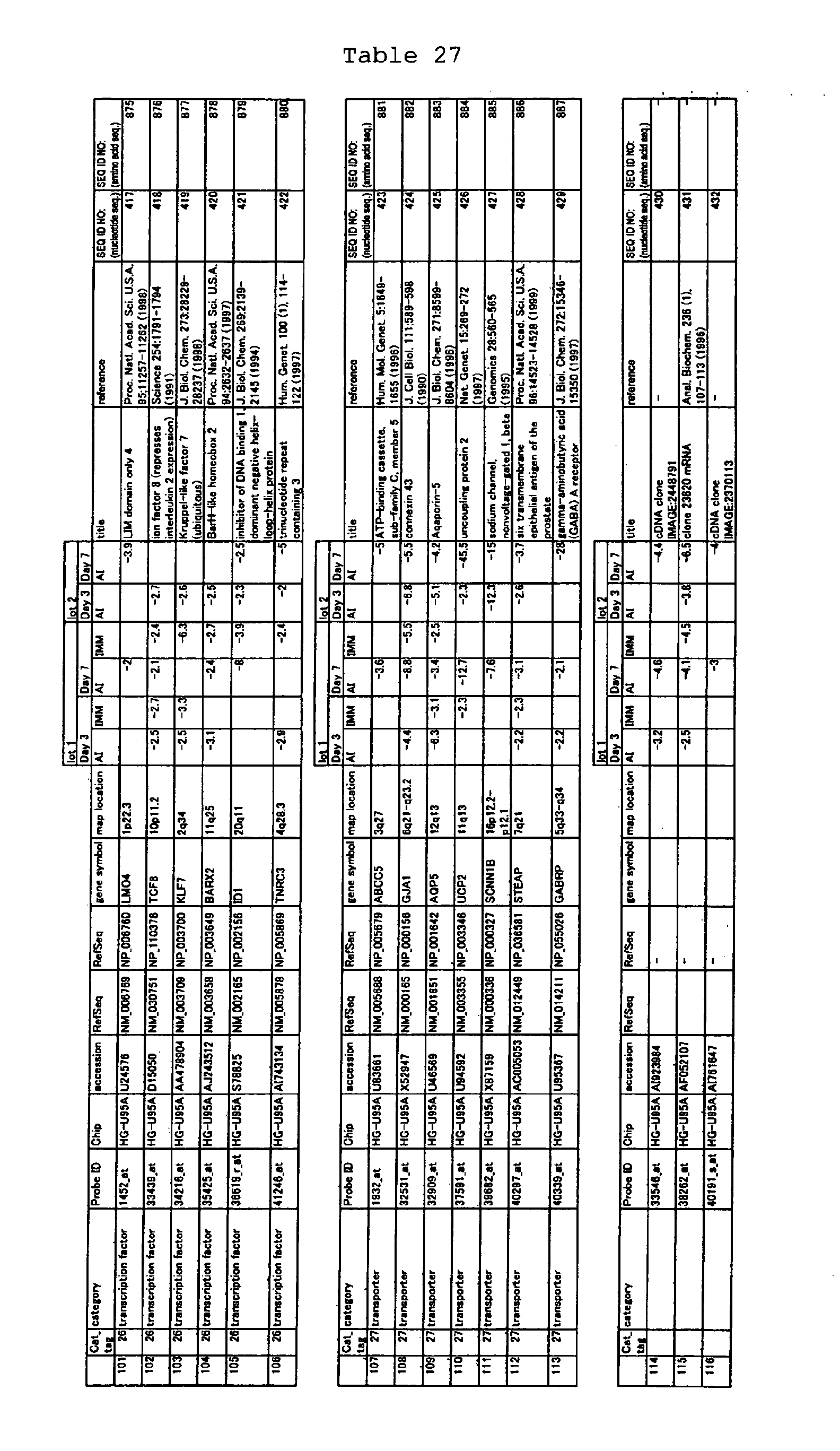
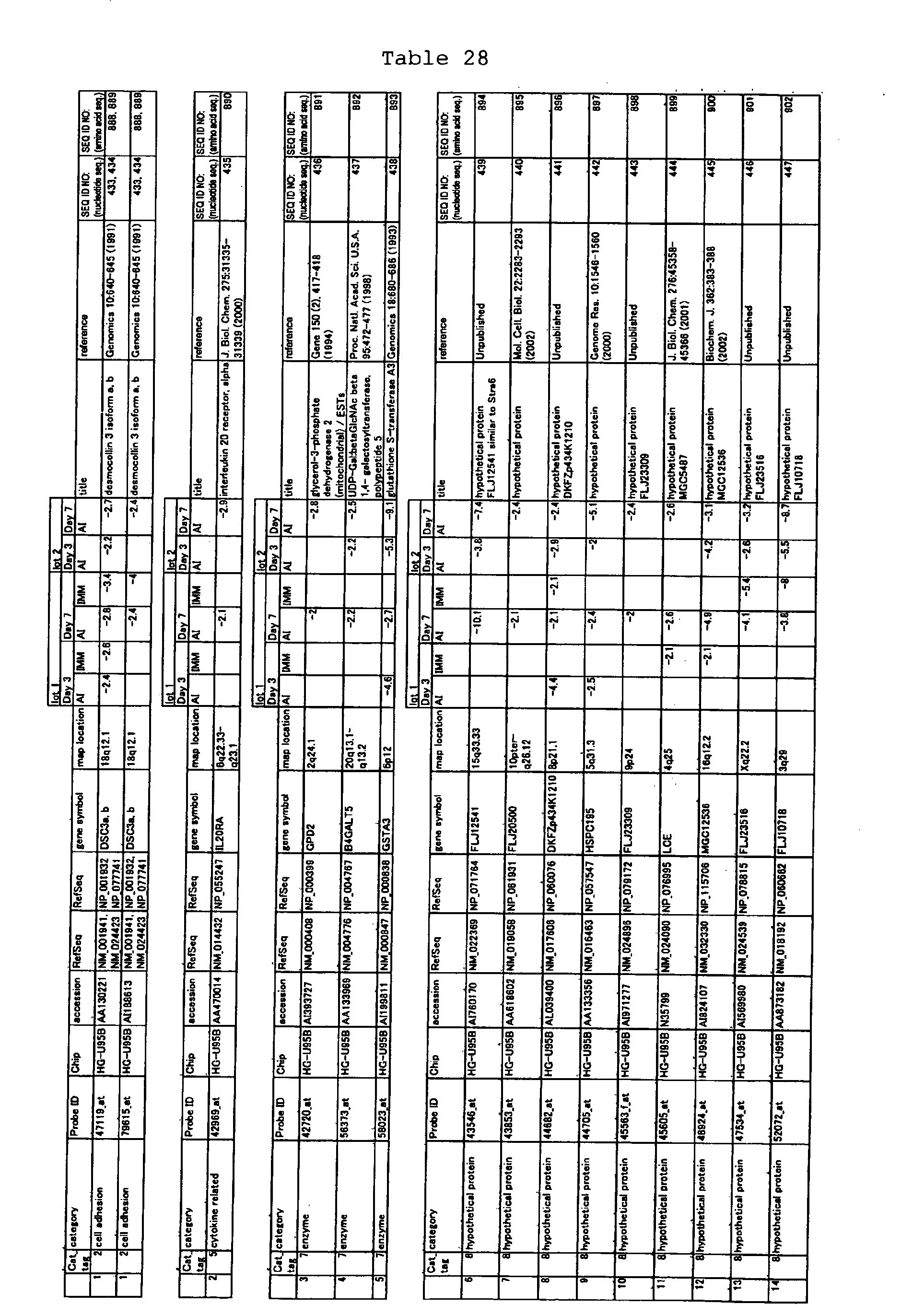
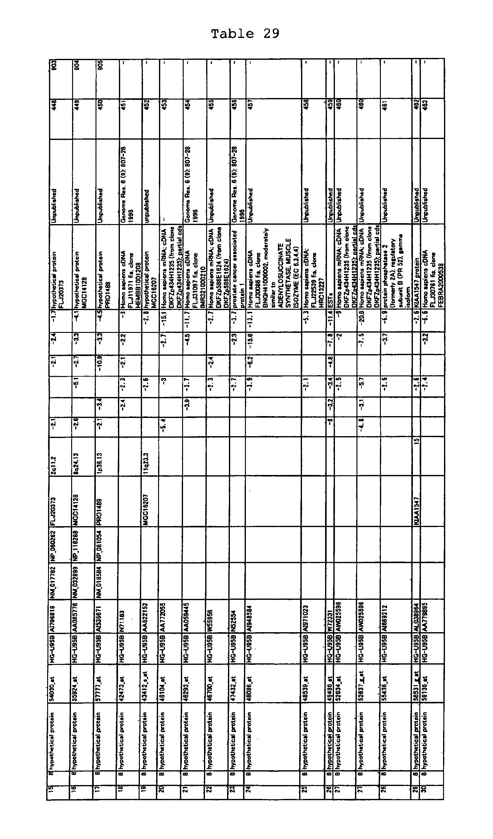
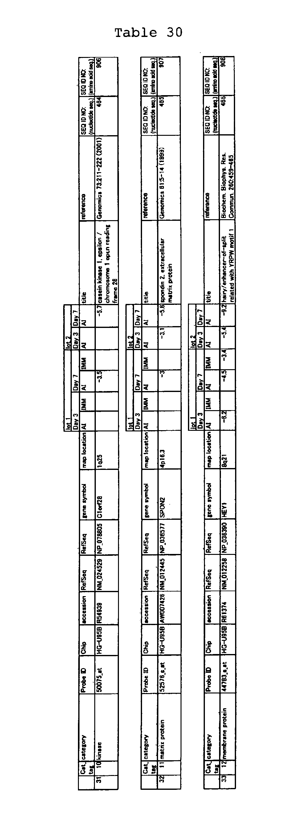


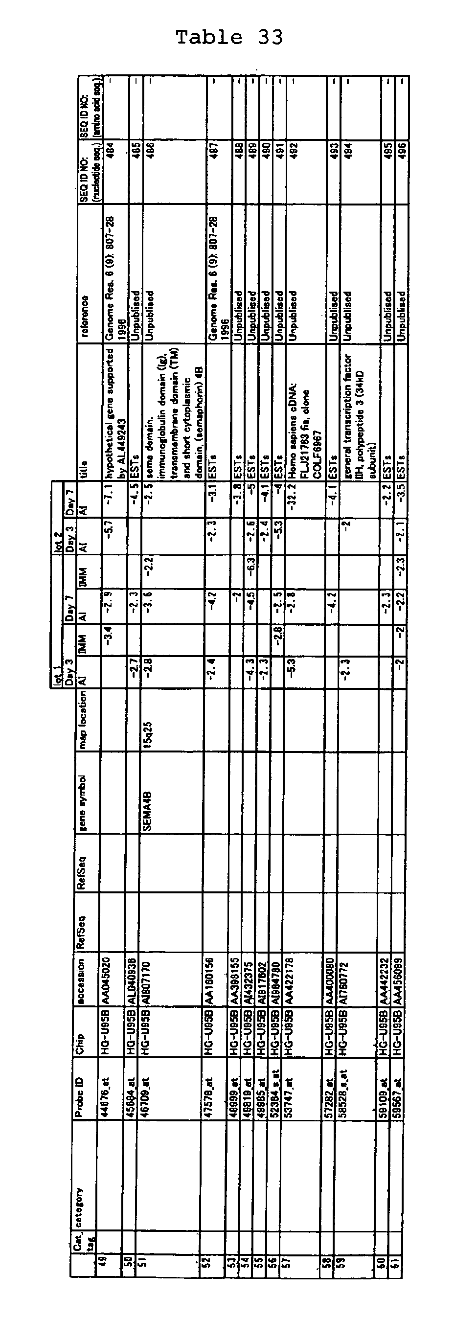
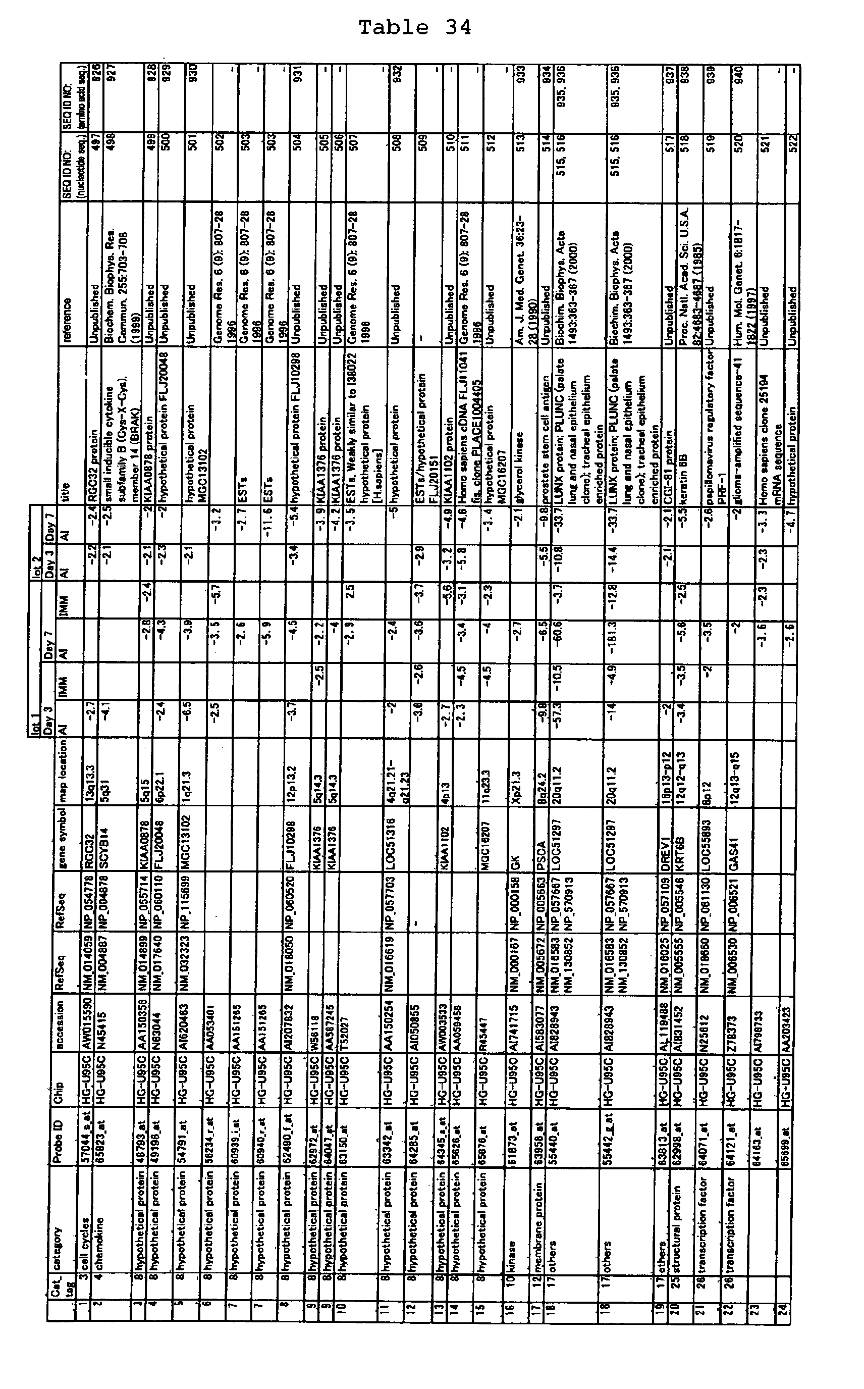


 FAM: 6-carboxy-fluorescein


 FAM: 6-carboxy-fluorescein

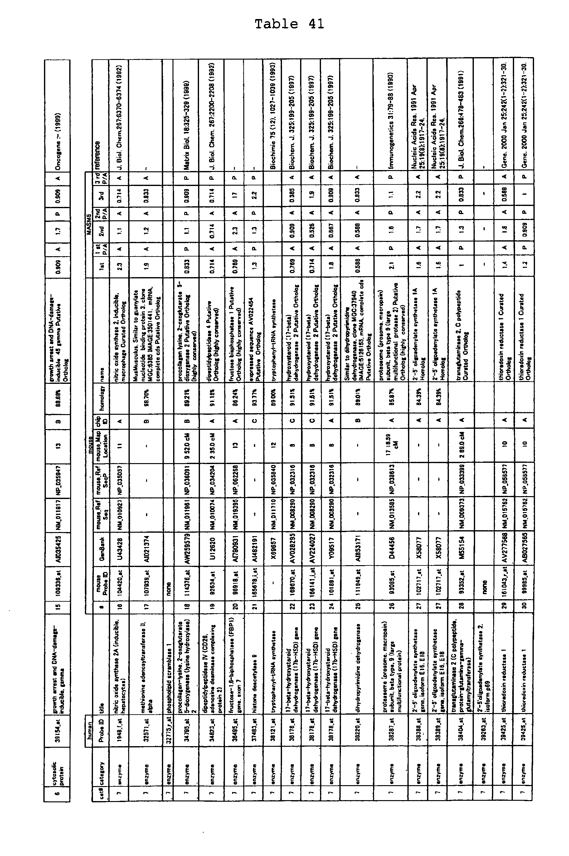


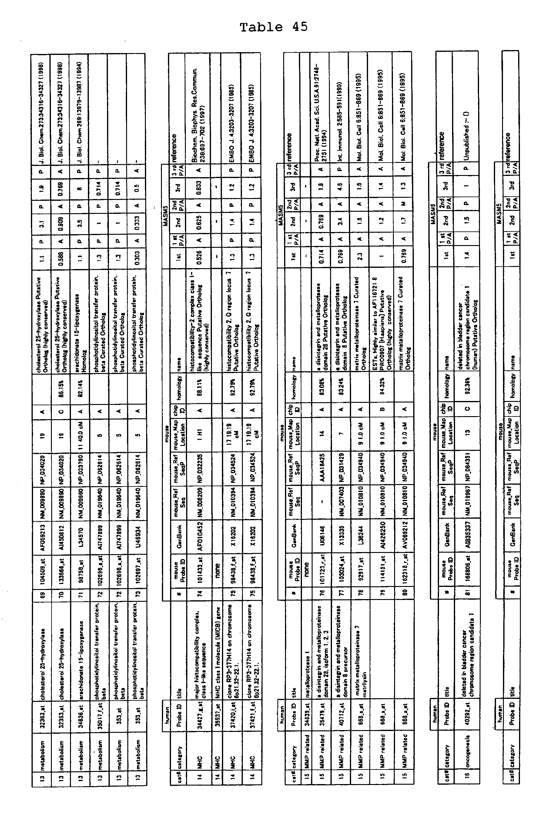


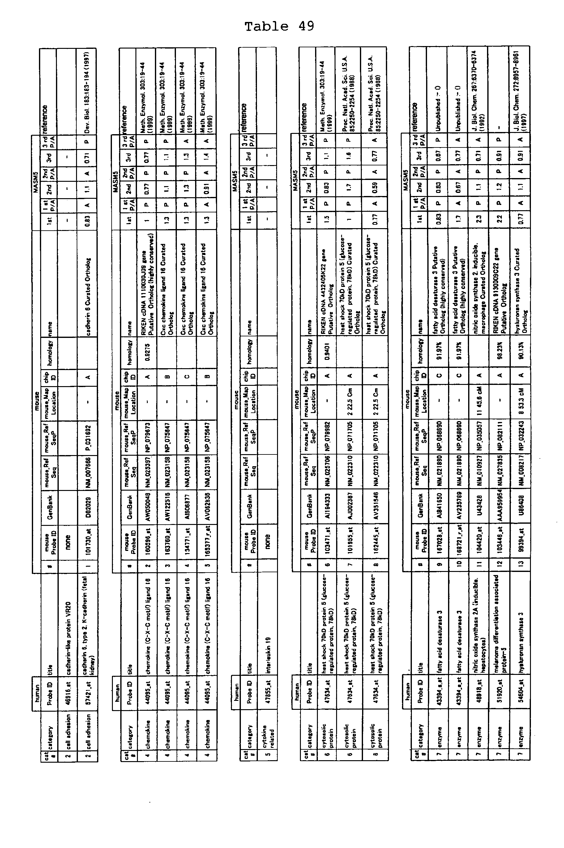

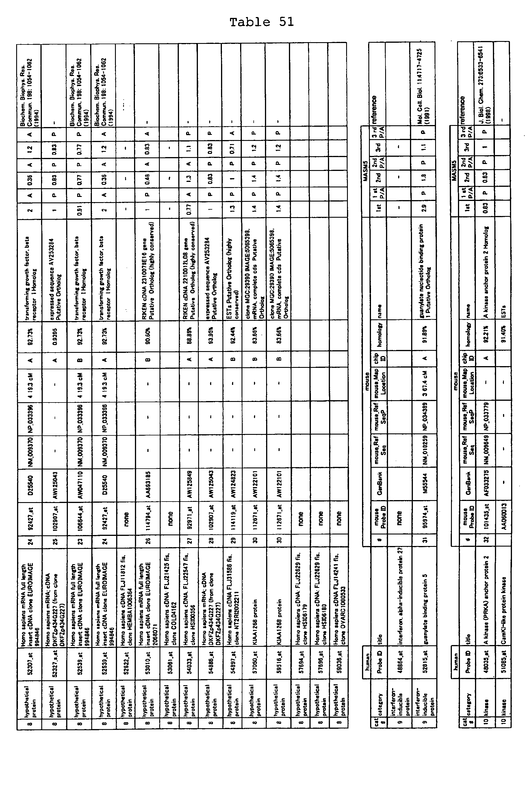


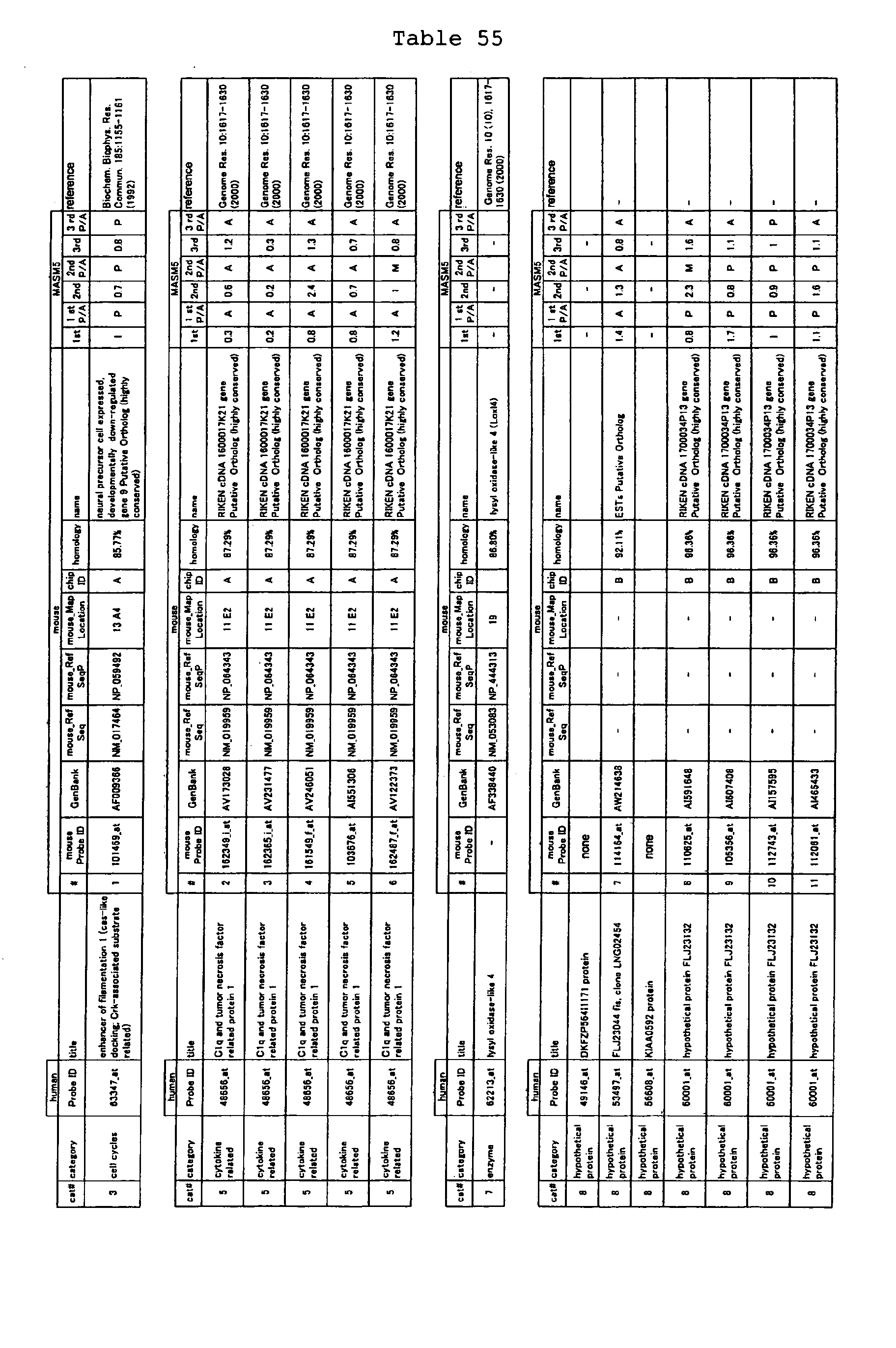


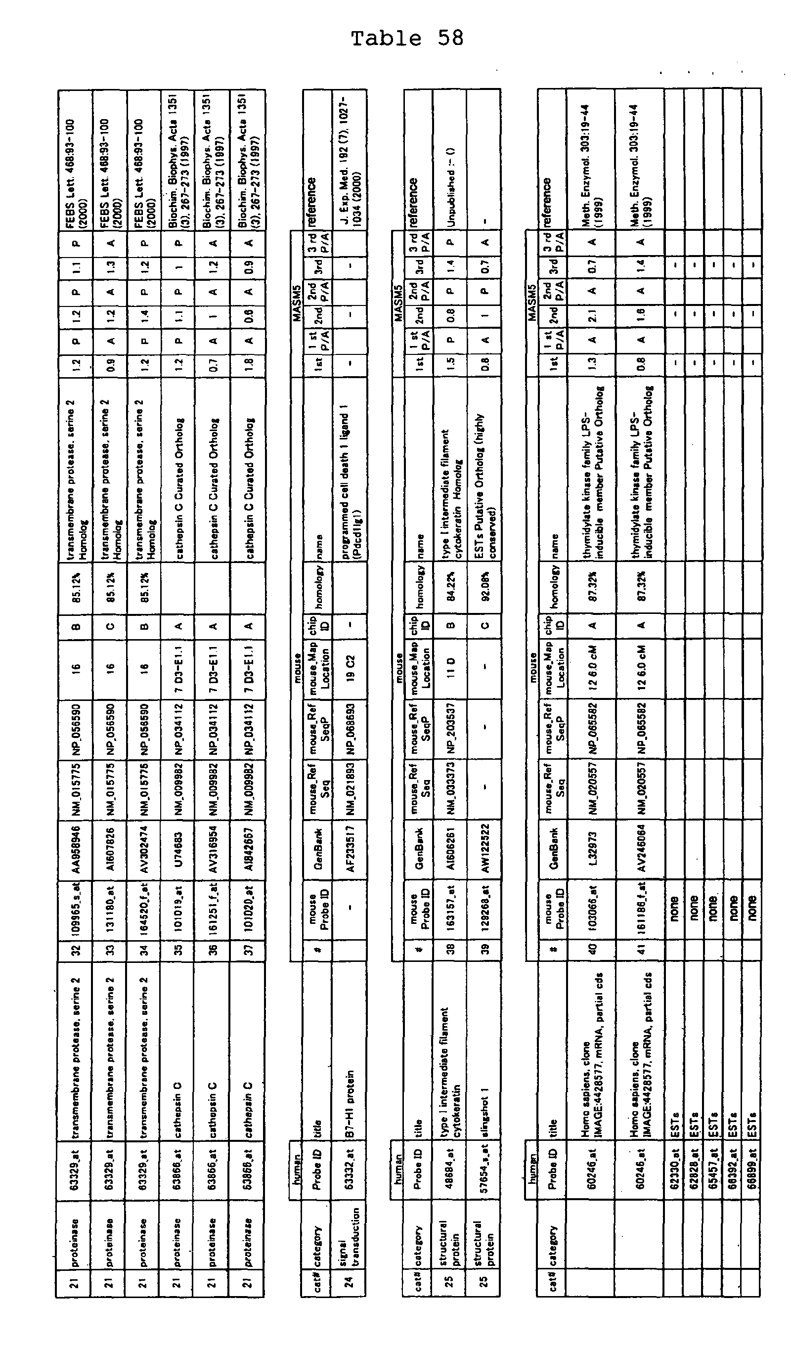


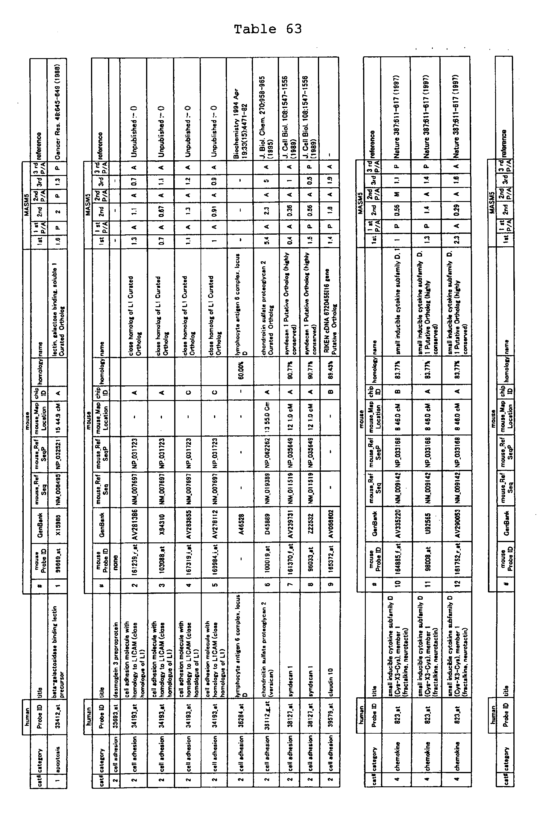


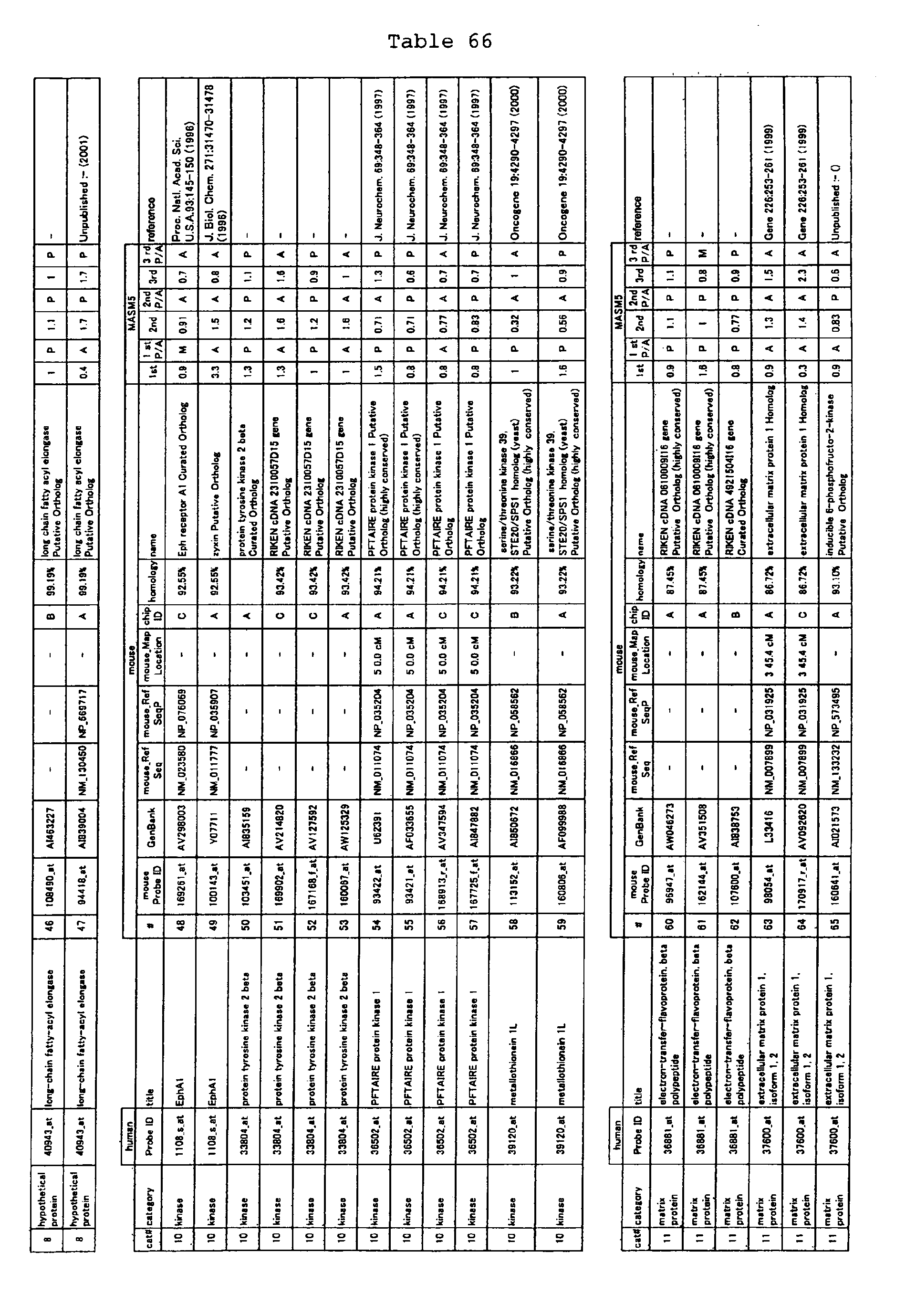
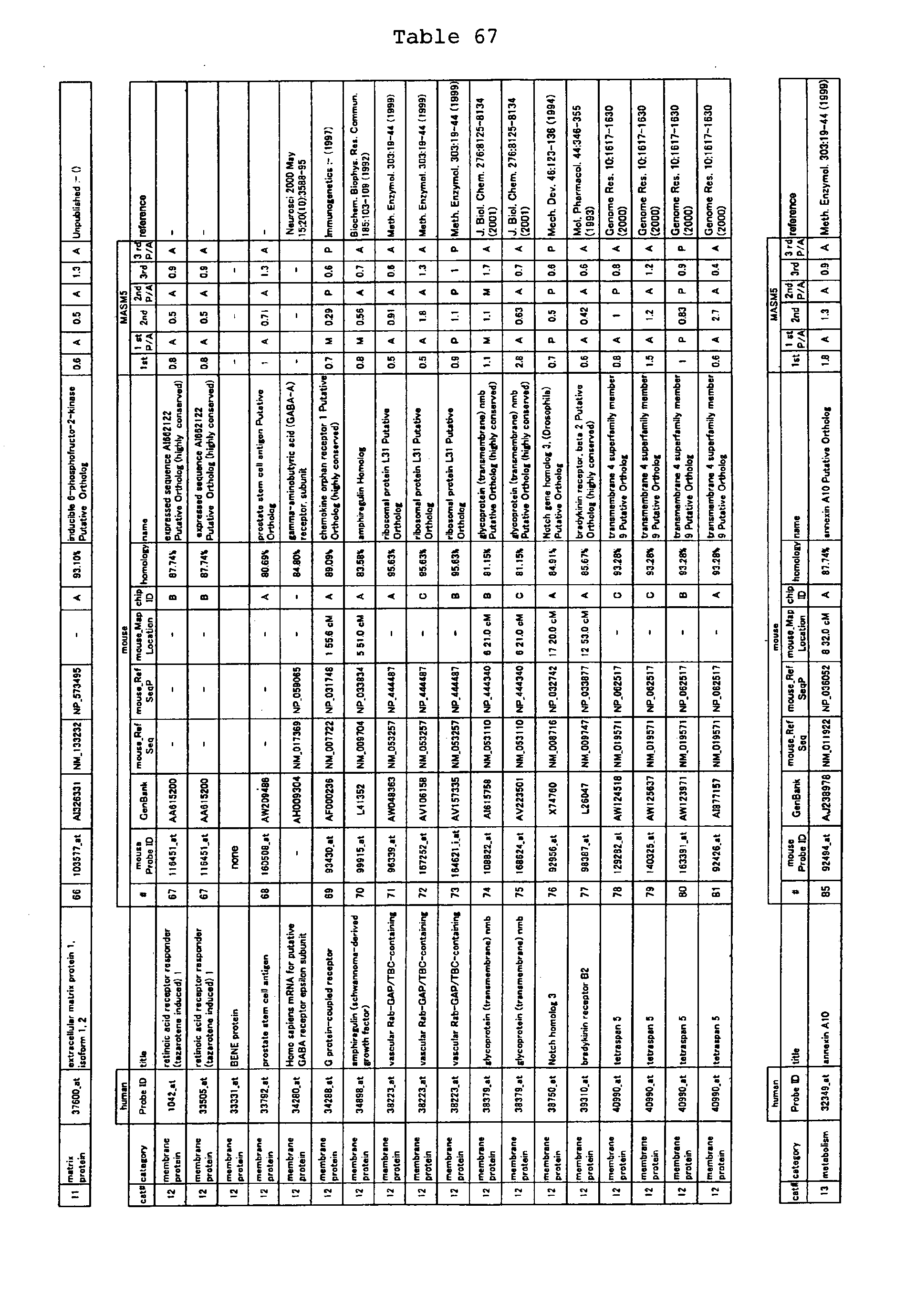


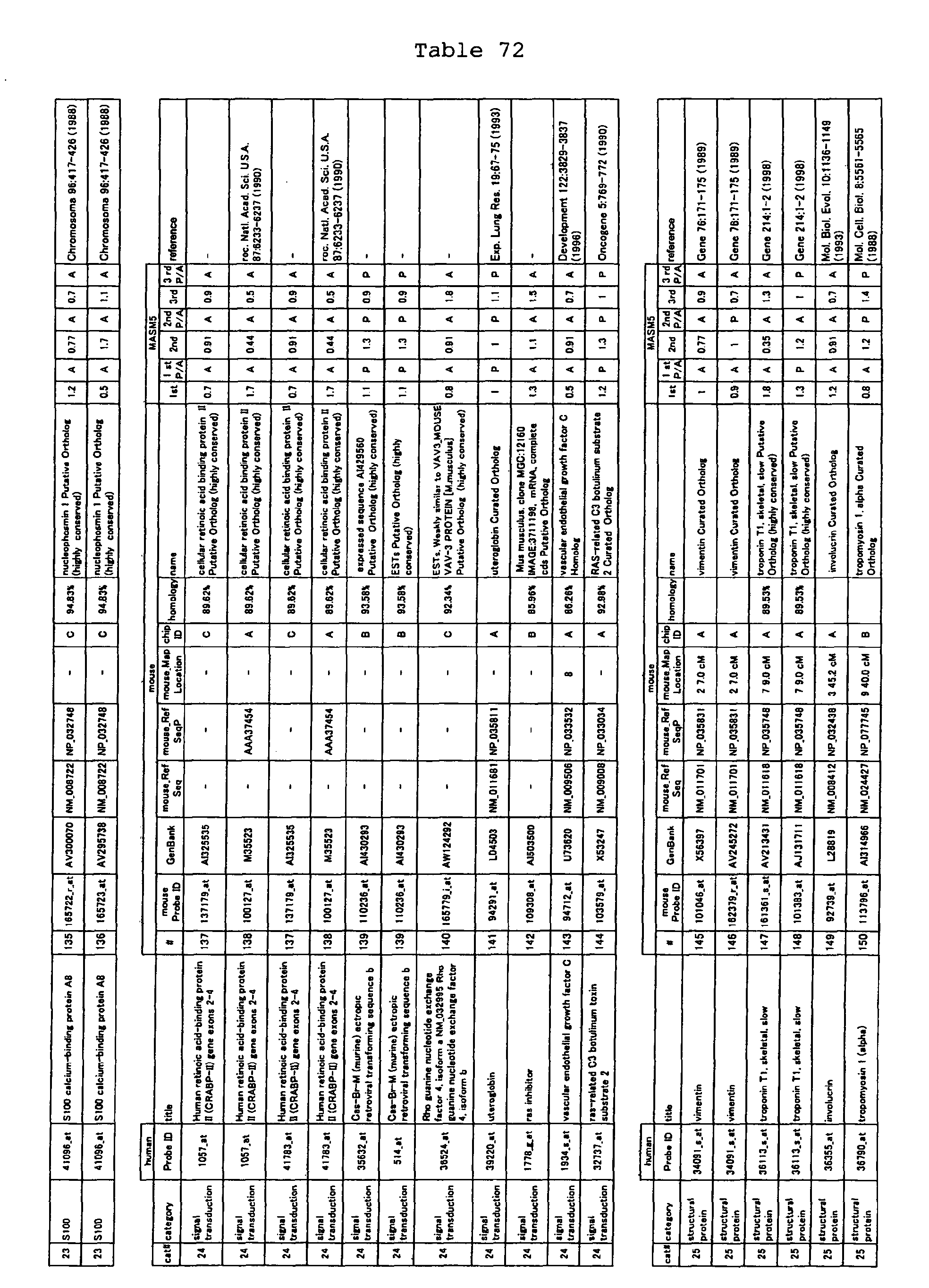

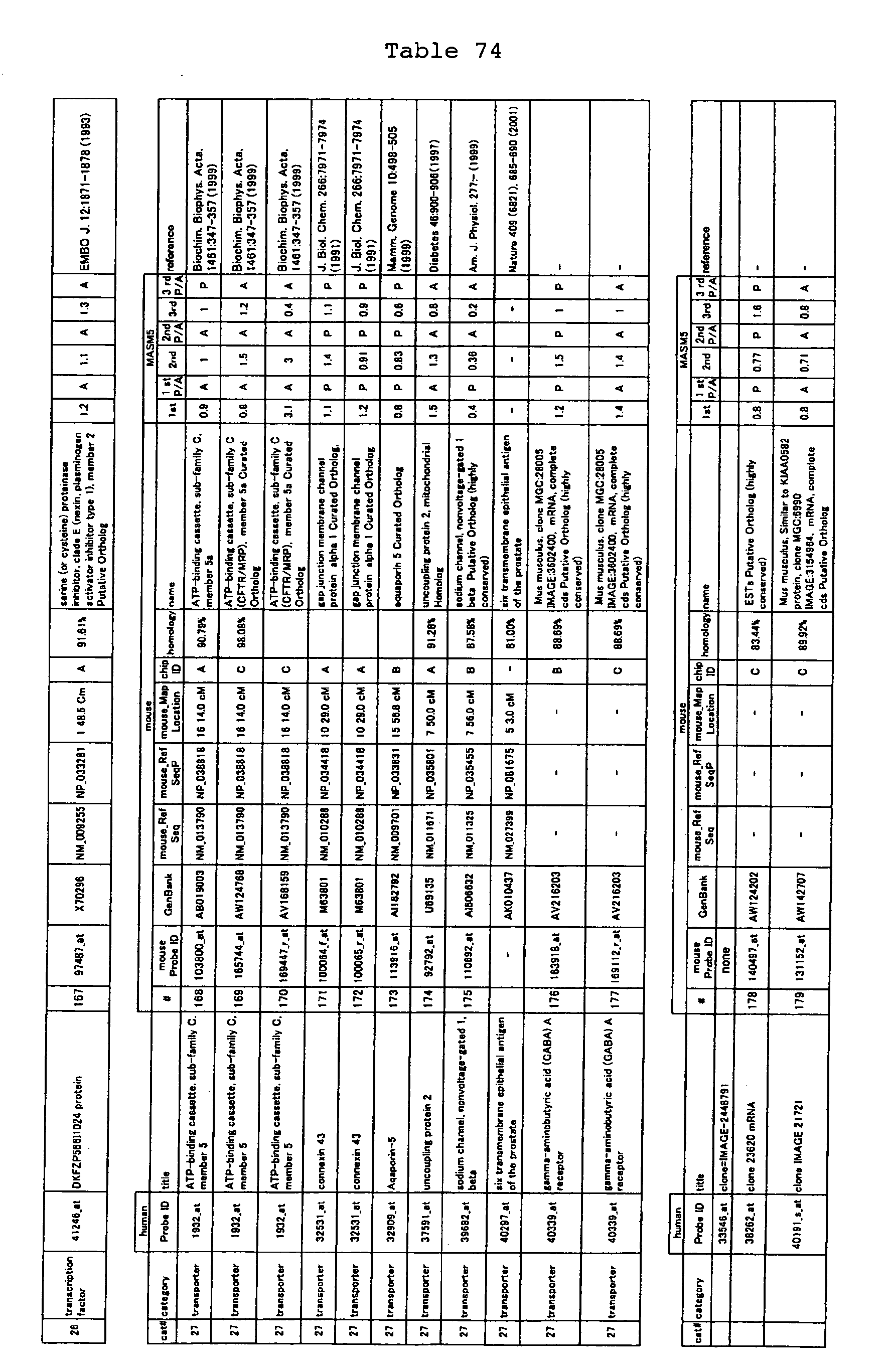
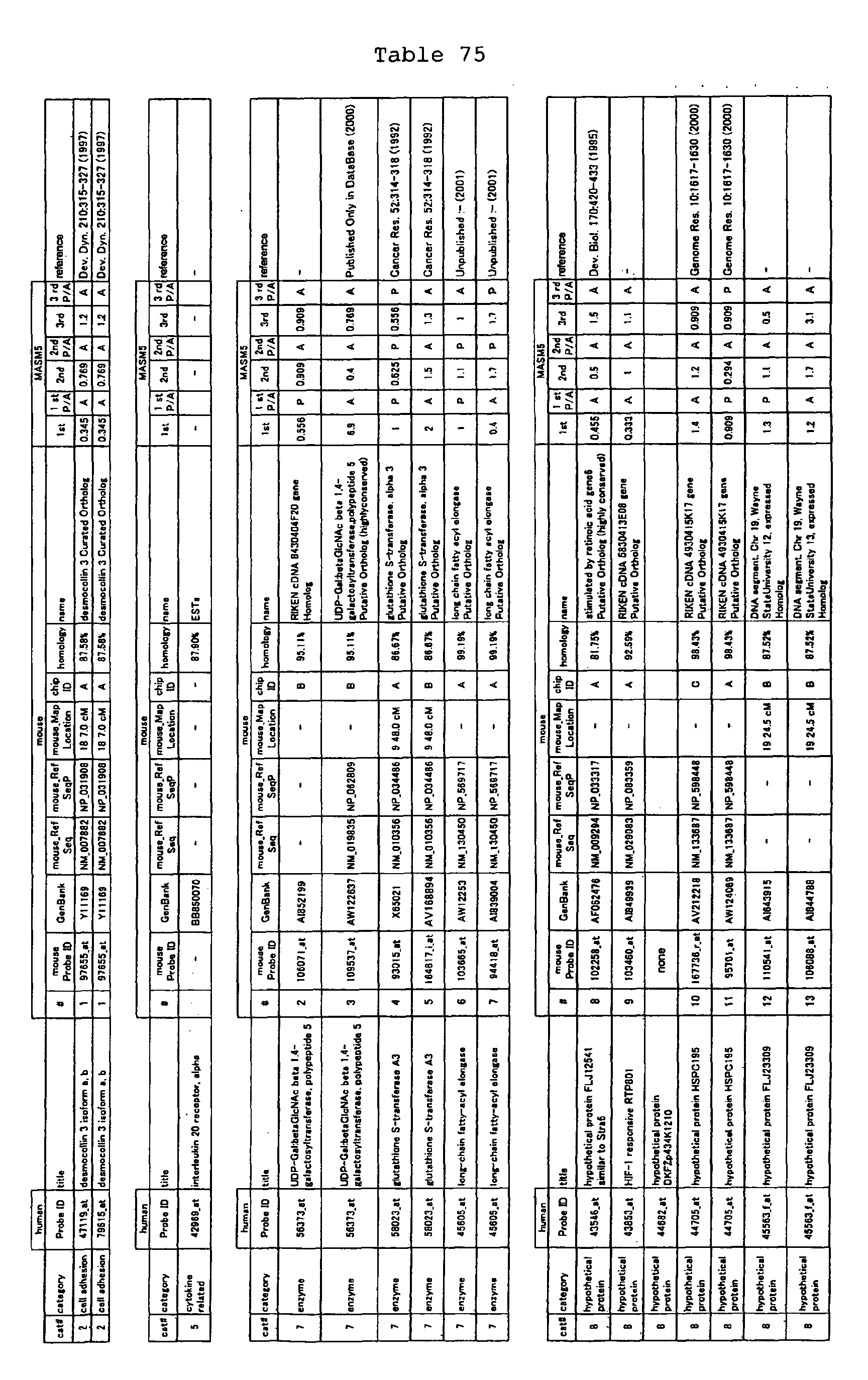


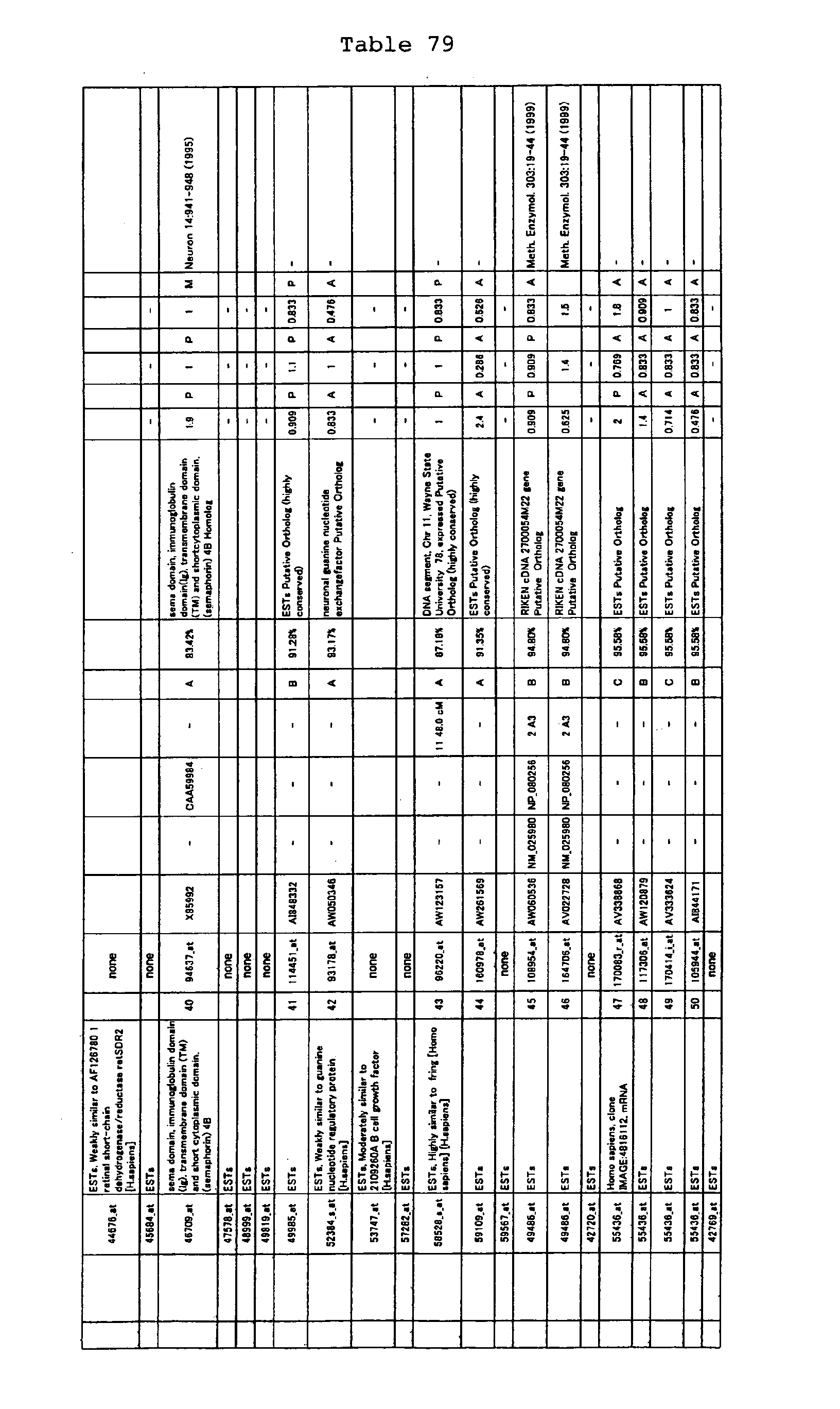
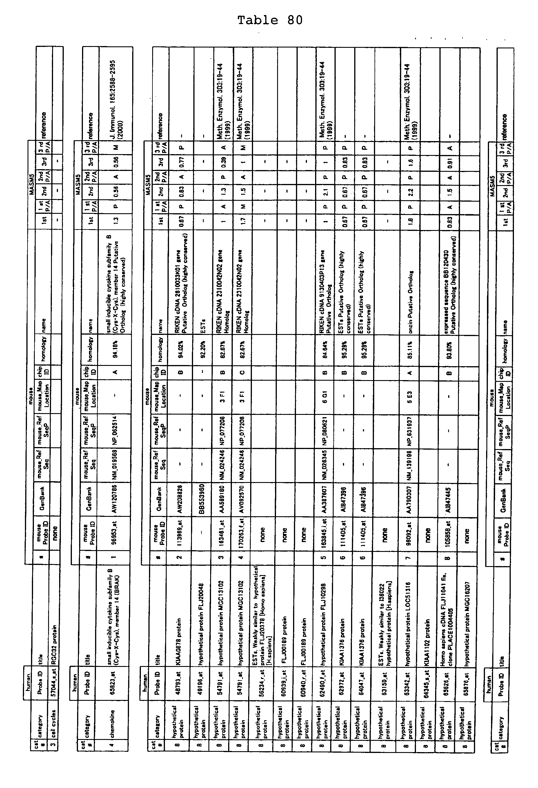


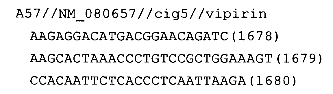


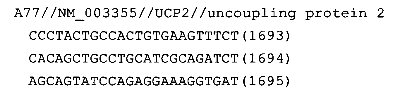
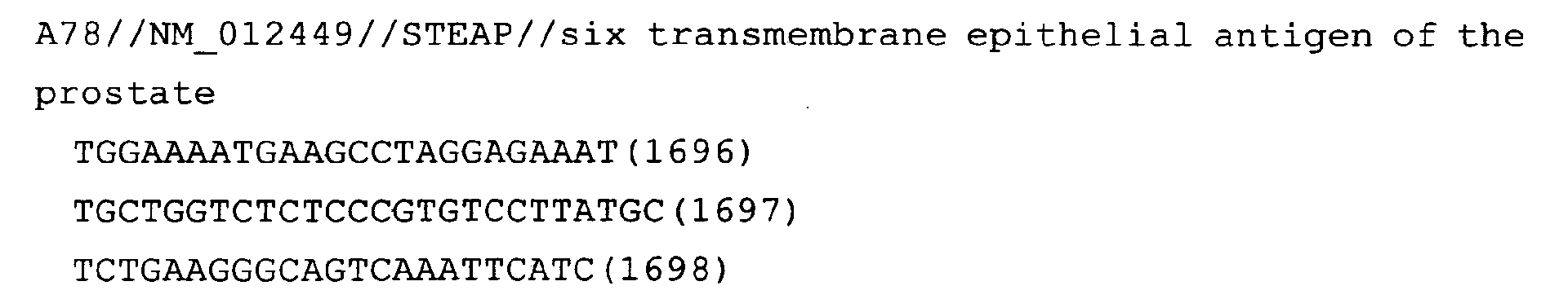

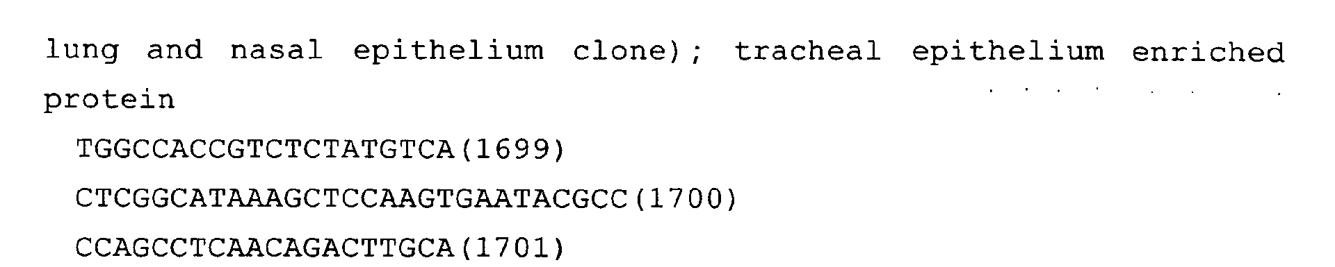


 Mouse genes;


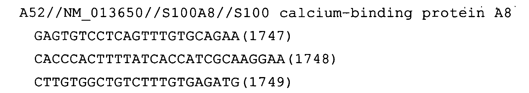

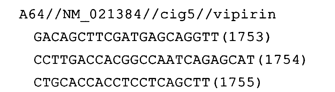
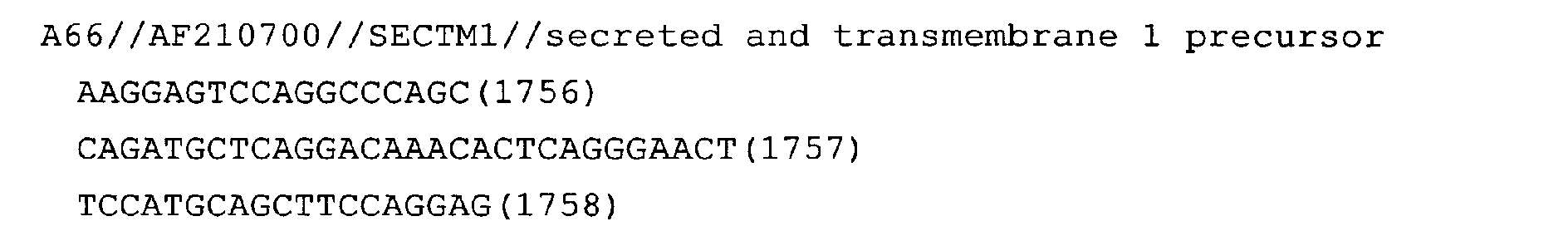
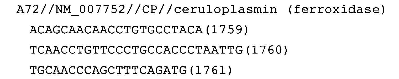


 Genes whose expression levels tend to vary in both humans and mice:


 Mouse genes;


 Genes whose expression levels varied in humans:
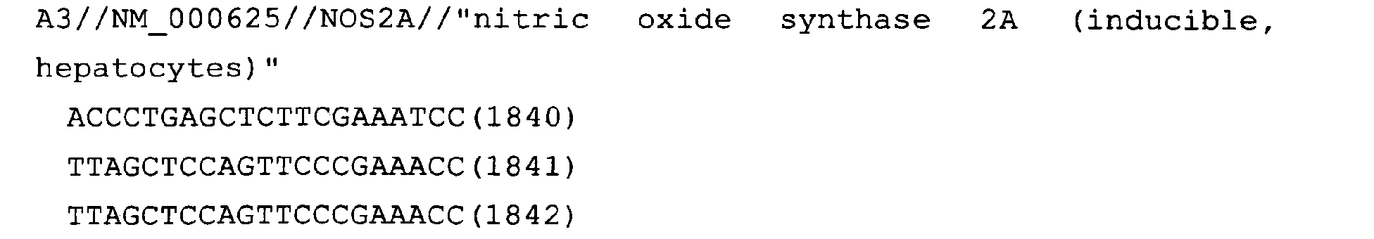


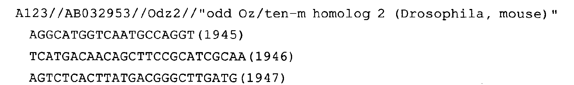
 Mouse genes;
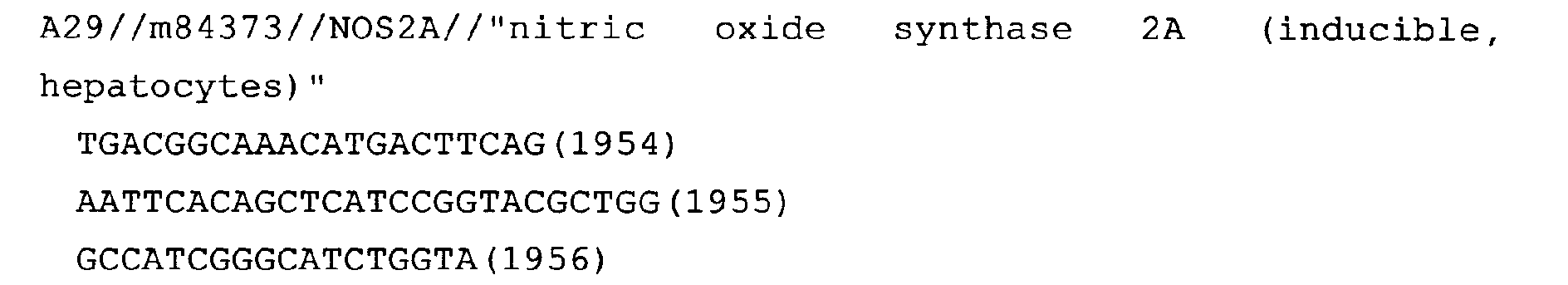
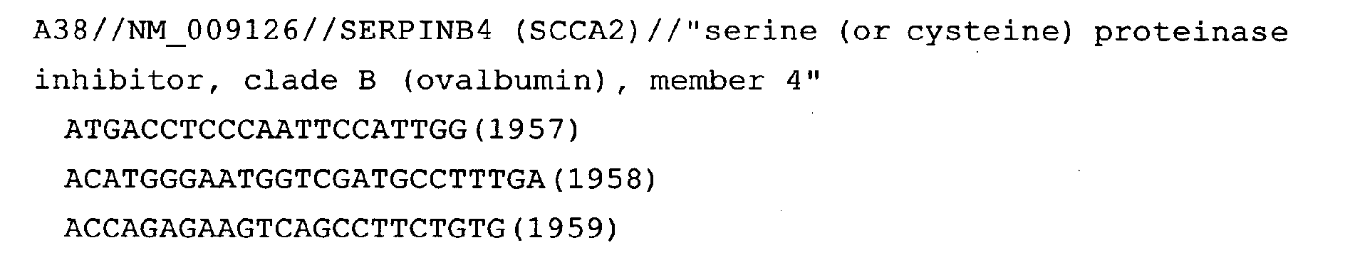
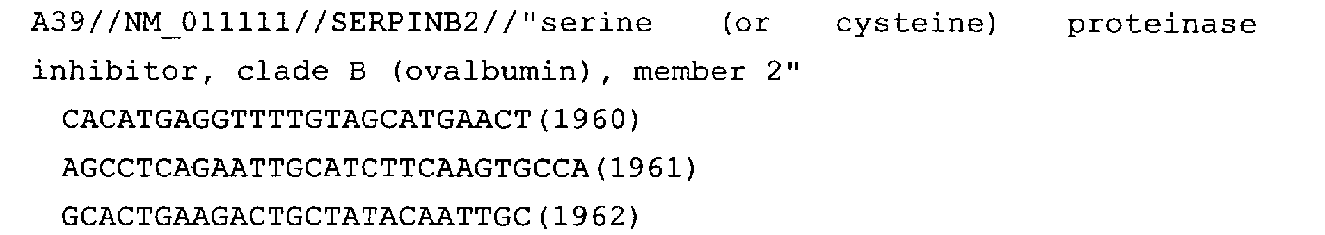
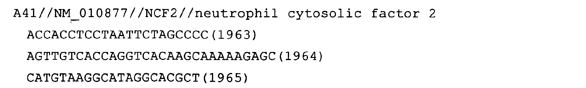


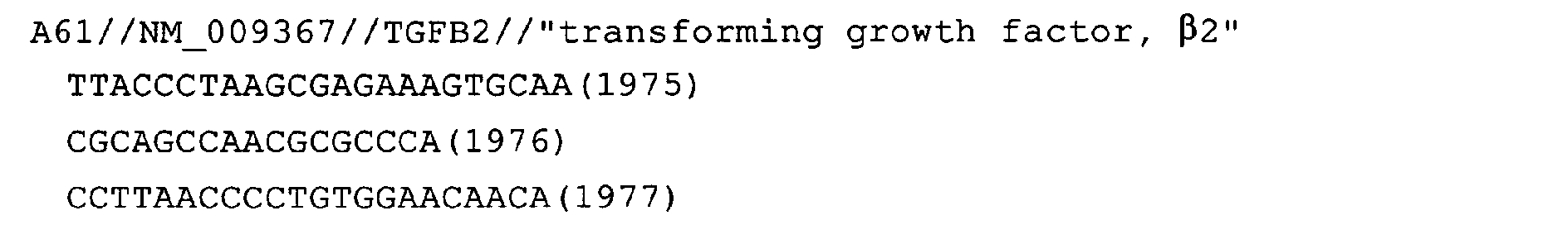


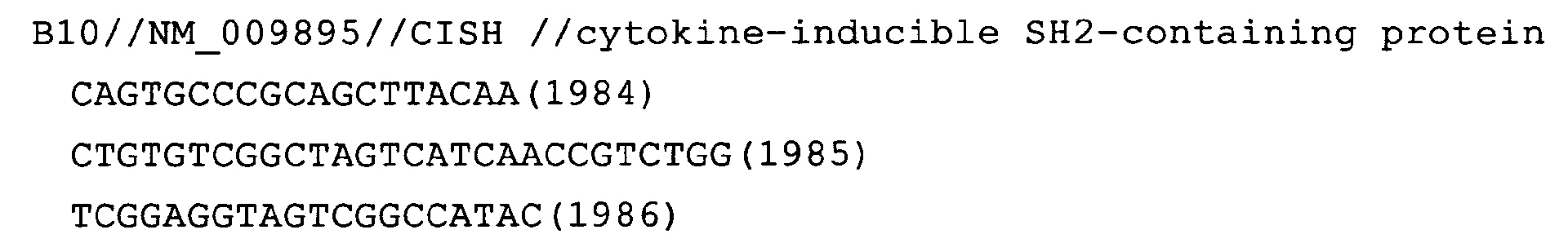
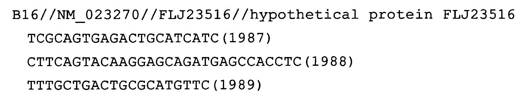


 Genes whose expression levels tend to vary in humans:


 Mouse genes;
 Genes whose expression levels varied in mice:
 Mouse genes;


 and
and