EP1397073B1 - Excisional and ultrasound medical treatment system - Google Patents
Excisional and ultrasound medical treatment system Download PDFInfo
- Publication number
- EP1397073B1 EP1397073B1 EP20020739438 EP02739438A EP1397073B1 EP 1397073 B1 EP1397073 B1 EP 1397073B1 EP 20020739438 EP20020739438 EP 20020739438 EP 02739438 A EP02739438 A EP 02739438A EP 1397073 B1 EP1397073 B1 EP 1397073B1
- Authority
- EP
- European Patent Office
- Prior art keywords
- ultrasound
- end effector
- tissue
- patient
- transducer
- Prior art date
- Legal status (The legal status is an assumption and is not a legal conclusion. Google has not performed a legal analysis and makes no representation as to the accuracy of the status listed.)
- Expired - Lifetime
Links
- 238000002604 ultrasonography Methods 0.000 title claims abstract description 322
- 238000011282 treatment Methods 0.000 title claims abstract description 249
- 239000012636 effector Substances 0.000 claims abstract description 245
- 238000012285 ultrasound imaging Methods 0.000 claims abstract description 82
- 238000001574 biopsy Methods 0.000 claims description 27
- 210000000481 breast Anatomy 0.000 claims description 23
- 230000000717 retained effect Effects 0.000 abstract description 22
- 210000001519 tissue Anatomy 0.000 description 133
- 238000000034 method Methods 0.000 description 124
- 230000003902 lesion Effects 0.000 description 67
- 230000014509 gene expression Effects 0.000 description 56
- 210000004204 blood vessel Anatomy 0.000 description 38
- FGUUSXIOTUKUDN-IBGZPJMESA-N C1(=CC=CC=C1)N1C2=C(NC([C@H](C1)NC=1OC(=NN=1)C1=CC=CC=C1)=O)C=CC=C2 Chemical compound C1(=CC=CC=C1)N1C2=C(NC([C@H](C1)NC=1OC(=NN=1)C1=CC=CC=C1)=O)C=CC=C2 FGUUSXIOTUKUDN-IBGZPJMESA-N 0.000 description 28
- 238000012986 modification Methods 0.000 description 19
- 230000004048 modification Effects 0.000 description 19
- 239000008280 blood Substances 0.000 description 18
- 210000004369 blood Anatomy 0.000 description 18
- 230000002496 gastric effect Effects 0.000 description 18
- 206010028980 Neoplasm Diseases 0.000 description 17
- 230000008901 benefit Effects 0.000 description 15
- 238000010586 diagram Methods 0.000 description 15
- 238000003384 imaging method Methods 0.000 description 15
- 210000004072 lung Anatomy 0.000 description 14
- 201000011510 cancer Diseases 0.000 description 11
- 238000013461 design Methods 0.000 description 9
- 238000001356 surgical procedure Methods 0.000 description 9
- 210000004185 liver Anatomy 0.000 description 8
- 230000007246 mechanism Effects 0.000 description 8
- 238000002679 ablation Methods 0.000 description 7
- GNFTZDOKVXKIBK-UHFFFAOYSA-N 3-(2-methoxyethoxy)benzohydrazide Chemical compound COCCOC1=CC=CC(C(=O)NN)=C1 GNFTZDOKVXKIBK-UHFFFAOYSA-N 0.000 description 6
- 230000006378 damage Effects 0.000 description 6
- 239000000463 material Substances 0.000 description 5
- 230000003213 activating effect Effects 0.000 description 4
- 210000001072 colon Anatomy 0.000 description 3
- 210000001035 gastrointestinal tract Anatomy 0.000 description 3
- 210000005228 liver tissue Anatomy 0.000 description 3
- 238000000968 medical method and process Methods 0.000 description 3
- 238000012544 monitoring process Methods 0.000 description 3
- 208000002699 Digestive System Neoplasms Diseases 0.000 description 2
- LFQSCWFLJHTTHZ-UHFFFAOYSA-N Ethanol Chemical compound CCO LFQSCWFLJHTTHZ-UHFFFAOYSA-N 0.000 description 2
- 230000017531 blood circulation Effects 0.000 description 2
- 210000002808 connective tissue Anatomy 0.000 description 2
- 238000007796 conventional method Methods 0.000 description 2
- 210000003238 esophagus Anatomy 0.000 description 2
- 201000010231 gastrointestinal system cancer Diseases 0.000 description 2
- 230000023597 hemostasis Effects 0.000 description 2
- 210000000944 nerve tissue Anatomy 0.000 description 2
- 210000002784 stomach Anatomy 0.000 description 2
- 239000000126 substance Substances 0.000 description 2
- 230000008685 targeting Effects 0.000 description 2
- 238000012360 testing method Methods 0.000 description 2
- 238000009210 therapy by ultrasound Methods 0.000 description 2
- 230000000007 visual effect Effects 0.000 description 2
- 206010019695 Hepatic neoplasm Diseases 0.000 description 1
- 206010061216 Infarction Diseases 0.000 description 1
- 208000000913 Kidney Calculi Diseases 0.000 description 1
- 206010058467 Lung neoplasm malignant Diseases 0.000 description 1
- 206010029148 Nephrolithiasis Diseases 0.000 description 1
- 240000007594 Oryza sativa Species 0.000 description 1
- 235000007164 Oryza sativa Nutrition 0.000 description 1
- 206010060862 Prostate cancer Diseases 0.000 description 1
- 208000000236 Prostatic Neoplasms Diseases 0.000 description 1
- 206010041235 Snoring Diseases 0.000 description 1
- FAPWRFPIFSIZLT-UHFFFAOYSA-M Sodium chloride Chemical compound [Na+].[Cl-] FAPWRFPIFSIZLT-UHFFFAOYSA-M 0.000 description 1
- 208000027418 Wounds and injury Diseases 0.000 description 1
- 210000000683 abdominal cavity Anatomy 0.000 description 1
- 239000011358 absorbing material Substances 0.000 description 1
- XAGFODPZIPBFFR-UHFFFAOYSA-N aluminium Chemical compound [Al] XAGFODPZIPBFFR-UHFFFAOYSA-N 0.000 description 1
- 229910052782 aluminium Inorganic materials 0.000 description 1
- 210000004141 ampulla of vater Anatomy 0.000 description 1
- 239000002246 antineoplastic agent Substances 0.000 description 1
- 229940041181 antineoplastic drug Drugs 0.000 description 1
- 230000000712 assembly Effects 0.000 description 1
- 238000000429 assembly Methods 0.000 description 1
- 230000000740 bleeding effect Effects 0.000 description 1
- 230000036770 blood supply Effects 0.000 description 1
- 235000013339 cereals Nutrition 0.000 description 1
- 230000000973 chemotherapeutic effect Effects 0.000 description 1
- 208000006990 cholangiocarcinoma Diseases 0.000 description 1
- 210000001953 common bile duct Anatomy 0.000 description 1
- 210000003459 common hepatic duct Anatomy 0.000 description 1
- 230000006835 compression Effects 0.000 description 1
- 238000007906 compression Methods 0.000 description 1
- 238000010276 construction Methods 0.000 description 1
- 230000008878 coupling Effects 0.000 description 1
- 238000010168 coupling process Methods 0.000 description 1
- 238000005859 coupling reaction Methods 0.000 description 1
- 210000001198 duodenum Anatomy 0.000 description 1
- 238000010304 firing Methods 0.000 description 1
- 239000012530 fluid Substances 0.000 description 1
- 238000009499 grossing Methods 0.000 description 1
- 210000005003 heart tissue Anatomy 0.000 description 1
- 238000010438 heat treatment Methods 0.000 description 1
- 230000007574 infarction Effects 0.000 description 1
- 208000014674 injury Diseases 0.000 description 1
- 208000014018 liver neoplasm Diseases 0.000 description 1
- 201000005202 lung cancer Diseases 0.000 description 1
- 208000020816 lung neoplasm Diseases 0.000 description 1
- 230000000877 morphologic effect Effects 0.000 description 1
- 230000003287 optical effect Effects 0.000 description 1
- 230000007170 pathology Effects 0.000 description 1
- 230000000149 penetrating effect Effects 0.000 description 1
- 230000035515 penetration Effects 0.000 description 1
- 210000000664 rectum Anatomy 0.000 description 1
- 230000002040 relaxant effect Effects 0.000 description 1
- 230000029058 respiratory gaseous exchange Effects 0.000 description 1
- 235000009566 rice Nutrition 0.000 description 1
- 238000007789 sealing Methods 0.000 description 1
- 238000007493 shaping process Methods 0.000 description 1
- 239000007787 solid Substances 0.000 description 1
- 229910001220 stainless steel Inorganic materials 0.000 description 1
- 239000010935 stainless steel Substances 0.000 description 1
- 210000003462 vein Anatomy 0.000 description 1
- -1 without limitation Substances 0.000 description 1
Images


Classifications
-
- A—HUMAN NECESSITIES
- A61—MEDICAL OR VETERINARY SCIENCE; HYGIENE
- A61B—DIAGNOSIS; SURGERY; IDENTIFICATION
- A61B8/00—Diagnosis using ultrasonic, sonic or infrasonic waves
- A61B8/44—Constructional features of the ultrasonic, sonic or infrasonic diagnostic device
- A61B8/4444—Constructional features of the ultrasonic, sonic or infrasonic diagnostic device related to the probe
- A61B8/445—Details of catheter construction
-
- A—HUMAN NECESSITIES
- A61—MEDICAL OR VETERINARY SCIENCE; HYGIENE
- A61B—DIAGNOSIS; SURGERY; IDENTIFICATION
- A61B8/00—Diagnosis using ultrasonic, sonic or infrasonic waves
- A61B8/12—Diagnosis using ultrasonic, sonic or infrasonic waves in body cavities or body tracts, e.g. by using catheters
-
- A—HUMAN NECESSITIES
- A61—MEDICAL OR VETERINARY SCIENCE; HYGIENE
- A61B—DIAGNOSIS; SURGERY; IDENTIFICATION
- A61B8/00—Diagnosis using ultrasonic, sonic or infrasonic waves
- A61B8/44—Constructional features of the ultrasonic, sonic or infrasonic diagnostic device
- A61B8/4483—Constructional features of the ultrasonic, sonic or infrasonic diagnostic device characterised by features of the ultrasound transducer
- A61B8/4488—Constructional features of the ultrasonic, sonic or infrasonic diagnostic device characterised by features of the ultrasound transducer the transducer being a phased array
-
- A—HUMAN NECESSITIES
- A61—MEDICAL OR VETERINARY SCIENCE; HYGIENE
- A61N—ELECTROTHERAPY; MAGNETOTHERAPY; RADIATION THERAPY; ULTRASOUND THERAPY
- A61N7/00—Ultrasound therapy
- A61N7/02—Localised ultrasound hyperthermia
- A61N7/022—Localised ultrasound hyperthermia intracavitary
-
- A—HUMAN NECESSITIES
- A61—MEDICAL OR VETERINARY SCIENCE; HYGIENE
- A61B—DIAGNOSIS; SURGERY; IDENTIFICATION
- A61B10/00—Other methods or instruments for diagnosis, e.g. instruments for taking a cell sample, for biopsy, for vaccination diagnosis; Sex determination; Ovulation-period determination; Throat striking implements
- A61B10/02—Instruments for taking cell samples or for biopsy
- A61B10/0233—Pointed or sharp biopsy instruments
-
- A—HUMAN NECESSITIES
- A61—MEDICAL OR VETERINARY SCIENCE; HYGIENE
- A61B—DIAGNOSIS; SURGERY; IDENTIFICATION
- A61B17/00—Surgical instruments, devices or methods, e.g. tourniquets
- A61B17/22—Implements for squeezing-off ulcers or the like on the inside of inner organs of the body; Implements for scraping-out cavities of body organs, e.g. bones; Calculus removers; Calculus smashing apparatus; Apparatus for removing obstructions in blood vessels, not otherwise provided for
- A61B17/22004—Implements for squeezing-off ulcers or the like on the inside of inner organs of the body; Implements for scraping-out cavities of body organs, e.g. bones; Calculus removers; Calculus smashing apparatus; Apparatus for removing obstructions in blood vessels, not otherwise provided for using mechanical vibrations, e.g. ultrasonic shock waves
- A61B17/22012—Implements for squeezing-off ulcers or the like on the inside of inner organs of the body; Implements for scraping-out cavities of body organs, e.g. bones; Calculus removers; Calculus smashing apparatus; Apparatus for removing obstructions in blood vessels, not otherwise provided for using mechanical vibrations, e.g. ultrasonic shock waves in direct contact with, or very close to, the obstruction or concrement
- A61B17/2202—Implements for squeezing-off ulcers or the like on the inside of inner organs of the body; Implements for scraping-out cavities of body organs, e.g. bones; Calculus removers; Calculus smashing apparatus; Apparatus for removing obstructions in blood vessels, not otherwise provided for using mechanical vibrations, e.g. ultrasonic shock waves in direct contact with, or very close to, the obstruction or concrement the ultrasound transducer being inside patient's body at the distal end of the catheter
-
- A—HUMAN NECESSITIES
- A61—MEDICAL OR VETERINARY SCIENCE; HYGIENE
- A61B—DIAGNOSIS; SURGERY; IDENTIFICATION
- A61B17/00—Surgical instruments, devices or methods, e.g. tourniquets
- A61B17/28—Surgical forceps
- A61B17/29—Forceps for use in minimally invasive surgery
-
- A—HUMAN NECESSITIES
- A61—MEDICAL OR VETERINARY SCIENCE; HYGIENE
- A61B—DIAGNOSIS; SURGERY; IDENTIFICATION
- A61B17/00—Surgical instruments, devices or methods, e.g. tourniquets
- A61B17/32—Surgical cutting instruments
- A61B17/320068—Surgical cutting instruments using mechanical vibrations, e.g. ultrasonic
- A61B17/320092—Surgical cutting instruments using mechanical vibrations, e.g. ultrasonic with additional movable means for clamping or cutting tissue, e.g. with a pivoting jaw
- A61B2017/320093—Surgical cutting instruments using mechanical vibrations, e.g. ultrasonic with additional movable means for clamping or cutting tissue, e.g. with a pivoting jaw additional movable means performing cutting operation
-
- A—HUMAN NECESSITIES
- A61—MEDICAL OR VETERINARY SCIENCE; HYGIENE
- A61B—DIAGNOSIS; SURGERY; IDENTIFICATION
- A61B90/00—Instruments, implements or accessories specially adapted for surgery or diagnosis and not covered by any of the groups A61B1/00 - A61B50/00, e.g. for luxation treatment or for protecting wound edges
- A61B90/36—Image-producing devices or illumination devices not otherwise provided for
- A61B90/37—Surgical systems with images on a monitor during operation
- A61B2090/378—Surgical systems with images on a monitor during operation using ultrasound
- A61B2090/3782—Surgical systems with images on a monitor during operation using ultrasound transmitter or receiver in catheter or minimal invasive instrument
- A61B2090/3784—Surgical systems with images on a monitor during operation using ultrasound transmitter or receiver in catheter or minimal invasive instrument both receiver and transmitter being in the instrument or receiver being also transmitter
-
- A—HUMAN NECESSITIES
- A61—MEDICAL OR VETERINARY SCIENCE; HYGIENE
- A61B—DIAGNOSIS; SURGERY; IDENTIFICATION
- A61B90/00—Instruments, implements or accessories specially adapted for surgery or diagnosis and not covered by any of the groups A61B1/00 - A61B50/00, e.g. for luxation treatment or for protecting wound edges
- A61B90/39—Markers, e.g. radio-opaque or breast lesions markers
- A61B2090/3925—Markers, e.g. radio-opaque or breast lesions markers ultrasonic
- A61B2090/3929—Active markers
-
- A—HUMAN NECESSITIES
- A61—MEDICAL OR VETERINARY SCIENCE; HYGIENE
- A61B—DIAGNOSIS; SURGERY; IDENTIFICATION
- A61B90/00—Instruments, implements or accessories specially adapted for surgery or diagnosis and not covered by any of the groups A61B1/00 - A61B50/00, e.g. for luxation treatment or for protecting wound edges
- A61B90/39—Markers, e.g. radio-opaque or breast lesions markers
- A61B2090/397—Markers, e.g. radio-opaque or breast lesions markers electromagnetic other than visible, e.g. microwave
- A61B2090/3975—Markers, e.g. radio-opaque or breast lesions markers electromagnetic other than visible, e.g. microwave active
-
- A—HUMAN NECESSITIES
- A61—MEDICAL OR VETERINARY SCIENCE; HYGIENE
- A61N—ELECTROTHERAPY; MAGNETOTHERAPY; RADIATION THERAPY; ULTRASOUND THERAPY
- A61N7/00—Ultrasound therapy
- A61N2007/0078—Ultrasound therapy with multiple treatment transducers
Definitions
- the present invention relates generally to ultrasound, and more particularly to an ultrasound medical system and/or to an ultrasound medical method.
- Known ultrasound medical systems and methods include using ultrasound imaging of patients to identify patient tissue for medical treatment and include using ultrasound to medically destroy identified patient tissue by heating the tissue. Imaging is done at lower power and medical treatment is done at higher power. Low power imaging ultrasound will not medically affect patient tissue. High power medical-treatment ultrasound, when focused at a focal zone a distance away from the ultrasound source, will substantially medically affect patient tissue in the focal zone. However, focused medical-treatment ultrasound will not substantially medically affect patient tissue outside the focal zone such as patient tissue located between the source and the focal zone.
- a transducer assembly includes a single ultrasound transducer having a single transducer element, or an array of transducer elements acting together, to ultrasonically image the patient and to ultrasonically ablate identified patient tissue. It is known to convert ultrasound imaging data into temperature imaging data for ultrasound-treated patient tissue to monitor the ultrasound treatment.
- a known transducer element includes a transducer element having a concave shape or an acoustic lens to focus ultrasound energy.
- a known array of transducer elements includes a planar, concave, or convex array of transducer elements to focus ultrasound energy.
- a known array of transducer elements includes an array whose transducer elements are electronically or mechanically controlled together to steer and focus the ultrasound emitted by the array to a focal zone (which may be large or which may be as small as, for example, a grain of rice) to provide three-dimensional medical ultrasound treatment of patient tissue.
- the transducer is placed on the surface of patient tissue for ultrasound imaging and/or ultrasound medical treatment of areas within the patient tissue.
- the transducer is surrounded with a balloon which is expanded to contact the surface of patient tissue by filling with a fluid such as a saline solution to provide acoustic coupling between the transducer and the patient tissue.
- Known ultrasound medical systems and methods include deploying an end effector having an ultrasound transducer outside the body to break up kidney stones inside the body, endoscopically inserting an end effector having an ultrasound transducer in the colon to medically destroy prostate cancer, laparoscopically inserting an end effector having an ultrasound transducer in the abdominal cavity to medically destroy a cancerous liver tumor, intravenously inserting a catheter end effector having an ultrasound transducer into a vein in the arm and moving the catheter to the heart to medically destroy diseased heart tissue, and interstitially inserting a needle end effector having an ultrasound transducer needle into the tongue to medically destroy tissue to reduce tongue volume to reduce snoring.
- Known methods for guiding an end effector within a patient include guiding the end effector from x-rays, from MRI images, and from ultrasound images obtained using the ultrasound transducer.
- Known ultrasound imaging includes Doppler ultrasound imaging to detect blood flow, and a proposed known use of ultrasound includes using an ultrasound transducer outside the body to stop internal bleeding (by sealing ruptured blood vessels) of a patient brought to an emergency room of a hospital.
- a Mammotome® Breast Biopsy System manufactured by Ethicon Endo-Surgery, Inc. inserts a tube into breast tissue, wherein the tube contains an end effector having a biopsy cutting tool.
- a known electromagnetic transponder and three-receiver system for calculating the position of the transponder and for guiding the transponder (which is attached to a heart catheter for monitoring the heart) inside a patient is the CARTO TM EP Navigation System used with a NAVI-STAR® catheter manufactured by Biosense Webster (a Johnson & Johnson Company).
- CARTO TM EP Navigation System used with a NAVI-STAR® catheter manufactured by Biosense Webster (a Johnson & Johnson Company).
- a device for biopsy treatment of breast tumours is disclosed in WO 01/97702 .
- the device comprises a lumen tube having a proximal opening through which a respective biopsy device and ablation device may be inserted in order to treat a tumour located in a patient via the distal opening of the tube.
- an ultrasound medical treatment system including a tube, a first end effector, and a second end effector.
- the tube has a distal end insertable into a patient and has a lumen.
- the first end effector has a cutting tool, is introducible into the lumen of the inserted tube from outside the patient, and is translatable through the lumen of the inserted tube to inside the patient.
- the second end effector has an ultrasound medical-treatment transducer assembly, is introducible into the lumen of the inserted tube from outside the patient, and is translatable through the lumen of the inserted tube to inside the patient.
- the ultrasound medical-treatment transducer assembly is an ultrasound imaging and medical-treatment transducer assembly.
- the cutting tool takes a biopsy sample of possibly cancerous breast tissue of a patient, and the breast receives ultrasound medical treatment from the transducer assembly to stop hemorrhaging in the breast.
- the medical treatment system according to the present invention is defined in appended claim 1. Further preferred features thereof are defined in subclaims 2-5.
- the present invention has, without limitation, application in conventional endoscopic and open surgical instrumentation as well as application in robotic-assisted surgery.
- any one or more of the following-described embodiments, expressions of embodiments, examples, methods, etc. can be combined with any one or more of the other following-described embodiments, expressions of embodiments, examples, methods, etc.
- any of the end effectors can be used in any of the methods
- any of the transducer arrangements can be used in any of the end effectors
- any appropriate methods can be combined such as combining the seventeenth and twentieth methods, etc.
- FIG. 1 illustrate a first configuration not according to the present invention.
- a first expression is for an ultrasound medical treatment system 10 including an end effector 12 insertable into a patient 14.
- the end effector 12 includes a tissue-retaining device 16.
- the tissue-retaining device 16 includes a first tissue-retaining member 18 having an (i.e., at least one) ultrasound medical-treatment transducer 20 (also called “transducer 20") and includes a second tissue-retaining member 22.
- the first and second tissue-retaining members 18 and 22 are operatively connected together to retain patient tissue 24 between the first and second tissue-retaining members 18 and 22 and to release patient tissue 24 so retained.
- an ultrasound medical-treatment transducer is an ultrasound transducer adapted at least for ultrasound medical treatment of a patient such as, but not limited to, a human patient.
- An ultrasound medical-treatment transducer includes either a single ultrasound medical-treatment transducer element or an array of ultrasound medical-treatment transducer elements, as is known to those skilled in the art.
- An ultrasound medical-treatment transducer may or may not also be adapted for ultrasound imaging of a patient.
- an ultrasound imaging transducer is an ultrasound transducer adapted at least for ultrasound imaging of a patient and may or may not also be adapted for ultrasound medical-treatment of a patient.
- tissue-retaining devices for retaining patient tissue between two tissue-retaining members during ultrasound medical treatment by one of the tissue-retaining members include having a single instrument which ultrasonically medically treats patient tissue and at the same time immobilizes patient tissue against undesired movement during the treatment. It is also noted that in one application the tissue-retaining device is a clamp which retains and holds tissue and that in another application the tissue-retaining device retains tissue against movement, but does not hold tissue, and therefore is not a clamp.
- the second tissue-retaining member 22 has an ultrasound imaging and/or medical treatment transducer.
- the tissue-retaining device 16 has at least one additional tissue-retaining member.
- Mechanisms, not shown, for remotely moving two (or more) members toward and away from each other are within the ordinary level of skill of the artisan and include, without limitation, the use of pivotal member attachments and the use of cables or motors.
- the retained patient tissue 24 is retained between the ultrasound medical-treatment transducer 20 and the second tissue-retaining member 22.
- the ultrasound medical-treatment transducer 20 focuses ultrasound energy, such focusing being known to those skilled in the art.
- the second tissue-retaining member 22 is substantially ultrasonically non-reflective.
- a second expression is for an ultrasound medical treatment system 10 including an end effector 12 insertable into a patient 14.
- the end effector 12 includes a tissue-retaining device 16.
- the tissue-retaining device 16 includes a first tissue-retaining member 18 having an (i.e., at least one) ultrasound imaging and medical-treatment transducer 26 (also called “transducer 26") and includes a second tissue-retaining member 22.
- the first and second tissue-retaining members 18 and 22 are operatively connected together to retain patient tissue 24 between the first and second tissue-retaining members 18 and 22 and to release patient tissue 24 so retained.
- an ultrasound imaging and medical-treatment transducer is an ultrasound transducer adapted at least for both ultrasound imaging and ultrasound medical treatment of a patient.
- An ultrasound imaging and medical-treatment transducer includes either a single ultrasound imaging and medical-treatment transducer element or an array of ultrasound medical transducer elements (including an array having at least one separate element for imaging and at least one separate element for medical treatment or an array having at least two elements each adapted for both imaging and medical treatment), as is known to those skilled in the art.
- the retained patient tissue 24 is retained between the imaging and medical-treatment transducer 26 and the second tissue-retaining member 22.
- the ultrasound imaging and medical-treatment transducer 26 focuses ultrasound energy.
- the second tissue-retaining member 22 is substantially ultrasonically non-reflective.
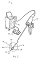
- a third expression shown in Figures 1-3 is for an ultrasound medical treatment system 10 including an end effector 12 insertable into a patient 14.
- the end effector 12 includes a tissue-retaining device 16.
- the tissue-retaining device 16 includes a first tissue-retaining member 18 having an (i.e., at least one) ultrasound medical-treatment transducer 20 and includes a second tissue-retaining member 22 having an (i.e., at least one) ultrasound reflector 28.
- the first and second tissue-retaining members 18 and 22 are operatively connected together to retain patient tissue 24 between the first and second tissue-retaining members 18 and 22 and to release patient tissue 24 so retained.
- Advantages of retaining patient tissue between two tissue-retaining members during ultrasound medical treatment by an ultrasound medical-treatment transducer of a first tissue-retaining member and an ultrasound reflector of a second tissue-retaining member include having a single instrument which ultrasonically medically treats patient tissue by direct ultrasound, which enhances the ultrasound medical treatment by reflected ultrasound, and which at the same time immobilizes patient tissue against undesired movement during the treatment.
- an ultrasound reflector 28 is a material which reflects ultrasound at least to a degree that would substantially medically affect patient tissue over a treatment period by direct ultrasound which is being reflected back by the ultrasound reflector.
- Choices of ultrasound reflecting materials include, without limitation, acoustically-rigid materials such as stainless steel (which reflects about 100%) and aluminum (which reflects about 80%) and acoustically-softer materials such as corporene (which reflects about 90%).
- An ultrasound reflecting material is contrasted with an ultrasound absorbing material such as, without limitation, rubber or plastic.
- the retained patient tissue 24 is retained between the ultrasound medical-treatment transducer 20 and the ultrasound reflector 28.
- the ultrasound medical-treatment transducer 20 and the ultrasound reflector 28 each focus ultrasound energy, such ultrasound reflector focusing being accomplished by the shape of, or by shaping, the reflector surface as is within the ordinary level of skill of the artisan.
- a fourth expression shown in Figures 1-3 is for an ultrasound medical treatment system 10 including an end effector 12 insertable into a patient 14.
- the end effector 12 includes a tissue-retaining device 16.
- the tissue-retaining device 16 includes a first tissue-retaining member 18 having an (i.e., at least one) ultrasound imaging and medical-treatment transducer 26 and includes a second tissue-retaining member 22 having an (i.e., at least one) ultrasound reflector 28.
- the first and second tissue-retaining members 18 and 22 are operatively connected together to retain patient tissue 24 between the first and second tissue-retaining members 18 and 22 and to release patient tissue 24 so retained.
- the retained patient tissue 24 is retained between the ultrasound imaging and medical-treatment transducer 26 and the ultrasound reflector 28.
- the ultrasound imaging and medical-treatment transducer 26 and the ultrasound reflector 28 each focus ultrasound energy.
- the ultrasound reflector 28 is disposed to receive ultrasound energy from the transducer 20 and 26 and is oriented to reflect the received ultrasound energy back into patient tissue 24 retained by the tissue-retaining device 16. In the same or a different example, the ultrasound reflector 28 is oriented to reflect the received ultrasound energy away from the transducer 20 and 26 when the patient tissue 14 is retained by the tissue-retaining device 16.
- one of the first and second tissue-retaining members 18 and 22 is controllably orientatable relative to the other of the first and second tissue-retaining members 18 and 22 such as, without limitation, by being orientatable along the double-headed arrows shown in Figure 2 .
- the second tissue-retaining member 22 is controllably orientatable relative to the first tissue-retaining member 18 to reflect the received ultrasound energy back along different directions.
- a first alternate end effector 30 is shown in Figure 4 wherein the second tissue-retaining member 32 is controllably orientatable relative to the first tissue-retaining member 34 as shown by the double-headed arrows in Figure 4 .
- Mechanisms, not shown, for remotely controlling the orientation of one member relative to another member are within the ordinary level of skill of the artisan and include, without limitation, the use of pivotal member attachments and the use of cables or motors.
- the transducer 20 and 26 generates wide-focused ultrasound (shown by the two single-headed arrows coming from the first tissue-retaining member 18 in Figure 3 ) and the ultrasound reflector 28 generates narrow-focused ultrasound (shown by the two single-headed arrows coming from the second tissue-retaining member 22 in Figure 3 ).
- the end effector 12 is an open-surgery end effector, an endoscopic end effector, a laparoscopic end effector (as shown in Figure 1 ), a catheter end effector (such as, but not limited to, an intravascular catheter end effector), or a needle end effector, as can be appreciated by those skilled in the art.
- the end effector 12 is used to retain a blood vessel and then to ultrasonically treat the blood vessel to seal the blood vessel stopping the flow of blood in the retained blood vessel.
- the end effector 12 is used to retain patient tissue and then to ultrasonically ablate at least a portion of the retained patient tissue.
- the end effector 12 has a longitudinal axis 35, and one of the first and second tissue-retaining members 18 and 22 at all times faces along a direction which is substantially perpendicular to the longitudinal axis 35. If the one tissue-retaining member were planar, this means that the longitudinal axis would be substantially parallel to the plane of the one tissue-retaining member. In one enablement, the one tissue-retaining member is the first tissue-retaining member 18.
- a second alternate end effector 36 has first and second tissue-retaining members 38 and 40 which are hinged together to relatively move as indicated by the double-headed arrow and which are shown in a partially open configuration in Figure 5 .
- the second alternate end effector 36 has a longitudinal axis 42, and one of the first and second tissue-retaining members 38 and 40 at all times faces along a direction which is substantially parallel to the longitudinal axis 42. If the one tissue-retaining member were planar, this means that the longitudinal axis would be substantially perpendicular to the plane of the one tissue-retaining member. In one enablement, the one tissue-retaining member is the first tissue-retaining member 38.
- a third alternate end effector 37 having first and second tissue-retaining members 39 and 41 with one member longitudinally movable with respect to the other member (as indicated by the double-headed arrow) is shown in Figure 6 .
- the third alternate end effector 37 has a longitudinal axis 43, and one of the first and second tissue-retaining members 39 and 41 at all times faces along a direction which is substantially parallel to the longitudinal axis 43.
- the one tissue-retaining member is the first tissue-retaining member 39.
- the ultrasound medical treatment system 10 also includes a handpiece 44 operatively connected to the end effector 12 and to an ultrasound controller 46 operatively connected to a foot-pedal power switch 47, as can be appreciated by those skilled in the art.
- a first method is for ultrasound medical treatment of a patient and uses the ultrasound medical treatment system as previously described in the first, second, third or fourth expression of the first embodiment with or without the previously-described variations, etc. thereof.
- the first method includes steps a) through e).
- Step a) includes endoscopically inserting the end effector into an ear, nose, or throat of the patient.
- Step b) includes guiding the end effector in the patient.
- Step c) includes identifying patient tissue for medical treatment such as optionally at least in part from ultrasound imaging using the transducer. Other ways of identifying patient tissue for medical treatment include, without limitation, using x-rays and/or MRI imaging, as are known to the artisan.
- Step d) includes retaining the identified patient tissue using the tissue-retaining device.
- Step e) includes medically treating the retained patient tissue with ultrasound using the transducer or using the transducer and the ultrasound reflector.
- one tissue-retaining member at all times faces along a direction which is substantially parallel to the longitudinal axis of the end effector (as seen in Figures 5 and 6 ).
- a second method is for ultrasound medical treatment of a patient and uses the ultrasound medical treatment system as previously described in the first, second, third or fourth expression of the first embodiment with or without the previously-described variations, etc. thereof.
- the second method includes steps a) through c).
- Step a) includes inserting the end effector 12 into the patient.
- Step b) includes retaining an intervertebral disk 48 (see Figure 3 ) of the patient with the tissue-retaining device, wherein the intervertebral disk 48 includes tissue.
- Step c) includes medically treating the retained intervertebral disk 48 with ultrasound to shrink the tissue using the transducer or using the transducer and the ultrasound reflector.
- one tissue-retaining member at all times faces along a direction which is substantially perpendicular to the longitudinal axis of the end effector (as seen in Figures 2 and 4 ).
- the intervertebral disk 48 includes connective and nerve tissue.
- a third method is for ultrasound medical treatment of a patient and uses the ultrasound medical treatment system as previously described in the first, second, third or fourth expression of the first embodiment with or without the previously-described variations, etc. thereof.
- the third method includes steps a) through c).
- Step a) includes inserting the end effector into the patient.
- Step b) includes retaining a joint of the patient with the tissue-retaining device, wherein the joint includes tissue.
- Step c) includes medically treating the retained joint with ultrasound to shrink the tissue using the transducer or using the transducer and the ultrasound reflector.
- one tissue-retaining member at all times faces along a direction which is substantially perpendicular to the longitudinal axis of the end effector (as seen in Figures 2 and 4 ).
- the joint includes connective and nerve tissue.
- one application of the ultrasound medical treatment system 10 of the previously-described first through fourth expressions of the first embodiment uses the tissue-retaining device to retain a blood vessel and uses the transducer, or the transducer and the ultrasound reflector, to substantially stop the flow of blood within the blood vessel.
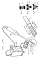
- FIGS 7-8 illustrate a second configuration not according to the present invention which is an ultrasound medical treatment system 50 including an end effector 52 insertable into a patient.
- the end effector 52 includes a tissue-retaining device 54.
- the tissue-retaining device 54 includes a first tissue-retaining member 56 having an ultrasound imaging and medical-treatment transducer 58 and includes a second tissue-retaining member 60 having an ultrasound reflector 62.
- the first and second tissue-retaining members 56 and 60 are operatively connected together to retain patient tissue between the first and second tissue-restraining members and to release patient tissue so retained.
- the first and second tissue-retaining members 56 and 60 always maintain a substantially parallel alignment.
- Advantages of having a substantially parallel alignment between the tissue-retaining members include, in one example, having the transducer and the ultrasound reflector maintain a substantially parallel alignment for improved reflected ultrasound medical treatment enhancement for any thickness of patient tissue retained by the tissue-retaining members.
- the first tissue-retaining member 56 is a distal end portion 64 of a first tube 66.
- the ultrasound medical treatment system 50 also includes a second tube 68, first and second link members 70 and 72, and a cable 74.
- the second tube 68 is oriented substantially parallel to the first tube 66.
- the first and second link members 70 and 72 are pivotally attached to the second tissue-retaining member 60 and to the second tube 68 at pivot points 76-82 creating a hinged parallelogram defined by a proximal portion 84 of the second tissue-retaining member 60, a distal portion 86 of the second tube 68, and the first and second link members 70 and 72.
- the ultrasound reflector 62 is disposed at a distal portion 88 of the second tissue-retaining member 60 and faces the transducer 58.
- the cable 74 is operatively connected to the hinged parallelogram to move the second tissue-retaining member 60 toward and away from the first tissue-retaining member 56.
- the ultrasound medical treatment system 50 also includes an outer tube 90.
- the cable 74 and the first and second tubes 66 and 68 are disposed in the outer tube 90.
- the ultrasound medical treatment system 50 also includes a handpiece 92.
- the cable 74 and the first, second, and outer tubes 66, 68 and 90 are operatively connected to the handpiece 92.
- the orientation of the first tube 66 about the longitudinal axis of the first tube 66 is controlled by a step motor (not shown) disposed in, and actuated by, the handpiece 92.
- the first tube 66 is a hollow tube allowing for transducer wiring (not shown), and the second tube is a solid tube (not shown).
- the tubes 66, 68, and 90 may be rigid or flexible which also is true for any tube arrangement (specifically disclosed as rigid or flexible, or not so specifically disclosed) of any end effector and for any end effector itself of any of the previous or following embodiments of the invention.
- FIG. 9-11 illustrate a third configuration not according to the present invention.
- a first expression is for an ultrasound medical system 94 including a tube 96 and a plurality of resiliently flexible fingers 98.
- the tube 96 has a distal end 100 insertable into a patient and has a lumen 102 with a distal opening 104.
- the fingers 98 are extendable out of the distal opening 104 of the lumen 102 creating a deployed state (seen in Figure 10 ) and which are at-least-partially retractable into the distal opening 104 of the lumen 102 creating a stowed state (seen in Figure 11 ).
- Each finger 98 includes an ultrasound transducer 106. The distance between the ultrasound transducers 106 of adjacent fingers 98 is greater in the deployed state than in the stowed state.
- an ultrasound medical system is a medical system which at least provides ultrasound imaging or ultrasound medical treatment of a patient.
- the tube and extendable/retractable flexible-finger array arrangement include, when the transducers are ultrasound medical-treatment transducers having a common focal zone in the deployed state, providing faster medical treatment times by allowing for more transducer ultrasound-emitting surface area which can be simply stowed into a compact shape for transport within a patient to and from the site of patient tissue receiving ultrasound medical treatment.
- the fingers 98 are only partially retracted into the distal opening 104 of the lumen 102 in the stowed state (as seen in Figure 11 ). In another variation, not shown, the fingers 98 are completely retracted into the distal opening 104 of the lumen 102 in the stowed state.
- the fingers 98 being extendable out of the distal opening 104 of the lumen 102 creating the deployed state and being at-least-partially retractable into the distal opening 104 of the lumen 102 creating the stowed state means the fingers 98 protrude more out of the distal opening 104 of the lumen 102 in the extended state than (if at all) in the stowed state.
- Mechanisms, not shown, for remotely extending and retracting fingers in a tube include, without limitation, a common shaft attached to the proximal ends of the fingers, disposed in the lumen of the tube, and spring-biased to move forward upon squeezing of a handpiece and to return backward upon relaxing of the handpiece, as is within the ordinary level of skill of the artisan.
- the distal opening 104 of the lumen 102 coincides with the distal end 100 of the tube 96.
- the distal opening of the lumen is spaced apart from the distal end of the tube.
- the distal opening 104 of the lumen 102 faces in the same direction as the distal end 100 of the tube 96.
- At least one of the transducers 106 is an ultrasound imaging transducer. In the same or a different example, at least one of the transducers 106 is an ultrasound medical-treatment transducer. In the same or a different example, at least one of the transducers 106 is an ultrasound imaging and medical-treatment transducer.
- a second expression is for an ultrasound medical treatment system 108 including a tube 96 and including an end effector 110 having a plurality of fingers 98.
- the tube 96 has a distal end 100 insertable into a patient and has a lumen 102 with a distal opening 104.
- the fingers 98 are extendable out of the distal opening 104 of the lumen 102 creating a deployed state (seen in Figure 10 ) and are at-least-partially retractable into the distal opening 104 of the lumen 102 creating a stowed state (seen in Figure 11 ).
- Each finger 98 includes an ultrasound medical-treatment transducer 112. The distance between the ultrasound medical-treatment transducers 112 of adjacent fingers 98 is greater in the deployed state than in the stowed state.
- a third expression is for an ultrasound medical treatment system 108 including a tube 96 and including an end effector 110 having a plurality of fingers 98.
- the tube 96 has a distal end 100 insertable into a patient and has a lumen 102 with a distal opening 104.
- the fingers 98 are extendable out of the distal opening 104 of the lumen 102 creating a deployed state (seen in Figure 10 ) and are at-least-partially retractable into the distal opening 104 of the lumen 102 creating a stowed state (seen in Figure 11 ).
- Each finger 98 includes an ultrasound imaging and medical-treatment transducer 114. The distance between the ultrasound imaging and medical-treatment transducers 114 of adjacent fingers 98 is greater in the deployed state than in the stowed state.
- the transducers 106, 112 and 114 each have an ultrasound-emitting concave surface 116.
- the transducers have a planar ultrasound-emitting surface.
- each concave surface 116 is concave as one moves along the corresponding finger 98 (as best seen in Figure 10 ).
- each concave surface is concave as one moves across the corresponding finger or is concave as one moves both along and across the corresponding finger (such as, for example, with a hemispherically-concave surface).
- the concave surfaces 116 together have a substantially common focal zone when the fingers 98 are in the deployed state.
- the end effector 110 is seen with its fingers 98 facing the patient tissue 119 in Figure 10 .
- the focal zones are not common.
- the fingers 98 define an open-hand finger array 118 in the deployed state.
- An alternate flexible finger arrangement in the form of a substitute end effector 120 is shown in Figure 12 , wherein the fingers 122 define a clawed-hand finger array 124 in the deployed state.
- the substitute end effector 120 is seen with its fingers 122 surrounding the patient tissue 126 for imaging and/or medical treatment by the ultrasound transducers 128 in Figure 12 . In other transducer arrangements, not shown, one or more or all of the ultrasound transducers face outward rather than facing inward.
- the fingers 98 are at least four in number.
- the end effector 110 (as well as the substitute end effector 120) is an open-surgery end effector, an endoscopic end effector, a laparoscopic end effector (as shown in Figure 9 ), a catheter end effector (such as, but not limited to, an intravascular catheter end effector), or a needle end effector, as can be appreciated by those skilled in the art.
- the ultrasound medical treatment system 108 also includes a handpiece 130 operatively connected to the end effector 110 and to an ultrasound controller 132 operatively connected to a foot-pedal power switch 133, as can be appreciated by those skilled in the art.
- a fourth configuration not according to the present invention is shown in Figures 13-15 .
- a first expression is for an ultrasound medical system 134 including an ultrasound transducer assembly 136 insertable into a patient.
- the ultrasound transducer assembly 136 has a longitudinal axis 138.
- the ultrasound transducer assembly 136 includes a plurality P of ultrasound transducers 140.
- Each transducer 140 has an ultrasound-emitting surface 142 oriented at an angle of substantially 360/P degrees apart from the ultrasound-emitting surface 142 of an adjacent transducer 140 when viewed in a cross section (see Figure 15 ) of the transducer assembly 136 taken by a cutting plane which is perpendicular to the longitudinal axis 138.
- Such a transducer configuration include, in one example, providing directed or focused medical-treatment ultrasound which is not possible with a cylindrical ultrasound transducer, as can be appreciated by those skilled in the art.
- an ultrasound transducer assembly 136 insertable into a patient is an ultrasound imaging transducer assembly, an ultrasound medical-treatment transducer assembly, or an ultrasound imaging and medical-treatment transducer assembly.
- An ultrasound imaging transducer assembly has at least one ultrasound imaging transducer, and an ultrasound medical-treatment transducer assembly has at least one ultrasound medical-treatment transducer.
- An ultrasound imaging and medical-treatment transducer assembly has at least one ultrasound imaging transducer and at least one ultrasound medical-treatment transducer or has at least one ultrasound imaging and medical-treatment transducer.
- a second expression is for an ultrasound medical-treatment system 144 including an end effector 146 insertable into a patient.
- the end effector 146 includes an ultrasound medical-treatment transducer assembly 148.
- the ultrasound medical-treatment transducer assembly 148 has a longitudinal axis 138.
- the ultrasound medical-treatment transducer assembly 148 includes a plurality P of ultrasound medical-treatment transducers 150.
- Each transducer 150 has an ultrasound-emitting surface 142 which faces away from the longitudinal axis 138 and which is oriented at an angle of substantially 360/P degrees apart from the ultrasound-emitting surface 142 of an adjacent transducer 150 when viewed in a cross section (see Figure 15 ) of the transducer assembly 148 taken by a cutting plane which is perpendicular to the longitudinal axis 138.
- at least one of the ultrasound medical-treatment transducers 150 is also adapted for ultrasound imaging.
- a method is for ultrasound medical treatment of a patient and uses the ultrasound medical treatment system 144 as previously described in the second expression.
- the fourth method includes steps a) through b).
- Step a) includes inserting the end effector 146 into the liver of the patient.
- Step b) includes medically treating a lesion in the liver with ultrasound from the ultrasound medical-treatment transducer assembly 148.
- step a) interstially inserts the end effector 146 into the lesion.
- step a) endoscopically inserts the end effector 146 into the liver through the hepato-biliary duct system.
- a third expression is for an ultrasound medical treatment system 144 including an end effector 146 insertable into a patient.
- the end effector 146 includes an ultrasound imaging and medical-treatment transducer assembly 152.
- the ultrasound imaging and medical-treatment transducer assembly 152 has a longitudinal axis 138.
- the ultrasound imaging and medical-treatment transducer assembly 152 includes a plurality P of ultrasound imaging and medical-treatment transducers 154.
- Each transducer 154 has an ultrasound-emitting surface 142 which faces away from the longitudinal axis 138 and which is oriented at an angle of substantially 360/P degrees apart from the ultrasound-emitting surface 142 of an adjacent transducer 154 when viewed in a cross section (see Figure 15 ) of the transducer assembly 152 taken by a cutting plane which is perpendicular to the longitudinal axis 138.
- a method is for ultrasound medical treatment of a patient and uses the ultrasound medical-treatment system 144 as previously described in the third expression of the fourth embodiment.
- the fourth method includes steps a) through c).
- Step a) includes inserting the end effector 146 into the liver of the patient.
- Step b) includes identifying a lesion in the liver for medical treatment at least in part from ultrasound imaging using the ultrasound imaging and medical-treatment transducer assembly 152.
- Step c) includes medically treating the lesion with ultrasound from the ultrasound imaging and medical-treatment transducer assembly 152.
- step a) interstially inserts the end effector 146 into the lesion.
- step a) endoscopically inserts the end effector 146 into the liver through the hepato-biliary duct system.
- the transducer assembly 136, 148, and 152 has a distal tip 156 and has a tip transducer 158.
- the tip transducer is a forward facing tip transducer.
- the tip transducer is a sideways facing tip transducer.
- the tip transducer is an ultrasound imaging tip transducer.
- the tip transducer is an ultrasound medical-treatment tip transducer.
- the tip transducer is an ultrasound imaging and medical-treatment tip transducer.
- the tip transducer is a transponder which emits electromagnetic waves or mechanical waves or both.
- each ultrasound-emitting surface 142 is substantially straight when viewed in the cross section, as seen in Figure 15 .
- each ultrasound-emitting surface 142 has a substantially concave shape as one moves along the ultrasound-emitting surface 142 in a direction parallel to the longitudinal axis 138, and each ultrasound-emitting surface 142 has a focal zone.
- each ultrasound-emitting surface 162 has a substantially planar shape.
- each ultrasound-emitting surface 164 has a substantially concave shape when viewed in the cross section, and each ultrasound-emitting surface 164 has a focal zone.
- each ultrasound-emitting surface 164 also has a substantially concave shape as one moves along the ultrasound-emitting surface 164 in a direction parallel to the longitudinal axis (such as, for example, by the ultrasound-emitting surface 164 having a hemispherically-concave shape).
- Such ultrasound-emitting surface shapes are equally applicable to any ultrasound transducer mentioned in any other embodiment of the invention.
- P is no greater than four. In one variation, P equals three as seen in Figures 15 and 17 . In another variation, P equals two as seen in Figure 16 .
- the end effector 146 is an open-surgery end effector, an endoscopic end effector, a laparoscopic end effector (as shown in Figure 13 ), a catheter end effector (such as, but not limited to, an intravascular catheter end effector), or a needle end effector, as can be appreciated by those skilled in the art.
- the ultrasound medical treatment system 144 also includes a handpiece 166 operatively connected to the end effector 146 and to an ultrasound controller 168 operatively connected to a foot-pedal power switch 169, as can be appreciated by the artisan.
- an ultrasound medical treatment system 170 includes a tube 172, a first end effector 174, and a second end effector 176.
- the tube 172 has a distal end 178 insertable into a patient 180 and has a lumen 182.
- the first end effector 174 has a cutting tool 184, is introducible into the lumen 182 of the inserted tube 172 from outside the patient 180, and is translatable through the lumen 182 of the inserted tube 172 to inside the patient 180.
- the second end effector 176 has an ultrasound medical-treatment transducer assembly 186, is introducible into the lumen 182 of the inserted tube 172 from outside the patient 180, and is translatable through the lumen 182 of the inserted tube 172 to inside the patient 180.
- the first and second end effectors are introduced into the lumen through separate openings in the lumen or through separate branch channels leading to the lumen.
- the first and second end effectors are introduced into the lumen through the same opening in the lumen.
- a lumen opening is disposed at the end of the tube. According to the invention a lumen opening is spaced apart from the end of the tube.
- a second expression of the embodiment of the present invention is for an ultrasound medical treatment system 170 including a tube 172, a first end effector 174, and a second end effector 176.
- the tube has a distal end 178 insertable into a patient 180 and has a lumen 182 with a distal opening 188 and a proximal opening 190.
- the first end effector 174 has a cutting tool 184, is introducible into the proximal opening 190, and is translatable through the lumen 182 to the distal opening 188.
- the second end effector 176 has an ultrasound medical-treatment transducer assembly 186, is introducible into the proximal opening 190, and is translatable through the lumen 182 to the distal opening 188.
- the lumen 182 is sized to allow introduction of only one of the first and second end effectors 174 and 176 at a time.
- the distal end 178 of the tube 172 is interstitially insertable into patient tissue 192 of the patient 180.
- the cutting tool 184 is a biopsy cutting tool 194 or other excisional cutting tool.
- a third expression of the embodiment of the present invention is for an ultrasound medical treatment system 170 including a tube 172, a first end effector 174, and a second end effector 176.
- the tube 172 has a distal end 178 interstitially insertable into breast tissue 196 of a patient 180 and has a lumen 182 with a distal opening 188 and a proximal opening 190.
- the first end effector 174 has a biopsy cutting tool 194 (or other excisional cutting tool), is introducible into the proximal opening 190, and is translatable through the lumen 182 to the distal opening 188.
- the second end effector 176 has an ultrasound medical-treatment transducer assembly 186, is introducible into the proximal opening 190, and is translatable through the lumen 182 to the distal opening 188.
- the lumen 182 is sized to allow introduction of only one of the first and second end effectors 174 and 176 at a time.
- the first end effector also includes a suction mechanism to draw in patient tissue to be biopsied by the biopsy cutting tool 194.
- the tube 172 and the first end effector 174 (with the biopsy cutting tool 194 including a suction mechanism) are based on components of a Mammotome® Breast Biopsy System manufactured by Ethicon Endo-Surgery, Inc. (a Johnson & Johnson Company).
- a method is for ultrasound medical treatment of a patient 180 and uses the ultrasound medical treatment system 170 as previously described in the third expression of the embodiment of the present invention.
- the method includes steps a) through h).
- Step a) includes identifying possibly cancerous breast tissue 196 of the patient.
- Step b) includes interstitially inserting the distal end 178 of the tube 172 into the patient 180 with the distal opening 188 disposed proximate the breast tissue 196 and with the proximal opening 190 disposed outside the patient.
- Step c) includes introducing the first end effector 174 into the proximal opening 190 and translating the first end effector 174 through the lumen 182 to the distal opening 188.
- Step d) includes obtaining a biopsy sample of the breast tissue 196 with the biopsy cutting tool 194.
- Step e) includes removing the first end effector 174 from the lumen 182
- Step f) includes introducing the second end effector 176 into the proximal opening 190 and translating the second end effector 176 through the lumen 182 to the distal opening 188.
- Step g) includes identifying an area of hemorrhaging in the breast tissue where the biopsy sample was obtained.
- Step h) includes medically treating the identified area with ultrasound using the transducer assembly 186 to substantially stop the hemorrhaging.
- the method also includes the steps of testing the biopsy sample for cancer and substantially ablating any remaining cancer in the breast tissue with ultrasound using the transducer assembly 186.
- Advantages of such an ultrasound medical treatment system and method include the ease of obtaining a breast biopsy and the control of hemorrhaging caused by the biopsy procedure coupled together in a minimally invasive manner.
- an ultrasound medical treatment system 170 includes a tube 172, a first end effector 174, and a second end effector 176.
- the tube 172 has a distal end 178 insertable into a patient 180 and has a lumen 182.
- the first end effector 174 has a cutting tool 184, is introducible into the lumen 182 of the inserted tube 172 from outside the patient 180, and is translatable through the lumen 182 of the inserted tube 172 to inside the patient 180.
- the second end effector 176 has an ultrasound imaging and medical-treatment transducer assembly 198, is introducible into the lumen 182 of the inserted tube 172 from outside the patient 180, and is translatable through the lumen 182 of the inserted tube 172 to inside the patient 180.
- the first and second end effectors are introduced into the lumen through separate openings in the lumen or through separate branch channels leading to the lumen.
- the first and second end effectors are introduced into the lumen through the same opening in the lumen.
- a lumen opening is disposed at the end of the tube. According to the invention, a lumen opening is spaced apart from the end of the tube.
- a fifth expression of the embodiment of the present invention is for an ultrasound medical treatment system 170 including a tube 172, a first end effector 174, and a second end effector 176.
- the tube has a distal end 178 insertable into a patient 180 and has a lumen 182 with a distal opening 188 and a proximal opening 190.
- the first end effector 174 has a cutting tool 184, is introducible into the proximal opening 190, and is translatable through the lumen 182 to the distal opening 188.
- the second end effector 176 has an ultrasound imaging and medical-treatment transducer assembly 198, is introducible into proximal opening 190, and is translatable through the lumen 182 to the distal opening 188.
- the lumen 182 is sized to allow introduction of only one of the first and second end effectors 174 and 176 at a time.
- the distal end 178 of the tube 172 is interstitially insertable into patient tissue 192 of the patient 180.
- the cutting tool 184 is a biopsy cutting tool 194 or other excisional-cutting tool.
- a sixth expression of the embodiment of the present invention is for an ultrasound medical treatment system 170 including a tube 172, a first end effector 174, and a second end effector 176.
- the tube 172 has a distal end 178 interstitially insertable into breast tissue 196 of a patient 180 and has a lumen 182 with a distal opening 188 and a proximal opening 190.
- the first end effector 174 has a biopsy cutting tool 194 (or other excisional cutting tool), is introducible into the proximal opening 190, and is translatable through the lumen 182 to the distal opening 188.
- the second end effector 176 has an ultrasound imaging and medical-treatment transducer assembly 196, is introducible into the proximal opening 190, and is translatable through the lumen 182 to the distal opening 188.
- the lumen 182 is sized to allow introduction of only one of the first and second end effectors 174 and 176 at a time.
- the tube 172 and the first end effector 174 are based on components of a Mammotome® Breast Biopsy System manufactured by Ethicon Endo-Surgery, Inc. (a Johnson & Johnson Company).
- a method is for ultrasound medical treatment of a patient 180 and uses the ultrasound medical treatment system 170 as previously described in the sixth expression of the embodiment of the present invention.
- the method includes steps a) through h).
- Step a) includes identifying possibly cancerous breast tissue 196 of the patient.
- Step b) includes interstitially inserting the distal end 178 of the tube 172 into the patient 180 with the distal opening 188 disposed proximate the breast tissue 196 and with the proximal opening 190 disposed outside the patient.
- Step c) includes introducing the first end effector 174 into the proximal opening 190 and translating the first end effector 174 through the lumen 182 to the distal opening 188.
- Step d) includes obtaining a biopsy sample of the breast tissue 196 with the biopsy cutting tool 194.
- Step e) includes removing the first end effector 174 from the lumen 182
- Step f) includes introducing the second end effector 176 into the proximal opening 190 and translating the second end effector 176 through the lumen 182 to the distal opening 188.
- Step g) includes identifying an area of hemorrhaging in the breast tissue where the biopsy sample was obtained from ultrasound imaging using the transducer assembly 198.
- Step h) includes medically treating the identified area with ultrasound using the transducer assembly 198 to substantially stop the hemorrhaging.
- the method also includes the steps of testing the biopsy sample for cancer and substantially ablating any remaining cancer in the breast tissue with ultrasound using the transducer assembly 198.
- Advantages of such an ultrasound medical treatment system and method include the ease of obtaining a breast biopsy and the imaging and control of hemorrhaging caused by the biopsy procedure coupled together in a minimally invasive manner.
- the ultrasound medical treatment system 170 also includes a handpiece 199 which is attached to the tube 172, which contains the first end effector 174 for extending the cutting tool 184 into, and withdrawing it from, the lumen 182, and which is operatively connected to an ultrasound controller 201 via a first cable 203.
- the second end effector 176 in this enablement, is operatively connected to the ultrasound controller 201 via a second cable 205 and is inserted into the lumen 182 from outside the handpiece 199 as shown in Figure 18 .
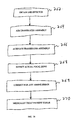
- a method is shown in block diagram form in Figure 21 and is for medical treatment of a patient.
- the method includes steps a) through f).
- Step a) is labeled "Obtain Transducer Assembly” in block 200 of Figure 21 .
- Step a) includes obtaining an ultrasound imaging transducer assembly.
- Step b) is labeled "Insert Assembly Into Gastrointestinal Area” in block 202 of Figure 21 .
- Step b) includes inserting the transducer assembly into a gastrointestinal area of the patient.
- Step c) is labeled "Guide Assembly” in block 204 of Figure 21 .
- Step c) includes guiding the transducer assembly within the gastrointestinal area.
- Step d) is labeled "Identify Patient Tissue For Treatment" in block 206 of Figure 21 .
- Step d) includes identifying patient tissue in the gastrointestinal area for medical treatment.
- Step e) is labeled "Stage Treatment From Ultrasound Imaging” in block 208 of Figure 21 .
- Step e) includes staging the medical treatment from ultrasound imaging using the transducer assembly.
- Step f) is labeled as "Medically Treat Patient” in block 210 of Figure 21 .
- Step f) includes medically treating the patient tissue according to the staging of step e). It is pointed out that in the method the medical treatment need not include ultrasound medical treatment with the transducer assembly used for staging and/or need not include ultrasound medical treatment with any other ultrasound transducer assembly.
- a first transducer assembly is used endoscopically to stage the medical treatment in step e) and a second transducer assembly is used laparoscopically to medically treat the patient tissue with ultrasound in step f).
- the first transducer assembly is used laparoscopically to stage the medical treatment in step e) and the second transducer assembly is used endoscopically to medically treat the patient tissue with ultrasound in step f).
- the medical treatment in step f) is radio-frequency, laser, microwave, or chemical ablation medical treatment. Other types of medical treatment are left to the artisan.
- the gastrointestinal (GI) area of a human patient includes, without limitation, the esophagus and the stomach of the upper GI area and the rectum and the colon of the lower GI area. It further is noted that the liver is also considered to be in the GI area for purposes of this method.
- GI tumors can be visualized with high frequency (6-30 MHz) ultrasound imaging using a cylindrical, side-firing, or half-convex ultrasound array or single-element transducer introduced endoscopically into the GI tract. All layers of the GI tract can be visualized including all layers of the esophagus, stomach, duodenum, colon, etc.
- a three-dimensional representation of the GI structures is created by collating a series of two-dimensional scans generated by axially advancing the ultrasound transducer. Any neoplastic growth, its morphological characteristics, as well as the tumor's size and shape can easily be determined from the three-dimensional representation.
- Such medical-treatment staging from ultrasound imaging include, in one example, providing a non-invasive medical-treatment staging technique which has greater resolution and which is more practical compared to conventional extracorporeal medical-treatment staging techniques such as using x-rays or MRI imaging or compared to using conventional endoscopic optical techniques.
- a method is for ultrasound medical treatment of a patient and includes steps a) through f).
- the method uses the same block diagram of Figure 21 as does the eighth method but with "end effector” replacing “transducer assembly” in block 200 and with “end effector” replacing “assembly” in blocks 202 and 204.
- Step a) includes obtaining an end effector having an ultrasound imaging and medical-treatment transducer assembly.
- Step b) includes inserting the end effector into a gastrointestinal area of the patient.
- Step c) includes guiding the transducer assembly within the gastrointestinal area.
- Step d) includes identifying patient tissue in the gastrointestinal area for medical treatment.
- Step e) includes staging the medical treatment from ultrasound imaging using the transducer assembly.
- Step f) includes medically treating the patient tissue with ultrasound using the transducer assembly according to the staging of step e).
- a method is for ultrasound medical treatment of a patient and includes steps a) through f).
- the method uses the same block diagram of Figure 21 as does a previous method but with "end effector” replacing “transducer assembly” in block 200 and with “end effector” replacing “assembly” in blocks 202 and 204.
- Step a) includes obtaining an end effector having an ultrasound imaging and medical-treatment transducer assembly.
- Step b) includes inserting the end effector into a gastrointestinal area of the patient.
- Step c) includes guiding the transducer assembly within the gastrointestinal area.
- Step d) includes identifying patient tissue in the gastrointestinal area for medical treatment at least in part from ultrasound imaging using the transducer assembly.
- Step e) includes staging the medical treatment from ultrasound imaging using the transducer assembly.
- Step f) includes medically treating the patient tissue with ultrasound using the transducer assembly according to the staging of step e).
- large GI tumors are staged through a laparoscopic access to the GI area, whereby the tumors are identified, staged and treated using an end effector having an ultrasound imaging and medical-treatment transducer assembly.
- the patient tissue is gastroesophageal tissue containing a lesion, and step f) ultrasonically substantially ablates the lesion.
- the gastroesophageal tissue contains a blood vessel supplying blood to the lesion, and step f) ultrasonically treats the blood vessel to substantially stop the supply of blood to the lesion from the blood vessel.
- the patient tissue is liver tissue containing a lesion and a blood vessel supplying blood to the lesion
- step f) ultrasonically treats the blood vessel to substantially stop the supply of blood to the lesion from the blood vessel.
- the patient tissue is liver tissue containing a lesion, and step f) ultrasonically substantially ablates the lesion.
- the liver tissue contains a blood vessel supplying blood to the lesion, and step f) also ultrasonically treats the blood vessel to substantially stop the supply of blood to the lesion from the blood vessel.
- an end effector having an ultrasound imaging and medical-treatment transducer assembly is introduced endoscopically into the GI tract, is advanced retrogradely through the ampulla of Vater up the common bile duct, and is advanced further into the hepatic duct system where liver parenchyma requiring medical treatment (such as cholangio-carcinomas) are identified, staged, and treated using the end effector.
- a method is shown in block diagram form in Figure 22 and is for ultrasound medical treatment of a patient.
- the method includes steps a) through f).
- Step a) is labeled "Obtain End Effector” in block 212 of Figure 22 .
- Step a) includes obtaining an end effector having an ultrasound medical-treatment transducer assembly.
- Step b) is labeled "Insert End Effector” in block 214 of Figure 22 .
- Step b) includes inserting the end effector into the patient.
- Step c) is labeled "Guide End Effector To Lung” in block 216 of Figure 22 .
- Step c) includes guiding the end effector within the patient to a lung of the patient.
- Step d) is labeled "Identify Lesion" in block 218 of Figure 22 .
- Step d) includes identifying a lesion on or in the lung for medical treatment.
- Step e) is labeled "Position Transducer Assembly” in block 220 of Figure 22 .
- Step e) includes positioning the transducer assembly on or in the lesion.
- Step f) is labeled "Medically Treat Lesion” in block 222 of Figure 22 .
- Step f) includes medically treating the lesion with ultrasound using the transducer assembly.
- a method is for ultrasound medical treatment of a patient and includes steps a) through f).
- the method uses the same block diagram of Figure 22 as does the eleventh method.
- Step a) includes obtaining an end effector having an ultrasound imaging and medical-treatment transducer assembly.
- Step b) includes inserting the end effector into the patient.
- Step c) includes guiding the end effector within the patient to a lung of the patient.
- Step d) includes identifying a lesion on or in the lung for medical treatment at least in part from ultrasound imaging using the transducer assembly.
- Step e) includes positioning the transducer assembly on or in the lesion.
- Step f) includes medically treating the lesion with ultrasound using the transducer assembly.
- step f) ultrasonically substantially ablates the lesion.
- the end effector is an endoscopic end effector and step b) transbronchial-endoscopically inserts the end effector into the patient.
- the end effector is a needle end effector and step b) interstitially inserts the end effector into the patient.
- step e) positions the transducer assembly on the lesion.
- step e) positions the transducer assembly in the lesion.
- step c) a bronchoscope is used to guide the end effector to a lung of the patient.
- Ultrasound medical treatment of the lung has conventionally been avoided because such ultrasound is prevented from reaching a lesion within the lung by the alveoli of the lung which contain air which reflect back most of the ultrasound preventing the ultrasound from effectively penetrating the lung to the lesion.
- Using higher power ultrasound for effective penetration of the lung to reach the lesion would injure or destroy the alveoli which are needed for breathing.
- positioning the ultrasound transducer on or in a lesion of the lung would allow ultrasound medical treatment of the lesion (such as a tumor or an infarct) without injury to the alveoli.
- Applicants' method is applicable to surface lesions as well as non-surface lesions.
- Advantages of Applicants' methods for ultrasound medical treatment include, in one example, the destruction of lung cancer lesions in cases which otherwise would be inoperable or incurable.
- a method of the invention is shown in block diagram form in Figure 23 and is for ultrasound medical treatment of a patient.
- the method includes steps a) through e).
- Step a) is labeled "Obtain End Effector” in block 224 of Figure 23 .
- Step a) includes obtaining an end effector having an ultrasound medical-treatment transducer assembly.
- Step b) is labeled "Insert End Effector” in block 226 of Figure 23 .
- Step b) includes inserting the end effector into the patient.
- Step c) is labeled "Guide End Effector” in block 228 of Figure 23 .
- Step c) includes guiding the end effector within the patient to a region of patient tissue containing a lesion.
- Step d) is labeled "Identify Blood Vessel Supplying Lesion" in block 230 of Figure 23 .
- Step d) includes identifying a blood vessel in the region which supplies blood to the lesion.
- Step e) is labeled "Stop Blood Supply Using Ultrasound” in block 232 of Figure 23 .
- Step e) includes medically treating the blood vessel with ultrasound from the transducer assembly to substantially seal the blood vessel to stop the supply of blood to the lesion from the blood vessel.
- One implementation of the method of the invention also includes the step of medically treating the lesion with ultrasound from the transducer assembly to substantially ablate the lesion.
- a method is for ultrasound medical treatment of a patient and includes steps a) through g). The method is similar to the thirteenth method.
- Step a) includes obtaining an end effector having an ultrasound imaging and medical-treatment transducer assembly.
- Step b) includes inserting the end effector into the patient.
- Step c) includes guiding the end effector within the patient to a region of patient tissue containing a lesion.
- Step d) includes identifying the lesion at least in part from ultrasound imaging using the transducer assembly.
- Step e) includes identifying a blood vessel in the region which supplies blood to the lesion from ultrasound imaging using the transducer assembly.
- Step f) includes medically treating the blood vessel with ultrasound from the transducer assembly to substantially seal the blood vessel to substantially stop the supply of blood to the lesion from the blood vessel.
- Step g) includes medically treating the lesion with ultrasound from the transducer assembly to substantially ablate the lesion. It is noted that Doppler ultrasound imaging alone, gray-scale ultrasound imaging alone, and a combination of Doppler and gray-scale ultrasound imaging are known ultrasound techniques to image blood flow in blood vessels.
- the end effector is an open-surgery end effector.
- the end effector is an endoscopic end effector.
- the end effector is a laparoscopic end effector.
- the end effector is a catheter end effector (such as, but not limited to, an intravascular catheter end effector).
- the end effector is a needle end effector.
- a broadened method of the invention eliminates the inserting into and guiding within steps of the above-described thirteenth method and includes steps a) through c).
- Step a) includes obtaining an end effector having an ultrasound medical-treatment transducer assembly.
- Step b) includes identifying a blood vessel in the patient which supplies blood to a lesion.
- Step c) includes medically treating the blood vessel with ultrasound from the transducer assembly to substantially seal the blood vessel to substantially stop the supply of blood to the lesion from the blood vessel.
- a broadened method of the invention eliminates the inserting into and guiding within steps of the above-described fourteenth method and includes steps a) through e).
- Step a) includes obtaining an end effector having an ultrasound imaging and medical-treatment transducer assembly.
- Step b) includes identifying a lesion in the patient at least in part from ultrasound imaging using the transducer assembly.
- Step c) includes identifying a blood vessel which supplies blood to the lesion from ultrasound imaging using the transducer assembly.
- Step d) includes medically treating the blood vessel with ultrasound from the transducer assembly to substantially seal the blood vessel to substantially stop the supply of blood to the lesion from the blood vessel.
- Step e) includes medically treating the lesion with ultrasound from the transducer assembly to substantially ablate the lesion.
- the end effector is an extracorporeal end effector.
- the end effector is an intracorporeal end effector.
- the end effector can be used in both an extracorporeal mode and in an intracorporeal mode.
- Advantages of Applicants' methods and broadened methods for ultrasound medical treatment include, in one example, the indirect destruction of cancer lesions by ultrasound hemostasis in blood vessels supplying the cancer lesions in cases which otherwise would be inoperable or incurable because the location of the cancer lesions prevents medical treatment of the lesions themselves.
- Advantages of Applicants' methods and broadened methods for ultrasound treatment include, in one example, direct destruction of cancer lesions by ultrasound ablation of the cancer lesions together with the indirect destruction of any cancer lesions missed in the ultrasound ablation step by ultrasound hemostasis in blood vessels supplying blood to the missed cancer lesions.
- an ultrasound medical treatment system 234 (only a portion of which is shown in Figure 24 ) includes an end effector 236 and at least three receivers 238.
- the end effector 236 has a transducer assembly 240 including a transducer 242 having at least one transducer element 244 adapted for emitting medical-treatment ultrasound waves and for emitting mechanical waves.
- mechanical waves includes ultrasound and non-ultrasound compression (acoustic) waves and ultrasound and non-ultrasound shear waves, and that waves include wave pulses.
- the receivers 238 are spaced apart from the transducer assembly 240, and the receivers 238 are adapted to receive the emitted mechanical waves for use in locating the position of the transducer assembly 240.
- Conventional methods for locating the position of a transponder emitting waves which are received by three receivers are well known.
- a second expression of the fifth configuration is identical to the first expression of the fifth configuration except that the at-least-one transducer element 244 is also adapted for emitting imaging ultrasound waves.
- the end effector and the receivers are disposable outside (including in one modification on) the patient.
- the end effector is insertable into the patient and the receivers are disposable outside (including in one modification on) the patient.
- an ultrasound medical treatment system 246 (only a portion of which is shown in Figure 25 ) includes an end effector 248 and at least three receivers 250.
- the end effector 248 has an ultrasound medical-treatment transducer assembly 252 and has a transponder 254
- the transponder 254 is adapted to emit waves, and the waves include electromagnetic waves or mechanical waves or both.
- the receivers 250 are spaced apart from the transducer assembly 252, and the receivers 250 are adapted to receive the emitted waves for use in locating the position of the transponder 254.
- the ultrasound medical-treatment transducer assembly 252 is an ultrasound imaging and medical-treatment transducer assembly 256.
- the end effector 248 is insertable into a patient, the transponder 254 is adapted to emit electromagnetic waves, and the receivers 250 are disposable outside the patient.
- the receivers 250 are disposable on the patient.
- the end effector is disposable outside (including in one modification on) the patient and the receivers are disposable outside (including in one modification on) the patient.
- the end effector 248 is an endoscopic end effector, a laparoscopic end effector, a catheter end effector (such as, but not limited to, an intravascular catheter end effector), or a needle end effector.
- the end effector 248 has a distal tip 260, and the transponder 254 is disposed at the distal tip 260 of the end effector 248.
- the transducer assembly 252 and 256 is disposed proximate the transponder 254.
- a method uses the ultrasound medical treatment system of the first expression of the sixth configuration and includes steps a) through h).
- Step a) includes inserting the end effector 248 into the patient.
- Step b) includes disposing the receivers 250 outside the patient.
- Step c) includes emitting electromagnetic waves from the transponder 254.
- Step d) includes receiving the electromagnetic waves with the disposed receivers 250.
- Step e) includes calculating the position of the transponder 254 from the received electromagnetic waves.
- Step f) includes guiding the end effector within the patient to a desired location from the calculated position of the transponder 254.
- Step g) includes, after step f), identifying patient tissue for medical treatment.
- Step h) includes medically treating the identified patient tissue with ultrasound using the transducer assembly 252.
- a method uses the ultrasound medical treatment system of the second expression of the sixth configuration and includes steps a) through h).
- Step a) includes inserting the end effector 248 into the patient.
- Step b) includes disposing the receivers 250 outside the patient.
- Step c) includes emitting electromagnetic waves from the transponder 254.
- Step d) includes receiving the electromagnetic waves with the disposed receivers 250.
- Step e) includes calculating the position of the transponder 254 from the received electromagnetic waves.
- Step f) includes guiding the end effector within the patient to a desired location from the calculated position of the transponder 254.
- Step g) includes, after step f), identifying patient tissue for medical treatment at least in part from ultrasound imaging using the transducer assembly 256.
- Step h) includes medically treating the identified patient tissue with ultrasound using the transducer assembly 256.
- a known electromagnetic transponder and three-receiver system for calculating the position of the transponder and for guiding the transponder (which is attached to a heart catheter for monitoring the heart) inside a patient is the CARTOTM EP Navigation System used with a NAVI-STAR® catheter manufactured by Biosense Webster (a Johnson & Johnson Company).
- an end effector with ultrasound medical treatment and position-location capabilities include, in one example, more accurately guiding the end effector inside a patient to patient tissue for ultrasound medical treatment of the patient tissue.
- a method is shown in block diagram form in Figure 26 and is for ultrasound medical treatment of a patient.
- the method includes steps a) through f).
- Step a) is labeled "Obtain End Effector” in block 262 of Figure 26 .
- Step a) includes obtaining an end effector having an ultrasound medical-treatment transducer assembly.
- Step b) is labeled "Aim Transducer Assembly" in block 264 of Figure 26 .
- Step b) includes aiming the transducer assembly to focus ultrasound energy at a desired focal zone of patient tissue. It is noted that, in one example, to aim a transducer assembly means to focus ultrasound energy at a particular distance from the transducer assembly and along a particular direction.
- Step c) is labeled "Activate Transducer Assembly" in block 266 of Figure 26 .
- Step c) includes activating the aimed transducer assembly to emit ultrasound energy sufficient to achieve a temperature increase in the patient tissue essentially without medically affecting the patient tissue.
- Step d) is labeled "Detect Actual Focal Zone” in block 268 of Figure 26 .
- Step d) includes after step c) detecting, from reflected ultrasound energy, an actual focal zone of patient tissue having a temperature increase.
- Step e) is labeled "Correct For Any Aiming Error" in block 269 of Figure 26 .
- Step e) includes correcting for any error between the desired focal zone and the actual focal zone.
- Step f) is labeled "Medically Treat Patient Tissue" in block 270 of Figure 26 .
- Step f) includes after step e), medically treating the patient tissue with ultrasound using the transducer assembly.
- step d) uses one or more additional ultrasound transducer assemblies, separate from the ultrasound transducer assembly used in steps a) through c) and e) through f), to detect, from reflected ultrasound energy, the actual focal zone.
- the same ultrasound transducer assembly is used for steps a) through f).
- the end effector is an extracorporeal end effector.
- the end effector is an intracorporeal end effector.
- the end effector can be used in both an extracorporeal mode and in an intracorporeal mode.
- a method is for ultrasound medical treatment of a patient and includes steps a) through f). The method uses the same block diagram of Figure 26 as does the seventeenth method.
- Step a) includes obtaining an end effector having an ultrasound imaging and medical-treatment transducer assembly.
- Step b) includes aiming the transducer assembly to focus ultrasound energy at a desired focal zone of patient tissue.
- Step c) includes activating the aimed transducer assembly to emit ultrasound energy sufficient to achieve a temperature increase in the patient tissue essentially without medically affecting the patient tissue.
- Step d) includes after step c) detecting, from reflected ultrasound energy using the transducer assembly, an actual focal zone of patient tissue having a temperature increase.
- Step e) includes correcting for any error between the desired focal zone and the actual focal zone.
- Step f) includes after step e), medically treating the patient tissue with ultrasound using the transducer assembly.
- the end effector is an extracorporeal end effector.
- the end effector is an intracorporeal end effector.
- the end effector can be used in both an extracorporeal mode and in an intracorporeal mode.
- a method for ultrasound medical treatment of a patient and includes steps a) through i).
- the method uses the same block diagram of Figure 26 as does the seventeenth method but with three extra steps added between block 262's step a) and block 264's step b) of the seventeenth method.
- step a) includes obtaining an end effector having an ultrasound imaging and medical-treatment transducer assembly.
- step b) includes inserting the end effector into the patient.
- Step c) includes guiding the end effector inside the patient.
- Step d) includes identifying a desired focal zone of patient tissue at least in part from ultrasound imaging using the transducer assembly.
- Step e) includes aiming the transducer assembly to focus ultrasound energy at the desired focal zone of patient tissue.
- Step f) includes activating the aimed transducer assembly to emit ultrasound energy sufficient to achieve a temperature increase in the patient tissue essentially without medically affecting the patient tissue.
- Step g) includes after step f) detecting, from reflected ultrasound energy using the transducer assembly, an actual focal zone of patient tissue having a temperature increase.
- Step h) includes correcting for any error between the desired focal zone and the actual focal zone.
- Step i) includes after step h), medically treating the patient tissue with ultrasound using the transducer assembly.
- the end effector is an endoscopic end effector.
- the end effector is a laparoscopic end effector.
- the end effector is a catheter end effector (such as, but not limited to, an intravascular catheter end effector).
- the end effector is a needle end effector.
- the achieved temperature increase will decrease over time so that the detected temperature increase may not exactly equal the achieved temperature increase.
- the temperature increase detected in the detecting step is equal substantially to the temperature increase achieved in the activating step. In one application of the methods, the detected temperature increase is not greater than about five degrees Celsius. In one variation, the detected temperature increase is not greater than about two degrees Celsius.
- the correcting step is performed automatically by a feedback control on the same mechanism used to aim the transducer assembly in the aiming step, as can be appreciated by the artisan.
- mechanisms for aiming an ultrasound medical-treatment transducer assembly include conventional electronic and/or mechanical techniques as are known to those skilled in the art.
- Advantages of correcting for any error between the desired and actual focal zones before medical treatment include more precise ultrasound medical treatment of patient tissue.
- better targeting maximizes the ablation of a lesion (and any appropriate margin) while minimizing medical treatment of patient tissue outside the lesion (and outside any appropriate margin).
- a method is shown in block diagram form in Figure 27 and is for ultrasound imaging of patient tissue of a patient.
- the method includes steps a) through c).
- Step a) is labeled "Obtain A First Signal From A Location At A First Time” in block 272 of Figure 27 .
- Step a) includes obtaining a first signal of a first imaging ultrasound wave which has been reflected back from a location in the patient tissue at a first time.
- Step b) is labeled "Obtain A Second Signal From The Location At A Later Second Time” in block 274 of Figure 27 .
- Step b) includes obtaining a second signal of a second imaging ultrasound wave which has been reflected back from the location in the patient tissue at a later second time wherein the patient has received at least some medical treatment by the second time.
- Step c) is labeled "Create An Image Of The Location Using The Two Signals" in block 276 of Figure 27 .
- Step c) includes creating an image of the location using the first signal and the second signal. It is understood that the terminology “creating an image” includes, without limitation, creating an image in visual form displayed, for example, on a monitor and creating an image in electronic form which, for example, is used by a computer without being displayed in visual form on a monitor. In one enablement of the method, the image of the location is visually displayed at a pixel location on a monitor.
- step c) includes creating an image of the location using at least the amplitude of the first signal and the amplitude of the second signal.
- step c) calculates the difference in the amplitudes between the first and second signals.
- step c) uses the calculated amplitude difference and uses one of the amplitudes of one of the first and second signals.
- step c) calculates the sum of the one amplitude and a function of the calculated amplitude difference.
- step c) calculates the amplitude difference, adds the difference to the second signal amplitude creating a processed amplitude of 8, and creates the image of the location using the processed amplitude.
- Other algorithms for using the amplitude of the first and second signals to enhance any amplitude difference in creating the image of the location after medical treatment are left to the artisan.
- step c) includes creating an image of the location using at least the phase of the first signal and the phase of the second signal.
- step c) calculates the difference in the phase between the first and second signals.
- step c) uses the calculated phase difference and uses one of the phases of one of the first and second signals.
- step c) calculates the sum of the one phase and a function of the calculated phase difference. In one illustration of a first signal phase of 6 degrees and a second signal phase of 7 degrees, step c) calculates the phase difference, adds the difference to the second signal phase creating a processed phase of 8 degrees, and creates the image of the location using the processed phase.
- Other algorithms for using the phase of the first and second signals to enhance any phase difference in creating the image after medical treatment are left to the artisan.
- step c) includes creating an image of the location using at least the amplitude and the phase of the first signal and the amplitude and phase of the second signal.
- step c) combines the discussions in the previous two paragraphs, as is within the ordinary level of skill of the artisan.
- the first signal of step a) has a first frequency (e.g., a first center frequency having a stigma) and the second signal of step b) has a second frequency (e.g., a second center frequency having a sigma) which is different from the first frequency (meaning, for example, that the center frequencies are different).
- the medical treatment is ultrasound medical treatment.
- steps a) through c) are repeated for different locations to image the patient tissue, wherein the image of the patient tissue includes medically-treated locations and medically-untreated locations.
- the image of the patient tissue is visually displayed on a monitor.
- the image remains as an image map in a computer without being displayed on a monitor.
- additional signals are obtained between steps a) and b) which are also used in creating the image of the location in step c).
- Applicants were the first to realize that changes in patient tissue because of medical treatment of patient tissue, such as ultrasound medical treatment, which affect the amplitude and/or phase of ultrasound imaging signals can be used to enhance the ultrasound image differences of medically-treated patient tissue from surrounding untreated tissue.
- Applicants have theorized that using different frequencies for the two signals can enhance amplitude and/or phase differences for medically treated and untreated tissue and can be used to enhance the ultrasound image differences of medically-treated patient tissue from surrounding untreated tissue.
- Advantages of the method and examples, etc. thereof include, in one application, better ultrasound image contrast between treated and untreated patient tissue providing better monitoring during patient treatment.
- Other medical treatments applicable to the method include, without limitation, other thermal ablation techniques such as radio-frequency, laser, and microwave medical treatments and chemical ablation techniques such as ethanol and chemo-therapeutics (including anti-cancer drugs).
- Other optional steps in the method include using signal smoothing techniques, as are known to those skilled in the art.
- any one or more of the previously-described embodiments, expressions of embodiments, examples, methods, etc. can be combined with any one or more of the other previously-described embodiments, expressions of embodiments, examples, methods, etc.
- any of the end effectors can be used in any of the methods
- any of the transducer arrangements can be used in any of the end effectors, and any appropriate methods can be combined.
Abstract
Description
- The present invention relates generally to ultrasound, and more particularly to an ultrasound medical system and/or to an ultrasound medical method.
- Known ultrasound medical systems and methods include using ultrasound imaging of patients to identify patient tissue for medical treatment and include using ultrasound to medically destroy identified patient tissue by heating the tissue. Imaging is done at lower power and medical treatment is done at higher power. Low power imaging ultrasound will not medically affect patient tissue. High power medical-treatment ultrasound, when focused at a focal zone a distance away from the ultrasound source, will substantially medically affect patient tissue in the focal zone. However, focused medical-treatment ultrasound will not substantially medically affect patient tissue outside the focal zone such as patient tissue located between the source and the focal zone.
- In one known example, a transducer assembly includes a single ultrasound transducer having a single transducer element, or an array of transducer elements acting together, to ultrasonically image the patient and to ultrasonically ablate identified patient tissue. It is known to convert ultrasound imaging data into temperature imaging data for ultrasound-treated patient tissue to monitor the ultrasound treatment. A known transducer element includes a transducer element having a concave shape or an acoustic lens to focus ultrasound energy. A known array of transducer elements includes a planar, concave, or convex array of transducer elements to focus ultrasound energy. A known array of transducer elements includes an array whose transducer elements are electronically or mechanically controlled together to steer and focus the ultrasound emitted by the array to a focal zone (which may be large or which may be as small as, for example, a grain of rice) to provide three-dimensional medical ultrasound treatment of patient tissue. In some applications, the transducer is placed on the surface of patient tissue for ultrasound imaging and/or ultrasound medical treatment of areas within the patient tissue. In other applications, the transducer is surrounded with a balloon which is expanded to contact the surface of patient tissue by filling with a fluid such as a saline solution to provide acoustic coupling between the transducer and the patient tissue.
- Known ultrasound medical systems and methods include deploying an end effector having an ultrasound transducer outside the body to break up kidney stones inside the body, endoscopically inserting an end effector having an ultrasound transducer in the colon to medically destroy prostate cancer, laparoscopically inserting an end effector having an ultrasound transducer in the abdominal cavity to medically destroy a cancerous liver tumor, intravenously inserting a catheter end effector having an ultrasound transducer into a vein in the arm and moving the catheter to the heart to medically destroy diseased heart tissue, and interstitially inserting a needle end effector having an ultrasound transducer needle into the tongue to medically destroy tissue to reduce tongue volume to reduce snoring. Known methods for guiding an end effector within a patient include guiding the end effector from x-rays, from MRI images, and from ultrasound images obtained using the ultrasound transducer. Known ultrasound imaging includes Doppler ultrasound imaging to detect blood flow, and a proposed known use of ultrasound includes using an ultrasound transducer outside the body to stop internal bleeding (by sealing ruptured blood vessels) of a patient brought to an emergency room of a hospital.
- A Mammotome® Breast Biopsy System manufactured by Ethicon Endo-Surgery, Inc. (a Johnson & Johnson Company) inserts a tube into breast tissue, wherein the tube contains an end effector having a biopsy cutting tool. A known electromagnetic transponder and three-receiver system for calculating the position of the transponder and for guiding the transponder (which is attached to a heart catheter for monitoring the heart) inside a patient is the CARTO™ EP Navigation System used with a NAVI-STAR® catheter manufactured by Biosense Webster (a Johnson & Johnson Company). Further, it is known that changes in patient tissue because of medical treatment of patient tissue, such as ultrasound medical treatment, affect the amplitude and/or phase of ultrasound imaging signals.
- A device for biopsy treatment of breast tumours is disclosed in
WO 01/97702 - What is needed is an improved ultrasound medical system and/or an improved ultrasound medical method. This invention addresses those needs lacking in an ultrasonic medical system and/or an ultrasonic medical method.
- One expression of an embodiment of the invention is an ultrasound medical treatment system including a tube, a first end effector, and a second end effector. The tube has a distal end insertable into a patient and has a lumen.
- The first end effector has a cutting tool, is introducible into the lumen of the inserted tube from outside the patient, and is translatable through the lumen of the inserted tube to inside the patient. The second end effector has an ultrasound medical-treatment transducer assembly, is introducible into the lumen of the inserted tube from outside the patient, and is translatable through the lumen of the inserted tube to inside the patient. In one example, the ultrasound medical-treatment transducer assembly is an ultrasound imaging and medical-treatment transducer assembly. In one use, the cutting tool takes a biopsy sample of possibly cancerous breast tissue of a patient, and the breast receives ultrasound medical treatment from the transducer assembly to stop hemorrhaging in the breast.
- The medical treatment system according to the present invention is defined in appended claim 1. Further preferred features thereof are defined in subclaims 2-5.
- The present invention has, without limitation, application in conventional endoscopic and open surgical instrumentation as well as application in robotic-assisted surgery.
-
-
Figure 1 is a perspective view of a first configuration (not claimed) not according to the present invention showing an ultrasound medical treatment system which includes a tissue-retaining device; -
Figure 2 is an enlarged view of the end effector of the ultrasound medical treatment system ofFigure 1 ; -
Figure 3 is a view of the end effector ofFigure 2 retaining an intervertebral disk of a patient; -
Figure 4 is a perspective view of a first alternate end effector which can be used in the ultrasound medical treatment system ofFigure 1 ; -
Figure 5 is a perspective view of a second alternate end effector which can be used in the ultrasound medical treatment system ofFigure 1 ; -
Figure 6 is a perspective view of a third alternate end effector which can be used in the ultrasound medical treatment system ofFigure 1 ; -
Figure 7 is a side elevational view of a second configuration (not claimed) not according to the present invention showing another ultrasound medical treatment system which includes a tissue-retaining device; -
Figure 8 is an enlarged, partially-cutaway view of the end effector of the ultrasound medical treatment system ofFigure 7 ; -
Figure 9 is a perspective view of a third configuration (not claimed) not according to the present invention showing an ultrasound medical system which includes flexible fingers, wherein each finger includes an ultrasound transducer; -
Figure 10 is an enlarged view of the tube and the flexible fingers of the ultrasound medical system ofFigure 9 showing the flexible fingers in a deployed fan-like state; -
Figure 11 is a view of the flexible fingers ofFigure 10 shown in a stowed state; -
Figure 12 is a perspective view of an alternate flexible finger arrangement which can be used in the ultrasound medical system ofFigure 9 , showing the flexible fingers in a deployed claw-like state surrounding patient tissue; -
Figure 13 is a perspective view of a fourth configuration (not claimed) not according to the present invention showing an ultrasound medical system which includes an ultrasound transducer assembly which includes at least two ultrasound transducers; -
Figure 14 is an enlarged view of the ultrasound transducer assembly of the ultrasound medical system ofFigure 13 ; -
Figure 15 is a cross-sectional view of the transducer assembly ofFigure 14 ; -
Figure 16 is a cross-sectional view of a first alternate transducer arrangement which can be used in place of the arrangement ofFigure 15 ; -
Figure 17 is a cross-sectional view of a second alternate transducer arrangement which can be used in place of the arrangement ofFigure 15 ; -
Figure 18 is a perspective view of an embodiment of the present invention showing an ultrasound medical treatment system which includes a cutting tool and an ultrasound medical-treatment transducer assembly; -
Figure 19 is an enlarged, cross-sectional view of the tube ofFigure 18 showing a cutting tool that has been introduced into the lumen of the tube; -
Figure 20 is an enlarged, cross-sectional view of the tube ofFigure 18 showing an ultrasound medical-treatment transducer assembly that has been introduced into the lumen of the tube; -
Figure 21 is a block diagram of a method which includes ultrasound staging of medical treatment of patient tissue in the gastrointestinal area; -
Figure 22 is a block diagram of a method which includes ultrasound medical treatment of a lesion on or in the lung of a patient; -
Figure 23 is a block diagram of a method which includes ultrasound medical treatment of a blood vessel to stop the supply of blood to a lesion from the blood vessel; -
Figure 24 is a perspective view of a configuration not according to the present invention showing a portion of an ultrasound medical treatment system which includes receivers for locating the position of the transducer assembly of the system; -
Figure 25 is a perspective view of a configuration not according to the present invention showing a portion of another ultrasound medical treatment system which includes receivers for locating the position of the transponder of the system; -
Figure 26 is a block diagram of a method which includes aiming the transducer assembly; and -
Figure 27 is a block diagram of a method which includes creating an image after starting medical treatment using an imaging ultrasound wave before medical treatment and an imaging ultrasound wave after starting medical treatment. - Before explaining the present invention in detail, it should be noted that the invention is not limited in its application or use to the details of construction and arrangement of parts illustrated in the accompanying drawings and description. The illustrative embodiments of the invention may be implemented or incorporated in other embodiments, variations and modifications, and may be practiced or carried out in various ways. Furthermore, unless otherwise indicated, the terms and expressions employed herein have been chosen for the purpose of describing the illustrative embodiments of the present invention for the convenience of the reader and are not for the purpose of limiting the invention.
- It is understood that any one or more of the following-described embodiments, expressions of embodiments, examples, methods, etc. can be combined with any one or more of the other following-described embodiments, expressions of embodiments, examples, methods, etc. For example, and without limitation, any of the end effectors can be used in any of the methods, any of the transducer arrangements can be used in any of the end effectors, and any appropriate methods can be combined such as combining the seventeenth and twentieth methods, etc.
- Referring now to the drawings,
Figures 1-3 illustrate a first configuration not according to the present invention. A first expression is for an ultrasound medical treatment system 10 including anend effector 12 insertable into apatient 14. Theend effector 12 includes a tissue-retainingdevice 16. The tissue-retainingdevice 16 includes a first tissue-retainingmember 18 having an (i.e., at least one) ultrasound medical-treatment transducer 20 (also called "transducer 20") and includes a second tissue-retainingmember 22. The first and second tissue-retainingmembers patient tissue 24 between the first and second tissue-retainingmembers patient tissue 24 so retained. - It is noted that an ultrasound medical-treatment transducer is an ultrasound transducer adapted at least for ultrasound medical treatment of a patient such as, but not limited to, a human patient. An ultrasound medical-treatment transducer includes either a single ultrasound medical-treatment transducer element or an array of ultrasound medical-treatment transducer elements, as is known to those skilled in the art. An ultrasound medical-treatment transducer may or may not also be adapted for ultrasound imaging of a patient. Likewise, an ultrasound imaging transducer is an ultrasound transducer adapted at least for ultrasound imaging of a patient and may or may not also be adapted for ultrasound medical-treatment of a patient.
- Advantages of retaining patient tissue between two tissue-retaining members during ultrasound medical treatment by one of the tissue-retaining members include having a single instrument which ultrasonically medically treats patient tissue and at the same time immobilizes patient tissue against undesired movement during the treatment. It is also noted that in one application the tissue-retaining device is a clamp which retains and holds tissue and that in another application the tissue-retaining device retains tissue against movement, but does not hold tissue, and therefore is not a clamp.
- In one variation, not shown, the second tissue-retaining
member 22 has an ultrasound imaging and/or medical treatment transducer. In the same or a different variation, not shown, the tissue-retainingdevice 16 has at least one additional tissue-retaining member. Mechanisms, not shown, for remotely moving two (or more) members toward and away from each other are within the ordinary level of skill of the artisan and include, without limitation, the use of pivotal member attachments and the use of cables or motors. In the same or a different variation, the retainedpatient tissue 24 is retained between the ultrasound medical-treatment transducer 20 and the second tissue-retainingmember 22. In the same or a different variation, the ultrasound medical-treatment transducer 20 focuses ultrasound energy, such focusing being known to those skilled in the art. In the same or a different variation, not shown, the second tissue-retainingmember 22 is substantially ultrasonically non-reflective. - A second expression is for an ultrasound medical treatment system 10 including an
end effector 12 insertable into apatient 14. Theend effector 12 includes a tissue-retainingdevice 16. The tissue-retainingdevice 16 includes a first tissue-retainingmember 18 having an (i.e., at least one) ultrasound imaging and medical-treatment transducer 26 (also called "transducer 26") and includes a second tissue-retainingmember 22. The first and second tissue-retainingmembers patient tissue 24 between the first and second tissue-retainingmembers patient tissue 24 so retained. - It is noted that an ultrasound imaging and medical-treatment transducer is an ultrasound transducer adapted at least for both ultrasound imaging and ultrasound medical treatment of a patient. An ultrasound imaging and medical-treatment transducer includes either a single ultrasound imaging and medical-treatment transducer element or an array of ultrasound medical transducer elements (including an array having at least one separate element for imaging and at least one separate element for medical treatment or an array having at least two elements each adapted for both imaging and medical treatment), as is known to those skilled in the art. In one variation, the retained
patient tissue 24 is retained between the imaging and medical-treatment transducer 26 and the second tissue-retainingmember 22. In the same or a different variation, the ultrasound imaging and medical-treatment transducer 26 focuses ultrasound energy. In the same or a different variation, not shown, the second tissue-retainingmember 22 is substantially ultrasonically non-reflective. - A third expression shown in
Figures 1-3 is for an ultrasound medical treatment system 10 including anend effector 12 insertable into apatient 14. Theend effector 12 includes a tissue-retainingdevice 16. The tissue-retainingdevice 16 includes a first tissue-retainingmember 18 having an (i.e., at least one) ultrasound medical-treatment transducer 20 and includes a second tissue-retainingmember 22 having an (i.e., at least one)ultrasound reflector 28. The first and second tissue-retainingmembers patient tissue 24 between the first and second tissue-retainingmembers patient tissue 24 so retained. - Advantages of retaining patient tissue between two tissue-retaining members during ultrasound medical treatment by an ultrasound medical-treatment transducer of a first tissue-retaining member and an ultrasound reflector of a second tissue-retaining member include having a single instrument which ultrasonically medically treats patient tissue by direct ultrasound, which enhances the ultrasound medical treatment by reflected ultrasound, and which at the same time immobilizes patient tissue against undesired movement during the treatment.
- It is noted that an
ultrasound reflector 28 is a material which reflects ultrasound at least to a degree that would substantially medically affect patient tissue over a treatment period by direct ultrasound which is being reflected back by the ultrasound reflector. Choices of ultrasound reflecting materials include, without limitation, acoustically-rigid materials such as stainless steel (which reflects about 100%) and aluminum (which reflects about 80%) and acoustically-softer materials such as corporene (which reflects about 90%). An ultrasound reflecting material is contrasted with an ultrasound absorbing material such as, without limitation, rubber or plastic. In one variation, the retainedpatient tissue 24 is retained between the ultrasound medical-treatment transducer 20 and theultrasound reflector 28. In the same or a different variation, the ultrasound medical-treatment transducer 20 and theultrasound reflector 28 each focus ultrasound energy, such ultrasound reflector focusing being accomplished by the shape of, or by shaping, the reflector surface as is within the ordinary level of skill of the artisan. - A fourth expression shown in
Figures 1-3 is for an ultrasound medical treatment system 10 including anend effector 12 insertable into apatient 14. Theend effector 12 includes a tissue-retainingdevice 16. The tissue-retainingdevice 16 includes a first tissue-retainingmember 18 having an (i.e., at least one) ultrasound imaging and medical-treatment transducer 26 and includes a second tissue-retainingmember 22 having an (i.e., at least one)ultrasound reflector 28. The first and second tissue-retainingmembers patient tissue 24 between the first and second tissue-retainingmembers patient tissue 24 so retained. In one variation, the retainedpatient tissue 24 is retained between the ultrasound imaging and medical-treatment transducer 26 and theultrasound reflector 28. In the same or a different variation, the ultrasound imaging and medical-treatment transducer 26 and theultrasound reflector 28 each focus ultrasound energy. - In one example of the previously-described third and fourth expressions, the
ultrasound reflector 28 is disposed to receive ultrasound energy from thetransducer patient tissue 24 retained by the tissue-retainingdevice 16. In the same or a different example, theultrasound reflector 28 is oriented to reflect the received ultrasound energy away from thetransducer patient tissue 14 is retained by the tissue-retainingdevice 16. An advantage of this arrangement is that it avoids damage to the transducer from the reflected ultrasound. In the same or a different example, one of the first and second tissue-retainingmembers members Figure 2 . In one modification, the second tissue-retainingmember 22 is controllably orientatable relative to the first tissue-retainingmember 18 to reflect the received ultrasound energy back along different directions. A firstalternate end effector 30 is shown inFigure 4 wherein the second tissue-retaining member 32 is controllably orientatable relative to the first tissue-retainingmember 34 as shown by the double-headed arrows inFigure 4 . Mechanisms, not shown, for remotely controlling the orientation of one member relative to another member are within the ordinary level of skill of the artisan and include, without limitation, the use of pivotal member attachments and the use of cables or motors. In one application, thetransducer member 18 inFigure 3 ) and theultrasound reflector 28 generates narrow-focused ultrasound (shown by the two single-headed arrows coming from the second tissue-retainingmember 22 inFigure 3 ). - In one example of the previously-described first through fourth expressions, the
end effector 12 is an open-surgery end effector, an endoscopic end effector, a laparoscopic end effector (as shown inFigure 1 ), a catheter end effector (such as, but not limited to, an intravascular catheter end effector), or a needle end effector, as can be appreciated by those skilled in the art. In one application, theend effector 12 is used to retain a blood vessel and then to ultrasonically treat the blood vessel to seal the blood vessel stopping the flow of blood in the retained blood vessel. In another application, theend effector 12 is used to retain patient tissue and then to ultrasonically ablate at least a portion of the retained patient tissue. - In one design of the previously-described first through fourth expressions, the
end effector 12 has alongitudinal axis 35, and one of the first and second tissue-retainingmembers longitudinal axis 35. If the one tissue-retaining member were planar, this means that the longitudinal axis would be substantially parallel to the plane of the one tissue-retaining member. In one enablement, the one tissue-retaining member is the first tissue-retainingmember 18. A secondalternate end effector 36 has first and second tissue-retainingmembers Figure 5 . The secondalternate end effector 36 has alongitudinal axis 42, and one of the first and second tissue-retainingmembers longitudinal axis 42. If the one tissue-retaining member were planar, this means that the longitudinal axis would be substantially perpendicular to the plane of the one tissue-retaining member. In one enablement, the one tissue-retaining member is the first tissue-retainingmember 38. A thirdalternate end effector 37 having first and second tissue-retainingmembers 39 and 41 with one member longitudinally movable with respect to the other member (as indicated by the double-headed arrow) is shown inFigure 6 . The thirdalternate end effector 37 has alongitudinal axis 43, and one of the first and second tissue-retainingmembers 39 and 41 at all times faces along a direction which is substantially parallel to thelongitudinal axis 43. In one enablement, the one tissue-retaining member is the first tissue-retainingmember 39. - In one enablement, as shown in
Figure 1 , the ultrasound medical treatment system 10 also includes ahandpiece 44 operatively connected to theend effector 12 and to anultrasound controller 46 operatively connected to a foot-pedal power switch 47, as can be appreciated by those skilled in the art. - A first method is for ultrasound medical treatment of a patient and uses the ultrasound medical treatment system as previously described in the first, second, third or fourth expression of the first embodiment with or without the previously-described variations, etc. thereof. The first method includes steps a) through e). Step a) includes endoscopically inserting the end effector into an ear, nose, or throat of the patient. Step b) includes guiding the end effector in the patient. Step c) includes identifying patient tissue for medical treatment such as optionally at least in part from ultrasound imaging using the transducer. Other ways of identifying patient tissue for medical treatment include, without limitation, using x-rays and/or MRI imaging, as are known to the artisan. Step d) includes retaining the identified patient tissue using the tissue-retaining device. Step e) includes medically treating the retained patient tissue with ultrasound using the transducer or using the transducer and the ultrasound reflector. In one implementation, one tissue-retaining member at all times faces along a direction which is substantially parallel to the longitudinal axis of the end effector (as seen in
Figures 5 and 6 ). - A second method is for ultrasound medical treatment of a patient and uses the ultrasound medical treatment system as previously described in the first, second, third or fourth expression of the first embodiment with or without the previously-described variations, etc. thereof. The second method includes steps a) through c). Step a) includes inserting the
end effector 12 into the patient. Step b) includes retaining an intervertebral disk 48 (seeFigure 3 ) of the patient with the tissue-retaining device, wherein the intervertebral disk 48 includes tissue. Step c) includes medically treating the retained intervertebral disk 48 with ultrasound to shrink the tissue using the transducer or using the transducer and the ultrasound reflector. In one implementation, one tissue-retaining member at all times faces along a direction which is substantially perpendicular to the longitudinal axis of the end effector (as seen inFigures 2 and4 ). In one application of the second method of the invention, the intervertebral disk 48 includes connective and nerve tissue. - A third method is for ultrasound medical treatment of a patient and uses the ultrasound medical treatment system as previously described in the first, second, third or fourth expression of the first embodiment with or without the previously-described variations, etc. thereof. The third method includes steps a) through c). Step a) includes inserting the end effector into the patient. Step b) includes retaining a joint of the patient with the tissue-retaining device, wherein the joint includes tissue. Step c) includes medically treating the retained joint with ultrasound to shrink the tissue using the transducer or using the transducer and the ultrasound reflector. In one implementation, one tissue-retaining member at all times faces along a direction which is substantially perpendicular to the longitudinal axis of the end effector (as seen in
Figures 2 and4 ). In one application of the third method of the invention, the joint includes connective and nerve tissue. - As previously mentioned, one application of the ultrasound medical treatment system 10 of the previously-described first through fourth expressions of the first embodiment uses the tissue-retaining device to retain a blood vessel and uses the transducer, or the transducer and the ultrasound reflector, to substantially stop the flow of blood within the blood vessel.
- Referring again to the drawings,
Figures 7-8 illustrate a second configuration not according to the present invention which is an ultrasoundmedical treatment system 50 including anend effector 52 insertable into a patient. Theend effector 52 includes a tissue-retainingdevice 54. The tissue-retainingdevice 54 includes a first tissue-retainingmember 56 having an ultrasound imaging and medical-treatment transducer 58 and includes a second tissue-retainingmember 60 having an ultrasound reflector 62. The first and second tissue-retainingmembers members - Advantages of having a substantially parallel alignment between the tissue-retaining members include, in one example, having the transducer and the ultrasound reflector maintain a substantially parallel alignment for improved reflected ultrasound medical treatment enhancement for any thickness of patient tissue retained by the tissue-retaining members.
- In one example of the second configuration, the first tissue-retaining
member 56 is adistal end portion 64 of afirst tube 66. The ultrasoundmedical treatment system 50 also includes asecond tube 68, first andsecond link members cable 74. Thesecond tube 68 is oriented substantially parallel to thefirst tube 66. The first andsecond link members member 60 and to thesecond tube 68 at pivot points 76-82 creating a hinged parallelogram defined by aproximal portion 84 of the second tissue-retainingmember 60, a distal portion 86 of thesecond tube 68, and the first andsecond link members distal portion 88 of the second tissue-retainingmember 60 and faces thetransducer 58. Thecable 74 is operatively connected to the hinged parallelogram to move the second tissue-retainingmember 60 toward and away from the first tissue-retainingmember 56. - In one variation, the ultrasound
medical treatment system 50 also includes anouter tube 90. Thecable 74 and the first andsecond tubes outer tube 90. In one modification, the ultrasoundmedical treatment system 50 also includes a handpiece 92. Thecable 74 and the first, second, andouter tubes first tube 66 about the longitudinal axis of thefirst tube 66 is controlled by a step motor (not shown) disposed in, and actuated by, the handpiece 92. In the same or another design, thefirst tube 66 is a hollow tube allowing for transducer wiring (not shown), and the second tube is a solid tube (not shown). Depending on use, thetubes - Referring to the drawings,
Figures 9-11 illustrate a third configuration not according to the present invention. A first expression is for an ultrasound medical system 94 including atube 96 and a plurality of resilientlyflexible fingers 98. Thetube 96 has adistal end 100 insertable into a patient and has alumen 102 with adistal opening 104. Thefingers 98 are extendable out of thedistal opening 104 of thelumen 102 creating a deployed state (seen inFigure 10 ) and which are at-least-partially retractable into thedistal opening 104 of thelumen 102 creating a stowed state (seen inFigure 11 ). Eachfinger 98 includes anultrasound transducer 106. The distance between theultrasound transducers 106 ofadjacent fingers 98 is greater in the deployed state than in the stowed state. It is noted that an ultrasound medical system is a medical system which at least provides ultrasound imaging or ultrasound medical treatment of a patient. - Advantages of the tube and extendable/retractable flexible-finger array arrangement include, when the transducers are ultrasound medical-treatment transducers having a common focal zone in the deployed state, providing faster medical treatment times by allowing for more transducer ultrasound-emitting surface area which can be simply stowed into a compact shape for transport within a patient to and from the site of patient tissue receiving ultrasound medical treatment.
- In one variation, the
fingers 98 are only partially retracted into thedistal opening 104 of thelumen 102 in the stowed state (as seen inFigure 11 ). In another variation, not shown, thefingers 98 are completely retracted into thedistal opening 104 of thelumen 102 in the stowed state. By thefingers 98 being extendable out of thedistal opening 104 of thelumen 102 creating the deployed state and being at-least-partially retractable into thedistal opening 104 of thelumen 102 creating the stowed state means thefingers 98 protrude more out of thedistal opening 104 of thelumen 102 in the extended state than (if at all) in the stowed state. Mechanisms, not shown, for remotely extending and retracting fingers in a tube include, without limitation, a common shaft attached to the proximal ends of the fingers, disposed in the lumen of the tube, and spring-biased to move forward upon squeezing of a handpiece and to return backward upon relaxing of the handpiece, as is within the ordinary level of skill of the artisan. In one modification, thedistal opening 104 of thelumen 102 coincides with thedistal end 100 of thetube 96. In another modification, not shown, the distal opening of the lumen is spaced apart from the distal end of the tube. In one implementation, thedistal opening 104 of thelumen 102 faces in the same direction as thedistal end 100 of thetube 96. Other implementations are left to the artisan, such as, without limitation, the distal opening of the lumen facing perpendicular to the distal end of the tube. In one example, at least one of thetransducers 106 is an ultrasound imaging transducer. In the same or a different example, at least one of thetransducers 106 is an ultrasound medical-treatment transducer. In the same or a different example, at least one of thetransducers 106 is an ultrasound imaging and medical-treatment transducer. - A second expression is for an ultrasound medical treatment system 108 including a
tube 96 and including anend effector 110 having a plurality offingers 98. Thetube 96 has adistal end 100 insertable into a patient and has alumen 102 with adistal opening 104. Thefingers 98 are extendable out of thedistal opening 104 of thelumen 102 creating a deployed state (seen inFigure 10 ) and are at-least-partially retractable into thedistal opening 104 of thelumen 102 creating a stowed state (seen inFigure 11 ). Eachfinger 98 includes an ultrasound medical-treatment transducer 112. The distance between the ultrasound medical-treatment transducers 112 ofadjacent fingers 98 is greater in the deployed state than in the stowed state. - A third expression is for an ultrasound medical treatment system 108 including a
tube 96 and including anend effector 110 having a plurality offingers 98. Thetube 96 has adistal end 100 insertable into a patient and has alumen 102 with adistal opening 104. Thefingers 98 are extendable out of thedistal opening 104 of thelumen 102 creating a deployed state (seen inFigure 10 ) and are at-least-partially retractable into thedistal opening 104 of thelumen 102 creating a stowed state (seen inFigure 11 ). Eachfinger 98 includes an ultrasound imaging and medical-treatment transducer 114. The distance between the ultrasound imaging and medical-treatment transducers 114 ofadjacent fingers 98 is greater in the deployed state than in the stowed state. - It is noted that the variations, modifications, and implementations, etc. previously discussed for the first expression of the third embodiment are equally applicable to the second and third expressions.
- In one example of the first, second and third expressions, the
transducers Figure 10 ). In another arrangement, not shown, each concave surface is concave as one moves across the corresponding finger or is concave as one moves both along and across the corresponding finger (such as, for example, with a hemispherically-concave surface). In one design, the concave surfaces 116 together have a substantially common focal zone when thefingers 98 are in the deployed state. Theend effector 110 is seen with itsfingers 98 facing thepatient tissue 119 inFigure 10 . In another design, not shown, the focal zones are not common. In one configuration, thefingers 98 define an open-hand finger array 118 in the deployed state. An alternate flexible finger arrangement in the form of a substitute end effector 120 is shown inFigure 12 , wherein thefingers 122 define a clawed-hand finger array 124 in the deployed state. The substitute end effector 120 is seen with itsfingers 122 surrounding thepatient tissue 126 for imaging and/or medical treatment by theultrasound transducers 128 inFigure 12 . In other transducer arrangements, not shown, one or more or all of the ultrasound transducers face outward rather than facing inward. - In the same or another example of the first, second and third expressions, the
fingers 98 are at least four in number. In the same or yet another example of the second and third expressions of the third embodiment, the end effector 110 (as well as the substitute end effector 120) is an open-surgery end effector, an endoscopic end effector, a laparoscopic end effector (as shown inFigure 9 ), a catheter end effector (such as, but not limited to, an intravascular catheter end effector), or a needle end effector, as can be appreciated by those skilled in the art. - In one enablement, as shown in
Figure 9 , the ultrasound medical treatment system 108 also includes ahandpiece 130 operatively connected to theend effector 110 and to an ultrasound controller 132 operatively connected to a foot-pedal power switch 133, as can be appreciated by those skilled in the art. - A fourth configuration not according to the present invention is shown in
Figures 13-15 . A first expression is for an ultrasound medical system 134 including anultrasound transducer assembly 136 insertable into a patient. Theultrasound transducer assembly 136 has alongitudinal axis 138. Theultrasound transducer assembly 136 includes a plurality P ofultrasound transducers 140. Eachtransducer 140 has an ultrasound-emittingsurface 142 oriented at an angle of substantially 360/P degrees apart from the ultrasound-emittingsurface 142 of anadjacent transducer 140 when viewed in a cross section (seeFigure 15 ) of thetransducer assembly 136 taken by a cutting plane which is perpendicular to thelongitudinal axis 138. - Advantages of such a transducer configuration include, in one example, providing directed or focused medical-treatment ultrasound which is not possible with a cylindrical ultrasound transducer, as can be appreciated by those skilled in the art.
- It is noted that an
ultrasound transducer assembly 136 insertable into a patient is an ultrasound imaging transducer assembly, an ultrasound medical-treatment transducer assembly, or an ultrasound imaging and medical-treatment transducer assembly. An ultrasound imaging transducer assembly has at least one ultrasound imaging transducer, and an ultrasound medical-treatment transducer assembly has at least one ultrasound medical-treatment transducer. An ultrasound imaging and medical-treatment transducer assembly has at least one ultrasound imaging transducer and at least one ultrasound medical-treatment transducer or has at least one ultrasound imaging and medical-treatment transducer. - A second expression is for an ultrasound medical-
treatment system 144 including an end effector 146 insertable into a patient. The end effector 146 includes an ultrasound medical-treatment transducer assembly 148. The ultrasound medical-treatment transducer assembly 148 has alongitudinal axis 138. The ultrasound medical-treatment transducer assembly 148 includes a plurality P of ultrasound medical-treatment transducers 150. Eachtransducer 150 has an ultrasound-emittingsurface 142 which faces away from thelongitudinal axis 138 and which is oriented at an angle of substantially 360/P degrees apart from the ultrasound-emittingsurface 142 of anadjacent transducer 150 when viewed in a cross section (seeFigure 15 ) of thetransducer assembly 148 taken by a cutting plane which is perpendicular to thelongitudinal axis 138. In one example, at least one of the ultrasound medical-treatment transducers 150 is also adapted for ultrasound imaging. - A method is for ultrasound medical treatment of a patient and uses the ultrasound
medical treatment system 144 as previously described in the second expression. The fourth method includes steps a) through b). Step a) includes inserting the end effector 146 into the liver of the patient. Step b) includes medically treating a lesion in the liver with ultrasound from the ultrasound medical-treatment transducer assembly 148. In one example, step a) interstially inserts the end effector 146 into the lesion. In another example, step a) endoscopically inserts the end effector 146 into the liver through the hepato-biliary duct system. - A third expression is for an ultrasound
medical treatment system 144 including an end effector 146 insertable into a patient. The end effector 146 includes an ultrasound imaging and medical-treatment transducer assembly 152. The ultrasound imaging and medical-treatment transducer assembly 152 has alongitudinal axis 138. The ultrasound imaging and medical-treatment transducer assembly 152 includes a plurality P of ultrasound imaging and medical-treatment transducers 154. Eachtransducer 154 has an ultrasound-emittingsurface 142 which faces away from thelongitudinal axis 138 and which is oriented at an angle of substantially 360/P degrees apart from the ultrasound-emittingsurface 142 of anadjacent transducer 154 when viewed in a cross section (seeFigure 15 ) of thetransducer assembly 152 taken by a cutting plane which is perpendicular to thelongitudinal axis 138. - A method is for ultrasound medical treatment of a patient and uses the ultrasound medical-
treatment system 144 as previously described in the third expression of the fourth embodiment. The fourth method includes steps a) through c). Step a) includes inserting the end effector 146 into the liver of the patient. Step b) includes identifying a lesion in the liver for medical treatment at least in part from ultrasound imaging using the ultrasound imaging and medical-treatment transducer assembly 152. Step c) includes medically treating the lesion with ultrasound from the ultrasound imaging and medical-treatment transducer assembly 152. In one example, step a) interstially inserts the end effector 146 into the lesion. In another example, step a) endoscopically inserts the end effector 146 into the liver through the hepato-biliary duct system. - In one example of the previously-described first, second and third expressions, the
transducer assembly distal tip 156 and has atip transducer 158. In one design, the tip transducer is a forward facing tip transducer. In another design, the tip transducer is a sideways facing tip transducer. In one variation, the tip transducer is an ultrasound imaging tip transducer. In another variation, the tip transducer is an ultrasound medical-treatment tip transducer. In a further variation, the tip transducer is an ultrasound imaging and medical-treatment tip transducer. In an additional variation, the tip transducer is a transponder which emits electromagnetic waves or mechanical waves or both. - In the same or a different example of the previously-described first, second and third expressions of the third embodiment, each ultrasound-emitting
surface 142 is substantially straight when viewed in the cross section, as seen inFigure 15 . In one variation, as seen inFigure 14 , each ultrasound-emittingsurface 142 has a substantially concave shape as one moves along the ultrasound-emittingsurface 142 in a direction parallel to thelongitudinal axis 138, and each ultrasound-emittingsurface 142 has a focal zone. In a first alternate transducer arrangement seenFigure 16 , each ultrasound-emittingsurface 162 has a substantially planar shape. In a second alternate transducer arrangement seen inFigure 17 , each ultrasound-emittingsurface 164 has a substantially concave shape when viewed in the cross section, and each ultrasound-emittingsurface 164 has a focal zone. In one modification, each ultrasound-emittingsurface 164 also has a substantially concave shape as one moves along the ultrasound-emittingsurface 164 in a direction parallel to the longitudinal axis (such as, for example, by the ultrasound-emittingsurface 164 having a hemispherically-concave shape). Such ultrasound-emitting surface shapes are equally applicable to any ultrasound transducer mentioned in any other embodiment of the invention. - In the same or a different example of the previously-described first, second and third expressions P is no greater than four. In one variation, P equals three as seen in
Figures 15 and 17 . In another variation, P equals two as seen inFigure 16 . - In the same or a different example of the previously-described second and third expressions, the end effector 146 is an open-surgery end effector, an endoscopic end effector, a laparoscopic end effector (as shown in
Figure 13 ), a catheter end effector (such as, but not limited to, an intravascular catheter end effector), or a needle end effector, as can be appreciated by those skilled in the art. In one enablement, as shown inFigure 13 , the ultrasoundmedical treatment system 144 also includes ahandpiece 166 operatively connected to the end effector 146 and to anultrasound controller 168 operatively connected to a foot-pedal power switch 169, as can be appreciated by the artisan. - An embodiment of the present invention is shown in
Figures 18-20 . In a first expression of the embodiment of the present invention, an ultrasoundmedical treatment system 170 includes atube 172, afirst end effector 174, and asecond end effector 176. Thetube 172 has adistal end 178 insertable into apatient 180 and has alumen 182. Thefirst end effector 174 has acutting tool 184, is introducible into thelumen 182 of the insertedtube 172 from outside thepatient 180, and is translatable through thelumen 182 of the insertedtube 172 to inside thepatient 180. Thesecond end effector 176 has an ultrasound medical-treatment transducer assembly 186, is introducible into thelumen 182 of the insertedtube 172 from outside thepatient 180, and is translatable through thelumen 182 of the insertedtube 172 to inside thepatient 180. In one variation, the first and second end effectors are introduced into the lumen through separate openings in the lumen or through separate branch channels leading to the lumen. In another variation, the first and second end effectors are introduced into the lumen through the same opening in the lumen. In one modification not according to the invention, a lumen opening is disposed at the end of the tube. According to the invention a lumen opening is spaced apart from the end of the tube. - A second expression of the embodiment of the present invention is for an ultrasound
medical treatment system 170 including atube 172, afirst end effector 174, and asecond end effector 176. The tube has adistal end 178 insertable into apatient 180 and has alumen 182 with adistal opening 188 and aproximal opening 190. Thefirst end effector 174 has acutting tool 184, is introducible into theproximal opening 190, and is translatable through thelumen 182 to thedistal opening 188. Thesecond end effector 176 has an ultrasound medical-treatment transducer assembly 186, is introducible into theproximal opening 190, and is translatable through thelumen 182 to thedistal opening 188. - In one example of the first and second expressions of the embodiment of the present invention, the
lumen 182 is sized to allow introduction of only one of the first andsecond end effectors distal end 178 of thetube 172 is interstitially insertable intopatient tissue 192 of thepatient 180. In the same or a different example, thecutting tool 184 is abiopsy cutting tool 194 or other excisional cutting tool. - A third expression of the embodiment of the present invention is for an ultrasound
medical treatment system 170 including atube 172, afirst end effector 174, and asecond end effector 176. Thetube 172 has adistal end 178 interstitially insertable intobreast tissue 196 of apatient 180 and has alumen 182 with adistal opening 188 and aproximal opening 190. Thefirst end effector 174 has a biopsy cutting tool 194 (or other excisional cutting tool), is introducible into theproximal opening 190, and is translatable through thelumen 182 to thedistal opening 188. Thesecond end effector 176 has an ultrasound medical-treatment transducer assembly 186, is introducible into theproximal opening 190, and is translatable through thelumen 182 to thedistal opening 188. Thelumen 182 is sized to allow introduction of only one of the first andsecond end effectors biopsy cutting tool 194. In one application, thetube 172 and the first end effector 174 (with thebiopsy cutting tool 194 including a suction mechanism) are based on components of a Mammotome® Breast Biopsy System manufactured by Ethicon Endo-Surgery, Inc. (a Johnson & Johnson Company). - A method is for ultrasound medical treatment of a
patient 180 and uses the ultrasoundmedical treatment system 170 as previously described in the third expression of the embodiment of the present invention. The method includes steps a) through h). Step a) includes identifying possiblycancerous breast tissue 196 of the patient. Step b) includes interstitially inserting thedistal end 178 of thetube 172 into thepatient 180 with thedistal opening 188 disposed proximate thebreast tissue 196 and with theproximal opening 190 disposed outside the patient. Step c) includes introducing thefirst end effector 174 into theproximal opening 190 and translating thefirst end effector 174 through thelumen 182 to thedistal opening 188. Step d) includes obtaining a biopsy sample of thebreast tissue 196 with thebiopsy cutting tool 194. Step e) includes removing thefirst end effector 174 from thelumen 182, Step f) includes introducing thesecond end effector 176 into theproximal opening 190 and translating thesecond end effector 176 through thelumen 182 to thedistal opening 188. Step g) includes identifying an area of hemorrhaging in the breast tissue where the biopsy sample was obtained. Step h) includes medically treating the identified area with ultrasound using the transducer assembly 186 to substantially stop the hemorrhaging. In one application, the method also includes the steps of testing the biopsy sample for cancer and substantially ablating any remaining cancer in the breast tissue with ultrasound using the transducer assembly 186. Advantages of such an ultrasound medical treatment system and method include the ease of obtaining a breast biopsy and the control of hemorrhaging caused by the biopsy procedure coupled together in a minimally invasive manner. - In a fourth expression of the embodiment of the present invention, an ultrasound
medical treatment system 170 includes atube 172, afirst end effector 174, and asecond end effector 176. Thetube 172 has adistal end 178 insertable into apatient 180 and has alumen 182. Thefirst end effector 174 has acutting tool 184, is introducible into thelumen 182 of the insertedtube 172 from outside thepatient 180, and is translatable through thelumen 182 of the insertedtube 172 to inside thepatient 180. Thesecond end effector 176 has an ultrasound imaging and medical-treatment transducer assembly 198, is introducible into thelumen 182 of the insertedtube 172 from outside thepatient 180, and is translatable through thelumen 182 of the insertedtube 172 to inside thepatient 180. In one variation, the first and second end effectors are introduced into the lumen through separate openings in the lumen or through separate branch channels leading to the lumen. In another variation, the first and second end effectors are introduced into the lumen through the same opening in the lumen. In one modification not according to the invention, a lumen opening is disposed at the end of the tube. According to the invention, a lumen opening is spaced apart from the end of the tube. - A fifth expression of the embodiment of the present invention is for an ultrasound
medical treatment system 170 including atube 172, afirst end effector 174, and asecond end effector 176. The tube has adistal end 178 insertable into apatient 180 and has alumen 182 with adistal opening 188 and aproximal opening 190. Thefirst end effector 174 has acutting tool 184, is introducible into theproximal opening 190, and is translatable through thelumen 182 to thedistal opening 188. Thesecond end effector 176 has an ultrasound imaging and medical-treatment transducer assembly 198, is introducible intoproximal opening 190, and is translatable through thelumen 182 to thedistal opening 188. - In one example of the fourth and fifth expressions of the embodiment of the present invention, the
lumen 182 is sized to allow introduction of only one of the first andsecond end effectors distal end 178 of thetube 172 is interstitially insertable intopatient tissue 192 of thepatient 180. In the same or a different example, thecutting tool 184 is abiopsy cutting tool 194 or other excisional-cutting tool. - A sixth expression of the embodiment of the present invention is for an ultrasound
medical treatment system 170 including atube 172, afirst end effector 174, and asecond end effector 176. Thetube 172 has adistal end 178 interstitially insertable intobreast tissue 196 of apatient 180 and has alumen 182 with adistal opening 188 and aproximal opening 190. Thefirst end effector 174 has a biopsy cutting tool 194 (or other excisional cutting tool), is introducible into theproximal opening 190, and is translatable through thelumen 182 to thedistal opening 188. Thesecond end effector 176 has an ultrasound imaging and medical-treatment transducer assembly 196, is introducible into theproximal opening 190, and is translatable through thelumen 182 to thedistal opening 188. Thelumen 182 is sized to allow introduction of only one of the first andsecond end effectors tube 172 and the first end effector 174 (with thebiopsy cutting tool 194 including a suction mechanism) are based on components of a Mammotome® Breast Biopsy System manufactured by Ethicon Endo-Surgery, Inc. (a Johnson & Johnson Company). - A method is for ultrasound medical treatment of a
patient 180 and uses the ultrasoundmedical treatment system 170 as previously described in the sixth expression of the embodiment of the present invention. The method includes steps a) through h). Step a) includes identifying possiblycancerous breast tissue 196 of the patient. Step b) includes interstitially inserting thedistal end 178 of thetube 172 into thepatient 180 with thedistal opening 188 disposed proximate thebreast tissue 196 and with theproximal opening 190 disposed outside the patient. Step c) includes introducing thefirst end effector 174 into theproximal opening 190 and translating thefirst end effector 174 through thelumen 182 to thedistal opening 188. Step d) includes obtaining a biopsy sample of thebreast tissue 196 with thebiopsy cutting tool 194. Step e) includes removing thefirst end effector 174 from thelumen 182, Step f) includes introducing thesecond end effector 176 into theproximal opening 190 and translating thesecond end effector 176 through thelumen 182 to thedistal opening 188. Step g) includes identifying an area of hemorrhaging in the breast tissue where the biopsy sample was obtained from ultrasound imaging using thetransducer assembly 198. Step h) includes medically treating the identified area with ultrasound using thetransducer assembly 198 to substantially stop the hemorrhaging. In one application, the method also includes the steps of testing the biopsy sample for cancer and substantially ablating any remaining cancer in the breast tissue with ultrasound using thetransducer assembly 198. Advantages of such an ultrasound medical treatment system and method include the ease of obtaining a breast biopsy and the imaging and control of hemorrhaging caused by the biopsy procedure coupled together in a minimally invasive manner. - In one enablement, as shown in
Figure 18 , the ultrasoundmedical treatment system 170 also includes ahandpiece 199 which is attached to thetube 172, which contains thefirst end effector 174 for extending thecutting tool 184 into, and withdrawing it from, thelumen 182, and which is operatively connected to anultrasound controller 201 via afirst cable 203. Thesecond end effector 176, in this enablement, is operatively connected to theultrasound controller 201 via asecond cable 205 and is inserted into thelumen 182 from outside thehandpiece 199 as shown inFigure 18 . - A method is shown in block diagram form in
Figure 21 and is for medical treatment of a patient. The method includes steps a) through f). Step a) is labeled "Obtain Transducer Assembly" inblock 200 ofFigure 21 . Step a) includes obtaining an ultrasound imaging transducer assembly. Step b) is labeled "Insert Assembly Into Gastrointestinal Area" inblock 202 ofFigure 21 . Step b) includes inserting the transducer assembly into a gastrointestinal area of the patient. Step c) is labeled "Guide Assembly" inblock 204 ofFigure 21 . Step c) includes guiding the transducer assembly within the gastrointestinal area. Step d) is labeled "Identify Patient Tissue For Treatment" inblock 206 ofFigure 21 . Step d) includes identifying patient tissue in the gastrointestinal area for medical treatment. Step e) is labeled "Stage Treatment From Ultrasound Imaging" inblock 208 ofFigure 21 . Step e) includes staging the medical treatment from ultrasound imaging using the transducer assembly. Step f) is labeled as "Medically Treat Patient" inblock 210 ofFigure 21 . Step f) includes medically treating the patient tissue according to the staging of step e). It is pointed out that in the method the medical treatment need not include ultrasound medical treatment with the transducer assembly used for staging and/or need not include ultrasound medical treatment with any other ultrasound transducer assembly. In one procedure depending on the pathology size and site, a first transducer assembly is used endoscopically to stage the medical treatment in step e) and a second transducer assembly is used laparoscopically to medically treat the patient tissue with ultrasound in step f). In one variation, the first transducer assembly is used laparoscopically to stage the medical treatment in step e) and the second transducer assembly is used endoscopically to medically treat the patient tissue with ultrasound in step f). In another procedure, the medical treatment in step f) is radio-frequency, laser, microwave, or chemical ablation medical treatment. Other types of medical treatment are left to the artisan. - It is noted that the gastrointestinal (GI) area of a human patient includes, without limitation, the esophagus and the stomach of the upper GI area and the rectum and the colon of the lower GI area. It further is noted that the liver is also considered to be in the GI area for purposes of this method.
- By "staging the medical treatment from ultrasound imaging" is meant at least using ultrasound images to determine the three-dimensional size and shape of the patient tissue that is to receive medical treatment. For example, and without limitation, upper and lower GI tumors can be visualized with high frequency (6-30 MHz) ultrasound imaging using a cylindrical, side-firing, or half-convex ultrasound array or single-element transducer introduced endoscopically into the GI tract. All layers of the GI tract can be visualized including all layers of the esophagus, stomach, duodenum, colon, etc. In one procedure, a three-dimensional representation of the GI structures is created by collating a series of two-dimensional scans generated by axially advancing the ultrasound transducer. Any neoplastic growth, its morphological characteristics, as well as the tumor's size and shape can easily be determined from the three-dimensional representation.
- Advantages of such medical-treatment staging from ultrasound imaging include, in one example, providing a non-invasive medical-treatment staging technique which has greater resolution and which is more practical compared to conventional extracorporeal medical-treatment staging techniques such as using x-rays or MRI imaging or compared to using conventional endoscopic optical techniques.
- A method is for ultrasound medical treatment of a patient and includes steps a) through f). The method uses the same block diagram of
Figure 21 as does the eighth method but with "end effector" replacing "transducer assembly" inblock 200 and with "end effector" replacing "assembly" inblocks - A method is for ultrasound medical treatment of a patient and includes steps a) through f). The method uses the same block diagram of
Figure 21 as does a previous method but with "end effector" replacing "transducer assembly" inblock 200 and with "end effector" replacing "assembly" inblocks - In one example of the methods, the patient tissue is gastroesophageal tissue containing a lesion, and step f) ultrasonically substantially ablates the lesion. In one modification, the gastroesophageal tissue contains a blood vessel supplying blood to the lesion, and step f) ultrasonically treats the blood vessel to substantially stop the supply of blood to the lesion from the blood vessel.
- In another example of the methods, the patient tissue is liver tissue containing a lesion and a blood vessel supplying blood to the lesion, and step f) ultrasonically treats the blood vessel to substantially stop the supply of blood to the lesion from the blood vessel.
- In an additional example of the methods, the patient tissue is liver tissue containing a lesion, and step f) ultrasonically substantially ablates the lesion. In one modification, the liver tissue contains a blood vessel supplying blood to the lesion, and step f) also ultrasonically treats the blood vessel to substantially stop the supply of blood to the lesion from the blood vessel. In one procedure, an end effector having an ultrasound imaging and medical-treatment transducer assembly is introduced endoscopically into the GI tract, is advanced retrogradely through the ampulla of Vater up the common bile duct, and is advanced further into the hepatic duct system where liver parenchyma requiring medical treatment (such as cholangio-carcinomas) are identified, staged, and treated using the end effector.
- A method is shown in block diagram form in
Figure 22 and is for ultrasound medical treatment of a patient. The method includes steps a) through f). Step a) is labeled "Obtain End Effector" inblock 212 ofFigure 22 . Step a) includes obtaining an end effector having an ultrasound medical-treatment transducer assembly. Step b) is labeled "Insert End Effector" inblock 214 ofFigure 22 . Step b) includes inserting the end effector into the patient. Step c) is labeled "Guide End Effector To Lung" inblock 216 ofFigure 22 . Step c) includes guiding the end effector within the patient to a lung of the patient. Step d) is labeled "Identify Lesion" inblock 218 ofFigure 22 . Step d) includes identifying a lesion on or in the lung for medical treatment. Step e) is labeled "Position Transducer Assembly" inblock 220 ofFigure 22 . Step e) includes positioning the transducer assembly on or in the lesion. Step f) is labeled "Medically Treat Lesion" inblock 222 ofFigure 22 . Step f) includes medically treating the lesion with ultrasound using the transducer assembly. - A method is for ultrasound medical treatment of a patient and includes steps a) through f). The method uses the same block diagram of
Figure 22 as does the eleventh method. Step a) includes obtaining an end effector having an ultrasound imaging and medical-treatment transducer assembly. Step b) includes inserting the end effector into the patient. Step c) includes guiding the end effector within the patient to a lung of the patient. Step d) includes identifying a lesion on or in the lung for medical treatment at least in part from ultrasound imaging using the transducer assembly. Step e) includes positioning the transducer assembly on or in the lesion. Step f) includes medically treating the lesion with ultrasound using the transducer assembly. - In one example of the methods, step f) ultrasonically substantially ablates the lesion. In one application, the end effector is an endoscopic end effector and step b) transbronchial-endoscopically inserts the end effector into the patient. In another application, the end effector is a needle end effector and step b) interstitially inserts the end effector into the patient. In one implementation, step e) positions the transducer assembly on the lesion. In another implementation, step e) positions the transducer assembly in the lesion. In one practice of the methods, step c) a bronchoscope is used to guide the end effector to a lung of the patient.
- Ultrasound medical treatment of the lung has conventionally been avoided because such ultrasound is prevented from reaching a lesion within the lung by the alveoli of the lung which contain air which reflect back most of the ultrasound preventing the ultrasound from effectively penetrating the lung to the lesion. Using higher power ultrasound for effective penetration of the lung to reach the lesion would injure or destroy the alveoli which are needed for breathing. Applicants theorized that positioning the ultrasound transducer on or in a lesion of the lung would allow ultrasound medical treatment of the lesion (such as a tumor or an infarct) without injury to the alveoli. It is noted that Applicants' method is applicable to surface lesions as well as non-surface lesions. Advantages of Applicants' methods for ultrasound medical treatment include, in one example, the destruction of lung cancer lesions in cases which otherwise would be inoperable or incurable.
- A method of the invention is shown in block diagram form in
Figure 23 and is for ultrasound medical treatment of a patient. The method includes steps a) through e). Step a) is labeled "Obtain End Effector" inblock 224 ofFigure 23 . Step a) includes obtaining an end effector having an ultrasound medical-treatment transducer assembly. Step b) is labeled "Insert End Effector" inblock 226 ofFigure 23 . Step b) includes inserting the end effector into the patient. Step c) is labeled "Guide End Effector" inblock 228 ofFigure 23 . Step c) includes guiding the end effector within the patient to a region of patient tissue containing a lesion. Step d) is labeled "Identify Blood Vessel Supplying Lesion" inblock 230 ofFigure 23 . Step d) includes identifying a blood vessel in the region which supplies blood to the lesion. Step e) is labeled "Stop Blood Supply Using Ultrasound" inblock 232 ofFigure 23 . Step e) includes medically treating the blood vessel with ultrasound from the transducer assembly to substantially seal the blood vessel to stop the supply of blood to the lesion from the blood vessel. One implementation of the method of the invention also includes the step of medically treating the lesion with ultrasound from the transducer assembly to substantially ablate the lesion. - A method is for ultrasound medical treatment of a patient and includes steps a) through g). The method is similar to the thirteenth method. Step a) includes obtaining an end effector having an ultrasound imaging and medical-treatment transducer assembly. Step b) includes inserting the end effector into the patient. Step c) includes guiding the end effector within the patient to a region of patient tissue containing a lesion. Step d) includes identifying the lesion at least in part from ultrasound imaging using the transducer assembly. Step e) includes identifying a blood vessel in the region which supplies blood to the lesion from ultrasound imaging using the transducer assembly. Step f) includes medically treating the blood vessel with ultrasound from the transducer assembly to substantially seal the blood vessel to substantially stop the supply of blood to the lesion from the blood vessel. Step g) includes medically treating the lesion with ultrasound from the transducer assembly to substantially ablate the lesion. It is noted that Doppler ultrasound imaging alone, gray-scale ultrasound imaging alone, and a combination of Doppler and gray-scale ultrasound imaging are known ultrasound techniques to image blood flow in blood vessels.
- In one application of the methods, the end effector is an open-surgery end effector. In another application, the end effector is an endoscopic end effector. In a further application, the end effector is a laparoscopic end effector. In an additional application, the end effector is a catheter end effector (such as, but not limited to, an intravascular catheter end effector). In a different application, the end effector is a needle end effector.
- A broadened method of the invention eliminates the inserting into and guiding within steps of the above-described thirteenth method and includes steps a) through c). Step a) includes obtaining an end effector having an ultrasound medical-treatment transducer assembly. Step b) includes identifying a blood vessel in the patient which supplies blood to a lesion. Step c) includes medically treating the blood vessel with ultrasound from the transducer assembly to substantially seal the blood vessel to substantially stop the supply of blood to the lesion from the blood vessel.
- A broadened method of the invention eliminates the inserting into and guiding within steps of the above-described fourteenth method and includes steps a) through e). Step a) includes obtaining an end effector having an ultrasound imaging and medical-treatment transducer assembly. Step b) includes identifying a lesion in the patient at least in part from ultrasound imaging using the transducer assembly. Step c) includes identifying a blood vessel which supplies blood to the lesion from ultrasound imaging using the transducer assembly. Step d) includes medically treating the blood vessel with ultrasound from the transducer assembly to substantially seal the blood vessel to substantially stop the supply of blood to the lesion from the blood vessel. Step e) includes medically treating the lesion with ultrasound from the transducer assembly to substantially ablate the lesion.
- In one example of the broadened methods, the end effector is an extracorporeal end effector. In another example, the end effector is an intracorporeal end effector. In a further example, the end effector can be used in both an extracorporeal mode and in an intracorporeal mode.
- Advantages of Applicants' methods and broadened methods for ultrasound medical treatment include, in one example, the indirect destruction of cancer lesions by ultrasound hemostasis in blood vessels supplying the cancer lesions in cases which otherwise would be inoperable or incurable because the location of the cancer lesions prevents medical treatment of the lesions themselves. Advantages of Applicants' methods and broadened methods for ultrasound treatment include, in one example, direct destruction of cancer lesions by ultrasound ablation of the cancer lesions together with the indirect destruction of any cancer lesions missed in the ultrasound ablation step by ultrasound hemostasis in blood vessels supplying blood to the missed cancer lesions.
- A fifth configuration not according to the present invention is shown in
Figure 24 . In a first expression of the fifth configuration, an ultrasound medical treatment system 234 (only a portion of which is shown inFigure 24 ) includes an end effector 236 and at least threereceivers 238. The end effector 236 has atransducer assembly 240 including a transducer 242 having at least one transducer element 244 adapted for emitting medical-treatment ultrasound waves and for emitting mechanical waves. It is noted that the terminology "mechanical waves" includes ultrasound and non-ultrasound compression (acoustic) waves and ultrasound and non-ultrasound shear waves, and that waves include wave pulses. Thereceivers 238 are spaced apart from thetransducer assembly 240, and thereceivers 238 are adapted to receive the emitted mechanical waves for use in locating the position of thetransducer assembly 240. Conventional methods (including triangulation methods) for locating the position of a transponder emitting waves which are received by three receivers are well known. A second expression of the fifth configuration is identical to the first expression of the fifth configuration except that the at-least-one transducer element 244 is also adapted for emitting imaging ultrasound waves. In one variation of the first and second expressions of the fifth configuration, the end effector and the receivers are disposable outside (including in one modification on) the patient. In another variation, the end effector is insertable into the patient and the receivers are disposable outside (including in one modification on) the patient. - A sixth configuration not according to the present invention is shown in
Figure 25 . In a first expression of the sixth configuration, an ultrasound medical treatment system 246 (only a portion of which is shown inFigure 25 ) includes anend effector 248 and at least threereceivers 250. Theend effector 248 has an ultrasound medical-treatment transducer assembly 252 and has atransponder 254 Thetransponder 254 is adapted to emit waves, and the waves include electromagnetic waves or mechanical waves or both. Thereceivers 250 are spaced apart from the transducer assembly 252, and thereceivers 250 are adapted to receive the emitted waves for use in locating the position of thetransponder 254. In a second expression of the sixth configuration, the ultrasound medical-treatment transducer assembly 252 is an ultrasound imaging and medical-treatment transducer assembly 256. - In one application of the first and second expressions, the
end effector 248 is insertable into a patient, thetransponder 254 is adapted to emit electromagnetic waves, and thereceivers 250 are disposable outside the patient. In one variation, thereceivers 250 are disposable on the patient. In another application, the end effector is disposable outside (including in one modification on) the patient and the receivers are disposable outside (including in one modification on) the patient. - In one example of the first and second expressions, the
end effector 248 is an endoscopic end effector, a laparoscopic end effector, a catheter end effector (such as, but not limited to, an intravascular catheter end effector), or a needle end effector. In one design of the first and second expressions, theend effector 248 has adistal tip 260, and thetransponder 254 is disposed at thedistal tip 260 of theend effector 248. In one variation, the transducer assembly 252 and 256 is disposed proximate thetransponder 254. - A method uses the ultrasound medical treatment system of the first expression of the sixth configuration and includes steps a) through h). Step a) includes inserting the
end effector 248 into the patient. Step b) includes disposing thereceivers 250 outside the patient. Step c) includes emitting electromagnetic waves from thetransponder 254. Step d) includes receiving the electromagnetic waves with thedisposed receivers 250. Step e) includes calculating the position of thetransponder 254 from the received electromagnetic waves. Step f) includes guiding the end effector within the patient to a desired location from the calculated position of thetransponder 254. Step g) includes, after step f), identifying patient tissue for medical treatment. Step h) includes medically treating the identified patient tissue with ultrasound using the transducer assembly 252. - A method uses the ultrasound medical treatment system of the second expression of the sixth configuration and includes steps a) through h). Step a) includes inserting the
end effector 248 into the patient. Step b) includes disposing thereceivers 250 outside the patient. Step c) includes emitting electromagnetic waves from thetransponder 254. Step d) includes receiving the electromagnetic waves with thedisposed receivers 250. Step e) includes calculating the position of thetransponder 254 from the received electromagnetic waves. Step f) includes guiding the end effector within the patient to a desired location from the calculated position of thetransponder 254. Step g) includes, after step f), identifying patient tissue for medical treatment at least in part from ultrasound imaging using the transducer assembly 256. Step h) includes medically treating the identified patient tissue with ultrasound using the transducer assembly 256. - A known electromagnetic transponder and three-receiver system for calculating the position of the transponder and for guiding the transponder (which is attached to a heart catheter for monitoring the heart) inside a patient is the CARTO™ EP Navigation System used with a NAVI-STAR® catheter manufactured by Biosense Webster (a Johnson & Johnson Company).
- Advantages of an end effector with ultrasound medical treatment and position-location capabilities include, in one example, more accurately guiding the end effector inside a patient to patient tissue for ultrasound medical treatment of the patient tissue.
- A method is shown in block diagram form in
Figure 26 and is for ultrasound medical treatment of a patient. The method includes steps a) through f). Step a) is labeled "Obtain End Effector" inblock 262 ofFigure 26 . Step a) includes obtaining an end effector having an ultrasound medical-treatment transducer assembly. Step b) is labeled "Aim Transducer Assembly" inblock 264 ofFigure 26 . Step b) includes aiming the transducer assembly to focus ultrasound energy at a desired focal zone of patient tissue. It is noted that, in one example, to aim a transducer assembly means to focus ultrasound energy at a particular distance from the transducer assembly and along a particular direction. Step c) is labeled "Activate Transducer Assembly" inblock 266 ofFigure 26 . Step c) includes activating the aimed transducer assembly to emit ultrasound energy sufficient to achieve a temperature increase in the patient tissue essentially without medically affecting the patient tissue. Step d) is labeled "Detect Actual Focal Zone" inblock 268 ofFigure 26 . Step d) includes after step c) detecting, from reflected ultrasound energy, an actual focal zone of patient tissue having a temperature increase. Step e) is labeled "Correct For Any Aiming Error" inblock 269 ofFigure 26 . Step e) includes correcting for any error between the desired focal zone and the actual focal zone. Step f) is labeled "Medically Treat Patient Tissue" inblock 270 ofFigure 26 . Step f) includes after step e), medically treating the patient tissue with ultrasound using the transducer assembly. In one application, step d) uses one or more additional ultrasound transducer assemblies, separate from the ultrasound transducer assembly used in steps a) through c) and e) through f), to detect, from reflected ultrasound energy, the actual focal zone. In another application, the same ultrasound transducer assembly is used for steps a) through f). In one example of the method, the end effector is an extracorporeal end effector. In another example, the end effector is an intracorporeal end effector. In a further example, the end effector can be used in both an extracorporeal mode and in an intracorporeal mode. - A method is for ultrasound medical treatment of a patient and includes steps a) through f). The method uses the same block diagram of
Figure 26 as does the seventeenth method. Step a) includes obtaining an end effector having an ultrasound imaging and medical-treatment transducer assembly. Step b) includes aiming the transducer assembly to focus ultrasound energy at a desired focal zone of patient tissue. Step c) includes activating the aimed transducer assembly to emit ultrasound energy sufficient to achieve a temperature increase in the patient tissue essentially without medically affecting the patient tissue. Step d) includes after step c) detecting, from reflected ultrasound energy using the transducer assembly, an actual focal zone of patient tissue having a temperature increase. Step e) includes correcting for any error between the desired focal zone and the actual focal zone. Step f) includes after step e), medically treating the patient tissue with ultrasound using the transducer assembly. In one example, the end effector is an extracorporeal end effector. In another example, the end effector is an intracorporeal end effector. In a further example, the end effector can be used in both an extracorporeal mode and in an intracorporeal mode. - A method for ultrasound medical treatment of a patient and includes steps a) through i). The method uses the same block diagram of
Figure 26 as does the seventeenth method but with three extra steps added betweenblock 262's step a) and block 264's step b) of the seventeenth method. In the nineteenth method, step a) includes obtaining an end effector having an ultrasound imaging and medical-treatment transducer assembly. Step b) includes inserting the end effector into the patient. Step c) includes guiding the end effector inside the patient. Step d) includes identifying a desired focal zone of patient tissue at least in part from ultrasound imaging using the transducer assembly. Step e) includes aiming the transducer assembly to focus ultrasound energy at the desired focal zone of patient tissue. Step f) includes activating the aimed transducer assembly to emit ultrasound energy sufficient to achieve a temperature increase in the patient tissue essentially without medically affecting the patient tissue. Step g) includes after step f) detecting, from reflected ultrasound energy using the transducer assembly, an actual focal zone of patient tissue having a temperature increase. Step h) includes correcting for any error between the desired focal zone and the actual focal zone. Step i) includes after step h), medically treating the patient tissue with ultrasound using the transducer assembly. - In one example of the methods, the end effector is an endoscopic end effector. In another example, the end effector is a laparoscopic end effector. In a further example, the end effector is a catheter end effector (such as, but not limited to, an intravascular catheter end effector). In an additional example, the end effector is a needle end effector.
- It is noted that the achieved temperature increase will decrease over time so that the detected temperature increase may not exactly equal the achieved temperature increase. In one implementation of the methods, the temperature increase detected in the detecting step is equal substantially to the temperature increase achieved in the activating step. In one application of the methods, the detected temperature increase is not greater than about five degrees Celsius. In one variation, the detected temperature increase is not greater than about two degrees Celsius.
- It is noted that conventional methods are known to the artisan to convert ultrasound image data into temperature images. In one variation of the methods, the correcting step is performed automatically by a feedback control on the same mechanism used to aim the transducer assembly in the aiming step, as can be appreciated by the artisan. As previously noted, mechanisms for aiming an ultrasound medical-treatment transducer assembly include conventional electronic and/or mechanical techniques as are known to those skilled in the art.
- Advantages of correcting for any error between the desired and actual focal zones before medical treatment include more precise ultrasound medical treatment of patient tissue. In one example, better targeting maximizes the ablation of a lesion (and any appropriate margin) while minimizing medical treatment of patient tissue outside the lesion (and outside any appropriate margin).
- A method is shown in block diagram form in
Figure 27 and is for ultrasound imaging of patient tissue of a patient. The method includes steps a) through c). Step a) is labeled "Obtain A First Signal From A Location At A First Time" in block 272 ofFigure 27 . Step a) includes obtaining a first signal of a first imaging ultrasound wave which has been reflected back from a location in the patient tissue at a first time. Step b) is labeled "Obtain A Second Signal From The Location At A Later Second Time" inblock 274 ofFigure 27 . Step b) includes obtaining a second signal of a second imaging ultrasound wave which has been reflected back from the location in the patient tissue at a later second time wherein the patient has received at least some medical treatment by the second time. Step c) is labeled "Create An Image Of The Location Using The Two Signals" inblock 276 ofFigure 27 . Step c) includes creating an image of the location using the first signal and the second signal. It is understood that the terminology "creating an image" includes, without limitation, creating an image in visual form displayed, for example, on a monitor and creating an image in electronic form which, for example, is used by a computer without being displayed in visual form on a monitor. In one enablement of the method, the image of the location is visually displayed at a pixel location on a monitor. - In one example of the method, step c) includes creating an image of the location using at least the amplitude of the first signal and the amplitude of the second signal. In one variation, step c) calculates the difference in the amplitudes between the first and second signals. In one modification, step c) uses the calculated amplitude difference and uses one of the amplitudes of one of the first and second signals. In one implementation, step c) calculates the sum of the one amplitude and a function of the calculated amplitude difference. In one illustration for a first signal amplitude of 6 and a second signal amplitude of 7, step c) calculates the amplitude difference, adds the difference to the second signal amplitude creating a processed amplitude of 8, and creates the image of the location using the processed amplitude. Other algorithms for using the amplitude of the first and second signals to enhance any amplitude difference in creating the image of the location after medical treatment are left to the artisan.
- In another example of the method, step c) includes creating an image of the location using at least the phase of the first signal and the phase of the second signal. In one variation, step c) calculates the difference in the phase between the first and second signals. In one modification, step c) uses the calculated phase difference and uses one of the phases of one of the first and second signals. In one implementation, step c) calculates the sum of the one phase and a function of the calculated phase difference. In one illustration of a first signal phase of 6 degrees and a second signal phase of 7 degrees, step c) calculates the phase difference, adds the difference to the second signal phase creating a processed phase of 8 degrees, and creates the image of the location using the processed phase. Other algorithms for using the phase of the first and second signals to enhance any phase difference in creating the image after medical treatment are left to the artisan.
- In an additional example of the method, step c) includes creating an image of the location using at least the amplitude and the phase of the first signal and the amplitude and phase of the second signal. In one variation step c) combines the discussions in the previous two paragraphs, as is within the ordinary level of skill of the artisan.
- In one application of the method and examples, etc. thereof, the first signal of step a) has a first frequency (e.g., a first center frequency having a stigma) and the second signal of step b) has a second frequency (e.g., a second center frequency having a sigma) which is different from the first frequency (meaning, for example, that the center frequencies are different). In the same or a different application, the medical treatment is ultrasound medical treatment. In the same or a different application, steps a) through c) are repeated for different locations to image the patient tissue, wherein the image of the patient tissue includes medically-treated locations and medically-untreated locations. In one enablement of the twentieth method of the invention, the image of the patient tissue is visually displayed on a monitor. In another enablement, the image remains as an image map in a computer without being displayed on a monitor. In one extension of the method, additional signals are obtained between steps a) and b) which are also used in creating the image of the location in step c).
- Applicants were the first to realize that changes in patient tissue because of medical treatment of patient tissue, such as ultrasound medical treatment, which affect the amplitude and/or phase of ultrasound imaging signals can be used to enhance the ultrasound image differences of medically-treated patient tissue from surrounding untreated tissue. Applicants have theorized that using different frequencies for the two signals can enhance amplitude and/or phase differences for medically treated and untreated tissue and can be used to enhance the ultrasound image differences of medically-treated patient tissue from surrounding untreated tissue. Advantages of the method and examples, etc. thereof include, in one application, better ultrasound image contrast between treated and untreated patient tissue providing better monitoring during patient treatment.
- Other medical treatments applicable to the method include, without limitation, other thermal ablation techniques such as radio-frequency, laser, and microwave medical treatments and chemical ablation techniques such as ethanol and chemo-therapeutics (including anti-cancer drugs). Other optional steps in the method include using signal smoothing techniques, as are known to those skilled in the art.
- It is understood that any one or more of the previously-described embodiments, expressions of embodiments, examples, methods, etc. can be combined with any one or more of the other previously-described embodiments, expressions of embodiments, examples, methods, etc. For example, and without limitation, any of the end effectors can be used in any of the methods, any of the transducer arrangements can be used in any of the end effectors, and any appropriate methods can be combined.
- The foregoing description of several expressions of embodiments of the invention has been presented for purposes of illustration. It is not intended to be exhaustive or to limit the invention to the precise forms and procedures disclosed, and obviously many modifications and variations are possible in light of the above teaching. For example, as would be apparent to those skilled in the art, the disclosures herein of the ultrasonic systems and methods have equal application in robotic assisted surgery taking into account the obvious modifications of the invention to be compatible with such a robotic system. It is intended that the scope of the invention be defined by the claims appended hereto.
Claims (5)
- An ultrasound medical treatment system (170) comprising:a) a tube (172) having a distal end (178) interstitially insertable into patient tissue of a patient and having a lumen (182) with a distal opening (188) and a proximal opening (190), wherein said distal opening (188) is spaced apart from said distal end (178) of said tube (172);b) a first end effector (174) having a cutting tool (184), introducible into the lumen (182) of the insertable tube (172) from outside the patient, and translatable through the lumen (182) of the insertable tube (172) to said distal opening (188) inside the patient; andc) a second end effector (176) having an ultrasound medical-treatment transducer assembly (186), introducible into the lumen (182) of the insertable tube (172) from outside the patient, and translatable through the lumen (182) of the insertable tube (172) to said distal opening (188) inside the patient.
- The ultrasound medical treatment system (170) of claim 1, wherein the lumen (182) is sized to allow introduction of only one of the first and second end effectors (172, 176) at a time.
- The ultrasound medical treatment system (170) of claim 3, wherein the cutting tool is a biopsy cutting tool.
- An ultrasound medical treatment system (170) according to any previous claim 1-3, said distal end of said tube (172) being interstitially insertable into breast tissue of a patient.
- An ultrasound medical treatment system (170) according to any previous claim 1-4, wherein said second end effector (176) comprises an ultrasound imaging and medical treatment transducer assembly.
Applications Claiming Priority (5)
| Application Number | Priority Date | Filing Date | Title |
|---|---|---|---|
| US29413501P | 2001-05-29 | 2001-05-29 | |
| US294135P | 2001-05-29 | ||
| US153510 | 2002-05-22 | ||
| US10/153,510 US20030014093A1 (en) | 2001-05-29 | 2002-05-22 | Excisional and ultrasound medical treatment system |
| PCT/US2002/016697 WO2002096505A2 (en) | 2001-05-29 | 2002-05-28 | Excisional and ultrasound medical treatment system |
Publications (3)
| Publication Number | Publication Date |
|---|---|
| EP1397073A2 EP1397073A2 (en) | 2004-03-17 |
| EP1397073A4 EP1397073A4 (en) | 2006-07-12 |
| EP1397073B1 true EP1397073B1 (en) | 2009-08-19 |
Family
ID=23132036
Family Applications (8)
| Application Number | Title | Priority Date | Filing Date |
|---|---|---|---|
| EP02737136A Withdrawn EP1432355A4 (en) | 2001-05-29 | 2002-05-28 | Faceted ultrasound medical transducer assembly |
| EP20020739438 Expired - Lifetime EP1397073B1 (en) | 2001-05-29 | 2002-05-28 | Excisional and ultrasound medical treatment system |
| EP02737163A Withdrawn EP1397070A4 (en) | 2001-05-29 | 2002-05-28 | Staging medical treatment using ultrasound |
| EP20020739439 Expired - Lifetime EP1397074B1 (en) | 2001-05-29 | 2002-05-28 | Tissue-retaining system for ultrasound medical treatment |
| EP20020741739 Expired - Lifetime EP1397075B1 (en) | 2001-05-29 | 2002-05-28 | Deployable ultrasound medical transducers |
| EP10179965.8A Expired - Lifetime EP2289443B1 (en) | 2001-05-29 | 2002-05-28 | Ultrasound medical transducer assembly with tissue retaining device |
| EP02731924A Withdrawn EP1427343A4 (en) | 2001-05-29 | 2002-05-28 | Ultrasound-based occlusive procedure for medical treatment |
| EP02739440A Withdrawn EP1401530A4 (en) | 2001-05-29 | 2002-05-28 | Guiding ultrasound end effector for medical treatment |
Family Applications Before (1)
| Application Number | Title | Priority Date | Filing Date |
|---|---|---|---|
| EP02737136A Withdrawn EP1432355A4 (en) | 2001-05-29 | 2002-05-28 | Faceted ultrasound medical transducer assembly |
Family Applications After (6)
| Application Number | Title | Priority Date | Filing Date |
|---|---|---|---|
| EP02737163A Withdrawn EP1397070A4 (en) | 2001-05-29 | 2002-05-28 | Staging medical treatment using ultrasound |
| EP20020739439 Expired - Lifetime EP1397074B1 (en) | 2001-05-29 | 2002-05-28 | Tissue-retaining system for ultrasound medical treatment |
| EP20020741739 Expired - Lifetime EP1397075B1 (en) | 2001-05-29 | 2002-05-28 | Deployable ultrasound medical transducers |
| EP10179965.8A Expired - Lifetime EP2289443B1 (en) | 2001-05-29 | 2002-05-28 | Ultrasound medical transducer assembly with tissue retaining device |
| EP02731924A Withdrawn EP1427343A4 (en) | 2001-05-29 | 2002-05-28 | Ultrasound-based occlusive procedure for medical treatment |
| EP02739440A Withdrawn EP1401530A4 (en) | 2001-05-29 | 2002-05-28 | Guiding ultrasound end effector for medical treatment |
Country Status (9)
| Country | Link |
|---|---|
| US (12) | US20030032898A1 (en) |
| EP (8) | EP1432355A4 (en) |
| JP (10) | JP2004532686A (en) |
| AT (3) | ATE459293T1 (en) |
| AU (4) | AU2002312085B2 (en) |
| CA (7) | CA2449068A1 (en) |
| DE (3) | DE60238838D1 (en) |
| ES (1) | ES2356808T3 (en) |
| WO (10) | WO2002096508A2 (en) |
Families Citing this family (283)
| Publication number | Priority date | Publication date | Assignee | Title |
|---|---|---|---|---|
| US6050943A (en) | 1997-10-14 | 2000-04-18 | Guided Therapy Systems, Inc. | Imaging, therapy, and temperature monitoring ultrasonic system |
| US7364577B2 (en) | 2002-02-11 | 2008-04-29 | Sherwood Services Ag | Vessel sealing system |
| US7223279B2 (en) * | 2000-04-21 | 2007-05-29 | Vascular Control Systems, Inc. | Methods for minimally-invasive, non-permanent occlusion of a uterine artery |
| US6550482B1 (en) * | 2000-04-21 | 2003-04-22 | Vascular Control Systems, Inc. | Methods for non-permanent occlusion of a uterine artery |
| US7914453B2 (en) | 2000-12-28 | 2011-03-29 | Ardent Sound, Inc. | Visual imaging system for ultrasonic probe |
| US20030120286A1 (en) * | 2001-03-28 | 2003-06-26 | Vascular Control System | Luminal clip applicator with sensor |
| WO2002078549A2 (en) | 2001-03-28 | 2002-10-10 | Vascular Control Systems, Inc. | Method and apparatus for the detection and ligation of uterine arteries |
| US7354444B2 (en) * | 2001-03-28 | 2008-04-08 | Vascular Control Systems, Inc. | Occlusion device with deployable paddles for detection and occlusion of blood vessels |
| ES2364666T3 (en) | 2001-04-06 | 2011-09-12 | Covidien Ag | SHUTTER AND DIVIDER OF GLASSES WITH NON-CONDUCTIVE BUMPER MEMBERS. |
| US20030032898A1 (en) * | 2001-05-29 | 2003-02-13 | Inder Raj. S. Makin | Method for aiming ultrasound for medical treatment |
| US7846096B2 (en) | 2001-05-29 | 2010-12-07 | Ethicon Endo-Surgery, Inc. | Method for monitoring of medical treatment using pulse-echo ultrasound |
| US20030069569A1 (en) * | 2001-08-29 | 2003-04-10 | Burdette Everette C. | Ultrasound device for treatment of intervertebral disc tissue |
| US6736835B2 (en) * | 2002-03-21 | 2004-05-18 | Depuy Acromed, Inc. | Early intervention spinal treatment methods and devices for use therein |
| US7207996B2 (en) * | 2002-04-04 | 2007-04-24 | Vascular Control Systems, Inc. | Doppler directed suturing and compression device and method |
| US7617005B2 (en) | 2002-04-08 | 2009-11-10 | Ardian, Inc. | Methods and apparatus for thermally-induced renal neuromodulation |
| US20040082859A1 (en) | 2002-07-01 | 2004-04-29 | Alan Schaer | Method and apparatus employing ultrasound energy to treat body sphincters |
| US8613744B2 (en) | 2002-09-30 | 2013-12-24 | Relievant Medsystems, Inc. | Systems and methods for navigating an instrument through bone |
| US8361067B2 (en) | 2002-09-30 | 2013-01-29 | Relievant Medsystems, Inc. | Methods of therapeutically heating a vertebral body to treat back pain |
| US8808284B2 (en) | 2008-09-26 | 2014-08-19 | Relievant Medsystems, Inc. | Systems for navigating an instrument through bone |
| US6907884B2 (en) | 2002-09-30 | 2005-06-21 | Depay Acromed, Inc. | Method of straddling an intraosseous nerve |
| US7258690B2 (en) | 2003-03-28 | 2007-08-21 | Relievant Medsystems, Inc. | Windowed thermal ablation probe |
| US20040122793A1 (en) * | 2002-12-20 | 2004-06-24 | Mese John C. | Dynamic generation of disk configuration from XML model |
| US7404821B2 (en) * | 2003-01-30 | 2008-07-29 | Vascular Control Systems, Inc. | Treatment for post partum hemorrhage |
| US7651511B2 (en) * | 2003-02-05 | 2010-01-26 | Vascular Control Systems, Inc. | Vascular clamp for caesarian section |
| US7165552B2 (en) * | 2003-03-27 | 2007-01-23 | Cierra, Inc. | Methods and apparatus for treatment of patent foramen ovale |
| US7293562B2 (en) | 2003-03-27 | 2007-11-13 | Cierra, Inc. | Energy based devices and methods for treatment of anatomic tissue defects |
| WO2004087235A2 (en) * | 2003-03-27 | 2004-10-14 | Cierra, Inc. | Methods and apparatus for treatment of patent foramen ovale |
| US6939348B2 (en) * | 2003-03-27 | 2005-09-06 | Cierra, Inc. | Energy based devices and methods for treatment of patent foramen ovale |
| US8021362B2 (en) * | 2003-03-27 | 2011-09-20 | Terumo Kabushiki Kaisha | Methods and apparatus for closing a layered tissue defect |
| US7972330B2 (en) * | 2003-03-27 | 2011-07-05 | Terumo Kabushiki Kaisha | Methods and apparatus for closing a layered tissue defect |
| US7186251B2 (en) * | 2003-03-27 | 2007-03-06 | Cierra, Inc. | Energy based devices and methods for treatment of patent foramen ovale |
| US7311701B2 (en) * | 2003-06-10 | 2007-12-25 | Cierra, Inc. | Methods and apparatus for non-invasively treating atrial fibrillation using high intensity focused ultrasound |
| US20040260199A1 (en) * | 2003-06-19 | 2004-12-23 | Wilson-Cook Medical, Inc. | Cytology collection device |
| US7367976B2 (en) | 2003-11-17 | 2008-05-06 | Sherwood Services Ag | Bipolar forceps having monopolar extension |
| US7686817B2 (en) | 2003-11-25 | 2010-03-30 | Vascular Control Systems, Inc. | Occlusion device for asymmetrical uterine artery anatomy |
| US8206304B1 (en) * | 2003-12-16 | 2012-06-26 | Vascular Technology Incorporated | Doppler transceiver and probe for use in minimally invasive procedures |
| EP1713395B1 (en) | 2004-01-13 | 2018-08-29 | Robarts Research Institute | An ultrasound imaging system and corresponding methods |
| JP4262631B2 (en) * | 2004-01-13 | 2009-05-13 | オリンパス株式会社 | Ultrasonic treatment device |
| US20050215899A1 (en) * | 2004-01-15 | 2005-09-29 | Trahey Gregg E | Methods, systems, and computer program products for acoustic radiation force impulse (ARFI) imaging of ablated tissue |
| EP1715788B1 (en) | 2004-02-17 | 2011-09-07 | Philips Electronics LTD | Method and apparatus for registration, verification, and referencing of internal organs |
| US7247141B2 (en) | 2004-03-08 | 2007-07-24 | Ethicon Endo-Surgery, Inc. | Intra-cavitary ultrasound medical system and method |
| US7854733B2 (en) | 2004-03-24 | 2010-12-21 | Biosense Webster, Inc. | Phased-array for tissue treatment |
| US20050228286A1 (en) * | 2004-04-07 | 2005-10-13 | Messerly Jeffrey D | Medical system having a rotatable ultrasound source and a piercing tip |
| US20050240123A1 (en) * | 2004-04-14 | 2005-10-27 | Mast T D | Ultrasound medical treatment system and method |
| US20050240105A1 (en) * | 2004-04-14 | 2005-10-27 | Mast T D | Method for reducing electronic artifacts in ultrasound imaging |
| US20050240124A1 (en) * | 2004-04-15 | 2005-10-27 | Mast T D | Ultrasound medical treatment system and method |
| US7494467B2 (en) * | 2004-04-16 | 2009-02-24 | Ethicon Endo-Surgery, Inc. | Medical system having multiple ultrasound transducers or an ultrasound transducer and an RF electrode |
| JP4578850B2 (en) * | 2004-04-19 | 2010-11-10 | オリンパス株式会社 | Ultrasonic treatment device |
| US8235909B2 (en) * | 2004-05-12 | 2012-08-07 | Guided Therapy Systems, L.L.C. | Method and system for controlled scanning, imaging and/or therapy |
| US20050256405A1 (en) * | 2004-05-17 | 2005-11-17 | Makin Inder Raj S | Ultrasound-based procedure for uterine medical treatment |
| US7883468B2 (en) * | 2004-05-18 | 2011-02-08 | Ethicon Endo-Surgery, Inc. | Medical system having an ultrasound source and an acoustic coupling medium |
| US7951095B2 (en) * | 2004-05-20 | 2011-05-31 | Ethicon Endo-Surgery, Inc. | Ultrasound medical system |
| US20050261587A1 (en) * | 2004-05-20 | 2005-11-24 | Makin Inder R S | Ultrasound medical system and method |
| US20050261588A1 (en) * | 2004-05-21 | 2005-11-24 | Makin Inder Raj S | Ultrasound medical system |
| US7473250B2 (en) * | 2004-05-21 | 2009-01-06 | Ethicon Endo-Surgery, Inc. | Ultrasound medical system and method |
| US7695436B2 (en) * | 2004-05-21 | 2010-04-13 | Ethicon Endo-Surgery, Inc. | Transmit apodization of an ultrasound transducer array |
| US7806839B2 (en) * | 2004-06-14 | 2010-10-05 | Ethicon Endo-Surgery, Inc. | System and method for ultrasound therapy using grating lobes |
| US7367975B2 (en) | 2004-06-21 | 2008-05-06 | Cierra, Inc. | Energy based devices and methods for treatment of anatomic tissue defects |
| WO2006018837A2 (en) * | 2004-08-17 | 2006-02-23 | Technion Research & Development Foundation Ltd. | Ultrasonic image-guided tissue-damaging procedure |
| US7824348B2 (en) | 2004-09-16 | 2010-11-02 | Guided Therapy Systems, L.L.C. | System and method for variable depth ultrasound treatment |
| US7393325B2 (en) | 2004-09-16 | 2008-07-01 | Guided Therapy Systems, L.L.C. | Method and system for ultrasound treatment with a multi-directional transducer |
| US9011336B2 (en) | 2004-09-16 | 2015-04-21 | Guided Therapy Systems, Llc | Method and system for combined energy therapy profile |
| US10864385B2 (en) | 2004-09-24 | 2020-12-15 | Guided Therapy Systems, Llc | Rejuvenating skin by heating tissue for cosmetic treatment of the face and body |
| US8444562B2 (en) | 2004-10-06 | 2013-05-21 | Guided Therapy Systems, Llc | System and method for treating muscle, tendon, ligament and cartilage tissue |
| US20130046209A1 (en) | 2011-07-10 | 2013-02-21 | Guided Therapy Systems, Llc | Systems and methods for improving an outside appearance of skin using ultrasound as an energy source |
| US8535228B2 (en) | 2004-10-06 | 2013-09-17 | Guided Therapy Systems, Llc | Method and system for noninvasive face lifts and deep tissue tightening |
| KR101328103B1 (en) | 2004-10-06 | 2013-11-13 | 가이디드 테라피 시스템스, 엘.엘.씨. | Method and system for noninvasive cosmetic enhancement |
| US9827449B2 (en) | 2004-10-06 | 2017-11-28 | Guided Therapy Systems, L.L.C. | Systems for treating skin laxity |
| US8690778B2 (en) | 2004-10-06 | 2014-04-08 | Guided Therapy Systems, Llc | Energy-based tissue tightening |
| US8133180B2 (en) | 2004-10-06 | 2012-03-13 | Guided Therapy Systems, L.L.C. | Method and system for treating cellulite |
| US9694212B2 (en) | 2004-10-06 | 2017-07-04 | Guided Therapy Systems, Llc | Method and system for ultrasound treatment of skin |
| US20060111744A1 (en) | 2004-10-13 | 2006-05-25 | Guided Therapy Systems, L.L.C. | Method and system for treatment of sweat glands |
| US11883688B2 (en) | 2004-10-06 | 2024-01-30 | Guided Therapy Systems, Llc | Energy based fat reduction |
| EP2409730A1 (en) | 2004-10-06 | 2012-01-25 | Guided Therapy Systems, L.L.C. | Method and system for ultrasound tissue treatment |
| US7758524B2 (en) | 2004-10-06 | 2010-07-20 | Guided Therapy Systems, L.L.C. | Method and system for ultra-high frequency ultrasound treatment |
| US11235179B2 (en) | 2004-10-06 | 2022-02-01 | Guided Therapy Systems, Llc | Energy based skin gland treatment |
| US11724133B2 (en) | 2004-10-07 | 2023-08-15 | Guided Therapy Systems, Llc | Ultrasound probe for treatment of skin |
| US11207548B2 (en) | 2004-10-07 | 2021-12-28 | Guided Therapy Systems, L.L.C. | Ultrasound probe for treating skin laxity |
| US20060089626A1 (en) * | 2004-10-22 | 2006-04-27 | Vlegele James W | Surgical device guide for use with an imaging system |
| US7833221B2 (en) * | 2004-10-22 | 2010-11-16 | Ethicon Endo-Surgery, Inc. | System and method for treatment of tissue using the tissue as a fiducial |
| US7452357B2 (en) * | 2004-10-22 | 2008-11-18 | Ethicon Endo-Surgery, Inc. | System and method for planning treatment of tissue |
| WO2006057786A1 (en) * | 2004-11-05 | 2006-06-01 | The Government Of The United States Of America As Represented By The Secretary, Department Of Health And Human Services | Access system |
| US20060184070A1 (en) * | 2004-11-12 | 2006-08-17 | Hansmann Douglas R | External ultrasonic therapy |
| US7751868B2 (en) * | 2004-11-12 | 2010-07-06 | Philips Electronics Ltd | Integrated skin-mounted multifunction device for use in image-guided surgery |
| US7805269B2 (en) * | 2004-11-12 | 2010-09-28 | Philips Electronics Ltd | Device and method for ensuring the accuracy of a tracking device in a volume |
| KR100714682B1 (en) * | 2004-12-02 | 2007-05-04 | 삼성전자주식회사 | File system path processing device and method thereof |
| US20060173387A1 (en) * | 2004-12-10 | 2006-08-03 | Douglas Hansmann | Externally enhanced ultrasonic therapy |
| WO2006078678A2 (en) * | 2005-01-18 | 2006-07-27 | Traxtal Inc. | Method and apparatus for guiding an instrument to a target in the lung |
| EP1838215B1 (en) * | 2005-01-18 | 2012-08-01 | Philips Electronics LTD | Electromagnetically tracked k-wire device |
| DE602006008740D1 (en) * | 2005-01-20 | 2009-10-08 | Wilson Cook Medical Inc | BIOPSY FORCEPS |
| US8801701B2 (en) * | 2005-03-09 | 2014-08-12 | Sunnybrook Health Sciences Centre | Method and apparatus for obtaining quantitative temperature measurements in prostate and other tissue undergoing thermal therapy treatment |
| US7771418B2 (en) * | 2005-03-09 | 2010-08-10 | Sunnybrook Health Sciences Centre | Treatment of diseased tissue using controlled ultrasonic heating |
| EP1861022A2 (en) * | 2005-03-22 | 2007-12-05 | Atropos Limited | A surgical instrument |
| AU2006235506B2 (en) | 2005-04-11 | 2011-06-30 | Terumo Kabushiki Kaisha | Methods and apparatus to achieve a closure of a layered tissue defect |
| US20060243280A1 (en) * | 2005-04-27 | 2006-11-02 | Caro Richard G | Method of determining lung condition indicators |
| AU2006242838B2 (en) | 2005-04-29 | 2012-02-16 | Isonea (Israel) Ltd | Cough detector |
| US20070055175A1 (en) * | 2005-05-25 | 2007-03-08 | Pulmosonix Pty Ltd | Devices and methods for tissue analysis |
| US8632461B2 (en) * | 2005-06-21 | 2014-01-21 | Koninklijke Philips N.V. | System, method and apparatus for navigated therapy and diagnosis |
| EP1937153B1 (en) * | 2005-06-21 | 2010-12-22 | Traxtal Inc. | Device and method for a trackable ultrasound |
| US7625343B2 (en) * | 2005-07-01 | 2009-12-01 | Scimed Life Systems, Inc. | Concave phased array imaging catheter |
| US20070016184A1 (en) * | 2005-07-14 | 2007-01-18 | Ethicon Endo-Surgery, Inc. | Medical-treatment electrode assembly and method for medical treatment |
| EP1921976A2 (en) * | 2005-08-12 | 2008-05-21 | University of Washington | Method and apparatus for preparing organs and tissues for laparoscopic surgery |
| US7837619B2 (en) * | 2005-08-19 | 2010-11-23 | Boston Scientific Scimed, Inc. | Transeptal apparatus, system, and method |
| US7998095B2 (en) * | 2005-08-19 | 2011-08-16 | Boston Scientific Scimed, Inc. | Occlusion device |
| US8062309B2 (en) * | 2005-08-19 | 2011-11-22 | Boston Scientific Scimed, Inc. | Defect occlusion apparatus, system, and method |
| US7824397B2 (en) | 2005-08-19 | 2010-11-02 | Boston Scientific Scimed, Inc. | Occlusion apparatus |
| US9661991B2 (en) * | 2005-08-24 | 2017-05-30 | Koninklijke Philips N.V. | System, method and devices for navigated flexible endoscopy |
| US20070049973A1 (en) * | 2005-08-29 | 2007-03-01 | Vascular Control Systems, Inc. | Method and device for treating adenomyosis and endometriosis |
| US8518069B2 (en) | 2005-09-07 | 2013-08-27 | Cabochon Aesthetics, Inc. | Dissection handpiece and method for reducing the appearance of cellulite |
| US9358033B2 (en) * | 2005-09-07 | 2016-06-07 | Ulthera, Inc. | Fluid-jet dissection system and method for reducing the appearance of cellulite |
| US9486274B2 (en) | 2005-09-07 | 2016-11-08 | Ulthera, Inc. | Dissection handpiece and method for reducing the appearance of cellulite |
| US9011473B2 (en) | 2005-09-07 | 2015-04-21 | Ulthera, Inc. | Dissection handpiece and method for reducing the appearance of cellulite |
| US10548659B2 (en) * | 2006-01-17 | 2020-02-04 | Ulthera, Inc. | High pressure pre-burst for improved fluid delivery |
| US20090093737A1 (en) * | 2007-10-09 | 2009-04-09 | Cabochon Aesthetics, Inc. | Ultrasound apparatus with treatment lens |
| US20070083121A1 (en) * | 2005-09-26 | 2007-04-12 | Hastings Harold M | Transesophageal ultrasound probe with reduced width |
| DE102005049268A1 (en) * | 2005-10-14 | 2007-04-19 | Siemens Ag | Medical technical system with patient bed for conducting clinical processes has reading devices designed and arranged so evaluation unit can determine position of patient on bed using reading device signals |
| US7885793B2 (en) | 2007-05-22 | 2011-02-08 | International Business Machines Corporation | Method and system for developing a conceptual model to facilitate generating a business-aligned information technology solution |
| US9248317B2 (en) | 2005-12-02 | 2016-02-02 | Ulthera, Inc. | Devices and methods for selectively lysing cells |
| US20070239011A1 (en) * | 2006-01-13 | 2007-10-11 | Mirabilis Medica, Inc. | Apparatus for delivering high intensity focused ultrasound energy to a treatment site internal to a patient's body |
| CN100463660C (en) * | 2006-01-18 | 2009-02-25 | 重庆海扶(Hifu)技术有限公司 | Ultrasonic therapeutic pincers |
| US20070197895A1 (en) | 2006-02-17 | 2007-08-23 | Sdgi Holdings, Inc. | Surgical instrument to assess tissue characteristics |
| US9107798B2 (en) | 2006-03-09 | 2015-08-18 | Slender Medical Ltd. | Method and system for lipolysis and body contouring |
| US7828734B2 (en) | 2006-03-09 | 2010-11-09 | Slender Medical Ltd. | Device for ultrasound monitored tissue treatment |
| US20070270661A1 (en) * | 2006-03-28 | 2007-11-22 | Marc Philippon | Method and apparatus for monitoring blood flow to the hip joint |
| JP5209603B2 (en) | 2006-04-13 | 2013-06-12 | ミラビリス メディカ インコーポレイテッド | Methods and apparatus for the treatment of functional uterine bleeding, endometrial pathologies, and cervical neoplasia by use of high intensity focused ultrasound energy |
| DE102006029122A1 (en) * | 2006-06-22 | 2007-12-27 | Amedo Gmbh | System for determining the position of a medical instrument |
| WO2008017998A2 (en) * | 2006-08-11 | 2008-02-14 | Koninklijke Philips Electronics, N.V. | Ultrasound system for cerebral blood flow imaging and microbubble-enhanced blood clot lysis |
| US20080287801A1 (en) * | 2006-08-14 | 2008-11-20 | Novelis, Inc. | Imaging device, imaging system, and methods of imaging |
| US9566454B2 (en) | 2006-09-18 | 2017-02-14 | Guided Therapy Systems, Llc | Method and sysem for non-ablative acne treatment and prevention |
| WO2008054752A2 (en) * | 2006-10-30 | 2008-05-08 | Dgimed Ortho, Inc. | Surgical cutting devices and methods |
| US20080140112A1 (en) * | 2006-12-07 | 2008-06-12 | Cierra, Inc. | Method for orienting a multi-electrode apparatus |
| US20080140070A1 (en) * | 2006-12-07 | 2008-06-12 | Cierra, Inc. | Multi-electrode apparatus for tissue welding and ablation |
| US20080146937A1 (en) * | 2006-12-14 | 2008-06-19 | General Electric Company | Mechanically expanding transducer assembly |
| JP2008245891A (en) * | 2007-03-30 | 2008-10-16 | Ge Medical Systems Global Technology Co Llc | Ultrasonic contrast radiography and ultrasonic diagnostic equipment |
| US10183183B2 (en) | 2007-04-13 | 2019-01-22 | Acoustic Medsystems, Inc. | Acoustic applicators for controlled thermal modification of tissue |
| US20150174388A1 (en) | 2007-05-07 | 2015-06-25 | Guided Therapy Systems, Llc | Methods and Systems for Ultrasound Assisted Delivery of a Medicant to Tissue |
| JP2010526589A (en) | 2007-05-07 | 2010-08-05 | ガイデッド セラピー システムズ, エル.エル.シー. | Method and system for modulating a mediant using acoustic energy |
| JP5038022B2 (en) * | 2007-05-29 | 2012-10-03 | バイオマップ有限会社 | Ultrasonic therapy device and program |
| US8864675B2 (en) * | 2007-06-28 | 2014-10-21 | W. L. Gore & Associates, Inc. | Catheter |
| US8285362B2 (en) | 2007-06-28 | 2012-10-09 | W. L. Gore & Associates, Inc. | Catheter with deflectable imaging device |
| US8852112B2 (en) | 2007-06-28 | 2014-10-07 | W. L. Gore & Associates, Inc. | Catheter with deflectable imaging device and bendable electrical conductor |
| US8052604B2 (en) * | 2007-07-31 | 2011-11-08 | Mirabilis Medica Inc. | Methods and apparatus for engagement and coupling of an intracavitory imaging and high intensity focused ultrasound probe |
| US9125552B2 (en) * | 2007-07-31 | 2015-09-08 | Ethicon Endo-Surgery, Inc. | Optical scanning module and means for attaching the module to medical instruments for introducing the module into the anatomy |
| US8439940B2 (en) | 2010-12-22 | 2013-05-14 | Cabochon Aesthetics, Inc. | Dissection handpiece with aspiration means for reducing the appearance of cellulite |
| US20100274161A1 (en) * | 2007-10-15 | 2010-10-28 | Slender Medical, Ltd. | Implosion techniques for ultrasound |
| US8439907B2 (en) | 2007-11-07 | 2013-05-14 | Mirabilis Medica Inc. | Hemostatic tissue tunnel generator for inserting treatment apparatus into tissue of a patient |
| US8187270B2 (en) * | 2007-11-07 | 2012-05-29 | Mirabilis Medica Inc. | Hemostatic spark erosion tissue tunnel generator with integral treatment providing variable volumetric necrotization of tissue |
| US8218847B2 (en) | 2008-06-06 | 2012-07-10 | Superdimension, Ltd. | Hybrid registration method |
| CN104545998B (en) | 2008-06-06 | 2020-07-14 | 奥赛拉公司 | System and method for cosmetic treatment and imaging |
| US8216161B2 (en) * | 2008-08-06 | 2012-07-10 | Mirabilis Medica Inc. | Optimization and feedback control of HIFU power deposition through the frequency analysis of backscattered HIFU signals |
| US9248318B2 (en) | 2008-08-06 | 2016-02-02 | Mirabilis Medica Inc. | Optimization and feedback control of HIFU power deposition through the analysis of detected signal characteristics |
| US20110178541A1 (en) * | 2008-09-12 | 2011-07-21 | Slender Medical, Ltd. | Virtual ultrasonic scissors |
| EP2339972B1 (en) | 2008-09-26 | 2018-04-11 | Relievant Medsystems, Inc. | Systems for navigating an instrument through bone |
| US10028753B2 (en) | 2008-09-26 | 2018-07-24 | Relievant Medsystems, Inc. | Spine treatment kits |
| EP2331207B1 (en) | 2008-10-03 | 2013-12-11 | Mirabilis Medica Inc. | Apparatus for treating tissues with hifu |
| US8142473B2 (en) | 2008-10-03 | 2012-03-27 | Tyco Healthcare Group Lp | Method of transferring rotational motion in an articulating surgical instrument |
| US9050449B2 (en) | 2008-10-03 | 2015-06-09 | Mirabilis Medica, Inc. | System for treating a volume of tissue with high intensity focused ultrasound |
| US20100106015A1 (en) * | 2008-10-23 | 2010-04-29 | Norris Perry R | Medical device alignment |
| US8162852B2 (en) * | 2008-10-23 | 2012-04-24 | Devicor Medical Products, Inc. | Methods for medical device alignment |
| US8161838B2 (en) | 2008-12-22 | 2012-04-24 | Intuitive Surgical Operations, Inc. | Method and apparatus for reducing at least one friction force opposing an axial force exerted through an actuator element |
| JP2012513837A (en) | 2008-12-24 | 2012-06-21 | ガイデッド セラピー システムズ, エルエルシー | Method and system for fat loss and / or cellulite treatment |
| EP2392002A2 (en) * | 2009-01-30 | 2011-12-07 | Koninklijke Philips Electronics N.V. | An acoustic device for ultrasonic imaging |
| US8246618B2 (en) | 2009-07-08 | 2012-08-21 | Tyco Healthcare Group Lp | Electrosurgical jaws with offset knife |
| US9358064B2 (en) | 2009-08-07 | 2016-06-07 | Ulthera, Inc. | Handpiece and methods for performing subcutaneous surgery |
| US11096708B2 (en) | 2009-08-07 | 2021-08-24 | Ulthera, Inc. | Devices and methods for performing subcutaneous surgery |
| US8388647B2 (en) * | 2009-10-28 | 2013-03-05 | Covidien Lp | Apparatus for tissue sealing |
| EP3132828B1 (en) | 2009-10-30 | 2017-10-11 | ReCor Medical, Inc. | Apparatus for treatment of hypertension through percutaneous ultrasound renal denervation |
| US8715186B2 (en) | 2009-11-24 | 2014-05-06 | Guided Therapy Systems, Llc | Methods and systems for generating thermal bubbles for improved ultrasound imaging and therapy |
| TW201129344A (en) * | 2010-02-23 | 2011-09-01 | Ying-Jie Su | Disease search device |
| SG183354A1 (en) * | 2010-03-05 | 2012-09-27 | Braun Melsungen Ag | System and method for administering medicaments on the basis of urine values |
| US20110218550A1 (en) * | 2010-03-08 | 2011-09-08 | Tyco Healthcare Group Lp | System and method for determining and adjusting positioning and orientation of a surgical device |
| US9707413B2 (en) | 2010-03-09 | 2017-07-18 | Profound Medical Inc. | Controllable rotating ultrasound therapy applicator |
| ES2714390T3 (en) | 2010-03-09 | 2019-05-28 | Profound Medical Inc | Ulttrasound therapy applicator |
| WO2011112251A1 (en) | 2010-03-09 | 2011-09-15 | Profound Medical Inc. | Fluid circuits for temperature control in a thermal therapy system |
| US11027154B2 (en) | 2010-03-09 | 2021-06-08 | Profound Medical Inc. | Ultrasonic therapy applicator and method of determining position of ultrasonic transducers |
| US20110237930A1 (en) * | 2010-03-14 | 2011-09-29 | Sean Donaldson | MRI compatible motor and positioning system |
| WO2011123446A1 (en) | 2010-03-30 | 2011-10-06 | Flatland Martin L | Tissue excision device |
| US8977013B2 (en) * | 2010-07-12 | 2015-03-10 | The Institute For Diagnostic Imaging Research, University Of Windsor | Biometric sensor and method for generating a three-dimensional representation of a portion of a finger |
| US9504446B2 (en) | 2010-08-02 | 2016-11-29 | Guided Therapy Systems, Llc | Systems and methods for coupling an ultrasound source to tissue |
| EP2600937B8 (en) | 2010-08-02 | 2024-03-06 | Guided Therapy Systems, L.L.C. | Systems for treating acute and/or chronic injuries in soft tissue |
| US9498278B2 (en) | 2010-09-08 | 2016-11-22 | Covidien Lp | Asymmetrical electrodes for bipolar vessel sealing |
| US20120095371A1 (en) | 2010-10-18 | 2012-04-19 | CardioSonic Ltd. | Ultrasound transducer and cooling thereof |
| US8857438B2 (en) | 2010-11-08 | 2014-10-14 | Ulthera, Inc. | Devices and methods for acoustic shielding |
| JP2012105815A (en) * | 2010-11-17 | 2012-06-07 | Olympus Corp | Ultrasonic irradiation device |
| RU2573443C2 (en) * | 2010-11-18 | 2016-01-20 | Конинклейке Филипс Электроникс Н.В. | Medical device with ultrasonic transducers integrated into flexible film |
| US9113940B2 (en) | 2011-01-14 | 2015-08-25 | Covidien Lp | Trigger lockout and kickback mechanism for surgical instruments |
| WO2012112165A1 (en) * | 2011-02-18 | 2012-08-23 | Recor Medical, Inc. | Apparatus for effecting renal denervation using ultrasound |
| US20120296240A1 (en) * | 2011-05-20 | 2012-11-22 | Slender Medical Ltd. | Ultrasound eye bag treatment |
| WO2013012641A1 (en) | 2011-07-11 | 2013-01-24 | Guided Therapy Systems, Llc | Systems and methods for coupling an ultrasound source to tissue |
| CN108095821B (en) * | 2011-11-08 | 2021-05-25 | 波士顿科学西美德公司 | Orifice renal nerve ablation |
| WO2013101772A1 (en) | 2011-12-30 | 2013-07-04 | Relievant Medsystems, Inc. | Systems and methods for treating back pain |
| USD680220S1 (en) | 2012-01-12 | 2013-04-16 | Coviden IP | Slider handle for laparoscopic device |
| CN103841910B (en) * | 2012-01-19 | 2017-02-15 | 奥林巴斯株式会社 | Medical system |
| US9832980B2 (en) | 2012-02-22 | 2017-12-05 | Carter J. Kovarik | Selectively bendable remote gripping tool |
| US11083475B2 (en) | 2012-02-22 | 2021-08-10 | Carter J. Kovarik | Medical device to remove an obstruction from a body lumen, vessel or organ |
| US10226266B2 (en) | 2012-02-22 | 2019-03-12 | Carter J. Kovarik | Selectively bendable remote gripping tool |
| US9592066B2 (en) | 2012-02-22 | 2017-03-14 | Carter J. Kovarik | Selectively bendable remote gripping tool |
| US9901245B2 (en) | 2012-02-22 | 2018-02-27 | Carter J. Kovarik | Selectively bendable remote gripping tool |
| USD780547S1 (en) | 2013-08-08 | 2017-03-07 | Carter J. Kovarik | Pick up device with flexible shaft portion |
| US9263663B2 (en) | 2012-04-13 | 2016-02-16 | Ardent Sound, Inc. | Method of making thick film transducer arrays |
| WO2013157011A2 (en) | 2012-04-18 | 2013-10-24 | CardioSonic Ltd. | Tissue treatment |
| JP6008581B2 (en) * | 2012-05-25 | 2016-10-19 | 富士フイルム株式会社 | Ultrasonic diagnostic apparatus, control method of ultrasonic diagnostic apparatus, and ultrasonic diagnostic program |
| JP6008580B2 (en) * | 2012-05-25 | 2016-10-19 | 富士フイルム株式会社 | Ultrasonic diagnostic apparatus, control method of ultrasonic diagnostic apparatus, and ultrasonic diagnostic program |
| US11357447B2 (en) | 2012-05-31 | 2022-06-14 | Sonivie Ltd. | Method and/or apparatus for measuring renal denervation effectiveness |
| US9155520B2 (en) * | 2012-06-15 | 2015-10-13 | Caitlin Marie Schneider | Ultrasound probe for laparoscopy |
| CA2879996A1 (en) * | 2012-07-23 | 2014-01-30 | Lazure Scientific, Inc. | Systems, methods and devices for precision high-intensity focused ultrasound |
| US20150209551A1 (en) | 2012-08-15 | 2015-07-30 | Everette C. Burdette | Mri compatible ablation catheter system incorporating directional high-intensity ultrasound for treatment |
| US10588691B2 (en) | 2012-09-12 | 2020-03-17 | Relievant Medsystems, Inc. | Radiofrequency ablation of tissue within a vertebral body |
| US9510802B2 (en) | 2012-09-21 | 2016-12-06 | Guided Therapy Systems, Llc | Reflective ultrasound technology for dermatological treatments |
| CN105263431A (en) * | 2012-11-02 | 2016-01-20 | 纽敦力公司 | Chemical ablation formulations and methods of treatments for various diseases |
| US10537375B2 (en) | 2015-04-24 | 2020-01-21 | Neurotronic, Inc. | Chemical ablation and method of treatment for various diseases |
| EP3598952A3 (en) | 2012-11-05 | 2020-04-15 | Relievant Medsystems, Inc. | Systems and methods for creating curved paths through bone and modulating nerves within the bone |
| US20140163371A1 (en) * | 2012-12-12 | 2014-06-12 | Olympus Medical Systems Corp. | Tumor tissue reductive procedure under ultrasound observation |
| EP2939630B1 (en) * | 2012-12-26 | 2018-02-14 | Tohoku University | Shock wave focusing device, shock wave generation apparatus, and shock wave ablation system |
| KR20140095164A (en) * | 2013-01-23 | 2014-08-01 | 삼성전자주식회사 | Untrasonic probe and medical system using the same |
| US10265119B2 (en) | 2013-02-15 | 2019-04-23 | Covidien Lp | Electrosurgical forceps |
| CN204017181U (en) | 2013-03-08 | 2014-12-17 | 奥赛拉公司 | Aesthstic imaging and processing system, multifocal processing system and perform the system of aesthetic procedure |
| CN105073045A (en) * | 2013-03-13 | 2015-11-18 | 柯惠Lp公司 | Clamp ultrasound probe for lung surgery |
| JP6337080B2 (en) | 2013-03-14 | 2018-06-06 | リコール メディカル インコーポレイテッドReCor Medical, Inc. | Method for plating or coating an ultrasonic transducer |
| CN106178294B (en) | 2013-03-14 | 2018-11-20 | 瑞蔻医药有限公司 | A kind of endovascular ablation system based on ultrasound |
| EP2968919B1 (en) | 2013-03-15 | 2021-08-25 | Medtronic Ardian Luxembourg S.à.r.l. | Controlled neuromodulation systems |
| US10561862B2 (en) | 2013-03-15 | 2020-02-18 | Guided Therapy Systems, Llc | Ultrasound treatment device and methods of use |
| US20140330124A1 (en) * | 2013-05-03 | 2014-11-06 | SonaCare Medical, LLC | Flexible endoscopic probe system and method of using same |
| WO2014188430A2 (en) | 2013-05-23 | 2014-11-27 | CardioSonic Ltd. | Devices and methods for renal denervation and assessment thereof |
| DE102013011964A1 (en) | 2013-07-15 | 2015-01-15 | SRH Wald-Klinikum Gera gGmbH | Arrangement and method for in vitro and in vivo treatment of lung tumors |
| US9724151B2 (en) | 2013-08-08 | 2017-08-08 | Relievant Medsystems, Inc. | Modulating nerves within bone using bone fasteners |
| US9907600B2 (en) * | 2013-11-15 | 2018-03-06 | Ethicon Llc | Ultrasonic anastomosis instrument with piezoelectric sealing head |
| DK3228254T3 (en) | 2014-02-21 | 2020-03-23 | 3Dintegrated Aps | KEEP INCLUDING A SURGICAL INSTRUMENT |
| SG11201608691YA (en) | 2014-04-18 | 2016-11-29 | Ulthera Inc | Band transducer ultrasound therapy |
| US9603668B2 (en) | 2014-07-02 | 2017-03-28 | Covidien Lp | Dynamic 3D lung map view for tool navigation inside the lung |
| US9633431B2 (en) | 2014-07-02 | 2017-04-25 | Covidien Lp | Fluoroscopic pose estimation |
| JP2017533036A (en) | 2014-10-30 | 2017-11-09 | ニューロトロニック・インコーポレイテッドNeurotronic, Inc. | Chemical ablation and methods of treatment for various diseases |
| US9986983B2 (en) | 2014-10-31 | 2018-06-05 | Covidien Lp | Computed tomography enhanced fluoroscopic system, device, and method of utilizing the same |
| KR101684781B1 (en) * | 2015-05-11 | 2016-12-08 | 고려대학교 산학협력단 | X-ray imaging apparatus for minimally invasive surgery |
| CN108024806B (en) | 2015-07-21 | 2022-07-01 | 3D集成公司 | Cannula assembly kit, trocar assembly kit, sleeve assembly, minimally invasive surgical system and method thereof |
| US11020144B2 (en) | 2015-07-21 | 2021-06-01 | 3Dintegrated Aps | Minimally invasive surgery system |
| US10674982B2 (en) | 2015-08-06 | 2020-06-09 | Covidien Lp | System and method for local three dimensional volume reconstruction using a standard fluoroscope |
| US10702226B2 (en) | 2015-08-06 | 2020-07-07 | Covidien Lp | System and method for local three dimensional volume reconstruction using a standard fluoroscope |
| US10716525B2 (en) | 2015-08-06 | 2020-07-21 | Covidien Lp | System and method for navigating to target and performing procedure on target utilizing fluoroscopic-based local three dimensional volume reconstruction |
| DK178899B1 (en) | 2015-10-09 | 2017-05-08 | 3Dintegrated Aps | A depiction system |
| WO2017087881A1 (en) * | 2015-11-18 | 2017-05-26 | Scott & White Healthcare | Apparatuses for detecting and/or destroying abnormal tissue, such as cancer tissue, and related methods |
| WO2017094257A1 (en) * | 2015-11-30 | 2017-06-08 | ソニー株式会社 | Handheld tool for endoscopic surgery |
| ES2939604T3 (en) | 2016-01-18 | 2023-04-25 | Ulthera Inc | Compact ultrasonic device having an annular ultrasonic array peripherally electrically connected to a flexible printed circuit board |
| WO2017144288A1 (en) | 2016-02-23 | 2017-08-31 | Koninklijke Philips N.V. | Ultrasound ablation device |
| US10786224B2 (en) | 2016-04-21 | 2020-09-29 | Covidien Lp | Biopsy devices and methods of use thereof |
| US11241218B2 (en) | 2016-08-16 | 2022-02-08 | Ulthera, Inc. | Systems and methods for cosmetic ultrasound treatment of skin |
| US11529190B2 (en) | 2017-01-30 | 2022-12-20 | Covidien Lp | Enhanced ablation and visualization techniques for percutaneous surgical procedures |
| US11793579B2 (en) | 2017-02-22 | 2023-10-24 | Covidien Lp | Integration of multiple data sources for localization and navigation |
| CN110621345A (en) | 2017-03-20 | 2019-12-27 | 索尼维有限公司 | Pulmonary hypertension treatment |
| US10699448B2 (en) | 2017-06-29 | 2020-06-30 | Covidien Lp | System and method for identifying, marking and navigating to a target using real time two dimensional fluoroscopic data |
| US10925628B2 (en) | 2017-09-18 | 2021-02-23 | Novuson Surgical, Inc. | Tissue engagement apparatus for theapeutic ultrasound apparatus and method |
| EP3694412A4 (en) | 2017-10-10 | 2021-08-18 | Covidien LP | System and method for identifying and marking a target in a fluoroscopic three-dimensional reconstruction |
| US10905498B2 (en) | 2018-02-08 | 2021-02-02 | Covidien Lp | System and method for catheter detection in fluoroscopic images and updating displayed position of catheter |
| US10930064B2 (en) | 2018-02-08 | 2021-02-23 | Covidien Lp | Imaging reconstruction system and method |
| US10893842B2 (en) | 2018-02-08 | 2021-01-19 | Covidien Lp | System and method for pose estimation of an imaging device and for determining the location of a medical device with respect to a target |
| US11944849B2 (en) | 2018-02-20 | 2024-04-02 | Ulthera, Inc. | Systems and methods for combined cosmetic treatment of cellulite with ultrasound |
| US11331161B2 (en) | 2018-03-23 | 2022-05-17 | Covidien Lp | Surgical assemblies facilitating tissue marking and methods of use thereof |
| US11071591B2 (en) | 2018-07-26 | 2021-07-27 | Covidien Lp | Modeling a collapsed lung using CT data |
| US11705238B2 (en) | 2018-07-26 | 2023-07-18 | Covidien Lp | Systems and methods for providing assistance during surgery |
| US11944388B2 (en) | 2018-09-28 | 2024-04-02 | Covidien Lp | Systems and methods for magnetic interference correction |
| US11877806B2 (en) | 2018-12-06 | 2024-01-23 | Covidien Lp | Deformable registration of computer-generated airway models to airway trees |
| US11957319B2 (en) | 2018-12-06 | 2024-04-16 | Verathon Inc. | Endobronchial ultrasound imaging |
| US11045075B2 (en) | 2018-12-10 | 2021-06-29 | Covidien Lp | System and method for generating a three-dimensional model of a surgical site |
| US11617493B2 (en) | 2018-12-13 | 2023-04-04 | Covidien Lp | Thoracic imaging, distance measuring, surgical awareness, and notification system and method |
| US11801113B2 (en) | 2018-12-13 | 2023-10-31 | Covidien Lp | Thoracic imaging, distance measuring, and notification system and method |
| US11357593B2 (en) | 2019-01-10 | 2022-06-14 | Covidien Lp | Endoscopic imaging with augmented parallax |
| US11625825B2 (en) | 2019-01-30 | 2023-04-11 | Covidien Lp | Method for displaying tumor location within endoscopic images |
| US11925333B2 (en) | 2019-02-01 | 2024-03-12 | Covidien Lp | System for fluoroscopic tracking of a catheter to update the relative position of a target and the catheter in a 3D model of a luminal network |
| US11564751B2 (en) | 2019-02-01 | 2023-01-31 | Covidien Lp | Systems and methods for visualizing navigation of medical devices relative to targets |
| US11744643B2 (en) | 2019-02-04 | 2023-09-05 | Covidien Lp | Systems and methods facilitating pre-operative prediction of post-operative tissue function |
| US11819285B2 (en) | 2019-04-05 | 2023-11-21 | Covidien Lp | Magnetic interference detection systems and methods |
| US11517294B2 (en) | 2019-05-07 | 2022-12-06 | Covidien Lp | Biopsy devices and methods of use thereof |
| US20200384292A1 (en) * | 2019-06-07 | 2020-12-10 | The Board Of Trustees Of The Leland Stanford Junior University | Pattern Interference Radiation Force (PIRF) neural stimulators |
| US11269173B2 (en) | 2019-08-19 | 2022-03-08 | Covidien Lp | Systems and methods for displaying medical video images and/or medical 3D models |
| CN110465008B (en) * | 2019-08-28 | 2021-02-12 | 黄晶 | Focused ultrasound treatment system |
| US11864935B2 (en) | 2019-09-09 | 2024-01-09 | Covidien Lp | Systems and methods for pose estimation of a fluoroscopic imaging device and for three-dimensional imaging of body structures |
| US11931111B2 (en) | 2019-09-09 | 2024-03-19 | Covidien Lp | Systems and methods for providing surgical guidance |
| CA3150339A1 (en) | 2019-09-12 | 2021-03-18 | Brian W. Donovan | Systems and methods for tissue modulation |
| US11627924B2 (en) | 2019-09-24 | 2023-04-18 | Covidien Lp | Systems and methods for image-guided navigation of percutaneously-inserted devices |
| US11847730B2 (en) | 2020-01-24 | 2023-12-19 | Covidien Lp | Orientation detection in fluoroscopic images |
| US11380060B2 (en) | 2020-01-24 | 2022-07-05 | Covidien Lp | System and method for linking a segmentation graph to volumetric data |
| US11950950B2 (en) | 2020-07-24 | 2024-04-09 | Covidien Lp | Zoom detection and fluoroscope movement detection for target overlay |
| WO2023043693A1 (en) * | 2021-09-20 | 2023-03-23 | The Penn State Research Foundation | Free motion laparoscopic surgical system |
Family Cites Families (422)
| Publication number | Priority date | Publication date | Assignee | Title |
|---|---|---|---|---|
| US538017A (en) * | 1895-04-23 | Mail-marking machine | ||
| US3168659A (en) | 1960-01-11 | 1965-02-02 | Gen Motors Corp | Variable focus transducer |
| US3779234A (en) | 1971-06-30 | 1973-12-18 | Intersc Res Inst | Ultrasonic catheter with rotating transducers |
| US3902501A (en) | 1973-06-21 | 1975-09-02 | Medtronic Inc | Endocardial electrode |
| US3927557A (en) | 1974-05-30 | 1975-12-23 | Gen Electric | Acoustic imaging apparatus with liquid-filled acoustic corrector lens |
| US4185502A (en) * | 1977-10-11 | 1980-01-29 | Ralph Frank | Transducer coupling apparatus |
| US4396019A (en) | 1978-03-06 | 1983-08-02 | Perry Jr John D | Vaginal myograph method and apparatus |
| US4211948A (en) | 1978-11-08 | 1980-07-08 | General Electric Company | Front surface matched piezoelectric ultrasonic transducer array with wide field of view |
| US4323077A (en) * | 1980-03-12 | 1982-04-06 | General Electric Company | Acoustic intensity monitor |
| US4798215A (en) | 1984-03-15 | 1989-01-17 | Bsd Medical Corporation | Hyperthermia apparatus |
| US4315514A (en) * | 1980-05-08 | 1982-02-16 | William Drewes | Method and apparatus for selective cell destruction |
| US4484569A (en) | 1981-03-13 | 1984-11-27 | Riverside Research Institute | Ultrasonic diagnostic and therapeutic transducer assembly and method for using |
| DE3374522D1 (en) * | 1982-10-26 | 1987-12-23 | University Of Aberdeen | |
| US5143073A (en) * | 1983-12-14 | 1992-09-01 | Edap International, S.A. | Wave apparatus system |
| US5150712A (en) | 1983-12-14 | 1992-09-29 | Edap International, S.A. | Apparatus for examining and localizing tumors using ultra sounds, comprising a device for localized hyperthermia treatment |
| US5158070A (en) | 1983-12-14 | 1992-10-27 | Edap International, S.A. | Method for the localized destruction of soft structures using negative pressure elastic waves |
| USRE33590E (en) * | 1983-12-14 | 1991-05-21 | Edap International, S.A. | Method for examining, localizing and treating with ultrasound |
| US5150711A (en) | 1983-12-14 | 1992-09-29 | Edap International, S.A. | Ultra-high-speed extracorporeal ultrasound hyperthermia treatment device |
| US5143074A (en) | 1983-12-14 | 1992-09-01 | Edap International | Ultrasonic treatment device using a focussing and oscillating piezoelectric element |
| US4884080A (en) | 1985-01-31 | 1989-11-28 | Kabushiki Kaisha Toshiba | Color image printing apparatus |
| JPS61209643A (en) * | 1985-03-15 | 1986-09-17 | 株式会社東芝 | Ultrasonic diagnostic and medical treatment apparatus |
| JPH0653120B2 (en) | 1985-05-10 | 1994-07-20 | オリンパス光学工業株式会社 | Ultrasonic diagnostic equipment |
| DE3604823C2 (en) * | 1986-02-15 | 1995-06-01 | Lindenmeier Heinz | High frequency generator with automatic power control for high frequency surgery |
| US5000185A (en) * | 1986-02-28 | 1991-03-19 | Cardiovascular Imaging Systems, Inc. | Method for intravascular two-dimensional ultrasonography and recanalization |
| US4787394A (en) | 1986-04-24 | 1988-11-29 | Kabushiki Kaisha Toshiba | Ultrasound therapy apparatus |
| EP0272347B1 (en) | 1986-12-24 | 1989-06-07 | Hewlett-Packard GmbH | Method of and apparatus for adjusting the intensity profile of an ultrasound beam |
| JPS63164944A (en) * | 1986-12-26 | 1988-07-08 | 株式会社東芝 | Ultrasonic remedy apparatus |
| US4765331A (en) | 1987-02-10 | 1988-08-23 | Circon Corporation | Electrosurgical device with treatment arc of less than 360 degrees |
| US4844080A (en) | 1987-02-19 | 1989-07-04 | Michael Frass | Ultrasound contact medium dispenser |
| US4984575A (en) * | 1987-04-16 | 1991-01-15 | Olympus Optical Co., Ltd. | Therapeutical apparatus of extracorporeal type |
| US4790329A (en) | 1987-06-12 | 1988-12-13 | Trustees Of Beth Israel Hospital | Adjustable biopsy localization device |
| FR2619003B1 (en) * | 1987-08-05 | 1997-06-27 | Toshiba Kk | ULTRASONIC THERAPEUTIC TREATMENT APPARATUS |
| FR2620294B1 (en) * | 1987-09-07 | 1990-01-19 | Technomed Int Sa | PIEZOELECTRIC DEVICE WITH REDUCED NEGATIVE WAVES, AND USE THEREOF FOR EXTRA-BODY LITHOTRITIS OR FOR THE DESTRUCTION OF SPECIAL TISSUES |
| DE3833309A1 (en) * | 1987-09-30 | 1989-04-20 | Toshiba Kawasaki Kk | Image processing device |
| DE3888273T3 (en) | 1987-09-30 | 1997-06-05 | Toshiba Kawasaki Kk | Medical apparatus for treatment with ultrasound. |
| US4932414A (en) * | 1987-11-02 | 1990-06-12 | Cornell Research Foundation, Inc. | System of therapeutic ultrasound and real-time ultrasonic scanning |
| US4955366A (en) | 1987-11-27 | 1990-09-11 | Olympus Optical Co., Ltd. | Ultrasonic therapeutical apparatus |
| US4899757A (en) * | 1988-02-22 | 1990-02-13 | Intertherapy, Inc. | Ultrasound imaging probe with zero dead space |
| US5209221A (en) * | 1988-03-01 | 1993-05-11 | Richard Wolf Gmbh | Ultrasonic treatment of pathological tissue |
| US4858613A (en) | 1988-03-02 | 1989-08-22 | Laboratory Equipment, Corp. | Localization and therapy system for treatment of spatially oriented focal disease |
| US4951653A (en) | 1988-03-02 | 1990-08-28 | Laboratory Equipment, Corp. | Ultrasound brain lesioning system |
| US5036855A (en) | 1988-03-02 | 1991-08-06 | Laboratory Equipment, Corp. | Localization and therapy system for treatment of spatially oriented focal disease |
| US4955365A (en) | 1988-03-02 | 1990-09-11 | Laboratory Equipment, Corp. | Localization and therapy system for treatment of spatially oriented focal disease |
| US5054470A (en) | 1988-03-02 | 1991-10-08 | Laboratory Equipment, Corp. | Ultrasonic treatment transducer with pressurized acoustic coupling |
| US5372138A (en) | 1988-03-21 | 1994-12-13 | Boston Scientific Corporation | Acousting imaging catheters and the like |
| US5588432A (en) | 1988-03-21 | 1996-12-31 | Boston Scientific Corporation | Catheters for imaging, sensing electrical potentials, and ablating tissue |
| DE3913023C2 (en) * | 1988-04-22 | 1993-10-14 | Toshiba Kawasaki Kk | Shatter wave treatment device |
| US6027449A (en) | 1988-05-11 | 2000-02-22 | Lunar Corporation | Ultrasonometer employing distensible membranes |
| JPH01288242A (en) | 1988-05-17 | 1989-11-20 | Nippon Oil Co Ltd | Ultrasonic probe for diagnosis in body cavity |
| US5242437A (en) | 1988-06-10 | 1993-09-07 | Trimedyne Laser Systems, Inc. | Medical device applying localized high intensity light and heat, particularly for destruction of the endometrium |
| US4938216A (en) * | 1988-06-21 | 1990-07-03 | Massachusetts Institute Of Technology | Mechanically scanned line-focus ultrasound hyperthermia system |
| US4960109A (en) | 1988-06-21 | 1990-10-02 | Massachusetts Institute Of Technology | Multi-purpose temperature sensing probe for hyperthermia therapy |
| US5158071A (en) | 1988-07-01 | 1992-10-27 | Hitachi, Ltd. | Ultrasonic apparatus for therapeutical use |
| US5374261A (en) * | 1990-07-24 | 1994-12-20 | Yoon; Inbae | Multifunctional devices for use in endoscopic surgical procedures and methods-therefor |
| EP0355175A1 (en) | 1988-08-17 | 1990-02-28 | Siemens Aktiengesellschaft | Apparatus for the contactless disintegration of concrements in the body of a living being |
| US5078144A (en) * | 1988-08-19 | 1992-01-07 | Olympus Optical Co. Ltd. | System for applying ultrasonic waves and a treatment instrument to a body part |
| US5203333A (en) * | 1989-05-15 | 1993-04-20 | Kabushiki Kaisha Toshiba | Acoustic wave therapy apparatus |
| JPH0323854A (en) * | 1989-06-21 | 1991-01-31 | Toshiba Corp | Shock wave treatment apparatus and continuous wave hyperthermia device |
| US5435311A (en) | 1989-06-27 | 1995-07-25 | Hitachi, Ltd. | Ultrasound therapeutic system |
| US5065761A (en) * | 1989-07-12 | 1991-11-19 | Diasonics, Inc. | Lithotripsy system |
| EP0419729A1 (en) | 1989-09-29 | 1991-04-03 | Siemens Aktiengesellschaft | Position finding of a catheter by means of non-ionising fields |
| US5149319A (en) | 1990-09-11 | 1992-09-22 | Unger Evan C | Methods for providing localized therapeutic heat to biological tissues and fluids |
| US20020150539A1 (en) | 1989-12-22 | 2002-10-17 | Unger Evan C. | Ultrasound imaging and treatment |
| US6088613A (en) | 1989-12-22 | 2000-07-11 | Imarx Pharmaceutical Corp. | Method of magnetic resonance focused surgical and therapeutic ultrasound |
| US5148809A (en) | 1990-02-28 | 1992-09-22 | Asgard Medical Systems, Inc. | Method and apparatus for detecting blood vessels and displaying an enhanced video image from an ultrasound scan |
| JPH03251240A (en) | 1990-02-28 | 1991-11-08 | Toshiba Corp | Ultrasonic medical treatment device |
| EP0449138B1 (en) * | 1990-03-24 | 1997-01-02 | Kabushiki Kaisha Toshiba | Apparatus for ultrasonic wave medical treatment |
| US5117832A (en) | 1990-09-21 | 1992-06-02 | Diasonics, Inc. | Curved rectangular/elliptical transducer |
| DE4037160A1 (en) | 1990-11-22 | 1992-05-27 | Dornier Medizintechnik | ACOUSTIC FOCUSING DEVICE |
| US5269291A (en) * | 1990-12-10 | 1993-12-14 | Coraje, Inc. | Miniature ultrasonic transducer for plaque ablation |
| US5304115A (en) * | 1991-01-11 | 1994-04-19 | Baxter International Inc. | Ultrasonic angioplasty device incorporating improved transmission member and ablation probe |
| US5316000A (en) | 1991-03-05 | 1994-05-31 | Technomed International (Societe Anonyme) | Use of at least one composite piezoelectric transducer in the manufacture of an ultrasonic therapy apparatus for applying therapy, in a body zone, in particular to concretions, to tissue, or to bones, of a living being and method of ultrasonic therapy |
| JPH04307044A (en) | 1991-04-02 | 1992-10-29 | Olympus Optical Co Ltd | Forceps apparatus |
| US5219335A (en) | 1991-05-23 | 1993-06-15 | Scimed Life Systems, Inc. | Intravascular device such as introducer sheath or balloon catheter or the like and methods for use thereof |
| DE4136004C1 (en) | 1991-10-31 | 1993-01-28 | Siemens Ag, 8000 Muenchen, De | |
| US5325860A (en) | 1991-11-08 | 1994-07-05 | Mayo Foundation For Medical Education And Research | Ultrasonic and interventional catheter and method |
| US5524620A (en) | 1991-11-12 | 1996-06-11 | November Technologies Ltd. | Ablation of blood thrombi by means of acoustic energy |
| US5238007A (en) | 1991-12-12 | 1993-08-24 | Vitatron Medical B.V. | Pacing lead with improved anchor mechanism |
| ATE144124T1 (en) * | 1991-12-20 | 1996-11-15 | Technomed Medical Systems | DEVICE FOR ULTRASONIC THERAPY EMITTING SOUND WAVES, THERMAL EFFECTS AND CAVITATION EFFECTS |
| US6183469B1 (en) * | 1997-08-27 | 2001-02-06 | Arthrocare Corporation | Electrosurgical systems and methods for the removal of pacemaker leads |
| FR2685872A1 (en) | 1992-01-07 | 1993-07-09 | Edap Int | APPARATUS OF EXTRACORPOREAL ULTRASONIC HYPERTHERMIA WITH VERY HIGH POWER AND ITS OPERATING METHOD. |
| AU3727993A (en) * | 1992-02-21 | 1993-09-13 | Diasonics Inc. | Ultrasound intracavity system for imaging therapy planning and treatment of focal disease |
| US5993389A (en) | 1995-05-22 | 1999-11-30 | Ths International, Inc. | Devices for providing acoustic hemostasis |
| JP3325300B2 (en) * | 1992-02-28 | 2002-09-17 | 株式会社東芝 | Ultrasound therapy equipment |
| JP3369504B2 (en) | 1992-02-28 | 2003-01-20 | 株式会社東芝 | Ultrasound therapy equipment |
| DE4207463C2 (en) * | 1992-03-10 | 1996-03-28 | Siemens Ag | Arrangement for the therapy of tissue with ultrasound |
| WO1993019705A1 (en) | 1992-03-31 | 1993-10-14 | Massachusetts Institute Of Technology | Apparatus and method for acoustic heat generation and hyperthermia |
| DE4213586C2 (en) * | 1992-04-24 | 1995-01-19 | Siemens Ag | Therapy device for treatment with focused acoustic waves |
| US5443470A (en) | 1992-05-01 | 1995-08-22 | Vesta Medical, Inc. | Method and apparatus for endometrial ablation |
| US5295484A (en) * | 1992-05-19 | 1994-03-22 | Arizona Board Of Regents For And On Behalf Of The University Of Arizona | Apparatus and method for intra-cardiac ablation of arrhythmias |
| WO1994002077A2 (en) | 1992-07-15 | 1994-02-03 | Angelase, Inc. | Ablation catheter system |
| DE4229630C2 (en) | 1992-09-04 | 1994-06-16 | Siemens Ag | Acoustic lens |
| DE4229817C2 (en) | 1992-09-07 | 1996-09-12 | Siemens Ag | Method for the non-destructive and / or non-invasive measurement of a temperature change in the interior of a living object in particular |
| US5620479A (en) * | 1992-11-13 | 1997-04-15 | The Regents Of The University Of California | Method and apparatus for thermal therapy of tumors |
| US5391197A (en) * | 1992-11-13 | 1995-02-21 | Dornier Medical Systems, Inc. | Ultrasound thermotherapy probe |
| US5733315A (en) * | 1992-11-13 | 1998-03-31 | Burdette; Everette C. | Method of manufacture of a transurethral ultrasound applicator for prostate gland thermal therapy |
| DE4238645C1 (en) | 1992-11-16 | 1994-05-05 | Siemens Ag | Therapeutic ultrasonic applicator for urogenital area - has ultrasonic waves focussed onto working zone defined by envelope curve with two perpendicular main axes |
| JPH06181923A (en) | 1992-12-21 | 1994-07-05 | Fujitsu Ltd | Ultrasonic diagnostic device |
| US5348017A (en) | 1993-01-19 | 1994-09-20 | Cardiovascular Imaging Systems, Inc. | Drive shaft for an intravascular catheter system |
| US5573497A (en) * | 1994-11-30 | 1996-11-12 | Technomed Medical Systems And Institut National | High-intensity ultrasound therapy method and apparatus with controlled cavitation effect and reduced side lobes |
| US5738635A (en) * | 1993-01-22 | 1998-04-14 | Technomed Medical Systems | Adjustable focusing therapeutic apparatus with no secondary focusing |
| DE4302537C1 (en) * | 1993-01-29 | 1994-04-28 | Siemens Ag | Ultrasound imaging and therapy device - generates imaging waves and focussed treatment waves having two differing frequencies for location and treatment of e.g tumours |
| DE4302538C1 (en) * | 1993-01-29 | 1994-04-07 | Siemens Ag | Ultrasonic therapy device for tumour treatment lithotripsy or osteorestoration - with ultrasonic imaging and ultrasonic treatment modes using respective acoustic wave frequencies |
| US6010531A (en) * | 1993-02-22 | 2000-01-04 | Heartport, Inc. | Less-invasive devices and methods for cardiac valve surgery |
| JP3860227B2 (en) * | 1993-03-10 | 2006-12-20 | 株式会社東芝 | Ultrasonic therapy device used under MRI guide |
| DE69431741T2 (en) | 1993-03-12 | 2003-09-11 | Toshiba Kawasaki Kk | Device for medical treatment with ultrasound |
| DE4309596A1 (en) * | 1993-03-22 | 1994-09-29 | Kari Dr Richter | Process for imaging using echo signals |
| DE4309597A1 (en) * | 1993-03-22 | 1994-09-29 | Kari Dr Richter | Process for imaging a part of the human body |
| JPH06285106A (en) * | 1993-03-30 | 1994-10-11 | Shimadzu Corp | Ultrasonic therapeutic device |
| EP0693954B1 (en) | 1993-04-15 | 1999-07-07 | Siemens Aktiengesellschaft | Therapeutic appliance for the treatment of conditions of the heart and of blood vessels in the vicinity of the heart |
| US5462522A (en) | 1993-04-19 | 1995-10-31 | Olympus Optical Co., Ltd. | Ultrasonic therapeutic apparatus |
| US5465724A (en) | 1993-05-28 | 1995-11-14 | Acuson Corporation | Compact rotationally steerable ultrasound transducer |
| US5630837A (en) | 1993-07-01 | 1997-05-20 | Boston Scientific Corporation | Acoustic ablation |
| US5860974A (en) * | 1993-07-01 | 1999-01-19 | Boston Scientific Corporation | Heart ablation catheter with expandable electrode and method of coupling energy to an electrode on a catheter shaft |
| US5840031A (en) | 1993-07-01 | 1998-11-24 | Boston Scientific Corporation | Catheters for imaging, sensing electrical potentials and ablating tissue |
| CA2165829A1 (en) | 1993-07-01 | 1995-01-19 | John E. Abele | Imaging, electrical potential sensing, and ablation catheters |
| US5571088A (en) | 1993-07-01 | 1996-11-05 | Boston Scientific Corporation | Ablation catheters |
| US5413550A (en) | 1993-07-21 | 1995-05-09 | Pti, Inc. | Ultrasound therapy system with automatic dose control |
| ATE172370T1 (en) * | 1993-07-26 | 1998-11-15 | Technomed Medical Systems | ENDOSCOPIC IMAGING AND THERAPY PROBE AND ITS TREATMENT SYSTEM |
| US5398691A (en) | 1993-09-03 | 1995-03-21 | University Of Washington | Method and apparatus for three-dimensional translumenal ultrasonic imaging |
| US5458597A (en) | 1993-11-08 | 1995-10-17 | Zomed International | Device for treating cancer and non-malignant tumors and methods |
| US5474075A (en) * | 1993-11-24 | 1995-12-12 | Thomas Jefferson University | Brush-tipped catheter for ultrasound imaging |
| US5462521A (en) | 1993-12-21 | 1995-10-31 | Angeion Corporation | Fluid cooled and perfused tip for a catheter |
| US5471988A (en) | 1993-12-24 | 1995-12-05 | Olympus Optical Co., Ltd. | Ultrasonic diagnosis and therapy system in which focusing point of therapeutic ultrasonic wave is locked at predetermined position within observation ultrasonic scanning range |
| JPH07184907A (en) | 1993-12-28 | 1995-07-25 | Toshiba Corp | Ultrasonic treating device |
| DE4443947B4 (en) | 1994-01-14 | 2005-09-22 | Siemens Ag | endoscope |
| FR2715313B1 (en) | 1994-01-27 | 1996-05-31 | Edap Int | Method for controlling a hyperthermia treatment device using ultrasound. |
| US5873828A (en) * | 1994-02-18 | 1999-02-23 | Olympus Optical Co., Ltd. | Ultrasonic diagnosis and treatment system |
| DE69516444T2 (en) | 1994-03-11 | 2001-01-04 | Intravascular Res Ltd | Ultrasonic transducer arrangement and method for its production |
| DE59409469D1 (en) | 1994-03-23 | 2000-09-07 | Erbe Elektromedizin | Multifunctional instrument for ultrasound surgery |
| US5649547A (en) | 1994-03-24 | 1997-07-22 | Biopsys Medical, Inc. | Methods and devices for automated biopsy and collection of soft tissue |
| US5526822A (en) * | 1994-03-24 | 1996-06-18 | Biopsys Medical, Inc. | Method and apparatus for automated biopsy and collection of soft tissue |
| JP3248344B2 (en) * | 1994-04-07 | 2002-01-21 | 富士写真光機株式会社 | Insertable ultrasonic diagnostic equipment |
| US5492126A (en) * | 1994-05-02 | 1996-02-20 | Focal Surgery | Probe for medical imaging and therapy using ultrasound |
| AU2373695A (en) | 1994-05-03 | 1995-11-29 | Board Of Regents, The University Of Texas System | Apparatus and method for noninvasive doppler ultrasound-guided real-time control of tissue damage in thermal therapy |
| US5817049A (en) | 1994-05-09 | 1998-10-06 | Somnus Medical Technologies, Inc. | Method for treatment of airway obstructions |
| US5728094A (en) | 1996-02-23 | 1998-03-17 | Somnus Medical Technologies, Inc. | Method and apparatus for treatment of air way obstructions |
| US5549638A (en) | 1994-05-17 | 1996-08-27 | Burdette; Everette C. | Ultrasound device for use in a thermotherapy apparatus |
| DE4421795C1 (en) | 1994-06-22 | 1996-01-04 | Siemens Ag | Implanted therapeutic acoustic wave source |
| US5672172A (en) | 1994-06-23 | 1997-09-30 | Vros Corporation | Surgical instrument with ultrasound pulse generator |
| US5505730A (en) | 1994-06-24 | 1996-04-09 | Stuart D. Edwards | Thin layer ablation apparatus |
| US5575788A (en) * | 1994-06-24 | 1996-11-19 | Stuart D. Edwards | Thin layer ablation apparatus |
| US5746224A (en) * | 1994-06-24 | 1998-05-05 | Somnus Medical Technologies, Inc. | Method for ablating turbinates |
| US5545195A (en) | 1994-08-01 | 1996-08-13 | Boston Scientific Corporation | Interstitial heating of tissue |
| US5398690A (en) * | 1994-08-03 | 1995-03-21 | Batten; Bobby G. | Slaved biopsy device, analysis apparatus, and process |
| JP3257908B2 (en) | 1994-09-08 | 2002-02-18 | オリジン電気株式会社 | DC high voltage generator |
| JPH0884740A (en) | 1994-09-16 | 1996-04-02 | Toshiba Corp | Treatment apparatus |
| US5694936A (en) | 1994-09-17 | 1997-12-09 | Kabushiki Kaisha Toshiba | Ultrasonic apparatus for thermotherapy with variable frequency for suppressing cavitation |
| US5606975A (en) * | 1994-09-19 | 1997-03-04 | The Board Of Trustees Of The Leland Stanford Junior University | Forward viewing ultrasonic imaging catheter |
| US5743862A (en) * | 1994-09-19 | 1998-04-28 | Kabushiki Kaisha Toshiba | Ultrasonic medical treatment apparatus |
| US5526816A (en) | 1994-09-22 | 1996-06-18 | Bracco Research S.A. | Ultrasonic spectral contrast imaging |
| JP2848586B2 (en) * | 1994-10-03 | 1999-01-20 | オリンパス光学工業株式会社 | Ultrasound diagnostic equipment |
| US5514130A (en) | 1994-10-11 | 1996-05-07 | Dorsal Med International | RF apparatus for controlled depth ablation of soft tissue |
| US5785705A (en) | 1994-10-11 | 1998-07-28 | Oratec Interventions, Inc. | RF method for controlled depth ablation of soft tissue |
| US5810742A (en) * | 1994-10-24 | 1998-09-22 | Transcan Research & Development Co., Ltd. | Tissue characterization based on impedance images and on impedance measurements |
| US5547459A (en) | 1994-10-25 | 1996-08-20 | Orthologic Corporation | Ultrasonic bone-therapy apparatus and method |
| US5575789A (en) | 1994-10-27 | 1996-11-19 | Valleylab Inc. | Energizable surgical tool safety device and method |
| US5520188A (en) | 1994-11-02 | 1996-05-28 | Focus Surgery Inc. | Annular array transducer |
| US5628743A (en) * | 1994-12-21 | 1997-05-13 | Valleylab Inc. | Dual mode ultrasonic surgical apparatus |
| DE4446429C1 (en) | 1994-12-23 | 1996-08-22 | Siemens Ag | Device for treating an object with focused ultrasound waves |
| US6936024B1 (en) | 1995-01-23 | 2005-08-30 | Russell A. Houser | Percutaneous transmyocardial revascularization (PTMR) system |
| DE19507478C1 (en) | 1995-03-03 | 1996-05-15 | Siemens Ag | Therapy device for treatment with focused ultrasound |
| US6176842B1 (en) * | 1995-03-08 | 2001-01-23 | Ekos Corporation | Ultrasound assembly for use with light activated drugs |
| US6246898B1 (en) * | 1995-03-28 | 2001-06-12 | Sonometrics Corporation | Method for carrying out a medical procedure using a three-dimensional tracking and imaging system |
| US5873902A (en) * | 1995-03-31 | 1999-02-23 | Focus Surgery, Inc. | Ultrasound intensity determining method and apparatus |
| DE69634714T2 (en) | 1995-03-31 | 2006-01-19 | Kabushiki Kaisha Toshiba, Kawasaki | Therapeutic ultrasound device |
| US5626607A (en) * | 1995-04-03 | 1997-05-06 | Heartport, Inc. | Clamp assembly and method of use |
| US6056735A (en) * | 1996-04-04 | 2000-05-02 | Olympus Optical Co., Ltd. | Ultrasound treatment system |
| DE19515748A1 (en) | 1995-04-28 | 1996-10-31 | Siemens Ag | Device for treatment with acoustic waves |
| US5735280A (en) * | 1995-05-02 | 1998-04-07 | Heart Rhythm Technologies, Inc. | Ultrasound energy delivery system and method |
| US6575969B1 (en) | 1995-05-04 | 2003-06-10 | Sherwood Services Ag | Cool-tip radiofrequency thermosurgery electrode system for tumor ablation |
| US5558092A (en) | 1995-06-06 | 1996-09-24 | Imarx Pharmaceutical Corp. | Methods and apparatus for performing diagnostic and therapeutic ultrasound simultaneously |
| DE19520748C2 (en) | 1995-06-07 | 1999-09-02 | Siemens Ag | Therapy device with a radiation source |
| DE19520749C1 (en) | 1995-06-07 | 1996-08-08 | Siemens Ag | Ultrasonic therapeutic appts. with X-ray transparent source of waves |
| US6231834B1 (en) | 1995-06-07 | 2001-05-15 | Imarx Pharmaceutical Corp. | Methods for ultrasound imaging involving the use of a contrast agent and multiple images and processing of same |
| US5709224A (en) * | 1995-06-07 | 1998-01-20 | Radiotherapeutics Corporation | Method and device for permanent vessel occlusion |
| US6521211B1 (en) | 1995-06-07 | 2003-02-18 | Bristol-Myers Squibb Medical Imaging, Inc. | Methods of imaging and treatment with targeted compositions |
| US6726677B1 (en) * | 1995-10-13 | 2004-04-27 | Transvascular, Inc. | Stabilized tissue penetrating catheters |
| US5590657A (en) * | 1995-11-06 | 1997-01-07 | The Regents Of The University Of Michigan | Phased array ultrasound system and method for cardiac ablation |
| US5782764A (en) | 1995-11-07 | 1998-07-21 | Iti Medical Technologies, Inc. | Fiber composite invasive medical instruments and methods for use in interventional imaging procedures |
| US5979453A (en) | 1995-11-09 | 1999-11-09 | Femrx, Inc. | Needle myolysis system for uterine fibriods |
| WO1997017018A1 (en) | 1995-11-09 | 1997-05-15 | Brigham & Women's Hospital | Aperiodic ultrasound phased array |
| US5895356A (en) * | 1995-11-15 | 1999-04-20 | American Medical Systems, Inc. | Apparatus and method for transurethral focussed ultrasound therapy |
| DE19644314A1 (en) * | 1995-11-23 | 1997-05-28 | Siemens Ag | Therapy appts. with acoustic wave source |
| US5857760A (en) * | 1995-11-29 | 1999-01-12 | Lumatech Corporation | Illuminated balloon apparatus and method |
| US5728062A (en) * | 1995-11-30 | 1998-03-17 | Pharmasonics, Inc. | Apparatus and methods for vibratory intraluminal therapy employing magnetostrictive transducers |
| US5769086A (en) | 1995-12-06 | 1998-06-23 | Biopsys Medical, Inc. | Control system and method for automated biopsy device |
| DE69634976T2 (en) | 1995-12-14 | 2006-04-20 | Koninklijke Philips Electronics N.V. | METHOD AND DEVICE FOR HEATING WITH ULTRASOUND, CONTROLLED BY IMAGING WITH MAGNETIC RESONANCE |
| JPH09192139A (en) | 1996-01-17 | 1997-07-29 | Olympus Optical Co Ltd | Ultrasonic wave probe |
| AU709081B2 (en) * | 1996-02-15 | 1999-08-19 | Biosense, Inc. | Medical procedures and apparatus using intrabody probes |
| US5800379A (en) | 1996-02-23 | 1998-09-01 | Sommus Medical Technologies, Inc. | Method for ablating interior sections of the tongue |
| US5820580A (en) * | 1996-02-23 | 1998-10-13 | Somnus Medical Technologies, Inc. | Method for ablating interior sections of the tongue |
| US6033397A (en) | 1996-03-05 | 2000-03-07 | Vnus Medical Technologies, Inc. | Method and apparatus for treating esophageal varices |
| US5676692A (en) * | 1996-03-28 | 1997-10-14 | Indianapolis Center For Advanced Research, Inc. | Focussed ultrasound tissue treatment method |
| US5779643A (en) * | 1996-11-26 | 1998-07-14 | Hewlett-Packard Company | Imaging guidewire with back and forth sweeping ultrasonic source |
| US5699805A (en) | 1996-06-20 | 1997-12-23 | Mayo Foundation For Medical Education And Research | Longitudinal multiplane ultrasound transducer underfluid catheter system |
| FR2750340B1 (en) * | 1996-06-28 | 1999-01-15 | Technomed Medical Systems | THERAPY PROBE |
| SE9602574D0 (en) | 1996-06-28 | 1996-06-28 | Siemens Elema Ab | Method and arrangement for locating a measurement and / or treatment catheter in a vessel or organ of a patient |
| DE19626408A1 (en) | 1996-07-01 | 1998-01-08 | Berchtold Gmbh & Co Geb | Trocar for laparoscopic operations |
| EP0909395B1 (en) | 1996-07-02 | 2002-02-27 | B-K Medical A/S | Apparatus for determining movements and velocities of moving objects |
| US5836896A (en) | 1996-08-19 | 1998-11-17 | Angiosonics | Method of inhibiting restenosis by applying ultrasonic energy |
| US5984882A (en) | 1996-08-19 | 1999-11-16 | Angiosonics Inc. | Methods for prevention and treatment of cancer and other proliferative diseases with ultrasonic energy |
| DE19635593C1 (en) | 1996-09-02 | 1998-04-23 | Siemens Ag | Ultrasound transducer for diagnostic and therapeutic use |
| US6024718A (en) * | 1996-09-04 | 2000-02-15 | The Regents Of The University Of California | Intraluminal directed ultrasound delivery device |
| US6036667A (en) * | 1996-10-04 | 2000-03-14 | United States Surgical Corporation | Ultrasonic dissection and coagulation system |
| US7052493B2 (en) | 1996-10-22 | 2006-05-30 | Epicor Medical, Inc. | Methods and devices for ablation |
| US6719755B2 (en) | 1996-10-22 | 2004-04-13 | Epicor Medical, Inc. | Methods and devices for ablation |
| US5769790A (en) * | 1996-10-25 | 1998-06-23 | General Electric Company | Focused ultrasound surgery system guided by ultrasound imaging |
| US6216704B1 (en) * | 1997-08-13 | 2001-04-17 | Surx, Inc. | Noninvasive devices, methods, and systems for shrinking of tissues |
| DE19648338C2 (en) * | 1996-11-22 | 2001-02-01 | Wolf Gmbh Richard | Device for tracking a target for a therapy device |
| US5827204A (en) * | 1996-11-26 | 1998-10-27 | Grandia; Willem | Medical noninvasive operations using focused modulated high power ultrasound |
| US5931848A (en) | 1996-12-02 | 1999-08-03 | Angiotrax, Inc. | Methods for transluminally performing surgery |
| US6030344A (en) * | 1996-12-04 | 2000-02-29 | Acuson Corporation | Methods and apparatus for ultrasound image quantification |
| US6086539A (en) | 1996-12-04 | 2000-07-11 | Acuson Corporation | Methods and apparatus for ultrasound image quantification |
| US5820564A (en) | 1996-12-16 | 1998-10-13 | Albatross Technologies, Inc. | Method and apparatus for surface ultrasound imaging |
| US6071279A (en) * | 1996-12-19 | 2000-06-06 | Ep Technologies, Inc. | Branched structures for supporting multiple electrode elements |
| US5759154A (en) | 1996-12-23 | 1998-06-02 | C. R. Bard, Inc. | Print mask technique for echogenic enhancement of a medical device |
| JP3850094B2 (en) | 1997-02-12 | 2006-11-29 | オリンパス株式会社 | Ultrasound diagnostic treatment system and treatment adapter |
| US5788636A (en) | 1997-02-25 | 1998-08-04 | Acuson Corporation | Method and system for forming an ultrasound image of a tissue while simultaneously ablating the tissue |
| US6045508A (en) * | 1997-02-27 | 2000-04-04 | Acuson Corporation | Ultrasonic probe, system and method for two-dimensional imaging or three-dimensional reconstruction |
| US5897356A (en) * | 1997-02-27 | 1999-04-27 | Micron Technology, Inc. | Methods of forming field oxide and active area regions on a semiconductive substrate |
| US5976105A (en) * | 1997-03-05 | 1999-11-02 | Marcove; Ralph C. | Intra annular ultrasound disc apparatus and method |
| US5873845A (en) * | 1997-03-17 | 1999-02-23 | General Electric Company | Ultrasound transducer with focused ultrasound refraction plate |
| SE518490C2 (en) * | 1997-04-18 | 2002-10-15 | Ultrazonix Dnt Ab | Device for non-invasive treatment of biological tissue |
| EP0873722A1 (en) * | 1997-04-24 | 1998-10-28 | Sulzer Osypka GmbH | Apparatus for an endocardiac treatment |
| US6001069A (en) | 1997-05-01 | 1999-12-14 | Ekos Corporation | Ultrasound catheter for providing a therapeutic effect to a vessel of a body |
| US5906580A (en) | 1997-05-05 | 1999-05-25 | Creare Inc. | Ultrasound system and method of administering ultrasound including a plurality of multi-layer transducer elements |
| US6024740A (en) * | 1997-07-08 | 2000-02-15 | The Regents Of The University Of California | Circumferential ablation device assembly |
| US5792140A (en) | 1997-05-15 | 1998-08-11 | Irvine Biomedical, Inc. | Catheter having cooled multiple-needle electrode |
| US6217576B1 (en) * | 1997-05-19 | 2001-04-17 | Irvine Biomedical Inc. | Catheter probe for treating focal atrial fibrillation in pulmonary veins |
| US5876399A (en) | 1997-05-28 | 1999-03-02 | Irvine Biomedical, Inc. | Catheter system and methods thereof |
| US5931805A (en) | 1997-06-02 | 1999-08-03 | Pharmasonics, Inc. | Catheters comprising bending transducers and methods for their use |
| US5987523A (en) | 1997-06-04 | 1999-11-16 | International Business Machines Corporation | Applet redirection for controlled access to non-orginating hosts |
| US6086583A (en) | 1997-06-05 | 2000-07-11 | Asahi Kogaku Kogyo Kabushiki Kaisha | Electric cautery for endoscope |
| FR2764516B1 (en) * | 1997-06-11 | 1999-09-03 | Inst Nat Sante Rech Med | ULTRASONIC INTRATISSULAIRE APPLICATOR FOR HYPERTHERMIA |
| EP0989822A4 (en) * | 1997-06-23 | 2004-07-28 | Focus Surgery Inc | Methods and devices for providing acoustic hemostasis |
| US5957849A (en) * | 1997-06-30 | 1999-09-28 | The Regents Of The University Of California | Endoluminal ultrasound-guided resectoscope |
| US5842994A (en) | 1997-07-02 | 1998-12-01 | Boston Scientific Technology, Inc. | Multifunction intraluminal ultrasound catheter having a removable core with maximized transducer aperture |
| US6547788B1 (en) | 1997-07-08 | 2003-04-15 | Atrionx, Inc. | Medical device with sensor cooperating with expandable member |
| US6117101A (en) | 1997-07-08 | 2000-09-12 | The Regents Of The University Of California | Circumferential ablation device assembly |
| US5769760A (en) * | 1997-07-22 | 1998-06-23 | Lin; Michael | Stationary exercise device |
| US6159207A (en) * | 1997-07-31 | 2000-12-12 | Yoon; Inbae | Protected ablation method and apparatus |
| WO1999008596A1 (en) * | 1997-08-19 | 1999-02-25 | Philipp Lang | Measurement of capillary related interstitial fluid using ultrasound methods and devices |
| US6128958A (en) | 1997-09-11 | 2000-10-10 | The Regents Of The University Of Michigan | Phased array system architecture |
| US6113558A (en) | 1997-09-29 | 2000-09-05 | Angiosonics Inc. | Pulsed mode lysis method |
| US6049159A (en) | 1997-10-06 | 2000-04-11 | Albatros Technologies, Inc. | Wideband acoustic transducer |
| US5893835A (en) * | 1997-10-10 | 1999-04-13 | Ethicon Endo-Surgery, Inc. | Ultrasonic clamp coagulator apparatus having dual rotational positioning |
| US6050943A (en) | 1997-10-14 | 2000-04-18 | Guided Therapy Systems, Inc. | Imaging, therapy, and temperature monitoring ultrasonic system |
| US6623430B1 (en) | 1997-10-14 | 2003-09-23 | Guided Therapy Systems, Inc. | Method and apparatus for safety delivering medicants to a region of tissue using imaging, therapy and temperature monitoring ultrasonic system |
| US6500121B1 (en) | 1997-10-14 | 2002-12-31 | Guided Therapy Systems, Inc. | Imaging, therapy, and temperature monitoring ultrasonic system |
| US6071239A (en) * | 1997-10-27 | 2000-06-06 | Cribbs; Robert W. | Method and apparatus for lipolytic therapy using ultrasound energy |
| US6007499A (en) * | 1997-10-31 | 1999-12-28 | University Of Washington | Method and apparatus for medical procedures using high-intensity focused ultrasound |
| US6156029A (en) * | 1997-11-25 | 2000-12-05 | Eclipse Surgical Technologies, Inc. | Selective treatment of endocardial/myocardial boundary |
| US6575956B1 (en) | 1997-12-31 | 2003-06-10 | Pharmasonics, Inc. | Methods and apparatus for uniform transcutaneous therapeutic ultrasound |
| US6093150A (en) * | 1997-12-31 | 2000-07-25 | Acuson Corporation | Ultrasound otoscope |
| US6372498B2 (en) | 1997-12-31 | 2002-04-16 | Pharmasonics, Inc. | Methods, systems, and kits for intravascular nucleic acid delivery |
| US6562844B2 (en) | 1998-01-23 | 2003-05-13 | Pharmacia & Upjohn Company | Oxazolidinone combinatorial libraries, compositions and methods of preparation |
| US6013031A (en) * | 1998-03-09 | 2000-01-11 | Mendlein; John D. | Methods and devices for improving ultrasonic measurements using anatomic landmarks and soft tissue correction |
| US6039689A (en) * | 1998-03-11 | 2000-03-21 | Riverside Research Institute | Stripe electrode transducer for use with therapeutic ultrasonic radiation treatment |
| US6012458A (en) | 1998-03-20 | 2000-01-11 | Mo; Larry Y. L. | Method and apparatus for tracking scan plane motion in free-hand three-dimensional ultrasound scanning using adaptive speckle correlation |
| US6074352A (en) * | 1998-03-26 | 2000-06-13 | Brigham And Women's Hospital | Method for the treatment of joint diseases characterized by unwanted pannus |
| JPH11276422A (en) | 1998-03-31 | 1999-10-12 | Fuji Photo Optical Co Ltd | Ultrasonic endoscope |
| US6432057B1 (en) | 1998-03-31 | 2002-08-13 | Lunar Corporation | Stabilizing acoustic coupler for limb densitometry |
| US6066123A (en) * | 1998-04-09 | 2000-05-23 | The Board Of Trustees Of The Leland Stanford Junior University | Enhancement of bioavailability by use of focused energy delivery to a target tissue |
| US5897523A (en) | 1998-04-13 | 1999-04-27 | Ethicon Endo-Surgery, Inc. | Articulating ultrasonic surgical instrument |
| US6066096A (en) | 1998-05-08 | 2000-05-23 | Duke University | Imaging probes and catheters for volumetric intraluminal ultrasound imaging and related systems |
| US6193709B1 (en) * | 1998-05-13 | 2001-02-27 | Olympus Optical Co., Ltd. | Ultrasonic treatment apparatus |
| US6740082B2 (en) | 1998-12-29 | 2004-05-25 | John H. Shadduck | Surgical instruments for treating gastro-esophageal reflux |
| US5997534A (en) | 1998-06-08 | 1999-12-07 | Tu; Hosheng | Medical ablation device and methods thereof |
| US6036646A (en) | 1998-07-10 | 2000-03-14 | Guided Therapy Systems, Inc. | Method and apparatus for three dimensional ultrasound imaging |
| US6346105B1 (en) * | 1998-07-27 | 2002-02-12 | Quantum Cor Incorporated | Device for treating tissue and methods thereof |
| US6112123A (en) | 1998-07-28 | 2000-08-29 | Endonetics, Inc. | Device and method for ablation of tissue |
| US6059731A (en) * | 1998-08-19 | 2000-05-09 | Mayo Foundation For Medical Education And Research | Simultaneous side-and-end viewing underfluid catheter |
| US6440147B1 (en) | 1998-09-03 | 2002-08-27 | Rubicor Medical, Inc. | Excisional biopsy devices and methods |
| US6022362A (en) | 1998-09-03 | 2000-02-08 | Rubicor Medical, Inc. | Excisional biopsy devices and methods |
| US6042556A (en) * | 1998-09-04 | 2000-03-28 | University Of Washington | Method for determining phase advancement of transducer elements in high intensity focused ultrasound |
| US6626852B2 (en) | 1998-09-08 | 2003-09-30 | Scimed Life Systems, Inc. | System for intraluminal imaging |
| US6093194A (en) * | 1998-09-14 | 2000-07-25 | Endocare, Inc. | Insertion device for stents and methods for use |
| US6425867B1 (en) * | 1998-09-18 | 2002-07-30 | University Of Washington | Noise-free real time ultrasonic imaging of a treatment site undergoing high intensity focused ultrasound therapy |
| US7722539B2 (en) | 1998-09-18 | 2010-05-25 | University Of Washington | Treatment of unwanted tissue by the selective destruction of vasculature providing nutrients to the tissue |
| JP3330092B2 (en) | 1998-09-30 | 2002-09-30 | 松下電器産業株式会社 | Ultrasound diagnostic equipment |
| AU1442500A (en) | 1998-10-05 | 2000-04-26 | Scimed Life Systems, Inc. | Large area thermal ablation |
| JP4091179B2 (en) | 1998-10-26 | 2008-05-28 | 株式会社東芝 | Ultrasonic therapy device |
| JP4095729B2 (en) | 1998-10-26 | 2008-06-04 | 株式会社日立製作所 | Therapeutic ultrasound system |
| US6540700B1 (en) * | 1998-10-26 | 2003-04-01 | Kabushiki Kaisha Toshiba | Ultrasound treatment apparatus |
| US6135963A (en) | 1998-12-07 | 2000-10-24 | General Electric Company | Imaging system with transmit apodization using pulse width variation |
| US6254601B1 (en) | 1998-12-08 | 2001-07-03 | Hysterx, Inc. | Methods for occlusion of the uterine arteries |
| US6296619B1 (en) | 1998-12-30 | 2001-10-02 | Pharmasonics, Inc. | Therapeutic ultrasonic catheter for delivering a uniform energy dose |
| US6138513A (en) | 1999-01-09 | 2000-10-31 | Barabash; Leonid S. | Method and apparatus for fast acquisition of ultrasound images |
| GB9901306D0 (en) * | 1999-01-21 | 1999-03-10 | Smythe David | 3D/4D ultrasound imaging system |
| US6206843B1 (en) | 1999-01-28 | 2001-03-27 | Ultra Cure Ltd. | Ultrasound system and methods utilizing same |
| CN1338909A (en) * | 1999-02-02 | 2002-03-06 | 外科器械股份有限公司 | Intrabody HIFU applicator |
| JP2000237205A (en) | 1999-02-17 | 2000-09-05 | Toshiba Corp | Ultrasonic therapeutic apparatus |
| GB2347043A (en) | 1999-02-19 | 2000-08-23 | Imp College Innovations Ltd | Ultrasound transducer arrays |
| US6508774B1 (en) * | 1999-03-09 | 2003-01-21 | Transurgical, Inc. | Hifu applications with feedback control |
| US6488626B1 (en) | 1999-04-07 | 2002-12-03 | Riverside Research Institute | Ultrasonic sensing by induced tissue motion |
| US6482178B1 (en) | 1999-05-21 | 2002-11-19 | Cook Urological Incorporated | Localization device with anchoring barbs |
| CA2374568C (en) | 1999-05-21 | 2011-11-22 | Exogen, Inc. | Apparatus and method for ultrasonically and electromagnetically treating tissue |
| US6682483B1 (en) * | 1999-05-28 | 2004-01-27 | Vuesonix Sensors, Inc. | Device and method for mapping and tracking blood flow and determining parameters of blood flow |
| JP2000336604A (en) | 1999-05-31 | 2000-12-05 | Nippon Hodo Co Ltd | Vehicle test course road surface |
| US20010007940A1 (en) | 1999-06-21 | 2001-07-12 | Hosheng Tu | Medical device having ultrasound imaging and therapeutic means |
| US20020068934A1 (en) | 1999-06-23 | 2002-06-06 | Edwards Stuart D. | Thin layer ablation apparatus |
| DE19930266A1 (en) * | 1999-06-25 | 2000-12-28 | Biotronik Mess & Therapieg | catheter |
| US6626899B2 (en) | 1999-06-25 | 2003-09-30 | Nidus Medical, Llc | Apparatus and methods for treating tissue |
| US6340348B1 (en) * | 1999-07-02 | 2002-01-22 | Acuson Corporation | Contrast agent imaging with destruction pulses in diagnostic medical ultrasound |
| CA2377583A1 (en) | 1999-07-19 | 2001-01-25 | Epicor, Inc. | Apparatus and method for ablating tissue |
| US6210330B1 (en) * | 1999-08-04 | 2001-04-03 | Rontech Medical Ltd. | Apparatus, system and method for real-time endovaginal sonography guidance of intra-uterine, cervical and tubal procedures |
| US6371973B1 (en) * | 1999-08-04 | 2002-04-16 | Ron-Tech Medical Ltd. | Forceps useful for intrabody guiding and/or positioning of a medical instrument |
| US6533726B1 (en) | 1999-08-09 | 2003-03-18 | Riverside Research Institute | System and method for ultrasonic harmonic imaging for therapy guidance and monitoring |
| JP2001104358A (en) | 1999-10-08 | 2001-04-17 | Toshiba Corp | Ultrasonic therapy apparatus |
| US6315723B1 (en) | 1999-10-08 | 2001-11-13 | Atl Ultrasound | Ultrasonic diagnostic imaging system with synthesized transmit focus |
| US6440071B1 (en) | 1999-10-18 | 2002-08-27 | Guided Therapy Systems, Inc. | Peripheral ultrasound imaging system |
| EP1229839A4 (en) * | 1999-10-25 | 2005-12-07 | Therus Corp | Use of focused ultrasound for vascular sealing |
| JP2001149372A (en) | 1999-11-26 | 2001-06-05 | Matsushita Electric Ind Co Ltd | Ultrasonic probe |
| US6626855B1 (en) | 1999-11-26 | 2003-09-30 | Therus Corpoation | Controlled high efficiency lesion formation using high intensity ultrasound |
| US6352532B1 (en) * | 1999-12-14 | 2002-03-05 | Ethicon Endo-Surgery, Inc. | Active load control of ultrasonic surgical instruments |
| WO2001043641A1 (en) * | 1999-12-15 | 2001-06-21 | Super Dimension Ltd. | Device and method for applying energy to a target |
| CA2394892A1 (en) | 1999-12-23 | 2001-06-28 | Therus Corporation | Ultrasound transducers for imaging and therapy |
| US6361531B1 (en) * | 2000-01-21 | 2002-03-26 | Medtronic Xomed, Inc. | Focused ultrasound ablation devices having malleable handle shafts and methods of using the same |
| JP2001212144A (en) * | 2000-01-31 | 2001-08-07 | Toshiba Corp | Ultrasonic diagnostic apparatus and ultrasonic imaging method |
| US6428477B1 (en) | 2000-03-10 | 2002-08-06 | Koninklijke Philips Electronics, N.V. | Delivery of theraputic ultrasound by two dimensional ultrasound array |
| IL139788A (en) | 2000-11-20 | 2006-10-05 | Minelu Zonnenschein | Stapler for endoscopes |
| US6770070B1 (en) | 2000-03-17 | 2004-08-03 | Rita Medical Systems, Inc. | Lung treatment apparatus and method |
| US20030120306A1 (en) * | 2000-04-21 | 2003-06-26 | Vascular Control System | Method and apparatus for the detection and occlusion of blood vessels |
| US6613004B1 (en) | 2000-04-21 | 2003-09-02 | Insightec-Txsonics, Ltd. | Systems and methods for creating longer necrosed volumes using a phased array focused ultrasound system |
| US6494844B1 (en) | 2000-06-21 | 2002-12-17 | Sanarus Medical, Inc. | Device for biopsy and treatment of breast tumors |
| US6371903B1 (en) * | 2000-06-22 | 2002-04-16 | Technomed Medical Systems, S.A. | Therapy probe |
| US6599245B1 (en) | 2000-06-27 | 2003-07-29 | Siemens Medical Solutions Usa, Inc. | Ultrasound transmission method and system for simulating a transmit apodization |
| US6635054B2 (en) | 2000-07-13 | 2003-10-21 | Transurgical, Inc. | Thermal treatment methods and apparatus with focused energy application |
| US6641579B1 (en) | 2000-09-29 | 2003-11-04 | Spectrasonics Imaging, Inc. | Apparatus and method for ablating cardiac tissue |
| US6758824B1 (en) | 2000-11-06 | 2004-07-06 | Suros Surgical Systems, Inc. | Biopsy apparatus |
| US6618620B1 (en) | 2000-11-28 | 2003-09-09 | Txsonics Ltd. | Apparatus for controlling thermal dosing in an thermal treatment system |
| US6540679B2 (en) | 2000-12-28 | 2003-04-01 | Guided Therapy Systems, Inc. | Visual imaging system for ultrasonic probe |
| US7914453B2 (en) | 2000-12-28 | 2011-03-29 | Ardent Sound, Inc. | Visual imaging system for ultrasonic probe |
| US6463510B1 (en) | 2000-12-29 | 2002-10-08 | Compaq Information Technologies Group, L.P. | Apparatus for identifying memory requests originating on remote I/O devices as noncacheable |
| US20020087081A1 (en) | 2001-01-04 | 2002-07-04 | Manuel Serrano | Method of mounting a transducer to a driveshaft |
| US20020111662A1 (en) | 2001-02-09 | 2002-08-15 | Iaizzo Paul A. | System and method for placing an implantable medical device within a body |
| WO2002078549A2 (en) | 2001-03-28 | 2002-10-10 | Vascular Control Systems, Inc. | Method and apparatus for the detection and ligation of uterine arteries |
| US20030181900A1 (en) | 2002-03-25 | 2003-09-25 | Long Gary L. | Endoscopic ablation system with a plurality of electrodes |
| US20020183739A1 (en) | 2001-03-30 | 2002-12-05 | Long Gary L. | Endoscopic ablation system with sealed sheath |
| US6562033B2 (en) * | 2001-04-09 | 2003-05-13 | Baylis Medical Co. | Intradiscal lesioning apparatus |
| US6783524B2 (en) | 2001-04-19 | 2004-08-31 | Intuitive Surgical, Inc. | Robotic surgical tool with ultrasound cauterizing and cutting instrument |
| AU2002258990A1 (en) | 2001-04-23 | 2002-11-05 | Transurgical, Inc. | Improvements in ablation therapy |
| US7211044B2 (en) | 2001-05-29 | 2007-05-01 | Ethicon Endo-Surgery, Inc. | Method for mapping temperature rise using pulse-echo ultrasound |
| US7846096B2 (en) | 2001-05-29 | 2010-12-07 | Ethicon Endo-Surgery, Inc. | Method for monitoring of medical treatment using pulse-echo ultrasound |
| US20030032898A1 (en) | 2001-05-29 | 2003-02-13 | Inder Raj. S. Makin | Method for aiming ultrasound for medical treatment |
| US6921398B2 (en) | 2001-06-04 | 2005-07-26 | Electrosurgery Associates, Llc | Vibrating electrosurgical ablator |
| US20020193781A1 (en) | 2001-06-14 | 2002-12-19 | Loeb Marvin P. | Devices for interstitial delivery of thermal energy into tissue and methods of use thereof |
| US7223255B2 (en) | 2001-06-19 | 2007-05-29 | Whisper Wear Inc. | System for a portable hands-free breast pump and method of using the same |
| US7035166B2 (en) * | 2002-10-21 | 2006-04-25 | Farsounder, Inc. | 3-D forward looking sonar with fixed frame of reference for navigation |
| US6666828B2 (en) | 2001-06-29 | 2003-12-23 | Medtronic, Inc. | Catheter system having disposable balloon |
| US7135029B2 (en) | 2001-06-29 | 2006-11-14 | Makin Inder Raj S | Ultrasonic surgical instrument for intracorporeal sonodynamic therapy |
| US6932771B2 (en) | 2001-07-09 | 2005-08-23 | Civco Medical Instruments Co., Inc. | Tissue warming device and method |
| US6974417B2 (en) | 2001-10-05 | 2005-12-13 | Queen's University At Kingston | Ultrasound transducer array |
| US6709397B2 (en) | 2001-10-16 | 2004-03-23 | Envisioneering, L.L.C. | Scanning probe |
| US6979293B2 (en) | 2001-12-14 | 2005-12-27 | Ekos Corporation | Blood flow reestablishment determination |
| AU2003209287A1 (en) * | 2002-01-15 | 2003-07-30 | The Regents Of The University Of California | System and method providing directional ultrasound therapy to skeletal joints |
| EP1465529A4 (en) | 2002-01-15 | 2005-04-20 | Bruce K Redding Jr | A wearable, portable sonic applicator for inducing the release of bioactive compounds from internal organs |
| US7819826B2 (en) | 2002-01-23 | 2010-10-26 | The Regents Of The University Of California | Implantable thermal treatment method and apparatus |
| IL148299A (en) | 2002-02-21 | 2014-04-30 | Technion Res & Dev Foundation | Ultrasound cardiac stimulator |
| US6648839B2 (en) | 2002-02-28 | 2003-11-18 | Misonix, Incorporated | Ultrasonic medical treatment device for RF cauterization and related method |
| US6887239B2 (en) | 2002-04-17 | 2005-05-03 | Sontra Medical Inc. | Preparation for transmission and reception of electrical signals |
| US20030212332A1 (en) | 2002-05-13 | 2003-11-13 | Paul Fenton | Disposable ultrasonic soft tissue cutting and coagulation systems |
| US20030212331A1 (en) | 2002-05-13 | 2003-11-13 | Paul Fenton | Ultrasonic soft tissue cutting and coagulation systems having multiple superposed vibrational modes |
| US7066893B2 (en) | 2002-06-06 | 2006-06-27 | Ethicon Endo-Surgery, Inc. | Biopsy method |
| US7063698B2 (en) | 2002-06-14 | 2006-06-20 | Ncontact Surgical, Inc. | Vacuum coagulation probes |
| US20040006336A1 (en) * | 2002-07-02 | 2004-01-08 | Scimed Life Systems, Inc. | Apparatus and method for RF ablation into conductive fluid-infused tissue |
| US7137963B2 (en) | 2002-08-26 | 2006-11-21 | Flowcardia, Inc. | Ultrasound catheter for disrupting blood vessel obstructions |
| JP3728283B2 (en) * | 2002-08-30 | 2005-12-21 | キヤノン株式会社 | Recording device |
| US6669638B1 (en) | 2002-10-10 | 2003-12-30 | Koninklijke Philips Electronics N.V. | Imaging ultrasound transducer temperature control system and method |
| US6921371B2 (en) | 2002-10-14 | 2005-07-26 | Ekos Corporation | Ultrasound radiating members for catheter |
| US6936048B2 (en) | 2003-01-16 | 2005-08-30 | Charlotte-Mecklenburg Hospital Authority | Echogenic needle for transvaginal ultrasound directed reduction of uterine fibroids and an associated method |
| US7628785B2 (en) | 2003-06-13 | 2009-12-08 | Piezo Technologies | Endoscopic medical treatment involving acoustic ablation |
| US7037306B2 (en) | 2003-06-30 | 2006-05-02 | Ethicon, Inc. | System for creating linear lesions for the treatment of atrial fibrillation |
| US7008438B2 (en) | 2003-07-14 | 2006-03-07 | Scimed Life Systems, Inc. | Anchored PTCA balloon |
| US7186252B2 (en) | 2003-09-29 | 2007-03-06 | Ethicon Endo-Surgery, Inc. | Endoscopic mucosal resection device and method of use |
| US20050137520A1 (en) | 2003-10-29 | 2005-06-23 | Rule Peter R. | Catheter with ultrasound-controllable porous membrane |
| US7833163B2 (en) | 2003-12-10 | 2010-11-16 | Siemens Medical Solutions Usa, Inc. | Steering angle varied pattern for ultrasound imaging with a two-dimensional array |
| US7247141B2 (en) | 2004-03-08 | 2007-07-24 | Ethicon Endo-Surgery, Inc. | Intra-cavitary ultrasound medical system and method |
| US20050228286A1 (en) | 2004-04-07 | 2005-10-13 | Messerly Jeffrey D | Medical system having a rotatable ultrasound source and a piercing tip |
| US20050240123A1 (en) | 2004-04-14 | 2005-10-27 | Mast T D | Ultrasound medical treatment system and method |
| US20050240124A1 (en) | 2004-04-15 | 2005-10-27 | Mast T D | Ultrasound medical treatment system and method |
| US20050234438A1 (en) | 2004-04-15 | 2005-10-20 | Mast T D | Ultrasound medical treatment system and method |
| US7494467B2 (en) | 2004-04-16 | 2009-02-24 | Ethicon Endo-Surgery, Inc. | Medical system having multiple ultrasound transducers or an ultrasound transducer and an RF electrode |
| US8235909B2 (en) | 2004-05-12 | 2012-08-07 | Guided Therapy Systems, L.L.C. | Method and system for controlled scanning, imaging and/or therapy |
| US20050267488A1 (en) | 2004-05-13 | 2005-12-01 | Omnisonics Medical Technologies, Inc. | Apparatus and method for using an ultrasonic medical device to treat urolithiasis |
| US20050256405A1 (en) | 2004-05-17 | 2005-11-17 | Makin Inder Raj S | Ultrasound-based procedure for uterine medical treatment |
| US7883468B2 (en) | 2004-05-18 | 2011-02-08 | Ethicon Endo-Surgery, Inc. | Medical system having an ultrasound source and an acoustic coupling medium |
| US7951095B2 (en) | 2004-05-20 | 2011-05-31 | Ethicon Endo-Surgery, Inc. | Ultrasound medical system |
| US20050261587A1 (en) | 2004-05-20 | 2005-11-24 | Makin Inder R S | Ultrasound medical system and method |
| US7695436B2 (en) | 2004-05-21 | 2010-04-13 | Ethicon Endo-Surgery, Inc. | Transmit apodization of an ultrasound transducer array |
| US20050261588A1 (en) | 2004-05-21 | 2005-11-24 | Makin Inder Raj S | Ultrasound medical system |
| US7473250B2 (en) | 2004-05-21 | 2009-01-06 | Ethicon Endo-Surgery, Inc. | Ultrasound medical system and method |
| US7474224B2 (en) | 2004-09-08 | 2009-01-06 | Smart Caregiver Corporation | Patient monitor with magnetic disarming circuit |
| US7824348B2 (en) | 2004-09-16 | 2010-11-02 | Guided Therapy Systems, L.L.C. | System and method for variable depth ultrasound treatment |
| US7393325B2 (en) | 2004-09-16 | 2008-07-01 | Guided Therapy Systems, L.L.C. | Method and system for ultrasound treatment with a multi-directional transducer |
| US9011336B2 (en) | 2004-09-16 | 2015-04-21 | Guided Therapy Systems, Llc | Method and system for combined energy therapy profile |
| US7530958B2 (en) | 2004-09-24 | 2009-05-12 | Guided Therapy Systems, Inc. | Method and system for combined ultrasound treatment |
| US8535228B2 (en) | 2004-10-06 | 2013-09-17 | Guided Therapy Systems, Llc | Method and system for noninvasive face lifts and deep tissue tightening |
| EP2409730A1 (en) | 2004-10-06 | 2012-01-25 | Guided Therapy Systems, L.L.C. | Method and system for ultrasound tissue treatment |
| KR101328103B1 (en) | 2004-10-06 | 2013-11-13 | 가이디드 테라피 시스템스, 엘.엘.씨. | Method and system for noninvasive cosmetic enhancement |
| US8133180B2 (en) | 2004-10-06 | 2012-03-13 | Guided Therapy Systems, L.L.C. | Method and system for treating cellulite |
| US7758524B2 (en) | 2004-10-06 | 2010-07-20 | Guided Therapy Systems, L.L.C. | Method and system for ultra-high frequency ultrasound treatment |
| US20120046547A1 (en) | 2004-10-06 | 2012-02-23 | Guided Therapy Systems, Llc | System and method for cosmetic treatment |
| US7530356B2 (en) | 2004-10-06 | 2009-05-12 | Guided Therapy Systems, Inc. | Method and system for noninvasive mastopexy |
| PT2409731T (en) | 2004-10-06 | 2017-10-23 | Guided Therapy Systems Llc | System for controlled thermal treatment of human superficial tissue |
| US7666143B2 (en) | 2004-12-14 | 2010-02-23 | Siemens Medical Solutions Usa, Inc. | Array rotation for ultrasound catheters |
| US8092475B2 (en) | 2005-04-15 | 2012-01-10 | Integra Lifesciences (Ireland) Ltd. | Ultrasonic horn for removal of hard tissue |
| JP4695188B2 (en) | 2005-04-25 | 2011-06-08 | アーデント サウンド, インコーポレイテッド | Method and apparatus for improving the safety of computer peripherals |
| US20070016184A1 (en) | 2005-07-14 | 2007-01-18 | Ethicon Endo-Surgery, Inc. | Medical-treatment electrode assembly and method for medical treatment |
| EP1921976A2 (en) * | 2005-08-12 | 2008-05-21 | University of Washington | Method and apparatus for preparing organs and tissues for laparoscopic surgery |
| US8025672B2 (en) | 2006-08-29 | 2011-09-27 | Misonix, Incorporated | Ultrasonic wound treatment method and apparatus |
| US9566454B2 (en) | 2006-09-18 | 2017-02-14 | Guided Therapy Systems, Llc | Method and sysem for non-ablative acne treatment and prevention |
| ES2579765T3 (en) | 2006-09-19 | 2016-08-16 | Guided Therapy Systems, L.L.C. | System for the treatment of muscle, tendon, ligamentous and cartilaginous tissue |
| US9241683B2 (en) | 2006-10-04 | 2016-01-26 | Ardent Sound Inc. | Ultrasound system and method for imaging and/or measuring displacement of moving tissue and fluid |
| JP2010526589A (en) | 2007-05-07 | 2010-08-05 | ガイデッド セラピー システムズ, エル.エル.シー. | Method and system for modulating a mediant using acoustic energy |
| DK2152167T3 (en) | 2007-05-07 | 2018-12-10 | Guided Therapy Systems Llc | Methods and systems for coupling and focusing acoustic energy using a coupling element |
| CN104545998B (en) | 2008-06-06 | 2020-07-14 | 奥赛拉公司 | System and method for cosmetic treatment and imaging |
| JP2012513837A (en) | 2008-12-24 | 2012-06-21 | ガイデッド セラピー システムズ, エルエルシー | Method and system for fat loss and / or cellulite treatment |
| US8715186B2 (en) | 2009-11-24 | 2014-05-06 | Guided Therapy Systems, Llc | Methods and systems for generating thermal bubbles for improved ultrasound imaging and therapy |
| EP2600937B8 (en) | 2010-08-02 | 2024-03-06 | Guided Therapy Systems, L.L.C. | Systems for treating acute and/or chronic injuries in soft tissue |
| US8857438B2 (en) | 2010-11-08 | 2014-10-14 | Ulthera, Inc. | Devices and methods for acoustic shielding |
-
2002
- 2002-05-22 US US10/152,769 patent/US20030032898A1/en not_active Abandoned
- 2002-05-22 US US10/153,246 patent/US20030013960A1/en not_active Abandoned
- 2002-05-22 US US10/152,766 patent/US7806892B2/en not_active Expired - Lifetime
- 2002-05-22 US US10/153,241 patent/US20030069502A1/en not_active Abandoned
- 2002-05-22 US US10/152,900 patent/US20030092988A1/en not_active Abandoned
- 2002-05-22 US US10/153,122 patent/US7473224B2/en active Active
- 2002-05-22 US US10/153,247 patent/US20030013971A1/en not_active Abandoned
- 2002-05-22 US US10/153,245 patent/US20030018266A1/en not_active Abandoned
- 2002-05-22 US US10/153,581 patent/US20030013972A1/en not_active Abandoned
- 2002-05-22 US US10/153,510 patent/US20030014093A1/en not_active Abandoned
- 2002-05-28 WO PCT/US2002/016700 patent/WO2002096508A2/en active Application Filing
- 2002-05-28 DE DE60238838T patent/DE60238838D1/en not_active Expired - Lifetime
- 2002-05-28 WO PCT/US2002/016698 patent/WO2002096506A2/en active IP Right Grant
- 2002-05-28 JP JP2002592989A patent/JP2004532686A/en active Pending
- 2002-05-28 AU AU2002312085A patent/AU2002312085B2/en not_active Ceased
- 2002-05-28 CA CA 2449068 patent/CA2449068A1/en not_active Abandoned
- 2002-05-28 WO PCT/US2002/016417 patent/WO2002096501A2/en active Application Filing
- 2002-05-28 CA CA 2448906 patent/CA2448906A1/en not_active Abandoned
- 2002-05-28 EP EP02737136A patent/EP1432355A4/en not_active Withdrawn
- 2002-05-28 DE DE60235541T patent/DE60235541D1/en not_active Expired - Lifetime
- 2002-05-28 WO PCT/US2002/016689 patent/WO2002096481A2/en active IP Right Grant
- 2002-05-28 WO PCT/US2002/016421 patent/WO2002096502A2/en active Application Filing
- 2002-05-28 JP JP2002593014A patent/JP2004528925A/en not_active Ceased
- 2002-05-28 CA CA 2448957 patent/CA2448957A1/en not_active Abandoned
- 2002-05-28 WO PCT/US2002/016695 patent/WO2002096304A1/en active Application Filing
- 2002-05-28 EP EP20020739438 patent/EP1397073B1/en not_active Expired - Lifetime
- 2002-05-28 AT AT02741739T patent/ATE459293T1/en not_active IP Right Cessation
- 2002-05-28 CA CA 2449062 patent/CA2449062A1/en not_active Abandoned
- 2002-05-28 AU AU2002314816A patent/AU2002314816B2/en not_active Ceased
- 2002-05-28 JP JP2002593009A patent/JP2004532688A/en not_active Ceased
- 2002-05-28 JP JP2002593010A patent/JP2004532689A/en active Pending
- 2002-05-28 ES ES02739439T patent/ES2356808T3/en not_active Expired - Lifetime
- 2002-05-28 AU AU2002303862A patent/AU2002303862B2/en not_active Ceased
- 2002-05-28 JP JP2002593012A patent/JP2004535860A/en active Pending
- 2002-05-28 AT AT02739438T patent/ATE439807T1/en not_active IP Right Cessation
- 2002-05-28 JP JP2002592822A patent/JP2004537350A/en active Pending
- 2002-05-28 AT AT02739439T patent/ATE494041T1/en not_active IP Right Cessation
- 2002-05-28 JP JP2002593013A patent/JP2004528924A/en active Pending
- 2002-05-28 AU AU2002312086A patent/AU2002312086B2/en not_active Ceased
- 2002-05-28 JP JP2002593016A patent/JP2004530481A/en not_active Abandoned
- 2002-05-28 JP JP2002593015A patent/JP2004532691A/en active Pending
- 2002-05-28 EP EP02737163A patent/EP1397070A4/en not_active Withdrawn
- 2002-05-28 EP EP20020739439 patent/EP1397074B1/en not_active Expired - Lifetime
- 2002-05-28 EP EP20020741739 patent/EP1397075B1/en not_active Expired - Lifetime
- 2002-05-28 EP EP10179965.8A patent/EP2289443B1/en not_active Expired - Lifetime
- 2002-05-28 EP EP02731924A patent/EP1427343A4/en not_active Withdrawn
- 2002-05-28 CA CA 2449015 patent/CA2449015A1/en not_active Abandoned
- 2002-05-28 CA CA 2449568 patent/CA2449568A1/en not_active Abandoned
- 2002-05-28 JP JP2002593011A patent/JP2004532690A/en active Pending
- 2002-05-28 WO PCT/US2002/016512 patent/WO2002096503A2/en active Application Filing
- 2002-05-28 WO PCT/US2002/016696 patent/WO2002096504A2/en active Application Filing
- 2002-05-28 WO PCT/US2002/016697 patent/WO2002096505A2/en active Application Filing
- 2002-05-28 CA CA 2449012 patent/CA2449012A1/en not_active Abandoned
- 2002-05-28 DE DE60233401T patent/DE60233401D1/en not_active Expired - Lifetime
- 2002-05-28 EP EP02739440A patent/EP1401530A4/en not_active Withdrawn
- 2002-05-28 WO PCT/US2002/016699 patent/WO2002096507A2/en active IP Right Grant
-
2010
- 2010-08-17 US US12/857,872 patent/US8333721B2/en not_active Expired - Lifetime
-
2012
- 2012-12-18 US US13/719,172 patent/US9005144B2/en not_active Expired - Fee Related
Also Published As
Similar Documents
| Publication | Publication Date | Title |
|---|---|---|
| EP1397073B1 (en) | Excisional and ultrasound medical treatment system | |
| AU2002312085A1 (en) | Tissue-retaining system for ultrasound medical treatment | |
| AU2002303862A1 (en) | Ultrasound-based occlusive procedure for medical treatment | |
| AU2002314816A1 (en) | Deployable ultrasound medical transducers | |
| AU2002312083B2 (en) | Treatment of lung lesions using ultrasound | |
| AU2002314817A1 (en) | Method for aiming ultrasound for medical treatment | |
| AU2002312084A1 (en) | Excisional and ultrasound medical treatment system | |
| AU2002310089A1 (en) | Faceted ultrasound medical transducer assembly | |
| AU2002312083A1 (en) | Treatment of lung lesions using ultrasound |
Legal Events
| Date | Code | Title | Description |
|---|---|---|---|
| PUAI | Public reference made under article 153(3) epc to a published international application that has entered the european phase |
Free format text: ORIGINAL CODE: 0009012 |
|
| 17P | Request for examination filed |
Effective date: 20031215 |
|
| AK | Designated contracting states |
Kind code of ref document: A2 Designated state(s): AT BE CH CY DE DK ES FI FR GB GR IE IT LI LU MC NL PT SE TR |
|
| AX | Request for extension of the european patent |
Extension state: AL LT LV MK RO SI |
|
| A4 | Supplementary search report drawn up and despatched |
Effective date: 20060614 |
|
| 17Q | First examination report despatched |
Effective date: 20070615 |
|
| GRAP | Despatch of communication of intention to grant a patent |
Free format text: ORIGINAL CODE: EPIDOSNIGR1 |
|
| GRAS | Grant fee paid |
Free format text: ORIGINAL CODE: EPIDOSNIGR3 |
|
| GRAA | (expected) grant |
Free format text: ORIGINAL CODE: 0009210 |
|
| AK | Designated contracting states |
Kind code of ref document: B1 Designated state(s): AT BE CH CY DE DK ES FI FR GB GR IE IT LI LU MC NL PT SE TR |
|
| REG | Reference to a national code |
Ref country code: GB Ref legal event code: FG4D |
|
| REG | Reference to a national code |
Ref country code: CH Ref legal event code: EP |
|
| REG | Reference to a national code |
Ref country code: IE Ref legal event code: FG4D |
|
| REF | Corresponds to: |
Ref document number: 60233401 Country of ref document: DE Date of ref document: 20091001 Kind code of ref document: P |
|
| PG25 | Lapsed in a contracting state [announced via postgrant information from national office to epo] |
Ref country code: AT Free format text: LAPSE BECAUSE OF FAILURE TO SUBMIT A TRANSLATION OF THE DESCRIPTION OR TO PAY THE FEE WITHIN THE PRESCRIBED TIME-LIMIT Effective date: 20090819 Ref country code: ES Free format text: LAPSE BECAUSE OF FAILURE TO SUBMIT A TRANSLATION OF THE DESCRIPTION OR TO PAY THE FEE WITHIN THE PRESCRIBED TIME-LIMIT Effective date: 20091130 Ref country code: FI Free format text: LAPSE BECAUSE OF FAILURE TO SUBMIT A TRANSLATION OF THE DESCRIPTION OR TO PAY THE FEE WITHIN THE PRESCRIBED TIME-LIMIT Effective date: 20090819 Ref country code: SE Free format text: LAPSE BECAUSE OF FAILURE TO SUBMIT A TRANSLATION OF THE DESCRIPTION OR TO PAY THE FEE WITHIN THE PRESCRIBED TIME-LIMIT Effective date: 20090819 |
|
| NLV1 | Nl: lapsed or annulled due to failure to fulfill the requirements of art. 29p and 29m of the patents act | ||
| PG25 | Lapsed in a contracting state [announced via postgrant information from national office to epo] |
Ref country code: NL Free format text: LAPSE BECAUSE OF FAILURE TO SUBMIT A TRANSLATION OF THE DESCRIPTION OR TO PAY THE FEE WITHIN THE PRESCRIBED TIME-LIMIT Effective date: 20090819 |
|
| PG25 | Lapsed in a contracting state [announced via postgrant information from national office to epo] |
Ref country code: PT Free format text: LAPSE BECAUSE OF FAILURE TO SUBMIT A TRANSLATION OF THE DESCRIPTION OR TO PAY THE FEE WITHIN THE PRESCRIBED TIME-LIMIT Effective date: 20091221 Ref country code: CY Free format text: LAPSE BECAUSE OF FAILURE TO SUBMIT A TRANSLATION OF THE DESCRIPTION OR TO PAY THE FEE WITHIN THE PRESCRIBED TIME-LIMIT Effective date: 20090819 |
|
| PG25 | Lapsed in a contracting state [announced via postgrant information from national office to epo] |
Ref country code: DK Free format text: LAPSE BECAUSE OF FAILURE TO SUBMIT A TRANSLATION OF THE DESCRIPTION OR TO PAY THE FEE WITHIN THE PRESCRIBED TIME-LIMIT Effective date: 20090819 |
|
| PLBE | No opposition filed within time limit |
Free format text: ORIGINAL CODE: 0009261 |
|
| STAA | Information on the status of an ep patent application or granted ep patent |
Free format text: STATUS: NO OPPOSITION FILED WITHIN TIME LIMIT |
|
| PG25 | Lapsed in a contracting state [announced via postgrant information from national office to epo] |
Ref country code: BE Free format text: LAPSE BECAUSE OF FAILURE TO SUBMIT A TRANSLATION OF THE DESCRIPTION OR TO PAY THE FEE WITHIN THE PRESCRIBED TIME-LIMIT Effective date: 20090819 |
|
| 26N | No opposition filed |
Effective date: 20100520 |
|
| PG25 | Lapsed in a contracting state [announced via postgrant information from national office to epo] |
Ref country code: GR Free format text: LAPSE BECAUSE OF FAILURE TO SUBMIT A TRANSLATION OF THE DESCRIPTION OR TO PAY THE FEE WITHIN THE PRESCRIBED TIME-LIMIT Effective date: 20091120 |
|
| PG25 | Lapsed in a contracting state [announced via postgrant information from national office to epo] |
Ref country code: MC Free format text: LAPSE BECAUSE OF NON-PAYMENT OF DUE FEES Effective date: 20100531 |
|
| REG | Reference to a national code |
Ref country code: CH Ref legal event code: PL |
|
| PG25 | Lapsed in a contracting state [announced via postgrant information from national office to epo] |
Ref country code: LI Free format text: LAPSE BECAUSE OF NON-PAYMENT OF DUE FEES Effective date: 20100531 Ref country code: CH Free format text: LAPSE BECAUSE OF NON-PAYMENT OF DUE FEES Effective date: 20100531 |
|
| PG25 | Lapsed in a contracting state [announced via postgrant information from national office to epo] |
Ref country code: IE Free format text: LAPSE BECAUSE OF NON-PAYMENT OF DUE FEES Effective date: 20100528 |
|
| PG25 | Lapsed in a contracting state [announced via postgrant information from national office to epo] |
Ref country code: LU Free format text: LAPSE BECAUSE OF NON-PAYMENT OF DUE FEES Effective date: 20100528 |
|
| PG25 | Lapsed in a contracting state [announced via postgrant information from national office to epo] |
Ref country code: TR Free format text: LAPSE BECAUSE OF FAILURE TO SUBMIT A TRANSLATION OF THE DESCRIPTION OR TO PAY THE FEE WITHIN THE PRESCRIBED TIME-LIMIT Effective date: 20090819 |
|
| REG | Reference to a national code |
Ref country code: FR Ref legal event code: PLFP Year of fee payment: 15 |
|
| REG | Reference to a national code |
Ref country code: FR Ref legal event code: PLFP Year of fee payment: 16 |
|
| REG | Reference to a national code |
Ref country code: FR Ref legal event code: PLFP Year of fee payment: 17 |
|
| PGFP | Annual fee paid to national office [announced via postgrant information from national office to epo] |
Ref country code: IT Payment date: 20190527 Year of fee payment: 18 Ref country code: DE Payment date: 20190514 Year of fee payment: 18 |
|
| PGFP | Annual fee paid to national office [announced via postgrant information from national office to epo] |
Ref country code: FR Payment date: 20190410 Year of fee payment: 18 |
|
| PGFP | Annual fee paid to national office [announced via postgrant information from national office to epo] |
Ref country code: GB Payment date: 20190522 Year of fee payment: 18 |
|
| REG | Reference to a national code |
Ref country code: DE Ref legal event code: R119 Ref document number: 60233401 Country of ref document: DE |
|
| GBPC | Gb: european patent ceased through non-payment of renewal fee |
Effective date: 20200528 |
|
| PG25 | Lapsed in a contracting state [announced via postgrant information from national office to epo] |
Ref country code: GB Free format text: LAPSE BECAUSE OF NON-PAYMENT OF DUE FEES Effective date: 20200528 Ref country code: FR Free format text: LAPSE BECAUSE OF NON-PAYMENT OF DUE FEES Effective date: 20200531 |
|
| PG25 | Lapsed in a contracting state [announced via postgrant information from national office to epo] |
Ref country code: DE Free format text: LAPSE BECAUSE OF NON-PAYMENT OF DUE FEES Effective date: 20201201 |
|
| PG25 | Lapsed in a contracting state [announced via postgrant information from national office to epo] |
Ref country code: IT Free format text: LAPSE BECAUSE OF NON-PAYMENT OF DUE FEES Effective date: 20200528 |