EP1409065B1 - Ophthalmic drug delivery device - Google Patents
Ophthalmic drug delivery device Download PDFInfo
- Publication number
- EP1409065B1 EP1409065B1 EP02750196A EP02750196A EP1409065B1 EP 1409065 B1 EP1409065 B1 EP 1409065B1 EP 02750196 A EP02750196 A EP 02750196A EP 02750196 A EP02750196 A EP 02750196A EP 1409065 B1 EP1409065 B1 EP 1409065B1
- Authority
- EP
- European Patent Office
- Prior art keywords
- drug delivery
- sclera
- pharmaceutically active
- delivery device
- eye
- Prior art date
- Legal status (The legal status is an assumption and is not a legal conclusion. Google has not performed a legal analysis and makes no representation as to the accuracy of the status listed.)
- Expired - Lifetime
Links
- 238000012377 drug delivery Methods 0.000 title claims description 15
- 239000003732 agents acting on the eye Substances 0.000 title claims description 13
- 229940023490 ophthalmic product Drugs 0.000 title claims description 13
- 239000013543 active substance Substances 0.000 claims description 32
- 239000012530 fluid Substances 0.000 claims description 24
- 238000002347 injection Methods 0.000 claims description 23
- 239000007924 injection Substances 0.000 claims description 23
- 210000003786 sclera Anatomy 0.000 claims description 22
- 210000001525 retina Anatomy 0.000 claims description 13
- 210000002808 connective tissue Anatomy 0.000 claims description 5
- 238000002513 implantation Methods 0.000 claims description 5
- 210000003161 choroid Anatomy 0.000 claims description 4
- 229940079593 drug Drugs 0.000 description 18
- 239000003814 drug Substances 0.000 description 18
- 208000002780 macular degeneration Diseases 0.000 description 17
- 239000007943 implant Substances 0.000 description 16
- 206010064930 age-related macular degeneration Diseases 0.000 description 15
- 238000011282 treatment Methods 0.000 description 15
- 208000005590 Choroidal Neovascularization Diseases 0.000 description 11
- 206010060823 Choroidal neovascularisation Diseases 0.000 description 11
- 210000001760 tenon capsule Anatomy 0.000 description 11
- 210000003205 muscle Anatomy 0.000 description 10
- 229920000642 polymer Polymers 0.000 description 10
- 239000000203 mixture Substances 0.000 description 7
- 208000001344 Macular Edema Diseases 0.000 description 6
- 206010038910 Retinitis Diseases 0.000 description 6
- 239000000463 material Substances 0.000 description 6
- 238000000034 method Methods 0.000 description 6
- 206010038923 Retinopathy Diseases 0.000 description 5
- 206010046851 Uveitis Diseases 0.000 description 5
- 230000000964 angiostatic effect Effects 0.000 description 5
- 239000003795 chemical substances by application Substances 0.000 description 5
- 210000000795 conjunctiva Anatomy 0.000 description 5
- 150000003431 steroids Chemical class 0.000 description 5
- 208000010412 Glaucoma Diseases 0.000 description 4
- 206010025415 Macular oedema Diseases 0.000 description 4
- 150000001875 compounds Chemical class 0.000 description 4
- 201000010230 macular retinal edema Diseases 0.000 description 4
- 230000000649 photocoagulation Effects 0.000 description 4
- 230000003252 repetitive effect Effects 0.000 description 4
- 210000001519 tissue Anatomy 0.000 description 4
- 241000701022 Cytomegalovirus Species 0.000 description 3
- 206010025421 Macule Diseases 0.000 description 3
- 206010038848 Retinal detachment Diseases 0.000 description 3
- 238000009792 diffusion process Methods 0.000 description 3
- 201000010099 disease Diseases 0.000 description 3
- 208000037265 diseases, disorders, signs and symptoms Diseases 0.000 description 3
- 210000000744 eyelid Anatomy 0.000 description 3
- 238000004519 manufacturing process Methods 0.000 description 3
- 210000004379 membrane Anatomy 0.000 description 3
- 239000012528 membrane Substances 0.000 description 3
- -1 polypropylene Polymers 0.000 description 3
- 238000001356 surgical procedure Methods 0.000 description 3
- 239000000725 suspension Substances 0.000 description 3
- 201000004569 Blindness Diseases 0.000 description 2
- 206010058202 Cystoid macular oedema Diseases 0.000 description 2
- 206010012689 Diabetic retinopathy Diseases 0.000 description 2
- 229920006397 acrylic thermoplastic Polymers 0.000 description 2
- 229940121369 angiogenesis inhibitor Drugs 0.000 description 2
- 239000004037 angiogenesis inhibitor Substances 0.000 description 2
- 230000001772 anti-angiogenic effect Effects 0.000 description 2
- 230000002924 anti-infective effect Effects 0.000 description 2
- 230000000340 anti-metabolite Effects 0.000 description 2
- 239000000030 antiglaucoma agent Substances 0.000 description 2
- 229960005475 antiinfective agent Drugs 0.000 description 2
- 229940100197 antimetabolite Drugs 0.000 description 2
- 239000002256 antimetabolite Substances 0.000 description 2
- 239000002775 capsule Substances 0.000 description 2
- 229920002678 cellulose Polymers 0.000 description 2
- 238000004891 communication Methods 0.000 description 2
- 201000010206 cystoid macular edema Diseases 0.000 description 2
- 230000007423 decrease Effects 0.000 description 2
- 235000015872 dietary supplement Nutrition 0.000 description 2
- 210000003717 douglas' pouch Anatomy 0.000 description 2
- 206010014801 endophthalmitis Diseases 0.000 description 2
- 230000004438 eyesight Effects 0.000 description 2
- 239000003112 inhibitor Substances 0.000 description 2
- 238000001746 injection moulding Methods 0.000 description 2
- 239000011859 microparticle Substances 0.000 description 2
- 239000000041 non-steroidal anti-inflammatory agent Substances 0.000 description 2
- 229940021182 non-steroidal anti-inflammatory drug Drugs 0.000 description 2
- 239000002674 ointment Substances 0.000 description 2
- 230000000149 penetrating effect Effects 0.000 description 2
- 238000002428 photodynamic therapy Methods 0.000 description 2
- 230000000704 physical effect Effects 0.000 description 2
- 229920003229 poly(methyl methacrylate) Polymers 0.000 description 2
- 229920001296 polysiloxane Polymers 0.000 description 2
- 230000006785 proliferative vitreoretinopathy Effects 0.000 description 2
- 230000004264 retinal detachment Effects 0.000 description 2
- 239000000243 solution Substances 0.000 description 2
- 230000003637 steroidlike Effects 0.000 description 2
- ISXSCDLOGDJUNJ-UHFFFAOYSA-N tert-butyl prop-2-enoate Chemical compound CC(C)(C)OC(=O)C=C ISXSCDLOGDJUNJ-UHFFFAOYSA-N 0.000 description 2
- GGUSQTSTQSHJAH-UHFFFAOYSA-N 1-(4-chlorophenyl)-2-[4-(4-fluorobenzyl)piperidin-1-yl]ethanol Chemical compound C=1C=C(Cl)C=CC=1C(O)CN(CC1)CCC1CC1=CC=C(F)C=C1 GGUSQTSTQSHJAH-UHFFFAOYSA-N 0.000 description 1
- YUWPMEXLKGOSBF-GACAOOTBSA-N Anecortave acetate Chemical compound O=C1CC[C@]2(C)C3=CC[C@]4(C)[C@](C(=O)COC(=O)C)(O)CC[C@H]4[C@@H]3CCC2=C1 YUWPMEXLKGOSBF-GACAOOTBSA-N 0.000 description 1
- PMATZTZNYRCHOR-CGLBZJNRSA-N Cyclosporin A Chemical compound CC[C@@H]1NC(=O)[C@H]([C@H](O)[C@H](C)C\C=C\C)N(C)C(=O)[C@H](C(C)C)N(C)C(=O)[C@H](CC(C)C)N(C)C(=O)[C@H](CC(C)C)N(C)C(=O)[C@@H](C)NC(=O)[C@H](C)NC(=O)[C@H](CC(C)C)N(C)C(=O)[C@H](C(C)C)NC(=O)[C@H](CC(C)C)N(C)C(=O)CN(C)C1=O PMATZTZNYRCHOR-CGLBZJNRSA-N 0.000 description 1
- 108010036949 Cyclosporine Proteins 0.000 description 1
- 102000004190 Enzymes Human genes 0.000 description 1
- 108090000790 Enzymes Proteins 0.000 description 1
- 108090000371 Esterases Proteins 0.000 description 1
- 208000001860 Eye Infections Diseases 0.000 description 1
- 102000001974 Hyaluronidases Human genes 0.000 description 1
- 108050009363 Hyaluronidases Proteins 0.000 description 1
- 241001465754 Metazoa Species 0.000 description 1
- 206010029113 Neovascularisation Diseases 0.000 description 1
- 239000004677 Nylon Substances 0.000 description 1
- 108091005804 Peptidases Proteins 0.000 description 1
- 102000035195 Peptidases Human genes 0.000 description 1
- 239000004695 Polyether sulfone Substances 0.000 description 1
- 229920000954 Polyglycolide Polymers 0.000 description 1
- 239000004743 Polypropylene Substances 0.000 description 1
- 239000004372 Polyvinyl alcohol Substances 0.000 description 1
- 208000002158 Proliferative Vitreoretinopathy Diseases 0.000 description 1
- 239000004365 Protease Substances 0.000 description 1
- 206010038934 Retinopathy proliferative Diseases 0.000 description 1
- 208000002847 Surgical Wound Diseases 0.000 description 1
- 208000036866 Vitreoretinopathy Diseases 0.000 description 1
- 208000000208 Wet Macular Degeneration Diseases 0.000 description 1
- 206010052428 Wound Diseases 0.000 description 1
- 208000027418 Wounds and injury Diseases 0.000 description 1
- 238000009825 accumulation Methods 0.000 description 1
- 230000001800 adrenalinergic effect Effects 0.000 description 1
- 239000000464 adrenergic agent Substances 0.000 description 1
- 239000000695 adrenergic alpha-agonist Substances 0.000 description 1
- 239000003242 anti bacterial agent Substances 0.000 description 1
- 239000002260 anti-inflammatory agent Substances 0.000 description 1
- 229940121363 anti-inflammatory agent Drugs 0.000 description 1
- 230000003110 anti-inflammatory effect Effects 0.000 description 1
- 229940088710 antibiotic agent Drugs 0.000 description 1
- 229940121375 antifungal agent Drugs 0.000 description 1
- 229940006133 antiglaucoma drug and miotics carbonic anhydrase inhibitors Drugs 0.000 description 1
- 239000004599 antimicrobial Substances 0.000 description 1
- 239000003963 antioxidant agent Substances 0.000 description 1
- 239000003443 antiviral agent Substances 0.000 description 1
- 229940121357 antivirals Drugs 0.000 description 1
- 239000002876 beta blocker Substances 0.000 description 1
- 229940030611 beta-adrenergic blocking agent Drugs 0.000 description 1
- 230000003115 biocidal effect Effects 0.000 description 1
- 230000000740 bleeding effect Effects 0.000 description 1
- 230000017531 blood circulation Effects 0.000 description 1
- 210000004204 blood vessel Anatomy 0.000 description 1
- DQXBYHZEEUGOBF-UHFFFAOYSA-N but-3-enoic acid;ethene Chemical compound C=C.OC(=O)CC=C DQXBYHZEEUGOBF-UHFFFAOYSA-N 0.000 description 1
- 239000003489 carbonate dehydratase inhibitor Substances 0.000 description 1
- 239000001913 cellulose Substances 0.000 description 1
- 229920002301 cellulose acetate Polymers 0.000 description 1
- 239000000544 cholinesterase inhibitor Substances 0.000 description 1
- 229960001265 ciclosporin Drugs 0.000 description 1
- 230000004087 circulation Effects 0.000 description 1
- 208000030499 combat disease Diseases 0.000 description 1
- 238000000748 compression moulding Methods 0.000 description 1
- 238000010276 construction Methods 0.000 description 1
- 229920001577 copolymer Polymers 0.000 description 1
- 210000004087 cornea Anatomy 0.000 description 1
- 229930182912 cyclosporin Natural products 0.000 description 1
- 230000006378 damage Effects 0.000 description 1
- 239000000850 decongestant Substances 0.000 description 1
- 229940124581 decongestants Drugs 0.000 description 1
- 238000013461 design Methods 0.000 description 1
- 238000002224 dissection Methods 0.000 description 1
- 238000004090 dissolution Methods 0.000 description 1
- 239000013583 drug formulation Substances 0.000 description 1
- 229950005455 eliprodil Drugs 0.000 description 1
- 239000000839 emulsion Substances 0.000 description 1
- 238000005516 engineering process Methods 0.000 description 1
- 229940088598 enzyme Drugs 0.000 description 1
- 239000005038 ethylene vinyl acetate Substances 0.000 description 1
- 238000001125 extrusion Methods 0.000 description 1
- 230000003480 fibrinolytic effect Effects 0.000 description 1
- 239000000945 filler Substances 0.000 description 1
- 239000003102 growth factor Substances 0.000 description 1
- 210000003630 histaminocyte Anatomy 0.000 description 1
- 229920001519 homopolymer Polymers 0.000 description 1
- 230000003301 hydrolyzing effect Effects 0.000 description 1
- 208000015181 infectious disease Diseases 0.000 description 1
- 239000004615 ingredient Substances 0.000 description 1
- 230000007774 longterm Effects 0.000 description 1
- 239000000314 lubricant Substances 0.000 description 1
- 239000011159 matrix material Substances 0.000 description 1
- 230000002503 metabolic effect Effects 0.000 description 1
- 238000012986 modification Methods 0.000 description 1
- 230000004048 modification Effects 0.000 description 1
- 208000021971 neovascular inflammatory vitreoretinopathy Diseases 0.000 description 1
- QEFAQIPZVLVERP-UHFFFAOYSA-N nepafenac Chemical compound NC(=O)CC1=CC=CC(C(=O)C=2C=CC=CC=2)=C1N QEFAQIPZVLVERP-UHFFFAOYSA-N 0.000 description 1
- 229960001002 nepafenac Drugs 0.000 description 1
- 201000001119 neuropathy Diseases 0.000 description 1
- 230000007823 neuropathy Effects 0.000 description 1
- 239000004090 neuroprotective agent Substances 0.000 description 1
- 229920001778 nylon Polymers 0.000 description 1
- 210000003733 optic disk Anatomy 0.000 description 1
- 210000001328 optic nerve Anatomy 0.000 description 1
- 229940094443 oxytocics prostaglandins Drugs 0.000 description 1
- 230000035515 penetration Effects 0.000 description 1
- 230000035699 permeability Effects 0.000 description 1
- 239000000546 pharmaceutical excipient Substances 0.000 description 1
- 239000004014 plasticizer Substances 0.000 description 1
- 229920001200 poly(ethylene-vinyl acetate) Polymers 0.000 description 1
- 229920000747 poly(lactic acid) Polymers 0.000 description 1
- 229920000515 polycarbonate Polymers 0.000 description 1
- 239000004417 polycarbonate Substances 0.000 description 1
- 229920006393 polyether sulfone Polymers 0.000 description 1
- 239000004633 polyglycolic acid Substances 0.000 description 1
- 239000004626 polylactic acid Substances 0.000 description 1
- 229920005597 polymer membrane Polymers 0.000 description 1
- 238000010094 polymer processing Methods 0.000 description 1
- 229920001155 polypropylene Polymers 0.000 description 1
- 229920002451 polyvinyl alcohol Polymers 0.000 description 1
- 230000000750 progressive effect Effects 0.000 description 1
- 239000002599 prostaglandin synthase inhibitor Substances 0.000 description 1
- 150000003180 prostaglandins Chemical class 0.000 description 1
- 230000000306 recurrent effect Effects 0.000 description 1
- 230000002207 retinal effect Effects 0.000 description 1
- 229920002379 silicone rubber Polymers 0.000 description 1
- 239000004945 silicone rubber Substances 0.000 description 1
- 210000004872 soft tissue Anatomy 0.000 description 1
- 239000003381 stabilizer Substances 0.000 description 1
- 239000002294 steroidal antiinflammatory agent Substances 0.000 description 1
- 210000002301 subretinal fluid Anatomy 0.000 description 1
- 239000000126 substance Substances 0.000 description 1
- 238000007910 systemic administration Methods 0.000 description 1
- 230000009885 systemic effect Effects 0.000 description 1
- 229940126585 therapeutic drug Drugs 0.000 description 1
- 238000002560 therapeutic procedure Methods 0.000 description 1
- 230000001988 toxicity Effects 0.000 description 1
- 231100000419 toxicity Toxicity 0.000 description 1
- 238000001721 transfer moulding Methods 0.000 description 1
- 238000011277 treatment modality Methods 0.000 description 1
- 210000005166 vasculature Anatomy 0.000 description 1
- 230000004304 visual acuity Effects 0.000 description 1
Images

Classifications
-
- A—HUMAN NECESSITIES
- A61—MEDICAL OR VETERINARY SCIENCE; HYGIENE
- A61F—FILTERS IMPLANTABLE INTO BLOOD VESSELS; PROSTHESES; DEVICES PROVIDING PATENCY TO, OR PREVENTING COLLAPSING OF, TUBULAR STRUCTURES OF THE BODY, e.g. STENTS; ORTHOPAEDIC, NURSING OR CONTRACEPTIVE DEVICES; FOMENTATION; TREATMENT OR PROTECTION OF EYES OR EARS; BANDAGES, DRESSINGS OR ABSORBENT PADS; FIRST-AID KITS
- A61F9/00—Methods or devices for treatment of the eyes; Devices for putting-in contact lenses; Devices to correct squinting; Apparatus to guide the blind; Protective devices for the eyes, carried on the body or in the hand
- A61F9/0008—Introducing ophthalmic products into the ocular cavity or retaining products therein
- A61F9/0017—Introducing ophthalmic products into the ocular cavity or retaining products therein implantable in, or in contact with, the eye, e.g. ocular inserts
Definitions
- the present invention generally pertains to biocompatible implants for delivery of pharmaceutically active agents to the eye. More particularly, but not by way of limitation, the present invention pertains to biocompatible implants for delivery of pharmaceutically active agents to the posterior segment of the eye.
- Age related macular degeneration (ARMD), choroidal neovascularization (CNV), retinopathies (e.g., diabetic retinopathy, vitreoretinopathy), retinitis (e.g., cytomegalovirus (CMV) retinitis), uveitis, macular edema, glaucoma, and neuropathies are several examples.
- AMD Age related macular degeneration
- CNV choroidal neovascularization
- retinopathies e.g., diabetic retinopathy, vitreoretinopathy
- retinitis e.g., cytomegalovirus (CMV) retinitis
- uveitis macular edema
- glaucoma glaucoma
- neuropathies are several examples.
- Age related macular degeneration is the leading cause of blindness in the elderly. ARMD attacks the center of vision and blurs it, making reading, driving, and other detailed tasks difficult or impossible. About 200,000 new cases of ARMD occur each year in the United States alone. Current estimates reveal that approximately forty percent of the population over age 75, and approximately twenty percent of the population over age 60, suffer from some degree of macular degeneration. "Wet" ARMD is the type of ARMD that most often causes blindness. In wet ARMD, newly formed choroidal blood vessels (choroidal neovascularization (CNV)) leak fluid and cause progressive damage to the retina.
- CNV choroidal neovascularization
- CNV in ARMD three main methods of treatment are currently being developed, (a) photocoagulation, (b) the use of angiogenesis inhibitors, and (c) photodynamic therapy.
- Photocoagulation is the most common treatment modality for CNV.
- photocoagulation can be harmful to the retina and is impractical when the CNV is near the fovea.
- photocoagulation often results in recurrent CNV.
- Oral or parenteral (non-ocular) administration of anti-angiogenic compounds is also being tested as a systemic treatment for ARMD.
- systemic administration usually provides sub-therapeutic drug levels to the eye.
- U.S. Patent No. 5,824,072 to Wong discloses a non-biodegradable polymeric implant with a pharmaceutically active agent disposed therein.
- the pharmaceutically active agent diffuses through the polymer body of the implant into the target tissue.
- the pharmaceutically active agent may include drugs for the treatment of macular degeneration and diabetic retinopathy.
- the implant is placed substantially within the tear fluid upon the outer surface of the eye over an avascular region, and may be anchored in the conjunctiva or sclera; episclerally or intrasclerally over an avascular region; substantially within the suprachoroidial space over an avascular region such as the pars plana or a surgically induced avascular region; or in direct communication with the vitreous.
- U.S. Patent No. 5,476,511 to Gwon et al. discloses a polymer implant for placement under the conjunctiva of the eye.
- the implant may be used to deliver neovascular inhibitors for the treatment of ARMD and drugs for the treatment of retinopathies, and retinitis.
- the pharmaceutically active agent diffuses through the polymer body of the implant.
- U.S. Patent No. 5,773,019 to Ashton et al. discloses a non-bioerodable polymer implant for delivery of certain drugs including angiostatic steroids and drugs such as cyclosporine for the treatment of uveitis. Once again, the pharmaceutically active agent diffuses through the polymer body of the implant.
- U.S. Patent No. 5,824,073 to Peyman discloses an indentor for positioning in the eye.
- the indentor has a raised portion that is used to indent or apply pressure to the sclera over the macular area of the eye.
- This patent discloses that such pressure decreases choroidal congestion and blood flow through the subretinal neovascular membrane, which, in turn, decreases bleeding and subretinal fluid accumulation.
- U.S. Patent Nos. 5,725,493 and 5,830,173 both disclose non-bioerodable implants that have a drug containing reservoir located outside the globe of the eye and a drug delivery tube running from the reservoir and into the vitreous cavity at the pars plana.
- the surgical procedure for implanting such a device should be safe, simple, quick, and capable of being performed in an outpatient setting.
- such a device should be easy and economical to manufacture.
- such an implant should be capable of use in ophthalmic clinical studies to deliver various agents that create a specific physical condition in a patient.
- WO 01/28472 describes a drug delivery device including a body having an internal surface for placement proximate a target tissue and a well having an opening to the internal surface. An inner core comprising a pharmaceutically active agent is disposed in the well.
- One aspect of the present invention is an ophthalmic drug delivery device having a first end and a second end, an injection port, a reservoir, and a sleeve.
- the injection port is for sealingly engaging a needle of a syringe, which is for providing a fluid comprising a pharmaceutically active agent.
- the reservoir is disposed within the device, is fluidly coupled to the injection port, and has an opening for communicating the fluid to an outer surface of a sclera of an eye.
- the sleeve is for engaging the device proximate overlapping portions of the first end and the second end for forming a generally ring-shaped three-dimensional geometry upon implantation of the device on the outer surface of the sclera.
- FIGS. 1-4 of the drawings like numerals being used for like and corresponding parts of the various drawings.
- FIGS. 1-4 schematically illustrate an ophthalmic drug delivery device 10 according to a preferred embodiment of the present invention.
- Device 10 may be used in any case where delivery of a pharmaceutically active agent to the eye is required.
- Device 10 is particularly useful for delivery of active agents to the posterior segment of the eye.
- a preferred use for device 10 is the delivery of pharmaceutically active agents to the retina for treating ARMD, choroidial neovascularization (CNV), retinopathies, retinitis, uveitis, macular edema, and glaucoma.
- ARMD choroidial neovascularization
- CNV choroidial neovascularization
- retinopathies retinitis, uveitis, macular edema
- macular edema macular edema
- glaucoma glaucoma
- Eye 52 has a cornea 54, a lens 56, a sclera 58, a choroid 60, a retina 62, and an optic nerve 64.
- An anterior segment 66 of eye 52 generally includes the portions of eye 52 anterior of a line 67.
- a posterior segment 68 of eye 52 generally includes the portions of eye 52 posterior of line 67.
- Retina 62 is physically attached to choroid 60 in a circumferential manner proximate pars plana 70.
- Retina 62 has a macula 72 located slightly lateral to its optic disk 19.
- macula 72 is comprised primarily of retinal cones and is the region of maximum visual acuity in retina 62.
- a Tenon's capsule or Tenon's membrane 74 is disposed on sclera 58.
- a conjunctiva 76 covers a short area of the globe of eye 52 posterior to limbus 77 (the bulbar conjunctiva) and folds up (the upper cul-de-sac) or down (the lower cul-de-sac) to cover the inner areas of upper eyelid 78 and lower eyelid 79, respectively.
- Conjunctiva 76 is disposed on top of Tenon's capsule 74.
- Eye 52 also has an equator 21. As shown in FIG. 2, superior rectus muscle 80, inferior rectus muscle 82, lateral rectus muscle, 84, and medial rectus muscle 86 are attached to sclera 58.
- device 10 is preferably disposed directly on the outer surface of sclera 58, below Tenon's capsule 74 for treatment of most posterior segment diseases or conditions.
- Device 10 is most preferably disposed directly on the outer surface of sclera 58, below Tenon's capsule 74, proximate equator 21.
- FIGS. 3 and 4 schematically illustrate device 10 in greater detail.
- Device 10 preferably includes a body 12 having a generally ring-shaped three-dimensional geometry in its assembled state shown in FIGS. 3 and 4.
- body 12 preferably has a generally rectangular three-dimensional geometry.
- a sleeve 26 encircles overlapping ends 28 and 30 of body 12 to maintain the ring-shaped three-dimensional geometry.
- Body 12 has a scleral surface 14, an orbital surface 16, an anterior side 18, and a posterior side 20.
- Body 12 also has a reservoir 22 located in its interior.
- Reservoir 22 preferably runs the entire length of body 80, and preferably has a plurality of openings 25 to posterior side 20.
- reservoir 22 may also have one or more openings to scleral surface 14, anterior side 18, or orbital surface 16 of body 12.
- openings 25 have a generally rectangular cross-section, but any cross-section can be used for these openings.
- Body 12 preferably further has an injection port 24. At least a portion of injection port 24 is preferably made of a fluid impervious material that can be penetrated by a needle and that reseals itself upon removal of the needle. A preferred material is silicone rubber. In addition, injection port 24 is preferably colored or marked by raised protuberances. Injection port 24 preferably extends from anterior side 18 of body 12. Reservoir 22 extends into injection port 24.
- body 12 preferably comprises a biocompatible, non-bioerodable material.
- Body 12 more preferably comprises a biocompatible, non-bioerodable polymeric composition.
- Said polymeric composition may be a homopolymer, a copolymer, straight, branched, cross-linked, or a blend.
- polymers suitable for use in said polymeric composition include silicone, polyvinyl alcohol, ethylene vinyl acetate, polylactic acid, nylon, polypropylene, polycarbonate, cellulose, cellulose acetate, polyglycolic acid, polylactic-glycolic acid, cellulose esters, polyethersulfone, acrylics, their derivatives, and combinations thereof. Examples of suitable soft acrylics are more fully disclosed in U.S. Patent No. 5,403,901.
- Said polymeric composition most preferably comprises silicone.
- said polymeric composition may also comprise other conventional materials that affect its physical properties, including, but not limited to, porosity, tortuosity, permeability, rigidity, hardness, and smoothness. Exemplary materials affecting certain ones of these physical properties include conventional plasticizers, fillers, and lubricants.
- Said polymeric composition may comprise other conventional materials that affect its chemical properties, including, but not limited to, toxicity and hydrophobicity.
- Fluid 104 may comprise a solution, a suspension, an emulsion, an ointment, a gel forming solution, a gel, a bioerodable polymer, a non-bioerodable polymer, microparticles, or combinations thereof. Most preferably, fluid 104 is a suspension with or without microparticles formed from bioerodable polymers. Fluid 104 includes one or more ophthalmically acceptable pharmaceutically active agents, and may also include conventional non-active incipients.
- anti-infectives including, without limitation, antibiotics, antivirals, and antifungals; antiallergenic agents and mast cell stabilizers; steroidal and non-steroidal anti-inflammatory agents; cyclooxygenase inhibitors, including, without limitation, Cox I and Cox II inhibitors; combinations of anti-infective and anti-inflammatory agents; decongestants; anti-glaucoma agents, including, without limitation, adrenergics, ⁇ -adrenergic blocking agents, ⁇ -adrenergic agonists, parasypathomimetic agents, cholinesterase inhibitors, carbonic anhydrase inhibitors, and prostaglandins; combinations of anti-glaucoma agents; antioxidants; nutritional supplements; drugs for the treatment of cystoid macular edema including, without limitation, non-steroidal anti-inflammatory agents; drugs for the treatment of ARMD, including, without limitation, angiogenesis inhibitors and nutritional supplements; drugs for the treatment of
- Such angiostatic steroids are more fully disclosed in U.S. Patent Nos. 5,679,666 and 5,770,592.
- Preferred ones of such angiostatic steroids include 4,9(11)-Pregnadien-17 ⁇ ,21-diol-3,20-dione and 4,9(11)-Pregnadien-17 ⁇ ,21-diol-3,20-dione-21-acetate.
- These preferred angiostatic steroids are preferably formulated as a suspension.
- a preferred non-steroidal anti-inflammatory for the treatment of cystoid macular edema is nepafenac.
- the conventional non-active excipients may include, but are not limited to, ingredients to enhance the stability, solubility, penetrability, or other properties of fluid 104.
- hydrolytic enzymes such as proteases, esterases, hyaluronidases, and collegenases may be utilized to enhance the penetration of the pharmaceutically active agents through natural and newly formed connective tissue that may encapsulate device 10 after implantation.
- Body 12 is preferably impermeable to fluid 104.
- Device 10 may be made by conventional polymer processing methods, including, but not limited to, injection molding, extrusion molding, transfer molding, and compression molding. Preferably, device 10 is formed using conventional injection molding techniques.
- Device 10 is preferably surgically placed directly on the outer surface of sclera 58 below Tenon's capsule 74 using a simple surgical technique that is capable of being performed in an outpatient setting.
- the surgeon first performs a 360 degree peritomy about 3 mm posterior to limbs 77 of eye 52.
- the surgeon then performs a blunt dissection to separate Tenon's capsule 74 from sclera 58 up to a point slightly posterior of equator 21.
- the surgeon positions device 10 on the outer surface of sclera 58 below superior rectus muscle 80, medial rectus muscle 86, inferior rectus muscle 82, and lateral rectus muscle 84 generally near equator 21.
- Injection port 24 is preferably located in the infra-temporal quadrant of eye 52 between inferior rectus muscle 82 and lateral rectus muscle 84.
- the surgeon tightens device 10 around sclera 58 and fixes overlapping ends 28 and 30 of body 12 with sleeve 26.
- the surgeon then moves Tenon's capsule 74 back to its original position and sutures it in place. After closing, the surgeon places antibiotic ointment on the surgical wound.
- the surgeon uses syringe 100 and needle 102 to inject fluid 104 into reservoir 22.
- the surgeon preferably moves lower eyelid 79 downward and instructs the patient to look upward so as to expose injection port 24.
- Injection port 24 may be easily visualized beneath the Tenon's capsule and any connective tissue encapsulating device 10 due to its color or raised protuberances.
- the surgeon sticks needle 102 into injection port 24, injects fluid 104 into reservoir 22, and removes needle 102 from the port 24.
- Port 24 reseals automatically upon removal of the needle.
- Fluid 104 is disposed throughout reservoir 22, and is in communication with sclera 58 via openings 25, or any other openings from reservoir 22.
- device 10 can be used to deliver a pharmaceutically effective amount of a pharmaceutically active agent through sclera 58 and choroid 60 into retina 62 for many years, depending on the particular physicochemical properties of the particular fluid 104 and its pharmaceutically active agent employed. Important physicochemical properties include hydrophobicity, solubility, dissolution rate, diffusion coefficient, and tissue affinity. In addition, it is believed that device 10 may be used to deliver both a localized distribution of drug primarily proximate macula 72, or to deliver drug to substantially the entire retina, depending upon the particular fluid 104 and its pharmaceutically active agents and incipients. After reservoir 22 no longer contains any fluid 104, a surgeon may refill reservoir 22 as described hereinabove. Although not shown in FIGS.
- posterior side 20 of body 12 may also include a sharp surface or edge.
- the surgeon may move device 10 slightly posteriorly so that such sharp surface or edge pierces any connective tissue that may encapsulate device 10 after implantation. Piercing this connective tissue facilitates proper distribution of fluid 104 via openings 25.
- device 10 minimizes the risk of penetrating the globe of the eye, always results in fluid 104 being distributed below the Tenon's capsule 74 on the outer surface of sclera 58, and results in a reproduceable distribution of fluid 212 on a desired portion of the outer surface of the sclera 58.
- the present invention provides improved devices and methods for safe, effective, rate-controlled delivery of a variety of pharmaceutically active agents to the eye.
- the devices of the present invention are especially useful for localized and/or pan-retinal delivery of pharmaceutically active agents to the posterior segment of the eye to combat diseases such as ARMD, CNV, retinopathies, retinitis, uveitis, macular edema, and glaucoma.
- the surgical procedure for implanting the devices is safe, simple, quick, and capable of being performed in an outpatient setting.
- the devices are easy and economical to manufacture.
- such devices are useful in clinical studies to deliver various agents that create a specific physical condition in a patient or animal subject.
Description
- The present invention generally pertains to biocompatible implants for delivery of pharmaceutically active agents to the eye. More particularly, but not by way of limitation, the present invention pertains to biocompatible implants for delivery of pharmaceutically active agents to the posterior segment of the eye.
- Several diseases and conditions of the posterior segment of the eye threaten vision. Age related macular degeneration (ARMD), choroidal neovascularization (CNV), retinopathies (e.g., diabetic retinopathy, vitreoretinopathy), retinitis (e.g., cytomegalovirus (CMV) retinitis), uveitis, macular edema, glaucoma, and neuropathies are several examples.
- Age related macular degeneration (ARMD) is the leading cause of blindness in the elderly. ARMD attacks the center of vision and blurs it, making reading, driving, and other detailed tasks difficult or impossible. About 200,000 new cases of ARMD occur each year in the United States alone. Current estimates reveal that approximately forty percent of the population over age 75, and approximately twenty percent of the population over
age 60, suffer from some degree of macular degeneration. "Wet" ARMD is the type of ARMD that most often causes blindness. In wet ARMD, newly formed choroidal blood vessels (choroidal neovascularization (CNV)) leak fluid and cause progressive damage to the retina. - In the particular case of CNV in ARMD, three main methods of treatment are currently being developed, (a) photocoagulation, (b) the use of angiogenesis inhibitors, and (c) photodynamic therapy. Photocoagulation is the most common treatment modality for CNV. However, photocoagulation can be harmful to the retina and is impractical when the CNV is near the fovea. Furthermore, over time, photocoagulation often results in recurrent CNV. Oral or parenteral (non-ocular) administration of anti-angiogenic compounds is also being tested as a systemic treatment for ARMD. However, due to drug-specific metabolic restrictions, systemic administration usually provides sub-therapeutic drug levels to the eye. Therefore, to achieve effective intraocular drug concentrations, either an unacceptably high dose or repetitive conventional doses are required. Periocular injections of these compounds often result in the drug being quickly washed out and depleted from the eye, via periocular vasculature and soft tissue, into the general circulation. Repetitive sub-Tenon's capsule injections of these compounds carry the potential risk of penetrating the globe and the severe, often blinding, complications of retinal detachment and endophthalmitis. In addition, it is difficult to perform such injections in a reproduceable manner, and each injection may result in a different distribution of drug along the scleral surface. Furthermore, many attempts to inject drug below the Tenon's capsule actually result in injections into the Tenon's capsule itself or the surrounding tissue, which is not desirable. Repetitive intraocular injections may also result in retinal detachment and endophthalmitis. Photodynamic therapy is a new technology for which the long-term efficacy is still largely unknown.
- In order to prevent complications related to the above-described treatments and to provide better ocular treatment, researchers have suggested various implants aimed at delivery of anti-angiogenic compounds to the eye. U.S. Patent No. 5,824,072 to Wong discloses a non-biodegradable polymeric implant with a pharmaceutically active agent disposed therein. The pharmaceutically active agent diffuses through the polymer body of the implant into the target tissue. The pharmaceutically active agent may include drugs for the treatment of macular degeneration and diabetic retinopathy. The implant is placed substantially within the tear fluid upon the outer surface of the eye over an avascular region, and may be anchored in the conjunctiva or sclera; episclerally or intrasclerally over an avascular region; substantially within the suprachoroidial space over an avascular region such as the pars plana or a surgically induced avascular region; or in direct communication with the vitreous.
- U.S. Patent No. 5,476,511 to Gwon et al. discloses a polymer implant for placement under the conjunctiva of the eye. The implant may be used to deliver neovascular inhibitors for the treatment of ARMD and drugs for the treatment of retinopathies, and retinitis. The pharmaceutically active agent diffuses through the polymer body of the implant.
- U.S. Patent No. 5,773,019 to Ashton et al. discloses a non-bioerodable polymer implant for delivery of certain drugs including angiostatic steroids and drugs such as cyclosporine for the treatment of uveitis. Once again, the pharmaceutically active agent diffuses through the polymer body of the implant.
- All of the above-described implants require careful design and manufacture to permit controlled diffusion of the pharmaceutically active agent through a polymer body or polymer membrane to the desired site of therapy. Drug release from these devices depends on the porosity and diffusion characteristics of the matrix or membrane, respectively. These parameters must be tailored for each drug moiety to be used with these devices. Consequently, these requirements generally increase the complexity and cost of such implants.
- U.S. Patent No. 5,824,073 to Peyman discloses an indentor for positioning in the eye. The indentor has a raised portion that is used to indent or apply pressure to the sclera over the macular area of the eye. This patent discloses that such pressure decreases choroidal congestion and blood flow through the subretinal neovascular membrane, which, in turn, decreases bleeding and subretinal fluid accumulation.
- U.S. Patent Nos. 5,725,493 and 5,830,173 both disclose non-bioerodable implants that have a drug containing reservoir located outside the globe of the eye and a drug delivery tube running from the reservoir and into the vitreous cavity at the pars plana.
- Despite the above-described ophthalmic implants, a need still exists for a surgically implantable ophthalmic drug delivery device capable of safe, effective, rate-controlled, delivery of a wide variety of pharmaceutically active agents. The surgical procedure for implanting such a device should be safe, simple, quick, and capable of being performed in an outpatient setting. Ideally, such a device should be easy and economical to manufacture. Furthermore, because of its versatility and capability to deliver a wide variety of pharmaceutically active agents, such an implant should be capable of use in ophthalmic clinical studies to deliver various agents that create a specific physical condition in a patient. Ideally, such an ophthalmic drug delivery device would be capable of localized delivery of pharmaceutically active agents to a specific portion of the retina, as well as pan-retinal delivery of pharmaceutically active agents. WO 01/28472 describes a drug delivery device including a body having an internal surface for placement proximate a target tissue and a well having an opening to the internal surface. An inner core comprising a pharmaceutically active agent is disposed in the well.
- One aspect of the present invention is an ophthalmic drug delivery device having a first end and a second end, an injection port, a reservoir, and a sleeve. The injection port is for sealingly engaging a needle of a syringe, which is for providing a fluid comprising a pharmaceutically active agent. The reservoir is disposed within the device, is fluidly coupled to the injection port, and has an opening for communicating the fluid to an outer surface of a sclera of an eye. The sleeve is for engaging the device proximate overlapping portions of the first end and the second end for forming a generally ring-shaped three-dimensional geometry upon implantation of the device on the outer surface of the sclera.
- For a more complete understanding of the present invention, and for further objects and advantages thereof, reference is made to the following description taken in conjunction with the accompanying drawings in which:
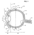
- FIG. 1 is a side sectional view schematically illustrating the human eye;
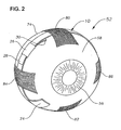
- FIG. 2 is a partially dissected, three dimensional schematic representation of the human eye;
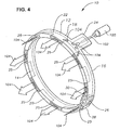
- FIG. 3 is a perspective view of an ophthalmic drug delivery device according to a preferred embodiment of the present invention; and
- FIG. 4 is a perspective view of the ophthalmic drug delivery device of FIG. 3 showing a preferred method of operation.
- The preferred embodiments of the present invention and their advantages are best understood by referring to FIGS. 1-4 of the drawings, like numerals being used for like and corresponding parts of the various drawings.
- FIGS. 1-4 schematically illustrate an ophthalmic
drug delivery device 10 according to a preferred embodiment of the present invention.Device 10 may be used in any case where delivery of a pharmaceutically active agent to the eye is required.Device 10 is particularly useful for delivery of active agents to the posterior segment of the eye. A preferred use fordevice 10 is the delivery of pharmaceutically active agents to the retina for treating ARMD, choroidial neovascularization (CNV), retinopathies, retinitis, uveitis, macular edema, and glaucoma. - Referring to FIGS. 1-2, a
human eye 52 is schematically illustrated.Eye 52 has acornea 54, alens 56, asclera 58, achoroid 60, aretina 62, and anoptic nerve 64. Ananterior segment 66 ofeye 52 generally includes the portions ofeye 52 anterior of aline 67. Aposterior segment 68 ofeye 52 generally includes the portions ofeye 52 posterior ofline 67.Retina 62 is physically attached tochoroid 60 in a circumferential manner proximate pars plana 70.Retina 62 has a macula 72 located slightly lateral to itsoptic disk 19. As is well known in the ophthalmic art,macula 72 is comprised primarily of retinal cones and is the region of maximum visual acuity inretina 62. A Tenon's capsule or Tenon'smembrane 74 is disposed onsclera 58. Aconjunctiva 76 covers a short area of the globe ofeye 52 posterior to limbus 77 (the bulbar conjunctiva) and folds up (the upper cul-de-sac) or down (the lower cul-de-sac) to cover the inner areas ofupper eyelid 78 andlower eyelid 79, respectively.Conjunctiva 76 is disposed on top of Tenon'scapsule 74.Eye 52 also has anequator 21. As shown in FIG. 2,superior rectus muscle 80,inferior rectus muscle 82, lateral rectus muscle, 84, andmedial rectus muscle 86 are attached to sclera 58. - As is shown in FIGS. 1 and 2, and as is described in greater detail hereinbelow,
device 10 is preferably disposed directly on the outer surface ofsclera 58, below Tenon'scapsule 74 for treatment of most posterior segment diseases or conditions.Device 10 is most preferably disposed directly on the outer surface ofsclera 58, below Tenon'scapsule 74,proximate equator 21. - FIGS. 3 and 4 schematically illustrate
device 10 in greater detail.Device 10 preferably includes abody 12 having a generally ring-shaped three-dimensional geometry in its assembled state shown in FIGS. 3 and 4. In its unassembled stated,body 12 preferably has a generally rectangular three-dimensional geometry. Asleeve 26 encircles overlapping ends 28 and 30 ofbody 12 to maintain the ring-shaped three-dimensional geometry. -
Body 12 has ascleral surface 14, anorbital surface 16, ananterior side 18, and aposterior side 20.Body 12 also has areservoir 22 located in its interior.Reservoir 22 preferably runs the entire length ofbody 80, and preferably has a plurality ofopenings 25 toposterior side 20. Although not shown in FIGS. 3-4,reservoir 22 may also have one or more openings toscleral surface 14,anterior side 18, ororbital surface 16 ofbody 12. As shown in FIGS. 3-4,openings 25 have a generally rectangular cross-section, but any cross-section can be used for these openings. -
Body 12 preferably further has aninjection port 24. At least a portion ofinjection port 24 is preferably made of a fluid impervious material that can be penetrated by a needle and that reseals itself upon removal of the needle. A preferred material is silicone rubber. In addition,injection port 24 is preferably colored or marked by raised protuberances.Injection port 24 preferably extends fromanterior side 18 ofbody 12.Reservoir 22 extends intoinjection port 24. - The remainder of
body 12 preferably comprises a biocompatible, non-bioerodable material.Body 12 more preferably comprises a biocompatible, non-bioerodable polymeric composition. Said polymeric composition may be a homopolymer, a copolymer, straight, branched, cross-linked, or a blend. Examples of polymers suitable for use in said polymeric composition include silicone, polyvinyl alcohol, ethylene vinyl acetate, polylactic acid, nylon, polypropylene, polycarbonate, cellulose, cellulose acetate, polyglycolic acid, polylactic-glycolic acid, cellulose esters, polyethersulfone, acrylics, their derivatives, and combinations thereof. Examples of suitable soft acrylics are more fully disclosed in U.S. Patent No. 5,403,901. - Said polymeric composition most preferably comprises silicone. Of course, said polymeric composition may also comprise other conventional materials that affect its physical properties, including, but not limited to, porosity, tortuosity, permeability, rigidity, hardness, and smoothness. Exemplary materials affecting certain ones of these physical properties include conventional plasticizers, fillers, and lubricants. Said polymeric composition may comprise other conventional materials that affect its chemical properties, including, but not limited to, toxicity and hydrophobicity.
- As shown in FIG. 4, a
conventional syringe 100 andneedle 102 may be used to impart a fluid 104 (indicated by arrows) containing a pharmaceutically active agent or agents intoreservoir 22 viainjection port 24.Fluid 104 may comprise a solution, a suspension, an emulsion, an ointment, a gel forming solution, a gel, a bioerodable polymer, a non-bioerodable polymer, microparticles, or combinations thereof. Most preferably,fluid 104 is a suspension with or without microparticles formed from bioerodable polymers.Fluid 104 includes one or more ophthalmically acceptable pharmaceutically active agents, and may also include conventional non-active incipients. Examples of pharmaceutically active agents suitable forfluid 104 are anti-infectives, including, without limitation, antibiotics, antivirals, and antifungals; antiallergenic agents and mast cell stabilizers; steroidal and non-steroidal anti-inflammatory agents; cyclooxygenase inhibitors, including, without limitation, Cox I and Cox II inhibitors; combinations of anti-infective and anti-inflammatory agents; decongestants; anti-glaucoma agents, including, without limitation, adrenergics, β-adrenergic blocking agents, α-adrenergic agonists, parasypathomimetic agents, cholinesterase inhibitors, carbonic anhydrase inhibitors, and prostaglandins; combinations of anti-glaucoma agents; antioxidants; nutritional supplements; drugs for the treatment of cystoid macular edema including, without limitation, non-steroidal anti-inflammatory agents; drugs for the treatment of ARMD, including, without limitation, angiogenesis inhibitors and nutritional supplements; drugs for the treatment of herpetic infections and CMV ocular infections; drugs for the treatment of proliferative vitreoretinopathy including, without limitation, antimetabolites and fibrinolytics; wound modulating agents, including, without limitation, growth factors; antimetabolites; neuroprotective drugs, including, without limitation, eliprodil; and angiostatic steroids for the treatment of diseases or conditions ofposterior segment 26, including, without limitation, ARMD, CNV, retinopathies, retinitis, uveitis, macular edema, and glaucoma. Such angiostatic steroids are more fully disclosed in U.S. Patent Nos. 5,679,666 and 5,770,592. Preferred ones of such angiostatic steroids include 4,9(11)-Pregnadien-17α,21-diol-3,20-dione and 4,9(11)-Pregnadien-17α,21-diol-3,20-dione-21-acetate. These preferred angiostatic steroids are preferably formulated as a suspension. A preferred non-steroidal anti-inflammatory for the treatment of cystoid macular edema is nepafenac. The conventional non-active excipients may include, but are not limited to, ingredients to enhance the stability, solubility, penetrability, or other properties offluid 104. In particular, hydrolytic enzymes such as proteases, esterases, hyaluronidases, and collegenases may be utilized to enhance the penetration of the pharmaceutically active agents through natural and newly formed connective tissue that may encapsulatedevice 10 after implantation.Body 12 is preferably impermeable tofluid 104. -
Device 10 may be made by conventional polymer processing methods, including, but not limited to, injection molding, extrusion molding, transfer molding, and compression molding. Preferably,device 10 is formed using conventional injection molding techniques. -
Device 10 is preferably surgically placed directly on the outer surface ofsclera 58 below Tenon'scapsule 74 using a simple surgical technique that is capable of being performed in an outpatient setting. The surgeon first performs a 360 degree peritomy about 3 mm posterior tolimbs 77 ofeye 52. The surgeon then performs a blunt dissection to separate Tenon'scapsule 74 fromsclera 58 up to a point slightly posterior ofequator 21. As shown best in FIG. 2, the surgeon then positionsdevice 10 on the outer surface ofsclera 58 belowsuperior rectus muscle 80,medial rectus muscle 86,inferior rectus muscle 82, andlateral rectus muscle 84 generally nearequator 21.Injection port 24 is preferably located in the infra-temporal quadrant ofeye 52 betweeninferior rectus muscle 82 andlateral rectus muscle 84. The surgeon tightensdevice 10 aroundsclera 58 and fixes overlapping ends 28 and 30 ofbody 12 withsleeve 26. The surgeon then moves Tenon'scapsule 74 back to its original position and sutures it in place. After closing, the surgeon places antibiotic ointment on the surgical wound. - Once
device 10 is located in the desired position, the surgeon usessyringe 100 andneedle 102 to inject fluid 104 intoreservoir 22. The surgeon preferably moveslower eyelid 79 downward and instructs the patient to look upward so as to exposeinjection port 24.Injection port 24 may be easily visualized beneath the Tenon's capsule and any connectivetissue encapsulating device 10 due to its color or raised protuberances. The surgeon sticksneedle 102 intoinjection port 24, injects fluid 104 intoreservoir 22, and removesneedle 102 from theport 24.Port 24 reseals automatically upon removal of the needle.Fluid 104 is disposed throughoutreservoir 22, and is in communication withsclera 58 viaopenings 25, or any other openings fromreservoir 22. - It is believed that
device 10 can be used to deliver a pharmaceutically effective amount of a pharmaceutically active agent throughsclera 58 andchoroid 60 intoretina 62 for many years, depending on the particular physicochemical properties of theparticular fluid 104 and its pharmaceutically active agent employed. Important physicochemical properties include hydrophobicity, solubility, dissolution rate, diffusion coefficient, and tissue affinity. In addition, it is believed thatdevice 10 may be used to deliver both a localized distribution of drug primarilyproximate macula 72, or to deliver drug to substantially the entire retina, depending upon theparticular fluid 104 and its pharmaceutically active agents and incipients. Afterreservoir 22 no longer contains any fluid 104, a surgeon may refillreservoir 22 as described hereinabove. Although not shown in FIGS. 1-4,posterior side 20 ofbody 12 may also include a sharp surface or edge. During refilling ofreservoir 22, the surgeon may movedevice 10 slightly posteriorly so that such sharp surface or edge pierces any connective tissue that may encapsulatedevice 10 after implantation. Piercing this connective tissue facilitates proper distribution offluid 104 viaopenings 25. In addition, unlike repetitive sub-Tenon's capsule injections of drug formulations,device 10 minimizes the risk of penetrating the globe of the eye, always results influid 104 being distributed below the Tenon'scapsule 74 on the outer surface ofsclera 58, and results in a reproduceable distribution of fluid 212 on a desired portion of the outer surface of thesclera 58. - From the above, it may be appreciated that the present invention provides improved devices and methods for safe, effective, rate-controlled delivery of a variety of pharmaceutically active agents to the eye. The devices of the present invention are especially useful for localized and/or pan-retinal delivery of pharmaceutically active agents to the posterior segment of the eye to combat diseases such as ARMD, CNV, retinopathies, retinitis, uveitis, macular edema, and glaucoma. The surgical procedure for implanting the devices is safe, simple, quick, and capable of being performed in an outpatient setting. The devices are easy and economical to manufacture. Furthermore, because of their capability to deliver a wide variety of pharmaceutically active agents, such devices are useful in clinical studies to deliver various agents that create a specific physical condition in a patient or animal subject.
- It is believed that the operation and construction of the present invention will be apparent from the foregoing description. While the apparatus and methods shown or described above have been characterized as being preferred, various changes and modifications may be made therein without departing from the scope of the invention as defined in the following claims.
Claims (7)
- An ophthalmic drug delivery device (10), comprising:a first end (28) and a second end (30);an injection port (24) for sealingly engaging a needle (102) of a syringe, said syringe for providing a fluid (104) comprising a pharmaceutically active agent;a reservoir (22) disposed within said device, fluidly coupled to said injection port, and having an opening (25) for communicating said fluid to an outer surface of a sclera (58) of an eye (52);and a sleeve (26) for engaging said device proximate overlapping portions of said first end and said second end for forming a generally ring-shaped three-dimensional geometry upon implantation of said device on said outer surface of said sclera.
- The ophthalmic drug delivery device of claim 1, further comprising an anterior side (18), and wherein said injection port (24) is disposed on said anterior side.
- The ophthalmic drug delivery device of claim 1, further comprising a posterior side (20), and wherein said posterior side comprises a sharp exterior surface for piercing connective tissue that may encapsulate said device upon implantation on said outer surface of said sclera (58).
- The ophthalmic drug delivery device of claim 1, wherein said reservoir comprises a plurality of said openings(25) for communicating said fluid (104) to said outer surface of said sclera (58).
- The ophthalmic drug delivery device of claim 1, wherein said device delivers said pharmaceutically active agent through said sclera (58) and a choroid (60) into a retina (62) of said eye (52).
- The ophthalmic drug delivery device of claim 5, wherein said device delivers said pharmaceutically active agent to substantially all of said retina (62).
- The ophthalmic delivery device of claim 5, wherein said device delivers said pharmaceutically active agent to a portion of said retina (62) proximate said device.
Applications Claiming Priority (3)
| Application Number | Priority Date | Filing Date | Title |
|---|---|---|---|
| US30728401P | 2001-07-23 | 2001-07-23 | |
| US307284P | 2001-07-23 | ||
| PCT/US2002/023048 WO2003009774A2 (en) | 2001-07-23 | 2002-07-22 | Ophthalmic drug delivery device |
Publications (3)
| Publication Number | Publication Date |
|---|---|
| EP1409065A2 EP1409065A2 (en) | 2004-04-21 |
| EP1409065A4 EP1409065A4 (en) | 2006-01-11 |
| EP1409065B1 true EP1409065B1 (en) | 2007-01-17 |
Family
ID=23189050
Family Applications (1)
| Application Number | Title | Priority Date | Filing Date |
|---|---|---|---|
| EP02750196A Expired - Lifetime EP1409065B1 (en) | 2001-07-23 | 2002-07-22 | Ophthalmic drug delivery device |
Country Status (16)
| Country | Link |
|---|---|
| US (1) | US7094226B2 (en) |
| EP (1) | EP1409065B1 (en) |
| JP (1) | JP4261343B2 (en) |
| CN (1) | CN1203814C (en) |
| AU (1) | AU2002319596B2 (en) |
| BR (1) | BR0210494A (en) |
| CA (1) | CA2447802C (en) |
| CY (1) | CY1105991T1 (en) |
| DE (1) | DE60217679T2 (en) |
| DK (1) | DK1409065T3 (en) |
| ES (1) | ES2278936T3 (en) |
| MX (1) | MXPA03011609A (en) |
| PL (1) | PL204644B1 (en) |
| PT (1) | PT1409065E (en) |
| WO (1) | WO2003009774A2 (en) |
| ZA (1) | ZA200308993B (en) |
Cited By (14)
| Publication number | Priority date | Publication date | Assignee | Title |
|---|---|---|---|---|
| US8277830B2 (en) | 2009-01-29 | 2012-10-02 | Forsight Vision4, Inc. | Posterior segment drug delivery |
| US8486052B2 (en) | 2001-06-12 | 2013-07-16 | The Johns Hopkins University School Of Medicine | Reservoir device for intraocular drug delivery |
| US8623395B2 (en) | 2010-01-29 | 2014-01-07 | Forsight Vision4, Inc. | Implantable therapeutic device |
| US8905963B2 (en) | 2010-08-05 | 2014-12-09 | Forsight Vision4, Inc. | Injector apparatus and method for drug delivery |
| US9474756B2 (en) | 2014-08-08 | 2016-10-25 | Forsight Vision4, Inc. | Stable and soluble formulations of receptor tyrosine kinase inhibitors, and methods of preparation thereof |
| US9492315B2 (en) | 2010-08-05 | 2016-11-15 | Forsight Vision4, Inc. | Implantable therapeutic device |
| US9526654B2 (en) | 2013-03-28 | 2016-12-27 | Forsight Vision4, Inc. | Ophthalmic implant for delivering therapeutic substances |
| US9968603B2 (en) | 2013-03-14 | 2018-05-15 | Forsight Vision4, Inc. | Systems for sustained intraocular delivery of low solubility compounds from a port delivery system implant |
| US10010448B2 (en) | 2012-02-03 | 2018-07-03 | Forsight Vision4, Inc. | Insertion and removal methods and apparatus for therapeutic devices |
| US10166142B2 (en) | 2010-01-29 | 2019-01-01 | Forsight Vision4, Inc. | Small molecule delivery with implantable therapeutic device |
| US10258503B2 (en) | 2014-07-15 | 2019-04-16 | Forsight Vision4, Inc. | Ocular implant delivery device and method |
| US10500091B2 (en) | 2014-11-10 | 2019-12-10 | Forsight Vision4, Inc. | Expandable drug delivery devices and methods of use |
| US10617557B2 (en) | 2010-08-05 | 2020-04-14 | Forsight Vision4, Inc. | Combined drug delivery methods and apparatus |
| US11813196B2 (en) | 2011-06-28 | 2023-11-14 | Forsight Vision4, Inc. | Diagnostic methods and apparatus |
Families Citing this family (50)
| Publication number | Priority date | Publication date | Assignee | Title |
|---|---|---|---|---|
| US7431710B2 (en) | 2002-04-08 | 2008-10-07 | Glaukos Corporation | Ocular implants with anchors and methods thereof |
| BR0205990A (en) * | 2001-08-29 | 2004-01-13 | Ricardo Azevedo Ponte Carvalho | An implantable one-way delivery system for therapeutic tissue agents |
| US20070027537A1 (en) * | 2002-08-02 | 2007-02-01 | David Castillejos | Method and intra-sclera implant for treatment of glaucoma and presbyopia |
| WO2004073551A2 (en) * | 2003-02-18 | 2004-09-02 | Massachusetts Eye And Ear Infirmary | Transscleral drug delivery device and related methods |
| US8167855B2 (en) * | 2003-08-26 | 2012-05-01 | Vista Scientific Llc | Ocular drug delivery device |
| US20060067980A1 (en) * | 2004-09-30 | 2006-03-30 | Bausch & Lomb Incorporated | Capsule for encasing tablets for surgical insertion into the human body |
| US20060233858A1 (en) * | 2005-03-08 | 2006-10-19 | Allergan, Inc. | Systems and methods providing targeted intraocular drug delivery |
| JP5681362B2 (en) | 2006-03-14 | 2015-03-04 | ユニバーシティー オブ サザン カリフォルニア | MEMS device for delivery of therapeutic agents |
| MX364408B (en) | 2007-12-20 | 2019-04-25 | Univ Southern California | APPARATUS and METHODS FOR DELIVERING THERAPEUTIC AGENTS. |
| JP2011509120A (en) | 2008-01-03 | 2011-03-24 | ユニバーシティ オブ サザン カリフォルニア | Implantable drug delivery device and apparatus and method for refilling the device |
| AU2015201289B2 (en) * | 2008-03-14 | 2016-12-01 | Institut National De La Sante Et De La Recherche Medicale | Eye injection device |
| WO2009137785A2 (en) | 2008-05-08 | 2009-11-12 | Replenish Pumps, Llc | Drug-delivery pumps and methods of manufacture |
| US9849238B2 (en) | 2008-05-08 | 2017-12-26 | Minipumps, Llc | Drug-delivery pump with intelligent control |
| WO2009137780A2 (en) | 2008-05-08 | 2009-11-12 | Replenish Pumps, Llc | Implantable pumps and cannulas therefor |
| CN104758999B (en) | 2008-05-08 | 2018-08-07 | 迷你泵有限责任公司 | Implantable drug delivery devices and the device and method for filling the device |
| US10064819B2 (en) | 2008-05-12 | 2018-09-04 | University Of Utah Research Foundation | Intraocular drug delivery device and associated methods |
| US9095404B2 (en) * | 2008-05-12 | 2015-08-04 | University Of Utah Research Foundation | Intraocular drug delivery device and associated methods |
| US10588855B2 (en) | 2008-05-12 | 2020-03-17 | University Of Utah Research Foundation | Intraocular drug delivery device and associated methods |
| WO2009140246A2 (en) | 2008-05-12 | 2009-11-19 | University Of Utah Research Foundation | Intraocular drug delivery device and associated methods |
| US10206813B2 (en) | 2009-05-18 | 2019-02-19 | Dose Medical Corporation | Implants with controlled drug delivery features and methods of using same |
| JP5695035B2 (en) | 2009-06-03 | 2015-04-01 | フォーサイト・ビジョン5・インコーポレイテッドForsight Vision5,Inc. | Anterior eye drug supply |
| EP2467797B1 (en) | 2009-08-18 | 2017-07-19 | MiniPumps, LLC | Electrolytic drug-delivery pump with adaptive control |
| CN103200983A (en) * | 2010-06-25 | 2013-07-10 | 欧几里得系统公司 | Device and method for the controlled delivery of ophthalmic solutions |
| AU2011329656B2 (en) | 2010-11-19 | 2017-01-05 | Forsight Vision4, Inc. | Therapeutic agent formulations for implanted devices |
| US9603997B2 (en) | 2011-03-14 | 2017-03-28 | Minipumps, Llc | Implantable drug pumps and refill devices therefor |
| US9919099B2 (en) | 2011-03-14 | 2018-03-20 | Minipumps, Llc | Implantable drug pumps and refill devices therefor |
| US10286146B2 (en) | 2011-03-14 | 2019-05-14 | Minipumps, Llc | Implantable drug pumps and refill devices therefor |
| US10245178B1 (en) | 2011-06-07 | 2019-04-02 | Glaukos Corporation | Anterior chamber drug-eluting ocular implant |
| US9102105B2 (en) | 2011-09-13 | 2015-08-11 | Vista Scientific Llc | Method for forming an ocular drug delivery device |
| WO2013040426A2 (en) | 2011-09-14 | 2013-03-21 | Forsight Labs, Llc | Ocular insert apparatus and methods |
| WO2013040247A2 (en) | 2011-09-16 | 2013-03-21 | Forsight Vision4, Inc. | Fluid exchange apparatus and methods |
| US9381112B1 (en) | 2011-10-06 | 2016-07-05 | William Eric Sponsell | Bleb drainage device, ophthalmological product and methods |
| US8632489B1 (en) | 2011-12-22 | 2014-01-21 | A. Mateen Ahmed | Implantable medical assembly and methods |
| CA2868800A1 (en) * | 2012-03-27 | 2013-10-03 | Duke University | Ophthalmic drug delivery device and methods of use |
| WO2014066775A1 (en) | 2012-10-26 | 2014-05-01 | Forsight Vision5, Inc. | Ophthalmic system for sustained release of drug to eye |
| US9597227B2 (en) * | 2013-03-15 | 2017-03-21 | Abbott Medical Optics Inc. | Trans-sclera portal for delivery of therapeutic agents |
| US9320645B2 (en) | 2013-05-29 | 2016-04-26 | Terry Glasser | Approach to administering ocular medication |
| WO2015184173A1 (en) | 2014-05-29 | 2015-12-03 | Dose Medical Corporation | Implants with controlled drug delivery features and methods of using same |
| US20160296532A1 (en) | 2015-04-13 | 2016-10-13 | Forsight Vision5, Inc. | Ocular Insert Composition of a Semi-Crystalline or Crystalline Pharmaceutically Active Agent |
| WO2016187426A1 (en) | 2015-05-19 | 2016-11-24 | Amorphex Therapeutics Llc | A device that delivers a sustained low-dose of a myopia-suppressing drug |
| WO2017040853A1 (en) | 2015-09-02 | 2017-03-09 | Glaukos Corporation | Drug delivery implants with bi-directional delivery capacity |
| US11564833B2 (en) | 2015-09-25 | 2023-01-31 | Glaukos Corporation | Punctal implants with controlled drug delivery features and methods of using same |
| CN113069681B (en) | 2015-11-20 | 2022-12-23 | 弗赛特影像4股份有限公司 | Method of manufacturing a therapeutic device for sustained drug delivery |
| ES2837524T3 (en) | 2016-04-05 | 2021-06-30 | Forsight Vision4 Inc | Implantable ocular drug delivery devices |
| WO2017184881A1 (en) | 2016-04-20 | 2017-10-26 | Harold Alexander Heitzmann | Bioresorbable ocular drug delivery device |
| JPWO2018143481A1 (en) * | 2017-02-01 | 2019-11-21 | 国立大学法人東北大学 | Sustained drug sustained release device capable of reinjecting drug and injectable gel for refilling |
| US11406533B2 (en) * | 2017-03-17 | 2022-08-09 | W. L. Gore & Associates, Inc. | Integrated aqueous shunt for glaucoma treatment |
| WO2019023617A1 (en) | 2017-07-27 | 2019-01-31 | University Of Utah Research Foundation | Therapeutic delivery device |
| CN111655206B (en) | 2017-11-21 | 2022-10-14 | 弗赛特影像4股份有限公司 | Fluid exchange device for expandable port delivery system and method of use |
| US20210267798A1 (en) * | 2020-02-27 | 2021-09-02 | Innfocus, Inc. | Ocular implant |
Family Cites Families (65)
| Publication number | Priority date | Publication date | Assignee | Title |
|---|---|---|---|---|
| US3416530A (en) | 1966-03-02 | 1968-12-17 | Richard A. Ness | Eyeball medication dispensing tablet |
| US3828777A (en) | 1971-11-08 | 1974-08-13 | Alza Corp | Microporous ocular device |
| US3884232A (en) * | 1974-06-05 | 1975-05-20 | Ruth Braun | Instrument for administering to the human eye |
| US4014335A (en) * | 1975-04-21 | 1977-03-29 | Alza Corporation | Ocular drug delivery device |
| US4256108A (en) * | 1977-04-07 | 1981-03-17 | Alza Corporation | Microporous-semipermeable laminated osmotic system |
| US4300557A (en) | 1980-01-07 | 1981-11-17 | The United States Of America As Represented By The Secretary Of The Department Of Health And Human Services | Method for treating intraocular malignancies |
| US4327725A (en) | 1980-11-25 | 1982-05-04 | Alza Corporation | Osmotic device with hydrogel driving member |
| US4624848A (en) | 1984-05-10 | 1986-11-25 | Ciba-Geigy Corporation | Active agent containing hydrogel devices wherein the active agent concentration profile contains a sigmoidal concentration gradient for improved constant release, their manufacture and use |
| US5322691A (en) | 1986-10-02 | 1994-06-21 | Sohrab Darougar | Ocular insert with anchoring protrusions |
| US5147647A (en) | 1986-10-02 | 1992-09-15 | Sohrab Darougar | Ocular insert for the fornix |
| US4853224A (en) | 1987-12-22 | 1989-08-01 | Visionex | Biodegradable ocular implants |
| US4997652A (en) | 1987-12-22 | 1991-03-05 | Visionex | Biodegradable ocular implants |
| DE3905050A1 (en) | 1989-02-18 | 1990-08-30 | Lohmann Therapie Syst Lts | THERAPEUTIC SYSTEM FOR DELAYED AND CONTROLLED TRANSDERMAL OR TRANSMUCOSAL ADMINISTRATION OF ACTIVE SUBSTANCES (II) |
| US4946450A (en) | 1989-04-18 | 1990-08-07 | Biosource Genetics Corporation | Glucan/collagen therapeutic eye shields |
| US5164188A (en) | 1989-11-22 | 1992-11-17 | Visionex, Inc. | Biodegradable ocular implants |
| DE4022553A1 (en) | 1990-07-16 | 1992-01-23 | Hund Helmut Gmbh | Medical contact lens - with recess contg. therapeutically or diagnostically active substance |
| US5030214A (en) * | 1990-11-01 | 1991-07-09 | Larry Spector | Ocular delivery system |
| US5290892A (en) | 1990-11-07 | 1994-03-01 | Nestle S.A. | Flexible intraocular lenses made from high refractive index polymers |
| US5378475A (en) | 1991-02-21 | 1995-01-03 | University Of Kentucky Research Foundation | Sustained release drug delivery devices |
| US5679666A (en) | 1991-11-22 | 1997-10-21 | Alcon Laboratories, Inc. | Prevention and treatment of ocular neovascularization by treatment with angiostatic steroids |
| US5770592A (en) | 1991-11-22 | 1998-06-23 | Alcon Laboratories, Inc. | Prevention and treatment of ocular neovascularization using angiostatic steroids |
| US5178635A (en) | 1992-05-04 | 1993-01-12 | Allergan, Inc. | Method for determining amount of medication in an implantable device |
| FR2690846B1 (en) | 1992-05-05 | 1995-07-07 | Aiache Jean Marc | GALENIC FORM FOR EYE ADMINISTRATION AND METHOD OF PREPARATION. |
| WO1994005257A1 (en) | 1992-09-08 | 1994-03-17 | Allergan, Inc. | Sustained release of ophthalmic drugs from a soluble polymer drug delivery vehicle |
| WO1995003009A1 (en) | 1993-07-22 | 1995-02-02 | Oculex Pharmaceuticals, Inc. | Method of treatment of macular degeneration |
| US5443505A (en) | 1993-11-15 | 1995-08-22 | Oculex Pharmaceuticals, Inc. | Biocompatible ocular implants |
| US5516522A (en) | 1994-03-14 | 1996-05-14 | Board Of Supervisors Of Louisiana State University | Biodegradable porous device for long-term drug delivery with constant rate release and method of making the same |
| AU704591B2 (en) | 1994-04-04 | 1999-04-29 | William R. Freeman | Use of phosphonylmethoxyalkyl nucleosides for the treatment of raised intraocular pressure |
| US5466233A (en) | 1994-04-25 | 1995-11-14 | Escalon Ophthalmics, Inc. | Tack for intraocular drug delivery and method for inserting and removing same |
| US5710165A (en) | 1994-07-06 | 1998-01-20 | Synthelabo | Use of polyamine antagonists for the treatment of glaucoma |
| AUPM897594A0 (en) | 1994-10-25 | 1994-11-17 | Daratech Pty Ltd | Controlled release container |
| DE69529572T2 (en) | 1994-11-10 | 2003-06-18 | Univ Kentucky Res Foundation L | IMPLANTABLE REFILLABLE DEVICE WITH CONTROLLED RELEASE FOR ADMINISTERING MEDICINAL SUBSTANCES DIRECTLY ON AN INNER PART OF THE BODY |
| AUPM982694A0 (en) * | 1994-12-02 | 1995-01-05 | University Of Queensland, The | Iontophoresis method and apparatus |
| US5725493A (en) | 1994-12-12 | 1998-03-10 | Avery; Robert Logan | Intravitreal medicine delivery |
| CN1124164C (en) | 1995-05-14 | 2003-10-15 | 奥普通诺尔有限公司 | Intraocular implant, delivery device, and method of implantation |
| US5773019A (en) | 1995-09-27 | 1998-06-30 | The University Of Kentucky Research Foundation | Implantable controlled release device to deliver drugs directly to an internal portion of the body |
| AUPN605795A0 (en) | 1995-10-19 | 1995-11-09 | F.H. Faulding & Co. Limited | Analgesic pharmaceutical composition |
| US5824073A (en) | 1996-03-18 | 1998-10-20 | Peyman; Gholam A. | Macular indentor for use in the treatment of subretinal neovascular membranes |
| US5743274A (en) | 1996-03-18 | 1998-04-28 | Peyman; Gholam A. | Macular bandage for use in the treatment of subretinal neovascular members |
| US5904144A (en) | 1996-03-22 | 1999-05-18 | Cytotherapeutics, Inc. | Method for treating ophthalmic diseases |
| JP3309175B2 (en) | 1996-03-25 | 2002-07-29 | 参天製薬株式会社 | Scleral plug containing proteinaceous drug |
| US5797898A (en) | 1996-07-02 | 1998-08-25 | Massachusetts Institute Of Technology | Microchip drug delivery devices |
| US5860994A (en) * | 1996-07-30 | 1999-01-19 | Yaacobi; Yoseph | Remotely operable intraocular surgical instrument for automated capsulectomies |
| US6120460A (en) * | 1996-09-04 | 2000-09-19 | Abreu; Marcio Marc | Method and apparatus for signal acquisition, processing and transmission for evaluation of bodily functions |
| ZA9710342B (en) | 1996-11-25 | 1998-06-10 | Alza Corp | Directional drug delivery stent and method of use. |
| NZ500018A (en) | 1997-03-31 | 2001-07-27 | Alza Corp | Diffusional implantable delivery system with capillary tube of cross-sectional area and length to deliver beneficial agent |
| JP2001513369A (en) | 1997-08-11 | 2001-09-04 | アラーガン・セイルズ・インコーポレイテッド | Sterile bioerodible implant devices and methods with improved biocompatibility |
| US5902598A (en) | 1997-08-28 | 1999-05-11 | Control Delivery Systems, Inc. | Sustained release drug delivery devices |
| US6066671A (en) | 1997-12-19 | 2000-05-23 | Alcon Laboratories, Inc. | Treatment of GLC1A glaucoma with 3-benzoyl-phenylacetic acids, esters, or amides |
| ATE365550T1 (en) | 1998-03-13 | 2007-07-15 | Univ Johns Hopkins Med | USE OF A PROTEIN KINASE INHIBITOR SUCH AS GENISTEIN IN THE TREATMENT OF DIABETIC RETINOPATHY |
| US6378526B1 (en) | 1998-08-03 | 2002-04-30 | Insite Vision, Incorporated | Methods of ophthalmic administration |
| US6146366A (en) | 1998-11-03 | 2000-11-14 | Ras Holding Corp | Device for the treatment of macular degeneration and other eye disorders |
| US6399655B1 (en) | 1998-12-22 | 2002-06-04 | Johns Hopkins University, School Of Medicine | Method for the prophylactic treatment of cataracts |
| US6217895B1 (en) | 1999-03-22 | 2001-04-17 | Control Delivery Systems | Method for treating and/or preventing retinal diseases with sustained release corticosteroids |
| ES2240180T3 (en) | 1999-10-21 | 2005-10-16 | Alcon Inc. | SUB-TENON ADMINISTRATION OF MEDICINES. |
| DE60040876D1 (en) * | 1999-10-21 | 2009-01-02 | Alcon Inc | medication supply |
| US6416777B1 (en) | 1999-10-21 | 2002-07-09 | Alcon Universal Ltd. | Ophthalmic drug delivery device |
| RU2149615C1 (en) | 1999-11-10 | 2000-05-27 | Нестеров Аркадий Павлович | Method for introducing drugs in treating posterior eye segment diseases |
| AUPQ496500A0 (en) | 2000-01-06 | 2000-02-03 | University Of Sydney, The | Kit |
| US6375972B1 (en) | 2000-04-26 | 2002-04-23 | Control Delivery Systems, Inc. | Sustained release drug delivery devices, methods of use, and methods of manufacturing thereof |
| DE60135352D1 (en) * | 2000-08-30 | 2008-09-25 | Univ Johns Hopkins | DEVICE FOR INTRA-OCCULAR ACTIVE AGGREGATION |
| JP2004522730A (en) * | 2001-01-03 | 2004-07-29 | ボシュ・アンド・ロム・インコーポレイテッド | Sustained release drug delivery device with coated drug core |
| US7181287B2 (en) | 2001-02-13 | 2007-02-20 | Second Sight Medical Products, Inc. | Implantable drug delivery device |
| US6713081B2 (en) * | 2001-03-15 | 2004-03-30 | The United States Of America As Represented By The Department Of Health And Human Services | Ocular therapeutic agent delivery devices and methods for making and using such devices |
| ATE506929T1 (en) | 2001-06-12 | 2011-05-15 | Univ Johns Hopkins Med | RESERVOIR DEVICE FOR INTRAOCULAR MEDICINAL DELIVERY |
-
2002
- 2002-07-22 PT PT02750196T patent/PT1409065E/en unknown
- 2002-07-22 DE DE60217679T patent/DE60217679T2/en not_active Expired - Lifetime
- 2002-07-22 CA CA002447802A patent/CA2447802C/en not_active Expired - Fee Related
- 2002-07-22 PL PL368207A patent/PL204644B1/en not_active IP Right Cessation
- 2002-07-22 AU AU2002319596A patent/AU2002319596B2/en not_active Ceased
- 2002-07-22 ES ES02750196T patent/ES2278936T3/en not_active Expired - Lifetime
- 2002-07-22 EP EP02750196A patent/EP1409065B1/en not_active Expired - Lifetime
- 2002-07-22 BR BR0210494-6A patent/BR0210494A/en not_active Application Discontinuation
- 2002-07-22 MX MXPA03011609A patent/MXPA03011609A/en active IP Right Grant
- 2002-07-22 JP JP2003515170A patent/JP4261343B2/en not_active Expired - Fee Related
- 2002-07-22 CN CN02813762.0A patent/CN1203814C/en not_active Expired - Fee Related
- 2002-07-22 WO PCT/US2002/023048 patent/WO2003009774A2/en active IP Right Grant
- 2002-07-22 DK DK02750196T patent/DK1409065T3/en active
-
2003
- 2003-11-12 US US10/706,105 patent/US7094226B2/en not_active Expired - Fee Related
- 2003-11-19 ZA ZA200308993A patent/ZA200308993B/en unknown
-
2007
- 2007-02-06 CY CY20071100160T patent/CY1105991T1/en unknown
Cited By (33)
| Publication number | Priority date | Publication date | Assignee | Title |
|---|---|---|---|---|
| US9522082B2 (en) | 2001-06-12 | 2016-12-20 | The Johns Hopkins University | Reservoir device for intraocular drug delivery |
| US9180046B2 (en) | 2001-06-12 | 2015-11-10 | The Johns Hopkins University School Of Medicine | Reservoir device for intraocular drug delivery |
| US10470924B2 (en) | 2001-06-12 | 2019-11-12 | The Johns Hopkins University | Reservoir device for intraocular drug delivery |
| US8486052B2 (en) | 2001-06-12 | 2013-07-16 | The Johns Hopkins University School Of Medicine | Reservoir device for intraocular drug delivery |
| US8808727B2 (en) | 2009-01-29 | 2014-08-19 | Forsight Vision4, Inc. | Posterior segment drug delivery |
| US8795712B2 (en) | 2009-01-29 | 2014-08-05 | Forsight Vision4, Inc. | Posterior segment drug delivery |
| US8399006B2 (en) | 2009-01-29 | 2013-03-19 | Forsight Vision4, Inc. | Posterior segment drug delivery |
| US9066779B2 (en) | 2009-01-29 | 2015-06-30 | Forsight Vision4, Inc. | Implantable therapeutic device |
| US8298578B2 (en) | 2009-01-29 | 2012-10-30 | Forsight Vision4, Inc. | Posterior segment drug delivery |
| US9417238B2 (en) | 2009-01-29 | 2016-08-16 | Forsight Vision4, Inc. | Posterior segment drug delivery |
| US8277830B2 (en) | 2009-01-29 | 2012-10-02 | Forsight Vision4, Inc. | Posterior segment drug delivery |
| US8623395B2 (en) | 2010-01-29 | 2014-01-07 | Forsight Vision4, Inc. | Implantable therapeutic device |
| US10166142B2 (en) | 2010-01-29 | 2019-01-01 | Forsight Vision4, Inc. | Small molecule delivery with implantable therapeutic device |
| US9861521B2 (en) | 2010-08-05 | 2018-01-09 | Forsight Vision4, Inc. | Injector apparatus and method for drug delivery |
| US10617557B2 (en) | 2010-08-05 | 2020-04-14 | Forsight Vision4, Inc. | Combined drug delivery methods and apparatus |
| US11679027B2 (en) | 2010-08-05 | 2023-06-20 | Forsight Vision4, Inc. | Combined drug delivery methods and apparatus |
| US9492315B2 (en) | 2010-08-05 | 2016-11-15 | Forsight Vision4, Inc. | Implantable therapeutic device |
| US8905963B2 (en) | 2010-08-05 | 2014-12-09 | Forsight Vision4, Inc. | Injector apparatus and method for drug delivery |
| US10265215B2 (en) | 2010-08-05 | 2019-04-23 | Forsight Vision4, Inc. | Injector apparatus and method for drug delivery |
| US9033911B2 (en) | 2010-08-05 | 2015-05-19 | Forsight Vision4, Inc. | Injector apparatus and method for drug delivery |
| US11813196B2 (en) | 2011-06-28 | 2023-11-14 | Forsight Vision4, Inc. | Diagnostic methods and apparatus |
| US10010448B2 (en) | 2012-02-03 | 2018-07-03 | Forsight Vision4, Inc. | Insertion and removal methods and apparatus for therapeutic devices |
| US10603209B2 (en) | 2012-02-03 | 2020-03-31 | Forsight Vision4, Inc. | Insertion and removal methods and apparatus for therapeutic devices |
| US9968603B2 (en) | 2013-03-14 | 2018-05-15 | Forsight Vision4, Inc. | Systems for sustained intraocular delivery of low solubility compounds from a port delivery system implant |
| US9526654B2 (en) | 2013-03-28 | 2016-12-27 | Forsight Vision4, Inc. | Ophthalmic implant for delivering therapeutic substances |
| US11510810B2 (en) | 2013-03-28 | 2022-11-29 | Forsight Vision4, Inc. | Ophthalmic implant for delivering therapeutic substances |
| US11337853B2 (en) | 2014-07-15 | 2022-05-24 | Forsight Vision4, Inc. | Ocular implant delivery device and method |
| US10258503B2 (en) | 2014-07-15 | 2019-04-16 | Forsight Vision4, Inc. | Ocular implant delivery device and method |
| US9895369B2 (en) | 2014-08-08 | 2018-02-20 | Forsight Vision4, Inc | Stable and soluble formulations of receptor tyrosine kinase inhibitors, and methods of preparation thereof |
| US10765677B2 (en) | 2014-08-08 | 2020-09-08 | Forsight Vision4, Inc. | Stable and soluble formulations of receptor tyrosine kinase inhibitors, and methods of preparation thereof |
| US9474756B2 (en) | 2014-08-08 | 2016-10-25 | Forsight Vision4, Inc. | Stable and soluble formulations of receptor tyrosine kinase inhibitors, and methods of preparation thereof |
| US11110001B2 (en) | 2014-11-10 | 2021-09-07 | Forsight Vision4, Inc. | Expandable drug delivery devices and methods of use |
| US10500091B2 (en) | 2014-11-10 | 2019-12-10 | Forsight Vision4, Inc. | Expandable drug delivery devices and methods of use |
Also Published As
| Publication number | Publication date |
|---|---|
| AU2002319596B2 (en) | 2006-04-27 |
| CY1105991T1 (en) | 2011-04-06 |
| CA2447802A1 (en) | 2003-02-06 |
| EP1409065A2 (en) | 2004-04-21 |
| US7094226B2 (en) | 2006-08-22 |
| US20040106906A1 (en) | 2004-06-03 |
| PL368207A1 (en) | 2005-03-21 |
| BR0210494A (en) | 2004-06-22 |
| ES2278936T3 (en) | 2007-08-16 |
| PT1409065E (en) | 2007-03-30 |
| CA2447802C (en) | 2009-09-29 |
| CN1203814C (en) | 2005-06-01 |
| WO2003009774A2 (en) | 2003-02-06 |
| JP2005521433A (en) | 2005-07-21 |
| ZA200308993B (en) | 2004-11-19 |
| MXPA03011609A (en) | 2004-07-01 |
| DE60217679T2 (en) | 2007-10-25 |
| DK1409065T3 (en) | 2007-05-21 |
| WO2003009774A3 (en) | 2003-08-21 |
| DE60217679D1 (en) | 2007-03-08 |
| JP4261343B2 (en) | 2009-04-30 |
| PL204644B1 (en) | 2010-01-29 |
| CN1525872A (en) | 2004-09-01 |
| EP1409065A4 (en) | 2006-01-11 |
Similar Documents
| Publication | Publication Date | Title |
|---|---|---|
| EP1409065B1 (en) | Ophthalmic drug delivery device | |
| EP1385452B1 (en) | Ophthalmic drug delivery device | |
| CA2383499C (en) | Drug delivery device | |
| AU2002319596A1 (en) | Ophthalmic drug delivery device | |
| US7943162B2 (en) | Drug delivery device | |
| US6416777B1 (en) | Ophthalmic drug delivery device | |
| AU2002319606A1 (en) | Ophthalmic drug delivery device |
Legal Events
| Date | Code | Title | Description |
|---|---|---|---|
| PUAI | Public reference made under article 153(3) epc to a published international application that has entered the european phase |
Free format text: ORIGINAL CODE: 0009012 |
|
| 17P | Request for examination filed |
Effective date: 20031114 |
|
| AK | Designated contracting states |
Kind code of ref document: A2 Designated state(s): AT BE BG CH CY CZ DE DK EE ES FI FR GB GR IE IT LI LU MC NL PT SE SK TR |
|
| A4 | Supplementary search report drawn up and despatched |
Effective date: 20051125 |
|
| RAP1 | Party data changed (applicant data changed or rights of an application transferred) |
Owner name: ALCON, INC. |
|
| RIC1 | Information provided on ipc code assigned before grant |
Ipc: A61F 9/00 19680901AFI20051121BHEP |
|
| GRAP | Despatch of communication of intention to grant a patent |
Free format text: ORIGINAL CODE: EPIDOSNIGR1 |
|
| GRAS | Grant fee paid |
Free format text: ORIGINAL CODE: EPIDOSNIGR3 |
|
| GRAA | (expected) grant |
Free format text: ORIGINAL CODE: 0009210 |
|
| AK | Designated contracting states |
Kind code of ref document: B1 Designated state(s): AT BE BG CH CY CZ DE DK EE ES FI FR GB GR IE IT LI LU MC NL PT SE SK TR |
|
| REG | Reference to a national code |
Ref country code: GB Ref legal event code: FG4D |
|
| REG | Reference to a national code |
Ref country code: CH Ref legal event code: EP |
|
| REG | Reference to a national code |
Ref country code: IE Ref legal event code: FG4D |
|
| REF | Corresponds to: |
Ref document number: 60217679 Country of ref document: DE Date of ref document: 20070308 Kind code of ref document: P |
|
| REG | Reference to a national code |
Ref country code: GR Ref legal event code: EP Ref document number: 20070400376 Country of ref document: GR |
|
| REG | Reference to a national code |
Ref country code: PT Ref legal event code: SC4A Free format text: AVAILABILITY OF NATIONAL TRANSLATION Effective date: 20070313 |
|
| REG | Reference to a national code |
Ref country code: EE Ref legal event code: FG4A Ref document number: E000857 Country of ref document: EE Effective date: 20070208 |
|
| REG | Reference to a national code |
Ref country code: CH Ref legal event code: NV Representative=s name: CRONIN INTELLECTUAL PROPERTY |
|
| REG | Reference to a national code |
Ref country code: SE Ref legal event code: TRGR |
|
| REG | Reference to a national code |
Ref country code: DK Ref legal event code: T3 |
|
| ET | Fr: translation filed | ||
| REG | Reference to a national code |
Ref country code: ES Ref legal event code: FG2A Ref document number: 2278936 Country of ref document: ES Kind code of ref document: T3 |
|
| PLBE | No opposition filed within time limit |
Free format text: ORIGINAL CODE: 0009261 |
|
| STAA | Information on the status of an ep patent application or granted ep patent |
Free format text: STATUS: NO OPPOSITION FILED WITHIN TIME LIMIT |
|
| 26N | No opposition filed |
Effective date: 20071018 |
|
| PGFP | Annual fee paid to national office [announced via postgrant information from national office to epo] |
Ref country code: CY Payment date: 20100713 Year of fee payment: 9 |
|
| PGFP | Annual fee paid to national office [announced via postgrant information from national office to epo] |
Ref country code: LU Payment date: 20110801 Year of fee payment: 10 |
|
| PGFP | Annual fee paid to national office [announced via postgrant information from national office to epo] |
Ref country code: IE Payment date: 20110725 Year of fee payment: 10 Ref country code: CH Payment date: 20110725 Year of fee payment: 10 Ref country code: DK Payment date: 20110725 Year of fee payment: 10 Ref country code: MC Payment date: 20110704 Year of fee payment: 10 Ref country code: BG Payment date: 20110726 Year of fee payment: 10 |
|
| PGFP | Annual fee paid to national office [announced via postgrant information from national office to epo] |
Ref country code: FI Payment date: 20110727 Year of fee payment: 10 Ref country code: EE Payment date: 20110705 Year of fee payment: 10 Ref country code: SK Payment date: 20110706 Year of fee payment: 10 Ref country code: SE Payment date: 20110727 Year of fee payment: 10 Ref country code: GR Payment date: 20110726 Year of fee payment: 10 Ref country code: PT Payment date: 20110706 Year of fee payment: 10 Ref country code: CZ Payment date: 20110714 Year of fee payment: 10 Ref country code: TR Payment date: 20110711 Year of fee payment: 10 Ref country code: AT Payment date: 20110705 Year of fee payment: 10 |
|
| PGFP | Annual fee paid to national office [announced via postgrant information from national office to epo] |
Ref country code: NL Payment date: 20110728 Year of fee payment: 10 |
|
| REG | Reference to a national code |
Ref country code: PT Ref legal event code: MM4A Free format text: LAPSE DUE TO NON-PAYMENT OF FEES Effective date: 20130122 |
|
| REG | Reference to a national code |
Ref country code: NL Ref legal event code: V1 Effective date: 20130201 |
|
| PG25 | Lapsed in a contracting state [announced via postgrant information from national office to epo] |
Ref country code: MC Free format text: LAPSE BECAUSE OF NON-PAYMENT OF DUE FEES Effective date: 20120731 |
|
| REG | Reference to a national code |
Ref country code: CH Ref legal event code: PL |
|
| REG | Reference to a national code |
Ref country code: GR Ref legal event code: ML Ref document number: 20070400376 Country of ref document: GR Effective date: 20130104 |
|
| REG | Reference to a national code |
Ref country code: SE Ref legal event code: EUG |
|
| REG | Reference to a national code |
Ref country code: AT Ref legal event code: MM01 Ref document number: 351623 Country of ref document: AT Kind code of ref document: T Effective date: 20120722 |
|
| REG | Reference to a national code |
Ref country code: DK Ref legal event code: EBP |
|
| REG | Reference to a national code |
Ref country code: SK Ref legal event code: MM4A Ref document number: E 1680 Country of ref document: SK Effective date: 20120722 |
|
| REG | Reference to a national code |
Ref country code: EE Ref legal event code: MM4A Ref document number: E000857 Country of ref document: EE Effective date: 20120731 |
|
| PG25 | Lapsed in a contracting state [announced via postgrant information from national office to epo] |
Ref country code: CH Free format text: LAPSE BECAUSE OF NON-PAYMENT OF DUE FEES Effective date: 20120731 Ref country code: EE Free format text: LAPSE BECAUSE OF NON-PAYMENT OF DUE FEES Effective date: 20120731 Ref country code: NL Free format text: LAPSE BECAUSE OF NON-PAYMENT OF DUE FEES Effective date: 20130201 Ref country code: FI Free format text: LAPSE BECAUSE OF NON-PAYMENT OF DUE FEES Effective date: 20120722 Ref country code: SE Free format text: LAPSE BECAUSE OF NON-PAYMENT OF DUE FEES Effective date: 20120723 Ref country code: LI Free format text: LAPSE BECAUSE OF NON-PAYMENT OF DUE FEES Effective date: 20120731 Ref country code: CZ Free format text: LAPSE BECAUSE OF NON-PAYMENT OF DUE FEES Effective date: 20120722 |
|
| REG | Reference to a national code |
Ref country code: IE Ref legal event code: MM4A |
|
| PG25 | Lapsed in a contracting state [announced via postgrant information from national office to epo] |
Ref country code: CY Free format text: LAPSE BECAUSE OF NON-PAYMENT OF DUE FEES Effective date: 20120722 Ref country code: SK Free format text: LAPSE BECAUSE OF NON-PAYMENT OF DUE FEES Effective date: 20120722 Ref country code: PT Free format text: LAPSE BECAUSE OF NON-PAYMENT OF DUE FEES Effective date: 20130122 Ref country code: GR Free format text: LAPSE BECAUSE OF NON-PAYMENT OF DUE FEES Effective date: 20130204 |
|
| PG25 | Lapsed in a contracting state [announced via postgrant information from national office to epo] |
Ref country code: AT Free format text: LAPSE BECAUSE OF NON-PAYMENT OF DUE FEES Effective date: 20120722 |
|
| PG25 | Lapsed in a contracting state [announced via postgrant information from national office to epo] |
Ref country code: IE Free format text: LAPSE BECAUSE OF NON-PAYMENT OF DUE FEES Effective date: 20120722 Ref country code: DK Free format text: LAPSE BECAUSE OF NON-PAYMENT OF DUE FEES Effective date: 20120731 |
|
| PG25 | Lapsed in a contracting state [announced via postgrant information from national office to epo] |
Ref country code: TR Free format text: LAPSE BECAUSE OF NON-PAYMENT OF DUE FEES Effective date: 20120722 |
|
| PG25 | Lapsed in a contracting state [announced via postgrant information from national office to epo] |
Ref country code: LU Free format text: LAPSE BECAUSE OF NON-PAYMENT OF DUE FEES Effective date: 20120722 |
|
| PG25 | Lapsed in a contracting state [announced via postgrant information from national office to epo] |
Ref country code: BG Free format text: LAPSE BECAUSE OF NON-PAYMENT OF DUE FEES Effective date: 20120731 |
|
| REG | Reference to a national code |
Ref country code: FR Ref legal event code: PLFP Year of fee payment: 15 |
|
| PGFP | Annual fee paid to national office [announced via postgrant information from national office to epo] |
Ref country code: ES Payment date: 20160613 Year of fee payment: 15 |
|
| PGFP | Annual fee paid to national office [announced via postgrant information from national office to epo] |
Ref country code: BE Payment date: 20160613 Year of fee payment: 15 Ref country code: FR Payment date: 20160613 Year of fee payment: 15 |
|
| PGFP | Annual fee paid to national office [announced via postgrant information from national office to epo] |
Ref country code: DE Payment date: 20160720 Year of fee payment: 15 Ref country code: IT Payment date: 20160720 Year of fee payment: 15 Ref country code: GB Payment date: 20160720 Year of fee payment: 15 |
|
| REG | Reference to a national code |
Ref country code: DE Ref legal event code: R119 Ref document number: 60217679 Country of ref document: DE |
|
| GBPC | Gb: european patent ceased through non-payment of renewal fee |
Effective date: 20170722 |
|
| REG | Reference to a national code |
Ref country code: FR Ref legal event code: ST Effective date: 20180330 |
|
| PG25 | Lapsed in a contracting state [announced via postgrant information from national office to epo] |
Ref country code: GB Free format text: LAPSE BECAUSE OF NON-PAYMENT OF DUE FEES Effective date: 20170722 Ref country code: DE Free format text: LAPSE BECAUSE OF NON-PAYMENT OF DUE FEES Effective date: 20180201 |
|
| PG25 | Lapsed in a contracting state [announced via postgrant information from national office to epo] |
Ref country code: FR Free format text: LAPSE BECAUSE OF NON-PAYMENT OF DUE FEES Effective date: 20170731 |
|
| REG | Reference to a national code |
Ref country code: BE Ref legal event code: MM Effective date: 20170731 |
|
| PG25 | Lapsed in a contracting state [announced via postgrant information from national office to epo] |
Ref country code: BE Free format text: LAPSE BECAUSE OF NON-PAYMENT OF DUE FEES Effective date: 20170731 Ref country code: IT Free format text: LAPSE BECAUSE OF NON-PAYMENT OF DUE FEES Effective date: 20170722 |
|
| REG | Reference to a national code |
Ref country code: ES Ref legal event code: FD2A Effective date: 20181030 |
|
| PG25 | Lapsed in a contracting state [announced via postgrant information from national office to epo] |
Ref country code: ES Free format text: LAPSE BECAUSE OF NON-PAYMENT OF DUE FEES Effective date: 20170723 |