EP1428470A2 - Apparatus for determining and ablating a corneal tissue volume of a receiving cornea for corneal lamellar grafting transplantation - Google Patents
Apparatus for determining and ablating a corneal tissue volume of a receiving cornea for corneal lamellar grafting transplantation Download PDFInfo
- Publication number
- EP1428470A2 EP1428470A2 EP04003911A EP04003911A EP1428470A2 EP 1428470 A2 EP1428470 A2 EP 1428470A2 EP 04003911 A EP04003911 A EP 04003911A EP 04003911 A EP04003911 A EP 04003911A EP 1428470 A2 EP1428470 A2 EP 1428470A2
- Authority
- EP
- European Patent Office
- Prior art keywords
- corneal
- ablating
- processing unit
- central processing
- thickness
- Prior art date
- Legal status (The legal status is an assumption and is not a legal conclusion. Google has not performed a legal analysis and makes no representation as to the accuracy of the status listed.)
- Granted
Links
Images

Classifications
-
- A—HUMAN NECESSITIES
- A61—MEDICAL OR VETERINARY SCIENCE; HYGIENE
- A61B—DIAGNOSIS; SURGERY; IDENTIFICATION
- A61B3/00—Apparatus for testing the eyes; Instruments for examining the eyes
- A61B3/10—Objective types, i.e. instruments for examining the eyes independent of the patients' perceptions or reactions
- A61B3/1005—Objective types, i.e. instruments for examining the eyes independent of the patients' perceptions or reactions for measuring distances inside the eye, e.g. thickness of the cornea
-
- A—HUMAN NECESSITIES
- A61—MEDICAL OR VETERINARY SCIENCE; HYGIENE
- A61B—DIAGNOSIS; SURGERY; IDENTIFICATION
- A61B3/00—Apparatus for testing the eyes; Instruments for examining the eyes
- A61B3/10—Objective types, i.e. instruments for examining the eyes independent of the patients' perceptions or reactions
- A61B3/11—Objective types, i.e. instruments for examining the eyes independent of the patients' perceptions or reactions for measuring interpupillary distance or diameter of pupils
-
- A—HUMAN NECESSITIES
- A61—MEDICAL OR VETERINARY SCIENCE; HYGIENE
- A61F—FILTERS IMPLANTABLE INTO BLOOD VESSELS; PROSTHESES; DEVICES PROVIDING PATENCY TO, OR PREVENTING COLLAPSING OF, TUBULAR STRUCTURES OF THE BODY, e.g. STENTS; ORTHOPAEDIC, NURSING OR CONTRACEPTIVE DEVICES; FOMENTATION; TREATMENT OR PROTECTION OF EYES OR EARS; BANDAGES, DRESSINGS OR ABSORBENT PADS; FIRST-AID KITS
- A61F9/00—Methods or devices for treatment of the eyes; Devices for putting-in contact lenses; Devices to correct squinting; Apparatus to guide the blind; Protective devices for the eyes, carried on the body or in the hand
- A61F9/007—Methods or devices for eye surgery
- A61F9/008—Methods or devices for eye surgery using laser
- A61F9/00802—Methods or devices for eye surgery using laser for photoablation
- A61F9/00804—Refractive treatments
-
- A—HUMAN NECESSITIES
- A61—MEDICAL OR VETERINARY SCIENCE; HYGIENE
- A61F—FILTERS IMPLANTABLE INTO BLOOD VESSELS; PROSTHESES; DEVICES PROVIDING PATENCY TO, OR PREVENTING COLLAPSING OF, TUBULAR STRUCTURES OF THE BODY, e.g. STENTS; ORTHOPAEDIC, NURSING OR CONTRACEPTIVE DEVICES; FOMENTATION; TREATMENT OR PROTECTION OF EYES OR EARS; BANDAGES, DRESSINGS OR ABSORBENT PADS; FIRST-AID KITS
- A61F9/00—Methods or devices for treatment of the eyes; Devices for putting-in contact lenses; Devices to correct squinting; Apparatus to guide the blind; Protective devices for the eyes, carried on the body or in the hand
- A61F9/007—Methods or devices for eye surgery
- A61F9/008—Methods or devices for eye surgery using laser
- A61F9/00802—Methods or devices for eye surgery using laser for photoablation
- A61F9/0081—Transplantation
-
- A—HUMAN NECESSITIES
- A61—MEDICAL OR VETERINARY SCIENCE; HYGIENE
- A61F—FILTERS IMPLANTABLE INTO BLOOD VESSELS; PROSTHESES; DEVICES PROVIDING PATENCY TO, OR PREVENTING COLLAPSING OF, TUBULAR STRUCTURES OF THE BODY, e.g. STENTS; ORTHOPAEDIC, NURSING OR CONTRACEPTIVE DEVICES; FOMENTATION; TREATMENT OR PROTECTION OF EYES OR EARS; BANDAGES, DRESSINGS OR ABSORBENT PADS; FIRST-AID KITS
- A61F9/00—Methods or devices for treatment of the eyes; Devices for putting-in contact lenses; Devices to correct squinting; Apparatus to guide the blind; Protective devices for the eyes, carried on the body or in the hand
- A61F9/007—Methods or devices for eye surgery
- A61F9/013—Instruments for compensation of ocular refraction ; Instruments for use in cornea removal, for reshaping or performing incisions in the cornea
-
- A—HUMAN NECESSITIES
- A61—MEDICAL OR VETERINARY SCIENCE; HYGIENE
- A61F—FILTERS IMPLANTABLE INTO BLOOD VESSELS; PROSTHESES; DEVICES PROVIDING PATENCY TO, OR PREVENTING COLLAPSING OF, TUBULAR STRUCTURES OF THE BODY, e.g. STENTS; ORTHOPAEDIC, NURSING OR CONTRACEPTIVE DEVICES; FOMENTATION; TREATMENT OR PROTECTION OF EYES OR EARS; BANDAGES, DRESSINGS OR ABSORBENT PADS; FIRST-AID KITS
- A61F9/00—Methods or devices for treatment of the eyes; Devices for putting-in contact lenses; Devices to correct squinting; Apparatus to guide the blind; Protective devices for the eyes, carried on the body or in the hand
- A61F9/007—Methods or devices for eye surgery
- A61F9/008—Methods or devices for eye surgery using laser
- A61F2009/00844—Feedback systems
-
- A—HUMAN NECESSITIES
- A61—MEDICAL OR VETERINARY SCIENCE; HYGIENE
- A61F—FILTERS IMPLANTABLE INTO BLOOD VESSELS; PROSTHESES; DEVICES PROVIDING PATENCY TO, OR PREVENTING COLLAPSING OF, TUBULAR STRUCTURES OF THE BODY, e.g. STENTS; ORTHOPAEDIC, NURSING OR CONTRACEPTIVE DEVICES; FOMENTATION; TREATMENT OR PROTECTION OF EYES OR EARS; BANDAGES, DRESSINGS OR ABSORBENT PADS; FIRST-AID KITS
- A61F9/00—Methods or devices for treatment of the eyes; Devices for putting-in contact lenses; Devices to correct squinting; Apparatus to guide the blind; Protective devices for the eyes, carried on the body or in the hand
- A61F9/007—Methods or devices for eye surgery
- A61F9/008—Methods or devices for eye surgery using laser
- A61F2009/00861—Methods or devices for eye surgery using laser adapted for treatment at a particular location
- A61F2009/00872—Cornea
Definitions
- the present invention relates to an apparatus for determining and ablating a corneal tissue volume of a receiving cornea for corneal lamellar grafting transplantation, as optimized for each individual patient.
- the corneal tissue is removed by a surgical instrument called “Krumeich's microkeratome” which, by a planing type of operation removes a corneal disc having a preset diameter and an approximatively even thickness.
- the aim of the present invention is to provide such an apparatus which is suitable to mutually coordinate an assembly of apparatus and which is specifically designed for defining, in an unique and optimum manner, the position, area and volume of the corneal tissue to be removed by a laser ablating operation, in order to optimally perform the ablating operation itself.
- a main object of the present invention is to provide such an apparatus which is very efficient in operation and which provides accurate values to allow the operator to perform optimally the ablating operation, i.e. in a manner optimized for each individual patient.
- Yet another object of the present invention is to provide such an apparatus which, owing to its peculiar constructional features, is very reliable and safe in operation.
- Yet another object of the present invention is to provide such an apparatus which can be easily made by using easily available elements and materials.
- an apparatus for determining and ablating a corneal tissue volume of a receiving cornea for lamellar corneal grafting transplantation as claimed in claim 1.
- the apparatus for determining and removing, by an ablating operation, a corneal tissue volume necessary for a lamellar grafting transplantation, as optimized for the individual patient comprises a central processing unit, generally indicated by the reference number 1, to which are coupled a corneal pachymeter (of an optical, ultrasound or the like type), generally indicated by the reference number 2, provided for morphologically detecting the corneal thickness and for tridimensionally mapping it.
- a corneal pachymeter of an optical, ultrasound or the like type
- a pupillometer indicated by the reference number 3, for determining the projection of the pupillar diaphragm, at the level of the corneal front surface, is connected.
- a photoablative laser (of an excimer, solid status, phantosecond or the like type), of a microspot type, generally indicated by the reference number 4, provided with a coupling interface, for reading the altimetric ablative datum, as expressed by microns on a x, y plane matrix or array.
- the apparatus according to the present invention allows to find the ablating volume, which is obtained from a difference of the pachymetric map, as detected for the individual patient by the pachymeter 2, and the optimum pachymetric map of the bed receiving the donor lens.
- the operator in using the apparatus according to the invention, will detect at the start, by using the pupillometer 3, the projection of the pupil on the cornea front surface, the diameter thereof and the position of the related centroid with respect to the corneal limbus, as well as the diameter of the overall cornea and the position of its centroid, with respect to the pupillar centroid.
- the operator will detect the pachymetric data by using a corneal pachymeter (of an optical, ultrasonic or the like type) 2.
- the pachymetric map is the tridimensional contour or profile which is best interpolatedly approximated to the data detected by the corneal pachymeter 2.
- the optimum contour or profile of the donor lens receiving bed is defined by the operator by finding or detecting the following parameters:
- the receiving bed center can be either selected between the corneal centroid, the projection on the cornea of the pupillar centroid, or arbitrarily by the operator.
- the diameter of the receiving bed is defined by the operator depending on the amount and location of the patient pathology.
- the minimum and maximum thicknesses of the receiving bed are defined by the operator depending on the detected pathology analysis and the desired post-surgical refracting geometry.
- the variation of the thickness along the receiving bed diameter is, depending on the operator selection, an even variation, a linear or exponential variation, depending on the desired post-surgical refracting geometry.
- the operator will detect, in an unique and optimum manner for the patient, the receiving bed provided for receiving lens of the donor.
- the ablating volume shown in Figure 2 is then obtained from the difference of the pachymetric map, as determined as above indicated, and the thus defined receiving bed.
- the above disclosed method will univocally define the tissue to be ablated volume, in order to set an optimum patient receiving bed.
- digital data will express the overall ablating surface, the overall ablating volume, the maximum and minimum ablating volumes and related planimetric locations as well as the planimetric location of the ablating center with respect to the pupillar centroid.
- the ablating volume related to the donor lens is obtained by defining the width A and height B of figure 3a to be generated on the lens or, alternately, the desired inclination or slope ⁇ .
- the altimetric ablative datum is represented on a x, y-plane square matrix, to allow the photoablative laser 4 to properly perform its detection operation, through a suitable interface, for detecting the ablating contour or profile which, upon detection, is practically followed.
- the system moreover, optionally through an intra-operating detection of the pachymetric datum, will verify that the treatment has been carried out according to the programmed ablating strategy, while modifying the number of surface-unit localized pulses.
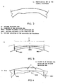
- figure 3 clearly shows the optimum receiving bed B, the letter D showing the diameter of said receiving bed.
- Smin shows the minimum thickness of the receiving bed
- Smax shows the maximum thickness of said receiving bed
- the letter V shows, in turn, the desired variation of the receiving bed thickness.
- the letter C shows the volume to be ablated for achieving the optimum receiving bed.
- Smax and Smin show respectively the maximum and minimum thickness of the mentioned receiving bed.
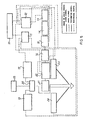
- said apparatus comprises a central processing unit 10, which is coupled to a pachymetric control unit 11 and a photoablative laser control unit 12.
- the latter is coupled to a laser cavity 13, fitted, in turn, to a control device for controlling the power of the laser beam 14 and to a device 15 for measuring the laser beam power.
- This apparatus is moreover characterized in that it further comprises a focalizing system 16 for focalizing the laser beam, coupled to a galvanometric system 22 and to an orienting system 21 for orienting the laser beam.
- a system 23 for detecting the corneal pachymetry and a source 24 for detecting said corneal pachymetry are moreover provided.
- the apparatus comprises furthermore an optic divider 20, to divide or split the video signal between an operating microscope 18, a video camera 19 for detecting the ocular motility, coupled to the photoablative laser control unit 12 and a further video camera 17 for detecting the ocular motility, coupled to the pachymeter control unit 11.
- an apparatus according to the block diagram shown in figure 5 has been provided, which is adapted to allow to detect in a very accurate manner the data and/or parameters necessary for addressing and properly performing the surgical operation.
- the used materials, as well as the contingent size and shapes, can be any, depending on requirements.
Abstract
Description
- The present invention relates to an apparatus for determining and ablating a corneal tissue volume of a receiving cornea for corneal lamellar grafting transplantation, as optimized for each individual patient.
- As is known, in a lamellar corneal grafting operation, a portion of corneal tissue having an even thickness and a variable diameter, depending on the amount and location of the different specific pathologies, is conventionally removed.
- Usually, the corneal tissue is removed by a surgical instrument called "Krumeich's microkeratome" which, by a planing type of operation removes a corneal disc having a preset diameter and an approximatively even thickness.
- Thus, the aim of the present invention is to provide such an apparatus which is suitable to mutually coordinate an assembly of apparatus and which is specifically designed for defining, in an unique and optimum manner, the position, area and volume of the corneal tissue to be removed by a laser ablating operation, in order to optimally perform the ablating operation itself.
- Within the scope of the above mentioned aim, a main object of the present invention is to provide such an apparatus which is very efficient in operation and which provides accurate values to allow the operator to perform optimally the ablating operation, i.e. in a manner optimized for each individual patient.
- Yet another object of the present invention is to provide such an apparatus which, owing to its peculiar constructional features, is very reliable and safe in operation.
- Yet another object of the present invention is to provide such an apparatus which can be easily made by using easily available elements and materials.
- According to the present invention, there is an apparatus for determining and ablating a corneal tissue volume of a receiving cornea for lamellar corneal grafting transplantation as claimed in
claim 1. - Further characteristics and advantages of the present invention will become more apparent hereinafter from the following disclosure of a preferred, though not exclusive, embodiment of an apparatus for determining and ablating an optimum volume of a corneal tissue as necessary for performing a lamellar corneal grafting operation, which is illustrated, by way of an indicative, but not limitative, example, in the accompanying drawings, where:
- Figure 1 is a flow diagram illustrating the operating flows which can be performed by the apparatus according to the present invention;
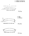
- Figure 2 is a cross-sectional view illustrating an optimum patient receiving bed;
- Figure 2a is a further cross sectional view illustrating a typical section of a donor lens;
- Figure 3 illustrates a typical section of the optimum receiving bed;
- Figure 3a represents an optimum typical section of the donor lens;
- Figure 4 illustrates a typical section of the optimum patient ablating contour, according to the present invention;
- Figure 4a represents a typical section of the donor lens ablating contour; and
- Figure 5 represents a block diagram of the ablating apparatus according to the present invention.
-
- With reference to the number references of Figure 1, the apparatus for determining and removing, by an ablating operation, a corneal tissue volume necessary for a lamellar grafting transplantation, as optimized for the individual patient, comprises a central processing unit, generally indicated by the
reference number 1, to which are coupled a corneal pachymeter (of an optical, ultrasound or the like type), generally indicated by thereference number 2, provided for morphologically detecting the corneal thickness and for tridimensionally mapping it. - To said central processing unit (1) a pupillometer, indicated by the
reference number 3, for determining the projection of the pupillar diaphragm, at the level of the corneal front surface, is connected. - To said
central processing unit 1 is moreover connected a photoablative laser (of an excimer, solid status, phantosecond or the like type), of a microspot type, generally indicated by thereference number 4, provided with a coupling interface, for reading the altimetric ablative datum, as expressed by microns on a x, y plane matrix or array. - The apparatus according to the present invention allows to find the ablating volume, which is obtained from a difference of the pachymetric map, as detected for the individual patient by the
pachymeter 2, and the optimum pachymetric map of the bed receiving the donor lens. - The operator, in using the apparatus according to the invention, will detect at the start, by using the
pupillometer 3, the projection of the pupil on the cornea front surface, the diameter thereof and the position of the related centroid with respect to the corneal limbus, as well as the diameter of the overall cornea and the position of its centroid, with respect to the pupillar centroid. - Then, the operator will detect the pachymetric data by using a corneal pachymeter (of an optical, ultrasonic or the like type) 2.
- As is known, the pachymetric map is the tridimensional contour or profile which is best interpolatedly approximated to the data detected by the
corneal pachymeter 2. - The optimum contour or profile of the donor lens receiving bed is defined by the operator by finding or detecting the following parameters:
- the center of the receiving bed;
- the diameter of the receiving bed;
- the minimum thickness at the center of the receiving bed
- the maximum thickness at the edge of the receiving bed; and
- the thickness variation along the diameter of the receiving bed.
- The receiving bed center can be either selected between the corneal centroid, the projection on the cornea of the pupillar centroid, or arbitrarily by the operator.
- The diameter of the receiving bed, in turn, is defined by the operator depending on the amount and location of the patient pathology.
- The minimum and maximum thicknesses of the receiving bed are defined by the operator depending on the detected pathology analysis and the desired post-surgical refracting geometry.
- The variation of the thickness along the receiving bed diameter is, depending on the operator selection, an even variation, a linear or exponential variation, depending on the desired post-surgical refracting geometry.
- By using the definition of the above mentioned parameters, the operator will detect, in an unique and optimum manner for the patient, the receiving bed provided for receiving lens of the donor.
- The ablating volume shown in Figure 2 is then obtained from the difference of the pachymetric map, as determined as above indicated, and the thus defined receiving bed.
- The above disclosed method will univocally define the tissue to be ablated volume, in order to set an optimum patient receiving bed.
- It is graphically represented by different color areas, clearly showing the receiving bed, its location and the residual cornea area not involved in the operation.
- Moreover, digital data will express the overall ablating surface, the overall ablating volume, the maximum and minimum ablating volumes and related planimetric locations as well as the planimetric location of the ablating center with respect to the pupillar centroid.
- The ablating volume related to the donor lens is obtained by defining the width A and height B of figure 3a to be generated on the lens or, alternately, the desired inclination or slope α.
- The altimetric ablative datum is represented on a x, y-plane square matrix, to allow the
photoablative laser 4 to properly perform its detection operation, through a suitable interface, for detecting the ablating contour or profile which, upon detection, is practically followed. - The system, moreover, optionally through an intra-operating detection of the pachymetric datum, will verify that the treatment has been carried out according to the programmed ablating strategy, while modifying the number of surface-unit localized pulses.
- In this case, the operation will end upon achieving a congruency of the detected and desired data.
- It should be pointed out that figure 3 clearly shows the optimum receiving bed B, the letter D showing the diameter of said receiving bed.
- In this figure, Smin shows the minimum thickness of the receiving bed, whereas Smax shows the maximum thickness of said receiving bed.
- The letter V shows, in turn, the desired variation of the receiving bed thickness.
- In figure 4, the letter C shows the volume to be ablated for achieving the optimum receiving bed.
- In this figure, moreover, Smax and Smin show respectively the maximum and minimum thickness of the mentioned receiving bed.
- With reference to figure 5, which is a block diagram of the apparatus according to the invention, said apparatus comprises a central processing unit 10, which is coupled to a pachymetric control unit 11 and a photoablative laser control unit 12.
- The latter is coupled to a laser cavity 13, fitted, in turn, to a control device for controlling the power of the
laser beam 14 and to adevice 15 for measuring the laser beam power. - This apparatus is moreover characterized in that it further comprises a focalizing
system 16 for focalizing the laser beam, coupled to agalvanometric system 22 and to an orienting system 21 for orienting the laser beam. - Moreover, a
system 23 for detecting the corneal pachymetry and asource 24 for detecting said corneal pachymetry are moreover provided. - The apparatus comprises furthermore an
optic divider 20, to divide or split the video signal between an operating microscope 18, avideo camera 19 for detecting the ocular motility, coupled to the photoablative laser control unit 12 and afurther video camera 17 for detecting the ocular motility, coupled to the pachymeter control unit 11. - From the above disclosure it should be apparent that the invention fully achieves the intended aim and objects.
- In particular, an apparatus according to the block diagram shown in figure 5 has been provided, which is adapted to allow to detect in a very accurate manner the data and/or parameters necessary for addressing and properly performing the surgical operation.
- The invention, as disclosed, is susceptible to several modifications and variations, all of which will come within the scope of the invention.
- Moreover, the used materials, as well as the contingent size and shapes, can be any, depending on requirements.
Claims (6)
- An apparatus for determining and ablating a corneal tissue volume of a receiving cornea for corneal lamellar grafting transplantation, comprising a central processing unit (1, 10), a pachymeter (2), coupled to the central processing unit (1, 10) for providing a corneal thickness tridimensional map (A), and a photoablative laser (4);
characterised in that the central processing unit (1, 10) comprises processing means for determining a volume (V) of corneal tissue to be ablated on the basis of a difference between the corneal thickness tridimensional map (A) and a receiving bed (B), having a predetermined bed thickness map, and the photoablative laser (4) is controlled by the central processing unit (1, 10) for ablating said volume (V) of corneal tissue. - Apparatus according to claim 1, characterised in that the central processing unit (1, 10) is configured for processing a plurality of geometric parameters (SMIN, SMAX, V) representative of said bed thickness map of the receiving bed (B).
- Apparatus according to claim 2, characterised in that said geometric parameters comprise at least a minimum thickness (SMIN), a maximum thickness (SMAX) and a thickness variation (V) along a diameter (D) of said receiving bed (B).
- Apparatus according to any one of the foregoing claims, characterised in that it comprises a photoablative laser control unit (12), coupled to said said central processing unit (1, 10) for controlling a number of surface-unit localized pulses supplied by the photoablative laser (4) according to a predetermined ablating strategy and to intra-operating pachymetric detection.
- Apparatus according to any one of the foregoing claims, characterised by comprising a pupillometer (3), coupled to the central control unit (1, 10).
- Apparatus according to claim 5, characterised in that the pupillometer (3) is configured for detecting a projection of a pupil on a corneal front surface, a diameter of said projection, and a centroid of said projection with respect to a corneal limbus.
Applications Claiming Priority (4)
| Application Number | Priority Date | Filing Date | Title |
|---|---|---|---|
| ITMI20002024 | 2000-09-15 | ||
| IT2000MI002024A IT1318699B1 (en) | 2000-09-15 | 2000-09-15 | EQUIPMENT TO DETERMINE AND ABLATE THE VOLUME OF THE CORNEAL TISSUE NECESSARY TO CARRY OUT A CORNEA LAMELLAR TRANSPLANT. |
| PCT/IT2001/000248 WO2002022003A1 (en) | 2000-09-15 | 2001-05-18 | Apparatus for determining and ablating the corneal tissue volume necessary for performing a corneal lamellar grafting operation |
| EP01936806A EP1320320B1 (en) | 2000-09-15 | 2001-05-18 | Apparatus for determining and ablating the corneal tissue volume necessary for performing a corneal lamellar grafting operation |
Related Parent Applications (2)
| Application Number | Title | Priority Date | Filing Date |
|---|---|---|---|
| EP01936806A Division EP1320320B1 (en) | 2000-09-15 | 2001-05-18 | Apparatus for determining and ablating the corneal tissue volume necessary for performing a corneal lamellar grafting operation |
| EP01936806.7 Division | 2001-05-18 |
Publications (4)
| Publication Number | Publication Date |
|---|---|
| EP1428470A2 true EP1428470A2 (en) | 2004-06-16 |
| EP1428470A3 EP1428470A3 (en) | 2005-01-05 |
| EP1428470B1 EP1428470B1 (en) | 2012-06-27 |
| EP1428470B2 EP1428470B2 (en) | 2020-04-01 |
Family
ID=11445813
Family Applications (2)
| Application Number | Title | Priority Date | Filing Date |
|---|---|---|---|
| EP01936806A Expired - Lifetime EP1320320B1 (en) | 2000-09-15 | 2001-05-18 | Apparatus for determining and ablating the corneal tissue volume necessary for performing a corneal lamellar grafting operation |
| EP04003911.7A Expired - Lifetime EP1428470B2 (en) | 2000-09-15 | 2001-05-18 | Apparatus for determining and ablating a corneal tissue volume of a receiving cornea for corneal lamellar grafting transplantation |
Family Applications Before (1)
| Application Number | Title | Priority Date | Filing Date |
|---|---|---|---|
| EP01936806A Expired - Lifetime EP1320320B1 (en) | 2000-09-15 | 2001-05-18 | Apparatus for determining and ablating the corneal tissue volume necessary for performing a corneal lamellar grafting operation |
Country Status (9)
| Country | Link |
|---|---|
| US (1) | US20040024389A1 (en) |
| EP (2) | EP1320320B1 (en) |
| AT (1) | ATE277555T1 (en) |
| AU (1) | AU2001262664A1 (en) |
| CA (1) | CA2422256A1 (en) |
| DE (1) | DE60106068T2 (en) |
| ES (1) | ES2391147T5 (en) |
| IT (1) | IT1318699B1 (en) |
| WO (1) | WO2002022003A1 (en) |
Cited By (4)
| Publication number | Priority date | Publication date | Assignee | Title |
|---|---|---|---|---|
| DE102007019815A1 (en) * | 2007-04-26 | 2008-10-30 | Carl Zeiss Meditec Ag | Corneal transplantation |
| US20090281529A1 (en) * | 2005-05-02 | 2009-11-12 | Carriazo Cesar C | Method for controlling a laser in the ablation of the corneal layer of an eye |
| ITMI20100571A1 (en) * | 2010-04-02 | 2011-10-03 | Ivis Technologies S R L | METHOD AND EQUIPMENT FOR PERSONALIZED PREPARATION OF CORNEE DI DONATORE FOR LAMINATE CORNEAL TRANSPLANTATION |
| EP3685800A1 (en) | 2019-01-22 | 2020-07-29 | AJL Ophthalmic, S.A. | Corneal implant |
Families Citing this family (5)
| Publication number | Priority date | Publication date | Assignee | Title |
|---|---|---|---|---|
| IT1318699B1 (en) † | 2000-09-15 | 2003-08-27 | Ligi Tecnologie Medicali S R L | EQUIPMENT TO DETERMINE AND ABLATE THE VOLUME OF THE CORNEAL TISSUE NECESSARY TO CARRY OUT A CORNEA LAMELLAR TRANSPLANT. |
| EP1447064B1 (en) * | 2003-02-12 | 2010-06-02 | Coherent GmbH | Kit-of-parts for surgically ablating eye tissue |
| EP1462074A1 (en) * | 2003-03-28 | 2004-09-29 | Cesar C. Dr. Carriazo | Ablation depth control system for corneal surgery |
| EP3781013A1 (en) * | 2018-04-20 | 2021-02-24 | IVIS TECHNOLOGIES S.r.l | Customized ablation to correct visual ametropia |
| DE102019103848B4 (en) | 2019-02-15 | 2023-03-09 | Schwind Eye-Tech-Solutions Gmbh | Method for controlling an ophthalmic surgical laser and treatment device |
Citations (3)
| Publication number | Priority date | Publication date | Assignee | Title |
|---|---|---|---|---|
| US4988348A (en) | 1989-05-26 | 1991-01-29 | Intelligent Surgical Lasers, Inc. | Method for reshaping the cornea |
| EP0697611A2 (en) | 1994-08-18 | 1996-02-21 | Carl Zeiss | Optical coherence tomography assisted surgical apparatus |
| DE19852331A1 (en) | 1998-11-13 | 2000-05-18 | Benedikt Jean | Simultaneous measurement of surface topometry, biometry of eye involves projecting placido-ring system onto cornea, reflecting, mirroring in tear film, recovering by video topography |
Family Cites Families (21)
| Publication number | Priority date | Publication date | Assignee | Title |
|---|---|---|---|---|
| US4669466A (en) * | 1985-01-16 | 1987-06-02 | Lri L.P. | Method and apparatus for analysis and correction of abnormal refractive errors of the eye |
| US4729652A (en) * | 1985-11-04 | 1988-03-08 | Eye Research Institute Of Retina Foundation | Apparatus and method for determining angular orientation of eye |
| US4665914A (en) * | 1985-12-27 | 1987-05-19 | Emanuel Tanne | Automatic corneal surgery system |
| US4724522A (en) * | 1986-05-27 | 1988-02-09 | Belgorod Barry M | Method and apparatus for modification of corneal refractive properties |
| US4911711A (en) * | 1986-12-05 | 1990-03-27 | Taunton Technologies, Inc. | Sculpture apparatus for correcting curvature of the cornea |
| US5098426A (en) * | 1989-02-06 | 1992-03-24 | Phoenix Laser Systems, Inc. | Method and apparatus for precision laser surgery |
| US6099522A (en) * | 1989-02-06 | 2000-08-08 | Visx Inc. | Automated laser workstation for high precision surgical and industrial interventions |
| US4995716A (en) * | 1989-03-09 | 1991-02-26 | Par Technology Corporation | Method and apparatus for obtaining the topography of an object |
| US5042937A (en) * | 1989-12-11 | 1991-08-27 | Pulse Medical Instruments | Optical system for an ophthamological instrument for examination of pupillary responses |
| US5187506A (en) * | 1990-09-28 | 1993-02-16 | Fairville Medical Optics Inc. | Method and apparatus for determining physiological parameters based on pupil response |
| US5141305A (en) * | 1991-01-31 | 1992-08-25 | Texas Tech University Health Sciences Center | Method and apparatus for evaluation of phasic visual neurons in humans |
| DE69232640T2 (en) * | 1991-11-06 | 2003-02-06 | Shui T Lai | DEVICE FOR CORNEAL SURGERY |
| AU5540594A (en) * | 1992-10-26 | 1994-05-24 | Shui T. Lai | Method of performing ophthalmic surgery |
| US5782822A (en) * | 1995-10-27 | 1998-07-21 | Ir Vision, Inc. | Method and apparatus for removing corneal tissue with infrared laser radiation |
| US5963300A (en) * | 1998-02-17 | 1999-10-05 | Amt Technologies, Corp. | Ocular biometer |
| US6000799A (en) * | 1998-05-01 | 1999-12-14 | Jozef F. Van De Velde | Maxwellian view and modulation control options for the scanning laser ophthalmoscope |
| AUPP697398A0 (en) * | 1998-11-06 | 1998-12-03 | Lions Eye Institute Of Western Australia Incorporated, The | Eye tracker for refractive surgery |
| US6245059B1 (en) * | 1999-04-07 | 2001-06-12 | Visx, Incorporated | Offset ablation profiles for treatment of irregular astigmation |
| US6551306B1 (en) † | 1999-04-13 | 2003-04-22 | Cesar C. Carriazo | Refractive laser ablation through topography |
| IT1318699B1 (en) * | 2000-09-15 | 2003-08-27 | Ligi Tecnologie Medicali S R L | EQUIPMENT TO DETERMINE AND ABLATE THE VOLUME OF THE CORNEAL TISSUE NECESSARY TO CARRY OUT A CORNEA LAMELLAR TRANSPLANT. |
| EP1462074A1 (en) † | 2003-03-28 | 2004-09-29 | Cesar C. Dr. Carriazo | Ablation depth control system for corneal surgery |
-
2000
- 2000-09-15 IT IT2000MI002024A patent/IT1318699B1/en active
-
2001
- 2001-05-18 EP EP01936806A patent/EP1320320B1/en not_active Expired - Lifetime
- 2001-05-18 DE DE60106068T patent/DE60106068T2/en not_active Expired - Lifetime
- 2001-05-18 ES ES04003911T patent/ES2391147T5/en not_active Expired - Lifetime
- 2001-05-18 WO PCT/IT2001/000248 patent/WO2002022003A1/en active IP Right Grant
- 2001-05-18 AU AU2001262664A patent/AU2001262664A1/en not_active Abandoned
- 2001-05-18 EP EP04003911.7A patent/EP1428470B2/en not_active Expired - Lifetime
- 2001-05-18 US US10/380,385 patent/US20040024389A1/en not_active Abandoned
- 2001-05-18 CA CA002422256A patent/CA2422256A1/en not_active Abandoned
- 2001-05-18 AT AT01936806T patent/ATE277555T1/en not_active IP Right Cessation
Patent Citations (3)
| Publication number | Priority date | Publication date | Assignee | Title |
|---|---|---|---|---|
| US4988348A (en) | 1989-05-26 | 1991-01-29 | Intelligent Surgical Lasers, Inc. | Method for reshaping the cornea |
| EP0697611A2 (en) | 1994-08-18 | 1996-02-21 | Carl Zeiss | Optical coherence tomography assisted surgical apparatus |
| DE19852331A1 (en) | 1998-11-13 | 2000-05-18 | Benedikt Jean | Simultaneous measurement of surface topometry, biometry of eye involves projecting placido-ring system onto cornea, reflecting, mirroring in tear film, recovering by video topography |
Cited By (8)
| Publication number | Priority date | Publication date | Assignee | Title |
|---|---|---|---|---|
| US20090281529A1 (en) * | 2005-05-02 | 2009-11-12 | Carriazo Cesar C | Method for controlling a laser in the ablation of the corneal layer of an eye |
| DE102007019815A1 (en) * | 2007-04-26 | 2008-10-30 | Carl Zeiss Meditec Ag | Corneal transplantation |
| US10362937B2 (en) | 2007-04-26 | 2019-07-30 | Carl Zeiss Meditec Ag | Cornea transplantation |
| US11154191B2 (en) | 2007-04-26 | 2021-10-26 | Carl Zeiss Meditec Ag | Cornea transplantation |
| ITMI20100571A1 (en) * | 2010-04-02 | 2011-10-03 | Ivis Technologies S R L | METHOD AND EQUIPMENT FOR PERSONALIZED PREPARATION OF CORNEE DI DONATORE FOR LAMINATE CORNEAL TRANSPLANTATION |
| EP2371329A1 (en) * | 2010-04-02 | 2011-10-05 | IVIS Tecnologie Medicali Srl, . | Method and apparatus for customized preparation of donor corneas for corneal lamellar implant |
| EP3685800A1 (en) | 2019-01-22 | 2020-07-29 | AJL Ophthalmic, S.A. | Corneal implant |
| WO2020152172A1 (en) | 2019-01-22 | 2020-07-30 | Ajl Ophthalmic, S.A. | Corneal implant |
Also Published As
| Publication number | Publication date |
|---|---|
| ES2391147T5 (en) | 2021-01-25 |
| AU2001262664A1 (en) | 2002-03-26 |
| ITMI20002024A1 (en) | 2002-03-15 |
| ATE277555T1 (en) | 2004-10-15 |
| US20040024389A1 (en) | 2004-02-05 |
| ITMI20002024A0 (en) | 2000-09-15 |
| EP1320320B1 (en) | 2004-09-29 |
| EP1320320A1 (en) | 2003-06-25 |
| DE60106068D1 (en) | 2004-11-04 |
| EP1428470A3 (en) | 2005-01-05 |
| DE60106068T2 (en) | 2006-02-16 |
| CA2422256A1 (en) | 2002-03-21 |
| EP1428470B2 (en) | 2020-04-01 |
| ES2391147T3 (en) | 2012-11-22 |
| EP1428470B1 (en) | 2012-06-27 |
| WO2002022003A1 (en) | 2002-03-21 |
| IT1318699B1 (en) | 2003-08-27 |
Similar Documents
| Publication | Publication Date | Title |
|---|---|---|
| AU2017243802B2 (en) | Visualization system for ophthalmic surgery | |
| US5308355A (en) | Ophthalmic surgical instrument and method | |
| KR100699403B1 (en) | Method for analyzing and improving vision | |
| US9955865B2 (en) | Method and system to detect ophthalmic tissue structure and pathologies | |
| JP5852237B2 (en) | Apparatus and method for laser assisted ocular surgical treatment system | |
| KR101476760B1 (en) | System for refractive ophthalmological surgery | |
| US20060100612A1 (en) | Laser-based device for non-mechanical, three-dimensional trepanation during cornea transplants | |
| EP1428470B1 (en) | Apparatus for determining and ablating a corneal tissue volume of a receiving cornea for corneal lamellar grafting transplantation | |
| EP3319503B1 (en) | Image processing method and system for edge detection and laser eye surgery system incorporating the same | |
| US9782232B1 (en) | Automated intraocular pressure tamponade | |
| MX2011000096A (en) | Device for ophthalmologic, particularly refractive, laser surgery. | |
| US20020154269A1 (en) | Stereoscopic measurement of cornea and illumination patterns | |
| JP7454055B2 (en) | Assembly comprising an OCT device, computer program, and computer implementation method therefor for verifying 3D reconstruction of a region of interest volume | |
| CN115103657A (en) | Direct laser trabeculoplasty method and apparatus | |
| EP1352623B1 (en) | Apparatus for determining and ablating corneal tissue volume necessary for correcting a visual ametropia |
Legal Events
| Date | Code | Title | Description |
|---|---|---|---|
| PUAI | Public reference made under article 153(3) epc to a published international application that has entered the european phase |
Free format text: ORIGINAL CODE: 0009012 |
|
| AC | Divisional application: reference to earlier application |
Ref document number: 1320320 Country of ref document: EP Kind code of ref document: P |
|
| AK | Designated contracting states |
Kind code of ref document: A2 Designated state(s): AT BE CH CY DE DK ES FI FR GB GR IE IT LI LU MC NL PT SE TR |
|
| PUAL | Search report despatched |
Free format text: ORIGINAL CODE: 0009013 |
|
| AK | Designated contracting states |
Kind code of ref document: A3 Designated state(s): AT BE CH CY DE DK ES FI FR GB GR IE IT LI LU MC NL PT SE TR |
|
| RIC1 | Information provided on ipc code assigned before grant |
Ipc: 7A 61F 9/013 B Ipc: 7A 61F 9/01 B Ipc: 7A 61B 3/11 B Ipc: 7A 61B 3/10 A |
|
| 17P | Request for examination filed |
Effective date: 20050630 |
|
| AKX | Designation fees paid |
Designated state(s): DE ES FR GB IT |
|
| 17Q | First examination report despatched |
Effective date: 20070601 |
|
| RAP1 | Party data changed (applicant data changed or rights of an application transferred) |
Owner name: IVIS TECHNOLOGIES S.R.L |
|
| TPAC | Observations filed by third parties |
Free format text: ORIGINAL CODE: EPIDOSNTIPA |
|
| GRAP | Despatch of communication of intention to grant a patent |
Free format text: ORIGINAL CODE: EPIDOSNIGR1 |
|
| GRAS | Grant fee paid |
Free format text: ORIGINAL CODE: EPIDOSNIGR3 |
|
| GRAA | (expected) grant |
Free format text: ORIGINAL CODE: 0009210 |
|
| AC | Divisional application: reference to earlier application |
Ref document number: 1320320 Country of ref document: EP Kind code of ref document: P |
|
| AK | Designated contracting states |
Kind code of ref document: B1 Designated state(s): DE ES FR GB IT |
|
| REG | Reference to a national code |
Ref country code: GB Ref legal event code: FG4D |
|
| REG | Reference to a national code |
Ref country code: DE Ref legal event code: R096 Ref document number: 60146778 Country of ref document: DE Effective date: 20120823 |
|
| REG | Reference to a national code |
Ref country code: ES Ref legal event code: FG2A Ref document number: 2391147 Country of ref document: ES Kind code of ref document: T3 Effective date: 20121122 |
|
| PLBI | Opposition filed |
Free format text: ORIGINAL CODE: 0009260 |
|
| 26 | Opposition filed |
Opponent name: SCHWIND EYE-TECH-SOLUTIONS GMBH & CO. KG Effective date: 20130212 |
|
| PLAX | Notice of opposition and request to file observation + time limit sent |
Free format text: ORIGINAL CODE: EPIDOSNOBS2 |
|
| REG | Reference to a national code |
Ref country code: DE Ref legal event code: R026 Ref document number: 60146778 Country of ref document: DE Effective date: 20130212 |
|
| PLBB | Reply of patent proprietor to notice(s) of opposition received |
Free format text: ORIGINAL CODE: EPIDOSNOBS3 |
|
| APBM | Appeal reference recorded |
Free format text: ORIGINAL CODE: EPIDOSNREFNO |
|
| APBP | Date of receipt of notice of appeal recorded |
Free format text: ORIGINAL CODE: EPIDOSNNOA2O |
|
| APAH | Appeal reference modified |
Free format text: ORIGINAL CODE: EPIDOSCREFNO |
|
| APBQ | Date of receipt of statement of grounds of appeal recorded |
Free format text: ORIGINAL CODE: EPIDOSNNOA3O |
|
| REG | Reference to a national code |
Ref country code: FR Ref legal event code: PLFP Year of fee payment: 16 |
|
| REG | Reference to a national code |
Ref country code: FR Ref legal event code: PLFP Year of fee payment: 17 |
|
| REG | Reference to a national code |
Ref country code: FR Ref legal event code: PLFP Year of fee payment: 18 |
|
| PLAB | Opposition data, opponent's data or that of the opponent's representative modified |
Free format text: ORIGINAL CODE: 0009299OPPO |
|
| APBU | Appeal procedure closed |
Free format text: ORIGINAL CODE: EPIDOSNNOA9O |
|
| R26 | Opposition filed (corrected) |
Opponent name: SCHWIND EYE-TECH-SOLUTIONS GMBH Effective date: 20130212 |
|
| PUAH | Patent maintained in amended form |
Free format text: ORIGINAL CODE: 0009272 |
|
| STAA | Information on the status of an ep patent application or granted ep patent |
Free format text: STATUS: PATENT MAINTAINED AS AMENDED |
|
| 27A | Patent maintained in amended form |
Effective date: 20200401 |
|
| AK | Designated contracting states |
Kind code of ref document: B2 Designated state(s): DE ES FR GB IT |
|
| REG | Reference to a national code |
Ref country code: DE Ref legal event code: R102 Ref document number: 60146778 Country of ref document: DE |
|
| PGFP | Annual fee paid to national office [announced via postgrant information from national office to epo] |
Ref country code: FR Payment date: 20200519 Year of fee payment: 20 Ref country code: DE Payment date: 20200527 Year of fee payment: 20 Ref country code: ES Payment date: 20200618 Year of fee payment: 20 |
|
| PGFP | Annual fee paid to national office [announced via postgrant information from national office to epo] |
Ref country code: GB Payment date: 20200522 Year of fee payment: 20 Ref country code: IT Payment date: 20200528 Year of fee payment: 20 |
|
| REG | Reference to a national code |
Ref country code: ES Ref legal event code: DC2A Ref document number: 2391147 Country of ref document: ES Kind code of ref document: T5 Effective date: 20210125 |
|
| REG | Reference to a national code |
Ref country code: DE Ref legal event code: R071 Ref document number: 60146778 Country of ref document: DE |
|
| REG | Reference to a national code |
Ref country code: GB Ref legal event code: PE20 Expiry date: 20210517 |
|
| PG25 | Lapsed in a contracting state [announced via postgrant information from national office to epo] |
Ref country code: GB Free format text: LAPSE BECAUSE OF EXPIRATION OF PROTECTION Effective date: 20210517 |
|
| REG | Reference to a national code |
Ref country code: ES Ref legal event code: FD2A Effective date: 20220103 |
|
| PG25 | Lapsed in a contracting state [announced via postgrant information from national office to epo] |
Ref country code: ES Free format text: LAPSE BECAUSE OF EXPIRATION OF PROTECTION Effective date: 20210519 |