EP1575642B1 - Multi-balloon catheter with hydrogel coating - Google Patents
Multi-balloon catheter with hydrogel coating Download PDFInfo
- Publication number
- EP1575642B1 EP1575642B1 EP03798753A EP03798753A EP1575642B1 EP 1575642 B1 EP1575642 B1 EP 1575642B1 EP 03798753 A EP03798753 A EP 03798753A EP 03798753 A EP03798753 A EP 03798753A EP 1575642 B1 EP1575642 B1 EP 1575642B1
- Authority
- EP
- European Patent Office
- Prior art keywords
- balloons
- catheter
- balloon
- biologically active
- coating
- Prior art date
- Legal status (The legal status is an assumption and is not a legal conclusion. Google has not performed a legal analysis and makes no representation as to the accuracy of the status listed.)
- Expired - Fee Related
Links
Images
Classifications
-
- A—HUMAN NECESSITIES
- A61—MEDICAL OR VETERINARY SCIENCE; HYGIENE
- A61M—DEVICES FOR INTRODUCING MEDIA INTO, OR ONTO, THE BODY; DEVICES FOR TRANSDUCING BODY MEDIA OR FOR TAKING MEDIA FROM THE BODY; DEVICES FOR PRODUCING OR ENDING SLEEP OR STUPOR
- A61M25/00—Catheters; Hollow probes
- A61M25/0043—Catheters; Hollow probes characterised by structural features
- A61M25/0045—Catheters; Hollow probes characterised by structural features multi-layered, e.g. coated
-
- A—HUMAN NECESSITIES
- A61—MEDICAL OR VETERINARY SCIENCE; HYGIENE
- A61L—METHODS OR APPARATUS FOR STERILISING MATERIALS OR OBJECTS IN GENERAL; DISINFECTION, STERILISATION OR DEODORISATION OF AIR; CHEMICAL ASPECTS OF BANDAGES, DRESSINGS, ABSORBENT PADS OR SURGICAL ARTICLES; MATERIALS FOR BANDAGES, DRESSINGS, ABSORBENT PADS OR SURGICAL ARTICLES
- A61L29/00—Materials for catheters, medical tubing, cannulae, or endoscopes or for coating catheters
- A61L29/08—Materials for coatings
- A61L29/085—Macromolecular materials
-
- A—HUMAN NECESSITIES
- A61—MEDICAL OR VETERINARY SCIENCE; HYGIENE
- A61L—METHODS OR APPARATUS FOR STERILISING MATERIALS OR OBJECTS IN GENERAL; DISINFECTION, STERILISATION OR DEODORISATION OF AIR; CHEMICAL ASPECTS OF BANDAGES, DRESSINGS, ABSORBENT PADS OR SURGICAL ARTICLES; MATERIALS FOR BANDAGES, DRESSINGS, ABSORBENT PADS OR SURGICAL ARTICLES
- A61L29/00—Materials for catheters, medical tubing, cannulae, or endoscopes or for coating catheters
- A61L29/14—Materials characterised by their function or physical properties, e.g. lubricating compositions
- A61L29/145—Hydrogels or hydrocolloids
-
- A—HUMAN NECESSITIES
- A61—MEDICAL OR VETERINARY SCIENCE; HYGIENE
- A61M—DEVICES FOR INTRODUCING MEDIA INTO, OR ONTO, THE BODY; DEVICES FOR TRANSDUCING BODY MEDIA OR FOR TAKING MEDIA FROM THE BODY; DEVICES FOR PRODUCING OR ENDING SLEEP OR STUPOR
- A61M25/00—Catheters; Hollow probes
- A61M25/10—Balloon catheters
- A61M25/1011—Multiple balloon catheters
-
- A—HUMAN NECESSITIES
- A61—MEDICAL OR VETERINARY SCIENCE; HYGIENE
- A61M—DEVICES FOR INTRODUCING MEDIA INTO, OR ONTO, THE BODY; DEVICES FOR TRANSDUCING BODY MEDIA OR FOR TAKING MEDIA FROM THE BODY; DEVICES FOR PRODUCING OR ENDING SLEEP OR STUPOR
- A61M25/00—Catheters; Hollow probes
- A61M25/10—Balloon catheters
- A61M25/1027—Making of balloon catheters
-
- A—HUMAN NECESSITIES
- A61—MEDICAL OR VETERINARY SCIENCE; HYGIENE
- A61M—DEVICES FOR INTRODUCING MEDIA INTO, OR ONTO, THE BODY; DEVICES FOR TRANSDUCING BODY MEDIA OR FOR TAKING MEDIA FROM THE BODY; DEVICES FOR PRODUCING OR ENDING SLEEP OR STUPOR
- A61M25/00—Catheters; Hollow probes
- A61M25/10—Balloon catheters
- A61M25/104—Balloon catheters used for angioplasty
-
- A—HUMAN NECESSITIES
- A61—MEDICAL OR VETERINARY SCIENCE; HYGIENE
- A61M—DEVICES FOR INTRODUCING MEDIA INTO, OR ONTO, THE BODY; DEVICES FOR TRANSDUCING BODY MEDIA OR FOR TAKING MEDIA FROM THE BODY; DEVICES FOR PRODUCING OR ENDING SLEEP OR STUPOR
- A61M25/00—Catheters; Hollow probes
- A61M25/0043—Catheters; Hollow probes characterised by structural features
- A61M25/0045—Catheters; Hollow probes characterised by structural features multi-layered, e.g. coated
- A61M2025/0046—Coatings for improving slidability
-
- A—HUMAN NECESSITIES
- A61—MEDICAL OR VETERINARY SCIENCE; HYGIENE
- A61M—DEVICES FOR INTRODUCING MEDIA INTO, OR ONTO, THE BODY; DEVICES FOR TRANSDUCING BODY MEDIA OR FOR TAKING MEDIA FROM THE BODY; DEVICES FOR PRODUCING OR ENDING SLEEP OR STUPOR
- A61M25/00—Catheters; Hollow probes
- A61M25/0043—Catheters; Hollow probes characterised by structural features
- A61M2025/0057—Catheters delivering medicament other than through a conventional lumen, e.g. porous walls or hydrogel coatings
-
- A—HUMAN NECESSITIES
- A61—MEDICAL OR VETERINARY SCIENCE; HYGIENE
- A61M—DEVICES FOR INTRODUCING MEDIA INTO, OR ONTO, THE BODY; DEVICES FOR TRANSDUCING BODY MEDIA OR FOR TAKING MEDIA FROM THE BODY; DEVICES FOR PRODUCING OR ENDING SLEEP OR STUPOR
- A61M25/00—Catheters; Hollow probes
- A61M25/10—Balloon catheters
- A61M2025/1043—Balloon catheters with special features or adapted for special applications
- A61M2025/105—Balloon catheters with special features or adapted for special applications having a balloon suitable for drug delivery, e.g. by using holes for delivery, drug coating or membranes
-
- A—HUMAN NECESSITIES
- A61—MEDICAL OR VETERINARY SCIENCE; HYGIENE
- A61M—DEVICES FOR INTRODUCING MEDIA INTO, OR ONTO, THE BODY; DEVICES FOR TRANSDUCING BODY MEDIA OR FOR TAKING MEDIA FROM THE BODY; DEVICES FOR PRODUCING OR ENDING SLEEP OR STUPOR
- A61M25/00—Catheters; Hollow probes
- A61M25/10—Balloon catheters
- A61M2025/1043—Balloon catheters with special features or adapted for special applications
- A61M2025/1052—Balloon catheters with special features or adapted for special applications for temporarily occluding a vessel for isolating a sector
-
- A—HUMAN NECESSITIES
- A61—MEDICAL OR VETERINARY SCIENCE; HYGIENE
- A61M—DEVICES FOR INTRODUCING MEDIA INTO, OR ONTO, THE BODY; DEVICES FOR TRANSDUCING BODY MEDIA OR FOR TAKING MEDIA FROM THE BODY; DEVICES FOR PRODUCING OR ENDING SLEEP OR STUPOR
- A61M25/00—Catheters; Hollow probes
- A61M25/10—Balloon catheters
- A61M2025/1043—Balloon catheters with special features or adapted for special applications
- A61M2025/1088—Balloon catheters with special features or adapted for special applications having special surface characteristics depending on material properties or added substances, e.g. for reducing friction
-
- A—HUMAN NECESSITIES
- A61—MEDICAL OR VETERINARY SCIENCE; HYGIENE
- A61M—DEVICES FOR INTRODUCING MEDIA INTO, OR ONTO, THE BODY; DEVICES FOR TRANSDUCING BODY MEDIA OR FOR TAKING MEDIA FROM THE BODY; DEVICES FOR PRODUCING OR ENDING SLEEP OR STUPOR
- A61M25/00—Catheters; Hollow probes
- A61M25/0067—Catheters; Hollow probes characterised by the distal end, e.g. tips
- A61M25/0068—Static characteristics of the catheter tip, e.g. shape, atraumatic tip, curved tip or tip structure
- A61M25/007—Side holes, e.g. their profiles or arrangements; Provisions to keep side holes unblocked
Abstract
Description
- The present invention relates to catheters which may be inserted into body lumens for delivery of biologically active agents. More particularly, the invention relates to catheters having inflatable balloons for occluding body lumens and delivering therapeutic, biologically active agents to a portion of the occluded body lumen.
- Atherosclerotic disease can cause the build-up of plaque deposits in a localized portion of an arterial vessel. As the deposition increases, the inner diameter of the vessel becomes smaller (stenosis occurs) and impedes blood flow through the vessel. This reduction in blood throughput is harmful to the body and must be corrected. One method of correcting this arterial stenosis is to perform angioplasty using a balloon catheter, which involves inserting a catheter having at least a dilatation balloon into the stenotic area of the artery, inflating this balloon and expanding the inner diameter of the plaque-ridden segment of the artery.
- An alternative variation of the above angioplasty method employs a catheter having two spaced-apart occlusion balloons whereby one of the balloons is a dilatation balloon used to expand the plaque-affected segment of the artery. Catheters having double occlusion balloons are disclosed, for example, in
U.S. Patent Nos. 5,836,967 and5,279,546 . After an initial expansion of the stenotic segment of the artery, the dilatation balloon is deflated and the catheter is manipulated and positioned such that the affected segment of the artery is between the two balloons. The two balloons are simultaneously pressurized and inflated, forming an enclosed, sealed space wherein the affected portion of the artery is contained within this space. In other words an occluded portion in the artery or lumen is formed. The catheter portion between the two balloons has a hole or plurality of holes for delivery of biologically active agents therethrough to variously treat the affected arterial segment. Thus, unlike a catheter having only a single balloon, the double-balloon catheter advantageously allows isolation of the affected segment and permits subsequent targeted application of beneficial agents to the affected arterial segment. - The double balloons thus function to seal off or occlude a section of the vessel apart from performing a dilatation function to expand the opening of a plaque-ridden, stenotic area of the vessel. During this occlusion step, the surfaces of the balloons contact healthy vessel tissue. Pressurizing and inflating the balloons to contact healthy endothelial or endoluminal tissue can cause injury to such tissue. The first kind of injury arises from abrading and compressing the surface cells of the inner vessel wall, which may lead to denuding of the surface, when the occlusion balloons are inflated. Denuding of the vessel wall may initiate the restenosis process. Another kind of injury, disruption of the internal elastic lamina, may be sustained because of the pressures that may be needed to provide a seal between the occlusion balloons and the vessel surfaces; this injury may result in distension of the vessel wall. Thus, certain balloon pressures may also cause injury to healthy vessel wall.
- Hence, a comparative disadvantage of using a double-balloon catheter for delivery of biologically active agents is that the balloons must contact healthy portions of the inner wall of the vessel being treated, unlike a catheter with only a single dilatation balloon which generally only touches the plaque-covered vessel wall in their dilatation function of opening the stenotic segment. A double-balloon catheter, however, contacts not only the plaque-ridden part of the vessel during the dilatation step, but also touches at least two healthy, unaffected areas of the vessel during the occlusion step when both balloons are inflated.
-
WO 96/04952 - A hydrogel polymer coating for a single-balloon catheter is disclosed in
US 5,954,706 , wherein an aqueous solution of a preselected drug to be delivered to a target tissue is incorporated in the hydrogel polymer.
Accordingly, there exists a need for an improved double-balloon catheter that minimizes the denuding of healthy endothelial or endoluminal tissue due to abrasion and compression by the occlusion balloons when they are inflated. - The present invention applies to a catheter having two or more occlusion balloons wherein the balloons are spaced apart, and the balloons are simultaneously inflated to form a sealed space in a portion of a body lumen or an occluded portion in the body lumen. An object of the present invention is to provide a balloon catheter that reduces abrasion-type damage to healthy endothelial or endoluminal tissue.
- Yet another object of the invention is to provide a double-balloon catheter having at least two balloons dispensing biologically active agents in the targeted area of the occluded portion of the body lumen situated between the occlusion balloons. Such balloons may provide superior sealing, can concurrently permit the use of comparatively lower balloon inflation pressures. The advantage of using lower pressures to achieve sufficient sealing is that the distension of the lumen wall may be reduced when the occlusion balloons are inflated.
- The present invention provides in one aspect, a balloon catheter for occluding two ends of a portion of a body lumen and treating the occluded portion. The catheter comprises at least two balloons each having an exterior surface and a catheter segment connectively placed between the balloons. A coating comprising a hydrogel polymer is applied to the exterior surface of each of the occlusion balloons. The presence of such hydrogel on the occlusion balloons reduces the damage to the endothelial tissue of the body lumen that contacts the balloon upon expansion. The balloons are capable of being inflated and deflated within the body lumen, and when both balloons are inflated, form a substantially closed space within the occluded portion of the body lumen by forming a seal between the balloon surface and inner wall surface of the body lumen. The catheter segment is capable of delivering a biologically active agent to the occluded portion of the body lumen.
- Embodiments of the balloon catheter may incorporate one or more of the following features. The delivery of the biologically active agent may be conducted using a plurality of holes or ports extending through the catheter wall of the catheter segment connecting the balloons. The holes or ports fluidly connect an inner lumen of the catheter segment and its exterior. The holes or ports are located between the balloons, and permit the biologically active agent to pass through from the inner catheter lumen to the exterior of the catheter segment. Other methods of delivering biologically active agents may also be employed with this multi-balloon catheter so long as the material is delivered between the balloons. The balloons can be made of an expandable material or a material that is substantially non-expandable, although the balloons are inflatable in either case.
- The biologically active agents that can be used may have various effects. For example, it may be an anti-inflammatory drug which reduces body reaction to tissue injury, or it may be an anti-proliferative agent which is capable of blocking smooth muscle cell proliferation, in particular, as a reaction to tissue injury or irritation. The biologically active agents used may be dispensed through holes or ports in the catheter wall.
- The exterior of both balloons are coated with a coating comprising a hydrogel polymer, which is preferably tenaciously adhering on the balloon surface. This coating can be variously manufactured to have different thicknesses in its dry and hydrated states. The hydrogel polymer is selected from poly-carboxylic acids, celluosic polymers, gelatin polyvinyl-pyrrolidone, maleic anhydride polymers, polyamides, polyvinyl alcohols, and polyethylene oxides. The hydrogel coating may be substantially free of a biologically active agent.
- In another aspect of the invention, the coating comprising a hydrogel polymer over the exterior of the balloons may be loaded with a biologically active agent within the hydrogel matrix. The coating over the balloons may be used to deliver biologically active agents to proactively treat the healthy, inner lining of the body lumen that contacts the coating. Suitable biologically active agents should be capable of being incorporated into a hydrogel matrix which can leach or elute out on when pressure is applied to the hydrogel coating the balloon surfaces. It will be appreciated that the hydrogel coating of a multi-balloon catheter may optionally contain a biologically active agent to treat the contact area of healthy endothelial lumen tissue. This is a separate treatment modality, distinct from the treatment modality wherein the biologically active agent is delivered from within the catheter to treat the plaque-affected segment of the lumen that is contained in the space formed by inflation of the balloons or occluded portion of the body lumen.
- In yet another aspect, a three-balloon catheter is provided, where the two outer balloons can be occlusion balloons and the middle balloon can be a dilatation or occlusion balloon. The two outer occlusion balloons can have a coating of hydrogel which is generally free of any therapeutic, biologically active agents since they contact only healthy vessel tissue. The middle, dilatation or occlusion balloon can be coated with a hydrogel which may be loaded with a biologically active agent to treat the target site of the vessel.
- In a further aspect of the invention, the entire exterior surface of the catheter as well as the balloons may be coated with a hydrogel for ease of manufacture.
- In yet another aspect of the invention, a multi-balloon catheter that maintains the integrity of the endothelial cells of the body lumen, having at least two spaced-apart balloons for treating a body lumen is provided, wherein the balloons are coated with a coating comprising a hydrogel polymer; both balloons are suitable to be inflated to contact the endothelial or cells of the inner wall of the body lumen; forming a substantially closed space between the balloons within the body lumen; and wherein the multi-balloon catheter is suitable to provide a biologically active agent into the closed space to treat a portion of the body lumen.
- The present invention will be explained with reference to the following drawings.
-
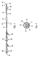
FIG. 1 is a perspective view of an embodiment of a double-balloon catheter with both balloons inflated; -
FIG. 2 is a cross-sectional view of the catheter ofFIG. 1 showing the inner channels at section A-A ofFIG. 1 ; -
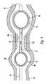
FIG. 3 is longitudinal cross-sectional view of a catheter of the present invention inserted within a stenotic blood vessel with both balloons inflated, this embodiment shows the hydrogel coating on the occlusion and dilatation balloons and also the plaque on the vessel inner wall; and -
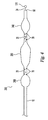
FIG. 4 shows an embodiment of a catheter having a plurality of balloons. -
FIG. 1 shows an exemplary embodiment of the catheter assembly having two spaced-apart balloons, theocclusion balloon 40 and dilatation/occlusion balloon 50. Theouter catheter 10 is configured with two lumina (not shown). The two balloons may be made of an expandable material or a substantially non-expandable material such as polyurethanes. In either case, the balloons are inflatable and thus capable of pushing against the inner wall of the body vessel once they are inserted. The catheter has adistal orifice 90, ametal marker 120 to enable x-ray identification of the location of the dilatation/occlusion balloon, and acatheter segment 20 that connects the two balloons. Extending through theorifice 90, aguidewire 34 has astop plug 170 which can be used to cap theorifice 90. A coating comprising a hydrogel polymer (not shown) is applied to the exterior surface ofballoons catheter segment 20 which permits a biologically active agent to pass from an inner lumen in thecatheter segment 20 to the exterior of thecatheter segment 20 and thereby target treatment to the section of the body lumen between the two balloons or the occluded portion of the body lumen. Also shown inFIG. 1 are a number of Y connectors, 140 and 160 withside ports Connections balloons - The catheter shown in
FIG. 1 is only one example of a catheter suitable for the present invention. Other catheter designs can also be employed in this invention. The skilled artisan is aware of such additional suitable catheter designs. -
FIG. 2 shows an exemplary embodiment of a cross-sectional view of the catheter ofFIG. 1 at section A-A. The outer catheter is shown with twolumens 35 and 33. Aninner catheter stem 31 is shown inserted intolumen 33 and thisstem 31 has twolumens occlusion balloon 40 and dilatation /occlusion balloon 50, to deliver a biologically active agent through theholes 70, and to receive theguidewire 34. Theouter catheter 10 is configured with two lumina. The lumen 35 serves to supply the pressurizing medium to theocclusion balloon 40 while thelumen 33 permits a biologically active agent to be delivered to a targeted vessel portion. Thelumen 30 serves to supply the pressurizing medium to dilatation/occlusion balloon 50 while lumen 32 can be used to receiveguidewire 34. It should be noted that this is only one of many embodiments that can be used to independently control the inflation and deflation of the two balloons as well as to provide a separate fluid channel for the biologically active agents to travel and exit from the holes or plurality ofholes 70. Generally, there should be at least three separate channels within the catheter to service the two balloons and to deliver the biologically active agents through the catheter holes 70. -
FIG. 3 shows, in accordance with the present invention, a longitudinal cross-sectional view of a catheter of the present invention that has been inserted within ablood vessel 80 with bothballoons hydrogel polymer 60 over the exterior surface of the occlusion and dilatation/occlusion balloons and also theplaque 180 on the vessel inner wall.FIG. 3 shows the subsequent position of the two balloons following the step in which the plaque-lined vessel has been previously dilated by inflating the dilatation/occlusion balloon 50 at the site of theplaque 180. After the expansion of the plaque site, the catheter is moved to re-position the two balloons such that the target area of the vessel having theplaque 180 on thevessel wall 80 is between theocclusion balloon 40 and dilatation/occlusion balloon 50. - In operation, the double-balloon catheter described in
FIGS. 1-3 is advanced into a body lumen such as an arterial vessel until the dilatation/occlusion balloon 50 is precisely located at the stenotic site, while the location of theballoon 50 is monitored by x-ray with the aid of ametal marker 120 attached to theinner stem 31 within the dilatation/occulsion 50 balloon. By supplying a pressurizing medium via theconnection 110 thedilatation balloon 50 can be inflated, dilating the stenotic, plaque-coated site of the lumen. Once this dilatation action has been completed, the pressurizing medium is drained from thedilatation balloon 50 and the catheter is advanced into the body lumen beyond the plaque-coated site until thedilatation balloon 50 is downstream, while theocclusion balloon 40 remains located in a portion upstream of the treated segment of the body lumen. Then, a pressurizing medium is supplied to the two balloons via theconnections holes 70 while theballoons connection 150 of the Y connector 160 into the occluded portion of the body lumen between the twoballoons ports 70 and into theinner stem 20 of the inner catheter and via theguidewire lumen 32. It is also possible to deliver the biologically active agent via theguidewire lumen 32 of the inner catheter and to drain the biologically active agent via thelumen 33 in the outer catheter. - With this embodiment, throughout the duration of treatment, the
guidewire 34 must be retracted in theguidewire lumen 32 sufficiently so thatocclusion element 170 abuts theorifice 90 to cap it. Once the orifice is sealed, theguidewire lumen 32 can be employed as the lumen for supply and removal of the biologically active agent. - After the biologically active agent is allowed to react with the target plaque and endothelial tissue of the body lumen upon which the plaque is deposited, and after the excess biologically active agent has been drawn off from the occluded portion of the body lumen formed by the two inflated balloons, the pressurizing medium is drawn off from the two balloons to deflate them. With the treatment procedure thus completed, the catheter assembly is removed from the body lumen.
- When plaque build-up is the cause of stenosis, the catheter having a balloon with a dual dilatation/occlusion function, as shown in
FIG. 1 , can be used. Many other treatments, however, do not require a balloon having a dilatation function. Instead, two occlusion balloons can be inflated to seal a vessel portion, wherein a biologically active agent may be delivered into this sealed portion. For example, preventing the recurrence of arterial thrombus in a vessel may be accomplished by isolating the affected portion of the vessel and delivering a biologically active agent, such as an anti-proliferative or anti-thrombogenic into the isolated portion. Because the balloons function only to occlude a vessel and touch healthy vessel wall which are especially susceptible to de-nuding, a catheter having two hydrogel-coated balloons which are generally free of any biologically active agents, in accordance with the present invention, can be used to substantially reduce the potential for vessel damage. Additionally, the balloons may be further optimized to reduce potential abrasion to the healthy tissue by being made more compliant than a typical dilatation balloon. - In accordance with the present invention, isolation of a vessel may be accomplished with another embodiment of the invention wherein a catheter has three balloons, each coated with hydrogel. A three balloon catheter can occlude two different sections of a target vessel simultaneously or at different times. Each balloon would be accessible through an independent catheter channel, and a separate channel or channels would be used to deliver the biological agent from the
holes 70 in thecatheter segments 20 between the balloons. A catheter having more than three balloons is also possible, restricted only by the complexity of needing additional channels within the catheter, and the physical limitations thereon. -
FIG. 4 shows one embodiment of a three-balloon catheter 200, wherein themiddle balloon 250 may be used to dilate the stenotic (lesion) portion of the vessel and theouter balloons 240, which contact only healthy vessel tissue and thus coated with hydrogel, can serve as the occlusion balloons for isolating the treatment portion of the vessel. The expandability of the middle 250 andouter balloons 240 can be chosen to optimize their respective functions. Because balloon abrasion to a vessel lesion is less of a concern than abrasion to healthy tissue, the dilatation balloon may be designed to be less expandable than an occlusion balloon and employ higher pressures (up to 20 atms). - In operation the middle,
dilatation balloon 250 may be inflated to dilate the stenotic area of the vessel. The middle,dilatation balloon 250 is deflated. Then the two, outer occlusion balloons 240 may be inflated to isolate the treatment portion of the vessel and a biologically active agent, e.g., cells or viral vectors may be delivered via the catheter into the enclosed space to treat the vessel wall. - In an alternative gene delivery embodiment, a three-
balloon catheter 200 is used to apply the viral vector (or other agent) present in the coating on the middle balloon 250 (not necessarily a dilatation). When themiddle balloon 250 is inflated, a therapeutic viral vector (or other agent) can be delivered via a diffusion process to the vessel wall The twoouter balloons 240 are inflated during this gene transfer process in order to provide vessel isolation. Optionally, in another embodiment of the method, in accordance with the present invention, another separate, biologically active agent can be delivered viaholes 70 in the catheter in addition to the gene transfer procedure. - In another embodiment of the catheter of the present invention, the two outer balloons may be placed around a dispatch catheter. The dispatch catheter has a coil frame which permits delivery of biologically active agents, while the two outer balloons provide isolation of the vessel portion being treated.
- There are a number of preferred variations of the present invention of the balloon catheters with hydrogel coating as provided in
FIGS. 1-4 . The first variation has an apparatus for delivering a biologically active agent situated between two spaced-apart, hydrogel coated balloons, but, morever, provides an additional treatment functionality in that the hydrogel is a matrix containing a biologically active agent that is delivered to the vessel wall when pressure is applied to the hydrogel coating on the balloon. Another variation builds on the above first or second variation. In this embodiment, the hydrogel coats not only the balloons, but also coats the entire catheter. This latter embodiment may be advantageous because it may be easier and less costly to manufacture. The hydrogel coating disposed over the surface of the balloons and, optionally, over the remaining surface of the catheter may or may not contain eluting biologically active agents. - Thus, the present invention provides a multi-balloon catheter that maintains the integrity of the endothelial cells of the inner wall of the body lumen wherein the catheter has at least two balloons, wherein the balloons are coated with a coating comprising a hydrogel polymer for treating a body lumen and wherein both balloons are suitable to be inflated to contact the endothelial cells of the inner wall of the body lumen; forming a substantially closed space between the balloons within the body lumen; and wherein the catheter is suitable to provide a biologically active agent into the closed space.
- The last step may be accomplished in various ways. However, a preferred means is to deliver the biologically active agent through a channel or inner lumen in the catheter segment connecting the two balloons. The inner catheter lumen is fluidly connected to
holes 70 or ports extending through the walls of thecatheter segment 20 which is connectively between the two spaced-apart balloons. Theholes 70 permit the biologically active agent to pass through or be ejected from an inner catheter lumen to the exterior of thecatheter segment 20. - There are several advantages provided by using hydrogel coated balloons. The hydrogel, which may be described alternatively as a foam, provides a cushioning interface between the balloon and the healthy body lumen cell lining. The cushioning can reduce the abrasion of the cells and consequent de-nuding of the body lumen when the balloons are inflated. Additionally, the hydrogel may help to create an effective sealant between the balloon and the body lumen, which may enable a reduction in pressure needed to inflate the balloons. Less pressure may reduce distension of the body lumen and thereby potentially reduce damage and irritation to the contact areas of the body lumen. In sum, the injury to the body lumen at the contact areas can be reduced.
- The presence of the coating comprising a hydrogel polymer over the surface of balloons is, thus, a key element to the multi-balloon catheter of the present invention. The hydrogel polymer comprises poly-carboxylic acids, celluosic polymers, gelatin polyvinyl-pyrrolidone, maleic anhydride polymers, polyamides, polyvinyl alcohols, or polyethylene oxides. Additional details concerning materials for making hydrogels are provided in
U.S. Patent Nos. 5,304,121 , and5,954,706, both issued to Sahatjian , andU.S. Patent Nos. 5,674,192 , and5,843,049 issued to Sahatjian et. al - In particular
U.S. Pat. No. 5,304,121 , Example 1, provides one method of producing hydrogel coated balloon using a "dipping" process. The surface of the balloon (polyethlene) of an angiographic catheter is prepared by wiping down with clean cloth. The ballon has an O.D. (outer diameter) of about 3.5 mm (inflated). The balloon is coated with a solution of 4,4 diphenylmethane diisocyanate (MDI) in methylethylketone for 30 minutes. After drying in an air oven at 85°C for 30 minutes, the balloon is dipped in a 1.7% solution of poly(acrylic acid) homopolymer having a molecular weight of about 3,000,000 in dimethylformamide (DMF) and tertiarybutyl alchohol. After drying at about 85°C for 30 minutes, a smooth coating is obtained. The balloon is oven dried for 8 hours at 50°C. One function of the drying steps is to remove solvent from the coating. The surface of the balloon becomes instantly lubricious upon exposure to water. The polyisocynanate solution is at a concentration of about 0.5 to 10% by weight. The poly (carboxyl acid) to polyisocyanate molar ratio is generally about 1:1. The formation of the hydrogel is further described inU. S. Pat. No. 5,091,205 . - In addition to the above "dipping" process, there are other methods for providing a hydrogel coating over a balloon including "spraying" the coating onto the balloon. While the "dipping" method is described above as an example, the present invention is not limited to the particular manufacturing method in which the hydrogel is applied to the balloon, and, indeed, is inclusive of all forms of making the hydrogel coated balloon.
- In general, when dried, the hydrogel coating should be greater than about 0.5 microns in thickness and preferably in the range of about 1.0 to 10.0 micrometers in thickness. The hydrogel coating thickness can swell by about a factor of 6 to 10 or even more when the hydrogel is hydrated. More preferably, the thickness of the hydrated coating is about 10 to about 55 microns in the swelled, uncompressed state.
- In one embodiment, the hydrogel can be loaded with a biologically active agent that is released when in contact with fluid or when inflation pressure is applied. This provides a way for further deactivating the body's inflammatory and cell proliferation response caused by irritation when the balloon contacts the healthy cell lining in the body lumen. Here, the treatment is directed not to the body lumen that is plaque-affected, but to the healthy part of the body lumen that is especially susceptible to injury when a foreign object such as a catheter balloon abrades or compresses the body lumen cell lining or distends the body lumen. Also, the hydrogel composition and biologically active agent may be chosen for the purpose of a particular rate-of-release response characteristic, this being dependent on the hydrogel's degree of cross-linking, charge, structure, thickness, or sensitivity to compression, and the agent's solubility, particle size and charge. For example, the response characteristic of the hydrogel coating may be designed such that an application of compressive pressure urges delivery of the biologically active agent in about 10 minutes or less, with 10 minutes representing the outward duration a balloon catheter should be inside an arterial vessel.
- One example of an embodiment in which the hydrogel includes a biologically active agent is the use of heparin with the hydrogel. Heparin is absorbed into the coating without complexing and is freely released therefrom. Such hydrogel-drug combination can deliver about half of the drug solution in response to pressures used for balloon angioplasty. In other particular embodiments, the hydrogel polymer includes acid groups and incorporates the biologically active agent which is anionic in nature and is bound by electrostatic attraction to cations in the coating, such as an ammonium cation.
- With respect to suitable biologically active agents that are intended to be delivered through a catheter channel, the agent should be easily dispensed through small holes or ports in the catheter segment joining the balloons. These biologically active agents are, in general, placed into an aqueous solution. Various biologically active agents may be delivered including, anti-inflammatory drugs, such as heparin or heparin derivatives, Ppack (dextro-phenylalanine proline argine chloromethylketone), enoxaprin, hirudin, or aspirin (acetylsalicylic acid) or an anti-proliferative agent, such as heparin (which has anti-proliferative properties), enoxaprin, angiopeptin, or monoclonal antibodies capable of blocking smooth muscle cell proliferation. Dosages applied to the body lumen tissue, for example, of heparin are in the range of 10-30 mg of heparin solution containing 200-1000 units of sodium heparin.
- Other examples of biologically active agents include thrombolytic agents, such as the enzyme, urokinase, which can be used to remove existing arterial thrombus. The term biologically active material" encompasses therapeutic agents, such as drugs, and also genetic materials and biological materials. The genetic materials mean DNA or RNA, including, without limitation, of DNA/RNA encoding a useful protein stated below, anti-sense DNA/RNA, intended to be inserted into a human body including viral vectors and non-viral vectors. Examples of DNA suitable for the present invention include DNA encoding
- anti-sense RNA
- tRNA or rRNA to replace defective or deficient endogenous molecules
- angiogenic factors including growth factors, such as acidic and basic fibroblast growth factors, vascular endothelial growth factor, epidermal growth factor, transforming growth factor α and β, platelet-derived endothelial growth factor, platelet-derived growth factor, tumor necrosis factor α, hepatocyte growth factor and insulin like growth factor
- cell ccle inhibitors including CD inhibitors
- thymidine kinase ("TK") and other agents useful for interfering with cell proliferation, and
- the family of bone morphogenic proteins ("BMP's") as explained below. Viral vectors include adenoviruses, gutted adenoviruses, adeno-associated virus, retroviruses, alpha virus (Semliki Forest, Sindbis, etc.), lentiviruses, herpes simplex virus, ex vivo modified cells (e.g., stem cells, fibroblasts, myoblasts, satellite cells, pericytes, cardiomyocytes, sketetal myocytes, macrophage), replication competent viruses (e.g., ONYX-015), and hybrid vectors. Non-viral vectors include artificial chromosomes and mini-chromosomes, plasmid DNA vectors (e.g., pCOR), cationic polymers (e.g., polyethyleneimine, polyethyleneimine (PEI)) graft copolymers (e.g., polyether-PEI and polyethylene oxide-PEI), neutral polymers PVP, SP1017 (SUPRATEK), lipids or lipoplexes, nanoparticles and microparticles with and without targeting sequences such as the protein transduction domain (PTD).
- The biological materials include cells, yeasts, bacteria, proteins, peptides, cytokines and hormones. Examples for peptides and proteins include growth factors (FGF, FGF-1, FGF-2, VEGF, Endotherial Mitogenic Growth Factors, and epidermal growth factors, transforming growth factor α and β, platelet derived endothelial growth factor, platelet derived growth factor, tumor necrosis factor α, hepatocyte growth factor and insulin like growth factor), transcription factors, proteinkinases, CD inhibitors, thymidine kinase, and bone morphogenic proteins (BMP's), such as BMP-2, BMP-3, BMP-4, BMP-5, BMP-6 (Vgr-1), BMP-7 (OP-1), BMP-8. BMP-9, BMP-10, BMP-11, BMP-12, BMP-13, BMP-14, BMP-15, and BMP-16. Currently preferred BMP's are BMP-2, BMP-3, BMP-4, BMP-5, BMP-6, BMP-7. Alternatively or in addition, molecules capable of inducing an upstream or downstream effect of a BMP can be provided. Such molecules include any of the "hedgehog" proteins, or the DNA's encoding them. These dimeric proteins can be provided as homodimers, heterodimers, or combinations thereof, alone or together with other molecules. Cells can be of human origin (autologous or allogeneic) or from an animal source (xenogeneic), genetically engineered, if desired, to deliver proteins of interest at the transplant site. The delivery media can be formulated as needed to maintain cell function and viability. Cells include whole bone marrow, bone marrow derived mono-nuclear cells, progenitor cells (e.g., endothelial progentitor cells) stem cells (e.g., mesenchymal, hematopoietic, neuronal), pluripotent stem cells, fibroblasts, macrophage, and satellite cells.
- Biologically active material also includes non-genetic therapeutic agents, such as:
- anti-thrombogenic agents such as heparin, heparin derivatives, urokinase, and PPack (dextrophenylalanine proline arginine chloromethylketone);
- anti-proliferative agents such as enoxaprin, angiopeptin, or monoclonal antibodies capable of blocking smooth muscle cell proliferation, hirudin, and acetylsalicylic acid, amlodipine and doxazosin;
- anti-inflammatory agents such as glucocorticoids, betamethasone, dexamethasone, prednisolone, corticosterone, budesonide, estrogen, sulfasalazine, and mesalamine;
- immunosuppressants such as sirolimus (RAPAMYCIN), tacrolimus, everolimus and dexamethasone,
- antineoplastic/antiproliferative/anti-miotic agents such as paclitaxel, 5-fluorouracil, cisplatin, vinblastine, vincristine, epothilones, methotrexate, azathioprine, halofuginone, adriamycin, actinomycin and mutamycin; endostatin, angiostatin and thymidine kinase inhibitors, and its analogs or derivatives;
- anesthetic agents such as lidocaine; bupivacaine, and ropivacaine;
- anti-coagulants such as D-Phe-Pro-Arg chloromethyl keton, an RGD peptide-containing componnd, heparin, antithrombin compounds, platelet receptor antagonists, anti-thrombin anticodies, anti-platelet receptor antibodies, aspirin (aspirin is also classified as an analgesic, antipyretic and anti-inflammatory drug), dipyridamole, protamine, hirudin, prostaglandin inhibitors, platelet inhibitors and tick antiplatelet peptides;
- vascular cell growth promotors such as growth factors, Vascular Endothelial Growth Factors (FEGF, all types including VEGF-2), growth factor receptors, transcriptional activators, and translational promotors;
- vascular cell growth inhibitors such as antiproliferative agents, growth factor inhibitors, growth factor receptor antagonists, transcriptional repressors, translational repressors, replication inhibitors, inhibitory antibodies, antibodies directed against growth factors, bifunctional molecules consisting of a growth factor and a cytotoxin, bifunctional molecules consisting of an antibody and a cytotoxin;
- cholesterol-lowering agents; vasodilating agents; and agents which interfere with endogenous vasoactive mechanisms;
- anti-oxidants, such as probucol;
- antibiotic agents, such as penicillin, cefoxitin, oxacillin, tobranycin
- angiogenic substances, such as acidic and basic fibrobrast growth factors, estrogen including estradiol (E2), estriol (E3) and 17-Beta Estradiol; and
- drugs for heart failure, such as digoxin, beta-blockers, angiotensin-converting enzyme (ACE) inhibitors including captopril and enalopril.
- Also, the biologically active materials of the present invention include nitric oxide adducts, which prevent and/or treat adverse effects associated with use of a medical device in a patient, such as restenosis and damaged blood vessel surface. Typical nitric oxide adducts include nitroglycerin, sodium nitroprusside, S-nitroso-proteins, S-nitnoso-thiols, long carbon-chain lipophilic S-nitrosothiols, S-nitrosodithiols, iron-nitrosyl compounds, thionitrates, thionitrites, sydnonimines, furoxans, organic nitrates, and nitrosated amino acids, preferably mono-or poly-nitrosylated proteins, particularly polynitrosated albumin or polymers or aggregates thereof. The albumin is preferably human or bovine, including humanized bovine serum albumin. Such nitric oxide adducts are disclosed in
U.S. Patent No. 6,087,479 to Stamler et al . - A biologically active material may be encapsulated in micro-capsules by the known methods. Still other biologically active agents can be therapeutic genes which may be carried in viral vectors such as recombinant, nuclear-specific adenoviral β-galactosidase. The delivered genes may be used to treat vascular proliferative disease by increasing gene expression in vascular tissue.
- Delivery of genes to a treatment site can be accomplished in several ways. The genes may be delivered as a viral solution through holes or ports in a double-balloon catheter wall after isolation of the vessel. Another method of delivering the gene to the site is by applying a gene vector to a balloon's hydrogel surface and then transferring the genes to vessel wall tissue by inflating the balloon. This balloon can be prepared, for example, with a viral solution of 25 µl of a 1.7 x 10¹¹ PFU/ml adenoviral β-galactosidase stock solution which can be applied over the surface of a hydrogel-coated, catheter balloon. This particular method of gene transfer is described in "Analysis of Adenoviral Transport Mechanisms in the Vessel Wall and Optimization of Gene Transfer Using Local Delivery Genes" by Palasis et al., Human Gene Therapy, 11:237-246, January 20, 2000.
- The following experimental studies have been performed and illustrate the advantages of using a hydrogel coated balloon to contact healthy endothelium as compared to a non-coated balloon. The experimental protocol was in compliance with the Guide for Care and Use of Laboratory Animals (NIH publication no. 86-23, 1985). New Zealand White Rabbits were anesthetized with ketamine (10 mg/kg) and acepromazine (0.2 mg/kg) after premedication with xylazine (2mg/kg). The bilateral external iliac arteries were used for all experiments. A balloon of a catheter having no hydrogel coating was inflated to about 6 atm, resulting in a balloon diameter of about 3mm inside one iliac and then deflated after 2 or 30 minutes elapsed. A balloon of a catheter coated with three layers of a high molecular weight polyacrylic acid polymer, i.e., hydrogel, was inserted into the contralateral iliac and then inflated to about 6 atm then deflated after 2 or 30 minutes. Three days after this transduction, the animals were anesthetized and the iliac arteries were harvested. The harvested vessels were fixed in paraformaldehyde for 10 minutes and washed in phosphate buffered solution post-fixation. The vessels were later cut open longitudinally. Cross-sectional specimens were embedded in paraffin, sectioned into 5 micrometer sections, and counterstained with hematoxylin and eosin. Slides were observed using light microscopy. An examination of the vessel specimens treated with the hydrogel coated balloon showed very little or no visible damage to the endothelium whereas, the vessel specimen treated with the uncoated balloon evidenced noticeable damage to the endothelial wall. Thus, the hydrogel coating over the balloon clearly prevented subsequent damage to healthy vessel endothelium when a balloon was dilated against the endothelium.
- It is to be understood that, while various embodiments or the invention have been described, the forgoing are intended only as exemplary embodiments and that other embodiments may be within the scope of the present invention. It is further understood that other advantages, modifications and embodiments may be within the scope of the present invention as defined by the appended claims.
Claims (8)
- A balloon catheter (10) for occluding two ends of a portion of a blood vessel (80) and treating the occluded portion of the blood vessel (80) comprising:a first balloon and a second balloon (40, 50, 240), each having an exterior surface and a catheter segment (20) connectively placed therebetween wherein said catheter segment (20) is capable of delivering a biologically active agent to the occluded portion of the blood vessel (80) and wherein said balloons (40, 50, 240) are capable of being inflated and deflated within the blood vessel (80), and when the balloons (40, 50, 240) are inflated, form a substantially fluid tight, closed space between the first and second balloons (40, 50, 240); anda coating (60),characterized in that the coating comprises a hydrogel polymer applied to the exterior surfaces of each of the balloons (40, 50, 240),
wherein the hydrogel polymer comprises poly-carboxylic acids, cellulosic polymers, gelatin, polyvinyl-pyrrolidone, maleic anhydride polymers, polyamides, polyvinyl alcohols, or polyethylene oxides. - The balloon catheter (10) of claim 1, wherein said coating (60) is between about 1.0 and about 10.0 micrometers in thickness.
- The balloon catheter (10) of claim 1, wherein said catheter segment (20) comprises an inner lumen, and a catheter wall and wherein at least one port (70) extends through the wall to the exterior of the catheter segment (20), said port (70) permitting the biologically active agent to pass through from the inner lumen to the exterior of the catheter segment (20).
- The balloon catheter (10) of claim 1, wherein said biologically active agent comprises heparin, PpacK (dextro-phenylalanine proline arginine chloromethylketone), enoxaprin, hirudin, aspirin, or monoclonal antibodies capable of blocking smooth muscle cell proliferation.
- The balloon catheter (10) of claim 1, wherein said poly-carboxylic acid is a polyacrylic acid.
- The balloon catheter (10) of claim 1, wherein the thickness of the coating (60) in a swelled, uncompressed state is between about 10 to about 50 microns.
- The balloon catheter (10) of claim 1, which comprises a third balloon (250) disposed between the first and second balloons (240) and a catheter segment (20) connectively placed between the first and third balloons and a catheter segment (20) connectively placed between the second and third balloons.
- A balloon catheter (10) of any of the previous claims, wherein said coating (60) is substantially free of any biologically active agent.
Applications Claiming Priority (3)
| Application Number | Priority Date | Filing Date | Title |
|---|---|---|---|
| US10/253,597 US7060051B2 (en) | 2002-09-24 | 2002-09-24 | Multi-balloon catheter with hydrogel coating |
| US253597 | 2002-09-24 | ||
| PCT/US2003/030378 WO2004028587A2 (en) | 2002-09-24 | 2003-09-24 | Multi-balloon catheter with hydrogel coating |
Publications (3)
| Publication Number | Publication Date |
|---|---|
| EP1575642A2 EP1575642A2 (en) | 2005-09-21 |
| EP1575642A4 EP1575642A4 (en) | 2007-03-07 |
| EP1575642B1 true EP1575642B1 (en) | 2010-03-24 |
Family
ID=31993191
Family Applications (1)
| Application Number | Title | Priority Date | Filing Date |
|---|---|---|---|
| EP03798753A Expired - Fee Related EP1575642B1 (en) | 2002-09-24 | 2003-09-24 | Multi-balloon catheter with hydrogel coating |
Country Status (5)
| Country | Link |
|---|---|
| US (1) | US7060051B2 (en) |
| EP (1) | EP1575642B1 (en) |
| AU (1) | AU2003276980A1 (en) |
| DE (1) | DE60331856D1 (en) |
| WO (1) | WO2004028587A2 (en) |
Cited By (31)
| Publication number | Priority date | Publication date | Assignee | Title |
|---|---|---|---|---|
| US7931683B2 (en) | 2007-07-27 | 2011-04-26 | Boston Scientific Scimed, Inc. | Articles having ceramic coated surfaces |
| US7938855B2 (en) | 2007-11-02 | 2011-05-10 | Boston Scientific Scimed, Inc. | Deformable underlayer for stent |
| US7942926B2 (en) | 2007-07-11 | 2011-05-17 | Boston Scientific Scimed, Inc. | Endoprosthesis coating |
| US7976915B2 (en) | 2007-05-23 | 2011-07-12 | Boston Scientific Scimed, Inc. | Endoprosthesis with select ceramic morphology |
| US7981150B2 (en) | 2006-11-09 | 2011-07-19 | Boston Scientific Scimed, Inc. | Endoprosthesis with coatings |
| US8002823B2 (en) | 2007-07-11 | 2011-08-23 | Boston Scientific Scimed, Inc. | Endoprosthesis coating |
| US8029554B2 (en) | 2007-11-02 | 2011-10-04 | Boston Scientific Scimed, Inc. | Stent with embedded material |
| US8049061B2 (en) | 2008-09-25 | 2011-11-01 | Abbott Cardiovascular Systems, Inc. | Expandable member formed of a fibrous matrix having hydrogel polymer for intraluminal drug delivery |
| US8067054B2 (en) | 2007-04-05 | 2011-11-29 | Boston Scientific Scimed, Inc. | Stents with ceramic drug reservoir layer and methods of making and using the same |
| US8066763B2 (en) | 1998-04-11 | 2011-11-29 | Boston Scientific Scimed, Inc. | Drug-releasing stent with ceramic-containing layer |
| US8071156B2 (en) | 2009-03-04 | 2011-12-06 | Boston Scientific Scimed, Inc. | Endoprostheses |
| US8070797B2 (en) | 2007-03-01 | 2011-12-06 | Boston Scientific Scimed, Inc. | Medical device with a porous surface for delivery of a therapeutic agent |
| US8076529B2 (en) | 2008-09-26 | 2011-12-13 | Abbott Cardiovascular Systems, Inc. | Expandable member formed of a fibrous matrix for intraluminal drug delivery |
| US8187620B2 (en) | 2006-03-27 | 2012-05-29 | Boston Scientific Scimed, Inc. | Medical devices comprising a porous metal oxide or metal material and a polymer coating for delivering therapeutic agents |
| US8216632B2 (en) | 2007-11-02 | 2012-07-10 | Boston Scientific Scimed, Inc. | Endoprosthesis coating |
| US8221822B2 (en) | 2007-07-31 | 2012-07-17 | Boston Scientific Scimed, Inc. | Medical device coating by laser cladding |
| US8226603B2 (en) | 2008-09-25 | 2012-07-24 | Abbott Cardiovascular Systems Inc. | Expandable member having a covering formed of a fibrous matrix for intraluminal drug delivery |
| US8231980B2 (en) | 2008-12-03 | 2012-07-31 | Boston Scientific Scimed, Inc. | Medical implants including iridium oxide |
| US8287937B2 (en) | 2009-04-24 | 2012-10-16 | Boston Scientific Scimed, Inc. | Endoprosthese |
| US8353949B2 (en) | 2006-09-14 | 2013-01-15 | Boston Scientific Scimed, Inc. | Medical devices with drug-eluting coating |
| US8431149B2 (en) | 2007-03-01 | 2013-04-30 | Boston Scientific Scimed, Inc. | Coated medical devices for abluminal drug delivery |
| US8449603B2 (en) | 2008-06-18 | 2013-05-28 | Boston Scientific Scimed, Inc. | Endoprosthesis coating |
| US8500687B2 (en) | 2008-09-25 | 2013-08-06 | Abbott Cardiovascular Systems Inc. | Stent delivery system having a fibrous matrix covering with improved stent retention |
| US8574615B2 (en) | 2006-03-24 | 2013-11-05 | Boston Scientific Scimed, Inc. | Medical devices having nanoporous coatings for controlled therapeutic agent delivery |
| US8771343B2 (en) | 2006-06-29 | 2014-07-08 | Boston Scientific Scimed, Inc. | Medical devices with selective titanium oxide coatings |
| US8815275B2 (en) | 2006-06-28 | 2014-08-26 | Boston Scientific Scimed, Inc. | Coatings for medical devices comprising a therapeutic agent and a metallic material |
| US8815273B2 (en) | 2007-07-27 | 2014-08-26 | Boston Scientific Scimed, Inc. | Drug eluting medical devices having porous layers |
| US8900292B2 (en) | 2007-08-03 | 2014-12-02 | Boston Scientific Scimed, Inc. | Coating for medical device having increased surface area |
| US8920491B2 (en) | 2008-04-22 | 2014-12-30 | Boston Scientific Scimed, Inc. | Medical devices having a coating of inorganic material |
| US8932346B2 (en) | 2008-04-24 | 2015-01-13 | Boston Scientific Scimed, Inc. | Medical devices having inorganic particle layers |
| US9284409B2 (en) | 2007-07-19 | 2016-03-15 | Boston Scientific Scimed, Inc. | Endoprosthesis having a non-fouling surface |
Families Citing this family (157)
| Publication number | Priority date | Publication date | Assignee | Title |
|---|---|---|---|---|
| US6774278B1 (en) * | 1995-06-07 | 2004-08-10 | Cook Incorporated | Coated implantable medical device |
| DE10115740A1 (en) * | 2001-03-26 | 2002-10-02 | Ulrich Speck | Preparation for restenosis prophylaxis |
| AU2002345328A1 (en) | 2001-06-27 | 2003-03-03 | Remon Medical Technologies Ltd. | Method and device for electrochemical formation of therapeutic species in vivo |
| AU2002348180A1 (en) | 2001-11-09 | 2003-05-26 | Novoste Corporation | Baloon catheter with non-deployable stent |
| US20040111108A1 (en) | 2001-11-09 | 2004-06-10 | Farnan Robert C. | Balloon catheter with non-deployable stent |
| US8845672B2 (en) * | 2002-05-09 | 2014-09-30 | Reshape Medical, Inc. | Balloon system and methods for treating obesity |
| DE10244847A1 (en) | 2002-09-20 | 2004-04-01 | Ulrich Prof. Dr. Speck | Medical device for drug delivery |
| US8080026B2 (en) | 2003-01-21 | 2011-12-20 | Angioscore, Inc. | Apparatus and methods for treating hardened vascular lesions |
| US7686824B2 (en) * | 2003-01-21 | 2010-03-30 | Angioscore, Inc. | Apparatus and methods for treating hardened vascular lesions |
| US20060041303A1 (en) * | 2004-08-18 | 2006-02-23 | Israel Henry M | Guidewire with stopper |
| US20060064009A1 (en) * | 2004-09-21 | 2006-03-23 | Webler William E | Vessel imaging devices and methods |
| US7494612B2 (en) * | 2004-10-29 | 2009-02-24 | Boston Scientific Scimed, Inc. | Medical balloon folding method and tooling |
| US8262605B2 (en) | 2004-12-09 | 2012-09-11 | Ams Research Corporation | Needleless delivery systems |
| US10076641B2 (en) * | 2005-05-11 | 2018-09-18 | The Spectranetics Corporation | Methods and systems for delivering substances into luminal walls |
| US20070009564A1 (en) * | 2005-06-22 | 2007-01-11 | Mcclain James B | Drug/polymer composite materials and methods of making the same |
| WO2007011708A2 (en) | 2005-07-15 | 2007-01-25 | Micell Technologies, Inc. | Stent with polymer coating containing amorphous rapamycin |
| AU2006270221B2 (en) | 2005-07-15 | 2012-01-19 | Micell Technologies, Inc. | Polymer coatings containing drug powder of controlled morphology |
| WO2007022055A1 (en) * | 2005-08-12 | 2007-02-22 | Massicotte J Mathieu | Method and device for extracting objects from the body |
| US20070100368A1 (en) * | 2005-10-31 | 2007-05-03 | Quijano Rodolfo C | Intragastric space filler |
| US8172792B2 (en) * | 2005-12-27 | 2012-05-08 | Tyco Healthcare Group Lp | Embolic protection systems for bifurcated conduits |
| US20090312696A1 (en) * | 2005-12-28 | 2009-12-17 | Copa Vincent G | Devices, Systems, and Related Methods for Delivery of Fluid to Tissue |
| US8840660B2 (en) | 2006-01-05 | 2014-09-23 | Boston Scientific Scimed, Inc. | Bioerodible endoprostheses and methods of making the same |
| US8089029B2 (en) | 2006-02-01 | 2012-01-03 | Boston Scientific Scimed, Inc. | Bioabsorbable metal medical device and method of manufacture |
| US8048150B2 (en) | 2006-04-12 | 2011-11-01 | Boston Scientific Scimed, Inc. | Endoprosthesis having a fiber meshwork disposed thereon |
| EP2019657B1 (en) | 2006-04-26 | 2015-05-27 | Micell Technologies, Inc. | Coatings containing multiple drugs |
| JP2009545407A (en) | 2006-08-02 | 2009-12-24 | ボストン サイエンティフィック サイムド,インコーポレイテッド | End prosthesis with 3D decomposition control |
| JP2010503485A (en) | 2006-09-15 | 2010-02-04 | ボストン サイエンティフィック リミテッド | Medical device and method for manufacturing the same |
| US8808726B2 (en) | 2006-09-15 | 2014-08-19 | Boston Scientific Scimed. Inc. | Bioerodible endoprostheses and methods of making the same |
| ES2368125T3 (en) | 2006-09-15 | 2011-11-14 | Boston Scientific Scimed, Inc. | BIOEROSIONABLE ENDOPROOTHESIS WITH BIOESTABLE INORGANIC LAYERS. |
| ATE517590T1 (en) | 2006-09-15 | 2011-08-15 | Boston Scient Ltd | BIOLOGICALLY ERODABLE ENDOPROTHESES |
| US8002821B2 (en) | 2006-09-18 | 2011-08-23 | Boston Scientific Scimed, Inc. | Bioerodible metallic ENDOPROSTHESES |
| US9539593B2 (en) * | 2006-10-23 | 2017-01-10 | Micell Technologies, Inc. | Holder for electrically charging a substrate during coating |
| US8998846B2 (en) | 2006-11-20 | 2015-04-07 | Lutonix, Inc. | Drug releasing coatings for balloon catheters |
| US8414910B2 (en) | 2006-11-20 | 2013-04-09 | Lutonix, Inc. | Drug releasing coatings for medical devices |
| US20080175887A1 (en) | 2006-11-20 | 2008-07-24 | Lixiao Wang | Treatment of Asthma and Chronic Obstructive Pulmonary Disease With Anti-proliferate and Anti-inflammatory Drugs |
| US8414526B2 (en) | 2006-11-20 | 2013-04-09 | Lutonix, Inc. | Medical device rapid drug releasing coatings comprising oils, fatty acids, and/or lipids |
| US9700704B2 (en) | 2006-11-20 | 2017-07-11 | Lutonix, Inc. | Drug releasing coatings for balloon catheters |
| US8414525B2 (en) | 2006-11-20 | 2013-04-09 | Lutonix, Inc. | Drug releasing coatings for medical devices |
| US8425459B2 (en) | 2006-11-20 | 2013-04-23 | Lutonix, Inc. | Medical device rapid drug releasing coatings comprising a therapeutic agent and a contrast agent |
| US20080276935A1 (en) | 2006-11-20 | 2008-11-13 | Lixiao Wang | Treatment of asthma and chronic obstructive pulmonary disease with anti-proliferate and anti-inflammatory drugs |
| US9737640B2 (en) | 2006-11-20 | 2017-08-22 | Lutonix, Inc. | Drug releasing coatings for medical devices |
| US20080119823A1 (en) | 2006-11-21 | 2008-05-22 | Crank Justin M | Injection Tube for Jet Injection Device |
| ITMI20062333A1 (en) * | 2006-12-05 | 2008-06-06 | Mario Salerno | DEVICE TO ASSIST THE SCLORESANT TREATMENT OF VARICOSE VEINS |
| ATE488259T1 (en) | 2006-12-28 | 2010-12-15 | Boston Scient Ltd | BIOERODIBLE ENDOPROTHES AND PRODUCTION METHODS THEREOF |
| US11426494B2 (en) | 2007-01-08 | 2022-08-30 | MT Acquisition Holdings LLC | Stents having biodegradable layers |
| EP2111184B1 (en) * | 2007-01-08 | 2018-07-25 | Micell Technologies, Inc. | Stents having biodegradable layers |
| CA2743022C (en) | 2007-01-21 | 2012-10-09 | Hemoteq Ag | Methods for coating catheter balloons with a defined quantity of active agent |
| US20150335415A1 (en) * | 2007-01-31 | 2015-11-26 | Stanley Batiste | Intravenous filter with guidewire and catheter access guide |
| US8226602B2 (en) * | 2007-03-30 | 2012-07-24 | Reshape Medical, Inc. | Intragastric balloon system and therapeutic processes and products |
| JP5443336B2 (en) * | 2007-04-17 | 2014-03-19 | ミセル テクノロジーズ、インコーポレイテッド | Stent with biodegradable layer |
| CA2688314C (en) | 2007-05-25 | 2013-12-03 | Micell Technologies, Inc. | Polymer films for medical device coating |
| US8142469B2 (en) * | 2007-06-25 | 2012-03-27 | Reshape Medical, Inc. | Gastric space filler device, delivery system, and related methods |
| US9192697B2 (en) | 2007-07-03 | 2015-11-24 | Hemoteq Ag | Balloon catheter for treating stenosis of body passages and for preventing threatening restenosis |
| US20090036831A1 (en) * | 2007-08-01 | 2009-02-05 | Cook Incorporated | Balloon catheter with delivery ports |
| AU2008284042B2 (en) | 2007-08-03 | 2013-06-27 | Scion NeuroStim, Inc. | Vestibular stimulation apparatus and associated methods of use |
| US8052745B2 (en) | 2007-09-13 | 2011-11-08 | Boston Scientific Scimed, Inc. | Endoprosthesis |
| WO2009051780A1 (en) * | 2007-10-19 | 2009-04-23 | Micell Technologies, Inc. | Drug coated stents |
| US8162880B2 (en) * | 2008-01-18 | 2012-04-24 | Swaminathan Jayaraman | Delivery of therapeutic and marking substance through intra lumen expansion of a delivery device |
| US7713540B2 (en) * | 2008-02-19 | 2010-05-11 | Medtronic Vascular, Inc. | Devices and methods for reverse lipid transport |
| US8951545B2 (en) | 2008-03-28 | 2015-02-10 | Surmodics, Inc. | Insertable medical devices having microparticulate-associated elastic substrates and methods for drug delivery |
| MX350637B (en) * | 2008-04-17 | 2017-09-11 | Micell Technologies Inc | Stents having bioabsorbable layers. |
| US9126025B2 (en) * | 2008-05-01 | 2015-09-08 | Bayer Intellectual Property Gmbh | Method of coating a folded catheter balloon |
| US7998192B2 (en) | 2008-05-09 | 2011-08-16 | Boston Scientific Scimed, Inc. | Endoprostheses |
| US8236046B2 (en) | 2008-06-10 | 2012-08-07 | Boston Scientific Scimed, Inc. | Bioerodible endoprosthesis |
| US8974462B2 (en) | 2008-06-13 | 2015-03-10 | Pivot Medical, Inc. | Devices and methods for minimally invasive access into a joint |
| AU2009257304A1 (en) | 2008-06-13 | 2009-12-17 | Pivot Medical, Inc. | Methods and apparatus for joint distraction |
| US20090318855A1 (en) * | 2008-06-24 | 2009-12-24 | Abbott Cardiovascular Systems Inc. | Devices and methods for improving intravascular uptake of agents |
| WO2010009335A1 (en) | 2008-07-17 | 2010-01-21 | Micell Technologies, Inc. | Drug delivery medical device |
| US9510856B2 (en) | 2008-07-17 | 2016-12-06 | Micell Technologies, Inc. | Drug delivery medical device |
| US7985252B2 (en) | 2008-07-30 | 2011-07-26 | Boston Scientific Scimed, Inc. | Bioerodible endoprosthesis |
| US9295820B2 (en) | 2008-08-14 | 2016-03-29 | Surmodics, Inc. | Method and apparatus for coating balloon catheters |
| WO2010024898A2 (en) | 2008-08-29 | 2010-03-04 | Lutonix, Inc. | Methods and apparatuses for coating balloon catheters |
| US8162879B2 (en) * | 2008-09-22 | 2012-04-24 | Tyco Healthcare Group Lp | Double balloon catheter and methods for homogeneous drug delivery using the same |
| US8382824B2 (en) | 2008-10-03 | 2013-02-26 | Boston Scientific Scimed, Inc. | Medical implant having NANO-crystal grains with barrier layers of metal nitrides or fluorides |
| EP2177238B1 (en) * | 2008-10-14 | 2016-11-09 | Dentsply IH AB | Medical device with controllably releasable antibacterial agent |
| US8226601B2 (en) | 2008-11-12 | 2012-07-24 | Sanovas, Inc. | Resector balloon system |
| US8540667B2 (en) * | 2008-11-12 | 2013-09-24 | Sanovas, Inc. | Multi-balloon catheter for extravasated drug delivery |
| US8834913B2 (en) * | 2008-12-26 | 2014-09-16 | Battelle Memorial Institute | Medical implants and methods of making medical implants |
| EP2403546A2 (en) | 2009-03-02 | 2012-01-11 | Boston Scientific Scimed, Inc. | Self-buffering medical implants |
| US9174031B2 (en) * | 2009-03-13 | 2015-11-03 | Reshape Medical, Inc. | Device and method for deflation and removal of implantable and inflatable devices |
| US10426453B2 (en) | 2009-03-17 | 2019-10-01 | Pivot Medical, Inc. | Method and apparatus for distracting a joint |
| EP2408402B1 (en) * | 2009-03-17 | 2020-05-06 | Stryker Corporation | Joint-spacing balloon catheter |
| US9186181B2 (en) | 2009-03-17 | 2015-11-17 | Pivot Medical, Inc. | Method and apparatus for distracting a joint |
| WO2010111232A2 (en) * | 2009-03-23 | 2010-09-30 | Micell Technologies, Inc. | Drug delivery medical device |
| EP2410954A4 (en) * | 2009-03-23 | 2014-03-05 | Micell Technologies Inc | Peripheral stents having layers |
| CN102481195B (en) * | 2009-04-01 | 2015-03-25 | 米歇尔技术公司 | Drug delivery medical device |
| WO2010115161A2 (en) * | 2009-04-03 | 2010-10-07 | Reshape Medical Inc | Improved intragastric space fillers and methods of manufacturing including in vitro testing |
| CA2759015C (en) | 2009-04-17 | 2017-06-20 | James B. Mcclain | Stents having controlled elution |
| US20100292641A1 (en) * | 2009-05-15 | 2010-11-18 | Bandula Wijay | Targeted drug delivery device and method |
| NZ597307A (en) * | 2009-06-02 | 2014-04-30 | Concept Medical Inc | Rejuvenating coronary artery by improving blood flow with the help of insertion of nano-balls (encapsulated nanoparticles) containing therapeutic agents by non implantable device for tissues and thereby providing in tissue release to address the required cell cycle |
| EP2944332B1 (en) | 2009-07-10 | 2016-08-17 | Boston Scientific Scimed, Inc. | Use of nanocrystals for a drug delivery balloon |
| US10080821B2 (en) | 2009-07-17 | 2018-09-25 | Boston Scientific Scimed, Inc. | Nucleation of drug delivery balloons to provide improved crystal size and density |
| US9358143B2 (en) | 2009-07-22 | 2016-06-07 | Reshape Medical, Inc. | Retrieval mechanisms for implantable medical devices |
| WO2011011743A2 (en) | 2009-07-23 | 2011-01-27 | Reshape Medical, Inc. | Deflation and removal of implantable medical devices |
| EP2456487A4 (en) | 2009-07-23 | 2017-05-17 | ReShape Medical, Inc. | Inflation and deflation mechanisms for inflatable medical devices |
| US8894568B2 (en) | 2009-09-24 | 2014-11-25 | Reshape Medical, Inc. | Normalization and stabilization of balloon surfaces for deflation |
| US8721649B2 (en) | 2009-12-04 | 2014-05-13 | Pivot Medical, Inc. | Hip joint access using a circumferential wire and balloon |
| US20120330132A1 (en) * | 2009-12-07 | 2012-12-27 | Paul Sorajja | Device for the Delineation of Cardiovascular or Other Anatomical Structures |
| WO2011097103A1 (en) * | 2010-02-02 | 2011-08-11 | Micell Technologies, Inc. | Stent and stent delivery system with improved deliverability |
| WO2011097636A1 (en) | 2010-02-08 | 2011-08-11 | Reshape Medical, Inc. | Improved and enhanced aspiration processes and mechanisms for intragastric devices |
| US9149611B2 (en) | 2010-02-08 | 2015-10-06 | Reshape Medical, Inc. | Materials and methods for improved intragastric balloon devices |
| WO2011106637A1 (en) | 2010-02-25 | 2011-09-01 | Reshape Medical, Inc. | Improved and enhanced explant processes and mechanisms for intragastric devices |
| US8668732B2 (en) | 2010-03-23 | 2014-03-11 | Boston Scientific Scimed, Inc. | Surface treated bioerodible metal endoprostheses |
| US8795762B2 (en) | 2010-03-26 | 2014-08-05 | Battelle Memorial Institute | System and method for enhanced electrostatic deposition and surface coatings |
| EP2555705A4 (en) | 2010-04-06 | 2014-01-15 | Reshape Medical Inc | Inflation devices for intragastric devices with improved attachment and detachment and associated systems and methods |
| EP2380604A1 (en) | 2010-04-19 | 2011-10-26 | InnoRa Gmbh | Improved coating formulations for scoring or cutting balloon catheters |
| WO2011133655A1 (en) | 2010-04-22 | 2011-10-27 | Micell Technologies, Inc. | Stents and other devices having extracellular matrix coating |
| US8927000B2 (en) | 2010-06-30 | 2015-01-06 | Surmodics, Inc. | Lipid coating for medical devices delivering bioactive agent |
| US20130172853A1 (en) | 2010-07-16 | 2013-07-04 | Micell Technologies, Inc. | Drug delivery medical device |
| EP2611476B1 (en) | 2010-09-02 | 2016-08-10 | Boston Scientific Scimed, Inc. | Coating process for drug delivery balloons using heat-induced rewrap memory |
| US8632559B2 (en) | 2010-09-21 | 2014-01-21 | Angioscore, Inc. | Method and system for treating valve stenosis |
| CA2817194A1 (en) | 2010-11-08 | 2012-05-18 | Pivot Medical, Inc. | Method and apparatus for distracting a joint |
| US20130317390A1 (en) * | 2011-01-27 | 2013-11-28 | Mayo Foundation For Medical Education And Research | Cytological sample acquisition device and method |
| US8597239B2 (en) * | 2011-03-01 | 2013-12-03 | Sanovas, Inc. | Abrading balloon catheter for extravasated drug delivery |
| US20160074581A1 (en) | 2014-09-17 | 2016-03-17 | Lawrence J. Gerrans | Modulated Drug Delivery |
| EP2497524A1 (en) * | 2011-03-08 | 2012-09-12 | Sanovas, Inc. | Multi-balloon catheter for extravasated drug delivery |
| JP2012200368A (en) * | 2011-03-24 | 2012-10-22 | Terumo Corp | Stent delivery system |
| JP2014516695A (en) | 2011-05-18 | 2014-07-17 | バトリックス・メディカル・インコーポレイテッド | Coated balloon for vascular stabilization |
| WO2012166819A1 (en) | 2011-05-31 | 2012-12-06 | Micell Technologies, Inc. | System and process for formation of a time-released, drug-eluting transferable coating |
| CA2841360A1 (en) | 2011-07-15 | 2013-01-24 | Micell Technologies, Inc. | Drug delivery medical device |
| US8669360B2 (en) | 2011-08-05 | 2014-03-11 | Boston Scientific Scimed, Inc. | Methods of converting amorphous drug substance into crystalline form |
| WO2013028208A1 (en) | 2011-08-25 | 2013-02-28 | Boston Scientific Scimed, Inc. | Medical device with crystalline drug coating |
| US10188772B2 (en) | 2011-10-18 | 2019-01-29 | Micell Technologies, Inc. | Drug delivery medical device |
| US10022127B2 (en) | 2011-11-02 | 2018-07-17 | Abbott Cardiovascular Systems Inc. | Double bellow occluder for sclerotherapy |
| WO2014022867A1 (en) * | 2012-08-03 | 2014-02-06 | Alumend, Llc | Endovascular multi-balloon cathethers with optical diffuser for treatment of vascular stenoses |
| US9332999B2 (en) | 2012-08-13 | 2016-05-10 | Covidien Lp | Apparatus and methods for clot disruption and evacuation |
| US9332998B2 (en) | 2012-08-13 | 2016-05-10 | Covidien Lp | Apparatus and methods for clot disruption and evacuation |
| US9125660B2 (en) * | 2013-04-14 | 2015-09-08 | Easynotes Ltd. | Inflation and deflation of obstruction device |
| CN110269959A (en) | 2013-03-12 | 2019-09-24 | 脉胜医疗技术公司 | Bioabsorbable biomedical implants |
| US9693676B2 (en) | 2013-05-10 | 2017-07-04 | J. Mathieu Massicotte | Toroidal balloon-driven vehicle |
| US10272606B2 (en) | 2013-05-15 | 2019-04-30 | Micell Technologies, Inc. | Bioabsorbable biomedical implants |
| US10117668B2 (en) | 2013-10-08 | 2018-11-06 | The Spectranetics Corporation | Balloon catheter with non-deployable stent having improved stability |
| GB2525031A (en) | 2014-04-10 | 2015-10-14 | Cook Medical Technologies Llc | Introducer assembly and protective sleeve therefor |
| US20160175559A1 (en) * | 2014-12-23 | 2016-06-23 | Cook Medical Technologies Llc | Medical Devices for Delivery and Retention of Bioactive Agents |
| KR101793051B1 (en) | 2015-12-04 | 2017-11-03 | 재단법인 아산사회복지재단 | Catheter |
| EP4252992A3 (en) | 2016-02-24 | 2024-01-10 | Incept, LLC | Enhanced flexibility neurovascular catheter |
| FR3049846B1 (en) | 2016-04-08 | 2018-04-13 | Marian Devonec | HEMOSTATIC DEVICE FOR THE TREATMENT OF POSTOPERATIVE BLEEDING OF THE PROSTATIC CAVITY AFTER BENIGN PROSTATE HYPERTROPHY SURGERY |
| KR102487413B1 (en) * | 2016-08-08 | 2023-01-10 | 씨. 알. 바드, 인크. | Methods and devices for improving the maturation rate of arteriovenous fistulas |
| TR201611494A2 (en) * | 2016-08-16 | 2018-03-21 | Yalcin Karakoca | OPTIC WASHING BALLOON CATHETER WITH LIQUID SPRAYING |
| CN109843152B (en) * | 2016-10-14 | 2022-02-25 | M·D·诺亚 | Balloon structure with anchoring portion for anchoring in body passage |
| US10653426B2 (en) | 2017-01-06 | 2020-05-19 | Incept, Llc | Thromboresistant coatings for aneurysm treatment devices |
| US11077287B2 (en) | 2017-10-02 | 2021-08-03 | Anlvr, Llc | Non-occluding balloon for cardiovascular drug delivery |
| US11395665B2 (en) | 2018-05-01 | 2022-07-26 | Incept, Llc | Devices and methods for removing obstructive material, from an intravascular site |
| CA3095844A1 (en) | 2018-05-01 | 2019-11-07 | Incept, Llc | Devices and methods for removing obstructive material from an intravascular site |
| US11517335B2 (en) | 2018-07-06 | 2022-12-06 | Incept, Llc | Sealed neurovascular extendable catheter |
| US11471582B2 (en) | 2018-07-06 | 2022-10-18 | Incept, Llc | Vacuum transfer tool for extendable catheter |
| US11090467B2 (en) * | 2018-10-02 | 2021-08-17 | Alucent Biomedical, Inc. | Apparatus and methods for scaffolding |
| CN113453742B (en) * | 2018-12-21 | 2023-03-24 | 克朗医疗有限公司 | Balloon blocking catheter |
| US11766539B2 (en) | 2019-03-29 | 2023-09-26 | Incept, Llc | Enhanced flexibility neurovascular catheter |
| AU2020366348A1 (en) | 2019-10-15 | 2022-05-12 | Imperative Care, Inc. | Systems and methods for multivariate stroke detection |
| US11638637B2 (en) | 2019-12-18 | 2023-05-02 | Imperative Care, Inc. | Method of removing embolic material with thrombus engagement tool |
| US20210315598A1 (en) | 2019-12-18 | 2021-10-14 | Imperative Care, Inc. | Methods of placing large bore aspiration catheters |
| CN113365687A (en) | 2019-12-18 | 2021-09-07 | 因普瑞缇夫护理公司 | Method and system for treating venous thromboembolic disorders |
| EP4117762A1 (en) | 2020-03-10 | 2023-01-18 | Imperative Care, Inc. | Enhanced flexibility neurovascular catheter |
| US11207497B1 (en) | 2020-08-11 | 2021-12-28 | Imperative Care, Inc. | Catheter with enhanced tensile strength |
| WO2022170187A1 (en) * | 2021-02-05 | 2022-08-11 | Pyne Devaraj | Detachable balloon embolization device and methods |
| CN215135662U (en) * | 2021-05-13 | 2021-12-14 | 王雅 | Balloon catheter with side hole on main body of catheter behind balloon |
Family Cites Families (11)
| Publication number | Priority date | Publication date | Assignee | Title |
|---|---|---|---|---|
| US4705502A (en) * | 1985-11-06 | 1987-11-10 | The Kendall Company | Suprapubic catheter with dual balloons |
| US5304121A (en) * | 1990-12-28 | 1994-04-19 | Boston Scientific Corporation | Drug delivery system making use of a hydrogel polymer coating |
| US5843089A (en) | 1990-12-28 | 1998-12-01 | Boston Scientific Corporation | Stent lining |
| US5674192A (en) | 1990-12-28 | 1997-10-07 | Boston Scientific Corporation | Drug delivery |
| US4976692A (en) * | 1990-09-13 | 1990-12-11 | Travenol Laboratories (Israel) Ltd. | Catheter particularly useful for inducing labor and/or for the application of a pharmaceutical substance to the cervix of the uterus |
| US5160790A (en) * | 1990-11-01 | 1992-11-03 | C. R. Bard, Inc. | Lubricious hydrogel coatings |
| US5397307A (en) * | 1993-12-07 | 1995-03-14 | Schneider (Usa) Inc. | Drug delivery PTCA catheter and method for drug delivery |
| US5514092A (en) * | 1994-08-08 | 1996-05-07 | Schneider (Usa) Inc. | Drug delivery and dilatation-drug delivery catheters in a rapid exchange configuration |
| US5509899A (en) * | 1994-09-22 | 1996-04-23 | Boston Scientific Corp. | Medical device with lubricious coating |
| US6017577A (en) * | 1995-02-01 | 2000-01-25 | Schneider (Usa) Inc. | Slippery, tenaciously adhering hydrophilic polyurethane hydrogel coatings, coated polymer substrate materials, and coated medical devices |
| US5817046A (en) * | 1997-07-14 | 1998-10-06 | Delcath Systems, Inc. | Apparatus and method for isolated pelvic perfusion |
-
2002
- 2002-09-24 US US10/253,597 patent/US7060051B2/en not_active Expired - Fee Related
-
2003
- 2003-09-24 WO PCT/US2003/030378 patent/WO2004028587A2/en not_active Application Discontinuation
- 2003-09-24 EP EP03798753A patent/EP1575642B1/en not_active Expired - Fee Related
- 2003-09-24 DE DE60331856T patent/DE60331856D1/en not_active Expired - Lifetime
- 2003-09-24 AU AU2003276980A patent/AU2003276980A1/en not_active Abandoned
Cited By (31)
| Publication number | Priority date | Publication date | Assignee | Title |
|---|---|---|---|---|
| US8066763B2 (en) | 1998-04-11 | 2011-11-29 | Boston Scientific Scimed, Inc. | Drug-releasing stent with ceramic-containing layer |
| US8574615B2 (en) | 2006-03-24 | 2013-11-05 | Boston Scientific Scimed, Inc. | Medical devices having nanoporous coatings for controlled therapeutic agent delivery |
| US8187620B2 (en) | 2006-03-27 | 2012-05-29 | Boston Scientific Scimed, Inc. | Medical devices comprising a porous metal oxide or metal material and a polymer coating for delivering therapeutic agents |
| US8815275B2 (en) | 2006-06-28 | 2014-08-26 | Boston Scientific Scimed, Inc. | Coatings for medical devices comprising a therapeutic agent and a metallic material |
| US8771343B2 (en) | 2006-06-29 | 2014-07-08 | Boston Scientific Scimed, Inc. | Medical devices with selective titanium oxide coatings |
| US8353949B2 (en) | 2006-09-14 | 2013-01-15 | Boston Scientific Scimed, Inc. | Medical devices with drug-eluting coating |
| US7981150B2 (en) | 2006-11-09 | 2011-07-19 | Boston Scientific Scimed, Inc. | Endoprosthesis with coatings |
| US8431149B2 (en) | 2007-03-01 | 2013-04-30 | Boston Scientific Scimed, Inc. | Coated medical devices for abluminal drug delivery |
| US8070797B2 (en) | 2007-03-01 | 2011-12-06 | Boston Scientific Scimed, Inc. | Medical device with a porous surface for delivery of a therapeutic agent |
| US8067054B2 (en) | 2007-04-05 | 2011-11-29 | Boston Scientific Scimed, Inc. | Stents with ceramic drug reservoir layer and methods of making and using the same |
| US7976915B2 (en) | 2007-05-23 | 2011-07-12 | Boston Scientific Scimed, Inc. | Endoprosthesis with select ceramic morphology |
| US8002823B2 (en) | 2007-07-11 | 2011-08-23 | Boston Scientific Scimed, Inc. | Endoprosthesis coating |
| US7942926B2 (en) | 2007-07-11 | 2011-05-17 | Boston Scientific Scimed, Inc. | Endoprosthesis coating |
| US9284409B2 (en) | 2007-07-19 | 2016-03-15 | Boston Scientific Scimed, Inc. | Endoprosthesis having a non-fouling surface |
| US7931683B2 (en) | 2007-07-27 | 2011-04-26 | Boston Scientific Scimed, Inc. | Articles having ceramic coated surfaces |
| US8815273B2 (en) | 2007-07-27 | 2014-08-26 | Boston Scientific Scimed, Inc. | Drug eluting medical devices having porous layers |
| US8221822B2 (en) | 2007-07-31 | 2012-07-17 | Boston Scientific Scimed, Inc. | Medical device coating by laser cladding |
| US8900292B2 (en) | 2007-08-03 | 2014-12-02 | Boston Scientific Scimed, Inc. | Coating for medical device having increased surface area |
| US8216632B2 (en) | 2007-11-02 | 2012-07-10 | Boston Scientific Scimed, Inc. | Endoprosthesis coating |
| US8029554B2 (en) | 2007-11-02 | 2011-10-04 | Boston Scientific Scimed, Inc. | Stent with embedded material |
| US7938855B2 (en) | 2007-11-02 | 2011-05-10 | Boston Scientific Scimed, Inc. | Deformable underlayer for stent |
| US8920491B2 (en) | 2008-04-22 | 2014-12-30 | Boston Scientific Scimed, Inc. | Medical devices having a coating of inorganic material |
| US8932346B2 (en) | 2008-04-24 | 2015-01-13 | Boston Scientific Scimed, Inc. | Medical devices having inorganic particle layers |
| US8449603B2 (en) | 2008-06-18 | 2013-05-28 | Boston Scientific Scimed, Inc. | Endoprosthesis coating |
| US8500687B2 (en) | 2008-09-25 | 2013-08-06 | Abbott Cardiovascular Systems Inc. | Stent delivery system having a fibrous matrix covering with improved stent retention |
| US8226603B2 (en) | 2008-09-25 | 2012-07-24 | Abbott Cardiovascular Systems Inc. | Expandable member having a covering formed of a fibrous matrix for intraluminal drug delivery |
| US8049061B2 (en) | 2008-09-25 | 2011-11-01 | Abbott Cardiovascular Systems, Inc. | Expandable member formed of a fibrous matrix having hydrogel polymer for intraluminal drug delivery |
| US8076529B2 (en) | 2008-09-26 | 2011-12-13 | Abbott Cardiovascular Systems, Inc. | Expandable member formed of a fibrous matrix for intraluminal drug delivery |
| US8231980B2 (en) | 2008-12-03 | 2012-07-31 | Boston Scientific Scimed, Inc. | Medical implants including iridium oxide |
| US8071156B2 (en) | 2009-03-04 | 2011-12-06 | Boston Scientific Scimed, Inc. | Endoprostheses |
| US8287937B2 (en) | 2009-04-24 | 2012-10-16 | Boston Scientific Scimed, Inc. | Endoprosthese |
Also Published As
| Publication number | Publication date |
|---|---|
| US20040059290A1 (en) | 2004-03-25 |
| EP1575642A4 (en) | 2007-03-07 |
| WO2004028587A2 (en) | 2004-04-08 |
| DE60331856D1 (en) | 2010-05-06 |
| AU2003276980A1 (en) | 2004-04-19 |
| EP1575642A2 (en) | 2005-09-21 |
| US7060051B2 (en) | 2006-06-13 |
| WO2004028587A3 (en) | 2005-12-22 |
Similar Documents
| Publication | Publication Date | Title |
|---|---|---|
| EP1575642B1 (en) | Multi-balloon catheter with hydrogel coating | |
| AU2020244556B2 (en) | Controlled porosity devices for tissue treatments, methods of use, and methods of manufacture | |
| US11213652B2 (en) | Body lumen fluid delivery device | |
| US6706013B1 (en) | Variable length drug delivery catheter | |
| US8740843B2 (en) | Coated balloon catheter | |
| US8740844B2 (en) | Medical device with drug delivery member | |
| US5439446A (en) | Stent and therapeutic delivery system | |
| EP2195068B1 (en) | Balloon catheter for delivering a therapeutic agent | |
| JP4271938B2 (en) | Stent having channel for containing and delivering biological agent and method for manufacturing the same | |
| US6585926B1 (en) | Method of manufacturing a porous balloon | |
| US6544221B1 (en) | Balloon designs for drug delivery | |
| US20040010306A1 (en) | Medical device and method for tissue removal and repair | |
| US20060085058A1 (en) | System and method for delivering a biologically active material to a body lumen | |
| US8617136B2 (en) | Balloon catheter devices with drug delivery extensions | |
| US20200101269A1 (en) | Apparatus and methods for scaffolding | |
| US20070078413A1 (en) | Medical device having a lubricant | |
| US20160271363A1 (en) | Stiffness adjustable catheter | |
| US20200276038A1 (en) | Apparatus and methods for restoring tissue | |
| CN115038488A (en) | Methods and devices for delivering cell therapies | |
| CN213609294U (en) | Medicine saccule dilating catheter for blood vessel flow guiding |
Legal Events
| Date | Code | Title | Description |
|---|---|---|---|
| PUAI | Public reference made under article 153(3) epc to a published international application that has entered the european phase |
Free format text: ORIGINAL CODE: 0009012 |
|
| 17P | Request for examination filed |
Effective date: 20050418 |
|
| AK | Designated contracting states |
Kind code of ref document: A2 Designated state(s): AT BE BG CH CY CZ DE DK EE ES FI FR GB GR HU IE IT LI LU MC NL PT RO SE SI SK TR |
|
| AX | Request for extension of the european patent |
Extension state: AL LT LV MK |
|
| PUAK | Availability of information related to the publication of the international search report |
Free format text: ORIGINAL CODE: 0009015 |
|
| DAX | Request for extension of the european patent (deleted) | ||
| RBV | Designated contracting states (corrected) |
Designated state(s): DE FR GB IE NL |
|
| RIC1 | Information provided on ipc code assigned before grant |
Ipc: A61M 35/00 20060101ALI20060110BHEP Ipc: A61M 25/00 20060101ALI20060110BHEP Ipc: A61M 27/00 20060101ALI20060110BHEP Ipc: A61M 29/00 20060101AFI20060110BHEP |
|
| A4 | Supplementary search report drawn up and despatched |
Effective date: 20070205 |
|
| RIC1 | Information provided on ipc code assigned before grant |
Ipc: A61M 25/00 20060101AFI20070130BHEP Ipc: A61M 25/10 20060101ALI20070130BHEP |
|
| 17Q | First examination report despatched |
Effective date: 20070522 |
|
| GRAP | Despatch of communication of intention to grant a patent |
Free format text: ORIGINAL CODE: EPIDOSNIGR1 |
|
| GRAP | Despatch of communication of intention to grant a patent |
Free format text: ORIGINAL CODE: EPIDOSNIGR1 |
|
| GRAS | Grant fee paid |
Free format text: ORIGINAL CODE: EPIDOSNIGR3 |
|
| GRAA | (expected) grant |
Free format text: ORIGINAL CODE: 0009210 |
|
| AK | Designated contracting states |
Kind code of ref document: B1 Designated state(s): DE FR GB IE NL |
|
| REG | Reference to a national code |
Ref country code: GB Ref legal event code: FG4D |
|
| REG | Reference to a national code |
Ref country code: IE Ref legal event code: FG4D |
|
| REF | Corresponds to: |
Ref document number: 60331856 Country of ref document: DE Date of ref document: 20100506 Kind code of ref document: P |
|
| REG | Reference to a national code |
Ref country code: NL Ref legal event code: T3 |
|
| PLBE | No opposition filed within time limit |
Free format text: ORIGINAL CODE: 0009261 |
|
| STAA | Information on the status of an ep patent application or granted ep patent |
Free format text: STATUS: NO OPPOSITION FILED WITHIN TIME LIMIT |
|
| 26N | No opposition filed |
Effective date: 20101228 |
|
| PGFP | Annual fee paid to national office [announced via postgrant information from national office to epo] |
Ref country code: GB Payment date: 20120919 Year of fee payment: 10 |
|
| PGFP | Annual fee paid to national office [announced via postgrant information from national office to epo] |
Ref country code: FR Payment date: 20120926 Year of fee payment: 10 |
|
| PGFP | Annual fee paid to national office [announced via postgrant information from national office to epo] |
Ref country code: NL Payment date: 20120908 Year of fee payment: 10 |
|
| PGFP | Annual fee paid to national office [announced via postgrant information from national office to epo] |
Ref country code: DE Payment date: 20130918 Year of fee payment: 11 Ref country code: IE Payment date: 20130910 Year of fee payment: 11 |
|
| REG | Reference to a national code |
Ref country code: NL Ref legal event code: V1 Effective date: 20140401 |
|
| GBPC | Gb: european patent ceased through non-payment of renewal fee |
Effective date: 20130924 |
|
| REG | Reference to a national code |
Ref country code: FR Ref legal event code: ST Effective date: 20140530 |
|
| PG25 | Lapsed in a contracting state [announced via postgrant information from national office to epo] |
Ref country code: GB Free format text: LAPSE BECAUSE OF NON-PAYMENT OF DUE FEES Effective date: 20130924 |
|
| PG25 | Lapsed in a contracting state [announced via postgrant information from national office to epo] |
Ref country code: NL Free format text: LAPSE BECAUSE OF NON-PAYMENT OF DUE FEES Effective date: 20140401 Ref country code: FR Free format text: LAPSE BECAUSE OF NON-PAYMENT OF DUE FEES Effective date: 20130930 |
|
| REG | Reference to a national code |
Ref country code: DE Ref legal event code: R119 Ref document number: 60331856 Country of ref document: DE |
|
| REG | Reference to a national code |
Ref country code: IE Ref legal event code: MM4A |
|
| PG25 | Lapsed in a contracting state [announced via postgrant information from national office to epo] |
Ref country code: DE Free format text: LAPSE BECAUSE OF NON-PAYMENT OF DUE FEES Effective date: 20150401 |
|
| PG25 | Lapsed in a contracting state [announced via postgrant information from national office to epo] |
Ref country code: IE Free format text: LAPSE BECAUSE OF NON-PAYMENT OF DUE FEES Effective date: 20140924 |