EP1748085A1 - Preparing RNA from a wax-embedded tissue specimen - Google Patents
Preparing RNA from a wax-embedded tissue specimen Download PDFInfo
- Publication number
- EP1748085A1 EP1748085A1 EP06253554A EP06253554A EP1748085A1 EP 1748085 A1 EP1748085 A1 EP 1748085A1 EP 06253554 A EP06253554 A EP 06253554A EP 06253554 A EP06253554 A EP 06253554A EP 1748085 A1 EP1748085 A1 EP 1748085A1
- Authority
- EP
- European Patent Office
- Prior art keywords
- rna
- labeled
- tissue specimen
- moiety
- sample
- Prior art date
- Legal status (The legal status is an assumption and is not a legal conclusion. Google has not performed a legal analysis and makes no representation as to the accuracy of the status listed.)
- Granted
Links
- 238000000034 method Methods 0.000 claims abstract description 131
- 239000000758 substrate Substances 0.000 claims abstract description 59
- 102000004190 Enzymes Human genes 0.000 claims abstract description 56
- 108090000790 Enzymes Proteins 0.000 claims abstract description 56
- 230000000694 effects Effects 0.000 claims abstract description 32
- 238000004458 analytical method Methods 0.000 claims abstract description 17
- 108091032973 (ribonucleotides)n+m Proteins 0.000 claims description 243
- IAZDPXIOMUYVGZ-UHFFFAOYSA-N Dimethylsulphoxide Chemical compound CS(C)=O IAZDPXIOMUYVGZ-UHFFFAOYSA-N 0.000 claims description 77
- 239000002773 nucleotide Substances 0.000 claims description 51
- 125000003729 nucleotide group Chemical group 0.000 claims description 51
- 108091034117 Oligonucleotide Proteins 0.000 claims description 47
- 239000003795 chemical substances by application Substances 0.000 claims description 40
- 238000002372 labelling Methods 0.000 claims description 25
- 238000009739 binding Methods 0.000 claims description 22
- 230000027455 binding Effects 0.000 claims description 21
- 238000005859 coupling reaction Methods 0.000 claims description 20
- 229910019142 PO4 Inorganic materials 0.000 claims description 17
- 101710086015 RNA ligase Proteins 0.000 claims description 17
- 230000008878 coupling Effects 0.000 claims description 16
- 238000010168 coupling process Methods 0.000 claims description 16
- 239000010452 phosphate Substances 0.000 claims description 16
- 238000000605 extraction Methods 0.000 claims description 14
- 238000010438 heat treatment Methods 0.000 claims description 13
- 239000004094 surface-active agent Substances 0.000 claims description 13
- 108091005804 Peptidases Proteins 0.000 claims description 12
- 239000004365 Protease Substances 0.000 claims description 12
- 125000002887 hydroxy group Chemical group [H]O* 0.000 claims description 12
- 101710111837 Cyclic phosphodiesterase Proteins 0.000 claims description 11
- 102100037486 Reverse transcriptase/ribonuclease H Human genes 0.000 claims description 11
- 230000003196 chaotropic effect Effects 0.000 claims description 11
- 239000002904 solvent Substances 0.000 claims description 11
- 239000003153 chemical reaction reagent Substances 0.000 claims description 10
- NBIIXXVUZAFLBC-UHFFFAOYSA-K phosphate Chemical compound [O-]P([O-])([O-])=O NBIIXXVUZAFLBC-UHFFFAOYSA-K 0.000 claims description 10
- 230000008569 process Effects 0.000 claims description 10
- 102000045595 Phosphoprotein Phosphatases Human genes 0.000 claims description 8
- 108700019535 Phosphoprotein Phosphatases Proteins 0.000 claims description 8
- 238000001556 precipitation Methods 0.000 claims description 8
- 230000009870 specific binding Effects 0.000 claims description 8
- 239000003161 ribonuclease inhibitor Substances 0.000 claims description 6
- 102000016911 Deoxyribonucleases Human genes 0.000 claims description 5
- 108010053770 Deoxyribonucleases Proteins 0.000 claims description 5
- 108091000080 Phosphotransferase Proteins 0.000 claims description 5
- 102000020233 phosphotransferase Human genes 0.000 claims description 5
- 241000894007 species Species 0.000 claims description 5
- 102000015623 Polynucleotide Adenylyltransferase Human genes 0.000 claims description 4
- 108010024055 Polynucleotide adenylyltransferase Proteins 0.000 claims description 4
- 238000000926 separation method Methods 0.000 claims description 4
- 238000001542 size-exclusion chromatography Methods 0.000 claims description 4
- 239000011550 stock solution Substances 0.000 claims description 4
- 241000588724 Escherichia coli Species 0.000 claims description 3
- 238000004255 ion exchange chromatography Methods 0.000 claims description 3
- 108010008286 DNA nucleotidylexotransferase Proteins 0.000 claims description 2
- 102100029764 DNA-directed DNA/RNA polymerase mu Human genes 0.000 claims description 2
- 240000004808 Saccharomyces cerevisiae Species 0.000 claims description 2
- 238000001914 filtration Methods 0.000 claims description 2
- 238000004366 reverse phase liquid chromatography Methods 0.000 claims description 2
- 238000010898 silica gel chromatography Methods 0.000 claims description 2
- 238000013375 chromatographic separation Methods 0.000 claims 2
- 239000000523 sample Substances 0.000 abstract description 65
- 239000013614 RNA sample Substances 0.000 abstract description 8
- 210000001519 tissue Anatomy 0.000 description 96
- 239000000243 solution Substances 0.000 description 51
- 229940088598 enzyme Drugs 0.000 description 48
- 238000009396 hybridization Methods 0.000 description 47
- 102000040430 polynucleotide Human genes 0.000 description 44
- 108091033319 polynucleotide Proteins 0.000 description 44
- 239000002157 polynucleotide Substances 0.000 description 44
- 102000039446 nucleic acids Human genes 0.000 description 38
- 108020004707 nucleic acids Proteins 0.000 description 38
- 150000007523 nucleic acids Chemical class 0.000 description 38
- 238000006243 chemical reaction Methods 0.000 description 29
- LFQSCWFLJHTTHZ-UHFFFAOYSA-N Ethanol Chemical compound CCO LFQSCWFLJHTTHZ-UHFFFAOYSA-N 0.000 description 26
- 239000012491 analyte Substances 0.000 description 25
- 108700011259 MicroRNAs Proteins 0.000 description 20
- 239000000203 mixture Substances 0.000 description 20
- 108091032955 Bacterial small RNA Proteins 0.000 description 16
- 239000000872 buffer Substances 0.000 description 16
- HEDRZPFGACZZDS-UHFFFAOYSA-N Chloroform Chemical compound ClC(Cl)Cl HEDRZPFGACZZDS-UHFFFAOYSA-N 0.000 description 14
- ISWSIDIOOBJBQZ-UHFFFAOYSA-N Phenol Chemical compound OC1=CC=CC=C1 ISWSIDIOOBJBQZ-UHFFFAOYSA-N 0.000 description 14
- 108091070501 miRNA Proteins 0.000 description 14
- 238000003491 array Methods 0.000 description 13
- 239000002679 microRNA Substances 0.000 description 13
- 239000003960 organic solvent Substances 0.000 description 13
- 108020004414 DNA Proteins 0.000 description 12
- 150000003839 salts Chemical class 0.000 description 12
- 230000000295 complement effect Effects 0.000 description 11
- 239000000047 product Substances 0.000 description 11
- DBMJMQXJHONAFJ-UHFFFAOYSA-M Sodium laurylsulphate Chemical compound [Na+].CCCCCCCCCCCCOS([O-])(=O)=O DBMJMQXJHONAFJ-UHFFFAOYSA-M 0.000 description 10
- 125000005647 linker group Chemical group 0.000 description 10
- 239000000126 substance Substances 0.000 description 10
- 238000005406 washing Methods 0.000 description 10
- 238000003556 assay Methods 0.000 description 9
- 239000000463 material Substances 0.000 description 9
- 238000002493 microarray Methods 0.000 description 9
- 108090000623 proteins and genes Proteins 0.000 description 9
- WSFSSNUMVMOOMR-UHFFFAOYSA-N Formaldehyde Chemical compound O=C WSFSSNUMVMOOMR-UHFFFAOYSA-N 0.000 description 8
- ZHNUHDYFZUAESO-UHFFFAOYSA-N Formamide Chemical compound NC=O ZHNUHDYFZUAESO-UHFFFAOYSA-N 0.000 description 8
- -1 RNA Chemical class 0.000 description 8
- FAPWRFPIFSIZLT-UHFFFAOYSA-M Sodium chloride Chemical compound [Na+].[Cl-] FAPWRFPIFSIZLT-UHFFFAOYSA-M 0.000 description 8
- 238000007254 oxidation reaction Methods 0.000 description 8
- KHIWWQKSHDUIBK-UHFFFAOYSA-N periodic acid Chemical compound OI(=O)(=O)=O KHIWWQKSHDUIBK-UHFFFAOYSA-N 0.000 description 8
- 235000021317 phosphate Nutrition 0.000 description 8
- 102000004169 proteins and genes Human genes 0.000 description 8
- 238000011144 upstream manufacturing Methods 0.000 description 8
- 239000001993 wax Substances 0.000 description 8
- JLCPHMBAVCMARE-UHFFFAOYSA-N [3-[[3-[[3-[[3-[[3-[[3-[[3-[[3-[[3-[[3-[[3-[[5-(2-amino-6-oxo-1H-purin-9-yl)-3-[[3-[[3-[[3-[[3-[[3-[[5-(2-amino-6-oxo-1H-purin-9-yl)-3-[[5-(2-amino-6-oxo-1H-purin-9-yl)-3-hydroxyoxolan-2-yl]methoxy-hydroxyphosphoryl]oxyoxolan-2-yl]methoxy-hydroxyphosphoryl]oxy-5-(5-methyl-2,4-dioxopyrimidin-1-yl)oxolan-2-yl]methoxy-hydroxyphosphoryl]oxy-5-(6-aminopurin-9-yl)oxolan-2-yl]methoxy-hydroxyphosphoryl]oxy-5-(6-aminopurin-9-yl)oxolan-2-yl]methoxy-hydroxyphosphoryl]oxy-5-(6-aminopurin-9-yl)oxolan-2-yl]methoxy-hydroxyphosphoryl]oxy-5-(6-aminopurin-9-yl)oxolan-2-yl]methoxy-hydroxyphosphoryl]oxyoxolan-2-yl]methoxy-hydroxyphosphoryl]oxy-5-(5-methyl-2,4-dioxopyrimidin-1-yl)oxolan-2-yl]methoxy-hydroxyphosphoryl]oxy-5-(4-amino-2-oxopyrimidin-1-yl)oxolan-2-yl]methoxy-hydroxyphosphoryl]oxy-5-(5-methyl-2,4-dioxopyrimidin-1-yl)oxolan-2-yl]methoxy-hydroxyphosphoryl]oxy-5-(5-methyl-2,4-dioxopyrimidin-1-yl)oxolan-2-yl]methoxy-hydroxyphosphoryl]oxy-5-(6-aminopurin-9-yl)oxolan-2-yl]methoxy-hydroxyphosphoryl]oxy-5-(6-aminopurin-9-yl)oxolan-2-yl]methoxy-hydroxyphosphoryl]oxy-5-(4-amino-2-oxopyrimidin-1-yl)oxolan-2-yl]methoxy-hydroxyphosphoryl]oxy-5-(4-amino-2-oxopyrimidin-1-yl)oxolan-2-yl]methoxy-hydroxyphosphoryl]oxy-5-(4-amino-2-oxopyrimidin-1-yl)oxolan-2-yl]methoxy-hydroxyphosphoryl]oxy-5-(6-aminopurin-9-yl)oxolan-2-yl]methoxy-hydroxyphosphoryl]oxy-5-(4-amino-2-oxopyrimidin-1-yl)oxolan-2-yl]methyl [5-(6-aminopurin-9-yl)-2-(hydroxymethyl)oxolan-3-yl] hydrogen phosphate Polymers Cc1cn(C2CC(OP(O)(=O)OCC3OC(CC3OP(O)(=O)OCC3OC(CC3O)n3cnc4c3nc(N)[nH]c4=O)n3cnc4c3nc(N)[nH]c4=O)C(COP(O)(=O)OC3CC(OC3COP(O)(=O)OC3CC(OC3COP(O)(=O)OC3CC(OC3COP(O)(=O)OC3CC(OC3COP(O)(=O)OC3CC(OC3COP(O)(=O)OC3CC(OC3COP(O)(=O)OC3CC(OC3COP(O)(=O)OC3CC(OC3COP(O)(=O)OC3CC(OC3COP(O)(=O)OC3CC(OC3COP(O)(=O)OC3CC(OC3COP(O)(=O)OC3CC(OC3COP(O)(=O)OC3CC(OC3COP(O)(=O)OC3CC(OC3COP(O)(=O)OC3CC(OC3COP(O)(=O)OC3CC(OC3COP(O)(=O)OC3CC(OC3CO)n3cnc4c(N)ncnc34)n3ccc(N)nc3=O)n3cnc4c(N)ncnc34)n3ccc(N)nc3=O)n3ccc(N)nc3=O)n3ccc(N)nc3=O)n3cnc4c(N)ncnc34)n3cnc4c(N)ncnc34)n3cc(C)c(=O)[nH]c3=O)n3cc(C)c(=O)[nH]c3=O)n3ccc(N)nc3=O)n3cc(C)c(=O)[nH]c3=O)n3cnc4c3nc(N)[nH]c4=O)n3cnc4c(N)ncnc34)n3cnc4c(N)ncnc34)n3cnc4c(N)ncnc34)n3cnc4c(N)ncnc34)O2)c(=O)[nH]c1=O JLCPHMBAVCMARE-UHFFFAOYSA-N 0.000 description 7
- 238000002474 experimental method Methods 0.000 description 7
- 238000011534 incubation Methods 0.000 description 7
- 238000002955 isolation Methods 0.000 description 7
- 239000012188 paraffin wax Substances 0.000 description 7
- 239000012071 phase Substances 0.000 description 7
- 235000019419 proteases Nutrition 0.000 description 7
- 102000040650 (ribonucleotides)n+m Human genes 0.000 description 6
- KFZMGEQAYNKOFK-UHFFFAOYSA-N Isopropanol Chemical compound CC(C)O KFZMGEQAYNKOFK-UHFFFAOYSA-N 0.000 description 6
- 239000008346 aqueous phase Substances 0.000 description 6
- 239000000975 dye Substances 0.000 description 6
- 239000000499 gel Substances 0.000 description 6
- 238000000746 purification Methods 0.000 description 6
- 102000004160 Phosphoric Monoester Hydrolases Human genes 0.000 description 5
- 108090000608 Phosphoric Monoester Hydrolases Proteins 0.000 description 5
- 108020004459 Small interfering RNA Proteins 0.000 description 5
- 150000001298 alcohols Chemical class 0.000 description 5
- 230000015572 biosynthetic process Effects 0.000 description 5
- 210000004027 cell Anatomy 0.000 description 5
- 230000003993 interaction Effects 0.000 description 5
- 102000004196 processed proteins & peptides Human genes 0.000 description 5
- 108090000765 processed proteins & peptides Proteins 0.000 description 5
- YBJHBAHKTGYVGT-ZKWXMUAHSA-N (+)-Biotin Chemical compound N1C(=O)N[C@@H]2[C@H](CCCCC(=O)O)SC[C@@H]21 YBJHBAHKTGYVGT-ZKWXMUAHSA-N 0.000 description 4
- TWRXJAOTZQYOKJ-UHFFFAOYSA-L Magnesium chloride Chemical compound [Mg+2].[Cl-].[Cl-] TWRXJAOTZQYOKJ-UHFFFAOYSA-L 0.000 description 4
- 238000004587 chromatography analysis Methods 0.000 description 4
- 230000029087 digestion Effects 0.000 description 4
- VHJLVAABSRFDPM-QWWZWVQMSA-N dithiothreitol Chemical compound SC[C@@H](O)[C@H](O)CS VHJLVAABSRFDPM-QWWZWVQMSA-N 0.000 description 4
- 238000001962 electrophoresis Methods 0.000 description 4
- 150000008300 phosphoramidites Chemical class 0.000 description 4
- 229920002401 polyacrylamide Polymers 0.000 description 4
- 229920001184 polypeptide Polymers 0.000 description 4
- 239000002244 precipitate Substances 0.000 description 4
- 230000001376 precipitating effect Effects 0.000 description 4
- 239000011541 reaction mixture Substances 0.000 description 4
- 239000011780 sodium chloride Substances 0.000 description 4
- 238000003756 stirring Methods 0.000 description 4
- 239000006228 supernatant Substances 0.000 description 4
- 238000003786 synthesis reaction Methods 0.000 description 4
- XLYOFNOQVPJJNP-UHFFFAOYSA-N water Substances O XLYOFNOQVPJJNP-UHFFFAOYSA-N 0.000 description 4
- QKNYBSVHEMOAJP-UHFFFAOYSA-N 2-amino-2-(hydroxymethyl)propane-1,3-diol;hydron;chloride Chemical compound Cl.OCC(N)(CO)CO QKNYBSVHEMOAJP-UHFFFAOYSA-N 0.000 description 3
- 102000002260 Alkaline Phosphatase Human genes 0.000 description 3
- 108020004774 Alkaline Phosphatase Proteins 0.000 description 3
- UHOVQNZJYSORNB-UHFFFAOYSA-N Benzene Chemical compound C1=CC=CC=C1 UHOVQNZJYSORNB-UHFFFAOYSA-N 0.000 description 3
- KCXVZYZYPLLWCC-UHFFFAOYSA-N EDTA Chemical compound OC(=O)CN(CC(O)=O)CCN(CC(O)=O)CC(O)=O KCXVZYZYPLLWCC-UHFFFAOYSA-N 0.000 description 3
- 108010067770 Endopeptidase K Proteins 0.000 description 3
- OKKJLVBELUTLKV-UHFFFAOYSA-N Methanol Chemical compound OC OKKJLVBELUTLKV-UHFFFAOYSA-N 0.000 description 3
- 108091028664 Ribonucleotide Proteins 0.000 description 3
- YXFVVABEGXRONW-UHFFFAOYSA-N Toluene Chemical compound CC1=CC=CC=C1 YXFVVABEGXRONW-UHFFFAOYSA-N 0.000 description 3
- 238000001042 affinity chromatography Methods 0.000 description 3
- 230000003247 decreasing effect Effects 0.000 description 3
- 238000000151 deposition Methods 0.000 description 3
- 238000001514 detection method Methods 0.000 description 3
- 239000000834 fixative Substances 0.000 description 3
- 239000003365 glass fiber Substances 0.000 description 3
- 238000000265 homogenisation Methods 0.000 description 3
- 230000004048 modification Effects 0.000 description 3
- 238000012986 modification Methods 0.000 description 3
- 239000002777 nucleoside Substances 0.000 description 3
- 238000005457 optimization Methods 0.000 description 3
- 238000004445 quantitative analysis Methods 0.000 description 3
- 230000002285 radioactive effect Effects 0.000 description 3
- 239000002336 ribonucleotide Substances 0.000 description 3
- 125000002652 ribonucleotide group Chemical group 0.000 description 3
- 235000000346 sugar Nutrition 0.000 description 3
- 239000011534 wash buffer Substances 0.000 description 3
- XMGQYMWWDOXHJM-JTQLQIEISA-N (+)-α-limonene Chemical compound CC(=C)[C@@H]1CCC(C)=CC1 XMGQYMWWDOXHJM-JTQLQIEISA-N 0.000 description 2
- KDCGOANMDULRCW-UHFFFAOYSA-N 7H-purine Chemical compound N1=CNC2=NC=NC2=C1 KDCGOANMDULRCW-UHFFFAOYSA-N 0.000 description 2
- 239000012099 Alexa Fluor family Substances 0.000 description 2
- IJGRMHOSHXDMSA-UHFFFAOYSA-N Atomic nitrogen Chemical compound N#N IJGRMHOSHXDMSA-UHFFFAOYSA-N 0.000 description 2
- 101100268670 Caenorhabditis elegans acc-3 gene Proteins 0.000 description 2
- 102000053602 DNA Human genes 0.000 description 2
- RTZKZFJDLAIYFH-UHFFFAOYSA-N Diethyl ether Chemical compound CCOCC RTZKZFJDLAIYFH-UHFFFAOYSA-N 0.000 description 2
- 241000196324 Embryophyta Species 0.000 description 2
- YNQLUTRBYVCPMQ-UHFFFAOYSA-N Ethylbenzene Chemical compound CCC1=CC=CC=C1 YNQLUTRBYVCPMQ-UHFFFAOYSA-N 0.000 description 2
- SXRSQZLOMIGNAQ-UHFFFAOYSA-N Glutaraldehyde Chemical compound O=CCCCC=O SXRSQZLOMIGNAQ-UHFFFAOYSA-N 0.000 description 2
- DHMQDGOQFOQNFH-UHFFFAOYSA-N Glycine Chemical compound NCC(O)=O DHMQDGOQFOQNFH-UHFFFAOYSA-N 0.000 description 2
- 102000003960 Ligases Human genes 0.000 description 2
- 108090000364 Ligases Proteins 0.000 description 2
- 241001465754 Metazoa Species 0.000 description 2
- SEQKRHFRPICQDD-UHFFFAOYSA-N N-tris(hydroxymethyl)methylglycine Chemical compound OCC(CO)(CO)[NH2+]CC([O-])=O SEQKRHFRPICQDD-UHFFFAOYSA-N 0.000 description 2
- 238000000636 Northern blotting Methods 0.000 description 2
- OAICVXFJPJFONN-UHFFFAOYSA-N Phosphorus Chemical compound [P] OAICVXFJPJFONN-UHFFFAOYSA-N 0.000 description 2
- 108010021757 Polynucleotide 5'-Hydroxyl-Kinase Proteins 0.000 description 2
- 102000008422 Polynucleotide 5'-hydroxyl-kinase Human genes 0.000 description 2
- 108010090804 Streptavidin Proteins 0.000 description 2
- 229920004892 Triton X-102 Polymers 0.000 description 2
- XSQUKJJJFZCRTK-UHFFFAOYSA-N Urea Chemical compound NC(N)=O XSQUKJJJFZCRTK-UHFFFAOYSA-N 0.000 description 2
- 230000001476 alcoholic effect Effects 0.000 description 2
- 229960002685 biotin Drugs 0.000 description 2
- 235000020958 biotin Nutrition 0.000 description 2
- 239000011616 biotin Substances 0.000 description 2
- 230000015556 catabolic process Effects 0.000 description 2
- 150000001875 compounds Chemical class 0.000 description 2
- 239000000356 contaminant Substances 0.000 description 2
- 230000000875 corresponding effect Effects 0.000 description 2
- 238000012217 deletion Methods 0.000 description 2
- 230000037430 deletion Effects 0.000 description 2
- 238000006209 dephosphorylation reaction Methods 0.000 description 2
- 238000011033 desalting Methods 0.000 description 2
- 201000010099 disease Diseases 0.000 description 2
- 208000037265 diseases, disorders, signs and symptoms Diseases 0.000 description 2
- 150000002170 ethers Chemical class 0.000 description 2
- 125000001495 ethyl group Chemical group [H]C([H])([H])C([H])([H])* 0.000 description 2
- 239000012530 fluid Substances 0.000 description 2
- 238000002523 gelfiltration Methods 0.000 description 2
- 230000014509 gene expression Effects 0.000 description 2
- 125000000623 heterocyclic group Chemical group 0.000 description 2
- 239000008240 homogeneous mixture Substances 0.000 description 2
- 125000004435 hydrogen atom Chemical group [H]* 0.000 description 2
- 238000003780 insertion Methods 0.000 description 2
- 230000037431 insertion Effects 0.000 description 2
- 230000016507 interphase Effects 0.000 description 2
- 238000005304 joining Methods 0.000 description 2
- KWGKDLIKAYFUFQ-UHFFFAOYSA-M lithium chloride Chemical compound [Li+].[Cl-] KWGKDLIKAYFUFQ-UHFFFAOYSA-M 0.000 description 2
- 229910001629 magnesium chloride Inorganic materials 0.000 description 2
- 238000004519 manufacturing process Methods 0.000 description 2
- 238000005259 measurement Methods 0.000 description 2
- MYWUZJCMWCOHBA-VIFPVBQESA-N methamphetamine Chemical compound CN[C@@H](C)CC1=CC=CC=C1 MYWUZJCMWCOHBA-VIFPVBQESA-N 0.000 description 2
- 238000000329 molecular dynamics simulation Methods 0.000 description 2
- 108091027963 non-coding RNA Proteins 0.000 description 2
- 102000042567 non-coding RNA Human genes 0.000 description 2
- 150000003833 nucleoside derivatives Chemical class 0.000 description 2
- 125000003835 nucleoside group Chemical group 0.000 description 2
- 239000012074 organic phase Substances 0.000 description 2
- 125000002467 phosphate group Chemical group [H]OP(=O)(O[H])O[*] 0.000 description 2
- 238000002264 polyacrylamide gel electrophoresis Methods 0.000 description 2
- 239000002243 precursor Substances 0.000 description 2
- 125000002924 primary amino group Chemical group [H]N([H])* 0.000 description 2
- 150000003212 purines Chemical class 0.000 description 2
- 150000003230 pyrimidines Chemical class 0.000 description 2
- 238000011084 recovery Methods 0.000 description 2
- FSYKKLYZXJSNPZ-UHFFFAOYSA-N sarcosine Chemical compound C[NH2+]CC([O-])=O FSYKKLYZXJSNPZ-UHFFFAOYSA-N 0.000 description 2
- JQWHASGSAFIOCM-UHFFFAOYSA-M sodium periodate Chemical compound [Na+].[O-]I(=O)(=O)=O JQWHASGSAFIOCM-UHFFFAOYSA-M 0.000 description 2
- 238000010561 standard procedure Methods 0.000 description 2
- 238000006467 substitution reaction Methods 0.000 description 2
- 238000012360 testing method Methods 0.000 description 2
- 230000003612 virological effect Effects 0.000 description 2
- DGVVWUTYPXICAM-UHFFFAOYSA-N β‐Mercaptoethanol Chemical compound OCCS DGVVWUTYPXICAM-UHFFFAOYSA-N 0.000 description 2
- SXGZJKUKBWWHRA-UHFFFAOYSA-N 2-(N-morpholiniumyl)ethanesulfonate Chemical compound [O-]S(=O)(=O)CC[NH+]1CCOCC1 SXGZJKUKBWWHRA-UHFFFAOYSA-N 0.000 description 1
- HHXYJYBYNZMZKX-UHFFFAOYSA-N 3,4:15,16-diepoxy-7-oxo-13(16),14-clerodadien-20,12-olide-(3alpha,4alpha)-form Natural products C12CCC3C4(C)CCCC(C)(C)C4CCC3(C)C1(C)CCC1C2(C)CCC1C(=C)C HHXYJYBYNZMZKX-UHFFFAOYSA-N 0.000 description 1
- DVLFYONBTKHTER-UHFFFAOYSA-N 3-(N-morpholino)propanesulfonic acid Chemical compound OS(=O)(=O)CCCN1CCOCC1 DVLFYONBTKHTER-UHFFFAOYSA-N 0.000 description 1
- LLTDOAPVRPZLCM-UHFFFAOYSA-O 4-(7,8,8,16,16,17-hexamethyl-4,20-disulfo-2-oxa-18-aza-6-azoniapentacyclo[11.7.0.03,11.05,9.015,19]icosa-1(20),3,5,9,11,13,15(19)-heptaen-12-yl)benzoic acid Chemical compound CC1(C)C(C)NC(C(=C2OC3=C(C=4C(C(C(C)[NH+]=4)(C)C)=CC3=3)S(O)(=O)=O)S(O)(=O)=O)=C1C=C2C=3C1=CC=C(C(O)=O)C=C1 LLTDOAPVRPZLCM-UHFFFAOYSA-O 0.000 description 1
- 108091027075 5S-rRNA precursor Proteins 0.000 description 1
- 108091005508 Acid proteases Proteins 0.000 description 1
- HRPVXLWXLXDGHG-UHFFFAOYSA-N Acrylamide Chemical compound NC(=O)C=C HRPVXLWXLXDGHG-UHFFFAOYSA-N 0.000 description 1
- 239000011547 Bouin solution Substances 0.000 description 1
- 108090000317 Chymotrypsin Proteins 0.000 description 1
- 206010009944 Colon cancer Diseases 0.000 description 1
- 208000035473 Communicable disease Diseases 0.000 description 1
- HMFHBZSHGGEWLO-SOOFDHNKSA-N D-ribofuranose Chemical compound OC[C@H]1OC(O)[C@H](O)[C@@H]1O HMFHBZSHGGEWLO-SOOFDHNKSA-N 0.000 description 1
- 241000238557 Decapoda Species 0.000 description 1
- 102000007260 Deoxyribonuclease I Human genes 0.000 description 1
- 108010008532 Deoxyribonuclease I Proteins 0.000 description 1
- 241000255581 Drosophila <fruit fly, genus> Species 0.000 description 1
- 108091069066 Drosophila melanogaster let-7 stem-loop Proteins 0.000 description 1
- 108091068830 Drosophila melanogaster miR-14 stem-loop Proteins 0.000 description 1
- 108091069571 Drosophila melanogaster miR-184 stem-loop Proteins 0.000 description 1
- 108091069306 Drosophila melanogaster miR-285 stem-loop Proteins 0.000 description 1
- 108091068825 Drosophila melanogaster miR-3 stem-loop Proteins 0.000 description 1
- 108091069049 Drosophila melanogaster miR-308 stem-loop Proteins 0.000 description 1
- OTMSDBZUPAUEDD-UHFFFAOYSA-N Ethane Chemical compound CC OTMSDBZUPAUEDD-UHFFFAOYSA-N 0.000 description 1
- 108090000270 Ficain Proteins 0.000 description 1
- 239000004471 Glycine Substances 0.000 description 1
- 229920002527 Glycogen Polymers 0.000 description 1
- ZRALSGWEFCBTJO-UHFFFAOYSA-N Guanidine Chemical class NC(N)=N ZRALSGWEFCBTJO-UHFFFAOYSA-N 0.000 description 1
- DGAQECJNVWCQMB-PUAWFVPOSA-M Ilexoside XXIX Chemical compound C[C@@H]1CC[C@@]2(CC[C@@]3(C(=CC[C@H]4[C@]3(CC[C@@H]5[C@@]4(CC[C@@H](C5(C)C)OS(=O)(=O)[O-])C)C)[C@@H]2[C@]1(C)O)C)C(=O)O[C@H]6[C@@H]([C@H]([C@@H]([C@H](O6)CO)O)O)O.[Na+] DGAQECJNVWCQMB-PUAWFVPOSA-M 0.000 description 1
- 102100034343 Integrase Human genes 0.000 description 1
- 206010025323 Lymphomas Diseases 0.000 description 1
- 241000124008 Mammalia Species 0.000 description 1
- 108010006035 Metalloproteases Proteins 0.000 description 1
- 102000005741 Metalloproteases Human genes 0.000 description 1
- 101100384865 Neurospora crassa (strain ATCC 24698 / 74-OR23-1A / CBS 708.71 / DSM 1257 / FGSC 987) cot-1 gene Proteins 0.000 description 1
- 101710163270 Nuclease Proteins 0.000 description 1
- 108020004711 Nucleic Acid Probes Proteins 0.000 description 1
- CTQNGGLPUBDAKN-UHFFFAOYSA-N O-Xylene Chemical compound CC1=CC=CC=C1C CTQNGGLPUBDAKN-UHFFFAOYSA-N 0.000 description 1
- 241000237502 Ostreidae Species 0.000 description 1
- 108090000526 Papain Proteins 0.000 description 1
- 102000057297 Pepsin A Human genes 0.000 description 1
- 108090000284 Pepsin A Proteins 0.000 description 1
- 102000035195 Peptidases Human genes 0.000 description 1
- 108091093037 Peptide nucleic acid Proteins 0.000 description 1
- 229920003171 Poly (ethylene oxide) Polymers 0.000 description 1
- 229920001214 Polysorbate 60 Polymers 0.000 description 1
- 108010059712 Pronase Proteins 0.000 description 1
- CZPWVGJYEJSRLH-UHFFFAOYSA-N Pyrimidine Chemical compound C1=CN=CN=C1 CZPWVGJYEJSRLH-UHFFFAOYSA-N 0.000 description 1
- 238000002123 RNA extraction Methods 0.000 description 1
- 230000004570 RNA-binding Effects 0.000 description 1
- 108010092799 RNA-directed DNA polymerase Proteins 0.000 description 1
- PYMYPHUHKUWMLA-LMVFSUKVSA-N Ribose Natural products OC[C@@H](O)[C@@H](O)[C@@H](O)C=O PYMYPHUHKUWMLA-LMVFSUKVSA-N 0.000 description 1
- 108010077895 Sarcosine Proteins 0.000 description 1
- 108010022999 Serine Proteases Proteins 0.000 description 1
- 102000012479 Serine Proteases Human genes 0.000 description 1
- 108020004682 Single-Stranded DNA Proteins 0.000 description 1
- UZMAPBJVXOGOFT-UHFFFAOYSA-N Syringetin Natural products COC1=C(O)C(OC)=CC(C2=C(C(=O)C3=C(O)C=C(O)C=C3O2)O)=C1 UZMAPBJVXOGOFT-UHFFFAOYSA-N 0.000 description 1
- 241000255588 Tephritidae Species 0.000 description 1
- 108090001109 Thermolysin Proteins 0.000 description 1
- 101710097834 Thiol protease Proteins 0.000 description 1
- 239000007997 Tricine buffer Substances 0.000 description 1
- 239000007983 Tris buffer Substances 0.000 description 1
- 239000013504 Triton X-100 Substances 0.000 description 1
- 229920004890 Triton X-100 Polymers 0.000 description 1
- 208000036142 Viral infection Diseases 0.000 description 1
- 230000002159 abnormal effect Effects 0.000 description 1
- 239000002253 acid Substances 0.000 description 1
- 230000009471 action Effects 0.000 description 1
- 230000002411 adverse Effects 0.000 description 1
- 150000001299 aldehydes Chemical class 0.000 description 1
- 125000001931 aliphatic group Chemical group 0.000 description 1
- 125000005907 alkyl ester group Chemical group 0.000 description 1
- 125000000217 alkyl group Chemical group 0.000 description 1
- HMFHBZSHGGEWLO-UHFFFAOYSA-N alpha-D-Furanose-Ribose Natural products OCC1OC(O)C(O)C1O HMFHBZSHGGEWLO-UHFFFAOYSA-N 0.000 description 1
- 125000003368 amide group Chemical group 0.000 description 1
- 150000001412 amines Chemical class 0.000 description 1
- 150000001413 amino acids Chemical class 0.000 description 1
- 239000007864 aqueous solution Substances 0.000 description 1
- 150000004945 aromatic hydrocarbons Chemical class 0.000 description 1
- 239000012298 atmosphere Substances 0.000 description 1
- 239000006177 biological buffer Substances 0.000 description 1
- 230000004071 biological effect Effects 0.000 description 1
- 239000012472 biological sample Substances 0.000 description 1
- 229920001222 biopolymer Polymers 0.000 description 1
- 210000004556 brain Anatomy 0.000 description 1
- 244000309466 calf Species 0.000 description 1
- 201000011510 cancer Diseases 0.000 description 1
- 239000004202 carbamide Substances 0.000 description 1
- 125000003178 carboxy group Chemical group [H]OC(*)=O 0.000 description 1
- 150000001768 cations Chemical class 0.000 description 1
- 230000001413 cellular effect Effects 0.000 description 1
- 238000005119 centrifugation Methods 0.000 description 1
- 230000008859 change Effects 0.000 description 1
- 239000003638 chemical reducing agent Substances 0.000 description 1
- 239000012501 chromatography medium Substances 0.000 description 1
- 210000000349 chromosome Anatomy 0.000 description 1
- 229960002376 chymotrypsin Drugs 0.000 description 1
- 239000012141 concentrate Substances 0.000 description 1
- 230000021615 conjugation Effects 0.000 description 1
- 239000000470 constituent Substances 0.000 description 1
- 239000013068 control sample Substances 0.000 description 1
- 238000007796 conventional method Methods 0.000 description 1
- 238000001816 cooling Methods 0.000 description 1
- 150000004696 coordination complex Chemical group 0.000 description 1
- 230000002596 correlated effect Effects 0.000 description 1
- SUYVUBYJARFZHO-RRKCRQDMSA-N dATP Chemical compound C1=NC=2C(N)=NC=NC=2N1[C@H]1C[C@H](O)[C@@H](COP(O)(=O)OP(O)(=O)OP(O)(O)=O)O1 SUYVUBYJARFZHO-RRKCRQDMSA-N 0.000 description 1
- SUYVUBYJARFZHO-UHFFFAOYSA-N dATP Natural products C1=NC=2C(N)=NC=NC=2N1C1CC(O)C(COP(O)(=O)OP(O)(=O)OP(O)(O)=O)O1 SUYVUBYJARFZHO-UHFFFAOYSA-N 0.000 description 1
- 238000006731 degradation reaction Methods 0.000 description 1
- 239000005547 deoxyribonucleotide Substances 0.000 description 1
- 125000002637 deoxyribonucleotide group Chemical group 0.000 description 1
- 230000001419 dependent effect Effects 0.000 description 1
- 230000008021 deposition Effects 0.000 description 1
- 239000003599 detergent Substances 0.000 description 1
- 238000003745 diagnosis Methods 0.000 description 1
- KCFYHBSOLOXZIF-UHFFFAOYSA-N dihydrochrysin Natural products COC1=C(O)C(OC)=CC(C2OC3=CC(O)=CC(O)=C3C(=O)C2)=C1 KCFYHBSOLOXZIF-UHFFFAOYSA-N 0.000 description 1
- 229940079593 drug Drugs 0.000 description 1
- 239000003814 drug Substances 0.000 description 1
- 238000010828 elution Methods 0.000 description 1
- 238000005516 engineering process Methods 0.000 description 1
- 150000002148 esters Chemical class 0.000 description 1
- 238000012869 ethanol precipitation Methods 0.000 description 1
- 238000013401 experimental design Methods 0.000 description 1
- 238000010195 expression analysis Methods 0.000 description 1
- 235000019836 ficin Nutrition 0.000 description 1
- POTUGHMKJGOKRI-UHFFFAOYSA-N ficin Chemical compound FI=CI=N POTUGHMKJGOKRI-UHFFFAOYSA-N 0.000 description 1
- GNBHRKFJIUUOQI-UHFFFAOYSA-N fluorescein Chemical compound O1C(=O)C2=CC=CC=C2C21C1=CC=C(O)C=C1OC1=CC(O)=CC=C21 GNBHRKFJIUUOQI-UHFFFAOYSA-N 0.000 description 1
- 239000007850 fluorescent dye Substances 0.000 description 1
- 239000008098 formaldehyde solution Substances 0.000 description 1
- 239000012634 fragment Substances 0.000 description 1
- 230000005861 gene abnormality Effects 0.000 description 1
- 238000012252 genetic analysis Methods 0.000 description 1
- 238000010353 genetic engineering Methods 0.000 description 1
- 229940096919 glycogen Drugs 0.000 description 1
- 150000002357 guanidines Chemical class 0.000 description 1
- ZJYYHGLJYGJLLN-UHFFFAOYSA-N guanidinium thiocyanate Chemical compound SC#N.NC(N)=N ZJYYHGLJYGJLLN-UHFFFAOYSA-N 0.000 description 1
- UYTPUPDQBNUYGX-UHFFFAOYSA-N guanine Chemical group O=C1NC(N)=NC2=C1N=CN2 UYTPUPDQBNUYGX-UHFFFAOYSA-N 0.000 description 1
- 125000005843 halogen group Chemical group 0.000 description 1
- 239000011874 heated mixture Substances 0.000 description 1
- 229930195733 hydrocarbon Natural products 0.000 description 1
- 150000002430 hydrocarbons Chemical class 0.000 description 1
- 239000001257 hydrogen Chemical group 0.000 description 1
- 229910052739 hydrogen Inorganic materials 0.000 description 1
- 230000002209 hydrophobic effect Effects 0.000 description 1
- 238000007654 immersion Methods 0.000 description 1
- 238000011532 immunohistochemical staining Methods 0.000 description 1
- 239000012535 impurity Substances 0.000 description 1
- 238000007901 in situ hybridization Methods 0.000 description 1
- 238000011065 in-situ storage Methods 0.000 description 1
- 230000002779 inactivation Effects 0.000 description 1
- 238000010348 incorporation Methods 0.000 description 1
- 238000007689 inspection Methods 0.000 description 1
- 230000002452 interceptive effect Effects 0.000 description 1
- 210000000936 intestine Anatomy 0.000 description 1
- 210000003734 kidney Anatomy 0.000 description 1
- 210000002429 large intestine Anatomy 0.000 description 1
- 239000007788 liquid Substances 0.000 description 1
- YFVGRULMIQXYNE-UHFFFAOYSA-M lithium;dodecyl sulfate Chemical compound [Li+].CCCCCCCCCCCCOS([O-])(=O)=O YFVGRULMIQXYNE-UHFFFAOYSA-M 0.000 description 1
- 210000004072 lung Anatomy 0.000 description 1
- 230000001404 mediated effect Effects 0.000 description 1
- 238000002844 melting Methods 0.000 description 1
- 230000008018 melting Effects 0.000 description 1
- 108020004999 messenger RNA Proteins 0.000 description 1
- 238000013508 migration Methods 0.000 description 1
- 230000005012 migration Effects 0.000 description 1
- 239000011259 mixed solution Substances 0.000 description 1
- 238000002156 mixing Methods 0.000 description 1
- 238000010369 molecular cloning Methods 0.000 description 1
- 239000003068 molecular probe Substances 0.000 description 1
- 239000000178 monomer Substances 0.000 description 1
- 210000003205 muscle Anatomy 0.000 description 1
- ZIUHHBKFKCYYJD-UHFFFAOYSA-N n,n'-methylenebisacrylamide Chemical compound C=CC(=O)NCNC(=O)C=C ZIUHHBKFKCYYJD-UHFFFAOYSA-N 0.000 description 1
- 229910052757 nitrogen Inorganic materials 0.000 description 1
- 239000002736 nonionic surfactant Substances 0.000 description 1
- 238000010606 normalization Methods 0.000 description 1
- 238000003499 nucleic acid array Methods 0.000 description 1
- 238000007899 nucleic acid hybridization Methods 0.000 description 1
- 239000002853 nucleic acid probe Substances 0.000 description 1
- TVMXDCGIABBOFY-UHFFFAOYSA-N octane Chemical compound CCCCCCCC TVMXDCGIABBOFY-UHFFFAOYSA-N 0.000 description 1
- 230000003287 optical effect Effects 0.000 description 1
- 230000003647 oxidation Effects 0.000 description 1
- 235000020636 oyster Nutrition 0.000 description 1
- 210000000496 pancreas Anatomy 0.000 description 1
- 235000019834 papain Nutrition 0.000 description 1
- 229940055729 papain Drugs 0.000 description 1
- 230000001575 pathological effect Effects 0.000 description 1
- 230000007170 pathology Effects 0.000 description 1
- 239000008188 pellet Substances 0.000 description 1
- 229940111202 pepsin Drugs 0.000 description 1
- 125000005010 perfluoroalkyl group Chemical group 0.000 description 1
- 238000006366 phosphorylation reaction Methods 0.000 description 1
- 239000003495 polar organic solvent Substances 0.000 description 1
- 238000007692 polyacrylamide-agarose gel electrophoresis Methods 0.000 description 1
- 229920000151 polyglycol Polymers 0.000 description 1
- 239000010695 polyglycol Substances 0.000 description 1
- 229920000642 polymer Polymers 0.000 description 1
- 229920000056 polyoxyethylene ether Polymers 0.000 description 1
- 238000012987 post-synthetic modification Methods 0.000 description 1
- 238000012985 postsynthetic coupling Methods 0.000 description 1
- 125000006239 protecting group Chemical group 0.000 description 1
- 238000011002 quantification Methods 0.000 description 1
- 239000000376 reactant Substances 0.000 description 1
- 239000011535 reaction buffer Substances 0.000 description 1
- 230000003252 repetitive effect Effects 0.000 description 1
- 239000011347 resin Substances 0.000 description 1
- 229920005989 resin Polymers 0.000 description 1
- PYWVYCXTNDRMGF-UHFFFAOYSA-N rhodamine B Chemical compound [Cl-].C=12C=CC(=[N+](CC)CC)C=C2OC2=CC(N(CC)CC)=CC=C2C=1C1=CC=CC=C1C(O)=O PYWVYCXTNDRMGF-UHFFFAOYSA-N 0.000 description 1
- 150000003291 riboses Chemical class 0.000 description 1
- 238000009938 salting Methods 0.000 description 1
- 229940043230 sarcosine Drugs 0.000 description 1
- 238000004062 sedimentation Methods 0.000 description 1
- 230000035945 sensitivity Effects 0.000 description 1
- 229910052708 sodium Inorganic materials 0.000 description 1
- 239000011734 sodium Substances 0.000 description 1
- NLJMYIDDQXHKNR-UHFFFAOYSA-K sodium citrate Chemical compound O.O.[Na+].[Na+].[Na+].[O-]C(=O)CC(O)(CC([O-])=O)C([O-])=O NLJMYIDDQXHKNR-UHFFFAOYSA-K 0.000 description 1
- 239000001509 sodium citrate Substances 0.000 description 1
- FQENQNTWSFEDLI-UHFFFAOYSA-J sodium diphosphate Chemical compound [Na+].[Na+].[Na+].[Na+].[O-]P([O-])(=O)OP([O-])([O-])=O FQENQNTWSFEDLI-UHFFFAOYSA-J 0.000 description 1
- 229940048086 sodium pyrophosphate Drugs 0.000 description 1
- 238000003860 storage Methods 0.000 description 1
- 125000001424 substituent group Chemical group 0.000 description 1
- 150000008163 sugars Chemical class 0.000 description 1
- 125000000446 sulfanediyl group Chemical group *S* 0.000 description 1
- 238000002198 surface plasmon resonance spectroscopy Methods 0.000 description 1
- 150000003505 terpenes Chemical class 0.000 description 1
- 235000007586 terpenes Nutrition 0.000 description 1
- 235000019818 tetrasodium diphosphate Nutrition 0.000 description 1
- 239000001577 tetrasodium phosphonato phosphate Substances 0.000 description 1
- 210000003932 urinary bladder Anatomy 0.000 description 1
- 230000009385 viral infection Effects 0.000 description 1
- 239000008096 xylene Substances 0.000 description 1
Images

Classifications
-
- C—CHEMISTRY; METALLURGY
- C12—BIOCHEMISTRY; BEER; SPIRITS; WINE; VINEGAR; MICROBIOLOGY; ENZYMOLOGY; MUTATION OR GENETIC ENGINEERING
- C12N—MICROORGANISMS OR ENZYMES; COMPOSITIONS THEREOF; PROPAGATING, PRESERVING, OR MAINTAINING MICROORGANISMS; MUTATION OR GENETIC ENGINEERING; CULTURE MEDIA
- C12N15/00—Mutation or genetic engineering; DNA or RNA concerning genetic engineering, vectors, e.g. plasmids, or their isolation, preparation or purification; Use of hosts therefor
- C12N15/09—Recombinant DNA-technology
- C12N15/10—Processes for the isolation, preparation or purification of DNA or RNA
- C12N15/1003—Extracting or separating nucleic acids from biological samples, e.g. pure separation or isolation methods; Conditions, buffers or apparatuses therefor
-
- C—CHEMISTRY; METALLURGY
- C12—BIOCHEMISTRY; BEER; SPIRITS; WINE; VINEGAR; MICROBIOLOGY; ENZYMOLOGY; MUTATION OR GENETIC ENGINEERING
- C12Q—MEASURING OR TESTING PROCESSES INVOLVING ENZYMES, NUCLEIC ACIDS OR MICROORGANISMS; COMPOSITIONS OR TEST PAPERS THEREFOR; PROCESSES OF PREPARING SUCH COMPOSITIONS; CONDITION-RESPONSIVE CONTROL IN MICROBIOLOGICAL OR ENZYMOLOGICAL PROCESSES
- C12Q1/00—Measuring or testing processes involving enzymes, nucleic acids or microorganisms; Compositions therefor; Processes of preparing such compositions
- C12Q1/68—Measuring or testing processes involving enzymes, nucleic acids or microorganisms; Compositions therefor; Processes of preparing such compositions involving nucleic acids
- C12Q1/6806—Preparing nucleic acids for analysis, e.g. for polymerase chain reaction [PCR] assay
Definitions
- the invention relates generally to methods of providing RNA, e.g. for use in analysis of tissues from which the RNA is derived. More specifically, the invention relates to preparing labeled RNA from a wax-embedded tissue specimen.
- FFPE paraffin-embedded
- RNAs such as short interfering RNAs (siRNAs), microRNAs (miRNA), tiny non-codingRNAs (tncRNA) and small modulatory RNA (smRNA), since the discovery of siRNA biological activity over a decade ago (see Novina et al. (2004) Nature 430: 161-164 ). Even though the functions of most discovered miRNAs remain a mystery, it has become clear that they exist in abundance in plants and animals, with up to tens of thousands of copies per cell.
- siRNAs short interfering RNAs
- miRNA microRNAs
- tncRNA tiny non-codingRNAs
- smRNA small modulatory RNA
- Polynucleotide arrays include regions of usually different sequence polynucleotides ("capture agents") arranged in a predetermined configuration on a support.
- the arrays are "addressable” in that these regions (sometimes referenced as “array features") have different predetermined locations ("addresses") on the support of array.
- the polynucleotide arrays typically are fabricated on planar supports either by depositing previously obtained polynucleotides onto the support in a site specific fashion or by site specific in situ synthesis of the polynucleotides upon the support.
- an array is contacted with a sample or labeled sample containing analytes (typically, but not necessarily, other polynucleotides) under conditions that promote specific binding of the analytes in the sample to one or more of the capture agents present on the array.
- analytes typically, but not necessarily, other polynucleotides
- the arrays when exposed to a sample, will undergo a binding reaction with the sample and exhibit an observed binding pattern.
- This binding pattern can be detected upon interrogating the array.
- all target polynucleotides (for example, DNA) in the sample can be labeled with a suitable label (such as a fluorescent compound), and the label then can be accurately observed (such as by observing the fluorescence pattern) on the array after exposure of the array to the sample.
- the observed binding pattern will be indicative of the presence and/or concentration of one or more components of the sample.
- Techniques for scanning arrays are described, for example, in U.S. Pat. 5,763,870 and U.S. Pat. 5,945,679 . Still other techniques useful for observing an array are described in U.S. Pat 5,721,435 .
- RNAs are of interest for use in array analysis of RNA to provide an observable label used in interrogating the array.
- the miRNA was transcribed into DNA with a biotin-labeled primer. This primer was subsequently labeled with streptavidin-linked Alexa dye prior to array hybridization. This method is susceptible to any reverse-transcriptase reaction bias. Further, the streptavidindye as well as streptavidin-biotin-RNA stochiometry may be difficult to quantify.
- the miRNA was labeled with biotin-conjugated dATP using Klenow enzyme while the miRNA was bound on an array, labeled with fluorophore-conjugated streptavidin, and then the array was interrogated.
- the miRNA was labeled with Ulysis Alexa Fluor system, which reacts with guanine residue (G) of RNA. Since different miRNAs do not have uniform G content, this method is not quantitative.
- the invention thus relates to novel methods for preparing labeled RNA from wax-embedded tissue specimens.
- a method of preparing labeled RNA from a wax-embedded tissue specimen comprising digesting the tissue specimen to provide a digested tissue specimen; purifying RNA from the digested tissue specimen to provide a purified RNA fraction; treating the purified RNA fraction with one or more enzymes selected from a cyclic phosphodiesterase, a phosphatase, or a kinase to provide a sample containing dephosphorylated RNA.
- RNA ligation activity in the presence of a labeled substrate under conditions sufficient to result in coupling of the labeled substrate to the RNA in the sample to provide labeled RNA, the conditions including a DMSO concentration in the range from about 20% to about 30%, wherein the labeled substrate comprises an observable label moiety attached to a nucleotide moiety.
- RNA from a wax-embedded tissue specimen to provide labeled RNA.
- the labeled RNA is then contacted with an array under conditions sufficient to provide for specific binding of labeled RNA to the array.
- the array typically is then interrogated to provide data on binding of RNA in the sample to the array.
- Optional or “optionally” means that the subsequently described circumstance may or may not occur, so that the description includes instances where the circumstance occurs and instances where it does not.
- a step of a process is optional, it means that the step may or may not be performed, and, thus, the description includes embodiments wherein the step is performed and embodiments wherein the step is not performed (i.e. it is omitted).
- oligonucleotide is a molecule containing from 2 to about 100 nucleotide subunits.
- nucleic acid and “polynucleotide” are used interchangeably herein to describe a polymer of any length composed of nucleotides, e.g., deoxyribonucleotides or ribonucleotides, or compounds produced synthetically (e.g., PNA as described in U.S. Patent No. 5,948,902 and the references cited therein) which can hybridize with naturally occurring nucleic acids in a sequence specific manner analogous to that of two naturally occurring nucleic acids, e.g., can participate in Watson-Crick base pairing interactions.
- nucleoside and nucleotide are intended to include those moieties that contain not only the known purine and pyrimidine bases, but also other heterocyclic bases that have been modified. Such modifications include methylated purines or pyrimidines, acylated purines or pyrimidines, alkylated riboses or other heterocycles.
- nucleoside and nucleotide include those moieties that contain not only conventional ribose and deoxyribose sugars, but other sugars as well.
- Modified nucleosides or nucleotides also include modifications on the sugar moiety, e.g., wherein one or more of the hydroxyl groups are replaced with halogen atoms or aliphatic groups, or are functionalized as ethers, amines, or the like.
- Analogues refer to molecules having structural features that are recognized in the literature as being mimetics, derivatives, having analogous structures, or other like terms, and include, for example, polynucleotides incorporating non-natural (not usually occurring in nature) nucleotides, unnatural nucleotide mimetics such as 2'-modified nucleosides, peptide nucleic acids, oligomeric nucleoside phosphonates, and any polynucleotide that has added substituent groups, such as protecting groups or linking moieties.
- “Moiety” and “group” are used to refer to a portion of a molecule, typically having a particular functional or structural feature, e.g. a linking group (a portion of a molecule connecting two other portions of the molecule), or an ethyl moiety (a portion of a molecule with a structure closely related to ethane).
- a moiety is generally bound to one or more other moieties to provide a molecular entity.
- a hydroxyl moiety bound to an ethyl moiety provides an ethanol molecule.
- the text may refer to a moiety by the name of the most closely related structure (e.g.
- an oligonucleotide moiety may be referenced as an oligonucleotide
- a mononucleotide moiety may be referenced as a mononucleotide).
- the appropriate meaning will be clear to those of ordinary skill in the art given the context, e.g. if the referenced term has a portion of its structure replaced with another group, then the referenced term is usually understood to be the moiety.
- a mononucleotide moiety is a single nucleotide which has a portion of its structure (e.g. a hydrogen atom, hydroxyl group, or other group) replaced by a different moiety (e.g.
- an oligonucleotide moiety is an oligonucleotide which has a portion of its structure (e.g. a hydrogen atom, hydroxyl group, or other group) replaced by a different moiety (e.g. a linking group, an observable label moiety, or other group).
- "Nucleotide moiety” is generic to both mononucleotide moiety and oligonucleotide moiety.
- Linkage refers to a first moiety bonded to two other moieties, wherein the two other moieties are linked via the first moiety.
- Typical linkages include ether (-O-), oxo (-C(O)-), amino (-NH-), amido (-N-C(O)-), thio (-S-), phospho (-P-), ester (-O-C(O)-).
- “Bound” may be used herein to indicate direct or indirect attachment.
- “bound” (or “bonded”) may refer to the existence of a chemical bond directly joining two moieties or indirectly joining two moieties (e.g. via a linking group or any other intervening portion of the molecule).
- the chemical bond may be a covalent bond, an ionic bond, a coordination complex, hydrogen bonding, van der Waals interactions, or hydrophobic stacking, or may exhibit characteristics of multiple types of chemical bonds.
- “bound” includes embodiments where the attachment is direct and also embodiments where the attachment is indirect.
- "Free as used in the context of a moiety that is free, indicates that the moiety is available to react with or be contacted by other components of the solution in which the moiety is a part.
- Isolated or purified generally refers to isolation of a substance (compound, polynucleotide, protein, polypeptide, polypeptide, chromosome, etc.) such that the substance comprises a substantial portion of the sample in which it resides (excluding solvents), i.e. greater than the substance is typically found in its natural or un-isolated state.
- a substantial portion of the sample comprises at least about 0.5%, at least about 1%, at least about 2%, at least about 5%, at least about 10%, at least about 20%, at least about 30%, at least about 50%, preferably at least about 80%, or more preferably at least about 90% of the sample (excluding solvents).
- a sample of isolated RNA will typically comprise at least about 5% total RNA, where percent is calculated in this context as mass (e.g. in micrograms) of total RNA in the sample divided by mass (e.g. in micrograms) of the sum of (total RNA + other constituents in the sample (excluding solvent) ).
- Techniques for purifying polynucleotides and polypeptides of interest are well known in the art and include, for example, gel electrophresis, ion-exchange chromatography, affinity chromatography, flow sorting, and sedimentation according to density.
- one or more of the sample, the enzyme having an RNA ligation activity, and the labeled substrate is in isolated form; more typically, all three are obtained in isolated form prior to use in the present methods.
- sample as used herein relates to a material or mixture of materials, typically, although not necessarily, in fluid form, containing one or more components of interest.
- an analyte is used herein to refer to a known or unknown component of a sample.
- an analyte may specifically bind to a capture agent on a support surface if the analyte and the capture agent are members of a specific binding pair.
- analytes are typically RNA or other polynucleotides.
- an “analyte” is referenced as a species in a mobile phase (e.g., fluid), to be detected by a “capture agent” which, in some embodiments, is bound to a support, or in other embodiments, is in solution.
- either of the “analyte” or “capture agent” may be the one which is to be evaluated by the other (thus, either one could be an unknown mixture of components of a sample, e.g., polynucleotides, to be evaluated by binding with the other).
- a "target” references an analyte.
- capture agent refers to an agent that binds an analyte through an interaction that is sufficient to permit the agent to bind and concentrate the analyte from a homogeneous mixture of different analytes.
- the binding interaction may be mediated by an affinity region of the capture agent.
- Representative capture agents include polypeptides and polynucleotides, for example antibodies, peptides, or fragments of double stranded or single-stranded DNA or RNA may employed. Capture agents usually "specifically bind" one or more analytes.
- telomere binding refers to the ability of a capture agent to preferentially bind to a particular analyte that is present in a homogeneous mixture of different analytes. In certain embodiments, a specific binding interaction will discriminate between desirable and undesirable analytes in a sample, in some embodiments more than about 10 to 100-fold or more (e.g., more than about 1000- or 10,000-fold).
- the binding constant of a capture agent and analyte is greater than 10 6 M -1 , greater than 10 7 M -1 , greater than 10 8 M -1 , greater than 10 9 M -1 , greater than 10 10 M -1 , usually up to about 10 12 M -1 , or even up to about 10 15 M -1 .
- stringent assay conditions refers to conditions that are compatible to produce binding pairs of nucleic acids, e.g., capture agents and analytes, of sufficient complementarity to provide for the desired level of specificity in the assay while being incompatible to the formation of binding pairs between binding members of insufficient complementarity to provide for the desired specificity.
- Stringent assay conditions are the summation or combination (totality) of both hybridization and wash conditions.
- stringent hybridization and “stringent hybridization wash conditions” in the context of nucleic acid hybridization are sequence dependent, and are different under different experimental conditions.
- Stringent hybridization conditions that can be used to identify nucleic acids within the scope of the invention can include, e.g., hybridization in a buffer comprising 50% formamide, 5X SSC, and 1% SDS at 42°C, or hybridization in a buffer comprising 5X SSC and 1% SDS at 65°C, both with a wash of 0.2X SSC and 0.1% SDS at 65°C.
- Exemplary stringent hybridization conditions can also include a hybridization in a buffer of 40% formamide, 1 M NaCl, and 1% SDS at 37°C, and a wash in 1X SSC at 45°C.
- hybridization to filter-bound DNA in 0.5 M NaHPO4, 7% sodium dodecyl sulfate (SDS), 1 mM EDTA at 65°C, and washing in 0.1X SSC/0.1% SDS at 68°C can be employed.
- Additional stringent hybridization conditions include hybridization at 60°C or higher and 3X SSC (450 mM sodium chloride/45 mM sodium citrate) or incubation at 42°C in a solution containing 30% formamide, 1M NaCl, 0.5% sodium sarcosine, 50 mM MES, pH 6.5.
- 3X SSC 450 mM sodium chloride/45 mM sodium citrate
- incubation at 42°C in a solution containing 30% formamide, 1M NaCl, 0.5% sodium sarcosine, 50 mM MES, pH 6.5 can be utilized to provide conditions of similar stringency.
- wash conditions used to identify nucleic acids may include, e.g.: a salt concentration of about 0.02 molar at pH 7 and a temperature of at least about 50°C or about 55°C to about 60°C; or, a salt concentration of about 0.15 M NaCl at 72°C for about 15 minutes; or, a salt concentration of about 0.2X SSC at a temperature of at least about 50°C or about 55°C to about 60°C for about 1 to about 20 minutes; or, multiple washes with a solution with a salt concentration of about 0.1X SSC containing 0.1% SDS at 20 to 50°C for 1 to 15 minutes; or, equivalent conditions.
- Stringent conditions for washing can also be, e.g., 0.2X SSC/0.1% SDS at 42°C.
- stringent conditions can include washing in 6X SSC/0.05% sodium pyrophosphate at 37°C (for 14-base oligos), 48°C (for 17-base oligos), 55°C (for 20-base oligos), and 60°C (for 23-base oligos). See Sambrook, Ausubel, or Tijssen (cited below) for detailed descriptions of equivalent hybridization and wash conditions and for reagents and buffers, e.g., SSC buffers and equivalent reagents and conditions.
- a specific example of stringent assay conditions is rotating hybridization at a temperature of about 55° C to about 70° C in a salt based hybridization buffer with a total monovalent cation concentration of 1.5M (e.g., as described in U.S. Patent Application No. 09/655,482 filed on September 5, 2000 , the disclosure of which is herein incorporated by reference) followed by washes of 0.5X SSC and 0.1X SSC at room temperature and 37°C.
- Stringent hybridization conditions may also include a "prehybridization" of aqueous phase nucleic acids with complexity-reducing nucleic acids to suppress repetitive sequences.
- certain stringent hybridization conditions include, prior to any hybridization to surface-bound polynucleotides, hybridization with Cot-1 DNA or with random sequence synthetic oligonucleotides (e.g. 25-mers), or the like.
- Stringent assay conditions are hybridization conditions that are at least as stringent as the above representative conditions, where a given set of conditions are considered to be at least as stringent if substantially no additional binding complexes that lack sufficient complementarity to provide for the desired specificity are produced in the given set of conditions as compared to the above specific conditions, where by "substantially no more” is meant less than about 5-fold more, typically less than about 3-fold more.
- Other stringent hybridization conditions are known in the art and may also be employed, as appropriate.
- pre-determined refers to an element whose identity is known prior to its use.
- a "pre-determined analyte” is an analyte whose identity is known prior to any binding to a capture agent.
- An element may be known by name, sequence, molecular weight, its function, or any other attribute or identifier.
- analyte of interest i.e., a known analyte that is of interest, is used synonymously with the term "pre-determined analyte”.
- array encompasses the term “microarray” and refers to an ordered array of capture agents for binding to aqueous analytes and the like.
- An “array” includes any two-dimensional or substantially two-dimensional (as well as a three-dimensional) arrangement of spatially addressable regions (i.e., "features") containing capture agents, particularly polynucleotides, and the like.
- Any given support may carry one, two, four or more arrays disposed on a surface of a support. Depending upon the use, any or all of the arrays may be the same or different from one another and each may contain multiple spots or features.
- a typical array may contain one or more, including more than two, more than ten, more than one hundred, more than one thousand, more ten thousand features, or even more than one hundred thousand features, in an area of less than 100 cm 2 , 20 cm 2 or even less than 10 cm 2 , e.g., less than about 5cm 2 , including less than about 1 cm 2 , less than about 1 mm 2 , e.g., 100 ⁇ m 2 , or even smaller.
- features may have widths (that is, diameter, for a round spot) in the range from a 10 ⁇ m to 1.0 cm.
- each feature may have a width in the range of 1.0 ⁇ m to 1.0 mm, usually 5.0 ⁇ m to 500 ⁇ m, and more usually 10 ⁇ m to 200 ⁇ m.
- Non-round features may have area ranges equivalent to that of circular features with the foregoing width (diameter) ranges.
- At least some, or all, of the features are of the same or different compositions (for example, when any repeats of each feature composition are excluded the remaining features may account for at least 5%, 10%, 20%, 50%, 95%, 99% or 100% of the total number of features).
- Inter-feature areas will typically (but not essentially) be present which do not carry any nucleic acids (or other biopolymer or chemical moiety of a type of which the features are composed).
- inter-feature areas typically will be present where the arrays are formed by processes involving drop deposition of reagents but may not be present when, for example, photolithographic array fabrication processes are used. It will be appreciated though, that the inter-feature areas, when present, could be of various sizes and configurations.
- Arrays can be fabricated by depositing (e.g., by contact- or jet-based methods) either precursor units (such as nucleotide or amino acid monomers) or pre-synthesized capture agent.
- An array is "addressable” when it has multiple regions of different moieties (e.g., different capture agent) such that a region (i.e., a "feature” or “spot” of the array) at a particular predetermined location (i.e., an "address") on the array will detect a particular sequence.
- An “array layout” refers to one or more characteristics of the features, such as feature positioning on the support, one or more feature dimensions, and an indication of a moiety at a given location.
- Interrogating refers to obtaining information from the array, especially information about analytes binding to the array.
- Hybridization assay references a process of contacting an array with a mobile phase containing analyte.
- An “array support” refers to an article that supports an addressable collection of capture agents.
- “Complementary” references a property of specific binding between polynucleotides based on the sequences of the polynucleotides.
- polynucleotides are complementary if they bind to each other in a hybridization assay under stringent conditions, e.g. if they produce a given or detectable level of signal in a hybridization assay.
- Portions of polynucleotides are complementary to each other if they follow conventional base-pairing rules, e.g. A pairs with T (or U) and G pairs with C.
- “Complementary” includes embodiments in which there is an absolute sequence complementarity, and also embodiments in which there is a substantial sequence complementarity.
- “Absolute sequence complementarity” means that there is 100% sequence complementarity between a first polynucleotide and a second polynucleotide, i.e. there are no insertions, deletions, or substitutions in either of the first and second polynucleotides with respect to the other polynucleotide (over the complementary region). Put another way, every base of the complementary region may be paired with its complementary base, i.e. following normal base-pairing rules. "Substantial sequence complementarity" permits one or more relatively small (less than 10 bases, e.g.
- the region that is complementary between a first polynucleotide and a second polynucleotide is typically at least about 10 bases long, more typically at least about 15 bases long, still more typically at least about 20 bases long, or at least about 25 bases long.
- the region that is complementary between a first polynucleotide and a second polynucleotide may be up to about 200 bases long, or up to about 120 bases long, up to about 100 bases long, up to about 80 bases long, up to about 60 bases long, or up to about 45 bases long.
- Upstream refers to the 5' direction along a polynucleotide, e.g. an RNA molecule.
- Downstream refers to the 3' direction along the polynucleotide.
- a label downstream of an analyte is located at (or is bound to) a nucleotide moiety that is located in the 3' direction from the analyte, e.g. bound to the 3' end of the analyte.
- an upstream label references a label that is located at (or is bound to) a nucleotide moiety that is located in the 5' direction from the analyte, e.g.
- a 5'-phosphate is a phosphate group located at the 5'-end of a polynucleotide.
- a 3'-hydroxyl is a hydroxyl group located at the 3'-end of a polynucleotide.
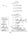
- Figure 1 illustrates downstream labeling of an analyte.
- Figure 2 illustrates upstream labeling of an analyte. If the polynucleotide is double stranded, one of the strands is selected as the reference strand, e.g. the strand that is labeled, or the strand that is not labeled (or some other criteria or feature of the strand may be used to designate one strand as the reference strand).
- RNA is prepared from a wax-embedded tissue specimen by a process that includes digesting the tissue specimen to provide a digested tissue specimen; purifying RNA from the digested tissue specimen to provide a purified RNA fraction; treating the purified RNA fraction with one or more enzymes selected from a cyclic phosphodiesterase, a phosphatase, or a kinase to provide a sample containing dephosphorylated RNA.
- the resulting sample is then contacted with an enzyme having an RNA ligation activity in the presence of a labeled substrate under conditions sufficient to result in coupling of the labeled substrate to the RNA in the sample to provide labeled RNA, the conditions including a DMSO concentration in the range from about 20% to about 30%, wherein the labeled substrate comprises an observable label moiety attached to a nucleotide moiety.
- the wax-embedded tissue specimen may be any wax-embedded tissue specimen, typically a specimen that has been isolated from a biological source, e.g. any plant, animal, particularly human or mammal source. Tissue specimens are often fixed with a fixative. Aldehyde fixatives such as formalin (formaldehyde) and glutaraldehyde are typically used. Tissue specimens fixed using other fixation techniques such as alcohol immersion ( Battifora and Kopinski, J. Histochem. Cytochem. (1986) 34:1095 ) are also suitable. The tissue specimens used typically are also embedded in paraffin. RNA can be isolated from a paraffin-embedded tissue specimen by the methods of the invention. In one embodiment, the tissue specimens are both formalin-fixed and paraffin-embedded.
- the wax-embedded tissue specimen used in the present invention includes any specimen typically referenced in the literature as "paraffin-embedded tissue", for example formalin fixed paraffin-embedded ("FFPE") tissue specimens.
- FFPE formalin fixed paraffin-embedded
- a collected tissue material e.g. pancreas, large intestine, cancer of large intestine, muscle, urinary bladder, kidney, lung, brain, lymphoma, etc.
- a fixative chemical having a protein-coagulative action
- tissue material in order to preserve the fine structure of tissue cells, and the thus treated tissue material is embedded in a wax, most typically paraffin, although any wax or resin material known in the literature as being useful for preserving tissue specimens may be used.
- any method may be used so long as it is usually employed in the art for removing wax from a wax-embedded tissue specimen.
- An exemplary method for removing wax includes adding a water-insoluble organic solvent to the wax-embedded tissue specimen.
- the water-insoluble organic solvent for dissolving and removing the wax which is used in such a method is not particularly limited, and any water-insoluble organic solvent may be used so long as it is usually used in the art for dissolving and removing wax from tissue specimens.
- Typical examples of the water-insoluble organic solvent are xylene, D-limonene, octane, benzene, toluene, ethylbenzene, and mixtures thereof etc., typically in an amount sufficient to submerse the tissue specimen. Such solvents are able to remove paraffin effectively from the tissue specimen without adversely affecting RNA isolation.
- the resulting mixture is centrifuged, and the supernatant is discarded, reserving the precipitate. The above procedure is repeated once more on the precipitate.
- the volume of organic solvent used and the number of washes necessary will depend on the size of the tissue specimen and the amount of paraffin to be removed. The more paraffin to be removed, the more washes that will be necessary. Typically, a tissue specimen will be washed between 1 and about 10 times, more typically between about two and about four times. Then, a volatile organic solvent such as ethanol is added to the thus obtained precipitate, and after stirring and shaking, the resulting mixture is centrifuged at ordinary temperature and the supernatant is discarded to remove the water-insoluble organic solvent. This procedure is repeated once on the precipitate. The thus obtained pellet is dried. Other methods for deparaffinization known to one of skill in the art may also be used in the method of the invention.
- Such methods include direct melting (Baneijee et al., 1995).
- Another such method includes contacting a wax-embedded tissue specimen with a dewaxing composition comprising a paraffin-solubilizing organic solvent selected from the group consisting of aromatic hydrocarbons, terpenes and isoparaffinic hydrocarbons, a polar organic solvent, and a surfactant to solubilize the wax associated with the specimen.
- Tissue specimens are typically rehydrated after deparaffinization.
- the method used for rehydration of tissue samples is not particularly limited and may generally be any method typically used in the art.
- the method for rehydration is step-wise washing with aqueous lower alcoholic solutions of decreasing concentration.
- Ethanol is a preferred lower alcohol for rehydration.
- Other alcohols may also be suitable for use with the invention including methanol, isopropanol and other similar lower alcohols.
- a "lower alcohol” is an alcohol having a molecular formula with five or fewer carbons.
- the tissue specimen is alternately vigorously mixed with alcoholic solutions and centrifuged.
- the concentration range of alcohol is decreased stepwise from about 100% to about 70% in water over about three to five incremental steps, where the change in solution concentration at each step is usually less than about 10% (i.e., the sequence: 100%, 95%, 90%, 80%, 70%).
- deparaffinization and rehydration are carried out simultaneously using a reagent such as EZ-DEWAX (BioGenex, San Ramon, Calif.).
- Digestion of the tissue specimen may include any suitable technique that results in degradation of cell structures and release of nucleic acids from the tissue specimen for later purification.
- Digesting the tissue specimen may include contacting the tissue specimen with a buffer or solvent in the presence of one or more agents selected from a protease, a chaotropic agent, a DNase, a surfactant, a salt, an RNase inhibitor, or other agent known in the literature to aid digestion of tissue specimens, or combinations thereof.
- Contacting with such agent(s) may be done concurrently (in the same solution) or may be done sequentially (e.g. a first solution results in isolation of a first fraction which is then contacted with a second solution, etc.) Contacting with such agent(s) may be done with heating, e.g.
- the contacting is typically done under conditions and for a time sufficient to result in digesting the tissue specimen. Such conditions will depend on the particular method and agents selected for digesting the tissue specimen, and as such, the conditions may vary. Such techniques for digestion of tissue specimens are generally well described in the literature.
- digestion of a tissue specimen includes homogenization of the tissue specimen.
- the tissue specimen can be homogenized by any standard mechanical, sonic or other suitable technique.
- Tissue homogenization is preferably carried out with a mechanical tissue homogenizers according to standard procedures.
- a number of commercially available homogenizers are suitable for use with the invention including: Ultra-Turrax homogenizer (IKA-Works, Inc., Wilmington, N.C.); Tissumizer (Tekmar-Dohrrmann, Cincinnati, Ohio); and Polytron (Brinkmann, Westbury, N.Y.).
- Purifying the RNA from the digested tissue specimen may include any suitable technique that results in isolation of the RNA from at least one component of the digested tissue specimen, thereby providing an isolated RNA fraction.
- Purifying RNA from the digested tissue specimen may include extraction (e.g. with an organic solvent such as chloroform extraction or phenol/chloroform extraction), precipitation (e.g. with ethanol, isopropanol or any other lower alcohol), chromatography (e.g. ion exchange chromatography, size exclusion chromatography, silica gel chromatography and reversed phase chromatography), electrophoretic methods (e.g. polyacrylamide gel electrophoresis and agarose gel electrophoresis), or by other techniques known in the literature, or combinations thereof, to result in the isolated RNA fraction. Such techniques for purification are generally well described in the literature.
- the RNA may optionally by further purified. Further purification may be accomplished by any of the aforementioned techniques for RNA recovery, for example extraction, precipitation, chromatography, electrophoretic methods, or other techniques described in the literature. In particular embodiments, further purification results in RNA that is substantially free from contaminating DNA or proteins, i.e. less than 50% (typically less than 30%, less than 20%, or less than 10%) by weight of the dry weight of the 'further purified RNA sample' is DNA and protein (combined).
- the process of purifying the RNA may include precipitation of the RNA using a precipitant.
- a precipitant used for precipitating RNA in the present invention is not particularly limited so long as it is typically used in the art for precipitating nucleic acid.
- an alcohol may be used as the precipitant.
- the alcohols are not particularly limited so long as they are usually used in the art for precipitating nucleic acid. Examples of the alcohols are ethanol, isopropanol, or other lower alcohols, or combinations thereof.
- the alcohol is used usually in a volume of 0.5 to 3 times that of a solution containing the extracted nucleic acid.
- the precipitation may be carried out in the presence of a carrier such as glycogen that facilitates precipitation.
- the process of purifying the RNA may include a two-phase extraction method.
- the two-phase extraction method may be any such method described in the literature for isolation of nucleic acids.
- the two-phase extraction method typically comprises separation into an aqueous phase containing the nucleic acid and an organic solvent phase containing denatured protein and the like by addition of one or more organic solvents.
- the organic solvents typically used are phenol, chloroform, etc.; a well known example of such a method is a phenol/chloroform extraction.
- purifying the RNA may include a size-based separation step to preferentially isolate small RNAs.
- the sample includes isolated small RNAs, e.g. purifying the RNA includes an isolation protocol for small RNA.
- the isolation protocol may be any known method for isolating small RNA from a complex RNA sample, such as size exclusion chromatography, electrophoresis, affinity chromatography, gel filtration, or any method known for fractionating nucleic acids based on their size.
- a number of techniques exist for digesting the tissue specimen Any method may be used so long as it is usually employed in the art for purifying nucleic acids from a digested tissue specimen.
- a number of techniques exist for purifying RNA from the digested tissue specimen and any method may be used so long as it is usually employed in the art for purification of RNA from a digested tissue specimen.
- digesting the tissue specimen comprises contacting the tissue specimen (freed of paraffin) with a solution containing a surfactant, a protease, etc. at room temperature to about 50° C for 4 to 48 hours to disrupt the tissue cells. This is followed by purifying to remove impurities, i.e. substances other than nucleic acids, by a two-phase extraction method, and then adding an alcohol to the residue to precipitate the RNA in the aqueous phase.
- digesting the tissue specimen comprises heating a deparaffinized tissue specimen in a solution containing a surfactant (e.g. sodium dodecyl sulfate) but not containing a protease, etc., at 37° C for 15 minutes, adding proteinase K and heating the resulting mixture at 37° C for 12 hours to disrupt the tissue cells.
- a surfactant e.g. sodium dodecyl sulfate
- proteinase K e.g. sodium dodecyl sulfate
- digesting the tissue specimen comprises homogenizing the tissue specimen in an aqueous solution of phenol and guanidinium isothiocyanate, and then mixing the resulting homogenate with chloroform. Following centrifugation, the homogenate separates into an organic phase, an interphase and an aqueous phase. Proteins are sequestered in the organic phase, DNA in the interphase, and RNA in the aqueous phase. Purifying the RNA can be accomplished via recovery of the aqueous phase followed by precipitation. (See, e.g. U.S. Pat. No. 5,346,994 to Chomczynski ).
- digesting the tissue specimen includes homogenization in the presence of a solution including a chaotropic agent (e.g. a guanidinium salt in the range of about 1M to about 5M).
- a chaotropic agent e.g. a guanidinium salt in the range of about 1M to about 5M.
- the chaotropic solution typically is buffered to a pH of about 3 to about 6 with a suitable biochemical buffer such as Tris-Cl.
- the chaotropic solution may also contain reducing agents, such as dithiothreitol (DTT) and ⁇ -mercaptoethanol (BME).
- the chaotropic solution may also contain RNAse inhibitors.
- the resulting solution is heated at a temperature of about 50° C to about 100° C for about 5 minutes to about 2 hours. See, e.g. U.S. Pat. No. 6,248,535 to Danenberg et al.
- RNA is typically purified from the chaotropic solution
- digesting the tissue specimen includes contacting the tissue specimen with at least one nonionic surfactant and a protease enzyme (sequentially or concurrently) in the presence of a buffer.
- a nonionic surfactant and a protease enzyme include, but are not limited to, polyoxyethylenesorbitan derivatives, polyoxyethylene ethers, polyglycol ethers, perfluoroalkyl polyoxyethylenes, fluorinated alkyl alkoxylates and fluorinated alkyl ester compounds, and combinations thereof, or other surfactants known in the art as useful for extracting RNA from tissue samples.
- protease enzyme is used to break down tissues and protein helping to release the nucleic acids.
- the protease may also degrade nucleases making the released RNA more stable.
- proteases examples include serine proteases (E. C. 3.4.21, e.g. Typsin, and chymotrypsin), Thiol proteases (E. C. 2.4.22, e.g., papain, ficin), carboxy (acid proteases, E. C. 2.4.23, e.g., pepsin), and metalloproteases (E. C. 2.4.24, e.g.,thermolysin, pronase).
- Suitable common biological buffers include one or more organic buffers that maintain the pH at from about 4 to about 10, more typically from about 6 to about 9, e.g. 3-(N-morpholino)propanesulfonic acid, 3-(N-morpholino)ethanesulfonic acid, tricine, glycine, TRIS, phosphate, and others readily apparent to those skilled in the art. Conditions of temperature, pH, ionic strength, and surfactant type may differ depending upon the protease used. The resulting digested tissue specimen is typically centrifuged and the supernatant is recovered. Purifying RNA from the supernatant typically follows, e.g. using alcohol precipitation and/or chromatography, or as otherwise described herein.
- RNA purifying techniques typically utilize either guanidine salts or phenol extraction, as described for example in Sambrook, J. et al. (1989) "Molecular Cloning", Cold Spring Harbor Press, 2nd Ed., at pp. 7.3-7.24 , and in Ausubel, F.M. et al. (1994) "Current Protocols In Molecular Biology", John Wiley & Sons, Inc., vol. 1, at pp. 4.0.3-4.4.7 .
- digesting the tissue specimen includes homogenizing the tissue specimen and performing an overnight incubation with Proteinase K. Nucleic acids in the digested tissue specimen are then bound to a glass fiber filter in the presence of a chaotropic salt. The glass fiber filter is washed to remove contaminating cellular components. Residual DNA is digested by incubation with DNase I. A second incubation step with Proteinase K improves the purity of RNA. Low salt elution removes the RNA from the glass fiber.
- a commercial kit is available for digesting the tissue specimen and purifying RNA from the digested tissue specimen (the High PureTM kit supplied by Roche).
- Treating the isolated RNA fraction typically includes contacting with one or more enzymes selected from a cyclic phosphodiesterase, a phosphatase, and a kinase. Said contacting may, in certain embodiments, employ more than one of the listed enzymes in the same reaction mixture; however, more commonly said contacting comprises serial reactions with individual enzymes.
- the isolated RNA fraction is first treated with cyclic phosphodiesterase (e.g. using a method such as that described in Bare et al. (1986) Biochem. 25: 5825-5830 , or any other effective method known in the art) to destroy any possible 2'-3' cyclic phosphates, a common structure due to breakdown of RNA in the tissue specimen.
- the sample is then treated with phosphatase to remove 5'-, 3'- or 2'- terminal phosphate groups from the RNA.
- An exemplary method of treating with phosphatase is described in Bare et al. (1986), but any effective method of treating the RNA with a phosphatase may be employed.
- any effective method of treating the RNA with a phosphatase may be employed.
- treating the isolated RNA fraction to a dephosphorylation reaction is conducted prior to contacting the RNA in the sample with an enzyme having RNA ligation activity.
- treating the isolated RNA fraction includes, prior to contacting the sample with the enzyme having an RNA ligation activity, contacting the sample with an enzyme having a 5'-phosphatase activity to remove 5'-phosphate groups from the RNA.
- the RNA in the sample has a 5'-phosphate; in such embodiments treating the isolated RNA fraction may include treating with an enzyme having a 5'-kinase activity, such as T4 polynucleotide kinase, thereby providing that RNA in the sample is dephosphorylated at the 3' end but possesses a 5'-phosphate group.
- an enzyme having a 5'-kinase activity such as T4 polynucleotide kinase
- the treatment of the purified RNA fraction results in a sample that includes dephosphorylated RNA.
- the sample is then contacted with an enzyme having an RNA ligation activity in the presence of a labeled substrate under conditions sufficient to result in coupling of the labeled substrate to the RNA in the sample to provide labeled RNA, the conditions including a DMSO concentration in the range from about 20% to about 30%, wherein the labeled substrate comprises an observable label moiety attached to a nucleotide moiety.
- the dimethylsulfoxide (DMSO) concentration is calculated as volume (e.g. in milliliters) of DMSO divided by total volume (e.g. in milliliters) of the solution containing the DMSO. This quantity is typically cast as a percentage by multiplying by 100%.
- the DMSO concentration will be in a range of 20% to about 30%, calculated as the volume of DMSO in the solution resulting from contacting the sample with an enzyme having an RNA ligation activity in the presence of a labeled substrate, divided by the total volume of the solution, and then multiplying by 100%.
- the other components present in the resulting solution will typically be water, buffer components, salt, RNA, labeled substrate, and enzyme having an RNA ligation activity, although other components may also be present.
- the sample includes small RNAs, especially RNAs less than about 500 bases long, e.g. less than about 400 bases long, less than about 300 bases long, less than about 200 bases long, or less than about 100 bases long.
- the sample includes one or more small RNAs, such as e.g. short interfering RNAs (siRNAs), microRNAs (miRNA), tiny non-coding RNAs (tncRNA) and small modulatory RNA (smRNA). See Novina et al., Nature 430: 161-164 (2004) .
- the sample includes isolated small RNAs, e.g. purifying the RNA includes isolating small RNAs.
- isolating small RNAs may include any known method for isolating small RNA from a complex RNA sample, such as size exclusion chromatography, electrophoresis, affinity chromatography, gel filtration, or any method known for fractionating nucleic acids based on their size.
- the small RNA targets may include isolated miRNAs, such as those described in the literature and in the public database accessible via the worldwide website which can be reached by typing three "w”s followed by ".sanger.ac.uklcgi-bin/Rfam/mirnalbrowse.pl" in a typical web browser address field.
- the sample includes isolated small RNAs, e.g.
- RNAs less than about 500 bases long e.g. less than about 400 bases long, less than about 300 bases long, less than about 200 bases long, less than about 100 bases long, or less than about 50 bases long.
- the enzyme having an RNA ligation activity is typically any RNA ligase enzyme, although other enzymes capable of coupling the labeled substrate to the RNA may be used.
- the enzyme having an RNA ligation activity is capable of coupling a nucleotide (or oligonucleotide, or RNA) having a 5' phosphate to an oligonucleotide having a 3' hydroxyl.
- Exemplary enzymes include T4 RNA ligase available from Amersham/Pharmacia company, ThermoPhageTM RNA ligase II (available from Prokaria LTD, Iceland), or other available RNA ligase enzymes known to be capable of coupling a nucleotide (or oligonucleotide, or RNA) having a 5' phosphate to an oligonucleotide having a 3' hydroxyl.
- the enzyme may be selected from yeast poly A polymerase, E. coli poly A polymerase, or terminal transferase (each of which is available from Amersham/Pharmacia).
- the enzyme having an RNA ligation activity should be selected such that the enzyme is capable performing the coupling when one (or both) of the nucleotide (or oligonucleotide) having a 5'phosphate and/or the oligonucleotide having a 3' hydroxyl includes a label.
- Selection of the enzyme having an RNA ligation activity will typically be based on availability of the enzyme and activity of the enzyme under the desired reaction conditions for the coupling (e.g. temperature, pH, ionic strength, source of RNA and/or labeled substrate, structural feature of RNA and/or labeled substrate, concentration of RNA and/or labeled substrate, presence of other materials (e.g. contaminants, salt, surfactant, other solvents) etc.)
- the coupling reaction is conducted under conditions sufficient to result in coupling of the labeled substrate to the RNA.
- the conditions of the coupling reaction will generally be selected with regard to the known (previously described) conditions for use of the particular enzyme chosen for use in the methods of the invention, with the specific modifications described herein.
- the DMSO of the reaction mixture for the coupling reaction will be in the range of 20% to 30%.
- Other experimental parameters may be selected based on known ranges for the experimental parameters or determined through routine experimentation based on, e.g. efficacy of the labeling reaction. Such other experimental parameters may include, e.g.
- RNA and/or labeled substrate temperature, pH, ionic strength, source of RNA and/or labeled substrate, structural feature of RNA and/or labeled substrate, concentration of RNA and/or labeled substrate, presence of other materials (e.g. contaminants, salt, surfactant, other solvents) etc.
- other materials e.g. contaminants, salt, surfactant, other solvents
- the labeled substrate comprises an observable label moiety attached to a nucleotide moiety.
- the nucleotide moiety is typically a mononucleotide moiety or an oligonucleotide moiety. In particular embodiments, the nucleotide moiety is less than about 100 bases long. In embodiments which result in upstream labeling of the RNA (examples shown in Figure 2), the nucleotide moiety will typically be at least three bases long. In certain embodiments, the nucleotide moiety will be less than 50 bases long, e.g. less than 40 bases long, less than 30 bases long, less than 20 bases long. In some embodiments, the nucleotide moiety will be 1, 2, 3, 4, 5, or 6 bases long.
- the nucleotide moiety may typically have any desired sequence or even an unknown sequence.
- a plurality of labeled substrates may be used in the same reaction (e.g. a plurality of nucleotide moieties, each having a different sequence, each having an observable label moiety attached), thereby resulting in ligating one of a plurality of nucleotide moieties to each molecule of RNA.
- the observable label moiety is a moiety that provides for an observable signal that indicates the presence of the observable label moiety.
- Typical examples include a chromogenic moiety, a fluorophore, a mass label, a spin label, a radiolabel, or other labels known in the art.
- the observable label moiety is a fluorophore selected from the group consisting of Cy3, Cy5, and an Alexa dye.
- label moieties include any commercially available fluorophores that can be conjugated to mononucleotides or polynucleotides, e.g.
- dyes from Molecular Probes such as the Alexa Fluor series (example: Alexa 350, Alexa 430, Alexa 532, Alexa 546, Alexa 568, and Alexa 594) and the series of BODIPY conjugates.
- Alexa Fluor series example: Alexa 350, Alexa 430, Alexa 532, Alexa 546, Alexa 568, and Alexa 594
- BODIPY conjugates include: Tamra, Fluorescein, carboxyfluorescene, JOE, rhodamine, carboxyrhodamine, CY series, Oyster series. More information about commercially available dyes for oligonucleotide conjugation can be found at the website which can be reached by typing three "w”s followed by "synthegen.com” in a typical web browser address field.
- any such dyes may potentially be used in accordance with the methods described herein.
- the examples described herein use a fluorophore as the label, it will be apparent to those of ordinary skill that other labels may be used (instead of a fluorophore, or even in addition to a fluorophore). Such labels typically are well known in the art.
- the observable label moiety may be attached to the nucleotide moiety at any site of the nucleotide moiety that is compatible with the ligation reaction. In other words, the label moiety should not prevent the ligation reaction, e.g. by interfering with the enzyme having an RNA ligation activity.
- the label moiety may be attached to the nucleotide moiety via the 3'-site at the end of the nucleotide moiety or may be attached at any available "internal" site (i.e. other than the 3'-site or 5'-site) of the nucleotide moiety.
- the label moiety may be attached to the nucleotide moiety via the 5'-site at the end of the nucleotide moiety or may be attached at any available "internal" site (i.e. other than the 3'-site or 5'-site) of the nucleotide moiety.
- the observable label moiety could be incorporated as a special phosphoramidite during the oligoribonucleotide synthesis or as a post-synthetic modification.
- An example of the phosphoramidite method includes direct coupling of label-containing phosphoramidite during synthesis of the oligonucleotide moiety, or the incorporation of amino-activated phosphoramidite during synthesis of the oligonucleotide moiety, which enables post-synthetic coupling to desired observable label moiety.
- the observable label moiety is attached to the nucleotide moiety via a linking group, wherein the linking group may be attached to the nucleotide moiety via a 5' terminus or a 3' terminus of the nucleotide moiety or any other available site of the nucleotide moiety.
- the linking group may be any linking group known in the art that does not prevent the ligation reaction (e.g. does not prevent the enzyme having an RNA ligation activity from ligating the RNA and the labeled substrate). Any such linking groups or other means of attachment of the label moiety to the nucleotide moiety known in the art may provide for the labeled substrate.
- the labeled substrate has the structure: N - D wherein:
- the method 100 of preparing labeled RNA from a wax-embedded tissue specimen 90 includes digesting 92 the tissue specimen 90 to provide a digested tissue specimen, followed by purifying 94 RNA from the digested tissue specimen to result in an isolated RNA fraction. This is followed by treating 96 the isolated RNA fraction with one or more enzymes selected from a cyclic phosphodiesterase and a phosphatase to provide a sample containing dephosphorylated RNA.
- DMSO 106 is added 104 to the sample (which includes the RNA 102).
- the labeled substrate 110A, 110B or 110C may then be added 108 to the resulting solution (containing the DMSO 106 and the RNA 102 from the sample).
- the labeled substrate 110A, 110B or 110C typically is a mononucleotide with a 3' fluorophore and 5' phosphate 110A, an oligonucleotide with a 5' phosphate and one or more internal fluorophore(s) 110B, or an oligonucleotide with a 3' fluorophore and 5' phosphate 110C.
- the 3' end of the oligonucleotide lacks the 3' hydroxyl, as indicated by the 'X' in Figure 1.
- a periodate oxidation reaction may be used to modify the 3' end, or the 3' end may be modified by having a fluorophore bound thereto.
- the enzyme having an RNA ligation activity 114 is also added 112.
- the concentrations of the solutions and the volumes added are planned to provide that the resulting solution has the desired concentration of DMSO (e.g.
- RNA 118A, 118B, or 118C Typical conditions 120 of overnight incubation at 16° C are shown for the embodiment of Figure 1, although these conditions may vary depending on the particular enzyme used and the RNA and labeled substrate provided.
- the label is a fluorophore 111, but other labels may be used as long as the coupling of the labeled substrate to the RNA may still occur. Selection and optimization of the conditions is within routine experimentation for one of ordinary skill in the art given the disclosure herein.
- the method 200 of preparing labeled RNA from a wax-embedded tissue specimen 90 includes digesting 92 the tissue specimen 90 to provide a digested tissue specimen, followed by purifying 94 RNA from the digested tissue specimen to result in an isolated RNA fraction. This is followed by treating 96 the isolated RNA fraction with one or more enzymes selected from a cyclic phosphodiesterase, a phosphatase, and a kinase to provide a sample containing dephosphorylated RNA 202, wherein the illustrated dephosphorylated RNA 202 is dephosphorylated at the 3'-terminus and has a 5'-phosphate.
- the method 200 includes pre-treating the RNA 202 by performing a periodate oxidation reaction 222 on the initial RNA sample.
- the periodate oxidation reaction helps prevent the RNA from concatenating or from ligating to itself when the enzyme having an RNA ligation activity is later added.
- the periodate oxidation reaction 222 is followed with inactivation and de-salting 224 of the solution resulting from the periodate oxidation reaction.
- the periodate oxidation reaction may be performed using any protocol known in the art. See, e.g. Neu, Harold C., et al., J. Biol. Chem 239: 2927-34 (1964) ; Kurata, Shinya, et al., Nucleic Acids Res.
- ribonucleotides are treated with estimated 5-fold molar excess of sodium metaperiodate for 20 minutes to 1 hour at 0° C to room temperature in the dark.
- the ribonucleotides can be cleaned from the solution by ethanol precipitation or desalting columns, such as BioRad Micro Bio-SpinTM 6.
- the method 200 includes adding 204 DMSO 206 to the solution containing the RNA 202.
- the labeled substrate 210A or 210B may then be added 208 to the resulting solution (containing the DMSO 206 and the RNA 202 from the sample).
- the labeled substrate 210A or 210B typically is an oligonucleotide with a 5' fluorophore 210A or an oligonucleotide with one or more internal fluorophore(s) 210B.
- the enzyme having an RNA ligation activity 214 is also added 212.
- the concentrations of the solutions and the volumes added are planned to provide that the resulting solution has the desired concentration of DMSO (e.g.
- RNA 218A or 218B products corresponding to 210A or 210B, respectively.
- Typical conditions 220 of overnight incubation at 16° C are shown for the embodiment of Figure 2, although these conditions may vary depending on the particular enzyme used and the RNA and labeled substrate provided.
- the temperature of the incubation will typically be in the range from about 4° C to about 37° C, more typically in the range from about 10° C to about 30° C, still more typically in the range from about 14° C to about 20° C.
- the label is a fluorophore 211, but other labels may be used as long as the coupling of the labeled substrate to the RNA may still occur. Selection and optimization of the conditions is within routine experimentation for one of ordinary skill in the art given the disclosure herein.
- the labeled substrate denominated 110A is shown as a mononucleotide attached to a fluorophore.
- the labeled substrates 110B and 110C are shown as an oligonucleotide having one or more fluorophore(s) attached to the oligonucleotide, in which the oligonucleotide does not have a 3' hydroxyl. The lack of the 3' hydroxyl helps prevent concatenation of the labeled substrate.
- the RNA 202 is subjected to periodate oxidation to help prevent the RNA from concatenating or from ligating to itself.
- the labeled substrate 110C is shown as an oligonucleotide having a fluorophore(s) attached to an oligonucleotide at the 3' end of the oligonucleotide.
- the labeled substrate denominated 210A and 210B illustrate representative embodiments in which the labeled substrate includes an oligonucleotide attached to one or more fluorophores.
- the labeled substrate includes an oligonucleotide having attached thereto a plurality, e.g. 2, 3, 4, 5, or more label moieties, up to about 10, 20, 30 or more label moieties, attached to the oligonucleotide moiety at internal (not terminal) sites.
- a label moiety is also attached to the oligonucleotide via one of the 3' or 5' terminal carbons of the oligonucleotide.
- the enzyme having an RNA ligation activity catalyzes a coupling reaction between a donor molecule having a 5'-phosphate and an acceptor molecule having a 3'-hydroxyl, as shown in the reaction:
- the acceptor molecule is the RNA 102 and the donor molecule is the labeled substrate 110A, 110B or 110C.
- the resulting product 118A, 118B, or 118C has the labeled substrate moiety downstream from the RNA, i.e. the product is a downstream labeled RNA.
- the acceptor molecule is the labeled substrate 210A or 210B and the donor molecule is the RNA 202.
- the resulting product 218A or 218B has the labeled substrate moiety upstream from the RNA, i.e. the product is an upstream labeled RNA.
- the labeled substrate may be dissolved in a solution that includes the DMSO, and the resulting solution mixed with the sample prior to contacting with the enzyme having an RNA ligase activity.
- the invention includes any process which results in contacting the sample with the enzyme having an RNA ligation activity in the presence of the labeled substrate under conditions which include a DMSO concentration in the range from about 20% to about 30%.
- the method 100 includes heating the solution containing the DMSO 106 and the RNA 102 from the sample.
- the RNA is typically heated to a temperature of at least about 80° C (e.g. at least about 85° C, at least about 90° C, at least about 95° C; and up to about 105° C or 110° C) under conditions that include a DMSO concentration of at least about 40% DMSO (typically up to about 60% DMSO, although in some embodiments the DMSO concentration may be up to 70% DMSO, up to 80% DMSO, or even more).
- This optional heating is maintained for at least 10 seconds, typically at least about 20 seconds, at least about 30 seconds, at least about 1 minute, at least about 2 minutes, and up to about 15 minutes, or more.
- reaction solutions of up to about 50 microliters are heated for about 30 to about 60 seconds per 5-10 microliters of reaction solution.
- the RNA is typically quickly cooled (e.g. to less than about 40° C, more typically less than about 20° C, or in some embodiments less than about 5° C) before adding the enzyme having an RNA ligation activity.
- a similar heating step may optionally be included in embodiments such as that illustrated in the figure.
- the labeled substrate has only one observable label moiety attached to the nucleotide moiety.
- the labeled RNA will consist essentially of RNA labeled with a single label moiety (i.e. each labeled RNA molecule will have only one observable label moiety attached - referenced herein as "singly-labeled RNA"). This potentially provides increased ease of use in quantitative methods using the labeled RNA.
- the nucleotide moiety of the labeled substrate has a plurality of observable label moieties.
- each labeled RNA molecule will have plurality of observable label moieties (referenced herein as "multiply-labeled RNA").
- the labeled RNA will consist essentially of RNA labeled with a plurality of label moieties. This increased labeling of the RNA may provide for greater sensitivity in analyses using the labeled RNA.
- each labeled RNA molecule in the sample will be labeled with a consistent number of observable label moieties (relative to the other labeled RNA molecules in the sample). This provides opportunity for more quantitative analysis of labeled RNA than in methods that provide an inconsistent number of observable labels per labeled RNA molecule.
- methods of performing an array analysis of an RNA sample are provided.
- the invention provides a method of performing an array analysis wherein the method includes labeling RNA to provide labeled RNA using a labeling method in accordance with the methods described herein.
- the labeled RNA is then contacted with an array under conditions sufficient to provide for specific binding of labeled RNA to the array.
- the array typically is then interrogated to provide data on binding of the labeled RNA to the array.
- Standard hybridization techniques (using stringent hybridization conditions) are used to hybridize a labeled sample to a nucleic acid array. Suitable methods are described in references describing CGH techniques ( Kallioniemi et al., Science 258:818-821 (1992) and WO 93/18186 ). Several guides to general techniques are available, e.g., Tijssen, Hybridization with Nucleic Acid Probes, Parts I and II (Elsevier, Amsterdam 1993 ). For descriptions of techniques suitable for in situ hybridizations, see Gall et al. Meth. Enzymol., 21:470-480 (1981) ; and Angerer et al.
- Hybridizing the sample to the array is typically performed under stringent hybridization conditions, as described herein and as known in the art.
- the array-surface bound polynucleotides are typically washed to remove unbound and not tightly bound labeled nucleic acids. Washing may be performed using any convenient washing protocol, where the washing conditions are typically stringent, as described above.
- the hybridization of the labeled target nucleic acids to the capture agents is then detected using standard techniques of reading the array, i.e. the array is interrogated.
- Reading the resultant hybridized array may be accomplished by illuminating the array and reading the location and intensity of resulting fluorescence at each feature of the array to detect any binding complexes on the surface of the array.
- a scanner may be used for this purpose, which is similar to the AGILENT MICROARRAY SCANNER available from Agilent Technologies, Palo Alto, CA.
- Other suitable devices and methods are described in U.S. patent applications: Serial No.
- arrays may be read by any other method or apparatus than the foregoing, with other reading methods including other optical techniques (for example, detecting chemiluminescent or electroluminescent labels) or electrical techniques (where each feature is provided with an electrode to detect hybridization at that feature in a manner disclosed in US 6,221,583 and elsewhere).
- other reading methods including other optical techniques (for example, detecting chemiluminescent or electroluminescent labels) or electrical techniques (where each feature is provided with an electrode to detect hybridization at that feature in a manner disclosed in US 6,221,583 and elsewhere).
- subsequent treatment of the array with the appropriate reagents may be employed to enable reading of the array.
- Some methods of detection such as surface plasmon resonance, do not require any labeling of nucleic acids, and are suitable for some embodiments.
- Results from the reading or evaluating may be raw results (such as fluorescence intensity readings for each feature in one or more color channels) or may be processed results (such as those obtained by subtracting a background measurement, or by rejecting a reading for a feature which is below a predetermined threshold, normalizing the results, and/or forming conclusions based on the pattern read from the array (such as whether or not a particular target sequence may have been present in the sample, or whether or not a pattern indicates a particular condition of an organism from which the sample came).
- raw results such as fluorescence intensity readings for each feature in one or more color channels
- processed results such as those obtained by subtracting a background measurement, or by rejecting a reading for a feature which is below a predetermined threshold, normalizing the results, and/or forming conclusions based on the pattern read from the array (such as whether or not a particular target sequence may have been present in the sample, or whether or not a pattern indicates a particular condition of an organism from which the sample came).
- results from interrogating the array are used to assess the level of binding of the population of labeled nucleic acids to capture agents on the array.
- level of binding means any assessment of binding (e.g. a quantitative or qualitative, relative or absolute assessment) usually done, as is known in the art, by detecting signal (i.e., pixel brightness) from a label associated with the sample nucleic acids, e.g. the sample is labeled.
- the level of binding of labeled nucleic acid to capture agent is typically obtained by measuring the surface density of the bound label (or of a signal resulting from the label).
- a surface-bound polynucleotide may be assessed by evaluating its binding to two populations of nucleic acids that are distinguishably labeled.
- the results obtained from hybridization with a first population of labeled nucleic acids may be compared to results obtained from hybridization with the second population of nucleic acids, usually after normalization of the data.
- the results may be expressed using any convenient means, e.g., as a number or numerical ratio, etc.
- kits which generally comprises one or more reagents needed to perform a method in accordance with the present invention.
- the kit may include one or more reagents needed for digesting the tissue specimen, e.g. one or more of a protease, DNase, detergent/surfactant, solvent, chaotropic agent, and/or RNase inhibitor, etc.
- the kit may include one or more components needed for purifying the RNA from the digested tissue specimen, e.g. solvent, a precipitant, chromatography media, and/or filtration apparatus, etc.
- the kit may include one or more components needed for treating the purified RNA fraction (e.g. cyclic phosphodiesterase and/or a phosphatase may be supplied in the kit).
- the kit may include one or more components selected from an enzyme having an RNA ligation activity, a labeled substrate, and/or a stock solution of DMSO.
- the kit may include one or more components for performing an array analysis, e.g. an array, hybridization buffer, and/or rinse buffer, etc.
- Each components supplied with a kit may be supplied in an individual container, and the individual containers may be packaged to provide the kit.
- RNA ligation was assessed with synthetic RNA oligonucleotides (21-23 nucleotides, Dharmacon) in reaction solutions containing 0, 15, 20, 25, and 30% DMSO. The reactions containing 25% DMSO were assayed with and without the pre-heating step.
- Stock solutions of 20 ⁇ M RNA oligonucleotides were stored in 1X TE (10 mM Tris-HCl, pH 7.5, 1 mM EDTA). Initial mixtures of RNA, DMSO and water were first assembled. For preheated samples, the heated mixture contained 40-70% DMSO and were heated using a 104° C heating block for 1.5-2 minutes. The heated samples were immediately set on ice for >5 minutes prior to final assembly.
- RNA ligase buffer 50mM Tris-HCl, pH 7.5, 10mM MgCl 2 , 10mM DTT, 1mM ATP, 60ng/ ⁇ L BSA
- RNA ligase 100 ⁇ M, 5'-phosphate-cytidyl-phosphate-Cy5-3' (pCpCy5) or 5'-phosphate-cytidyl-phosphate-Cy3-3'(pCpCy3) (Dharmacon) and 2-4 ⁇ M RNA oligonucleotides.
- the reactions were incubated at 16° C overnight.
- RNA ligase was inactivated by heating the reaction solutions using a 104° C heating block for 1.5-2 minutes, followed by immediately setting on ice for >5 minutes.
- the labeling efficiency was determined by 5'-phosphorylation of RNA ligation reaction aliquots with radioactive P 32 -gamma-ATP. The resulting mixture was desalted with Micro Bio-SpinTM (BioRad) desalting columns. The desalted mixture was loaded onto denaturing polyacrylamide gel. Since the ligation products contain an extra nucleotide and fluorophore, they have a lower electrophoretic migration rate than the unligated precursors. P 32 -labeled RNA bands are visualized and quantified with phosphorimager (Molecular Dynamics). The ligation efficiency was determined by the ratio of ligated vs. unligated P 32 -labeled RNA bands. Thus, ligation efficiency may be expressed as the mol% of initial RNA that winds up having an attached label moiety.
- T4 RNA ligase is used to label synthetic RNA oligonucleotides with 5'-phosphate-cytidyl-phosphate-Cy5-3' (pCpCy5) or 5'-phosphate-cytidyl-phosphate-Cy3-3'(pCpCy3).
- the reaction conditions described here have been observed to result in ligation efficiencies of about 60% or more, e.g. about 70% or more, or 80% or more, up to about 95% or more, e.g. up to about 99% with minimal sequence discrimination. This was accomplished by reacting at 25% DMSO, 16° C overnight, with donor to acceptor ratio of>12.5:1.
- the reaction buffer contains 50mM Tris-HCl, pH 7.5, 10mM MgCl2, 10mM DTT, 1mM ATP, 60 ⁇ g/mL BSA, and 25% DMSO.
- Typical reactions are 10 ⁇ L with 2 ⁇ M RNA, 100 ⁇ M pCpCy5 or pCpCy3, and 4 units T4 RNA ligase (Amersham/Pharmacia). Reaction efficiency seemed unaffected by increasing the RNA concentration to 8 ⁇ M or decreasing enzyme to 2 units.
- the labeling efficiency was determined by first performing the ligation reaction. An aliquot of the ligation mixture was then labeled on the 5' end with radioactive P 32 - ⁇ -ATP and T4 Polynucleotide Kinase. The control sample, which did not undergo ligation reaction, and final reaction mixture was denatured with formamide and assessed with denaturing polyacrylamide gel electrophoresis (1X or 0.5X TBE, 50% urea, 15-20% polyacrylamide with 19:1 acrylamide to bisacrylamide ratio, at about 50° C). The resulting gel was scanned with Molecular Dynamics Storm Phosphorimager for pCpCy5-labeled RNA. The ligation product was clearly visible as a red fluorescent miRNA.
- the gel was then exposed to phosphor screen to determine the pCpCy5-labeling efficiency. Since the addition of pCpCy5 increases the acceptor miRNA by 1 nucleotide and a fluorophore, the mobility of the cy5-labeled strand was lower than the unreacted strand. They appear as distinct bands when scanned in the phosphor mode on the phosphorimager; this was further verified by the relative mobility between the ligase reacted samples and unreacted controls (both the ligase reacted samples and the unreacted controls were labeled with P 32 - ⁇ -ATP). Thus the relative level of radioactivity between the Cy-labeled and unlabeled bands reveals the ligation efficiency.
- the reaction efficiency of pCpCy3 was determined similarly except the Cy3 labeled strand was undetectable by the fluorescent mode of the phosphorimager.
- the product and reactant miRNA bands of the Cy3 reaction were defined by the mobility of Cy5 reactions in polyacrylamide gel electrophoreseis and phosphorimager analysis.
- the labeling reaction was expanded to include the following sequences (SEQ ID NOs:6-10) with pCpCy5 and pCpCy3 in separate studies. These additional strands address any bias that may result from 3' terminal nucleotide, potential secondary structures and nucleotide content of the miRNA.
- the RNA ligase method can be used for dyes other than Cy5 and Cy3, but the efficiency may differ from the ones presented here.
- the present invention thus provides quantitative methods of performing array hybridization experiments. It is expected that this will provide a more sensitive assay system for the detection of variations of miRNA, such as found in developmental stages, tissue samples, disease states, as well as any individual and/or abnormal variations. Moreover, if more viral miRNAs are identified, this can become a novel diagnostic tool for active as well as latent viral infections
- RNA ligase is used to label a complex RNA mixture, such as the total RNA or isolated mixtures of small RNAs from biological samples.
- the labeled mixtures are run on denaturing polyacrylamide gel and Northern blots are performed of individual miRNAs with radioactive probes.
- the RNAs labeled by RNA ligase will have a lower mobility relative to its unlabeled counterpart.
- each target sequence will run as a doublet when probed by Northern blot.
- the ratio of RNA species in these doublets reflects the molar ratio of the RNA ligase labeled vs. unlabeled RNA species.
- Labeled miRNA were desalted with BioRad Micro Bio-SpinTM 6 (as directed by BioRad instructions) to remove free fluorescent tags.
- the desalted miRNA was added to solution containing water and carrier (25-mer DNA with random sequence). The solution was heated for approximately 1 minute per 10ul solution at 100° C and immediately placed on ice. After cooling, 2X Agilent Hyb Buffer (1225mM LiCl, 300mM Li-MES, pH 6.1, 12mM EDTA, 3.0% (w/v) lithium dodecyl sulfate, 2.0% (w/v) Triton X-100) was added to the mixture and the viscous liquid was mixed carefully. The final solution contained 1X Hyb Buffer and 0.1 ⁇ g/ ⁇ l random 25-mer. The concentration of miRNA was varied for different experiments.
- Hybridization was performed with SureHyb hybridization chamber (Agilent Part Number:G2534A) and place on rotisserie of hybridization oven overnight. The hybridization temperature was tested at 50° C and 60° C.
- the Sure-Hyb chamber complex was removed from the oven and immediately disassembled in Wash Buffer 1 (6X SSC, 0.005% Triton X-102) at room temperature.
- the microarray was transferred to a fresh wash chamber containing Wash Buffer 1 and washed by stirring for 10 minutes at room temperature.
- the microarray was then washed in Wash Buffer 2 (0.1X SSC, 0.005% Triton X-102) by stirring at room temperature for 5 minutes.
- the microarray was slowly lifted out of the wash chamber after washing and dried with nitrogen as needed.
- the microarrays were scanned with Agilent Scanner (Agilent Product Number: G2565BA).
- the scanned data was extracted with Agilent Feature Extraction Software (Agilent Product Number; G2567AA) and the green and red background-subtracted signals were evaluated for hybridization efficiency and specificity. Data was further analyzed using Spotfire software and Microsoft Excel.
Abstract
Description
- The invention relates generally to methods of providing RNA, e.g. for use in analysis of tissues from which the RNA is derived. More specifically, the invention relates to preparing labeled RNA from a wax-embedded tissue specimen.
- Most pathological tissue specimens are routinely formalin-fixed and paraffin-embedded (FFPE) to allow for histological analysis and for archival storage. Such preserved tissues are analyzed using genetic analysis techniques for diagnosis of genopathies, infectious diseases, malignant tumors, etc. and inspection for gene abnormality responsible for these diseases. Semi-quantitative gene expression analysis can be conducted indirectly in such fixed and embedded specimens by use of immunohistochemical staining techniques to observe protein expression levels. Other available techniques allow analysis of nucleic acids from such specimens. In particular, extraction of nucleic acid from FFPE specimens was described in Goelz et al. (1985) Biochem. Biophys. Res. Commun., 130: 118-126. Because paraffin-embedded specimens are widely available, rapid and reliable methods are needed for obtaining nucleic acids, particularly RNA, from such specimens.
- There has been great interest in the analysis of small RNAs, such as short interfering RNAs (siRNAs), microRNAs (miRNA), tiny non-codingRNAs (tncRNA) and small modulatory RNA (smRNA), since the discovery of siRNA biological activity over a decade ago (see Novina et al. (2004) Nature 430: 161-164). Even though the functions of most discovered miRNAs remain a mystery, it has become clear that they exist in abundance in plants and animals, with up to tens of thousands of copies per cell. In the fruit fly, 78 have been identified, and over 200 have been identified in human (see the public database accessible via the worldwide website which can be reached by typing three "w"s followed by ".sanger.ac.uk/cgi-bin/Rfam/mirna/browse.pl" in a typical web browser address field). See also Griffiths-Jones, S. (2004) Nucleic Acids Res. 32: D109-D111. The levels of individual miRNAs seem to vary with developmental stages and tissue types. The level of fluctuation may be correlated with phenotype, mRNA levels, or protein levels for better biological insight. Thus quantitative measurements of miRNA may be of great importance. Further, viral miRNAs have been identified and may play a role in latency (see Pfeffer et al. (2004) Science, 304: 734-736), making the detection and quantification of miRNAs a potentially valuable diagnostic tool.
- Analytic methods employing polynucleotide arrays have been used for investigating these small RNAs. See, e.g., Liu et al. (2004) Proc. Nat'1 Acad. Sci. USA, 101: 9740-9744; Nelson et al. (2004) Nature Methods, 1: 1-7; and Babak et al. (2004) RNA, 10: 1813-1819. Polynucleotide arrays (such as DNA or RNA arrays) include regions of usually different sequence polynucleotides ("capture agents") arranged in a predetermined configuration on a support. The arrays are "addressable" in that these regions (sometimes referenced as "array features") have different predetermined locations ("addresses") on the support of array. The polynucleotide arrays typically are fabricated on planar supports either by depositing previously obtained polynucleotides onto the support in a site specific fashion or by site specific in situ synthesis of the polynucleotides upon the support.
- In use, an array is contacted with a sample or labeled sample containing analytes (typically, but not necessarily, other polynucleotides) under conditions that promote specific binding of the analytes in the sample to one or more of the capture agents present on the array. Thus, the arrays, when exposed to a sample, will undergo a binding reaction with the sample and exhibit an observed binding pattern. This binding pattern can be detected upon interrogating the array. For example all target polynucleotides (for example, DNA) in the sample can be labeled with a suitable label (such as a fluorescent compound), and the label then can be accurately observed (such as by observing the fluorescence pattern) on the array after exposure of the array to the sample. Assuming that the different sequence polynucleotides were correctly deposited in accordance with the predetermined configuration, then the observed binding pattern will be indicative of the presence and/or concentration of one or more components of the sample. Techniques for scanning arrays are described, for example, in
U.S. Pat. 5,763,870 andU.S. Pat. 5,945,679 . Still other techniques useful for observing an array are described inU.S. Pat 5,721,435 . - Methods of labeling RNAs are of interest for use in array analysis of RNA to provide an observable label used in interrogating the array. In the study of Liu et al., the miRNA was transcribed into DNA with a biotin-labeled primer. This primer was subsequently labeled with streptavidin-linked Alexa dye prior to array hybridization. This method is susceptible to any reverse-transcriptase reaction bias. Further, the streptavidindye as well as streptavidin-biotin-RNA stochiometry may be difficult to quantify. In the study of Nelson et al. (2004) Nature Methods, 1: 1-7, the miRNA was labeled with biotin-conjugated dATP using Klenow enzyme while the miRNA was bound on an array, labeled with fluorophore-conjugated streptavidin, and then the array was interrogated. In the study of Babak et al. (2004) RNA, 10: 1813-1819, the miRNA was labeled with Ulysis Alexa Fluor system, which reacts with guanine residue (G) of RNA. Since different miRNAs do not have uniform G content, this method is not quantitative.
- There is a continuing interest in developing methods of preparing labeled RNA from wax-embedded tissue specimens. Such methods may be used in conjunction with analytical methods based on observing the label, such as array-based analysis of polynucleotides.
- The invention thus relates to novel methods for preparing labeled RNA from wax-embedded tissue specimens. According to a first aspect of the present invention , there is provided a method of preparing labeled RNA from a wax-embedded tissue specimen, the method comprising digesting the tissue specimen to provide a digested tissue specimen; purifying RNA from the digested tissue specimen to provide a purified RNA fraction; treating the purified RNA fraction with one or more enzymes selected from a cyclic phosphodiesterase, a phosphatase, or a kinase to provide a sample containing dephosphorylated RNA. The resulting sample is then contacted with an enzyme having an RNA ligation activity in the presence of a labeled substrate under conditions sufficient to result in coupling of the labeled substrate to the RNA in the sample to provide labeled RNA, the conditions including a DMSO concentration in the range from about 20% to about 30%, wherein the labeled substrate comprises an observable label moiety attached to a nucleotide moiety.
According to a second aspect of the present invention, there is provided a kit for preparing labeled RNA from a wax-embedded tissue specimen, the kit comprising: - an enzyme having an RNA ligation activity, and
- one or more reagents selected from the group consisting of a protease, a DNase, a surfactant, a solvent, a chaotropic agent, an RNase inhibitor, a precipitant, a cyclic phosphodiesterase, a phosphatase, a labeled substrate, and a stock solution of DMSO.
- Methods of performing an array analysis are also taught herein. According to a third aspect of the present invention, there is provided a method of performing an array analysis wherein the method includes labeling RNA from a wax-embedded tissue specimen to provide labeled RNA. The labeled RNA is then contacted with an array under conditions sufficient to provide for specific binding of labeled RNA to the array. The array typically is then interrogated to provide data on binding of RNA in the sample to the array.
- Additional objects, advantages, and novel features of this invention shall be set forth in part in the descriptions and examples that follow and in part will become apparent to those skilled in the art upon examination of the following specifications or may be learned by the practice of the invention. The objects and advantages of the invention may be realized and attained by means of the instruments, combinations, compositions and methods particularly pointed out in the appended claims.
- Preferred embodiments of the invention will now be described by way of example only and with reference to the drawings in which
- Figure 1 schematically illustrates embodiments of the present invention.
- Figure 2 schematically illustrates other embodiments of the present invention.
- To facilitate understanding, identical reference numerals have been used, where practical, to designate corresponding elements that are common to the Figures. Figure components are not drawn to scale.
- Before the invention is described in detail, it is to be understood that unless otherwise indicated this invention is not limited to particular materials, reagents, reaction materials, manufacturing processes, or the like, as such may vary. It is also to be understood that the terminology used herein is for purposes of describing particular embodiments only, and is not intended to be limiting. It is also possible in the present invention that steps may be executed in different sequence where this is logically possible. However, the sequence described below is preferred.
- It must be noted that, as used in the specification and the appended claims, the singular forms "a," "an" and "the" include plural referents unless the context clearly dictates otherwise. Thus, for example, reference to "an insoluble support" includes a plurality of insoluble supports. Similarly, reference to "an RNA" includes a plurality of different identity (sequence) RNA species.
- Furthermore, where a range of values is provided, it is understood that every intervening value, between the upper and lower limit of that range and any other stated or intervening value in that stated range is encompassed within the invention. Also, it is contemplated that any optional feature of the inventive variations described may be set forth and claimed independently, or in combination with any one or more of the features described herein. It is further noted that the claims may be drafted to exclude any optional element. As such, this statement is intended to serve as antecedent basis for use of such exclusive terminology as "solely," "only," and the like in connection with the recitation of claim elements, or use of a "negative" limitation. In this specification and in the claims that follow, reference will be made to a number of terms that shall be defined to have the following meanings unless a contrary intention is apparent.
- "Optional" or "optionally" means that the subsequently described circumstance may or may not occur, so that the description includes instances where the circumstance occurs and instances where it does not. For example, if a step of a process is optional, it means that the step may or may not be performed, and, thus, the description includes embodiments wherein the step is performed and embodiments wherein the step is not performed (i.e. it is omitted).
- An "oligonucleotide" is a molecule containing from 2 to about 100 nucleotide subunits. The term "nucleic acid" and "polynucleotide" are used interchangeably herein to describe a polymer of any length composed of nucleotides, e.g., deoxyribonucleotides or ribonucleotides, or compounds produced synthetically (e.g., PNA as described in
U.S. Patent No. 5,948,902 and the references cited therein) which can hybridize with naturally occurring nucleic acids in a sequence specific manner analogous to that of two naturally occurring nucleic acids, e.g., can participate in Watson-Crick base pairing interactions. The terms "nucleoside" and "nucleotide" are intended to include those moieties that contain not only the known purine and pyrimidine bases, but also other heterocyclic bases that have been modified. Such modifications include methylated purines or pyrimidines, acylated purines or pyrimidines, alkylated riboses or other heterocycles. In addition, the terms "nucleoside" and "nucleotide" include those moieties that contain not only conventional ribose and deoxyribose sugars, but other sugars as well. Modified nucleosides or nucleotides also include modifications on the sugar moiety, e.g., wherein one or more of the hydroxyl groups are replaced with halogen atoms or aliphatic groups, or are functionalized as ethers, amines, or the like. "Analogues" refer to molecules having structural features that are recognized in the literature as being mimetics, derivatives, having analogous structures, or other like terms, and include, for example, polynucleotides incorporating non-natural (not usually occurring in nature) nucleotides, unnatural nucleotide mimetics such as 2'-modified nucleosides, peptide nucleic acids, oligomeric nucleoside phosphonates, and any polynucleotide that has added substituent groups, such as protecting groups or linking moieties. - "Moiety" and "group" are used to refer to a portion of a molecule, typically having a particular functional or structural feature, e.g. a linking group (a portion of a molecule connecting two other portions of the molecule), or an ethyl moiety (a portion of a molecule with a structure closely related to ethane). A moiety is generally bound to one or more other moieties to provide a molecular entity. As a simple example, a hydroxyl moiety bound to an ethyl moiety provides an ethanol molecule. At various points herein, the text may refer to a moiety by the name of the most closely related structure (e.g. an oligonucleotide moiety may be referenced as an oligonucleotide, a mononucleotide moiety may be referenced as a mononucleotide). However, despite this seeming informality of terminology, the appropriate meaning will be clear to those of ordinary skill in the art given the context, e.g. if the referenced term has a portion of its structure replaced with another group, then the referenced term is usually understood to be the moiety. For example, a mononucleotide moiety is a single nucleotide which has a portion of its structure (e.g. a hydrogen atom, hydroxyl group, or other group) replaced by a different moiety (e.g. a linking group, an observable label moiety, or other group). Similarly, an oligonucleotide moiety is an oligonucleotide which has a portion of its structure (e.g. a hydrogen atom, hydroxyl group, or other group) replaced by a different moiety (e.g. a linking group, an observable label moiety, or other group). "Nucleotide moiety" is generic to both mononucleotide moiety and oligonucleotide moiety.
- "Linkage" as used herein refers to a first moiety bonded to two other moieties, wherein the two other moieties are linked via the first moiety. Typical linkages include ether (-O-), oxo (-C(O)-), amino (-NH-), amido (-N-C(O)-), thio (-S-), phospho (-P-), ester (-O-C(O)-).
- "Bound" may be used herein to indicate direct or indirect attachment. In the context of chemical structures, "bound" (or "bonded") may refer to the existence of a chemical bond directly joining two moieties or indirectly joining two moieties (e.g. via a linking group or any other intervening portion of the molecule). The chemical bond may be a covalent bond, an ionic bond, a coordination complex, hydrogen bonding, van der Waals interactions, or hydrophobic stacking, or may exhibit characteristics of multiple types of chemical bonds. In certain instances, "bound" includes embodiments where the attachment is direct and also embodiments where the attachment is indirect. "Free," as used in the context of a moiety that is free, indicates that the moiety is available to react with or be contacted by other components of the solution in which the moiety is a part.
- "Isolated" or "purified" generally refers to isolation of a substance (compound, polynucleotide, protein, polypeptide, polypeptide, chromosome, etc.) such that the substance comprises a substantial portion of the sample in which it resides (excluding solvents), i.e. greater than the substance is typically found in its natural or un-isolated state. Typically, a substantial portion of the sample comprises at least about 0.5%, at least about 1%, at least about 2%, at least about 5%, at least about 10%, at least about 20%, at least about 30%, at least about 50%, preferably at least about 80%, or more preferably at least about 90% of the sample (excluding solvents). For example, a sample of isolated RNA will typically comprise at least about 5% total RNA, where percent is calculated in this context as mass (e.g. in micrograms) of total RNA in the sample divided by mass (e.g. in micrograms) of the sum of (total RNA + other constituents in the sample (excluding solvent) ). Techniques for purifying polynucleotides and polypeptides of interest are well known in the art and include, for example, gel electrophresis, ion-exchange chromatography, affinity chromatography, flow sorting, and sedimentation according to density. In typical embodiments, one or more of the sample, the enzyme having an RNA ligation activity, and the labeled substrate is in isolated form; more typically, all three are obtained in isolated form prior to use in the present methods.
- The term "sample" as used herein relates to a material or mixture of materials, typically, although not necessarily, in fluid form, containing one or more components of interest.
- The term "analyte" is used herein to refer to a known or unknown component of a sample. In certain embodiments of the invention, an analyte may specifically bind to a capture agent on a support surface if the analyte and the capture agent are members of a specific binding pair. In general, analytes are typically RNA or other polynucleotides. Typically, an "analyte" is referenced as a species in a mobile phase (e.g., fluid), to be detected by a "capture agent" which, in some embodiments, is bound to a support, or in other embodiments, is in solution. However, either of the "analyte" or "capture agent" may be the one which is to be evaluated by the other (thus, either one could be an unknown mixture of components of a sample, e.g., polynucleotides, to be evaluated by binding with the other). A "target" references an analyte.
- The term "capture agent" refers to an agent that binds an analyte through an interaction that is sufficient to permit the agent to bind and concentrate the analyte from a homogeneous mixture of different analytes. The binding interaction may be mediated by an affinity region of the capture agent. Representative capture agents include polypeptides and polynucleotides, for example antibodies, peptides, or fragments of double stranded or single-stranded DNA or RNA may employed. Capture agents usually "specifically bind" one or more analytes.
- The term "specific binding" refers to the ability of a capture agent to preferentially bind to a particular analyte that is present in a homogeneous mixture of different analytes. In certain embodiments, a specific binding interaction will discriminate between desirable and undesirable analytes in a sample, in some embodiments more than about 10 to 100-fold or more (e.g., more than about 1000- or 10,000-fold). In certain embodiments, the binding constant of a capture agent and analyte is greater than 106 M-1, greater than 107 M-1, greater than 108 M-1, greater than 109 M-1, greater than 1010 M-1, usually up to about 1012 M-1, or even up to about 1015 M-1.
- The term "stringent assay conditions" as used herein refers to conditions that are compatible to produce binding pairs of nucleic acids, e.g., capture agents and analytes, of sufficient complementarity to provide for the desired level of specificity in the assay while being incompatible to the formation of binding pairs between binding members of insufficient complementarity to provide for the desired specificity. Stringent assay conditions are the summation or combination (totality) of both hybridization and wash conditions.
- A "stringent hybridization" and "stringent hybridization wash conditions" in the context of nucleic acid hybridization (e.g., as in array, Southern or Northern hybridizations) are sequence dependent, and are different under different experimental conditions. Stringent hybridization conditions that can be used to identify nucleic acids within the scope of the invention can include, e.g., hybridization in a buffer comprising 50% formamide, 5X SSC, and 1% SDS at 42°C, or hybridization in a buffer comprising 5X SSC and 1% SDS at 65°C, both with a wash of 0.2X SSC and 0.1% SDS at 65°C. Exemplary stringent hybridization conditions can also include a hybridization in a buffer of 40% formamide, 1 M NaCl, and 1% SDS at 37°C, and a wash in 1X SSC at 45°C. Alternatively, hybridization to filter-bound DNA in 0.5 M NaHPO4, 7% sodium dodecyl sulfate (SDS), 1 mM EDTA at 65°C, and washing in 0.1X SSC/0.1% SDS at 68°C can be employed. Yet additional stringent hybridization conditions include hybridization at 60°C or higher and 3X SSC (450 mM sodium chloride/45 mM sodium citrate) or incubation at 42°C in a solution containing 30% formamide, 1M NaCl, 0.5% sodium sarcosine, 50 mM MES, pH 6.5. Those of ordinary skill will readily recognize that alternative but comparable hybridization and wash conditions can be utilized to provide conditions of similar stringency.
- In certain embodiments, the stringency of the wash conditions may affect the degree to which nucleic acids are specifically hybridized to complementary capture agents. Wash conditions used to identify nucleic acids may include, e.g.: a salt concentration of about 0.02 molar at pH 7 and a temperature of at least about 50°C or about 55°C to about 60°C; or, a salt concentration of about 0.15 M NaCl at 72°C for about 15 minutes; or, a salt concentration of about 0.2X SSC at a temperature of at least about 50°C or about 55°C to about 60°C for about 1 to about 20 minutes; or, multiple washes with a solution with a salt concentration of about 0.1X SSC containing 0.1% SDS at 20 to 50°C for 1 to 15 minutes; or, equivalent conditions. Stringent conditions for washing can also be, e.g., 0.2X SSC/0.1% SDS at 42°C. In instances wherein the nucleic acid molecules are deoxyoligonucleotides (i.e., oligonucleotides), stringent conditions can include washing in 6X SSC/0.05% sodium pyrophosphate at 37°C (for 14-base oligos), 48°C (for 17-base oligos), 55°C (for 20-base oligos), and 60°C (for 23-base oligos). See Sambrook, Ausubel, or Tijssen (cited below) for detailed descriptions of equivalent hybridization and wash conditions and for reagents and buffers, e.g., SSC buffers and equivalent reagents and conditions.
- A specific example of stringent assay conditions is rotating hybridization at a temperature of about 55° C to about 70° C in a salt based hybridization buffer with a total monovalent cation concentration of 1.5M (e.g., as described in
U.S. Patent Application No. 09/655,482 filed on September 5, 2000 - Stringent hybridization conditions may also include a "prehybridization" of aqueous phase nucleic acids with complexity-reducing nucleic acids to suppress repetitive sequences. For example, certain stringent hybridization conditions include, prior to any hybridization to surface-bound polynucleotides, hybridization with Cot-1 DNA or with random sequence synthetic oligonucleotides (e.g. 25-mers), or the like.
- Stringent assay conditions are hybridization conditions that are at least as stringent as the above representative conditions, where a given set of conditions are considered to be at least as stringent if substantially no additional binding complexes that lack sufficient complementarity to provide for the desired specificity are produced in the given set of conditions as compared to the above specific conditions, where by "substantially no more" is meant less than about 5-fold more, typically less than about 3-fold more. Other stringent hybridization conditions are known in the art and may also be employed, as appropriate.
- The term "pre-determined" refers to an element whose identity is known prior to its use. For example, a "pre-determined analyte" is an analyte whose identity is known prior to any binding to a capture agent. An element may be known by name, sequence, molecular weight, its function, or any other attribute or identifier. In some embodiments, the term "analyte of interest", i.e., a known analyte that is of interest, is used synonymously with the term "pre-determined analyte".
- The term "array" encompasses the term "microarray" and refers to an ordered array of capture agents for binding to aqueous analytes and the like. An "array" includes any two-dimensional or substantially two-dimensional (as well as a three-dimensional) arrangement of spatially addressable regions (i.e., "features") containing capture agents, particularly polynucleotides, and the like. Any given support may carry one, two, four or more arrays disposed on a surface of a support. Depending upon the use, any or all of the arrays may be the same or different from one another and each may contain multiple spots or features. A typical array may contain one or more, including more than two, more than ten, more than one hundred, more than one thousand, more ten thousand features, or even more than one hundred thousand features, in an area of less than 100 cm2, 20 cm2 or even less than 10 cm2, e.g., less than about 5cm2, including less than about 1 cm2, less than about 1 mm2, e.g., 100 µm2, or even smaller. For example, features may have widths (that is, diameter, for a round spot) in the range from a 10 µm to 1.0 cm. In other embodiments each feature may have a width in the range of 1.0 µm to 1.0 mm, usually 5.0 µm to 500 µm, and more usually 10 µm to 200 µm. Non-round features may have area ranges equivalent to that of circular features with the foregoing width (diameter) ranges. At least some, or all, of the features are of the same or different compositions (for example, when any repeats of each feature composition are excluded the remaining features may account for at least 5%, 10%, 20%, 50%, 95%, 99% or 100% of the total number of features). Inter-feature areas will typically (but not essentially) be present which do not carry any nucleic acids (or other biopolymer or chemical moiety of a type of which the features are composed). Such inter-feature areas typically will be present where the arrays are formed by processes involving drop deposition of reagents but may not be present when, for example, photolithographic array fabrication processes are used. It will be appreciated though, that the inter-feature areas, when present, could be of various sizes and configurations.
- Arrays can be fabricated by depositing (e.g., by contact- or jet-based methods) either precursor units (such as nucleotide or amino acid monomers) or pre-synthesized capture agent. An array is "addressable" when it has multiple regions of different moieties (e.g., different capture agent) such that a region (i.e., a "feature" or "spot" of the array) at a particular predetermined location (i.e., an "address") on the array will detect a particular sequence. An "array layout" refers to one or more characteristics of the features, such as feature positioning on the support, one or more feature dimensions, and an indication of a moiety at a given location. "Interrogating" the array refers to obtaining information from the array, especially information about analytes binding to the array. "Hybridization assay" references a process of contacting an array with a mobile phase containing analyte. An "array support" refers to an article that supports an addressable collection of capture agents.
- "Complementary" references a property of specific binding between polynucleotides based on the sequences of the polynucleotides. As used herein, polynucleotides are complementary if they bind to each other in a hybridization assay under stringent conditions, e.g. if they produce a given or detectable level of signal in a hybridization assay. Portions of polynucleotides are complementary to each other if they follow conventional base-pairing rules, e.g. A pairs with T (or U) and G pairs with C. "Complementary" includes embodiments in which there is an absolute sequence complementarity, and also embodiments in which there is a substantial sequence complementarity. "Absolute sequence complementarity" means that there is 100% sequence complementarity between a first polynucleotide and a second polynucleotide, i.e. there are no insertions, deletions, or substitutions in either of the first and second polynucleotides with respect to the other polynucleotide (over the complementary region). Put another way, every base of the complementary region may be paired with its complementary base, i.e. following normal base-pairing rules. "Substantial sequence complementarity" permits one or more relatively small (less than 10 bases, e.g. less than 5 bases, typically less than 3 bases, more typically a single base) insertions, deletions, or substitutions in the first and/or second polynucleotide (over the complementary region) relative to the other polynucleotide. The region that is complementary between a first polynucleotide and a second polynucleotide (e.g. a target analyte and a capture agent) is typically at least about 10 bases long, more typically at least about 15 bases long, still more typically at least about 20 bases long, or at least about 25 bases long. In various typical embodiments, the region that is complementary between a first polynucleotide and a second polynucleotide (e.g. target analyte and a capture agent) may be up to about 200 bases long, or up to about 120 bases long, up to about 100 bases long, up to about 80 bases long, up to about 60 bases long, or up to about 45 bases long.
- "Upstream" as used herein refers to the 5' direction along a polynucleotide, e.g. an RNA molecule. "Downstream" refers to the 3' direction along the polynucleotide. Hence, a label downstream of an analyte is located at (or is bound to) a nucleotide moiety that is located in the 3' direction from the analyte, e.g. bound to the 3' end of the analyte. Similarly, an "upstream label" references a label that is located at (or is bound to) a nucleotide moiety that is located in the 5' direction from the analyte, e.g. bound to the 5' end of the analyte. "3'-" and "5'-" have their conventional meaning as known in the art. A 5'-phosphate is a phosphate group located at the 5'-end of a polynucleotide. A 3'-hydroxyl is a hydroxyl group located at the 3'-end of a polynucleotide. As an example, Figure 1 illustrates downstream labeling of an analyte. As another example, Figure 2 illustrates upstream labeling of an analyte. If the polynucleotide is double stranded, one of the strands is selected as the reference strand, e.g. the strand that is labeled, or the strand that is not labeled (or some other criteria or feature of the strand may be used to designate one strand as the reference strand).
- Accordingly, in particular embodiments of the present invention described herein, RNA is prepared from a wax-embedded tissue specimen by a process that includes digesting the tissue specimen to provide a digested tissue specimen; purifying RNA from the digested tissue specimen to provide a purified RNA fraction; treating the purified RNA fraction with one or more enzymes selected from a cyclic phosphodiesterase, a phosphatase, or a kinase to provide a sample containing dephosphorylated RNA. The resulting sample is then contacted with an enzyme having an RNA ligation activity in the presence of a labeled substrate under conditions sufficient to result in coupling of the labeled substrate to the RNA in the sample to provide labeled RNA, the conditions including a DMSO concentration in the range from about 20% to about 30%, wherein the labeled substrate comprises an observable label moiety attached to a nucleotide moiety.
- The wax-embedded tissue specimen may be any wax-embedded tissue specimen, typically a specimen that has been isolated from a biological source, e.g. any plant, animal, particularly human or mammal source. Tissue specimens are often fixed with a fixative. Aldehyde fixatives such as formalin (formaldehyde) and glutaraldehyde are typically used. Tissue specimens fixed using other fixation techniques such as alcohol immersion (Battifora and Kopinski, J. Histochem. Cytochem. (1986) 34:1095) are also suitable. The tissue specimens used typically are also embedded in paraffin. RNA can be isolated from a paraffin-embedded tissue specimen by the methods of the invention. In one embodiment, the tissue specimens are both formalin-fixed and paraffin-embedded.
- The wax-embedded tissue specimen used in the present invention includes any specimen typically referenced in the literature as "paraffin-embedded tissue", for example formalin fixed paraffin-embedded ("FFPE") tissue specimens. Typically, a collected tissue material (e.g. pancreas, large intestine, cancer of large intestine, muscle, urinary bladder, kidney, lung, brain, lymphoma, etc.) is treated with a fixative chemical having a protein-coagulative action (e.g. a formaldehyde solution, glutaraldehyde solution, formaldehyde-alcohol mixed solution, alcohol solution, Bouin solution, Zenker solution, Hely solution, osmic acid solution, Carnoy solution, etc.) in order to preserve the fine structure of tissue cells, and the thus treated tissue material is embedded in a wax, most typically paraffin, although any wax or resin material known in the literature as being useful for preserving tissue specimens may be used.
- For removing wax (e.g. paraffin) from the wax-embedded tissue specimen used in the present invention, any method may be used so long as it is usually employed in the art for removing wax from a wax-embedded tissue specimen. An exemplary method for removing wax includes adding a water-insoluble organic solvent to the wax-embedded tissue specimen. The water-insoluble organic solvent for dissolving and removing the wax which is used in such a method is not particularly limited, and any water-insoluble organic solvent may be used so long as it is usually used in the art for dissolving and removing wax from tissue specimens. Typical examples of the water-insoluble organic solvent are xylene, D-limonene, octane, benzene, toluene, ethylbenzene, and mixtures thereof etc., typically in an amount sufficient to submerse the tissue specimen. Such solvents are able to remove paraffin effectively from the tissue specimen without adversely affecting RNA isolation. In typical exemplary methods, after sufficient stirring and shaking of the wax-embedded tissue specimen in the water-insoluble organic solvent, the resulting mixture is centrifuged, and the supernatant is discarded, reserving the precipitate. The above procedure is repeated once more on the precipitate. The volume of organic solvent used and the number of washes necessary will depend on the size of the tissue specimen and the amount of paraffin to be removed. The more paraffin to be removed, the more washes that will be necessary. Typically, a tissue specimen will be washed between 1 and about 10 times, more typically between about two and about four times. Then, a volatile organic solvent such as ethanol is added to the thus obtained precipitate, and after stirring and shaking, the resulting mixture is centrifuged at ordinary temperature and the supernatant is discarded to remove the water-insoluble organic solvent. This procedure is repeated once on the precipitate. The thus obtained pellet is dried. Other methods for deparaffinization known to one of skill in the art may also be used in the method of the invention. Such methods include direct melting (Baneijee et al., 1995). Another such method includes contacting a wax-embedded tissue specimen with a dewaxing composition comprising a paraffin-solubilizing organic solvent selected from the group consisting of aromatic hydrocarbons, terpenes and isoparaffinic hydrocarbons, a polar organic solvent, and a surfactant to solubilize the wax associated with the specimen.
- Tissue specimens are typically rehydrated after deparaffinization. The method used for rehydration of tissue samples is not particularly limited and may generally be any method typically used in the art. In particular embodiments, the method for rehydration is step-wise washing with aqueous lower alcoholic solutions of decreasing concentration. Ethanol is a preferred lower alcohol for rehydration. Other alcohols may also be suitable for use with the invention including methanol, isopropanol and other similar lower alcohols. A "lower alcohol" is an alcohol having a molecular formula with five or fewer carbons. The tissue specimen is alternately vigorously mixed with alcoholic solutions and centrifuged. In a preferred embodiment, the concentration range of alcohol is decreased stepwise from about 100% to about 70% in water over about three to five incremental steps, where the change in solution concentration at each step is usually less than about 10% (i.e., the sequence: 100%, 95%, 90%, 80%, 70%). In another embodiment, deparaffinization and rehydration are carried out simultaneously using a reagent such as EZ-DEWAX (BioGenex, San Ramon, Calif.).
- Digestion of the tissue specimen may include any suitable technique that results in degradation of cell structures and release of nucleic acids from the tissue specimen for later purification. Digesting the tissue specimen may include contacting the tissue specimen with a buffer or solvent in the presence of one or more agents selected from a protease, a chaotropic agent, a DNase, a surfactant, a salt, an RNase inhibitor, or other agent known in the literature to aid digestion of tissue specimens, or combinations thereof. Contacting with such agent(s) may be done concurrently (in the same solution) or may be done sequentially (e.g. a first solution results in isolation of a first fraction which is then contacted with a second solution, etc.) Contacting with such agent(s) may be done with heating, e.g. at least to about 30° C or higher, e.g. up to about 100° C, more typically in the range from about 40° C to about 80° C. The contacting is typically done under conditions and for a time sufficient to result in digesting the tissue specimen. Such conditions will depend on the particular method and agents selected for digesting the tissue specimen, and as such, the conditions may vary. Such techniques for digestion of tissue specimens are generally well described in the literature.
- In certain embodiments, digestion of a tissue specimen includes homogenization of the tissue specimen. The tissue specimen can be homogenized by any standard mechanical, sonic or other suitable technique. Tissue homogenization is preferably carried out with a mechanical tissue homogenizers according to standard procedures. A number of commercially available homogenizers are suitable for use with the invention including: Ultra-Turrax homogenizer (IKA-Works, Inc., Wilmington, N.C.); Tissumizer (Tekmar-Dohrrmann, Cincinnati, Ohio); and Polytron (Brinkmann, Westbury, N.Y.).
- Purifying the RNA from the digested tissue specimen may include any suitable technique that results in isolation of the RNA from at least one component of the digested tissue specimen, thereby providing an isolated RNA fraction. Purifying RNA from the digested tissue specimen may include extraction (e.g. with an organic solvent such as chloroform extraction or phenol/chloroform extraction), precipitation (e.g. with ethanol, isopropanol or any other lower alcohol), chromatography (e.g. ion exchange chromatography, size exclusion chromatography, silica gel chromatography and reversed phase chromatography), electrophoretic methods (e.g. polyacrylamide gel electrophoresis and agarose gel electrophoresis), or by other techniques known in the literature, or combinations thereof, to result in the isolated RNA fraction. Such techniques for purification are generally well described in the literature.
- Following an initial purification of the RNA from the digested tissue specimen, the RNA may optionally by further purified. Further purification may be accomplished by any of the aforementioned techniques for RNA recovery, for example extraction, precipitation, chromatography, electrophoretic methods, or other techniques described in the literature. In particular embodiments, further purification results in RNA that is substantially free from contaminating DNA or proteins, i.e. less than 50% (typically less than 30%, less than 20%, or less than 10%) by weight of the dry weight of the 'further purified RNA sample' is DNA and protein (combined).
- In typical embodiments, the process of purifying the RNA may include precipitation of the RNA using a precipitant. A precipitant used for precipitating RNA in the present invention is not particularly limited so long as it is typically used in the art for precipitating nucleic acid. For example, an alcohol may be used as the precipitant. The alcohols are not particularly limited so long as they are usually used in the art for precipitating nucleic acid. Examples of the alcohols are ethanol, isopropanol, or other lower alcohols, or combinations thereof. For precipitating desired nucleic acid, the alcohol is used usually in a volume of 0.5 to 3 times that of a solution containing the extracted nucleic acid. The precipitation may be carried out in the presence of a carrier such as glycogen that facilitates precipitation.
- In certain embodiments, the process of purifying the RNA may include a two-phase extraction method. The two-phase extraction method may be any such method described in the literature for isolation of nucleic acids. The two-phase extraction method typically comprises separation into an aqueous phase containing the nucleic acid and an organic solvent phase containing denatured protein and the like by addition of one or more organic solvents. The organic solvents typically used are phenol, chloroform, etc.; a well known example of such a method is a phenol/chloroform extraction.
- In some embodiments, purifying the RNA may include a size-based separation step to preferentially isolate small RNAs. In particular embodiments, the sample includes isolated small RNAs, e.g. purifying the RNA includes an isolation protocol for small RNA. The isolation protocol may be any known method for isolating small RNA from a complex RNA sample, such as size exclusion chromatography, electrophoresis, affinity chromatography, gel filtration, or any method known for fractionating nucleic acids based on their size.
- A number of techniques exist for digesting the tissue specimen. Any method may be used so long as it is usually employed in the art for purifying nucleic acids from a digested tissue specimen. Similarly, a number of techniques exist for purifying RNA from the digested tissue specimen, and any method may be used so long as it is usually employed in the art for purification of RNA from a digested tissue specimen.
- In this regard, in certain embodiments in accordance with the present invention, digesting the tissue specimen comprises contacting the tissue specimen (freed of paraffin) with a solution containing a surfactant, a protease, etc. at room temperature to about 50° C for 4 to 48 hours to disrupt the tissue cells. This is followed by purifying to remove impurities, i.e. substances other than nucleic acids, by a two-phase extraction method, and then adding an alcohol to the residue to precipitate the RNA in the aqueous phase.
- In an embodiment, digesting the tissue specimen comprises heating a deparaffinized tissue specimen in a solution containing a surfactant (e.g. sodium dodecyl sulfate) but not containing a protease, etc., at 37° C for 15 minutes, adding proteinase K and heating the resulting mixture at 37° C for 12 hours to disrupt the tissue cells. This is followed by purifying the RNA using a phenol/chloroform extraction. (See Laboratory Medicine (1991) 22: 543-546.)
- In another embodiment, digesting the tissue specimen comprises homogenizing the tissue specimen in an aqueous solution of phenol and guanidinium isothiocyanate, and then mixing the resulting homogenate with chloroform. Following centrifugation, the homogenate separates into an organic phase, an interphase and an aqueous phase. Proteins are sequestered in the organic phase, DNA in the interphase, and RNA in the aqueous phase. Purifying the RNA can be accomplished via recovery of the aqueous phase followed by precipitation. (See, e.g.
U.S. Pat. No. 5,346,994 to Chomczynski ). - In certain embodiments, digesting the tissue specimen includes homogenization in the presence of a solution including a chaotropic agent (e.g. a guanidinium salt in the range of about 1M to about 5M). The chaotropic solution typically is buffered to a pH of about 3 to about 6 with a suitable biochemical buffer such as Tris-Cl. The chaotropic solution may also contain reducing agents, such as dithiothreitol (DTT) and β-mercaptoethanol (BME). The chaotropic solution may also contain RNAse inhibitors. In typical embodiments, the resulting solution is heated at a temperature of about 50° C to about 100° C for about 5 minutes to about 2 hours. See, e.g.
U.S. Pat. No. 6,248,535 to Danenberg et al. RNA is typically purified from the chaotropic solution by a process that includes using phenol/chloroform extraction and/or chromatography. - In certain embodiments, digesting the tissue specimen includes contacting the tissue specimen with at least one nonionic surfactant and a protease enzyme (sequentially or concurrently) in the presence of a buffer. (See, e.g. Slebos et al. (1992) Diagnostic Molecular Pathology 1(2):136-141). Examples of surfactants useful in this regard include, but are not limited to, polyoxyethylenesorbitan derivatives, polyoxyethylene ethers, polyglycol ethers, perfluoroalkyl polyoxyethylenes, fluorinated alkyl alkoxylates and fluorinated alkyl ester compounds, and combinations thereof, or other surfactants known in the art as useful for extracting RNA from tissue samples. The protease enzyme is used to break down tissues and protein helping to release the nucleic acids. The protease may also degrade nucleases making the released RNA more stable. Examples of proteases that may be used include serine proteases (E. C. 3.4.21, e.g. Typsin, and chymotrypsin), Thiol proteases (E. C. 2.4.22, e.g., papain, ficin), carboxy (acid proteases, E. C. 2.4.23, e.g., pepsin), and metalloproteases (E. C. 2.4.24, e.g.,thermolysin, pronase). Suitable common biological buffers include one or more organic buffers that maintain the pH at from about 4 to about 10, more typically from about 6 to about 9, e.g. 3-(N-morpholino)propanesulfonic acid, 3-(N-morpholino)ethanesulfonic acid, tricine, glycine, TRIS, phosphate, and others readily apparent to those skilled in the art. Conditions of temperature, pH, ionic strength, and surfactant type may differ depending upon the protease used. The resulting digested tissue specimen is typically centrifuged and the supernatant is recovered. Purifying RNA from the supernatant typically follows, e.g. using alcohol precipitation and/or chromatography, or as otherwise described herein.
- Other known techniques for purifying RNA typically utilize either guanidine salts or phenol extraction, as described for example in Sambrook, J. et al. (1989) "Molecular Cloning", Cold Spring Harbor Press, 2nd Ed., at pp. 7.3-7.24, and in Ausubel, F.M. et al. (1994) "Current Protocols In Molecular Biology", John Wiley & Sons, Inc., vol. 1, at pp. 4.0.3-4.4.7.
- In particular embodiments, digesting the tissue specimen includes homogenizing the tissue specimen and performing an overnight incubation with Proteinase K. Nucleic acids in the digested tissue specimen are then bound to a glass fiber filter in the presence of a chaotropic salt. The glass fiber filter is washed to remove contaminating cellular components. Residual DNA is digested by incubation with DNase I. A second incubation step with Proteinase K improves the purity of RNA. Low salt elution removes the RNA from the glass fiber. A commercial kit is available for digesting the tissue specimen and purifying RNA from the digested tissue specimen (the High Pure™ kit supplied by Roche).
- Treating the isolated RNA fraction typically includes contacting with one or more enzymes selected from a cyclic phosphodiesterase, a phosphatase, and a kinase. Said contacting may, in certain embodiments, employ more than one of the listed enzymes in the same reaction mixture; however, more commonly said contacting comprises serial reactions with individual enzymes. In typical embodiments, the isolated RNA fraction is first treated with cyclic phosphodiesterase (e.g. using a method such as that described in Bare et al. (1986) Biochem. 25: 5825-5830, or any other effective method known in the art) to destroy any possible 2'-3' cyclic phosphates, a common structure due to breakdown of RNA in the tissue specimen. Typically, the sample is then treated with phosphatase to remove 5'-, 3'- or 2'- terminal phosphate groups from the RNA. An exemplary method of treating with phosphatase is described in Bare et al. (1986), but any effective method of treating the RNA with a phosphatase may be employed. For example, for embodiments such as that pictured in Figure 1, in which the
RNA 102 in the sample does not have a 5'-phosphate, treating the isolated RNA fraction to a dephosphorylation reaction is conducted prior to contacting the RNA in the sample with an enzyme having RNA ligation activity. Such dephosphorylation reactions are well known in the art, for example, treating the isolated RNA fraction with an enzyme having a 5'-phosphatase activity, e.g. calf intestine alkaline phosphatase, shrimp alkaline phosphatase, or E. coli alkaline phosphatase, or any other method of dephosphorylating the RNA known in the art. Thus, in certain embodiments, treating the isolated RNA fraction includes, prior to contacting the sample with the enzyme having an RNA ligation activity, contacting the sample with an enzyme having a 5'-phosphatase activity to remove 5'-phosphate groups from the RNA. It should be noted that, in particular embodiments, such as depicted in Figure 2, the RNA in the sample has a 5'-phosphate; in such embodiment treating the isolated RNA fraction may include treating with an enzyme having a 5'-kinase activity, such as T4 polynucleotide kinase, thereby providing that RNA in the sample is dephosphorylated at the 3' end but possesses a 5'-phosphate group. - The treatment of the purified RNA fraction results in a sample that includes dephosphorylated RNA. In particular embodiments of the present invention, the sample is then contacted with an enzyme having an RNA ligation activity in the presence of a labeled substrate under conditions sufficient to result in coupling of the labeled substrate to the RNA in the sample to provide labeled RNA, the conditions including a DMSO concentration in the range from about 20% to about 30%, wherein the labeled substrate comprises an observable label moiety attached to a nucleotide moiety.
- The dimethylsulfoxide (DMSO) concentration is calculated as volume (e.g. in milliliters) of DMSO divided by total volume (e.g. in milliliters) of the solution containing the DMSO. This quantity is typically cast as a percentage by multiplying by 100%. For example, the DMSO concentration will be in a range of 20% to about 30%, calculated as the volume of DMSO in the solution resulting from contacting the sample with an enzyme having an RNA ligation activity in the presence of a labeled substrate, divided by the total volume of the solution, and then multiplying by 100%. The other components present in the resulting solution will typically be water, buffer components, salt, RNA, labeled substrate, and enzyme having an RNA ligation activity, although other components may also be present.
- In particular embodiments, the sample includes small RNAs, especially RNAs less than about 500 bases long, e.g. less than about 400 bases long, less than about 300 bases long, less than about 200 bases long, or less than about 100 bases long. In particular embodiments, the sample includes one or more small RNAs, such as e.g. short interfering RNAs (siRNAs), microRNAs (miRNA), tiny non-coding RNAs (tncRNA) and small modulatory RNA (smRNA). See Novina et al., Nature 430: 161-164 (2004). In particular embodiments, the sample includes isolated small RNAs, e.g. purifying the RNA includes isolating small RNAs. In this regard, isolating small RNAs may include any known method for isolating small RNA from a complex RNA sample, such as size exclusion chromatography, electrophoresis, affinity chromatography, gel filtration, or any method known for fractionating nucleic acids based on their size. In certain embodiments, the small RNA targets may include isolated miRNAs, such as those described in the literature and in the public database accessible via the worldwide website which can be reached by typing three "w"s followed by ".sanger.ac.uklcgi-bin/Rfam/mirnalbrowse.pl" in a typical web browser address field. In particular embodiments, the sample includes isolated small RNAs, e.g. the sample results from an isolation protocol for small RNA, especially RNAs less than about 500 bases long, e.g. less than about 400 bases long, less than about 300 bases long, less than about 200 bases long, less than about 100 bases long, or less than about 50 bases long.
- The enzyme having an RNA ligation activity is typically any RNA ligase enzyme, although other enzymes capable of coupling the labeled substrate to the RNA may be used. In particular embodiments, the enzyme having an RNA ligation activity is capable of coupling a nucleotide (or oligonucleotide, or RNA) having a 5' phosphate to an oligonucleotide having a 3' hydroxyl. Exemplary enzymes include T4 RNA ligase available from Amersham/Pharmacia company, ThermoPhage™ RNA ligase II (available from Prokaria LTD, Iceland), or other available RNA ligase enzymes known to be capable of coupling a nucleotide (or oligonucleotide, or RNA) having a 5' phosphate to an oligonucleotide having a 3' hydroxyl. In certain embodiments, the enzyme may be selected from yeast poly A polymerase, E. coli poly A polymerase, or terminal transferase (each of which is available from Amersham/Pharmacia). The enzyme having an RNA ligation activity should be selected such that the enzyme is capable performing the coupling when one (or both) of the nucleotide (or oligonucleotide) having a 5'phosphate and/or the oligonucleotide having a 3' hydroxyl includes a label. Selection of the enzyme having an RNA ligation activity will typically be based on availability of the enzyme and activity of the enzyme under the desired reaction conditions for the coupling (e.g. temperature, pH, ionic strength, source of RNA and/or labeled substrate, structural feature of RNA and/or labeled substrate, concentration of RNA and/or labeled substrate, presence of other materials (e.g. contaminants, salt, surfactant, other solvents) etc.)
- The coupling reaction is conducted under conditions sufficient to result in coupling of the labeled substrate to the RNA. The conditions of the coupling reaction will generally be selected with regard to the known (previously described) conditions for use of the particular enzyme chosen for use in the methods of the invention, with the specific modifications described herein. As already indicated, the DMSO of the reaction mixture for the coupling reaction will be in the range of 20% to 30%. Other experimental parameters may be selected based on known ranges for the experimental parameters or determined through routine experimentation based on, e.g. efficacy of the labeling reaction. Such other experimental parameters may include, e.g. temperature, pH, ionic strength, source of RNA and/or labeled substrate, structural feature of RNA and/or labeled substrate, concentration of RNA and/or labeled substrate, presence of other materials (e.g. contaminants, salt, surfactant, other solvents) etc.
- The labeled substrate comprises an observable label moiety attached to a nucleotide moiety. The nucleotide moiety is typically a mononucleotide moiety or an oligonucleotide moiety. In particular embodiments, the nucleotide moiety is less than about 100 bases long. In embodiments which result in upstream labeling of the RNA (examples shown in Figure 2), the nucleotide moiety will typically be at least three bases long. In certain embodiments, the nucleotide moiety will be less than 50 bases long, e.g. less than 40 bases long, less than 30 bases long, less than 20 bases long. In some embodiments, the nucleotide moiety will be 1, 2, 3, 4, 5, or 6 bases long. The nucleotide moiety may typically have any desired sequence or even an unknown sequence. In certain embodiments, a plurality of labeled substrates may be used in the same reaction (e.g. a plurality of nucleotide moieties, each having a different sequence, each having an observable label moiety attached), thereby resulting in ligating one of a plurality of nucleotide moieties to each molecule of RNA.
- The observable label moiety is a moiety that provides for an observable signal that indicates the presence of the observable label moiety. Typical examples include a chromogenic moiety, a fluorophore, a mass label, a spin label, a radiolabel, or other labels known in the art. In particular embodiments, the observable label moiety is a fluorophore selected from the group consisting of Cy3, Cy5, and an Alexa dye. Further examples of label moieties include any commercially available fluorophores that can be conjugated to mononucleotides or polynucleotides, e.g. dyes from Molecular Probes (Eugene , OR and Leiden, The Netherlands) such as the Alexa Fluor series (example: Alexa 350, Alexa 430, Alexa 532, Alexa 546, Alexa 568, and Alexa 594) and the series of BODIPY conjugates. Other examples include: Tamra, Fluorescein, carboxyfluorescene, JOE, rhodamine, carboxyrhodamine, CY series, Oyster series. More information about commercially available dyes for oligonucleotide conjugation can be found at the website which can be reached by typing three "w"s followed by "synthegen.com" in a typical web browser address field. Any such dyes may potentially be used in accordance with the methods described herein. Although the examples described herein use a fluorophore as the label, it will be apparent to those of ordinary skill that other labels may be used (instead of a fluorophore, or even in addition to a fluorophore). Such labels typically are well known in the art.
- The observable label moiety may be attached to the nucleotide moiety at any site of the nucleotide moiety that is compatible with the ligation reaction. In other words, the label moiety should not prevent the ligation reaction, e.g. by interfering with the enzyme having an RNA ligation activity. In embodiments of downstream labeling (such as illustrated in Figure 1), the label moiety may be attached to the nucleotide moiety via the 3'-site at the end of the nucleotide moiety or may be attached at any available "internal" site (i.e. other than the 3'-site or 5'-site) of the nucleotide moiety. In embodiments of upstream labeling (such as illustrated in Figure 2), the label moiety may be attached to the nucleotide moiety via the 5'-site at the end of the nucleotide moiety or may be attached at any available "internal" site (i.e. other than the 3'-site or 5'-site) of the nucleotide moiety. The observable label moiety could be incorporated as a special phosphoramidite during the oligoribonucleotide synthesis or as a post-synthetic modification. An example of the phosphoramidite method includes direct coupling of label-containing phosphoramidite during synthesis of the oligonucleotide moiety, or the incorporation of amino-activated phosphoramidite during synthesis of the oligonucleotide moiety, which enables post-synthetic coupling to desired observable label moiety. In particular embodiments, the observable label moiety is attached to the nucleotide moiety via a linking group, wherein the linking group may be attached to the nucleotide moiety via a 5' terminus or a 3' terminus of the nucleotide moiety or any other available site of the nucleotide moiety. The linking group may be any linking group known in the art that does not prevent the ligation reaction (e.g. does not prevent the enzyme having an RNA ligation activity from ligating the RNA and the labeled substrate). Any such linking groups or other means of attachment of the label moiety to the nucleotide moiety known in the art may provide for the labeled substrate.
- Thus, in particular embodiments, the labeled substrate has the structure:

wherein: - N is selected from a mononucleotide moiety or an oligonucleotide moiety having a length of less than about 100 bases, and
- D is an observable label moiety (e.g. attached to N via a 5' terminus or a 3' terminus of N, or any other available site of N).
- An embodiment of a method in accordance with the present invention is illustrated in Figure 1. In Figure 1, the
method 100 of preparing labeled RNA from a wax-embeddedtissue specimen 90 includes digesting 92 thetissue specimen 90 to provide a digested tissue specimen, followed by purifying 94 RNA from the digested tissue specimen to result in an isolated RNA fraction. This is followed by treating 96 the isolated RNA fraction with one or more enzymes selected from a cyclic phosphodiesterase and a phosphatase to provide a sample containing dephosphorylated RNA. In the illustrated embodiment,DMSO 106 is added 104 to the sample (which includes the RNA 102). The labeledsubstrate DMSO 106 and theRNA 102 from the sample). The labeledsubstrate phosphate 110A, an oligonucleotide with a 5' phosphate and one or more internal fluorophore(s) 110B, or an oligonucleotide with a 3' fluorophore and 5'phosphate 110C. (In an embodiment in which the labeledsubstrate 110B has one or more internal fluorophores, the 3' end of the oligonucleotide lacks the 3' hydroxyl, as indicated by the 'X' in Figure 1. For example, a periodate oxidation reaction may be used to modify the 3' end, or the 3' end may be modified by having a fluorophore bound thereto.) The enzyme having anRNA ligation activity 114 is also added 112. In typical embodiments, the concentrations of the solutions and the volumes added are planned to provide that the resulting solution has the desired concentration of DMSO (e.g. in the range of about 20% to about 30%, more typically in the range of about 22% to about 28%, even more typically in the range of about 24% to about 26%). The resulting solution is then allowed to react 116 under conditions and for a time sufficient for the coupling of the labeled substrate to the RNA to occur, thereby providing the labeledRNA Typical conditions 120 of overnight incubation at 16° C are shown for the embodiment of Figure 1, although these conditions may vary depending on the particular enzyme used and the RNA and labeled substrate provided. In the illustrated embodiment, the label is afluorophore 111, but other labels may be used as long as the coupling of the labeled substrate to the RNA may still occur. Selection and optimization of the conditions is within routine experimentation for one of ordinary skill in the art given the disclosure herein. - Another embodiment of a method in accordance with the present invention is illustrated in Figure 2. In Figure 2, the
method 200 of preparing labeled RNA from a wax-embeddedtissue specimen 90 includes digesting 92 thetissue specimen 90 to provide a digested tissue specimen, followed by purifying 94 RNA from the digested tissue specimen to result in an isolated RNA fraction. This is followed by treating 96 the isolated RNA fraction with one or more enzymes selected from a cyclic phosphodiesterase, a phosphatase, and a kinase to provide a sample containingdephosphorylated RNA 202, wherein the illustrateddephosphorylated RNA 202 is dephosphorylated at the 3'-terminus and has a 5'-phosphate. In Figure 2, themethod 200 includes pre-treating theRNA 202 by performing aperiodate oxidation reaction 222 on the initial RNA sample. The periodate oxidation reaction helps prevent the RNA from concatenating or from ligating to itself when the enzyme having an RNA ligation activity is later added. Theperiodate oxidation reaction 222 is followed with inactivation andde-salting 224 of the solution resulting from the periodate oxidation reaction. The periodate oxidation reaction may be performed using any protocol known in the art. See, e.g. Neu, Harold C., et al., J. Biol. Chem 239: 2927-34 (1964); Kurata, Shinya, et al., Nucleic Acids Res. 31: e145 (2003); England, T.E., et al., Meth. Enzymol. 65: 65-74 (1980); England, T.E., et al., Nature 275: 560-61 (1978); and Maruyama, K., et al., Gene 138: 171-74 (1994). In a typical periodate oxidation reaction, ribonucleotides are treated with estimated 5-fold molar excess of sodium metaperiodate for 20 minutes to 1 hour at 0° C to room temperature in the dark. The ribonucleotides can be cleaned from the solution by ethanol precipitation or desalting columns, such as BioRad Micro Bio-Spin™ 6. - Continuing with the embodiment shown in Figure 2, the
method 200 includes adding 204DMSO 206 to the solution containing theRNA 202. The labeledsubstrate DMSO 206 and theRNA 202 from the sample). The labeledsubstrate fluorophore 210A or an oligonucleotide with one or more internal fluorophore(s) 210B. The enzyme having anRNA ligation activity 214 is also added 212. In typical embodiments, the concentrations of the solutions and the volumes added are planned to provide that the resulting solution has the desired concentration of DMSO (e.g. in the range of about 20% to about 30%, more typically in the range of about 22% to about 28%, even more typically in the range of about 24% to about 26%). The resulting solution is then allowed to react 216 under conditions and for a time sufficient for the coupling of the labeled substrate to the RNA to occur, thereby providing the labeledRNA Typical conditions 220 of overnight incubation at 16° C are shown for the embodiment of Figure 2, although these conditions may vary depending on the particular enzyme used and the RNA and labeled substrate provided. For example, the temperature of the incubation will typically be in the range from about 4° C to about 37° C, more typically in the range from about 10° C to about 30° C, still more typically in the range from about 14° C to about 20° C. In the illustrated embodiment, the label is a fluorophore 211, but other labels may be used as long as the coupling of the labeled substrate to the RNA may still occur. Selection and optimization of the conditions is within routine experimentation for one of ordinary skill in the art given the disclosure herein. - In the embodiment illustrated in Figure 1, the labeled substrate denominated 110A is shown as a mononucleotide attached to a fluorophore. In other embodiments illustrated in Figure 1, the labeled
substrates RNA 202 is subjected to periodate oxidation to help prevent the RNA from concatenating or from ligating to itself. The labeledsubstrate 110C is shown as an oligonucleotide having a fluorophore(s) attached to an oligonucleotide at the 3' end of the oligonucleotide. In Figure 2, the labeled substrate denominated 210A and 210B illustrate representative embodiments in which the labeled substrate includes an oligonucleotide attached to one or more fluorophores. In certain embodiments, the labeled substrate includes an oligonucleotide having attached thereto a plurality, e.g. 2, 3, 4, 5, or more label moieties, up to about 10, 20, 30 or more label moieties, attached to the oligonucleotide moiety at internal (not terminal) sites. In certain such embodiments, a label moiety is also attached to the oligonucleotide via one of the 3' or 5' terminal carbons of the oligonucleotide. - In particular embodiments, the enzyme having an RNA ligation activity catalyzes a coupling reaction between a donor molecule having a 5'-phosphate and an acceptor molecule having a 3'-hydroxyl, as shown in the reaction:

- Acc-3'-OH is the acceptor molecule having a 3'-hydroxyl;
- PO4-5'-Don is the donor molecule having a 5'-phosphate;
- Acc-3'-O-PO3-5'-Don is the product having the coupled donor and acceptor moieties (e.g. the labeled RNA); and
- (enz) is the enzyme having an RNA ligation activity.
- In certain embodiments, such as that illustrated in Figure 1, the acceptor molecule is the
RNA 102 and the donor molecule is the labeledsubstrate product substrate RNA 202. The resultingproduct - It should be noted that the general utility of the method is not limited to the particular sequence of steps shown in the figures. Other sequences of steps leading to essentially similar results are intended to be included in the invention. For example, in certain embodiments, the labeled substrate may be dissolved in a solution that includes the DMSO, and the resulting solution mixed with the sample prior to contacting with the enzyme having an RNA ligase activity. Thus, in particular embodiments, the invention includes any process which results in contacting the sample with the enzyme having an RNA ligation activity in the presence of the labeled substrate under conditions which include a DMSO concentration in the range from about 20% to about 30%.
- With reference to Figure 1, in certain embodiments, after the
DMSO 106 is added 104 and before the enzyme having anRNA ligation activity 114 is added 112, themethod 100 includes heating the solution containing theDMSO 106 and theRNA 102 from the sample. In this optional heating step, the RNA is typically heated to a temperature of at least about 80° C (e.g. at least about 85° C, at least about 90° C, at least about 95° C; and up to about 105° C or 110° C) under conditions that include a DMSO concentration of at least about 40% DMSO (typically up to about 60% DMSO, although in some embodiments the DMSO concentration may be up to 70% DMSO, up to 80% DMSO, or even more). This optional heating is maintained for at least 10 seconds, typically at least about 20 seconds, at least about 30 seconds, at least about 1 minute, at least about 2 minutes, and up to about 15 minutes, or more. In particular embodiments, reaction solutions of up to about 50 microliters are heated for about 30 to about 60 seconds per 5-10 microliters of reaction solution. After the heating, the RNA is typically quickly cooled (e.g. to less than about 40° C, more typically less than about 20° C, or in some embodiments less than about 5° C) before adding the enzyme having an RNA ligation activity. With reference to Figure 2, a similar heating step may optionally be included in embodiments such as that illustrated in the figure. - In some embodiments, the labeled substrate has only one observable label moiety attached to the nucleotide moiety. In such embodiments, the labeled RNA will consist essentially of RNA labeled with a single label moiety (i.e. each labeled RNA molecule will have only one observable label moiety attached - referenced herein as "singly-labeled RNA"). This potentially provides increased ease of use in quantitative methods using the labeled RNA.
- In other embodiments, the nucleotide moiety of the labeled substrate has a plurality of observable label moieties. In such embodiments, when the labeling reaction is performed to yield the labeled RNA, each labeled RNA molecule will have plurality of observable label moieties (referenced herein as "multiply-labeled RNA"). Thus, the labeled RNA will consist essentially of RNA labeled with a plurality of label moieties. This increased labeling of the RNA may provide for greater sensitivity in analyses using the labeled RNA. In particular embodiments, each labeled RNA molecule in the sample will be labeled with a consistent number of observable label moieties (relative to the other labeled RNA molecules in the sample). This provides opportunity for more quantitative analysis of labeled RNA than in methods that provide an inconsistent number of observable labels per labeled RNA molecule.
- In certain embodiments, methods of performing an array analysis of an RNA sample are provided. In certain embodiments, the invention provides a method of performing an array analysis wherein the method includes labeling RNA to provide labeled RNA using a labeling method in accordance with the methods described herein. The labeled RNA is then contacted with an array under conditions sufficient to provide for specific binding of labeled RNA to the array. The array typically is then interrogated to provide data on binding of the labeled RNA to the array.
- Standard hybridization techniques (using stringent hybridization conditions) are used to hybridize a labeled sample to a nucleic acid array. Suitable methods are described in references describing CGH techniques (Kallioniemi et al., Science 258:818-821 (1992) and
WO 93/18186 United States Patent Nos: 6,335,167 ;6,197,501 ;5,830,645 ; and5,665,549 ; the disclosures of which are herein incorporated by reference. Hybridizing the sample to the array is typically performed under stringent hybridization conditions, as described herein and as known in the art. Selection of appropriate conditions, including temperature, salt concentration, polynucleotide concentration, time(duration) of hybridization, stringency of washing conditions, and the like will depend on experimental design, including source of sample, identity of capture agents, degree of complementarity expected, etc., and are within routine experimentation for those of ordinary skill in the art to which the invention applies. - Following hybridization, the array-surface bound polynucleotides are typically washed to remove unbound and not tightly bound labeled nucleic acids. Washing may be performed using any convenient washing protocol, where the washing conditions are typically stringent, as described above.
- Following hybridization and washing, as described above, the hybridization of the labeled target nucleic acids to the capture agents is then detected using standard techniques of reading the array, i.e. the array is interrogated. Reading the resultant hybridized array may be accomplished by illuminating the array and reading the location and intensity of resulting fluorescence at each feature of the array to detect any binding complexes on the surface of the array. For example, a scanner may be used for this purpose, which is similar to the AGILENT MICROARRAY SCANNER available from Agilent Technologies, Palo Alto, CA. Other suitable devices and methods are described in
U.S. patent applications: Serial No. 09/846125 "Reading Multi-Featured Arrays" by Dorsel et al.; andUnited States Patent No. 6,406,849 . However, arrays may be read by any other method or apparatus than the foregoing, with other reading methods including other optical techniques (for example, detecting chemiluminescent or electroluminescent labels) or electrical techniques (where each feature is provided with an electrode to detect hybridization at that feature in a manner disclosed inUS 6,221,583 and elsewhere). In the case of indirect labeling, subsequent treatment of the array with the appropriate reagents may be employed to enable reading of the array. Some methods of detection, such as surface plasmon resonance, do not require any labeling of nucleic acids, and are suitable for some embodiments. - Results from the reading or evaluating may be raw results (such as fluorescence intensity readings for each feature in one or more color channels) or may be processed results (such as those obtained by subtracting a background measurement, or by rejecting a reading for a feature which is below a predetermined threshold, normalizing the results, and/or forming conclusions based on the pattern read from the array (such as whether or not a particular target sequence may have been present in the sample, or whether or not a pattern indicates a particular condition of an organism from which the sample came).
- In certain embodiments, results from interrogating the array are used to assess the level of binding of the population of labeled nucleic acids to capture agents on the array. The term "level of binding" means any assessment of binding (e.g. a quantitative or qualitative, relative or absolute assessment) usually done, as is known in the art, by detecting signal (i.e., pixel brightness) from a label associated with the sample nucleic acids, e.g. the sample is labeled. The level of binding of labeled nucleic acid to capture agent is typically obtained by measuring the surface density of the bound label (or of a signal resulting from the label).
- In certain embodiments, a surface-bound polynucleotide may be assessed by evaluating its binding to two populations of nucleic acids that are distinguishably labeled. In these embodiments, for a single surface-bound polynucleotide of interest, the results obtained from hybridization with a first population of labeled nucleic acids may be compared to results obtained from hybridization with the second population of nucleic acids, usually after normalization of the data. The results may be expressed using any convenient means, e.g., as a number or numerical ratio, etc.
- The components required to perform the methods of the invention may conveniently be supplied in the form of kits. Thus, in certain embodiments, a kit is provided which generally comprises one or more reagents needed to perform a method in accordance with the present invention. For example, in certain embodiments, the kit may include one or more reagents needed for digesting the tissue specimen, e.g. one or more of a protease, DNase, detergent/surfactant, solvent, chaotropic agent, and/or RNase inhibitor, etc. In certain embodiments, the kit may include one or more components needed for purifying the RNA from the digested tissue specimen, e.g. solvent, a precipitant, chromatography media, and/or filtration apparatus, etc. In certain embodiments, the kit may include one or more components needed for treating the purified RNA fraction (e.g. cyclic phosphodiesterase and/or a phosphatase may be supplied in the kit). In certain embodiments, the kit may include one or more components selected from an enzyme having an RNA ligation activity, a labeled substrate, and/or a stock solution of DMSO. In certain embodiments, the kit may include one or more components for performing an array analysis, e.g. an array, hybridization buffer, and/or rinse buffer, etc. Each components supplied with a kit may be supplied in an individual container, and the individual containers may be packaged to provide the kit.
- The practice of the present invention will employ, unless otherwise indicated, conventional techniques of synthetic organic chemistry, biochemistry, molecular biology, and the like, which are within the skill of the art. Such techniques are explained fully in the literature. Unless otherwise defined herein, all technical and scientific terms used herein have the same meaning as commonly understood by one of ordinary skill in the art to which the invention belongs.
- The following examples are put forth so as to provide those of ordinary skill in the art with a complete disclosure and description of how to perform the methods and use the compositions disclosed and claimed herein. Efforts have been made to ensure accuracy with respect to numbers (e.g., amounts, temperature, etc.) but some errors and deviations should be accounted for. Unless indicated otherwise, parts are parts by weight, temperature is in °C and pressure is at or near atmospheric. Standard temperature and pressure are defined as 20° C and 1 atmosphere.
- RNA ligation was assessed with synthetic RNA oligonucleotides (21-23 nucleotides, Dharmacon) in reaction solutions containing 0, 15, 20, 25, and 30% DMSO. The reactions containing 25% DMSO were assayed with and without the pre-heating step. Stock solutions of 20µM RNA oligonucleotides were stored in 1X TE (10 mM Tris-HCl, pH 7.5, 1 mM EDTA). Initial mixtures of RNA, DMSO and water were first assembled. For preheated samples, the heated mixture contained 40-70% DMSO and were heated using a 104° C heating block for 1.5-2 minutes. The heated samples were immediately set on ice for >5 minutes prior to final assembly. The final reaction contains 1X Amersham Pharmacia RNA ligase buffer (50mM Tris-HCl, pH 7.5, 10mM MgCl2, 10mM DTT, 1mM ATP, 60ng/µL BSA) 1unit/µL T4 RNA ligase, 100µM, 5'-phosphate-cytidyl-phosphate-Cy5-3' (pCpCy5) or 5'-phosphate-cytidyl-phosphate-Cy3-3'(pCpCy3) (Dharmacon) and 2-4µM RNA oligonucleotides. The reactions were incubated at 16° C overnight. RNA ligase was inactivated by heating the reaction solutions using a 104° C heating block for 1.5-2 minutes, followed by immediately setting on ice for >5 minutes.
- The labeling efficiency was determined by 5'-phosphorylation of RNA ligation reaction aliquots with radioactive P32-gamma-ATP. The resulting mixture was desalted with Micro Bio-Spin™ (BioRad) desalting columns. The desalted mixture was loaded onto denaturing polyacrylamide gel. Since the ligation products contain an extra nucleotide and fluorophore, they have a lower electrophoretic migration rate than the unligated precursors. P32-labeled RNA bands are visualized and quantified with phosphorimager (Molecular Dynamics). The ligation efficiency was determined by the ratio of ligated vs. unligated P32-labeled RNA bands. Thus, ligation efficiency may be expressed as the mol% of initial RNA that winds up having an attached label moiety.
- In the experiments described here, T4 RNA ligase is used to label synthetic RNA oligonucleotides with 5'-phosphate-cytidyl-phosphate-Cy5-3' (pCpCy5) or 5'-phosphate-cytidyl-phosphate-Cy3-3'(pCpCy3). The reaction conditions described here have been observed to result in ligation efficiencies of about 60% or more, e.g. about 70% or more, or 80% or more, up to about 95% or more, e.g. up to about 99% with minimal sequence discrimination. This was accomplished by reacting at 25% DMSO, 16° C overnight, with donor to acceptor ratio of>12.5:1. The reaction buffer contains 50mM Tris-HCl, pH 7.5, 10mM MgCl2, 10mM DTT, 1mM ATP, 60µg/mL BSA, and 25% DMSO. Typical reactions are 10µL with 2µM RNA, 100µM pCpCy5 or pCpCy3, and 4 units T4 RNA ligase (Amersham/Pharmacia). Reaction efficiency seemed unaffected by increasing the RNA concentration to 8µM or decreasing enzyme to 2 units.
- The labeling efficiency was determined by first performing the ligation reaction. An aliquot of the ligation mixture was then labeled on the 5' end with radioactive P32-γ-ATP and T4 Polynucleotide Kinase. The control sample, which did not undergo ligation reaction, and final reaction mixture was denatured with formamide and assessed with denaturing polyacrylamide gel electrophoresis (1X or 0.5X TBE, 50% urea, 15-20% polyacrylamide with 19:1 acrylamide to bisacrylamide ratio, at about 50° C). The resulting gel was scanned with Molecular Dynamics Storm Phosphorimager for pCpCy5-labeled RNA. The ligation product was clearly visible as a red fluorescent miRNA. The gel was then exposed to phosphor screen to determine the pCpCy5-labeling efficiency. Since the addition of pCpCy5 increases the acceptor miRNA by 1 nucleotide and a fluorophore, the mobility of the cy5-labeled strand was lower than the unreacted strand. They appear as distinct bands when scanned in the phosphor mode on the phosphorimager; this was further verified by the relative mobility between the ligase reacted samples and unreacted controls (both the ligase reacted samples and the unreacted controls were labeled with P32-γ-ATP). Thus the relative level of radioactivity between the Cy-labeled and unlabeled bands reveals the ligation efficiency. The reaction efficiency of pCpCy3 was determined similarly except the Cy3 labeled strand was undetectable by the fluorescent mode of the phosphorimager. The product and reactant miRNA bands of the Cy3 reaction were defined by the mobility of Cy5 reactions in polyacrylamide gel electrophoreseis and phosphorimager analysis.
- Ligation efficiency under different reaction conditions was extensively tested with pCpCy5 and 4 separate synthetic oligonucleotides (SEQ ID NOs:1-4), each of which contains the same sequence as drosophila miRNA (as indicated):
TABLE 1: Test Sample miRNAs SEQ ID NO: Name Sequence 3' Terminal NT #NT 1 dme-miR-3 UCACUGGGCAAAGUGUGUCUCA A 22 2 dme-let-7 UGAGGUAGUAGGUUGUAUAGU U 21 3 dme-miR-14 UCAGUCUUUUUCUCUCUCCUA A 21 4 dme-miR-31a UGGCAAGAUGUCGGCAUAGCUGA A 23 - After optimization of labeling efficiencies of these RNAs with Cy5, the labeling reaction was expanded to include the following sequences (SEQ ID NOs:6-10) with pCpCy5 and pCpCy3 in separate studies. These additional strands address any bias that may result from 3' terminal nucleotide, potential secondary structures and nucleotide content of the miRNA.
TABLE 2: Additional Test Sample miRNAs SEQ ID NO: Name Sequence 3' Terminal NT #NT 5 dme-miR-2b UAUCACAGCCAGCUUUGAGGAGC C 23 6 dme-miR-6 UAUCACAGUGGCUGUUCUUUUU U 22 7 dme-miR-184* CCUUAUCAUUCUCUCGCCCCG G 21 8 dme-miR-285 UAGCACCAUUCGAAAUCAGUGC C 22 9 dme-miR-308 AAUCACAGGAUUAUACUGUGAG G 22 10 dme-miR-316 UGUCUUUUUCCGCUUACUGGCG G 22 - Potentially, the RNA ligase method can be used for dyes other than Cy5 and Cy3, but the efficiency may differ from the ones presented here. Moreover, it is possible to determine the labeling efficiencies of each individual miRNA of a given set and perform highly quantitative microarray experiments by correlating fluorophore counts with number of molecule. For example, in an array hybridization experiment wherein an array is contacted with a labeled RNA sample, it is possible to ascertain the total quantity of fluorophores in a given area of the array by interrogating (or scanning) the array; given the labeling efficiency of the labeled RNA sample (determined as disclosed herein), the quantity of RNA hybridized to the given area of the array may be determined.
- Given that the approximate labeling efficiency may be determined (as described herein), in particular embodiments the present invention thus provides quantitative methods of performing array hybridization experiments. It is expected that this will provide a more sensitive assay system for the detection of variations of miRNA, such as found in developmental stages, tissue samples, disease states, as well as any individual and/or abnormal variations. Moreover, if more viral miRNAs are identified, this can become a novel diagnostic tool for active as well as latent viral infections
- RNA ligase is used to label a complex RNA mixture, such as the total RNA or isolated mixtures of small RNAs from biological samples. The labeled mixtures are run on denaturing polyacrylamide gel and Northern blots are performed of individual miRNAs with radioactive probes. The RNAs labeled by RNA ligase will have a lower mobility relative to its unlabeled counterpart. Thus each target sequence will run as a doublet when probed by Northern blot. The ratio of RNA species in these doublets reflects the molar ratio of the RNA ligase labeled vs. unlabeled RNA species.
- The synthetic miRNA set forth above were either labeled with Cy5 or Cy3 and hybridized onto microarrays as follows:
- Labeled miRNA were desalted with BioRad Micro Bio-Spin™ 6 (as directed by BioRad instructions) to remove free fluorescent tags. The desalted miRNA was added to solution containing water and carrier (25-mer DNA with random sequence). The solution was heated for approximately 1 minute per 10ul solution at 100° C and immediately placed on ice. After cooling, 2X Agilent Hyb Buffer (1225mM LiCl, 300mM Li-MES, pH 6.1, 12mM EDTA, 3.0% (w/v) lithium dodecyl sulfate, 2.0% (w/v) Triton X-100) was added to the mixture and the viscous liquid was mixed carefully. The final solution contained 1X Hyb Buffer and 0.1µg/µl random 25-mer. The concentration of miRNA was varied for different experiments.
- Hybridization was performed with SureHyb hybridization chamber (Agilent Part Number:G2534A) and place on rotisserie of hybridization oven overnight. The hybridization temperature was tested at 50° C and 60° C.
- After hybridization was completed, the Sure-Hyb chamber complex was removed from the oven and immediately disassembled in Wash Buffer 1 (6X SSC, 0.005% Triton X-102) at room temperature. The microarray was transferred to a fresh wash chamber containing Wash Buffer 1 and washed by stirring for 10 minutes at room temperature. The microarray was then washed in Wash Buffer 2 (0.1X SSC, 0.005% Triton X-102) by stirring at room temperature for 5 minutes. The microarray was slowly lifted out of the wash chamber after washing and dried with nitrogen as needed. The microarrays were scanned with Agilent Scanner (Agilent Product Number: G2565BA). The scanned data was extracted with Agilent Feature Extraction Software (Agilent Product Number; G2567AA) and the green and red background-subtracted signals were evaluated for hybridization efficiency and specificity. Data was further analyzed using Spotfire software and Microsoft Excel.
- While the foregoing embodiments of the invention have been set forth in considerable detail for the purpose of making a complete disclosure of the invention, it will be apparent to those of skill in the art that numerous changes may be made in such details without departing from the principles of the invention. Accordingly, the invention should be limited only by the following claims.
- All patents, patent applications, and publications mentioned herein are hereby incorporated by reference in their entireties, provided that, if there is a conflict in definitions, the definitions provided herein shall control.
- The disclosures in
United States patent application no. 11/193,271 

Claims (20)
- A method of preparing labeled RNA from a wax-embedded tissue specimen, the method comprising:digesting the tissue specimen to provide a digested tissue specimen,purifying RNA from the digested tissue specimen to provide a purified RNA fraction,treating the purified RNA fraction with one or more enzymes selected from a cyclic phosphodiesterase, a phosphatase, or a kinase to provide a sample containing dephosphorylated RNA,contacting the sample with an enzyme having an RNA ligation activity in the presence of a labeled substrate under conditions sufficient to result in coupling of the labeled substrate to the RNA in the sample to provide labeled RNA, the conditions including a DMSO concentration in the range from about 20% to about 30%, wherein the labeled substrate comprises an observable label moiety attached to a nucleotide moiety.
- The method of claim 1, further comprising, prior to digesting the tissue specimen, removing wax from the wax-embedded tissue specimen.
- The method of claim 1 or 2, wherein digesting the tissue specimen comprises homogenizing the tissue specimen.
- The method of any preceding claim, wherein digesting the tissue specimen comprises contacting the tissue specimen with one or more agents selected from a protease, a DNase, a surfactant, an RNase inhibitor, and a chaotropic agent under conditions and for a time sufficient to provide the digested tissue specimen.
- The method of any preceding claim, wherein purifying RNA comprises a process which includes one or more steps selected from a two-phase extraction, a precipitation, a chromatographic separation, an electrophoretic separation, and filtration.
- The method of claim 5, wherein said chromatographic separation comprises ion exchange chromatography, size exclusion chromatography, silica gel chromatography, reversed phase chromatography or a combination thereof.
- The method of any preceding claim, wherein treating the purified RNA fraction comprises first contacting the purified RNA fraction with the cyclic phosphodiesterase, recovering an intermediate fraction containing RNA, then contacting the intermediate fraction with the phosphatase, and recovering the sample containing dephosphorylated RNA.
- The method of any preceding claim, wherein the nucleotide moiety is selected from a mononucleotide moiety or an oligonucleotide moiety.
- The method of any preceding claim, wherein the nucleotide moiety has a length in the range from 2 to about 50 bases.
- The method of any preceding claim, wherein purifying RNA comprises conducting a size-based separation to isolate a fraction of RNA having a size smaller than about 200 bases long.
- The method of any preceding claim, wherein the RNA in the sample comprises isolated RNA having length less than about 200 bases.
- The method of any preceding claim, wherein the observable label moiety is selected from a chromogenic moiety, a fluorophore, a mass label, a spin label, or a radiolabel.
- The method of any preceding claim, wherein the observable label moiety is a fluorophore selected from the group consisting Cy3, Cy5, or an Alexa dye.
- The method of any preceding claim, wherein the labeled RNA is at least 70% of the initial RNA in the sample.
- The method of any preceding claim, further comprising, prior to the contacting, heating the sample to at least about 80°C under conditions including at least about 40% DMSO.
- The method of any preceding claim, wherein the enzyme is capable of coupling a species selected from the group consisting of a nucleotide having a 5' phosphate, an oligonucleotide having a 5' phosphate, and an RNA having a 5' phosphate to an oligonucleotide having a 3' hydroxyl.
- The method of any preceding claim, wherein the enzyme is selected from T4 RNA ligase, RNA ligase II, yeast poly A polymerase, E. coli poly A polymerase, or terminal transferase.
- The method of any preceding claim, wherein the enzyme is T4 RNA ligase.
- A method of performing an array analysis comprising:labeling RNA from a wax-embedded tissue specimen using a method according to any preceding claim to provide the labeled RNA;contacting the labeled RNA with an array under conditions sufficient to provide for specific binding of labeled RNA to the array; andinterrogating the array to provide data on binding of the labeled RNA to the array.
- A kit for preparing labeled RNA from a wax-embedded tissue specimen, the kit comprising:an enzyme having an RNA ligation activity, andone or more reagents selected from the group consisting of a protease, a DNase, a surfactant, a solvent, a chaotropic agent, an RNase inhibitor, a precipitant, a cyclic phosphodiesterase, a phosphatase, a labeled substrate, and a stock solution of DMSO.
Applications Claiming Priority (1)
| Application Number | Priority Date | Filing Date | Title |
|---|---|---|---|
| US11/193,271 US7544471B2 (en) | 2005-07-30 | 2005-07-30 | Preparing RNA from a wax-embedded tissue specimen |
Publications (2)
| Publication Number | Publication Date |
|---|---|
| EP1748085A1 true EP1748085A1 (en) | 2007-01-31 |
| EP1748085B1 EP1748085B1 (en) | 2010-06-23 |
Family
ID=37395881
Family Applications (1)
| Application Number | Title | Priority Date | Filing Date |
|---|---|---|---|
| EP06253554A Active EP1748085B1 (en) | 2005-07-30 | 2006-07-06 | Preparing RNA from a wax-embedded tissue specimen |
Country Status (3)
| Country | Link |
|---|---|
| US (1) | US7544471B2 (en) |
| EP (1) | EP1748085B1 (en) |
| DE (1) | DE602006015017D1 (en) |
Cited By (1)
| Publication number | Priority date | Publication date | Assignee | Title |
|---|---|---|---|---|
| WO2009002937A2 (en) | 2007-06-22 | 2008-12-31 | Response Genetics, Inc | Methods for isolating long fragment rna from fixed samples |
Families Citing this family (11)
| Publication number | Priority date | Publication date | Assignee | Title |
|---|---|---|---|---|
| US7541144B2 (en) | 2005-01-31 | 2009-06-02 | Agilent Technologies, Inc. | RNA labeling method |
| US7544471B2 (en) | 2005-07-30 | 2009-06-09 | Agilent Technologies, Inc. | Preparing RNA from a wax-embedded tissue specimen |
| US7700289B2 (en) | 2006-04-03 | 2010-04-20 | Agilent Technologies, Inc. | Method for labeling RNA |
| US7572585B2 (en) * | 2006-07-31 | 2009-08-11 | Agilent Technologies, Inc. | Enzymatic labeling of RNA |
| DE202009018981U1 (en) | 2009-07-01 | 2015-03-11 | Axagarius Gmbh & Co. Kg | Use of a kit for the isolation of biomolecules from biological samples and kit therefor |
| US9005891B2 (en) | 2009-11-10 | 2015-04-14 | Genomic Health, Inc. | Methods for depleting RNA from nucleic acid samples |
| TWI425977B (en) | 2010-11-24 | 2014-02-11 | Ind Tech Res Inst | Method for deparaffinizing a formalin-fixed paraffin-embedded (ffpe) tissue and kit thereof |
| EP2646551B1 (en) | 2010-11-30 | 2017-06-07 | Life Technologies Corporation | Alkylene glycols and polymers and copolymers thereof for direct isolation of nucleic acid from embedded samples |
| EP2780453B1 (en) | 2011-11-17 | 2017-03-29 | Rheonix, Inc. | Microfluidic apparatus, method, and applications |
| KR101499094B1 (en) * | 2012-08-09 | 2015-03-06 | 한양대학교 에리카산학협력단 | Biomarker for diagnosis of exposure to volatile organic compounds |
| CA3178445A1 (en) * | 2020-03-31 | 2021-10-07 | BioSkryb Genomics, Inc. | Detection of low abundance viruses |
Citations (14)
| Publication number | Priority date | Publication date | Assignee | Title |
|---|---|---|---|---|
| WO1993018186A1 (en) | 1992-03-04 | 1993-09-16 | The Regents Of The University Of California | Comparative genomic hybridization (cgh) |
| US5346994A (en) | 1992-01-28 | 1994-09-13 | Piotr Chomczynski | Shelf-stable product and process for isolating RNA, DNA and proteins |
| US5721435A (en) | 1996-04-09 | 1998-02-24 | Hewlett Packard Company | Methods and apparatus for measuring optical properties of biological and chemical substances |
| US5763870A (en) | 1996-12-13 | 1998-06-09 | Hewlett-Packard Company | Method and system for operating a laser device employing an integral power-regulation sensor |
| US5830645A (en) | 1994-12-09 | 1998-11-03 | The Regents Of The University Of California | Comparative fluorescence hybridization to nucleic acid arrays |
| US5945679A (en) | 1997-01-30 | 1999-08-31 | Hewlett-Packard Company | Apparatus for scanning a chemical array |
| US5948902A (en) | 1997-11-20 | 1999-09-07 | South Alabama Medical Science Foundation | Antisense oligonucleotides to human serine/threonine protein phosphatase genes |
| US6197501B1 (en) | 1993-12-27 | 2001-03-06 | Deutsches Krebsforschungszentrum | Arrangement of nucleic acid sequences for comparative genomic hybridization |
| US6221583B1 (en) | 1996-11-05 | 2001-04-24 | Clinical Micro Sensors, Inc. | Methods of detecting nucleic acids using electrodes |
| US6248535B1 (en) | 1999-12-20 | 2001-06-19 | University Of Southern California | Method for isolation of RNA from formalin-fixed paraffin-embedded tissue specimens |
| US6335167B1 (en) | 1992-03-04 | 2002-01-01 | The Regents Of The University Of California | Comparative genomic hybridization (CGH) |
| US6406849B1 (en) | 1999-10-29 | 2002-06-18 | Agilent Technologies, Inc. | Interrogating multi-featured arrays |
| US20020160369A1 (en) | 2001-04-30 | 2002-10-31 | Dorsel Andreas N. | Reading multi-featured arrays |
| WO2005054466A2 (en) * | 2003-07-25 | 2005-06-16 | Ambion, Inc. | Methods and compositions for preparing rna from a fixed sample |
Family Cites Families (20)
| Publication number | Priority date | Publication date | Assignee | Title |
|---|---|---|---|---|
| CA1223831A (en) | 1982-06-23 | 1987-07-07 | Dean Engelhardt | Modified nucleotides, methods of preparing and utilizing and compositions containing the same |
| US4661450A (en) * | 1983-05-03 | 1987-04-28 | Molecular Genetics Research And Development Limited Partnership | Molecular cloning of RNA using RNA ligase and synthetic oligonucleotides |
| YU187991A (en) * | 1990-12-11 | 1994-09-09 | Hoechst Aktiengesellschaft | 3- (2) -AMINO-ALI THIOL-MODIFIED, FLUORESCENT-DYED NUCLEOSIDES, NUCLEOTIDS AND OLIGONUCLEOTIDES, PROCESS FOR THEIR OBTAINING AND THEIR USE |
| DE4239531A1 (en) | 1992-11-25 | 1994-05-26 | Gabor Dr Igloi | Reagent for coupling various substances to nucleic acids and process for its production |
| US6401267B1 (en) | 1993-09-27 | 2002-06-11 | Radoje Drmanac | Methods and compositions for efficient nucleic acid sequencing |
| DE4406524A1 (en) | 1994-02-28 | 1995-08-31 | Boehringer Mannheim Gmbh | 3 'RNA tag with terminal transferase |
| EP0880598A4 (en) * | 1996-01-23 | 2005-02-23 | Affymetrix Inc | Nucleic acid analysis techniques |
| JP2002515738A (en) * | 1996-01-23 | 2002-05-28 | アフィメトリックス,インコーポレイティド | Nucleic acid analysis |
| EP0897577B1 (en) * | 1996-05-11 | 2002-01-02 | The Victoria University Of Manchester | Photorefractive composite |
| US6218142B1 (en) * | 1997-03-05 | 2001-04-17 | Michael Wassenegger | Nucleic acid molecules encoding polypeptides having the enzymatic activity of an RNA-directed RNA polymerase (RDRP) |
| US5985567A (en) * | 1997-08-15 | 1999-11-16 | Beckman Coulter, Inc. | Hybridization detection by pretreating bound single-stranded probes |
| US6635418B2 (en) | 1998-02-13 | 2003-10-21 | Igen International, Inc. | Assay methods for nucleic acid in a sample |
| AU776129B2 (en) | 1999-05-21 | 2004-08-26 | Invitrogen Corporation | Compositions and methods for labeling of nucleic acid molecules |
| US6540080B2 (en) * | 1999-08-10 | 2003-04-01 | Albert Moreyra | Protective wrap for protecting and packaging and method for producing same |
| GB0023057D0 (en) | 2000-09-20 | 2000-11-01 | Randox Lab Ltd | Liquid reagent |
| US7504215B2 (en) | 2002-07-12 | 2009-03-17 | Affymetrix, Inc. | Nucleic acid labeling methods |
| KR101049126B1 (en) * | 2004-07-20 | 2011-07-14 | 엘지전자 주식회사 | LED window structure of the control panel |
| US7541144B2 (en) | 2005-01-31 | 2009-06-02 | Agilent Technologies, Inc. | RNA labeling method |
| US7544471B2 (en) | 2005-07-30 | 2009-06-09 | Agilent Technologies, Inc. | Preparing RNA from a wax-embedded tissue specimen |
| US20080272006A1 (en) | 2005-11-30 | 2008-11-06 | Zhiqiang Gao | Electrochemical Method of Detecting an Analyte |
-
2005
- 2005-07-30 US US11/193,271 patent/US7544471B2/en active Active
-
2006
- 2006-07-06 DE DE602006015017T patent/DE602006015017D1/en active Active
- 2006-07-06 EP EP06253554A patent/EP1748085B1/en active Active
Patent Citations (15)
| Publication number | Priority date | Publication date | Assignee | Title |
|---|---|---|---|---|
| US5346994A (en) | 1992-01-28 | 1994-09-13 | Piotr Chomczynski | Shelf-stable product and process for isolating RNA, DNA and proteins |
| US5665549A (en) | 1992-03-04 | 1997-09-09 | The Regents Of The University Of California | Comparative genomic hybridization (CGH) |
| US6335167B1 (en) | 1992-03-04 | 2002-01-01 | The Regents Of The University Of California | Comparative genomic hybridization (CGH) |
| WO1993018186A1 (en) | 1992-03-04 | 1993-09-16 | The Regents Of The University Of California | Comparative genomic hybridization (cgh) |
| US6197501B1 (en) | 1993-12-27 | 2001-03-06 | Deutsches Krebsforschungszentrum | Arrangement of nucleic acid sequences for comparative genomic hybridization |
| US5830645A (en) | 1994-12-09 | 1998-11-03 | The Regents Of The University Of California | Comparative fluorescence hybridization to nucleic acid arrays |
| US5721435A (en) | 1996-04-09 | 1998-02-24 | Hewlett Packard Company | Methods and apparatus for measuring optical properties of biological and chemical substances |
| US6221583B1 (en) | 1996-11-05 | 2001-04-24 | Clinical Micro Sensors, Inc. | Methods of detecting nucleic acids using electrodes |
| US5763870A (en) | 1996-12-13 | 1998-06-09 | Hewlett-Packard Company | Method and system for operating a laser device employing an integral power-regulation sensor |
| US5945679A (en) | 1997-01-30 | 1999-08-31 | Hewlett-Packard Company | Apparatus for scanning a chemical array |
| US5948902A (en) | 1997-11-20 | 1999-09-07 | South Alabama Medical Science Foundation | Antisense oligonucleotides to human serine/threonine protein phosphatase genes |
| US6406849B1 (en) | 1999-10-29 | 2002-06-18 | Agilent Technologies, Inc. | Interrogating multi-featured arrays |
| US6248535B1 (en) | 1999-12-20 | 2001-06-19 | University Of Southern California | Method for isolation of RNA from formalin-fixed paraffin-embedded tissue specimens |
| US20020160369A1 (en) | 2001-04-30 | 2002-10-31 | Dorsel Andreas N. | Reading multi-featured arrays |
| WO2005054466A2 (en) * | 2003-07-25 | 2005-06-16 | Ambion, Inc. | Methods and compositions for preparing rna from a fixed sample |
Non-Patent Citations (26)
| Title |
|---|
| ANGERER: "GENETIC ENGINEERING: PRINCIPLES AND METHODS", vol. 7, 1985, PLENUM PRESS, pages: 43 - 65 |
| AUSUBEL, F.M.: "CURRENT PROTOCOLS IN MOLECULAR BIOLOGY", vol. 1, 1994, JOHN WILEY & SONS, pages: 4.0.3 - 4.4.7 |
| BABAK, RNA, vol. 10, 2004, pages 1813 - 1819 |
| BARE, BIOCHEM., vol. 25, 1986, pages 5825 - 5830 |
| BATTIFORA; KOPINSKI, J. HISTOCHEM. CYTOCHEM., vol. 34, 1986, pages 1095 |
| BRUCE A G ET AL: "Reactions at the termini of tRNA with T4 RNA ligase.", NUCLEIC ACIDS RESEARCH. OCT 1978, vol. 5, no. 10, October 1978 (1978-10-01), pages 3665 - 3677, XP002408563, ISSN: 0305-1048 * |
| ENGLAND, T.E., METH. ENZYMOL., vol. 65, 1980, pages 65 - 74 |
| ENGLAND, T.E., NATURE, vol. 275, 1978, pages 560 - 61 |
| GALL, METH. ENZYMOL., vol. 21, 1981, pages 470 - 480 |
| GOELZ, BIOCHEM. BIOPHYS. RES. COMMUN., vol. 130, 1985, pages 118 - 126 |
| GRIFFITHS-JONES, S., NUCLEIC ACIDS RES., vol. 32, 2004, pages D109 - D111 |
| KALLIONIEMI, SCIENCE, vol. 258, 1992, pages 818 - 821 |
| KURATA; SHINYA, NUCLEIC ACIDS RES., vol. 31, 2003, pages E145 |
| LABORATORY MEDICINE, vol. 22, 1991, pages 543 - 546 |
| LEHMANN U ET AL: "Real-time PCR analysis of DNA and RNA extracted from formalin-fixed and paraffin-embedded biopsies", METHODS : A COMPANION TO METHODS IN ENZYMOLOGY, ACADEMIC PRESS INC., NEW YORK, NY, US, vol. 25, no. 4, December 2001 (2001-12-01), pages 409 - 418, XP002320656, ISSN: 1046-2023 * |
| LIU, PROC. NAT '1 ACAD. SCI. USA, vol. 101, 2004, pages 9740 - 9744 |
| MARUYAMA, K., GENE, vol. 138, 1994, pages 171 - 74 |
| NELSON, NATURE METHODS, vol. 1, 2004, pages 1 - 7 |
| NEU, HAROLD C., J. BIOL. CHEM, vol. 239, 1964, pages 2927 - 34 |
| NOVINA, NATURE, vol. 430, 2004, pages 161 - 164 |
| PFEFFER, SCIENCE, vol. 304, 2004, pages 734 - 736 |
| ROMANIUK P J ET AL: "Joining of RNA molecules with RNA ligase.", METHODS IN ENZYMOLOGY. 1983, vol. 100, 1983, pages 52 - 59, XP001248219, ISSN: 0076-6879 * |
| SAMBROOK, J.: "MOLECULAR CLONING", 1989, COLD SPRING HARBOR PRESS, pages: 7.3 - 7.24 |
| SLEBOS, DIAGNOSTIC MOLECULAR PATHOLOGY, vol. 1, no. 2, 1992, pages 136 - 141 |
| TIJSSEN: "HYBRIDIZATION WITH NUCLEIC ACID PROBES", 1993, ELSEVIER |
| WANG H. ET AL.: "Direct and sensitive miRNA profiling from low-input total RNA", RNA, vol. 13, 14 November 2006 (2006-11-14), Cold Spring Harbor Laboratory Press, pages 1 - 9, XP002408565, Retrieved from the Internet <URL:http://www.rnajournal.org/cgi/reprint/rna.234507v1> [retrieved on 20061122] * |
Cited By (2)
| Publication number | Priority date | Publication date | Assignee | Title |
|---|---|---|---|---|
| WO2009002937A2 (en) | 2007-06-22 | 2008-12-31 | Response Genetics, Inc | Methods for isolating long fragment rna from fixed samples |
| WO2009002937A3 (en) * | 2007-06-22 | 2009-02-12 | Response Genetics Inc | Methods for isolating long fragment rna from fixed samples |
Also Published As
| Publication number | Publication date |
|---|---|
| US20070026411A1 (en) | 2007-02-01 |
| EP1748085B1 (en) | 2010-06-23 |
| DE602006015017D1 (en) | 2010-08-05 |
| US7544471B2 (en) | 2009-06-09 |
Similar Documents
| Publication | Publication Date | Title |
|---|---|---|
| EP1748085B1 (en) | Preparing RNA from a wax-embedded tissue specimen | |
| US11732300B2 (en) | Increasing efficiency of spatial analysis in a biological sample | |
| EP4139485B1 (en) | Methods for spatial analysis using targeted rna depletion | |
| EP3891300B1 (en) | Methods for spatial analysis using rna-templated ligation | |
| US20230279477A1 (en) | Methods for spatial analysis using targeted rna capture | |
| US8076064B2 (en) | Method of treatment of RNA sample | |
| CN108350484B (en) | Compositions and methods for RNA analysis | |
| US7718365B2 (en) | Microarray analysis of RNA | |
| US20130053254A1 (en) | Process for parallel isolation and/or purification of rna and dna | |
| US7541144B2 (en) | RNA labeling method | |
| WO2017196527A1 (en) | Consecutive hybridization for multiplexed analysis of biological samples | |
| EP4267760A1 (en) | Methods and compositions for analyte detection | |
| US20080102454A1 (en) | Reducing size of analytes in RNA sample | |
| WO2023283442A1 (en) | Methods for detecting analytes using sparse labelling | |
| US7645578B2 (en) | Cleavage of RNA at redundant sites | |
| US7700289B2 (en) | Method for labeling RNA | |
| CN116391046A (en) | Method for nucleic acid detection by oligo-hybridization and PCR-based amplification | |
| KR101773408B1 (en) | METHOD FOR PREPARING aRNA AND METHOD FOR ASSAYING GENE EXPRESSION | |
| KR20120105460A (en) | Method for analyzing rna | |
| EP4132942A1 (en) | In situ methods for detecting target nucleic acids byhybridization and a kit thereof | |
| CN112689681A (en) | Method and kit for detecting and/or quantifying a target nucleotide sequence |
Legal Events
| Date | Code | Title | Description |
|---|---|---|---|
| PUAI | Public reference made under article 153(3) epc to a published international application that has entered the european phase |
Free format text: ORIGINAL CODE: 0009012 |
|
| AK | Designated contracting states |
Kind code of ref document: A1 Designated state(s): AT BE BG CH CY CZ DE DK EE ES FI FR GB GR HU IE IS IT LI LT LU LV MC NL PL PT RO SE SI SK TR |
|
| AX | Request for extension of the european patent |
Extension state: AL BA HR MK YU |
|
| 17P | Request for examination filed |
Effective date: 20070611 |
|
| 17Q | First examination report despatched |
Effective date: 20070706 |
|
| AKX | Designation fees paid |
Designated state(s): DE FR GB |
|
| GRAP | Despatch of communication of intention to grant a patent |
Free format text: ORIGINAL CODE: EPIDOSNIGR1 |
|
| GRAS | Grant fee paid |
Free format text: ORIGINAL CODE: EPIDOSNIGR3 |
|
| GRAA | (expected) grant |
Free format text: ORIGINAL CODE: 0009210 |
|
| AK | Designated contracting states |
Kind code of ref document: B1 Designated state(s): DE FR GB |
|
| REF | Corresponds to: |
Ref document number: 602006015017 Country of ref document: DE Date of ref document: 20100805 Kind code of ref document: P |
|
| PLBE | No opposition filed within time limit |
Free format text: ORIGINAL CODE: 0009261 |
|
| STAA | Information on the status of an ep patent application or granted ep patent |
Free format text: STATUS: NO OPPOSITION FILED WITHIN TIME LIMIT |
|
| 26N | No opposition filed |
Effective date: 20110324 |
|
| REG | Reference to a national code |
Ref country code: DE Ref legal event code: R097 Ref document number: 602006015017 Country of ref document: DE Effective date: 20110323 |
|
| REG | Reference to a national code |
Ref country code: FR Ref legal event code: PLFP Year of fee payment: 11 |
|
| REG | Reference to a national code |
Ref country code: FR Ref legal event code: PLFP Year of fee payment: 12 |
|
| REG | Reference to a national code |
Ref country code: FR Ref legal event code: PLFP Year of fee payment: 13 |
|
| PGFP | Annual fee paid to national office [announced via postgrant information from national office to epo] |
Ref country code: FR Payment date: 20230620 Year of fee payment: 18 |
|
| PGFP | Annual fee paid to national office [announced via postgrant information from national office to epo] |
Ref country code: GB Payment date: 20230601 Year of fee payment: 18 |
|
| PGFP | Annual fee paid to national office [announced via postgrant information from national office to epo] |
Ref country code: DE Payment date: 20230531 Year of fee payment: 18 |