EP1898991B1 - Protein activity modification - Google Patents
Protein activity modification Download PDFInfo
- Publication number
- EP1898991B1 EP1898991B1 EP06759102.4A EP06759102A EP1898991B1 EP 1898991 B1 EP1898991 B1 EP 1898991B1 EP 06759102 A EP06759102 A EP 06759102A EP 1898991 B1 EP1898991 B1 EP 1898991B1
- Authority
- EP
- European Patent Office
- Prior art keywords
- ccm
- control
- protein
- tissue
- proteins
- Prior art date
- Legal status (The legal status is an assumption and is not a legal conclusion. Google has not performed a legal analysis and makes no representation as to the accuracy of the status listed.)
- Active
Links
Images


Classifications
-
- A—HUMAN NECESSITIES
- A61—MEDICAL OR VETERINARY SCIENCE; HYGIENE
- A61N—ELECTROTHERAPY; MAGNETOTHERAPY; RADIATION THERAPY; ULTRASOUND THERAPY
- A61N1/00—Electrotherapy; Circuits therefor
- A61N1/18—Applying electric currents by contact electrodes
- A61N1/20—Applying electric currents by contact electrodes continuous direct currents
- A61N1/205—Applying electric currents by contact electrodes continuous direct currents for promoting a biological process
-
- A—HUMAN NECESSITIES
- A61—MEDICAL OR VETERINARY SCIENCE; HYGIENE
- A61N—ELECTROTHERAPY; MAGNETOTHERAPY; RADIATION THERAPY; ULTRASOUND THERAPY
- A61N1/00—Electrotherapy; Circuits therefor
- A61N1/18—Applying electric currents by contact electrodes
- A61N1/32—Applying electric currents by contact electrodes alternating or intermittent currents
- A61N1/36—Applying electric currents by contact electrodes alternating or intermittent currents for stimulation
- A61N1/362—Heart stimulators
- A61N1/3627—Heart stimulators for treating a mechanical deficiency of the heart, e.g. congestive heart failure or cardiomyopathy
-
- A—HUMAN NECESSITIES
- A61—MEDICAL OR VETERINARY SCIENCE; HYGIENE
- A61N—ELECTROTHERAPY; MAGNETOTHERAPY; RADIATION THERAPY; ULTRASOUND THERAPY
- A61N2/00—Magnetotherapy
- A61N2/004—Magnetotherapy specially adapted for a specific therapy
-
- C—CHEMISTRY; METALLURGY
- C12—BIOCHEMISTRY; BEER; SPIRITS; WINE; VINEGAR; MICROBIOLOGY; ENZYMOLOGY; MUTATION OR GENETIC ENGINEERING
- C12N—MICROORGANISMS OR ENZYMES; COMPOSITIONS THEREOF; PROPAGATING, PRESERVING, OR MAINTAINING MICROORGANISMS; MUTATION OR GENETIC ENGINEERING; CULTURE MEDIA
- C12N13/00—Treatment of microorganisms or enzymes with electrical or wave energy, e.g. magnetism, sonic waves
-
- C—CHEMISTRY; METALLURGY
- C12—BIOCHEMISTRY; BEER; SPIRITS; WINE; VINEGAR; MICROBIOLOGY; ENZYMOLOGY; MUTATION OR GENETIC ENGINEERING
- C12Q—MEASURING OR TESTING PROCESSES INVOLVING ENZYMES, NUCLEIC ACIDS OR MICROORGANISMS; COMPOSITIONS OR TEST PAPERS THEREFOR; PROCESSES OF PREPARING SUCH COMPOSITIONS; CONDITION-RESPONSIVE CONTROL IN MICROBIOLOGICAL OR ENZYMOLOGICAL PROCESSES
- C12Q1/00—Measuring or testing processes involving enzymes, nucleic acids or microorganisms; Compositions therefor; Processes of preparing such compositions
- C12Q1/68—Measuring or testing processes involving enzymes, nucleic acids or microorganisms; Compositions therefor; Processes of preparing such compositions involving nucleic acids
- C12Q1/6876—Nucleic acid products used in the analysis of nucleic acids, e.g. primers or probes
- C12Q1/6883—Nucleic acid products used in the analysis of nucleic acids, e.g. primers or probes for diseases caused by alterations of genetic material
-
- G—PHYSICS
- G01—MEASURING; TESTING
- G01N—INVESTIGATING OR ANALYSING MATERIALS BY DETERMINING THEIR CHEMICAL OR PHYSICAL PROPERTIES
- G01N33/00—Investigating or analysing materials by specific methods not covered by groups G01N1/00 - G01N31/00
- G01N33/48—Biological material, e.g. blood, urine; Haemocytometers
- G01N33/50—Chemical analysis of biological material, e.g. blood, urine; Testing involving biospecific ligand binding methods; Immunological testing
- G01N33/68—Chemical analysis of biological material, e.g. blood, urine; Testing involving biospecific ligand binding methods; Immunological testing involving proteins, peptides or amino acids
- G01N33/6893—Chemical analysis of biological material, e.g. blood, urine; Testing involving biospecific ligand binding methods; Immunological testing involving proteins, peptides or amino acids related to diseases not provided for elsewhere
-
- A—HUMAN NECESSITIES
- A61—MEDICAL OR VETERINARY SCIENCE; HYGIENE
- A61N—ELECTROTHERAPY; MAGNETOTHERAPY; RADIATION THERAPY; ULTRASOUND THERAPY
- A61N1/00—Electrotherapy; Circuits therefor
- A61N1/18—Applying electric currents by contact electrodes
- A61N1/32—Applying electric currents by contact electrodes alternating or intermittent currents
- A61N1/36—Applying electric currents by contact electrodes alternating or intermittent currents for stimulation
- A61N1/362—Heart stimulators
- A61N1/3628—Heart stimulators using sub-threshold or non-excitatory signals
-
- A—HUMAN NECESSITIES
- A61—MEDICAL OR VETERINARY SCIENCE; HYGIENE
- A61N—ELECTROTHERAPY; MAGNETOTHERAPY; RADIATION THERAPY; ULTRASOUND THERAPY
- A61N1/00—Electrotherapy; Circuits therefor
- A61N1/18—Applying electric currents by contact electrodes
- A61N1/32—Applying electric currents by contact electrodes alternating or intermittent currents
- A61N1/36—Applying electric currents by contact electrodes alternating or intermittent currents for stimulation
- A61N1/362—Heart stimulators
- A61N1/3629—Heart stimulators in combination with non-electric therapy
-
- C—CHEMISTRY; METALLURGY
- C12—BIOCHEMISTRY; BEER; SPIRITS; WINE; VINEGAR; MICROBIOLOGY; ENZYMOLOGY; MUTATION OR GENETIC ENGINEERING
- C12Q—MEASURING OR TESTING PROCESSES INVOLVING ENZYMES, NUCLEIC ACIDS OR MICROORGANISMS; COMPOSITIONS OR TEST PAPERS THEREFOR; PROCESSES OF PREPARING SUCH COMPOSITIONS; CONDITION-RESPONSIVE CONTROL IN MICROBIOLOGICAL OR ENZYMOLOGICAL PROCESSES
- C12Q2600/00—Oligonucleotides characterized by their use
- C12Q2600/112—Disease subtyping, staging or classification
-
- C—CHEMISTRY; METALLURGY
- C12—BIOCHEMISTRY; BEER; SPIRITS; WINE; VINEGAR; MICROBIOLOGY; ENZYMOLOGY; MUTATION OR GENETIC ENGINEERING
- C12Q—MEASURING OR TESTING PROCESSES INVOLVING ENZYMES, NUCLEIC ACIDS OR MICROORGANISMS; COMPOSITIONS OR TEST PAPERS THEREFOR; PROCESSES OF PREPARING SUCH COMPOSITIONS; CONDITION-RESPONSIVE CONTROL IN MICROBIOLOGICAL OR ENZYMOLOGICAL PROCESSES
- C12Q2600/00—Oligonucleotides characterized by their use
- C12Q2600/158—Expression markers
-
- G—PHYSICS
- G01—MEASURING; TESTING
- G01N—INVESTIGATING OR ANALYSING MATERIALS BY DETERMINING THEIR CHEMICAL OR PHYSICAL PROPERTIES
- G01N2800/00—Detection or diagnosis of diseases
- G01N2800/32—Cardiovascular disorders
- G01N2800/325—Heart failure or cardiac arrest, e.g. cardiomyopathy, congestive heart failure
Definitions
- the present invention relates to an apparatus for treating cardiac tissue according to claim 1 and a method of manufacturing a therapeutic device according to claim 9.
- Living cells include many mechanisms by which the biological activity of a protein is modulated, including: modification of concentration of the protein or its substrates, modification of the concentration of materials that catalyzes protein activity, indirect modification of protein structure, such as by changing of pH or concentrations of materials that modify protein structure, and direct modification of protein spatial structure and/or charge distribution by attachment of cofactors such as a phosphate moiety (phosphorylation), glucose, ions, metal ions, heme groups or iron-sulfur complexes and coenzymes for example.
- cofactors such as a phosphate moiety (phosphorylation), glucose, ions, metal ions, heme groups or iron-sulfur complexes and coenzymes for example.
- the symptoms of many diseases include changes in protein activity, as indicated, for example, by phosphorylation (hyper- or hypo-).
- phosphorylation hyper- or hypo-
- cardiac heart failure where, as the disease progresses the phosphorylation of some proteins goes down and others go up. Levels of various proteins also change.
- PCT publication WO 2005/056111 describes using a PMF signal on calcium dependent myosin phosphorylation in a cell free reaction mixture.
- PCT publication WO 2005/102188 describes PMF stimulation applied to Jurkat cells reduces DNA synthesis and makes them behave like normal T-lymphocytes stimulated by antigens at the T-cell receptor such as anti-CD3, possibly by interacting with the T-cell receptor.
- PCT publication WO 2005/105013 describes applying a PMF to a heart in order to achieve angiogenesis and neovascularization.
- WO 2006073671 relates to a method of modifying tissue behavior, including cardiac tissue, comprising: determining a desired modification of tissue behavior for at least one of treatment of a disease, short or long term modification of tissue behavior, assessing tissue state and assessing tissue response to stimulation; selecting an electric field having a known effect of modifying protein activation levels of at least one protein as an immediate response of a tissue to the field, said known effect correlated with said desired modification; and applying said field to said tissue.
- WO 2004/080533 describes a method and an apparatus (120) for modifying gene expression in cardiac muscle cells (110), by the application of electric fields.
- the present invention relates to an apparatus for treating cardiac tissue, comprising:
- the parameter is further selected to have a lasting affirmative modifying effect on behavior of the cardiac tissue that continues for at least 15 minutes after application of the signal sequence to the electrode.
- the lasting affirmative effect is characterized by at least one member selected from the group consisting of:
- the stored parameter is selected so the field is a non- excitatory field.
- the stored parameter is selected so the controller applies the electrical sequence in spurts of applications, with delays between the spurts.
- the delay is selected from a group including at least 1 minute, at least 5 minutes and at least 10 minutes.
- the stored parameter is selected so the spurt is applied for a period of time selected from the group consisting of: less than a single heartbeat; less than 3 seconds; less than 10 seconds; less than 100 seconds.
- the stored parameter is selected so the field increases contractility or does not increase oxygen consumption.
- the delay is characterized by at least one criterion selected from the group consisting of at least 3 times a length of the spurt; at least 10 times a length of the spurt and at least 50 times a length of the spurt.
- the stored parameter is selected so that the lasting effect includes at least one of physical or biochemical remodeling of the heart or cells.
- the stored parameter is selected so that the field will modify an existing balance between phosphorylation and dephosphorylation to a desired balance.
- said lasting effect continues for at least one month after application of the signal sequence to the electrode.
- said sensor is configured for sensing an effect indicative of a change in phosphorylation of phospholamban (PLB-P).
- the sensor is configured for measuring local muscle function.
- the present invention also relates to a method of manufacturing a therapeutic device for inducing an electric field sequence in cardiac tissue, the method comprising:
- the parameter is further selected so that the field will have a lasting affirmative modifying effect on behavior of the cardiac tissue that continues for at least 15 minutes after application of the signal sequence to the electrode.
- the controller is programmed to apply the sequence in spurts of applications with delays between the spurts.
- the delay is selected from a group including: at least 1 minute, at least 5 minutes and at least 10 minutes.
- the controller is programmed to apply the spurt for a period of time selected from the group consisting of: less than a single heartbeat: less than 3 seconds; less than 10 seconds; and less than 100 seconds.
- the delay is characterized by at least one criteria selected from the group consisting of at least 3 times a length of the spurt; at least 10 times a length of the spurt and at least 50 times a length of the spurt.
- the lasting effect continues for at least one month after application of the signal sequence to the electrode.
- the sensor is configured for measuring local muscle function.
- Exemplary embodiments of the present invention are based on the discovery that certain electrical signals have an immediate effect on the phosphorylation of certain cardiac proteins, for example, at least one, at least 2, at least 3 or at least 5 proteins.
- a set of proteins is affected, for example proteins that relate to calcium availability.
- proteins that control calcium pumping and others all of which are known to change in heart failure
- proteins that were affected is also shown. Proteins not related to heart failure are apparently not affected. The effect has been found to work in a range of tissue organization levels starting from tissue homogenate, through isolated cells and even in in-vivo dog hearts.
- the effect remains (in dog hearts) for 15 minutes at least, and an initial effect is noticeable after as little as 1 minute or even a few seconds, such as 3-10 seconds, for tissue samples from a foci of signal application.
- Some proteins were phosphorylated and some were un-phosphorylated by the signal.
- the results show a change in the phosphorylation values in a direction of "normal" levels, or their being maintained at normal values. Experiments using pacing signals have not shown this effect.
- This phosphorylation effect is optionally in addition to effects on mRNA expression and protein expression levels, which, in some embodiments of the invention are normalized, changed to more normal values and/or changed in a manner which overcompensates for a deficit.
- this discovery is used in the design of methods and apparatus for treating tissue, especially cardiac tissue.
- expression and/or regulation should be construed in their broadest possible sense so that they include any factor which influences a biological activity.
- expression and/or regulation include, but are not limited to factors such as control exercised at the genomic DNA level, the RNA level, the protein level and secretion level.
- control may be exercised, for example, by altering a phosphorylation and/or a methylation state at one or more sites in a relevant genomic sequence.
- control may be exercised, for example, by regulating a rate of transcription of an mRNA transcript, and/or by altering a stability of an mRNA transcript, and/or by altering a relative amount of splice variants from a single mRNA transcript.
- control may be exercised, for example, by regulating one or more cleavage events and/or addition of one or more side chains and/or protein stability.
- cleavage of a protein may increase a biological activity of the protein and/or facilitate secretion from a cell.
- cleavage of a protein may reduce or eliminate activity of the protein.
- side chains denotes any molecular moiety which can be attached to an amino acid of the protein.
- side chains are attached after translation.
- a tRNA molecule may attach an amino acid bearing a side chain during translation.
- control may be exercised, for example, by allowing or preventing of secretion of compounds, such as connective tissue dissolving enzymes and biochemical signaling molecules.
- Fig. 1 is a schematic diagram of a tissue controller 100 utilizing an electrical field to achieve a phosphorylation effect, in accordance with an exemplary embodiment of the invention.
- controller 100 comprises at least one electrode 102 adapted to apply an electric field to a tissue 104, for example a heart.
- a control circuitry 106 optionally controls the output of a power drive 108 which electrifies the at least one electrode 102.
- One or more physiological sensors 110 provide feedback to be used by circuitry 106 in controlling the electrification of the at least one electrode 102, for example, general feedback about the heart (e.g., ECG) and/or a micro-biological sensor, for example for mRNA expression profiles or protein activity level.
- ECG ECG
- micro-biological sensor for example for mRNA expression profiles or protein activity level.
- such sensors include fiber-optic anti-body based sensors, DNA or protein chips and/or lab-on-chip type sensors.
- General body sensors, such as a sensor 112 may be used as well, for example, to estimate fluid retention or motion of the patient (e.g., rest/exercise as estimated using an accelerator).
- controller 100 is in the form of a pacemaker or non-excitatory controller, for example as described in one or more of the following applications and publications.
- Cardiac output enhanced pacemaker US patent 6,463,324 , Apparatus And Method For Controlling The Contractility Of Muscles, US patent 6,233,484 , Controlling Heart Performance Using A Non-Excitatory Electric Field, US patent 6,317,631 , Muscle Contraction Assist Device, US patent 6,285,906 , Modulation Of Intracellular Calcium Concentration Using Non-Excitatory Electrical Signals Applied To The Tissue, PCT WO01/24871 and PCT WO00/12525 , Electrical Muscle Controller, US patent 6,363,279 , Electrical Muscle Controller using a Non-Excitatory Field, US patent 6,330,476 , Cardiac Output Controller, US patent 6,298,268 , Cardiac Output Enhanced Pacemaker, US patent 6,463,324 , Sensor Based Regulation of Excitable Tissue Control of the Heart, WO00/27475 , Regulation of Excitable Tissue Control of the Heart based on Physiological Input, WO00/27476
- the devices described in PCT publications WO 2005/056111 , WO 2005/102188 and/or WO 2005/105013 , with suitable changes in programming, are used.
- controller 100 includes a memory in which various measured and/or expected values and behaviors of tissue and bio-chemicals are stored. Controller 100 may be implanted, only the electrodes implanted or be wholly outside the body, optionally only with a sensor implanted, optionally with a sensor inserted as needed.
- controller 100 includes a molecule source 114 or controls an external molecule source (not shown), for example, using a data line or a wireless communication.
- the molecule source sends a signal to controller 100 that a molecule was provided.
- an external trigger is used for both controller 100 and molecule source 114.
- the source is provided using a tube or other delivery system 116. Such a source may be used, for example, as described below with reference to Fig. 4 .
- controller 100 uses one or more external sensors 120 (e.g., body ECG) and/or an external control 118 to guide its behavior.
- an external programmer/monitor is used for communication from a patient to a caregiver, for example, a hospital or monitoring agency.
- a programmer/monitor is used to send programs and/or setting to controller 100 and/or send queries regarding the effects of certain treatment sequences.
- controller 100 maintains a log of physiological measures and/or applied sequences and/or synchronization thereof. The log may be limited to times before and after the treatment and/or include random times or times triggered by various physiological measurements.
- Controller 100 optionally provides one or more of the therapeutic effects listed following this paragraph.
- the effects are provided simultaneously or alternately (e.g., alternating or otherwise intermixed electrification signals to have the multiple desired effects).
- the behavior of the controller is modified to provide a tradeoff between multiple effects.
- Some of the effects may be enhanced and/or caused by modification of protein activity and/or mRNA expression.
- the sequences used for "standard" effects are modified so that desired biochemical effects as described herein are achieved. Such modification may be by per patient optimization or by off-line general quasi-optimization independent of any particular target patient.
- the controller is programmable, for example, using outside programming to perform one or more of the following therapies, as described elsewhere:
- CCM cardiac contractility modification
- CCM pulses originally designed for cardiac contractility modification have an immediate and/or a long range effect on genomic expression (e.g., as evidenced by mRNA expression) and/or on protein activity.
- CCM pulses often require a certain timing, which may not be required for some phosphorylation effects.
- CCM pulses may need to be applied more often for significant CCM effects than for significant phosphorylation effects.
- phosphorylation effects may be achieved even when CCM or at least contractility increase is reversed or null or fluctuates.
- phosphorylation effects may also be achieved using excitatory signals or a combination of excitatory and non-excitatory signals and not only with pure non-excitatory signals.
- existing phospholamban protein is phosphorylated, without the need to synthesize more protein, but rather making use of what is already there in a "dephosphorylated state".
- the treatment and/or phosphorylation act as a trigger for later synthesis or change in synthesis of proteins.
- one or both of between beat and within beat phosphorylation levels are controlled. For example, to reduce inter-beat effects, phosphorylation increase is kept at a level which cellular homeostasis mechanism can counteract by the next time a field is applied. In some cases, this requires one or both of control of field amplitude and frequency of application.
- proteins are differently controlled, for example intra-cellular and trans-membrane proteins.
- Fig. 4 is a flowchart of a method of therapy taking phosphorylation (or other biochemical change) into account.
- a diagnosis of a patient is optionally made.
- the patient is re-diagnosed as his therapy advances.
- one or more proteins to be affected by the electrical signals are selected.
- the proteins are selected based on the diagnosis.
- a set of related proteins for example, calcium availability proteins, are selected.
- one or more locations in the heart to be affected are selected.
- target protein, mRNA and/or phosphorylation levels and/or other tissue or organ effects are selected.
- additional considerations are selected and/or rejected.
- the additional consideration is that the heart be paced and/or its LVEF (left ventricular ejection fraction) be increased.
- the pulses may be applied less often than pacing pulses, more painful treatments, such as external pulses, are used.
- pro-arrhythmic pulses are used, for example, if the treatment is under a hospital setting or if the treatment is infrequent enough so that the total danger of arrhythmia over a time period is within acceptable parameters.
- Controller 100 optionally includes a defibrillation and/or fencing function applied through the same or other electrodes.
- immediate reduction in cardiac efficiency is acceptable, as this is under controlled conditions and increased cardiac health will follow shortly (e.g., within minutes, hours, days or weeks).
- a protein modifying signal is applied as an exciting signal.
- a protein modifying signal is applied in parts of the cardiac cycle where it reduces contractility (or dP/dt) and/or prevents normal signal propagation in the heart.
- the part of the heart to which the signal is applied is decoupled or deactivated, for example, by cold, by fencing or by various cardioplegia drugs.
- the protein modification signal has an effect that is at least partly independent of the point in the cardiac cycle at which it is applied, at least for some proteins, such as proteins that are not electrically sensitive to the electrical cycle in the cell.
- the effect depends on the availability of ATP for phosphorylation and not on the particular charge conditions in the cell.
- the ion concentrations may have an effect on the efficacy of the signal and these may be dependent on the phase in cell depolarization/repolarization cycle.
- the effect of a particular sequence and/or for a particular patient is taken into account when deciding on the strength and/or other parameter of a signal.
- a CMM-type signal has multiple effects on cardiac tissue which may be targeted separately, at least to some extent. Some effects are causative, but may be at least partially decoupled or require multiple inputs to generate a particular desired result. Some of the effects are:
- a pulse sequence and/or application schedule are optionally generated or selected, for example, using a look-up table or by searching for a solution.
- Many optimization and search methods are known in the art and may be used to help determine a treatment protocol; for example, linear programming, hill climbing and trial and error experimentation (e.g., manual or automatic experiments).
- the particular characteristics of the tissue and/or patient may also be determined, for example, by experimentation or by a table linking disease type to an expected (and/or desired) effect of a change in the protocol.
- the generation is optionally performed on controller 100.
- the generation is at a treatment center, for example, if the patient comes in periodically for treatment or, if treatment is by remote means, by using a telephone link to control an implanted or external field source.
- the sequence is applied to the tissue.
- compensation for the effects of the sequence may be provided, if necessary, for example, anti-arrhythmia treatment or oxygen provision.
- the compensation is provided before and/or during the sequence application, or intermingled therewith.
- an additional therapy and/or cofactor are optionally provided, which optionally interact synergistically with the sequence, for example, on a cellular level or by one assisting the other to have an effect.
- the additional therapy is pharmaceutical.
- the additional therapy provides a cofactor or substrate which the proteins need to change their activity level.
- DNA therapy is made more specific by the proteins being generated and/or being activated by the field.
- exercise or rest is enforced so as to build-up a supply of substrate (e.g., protein or phosphor) on which the field can have an effect.
- the additional therapy is applied systemically.
- at least one additional therapy is applied locally, for example, using targeting methods and/or local delivery methods as known in the art.
- the methods of PCT publications WO 01/93951 , WO 00/74773 and/or WO 01/93950 are used.
- a same electrical field source applies both a therapeutic-effect field and a targeting/transport field.
- the effect of the field is optionally measured.
- the measurement is in substantial real-time.
- a gene or protein chip are used to detect protein, phosphorylation and/or mRNA levels.
- an optical sensor is used, for example an anti-body carrying optical detector.
- the sensor is consumable and lasts, for example, for 5, 10, 20 or 100 uses (e.g., a multiplicity of single use sensors may be supplied).
- spectroscopy methods are used, for example, Raman spectroscopy.
- phosphorylation may be measured directly, optionally, cellular and/or organ behavior characteristics are measured instead, for example, stroke volume and effect on ECG.
- evaluation methods as described in US provisional application serial number 60/765,974, filed February 7, 2006, by inventors Benny ROUSSO et al. are used.
- evaluation is used to evaluate one or more of patients state, disease state and/or disease progression.
- Evaluation means may be included in controller 100 or be external to patient. Evaluation may be applied relatively continuously, for example more often than once a day, or less often for example, weekly or monthly or less often.
- evaluation is used to identify events where changes in therapy are desirable, for example, where therapy was insufficient, where patient reached his bounds or where adverse effects are found.
- evaluation comprises detecting changes in cardiac activation due to change sin conduction velocity.
- the changes are detected by detecting a change in relative timing of events on an impedance measurement of the heart.
- the sequence is optionally repeated, optionally being modified according to the obtained results.
- multiple feedback loops are maintained, for example, some parameters being measured on a second by second or minute by minute basis and others being measured on an hourly, daily, weekly and/or monthly schedule.
- the measurements are off-line, for example, by biopsy taking.
- the sample is frozen, for example in liquid nitrogen, to prevent changes.
- the results are optionally transmitted to controller 100.
- the intended effect of the electrical therapy is to tip a balance between phosphorylation and dephosphorylation mechanisms in the cell.
- the electric field can be applied so that a protein (such as calcium channel) is more easily phosphorylated, while dephosphorylation mechanisms stay the same (or vice-versa). This can cause both an immediate (intra-beat) effect on phosphorylation levels and, possibly depending on the ratio between the immediate effect and the dephosphorylation mechanism, can cause a longer term increase.
- the long term increase is carried past normal levels, for example, to force a certain operation state of the controlled tissue.
- the electrical modification of proteins is used to achieve an effect that does not directly translate into long term changes in protein levels.
- the electrical modification is used to trigger a change in cellular state. For example, once certain cellular balances are upset, cellular mechanism will then change the operational mode of the cell in a desired manner.
- the electrical modification is used to support a failing cellular mechanism so that the cell can recover.
- electrical modification of biochemical behavior works together with existing control mechanism of the body, in addition to or instead of over-expression or under-expression of body biochemicals.
- existing control mechanism are not disrupted, but rather utilized, for example, using existing control mechanism to control functionality provided by the therapy, causing existing control mechanism to act when they are inhibited from acting for some reason and/or inhibiting over-reactive control mechanism.
- the electrical modification is used to damp or overcome an over-protective or run-away protection/control mechanism.
- an over-protective or run-away protection/control mechanism is used to damp or overcome an over-protective or run-away protection/control mechanism.
- an over-protective or run-away protection/control mechanism is used to damp or overcome an over-protective or run-away protection/control mechanism.
- the electrical modification can be used to suppress this mechanism, so that contractility can resume, especially if the cell is actually capable of contraction and such contraction is suppressed or reduced by a run-away mechanism.
- the protein modification is stopped and is used as an indication that the cellular protection mechanism was not actually being over protective.
- the electrical modification increases calcium availability, thereby allowing existing control mechanism to "decide” if this increased availability should be utilized at any given instant to increase cardiac output. It is noted that the available increase and ability to work with existing mechanism can prevent degradation of heart tissue.
- the therapy does not require any particular ligand or effector to tie to, but acts directly on bio-molecules.
- the electrical modification is not treated as a systemic therapy, but as a local therapy, for example, limited in effect to tissue directly or indirectly affected.
- pulse length, power, shape, intra-pulse delay, repetition rate and/or delay between sequences may be modified and/or optimized to have a desired effect (or mainly such a desired effect).
- short term effects/goals and long term effects/goals may be at odds.
- CCM pulses as described herein may be used, optionally, the pulses used are modified, for example, to save power and/or reduce the need for synchronization.
- the applied pulses and/or sequences require considerably less power than CCM signals (e.g., 7.73 volts for 33 ms each 45 seconds), for example, only 20%, only 10%, only 5%, only 1%, or intermediate or smaller power usage.
- the power per pulse is maintained, but the number of pulses in a time period is reduced, so that a cumulative power level is reduced (e.g., as compared to CCM signals).
- the amplitude and/or duration used is insufficient for contractility, for example, being under the amount (if any) which causes a 20%, 10%, 3%, 2%, 1% or intermediate or smaller increase in contractility over a period of 5 minutes from initial application.
- the application rate, power and/or duration are smaller.
- the voltage used is lower than for CCM, for example, being 0.1, 0.5-1 volts, or less, or values such as 2V or 3V or other values smaller than 8 Volts. It should be noted that in the results shown below, the CCM signal was clearly more than required to achieve a meaningful phosphorylation, and thus a signal less powerful may be suitable. Larger voltages such as 10, 20 or 30 volts may be used in some embodiments.
- the duration of the pulses is as short as 1 ms (with an optional associated increase in power), or longer, such as 10 ms, 20 ms or more.
- the signal may be lengthened, for example, being 50, 100, 150, 200, 300, 400 ms or more.
- medication which increases a refractory period is used in conjunction with long pulses.
- fast and short term acting medication is used during pulse application.
- a total charge carried by a phosphorylation pulse is at least 5, 10, 30, 50 or 100 times the charge carried by a pacing pulse, such as a 3V 0.75ms pulse.
- High power pulses are optionally applied as sub-threshold (for excitation) pulses.
- the current for the pulse is between 0.2 ma and 20 mA, or intermediate or higher values.
- Other exemplary values for current (maximum current) are 0.4, 0.8, 1, 3, 7 or 10 mA (or intermediate values).
- the applied signal comprises a series of pulses, for example, each pulse being bi-phasic, with each phase being 5.5 msec (-100 Hz), applied in synchronization with a local pacing activity (e.g., at a delay thereto).
- the series is of 2-3 pulses, or a larger number, for example, 5, 10, 20 or more or intermediate numbers.
- waveforms can be used, for example, sinus waves or triangular waves.
- a delay is provided between pulses of a series.
- a pulse includes both excitatory and non-excitatory components.
- signals applied outside the absolute refractory period are applied at lower amplitudes.
- the relevant thresholds are optionally determined by experimentation or using standard values (noting that diseased tissue may have a lower threshold and/or abnormal refractory periods.
- medication is provided to extend the refractory period and allow a greater charge and/or longer pulse sequence to be delivered during a single beat.
- a tune-up of the pulse parameter is carried out, for example, to enable power to be reduced to a minimum which has an effect and/or as the patient response changes.
- the application schedule includes reducing the number of applied sequences and/or increasing the delay between them. For example, as shown below, a 1 min application has an effect even after 15 minutes. Thus, it is expected that a short application, for example, 20-60 seconds can be used to maintain more normalized phosphorylation levels for many minutes, for example, 15, 20, 40, 60 minutes or more.
- a small number of spurts can thus be used to maintain relatively "normalized" levels for many hours, such as 1, 2, 4, 6, 10, 12, 24 or more per hour (e.g., one spurt for each beat or small number of beats such as 2, 5, 10, 20 or intermediate numbers). It should be noted that reduced frequency of application reduces total power needs.
- the delays between spurts are not electric field free.
- a continuous low level filed may be applied, for example for causing hyper polarization of the tissue, reducing contractility and/or modifying immediate conduction velocity.
- a pulse sequence may maintain a baseline signal (constant or varying) even between pulses of the sequence.
- the base line signal is selected to be non-excitatory, for example, by virtue of its frequency (e.g., too high or too low) and/or power level. As noted herein, such a baseline signal may generate enough charge to positively affect phosphorylation.
- different stages in treatment are identified: immediate, mRNA, protein and physical remodeling.
- Each state has its own time constants for initiation and for decline.
- a therapy is applied long enough to have a noticeable effect on the stage where effect is required. Times of no therapy application may be selected to match the stage. For example, at remodeling stage, delays (no application times) of days or weeks may be acceptable.
- applications to continue a lower level stage e.g., immediate
- a complete therapy plan may include desired effects on the different stages and an overlap of sequences and sequence application times is generated to address the stages required at the point in the process required.
- a model is created linking the different stages and therapy application. This model is used to decide when therapy may be applied and when it must be applied. Further, additional therapies may be evaluated for application depending on how they impact the overall strategy. In some cases, it is expected that computer modeling and/or such mechanisms will be required to find a useful solution for a give case.
- power is lower to reduce battery requirement, to prevent noticeable effects by a patient and/or prevent immediate tissue effects.
- power is reduced for safety or comfort reasons, for example, by selecting a power level that does not affect other tissue or parts of tissue than the tissue for which therapy is desired.
- the comfort factor considered is pain caused by inadvertent stimulation of nerves.
- pain is reduced and/or avoided, by selecting a subset of a plurality of leads, electrodes and/or power settings which minimizes pain and/or discomfort, while still providing useful therapy (e.g., increase phosphorylation).
- useful therapy e.g., increase phosphorylation
- what is minimized is stimulation of the autonomous nervous system.
- stimulation parameters are changed, as the patient habituates.
- the effect of a signal is estimated by calculating the field which will reach target tissue and base don tables which link the field strength on effects.
- the tables are generated using tissue homogenate and/or extracted cardiomyocytes.
- the testing is on the patient's own tissue.
- pulse duration is in the range of 5-150msec, for example, 10-40 msec.
- pulse duration may be of one or combined groups in ranges such as 1nsec-0.5usec, 0.1-10usec, 1-100usec, 10-500usec, 100-1000usec, 500usec-10msec, 1-100msec, 10-1000msec, 100-10000msec.
- Applied signals optionally are composed of such pulses, and their combinations. Signals may also be applied to excitatory tissue within the refractory period of such tissue, or outside such refractory period, or during a relative refractory period extending after the end of such period. Signals may be composed of pulses, square, saw-tooth, smooth or continuous waveforms, whether applied in stand alone pulses, in series, continuously, or superimposed.
- Signals may be applied on every tissue activation cycle, e.g. every heart beat, or other physiological cycle (breathing, sympathetic and parasympathetic systems cycle, other muscle contraction, etc). Alternatively, the application may be intermittently in only some of the cycles. Alternatively, signals may be applied at random timing or at a pre-determined timing.
- Treatment period may last, for example seconds, minutes, hours, days, weeks, months, or years.
- Treatment may be alternating such as to be applied for some time period, and some rest period intermittently.
- such treatment schedule may be activated for several hours (e.g. about 1-5 hours) every several hours (e.g. every 3-24 hours), or activated 12 hours out of every 24 hours (12 continuous hours every day or 12 intermittent hours: 1 hour on followed by 1 hour off).
- Treatment schedule may be configured for alternating among days or weeks, such as 3 days of treatment followed by 2 days of rest, repeatedly, or 4 weeks of treatment followed by 1 week of rest.
- signals may be applied to provide treatment in a manner that changes according to changes in physiological condition.
- Such change in physiological may be sensed by the device, or may be communicated to the device.
- Signals may be selected from a group of signals, each with properties selected for a desired effect, and the device may alternate among the signals, superimpose two or more of the signals, automatically adjust one or more of the signals, and/or change the ratio among the delivery time and magnitude of those signals.
- treatment is configured so that a particular gene/protein will be affected in a desirable manner. It is noted that in accordance with some embodiments of the disclosure, different heart failure states are identified based on the protein expression, mRNA expression and/or protein activity profiles; or based on changes in such profiles in response to treatment, for example, immediate response or longer term response (e.g., hours, weeks or months).
- the treatment may target particular proteins, pathways or groups of proteins, for example, SR proteins.
- the treatment aims to undo the negative effects described below, for example, by modifying the protein level and/or activity.
- analysis and/or treatment relates simultaneously to several genes, for example a set of two, a set of three, a set of five or sets of other numbers of genes, for example genes selected form the list below.
- the set includes genes from at least two or from at least three different gene type classifications.
- a profile used for assessment can include, for example, 1, 2, 3, 4, 5 or more markers from each of the types of mRNA, protein and protein activity.
- MMPs Matrix Metalloproteinases
- Stretch response genes are up-regulated in the presence of progressive LV dilation and myocytes stretch as occurs in heart failure. The importance of these genes is they trigger maladaptive cardiomyocytes hypertrophy which then leads to abnormal calcium cycling.
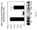
- SERCA-2a is also a member of the so-called fetal program gene.
- calcium binding proteins such as S100A1 and sorcin improve contractility by increasing calcium uptake into the sarcoplasmic reticulum. Expression of these proteins can also modify the behaviors of ryanodine calcium release channels. Alternatively down-regulation (or avoiding upregulation) of these calcium binding proteins can diminish contractility. This may be applicable to hypercontractile states of the heart associated with certain diseases such as hypertrophic obstructive cardiomyopathy. Contractility may also be reduced using hyperpolarizing fields.
- HF chronic HF is produced by multiple sequential intracoronary embolizations with polystyrene Latex microspheres (70-102 ⁇ m in diameter) which result in loss of viable myocardium, LV enlargement and a decrease in LV ejection fraction.
- 14 healthy mongrel dogs weighing between 20 and 30 kg underwent coronary microembolizations to produce HF.
- Embolizations were performed one week apart and were discontinued when LV ejection fraction, determined angiographically, was ⁇ 30%.
- Microembolizations were performed during cardiac catheterization under general anesthesia and sterile conditions.
- a series of 6 additional dogs underwent intracoronary microembolizations to produce HF as described earlier.
- a mid-sternotomy was performed, the pericardium was opened and epicardial CCM leads were placed on the anterior wall between the 2nd and 3rd diagonal branches.
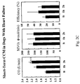
- Hemodynamic measurements including measurements of MVO 2 were made before and 2 hours after continuous CCM signal delivery at 7.73 volts.
- myocardial samples were obtained from the LV anterior wall in the region of the CCM leads and from the LV posterior wall remote from the CCM leads.
- Left ventricular tissue from 6 normal dogs and 6 HF dogs that were untreated was obtained and prepared in the same manner and used for comparisons. All tissue was rapidly frozen in liquid nitrogen and stored at -70°C until needed.
- Aortic and LV pressures were measured with catheter-tip micromanometers (Millar Instruments, Houston, TX) during cardiac catheterization.
- LV end-diastolic pressure was measured from the LV waveform.
- Single-plane left ventriculograms were obtained during each catheterization after completion of the hemodynamic measurements with the dog placed on its right side.
- Ventriculograms (approximately 60 right anterior oblique projection) were recorded on 35mm cine film at 30 frames per second during the injection of 20 ml of contrast material (Reno-M-60, Squibb, Princeton, NJ). Correction for image magnification was made with a radiopaque calibrated grid placed at the level of the LV.
- LV end-diastolic volume and end-systolic volume were calculated from ventricular silhouettes using the area-length method, such as described in Dodge HT, Sandler H, Baxley WA, Hawley RR. Usefulness and limitations of radiographic methods for determining left ventricular volume. Am J Cardiol. 1966;18:10-24 . Stroke volume was calculated as the difference between LV end-diastolic volume and end-systolic volume. Total coronary blood flow (CBF), and MVO 2 , were measured and calculated as described, for example, in Chandler MP, Stanley WC, Morita H, Suzuki G, Roth BA, Blackburn B, Wolff A, Sabbah HN. Acute treatment with ranolazine improves mechanical efficiency in dogs with chronic heart failure. Circ Res. 2002;91:278-280 . LV mechanical efficiency was calculated as the ratio of LV power to MVO 2 following the same paper.
- Calsequestrin (CSQ), atrial natriuretic peptide (ANP), brain natriuretic peptide (BNP), ryanodine receptor (RyR), total phospholamban (PLB), phosphorylated PLB (P-PLB), sarcoplasmic reticulum (SR) calcium ATPase (SERCA-2a), and ⁇ 1 -adrenergic receptor ( ⁇ 1 -AR) were measured by Western Blots. Briefly, LV homogenate was prepared from -100 mg LV powder as described, for example, in Gupta RC, Mishra S., Mishima T, Goldstein S, Sabbah HN.
- the nitrocellulose blot was incubated with the appropriately diluted primary monoclonal or polyclonal antibody specific to each protein based on the supplier's instructions. Antibody-binding proteins were visualized by autoradiography after treating the blots with horseradish peroxidase-conjugated secondary antibody (anti-rabbit) and ECL color developing reagents.
- ANP, BNP, ⁇ 1 -AR, SERCA-2a, PLB, or RyR-specific antibody recognized 20, 14, 65, 110, 5.5, and 250 kDa protein bands respectively.
- Total P-PLB was quantified in SDS-phosphoprotein-enriched fraction (PPE) prepared from LV homogenate using PLB specific monoclonal antibody.
- P-PLB at serine-16 or threonine-17 was quantified in SDS-LV homogenate using primary antibodies specific to the P-PLB at serine-16 or at threonine-17.
- PPE was prepared from LV tissue using a BD Bioscience phosphoprotein enrichment kit, for example as described in Gupta RC, Mishra S, Yang XP, Sabbah HN.
- Reduced inhibitor 1 and 2 activity is associated with increased protein phosphatase type 1 activity in left ventricular myocardium of one-kidney, one-clip hypertensive rats. Mol Cell Biochem. 2005;269:49-57 .
- Band intensity was quantified using a Bio-Rad GS-670 imaging densitometer and expressed as densitometric units x mm 2 . In all instances, the antibody was present in excess over the antigen and the density of each protein band was in the linear scale.
- RNA expression of glyseraldehyde-3-phosphate dehydrogenase (GAPDH), ⁇ -myosin heavy chain (MHC), ⁇ 1 -AR, ANP, BNP, SERCA-2a, total PLB, RyR and CSQ was measured.
- Total RNA with an absorbance ratio (260 nm/280 nm) above 1.7 was isolated from frozen LV tissue as described in Mol Cell Biochem. 2005;269:49-57 . Approximately 4-10 ⁇ g RNA was reverse-transcribed into cDNA in an assay volume of 80 microliter.
- PCR polymerase chain reaction
- 2-5 ⁇ l first-strand cDNA was added to 50 ⁇ l of a reaction mixture containing 20 pmol of each forward and reverse primer of each gene, 200 ⁇ M of each dNTP, 10 mM Tris-HCl (pH 8.8), 50 mM KCl, 0.1% Triton-X100 and 3.0 mM MgCl 2 and 1 unit platinum Taq DNA polymerase (Invitrogen, Carlsbad, CA) and PCR allowed to proceed for 20 to 40 cycles.
- PCR cycle was determined to ensure that the gene product is forming in linear range.
- PCR products were analyzed by subjecting 20 ⁇ l of each reaction mixture to electrophoresis on 1%-1.5% ethidium-bromide-agarose gels. Band size of the products was compared with standard DNA size markers and confirmed by sequencing of the forward (F) and reverse (R) primers for each gene and gene product.
- mRNA expression of ⁇ -MHC was measured by amplification of cDNA by reverse transcriptase-PCR followed by digestion with Pst1 restriction enzyme as described, for example, in Feldman AM, Ray PE, Silan CM, Mercer JA, Minobe W, Bristow MR.
- transverse slices (approximately 3 mm thick) one each from basal, middle and apical thirds of the LV, were obtained.
- tissue samples obtained from 7 normal dogs were prepared in an identical manner.
- transmural tissue blocks were obtained and embedded in paraffin blocks.
- 6 ⁇ m thick sections were prepared and stained with Gomori trichrome to identify fibrous tissue.
- the volume fraction of replacement fibrosis namely, the proportion of scar tissue to viable tissue in all three transverse LV slices, was calculated as the percent total surface area occupied by fibrous tissue using computer-based video densitometry (MOCHA, Jandel Scientific, Corte Madera, CA).
- Transmural tissue blocks were obtained from the free wall segment of the slice, mounted on cork using Tissue-Tek embedding medium (Sakura, Torrance, CA), and rapidly frozen in isopentane pre-cooled in liquid nitrogen and stored at -70°C until used.
- Cryostat sections were prepared and stained with fluorescein-labeled peanut agglutinin (Vector Laboratories Inc., Burlingame, CA) after pretreatment with 3.3 U/ml neuraminidase type V (Sigma Chemical Co., St.
- Histomorphometric, biochemical and molecular differences between normal dogs, sham-operated untreated HF dogs and CCM-treated HF dogs were examined using one-way analysis of variance (ANOVA) with ⁇ set at 0.05. If significance was achieved, pairwise comparisons were performed among groups using the Student-Newman-Kuels test with a probability value of ⁇ 0.05 considered significant. All hemodynamic, ventriculographic and histomorphometric assessments were made by investigators blinded to treatment group.
- the CCM signal was a 7.73 volt, epicardial LV anterior wall signal.
- the definitions for the various variables follow Circulation Research 91:278, 2002 .
- the signal is applied to the epicardial surface of the heart.
- CCM is a pulse at 80 applications per minute (synchronized to a heart beat, if any), at 7.73 Volts, with between 4 and 6 phases, each phase being 5.56 ms long and being continuous and of opposite polarity of a previous phase. The number of phases was not changed within an experiment.
- the signal was generally applied to a septum, from a right chamber, with a distance of 1-2 cm between an electrode pair used to apply the signal.
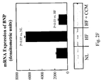
- the marking "NL" indicates normal tissue levels.
- Figs. 2A-2C show the improvement of LV function without MVO 2 increase, in CCM treated dogs as a function of time and compared to baseline values.
- Figs. 2D-2G show changes in mRNA expression of alpha-MHC, ANP, BNP and SERCA-2a in normal, HF and HF dogs with CCM treatment respectively, after several hours of treatment.
- Fig. 2H shows phosphorylated phospholamban normalized to total phospholamban following therapy of several hours (indicated herein as "short-term”).
- Fig. 2I shows corresponding blots using a Western Blotting method. It should be noted that both phospholamban levels and phosphorylation levels thereof improve with therapy.
- Fig. 2J shows reduction in mRNA expression of NCX following CCM treatment.
- Fig. 2K shows a general (or slight reduction) normalization of NCX protein values while still maintaining increased relative phosphorylation. This may allow some compensation for disturbed cardiac function.
- Fig. 2L shows decreased mRNA expression of GATA-4, to even below normal levels.
- Fig. 2M shows that protein expression of GATA-4, is however, still increased relative to normal. This may be useful for control of NCX and/or other proteins. This result also indicates that merely controlling mRNA may not sufficiently determine the cellular behavior, as protein levels and/or phosphorylation levels may compensate or over compensate. In general, however, the levels are normalized as compared to HF.
- Figs. 2N and 2O show the effect of chronic (e.g., 3 month) treatment with CCM on mRNA expression profiles. Normalization of these important proteins can be seen.
- the electric field can operate differently on different proteins, for example, directly affecting some proteins and these proteins indirectly affecting the behavior and/or levels of other proteins.
- there are multiple pathways in the cells and the electrical treatment may affect multiple (e.g., 1, 2, 3, 4 or more) pathways in parallel.
- the resulting effects on proteins may be increasing or decreasing their expression and/or activity levels. Different such effects may be desirable for different proteins and/or different disease conditions.
- Different proteins may be predisposed (e.g., based on their structure, surrounding materials and/or existing pathways) to differently increase and/or decrease. A particular experiment with Phospholamban is described below.
- Phospholamban is down regulated in heart failure and is nearly normalized with CCM therapy. This may explain the improvement in LV function by CCM treatment. CCM appears to normalize the RYR message which is consistent with proper therapy.
- the up-regulation of alpha-MHC with CCM may be contributing to the sustained long-term improvement in LV ejection fraction. Decrease in MMP1 following CCM therapy is in and of itself desirable. Inhibiting gelatinases, as shown, is beneficial, possibly reducing interstitial fibrosis and leading to improved LV diastolic compliance and, hence, improved diastolic filling and function.
- p21RAS and p38 mitogen activated protein kinese are emissions of stretch response genes which are down-regulated following CCM therapy and correlate with reduced cardiomyocytes hypertrophy. Integrin-a5 is clearly normalized following long-term CCM therapy. Up-regulation of Beta1-Adrenergic Receptor is viewed as a positive development which enhances the sensitivity of the contractile element of catecholamines. Beta-blockers are known to enhance the sensitivity of the myocardium to exogenous as well as endogenous catecholamines when used in heart failure patients over long periods of time in excess of 3 months. In the normal heart, beta blockers reduce the sensitivity of the heart to catecholamines. As will be described below, the therapies as described herein may be used together with beta-blockers, for both short and long term synergistic effects.
- some therapies according to the present disclosure will focus on general improvement of health, while other therapies will focus on increasing tissue responsiveness, for example, to certain drugs, and thus focus on improving fewer than all mRNA and/or protein and/or phosphorylation indicators.
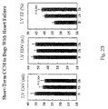
- phosphorylation effects for at least some proteins can be generated in immediate time frames, such as less than 1 minute and even less than 10 or 5 seconds in some cases. Further, an immediacy of effect is also characterized by a reduced number of intermediate stages, indicated by the fact that protein phosphorylation effects can be imposed even in tissue homogenate. Further, specificity of the phosphorylation effects to certain proteins that are relevant for HF is also shown. Further, a lack of effect of an exemplary pacing signal is also shown.
- Figs. 3A and 3B show the effect of a CMM signal applied to tissue homogenate from LV failed heart tissue. As can be seen, even a 10 second signal was sufficient to generate noticeable and significant changes in phosphorylation. Changes in phosphorylation are shown in Serine-16 Monometric PLB (Phospholamban) form, Threonin-17 mometric PLB form, Serine 16 Pentametric PLB form and Ryanodine channels.
- the tissue homogenate was prepared in the following manner. Approximately 14g frozen LV tissue from a dog with chronic HF in 42 ml 50 mM Tris-HCl, pH 7.5 was homogenized three times for 20 seconds each time using a 10-mm generator (Omni International, Waterbury, CT) at setting 10. The homogenate was then filtered through 4 layers of cheese cloth. The resulting homogenate was stored in ice and its protein concentration was determined by the Lowry method.
- the CCM signals were delivered to the homogenate as follows.
- the homogenate was diluted 2 fold in homogenate buffer and subsequently aliquotted 3 ml each in assay tubes.
- Assay tubes were divided into 2 sets (Set A and Set B), each subset consisting of 7 assay tubes.
- CCM signals were delivered for 10", 30", 1', 5', 30', and 60' in one of the sets, while the other set served as time control.
- the reaction was stopped by adding concentrated SDS. Protein assay on all the samples were performed by Lowry method.
- Phosphorylation of PLB at serine-16 (Ser-16) and threonine-17 (Thr-17) was determined by Western blotting using specific antibody as described in Mishra S, Sabbah HN, Jain JC, Gupta RC: "Reduced Ca2+-calmodulin-dependent protein kinase activity and expression in LV myocardium of dogs with heart failure", Am J Physiol Heart Circ Physiol 284:H876-H883, 2003 .
- tissue homogenate was generally activated at room temperature, below the normal operating temperature of a heart.
- This and other features of the results suggest a direct chemical or electrical effect on the proteins which is possibly divorced or semi divorced from cell function and/or complex biochemical mechanisms(e.g., more than two or three steps or with feedback).
- Such divorcing may help in the application of the effect under various conditions including various polarization conditions and tissue health states.
- the effect does not directly depend on the membrane polarization of the cell, therefore, this phosphorylation effect may be achieved at times other than a refractory period.
- therapy may be applied during a refractory period to avoid inadvertent pacing.
- tissue is desensitized, for example, using a suitable electrical signal, cold, or pharmaceutical, there is no need for specific timing.
- suitable charge provided during a pacing signal may be sufficient for a therapeutic effect.
- a typical pacing pulse is up to 1ms and 5V, which at 500 Ohm lead impedance is 50 micro-joule.
- the provided charge per beat is at least 100, at least 300, at lest 500, at least 1000, at least 2000, at least 5000 or more or intermediate values of micro-joules. It is hypothesized, that at least for some treatments, the applied field has to be above a minimum charge per heart beat, or the effect is lost, for example, due to electrical masking in the cell or due to biochemical interactions that occur within a heart beat.
- the applied energy is directed mostly at a tissue having a volume of less than 20 cm 3 , less than 10 cm 3 , less than 5 cm 3 , less than 3 cm 3 or larger or intermediate volumes.
- tissue homogenate was carried out in the presence and absence of protein kinese inhibitors.
- the homogenate was the same as above, from dogs with heart failure.
- Two different phosphorylation locations (threonin-17 and serine-16) on phospholamban produced two different results in the presence of the kinese inhibitors (in its absence, the above results were reproduced).
- the kinese inhibitor is STAUROSPORIN, a Pale yellow solid. Advertised as a potent, cell-permeable, and broad spectrum inhibitor of protein kinases.
- Akt synergistic effect is caused by the behavior of Akt (or similar proteins), which is described in a paper Gallo P, Santonastasi M, Ceci M, Scimia C, Di Sciascio G, Condorelli G.
- Akt overexpression improves cardiac function and ameliorates heart failure in pressure overload animal model phosphorylating key Ca2+ handling proteins. J Am Coll Cardiol 2006;21:76A .
- Akt selectively phosphorylates phospholamban at the threonin-17 site.
- the different sites on phospholamban are physiologically set up to be phosphorylated under different mechanism and while an electric field has a direct effect on both locations, one location is more sensitive to electric fields and the other location is more sensitive to biochemical interaction (or at least such as mediated by kinese). It is further hypothesized that the effect of the electric field applied by the therapy mimics (possibly in an enhanced manner) a regulated effect provided naturally by electrical activity of the heart.
- Fig. 3C shows phosphorylation of a Ryanodine receptor in isolated (in-vitro) failed cardiomyocytes, after application of CCM for 10, 20, 30 and 60 seconds. Lack of significant immediate effect is consistent with the lack of long-term effect shown above and serves to show that the effect of the CCM signal can be made specific to certain proteins.
- Fig. 3D shows phosphorylation of PLB and GATA, as compared to that of CSQ, in failed isolated myocytes.
- a phosphorylation effect is shown for some of the proteins, which matches the general results for tissue homogenate. Also noteworthy is that the effect increases over time at different rates for different proteins. Relaxation times of phosphorylation levels for different proteins are also generally different.
- the change in GATA-4 is important because when GATA-4 is phosphorylated, this process decreases the activity of the sodium calcium exchanger and helps contractility improve quickly.
- Fig. 3E shows that a pacing signal applied at 3V for pulses of 0.75 msec did not have any significant immediate effect on any of the proteins. Possibly, this is caused by the reduced current density of the pacing pulse and/or due to the substantially lower charge transport rate. Possibly, any minimal effect of the pacing signal is relaxed in the times between signals. Optionally, the pacing signal is not strong enough to pass a certain minimal threshold of effect.
- mRNA expression levels were measured for some genes in human subjects.
- Therapy with non-excitatory cardiac contractility modulation (CCM) electrical signals was delivered to LV muscle during the absolute refractory period improves LV function in patients with HF.
- CCM cardiac contractility modulation
- the effects of 3 months CCM therapy on mRNA expression of cardiac fetal and SR genes in 5 patients with advanced HF were examined.
- right sided endomyocardial biopsies were obtained at baseline, prior to activating CCM therapy, and at 3 and 6 months thereafter.
- CCM therapy was delivered in random order of ON for 3 months and OFF for 3 months.
- mRNA expression measurement was performed in a blinded fashion as to the ON/OFF order of therapy.
- fetal genes A-type (ANP) and B-type (BNP) natriuretic peptides and a-myosin heavy chain (MHC), and the SR genes SERCA-2a, phospholamban (PLB) and ryanodine receptors (RYR) was measured using RT-PCR and bands quantified in densitometric units (du). The percent change in du between baseline and the ON and OFF 3 months phases was calculated.
- the 3 months therapy OFF phase was associated with increased expression of ANP and BNP and decreased expression of ⁇ -MHC, SERCA-2a, PLB and RYR (Table).
- the 3 months ON therapy phase resulted in decreased expression of ANP and BNP and increased expression of ⁇ -MHC, SERCA-2a, PLB and RYR (Table).
- CCM therapy reverses the cardiac maladaptive fetal gene program and normalizes expression of key SR Ca2+ cycling genes.
- Figs. 5A-5R shows protein expression results for the following proteins in chronic dogs, in control, heart failure and treated heart failure conditions: CSQ, SERCA-2a, PLB, RyR, NCX, IL-6, GATA-4, GAPDH, MMP-9, Tubulin-Beta, GATA-1, MMP-1, Tubulin-Alpha, Titin, TIMP-1, Integrin- ⁇ 5, TNF ⁇ , p21ras, p38 MAPK, TIMP-2, ⁇ 1 -AR, MMP-2, ANP and BNP.
- Figs. 5A-5D show results for the following SR proteins: Calsequestrin, phospholamban, SERCA-2a (Calcium ATPase) and ryanodine receptors; the following Pump Proteins: Sodium-Calcium Exchanger; the following Transcription Factors: GATA-4; and the following Cytokines: Interleukin-6.
- the CCM signals appear to normalize the over-expression of the sodium-calcium exchanger.
- Figs. 5E-5H show results for GAPDH (Housekeeping), transcription factor GATA-1 which did not change, matrix metalloproteinase-9 which changes consistent with mRNA expression and cytoskeletal protein Tubulin-beta which also changes consistent with what is shown for mRNA expression.
- GATA-1 is shown in comparison with GATA-4.
- Figs. 5I-5L show results from the proteins matrix-metalloproteinase-1 (MMP-1), cytoskeletal proteins tubulin alpha and titin, tissue inhibitor of matrix-metalloproteinase-1 (TIMP-1) and cell surface protein integrin-alpha-5.
- MMP-1 matrix-metalloproteinase-1
- TRIP-1 tissue inhibitor of matrix-metalloproteinase-1
- cell surface protein integrin-alpha-5 cell surface protein integrin-alpha-5.
- integrin-alpha-5 can be affected by other means, such as mechanically constraining the heart (e.g., thus directly affecting its transduction function).
- Integrin-alpha -5 NL HF-Control HF + CCM 228 340 254 153 455 239 160 437 212 223 358 193 185 332 168 201 356 253 324 249 192 372 224 31 52 34 13 20 13 ANOVA 0.57 p vs. NL ⁇ 0.05 NS p vs. HF-Control ⁇ 0.05
- Figs. 5M-5P show results from the proteins TNF- ⁇ (showed in comparison to IL-6), p21ras, p38 MAPK, TIMP-2 (showed in comparison to TIMP-1 and ⁇ 1-AR.
- TIMP-2 The lack of change in TIMP-2 is consistent with previous observations. Long-term CCM therapy significantly reduced protein expression of the cytokine TNF- ⁇ and significantly reduced the expression of the stretch proteins p21ras as well as p38 MAPK. This is consistent with the observation that CCM therapy attenuates cardiomyocyte hypertrophy. Also to be noted is up-regulation of the beta-1 adrenergic receptor, which is favorable.
- Figs. 5Q and 5R show results for MMP-2 (in comparison to MMP-1 and MMP-9) and ANP and BNP.
- mRNA expression in LV free wall of the housekeeping gene GAPDH and CSQ, the fetal program genes consisting of ⁇ 1 -AR, ⁇ MHC, ANP, and BNP and the cardiac SR genes SERCA-2a, PLB, and RyR are shown in table 5 quantified in densitometric units. Expression of GAPDH and CSQ was unchanged among the 3 study groups namely, normal dogs, sham-operated HF dogs and HF CMM-treated dogs. mRNA expression of ⁇ 1 -AR, ⁇ MHC, SERCA-2a, PLB and RyR decreased and expression of ANP and BNP increased significantly in sham-operated HF dogs compared to normal.
- this mechanism is used to test if a patient is getting better - by stopping a therapy which is directly causing phospohorylation and seeing if phospholamban levels normalize (or trend to) after a few days and/or if other protein levels trend towards disease state values.
- relaxation time between signal application is an integral part of therapy.
- relaxation time is used to allow the cell to find a new balance between the expressed proteins that are not on corresponding levels, such a balance may include protein levels (or mRNA levels or phosphorylation or structural remodeling) degrading and/or protein levels improving.
- Such relation times may be on order of seconds, minutes hours or days, depending on which mechanism are to be allowed to take part (e.g., protein based, etc.).
- phosphorylation-modifying therapy when applying phosphorylation-modifying therapy as described herein, a process of weaning is applied. Possibly, if treatment is stopped suddenly, phospholamban levels will be too low to support suitable cardiac activity, possibly causing a downwards-spiral in patient health.
- phospholamban levels will be too low to support suitable cardiac activity, possibly causing a downwards-spiral in patient health.
- therapy is reduced to allow phospholamban recovery.
- the pauses are timed according to measured recovery in phospholamban levels.
- electrical therapy is applied selectively to tissue measured as having reduced phospholamban levels and/or phosphorylation levels, so as not to potentially damage healthy tissue.
- changes to "healthy" tissue is desirable. For example, increasing phosphorylation and thus possibly reducing phospholamban may be desirable if long term reduction in contractility is desired.
- phosphorylation may be increased in normal tissue in order to cause over (or under) expression of some proteins, such as gap junction proteins or mechanical proteins. It should be appreciated that a therapy target of diseased tissue need not be a mirror of a healthy tissue state.
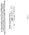
- Fig. 6 shows mRNA expression levels for Phospholamban, SERCA-2a and Ryanodine receptors, showing chronic improvement in septal tissue to which a field was applied chronically.
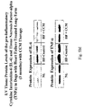
- Fig. 8 shows phosphorylation levels in chronically treated dogs, at the application location in the septum.
- protein levels of P-PLB at serine-16 and threonine-17 were also examined. Measurements were made in tissue obtained from both the inter-ventricular septum and the LV free wall. At both sites, protein levels of P-PLB at serine-16 and threonine-17 were significantly lower in sham-operated HF dogs compared to normal dogs and returned to near normal levels after 3 months of CCM therapy ( Fig. 11 , Table 6). In both the inter-ventricular septum and LV free wall, the ratio of P-PLB at serine-16 to total PLB and the ratio of P-PLB at threnonine-17 were also significantly lower in sham-operated HF dogs compared to normal dogs (Table 6).
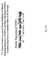
- Fig. 7A and 7B shows mRNA expression at sites remote from the application of the signal, but still within the left ventricle, at a relatively short time of four hours apparently no significant effect (mRNA, protein and/or phosphorylation) is shown.
- This may indicate that the effect of the CCM signal is first local, for example on a molecular level and then propagates to remote location, for example by biochemical signaling or by a mechanical signaling indicated by the change in contraction behavior of the treated tissue and/or of the chamber as a whole.
- the electric field causes phosphorylation of phospholamban. This in turn increases the activity/affinity of SRECA-2a for calcium and immediately improves SR calcium cycling.
- GATA-4 and the sodium calcium exchanger may play an additive role in the improved function.
- LV function begins to improve and the LV gets smaller, many of the molecular/biochemical maladaptations begin to correct, which adds to the long-term benefits.
- Improved SR cardiac cycling may be a goal of some therapies in accordance with exemplary embodiments of the disclosure.
- LV tissue obtained near the site of CCM lead implants was compared to LV tissue obtained from a site remote from the CCM leads.
- LV tissue samples obtained from the same sites from dogs with HF that were untreated and normal dogs were used for comparisons.
- the ratio of P-PLB to total PLB increased significantly in CCM-treated HF dogs compared to untreated HF dogs in the LV anterior wall at the site of signal delivery ( Fig. 12A ), whereas it was essentially unchanged in the LV posterior wall remote from the site of CCM signal delivery ( Fig. 12B ).
- the location to which electrification will be applied is selected based on a model of what areas will cause a biochemical or mechanical cascade in the heart and improve its function.
- the size of areas is optimized to reduce power needs, while retain an expected time frame of treatment.
- an area to be treated is selected based on immediate response of tissue therein to electrical stimulation.
- a mechanical cascade is a desired change in stretching of tissue which will, as that tissue improve, propagate.
- Another example of mechanical cascade is selecting sufficient and correct tissue in the ventricle such that immediate hemodynamic effects (e.g., improvement) are seen and sensed by the rest of the chamber.
- a possible mechanism of non-mechanical propagation is that healthy cells can help regulate ionic concentrations of neighboring cells via gap junctions between them. Alternatively or additionally, other communication may occur via the gap junctions.
- the efficacy of a treatment is measured by tracking remote effects alternatively or additionally, to tracking local effects.
- One or more of the following logics may be used: 1. There is more remote tissue to sample, with less danger of damage to heart. 2. The effect in remote tissue is more gradual and thus easier to track. 3. Acute effects may not occur (or be smaller) in remote tissue, thereby preventing the masking of longest term effects by acute effects.
- the measurement is made when no treatment is applied.
- An example of an acute effect which may mask a longer term effect is conduction velocity.
- S100A1 sarcoplasmic reticulum (SR) calcium cycling by increasing calcium-uptake and reducing SR calcium-leak from ryanodine channels.
- SR sarcoplasmic reticulum
- S100A1 is significantly decreased in left ventricular (LV) myocardium of explanted failed human hearts.
- S100A1 mRNA and protein expression decreased significantly in untreated HF dogs compared to NL dogs.
- Chronic CCM therapy significantly increased mRNA and protein expression of S100A1.
- Restoration of expression of this calcium binding protein improves calcium cycling within the SR and may account, at least in part, for the observed improvement of LV function seen following chronic CCM therapy.
- Sorcin mRNA and protein expression decreased significantly in untreated HF dogs compared to NL dogs.
- Chronic CCM therapy significantly increased mRNA and protein expression of Sorcin.
- Presenilin-2 increased in HF and decreased with CCM.
- Preseilin-1 was measured as an internal control and it did not change. This suggests that Presenilin-2 can be used as a target (e.g., treatment goal) for treating HF and as an indicator for diagnosis and modification of treatment.
- Restoration of expression of Sorcin may prevent or limit the RyR2 calcium leak and in doing so improve calcium cycling within the SR. Correction of this maladaptation by CCM therapy may account, at least in part, for the observed improvement of LV function.
- the combined correction of Sorcin and Presenilin, which interact to regulate cardiac ryanodine calcium release channels may act as a mediator of recovery of calcium overload in cardiomyocytes due to "RyR-2 calcium leak" in heart failure.
- Calstabin-2 decreased in heart failure but remained depressed even after 3 months of CCM therapy. This suggests that CCM does not act by merely resetting cellular function, however, as noted above, resetting may be part of the process.
- Castabin-2 does not rebound as it is tied to phospholamban levels.
- Castabin-2 indicates a path for the progression of heart failure. Until the underlying cause is removed, it may remain depressed. Possibly, an improvement over significantly longer periods of time is to be expected. In an exemplary embodiment of the disclosure, this is used to estimate the ability of a patient to stop therapy. For example, if Castabin-2 levels trend to or do normalize, this can indicate that the tissue state is not diseased or becoming healthier. Optionally, this allows tracking of tissue improvement even during ongoing electrical therapy.
- signal optimization techniques as described herein are used to find and then apply a signal specific (or more specific) to modifying Castabin-2.
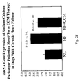
- Figs. 13A and 13B present mRNA ( Fig. 13A ) and protein blots ( Fig. 13B ) illustrating expression of Sorcin in LV tissue of HF Dogs treated with CCM for 3 months according to an exemplary embodiment of the disclosure.
- Figs. 14A, 14B, 14C and 14D present mRNA ( Fig. 14A and 14C ) and protein blots ( Fig. 14B and 14D ) of the Presenilin-1 ( Fig. 14A and 14B ) and Presenilin 2 ( Fig. 14C and 14D ) in LV tissue of HF Dogs treated with CCM for 3 months according to an exemplary embodiment of the disclosure.
- Figs. 15A and 15B present mRNA ( Fig. 15A ) and protein blots ( Fig. 15B ) illustrating expression of Calstabin in LV tissue of HF Dogs treated with CCM for 3 months according to an exemplary embodiment of the disclosure.
- HF-Control NS Protein Expression of Calstabin-2 Normal HF-Control HF + CCM 5871 3991 3679 5602 3643 3923 5029 3156 3560 5579 4645 4083 6726 4567 4600 5190 4995 4381 Mean 5,666 4,166 4,038 STD 602 693 402 SEM 246 283 164 ANOVA 0.0001 p vs. Normal ⁇ 0.05 ⁇ 0.05 p vs. HF-Control NS
- Figs. 9A and 9B are graphs showing the relationship between rise time and decay time of a contractility increase in dogs. Similar results were observed in humans, albeit with generally slower rise times and slower decay times.
- Fig. 9A in which the scale is in seconds, shows a rise time of several tens of seconds at about time 270.
- the contractility increase decays.
- the blip at 600 is probably caused by an arrhythmia.
- the decay continues from 600 until 950.
- a second signal application is at about 950 with a change in signal application location at about time 1350, before the signal effect washed out.
- Fig. 9B the signal is shown as an outline of a square pulse, with the signal stopped each time an arrhythmia is detected, shown as a short pause in the signal.
- a rise time of several tens of seconds is found.
- a decay time of 230 seconds is shown for the first signal.
- a longer decay time of over 400 seconds is shown for a second signal.
- the second signal caused an increase in contractility at about 1050 seconds, due to change in the applied signals.
- the acute and long term effects last at least a few seconds after treatment is stopped and have different wash-out times.
- Different waiting times may be used to selectively measure acute and longer term effects. For example, 30 minutes may be sufficient for avoiding acute effects on phosphorylation.
- a controller learns the particular washout behavior of a patient and adjusts the treatment to be re-applied only after significant washout and/or be applied at a minimal length that has a washout.
- the delay between application and/or length of applications are selected to optimize one or more of power consumption, danger of arrhythmia and/or discomfort to patient.
- WO 2005/087310 filed as PCT/IL2005/00316 on March 18, 2005
- US provisional application 60/719,517, filed September 22, 2005 and US provisional application 60/654,056 filed February 17th
- 60/719,517 describes how applying a signal to a stomach has an effect on reducing blood glucose levels which lasts after the signal is stopped, for example, for more than one day.
- an electrical therapy may be used to change the mode of operation of tissue, on a cellular level, possibly for tissue in general or for excitable tissue at least.
- a non-immediate effect of signal application which lasts after the signal application is stopped, is an affirmative effect, in which the tissue exhibits a positive change in action, rather than a negative effect, for example, of preventing arrhythmia.
- the effect is on a cellular level, rather than or in addition to an effect on an organ level, for example a cellular level as measured by protein activity and/or expression levels.
- Another type of long term/wash-out effect is a long term therapeutic effect lasting days or weeks after treatment.
- two sets of experiments on humans In the first set, patients were selectively treated 3 months on and 3 months off or vice versa.
- patients In the second set of experiments patients were either treated for six months or not. While the patients that were 3 months on and then 3 off fared less well (after 6 months) than those that were 3 off and then 3 on, the overall situation after 6 months was better than what would be expected from a baseline (no treatment) situation.
- the second set of experiments while suggesting no overall improvement after 6 months, shows a trend to lesser degradation and also suggests what the expected degradation would be.
- the therapies as described herein are used to slow down degradation of a patient's situation, rather than a treatment.
- each patient is selectively tested to see how long the treatment effects last and/or what minimal treatment period is required for longer term effects.
- the length of treatment and/or "off" periods between treatments also vary as a function of time.
- Various physiological measures for example as described above may be used to assess improvement, as well as mRNA, protein and/or phosphorylation levels.
- Baseline EF 25 ⁇ 6% vs 26 ⁇ 6%
- VO 2,peak (14.4 ⁇ 2.5 vs 14.2 ⁇ 2.7 ml/kg/min)
- MLWHFQ 41 ⁇ 20 vs 35 ⁇ 19
- Baseline EF 25 ⁇ 6% vs 26 ⁇ 6%
- VO 2,peak (14.4 ⁇ 2.5 vs 14.2 ⁇ 2.7 ml/kg/min)
- MLWHFQ 41 ⁇ 20 vs 35 ⁇ 19
- the increase in the untreated group appeared to be less than a return to baseline values or other values that would be expected with a diseased patient with natural progression.
- New York Heart Association classification improved similarly in both groups.
- the proportion of patients in Class I, II and III at 24 weeks were 19, 45 and 36, respectively. This compared to 18, 52 and 30, respectively, in the Control group.
- MLWHFQ also improved significantly and similarly in both groups.
- MLWHFQ decreased from baseline values by 16.2 ⁇ 5.9 and 18.3 ⁇ 4.8 in the control and treatment groups, respectively.
- the significant and sustained improvements in NYHA and MLWHFQ observed in both groups speaks to the presence of a significant placebo effect.
- beta blockers and electrical therapy may be used to increase contractility and compensate for an initial reduction in contractility caused by beta blockers. This may suggest changing the sequence amount and/or type when the medication starts having a positive effect, for example, several weeks or several months (e.g., 3) later.
- electrical therapy is used to compensate for times when medication is stopped, for example, due to adverse systemic effects.
- the dosage of medication and the dosage of therapy are selected to minimize or reduce systemic effects and/or reduce power needs and/or side effects (if any) of electrical therapy.
- LV EF 30%-40% dogs with intracoronary microembolization-induced chronic HF (LV EF 30%-40%) were randomized to 3 months therapy with either BB (beta-blockers) alone (extended release metoprolol succinate, 100 mg once daily), extended release metoprolol succinate (100 mg, once daily) combined with CCM therapy, or to no therapy at all (Control).
- BB beta-blockers
- ESV end-systolic volume
- EF were measured before randomization
- POST 3 months of therapy
- Control BB Alone BB + CCM PRE POST PRE POST PRE POST LV EDV (ml) 60 ⁇ 1 64 ⁇ 1 64 ⁇ 4 62 ⁇ 5 70 ⁇ 4 69 ⁇ 4 LV ESV (ml) 39 ⁇ 1 45 ⁇ 1* 41 ⁇ 2 36 ⁇ 4* 45 ⁇ 3 36 ⁇ 3* LV EF (%) 36 ⁇ 1 31 ⁇ 1* 36 ⁇ 1 42 ⁇ 2* 36 ⁇ 1 48 ⁇ 1* * p ⁇ 0.05 PRE vs. POST
- EDV (ml) Dog# Base Pre Post 03-031 51 59 63 03-065 54 61 68 04-103 52 58 61 04-122 48 63 64 05-002 54 61 64 05-027 59 60 64 Mean 53 60 64 STD 4 2 2 SEM 1.5 0.7 0.9 ESV (ml) Dog# Base Pre Post 03-031 24 39 46 03-065 23 39 48 04-103 21 37 43 04-122 23 39 43 05-002 28 39 43 05-027 28 39 44 Mean 25 39 45 STD 3 1 2 SEM 1.2 0.3 0.8 EF (%) Dog# Base Pre Post Delta 03-031 51 34 27 -7 03-065 48 36 29 -7 04-103 60 36 30 -6 04-122 52 38 33 -5 05-002 48 36 33 -3 05-027 53 35 31 -4 Mean 52 36 31 -5 STD 4 1 2 2 SEM 1.8 0.5 1.0 0.7 EDV LV end-diastolic volume
- ESV LV end-systolic
- the above experimental results are used as a basis for diagnosis.
- the diagnosis indicates one or both of a disease type and disease severity.
- the type can be seen from the behavior of the proteins, for example comparing protein levels to calcium overload to diagnose systolic or diastolic dysfunction.
- the relative phosphorylation levels are used as a measure of severity.
- the use of multiple proteins and mRNA expression values provides a robust indicator of disease.
- only one or two of proteins, phosphorylation and mRNA are used.
- cardiac function values, such as stroke volume are used as well.
- the response of tissue to a field is used as well, for example phosphorylation response.
- At least 3, at least 5, at least 10, at least 20 or more proteins levels, phosphorylation levels and/or mRNA levels are used to aid in diagnosis.
- a table is maintained which includes ranges of values that match various disease types and/or conditions.
- the patient is re-diagnosed.
- the diagnosis is according to groups of mRNA and/or proteins that have associated functions.
- a DNA chip and/or protein chip and/or a biochip are used to measure the above levels.
- treatments are selected by applying the above pulses to tissue and measuring the effect.
- a tissue sample is removed, for example by biopsy and a range of sequences are tested on the sample.
- the results of the testing indicate which sequence might have a desired therapeutic effect.
- Such testing can include analysis and/or application of a treatment, such as an electric field. As noted above, at least some of the tests can be meaningfully applied to tissue homogenate and other unstructured tissue configurations.
- tissue part In an exemplary embodiment of the disclosure, different parts of the tissue are tested, for example, to see which tissue part will respond better and/or avoid side effects.
- a kit for performing such analyses as described herein.
- the kit comprises a set of reagents, antibodies and/or other biochemical tools as known in the art.
- the kit comprises one or more mRNA or protein detecting chips.
- the kit comprises software for analyzing gel migration.
- the kit comprises a source of treatment, for example electrodes and optionally a power source, for electrifying a sample for testing its responsiveness.
- the kit includes a sampling means for selecting part of the sample at a pre-described time, to check temporal response of the sample.
- An existing kit may be modified for use, for example, a kit available from Biosite, Inc. to measure blood levels of BNP. Such a kit could include instructions for use with the methods described herein and optionally include sampling means or a timer to ensure correct timing of activities.
- the kit includes or is used with a bioreactor that includes a controllable sampling element which cans electively extract a portion of the sample in the bioreactor and test it in a separate chamber.
- a controllable sampling element which cans electively extract a portion of the sample in the bioreactor and test it in a separate chamber.
- this is embodied using lab-on-chip technology and/or fluidic control of material flow.
- the controllable sampling element comprises a robot and a pipette that takes part of the sample and inserts it into an assaying chamber.
- Various automated assaying devices are known in the art of drug discovery and may be used and/or modified to be smaller and/or simpler.
- the kit includes a database or a link to a database (e.g., on the internet) that links biochemical markers to tissue states, treatment states and/or disease states.
- kits are sterile and/or frozen and optionally include instructions for use as described herein.
- one or more kits are packaged with a controller designed for implantation, for use in determining suitable electrode placement therefore.
- the kit includes one or more antibody reagents useful for detecting phosphorylated and/or dephosphorylated forms of the proteins desired.
- tracers which are anti-body based are used in-vivo, for example provided using a catheter or using the delivery electrodes or a separate needle or other injection mechanism.
- the tracer is radioactive or fluorescent.
- a calibration step for example, per patient, may be carried out.
- a comparison before and after field application is used to determine change in phosphorylation.
- phosphorylation of proteins which affects ECG signals or correlated proteins which affect ECG signals is detected by detecting changes in ECG signals.
- changes in conduction velocity caused by increased secretion of protein are detected in an ECG measurement.
- the measurement is remote from a signal application area.
- changes in contraction velocity under various conditions are assed from images of the heart or using other means.
- physiological measures are correlated with protein effects (e.g., in a per-patient or per-population calibration process) and used to estimate effects on protein synthesis from physiological measures.
- the above sequences are used to treat tissue plugs that are removed from the body, treated and reinserted.
- the reinsertion may be, for example, at the location of removal, at scarred locations, at locations bordering scars or at otherwise weakened location of the heart.
- the above sequences are used as part of a program to re-invigorate scar or fibrotic tissue.
- the effect of the sequence is to cause at least some cells to become functioning, for example in the border of the scar.
- the above sequence is used to selectively increase oxygen efficiency in some parts of the heart, for example, parts with a vascular blockage thereto.
- the above sequences are applied to tissue transplants, for example, whole heart transplants or plug transplants (from a donor), prior to and/or after transplant.
- the above sequences are applied after DNA or stem cell therapy to the heart, for example, to enhance effects and/or to assist in cellular functional differentiation and/or regeneration.
- the above sequences are used to have a desired modeling effect on the heart, for example, modifying an elasticity and/or contractility profile of a heart portion and/or directly controlling conduction velocity.
- a first type of remodeling is on the biochemical level and includes, for example, normalization of protein synthesis.
- a second type of remodeling is mechanical remodeling, for example, fibrosis reduction, chamber size change, vascular increase and/or wall thickening.
- a third type of remodeling is function remodeling which may result form a combination of biochemical and structural remodeling. This includes, for example, cardiac electrical and/or mechanical activation profile (what gets activated, to what degree and in what order), contractility enhancement and conduction velocity. As described herein, these types of remodeling may be achieved using methods described herein.
- Differential remodeling of different types may be achieved, for example, by selectively applying signals with a strong acute effect (which affect mechanical remodeling) and those with a long term effect.
- tissue which is both paced and treated with non-excitatory signals may thicken, as opposed to tissue which is only treated.
- multiple methods of improving contractility are applied at a same time, or methods of improving contractility applied at a same time as methods that reduce contractility such as the initial effect of beta-blockers.
- contractility enhancement by effects on membrane proteins is carried out at least partly independently from protein effects on SR proteins.
- the selectivity is using methods as described above.
- such enhancement is provided without adversely affecting short term activity, as pacing or inhibition might.
- the enhancement is local and has fewer systemic effects (as would be expected with ACE-inhibitors or beta-blockers, for example).
- short term activity is improved.
- an electric field as described herein can be used, for example, to modify PLB phosphorylation in blood vessel cells and/or the GI tract.
- elasticity compliance is restored and vasomotor tone is restored and/or responsiveness are restored to blood vessels and/or GI tract portions using the electric field as described herein.
- a hardened aorta is made more supple (and thus relieve cardiac problems) by suitable treatment.
- Vascular resistance in general, may be modified.
- signals as described herein are used to help increase muscle strength and/or normalize protein expression therein.
- a method of optimizing treatments to the tissue is provided, taking into account effects on protein levels.
- a CCM signal or a pacing signal, exercise or a pharmaceutical treatment may each (or in combination) have an effect on protein expression and/or behavior.
- such a treatment is optimized so that it has a greater (desired) effect on proteins.
- a CCM signal is optimized by applying variations of the signals (e.g., different amplitude, frequencies pulse forms, etc.) to a set of tissue homogenate sets and selecting the signal(s) for which a better effect is achieved.
- protein measurement may allow faster within-patient optimization and/or allow optimization based on the response to a small number of beats (e.g., 100, 50, 10, 3 or intermediate or fewer numbers), rather than waiting for a longer term effect which may damage the heart if it is negative.
- a small number of beats e.g. 100, 50, 10, 3 or intermediate or fewer numbers
- an existing device is optimized by changing its applied sequence with a sequence selected to have a desired protein effect.
- a device is programmed by a used selecting a desired pulse sequence form an existing set or by selecting parameters which are expected to have a desired protein effect.
- the applied pulse sequence (optionally including non-treated beats) and/or desired effect may change over the course of a treatment.
- One type of change is when the patient state stabilizes and/or the focus of maladaptation changes.
- a first step of treatment is in stabilizing heart treatment and a second step is in increasing contractility and/or remodeling the heart.
- Another type of change is where a different effect is desired at different times during a treatment, for example, a one series of beats being utilized to treat one protein and another series of beats to have another effect. It should be noted that not all treatments need to be synchronized the cardiac heart beat.
- the change includes one or both of changing the sequence and changing the tissue to which the sequence is applied, for example by switching and/or by moving electrode(s).
- the applied sequence takes into account a provided pharmaceutical and/or a half-life in the body thereof.
- a transmitter is provided to a patient to indicate to the controller that a medication was taken.
- the medication (or another treatment) is provided to specifically interact with the sequence.
- a signal or medicine is provided which has a long effect and while that effect is going on, a signal or other treatment is provided which has an opposite effect that momentarily counteracts long-term effects.
- a medication which extends a refractory period is provided together with an electrical treatment that applies a phosphorylation-modifying signal.
- medication is provided to enforce resting of the cells, using a mechanism which does not prevent the CCM or CCM-like signal from working, possibly, a medication that blocks trans-membrane channels.
- sequences that are applied therapeutically may change between applications to a same patient, for example, vary in power, shape, repetition number, duration of application and location. As noted above, such variations may be during a treatment process of a particular complaint or set of complaints.
- an external programming station may be provided, which optionally communicates with an implantable device using telemetry. Data collection using telemetry may also be practiced.
- computer readable media including such programs are also included.
- micro-code and other types of programming as well as hardwired circuitry and ASICs. This is a list of examples and should not be considered as limiting.
- An exemplary device/software includes a decision making module, a timing module, a power module and/or a signal analysis modules. Section headings are provided for navigation and should not be considered as limiting their contents to that section only.
Landscapes
- Health & Medical Sciences (AREA)
- Life Sciences & Earth Sciences (AREA)
- Engineering & Computer Science (AREA)
- Chemical & Material Sciences (AREA)
- Biomedical Technology (AREA)
- General Health & Medical Sciences (AREA)
- Organic Chemistry (AREA)
- Nuclear Medicine, Radiotherapy & Molecular Imaging (AREA)
- Radiology & Medical Imaging (AREA)
- Animal Behavior & Ethology (AREA)
- Public Health (AREA)
- Veterinary Medicine (AREA)
- Genetics & Genomics (AREA)
- Zoology (AREA)
- Wood Science & Technology (AREA)
- Molecular Biology (AREA)
- Bioinformatics & Cheminformatics (AREA)
- Proteomics, Peptides & Aminoacids (AREA)
- Biotechnology (AREA)
- Microbiology (AREA)
- Biochemistry (AREA)
- Immunology (AREA)
- Analytical Chemistry (AREA)
- General Engineering & Computer Science (AREA)
- Physics & Mathematics (AREA)
- Cardiology (AREA)
- Hematology (AREA)
- Urology & Nephrology (AREA)
- Pathology (AREA)
- Biophysics (AREA)
- Cell Biology (AREA)
- Heart & Thoracic Surgery (AREA)
- Hospice & Palliative Care (AREA)
- Food Science & Technology (AREA)
- Medicinal Chemistry (AREA)
- General Physics & Mathematics (AREA)
- Medicines That Contain Protein Lipid Enzymes And Other Medicines (AREA)
- Investigating Or Analysing Biological Materials (AREA)
- Measuring Or Testing Involving Enzymes Or Micro-Organisms (AREA)
- Electrotherapy Devices (AREA)
Description
- The present invention relates to an apparatus for treating cardiac tissue according to
claim 1 and a method of manufacturing a therapeutic device according toclaim 9. - While some proteins have a mainly structural role in cellular life, many proteins are biologically active. Living cells include many mechanisms by which the biological activity of a protein is modulated, including: modification of concentration of the protein or its substrates, modification of the concentration of materials that catalyzes protein activity, indirect modification of protein structure, such as by changing of pH or concentrations of materials that modify protein structure, and direct modification of protein spatial structure and/or charge distribution by attachment of cofactors such as a phosphate moiety (phosphorylation), glucose, ions, metal ions, heme groups or iron-sulfur complexes and coenzymes for example.
- The symptoms of many diseases include changes in protein activity, as indicated, for example, by phosphorylation (hyper- or hypo-). One example is cardiac heart failure, where, as the disease progresses the phosphorylation of some proteins goes down and others go up. Levels of various proteins also change.
- As described, for example in N Engl J Med 346:1357, 2002, the disclosure of which is incorporated herein by reference, patients with CHF who respond to therapy with beta blockers manifest reversal that is normalization of the maladaptive fetal gene program.
- In a paper entitled "Voltage-dependent potentiation of the activity of cardiac L-type calcium channel al subunits due to phosphorylation by cAMP-dependent protein kinese", by Adrian SCULPTOREANU, Eric ROTMAN, Masami TAKAHASHI, Todd SCHEUER, AND William A. CATTERALL, in Proc. Natl. Acad. Sci. USA Vol. 90, pp. 10135-10139, November 1993 (Physiology), fast phosphorylation of trans-membrane calcium channels and a possible mechanism therefore, are described.
-
US patent 6,919,205 , describes regulation of type II cartilage genes and proteins using electromagnetic and electric fields. -
PCT publication WO 2005/056111 , describes using a PMF signal on calcium dependent myosin phosphorylation in a cell free reaction mixture. -
PCT publication WO 2005/102188 , describes PMF stimulation applied to Jurkat cells reduces DNA synthesis and makes them behave like normal T-lymphocytes stimulated by antigens at the T-cell receptor such as anti-CD3, possibly by interacting with the T-cell receptor. -
PCT publication WO 2005/105013 , describes applying a PMF to a heart in order to achieve angiogenesis and neovascularization. -
WO 2006073671 relates to a method of modifying tissue behavior, including cardiac tissue, comprising: determining a desired modification of tissue behavior for at least one of treatment of a disease, short or long term modification of tissue behavior, assessing tissue state and assessing tissue response to stimulation; selecting an electric field having a known effect of modifying protein activation levels of at least one protein as an immediate response of a tissue to the field, said known effect correlated with said desired modification; and applying said field to said tissue. -
WO 2004/080533 describes a method and an apparatus (120) for modifying gene expression in cardiac muscle cells (110), by the application of electric fields. - The present invention relates to an apparatus for treating cardiac tissue, comprising:
- an electrode adapted to apply an electrical signal that induces an electric field sequence in in-vivo cardiac tissue;
- a sensor configured for sensing an effect indicative of a change in phosphorylation level of proteins in the cardiac tissue by detecting a modification of a sensed parameter on a time scale of minutes or seconds;
- a controller operable to apply the electrical signal to the electrode and configured for receiving measurements from said sensor,
- the controller including a memory having stored therein an electrical signal parameter,
- wherein the controller is configured for selecting the electrical signal parameter in response to measurements received from said sensor, such that the electric field sequence resulting from application of the electrical signal to the electrode according to the electrical signal parameter has an immediate affirmative modifying effect on phosphorylation of proteins in the cardiac tissue.
- Preferably, the parameter is further selected to have a lasting affirmative modifying effect on behavior of the cardiac tissue that continues for at least 15 minutes after application of the signal sequence to the electrode.
- Preferably, the lasting affirmative effect is characterized by at least one member selected from the group consisting of:
- a wash-out period;
- enhanced tissue function of tissue to which the field is applied; and
- enhanced tissue function of tissue to which the field is not applied.
- Preferably, the stored parameter is selected so the field is a non- excitatory field.
- Preferably, the stored parameter is selected so the controller applies the electrical sequence in spurts of applications, with delays between the spurts.
- Preferably, the delay is selected from a group including at least 1 minute, at least 5 minutes and at least 10 minutes.
- Preferably, the stored parameter is selected so the spurt is applied for a period of time selected from the group consisting of: less than a single heartbeat; less than 3 seconds; less than 10 seconds; less than 100 seconds.
- Preferably, the stored parameter is selected so the field increases contractility or does not increase oxygen consumption.
- Preferably, the delay is characterized by at least one criterion selected from the group consisting of at least 3 times a length of the spurt; at least 10 times a length of the spurt and at least 50 times a length of the spurt.
- Preferably, the stored parameter is selected so that the lasting effect includes at least one of physical or biochemical remodeling of the heart or cells.
- Preferably, the stored parameter is selected so that the field will modify an existing balance between phosphorylation and dephosphorylation to a desired balance.
- Preferably, said lasting effect continues for at least one month after application of the signal sequence to the electrode.
- Preferably, said sensor is configured for sensing an effect indicative of a change in phosphorylation of phospholamban (PLB-P).
- Preferably, the sensor is configured for measuring local muscle function.
- The present invention also relates to a method of manufacturing a therapeutic device for inducing an electric field sequence in cardiac tissue, the method comprising:
- providing an electrode adapted to apply an electrical signal that induces the electric field sequence in the cardiac tissue;
- providing a sensor configured for sensing an effect indicative of a change in phosphorylation level of proteins in the cardiac tissue by detecting a modification of a sensed parameter on a time scale of minutes or seconds;
- providing a controller including a memory having stored therein an electrical signal parameter; and
- programming the controller to
- apply the electrical signal to the electrode,
- receive measurements from the sensor,
- select the electrical signal parameter in response to measurements received from said sensor, such that the electric field sequence resulting from application of the electrical signal to the electrode according to the electrical signal parameter has an immediate affirmative modifying effect on phosphorylation of proteins in the cardiac tissue .
- Preferably, the parameter is further selected so that the field will have a lasting affirmative modifying effect on behavior of the cardiac tissue that continues for at least 15 minutes after application of the signal sequence to the electrode.
- Preferably, the controller is programmed to apply the sequence in spurts of applications with delays between the spurts.
- Preferably, the delay is selected from a group including: at least 1 minute, at least 5 minutes and at least 10 minutes.
- Preferably, the controller is programmed to apply the spurt for a period of time selected from the group consisting of: less than a single heartbeat: less than 3 seconds; less than 10 seconds; and less than 100 seconds.
- Preferably, the delay is characterized by at least one criteria selected from the group consisting of at least 3 times a length of the spurt; at least 10 times a length of the spurt and at least 50 times a length of the spurt.
- Preferably, the lasting effect continues for at least one month after application of the signal sequence to the electrode.
- Preferably, the sensor is configured for measuring local muscle function.
- Embodiments disclosed herein that do not fall under the scope of the claims (in particular any therapeutic methods), do not form part of the invention.
- Non-limiting embodiments of the invention will be described with reference to the following description of exemplary embodiments, in conjunction with the figures. The figures are generally not shown to scale and any sizes are only meant to be exemplary and not necessarily limiting. In the figures, identical structures, elements or parts that appear in more than one figure are preferably labeled with a same or similar number in all the figures in which they appear, in which:
-
Fig. 1 is a schematic diagram of a tissue controller utilizing an electrical field to achieve a phosphorylation effect, in accordance with an exemplary embodiment of the invention; -
Figs. 2A-2O show the effect of a short term or chronic (multi-month application period) CCM signal on proteins and mRNA in the heart, in accordance with an exemplary embodiment of the invention; -
Figs. 3A-3E show the immediate effect of CCM signals on proteins in accordance with exemplary embodiments of the invention; -
Fig. 4 is a flowchart of a method of therapy taking phosphorylation into account; -
Figs. 5A-5R are blots showing relative protein levels (in dogs) for control, heart failure and heart failure with chronic CCM application, in accordance with an exemplary embodiment of the invention; -
Fig. 6 shows mRNA expression levels in a cardiac septum (where signals are applied), in a chronic study, in accordance with an exemplary embodiment of the invention; -
Fig. 7A and7B show a general lack of acute effect of CCM on tissue remote from a CCM delivery site, in accordance with an exemplary embodiment of the invention; -
Fig. 8 shows levels of phosphorylated phospholamban in dog septum with heart failure with chronic treatment, in accordance with an exemplary embodiment of the invention; -
Fig. 9A andFig. 9B show rise times and decay times for treatment using a CCM signal, in accordance with an exemplary embodiment of the invention; -
Fig. 10 presents western blot data illustrating effect of CCM according to an exemplary embodiment of the invention on protein levels of CSQ, SERCA-2a, PLB and RyR in the LV free wall and inter-ventricular septum; -
Fig. 11 presents western blot data illustrating effect of 3 months of CCM according to an exemplary embodiment of the invention on levels of P-PLB at serine-16 and threonine-17 in the LV free wall and inter-ventricular septum relative to sham-operated HF dogs and normal dogs; -
Figs. 12A and12B present western blot data and bar graphs illustrating effect of CCM according to an exemplary embodiment of the invention on the ratio of P-PLB to total PLB relative to untreated HF dogs in the LV anterior wall at the site of signal delivery (Fig. 12A ), and in the LV posterior wall remote from the site of CCM signal delivery (Fig. 12B ); -
Figs. 13A and 13B present mRNA (Fig. 13A ) and protein blots (Fig. 13B ) illustrating expression of Sorcin in LV tissue of HF Dogs treated with CCM for 3 months according to an exemplary embodiment of the invention; -
Figs. 14A, 14B, 14C and 14D present mRNA (Fig. 14A and 14C ) and protein blots (Fig. 14B and 14D ) of the Presenilin-1 (Fig. 14A and 14B ) and Presenilin-2 (Fig. 14C and 14D ) in LV tissue of HF Dogs treated with CCM for 3 months according to an exemplary embodiment of the invention; and -
Figs. 15A and 15B present mRNA (Fig. 15A ) and protein blots (Fig. 15B ) illustrating expression of Calstabin in LV tissue of HF Dogs treated with CCM for 3 months according to an exemplary embodiment of the invention. - Exemplary embodiments of the present invention are based on the discovery that certain electrical signals have an immediate effect on the phosphorylation of certain cardiac proteins, for example, at least one, at least 2, at least 3 or at least 5 proteins. Optionally, a set of proteins is affected, for example proteins that relate to calcium availability. In particular, an effect on proteins that control calcium pumping and others (all of which are known to change in heart failure), has been discovered. Selectively in which proteins were affected is also shown. Proteins not related to heart failure are apparently not affected. The effect has been found to work in a range of tissue organization levels starting from tissue homogenate, through isolated cells and even in in-vivo dog hearts. The effect remains (in dog hearts) for 15 minutes at least, and an initial effect is noticeable after as little as 1 minute or even a few seconds, such as 3-10 seconds, for tissue samples from a foci of signal application. Some proteins were phosphorylated and some were un-phosphorylated by the signal. Generally, the results show a change in the phosphorylation values in a direction of "normal" levels, or their being maintained at normal values. Experiments using pacing signals have not shown this effect.
- This phosphorylation effect is optionally in addition to effects on mRNA expression and protein expression levels, which, in some embodiments of the invention are normalized, changed to more normal values and/or changed in a manner which overcompensates for a deficit.
- In accordance with exemplary embodiments of the disclosure, this discovery is used in the design of methods and apparatus for treating tissue, especially cardiac tissue.
- For purposes of this specification and the accompanying claims, the terms "expression" and "regulation" should be construed in their broadest possible sense so that they include any factor which influences a biological activity. Thus expression and/or regulation include, but are not limited to factors such as control exercised at the genomic DNA level, the RNA level, the protein level and secretion level.
- With regard to genomic DNA, control may be exercised, for example, by altering a phosphorylation and/or a methylation state at one or more sites in a relevant genomic sequence.
- With regard to RNA, control may be exercised, for example, by regulating a rate of transcription of an mRNA transcript, and/or by altering a stability of an mRNA transcript, and/or by altering a relative amount of splice variants from a single mRNA transcript.
- With regard to protein, control may be exercised, for example, by regulating one or more cleavage events and/or addition of one or more side chains and/or protein stability. In some cases cleavage of a protein may increase a biological activity of the protein and/or facilitate secretion from a cell. In other cases, cleavage of a protein may reduce or eliminate activity of the protein. The term side chains, as used herein, denotes any molecular moiety which can be attached to an amino acid of the protein. In some cases, side chains are attached after translation. In other cases a tRNA molecule may attach an amino acid bearing a side chain during translation.
- With regard to secretion, control may be exercised, for example, by allowing or preventing of secretion of compounds, such as connective tissue dissolving enzymes and biochemical signaling molecules.
-
Fig. 1 is a schematic diagram of atissue controller 100 utilizing an electrical field to achieve a phosphorylation effect, in accordance with an exemplary embodiment of the invention. In an exemplary embodiment of the disclosure,controller 100 comprises at least oneelectrode 102 adapted to apply an electric field to atissue 104, for example a heart. Acontrol circuitry 106 optionally controls the output of apower drive 108 which electrifies the at least oneelectrode 102. One or morephysiological sensors 110 provide feedback to be used bycircuitry 106 in controlling the electrification of the at least oneelectrode 102, for example, general feedback about the heart (e.g., ECG) and/or a micro-biological sensor, for example for mRNA expression profiles or protein activity level. Optionally, such sensors include fiber-optic anti-body based sensors, DNA or protein chips and/or lab-on-chip type sensors. General body sensors, such as asensor 112 may be used as well, for example, to estimate fluid retention or motion of the patient (e.g., rest/exercise as estimated using an accelerator). - In an exemplary embodiment of the disclosure,
controller 100 is in the form of a pacemaker or non-excitatory controller, for example as described in one or more of the following applications and publications. - Exemplary protocols for the actual delivery of signals to the heart and/or implantation of wires to deliver CCM signals is set forth in
PCT publication No. WO 97/25098 U.S. Patent No. 6,317,631 . Following is a list of patents and publications which describe apparatus and methods which may be useful in conjunction with the present invention, as are the disclosures of all publications mentioned in this application: - Cardiac output enhanced pacemaker,
US patent 6,463,324 , Apparatus And Method For Controlling The Contractility Of Muscles,US patent 6,233,484 , Controlling Heart Performance Using A Non-Excitatory Electric Field,US patent 6,317,631 , Muscle Contraction Assist Device,US patent 6,285,906 , Modulation Of Intracellular Calcium Concentration Using Non-Excitatory Electrical Signals Applied To The Tissue,PCT WO01/24871 PCT WO00/12525 US patent 6,363,279 , Electrical Muscle Controller using a Non-Excitatory Field,US patent 6,330,476 , Cardiac Output Controller,US patent 6,298,268 , Cardiac Output Enhanced Pacemaker,US patent 6,463,324 , Sensor Based Regulation of Excitable Tissue Control of the Heart,WO00/27475 WO00/27476 US patent 6,587,721 , Pacing with Hemodynamic Enhancement,PCT IL99/00392 PCT WO0182771A3 PCT WO01/30445 PCT WO0/30139 - In some embodiments of the disclosure, the devices described in
PCT publications WO 2005/056111 ,WO 2005/102188 and/orWO 2005/105013 , with suitable changes in programming, are used. - In an exemplary embodiment of the disclosure,
controller 100 includes a memory in which various measured and/or expected values and behaviors of tissue and bio-chemicals are stored.Controller 100 may be implanted, only the electrodes implanted or be wholly outside the body, optionally only with a sensor implanted, optionally with a sensor inserted as needed. - In an exemplary embodiment of the disclosure,
controller 100 includes amolecule source 114 or controls an external molecule source (not shown), for example, using a data line or a wireless communication. Optionally, the molecule source sends a signal tocontroller 100 that a molecule was provided. Alternatively, an external trigger is used for bothcontroller 100 andmolecule source 114. Optionally, the source is provided using a tube orother delivery system 116. Such a source may be used, for example, as described below with reference toFig. 4 . - In an exemplary embodiment of the disclosure,
controller 100 uses one or more external sensors 120 (e.g., body ECG) and/or anexternal control 118 to guide its behavior. Optionally, an external programmer/monitor is used for communication from a patient to a caregiver, for example, a hospital or monitoring agency. Optionally, such a programmer/monitor is used to send programs and/or setting tocontroller 100 and/or send queries regarding the effects of certain treatment sequences. Optionally,controller 100 maintains a log of physiological measures and/or applied sequences and/or synchronization thereof. The log may be limited to times before and after the treatment and/or include random times or times triggered by various physiological measurements. -
Controller 100 optionally provides one or more of the therapeutic effects listed following this paragraph. Optionally, when two or more effects are provided, the effects are provided simultaneously or alternately (e.g., alternating or otherwise intermixed electrification signals to have the multiple desired effects). In some cases, the behavior of the controller is modified to provide a tradeoff between multiple effects. Some of the effects may be enhanced and/or caused by modification of protein activity and/or mRNA expression. In some cases, the sequences used for "standard" effects are modified so that desired biochemical effects as described herein are achieved. Such modification may be by per patient optimization or by off-line general quasi-optimization independent of any particular target patient. Optionally, the controller is programmable, for example, using outside programming to perform one or more of the following therapies, as described elsewhere: - a) pacing;
- b) contractility enhancement;
- c) cardiac resynchronization;
- d) conduction velocity modification;
- e) remodeling;
- f) arrhythmia treatment and/or prevention;
- g) healing of diseased cardiac tissue; and
- h) stabilizing cardiac condition.
- As noted above and as will be described below, it has been discovered that pulses originally designed for cardiac contractility modification (CCM) have an immediate and/or a long range effect on genomic expression (e.g., as evidenced by mRNA expression) and/or on protein activity. However, it is believed that other pulses also have such effects. In particular, CCM pulses often require a certain timing, which may not be required for some phosphorylation effects. In particular also, CCM pulses may need to be applied more often for significant CCM effects than for significant phosphorylation effects. In particular also, phosphorylation effects may be achieved even when CCM or at least contractility increase is reversed or null or fluctuates. In particular also, phosphorylation effects may also be achieved using excitatory signals or a combination of excitatory and non-excitatory signals and not only with pure non-excitatory signals.
- In an exemplary embodiment of the disclosure, within seconds of applying the electric field to the tissue in-vivo or in-vitro, existing phospholamban protein is phosphorylated, without the need to synthesize more protein, but rather making use of what is already there in a "dephosphorylated state". In some embodiments, while the phosphorylation provides an "immediate response", the treatment and/or phosphorylation act as a trigger for later synthesis or change in synthesis of proteins.
- In an exemplary embodiment of the disclosure, one or both of between beat and within beat phosphorylation levels are controlled. For example, to reduce inter-beat effects, phosphorylation increase is kept at a level which cellular homeostasis mechanism can counteract by the next time a field is applied. In some cases, this requires one or both of control of field amplitude and frequency of application.
- Optionally, different proteins are differently controlled, for example intra-cellular and trans-membrane proteins.
-
Fig. 4 is a flowchart of a method of therapy taking phosphorylation (or other biochemical change) into account. - At 402 a diagnosis of a patient is optionally made. Optionally, the patient is re-diagnosed as his therapy advances.
- At 404, one or more proteins to be affected by the electrical signals are selected. Optionally, the proteins are selected based on the diagnosis. Optionally, a set of related proteins, for example, calcium availability proteins, are selected. Optionally, one or more locations in the heart to be affected are selected. Optionally, target protein, mRNA and/or phosphorylation levels and/or other tissue or organ effects are selected.
- At 406, additional considerations are selected and/or rejected. In one example, the additional consideration is that the heart be paced and/or its LVEF (left ventricular ejection fraction) be increased. In a different example, considering that the pulses may be applied less often than pacing pulses, more painful treatments, such as external pulses, are used. Optionally, pro-arrhythmic pulses are used, for example, if the treatment is under a hospital setting or if the treatment is infrequent enough so that the total danger of arrhythmia over a time period is within acceptable parameters.
Controller 100 optionally includes a defibrillation and/or fencing function applied through the same or other electrodes. In an exemplary embodiment of the disclosure, immediate reduction in cardiac efficiency is acceptable, as this is under controlled conditions and increased cardiac health will follow shortly (e.g., within minutes, hours, days or weeks). - In another example, a protein modifying signal is applied as an exciting signal. In another example, a protein modifying signal is applied in parts of the cardiac cycle where it reduces contractility (or dP/dt) and/or prevents normal signal propagation in the heart. Optionally, the part of the heart to which the signal is applied is decoupled or deactivated, for example, by cold, by fencing or by various cardioplegia drugs.
- In an exemplary embodiment of the disclosure, it is assumed that the protein modification signal has an effect that is at least partly independent of the point in the cardiac cycle at which it is applied, at least for some proteins, such as proteins that are not electrically sensitive to the electrical cycle in the cell. For example, the effect depends on the availability of ATP for phosphorylation and not on the particular charge conditions in the cell. For some applied signals, the ion concentrations may have an effect on the efficacy of the signal and these may be dependent on the phase in cell depolarization/repolarization cycle. In an exemplary embodiment of the disclosure, the effect of a particular sequence and/or for a particular patient is taken into account when deciding on the strength and/or other parameter of a signal.
- In an exemplary embodiment of the disclosure, it is assumed that a CMM-type signal has multiple effects on cardiac tissue which may be targeted separately, at least to some extent. Some effects are causative, but may be at least partially decoupled or require multiple inputs to generate a particular desired result. Some of the effects are:
- a) Effect on tissue polarization. This may include, for example, hyper-polarization, pacing and depolarization.
- b) Effect on repolarization/depolarization cycle. This may include, for example, extending a plateau duration.
- c) Effect on tissue function (external), for example, increased contraction strength and inhibition of mechanical and/or electrical activity.
- d) Effect on protein phosphorylation.
- e) Effect on genomic expression.
- f) Short vs. long term effects (e.g., remodeling).
- At 408 a pulse sequence and/or application schedule (e.g., once a week) are optionally generated or selected, for example, using a look-up table or by searching for a solution. Many optimization and search methods are known in the art and may be used to help determine a treatment protocol; for example, linear programming, hill climbing and trial and error experimentation (e.g., manual or automatic experiments). The particular characteristics of the tissue and/or patient may also be determined, for example, by experimentation or by a table linking disease type to an expected (and/or desired) effect of a change in the protocol. The generation is optionally performed on
controller 100. Optionally, the generation is at a treatment center, for example, if the patient comes in periodically for treatment or, if treatment is by remote means, by using a telephone link to control an implanted or external field source. - At 410, the sequence is applied to the tissue.
- At 412, compensation for the effects of the sequence may be provided, if necessary, for example, anti-arrhythmia treatment or oxygen provision. Optionally, the compensation is provided before and/or during the sequence application, or intermingled therewith.
- At 414 an additional therapy and/or cofactor are optionally provided, which optionally interact synergistically with the sequence, for example, on a cellular level or by one assisting the other to have an effect. In one example, the additional therapy is pharmaceutical. In another example, the additional therapy provides a cofactor or substrate which the proteins need to change their activity level. In another example, DNA therapy is made more specific by the proteins being generated and/or being activated by the field. In another example, exercise or rest is enforced so as to build-up a supply of substrate (e.g., protein or phosphor) on which the field can have an effect.
- In an exemplary embodiment of the disclosure, the additional therapy is applied systemically. Alternatively or additionally, at least one additional therapy is applied locally, for example, using targeting methods and/or local delivery methods as known in the art. Optionally, the methods of
PCT publications WO 01/93951 WO 00/74773 WO 01/93950 - At 416, the effect of the field is optionally measured. Optionally, the measurement is in substantial real-time. In an exemplary embodiment of the disclosure, a gene or protein chip are used to detect protein, phosphorylation and/or mRNA levels. Alternatively or additionally, an optical sensor is used, for example an anti-body carrying optical detector. Optionally, the sensor is consumable and lasts, for example, for 5, 10, 20 or 100 uses (e.g., a multiplicity of single use sensors may be supplied). Optionally, spectroscopy methods are used, for example, Raman spectroscopy.
- While phosphorylation may be measured directly, optionally, cellular and/or organ behavior characteristics are measured instead, for example, stroke volume and effect on ECG.
- In an exemplary embodiment of the disclosure, evaluation methods as described in
US provisional application serial number 60/765,974, filed February 7, 2006, by inventors Benny ROUSSO et al. - In an exemplary embodiment of the disclosure, evaluation is used to evaluate one or more of patients state, disease state and/or disease progression. Evaluation means may be included in
controller 100 or be external to patient. Evaluation may be applied relatively continuously, for example more often than once a day, or less often for example, weekly or monthly or less often. In an exemplary embodiment of the disclosure, evaluation is used to identify events where changes in therapy are desirable, for example, where therapy was insufficient, where patient reached his bounds or where adverse effects are found. - In an exemplary embodiment of the disclosure, evaluation comprises detecting changes in cardiac activation due to change sin conduction velocity. Optionally, the changes are detected by detecting a change in relative timing of events on an impedance measurement of the heart.
- At 418, the sequence is optionally repeated, optionally being modified according to the obtained results.
- Optionally, multiple feedback loops are maintained, for example, some parameters being measured on a second by second or minute by minute basis and others being measured on an hourly, daily, weekly and/or monthly schedule.
- Optionally, the measurements are off-line, for example, by biopsy taking. Optionally, the sample is frozen, for example in liquid nitrogen, to prevent changes. The results are optionally transmitted to
controller 100. - In an exemplary embodiment of the disclosure, the intended effect of the electrical therapy is to tip a balance between phosphorylation and dephosphorylation mechanisms in the cell. For example, the electric field can be applied so that a protein (such as calcium channel) is more easily phosphorylated, while dephosphorylation mechanisms stay the same (or vice-versa). This can cause both an immediate (intra-beat) effect on phosphorylation levels and, possibly depending on the ratio between the immediate effect and the dephosphorylation mechanism, can cause a longer term increase.
- In some cases, the long term increase is carried past normal levels, for example, to force a certain operation state of the controlled tissue.
- In an exemplary embodiment of the disclosure, the electrical modification of proteins is used to achieve an effect that does not directly translate into long term changes in protein levels.
- In an exemplary embodiment of the disclosure, the electrical modification is used to trigger a change in cellular state. For example, once certain cellular balances are upset, cellular mechanism will then change the operational mode of the cell in a desired manner.
- In an exemplary embodiment of the disclosure, the electrical modification is used to support a failing cellular mechanism so that the cell can recover.
- In an exemplary embodiment of the disclosure, electrical modification of biochemical behavior works together with existing control mechanism of the body, in addition to or instead of over-expression or under-expression of body biochemicals. In an exemplary embodiment of the disclosure, existing control mechanism are not disrupted, but rather utilized, for example, using existing control mechanism to control functionality provided by the therapy, causing existing control mechanism to act when they are inhibited from acting for some reason and/or inhibiting over-reactive control mechanism.
- In an exemplary embodiment of the disclosure, the electrical modification is used to damp or overcome an over-protective or run-away protection/control mechanism. One example, in cardiac cells, is a mechanism that when the cell feels over stressed, reduces contractility so that the cell can funnel its resources to viability. The electrical modification can be used to suppress this mechanism, so that contractility can resume, especially if the cell is actually capable of contraction and such contraction is suppressed or reduced by a run-away mechanism. Optionally, if there is a degradation in function, for example as detected by reduction in cardiac output or degraded ECG signals, the protein modification is stopped and is used as an indication that the cellular protection mechanism was not actually being over protective.
- In an exemplary embodiment of the disclosure, the electrical modification increases calcium availability, thereby allowing existing control mechanism to "decide" if this increased availability should be utilized at any given instant to increase cardiac output. It is noted that the available increase and ability to work with existing mechanism can prevent degradation of heart tissue.
- In an exemplary embodiment of the disclosure, the therapy does not require any particular ligand or effector to tie to, but acts directly on bio-molecules.
- In an exemplary embodiment of the disclosure, the electrical modification is not treated as a systemic therapy, but as a local therapy, for example, limited in effect to tissue directly or indirectly affected.
- Further to the examples above, following are exemplary considerations to be taken into account during sequence and/or schedule design. In particular, pulse length, power, shape, intra-pulse delay, repetition rate and/or delay between sequences may be modified and/or optimized to have a desired effect (or mainly such a desired effect).
- a) Pulse rate and length. Protein specificity is optionally achieved based on one or both of length of each pulse and delay between pulses. As noted, some proteins are significantly affected by short pulses. Such a protein can be selectively affected by using shorter pulses than needed for other purposes, and repeating the pulses at inter-pulse delays shorter than a relaxation time for the affected protein. Optionally, proteins are targeted based on their location in the cell and the type (e.g., amplitude, frequency and/or waveform) of pulse that is expected to penetrate sufficiently into the cell and/or affect the particular protein. Optionally, pulse rate and/or delays and/or length are modified as needed to achieve a target. This allows for targeting the effect to a limited number of proteins.
- b) mRNA vs. protein effects. In an exemplary embodiment of the disclosure, mRNA and/or protein effects are selected by applying pulses which have a short term effect on proteins but the effect is not long enough to trigger significant mRNA expression effects. For example, if a protein phosphorylation level is not elevated for long enough, it may be that mRNA effects will be absent. However, phosphorylation may be increased multiple discrete times (with delays between them). In accordance with some embodiments of the disclosure, some mRNA effects are directly determined by proteins, so that protein levels may be controlled in order to achieve selective mRNA effects.
- c) Counter effects. In an exemplary embodiment of the disclosure, the control is selected so that mRNA effects and phosphorylation effects are not synchronized. For example, long plateaus of increased phosphorylation may be used to increase mRNA effect, but total phosphorylation modification may be selected to be insignificant over a time period. One reason for this may be lack of sufficient blood flow to the heart, so that acute changes are less desirable than gradual changes. In another example, pharmaceuticals which counteract an effect may be provided to effectively select certain effect(s). It is noted however, that phosphorylation changes associated with increased contractility did not show increased oxygen demand, in some experiments at least and in at least one experiment, even reduction in oxygen demands.
- d) Stability. Optionally, the long term effect of treatment is a new balanced set point between the various proteins. However, in the short term, such a balancing need not be achieved. Optionally, the heart is controlled so that the various proteins are not at a balance with regard to their respective activities.
- e) Physiological time scales. In an exemplary embodiment of the disclosure, the rate of application and/or duration relates to the time scales of physiological behavior, for example, sleep/wake cycles, eating times, time scale for formation of protein and/or mRNA transcription (e.g., 1-3 days and 1-4 hours, respectively).
- f) Triggering. In an exemplary embodiment of the disclosure, a pulse sequence is applied and/or modified in response to a trigger event. In an exemplary embodiment of the disclosure,
controller 100 has stored thereon (or on an external controller) a table of situations and response (e.g., different sequence application parameters). Alternatively or additionally, to a table, other programming means may be used, for example, logic and neural networks. Exemplary trigger events include, stress, arrhythmia, ischemia, eating, resting, changes in concentration of biochemicals (e.g., intra-cellular, extra-cellular and/or blood) and/or changes in electrical signals (e.g., ECG, EMG and/or EEG). Possibly, this allows the sequences to be applied only where there is a need and/or when they will have a significant effect. - g) Treatment of areas and combination of therapies. In an exemplary embodiment of the disclosure, treatment is applied to multiple areas. Optionally, sequences are not applied in parallel to different areas, but in series. For example, one area being treated and then a second area is treated. Optionally, the duration of each treatment and/or cycle time depend on one or more of stabilization times, relaxation times and/or length of effect times. In one example, each area is treated one hour on and several hours off, with other areas (e.g., 2, 3, 4) being treated in the "off" time. In an example of therapy combination, if the allowed delay between applications of one therapy are longer than the application times of another therapy, the two therapies can be applied in a mixed manner to tissue. In some cases, two therapies can be applied simultaneously, for example, signals with different electrical frequencies, each of which has a selective effect on different biochemical pathways, for example, one signal which affects nerves (e.g., high frequency) and a second signal which affects phosphorylation.
One example is found in skeletal muscles, where their biochemical state (aerobic or anaerobic) can depend on nerve stimulation. Optionally, nerve stimulation is used to prompt cells to be more (or less) sensitive to phosphorylation control of phospholamban. - h) Noticeability of effects. In some cases, it may be desirable to prevent any immediate effects (e.g., contractility changes), while still providing therapy. For example, if only 10% of beats are treated, this may not be noticeable on a daily basis, but the cumulative effects of protein synthesis will be noticeable after a while. Optionally, the sequence is provided in a manner which will not be noticeable over time scales of minutes (e.g. 1-20), hours (e.g., 1-10) and/or days (e.g., 1-7). In an exemplary embodiment of the disclosure, the lack of noticeability is used to prevent interfering with the patient's lifestyle.
- i) Compliance. In an exemplary embodiment of the disclosure, electrical field application does not require compliance from the patient and this can be a benefit. Optionally, applying the signal at a time when the patient is asleep, can prevent the patient from causing (intentionally or unintentionally) physiological states where the therapy is not applied for various reasons (e.g., safety, suitability). Optionally, however, compliance is required, for example, for taking of a medication that works together with the therapy. Optionally, if a patient does not indicate to the system that the medication was taken (e.g., using a wireless communication device), or if this lack is sensed by the system, the applied sequence is changed, initiated and/or inhibited, to compensate for this lack.
- j) Adaptation. In general, electrical signals are expected to be less susceptible to adaptation than pharmaceuticals, because the duration of electrical therapy and its onset can be controlled better than for pharmaceuticals. In an exemplary embodiment of the disclosure, providing the therapy in a manner which is specific in time, prevent adaptation mechanism that relate to the mere existence of the therapy. Optionally, the therapy is changed over time, to prevent and/or counteract adaptation. Optionally, the therapy is changed when an adaptation is suggested by lack of or reduced reaction of the patient.
- In general, short term effects/goals and long term effects/goals may be at odds.
- While CCM pulses as described herein may be used, optionally, the pulses used are modified, for example, to save power and/or reduce the need for synchronization.
- In an exemplary embodiment of the disclosure, the applied pulses and/or sequences require considerably less power than CCM signals (e.g., 7.73 volts for 33 ms each 45 seconds), for example, only 20%, only 10%, only 5%, only 1%, or intermediate or smaller power usage. Optionally, the power per pulse is maintained, but the number of pulses in a time period is reduced, so that a cumulative power level is reduced (e.g., as compared to CCM signals).
- In an exemplary embodiment of the disclosure, the amplitude and/or duration used is insufficient for contractility, for example, being under the amount (if any) which causes a 20%, 10%, 3%, 2%, 1% or intermediate or smaller increase in contractility over a period of 5 minutes from initial application. For example, the application rate, power and/or duration are smaller.
- In an exemplary embodiment of the disclosure, the voltage used is lower than for CCM, for example, being 0.1, 0.5-1 volts, or less, or values such as 2V or 3V or other values smaller than 8 Volts. It should be noted that in the results shown below, the CCM signal was clearly more than required to achieve a meaningful phosphorylation, and thus a signal less powerful may be suitable. Larger voltages such as 10, 20 or 30 volts may be used in some embodiments.
- In an exemplary embodiment of the disclosure, the duration of the pulses is as short as 1 ms (with an optional associated increase in power), or longer, such as 10 ms, 20 ms or more. Alternatively, the signal may be lengthened, for example, being 50, 100, 150, 200, 300, 400 ms or more. Optionally, medication which increases a refractory period is used in conjunction with long pulses. Optionally, fast and short term acting medication is used during pulse application.
- In an exemplary embodiment of the disclosure, a total charge carried by a phosphorylation pulse is at least 5, 10, 30, 50 or 100 times the charge carried by a pacing pulse, such as a 3V 0.75ms pulse.
- High power pulses are optionally applied as sub-threshold (for excitation) pulses.
- In an exemplary embodiment of the disclosure, the current for the pulse is between 0.2 ma and 20 mA, or intermediate or higher values. Other exemplary values for current (maximum current) are 0.4, 0.8, 1, 3, 7 or 10 mA (or intermediate values).
- In an exemplary embodiment of the disclosure, the applied signal comprises a series of pulses, for example, each pulse being bi-phasic, with each phase being 5.5 msec (-100 Hz), applied in synchronization with a local pacing activity (e.g., at a delay thereto). Optionally, the series is of 2-3 pulses, or a larger number, for example, 5, 10, 20 or more or intermediate numbers.
- Other waveforms can be used, for example, sinus waves or triangular waves. Optionally, a delay is provided between pulses of a series. Optionally, a pulse includes both excitatory and non-excitatory components.
- In an exemplary embodiment of the disclosure, signals applied outside the absolute refractory period are applied at lower amplitudes. The relevant thresholds are optionally determined by experimentation or using standard values (noting that diseased tissue may have a lower threshold and/or abnormal refractory periods. Optionally, medication is provided to extend the refractory period and allow a greater charge and/or longer pulse sequence to be delivered during a single beat.
- In an exemplary embodiment of the disclosure, a tune-up of the pulse parameter is carried out, for example, to enable power to be reduced to a minimum which has an effect and/or as the patient response changes.
- In an exemplary embodiment of the disclosure, the application schedule includes reducing the number of applied sequences and/or increasing the delay between them. For example, as shown below, a 1 min application has an effect even after 15 minutes. Thus, it is expected that a short application, for example, 20-60 seconds can be used to maintain more normalized phosphorylation levels for many minutes, for example, 15, 20, 40, 60 minutes or more. Optionally, a small number of spurts can thus be used to maintain relatively "normalized" levels for many hours, such as 1, 2, 4, 6, 10, 12, 24 or more per hour (e.g., one spurt for each beat or small number of beats such as 2, 5, 10, 20 or intermediate numbers). It should be noted that reduced frequency of application reduces total power needs.
- In an exemplary embodiment of the disclosure, the delays between spurts are not electric field free. For example, a continuous low level filed may be applied, for example for causing hyper polarization of the tissue, reducing contractility and/or modifying immediate conduction velocity. Similarly, a pulse sequence may maintain a baseline signal (constant or varying) even between pulses of the sequence. Optionally, the base line signal is selected to be non-excitatory, for example, by virtue of its frequency (e.g., too high or too low) and/or power level. As noted herein, such a baseline signal may generate enough charge to positively affect phosphorylation.
- In an exemplary embodiment of the disclosure, different stages in treatment are identified: immediate, mRNA, protein and physical remodeling. Each state has its own time constants for initiation and for decline. In an exemplary embodiment of the disclosure, a therapy is applied long enough to have a noticeable effect on the stage where effect is required. Times of no therapy application may be selected to match the stage. For example, at remodeling stage, delays (no application times) of days or weeks may be acceptable. Optionally, even at such a remodeling stage, applications to continue a lower level stage (e.g., immediate) may be applied, at least part of the time. In an exemplary embodiment of the disclosure, a complete therapy plan may include desired effects on the different stages and an overlap of sequences and sequence application times is generated to address the stages required at the point in the process required. Optionally, a model is created linking the different stages and therapy application. This model is used to decide when therapy may be applied and when it must be applied. Further, additional therapies may be evaluated for application depending on how they impact the overall strategy. In some cases, it is expected that computer modeling and/or such mechanisms will be required to find a useful solution for a give case.
- In some embodiments, power is lower to reduce battery requirement, to prevent noticeable effects by a patient and/or prevent immediate tissue effects. Optionally, power is reduced for safety or comfort reasons, for example, by selecting a power level that does not affect other tissue or parts of tissue than the tissue for which therapy is desired.
- In an exemplary embodiment of the disclosure, the comfort factor considered is pain caused by inadvertent stimulation of nerves. In an exemplary embodiment of the disclosure, pain is reduced and/or avoided, by selecting a subset of a plurality of leads, electrodes and/or power settings which minimizes pain and/or discomfort, while still providing useful therapy (e.g., increase phosphorylation). Alternatively or additionally, what is minimized is stimulation of the autonomous nervous system. Optionally, stimulation parameters are changed, as the patient habituates. In an exemplary embodiment of the disclosure, the effect of a signal is estimated by calculating the field which will reach target tissue and base don tables which link the field strength on effects. Optionally, the tables are generated using tissue homogenate and/or extracted cardiomyocytes. Optionally, the testing is on the patient's own tissue.
- In an exemplary embodiment of the disclosure, pulse duration is in the range of 5-150msec, for example, 10-40 msec. Alternatively pulse duration may be of one or combined groups in ranges such as 1nsec-0.5usec, 0.1-10usec, 1-100usec, 10-500usec, 100-1000usec, 500usec-10msec, 1-100msec, 10-1000msec, 100-10000msec.
- Additional exemplary pulse properties are now described.
- Applied signals optionally are composed of such pulses, and their combinations. Signals may also be applied to excitatory tissue within the refractory period of such tissue, or outside such refractory period, or during a relative refractory period extending after the end of such period. Signals may be composed of pulses, square, saw-tooth, smooth or continuous waveforms, whether applied in stand alone pulses, in series, continuously, or superimposed.
- Signals may be applied on every tissue activation cycle, e.g. every heart beat, or other physiological cycle (breathing, sympathetic and parasympathetic systems cycle, other muscle contraction, etc). Alternatively, the application may be intermittently in only some of the cycles. Alternatively, signals may be applied at random timing or at a pre-determined timing.
- Treatment period may last, for example seconds, minutes, hours, days, weeks, months, or years.
- Treatment may be alternating such as to be applied for some time period, and some rest period intermittently. For example, such treatment schedule may be activated for several hours (e.g. about 1-5 hours) every several hours (e.g. every 3-24 hours), or activated 12 hours out of every 24 hours (12 continuous hours every day or 12 intermittent hours: 1 hour on followed by 1 hour off).
- Treatment schedule may be configured for alternating among days or weeks, such as 3 days of treatment followed by 2 days of rest, repeatedly, or 4 weeks of treatment followed by 1 week of rest.
- Alternatively, signals may be applied to provide treatment in a manner that changes according to changes in physiological condition. Such change in physiological may be sensed by the device, or may be communicated to the device.
- Signals may be selected from a group of signals, each with properties selected for a desired effect, and the device may alternate among the signals, superimpose two or more of the signals, automatically adjust one or more of the signals, and/or change the ratio among the delivery time and magnitude of those signals.
- Following is a partial list of genes (and corresponding proteins) whose expression is correlated with some types of heart failure (termed "heart failure" in short below). In an exemplary embodiment of the disclosure, treatment is configured so that a particular gene/protein will be affected in a desirable manner. It is noted that in accordance with some embodiments of the disclosure, different heart failure states are identified based on the protein expression, mRNA expression and/or protein activity profiles; or based on changes in such profiles in response to treatment, for example, immediate response or longer term response (e.g., hours, weeks or months). The treatment may target particular proteins, pathways or groups of proteins, for example, SR proteins. Optionally, the treatment aims to undo the negative effects described below, for example, by modifying the protein level and/or activity. Optionally, analysis and/or treatment relates simultaneously to several genes, for example a set of two, a set of three, a set of five or sets of other numbers of genes, for example genes selected form the list below. Optionally, the set includes genes from at least two or from at least three different gene type classifications. A profile used for assessment can include, for example, 1, 2, 3, 4, 5 or more markers from each of the types of mRNA, protein and protein activity.
-
- a) ANP = atrial natriuretic peptide or A-type natriuretic peptide, is increased in heart failure. The increase in ANP is related to increased atrial enlargement and stretch which is bad. Increased ANP correlates with increased mortality and morbidity in heart failure.
- b) BNP = Brain natriuretic peptide or B-type natriuretic peptide is increased in heart failure. BNP is elaborated from ventricular tissue. The increase in BNP is due largely to increased LV size and stretch which is bad in heart failure. Increased BNP in heart failure correlates with increased mortality and/or morbidity. BNP is also a member of the so-called "fetal gene program" which also negatively impacts heart failure.
- c) GAPDH. This is a gene whose expression does not change in heart failure and is used as "a housekeeping gene" to ensure good quality RNA extraction. If the expression of this gene changes during RT-PCR this may indicate poor RNA quality and the results of all other gene expression measurements may become questionable.
-
- d) RYR2 = ryanodine receptors also referred to as sarcoplasmic reticulum calcium release channels. These channels control the release of calcium from the sarcoplasmic reticulum. This is the calcium signal that is needed to activate the contractile apparatus (actin myosin cross bridging). These channels are hyperphosphorylated in heart failure and turn very active and, therefore, are "leaky," leading to possible calcium overload which is bad for the heart muscle cell. Reducing or normalizing phosphorylation may be desirable for these proteins.
- e) NCX = sodium calcium exchanger. Under normal conditions, the NCX takes calcium out of the cell in return for Na. This maintains calcium homeostasis and prevents calcium overload which is bad for muscle cell function and survival. In heart failure the NCX is increased and is hyperphosphorylated and may begin to work in what is called "reverse mode", to compensate for reduced SERCA-2A activity, and may cause calcium overload (=diastolic dysfunction). Too much activity in forward mode depletes SR calcium (=systolic dysfunction).
- f) PLB = Phospholamban. This is an essential sarcoplasmic reticulum protein. Under normal conditions PLB is phosphorylated (PLB-P). When that happens it activates SERCA-2a (calcium ATPase) which then pumps calcium from the cytosol into the SR and thus prevents calcium overload. PLB is decreased in heart failure and is dephosphorylated. Because of that, SERCA-2a activity is reduced and it is less able to pump calcium back into the sarcoplasmic reticulum. This leads to calcium overload. When the SR has reduced calcium, there is less calcium release through the calcium release channels and contractility decreases.
- g) SERCA-2a = calcium ATPase. This sarcoplasmic reticulum (SR) pump, under normal conditions, pumps calcium from the cytosol into the SR. In heart failure SERCA-2a decreases dramatically and its activity also decreases leading to calcium overload and poor intracellular calcium cycling. This decreases contraction strength.
- h) Calsequestrin (CSQ). Clasequestrin is an SR protein involved in calcium sequestration and does not change in heart failure. Because it does not change in heart failure, it is frequently used as a housekeeping gene. It may also be used to normalize when samples are inconsistent in the loading process.
-
- i) MMP1. This gene is involved in the degradation of connective tissue at all levels and, for this reason, its elevation in heart failure is not desirable and counteracted in some embodiments of the disclosure.
- j) MMP2 and MMP9. These are referred to as to as "gelatinases". Inhibiting gelatinases in the setting of heart failure appears to be helpful particularly with respect to reducing "interstitial fibrosis" or the accumulation of connective tissue or collagen in the cardiac interstitium. Reducing interstitial fibrosis leads to improved LV diastolic compliance and, hence, improved diastolic filling and function.
- Stretch response genes are up-regulated in the presence of progressive LV dilation and myocytes stretch as occurs in heart failure. The importance of these genes is they trigger maladaptive cardiomyocytes hypertrophy which then leads to abnormal calcium cycling.
- k) p38 = p38 alpha-beta mitogen activated protein kinese. This is a stretch response gene. Its expression increases in heart failure and that can lead to many abnormalities including the development of hypertrophy and activation of multiple transcription factors that lead to activation of the fetal gene program. An increase in P38 correlates with maladaptive hypertrophy and ventricular enlargement. This indicates a bad prognosis for heart failure.
- 1) p21 ras = This is also a stretch response gene. Its expression increases in heart failure due to ventricular enlargement and stretch. When stretch and wall stress increases in heart failure, these mechanical factors increase a family of cell surface proteins known as integrins. Integrins, when activated, lead to increase in p21 ras and p38 and both lead to maladaptive hypertrophy.
- m) Integrin-a5. This is a cell surface receptor gene whose protein acts as a mechanical transducer. It is activated in response to mechanical stretch or stress mediated by LV dilation. When activated, it promotes regulation of stretch response protein. Down regulation of this gene in heart failure is a desirable feature of some embodiments of the disclosure.
-
- n) Alpha-MHC = alpha myosin heavy chain is reduced in heart failure. Because the alpha isoform of MHC is the isoform responsible for increased velocity of shortening of cardiac muscle cells, a reduction in alpha MHC impacts negatively on function/contraction of the failing ventricle. Alpha MHC restoration is associated (and optionally provided by some embodiments of the disclosure) with LV contraction improvements.
- o) Betal-Adrenergic Receptor. This gene is down-regulated in heart failure. Drugs such as metoprolol which are selective beta-1 receptor blockers which up-regulate the beta-1 receptor improve mortality and morbidity in heart failure and appear to also improve, albeit in a limited way, exercise tolerance. Up-regulation of this gene is viewed as a positive development in some embodiments of the disclosure which enhances the sensitivity of the contractile element of catecholamines.
- SERCA-2a, mentioned above, is also a member of the so-called fetal program gene.
- Enhanced expression of calcium binding proteins such as S100A1 and sorcin improve contractility by increasing calcium uptake into the sarcoplasmic reticulum. Expression of these proteins can also modify the behaviors of ryanodine calcium release channels. Alternatively down-regulation (or avoiding upregulation) of these calcium binding proteins can diminish contractility. This may be applicable to hypercontractile states of the heart associated with certain diseases such as hypertrophic obstructive cardiomyopathy. Contractility may also be reduced using hyperpolarizing fields.
- Before describing experiments in which an immediate effect was discovered on phosphorylation, experiments on relatively long term effects will be described, including effects that occur (at least to a significant degree) after a few hours of continuous CCM application and after 3 months. These experiments generally show that LV function in dogs with HF improves without an associated increase in MVO2.
- In this experimental preparation, chronic HF is produced by multiple sequential intracoronary embolizations with polystyrene Latex microspheres (70-102 µm in diameter) which result in loss of viable myocardium, LV enlargement and a decrease in LV ejection fraction. 14 healthy mongrel dogs weighing between 20 and 30 kg underwent coronary microembolizations to produce HF. Embolizations were performed one week apart and were discontinued when LV ejection fraction, determined angiographically, was <30%. Microembolizations were performed during cardiac catheterization under general anesthesia and sterile conditions. Animals were induced with intravenous oxymorphone hydrochloride (0.22 mg/kg) and diazepam (0.17 mg/kg) and a plane of anesthesia was maintained with 1-2 % isoflurane. Some of the results shown herein use fewer than all dogs.
- Two weeks after the target LV ejection fraction was reached, dogs were anesthetized as described above, intubated and ventilated with room air. The right external jugular vein was surgically exposed and used to position the CCM leads. Two standard active fixation leads were advanced into the right ventricle and positioned on the anterior and posterior septal grooves and were used to sense ventricular activity and deliver CCM electrical signals. A third active fixation lead was positioned in the right atrium for p-wave sensing. The leads were connected to a CCM signal generator (OPTIMIZER™ II, Impulse Dynamics NV, Curacao NV). The generator was implanted in a subcutaneous pocket created on the right side of the neck. All 14 animals were implanted and all were allowed to recover. Studies were performed 2 weeks after OPTIMIZER™ System implantation. This period of time was allowed to ensure that the leads stabilized in place.
- Two weeks after OPTIMIZER™ System implantation, dogs were anesthetized and underwent a pre-treatment left and right heart catheterization to assess hemodynamics and measure MVO2. Dogs were then randomized to an active treatment group (n=7) or to a sham-operated control group (n=7). In the active treatment group, the OPTIMIZER™ system was activated to deliver CCM therapy. CCM therapy was administered for 5 hours/day based on a duty cycle of one hour ON (CCM signal ± 7.73 volts) and 3 hours and 48 minutes OFF for 3 months. Sham-operated control dogs did not receive any therapy whatsoever and were also followed for 3 months. At the end of 3 months of therapy or follow-up, all hemodynamic measures were repeated including MVO2. After completion of all hemodynamic measurements and while under general anesthesia, the dogs' chest was opened and the heart rapidly harvested and tissue from the inter-ventricular septum and LV free wall was obtained and prepared for histological and biochemical evaluation. Tissue from 6 normal dogs was obtained and prepared in the same manner and used for comparisons. All tissue was stored at -70°C until needed.
- To address certain aspects of the mechanisms of action of CCM therapy, a series of 6 additional dogs underwent intracoronary microembolizations to produce HF as described earlier. In these dogs, under general anesthesia, a mid-sternotomy was performed, the pericardium was opened and epicardial CCM leads were placed on the anterior wall between the 2nd and 3rd diagonal branches. Hemodynamic measurements including measurements of MVO2 were made before and 2 hours after continuous CCM signal delivery at 7.73 volts. At the end of 2 hours of therapy, myocardial samples were obtained from the LV anterior wall in the region of the CCM leads and from the LV posterior wall remote from the CCM leads. Left ventricular tissue from 6 normal dogs and 6 HF dogs that were untreated was obtained and prepared in the same manner and used for comparisons. All tissue was rapidly frozen in liquid nitrogen and stored at -70°C until needed.
- Aortic and LV pressures were measured with catheter-tip micromanometers (Millar Instruments, Houston, TX) during cardiac catheterization. LV end-diastolic pressure was measured from the LV waveform. Single-plane left ventriculograms were obtained during each catheterization after completion of the hemodynamic measurements with the dog placed on its right side. Ventriculograms (approximately 60 right anterior oblique projection) were recorded on 35mm cine film at 30 frames per second during the injection of 20 ml of contrast material (Reno-M-60, Squibb, Princeton, NJ). Correction for image magnification was made with a radiopaque calibrated grid placed at the level of the LV. LV end-diastolic volume and end-systolic volume were calculated from ventricular silhouettes using the area-length method, such as described in Dodge HT, Sandler H, Baxley WA, Hawley RR. Usefulness and limitations of radiographic methods for determining left ventricular volume. Am J Cardiol. 1966;18:10-24. Stroke volume was calculated as the difference between LV end-diastolic volume and end-systolic volume. Total coronary blood flow (CBF), and MVO2, were measured and calculated as described, for example, in Chandler MP, Stanley WC, Morita H, Suzuki G, Roth BA, Blackburn B, Wolff A, Sabbah HN. Acute treatment with ranolazine improves mechanical efficiency in dogs with chronic heart failure. Circ Res. 2002;91:278-280. LV mechanical efficiency was calculated as the ratio of LV power to MVO2 following the same paper.
- Calsequestrin (CSQ), atrial natriuretic peptide (ANP), brain natriuretic peptide (BNP), ryanodine receptor (RyR), total phospholamban (PLB), phosphorylated PLB (P-PLB), sarcoplasmic reticulum (SR) calcium ATPase (SERCA-2a), and β1-adrenergic receptor (β1-AR) were measured by Western Blots. Briefly, LV homogenate was prepared from -100 mg LV powder as described, for example, in Gupta RC, Mishra S., Mishima T, Goldstein S, Sabbah HN. Reduced sarcoplasmic Reticulum Ca2+-uptake and Phospholamban Expression in Ventricular Myocardium of Dogs with Heart Failure. J Moll Cell Cardiol. 1999;31:1381-1389, and protein determined using the Lowry method, for example as described in Lowry OH, Rosebrough NJ, Farr AL, Randall RJ. Cleavage of structural proteins during the assembly of the head of bacteriophage T4. Nature. 1951;27:680-685. CSQ, a calcium-binding protein located in the SR and is unchanged in HF was used to normalize protein loading on the gel. Approximately 20-100 µg protein of each sample was separated on 4-20% SDS-polyacrylamide gel and the separated proteins were electrophoretically transferred to a nitrocellulose membrane. The accuracy of the electrotransfer was confirmed by staining the membrane with 0.1% amido black. For identification of the desired protein, the nitrocellulose blot was incubated with the appropriately diluted primary monoclonal or polyclonal antibody specific to each protein based on the supplier's instructions. Antibody-binding proteins were visualized by autoradiography after treating the blots with horseradish peroxidase-conjugated secondary antibody (anti-rabbit) and ECL color developing reagents. ANP, BNP, β1-AR, SERCA-2a, PLB, or RyR-specific antibody recognized 20, 14, 65, 110, 5.5, and 250 kDa protein bands respectively. Total P-PLB was quantified in SDS-phosphoprotein-enriched fraction (PPE) prepared from LV homogenate using PLB specific monoclonal antibody. P-PLB at serine-16 or threonine-17 was quantified in SDS-LV homogenate using primary antibodies specific to the P-PLB at serine-16 or at threonine-17. PPE was prepared from LV tissue using a BD Bioscience phosphoprotein enrichment kit, for example as described in Gupta RC, Mishra S, Yang XP, Sabbah HN. . Band intensity was quantified using a Bio-Rad GS-670 imaging densitometer and expressed as densitometric units x mm2. In all instances, the antibody was present in excess over the antigen and the density of each protein band was in the linear scale.
- mRNA expression of glyseraldehyde-3-phosphate dehydrogenase (GAPDH), α-myosin heavy chain (MHC), β1-AR, ANP, BNP, SERCA-2a, total PLB, RyR and CSQ was measured. Total RNA with an absorbance ratio (260 nm/280 nm) above 1.7 was isolated from frozen LV tissue as described in Mol Cell Biochem. 2005;269:49-57. Approximately 4-10 µg RNA was reverse-transcribed into cDNA in an assay volume of 80 microliter. For each polymerase chain reaction (PCR), 2-5 µl first-strand cDNA was added to 50 µl of a reaction mixture containing 20 pmol of each forward and reverse primer of each gene, 200 µM of each dNTP, 10 mM Tris-HCl (pH 8.8), 50 mM KCl, 0.1% Triton-X100 and 3.0 mM MgCl2 and 1 unit platinum Taq DNA polymerase (Invitrogen, Carlsbad, CA) and PCR allowed to proceed for 20 to 40 cycles. For each gene, PCR cycle was determined to ensure that the gene product is forming in linear range. PCR products were analyzed by subjecting 20 µl of each reaction mixture to electrophoresis on 1%-1.5% ethidium-bromide-agarose gels. Band size of the products was compared with standard DNA size markers and confirmed by sequencing of the forward (F) and reverse (R) primers for each gene and gene product. mRNA expression of α-MHC was measured by amplification of cDNA by reverse transcriptase-PCR followed by digestion with Pst1 restriction enzyme as described, for example, in Feldman AM, Ray PE, Silan CM, Mercer JA, Minobe W, Bristow MR. Selective gene expression in failing human heart: quantification of steady-state levels of messenger RNA in endomyocardial biopsies using the polymerase chain reaction. Circulation. 1991; 83: 1866-1872. Fluorescent band intensity was quantified using a Bio-Rad GS-670 imaging densitometer and expressed as optical density x mm2.
- From each heart, 3 transverse slices (approximately 3 mm thick) one each from basal, middle and apical thirds of the LV, were obtained. For comparison, tissue samples obtained from 7 normal dogs were prepared in an identical manner. From each slice, transmural tissue blocks were obtained and embedded in paraffin blocks. From each block, 6 µm thick sections were prepared and stained with Gomori trichrome to identify fibrous tissue. The volume fraction of replacement fibrosis, namely, the proportion of scar tissue to viable tissue in all three transverse LV slices, was calculated as the percent total surface area occupied by fibrous tissue using computer-based video densitometry (MOCHA, Jandel Scientific, Corte Madera, CA). Transmural tissue blocks were obtained from the free wall segment of the slice, mounted on cork using Tissue-Tek embedding medium (Sakura, Torrance, CA), and rapidly frozen in isopentane pre-cooled in liquid nitrogen and stored at -70°C until used. Cryostat sections were prepared and stained with fluorescein-labeled peanut agglutinin (Vector Laboratories Inc., Burlingame, CA) after pretreatment with 3.3 U/ml neuraminidase type V (Sigma Chemical Co., St. Louis, MO) to delineate the myocyte border and the interstitial space, including capillaries, for example as described in Liu YH, Yang XP, Sharov VG, Nass O, Sabbah HN, Peterson E, Carretero OA. Effects of angiotensin-converting enzyme inhibitors and angiotensin II . Sections were double stained with rhodamine-labeled Griffonia simplicifolia lectin I (GSL I) to identify capillaries. Ten radially oriented microscopic fields (magnification x100, objective x40, and ocular 2.5), were selected at random from each section and photographed using 35 mm color film. Fields containing scar tissue (infarcts) were excluded. Average cross-sectional area of each myocyte was measured using computer-based planimetry. The volume fraction of interstitial collagen (reactive interstitial fibrosis) was calculated as the percent total surface area occupied by interstitial space, minus the percent total area occupied by capillaries (following J Clin Invest. 1997;99:1926-1935 ). Capillary density was calculated as the number of capillaries per square millimeter and as the index capillary per fiber ratio. The oxygen diffusion distance was measured as half the distance between two adjoining capillaries.
- To ensure that all hemodynamic measures were similar at baseline, comparisons were made between the two study groups before any embolization and at the time of randomization before initiating active therapy. To assess treatment effect, the change in each measure from pre-treatment to post-treatment was calculated for each of the study groups and then compared between groups. For these comparisons a t-statistic for two means was used with a probability value ≤0.05 considered significant. Within group comparisons between pre-treatment and post-treatment hemodynamic measures were made using a Student's paired t-test with p≤0.05 considered significant. Histomorphometric, biochemical and molecular differences between normal dogs, sham-operated untreated HF dogs and CCM-treated HF dogs were examined using one-way analysis of variance (ANOVA) with α set at 0.05. If significance was achieved, pairwise comparisons were performed among groups using the Student-Newman-Kuels test with a probability value of ≤0.05 considered significant. All hemodynamic, ventriculographic and histomorphometric assessments were made by investigators blinded to treatment group.
- The results of these experiments are presented in summary below and in greater detail after.
- At baseline, all dogs entered into the study had hemodynamic measures that were within normal limits for conditioned mongrel dogs. The hemodynamic and ventriculographic results obtained before initiating therapy or follow-up (pre-treatment) and 3 months after initiating therapy or follow-up (post-treatment) are shown in Table 1 for dogs randomized into the CCM-active treatment group and Table 2 for dogs randomized into the sham-operated untreated HF group. In sham-operated dogs, comparison of pre-treatment to post-treatment showed no differences in heart rate, systolic aortic pressure, LV end-diastolic pressure or stroke volume (Table 1). In this group, LV end-diastolic and end-systolic volume increased significantly while LV ejection fraction decreased significantly. This was associated with an increase in total CBF and an increase in MVO2. LV mechanical efficiency tended to decrease but the reduction was not statistically significant (Table 1).
Table 1 Chronic Hemodynamic and Angiographic Findings in Sham-Operated Dogs with Heart Failure Before (Pre-Treatment) and After 3 Months of Follow-up (Post-Treatment) (n=7) Pre-Treatment Post-Treatment P-Value Heart Rate (beats/min) 89 ± 4 101 ± 7 NS Systolic Aortic Pressure (mmHg) 97 ± 6 104 ± 6 NS LV EDP (mmHg) 13 ± 1 14 ± 1 NS LV EDV (ml) 67 ± 2 77 ± 2 0.0001 LV ESV (ml) 49 ± 2 60 ± 2 0.0001 LV EF (%) 27 ± 1 23 ± 1 0.001 Stroke Volume (ml) 18 ± 1 17 ± 1 NS Total LV CBF (ml/min), n=6 42 ± 4 59 ± 6 0.015 MVO2 (µmols/min), n=6 193 ± 25 286 ± 36 0.026 LV Efficiency (%), n=6 28 ± 4 21 ± 3 NS LV = left ventricular; EDP = end-diastolic pressure; EDV = end-diastolic volume; ESV = end-systolic volume; EF = ejection fraction; CBF = coronary blood flow; MVO2 = myocardial oxygen consumption. P-Value = Probability value based on comparison between Pre- and Post-Treatment. NS = Not significant. - In CCM-treated dogs, comparison of pre-treatment to post-treatment also showed no differences in heart rate and systolic aortic pressure. LV end-diastolic pressure decreased as did LV end-diastolic volume and end-systolic volume while stroke volume and LV ejection fraction increased (Table 2). This functional improvement was associated with a decrease in total CBF and a decrease in MVO2 along with a significant increase in LV mechanical efficiency (Table 2).
Table 2 Chronic Hemodynamic and Angiographic Findings in CCM-Treated Dogs with Heart Failure Before (Pre-Treatment) and 3 Months After Initiating Treatment (n=7) Pre-Treatment Post-Treatment P-Value Heart Rate (beats/min) 78 ± 3 87 ± 3 NS Systolic Aortic Pressure (mmHg) 101 ± 4 102 ± 5 NS LV EDP (mmHg) 14 ± 1 8 ± 1 0.01 LV EDV (ml) 70 ± 6 66 ± 5 0.08 LV ESV (ml) 52 ± 6 45 ± 4 0.008 LV EF (%) 27 ± 1 33 ± 1 0.0001 Stroke Volume (ml) 18 ± 1 21 ± 1 0.0001 Total LV CBF (ml/min) 73 ± 7 43 ± 3 0.005 MVO2 (µmols/min) 275 ± 39 168 ± 19 0.01 LV Efficiency (%) 19 ± 4 36 ± 5 0.0001 - The change (Δ) in hemodynamic and ventriculographic measures from pre-treatment to post-treatment was compared between the two study groups (Table 3). Heart rate and systolic aortic pressure were unchanged. Compared to sham-operated controls, CCM-treated dogs had a significantly lower LV end-diastolic pressure, end-diastolic volume and end-systolic volume along with significantly higher LV ejection fraction and stroke volume (Table 3). This improvement in LV function in CCM-treated dogs was accompanied by a significant reduction of CBF and MVO2 and a significant increase in LV mechanical efficiency (Table 3).
Table 3 TREATMENT EFFECT Comparison of the Change (Δ) from Pre-treatment to Post-Treatment between Sham-Operated Untreated Heart Failure Dogs and CCM-Treated Heart Failure Dogs Sham-Operated Untreated HF Dogs CCM-Treated HF Dogs P-Value Heart Rate (beats/min) 12 ± 8 8 ± 4 NS Systolic Aortic Pressure (mmHg) 6 ± 6 1 ± 7 NS LV EDP (mmHg) 1 ± 2 60 ± 2 0.029 LV EDV (ml) 10 ± 1 -4 ± 2 0.0001 LV ESV (ml) 11 ± 1 -7 ± 2 0.0001 LV EF (%) -4 ± 1 6 ± 1 0.0001 Stroke Volume (ml) -1 ± 0.5 3 ± 0.4 0.0001 Total LV CBF (ml/min), n=6 18 ± 5 -30 ± 7 0.0001 MVO2 (µmols/min), n=6 93 ± 30 -96 ± 27 0.001 LV Efficiency (%), n=6 -7 ± 4 60 ± 2 0.0001 P-Value = Probability value based on comparison between sham-operated and CCM-treated dogs. Other abbreviations are the same as in table 1. - Histomorphometric results are shown in Table 4. Volume fraction of replacement fibrosis, volume fraction of interstitial fibrosis and cardiomyocyte cross-sectional area were significantly higher in sham-operated dogs compared with normal dogs. Volume fraction of replacement fibrosis was reduced by 23%, volume fraction of interstitial fibrosis was reduced by 16% and average cardiomyocyte cross-sectional area was reduced by 19% compared to sham-operated HF dogs. Capillary density decreased in sham-operated HF dogs while oxygen diffusion distance increased when compared to normal dogs (Table 4). CCM therapy restored capillary density and oxygen diffusion distance to near normal levels (Table 4).
Table 4 Chronic Histomorhometric Findings in Left Ventricular Myocardium of Normal Dogs, Sham-Operated Untreated Heart Failure Dogs and CCM-Treated Heart Failure Dogs Normal Dogs Sham-Operated Untreated HF Dogs CCM-Treated HF Dogs VFRF (%) 0.0 14.3 ± 1.5* 11.0 ± 0.8*† VFIF(%) 3.7 ± 0.1 11.2 ± 0.3* 9.4 ± 0.7*† MCSA (µm2) 409 ± 10 719 ± 36* 581 ± 28*† CD (# capillaries/mm2) 2607 ± 80 1882 ± 67* 2192 ± 117*† CD (# capillaries/fiber) 1.00 ± 0.0 0.92 ± 0.02* 1.03 ± 0.02*† ODD (µm) 8.9 ± 0.2 11.7 ± 0.3* 10.2 ± 0.2*† VFRF = Volume fraction of replacement fibrosis; VFIF = Volume Fraction of Interstitial Fibrosis; MCSA = Cardiomyocyte cross-sectional area; CD = Capillary density; ODD = Oxygen diffusion distance. * = p<0.05 vs. normal dogs; † = p<0.05 vs. Sham-operated dogs. - In a set of short term experiments, 6 dogs that had micro-embolization induced HF were used. The CCM signal was a 7.73 volt, epicardial LV anterior wall signal. The definitions for the various variables follow Circulation Research 91:278, 2002. The signal is applied to the epicardial surface of the heart.
- Where not otherwise specified, CCM is a pulse at 80 applications per minute (synchronized to a heart beat, if any), at 7.73 Volts, with between 4 and 6 phases, each phase being 5.56 ms long and being continuous and of opposite polarity of a previous phase. The number of phases was not changed within an experiment. The signal was generally applied to a septum, from a right chamber, with a distance of 1-2 cm between an electrode pair used to apply the signal.
- The marking "NL" indicates normal tissue levels.
-
Figs. 2A-2C show the improvement of LV function without MVO2 increase, in CCM treated dogs as a function of time and compared to baseline values. -
Figs. 2D-2G show changes in mRNA expression of alpha-MHC, ANP, BNP and SERCA-2a in normal, HF and HF dogs with CCM treatment respectively, after several hours of treatment. - As will be shown below, effects on protein phosphorylation (at least) can be shown after a short time.
-
Fig. 2H shows phosphorylated phospholamban normalized to total phospholamban following therapy of several hours (indicated herein as "short-term").Fig. 2I shows corresponding blots using a Western Blotting method. It should be noted that both phospholamban levels and phosphorylation levels thereof improve with therapy. -
Fig. 2J shows reduction in mRNA expression of NCX following CCM treatment.Fig. 2K shows a general (or slight reduction) normalization of NCX protein values while still maintaining increased relative phosphorylation. This may allow some compensation for disturbed cardiac function. -
Fig. 2L shows decreased mRNA expression of GATA-4, to even below normal levels.Fig. 2M shows that protein expression of GATA-4, is however, still increased relative to normal. This may be useful for control of NCX and/or other proteins. This result also indicates that merely controlling mRNA may not sufficiently determine the cellular behavior, as protein levels and/or phosphorylation levels may compensate or over compensate. In general, however, the levels are normalized as compared to HF. -
Figs. 2N and2O show the effect of chronic (e.g., 3 month) treatment with CCM on mRNA expression profiles. Normalization of these important proteins can be seen. - It should be noted that the electric field can operate differently on different proteins, for example, directly affecting some proteins and these proteins indirectly affecting the behavior and/or levels of other proteins. It should also be noted that there are multiple pathways in the cells and the electrical treatment may affect multiple (e.g., 1, 2, 3, 4 or more) pathways in parallel. The resulting effects on proteins may be increasing or decreasing their expression and/or activity levels. Different such effects may be desirable for different proteins and/or different disease conditions. Different proteins may be predisposed (e.g., based on their structure, surrounding materials and/or existing pathways) to differently increase and/or decrease. A particular experiment with Phospholamban is described below.
- The following tables summarize the results on mRNA expression for normal dogs (NL), dogs with HF and dogs with HF and chronic CCM treatment. Protein levels and phosphorylation levels are described later on. A summarizing table is also provided later on in the application.
Dog Numbers HF-Control HF + CCM 02-097 02-106 02-098 02-107 02-103 02-108 02-130 02-012 03-045 03-023 04-004 03-050 04-018 04-005 mRNA Expression for GAPDH NL HF-Control HF + CCM 223 195 246 227 195 223 238 223 232 215 241 250 217 227 229 192 237 237 240 232 Mean 219 223 236 STD 15 20 10 SEM 6 8 4 ANOVA=0.16 mRNA Expression for TNF-alpha NL HF-Control HF + CCM 48 235 85 53 223 117 36 182 107 28 194 98 39 232 144 31 234 81 240 92 Mean 39 220 103 STD 10 23 22 SEM 4 9 8 ANOVA=0.0001 p vs. NL <0.05 <0.05 p vs. HF-Control <0.05 mRNA Expression for Activin-A NL HF-Control HF + CCM 101 302 174 104 295 142 109 282 150 136 269 148 153 263 100 199 232 92 245 88 Mean 134 270 128 STD 38 26 34 SEM 15 10 13 ANOVA=0.0001 p vs. NL <0.05 NS p vs. HF-Control <0.05 mRNA Expression for Tubulin-alpha NL HF-Control HF + CCM 140 164 82 120 160 124 125 162 144 117 185 146 124 165 160 125 176 141 163 168 Mean 125 168 138 STD 8 9 28 SEM 3 3 11 ANOVA=0.002 p vs. NL <0.05 NS p vs. HF-Control <0.05 mRNA Expression for ANP NL HF-Control HF + CCM 21 35 26 19 29 27 17 27 29 12 28 27 18 38 28 23 35 26 43 24 Mean 18 34 27 STD 4 6 2 SEM 2 2 1 ANOVA=0.0001 p vs. NL <0.05 <0.05 p vs. HF-Control <0.05 mRNA Expression for TIMP-1 NL HF-Control HF + CCM 160 164 131 185 188 146 184 243 136 203 235 191 248 185 151 270 130 170 173 174 Mean 208 188 157 STD 42 40 22 SEM 17 15 8 ANOVA=0.052 mRNA Expression for IL-6 NL HF-Control HF + CCM 277 556 430 302 533 409 349 524 433 337 547 409 350 558 421 348 552 381 567 365 Mean 327 548 407 STD 31 15 25 SEM 12 6 10 ANOVA=0.0001 p vs. NL <0.05 <0.05 p vs. HF-Control <0.05 mRNA Expression for Titin NL HF-Control HF + CCM 183 137 281 172 222 278 241 182 242 285 197 224 313 124 257 294 135 205 196 231 Mean 248 170 245 STD 60 38 28 SEM 24 14 11 ANOVA=0.005 p vs. NL <0.05 NS p vs. HF-Control <0.05 mRNA Expression for Tubulin-beta NL HF-Control HF + CCM 88 123 75 77 127 79 68 115 94 65 146 91 60 133 108 62 116 98 133 105 Mean 70 128 93 STD 11 11 12 SEM 4 4 5 ANOVA=0.0001 p vs. NL <0.05 <0.05 p vs. HF-Control <0.05 mRNA Expression for BNP NL HF-Control HF + CCM 32 173 30 13 182 47 21 166 59 31 173 56 22 194 35 17 186 25 163 58 Mean 23 177 44 STD 8 11 14 SEM 3 4 5 ANOVA=0.0001 p vs. NL <0.05 <0.05 p vs. HF-Control <0.05 mRNA Expression for TIMP-2 NL HF-Control HF + CCM 233 252 222 200 247 239 223 238 235 230 239 229 210 229 225 246 240 219 224 198 Mean 224 238 224 STD 17 10 13 SEM 7 4 5 ANOVA=0.12 mRNA Expression for MMP-1 NL HF-Control HF + CCM 24 40 29 20 38 26 18 32 30 24 31 24 23 41 25 33 54 34 32 37 Mean 24 38 29 STD 5 8 5 SEM 2 3 2 ANOVA=0.003 p vs. NL <0.05 NS p vs. HF-Control <0.05 mRNA Expression for MMP-9 NL HF-Control HF + CCM 35 60 45 25 69 49 29 43 49 26 71 29 31 44 42 27 41 64 39 42 Mean 29 52 46 STD 4 14 11 SEM 2 5 4 ANOVA=0.003 p vs. NL <0.05 <0.05 p vs. HF-Control NS mRNA Expression for p38 MAPK NL HF-Control HF + CCM 48 41 40 52 60 44 25 46 28 41 57 29 27 59 33 25 67 20 56 20 Mean 36 55 31 STD 12 9 9 SEM 5 3 3 ANOVA=0.0001 p vs. NL <0.05 NS p vs. HF-Control <0.05 mRNA Expression for NCX NL HF-Control HF + CCM 25 85 31 30 115 41 36 63 55 32 139 50 28 39 46 29 121 53 126 62 Mean 30 98 48 STD 4 37 10 SEM 2 14 4 ANOVA=0.0001 p vs. NL <0.05 NS p vs. HF-Control <0.05 mRNA Expression for Betal-receptor NL HF-Control HF + CCM 22 11 18 21 10 17 20 6 17 19 8 18 24 12 19 25 11 22 13 17 Mean 22 10 18 STD 2 2 2 SEM 1 1 1 ANOVA=0.0001 p vs. NL <0.05 <0.05 p vs. HF-Control <0.05 mRNA Expression for MMP-2 NL HF-Control HF + CCM 25 56 42 22 42 29 24 40 30 22 39 28 23 41 30 24 48 27 41 33 Mean 23 44 31 STD 1 6 5 SEM 0 2 2 ANOVA=0.0001 p vs. NL <0.05 <0.05 p vs. HF-Control <0.05 mRNA Expression for p21RAS NL HF-Control HF + CCM 85 284 297 88 295 270 162 305 259 167 277 248 202 299 228 213 295 202 284 201 Mean 153 291 244 STD 55 10 36 SEM 22 4 13 ANOVA=0.0001 p vs. NL <0.05 <0.05 p vs. HF-Control <0.05 mRNA Expression for Integrin-a5 NL HF-Control HF + CCM 7 17 7 10 14 9 4 14 10 11 15 7 8 25 6 7 20 10 12 16 Mean 8 17 9 STD 2 4 3 SEM 1 2 1 ANOVA=0.0001 p vs. NL <0.05 NS p vs. HF-Control <0.05 mRNA Expression for GATA-4 NL HF-Control HF + CCM 83 153 84 142 247 103 136 138 242 105 240 191 78 164 113 71 254 254 233 135 Mean 103 204 160 STD 31 50 69 SEM 12 19 26 ANOVA=0.012 p vs. NL <0.05 <0.05 p vs. HF-Control <0.05 mRNA Expression for SERCA-2a NL HF-Control HF + CCM 218 167 172 235 189 200 242 159 194 238 178 192 250 178 195 232 179 198 171 193 Mean 236 174 192 STD 11 10 9 SEM 4 4 3 ANOVA=0.0001 p vs. NL <0.05 <0.05 p vs. HF-Control <0.05 mRNA Expression for PLB NL HF-Control HF + CCM 221 170 217 229 149 224 255 155 237 241 146 222 248 167 210 200 149 190 109 182 Mean 232 149 212 STD 20 20 19 SEM 8 8 7 ANOVA=0.0001 p vs. NL <0.05 NS p vs. HF-Control <0.05 mRNA Expression for CSQ NL HF-Control HF + CCM 261 274 192 258 271 225 247 281 262 280 291 299 268 250 276 269 276 281 279 281 Mean 264 275 259 STD 11 13 38 SEM 5 5 14 ANOVA=0.47 p vs. NL NS NS p vs. HF-Control NS mRNA Expression for RYR NL HF-Control HF + CCM 40 25 39 36 23 30 31 29 15 42 18 33 34 21 34 42 22 58 17 43 Mean 38 22 36 STD 5 4 13 SEM 2 2 5 ANOVA=0.008 p vs. NL <0.05 NS p vs. HF-Control <0.05 mRNA Expression for α-MHC NL HF-Control HF + CCM 176 133 192 212 136 165 219 115 158 218 140 181 221 179 176 224 192 194 144 192 Mean 212 148 180 STD 18 27 14 SEM 7 10 5 ANOVA=0.0001 p vs. NL <0.05 <0.05 p vs. HF-Control <0.05 - Phospholamban is down regulated in heart failure and is nearly normalized with CCM therapy. This may explain the improvement in LV function by CCM treatment. CCM appears to normalize the RYR message which is consistent with proper therapy. The up-regulation of alpha-MHC with CCM may be contributing to the sustained long-term improvement in LV ejection fraction. Decrease in MMP1 following CCM therapy is in and of itself desirable. Inhibiting gelatinases, as shown, is beneficial, possibly reducing interstitial fibrosis and leading to improved LV diastolic compliance and, hence, improved diastolic filling and function. p21RAS and p38 mitogen activated protein kinese (MAPK) are emissions of stretch response genes which are down-regulated following CCM therapy and correlate with reduced cardiomyocytes hypertrophy. Integrin-a5 is clearly normalized following long-term CCM therapy. Up-regulation of Beta1-Adrenergic Receptor is viewed as a positive development which enhances the sensitivity of the contractile element of catecholamines. Beta-blockers are known to enhance the sensitivity of the myocardium to exogenous as well as endogenous catecholamines when used in heart failure patients over long periods of time in excess of 3 months. In the normal heart, beta blockers reduce the sensitivity of the heart to catecholamines. As will be described below, the therapies as described herein may be used together with beta-blockers, for both short and long term synergistic effects.
- Possibly, some therapies according to the present disclosure will focus on general improvement of health, while other therapies will focus on increasing tissue responsiveness, for example, to certain drugs, and thus focus on improving fewer than all mRNA and/or protein and/or phosphorylation indicators.
- The inventors of the present application have discovered that, surprisingly, phosphorylation effects for at least some proteins can be generated in immediate time frames, such as less than 1 minute and even less than 10 or 5 seconds in some cases. Further, an immediacy of effect is also characterized by a reduced number of intermediate stages, indicated by the fact that protein phosphorylation effects can be imposed even in tissue homogenate. Further, specificity of the phosphorylation effects to certain proteins that are relevant for HF is also shown. Further, a lack of effect of an exemplary pacing signal is also shown.
-
Figs. 3A and3B show the effect of a CMM signal applied to tissue homogenate from LV failed heart tissue. As can be seen, even a 10 second signal was sufficient to generate noticeable and significant changes in phosphorylation. Changes in phosphorylation are shown in Serine-16 Monometric PLB (Phospholamban) form, Threonin-17 mometric PLB form,Serine 16 Pentametric PLB form and Ryanodine channels. - The tissue homogenate was prepared in the following manner. Approximately 14g frozen LV tissue from a dog with chronic HF in 42
ml 50 mM Tris-HCl, pH 7.5 was homogenized three times for 20 seconds each time using a 10-mm generator (Omni International, Waterbury, CT) at setting 10. The homogenate was then filtered through 4 layers of cheese cloth. The resulting homogenate was stored in ice and its protein concentration was determined by the Lowry method. - The CCM signals were delivered to the homogenate as follows. The homogenate was diluted 2 fold in homogenate buffer and subsequently aliquotted 3 ml each in assay tubes. Assay tubes were divided into 2 sets (Set A and Set B), each subset consisting of 7 assay tubes. CCM signals were delivered for 10", 30", 1', 5', 30', and 60' in one of the sets, while the other set served as time control. The reaction was stopped by adding concentrated SDS. Protein assay on all the samples were performed by Lowry method.
- Phosphorylation of PLB at serine-16 (Ser-16) and threonine-17 (Thr-17) was determined by Western blotting using specific antibody as described in Mishra S, Sabbah HN, Jain JC, Gupta RC: "Reduced Ca2+-calmodulin-dependent protein kinase activity and expression in LV myocardium of dogs with heart failure", Am J Physiol Heart Circ Physiol 284:H876-H883, 2003. Briefly, approximately 100 microgram protein for Se-16 and 40 microgram for Thr-17 were elecrophoresed on 18% SDS-gel, protein was transferred from the gel to nitrocellulose membrane, the blot was probed with primary and secondary antibodies and finally bands were visualized by an ECL method.
- It should be noted that tissue homogenate was generally activated at room temperature, below the normal operating temperature of a heart. This and other features of the results suggest a direct chemical or electrical effect on the proteins which is possibly divorced or semi divorced from cell function and/or complex biochemical mechanisms(e.g., more than two or three steps or with feedback). Such divorcing may help in the application of the effect under various conditions including various polarization conditions and tissue health states. In an exemplary embodiment of the disclosure, it is noted that the effect does not directly depend on the membrane polarization of the cell, therefore, this phosphorylation effect may be achieved at times other than a refractory period. Depending on the non-excitatory signal applied, therapy may be applied during a refractory period to avoid inadvertent pacing. However, if the tissue is desensitized, for example, using a suitable electrical signal, cold, or pharmaceutical, there is no need for specific timing. In another example, suitable charge provided during a pacing signal may be sufficient for a therapeutic effect.
- For example, a typical pacing pulse is up to 1ms and 5V, which at 500 Ohm lead impedance is 50 micro-joule. A CCM pulse as described herein is 2000 micro-joule per pulse and more if multiple pulses are provided in a single sequence (e.g., 4 pulses = 8000 micro-joules).
- In an exemplary embodiment of the disclosure, the provided charge per beat is at least 100, at least 300, at lest 500, at least 1000, at least 2000, at least 5000 or more or intermediate values of micro-joules. It is hypothesized, that at least for some treatments, the applied field has to be above a minimum charge per heart beat, or the effect is lost, for example, due to electrical masking in the cell or due to biochemical interactions that occur within a heart beat.
- In an exemplary embodiment of the disclosure, the applied energy is directed mostly at a tissue having a volume of less than 20 cm3, less than 10 cm3, less than 5 cm3, less than 3 cm3 or larger or intermediate volumes.
- Additional experiments of tissue homogenate were carried out in the presence and absence of protein kinese inhibitors. The homogenate was the same as above, from dogs with heart failure. Two different phosphorylation locations (threonin-17 and serine-16) on phospholamban produced two different results in the presence of the kinese inhibitors (in its absence, the above results were reproduced). The kinese inhibitor is STAUROSPORIN, a Pale yellow solid. Advertised as a potent, cell-permeable, and broad spectrum inhibitor of protein kinases. Inhibits protein kinase A (IC50 = 7 nM), CaM kinase (IC50 = 20 nM), myosin light chain kinase (IC50 = 1.3 nM), protein kinase C (IC50 = 700 pM), and protein kinase G (IC50 = 8.5 nM). Also inhibits platelet aggregation induced by collagen or ADP but has no effect on thrombin-induced platelet aggregation. Induces apoptosis in human malignant glioma cell lines. Arrests normal cells at the G1 checkpoint. Purity: ≥97% by HPLC. CAS 62996-74-1. In the presence of the kinese inhibitors serine-16 responded close to usual, possibly with a somewhat delayed and/or reduced response (statistics are weak) and threonin-17 has a reduced and delayed response, in that no immediate effect was apparent, but some effect was apparent under longer stimulation times. This suggests two things. First, the pathways for phosphorylation are different for the two locations, when stimulated by an electric field. Second, there appears to be a synergistic effect between kinese and electric field application. These differential and synergist effects suggest the ability to select pathways in affecting tissue
- Without being limited to any particular hypothesis, it is hypothesized by the inventors that the synergistic effect is caused by the behavior of Akt (or similar proteins), which is described in a paper Gallo P, Santonastasi M, Ceci M, Scimia C, Di Sciascio G, Condorelli G. Akt overexpression improves cardiac function and ameliorates heart failure in pressure overload animal model phosphorylating key Ca2+ handling proteins. J Am Coll Cardiol 2006;21:76A. In this paper, it is shown that Akt selectively phosphorylates phospholamban at the threonin-17 site. In this, non-limiting explanation, the different sites on phospholamban are physiologically set up to be phosphorylated under different mechanism and while an electric field has a direct effect on both locations, one location is more sensitive to electric fields and the other location is more sensitive to biochemical interaction (or at least such as mediated by kinese). It is further hypothesized that the effect of the electric field applied by the therapy mimics (possibly in an enhanced manner) a regulated effect provided naturally by electrical activity of the heart.
- The following tables summarize the experimental results for serine-16:
Control Control + CCM Control + PK Inhibitor PK Inhibitor + CCM P-PLB with CCM Signals Delivered for 10 sec 5.9 19.9 8.0 22.9 7.6 19.3 9.0 15.1 13.5 21.2 13.7 11.0 Av 9.0 20.2 10.2 16.3 P-PLB with CCM Signals Delivered for 20 sec 8.6 26.7 14.0 14.1 6.9 15.6 12.2 18.2 10.4 16.9 10.6 17.5 Av 8.6 19.7 12.3 16.6 P-PLB with CCM Signals Delivered for 30 sec 8.8 21.5 6.8 14.3 15.0 20.2 9.7 18.1 18.0 11.1 11.9 13.0 Av 13.9 17.6 9.4 15.1 P-PLB with CCM Signals Delivered for 60 sec 12.5 17.5 15.0 22.7 10.3 17.1 9.8 17.1 7.2 24.3 Av 10.0 19.6 12.4 19.9 - The following tables summarize the experimental results for theronin-17:
Control Control + CCM Control + PK Inhibitor PK Inhibitor + CCM P-PLB with CCM Signals Delivered for 10 sec 7.8 10.2 8.9 14.6 10.2 11.4 12.2 9.9 10.7 7.3 9.5 8.7 Av 9.5 9.6 10.2 11.1 P-PLB with CCM Signals Delivered for 20 sec 8.5 26.4 13.2 15.0 9.1 41.1 6.2 12.5 16.0 42.3 7.6 8.3 Av 11.2 36.6 9.0 12.0 P-PLB with CCM Signals Delivered for 30 sec 7.4 16.1 8.2 24.0 5.4 13.4 12.8 11.5 7.7 9.5 16.0 14.9 Av 6.8 13.0 12.3 16.8 P-PLB with CCM Signals Delivered for 60 sec 8.7 28.0 18.3 17.1 13.2 20.5 12.1 12.9 15.2 17.8 11.2 Av 12.4 22.1 13.9 15.0 -
Fig. 3C shows phosphorylation of a Ryanodine receptor in isolated (in-vitro) failed cardiomyocytes, after application of CCM for 10, 20, 30 and 60 seconds. Lack of significant immediate effect is consistent with the lack of long-term effect shown above and serves to show that the effect of the CCM signal can be made specific to certain proteins. -
Fig. 3D shows phosphorylation of PLB and GATA, as compared to that of CSQ, in failed isolated myocytes. As can be seen a phosphorylation effect is shown for some of the proteins, which matches the general results for tissue homogenate. Also noteworthy is that the effect increases over time at different rates for different proteins. Relaxation times of phosphorylation levels for different proteins are also generally different. The change in GATA-4 is important because when GATA-4 is phosphorylated, this process decreases the activity of the sodium calcium exchanger and helps contractility improve quickly. -
Fig. 3E shows that a pacing signal applied at 3V for pulses of 0.75 msec did not have any significant immediate effect on any of the proteins. Possibly, this is caused by the reduced current density of the pacing pulse and/or due to the substantially lower charge transport rate. Possibly, any minimal effect of the pacing signal is relaxed in the times between signals. Optionally, the pacing signal is not strong enough to pass a certain minimal threshold of effect. - The following tables summarize the results from 2 heart failure dogs in which phosphorylation of phospholamban (PLB-P) after application of CCM signals was studied, while taking biopsies at 1, 5, 10, and 15 minutes. Normalization to CSQ was used to correct for any effect of blood in the biopsies. The increase in PLB-P matches a measured increase in dP/dt measured in these experiments. It is noted that PLB-P levels remained elevated for 15 minutes after application of CCM, suggesting a temporally sparse field application treatment. Optionally, such a prolonged elevation has a long term effect on mRNA expression. Various locations on the LV were tried, all with similar results, as shown below.

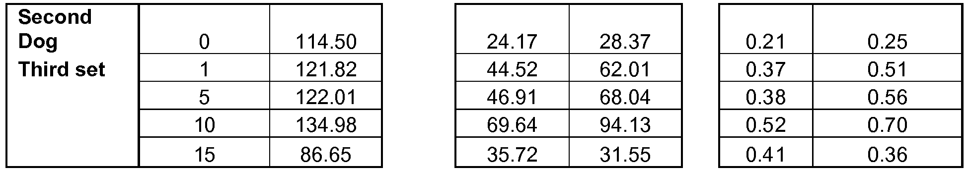
-
CCM Time (min) PLB-P Normalized to CSQ Ser-16 Thr-17 0 0.23 0.23 1 0.47 0.54 5 0.46 0.55 10 0.56 0.57 15 0.44 0.47 Two sets only (3rd set from each dog) - Without being limited to any particular hypothesis, it is hypothesized by the inventors that the applied electric field either has a direct effect on the proteins or has an effect on a cofactor or protein that enhance phosphorylation of proteins. The above "Voltage-dependent potentiation ..." paper suggests that an electric field can directly modify the natural phosphorylation rate of a protein.
- mRNA expression levels were measured for some genes in human subjects. Therapy with non-excitatory cardiac contractility modulation (CCM) electrical signals was delivered to LV muscle during the absolute refractory period improves LV function in patients with HF. The effects of 3 months CCM therapy on mRNA expression of cardiac fetal and SR genes in 5 patients with advanced HF were examined. In the experiment, right sided endomyocardial biopsies were obtained at baseline, prior to activating CCM therapy, and at 3 and 6 months thereafter. CCM therapy was delivered in random order of ON for 3 months and OFF for 3 months. mRNA expression measurement was performed in a blinded fashion as to the ON/OFF order of therapy. Expression of the fetal genes A-type (ANP) and B-type (BNP) natriuretic peptides and a-myosin heavy chain (MHC), and the SR genes SERCA-2a, phospholamban (PLB) and ryanodine receptors (RYR) was measured using RT-PCR and bands quantified in densitometric units (du). The percent change in du between baseline and the ON and
OFF 3 months phases was calculated. - The 3 months therapy OFF phase was associated with increased expression of ANP and BNP and decreased expression of α-MHC, SERCA-2a, PLB and RYR (Table). In contrast, the 3 months ON therapy phase resulted in decreased expression of ANP and BNP and increased expression of α-MHC, SERCA-2a, PLB and RYR (Table). This suggests that in patients with HF, CCM therapy reverses the cardiac maladaptive fetal gene program and normalizes expression of key SR Ca2+ cycling genes. These observations are consistent with the observed improvement in LV function in patients with HF following long-term CCM therapy.
mRNA Expression (% Change from Baseline) OFF Phase ON Phase P-Value ANP 82 ± 26 -57 ± 9 0.009 BNP 81 ± 28 -55 ± 9 0.007 α-MHC -29 ± 9 80 ± 16 0.004 SERCA-2a -21 ± 10 45 ± 14 0.039 PLB 4 ± 18 93 ± 45 0.084 RYR -20 ± 6 34 ± 6 0.002 -
Figs. 5A-5R shows protein expression results for the following proteins in chronic dogs, in control, heart failure and treated heart failure conditions: CSQ, SERCA-2a, PLB, RyR, NCX, IL-6, GATA-4, GAPDH, MMP-9, Tubulin-Beta, GATA-1, MMP-1, Tubulin-Alpha, Titin, TIMP-1, Integrin-α5, TNF α, p21ras, p38 MAPK, TIMP-2, β1-AR, MMP-2, ANP and BNP. - It should be noted that some blots are shown twice, in order to facilitate comparison between them.
- Following is a tabular analysis of these results with a short discussion.
-
Figs. 5A-5D show results for the following SR proteins: Calsequestrin, phospholamban, SERCA-2a (Calcium ATPase) and ryanodine receptors; the following Pump Proteins: Sodium-Calcium Exchanger; the following Transcription Factors: GATA-4; and the following Cytokines: Interleukin-6. - In general, these seven proteins moved directionally the same as their mRNA expression. Phospholamban showed complete normalization as did SERCA-2a.
- These results are consistent with the concept that the CCM acute and chronic effect is mediated by favorable modification of calcium cycling within the sarcoplasmic reticulum.
- Also notable is that under chronic condition, the CCM signals appear to normalize the over-expression of the sodium-calcium exchanger.
-
Dog Numbers - same for all Figs. 5A-5R HF-Control HF + CCM 02-097 02-106 02-098 02-107 02-103 02-108 02-130 02-012 03-045 03-023 04-004 03-050 04-018 04-005 Protein Expression of CSQ NL HF-Control HF + CCM 49 41 33 41 44 40 38 33 48 45 42 38 50 41 40 34 37 47 44 61 Mean 43 40 44 STD 6 4 9 SEM 3 1 3 ANOVA=0.49 p vs. NL p vs. HF-Control Protein Expression of Phospholamban NL HF-Control HF + CCM 64 30 75 63 23 60 74 27 97 63 29 65 75 18 80 52 30 76 29 69 Mean 65 27 75 STD 8 5 12 SEM 3 2 5 ANOVA=0.0001 p vs. NL <0.05 NS p vs. HF-Control <0.05 Protein Expression of SERCA-2a NL HF-Control HF + CCM 77 37 94 77 69 67 89 54 68 117 58 57 95 39 59 74 57 111 63 53 88 54 73 16 12 21 7 4 8 ANOVA=0.007 p vs. NL <0.05 NS p vs. HF-Control <0.05 Protein Expression of RvR NL HF-Control HF + CCM 129 75 115 123 98 107 153 102 119 104 86 86 140 100 78 104 86 91 72 147 126 88 106 20 12 23 8 5 9 ANOVA=0.009 p vs. NL <0.05 NS p vs. HF-Control NS -
Protein Expression of NCX NL HF-Control HF + CCM 26 62 30 43 50 39 29 38 35 36 45 29 41 49 50 44 76 25 58 51 37 54 37 8 12 10 3 5 4 ANOVA=0.013 p vs. NL <0.05 NS p vs. HF-Control <0.05 -
Protein Expression of IL-6 NL HF-Control HF + CCM 56 74 31 56 51 70 43 87 51 50 84 106 47 107 65 86 118 66 90 51 56 87 63 15 22 23 6 8 9 ANOVA=0.033 p vs. NL <0.05 NS p vs. HF-Control <0.05 -
Protein Expression of GATA-4 NL HF-Control HF + CCM 90 158 63 97 179 93 75 100 129 105 133 126 103 157 113 106 127 103 110 141 96 138 110 12 28 26 5 11 10 ANOVA=0.018 p vs. NL <0.05 NS p vs. HF-Control <0.05 -
Figs. 5E-5H show results for GAPDH (Housekeeping), transcription factor GATA-1 which did not change, matrix metalloproteinase-9 which changes consistent with mRNA expression and cytoskeletal protein Tubulin-beta which also changes consistent with what is shown for mRNA expression. GATA-1 is shown in comparison with GATA-4. -
Protein Expression of GAPDH NL HF-Control HF + CCM 32 34 34 36 33 36 35 37 32 35 32 28 34 31 35 31 27 46 27 30 34 32 34 2 4 6 1 1 2 ANOVA=0.54 p vs. NL NS NS p vs. HF-Control NS -
Protein Expression of MMP-9 NL HF-Control HF + CCM 394 637 668 448 779 569 452 821 611 455 733 551 488 687 504 426 643 572 742 486 444 720 566 32 69 62 13 26 23 ANOVA=0.0001 p vs. NL <0.05 <0.05 p vs. HF-Control <0.05 -
Tubulin-beta NL HF-Control HF + CCM 512 1008 656 644 810 693 584 973 647 450 725 543 390 693 654 301 778 675 1078 689 480 866 651 126 151 51 52 57 19 ANOVA=0.0001 p vs. NL <0.05 <0.05 p vs. HF-Control <0.05 -
Protein Expression of GATA-1 NL HF-Control HF + CCM 83 108 118 97 110 121 99 60 130 115 109 122 106 106 121 93 94 75 103 101 99 99 113 11 18 19 4 7 7 ANOVA=0.22 p vs. NL NS NS p vs. HF-Control NS -
Figs. 5I-5L show results from the proteins matrix-metalloproteinase-1 (MMP-1), cytoskeletal proteins tubulin alpha and titin, tissue inhibitor of matrix-metalloproteinase-1 (TIMP-1) and cell surface protein integrin-alpha-5. - There were no apparent changes in TIMP-1. CCM therapy also had no significant effect on integrin-alpha-5. It should be noted that integrin-alpha-5 can be affected by other means, such as mechanically constraining the heart (e.g., thus directly affecting its transduction function).
- CCM therapy, however, significantly down-regulated MMP-1, tubulin-alpha and titin which is consistent with the observation with respect to the effects of CCM on mRNA expression of these genes.
-
Protein Expression of MMP-1 NL HF-Control HF + CCM 521 936 482 449 894 574 425 883 511 484 1066 538 437 985 527 525 997 539 971 476 474 962 521 43 63 34 18 24 13 ANOVA=0.0001 p vs. NL <0.05 NS p vs. HF-Control <0.05 -
Tubulin-alpha NL HF-Control HF + CCM 196 298 217 161 322 233 129 359 187 136 283 214 142 307 239 158 260 257 274 250 154 300 228 24 33 24 10 13 9 ANOVA=0.0001 p vs. NL <0.05 <0.05 p vs. HF-Control <0.05 Protein Expression of Titin NL HF-Control HF + CCM 609 232 368 575 211 285 528 218 306 412 302 213 400 231 248 467 223 329 191 243 499 230 285 86 35 54 35 13 20 ANOVA=0.0001 p vs. NL <0.05 <0.05 p vs. HF-Control NS -
Protein Expression of TIMP-1 NL HF-Control HF + CCM 609 914 786 718 835 849 915 863 811 694 679 888 724 773 781 834 676 762 787 709 749 790 798 109 90 58 44 34 22 ANOVA=0.57 p vs. NL NS NS p vs. HF-Control NS -
Integrin-alpha -5 NL HF-Control HF + CCM 228 340 254 153 455 239 160 437 212 223 358 193 185 332 168 201 356 253 324 249 192 372 224 31 52 34 13 20 13 ANOVA=0.57 p vs. NL <0.05 NS p vs. HF-Control <0.05 -
Figs. 5M-5P show results from the proteins TNF-α (showed in comparison to IL-6), p21ras, p38 MAPK, TIMP-2 (showed in comparison to TIMP-1 and β1-AR. - The lack of change in TIMP-2 is consistent with previous observations. Long-term CCM therapy significantly reduced protein expression of the cytokine TNF- α and significantly reduced the expression of the stretch proteins p21ras as well as p38 MAPK. This is consistent with the observation that CCM therapy attenuates cardiomyocyte hypertrophy. Also to be noted is up-regulation of the beta-1 adrenergic receptor, which is favorable.
-
Protein Expression of IL-6 NL HF-Control HF + CCM 56 74 31 56 51 70 43 87 51 50 84 106 47 107 65 86 118 66 90 51 56 87 63 15 22 23 6 8 9 ANOVA=0.033 p vs. NL <0.05 NS p vs. HF-Control <0.05 Protein Expression of TNFα NL HF-Control HF + CCM 44 155 63 44 161 89 31 149 53 47 125 87 51 168 75 39 180 96 176 80 43 159 78 7 19 15 3 7 6 ANOVA=0.0001 p vs. NL <0.05 <0.05 p vs. HF-Control <0.05 -
Protein Expression of p21ras NL HF-Control HF + CCM 44 112 92 57 142 89 38 138 83 30 123 110 34 66 61 36 73 53 90 72 40 106 80 10 31 20 4 12 7 ANOVA=0.0001 p vs. NL <0.05 <0.05 p vs. HF-Control <0.05 Protein Expression of p38 MAPK NL HF-Control HF + CCM 21 46 32 15 52 21 13 41 35 17 67 36 14 43 24 19 38 19 33 15 17 46 26 3 11 8 1 4 3 ANOVA=0.0001 p vs. NL <0.05 NS p vs. HF-Control <0.05 -
Protein Expression of TIMP-2 NL HF-Control HF + CCM 84 62 85 73 55 80 61 43 68 93 89 72 87 88 56 88 84 79 81 86 81 72 75 12 18 11 5 7 4 ANOVA=0.53 p vs. NL NS NS p vs. HF-Control NS -
Protein Expression of β1-AR NL HF-Control HF + CCM 135 66 86 75 30 97 88 41 53 111 64 95 111 113 86 93 56 96 64 113 102 62 89 21 26 18 9 10 7 ANOVA=0.015 p vs. NL <0.05 NS p vs. HF-Control <0.05 -
Figs. 5Q and5R show results for MMP-2 (in comparison to MMP-1 and MMP-9) and ANP and BNP. -
Protein Expression of MMP-2 NL HF-Control HF + CCM 47 74 41 29 77 30 31 57 25 42 78 56 38 81 63 42 51 65 41 61 38 66 49 7 16 16 3 6 6 ANOVA=0.0001 p vs. NL <0.05 NS p vs. HF-Control <0.05 -
Protein Expression of ANP NL HF-Control HF + CCM 64 104 62 77 179 60 108 271 39 82 128 89 92 135 62 103 113 76 97 54 88 147 63 17 61 16 7 23 6 ANOVA=0.002 p vs. NL <0.05 NS p vs. HF-Control <0.05 Protein Expression of BNP NL HF-Control HF + CCM 31 84 78 62 94 62 107 111 69 65 87 69 57 94 84 66 77 77 89 73 65 91 73 25 11 7 10 4 3 ANOVA=0.002 p vs. NL <0.05 NS p vs. HF-Control <0.05 - Referring back to the 14 dogs tested for chronic effects, mRNA expression in LV free wall of the housekeeping gene GAPDH and CSQ, the fetal program genes consisting of β1-AR, αMHC, ANP, and BNP and the cardiac SR genes SERCA-2a, PLB, and RyR are shown in table 5 quantified in densitometric units. Expression of GAPDH and CSQ was unchanged among the 3 study groups namely, normal dogs, sham-operated HF dogs and HF CMM-treated dogs. mRNA expression of β1-AR, αMHC, SERCA-2a, PLB and RyR decreased and expression of ANP and BNP increased significantly in sham-operated HF dogs compared to normal. Three months of CCM therapy restored the expression of all genes to near normal levels. Protein expression in the LV free wall of CSQ, β1-AR, ANP, BNP SERCA-2a, PLB and RyR are also shown in Table 5 quantified in densitometric units. Protein levels of CSQ were unchanged among the 3 study groups. Protein levels of β1-AR, SERCA-2a, PLB and RyR decreased and that of ANP and BNP increased significantly in sham-operated HF dogs compared to normal dogs. Long-term CCM therapy restored the expression of all measured proteins except PLB to near normal levels (Table 5). Possibly, phospholamban was not restored because of the differential phosphorylation thereof. In an exemplary embodiment of the disclosure, this mechanism is used to selectively increase synthesis of some proteins over others.
- In an exemplary embodiment of the disclosure , this mechanism is used to test if a patient is getting better - by stopping a therapy which is directly causing phospohorylation and seeing if phospholamban levels normalize (or trend to) after a few days and/or if other protein levels trend towards disease state values.
- In an exemplary embodiment of the disclosure, relaxation time between signal application is an integral part of therapy. In one example, relaxation time is used to allow the cell to find a new balance between the expressed proteins that are not on corresponding levels, such a balance may include protein levels (or mRNA levels or phosphorylation or structural remodeling) degrading and/or protein levels improving. Such relation times may be on order of seconds, minutes hours or days, depending on which mechanism are to be allowed to take part (e.g., protein based, etc.).
- In an exemplary embodiment of the disclosure, when applying phosphorylation-modifying therapy as described herein, a process of weaning is applied. Possibly, if treatment is stopped suddenly, phospholamban levels will be too low to support suitable cardiac activity, possibly causing a downwards-spiral in patient health. In an exemplary embodiment of the disclosure, as protein levels normalize (for other proteins), therapy is reduced to allow phospholamban recovery. Optionally, the pauses are timed according to measured recovery in phospholamban levels.
- In an exemplary embodiment of the disclosure, electrical therapy is applied selectively to tissue measured as having reduced phospholamban levels and/or phosphorylation levels, so as not to potentially damage healthy tissue. In some cases, changes to "healthy" tissue is desirable. For example, increasing phosphorylation and thus possibly reducing phospholamban may be desirable if long term reduction in contractility is desired. In another example, phosphorylation may be increased in normal tissue in order to cause over (or under) expression of some proteins, such as gap junction proteins or mechanical proteins. It should be appreciated that a therapy target of diseased tissue need not be a mirror of a healthy tissue state.
Table 5 mRNA and Protein Expression of Fetal Program and Sarcoplasmic Reticulum Genes/Proteins in Left Ventricular Free Wall of Normal Dogs (NL) (n=6), Sham-Operated Untreated Heart Failure Dogs (Sham, n=7) and CCM-Treated Heart Failure Dogs (CCM, n=7) mRNA Expression (du) Protein Expression (du) NL Sham CCM NL Sham CCM B1-AR 22 ± 1 11 ± 1* 18 ± 1*† 102 ± 9 62 ± 10* 89 ± 7† A-MHC 212 ± 7 148 ± 10* 180 ± 5*† --- --- --- ANP 18 ± 2 34 ± 2* 27 ± 1*† 88 ± 7 147 ± 23* 63 ± 6† BNP 23 ± 3 177 ± 4* 44 ± 5*† 65 ± 10 91 ± 4* 73 ± 3† SERCA-2a 236 ± 4 154 ± 4* 192 ± 3*† 88 ± 7 54 ± 4* 73 ± 8† PLB 232 ± 8 149 ± 8* 212 ± 7† 446 ± 19 277 ± 12* 299 ± 9* RyR 38 ± 2 22 ± 2* 36 ± 5† 126 ± 8 88 ± 5* 106 ± 9† CSQ 264 ± 5 275 ± 5 259 ± 14 43 ± 3 40 ± 1 44 ± 3 GAPDH 219 ± 6 223 ± 8 236 ± 4 ---- ---- ---- du = Densitometric units; AR = Adrenergic receptor; MHC = Myosin Heavy Chain; ANP = A-type natriuretic peptide; BNP = B-type natriuretic peptide; SERCA-2a = Cardiac sarcoplasmic reticulum calcium ATPase; PLB = Phospholamban; RyR = Ryanodine receptor; CSQ = Calsequestrin; GAPDH = Glyseraldehyde-3-phosphate dehydrogenase. * = p<0.05 vs. NL; † = p<0.05 vs. Sham. - The above results showed analysis of tissue samples at the treated site.
Fig. 6 shows mRNA expression levels for Phospholamban, SERCA-2a and Ryanodine receptors, showing chronic improvement in septal tissue to which a field was applied chronically. - Referring to the 14 dogs chronic study described above, the restoration to near normal levels of the fetal program and most SR proteins after 3 months of CCM therapy was the same in LV tissue obtained from the inter-ventricular septum, the site nearest to the CCM signal delivery leads, and the LV free wall, a site remote from the CCM leads. A typical example illustrating the changes in protein levels of CSQ, SERCA-2a, PLB and RyR between the two LV sites is shown in
Fig. 10 . -
Fig. 8 shows phosphorylation levels in chronically treated dogs, at the application location in the septum. - In addition to examining protein expression of fetal program genes and SR proteins, protein levels of P-PLB at serine-16 and threonine-17 were also examined. Measurements were made in tissue obtained from both the inter-ventricular septum and the LV free wall. At both sites, protein levels of P-PLB at serine-16 and threonine-17 were significantly lower in sham-operated HF dogs compared to normal dogs and returned to near normal levels after 3 months of CCM therapy (
Fig. 11 , Table 6). In both the inter-ventricular septum and LV free wall, the ratio of P-PLB at serine-16 to total PLB and the ratio of P-PLB at threnonine-17 were also significantly lower in sham-operated HF dogs compared to normal dogs (Table 6). Long-term CCM therapy resulted in a significant increase of both ratios in both the interventricular Septum and the LV free wall (Table 6).Table 6 Protein Expression of Total Phospholamban and Phosphorylated Phospholamban at Serine-16 and Threonine-17 in the Inter-Ventricular Septum and Left Ventricle Free Wall of Normal Dogs (NL) (n=6), Sham-Operated Untreated Heart Failure Dogs (Sham, n=7) and CCM-Treated Heart Failure Dogs (CCM, n=7)) Inter-Ventricular Septum LV Free Wall NL Sham CCM NL Sham CCM Total PLB (du) 445±7 305±23* 299±16* 446±19 305±19* 299±9* P-PLB Ser-16 (du) 85±6 47±5* 79±6† 128±11 50±12* 87±11*† P-PLB Thr-17 (du) 146±6 62±10* 129±12† 137±4 56±7* 109±18† P-PLB Ser-16 / Total PLB 0.19±0.01 0.15±0.01* 0.27±0.02† 0.29±0.03 0.16±0.03* 0.29±0.04† P-PLB Thr-17 / Total PLB 0.33±0.01 0.21±0.04* 0.44±0.05† 0.31±0.02 0.19±0.03* 0.36±0.05† PLB = phospholamban; P-PLB = phosphorylated phospholamban; Ser-16 = serine-16; Thr-17 = theonine-17. * = p<0.05 vs. NL; † = p<0.05 vs. Sham. -
Fig. 7A and7B shows mRNA expression at sites remote from the application of the signal, but still within the left ventricle, at a relatively short time of four hours apparently no significant effect (mRNA, protein and/or phosphorylation) is shown. This may indicate that the effect of the CCM signal is first local, for example on a molecular level and then propagates to remote location, for example by biochemical signaling or by a mechanical signaling indicated by the change in contraction behavior of the treated tissue and/or of the chamber as a whole. The following non-limiting mechanism is suggested: the electric field causes phosphorylation of phospholamban. This in turn increases the activity/affinity of SRECA-2a for calcium and immediately improves SR calcium cycling. GATA-4 and the sodium calcium exchanger may play an additive role in the improved function. As LV function begins to improve and the LV gets smaller, many of the molecular/biochemical maladaptations begin to correct, which adds to the long-term benefits. Improved SR cardiac cycling may be a goal of some therapies in accordance with exemplary embodiments of the disclosure. - In open chest HF dogs, acute hemodynamic and local/remote effects were determined. Continuous delivery of CCM therapy over the course of 2 hours improved LV systolic function and associated hemodynamics. Compared to baseline before initiating CCM therapy, 2 hours of CCM therapy resulted in a significant increase of LV ejection fraction (31 ± 2 vs. 26 ± 1%, p<0.05), a reduction of MVO2 that did not reach statistical significance (180 ± 34 vs. 257 ± 41 µmols/min) and a trend toward an increase in LV mechanical efficiency that also did not reach statistical significance (33 ± 8 vs. 19 ± 4%).
- LV tissue obtained near the site of CCM lead implants, was compared to LV tissue obtained from a site remote from the CCM leads. LV tissue samples obtained from the same sites from dogs with HF that were untreated and normal dogs were used for comparisons. Compared to untreated HF dogs, the ratio of P-PLB to total PLB increased significantly in CCM-treated HF dogs compared to untreated HF dogs in the LV anterior wall at the site of signal delivery (
Fig. 12A ), whereas it was essentially unchanged in the LV posterior wall remote from the site of CCM signal delivery (Fig. 12B ). - In an exemplary embodiment of the disclosure, the location to which electrification will be applied is selected based on a model of what areas will cause a biochemical or mechanical cascade in the heart and improve its function. Optionally, the size of areas is optimized to reduce power needs, while retain an expected time frame of treatment.
- In an exemplary embodiment of the disclosure, an area to be treated is selected based on immediate response of tissue therein to electrical stimulation.
- One example of a mechanical cascade is a desired change in stretching of tissue which will, as that tissue improve, propagate. Another example of mechanical cascade is selecting sufficient and correct tissue in the ventricle such that immediate hemodynamic effects (e.g., improvement) are seen and sensed by the rest of the chamber.
- A possible mechanism of non-mechanical propagation (which may be utilized) is that healthy cells can help regulate ionic concentrations of neighboring cells via gap junctions between them. Alternatively or additionally, other communication may occur via the gap junctions.
- Another possible mechanism is that low level fields reach further and take longer to have an effect. This suggests that low-level fields in general may be used, with a rapidity of effect depending on the field strength and/or mechanical or other improvement of parts of the heart.
- In some embodiments of the disclosure, the efficacy of a treatment is measured by tracking remote effects alternatively or additionally, to tracking local effects. One or more of the following logics may be used: 1. There is more remote tissue to sample, with less danger of damage to heart. 2. The effect in remote tissue is more gradual and thus easier to track. 3. Acute effects may not occur (or be smaller) in remote tissue, thereby preventing the masking of longest term effects by acute effects. Optionally, for local or remote measurement, the measurement is made when no treatment is applied. An example of an acute effect which may mask a longer term effect is conduction velocity.
- A short tabular summary of the results of
Figs. 6-8 follows: -
Dog Numbers HF-Control HF + CCM 02-097 02-106 02-098 02-107 02-103 02-108 02-130 02-012 03-045 03-023 04-004 03-050 04-018 04-005 mRNA Expression for SERCA-2a in Septum NL HF-Control HF + CCM 238 172 207 224 123 214 203 167 178 199 168 191 226 151 179 162 123 150 149 162 Mean 209 150 183 STD 27 21 23 SEM 11 8 9 ANOVA=0.001 p vs. NL <0.05 NS p vs. HF-Control <0.05 mRNA Expression for PLB in Septum NL HF-Control HF + CCM 241 255 394 330 277 374 324 281 327 344 276 364 368 290 352 352 275 292 278 243 Mean 327 276 335 STD 45 11 52 SEM 18 4 20 ANOVA=0.029 p vs. NL <0.05 NS p vs. HF-Control <0.05 mRNA Expression for RYR in Septum NL HF-Control HF + CCM 272 287 267 371 225 277 289 229 300 294 262 319 321 232 323 294 234 252 238 248 307 244 284 35 23 31 14 9 12 ANOVA=0.005 p vs. NL <0.05 NS p vs. HF-Control <0.05 -
mRNA Expression for ANP NL HF-Control HF + CCM 155 301 342 164 336 316 175 356 307 170 325 303 229 318 291 212 350 282 Mean 184 331 307 STD 29 20 21 SEM 12 8 9 ANOVA=0.0001 p vs. NL <0.05 <0.05 p vs. HF-Control NS mRNA Expression for SERCA-2a NL HF-Control HF + CCM 257 68 126 266 50 57 334 56 140 293 84 71 285 53 36 263 47 38 Mean 283 60 78 STD 29 14 45 SEM 12 6 18 ANOVA=0.0001 p vs. NL <0.05 <0.05 p vs. HF-Control NS mRNA Expression for VEGF NL HF-Control HF + CCM 14 4 8 16 5 6 13 5 7 19 14 11 24 8 13 22 9 4 Mean 18 8 8 STD 4 4 3 SEM 2 2 1 ANOVA=0.001 p vs. NL <0.05 <0.05 p vs. HF-Control NS mRNA Expression for GATA-4 NL HF-Control HF + CCM 121 352 341 129 367 349 145 373 333 126 350 325 133 377 300 136 394 262 Mean 132 369 318 STD 8 16 32 SEM 3 7 13 ANOVA=0.0001 p vs. NL <0.05 <0.05 p vs. HF-Control <0.05 mRNA Expression for BNP NL HF-Control HF + CCM 320 1721 1862 299 1629 2012 331 1690 1952 294 1725 2111 361 1662 1991 349 2016 1641 326 1741 1928 27 140 162 11 57 66 ANOVA=0.0001 p vs. NL <0.05 <0.05 p vs. HF-Control <0.05 mRNA Expression for Phospholamban NL HF-Control HF + CCM 18 9 14 17 9 11 17 7 11 19 11 9 20 7 6 21 11 4 19 9 9 2 2 4 1 1 1 ANOVA=0.0001 p vs. NL <0.05 <0.05 p vs. HF-Control NS mRNA Expression for bFGF NL HF-Control HF + CCM 186 90 110 195 76 100 194 108 87 215 77 88 189 104 83 200 100 70 197 93 90 10 14 14 4 6 6 ANOVA=0.0001 p vs. NL <0.05 <0.05 p vs. HF-Control NS mRNA Expression for α-MHC NL HF-Control HF + CCM 457 242 222 609 228 218 448 176 208 545 188 221 642 313 181 557 220 156 543 228 201 78 49 27 32 20 11 ANOVA=0.0001 p vs. NL <0.05 <0.05 p vs. HF-Control NS -
Dog Numbers HF-Control HF + CCM 02-097 02-106 02-098 02-107 02-103 02-108 02-130 02-012 03-045 03-023 04-004 03-050 04-018 04-005 Phosphorylated PLB at Serine-16 NL HF-Control HF + CCM 143 86 99 109 95 106 153 63 68 161 25 97 102 15 105 100 24 31 43 106 Mean 128 50 87 STD 28 32 28 SEM 11 12 11 ANOVA=0.0001 p vs. NL <0.05 <0.05 p vs. HF-Control <0.05 Phosphorylated PLB at Threonine-17 NL HF-Control HF + CCM 135 90 80 126 41 68 137 53 62 148 52 162 146 32 168 129 59 78 67 147 137 56 109 9 19 47 4 7 18 ANOVA=0.0001 p vs. NL <0.05 NS p vs. HF-Control <0.05 - Experimental results of the effect of CCM signals on other proteins is described below.
- Overexpression of the calcium-binding protein S100A1 in failing rat cardiomyocytes normalizes sarcoplasmic reticulum (SR) calcium cycling by increasing calcium-uptake and reducing SR calcium-leak from ryanodine channels. Expression of S100A1 is significantly decreased in left ventricular (LV) myocardium of explanted failed human hearts.
- RNA was extracted and homogenate prepared from LV tissue obtained from 6 CCM-treated HF dogs, 6 untreated HF dogs and 6 normal (NL) dogs (these are the same dogs as above). S100A1 mRNA expression was measured using reverse transcriptase polymerase chain reaction (RT-PCR) and protein expression was measured using Western blotting. Bands obtained after gel electrophoresis were quantified in densitometric units (du).
- S100A1 mRNA and protein expression decreased significantly in untreated HF dogs compared to NL dogs. Chronic CCM therapy significantly increased mRNA and protein expression of S100A1. Restoration of expression of this calcium binding protein improves calcium cycling within the SR and may account, at least in part, for the observed improvement of LV function seen following chronic CCM therapy.
mRNA and Protein Expression of S100A1 NL HF-Untreated HF + CCM S100A1 mRNA (du) 13.0 ± 0.7 1.5 ± 0.1* 8.6 ± 0.5*† S100A1 Protein (du) 215 ± 15 63 ± 6* 190 ± 8† *=p<0.05 vs. NL; †=p<0.05 vs. HF-Untreated - RNA was extracted and homogenate prepared from LV tissue obtained from 6 CCM-treated HF dogs, 6 untreated HF dogs and 6 normal (NL) dogs. These are the same dogs as in the previous section. Sorcin, Presenilin and Calstabin mRNA expression was measured using reverse transcriptase polymerase chain reaction (RT-PCR) and protein expression was measured using Western blotting. Bands obtained after gel electrophoresis were quantified in densitometric units (du).
- Sorcin mRNA and protein expression decreased significantly in untreated HF dogs compared to NL dogs. Chronic CCM therapy significantly increased mRNA and protein expression of Sorcin. Presenilin-2 increased in HF and decreased with CCM. Preseilin-1 was measured as an internal control and it did not change. This suggests that Presenilin-2 can be used as a target (e.g., treatment goal) for treating HF and as an indicator for diagnosis and modification of treatment.
- Restoration of expression of Sorcin may prevent or limit the RyR2 calcium leak and in doing so improve calcium cycling within the SR. Correction of this maladaptation by CCM therapy may account, at least in part, for the observed improvement of LV function. In particular, the combined correction of Sorcin and Presenilin, which interact to regulate cardiac ryanodine calcium release channels may act as a mediator of recovery of calcium overload in cardiomyocytes due to "RyR-2 calcium leak" in heart failure.
- It should be noted that Calstabin-2 decreased in heart failure but remained depressed even after 3 months of CCM therapy. This suggests that CCM does not act by merely resetting cellular function, however, as noted above, resetting may be part of the process. One possible explanation is that Castabin-2 does not rebound as it is tied to phospholamban levels. Another possible explanation is that Castabin-2 indicates a path for the progression of heart failure. Until the underlying cause is removed, it may remain depressed. Possibly, an improvement over significantly longer periods of time is to be expected. In an exemplary embodiment of the disclosure, this is used to estimate the ability of a patient to stop therapy. For example, if Castabin-2 levels trend to or do normalize, this can indicate that the tissue state is not diseased or becoming healthier. Optionally, this allows tracking of tissue improvement even during ongoing electrical therapy. In an exemplary embodiment of the disclosure, signal optimization techniques as described herein are used to find and then apply a signal specific (or more specific) to modifying Castabin-2.
- The results are shown in
Figs. 13-15 and in the tables below (in densitometric units). -
Figs. 13A and 13B present mRNA (Fig. 13A ) and protein blots (Fig. 13B ) illustrating expression of Sorcin in LV tissue of HF Dogs treated with CCM for 3 months according to an exemplary embodiment of the disclosure. -
Figs. 14A, 14B, 14C and 14D present mRNA (Fig. 14A and 14C ) and protein blots (Fig. 14B and 14D ) of the Presenilin-1 (Fig. 14A and 14B ) and Presenilin 2 (Fig. 14C and 14D ) in LV tissue of HF Dogs treated with CCM for 3 months according to an exemplary embodiment of the disclosure. -
Figs. 15A and 15B present mRNA (Fig. 15A ) and protein blots (Fig. 15B ) illustrating expression of Calstabin in LV tissue of HF Dogs treated with CCM for 3 months according to an exemplary embodiment of the disclosure.Dog Numbers HF-Control HF + CCM 02-097 02-106 02-098 02-107 02-103 02-108 02-130 02-012 03-045 03-023 04-018 03-050 mRNA Expression of Sorcin Normal HF-Control HF + CCM 0.52 0.19 0.37 0.41 0.21 0.37 0.55 0.25 0.30 0.53 0.19 0.29 0.53 0.18 0.39 0.59 0.15 0.37 Mean 0.52 0.20 0.35 STD 0.06 0.04 0.04 SEM 0.02 0.02 0.02 ANOVA=0.0001 p vs. Normal <0.05 <0.05 p vs. HF-Control <0.05 Protein Expression of Sorcin Normal HF-Control HF + CCM 0.75 0.30 0.396 0.65 0.26 0.456 0.69 0.28 0.426 0.75 0.30 0.412 0.73 0.25 0.426 0.68 0.23 0.486 Mean 0.71 0.27 0.43 STD 0.04 0.03 0.03 SEM 0.02 0.01 0.01 ANOVA=0.0001 p vs. Normal <0.05 <0.05 p vs. HF-Control <0.05 mRNA Expression of Presenilin-1 Normal HF-Control HF + CCM 0.34 0.4 0.38 0.36 0.44 0.4 0.38 0.39 0.45 0.39 0.43 0.39 0.29 0.38 0.38 0.32 0.33 0.35 Mean 0.35 0.40 0.39 STD 0.04 0.04 0.03 SEM 0.02 0.02 0.01 ANOVA=0.131 p vs. Normal NS NS p vs. HF-Control NS Protein Expression of Presenilin-1 Normal HF-Control HF + CCM 0.12 0.15 0.14 0.11 0.19 0.13 0.09 0.21 0.14 0.15 0.11 0.16 0.16 0.09 0.18 0.11 0.13 0.11 Mean 0.12 0.15 0.14 STD 0.03 0.05 0.02 SEM 0.01 0.02 0.01 ANOVA=0.338 p vs. Normal NS NS p vs. HF-Control NS mRNA Expression of Presenilin-2 Normal HF-Control HF + CCM 0.28 0.63 0.34 0.32 0.61 0.31 0.29 0.59 0.29 0.35 0.49 0.42 0.2 0.56 0.35 0.31 0.62 0.38 Mean 0.29 0.58 0.35 STD 0.05 0.05 0.05 SEM 0.02 0.02 0.02 ANOVA=0.0001 p vs. Normal <0.05 NS p vs. HF-Control <0.05 Protein Expression of Presenilin-2 Normal HF-Control HF + CCM 0.201 0.52 0.25 0.159 0.48 0.29 0.198 0.55 0.21 0.285 0.62 0.19 0.248 0.49 0.26 0.196 0.61 0.11 Mean 0.21 0.55 0.22 STD 0.04 0.06 0.06 SEM 0.02 0.02 0.03 ANOVA=0.0001 p vs. Normal <0.05 NS p vs. HF-Control <0.05 mRNA Expression of Calstabin-2 Normal HF-Control HF + CCM 1667 847 1477 1488 1204 1355 1486 1389 906 1723 887 619 1778 1120 1497 1840 1343 1224 Mean 1,664 1,132 1,180 STD 148 227 350 SEM 61 93 143 ANOVA=0.004 p vs. Normal <0.05 <0.05 p vs. HF-Control NS Protein Expression of Calstabin-2 Normal HF-Control HF + CCM 5871 3991 3679 5602 3643 3923 5029 3156 3560 5579 4645 4083 6726 4567 4600 5190 4995 4381 Mean 5,666 4,166 4,038 STD 602 693 402 SEM 246 283 164 ANOVA=0.0001 p vs. Normal <0.05 <0.05 p vs. HF-Control NS -
Figs. 9A and9B are graphs showing the relationship between rise time and decay time of a contractility increase in dogs. Similar results were observed in humans, albeit with generally slower rise times and slower decay times. -
Fig. 9A , in which the scale is in seconds, shows a rise time of several tens of seconds at about time 270. Once the signal is stopped (upper graph shows the signal application times), the contractility increase decays. The blip at 600 is probably caused by an arrhythmia. The decay continues from 600 until 950. A second signal application is at about 950 with a change in signal application location at about time 1350, before the signal effect washed out. - In
Fig. 9B the signal is shown as an outline of a square pulse, with the signal stopped each time an arrhythmia is detected, shown as a short pause in the signal. - Again, a rise time of several tens of seconds is found. A decay time of 230 seconds is shown for the first signal. A longer decay time of over 400 seconds is shown for a second signal. The second signal caused an increase in contractility at about 1050 seconds, due to change in the applied signals.
- In an exemplary embodiment of the disclosure, it is noted that the acute and long term effects last at least a few seconds after treatment is stopped and have different wash-out times. Thus, measuring patient parameters is possible while the treatment apparatus is off. Different waiting times may be used to selectively measure acute and longer term effects. For example, 30 minutes may be sufficient for avoiding acute effects on phosphorylation.
- In an exemplary system, a controller learns the particular washout behavior of a patient and adjusts the treatment to be re-applied only after significant washout and/or be applied at a minimal length that has a washout. Optionally, the delay between application and/or length of applications are selected to optimize one or more of power consumption, danger of arrhythmia and/or discomfort to patient.
-
WO 2005/087310 , filed asPCT/IL2005/00316 on March 18, 2005 US provisional application 60/719,517, filed September 22, 2005US provisional application 60/654,05660/719,517 - It is a particular feature of some embodiments of the disclosure that a non-immediate effect of signal application, which lasts after the signal application is stopped, is an affirmative effect, in which the tissue exhibits a positive change in action, rather than a negative effect, for example, of preventing arrhythmia. In an exemplary embodiment of the disclosure, the effect is on a cellular level, rather than or in addition to an effect on an organ level, for example a cellular level as measured by protein activity and/or expression levels.
- Another type of long term/wash-out effect is a long term therapeutic effect lasting days or weeks after treatment. Below are described two sets of experiments on humans. In the first set, patients were selectively treated 3 months on and 3 months off or vice versa. In the second set of experiments patients were either treated for six months or not. While the patients that were 3 months on and then 3 off fared less well (after 6 months) than those that were 3 off and then 3 on, the overall situation after 6 months was better than what would be expected from a baseline (no treatment) situation. The second set of experiments while suggesting no overall improvement after 6 months, shows a trend to lesser degradation and also suggests what the expected degradation would be. Optionally, the therapies as described herein are used to slow down degradation of a patient's situation, rather than a treatment. Optionally, each patient is selectively tested to see how long the treatment effects last and/or what minimal treatment period is required for longer term effects. Optionally, the length of treatment and/or "off" periods between treatments also vary as a function of time. Various physiological measures, for example as described above may be used to assess improvement, as well as mRNA, protein and/or phosphorylation levels.
- In the first set of experiments 126 subjects with ischemic (60%) or idiopathic (40%) cardiomyopathy, EF<35%, NYHA Class II (23%) or III (73%) received a CCM pulse generator. Patients were randomly assigned to Group 1 (n=62,
CCM treatment 3 months, sham treatment second 3 months) or Group 2 (n=64,sham treatment 3 months, CCM treatment second 3 months). The coprimary endpoints were differences between groups of changes in peak oxygen consumption (VO2.peak) and Minnesota Living with Heart Failure Questionnaire (MLWHFQ). - Baseline EF (25±6% vs 26±6%), VO2,peak (14.4±2.5 vs 14.2±2.7 ml/kg/min) and MLWHFQ (41±20 vs 35±19) were similar in both groups. VO2,peak increased similarly in both groups during the first 3 months (0.64±0.36 vs 0.53±0.46 ml O2/kg/min, p=NS, possibly a placebo effect). However, during the next 3 months, VO2,peak decreased in the group switched to sham treatment (-0.92±0.42 ml/kg/min) and continued increasing in patients switched to active treatment (0.35±0.35 ml/kg/min, p=0.02), so that the final difference between groups was 1.2 ml/kg/min. It is hypothesized that this reduction is less than what would be expected after 3 months of an untreated patient. MLWHFQ also improved in both groups during the
fist 3 months (placebo effect), though the improvement trended better with treatment (-14.65±2.04 vs -9.8±2.2, p=NS). During the second 3 months, MLWHFQ increased in the group switched to sham (5.1±2.3) and continued to decrease in patients switched to active treatment (-1±2, p=0.03). However, the increase in the untreated group appeared to be less than a return to baseline values or other values that would be expected with a diseased patient with natural progression. - It should be noted that significant improvement is achieved even for patients that had an ON phase followed by an OFF phase, or at least the effects of treatment remained for a while. This suggests that the therapeutic effects are long lasting, for example, more than a week, more than a month, more than three months or longer. This is in contrast to cardiac resynchronization therapy where improvements seem to last a week or two at most. Even in this case, the heart desynchronization appears almost immediately (as is the case with electrical therapy in general which usually last a single beat).
- In a second set of experiments, 49 subjects with EF<35%, normal QRS duration (105±15 ms) and NYHA Class III (n=48) or IV (n=1) despite medical therapy received a CCM pulse generator. Devices were programmed randomly to deliver CCM signals (Treatment, n=25) or to remain off (Control, n=24) for 6 months. Evaluations included NYHA, 6 minute hall walk (6MW), cardiopulmonary stress test, Minnesota Living with Heart Failure Questionnaire (MLWHFQ) and Halter.
- Although most baseline features were balanced between groups, EF (31.4±7.4 vs 24.9±6.5%, p=0.003), end-diastolic dimension (52.1±21.4 vs 62.5±6.2 mm, p=0.01), peak VO2 (16.0±2.9 vs 14.3±2.8 ml O2/kg/min, p=0.02) and anaerobic threshold (12.3±2.5 vs 10.6±2.4 ml O2/kg/min, p=0.01) were worse in Treatment than Control. Nevertheless, there was 1 death in a Control subject and more Treatment patients were free of hospitalization for any cause at 6 months (84% vs 62%). No changes in ectopy were observed. Compared to baseline, 6MW (13.4 m), peak VO2 (0.2 ml O2/kg/min) and anaerobic threshold (0.8 ml O2/kg/min) increased more in Treatment than Controls. None of the differences were statistically significant (small sample size).
- Six minute hall walk showed similar improvements in both groups at 12 weeks (~ 20 meters), with the curves diverging at 6 months with an approximately 15 meter greater increase in the treatment group. Peak VO2 decreased over time in both groups (∼-0.75 at 12W, ∼-1 at 24 weeks), but remained higher in the treatment group than in controls, by ∼0.2 ml O2/kg/min. In contrast, anaerobic threshold, which decreased in the control group (∼-0.8 at 12W, 24W), decreased initially (∼-0.6 at 12W) but then returned to baseline values at 6 months in the Treatment group. At the final follow-up, the difference between the two groups averaged 0.82 ml O2/kg/min. Ejection fraction increased minimally in both groups at 6 months (1.8±0.8 in the treatment group vs 1.3±1.6 in the control group).
- New York Heart Association classification improved similarly in both groups. For the treatment group, the proportion of patients in Class I, II and III at 24 weeks were 19, 45 and 36, respectively. This compared to 18, 52 and 30, respectively, in the Control group. MLWHFQ also improved significantly and similarly in both groups. At the 6 month follow-up, MLWHFQ decreased from baseline values by 16.2±5.9 and 18.3±4.8 in the control and treatment groups, respectively. The significant and sustained improvements in NYHA and MLWHFQ observed in both groups speaks to the presence of a significant placebo effect.
- While it might be supposed that beta blockers and electrical therapy as described here utilize similar pathways and therefore cannot be additive, the following experiment shows that this is not correct. In addition, electrical therapy may be used to increase contractility and compensate for an initial reduction in contractility caused by beta blockers. This may suggest changing the sequence amount and/or type when the medication starts having a positive effect, for example, several weeks or several months (e.g., 3) later. Alternatively or additionally, electrical therapy is used to compensate for times when medication is stopped, for example, due to adverse systemic effects. In an exemplary embodiment of the disclosure, the dosage of medication and the dosage of therapy are selected to minimize or reduce systemic effects and/or reduce power needs and/or side effects (if any) of electrical therapy.
- In a set of experiments, dogs with intracoronary microembolization-induced chronic HF (
LV EF 30%-40%) were randomized to 3 months therapy with either BB (beta-blockers) alone (extended release metoprolol succinate, 100 mg once daily), extended release metoprolol succinate (100 mg, once daily) combined with CCM therapy, or to no therapy at all (Control). LV end-diastolic volume (EDV), end-systolic volume (ESV) and EF were measured before randomization (PRE) and after 3 months of therapy (POST). To determine "treatment effect", the change Δ between PRE and POST therapy for each group was compared among the 3 study groups. - In Control dogs, LV EDV and ESV increased significantly and EF decreased significantly. Therapy with BB alone and combined therapy with BB and CCM both prevented the increase in EDV, significantly reduced ESV and significantly increased EF. Treatment effect (Δ) comparisons showed that combination therapy with BB and CCM significantly increased LV EF above that seen with BB alone (12 ± 1% vs. 6 ± 1%, p<0.001).
Control BB Alone BB + CCM PRE POST PRE POST PRE POST LV EDV (ml) 60 ± 1 64 ± 1 64 ± 4 62 ± 5 70 ± 4 69 ± 4 LV ESV (ml) 39 ± 1 45 ± 1* 41 ± 2 36 ± 4* 45 ± 3 36 ± 3* LV EF (%) 36 ± 1 31 ± 1* 36 ± 1 42 ± 2* 36 ± 1 48 ± 1* *=p<0.05 PRE vs. POST - Following are detailed results:
-


EF (%) Dog# Base Pre Post Delta 04-112 52 36 47 11 04-120 51 36 48 12 04-124 57 35 46 11 04-145 58 38 51 13 05-025 54 36 44 8 05-033 55 38 49 11 05-034 46 36 48 12 Mean 53 36 48 11 STD 4 1 2 2 SEM 1.5 0.4 0.8 0.6 -
EDV (ml) Dog# Base Pre Post 03-057 54 62 59 03-066 58 74 76 04-147 51 57 55 05-006 55 64 59 05-028 54 60 60 05-042 54 59 59 Mean 54 63 61 STD 2 6 7 SEM 0.9 2.5 3.0 ESV (ml) Dog# Base Pre Post 03-057 27 38 33 03-066 26 48 47 04-147 19 38 33 05-006 30 39 32 05-028 30 37 35 05-042 25 37 31 Mean 26 40 35 STD 4 4 6 SEM 1.7 1.7 2.4 EF (%) Dog# Base Pre Post Delta 03-057 50 38 44 6 03-066 55 35 38 3 04-147 62 33 40 7 05-006 46 39 46 7 05-028 44 38 42 4 05-042 54 37 47 10 Mean 52 37 43 6 STD 7 2 3 2 SEM 2.7 0.9 1.4 1.0 -
EDV (ml) Dog# Base Pre Post 03-031 51 59 63 03-065 54 61 68 04-103 52 58 61 04-122 48 63 64 05-002 54 61 64 05-027 59 60 64 Mean 53 60 64 STD 4 2 2 SEM 1.5 0.7 0.9 ESV (ml) Dog# Base Pre Post 03-031 24 39 46 03-065 23 39 48 04-103 21 37 43 04-122 23 39 43 05-002 28 39 43 05-027 28 39 44 Mean 25 39 45 STD 3 1 2 SEM 1.2 0.3 0.8 EF (%) Dog# Base Pre Post Delta 03-031 51 34 27 -7 03-065 48 36 29 -7 04-103 60 36 30 -6 04-122 52 38 33 -5 05-002 48 36 33 -3 05-027 53 35 31 -4 Mean 52 36 31 -5 STD 4 1 2 2 SEM 1.8 0.5 1.0 0.7
ESV = LV end-systolic volume
EF = LV ejection fraction - In an exemplary embodiment of the disclosure, the above experimental results are used as a basis for diagnosis. Optionally, the diagnosis indicates one or both of a disease type and disease severity. In an exemplary embodiment of the disclosure, the type can be seen from the behavior of the proteins, for example comparing protein levels to calcium overload to diagnose systolic or diastolic dysfunction. In another example, the relative phosphorylation levels are used as a measure of severity.
- In an exemplary embodiment of the disclosure, the use of multiple proteins and mRNA expression values provides a robust indicator of disease. In some embodiments, only one or two of proteins, phosphorylation and mRNA are used. Optionally, cardiac function values, such as stroke volume are used as well. Optionally, the response of tissue to a field is used as well, for example phosphorylation response.
- In an exemplary embodiment of the disclosure, at least 3, at least 5, at least 10, at least 20 or more proteins levels, phosphorylation levels and/or mRNA levels are used to aid in diagnosis. Optionally, a table is maintained which includes ranges of values that match various disease types and/or conditions. Optionally, as therapy progresses, the patient is re-diagnosed. Optionally, the diagnosis is according to groups of mRNA and/or proteins that have associated functions.
- In an exemplary embodiment of the disclosure, a DNA chip and/or protein chip and/or a biochip are used to measure the above levels.
- In an exemplary embodiment of the disclosure, treatments are selected by applying the above pulses to tissue and measuring the effect. Optionally, a tissue sample is removed, for example by biopsy and a range of sequences are tested on the sample. Optionally, the results of the testing indicate which sequence might have a desired therapeutic effect. Such testing can include analysis and/or application of a treatment, such as an electric field. As noted above, at least some of the tests can be meaningfully applied to tissue homogenate and other unstructured tissue configurations.
- In an exemplary embodiment of the disclosure, different parts of the tissue are tested, for example, to see which tissue part will respond better and/or avoid side effects.
- In an exemplary embodiment of the disclosure, a kit is provided for performing such analyses as described herein. In an exemplary embodiment of the disclosure, the kit comprises a set of reagents, antibodies and/or other biochemical tools as known in the art. Alternatively or additionally, the kit comprises one or more mRNA or protein detecting chips. Alternatively or additionally, the kit comprises software for analyzing gel migration.
- In an exemplary embodiment of the disclosure, the kit comprises a source of treatment, for example electrodes and optionally a power source, for electrifying a sample for testing its responsiveness. Optionally, the kit includes a sampling means for selecting part of the sample at a pre-described time, to check temporal response of the sample. An existing kit may be modified for use, for example, a kit available from Biosite, Inc. to measure blood levels of BNP. Such a kit could include instructions for use with the methods described herein and optionally include sampling means or a timer to ensure correct timing of activities.
- The kit includes or is used with a bioreactor that includes a controllable sampling element which cans electively extract a portion of the sample in the bioreactor and test it in a separate chamber. Optionally, this is embodied using lab-on-chip technology and/or fluidic control of material flow. In an exemplary embodiment of the disclosure, the controllable sampling element comprises a robot and a pipette that takes part of the sample and inserts it into an assaying chamber. Various automated assaying devices are known in the art of drug discovery and may be used and/or modified to be smaller and/or simpler.
- In an exemplary embodiment of the disclosure, the kit includes a database or a link to a database (e.g., on the internet) that links biochemical markers to tissue states, treatment states and/or disease states.
- Optionally, the kits are sterile and/or frozen and optionally include instructions for use as described herein. Optionally, one or more kits are packaged with a controller designed for implantation, for use in determining suitable electrode placement therefore.
- In an exemplary embodiment of the disclosure, the kit includes one or more antibody reagents useful for detecting phosphorylated and/or dephosphorylated forms of the proteins desired.
- Optionally, tracers which are anti-body based are used in-vivo, for example provided using a catheter or using the delivery electrodes or a separate needle or other injection mechanism. Optionally, the tracer is radioactive or fluorescent.
- A calibration step, for example, per patient, may be carried out. Alternatively or additionally, a comparison before and after field application is used to determine change in phosphorylation.
- Optionally, phosphorylation of proteins which affects ECG signals or correlated proteins which affect ECG signals is detected by detecting changes in ECG signals.
- In an exemplary embodiment of the disclosure, changes in conduction velocity caused by increased secretion of protein are detected in an ECG measurement. Optionally, the measurement is remote from a signal application area.
- Alternatively or additionally to conduction velocity, changes in contraction velocity under various conditions are assed from images of the heart or using other means.
- Optionally, other physiological measures are correlated with protein effects (e.g., in a per-patient or per-population calibration process) and used to estimate effects on protein synthesis from physiological measures.
- In an exemplary embodiment of the disclosure, the above sequences are used to treat tissue plugs that are removed from the body, treated and reinserted. The reinsertion may be, for example, at the location of removal, at scarred locations, at locations bordering scars or at otherwise weakened location of the heart.
- In an exemplary embodiment of the disclosure, the above sequences are used as part of a program to re-invigorate scar or fibrotic tissue. Optionally, the effect of the sequence is to cause at least some cells to become functioning, for example in the border of the scar.
- In an exemplary embodiment of the disclosure, the above sequence is used to selectively increase oxygen efficiency in some parts of the heart, for example, parts with a vascular blockage thereto.
- In an exemplary embodiment of the disclosure, the above sequences are applied to tissue transplants, for example, whole heart transplants or plug transplants (from a donor), prior to and/or after transplant.
- In an exemplary embodiment of the disclosure, the above sequences are applied after DNA or stem cell therapy to the heart, for example, to enhance effects and/or to assist in cellular functional differentiation and/or regeneration.
- In an exemplary embodiment of the disclosure, the above sequences are used to have a desired modeling effect on the heart, for example, modifying an elasticity and/or contractility profile of a heart portion and/or directly controlling conduction velocity.
- Three types of remodeling may be provided, possible to different degrees at different parts of the heart. A first type of remodeling is on the biochemical level and includes, for example, normalization of protein synthesis. A second type of remodeling is mechanical remodeling, for example, fibrosis reduction, chamber size change, vascular increase and/or wall thickening. A third type of remodeling is function remodeling which may result form a combination of biochemical and structural remodeling. This includes, for example, cardiac electrical and/or mechanical activation profile (what gets activated, to what degree and in what order), contractility enhancement and conduction velocity. As described herein, these types of remodeling may be achieved using methods described herein. Differential remodeling of different types may be achieved, for example, by selectively applying signals with a strong acute effect (which affect mechanical remodeling) and those with a long term effect. For example, tissue which is both paced and treated with non-excitatory signals may thicken, as opposed to tissue which is only treated.
- In an exemplary embodiment of the disclosure, multiple methods of improving contractility are applied at a same time, or methods of improving contractility applied at a same time as methods that reduce contractility such as the initial effect of beta-blockers.
- In an exemplary embodiment of the disclosure, contractility enhancement by effects on membrane proteins is carried out at least partly independently from protein effects on SR proteins. Optionally, the selectivity is using methods as described above.
- In an exemplary embodiment of the disclosure, such enhancement is provided without adversely affecting short term activity, as pacing or inhibition might. In an exemplary embodiment of the disclosure, the enhancement is local and has fewer systemic effects (as would be expected with ACE-inhibitors or beta-blockers, for example). In some embodiments, short term activity is improved.
- It should be noted that these proteins are also known in other body organs, such as smooth muscle cells and slow-twitch skeletal muscle cells. Thus, an electric field as described herein can be used, for example, to modify PLB phosphorylation in blood vessel cells and/or the GI tract. Optionally, elasticity compliance is restored and vasomotor tone is restored and/or responsiveness are restored to blood vessels and/or GI tract portions using the electric field as described herein. In an exemplary embodiment of the disclosure, a hardened aorta is made more supple (and thus relieve cardiac problems) by suitable treatment. Vascular resistance in general, may be modified. Optionally, signals as described herein are used to help increase muscle strength and/or normalize protein expression therein.
- It should be noted that in smooth muscle cells the depolarization cycle is much longer and there is no danger of fatal arrhythmia, so more varied pulses may be attempted without significant danger and may provide longer term effects.
- In an exemplary embodiment of the disclosure, a method of optimizing treatments to the tissue is provided, taking into account effects on protein levels. For example, a CCM signal or a pacing signal, exercise or a pharmaceutical treatment may each (or in combination) have an effect on protein expression and/or behavior. In an exemplary embodiment of the disclosure, such a treatment is optimized so that it has a greater (desired) effect on proteins. In one example, a CCM signal is optimized by applying variations of the signals (e.g., different amplitude, frequencies pulse forms, etc.) to a set of tissue homogenate sets and selecting the signal(s) for which a better effect is achieved. It is noted that while this may be carried out in vivo, the ability to try out signals with various pulse parameters on tissue homogenate without the need for safety testing and worry about danger of damage to a patient/animal, can allow a much faster and/or cheaper search to be made. Searching methods as known in the art may be used. It is noted that such searching can also be carried out for small molecule drugs which have a direct effect on phosphorylation, for example.
- It is noted that immediate protein levels may be results that are faster to achieve or have less noise, than measuring actual improvement in a patient. Thus, protein measurement may allow faster within-patient optimization and/or allow optimization based on the response to a small number of beats (e.g., 100, 50, 10, 3 or intermediate or fewer numbers), rather than waiting for a longer term effect which may damage the heart if it is negative.
- In an exemplary embodiment of the disclosure, an existing device is optimized by changing its applied sequence with a sequence selected to have a desired protein effect.
- In an exemplary embodiment of the disclosure, a device is programmed by a used selecting a desired pulse sequence form an existing set or by selecting parameters which are expected to have a desired protein effect.
- It should be noted that the applied pulse sequence (optionally including non-treated beats) and/or desired effect may change over the course of a treatment. One type of change is when the patient state stabilizes and/or the focus of maladaptation changes. In one example, a first step of treatment is in stabilizing heart treatment and a second step is in increasing contractility and/or remodeling the heart.
- Another type of change is where a different effect is desired at different times during a treatment, for example, a one series of beats being utilized to treat one protein and another series of beats to have another effect. It should be noted that not all treatments need to be synchronized the cardiac heart beat.
- Both types of change may be controlled using feedback. Optionally, the change includes one or both of changing the sequence and changing the tissue to which the sequence is applied, for example by switching and/or by moving electrode(s).
- In an exemplary embodiment of the disclosure, the applied sequence takes into account a provided pharmaceutical and/or a half-life in the body thereof. In an exemplary embodiment of the disclosure, a transmitter is provided to a patient to indicate to the controller that a medication was taken.
- Optionally, the medication (or another treatment) is provided to specifically interact with the sequence. For example, a signal or medicine is provided which has a long effect and while that effect is going on, a signal or other treatment is provided which has an opposite effect that momentarily counteracts long-term effects. In one example, a medication which extends a refractory period is provided together with an electrical treatment that applies a phosphorylation-modifying signal. In another example, medication is provided to enforce resting of the cells, using a mechanism which does not prevent the CCM or CCM-like signal from working, possibly, a medication that blocks trans-membrane channels.
- It should be appreciated that some sequences that are applied therapeutically may change between applications to a same patient, for example, vary in power, shape, repetition number, duration of application and location. As noted above, such variations may be during a treatment process of a particular complaint or set of complaints.
- The following papers, present various results of the effect of a CCM (Cardiac Contractility Modulation) signal, on gene expression and protein phosphorylation:
- a) an abstract, Control/Tracking Number : 05-A-314176-ACC "Chronic Therapy With Non-Excitatory Cardiac Contractility Modulation Electric Signals Improves Left Ventricular Function, Reduces Myocardial Oxygen Consumption and Increases Myocardial Mechanical Efficiency", by Hani N. Sabbah, Makoto Imai, Sharad Rastogi, Naveen Sharma, Margaret P. Chandler, Walid Haddad, Yuval Mika, William C. Stanley, Henry Ford Health System, Detroit, MI, Case Western Reserve University, Cleveland, OH; In American College of cardiology foundation.
- b) "Non-Excitatory Cardiac Contractility Modulation Electric Signals Normalize Phosphorylation and Expression of the Sodium Calcium Exchanger in Left Ventricular Myocardium of Dogs with Heart Failure", by Ramesh C. Gupta, Sudhish Mishra, Sharad Rastogi, Makato Imai, Walid Hadad, Yuval MiKa, Hani N. Sabbah, Henry Ford Health System, Detroit, MI, Impulse Dynamics, Mount Laurel, NJ; In Journal of the American College of Cardiology 2005;45:151A.
- c) "Short-Term Therapy with Non-Excitatory Cardiac Contractility Modulation Electric Signals Increases Phosphorylation of Phospholamban in Left Ventricular Myocardium of Dogs With Chronic Heart Failure", by Sudhish Mishra, Ramesh C. Gupta, Sharad Rastogi, Henry Ford Health System, Detroit, MI; Walid Haddad, Yuval Mika, Impulse Dynamics USA, Mount Laurel, NJ; Hani N. Sabbah, Henry Ford Health System, Detroit, MI; In Circulation vol. 110; page III604, 2004.
- While the above described apparatus has focused on hardware and/or methods, it should be understood that the present disclosure includes programmable hardware, software for programmable devices, software for programming such hardware and computers including software for programming devices. For example, an external programming station may be provided, which optionally communicates with an implantable device using telemetry. Data collection using telemetry may also be practiced. In addition, computer readable media including such programs are also included. Also included are micro-code and other types of programming, as well as hardwired circuitry and ASICs. This is a list of examples and should not be considered as limiting. An exemplary device/software includes a decision making module, a timing module, a power module and/or a signal analysis modules. Section headings are provided for navigation and should not be considered as limiting their contents to that section only.
- It should be understood that features and/or steps described with respect to one embodiment may be used with other embodiments and that not all embodiments of the disclosure have all of the features and/or steps shown in a particular figure or described with respect to one of the embodiments. Variations of embodiments described will occur to persons of the art. Furthermore, the terms "comprise," "include," "have" and their conjugates, shall mean, when used in the claims, "including but not necessarily limited to." When the term "based on" is used in the claims it is to be interpreted as meaning "at least partially based on".
Claims (14)
- Apparatus for treating cardiac tissue, comprising:an electrode adapted to apply an electrical signal that induces an electric field sequence in in-vivo cardiac tissue;a sensor configured for sensing an effect indicative of a change in phosphorylation level of proteins in the cardiac tissue by detecting a modification of a sensed parameter on a time scale of minutes or seconds;a controller operable to apply the electrical signal to the electrode and configured for receiving measurements from said sensor,the controller including a memory having stored therein an electrical signal parameter,wherein the controller is configured for selecting the electrical signal parameter in response to measurements received from said sensor, such that the electric field sequence resulting from application of the electrical signal to the electrode according to the electrical signal parameter has an immediate affirmative modifying effect on phosphorylation of proteins in the cardiac tissue.
- Apparatus according to claim 1, wherein the stored parameter is selected so the field is a non- excitatory field.
- Apparatus according to any of claims 1-2, wherein the stored parameter is selected so the controller applies the electrical sequence in spurts of applications, with delays between the spurts.
- Apparatus according to claim 3 wherein the delay is selected from a group including at least 1 minute, at least 5 minutes and at least 10 minutes.
- Apparatus according to claim 3 or claim 4, wherein the stored parameter is selected so the spurt is applied for a period of time selected from the group consisting of: less than a single heartbeat; less than 3 seconds; less than 10 seconds; and less than 100 seconds.
- Apparatus according to any of claims 3-5, wherein the delay is one of at least 3 times a length of the spurt; at least 10 times a length of the spurt and at least 50 times a length of the spurt.
- Apparatus according to any of the preceding claims, wherein said sensor is configured for sensing an effect indicative of a change in phosphorylation of phospholamban (PLB-P).
- Apparatus according to any of the preceding claims, wherein the sensor is configured for measuring local muscle function.
- A method of manufacturing a therapeutic device for inducing an electric field sequence in cardiac tissue, the method comprising:providing an electrode adapted to apply an electrical signal that induces the electric field sequence in the cardiac tissue;providing a sensor configured for sensing an effect indicative of a change in phosphorylation level of proteins in the cardiac tissue by detecting a modification of a sensed parameter on a time scale of minutes or seconds;providing a controller including a memory having stored therein an electrical signal parameter; andprogramming the controller toapply the electrical signal to the electrode,receive measurements from the sensor.select the electrical signal parameter in response to measurements received from said sensor, such that the electric field sequence resulting from application of the electrical signal to the electrode according to the electrical signal parameter has an immediate affirmative modifying effect on phosphorylation of proteins in the cardiac tissue.
- The method according to claim 9, wherein the controller is programmed to apply the sequence in spurts of applications with delays between the spurts.
- The method according to claim 10, wherein the delay is selected from a group including: at least 1 minute, at least 5 minutes and at least 10 minutes.
- The method of manufacture according to claim 10, wherein the controller is programmed to apply the spurt for a period of time selected from the group consisting of: less than a single heartbeat: less than 3 seconds; less than 10 seconds; and less than 100 seconds.
- The method of manufacture according to any of claims 10-12, wherein the delay is one of at least 3 times a length of the spurt; at least 10 times a length of the spurt and at least 50 times a length of the spurt.
- The method of manufacture according to any of claims 9-13, wherein the sensor is configured for measuring local muscle function.
Applications Claiming Priority (5)
| Application Number | Priority Date | Filing Date | Title |
|---|---|---|---|
| US67776105P | 2005-05-04 | 2005-05-04 | |
| US71951705P | 2005-09-22 | 2005-09-22 | |
| PCT/US2005/044557 WO2006073671A1 (en) | 2004-12-09 | 2005-12-09 | Protein activity modification |
| PCT/IL2006/000204 WO2006087717A2 (en) | 2005-02-17 | 2006-02-16 | Non-immediate effects of therapy |
| PCT/US2006/017281 WO2006119467A2 (en) | 2005-05-04 | 2006-05-04 | Protein activity modification |
Publications (3)
| Publication Number | Publication Date |
|---|---|
| EP1898991A2 EP1898991A2 (en) | 2008-03-19 |
| EP1898991A4 EP1898991A4 (en) | 2008-12-31 |
| EP1898991B1 true EP1898991B1 (en) | 2016-06-29 |
Family
ID=37308723
Family Applications (1)
| Application Number | Title | Priority Date | Filing Date |
|---|---|---|---|
| EP06759102.4A Active EP1898991B1 (en) | 2005-05-04 | 2006-05-04 | Protein activity modification |
Country Status (3)
| Country | Link |
|---|---|
| US (5) | US8548583B2 (en) |
| EP (1) | EP1898991B1 (en) |
| WO (1) | WO2006119467A2 (en) |
Families Citing this family (71)
| Publication number | Priority date | Publication date | Assignee | Title |
|---|---|---|---|---|
| US8825152B2 (en) * | 1996-01-08 | 2014-09-02 | Impulse Dynamics, N.V. | Modulation of intracellular calcium concentration using non-excitatory electrical signals applied to the tissue |
| US8321013B2 (en) * | 1996-01-08 | 2012-11-27 | Impulse Dynamics, N.V. | Electrical muscle controller and pacing with hemodynamic enhancement |
| US9289618B1 (en) | 1996-01-08 | 2016-03-22 | Impulse Dynamics Nv | Electrical muscle controller |
| US7167748B2 (en) * | 1996-01-08 | 2007-01-23 | Impulse Dynamics Nv | Electrical muscle controller |
| JP4175662B2 (en) | 1996-01-08 | 2008-11-05 | インパルス ダイナミクス エヌ.ヴイ. | Electric muscle control device |
| US9713723B2 (en) | 1996-01-11 | 2017-07-25 | Impulse Dynamics Nv | Signal delivery through the right ventricular septum |
| DE69739334D1 (en) | 1997-07-16 | 2009-05-07 | Metacure N V | Device for controlling a smooth muscle |
| US9101765B2 (en) * | 1999-03-05 | 2015-08-11 | Metacure Limited | Non-immediate effects of therapy |
| US8666495B2 (en) * | 1999-03-05 | 2014-03-04 | Metacure Limited | Gastrointestinal methods and apparatus for use in treating disorders and controlling blood sugar |
| WO2006073671A1 (en) * | 2004-12-09 | 2006-07-13 | Impulse Dynamics Nv | Protein activity modification |
| US8346363B2 (en) * | 1999-03-05 | 2013-01-01 | Metacure Limited | Blood glucose level control |
| US8019421B2 (en) * | 1999-03-05 | 2011-09-13 | Metacure Limited | Blood glucose level control |
| US8700161B2 (en) * | 1999-03-05 | 2014-04-15 | Metacure Limited | Blood glucose level control |
| US20050014201A1 (en) * | 2001-10-25 | 2005-01-20 | Mordechai Deuthsch | Interactive transparent individual cells biochip processor |
| JP2006519663A (en) * | 2003-03-10 | 2006-08-31 | インパルス ダイナミックス エヌヴイ | Apparatus and method for delivering electrical signals for regulating gene expression in heart tissue |
| US11439815B2 (en) | 2003-03-10 | 2022-09-13 | Impulse Dynamics Nv | Protein activity modification |
| US8792985B2 (en) * | 2003-07-21 | 2014-07-29 | Metacure Limited | Gastrointestinal methods and apparatus for use in treating disorders and controlling blood sugar |
| US11779768B2 (en) | 2004-03-10 | 2023-10-10 | Impulse Dynamics Nv | Protein activity modification |
| US8352031B2 (en) * | 2004-03-10 | 2013-01-08 | Impulse Dynamics Nv | Protein activity modification |
| WO2006097934A2 (en) * | 2005-03-18 | 2006-09-21 | Metacure Limited | Pancreas lead |
| EP1898991B1 (en) | 2005-05-04 | 2016-06-29 | Impulse Dynamics NV | Protein activity modification |
| ATE480198T1 (en) | 2005-08-02 | 2010-09-15 | Neurotherm Inc | APPARATUS TO DIAGNOSE AND TREAT NERVOUS DYSFUNCTION |
| EP2094352A4 (en) | 2006-12-06 | 2010-05-19 | Cleveland Clinic Foundation | Method and system for treating acute heart failure by neuromodulation |
| EP2150308B1 (en) | 2007-05-09 | 2018-08-08 | MetaCure Limited | Analysis and regulation of food intake |
| US8934975B2 (en) | 2010-02-01 | 2015-01-13 | Metacure Limited | Gastrointestinal electrical therapy |
| US20120150756A1 (en) * | 2010-12-14 | 2012-06-14 | Elwha LLC, a limited liability corporation of the State of Delaware | Ecological impact quantification identifiers |
| JP6301926B2 (en) | 2012-08-09 | 2018-03-28 | ユニバーシティ オブ アイオワ リサーチ ファウンデーション | Catheter, catheter system, and method for piercing tissue structure |
| EP3091921B1 (en) | 2014-01-06 | 2019-06-19 | Farapulse, Inc. | Apparatus for renal denervation ablation |
| WO2015171921A2 (en) | 2014-05-07 | 2015-11-12 | Mickelson Steven R | Methods and apparatus for selective tissue ablation |
| CN111790047B (en) | 2014-05-22 | 2022-09-27 | 卡迪诺米克公司 | Catheter and catheter system for electrical neuromodulation |
| EP3154464A4 (en) | 2014-06-12 | 2018-01-24 | Iowa Approach Inc. | Method and apparatus for rapid and selective tissue ablation with cooling |
| WO2015192027A1 (en) | 2014-06-12 | 2015-12-17 | Iowa Approach Inc. | Method and apparatus for rapid and selective transurethral tissue ablation |
| WO2016040037A1 (en) | 2014-09-08 | 2016-03-17 | CARDIONOMIC, Inc. | Catheter and electrode systems for electrical neuromodulation |
| EP3194017A1 (en) | 2014-09-08 | 2017-07-26 | Cardionomic, Inc. | Methods for electrical neuromodulation of the heart |
| US10445708B2 (en) * | 2014-10-03 | 2019-10-15 | Ecoatm, Llc | System for electrically testing mobile devices at a consumer-operated kiosk, and associated devices and methods |
| WO2016060983A1 (en) | 2014-10-14 | 2016-04-21 | Iowa Approach Inc. | Method and apparatus for rapid and safe pulmonary vein cardiac ablation |
| WO2016111940A1 (en) | 2015-01-05 | 2016-07-14 | CARDIONOMIC, Inc. | Cardiac modulation facilitation methods and systems |
| US9956393B2 (en) | 2015-02-24 | 2018-05-01 | Elira, Inc. | Systems for increasing a delay in the gastric emptying time for a patient using a transcutaneous electro-dermal patch |
| US10376145B2 (en) | 2015-02-24 | 2019-08-13 | Elira, Inc. | Systems and methods for enabling a patient to achieve a weight loss objective using an electrical dermal patch |
| US10765863B2 (en) | 2015-02-24 | 2020-09-08 | Elira, Inc. | Systems and methods for using a transcutaneous electrical stimulation device to deliver titrated therapy |
| US20220062621A1 (en) | 2015-02-24 | 2022-03-03 | Elira, Inc. | Electrical Stimulation-Based Weight Management System |
| US10335302B2 (en) | 2015-02-24 | 2019-07-02 | Elira, Inc. | Systems and methods for using transcutaneous electrical stimulation to enable dietary interventions |
| US10118035B2 (en) | 2015-02-24 | 2018-11-06 | Elira, Inc. | Systems and methods for enabling appetite modulation and/or improving dietary compliance using an electro-dermal patch |
| US10864367B2 (en) | 2015-02-24 | 2020-12-15 | Elira, Inc. | Methods for using an electrical dermal patch in a manner that reduces adverse patient reactions |
| US10130423B1 (en) | 2017-07-06 | 2018-11-20 | Farapulse, Inc. | Systems, devices, and methods for focal ablation |
| US10660702B2 (en) | 2016-01-05 | 2020-05-26 | Farapulse, Inc. | Systems, devices, and methods for focal ablation |
| US10172673B2 (en) | 2016-01-05 | 2019-01-08 | Farapulse, Inc. | Systems devices, and methods for delivery of pulsed electric field ablative energy to endocardial tissue |
| US20170189097A1 (en) | 2016-01-05 | 2017-07-06 | Iowa Approach Inc. | Systems, apparatuses and methods for delivery of ablative energy to tissue |
| AU2017229496B2 (en) | 2016-03-09 | 2022-03-31 | CARDIONOMIC, Inc. | Cardiac contractility neurostimulation systems and methods |
| WO2017218734A1 (en) | 2016-06-16 | 2017-12-21 | Iowa Approach, Inc. | Systems, apparatuses, and methods for guide wire delivery |
| US9987081B1 (en) | 2017-04-27 | 2018-06-05 | Iowa Approach, Inc. | Systems, devices, and methods for signal generation |
| US10617867B2 (en) | 2017-04-28 | 2020-04-14 | Farapulse, Inc. | Systems, devices, and methods for delivery of pulsed electric field ablative energy to esophageal tissue |
| CN115844523A (en) | 2017-09-12 | 2023-03-28 | 波士顿科学医学有限公司 | Systems, devices, and methods for ventricular focal ablation |
| WO2019055434A1 (en) | 2017-09-13 | 2019-03-21 | CARDIONOMIC, Inc. | Neurostimulation systems and methods for affecting cardiac contractility |
| EP3684463A4 (en) | 2017-09-19 | 2021-06-23 | Neuroenhancement Lab, LLC | Method and apparatus for neuroenhancement |
| US11717686B2 (en) | 2017-12-04 | 2023-08-08 | Neuroenhancement Lab, LLC | Method and apparatus for neuroenhancement to facilitate learning and performance |
| US11318277B2 (en) | 2017-12-31 | 2022-05-03 | Neuroenhancement Lab, LLC | Method and apparatus for neuroenhancement to enhance emotional response |
| US11364361B2 (en) | 2018-04-20 | 2022-06-21 | Neuroenhancement Lab, LLC | System and method for inducing sleep by transplanting mental states |
| WO2019217317A1 (en) | 2018-05-07 | 2019-11-14 | Farapulse, Inc. | Systems, apparatuses, and methods for filtering high voltage noise induced by pulsed electric field ablation |
| CN112087978B (en) | 2018-05-07 | 2023-01-17 | 波士顿科学医学有限公司 | Epicardial ablation catheter |
| CN115836908A (en) | 2018-05-07 | 2023-03-24 | 波士顿科学医学有限公司 | Systems, devices, and methods for delivering ablation energy to tissue |
| CN112292178A (en) * | 2018-06-14 | 2021-01-29 | 美敦力公司 | Delivery of cardiac pacing therapy for cardiac remodeling |
| JP2021535776A (en) | 2018-08-13 | 2021-12-23 | カーディオノミック,インク. | Systems and methods that act on systole and / or relaxation |
| WO2020056418A1 (en) | 2018-09-14 | 2020-03-19 | Neuroenhancement Lab, LLC | System and method of improving sleep |
| US10687892B2 (en) | 2018-09-20 | 2020-06-23 | Farapulse, Inc. | Systems, apparatuses, and methods for delivery of pulsed electric field ablative energy to endocardial tissue |
| SG11202111619WA (en) | 2019-05-06 | 2021-11-29 | Cardionomic Inc | Systems and methods for denoising physiological signals during electrical neuromodulation |
| US10625080B1 (en) | 2019-09-17 | 2020-04-21 | Farapulse, Inc. | Systems, apparatuses, and methods for detecting ectopic electrocardiogram signals during pulsed electric field ablation |
| US20220379120A1 (en) | 2019-10-23 | 2022-12-01 | Impulse Dynamics Nv | Methods for planning and delivering cardiac electrical stimulation |
| US11497541B2 (en) | 2019-11-20 | 2022-11-15 | Boston Scientific Scimed, Inc. | Systems, apparatuses, and methods for protecting electronic components from high power noise induced by high voltage pulses |
| US11065047B2 (en) | 2019-11-20 | 2021-07-20 | Farapulse, Inc. | Systems, apparatuses, and methods for protecting electronic components from high power noise induced by high voltage pulses |
| US10842572B1 (en) | 2019-11-25 | 2020-11-24 | Farapulse, Inc. | Methods, systems, and apparatuses for tracking ablation devices and generating lesion lines |
Citations (1)
| Publication number | Priority date | Publication date | Assignee | Title |
|---|---|---|---|---|
| WO2004080533A1 (en) * | 2003-03-10 | 2004-09-23 | Impulse Dynamics Nv | Apparatus and method for delivering electrical signals to modify gene expression in cardiac tissue |
Family Cites Families (502)
| Publication number | Priority date | Publication date | Assignee | Title |
|---|---|---|---|---|
| GB304587A (en) | 1928-01-21 | 1930-03-13 | Abraham Esau | Improvements in apparatus for treatment by diathermy |
| US3211154A (en) | 1962-06-25 | 1965-10-12 | Mine Safety Appliances Co | Sequence switch for ventricular defibrillator |
| FR94491E (en) * | 1965-10-13 | 1969-08-22 | Philips Massiot Mat Medic | Pacemaker. |
| US6136019A (en) | 1996-08-19 | 2000-10-24 | Mower Family Chf Treatment Irrevocable Trust | Augmentation of electrical conduction and contractility by biphasic cardiac pacing administered via the cardiac blood pool |
| US6343232B1 (en) * | 1966-08-19 | 2002-01-29 | Mower Chf Treatment Irrevocable Trust | Augmentation of muscle contractility by biphasic stimulation |
| US6141587A (en) | 1996-08-19 | 2000-10-31 | Mower Family Chf Treatment Irrevocable Trust | Augmentation of muscle contractility by biphasic stimulation |
| DE1679493B1 (en) | 1967-05-02 | 1970-06-04 | Berckheim Graf Von | Arrangement for the electrical air conditioning of rooms in buildings and vehicles |
| US3587567A (en) | 1968-12-20 | 1971-06-28 | Peter Paul Schiff | Mechanical ventricular assistance assembly |
| DE1924227C3 (en) * | 1969-05-12 | 1974-12-05 | Draegerwerk Ag, 2400 Luebeck | Anesthetic vaporizer |
| US3651806A (en) * | 1969-10-24 | 1972-03-28 | Philip I Hirshberg | Method and apparatus for administering digitalizing medications |
| US3933147A (en) * | 1970-04-02 | 1976-01-20 | Vall Wilbur E Du | Apparatus and method for treating disorders in the region of the pubococcygeous muscle |
| US3942536A (en) * | 1971-03-15 | 1976-03-09 | Mieczyslaw Mirowski | Cardioverting device having single intravascular catheter electrode system and method for its use |
| US3796221A (en) * | 1971-07-07 | 1974-03-12 | N Hagfors | Apparatus for delivering electrical stimulation energy to body-implanted apparatus with signal-receiving means |
| SU386634A1 (en) | 1971-08-06 | 1973-06-21 | METHOD OF INTERRUPTION OF PREGNANCY | |
| GB1394171A (en) | 1973-05-16 | 1975-05-14 | Whittaker Corp | Blood glucose level monitoring-alarm system and method therefor |
| NL7409823A (en) * | 1973-07-31 | 1975-02-04 | Fujitsu Ltd | OUTPUT DEVICE FOR COORDINATE POSITIONS INFORMATION. |
| US3911930A (en) | 1974-03-01 | 1975-10-14 | Stimulation Tech | Method and structure of preventing and treating ileus, and reducing acute pain by electrical pulse stimulation |
| US4572191B1 (en) * | 1974-04-25 | 2000-10-24 | Mirowski Miecyslaw | Command atrial cardioverter |
| US3952750A (en) | 1974-04-25 | 1976-04-27 | Mieczyslaw Mirowski | Command atrial cardioverting device |
| US4316472C1 (en) * | 1974-04-25 | 2001-08-14 | Mieczyslaw Mirowski | Cardioverting device with stored energy selecting means and discharge initiating means and related method |
| US3924641A (en) | 1974-08-19 | 1975-12-09 | Axotronics Inc | Bi-phasic current stimulation system |
| US4055190A (en) | 1974-12-19 | 1977-10-25 | Michio Tany | Electrical therapeutic apparatus |
| SU553977A1 (en) | 1975-03-12 | 1977-04-15 | Свердловский научно-исследовательский институт охраны материнства и младенчества | Electric Stimulator |
| US4184493A (en) * | 1975-09-30 | 1980-01-22 | Mieczyslaw Mirowski | Circuit for monitoring a heart and for effecting cardioversion of a needy heart |
| US4030509A (en) | 1975-09-30 | 1977-06-21 | Mieczyslaw Mirowski | Implantable electrodes for accomplishing ventricular defibrillation and pacing and method of electrode implantation and utilization |
| US4202340A (en) | 1975-09-30 | 1980-05-13 | Mieczyslaw Mirowski | Method and apparatus for monitoring heart activity, detecting abnormalities, and cardioverting a malfunctioning heart |
| US4315503A (en) * | 1976-11-17 | 1982-02-16 | Electro-Biology, Inc. | Modification of the growth, repair and maintenance behavior of living tissues and cells by a specific and selective change in electrical environment |
| US4106494A (en) | 1977-08-29 | 1978-08-15 | American Optical Corporation | Heart defibrillating and monitoring system |
| US4164216A (en) | 1978-01-26 | 1979-08-14 | Person Orville W | Throat obstruction expulsion device |
| US4223678A (en) | 1978-05-03 | 1980-09-23 | Mieczyslaw Mirowski | Arrhythmia recorder for use with an implantable defibrillator |
| US4168711A (en) | 1978-06-08 | 1979-09-25 | American Optical Corporation | Reversal protection for RLC defibrillator |
| US4273114A (en) | 1978-10-19 | 1981-06-16 | Michigan Instruments, Inc. | Cardiopulmonary resuscitator, defibrillator and monitor |
| US4293734A (en) | 1979-02-23 | 1981-10-06 | Peptek, Incorporated | Touch panel system and method |
| SU831131A1 (en) | 1979-04-19 | 1981-05-23 | Всесоюзный Научно-Исследовательскийинститут Акушерства И Гинекологии | Method of curing pregnancy preinterruption |
| US4237895A (en) | 1979-04-20 | 1980-12-09 | Medcor, Inc. | Control signal transmitter and monitor for implanted pacer |
| US4403614A (en) | 1979-07-19 | 1983-09-13 | Medtronic, Inc. | Implantable cardioverter |
| US4686332A (en) | 1986-06-26 | 1987-08-11 | International Business Machines Corporation | Combined finger touch and stylus detection system for use on the viewing surface of a visual display device |
| US4369791A (en) * | 1979-10-01 | 1983-01-25 | Medtronic, Inc. | Body implantable electrode |
| US4312354A (en) * | 1980-02-04 | 1982-01-26 | Arco Medical Products Company | Pacemaker with circuit for pulse width modulating stimulus pulses in accordance with programmed parameter control states |
| US4337776A (en) | 1980-08-29 | 1982-07-06 | Telectronics Pty. Ltd. | Impedance measuring pacer |
| US4440172A (en) | 1980-10-02 | 1984-04-03 | Mieczyslaw Mirowski | Apparatus for combining pacing and cardioverting functions in a single implanted device |
| US4387717A (en) | 1980-10-03 | 1983-06-14 | Research Corporation | Pacer internal cardiac electrogram sensing system |
| JPS5772627A (en) | 1980-10-21 | 1982-05-07 | Tokyo Shibaura Electric Co | Apparatus for detecting abnormal cell |
| WO1982001656A1 (en) | 1980-11-20 | 1982-05-27 | Roy E Mcdonnell | Electrical control of body discharges and headaches |
| US4639720A (en) * | 1981-01-12 | 1987-01-27 | Harris Corporation | Electronic sketch pad |
| US4407288B1 (en) | 1981-02-18 | 2000-09-19 | Mieczyslaw Mirowski | Implantable heart stimulator and stimulation method |
| US4384585A (en) | 1981-03-06 | 1983-05-24 | Medtronic, Inc. | Synchronous intracardiac cardioverter |
| US4693253A (en) | 1981-03-23 | 1987-09-15 | Medtronic, Inc. | Automatic implantable defibrillator and pacer |
| US4406288A (en) | 1981-04-06 | 1983-09-27 | Hugh P. Cash | Bladder control device and method |
| US4428366A (en) * | 1981-05-06 | 1984-01-31 | Alfred B. Kurtz | Electromagnetic apparatus and method for the reduction of serum glucose levels |
| US4765341A (en) | 1981-06-22 | 1988-08-23 | Mieczyslaw Mirowski | Cardiac electrode with attachment fin |
| US4411268A (en) | 1982-02-10 | 1983-10-25 | Medtronic, Inc. | Muscle stimulator |
| US4543738A (en) | 1982-03-30 | 1985-10-01 | Mower Morton M | Ski boot for concentrating a skier's weight on a ski edge |
| US4708145A (en) | 1982-06-01 | 1987-11-24 | Medtronic, Inc. | Sequential-pulse, multiple pathway defibrillation method |
| US4559946A (en) | 1982-06-18 | 1985-12-24 | Mieczyslaw Mirowski | Method and apparatus for correcting abnormal cardiac activity by low energy shocks |
| US4554922A (en) | 1982-09-30 | 1985-11-26 | Prystowsky Eric N | Method of inhibiting cardiac arrhythmias |
| CA1199371A (en) * | 1982-12-03 | 1986-01-14 | Orest Z. Roy | Ultrasonic enhancement of cardiac contractility synchronised with ecg event or defibrillation pulse |
| US4506680A (en) * | 1983-03-17 | 1985-03-26 | Medtronic, Inc. | Drug dispensing body implantable lead |
| CA1247960A (en) | 1983-03-24 | 1989-01-03 | Hideki Aoki | Transcutaneously implantable element |
| US4554992A (en) | 1983-09-09 | 1985-11-26 | Fmc Corporation | Hydraulically operated four wheel sweeper |
| US4550221A (en) | 1983-10-07 | 1985-10-29 | Scott Mabusth | Touch sensitive control device |
| FR2557371B1 (en) | 1983-12-27 | 1987-01-16 | Thomson Csf | PHOTOSENSITIVE DEVICE COMPRISING BETWEEN THE DETECTORS OF THE RADIATION OPAQUE AREAS TO BE DETECTED, AND MANUFACTURING METHOD |
| US4559947A (en) | 1984-01-27 | 1985-12-24 | Renger Herman L | Cardiac tissue stimulator providing P-wave verification, telemetry, marker channels, and antitachycardia capabilities |
| EP0156593A3 (en) | 1984-03-22 | 1985-12-27 | AMP INCORPORATED (a New Jersey corporation) | Method and apparatus for mode changes and/or touch mouse control |
| US4543956A (en) | 1984-05-24 | 1985-10-01 | Cordis Corporation | Biphasic cardiac pacer |
| US4628934A (en) | 1984-08-07 | 1986-12-16 | Cordis Corporation | Method and means of electrode selection for pacemaker with multielectrode leads |
| US4566456A (en) * | 1984-10-18 | 1986-01-28 | Cordis Corporation | Apparatus and method for adjusting heart/pacer rate relative to right ventricular systolic pressure to obtain a required cardiac output |
| US4674508A (en) | 1985-05-28 | 1987-06-23 | Cordis Corporation | Low-power consumption cardiac pacer based on automatic verification of evoked contractions |
| US4637397A (en) | 1985-05-30 | 1987-01-20 | Case Western Reserve University | Triphasic wave defibrillation |
| US4690155A (en) | 1985-07-03 | 1987-09-01 | Cordis Corporation | Monophasic action potential recording lead |
| GB8526417D0 (en) | 1985-10-25 | 1985-11-27 | Davies D W | Recognition of ventricular tachycardia |
| US4726379A (en) * | 1985-11-14 | 1988-02-23 | Cardiac Control Systems, Inc. | Cardiac pacer with switching circuit for isolation |
| JPS62112530U (en) | 1986-01-07 | 1987-07-17 | ||
| US4717581A (en) * | 1986-02-06 | 1988-01-05 | Eic Laboratories, Inc. | Iridium oxide coated electrodes for neural stimulation |
| US4679572A (en) | 1986-03-11 | 1987-07-14 | Intermedics, Inc. | Low threshold cardiac pacing electrodes |
| JPS62275471A (en) | 1986-05-09 | 1987-11-30 | 斎藤 義明 | Heart pacemaker |
| US4834100A (en) | 1986-05-12 | 1989-05-30 | Charms Bernard L | Apparatus and method of defibrillation |
| DE3772450D1 (en) * | 1986-05-22 | 1991-10-02 | Siemens Ag | MEASURING DEVICE FOR INTRA-CARDIAL DETECTION OF BLOOD OXYGEN SATURATION. |
| US4998532A (en) * | 1986-05-23 | 1991-03-12 | Lti Biomedical, Inc. | Portable electro-therapy system |
| US4830006B1 (en) | 1986-06-17 | 1997-10-28 | Intermedics Inc | Implantable cardiac stimulator for detection and treatment of ventricular arrhythmias |
| JPS6370326A (en) | 1986-09-12 | 1988-03-30 | Wacom Co Ltd | Position detector |
| US5067940A (en) * | 1988-03-23 | 1991-11-26 | Life Resonances, Inc. | Method and apparatus for controlling the growth of cartilage |
| US4726279A (en) * | 1986-11-12 | 1988-02-23 | United Technologies Corporation | Wake stabilized supersonic combustion ram cannon |
| DE3732640C1 (en) | 1987-09-28 | 1989-05-18 | Alt Eckhard | Medical device for determining physiological functional parameters |
| US4870974A (en) | 1987-09-30 | 1989-10-03 | Chinese Pla General Hospital | Apparatus and method for detecting heart characteristics by way of electrical stimulation |
| US4967749A (en) | 1987-10-06 | 1990-11-06 | Leonard Bloom | Hemodynamically responsive system for and method of treating a malfunctioning heart |
| US5018522A (en) | 1987-10-26 | 1991-05-28 | Medtronic, Inc. | Ramped waveform non-invasive pacemaker |
| KR0122737B1 (en) * | 1987-12-25 | 1997-11-20 | 후루다 모또오 | Position detecting device |
| US5198432A (en) | 1988-01-29 | 1993-03-30 | Center For Innovative Technology | Method of preventing chlorohydrocarbon toxicity using sterol derivatives |
| US5387419A (en) * | 1988-03-31 | 1995-02-07 | The University Of Michigan | System for controlled release of antiarrhythmic agents |
| US4914624A (en) | 1988-05-06 | 1990-04-03 | Dunthorn David I | Virtual button for touch screen |
| DE3816042A1 (en) | 1988-05-10 | 1989-11-23 | Alt Eckhard | ENERGY SAVING HEART PACEMAKER |
| CA1327838C (en) | 1988-06-13 | 1994-03-15 | Fred Zacouto | Implantable device to prevent blood clotting disorders |
| US4850959A (en) | 1988-08-02 | 1989-07-25 | Bioresearch, Inc. | Bioelectrochemical modulation of biological functions using resonant/non-resonant fields synergistically |
| US5002052A (en) * | 1988-08-29 | 1991-03-26 | Intermedics, Inc. | System and method for detection and treatment of ventricular arrhythmias |
| JPH0538723Y2 (en) | 1988-12-19 | 1993-09-30 | ||
| US4928688A (en) | 1989-01-23 | 1990-05-29 | Mieczyslaw Mirowski | Method and apparatus for treating hemodynamic disfunction |
| USRE38119E1 (en) | 1989-01-23 | 2003-05-20 | Mirowski Family Ventures, LLC | Method and apparatus for treating hemodynamic disfunction |
| US5144554A (en) | 1989-03-04 | 1992-09-01 | Xueshan Zhang | Apparatus for diagnosing and providing therapy for gastrointestinal diseases without causing patient discomfort and injury |
| US5031617A (en) | 1989-03-13 | 1991-07-16 | Klettner Harold L | Method of altering human blood glucose levels by the application of electric charge |
| US5433730A (en) | 1989-05-03 | 1995-07-18 | Intermedics, Inc. | Conductive pouch electrode for defibrillation |
| US4971058A (en) | 1989-07-06 | 1990-11-20 | Ventritex, Inc. | Cardiac therapy method with duration timer |
| EP0399063B1 (en) | 1989-05-22 | 1994-01-05 | Pacesetter AB | Implantable medical device to stimulate contraction in tissues with an adjustable stimulation intensity, and process for using same |
| US5101814A (en) | 1989-08-11 | 1992-04-07 | Palti Yoram Prof | System for monitoring and controlling blood glucose |
| US5190041A (en) * | 1989-08-11 | 1993-03-02 | Palti Yoram Prof | System for monitoring and controlling blood glucose |
| US5063929A (en) | 1989-08-25 | 1991-11-12 | Staodyn, Inc. | Electronic stimulating device having timed treatment of varying intensity and method therefor |
| US5050612A (en) | 1989-09-12 | 1991-09-24 | Matsumura Kenneth N | Device for computer-assisted monitoring of the body |
| US5097833A (en) * | 1989-09-19 | 1992-03-24 | Campos James M | Transcutaneous electrical nerve and/or muscle stimulator |
| US4996984A (en) | 1989-09-26 | 1991-03-05 | Eli Lilly And Company | Defibrillation method |
| DE68928987T2 (en) | 1989-10-02 | 1999-11-11 | Koninkl Philips Electronics Nv | Data processing system with a touch display and a digitizing tablet, both integrated in an input device |
| US5402151A (en) * | 1989-10-02 | 1995-03-28 | U.S. Philips Corporation | Data processing system with a touch screen and a digitizing tablet, both integrated in an input device |
| US5041107A (en) | 1989-10-06 | 1991-08-20 | Cardiac Pacemakers, Inc. | Electrically controllable, non-occluding, body implantable drug delivery system |
| US5020544A (en) | 1989-11-01 | 1991-06-04 | Cardiac Pacemakers, Inc. | Low energy defibrillation electrode |
| US5044375A (en) | 1989-12-08 | 1991-09-03 | Cardiac Pacemakers, Inc. | Unitary intravascular defibrillating catheter with separate bipolar sensing |
| US5097832A (en) * | 1990-03-09 | 1992-03-24 | Siemens-Pacesetter, Inc. | System and method for preventing false pacemaker pvc response |
| US4998531A (en) * | 1990-03-28 | 1991-03-12 | Cardiac Pacemakers, Inc. | Implantable N-phasic defibrillator output bridge circuit |
| US5386835A (en) * | 1990-03-30 | 1995-02-07 | Elphick; Kevin J. | Barrier means |
| US5097843A (en) * | 1990-04-10 | 1992-03-24 | Siemens-Pacesetter, Inc. | Porous electrode for a pacemaker |
| US5048522A (en) | 1990-04-13 | 1991-09-17 | Therapeutic Technologies, Inc. | Power muscle stimulator |
| US5236413B1 (en) | 1990-05-07 | 1996-06-18 | Andrew J Feiring | Method and apparatus for inducing the permeation of medication into internal tissue |
| US5083564A (en) * | 1990-06-01 | 1992-01-28 | Board Of Regents Of The University Of Oklahoma | Method for alleviating and diagnosing symptoms of heart block |
| US5320642A (en) | 1990-06-01 | 1994-06-14 | Board Of Regents For The University Of Ok | Method for alleviating and diagnosing symptoms of heart block |
| US5431688A (en) | 1990-06-12 | 1995-07-11 | Zmd Corporation | Method and apparatus for transcutaneous electrical cardiac pacing |
| US5205284A (en) | 1990-06-12 | 1993-04-27 | Zoll Medical Corporation | Method and apparatus for transcutaneous electrical cardiac pacing with background stimulation |
| US5193537A (en) | 1990-06-12 | 1993-03-16 | Zmd Corporation | Method and apparatus for transcutaneous electrical cardiac pacing |
| CA2081896A1 (en) * | 1990-06-15 | 1991-12-16 | James E. Shapland | Drug delivery apparatus and method |
| US5499971A (en) * | 1990-06-15 | 1996-03-19 | Cortrak Medical, Inc. | Method for iontophoretically delivering drug adjacent to a heart |
| US5087243A (en) * | 1990-06-18 | 1992-02-11 | Boaz Avitall | Myocardial iontophoresis |
| US5111814A (en) | 1990-07-06 | 1992-05-12 | Thomas Jefferson University | Laryngeal pacemaker |
| US5399148A (en) | 1990-07-06 | 1995-03-21 | Baswat Holdings Pty. Ltd. | External cardiac massage device |
| US5233985A (en) | 1990-08-10 | 1993-08-10 | Medtronic, Inc. | Cardiac pacemaker with operational amplifier output circuit |
| US5156149A (en) | 1990-08-10 | 1992-10-20 | Medtronic, Inc. | Sensor for detecting cardiac depolarizations particularly adapted for use in a cardiac pacemaker |
| US5370665A (en) | 1990-08-10 | 1994-12-06 | Medtronic, Inc. | Medical stimulator with multiple operational amplifier output stimulation circuits |
| US5085218A (en) * | 1990-08-31 | 1992-02-04 | Cardiac Pacemakers, Inc. | Bipolar myocardial positive fixation lead with improved sensing capability |
| US5267560A (en) | 1990-09-07 | 1993-12-07 | Cohen Fred M | Methods for control of the ventricular activation sequence |
| JPH04117967A (en) | 1990-09-07 | 1992-04-17 | Nippon Sogo Igaku Kenkyusho:Kk | Heart pace maker for treating superior ventricle tachycardia |
| JP3008565B2 (en) | 1990-09-10 | 2000-02-14 | 味の素株式会社 | Method for producing L-glutamic acid by fermentation method |
| US5584804A (en) | 1990-10-10 | 1996-12-17 | Life Resuscitation Technologies, Inc. | Brain resuscitation and organ preservation device and method for performing the same |
| US5163428A (en) | 1990-10-11 | 1992-11-17 | Ventritex, Inc. | Implantable cardiac defibrillator with current leakage detecting means |
| US5111815A (en) | 1990-10-15 | 1992-05-12 | Cardiac Pacemakers, Inc. | Method and apparatus for cardioverter/pacer utilizing neurosensing |
| EP0481684A3 (en) | 1990-10-19 | 1993-03-03 | Angelase, Inc. | Location and ablation of an active site of ventricular tachycardia |
| US5172699A (en) | 1990-10-19 | 1992-12-22 | Angelase, Inc. | Process of identification of a ventricular tachycardia (VT) active site and an ablation catheter system |
| US5154501A (en) | 1990-10-19 | 1992-10-13 | Angelase, Inc. | Process for identification of an active site of ventricular tachycardia and for electrode attachment of an endocardial defibrilator |
| US5172690A (en) | 1990-10-26 | 1992-12-22 | Telectronics Pacing Systems, Inc. | Automatic stimulus artifact reduction for accurate analysis of the heart's stimulated response |
| US5133354A (en) | 1990-11-08 | 1992-07-28 | Medtronic, Inc. | Method and apparatus for improving muscle tone |
| US5163427A (en) | 1990-11-14 | 1992-11-17 | Medtronic, Inc. | Apparatus for delivering single and multiple cardioversion and defibrillation pulses |
| US5281219A (en) * | 1990-11-23 | 1994-01-25 | Medtronic, Inc. | Multiple stimulation electrodes |
| US5137021A (en) | 1990-11-29 | 1992-08-11 | Medtronic, Inc. | Lead current measurement circuit |
| US5174286A (en) | 1990-12-07 | 1992-12-29 | Raul Chirife | Sensor for right ventricular and thoracic volumes using the trailing edge value of a generated pulse |
| US5129394A (en) | 1991-01-07 | 1992-07-14 | Medtronic, Inc. | Method and apparatus for controlling heart rate in proportion to left ventricular pressure |
| US5476497A (en) | 1991-01-09 | 1995-12-19 | Ann Mirowski | Oval electrode lead body |
| IT1246631B (en) * | 1991-01-25 | 1994-11-24 | Gd Spa | STABILIZATION CONVEYOR OF CIGARETTES PACKAGES OUT OF A PACKAGING MACHINE. |
| US5107834A (en) | 1991-01-30 | 1992-04-28 | Cardiac Pacemakers, Inc. | Low energy multiple shock defibrillation/cardioversion discharge technique and electrode configuration |
| US5188104A (en) | 1991-02-01 | 1993-02-23 | Cyberonics, Inc. | Treatment of eating disorders by nerve stimulation |
| US5156147A (en) | 1991-02-05 | 1992-10-20 | Cardiac Pacemakers, Inc. | Variable rate pacemaker having upper rate limit governor based on hemodynamic performance |
| US5161527A (en) | 1991-02-13 | 1992-11-10 | Telectronics Pacing Systems, Inc. | Apparatus and method for detecting abnormal cardiac rhythms in dual chamber arrhythmia control system |
| US5190036A (en) * | 1991-02-28 | 1993-03-02 | Linder Steven H | Abdominal binder for effectuating cough stimulation |
| US5188106A (en) * | 1991-03-08 | 1993-02-23 | Telectronics Pacing Systems, Inc. | Method and apparatus for chronically monitoring the hemodynamic state of a patient using doppler ultrasound |
| US5199428A (en) | 1991-03-22 | 1993-04-06 | Medtronic, Inc. | Implantable electrical nerve stimulator/pacemaker with ischemia for decreasing cardiac workload |
| US5464020A (en) | 1991-03-25 | 1995-11-07 | Lerner; Albert M. | Diagnosing and treating subacute cardiac dysfunction |
| JPH04117967U (en) | 1991-04-06 | 1992-10-22 | エヌオーケー株式会社 | spool valve |
| US5482052A (en) * | 1991-04-29 | 1996-01-09 | Lerner; Eduard N. | Method and apparatus for determining sensory functions |
| US5458568A (en) | 1991-05-24 | 1995-10-17 | Cortrak Medical, Inc. | Porous balloon for selective dilatation and drug delivery |
| CA2109793A1 (en) * | 1991-05-24 | 1992-12-10 | Stuart D. Edwards | Combination monophasic action potential/ablation catheter and high-performance filter system |
| US5584803A (en) | 1991-07-16 | 1996-12-17 | Heartport, Inc. | System for cardiac procedures |
| US5213098A (en) | 1991-07-26 | 1993-05-25 | Medtronic, Inc. | Post-extrasystolic potentiation stimulation with physiologic sensor feedback |
| US5231988A (en) | 1991-08-09 | 1993-08-03 | Cyberonics, Inc. | Treatment of endocrine disorders by nerve stimulation |
| US5230336A (en) | 1991-08-16 | 1993-07-27 | Ventritex, Inc. | Method and apparatus for impedance based automatic pulse duration adjustment for defibrillation shock delivery |
| US5185620A (en) * | 1991-08-22 | 1993-02-09 | Cooper George F | Eyeglass system |
| US5381160A (en) * | 1991-09-27 | 1995-01-10 | Calcomp Inc. | See-through digitizer with clear conductive grid |
| US5184616A (en) * | 1991-10-21 | 1993-02-09 | Telectronics Pacing Systems, Inc. | Apparatus and method for generation of varying waveforms in arrhythmia control system |
| US5314448A (en) | 1991-10-28 | 1994-05-24 | Angeion Corporation | Process for defibrillation pretreatment of a heart |
| EP0610428A1 (en) | 1991-10-31 | 1994-08-17 | Medtronic, Inc. | Muscle control and monitoring system |
| DE69213657T2 (en) | 1991-11-04 | 1997-01-23 | Cardiac Pacemakers Inc | Implantable device for monitoring and stimulating the heart for diagnosis and therapy |
| US5324327A (en) | 1991-12-17 | 1994-06-28 | Cohen Donald M | Low threshold cardiac pacing lead |
| US5184620A (en) * | 1991-12-26 | 1993-02-09 | Marquette Electronics, Inc. | Method of using a multiple electrode pad assembly |
| FR2685624B1 (en) | 1991-12-31 | 1994-03-11 | Ela Medical | CARDIAC ACTIVITY ANALYSIS SYSTEM FOR AN IMPLANTABLE DEVICE FOR TREATING TACHYCARDIAS. |
| US5534015A (en) * | 1992-02-18 | 1996-07-09 | Angeion Corporation | Method and apparatus for generating biphasic waveforms in an implantable defibrillator |
| US5284491A (en) * | 1992-02-27 | 1994-02-08 | Medtronic, Inc. | Cardiac pacemaker with hysteresis behavior |
| US5531764A (en) | 1992-03-24 | 1996-07-02 | Angeion Corporation | Implantable defibrillator system and method having successive changeable defibrillation waveforms |
| US5342404A (en) | 1992-04-03 | 1994-08-30 | Intermedics, Inc. | Implantable medical interventional device |
| US5365461A (en) | 1992-04-30 | 1994-11-15 | Microtouch Systems, Inc. | Position sensing computer input device |
| RU2014844C1 (en) | 1992-05-06 | 1994-06-30 | Московский областной научно-исследовательский клинический институт | Method for treating viral diarrhea |
| US5501662A (en) * | 1992-05-22 | 1996-03-26 | Genetronics, Inc. | Implantable electroporation method and apparatus for drug and gene delivery |
| IT1260485B (en) | 1992-05-29 | 1996-04-09 | PROCEDURE AND DEVICE FOR THE TREATMENT OF THE OBESITY OF A PATIENT | |
| US6239389B1 (en) | 1992-06-08 | 2001-05-29 | Synaptics, Inc. | Object position detection system and method |
| US5543588A (en) | 1992-06-08 | 1996-08-06 | Synaptics, Incorporated | Touch pad driven handheld computing device |
| US5861583A (en) * | 1992-06-08 | 1999-01-19 | Synaptics, Incorporated | Object position detector |
| US5914465A (en) | 1992-06-08 | 1999-06-22 | Synaptics, Inc. | Object position detector |
| DE69324067T2 (en) | 1992-06-08 | 1999-07-15 | Synaptics Inc | Object position detector |
| RU2055606C1 (en) | 1992-06-15 | 1996-03-10 | Научно-исследовательский институт полупроводниковых приборов | Electric stimulator for stomach-guts section |
| US5350403A (en) | 1992-06-17 | 1994-09-27 | Siemens Aktiengesellschaft | Apparatus for charging living tissue with electrical pulses |
| US5366486A (en) | 1992-06-25 | 1994-11-22 | Indiana University Foundation | Automatic fibrillation detector and defibrillator apparatus and method |
| US5243980A (en) | 1992-06-30 | 1993-09-14 | Medtronic, Inc. | Method and apparatus for discrimination of ventricular and supraventricular tachycardia |
| US5292344A (en) * | 1992-07-10 | 1994-03-08 | Douglas Donald D | Percutaneously placed electrical gastrointestinal pacemaker stimulatory system, sensing system, and pH monitoring system, with optional delivery port |
| US5342401A (en) | 1992-08-19 | 1994-08-30 | The Regents Of The University Of California | Real time cardiac arrhythmia stabilizing system |
| SE9202663D0 (en) | 1992-09-16 | 1992-09-16 | Siemens Elema Ab | IMPLANTABLE HEART DEFIBRILLATOR |
| SE9202662D0 (en) | 1992-09-16 | 1992-09-16 | Siemens Elema Ab | DEVICE FOR CREATING STIMULATION PULSES AND DEFIBRILLATION SHOCKS CREATED HEART DEFIBRILLATION SEQUENCES |
| US5662687A (en) | 1992-09-16 | 1997-09-02 | Pacesetter Ab | Implantable heart defibrillator |
| US5634899A (en) * | 1993-08-20 | 1997-06-03 | Cortrak Medical, Inc. | Simultaneous cardiac pacing and local drug delivery method |
| US5320643A (en) | 1992-10-06 | 1994-06-14 | Medtronic, Inc. | Automatic cardiac capture restoration and threshold-seeking method and apparatus |
| AU5331094A (en) | 1992-10-20 | 1994-05-09 | Noel Desmond Gray | A heart pacemaker |
| US5522853A (en) | 1992-10-27 | 1996-06-04 | Angeion Corporation | Method and apparatus for progressive recruitment of cardiac fibrillation |
| US5334222A (en) | 1992-11-03 | 1994-08-02 | Cardiac Pacemakers, Inc. | Cardiac stimulating apparatus and method for heart failure therapy |
| SE9203284D0 (en) | 1992-11-04 | 1992-11-04 | Siemens Elema Ab | HJAERTSTIMULATOR |
| US5807306A (en) | 1992-11-09 | 1998-09-15 | Cortrak Medical, Inc. | Polymer matrix drug delivery apparatus |
| US5346506A (en) | 1992-11-10 | 1994-09-13 | Mower Morton M | Method for establishing defibrillation threshold for a cardiac defibrillator |
| US5318591A (en) | 1992-11-23 | 1994-06-07 | Siemens Pacesetter, Inc. | Implantable cardioverter-defibrillator having early charging capability |
| US5353800A (en) | 1992-12-11 | 1994-10-11 | Medtronic, Inc. | Implantable pressure sensor lead |
| US5991649A (en) | 1992-12-22 | 1999-11-23 | University Of Texas | Methods for activating the muscle cells or nerves of the uterus or cervix |
| US5397344A (en) * | 1992-12-22 | 1995-03-14 | Schering Aktiengesellschaft | Methods of and apparatus for measuring uterine electrical and mechanical activity |
| US5447526A (en) | 1992-12-24 | 1995-09-05 | Karsdon; Jeffrey | Transcutaneous electric muscle/nerve controller/feedback unit |
| US5327887A (en) | 1993-01-25 | 1994-07-12 | Ludwik Nowakowski | Cardiopulmonary resuscitation device |
| US5386837A (en) * | 1993-02-01 | 1995-02-07 | Mmtc, Inc. | Method for enhancing delivery of chemotherapy employing high-frequency force fields |
| US5320543A (en) | 1993-02-04 | 1994-06-14 | Barton Craig S | Flexible plug protector |
| GB9302335D0 (en) | 1993-02-05 | 1993-03-24 | Macdonald Alexander J R | Electrotherapeutic apparatus |
| US5697953A (en) | 1993-03-13 | 1997-12-16 | Angeion Corporation | Implantable cardioverter defibrillator having a smaller displacement volume |
| US6133906A (en) | 1993-03-15 | 2000-10-17 | Microtouch Systems, Inc. | Display-integrated stylus detection system |
| US5702359A (en) | 1995-06-06 | 1997-12-30 | Genetronics, Inc. | Needle electrodes for mediated delivery of drugs and genes |
| JP2986047B2 (en) | 1993-04-29 | 1999-12-06 | インターナショナル・ビジネス・マシーンズ・コーポレイション | Digital input display device and input processing device and method |
| US5445609A (en) | 1993-05-28 | 1995-08-29 | Alza Corporation | Electrotransport agent delivery device having a disposable component and a removable liner |
| US5528002A (en) | 1993-07-15 | 1996-06-18 | Pentel Kabushiki Kaisha | Noiseproof digitizing apparatus with low power cordless pen |
| US5391199A (en) * | 1993-07-20 | 1995-02-21 | Biosense, Inc. | Apparatus and method for treating cardiac arrhythmias |
| IL116699A (en) | 1996-01-08 | 2001-09-13 | Biosense Ltd | Method of constructing cardiac map |
| US5738096A (en) | 1993-07-20 | 1998-04-14 | Biosense, Inc. | Cardiac electromechanics |
| US5468254A (en) | 1993-07-26 | 1995-11-21 | Cardiac Pacemakers, Inc. | Method and apparatus for defibrillation using a multiphasic truncated exponential waveform |
| US5571997A (en) | 1993-08-02 | 1996-11-05 | Kurta Corporation | Pressure sensitive pointing device for transmitting signals to a tablet |
| US5368040A (en) | 1993-08-02 | 1994-11-29 | Medtronic, Inc. | Apparatus and method for determining a plurality of hemodynamic variables from a single, chroniclaly implanted absolute pressure sensor |
| BE1007462A3 (en) | 1993-08-26 | 1995-07-04 | Philips Electronics Nv | Data processing device with touch sensor and power. |
| US5489293A (en) | 1993-08-31 | 1996-02-06 | Ventritex, Inc. | Method and apparatus for treating cardiac tachyarrhythmia |
| US5443485A (en) | 1993-09-08 | 1995-08-22 | Intermedics, Inc. | Apparatus and method for capture detection in a cardiac stimulator |
| WO1995007728A2 (en) * | 1993-09-15 | 1995-03-23 | Pacesetter, Inc. | Synchronized cardioverter shock therapy for preemptive depolarization |
| US5447525A (en) | 1993-09-15 | 1995-09-05 | Medtronic, Inc. | Pacemaker which adapts to minimize current drain and provide desired capture safety margin |
| US5415629A (en) | 1993-09-15 | 1995-05-16 | Henley; Julian L. | Programmable apparatus for the transdermal delivery of drugs and method |
| US5476485A (en) | 1993-09-21 | 1995-12-19 | Pacesetter, Inc. | Automatic implantable pulse generator |
| US5411531A (en) | 1993-09-23 | 1995-05-02 | Medtronic, Inc. | Method and apparatus for control of A-V interval |
| NO177982C (en) | 1993-09-24 | 1996-01-03 | Willy Vistung | Device by mobile heart massager |
| JPH07126600A (en) | 1993-10-29 | 1995-05-16 | Sekisui Chem Co Ltd | Moisture-curable adhesive composition |
| SE9303736D0 (en) | 1993-11-12 | 1993-11-12 | Siemens Elema Ab | Apparatus intended to sense the physical state of a living being |
| GB9302462D0 (en) | 1993-12-01 | 1993-12-01 | Semple Keith | Motor cycle centre-stand lock |
| US5431693A (en) | 1993-12-10 | 1995-07-11 | Intermedics, Inc. | Method of verifying capture of the heart by a pacemaker |
| US5425363A (en) | 1993-12-17 | 1995-06-20 | Wang; Yong G. | Plunge electrode for recording multiple intramyocardial monophasic action potential |
| US5419763B1 (en) | 1994-01-04 | 1997-07-15 | Cor Trak Medical Inc | Prostatic drug-delivery catheter |
| RU2077273C1 (en) | 1994-02-08 | 1997-04-20 | Вениамин Иванович Шапошников | Method to apply intestinal and gastric fistulas |
| US5391192A (en) * | 1994-03-04 | 1995-02-21 | Telectronics Pacing Systems, Inc. | Automatic ventricular pacing pulse threshold determination utilizing an external programmer and a surface electrocardiogram |
| EP0672427A1 (en) | 1994-03-17 | 1995-09-20 | Siemens-Elema AB | System for infusion of medicine into the body of a patient |
| IT233201Y1 (en) | 1994-03-24 | 2000-01-26 | Bracco Spa | TWO-COMPONENT DEVICE FOR THE ADMINISTRATION OF DRUGS |
| GB9406702D0 (en) | 1994-04-05 | 1994-05-25 | Binstead Ronald P | Multiple input proximity detector and touchpad system |
| CA2187455C (en) | 1994-04-21 | 2000-11-28 | Luc R. Mongeon | Treatment of atrial fibrillation |
| KR100300397B1 (en) | 1994-04-21 | 2001-10-22 | 김순택 | System having touch panel and digitizer function and driving method |
| US5562708A (en) | 1994-04-21 | 1996-10-08 | Medtronic, Inc. | Method and apparatus for treatment of atrial fibrillation |
| US5540722A (en) | 1994-05-16 | 1996-07-30 | Physiometrix, Inc. | Switch apparatus and method for switching between multiple electrodes for diagnostic and therapeutic procedures |
| US5527345A (en) | 1994-05-17 | 1996-06-18 | Infinger; Kenneth R. | Implantable atrial defibrillator having an intermittenly activated pacing modality |
| US5543589A (en) | 1994-05-23 | 1996-08-06 | International Business Machines Corporation | Touchpad with dual sensor that simplifies scanning |
| US5735876A (en) | 1994-05-31 | 1998-04-07 | Galvani Ltd. | Electrical cardiac output forcing method and apparatus for an atrial defibrillator |
| JP3186946B2 (en) | 1994-05-31 | 2001-07-11 | シャープ株式会社 | Coordinate detection device |
| JP2691510B2 (en) | 1994-06-02 | 1997-12-17 | 株式会社バイオセンサー研究所 | Enzyme measuring device |
| US5514162A (en) | 1994-06-07 | 1996-05-07 | Pacesetter, Inc. | System and method for automatically determining the slope of a transfer function for a rate-responsive cardiac pacemaker |
| US5505700A (en) | 1994-06-14 | 1996-04-09 | Cordis Corporation | Electro-osmotic infusion catheter |
| US5681278A (en) | 1994-06-23 | 1997-10-28 | Cormedics Corp. | Coronary vasculature treatment method |
| EP0688579B1 (en) | 1994-06-24 | 2001-08-22 | St. Jude Medical AB | Device for heart therapy |
| US5771890A (en) | 1994-06-24 | 1998-06-30 | Cygnus, Inc. | Device and method for sampling of substances using alternating polarity |
| US5601611A (en) * | 1994-08-05 | 1997-02-11 | Ventritex, Inc. | Optical blood flow measurement apparatus and method and implantable defibrillator incorporating same |
| ATE188108T1 (en) | 1994-08-19 | 2000-01-15 | Biosense Inc | MEDICAL DIAGNOSIS, TREATMENT AND DISPLAY SYSTEM |
| SE9402865D0 (en) | 1994-08-29 | 1994-08-29 | Siemens Elema Ab | Implantable cardiac defibrillator |
| US5626622A (en) | 1994-09-21 | 1997-05-06 | Telectronics Pacing Systems, Inc. | Dual sensor rate responsive pacemaker |
| US5540734A (en) | 1994-09-28 | 1996-07-30 | Zabara; Jacob | Cranial nerve stimulation treatments using neurocybernetic prosthesis |
| SE9403344L (en) | 1994-09-30 | 1996-03-31 | Synectics Medical Ab | Procedure and apparatus for mainly electrogastrography and electrourography |
| US5687734A (en) | 1994-10-20 | 1997-11-18 | Hewlett-Packard Company | Flexible patient monitoring system featuring a multiport transmitter |
| DE4440386A1 (en) | 1994-11-11 | 1996-05-15 | Pacesetter Ab | Electrodes for medical applications |
| US5736580A (en) | 1994-11-14 | 1998-04-07 | Alza Croporation | Composition, device, and method for electrotransport agent delivery |
| TW274598B (en) | 1994-11-15 | 1996-04-21 | Alps Electric Co Ltd | Coordinate input device for pen of finger tip |
| US5622687A (en) | 1994-11-15 | 1997-04-22 | Molecular Biosystems, Inc. | Calixarene conjugates useful as MRI and CT diagnostic imaging agents |
| US5713933A (en) | 1994-11-30 | 1998-02-03 | Medtronic, Inc. | Method and apparatus for automatic pacing threshold determination |
| US5549646A (en) | 1994-12-06 | 1996-08-27 | Pacesetter, Inc. | Periodic electrical lead intergrity testing system and method for implantable cardiac stimulating devices |
| US5476487A (en) | 1994-12-28 | 1995-12-19 | Pacesetter, Inc. | Autothreshold assessment in an implantable pacemaker |
| US6690963B2 (en) * | 1995-01-24 | 2004-02-10 | Biosense, Inc. | System for determining the location and orientation of an invasive medical instrument |
| US5654030A (en) | 1995-02-07 | 1997-08-05 | Intermedics, Inc. | Method of making implantable stimulation electrodes |
| JPH08227336A (en) | 1995-02-20 | 1996-09-03 | Wacom Co Ltd | Pressure sensing mechanism and stylus pen |
| SE9500620D0 (en) | 1995-02-20 | 1995-02-20 | Pacesetter Ab | Cardiac stimulation device |
| US5626620A (en) | 1995-02-21 | 1997-05-06 | Medtronic, Inc. | Dual chamber pacing system and method with continual adjustment of the AV escape interval so as to maintain optimized ventricular pacing for treating cardiomyopathy |
| US5556421A (en) | 1995-02-22 | 1996-09-17 | Intermedics, Inc. | Implantable medical device with enclosed physiological parameter sensors or telemetry link |
| EP0756733B1 (en) | 1995-02-22 | 2002-09-18 | Koninklijke Philips Electronics N.V. | Low-cost resistive tablet with touch and stylus functionality |
| US5540730A (en) | 1995-06-06 | 1996-07-30 | Cyberonics, Inc. | Treatment of motility disorders by nerve stimulation |
| US5790107A (en) | 1995-06-07 | 1998-08-04 | Logitech, Inc. | Touch sensing method and apparatus |
| US6041252A (en) | 1995-06-07 | 2000-03-21 | Ichor Medical Systems Inc. | Drug delivery system and method |
| US5713924A (en) | 1995-06-27 | 1998-02-03 | Medtronic, Inc. | Defibrillation threshold reduction system |
| US5995860A (en) | 1995-07-06 | 1999-11-30 | Thomas Jefferson University | Implantable sensor and system for measurement and control of blood constituent levels |
| US5956020A (en) | 1995-07-27 | 1999-09-21 | Microtouch Systems, Inc. | Touchscreen controller with pen and/or finger inputs |
| US5697884A (en) | 1995-08-17 | 1997-12-16 | Medtronic, Inc. | Cardiac assist device having circadian muscle simulation |
| US5836311A (en) | 1995-09-20 | 1998-11-17 | Medtronic, Inc. | Method and apparatus for temporarily immobilizing a local area of tissue |
| US5782873A (en) * | 1995-10-11 | 1998-07-21 | Trustees Of Boston University | Method and apparatus for improving the function of sensory cells |
| JP3171373B2 (en) | 1995-10-12 | 2001-05-28 | アルプス電気株式会社 | Microcurrent detection circuit and coordinate input device using the same |
| US5738105A (en) | 1995-10-24 | 1998-04-14 | Angeion Corporation | Method and apparatus for sensing R-waves using both near field and far field sensing simultaneously |
| US5741211A (en) | 1995-10-26 | 1998-04-21 | Medtronic, Inc. | System and method for continuous monitoring of diabetes-related blood constituents |
| US6473069B1 (en) | 1995-11-13 | 2002-10-29 | Cirque Corporation | Apparatus and method for tactile feedback from input device |
| RU2078547C1 (en) | 1995-11-28 | 1997-05-10 | Центральный научно-исследовательский рентгено-радиологический институт МЗМП РФ | Method for treating polyposis of stomach and duodenum |
| US5779661A (en) | 1995-12-11 | 1998-07-14 | Physion, S.R.L. | Method of treating dysfunctional bladder syndromes by electromotive drug administration |
| WO1997022338A1 (en) | 1995-12-15 | 1997-06-26 | Novo Nordisk A/S | Novel method |
| US5825352A (en) | 1996-01-04 | 1998-10-20 | Logitech, Inc. | Multiple fingers contact sensing method for emulating mouse buttons and mouse operations on a touch sensor pad |
| US5920309A (en) | 1996-01-04 | 1999-07-06 | Logitech, Inc. | Touch sensing method and apparatus |
| US8825152B2 (en) * | 1996-01-08 | 2014-09-02 | Impulse Dynamics, N.V. | Modulation of intracellular calcium concentration using non-excitatory electrical signals applied to the tissue |
| AU712539B2 (en) | 1996-01-08 | 1999-11-11 | Biosense, Inc. | Methods and apparatus for myocardial revascularization |
| DE69732774T2 (en) | 1996-01-08 | 2006-02-02 | Impulse Dynamics N.V. | Device for controlling cardiac activity using non-excitatory pre-stimulation |
| US8321013B2 (en) | 1996-01-08 | 2012-11-27 | Impulse Dynamics, N.V. | Electrical muscle controller and pacing with hemodynamic enhancement |
| US6363279B1 (en) | 1996-01-08 | 2002-03-26 | Impulse Dynamics N.V. | Electrical muscle controller |
| US7167748B2 (en) * | 1996-01-08 | 2007-01-23 | Impulse Dynamics Nv | Electrical muscle controller |
| JP4175662B2 (en) | 1996-01-08 | 2008-11-05 | インパルス ダイナミクス エヌ.ヴイ. | Electric muscle control device |
| IL125424A0 (en) | 1998-07-20 | 1999-03-12 | New Technologies Sa Ysy Ltd | Pacing with hemodynamic enhancement |
| IL119261A0 (en) | 1996-09-17 | 1996-12-05 | New Technologies Sa Ysy Ltd | Electrical muscle controller |
| US9713723B2 (en) | 1996-01-11 | 2017-07-25 | Impulse Dynamics Nv | Signal delivery through the right ventricular septum |
| US20020032467A1 (en) | 2000-05-04 | 2002-03-14 | Itsik Shemer | Signal delivery through the right ventricular septum |
| US6415178B1 (en) | 1996-09-16 | 2002-07-02 | Impulse Dynamics N.V. | Fencing of cardiac muscles |
| JPH09190268A (en) | 1996-01-11 | 1997-07-22 | Canon Inc | Information processor and method for processing information |
| RU2075980C1 (en) | 1996-01-18 | 1997-03-27 | Акционерное общество открытого типа "Завод "Компонент" | Electrostimulator of gastrointestinal tract |
| WO1997027900A1 (en) | 1996-01-31 | 1997-08-07 | Khvorostov Sergei Alexandrovic | Electronic normalizer |
| US6066163A (en) | 1996-02-02 | 2000-05-23 | John; Michael Sasha | Adaptive brain stimulation method and system |
| SE9600389D0 (en) | 1996-02-02 | 1996-02-02 | Pacesetter Ab | Medical device used to stimulate tissue |
| ES2251018T3 (en) | 1996-02-15 | 2006-04-16 | Biosense Webster, Inc. | CATHETER WITH LUMEN. |
| IL119262A0 (en) | 1996-02-15 | 1996-12-05 | Biosense Israel Ltd | Locatable biopsy needle |
| WO1997029700A1 (en) | 1996-02-15 | 1997-08-21 | Victor Spivak | Multi-element energy focusing |
| IL125757A (en) | 1996-02-15 | 2003-09-17 | Biosense Inc | Medical procedures and apparatus using intrabody probes |
| WO1997029701A1 (en) | 1996-02-15 | 1997-08-21 | Biosense Inc. | Catheter based surgery |
| US5913876A (en) | 1996-02-20 | 1999-06-22 | Cardiothoracic Systems, Inc. | Method and apparatus for using vagus nerve stimulation in surgery |
| US5727569A (en) * | 1996-02-20 | 1998-03-17 | Cardiothoracic Systems, Inc. | Surgical devices for imposing a negative pressure to fix the position of cardiac tissue during surgery |
| US5651378A (en) | 1996-02-20 | 1997-07-29 | Cardiothoracic Systems, Inc. | Method of using vagal nerve stimulation in surgery |
| US20030167476A1 (en) * | 1996-03-26 | 2003-09-04 | Conklin Bruce R. | Selective target cell activation by expression of a G protein-coupled receptor activated superiorly by synthetic ligand |
| US5683431A (en) | 1996-03-27 | 1997-11-04 | Medtronic, Inc. | Verification of capture by sensing evoked response across cardioversion electrodes |
| US5962246A (en) | 1996-03-29 | 1999-10-05 | The University Of Medicine And Dentistry Of New Jersey | dUTPase, its isoforms, and diagnostic and other uses |
| WO1997036646A1 (en) | 1996-04-01 | 1997-10-09 | Valery Ivanovich Kobozev | Electrical gastro-intestinal tract stimulator |
| US5782876A (en) | 1996-04-15 | 1998-07-21 | Medtronic, Inc. | Method and apparatus using windows and an index value for identifying cardic arrhythmias |
| US5683429A (en) | 1996-04-30 | 1997-11-04 | Medtronic, Inc. | Method and apparatus for cardiac pacing to prevent atrial fibrillation |
| US5792198A (en) | 1996-04-30 | 1998-08-11 | Nappholz; Tibor A. | Auto adaptation of RR interval in implantable pacemaker |
| US6006134A (en) | 1998-04-30 | 1999-12-21 | Medtronic, Inc. | Method and device for electronically controlling the beating of a heart using venous electrical stimulation of nerve fibers |
| US5713929A (en) | 1996-05-03 | 1998-02-03 | Medtronic, Inc. | Arrhythmia and fibrillation prevention pacemaker using ratchet up and decay modes of operation |
| US5690691A (en) | 1996-05-08 | 1997-11-25 | The Center For Innovative Technology | Gastro-intestinal pacemaker having phased multi-point stimulation |
| US5720768A (en) * | 1996-05-22 | 1998-02-24 | Sulzer Intermedics Inc. | Dual chamber pacing with interchamber delay |
| US6128007A (en) | 1996-07-29 | 2000-10-03 | Motorola, Inc. | Method and apparatus for multi-mode handwritten input and hand directed control of a computing device |
| US5911223A (en) * | 1996-08-09 | 1999-06-15 | Massachusetts Institute Of Technology | Introduction of modifying agents into skin by electroporation |
| US6141586A (en) | 1996-08-19 | 2000-10-31 | Mower Family Chf Treatment Irrevocable Trust | Method and apparatus to allow cyclic pacing at an average rate just above the intrinsic heart rate so as to maximize inotropic pacing effects at minimal heart rates |
| US6341235B1 (en) * | 1996-08-19 | 2002-01-22 | Mower Chf Treatment Irrevocable Trust | Augmentation of electrical conduction and contractility by biphasic cardiac pacing administered via the cardiac blood pool |
| US6337995B1 (en) * | 1996-08-19 | 2002-01-08 | Mower Chf Treatment Irrevocable Trust | Atrial sensing and multiple site stimulation as intervention for atrial fibrillation |
| US5871506A (en) * | 1996-08-19 | 1999-02-16 | Mower; Morton M. | Augmentation of electrical conduction and contractility by biphasic cardiac pacing |
| US6411847B1 (en) | 1996-08-19 | 2002-06-25 | Morton M. Mower | Apparatus for applying cyclic pacing at an average rate just above the intrinsic heart rate |
| US6295470B1 (en) | 1996-08-19 | 2001-09-25 | The Mower Family Chf Treatment Irrevocable Trust | Antitachycardial pacing |
| US7440800B2 (en) | 1996-08-19 | 2008-10-21 | Mr3 Medical, Llc | System and method for managing detrimental cardiac remodeling |
| US6178351B1 (en) * | 1996-08-19 | 2001-01-23 | The Mower Family Chf Treatment Irrevocable Trust | Atrial sensing and multiple site stimulation as intervention means for atrial fibrillation |
| US5755740A (en) | 1996-08-22 | 1998-05-26 | Nappholz; Tibor | Pacemaker with automatic calibration of the response of multiple sensors |
| US5713935A (en) * | 1996-08-23 | 1998-02-03 | Sulzer Intermedics Inc. | Method and apparatus for monitored biphasic cardiac impedance sensing |
| WO1998009679A1 (en) | 1996-09-05 | 1998-03-12 | The Governors Of The University Of Alberta | Gastro-intestinal electrical pacemaker |
| ZA976112B (en) | 1996-09-16 | 1998-02-10 | New Technologies Sa Ysy Ltd | Apparatus and method for controlling the contractility of muscles. |
| US6233484B1 (en) | 1996-09-16 | 2001-05-15 | Impulse Dynamics N.V. | Apparatus and method for controlling the contractility of muscles |
| US6463324B1 (en) | 1996-09-16 | 2002-10-08 | Impulse Dynamics N. V. | Cardiac output enhanced pacemaker |
| EP0926997B1 (en) | 1996-09-17 | 2004-11-17 | Biosense Webster, Inc. | Position confirmation with learn and test functions |
| US5782881A (en) | 1996-09-20 | 1998-07-21 | Lu; Richard | Pacemaker with safety pacing |
| US5797967A (en) | 1996-09-27 | 1998-08-25 | Cardiac Pacemakers, Inc. | System and method to reduce defibrillation requirements |
| US5800464A (en) | 1996-10-03 | 1998-09-01 | Medtronic, Inc. | System for providing hyperpolarization of cardiac to enhance cardiac function |
| US5814079A (en) | 1996-10-04 | 1998-09-29 | Medtronic, Inc. | Cardiac arrhythmia management by application of adnodal stimulation for hyperpolarization of myocardial cells |
| WO1998015317A1 (en) | 1996-10-07 | 1998-04-16 | Sulzer Intermedics Inc. | Controllable drug injection electrode |
| JP4676580B2 (en) | 1996-11-05 | 2011-04-27 | パーデュー・リサーチ・ファウンデーション | Myocardial graft composition |
| TW408277B (en) | 1996-11-15 | 2000-10-11 | Alps Electric Co Ltd | Small current detector circuit and locator device using the same |
| ZA9710342B (en) | 1996-11-25 | 1998-06-10 | Alza Corp | Directional drug delivery stent and method of use. |
| WO1998030967A2 (en) | 1996-12-20 | 1998-07-16 | Logitech, Inc. | Flexible touchpad circuit with mounted circuit board |
| US6151586A (en) | 1996-12-23 | 2000-11-21 | Health Hero Network, Inc. | Computerized reward system for encouraging participation in a health management program |
| US6026326A (en) * | 1997-01-13 | 2000-02-15 | Medtronic, Inc. | Apparatus and method for treating chronic constipation |
| EP1011792B1 (en) * | 1997-02-26 | 2005-12-07 | Alfred E. Mann Foundation for Scientific Research | Battery-powered patient implantable device |
| US6086582A (en) | 1997-03-13 | 2000-07-11 | Altman; Peter A. | Cardiac drug delivery system |
| US5954761A (en) | 1997-03-25 | 1999-09-21 | Intermedics Inc. | Implantable endocardial lead assembly having a stent |
| US5792189A (en) | 1997-04-04 | 1998-08-11 | The Research Foundation Of State University Of New York | Defibrillation utilizing the dominant frequency of fibrillation |
| US5836994A (en) | 1997-04-30 | 1998-11-17 | Medtronic, Inc. | Method and apparatus for electrical stimulation of the gastrointestinal tract |
| US5861014A (en) | 1997-04-30 | 1999-01-19 | Medtronic, Inc. | Method and apparatus for sensing a stimulating gastrointestinal tract on-demand |
| US6381495B1 (en) | 1997-05-28 | 2002-04-30 | Transneuronix, Inc. | Medical device for use in laparoscopic surgery |
| ES2159184T3 (en) | 1997-06-13 | 2001-09-16 | Novo Nordisk As | NEW POSOLOGY FOR DMNI. |
| US5919216A (en) | 1997-06-16 | 1999-07-06 | Medtronic, Inc. | System and method for enhancement of glucose production by stimulation of pancreatic beta cells |
| US6093167A (en) | 1997-06-16 | 2000-07-25 | Medtronic, Inc. | System for pancreatic stimulation and glucose measurement |
| US5807234A (en) | 1997-06-27 | 1998-09-15 | Pacesetter, Inc. | Myostimulator control using metabolic demand and muscle performance |
| DE69739334D1 (en) | 1997-07-16 | 2009-05-07 | Metacure N V | Device for controlling a smooth muscle |
| US7006871B1 (en) * | 1997-07-16 | 2006-02-28 | Metacure N.V. | Blood glucose level control |
| WO2000053257A1 (en) | 1999-03-05 | 2000-09-14 | Impulse Dynamics Nv | Blood glucose level control |
| US20050095227A1 (en) * | 1997-07-22 | 2005-05-05 | The General Hospital Corporation | Treating heart failure |
| AUPO833497A0 (en) | 1997-07-31 | 1997-08-28 | Gray, Noel Desmond | Improved heart pacemaker |
| WO1999009971A1 (en) | 1997-08-26 | 1999-03-04 | Emory University | Pharmacologic drug combination in vagal-induced asystole |
| US6037882A (en) * | 1997-09-30 | 2000-03-14 | Levy; David H. | Method and apparatus for inputting data to an electronic system |
| EP1025178B1 (en) | 1997-10-23 | 2002-12-18 | H.B. Fuller Licensing & Financing, Inc. | Hot melt pressure sensitive adhesive which exhibits minimal staining |
| SE9704076D0 (en) | 1997-11-06 | 1997-11-06 | Holdingbolaget Vid Goeteborgs | Method for permeabilization of cell structures and use thereof |
| US6180082B1 (en) | 1997-11-24 | 2001-01-30 | Board Of Supervisors Of Louisiana State University And Agricultural And Mechanical College | Method to enhance tissue accumulation of radiolabeled compounds |
| ID27614A (en) | 1997-11-28 | 2001-04-12 | Masayuki Matsuura | METHOD AND RADAS FOR WAVE THERAPY |
| US6254890B1 (en) | 1997-12-12 | 2001-07-03 | Massachusetts Institute Of Technology | Sub-100nm biodegradable polymer spheres capable of transporting and releasing nucleic acids |
| US6392636B1 (en) | 1998-01-22 | 2002-05-21 | Stmicroelectronics, Inc. | Touchpad providing screen cursor/pointer movement control |
| JP4177903B2 (en) | 1998-01-22 | 2008-11-05 | バイオセンス・ウエブスター・インコーポレーテツド | Measurement in the body |
| US7663607B2 (en) | 2004-05-06 | 2010-02-16 | Apple Inc. | Multipoint touchscreen |
| US6067470A (en) | 1998-03-05 | 2000-05-23 | Mower Family Chf Treatment Irrevocable Trust | System and method for multiple site biphasic stimulation to revert ventricular arrhythmias |
| US6278443B1 (en) | 1998-04-30 | 2001-08-21 | International Business Machines Corporation | Touch screen with random finger placement and rolling on screen to control the movement of information on-screen |
| DE69915235T2 (en) | 1998-04-30 | 2005-04-14 | Medtronic, Inc., Minneapolis | IMPLANTABLE SYSTEM CONTAINING MEDICAMENT RELEASING CELLS FOR A REQUIREENT, LOCAL ADMINISTRATION OF MEDICAMENTS |
| KR19990085365A (en) | 1998-05-16 | 1999-12-06 | 허영섭 | Biodegradable polymer microspheres capable of continuously controlled controlled release and preparation method thereof |
| UA53708C2 (en) | 1998-07-02 | 2003-02-17 | Дзе Мовер Фемілі Сіейчеф Іревокебл Траст | Method for two phase electric cardiac stimulation (variants) |
| US7599736B2 (en) * | 2001-07-23 | 2009-10-06 | Dilorenzo Biomedical, Llc | Method and apparatus for neuromodulation and physiologic modulation for the treatment of metabolic and neuropsychiatric disease |
| US6949081B1 (en) | 1998-08-26 | 2005-09-27 | Non-Invasive Technology, Inc. | Sensing and interactive drug delivery |
| EP1107797A4 (en) | 1998-08-27 | 2002-10-16 | Quark Biotech Inc | Hypoxia regulated genes |
| WO2000016741A1 (en) | 1998-09-21 | 2000-03-30 | Elan Corporation, Plc | Method and system for enhancing delivery of an agent |
| JP4142175B2 (en) | 1998-10-20 | 2008-08-27 | 松下電器産業株式会社 | Graphical user interface device |
| IL126905A0 (en) | 1998-11-05 | 1999-09-22 | Impulse Dynamics Ltd | Multi-electrode catheter |
| AU1072300A (en) | 1998-11-06 | 2000-05-29 | Impulse Dynamics N.V. | Regulation of excitable tissue control of the heart based on physiological input |
| AU1072100A (en) | 1998-11-06 | 2000-05-29 | Impulse Dynamics N.V. | Trigger-based regulation of excitable tissue control in the heart |
| US6292693B1 (en) | 1998-11-06 | 2001-09-18 | Impulse Dynamics N.V. | Contractility enhancement using excitable tissue control and multi-site pacing |
| AU1072200A (en) | 1998-11-06 | 2000-05-29 | Impulse Dynamics N.V. | Sensor-based regulation of excitable tissue control of the heart |
| US6463323B1 (en) | 1998-11-12 | 2002-10-08 | Em Vascular, Inc. | Electrically mediated angiogenesis |
| CA2352363A1 (en) | 1998-11-27 | 2000-06-08 | Synaptics (Uk) Limited | Position sensor |
| US6452514B1 (en) | 1999-01-26 | 2002-09-17 | Harald Philipp | Capacitive sensor and array |
| US6152882A (en) | 1999-01-26 | 2000-11-28 | Impulse Dynamics N.V. | Apparatus and method for chronic measurement of monophasic action potentials |
| US20020081732A1 (en) | 2000-10-18 | 2002-06-27 | Bowlin Gary L. | Electroprocessing in drug delivery and cell encapsulation |
| US8346363B2 (en) | 1999-03-05 | 2013-01-01 | Metacure Limited | Blood glucose level control |
| WO2006073671A1 (en) | 2004-12-09 | 2006-07-13 | Impulse Dynamics Nv | Protein activity modification |
| US8700161B2 (en) | 1999-03-05 | 2014-04-15 | Metacure Limited | Blood glucose level control |
| US20040249421A1 (en) | 2000-09-13 | 2004-12-09 | Impulse Dynamics Nv | Blood glucose level control |
| US9101765B2 (en) | 1999-03-05 | 2015-08-11 | Metacure Limited | Non-immediate effects of therapy |
| US8019421B2 (en) | 1999-03-05 | 2011-09-13 | Metacure Limited | Blood glucose level control |
| US6261280B1 (en) | 1999-03-22 | 2001-07-17 | Medtronic, Inc | Method of obtaining a measure of blood glucose |
| US6023640A (en) * | 1999-03-29 | 2000-02-08 | Ross; Jesse | Method contributing to obviating male impotency |
| AU3931300A (en) * | 1999-03-31 | 2000-10-16 | Rosetta Inpharmatics, Inc. | Methods for identifying pathway-specific reporters and target genes, and uses thereof |
| US6684104B2 (en) * | 1999-04-14 | 2004-01-27 | Transneuronix, Inc. | Gastric stimulator apparatus and method for installing |
| US6292704B1 (en) | 1999-05-25 | 2001-09-18 | Impulse Dynamics N. V. | High capacitance myocardial electrodes |
| US6285906B1 (en) | 1999-05-26 | 2001-09-04 | Impulse Dynamics N. V. | Muscle contraction assist device |
| US7092753B2 (en) | 1999-06-04 | 2006-08-15 | Impulse Dynamics Nv | Drug delivery device |
| AU4947100A (en) | 1999-06-04 | 2000-12-28 | Impulse Dynamics N.V. | Drug delivery device |
| US7190997B1 (en) | 1999-06-04 | 2007-03-13 | Impulse Dynamics Nv | Drug delivery device |
| US7171263B2 (en) * | 1999-06-04 | 2007-01-30 | Impulse Dynamics Nv | Drug delivery device |
| US6233487B1 (en) | 1999-06-08 | 2001-05-15 | Impulse Dynamics N.V. | Apparatus and method for setting the parameters of an alert window used for timing the delivery of ETC signals to a heart under varying cardiac conditions |
| US6504530B1 (en) * | 1999-09-07 | 2003-01-07 | Elo Touchsystems, Inc. | Touch confirming touchscreen utilizing plural touch sensors |
| US6296693B1 (en) | 1999-09-17 | 2001-10-02 | Mccarthy Walton W. | Life cell |
| AU7811700A (en) | 1999-10-04 | 2001-05-10 | Impulse Dynamics N.V. | Modulation of intracellular calcium concentration using non-excitatory electrical signals applied to the tissue |
| US7027863B1 (en) | 1999-10-25 | 2006-04-11 | Impulse Dynamics N.V. | Device for cardiac therapy |
| WO2001030445A1 (en) | 1999-10-25 | 2001-05-03 | Impulse Dynamics N.V. | Cardiac contractility modulation device having anti-arrhythmic capabilities and a method of operating thereof |
| JP4365493B2 (en) | 1999-10-25 | 2009-11-18 | 東北リコー株式会社 | Printing device |
| US20020026141A1 (en) * | 1999-11-04 | 2002-02-28 | Medtronic, Inc. | System for pancreatic stimulation and glucose measurement |
| US6587093B1 (en) | 1999-11-04 | 2003-07-01 | Synaptics Incorporated | Capacitive mouse |
| JP2001142639A (en) | 1999-11-15 | 2001-05-25 | Pioneer Electronic Corp | Touch panel device |
| US6853862B1 (en) * | 1999-12-03 | 2005-02-08 | Medtronic, Inc. | Gastroelectric stimulation for influencing pancreatic secretions |
| WO2001049367A1 (en) | 1999-12-29 | 2001-07-12 | Impulse Dynamics Nv | Field delivery safety system using detection of atypical ecg |
| US6600953B2 (en) | 2000-12-11 | 2003-07-29 | Impulse Dynamics N.V. | Acute and chronic electrical signal therapy for obesity |
| WO2001052931A1 (en) | 2000-01-21 | 2001-07-26 | Impulse Dynamics Nv | Blood flow controller |
| US6417846B1 (en) | 2000-02-02 | 2002-07-09 | Lee Si-Ken | Multifunction input device |
| US6919205B2 (en) | 2000-02-23 | 2005-07-19 | The Trustees Of The University Of Pennsylvania | Regulation of type II collagen gene expression using specific and selective electrical and electromagnetic signals |
| US6572542B1 (en) | 2000-03-03 | 2003-06-03 | Medtronic, Inc. | System and method for monitoring and controlling the glycemic state of a patient |
| DE60024144T2 (en) | 2000-03-05 | 2006-08-03 | Impulse Dynamics N.V. | CONTROL OF THE BLOOD SUGAR MIRROR |
| US6810286B2 (en) * | 2000-03-06 | 2004-10-26 | Medtronic, Inc | Stimulation for delivery of molecular therapy |
| US6612983B1 (en) | 2000-03-28 | 2003-09-02 | Medtronic, Inc. | Pancreatic secretion response to stimulation test protocol |
| US6640135B1 (en) | 2000-04-06 | 2003-10-28 | Cardiac Pacemakers, Inc. | Apparatus and method for spatially and temporally distributing cardiac electrical stimulation |
| US6826428B1 (en) * | 2000-04-11 | 2004-11-30 | The Board Of Regents Of The University Of Texas System | Gastrointestinal electrical stimulation |
| JP2001308247A (en) * | 2000-04-19 | 2001-11-02 | Nec Kansai Ltd | Lead frame and surface mounting type semiconductor device |
| AU6419701A (en) | 2000-05-31 | 2001-12-11 | Impulse Dynamics N.V. | Electropancreatography |
| JP3910019B2 (en) | 2000-07-04 | 2007-04-25 | アルプス電気株式会社 | Input device |
| US6555235B1 (en) | 2000-07-06 | 2003-04-29 | 3M Innovative Properties Co. | Touch screen system |
| US6605039B2 (en) | 2000-07-24 | 2003-08-12 | Medtronic, Inc. | Cell-based biosensors suitable for implantable medical device applications |
| US6634895B2 (en) | 2000-07-28 | 2003-10-21 | Cooper Technologies Company | Adapter for track lighting systems |
| US6690156B1 (en) * | 2000-07-28 | 2004-02-10 | N-Trig Ltd. | Physical object location apparatus and method and a graphic display device using the same |
| US6505745B1 (en) * | 2000-08-01 | 2003-01-14 | Richard E Anderson | Holder for articles such as napkins |
| ATE505229T1 (en) | 2000-09-07 | 2011-04-15 | Mann Medical Res Organization | APPARATUS FOR CONTROLLING INTESTINAL FUNCTION |
| CN1476339A (en) | 2000-09-26 | 2004-02-18 | 特兰施钮罗尼克斯股份有限公司 | Method and apparatus for treating obesity by electrical stimulation of gastrointestinal tract using sensed activity |
| US6567700B1 (en) | 2000-10-19 | 2003-05-20 | Robert Turcott | Implantable cardiac stimulation device and method which optimizes pacing effectiveness |
| US7440806B1 (en) | 2000-11-21 | 2008-10-21 | Boston Scientific Neuromodulation Corp. | Systems and methods for treatment of diabetes by electrical brain stimulation and/or drug infusion |
| US6484057B2 (en) | 2000-12-21 | 2002-11-19 | Uab Research Foundation | Pacing methods and devices for treating cardiac arrhythmias and fibrillation |
| WO2002053093A2 (en) | 2001-01-05 | 2002-07-11 | Impulse Dynamics Nv | Regulation of eating habits |
| US6570557B1 (en) | 2001-02-10 | 2003-05-27 | Finger Works, Inc. | Multi-touch system and method for emulating modifier keys via fingertip chords |
| EP2298166B1 (en) | 2001-04-18 | 2014-01-15 | Metacure Limited | Apparatus for analysis of eating habits |
| US6583676B2 (en) | 2001-06-20 | 2003-06-24 | Apple Computer, Inc. | Proximity/touch detector and calibration circuit |
| US20030100889A1 (en) | 2001-07-05 | 2003-05-29 | Nicolas Duverger | Method of administration of a gene of interest to a vascular tissue |
| US7191000B2 (en) * | 2001-07-31 | 2007-03-13 | Cardiac Pacemakers, Inc. | Cardiac rhythm management system for edema |
| US6762752B2 (en) | 2001-11-29 | 2004-07-13 | N-Trig Ltd. | Dual function input device and method |
| CN1617753A (en) | 2001-11-29 | 2005-05-18 | 冲击动力股份有限公司 | Sensing of pancreatic electrical activity |
| US20030144708A1 (en) | 2002-01-29 | 2003-07-31 | Starkebaum Warren L. | Methods and apparatus for retarding stomach emptying for treatment of eating disorders |
| US7239912B2 (en) | 2002-03-22 | 2007-07-03 | Leptos Biomedical, Inc. | Electric modulation of sympathetic nervous system |
| US20030188899A1 (en) | 2002-04-09 | 2003-10-09 | Ching-Chuan Chao | Prepositive electro-magnetic tablet with transparent antenna |
| US20050065553A1 (en) | 2003-06-13 | 2005-03-24 | Omry Ben Ezra | Applications of vagal stimulation |
| US6652444B1 (en) | 2002-08-13 | 2003-11-25 | Jesse Ross | Supplemental diabetic treatment method |
| US6875605B1 (en) | 2002-08-21 | 2005-04-05 | Florida State University Research Foundation, Inc. | Modular cell culture bioreactor and associated methods |
| US7292229B2 (en) | 2002-08-29 | 2007-11-06 | N-Trig Ltd. | Transparent digitiser |
| KR100459230B1 (en) | 2002-11-14 | 2004-12-03 | 엘지.필립스 엘시디 주식회사 | touch panel for display device |
| US20040106954A1 (en) * | 2002-11-15 | 2004-06-03 | Whitehurst Todd K. | Treatment of congestive heart failure |
| US20040158289A1 (en) * | 2002-11-30 | 2004-08-12 | Girouard Steven D. | Method and apparatus for cell and electrical therapy of living tissue |
| DE10260235B4 (en) | 2002-12-20 | 2010-10-28 | Infineon Technologies Ag | Method for patterning a resist layer and negative resist layer |
| DE602004027705D1 (en) | 2003-02-10 | 2010-07-29 | N trig ltd | TOUCH DETECTION FOR A DIGITIZER |
| US6885889B2 (en) * | 2003-02-28 | 2005-04-26 | Medtronic, Inc. | Method and apparatus for optimizing cardiac resynchronization therapy based on left ventricular acceleration |
| DE10316177B4 (en) * | 2003-04-10 | 2007-05-31 | Cardiac Pacemakers, Inc., St. Paul | Pacemaker electrode arrangement |
| US7529592B2 (en) | 2003-04-11 | 2009-05-05 | Cardiac Pacemakers, Inc. | Subcutaneous electrode and lead with temporary pharmacological agents |
| US7744524B2 (en) | 2003-12-05 | 2010-06-29 | Ivivi Health Sciences, Llc | Apparatus and method for electromagnetic treatment of plant, animal, and human tissue, organs, cells, and molecules |
| US20050149142A1 (en) | 2004-01-07 | 2005-07-07 | Starkebaum Warren L. | Gastric stimulation responsive to sensing feedback |
| US8352031B2 (en) * | 2004-03-10 | 2013-01-08 | Impulse Dynamics Nv | Protein activity modification |
| EP1735047A4 (en) | 2004-03-18 | 2011-02-09 | Metacure Nv | Gastrointestinal methods and apparatus for use in treating disorders and controlling blood sugar |
| CA2561565C (en) * | 2004-04-08 | 2013-11-26 | Sangamo Biosciences, Inc. | Methods for repression of phospholamban gene and modulating cardiac contractility |
| JP2007532284A (en) | 2004-04-19 | 2007-11-15 | アイヴィヴィ テクノロジーズ,インク. | Electromagnetic therapy apparatus and method |
| BRPI0509432A (en) | 2004-04-26 | 2007-09-04 | Ivivi Technologies Inc | method for using an inductive electromagnetic treatment apparatus and an inductive electromagnetic treatment apparatus |
| US7828711B2 (en) * | 2004-08-16 | 2010-11-09 | Cardiac Pacemakers, Inc. | Method and apparatus for modulating cellular growth and regeneration using ventricular assist device |
| WO2006028281A1 (en) | 2004-09-08 | 2006-03-16 | Olympus Corporation | Capsule type medical device |
| EP1868679B1 (en) | 2005-02-17 | 2017-05-03 | MetaCure Limited | Non-immediate effects of therapy |
| BRPI0609831A2 (en) | 2005-03-17 | 2011-10-11 | Contipi Ltd | mechanism for improvement of urinary incontinence in women |
| WO2006097934A2 (en) | 2005-03-18 | 2006-09-21 | Metacure Limited | Pancreas lead |
| EP1898991B1 (en) | 2005-05-04 | 2016-06-29 | Impulse Dynamics NV | Protein activity modification |
| US20070016262A1 (en) * | 2005-07-13 | 2007-01-18 | Betastim, Ltd. | Gi and pancreatic device for treating obesity and diabetes |
| EP1933880A4 (en) * | 2005-09-09 | 2009-11-18 | Quark Pharmaceuticals Inc | Oligoribonucleotides and methods of use thereof for treatment of cardiovascular diseases |
| US8892217B2 (en) | 2006-08-03 | 2014-11-18 | Medtronic, Inc. | Implantable medical lead with proximal retrieval wire |
| CN102958433B (en) * | 2011-06-13 | 2015-04-22 | 株式会社东芝 | Magnetic resonance imaging device, and load calculating method for gradient magnetic field generating system |
-
2006
- 2006-05-04 EP EP06759102.4A patent/EP1898991B1/en active Active
- 2006-05-04 US US11/919,491 patent/US8548583B2/en active Active
- 2006-05-04 WO PCT/US2006/017281 patent/WO2006119467A2/en active Application Filing
-
2013
- 2013-08-20 US US13/970,647 patent/US8977353B2/en not_active Expired - Lifetime
-
2015
- 2015-03-09 US US14/641,480 patent/US9440080B2/en not_active Expired - Lifetime
-
2016
- 2016-08-29 US US15/249,506 patent/US10352948B2/en not_active Expired - Lifetime
-
2018
- 2018-12-26 US US16/232,088 patent/US20190137513A1/en not_active Abandoned
Patent Citations (1)
| Publication number | Priority date | Publication date | Assignee | Title |
|---|---|---|---|---|
| WO2004080533A1 (en) * | 2003-03-10 | 2004-09-23 | Impulse Dynamics Nv | Apparatus and method for delivering electrical signals to modify gene expression in cardiac tissue |
Also Published As
| Publication number | Publication date |
|---|---|
| US8977353B2 (en) | 2015-03-10 |
| US20190137513A1 (en) | 2019-05-09 |
| WO2006119467A2 (en) | 2006-11-09 |
| US9440080B2 (en) | 2016-09-13 |
| US20100016923A1 (en) | 2010-01-21 |
| US10352948B2 (en) | 2019-07-16 |
| US20160370384A1 (en) | 2016-12-22 |
| US8548583B2 (en) | 2013-10-01 |
| WO2006119467A3 (en) | 2006-12-28 |
| EP1898991A2 (en) | 2008-03-19 |
| US20130338425A1 (en) | 2013-12-19 |
| US20190227080A9 (en) | 2019-07-25 |
| EP1898991A4 (en) | 2008-12-31 |
| US20150174404A1 (en) | 2015-06-25 |
Similar Documents
| Publication | Publication Date | Title |
|---|---|---|
| US10352948B2 (en) | Protein activity modification | |
| US8352031B2 (en) | Protein activity modification | |
| EP1827571B1 (en) | Protein activity modification | |
| EP1606011B1 (en) | Apparatus for delivering electrical signals to modify gene expression in cardiac tissue | |
| US7769442B2 (en) | Device and method for inhibiting release of pro-inflammatory mediator | |
| US9592398B2 (en) | Leadless implantable medical device with osmotic pump | |
| US7418292B2 (en) | Device and method for attenuating an immune response | |
| Butter et al. | Enhanced inotropic state of the failing left ventricle by cardiac contractility modulation electrical signals is not associated with increased myocardial oxygen consumption | |
| US20040106954A1 (en) | Treatment of congestive heart failure | |
| US20240042214A1 (en) | Protein activity modification | |
| US20030036773A1 (en) | Systems and methods for treatment of coronary artery disease | |
| JP2006519663A5 (en) | ||
| US11439815B2 (en) | Protein activity modification | |
| Talha et al. | Role of cardiac contractility modulation in heart failure with a higher ejection fraction | |
| Eble et al. | Myosin heavy chain synthesis during the progression of chronic tachycardia induced heart failure in rabbits | |
| Li et al. | Relationship and mechanism of Kv2. 1 expression to ADH secretion in rats with heart failure | |
| Burkhoff et al. | Cardiac Contractility Modulation by Electrical Signals Applied during the Absolute Refractory Period as a Treatment for Chronic Heart Failure |
Legal Events
| Date | Code | Title | Description |
|---|---|---|---|
| PUAI | Public reference made under article 153(3) epc to a published international application that has entered the european phase |
Free format text: ORIGINAL CODE: 0009012 |
|
| 17P | Request for examination filed |
Effective date: 20071130 |
|
| AK | Designated contracting states |
Kind code of ref document: A2 Designated state(s): AT BE BG CH CY CZ DE DK EE ES FI FR GB GR HU IE IS IT LI LT LU LV MC NL PL PT RO SE SI SK TR |
|
| DAX | Request for extension of the european patent (deleted) | ||
| A4 | Supplementary search report drawn up and despatched |
Effective date: 20081128 |
|
| RIC1 | Information provided on ipc code assigned before grant |
Ipc: A61N 1/32 20060101AFI20081124BHEP Ipc: C12N 13/00 20060101ALI20081124BHEP |
|
| 17Q | First examination report despatched |
Effective date: 20090304 |
|
| GRAP | Despatch of communication of intention to grant a patent |
Free format text: ORIGINAL CODE: EPIDOSNIGR1 |
|
| INTG | Intention to grant announced |
Effective date: 20160205 |
|
| GRAS | Grant fee paid |
Free format text: ORIGINAL CODE: EPIDOSNIGR3 |
|
| RIN1 | Information on inventor provided before grant (corrected) |
Inventor name: BEN HAIM, SHLOMO Inventor name: ROUSSO, BENNY Inventor name: SABBAH, HANI, N. Inventor name: MIKA, YUVAL |
|
| GRAA | (expected) grant |
Free format text: ORIGINAL CODE: 0009210 |
|
| AK | Designated contracting states |
Kind code of ref document: B1 Designated state(s): AT BE BG CH CY CZ DE DK EE ES FI FR GB GR HU IE IS IT LI LT LU LV MC NL PL PT RO SE SI SK TR |
|
| REG | Reference to a national code |
Ref country code: GB Ref legal event code: FG4D |
|
| REG | Reference to a national code |
Ref country code: CH Ref legal event code: EP |
|
| REG | Reference to a national code |
Ref country code: AT Ref legal event code: REF Ref document number: 808596 Country of ref document: AT Kind code of ref document: T Effective date: 20160715 |
|
| REG | Reference to a national code |
Ref country code: IE Ref legal event code: FG4D |
|
| REG | Reference to a national code |
Ref country code: DE Ref legal event code: R096 Ref document number: 602006049468 Country of ref document: DE |
|
| REG | Reference to a national code |
Ref country code: LT Ref legal event code: MG4D |
|
| PG25 | Lapsed in a contracting state [announced via postgrant information from national office to epo] |
Ref country code: FI Free format text: LAPSE BECAUSE OF FAILURE TO SUBMIT A TRANSLATION OF THE DESCRIPTION OR TO PAY THE FEE WITHIN THE PRESCRIBED TIME-LIMIT Effective date: 20160629 Ref country code: LT Free format text: LAPSE BECAUSE OF FAILURE TO SUBMIT A TRANSLATION OF THE DESCRIPTION OR TO PAY THE FEE WITHIN THE PRESCRIBED TIME-LIMIT Effective date: 20160629 |
|
| REG | Reference to a national code |
Ref country code: NL Ref legal event code: MP Effective date: 20160629 |
|
| PG25 | Lapsed in a contracting state [announced via postgrant information from national office to epo] |
Ref country code: SE Free format text: LAPSE BECAUSE OF FAILURE TO SUBMIT A TRANSLATION OF THE DESCRIPTION OR TO PAY THE FEE WITHIN THE PRESCRIBED TIME-LIMIT Effective date: 20160629 Ref country code: NL Free format text: LAPSE BECAUSE OF FAILURE TO SUBMIT A TRANSLATION OF THE DESCRIPTION OR TO PAY THE FEE WITHIN THE PRESCRIBED TIME-LIMIT Effective date: 20160629 Ref country code: GR Free format text: LAPSE BECAUSE OF FAILURE TO SUBMIT A TRANSLATION OF THE DESCRIPTION OR TO PAY THE FEE WITHIN THE PRESCRIBED TIME-LIMIT Effective date: 20160930 Ref country code: LV Free format text: LAPSE BECAUSE OF FAILURE TO SUBMIT A TRANSLATION OF THE DESCRIPTION OR TO PAY THE FEE WITHIN THE PRESCRIBED TIME-LIMIT Effective date: 20160629 |
|
| REG | Reference to a national code |
Ref country code: AT Ref legal event code: MK05 Ref document number: 808596 Country of ref document: AT Kind code of ref document: T Effective date: 20160629 |
|
| PG25 | Lapsed in a contracting state [announced via postgrant information from national office to epo] |
Ref country code: RO Free format text: LAPSE BECAUSE OF FAILURE TO SUBMIT A TRANSLATION OF THE DESCRIPTION OR TO PAY THE FEE WITHIN THE PRESCRIBED TIME-LIMIT Effective date: 20160629 Ref country code: SK Free format text: LAPSE BECAUSE OF FAILURE TO SUBMIT A TRANSLATION OF THE DESCRIPTION OR TO PAY THE FEE WITHIN THE PRESCRIBED TIME-LIMIT Effective date: 20160629 Ref country code: IS Free format text: LAPSE BECAUSE OF FAILURE TO SUBMIT A TRANSLATION OF THE DESCRIPTION OR TO PAY THE FEE WITHIN THE PRESCRIBED TIME-LIMIT Effective date: 20161029 Ref country code: CZ Free format text: LAPSE BECAUSE OF FAILURE TO SUBMIT A TRANSLATION OF THE DESCRIPTION OR TO PAY THE FEE WITHIN THE PRESCRIBED TIME-LIMIT Effective date: 20160629 Ref country code: EE Free format text: LAPSE BECAUSE OF FAILURE TO SUBMIT A TRANSLATION OF THE DESCRIPTION OR TO PAY THE FEE WITHIN THE PRESCRIBED TIME-LIMIT Effective date: 20160629 Ref country code: IT Free format text: LAPSE BECAUSE OF FAILURE TO SUBMIT A TRANSLATION OF THE DESCRIPTION OR TO PAY THE FEE WITHIN THE PRESCRIBED TIME-LIMIT Effective date: 20160629 |
|
| PG25 | Lapsed in a contracting state [announced via postgrant information from national office to epo] |
Ref country code: BE Free format text: LAPSE BECAUSE OF FAILURE TO SUBMIT A TRANSLATION OF THE DESCRIPTION OR TO PAY THE FEE WITHIN THE PRESCRIBED TIME-LIMIT Effective date: 20160629 Ref country code: PT Free format text: LAPSE BECAUSE OF FAILURE TO SUBMIT A TRANSLATION OF THE DESCRIPTION OR TO PAY THE FEE WITHIN THE PRESCRIBED TIME-LIMIT Effective date: 20161031 Ref country code: AT Free format text: LAPSE BECAUSE OF FAILURE TO SUBMIT A TRANSLATION OF THE DESCRIPTION OR TO PAY THE FEE WITHIN THE PRESCRIBED TIME-LIMIT Effective date: 20160629 Ref country code: PL Free format text: LAPSE BECAUSE OF FAILURE TO SUBMIT A TRANSLATION OF THE DESCRIPTION OR TO PAY THE FEE WITHIN THE PRESCRIBED TIME-LIMIT Effective date: 20160629 Ref country code: ES Free format text: LAPSE BECAUSE OF FAILURE TO SUBMIT A TRANSLATION OF THE DESCRIPTION OR TO PAY THE FEE WITHIN THE PRESCRIBED TIME-LIMIT Effective date: 20160629 |
|
| REG | Reference to a national code |
Ref country code: DE Ref legal event code: R097 Ref document number: 602006049468 Country of ref document: DE |
|
| PLBE | No opposition filed within time limit |
Free format text: ORIGINAL CODE: 0009261 |
|
| STAA | Information on the status of an ep patent application or granted ep patent |
Free format text: STATUS: NO OPPOSITION FILED WITHIN TIME LIMIT |
|
| REG | Reference to a national code |
Ref country code: FR Ref legal event code: PLFP Year of fee payment: 12 |
|
| PG25 | Lapsed in a contracting state [announced via postgrant information from national office to epo] |
Ref country code: DK Free format text: LAPSE BECAUSE OF FAILURE TO SUBMIT A TRANSLATION OF THE DESCRIPTION OR TO PAY THE FEE WITHIN THE PRESCRIBED TIME-LIMIT Effective date: 20160629 |
|
| 26N | No opposition filed |
Effective date: 20170330 |
|
| STAA | Information on the status of an ep patent application or granted ep patent |
Free format text: STATUS: NO OPPOSITION FILED WITHIN TIME LIMIT |
|
| PG25 | Lapsed in a contracting state [announced via postgrant information from national office to epo] |
Ref country code: SI Free format text: LAPSE BECAUSE OF FAILURE TO SUBMIT A TRANSLATION OF THE DESCRIPTION OR TO PAY THE FEE WITHIN THE PRESCRIBED TIME-LIMIT Effective date: 20160629 Ref country code: BG Free format text: LAPSE BECAUSE OF FAILURE TO SUBMIT A TRANSLATION OF THE DESCRIPTION OR TO PAY THE FEE WITHIN THE PRESCRIBED TIME-LIMIT Effective date: 20160929 Ref country code: LU Free format text: LAPSE BECAUSE OF NON-PAYMENT OF DUE FEES Effective date: 20170531 |
|
| REG | Reference to a national code |
Ref country code: CH Ref legal event code: PL |
|
| PG25 | Lapsed in a contracting state [announced via postgrant information from national office to epo] |
Ref country code: MC Free format text: LAPSE BECAUSE OF FAILURE TO SUBMIT A TRANSLATION OF THE DESCRIPTION OR TO PAY THE FEE WITHIN THE PRESCRIBED TIME-LIMIT Effective date: 20160629 |
|
| REG | Reference to a national code |
Ref country code: IE Ref legal event code: MM4A |
|
| PG25 | Lapsed in a contracting state [announced via postgrant information from national office to epo] |
Ref country code: LI Free format text: LAPSE BECAUSE OF NON-PAYMENT OF DUE FEES Effective date: 20170531 Ref country code: CH Free format text: LAPSE BECAUSE OF NON-PAYMENT OF DUE FEES Effective date: 20170531 |
|
| PG25 | Lapsed in a contracting state [announced via postgrant information from national office to epo] |
Ref country code: LU Free format text: LAPSE BECAUSE OF NON-PAYMENT OF DUE FEES Effective date: 20170504 |
|
| PG25 | Lapsed in a contracting state [announced via postgrant information from national office to epo] |
Ref country code: IE Free format text: LAPSE BECAUSE OF NON-PAYMENT OF DUE FEES Effective date: 20170504 |
|
| REG | Reference to a national code |
Ref country code: FR Ref legal event code: PLFP Year of fee payment: 13 |
|
| PG25 | Lapsed in a contracting state [announced via postgrant information from national office to epo] |
Ref country code: HU Free format text: LAPSE BECAUSE OF FAILURE TO SUBMIT A TRANSLATION OF THE DESCRIPTION OR TO PAY THE FEE WITHIN THE PRESCRIBED TIME-LIMIT; INVALID AB INITIO Effective date: 20060504 |
|
| PG25 | Lapsed in a contracting state [announced via postgrant information from national office to epo] |
Ref country code: CY Free format text: LAPSE BECAUSE OF NON-PAYMENT OF DUE FEES Effective date: 20160629 |
|
| PG25 | Lapsed in a contracting state [announced via postgrant information from national office to epo] |
Ref country code: TR Free format text: LAPSE BECAUSE OF FAILURE TO SUBMIT A TRANSLATION OF THE DESCRIPTION OR TO PAY THE FEE WITHIN THE PRESCRIBED TIME-LIMIT Effective date: 20160629 |
|
| PGFP | Annual fee paid to national office [announced via postgrant information from national office to epo] |
Ref country code: FR Payment date: 20230525 Year of fee payment: 18 Ref country code: DE Payment date: 20230519 Year of fee payment: 18 |
|
| PGFP | Annual fee paid to national office [announced via postgrant information from national office to epo] |
Ref country code: GB Payment date: 20230523 Year of fee payment: 18 |