EP1997510A1 - Nicht-neurotoxische Plasminogen Aktivierende Faktoren zur Behandlung von Schlaganfall - Google Patents
Nicht-neurotoxische Plasminogen Aktivierende Faktoren zur Behandlung von Schlaganfall Download PDFInfo
- Publication number
- EP1997510A1 EP1997510A1 EP08012976A EP08012976A EP1997510A1 EP 1997510 A1 EP1997510 A1 EP 1997510A1 EP 08012976 A EP08012976 A EP 08012976A EP 08012976 A EP08012976 A EP 08012976A EP 1997510 A1 EP1997510 A1 EP 1997510A1
- Authority
- EP
- European Patent Office
- Prior art keywords
- dspa
- plasminogen
- stroke
- treatment
- fibrin
- Prior art date
- Legal status (The legal status is an assumption and is not a legal conclusion. Google has not performed a legal analysis and makes no representation as to the accuracy of the status listed.)
- Granted
Links
- 231100000501 nonneurotoxic Toxicity 0.000 title description 9
- 238000011282 treatment Methods 0.000 claims abstract description 55
- 208000006011 Stroke Diseases 0.000 claims abstract description 52
- 230000000694 effects Effects 0.000 claims abstract description 48
- BWGVNKXGVNDBDI-UHFFFAOYSA-N Fibrin monomer Chemical compound CNC(=O)CNC(=O)CN BWGVNKXGVNDBDI-UHFFFAOYSA-N 0.000 claims abstract description 37
- 229950003499 fibrin Drugs 0.000 claims abstract description 37
- 108010073385 Fibrin Proteins 0.000 claims abstract description 36
- 102000009123 Fibrin Human genes 0.000 claims abstract description 36
- 102000013566 Plasminogen Human genes 0.000 claims abstract description 23
- 108010051456 Plasminogen Proteins 0.000 claims abstract description 23
- 230000003213 activating effect Effects 0.000 claims abstract description 12
- HOKKHZGPKSLGJE-GSVOUGTGSA-N N-Methyl-D-aspartic acid Chemical compound CN[C@@H](C(O)=O)CC(O)=O HOKKHZGPKSLGJE-GSVOUGTGSA-N 0.000 claims description 25
- 230000004770 neurodegeneration Effects 0.000 claims description 21
- 230000003902 lesion Effects 0.000 claims description 15
- 239000003814 drug Substances 0.000 claims description 7
- 230000001225 therapeutic effect Effects 0.000 claims description 7
- 241000282412 Homo Species 0.000 claims description 5
- 101000801481 Homo sapiens Tissue-type plasminogen activator Proteins 0.000 claims description 4
- 102000047823 human PLAT Human genes 0.000 claims description 4
- 210000001577 neostriatum Anatomy 0.000 claims description 4
- 230000000626 neurodegenerative effect Effects 0.000 claims description 3
- 238000004519 manufacturing process Methods 0.000 claims 2
- 125000003275 alpha amino acid group Chemical group 0.000 claims 1
- 230000006698 induction Effects 0.000 claims 1
- ALPWRKFXEOAUDR-GKEJWYBXSA-M sodium;[(2r)-2,3-di(octadecanoyloxy)propyl] hydrogen phosphate Chemical compound [Na+].CCCCCCCCCCCCCCCCCC(=O)OC[C@H](COP(O)([O-])=O)OC(=O)CCCCCCCCCCCCCCCCC ALPWRKFXEOAUDR-GKEJWYBXSA-M 0.000 claims 1
- 102000003978 Tissue Plasminogen Activator Human genes 0.000 abstract description 142
- 108090000373 Tissue Plasminogen Activator Proteins 0.000 abstract description 142
- 229960000187 tissue plasminogen activator Drugs 0.000 abstract description 139
- 108090000435 Urokinase-type plasminogen activator Proteins 0.000 abstract description 11
- 102000003990 Urokinase-type plasminogen activator Human genes 0.000 abstract description 11
- 229960005356 urokinase Drugs 0.000 abstract description 11
- 208000035404 Autolysis Diseases 0.000 abstract description 6
- 206010057248 Cell death Diseases 0.000 abstract description 6
- 230000028043 self proteolysis Effects 0.000 abstract description 6
- 239000013543 active substance Substances 0.000 abstract description 2
- 230000001966 cerebroprotective effect Effects 0.000 abstract 1
- 230000010534 mechanism of action Effects 0.000 abstract 1
- 239000008194 pharmaceutical composition Substances 0.000 abstract 1
- 150000003839 salts Chemical class 0.000 abstract 1
- VLSMHEGGTFMBBZ-UHFFFAOYSA-N alpha-Kainic acid Natural products CC(=C)C1CNC(C(O)=O)C1CC(O)=O VLSMHEGGTFMBBZ-UHFFFAOYSA-N 0.000 description 41
- VLSMHEGGTFMBBZ-OOZYFLPDSA-N kainic acid Chemical compound CC(=C)[C@H]1CN[C@H](C(O)=O)[C@H]1CC(O)=O VLSMHEGGTFMBBZ-OOZYFLPDSA-N 0.000 description 41
- 229950006874 kainic acid Drugs 0.000 description 41
- 241000699670 Mus sp. Species 0.000 description 40
- 102000001938 Plasminogen Activators Human genes 0.000 description 30
- 108010001014 Plasminogen Activators Proteins 0.000 description 30
- 229940127126 plasminogen activator Drugs 0.000 description 30
- 238000001802 infusion Methods 0.000 description 29
- 238000002347 injection Methods 0.000 description 20
- 239000007924 injection Substances 0.000 description 20
- 210000001320 hippocampus Anatomy 0.000 description 18
- 230000002537 thrombolytic effect Effects 0.000 description 18
- 210000004556 brain Anatomy 0.000 description 17
- 102000018899 Glutamate Receptors Human genes 0.000 description 14
- 108010027915 Glutamate Receptors Proteins 0.000 description 14
- 206010029350 Neurotoxicity Diseases 0.000 description 14
- 206010044221 Toxic encephalopathy Diseases 0.000 description 14
- 230000001419 dependent effect Effects 0.000 description 14
- 230000007135 neurotoxicity Effects 0.000 description 14
- 231100000228 neurotoxicity Toxicity 0.000 description 14
- 210000001519 tissue Anatomy 0.000 description 14
- 150000001413 amino acids Chemical class 0.000 description 13
- 229940024606 amino acid Drugs 0.000 description 12
- 229930195712 glutamate Natural products 0.000 description 12
- 230000035945 sensitivity Effects 0.000 description 12
- WHUUTDBJXJRKMK-VKHMYHEASA-N L-glutamic acid Chemical compound OC(=O)[C@@H](N)CCC(O)=O WHUUTDBJXJRKMK-VKHMYHEASA-N 0.000 description 11
- 241001465754 Metazoa Species 0.000 description 11
- 102000035195 Peptidases Human genes 0.000 description 11
- 108091005804 Peptidases Proteins 0.000 description 11
- 239000004365 Protease Substances 0.000 description 11
- 235000001014 amino acid Nutrition 0.000 description 11
- 239000003146 anticoagulant agent Substances 0.000 description 11
- 210000004027 cell Anatomy 0.000 description 11
- 108010049003 Fibrinogen Proteins 0.000 description 10
- 102000008946 Fibrinogen Human genes 0.000 description 10
- 229940012952 fibrinogen Drugs 0.000 description 10
- 230000035772 mutation Effects 0.000 description 10
- 230000002887 neurotoxic effect Effects 0.000 description 10
- 108010023197 Streptokinase Proteins 0.000 description 9
- 230000004913 activation Effects 0.000 description 9
- 239000003527 fibrinolytic agent Substances 0.000 description 9
- 238000000034 method Methods 0.000 description 9
- 229960005202 streptokinase Drugs 0.000 description 9
- 230000003492 excitotoxic effect Effects 0.000 description 8
- 231100000063 excitotoxicity Toxicity 0.000 description 8
- 230000002797 proteolythic effect Effects 0.000 description 8
- 229960000103 thrombolytic agent Drugs 0.000 description 8
- 206010008111 Cerebral haemorrhage Diseases 0.000 description 7
- 102000012479 Serine Proteases Human genes 0.000 description 7
- 108010022999 Serine Proteases Proteins 0.000 description 7
- 239000012190 activator Substances 0.000 description 7
- 230000006378 damage Effects 0.000 description 7
- 230000002950 deficient Effects 0.000 description 7
- 230000000971 hippocampal effect Effects 0.000 description 7
- 231100000189 neurotoxic Toxicity 0.000 description 7
- 102000010911 Enzyme Precursors Human genes 0.000 description 6
- 108010062466 Enzyme Precursors Proteins 0.000 description 6
- 102000004190 Enzymes Human genes 0.000 description 6
- 108090000790 Enzymes Proteins 0.000 description 6
- 238000013459 approach Methods 0.000 description 6
- 230000003197 catalytic effect Effects 0.000 description 6
- 229940088598 enzyme Drugs 0.000 description 6
- 230000001537 neural effect Effects 0.000 description 6
- 210000002569 neuron Anatomy 0.000 description 6
- 208000024891 symptom Diseases 0.000 description 6
- 241000288900 Desmodus rotundus Species 0.000 description 5
- 230000001154 acute effect Effects 0.000 description 5
- 238000003776 cleavage reaction Methods 0.000 description 5
- 239000000284 extract Substances 0.000 description 5
- 238000003364 immunohistochemistry Methods 0.000 description 5
- 230000006872 improvement Effects 0.000 description 5
- 238000011534 incubation Methods 0.000 description 5
- 230000001404 mediated effect Effects 0.000 description 5
- 229940012957 plasmin Drugs 0.000 description 5
- 238000011002 quantification Methods 0.000 description 5
- 230000002829 reductive effect Effects 0.000 description 5
- 230000007017 scission Effects 0.000 description 5
- 239000000243 solution Substances 0.000 description 5
- 238000002560 therapeutic procedure Methods 0.000 description 5
- 230000000451 tissue damage Effects 0.000 description 5
- 231100000827 tissue damage Toxicity 0.000 description 5
- 201000006474 Brain Ischemia Diseases 0.000 description 4
- 206010008120 Cerebral ischaemia Diseases 0.000 description 4
- 208000007536 Thrombosis Diseases 0.000 description 4
- 208000027418 Wounds and injury Diseases 0.000 description 4
- 206010008118 cerebral infarction Diseases 0.000 description 4
- 238000011161 development Methods 0.000 description 4
- 230000018109 developmental process Effects 0.000 description 4
- 230000002209 hydrophobic effect Effects 0.000 description 4
- 230000003993 interaction Effects 0.000 description 4
- 230000000302 ischemic effect Effects 0.000 description 4
- 238000012360 testing method Methods 0.000 description 4
- VOROEQBFPPIACJ-UHFFFAOYSA-N 5-Phosphononorvaline Chemical compound OC(=O)C(N)CCCP(O)(O)=O VOROEQBFPPIACJ-UHFFFAOYSA-N 0.000 description 3
- 108091005462 Cation channels Proteins 0.000 description 3
- 208000032843 Hemorrhage Diseases 0.000 description 3
- HTTJABKRGRZYRN-UHFFFAOYSA-N Heparin Chemical compound OC1C(NC(=O)C)C(O)OC(COS(O)(=O)=O)C1OC1C(OS(O)(=O)=O)C(O)C(OC2C(C(OS(O)(=O)=O)C(OC3C(C(O)C(O)C(O3)C(O)=O)OS(O)(=O)=O)C(CO)O2)NS(O)(=O)=O)C(C(O)=O)O1 HTTJABKRGRZYRN-UHFFFAOYSA-N 0.000 description 3
- COLNVLDHVKWLRT-QMMMGPOBSA-N L-phenylalanine Chemical compound OC(=O)[C@@H](N)CC1=CC=CC=C1 COLNVLDHVKWLRT-QMMMGPOBSA-N 0.000 description 3
- OKKJLVBELUTLKV-UHFFFAOYSA-N Methanol Chemical compound OC OKKJLVBELUTLKV-UHFFFAOYSA-N 0.000 description 3
- 238000004458 analytical method Methods 0.000 description 3
- 229940009098 aspartate Drugs 0.000 description 3
- 238000003556 assay Methods 0.000 description 3
- 230000000740 bleeding effect Effects 0.000 description 3
- 210000004369 blood Anatomy 0.000 description 3
- 239000008280 blood Substances 0.000 description 3
- 230000008499 blood brain barrier function Effects 0.000 description 3
- 230000036770 blood supply Effects 0.000 description 3
- 210000001218 blood-brain barrier Anatomy 0.000 description 3
- 239000011575 calcium Substances 0.000 description 3
- 230000034994 death Effects 0.000 description 3
- 208000037265 diseases, disorders, signs and symptoms Diseases 0.000 description 3
- 230000003480 fibrinolytic effect Effects 0.000 description 3
- 229960002897 heparin Drugs 0.000 description 3
- 229920000669 heparin Polymers 0.000 description 3
- 208000014674 injury Diseases 0.000 description 3
- 230000003447 ipsilateral effect Effects 0.000 description 3
- 208000028867 ischemia Diseases 0.000 description 3
- 108010087750 lysyl-plasminogen Proteins 0.000 description 3
- 230000006724 microglial activation Effects 0.000 description 3
- 230000002025 microglial effect Effects 0.000 description 3
- 208000010125 myocardial infarction Diseases 0.000 description 3
- JTJMJGYZQZDUJJ-UHFFFAOYSA-N phencyclidine Chemical compound C1CCCCN1C1(C=2C=CC=CC=2)CCCCC1 JTJMJGYZQZDUJJ-UHFFFAOYSA-N 0.000 description 3
- 229950010883 phencyclidine Drugs 0.000 description 3
- 210000003296 saliva Anatomy 0.000 description 3
- 238000002415 sodium dodecyl sulfate polyacrylamide gel electrophoresis Methods 0.000 description 3
- 238000010186 staining Methods 0.000 description 3
- 238000006467 substitution reaction Methods 0.000 description 3
- 230000035899 viability Effects 0.000 description 3
- YBJHBAHKTGYVGT-ZKWXMUAHSA-N (+)-Biotin Chemical compound N1C(=O)N[C@@H]2[C@H](CCCCC(=O)O)SC[C@@H]21 YBJHBAHKTGYVGT-ZKWXMUAHSA-N 0.000 description 2
- PVPBBTJXIKFICP-UHFFFAOYSA-N (7-aminophenothiazin-3-ylidene)azanium;chloride Chemical compound [Cl-].C1=CC(=[NH2+])C=C2SC3=CC(N)=CC=C3N=C21 PVPBBTJXIKFICP-UHFFFAOYSA-N 0.000 description 2
- BSYNRYMUTXBXSQ-UHFFFAOYSA-N Aspirin Chemical compound CC(=O)OC1=CC=CC=C1C(O)=O BSYNRYMUTXBXSQ-UHFFFAOYSA-N 0.000 description 2
- 108010027612 Batroxobin Proteins 0.000 description 2
- 102000004506 Blood Proteins Human genes 0.000 description 2
- 108010017384 Blood Proteins Proteins 0.000 description 2
- RZZPDXZPRHQOCG-OJAKKHQRSA-M CDP-choline(1-) Chemical compound O[C@@H]1[C@H](O)[C@@H](COP([O-])(=O)OP([O-])(=O)OCC[N+](C)(C)C)O[C@H]1N1C(=O)N=C(N)C=C1 RZZPDXZPRHQOCG-OJAKKHQRSA-M 0.000 description 2
- 101100289995 Caenorhabditis elegans mac-1 gene Proteins 0.000 description 2
- 101100298998 Caenorhabditis elegans pbs-3 gene Proteins 0.000 description 2
- 239000012848 Dextrorphan Substances 0.000 description 2
- 108010058861 Fibrin Fibrinogen Degradation Products Proteins 0.000 description 2
- 108010010803 Gelatin Proteins 0.000 description 2
- YQEZLKZALYSWHR-UHFFFAOYSA-N Ketamine Chemical compound C=1C=CC=C(Cl)C=1C1(NC)CCCCC1=O YQEZLKZALYSWHR-UHFFFAOYSA-N 0.000 description 2
- CKLJMWTZIZZHCS-REOHCLBHSA-N L-aspartic acid Chemical compound OC(=O)[C@@H](N)CC(O)=O CKLJMWTZIZZHCS-REOHCLBHSA-N 0.000 description 2
- 241000699666 Mus <mouse, genus> Species 0.000 description 2
- 206010028851 Necrosis Diseases 0.000 description 2
- 229930040373 Paraformaldehyde Natural products 0.000 description 2
- MTCFGRXMJLQNBG-UHFFFAOYSA-N Serine Natural products OCC(N)C(O)=O MTCFGRXMJLQNBG-UHFFFAOYSA-N 0.000 description 2
- FAPWRFPIFSIZLT-UHFFFAOYSA-M Sodium chloride Chemical compound [Na+].[Cl-] FAPWRFPIFSIZLT-UHFFFAOYSA-M 0.000 description 2
- 229930006000 Sucrose Natural products 0.000 description 2
- CZMRCDWAGMRECN-UGDNZRGBSA-N Sucrose Chemical compound O[C@H]1[C@H](O)[C@@H](CO)O[C@@]1(CO)O[C@@H]1[C@H](O)[C@@H](O)[C@H](O)[C@@H](CO)O1 CZMRCDWAGMRECN-UGDNZRGBSA-N 0.000 description 2
- 108010076830 Thionins Proteins 0.000 description 2
- 229960001138 acetylsalicylic acid Drugs 0.000 description 2
- 239000002253 acid Substances 0.000 description 2
- 239000000556 agonist Substances 0.000 description 2
- 125000000539 amino acid group Chemical group 0.000 description 2
- -1 amino acids aspartate Chemical class 0.000 description 2
- 239000003242 anti bacterial agent Substances 0.000 description 2
- 229940088710 antibiotic agent Drugs 0.000 description 2
- 229940127219 anticoagulant drug Drugs 0.000 description 2
- 230000009286 beneficial effect Effects 0.000 description 2
- 230000015572 biosynthetic process Effects 0.000 description 2
- 210000005013 brain tissue Anatomy 0.000 description 2
- 238000006555 catalytic reaction Methods 0.000 description 2
- 150000001768 cations Chemical class 0.000 description 2
- 210000001638 cerebellum Anatomy 0.000 description 2
- 238000006243 chemical reaction Methods 0.000 description 2
- 230000004087 circulation Effects 0.000 description 2
- 229960001284 citicoline Drugs 0.000 description 2
- 230000000052 comparative effect Effects 0.000 description 2
- 230000008094 contradictory effect Effects 0.000 description 2
- 238000001514 detection method Methods 0.000 description 2
- JAQUASYNZVUNQP-PVAVHDDUSA-N dextrorphan Chemical compound C1C2=CC=C(O)C=C2[C@@]23CCN(C)[C@@H]1[C@H]2CCCC3 JAQUASYNZVUNQP-PVAVHDDUSA-N 0.000 description 2
- 229950006878 dextrorphan Drugs 0.000 description 2
- 201000010099 disease Diseases 0.000 description 2
- QLTXKCWMEZIHBJ-PJGJYSAQSA-N dizocilpine maleate Chemical compound OC(=O)\C=C/C(O)=O.C12=CC=CC=C2[C@]2(C)C3=CC=CC=C3C[C@H]1N2 QLTXKCWMEZIHBJ-PJGJYSAQSA-N 0.000 description 2
- 229940079593 drug Drugs 0.000 description 2
- 239000000928 excitatory amino acid agonist Substances 0.000 description 2
- 238000002474 experimental method Methods 0.000 description 2
- 239000000208 fibrin degradation product Substances 0.000 description 2
- 230000020764 fibrinolysis Effects 0.000 description 2
- 239000000834 fixative Substances 0.000 description 2
- 239000012530 fluid Substances 0.000 description 2
- 230000006870 function Effects 0.000 description 2
- 239000000499 gel Substances 0.000 description 2
- 239000008273 gelatin Substances 0.000 description 2
- 229920000159 gelatin Polymers 0.000 description 2
- 235000019322 gelatine Nutrition 0.000 description 2
- 235000011852 gelatine desserts Nutrition 0.000 description 2
- 239000011521 glass Substances 0.000 description 2
- 230000002008 hemorrhagic effect Effects 0.000 description 2
- HNDVDQJCIGZPNO-UHFFFAOYSA-N histidine Natural products OC(=O)C(N)CC1=CN=CN1 HNDVDQJCIGZPNO-UHFFFAOYSA-N 0.000 description 2
- 238000003384 imaging method Methods 0.000 description 2
- 238000001727 in vivo Methods 0.000 description 2
- 230000004941 influx Effects 0.000 description 2
- 238000001990 intravenous administration Methods 0.000 description 2
- 238000011835 investigation Methods 0.000 description 2
- 229960003299 ketamine Drugs 0.000 description 2
- 230000007246 mechanism Effects 0.000 description 2
- 239000012528 membrane Substances 0.000 description 2
- 239000000178 monomer Substances 0.000 description 2
- 238000002703 mutagenesis Methods 0.000 description 2
- 231100000350 mutagenesis Toxicity 0.000 description 2
- 230000017074 necrotic cell death Effects 0.000 description 2
- 230000016273 neuron death Effects 0.000 description 2
- 239000004090 neuroprotective agent Substances 0.000 description 2
- 231100000252 nontoxic Toxicity 0.000 description 2
- 230000003000 nontoxic effect Effects 0.000 description 2
- 229920002866 paraformaldehyde Polymers 0.000 description 2
- 230000010412 perfusion Effects 0.000 description 2
- COLNVLDHVKWLRT-UHFFFAOYSA-N phenylalanine Natural products OC(=O)C(N)CC1=CC=CC=C1 COLNVLDHVKWLRT-UHFFFAOYSA-N 0.000 description 2
- 230000001766 physiological effect Effects 0.000 description 2
- 238000002360 preparation method Methods 0.000 description 2
- 230000001737 promoting effect Effects 0.000 description 2
- 235000018102 proteins Nutrition 0.000 description 2
- 102000004169 proteins and genes Human genes 0.000 description 2
- 108090000623 proteins and genes Proteins 0.000 description 2
- 150000004053 quinones Chemical class 0.000 description 2
- 102000005962 receptors Human genes 0.000 description 2
- 108020003175 receptors Proteins 0.000 description 2
- 230000009467 reduction Effects 0.000 description 2
- 230000008929 regeneration Effects 0.000 description 2
- 238000011069 regeneration method Methods 0.000 description 2
- 230000001105 regulatory effect Effects 0.000 description 2
- 230000000717 retained effect Effects 0.000 description 2
- 239000011734 sodium Substances 0.000 description 2
- 230000000638 stimulation Effects 0.000 description 2
- 239000000758 substrate Substances 0.000 description 2
- 239000005720 sucrose Substances 0.000 description 2
- 229940124597 therapeutic agent Drugs 0.000 description 2
- MTCFGRXMJLQNBG-REOHCLBHSA-N (2S)-2-Amino-3-hydroxypropansäure Chemical group OC[C@H](N)C(O)=O MTCFGRXMJLQNBG-REOHCLBHSA-N 0.000 description 1
- IXNRIBJPEOUGGB-DIKMWTQISA-N (2s)-6-amino-n-[(2r)-2-aminohexanoyl]-n-[(2s)-2-amino-3-(4-hydroxycyclohexyl)propanoyl]-2-(4-nitroanilino)hexanamide Chemical compound C([C@H](N)C(=O)N(C(=O)[C@H](N)CCCC)C(=O)[C@H](CCCCN)NC=1C=CC(=CC=1)[N+]([O-])=O)C1CCC(O)CC1 IXNRIBJPEOUGGB-DIKMWTQISA-N 0.000 description 1
- JWICNZAGYSIBAR-LEEGLKINSA-N (4s)-4-[[2-[[(2s)-2-[[(2s)-2-[[(2s)-2-aminopropanoyl]amino]-3-carboxypropanoyl]amino]-3-hydroxypropanoyl]amino]acetyl]amino]-5-[[2-[[(2s)-3-carboxy-1-[[(2s)-1-[[1-[[(2s)-1-[[(2s)-4-carboxy-1-[[2-[[2-[[2-[[(2s)-1-[[(1s)-1-carboxy-4-(diaminomethylideneamino Chemical compound NC(N)=NCCC[C@@H](C(O)=O)NC(=O)[C@H](C(C)C)NC(=O)CNC(=O)CNC(=O)CNC(=O)[C@H](CCC(O)=O)NC(=O)[C@H](C)NC(=O)C(CC(C)C)NC(=O)[C@@H](NC(=O)[C@H](CC(O)=O)NC(=O)CNC(=O)[C@H](CCC(O)=O)NC(=O)CNC(=O)[C@H](CO)NC(=O)[C@H](CC(O)=O)NC(=O)[C@H](C)N)CC1=CC=CC=C1 JWICNZAGYSIBAR-LEEGLKINSA-N 0.000 description 1
- MGRVRXRGTBOSHW-UHFFFAOYSA-N (aminomethyl)phosphonic acid Chemical compound NCP(O)(O)=O MGRVRXRGTBOSHW-UHFFFAOYSA-N 0.000 description 1
- UHDGCWIWMRVCDJ-UHFFFAOYSA-N 1-beta-D-Xylofuranosyl-NH-Cytosine Natural products O=C1N=C(N)C=CN1C1C(O)C(O)C(CO)O1 UHDGCWIWMRVCDJ-UHFFFAOYSA-N 0.000 description 1
- QKNYBSVHEMOAJP-UHFFFAOYSA-N 2-amino-2-(hydroxymethyl)propane-1,3-diol;hydron;chloride Chemical compound Cl.OCC(N)(CO)CO QKNYBSVHEMOAJP-UHFFFAOYSA-N 0.000 description 1
- HSPNNACSFGGPJW-UHFFFAOYSA-N 2-amino-5-phosphonoheptanoic acid Chemical compound CCC(P(O)(O)=O)CCC(N)C(O)=O HSPNNACSFGGPJW-UHFFFAOYSA-N 0.000 description 1
- KKJUPNGICOCCDW-UHFFFAOYSA-N 7-N,N-Dimethylamino-1,2,3,4,5-pentathiocyclooctane Chemical compound CN(C)C1CSSSSSC1 KKJUPNGICOCCDW-UHFFFAOYSA-N 0.000 description 1
- 108010001779 Ancrod Proteins 0.000 description 1
- 241000024188 Andala Species 0.000 description 1
- ICZWAZVKLACMKR-CIUDSAMLSA-N Asp-His-Ser Chemical compound OC(=O)C[C@H](N)C(=O)N[C@H](C(=O)N[C@@H](CO)C(O)=O)CC1=CN=CN1 ICZWAZVKLACMKR-CIUDSAMLSA-N 0.000 description 1
- DCXYFEDJOCDNAF-UHFFFAOYSA-N Asparagine Natural products OC(=O)C(N)CC(N)=O DCXYFEDJOCDNAF-UHFFFAOYSA-N 0.000 description 1
- 229930003347 Atropine Natural products 0.000 description 1
- 108090001008 Avidin Proteins 0.000 description 1
- 108090000312 Calcium Channels Proteins 0.000 description 1
- 102000003922 Calcium Channels Human genes 0.000 description 1
- 241000271064 Calloselasma rhodostoma Species 0.000 description 1
- 241000282472 Canis lupus familiaris Species 0.000 description 1
- 241000283707 Capra Species 0.000 description 1
- 108091006146 Channels Proteins 0.000 description 1
- 108090000317 Chymotrypsin Proteins 0.000 description 1
- 241000699800 Cricetinae Species 0.000 description 1
- UHDGCWIWMRVCDJ-PSQAKQOGSA-N Cytidine Natural products O=C1N=C(N)C=CN1[C@@H]1[C@@H](O)[C@@H](O)[C@H](CO)O1 UHDGCWIWMRVCDJ-PSQAKQOGSA-N 0.000 description 1
- ASNFTDCKZKHJSW-UHFFFAOYSA-N DL-Quisqualic acid Natural products OC(=O)C(N)CN1OC(=O)NC1=O ASNFTDCKZKHJSW-UHFFFAOYSA-N 0.000 description 1
- 108010057987 Desmodus rotundus salivary plasminogen activator alpha 1 Proteins 0.000 description 1
- KCXVZYZYPLLWCC-UHFFFAOYSA-N EDTA Chemical compound OC(=O)CN(CC(O)=O)CCN(CC(O)=O)CC(O)=O KCXVZYZYPLLWCC-UHFFFAOYSA-N 0.000 description 1
- 206010014498 Embolic stroke Diseases 0.000 description 1
- 208000005189 Embolism Diseases 0.000 description 1
- 101800000974 Fibrinopeptide A Proteins 0.000 description 1
- 102400000525 Fibrinopeptide A Human genes 0.000 description 1
- 108010027339 H-norleucyl-hexahydrotyrosyl-lysine-4-nitroanilide Proteins 0.000 description 1
- 102000007625 Hirudins Human genes 0.000 description 1
- 108010007267 Hirudins Proteins 0.000 description 1
- 101000856199 Homo sapiens Chymotrypsin-like protease CTRL-1 Proteins 0.000 description 1
- RKUNBYITZUJHSG-UHFFFAOYSA-N Hyosciamin-hydrochlorid Natural products CN1C(C2)CCC1CC2OC(=O)C(CO)C1=CC=CC=C1 RKUNBYITZUJHSG-UHFFFAOYSA-N 0.000 description 1
- 206010020751 Hypersensitivity Diseases 0.000 description 1
- 206010021143 Hypoxia Diseases 0.000 description 1
- DGAQECJNVWCQMB-PUAWFVPOSA-M Ilexoside XXIX Chemical compound C[C@@H]1CC[C@@]2(CC[C@@]3(C(=CC[C@H]4[C@]3(CC[C@@H]5[C@@]4(CC[C@@H](C5(C)C)OS(=O)(=O)[O-])C)C)[C@@H]2[C@]1(C)O)C)C(=O)O[C@H]6[C@@H]([C@H]([C@@H]([C@H](O6)CO)O)O)O.[Na+] DGAQECJNVWCQMB-PUAWFVPOSA-M 0.000 description 1
- 206010061216 Infarction Diseases 0.000 description 1
- 208000032382 Ischaemic stroke Diseases 0.000 description 1
- DCXYFEDJOCDNAF-REOHCLBHSA-N L-asparagine Chemical compound OC(=O)[C@@H](N)CC(N)=O DCXYFEDJOCDNAF-REOHCLBHSA-N 0.000 description 1
- 241000408521 Lucida Species 0.000 description 1
- 241000124008 Mammalia Species 0.000 description 1
- 102000004868 N-Methyl-D-Aspartate Receptors Human genes 0.000 description 1
- 108090001041 N-Methyl-D-Aspartate Receptors Proteins 0.000 description 1
- 102220611095 Putative uncharacterized protein PIK3CD-AS1_R15E_mutation Human genes 0.000 description 1
- ASNFTDCKZKHJSW-REOHCLBHSA-N Quisqualic acid Chemical compound OC(=O)[C@@H](N)CN1OC(=O)NC1=O ASNFTDCKZKHJSW-REOHCLBHSA-N 0.000 description 1
- 108010008281 Recombinant Fusion Proteins Proteins 0.000 description 1
- 102000007056 Recombinant Fusion Proteins Human genes 0.000 description 1
- 206010061372 Streptococcal infection Diseases 0.000 description 1
- 241001635169 Tachia Species 0.000 description 1
- 101000712605 Theromyzon tessulatum Theromin Proteins 0.000 description 1
- 229940122388 Thrombin inhibitor Drugs 0.000 description 1
- 102100026966 Thrombomodulin Human genes 0.000 description 1
- 108010079274 Thrombomodulin Proteins 0.000 description 1
- 102100033571 Tissue-type plasminogen activator Human genes 0.000 description 1
- 108050006955 Tissue-type plasminogen activator Proteins 0.000 description 1
- 229920004890 Triton X-100 Polymers 0.000 description 1
- 108090000631 Trypsin Proteins 0.000 description 1
- 102000004142 Trypsin Human genes 0.000 description 1
- 206010052428 Wound Diseases 0.000 description 1
- 238000010306 acid treatment Methods 0.000 description 1
- 239000004480 active ingredient Substances 0.000 description 1
- 206010000891 acute myocardial infarction Diseases 0.000 description 1
- 239000011543 agarose gel Substances 0.000 description 1
- 229940126575 aminoglycoside Drugs 0.000 description 1
- 229960004233 ancrod Drugs 0.000 description 1
- 239000002260 anti-inflammatory agent Substances 0.000 description 1
- 230000010100 anticoagulation Effects 0.000 description 1
- 239000000427 antigen Substances 0.000 description 1
- 230000000890 antigenic effect Effects 0.000 description 1
- 102000036639 antigens Human genes 0.000 description 1
- 108091007433 antigens Proteins 0.000 description 1
- 208000037849 arterial hypertension Diseases 0.000 description 1
- 210000001367 artery Anatomy 0.000 description 1
- 229960001230 asparagine Drugs 0.000 description 1
- 235000009582 asparagine Nutrition 0.000 description 1
- 210000001130 astrocyte Anatomy 0.000 description 1
- RKUNBYITZUJHSG-SPUOUPEWSA-N atropine Chemical compound O([C@H]1C[C@H]2CC[C@@H](C1)N2C)C(=O)C(CO)C1=CC=CC=C1 RKUNBYITZUJHSG-SPUOUPEWSA-N 0.000 description 1
- 229960000396 atropine Drugs 0.000 description 1
- 230000001580 bacterial effect Effects 0.000 description 1
- 229960002210 batroxobin Drugs 0.000 description 1
- 230000008901 benefit Effects 0.000 description 1
- 229960002685 biotin Drugs 0.000 description 1
- 235000020958 biotin Nutrition 0.000 description 1
- 239000011616 biotin Substances 0.000 description 1
- 230000017531 blood circulation Effects 0.000 description 1
- 208000029028 brain injury Diseases 0.000 description 1
- 239000000872 buffer Substances 0.000 description 1
- 230000000747 cardiac effect Effects 0.000 description 1
- 230000001925 catabolic effect Effects 0.000 description 1
- 230000015556 catabolic process Effects 0.000 description 1
- 230000001364 causal effect Effects 0.000 description 1
- 238000004113 cell culture Methods 0.000 description 1
- 230000030833 cell death Effects 0.000 description 1
- 230000019522 cellular metabolic process Effects 0.000 description 1
- 210000003169 central nervous system Anatomy 0.000 description 1
- 230000002490 cerebral effect Effects 0.000 description 1
- 230000008859 change Effects 0.000 description 1
- 239000003153 chemical reaction reagent Substances 0.000 description 1
- 229960005091 chloramphenicol Drugs 0.000 description 1
- WIIZWVCIJKGZOK-RKDXNWHRSA-N chloramphenicol Chemical compound ClC(Cl)C(=O)N[C@H](CO)[C@H](O)C1=CC=C([N+]([O-])=O)C=C1 WIIZWVCIJKGZOK-RKDXNWHRSA-N 0.000 description 1
- 229960001231 choline Drugs 0.000 description 1
- OEYIOHPDSNJKLS-UHFFFAOYSA-N choline Chemical compound C[N+](C)(C)CCO OEYIOHPDSNJKLS-UHFFFAOYSA-N 0.000 description 1
- 229960002376 chymotrypsin Drugs 0.000 description 1
- 230000006957 competitive inhibition Effects 0.000 description 1
- 210000003618 cortical neuron Anatomy 0.000 description 1
- 238000007727 cost benefit analysis Methods 0.000 description 1
- ATDGTVJJHBUTRL-UHFFFAOYSA-N cyanogen bromide Chemical group BrC#N ATDGTVJJHBUTRL-UHFFFAOYSA-N 0.000 description 1
- UHDGCWIWMRVCDJ-ZAKLUEHWSA-N cytidine Chemical compound O=C1N=C(N)C=CN1[C@H]1[C@H](O)[C@@H](O)[C@H](CO)O1 UHDGCWIWMRVCDJ-ZAKLUEHWSA-N 0.000 description 1
- 238000006731 degradation reaction Methods 0.000 description 1
- 230000004069 differentiation Effects 0.000 description 1
- 239000012895 dilution Substances 0.000 description 1
- 238000010790 dilution Methods 0.000 description 1
- 229940042399 direct acting antivirals protease inhibitors Drugs 0.000 description 1
- 208000035475 disorder Diseases 0.000 description 1
- 210000005110 dorsal hippocampus Anatomy 0.000 description 1
- 239000003937 drug carrier Substances 0.000 description 1
- 239000012636 effector Substances 0.000 description 1
- 229940124645 emergency medicine Drugs 0.000 description 1
- 230000037149 energy metabolism Effects 0.000 description 1
- 239000004060 excitotoxin Substances 0.000 description 1
- 230000006592 excitoxicity Effects 0.000 description 1
- 230000004992 fission Effects 0.000 description 1
- WHUUTDBJXJRKMK-VKHMYHEASA-L glutamate group Chemical group N[C@@H](CCC(=O)[O-])C(=O)[O-] WHUUTDBJXJRKMK-VKHMYHEASA-L 0.000 description 1
- 239000003825 glutamate receptor antagonist Substances 0.000 description 1
- 150000002306 glutamic acid derivatives Chemical class 0.000 description 1
- 108010017446 glycyl-prolyl-arginyl-proline Proteins 0.000 description 1
- 230000036541 health Effects 0.000 description 1
- 231100000171 higher toxicity Toxicity 0.000 description 1
- WQPDUTSPKFMPDP-OUMQNGNKSA-N hirudin Chemical compound C([C@@H](C(=O)N[C@@H](CCC(O)=O)C(=O)N[C@@H](CCC(O)=O)C(=O)N[C@@H]([C@@H](C)CC)C(=O)N1[C@@H](CCC1)C(=O)N[C@@H](CCC(O)=O)C(=O)N[C@@H](CCC(O)=O)C(=O)N[C@@H](CC=1C=CC(OS(O)(=O)=O)=CC=1)C(=O)N[C@@H](CC(C)C)C(=O)N[C@@H](CCC(N)=O)C(O)=O)NC(=O)[C@H](CC(O)=O)NC(=O)CNC(=O)[C@H](CC(O)=O)NC(=O)[C@H](CC(N)=O)NC(=O)[C@H](CC=1NC=NC=1)NC(=O)[C@H](CO)NC(=O)[C@H](CCC(N)=O)NC(=O)[C@H]1N(CCC1)C(=O)[C@H](CCCCN)NC(=O)[C@H]1N(CCC1)C(=O)[C@@H](NC(=O)CNC(=O)[C@H](CCC(O)=O)NC(=O)CNC(=O)[C@@H](NC(=O)[C@@H](NC(=O)[C@H]1NC(=O)[C@H](CCC(N)=O)NC(=O)[C@H](CC(N)=O)NC(=O)[C@H](CCCCN)NC(=O)[C@H](CCC(O)=O)NC(=O)CNC(=O)[C@H](CC(O)=O)NC(=O)[C@H](CO)NC(=O)CNC(=O)[C@H](CC(C)C)NC(=O)[C@H]([C@@H](C)CC)NC(=O)[C@@H]2CSSC[C@@H](C(=O)N[C@@H](CCC(O)=O)C(=O)NCC(=O)N[C@@H](CO)C(=O)N[C@@H](CC(N)=O)C(=O)N[C@H](C(=O)N[C@H](C(NCC(=O)N[C@@H](CCC(N)=O)C(=O)NCC(=O)N[C@@H](CC(N)=O)C(=O)N[C@@H](CCCCN)C(=O)N2)=O)CSSC1)C(C)C)NC(=O)[C@H](CC(C)C)NC(=O)[C@H]1NC(=O)[C@H](CC(C)C)NC(=O)[C@H](CC(N)=O)NC(=O)[C@H](CCC(N)=O)NC(=O)CNC(=O)[C@H](CO)NC(=O)[C@H](CCC(O)=O)NC(=O)[C@H]([C@@H](C)O)NC(=O)[C@@H](NC(=O)[C@H](CC(O)=O)NC(=O)[C@@H](NC(=O)[C@H](CC=2C=CC(O)=CC=2)NC(=O)[C@@H](NC(=O)[C@@H](N)C(C)C)C(C)C)[C@@H](C)O)CSSC1)C(C)C)[C@@H](C)O)[C@@H](C)O)C1=CC=CC=C1 WQPDUTSPKFMPDP-OUMQNGNKSA-N 0.000 description 1
- 229940006607 hirudin Drugs 0.000 description 1
- 125000000487 histidyl group Chemical group [H]N([H])C(C(=O)O*)C([H])([H])C1=C([H])N([H])C([H])=N1 0.000 description 1
- 229940106780 human fibrinogen Drugs 0.000 description 1
- 238000013115 immunohistochemical detection Methods 0.000 description 1
- 238000011532 immunohistochemical staining Methods 0.000 description 1
- 230000008595 infiltration Effects 0.000 description 1
- 238000001764 infiltration Methods 0.000 description 1
- 239000003112 inhibitor Substances 0.000 description 1
- 230000002401 inhibitory effect Effects 0.000 description 1
- 208000020658 intracerebral hemorrhage Diseases 0.000 description 1
- 238000010253 intravenous injection Methods 0.000 description 1
- 150000002500 ions Chemical class 0.000 description 1
- 238000012933 kinetic analysis Methods 0.000 description 1
- 210000000265 leukocyte Anatomy 0.000 description 1
- 239000007788 liquid Substances 0.000 description 1
- 239000012139 lysis buffer Substances 0.000 description 1
- 210000000274 microglia Anatomy 0.000 description 1
- 230000004048 modification Effects 0.000 description 1
- 238000012986 modification Methods 0.000 description 1
- 230000001338 necrotic effect Effects 0.000 description 1
- 239000013642 negative control Substances 0.000 description 1
- 210000000653 nervous system Anatomy 0.000 description 1
- 230000003961 neuronal insult Effects 0.000 description 1
- 230000000324 neuroprotective effect Effects 0.000 description 1
- 239000002858 neurotransmitter agent Substances 0.000 description 1
- 230000006959 non-competitive inhibition Effects 0.000 description 1
- 230000003287 optical effect Effects 0.000 description 1
- 230000008506 pathogenesis Effects 0.000 description 1
- 229960001412 pentobarbital Drugs 0.000 description 1
- WEXRUCMBJFQVBZ-UHFFFAOYSA-N pentobarbital Chemical compound CCCC(C)C1(CC)C(=O)NC(=O)NC1=O WEXRUCMBJFQVBZ-UHFFFAOYSA-N 0.000 description 1
- 239000000137 peptide hydrolase inhibitor Substances 0.000 description 1
- 230000002085 persistent effect Effects 0.000 description 1
- 230000033885 plasminogen activation Effects 0.000 description 1
- 230000008092 positive effect Effects 0.000 description 1
- 230000001242 postsynaptic effect Effects 0.000 description 1
- 239000002243 precursor Substances 0.000 description 1
- 230000002265 prevention Effects 0.000 description 1
- 108090000765 processed proteins & peptides Proteins 0.000 description 1
- 239000000047 product Substances 0.000 description 1
- 210000001176 projection neuron Anatomy 0.000 description 1
- 230000002035 prolonged effect Effects 0.000 description 1
- 230000001681 protective effect Effects 0.000 description 1
- 230000006916 protein interaction Effects 0.000 description 1
- 238000011084 recovery Methods 0.000 description 1
- 238000010992 reflux Methods 0.000 description 1
- 238000011160 research Methods 0.000 description 1
- 230000004044 response Effects 0.000 description 1
- 230000000284 resting effect Effects 0.000 description 1
- 230000002441 reversible effect Effects 0.000 description 1
- 238000012502 risk assessment Methods 0.000 description 1
- 108010073863 saruplase Proteins 0.000 description 1
- 238000001275 scanning Auger electron spectroscopy Methods 0.000 description 1
- 238000012206 semi-quantitative assay Methods 0.000 description 1
- 210000002966 serum Anatomy 0.000 description 1
- 230000011664 signaling Effects 0.000 description 1
- 210000003625 skull Anatomy 0.000 description 1
- 229910052708 sodium Inorganic materials 0.000 description 1
- 239000011780 sodium chloride Substances 0.000 description 1
- 108700003414 solulin Proteins 0.000 description 1
- 230000000087 stabilizing effect Effects 0.000 description 1
- 238000010561 standard procedure Methods 0.000 description 1
- 108010003641 statine renin inhibitory peptide Proteins 0.000 description 1
- 230000004936 stimulating effect Effects 0.000 description 1
- 239000000126 substance Substances 0.000 description 1
- 239000006228 supernatant Substances 0.000 description 1
- 239000003868 thrombin inhibitor Substances 0.000 description 1
- 230000036962 time dependent Effects 0.000 description 1
- 238000004448 titration Methods 0.000 description 1
- 108010000201 triabin Proteins 0.000 description 1
- 230000001960 triggered effect Effects 0.000 description 1
- 239000012588 trypsin Substances 0.000 description 1
- 239000002435 venom Substances 0.000 description 1
- 210000001048 venom Anatomy 0.000 description 1
- 231100000611 venom Toxicity 0.000 description 1
- 210000000857 visual cortex Anatomy 0.000 description 1
- 238000005406 washing Methods 0.000 description 1
- XLYOFNOQVPJJNP-UHFFFAOYSA-N water Substances O XLYOFNOQVPJJNP-UHFFFAOYSA-N 0.000 description 1
- 230000003313 weakening effect Effects 0.000 description 1
Images


Classifications
-
- C—CHEMISTRY; METALLURGY
- C12—BIOCHEMISTRY; BEER; SPIRITS; WINE; VINEGAR; MICROBIOLOGY; ENZYMOLOGY; MUTATION OR GENETIC ENGINEERING
- C12N—MICROORGANISMS OR ENZYMES; COMPOSITIONS THEREOF; PROPAGATING, PRESERVING, OR MAINTAINING MICROORGANISMS; MUTATION OR GENETIC ENGINEERING; CULTURE MEDIA
- C12N9/00—Enzymes; Proenzymes; Compositions thereof; Processes for preparing, activating, inhibiting, separating or purifying enzymes
- C12N9/14—Hydrolases (3)
- C12N9/48—Hydrolases (3) acting on peptide bonds (3.4)
- C12N9/50—Proteinases, e.g. Endopeptidases (3.4.21-3.4.25)
- C12N9/64—Proteinases, e.g. Endopeptidases (3.4.21-3.4.25) derived from animal tissue
- C12N9/6421—Proteinases, e.g. Endopeptidases (3.4.21-3.4.25) derived from animal tissue from mammals
- C12N9/6424—Serine endopeptidases (3.4.21)
- C12N9/6456—Plasminogen activators
- C12N9/6459—Plasminogen activators t-plasminogen activator (3.4.21.68), i.e. tPA
-
- A—HUMAN NECESSITIES
- A61—MEDICAL OR VETERINARY SCIENCE; HYGIENE
- A61K—PREPARATIONS FOR MEDICAL, DENTAL OR TOILETRY PURPOSES
- A61K31/00—Medicinal preparations containing organic active ingredients
- A61K31/70—Carbohydrates; Sugars; Derivatives thereof
- A61K31/7042—Compounds having saccharide radicals and heterocyclic rings
- A61K31/7052—Compounds having saccharide radicals and heterocyclic rings having nitrogen as a ring hetero atom, e.g. nucleosides, nucleotides
- A61K31/706—Compounds having saccharide radicals and heterocyclic rings having nitrogen as a ring hetero atom, e.g. nucleosides, nucleotides containing six-membered rings with nitrogen as a ring hetero atom
- A61K31/7064—Compounds having saccharide radicals and heterocyclic rings having nitrogen as a ring hetero atom, e.g. nucleosides, nucleotides containing six-membered rings with nitrogen as a ring hetero atom containing condensed or non-condensed pyrimidines
- A61K31/7068—Compounds having saccharide radicals and heterocyclic rings having nitrogen as a ring hetero atom, e.g. nucleosides, nucleotides containing six-membered rings with nitrogen as a ring hetero atom containing condensed or non-condensed pyrimidines having oxo groups directly attached to the pyrimidine ring, e.g. cytidine, cytidylic acid
-
- A—HUMAN NECESSITIES
- A61—MEDICAL OR VETERINARY SCIENCE; HYGIENE
- A61K—PREPARATIONS FOR MEDICAL, DENTAL OR TOILETRY PURPOSES
- A61K38/00—Medicinal preparations containing peptides
- A61K38/16—Peptides having more than 20 amino acids; Gastrins; Somatostatins; Melanotropins; Derivatives thereof
- A61K38/17—Peptides having more than 20 amino acids; Gastrins; Somatostatins; Melanotropins; Derivatives thereof from animals; from humans
- A61K38/22—Hormones
- A61K38/28—Insulins
-
- A—HUMAN NECESSITIES
- A61—MEDICAL OR VETERINARY SCIENCE; HYGIENE
- A61K—PREPARATIONS FOR MEDICAL, DENTAL OR TOILETRY PURPOSES
- A61K38/00—Medicinal preparations containing peptides
- A61K38/16—Peptides having more than 20 amino acids; Gastrins; Somatostatins; Melanotropins; Derivatives thereof
- A61K38/43—Enzymes; Proenzymes; Derivatives thereof
- A61K38/46—Hydrolases (3)
- A61K38/48—Hydrolases (3) acting on peptide bonds (3.4)
- A61K38/49—Urokinase; Tissue plasminogen activator
-
- A—HUMAN NECESSITIES
- A61—MEDICAL OR VETERINARY SCIENCE; HYGIENE
- A61K—PREPARATIONS FOR MEDICAL, DENTAL OR TOILETRY PURPOSES
- A61K45/00—Medicinal preparations containing active ingredients not provided for in groups A61K31/00 - A61K41/00
- A61K45/06—Mixtures of active ingredients without chemical characterisation, e.g. antiphlogistics and cardiaca
-
- A—HUMAN NECESSITIES
- A61—MEDICAL OR VETERINARY SCIENCE; HYGIENE
- A61P—SPECIFIC THERAPEUTIC ACTIVITY OF CHEMICAL COMPOUNDS OR MEDICINAL PREPARATIONS
- A61P25/00—Drugs for disorders of the nervous system
-
- A—HUMAN NECESSITIES
- A61—MEDICAL OR VETERINARY SCIENCE; HYGIENE
- A61P—SPECIFIC THERAPEUTIC ACTIVITY OF CHEMICAL COMPOUNDS OR MEDICINAL PREPARATIONS
- A61P25/00—Drugs for disorders of the nervous system
- A61P25/28—Drugs for disorders of the nervous system for treating neurodegenerative disorders of the central nervous system, e.g. nootropic agents, cognition enhancers, drugs for treating Alzheimer's disease or other forms of dementia
-
- A—HUMAN NECESSITIES
- A61—MEDICAL OR VETERINARY SCIENCE; HYGIENE
- A61P—SPECIFIC THERAPEUTIC ACTIVITY OF CHEMICAL COMPOUNDS OR MEDICINAL PREPARATIONS
- A61P29/00—Non-central analgesic, antipyretic or antiinflammatory agents, e.g. antirheumatic agents; Non-steroidal antiinflammatory drugs [NSAID]
-
- A—HUMAN NECESSITIES
- A61—MEDICAL OR VETERINARY SCIENCE; HYGIENE
- A61P—SPECIFIC THERAPEUTIC ACTIVITY OF CHEMICAL COMPOUNDS OR MEDICINAL PREPARATIONS
- A61P31/00—Antiinfectives, i.e. antibiotics, antiseptics, chemotherapeutics
-
- A—HUMAN NECESSITIES
- A61—MEDICAL OR VETERINARY SCIENCE; HYGIENE
- A61P—SPECIFIC THERAPEUTIC ACTIVITY OF CHEMICAL COMPOUNDS OR MEDICINAL PREPARATIONS
- A61P43/00—Drugs for specific purposes, not provided for in groups A61P1/00-A61P41/00
-
- A—HUMAN NECESSITIES
- A61—MEDICAL OR VETERINARY SCIENCE; HYGIENE
- A61P—SPECIFIC THERAPEUTIC ACTIVITY OF CHEMICAL COMPOUNDS OR MEDICINAL PREPARATIONS
- A61P7/00—Drugs for disorders of the blood or the extracellular fluid
- A61P7/02—Antithrombotic agents; Anticoagulants; Platelet aggregation inhibitors
-
- A—HUMAN NECESSITIES
- A61—MEDICAL OR VETERINARY SCIENCE; HYGIENE
- A61P—SPECIFIC THERAPEUTIC ACTIVITY OF CHEMICAL COMPOUNDS OR MEDICINAL PREPARATIONS
- A61P9/00—Drugs for disorders of the cardiovascular system
-
- A—HUMAN NECESSITIES
- A61—MEDICAL OR VETERINARY SCIENCE; HYGIENE
- A61P—SPECIFIC THERAPEUTIC ACTIVITY OF CHEMICAL COMPOUNDS OR MEDICINAL PREPARATIONS
- A61P9/00—Drugs for disorders of the cardiovascular system
- A61P9/10—Drugs for disorders of the cardiovascular system for treating ischaemic or atherosclerotic diseases, e.g. antianginal drugs, coronary vasodilators, drugs for myocardial infarction, retinopathy, cerebrovascula insufficiency, renal arteriosclerosis
-
- C—CHEMISTRY; METALLURGY
- C12—BIOCHEMISTRY; BEER; SPIRITS; WINE; VINEGAR; MICROBIOLOGY; ENZYMOLOGY; MUTATION OR GENETIC ENGINEERING
- C12Y—ENZYMES
- C12Y304/00—Hydrolases acting on peptide bonds, i.e. peptidases (3.4)
- C12Y304/21—Serine endopeptidases (3.4.21)
- C12Y304/21068—Tissue plasminogen activator (3.4.21.68), i.e. tPA
-
- C—CHEMISTRY; METALLURGY
- C12—BIOCHEMISTRY; BEER; SPIRITS; WINE; VINEGAR; MICROBIOLOGY; ENZYMOLOGY; MUTATION OR GENETIC ENGINEERING
- C12Y—ENZYMES
- C12Y304/00—Hydrolases acting on peptide bonds, i.e. peptidases (3.4)
- C12Y304/21—Serine endopeptidases (3.4.21)
- C12Y304/21069—Protein C activated (3.4.21.69)
Definitions
- the invention relates to a therapeutic application of non-neurotoxic plasminogen activators, in particular from the saliva of Desmodus rotundus (DSPA), preferably for the treatment of stroke and takes the priority of German patent application 101 53 601.1 and the European Patent Application 01 130 006.8 to which reference is made to the content.
- DSPA Desmodus rotundus
- stroke summarizes various clinical pictures that are similar in their clinical symptoms.
- ischemic insults and haemorrhagic insults are possible based on the respective pathogenesis.
- Ischemic insults are a reduction or interruption of the blood flow to the brain due to a lack of arterial blood supply. This is often caused by a thrombosis of an arteriosclerotic stenosed vessel, but also by arterio-arterial or cardiac embolisms.
- the hemorrhagic insults go u.a. to a perforation of arterial hypertension damaged brain supplying arteries. Of all cerebral insults, however, only about 20% are caused by this form of bleeding, so that the strokes caused by thrombosis by far the greatest importance.
- the ischemia of neuronal tissue is - in comparison to other tissue ischemia - particularly associated with the necrosis of the affected cells.
- the increased incidence of necrosis can be explained more recently by the phenomenon of so-called excitotoxicity, which is a complex cascade of a multitude of reaction steps. This is therefore triggered by the fact that suffering from oxygen deficiency Ischemic neurons rapidly lose and depolarize ATP.
- the glutamate receptors are activated via .
- the glutamate receptors in turn regulate voltage-dependent cation channels that are opened when glutamate binds to its receptor.
- a Na + and a Ca 2+ influx into the cell which leads to a massive disorder of Ca 2+ -dependent cellular metabolism - including the energy metabolism - leads.
- the activation of Ca 2+ -dependent catabolic enzymes could be responsible for the subsequent cell death occurring ( Lee, Jin-Mo et al., "The changing landscape of ischemic brain injury mechanisms "; Dennis W. Zhol "Glutamate neurotoxicity and deseases of the nervous system ").
- the body's own fibrinolysis is based on the proteolytic activity of the serine protease plasmin, which results from catalysis (activation) from the inactive precursor plasminogen.
- the natural activation of plasminogen is carried out by the body's own plasminogen activators u-PA (urokinase-type plaminogen activator) and t-PA ( tissue plasminogen activator ).
- u-PA urokinase-type plaminogen activator
- t-PA tissue plasminogen activator
- the latter forms - in contrast to u-PA - together with fibrin and plasminogen a so-called activator complex.
- the catalytic activity of t-PA is therefore fibrin-dependent and undergoes an approximately 550-fold increase in the presence of fibrin.
- fibrinogen can also stimulate t-PA-mediated catalysis of plasmin to plasminogen, albeit to a much lesser extent.
- t-PA activity undergoes only a 25-fold increase in the presence of fibrinogen.
- the fibrin degradation products (FDP) also stimulate t-PA activity.
- Streptokinase forms a complex with plasminogen that is capable of converting other plasminogen molecules into plasmin.
- streptokinase as a bacterial protease in the body can cause allergic reactions. Also, a so-called. Streptokinase resistance in previous streptococcal infection with appropriate antibody formation may be present, which complicate a therapy.
- urokinase - also a "classic" fibrinolytic agent - is used which, in contrast to streptokinase, has no antigenic properties since it is an endogenous enzyme found in numerous tissues. It is a cofactor independent activator of plasminogen. Urokinase is produced in renal cell cultures.
- ECASS European Acute Stroke Trial
- rt-PA therapy is currently the only treatment approved by the US Food and Drug Administration (FDA) for acute cerebral ischemia. However, this is limited to application of rt-PA within three hours of starting the stroke.
- FDA US Food and Drug Administration
- the neurotoxicity of the t-PA is indirectly due to the activation of the plaminogen from the plaminogen.
- Actual effector of neurotoxicity according to this model is therefore the plasmin ( Chen ZL and Strickland S, 1997: Neuronal Death in the hippocampus is promoted by plasmin-catalyzed degradation of laminin. Cell: 91, 917-925 ).
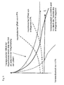
- Fig. 9 A summary of the time-dependent neurotoxic effects of t-PA is shown by the Fig. 9 , It also highlights the increased toxicity of recombinant t-PA compared to endogenous t-PA, presumably because rt-PA enters tissue at higher levels.
- DSPA Desmodus rotundus plasminogen activator
- DSPA in particular, is a plasminogen activator with high homology (similarity) to t-PA, so that - in addition to the disillusionment with the neurotoxic side effects of t-PA - no further hopes have been placed in DSPA either.
- Another novel method of treatment does not target the thrombus, blood liquefaction or anticoagulation, but attempts to increase the vitality of the cells damaged by the interruption of the blood supply ( WO 01/51613 A1 and WO 01/51614 A1 ).
- antibiotics from the group of quinones, aminoglycosides or chloramphenicol are applied.
- These fission products are components of the neuronal cell membrane and can thus support the regeneration of the damaged tissue ( U.S. Patent 5,827,832 ).
- the present invention is therefore based on the object to provide a new therapeutic approach for the treatment of stroke in humans.
- the core of the invention is to use a plasma activator for stroke treatment, whose activity as a mature enzyme is highly selectively increased by fibrin many times, namely by more than 650-fold.
- this plasminogen activator is based on the finding that the neurotoxicity of the tissue plasminogen activator (t-PA) is due to damage or destruction of the blood-brain barrier caused by stroke in the brain and the brain circulating in the blood fibrinogen can thus penetrate into the neuronal tissue of the brain. There it activates t-PA, indirectly by the activation of the glutamate receptor or by the activation of the plasminogen - leads to further tissue damage. To avoid this effect, a plasminogen activator is now used according to the invention, which shows increased fibrin selectivity and, conversely, reduced activability by fibrinogen.
- t-PA tissue plasminogen activator
- the plasminogen activator according to the invention is not activated on the passage of fibrinogen from the blood into the neuronal tissue as a result of the damaged blood-brain barrier - or in a significantly reduced extent compared to t-PA, since its activator Due to its size, fibrin can not enter the neuronal tissue.
- the plasminogen activators according to the invention are thus non-neurotoxic.
- non-toxic plasminogen activators are used which have at least one element of a so-called zymogen triad.
- a comparable triad is known from the catalytic center of serine proteases of the chymotrypsin family, which consists of the three interacting amino acids aspartate 194, histidine 40 and serine 32. However, this triad is not present in the t-PA belonging to the family of chymotrypsin-like serine proteases.
- the targeted mutagenesis of the native t-PA to introduce at least one of these amino acids at suitable positions to a reduction of the activity of the pro-enzyme (single-chain t-PA) and an increase in the activity of the mature enzyme (two-chain t-PA). PA) in the presence of fibrin.
- the introduction of at least one amino acid from the triad - or from amino acids that perform the corresponding function in the triad - leads to an increase in the zymogenicity of the t-PA (ratio of the activity of the mature enzyme to the activity of the pro-enzyme) Increase of fibrin specificity by conformational interaction between the introduced amino acid residues and / or amino acid residues of the wild-type sequence.
- Both mutants additionally have substitution of Arg275 to R275E to prevent cleavage of the single-chain t-PA into the two-chain form at cleavage site Aug275-I276 by plasmin.
- this mutant R275E increases the fibrin specificity of the t-PA by 6,900 times ( K Tachias, Madison EL 1995: Variants of Tissue-type Plasminogen Activators Which Display Substantially Enhanced Stimulation by Fibrin, in: Journal of Biological Chemistry 270, 31: 18319-18322 ).
- Positions 305 and 292 of t-PA are homologous to positions His40 and Ser32 of the known triad of serine proteases. By appropriate substitution with histidine or serine, these amino acids can interact with the aspartate 477 of t-PA, so that the known triad can be functionally formed in the t-PA mutants (Madison et al 1993).
- these t-PA mutants can be used for the treatment of stroke, because they have no or - compared to the wild-type t-PA - due to their increased fibrin specificity only a significantly reduced neurotoxicity.
- t-PA mutants F305H; F305H, A292S alone or in combination with R275E the publications of Madison et al. 1993 as well as Tachias and Madison 1995 are fully referenced.
- Plasminogen activators belong to the group of serine proteases of the chymptrypsin family and consistently show the conserved amino acid Asp194, which is responsible for the stability of the catalytically active conformation of the mature proteases. It is known, that Asp194 interacts with His40 in the zymogens of serine proteases. By activating cleavage of the zymogen, these interactions are impossible, and the side chain of the Asp194 is rotated by about 170 °, to then form a new salt bridge with the IIe16. This salt bridge is involved in the stability of the oxyanion pocket of the catalytic triad of the mature serine protease. It is also available in t-PA.
- a point mutation of the Asp194 initially makes the formation or stability of the catalytic conformation of the serine proteases impossible.
- the mutated plasminogen activators show a marked increase in activity, especially in comparison with the mature wild-type form, which can only be explained by the fact that the interactions with fibrin allow a conformational change enabling catalytic activity ( L Strandberg, Madison EL, 1995: Variants of Tissue-type Plasminogen Activator with Substantially Enhanced Response and Selectivity to Fibrin Co-factors. in: Journal of Biological Chemistry 270, 40: 2344-23449 ).
- the Asp194 mutants of the plasminogen activators show a high activity increase in the presence of fibrin, which allows their use according to the invention.
- t-PA is used according to the invention whose Asp194 is substituted by glutamate (D194E) or asparagine (D194N).
- D194E glutamate
- D194N asparagine
- these mutants may also contain a substitution of the Arg15 to R15E that prevents cleavage of the single-chain t-PA to the two-chain t-PA at the peptide bond Arg15-lle16 by plasmin.
- the activation of t-PA by fibrin is already increased by a factor of 12,000.
- the publication by Strandberg and Madison 1995 is fully referenced.
- An increase in fibrin-dependent plasminogen activators can also be achieved by introducing point mutations into the so-called "autolysis loop".
- This amino acid portion is known from trypsin; however, it also exists as a homologous segment in serine proteases and is characterized in particular by three hydrophobic amino acids (Leu, Pro and Phe).
- the autolysis loop in plasminogen activators is responsible for the interaction with plasminogen. Point mutations in this area can lead to the fact that the protein / protein interactions between plasminogen and plasma activator can no longer functionally be formed. These mutations are functionally relevant only in the absence of fibrin.
- a t-PA with point mutations in positions 420-423 is used.
- the fibrin dependence of t-PA increases by a factor of up to 61,000 (K Song-Hua et al.).
- Song Hua et al. studied the point mutations L420A, L420E, S421G, S421E, P422A, P422G, P422E, F423A and F423E.
- the publication is hereby incorporated by reference in its entirety for the use according to the invention.
- a modified tissue plasminogen activator having an amino acid sequence according to SEQ.ID No. 1 (US Pat. Fig. 13 ) used.
- This modified t-PA differs from wild-type t-PA by replacing the hydrophobic amino acid at positions 420-423 in the autolysis loop, which are occupied as follows: His420, Asp421, Ala422, and Dys423.
- This t-PA preferably has a phenylalanine at position 194.
- position 275 may be due to a glutamate be occupied.
- position 194 is occupied by phenylalanine.
- a modified urokinase can be used according to the invention.
- This urokinase according to the invention can be the amino acid according to SEQ. ID No. 2 ( Fig. 14 ), in which the hydrophobic amino acids of the autolysis loop are replaced by Val420, Thr421, Asp422 and Ser423.
- this urokinase carries an Ile275 as well as a Glu194. This mutant shows in comparison to the wild-type urokinase a 500-fold increased fibrin specificity.
- DSPA plasminogen activator
- the term DSPA encompasses four different proteases which fulfill a fundamental need of the vampire bat, namely the prolonged bleeding time of the prey wounds caused by these animals (Cartwright, 1974). These four proteases (DSPA ⁇ 1 , DSPA ⁇ 2 , DSPA ⁇ , DSPA ⁇ ) all show a high similarity (homology) to human t-PA. Similarly, they have similar or consistent physiological activities that justify their summary to the generic term DSPA.
- DSPA is the subject of EP 0 352 119 A1 as well as the U.S. Patents 6,008,019 and 5,830,849 which are hereby incorporated by reference for purposes of disclosure.
- DSPA ⁇ 1 is the best studied protease in this group so far. It has in its amino acid sequence a homology of over 72% compared to the known human t-PA amino acid sequence (Krfordschmar et al., 1991). Nevertheless, there is between t-PA and DSPA two main differences.
- DSPA is a fully active molecule to peptidic substrates that is not converted into a two-chain form like the t-PA (Gardell et al, 1989, Krrienschmar et al., 1991).
- the catalytic activity of DSPA shows almost absolute fibrin dependence (Gardell et al., 1989, Bringmann et al., 1995, Toschi et al., 1998).
- DSPA ⁇ 1 the activity of DSPA ⁇ 1 is increased 100,000 fold in the presence of fibrin while t-PA activity increases only about 550-fold.
- DSPA activity is much less induced by fibrinogen; it only undergoes an increase of 7 to 9 times (Bringmann et al., 1995).
- DSPA is therefore significantly more fibrin-dependent and fibrin-specific than the wild-type t-PA, which is activated only about 550-fold by fibrin.
- DSPA is an interesting candidate for the development of a thrombolytic because of its fibrinolytic properties and its close resemblance to t-PA.
- the development of DSPA as a thrombolytic agent has been limited to the treatment of myocardial infarction, because - due to the involvement of t PA on glutamate-dependent neurotoxicity - there were no reasonable hopes to successfully use a t-PA-related plasminogen activator to treat acute stroke.
- DSPA has no neurotoxic effects in spite of the high degree of similarity (homology) to t-PA and despite the fact that its physiological effect on t-PA largely corresponds to t-PA.
- This finding immediately goes hand in hand with the finding that DSPA can be used as a thrombolytic agent for the treatment of stroke without any additional risk of damaging the neuronal tissue.
- DSPA can also be used later than three hours after the onset of symptoms in stroke.
- Another teaching of the present invention is the possibility resulting from the above-described finding of also modifying or producing other plasminogen activators in such a way that they have this property of DSPA or do not have the neurotoxicity of the tPA.
- the basis for this is the recognized causal link, which in the future will allow neurotoxic plasminogen activators to be converted into non-neurotoxic plaminogen activators or to produce non-neurotoxic plasminogen activators on the basis of known or newly discovered neurotoxic plasminogen activators.
- the new teaching is based on in vivo comparative studies of the neurodegenerative effects of t-PA on the one hand and of DSPA on the other hand, which were carried out with the aid of the so-called "kainic acid model” and a model for the investigation of the NMDA-induced lesion of the striatum.
- the kainic acid model (also kainic acid injury model) is based on the fact that the neurotoxic glutamate cascade by the application of external kainic acid (KA) as agonist of the kainic acid type (KA-type) glutamate receptor and the NMDA and AMPA glutamate receptors is stimulated.
- KA kainic acid
- t-PA deficient mouse strain it could be shown that the sensitivity of the test animals to kainic acid reached the level of the wild-type mice only with an additional administration of external t-PAs.
- infusion of an equimolar concentration of DSPA under the same experimental conditions failed to restore sensitivity to cain acid (KA). Accordingly, the effect of t-PA could not be substituted by DSPA.
- DSPA has at least 100-fold lower activity than t-PA in promoting neurodegeneration after KA treatment (see also US Pat Figures 11 and 12 ).
- DSPA is a substantially inert protease in the central nervous system of a mammal, including humans, and, unlike t-PA, does not induce an increase in KA or NMDA-induced neurotoxicity. Contrary to expectations, this lack of neurotoxicity makes DSPA a suitable thrombolytic for the treatment of acute stroke.
- the special advantage for stroke treatment is the fact that the use of these plasminogen activators, unlike that of wild-type t-PA, is not limited to the narrow time window of up to three hours after the onset of stroke is limited. Rather, the treatment can also take place at a later point in time - that is to say, after six hours or even later - without the risk - as in the case of t-PA - of promoting excitotoxicity.
- Initial clinical trials with DSPA even prove the safe treatment of patients over a period of more than six or nine hours after the onset of symptoms.
- This indefinite treatment option with non-neurotoxic plasminogen activators is so important because of it For the first time a treatment of acute stroke patients is possible without hesitation, in which the onset of the stroke can not be determined with sufficient certainty over time. This group of patients had previously been excluded from thrombolysis with plasminogen activators for reasons of caution and risk assessment. This eliminates a significant contraindication to the approved use of a thrombolytic in stroke.
- DSPA and the other non-neurotoxic plasminogen activators themselves show no neurotoxic side effects.
- the glutamate receptor can serve competitively or non-competitively inhibiting neuroprotective agents.
- Useful combinations are possible, for example, with the known inhibitors of the glutamate receptors of the NMDA type, of the kainic acid type or of the quisqualate type, such as e.g. with APV, APH, phencyclidine, MK-801, dextrorphan or ketamine.
- a combination with cations may be advantageous, since cations, in particular Zn ions, block the glutamate receptor-regulated cation channel and thus can reduce the neurotoxic effects.
- the non-neuorotoxic plasminogen activators can be combined with at least one further therapeutic agent or with a pharmaceutically acceptable carrier.
- a therapeutic agent that helps to avoid tissue damage by revitalizing the cells, contributes to a regeneration of the already damaged tissue or serves to avoid subsequent strokes.
- antibiotics such as quinones
- anticoagulants such as heparin or hirudin as well as with citicholine or aspirin can be advantageous.
- thrombomodulin Thrombomodulinanaloga such.
- B. solulin, triabin or pallidipine thrombomodulin, Thrombomodulinanaloga
- combinations with anti-inflammatory substances that affect leukocyte infiltration are beneficial.
- the determination of the proteolytic activity in the brain tissue after infusion of t-PA or DSPA ⁇ 1 was carried out by means of zymographic analysis (Granelli-Piperno and Reich, 1974). After the 7-day infusion period into the hippocampus, the mice were anesthetized, then perfused transcardially with PBS and the brains removed. The hippocampus region was removed, placed in Eppendorf tubes and in equal volumes (w / v) (about 30-50 ⁇ l) with 0.5% NP-40 lysis buffer without protease inhibitors (0.5% NP-40, 10 mM Tris-HCl pH 7.4, 10 mM NaCl, 3 mM MgCl 2 , 1 mM EDTA). Brain extracts were homogenized using a hand-operated glass homogenizer and left on ice for 30 min. The samples were then centrifuged and the supernatant removed. The amount of protein present was determined (Bio-Rad reagent).
- the proteolytic activity in the brain tissue samples or extracts was determined by zymographic analysis according to the method of Granelli-Piperno and Reich (1974).
- the samples of recombinant protein (up to 100 nmol) or brain tissue extract (20 ⁇ g) were subjected to 10% SDS-PAGE under non-reducing conditions.
- the gels were removed from the plates, washed for 2 hours in 1% Triton X100 and then placed on an agarose gel with polymerized fibrinogen and plasminogen (Granelli-Piperno and Reich, 1974). The gels were incubated at 37 ° C in a humid chamber until proteolysed zones became apparent.
- the kainic acid injury model is based on the approach of Tsirka et al. (1995).
- the animals were injected intraperitoneally (i.p.) with atropine (4 mg / kg), then with i.p. Injection of sodium pentobarbital (70 mg / kg) anesthetized. They were then placed in a stereotactic frame so that a microosmotic pump (Alzet model 1007D, Alzet CA USA) was subcutaneously subcutaneously with 100 ⁇ l PBS or recombinant human t-PA (0.12 mg / ml, 1.85 ⁇ M) or DSPA ⁇ 1 (1.85 ⁇ M) could be implanted between the shoulder blades.
- a microosmotic pump Alzet model 1007D, Alzet CA USA
- the pumps were connected by sterile tubes to a brain cannula and inserted through a skull opening at the coordinates bregma -2.5 mm, midolateral 0.5 mm and dorsoventral 1.6 mm to introduce the fluid near the midline.
- the cannula was subsequently adhered to the desired position and the pumps were opened to infuse the appropriate solutions at a flow rate of 0.5 ⁇ l per hour for seven days.
- mice Two days after infusion of the proteases, the mice were re-anesthetized and placed in the stereotactic frame. 1.5 nmoles of kainic acid in 0.3 ⁇ l of PBS were then injected unilaterally into the hippocampus. The coordinates were: bregma 2.5 mm, medial-lateral 1.7 mm and dorsoventral 1.6 mm. The excitotoxin (KA) was given over a period of 30 Sec. supplied. After kainic acid treatment, the needle remained at these coordinates for a further 2 minutes to prevent backflow of the liquid.
- KA excitotoxin
- the animals were anesthetized and perfused transcardially with 30 ml of PBS, followed by 70 ml of a 4% paraformaldehyde solution, postfixed for 24 hours in the same fixative, followed by incubation in 30% sucrose for a further 24 hours.
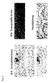
- Coronal sections (40 ⁇ m) of the brain were then cut on an iced microtome, either counterstained with thionin (BDH, Australia) or prepared for immunohistochemistry.
- the quantification of neuronal loss in the CA1-CA3 hippocampal subfields was performed as previously described (Tsirka et al., 1995, Tsirka et al., 1996). Five consecutive sections of the dorsal hippocampus from all treated groups were prepared, which sections actually included the site of the KA injection and the area of the lesion. The hippocampal subfields (CA1-CA3) of these sections were followed by camera lucida drawings of the hippocampus. The total length of these subfields was measured in comparison to 1 mm standards followed at the same magnification.
- the length of the tissue sections with vital pyramidal neurons (with normal morphology) and the length of tissue sections without neurons (no cells present, no thionin staining) was determined.
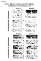
- Wild type mice (c57 / Black6) were anesthetized and placed in a stereotactic frame (see above). The mice then received a unilateral injection into the left stratum with 50 nmol NMDA alone or in combination with 46 ⁇ M rt-PA or 46 ⁇ M DSPA ⁇ 1. As a control, t-PA and DSPA ⁇ 1 alone were also injected as controls at a concentration of 46 ⁇ M. The injection coordinates were: bregma -0.4 mm, midolateral 2.0 mm and dorsoventral 2.5 mm.
- mice were anesthetized and perfused transcardially with 30 ml of PBS, followed by 70 ml of a 4% paraformaldehyde solution, post-fixation for 24 hours in the same fixative, followed by incubation in 30% sucrose for a further 24 hours. Coronal sections (40 ⁇ m) were then cut on an icing microtome and placed on gelatin-coated glass slides.
- the quantification of the striatal lesion volume was determined by the methods described by Callaway et al. (2000). Ten consecutive coronal sections covering the area of the injury were prepared. The damaged region was visualized using the Callaway method and the lesion volume was quantified using a Micro Computer Imaging Device (MCID, Imaging Research Inc., Brock University, Ontario, Canada).
- Immunohistochemistry was carried out according to standard methods. Coronal sections were immersed in a solution of 3% H 2 O 2 /10% Methanol for 5 min, followed by incubation with 5% normal goat serum for 60 min. The sections were then probed overnight with either an anti-GFAP antibody (1: 1,000; Dako, Carpinteria, Ca, USA) for the detection of astrocytes, with an anti-MAC-1 antibody (1: 1,000, Serotec, Raleigh, NC, USA) for the detection of microglia or polyclonal anti-DSPA antibodies (Schering AG, Berlin). After washing, the sections were incubated with the appropriate biotinylated secondary antibodies (Vector Laboratories, Burlingame, CA, USA).
- the first experiments were designed to confirm that both DSPA and t-PA retain their proteolytic activity over the 7-day duration of the infusion.
- aliquots of t-PA and DSPA 100 nmol were incubated both at 37 ° C and at 30 ° C for seven days in a water bath.
- serial 5-fold dilutions of the samples were subjected to SDS-PAGE with non-reducing conditions and the proteolytic activity measured by zymographic analyzes.
- t-PA and DSPA activity is also found in hippocampal extracts of t-PA - / - mice after infusion
- t-PA - / mice received infusions with t-PA or DSPA (see above) for seven days. The mice were then killed by transcardiac perfusion with PBS and the brains removed. The ipsilateral and contralateral hippocampal regions were isolated as well as a region of the cerebellum (as a negative control). Tissue samples (20 ⁇ g) were subjected to SDS-PAGE and zymographic analysis as described in the Methods section. How out FIG.
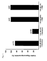
- DSPA infusion does not restore the sensitivity to kainic acid-dependent neurodegeneration in vivo
- t-PA - / - mice are resistant to kainic acid-dependent neurodegeneration. In contrast, infusion of rt-PA into the hippocampus completely restores the sensitivity to kainic acid-dependent Damage.
- DSPA can replace the t-PA in this effect
- cort-PA - / - mice were resistant to KA after infusion of PBS.
- infusions of recombinant t-PA restored the sensitivity to treatment with KA.
- infusion of the same concentration of DSPA into the hippocampal region did not alter the sensitivity of these animals to KA (see Fig. 4a and. 4b ).
- the concentration of t-PA used for the infusion was based on the concentration described by Tsirka et al (1995) (100 ⁇ l of 0.12 mg / ml [1.85 ⁇ M].)
- infusion of the 10-fold increased DSPA concentration caused only minimal neuronal loss after KA treatment, indicating that DSPA does not increase the sensitivity to KA.
- t-PA and DSPA were further investigated in a model of neurodegeneration on wild-type mice. Injection of t-PA into the striatum of these mice has been shown to enhance the neurodegenerative effects produced by the glutamate analog NMDA (Nicole et al., 2001).
- Indirect chromogenic assays of t-PA activity use the substrates Lys plasminogen (American Diagnostica) and Spectrozyme PL (American Diagnostica) and were prepared according to Madison EL, Goldsmith EJ, Gerard RD, Gething M.-J., Sambrook JF (1989) Nature 339 721-724 ; Madison EL, Goldsmith EJ, Gerard RD, Gething MJ, Sambrook JF and Bassel-Duby RS (1990) Proc. Natl. Acad. Sci. USA 87, 3530-3533 such as Madison EL, Goldsmith EJ, Gething MJ, Sambrook JF and Gerard RD (1990) J. Biol. Chem. 265, 21423-21426 carried out.
- DESAFIB American Diagnostica
- a preparation of soluble fibrin monomers was prepared by the cleavage of high purity human fibrinogen by the protease Batroxobin won. Batroxobin cleaves the Arg 16 -Gly 17 bond in the A ⁇ chain of the fibrinogen, releasing fibrinopeptide A.
- the resulting des-AA-fibrinogen in the form of fibrin I monomers is soluble in the presence of the peptide Gly-Pro-Arg-Pro.
- the concentration of Lys plasminogen was varied from 0.0125 to 0.2 ⁇ M in the presence of DESAFIB, in the absence of the co-factor of 0.9 to 16 ⁇ M.
- Standard indirect chromogenic assays were performed according to the publications cited above. Batches of 100 ⁇ l total volume with 0.25-1 ng enzyme, 0.2 ⁇ M Lys plasminogen and 0.62 mM Spectrozym PL were used. The tests were performed in the presence of either buffer, 25 ⁇ g / ml DESAFIB, 100 ⁇ g / ml cyanogen bromide fragments of fibrinogen (American Diagnostica) or 100 ⁇ g / ml of the stimulatory 13-amino acid peptide P368. The analyzes were carried out in microtiter plates and the optical density was measured at 405 nm wavelength for 1 h every 30 seconds in a "Molecular Devices Thermomax". The reaction temperature was 37 ° C.
Abstract
Description
- Die Erfindung betrifft eine therapeutische Anwendung nicht-neurotoxischer Plasminogen-Aktivatoren, insbesondere aus dem Speichel von Desmodus rotundus (DSPA), vorzugsweise zur Behandlung von Schlaganfall und nimmt die Priorität der
deutschen Patentanmeldung 101 53 601.1 und dereuropäischen Patentanmeldung 01 130 006.8 - Unter dem Begriff "Schlaganfall" werden verschiedene Krankheitsbilder zusammengefaßt, die sich in ihrer klinischen Symptomatik ähneln. Eine erste Differenzierung dieser Krankheitsbilder in die sogenannten ischämischen Insulte und hämorrhagischen Insulte ist ausgehend von der jeweiligen Pathogenese möglich.
- Bei ischämischen Insulten (Ischämie) handelt es sich um eine Verminderung oder Unterbrechung der Durchblutung des Gehirns infolge mangelnder arterieller Blutzufuhr. Diese wird häufig durch eine Thrombose eines arteriosklerotisch stenosierten Gefäßes, aber auch durch arterioarterielle bzw. kardiale Embolien verursacht.
- Die hämorrhagischen Insulte gehen dagegen u.a. auf eine Perforation der durch arterielle Hypertonie beschädigten, Hirn versorgenden Arterien zurück. Von allen zerebralen Insulten werden aber lediglich etwa 20% durch diese Form der Blutungen verursacht, so daß den durch Thrombose verursachten Schlaganfällen die weitaus größte Bedeutung zukommt.
- Die lschämie neuronaler Gewebe geht - im Vergleich zu anderen Gewebe-Ischämien - in besonderem Umfang mit der Nekrose der betroffenen Zellen einher. Die erhöhte Nekroseinzidenz kann nach neuerem Verständnis durch das Phänomen der sog. Exzitotoxizität erklärt werden, bei der es sich um eine komplexe Kaskade einer Vielzahl von Reaktionsschritten handelt. Diese wird demnach dadurch ausgelöst, daß die unter Sauerstoffmangel leidenden ischämischen Neuronen schnell ATP verlieren und depolarisieren. Dies führt zu einer verstärkten postsynaptischen Abgabe des Neurotransmitters Glutamat, der seinerseits membrangebundene Glutamat-Rezeptoren aktiviert, die Kationenkanäle regulieren. Als Folge der erhöhten Glutamat-Ausschüttung werden die Glutamat-Rezeptoren jedoch überaktiviert.
- Die Glutamat-Rezeptoren regulieren ihrerseits spannungsabhängige Kationenkanäle, die - bei der Bindung von Glutamat an seinen Rezeptor - geöffnet werden. Damit setzt ein Na+ und ein Ca2+-Einstrom in die Zelle ein, der zu einer massiven Störung des Ca2+-abhängigen zellulären Stoffwechsels - einschließlich des Energiestoffwechsels - führt. Für den nachfolgend eintretenden Zelltod könnte dabei insbesondere die Aktivierung Ca2+-abhängiger katabolischer Enzyme verantwortlich sein (Lee, Jin-Mo et al., "The changing landscape of ischemic brain injury mechanisms"; Dennis W. Zhol "Glutamat neurotoxicity and deseases of the nervous system").
- Selbst wenn der Mechanismus der Glutamat vermittelten Neurotoxizität derzeit noch nicht im Detail verstanden wird, scheint dennoch Einigkeit darüber zu bestehen, daß dieses Phänomen in einem erheblichen Umfang zu dem neuronalen Zelltod nach einer zerebralen Ischämie beiträgt (Jin-Mo Lee et. al.).
- Bei der Therapie der akuten zerebralen Ischämie steht neben der Sicherstellung vitaler Funktionen und der Stabilisierung physiologischer Parameter zunächst das Bestreben nach einer Wiedereröffnung des verschlossenen Gefäßes im Vordergrund. Dieses Ziel wird mit verschiedenen Ansätzen verfolgt. Die rein mechanische Wiederveröffentlichung hat bisher noch keine zufriedenstellenden Ergebnisse erzielt, wie etwa die PTCA bei Herzinfarkt. Lediglich mit erfolgreicher Fibrinolyse konnte bisher eine ausreichende Besserung des Zustands für die Patienten aufgezeigt werden. Dies kann durch lokale Applikation mittels Katheter geschehen (PROCAT, eine Studie mit Pro-Urokinase), dieses Verfahren hat bisher jedoch trotz erster positiver Ergebnisse nicht zur Zulassung einer pharmazeutischen Substanz geführt.
- Die körpereigene Fibrinolyse basiert auf der proteolytischen Aktivität der Serin-Protease Plasmin, die durch eine Katalyse (Aktivierung) aus der inaktiven Vorstufe Plasminogen hervorgeht. Die natürliche Aktivierung des Plasminogens erfolgt durch die körpereigenen Plasminogen-Aktivatoren u-PA (Urokinase-typ Plaminogenaktivator) und t-PA (Tissue Plasminogen Aktivator). Letzterer bildet - im Gegensatz zu u-PA - gemeinsam mit Fibrin und Plasminogen einen sog. Aktivatorkomplex. Die katalytische Aktivität des t-PA ist demnach Fibrin-abhängig und erfährt in Gegenwart von Fibrin eine etwa 550-fache Steigerung. Neben Fibrin kann auch Fibrinogen die t-PA vermittelte Katalyse von Plasmin zu Plasminogen stimulieren - wenn auch in sehr viel geringerem Umfang. So erfährt die t-PA Aktivität in Gegenwart von Fibrinogen lediglich eine 25-fache Steigerung. Die Spaltprodukte des Fibrins (fibrin degradation products (FDP)) stimulieren ihrerseits ebenfalls die t-PA Aktivität.
- Frühe Ansätze der thrombolytischen Behandlung des akuten Schlaganfalls gehen bereits auf die 1950-iger Jahre zurück. Aber erst ab 1995 wurden erste umfangreiche klinische Versuche mit Streptokinase, einem Fibrinolytikum aus beta-hämolysierenden Streptokokken, durchgeführt. Streptokinase bildet mit Plasminogen einen Komplex, der in der Lage ist, andere Plasminogenmoleküle in Plasmin umzusetzen.
- Die Therapie mit Streptokinase ist aber mit wesentlichen Nachteilen verbunden, da Streptokinase als bakterielle Protease im Körper allergische Reaktionen auslösen kann. Auch kann eine sog. Streptokinase-Resistenz bei vorheriger Streptokokken-Infektion mit entsprechender Antikörperbildung vorliegen, die eine Therapie erschweren. Darüber hinaus deuteten klinische Studien in Europa (Multicenter Acute Stroke Trial of Europe (MAST-E), Multicenter Acute Stroke Trial of Italy (MAST-I)) und Australien (Australien Streptokinase Trial (AST)) auf ein derart erhöhtes Mortalitätsrisiko und die erhebliche Gefahr von intrazerebralen Blutungen (intracerebral hemorrhage, ICH) nach der Patienten-Behandlung mit Streptokinase hin, daß diese Studien sogar frühzeitig abgebrochen werden mußten.
- In einem alternativen Therpieansatz wird Urokinase - ebenso ein "klassisches" Fibrinolytikum - appliziert, die im Gegensatz zu Streptokinase keine antigenen Eigenschaften aufweist, da es sich dabei um ein in zahlreichen Geweben vorkommendes körpereigenes Enzym handelt. Es stellt einen Kofaktor unabhängigen Aktivator des Plasminogens dar. Urokinase wird in Nierenzellkulturen hergestellt.
- Umfangreiche Erfahrungen im Bereich der therapeutischen Thrombolyse liegen mit dem Gewebe-typ Plasminogen-Aktivator - dem sog. rt-PA - vor (siehe
EP 0 093 619 ,US-Patentschrift 4 766 075 ), der in rekombinanten Hamsterzellen produziert wird. Mit t-PA wurden - neben der Hauptindikation akuter Myokardinfarkt - in den 1990-iger Jahren weltweit eine Reihe klinischer Studien mit zum Teil noch unverstandenen und widersprüchlichen Ergebnissen durchgeführt. So wurden zunächst im sog. European Acute Stroke Trial (ECASS) Patienten innerhalb eines Zeitraums von sechs Stunden nach dem Einsetzen der Symptome des Schlaganfalls intravenös mit rt-PA behandelt und nach 90 Tagen die Mortalitätsrate sowie der Barthel-Index als Maßstab für die Versehrtheit oder selbständige Lebensfähigkeit der Patienten untersucht. Dabei ergab sich keine signifikante Verbesserung der Lebensfähigkeit, sehr wohl aber eine - wenn auch nicht signifikante - Erhöhung der Mortalität. Dies erlaubte die Schlußfolgerung, daß eine thrombolytische Behandlung von individuell nach ihrer jeweiligen Krankengeschichte gezielt ausgesuchten Patienten mit rt-PA unmittelbar nach dem Beginn des Schlaganfalls möglicherweise vorteilhaft sein könnte. Aufgrund des beobachteten erhöhten Risikos einer intrazerebralen Haemorrhagie (ICH) nach nur wenigen Stunden nach Einsetzen des Schlaganfalls war aber von einer generellen Anwendung von rt-PA innerhalb des untersuchten Zeitraums von sechs Stunden nach Beginn des Schlaganfalls abzuraten (so C. Lewandowski C und Wiliam Barsan, 2001: Treatment of Acute Strocke; in: Annals of Emergency Medcine 37:2; S. 202 ff). - Die thrombolytische Behandlung des Schlaganfalls war später ebenso Gegenstand einer klinischen Studie des National Institute of Neurologic Disorder and Stroke (sog. NINDS rtPA Stroke Trial) in den USA. Dabei stand jedoch die Untersuchung der Wirkung einer intravenösen rt-PA-Behandlung innerhalb eines nur dreistündigen Zeitraums nach Beginn der Symptome auf den Gesundheitszustand der Patienten nach drei Monaten im Vordergrund. Obwohl die Autoren auch hier ein erhöhtes Risiko für ICH feststellten, wird dennoch die rt-PA Behandlung innerhalb dieses begrenzten Zeitraums von drei Stunden aufgrund der ebenfalls durch die Studie erkannten positiven Effekte dieser Behandlung auf die Lebensfähigkeit der Patienten empfohlen.
- In zwei weiteren Studien (ECASS II Trial; Alteplase Thrombolysis for Acute Noninterventional Therapy in Ischemic Stroke (ATLANTIS)) wurde untersucht, ob sich diese positive Aussage über die Wirkung der rt-PA Behandlung innerhalb von drei Stunden nach Beginn des Schlaganfalls auch für ein Behandlung im sechsstündigen Zeitraum bestätigen ließen. Diese Frage konnte jedoch nicht positiv beantwortet werden, da durch eine Behandlung in diesem Zeitraum neben dem erhöhten Risiko für ICH keine Verbesserung der klinischen Symptome oder eine Verringerung der Mortalität beobachtet werden konnte.
- Diese z.T. widersprüchlichen Ergebnisse haben zu einer großen Vorsicht in der Anwendung des rt-PA geführt. So wurde bereits 1996 in einer Publikation der American Heart Association konstatiert, daß unter den Ärzten hinsichtlich der thrombolytischen Behandlung des Schlaganfalls eine ausgeprägte Skepsis herrsche, die im Bezug auf die Therapie des Myokardinfarkts mit Fibrinolytika nicht bestehe (van Gijn J, MD, FRCP, 1996 - Circulation 1996, 93: 1616-1617).
- Eine Begründung dieser Skepsis findet sich in einer erstmalig 1997 publizierten und im März 2001 aktualisierten Zusammenschau aller "Stroke Trials". Danach führten alle Thrombolytika-Behandlungen (Urokinase, Streptokinase, rt-PA oder rekombinate Urokinase) insbesondere aufgrund von ICH zu einer signifikant erhöhten Mortalität innerhalb der ersten zehn Tage nach dem Schlaganfall, während sich bei einer Behandlung innerhalb des 6-Stunden-Zeitraums die Gesamtanzahl der entweder toten oder behinderten Patienten reduzierte. Von einer breiten Anwendung von Thrombolytika für eine Schlaganfallbehandlung wird daher abgeraten.
- Diese Ergebnisse haben schon vorher andere Autoren zu der eher sarkastischen Aussage bewogen, daß Schlaganfall-Patienten nunmehr die Wahl hätten, zu sterben oder behindert zu überleben (SCRIP 1997: 2265, 26).
- Gleichwohl stellt die Therapie mit rt-PA derzeit die einzige von der Food and Drug Administration (FDA) in den USA zugelassene Behandlungsmethode der akuten zerebralen Ischämie dar. Diese ist allerdings beschränkt auf eine Applikation des rt-PA innerhalb von drei Stunden nach dem Beginn des Schlaganfalls.
- Die Zulassung des rt-PA erfolgte 1996. Unmittelbar zuvor, nämlich im Jahre 1995, wurden erste Hinweise auf schädliche Nebenwirkungen des t-PA bekannt, die einen Erklärungsansatz für die dramatischen Effekte des t-PA bei der Schlaganfallbehandlung außerhalb des 3-Stunden Behandlungszeitraums liefern. Danach produzieren Mikrogliazellen und neuronale Zellen des Hippocampus körpereigenes t-PA, das an der Glutamat vermittelten Exzitotoxizität beteiligt ist. Diese Schlußfolgerung entstand durch einen Vergleich von t-PA defizienten Mäusen und Wildtypmäusen, denen jeweils Glutamat-Agonisten in den Hippocampus injiziert wurden. Dabei zeigten die t-PA defizienten Mäuse eine deutlich erhöhte Resistenz gegen extern (inthrathekal) appliziertes Glutamat (Tsirka SE et. al., Nature, Vol. 377, 1995, "Exzitoxin-induced neuronal degeneration and seizure are mediated by tissue plasminogen activator"). Diese Ergebnisse wurden im Jahre 1998 bestätigt, als Wang et al. bei t-PA defizienten Mäusen durch die intravenöse Injektion von t-PA nahezu eine Verdoppelung des nekrotischen neuronalen Gewebes nachweisen konnten. Dieser negative Effekt des externen t-PA lag bei Wildmäusen dagegen nur etwa bei 33% (Wang et al., 1998, Nature, "Tissue plasminogen activator (t-PA) increases neuronal damage after focal cerebral ischemia in wild type and t-PA deficient mice").
- Weiterführende Ergebnisse über die Stimulation der Exzitotoxizität durch t-PA publizierten Nicole et al. zu Beginn des Jahres 2001 (Nicole O; Docagne F Ali C; Margaill I; Carmeliet P; MacKenzie ET; Vivien D und Buisson A, 2001: The proteolytic activity of tissue-plasminogen activator enhances NMDA receptor-mediated signaling; in: Nat Med 7, 59-64). Sie konnten nachweisen, daß von depolarisierten kortikalen Neuronen ausgeschüttetes t-PA mit der sogenannten NR1 Untereinheit des Glutamat Rezeptors des NMDA Typs interagiert und diese spaltet. Diese Modifikation führt zu einer Erhöhung der Rezeptoraktivität, die ihrerseits für eine erhöhte Gewebeschädigung nach der Applikation des Glutamat Agonisten NMDA durch eine induzierte Exzitotoxizität verantwortlich ist. Das t-PA wirkt somit durch die Aktivierung des Glutamat-Rezeptor des NMDA-Typs neurotoxisch.
- Nach einem weiteren Erklärungsansatz ist die Neurotoxizität des t-PA mittelbar auf die Aktivierung des Plasmins aus dem Plaminogen zurückzuführen. Eigentlicher Effektor der Neurotoxizität ist nach diesem Modell demnach das Plasmin (Chen ZL und Strickland S, 1997: Neuronal Death in the hippocampus is promoted by plasmin-catalysed degradation of laminin. Cell:91, 917-925).
- Eine zusammenfassende Darstellung der zeitabhängigen neurotoxischen Effekte des t-PA zeigt die
Abb. 9 . Darin wird zudem die erhöhte Toxizität des rekombinanten t-PA im Vergleich zum endogenen t-PA deutlich, die vermutlich darauf zurückzuführen ist, daß rt-PA in höheren Konzentrationen in das Gewebe eintritt. - Dass trotz dieser Hinweise auf die neurotoxischen Nebeneffekte des t-PA und trotz der nachgewiesenen Erhöhung der Mortalität durch t-PA dennoch eine arzneimittelrechtliche Zulassung durch die FDA erfolgte, kann wohl nur durch den Mangel an ungefährlichen und effektiven Alternativen - und eine sehr pragmatische Kosten-Nutzen Analyse - erklärt werden. Es besteht aber nach wie vor Bedarf für sicherere Therapien, wobei bei der Entwicklung neuer Thrombolytika - sofern denn von der Thrombolyse nicht sogar ganz Abstand zu nehmen ist - das Problem der Neurotoxizität berücksichtigt werden muß (so z.B. Wang et al a.a.O.; Lewandowski und Barsan 2001 a.a.O).
- Aus diesem Grund unterblieben weiterführende Untersuchungen bekannter Thrombolytika einschließlich des DSPA (Desmodus rotundus Plasminogen Aktivator) zur Entwicklung eines neuen Therapeutikums für Schlaganfall, obwohl prinzipiell sämtliche Thrombolytika geeignet sein könnten und beispielsweise im Falle des DSPA bereits in einer ersten Veröffentlichung auf dessen mögliche Eignung für dieses Indikationsgebiet hingewiesen wurde (Medan P; Tatlisumak T; Takano K; Carano RAD; Hadley SJ; Fisher M: Thrombolysis with recombinant Desmodus saliva plasminogen activator (rDSPA) in a rat embolic stroke model; in: Cerebrovasc Dis 1996:6; 175-194 (4th International Symposium on Thrombolic Therapy in Acute Ishemic Stroke)). Gerade bei DSPA handelt es sich aber um einen Plasminogen Aktivator mit hoher Homologie (Ähnlichkeit) zu t-PA, so daß - zugleich mit der Ernüchterung über die neurotoxischen Nebenwirkungen des t-PA - auch in DSPA keine weiteren Hoffnungen gesetzt wurden.
- Vielmehr wird in jüngster Zeit u.a. die Strategie verfolgt, zur Verbesserung der bisherigen thrombolytischen Schlaganfallbehandlung mit den bekannten Thrombolytika die Substanzen nicht mehr intravenös, sondern intra-arteriell über einen Katheter direkt an den intravaskulären Thrombus zu bringen. Erste Erfahrungen dazu liegen mit der rekombinant erzeugten Urokinase vor. Damit kann möglicherweise die für die Thrombolyse erforderliche Dosis gesenkt und damit ein Teil der Nebenwirkungen reduziert werden. Diese Applikationsform erfordert jedoch einen hohen technischen Aufwand und steht daher nicht überall zur Verfügung. Zudem erfordert sie eine gewisse Zeit für die eingehende Vorbereitung des Patienten, die nicht immer zur Verfügung steht und stellt ein zusätzliches Risiko dar.
- Ferner sind derzeitige Bestrebungen auf Antikoagulantien wie Heparin, Aspirin oder Ancrod, dem Wirkstoff aus dem Gift der Malayischen Grubenotter, ausgerichtet. Zwei u.a. auch auf die Untersuchung der Wirkungen von Heparin ausgerichtete klinische Studien (International Stroke Trial (IST) und Trial of ORG 10172 in Acute Stroke Treatment (TOAST)) deuten jedoch nicht auf eine signifikante Verbesserung der Mortalität und eine Verhinderung eines erneuten Schlaganfalls hin.
- Eine weitere neuartige Behandlungsmethode setzt weder an dem Thrombus noch an der Blutverflüssigung oder Antikoagulation an, sondern versucht, die Vitalität der durch die Unterbrechung der Blutzufuhr geschädigten Zellen zu steigern (
WO 01/51613 A1 WO 01/51614 A1 US-Patentschrift 5 827 832 ). - Jüngste Bestrebungen auf der Suche nach sicheren Behandlungsmethoden basieren auf den neuen Erkenntnissen darüber, daß ein Teil der fatalen Folgen des Schlaganfalls nur mittelbar auf die unterbrochene Blutzufuhr unmittelbar aber auf die Exzito- oder Neurotoxizität unter Beteiligung des überangeregten Glutamat-Rezeptors zurückzuführen ist, die wiederum insbesondere auch durch t-PA verstärkt wird (s. o). Ein Ansatz zur Verminderung dieser Exzitotoxizität ist daher die Applikation von sog. Neuroprotektiva. Diese können allein oder in Kombination mit Fibrinolytika verwendet werden, um so deren neurotoxischen Effekt zu minimieren. Sie können dabei entweder direkt, beispielsweise als Glutamat-Rezeptor-Antagonisten oder indirekt über die Blockade von spannungsabhängigen Natrium- oder Kalziumkanälen zu einer Abschwächung der Exzitotoxizität führen (Jin-Mo Lee et. al. a.a.O).
- Eine kompetitive Hemmung (Antagonisierung) des Glutamat-Rezeptors des NMDA Typs ist dabei beispielsweise mit 2-amino-5-Phosphonovalerat (APV) oder 2-amino-5-Phosphonoheptanoate (APH) möglich. Eine nicht-kompetitive Hemmung kann beispielsweise durch Substanzen erreicht werden, die an die Phencyclidin Seite der Kanäle binden, wie Phencyclidin, MK-801, Dextrorphan oder Ketamin.
- Bislang haben die Behandlungen mit Neuroprotektiva jedoch noch nicht den gewünschten Erfolg erbracht, möglicherweise weil sie mit Thrombolytika kombiniert werden müssten, um ihre protektive Wirkung zu entfalten. Dies gilt im gleichen Maße für die anderen Wirkstoffe auch (siehe
Fig. 10 ). - Selbst mit einer Kombination aus t-PA und einem Neuroprotektivum ermöglicht jedoch allenfalls nur einen Erfolg im Sinne einer Schadensbegrenzung. Die Nachteile der Neurotoxizität des verwendeten Fibrinolytikums selber werden damit aber nicht vermieden.
- Der vorliegenden Erfindung liegt daher die Aufgabe zugrunde, einen neuen therapeutischen Ansatz zur Behandlung des Schlaganfalls beim Menschen bereitzustellen.
- Diese Aufgabe wird gelöst durch die Verwendung von nicht-toxischen Plasminogen Aktivierenden Faktoren gemäß dem Hautanspruch zur therapeutischen Behandlung von Schlaganfall. Weitere vorteilhafte Verwendungen sind jeweils Gegenstand eines nebengeordneten Anspruchs sowie weiterführender Unteransprüche.
- Kern der Erfindung ist es, einen Plasmingenaktivator für die Schlaganfallbehandlung einzusetzen, dessen Aktivität als reifes Enzym hochselektiv durch Fibrin um ein Vielfaches gesteigert wird, nämlich um mehr als das 650-fache.
- Die erfindungsgemäße Verwendung dieses Plasminogen-Aktivators basiert auf der Erkenntnis, daß die Neurotoxizität des GewebePlasminogen-Aktivators (t-PA) darauf zurückzuführen ist, daß durch die aufgrund des Schlaganfalls verursachte Gewebezerstörung im Gehirn die Blut-Hirn-Schranke beschädigt oder zerstört wird und das im Blut zirkulierende Fibrinogen somit in das neuronale Gewebe des Gehirns eindringen kann. Dort aktiviert es t-PA, das- mittelbar durch die Aktivierung des Glutamat-Rezeptors oder durch die Aktivierung des Plasminogens - zu weiteren Gewebeschädigungen führt. Zur Vermeidung dieses Effekts wird nun erfindungsgemäß ein Plasminogen-Aktivator eingesetzt, der eine erhöhte Fibrinselektivität und - im Umkehrschluß - eine verringerte Aktivierbarkeit durch Fibrinogen zeigt. Daraus folgt, daß der erfindungsemäße Plasminogen-Aktivator beim Übertritt von Fibrinogen aus dem Blut in das neuronale Gewebe in Folge der beschädigten Blut-Hirn-Schranke nicht - oder in einem im Vergleich zum t-PA deutlich verringertem Ausmaß - aktiviert wird, da sein Aktivator Fibrin aufgrund seiner Größe nicht in das neuronale Gewebe eintreten kann. Die erfindungsgemäßen Plasminogen-Aktivatoren sind somit nicht-neurotoxisch.
- In einer bevorzugten Ausführungsform werden nicht-toxische Plasminogen-Aktivatoren eingesetzt, die wenigstens ein Element einer sog. Zymogen-Triade aufweisen. Eine vergleichbare Triade ist aus dem katalytischen Zentrum von Serinproteasen der Chymotrypsin-Familie bekannt, die aus den drei interagierenden Aminosäuren Aspartat 194, Histidin 40 und Serin 32 besteht. Diese Triade ist bei dem zu der Familie der Chymotrypsin-artigen Serinproteasen gehörenden t-PA jedoch nicht vorhanden. Es ist aber bekannt, daß die gezielte Mutagenese des nativen t-PA zur Einführung wenigstens einer dieser Aminosäuren an geeigneten Positionen zu einer Verringerung der Aktivität des Pro-Enzyms (einketttiges t-PA) und einer Erhöhung der Aktivität des reifen Enzyms (zweikettiges t-PA) bei Anwesenheit von Fibrin führt. Somit führt die Einführung wenigstens einer Aminosäure aus der Triade - oder von Aminosäuren, die die entsprechende Funktion in der Triade übernehmen - zu einer Erhöhung der Zymogenität des t-PA (Verhältnis der Aktivität des reifen Enzyms zu der Aktivität des Pro-Enzyms) bei deutlicher Erhöhung der Fibrinspezifität, durch konformative Wechselwirkung zwischen den eingeführten Aminosäureresten und/oder Aminosäureresten der wildtypischen Sequenz.
- So ist es bekannt, daß die Mutagenese des nativen t-PA zur Substitution des Phe305 zu His (F305H) sowie des Ala 292 zu Ser (A292S) zu einer 20-fachen Erhöhung der Zymogenität führt, während aber bereits bei der Variante F305H allein eine Steigerung um das 5-fache erzeugt wird (EL Madison, Kobe A, Gething M-J, Sambrook JF, Goldsmith EJ 1993: Converting Tissue Plasminogen Activator to a Zymogen: A regulatory Triad of Asp-His-Ser; Science: 262, 419-421). Diese t-PA Mutanten zeigen bei der Anwesenheit von Fibrin eine Aktivitätssteigerung um das 30.000-fache (F305H) bzw. um das 130.000-fache (F305H, A292S). Beide Mutanten weisen zusätzlich eine Substitution des Arg275 zu R275E auf, um die Spaltung des einkettigen t-PA in die zweikettige Form an der Spaltungsstelle Aug275-lle276 durch Plasmin zu verhindern. Allein diese Mutante R275E erhöht die Fibrinspezifität des t-PA um das 6.900-fache (K Tachias, Madison EL 1995: Variants of Tissue-type Plasminogen Activator Which Display Substantially Enhanced Stimulation by Fibrin, in: Journal of Biological Chemistry 270, 31: 18319-18322).
- Die Positionen 305 und 292 des t-PA sind homolog zu den Positionen His40 und Ser32 der bekannten Triade der Serinproteasen. Durch die entsprechende Substitution mit Histidin bzw. Serin können diese Aminosäuren mit dem Aspartat477 des t-PA interagieren, so daß die bekannte Triade funktionell in den t-PA Mutanten ausgebildet werden kann (Madison et al 1993).
- Erfindungsgemäß können diese t-PA-Mutanten zur Behandlung des Schlaganfalls eingesetzt werden, da sie aufgrund ihrer erhöhten Fibrinspezifität keine oder - im Vergleich zum Wildtyp t-PA - eine nur deutlich verringerte Neurotoxizität aufweisen. Zu Zwecken der Offenbarung der erwähnten t-PA Mutanten F305H; F305H, A292S allein oder in Kombination mit R275E werden die Veröffentlichungen von Madison et al 1993 sowie Tachias und Madison 1995 vollumfänglich in bezug genommen.
- Die Erhöhung der Fibrinspezifität von Plasminogen-Aktivatoren ist alternativ ebenso möglich durch eine Punktmutation des Asp194 (oder eines Aspartats an homologer Position). Plasminogen-Aktivatoren gehören zu der Gruppe der Serinproteasen der Chymptrypsin-Familie und weisen übereinstimmend die konservierte Aminosäure Asp194 auf, die für die Stabilität der katalytisch aktiven Konformation der reifen Proteasen verantwortlich ist. Es ist bekannt, daß das Asp194 in den Zymogenen der Serinproteasen mit His40 interagiert. Durch die aktivierende Spaltung des Zymogens werden diese Wechselwirkungen unmöglich, und die Seitenkette des Asp194 um etwa 170° rotiert, um anschließend mit dem IIe16 eine neue Salzbrücke auszubilden. Diese Salzbrücke ist an der Stabilität der Oxyanion-Tasche der katalytischen Triade der reifen Serinprotease beteiligt. Sie liegt auch im t-PA vor.
- Eine Punktmutation des Asp194 macht die Ausbildung bzw. Stabilität der katalytischen Konformation der Serinproteasen zunächst unmöglich. Gleichwohl zeigen die mutierten Plasminogen-Aktivatoren bei Anwesenheit ihres Co-Faktors Fibrin - gerade auch im Vergleich zu der reifen Wildtyp-Form - eine deutliche Aktivitätssteigerung, die nur damit zu erklären ist, daß die Wechselwirkungen mit Fibrin eine eine katalytische Aktivität ermöglichende Konformationsänderung erlaubt (L Strandberg, Madison EL, 1995: Variants of Tissue-type Plasminogen Activator with Substantially Enhanced Response and Selectivity towards Fibrin Co-factors. in: Journal of Biological Chemistry 270, 40: 2344-23449). Somit zeigen die Asp194-Mutanten der Plasminogen-Aktivatoren eine hohe Aktivitätsteigerung bei Anwesenheit von Fibrin, die ihre erfindungsgemäße Verwendung ermöglicht.
- In einer bevorzugten Ausführungsform wird t-PA erfindungsgemäß eingesetzt, dessen Asp194 durch Glutamat (D194E) bzw. Asparagin (D194N) substituiert ist. Damit reduziert sich bei Abwesenheit von Fibrin die Aktivität von t-PA um den Faktor 1-2000, während dagegen bei Anwesenheit von Fibrin eine Aktivitätssteigerung um den Faktor 498.000 bis 1.050.000 erreicht werden kann. Diese Mutanten können zudem eine Substitution des Arg15 zu R15E enthalten, die die Spaltung des einkettigen t-PA zu dem zweikettigen t-PA an der Peptidbindung Arg15-lle16 durch Plasmin verhindert. Durch diese Mutation allein wird die Aktivierung des t-PA durch Fibrin bereits um den Faktor 12.000 erhöht. Zu Zwecken der Offenbarung der Mutationen an den Positionen 194 sowie 15 des t-PA wird die Veröffentlichung von Strandberg und Madison 1995 vollumfänglich in Bezug genommen.
- Eine Erhöhung der Fibrin-Abhängigkeit von Plasminogen-Aktivatoren kann zudem über die Einführung von Punktmutationen in die sog. "Autolyse-Schleife" erreicht werden. Dieser Aminosäure-Abschnitt ist aus Trypsin bekannt; er liegt aber als homologer Abschnitt auch in Serinproteasen vor und ist insbesondere gekennzeichnet durch drei hydrophobe Aminosäuren (Leu, Pro und Phe). Die Autolyse-Schleife in Plasminogen-Aktivatoren ist für die Interaktion mit Plasminogen verantwortlich. Punktmutationen in diesem Bereich können dazu führen, daß die Protein/Protein Wechselwirkungen zwischen Plasminogen und Plasmimogen-Aktivator funktionell nicht mehr ausgebildet werden können. Diese Mutationen sind funktionell jedoch nur bei Abwesenheit von Fibrin relevant. Bei Anwesenheit von Fibrin sind sie dagegen für eine Aktivitätssteigerung der Plasminogen-Aktivatoren verantwortlich (K Song-Hua, Tachias K, Lamba D, Bode W, madison EL, 1997: Identifikation of a Hydrophobic exocite on Tissue Type Plasminogen Activator That Modulates Specificity for Plasminogen. In: Journal of Biological Chemistry 272; 3, 181-1816).
- In einer bevorzugten Ausführungsform wird ein t-PA mit Punktmutationen in den Positionen 420-423 eingesetzt. Werden diese Reste durch gezielte Punktmutationen substituiert, steigert sich die Fibrin-Abhängigkeit des t-PA um einen Faktor von bis zu 61.000 (K Song-Hua et al.). Song-Hua et al. untersuchten die Punktmutationen L420A, L420E, S421G, S421E, P422A, P422G, P422E, F423A und F423E. Die Veröffentlichung wird hiermit vollumfänglich für die erfindungsgemäße Verwendung in bezug genommen.
- In einer weiteren vorteilhaften Ausführung wird modifizierter Gewebeplasminoginaktivator mit einer Aminosäuresequenz gemäß SEQ.ID Nr. 1 (
Fig. 13 ) verwendet. Dieses modifizierte t-PA unterscheidet sich von dem Wildtyp-t-PA durch den Austausch der hydrophoben Aminosäure in den Positionen 420 - 423 in der Autolyse-Schleife, die wie folgt besetzt sind: His420, Asp421, Ala422 und Dys423. Dieses t-PA weist bevorzugt ein Phenylalanin an der Position 194 auf. Zudem kann die Position 275 durch ein Glutamat besetzt sein. Vorteilhafterweise ist die Position 194 von Phenylalanin besetzt. - Weiterhin kann eine modifizierte Urokinase erfindungsgemäß eingesetzt werden. Diese erfindungsgemäße Urokinase kann die Aminosäure gemäß SEQ. ID Nr. 2 (
Fig. 14 ) aufweisen, in der die hydrophoben Aminosäuren der Autolyse-Schleife durch Val420, Thr421, Asp422 und Ser423 ersetzt sind. Vorteilhafterweise trägt diese Urokinase ein Ile275 sowie ein Glu194. Diese Mutante zeigt im Vergleich zu der Wildtyp-Urokinase eine um das 500-fache erhöhte Fibrinspezifität. - Beide Mutanten - sowohl Urokinase als auch t-PA - wurden in semi-quantitativen Tests untersucht und ergaben eine im Vergleich zum Wildtyp t-PA erhöhte Fibrinspezifität.
- Eine hohe Steigerung der Aktivität bei Anwesenheit von Fibrin - nämlich eine Steigerung um das 100.000-fache - zeigt zudem auch der Plasminogen-Aktivator (DSPA) aus dem Speichel der Vampirfledermaus (Desmodus rotundus), der daher bevorzugt erfindungsgemäß verwendet werden kann. Unter dem Begriff DSPA werden vier verschiedene Proteasen zusammengefaßt, die ein grundlegendes Bedürfnis der Vampirfledermaus, nämlich die verlängerte Blutungsdauer der von diesen Tieren verursachten Wunden der Beutetiere erfüllen (Cartwright, 1974). Diese vier Proteasen (DSPAα1, DSPAα2, DSPAß, DSPAγ) weisen übereinstimmend eine hohe Ähnlichkeit (Homologie) zu humanem t-PA auf. Ebenso weisen sie ähnliche oder übereinstimmende physiologische Aktivitäten auf, die ihre Zusammenfassung zu dem Oberbegriff DSPA rechtfertigen. DSPA ist Gegenstand der
EP 0 352 119 A1 sowie derUS Patente 6 008 019 und5 830 849 die hiermit zu Offenbarungszwecken vollumfänglich in bezug genommen werden. - Bei DSPAα1 handelt es sich um die bisher am besten untersuchte Protease dieser Gruppe. Sie weist in ihrer Aminosäuresequenz eine Homologie von über 72% gegenüber der bekannten humanem t-PA Aminosäuresequenz auf (Krätzschmar et al., 1991). Dennoch bestehen zwischen t-PA und DSPA zwei wesentliche Unterschiede. Erstens nämlich stellt DSPA als Einzelkette gegenüber Peptidsubstraten ein voll aktives Molekül dar, das nicht - wie das t-PA - in eine zweikettige Form überführt wird (Gardell et al, 1989; Krätzschmar et al., 1991). Zweitens zeigt die katalytische Aktivität des DSPA eine nahezu absolute Fibrin Abhängigkeit (Gardell et al., 1989; Bringmann et al., 1995; Toschi et al., 1998). So wird beispielsweise die Aktivität des DSPAα1 bei Anwesenheit von Fibrin um das 100.000-fache gesteigert, während die t-PA-Aktivität nur um das etwa 550-fache ansteigt. Die DSPA-Aktivität wird dagegen durch Fibrinogen wesentlich weniger stark induziert; es erfährt nur eine Steigerung um das 7- bis 9-fache (Bringmann et al., 1995). DSPA ist somit erheblich stärker Fibrin abhängig und Fibrin spezifisch als das Wildtyp t-PA, das nur etwa 550-fach durch Fibrin aktiviert wird.
- Aufgrund seiner fibrinolytischen Eigenschaften und der großen Ähnlichkeit zu t-PA stellt DSPA zwar einen interessanten Kandidaten für die Entwicklung eines Thrombolytikums dar. Gleichwohl wurde in der Vergangenheit die Entwicklung von DSPA als Thrombolytikum auf die Behandlung des Myokardinfarkts beschränkt, da - aufgrund der Beteiligung des t-PA an der Glutamat abhängigen Neurotoxizität - keine berechtigten Hoffnungen bestanden, einen mit t-PA verwandten Plasminogen Aktivator erfolgreich zur Behandlung des akuten Schlaganfalls einzusetzen.
- Nun hat sich aber überraschenderweise gezeigt, daß DSPA trotz der hohen Ähnlichkeit (Homologie) zu t-PA und trotz der weitgehenden Übereinstimmung seiner physiologischen Wirkung zu t-PA im Gegensatz zu diesem keine neurotoxischen Wirkungen aufweist. Mit dieser Feststellung geht unmittelbar die Erkenntnis einher, daß DSPA als Thrombolytikum doch zur Therapie des Schlagfalls verwendet werden kann, ohne daß damit ein zusätzliches Risiko der Schädigung des neuronalen Gewebes verbunden wäre. Dies bedeutet insbesondere, daß DSPA auch später als drei Stunden nach dem Einsetzen der Symptome beim Schlaganfall eingesetzt werden kann.
- Weitere Lehre der vorliegenden Erfindung ist die sich aus vorbeschriebenen Erkenntnis ergebende Möglichkeit auch andere Plasminogen Aktivatoren derart zu modifizieren oder Herzustellen, dass sie diese Eigenschaft des DSPA aufweisen bzw. die Neurotoxizität des tPA nicht aufweisen. Grundlage hierfür ist der erkannte Wirkzusammenhang, der es zukünftig erlaubt neurotoxische Plasminogen Aktivatoren in nicht-neurotoxische Plaminogen Aktivatoren umzuwandeln bzw. auf der Basis bekannter oder neu entdeckter neurotoxischer Plasminogen Aktivatoren nicht-neurotoxische Plasminogen Aktivatoren herzustellen.
- Die neue Lehre basiert auf in vivo-Vergleichsuntersuchungen der neurodegenerativen Wirkung des t-PA einerseits und des DSPA andererseits, die unter Zuhilfenahme des sog. "Kainsäure-Modells" sowie eines Modells zur Untersuchung der NMDA induzierten Läsion des Striatums durchgeführt wurden.
- Das Kainsäure-Modell (auch Kainsäure Verletzungsmodell) beruht darauf, daß die neurotoxische Glutamat Kaskade durch die Applikation von externer Kainsäure (KA) als Agonist des Glutamat-Rezeptors des Kainsäure Typs (KA-Typ) sowie der NMDA- und AMPA-Glutamat-Rezeptoren stimuliert wird. Durch die Verwendung eines t-PA defizienten Mäusestamms als Versuchsmodell konnte damit gezeigt werden, daß die Sensitivität der Versuchstiere gegen Kainsäure nur bei einer zusätzlichen Gabe externen t-PAs das Niveau der Wildtypmäuse erreichte. Dagegen konnte eine Infusion einer äquimolaren Konzentration an DSPA unter den gleichen experimentellen Bedingungen die Sensitivität gegenüber der Kain-Säure (KA) nicht wiederherstellen. Die Wirkung des t-PA konnten demnach durch DSPA nicht substituiert werden. Eine Zusammenfassung dieser Ergebnisse ist in Tabelle 2 dargestellt.
Tabelle 2 Hippocampal length intact (mm) Treatment group Number of animals Contralateral side mean (SEM) Ipsilateral side mean (SEM) Percentage remaining t-PA infusion (1.85 uM) + KA 12 15.99 (0.208) 3.63 (0.458) 22.7* DSPA infusion (1.85 uM) + KA 11 16.07 (0.124) 13.8 (0.579) 85.87 t-PA infusion (1.85 uM) + PBS 3 16.75 (0.381) 17.08 (0.363) 101.97 DSPA infusion (1.85 uM) + PBS 3 15.75 (0.629) 15.83 (0.363) 100.50 t-PA infusion (0.185 uM) + KA 3 15.60 (0.702) 5.07 (1.09) 32.5 DSPA infusion (18.5 uM) + KA 3 16.06 (0.176) 13.80 (1.22) 85.93 P < 0.0001 - Quantitative Untersuchungen an diesem Modell zeigten, daß selbst eine 10-fache Steigerung der DSPA-Konzentration die Sensitivität der t-PA defizienten Mäusen gegen die KA-Behandlung nicht wiederherstellte, während aber bereits eine 10-fach niedrigere t-PA-Konzentration KA-induzierte Gewebeschäden herbeiführte. Daraus folgt, daß DSPA bei der Förderung der Neurodegeneration nach KA Behandlung eine wenigstens 100-fach niedrigere Aktivität als t-PA aufweist (siehe auch
Figuren 11 und12 ). - In dem zweiten Modell der Neurodegeneration wurden die möglichen Wirkungen des t-PA sowie des DSPA auf die Förderung der NMDA-abhängigen Neurodegeneration in Wildtypmäusen verglichen. Dazu wurde Wildtypmäusen NMDA (als Agonist des Glutamat Rezeptors des NMDA-Typs) allein oder in Kombination mit t-PA oder DSPA injiziert. Dieses Modell erlaubt die Untersuchung der Wirkung dieser Proteasen unter Bedingungen, unter denen eine Neurodegeneration ohnehin eintritt und aufgrund des Zusammenbruchs der Blut-Hirn-Schranke einen Einstrom von Plasmaproteinen verursacht (Chen et al., 1999).
- Bei Arbeiten an diesem Modell führte die Injektion von NMDA zu reproduzierbaren Läsionen im Striatum der Mäuse. Das Volumen dieser Läsionen wurde durch eine gemeinsame Injektion von t-PA und NMDA um mindestens 50% vergrößert. Dagegen führte eine Co-Injektion mit DSPAα1 zu keiner Erhöhung der Vergrößerung der durch die NMDA-Injektion verursachten Läsionen. Sogar in Gegenwart von Plasma-Proteinen, die infolge der NMDA-abhängigen Neurodegeneration in das Läsionsgebiet diffundieren konnten, führte DSPA zu keinem Anstieg der Neurodegeneration (siehe auch Tabelle 3).

- Diese Ergebnisse zeigen, daß DSPA im Zentralnervensystem eines Säugers - also auch des Menschen - eine im wesentlichen inerte Protease darstellt und - im Gegensatz zu t-PA - keine Erhöhung der von KA oder NMDA induzierten Neurotoxizität hervorruft. Diese fehlende Neurotoxizität macht DSPA entgegen der Erwartung zu einem geeigneten Thrombolytikum für die Behandlung des akuten Schlaganfalls.
- Erste Ergebnisse aus klinischen Studien zeigen die Übertragbarkeit dieser Ergebnisse auch auf die Behandlung des Schlaganfalls beim Menschen. So sind bei Patienten nach erfolgreicher Perfusion deutliche Verbesserungen festzustellen (Verbesserung um 8 Punkte NIHSS oder NIHSS score 0 - 1). Dies macht die Tabelle 1 deutlich.
Tabelle 1 Patient NIHSS Baseline Post Tmt Day 7 Day 30 Day 90 sAEs 1001 12 7 4 4 Re-Infarction 1002 8 9 2 0 0 1003 8 10 12 10 * 1004 8 4 2 0 0 1005 11 11 4 5 * 1006 9 7 1 * * 1007 14 6 * * 2001 19 20 -- -- -- ICH, death 2002 15 21 -- -- -- ICH, death 3001 8 7 6 5 * 3002 15 16 9 8 * 3003 10 19 21 * -- Death day 39 - Aufgrund der fehlenden Neurotoxizität des DSPA sowie der übrigen ebenso nicht-neurotoxischen Plasminogen-Aktivatoren (s. o.) ergibt sich als besonderer Vorteil für die Schlaganfallbehandlung die Tatsache, daß die Verwendung dieser Plasminogen-Aktivatoren - anders als die des Wildtyp t-PA - nicht nur auf das enge Zeitfenster von bis zu drei Stunden nach Beginn des Schlaganfalls beschränkt ist. Vielmehr kann die Behandlung auch zu einem späteren Zeitpunkt - so also ohne weiteres auch nach sechs Stunden oder noch später - erfolgen, ohne daß dem - wie bei t-PA - das Risiko einer Förderung der Exzitotoxizität entgegenstünde. Erste klinische Untersuchungen mit dem DSPA belegen sogar die unbedenkliche Behandlung von Patienten in einem Zeitraum von mehr als sechs bzw. neun Stunden nach dem Einsetzen der Symptome.
- Diese zeitlich unbegrenzte Behandlungsoption mit nicht-neurotoxischen Plasminogen-Aktivatoren ist gerade deswegen so bedeutsam, da damit erstmals eine Behandlung von akuten Schlaganfallpatienten bedenkenlos möglich wird, bei denen der Beginn des Schlaganfalls zeitlich nicht mit ausreichender Sicherheit ermittelt werden kann. Diese Patientengruppe war bisher aus Gründen der Vorsicht und der Risikoabschätzung von der Thrombolyse mit Plasminogen-Aktivatoren ausgenommen worden. Damit entfällt eine wesentliche Kontraindikation für die zugelassene Verwendung eines Thrombolytikums bei Schlaganfall.
- DSPA sowie die übrigen nicht-neurotoxischen Plasminogen-Aktivatoren selber zeigen zwar keine neurotoxischen Nebeneffekte. Gleichwohl kann es vorteilhaft sein, sie zur Behandlung des Schlaganfalls in Kombination mit einem Neuroprotektivum zu verabreichen, um so die durch das körpereigene Glutamat anderenfalls verursachte Gewebeschädigung möglichst zu begrenzen. Dazu können den Glutamat-Rezeptor kompetitiv oder nichtkompetitiv hemmende Neuroprotektiva dienen. Sinnvolle Kombinationen sind dabei beispielsweise mit den bekannten Hemmstoffen der Glutamat-Rezeptoren des NMDA-Typus, des Kainsäure-Typs oder des Quisqualat-Typs möglich, so z.B. mit APV, APH, Phencyclidin, MK-801, Dextrorphan oder Ketamin.
- Ebenso kann eine Kombination mit Kationen vorteilhaft sein, da Kationen, insbesondere Zn-lonen, den vom Glutamat-Rezeptor regulierten Kationenkanal blockieren und so die neurotoxischen Effekte reduzieren können.
- In einer weiteren vorteilhaften Ausführungsform können die nicht-neuorotoxischen Plasminogen-Aktivatoren mit mindestens einem weiteren Therapeutikum oder mit einem pharmazeutisch unbedenklichen Träger kombiniert werden. Besonders vorteilhaft ist dabei die Kombination mit einem Therapeutikum, das durch die Vitalisierung der Zellen die Gewebeschädigung vermeiden hilft, zu einer Regeneration des bereits geschädigten Gewebe beiträgt oder der Vermeidung nachfolgender Schlaganfälle dient. Vorteilhaft können z.B. Kombinationen mit Antibiotika wie Quinonen, Antikoagulantien wie Heparin oder Hirudin sowie mit Citicholin oder Aspirin sein.
- Vorteilhaft kann zudem auch eine Kombination mit mindestens einem Thrombininhibitor sein. Vorzugsweise können, Thrombomodulin, Thrombomodulinanaloga wie z. B. Solulin, Triabin oder Pallidipin eingesetzt werden. Auch sind Kombinationen mit antiinflammatorischen Substanzen von Vorteil, die die Leukozyten Infiltration beeinflussen.
- Wildtypmäuse (c57/Black 6) und t-PA defiziente Mäuse (t-PA-/- Mäuse) (c57/ Black 6) (Carmeliet et al., 1994) wurden von Dr. Peter Carmeliet, Leuven, Belgien, zur Verfügung gestellt.
- Die Bestimmung der proteolytischen Aktivität im Hirngewebe nach Infusion von t-PA oder DSPAα1 erfolgte mittels zymographischer Analyse (Granelli-Piperno und Reich, 1974). Nach der 7-tägigen Infusionsdauer in den Hippocampus wurden die Mäuse betäubt, anschließend transcardial mit PBS perfundiert und die Gehirne entnommen. Die Hippocampus Region wurde entnommen, in Eppendorf Gefäße gegeben und in gleichen Volumina (w/v) (etwa 30-50 µl) mit 0.5% NP-40 Lyse Puffer ohne Protease-Inhibitoren (0.5% NP-40, 10 mM Tris-HCl pH 7.4, 10 mM NaCl, 3 mM MgCl2, 1mM EDTA) inkubiert. Gehirn-Extrakte wurden mittels eines handbetriebenen Glass-Homogenisators homogenisiert und für 30 min auf Eis belassen. Die Proben wurden danach zentrifugiert und der Überstand abgenommen. Die vorliegende Protein-Menge wurde bestimmt (Bio-Rad Reagenz).
- Die proteolytische Aktivität in den Proben oder den Extrakten des Gehirngewebes wurde durch zymographische Analyse gemäß dem Verfahren von Granelli-Piperno und Reich (1974) bestimmt. Dabei wurden die Proben des rekombinanten Proteins (bis zu 100 nmol) oder des Gehirngewebe-Extrakts (20 µg) einer 10%igen SDS-PAGE unter nicht-reduzierenden Bedingungen unterzogen. Die Gele wurden aus den Platten entnommen, für zwei Stunden in 1 % Triton X100 gewaschen und anschließend einem Agarosegel mit polymerisiertem Fibrinogen und Plasminogen aufgelegt (Granelli-Piperno und Reich, 1974). Die Gele wurden bei 37°C in einer Feuchtkammer inkubiert bis proteolysierte Zonen erkennbar wurden.
- Das Kainsäure-Verletzungsmodell basiert auf dem Ansatz von Tsirka et al. (1995). Die Tiere wurden intraperitonal (i.p.) mit Atropin (4 mg/kg) injiziert, dann mit einer i.p. Injektion von Natrium Pentobarbital (70 mg/kg) betäubt. Anschließend wurden sie in einen stereotaktischen Rahmen eingesetzt, so daß eine mikroosmotische Pumpe (Alzet model 1007D, Alzet CA. USA) mit 100 µl PBS oder rekombinantem humanen t-PA (0.12 mg/ml; 1.85 µM) oder DSPAα1 (1.85 µM) subkutan zwischen die Schulterblätter implantiert werden konnte. Die Pumpen wurden durch sterile Röhrchen mit einer Gehirn-Kanüle verbunden und durch eine Schädelöffnung an den Koordinaten bregma -2.5 mm, midolateral 0.5 mm und dorsoventral 1.6 mm eingesetzt, um die Flüssigkeit nahe der Mittellinie einzuführen. Die Kanüle wurde nachfolgend an der gewünschten Position festgeklebt und die Pumpen geöffnet, um die entsprechenden Lösungen bei einer Flußrate von 0.5 µl pro Stunde über sieben Tage zu infundieren.
- Zwei Tage nach der Infusion der Proteasen wurden die Mäuse erneut betäubt und in den stereotaktischen Rahmen eingebracht. 1.5 nmol Kainsäure in 0.3 µl PBS wurden anschließend unilateral in den Hippocampus injiziert. Die Koordinaten waren: bregma 2.5 mm, medial-lateral 1.7 mm und dorsoventral 1.6 mm. Das Exzitotoxin (KA) wurde über eine Dauer von 30 sec. zugeführt. Nach der Kainsäure-Behandlung verblieb die Nadel für weitere 2 min an diesen Koordinaten, um einen Rückfluß der Flüssigkeit zu verhindern.
- Fünf Tage nach der KA Injektion wurde die Tiere betäubt und transkardial mit 30 ml PBS perfundiert, gefolgt von 70 ml einer 4% Paraformaldehydlösung, für 24 Stunden im gleichen Fixativ nachfixiert, gefolgt von einer Inkubation in 30% Sucrose für weitere 24 Stunden. Coronalschnitte (40 µm) des Gehirns wurden anschließend auf einem vereisbaren Microtom geschnitten, entweder mit Thionin (BDH, Australien) gegengefärbt oder für die immunohistochemische Untersuchung vorbereitet.
- Die Quantifizierung des neuronalen Verlusts in den CA1-CA3 Hippocampus Subfeldern wurde wie zuvor beschrieben durchgeführt (Tsirka et al. 1995; Tsirka et al. 1996). Fünf aufeinander folgende Abschnitte des dorsalen Hippocampus aus allen behandelten Gruppen wurden vorbereitet, wobei die Abschnitte tatsächlich die Stelle der KA Injektion und das Gebiet der Läsion umfaßten. Die Hippocampus Subfelder (CA1-CA3) dieser Abschnitte wurden mittels Camera lucida Zeichnungen des Hippocampus verfolgt. Die Gesamtlänge dieser Subfelder wurde gemessen im Vergleich zu 1 mm Standards, die bei gleicher Vergrößerung verfolgt wurden. Die Länge der Gewebeabschnitte mit vitalen Pyramidenneuronen (mit normaler Morphologie) und die Länge der Gewebeabschnitte ohne Neuronen (keine Zellen anwesend, keine Thionin Färbung) wurde ermittelt. Die Längen, die intakte Neuronen und neuronale Verluste im Bereich jedes Hippocampus Subfeldes darstellten, wurden zwischen den Abschnitten gemittelt und die Standardabweichung bestimmt.
- Wildtypmäuse (c57/Black6) wurden betäubt und in einen stereotaktischen Rahmen eingesetzt (s.o.). Die Mäuse erhielten anschließend eine unilaterale Injektion in das linke Stratum mit 50 nmol NMDA allein oder in Kombination mit 46 µM rt-PA oder 46 µM DSPAα1. Als Kontrolle wurden zudem t-PA und DSPAα1 allein in einer Konzentration von 46 µM als Kontrollen injiziert. Die Injektions-Koordinaten waren: bregma -0.4 mm, midolateral 2.0 mm und dorsoventral 2.5 mm. Die Lösungen (1 µl Gesamtvolumen für alle Behandlungen) wurden über eine Zeitspanne von 5 Minuten bei 0.2 µl/min übertragen, wobei die Nadel weitere 2 min nach der Injektion am Injektionsort verblieb, um den Rückfluß von Flüssigkeit zu minimieren. Nach 24 Stunden wurden die Mäuse betäubt und transkardial mit 30 ml PBS perfundiert, gefolgt von 70 ml einer 4% Paraformaldehydlösung, einer Nach-Fixierung für 24 Stunden im gleichen Fixativ, gefolgt von einer Inkubation in 30% Sucrose für weitere 24 Stunden. Coronalschnitte (40 µm) wurden dann auf einem vereisbaren Microtom geschnitten und auf Gelatine beschichtete Glasträger aufgelegt.
- Die Quantifizierung des striatalen Läsionsvolumens wurde anhand der von Callaway et al. (2000) beschriebenen Methode durchgeführt. Dabei wurden 10 aufeinander folgende Coronalabschnitte vorbereitet, die das Gebiet der Schädigung umfaßten. Die geschädigte Region wurde anhand der Callaway Methode visualisiert und das Läsionsvolumen durch Verwendung eines Micro Computer Imaging Device (MCID, Imaging Research Inc., Brock University, Ontario, Canada) quantifiziert.
- Die lmmunhistochemie wurde nach Standardmethoden durchgeführt. Coronalsektionen wurden einer Immersion in einer Lösung aus 3% H2O2/10% Methanol für 5 min unterzogen, gefolgt von einer Inkubation mit 5% normal Ziegenserum für 60 min. Die Schnitte wurden dann über Nacht entweder mit einem anti-GFAP Antikörper (1:1,000; Dako, Carpinteria, Ca, USA) zum Nachweis von Astrocyten, mit einem anti-MAC-1 Antikörper (1:1,000; Serotec, Raleigh, NC, USA) zum Nachweis von Mikroglia oder polyclonalen anti-DSPA Antikörpern (Schering AG, Berlin) inkubiert. Nach dem Waschen wurden die Schnitte mit den passenden biotinylierten sekundären Antikörpern (Vector Laboratories, Burlingame, CA, USA) inkubiert. Darauf folgte eine abschließende Inkubation in einem Avidin/Biotin-Komplex (Vector Laboratories, Burlingame, CA, USA) für 60 min vor der endgültigen Anfärbung mit 3,3'-Diaminobebzidin/0.03% H2O2. Die Schnitte wurden dann auf gelatinebeschichtete Träger übertragen, getrocknet, dehydriert und mit permount eingedeckelt.
- Die ersten Versuche wurden konzipiert, um zu bestätigen, daß sowohl DSPA als auch t-PA ihre proteolytische Aktivität über die 7-tägige Dauer der Infusion behalten. Zu diesem Zweck wurden Aliquots von t-PA und DSPA (100 nmol) sowohl bei 37°C als auch bei 30°C für sieben Tage in einem Wasserbad inkubiert. Um die proteolytische Aktivität zu bestimmen, wurden serielle 5-fache Verdünnungen der Proben einer SDS-PAGE mit nicht-reduzierenden Bedingungen unterworfen und die proteolytische Aktivität durch zymographische Analysen gemessen. Jeweils ein Aliquot von t-PA und DSPA, das während dieser sieben Tage eingefroren geblieben war, wurde als Kontrolle benutzt. Wie aus
Figur 1 ersichtlich ist, gab es nur einen geringen Verlust der DSPA- oder t-PA Aktivität, sowohl bei der Inkubation bei 25°C als auch bei 37°C über diesen Zeitraum. - Zunächst mußte festgestellt werden, daß die bei der Infusion verabreichten Proteasen überhaupt im Gehirn der behandelten Tiere vorhanden waren und auch dort ihre proteolytische Aktivität behielten. Dazu erhielten t-PA -/Mäuse sieben Tage lang Infusionen mit t-PA oder DSPA (s.o.). Die Mäuse wurden anschließend durch transkardiale Perfusion mit PBS getötet, und die Gehirne entnommen. Die ipsilateralen und contralateralen Hippocampus Regionen wurden isoliert, ebenso eine Region des Cerebellums (als Negativ-Kontrolle). Gewebeproben (20 µg) wurden einer SDS-PAGE und zymographischen Analyse gemäß der Beschreibung im Methodenteil unterworfen. Wie aus
Figur 2 ersichtlich, wurden sowohl t-PA als auch DSPA Aktivitäten im ipsilateralen Bereich des Hippocampus gemessen, wobei außerdem etwas Aktivität auf der contralateralen Seite gemessen wurde. Dies zeigte, daß die infundierten Proteasen nicht nur ihre Aktivität im Gehirn behalten, sondern sich außerdem in der Hippocampus Region ausgebreitet hatten. Bei der Kontrolle wurde keine Aktivität in dem aus dem Cerebellum präparierten Extrakt nachgewiesen. - Um nachzuweisen, daß DSPA sich tatsächlich über die Hippocampus Region ausgebreitet hatte, wurden coronale Gehirnschnitte von t-PA -/Mäusen nach DSPA-Infusion immunohistochemisch analysiert. Dabei wurde DSPA Antigen in der Hippocampus Region nachgewiesen, wobei die Umgebung der Infusionsstelle die stärkste Färbung aufwies. Dieses Ergebnis bestätigt, daß das infundierte DSPA löslich ist und tatsächlich im Hippocampus vorliegt.
- t-PA -/- Mäuse sind resistent gegenüber Kainsäure-abhängiger Neurodegeneration. Dagegen führt eine Infusion von rt-PA in den Hippocampus zu einer vollständigen Wiederherstellung der Sensitivität gegenüber Kainsäure-abhängiger Schädigung. Zur Beantwortung der Frage, ob DSPA das t-PA in dieser Wirkung ersetzen kann, erhielten t-PA -/- Mäuse mittels einer miniosmotischen Pumpe Infusionen mit t-PA oder DSPA in den Hippocampus. Für beide Gruppen wurden 12 Mäuse verwendet. Zwei Tage später erhielten die Tiere Injektionen von Kainsäure bei einer sich anschließenden Ruhephase. Fünf Tage danach wurden die Tiere getötet. Die Gehirne wurden entnommen und aufgearbeitet (s.o.). Als Kontrollen wurden außerdem t-PA -/- Mäuse vor der KA Behandlung mit PBS infundiert (n=3).
- Es wurden coronale Gehirnschnitte präpariert und die Neuronen durch die Färbung nach Nissl detektiert. Es wurde deutlich, daß die t-PA -/- Mäuse nach Infusion von PBS resistent gegenüber KA waren. Dagegen führten Infusionen mit rekombinantem t-PA zur Wiederherstellung der Sensitivität gegenüber der Behandlung mit KA. Im Gegensatz dazu veränderte die Infusion der gleichen Konzentration an DSPA in die Hippocampus-Region die Sensitivität dieser Tiere gegenüber KA nicht (siehe
Fig. 4a und.4b ). - Eine Quantifizierung dieser Ergebnisse erfolgte unter Verwendung von 12 Mäusen in jeder Gruppe. Bei 2 der 12 mit DSPA infundierten Mäusen beobachteten wir einige geringfügige Neurodegenerationen. Der Grund hierfür ist noch unklar und möglicherweise unabhängig von der Gegenwart von DSPA. Die kombinierten Daten berücksichtigen den geringfügigen Effekt, der bei diesen beiden Tieren beobachtet wurde. Alle 12 mit t-PA behandelten Mäuse waren empfindlich gegenüber der KA Behandlung. Diese Ergebnisse zeigen, daß bei einer Infusion von t-PA oder DSPAα1 in äquimolaren Konzentrationen nur die Gabe von t-PA zu einer Wiederherstellung der Sensitivität gegenüber KA-induzierter Neurodegeneration führt.
- Die Wiederherstellung der KA Sensitivität von t-PA -/- Mäusen nach t-PA Infusion resultiert nachweislich in einer Mikroglia-Aktivierung (Rogove et al., 1999). Um das Ausmaß der Mikroglia-Aktivierung nach t-PA- oder DSPA-Infusion und anschließender KA Behandlung zu bestimmen, wurden Coronal-Schnitte der Mäuse einer immunohistochemischen Färbung für aktivierte Microglia-Zellen unter Verwendung des Mac-1 Antikörpers unterworfen. Die Wiederherstellung der KA Sensitivität nach t-PA Infusion resultierte in einem klaren Anstieg der Mac-1 positiven Zellen. Dies wurde nicht bei den Mäusen beobachtet, die Infusionen mit DSPA erhalten hatten. Somit führt die Gegenwart von DSPA nicht zu einer Aktivierung von Mikroglia-Zellen nach KA Behandlung.
- Die Konzentration von t-PA, die für die Infusion benutzt wurde, basierte auf der von Tsirka et al (1995) beschriebenen Konzentration (100 µl von 0.12 mg/ml [1.85 µM]. Wir wiederholten die KA Belastungs-Experimente unter Verwendung einer 10-fach niedrigeren t-PA-Menge (0.185 µM) und einer höheren Menge an DSPA (18.5 µM). Die niedrigere t-PA-Konzentration war noch immer in der Lage, die Sensitivität gegenüber der KA-Behandlung wiederherzustellen (n=3). Von besonderem Interesse war, daß die Infusion der 10fach erhöhten DSPA-Konzentration nur geringen neuronalen Verlust nach KA Behandlung verursachte. Diese Daten weisen verstärkt darauf hin, daß DSPA die Sensitivität gegenüber KA nicht erhöht.
- Die Wirkungen von t-PA und DSPA wurden des weiteren in einem Modell der Neurodegeneration an Wildtypmäusen untersucht. Die Injektion von t-PA in das Striatum dieser Mäuse führt nachweislich zu einer Vertärkung der von dem Glutamat Analogen NMDA hervorgerufenen neurodegenerativen Effekte (Nicole et al., 2001).
- Dazu wurden NMDA-Injektionen mit einem Gesamtvolumen von 1 µl in die Striatum Region von Wildtypmäusen in Gegenwart von t-PA oder DSPA (jeweils 46 µM) verabreicht. Nach 24 Stunden wurden die Gehirne entnommen und die Größe der Läsionen nach der Callaway Methode (Callaway et al., 2000) quantifiziert (s.o.). Wie aus
Figur 7 ersichtlich, verursachte die Injektion von NMDA allein eine reproduzierbare Läsion bei allen behandelten Mäusen (n=4). Wurden t-PA und NMDA jedoch gemeinsam injiziert, nahm die Größe der Läsionen um etwa 50% zu (P<0.01, n=4). In deutlichem Gegensatz dazu verursachte die Co-Injektion von NMDA und der gleichen Konzentration von DSPA keinen Anstieg der Läsionsgröße im Vergleich zu NMDA allein. - Injektionen von t-PA oder DSPA allein führten nicht zu einer nachweisbaren Neurodegeneration. Die fehlende Wirkung von t-PA bei alleiniger Verabreichung ist konsistent mit den Ergebnissen von Nicole et al. (2001). Diese Daten zeigen, daß die Anwesenheit von DSPA die Neurodegeneration auch während eines neurodegenerativen Vorgangs nicht weiter erhöht.
- Um zu bestätigen, daß sich die Injektion von DSPA tatsächlich in der Hippocampus Region ausgebreitet hat, wurden immunohistochemische Untersuchungen an Coronar-Schnitten unter Verwendung des DSPA Antikörpers durchgeführt. Untersuchungen zeigen, daß DSPA tatsächlich in die striatale Region eingedrungen ist.
- Indirekte chromogene Tests der t-PA-Aktivität verwenden die Substrate Lys-Plasminogen (American Diagnostica) und Spectrozyme PL (American Diagnostica) und wurden gemäß Madison E.L., Goldsmith E.J., Gerard R.D., Gething M.-J., Sambrook J.F. (1989) Nature 339 721-724; Madison E.L., Goldsmith E.J., Gerard R.D., Gething M.J., Sambrook J.F. and Bassel-Duby R.S. (1990) Proc. Natl. Acad. Sci. U.S.A. 87, 3530-3533 sowie Madison E.L., Goldsmith E.J., Gething M.J., Sambrook J.F. und Gerard R.D. (1990) J. Biol. Chem. 265, 21423-21426 durchgeführt. Tests wurden sowohl in der An- als auch der Abwesenheit des Co-Faktors DESAFIB (American Diagnostica) durchgeführt. DESAFIB, eine Präparation löslicher Fibrin- Monomere, wurde durch die Spaltung hochreinen humanen Fibrinogens durch die Protease Batroxobin gewonnen. Batroxobin spaltet dabei die Arg16-Gly17-Bindung in der Aα-Kette des Fibrinogens und setzt dabei Fibrinopeptid A frei. Das resultierende des-AA-Fibrinogen in Form von Fibrin I Monomeren ist in Anwesenheit des Peptids Gly-Pro-Arg-Pro löslich. Die Konzentration des Lys-Plasminogens wurde in Anwesenheit von DESAFIB von 0,0125 bis zu 0,2 µM variiert, in Abwesenheit des Co-Faktors von 0,9 bis 16 µM.
- Indirekte chromogene Standardtests wurden gemäß der oben zitierten Veröffentlichungen durchgeführt. Dabei wurden Ansätze von 100 µl Gesamtvolumen mit 0,25-1 ng Enzym, 0,2 µM Lys-Plasminogen und 0,62 mM Spectrozym PL verwendet. Die Tests wurden in Anwesenheit von entweder Puffer, 25 µg/ml DESAFIB, 100 µg/ml Cyanogen Bromid Fragmenten von Fibrinogen (American Diagnostica) oder 100 µg/ml des stimulatorischen 13-Aminosäure-Peptids P368 durchgeführt. Die Analysen wurden in Mikrotiter-Platten durchgeführt und die optische Dichte bei 405 nm Wellenlänge 1 h lang alle 30 sec in einem "Molecular Devices Thermomax" gemessen. Die Reaktionstemperatur betrug 37°C.
-
- Baranes D, Lederfein D, Huang YY, Chen M, Bailey CH, Kandel ER. 1998. Tissue plasminogen activator contributes to the late phase of LTP and to synaptic growth in the hippocampal mossy fiber pathway. Neuron 21:813-25.
- Bringmann P, Gruber D, Liese A, Toschi L, Kratzchmar J, Schleuning WD, Donner P. 1995. Structural features mediating fibrin selectivity of vampire bat plasminogen activators. J Biol Chem 270:25596-25603.
- Callaway JK, Knight MJ, Watkins DJ, Beart PM, Jarrott B, Delaney PM. 2000. A novel, rapid, computerized method for quantitation of neuronal damage in a rat model of stroke. J Neurosci Methods 102:53-60
- Carmeliet P, Schoonjans L, Kieckens L, Ream B, Degen J, Bronson R, De Vos R, van den Oord JJ, Collen D, Mulligan RC. 1994. Physiological consequences of loss of plasminogen activator gene function in mice. Nature 368:419-424.
- Cartwright T. 1974. The plasminogen activator of vampire bat saliva. Blood 43:317-326.
- Chen ZL Strickland S. 1997. Neuronal death in the hippocampus is promoted by plasmin-catalysed degradation of laminin. Cell 91:917-925.
- Chen, Z-L, Indyk, J.A., Bugge, T.H., Kombrinck, K.W., and Strickland S. 1999. Neuronal Death and blood-brain barrier breakdown after excitotoxic injury are independent processes. J. Neuroscience 19:9813-9820
- Frey U, Muller M, Kuhl D. 1996. A different form of long-lasting potentiation revealed in tissue plasminogen activator mutant mice. J Neurosci 16:2057-2063.
- Gardell SJ, Duong LT, Diehl RE, York JD, Hare TR, Register RB, Jacobs JW, Dixon RA, Friedman PA. 1989. Isolation, characterization, and cDNA cloning of a vampire bat salivary plasminogen activator. J Biol Chem 264:17947-17952.
- Gardell SJ, Ramjit DR, Stabilito II, Fujita T, Lynch JJ, Cuca GC, Jain D, Wang SP, Tung JS, Mark GE, et al. 1991. Effective thrombolysis without marked plasminemia after bolus intravenous administration of vampire bat salivary plasminogen activator in rabbits. Circulation 84:244-253.
- Granelli-Piperno, A and Reich, E. 1974. A study of proteases and protease inhibitor complexes in biological fluids. J. Exp. Med. 148: 223-234.
- Huang YY, Bach ME, Lipp HP, Zhuo M, Wolfer DP, Hawkins RD, Schoonjans L, Kandel ER, Godfraind JM, Mulligan R, Collen D, Carmeliet P. 1996. Mice lacking the gene encoding tissue-type plasminogen activator show a selective interference with late-phase long-term potentiation in both Schaffer collateral and mossy fiber pathways. Proc Natl Acad Sci USA. 93:8699-86704.
- Kratzschmar J, Haendler B, Langer G, Boidol W, Bringmann P, Alagon A, Donner P, Schleuning WD. 1991. The plasminogen activator family from the salivary gland of the vampire bat Desmodus rotundus: cloning and expression. Gene 105:229-237.
- Madani R, Hulo S, Toni N, Madani H, Steimer T, Muller D, Vassalli JD. (1999) Enhanced hippocampal long-term potentiation and learning by increased neuronal expression of tissue-type plasminogen activator in transgenic mice. EMBO J.18:3007-3012
- Mellott MJ, Stabilito II, Holahan MA, Cuca GC, Wang S, Li P, Barrett JS, Lynch JJ, Gardell SJ. 1992. Vampire bat salivary plasminogen activator promotes rapid and sustained reperfusion concomitant systemic plasminogen activation in a canine model of arterial thrombosis. Arterioscler. Thromb. 12:212-221.
- Muschick, P., Zeggert D., Donner, P., Witt, W. 1993. Thrombolytic properties of Desmodus (vampire bat) plasminogen activator DSPAα1, Alteplase and streptokinase following intravenous bolus injection in a rabbit model of carotid artery thrombolysis. Fibrinolysis 7: 284-290.
- Müller, C.M., Griesinger, C.B. 1998. Tissue plasminogen activator mediates reverse occlusion plasticity in visual cortex. Nature Neuroscience. 1: 47-53.
- National Institute of Neurological Disorders and stroke rt-PA study group. 1995. New. Engl. J. Med. 333: 1581-1587
- Nicole, O., Docagne, F., Ali, C., Margaill, I., Carmeliet, P., MacKenzie, E.T., Vivien, D. & Buisson, A. (2001) The proteolytic activity of tissue-plasminogen activator enhances NMDA receptor-mediated signaling. Nat Med 7, 59-64.
- Rogove AD, Siao C, Keyt B, Strickland S, Tsirka SE. 1999. Activation of microglia reveals a non-proteolytic cytokine function for tissue plasminogen activator in the central nervous system. J Cell Sci 112:4007-4016.
- Schleuning WD, Alagon A, Boidol W, Bringmann P, Petri T, Kratzschmar J, Haendler B, Langer G, Baldus B, Witt W, et al. 1992. Plasminogen activators from the saliva of Desmodus rotundus (common vampire bat): unique fibrin specificity. Ann N Y Acad Sci 667:395-403.
- Seeds, N.W., Williams, B.L., Bickford, P.C. 1995. Tissue plasminogen activator induction in purkinje neurons after cerebellar motor learning. Science. 270:1992-1994.
- Stewart RJ, Fredenburgh JC, Weitz Jl. 1998. Characterization of the interactions of plasminogen and tissue and vampire bat plasminogen activators with fibrinogen, fibrin, and the complex of D-dimer noncovalently linked to fragment E. J Biol Chem 273:18292-18299.
- Traynelis, SF, Lipton, SA. 2001. Is tissue plasminogen activator a threat to neurons? Nature Medicine, 7:17-18.
- Toschi L, Bringmann P, Petri T, Donner P, Schleuning WD. 1998. Fibrin selectivity of the isolated protease domains of tissue-type and vampire bat salivary gland plasminogen activators. Eur J Biochem 252:108-112.
- Tsirka SE, Gualandris A, Amaral DG, Strickland S. 1995. Excitotoxin-induced neuronal degeneration and seizure are mediated by tissue plasminogen activator. Nature. 377:340-344.
- Tsirka, S., Rogove, A. D., and Strickland, S. 1996. Neuronal cell death and tPA. Nature 384: 123-124
- Tsirka SE, Rogove AD, Bugge TH, Degen JL, Strickland S. 1997. An extracellular proteolytic cascade promotes neuronal degeneration in the mouse hippocampus. J Neurosci 17:543-552
- Tsirka SE, Bugge TH, Degen JL, Strickland S. 1997. Neuronal death in the central nervous system demonstrates a non-fibrin substrate for plasmin. Proc Natl Acad Sci USA 94:9779-9781.
- Wang YF, Tsirka SE, Strickland S, Stieg PE, Soriano SG, Lipton SA. 1998. Tissue plasminogen activator (tPA) increases neuronal damage after focal cerebral ischemia in wild-type and tPA-deficient mice. Nat Med. 4:228-231.
- Walker JB, Nesheim ME. 2001. A kinetic analysis of the tissue plasminogen activator and DSPAalphal cofactor activities of untreated and TAFIatreated soluble fibrin degradation products of varying size. J Biol Chem 276:3138-3148.
- Witt W, Maass B, Baldus B, Hildebrand M, Donner P, Schleuning WD. 1994. Coronary thrombolysis with Desmodus salivary plasminogen activator in dogs. Fast and persistent recanalization by intravenous bolus administration. Circulation. 90:421-426.
Claims (6)
- Verwendung eines Plasminogen Aktivierenden Faktors zur Herstellung eines Medikaments zur Behandlung von Schlaganfall, dessen Aktivität durch die Anwesenheit von Fibrin um das mehr als 650-fache gesteigert wird.
- Verwendung eines Plasminogen Aktivierenden Faktors nach Anspruch 1, dadurch gekennzeichnet, daß der Plasminogen Aktivierende Faktor DSPA ist.
- Verwendung eines Plasminogen Aktivierenden Faktors zur Herstellung eines Medikaments zur Behandlung von Schlaganfall bei Menschen, dadurch gekennzeichnet, daß der Plasminogen Aktivierende Faktor keine neurodegenerativen Effekte des Wildtyp-tPA aufweist, wie sie in einem Modell zur NMDA induzierten Läsionen im Striatum erfaßbar sind.
- Verwendung eines Plasminogen Aktivierenden Faktors nach einem der vorherigen Ansprüche zur therapeutischen Behandlung des Schlaganfalls beim Menschen nach Ablauf von drei Stunden seit Beginn des Schlaganfalls.
- Verwendung eines Plasminogen Aktivierenden Faktors nach Anspruch 4, dadurch gekennzeichnet, daß die Induktion von Neurodeneration durch den Plasminogen Aktivierenden Faktor wenigstens 100-fach niedriger ist als die von Wildtypt-PA.
- Verwendung eines Plasminogen Aktivierenden Faktors nach einem der vorherigen Ansprüche, dadurch gekennzeichnet, daß er eine Aminosäuresequenz mit einer mindestens 72% Homologie gegenüber humanem t-PA aufweist.
Priority Applications (4)
| Application Number | Priority Date | Filing Date | Title |
|---|---|---|---|
| EP10011603A EP2368567A1 (de) | 2001-11-02 | 2002-10-31 | Nicht-neurotoxische Plasminogen Aktivierende Faktoren zur Behandlung von Schlaganfall |
| SI200230980T SI1997510T1 (sl) | 2001-11-02 | 2002-10-31 | Ne neurotoksiäśni plasminogen aktivirajoäśi faktroji za obdelavo moĺ˝ganske kapi |
| EP08012976A EP1997510B1 (de) | 2001-11-02 | 2002-10-31 | Nicht-neurotoxische Plasminogen Aktivierende Faktoren zur Behandlung von Schlaganfall |
| CY20121100407T CY1112957T1 (el) | 2001-11-02 | 2012-04-30 | Μη νευροτοξικοι παραγοντες, οι οποιοι ενεργοποιουν το πλασμινογονο για τη θεραπευτικη αγωγη εγκεφαλικου επεισοδιου |
Applications Claiming Priority (4)
| Application Number | Priority Date | Filing Date | Title |
|---|---|---|---|
| DE10153601A DE10153601A1 (de) | 2001-11-02 | 2001-11-02 | DSPA zur Behandlung von Schlaganfall |
| EP01130006A EP1308166B1 (de) | 2001-11-02 | 2001-12-17 | Nicht-neurotoxische Plasminogen Aktivierende Faktoren zur Behandlung von Schlaganfällen |
| EP08012976A EP1997510B1 (de) | 2001-11-02 | 2002-10-31 | Nicht-neurotoxische Plasminogen Aktivierende Faktoren zur Behandlung von Schlaganfall |
| EP02785348A EP1441757B1 (de) | 2001-11-02 | 2002-10-31 | Nicht-neurotoxische plasminogen aktivierende faktoren zur behandlung von schlaganfall |
Related Parent Applications (2)
| Application Number | Title | Priority Date | Filing Date |
|---|---|---|---|
| EP02785348.0 Division | 2002-10-31 | ||
| EP02785348A Division EP1441757B1 (de) | 2001-11-02 | 2002-10-31 | Nicht-neurotoxische plasminogen aktivierende faktoren zur behandlung von schlaganfall |
Related Child Applications (1)
| Application Number | Title | Priority Date | Filing Date |
|---|---|---|---|
| EP10011603.7 Division-Into | 2010-09-29 |
Publications (3)
| Publication Number | Publication Date |
|---|---|
| EP1997510A1 true EP1997510A1 (de) | 2008-12-03 |
| EP1997510A8 EP1997510A8 (de) | 2011-04-13 |
| EP1997510B1 EP1997510B1 (de) | 2012-02-29 |
Family
ID=7704253
Family Applications (4)
| Application Number | Title | Priority Date | Filing Date |
|---|---|---|---|
| EP01130006A Expired - Lifetime EP1308166B1 (de) | 2001-11-02 | 2001-12-17 | Nicht-neurotoxische Plasminogen Aktivierende Faktoren zur Behandlung von Schlaganfällen |
| EP10011603A Withdrawn EP2368567A1 (de) | 2001-11-02 | 2002-10-31 | Nicht-neurotoxische Plasminogen Aktivierende Faktoren zur Behandlung von Schlaganfall |
| EP02785348A Expired - Lifetime EP1441757B1 (de) | 2001-11-02 | 2002-10-31 | Nicht-neurotoxische plasminogen aktivierende faktoren zur behandlung von schlaganfall |
| EP08012976A Expired - Lifetime EP1997510B1 (de) | 2001-11-02 | 2002-10-31 | Nicht-neurotoxische Plasminogen Aktivierende Faktoren zur Behandlung von Schlaganfall |
Family Applications Before (3)
| Application Number | Title | Priority Date | Filing Date |
|---|---|---|---|
| EP01130006A Expired - Lifetime EP1308166B1 (de) | 2001-11-02 | 2001-12-17 | Nicht-neurotoxische Plasminogen Aktivierende Faktoren zur Behandlung von Schlaganfällen |
| EP10011603A Withdrawn EP2368567A1 (de) | 2001-11-02 | 2002-10-31 | Nicht-neurotoxische Plasminogen Aktivierende Faktoren zur Behandlung von Schlaganfall |
| EP02785348A Expired - Lifetime EP1441757B1 (de) | 2001-11-02 | 2002-10-31 | Nicht-neurotoxische plasminogen aktivierende faktoren zur behandlung von schlaganfall |
Country Status (27)
| Country | Link |
|---|---|
| US (7) | US20050048027A1 (de) |
| EP (4) | EP1308166B1 (de) |
| JP (2) | JP5004407B2 (de) |
| KR (3) | KR20070087248A (de) |
| CN (2) | CN101156948A (de) |
| AT (3) | ATE440614T1 (de) |
| AU (1) | AU2002350660C1 (de) |
| BR (1) | BR0213856A (de) |
| CA (1) | CA2465792C (de) |
| CY (3) | CY1108446T1 (de) |
| DE (3) | DE10153601A1 (de) |
| DK (3) | DK1308166T3 (de) |
| EA (3) | EA013707B1 (de) |
| ES (3) | ES2332576T3 (de) |
| HK (1) | HK1057328A1 (de) |
| HR (1) | HRP20040383B1 (de) |
| HU (1) | HU230163B1 (de) |
| IL (1) | IL205695A0 (de) |
| MX (1) | MXPA04004155A (de) |
| NO (1) | NO329789B1 (de) |
| NZ (2) | NZ545414A (de) |
| PL (1) | PL208343B1 (de) |
| PT (3) | PT1308166E (de) |
| SG (1) | SG148034A1 (de) |
| SI (2) | SI1441757T1 (de) |
| WO (1) | WO2003037363A2 (de) |
| ZA (1) | ZA200403285B (de) |
Families Citing this family (14)
| Publication number | Priority date | Publication date | Assignee | Title |
|---|---|---|---|---|
| DE10153601A1 (de) | 2001-11-02 | 2003-05-22 | Paion Gmbh | DSPA zur Behandlung von Schlaganfall |
| US20060135425A1 (en) * | 2003-05-02 | 2006-06-22 | Paion Deutschland Gmbh | Intravenous injection of plasminogen non-neurotoxic activators for treating cerebral stroke |
| WO2004096267A1 (de) * | 2003-05-02 | 2004-11-11 | Paion Gmbh | Intravenöse injektion nicht-neurotoxischer plasminogen-aktivatoren zur behandlung von schlaganfall |
| US20080057050A1 (en) * | 2003-05-02 | 2008-03-06 | Paion Deutschland Gmbh | Intravenous injection of plasminogen non-neurotoxic activators for treating cerebral stroke |
| CA2524573A1 (en) * | 2003-05-02 | 2004-11-11 | Paion Deutschland Gmbh | Intravenous injection of non-neurotoxic plasminogen activators for the treatment of stroke |
| AU2004237407A1 (en) * | 2003-05-05 | 2004-11-18 | Paion Deutschland Gmbh | Glutamate receptor antagonists as neuroprotectives |
| DE10342518A1 (de) * | 2003-09-12 | 2005-05-12 | Paion Gmbh | Plasminogen-Aktivatoren mit verringerter Lysin-Bindungskapazität |
| JP2006241109A (ja) * | 2005-03-04 | 2006-09-14 | Paion Deutschland Gmbh | ヒトPセレクチンおよびDSPAα1に対する抗体を含む融合タンパク質 |
| AR068914A1 (es) * | 2007-10-18 | 2009-12-16 | Paion Deutschland Gmbh | Uso de un activador de plasminogeno para la preparacion de un medicamento para el tratamiento de apoplejia |
| WO2011107299A2 (en) | 2010-04-16 | 2011-09-09 | Paion Deutschland Gmbh | Method for the manufacture of recombinant dspa alpha1 |
| WO2012093132A1 (en) * | 2011-01-05 | 2012-07-12 | Thrombogenics Nv | Plasminogen and plasmin variants |
| JP6000353B2 (ja) | 2011-08-12 | 2016-09-28 | スロンボジェニックス・ナムローゼ・フェンノートシャップThromboGenics NV | プラスミノーゲンおよびプラスミンの変異体 |
| US11613744B2 (en) | 2018-12-28 | 2023-03-28 | Vertex Pharmaceuticals Incorporated | Modified urokinase-type plasminogen activator polypeptides and methods of use |
| WO2020140101A1 (en) * | 2018-12-28 | 2020-07-02 | Catalyst Biosciences, Inc. | Modified urokinase-type plasminogen activator polypeptides and methods of use |
Citations (9)
| Publication number | Priority date | Publication date | Assignee | Title |
|---|---|---|---|---|
| EP0093619A1 (de) | 1982-05-05 | 1983-11-09 | Genentech, Inc. | Plasminogenaktivator für menschliches Gewebe, diesen Aktivator enthaltende pharmazeutische Zusammensetzungen, Verfahren zur Herstellung des Aktivators sowie DNA und transformierte Zellzwischenprodukte hierfür |
| US4766075A (en) | 1982-07-14 | 1988-08-23 | Genentech, Inc. | Human tissue plasminogen activator |
| EP0352119A2 (de) | 1988-07-20 | 1990-01-24 | Schering Aktiengesellschaft | Fledermausspeichel-Plasminogenaktivatoren |
| EP0383417A1 (de) * | 1989-02-13 | 1990-08-22 | Schering Aktiengesellschaft | Fledermausspeichel-Plasminogenaktivator vPA-alpha 1 |
| DE3917949A1 (de) * | 1989-05-30 | 1991-01-24 | Schering Ag | Neues thrombolytikum |
| US5827832A (en) | 1995-03-06 | 1998-10-27 | Interneuron Pharmaceuticals, Inc. | Method of protecting brain tissue from cerebral infarction subsequent to ischemia |
| US6008019A (en) | 1989-02-13 | 1999-12-28 | Schering Aktiengesellschaft | Plasminogen activator from saliva of the vampire bat |
| US6235279B1 (en) * | 1991-04-16 | 2001-05-22 | Boehringer Mannheim Gmbh | Method for treating thromboembolic conditions by inhibiting reocclusion via the use of multiple bolus administration of thrombolytically active proteins |
| WO2001051614A1 (en) | 2000-01-12 | 2001-07-19 | Hypoxi Co. Ltd. | Therapeutic agent for ischemia which inhibits apoptosis under ischemic condition |
Family Cites Families (47)
| Publication number | Priority date | Publication date | Assignee | Title |
|---|---|---|---|---|
| US5244806A (en) * | 1985-08-26 | 1993-09-14 | Eli Lilly And Company | DNA encoding novel tissue plasminogen activator derivatives having kringles 1 and 2 deleted, vectors and host cells |
| DE3643158A1 (de) | 1986-04-21 | 1987-11-19 | Boehringer Mannheim Gmbh | Gewebs-plasminogen-aktivator(tpa) - derivat und seine herstellung |
| JPH087179B2 (ja) * | 1986-09-27 | 1996-01-29 | 日本碍子株式会社 | ガスセンサ |
| WO1988005081A2 (en) * | 1986-12-30 | 1988-07-14 | Cetus Corporation | Novel plasminogen activator |
| IL87276A (en) * | 1987-08-03 | 1995-07-31 | Fujisawa Pharmaceutical Co | Analog of a tissue plasminogen activator containing the Pringle 2 and protease regions only, DNA encoding it, processes for its preparation, and pharmaceutical preparations containing it |
| EP0386240B1 (de) * | 1987-10-29 | 1995-07-19 | Yamanouchi Pharmaceutical Co. Ltd. | Polypeptid-verbindungen |
| US5094953A (en) * | 1988-03-21 | 1992-03-10 | Genentech, Inc. | Human tissue plasminogen activator variants |
| DE3825253A1 (de) | 1988-07-25 | 1990-02-01 | Boehringer Mannheim Gmbh | T-pa-derivat und seine herstellung |
| US5714145A (en) * | 1988-09-02 | 1998-02-03 | Genentech, Inc. | Tissue plasminogen activator having zymogenic or fibrin specific properties |
| JPH0273774A (ja) * | 1988-09-09 | 1990-03-13 | Nec Corp | ファクシミリ装置 |
| GB8901422D0 (en) * | 1989-01-23 | 1989-03-15 | Fujisawa Pharmaceutical Co | New tissue plasminogen activator |
| DE3903581A1 (de) * | 1989-02-07 | 1990-08-16 | Boehringer Mannheim Gmbh | Gewebs-plasminogenaktivator-derivat |
| US5676947A (en) * | 1989-02-07 | 1997-10-14 | Boehringer Manneheim Gmbh | Method for treating thromboembolic conditions using thrombolytically active proteins |
| DE3904580A1 (de) | 1989-02-13 | 1990-08-16 | Schering Ag | Neues thrombolytikum |
| JPH04506148A (ja) * | 1989-04-07 | 1992-10-29 | カンサーフォースクニングスフォンデン・アフ・1989(フォンデン・チル・フレメ・アフ・エクスペリメンテル・カンサーフォースクニング) | ウロキナーゼ型プラスミノーゲン活性化因子レセプター |
| US5891664A (en) * | 1989-04-07 | 1999-04-06 | Cancerforskningsfondet Af 1989 | Vectors and methods for recombinant production of uPA-binding fragments of the human urokinase-type plasminogen receptor (uPAR) |
| JPH0352119A (ja) * | 1989-07-19 | 1991-03-06 | Matsushita Electric Ind Co Ltd | 光学式記録再生装置 |
| NL8902454A (nl) | 1989-10-03 | 1991-05-01 | Stichting Centraal Lab | Mutanten van de humane plasminogeen activator inhibitor 1 (pai-1), hun bereiding en toepassing, en recombinante polynucleotiden die voor deze mutanten coderende genetische informatie omvatten. |
| US5326700A (en) * | 1990-11-06 | 1994-07-05 | Eli Lilly And Company | DNA sequences encoding t-PA derivatives with cleavable sites |
| DE4123845A1 (de) * | 1991-07-18 | 1993-01-21 | Boehringer Mannheim Gmbh | Pharmazeutische verpackungseinheit enthaltend plasminogenaktivatoren zur mehrfachbolusgabe |
| US5595736A (en) * | 1991-04-22 | 1997-01-21 | Eli Lilly And Company | Compounds and methods for treatment of thromboembolic disorders |
| NL9101520A (nl) * | 1991-09-09 | 1993-04-01 | Stork Fibron Bv | Werkwijze voor het vervaardigen van een eetbaar produkt. |
| NZ246467A (en) | 1991-12-16 | 1995-10-26 | Genentech Inc | Tissue plasminogen activator variants and their preparation and use |
| WO1994013807A1 (de) * | 1992-12-04 | 1994-06-23 | Schering Aktiengesellschaft | Thrombininhibitor aus speichel von protostomiern |
| US5510330A (en) * | 1994-03-25 | 1996-04-23 | Boehringer Mannheim Gmbh | Combinations of thrombolytically active proteins and non-heparin anticoagulants, and uses thereof. |
| GB9412131D0 (en) * | 1994-06-17 | 1994-08-10 | British Bio Technology | Thrombolytic composition |
| DE4423574A1 (de) | 1994-07-05 | 1996-01-11 | Boehringer Mannheim Gmbh | Nicht-glykosylierte Plasminogenaktivator-Derivate und ihre Verwendung bei erhöhtem Blutungsrisiko |
| US5786187A (en) * | 1995-09-21 | 1998-07-28 | The Research Foundation Of State University Of New York | Method for reducing neuronal degeneration associated with seizure |
| US5945432A (en) * | 1995-12-22 | 1999-08-31 | The University Of Vermont And State Agricultural College | Thrombolytic agents and thienopyridine derivatives in acute stroke |
| US20020081294A1 (en) * | 1996-01-23 | 2002-06-27 | Genentech, Inc. | Co-administration of a thrombolytic and an anti-CD18 antibody in stroke |
| US5731186A (en) * | 1996-02-05 | 1998-03-24 | Schering Aktiengesellschaft | Method for the production of rDSPA α1 |
| US6235947B1 (en) * | 1997-04-14 | 2001-05-22 | Takeda Chemical Industries, Ltd. | D-mannitol and its preparation |
| US5830349A (en) * | 1996-07-22 | 1998-11-03 | Dana Corporation | Flow inverter for filters |
| AU722221B2 (en) * | 1996-10-01 | 2000-07-27 | American Cyanamid Company | Biological insect control agents expressing insect-specific mite toxin genes, methods and compositions |
| US6008109A (en) * | 1997-12-19 | 1999-12-28 | Advanced Micro Devices, Inc. | Trench isolation structure having a low K dielectric encapsulated by oxide |
| DE10008646A1 (de) | 2000-02-24 | 2001-09-13 | Kempten Elektroschmelz Gmbh | Beschichteter Formkörper aus Siliciumnitrid |
| GB0025473D0 (en) * | 2000-10-17 | 2000-11-29 | Pfizer Ltd | Pharmaceutical combinations |
| US20020089179A1 (en) * | 2000-12-30 | 2002-07-11 | Levon Guyumjan | Hose coupling apparatus and method |
| DE10153601A1 (de) | 2001-11-02 | 2003-05-22 | Paion Gmbh | DSPA zur Behandlung von Schlaganfall |
| US20080057050A1 (en) * | 2003-05-02 | 2008-03-06 | Paion Deutschland Gmbh | Intravenous injection of plasminogen non-neurotoxic activators for treating cerebral stroke |
| WO2004096267A1 (de) | 2003-05-02 | 2004-11-11 | Paion Gmbh | Intravenöse injektion nicht-neurotoxischer plasminogen-aktivatoren zur behandlung von schlaganfall |
| CA2524573A1 (en) | 2003-05-02 | 2004-11-11 | Paion Deutschland Gmbh | Intravenous injection of non-neurotoxic plasminogen activators for the treatment of stroke |
| US20060135425A1 (en) * | 2003-05-02 | 2006-06-22 | Paion Deutschland Gmbh | Intravenous injection of plasminogen non-neurotoxic activators for treating cerebral stroke |
| AU2004237407A1 (en) * | 2003-05-05 | 2004-11-18 | Paion Deutschland Gmbh | Glutamate receptor antagonists as neuroprotectives |
| WO2005018564A2 (en) | 2003-08-22 | 2005-03-03 | Pharmacia Corporation | Compositions of a cyclooxygenase-2 selective inhibitor and a low-molecular-weight heparin for the treatment of central nervous system damage |
| DE10342518A1 (de) | 2003-09-12 | 2005-05-12 | Paion Gmbh | Plasminogen-Aktivatoren mit verringerter Lysin-Bindungskapazität |
| AR068914A1 (es) * | 2007-10-18 | 2009-12-16 | Paion Deutschland Gmbh | Uso de un activador de plasminogeno para la preparacion de un medicamento para el tratamiento de apoplejia |
-
2001
- 2001-11-02 DE DE10153601A patent/DE10153601A1/de not_active Ceased
- 2001-12-17 EP EP01130006A patent/EP1308166B1/de not_active Expired - Lifetime
- 2001-12-17 ES ES01130006T patent/ES2332576T3/es not_active Expired - Lifetime
- 2001-12-17 AT AT01130006T patent/ATE440614T1/de active
- 2001-12-17 PT PT01130006T patent/PT1308166E/pt unknown
- 2001-12-17 DK DK01130006T patent/DK1308166T3/da active
- 2001-12-17 DE DE50115077T patent/DE50115077D1/de not_active Expired - Lifetime
-
2002
- 2002-10-31 PT PT08012976T patent/PT1997510E/pt unknown
- 2002-10-31 SG SG200604427-5A patent/SG148034A1/en unknown
- 2002-10-31 DK DK02785348T patent/DK1441757T3/da active
- 2002-10-31 HU HU0402165A patent/HU230163B1/hu not_active IP Right Cessation
- 2002-10-31 EP EP10011603A patent/EP2368567A1/de not_active Withdrawn
- 2002-10-31 ES ES08012976T patent/ES2382224T3/es not_active Expired - Lifetime
- 2002-10-31 NZ NZ545414A patent/NZ545414A/en not_active IP Right Cessation
- 2002-10-31 KR KR1020077017712A patent/KR20070087248A/ko active Application Filing
- 2002-10-31 BR BR0213856-5A patent/BR0213856A/pt not_active Application Discontinuation
- 2002-10-31 US US10/494,004 patent/US20050048027A1/en not_active Abandoned
- 2002-10-31 KR KR1020047006691A patent/KR100816221B1/ko not_active IP Right Cessation
- 2002-10-31 EA EA200801714A patent/EA013707B1/ru not_active IP Right Cessation
- 2002-10-31 NZ NZ532903A patent/NZ532903A/en not_active IP Right Cessation
- 2002-10-31 AU AU2002350660A patent/AU2002350660C1/en not_active Ceased
- 2002-10-31 EP EP02785348A patent/EP1441757B1/de not_active Expired - Lifetime
- 2002-10-31 CN CNA2007101051509A patent/CN101156948A/zh active Pending
- 2002-10-31 AT AT02785348T patent/ATE401910T1/de active
- 2002-10-31 AT AT08012976T patent/ATE547115T1/de active
- 2002-10-31 SI SI200230745T patent/SI1441757T1/sl unknown
- 2002-10-31 MX MXPA04004155A patent/MXPA04004155A/es active IP Right Grant
- 2002-10-31 PT PT02785348T patent/PT1441757E/pt unknown
- 2002-10-31 EA EA200400624A patent/EA010858B1/ru not_active IP Right Cessation
- 2002-10-31 SI SI200230980T patent/SI1997510T1/sl unknown
- 2002-10-31 WO PCT/EP2002/012204 patent/WO2003037363A2/de active Application Filing
- 2002-10-31 PL PL369872A patent/PL208343B1/pl unknown
- 2002-10-31 EP EP08012976A patent/EP1997510B1/de not_active Expired - Lifetime
- 2002-10-31 JP JP2003539706A patent/JP5004407B2/ja not_active Expired - Fee Related
- 2002-10-31 KR KR1020097025518A patent/KR20090128583A/ko not_active Application Discontinuation
- 2002-10-31 DE DE50212548T patent/DE50212548D1/de not_active Expired - Lifetime
- 2002-10-31 ES ES02785348T patent/ES2311070T3/es not_active Expired - Lifetime
- 2002-10-31 CN CNB028234782A patent/CN100446810C/zh not_active Expired - Fee Related
- 2002-10-31 CA CA2465792A patent/CA2465792C/en not_active Expired - Fee Related
- 2002-10-31 DK DK08012976.0T patent/DK1997510T3/da active
- 2002-10-31 EA EA201000386A patent/EA201000386A1/ru unknown
-
2003
- 2003-11-07 HK HK03108108.9A patent/HK1057328A1/xx not_active IP Right Cessation
-
2004
- 2004-04-30 HR HRP20040383AA patent/HRP20040383B1/xx not_active IP Right Cessation
- 2004-04-30 ZA ZA200403285A patent/ZA200403285B/en unknown
- 2004-06-01 NO NO20042262A patent/NO329789B1/no not_active IP Right Cessation
-
2005
- 2005-12-20 US US11/311,475 patent/US20060142195A1/en not_active Abandoned
-
2008
- 2008-06-27 US US12/163,828 patent/US8071091B2/en not_active Expired - Fee Related
- 2008-08-22 US US12/196,785 patent/US8119597B2/en not_active Expired - Fee Related
- 2008-10-23 CY CY20081101193T patent/CY1108446T1/el unknown
-
2009
- 2009-01-05 JP JP2009000322A patent/JP2009137983A/ja not_active Withdrawn
- 2009-11-19 CY CY20091101206T patent/CY1109648T1/el unknown
-
2010
- 2010-05-11 IL IL205695A patent/IL205695A0/en unknown
-
2012
- 2012-02-10 US US13/370,706 patent/US20130039902A1/en not_active Abandoned
- 2012-04-30 CY CY20121100407T patent/CY1112957T1/el unknown
-
2014
- 2014-01-09 US US14/151,390 patent/US20140199287A1/en not_active Abandoned
-
2016
- 2016-03-24 US US15/079,550 patent/US20170051268A1/en not_active Abandoned
Patent Citations (11)
| Publication number | Priority date | Publication date | Assignee | Title |
|---|---|---|---|---|
| EP0093619A1 (de) | 1982-05-05 | 1983-11-09 | Genentech, Inc. | Plasminogenaktivator für menschliches Gewebe, diesen Aktivator enthaltende pharmazeutische Zusammensetzungen, Verfahren zur Herstellung des Aktivators sowie DNA und transformierte Zellzwischenprodukte hierfür |
| US4766075A (en) | 1982-07-14 | 1988-08-23 | Genentech, Inc. | Human tissue plasminogen activator |
| EP0352119A2 (de) | 1988-07-20 | 1990-01-24 | Schering Aktiengesellschaft | Fledermausspeichel-Plasminogenaktivatoren |
| US5830849A (en) | 1988-07-20 | 1998-11-03 | Schering Aktiengesellschaft | Vampire bat salivary plasminogen activators |
| EP0383417A1 (de) * | 1989-02-13 | 1990-08-22 | Schering Aktiengesellschaft | Fledermausspeichel-Plasminogenaktivator vPA-alpha 1 |
| US6008019A (en) | 1989-02-13 | 1999-12-28 | Schering Aktiengesellschaft | Plasminogen activator from saliva of the vampire bat |
| DE3917949A1 (de) * | 1989-05-30 | 1991-01-24 | Schering Ag | Neues thrombolytikum |
| US6235279B1 (en) * | 1991-04-16 | 2001-05-22 | Boehringer Mannheim Gmbh | Method for treating thromboembolic conditions by inhibiting reocclusion via the use of multiple bolus administration of thrombolytically active proteins |
| US5827832A (en) | 1995-03-06 | 1998-10-27 | Interneuron Pharmaceuticals, Inc. | Method of protecting brain tissue from cerebral infarction subsequent to ischemia |
| WO2001051614A1 (en) | 2000-01-12 | 2001-07-19 | Hypoxi Co. Ltd. | Therapeutic agent for ischemia which inhibits apoptosis under ischemic condition |
| WO2001051613A1 (en) | 2000-01-12 | 2001-07-19 | Hypoxi Co. Ltd. | Method for inhibiting apoptosis under ischemic condition |
Non-Patent Citations (47)
| Title |
|---|
| "National Institute of Neurological Disorders and stroke rt-PA study group", NEW. ENGL. J. MED., vol. 333, 1995, pages 1581 - 1587 |
| BARANES D; LEDERFEIN D; HUANG YY; CHEN M; BAILEY CH; KANDEL ER.: "Tissue plasminogen activator contributes to the late phase of LTP and to synaptic growth in the hippocampal mossy fiber pathway", NEURON, vol. 21, 1998, pages 813 - 25 |
| BRINGMANN P; GRUBER D; LIESE A; TOSCHI L; KRATZCHMAR J; SCHLEUNING WD; DONNER P.: "Structural features mediating fibrin selectivity of vampire bat plasminogen activators", J BIOL CHEM, vol. 270, 1995, pages 25596 - 25603 |
| CALLAWAY JK; KNIGHT MJ; WATKINS DJ; BEART PM; JARROTT B; DELANEY PM.: "A novel, rapid, computerized method for quantitation of neuronal damage in a rat model of stroke", J NEUROSCI METHODS, vol. 102, 2000, pages 53 - 60 |
| CARMELIET P; SCHOONJANS L; KIECKENS L; REAM B; DEGEN J; BRONSON R; DE VOS R; VAN DEN OORD JJ; COLLEN D; MULLIGAN RC.: "Physiological consequences of loss of plasminogen activator gene function in mice", NATURE, vol. 368, 1994, pages 419 - 424 |
| CARTWRIGHT T.: "The plasminogen activator of vampire bat saliva", BLOOD, vol. 43, 1974, pages 317 - 326 |
| CHEN ZL; STRICKLAND S.: "Neuronal death in the hippocampus is promoted by plasmin-catalysed degradation of laminin", CELL, vol. 91, 1997, pages 917 - 925 |
| CHEN ZL; STRICKLAND S: "Neuronal Death in the hippocampus is promoted by plasmin-catalysed degradation of laminin", CELL, vol. 91, 1997, pages 917 - 925 |
| CHEN, Z-L; INDYK, J.A.; BUGGE, T.H.; KOMBRINCK, K.W.; STRICKLAND S.: "Neuronal Death and blood-brain barrier breakdown after excitotoxic injury are independent processes", J. NEUROSCIENCE, vol. 19, 1999, pages 9813 - 9820 |
| FREY U; MULLER M; KUHL D.: "A different form of long-lasting potentiation revealed in tissue plasminogen activator mutant mice", J NEUROSCI, vol. 16, 1996, pages 2057 - 2063 |
| GARDELL SJ; DUONG LT; DIEHL RE; YORK JD; HARE TR; REGISTER RB; JACOBS JW; DIXON RA; FRIEDMAN PA.: "Isolation, characterization, and cDNA cloning of a vampire bat salivary plasminogen activator", J BIOL CHEM, vol. 264, 1989, pages 17947 - 17952, XP001317356 |
| GARDELL SJ; RAMJIT DR; STABILITO 11; FUJITA T; LYNCH JJ; CUCA GC; JAIN D; WANG SP; TUNG JS; MARK GE ET AL.: "Effective thrombolysis without marked plasminemia after bolus intravenous administration of vampire bat salivary plasminogen activator in rabbits", CIRCULATION, vol. 84, 1991, pages 244 - 253 |
| GRANELLI-PIPERNO, A; REICH, E.: "A study of proteases and protease inhibitor complexes in biological fluids", J. EXP. MED., vol. 148, 1974, pages 223 - 234 |
| HUANG YY; BACH ME; LIPP HP; ZHUO M; WOLFER DP; HAWKINS RD; SCHOONJANS L; KANDEL ER; GODFRAIND JM; MULLIGAN R: "Mice lacking the gene encoding tissue-type plasminogen activator show a selective interference with late-phase long-term potentiation in both Schaffer collateral and mossy fiber pathways", PROC NATL ACAD SEI USA, vol. 93, 1996, pages 8699 - 86704 |
| KE S H ET AL: "Identification of a hydrophobic exosite on tissue type plasminogen activator that modulates specificity for plasminogen.", THE JOURNAL OF BIOLOGICAL CHEMISTRY. UNITED STATES 17 JAN 1997, vol. 272, no. 3, 17 January 1997 (1997-01-17), pages 1811 - 1816, XP002232775, ISSN: 0021-9258 * |
| KRATZSCHMAR J; HAENDLER B; LANGER G; BOIDOL W; BRINGMANN P; ALAGON A; DONNER P; SCHLEUNING WD.: "The plasminogen activator family from the salivary gland of the vampire bat Desmodus rotundus: cloning and expression", GENE, vol. 105, 1991, pages 229 - 237, XP001154627, DOI: doi:10.1016/0378-1119(91)90155-5 |
| LEE, JIN-MO; DENNIS W. ZHOL ET AL.: "The changing landscape of ischemic brain injury mechanisms", GLUTAMAT NEUROTOXICITY AND DESEASES OF THE NERVOUS SYSTEM |
| MADANI R; HULO S; TONI N; MADANI H; STEIMER T; MULLER D; VASSALLI JD.: "Enhanced hippocampal long-term potentiation and learning by increased neuronal expression of tissue-type plasminogen activator in transgenic mice", EMBO J., vol. 18, 1999, pages 3007 - 3012 |
| MADISON E.L.; GOLDSMITH E.J.; GERARD R.D.; GETHING M.-J.; SAMBROOK J.F., NATURE, vol. 339, 1989, pages 721 - 724 |
| MADISON E.L.; GOLDSMITH E.J.; GERARD R.D.; GETHING M.J.; SAMBROOK J.F.; BASSEL-DUBY R.S., PROC. NATL. ACAD. SCI. U.S.A., vol. 87, 1990, pages 3530 - 3533 |
| MADISON E.L.; GOLDSMITH E.J.; GETHING M.J.; SAMBROOK J.F.; GERARD R.D., J. BIOL. CHEM., vol. 265, 1990, pages 21423 - 21426 |
| MEDAN P; TATLISUMAK T; TAKANO K; CARANO RAD; HADLEY SJ; FISHER M: "Thrombolysis with recombinant Desmodus saliva plasminogen activator (rDSPA) in a rat embolic stroke model", CEREBROVASC DIS, vol. 6, 1996, pages 175 - 194 |
| MEDEN P ET AL: "THROMBOLYSIS WITH RECOMBINANT DESMODUS SALIVA PLASMINOGEN ACTIVATOR (RDSPAALPHA1) IN A RAT EMBOLIC STROKE MODEL", CEREBROVASCULAR DISEASES, KARGER, BASEL,, CH, vol. 6, 1996, pages 190,AB67, XP008011404, ISSN: 1015-9770 * |
| MELLOTT MJ; STABILITO 11; HOLAHAN MA; CUCA GC; WANG S; LI P; BARRETT JS; LYNCH JJ; GARDELL SJ.: "Vampire bat salivary plasminogen activator promotes rapid and sustained reperfusion concomitant systemic plasminogen activation in a canine model of arterial thrombosis", ARTERIOSCLER. THROMB., vol. 12, 1992, pages 212 - 221 |
| MÜLLER, C.M.; GRIESINGER, C.B.: "Tissue plasminogen activator mediates reverse occlusion plasticity in visual cortex", NATURE NEUROSCIENCE, vol. 1, 1998, pages 47 - 53 |
| MUSCHICK, P.; ZEGGERT D.; DONNER, P.; WITT, W.: "Thrombolytic properties of Desmodus (vampire bat) plasminogen activator DSPAa1, Alteplase and streptokinase following intravenous bolus injection in a rabbit model of carotid artery thrombolysis", FIBRINOLYSIS, vol. 7, 1993, pages 284 - 290, XP022986211, DOI: doi:10.1016/0268-9499(93)90137-K |
| NICOLE 0; DOCAGNE F ALI C; MARGAILL I; CARMELIET P; MACKENZIE ET; VIVIEN D; BUISSON A: "The proteolytic activity of tissue-plasminogen activator enhances NMDA receptor-mediated signaling", NAT MED, vol. 7, 2001, pages 59 - 64 |
| NICOLE, 0.; DOCAGNE, F.; ALI, C.; MARGAILL; CARMELIET, P.; MACKENZIE, E.T.; VIVIEN, D.; BUISSON, A.: "The proteolytic activity of tissue-plasminogen activator enhances NMDA receptor-mediated signaling", NAT MED, vol. 7, 2001, pages 59 - 64 |
| ROGOVE AD; SIAO C; KEYT B; STRICKLAND S; TSIRKA SE: "Activation of microglia reveals a non-proteolytic cytokine function for tissue plasminogen activator in the central nervous system", J CELL SEI, vol. 112, 1999, pages 4007 - 4016, XP002570020 |
| SCHLEUNING WD; ALAGON A; BOIDOL W; BRINGMANN P; PETRI T; KRATZSCHMAR J; HAENDLER B; LANGER G; BALDUS B; WITT W ET AL.: "Plasminogen activators from the saliva of Desmodus rotundus (common vampire bat): unique fibrin specificity", ANN N Y ACAD SEI, vol. 667, 1992, pages 395 - 403 |
| SCRIP, vol. 2265, 1997, pages 26 |
| SEEDS, N.W.; WILLIAMS, B.L; BICKFORD, P.C.: "Tissue plasminogen activator induction in purkinje neurons after cerebellar motor learning", SCIENCE, vol. 270, 1995, pages 1992 - 1994 |
| STEWART RJ; FREDENBURGH JC; WEITZ JI.: "Characterization of the interactions of plasminogen and tissue and vampire bat plasminogen activators with fibrinogen, fibrin, and the complex of D-dimer noncovalently linked to fragment E", J BIOL CHEM, vol. 273, 1998, pages 18292 - 18299 |
| STRANDBERG L ET AL: "Variants of tissue-type plasminogen activator with substantially enhanced response and selectivity toward fibrin co-factors.", THE JOURNAL OF BIOLOGICAL CHEMISTRY. UNITED STATES 6 OCT 1995, vol. 270, no. 40, 6 October 1995 (1995-10-06), pages 23444 - 23449, XP002232776, ISSN: 0021-9258 * |
| TACHIAS K ET AL: "VARIANTS OF TISSUE-TYPE PLASMINOGEN ACTIVATOR WHICH DISPLAY SUBSTANTIALLY ENHANCED STIMULATION BY FIBRIN", JOURNAL OF BIOLOGICAL CHEMISTRY, AMERICAN SOCIETY OF BIOLOGICAL CHEMISTS, BALTIMORE, MD, US, vol. 270, no. 31, 4 August 1995 (1995-08-04), pages 18319 - 18322, XP002063362, ISSN: 0021-9258 * |
| TOSCHI L; BRINGMANN P; PETRI T; DONNER P; SCHLEUNING WD.: "Fibrin selectivity of the isolated protease domains of tissue-type and vampire bat salivary gland plasminogen activators", EUR J BIOCHEM, vol. 252, 1998, pages 108 - 112, XP002232777, DOI: doi:10.1046/j.1432-1327.1998.2520108.x |
| TRAYNELIS, SF; LIPTON, SA.: "Is tissue plasminogen activator a threat to neurons?", NATURE MEDICINE, vol. 7, 2001, pages 17 - 18 |
| TSIRKA SE: "Exzitoxin-induced neuronal degeneration and seizure are mediated by tissue plasminogen activator", NATURE, vol. 377, 1995 |
| TSIRKA SE; BUGGE TH; DEGEN JL; STRICKLAND S.: "Neuronal death in the central nervous system demonstrates a non-fibrin substrate for plasmin", PROC NATL ACAD SEI USA, vol. 94, 1997, pages 9779 - 9781 |
| TSIRKA SE; GUALANDRIS A; AMARAL DG; STRICKLAND S.: "Excitotoxin-induced neuronal degeneration and seizure are mediated by tissue plasminogen activator", NATURE, vol. 377, 1995, pages 340 - 344, XP001157270, DOI: doi:10.1038/377340a0 |
| TSIRKA SE; ROGOVE AD; BUGGE TH; DEGEN JL; STRICKLAND S.: "An extracellular proteolytic cascade promotes neuronal degeneration in the mouse hippocampus", J NEUROSCI, vol. 17, 1997, pages 543 - 552 |
| TSIRKA, S.; ROGOVE, A. D.; STRICKLAND, S.: "Neuronal cell death and tPA", NATURE, vol. 384, 1996, pages 123 - 124 |
| VAN GIJN J, MD, FRCP, 1996 - CIRCULATION, vol. 93, 1996, pages 1616 - 1617 |
| WALKER JB; NESHEIM ME.: "A kinetic analysis of the tissue plasminogen activator and DSPAalpha1 cofactor activities of untreated and TAFlatreated soluble fibrin degradation products of varying size", J BIOL CHEM, vol. 276, 2001, pages 3138 - 3148 |
| WANG ET AL.: "Tissue plasminogen activator (t-PA) increases neuronal damage after focal cerebral ischemia in wild type and t-PA deficient mice", NATURE, 1998 |
| WANG YF; TSIRKA SE; STRICKLAND S; STIEG PE; SORIANO SG; LIPTON SA.: "Tissue plasminogen activator (tPA) increases neuronal damage after focal cerebral ischemia in wild-type and tPA-deficient mice", NAT MED., vol. 4, 1998, pages 228 - 231 |
| WITT W; MAASS B; BALDUS B; HILDEBRAND M; DONNER P; SCHLEUNING WD.: "Coronary thrombolysis with Desmodus salivary plasminogen activator in dogs. Fast and persistent recanalization by intravenous bolus administration", CIRCULATION, vol. 90, 1994, pages 421 - 426, XP002417133 |
Also Published As
Similar Documents
| Publication | Publication Date | Title |
|---|---|---|
| EP1622639B1 (de) | Intravenöse injektion nicht-neurotoxischer plasminogen-aktivatoren zur behandlung von schlaganfall | |
| US8071091B2 (en) | Non-neurotoxic plasminogen activating factors for treating stroke | |
| JP2006525270A5 (de) | ||
| DE4323754C1 (de) | Bifunktionelle Urokinasevarianten mit verbesserten fibrinolytischen Eigenschaften und thrombinhemmender Wirkung | |
| AU2010200718A1 (en) | Intravenous injection of non-neurotoxic plasminogen activators for the treatment of stroke | |
| US20060135425A1 (en) | Intravenous injection of plasminogen non-neurotoxic activators for treating cerebral stroke | |
| US20080057050A1 (en) | Intravenous injection of plasminogen non-neurotoxic activators for treating cerebral stroke | |
| MXPA05011761A (en) | Intravenous injection of plasminogen non-neurotoxic activators for treating cerebral stroke | |
| AU2008201735A1 (en) | Non-neurotoxic plasminogen activating factors for treating stroke |
Legal Events
| Date | Code | Title | Description |
|---|---|---|---|
| PUAI | Public reference made under article 153(3) epc to a published international application that has entered the european phase |
Free format text: ORIGINAL CODE: 0009012 |
|
| 17P | Request for examination filed |
Effective date: 20080721 |
|
| AC | Divisional application: reference to earlier application |
Ref document number: 1441757 Country of ref document: EP Kind code of ref document: P |
|
| AK | Designated contracting states |
Kind code of ref document: A1 Designated state(s): AT BE BG CH CY CZ DE DK EE ES FI FR GB GR IE IT LI LU MC NL PT SE SK TR |
|
| AX | Request for extension of the european patent |
Extension state: AL LT LV MK RO SI |
|
| 17Q | First examination report despatched |
Effective date: 20090623 |
|
| AKX | Designation fees paid |
Designated state(s): AT BE BG CH CY CZ DE DK EE ES FI FR GB GR IE IT LI LU MC NL PT SE SK TR |
|
| AXX | Extension fees paid |
Extension state: SI Payment date: 20080721 Extension state: MK Payment date: 20080721 Extension state: AL Payment date: 20080721 Extension state: LV Payment date: 20080721 Extension state: LT Payment date: 20080721 Extension state: RO Payment date: 20080721 |
|
| GRAP | Despatch of communication of intention to grant a patent |
Free format text: ORIGINAL CODE: EPIDOSNIGR1 |
|
| GRAS | Grant fee paid |
Free format text: ORIGINAL CODE: EPIDOSNIGR3 |
|
| GRAA | (expected) grant |
Free format text: ORIGINAL CODE: 0009210 |
|
| AC | Divisional application: reference to earlier application |
Ref document number: 1441757 Country of ref document: EP Kind code of ref document: P |
|
| AK | Designated contracting states |
Kind code of ref document: B1 Designated state(s): AT BE BG CH CY CZ DE DK EE ES FI FR GB GR IE IT LI LU MC NL PT SE SK TR |
|
| AX | Request for extension of the european patent |
Extension state: AL LT LV MK RO SI |
|
| REG | Reference to a national code |
Ref country code: GB Ref legal event code: FG4D Free format text: NOT ENGLISH Ref country code: CH Ref legal event code: EP |
|
| REG | Reference to a national code |
Ref country code: AT Ref legal event code: REF Ref document number: 547115 Country of ref document: AT Kind code of ref document: T Effective date: 20120315 |
|
| REG | Reference to a national code |
Ref country code: IE Ref legal event code: FG4D Free format text: LANGUAGE OF EP DOCUMENT: GERMAN |
|
| RAP2 | Party data changed (patent owner data changed or rights of a patent transferred) |
Owner name: H. LUNDBECK A/S |
|
| REG | Reference to a national code |
Ref country code: DE Ref legal event code: R096 Ref document number: 50215400 Country of ref document: DE Effective date: 20120426 |
|
| REG | Reference to a national code |
Ref country code: PT Ref legal event code: SC4A Free format text: AVAILABILITY OF NATIONAL TRANSLATION Effective date: 20120508 Ref country code: DK Ref legal event code: T3 |
|
| REG | Reference to a national code |
Ref country code: NL Ref legal event code: T3 |
|
| REG | Reference to a national code |
Ref country code: SE Ref legal event code: TRGR |
|
| REG | Reference to a national code |
Ref country code: ES Ref legal event code: FG2A Ref document number: 2382224 Country of ref document: ES Kind code of ref document: T3 Effective date: 20120606 |
|
| REG | Reference to a national code |
Ref country code: NL Ref legal event code: SD Effective date: 20120604 |
|
| REG | Reference to a national code |
Ref country code: GR Ref legal event code: EP Ref document number: 20120401050 Country of ref document: GR Effective date: 20120614 |
|
| REG | Reference to a national code |
Ref country code: GB Ref legal event code: 732E Free format text: REGISTERED BETWEEN 20120621 AND 20120627 |
|
| REG | Reference to a national code |
Ref country code: SK Ref legal event code: T3 Ref document number: E 11669 Country of ref document: SK |
|
| REG | Reference to a national code |
Ref country code: EE Ref legal event code: FG4A Ref document number: E006685 Country of ref document: EE Effective date: 20120508 |
|
| PLBE | No opposition filed within time limit |
Free format text: ORIGINAL CODE: 0009261 |
|
| STAA | Information on the status of an ep patent application or granted ep patent |
Free format text: STATUS: NO OPPOSITION FILED WITHIN TIME LIMIT |
|
| 26N | No opposition filed |
Effective date: 20121130 |
|
| REG | Reference to a national code |
Ref country code: DE Ref legal event code: R097 Ref document number: 50215400 Country of ref document: DE Effective date: 20121130 |
|
| REG | Reference to a national code |
Ref country code: PT Ref legal event code: PC4A Owner name: H. LUNDBECK A/S, DK Effective date: 20140722 |
|
| REG | Reference to a national code |
Ref country code: AT Ref legal event code: PC Ref document number: 547115 Country of ref document: AT Kind code of ref document: T Owner name: H. LUNDBECK A/S, DK Effective date: 20150728 |
|
| REG | Reference to a national code |
Ref country code: FR Ref legal event code: PLFP Year of fee payment: 15 |
|
| PGFP | Annual fee paid to national office [announced via postgrant information from national office to epo] |
Ref country code: EE Payment date: 20160907 Year of fee payment: 15 Ref country code: BG Payment date: 20160817 Year of fee payment: 15 Ref country code: MC Payment date: 20160829 Year of fee payment: 15 |
|
| PGFP | Annual fee paid to national office [announced via postgrant information from national office to epo] |
Ref country code: SK Payment date: 20160928 Year of fee payment: 15 Ref country code: GR Payment date: 20160926 Year of fee payment: 15 Ref country code: FR Payment date: 20160919 Year of fee payment: 15 |
|
| PGFP | Annual fee paid to national office [announced via postgrant information from national office to epo] |
Ref country code: ES Payment date: 20160915 Year of fee payment: 15 Ref country code: BE Payment date: 20160824 Year of fee payment: 15 Ref country code: LU Payment date: 20161018 Year of fee payment: 15 Ref country code: TR Payment date: 20160928 Year of fee payment: 15 |
|
| PGFP | Annual fee paid to national office [announced via postgrant information from national office to epo] |
Ref country code: IE Payment date: 20161010 Year of fee payment: 15 Ref country code: CY Payment date: 20160906 Year of fee payment: 15 Ref country code: FI Payment date: 20161010 Year of fee payment: 15 Ref country code: CZ Payment date: 20161012 Year of fee payment: 15 Ref country code: NL Payment date: 20161010 Year of fee payment: 15 Ref country code: DK Payment date: 20161012 Year of fee payment: 15 Ref country code: CH Payment date: 20161013 Year of fee payment: 15 Ref country code: DE Payment date: 20161025 Year of fee payment: 15 Ref country code: GB Payment date: 20161026 Year of fee payment: 15 |
|
| PGFP | Annual fee paid to national office [announced via postgrant information from national office to epo] |
Ref country code: AT Payment date: 20160928 Year of fee payment: 15 Ref country code: SE Payment date: 20161011 Year of fee payment: 15 Ref country code: PT Payment date: 20161017 Year of fee payment: 15 Ref country code: IT Payment date: 20161024 Year of fee payment: 15 |
|
| REG | Reference to a national code |
Ref country code: DE Ref legal event code: R119 Ref document number: 50215400 Country of ref document: DE |
|
| REG | Reference to a national code |
Ref country code: EE Ref legal event code: MM4A Ref document number: E006685 Country of ref document: EE Effective date: 20171031 |
|
| REG | Reference to a national code |
Ref country code: DK Ref legal event code: EBP Effective date: 20171031 |
|
| REG | Reference to a national code |
Ref country code: LT Ref legal event code: MM9D Effective date: 20171031 |
|
| PG25 | Lapsed in a contracting state [announced via postgrant information from national office to epo] |
Ref country code: MC Free format text: LAPSE BECAUSE OF NON-PAYMENT OF DUE FEES Effective date: 20171031 |
|
| REG | Reference to a national code |
Ref country code: CH Ref legal event code: PL |
|
| REG | Reference to a national code |
Ref country code: NL Ref legal event code: MM Effective date: 20171101 |
|
| REG | Reference to a national code |
Ref country code: AT Ref legal event code: MM01 Ref document number: 547115 Country of ref document: AT Kind code of ref document: T Effective date: 20171031 |
|
| GBPC | Gb: european patent ceased through non-payment of renewal fee |
Effective date: 20171031 |
|
| REG | Reference to a national code |
Ref country code: SK Ref legal event code: MM4A Ref document number: E 11669 Country of ref document: SK Effective date: 20171031 |
|
| REG | Reference to a national code |
Ref country code: SE Ref legal event code: EUG |
|
| REG | Reference to a national code |
Ref country code: IE Ref legal event code: MM4A |
|
| REG | Reference to a national code |
Ref country code: FR Ref legal event code: ST Effective date: 20180629 |
|
| PG25 | Lapsed in a contracting state [announced via postgrant information from national office to epo] |
Ref country code: CZ Free format text: LAPSE BECAUSE OF NON-PAYMENT OF DUE FEES Effective date: 20171031 Ref country code: LU Free format text: LAPSE BECAUSE OF NON-PAYMENT OF DUE FEES Effective date: 20171031 Ref country code: SK Free format text: LAPSE BECAUSE OF NON-PAYMENT OF DUE FEES Effective date: 20171031 Ref country code: GB Free format text: LAPSE BECAUSE OF NON-PAYMENT OF DUE FEES Effective date: 20171031 Ref country code: CH Free format text: LAPSE BECAUSE OF NON-PAYMENT OF DUE FEES Effective date: 20171031 Ref country code: NL Free format text: LAPSE BECAUSE OF NON-PAYMENT OF DUE FEES Effective date: 20171101 Ref country code: LI Free format text: LAPSE BECAUSE OF NON-PAYMENT OF DUE FEES Effective date: 20171031 Ref country code: PT Free format text: LAPSE BECAUSE OF NON-PAYMENT OF DUE FEES Effective date: 20180430 Ref country code: EE Free format text: LAPSE BECAUSE OF NON-PAYMENT OF DUE FEES Effective date: 20171031 Ref country code: DE Free format text: LAPSE BECAUSE OF NON-PAYMENT OF DUE FEES Effective date: 20180501 Ref country code: FI Free format text: LAPSE BECAUSE OF NON-PAYMENT OF DUE FEES Effective date: 20171031 Ref country code: CY Free format text: LAPSE BECAUSE OF NON-PAYMENT OF DUE FEES Effective date: 20171031 |
|
| REG | Reference to a national code |
Ref country code: SI Ref legal event code: KO00 Effective date: 20180605 |
|
| REG | Reference to a national code |
Ref country code: BE Ref legal event code: MM Effective date: 20171031 |
|
| PG25 | Lapsed in a contracting state [announced via postgrant information from national office to epo] |
Ref country code: BE Free format text: LAPSE BECAUSE OF NON-PAYMENT OF DUE FEES Effective date: 20171031 Ref country code: FR Free format text: LAPSE BECAUSE OF NON-PAYMENT OF DUE FEES Effective date: 20171031 Ref country code: GR Free format text: LAPSE BECAUSE OF NON-PAYMENT OF DUE FEES Effective date: 20180504 Ref country code: SE Free format text: LAPSE BECAUSE OF NON-PAYMENT OF DUE FEES Effective date: 20171101 Ref country code: AT Free format text: LAPSE BECAUSE OF NON-PAYMENT OF DUE FEES Effective date: 20171031 |
|
| PG25 | Lapsed in a contracting state [announced via postgrant information from national office to epo] |
Ref country code: IT Free format text: LAPSE BECAUSE OF NON-PAYMENT OF DUE FEES Effective date: 20171031 Ref country code: IE Free format text: LAPSE BECAUSE OF NON-PAYMENT OF DUE FEES Effective date: 20171031 |
|
| PG25 | Lapsed in a contracting state [announced via postgrant information from national office to epo] |
Ref country code: DK Free format text: LAPSE BECAUSE OF NON-PAYMENT OF DUE FEES Effective date: 20171031 |
|
| REG | Reference to a national code |
Ref country code: ES Ref legal event code: FD2A Effective date: 20181221 |
|
| PG25 | Lapsed in a contracting state [announced via postgrant information from national office to epo] |
Ref country code: BG Free format text: LAPSE BECAUSE OF NON-PAYMENT OF DUE FEES Effective date: 20180506 |
|
| PG25 | Lapsed in a contracting state [announced via postgrant information from national office to epo] |
Ref country code: ES Free format text: LAPSE BECAUSE OF NON-PAYMENT OF DUE FEES Effective date: 20171101 |
|
| PG25 | Lapsed in a contracting state [announced via postgrant information from national office to epo] |
Ref country code: TR Free format text: LAPSE BECAUSE OF NON-PAYMENT OF DUE FEES Effective date: 20171031 |