EP2147651A1 - Suction coagulator with thermal insulation - Google Patents
Suction coagulator with thermal insulation Download PDFInfo
- Publication number
- EP2147651A1 EP2147651A1 EP20090009623 EP09009623A EP2147651A1 EP 2147651 A1 EP2147651 A1 EP 2147651A1 EP 20090009623 EP20090009623 EP 20090009623 EP 09009623 A EP09009623 A EP 09009623A EP 2147651 A1 EP2147651 A1 EP 2147651A1
- Authority
- EP
- European Patent Office
- Prior art keywords
- electrosurgical
- tube
- suction coagulator
- electrode
- coolant
- Prior art date
- Legal status (The legal status is an assumption and is not a legal conclusion. Google has not performed a legal analysis and makes no representation as to the accuracy of the status listed.)
- Granted
Links
Images


Classifications
-
- A—HUMAN NECESSITIES
- A61—MEDICAL OR VETERINARY SCIENCE; HYGIENE
- A61B—DIAGNOSIS; SURGERY; IDENTIFICATION
- A61B18/00—Surgical instruments, devices or methods for transferring non-mechanical forms of energy to or from the body
- A61B18/04—Surgical instruments, devices or methods for transferring non-mechanical forms of energy to or from the body by heating
- A61B18/12—Surgical instruments, devices or methods for transferring non-mechanical forms of energy to or from the body by heating by passing a current through the tissue to be heated, e.g. high-frequency current
- A61B18/14—Probes or electrodes therefor
- A61B18/1485—Probes or electrodes therefor having a short rigid shaft for accessing the inner body through natural openings
-
- A—HUMAN NECESSITIES
- A61—MEDICAL OR VETERINARY SCIENCE; HYGIENE
- A61B—DIAGNOSIS; SURGERY; IDENTIFICATION
- A61B18/00—Surgical instruments, devices or methods for transferring non-mechanical forms of energy to or from the body
- A61B18/04—Surgical instruments, devices or methods for transferring non-mechanical forms of energy to or from the body by heating
- A61B18/12—Surgical instruments, devices or methods for transferring non-mechanical forms of energy to or from the body by heating by passing a current through the tissue to be heated, e.g. high-frequency current
- A61B18/14—Probes or electrodes therefor
- A61B18/1492—Probes or electrodes therefor having a flexible, catheter-like structure, e.g. for heart ablation
-
- A—HUMAN NECESSITIES
- A61—MEDICAL OR VETERINARY SCIENCE; HYGIENE
- A61B—DIAGNOSIS; SURGERY; IDENTIFICATION
- A61B17/00—Surgical instruments, devices or methods, e.g. tourniquets
- A61B2017/00831—Material properties
- A61B2017/0088—Material properties ceramic
-
- A—HUMAN NECESSITIES
- A61—MEDICAL OR VETERINARY SCIENCE; HYGIENE
- A61B—DIAGNOSIS; SURGERY; IDENTIFICATION
- A61B18/00—Surgical instruments, devices or methods for transferring non-mechanical forms of energy to or from the body
- A61B2018/00005—Cooling or heating of the probe or tissue immediately surrounding the probe
- A61B2018/00011—Cooling or heating of the probe or tissue immediately surrounding the probe with fluids
- A61B2018/00023—Cooling or heating of the probe or tissue immediately surrounding the probe with fluids closed, i.e. without wound contact by the fluid
-
- A—HUMAN NECESSITIES
- A61—MEDICAL OR VETERINARY SCIENCE; HYGIENE
- A61B—DIAGNOSIS; SURGERY; IDENTIFICATION
- A61B18/00—Surgical instruments, devices or methods for transferring non-mechanical forms of energy to or from the body
- A61B2018/00005—Cooling or heating of the probe or tissue immediately surrounding the probe
- A61B2018/00011—Cooling or heating of the probe or tissue immediately surrounding the probe with fluids
- A61B2018/00029—Cooling or heating of the probe or tissue immediately surrounding the probe with fluids open
-
- A—HUMAN NECESSITIES
- A61—MEDICAL OR VETERINARY SCIENCE; HYGIENE
- A61B—DIAGNOSIS; SURGERY; IDENTIFICATION
- A61B18/00—Surgical instruments, devices or methods for transferring non-mechanical forms of energy to or from the body
- A61B2018/00053—Mechanical features of the instrument of device
-
- A—HUMAN NECESSITIES
- A61—MEDICAL OR VETERINARY SCIENCE; HYGIENE
- A61B—DIAGNOSIS; SURGERY; IDENTIFICATION
- A61B18/00—Surgical instruments, devices or methods for transferring non-mechanical forms of energy to or from the body
- A61B2018/00053—Mechanical features of the instrument of device
- A61B2018/00059—Material properties
- A61B2018/00065—Material properties porous
-
- A—HUMAN NECESSITIES
- A61—MEDICAL OR VETERINARY SCIENCE; HYGIENE
- A61B—DIAGNOSIS; SURGERY; IDENTIFICATION
- A61B18/00—Surgical instruments, devices or methods for transferring non-mechanical forms of energy to or from the body
- A61B2018/00053—Mechanical features of the instrument of device
- A61B2018/00059—Material properties
- A61B2018/00071—Electrical conductivity
- A61B2018/00083—Electrical conductivity low, i.e. electrically insulating
-
- A—HUMAN NECESSITIES
- A61—MEDICAL OR VETERINARY SCIENCE; HYGIENE
- A61B—DIAGNOSIS; SURGERY; IDENTIFICATION
- A61B18/00—Surgical instruments, devices or methods for transferring non-mechanical forms of energy to or from the body
- A61B2018/00053—Mechanical features of the instrument of device
- A61B2018/00059—Material properties
- A61B2018/00089—Thermal conductivity
- A61B2018/00101—Thermal conductivity low, i.e. thermally insulating
-
- A—HUMAN NECESSITIES
- A61—MEDICAL OR VETERINARY SCIENCE; HYGIENE
- A61B—DIAGNOSIS; SURGERY; IDENTIFICATION
- A61B18/00—Surgical instruments, devices or methods for transferring non-mechanical forms of energy to or from the body
- A61B2018/00053—Mechanical features of the instrument of device
- A61B2018/00107—Coatings on the energy applicator
- A61B2018/00113—Coatings on the energy applicator with foam
-
- A—HUMAN NECESSITIES
- A61—MEDICAL OR VETERINARY SCIENCE; HYGIENE
- A61B—DIAGNOSIS; SURGERY; IDENTIFICATION
- A61B18/00—Surgical instruments, devices or methods for transferring non-mechanical forms of energy to or from the body
- A61B2018/00053—Mechanical features of the instrument of device
- A61B2018/00273—Anchoring means for temporary attachment of a device to tissue
- A61B2018/00291—Anchoring means for temporary attachment of a device to tissue using suction
-
- A—HUMAN NECESSITIES
- A61—MEDICAL OR VETERINARY SCIENCE; HYGIENE
- A61B—DIAGNOSIS; SURGERY; IDENTIFICATION
- A61B18/00—Surgical instruments, devices or methods for transferring non-mechanical forms of energy to or from the body
- A61B2018/00571—Surgical instruments, devices or methods for transferring non-mechanical forms of energy to or from the body for achieving a particular surgical effect
- A61B2018/00589—Coagulation
-
- A—HUMAN NECESSITIES
- A61—MEDICAL OR VETERINARY SCIENCE; HYGIENE
- A61B—DIAGNOSIS; SURGERY; IDENTIFICATION
- A61B2218/00—Details of surgical instruments, devices or methods for transferring non-mechanical forms of energy to or from the body
- A61B2218/001—Details of surgical instruments, devices or methods for transferring non-mechanical forms of energy to or from the body having means for irrigation and/or aspiration of substances to and/or from the surgical site
- A61B2218/002—Irrigation
-
- A—HUMAN NECESSITIES
- A61—MEDICAL OR VETERINARY SCIENCE; HYGIENE
- A61B—DIAGNOSIS; SURGERY; IDENTIFICATION
- A61B2218/00—Details of surgical instruments, devices or methods for transferring non-mechanical forms of energy to or from the body
- A61B2218/001—Details of surgical instruments, devices or methods for transferring non-mechanical forms of energy to or from the body having means for irrigation and/or aspiration of substances to and/or from the surgical site
- A61B2218/007—Aspiration
Definitions
- the present invention relates generally to electrosurgical coagulators and, more particularly, to an electrosurgical suction coagulator having improved thermal insulation between the active electrode and adjacent tissue.
- the coagulation of bleeding blood vessels and tissue using electrically conductive suction tubes is a technique which has been widely used for some time.
- a combination electrosurgery and suction device is employed in surgery wherever excessive blood must be removed from the bleeding site in order to facilitate hemostasis of any bleeding vessels.
- several layers of tissue usually must be penetrated to reach the operative field.
- tissue surrounding the organ When resecting an organ, for example, a gallbladder, the tissue surrounding the organ must be penetrated and dissected before the organ can be removed.
- the tissues being dissected often contain blood vessels, nerves, lymph vessels, and the like, which should not be severed.
- the technique of blunt dissection is often used to prevent unnecessary damage caused by severing these vessels or nerves.
- Blunt dissection involves the use of a blunt surface to break through the tissue, thereby preventing the damage and bleeding caused by lasers and scalpels, the tools of sharp dissection.
- Hard surgical sponges generally known as peanuts or Kittner sponges, or a surgeon's fingers are often used as blunt dissectors.
- a peanut is a tightly wound ball of absorbent material, such as gauze or other woven cotton, which is typically gripped with forceps and acts to abrade the tissue being dissected so that the dissection can be performed by either pulling on the tissue or by forcing the peanut through the tissue.
- Laparoscopy surgery performed through several small incisions made in the body rather than through a single large opening, has become the preferred method of performing certain procedures due to the reduced trauma and risk of infection as compared to normal, open surgical procedures.
- conventional blunt dissectors such as the peanut
- the view of the operative field often becomes obstructed by pieces of tissue, blood and other bodily fluids produced during blunt dissection, necessitating the immediate need for both irrigation and aspiration of the operative field. Since it is undesirable to create additional incisions, the dissection must be stopped, the dissector must be removed, and an irrigator and/or aspirator must be inserted to remove the fluid and debris.
- electrosurgery is a technique of using alternating current electrical signals in the approximately 200 kHz-3.3 mHz range that are generated by a source of electrosurgical energy, such as an electrosurgical generator, in connection with surgical instruments, to cut or coagulate biologic tissue endogenically.
- This electrosurgical signal can be a sinusoidal waveform operating in a continuous mode at a 100% duty cycle, or pulse modulated at a duty cycle of less than 100%.
- electrosurgical signals are operated at 100% duty cycle for maximal cutting effect, and are pulse modulated at duty cycles ranging from 50% to 25% for less aggressive cutting, or, at a substantially lower duty cycle of approximately 6%, for coagulating.
- the electrosurgical carrier signal may also be varied in intensity.
- the electrosurgical signal is applied to the patient via electrodes in either monopolar mode, or bipolar mode.
- monopolar mode the active electrode is the surgical instrument at the surgical site, and the return electrode is elsewhere on the patient, such that the electrosurgical signal passes through the patient's body from the surgical site to the return electrode.
- bipolar mode both the active and return electrodes are at the surgical site, such as with an instrument having an array of electrodes, so that the electrosurgical signal passes only through the tissue situated between the RF electrodes of the instrument.
- Electrosurgical suction coagulators which both coagulate and dissect tissue have also been available for some time.
- these devices include a shaft formed from a conductive suction tube electrode having an electrically insulating coating over all but a most distal portion of the tube, so that the distal portion forms a generally annular ablating electrode.
- the shaft may be formed of malleable materials to enable a surgeon to bend the shaft to a desired shape.
- the distal end can be used as a blunt dissection device and/or a blunt coagulator.
- a suction source is attached to a proximal portion of the tube for evacuating excess fluid and debris from the surgical site through the distal end of the tube.
- the electrode is operably coupled to a source of electrosurgical energy, such as an electrosurgical generator.
- the described electrosurgical suction coagulators may have drawbacks.
- heat conducted from the suction tube electrode to the outer surface of the shaft may cause the surface of the shaft to reach temperatures of 60°C or greater.
- This may be a concern during surgical procedures, such as an electrosurgical adenotonsillectomy, where the shaft of a suction coagulator may be in proximity to, or in contact with, anatomical structures unrelated to the procedure, such as the uvula or the oral commissure.
- the elevated shaft temperature may have undesirable effects on such unrelated anatomical structures, including uvular edema and erythema of the oral commissure area.
- An electrosurgical suction coagulator which avoids or minimizes such undesirable effects would be a welcome advance in the art, particularly when such benefits are realized in a rugged, reliable, and relatively simple design.
- the present disclosure provides an electrosurgical suction coagulation system having, and related methods for, improved control of the shaft surface temperature.
- embodiments in accordance with the present disclosure may provide passive thermal insulation of the shaft, active cooling of the shaft, and may advantageously include combinations of passive insulation and active cooling, as will be described hereinbelow.
- an electrosurgical suction coagulator includes a shaft formed from a conductive suction tube, an outer dielectric sheath covering over all but a distal electrode portion of the tube, and has disposed therebetween an insulating layer formed from braided material having low thermal conduction, for example, braided polymeric or ceramic fibers.
- the braided material may be configured as a tubular braided sheath or a spiral wrapped layer. The combination of air voids in the braided layer and the low thermal conductive properties of the braided insulating material may reduce thermal conduction from the metallic suction tube to the exterior surface of the instrument.
- an insulating layer may be formed from woven material.
- the shaft of a suction coagulator in accordance with the present disclosure may be straight or contoured.
- the shaft may additionally be formed from malleable materials to enable a user, for example, a surgeon or clinician, to bend the shaft to a desired shape.
- a suction coagulator in accordance with the present disclosure may include a handle.
- the handle may include at least one control for activating the electrosurgical energy and/or evacuation (i.e., suction).
- a suction coagulator includes thermal isolation between a suction tube and a distal electrode tip, formed from, for example without limitation, ceramic insulating material and/or polymeric insulating material.
- the tip may be operably coupled to the suction tube by at least one electrically conductive element, such as a wire.
- a distal electrode tip may be operably coupled to a source of electrosurgical energy by at least one of a wire and the suction tube.
- an insulating layer disposed between the tubular electrode and dielectric sheath is formed from closed-cell foam material, for example, closed cell foamed polyurethane.
- the outer surface of the dielectric sheath may include a closed cell foam covering disposed thereupon, which may further reduce thermal conduction from the electrode to adjacent tissue.
- the outer surface of the dielectric sheath may include an open cell foam covering disposed thereupon.
- the open foam layer may be infused with a fluid, for example, water or saline solution, which may increase the thermal mass of the covering and provide a cooling effect, thereby reducing surface temperature of the instrument shaft.
- an electrosurgical generator in accordance with the present disclosure may be configured to limit the activation time of a suction coagulator, and/or enforce minimum quiescent times between activations.
- the electrosurgical generator may determine whether the activation time has exceeded a threshold, and in response thereto, deactivate the generator. Additionally, reactivation of the generator may be inhibited until the expiration of a "rest" time period, or until a user input is received by the generator.
- the instrument may be configured to provide instrument identification information to the generator, for example, an optical code (i.e., barcode), an RFID tag, or other suitable machine- or human-readable encodings.
- the generator may use such instrument identification information to determine corresponding activation and quiescent time parameters for the instrument.
- a suction coagulator in accordance with the present disclosure includes a sensor that is adapted to sense the surface temperature of the instrument.
- the sensor may be operably coupled to an electrosurgical generator.
- the electrosurgical generator may be configured to respond to the sensed temperature, by, for example, limiting the activation time, altering the electrosurgical signal, and/or deactivating the generation of the electrosurgical signal.
- the generator may additionally or alternatively respond to at least one parameter related to the sensed temperature of the instrument, for example, a change in temperature of the instrument and/or a rate of change of temperature of the instrument.
- an electrosurgical generator in accordance with the present disclosure may be configured to issue a prompt (e.g., an alarm) to the user.
- a prompt may be issued to advise the user to pause the activation of the instrument.
- a prompt may be issued to advise the user to replenish depleted fluids in, for example, a fluid-infused open foam cover.
- Such a prompt may be based upon, for example, cumulative activation time, instrument identity, and/or the surface temperature of the instrument.
- the alarm may be automatically cleared after a predetermined time period. Additionally or alternatively, the alarm may be cleared by a user input received by the electrosurgical generator.
- an electrosurgical suction coagulator includes a conduit for introducing a coolant, for example, saline solution, to the distal tip of the instrument during use.
- the conduit may be configured to "drip" coolant onto an electrode disposed at the distal end of the instrument.
- the conduit may be in fluid communication, preferably at the proximal end of the instrument, to a source of cooling fluid, for example, a saline bag, that may provide cooling fluid via any suitable manner of delivery, for example, by gravity feed, pump, or pressurized vessel.
- en electrosurgical suction coagulator includes a coolant jacket that may be formed by a conduit included in the instrument. Coolant is introduced into the coolant jacket, preferably at the proximal end of the instrument, flows through the conduit towards the distal tip region of the instrument, and exits the instrument.
- the coolant jacket may be configured to cool the tip and/or the surface of the instrument.
- Fig. 1 is an oblique view of an exemplary embodiment of an electrosurgical suction coagulator system in accordance with the present disclosure
- Fig. 2A is a side cutaway view of an exemplary embodiment of an electrosurgical suction coagulator in accordance with the present disclosure having a braided insulation region;
- Fig. 2B is a section view of the electrosurgical suction coagulator of Fig. 2A ;
- Fig. 3A is a side cutaway view of another exemplary embodiment of an electrosurgical suction coagulator in accordance with the present disclosure having a closed cell foam insulation region;
- Fig. 3B is a section view of the electrosurgical suction coagulator of Fig. 3A ;
- Fig. 4A is a side cutaway view of yet another exemplary embodiment of an electrosurgical suction coagulator in accordance with the present disclosure having an inner closed cell foam insulation region and an outer closed cell foam insulation region;
- Fig. 4B is a section view of the electrosurgical suction coagulator of Fig. 4A ;
- Fig. 5A is a side cutaway view of still another exemplary embodiment of an electrosurgical suction coagulator in accordance with the present disclosure having an outer closed cell foam insulation region;
- Fig. 5B is a section view of the electrosurgical suction coagulator of Fig. 5A ;
- Fig. 6A is a side cutaway view of another exemplary embodiment of an electrosurgical suction coagulator in accordance with the present disclosure having an outer open cell foam insulation region;
- Fig. 6B is a section view of the electrosurgical suction coagulator of Fig. 6A ;
- Fig. 7A is a side cutaway view of another exemplary embodiment of an electrosurgical suction coagulator in accordance with the present disclosure having a lumen to deliver coolant to the distal end thereof;
- Fig. 7B is a section view of the electrosurgical suction coagulator of Fig. 7A ;
- Fig. 8A is a side cutaway view of an exemplary embodiment of an electrosurgical suction coagulator in accordance with the present disclosure having a spiral coolant jacket;
- Fig. 8B is an oblique view of the exemplary electrosurgical suction coagulator of Fig. 8A ;
- Fig. 9A is a side cutaway view of an exemplary embodiment of an electrosurgical suction coagulator in accordance with the present disclosure having a cylindrical coolant jacket;
- Fig. 9B is a section view of the electrosurgical suction coagulator of Fig. 9A .
- proximal refers to the end of the apparatus that is closer to the user and the term “distal” refers to the end of the apparatus that is further from the user.
- distal refers to the end of the apparatus that is further from the user.
- an electrosurgical suction coagulator system 100 having a suction coagulator 110 that is operably coupled to an electrosurgical generator 140 via a conductor 145.
- Suction coagulator 110 is operably coupled to a vacuum source 150 by a lumen 155.
- Suction coagulator 110 includes a handle 115 disposed at the proximal end thereof and a elongated shaft 120 extending distally from the handle 115.
- the shaft 120 may be formed from material having malleable or flexible properties, for example without limitation, metallic material such as aluminum and alloys thereof and/or polymeric materials such as polyurethane (PU) or polyvinyl chloride (PVC).
- PU polyurethane
- PVC polyvinyl chloride
- Distal end 124 of shaft 120 includes an exposed tubular electrode 125 for delivering electrosurgical energy to tissue, the electrode 125 having a conduit 126 defined longitudinally therethrough for providing suction to a surgical site.
- Conduit 126 is in fluid communication with vacuum source 150 via lumen 155.
- handle 115 may include a control 130 which may be a handswitch for controlling the application of electrosurgical energy, i.e., activation and deactivation of an electrosurgical signal.
- Handle 115 may include an additional or second control 131 for controlling the application of suction to the surgical site.
- control 131 may be operably coupled to a valve (not shown) that may be disposed within handle 115, shaft 120, vacuum source 150, and/or lumen 155.
- control 131 may be operably coupled to a regulator, motor control, or other suitable manner of vacuum control.
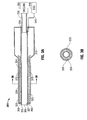
- a suction coagulator 200 in accordance with the present disclosure includes a housing 215 disposed proximally to an elongated shaft 220.
- Housing 215 may be a handle.
- Shaft 220 includes an insulating sheath 226 formed from any suitable dielectric material, for example, polymeric materials such as PU, PVC and the like.
- Shaft 220 includes a conductive tube 224 disposed coaxially within insulating sheath 226 and having a tubular distal tip electrode 225 protruding distally from insulating sheath 226 to form at least one aspiration port 265.
- Conductive tube 224 may be formed from any suitable electrically conductive material, including without limitation, aluminum or stainless steel.
- An insulator 270 having a generally cylindrical shape is disposed between conductive tube 224 and insulating sheath 226.
- Insulator 270 may be formed from any suitable heat-insulating material, for example without limitation, high-temperature polymers, ceramic fiber, or mineral fiber.
- Insulator 270 may be constructed from braided, woven, spun-woven, or randomly dispersed materials.
- An isolator 260 is disposed between distal tip electrode 225 and conductive tube 224 to thermally insulate the distal tip electrode 225 from the conductive tube 224 and to position distal tip electrode 225 coaxially with the distal end of insulating sheath 226.
- Distal tip electrode 225 and conductive tube 224 are operably connected by a conductive element 227, which may be a wire or a strap, to facilitate the delivery of electrosurgical energy to a surgical site (not shown) by distal tip electrode 225.
- a conductive element 227 which may be a wire or a strap, to facilitate the delivery of electrosurgical energy to a surgical site (not shown) by distal tip electrode 225.
- isolator 260 may be formed of heat-resistant material, for example, ceramic material.
- isolator 260 is integrally formed with sheath 226.
- insulator 270 acts to insulate the outer surface of sheath 226 from thermal energy that may propagate from, for example, the surgical site (not explicitly shown), distal tip electrode 225, and/or conductive tube 224.
- Vacuum source 250 may be selectively activated to provide aspiration suction to tube 224 and tip 225 to facilitate the removal of biodebris from the surgical site (not explicitly shown).
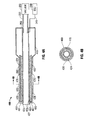
- a suction coagulator 300 includes an elongated shaft 320 supported by a housing 315, the shaft 320 further including an insulator 370 having a generally cylindrical shape that is longitudinally disposed between a conductive tube 324 and a dielectric sheath 326.
- Insulator 370 may be formed from a closed cell foam material, for example without limitation, closed cell polyurethane foam.
- a tubular distal tip electrode 325 extends from the distal end of shaft 320 to form at least one aspiration port 365.
- An isolator 360 is disposed between distal tip electrode 325 and conductive tube 324 to thermally insulate the distal tip electrode 325 from the conductive tube 324 and additionally to position distal tip electrode 325 coaxially with the distal end of dielectric sheath 326.
- Distal tip electrode 325 and conductive tube 324 are operably coupled by a conductive element 327, which may be a wire or a strap.
- isolator 360 may be formed of heat-resistant material, for example, ceramic.
- seal 360 may be integrally formed with sheath 326.
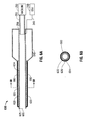
- a suction coagulator 400 includes an elongated shaft 420 that is supported by a housing 415.
- the shaft 420 includes an insulator 470 having a generally cylindrical shape that is longitudinally disposed between a conductive tube 424 and a dielectric sheath 426, and an insulator 480 having a generally cylindrical shape that is longitudinally disposed around dielectric sheath 426.
- a tubular distal tip electrode 425 extends from the distal end of shaft 420 to form at least one aspiration port 465.
- Insulators 470, 480 may be formed from a closed cell foam material, for example without limitation, closed cell polyurethane foam.
- insulators 470, 480 act to insulate the outer surface of shaft 420 from thermal energy that may propagate from, for example, the surgical site, distal tip electrode 425, and/or conductive tube 424.
- An isolator 460 is disposed between distal tip electrode 425 and conductive tube 424 to thermally insulate the distal tip electrode 425 from the conductive tube 424 and additionally to position distal tip electrode 425 coaxially with the distal end of dielectric sheath 426.
- Distal tip electrode 425 and electrode 424 are operably coupled by a conductive element 427, which may be a wire or a strap.
- Insulator 480 may include at the distal end thereof an annular insulating region 481 that encloses the distal end 425 of dielectric sheath 426 and/or isolator 460.
- annular insulating region 481 may be joined to electrode 424 by a bonded region 482, for example, by adhesive, heat weld, or crimp.
- a suction coagulator 500 includes an elongated shaft 520 that is supported by a housing 515.
- the shaft 520 further including a tubular electrode 524 having generally cylindrical sheath 526 longitudinally disposed around the outer surface thereof.
- An insulator 580 is concentrically disposed around sheath 526.
- Insulator 580 may be formed from a closed cell foam material, for example without limitation, closed cell polyurethane foam. In use, insulator 580 acts to reduce the propagation of thermal energy from, for example, the surgical site, an electrode tip 525, and/or electrode 524, to the outer surface of shaft 520.
- FIGs. 6A and 6B there is illustrated an envisioned embodiment of a suction coagulator 600 in accordance with the present disclosure wherein an elongated longitudinal shaft 620 is supported by a housing 615.
- An open cell foam cover 680 surrounds shaft 620.
- the shaft 620 includes a tubular electrosurgical electrode 624 disposed longitudinally therethrough, the tubular electrosurgical electrode 624 having an exposed tip 625 for delivering electrosurgical energy to tissue.
- a generally cylindrical sheath 626 is longitudinally disposed around substantially all but the exposed tip 625 of tubular electrosurgical electrode 624.
- Electrode 624 is in fluid communication with the source of vacuum 250 for the aspiration of biodebris, for example, tissue, eschar, blood and/or other bodily fluids.
- the open cell foam cover may be infused with a fluid (not explicitly shown) for example, water or saline solution.
- a fluid for example, water or saline solution.
- the fluid may increase the thermal mass of the covering and, additionally or alternatively, may provide a cooling effect. In this manner, an increase in surface temperature of the instrument shaft may be diminished or precluded, thereby reducing the risk of undesirable effects on adjacent anatomical structures.
- a suction coagulator 700 includes an elongated shaft 720 that is supported by a housing 715.
- the shaft 720 includes a tubular electrosurgical electrode 724 disposed longitudinally therethrough, the tubular electrosurgical electrode 724 having an exposed tip 725 for delivering electrosurgical energy to tissue.
- a generally cylindrical sheath 726 is longitudinally disposed around substantially all but the exposed tip 725 of tubular electrosurgical electrode 724.
- At least one cooling lumen 770 is disposed longitudinally on the shaft 720 for delivering coolant C to the distal region, i.e., electrode 725 of suction coagulator 700.
- Cooling lumen 770 is in fluid communication with a reservoir 790 via a conduit 795.
- a connector 796 is provided for coupling a conduit 795 to cooling lumen 770.
- Reservoir 790 may contain a coolant, for example without limitation, saline or water.
- coolant C may flow from reservoir 790 through conduit 795, lumen 770, and discharge from distal end 772 of lumen 770.
- a valve (not explicitly shown) may be provided to regulate the flow of coolant. The valve (not explicitly shown) may be caused to be actuated by a user and/or by an automated controller, such as a processor.
- Coolant C may flow from reservoir 790 via gravity feed (i.e., "drip" feed) and/or by pressure feed provided by, for example without limitation, a centrifugal pump, a positive displacement pump, or a peristaltic pump (not explicitly shown).
- gravity feed i.e., "drip" feed
- pressure feed provided by, for example without limitation, a centrifugal pump, a positive displacement pump, or a peristaltic pump (not explicitly shown).
- a proximal housing 815 supports an elongated shaft 820 extending distally therefrom.
- a generally tubular cover 826 is longitudinally disposed around substantially all but an exposed tip 825 of a tubular electrosurgical electrode 824 that is disposed longitudinally through shaft 820.
- a region 871 between cover 826 and electrode 824 defines a cooling jacket 872 that surrounds the tubular electrode 824.
- a cooling jacket 872 may include a cooling lumen 873 having a generally helical shape, and having an inlet port 870 and an outlet port 875.
- the helical coils formed by cooling lumen 873 may form an open helix, wherein the helix pitch is greater than the outer diameter of the cooling lumen 873, or a closed helix wherein the helix pitch is substantially equal to the outer diameter of the cooling lumen 873.
- Inlet port 870 is in fluid communication with a coolant source 890 via a conduit 895.
- Coolant C may be any biocompatible fluid, for example without limitation, saline, water, or air. Coolant C may flow from coolant source 890 via gravity feed (i.e., "drip" feed) and/or by pressure feed provided by, for example, a pump, as previously described herein.
- coolant flows distally though the helical cooling lumen 873 until the distal end 878 of jacket 872 is reached. Coolant C then flows proximally through a return lumen 874 to outlet port 875, whereupon the coolant exits the suction coagulator 800.
- coolant C flow may be reversed from that described hereinabove, i.e., coolant may flow initially to distal end 878 and thereafter proceed proximally through helical cooling lumen 873, and subsequently, discharged from the suction coagulator 800 at outlet port 875. In this manner, a cooling effect can be selectively biased towards a proximal end of the shaft or a distal end of the shaft as desired.
- coolant C may be caused flow distally wherein fresh coolant is introduced to cooling jacket 872 at the proximal end thereof.
- a cooling effect may be biased toward a proximal end 830 of shaft 820, which may be adjacent to, for example, anatomical structures unrelated to the electrosurgical procedure, such as the uvula and the oral commissure area, thereby reducing the risk of undesired effects to such areas.
- cooling may be biased towards a distal end 831 of shaft 820 by causing coolant to flow proximally by introducing coolant C to cooling jacket 872 at the distal end thereof.
- the direction of coolant C flow may be selected by causing a reversing valve (not explicitly shown) that is in fluid communication with cooling jacket 872 to be actuated in a manner consistent with the desired direction of coolant C flow.
- Figs 9A and 9B illustrate still another envisioned embodiment of a suction coagulator 900 in accordance with the present disclosure is illustrated, the suction coagulator including a distal housing 915 having extending distally therefrom an elongated shaft 920.
- Shaft 920 includes a cooling jacket 972 that is formed by the generally cylindrical region longitudinally disposed between a tubular electrode 924 and a tubular cover 926.
- the cooling jacket is sealed at the distal end thereof by distal seal 960 and at the proximal end thereof by proximal seal 961.
- a cooling supply lumen 970 is in fluid communication with the cooling jacket via an inlet port 962 provided by proximal seal 961.
- coolant C is admitted into cooling jacket 972 at the proximal end thereof, and thereafter flows distally.
- a distal return opening 963 is provided by cover 926, or additionally or alternatively, by distal seal 960.
- Supply end 870 is in fluid communication with a coolant source 890 via a conduit 895.
- Coolant C may be any biocompatible fluid, for example without limitation, saline, water, or air. Coolant C may flow to cooling jacket 972 via conduits 970, 995 from coolant source 990 via gravity feed (i.e., "drip" feed) and/or by pressure feed provided by, for example, a pump, as previously described herein.
- coolant C flows distally though the cooling jacket 972 until the distal end 978 of jacket 972 is reached. Coolant C then flows through distal return opening 963, proximally through a return lumen 974 to outlet port 975, whereupon the coolant exits the suction coagulator 900.
- coolant C flow may be reversed from that described hereinabove, i.e., coolant C may flow initially to distal end 978 and thereafter proceed proximally through cooling jacket 972, and subsequently, discharged from the suction coagulator 900 at outlet port 975. The direction of coolant flow may be selectively reversed as previously described herein.
Abstract
Description
- The present invention relates generally to electrosurgical coagulators and, more particularly, to an electrosurgical suction coagulator having improved thermal insulation between the active electrode and adjacent tissue.
- The coagulation of bleeding blood vessels and tissue using electrically conductive suction tubes is a technique which has been widely used for some time. Typically, a combination electrosurgery and suction device is employed in surgery wherever excessive blood must be removed from the bleeding site in order to facilitate hemostasis of any bleeding vessels. More particularly, during any given surgical procedure, several layers of tissue usually must be penetrated to reach the operative field. When resecting an organ, for example, a gallbladder, the tissue surrounding the organ must be penetrated and dissected before the organ can be removed. The tissues being dissected, however, often contain blood vessels, nerves, lymph vessels, and the like, which should not be severed. The technique of blunt dissection is often used to prevent unnecessary damage caused by severing these vessels or nerves.
- Blunt dissection, as opposed to sharp dissection, involves the use of a blunt surface to break through the tissue, thereby preventing the damage and bleeding caused by lasers and scalpels, the tools of sharp dissection. Hard surgical sponges, generally known as peanuts or Kittner sponges, or a surgeon's fingers are often used as blunt dissectors. A peanut is a tightly wound ball of absorbent material, such as gauze or other woven cotton, which is typically gripped with forceps and acts to abrade the tissue being dissected so that the dissection can be performed by either pulling on the tissue or by forcing the peanut through the tissue.
- Laparoscopy, surgery performed through several small incisions made in the body rather than through a single large opening, has become the preferred method of performing certain procedures due to the reduced trauma and risk of infection as compared to normal, open surgical procedures. As a result, the use of conventional blunt dissectors, such as the peanut, during laparoscopic procedures presents many significant drawbacks. For instance, peanuts, being secured only by forceps, can become loose in the body. Further, the view of the operative field often becomes obstructed by pieces of tissue, blood and other bodily fluids produced during blunt dissection, necessitating the immediate need for both irrigation and aspiration of the operative field. Since it is undesirable to create additional incisions, the dissection must be stopped, the dissector must be removed, and an irrigator and/or aspirator must be inserted to remove the fluid and debris.
- The use of electrical energy including radiofrequency and microwave energy and, in particular, radiofrequency ("RF") electrodes or microwave antennae for ablation of tissue in the body or for the treatment of pain is known. For example, electrosurgery is a technique of using alternating current electrical signals in the approximately 200 kHz-3.3 mHz range that are generated by a source of electrosurgical energy, such as an electrosurgical generator, in connection with surgical instruments, to cut or coagulate biologic tissue endogenically. This electrosurgical signal can be a sinusoidal waveform operating in a continuous mode at a 100% duty cycle, or pulse modulated at a duty cycle of less than 100%. Typically, electrosurgical signals are operated at 100% duty cycle for maximal cutting effect, and are pulse modulated at duty cycles ranging from 50% to 25% for less aggressive cutting, or, at a substantially lower duty cycle of approximately 6%, for coagulating. The electrosurgical carrier signal may also be varied in intensity. The electrosurgical signal is applied to the patient via electrodes in either monopolar mode, or bipolar mode. In monopolar mode, the active electrode is the surgical instrument at the surgical site, and the return electrode is elsewhere on the patient, such that the electrosurgical signal passes through the patient's body from the surgical site to the return electrode. In bipolar mode, both the active and return electrodes are at the surgical site, such as with an instrument having an array of electrodes, so that the electrosurgical signal passes only through the tissue situated between the RF electrodes of the instrument.
- Electrosurgical suction coagulators which both coagulate and dissect tissue have also been available for some time. Generally, these devices include a shaft formed from a conductive suction tube electrode having an electrically insulating coating over all but a most distal portion of the tube, so that the distal portion forms a generally annular ablating electrode. The shaft may be formed of malleable materials to enable a surgeon to bend the shaft to a desired shape. The distal end can be used as a blunt dissection device and/or a blunt coagulator. A suction source is attached to a proximal portion of the tube for evacuating excess fluid and debris from the surgical site through the distal end of the tube. The electrode is operably coupled to a source of electrosurgical energy, such as an electrosurgical generator.
- The described electrosurgical suction coagulators may have drawbacks. In particular, heat conducted from the suction tube electrode to the outer surface of the shaft may cause the surface of the shaft to reach temperatures of 60°C or greater. This may be a concern during surgical procedures, such as an electrosurgical adenotonsillectomy, where the shaft of a suction coagulator may be in proximity to, or in contact with, anatomical structures unrelated to the procedure, such as the uvula or the oral commissure. The elevated shaft temperature may have undesirable effects on such unrelated anatomical structures, including uvular edema and erythema of the oral commissure area. An electrosurgical suction coagulator which avoids or minimizes such undesirable effects would be a welcome advance in the art, particularly when such benefits are realized in a rugged, reliable, and relatively simple design.
- The present disclosure provides an electrosurgical suction coagulation system having, and related methods for, improved control of the shaft surface temperature. In particular, embodiments in accordance with the present disclosure may provide passive thermal insulation of the shaft, active cooling of the shaft, and may advantageously include combinations of passive insulation and active cooling, as will be described hereinbelow.
- In an embodiment in accordance with the present disclosure, an electrosurgical suction coagulator includes a shaft formed from a conductive suction tube, an outer dielectric sheath covering over all but a distal electrode portion of the tube, and has disposed therebetween an insulating layer formed from braided material having low thermal conduction, for example, braided polymeric or ceramic fibers. The braided material may be configured as a tubular braided sheath or a spiral wrapped layer. The combination of air voids in the braided layer and the low thermal conductive properties of the braided insulating material may reduce thermal conduction from the metallic suction tube to the exterior surface of the instrument. In envisioned embodiments, an insulating layer may be formed from woven material.
- In embodiments, the shaft of a suction coagulator in accordance with the present disclosure may be straight or contoured. The shaft may additionally be formed from malleable materials to enable a user, for example, a surgeon or clinician, to bend the shaft to a desired shape. A suction coagulator in accordance with the present disclosure may include a handle. The handle may include at least one control for activating the electrosurgical energy and/or evacuation (i.e., suction).
- In envisioned embodiments, a suction coagulator includes thermal isolation between a suction tube and a distal electrode tip, formed from, for example without limitation, ceramic insulating material and/or polymeric insulating material. The tip may be operably coupled to the suction tube by at least one electrically conductive element, such as a wire. Additionally or alternatively, a distal electrode tip may be operably coupled to a source of electrosurgical energy by at least one of a wire and the suction tube.
- In another envisioned embodiment, an insulating layer disposed between the tubular electrode and dielectric sheath is formed from closed-cell foam material, for example, closed cell foamed polyurethane. Additionally or alternatively, the outer surface of the dielectric sheath may include a closed cell foam covering disposed thereupon, which may further reduce thermal conduction from the electrode to adjacent tissue.
- In embodiments, the outer surface of the dielectric sheath may include an open cell foam covering disposed thereupon. During use, the open foam layer may be infused with a fluid, for example, water or saline solution, which may increase the thermal mass of the covering and provide a cooling effect, thereby reducing surface temperature of the instrument shaft.
- In embodiments, an electrosurgical generator in accordance with the present disclosure may be configured to limit the activation time of a suction coagulator, and/or enforce minimum quiescent times between activations. During use, the electrosurgical generator may determine whether the activation time has exceeded a threshold, and in response thereto, deactivate the generator. Additionally, reactivation of the generator may be inhibited until the expiration of a "rest" time period, or until a user input is received by the generator.
- The instrument may be configured to provide instrument identification information to the generator, for example, an optical code (i.e., barcode), an RFID tag, or other suitable machine- or human-readable encodings. The generator may use such instrument identification information to determine corresponding activation and quiescent time parameters for the instrument.
- In an envisioned embodiment, a suction coagulator in accordance with the present disclosure includes a sensor that is adapted to sense the surface temperature of the instrument. The sensor may be operably coupled to an electrosurgical generator. The electrosurgical generator may be configured to respond to the sensed temperature, by, for example, limiting the activation time, altering the electrosurgical signal, and/or deactivating the generation of the electrosurgical signal. In embodiments, the generator may additionally or alternatively respond to at least one parameter related to the sensed temperature of the instrument, for example, a change in temperature of the instrument and/or a rate of change of temperature of the instrument.
- In embodiments, an electrosurgical generator in accordance with the present disclosure may be configured to issue a prompt (e.g., an alarm) to the user. A prompt may be issued to advise the user to pause the activation of the instrument. In envisioned embodiments, a prompt may be issued to advise the user to replenish depleted fluids in, for example, a fluid-infused open foam cover. Such a prompt may be based upon, for example, cumulative activation time, instrument identity, and/or the surface temperature of the instrument. The alarm may be automatically cleared after a predetermined time period. Additionally or alternatively, the alarm may be cleared by a user input received by the electrosurgical generator.
- Other embodiments according to the present disclosure are envisioned wherein an electrosurgical suction coagulator includes a conduit for introducing a coolant, for example, saline solution, to the distal tip of the instrument during use. The conduit may be configured to "drip" coolant onto an electrode disposed at the distal end of the instrument. The conduit may be in fluid communication, preferably at the proximal end of the instrument, to a source of cooling fluid, for example, a saline bag, that may provide cooling fluid via any suitable manner of delivery, for example, by gravity feed, pump, or pressurized vessel.
- In other envisioned embodiments, en electrosurgical suction coagulator according the present disclosure includes a coolant jacket that may be formed by a conduit included in the instrument. Coolant is introduced into the coolant jacket, preferably at the proximal end of the instrument, flows through the conduit towards the distal tip region of the instrument, and exits the instrument. The coolant jacket may be configured to cool the tip and/or the surface of the instrument.
- The above and other aspects, features, and advantages of the present disclosure will become more apparent in light of the following detailed description when taken in conjunction with the accompanying drawings in which:
-
Fig. 1 is an oblique view of an exemplary embodiment of an electrosurgical suction coagulator system in accordance with the present disclosure; -
Fig. 2A is a side cutaway view of an exemplary embodiment of an electrosurgical suction coagulator in accordance with the present disclosure having a braided insulation region; -
Fig. 2B is a section view of the electrosurgical suction coagulator ofFig. 2A ; -
Fig. 3A is a side cutaway view of another exemplary embodiment of an electrosurgical suction coagulator in accordance with the present disclosure having a closed cell foam insulation region; -
Fig. 3B is a section view of the electrosurgical suction coagulator ofFig. 3A ; -
Fig. 4A is a side cutaway view of yet another exemplary embodiment of an electrosurgical suction coagulator in accordance with the present disclosure having an inner closed cell foam insulation region and an outer closed cell foam insulation region; -
Fig. 4B is a section view of the electrosurgical suction coagulator ofFig. 4A ; -
Fig. 5A is a side cutaway view of still another exemplary embodiment of an electrosurgical suction coagulator in accordance with the present disclosure having an outer closed cell foam insulation region; -
Fig. 5B is a section view of the electrosurgical suction coagulator ofFig. 5A ; -
Fig. 6A is a side cutaway view of another exemplary embodiment of an electrosurgical suction coagulator in accordance with the present disclosure having an outer open cell foam insulation region; -
Fig. 6B is a section view of the electrosurgical suction coagulator ofFig. 6A ; -
Fig. 7A is a side cutaway view of another exemplary embodiment of an electrosurgical suction coagulator in accordance with the present disclosure having a lumen to deliver coolant to the distal end thereof; -
Fig. 7B is a section view of the electrosurgical suction coagulator ofFig. 7A ; -
Fig. 8A is a side cutaway view of an exemplary embodiment of an electrosurgical suction coagulator in accordance with the present disclosure having a spiral coolant jacket; -
Fig. 8B is an oblique view of the exemplary electrosurgical suction coagulator ofFig. 8A ; -
Fig. 9A is a side cutaway view of an exemplary embodiment of an electrosurgical suction coagulator in accordance with the present disclosure having a cylindrical coolant jacket; and -
Fig. 9B is a section view of the electrosurgical suction coagulator ofFig. 9A . - Particular embodiments of the present disclosure will be described herein with reference to the accompanying drawings. As shown in the drawings and as described throughout the following description, and as is traditional when referring to relative positioning on an object, the term "proximal" refers to the end of the apparatus that is closer to the user and the term "distal" refers to the end of the apparatus that is further from the user. In the following description, well-known functions or constructions are not described in detail to avoid obscuring the present disclosure in unnecessary detail.
- With reference to
Fig. 1 , an electrosurgicalsuction coagulator system 100 is presented having asuction coagulator 110 that is operably coupled to anelectrosurgical generator 140 via aconductor 145.Suction coagulator 110 is operably coupled to avacuum source 150 by alumen 155.Suction coagulator 110 includes ahandle 115 disposed at the proximal end thereof and aelongated shaft 120 extending distally from thehandle 115. Theshaft 120 may be formed from material having malleable or flexible properties, for example without limitation, metallic material such as aluminum and alloys thereof and/or polymeric materials such as polyurethane (PU) or polyvinyl chloride (PVC). Ashaft 120 thus formed may be bent to a desired shape by the user, as shown by way of example by bent shaft 120'. -
Distal end 124 ofshaft 120 includes an exposedtubular electrode 125 for delivering electrosurgical energy to tissue, theelectrode 125 having aconduit 126 defined longitudinally therethrough for providing suction to a surgical site.Conduit 126 is in fluid communication withvacuum source 150 vialumen 155. - In an embodiment, handle 115 may include a
control 130 which may be a handswitch for controlling the application of electrosurgical energy, i.e., activation and deactivation of an electrosurgical signal. Handle 115 may include an additional orsecond control 131 for controlling the application of suction to the surgical site. In embodiments,control 131 may be operably coupled to a valve (not shown) that may be disposed withinhandle 115,shaft 120,vacuum source 150, and/orlumen 155. In other envisioned embodiments,control 131 may be operably coupled to a regulator, motor control, or other suitable manner of vacuum control. - Turning now to
Figs. 2A and 2B , asuction coagulator 200 in accordance with the present disclosure includes ahousing 215 disposed proximally to anelongated shaft 220.Housing 215 may be a handle.Shaft 220 includes an insulatingsheath 226 formed from any suitable dielectric material, for example, polymeric materials such as PU, PVC and the like.Shaft 220 includes aconductive tube 224 disposed coaxially within insulatingsheath 226 and having a tubulardistal tip electrode 225 protruding distally from insulatingsheath 226 to form at least oneaspiration port 265.Conductive tube 224 may be formed from any suitable electrically conductive material, including without limitation, aluminum or stainless steel. Aninsulator 270 having a generally cylindrical shape is disposed betweenconductive tube 224 and insulatingsheath 226.Insulator 270 may be formed from any suitable heat-insulating material, for example without limitation, high-temperature polymers, ceramic fiber, or mineral fiber.Insulator 270 may be constructed from braided, woven, spun-woven, or randomly dispersed materials. Anisolator 260 is disposed betweendistal tip electrode 225 andconductive tube 224 to thermally insulate thedistal tip electrode 225 from theconductive tube 224 and to positiondistal tip electrode 225 coaxially with the distal end of insulatingsheath 226.Distal tip electrode 225 andconductive tube 224 are operably connected by aconductive element 227, which may be a wire or a strap, to facilitate the delivery of electrosurgical energy to a surgical site (not shown) bydistal tip electrode 225. In an embodiment,isolator 260 may be formed of heat-resistant material, for example, ceramic material. In other envisioned embodiments,isolator 260 is integrally formed withsheath 226. In use,insulator 270 acts to insulate the outer surface ofsheath 226 from thermal energy that may propagate from, for example, the surgical site (not explicitly shown),distal tip electrode 225, and/orconductive tube 224. Vacuumsource 250 may be selectively activated to provide aspiration suction totube 224 andtip 225 to facilitate the removal of biodebris from the surgical site (not explicitly shown). - In another envisioned embodiment best illustrated in
Figs. 3A and 3B , asuction coagulator 300 includes anelongated shaft 320 supported by ahousing 315, theshaft 320 further including aninsulator 370 having a generally cylindrical shape that is longitudinally disposed between aconductive tube 324 and adielectric sheath 326.Insulator 370 may be formed from a closed cell foam material, for example without limitation, closed cell polyurethane foam. A tubulardistal tip electrode 325 extends from the distal end ofshaft 320 to form at least oneaspiration port 365. Anisolator 360 is disposed betweendistal tip electrode 325 andconductive tube 324 to thermally insulate thedistal tip electrode 325 from theconductive tube 324 and additionally to positiondistal tip electrode 325 coaxially with the distal end ofdielectric sheath 326.Distal tip electrode 325 andconductive tube 324 are operably coupled by aconductive element 327, which may be a wire or a strap. In an embodiment,isolator 360 may be formed of heat-resistant material, for example, ceramic. In other envisioned embodiments,seal 360 may be integrally formed withsheath 326. - In yet another envisioned embodiment best illustrated in
Figs. 4A and 4B , asuction coagulator 400 includes anelongated shaft 420 that is supported by ahousing 415. Theshaft 420 includes aninsulator 470 having a generally cylindrical shape that is longitudinally disposed between aconductive tube 424 and adielectric sheath 426, and aninsulator 480 having a generally cylindrical shape that is longitudinally disposed arounddielectric sheath 426. A tubulardistal tip electrode 425 extends from the distal end ofshaft 420 to form at least oneaspiration port 465.Insulators insulators shaft 420 from thermal energy that may propagate from, for example, the surgical site,distal tip electrode 425, and/orconductive tube 424. Anisolator 460 is disposed betweendistal tip electrode 425 andconductive tube 424 to thermally insulate thedistal tip electrode 425 from theconductive tube 424 and additionally to positiondistal tip electrode 425 coaxially with the distal end ofdielectric sheath 426.Distal tip electrode 425 andelectrode 424 are operably coupled by aconductive element 427, which may be a wire or a strap.Insulator 480 may include at the distal end thereof an annularinsulating region 481 that encloses thedistal end 425 ofdielectric sheath 426 and/orisolator 460. In embodiments, annularinsulating region 481 may be joined toelectrode 424 by a bondedregion 482, for example, by adhesive, heat weld, or crimp. - Turning to
Figs. 5A and 5B , yet another embodiment according to the present disclosure is illustrated wherein asuction coagulator 500 includes anelongated shaft 520 that is supported by ahousing 515. Theshaft 520 further including atubular electrode 524 having generallycylindrical sheath 526 longitudinally disposed around the outer surface thereof. Aninsulator 580 is concentrically disposed aroundsheath 526.Insulator 580 may be formed from a closed cell foam material, for example without limitation, closed cell polyurethane foam. In use,insulator 580 acts to reduce the propagation of thermal energy from, for example, the surgical site, anelectrode tip 525, and/orelectrode 524, to the outer surface ofshaft 520. - In
Figs. 6A and 6B there is illustrated an envisioned embodiment of asuction coagulator 600 in accordance with the present disclosure wherein an elongatedlongitudinal shaft 620 is supported by ahousing 615. An opencell foam cover 680 surroundsshaft 620. Theshaft 620 includes a tubularelectrosurgical electrode 624 disposed longitudinally therethrough, the tubularelectrosurgical electrode 624 having an exposed tip 625 for delivering electrosurgical energy to tissue. A generallycylindrical sheath 626 is longitudinally disposed around substantially all but the exposed tip 625 of tubularelectrosurgical electrode 624.Electrode 624 is in fluid communication with the source ofvacuum 250 for the aspiration of biodebris, for example, tissue, eschar, blood and/or other bodily fluids. During use, the open cell foam cover may be infused with a fluid (not explicitly shown) for example, water or saline solution. The fluid may increase the thermal mass of the covering and, additionally or alternatively, may provide a cooling effect. In this manner, an increase in surface temperature of the instrument shaft may be diminished or precluded, thereby reducing the risk of undesirable effects on adjacent anatomical structures. - In
Figs. 7A and 7B , there is shown an envisioned embodiment wherein asuction coagulator 700 includes anelongated shaft 720 that is supported by ahousing 715. Theshaft 720 includes a tubularelectrosurgical electrode 724 disposed longitudinally therethrough, the tubularelectrosurgical electrode 724 having an exposedtip 725 for delivering electrosurgical energy to tissue. A generallycylindrical sheath 726 is longitudinally disposed around substantially all but the exposedtip 725 of tubularelectrosurgical electrode 724. At least onecooling lumen 770 is disposed longitudinally on theshaft 720 for delivering coolant C to the distal region, i.e.,electrode 725 ofsuction coagulator 700.Cooling lumen 770 is in fluid communication with areservoir 790 via aconduit 795. In embodiments, aconnector 796 is provided for coupling aconduit 795 tocooling lumen 770.Reservoir 790 may contain a coolant, for example without limitation, saline or water. In use, coolant C may flow fromreservoir 790 throughconduit 795,lumen 770, and discharge fromdistal end 772 oflumen 770. A valve (not explicitly shown) may be provided to regulate the flow of coolant. The valve (not explicitly shown) may be caused to be actuated by a user and/or by an automated controller, such as a processor. Coolant C may flow fromreservoir 790 via gravity feed (i.e., "drip" feed) and/or by pressure feed provided by, for example without limitation, a centrifugal pump, a positive displacement pump, or a peristaltic pump (not explicitly shown). - Turning now to
Figs. 8A and8B , another envisioned embodiment of asuction coagulator 800 in accordance with the present disclosure is illustrated wherein aproximal housing 815 supports anelongated shaft 820 extending distally therefrom. A generallytubular cover 826 is longitudinally disposed around substantially all but an exposedtip 825 of a tubularelectrosurgical electrode 824 that is disposed longitudinally throughshaft 820. A region 871 betweencover 826 andelectrode 824 defines acooling jacket 872 that surrounds thetubular electrode 824. As best shown inFig 8A and8B , a coolingjacket 872 may include acooling lumen 873 having a generally helical shape, and having aninlet port 870 and anoutlet port 875. The helical coils formed by coolinglumen 873 may form an open helix, wherein the helix pitch is greater than the outer diameter of thecooling lumen 873, or a closed helix wherein the helix pitch is substantially equal to the outer diameter of thecooling lumen 873.Inlet port 870 is in fluid communication with a coolant source 890 via a conduit 895. Coolant C may be any biocompatible fluid, for example without limitation, saline, water, or air. Coolant C may flow from coolant source 890 via gravity feed (i.e., "drip" feed) and/or by pressure feed provided by, for example, a pump, as previously described herein. In one embodiment, coolant flows distally though thehelical cooling lumen 873 until thedistal end 878 ofjacket 872 is reached. Coolant C then flows proximally through areturn lumen 874 tooutlet port 875, whereupon the coolant exits thesuction coagulator 800. In another embodiment, coolant C flow may be reversed from that described hereinabove, i.e., coolant may flow initially todistal end 878 and thereafter proceed proximally throughhelical cooling lumen 873, and subsequently, discharged from thesuction coagulator 800 atoutlet port 875. In this manner, a cooling effect can be selectively biased towards a proximal end of the shaft or a distal end of the shaft as desired. For example, in use during an electrosurgical procedure such as an adenotonsillectomy, coolant C may be caused flow distally wherein fresh coolant is introduced to coolingjacket 872 at the proximal end thereof. Thus a cooling effect may be biased toward a proximal end 830 ofshaft 820, which may be adjacent to, for example, anatomical structures unrelated to the electrosurgical procedure, such as the uvula and the oral commissure area, thereby reducing the risk of undesired effects to such areas. Alternatively, cooling may be biased towards adistal end 831 ofshaft 820 by causing coolant to flow proximally by introducing coolant C to coolingjacket 872 at the distal end thereof. In embodiments, the direction of coolant C flow may be selected by causing a reversing valve (not explicitly shown) that is in fluid communication withcooling jacket 872 to be actuated in a manner consistent with the desired direction of coolant C flow. -
Figs 9A and 9B illustrate still another envisioned embodiment of asuction coagulator 900 in accordance with the present disclosure is illustrated, the suction coagulator including adistal housing 915 having extending distally therefrom anelongated shaft 920.Shaft 920 includes acooling jacket 972 that is formed by the generally cylindrical region longitudinally disposed between atubular electrode 924 and atubular cover 926. The cooling jacket is sealed at the distal end thereof bydistal seal 960 and at the proximal end thereof byproximal seal 961. Acooling supply lumen 970 is in fluid communication with the cooling jacket via aninlet port 962 provided byproximal seal 961. During use, coolant C is admitted intocooling jacket 972 at the proximal end thereof, and thereafter flows distally. A distal return opening 963 is provided bycover 926, or additionally or alternatively, bydistal seal 960.Supply end 870 is in fluid communication with a coolant source 890 via a conduit 895. Coolant C may be any biocompatible fluid, for example without limitation, saline, water, or air. Coolant C may flow to coolingjacket 972 viaconduits 970, 995 from coolant source 990 via gravity feed (i.e., "drip" feed) and/or by pressure feed provided by, for example, a pump, as previously described herein. In one embodiment, coolant C flows distally though the coolingjacket 972 until thedistal end 978 ofjacket 972 is reached. Coolant C then flows through distal return opening 963, proximally through areturn lumen 974 tooutlet port 975, whereupon the coolant exits thesuction coagulator 900. In another embodiment, coolant C flow may be reversed from that described hereinabove, i.e., coolant C may flow initially todistal end 978 and thereafter proceed proximally through coolingjacket 972, and subsequently, discharged from thesuction coagulator 900 atoutlet port 975. The direction of coolant flow may be selectively reversed as previously described herein. - The described embodiments of the present disclosure are intended to be illustrative rather than restrictive, and are not intended to represent every embodiment of the present disclosure. Further variations of the above-disclosed embodiments and other features and functions, or alternatives thereof, may be made or desirably combined into many other different systems or applications without departing from the spirit or scope of the disclosure as set forth in the following claims both literally and in equivalents recognized in law.
Claims (11)
- An electrosurgical suction coagulator, comprising:a housing having proximal and distal ends and a substantially malleable elongated tube-like shaft extending longitudinally from the distal end thereof, the substantially malleable elongated tube-like shaft including:a tube-like dielectric sheath;a tube-like electrode disposed coaxially through the tube-like dielectric sheath and configured to operably couple to a source of electrosurgical energy, wherein the distal end of the tube-like electrode protrudes at least partially from the distal end of the tube-like dielectric sheath, the tube-like electrode having at the distal end thereof at least one aspiration port defined therein, the tube-like electrode being adapted at the proximal end thereof to operably couple to a source of aspiration suction; anda generally cylindrical region coaxially disposed between the tube-like sheath and the tube-like electrode, wherein the cylindrical insulating region is selectively cooled utilizing at least one of a coolant from a cooling source and an insulator.
- An electrosurgical suction coagulator according to claim 1, wherein the insulator includes a braided material.
- An electrosurgical suction coagulator according to claim 1, wherein the insulator includes a closed cell foam material.
- An electrosurgical suction coagulator according to claim 1, 2 or 3, further comprising:an external insulator disposed coaxially around the tube-like dielectric sheath, the external insulator extending from about the proximal end of the dielectric sheath to about the distal end of the dielectric sheath.
- An electrosurgical suction coagulator according to claim 4, wherein the eternal insulator includes a closed cell foam material.
- An electrosurgical suction coagulator according to claim 4 or 5, wherein the distal end of the external insulator includes an annular distal insulating region.
- An electrosurgical suction coagulator according to claim 6, wherein the annular distal insulating region is joined to the tube-like electrode.
- An electrosurgical suction coagulator according to any one of the preceding claims, further comprising:at least one control which activates at least one of the source of electrosurgical energy and source of aspiration suction.
- An electrosurgical suction coagulator according to any one of the preceding claims, further comprising:a cooling jacket having an inlet port and an outlet port, wherein the inlet port is configured to operably couple to a source of coolant and the outlet port is configured to expel coolant from the electrosurgical suction coagulator.
- An electrosurgical suction coagulator according to claim 9, wherein the cooling jacket includes a lumen having a substantially helical shape disposed concentrically around the tube-like electrode.
- An electrosurgical suction coagulator according to claim 9 or 10, wherein the cooling jacket includes a chamber having a substantially cylindrical shape disposed concentrically around the tube-like electrode.
Applications Claiming Priority (1)
| Application Number | Priority Date | Filing Date | Title |
|---|---|---|---|
| US12/179,206 US8328804B2 (en) | 2008-07-24 | 2008-07-24 | Suction coagulator |
Publications (2)
| Publication Number | Publication Date |
|---|---|
| EP2147651A1 true EP2147651A1 (en) | 2010-01-27 |
| EP2147651B1 EP2147651B1 (en) | 2015-05-27 |
Family
ID=41087339
Family Applications (1)
| Application Number | Title | Priority Date | Filing Date |
|---|---|---|---|
| EP09009623.1A Not-in-force EP2147651B1 (en) | 2008-07-24 | 2009-07-24 | Suction coagulator with thermal insulation |
Country Status (2)
| Country | Link |
|---|---|
| US (3) | US8328804B2 (en) |
| EP (1) | EP2147651B1 (en) |
Cited By (15)
| Publication number | Priority date | Publication date | Assignee | Title |
|---|---|---|---|---|
| EP2263585A1 (en) * | 2009-06-19 | 2010-12-22 | Tyco Healthcare Group, LP | Thermal barrier for suction coagulator |
| WO2012139135A3 (en) * | 2011-04-08 | 2013-01-17 | Vivant Medical, Inc. | Flexible microwave catheters for natural or artificial lumens |
| EP2765837A2 (en) * | 2013-02-11 | 2014-08-13 | Colorado State University Research Foundation | System and method for treatment of biofilms |
| US9044254B2 (en) | 2012-08-07 | 2015-06-02 | Covidien Lp | Microwave ablation catheter and method of utilizing the same |
| US9121774B2 (en) | 2012-06-22 | 2015-09-01 | Covidien Lp | Microwave thermometry for microwave ablation systems |
| US9486271B2 (en) | 2012-03-05 | 2016-11-08 | Covidien Lp | Method and apparatus for identification using capacitive elements |
| US9610122B2 (en) | 2013-03-29 | 2017-04-04 | Covidien Lp | Step-down coaxial microwave ablation applicators and methods for manufacturing same |
| USRE46362E1 (en) | 2009-11-16 | 2017-04-11 | Covidien Lp | Twin sealing chamber hub |
| US10130382B2 (en) | 2014-03-27 | 2018-11-20 | Medtronic Xomed, Inc. | Powered surgical handpiece having a surgical tool with an RFID tag |
| US10376309B2 (en) | 2016-08-02 | 2019-08-13 | Covidien Lp | Ablation cable assemblies and a method of manufacturing the same |
| US10624697B2 (en) | 2014-08-26 | 2020-04-21 | Covidien Lp | Microwave ablation system |
| US10813691B2 (en) | 2014-10-01 | 2020-10-27 | Covidien Lp | Miniaturized microwave ablation assembly |
| US10813692B2 (en) | 2016-02-29 | 2020-10-27 | Covidien Lp | 90-degree interlocking geometry for introducer for facilitating deployment of microwave radiating catheter |
| US11065053B2 (en) | 2016-08-02 | 2021-07-20 | Covidien Lp | Ablation cable assemblies and a method of manufacturing the same |
| US11197715B2 (en) | 2016-08-02 | 2021-12-14 | Covidien Lp | Ablation cable assemblies and a method of manufacturing the same |
Families Citing this family (29)
| Publication number | Priority date | Publication date | Assignee | Title |
|---|---|---|---|---|
| US8328804B2 (en) | 2008-07-24 | 2012-12-11 | Covidien Lp | Suction coagulator |
| US9833281B2 (en) * | 2008-08-18 | 2017-12-05 | Encision Inc. | Enhanced control systems including flexible shielding and support systems for electrosurgical applications |
| US9060765B2 (en) | 2010-11-08 | 2015-06-23 | Bovie Medical Corporation | Electrosurgical apparatus with retractable blade |
| US20130046299A1 (en) * | 2011-08-18 | 2013-02-21 | John J. Newkirk | Intelligent electrosurgical electrode and tracking system |
| JP5198683B1 (en) * | 2012-11-21 | 2013-05-15 | 富士システムズ株式会社 | Drain tube for washing electric knife |
| JP5275527B1 (en) * | 2013-02-05 | 2013-08-28 | 富士システムズ株式会社 | Drain tube for washing electric knife |
| EP2968925B1 (en) | 2013-03-14 | 2020-02-19 | Cynosure, LLC | Electrosurgical systems |
| US20140276813A1 (en) * | 2013-03-15 | 2014-09-18 | Larry Kyle Gambrell | Suction electrocautery device having controlled irrigation and rotating auger |
| WO2014145148A2 (en) * | 2013-03-15 | 2014-09-18 | Ellman International, Inc. | Surgical instruments and systems with multimodes of treatments and electrosurgical operation |
| US9956029B2 (en) | 2014-10-31 | 2018-05-01 | Medtronic Advanced Energy Llc | Telescoping device with saline irrigation line |
| US10080600B2 (en) | 2015-01-21 | 2018-09-25 | Covidien Lp | Monopolar electrode with suction ability for CABG surgery |
| WO2016123147A1 (en) * | 2015-01-28 | 2016-08-04 | Bovie Medical Corporation | Cold plasma electrosurgical apparatus with bent tip applicator |
| US9775673B2 (en) | 2015-03-26 | 2017-10-03 | Garner B. Meads, JR. | Nasal coagulation suction device and methods |
| US10806512B2 (en) | 2015-03-26 | 2020-10-20 | Garner B. Meads, JR. | Nasal coagulation suction device and methods |
| US20160317209A1 (en) * | 2015-04-28 | 2016-11-03 | I.C. Medical, Inc. | Electrosurgery blades with argon beam capability |
| US11039875B2 (en) | 2016-04-26 | 2021-06-22 | Kirwan Surgical Products Llc | Non-stick monopolar suction coagulator |
| US10052169B2 (en) | 2016-11-03 | 2018-08-21 | Meshil A. M. O. H. Al-Jarba | Shield for electrosurgical suction coagulator and kit including the same |
| US10335225B2 (en) * | 2016-11-21 | 2019-07-02 | Arthrex, Inc. | Electrosurgical medical device handpiece with insulated aspiration system |
| AU2018212000B2 (en) | 2017-01-30 | 2023-06-29 | Apyx Medical Corporation | Electrosurgical apparatus with flexible shaft |
| WO2018222562A1 (en) | 2017-05-30 | 2018-12-06 | Bovie Medical Corporation | Electrosurgical apparatus with robotic tip |
| USD875933S1 (en) | 2017-07-26 | 2020-02-18 | Brian K. Reaux | Medical coagulation and ablation blade |
| KR102505869B1 (en) | 2018-02-07 | 2023-03-07 | 싸이노슈어, 엘엘씨 | Method and Apparatus for Controlled RF Processing and RF Generator System |
| US11903636B2 (en) * | 2018-09-27 | 2024-02-20 | Covidien Lp | Energy-based tissue specimen removal |
| US11298155B2 (en) | 2019-04-24 | 2022-04-12 | Covidien Lp | Cutting guard with radiofrequency dissection |
| US11364071B2 (en) | 2019-04-24 | 2022-06-21 | Covidien Lp | Handheld dissector |
| USD1005484S1 (en) | 2019-07-19 | 2023-11-21 | Cynosure, Llc | Handheld medical instrument and docking base |
| US20210145556A1 (en) * | 2019-11-18 | 2021-05-20 | Salvatore Dericco | Oral irrigator tip |
| US11730533B2 (en) | 2020-06-12 | 2023-08-22 | Covidien Lp | Auxiliary electrosurgical return rivet for use with cutting guard |
| US11147545B1 (en) | 2020-06-12 | 2021-10-19 | Covidien Lp | Cutting guard with ground connection |
Citations (7)
| Publication number | Priority date | Publication date | Assignee | Title |
|---|---|---|---|---|
| US3971383A (en) * | 1974-05-07 | 1976-07-27 | Erbe Elektromedizin Kg | Cryogenic surgical instrument |
| US5277696A (en) * | 1991-11-19 | 1994-01-11 | Delma Elektro- Und Medizinische Apparatebau Gesellschaft Mbh | Medical high frequency coagulation instrument |
| US5730742A (en) * | 1994-08-04 | 1998-03-24 | Alto Development Corporation | Inclined, flared, thermally-insulated, anti-clog tip for electrocautery suction tubes |
| US5951546A (en) * | 1994-12-13 | 1999-09-14 | Lorentzen; Torben | Electrosurgical instrument for tissue ablation, an apparatus, and a method for providing a lesion in damaged and diseased tissue from a mammal |
| US6379348B1 (en) * | 2000-03-15 | 2002-04-30 | Gary M. Onik | Combined electrosurgical-cryosurgical instrument |
| US20040082951A1 (en) * | 2002-09-10 | 2004-04-29 | O'halloran Laurence R. | Beveled tonsil suction cautery dissector |
| US20040260280A1 (en) * | 2003-05-01 | 2004-12-23 | Sartor Joe Don | Suction coagulator with dissecting probe |
Family Cites Families (102)
| Publication number | Priority date | Publication date | Assignee | Title |
|---|---|---|---|---|
| US2808833A (en) * | 1952-12-06 | 1957-10-08 | Birtcher Corp | Gas blanketed clotting instrument |
| DE1159574B (en) | 1961-11-29 | 1963-12-19 | Siemens Reiniger Werke Ag | Safety device for high-frequency surgical apparatus |
| FR1340509A (en) | 1962-11-27 | 1963-10-18 | Siemens Reiniger Werke Ag | Safety device for high frequency surgical devices |
| GB1222243A (en) | 1967-07-05 | 1971-02-10 | Kearns Tribune Corp | Generating plasma |
| US3630203A (en) * | 1969-12-11 | 1971-12-28 | Air Prod & Chem | Cryogenic biological apparatus |
| US3838242A (en) | 1972-05-25 | 1974-09-24 | Hogle Kearns Int | Surgical instrument employing electrically neutral, d.c. induced cold plasma |
| US3974833A (en) * | 1973-03-19 | 1976-08-17 | Durden Iii John G | Disposable electrosurgical cautery having optional suction control feature |
| US3828780A (en) * | 1973-03-26 | 1974-08-13 | Valleylab Inc | Combined electrocoagulator-suction instrument |
| FR2235669A1 (en) | 1973-07-07 | 1975-01-31 | Lunacek Boris | Gynaecological sterilisation instrument - has hollow electrode protruding from the end of a curved ended tube |
| DE2429021C2 (en) | 1974-06-18 | 1983-12-08 | Erbe Elektromedizin GmbH, 7400 Tübingen | Remote switching device for an HF surgical device |
| DE2460481A1 (en) | 1974-12-20 | 1976-06-24 | Delma Elektro Med App | Electrode grip for remote HF surgical instrument switching - has shaped insulated piece with contact ring of sterilizable (silicon) rubber |
| DE3045996A1 (en) | 1980-12-05 | 1982-07-08 | Medic Eschmann Handelsgesellschaft für medizinische Instrumente mbH, 2000 Hamburg | Electro-surgical scalpel instrument - has power supply remotely controlled by surgeon |
| NO165661C (en) | 1984-12-11 | 1991-03-20 | Valleylab Inc | ELECTRO-SURGICAL GENERATION SYSTEM. |
| JPS61159953A (en) | 1985-01-08 | 1986-07-19 | オリンパス光学工業株式会社 | Prostate cutting apparatus |
| US4781175A (en) | 1986-04-08 | 1988-11-01 | C. R. Bard, Inc. | Electrosurgical conductive gas stream technique of achieving improved eschar for coagulation |
| SU1438745A1 (en) | 1986-12-24 | 1988-11-23 | Всесоюзный научно-исследовательский и испытательный институт медицинской техники | Ultrasonic surgical instrument |
| US4869715A (en) | 1988-04-21 | 1989-09-26 | Sherburne Fred S | Ultrasonic cone and method of construction |
| US4978338A (en) | 1988-04-21 | 1990-12-18 | Therex Corp. | Implantable infusion apparatus |
| US4949734A (en) | 1988-08-25 | 1990-08-21 | Gerald Bernstein | Shield for electrosurgical device |
| US4932952A (en) | 1988-12-20 | 1990-06-12 | Alto Development Corporation | Antishock, anticlog suction coagulator |
| US5460602A (en) | 1989-01-23 | 1995-10-24 | Shapira; Nadiv | Smoke evacuator for smoke generating devices |
| US5088997A (en) | 1990-03-15 | 1992-02-18 | Valleylab, Inc. | Gas coagulation device |
| US5098430A (en) | 1990-03-16 | 1992-03-24 | Beacon Laboratories, Inc. | Dual mode electrosurgical pencil |
| US5084045A (en) | 1990-09-17 | 1992-01-28 | Helenowski Tomasz K | Suction surgical instrument |
| US5957882A (en) | 1991-01-11 | 1999-09-28 | Advanced Cardiovascular Systems, Inc. | Ultrasound devices for ablating and removing obstructive matter from anatomical passageways and blood vessels |
| US5409453A (en) | 1992-08-12 | 1995-04-25 | Vidamed, Inc. | Steerable medical probe with stylets |
| US5312401A (en) | 1991-07-10 | 1994-05-17 | Electroscope, Inc. | Electrosurgical apparatus for laparoscopic and like procedures |
| AU669666B2 (en) | 1991-08-16 | 1996-06-20 | Myriadlase, Inc | Laterally reflecting tip for laser transmitting fiber |
| DE9117299U1 (en) | 1991-11-27 | 2000-03-23 | Erbe Elektromedizin | Device for the coagulation of biological tissue |
| DE9117019U1 (en) | 1991-11-27 | 1995-03-09 | Erbe Elektromedizin | Device for the coagulation of biological tissues |
| DE4139029C2 (en) | 1991-11-27 | 1996-05-23 | Erbe Elektromedizin | Device for the coagulation of biological tissues |
| US6190381B1 (en) | 1995-06-07 | 2001-02-20 | Arthrocare Corporation | Methods for tissue resection, ablation and aspiration |
| US6102046A (en) | 1995-11-22 | 2000-08-15 | Arthrocare Corporation | Systems and methods for electrosurgical tissue revascularization |
| US5333603A (en) | 1992-02-25 | 1994-08-02 | Daniel Schuman | Endoscope with palm rest |
| US5514131A (en) | 1992-08-12 | 1996-05-07 | Stuart D. Edwards | Method for the ablation treatment of the uvula |
| US5720745A (en) | 1992-11-24 | 1998-02-24 | Erbe Electromedizin Gmbh | Electrosurgical unit and method for achieving coagulation of biological tissue |
| US5364395A (en) | 1993-05-14 | 1994-11-15 | West Jr Hugh S | Arthroscopic surgical instrument with cauterizing capability |
| DE4326037C2 (en) | 1993-08-03 | 1997-01-16 | Dieter C Dr Med Goebel | Laser device |
| US5807395A (en) | 1993-08-27 | 1998-09-15 | Medtronic, Inc. | Method and apparatus for RF ablation and hyperthermia |
| US5472441A (en) | 1993-11-08 | 1995-12-05 | Zomed International | Device for treating cancer and non-malignant tumors and methods |
| US5462521A (en) | 1993-12-21 | 1995-10-31 | Angeion Corporation | Fluid cooled and perfused tip for a catheter |
| US5843021A (en) | 1994-05-09 | 1998-12-01 | Somnus Medical Technologies, Inc. | Cell necrosis apparatus |
| US5520685A (en) | 1994-08-04 | 1996-05-28 | Alto Development Corporation | Thermally-insulated anti-clog tip for electrocautery suction tubes |
| US5814044A (en) | 1995-02-10 | 1998-09-29 | Enable Medical Corporation | Apparatus and method for morselating and removing tissue from a patient |
| US6063081A (en) | 1995-02-22 | 2000-05-16 | Medtronic, Inc. | Fluid-assisted electrocautery device |
| US6409722B1 (en) | 1998-07-07 | 2002-06-25 | Medtronic, Inc. | Apparatus and method for creating, maintaining, and controlling a virtual electrode used for the ablation of tissue |
| US5669904A (en) | 1995-03-07 | 1997-09-23 | Valleylab Inc. | Surgical gas plasma ignition apparatus and method |
| US6213999B1 (en) | 1995-03-07 | 2001-04-10 | Sherwood Services Ag | Surgical gas plasma ignition apparatus and method |
| US5647871A (en) | 1995-03-10 | 1997-07-15 | Microsurge, Inc. | Electrosurgery with cooled electrodes |
| US6053912A (en) | 1995-05-01 | 2000-04-25 | Ep Techonologies, Inc. | Systems and methods for sensing sub-surface temperatures in body tissue during ablation with actively cooled electrodes |
| AU5700796A (en) | 1995-06-06 | 1996-12-24 | Valleylab, Inc. | Power control for an electrosurgical generator |
| CA2224975A1 (en) | 1995-06-23 | 1997-01-09 | Gyrus Medical Limited | An electrosurgical instrument |
| DE19537897A1 (en) | 1995-09-19 | 1997-03-20 | Erbe Elektromedizin | Multi=functional surgical instrument suitable for variable surgical methods |
| US6117134A (en) * | 1996-02-14 | 2000-09-12 | Cunningham; James Steven | Instrument for suction electrosurgery |
| US6039730A (en) * | 1996-06-24 | 2000-03-21 | Allegheny-Singer Research Institute | Method and apparatus for cryosurgery |
| US5951548A (en) | 1997-02-21 | 1999-09-14 | Stephen R. DeSisto | Self-evacuating electrocautery device |
| DE29724247U1 (en) | 1997-03-20 | 2000-08-03 | Erbe Elektromedizin | Device for the coagulation of biological tissue |
| US5854455A (en) | 1997-03-25 | 1998-12-29 | Ut Automotive Dearborn, Inc. | Switching device with secondary switching function |
| US5968042A (en) | 1997-06-17 | 1999-10-19 | Ernster; Joel A. | Monopolar suction coagulator |
| US6033375A (en) | 1997-12-23 | 2000-03-07 | Fibrasonics Inc. | Ultrasonic probe with isolated and teflon coated outer cannula |
| US5954686A (en) | 1998-02-02 | 1999-09-21 | Garito; Jon C | Dual-frequency electrosurgical instrument |
| DE19820240C2 (en) | 1998-05-06 | 2002-07-11 | Erbe Elektromedizin | Electrosurgical instrument |
| US6210400B1 (en) | 1998-07-22 | 2001-04-03 | Endovasix, Inc. | Flexible flow apparatus and method for the disruption of occlusions |
| JP4138249B2 (en) * | 1998-08-14 | 2008-08-27 | ケイ・ユー・リューヴェン・リサーチ・アンド・デヴェロップメント | High frequency energy emitting device |
| US6458126B1 (en) | 1998-09-19 | 2002-10-01 | Donald E. Doyle | Electrosurgical suction and coagulation instrument |
| DE19848784C2 (en) | 1998-10-22 | 2003-05-08 | Erbe Elektromedizin | probe |
| DE102004033595A1 (en) * | 2004-07-07 | 2006-02-16 | Celon Ag Medical Instruments | Bipolar coagulation electrode |
| US6723094B1 (en) * | 1998-12-18 | 2004-04-20 | Kai Desinger | Electrode assembly for a surgical instrument provided for carrying out an electrothermal coagulation of tissue |
| EP1614393B1 (en) | 1999-05-03 | 2009-09-23 | Jon C. Garito | Electrosurgical handpiece for treating tissue |
| US6206878B1 (en) | 1999-05-07 | 2001-03-27 | Aspen Laboratories, Inc. | Condition responsive gas flow adjustment in gas-assisted electrosurgery |
| US6238388B1 (en) | 1999-09-10 | 2001-05-29 | Alan G. Ellman | Low-voltage electrosurgical apparatus |
| CA2320652C (en) | 1999-10-05 | 2011-01-11 | Sherwood Services Ag | Swirling system for ionizable gas coagulator |
| US6379350B1 (en) | 1999-10-05 | 2002-04-30 | Oratec Interventions, Inc. | Surgical instrument for ablation and aspiration |
| US6475217B1 (en) | 1999-10-05 | 2002-11-05 | Sherwood Services Ag | Articulating ionizable gas coagulator |
| US6616660B1 (en) | 1999-10-05 | 2003-09-09 | Sherwood Services Ag | Multi-port side-fire coagulator |
| EP1595507B1 (en) | 1999-10-05 | 2012-03-21 | Covidien AG | Articulating ionizable gas coagulator |
| ES2354563T3 (en) | 2000-02-16 | 2011-03-16 | Covidien Ag | IMPROVED ENGINE GAS ENHANCED ELECTRICAL APPLIANCE. |
| AU2001243234A1 (en) | 2000-02-23 | 2001-09-03 | United States Surgical, A Division Of Tyco Healthcare Group | Adhesive applicator |
| ATE442815T1 (en) | 2000-02-28 | 2009-10-15 | Conmed Corp | ELECTROSURGICAL BLADE WITH DIRECT ADHESION OF A SILICONE COATING |
| US6770070B1 (en) | 2000-03-17 | 2004-08-03 | Rita Medical Systems, Inc. | Lung treatment apparatus and method |
| US6406476B1 (en) | 2000-08-15 | 2002-06-18 | Kirwan Surgical Products, Inc. | Bipolar, fluid assisted coagulator/ablator probe for arthroscopy |
| US6773438B1 (en) | 2000-10-19 | 2004-08-10 | Ethicon Endo-Surgery | Surgical instrument having a rotary lockout mechanism |
| US7485124B2 (en) | 2000-10-19 | 2009-02-03 | Ethicon Endo-Surgery, Inc. | Surgical instrument having a fastener delivery mechanism |
| US6447524B1 (en) | 2000-10-19 | 2002-09-10 | Ethicon Endo-Surgery, Inc. | Fastener for hernia mesh fixation |
| AU1791902A (en) | 2000-12-15 | 2002-06-24 | Sherwood Serv Ag | Electrosurgical electrode shroud |
| US7175336B2 (en) | 2001-01-26 | 2007-02-13 | Depuy Acromed, Inc. | Graft delivery system |
| US6652514B2 (en) | 2001-09-13 | 2003-11-25 | Alan G. Ellman | Intelligent selection system for electrosurgical instrument |
| US20030181904A1 (en) | 2002-01-23 | 2003-09-25 | Levine Andy H. | Electrosurgical cutting, coagulating and suction instrument |
| US7572257B2 (en) | 2002-06-14 | 2009-08-11 | Ncontact Surgical, Inc. | Vacuum coagulation and dissection probes |
| AU2003254107B2 (en) | 2002-07-25 | 2008-06-19 | Covidien Ag | Electrosurgical pencil with drag sensing capability |
| DE10253819A1 (en) | 2002-11-18 | 2004-07-01 | Storz Endoskop Produktions Gmbh | Electrosurgical device and method for operating the same |
| ES2300746T3 (en) | 2003-02-20 | 2008-06-16 | Covidien Ag | MOTION DETECTOR TO CONTROL THE ELECTROCHURGICAL OUTPUT. |
| AU2003904278A0 (en) | 2003-08-13 | 2003-08-28 | Thomas J. Borody | Improved oral oxygenating device |
| WO2005060849A1 (en) | 2003-11-20 | 2005-07-07 | Sherwood Services Ag | Electrosurgical pencil with plurality of controls |
| US7628787B2 (en) | 2004-02-03 | 2009-12-08 | Covidien Ag | Self contained, gas-enhanced surgical instrument |
| US7371233B2 (en) | 2004-02-19 | 2008-05-13 | Boston Scientific Scimed, Inc. | Cooled probes and apparatus for maintaining contact between cooled probes and tissue |
| US7553309B2 (en) | 2004-10-08 | 2009-06-30 | Covidien Ag | Electrosurgical system employing multiple electrodes and method thereof |
| US8216234B2 (en) | 2004-11-10 | 2012-07-10 | Ethicon Endo-Surgery, Inc. | Tissue resection device |
| US20070162043A1 (en) | 2005-09-01 | 2007-07-12 | Csaba Truckai | Methods for sensing retrograde flows of bone fill material |
| US7588057B2 (en) * | 2006-08-01 | 2009-09-15 | Teleflex Fluid Systems, Inc. | Insulated hose assembly and method of manufacture |
| US20080161890A1 (en) * | 2007-01-03 | 2008-07-03 | Boston Scientific Scimed, Inc. | Methods, systems, and apparatuses for protecting esophageal tissue during ablation |
| US8328804B2 (en) | 2008-07-24 | 2012-12-11 | Covidien Lp | Suction coagulator |
-
2008
- 2008-07-24 US US12/179,206 patent/US8328804B2/en active Active
-
2009
- 2009-07-24 EP EP09009623.1A patent/EP2147651B1/en not_active Not-in-force
-
2012
- 2012-12-10 US US13/710,001 patent/US8808287B2/en not_active Expired - Fee Related
-
2014
- 2014-08-11 US US14/456,832 patent/US9028490B2/en not_active Expired - Fee Related
Patent Citations (7)
| Publication number | Priority date | Publication date | Assignee | Title |
|---|---|---|---|---|
| US3971383A (en) * | 1974-05-07 | 1976-07-27 | Erbe Elektromedizin Kg | Cryogenic surgical instrument |
| US5277696A (en) * | 1991-11-19 | 1994-01-11 | Delma Elektro- Und Medizinische Apparatebau Gesellschaft Mbh | Medical high frequency coagulation instrument |
| US5730742A (en) * | 1994-08-04 | 1998-03-24 | Alto Development Corporation | Inclined, flared, thermally-insulated, anti-clog tip for electrocautery suction tubes |
| US5951546A (en) * | 1994-12-13 | 1999-09-14 | Lorentzen; Torben | Electrosurgical instrument for tissue ablation, an apparatus, and a method for providing a lesion in damaged and diseased tissue from a mammal |
| US6379348B1 (en) * | 2000-03-15 | 2002-04-30 | Gary M. Onik | Combined electrosurgical-cryosurgical instrument |
| US20040082951A1 (en) * | 2002-09-10 | 2004-04-29 | O'halloran Laurence R. | Beveled tonsil suction cautery dissector |
| US20040260280A1 (en) * | 2003-05-01 | 2004-12-23 | Sartor Joe Don | Suction coagulator with dissecting probe |
Cited By (46)
| Publication number | Priority date | Publication date | Assignee | Title |
|---|---|---|---|---|
| US8753341B2 (en) | 2009-06-19 | 2014-06-17 | Covidien Lp | Thermal barrier for suction coagulator |
| EP2263585A1 (en) * | 2009-06-19 | 2010-12-22 | Tyco Healthcare Group, LP | Thermal barrier for suction coagulator |
| USRE46362E1 (en) | 2009-11-16 | 2017-04-11 | Covidien Lp | Twin sealing chamber hub |
| US10363094B2 (en) | 2011-04-08 | 2019-07-30 | Covidien Lp | Flexible microwave catheters for natural or artificial lumens |
| US9220562B2 (en) | 2011-04-08 | 2015-12-29 | Covidien Lp | Flexible microwave catheters for natural or artificial lumens |
| CN103841913A (en) * | 2011-04-08 | 2014-06-04 | 柯惠有限合伙公司 | Flexible microwave catheters for natural or artificial lumens |
| US10321956B2 (en) | 2011-04-08 | 2019-06-18 | Covidien Lp | Flexible microwave catheters for natural or artificial lumens |
| US10314652B2 (en) | 2011-04-08 | 2019-06-11 | Covidien Lp | Flexible microwave catheters for natural or artificial lumens |
| CN105496552A (en) * | 2011-04-08 | 2016-04-20 | 柯惠有限合伙公司 | Flexible microwave catheters |
| US10226296B2 (en) | 2011-04-08 | 2019-03-12 | Covidien Lp | Flexible microwave catheters for natural or artificial lumens |
| US9358066B2 (en) | 2011-04-08 | 2016-06-07 | Covidien Lp | Flexible microwave catheters for natural or artificial lumens |
| CN105496552B (en) * | 2011-04-08 | 2018-04-10 | 柯惠有限合伙公司 | Flexible microwave catheter |
| WO2012139135A3 (en) * | 2011-04-08 | 2013-01-17 | Vivant Medical, Inc. | Flexible microwave catheters for natural or artificial lumens |
| CN103841913B (en) * | 2011-04-08 | 2016-02-24 | 柯惠有限合伙公司 | For the flexible microwave catheter of natural or artificial tube chamber |
| US9387038B2 (en) | 2011-04-08 | 2016-07-12 | Covidien Lp | Flexible microwave catheters for natural or artificial lumens |
| US9486271B2 (en) | 2012-03-05 | 2016-11-08 | Covidien Lp | Method and apparatus for identification using capacitive elements |
| US10278788B2 (en) | 2012-03-05 | 2019-05-07 | Covidien Lp | Method and apparatus for identification using capacitive elements |
| US10363095B2 (en) | 2012-06-22 | 2019-07-30 | Covidien Lp | Microwave thermometry for microwave ablation systems |
| US9743986B2 (en) | 2012-06-22 | 2017-08-29 | Covidien Lp | Microwave thermometry for microwave ablation systems |
| US9121774B2 (en) | 2012-06-22 | 2015-09-01 | Covidien Lp | Microwave thermometry for microwave ablation systems |
| US9127989B2 (en) | 2012-06-22 | 2015-09-08 | Covidien Lp | Microwave thermometry for microwave ablation systems |
| US9151680B2 (en) | 2012-06-22 | 2015-10-06 | Covidien Lp | Microwave thermometry for microwave ablation systems |
| US9247992B2 (en) | 2012-08-07 | 2016-02-02 | Covidien, LP | Microwave ablation catheter and method of utilizing the same |
| US9247993B2 (en) | 2012-08-07 | 2016-02-02 | Covidien, LP | Microwave ablation catheter and method of utilizing the same |
| US9370398B2 (en) | 2012-08-07 | 2016-06-21 | Covidien Lp | Microwave ablation catheter and method of utilizing the same |
| US9993296B2 (en) | 2012-08-07 | 2018-06-12 | Covidien Lp | Microwave ablation catheter and method of utilizing the same |
| US9993295B2 (en) | 2012-08-07 | 2018-06-12 | Covidien Lp | Microwave ablation catheter and method of utilizing the same |
| US11678934B2 (en) | 2012-08-07 | 2023-06-20 | Covidien Lp | Microwave ablation system |
| US9259269B2 (en) | 2012-08-07 | 2016-02-16 | Covidien Lp | Microwave ablation catheter and method of utilizing the same |
| US9044254B2 (en) | 2012-08-07 | 2015-06-02 | Covidien Lp | Microwave ablation catheter and method of utilizing the same |
| EP2765837A3 (en) * | 2013-02-11 | 2015-01-14 | Colorado State University Research Foundation | System and method for treatment of biofilms |
| EP2765837A2 (en) * | 2013-02-11 | 2014-08-13 | Colorado State University Research Foundation | System and method for treatment of biofilms |
| US9269544B2 (en) | 2013-02-11 | 2016-02-23 | Colorado State University Research Foundation | System and method for treatment of biofilms |
| US9987087B2 (en) | 2013-03-29 | 2018-06-05 | Covidien Lp | Step-down coaxial microwave ablation applicators and methods for manufacturing same |
| US10383688B2 (en) | 2013-03-29 | 2019-08-20 | Covidien Lp | Step-down coaxial microwave ablation applicators and methods for manufacturing same |
| US11382692B2 (en) | 2013-03-29 | 2022-07-12 | Covidien Lp | Step-down coaxial microwave ablation applicators and methods for manufacturing same |
| US9610122B2 (en) | 2013-03-29 | 2017-04-04 | Covidien Lp | Step-down coaxial microwave ablation applicators and methods for manufacturing same |
| US10130382B2 (en) | 2014-03-27 | 2018-11-20 | Medtronic Xomed, Inc. | Powered surgical handpiece having a surgical tool with an RFID tag |
| US10987121B2 (en) | 2014-03-27 | 2021-04-27 | Medtronic Xomed, Inc. | Powered surgical handpiece having a surgical tool with an RFID tag |
| US10624697B2 (en) | 2014-08-26 | 2020-04-21 | Covidien Lp | Microwave ablation system |
| US10813691B2 (en) | 2014-10-01 | 2020-10-27 | Covidien Lp | Miniaturized microwave ablation assembly |
| US11839426B2 (en) | 2014-10-01 | 2023-12-12 | Covidien Lp | Miniaturized microwave ablation assembly |
| US10813692B2 (en) | 2016-02-29 | 2020-10-27 | Covidien Lp | 90-degree interlocking geometry for introducer for facilitating deployment of microwave radiating catheter |
| US10376309B2 (en) | 2016-08-02 | 2019-08-13 | Covidien Lp | Ablation cable assemblies and a method of manufacturing the same |
| US11065053B2 (en) | 2016-08-02 | 2021-07-20 | Covidien Lp | Ablation cable assemblies and a method of manufacturing the same |
| US11197715B2 (en) | 2016-08-02 | 2021-12-14 | Covidien Lp | Ablation cable assemblies and a method of manufacturing the same |
Also Published As
| Publication number | Publication date |
|---|---|
| US8328804B2 (en) | 2012-12-11 |
| US8808287B2 (en) | 2014-08-19 |
| US20140350555A1 (en) | 2014-11-27 |
| EP2147651B1 (en) | 2015-05-27 |
| US20100023008A1 (en) | 2010-01-28 |
| US9028490B2 (en) | 2015-05-12 |
| US20130103033A1 (en) | 2013-04-25 |
Similar Documents
| Publication | Publication Date | Title |
|---|---|---|
| US9028490B2 (en) | Suction coagulator | |
| US20210161578A1 (en) | Cryogenic balloon device with radiofrequency treatment region | |
| EP2156803B1 (en) | Insulated tube for a suction coagulator | |
| US20180185083A1 (en) | Methods and devices for controlling ablation therapy | |
| JP6195920B2 (en) | Ablation device having an expandable chamber for anchoring the ablation device to tissue | |
| EP2193819B1 (en) | Prevention of kinks in catheter irrigation tubes | |
| JP5046931B2 (en) | Method and apparatus for coagulating and / or constricting hollow anatomical structures | |
| US20140276659A1 (en) | Surgical instruments and systems with multimodes of treatments and electrosurgical operation | |
| US20060206109A1 (en) | Finger mountable lesion formation devices and methods | |
| US11071579B2 (en) | Bipolar cutting and hemostasis blade system | |
| US20230116333A1 (en) | Medical device capable of injection, cutting and coagulation | |
| US20140276813A1 (en) | Suction electrocautery device having controlled irrigation and rotating auger | |
| US20230024565A1 (en) | Electrosurgical instrument | |
| EP1767165B1 (en) | Cooled ablation needle | |
| US20130172876A1 (en) | Apparatus, System, and Method for Performing Surface Tissue Desiccation Having an Internal Cooling system | |
| WO2008115917A1 (en) | Electrode dome and method of use |
Legal Events
| Date | Code | Title | Description |
|---|---|---|---|
| PUAI | Public reference made under article 153(3) epc to a published international application that has entered the european phase |
Free format text: ORIGINAL CODE: 0009012 |
|
| AK | Designated contracting states |
Kind code of ref document: A1 Designated state(s): AT BE BG CH CY CZ DE DK EE ES FI FR GB GR HR HU IE IS IT LI LT LU LV MC MK MT NL NO PL PT RO SE SI SK SM TR |
|
| AX | Request for extension of the european patent |
Extension state: AL BA RS |
|
| 17P | Request for examination filed |
Effective date: 20100518 |
|
| 17Q | First examination report despatched |
Effective date: 20100614 |
|
| RAP1 | Party data changed (applicant data changed or rights of an application transferred) |
Owner name: COVIDIEN LP |
|
| GRAP | Despatch of communication of intention to grant a patent |
Free format text: ORIGINAL CODE: EPIDOSNIGR1 |
|
| INTG | Intention to grant announced |
Effective date: 20150109 |
|
| GRAS | Grant fee paid |
Free format text: ORIGINAL CODE: EPIDOSNIGR3 |
|
| GRAA | (expected) grant |
Free format text: ORIGINAL CODE: 0009210 |
|
| AK | Designated contracting states |
Kind code of ref document: B1 Designated state(s): AT BE BG CH CY CZ DE DK EE ES FI FR GB GR HR HU IE IS IT LI LT LU LV MC MK MT NL NO PL PT RO SE SI SK SM TR |
|
| REG | Reference to a national code |
Ref country code: GB Ref legal event code: FG4D |
|
| RIN1 | Information on inventor provided before grant (corrected) |
Inventor name: HEARD, DAVID N. Inventor name: SARTOR, JOE DON |
|
| REG | Reference to a national code |
Ref country code: CH Ref legal event code: EP |
|
| REG | Reference to a national code |
Ref country code: AT Ref legal event code: REF Ref document number: 728409 Country of ref document: AT Kind code of ref document: T Effective date: 20150615 |
|
| REG | Reference to a national code |
Ref country code: IE Ref legal event code: FG4D |
|
| REG | Reference to a national code |
Ref country code: DE Ref legal event code: R096 Ref document number: 602009031397 Country of ref document: DE |
|
| REG | Reference to a national code |
Ref country code: AT Ref legal event code: MK05 Ref document number: 728409 Country of ref document: AT Kind code of ref document: T Effective date: 20150527 |
|
| REG | Reference to a national code |
Ref country code: LT Ref legal event code: MG4D |
|
| PG25 | Lapsed in a contracting state [announced via postgrant information from national office to epo] |
Ref country code: NO Free format text: LAPSE BECAUSE OF FAILURE TO SUBMIT A TRANSLATION OF THE DESCRIPTION OR TO PAY THE FEE WITHIN THE PRESCRIBED TIME-LIMIT Effective date: 20150827 Ref country code: HR Free format text: LAPSE BECAUSE OF FAILURE TO SUBMIT A TRANSLATION OF THE DESCRIPTION OR TO PAY THE FEE WITHIN THE PRESCRIBED TIME-LIMIT Effective date: 20150527 Ref country code: FI Free format text: LAPSE BECAUSE OF FAILURE TO SUBMIT A TRANSLATION OF THE DESCRIPTION OR TO PAY THE FEE WITHIN THE PRESCRIBED TIME-LIMIT Effective date: 20150527 Ref country code: PT Free format text: LAPSE BECAUSE OF FAILURE TO SUBMIT A TRANSLATION OF THE DESCRIPTION OR TO PAY THE FEE WITHIN THE PRESCRIBED TIME-LIMIT Effective date: 20150928 Ref country code: LT Free format text: LAPSE BECAUSE OF FAILURE TO SUBMIT A TRANSLATION OF THE DESCRIPTION OR TO PAY THE FEE WITHIN THE PRESCRIBED TIME-LIMIT Effective date: 20150527 Ref country code: ES Free format text: LAPSE BECAUSE OF FAILURE TO SUBMIT A TRANSLATION OF THE DESCRIPTION OR TO PAY THE FEE WITHIN THE PRESCRIBED TIME-LIMIT Effective date: 20150527 |
|
| REG | Reference to a national code |
Ref country code: NL Ref legal event code: MP Effective date: 20150527 |
|
| PG25 | Lapsed in a contracting state [announced via postgrant information from national office to epo] |
Ref country code: LV Free format text: LAPSE BECAUSE OF FAILURE TO SUBMIT A TRANSLATION OF THE DESCRIPTION OR TO PAY THE FEE WITHIN THE PRESCRIBED TIME-LIMIT Effective date: 20150527 Ref country code: IS Free format text: LAPSE BECAUSE OF FAILURE TO SUBMIT A TRANSLATION OF THE DESCRIPTION OR TO PAY THE FEE WITHIN THE PRESCRIBED TIME-LIMIT Effective date: 20150927 Ref country code: AT Free format text: LAPSE BECAUSE OF FAILURE TO SUBMIT A TRANSLATION OF THE DESCRIPTION OR TO PAY THE FEE WITHIN THE PRESCRIBED TIME-LIMIT Effective date: 20150527 Ref country code: BG Free format text: LAPSE BECAUSE OF FAILURE TO SUBMIT A TRANSLATION OF THE DESCRIPTION OR TO PAY THE FEE WITHIN THE PRESCRIBED TIME-LIMIT Effective date: 20150827 Ref country code: GR Free format text: LAPSE BECAUSE OF FAILURE TO SUBMIT A TRANSLATION OF THE DESCRIPTION OR TO PAY THE FEE WITHIN THE PRESCRIBED TIME-LIMIT Effective date: 20150828 |
|
| PG25 | Lapsed in a contracting state [announced via postgrant information from national office to epo] |
Ref country code: EE Free format text: LAPSE BECAUSE OF FAILURE TO SUBMIT A TRANSLATION OF THE DESCRIPTION OR TO PAY THE FEE WITHIN THE PRESCRIBED TIME-LIMIT Effective date: 20150527 Ref country code: DK Free format text: LAPSE BECAUSE OF FAILURE TO SUBMIT A TRANSLATION OF THE DESCRIPTION OR TO PAY THE FEE WITHIN THE PRESCRIBED TIME-LIMIT Effective date: 20150527 |
|
| PG25 | Lapsed in a contracting state [announced via postgrant information from national office to epo] |
Ref country code: CZ Free format text: LAPSE BECAUSE OF FAILURE TO SUBMIT A TRANSLATION OF THE DESCRIPTION OR TO PAY THE FEE WITHIN THE PRESCRIBED TIME-LIMIT Effective date: 20150527 Ref country code: PL Free format text: LAPSE BECAUSE OF FAILURE TO SUBMIT A TRANSLATION OF THE DESCRIPTION OR TO PAY THE FEE WITHIN THE PRESCRIBED TIME-LIMIT Effective date: 20150527 Ref country code: MC Free format text: LAPSE BECAUSE OF FAILURE TO SUBMIT A TRANSLATION OF THE DESCRIPTION OR TO PAY THE FEE WITHIN THE PRESCRIBED TIME-LIMIT Effective date: 20150527 Ref country code: RO Free format text: LAPSE BECAUSE OF NON-PAYMENT OF DUE FEES Effective date: 20150527 Ref country code: SK Free format text: LAPSE BECAUSE OF FAILURE TO SUBMIT A TRANSLATION OF THE DESCRIPTION OR TO PAY THE FEE WITHIN THE PRESCRIBED TIME-LIMIT Effective date: 20150527 |
|
| REG | Reference to a national code |
Ref country code: CH Ref legal event code: PL |
|
| REG | Reference to a national code |
Ref country code: DE Ref legal event code: R097 Ref document number: 602009031397 Country of ref document: DE |
|
| PG25 | Lapsed in a contracting state [announced via postgrant information from national office to epo] |
Ref country code: LU Free format text: LAPSE BECAUSE OF FAILURE TO SUBMIT A TRANSLATION OF THE DESCRIPTION OR TO PAY THE FEE WITHIN THE PRESCRIBED TIME-LIMIT Effective date: 20150724 |
|
| PLBE | No opposition filed within time limit |
Free format text: ORIGINAL CODE: 0009261 |
|
| STAA | Information on the status of an ep patent application or granted ep patent |
Free format text: STATUS: NO OPPOSITION FILED WITHIN TIME LIMIT |
|
| PG25 | Lapsed in a contracting state [announced via postgrant information from national office to epo] |
Ref country code: IT Free format text: LAPSE BECAUSE OF FAILURE TO SUBMIT A TRANSLATION OF THE DESCRIPTION OR TO PAY THE FEE WITHIN THE PRESCRIBED TIME-LIMIT Effective date: 20150527 Ref country code: LI Free format text: LAPSE BECAUSE OF NON-PAYMENT OF DUE FEES Effective date: 20150731 Ref country code: CH Free format text: LAPSE BECAUSE OF NON-PAYMENT OF DUE FEES Effective date: 20150731 |
|
| 26N | No opposition filed |
Effective date: 20160301 |
|
| PG25 | Lapsed in a contracting state [announced via postgrant information from national office to epo] |
Ref country code: SI Free format text: LAPSE BECAUSE OF FAILURE TO SUBMIT A TRANSLATION OF THE DESCRIPTION OR TO PAY THE FEE WITHIN THE PRESCRIBED TIME-LIMIT Effective date: 20150527 |
|
| REG | Reference to a national code |
Ref country code: FR Ref legal event code: PLFP Year of fee payment: 8 |
|
| PG25 | Lapsed in a contracting state [announced via postgrant information from national office to epo] |
Ref country code: BE Free format text: LAPSE BECAUSE OF FAILURE TO SUBMIT A TRANSLATION OF THE DESCRIPTION OR TO PAY THE FEE WITHIN THE PRESCRIBED TIME-LIMIT Effective date: 20150527 |
|
| PG25 | Lapsed in a contracting state [announced via postgrant information from national office to epo] |
Ref country code: MT Free format text: LAPSE BECAUSE OF FAILURE TO SUBMIT A TRANSLATION OF THE DESCRIPTION OR TO PAY THE FEE WITHIN THE PRESCRIBED TIME-LIMIT Effective date: 20150527 |
|
| PG25 | Lapsed in a contracting state [announced via postgrant information from national office to epo] |
Ref country code: SM Free format text: LAPSE BECAUSE OF FAILURE TO SUBMIT A TRANSLATION OF THE DESCRIPTION OR TO PAY THE FEE WITHIN THE PRESCRIBED TIME-LIMIT Effective date: 20150527 Ref country code: HU Free format text: LAPSE BECAUSE OF FAILURE TO SUBMIT A TRANSLATION OF THE DESCRIPTION OR TO PAY THE FEE WITHIN THE PRESCRIBED TIME-LIMIT; INVALID AB INITIO Effective date: 20090724 |
|
| REG | Reference to a national code |
Ref country code: FR Ref legal event code: PLFP Year of fee payment: 9 |
|
| PG25 | Lapsed in a contracting state [announced via postgrant information from national office to epo] |
Ref country code: SE Free format text: LAPSE BECAUSE OF FAILURE TO SUBMIT A TRANSLATION OF THE DESCRIPTION OR TO PAY THE FEE WITHIN THE PRESCRIBED TIME-LIMIT Effective date: 20150527 Ref country code: CY Free format text: LAPSE BECAUSE OF FAILURE TO SUBMIT A TRANSLATION OF THE DESCRIPTION OR TO PAY THE FEE WITHIN THE PRESCRIBED TIME-LIMIT Effective date: 20150527 Ref country code: NL Free format text: LAPSE BECAUSE OF FAILURE TO SUBMIT A TRANSLATION OF THE DESCRIPTION OR TO PAY THE FEE WITHIN THE PRESCRIBED TIME-LIMIT Effective date: 20150527 |
|
| PG25 | Lapsed in a contracting state [announced via postgrant information from national office to epo] |
Ref country code: TR Free format text: LAPSE BECAUSE OF FAILURE TO SUBMIT A TRANSLATION OF THE DESCRIPTION OR TO PAY THE FEE WITHIN THE PRESCRIBED TIME-LIMIT Effective date: 20150527 |
|
| REG | Reference to a national code |
Ref country code: FR Ref legal event code: PLFP Year of fee payment: 10 |
|
| PG25 | Lapsed in a contracting state [announced via postgrant information from national office to epo] |
Ref country code: MK Free format text: LAPSE BECAUSE OF FAILURE TO SUBMIT A TRANSLATION OF THE DESCRIPTION OR TO PAY THE FEE WITHIN THE PRESCRIBED TIME-LIMIT Effective date: 20150527 |
|
| PGFP | Annual fee paid to national office [announced via postgrant information from national office to epo] |
Ref country code: GB Payment date: 20200624 Year of fee payment: 12 |
|
| PGFP | Annual fee paid to national office [announced via postgrant information from national office to epo] |
Ref country code: FR Payment date: 20210623 Year of fee payment: 13 |
|
| PGFP | Annual fee paid to national office [announced via postgrant information from national office to epo] |
Ref country code: IE Payment date: 20210624 Year of fee payment: 13 |
|
| PGFP | Annual fee paid to national office [announced via postgrant information from national office to epo] |
Ref country code: DE Payment date: 20210622 Year of fee payment: 13 |
|
| GBPC | Gb: european patent ceased through non-payment of renewal fee |
Effective date: 20210724 |
|
| PG25 | Lapsed in a contracting state [announced via postgrant information from national office to epo] |
Ref country code: GB Free format text: LAPSE BECAUSE OF NON-PAYMENT OF DUE FEES Effective date: 20210724 |
|
| REG | Reference to a national code |
Ref country code: DE Ref legal event code: R119 Ref document number: 602009031397 Country of ref document: DE |
|
| PG25 | Lapsed in a contracting state [announced via postgrant information from national office to epo] |
Ref country code: FR Free format text: LAPSE BECAUSE OF NON-PAYMENT OF DUE FEES Effective date: 20220731 |
|
| PG25 | Lapsed in a contracting state [announced via postgrant information from national office to epo] |
Ref country code: DE Free format text: LAPSE BECAUSE OF NON-PAYMENT OF DUE FEES Effective date: 20230201 |
|
| PG25 | Lapsed in a contracting state [announced via postgrant information from national office to epo] |
Ref country code: IE Free format text: LAPSE BECAUSE OF NON-PAYMENT OF DUE FEES Effective date: 20220724 |