EP2168486B1 - Modulares System zur Brustdiagnose und -intervention - Google Patents
Modulares System zur Brustdiagnose und -intervention Download PDFInfo
- Publication number
- EP2168486B1 EP2168486B1 EP09154891A EP09154891A EP2168486B1 EP 2168486 B1 EP2168486 B1 EP 2168486B1 EP 09154891 A EP09154891 A EP 09154891A EP 09154891 A EP09154891 A EP 09154891A EP 2168486 B1 EP2168486 B1 EP 2168486B1
- Authority
- EP
- European Patent Office
- Prior art keywords
- gantry
- breast
- instrument
- ray
- machine according
- Prior art date
- Legal status (The legal status is an assumption and is not a legal conclusion. Google has not performed a legal analysis and makes no representation as to the accuracy of the status listed.)
- Expired - Fee Related
Links
Images
Classifications
-
- A—HUMAN NECESSITIES
- A61—MEDICAL OR VETERINARY SCIENCE; HYGIENE
- A61B—DIAGNOSIS; SURGERY; IDENTIFICATION
- A61B6/00—Apparatus for radiation diagnosis, e.g. combined with radiation therapy equipment
- A61B6/02—Devices for diagnosis sequentially in different planes; Stereoscopic radiation diagnosis
- A61B6/03—Computerised tomographs
- A61B6/032—Transmission computed tomography [CT]
-
- A—HUMAN NECESSITIES
- A61—MEDICAL OR VETERINARY SCIENCE; HYGIENE
- A61B—DIAGNOSIS; SURGERY; IDENTIFICATION
- A61B5/00—Measuring for diagnostic purposes; Identification of persons
- A61B5/43—Detecting, measuring or recording for evaluating the reproductive systems
- A61B5/4306—Detecting, measuring or recording for evaluating the reproductive systems for evaluating the female reproductive systems, e.g. gynaecological evaluations
- A61B5/4312—Breast evaluation or disorder diagnosis
-
- A—HUMAN NECESSITIES
- A61—MEDICAL OR VETERINARY SCIENCE; HYGIENE
- A61B—DIAGNOSIS; SURGERY; IDENTIFICATION
- A61B5/00—Measuring for diagnostic purposes; Identification of persons
- A61B5/70—Means for positioning the patient in relation to the detecting, measuring or recording means
- A61B5/704—Tables
-
- A—HUMAN NECESSITIES
- A61—MEDICAL OR VETERINARY SCIENCE; HYGIENE
- A61B—DIAGNOSIS; SURGERY; IDENTIFICATION
- A61B6/00—Apparatus for radiation diagnosis, e.g. combined with radiation therapy equipment
- A61B6/02—Devices for diagnosis sequentially in different planes; Stereoscopic radiation diagnosis
- A61B6/03—Computerised tomographs
- A61B6/032—Transmission computed tomography [CT]
- A61B6/035—Mechanical aspects of CT
-
- A—HUMAN NECESSITIES
- A61—MEDICAL OR VETERINARY SCIENCE; HYGIENE
- A61B—DIAGNOSIS; SURGERY; IDENTIFICATION
- A61B6/00—Apparatus for radiation diagnosis, e.g. combined with radiation therapy equipment
- A61B6/04—Positioning of patients; Tiltable beds or the like
- A61B6/0407—Supports, e.g. tables or beds, for the body or parts of the body
- A61B6/0435—Supports, e.g. tables or beds, for the body or parts of the body with means for imaging suspended breasts
-
- A—HUMAN NECESSITIES
- A61—MEDICAL OR VETERINARY SCIENCE; HYGIENE
- A61B—DIAGNOSIS; SURGERY; IDENTIFICATION
- A61B6/00—Apparatus for radiation diagnosis, e.g. combined with radiation therapy equipment
- A61B6/42—Apparatus for radiation diagnosis, e.g. combined with radiation therapy equipment with arrangements for detecting radiation specially adapted for radiation diagnosis
- A61B6/4275—Apparatus for radiation diagnosis, e.g. combined with radiation therapy equipment with arrangements for detecting radiation specially adapted for radiation diagnosis using a detector unit almost surrounding the patient, e.g. more than 180°
-
- A—HUMAN NECESSITIES
- A61—MEDICAL OR VETERINARY SCIENCE; HYGIENE
- A61B—DIAGNOSIS; SURGERY; IDENTIFICATION
- A61B6/00—Apparatus for radiation diagnosis, e.g. combined with radiation therapy equipment
- A61B6/50—Clinical applications
- A61B6/502—Clinical applications involving diagnosis of breast, i.e. mammography
-
- A—HUMAN NECESSITIES
- A61—MEDICAL OR VETERINARY SCIENCE; HYGIENE
- A61B—DIAGNOSIS; SURGERY; IDENTIFICATION
- A61B90/00—Instruments, implements or accessories specially adapted for surgery or diagnosis and not covered by any of the groups A61B1/00 - A61B50/00, e.g. for luxation treatment or for protecting wound edges
- A61B90/10—Instruments, implements or accessories specially adapted for surgery or diagnosis and not covered by any of the groups A61B1/00 - A61B50/00, e.g. for luxation treatment or for protecting wound edges for stereotaxic surgery, e.g. frame-based stereotaxis
- A61B90/14—Fixators for body parts, e.g. skull clamps; Constructional details of fixators, e.g. pins
- A61B90/17—Fixators for body parts, e.g. skull clamps; Constructional details of fixators, e.g. pins for soft tissue, e.g. breast-holding devices
-
- G—PHYSICS
- G01—MEASURING; TESTING
- G01K—MEASURING TEMPERATURE; MEASURING QUANTITY OF HEAT; THERMALLY-SENSITIVE ELEMENTS NOT OTHERWISE PROVIDED FOR
- G01K11/00—Measuring temperature based upon physical or chemical changes not covered by groups G01K3/00, G01K5/00, G01K7/00 or G01K9/00
- G01K11/30—Measuring temperature based upon physical or chemical changes not covered by groups G01K3/00, G01K5/00, G01K7/00 or G01K9/00 using measurement of the effect of a material on X-radiation, gamma radiation or particle radiation
-
- G—PHYSICS
- G06—COMPUTING; CALCULATING OR COUNTING
- G06T—IMAGE DATA PROCESSING OR GENERATION, IN GENERAL
- G06T7/00—Image analysis
- G06T7/10—Segmentation; Edge detection
- G06T7/12—Edge-based segmentation
-
- G—PHYSICS
- G06—COMPUTING; CALCULATING OR COUNTING
- G06T—IMAGE DATA PROCESSING OR GENERATION, IN GENERAL
- G06T7/00—Image analysis
- G06T7/70—Determining position or orientation of objects or cameras
- G06T7/73—Determining position or orientation of objects or cameras using feature-based methods
- G06T7/74—Determining position or orientation of objects or cameras using feature-based methods involving reference images or patches
-
- A—HUMAN NECESSITIES
- A61—MEDICAL OR VETERINARY SCIENCE; HYGIENE
- A61B—DIAGNOSIS; SURGERY; IDENTIFICATION
- A61B18/00—Surgical instruments, devices or methods for transferring non-mechanical forms of energy to or from the body
-
- A—HUMAN NECESSITIES
- A61—MEDICAL OR VETERINARY SCIENCE; HYGIENE
- A61B—DIAGNOSIS; SURGERY; IDENTIFICATION
- A61B17/00—Surgical instruments, devices or methods, e.g. tourniquets
- A61B2017/00017—Electrical control of surgical instruments
- A61B2017/00022—Sensing or detecting at the treatment site
- A61B2017/00084—Temperature
-
- A—HUMAN NECESSITIES
- A61—MEDICAL OR VETERINARY SCIENCE; HYGIENE
- A61B—DIAGNOSIS; SURGERY; IDENTIFICATION
- A61B90/00—Instruments, implements or accessories specially adapted for surgery or diagnosis and not covered by any of the groups A61B1/00 - A61B50/00, e.g. for luxation treatment or for protecting wound edges
- A61B90/36—Image-producing devices or illumination devices not otherwise provided for
- A61B90/37—Surgical systems with images on a monitor during operation
- A61B2090/376—Surgical systems with images on a monitor during operation using X-rays, e.g. fluoroscopy
- A61B2090/3762—Surgical systems with images on a monitor during operation using X-rays, e.g. fluoroscopy using computed tomography systems [CT]
-
- A—HUMAN NECESSITIES
- A61—MEDICAL OR VETERINARY SCIENCE; HYGIENE
- A61B—DIAGNOSIS; SURGERY; IDENTIFICATION
- A61B5/00—Measuring for diagnostic purposes; Identification of persons
- A61B5/01—Measuring temperature of body parts ; Diagnostic temperature sensing, e.g. for malignant or inflamed tissue
- A61B5/015—By temperature mapping of body part
-
- A—HUMAN NECESSITIES
- A61—MEDICAL OR VETERINARY SCIENCE; HYGIENE
- A61B—DIAGNOSIS; SURGERY; IDENTIFICATION
- A61B6/00—Apparatus for radiation diagnosis, e.g. combined with radiation therapy equipment
- A61B6/02—Devices for diagnosis sequentially in different planes; Stereoscopic radiation diagnosis
- A61B6/027—Devices for diagnosis sequentially in different planes; Stereoscopic radiation diagnosis characterised by the use of a particular data acquisition trajectory, e.g. helical or spiral
-
- A—HUMAN NECESSITIES
- A61—MEDICAL OR VETERINARY SCIENCE; HYGIENE
- A61B—DIAGNOSIS; SURGERY; IDENTIFICATION
- A61B6/00—Apparatus for radiation diagnosis, e.g. combined with radiation therapy equipment
- A61B6/06—Diaphragms
-
- A—HUMAN NECESSITIES
- A61—MEDICAL OR VETERINARY SCIENCE; HYGIENE
- A61B—DIAGNOSIS; SURGERY; IDENTIFICATION
- A61B6/00—Apparatus for radiation diagnosis, e.g. combined with radiation therapy equipment
- A61B6/10—Application or adaptation of safety means
- A61B6/107—Protection against radiation, e.g. shielding
-
- A—HUMAN NECESSITIES
- A61—MEDICAL OR VETERINARY SCIENCE; HYGIENE
- A61B—DIAGNOSIS; SURGERY; IDENTIFICATION
- A61B6/00—Apparatus for radiation diagnosis, e.g. combined with radiation therapy equipment
- A61B6/58—Testing, adjusting or calibrating apparatus or devices for radiation diagnosis
- A61B6/582—Calibration
- A61B6/583—Calibration using calibration phantoms
-
- G—PHYSICS
- G06—COMPUTING; CALCULATING OR COUNTING
- G06T—IMAGE DATA PROCESSING OR GENERATION, IN GENERAL
- G06T2207/00—Indexing scheme for image analysis or image enhancement
- G06T2207/10—Image acquisition modality
- G06T2207/10072—Tomographic images
- G06T2207/10081—Computed x-ray tomography [CT]
-
- G—PHYSICS
- G06—COMPUTING; CALCULATING OR COUNTING
- G06T—IMAGE DATA PROCESSING OR GENERATION, IN GENERAL
- G06T2207/00—Indexing scheme for image analysis or image enhancement
- G06T2207/30—Subject of image; Context of image processing
- G06T2207/30004—Biomedical image processing
- G06T2207/30068—Mammography; Breast
-
- G—PHYSICS
- G06—COMPUTING; CALCULATING OR COUNTING
- G06T—IMAGE DATA PROCESSING OR GENERATION, IN GENERAL
- G06T2207/00—Indexing scheme for image analysis or image enhancement
- G06T2207/30—Subject of image; Context of image processing
- G06T2207/30004—Biomedical image processing
- G06T2207/30096—Tumor; Lesion
Definitions
- the invention relates to an X-ray machine for imaging the female breast (mammography). This device can also be used for breast surgery.
- Various x-ray devices are known for examining the female breast. Under a couch, on which a patient to be examined lies, there is an X-ray device with a rotating gantry, which has an X-ray tube and a detector. Such a device is for example in the US 4,015,836 disclosed.
- a disadvantage of this prior art is the large footprint as well as the lack of accessibility of the breast to be examined. Furthermore, the patient is placed in a relatively uncomfortable position with the head down, so that as large a proportion of the breast as possible can be detected by the x-ray device.
- An improvement here is the arrangement of the US 2006/0094950 The patient has a more comfortable position here. Furthermore, the breast to be examined is accessible only with special instruments.
- the invention has for its object to design an X-ray machine, which images the female breast diagnostically accurate, fast and cost-effective while allowing surgery in the chest. At the same time, the attending physician should have a good and ergonomically advantageous access to the breast.
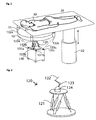
- An inventive X-ray device for imaging a female breast comprises a patient couch 20 on which a gantry 10 of a spiral computer tomograph is suspended.
- the patient couch 20 has a breast cutout 21, through which the breast 31 of a patient 30 preferably hangs downward in the direction of the gantry 10.
- the gantry 10 has a gantry lifting drive 11, with which it can be moved relative to the patient table 20.
- the gantry 10 rotates about the breast 31 of the patient 30.
- Staggered simultaneously and / or in time intervals the gantry 10 is displaced in the longitudinal direction of the breast, that is to say preferably in the vertical direction.
- the shift can be done either continuously at constant speed or proportional to the rotation of the gantry. Alternatively, the shift can also take place in steps, so that, for example, an offset by the width of the detector 14 takes place after each rotation of the gantry.
- an instrument 123 is provided, which engages from below into the free opening of the gantry. With it, you can take samples from the breast or inject contrast media without affecting the gantry.
- This instrument is preferably located on an instrument carrier 124, which is optionally connected via an articulated arm 126 to the patient couch 20 or the gantry suspension 125.
- the instrument panel 124 may be placed on a mobile platform 130 be. It is particularly advantageous if, for the exact positioning of the instrument carrier, it is connected to the mobile platform 130 via a hexapod 121.
- For exact positioning of the instrument carrier 124 measuring devices are available. In the case of an articulated arm this has in the pivot joints 123 preferably angle or position sensors.
- a measuring system can be provided, which selectively determines the position of the instrument carrier or the mobile platform to the gantry, the patient table or another fixed part associated with it and transmits corresponding correction information to the articulated arm or the Hexapod.
- X-ray images or examinations of the breast can be made initially without the instrument carrier or the intervention instrument. Due to the construction in which the gantry is suspended from the patient bed, results in a compact space-saving design, in which also the examining or treating doctor or other personnel access to the breast to be examined.
- a contrast agent can be injected into the breast before a shot or between two shots. If an intervention such as a biopsy is to be performed, then the instrument carrier is introduced with the corresponding instrument below the gantry. This is especially easy if there is a mobile platform for this purpose. This can then be rolled for example on its own wheels from a lateral position below the gantry.
- an exact adjustment of the instrument carrier with the instrument is then carried out by a positioning unit, which is attached to the mobile platform due to positional data, which were determined by means of at least one measuring system.
- the mobile platform can be driven out of the area of the gantry again, so that the examining person can be seen Doctor has access to the chest again.
- the device according to the invention does not necessitate a transfer of the patient between x-ray and biopsy. Furthermore, it is no longer necessary to mark the puncture points on the skin according to the X-ray image and then make the biopsy through them. Rather, the biopsy can now be made exactly controlled based on the X-ray data.
- the exact positioning of the instrument can be monitored during an intervention.
- one or more X-ray images are taken to control the instrument or the position of the instrument.
- the gantry rotates continuously during the intervention and makes 90 degree staggered shots. This makes the intervention instrument controllable in two levels. For example, it can be determined whether a biopsy needle bends.
- the first two steps can be reversed in their order.
- Essential here is the calculation of the position of the intervention instrument (122) by means of its relative position with respect to the gantry or a part connected to the gantry.
- FIG. 1 an inventive X-ray device is shown.
- a patient 30 is lying on the patient couch 20.
- the breast to be examined hangs over a breast cutout 21 through the patient couch 20 into the receiving area of a gantry 10.
- the gantry 10 is a spiral computer tomography gantry with an x-ray tube and a detector which surrounds the turning examining breast.
- the breast is imaged.
- a shift in the vertical direction is performed via the Gantryhubantrieb 11, so that the breast is scanned spirally.
- the patient couch 20 is height-adjustable via a patient couch lift drive 22.
- it can also be rotatable about the axis of the patient couch lifting drive 22.
- a mobile platform 130 below the gantry 10 is a mobile platform 130.
- This mobile platform can be driven or removed under the gantry during the examination. It preferably has wheels which are advantageously lockable. Optionally, it can roll or slide on the floor or on rails. Especially cheap gliders or airgliders based on Air cushion bearings.
- the mobile platform can have its own drive for exact positioning below the gantry or it can also be moved there manually. Guide rails or positioning aids embedded in the floor, such as induction loops, can simplify exact positioning.
- the instrument carrier hidden in this figure by the gantry is held in its working position on the chest by means of a positioning unit 120.
- the positioning unit may be, for example, a hexapod or an articulated arm.
- 131a, 131b and 131c are provided which determine the distance to measuring marks 132a, 132b, 132c on the gantry suspension with measuring signals 133a, 133b, 133c, here for example light.
- an automatic or also a manual correction of the position of the mobile platform can take place.
- the position data of the measuring system can be used to calculate the exact position of the instrument carrier.
- the measuring system can basically work according to all known principles for position or distance measurement. It preferably works with optical sensors such as laser sensors or with ultrasonic sensors or radio sensors.
- a positioning unit 120 which positions the instrument carrier 124 in the correct working position with respect to the breast.
- the exact positioning takes place here by means of a hexapod 121.
- an articulated arm with at least one pivot 123 is provided between the instrument carrier and the intervention instrument 122. In principle, several joints can be provided. Due to the Hexapod this is usually not necessary in the illustrated embodiment.
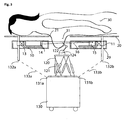
- FIG. 3 a device according to the invention is shown in a side view.
- the gantry is shown in section.
- an X-ray tube 15 Within the gantry housing 29 is an X-ray tube 15, which a Beam fan 16 for radiating the breast 31 generated.
- the radiation is received by a detector 14 and guided to an evaluation unit (not shown here).
- the gantry is rotatable on the one hand with a Gantryfiberlager 13 around the breast and on the Gantryhubantrieb 11 in height or at an inner distance to the patient displaced.
- a spiral scan of the breast 31 to be examined is possible.
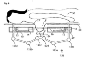
- FIG. 4 a device according to the invention with an articulated arm for positioning the instrument carrier 124 and the intervention instrument 122 is disclosed.
- This articulated arm is fixed here to the girders of the gantry suspension 125.
- the articulated arm can be removed during the examination or treatment or attached to the gantry suspension again.
- the articulated arm can also be attached to the patient couch 20 itself or another component connected thereto.
- such attachment in a simple manner, for example, with a quick release solvable, so that the entire assembly with the articulated arm 126 can be quickly mounted and removed again.
- He can also work on a mobile platform 130, as in FIG. 1 shown, instead of Hexapod drawn there.
- the articulated arm shown here has a plurality of hinges 123a, 123b, 123c, 123d, 123e and 123f.
- Rotary drives and / or angle sensors and / or position sensors are advantageously integrated in the rotary joints.
- a measuring system could also be present on the instrument carrier, so that the exact position of the instrument carrier with respect to the gantry 10 and / or the patient couch 20 can be determined.
Description
- Die Erfindung betrifft ein Röntgengerät zur Abbildung der weiblichen Brust (Mammografie). Mit diesem Gerät können auch Eingriffe in die Brust vorgenommen werden.
- Zur Untersuchung der weiblichen Brust sind verschiedene Röntgengeräte bekannt. Unter einer Liege, auf der eine zu untersuchende Patientin liegt, befindet sich eine Röntgenvorrichtung mit einer rotierenden Gantry, welche eine Röntgenröhre und einem Detektor aufweist. Ein solches Gerät ist beispielsweise in der
US 4,015,836 offenbart. Nachteilig an diesem Stand der Technik ist der große Platzbedarf so wie die mangelnde Zugänglichkeit der zu untersuchenden Brust. Weiterhin wird die Patientin in eine relativ unbequeme Lage mit tief liegendem Kopf gebracht, damit von der Röntgenvorrichtung ein möglichst großer Anteil der Brust erfasst werden kann. Eine Verbesserung stellt hier die Anordnung aus derUS 2006/0094950 dar. Die Patientin hat hier eine bequemere Lage. Weiterhin ist auch die zu untersuchende Brust nur mit speziellen Instrumenten zugänglich. Nachteilig an dieser Anordnung ist der große Platzbedarf, der sich durch die große Bauform der Gantry ergibt. In derUS 2007/0064867 ist ein Röntgengerät, nach dem Oberbegriff von Anspruch 1, basierend auf einem Spiral-CT Scanner offenbart. Hier wird die Auflösung durch die wenig stabile mechanische Konstruktion begrenzt. Zudem ist auch hier die zu untersuchende Brust nicht von außen zugänglich. - Der Erfindung liegt die Aufgabe zugrunde, ein Röntgengerät zu gestalten, welches die weibliche Brust diagnostisch exakt, schnell und kostengünstig abbildet und gleichzeitig Eingriffe in die Brust ermöglicht. Gleichzeitig soll der behandelnde Arzt einen guten und ergonomisch vorteilhaften Zugang zu der Brust haben.
- Diese Aufgabe wird durch ein Röntgengerät nach Anspruch 1 gelöst. Vorteilhafte Ausgestaltungen der Erfindung sind in den Unteransprüchen angegeben.
- Ein erfindungsgemäßes Röntgengerät zur Abbildung einer weiblichen Brust umfasst eine Patientenliege 20, an welcher eine Gantry 10 eines Spiral- Computertomographen aufgehängt ist. Die Patientenliege 20 weist einen Brustausschnitt 21 auf, durch welchen die Brust 31 einer Patientin 30 vorzugsweise nach unten in Richtung der Gantry 10 hängt. Die Gantry 10 hat einen Gantryhubantrieb 11, mit dem sie gegenüber dem Patiententisch 20 bewegt werden kann. Zur Röntgenaufnahme rotiert die Gantry 10 um die Brust 31 der Patientin 30. Gleichzeitig und/oder in Zeitintervallen gestaffelt erfolgt eine Verschiebung der Gantry 10 in Längsrichtung der Brust, das heißt vorzugsweise in vertikaler Richtung. Die Verschiebung kann wahlweise kontinuierlich mit konstanter Geschwindigkeit oder proportional zur Rotation der Gantry erfolgen. Alternativ kann die Verschiebung auch in Schritten erfolgen, so dass beispielsweise nach jeder Umdrehung der Gantry ein Versatz um die Breite des Detektors 14 erfolgt.
- Zur Intervention, insbesondere für eine Biopsie ist ein Instrument 123 vorgesehen, welches von unten in die freie Öffnung der Gantry eingreift. Damit können ohne die Gantry zu beeinflussen, Proben aus der Brust entnommen oder auch Kontrastmittel injiziert werden. Dieses Instrument befindet sich bevorzugt auf einem Instrumententräger 124, der wahlweise über einen Gelenkarm 126 mit der Patientenliege 20 oder der Gantryaufhängung 125 verbunden ist. Alternativ hierzu kann der Instrumententräger 124 auf einer mobilen Plattform 130 angeordnet sein. Besonders vorteilhaft ist es, wenn zur exakten Positionierung des Instrumententrägers dieser über einen Hexapod 121 mit der mobilen Plattform 130 verbunden ist. Zur exakten Positionierung des Instrumententrägers 124 sind Messeinrichtungen vorhanden. Im Falle eines Gelenkarms hat dieser in den Drehgelenken 123 bevorzugt Winkel- oder Positionssensoren. Weiterhin kann ein Messsystem vorgesehen sein, welches wahlweise die Position des Instrumententrägers oder auch der mobilen Plattform zur Gantry, dem Patiententisch oder einem anderen Fest damit verbundenen Teil ermittelt und entsprechende Korrekturinformationen an den Gelenkarm beziehungsweise den Hexapod übersendet.
- Mit einem erfindungsgemäßen Röntgengerät können zunächst ohne den Instrumententräger bzw. das Interventionsinstrument Röntgenaufnahmen beziehungsweise Untersuchungen der Brust angestellt werden. Aufgrund der Konstruktion, bei der die Gantry an der Patientenliege aufgehängt ist, ergibt sich eine kompakte Platz sparende Bauweise, bei der auch der untersuchende beziehungsweise behandelnde Arzt oder anderes Personal Zugang zu der zu untersuchenden Brust. So kann beispielsweise vor einer Aufnahme oder zwischen zwei Aufnahmen ein Kontrastmittel in die Brust injiziert werden. Soll nun eine Intervention, wie eine Biopsie durchgeführt werden, so wird der Instrumententräger mit dem entsprechend Instrument unterhalb der Gantry eingebracht. Dieses besonders einfach, wenn hierzu eine Mobile Plattform vorhanden ist. Diese kann dann beispielsweise auf eigenen Rädern von einer seitlichen Position aus unter die Gantry gerollt werden. In den meisten Fällen wird eine exakte Justierung nicht notwendig sein, so dass einfache Markierungen auf dem Boden ausreichen. Eine exakte Justierung des Instrumententrägers mit dem Instrument erfolgt dann durch eine Positioniereinheit, die an der mobilen Plattform angebracht ist aufgrund von Positionsdaten, die mittels wenigstens eines Messsystems ermittelt wurden. Nach der Intervention, beispielsweise einer Biopsie kann die Mobile Plattform wieder aus dem Bereich der Gantry gefahren werden, so dass der untersuchende Arzt wieder Zugang zur Brust hat. Durch die Erfindungsgemäße Vorrichtung ist eine Umbettung der Patientin zwischen Röntgenaufnahme und Biopsie nicht notwendig. Weiterhin ist es nicht mehr notwendig, die Einstichpunkte auf der Haut entsprechend dem Röntgenbild zu markieren und dann durch diese die Biopsie vorzunehmen. Vielmehr kann die Biopsie nun exakt gesteuert aufgrund der Röntgendaten vorgenommen werden.
- Entsprechend einer weiteren Ausgestaltung der Erfindung kann während einer Intervention die exakte Positionierung des Instruments überwacht werden. Hierzu werden während der Intervention eine oder mehrere Röntgenaufnahmen zur Kontrolle des Instruments beziehungsweise der Position des Instruments gemacht. Vorteilhafterweise dreht sich die Gantry kontinuierlich während der Intervention und macht um 90 Grad versetzte Aufnahmen. Dadurch ist das Interventionsinstrument in zwei Ebenen kontrollierbar. So kann beispielsweise festgestellt werden, ob sich eine Biopsienadel verbiegt.
- Ein Verfahren, welches mit dem hier beschriebenen Röntgengerät durchgeführt werden kann, umfasst die folgenden Schritte:
- Erstellen einer dreidimensionalen Röntgenaufnahme,
- Erfassen der relativen Position des Interventionsinstrumentes (122) in Bezug auf die Gantry (10),
- Berechnen der Position des Interventionsinstrumentes (122) in Bezug auf die dreidimensionale Röntgenaufnahme.
- Bei diesem Verfahren können die ersten beiden Schritte in ihrer Reihenfolge vertauscht werden. Wesentlich hierbei ist die Berechnung der Position des Interventionsinstrumentes (122) mittels seiner relativen Position in Bezug auf die Gantry oder eines mit der Gantry verbundenen Teils.
- Die Erfindung wird nachstehend ohne Beschränkung des allgemeinen Erfindungsgedankens anhand von Ausführungsbeispielen unter Bezugnahme auf die Zeichnungen exemplarisch beschrieben.
-
Figur 1 zeigt eine Erfindungsgemäße Vorrichtung -
Figur 2 zeigt eine Positioniereinheit mit einem Hexapod -
Figur 3 zeigt eine Erfindungsgemäße Vorrichtung in seitlicher Ansicht -
Figur 4 zeigt eine Erfindungsgemäße Vorrichtung mit einem Gelenkarm - In
Figur 1 ist ein erfindungsgemäßes Röntgengerät dargestellt. Auf der Patientenliege 20 liegt eine Patientin 30. Die zu untersuchende Brust hängt über einen Brustausschnitt 21 durch die Patientenliege 20 in den Aufnahmebereich einer Gantry 10. Die Gantry 10 ist eine Spiral- Computertomographen Gantry mit einer Röntgenröhre und einem Detektor, welche sich um die zu untersuchende Brust drehen. Während der Drehung wird die Brust abgebildet. Gleichzeitig mit der Drehung wird über den Gantryhubantrieb 11 eine Verschiebung in vertikaler Richtung durchgeführt, so dass die Brust spiralförmig abgetastet wird. Die Patientenliege 20 ist über einen Patientenliegenhubantrieb 22 in der Höhe verstellbar. Optional kann bei einem fest installierten Patiententisch dieser auch noch um die Achse des Patientenliegenhubantriebs 22 drehbar sein. - Unter der Gantry 10 befindet sich eine Mobile Plattform 130. Diese Mobile Plattform kann während der Untersuchung unter die Gantry gefahren oder auch wieder entfernt werden. Sie weist bevorzugt Räder auf, die vorteilhafterweise blockierbar sind. Wahlweise kann sie auf dem Fußboden oder aber auch auf Schienen rollen oder gleiten. Besonders günstig Gleiter oder Luftgleiter basierend auf Luftkissenlagern. Die Mobile Plattform kann einen eigenen Antrieb zur exakten Positionierung unterhalb der Gantry aufweisen oder auch manuell dorthin verschoben werden. Führungsschienen oder auch in den Boden eingebettete Positionierhilfen wie Induktionsschleifen können eine exakte Positionierung vereinfachen. Der in dieser Figur durch die Gantry verdeckte Instrumententräger wird mittels einer Positioniereinheit 120 in seiner Arbeitsposition an der Brust gehalten. Die Positioniereinheit kann beispielsweise ein Hexapod oder auch ein Gelenkarm sein. Zur exakten Positionierung sind Positionsmesssysteme 131a, 131b und 131c vorgesehen, die mit Messsignalen 133a, 133b, 133c, hier beispielsweise Licht, den Abstand zu Messmarken 132a, 132b, 132c an der Gantryaufhängung ermitteln. Wahlweise kann nun aufgrund der Messung der Positionsmesssysteme eine automatische oder auch eine manuelle Korrektur der Position der mobilen Plattform erfolgen. Alternativ und/oder zusätzlich können die Positionsdaten des Messsystems zur Berechnung der exakten Position des Instrumententrägers herangezogen werden. Das Messsystem kann grundsätzlich nach allen bekannten Prinzipien zur Positions- beziehungsweise Distanzmessung arbeiten. Bevorzugt arbeitet es mit optischen Sensoren wie Lasersensoren oder auch mit Ultraschallsensoren oder Funksensoren.
- In
Figur 2 ist eine Positioniereinheit 120 dargestellt, die den Instrumententräger 124 in die richtige Arbeitsposition in Bezug auf die Brust positioniert. Die exakte Positionierung erfolgt hier mittels eines Hexapod 121. Vorteilhafterweise ist zwischen den Instrumententräger und dem Interventions-Instrument 122 noch ein Gelenkarm mit wenigstens einem Drehgelenk 123 vorgesehen. Grundsätzlich können auch mehrere Gelenke vorgesehen sein. Aufgrund des Hexapod ist dies in der dargestellten Ausführungsform meist nicht notwendig. - In
Figur 3 ist eine erfindungsgemäße Vorrichtung in seitlicher Ansicht dargestellt. Zur besseren Veranschaulichung ist noch die Gantry im Schnitt dargestellt. Innerhalb des Gantrygehäuses 29 befindet sich eine Röntgenröhre 15, welche einen Strahlenfächer 16 zur Durchstrahlung der Brust 31 erzeugt. Die Strahlung wird von einem Detektor 14 empfangen und zu einer Auswerteeinheit (hier nicht dargestellt) geführt. Die Gantry ist einerseits mit einem Gantrydrehlager 13 um die Brust drehbar und über den Gantryhubantrieb 11 in ihrer Höhe beziehungsweise im inneren Abstand zur Patientin verschiebbar. Damit ist eine spiralförmige Abtastung der zu untersuchenden Brust 31 möglich. - In
Figur 4 ist eine erfindungsgemäße Vorrichtung mit einem Gelenkarm zur Positionierung des Instrumententrägers 124 und des Intervention- Instruments 122 offenbart. Dieser Gelenkarm ist hier an den Trägern der Gantryaufhängung 125 fixiert. Vorteilhafterweise lässt er sich während der Untersuchung beziehungsweise Behandlung entfernen oder auch wieder an der Gantryaufhängung befestigen. Anstelle der Gantryaufhängung kann der Gelenkarm auch an der Patientenliege 20 selbst oder einem anderen damit verbundenen Bauteil befestigt sein. Vorzugsweise ist eine solche Befestigung auf einfache Art und Weise, beispielsweise mit einem Schnellverschluss lösbar, so dass die gesamte Baugruppe mit dem Gelenkarm 126 schnell montiert und auch wieder abgenommen werden kann. Er kann auch auf einer mobilen Plattform 130, wie inFigur 1 dargestellt, anstelle des dort gezeichneten Hexapod eingesetzt werden. Der hier dargestellte Gelenkarm weist mehrere Drehgelenke 123a, 123b, 123c, 123d, 123e und 123f auf. In den Drehgelenken sind vorteilhafterweise Drehantriebe und/oder Winkelsensoren und/oder Positionssensoren integriert. Grundsätzlich könnte auch ein Messsystem an den Instrumententräger vorhanden sein, so das die exakte Position des Instrumententräger in Bezug auf die Gantry 10 und/oder die Patientenliege 20 ermittelt werden kann. -
- 10
- Gantry
- 11
- Gantryhubantrieb
- 13
- Gantrydrehlager
- 14
- Detektor
- 15
- Röntgenröhre
- 16
- Strahlenfächer
- 20
- Patientenliege
- 21
- Brustausschnitt
- 22
- Patientenliegenhubantrieb
- 29
- Gantrygehäuse
- 30
- Patient
- 31
- Brust
- 120
- Positioniereinheit
- 121
- Hexapod
- 122
- Interventions- Instrument
- 123
- Drehgelenk
- 124
- Instrumententräger
- 125
- Gantryaufhängung
- 126
- Gelenkarm
- 130
- Mobile Plattform
- 131
- Messsystem
- 132
- Messmarke
- 133
- Messsignal (optisch)
Claims (9)
- Röntgengerät zur Abbildung und Intervention einer Brust einer Patientin (30), mit- einer Röntgeneinrichtung mit einer näherungsweise um eine vertikale Drehachse drehbare Gantry (10) umfassend eine Röntgenröhre (15) und einen Röntgendetektor (14), wobei die Gantry (10) mittels eines Gantryhubantriebs (11) in vertikaler Richtung bewegt werden kann und die vertikale Bewegung in Abhängigkeit von der Rotationsbewegung erfolgt, und- einer horizontal angeordneten Patientenliege (20) mit einem Brustausschnitt (21) zur Aufnahme einer Brust der Patientin (30),dadurch gekennzeichnet, dass
die Gantry (10) an der Unterseite der Patientenliege (20) aufgehängt ist und ein Instrumententräger (124) zur Aufnahme wenigstens eines Interventions-Instruments (122) vorgesehen ist, der von unten durch die Gantry in der Umgebung der Brust positioniert wird, so dass mit dem Interventions-Instrument eine Intervention ausgeführt werden kann. - Röntgengerät nach Anspruch 1,
dadurch gekennzeichnet, dass
eine mobile Plattform (130) vorgesehen ist, welche über eine Positioniereinheit (120) den Instrumententräger (124) trägt, wobei die Mobile Plattform (130) von der Seite unter die Gantry (10) eingebracht werden kann. - Röntgengerät nach Anspruch 1,
dadurch gekennzeichnet, dass
ein Gelenkarm (126) vorgesehen ist, welcher den Instrumententräger (124) trägt, und an der Gantryaufhängung (125) oder anderen fest mit dieser verbundenen Teilen fixiert ist. - Röntgengerät nach Anspruch 3,
dadurch gekennzeichnet, dass
die Fixierung des Gelenkarms mit einer Schnellverschlusseinrichtung lösbar ist. - Röntgengerät nach Anspruch 2,
dadurch gekennzeichnet, dass
die Mobile Plattform (130) wenigstens ein Messsystem (131) zur exakten Bestimmung der Position der mobilen Plattform gegenüber der Gantry (10) oder eines fest mit dieser verbundenen Teiles aufweist, wobei die durch das Messsystem bestimmten Positionsinformationen zur exakten Positionierung des Instrumententräger (124) verwendet werden. - Röntgengerät nach Anspruch 2,
dadurch gekennzeichnet, dass
die Positioniereinheit (120) ein Hexapod (121) ist. - Röntgengerät nach Anspruch 2,
dadurch gekennzeichnet, dass
die Mobile Plattform (130) Räder, Gleiter oder Luftgleiter aufweist. - Röntgengerät nach Anspruch 1, 2 oder 3,
dadurch gekennzeichnet, dass
wenigstens ein Messsystem (131) zur Bestimmung der Position des Instrumententrägers (124) oder eines damit verbundenen Teils gegenüber der Gantry (10) oder eines fest mit dieser verbundenen Teiles Positionsinformationen zur exakten Positionierung des Instrumententrägers (124) und/oder des Instruments (122) in Bezug auf die Brust (31) liefert. - Röntgengerät nach einem der vorhergehenden Ansprüche,
dadurch gekennzeichnet, dass
während einer Intervention Röntgenaufnahmen zur Kontrolle der Position des Interventions- Instruments (122) durchgeführt werden.
Applications Claiming Priority (1)
| Application Number | Priority Date | Filing Date | Title |
|---|---|---|---|
| DE102008042430 | 2008-09-29 |
Publications (2)
| Publication Number | Publication Date |
|---|---|
| EP2168486A1 EP2168486A1 (de) | 2010-03-31 |
| EP2168486B1 true EP2168486B1 (de) | 2011-10-05 |
Family
ID=40524871
Family Applications (8)
| Application Number | Title | Priority Date | Filing Date |
|---|---|---|---|
| EP09154854A Withdrawn EP2168485A1 (de) | 2008-09-29 | 2009-03-11 | Brustfixierung für ein Untersuchungsgerät zur Untersuchung der weiblichen Brust |
| EP09154884.2A Withdrawn EP2178048A3 (de) | 2008-09-29 | 2009-03-11 | Verfahren zur Definition eines patientenindividuellen Koordinationssystems einer weiblichen Brust |
| EP09154833A Expired - Fee Related EP2168489B1 (de) | 2008-09-29 | 2009-03-11 | Röntgengerät zur Brustuntersuchung im Stehen |
| EP09154863A Withdrawn EP2168491A1 (de) | 2008-09-29 | 2009-03-11 | Brustfixierung mit Probencontainer für ein Untersuchungsgerät der weiblichen Brust |
| EP09154842A Expired - Fee Related EP2168484B1 (de) | 2008-09-29 | 2009-03-11 | Röntgengerät zur Brustuntersuchung mit einer in eine Patientenliege integrierten Gantry |
| EP09154848A Withdrawn EP2168490A1 (de) | 2008-09-29 | 2009-03-11 | Röntgengerät zur Brustuntersuchung mit einer Detektor-Röhren Anordnung für hochauflösende Aufnahmen |
| EP09154891A Expired - Fee Related EP2168486B1 (de) | 2008-09-29 | 2009-03-11 | Modulares System zur Brustdiagnose und -intervention |
| EP09154900A Withdrawn EP2168487A1 (de) | 2008-09-29 | 2009-03-11 | Verfahren und Vorrichtung zur thermischen Brusttumor-Behandlung mit 3D Monitorfunktion |
Family Applications Before (6)
| Application Number | Title | Priority Date | Filing Date |
|---|---|---|---|
| EP09154854A Withdrawn EP2168485A1 (de) | 2008-09-29 | 2009-03-11 | Brustfixierung für ein Untersuchungsgerät zur Untersuchung der weiblichen Brust |
| EP09154884.2A Withdrawn EP2178048A3 (de) | 2008-09-29 | 2009-03-11 | Verfahren zur Definition eines patientenindividuellen Koordinationssystems einer weiblichen Brust |
| EP09154833A Expired - Fee Related EP2168489B1 (de) | 2008-09-29 | 2009-03-11 | Röntgengerät zur Brustuntersuchung im Stehen |
| EP09154863A Withdrawn EP2168491A1 (de) | 2008-09-29 | 2009-03-11 | Brustfixierung mit Probencontainer für ein Untersuchungsgerät der weiblichen Brust |
| EP09154842A Expired - Fee Related EP2168484B1 (de) | 2008-09-29 | 2009-03-11 | Röntgengerät zur Brustuntersuchung mit einer in eine Patientenliege integrierten Gantry |
| EP09154848A Withdrawn EP2168490A1 (de) | 2008-09-29 | 2009-03-11 | Röntgengerät zur Brustuntersuchung mit einer Detektor-Röhren Anordnung für hochauflösende Aufnahmen |
Family Applications After (1)
| Application Number | Title | Priority Date | Filing Date |
|---|---|---|---|
| EP09154900A Withdrawn EP2168487A1 (de) | 2008-09-29 | 2009-03-11 | Verfahren und Vorrichtung zur thermischen Brusttumor-Behandlung mit 3D Monitorfunktion |
Country Status (2)
| Country | Link |
|---|---|
| US (8) | US8199993B2 (de) |
| EP (8) | EP2168485A1 (de) |
Families Citing this family (62)
| Publication number | Priority date | Publication date | Assignee | Title |
|---|---|---|---|---|
| US8272088B2 (en) * | 2007-09-06 | 2012-09-25 | Orbital Therapy Llc | Patient support system for full access prone position breast radiotherapy |
| EP2168485A1 (de) * | 2008-09-29 | 2010-03-31 | MIR Medical Imaging Research Holding GmbH | Brustfixierung für ein Untersuchungsgerät zur Untersuchung der weiblichen Brust |
| DE102008049711A1 (de) * | 2008-09-30 | 2010-04-15 | Siemens Aktiengesellschaft | Lagerungsvorrichtung, Patientenlagerungstisch und medizinisches Gerät |
| US8014490B2 (en) * | 2009-10-20 | 2011-09-06 | Linda Mitchell | Mammogram tender machine |
| US8421604B2 (en) * | 2009-11-30 | 2013-04-16 | Symbol Technologies, Inc. | Method and apparatus for identifying read zone of RFID reader |
| US8374312B2 (en) * | 2010-02-18 | 2013-02-12 | Varian Medical Systems, Inc. | Prone patient positioning devices and methods |
| DE102010011660A1 (de) * | 2010-03-17 | 2011-09-22 | Siemens Aktiengesellschaft | Mammographiegerät |
| JP5700950B2 (ja) * | 2010-04-21 | 2015-04-15 | キヤノン株式会社 | 生体情報取得装置 |
| US20140191852A1 (en) * | 2010-05-13 | 2014-07-10 | Carestream Health, Inc. | Method and system for phosphor plate identification in computed radiography |
| US20120001737A1 (en) * | 2010-05-13 | 2012-01-05 | Amir Berger | Method and system for computed radiography |
| GB2483640A (en) * | 2010-09-10 | 2012-03-21 | Specialty Magnetics Ltd | Breast immobilisation arrangement |
| WO2012048000A2 (en) | 2010-10-05 | 2012-04-12 | Hologic, Inc. | Upright x-ray breast imaging with a ct mode, multiple tomosynthesis modes, and a mammography mode |
| DE102010052603A1 (de) * | 2010-11-25 | 2012-05-31 | Artemis Imaging Gmbh | Patientenliege |
| WO2012120498A1 (en) * | 2011-03-04 | 2012-09-13 | Technion Research & Development | Non-invasive thermal treatment monitoring |
| DE102011006353A1 (de) | 2011-03-29 | 2012-10-04 | Siemens Aktiengesellschaft | Mammographieanlage |
| WO2012171029A1 (en) | 2011-06-09 | 2012-12-13 | The Regents Of The University Of California | Excised specimen imaging using a combined pet and micro ct scanner |
| US8842806B2 (en) | 2012-04-03 | 2014-09-23 | Carestream Health, Inc. | Apparatus and method for breast imaging |
| EP2845024B1 (de) | 2012-05-02 | 2019-04-10 | Koninklijke Philips N.V. | Thermometrieabbildung |
| US9307961B2 (en) * | 2012-06-29 | 2016-04-12 | Carefusion 2200, Inc. | Fine needle aspiration biopsy device |
| KR102001926B1 (ko) * | 2012-09-11 | 2019-07-30 | 삼성디스플레이 주식회사 | 엑스레이 검출기, 이를 포함하는 엑스레이 검출 시스템 및 엑스레이 검출 방법 |
| DE102012216687A1 (de) * | 2012-09-18 | 2014-03-20 | Jan Rimbach | Vorrichtung zur Untersuchung von Prüfkörpern |
| DE102012217301B4 (de) | 2012-09-25 | 2021-10-14 | Bayer Pharma Aktiengesellschaft | Kombination aus Kontrastmittel und Mammographie-CT-System mit vorgegebenem Energiebereich und Verfahren zur Erzeugung tomographischer Mammographie-CT-Aufnahmen durch diese Kombination |
| CN103908343B (zh) | 2012-12-31 | 2016-10-05 | 西门子(深圳)磁共振有限公司 | 患者检查床和磁共振成像设备 |
| EP3964132B1 (de) | 2013-10-09 | 2023-07-19 | Hologic, Inc. | Röntgenstrahl-brusttomosynthese mit erhöhung der räumlichen auflösung in dickenrichtung einer abgeflachten brust |
| US9161725B1 (en) * | 2014-02-05 | 2015-10-20 | Regine Millien-White | Adjustable breast examination device |
| JP6376783B2 (ja) * | 2014-03-12 | 2018-08-22 | キヤノン株式会社 | 乳房断層撮影装置および制御方法 |
| JP6381253B2 (ja) * | 2014-03-31 | 2018-08-29 | キヤノン株式会社 | 放射線撮影装置、断層撮影装置 |
| EP3125758B1 (de) * | 2014-04-04 | 2018-12-12 | Pierfrancesco Pavoni | Zugriffs-gate oder -gantry mit einer antennenanordnung zur therapie oder bildgebung |
| US9326739B2 (en) | 2014-04-28 | 2016-05-03 | Cheryl A. Galambos McLaughlin | Mammogram table |
| US9301726B2 (en) * | 2014-05-02 | 2016-04-05 | Wisconsin Alumni Research Foundation | CT machine for multi-angle scanning of stationary patients |
| CN104173075B (zh) * | 2014-08-26 | 2016-07-06 | 李丙曙 | 放射科检查床 |
| JP6611428B2 (ja) * | 2014-12-09 | 2019-11-27 | キヤノン株式会社 | マンモ断層撮像システム |
| EP3238628A4 (de) * | 2014-12-26 | 2018-08-15 | Rayence Co., Ltd. | Hebevorrichtung für ein kompressionspaddle und röntgenbildfotografievorrichtung damit |
| CN105832353B (zh) * | 2015-01-30 | 2020-11-06 | 佳能株式会社 | 放射线摄像系统 |
| JP6651069B2 (ja) * | 2015-05-13 | 2020-02-19 | フジデノロ株式会社 | 固定具装着装置 |
| KR20160139292A (ko) * | 2015-05-27 | 2016-12-07 | 삼성전자주식회사 | Rf 표면 코일부 및 이를 포함하는 자기공명영상 시스템 |
| JP6525768B2 (ja) * | 2015-06-30 | 2019-06-05 | キヤノン株式会社 | 乳房撮影装置 |
| US10542951B2 (en) * | 2015-07-23 | 2020-01-28 | General Electric Company | Systems, methods, and devices for simplified high quality imaging of biopsy samples on a mammography machine |
| WO2017019401A1 (en) * | 2015-07-24 | 2017-02-02 | Dretzaka-Kaye Tricia | Anatomy scanning system and method |
| WO2017091787A1 (en) * | 2015-11-25 | 2017-06-01 | The Regents Of The University Of California | 3d-beam modulation filter for equalizing dose and image quality in breast ct |
| DE102015225236A1 (de) * | 2015-12-15 | 2017-06-22 | Siemens Healthcare Gmbh | Mammographie-Screening mit hoher Durchgangsrate |
| CN106933857B (zh) * | 2015-12-30 | 2020-12-29 | 创新先进技术有限公司 | 一种数据仓库中任务的调度方法、装置 |
| DE102016206198A1 (de) * | 2016-04-13 | 2017-10-19 | Siemens Healthcare Gmbh | Röntgensystem |
| US10603003B2 (en) * | 2016-04-14 | 2020-03-31 | Dedicating2Imaging, LLC | CT systems for imaging of the breast |
| US11395593B2 (en) * | 2016-09-14 | 2022-07-26 | Mor Research Applications Ltd. | Device, system and method for detecting irregularities in soft tissue |
| US10180207B1 (en) * | 2017-07-13 | 2019-01-15 | Danylo Kozub | Stand |
| CN108175430A (zh) * | 2018-01-17 | 2018-06-19 | 江苏美伦影像系统有限公司 | 一种具有辐射防护功能的乳腺x射线摄影系统 |
| US10959747B1 (en) * | 2018-04-02 | 2021-03-30 | Lifei Guo | Tissue removing |
| US10893844B1 (en) * | 2018-10-10 | 2021-01-19 | David Byron Douglas | Method and apparatus for performing 3D imaging examinations of a structure under differing configurations and analyzing morphologic changes |
| DE102018207636A1 (de) * | 2018-05-16 | 2019-11-21 | Siemens Healthcare Gmbh | Patiententisch mit Vorrichtung zur reversiblen Aufnahme einer Transferplatte |
| CN108956656B (zh) * | 2018-07-17 | 2021-02-05 | 青岛大学附属医院 | 一种高衬度低剂量相位衬度ct成像装置 |
| CN110975156B (zh) * | 2019-11-15 | 2021-11-19 | 山东大学齐鲁医院 | 乳房牵引固定装置及系统 |
| CA3173541A1 (en) * | 2020-03-31 | 2021-10-07 | Hologic, Inc. | Systems and methods for x-ray imaging tissue specimens |
| KR102640269B1 (ko) * | 2020-05-29 | 2024-02-26 | (의료)길의료재단 | 유방암 치료용 방사선 조사 장치 |
| CN111714222B (zh) * | 2020-06-29 | 2021-07-23 | 北京欧扬医疗美容门诊部有限公司 | 一种无痕隆胸用脂肪自体植入装置 |
| CN111714191A (zh) * | 2020-06-30 | 2020-09-29 | 广西医科大学附属肿瘤医院 | 用于锥光束乳腺ct引导下悬垂穿刺的激光定位装置 |
| US11692951B2 (en) * | 2021-02-24 | 2023-07-04 | GE Precision Healthcare LLC | System and method for specimen imaging using an existing mammography imaging system |
| EP4226876A1 (de) * | 2022-02-09 | 2023-08-16 | Storz Medical AG | Stosswellenvorrichtung mit verbesserter akustischer kopplung |
| EP4226877A1 (de) * | 2022-02-09 | 2023-08-16 | Storz Medical AG | Stosswellenvorrichtung mit integrierter ultraschallsonde |
| EP4226875A1 (de) * | 2022-02-09 | 2023-08-16 | Storz Medical AG | Stosswellenvorrichtung mit einer quelle, die sich selbst auf eine röntgenvorrichtung ausrichtet |
| EP4226874A1 (de) * | 2022-02-09 | 2023-08-16 | Storz Medical AG | Ultraschall- und/oder stosswellenvorrichtung mit auf einer hexapod-plattform montierter quelle |
| WO2023200896A1 (en) * | 2022-04-14 | 2023-10-19 | Koning Corporation | Cone beam breast computed tomography with patient support subsystem |
Family Cites Families (110)
| Publication number | Priority date | Publication date | Assignee | Title |
|---|---|---|---|---|
| US3673394A (en) | 1969-02-18 | 1972-06-27 | North American Rockwell | Measuring method and apparatus |
| US4015836A (en) | 1975-07-31 | 1977-04-05 | General Electric Company | Mammography table |
| US4400827A (en) * | 1981-11-13 | 1983-08-23 | Spears James R | Method and apparatus for calibrating rapid sequence radiography |
| US4680028A (en) * | 1984-07-02 | 1987-07-14 | Lact-Assist, Incorporated | Flexible breast receptor for breast pump |
| US4709382A (en) * | 1984-11-21 | 1987-11-24 | Picker International, Inc. | Imaging with focused curved radiation detectors |
| US5415169A (en) | 1989-11-21 | 1995-05-16 | Fischer Imaging Corporation | Motorized mammographic biopsy apparatus |
| FI85803C (fi) | 1989-11-23 | 1992-06-10 | Planmed Oy | Foerfarande och anordning foer styrning av funktioner av en mammografiroentgenanordning. |
| US5569266A (en) | 1991-03-11 | 1996-10-29 | Fischer Imaging Corporation | Magnetic resonance imaging device useful for guiding a medical instrument |
| US5409497A (en) | 1991-03-11 | 1995-04-25 | Fischer Imaging Corporation | Orbital aiming device for mammo biopsy |
| US5289520A (en) | 1991-11-27 | 1994-02-22 | Lorad Corporation | Stereotactic mammography imaging system with prone position examination table and CCD camera |
| US5308321A (en) | 1992-05-05 | 1994-05-03 | Castro Donna J | Retainer assisted by vacuum expansion system |
| US5273435B1 (en) | 1992-07-16 | 1995-12-05 | Wisconsin Med College Inc | Tumor localization phantom |
| US5386447A (en) | 1992-09-23 | 1995-01-31 | Fischer Imaging Corporation | Mammographic screening and biopsy apparatus |
| US5490513A (en) * | 1992-09-28 | 1996-02-13 | Fonar Corporation | Multiple patient breast scanning on a magnetic resonance imaging apparatus |
| US6075879A (en) * | 1993-09-29 | 2000-06-13 | R2 Technology, Inc. | Method and system for computer-aided lesion detection using information from multiple images |
| JPH07303633A (ja) * | 1994-05-11 | 1995-11-21 | Mitsubishi Electric Corp | X線乳房撮影装置 |
| US5528043A (en) | 1995-04-21 | 1996-06-18 | Thermotrex Corporation | X-ray image sensor |
| US5609827A (en) | 1995-05-02 | 1997-03-11 | Beekley Corporation | Biopsy specimen container |
| US5709206A (en) | 1995-11-27 | 1998-01-20 | Teboul; Michel | Imaging system for breast sonography |
| US5757878A (en) * | 1996-08-16 | 1998-05-26 | Analogic Corporation | Detector arrangement for x-ray tomography system |
| DE19639975C1 (de) | 1996-09-27 | 1998-05-07 | Siemens Ag | Medizinische Einrichtung mit einer tunnelförmigen Öffnung zur Aufnahme eines Untersuchungsobjektes |
| EP0983020A1 (de) | 1997-05-06 | 2000-03-08 | Quanta Vision, Inc. | Gewebeanalysevorrichtung |
| US6358246B1 (en) | 1999-06-25 | 2002-03-19 | Radiotherapeutics Corporation | Method and system for heating solid tissue |
| US5991357A (en) | 1997-12-16 | 1999-11-23 | Analogic Corporation | Integrated radiation detecting and collimating assembly for X-ray tomography system |
| US6175117B1 (en) * | 1998-01-23 | 2001-01-16 | Quanta Vision, Inc. | Tissue analysis apparatus |
| DE19812995A1 (de) | 1998-03-25 | 1999-10-07 | Siemens Ag | Mammographie-Gerät, insbesondere für Vergrößerungs-Mammographie |
| US6242743B1 (en) * | 1998-08-11 | 2001-06-05 | Mosaic Imaging Technology, Inc. | Non-orbiting tomographic imaging system |
| JP2000116631A (ja) | 1998-10-16 | 2000-04-25 | Toshiba Corp | X線診断装置 |
| JP3866431B2 (ja) * | 1999-02-17 | 2007-01-10 | 株式会社東芝 | X線ct装置 |
| US6684097B1 (en) | 1999-04-22 | 2004-01-27 | University Of Miami | Intraoperative monitoring of temperature-induced tissue changes with a high-resolution digital x-ray system during thermotherapy |
| TW406009B (en) * | 1999-07-16 | 2000-09-21 | Nat Science Council | 3-D localization method of clustered microcalcifications using cranio-caudal and medio-lateral oblique views |
| US6254614B1 (en) * | 1999-10-18 | 2001-07-03 | Jerry M. Jesseph | Device and method for improved diagnosis and treatment of cancer |
| US6480565B1 (en) * | 1999-11-18 | 2002-11-12 | University Of Rochester | Apparatus and method for cone beam volume computed tomography breast imaging |
| US6987831B2 (en) | 1999-11-18 | 2006-01-17 | University Of Rochester | Apparatus and method for cone beam volume computed tomography breast imaging |
| DE10026792A1 (de) * | 2000-05-31 | 2001-12-06 | Bip Biomedizinische Instr & Pr | Diagnose- und Therapietisch |
| US6463122B1 (en) | 2000-08-21 | 2002-10-08 | Bio-Imaging Resource, Inc. | Mammography of computer tomography for imaging and therapy |
| US7467892B2 (en) * | 2000-08-29 | 2008-12-23 | Imaging Therapeutics, Inc. | Calibration devices and methods of use thereof |
| US7940966B2 (en) | 2000-11-24 | 2011-05-10 | U-Systems, Inc. | Full-field breast image data processing and archiving |
| US6419390B1 (en) * | 2001-03-26 | 2002-07-16 | Marianette Landis-Lowell | Folding mammography table and method of use |
| US6516045B2 (en) * | 2001-05-04 | 2003-02-04 | The Regents Of The University Of California | Device and method for determining proportions of body materials |
| US6418188B1 (en) * | 2001-06-14 | 2002-07-09 | Juanita L. Broadnax | Radiation breast cup and method |
| US6674835B2 (en) * | 2001-10-12 | 2004-01-06 | General Electric Co. | Methods and apparatus for estimating a material composition of an imaged object |
| US6671975B2 (en) | 2001-12-10 | 2004-01-06 | C. William Hennessey | Parallel kinematic micromanipulator |
| DE10207623B4 (de) | 2002-02-22 | 2004-05-06 | Siemens Ag | Verfahren für die Computertomographie sowie Computertomographie (CT)-Gerät |
| US20040254461A1 (en) * | 2002-03-20 | 2004-12-16 | Ackerman William H. | Acoustic beam shaping by pulse power modulation at constant amplitude |
| US7783089B2 (en) | 2002-04-15 | 2010-08-24 | General Electric Company | Method and apparatus for providing mammographic image metrics to a clinician |
| US7218766B2 (en) * | 2002-04-15 | 2007-05-15 | General Electric Company | Computer aided detection (CAD) for 3D digital mammography |
| CA2393101A1 (en) | 2002-07-11 | 2004-01-11 | Martin Cyr | Apparatus, system and method of calibrating medical imaging systems |
| AU2003253954A1 (en) | 2002-07-16 | 2004-02-02 | Alfred E. Mann Institute For Biomedical Engineering At The University Of Southern California | Support bra for ultrasonic breast scanner |
| US6904119B2 (en) * | 2002-10-02 | 2005-06-07 | Shimadzu Corporation | Radiographic apparatus |
| US7149566B2 (en) | 2002-10-31 | 2006-12-12 | Manoa Medical, Inc. | Soft tissue orientation and imaging guide systems and methods |
| US7809422B2 (en) * | 2002-11-08 | 2010-10-05 | Art Advanced Research Technologies Inc. | Method and apparatus for optical imaging |
| US7286634B2 (en) * | 2002-12-23 | 2007-10-23 | Select Technologies, Llc | Method and apparatus for improving baggage screening examination |
| EP1599139B1 (de) | 2003-02-20 | 2009-08-12 | Manoa Medical, Inc. | Biegbare schneidevorrichtung |
| US6872001B1 (en) | 2003-05-05 | 2005-03-29 | Peco Controls Corp. | X-ray shielding structure for food inspection station |
| US7850613B2 (en) | 2003-05-30 | 2010-12-14 | Orison Corporation | Apparatus and method for three dimensional ultrasound breast imaging |
| US6982424B2 (en) | 2003-06-02 | 2006-01-03 | Ge Medical Systems Global Technology Company, Llc | X-ray and CT image detector |
| US7291841B2 (en) | 2003-06-16 | 2007-11-06 | Robert Sigurd Nelson | Device and system for enhanced SPECT, PET, and Compton scatter imaging in nuclear medicine |
| US6837772B1 (en) | 2003-07-18 | 2005-01-04 | Regina Miracle International Limited | Breast cup construction |
| GB0318701D0 (en) * | 2003-08-08 | 2003-09-10 | Inst Of Cancer Res The | A method and apparatus for image processing |
| JP2005258370A (ja) | 2003-09-05 | 2005-09-22 | Fuji Photo Film Co Ltd | 放射線カセッテ |
| US7005988B2 (en) * | 2003-09-19 | 2006-02-28 | International Business Machines Corporation | Using radio frequency identification to detect and/or prevent theft and shoplifting |
| US20050070817A1 (en) * | 2003-09-30 | 2005-03-31 | Mueller Richard L. | Lavage assist device |
| US20050096515A1 (en) * | 2003-10-23 | 2005-05-05 | Geng Z. J. | Three-dimensional surface image guided adaptive therapy system |
| US7653229B2 (en) | 2003-12-23 | 2010-01-26 | General Electric Company | Methods and apparatus for reconstruction of volume data from projection data |
| JP4119835B2 (ja) | 2003-12-26 | 2008-07-16 | ジーイー・メディカル・システムズ・グローバル・テクノロジー・カンパニー・エルエルシー | 被曝線量計算方法およびx線撮影装置 |
| US7519209B2 (en) * | 2004-06-23 | 2009-04-14 | Vanderbilt University | System and methods of organ segmentation and applications of same |
| DE102004042790A1 (de) | 2004-09-03 | 2006-03-09 | Siemens Ag | Röntgeneinrichtung |
| US20060145871A1 (en) | 2004-12-02 | 2006-07-06 | Smith & Nephew, Inc. | Radio Frequency Identification for Medical Devices |
| WO2006086765A2 (en) * | 2005-02-11 | 2006-08-17 | University Of Florida Research Foundation, Inc. | System including computed tomography device for image guided treatment |
| US20060239398A1 (en) | 2005-03-07 | 2006-10-26 | Fused Multimodality Imaging, Ltd. | Breast diagnostic apparatus for fused SPECT, PET, x-ray CT, and optical surface imaging of breast cancer |
| JPWO2006106927A1 (ja) | 2005-04-01 | 2008-09-11 | 啓治 澁谷 | 乳房の検査装置 |
| US10492749B2 (en) | 2005-05-03 | 2019-12-03 | The Regents Of The University Of California | Biopsy systems for breast computed tomography |
| DE102005022347B4 (de) | 2005-05-13 | 2010-08-12 | Siemens Ag | Medizintechnisches Basissystem und medizintechnisches System |
| US7573034B2 (en) * | 2005-05-18 | 2009-08-11 | Carestream Health, Inc. | Mobile radiography image recording system |
| US7492858B2 (en) * | 2005-05-20 | 2009-02-17 | Varian Medical Systems, Inc. | System and method for imaging and treatment of tumorous tissue in breasts using computed tomography and radiotherapy |
| US7304578B1 (en) | 2005-06-02 | 2007-12-04 | Hewlett-Packard Development Company, L.P. | Tag including RFID circuit storing data modifiable using a physically alterable medium |
| CN101203170B (zh) | 2005-06-02 | 2015-11-25 | 赛利恩影像股份有限公司 | 计算机辅助检测系统 |
| WO2007008530A1 (en) * | 2005-07-08 | 2007-01-18 | Wisconsin Alumni Research Foundation | Backprojection reconstruction method for ct imaging |
| US20070064867A1 (en) | 2005-09-20 | 2007-03-22 | Hansen Timothy B | Apparatus and method to acquire data for reconstruction of images pertaining to functional and anatomical structure of the breast |
| JP4837507B2 (ja) * | 2005-10-06 | 2011-12-14 | 富士フイルム株式会社 | 乳房画像撮影装置 |
| DE102005048049B4 (de) | 2005-10-07 | 2010-09-23 | Karlsruher Institut für Technologie | Vorrichtung zur bildgestützten Mammadiagnose und -therapie |
| US7742796B2 (en) | 2005-10-25 | 2010-06-22 | General Electric Company | Breast immobilization device and method of imaging the breast |
| US7558370B2 (en) | 2005-11-07 | 2009-07-07 | Sommer Jr Edward J | Method and apparatus for improving identification and control of articles passing through a scanning system |
| DE102005053993A1 (de) * | 2005-11-10 | 2007-05-24 | Siemens Ag | Diagnosevorrichtung und Diagnoseverfahren für kombinierte und/oder kombinierbare radiographische und nuklearmedizinische Untersuchungen |
| US8014576B2 (en) * | 2005-11-23 | 2011-09-06 | The Medipattern Corporation | Method and system of computer-aided quantitative and qualitative analysis of medical images |
| US10064584B2 (en) | 2005-12-22 | 2018-09-04 | Visen Medical, Inc. | Combined x-ray and optical tomographic imaging system |
| CN101370429A (zh) | 2006-01-17 | 2009-02-18 | 成象诊断系统公司 | 具有可变患者定位功能的激光成像设备 |
| US7806855B2 (en) | 2006-04-11 | 2010-10-05 | Playtex Products, Inc. | Manual breast pump |
| US7483511B2 (en) * | 2006-06-06 | 2009-01-27 | Ge Homeland Protection, Inc. | Inspection system and method |
| US7840046B2 (en) * | 2006-06-27 | 2010-11-23 | Siemens Medical Solutions Usa, Inc. | System and method for detection of breast masses and calcifications using the tomosynthesis projection and reconstructed images |
| US7677799B2 (en) * | 2006-07-28 | 2010-03-16 | General Electric Company | Coordination of radiological imaging subsystems and components |
| US7871406B2 (en) | 2006-08-04 | 2011-01-18 | INTIO, Inc. | Methods for planning and performing thermal ablation |
| US20080037703A1 (en) * | 2006-08-09 | 2008-02-14 | Digimd Corporation | Three dimensional breast imaging |
| WO2008024611A2 (en) | 2006-08-21 | 2008-02-28 | Ev Products, Inc. | Staggered array imaging system using pixilated radiation detectors |
| US7715523B2 (en) | 2006-09-28 | 2010-05-11 | Lafferty Peter R | System and apparatus for rapid stereotactic breast biopsy analysis |
| US20080084961A1 (en) * | 2006-10-04 | 2008-04-10 | Cynthia Keppel | Method and apparatus for combined gamma/x-ray imaging in stereotactic biopsy |
| JP4857070B2 (ja) | 2006-10-11 | 2012-01-18 | キヤノン株式会社 | 乳房撮影用x線ct装置 |
| WO2008054279A1 (en) | 2006-10-31 | 2008-05-08 | Xcounter Ab | Imaging arrangement and system for imaging |
| JP4851298B2 (ja) | 2006-10-31 | 2012-01-11 | 富士フイルム株式会社 | 放射線断層画像生成装置 |
| US20080221479A1 (en) | 2007-03-07 | 2008-09-11 | Ritchie Paul G | Integrated Imaging and Biopsy System with Integrated Utilities |
| US7597104B2 (en) | 2007-03-23 | 2009-10-06 | Zheng Mike Q | Method and device for immobilization of the human breast in a prone position for radiotherapy |
| JP3133186U (ja) * | 2007-04-17 | 2007-07-05 | 岡崎産業株式会社 | ブラジャー用洗濯ケース |
| JP2008272093A (ja) | 2007-04-26 | 2008-11-13 | Toshiba Corp | 乳房用x線撮影装置および乳房用x線撮影方法 |
| US7453978B1 (en) * | 2007-06-25 | 2008-11-18 | University Of Tennessee Research Foundation | Variable resolution x-ray CT detector with multi-axis tilt |
| US7764765B2 (en) * | 2007-07-24 | 2010-07-27 | Fujifilm Corporation | Cassette and mobile X-ray image capturing apparatus |
| GB2465726A (en) * | 2007-08-23 | 2010-06-02 | Fischer Medical Technologies Inc | Improved computed tomography breast imaging and biopsy system |
| US7697658B2 (en) | 2008-02-01 | 2010-04-13 | Virginia Tech Intellectual Properties, Inc. | Interior tomography and instant tomography by reconstruction from truncated limited-angle projection data |
| EP2168485A1 (de) | 2008-09-29 | 2010-03-31 | MIR Medical Imaging Research Holding GmbH | Brustfixierung für ein Untersuchungsgerät zur Untersuchung der weiblichen Brust |
| US20100128843A1 (en) | 2008-11-22 | 2010-05-27 | Mir Medical Imaging Research Holding Gmbh | Device for Locating a Female Breast for Diagnostic Imaging and Intervention |
-
2009
- 2009-03-11 EP EP09154854A patent/EP2168485A1/de not_active Withdrawn
- 2009-03-11 US US12/401,976 patent/US8199993B2/en not_active Expired - Fee Related
- 2009-03-11 US US12/402,141 patent/US20100080349A1/en not_active Abandoned
- 2009-03-11 US US12/401,792 patent/US8102964B2/en not_active Expired - Fee Related
- 2009-03-11 US US12/402,225 patent/US7945019B2/en not_active Expired - Fee Related
- 2009-03-11 EP EP09154884.2A patent/EP2178048A3/de not_active Withdrawn
- 2009-03-11 US US12/402,059 patent/US7869564B2/en active Active
- 2009-03-11 US US12/401,814 patent/US7881427B2/en not_active Expired - Fee Related
- 2009-03-11 EP EP09154833A patent/EP2168489B1/de not_active Expired - Fee Related
- 2009-03-11 EP EP09154863A patent/EP2168491A1/de not_active Withdrawn
- 2009-03-11 EP EP09154842A patent/EP2168484B1/de not_active Expired - Fee Related
- 2009-03-11 US US12/401,765 patent/US7864918B2/en active Active
- 2009-03-11 US US12/401,735 patent/US7924974B2/en not_active Expired - Fee Related
- 2009-03-11 EP EP09154848A patent/EP2168490A1/de not_active Withdrawn
- 2009-03-11 EP EP09154891A patent/EP2168486B1/de not_active Expired - Fee Related
- 2009-03-11 EP EP09154900A patent/EP2168487A1/de not_active Withdrawn
Also Published As
| Publication number | Publication date |
|---|---|
| EP2178048A3 (de) | 2017-07-19 |
| EP2168487A1 (de) | 2010-03-31 |
| EP2168485A1 (de) | 2010-03-31 |
| US7945019B2 (en) | 2011-05-17 |
| US20100080345A1 (en) | 2010-04-01 |
| US7864918B2 (en) | 2011-01-04 |
| US20100080347A1 (en) | 2010-04-01 |
| US20100080348A1 (en) | 2010-04-01 |
| US20100080349A1 (en) | 2010-04-01 |
| EP2168489B1 (de) | 2011-06-29 |
| US20100080344A1 (en) | 2010-04-01 |
| EP2168489A1 (de) | 2010-03-31 |
| EP2168486A1 (de) | 2010-03-31 |
| EP2178048A2 (de) | 2010-04-21 |
| US7869564B2 (en) | 2011-01-11 |
| EP2168490A1 (de) | 2010-03-31 |
| US8102964B2 (en) | 2012-01-24 |
| EP2168484B1 (de) | 2011-10-26 |
| US7881427B2 (en) | 2011-02-01 |
| EP2168491A1 (de) | 2010-03-31 |
| US8199993B2 (en) | 2012-06-12 |
| US7924974B2 (en) | 2011-04-12 |
| US20100080343A1 (en) | 2010-04-01 |
| EP2168484A1 (de) | 2010-03-31 |
| US20100080346A1 (en) | 2010-04-01 |
| US20100080350A1 (en) | 2010-04-01 |
Similar Documents
| Publication | Publication Date | Title |
|---|---|---|
| EP2168486B1 (de) | Modulares System zur Brustdiagnose und -intervention | |
| EP1296609B1 (de) | Medizinische vorrichtung für stereotaxie und patientenpositionierung | |
| DE19505276A1 (de) | Computertomograph | |
| DE202011004071U1 (de) | Kompressionsplatte für Tomosynthese | |
| DE102006011234A1 (de) | Röntgenaufnahmevorrichtung mit einem Röntgendetektor und einem Röntgenstrahler | |
| DE102019209543A1 (de) | Verfahren zum Bereitstellen einer Kollisionsinformation und medizinische Bildgebungsvorrichtung | |
| EP3378401A1 (de) | Darstellung eines interessierenden bereichs | |
| DE102005013151A1 (de) | Patientenauflage | |
| DE19905239A1 (de) | Automatisch steuerbare Positioniereinrichtung für diagnostische und therapeutische Anwendungen in Magnetresonanztomographen (MRT) | |
| DE10126641A1 (de) | Verfahren zum Betrieb eines Computertomographen | |
| EP2919654B1 (de) | Röntgenologischer arbeitsplatz | |
| DE202005021902U1 (de) | Magnetresonanztomograph zur Anzeige einer Einstichstelle für eine Biopsie bei einer MRT-Untersuchung | |
| DE202015106190U1 (de) | Röntgendiagnostikvorrichtung | |
| EP3199106B1 (de) | Vorrichtung und verfahren zur ultraschalluntersuchung | |
| DE102007037022B4 (de) | Haltevorrichtung zur Halterung des Kopfes eines auf einer Patientenliege eines Tomographiegerätes gelagerten Patienten in einer Kopfschale | |
| DE19938955B4 (de) | Vorrichtung zur schonenden, wenigstens teilautomatisierten Entnahme von biologischem Gewebe aus einem Körper | |
| WO2005041776A1 (de) | Patientenlagerungsvorrichtung für computer-tomographen | |
| DE102013210860A1 (de) | Medizinisches Gerät mit einer Gantry, insbesondere mit einer kippbaren Gantry, und einer kippbaren Vorrichtung zur Patientenhalterung sowie Verfahren zur Positionierung und Bewegung einer solchen Vorrichtung | |
| DE202018104487U1 (de) | Biopsiesystem | |
| DE102010017956B4 (de) | Verfahren zur Anpassung eines Aufnahmeparameters eines Aufnahmeprotokolls, Computertomographiegerät und Datenträger | |
| WO2014009095A2 (de) | Medizinisches gerät mit einer gantry | |
| DE102010031737A1 (de) | Vorrichtung zur Gewebeentnahme | |
| DE102007049797B4 (de) | Verfahren zur Bildaufzeichnung und -darstellung eines Objektbereichs mit einem bildgebenden Gerät der tomographischen Bildgebung sowie zur Durchführung des Verfahrens ausgebildetes Gerät | |
| DE102011081420A1 (de) | Mammografiegerät | |
| DE102006023211A1 (de) | Röntgenvorrichtung mit einem Röntgenstrahler und einem Röntgendetektor |
Legal Events
| Date | Code | Title | Description |
|---|---|---|---|
| PUAI | Public reference made under article 153(3) epc to a published international application that has entered the european phase |
Free format text: ORIGINAL CODE: 0009012 |
|
| AK | Designated contracting states |
Kind code of ref document: A1 Designated state(s): AT BE BG CH CY CZ DE DK EE ES FI FR GB GR HR HU IE IS IT LI LT LU LV MC MK MT NL NO PL PT RO SE SI SK TR |
|
| AX | Request for extension of the european patent |
Extension state: AL BA RS |
|
| 17P | Request for examination filed |
Effective date: 20100930 |
|
| AKX | Designation fees paid |
Designated state(s): DE |
|
| RAP1 | Party data changed (applicant data changed or rights of an application transferred) |
Owner name: MIR MEDICAL IMAGING RESEARCH HOLDING GMBH Owner name: FRIEDRICH-ALEXANDER-UNIVERSITAET ERLANGEN-NUERNBER |
|
| GRAP | Despatch of communication of intention to grant a patent |
Free format text: ORIGINAL CODE: EPIDOSNIGR1 |
|
| GRAS | Grant fee paid |
Free format text: ORIGINAL CODE: EPIDOSNIGR3 |
|
| GRAA | (expected) grant |
Free format text: ORIGINAL CODE: 0009210 |
|
| AK | Designated contracting states |
Kind code of ref document: B1 Designated state(s): DE |
|
| REG | Reference to a national code |
Ref country code: DE Ref legal event code: R096 Ref document number: 502009001504 Country of ref document: DE Effective date: 20111201 |
|
| PLBE | No opposition filed within time limit |
Free format text: ORIGINAL CODE: 0009261 |
|
| STAA | Information on the status of an ep patent application or granted ep patent |
Free format text: STATUS: NO OPPOSITION FILED WITHIN TIME LIMIT |
|
| 26N | No opposition filed |
Effective date: 20120706 |
|
| REG | Reference to a national code |
Ref country code: DE Ref legal event code: R097 Ref document number: 502009001504 Country of ref document: DE Effective date: 20120706 |
|
| PGFP | Annual fee paid to national office [announced via postgrant information from national office to epo] |
Ref country code: DE Payment date: 20150330 Year of fee payment: 7 |
|
| REG | Reference to a national code |
Ref country code: DE Ref legal event code: R119 Ref document number: 502009001504 Country of ref document: DE |
|
| PG25 | Lapsed in a contracting state [announced via postgrant information from national office to epo] |
Ref country code: DE Free format text: LAPSE BECAUSE OF NON-PAYMENT OF DUE FEES Effective date: 20161001 |