EP2258437A1 - Catheter for intracerebral application - Google Patents
Catheter for intracerebral application Download PDFInfo
- Publication number
- EP2258437A1 EP2258437A1 EP10010990A EP10010990A EP2258437A1 EP 2258437 A1 EP2258437 A1 EP 2258437A1 EP 10010990 A EP10010990 A EP 10010990A EP 10010990 A EP10010990 A EP 10010990A EP 2258437 A1 EP2258437 A1 EP 2258437A1
- Authority
- EP
- European Patent Office
- Prior art keywords
- tube
- catheter
- brain
- section
- fine
- Prior art date
- Legal status (The legal status is an assumption and is not a legal conclusion. Google has not performed a legal analysis and makes no representation as to the accuracy of the status listed.)
- Ceased
Links
Images


Classifications
-
- A—HUMAN NECESSITIES
- A61—MEDICAL OR VETERINARY SCIENCE; HYGIENE
- A61M—DEVICES FOR INTRODUCING MEDIA INTO, OR ONTO, THE BODY; DEVICES FOR TRANSDUCING BODY MEDIA OR FOR TAKING MEDIA FROM THE BODY; DEVICES FOR PRODUCING OR ENDING SLEEP OR STUPOR
- A61M39/00—Tubes, tube connectors, tube couplings, valves, access sites or the like, specially adapted for medical use
- A61M39/02—Access sites
- A61M39/0247—Semi-permanent or permanent transcutaneous or percutaneous access sites to the inside of the body
-
- A—HUMAN NECESSITIES
- A61—MEDICAL OR VETERINARY SCIENCE; HYGIENE
- A61B—DIAGNOSIS; SURGERY; IDENTIFICATION
- A61B90/00—Instruments, implements or accessories specially adapted for surgery or diagnosis and not covered by any of the groups A61B1/00 - A61B50/00, e.g. for luxation treatment or for protecting wound edges
- A61B90/10—Instruments, implements or accessories specially adapted for surgery or diagnosis and not covered by any of the groups A61B1/00 - A61B50/00, e.g. for luxation treatment or for protecting wound edges for stereotaxic surgery, e.g. frame-based stereotaxis
- A61B90/11—Instruments, implements or accessories specially adapted for surgery or diagnosis and not covered by any of the groups A61B1/00 - A61B50/00, e.g. for luxation treatment or for protecting wound edges for stereotaxic surgery, e.g. frame-based stereotaxis with guides for needles or instruments, e.g. arcuate slides or ball joints
-
- A—HUMAN NECESSITIES
- A61—MEDICAL OR VETERINARY SCIENCE; HYGIENE
- A61M—DEVICES FOR INTRODUCING MEDIA INTO, OR ONTO, THE BODY; DEVICES FOR TRANSDUCING BODY MEDIA OR FOR TAKING MEDIA FROM THE BODY; DEVICES FOR PRODUCING OR ENDING SLEEP OR STUPOR
- A61M25/00—Catheters; Hollow probes
-
- A—HUMAN NECESSITIES
- A61—MEDICAL OR VETERINARY SCIENCE; HYGIENE
- A61P—SPECIFIC THERAPEUTIC ACTIVITY OF CHEMICAL COMPOUNDS OR MEDICINAL PREPARATIONS
- A61P25/00—Drugs for disorders of the nervous system
- A61P25/28—Drugs for disorders of the nervous system for treating neurodegenerative disorders of the central nervous system, e.g. nootropic agents, cognition enhancers, drugs for treating Alzheimer's disease or other forms of dementia
-
- A—HUMAN NECESSITIES
- A61—MEDICAL OR VETERINARY SCIENCE; HYGIENE
- A61P—SPECIFIC THERAPEUTIC ACTIVITY OF CHEMICAL COMPOUNDS OR MEDICINAL PREPARATIONS
- A61P35/00—Antineoplastic agents
-
- A—HUMAN NECESSITIES
- A61—MEDICAL OR VETERINARY SCIENCE; HYGIENE
- A61P—SPECIFIC THERAPEUTIC ACTIVITY OF CHEMICAL COMPOUNDS OR MEDICINAL PREPARATIONS
- A61P9/00—Drugs for disorders of the cardiovascular system
- A61P9/10—Drugs for disorders of the cardiovascular system for treating ischaemic or atherosclerotic diseases, e.g. antianginal drugs, coronary vasodilators, drugs for myocardial infarction, retinopathy, cerebrovascula insufficiency, renal arteriosclerosis
-
- A—HUMAN NECESSITIES
- A61—MEDICAL OR VETERINARY SCIENCE; HYGIENE
- A61B—DIAGNOSIS; SURGERY; IDENTIFICATION
- A61B90/00—Instruments, implements or accessories specially adapted for surgery or diagnosis and not covered by any of the groups A61B1/00 - A61B50/00, e.g. for luxation treatment or for protecting wound edges
- A61B90/10—Instruments, implements or accessories specially adapted for surgery or diagnosis and not covered by any of the groups A61B1/00 - A61B50/00, e.g. for luxation treatment or for protecting wound edges for stereotaxic surgery, e.g. frame-based stereotaxis
- A61B2090/103—Cranial plugs for access to brain
-
- A—HUMAN NECESSITIES
- A61—MEDICAL OR VETERINARY SCIENCE; HYGIENE
- A61M—DEVICES FOR INTRODUCING MEDIA INTO, OR ONTO, THE BODY; DEVICES FOR TRANSDUCING BODY MEDIA OR FOR TAKING MEDIA FROM THE BODY; DEVICES FOR PRODUCING OR ENDING SLEEP OR STUPOR
- A61M25/00—Catheters; Hollow probes
- A61M25/0021—Catheters; Hollow probes characterised by the form of the tubing
- A61M2025/0042—Microcatheters, cannula or the like having outside diameters around 1 mm or less
-
- A—HUMAN NECESSITIES
- A61—MEDICAL OR VETERINARY SCIENCE; HYGIENE
- A61M—DEVICES FOR INTRODUCING MEDIA INTO, OR ONTO, THE BODY; DEVICES FOR TRANSDUCING BODY MEDIA OR FOR TAKING MEDIA FROM THE BODY; DEVICES FOR PRODUCING OR ENDING SLEEP OR STUPOR
- A61M25/00—Catheters; Hollow probes
- A61M25/01—Introducing, guiding, advancing, emplacing or holding catheters
- A61M25/02—Holding devices, e.g. on the body
- A61M2025/0213—Holding devices, e.g. on the body where the catheter is attached by means specifically adapted to a part of the human body
- A61M2025/0233—Holding devices, e.g. on the body where the catheter is attached by means specifically adapted to a part of the human body specifically adapted for attaching to a body wall by means which are on both sides of the wall, e.g. for attaching to an abdominal wall
-
- A—HUMAN NECESSITIES
- A61—MEDICAL OR VETERINARY SCIENCE; HYGIENE
- A61M—DEVICES FOR INTRODUCING MEDIA INTO, OR ONTO, THE BODY; DEVICES FOR TRANSDUCING BODY MEDIA OR FOR TAKING MEDIA FROM THE BODY; DEVICES FOR PRODUCING OR ENDING SLEEP OR STUPOR
- A61M39/00—Tubes, tube connectors, tube couplings, valves, access sites or the like, specially adapted for medical use
- A61M39/02—Access sites
- A61M39/0247—Semi-permanent or permanent transcutaneous or percutaneous access sites to the inside of the body
- A61M2039/025—Semi-permanent or permanent transcutaneous or percutaneous access sites to the inside of the body through bones or teeth, e.g. through the skull
-
- A—HUMAN NECESSITIES
- A61—MEDICAL OR VETERINARY SCIENCE; HYGIENE
- A61M—DEVICES FOR INTRODUCING MEDIA INTO, OR ONTO, THE BODY; DEVICES FOR TRANSDUCING BODY MEDIA OR FOR TAKING MEDIA FROM THE BODY; DEVICES FOR PRODUCING OR ENDING SLEEP OR STUPOR
- A61M39/00—Tubes, tube connectors, tube couplings, valves, access sites or the like, specially adapted for medical use
- A61M39/02—Access sites
- A61M39/0247—Semi-permanent or permanent transcutaneous or percutaneous access sites to the inside of the body
- A61M2039/0273—Semi-permanent or permanent transcutaneous or percutaneous access sites to the inside of the body for introducing catheters into the body
-
- A—HUMAN NECESSITIES
- A61—MEDICAL OR VETERINARY SCIENCE; HYGIENE
- A61M—DEVICES FOR INTRODUCING MEDIA INTO, OR ONTO, THE BODY; DEVICES FOR TRANSDUCING BODY MEDIA OR FOR TAKING MEDIA FROM THE BODY; DEVICES FOR PRODUCING OR ENDING SLEEP OR STUPOR
- A61M39/00—Tubes, tube connectors, tube couplings, valves, access sites or the like, specially adapted for medical use
- A61M39/02—Access sites
- A61M39/0247—Semi-permanent or permanent transcutaneous or percutaneous access sites to the inside of the body
- A61M2039/0282—Semi-permanent or permanent transcutaneous or percutaneous access sites to the inside of the body with implanted tubes connected to the port
-
- A—HUMAN NECESSITIES
- A61—MEDICAL OR VETERINARY SCIENCE; HYGIENE
- A61M—DEVICES FOR INTRODUCING MEDIA INTO, OR ONTO, THE BODY; DEVICES FOR TRANSDUCING BODY MEDIA OR FOR TAKING MEDIA FROM THE BODY; DEVICES FOR PRODUCING OR ENDING SLEEP OR STUPOR
- A61M2210/00—Anatomical parts of the body
- A61M2210/06—Head
- A61M2210/0693—Brain, cerebrum
-
- A—HUMAN NECESSITIES
- A61—MEDICAL OR VETERINARY SCIENCE; HYGIENE
- A61M—DEVICES FOR INTRODUCING MEDIA INTO, OR ONTO, THE BODY; DEVICES FOR TRANSDUCING BODY MEDIA OR FOR TAKING MEDIA FROM THE BODY; DEVICES FOR PRODUCING OR ENDING SLEEP OR STUPOR
- A61M25/00—Catheters; Hollow probes
- A61M25/01—Introducing, guiding, advancing, emplacing or holding catheters
- A61M25/02—Holding devices, e.g. on the body
Abstract
Description
- The present invention relates to apparatus for use in neurosurgery, and to a method of positioning neurosurgical apparatus. The apparatus and method are particularly useful in stereotactically targeting treatment of abnormalities of brain function, and for the infusion of therapeutic agents directly into the brain parenchyma. This would be particularly useful when a therapeutic agent given systemically will have widespread unwanted side effects which would be avoided by confining the delivery to the malfunctioning or damaged brain tissue.
- Examples of treating abnormalities of brain function include the acute infusion of Gamma-amino-buturic-acid agonists into an epileptic focus or pathway to block transmission, and the chronic delivery of opiates or other analgesics infused directly to the peri-aqueductal grey matter or to thalamic targets for the treatment of intractable pain. Also, cytotoxic agents can be delivered directly into a brain tumour. Intraparenchymal infusion could also be used to deliver therapeutic agents to brain targets that could not be delivered systemically because they will not cross the blood-brain barrier. For example, the treatment of patients with Parkinson's disease, Alzheimer's disease, head injury, stroke and multiple sclerosis may be carried out by the infusion of neurotrophic factors to protect and repair failing or damaged nerve cells. Neurotrophins may also be infused to support neural grafts transplanted into damaged or malfunctioning areas of the brain in order restore function.
- Intraparenchymal drug delivery has been demonstrated in non human primates and in rats. For intraparenchymal drug delivery to a human or non-human brain, it is proposed that a catheter be implanted, and the drug be pumped intermittently or continuously to the desired brain target. For long term drug delivery, a pump containing a reservoir would be implanted subcutaneously and the reservoir refilled as necessary percutaneously through a palpable port.
- In particular
US-A-6 042 579 discloses techniques for treating neurodegenerative disorders by the infusion of nerve growth factors into the brain. - In order to perform neurosurgery, the surgeon needs, in the first instance, to identify the position of the desired target. This is normally achieved by fixing a stereotactic reference frame to the patient's head which can be seen on diagnostic images, and from which measurements can be made. The stereotactic frame then acts as a platform from which an instrument is guided to a desired target using a stereoguide that is set to the measured co-ordinates. Once an instrument is guided to the desired target, treatment can begin.
- A number of difficulties are encountered in such neurosurgical procedures. Sub-optimal placement of the instrument being inserted may lead to significant morbidity or treatment failure. Brain targets for treating functional disorders are usually deeply situated and have small volumes. For example, a desired target for treating Parkinson's disease is situated in the sub-thalamic nucleus and is 3-4mm in diameter, or an ovoid of 3-4mm in diameter and 5-6mm in length. Other targets such as the globus palladus or targets in the thalamus are usually no more than 1-2mm larger. For such a small target sub-optimal placement of as little as 1mm will not only reduce the effectiveness of the treatment, but may also induce unwanted side affects such as weakness, altered sensation, worsened speech and double vision. However, functional neurosurgical targets are often difficult or impossible to visualise on diagnostic images, and so that actual position may be need to be inferred with the reference to visible landmarks in the brain and using a standard atlas of the brain to assist the process. Anatomical variations between an individual and the atlas, and even between different sides of the same brain of an individual means that placement may be sub-optimal. Other reasons for sub-optimal placement may result from patient movement during image acquisition, or geometric distortion of imaging which can be intrinsic to the images method. Also, during surgery, brain shift can occur which might result from the change in the head position from that during image acquisition to the position on the operating table, from leakage of cerebrospinal fluid when a burr hole is made with a subsequent sinking of the brain, and also from the passage of the instrument through the brain. Surgeons attempt to correct these errors by performing electrophysiological studies on the patient undergoing functional neurosurgery, kept awake during the procedures.
- Intraparenchymal catheters may be guided to their targets in the brain using stereotactic techniques. Typically, stereotactic localisation of a brain target is accomplished by fixing the stereotactic base ring to the skull and identifying the position of the target using imaging techniques. The position of the target is identified using three dimension co-ordinates by making measurements from radio-opaque fiducials attached to the stereotactic base ring. The stereotactic base ring may then be used as a platform from which to guide instruments to the target using a stereoguide on the stereotactic base ring that is set to the measured coordinates. The catheter may then be guided towards the target through the brain tissue after rigidifying it by the insertion of a stiff wire through its bore. Alternatively, a straight wire may be guided to the target first, and the catheter introduced around the wire so that one end (i.e. the inserted or distal end) is located within the brain, and the opposite end (i.e. the external or proximal end) remains outside the brain. Once positioned, the external end of the catheter can be fixed to the skull, and connected to a pump whereby the therapeutic agent may be administered. It will be appreciated that the outer diameter of the catheter tubing should be as small as possible, particularly when especially sensitive parts of the brain are to be treated, such as the mesencephalic targets, and are therefore to be passed through by the catheter. Such highly sensitive regions of the brain tend to be located in deep positions typically between 70 and 100mm from the surface of the skull, such as the brain stem. Of course, the thinner the catheter tubing, the greater the deflection during insertion to those deep targets within the brain, and the increased likelihood that placement will be sub-optimal.
- The present invention seeks to optimise the placement of the catheter whilst minimising the trauma to the brain by utilising small diameter catheter tubing.
- According to a first aspect of the invention, there is provided a neurosurgical catheter device for insertion into the brain parenchyma of a patient, characterised in that the device comprises a fine tube having an external diameter of not more than 1.0mm. Further preferred features of the invention are defined in the appended claims.
- Also described herein is a neurosurgical catheter having a fine tube arranged for insertion into the brain of a patient with an external diameter of not more than 1.0mm. The catheter may comprise a port or ports at its distal end for delivery of a therapeutic agent to a desired brain target. Advantageously, the catheter further comprises a connector tube. Preferably, a hub is disposed between the fine tube and the connector tube. The hub may comprise one or more flanges by which it can be secured to the skull of a patient.
- It is preferred that the external diameter of the catheter is not more than 0.7mm, and even more preferred that it is not more than 0.65mm. Most preferably, its external diameter is not more than 0.5mm. The catheter is preferably generally circular in cross section.
- It is also preferred that the catheter is a deep target neurosurgical catheter and has a length of at least 40mm, more preferably at least 70mm and most preferably at least 90mm.
- Since the fine tube is so fine, it is desirable for the catheter to further comprise a connector tube connected to one end of the fine tube, the connector tube being of greater diameter than the fine tube. Preferably the connector tube has an outer diameter of about 2mm. Connection is achieved by the inclusion of the hub disposed between the fine tube and the connector tube. According to a preferred embodiment, the hub includes a passageway connecting the fine tube and the connector tube, the passageway including a first passage in which the fine tube is securely inserted, a second passage in which the connector tube is securely inserted and a further link passage disposed between the first and second passages.
- In a preferred embodiment, the hub includes a cylindrical body and one or more flanges by which the hub can be secured to the skull of the patient. The hub may be secured using any fixing arrangement, including glue and screws. It is particularly preferred that each flange includes an internal surface defining a countersunk hole by which the hub can be secured to the skull of a patient by screws.
- Preferably, the hub includes a stop surface adjacent to where the fine tube is secured to the hub. It is also preferred that the hub is tapered towards that stop.
- A neurosurgical instrument is also described herein that comprises a tube for insertion into the brain of a patient towards a desired target, the tube having a distal end for insertion into the brain, a proximal end and a head disposed at the proximate end of the tube for attachment to the skull of the patient; and a catheter according to the present invention for insertion into the brain of the patient via the tube. Other advantageous or preferred features of the catheter are described above.
- Preferably, the head of the guide tube includes an externally threaded surface for engagement with the skull of the patient via an acrylic cement. According to a preferred embodiment, the head includes a slotted dome structure, and the catheter has a hub having a stop at one end which abuts the dome structure once the fine tube has been inserted into the guide tube. The slot is preferably shaped such that, as the catheter is bent over in the slot, it resists kinking. The domed structure is preferably shaped such that, as the catheter is bent over in the slot with the stop abutting the domed surface, the distal end of the catheter will remain accurately located at its target. Reference is made to
GB-A-2357700 - There is also described herein a neurosurgical guide device comprising a tube for insertion into the brain of a patient towards a desired target, the tube having distal end for insertion, an opposite proximal end and a head disposed at the proximate end of the tube for attachment to the skull of the patient, characterised in that the internal diameter of the tube is not more than 1mm; wherein the tube is of a length such that the distal end falls short of the target by between 1 and 20mm. Preferably the length is such that the distal end falls short by between 5 and 10mm.
- Also described herein is a method of positioning a catheter at a target in the brain of a patient comprising; inserting a neurosurgical guide tube according to the third aspect of the present invention into the brain towards the target, wherein the distal end falls short of the target by between 5 and 20mm; securing the head of the guide device to the skull; and inserting a catheter according to the present invention through the tube and to the target.
- It is preferred that the catheter is positioned at a deeply positioned target of at least 40mm from the surface of the skull, more preferably at least 70mm from the surface, and most preferably at least 90mm from the surface of the skull.
- The present application may also comprise a kit comprising: one or more neurosurgical catheters according to the present invention; one or more guide tubes for insertion into the brain of a patient towards a desired target, each tube having distal and proximate ends and a head disposed at the proximate end of the tube for attachment to the skull of the patient; and one or more guide wires.
- Preferably, the kit according is provided in a pack having separately marked sections, wherein each section contains one catheter, one guide tube and one guide wire. This enables each set of elements (i.e. the catheter, the guide tube and the guide wire) can be distinguished from other sets of the elements. This is important when different sets of elements are used on different sites of the brain.
- Embodiments of the present invention will be described by way of example only with reference to the accompanying drawings in which:
-
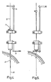
Figure 1 is a view of a catheter according to the present invention; -
Figure 2 is a view showing part of the catheter ofFigure 1 with internal features shown in dotted lines; -
Figure 3 is an end view of the catheter from the right hand end ofFigure 2 ; -
Figure 4 shows a first phase of stereotactic insertion; -
Figure 5 shows a second phase of stereotactic insertion; -
Figure 6 shows a third phase of stereotactic insertion; -
Figure 7 shows a fourth phase of stereotactic insertion; -
Figure 8 is a perspective view of a guide tube with a dome-shaped head; -
Figure 9 is a sectional view of the guide tube ofFigure 8 with the dome-shaped head; -
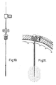
Figure 10 is a schematic view showing the catheter ofFigure 1 inserted through a guide tube ; and -
Figure 11 is a view of the catheter in situ once insertion is complete. - As explained above, insertion of a catheter into particularly sensitive regions of the brain leads to trauma on insertion which surgeons wish to minimise. The finer the catheter the less trauma the brain experiences. However, since the accuracy of insertion is crucially important, and since these particularly sensitive areas of the brain are a considerable distance from the skull surface, larger diameter catheters have been considered to be necessary in order to accurately place the distal end of the catheter. However, the present invention allows much finer catheters to be used.
-
Figures 1, 2 and 3 show acatheter 1 according to the present invention. Thecatheter 1 includes a length of fine tubing 2, the outer diameter which is no more than 1mm, and most preferably no greater than 0.7mm. It is even more preferred that the outer diameter be no more than 0.5mm. In this instance, the catheter tubing 2 is constructed from polyurethane plastic and preferably from Carbothane 55DB20 (Thermedics Polymer Products, Woburn MA, USA). The fine tubing 2 is linked to a length ofconnector tubing 3 having an outer diameter of about 2mm, via ahub 4. Theconnector tubing 3 is, in this case, made from polyurethane plastic, such as carbothane 85AB20, although other materials could also be used. - The
hub 4 in this case is also constructed using polyurethane, such as carbothane 72DB20. Again, other materials may also be appropriate. - The fine tubing 2 is intended to be inserted into the brain of a patient, whereas the
connector tubing 3 is intended to be connected to outflow tubing of a pump by which a therapeutic agent may be pumped intermittently or continuously to a desired brain target. For long term drug delivery, the pump would be implanted subcutaneously and the reservoir refilled as necessary percutaneously through a palpable port. In this case, theconnector tubing 3 would be connected to outflow tubing of the pump which would be tunnelled subcutaneously from the pump to the catheter. It's length will depend on particular installations and will be cut to length appropriately. - The
hub 4 includes a central body 5, which is generally cylindrical and a pair of diametrical opposing wings 6 each a containing a countersunk hole whereby the hub may be screwed to the outer surface of the skull of the patient. - The cylindrical body 5 of the
hub 4 includes a passageway passing through its complete length. The passageway includes a firstnarrow passage 8 of uniform diameter into which the fine tubing is inserted and securely held. The passageway also includes a secondwide passage 9 of uniform diameter into which theconnector tubing 3 is inserted and securely held. Between the first andsecond passages third linking passage 10 which is generally tapered in order to take account of the different internal diameters of the fine tubing 2 and theconnector tubing 3. It will be noted that the ends of thethird passage 10 are of the same or very similar diameter to the internal diameters of the fine tubing 2 and theconnector tubing 3. - From
Figure 2 , it can be seen that the right hand end of thehub 4 is frustoconical, and the end of the hub is planar and forms astop 11, the significance of which will be understood from the description below. - The insertion of the catheter will now be described. Firstly, a stereotactic frame is attached to the patient's skull and the position of the intracranial target is identified by imaging the patient wearing the stereotactic frame and defining the position of the target as three dimensional co-ordinates. This step is explained in more detail in the introduction to this patent specification and is a standard technique within the field of neurosurgery.
- Once the target has been defined, a stereoguide is used which is set to the target coordinates. An appropriately sized guide tube having an internal diameter of no more that 1mm is directed into the brain in the direction of the target. The guide tube is arranged with a head at one end, which, once inserted, can be attached to the patient's skull, for example by being bonded into a burrhole in the skull using an acrylic cement.
- Before insertion, the guide tube is cut to a length short of the target, and sufficiently short that, while it passes through brain tissue, it does not enter those parts of the brain which are particularly sensitive to trauma. The distal end of the guide tube will typically fall several millimetres short of the target. The distance from the top of the head of the guide tube to the target is then measured, and a radio-opaque stylette is cut to length such that, when inserted down the guide tube it's distal end reaches the planned target. This means that the stylette will extend beyond the distal end of the guide tube.
- The patient is then re-imaged in order to confirm the satisfactory placement of the stylette prior to removing the stylette and replacing it with the intraparenchymal catheter cut to the same length as the stylette. Again, the catheter will have an outer diameter of no more than one millimetre although it will be appreciated that the catheter, the stylette and the guide tube will all be matched so that the catheter and stylette will fit properly within the guide tube. If it is desired to use a very fine catheter of, say, 0.65mm in outer diameter, an appropriate guide tube will also be used with an internal diameter of 0.75mm.
- When the catheter is inserted, it is expected that it will be reinforced during insertion by the location of a stiff wire through it's bore, most likely made from tungsten. Once the catheter has been inserted in the guide tube, the
stop 11 on thehub 4 will abut the head of the guide tube meaning that the distal end of the catheter has reached the target. The stiff wire is removed, and the fine tubing 2 is bent through about 90° so that thehub 4 can be secured to the outer surface of the skull using screws passing through the countersunk holes 7. To facilitate the bending, the head of the guide tube is dome shaped and arranged such that, during bending, not only will the fine tubing 2 not kink, but also the distal end of the fine tubing will not move. This will be explained in more detail later in this specification. - The
connector tubing 3 can then be connected to the outflow tubing of a pump. Generally, theconnector tubing 3, will be tunnelled subcutaneously to the remotely positioned pump. - In an alternative insertion technique, a number of phases or steps are taken which are shown in
Figures 4 to 7 . As will be appreciated, small diameter catheters have a tendency to drift off the planned trajectory during insertion as a result of the flexibility inherent in a small diameter instrument. Since neurosurgical targets are often deeply situated, typically 70-80mm from the surface of the skull, and sometimes as much as 100mm from the skull surface, the catheter must normally be very rigid, and therefore of a larger diameter. - Examples of possible targets include parts of the mesencephalon including the subthalamic nucleus, the substantia nigra and the pedunculor-pontine nucleus. This is a particularly critical region of the brain, where it is important to minimise trauma from the passage of an instrument, which is typically situated about 70-80mm from the skull surface and contained within a volume which has a height of approximately 20-25mm.
- To facilitate insertion of very fine catheters into mesencephalic targets, insertion takes place as follows.
- Firstly, a small diameter
tungsten guide wire 22 of 0.6mm in diameter is inserted in atube 21 with an outer diameter of 1.7mm and fixed within thetube 21 with a finger-tightenedgrub screw 23 such that thewire 22 protrudes from the distal end of thetube 21 by 25mm. Thetube 21 is tapered towards its end for a length of 20mm. Thetube 21 andwire 22 can be seen inFigure 4 showing the first phase of insertion in which thetube 21 with thewire 22 projecting from its end can be seen. The finger tightenedgrub screw 23 can be seen at the top oftube 21, in which thewire 22 is held. - Insertion takes place from a stereotactic frame in which the target has been identified and defined in terms of three dimensional coordinates. The stereotactic frame carries a stereoguide which has been modified in order to permit this technique. During the first phase of insertion shown in
Figure 4 , the tube and wire are together lowered towards the target. In this case, the tube is 165mm in length, and since thetube 21 and the wire are inserted as a unit, the distance from the top of the tube to the tip of theguide wire 22 is 190mm. Thewire 22 extends above the top of the tube by approximately 150mm. The stereoguide includes anupper clamp 24 and alower clamp 25, and each of these clamps can be swivelled between a position of engagement with the wire or tube which is being inserted or removed, and a position remote from that. - Once the
guide wire 22 has reached its target, theupper clamp 24 is swivelled to clamp the proximal end of theguide wire 22. Once thegrub screw 23 has been loosened, thetube 21 can be withdrawn from the brain leaving thewire 22 in situ. Once thetube 21 has been raised up towards the upper clamp, the lower clamp can be swung across to clamp the now exposedwire 22, and theupper clamp 24 can be released, as shown inFigure 5 . This allows thetube 21 to be removed altogether from the top of thewire 22. - A
guide tube 31 is threaded onto thewire 22, and theupper clamp 24 is then swung around and closed on thewire 22. Thelower clamp 25 can then be released to allow theguide tube 31 to be inserted into the brain so that its distance is approximately 1 or 2 cm short of the target, also shown inFigure 7 . The guide tube 32 has at its upper end a head with a threaded outer surface which permits the head to be screwed into the tapped burrhole in the patient's skull, thereby securing theguide tube 31 securely in position. Further features of the head will become clear later in this description. - Once the
guide tube 31 is installed, theguidewire 22 may be removed andFigure 7 shows that a 0.65mm catheter 36 can then be inserted down theguide tube 31 to the target. - This method has the particular advantage that, on the first pass, the guide wire being stiffened by the
tube 21 will hit the target, and then by inserting a guide tube short of the target, the brain target will be fixed and the guide tube will facilitate the insertion of a very fine instrument to the target. For the treatment of certain conditions such as Alzheimer's disease it is necessary to deliver nerve growth factors to targets in the nucleus basalis through several in-dwelling catheters. If each catheter is only 0.65mm in diameter, multiple fine catheters can be inserted without substantially disrupting the tissue it is intended to regenerate. - In this insertion method, certain diameters of the
wire 22, the inside of thetube 21, the outside of thetube 21 and the diameters of theguide tube 31 and thecatheter 36 have been referred to. Of course, it will be appreciated that different diameters may be suitable and that the important factor is that the outer diameter of thewire 22 and of thefine catheter 36 which passed through the mesencephalon are as fine as possible, and no larger than 1mm in cross section. It is preferred that the diameter is no more than 0.7mm, and even more preferred that it is not more than 0.65mm. The top part of theguide tube 31 is shown inFigures 8 and 9 from which it will been seen that the top of thetube 41 carries ahead 42 which has a threaded outer surface which can be screwed into the burrhole in the skull through which instruments are inserted. The top of thetube 41 opens into a slot 44 in thehead 42. Thehead 42 is formed with adome structure 45 in which the slot 44 is located. - As will been seen from
Figure 9 , once the head has been secured into the skull, the catheter is located in thetube 41 and is then bent from position y to position z. The inner edge around which the catheter is bent is radiused and is shaped in the slot 44 such that the catheter will not kink and such that the distance from x to y is the same as the distance from x to z so that the distal end of the catheter is not moved during the bending process. - It will be understood from
Figures 8 and 9 , that, in this embodiment, theguide tube 31 is formed with thehead 42 including the threadedsurface 43 and thedomed structure 45 as an integral unit. - Referring now to
Figures 10 and 11 , it will be seen that a catheter has been inserted into the brain on a stiff wire (not shown) such that thestop 11 abuts the top of thedome structure 45. At this point, the stiff wire is removed and the distal end of the catheter is in the correct position for treatment. The catheter is then bent over to the position shown inFigure 11 maintaining thestop 11 against thedome structure 45.Figure 11 also shows how thehub 4 is attached via screws to the skull, and how theconnector tubing 3 is directed off towards the pump. - It is preferred that the catheter delivers drugs through a single port at its distal end. This has advantages over catheters with multiple ports at their distal end that may be used for intraparenchymal delivery to the brain. In particular, the use of a single port minimises the risk of the port becoming obstructed at the low flow rate anticipated for intraparenchymal delivery from the build up of proteinaceous material or gliotic ingrowth. A further advantage of having a single port at the distal end of the intraparenchymal catheter is that it ensures drug delivery at the defined target. The site of drug delivery from a multiport catheter is unpredictable, particularly at low flow rates. This is because flow will be maximal through the port with the lowest resistance. Even though the ports may be of an identical size, the degree to which tissue obstructs any individual port will vary. The net result will be off-axis drug delivery, probably from a single port, which will be sub-optimal for drug delivery to a small target.
- Trauma to the brain is minimised upon insertion as a result of using a very fine catheter of no more that 1mm in diameter and preferably less than 0.7mm in diameter. In addition, the small diameter catheter makes it suitable for drug delivery to small targets in the brain stem such as the substantia nigra and the pedunculopontine nucleus as well as to other small targets such as the nucleus basalis, the peri-aqueductal grey matter and various thalamic nuclei.
- During infusion of a therapeutic substance, the substance flowing from the catheter port or ports will preferably follow the path of least resistance, i.e. flow back along the tissue/catheter interface, up to the cortical surface and then into the CSF compartment. Depending on the flow rate it will defuse variably into the tissues with an ovoid volume of distribution. Containing the drug within a small brain target can therefore be a problem. If, however, the catheter has been inserted into the brain down an indwelling guide tube as in the present invention, then drug exiting the distal port flows back along the tissue/catheter interface until it reaches the guide tube. It then flows preferably along the interface between the guide tube and the catheter and out of the skull into the subgaleal compartment of the scalp. The volume of brain tissue exposed to the drug can therefore be controlled by adjusting the length of the catheter that projects beyond the guide tube as well as adjusting the flow rate. Such fine control is essential if one is to contain delivery of drugs such as neurotrophins within small brain targets.
- In a trial the intraparenchymal catheter of the present invention was implanted into the brains of five patients with advanced Parkinson's disease via a guide tube, the distal end of which was positioned just short of the desired target. One patient had the catheter implanted unilaterally and four had bilateral implants into the dorsal putamen (i.e. the desired target). Recombinant-methionyl human glial cell line derived neurotrophic factor (r-met Hu GDNF) was chronically infused through the catheters into their dorsal putamen via remotely positioned pumps (8626 Synchromed Pumps, Medtronic Inc, Minneapolis), implanted subcutaneously in the abdominal wall. GDNF is a neurotrophic factor that has been shown to reverse the symptoms of experimentally induced Parkinson's disease in animals. In this trial in humans it was infused at flow rates ranging from 2 to 8 µl per hour and doses between 10.8 and 3.2 micrograms/putamen/day. The patients were assessed preoperatively and at six months using the internationally recognised and validated scoring system for assessing the severity of Parkinson's disease, the Unified Parkinson's Disease Rating Score (UPDRS). At six months there was a 40% improvement in the patients UPDRS scores. The infusions were well tolerated and there were no major side affects.
Claims (15)
- A neurosurgical catheter device for insertion into the brain parenchyma of a patient, characterised in that the device comprises a fine tube having an external diameter of not more than 1.0mm.
- A neurosurgical catheter device according to claim 1 for insertion into the brain parenchyma of a patient,
the distal end of the device comprising a first section of fine tube having an external diameter of not more than 1.0mm, the first section of fine tube comprising a port or ports for delivery of a therapeutic agent to a desired brain target,
wherein the device comprises a second section of tube having a greater external diameter than the first section of fine tube. - A device according to claim 2, wherein, in use, the distal end of the first section of fine tube and the distal end of the second section of tube are both inserted into the brain parenchyma of the patient.
- A device according to any one of claims 2 to 3, comprising a step between the different diameters of the first section of fine tube and the second section of tube.
- A device according to claim 2, wherein a hub is disposed between the first section of fine tube and the second section of tube.
- A device according to any one of claims 2 to 5, wherein the second section of tube has an internal diameter of not more than 1.0mm.
- A device according to any one of claims 2 to 6, wherein a head is disposed at the proximal end of the second section of tube for attachment to the skull of the patient.
- A device according to any one of claims 2 to 7, wherein the first section of fine tube has an external diameter of not more than 0.7mm.
- A device according to any one of claims 2 to 8, wherein the distal end of the device can be inserted into the brain parenchyma to a depth of up to 100mm from the skull.
- A device according to any one of claims 2 to 9, wherein the first section of fine tube comprises a single port at its distal end for delivery of therapeutic agent to a brain target.
- Neurosurgical apparatus comprising;
a neurosurgical catheter device according to any preceding claim, a pump, and
a connector tube,
wherein the connector tube provides a connection between the outflow tubing of the pump and the fine tube of the neurosurgical catheter device. - Neurosurgical apparatus according to claim 11, wherein the pump is arranged to intermittently pump therapeutic agent to the neurosurgical catheter device.
- Neurosurgical apparatus according to claim 11, wherein the pump is arranged to continuously pump therapeutic agent to the neurosurgical catheter device.
- Neurosurgical apparatus according to any one of claims 11 to 13 that is subcutaneously implantable.
- A method of operating neurosurgical apparatus comprising a fine tube for insertion in to the brain, a pump and a connector tube that connects the outflow tubing of the pump to the fine tube, the method comprising the step of using the pump to intermittently pump therapeutic agent to the fine tube.
Applications Claiming Priority (3)
| Application Number | Priority Date | Filing Date | Title |
|---|---|---|---|
| GBGB0205772.7A GB0205772D0 (en) | 2002-03-12 | 2002-03-12 | Catheter |
| EP08018637A EP2018829B1 (en) | 2002-03-12 | 2003-03-11 | Catheter and guide tube for intracerebral application |
| EP03708342A EP1482851B1 (en) | 2002-03-12 | 2003-03-11 | Catheter and guide tube for intracerebral application |
Related Parent Applications (2)
| Application Number | Title | Priority Date | Filing Date |
|---|---|---|---|
| EP03708342.5 Division | 2003-03-11 | ||
| EP08018637.2 Division | 2008-10-24 |
Publications (1)
| Publication Number | Publication Date |
|---|---|
| EP2258437A1 true EP2258437A1 (en) | 2010-12-08 |
Family
ID=9932788
Family Applications (3)
| Application Number | Title | Priority Date | Filing Date |
|---|---|---|---|
| EP08018637A Expired - Lifetime EP2018829B1 (en) | 2002-03-12 | 2003-03-11 | Catheter and guide tube for intracerebral application |
| EP10010990A Ceased EP2258437A1 (en) | 2002-03-12 | 2003-03-11 | Catheter for intracerebral application |
| EP03708342A Expired - Lifetime EP1482851B1 (en) | 2002-03-12 | 2003-03-11 | Catheter and guide tube for intracerebral application |
Family Applications Before (1)
| Application Number | Title | Priority Date | Filing Date |
|---|---|---|---|
| EP08018637A Expired - Lifetime EP2018829B1 (en) | 2002-03-12 | 2003-03-11 | Catheter and guide tube for intracerebral application |
Family Applications After (1)
| Application Number | Title | Priority Date | Filing Date |
|---|---|---|---|
| EP03708342A Expired - Lifetime EP1482851B1 (en) | 2002-03-12 | 2003-03-11 | Catheter and guide tube for intracerebral application |
Country Status (12)
| Country | Link |
|---|---|
| US (3) | US8128600B2 (en) |
| EP (3) | EP2018829B1 (en) |
| JP (2) | JP4465493B2 (en) |
| AT (2) | ATE497735T1 (en) |
| CA (3) | CA2974428C (en) |
| DE (2) | DE60325181D1 (en) |
| DK (1) | DK1482851T3 (en) |
| ES (1) | ES2316732T3 (en) |
| GB (1) | GB0205772D0 (en) |
| MX (1) | MXPA04008297A (en) |
| PL (1) | PL372262A1 (en) |
| WO (1) | WO2003077785A1 (en) |
Cited By (2)
| Publication number | Priority date | Publication date | Assignee | Title |
|---|---|---|---|---|
| WO2014016591A1 (en) * | 2012-07-24 | 2014-01-30 | Renishaw Plc | Neurosurgical apparatus and methods |
| US9452241B2 (en) | 2006-08-18 | 2016-09-27 | Renishaw (Ireland) Limited | Neurosurgical instruments |
Families Citing this family (77)
| Publication number | Priority date | Publication date | Assignee | Title |
|---|---|---|---|---|
| US7660621B2 (en) | 2000-04-07 | 2010-02-09 | Medtronic, Inc. | Medical device introducer |
| US7704260B2 (en) | 2002-09-17 | 2010-04-27 | Medtronic, Inc. | Low profile instrument immobilizer |
| US8946151B2 (en) | 2003-02-24 | 2015-02-03 | Northern Bristol N.H.S. Trust Frenchay Hospital | Method of treating Parkinson's disease in humans by convection-enhanced infusion of glial cell-line derived neurotrophic factor to the putamen |
| US8012167B2 (en) * | 2003-08-14 | 2011-09-06 | Loma Linda University Medical Center | Vascular wound closure device and method |
| US20050182422A1 (en) * | 2004-02-13 | 2005-08-18 | Schulte Gregory T. | Apparatus for securing a therapy delivery device within a burr hole and method for making same |
| SI1807009T1 (en) | 2004-10-05 | 2015-04-30 | Genzyme Corporation | Stepped cannula |
| US20060129126A1 (en) * | 2004-11-19 | 2006-06-15 | Kaplitt Michael G | Infusion device and method for infusing material into the brain of a patient |
| CA2616010C (en) * | 2005-07-20 | 2013-11-05 | Neil R. Euliano | Medication compliance system and associated methods |
| US9047746B1 (en) | 2005-07-20 | 2015-06-02 | Neil Euliano | Electronic medication compliance monitoring system and associated methods |
| AU2006283189B2 (en) * | 2005-08-23 | 2013-01-31 | The Regents Of The University Of California | Reflux resistant cannula and system for chronic delivery of therapeutic agents using convection-enhanced delivery |
| US7819842B2 (en) * | 2006-11-21 | 2010-10-26 | Medtronic, Inc. | Chronically implantable guide tube for repeated intermittent delivery of materials or fluids to targeted tissue sites |
| GB0623395D0 (en) | 2006-11-23 | 2007-01-03 | Renishaw Plc | Port |
| US20080275466A1 (en) * | 2007-05-01 | 2008-11-06 | James Grant Skakoon | Dual cannula system and method for using same |
| RU2442617C2 (en) * | 2007-05-17 | 2012-02-20 | Медженезиз Терапьютикс Инк. | Convective conveying catheter with the dismountable stiffening element and the method of its application |
| US8147480B2 (en) * | 2007-09-28 | 2012-04-03 | Codman & Shurtleff, Inc. | Catheter for reduced reflux in targeted tissue delivery of a therapeutic agent |
| US7766875B2 (en) * | 2007-09-28 | 2010-08-03 | Codman & Shurtleff, Inc. | Catheter for reduced reflux in targeted tissue delivery of a therapeutic agent |
| CA2701744A1 (en) * | 2007-10-08 | 2009-04-16 | Renishaw (Ireland) Limited | Catheter |
| CN101883535B (en) | 2007-10-08 | 2013-05-01 | 瑞尼斯豪(爱尔兰)有限公司 | Apparatus for stereotactic neurosurgery |
| GB0802634D0 (en) * | 2008-02-13 | 2008-03-19 | Renishaw Plc | Catheter |
| US8326439B2 (en) | 2008-04-16 | 2012-12-04 | Nevro Corporation | Treatment devices with delivery-activated inflatable members, and associated systems and methods for treating the spinal cord and other tissues |
| US8834446B2 (en) * | 2008-06-12 | 2014-09-16 | DePuy Synthes Products, LLC | Pulsatile flux drug delivery |
| CA2739173A1 (en) * | 2008-10-08 | 2010-04-15 | Renishaw (Ireland) Limited | Catheter |
| GB201002370D0 (en) | 2010-02-12 | 2010-03-31 | Renishaw Ireland Ltd | Percutaneous drug delivery apparatus |
| PL2558154T3 (en) | 2010-04-16 | 2020-11-30 | Clearpoint Neuro, Inc. | Mri surgical systems including mri-compatible surgical cannulae for transferring a substance to and/or from a patient |
| JP5656146B2 (en) * | 2010-05-19 | 2015-01-21 | 学校法人 久留米大学 | Drug administration device and drug administration method |
| US9633378B1 (en) | 2010-12-06 | 2017-04-25 | Wayfare Interactive, Inc. | Deep-linking system, method and computer program product for online advertisement and E-commerce |
| US20130035660A1 (en) | 2011-08-01 | 2013-02-07 | Alcyone Lifesciences, Inc. | Multidirectional microfluidic drug delivery devices with conformable balloons |
| GB201117061D0 (en) | 2011-10-04 | 2011-11-16 | Renishaw Ireland Ltd | Neurosurgical apparatus |
| GB201202094D0 (en) | 2012-02-07 | 2012-03-21 | Renishaw Ireland Ltd | Medical apparatus |
| GB201202091D0 (en) | 2012-02-07 | 2012-03-21 | Renishaw Ireland Ltd | Drug delivery apparatus |
| GB201202093D0 (en) | 2012-02-07 | 2012-03-21 | Renishaw Ireland Ltd | Drug storage apparatus |
| GB201203426D0 (en) * | 2012-02-28 | 2012-04-11 | Renishaw Plc | Neurosurgical apparatus |
| GB201204263D0 (en) | 2012-03-12 | 2012-04-25 | Renishaw Plc | Giloma treatment |
| US10099034B2 (en) | 2012-07-27 | 2018-10-16 | The Regents Of The University Of California | Microinjection catheter |
| GB201217606D0 (en) | 2012-10-02 | 2012-11-14 | Renishaw Plc | Neurosurgical device and method |
| CA3123066A1 (en) | 2012-12-18 | 2014-06-26 | Alcyone Lifesciences, Inc. | Systems and methods for reducing or preventing backflow in a delivery system |
| EP2945540A1 (en) * | 2013-01-17 | 2015-11-25 | General Electric Company | C-arm of medical imaging system |
| US9415190B2 (en) * | 2013-02-13 | 2016-08-16 | Interrad Medical, Inc. | Systems and methods for anchoring medical devices |
| WO2014128824A1 (en) * | 2013-02-19 | 2014-08-28 | テルモ株式会社 | Medical instrument |
| WO2014128875A1 (en) * | 2013-02-21 | 2014-08-28 | テルモ株式会社 | Medical instrument |
| GB201308917D0 (en) | 2013-05-17 | 2013-07-03 | Renishaw Plc | Delivery |
| WO2014189253A2 (en) * | 2013-05-22 | 2014-11-27 | 사회복지법인 삼성생명공익재단 | Apparatus for intracerebral injection of drugs, assembly for providing apparatus for intracerebral injection of drugs, and method for intracerebral administration of brain disease treatment drugs |
| CA3120114A1 (en) | 2013-06-17 | 2014-12-24 | Alcyone Lifesciences, Inc. | Methods and devices for protecting catheter tips and stereotactic fixtures for microcatheters |
| WO2015017609A2 (en) | 2013-07-31 | 2015-02-05 | Alcyone Lifesciences, Inc. | Systems and methods for drug delivery, treatment, and monitoring |
| US9891296B2 (en) | 2013-09-13 | 2018-02-13 | MRI Interventions, Inc. | Intrabody fluid transfer devices, systems and methods |
| US10521561B1 (en) | 2013-12-17 | 2019-12-31 | Etectrx, Inc. | Electronic compliance system and associated methods |
| US10369329B2 (en) | 2014-01-30 | 2019-08-06 | Renishaw Plc | Neurosurgical apparatus and method |
| GB201404978D0 (en) | 2014-03-20 | 2014-05-07 | Renishaw Plc | Neurosurgical apparatus |
| US9409020B2 (en) | 2014-05-20 | 2016-08-09 | Nevro Corporation | Implanted pulse generators with reduced power consumption via signal strength/duration characteristics, and associated systems and methods |
| FR3026633A1 (en) | 2014-10-07 | 2016-04-08 | Commissariat Energie Atomique | ROCKING TROUGH OF A CATHETER |
| AU2015336218B2 (en) | 2014-10-22 | 2020-07-23 | Nevro Corp. | Systems and methods for extending the life of an implanted pulse generator battery |
| US10806396B2 (en) | 2015-01-26 | 2020-10-20 | Alcyone Lifesciences, Inc. | Drug delivery methods with tracer |
| JP1553633S (en) * | 2015-02-13 | 2016-07-11 | ||
| US9517344B1 (en) | 2015-03-13 | 2016-12-13 | Nevro Corporation | Systems and methods for selecting low-power, effective signal delivery parameters for an implanted pulse generator |
| GB201506052D0 (en) | 2015-04-09 | 2015-05-27 | Renishaw Plc | Movement disorder |
| US10300277B1 (en) | 2015-12-14 | 2019-05-28 | Nevro Corp. | Variable amplitude signals for neurological therapy, and associated systems and methods |
| ES2904702T3 (en) | 2015-12-31 | 2022-04-05 | Nevro Corp | Controller for nerve stimulation circuit and associated systems and methods |
| WO2017120167A1 (en) | 2016-01-04 | 2017-07-13 | Alcyone Lifesciences, Inc. | Methods and devices for treating stroke |
| EP3393571B1 (en) | 2016-02-17 | 2024-03-06 | ClearPoint Neuro, Inc. | Intrabody surgical fluid transfer assemblies with adjustable exposed cannula to needle tip length, related systems and methods |
| US20180035164A1 (en) * | 2016-03-02 | 2018-02-01 | Antennas Direct, Inc. | Wireless-capable remote antenna boxes and related systems and methods |
| USD793549S1 (en) | 2016-05-04 | 2017-08-01 | DePuy Synthes Products, Inc. | Catheter clip |
| EP3243549A1 (en) | 2016-05-12 | 2017-11-15 | Renishaw plc | Percutaneous access apparatus |
| EP3506817A4 (en) | 2016-08-30 | 2020-07-22 | The Regents of The University of California | Methods for biomedical targeting and delivery and devices and systems for practicing the same |
| JP7229989B2 (en) | 2017-07-17 | 2023-02-28 | ボイジャー セラピューティクス インコーポレイテッド | Trajectory array guide system |
| KR102223085B1 (en) * | 2017-11-01 | 2021-03-04 | 사회복지법인 삼성생명공익재단 | Apparatus for intracerebral injection of drugs and method for intracerebral injection of drugs |
| CR20200357A (en) | 2018-01-30 | 2021-03-29 | Nevro Corp | Efficient use of an implantable pulse generator battery, and associated systems and methods |
| US11253237B2 (en) | 2018-05-09 | 2022-02-22 | Clearpoint Neuro, Inc. | MRI compatible intrabody fluid transfer systems and related devices and methods |
| US11022664B2 (en) | 2018-05-09 | 2021-06-01 | Clearpoint Neuro, Inc. | MRI compatible intrabody fluid transfer systems and related devices and methods |
| GB201812746D0 (en) | 2018-08-06 | 2018-09-19 | Gill Steven S | Method |
| US11058875B1 (en) | 2018-09-19 | 2021-07-13 | Nevro Corp. | Motor function in spinal cord injury patients via electrical stimulation, and associated systems and methods |
| US11590352B2 (en) | 2019-01-29 | 2023-02-28 | Nevro Corp. | Ramped therapeutic signals for modulating inhibitory interneurons, and associated systems and methods |
| US10933238B2 (en) | 2019-01-31 | 2021-03-02 | Nevro Corp. | Power control circuit for sterilized devices, and associated systems and methods |
| US11684750B2 (en) | 2019-10-08 | 2023-06-27 | Clearpoint Neuro, Inc. | Extension tube assembly and related medical fluid transfer systems and methods |
| GB202009981D0 (en) | 2020-06-30 | 2020-08-12 | Xced Llp | Method |
| GB202106224D0 (en) | 2021-04-30 | 2021-06-16 | Neurochase Tech Ltd | Neurosurgical device |
| EP4329647A1 (en) | 2021-04-30 | 2024-03-06 | Neurochase Technologies Limited | Implantable guide device |
| GB202109503D0 (en) | 2021-07-01 | 2021-08-18 | Xced Llp | Method |
Citations (11)
| Publication number | Priority date | Publication date | Assignee | Title |
|---|---|---|---|---|
| US5119832A (en) * | 1989-07-11 | 1992-06-09 | Ravi Xavier | Epidural catheter with nerve stimulators |
| WO1997040879A1 (en) * | 1996-04-30 | 1997-11-06 | Medtronic, Inc. | Therapeutic method for treatment of alzheimer's disease |
| US5713858A (en) * | 1995-04-28 | 1998-02-03 | Medtronic, Inc. | Permanently implantable guiding catheter |
| US6042579A (en) | 1997-04-30 | 2000-03-28 | Medtronic, Inc. | Techniques for treating neurodegenerative disorders by infusion of nerve growth factors into the brain |
| WO2000061017A1 (en) * | 1999-05-04 | 2000-10-19 | Neurodynamics, Inc. | Improved catheter guide and drill guide apparatus and method for perpendicular insertion into a cranium orifice |
| WO2000069502A1 (en) * | 1999-05-14 | 2000-11-23 | Boston Scientific Limited | Single lumen balloon-tipped micro catheter with reinforced shaft |
| US6227203B1 (en) * | 1998-02-12 | 2001-05-08 | Medtronic, Inc. | Techniques for controlling abnormal involuntary movements by brain stimulation and drug infusion |
| US20010001117A1 (en) * | 1998-04-28 | 2001-05-10 | Chow Sean L. | Flow directed catheter |
| US20010003156A1 (en) * | 1999-12-01 | 2001-06-07 | Steven Gill | Neurosurgical guide device |
| US20010027599A1 (en) * | 1995-04-28 | 2001-10-11 | Medtronic, Inc. | Intraparenchymal infusion catheter system |
| US20010056275A1 (en) * | 2000-03-24 | 2001-12-27 | Stephen Brushey | Anesthesia conduction catheter |
Family Cites Families (22)
| Publication number | Priority date | Publication date | Assignee | Title |
|---|---|---|---|---|
| JPS61206460A (en) * | 1985-03-11 | 1986-09-12 | 大槻 泰介 | Catheter inserting apparatus for local injection of brain abscess |
| US5006122A (en) * | 1988-12-02 | 1991-04-09 | The United States Of America As Represented By The Department Of Health And Human Services | Tissue transplantation system |
| US5752930A (en) * | 1995-04-28 | 1998-05-19 | Medtronic, Inc. | Implantable techniques for infusing equal volumes of agents to spaced sites |
| US5971975A (en) * | 1996-10-09 | 1999-10-26 | Target Therapeutics, Inc. | Guide catheter with enhanced guidewire tracking |
| US6267769B1 (en) * | 1997-05-15 | 2001-07-31 | Regents Of The Universitiy Of Minnesota | Trajectory guide method and apparatus for use in magnetic resonance and computerized tomographic scanners |
| US5951539A (en) * | 1997-06-10 | 1999-09-14 | Target Therpeutics, Inc. | Optimized high performance multiple coil spiral-wound vascular catheter |
| US5916200A (en) * | 1997-10-01 | 1999-06-29 | Walter Lorenz Surgical, Inc. | Apparatus and method for stabilization of a cranial shunt |
| US5947296A (en) * | 1997-10-30 | 1999-09-07 | Schneider/Namic | Multipack package |
| AU9790498A (en) * | 1997-10-31 | 1999-05-24 | Neurovasx, Inc. | Sub-microcatheter |
| US6045532A (en) * | 1998-02-20 | 2000-04-04 | Arthrocare Corporation | Systems and methods for electrosurgical treatment of tissue in the brain and spinal cord |
| US6591472B1 (en) * | 1998-12-08 | 2003-07-15 | Medtronic, Inc. | Multiple segment catheter and method of fabrication |
| CA2328614C (en) * | 1999-02-12 | 2012-06-26 | Biostream, Inc. | Matrices for drug delivery and methods for making and using the same |
| US6348050B1 (en) * | 1999-04-30 | 2002-02-19 | Medtronic, Inc. | Infusion systems for creating microenvironments in a living body |
| US6517550B1 (en) * | 2000-02-02 | 2003-02-11 | Board Of Regents, The University Of Texas System | Foreign body retrieval device |
| US6609030B1 (en) * | 2000-02-24 | 2003-08-19 | Electrocore Techniques, Llc | Method of treating psychiatric diseases by neuromodulation within the dorsomedial thalamus |
| AU2001281745A1 (en) * | 2000-07-25 | 2002-02-05 | Csf Dynamics A/S | A ventricle drain |
| WO2002013714A1 (en) * | 2000-08-17 | 2002-02-21 | Image Guided Neurologies, Inc. | Trajectory guide with instrument immobilizer |
| US7033326B1 (en) * | 2000-12-29 | 2006-04-25 | Advanced Bionics Corporation | Systems and methods of implanting a lead for brain stimulation |
| US6719727B2 (en) * | 2001-05-25 | 2004-04-13 | Becton Dickinson And Company | Catheter having a wing with a stiffening member therein |
| US7013177B1 (en) * | 2001-07-05 | 2006-03-14 | Advanced Bionics Corporation | Treatment of pain by brain stimulation |
| US7981863B2 (en) * | 2001-09-19 | 2011-07-19 | Neuronova Ab | Treatment of Parkinson's disease with PDGF |
| US20040209810A1 (en) * | 2003-02-24 | 2004-10-21 | Gill Steven S. | Method of treating Parkinson's disease in humans by intraputaminal infusion of glial cell-line derived neurotrophic factor |
-
2002
- 2002-03-12 GB GBGB0205772.7A patent/GB0205772D0/en not_active Ceased
-
2003
- 2003-03-11 DK DK03708342T patent/DK1482851T3/en active
- 2003-03-11 CA CA2974428A patent/CA2974428C/en not_active Expired - Lifetime
- 2003-03-11 WO PCT/GB2003/001030 patent/WO2003077785A1/en active Application Filing
- 2003-03-11 EP EP08018637A patent/EP2018829B1/en not_active Expired - Lifetime
- 2003-03-11 AT AT08018637T patent/ATE497735T1/en not_active IP Right Cessation
- 2003-03-11 CA CA2872998A patent/CA2872998C/en not_active Expired - Lifetime
- 2003-03-11 EP EP10010990A patent/EP2258437A1/en not_active Ceased
- 2003-03-11 DE DE60325181T patent/DE60325181D1/en not_active Expired - Lifetime
- 2003-03-11 DE DE60336030T patent/DE60336030D1/en not_active Expired - Lifetime
- 2003-03-11 JP JP2003575842A patent/JP4465493B2/en not_active Expired - Fee Related
- 2003-03-11 EP EP03708342A patent/EP1482851B1/en not_active Expired - Lifetime
- 2003-03-11 US US10/505,240 patent/US8128600B2/en not_active Expired - Fee Related
- 2003-03-11 PL PL03372262A patent/PL372262A1/en not_active Application Discontinuation
- 2003-03-11 ES ES03708342T patent/ES2316732T3/en not_active Expired - Lifetime
- 2003-03-11 CA CA2475855A patent/CA2475855C/en not_active Expired - Lifetime
- 2003-03-11 MX MXPA04008297A patent/MXPA04008297A/en active IP Right Grant
- 2003-03-11 AT AT03708342T patent/ATE416706T1/en not_active IP Right Cessation
-
2009
- 2009-07-30 JP JP2009178428A patent/JP4929318B2/en not_active Expired - Fee Related
-
2011
- 2011-07-19 US US13/137,071 patent/US20110282319A1/en not_active Abandoned
-
2017
- 2017-09-27 US US15/717,417 patent/US20180015273A1/en not_active Abandoned
Patent Citations (12)
| Publication number | Priority date | Publication date | Assignee | Title |
|---|---|---|---|---|
| US5119832A (en) * | 1989-07-11 | 1992-06-09 | Ravi Xavier | Epidural catheter with nerve stimulators |
| US5713858A (en) * | 1995-04-28 | 1998-02-03 | Medtronic, Inc. | Permanently implantable guiding catheter |
| US20010027599A1 (en) * | 1995-04-28 | 2001-10-11 | Medtronic, Inc. | Intraparenchymal infusion catheter system |
| WO1997040879A1 (en) * | 1996-04-30 | 1997-11-06 | Medtronic, Inc. | Therapeutic method for treatment of alzheimer's disease |
| US6042579A (en) | 1997-04-30 | 2000-03-28 | Medtronic, Inc. | Techniques for treating neurodegenerative disorders by infusion of nerve growth factors into the brain |
| US6227203B1 (en) * | 1998-02-12 | 2001-05-08 | Medtronic, Inc. | Techniques for controlling abnormal involuntary movements by brain stimulation and drug infusion |
| US20010001117A1 (en) * | 1998-04-28 | 2001-05-10 | Chow Sean L. | Flow directed catheter |
| WO2000061017A1 (en) * | 1999-05-04 | 2000-10-19 | Neurodynamics, Inc. | Improved catheter guide and drill guide apparatus and method for perpendicular insertion into a cranium orifice |
| WO2000069502A1 (en) * | 1999-05-14 | 2000-11-23 | Boston Scientific Limited | Single lumen balloon-tipped micro catheter with reinforced shaft |
| US20010003156A1 (en) * | 1999-12-01 | 2001-06-07 | Steven Gill | Neurosurgical guide device |
| GB2357700A (en) | 1999-12-01 | 2001-07-04 | Steven Gill | Neurosurgical guide device |
| US20010056275A1 (en) * | 2000-03-24 | 2001-12-27 | Stephen Brushey | Anesthesia conduction catheter |
Non-Patent Citations (2)
| Title |
|---|
| GILL S S ET AL: "Direct brain infusion of glial cell line-derived neurotrophic factor in Parkinson disease", NATURE MEDICINE, NATURE PUBLISHING GROUP, NEW YORK, NY, US, vol. 9, no. 5, 1 May 2003 (2003-05-01), pages 589 - 595, XP002343708, ISSN: 1078-8956, DOI: 10.1038/NM850 * |
| SALVATORE M F ET AL: "Point source concentration of GDNF may explain failure of phase II clinical trial", EXPERIMENTAL NEUROLOGY, ELSEVIER, AMSTERDAM, NL, vol. 202, no. 2, 1 December 2006 (2006-12-01), pages 497 - 505, XP024946082, ISSN: 0014-4886, [retrieved on 20061201], DOI: 10.1016/J.EXPNEUROL.2006.07.015 * |
Cited By (5)
| Publication number | Priority date | Publication date | Assignee | Title |
|---|---|---|---|---|
| US9452241B2 (en) | 2006-08-18 | 2016-09-27 | Renishaw (Ireland) Limited | Neurosurgical instruments |
| US10857327B2 (en) | 2006-08-18 | 2020-12-08 | Renishaw Plc | Neurosurgical instruments |
| WO2014016591A1 (en) * | 2012-07-24 | 2014-01-30 | Renishaw Plc | Neurosurgical apparatus and methods |
| CN104582610A (en) * | 2012-07-24 | 2015-04-29 | 瑞尼斯豪公司 | Neurosurgical apparatus and methods |
| US10751513B2 (en) | 2012-07-24 | 2020-08-25 | Renishaw Plc | Neurosurgical apparatus and methods |
Also Published As
| Publication number | Publication date |
|---|---|
| CA2475855A1 (en) | 2003-09-25 |
| JP4929318B2 (en) | 2012-05-09 |
| DK1482851T3 (en) | 2009-02-09 |
| EP1482851B1 (en) | 2008-12-10 |
| ES2316732T3 (en) | 2009-04-16 |
| CA2974428A1 (en) | 2003-09-25 |
| ATE497735T1 (en) | 2011-02-15 |
| US20110282319A1 (en) | 2011-11-17 |
| CA2872998C (en) | 2017-10-17 |
| EP2018829A3 (en) | 2009-04-01 |
| JP2009279433A (en) | 2009-12-03 |
| ATE416706T1 (en) | 2008-12-15 |
| EP2018829B1 (en) | 2011-02-09 |
| EP1482851A1 (en) | 2004-12-08 |
| EP2018829A2 (en) | 2009-01-28 |
| CA2974428C (en) | 2021-03-23 |
| DE60325181D1 (en) | 2009-01-22 |
| CA2475855C (en) | 2015-01-20 |
| AU2003212522A1 (en) | 2003-09-29 |
| US20180015273A1 (en) | 2018-01-18 |
| MXPA04008297A (en) | 2005-06-08 |
| CA2872998A1 (en) | 2003-09-25 |
| PL372262A1 (en) | 2005-07-11 |
| US20050154297A1 (en) | 2005-07-14 |
| WO2003077785A1 (en) | 2003-09-25 |
| JP4465493B2 (en) | 2010-05-19 |
| US8128600B2 (en) | 2012-03-06 |
| JP2005519693A (en) | 2005-07-07 |
| DE60336030D1 (en) | 2011-03-24 |
| GB0205772D0 (en) | 2002-04-24 |
Similar Documents
| Publication | Publication Date | Title |
|---|---|---|
| EP1482851B1 (en) | Catheter and guide tube for intracerebral application | |
| EP1509153B1 (en) | Stereoguide for clamping neurosurgical instruments | |
| US6503242B1 (en) | Therapeutic method for treatment of Alzheimer's disease | |
| EP1327460B1 (en) | Multi-catheter insertion device | |
| US20010027309A1 (en) | Therapeutic method for treatment of alzheimer's disease | |
| WO2003090820A1 (en) | Catheter anchor system | |
| US9352125B2 (en) | Portal anchors incorporating strain relief cup and systems using same | |
| US11065421B2 (en) | Catheter curvature braces and methods of using same | |
| AU2003212522B2 (en) | Catheter and guide tube for intracerebral application | |
| WO2007075912A2 (en) | A system and method of administering a therapeutic material to brain tissue |
Legal Events
| Date | Code | Title | Description |
|---|---|---|---|
| PUAI | Public reference made under article 153(3) epc to a published international application that has entered the european phase |
Free format text: ORIGINAL CODE: 0009012 |
|
| AC | Divisional application: reference to earlier application |
Ref document number: 2018829 Country of ref document: EP Kind code of ref document: P Ref document number: 1482851 Country of ref document: EP Kind code of ref document: P |
|
| AK | Designated contracting states |
Kind code of ref document: A1 Designated state(s): AT BE BG CH CY CZ DE DK EE ES FI FR GB GR HU IE IT LI LU MC NL PT RO SE SI SK TR |
|
| 17P | Request for examination filed |
Effective date: 20110517 |
|
| 17Q | First examination report despatched |
Effective date: 20111012 |
|
| STAA | Information on the status of an ep patent application or granted ep patent |
Free format text: STATUS: EXAMINATION IS IN PROGRESS |
|
| APBK | Appeal reference recorded |
Free format text: ORIGINAL CODE: EPIDOSNREFNE |
|
| APBN | Date of receipt of notice of appeal recorded |
Free format text: ORIGINAL CODE: EPIDOSNNOA2E |
|
| APBR | Date of receipt of statement of grounds of appeal recorded |
Free format text: ORIGINAL CODE: EPIDOSNNOA3E |
|
| APAF | Appeal reference modified |
Free format text: ORIGINAL CODE: EPIDOSCREFNE |
|
| APAF | Appeal reference modified |
Free format text: ORIGINAL CODE: EPIDOSCREFNE |
|
| APBT | Appeal procedure closed |
Free format text: ORIGINAL CODE: EPIDOSNNOA9E |
|
| STAA | Information on the status of an ep patent application or granted ep patent |
Free format text: STATUS: THE APPLICATION HAS BEEN REFUSED |
|
| 18R | Application refused |
Effective date: 20210125 |