EP2261250B1 - GCSF-Albumin fusion proteins - Google Patents
GCSF-Albumin fusion proteins Download PDFInfo
- Publication number
- EP2261250B1 EP2261250B1 EP10075030.6A EP10075030A EP2261250B1 EP 2261250 B1 EP2261250 B1 EP 2261250B1 EP 10075030 A EP10075030 A EP 10075030A EP 2261250 B1 EP2261250 B1 EP 2261250B1
- Authority
- EP
- European Patent Office
- Prior art keywords
- albumin fusion
- fusion proteins
- protein
- fusion protein
- albumin
- Prior art date
- Legal status (The legal status is an assumption and is not a legal conclusion. Google has not performed a legal analysis and makes no representation as to the accuracy of the status listed.)
- Expired - Lifetime
Links
- 108020001507 fusion proteins Proteins 0.000 title claims description 528
- 102000037865 fusion proteins Human genes 0.000 title claims description 527
- 102000009027 Albumins Human genes 0.000 claims description 519
- 108010088751 Albumins Proteins 0.000 claims description 519
- 102000040430 polynucleotide Human genes 0.000 claims description 220
- 108091033319 polynucleotide Proteins 0.000 claims description 220
- 239000002157 polynucleotide Substances 0.000 claims description 220
- 210000004027 cell Anatomy 0.000 claims description 120
- 238000000034 method Methods 0.000 claims description 87
- 230000014509 gene expression Effects 0.000 claims description 63
- 150000007523 nucleic acids Chemical class 0.000 claims description 60
- 108091006905 Human Serum Albumin Proteins 0.000 claims description 59
- 102000008100 Human Serum Albumin Human genes 0.000 claims description 59
- 102000039446 nucleic acids Human genes 0.000 claims description 56
- 108020004707 nucleic acids Proteins 0.000 claims description 56
- 239000013598 vector Substances 0.000 claims description 53
- 240000004808 Saccharomyces cerevisiae Species 0.000 claims description 46
- 235000014680 Saccharomyces cerevisiae Nutrition 0.000 claims description 41
- 230000000694 effects Effects 0.000 claims description 39
- 206010028980 Neoplasm Diseases 0.000 claims description 36
- 208000004235 neutropenia Diseases 0.000 claims description 34
- 238000011282 treatment Methods 0.000 claims description 31
- 208000023275 Autoimmune disease Diseases 0.000 claims description 21
- 239000013604 expression vector Substances 0.000 claims description 19
- 201000011510 cancer Diseases 0.000 claims description 18
- 230000002265 prevention Effects 0.000 claims description 17
- 239000003814 drug Substances 0.000 claims description 15
- 230000013595 glycosylation Effects 0.000 claims description 15
- 238000006206 glycosylation reaction Methods 0.000 claims description 15
- 208000015181 infectious disease Diseases 0.000 claims description 14
- 108010017080 Granulocyte Colony-Stimulating Factor Proteins 0.000 claims description 12
- 150000001413 amino acids Chemical class 0.000 claims description 12
- 238000003745 diagnosis Methods 0.000 claims description 12
- 208000032839 leukemia Diseases 0.000 claims description 12
- 208000027866 inflammatory disease Diseases 0.000 claims description 11
- 210000005253 yeast cell Anatomy 0.000 claims description 10
- 238000002512 chemotherapy Methods 0.000 claims description 9
- 239000003550 marker Substances 0.000 claims description 9
- 230000001154 acute effect Effects 0.000 claims description 8
- 230000002950 deficient Effects 0.000 claims description 8
- 230000035897 transcription Effects 0.000 claims description 7
- 238000013518 transcription Methods 0.000 claims description 7
- 239000004365 Protease Substances 0.000 claims description 6
- 102100029516 Basic salivary proline-rich protein 1 Human genes 0.000 claims description 5
- 208000017604 Hodgkin disease Diseases 0.000 claims description 5
- 208000010747 Hodgkins lymphoma Diseases 0.000 claims description 5
- 101001125486 Homo sapiens Basic salivary proline-rich protein 1 Proteins 0.000 claims description 5
- 206010025323 Lymphomas Diseases 0.000 claims description 5
- 208000015914 Non-Hodgkin lymphomas Diseases 0.000 claims description 5
- 108091005804 Peptidases Proteins 0.000 claims description 5
- 208000025212 Constitutional neutropenia Diseases 0.000 claims description 4
- 206010052210 Infantile genetic agranulocytosis Diseases 0.000 claims description 4
- 208000024893 Acute lymphoblastic leukemia Diseases 0.000 claims description 3
- 208000014697 Acute lymphocytic leukaemia Diseases 0.000 claims description 3
- 208000012565 Kostmann syndrome Diseases 0.000 claims description 3
- 201000004681 Psoriasis Diseases 0.000 claims description 3
- 238000012258 culturing Methods 0.000 claims description 3
- 208000007345 glycogen storage disease Diseases 0.000 claims description 3
- 208000025113 myeloid leukemia Diseases 0.000 claims description 3
- 208000027390 severe congenital neutropenia 3 Diseases 0.000 claims description 3
- 230000029663 wound healing Effects 0.000 claims description 3
- 201000003793 Myelodysplastic syndrome Diseases 0.000 claims description 2
- 239000002299 complementary DNA Substances 0.000 claims description 2
- 102000004269 Granulocyte Colony-Stimulating Factor Human genes 0.000 claims 4
- 102100037486 Reverse transcriptase/ribonuclease H Human genes 0.000 claims 1
- 108090000623 proteins and genes Proteins 0.000 description 209
- 102000004169 proteins and genes Human genes 0.000 description 185
- 235000018102 proteins Nutrition 0.000 description 180
- 230000001225 therapeutic effect Effects 0.000 description 150
- 208000037265 diseases, disorders, signs and symptoms Diseases 0.000 description 114
- 108090000765 processed proteins & peptides Proteins 0.000 description 107
- 229920001184 polypeptide Polymers 0.000 description 103
- 102000004196 processed proteins & peptides Human genes 0.000 description 102
- 201000010099 disease Diseases 0.000 description 58
- 208000035475 disorder Diseases 0.000 description 55
- 239000000203 mixture Substances 0.000 description 44
- 238000003556 assay Methods 0.000 description 35
- 239000002738 chelating agent Substances 0.000 description 34
- -1 for example Chemical class 0.000 description 33
- 230000028993 immune response Effects 0.000 description 32
- 210000001519 tissue Anatomy 0.000 description 31
- 150000001875 compounds Chemical class 0.000 description 30
- 230000004927 fusion Effects 0.000 description 30
- 108020004414 DNA Proteins 0.000 description 25
- 239000000427 antigen Substances 0.000 description 25
- 102000036639 antigens Human genes 0.000 description 25
- 108091007433 antigens Proteins 0.000 description 25
- OKKJLVBELUTLKV-UHFFFAOYSA-N Methanol Chemical compound OC OKKJLVBELUTLKV-UHFFFAOYSA-N 0.000 description 24
- 210000003719 b-lymphocyte Anatomy 0.000 description 23
- 108010076504 Protein Sorting Signals Proteins 0.000 description 22
- 230000027455 binding Effects 0.000 description 21
- 239000003795 chemical substances by application Substances 0.000 description 20
- 238000002965 ELISA Methods 0.000 description 18
- 238000003018 immunoassay Methods 0.000 description 18
- 230000001965 increasing effect Effects 0.000 description 18
- 102100039619 Granulocyte colony-stimulating factor Human genes 0.000 description 17
- 239000012472 biological sample Substances 0.000 description 17
- 210000001744 T-lymphocyte Anatomy 0.000 description 16
- 208000007502 anemia Diseases 0.000 description 16
- 102000004190 Enzymes Human genes 0.000 description 15
- 108090000790 Enzymes Proteins 0.000 description 15
- 230000004071 biological effect Effects 0.000 description 15
- 229940088598 enzyme Drugs 0.000 description 15
- VWQVUPCCIRVNHF-OUBTZVSYSA-N Yttrium-90 Chemical compound [90Y] VWQVUPCCIRVNHF-OUBTZVSYSA-N 0.000 description 14
- 239000002671 adjuvant Substances 0.000 description 14
- 230000006378 damage Effects 0.000 description 14
- 238000003384 imaging method Methods 0.000 description 14
- 238000001514 detection method Methods 0.000 description 13
- 241000282414 Homo sapiens Species 0.000 description 12
- 241000235648 Pichia Species 0.000 description 12
- 102000007562 Serum Albumin Human genes 0.000 description 12
- 108010071390 Serum Albumin Proteins 0.000 description 12
- 238000009472 formulation Methods 0.000 description 12
- 238000004519 manufacturing process Methods 0.000 description 12
- 230000035755 proliferation Effects 0.000 description 12
- 238000003127 radioimmunoassay Methods 0.000 description 12
- 241000894006 Bacteria Species 0.000 description 11
- 206010061598 Immunodeficiency Diseases 0.000 description 11
- KDLHZDBZIXYQEI-UHFFFAOYSA-N Palladium Chemical compound [Pd] KDLHZDBZIXYQEI-UHFFFAOYSA-N 0.000 description 11
- WDLRUFUQRNWCPK-UHFFFAOYSA-N Tetraxetan Chemical group OC(=O)CN1CCN(CC(O)=O)CCN(CC(O)=O)CCN(CC(O)=O)CC1 WDLRUFUQRNWCPK-UHFFFAOYSA-N 0.000 description 11
- 125000003275 alpha amino acid group Chemical group 0.000 description 11
- 230000000295 complement effect Effects 0.000 description 11
- 230000003247 decreasing effect Effects 0.000 description 11
- 208000037765 diseases and disorders Diseases 0.000 description 11
- 125000003729 nucleotide group Chemical group 0.000 description 11
- 238000011084 recovery Methods 0.000 description 11
- 230000028327 secretion Effects 0.000 description 11
- YBJHBAHKTGYVGT-ZKWXMUAHSA-N (+)-Biotin Chemical compound N1C(=O)N[C@@H]2[C@H](CCCCC(=O)O)SC[C@@H]21 YBJHBAHKTGYVGT-ZKWXMUAHSA-N 0.000 description 10
- 241000588724 Escherichia coli Species 0.000 description 10
- 208000029462 Immunodeficiency disease Diseases 0.000 description 10
- 229920002684 Sepharose Polymers 0.000 description 10
- 230000007423 decrease Effects 0.000 description 10
- 230000007812 deficiency Effects 0.000 description 10
- 230000007813 immunodeficiency Effects 0.000 description 10
- 238000001727 in vivo Methods 0.000 description 10
- 239000000463 material Substances 0.000 description 10
- 230000001404 mediated effect Effects 0.000 description 10
- 108020004999 messenger RNA Proteins 0.000 description 10
- 239000002773 nucleotide Substances 0.000 description 10
- 239000013612 plasmid Substances 0.000 description 10
- 238000003752 polymerase chain reaction Methods 0.000 description 10
- 238000000746 purification Methods 0.000 description 10
- 239000000243 solution Substances 0.000 description 10
- 238000003860 storage Methods 0.000 description 10
- 208000024891 symptom Diseases 0.000 description 10
- 238000002560 therapeutic procedure Methods 0.000 description 10
- 206010020983 Hypogammaglobulinaemia Diseases 0.000 description 9
- 241000235058 Komagataella pastoris Species 0.000 description 9
- 239000002253 acid Substances 0.000 description 9
- 235000001014 amino acid Nutrition 0.000 description 9
- 229940024606 amino acid Drugs 0.000 description 9
- 230000000903 blocking effect Effects 0.000 description 9
- 238000006243 chemical reaction Methods 0.000 description 9
- 230000004069 differentiation Effects 0.000 description 9
- 229940079593 drug Drugs 0.000 description 9
- 239000012634 fragment Substances 0.000 description 9
- ZDXPYRJPNDTMRX-UHFFFAOYSA-N glutamine Natural products OC(=O)C(N)CCC(N)=O ZDXPYRJPNDTMRX-UHFFFAOYSA-N 0.000 description 9
- 210000000265 leukocyte Anatomy 0.000 description 9
- 210000002540 macrophage Anatomy 0.000 description 9
- 210000004379 membrane Anatomy 0.000 description 9
- 239000012528 membrane Substances 0.000 description 9
- 239000008194 pharmaceutical composition Substances 0.000 description 9
- 238000012545 processing Methods 0.000 description 9
- 239000000126 substance Substances 0.000 description 9
- 208000008190 Agammaglobulinemia Diseases 0.000 description 8
- OKTJSMMVPCPJKN-UHFFFAOYSA-N Carbon Chemical compound [C] OKTJSMMVPCPJKN-UHFFFAOYSA-N 0.000 description 8
- 206010010099 Combined immunodeficiency Diseases 0.000 description 8
- 201000003874 Common Variable Immunodeficiency Diseases 0.000 description 8
- ZDXPYRJPNDTMRX-VKHMYHEASA-N L-glutamine Chemical compound OC(=O)[C@@H](N)CCC(N)=O ZDXPYRJPNDTMRX-VKHMYHEASA-N 0.000 description 8
- 108091028043 Nucleic acid sequence Proteins 0.000 description 8
- FAPWRFPIFSIZLT-UHFFFAOYSA-M Sodium chloride Chemical compound [Na+].[Cl-] FAPWRFPIFSIZLT-UHFFFAOYSA-M 0.000 description 8
- 241000700605 Viruses Species 0.000 description 8
- 230000004913 activation Effects 0.000 description 8
- 210000004369 blood Anatomy 0.000 description 8
- 239000008280 blood Substances 0.000 description 8
- 210000000601 blood cell Anatomy 0.000 description 8
- 210000000987 immune system Anatomy 0.000 description 8
- 239000003446 ligand Substances 0.000 description 8
- 125000005647 linker group Chemical group 0.000 description 8
- 229910052751 metal Inorganic materials 0.000 description 8
- 239000002184 metal Substances 0.000 description 8
- 230000035772 mutation Effects 0.000 description 8
- 230000008569 process Effects 0.000 description 8
- 235000002639 sodium chloride Nutrition 0.000 description 8
- 241000894007 species Species 0.000 description 8
- 238000013519 translation Methods 0.000 description 8
- 102000002260 Alkaline Phosphatase Human genes 0.000 description 7
- 108020004774 Alkaline Phosphatase Proteins 0.000 description 7
- WQZGKKKJIJFFOK-GASJEMHNSA-N Glucose Natural products OC[C@H]1OC(O)[C@H](O)[C@@H](O)[C@@H]1O WQZGKKKJIJFFOK-GASJEMHNSA-N 0.000 description 7
- 101000746367 Homo sapiens Granulocyte colony-stimulating factor Proteins 0.000 description 7
- 101710173438 Late L2 mu core protein Proteins 0.000 description 7
- 241000124008 Mammalia Species 0.000 description 7
- 238000005481 NMR spectroscopy Methods 0.000 description 7
- 101710188315 Protein X Proteins 0.000 description 7
- 230000001363 autoimmune Effects 0.000 description 7
- 230000015572 biosynthetic process Effects 0.000 description 7
- 229910052799 carbon Inorganic materials 0.000 description 7
- 238000004587 chromatography analysis Methods 0.000 description 7
- 238000010367 cloning Methods 0.000 description 7
- 210000003743 erythrocyte Anatomy 0.000 description 7
- 239000007924 injection Substances 0.000 description 7
- 238000002347 injection Methods 0.000 description 7
- 238000002372 labelling Methods 0.000 description 7
- 201000002364 leukopenia Diseases 0.000 description 7
- 231100001022 leukopenia Toxicity 0.000 description 7
- 125000002496 methyl group Chemical group [H]C([H])([H])* 0.000 description 7
- 210000000440 neutrophil Anatomy 0.000 description 7
- 239000000047 product Substances 0.000 description 7
- 239000000758 substrate Substances 0.000 description 7
- 238000001356 surgical procedure Methods 0.000 description 7
- 201000000596 systemic lupus erythematosus Diseases 0.000 description 7
- 238000012360 testing method Methods 0.000 description 7
- 239000003053 toxin Substances 0.000 description 7
- 231100000765 toxin Toxicity 0.000 description 7
- 108700012359 toxins Proteins 0.000 description 7
- 238000005406 washing Methods 0.000 description 7
- 206010053567 Coagulopathies Diseases 0.000 description 6
- PEDCQBHIVMGVHV-UHFFFAOYSA-N Glycerine Chemical compound OCC(O)CO PEDCQBHIVMGVHV-UHFFFAOYSA-N 0.000 description 6
- 241000320412 Ogataea angusta Species 0.000 description 6
- 206010035226 Plasma cell myeloma Diseases 0.000 description 6
- 208000006673 asthma Diseases 0.000 description 6
- 239000011324 bead Substances 0.000 description 6
- 210000001124 body fluid Anatomy 0.000 description 6
- 238000004113 cell culture Methods 0.000 description 6
- 230000001684 chronic effect Effects 0.000 description 6
- 230000035602 clotting Effects 0.000 description 6
- 210000003527 eukaryotic cell Anatomy 0.000 description 6
- GNBHRKFJIUUOQI-UHFFFAOYSA-N fluorescein Chemical compound O1C(=O)C2=CC=CC=C2C21C1=CC=C(O)C=C1OC1=CC(O)=CC=C21 GNBHRKFJIUUOQI-UHFFFAOYSA-N 0.000 description 6
- 238000005755 formation reaction Methods 0.000 description 6
- 230000005714 functional activity Effects 0.000 description 6
- 239000008103 glucose Substances 0.000 description 6
- 210000003958 hematopoietic stem cell Anatomy 0.000 description 6
- 210000002865 immune cell Anatomy 0.000 description 6
- 238000000338 in vitro Methods 0.000 description 6
- 239000012678 infectious agent Substances 0.000 description 6
- 230000004048 modification Effects 0.000 description 6
- 238000012986 modification Methods 0.000 description 6
- 210000000056 organ Anatomy 0.000 description 6
- 125000001997 phenyl group Chemical group [H]C1=C([H])C([H])=C(*)C([H])=C1[H] 0.000 description 6
- 208000002491 severe combined immunodeficiency Diseases 0.000 description 6
- 239000011780 sodium chloride Substances 0.000 description 6
- 229910052717 sulfur Inorganic materials 0.000 description 6
- XLYOFNOQVPJJNP-UHFFFAOYSA-N water Substances O XLYOFNOQVPJJNP-UHFFFAOYSA-N 0.000 description 6
- 238000001262 western blot Methods 0.000 description 6
- 208000030507 AIDS Diseases 0.000 description 5
- 108010025188 Alcohol oxidase Proteins 0.000 description 5
- 108091026890 Coding region Proteins 0.000 description 5
- 208000035473 Communicable disease Diseases 0.000 description 5
- 201000004624 Dermatitis Diseases 0.000 description 5
- 108010029961 Filgrastim Proteins 0.000 description 5
- 241000233866 Fungi Species 0.000 description 5
- 108700039691 Genetic Promoter Regions Proteins 0.000 description 5
- 108010001336 Horseradish Peroxidase Proteins 0.000 description 5
- 206010020751 Hypersensitivity Diseases 0.000 description 5
- 208000030289 Lymphoproliferative disease Diseases 0.000 description 5
- 201000009906 Meningitis Diseases 0.000 description 5
- 241001465754 Metazoa Species 0.000 description 5
- 208000027418 Wounds and injury Diseases 0.000 description 5
- 230000002159 abnormal effect Effects 0.000 description 5
- 238000001042 affinity chromatography Methods 0.000 description 5
- 230000001580 bacterial effect Effects 0.000 description 5
- 230000001588 bifunctional effect Effects 0.000 description 5
- 229960002685 biotin Drugs 0.000 description 5
- 235000020958 biotin Nutrition 0.000 description 5
- 239000011616 biotin Substances 0.000 description 5
- 125000000484 butyl group Chemical group [H]C([*])([H])C([H])([H])C([H])([H])C([H])([H])[H] 0.000 description 5
- 239000000969 carrier Substances 0.000 description 5
- 230000004663 cell proliferation Effects 0.000 description 5
- 230000035605 chemotaxis Effects 0.000 description 5
- 239000011248 coating agent Substances 0.000 description 5
- 238000000576 coating method Methods 0.000 description 5
- 238000012875 competitive assay Methods 0.000 description 5
- 238000013270 controlled release Methods 0.000 description 5
- 210000003979 eosinophil Anatomy 0.000 description 5
- 238000001415 gene therapy Methods 0.000 description 5
- 230000002068 genetic effect Effects 0.000 description 5
- 208000026278 immune system disease Diseases 0.000 description 5
- 229910052738 indium Inorganic materials 0.000 description 5
- 230000000977 initiatory effect Effects 0.000 description 5
- 208000014674 injury Diseases 0.000 description 5
- 150000002500 ions Chemical class 0.000 description 5
- 206010024378 leukocytosis Diseases 0.000 description 5
- 210000004698 lymphocyte Anatomy 0.000 description 5
- 229910021645 metal ion Inorganic materials 0.000 description 5
- 150000002739 metals Chemical class 0.000 description 5
- 125000001360 methionine group Chemical group N[C@@H](CCSC)C(=O)* 0.000 description 5
- 125000002347 octyl group Chemical group [H]C([*])([H])C([H])([H])C([H])([H])C([H])([H])C([H])([H])C([H])([H])C([H])([H])C([H])([H])[H] 0.000 description 5
- 230000002035 prolonged effect Effects 0.000 description 5
- 102000005962 receptors Human genes 0.000 description 5
- 108020003175 receptors Proteins 0.000 description 5
- 230000001105 regulatory effect Effects 0.000 description 5
- 230000001177 retroviral effect Effects 0.000 description 5
- PYWVYCXTNDRMGF-UHFFFAOYSA-N rhodamine B Chemical compound [Cl-].C=12C=CC(=[N+](CC)CC)C=C2OC2=CC(N(CC)CC)=CC=C2C=1C1=CC=CC=C1C(O)=O PYWVYCXTNDRMGF-UHFFFAOYSA-N 0.000 description 5
- 206010043554 thrombocytopenia Diseases 0.000 description 5
- 238000002054 transplantation Methods 0.000 description 5
- BFSVOASYOCHEOV-UHFFFAOYSA-N 2-diethylaminoethanol Chemical compound CCN(CC)CCO BFSVOASYOCHEOV-UHFFFAOYSA-N 0.000 description 4
- 208000010839 B-cell chronic lymphocytic leukemia Diseases 0.000 description 4
- 208000024172 Cardiovascular disease Diseases 0.000 description 4
- 102000004127 Cytokines Human genes 0.000 description 4
- 108090000695 Cytokines Proteins 0.000 description 4
- 239000003155 DNA primer Substances 0.000 description 4
- 208000000398 DiGeorge Syndrome Diseases 0.000 description 4
- 244000286779 Hansenula anomala Species 0.000 description 4
- 229910052689 Holmium Inorganic materials 0.000 description 4
- 241000282412 Homo Species 0.000 description 4
- 206010061218 Inflammation Diseases 0.000 description 4
- 241000235649 Kluyveromyces Species 0.000 description 4
- 241001138401 Kluyveromyces lactis Species 0.000 description 4
- 208000031422 Lymphocytic Chronic B-Cell Leukemia Diseases 0.000 description 4
- 206010025327 Lymphopenia Diseases 0.000 description 4
- 208000034578 Multiple myelomas Diseases 0.000 description 4
- 208000009525 Myocarditis Diseases 0.000 description 4
- 102000035195 Peptidases Human genes 0.000 description 4
- 206010073391 Platelet dysfunction Diseases 0.000 description 4
- 241000235070 Saccharomyces Species 0.000 description 4
- NINIDFKCEFEMDL-UHFFFAOYSA-N Sulfur Chemical compound [S] NINIDFKCEFEMDL-UHFFFAOYSA-N 0.000 description 4
- GKLVYJBZJHMRIY-OUBTZVSYSA-N Technetium-99 Chemical compound [99Tc] GKLVYJBZJHMRIY-OUBTZVSYSA-N 0.000 description 4
- YZCKVEUIGOORGS-NJFSPNSNSA-N Tritium Chemical compound [3H] YZCKVEUIGOORGS-NJFSPNSNSA-N 0.000 description 4
- 206010068348 X-linked lymphoproliferative syndrome Diseases 0.000 description 4
- 230000030741 antigen processing and presentation Effects 0.000 description 4
- 201000003710 autoimmune thrombocytopenic purpura Diseases 0.000 description 4
- 210000001772 blood platelet Anatomy 0.000 description 4
- 239000010839 body fluid Substances 0.000 description 4
- 210000001185 bone marrow Anatomy 0.000 description 4
- 239000000872 buffer Substances 0.000 description 4
- 150000001720 carbohydrates Chemical class 0.000 description 4
- 230000003915 cell function Effects 0.000 description 4
- 208000032852 chronic lymphocytic leukemia Diseases 0.000 description 4
- 238000003776 cleavage reaction Methods 0.000 description 4
- 230000001268 conjugating effect Effects 0.000 description 4
- 238000012217 deletion Methods 0.000 description 4
- 230000037430 deletion Effects 0.000 description 4
- 239000003937 drug carrier Substances 0.000 description 4
- 230000002255 enzymatic effect Effects 0.000 description 4
- 230000006870 function Effects 0.000 description 4
- 229910052733 gallium Inorganic materials 0.000 description 4
- 239000000499 gel Substances 0.000 description 4
- 239000010931 gold Substances 0.000 description 4
- 125000004051 hexyl group Chemical group [H]C([H])([H])C([H])([H])C([H])([H])C([H])([H])C([H])([H])C([H])([H])* 0.000 description 4
- 238000004128 high performance liquid chromatography Methods 0.000 description 4
- APFVFJFRJDLVQX-UHFFFAOYSA-N indium atom Chemical compound [In] APFVFJFRJDLVQX-UHFFFAOYSA-N 0.000 description 4
- 230000004054 inflammatory process Effects 0.000 description 4
- 238000001802 infusion Methods 0.000 description 4
- 238000002955 isolation Methods 0.000 description 4
- 229910052747 lanthanoid Inorganic materials 0.000 description 4
- 150000002602 lanthanoids Chemical class 0.000 description 4
- 230000000670 limiting effect Effects 0.000 description 4
- 150000002632 lipids Chemical class 0.000 description 4
- HWYHZTIRURJOHG-UHFFFAOYSA-N luminol Chemical compound O=C1NNC(=O)C2=C1C(N)=CC=C2 HWYHZTIRURJOHG-UHFFFAOYSA-N 0.000 description 4
- 244000005700 microbiome Species 0.000 description 4
- 210000001616 monocyte Anatomy 0.000 description 4
- 201000006417 multiple sclerosis Diseases 0.000 description 4
- 229940029345 neupogen Drugs 0.000 description 4
- 229910052757 nitrogen Inorganic materials 0.000 description 4
- 150000002482 oligosaccharides Polymers 0.000 description 4
- 229910052763 palladium Inorganic materials 0.000 description 4
- 229920002401 polyacrylamide Polymers 0.000 description 4
- 239000013615 primer Substances 0.000 description 4
- 238000004393 prognosis Methods 0.000 description 4
- 235000019419 proteases Nutrition 0.000 description 4
- 238000002601 radiography Methods 0.000 description 4
- 230000010076 replication Effects 0.000 description 4
- 108091008146 restriction endonucleases Proteins 0.000 description 4
- 206010039073 rheumatoid arthritis Diseases 0.000 description 4
- 239000000523 sample Substances 0.000 description 4
- 230000007017 scission Effects 0.000 description 4
- 210000002966 serum Anatomy 0.000 description 4
- 239000007787 solid Substances 0.000 description 4
- 210000000130 stem cell Anatomy 0.000 description 4
- 230000004936 stimulating effect Effects 0.000 description 4
- 239000011593 sulfur Substances 0.000 description 4
- 229910052713 technetium Inorganic materials 0.000 description 4
- 229910052716 thallium Inorganic materials 0.000 description 4
- 230000009466 transformation Effects 0.000 description 4
- 229910052722 tritium Inorganic materials 0.000 description 4
- 210000004881 tumor cell Anatomy 0.000 description 4
- ZCYVEMRRCGMTRW-UHFFFAOYSA-N 7553-56-2 Chemical compound [I] ZCYVEMRRCGMTRW-UHFFFAOYSA-N 0.000 description 3
- 241000228245 Aspergillus niger Species 0.000 description 3
- 206010003594 Ataxia telangiectasia Diseases 0.000 description 3
- 201000001320 Atherosclerosis Diseases 0.000 description 3
- 208000032791 BCR-ABL1 positive chronic myelogenous leukemia Diseases 0.000 description 3
- 208000010833 Chronic myeloid leukaemia Diseases 0.000 description 3
- KCXVZYZYPLLWCC-UHFFFAOYSA-N EDTA Chemical compound OC(=O)CN(CC(O)=O)CCN(CC(O)=O)CC(O)=O KCXVZYZYPLLWCC-UHFFFAOYSA-N 0.000 description 3
- 241000196324 Embryophyta Species 0.000 description 3
- LFQSCWFLJHTTHZ-UHFFFAOYSA-N Ethanol Chemical compound CCO LFQSCWFLJHTTHZ-UHFFFAOYSA-N 0.000 description 3
- PXGOKWXKJXAPGV-UHFFFAOYSA-N Fluorine Chemical compound FF PXGOKWXKJXAPGV-UHFFFAOYSA-N 0.000 description 3
- WSFSSNUMVMOOMR-UHFFFAOYSA-N Formaldehyde Chemical compound O=C WSFSSNUMVMOOMR-UHFFFAOYSA-N 0.000 description 3
- GYHNNYVSQQEPJS-UHFFFAOYSA-N Gallium Chemical compound [Ga] GYHNNYVSQQEPJS-UHFFFAOYSA-N 0.000 description 3
- 208000018522 Gastrointestinal disease Diseases 0.000 description 3
- 108010015776 Glucose oxidase Proteins 0.000 description 3
- 239000004366 Glucose oxidase Substances 0.000 description 3
- 208000031886 HIV Infections Diseases 0.000 description 3
- 108010054147 Hemoglobins Proteins 0.000 description 3
- 102000001554 Hemoglobins Human genes 0.000 description 3
- 201000004331 Henoch-Schoenlein purpura Diseases 0.000 description 3
- 241000238631 Hexapoda Species 0.000 description 3
- 208000007924 IgA Deficiency Diseases 0.000 description 3
- 108060003951 Immunoglobulin Proteins 0.000 description 3
- 244000285963 Kluyveromyces fragilis Species 0.000 description 3
- 101710175625 Maltose/maltodextrin-binding periplasmic protein Proteins 0.000 description 3
- ZOKXTWBITQBERF-AKLPVKDBSA-N Molybdenum Mo-99 Chemical compound [99Mo] ZOKXTWBITQBERF-AKLPVKDBSA-N 0.000 description 3
- 241001529936 Murinae Species 0.000 description 3
- 208000033761 Myelogenous Chronic BCR-ABL Positive Leukemia Diseases 0.000 description 3
- 239000004677 Nylon Substances 0.000 description 3
- 230000004989 O-glycosylation Effects 0.000 description 3
- 208000002774 Paraproteinemias Diseases 0.000 description 3
- 239000004793 Polystyrene Substances 0.000 description 3
- 206010039915 Selective IgA immunodeficiency Diseases 0.000 description 3
- 208000006011 Stroke Diseases 0.000 description 3
- 208000031981 Thrombocytopenic Idiopathic Purpura Diseases 0.000 description 3
- 241000235006 Torulaspora Species 0.000 description 3
- ZMANZCXQSJIPKH-UHFFFAOYSA-N Triethylamine Chemical compound CCN(CC)CC ZMANZCXQSJIPKH-UHFFFAOYSA-N 0.000 description 3
- 208000036142 Viral infection Diseases 0.000 description 3
- 206010052428 Wound Diseases 0.000 description 3
- 229910052769 Ytterbium Inorganic materials 0.000 description 3
- 230000021736 acetylation Effects 0.000 description 3
- 238000006640 acetylation reaction Methods 0.000 description 3
- 238000007818 agglutination assay Methods 0.000 description 3
- 230000001668 ameliorated effect Effects 0.000 description 3
- 238000004458 analytical method Methods 0.000 description 3
- 210000004102 animal cell Anatomy 0.000 description 3
- 238000005571 anion exchange chromatography Methods 0.000 description 3
- 150000001450 anions Chemical class 0.000 description 3
- 208000010668 atopic eczema Diseases 0.000 description 3
- 210000003651 basophil Anatomy 0.000 description 3
- 238000001574 biopsy Methods 0.000 description 3
- 238000005277 cation exchange chromatography Methods 0.000 description 3
- 239000013592 cell lysate Substances 0.000 description 3
- 229920002678 cellulose Polymers 0.000 description 3
- 235000010980 cellulose Nutrition 0.000 description 3
- 238000007385 chemical modification Methods 0.000 description 3
- 239000003153 chemical reaction reagent Substances 0.000 description 3
- 230000001609 comparable effect Effects 0.000 description 3
- 125000004122 cyclic group Chemical group 0.000 description 3
- 231100000433 cytotoxic Toxicity 0.000 description 3
- 230000001472 cytotoxic effect Effects 0.000 description 3
- 238000011161 development Methods 0.000 description 3
- 230000018109 developmental process Effects 0.000 description 3
- 238000002405 diagnostic procedure Methods 0.000 description 3
- 238000004520 electroporation Methods 0.000 description 3
- 239000003623 enhancer Substances 0.000 description 3
- 230000001747 exhibiting effect Effects 0.000 description 3
- 238000000605 extraction Methods 0.000 description 3
- 239000011737 fluorine Substances 0.000 description 3
- 229910052731 fluorine Inorganic materials 0.000 description 3
- 230000009395 genetic defect Effects 0.000 description 3
- 229940116332 glucose oxidase Drugs 0.000 description 3
- 235000019420 glucose oxidase Nutrition 0.000 description 3
- 208000024908 graft versus host disease Diseases 0.000 description 3
- 230000002439 hemostatic effect Effects 0.000 description 3
- 208000006454 hepatitis Diseases 0.000 description 3
- 231100000283 hepatitis Toxicity 0.000 description 3
- 201000008298 histiocytosis Diseases 0.000 description 3
- 238000004191 hydrophobic interaction chromatography Methods 0.000 description 3
- RAXXELZNTBOGNW-UHFFFAOYSA-N imidazole Natural products C1=CNC=N1 RAXXELZNTBOGNW-UHFFFAOYSA-N 0.000 description 3
- 238000010166 immunofluorescence Methods 0.000 description 3
- 102000018358 immunoglobulin Human genes 0.000 description 3
- 201000007156 immunoglobulin alpha deficiency Diseases 0.000 description 3
- 238000001114 immunoprecipitation Methods 0.000 description 3
- 238000011065 in-situ storage Methods 0.000 description 3
- 230000004968 inflammatory condition Effects 0.000 description 3
- 230000002757 inflammatory effect Effects 0.000 description 3
- 238000001990 intravenous administration Methods 0.000 description 3
- 238000007914 intraventricular administration Methods 0.000 description 3
- 239000011630 iodine Substances 0.000 description 3
- 229910052740 iodine Inorganic materials 0.000 description 3
- 239000002502 liposome Substances 0.000 description 3
- 230000004807 localization Effects 0.000 description 3
- 210000004962 mammalian cell Anatomy 0.000 description 3
- 229930182817 methionine Natural products 0.000 description 3
- 206010028537 myelofibrosis Diseases 0.000 description 3
- 208000010125 myocardial infarction Diseases 0.000 description 3
- 235000015097 nutrients Nutrition 0.000 description 3
- 229920001778 nylon Polymers 0.000 description 3
- 229920001542 oligosaccharide Polymers 0.000 description 3
- 230000003647 oxidation Effects 0.000 description 3
- 238000007254 oxidation reaction Methods 0.000 description 3
- 244000045947 parasite Species 0.000 description 3
- 230000036961 partial effect Effects 0.000 description 3
- 230000037361 pathway Effects 0.000 description 3
- 230000026731 phosphorylation Effects 0.000 description 3
- 238000006366 phosphorylation reaction Methods 0.000 description 3
- 239000013600 plasmid vector Substances 0.000 description 3
- 210000001778 pluripotent stem cell Anatomy 0.000 description 3
- 229920002223 polystyrene Polymers 0.000 description 3
- 230000001323 posttranslational effect Effects 0.000 description 3
- 210000001236 prokaryotic cell Anatomy 0.000 description 3
- 230000002285 radioactive effect Effects 0.000 description 3
- 230000004044 response Effects 0.000 description 3
- 229910052702 rhenium Inorganic materials 0.000 description 3
- 150000003839 salts Chemical group 0.000 description 3
- 201000000306 sarcoidosis Diseases 0.000 description 3
- 208000029138 selective IgA deficiency disease Diseases 0.000 description 3
- 230000007958 sleep Effects 0.000 description 3
- 239000007790 solid phase Substances 0.000 description 3
- GKLVYJBZJHMRIY-UHFFFAOYSA-N technetium atom Chemical compound [Tc] GKLVYJBZJHMRIY-UHFFFAOYSA-N 0.000 description 3
- BKVIYDNLLOSFOA-UHFFFAOYSA-N thallium Chemical compound [Tl] BKVIYDNLLOSFOA-UHFFFAOYSA-N 0.000 description 3
- 230000002537 thrombolytic effect Effects 0.000 description 3
- 230000005030 transcription termination Effects 0.000 description 3
- 238000001890 transfection Methods 0.000 description 3
- 230000008733 trauma Effects 0.000 description 3
- 238000010798 ubiquitination Methods 0.000 description 3
- 230000034512 ubiquitination Effects 0.000 description 3
- 239000003981 vehicle Substances 0.000 description 3
- 230000009385 viral infection Effects 0.000 description 3
- 230000003612 virological effect Effects 0.000 description 3
- 239000011534 wash buffer Substances 0.000 description 3
- SBKVPJHMSUXZTA-MEJXFZFPSA-N (2S)-2-[[(2S)-2-[[(2S)-1-[(2S)-5-amino-2-[[2-[[(2S)-1-[(2S)-6-amino-2-[[(2S)-2-[[(2S)-5-amino-2-[[(2S)-2-[[(2S)-2-[[(2S)-2-[[(2S)-2-amino-3-(1H-indol-3-yl)propanoyl]amino]-3-(1H-imidazol-4-yl)propanoyl]amino]-3-(1H-indol-3-yl)propanoyl]amino]-4-methylpentanoyl]amino]-5-oxopentanoyl]amino]-4-methylpentanoyl]amino]hexanoyl]pyrrolidine-2-carbonyl]amino]acetyl]amino]-5-oxopentanoyl]pyrrolidine-2-carbonyl]amino]-4-methylsulfanylbutanoyl]amino]-3-(4-hydroxyphenyl)propanoic acid Chemical compound C([C@@H](C(=O)N[C@@H](CC=1C2=CC=CC=C2NC=1)C(=O)N[C@@H](CC(C)C)C(=O)N[C@@H](CCC(N)=O)C(=O)N[C@@H](CC(C)C)C(=O)N[C@@H](CCCCN)C(=O)N1CCC[C@H]1C(=O)NCC(=O)N[C@@H](CCC(N)=O)C(=O)N1CCC[C@H]1C(=O)N[C@@H](CCSC)C(=O)N[C@@H](CC=1C=CC(O)=CC=1)C(O)=O)NC(=O)[C@@H](N)CC=1C2=CC=CC=C2NC=1)C1=CNC=N1 SBKVPJHMSUXZTA-MEJXFZFPSA-N 0.000 description 2
- PGOHTUIFYSHAQG-LJSDBVFPSA-N (2S)-6-amino-2-[[(2S)-5-amino-2-[[(2S)-2-[[(2S)-2-[[(2S)-2-[[(2S)-4-amino-2-[[(2S)-2-[[(2S)-2-[[(2S)-2-[[(2S)-2-[[(2S)-5-amino-2-[[(2S)-5-amino-2-[[(2S)-2-[[(2S)-2-[[(2S)-2-[[(2S,3R)-2-[[(2S)-5-amino-2-[[(2S)-2-[[(2S)-2-[[(2S,3R)-2-[[(2S)-2-[[(2S)-2-[[(2S)-2-[[(2S)-2-[[(2S)-5-amino-2-[[(2S)-1-[(2S,3R)-2-[[(2S)-2-[[(2S)-2-[[(2R)-2-[[(2S)-2-[[(2S)-2-[[2-[[(2S)-2-[[(2S)-2-[[(2S)-2-[[(2S)-1-[(2S)-2-[[(2S)-2-[[(2S)-2-[[(2S)-2-amino-4-methylsulfanylbutanoyl]amino]-3-(1H-indol-3-yl)propanoyl]amino]-5-carbamimidamidopentanoyl]amino]propanoyl]pyrrolidine-2-carbonyl]amino]-3-methylbutanoyl]amino]-4-methylpentanoyl]amino]-4-methylpentanoyl]amino]acetyl]amino]-3-hydroxypropanoyl]amino]-4-methylpentanoyl]amino]-3-sulfanylpropanoyl]amino]-4-methylsulfanylbutanoyl]amino]-5-carbamimidamidopentanoyl]amino]-3-hydroxybutanoyl]pyrrolidine-2-carbonyl]amino]-5-oxopentanoyl]amino]-3-hydroxypropanoyl]amino]-3-hydroxypropanoyl]amino]-3-(1H-imidazol-5-yl)propanoyl]amino]-4-methylpentanoyl]amino]-3-hydroxybutanoyl]amino]-3-(1H-indol-3-yl)propanoyl]amino]-5-carbamimidamidopentanoyl]amino]-5-oxopentanoyl]amino]-3-hydroxybutanoyl]amino]-3-hydroxypropanoyl]amino]-3-carboxypropanoyl]amino]-3-hydroxypropanoyl]amino]-5-oxopentanoyl]amino]-5-oxopentanoyl]amino]-3-phenylpropanoyl]amino]-5-carbamimidamidopentanoyl]amino]-3-methylbutanoyl]amino]-4-methylpentanoyl]amino]-4-oxobutanoyl]amino]-5-carbamimidamidopentanoyl]amino]-3-(1H-indol-3-yl)propanoyl]amino]-4-carboxybutanoyl]amino]-5-oxopentanoyl]amino]hexanoic acid Chemical compound CSCC[C@H](N)C(=O)N[C@@H](Cc1c[nH]c2ccccc12)C(=O)N[C@@H](CCCNC(N)=N)C(=O)N[C@@H](C)C(=O)N1CCC[C@H]1C(=O)N[C@@H](C(C)C)C(=O)N[C@@H](CC(C)C)C(=O)N[C@@H](CC(C)C)C(=O)NCC(=O)N[C@@H](CO)C(=O)N[C@@H](CC(C)C)C(=O)N[C@@H](CS)C(=O)N[C@@H](CCSC)C(=O)N[C@@H](CCCNC(N)=N)C(=O)N[C@@H]([C@@H](C)O)C(=O)N1CCC[C@H]1C(=O)N[C@@H](CCC(N)=O)C(=O)N[C@@H](CO)C(=O)N[C@@H](CO)C(=O)N[C@@H](Cc1cnc[nH]1)C(=O)N[C@@H](CC(C)C)C(=O)N[C@@H]([C@@H](C)O)C(=O)N[C@@H](Cc1c[nH]c2ccccc12)C(=O)N[C@@H](CCCNC(N)=N)C(=O)N[C@@H](CCC(N)=O)C(=O)N[C@@H]([C@@H](C)O)C(=O)N[C@@H](CO)C(=O)N[C@@H](CC(O)=O)C(=O)N[C@@H](CO)C(=O)N[C@@H](CCC(N)=O)C(=O)N[C@@H](CCC(N)=O)C(=O)N[C@@H](Cc1ccccc1)C(=O)N[C@@H](CCCNC(N)=N)C(=O)N[C@@H](C(C)C)C(=O)N[C@@H](CC(C)C)C(=O)N[C@@H](CC(N)=O)C(=O)N[C@@H](CCCNC(N)=N)C(=O)N[C@@H](Cc1c[nH]c2ccccc12)C(=O)N[C@@H](CCC(O)=O)C(=O)N[C@@H](CCC(N)=O)C(=O)N[C@@H](CCCCN)C(O)=O PGOHTUIFYSHAQG-LJSDBVFPSA-N 0.000 description 2
- VBICKXHEKHSIBG-UHFFFAOYSA-N 1-monostearoylglycerol Chemical compound CCCCCCCCCCCCCCCCCC(=O)OCC(O)CO VBICKXHEKHSIBG-UHFFFAOYSA-N 0.000 description 2
- UPMSDWGVIHUJDU-UHFFFAOYSA-N 2-(5-isothiocyanato-2-methoxyphenyl)-2-[4,7,10-tris(carboxymethyl)-1,4,7,10-tetrazacyclododec-1-yl]acetic acid Chemical compound COC1=CC=C(N=C=S)C=C1C(C(O)=O)N1CCN(CC(O)=O)CCN(CC(O)=O)CCN(CC(O)=O)CC1 UPMSDWGVIHUJDU-UHFFFAOYSA-N 0.000 description 2
- JHALWMSZGCVVEM-UHFFFAOYSA-N 2-[4,7-bis(carboxymethyl)-1,4,7-triazonan-1-yl]acetic acid Chemical compound OC(=O)CN1CCN(CC(O)=O)CCN(CC(O)=O)CC1 JHALWMSZGCVVEM-UHFFFAOYSA-N 0.000 description 2
- 108020005065 3' Flanking Region Proteins 0.000 description 2
- 108010022752 Acetylcholinesterase Proteins 0.000 description 2
- 102000012440 Acetylcholinesterase Human genes 0.000 description 2
- 208000029483 Acquired immunodeficiency Diseases 0.000 description 2
- 206010001052 Acute respiratory distress syndrome Diseases 0.000 description 2
- 108010000239 Aequorin Proteins 0.000 description 2
- GUBGYTABKSRVRQ-XLOQQCSPSA-N Alpha-Lactose Chemical compound O[C@@H]1[C@@H](O)[C@@H](O)[C@@H](CO)O[C@H]1O[C@@H]1[C@@H](CO)O[C@H](O)[C@H](O)[C@H]1O GUBGYTABKSRVRQ-XLOQQCSPSA-N 0.000 description 2
- 108010039627 Aprotinin Proteins 0.000 description 2
- 241000228212 Aspergillus Species 0.000 description 2
- 241000351920 Aspergillus nidulans Species 0.000 description 2
- 206010003658 Atrial Fibrillation Diseases 0.000 description 2
- 102100026189 Beta-galactosidase Human genes 0.000 description 2
- 241000283690 Bos taurus Species 0.000 description 2
- 208000011691 Burkitt lymphomas Diseases 0.000 description 2
- 101100408676 Caenorhabditis elegans pmt-1 gene Proteins 0.000 description 2
- 241000222120 Candida <Saccharomycetales> Species 0.000 description 2
- 241000282472 Canis lupus familiaris Species 0.000 description 2
- 229910052684 Cerium Inorganic materials 0.000 description 2
- 208000031879 Chédiak-Higashi syndrome Diseases 0.000 description 2
- 206010010356 Congenital anomaly Diseases 0.000 description 2
- 206010010741 Conjunctivitis Diseases 0.000 description 2
- IGXWBGJHJZYPQS-SSDOTTSWSA-N D-Luciferin Chemical compound OC(=O)[C@H]1CSC(C=2SC3=CC=C(O)C=C3N=2)=N1 IGXWBGJHJZYPQS-SSDOTTSWSA-N 0.000 description 2
- 229920002271 DEAE-Sepharose Polymers 0.000 description 2
- CYCGRDQQIOGCKX-UHFFFAOYSA-N Dehydro-luciferin Natural products OC(=O)C1=CSC(C=2SC3=CC(O)=CC=C3N=2)=N1 CYCGRDQQIOGCKX-UHFFFAOYSA-N 0.000 description 2
- 208000001490 Dengue Diseases 0.000 description 2
- 206010012310 Dengue fever Diseases 0.000 description 2
- YZCKVEUIGOORGS-OUBTZVSYSA-N Deuterium Chemical compound [2H] YZCKVEUIGOORGS-OUBTZVSYSA-N 0.000 description 2
- 229920002307 Dextran Polymers 0.000 description 2
- 206010059866 Drug resistance Diseases 0.000 description 2
- 229910052692 Dysprosium Inorganic materials 0.000 description 2
- 208000005189 Embolism Diseases 0.000 description 2
- 102100031780 Endonuclease Human genes 0.000 description 2
- 108010042407 Endonucleases Proteins 0.000 description 2
- 206010014950 Eosinophilia Diseases 0.000 description 2
- 229910052691 Erbium Inorganic materials 0.000 description 2
- 241000701533 Escherichia virus T4 Species 0.000 description 2
- LYCAIKOWRPUZTN-UHFFFAOYSA-N Ethylene glycol Chemical compound OCCO LYCAIKOWRPUZTN-UHFFFAOYSA-N 0.000 description 2
- 229910052693 Europium Inorganic materials 0.000 description 2
- 241000282326 Felis catus Species 0.000 description 2
- BJGNCJDXODQBOB-UHFFFAOYSA-N Fivefly Luciferin Natural products OC(=O)C1CSC(C=2SC3=CC(O)=CC=C3N=2)=N1 BJGNCJDXODQBOB-UHFFFAOYSA-N 0.000 description 2
- 230000005526 G1 to G0 transition Effects 0.000 description 2
- 208000025499 G6PD deficiency Diseases 0.000 description 2
- 208000000259 GATA2 Deficiency Diseases 0.000 description 2
- 229910052688 Gadolinium Inorganic materials 0.000 description 2
- 241000287828 Gallus gallus Species 0.000 description 2
- 206010018444 Glucose-6-phosphate dehydrogenase deficiency Diseases 0.000 description 2
- 102100031132 Glucose-6-phosphate isomerase Human genes 0.000 description 2
- 108010070600 Glucose-6-phosphate isomerase Proteins 0.000 description 2
- 208000003807 Graves Disease Diseases 0.000 description 2
- 208000015023 Graves' disease Diseases 0.000 description 2
- 208000037357 HIV infectious disease Diseases 0.000 description 2
- 101150079981 HSP150 gene Proteins 0.000 description 2
- 208000030836 Hashimoto thyroiditis Diseases 0.000 description 2
- 208000032843 Hemorrhage Diseases 0.000 description 2
- 206010019617 Henoch-Schonlein purpura Diseases 0.000 description 2
- 208000007514 Herpes zoster Diseases 0.000 description 2
- 108010093488 His-His-His-His-His-His Proteins 0.000 description 2
- 206010048643 Hypereosinophilic syndrome Diseases 0.000 description 2
- 206010021245 Idiopathic thrombocytopenic purpura Diseases 0.000 description 2
- 208000031814 IgA Vasculitis Diseases 0.000 description 2
- 206010021450 Immunodeficiency congenital Diseases 0.000 description 2
- 208000022559 Inflammatory bowel disease Diseases 0.000 description 2
- 102000004195 Isomerases Human genes 0.000 description 2
- 108090000769 Isomerases Proteins 0.000 description 2
- GUBGYTABKSRVRQ-QKKXKWKRSA-N Lactose Natural products OC[C@H]1O[C@@H](O[C@H]2[C@H](O)[C@@H](O)C(O)O[C@@H]2CO)[C@H](O)[C@@H](O)[C@H]1O GUBGYTABKSRVRQ-QKKXKWKRSA-N 0.000 description 2
- 102000004856 Lectins Human genes 0.000 description 2
- 108090001090 Lectins Proteins 0.000 description 2
- 241000222722 Leishmania <genus> Species 0.000 description 2
- 108010062867 Lenograstim Proteins 0.000 description 2
- 108060001084 Luciferase Proteins 0.000 description 2
- 239000005089 Luciferase Substances 0.000 description 2
- DDWFXDSYGUXRAY-UHFFFAOYSA-N Luciferin Natural products CCc1c(C)c(CC2NC(=O)C(=C2C=C)C)[nH]c1Cc3[nH]c4C(=C5/NC(CC(=O)O)C(C)C5CC(=O)O)CC(=O)c4c3C DDWFXDSYGUXRAY-UHFFFAOYSA-N 0.000 description 2
- 229910052765 Lutetium Inorganic materials 0.000 description 2
- 108010038049 Mating Factor Proteins 0.000 description 2
- 201000005505 Measles Diseases 0.000 description 2
- 102000018697 Membrane Proteins Human genes 0.000 description 2
- 108010052285 Membrane Proteins Proteins 0.000 description 2
- QPCDCPDFJACHGM-UHFFFAOYSA-N N,N-bis{2-[bis(carboxymethyl)amino]ethyl}glycine Chemical compound OC(=O)CN(CC(O)=O)CCN(CC(=O)O)CCN(CC(O)=O)CC(O)=O QPCDCPDFJACHGM-UHFFFAOYSA-N 0.000 description 2
- 229910052779 Neodymium Inorganic materials 0.000 description 2
- 206010029240 Neuritis Diseases 0.000 description 2
- 239000000020 Nitrocellulose Substances 0.000 description 2
- 208000005141 Otitis Diseases 0.000 description 2
- 206010033102 Otosalpingitis Diseases 0.000 description 2
- 208000002193 Pain Diseases 0.000 description 2
- 229930182555 Penicillin Natural products 0.000 description 2
- JGSARLDLIJGVTE-MBNYWOFBSA-N Penicillin G Chemical compound N([C@H]1[C@H]2SC([C@@H](N2C1=O)C(O)=O)(C)C)C(=O)CC1=CC=CC=C1 JGSARLDLIJGVTE-MBNYWOFBSA-N 0.000 description 2
- 208000031845 Pernicious anaemia Diseases 0.000 description 2
- 108010004729 Phycoerythrin Proteins 0.000 description 2
- 208000007452 Plasmacytoma Diseases 0.000 description 2
- 206010035664 Pneumonia Diseases 0.000 description 2
- 208000008601 Polycythemia Diseases 0.000 description 2
- 239000004743 Polypropylene Substances 0.000 description 2
- 229910052777 Praseodymium Inorganic materials 0.000 description 2
- 101710101148 Probable 6-oxopurine nucleoside phosphorylase Proteins 0.000 description 2
- 102000030764 Purine-nucleoside phosphorylase Human genes 0.000 description 2
- 206010037549 Purpura Diseases 0.000 description 2
- 241001672981 Purpura Species 0.000 description 2
- 108020004511 Recombinant DNA Proteins 0.000 description 2
- 206010063837 Reperfusion injury Diseases 0.000 description 2
- 208000013616 Respiratory Distress Syndrome Diseases 0.000 description 2
- 101100386089 Saccharomyces cerevisiae (strain ATCC 204508 / S288c) MET17 gene Proteins 0.000 description 2
- 244000206963 Saccharomyces cerevisiae var. diastaticus Species 0.000 description 2
- 229910052772 Samarium Inorganic materials 0.000 description 2
- 241000235346 Schizosaccharomyces Species 0.000 description 2
- 206010057863 Selective IgG subclass deficiency Diseases 0.000 description 2
- 206010040047 Sepsis Diseases 0.000 description 2
- 206010041660 Splenomegaly Diseases 0.000 description 2
- 229920002472 Starch Polymers 0.000 description 2
- 108091081024 Start codon Proteins 0.000 description 2
- 241000282887 Suidae Species 0.000 description 2
- 206010051379 Systemic Inflammatory Response Syndrome Diseases 0.000 description 2
- 229910052771 Terbium Inorganic materials 0.000 description 2
- 208000005485 Thrombocytosis Diseases 0.000 description 2
- 108010000499 Thromboplastin Proteins 0.000 description 2
- 208000007536 Thrombosis Diseases 0.000 description 2
- 229910052775 Thulium Inorganic materials 0.000 description 2
- 102100030859 Tissue factor Human genes 0.000 description 2
- 206010052779 Transplant rejections Diseases 0.000 description 2
- 206010067584 Type 1 diabetes mellitus Diseases 0.000 description 2
- 206010047115 Vasculitis Diseases 0.000 description 2
- 208000033559 Waldenström macroglobulinemia Diseases 0.000 description 2
- FHNFHKCVQCLJFQ-NJFSPNSNSA-N Xenon-133 Chemical compound [133Xe] FHNFHKCVQCLJFQ-NJFSPNSNSA-N 0.000 description 2
- 241000235015 Yarrowia lipolytica Species 0.000 description 2
- 241000235033 Zygosaccharomyces rouxii Species 0.000 description 2
- 229940022698 acetylcholinesterase Drugs 0.000 description 2
- 230000003213 activating effect Effects 0.000 description 2
- 239000013543 active substance Substances 0.000 description 2
- 125000002015 acyclic group Chemical group 0.000 description 2
- 230000001800 adrenalinergic effect Effects 0.000 description 2
- 208000011341 adult acute respiratory distress syndrome Diseases 0.000 description 2
- 201000000028 adult respiratory distress syndrome Diseases 0.000 description 2
- 239000000556 agonist Substances 0.000 description 2
- 208000026935 allergic disease Diseases 0.000 description 2
- 230000000172 allergic effect Effects 0.000 description 2
- 230000009435 amidation Effects 0.000 description 2
- 238000007112 amidation reaction Methods 0.000 description 2
- 125000000539 amino acid group Chemical group 0.000 description 2
- BFNBIHQBYMNNAN-UHFFFAOYSA-N ammonium sulfate Chemical compound N.N.OS(O)(=O)=O BFNBIHQBYMNNAN-UHFFFAOYSA-N 0.000 description 2
- 229910052921 ammonium sulfate Inorganic materials 0.000 description 2
- 238000012870 ammonium sulfate precipitation Methods 0.000 description 2
- 235000011130 ammonium sulphate Nutrition 0.000 description 2
- 239000003708 ampul Substances 0.000 description 2
- 239000005557 antagonist Substances 0.000 description 2
- 230000000844 anti-bacterial effect Effects 0.000 description 2
- 230000000843 anti-fungal effect Effects 0.000 description 2
- 230000002141 anti-parasite Effects 0.000 description 2
- 230000010056 antibody-dependent cellular cytotoxicity Effects 0.000 description 2
- 239000003146 anticoagulant agent Substances 0.000 description 2
- 229940034982 antineoplastic agent Drugs 0.000 description 2
- 239000003096 antiparasitic agent Substances 0.000 description 2
- 230000007416 antiviral immune response Effects 0.000 description 2
- 206010003246 arthritis Diseases 0.000 description 2
- 229910052788 barium Inorganic materials 0.000 description 2
- DSAJWYNOEDNPEQ-UHFFFAOYSA-N barium atom Chemical compound [Ba] DSAJWYNOEDNPEQ-UHFFFAOYSA-N 0.000 description 2
- WQZGKKKJIJFFOK-VFUOTHLCSA-N beta-D-glucose Chemical compound OC[C@H]1O[C@@H](O)[C@H](O)[C@@H](O)[C@@H]1O WQZGKKKJIJFFOK-VFUOTHLCSA-N 0.000 description 2
- 108010005774 beta-Galactosidase Proteins 0.000 description 2
- 230000000740 bleeding effect Effects 0.000 description 2
- 208000015294 blood coagulation disease Diseases 0.000 description 2
- 208000014759 blood platelet disease Diseases 0.000 description 2
- 210000004899 c-terminal region Anatomy 0.000 description 2
- 229910052792 caesium Inorganic materials 0.000 description 2
- TVFDJXOCXUVLDH-UHFFFAOYSA-N caesium atom Chemical compound [Cs] TVFDJXOCXUVLDH-UHFFFAOYSA-N 0.000 description 2
- 239000001506 calcium phosphate Substances 0.000 description 2
- 229910000389 calcium phosphate Inorganic materials 0.000 description 2
- 235000011010 calcium phosphates Nutrition 0.000 description 2
- 230000002612 cardiopulmonary effect Effects 0.000 description 2
- 230000003197 catalytic effect Effects 0.000 description 2
- 230000024245 cell differentiation Effects 0.000 description 2
- 230000012292 cell migration Effects 0.000 description 2
- 239000001913 cellulose Substances 0.000 description 2
- 235000013330 chicken meat Nutrition 0.000 description 2
- 239000003593 chromogenic compound Substances 0.000 description 2
- 239000013599 cloning vector Substances 0.000 description 2
- 230000000052 comparative effect Effects 0.000 description 2
- 230000002860 competitive effect Effects 0.000 description 2
- 238000010276 construction Methods 0.000 description 2
- 229910052802 copper Inorganic materials 0.000 description 2
- 208000025729 dengue disease Diseases 0.000 description 2
- 229910052805 deuterium Inorganic materials 0.000 description 2
- 206010012601 diabetes mellitus Diseases 0.000 description 2
- 239000003085 diluting agent Substances 0.000 description 2
- 230000004064 dysfunction Effects 0.000 description 2
- 208000019258 ear infection Diseases 0.000 description 2
- 239000012636 effector Substances 0.000 description 2
- 230000002708 enhancing effect Effects 0.000 description 2
- 238000012869 ethanol precipitation Methods 0.000 description 2
- 239000012530 fluid Substances 0.000 description 2
- 239000007850 fluorescent dye Substances 0.000 description 2
- 230000022244 formylation Effects 0.000 description 2
- 238000006170 formylation reaction Methods 0.000 description 2
- 238000005227 gel permeation chromatography Methods 0.000 description 2
- 238000012817 gel-diffusion technique Methods 0.000 description 2
- 239000011521 glass Substances 0.000 description 2
- 208000008605 glucosephosphate dehydrogenase deficiency Diseases 0.000 description 2
- 102000006602 glyceraldehyde-3-phosphate dehydrogenase Human genes 0.000 description 2
- 108020004445 glyceraldehyde-3-phosphate dehydrogenase Proteins 0.000 description 2
- 210000003714 granulocyte Anatomy 0.000 description 2
- 239000001963 growth medium Substances 0.000 description 2
- 238000001631 haemodialysis Methods 0.000 description 2
- 208000017367 hand-Schuller-Christian disease Diseases 0.000 description 2
- 208000018706 hematopoietic system disease Diseases 0.000 description 2
- 230000000322 hemodialysis Effects 0.000 description 2
- 208000007475 hemolytic anemia Diseases 0.000 description 2
- 208000009429 hemophilia B Diseases 0.000 description 2
- 210000003630 histaminocyte Anatomy 0.000 description 2
- 238000002744 homologous recombination Methods 0.000 description 2
- 230000006801 homologous recombination Effects 0.000 description 2
- 229920001519 homopolymer Polymers 0.000 description 2
- 208000033519 human immunodeficiency virus infectious disease Diseases 0.000 description 2
- 238000012872 hydroxylapatite chromatography Methods 0.000 description 2
- 206010066130 hyper-IgM syndrome Diseases 0.000 description 2
- 208000006278 hypochromic anemia Diseases 0.000 description 2
- 208000024364 idiopathic hypereosinophilic syndrome Diseases 0.000 description 2
- 230000005965 immune activity Effects 0.000 description 2
- 230000000951 immunodiffusion Effects 0.000 description 2
- 208000015446 immunoglobulin a vasculitis Diseases 0.000 description 2
- 230000016784 immunoglobulin production Effects 0.000 description 2
- 239000007943 implant Substances 0.000 description 2
- 238000011503 in vivo imaging Methods 0.000 description 2
- 230000001939 inductive effect Effects 0.000 description 2
- 206010022000 influenza Diseases 0.000 description 2
- 239000004615 ingredient Substances 0.000 description 2
- 239000003112 inhibitor Substances 0.000 description 2
- 230000005764 inhibitory process Effects 0.000 description 2
- 230000010354 integration Effects 0.000 description 2
- 230000003993 interaction Effects 0.000 description 2
- 238000012933 kinetic analysis Methods 0.000 description 2
- 239000008101 lactose Substances 0.000 description 2
- 229910052746 lanthanum Inorganic materials 0.000 description 2
- 239000002523 lectin Substances 0.000 description 2
- 239000007788 liquid Substances 0.000 description 2
- 210000004185 liver Anatomy 0.000 description 2
- 238000004020 luminiscence type Methods 0.000 description 2
- 230000000527 lymphocytic effect Effects 0.000 description 2
- 239000012139 lysis buffer Substances 0.000 description 2
- HQKMJHAJHXVSDF-UHFFFAOYSA-L magnesium stearate Chemical compound [Mg+2].CCCCCCCCCCCCCCCCCC([O-])=O.CCCCCCCCCCCCCCCCCC([O-])=O HQKMJHAJHXVSDF-UHFFFAOYSA-L 0.000 description 2
- 238000002595 magnetic resonance imaging Methods 0.000 description 2
- 201000004792 malaria Diseases 0.000 description 2
- 230000036210 malignancy Effects 0.000 description 2
- 239000002609 medium Substances 0.000 description 2
- MYWUZJCMWCOHBA-VIFPVBQESA-N methamphetamine Chemical compound CN[C@@H](C)CC1=CC=CC=C1 MYWUZJCMWCOHBA-VIFPVBQESA-N 0.000 description 2
- 239000011859 microparticle Substances 0.000 description 2
- 230000005012 migration Effects 0.000 description 2
- 238000013508 migration Methods 0.000 description 2
- 238000001823 molecular biology technique Methods 0.000 description 2
- 201000005962 mycosis fungoides Diseases 0.000 description 2
- 210000000066 myeloid cell Anatomy 0.000 description 2
- 201000000050 myeloid neoplasm Diseases 0.000 description 2
- 230000002107 myocardial effect Effects 0.000 description 2
- 201000008383 nephritis Diseases 0.000 description 2
- 229920001220 nitrocellulos Polymers 0.000 description 2
- 230000036963 noncompetitive effect Effects 0.000 description 2
- 230000005298 paramagnetic effect Effects 0.000 description 2
- 244000052769 pathogen Species 0.000 description 2
- 229940049954 penicillin Drugs 0.000 description 2
- 230000007030 peptide scission Effects 0.000 description 2
- 210000001322 periplasm Anatomy 0.000 description 2
- 239000000546 pharmaceutical excipient Substances 0.000 description 2
- YBYRMVIVWMBXKQ-UHFFFAOYSA-N phenylmethanesulfonyl fluoride Chemical compound FS(=O)(=O)CC1=CC=CC=C1 YBYRMVIVWMBXKQ-UHFFFAOYSA-N 0.000 description 2
- 239000003016 pheromone Substances 0.000 description 2
- 229940080469 phosphocellulose Drugs 0.000 description 2
- 210000002381 plasma Anatomy 0.000 description 2
- 101150092906 pmt1 gene Proteins 0.000 description 2
- 229920001155 polypropylene Polymers 0.000 description 2
- 239000002244 precipitate Substances 0.000 description 2
- 238000001556 precipitation Methods 0.000 description 2
- 230000002062 proliferating effect Effects 0.000 description 2
- 235000004252 protein component Nutrition 0.000 description 2
- 230000006337 proteolytic cleavage Effects 0.000 description 2
- 230000005855 radiation Effects 0.000 description 2
- 239000012217 radiopharmaceutical Substances 0.000 description 2
- 229940121896 radiopharmaceutical Drugs 0.000 description 2
- 230000002799 radiopharmaceutical effect Effects 0.000 description 2
- 238000010188 recombinant method Methods 0.000 description 2
- 230000008929 regeneration Effects 0.000 description 2
- 238000011069 regeneration method Methods 0.000 description 2
- 238000011160 research Methods 0.000 description 2
- 208000023504 respiratory system disease Diseases 0.000 description 2
- 230000004043 responsiveness Effects 0.000 description 2
- 238000010839 reverse transcription Methods 0.000 description 2
- WUAPFZMCVAUBPE-UHFFFAOYSA-N rhenium atom Chemical compound [Re] WUAPFZMCVAUBPE-UHFFFAOYSA-N 0.000 description 2
- 206010039083 rhinitis Diseases 0.000 description 2
- 230000003248 secreting effect Effects 0.000 description 2
- 239000013605 shuttle vector Substances 0.000 description 2
- 208000007056 sickle cell anemia Diseases 0.000 description 2
- 201000009890 sinusitis Diseases 0.000 description 2
- 238000002415 sodium dodecyl sulfate polyacrylamide gel electrophoresis Methods 0.000 description 2
- 239000008107 starch Substances 0.000 description 2
- 235000019698 starch Nutrition 0.000 description 2
- 230000035882 stress Effects 0.000 description 2
- 238000006467 substitution reaction Methods 0.000 description 2
- 239000000829 suppository Substances 0.000 description 2
- 208000011580 syndromic disease Diseases 0.000 description 2
- 210000001179 synovial fluid Anatomy 0.000 description 2
- 230000009885 systemic effect Effects 0.000 description 2
- 230000008685 targeting Effects 0.000 description 2
- 229940124597 therapeutic agent Drugs 0.000 description 2
- 230000002992 thymic effect Effects 0.000 description 2
- 230000002103 transcriptional effect Effects 0.000 description 2
- QORWJWZARLRLPR-UHFFFAOYSA-H tricalcium bis(phosphate) Chemical compound [Ca+2].[Ca+2].[Ca+2].[O-]P([O-])([O-])=O.[O-]P([O-])([O-])=O QORWJWZARLRLPR-UHFFFAOYSA-H 0.000 description 2
- 238000011144 upstream manufacturing Methods 0.000 description 2
- 210000002700 urine Anatomy 0.000 description 2
- 230000002792 vascular Effects 0.000 description 2
- 230000000007 visual effect Effects 0.000 description 2
- 208000002670 vitamin B12 deficiency Diseases 0.000 description 2
- MTCFGRXMJLQNBG-REOHCLBHSA-N (2S)-2-Amino-3-hydroxypropansäure Chemical compound OC[C@H](N)C(O)=O MTCFGRXMJLQNBG-REOHCLBHSA-N 0.000 description 1
- FKHUGQZRBPETJR-RXSRXONKSA-N (2r)-2-[[(4r)-4-[[(2s)-2-[[(2r)-2-[(3r,4r,5s,6r)-3-acetamido-2,5-dihydroxy-6-(hydroxymethyl)oxan-4-yl]oxypropanoyl]amino]propanoyl]amino]-5-amino-5-oxopentanoyl]amino]-6-(octadecanoylamino)hexanoic acid Chemical compound CCCCCCCCCCCCCCCCCC(=O)NCCCC[C@H](C(O)=O)NC(=O)CC[C@H](C(N)=O)NC(=O)[C@H](C)NC(=O)[C@@H](C)O[C@H]1[C@H](O)[C@@H](CO)OC(O)[C@@H]1NC(C)=O FKHUGQZRBPETJR-RXSRXONKSA-N 0.000 description 1
- YKFCISHFRZHKHY-NGQGLHOPSA-N (2s)-2-amino-3-(3,4-dihydroxyphenyl)-2-methylpropanoic acid;trihydrate Chemical compound O.O.O.OC(=O)[C@](N)(C)CC1=CC=C(O)C(O)=C1.OC(=O)[C@](N)(C)CC1=CC=C(O)C(O)=C1 YKFCISHFRZHKHY-NGQGLHOPSA-N 0.000 description 1
- FQVLRGLGWNWPSS-BXBUPLCLSA-N (4r,7s,10s,13s,16r)-16-acetamido-13-(1h-imidazol-5-ylmethyl)-10-methyl-6,9,12,15-tetraoxo-7-propan-2-yl-1,2-dithia-5,8,11,14-tetrazacycloheptadecane-4-carboxamide Chemical compound N1C(=O)[C@@H](NC(C)=O)CSSC[C@@H](C(N)=O)NC(=O)[C@H](C(C)C)NC(=O)[C@H](C)NC(=O)[C@@H]1CC1=CN=CN1 FQVLRGLGWNWPSS-BXBUPLCLSA-N 0.000 description 1
- VYEWZWBILJHHCU-OMQUDAQFSA-N (e)-n-[(2s,3r,4r,5r,6r)-2-[(2r,3r,4s,5s,6s)-3-acetamido-5-amino-4-hydroxy-6-(hydroxymethyl)oxan-2-yl]oxy-6-[2-[(2r,3s,4r,5r)-5-(2,4-dioxopyrimidin-1-yl)-3,4-dihydroxyoxolan-2-yl]-2-hydroxyethyl]-4,5-dihydroxyoxan-3-yl]-5-methylhex-2-enamide Chemical compound N1([C@@H]2O[C@@H]([C@H]([C@H]2O)O)C(O)C[C@@H]2[C@H](O)[C@H](O)[C@H]([C@@H](O2)O[C@@H]2[C@@H]([C@@H](O)[C@H](N)[C@@H](CO)O2)NC(C)=O)NC(=O)/C=C/CC(C)C)C=CC(=O)NC1=O VYEWZWBILJHHCU-OMQUDAQFSA-N 0.000 description 1
- JFJNVIPVOCESGZ-UHFFFAOYSA-N 2,3-dipyridin-2-ylpyridine Chemical compound N1=CC=CC=C1C1=CC=CN=C1C1=CC=CC=N1 JFJNVIPVOCESGZ-UHFFFAOYSA-N 0.000 description 1
- MIJDSYMOBYNHOT-UHFFFAOYSA-N 2-(ethylamino)ethanol Chemical compound CCNCCO MIJDSYMOBYNHOT-UHFFFAOYSA-N 0.000 description 1
- FZDFGHZZPBUTGP-UHFFFAOYSA-N 2-[[2-[bis(carboxymethyl)amino]-3-(4-isothiocyanatophenyl)propyl]-[2-[bis(carboxymethyl)amino]propyl]amino]acetic acid Chemical compound OC(=O)CN(CC(O)=O)C(C)CN(CC(O)=O)CC(N(CC(O)=O)CC(O)=O)CC1=CC=C(N=C=S)C=C1 FZDFGHZZPBUTGP-UHFFFAOYSA-N 0.000 description 1
- FBUTXZSKZCQABC-UHFFFAOYSA-N 2-amino-1-methyl-7h-purine-6-thione Chemical compound S=C1N(C)C(N)=NC2=C1NC=N2 FBUTXZSKZCQABC-UHFFFAOYSA-N 0.000 description 1
- 208000010543 22q11.2 deletion syndrome Diseases 0.000 description 1
- HUDPLKWXRLNSPC-UHFFFAOYSA-N 4-aminophthalhydrazide Chemical compound O=C1NNC(=O)C=2C1=CC(N)=CC=2 HUDPLKWXRLNSPC-UHFFFAOYSA-N 0.000 description 1
- ODHCTXKNWHHXJC-VKHMYHEASA-N 5-oxo-L-proline Chemical compound OC(=O)[C@@H]1CCC(=O)N1 ODHCTXKNWHHXJC-VKHMYHEASA-N 0.000 description 1
- CJIJXIFQYOPWTF-UHFFFAOYSA-N 7-hydroxycoumarin Natural products O1C(=O)C=CC2=CC(O)=CC=C21 CJIJXIFQYOPWTF-UHFFFAOYSA-N 0.000 description 1
- 230000005730 ADP ribosylation Effects 0.000 description 1
- 206010065040 AIDS dementia complex Diseases 0.000 description 1
- 101150061183 AOX1 gene Proteins 0.000 description 1
- 101150006240 AOX2 gene Proteins 0.000 description 1
- 108010066676 Abrin Proteins 0.000 description 1
- 101100295756 Acinetobacter baumannii (strain ATCC 19606 / DSM 30007 / JCM 6841 / CCUG 19606 / CIP 70.34 / NBRC 109757 / NCIMB 12457 / NCTC 12156 / 81) omp38 gene Proteins 0.000 description 1
- 208000031261 Acute myeloid leukaemia Diseases 0.000 description 1
- 208000016585 Acute panmyelosis with myelofibrosis Diseases 0.000 description 1
- 208000026872 Addison Disease Diseases 0.000 description 1
- 241000701242 Adenoviridae Species 0.000 description 1
- 206010062269 Adrenalitis Diseases 0.000 description 1
- 208000002004 Afibrinogenemia Diseases 0.000 description 1
- 102000007698 Alcohol dehydrogenase Human genes 0.000 description 1
- 108010021809 Alcohol dehydrogenase Proteins 0.000 description 1
- 102100034035 Alcohol dehydrogenase 1A Human genes 0.000 description 1
- 102100034044 All-trans-retinol dehydrogenase [NAD(+)] ADH1B Human genes 0.000 description 1
- 101710193111 All-trans-retinol dehydrogenase [NAD(+)] ADH4 Proteins 0.000 description 1
- 206010027654 Allergic conditions Diseases 0.000 description 1
- 240000006108 Allium ampeloprasum Species 0.000 description 1
- 235000005254 Allium ampeloprasum Nutrition 0.000 description 1
- 101710092462 Alpha-hemolysin Proteins 0.000 description 1
- 101710197219 Alpha-toxin Proteins 0.000 description 1
- 206010001889 Alveolitis Diseases 0.000 description 1
- 208000024827 Alzheimer disease Diseases 0.000 description 1
- QGZKDVFQNNGYKY-UHFFFAOYSA-O Ammonium Chemical compound [NH4+] QGZKDVFQNNGYKY-UHFFFAOYSA-O 0.000 description 1
- 102000013142 Amylases Human genes 0.000 description 1
- 108010065511 Amylases Proteins 0.000 description 1
- 206010002065 Anaemia megaloblastic Diseases 0.000 description 1
- 206010002198 Anaphylactic reaction Diseases 0.000 description 1
- 206010002388 Angina unstable Diseases 0.000 description 1
- 206010002556 Ankylosing Spondylitis Diseases 0.000 description 1
- 208000003343 Antiphospholipid Syndrome Diseases 0.000 description 1
- 206010002961 Aplasia Diseases 0.000 description 1
- 206010002965 Aplasia pure red cell Diseases 0.000 description 1
- 208000032467 Aplastic anaemia Diseases 0.000 description 1
- 206010003011 Appendicitis Diseases 0.000 description 1
- 101100179978 Arabidopsis thaliana IRX10 gene Proteins 0.000 description 1
- 101100233722 Arabidopsis thaliana IRX10L gene Proteins 0.000 description 1
- 241000712892 Arenaviridae Species 0.000 description 1
- 206010003178 Arterial thrombosis Diseases 0.000 description 1
- 206010053555 Arthritis bacterial Diseases 0.000 description 1
- 108010024976 Asparaginase Proteins 0.000 description 1
- 102000015790 Asparaginase Human genes 0.000 description 1
- DCXYFEDJOCDNAF-UHFFFAOYSA-N Asparagine Natural products OC(=O)C(N)CC(N)=O DCXYFEDJOCDNAF-UHFFFAOYSA-N 0.000 description 1
- 101100136076 Aspergillus oryzae (strain ATCC 42149 / RIB 40) pel1 gene Proteins 0.000 description 1
- BSYNRYMUTXBXSQ-UHFFFAOYSA-N Aspirin Chemical compound CC(=O)OC1=CC=CC=C1C(O)=O BSYNRYMUTXBXSQ-UHFFFAOYSA-N 0.000 description 1
- 206010003591 Ataxia Diseases 0.000 description 1
- 206010003805 Autism Diseases 0.000 description 1
- 208000020706 Autistic disease Diseases 0.000 description 1
- 208000032116 Autoimmune Experimental Encephalomyelitis Diseases 0.000 description 1
- 206010050245 Autoimmune thrombocytopenia Diseases 0.000 description 1
- 238000012935 Averaging Methods 0.000 description 1
- 108090001008 Avidin Proteins 0.000 description 1
- 208000004736 B-Cell Leukemia Diseases 0.000 description 1
- 208000003950 B-cell lymphoma Diseases 0.000 description 1
- 239000005528 B01AC05 - Ticlopidine Substances 0.000 description 1
- 244000063299 Bacillus subtilis Species 0.000 description 1
- 235000014469 Bacillus subtilis Nutrition 0.000 description 1
- 208000035143 Bacterial infection Diseases 0.000 description 1
- 208000001593 Bernard-Soulier syndrome Diseases 0.000 description 1
- 102100030981 Beta-alanine-activating enzyme Human genes 0.000 description 1
- 208000008439 Biliary Liver Cirrhosis Diseases 0.000 description 1
- 208000033222 Biliary cirrhosis primary Diseases 0.000 description 1
- 241000702628 Birnaviridae Species 0.000 description 1
- 208000019838 Blood disease Diseases 0.000 description 1
- 208000035049 Blood-Borne Infections Diseases 0.000 description 1
- 241000589969 Borreliella burgdorferi Species 0.000 description 1
- 208000003508 Botulism Diseases 0.000 description 1
- 201000010717 Bruton-type agammaglobulinemia Diseases 0.000 description 1
- 206010006811 Bursitis Diseases 0.000 description 1
- OYPRJOBELJOOCE-UHFFFAOYSA-N Calcium Chemical compound [Ca] OYPRJOBELJOOCE-UHFFFAOYSA-N 0.000 description 1
- 241000282836 Camelus dromedarius Species 0.000 description 1
- 241000283707 Capra Species 0.000 description 1
- 206010062746 Carditis Diseases 0.000 description 1
- 108090000994 Catalytic RNA Proteins 0.000 description 1
- 102000053642 Catalytic RNA Human genes 0.000 description 1
- 241000700199 Cavia porcellus Species 0.000 description 1
- 102000000844 Cell Surface Receptors Human genes 0.000 description 1
- 108010001857 Cell Surface Receptors Proteins 0.000 description 1
- 206010057248 Cell death Diseases 0.000 description 1
- 206010007882 Cellulitis Diseases 0.000 description 1
- 108010012236 Chemokines Proteins 0.000 description 1
- 102000019034 Chemokines Human genes 0.000 description 1
- 201000009182 Chikungunya Diseases 0.000 description 1
- 108010049048 Cholera Toxin Proteins 0.000 description 1
- 102000009016 Cholera Toxin Human genes 0.000 description 1
- 206010008909 Chronic Hepatitis Diseases 0.000 description 1
- 208000008818 Chronic Mucocutaneous Candidiasis Diseases 0.000 description 1
- 108090000317 Chymotrypsin Proteins 0.000 description 1
- 241001508787 Citeromyces Species 0.000 description 1
- 108020004705 Codon Proteins 0.000 description 1
- 102000000503 Collagen Type II Human genes 0.000 description 1
- 108010041390 Collagen Type II Proteins 0.000 description 1
- 206010009944 Colon cancer Diseases 0.000 description 1
- 208000028702 Congenital thrombocyte disease Diseases 0.000 description 1
- 241000699800 Cricetinae Species 0.000 description 1
- 241000699802 Cricetulus griseus Species 0.000 description 1
- 208000011231 Crohn disease Diseases 0.000 description 1
- 208000000280 Cyclic neutropenia Diseases 0.000 description 1
- 201000003883 Cystic fibrosis Diseases 0.000 description 1
- 102100025621 Cytochrome b-245 heavy chain Human genes 0.000 description 1
- 241000701022 Cytomegalovirus Species 0.000 description 1
- NBSCHQHZLSJFNQ-GASJEMHNSA-N D-Glucose 6-phosphate Chemical compound OC1O[C@H](COP(O)(O)=O)[C@@H](O)[C@H](O)[C@H]1O NBSCHQHZLSJFNQ-GASJEMHNSA-N 0.000 description 1
- FBPFZTCFMRRESA-KVTDHHQDSA-N D-Mannitol Chemical compound OC[C@@H](O)[C@@H](O)[C@H](O)[C@H](O)CO FBPFZTCFMRRESA-KVTDHHQDSA-N 0.000 description 1
- 102000012410 DNA Ligases Human genes 0.000 description 1
- 108010061982 DNA Ligases Proteins 0.000 description 1
- 108010017826 DNA Polymerase I Proteins 0.000 description 1
- 102000004594 DNA Polymerase I Human genes 0.000 description 1
- 241000450599 DNA viruses Species 0.000 description 1
- 108010014303 DNA-directed DNA polymerase Proteins 0.000 description 1
- 102000016928 DNA-directed DNA polymerase Human genes 0.000 description 1
- XPDXVDYUQZHFPV-UHFFFAOYSA-N Dansyl Chloride Chemical compound C1=CC=C2C(N(C)C)=CC=CC2=C1S(Cl)(=O)=O XPDXVDYUQZHFPV-UHFFFAOYSA-N 0.000 description 1
- MQJKPEGWNLWLTK-UHFFFAOYSA-N Dapsone Chemical compound C1=CC(N)=CC=C1S(=O)(=O)C1=CC=C(N)C=C1 MQJKPEGWNLWLTK-UHFFFAOYSA-N 0.000 description 1
- 206010012438 Dermatitis atopic Diseases 0.000 description 1
- 102100024746 Dihydrofolate reductase Human genes 0.000 description 1
- 108010053187 Diphtheria Toxin Proteins 0.000 description 1
- 102000016607 Diphtheria Toxin Human genes 0.000 description 1
- 241000255581 Drosophila <fruit fly, genus> Species 0.000 description 1
- 206010013883 Dwarfism Diseases 0.000 description 1
- 201000009084 Dysgammaglobulinemia Diseases 0.000 description 1
- 206010058314 Dysplasia Diseases 0.000 description 1
- 238000012286 ELISA Assay Methods 0.000 description 1
- 206010014490 Elliptocytosis hereditary Diseases 0.000 description 1
- 206010014596 Encephalitis Japanese B Diseases 0.000 description 1
- 208000017701 Endocrine disease Diseases 0.000 description 1
- 101710146739 Enterotoxin Proteins 0.000 description 1
- 206010014952 Eosinophilia myalgia syndrome Diseases 0.000 description 1
- 241000283086 Equidae Species 0.000 description 1
- 241000283073 Equus caballus Species 0.000 description 1
- 241000701959 Escherichia virus Lambda Species 0.000 description 1
- 208000009386 Experimental Arthritis Diseases 0.000 description 1
- 206010016075 Factor I deficiency Diseases 0.000 description 1
- 206010016077 Factor IX deficiency Diseases 0.000 description 1
- 208000028387 Felty syndrome Diseases 0.000 description 1
- 208000001640 Fibromyalgia Diseases 0.000 description 1
- 206010016654 Fibrosis Diseases 0.000 description 1
- 206010016880 Folate deficiency Diseases 0.000 description 1
- 208000010188 Folic Acid Deficiency Diseases 0.000 description 1
- 206010016936 Folliculitis Diseases 0.000 description 1
- 101150094690 GAL1 gene Proteins 0.000 description 1
- 101150038242 GAL10 gene Proteins 0.000 description 1
- 102100028501 Galanin peptides Human genes 0.000 description 1
- 208000007882 Gastritis Diseases 0.000 description 1
- 208000005577 Gastroenteritis Diseases 0.000 description 1
- 208000015872 Gaucher disease Diseases 0.000 description 1
- 108010010803 Gelatin Proteins 0.000 description 1
- 108700004714 Gelonium multiflorum GEL Proteins 0.000 description 1
- 108700007698 Genetic Terminator Regions Proteins 0.000 description 1
- 101000892220 Geobacillus thermodenitrificans (strain NG80-2) Long-chain-alcohol dehydrogenase 1 Proteins 0.000 description 1
- VFRROHXSMXFLSN-UHFFFAOYSA-N Glc6P Natural products OP(=O)(O)OCC(O)C(O)C(O)C(O)C=O VFRROHXSMXFLSN-UHFFFAOYSA-N 0.000 description 1
- 206010018364 Glomerulonephritis Diseases 0.000 description 1
- 208000005232 Glossitis Diseases 0.000 description 1
- 108010073178 Glucan 1,4-alpha-Glucosidase Proteins 0.000 description 1
- 102100022624 Glucoamylase Human genes 0.000 description 1
- 108010021582 Glucokinase Proteins 0.000 description 1
- 102000030595 Glucokinase Human genes 0.000 description 1
- 108010051815 Glutamyl endopeptidase Proteins 0.000 description 1
- 208000024869 Goodpasture syndrome Diseases 0.000 description 1
- 201000005569 Gout Diseases 0.000 description 1
- 208000009329 Graft vs Host Disease Diseases 0.000 description 1
- 208000035895 Guillain-Barré syndrome Diseases 0.000 description 1
- 101150009006 HIS3 gene Proteins 0.000 description 1
- 208000034502 Haemoglobin C disease Diseases 0.000 description 1
- 206010018910 Haemolysis Diseases 0.000 description 1
- 235000014683 Hansenula anomala Nutrition 0.000 description 1
- 101710154606 Hemagglutinin Proteins 0.000 description 1
- 208000035920 Hemoglobin E disease Diseases 0.000 description 1
- 208000035186 Hemolytic Autoimmune Anemia Diseases 0.000 description 1
- 208000032759 Hemolytic-Uremic Syndrome Diseases 0.000 description 1
- 208000031220 Hemophilia Diseases 0.000 description 1
- 208000009292 Hemophilia A Diseases 0.000 description 1
- 206010019755 Hepatitis chronic active Diseases 0.000 description 1
- 208000028782 Hereditary disease Diseases 0.000 description 1
- 208000001825 Hereditary elliptocytosis Diseases 0.000 description 1
- 208000031953 Hereditary hemorrhagic telangiectasia Diseases 0.000 description 1
- 208000006933 Hermanski-Pudlak Syndrome Diseases 0.000 description 1
- 206010071775 Hermansky-Pudlak syndrome Diseases 0.000 description 1
- 208000009889 Herpes Simplex Diseases 0.000 description 1
- 102000005548 Hexokinase Human genes 0.000 description 1
- 108700040460 Hexokinases Proteins 0.000 description 1
- 208000021519 Hodgkin lymphoma Diseases 0.000 description 1
- 101000780443 Homo sapiens Alcohol dehydrogenase 1A Proteins 0.000 description 1
- 101000773364 Homo sapiens Beta-alanine-activating enzyme Proteins 0.000 description 1
- 101100114967 Homo sapiens CSF3 gene Proteins 0.000 description 1
- 101100121078 Homo sapiens GAL gene Proteins 0.000 description 1
- 101000955035 Homo sapiens Homeobox protein MOX-1 Proteins 0.000 description 1
- 101000972485 Homo sapiens Lupus La protein Proteins 0.000 description 1
- 101001108219 Homo sapiens NADPH oxidase 1 Proteins 0.000 description 1
- 101001098352 Homo sapiens OX-2 membrane glycoprotein Proteins 0.000 description 1
- 208000019758 Hypergammaglobulinemia Diseases 0.000 description 1
- 206010020850 Hyperthyroidism Diseases 0.000 description 1
- 208000001484 Idiopathic CD4-Positive T-Lymphocytopenia Diseases 0.000 description 1
- 201000009794 Idiopathic Pulmonary Fibrosis Diseases 0.000 description 1
- 208000010159 IgA glomerulonephritis Diseases 0.000 description 1
- 206010021263 IgA nephropathy Diseases 0.000 description 1
- 208000004518 IgG Deficiency Diseases 0.000 description 1
- DGAQECJNVWCQMB-PUAWFVPOSA-M Ilexoside XXIX Chemical compound C[C@@H]1CC[C@@]2(CC[C@@]3(C(=CC[C@H]4[C@]3(CC[C@@H]5[C@@]4(CC[C@@H](C5(C)C)OS(=O)(=O)[O-])C)C)[C@@H]2[C@]1(C)O)C)C(=O)O[C@H]6[C@@H]([C@H]([C@@H]([C@H](O6)CO)O)O)O.[Na+] DGAQECJNVWCQMB-PUAWFVPOSA-M 0.000 description 1
- 208000024781 Immune Complex disease Diseases 0.000 description 1
- 108010021625 Immunoglobulin Fragments Proteins 0.000 description 1
- 102000008394 Immunoglobulin Fragments Human genes 0.000 description 1
- 206010061216 Infarction Diseases 0.000 description 1
- 208000004575 Infectious Arthritis Diseases 0.000 description 1
- 206010022491 Insulin resistant diabetes Diseases 0.000 description 1
- 102000014150 Interferons Human genes 0.000 description 1
- 108010050904 Interferons Proteins 0.000 description 1
- 108010002352 Interleukin-1 Proteins 0.000 description 1
- 208000015710 Iron-Deficiency Anemia Diseases 0.000 description 1
- 201000005807 Japanese encephalitis Diseases 0.000 description 1
- 108700013992 Kappa-Chain Deficiency Proteins 0.000 description 1
- 241001304302 Kuraishia capsulata Species 0.000 description 1
- DCXYFEDJOCDNAF-REOHCLBHSA-N L-asparagine Chemical compound OC(=O)[C@@H](N)CC(N)=O DCXYFEDJOCDNAF-REOHCLBHSA-N 0.000 description 1
- HNDVDQJCIGZPNO-YFKPBYRVSA-N L-histidine Chemical compound OC(=O)[C@@H](N)CC1=CN=CN1 HNDVDQJCIGZPNO-YFKPBYRVSA-N 0.000 description 1
- FBOZXECLQNJBKD-ZDUSSCGKSA-N L-methotrexate Chemical compound C=1N=C2N=C(N)N=C(N)C2=NC=1CN(C)C1=CC=C(C(=O)N[C@@H](CCC(O)=O)C(O)=O)C=C1 FBOZXECLQNJBKD-ZDUSSCGKSA-N 0.000 description 1
- 101150007280 LEU2 gene Proteins 0.000 description 1
- 206010023567 Labyrinthitis Diseases 0.000 description 1
- 201000005099 Langerhans cell histiocytosis Diseases 0.000 description 1
- 206010069698 Langerhans' cell histiocytosis Diseases 0.000 description 1
- 201000008197 Laryngitis Diseases 0.000 description 1
- 241000221479 Leucosporidium Species 0.000 description 1
- 201000001779 Leukocyte adhesion deficiency Diseases 0.000 description 1
- 206010024379 Leukocytoses Diseases 0.000 description 1
- NNJVILVZKWQKPM-UHFFFAOYSA-N Lidocaine Chemical compound CCN(CC)CC(=O)NC1=C(C)C=CC=C1C NNJVILVZKWQKPM-UHFFFAOYSA-N 0.000 description 1
- 102000006830 Luminescent Proteins Human genes 0.000 description 1
- 108010047357 Luminescent Proteins Proteins 0.000 description 1
- 208000004852 Lung Injury Diseases 0.000 description 1
- 208000019693 Lung disease Diseases 0.000 description 1
- 102100022742 Lupus La protein Human genes 0.000 description 1
- 208000018501 Lymphatic disease Diseases 0.000 description 1
- 241000407429 Maja Species 0.000 description 1
- 102000013460 Malate Dehydrogenase Human genes 0.000 description 1
- 108010026217 Malate Dehydrogenase Proteins 0.000 description 1
- 208000002720 Malnutrition Diseases 0.000 description 1
- 229930195725 Mannitol Natural products 0.000 description 1
- 101710140452 Mating factor alpha-1 Proteins 0.000 description 1
- 208000000682 Megaloblastic Anemia Diseases 0.000 description 1
- 108010059724 Micrococcal Nuclease Proteins 0.000 description 1
- 208000036696 Microcytic anaemia Diseases 0.000 description 1
- 206010049567 Miller Fisher syndrome Diseases 0.000 description 1
- 208000003250 Mixed connective tissue disease Diseases 0.000 description 1
- 206010028080 Mucocutaneous candidiasis Diseases 0.000 description 1
- 241000699666 Mus <mouse, genus> Species 0.000 description 1
- 208000021642 Muscular disease Diseases 0.000 description 1
- 241000186362 Mycobacterium leprae Species 0.000 description 1
- 208000033776 Myeloid Acute Leukemia Diseases 0.000 description 1
- 208000014767 Myeloproliferative disease Diseases 0.000 description 1
- 206010061302 Myringitis Diseases 0.000 description 1
- KWYHDKDOAIKMQN-UHFFFAOYSA-N N,N,N',N'-tetramethylethylenediamine Chemical compound CN(C)CCN(C)C KWYHDKDOAIKMQN-UHFFFAOYSA-N 0.000 description 1
- GXCLVBGFBYZDAG-UHFFFAOYSA-N N-[2-(1H-indol-3-yl)ethyl]-N-methylprop-2-en-1-amine Chemical compound CN(CCC1=CNC2=C1C=CC=C2)CC=C GXCLVBGFBYZDAG-UHFFFAOYSA-N 0.000 description 1
- SQVRNKJHWKZAKO-PFQGKNLYSA-N N-acetyl-beta-neuraminic acid Chemical compound CC(=O)N[C@@H]1[C@@H](O)C[C@@](O)(C(O)=O)O[C@H]1[C@H](O)[C@H](O)CO SQVRNKJHWKZAKO-PFQGKNLYSA-N 0.000 description 1
- 230000004988 N-glycosylation Effects 0.000 description 1
- 102100021873 NADPH oxidase 1 Human genes 0.000 description 1
- 229930193140 Neomycin Natural products 0.000 description 1
- 208000009567 Neonatal Alloimmune Thrombocytopenia Diseases 0.000 description 1
- 208000012902 Nervous system disease Diseases 0.000 description 1
- 208000025966 Neurological disease Diseases 0.000 description 1
- 201000008962 Nezelof syndrome Diseases 0.000 description 1
- 208000014060 Niemann-Pick disease Diseases 0.000 description 1
- 238000000636 Northern blotting Methods 0.000 description 1
- 108010051791 Nuclear Antigens Proteins 0.000 description 1
- 102000019040 Nuclear Antigens Human genes 0.000 description 1
- 102000007999 Nuclear Proteins Human genes 0.000 description 1
- 108010089610 Nuclear Proteins Proteins 0.000 description 1
- 101710163270 Nuclease Proteins 0.000 description 1
- 206010030216 Oesophagitis Diseases 0.000 description 1
- 108700020796 Oncogene Proteins 0.000 description 1
- 241000283973 Oryctolagus cuniculus Species 0.000 description 1
- 240000007594 Oryza sativa Species 0.000 description 1
- 235000007164 Oryza sativa Nutrition 0.000 description 1
- 208000002804 Osteochondritis Diseases 0.000 description 1
- 201000009859 Osteochondrosis Diseases 0.000 description 1
- 206010031252 Osteomyelitis Diseases 0.000 description 1
- 206010033078 Otitis media Diseases 0.000 description 1
- 101710093908 Outer capsid protein VP4 Proteins 0.000 description 1
- 101710135467 Outer capsid protein sigma-1 Proteins 0.000 description 1
- 238000012408 PCR amplification Methods 0.000 description 1
- 101150012394 PHO5 gene Proteins 0.000 description 1
- 101150070163 PRA1 gene Proteins 0.000 description 1
- 239000002033 PVDF binder Substances 0.000 description 1
- 241000235652 Pachysolen Species 0.000 description 1
- 206010033645 Pancreatitis Diseases 0.000 description 1
- 108090000526 Papain Proteins 0.000 description 1
- 208000002606 Paramyxoviridae Infections Diseases 0.000 description 1
- 208000030852 Parasitic disease Diseases 0.000 description 1
- 208000018737 Parkinson disease Diseases 0.000 description 1
- 208000000733 Paroxysmal Hemoglobinuria Diseases 0.000 description 1
- 235000019483 Peanut oil Nutrition 0.000 description 1
- 241001494479 Pecora Species 0.000 description 1
- 206010034277 Pemphigoid Diseases 0.000 description 1
- 201000011152 Pemphigus Diseases 0.000 description 1
- 108010033276 Peptide Fragments Proteins 0.000 description 1
- 102000007079 Peptide Fragments Human genes 0.000 description 1
- 208000037581 Persistent Infection Diseases 0.000 description 1
- 208000004666 Phagocyte Bactericidal Dysfunction Diseases 0.000 description 1
- 206010057249 Phagocytosis Diseases 0.000 description 1
- 201000007100 Pharyngitis Diseases 0.000 description 1
- 102100036050 Phosphatidylinositol N-acetylglucosaminyltransferase subunit A Human genes 0.000 description 1
- 102000001105 Phosphofructokinases Human genes 0.000 description 1
- 108010069341 Phosphofructokinases Proteins 0.000 description 1
- 101710124951 Phospholipase C Proteins 0.000 description 1
- 102000045595 Phosphoprotein Phosphatases Human genes 0.000 description 1
- 108700019535 Phosphoprotein Phosphatases Proteins 0.000 description 1
- 102000004160 Phosphoric Monoester Hydrolases Human genes 0.000 description 1
- 108090000608 Phosphoric Monoester Hydrolases Proteins 0.000 description 1
- 108010053210 Phycocyanin Proteins 0.000 description 1
- 241000224016 Plasmodium Species 0.000 description 1
- 208000013544 Platelet disease Diseases 0.000 description 1
- 241000233870 Pneumocystis Species 0.000 description 1
- 208000000474 Poliomyelitis Diseases 0.000 description 1
- 206010065159 Polychondritis Diseases 0.000 description 1
- 208000025237 Polyendocrinopathy Diseases 0.000 description 1
- 239000004698 Polyethylene Substances 0.000 description 1
- 229920001213 Polysorbate 20 Polymers 0.000 description 1
- 206010036303 Post streptococcal glomerulonephritis Diseases 0.000 description 1
- ZLMJMSJWJFRBEC-UHFFFAOYSA-N Potassium Chemical compound [K] ZLMJMSJWJFRBEC-UHFFFAOYSA-N 0.000 description 1
- 208000006664 Precursor Cell Lymphoblastic Leukemia-Lymphoma Diseases 0.000 description 1
- 208000012654 Primary biliary cholangitis Diseases 0.000 description 1
- 208000031951 Primary immunodeficiency Diseases 0.000 description 1
- 208000024777 Prion disease Diseases 0.000 description 1
- ONIBWKKTOPOVIA-UHFFFAOYSA-N Proline Natural products OC(=O)C1CCCN1 ONIBWKKTOPOVIA-UHFFFAOYSA-N 0.000 description 1
- 101710118538 Protease Proteins 0.000 description 1
- 101710176177 Protein A56 Proteins 0.000 description 1
- 101100084022 Pseudomonas aeruginosa (strain ATCC 15692 / DSM 22644 / CIP 104116 / JCM 14847 / LMG 12228 / 1C / PRS 101 / PAO1) lapA gene Proteins 0.000 description 1
- 108700033844 Pseudomonas aeruginosa toxA Proteins 0.000 description 1
- 208000010378 Pulmonary Embolism Diseases 0.000 description 1
- 201000004328 Pulpitis Diseases 0.000 description 1
- 206010037464 Pulpitis dental Diseases 0.000 description 1
- 108010011939 Pyruvate Decarboxylase Proteins 0.000 description 1
- 239000012564 Q sepharose fast flow resin Substances 0.000 description 1
- 239000012614 Q-Sepharose Substances 0.000 description 1
- 239000012083 RIPA buffer Substances 0.000 description 1
- 206010037742 Rabies Diseases 0.000 description 1
- 241000700159 Rattus Species 0.000 description 1
- 208000012322 Raynaud phenomenon Diseases 0.000 description 1
- 102000007056 Recombinant Fusion Proteins Human genes 0.000 description 1
- 108010008281 Recombinant Fusion Proteins Proteins 0.000 description 1
- 208000035415 Reinfection Diseases 0.000 description 1
- 208000033464 Reiter syndrome Diseases 0.000 description 1
- 208000001647 Renal Insufficiency Diseases 0.000 description 1
- 206010063897 Renal ischaemia Diseases 0.000 description 1
- 241000725643 Respiratory syncytial virus Species 0.000 description 1
- 206010038910 Retinitis Diseases 0.000 description 1
- 101100394989 Rhodopseudomonas palustris (strain ATCC BAA-98 / CGA009) hisI gene Proteins 0.000 description 1
- 241000223252 Rhodotorula Species 0.000 description 1
- AUNGANRZJHBGPY-SCRDCRAPSA-N Riboflavin Chemical compound OC[C@@H](O)[C@@H](O)[C@@H](O)CN1C=2C=C(C)C(C)=CC=2N=C2C1=NC(=O)NC2=O AUNGANRZJHBGPY-SCRDCRAPSA-N 0.000 description 1
- 102000006382 Ribonucleases Human genes 0.000 description 1
- 108010083644 Ribonucleases Proteins 0.000 description 1
- 102000004389 Ribonucleoproteins Human genes 0.000 description 1
- 108010081734 Ribonucleoproteins Proteins 0.000 description 1
- 108010039491 Ricin Proteins 0.000 description 1
- 208000000705 Rift Valley Fever Diseases 0.000 description 1
- 241000702670 Rotavirus Species 0.000 description 1
- 101100319895 Saccharomyces cerevisiae (strain ATCC 204508 / S288c) YAP3 gene Proteins 0.000 description 1
- 101100160515 Saccharomyces cerevisiae (strain ATCC 204508 / S288c) YPS1 gene Proteins 0.000 description 1
- 235000001006 Saccharomyces cerevisiae var diastaticus Nutrition 0.000 description 1
- 241000235003 Saccharomycopsis Species 0.000 description 1
- 241000531795 Salmonella enterica subsp. enterica serovar Paratyphi A Species 0.000 description 1
- 241000293871 Salmonella enterica subsp. enterica serovar Typhi Species 0.000 description 1
- 241000293869 Salmonella enterica subsp. enterica serovar Typhimurium Species 0.000 description 1
- 208000007893 Salpingitis Diseases 0.000 description 1
- 108010084592 Saporins Proteins 0.000 description 1
- 241000235347 Schizosaccharomyces pombe Species 0.000 description 1
- 206010039705 Scleritis Diseases 0.000 description 1
- 206010039710 Scleroderma Diseases 0.000 description 1
- 108091058545 Secretory proteins Proteins 0.000 description 1
- 102000040739 Secretory proteins Human genes 0.000 description 1
- 208000028483 Selective IgM deficiency Diseases 0.000 description 1
- 206010053879 Sepsis syndrome Diseases 0.000 description 1
- 206010040070 Septic Shock Diseases 0.000 description 1
- MTCFGRXMJLQNBG-UHFFFAOYSA-N Serine Natural products OCC(N)C(O)=O MTCFGRXMJLQNBG-UHFFFAOYSA-N 0.000 description 1
- 208000009359 Sezary Syndrome Diseases 0.000 description 1
- 208000021388 Sezary disease Diseases 0.000 description 1
- 241000607768 Shigella Species 0.000 description 1
- VYPSYNLAJGMNEJ-UHFFFAOYSA-N Silicium dioxide Chemical compound O=[Si]=O VYPSYNLAJGMNEJ-UHFFFAOYSA-N 0.000 description 1
- 201000010001 Silicosis Diseases 0.000 description 1
- 208000021386 Sjogren Syndrome Diseases 0.000 description 1
- 241000256248 Spodoptera Species 0.000 description 1
- 201000002661 Spondylitis Diseases 0.000 description 1
- 241000228389 Sporidiobolus Species 0.000 description 1
- 208000010040 Sprains and Strains Diseases 0.000 description 1
- 208000010443 Steatitis Diseases 0.000 description 1
- 206010072148 Stiff-Person syndrome Diseases 0.000 description 1
- 108010090804 Streptavidin Proteins 0.000 description 1
- 241000193998 Streptococcus pneumoniae Species 0.000 description 1
- 241000193990 Streptococcus sp. 'group B' Species 0.000 description 1
- 241000187747 Streptomyces Species 0.000 description 1
- 229930006000 Sucrose Natural products 0.000 description 1
- CZMRCDWAGMRECN-UGDNZRGBSA-N Sucrose Chemical compound O[C@H]1[C@H](O)[C@@H](CO)O[C@@]1(CO)O[C@@H]1[C@H](O)[C@@H](O)[C@H](O)[C@@H](CO)O1 CZMRCDWAGMRECN-UGDNZRGBSA-N 0.000 description 1
- 239000012505 Superdex™ Substances 0.000 description 1
- OUUQCZGPVNCOIJ-UHFFFAOYSA-M Superoxide Chemical compound [O-][O] OUUQCZGPVNCOIJ-UHFFFAOYSA-M 0.000 description 1
- 230000024932 T cell mediated immunity Effects 0.000 description 1
- 208000000389 T-cell leukemia Diseases 0.000 description 1
- 210000000662 T-lymphocyte subset Anatomy 0.000 description 1
- FEWJPZIEWOKRBE-UHFFFAOYSA-N Tartaric Acid Chemical class [H+].[H+].[O-]C(=O)C(O)C(O)C([O-])=O FEWJPZIEWOKRBE-UHFFFAOYSA-N 0.000 description 1
- 208000000491 Tendinopathy Diseases 0.000 description 1
- 206010043255 Tendonitis Diseases 0.000 description 1
- 108020005038 Terminator Codon Proteins 0.000 description 1
- 206010043376 Tetanus Diseases 0.000 description 1
- 239000004098 Tetracycline Substances 0.000 description 1
- 208000002903 Thalassemia Diseases 0.000 description 1
- 108010022394 Threonine synthase Proteins 0.000 description 1
- 208000000392 Thrombasthenia Diseases 0.000 description 1
- 208000001435 Thromboembolism Diseases 0.000 description 1
- 201000007023 Thrombotic Thrombocytopenic Purpura Diseases 0.000 description 1
- 208000002715 Thymic aplasia Diseases 0.000 description 1
- 102000006601 Thymidine Kinase Human genes 0.000 description 1
- 108020004440 Thymidine kinase Proteins 0.000 description 1
- ATJFFYVFTNAWJD-UHFFFAOYSA-N Tin Chemical compound [Sn] ATJFFYVFTNAWJD-UHFFFAOYSA-N 0.000 description 1
- 244000288561 Torulaspora delbrueckii Species 0.000 description 1
- 108020004566 Transfer RNA Proteins 0.000 description 1
- 208000032109 Transient ischaemic attack Diseases 0.000 description 1
- 208000030886 Traumatic Brain injury Diseases 0.000 description 1
- 206010069363 Traumatic lung injury Diseases 0.000 description 1
- 102000005924 Triose-Phosphate Isomerase Human genes 0.000 description 1
- 108700015934 Triose-phosphate isomerases Proteins 0.000 description 1
- 239000013504 Triton X-100 Substances 0.000 description 1
- 229920004890 Triton X-100 Polymers 0.000 description 1
- 108090000631 Trypsin Proteins 0.000 description 1
- 102000004142 Trypsin Human genes 0.000 description 1
- 102100040247 Tumor necrosis factor Human genes 0.000 description 1
- YJQCOFNZVFGCAF-UHFFFAOYSA-N Tunicamycin II Natural products O1C(CC(O)C2C(C(O)C(O2)N2C(NC(=O)C=C2)=O)O)C(O)C(O)C(NC(=O)C=CCCCCCCCCC(C)C)C1OC1OC(CO)C(O)C(O)C1NC(C)=O YJQCOFNZVFGCAF-UHFFFAOYSA-N 0.000 description 1
- 101150042957 UBC4 gene Proteins 0.000 description 1
- 108090000848 Ubiquitin Proteins 0.000 description 1
- 102000044159 Ubiquitin Human genes 0.000 description 1
- 208000007814 Unstable Angina Diseases 0.000 description 1
- 108010046334 Urease Proteins 0.000 description 1
- 208000024780 Urticaria Diseases 0.000 description 1
- 208000006374 Uterine Cervicitis Diseases 0.000 description 1
- 206010046793 Uterine inflammation Diseases 0.000 description 1
- 206010046851 Uveitis Diseases 0.000 description 1
- 206010046914 Vaginal infection Diseases 0.000 description 1
- 201000008100 Vaginitis Diseases 0.000 description 1
- 206010047249 Venous thrombosis Diseases 0.000 description 1
- 241000251539 Vertebrata <Metazoa> Species 0.000 description 1
- 241000607626 Vibrio cholerae Species 0.000 description 1
- 206010047642 Vitiligo Diseases 0.000 description 1
- 208000027276 Von Willebrand disease Diseases 0.000 description 1
- 208000006110 Wiskott-Aldrich syndrome Diseases 0.000 description 1
- 208000023940 X-Linked Combined Immunodeficiency disease Diseases 0.000 description 1
- 208000016349 X-linked agammaglobulinemia Diseases 0.000 description 1
- 208000022440 X-linked sideroblastic anemia 1 Diseases 0.000 description 1
- 241000235013 Yarrowia Species 0.000 description 1
- 208000003152 Yellow Fever Diseases 0.000 description 1
- 230000001594 aberrant effect Effects 0.000 description 1
- 230000005856 abnormality Effects 0.000 description 1
- 238000010521 absorption reaction Methods 0.000 description 1
- 230000035508 accumulation Effects 0.000 description 1
- 238000009825 accumulation Methods 0.000 description 1
- CYJYKTMBMMYRHR-UHFFFAOYSA-N acetic acid;1,4,7-triazonane Chemical compound CC(O)=O.CC(O)=O.CC(O)=O.C1CNCCNCCN1 CYJYKTMBMMYRHR-UHFFFAOYSA-N 0.000 description 1
- 229960001138 acetylsalicylic acid Drugs 0.000 description 1
- 150000007513 acids Chemical class 0.000 description 1
- 208000017733 acquired polycythemia vera Diseases 0.000 description 1
- DZBUGLKDJFMEHC-UHFFFAOYSA-N acridine Chemical class C1=CC=CC2=CC3=CC=CC=C3N=C21 DZBUGLKDJFMEHC-UHFFFAOYSA-N 0.000 description 1
- 239000012190 activator Substances 0.000 description 1
- 201000005638 acute proliferative glomerulonephritis Diseases 0.000 description 1
- 230000010933 acylation Effects 0.000 description 1
- 238000005917 acylation reaction Methods 0.000 description 1
- 201000009628 adenosine deaminase deficiency Diseases 0.000 description 1
- 108700010877 adenoviridae proteins Proteins 0.000 description 1
- 230000001919 adrenal effect Effects 0.000 description 1
- 239000007801 affinity label Substances 0.000 description 1
- 238000011256 aggressive treatment Methods 0.000 description 1
- 201000009961 allergic asthma Diseases 0.000 description 1
- 230000007815 allergy Effects 0.000 description 1
- 230000000735 allogeneic effect Effects 0.000 description 1
- 108010004469 allophycocyanin Proteins 0.000 description 1
- 201000006288 alpha thalassemia Diseases 0.000 description 1
- 239000002776 alpha toxin Substances 0.000 description 1
- AWUCVROLDVIAJX-UHFFFAOYSA-N alpha-glycerophosphate Natural products OCC(O)COP(O)(O)=O AWUCVROLDVIAJX-UHFFFAOYSA-N 0.000 description 1
- 108010001818 alpha-sarcin Proteins 0.000 description 1
- VREFGVBLTWBCJP-UHFFFAOYSA-N alprazolam Chemical compound C12=CC(Cl)=CC=C2N2C(C)=NN=C2CN=C1C1=CC=CC=C1 VREFGVBLTWBCJP-UHFFFAOYSA-N 0.000 description 1
- 229960000723 ampicillin Drugs 0.000 description 1
- AVKUERGKIZMTKX-NJBDSQKTSA-N ampicillin Chemical compound C1([C@@H](N)C(=O)N[C@H]2[C@H]3SC([C@@H](N3C2=O)C(O)=O)(C)C)=CC=CC=C1 AVKUERGKIZMTKX-NJBDSQKTSA-N 0.000 description 1
- 230000003321 amplification Effects 0.000 description 1
- 235000019418 amylase Nutrition 0.000 description 1
- 229940025131 amylases Drugs 0.000 description 1
- 230000036783 anaphylactic response Effects 0.000 description 1
- 208000003455 anaphylaxis Diseases 0.000 description 1
- 230000033115 angiogenesis Effects 0.000 description 1
- 238000002399 angioplasty Methods 0.000 description 1
- 238000010171 animal model Methods 0.000 description 1
- 230000008485 antagonism Effects 0.000 description 1
- 230000003367 anti-collagen effect Effects 0.000 description 1
- 230000000118 anti-neoplastic effect Effects 0.000 description 1
- 230000000692 anti-sense effect Effects 0.000 description 1
- 230000002931 anti-spermtozoidal effect Effects 0.000 description 1
- 230000000259 anti-tumor effect Effects 0.000 description 1
- 229940127219 anticoagulant drug Drugs 0.000 description 1
- 239000001961 anticonvulsive agent Substances 0.000 description 1
- 230000000890 antigenic effect Effects 0.000 description 1
- 239000002246 antineoplastic agent Substances 0.000 description 1
- 239000003200 antithyroid agent Substances 0.000 description 1
- 229940043671 antithyroid preparations Drugs 0.000 description 1
- 238000013459 approach Methods 0.000 description 1
- 229960004405 aprotinin Drugs 0.000 description 1
- 239000012062 aqueous buffer Substances 0.000 description 1
- 239000007864 aqueous solution Substances 0.000 description 1
- 101150042295 arfA gene Proteins 0.000 description 1
- 230000010516 arginylation Effects 0.000 description 1
- 210000001367 artery Anatomy 0.000 description 1
- 229960003272 asparaginase Drugs 0.000 description 1
- DCXYFEDJOCDNAF-UHFFFAOYSA-M asparaginate Chemical compound [O-]C(=O)C(N)CC(N)=O DCXYFEDJOCDNAF-UHFFFAOYSA-M 0.000 description 1
- 229960001230 asparagine Drugs 0.000 description 1
- 235000009582 asparagine Nutrition 0.000 description 1
- 125000000613 asparagine group Chemical group N[C@@H](CC(N)=O)C(=O)* 0.000 description 1
- 229910052789 astatine Inorganic materials 0.000 description 1
- 125000004429 atom Chemical group 0.000 description 1
- QVGXLLKOCUKJST-UHFFFAOYSA-N atomic oxygen Chemical compound [O] QVGXLLKOCUKJST-UHFFFAOYSA-N 0.000 description 1
- 201000008937 atopic dermatitis Diseases 0.000 description 1
- 201000000448 autoimmune hemolytic anemia Diseases 0.000 description 1
- 238000011888 autopsy Methods 0.000 description 1
- 238000000376 autoradiography Methods 0.000 description 1
- 208000036556 autosomal recessive T cell-negative B cell-negative NK cell-negative due to adenosine deaminase deficiency severe combined immunodeficiency Diseases 0.000 description 1
- 208000022362 bacterial infectious disease Diseases 0.000 description 1
- 208000002479 balanitis Diseases 0.000 description 1
- 210000002469 basement membrane Anatomy 0.000 description 1
- 230000008901 benefit Effects 0.000 description 1
- 102000012740 beta Adrenergic Receptors Human genes 0.000 description 1
- 108010079452 beta Adrenergic Receptors Proteins 0.000 description 1
- 208000005980 beta thalassemia Diseases 0.000 description 1
- SQVRNKJHWKZAKO-UHFFFAOYSA-N beta-N-Acetyl-D-neuraminic acid Natural products CC(=O)NC1C(O)CC(O)(C(O)=O)OC1C(O)C(O)CO SQVRNKJHWKZAKO-UHFFFAOYSA-N 0.000 description 1
- 239000011230 binding agent Substances 0.000 description 1
- 238000004166 bioassay Methods 0.000 description 1
- 230000003115 biocidal effect Effects 0.000 description 1
- 239000013060 biological fluid Substances 0.000 description 1
- 238000005415 bioluminescence Methods 0.000 description 1
- 230000029918 bioluminescence Effects 0.000 description 1
- 229960000074 biopharmaceutical Drugs 0.000 description 1
- 229910052797 bismuth Inorganic materials 0.000 description 1
- 208000010217 blepharitis Diseases 0.000 description 1
- 238000004820 blood count Methods 0.000 description 1
- 231100001015 blood dyscrasias Toxicity 0.000 description 1
- 201000001383 blood group incompatibility Diseases 0.000 description 1
- 238000006664 bond formation reaction Methods 0.000 description 1
- 210000000988 bone and bone Anatomy 0.000 description 1
- 210000004556 brain Anatomy 0.000 description 1
- 208000029028 brain injury Diseases 0.000 description 1
- 210000000481 breast Anatomy 0.000 description 1
- 206010006451 bronchitis Diseases 0.000 description 1
- 208000000594 bullous pemphigoid Diseases 0.000 description 1
- 239000011575 calcium Substances 0.000 description 1
- 229910052791 calcium Inorganic materials 0.000 description 1
- 239000002775 capsule Substances 0.000 description 1
- 235000014633 carbohydrates Nutrition 0.000 description 1
- 150000001732 carboxylic acid derivatives Chemical class 0.000 description 1
- 210000000748 cardiovascular system Anatomy 0.000 description 1
- 230000015556 catabolic process Effects 0.000 description 1
- 150000001768 cations Chemical class 0.000 description 1
- 230000030833 cell death Effects 0.000 description 1
- 230000006037 cell lysis Effects 0.000 description 1
- 108091092328 cellular RNA Proteins 0.000 description 1
- 230000001413 cellular effect Effects 0.000 description 1
- 230000036755 cellular response Effects 0.000 description 1
- 230000005754 cellular signaling Effects 0.000 description 1
- 210000003169 central nervous system Anatomy 0.000 description 1
- 208000015114 central nervous system disease Diseases 0.000 description 1
- 229940083181 centrally acting adntiadrenergic agent methyldopa Drugs 0.000 description 1
- 210000002230 centromere Anatomy 0.000 description 1
- 206010008323 cervicitis Diseases 0.000 description 1
- 230000008859 change Effects 0.000 description 1
- 238000010382 chemical cross-linking Methods 0.000 description 1
- 230000001767 chemoprotection Effects 0.000 description 1
- 210000004978 chinese hamster ovary cell Anatomy 0.000 description 1
- 201000001352 cholecystitis Diseases 0.000 description 1
- 229910052804 chromium Inorganic materials 0.000 description 1
- 210000000349 chromosome Anatomy 0.000 description 1
- 208000016532 chronic granulomatous disease Diseases 0.000 description 1
- 208000020832 chronic kidney disease Diseases 0.000 description 1
- 208000025302 chronic primary adrenal insufficiency Diseases 0.000 description 1
- 229960002376 chymotrypsin Drugs 0.000 description 1
- 230000007882 cirrhosis Effects 0.000 description 1
- 208000019425 cirrhosis of liver Diseases 0.000 description 1
- 206010009887 colitis Diseases 0.000 description 1
- 238000004737 colorimetric analysis Methods 0.000 description 1
- 238000002591 computed tomography Methods 0.000 description 1
- 239000012141 concentrate Substances 0.000 description 1
- 230000021615 conjugation Effects 0.000 description 1
- 239000000356 contaminant Substances 0.000 description 1
- 239000000599 controlled substance Substances 0.000 description 1
- 239000003246 corticosteroid Substances 0.000 description 1
- 229960001334 corticosteroids Drugs 0.000 description 1
- 230000008878 coupling Effects 0.000 description 1
- 238000010168 coupling process Methods 0.000 description 1
- 238000005859 coupling reaction Methods 0.000 description 1
- 238000004132 cross linking Methods 0.000 description 1
- 230000009260 cross reactivity Effects 0.000 description 1
- 201000003278 cryoglobulinemia Diseases 0.000 description 1
- ATDGTVJJHBUTRL-UHFFFAOYSA-N cyanogen bromide Chemical compound BrC#N ATDGTVJJHBUTRL-UHFFFAOYSA-N 0.000 description 1
- XUJNEKJLAYXESH-UHFFFAOYSA-N cysteine Natural products SCC(N)C(O)=O XUJNEKJLAYXESH-UHFFFAOYSA-N 0.000 description 1
- 235000018417 cysteine Nutrition 0.000 description 1
- 201000003146 cystitis Diseases 0.000 description 1
- 231100000409 cytocidal Toxicity 0.000 description 1
- 230000000445 cytocidal effect Effects 0.000 description 1
- 230000016396 cytokine production Effects 0.000 description 1
- 239000000824 cytostatic agent Substances 0.000 description 1
- 230000001085 cytostatic effect Effects 0.000 description 1
- 210000001151 cytotoxic T lymphocyte Anatomy 0.000 description 1
- 239000002254 cytotoxic agent Substances 0.000 description 1
- 229940127089 cytotoxic agent Drugs 0.000 description 1
- 231100000599 cytotoxic agent Toxicity 0.000 description 1
- 239000003145 cytotoxic factor Substances 0.000 description 1
- 230000003013 cytotoxicity Effects 0.000 description 1
- 231100000135 cytotoxicity Toxicity 0.000 description 1
- 229960000860 dapsone Drugs 0.000 description 1
- 230000007547 defect Effects 0.000 description 1
- 230000008260 defense mechanism Effects 0.000 description 1
- 230000003412 degenerative effect Effects 0.000 description 1
- 238000006731 degradation reaction Methods 0.000 description 1
- 230000017858 demethylation Effects 0.000 description 1
- 238000010520 demethylation reaction Methods 0.000 description 1
- 210000004443 dendritic cell Anatomy 0.000 description 1
- KXGVEGMKQFWNSR-LLQZFEROSA-N deoxycholic acid Chemical compound C([C@H]1CC2)[C@H](O)CC[C@]1(C)[C@@H]1[C@@H]2[C@@H]2CC[C@H]([C@@H](CCC(O)=O)C)[C@@]2(C)[C@@H](O)C1 KXGVEGMKQFWNSR-LLQZFEROSA-N 0.000 description 1
- 229960003964 deoxycholic acid Drugs 0.000 description 1
- 230000000994 depressogenic effect Effects 0.000 description 1
- 238000001212 derivatisation Methods 0.000 description 1
- 238000013461 design Methods 0.000 description 1
- 239000008121 dextrose Substances 0.000 description 1
- 238000002059 diagnostic imaging Methods 0.000 description 1
- 235000005911 diet Nutrition 0.000 description 1
- 230000000378 dietary effect Effects 0.000 description 1
- 230000029087 digestion Effects 0.000 description 1
- 210000002249 digestive system Anatomy 0.000 description 1
- 102000004419 dihydrofolate reductase Human genes 0.000 description 1
- 108020001096 dihydrofolate reductase Proteins 0.000 description 1
- 238000010790 dilution Methods 0.000 description 1
- 239000012895 dilution Substances 0.000 description 1
- 206010013023 diphtheria Diseases 0.000 description 1
- LOKCTEFSRHRXRJ-UHFFFAOYSA-I dipotassium trisodium dihydrogen phosphate hydrogen phosphate dichloride Chemical compound P(=O)(O)(O)[O-].[K+].P(=O)(O)([O-])[O-].[Na+].[Na+].[Cl-].[K+].[Cl-].[Na+] LOKCTEFSRHRXRJ-UHFFFAOYSA-I 0.000 description 1
- 229940042399 direct acting antivirals protease inhibitors Drugs 0.000 description 1
- 208000009190 disseminated intravascular coagulation Diseases 0.000 description 1
- 238000010494 dissociation reaction Methods 0.000 description 1
- 230000005593 dissociations Effects 0.000 description 1
- 238000009826 distribution Methods 0.000 description 1
- 208000007784 diverticulitis Diseases 0.000 description 1
- 239000002552 dosage form Substances 0.000 description 1
- 231100000673 dose–response relationship Toxicity 0.000 description 1
- 229940126534 drug product Drugs 0.000 description 1
- 241001493065 dsRNA viruses Species 0.000 description 1
- 239000000975 dye Substances 0.000 description 1
- 238000001962 electrophoresis Methods 0.000 description 1
- 239000003995 emulsifying agent Substances 0.000 description 1
- 239000000839 emulsion Substances 0.000 description 1
- 238000005538 encapsulation Methods 0.000 description 1
- 206010014599 encephalitis Diseases 0.000 description 1
- 206010014665 endocarditis Diseases 0.000 description 1
- 230000002124 endocrine Effects 0.000 description 1
- 210000003372 endocrine gland Anatomy 0.000 description 1
- 210000000750 endocrine system Anatomy 0.000 description 1
- 239000006274 endogenous ligand Substances 0.000 description 1
- 206010014801 endophthalmitis Diseases 0.000 description 1
- 210000002472 endoplasmic reticulum Anatomy 0.000 description 1
- 210000002889 endothelial cell Anatomy 0.000 description 1
- 239000002158 endotoxin Substances 0.000 description 1
- 238000005516 engineering process Methods 0.000 description 1
- 230000000688 enterotoxigenic effect Effects 0.000 description 1
- 239000000147 enterotoxin Substances 0.000 description 1
- 231100000655 enterotoxin Toxicity 0.000 description 1
- 238000006911 enzymatic reaction Methods 0.000 description 1
- 238000001976 enzyme digestion Methods 0.000 description 1
- 208000006881 esophagitis Diseases 0.000 description 1
- 150000002148 esters Chemical class 0.000 description 1
- 239000006277 exogenous ligand Substances 0.000 description 1
- 239000013613 expression plasmid Substances 0.000 description 1
- 210000002744 extracellular matrix Anatomy 0.000 description 1
- 210000001723 extracellular space Anatomy 0.000 description 1
- 239000000284 extract Substances 0.000 description 1
- 201000001155 extrinsic allergic alveolitis Diseases 0.000 description 1
- 208000030533 eye disease Diseases 0.000 description 1
- 201000007386 factor VII deficiency Diseases 0.000 description 1
- 235000013861 fat-free Nutrition 0.000 description 1
- 239000000835 fiber Substances 0.000 description 1
- 229960004177 filgrastim Drugs 0.000 description 1
- 235000013312 flour Nutrition 0.000 description 1
- ZFKJVJIDPQDDFY-UHFFFAOYSA-N fluorescamine Chemical compound C12=CC=CC=C2C(=O)OC1(C1=O)OC=C1C1=CC=CC=C1 ZFKJVJIDPQDDFY-UHFFFAOYSA-N 0.000 description 1
- MHMNJMPURVTYEJ-UHFFFAOYSA-N fluorescein-5-isothiocyanate Chemical compound O1C(=O)C2=CC(N=C=S)=CC=C2C21C1=CC=C(O)C=C1OC1=CC(O)=CC=C21 MHMNJMPURVTYEJ-UHFFFAOYSA-N 0.000 description 1
- 238000001215 fluorescent labelling Methods 0.000 description 1
- 201000008909 folic acid deficiency anemia Diseases 0.000 description 1
- 125000000524 functional group Chemical group 0.000 description 1
- 230000002538 fungal effect Effects 0.000 description 1
- 230000006251 gamma-carboxylation Effects 0.000 description 1
- 230000002496 gastric effect Effects 0.000 description 1
- 239000008273 gelatin Substances 0.000 description 1
- 229920000159 gelatin Polymers 0.000 description 1
- 235000019322 gelatine Nutrition 0.000 description 1
- 235000011852 gelatine desserts Nutrition 0.000 description 1
- 238000002523 gelfiltration Methods 0.000 description 1
- 238000010353 genetic engineering Methods 0.000 description 1
- 208000007565 gingivitis Diseases 0.000 description 1
- 210000000585 glomerular basement membrane Anatomy 0.000 description 1
- 150000002308 glutamine derivatives Chemical class 0.000 description 1
- YQEMORVAKMFKLG-UHFFFAOYSA-N glycerine monostearate Natural products CCCCCCCCCCCCCCCCCC(=O)OC(CO)CO YQEMORVAKMFKLG-UHFFFAOYSA-N 0.000 description 1
- SVUQHVRAGMNPLW-UHFFFAOYSA-N glycerol monostearate Natural products CCCCCCCCCCCCCCCCC(=O)OCC(O)CO SVUQHVRAGMNPLW-UHFFFAOYSA-N 0.000 description 1
- 125000000267 glycino group Chemical group [H]N([*])C([H])([H])C(=O)O[H] 0.000 description 1
- 229910052737 gold Inorganic materials 0.000 description 1
- PCHJSUWPFVWCPO-UHFFFAOYSA-N gold Chemical compound [Au] PCHJSUWPFVWCPO-UHFFFAOYSA-N 0.000 description 1
- 230000012010 growth Effects 0.000 description 1
- 239000000122 growth hormone Substances 0.000 description 1
- 101150059349 gut2 gene Proteins 0.000 description 1
- 150000003278 haem Chemical group 0.000 description 1
- 230000035931 haemagglutination Effects 0.000 description 1
- 230000003394 haemopoietic effect Effects 0.000 description 1
- 230000036541 health Effects 0.000 description 1
- 210000003709 heart valve Anatomy 0.000 description 1
- 229910001385 heavy metal Inorganic materials 0.000 description 1
- 208000014951 hematologic disease Diseases 0.000 description 1
- 201000001505 hemoglobinuria Diseases 0.000 description 1
- 230000008588 hemolysis Effects 0.000 description 1
- 230000002949 hemolytic effect Effects 0.000 description 1
- 230000011132 hemopoiesis Effects 0.000 description 1
- 208000031169 hemorrhagic disease Diseases 0.000 description 1
- 208000002672 hepatitis B Diseases 0.000 description 1
- 208000009601 hereditary spherocytosis Diseases 0.000 description 1
- HNDVDQJCIGZPNO-UHFFFAOYSA-N histidine Natural products OC(=O)C(N)CC1=CN=CN1 HNDVDQJCIGZPNO-UHFFFAOYSA-N 0.000 description 1
- KJZYNXUDTRRSPN-UHFFFAOYSA-N holmium atom Chemical compound [Ho] KJZYNXUDTRRSPN-UHFFFAOYSA-N 0.000 description 1
- 229930186900 holotoxin Natural products 0.000 description 1
- 210000005260 human cell Anatomy 0.000 description 1
- 230000008348 humoral response Effects 0.000 description 1
- 229910052739 hydrogen Inorganic materials 0.000 description 1
- 239000001257 hydrogen Substances 0.000 description 1
- 230000002209 hydrophobic effect Effects 0.000 description 1
- 150000004679 hydroxides Chemical class 0.000 description 1
- WGCNASOHLSPBMP-UHFFFAOYSA-N hydroxyacetaldehyde Natural products OCC=O WGCNASOHLSPBMP-UHFFFAOYSA-N 0.000 description 1
- 230000033444 hydroxylation Effects 0.000 description 1
- 238000005805 hydroxylation reaction Methods 0.000 description 1
- 230000003463 hyperproliferative effect Effects 0.000 description 1
- 230000009610 hypersensitivity Effects 0.000 description 1
- 208000022098 hypersensitivity pneumonitis Diseases 0.000 description 1
- 239000012216 imaging agent Substances 0.000 description 1
- 230000003100 immobilizing effect Effects 0.000 description 1
- 230000001900 immune effect Effects 0.000 description 1
- 238000003365 immunocytochemistry Methods 0.000 description 1
- 238000000760 immunoelectrophoresis Methods 0.000 description 1
- 230000005847 immunogenicity Effects 0.000 description 1
- 238000003364 immunohistochemistry Methods 0.000 description 1
- 238000010324 immunological assay Methods 0.000 description 1
- 239000012133 immunoprecipitate Substances 0.000 description 1
- 239000003018 immunosuppressive agent Substances 0.000 description 1
- 229940125721 immunosuppressive agent Drugs 0.000 description 1
- 238000002650 immunosuppressive therapy Methods 0.000 description 1
- 238000003017 in situ immunoassay Methods 0.000 description 1
- 238000000099 in vitro assay Methods 0.000 description 1
- 238000010874 in vitro model Methods 0.000 description 1
- 238000010348 incorporation Methods 0.000 description 1
- 238000011534 incubation Methods 0.000 description 1
- 239000000411 inducer Substances 0.000 description 1
- 230000006698 induction Effects 0.000 description 1
- 230000007574 infarction Effects 0.000 description 1
- 230000002458 infectious effect Effects 0.000 description 1
- 201000006747 infectious mononucleosis Diseases 0.000 description 1
- 230000001524 infective effect Effects 0.000 description 1
- 208000000509 infertility Diseases 0.000 description 1
- 231100000535 infertility Toxicity 0.000 description 1
- 230000036512 infertility Effects 0.000 description 1
- 230000008595 infiltration Effects 0.000 description 1
- 238000001764 infiltration Methods 0.000 description 1
- 208000021646 inflammation of heart layer Diseases 0.000 description 1
- 230000028709 inflammatory response Effects 0.000 description 1
- 208000037797 influenza A Diseases 0.000 description 1
- 208000037798 influenza B Diseases 0.000 description 1
- 230000002401 inhibitory effect Effects 0.000 description 1
- ZPNFWUPYTFPOJU-LPYSRVMUSA-N iniprol Chemical compound C([C@H]1C(=O)NCC(=O)NCC(=O)N[C@H]2CSSC[C@H]3C(=O)N[C@@H](CCCCN)C(=O)N[C@@H](C)C(=O)N[C@@H](CCCNC(N)=N)C(=O)N[C@H](C(N[C@H](C(=O)N[C@@H](CCCNC(N)=N)C(=O)N[C@@H](CC=4C=CC(O)=CC=4)C(=O)N[C@@H](CC=4C=CC=CC=4)C(=O)N[C@@H](CC=4C=CC(O)=CC=4)C(=O)N[C@@H](CC(N)=O)C(=O)N[C@@H](C)C(=O)N[C@@H](CCCCN)C(=O)N[C@@H](C)C(=O)NCC(=O)N[C@@H](CC(C)C)C(=O)N[C@@H](CSSC[C@H](NC(=O)[C@H](CC(O)=O)NC(=O)[C@H](CCC(O)=O)NC(=O)[C@H](C)NC(=O)[C@H](CO)NC(=O)[C@H](CCCCN)NC(=O)[C@H](CC=4C=CC=CC=4)NC(=O)[C@H](CC(N)=O)NC(=O)[C@H](CC(N)=O)NC(=O)[C@H](CCCNC(N)=N)NC(=O)[C@H](CCCCN)NC(=O)[C@H](C)NC(=O)[C@H](CCCNC(N)=N)NC2=O)C(=O)N[C@@H](CCSC)C(=O)N[C@@H](CCCNC(N)=N)C(=O)N[C@@H]([C@@H](C)O)C(=O)N[C@@H](CSSC[C@H](NC(=O)[C@H](CC=2C=CC=CC=2)NC(=O)[C@H](CC(O)=O)NC(=O)[C@H]2N(CCC2)C(=O)[C@@H](N)CCCNC(N)=N)C(=O)N[C@@H](CC(C)C)C(=O)N[C@@H](CCC(O)=O)C(=O)N2[C@@H](CCC2)C(=O)N2[C@@H](CCC2)C(=O)N[C@@H](CC=2C=CC(O)=CC=2)C(=O)N[C@@H]([C@@H](C)O)C(=O)NCC(=O)N2[C@@H](CCC2)C(=O)N3)C(=O)NCC(=O)NCC(=O)N[C@@H](C)C(O)=O)C(=O)N[C@@H](CCC(N)=O)C(=O)N[C@H](C(=O)N[C@@H](CC=2C=CC=CC=2)C(=O)N[C@H](C(=O)N1)C(C)C)[C@@H](C)O)[C@@H](C)CC)=O)[C@@H](C)CC)C1=CC=C(O)C=C1 ZPNFWUPYTFPOJU-LPYSRVMUSA-N 0.000 description 1
- 229940047124 interferons Drugs 0.000 description 1
- 201000004332 intermediate coronary syndrome Diseases 0.000 description 1
- 208000036971 interstitial lung disease 2 Diseases 0.000 description 1
- 210000004347 intestinal mucosa Anatomy 0.000 description 1
- 230000003834 intracellular effect Effects 0.000 description 1
- 238000007918 intramuscular administration Methods 0.000 description 1
- 238000007912 intraperitoneal administration Methods 0.000 description 1
- 238000007913 intrathecal administration Methods 0.000 description 1
- 229940029329 intrinsic factor Drugs 0.000 description 1
- 238000011835 investigation Methods 0.000 description 1
- 230000026045 iodination Effects 0.000 description 1
- 238000006192 iodination reaction Methods 0.000 description 1
- 238000004255 ion exchange chromatography Methods 0.000 description 1
- XEEYBQQBJWHFJM-UHFFFAOYSA-N iron Substances [Fe] XEEYBQQBJWHFJM-UHFFFAOYSA-N 0.000 description 1
- 229910052742 iron Inorganic materials 0.000 description 1
- SZVJSHCCFOBDDC-UHFFFAOYSA-N iron(II,III) oxide Inorganic materials O=[Fe]O[Fe]O[Fe]=O SZVJSHCCFOBDDC-UHFFFAOYSA-N 0.000 description 1
- 208000012947 ischemia reperfusion injury Diseases 0.000 description 1
- 230000000302 ischemic effect Effects 0.000 description 1
- 210000004153 islets of langerhan Anatomy 0.000 description 1
- JJWLVOIRVHMVIS-UHFFFAOYSA-N isopropylamine Chemical compound CC(C)N JJWLVOIRVHMVIS-UHFFFAOYSA-N 0.000 description 1
- 230000000155 isotopic effect Effects 0.000 description 1
- 238000005304 joining Methods 0.000 description 1
- 210000001503 joint Anatomy 0.000 description 1
- 229930027917 kanamycin Natural products 0.000 description 1
- 229960000318 kanamycin Drugs 0.000 description 1
- SBUJHOSQTJFQJX-NOAMYHISSA-N kanamycin Chemical compound O[C@@H]1[C@@H](O)[C@H](O)[C@@H](CN)O[C@@H]1O[C@H]1[C@H](O)[C@@H](O[C@@H]2[C@@H]([C@@H](N)[C@H](O)[C@@H](CO)O2)O)[C@H](N)C[C@@H]1N SBUJHOSQTJFQJX-NOAMYHISSA-N 0.000 description 1
- 229930182823 kanamycin A Natural products 0.000 description 1
- 206010023332 keratitis Diseases 0.000 description 1
- 208000017169 kidney disease Diseases 0.000 description 1
- 201000006370 kidney failure Diseases 0.000 description 1
- 229960002618 lenograstim Drugs 0.000 description 1
- 231100000225 lethality Toxicity 0.000 description 1
- 208000027884 letterer-Siwe disease Diseases 0.000 description 1
- 229960004194 lidocaine Drugs 0.000 description 1
- 238000007834 ligase chain reaction Methods 0.000 description 1
- 239000003589 local anesthetic agent Substances 0.000 description 1
- 230000005923 long-lasting effect Effects 0.000 description 1
- 210000004072 lung Anatomy 0.000 description 1
- 231100000515 lung injury Toxicity 0.000 description 1
- 206010025135 lupus erythematosus Diseases 0.000 description 1
- 206010025226 lymphangitis Diseases 0.000 description 1
- 210000003563 lymphoid tissue Anatomy 0.000 description 1
- 231100001023 lymphopenia Toxicity 0.000 description 1
- 201000001268 lymphoproliferative syndrome Diseases 0.000 description 1
- 239000008176 lyophilized powder Substances 0.000 description 1
- 101150109301 lys2 gene Proteins 0.000 description 1
- 239000006166 lysate Substances 0.000 description 1
- 230000002934 lysing effect Effects 0.000 description 1
- 201000000564 macroglobulinemia Diseases 0.000 description 1
- ZLNQQNXFFQJAID-UHFFFAOYSA-L magnesium carbonate Chemical compound [Mg+2].[O-]C([O-])=O ZLNQQNXFFQJAID-UHFFFAOYSA-L 0.000 description 1
- 239000001095 magnesium carbonate Substances 0.000 description 1
- 229910000021 magnesium carbonate Inorganic materials 0.000 description 1
- 235000019359 magnesium stearate Nutrition 0.000 description 1
- 230000001071 malnutrition Effects 0.000 description 1
- 235000000824 malnutrition Nutrition 0.000 description 1
- 229910052748 manganese Inorganic materials 0.000 description 1
- 239000000594 mannitol Substances 0.000 description 1
- 235000010355 mannitol Nutrition 0.000 description 1
- 238000013507 mapping Methods 0.000 description 1
- 208000004396 mastitis Diseases 0.000 description 1
- SKEFKEOTNIPLCQ-LWIQTABASA-N mating hormone Chemical compound C([C@@H](C(=O)NC(CC=1C2=CC=CC=C2NC=1)C(=O)N[C@@H](CC(C)C)C(=O)N[C@@H](CCC(N)=O)C(=O)N[C@@H](CC(C)C)C(=O)N[C@@H](CCCCN)C(=O)N1[C@@H](CCC1)C(=O)NCC(=O)N[C@@H](CCC(N)=O)C(=O)N1[C@@H](CCC1)C(=O)N[C@@H](CCS(C)=O)C(=O)NC(CC=1C=CC(O)=CC=1)C(O)=O)NC(=O)[C@@H](N)CC=1C2=CC=CC=C2NC=1)C1=CN=CN1 SKEFKEOTNIPLCQ-LWIQTABASA-N 0.000 description 1
- 230000007246 mechanism Effects 0.000 description 1
- 231100001016 megaloblastic anemia Toxicity 0.000 description 1
- 210000002752 melanocyte Anatomy 0.000 description 1
- 201000001441 melanoma Diseases 0.000 description 1
- 230000002503 metabolic effect Effects 0.000 description 1
- SXTAYKAGBXMACB-UHFFFAOYSA-N methionine S-imide-S-oxide Natural products CS(=N)(=O)CCC(N)C(O)=O SXTAYKAGBXMACB-UHFFFAOYSA-N 0.000 description 1
- 229960000485 methotrexate Drugs 0.000 description 1
- 230000011987 methylation Effects 0.000 description 1
- 238000007069 methylation reaction Methods 0.000 description 1
- 230000002906 microbiologic effect Effects 0.000 description 1
- 239000003094 microcapsule Substances 0.000 description 1
- 238000000386 microscopy Methods 0.000 description 1
- 210000001589 microsome Anatomy 0.000 description 1
- 235000013336 milk Nutrition 0.000 description 1
- 239000008267 milk Substances 0.000 description 1
- 210000004080 milk Anatomy 0.000 description 1
- 239000002480 mineral oil Substances 0.000 description 1
- 235000010446 mineral oil Nutrition 0.000 description 1
- 210000003470 mitochondria Anatomy 0.000 description 1
- 210000004115 mitral valve Anatomy 0.000 description 1
- 239000002808 molecular sieve Substances 0.000 description 1
- 239000009562 momordin Substances 0.000 description 1
- 201000005328 monoclonal gammopathy of uncertain significance Diseases 0.000 description 1
- 210000002864 mononuclear phagocyte Anatomy 0.000 description 1
- NKAAEMMYHLFEFN-UHFFFAOYSA-M monosodium tartrate Chemical compound [Na+].OC(=O)C(O)C(O)C([O-])=O NKAAEMMYHLFEFN-UHFFFAOYSA-M 0.000 description 1
- 210000002200 mouth mucosa Anatomy 0.000 description 1
- 206010028417 myasthenia gravis Diseases 0.000 description 1
- 208000000638 myeloperoxidase deficiency Diseases 0.000 description 1
- ZTLGJPIZUOVDMT-UHFFFAOYSA-N n,n-dichlorotriazin-4-amine Chemical compound ClN(Cl)C1=CC=NN=N1 ZTLGJPIZUOVDMT-UHFFFAOYSA-N 0.000 description 1
- 108010032539 nartograstim Proteins 0.000 description 1
- 229950010676 nartograstim Drugs 0.000 description 1
- 210000000822 natural killer cell Anatomy 0.000 description 1
- 239000006199 nebulizer Substances 0.000 description 1
- 229960004927 neomycin Drugs 0.000 description 1
- 208000030372 neonatal thrombocytopenia Diseases 0.000 description 1
- 210000005170 neoplastic cell Anatomy 0.000 description 1
- 210000000653 nervous system Anatomy 0.000 description 1
- 230000001537 neural effect Effects 0.000 description 1
- 208000015122 neurodegenerative disease Diseases 0.000 description 1
- 239000002858 neurotransmitter agent Substances 0.000 description 1
- 230000007935 neutral effect Effects 0.000 description 1
- 229910052759 nickel Inorganic materials 0.000 description 1
- 229940021182 non-steroidal anti-inflammatory drug Drugs 0.000 description 1
- 230000009871 nonspecific binding Effects 0.000 description 1
- 238000003199 nucleic acid amplification method Methods 0.000 description 1
- 210000004940 nucleus Anatomy 0.000 description 1
- 208000015380 nutritional deficiency disease Diseases 0.000 description 1
- 239000003921 oil Substances 0.000 description 1
- 235000019198 oils Nutrition 0.000 description 1
- 101150087557 omcB gene Proteins 0.000 description 1
- 101150115693 ompA gene Proteins 0.000 description 1
- 201000005737 orchitis Diseases 0.000 description 1
- 230000003204 osmotic effect Effects 0.000 description 1
- 230000002611 ovarian Effects 0.000 description 1
- 210000001672 ovary Anatomy 0.000 description 1
- 238000012261 overproduction Methods 0.000 description 1
- 229910052760 oxygen Inorganic materials 0.000 description 1
- 239000001301 oxygen Substances 0.000 description 1
- 239000006179 pH buffering agent Substances 0.000 description 1
- 238000004806 packaging method and process Methods 0.000 description 1
- 229940055729 papain Drugs 0.000 description 1
- 235000019834 papain Nutrition 0.000 description 1
- 210000003681 parotid gland Anatomy 0.000 description 1
- 201000003045 paroxysmal nocturnal hemoglobinuria Diseases 0.000 description 1
- 239000002245 particle Substances 0.000 description 1
- 230000007170 pathology Effects 0.000 description 1
- 239000000312 peanut oil Substances 0.000 description 1
- 108010087558 pectate lyase Proteins 0.000 description 1
- 230000006320 pegylation Effects 0.000 description 1
- 101150040383 pel2 gene Proteins 0.000 description 1
- 101150050446 pelB gene Proteins 0.000 description 1
- 201000001976 pemphigus vulgaris Diseases 0.000 description 1
- 239000000137 peptide hydrolase inhibitor Substances 0.000 description 1
- 208000008494 pericarditis Diseases 0.000 description 1
- 206010034674 peritonitis Diseases 0.000 description 1
- 239000003208 petroleum Substances 0.000 description 1
- 230000008782 phagocytosis Effects 0.000 description 1
- 229940124531 pharmaceutical excipient Drugs 0.000 description 1
- 239000000825 pharmaceutical preparation Substances 0.000 description 1
- 238000002205 phenol-chloroform extraction Methods 0.000 description 1
- RXNXLAHQOVLMIE-UHFFFAOYSA-N phenyl 10-methylacridin-10-ium-9-carboxylate Chemical compound C12=CC=CC=C2[N+](C)=C2C=CC=CC2=C1C(=O)OC1=CC=CC=C1 RXNXLAHQOVLMIE-UHFFFAOYSA-N 0.000 description 1
- 208000001297 phlebitis Diseases 0.000 description 1
- 101150009573 phoA gene Proteins 0.000 description 1
- 239000002953 phosphate buffered saline Substances 0.000 description 1
- 125000002467 phosphate group Chemical group [H]OP(=O)(O[H])O[*] 0.000 description 1
- UEZVMMHDMIWARA-UHFFFAOYSA-M phosphonate Chemical compound [O-]P(=O)=O UEZVMMHDMIWARA-UHFFFAOYSA-M 0.000 description 1
- 239000003934 phosphoprotein phosphatase inhibitor Substances 0.000 description 1
- 230000000704 physical effect Effects 0.000 description 1
- 239000006187 pill Substances 0.000 description 1
- 210000004180 plasmocyte Anatomy 0.000 description 1
- 201000000317 pneumocystosis Diseases 0.000 description 1
- 108700028325 pokeweed antiviral Proteins 0.000 description 1
- 230000008488 polyadenylation Effects 0.000 description 1
- 201000006292 polyarteritis nodosa Diseases 0.000 description 1
- 208000037244 polycythemia vera Diseases 0.000 description 1
- 229920000573 polyethylene Polymers 0.000 description 1
- 230000000379 polymerizing effect Effects 0.000 description 1
- 208000005987 polymyositis Diseases 0.000 description 1
- 239000000256 polyoxyethylene sorbitan monolaurate Substances 0.000 description 1
- 239000004800 polyvinyl chloride Substances 0.000 description 1
- 229920000915 polyvinyl chloride Polymers 0.000 description 1
- 229920002981 polyvinylidene fluoride Polymers 0.000 description 1
- 230000004481 post-translational protein modification Effects 0.000 description 1
- 229910052700 potassium Inorganic materials 0.000 description 1
- 239000011591 potassium Substances 0.000 description 1
- 239000000843 powder Substances 0.000 description 1
- 239000002243 precursor Substances 0.000 description 1
- 230000013823 prenylation Effects 0.000 description 1
- 208000028529 primary immunodeficiency disease Diseases 0.000 description 1
- 208000003476 primary myelofibrosis Diseases 0.000 description 1
- MFDFERRIHVXMIY-UHFFFAOYSA-N procaine Chemical compound CCN(CC)CCOC(=O)C1=CC=C(N)C=C1 MFDFERRIHVXMIY-UHFFFAOYSA-N 0.000 description 1
- 229960004919 procaine Drugs 0.000 description 1
- 229940002612 prodrug Drugs 0.000 description 1
- 239000000651 prodrug Substances 0.000 description 1
- 230000000069 prophylactic effect Effects 0.000 description 1
- 238000011321 prophylaxis Methods 0.000 description 1
- 125000001436 propyl group Chemical group [H]C([*])([H])C([H])([H])C([H])([H])[H] 0.000 description 1
- QQONPFPTGQHPMA-UHFFFAOYSA-N propylene Natural products CC=C QQONPFPTGQHPMA-UHFFFAOYSA-N 0.000 description 1
- 125000004805 propylene group Chemical group [H]C([H])([H])C([H])([*:1])C([H])([H])[*:2] 0.000 description 1
- 201000007094 prostatitis Diseases 0.000 description 1
- 238000013197 protein A assay Methods 0.000 description 1
- 108010026824 protein O-mannosyltransferase Proteins 0.000 description 1
- 238000000164 protein isolation Methods 0.000 description 1
- 230000002797 proteolythic effect Effects 0.000 description 1
- 230000002685 pulmonary effect Effects 0.000 description 1
- 229940043131 pyroglutamate Drugs 0.000 description 1
- 230000006340 racemization Effects 0.000 description 1
- 239000012857 radioactive material Substances 0.000 description 1
- 239000002287 radioligand Substances 0.000 description 1
- 238000001959 radiotherapy Methods 0.000 description 1
- 208000002574 reactive arthritis Diseases 0.000 description 1
- 230000010837 receptor-mediated endocytosis Effects 0.000 description 1
- 230000007115 recruitment Effects 0.000 description 1
- 230000000306 recurrent effect Effects 0.000 description 1
- 208000028641 recurrent infections associated with rare immunoglobulin isotypes deficiency Diseases 0.000 description 1
- 230000009467 reduction Effects 0.000 description 1
- 238000006722 reduction reaction Methods 0.000 description 1
- 230000022532 regulation of transcription, DNA-dependent Effects 0.000 description 1
- 208000009169 relapsing polychondritis Diseases 0.000 description 1
- 238000009877 rendering Methods 0.000 description 1
- 230000008439 repair process Effects 0.000 description 1
- 230000001850 reproductive effect Effects 0.000 description 1
- 210000004994 reproductive system Anatomy 0.000 description 1
- 208000017443 reproductive system disease Diseases 0.000 description 1
- 239000011347 resin Substances 0.000 description 1
- 229920005989 resin Polymers 0.000 description 1
- 230000000241 respiratory effect Effects 0.000 description 1
- 210000002345 respiratory system Anatomy 0.000 description 1
- 201000007153 reticular dysgenesis Diseases 0.000 description 1
- 238000012552 review Methods 0.000 description 1
- 208000004124 rheumatic heart disease Diseases 0.000 description 1
- 229910052703 rhodium Inorganic materials 0.000 description 1
- 108091092562 ribozyme Proteins 0.000 description 1
- 235000009566 rice Nutrition 0.000 description 1
- 238000007363 ring formation reaction Methods 0.000 description 1
- 229950003733 romurtide Drugs 0.000 description 1
- CVHZOJJKTDOEJC-UHFFFAOYSA-N saccharin Chemical compound C1=CC=C2C(=O)NS(=O)(=O)C2=C1 CVHZOJJKTDOEJC-UHFFFAOYSA-N 0.000 description 1
- 239000012723 sample buffer Substances 0.000 description 1
- 230000037390 scarring Effects 0.000 description 1
- 210000000582 semen Anatomy 0.000 description 1
- 201000001223 septic arthritis Diseases 0.000 description 1
- 230000036303 septic shock Effects 0.000 description 1
- 206010040400 serum sickness Diseases 0.000 description 1
- 239000008159 sesame oil Substances 0.000 description 1
- 235000011803 sesame oil Nutrition 0.000 description 1
- SQVRNKJHWKZAKO-OQPLDHBCSA-N sialic acid Chemical compound CC(=O)N[C@@H]1[C@@H](O)C[C@@](O)(C(O)=O)OC1[C@H](O)[C@H](O)CO SQVRNKJHWKZAKO-OQPLDHBCSA-N 0.000 description 1
- 208000031162 sideroblastic anemia Diseases 0.000 description 1
- 239000000741 silica gel Substances 0.000 description 1
- 229910002027 silica gel Inorganic materials 0.000 description 1
- 229910052710 silicon Inorganic materials 0.000 description 1
- 229910052709 silver Inorganic materials 0.000 description 1
- 238000001542 size-exclusion chromatography Methods 0.000 description 1
- 235000020183 skimmed milk Nutrition 0.000 description 1
- 210000003491 skin Anatomy 0.000 description 1
- 210000002460 smooth muscle Anatomy 0.000 description 1
- AWUCVROLDVIAJX-GSVOUGTGSA-N sn-glycerol 3-phosphate Chemical compound OC[C@@H](O)COP(O)(O)=O AWUCVROLDVIAJX-GSVOUGTGSA-N 0.000 description 1
- 239000011734 sodium Substances 0.000 description 1
- 229910052708 sodium Inorganic materials 0.000 description 1
- URGAHOPLAPQHLN-UHFFFAOYSA-N sodium aluminosilicate Chemical compound [Na+].[Al+3].[O-][Si]([O-])=O.[O-][Si]([O-])=O URGAHOPLAPQHLN-UHFFFAOYSA-N 0.000 description 1
- 239000012279 sodium borohydride Substances 0.000 description 1
- 229910000033 sodium borohydride Inorganic materials 0.000 description 1
- RYYKJJJTJZKILX-UHFFFAOYSA-M sodium octadecanoate Chemical compound [Na+].CCCCCCCCCCCCCCCCCC([O-])=O RYYKJJJTJZKILX-UHFFFAOYSA-M 0.000 description 1
- 229910000162 sodium phosphate Inorganic materials 0.000 description 1
- 239000001488 sodium phosphate Substances 0.000 description 1
- 239000002904 solvent Substances 0.000 description 1
- 230000000392 somatic effect Effects 0.000 description 1
- 239000003549 soybean oil Substances 0.000 description 1
- 235000012424 soybean oil Nutrition 0.000 description 1
- 238000010911 splenectomy Methods 0.000 description 1
- 238000010186 staining Methods 0.000 description 1
- 238000010561 standard procedure Methods 0.000 description 1
- 239000008227 sterile water for injection Substances 0.000 description 1
- 208000003265 stomatitis Diseases 0.000 description 1
- 229940031000 streptococcus pneumoniae Drugs 0.000 description 1
- 238000007920 subcutaneous administration Methods 0.000 description 1
- 239000005720 sucrose Substances 0.000 description 1
- 230000019635 sulfation Effects 0.000 description 1
- 238000005670 sulfation reaction Methods 0.000 description 1
- 229940124530 sulfonamide Drugs 0.000 description 1
- 150000003456 sulfonamides Chemical class 0.000 description 1
- 239000006228 supernatant Substances 0.000 description 1
- 239000013589 supplement Substances 0.000 description 1
- 239000000725 suspension Substances 0.000 description 1
- 238000013268 sustained release Methods 0.000 description 1
- 239000012730 sustained-release form Substances 0.000 description 1
- 201000004595 synovitis Diseases 0.000 description 1
- 238000003786 synthesis reaction Methods 0.000 description 1
- 238000010189 synthetic method Methods 0.000 description 1
- 239000003826 tablet Substances 0.000 description 1
- 239000000454 talc Substances 0.000 description 1
- 229910052623 talc Inorganic materials 0.000 description 1
- 235000002906 tartaric acid Nutrition 0.000 description 1
- 201000004415 tendinitis Diseases 0.000 description 1
- 229960002180 tetracycline Drugs 0.000 description 1
- 229930101283 tetracycline Natural products 0.000 description 1
- 235000019364 tetracycline Nutrition 0.000 description 1
- 150000003522 tetracyclines Chemical class 0.000 description 1
- 125000000341 threoninyl group Chemical group [H]OC([H])(C([H])([H])[H])C([H])(N([H])[H])C(*)=O 0.000 description 1
- DSNBHJFQCNUKMA-SCKDECHMSA-N thromboxane A2 Chemical compound OC(=O)CCC\C=C/C[C@@H]1[C@@H](/C=C/[C@@H](O)CCCCC)O[C@@H]2O[C@H]1C2 DSNBHJFQCNUKMA-SCKDECHMSA-N 0.000 description 1
- 208000008732 thymoma Diseases 0.000 description 1
- PHWBOXQYWZNQIN-UHFFFAOYSA-N ticlopidine Chemical compound ClC1=CC=CC=C1CN1CC(C=CS2)=C2CC1 PHWBOXQYWZNQIN-UHFFFAOYSA-N 0.000 description 1
- 229960005001 ticlopidine Drugs 0.000 description 1
- 230000017423 tissue regeneration Effects 0.000 description 1
- 206010044008 tonsillitis Diseases 0.000 description 1
- 230000000699 topical effect Effects 0.000 description 1
- 238000012549 training Methods 0.000 description 1
- 230000005026 transcription initiation Effects 0.000 description 1
- 238000010361 transduction Methods 0.000 description 1
- 230000026683 transduction Effects 0.000 description 1
- 230000001131 transforming effect Effects 0.000 description 1
- 230000009261 transgenic effect Effects 0.000 description 1
- 201000010875 transient cerebral ischemia Diseases 0.000 description 1
- 208000016367 transient hypogammaglobulinemia of infancy Diseases 0.000 description 1
- 230000007704 transition Effects 0.000 description 1
- 230000014616 translation Effects 0.000 description 1
- 230000014621 translational initiation Effects 0.000 description 1
- 230000032258 transport Effects 0.000 description 1
- 229940108519 trasylol Drugs 0.000 description 1
- 230000009529 traumatic brain injury Effects 0.000 description 1
- 238000011269 treatment regimen Methods 0.000 description 1
- 150000003626 triacylglycerols Chemical class 0.000 description 1
- RYFMWSXOAZQYPI-UHFFFAOYSA-K trisodium phosphate Chemical compound [Na+].[Na+].[Na+].[O-]P([O-])([O-])=O RYFMWSXOAZQYPI-UHFFFAOYSA-K 0.000 description 1
- IHIXIJGXTJIKRB-UHFFFAOYSA-N trisodium vanadate Chemical compound [Na+].[Na+].[Na+].[O-][V]([O-])([O-])=O IHIXIJGXTJIKRB-UHFFFAOYSA-N 0.000 description 1
- 239000012588 trypsin Substances 0.000 description 1
- 229960001322 trypsin Drugs 0.000 description 1
- 230000037455 tumor specific immune response Effects 0.000 description 1
- 229910052721 tungsten Inorganic materials 0.000 description 1
- MEYZYGMYMLNUHJ-UHFFFAOYSA-N tunicamycin Natural products CC(C)CCCCCCCCCC=CC(=O)NC1C(O)C(O)C(CC(O)C2OC(C(O)C2O)N3C=CC(=O)NC3=O)OC1OC4OC(CO)C(O)C(O)C4NC(=O)C MEYZYGMYMLNUHJ-UHFFFAOYSA-N 0.000 description 1
- 208000025301 tympanitis Diseases 0.000 description 1
- 208000035408 type 1 diabetes mellitus 1 Diseases 0.000 description 1
- ORHBXUUXSCNDEV-UHFFFAOYSA-N umbelliferone Chemical compound C1=CC(=O)OC2=CC(O)=CC=C21 ORHBXUUXSCNDEV-UHFFFAOYSA-N 0.000 description 1
- HFTAFOQKODTIJY-UHFFFAOYSA-N umbelliferone Natural products Cc1cc2C=CC(=O)Oc2cc1OCC=CC(C)(C)O HFTAFOQKODTIJY-UHFFFAOYSA-N 0.000 description 1
- 208000000143 urethritis Diseases 0.000 description 1
- 210000003932 urinary bladder Anatomy 0.000 description 1
- 229940124931 vaccine adjuvant Drugs 0.000 description 1
- 239000012646 vaccine adjuvant Substances 0.000 description 1
- 229910052720 vanadium Inorganic materials 0.000 description 1
- 208000019553 vascular disease Diseases 0.000 description 1
- 235000013311 vegetables Nutrition 0.000 description 1
- 229940118696 vibrio cholerae Drugs 0.000 description 1
- 208000012137 von Willebrand disease (hereditary or acquired) Diseases 0.000 description 1
- 238000009736 wetting Methods 0.000 description 1
- 239000000080 wetting agent Substances 0.000 description 1
- 230000037314 wound repair Effects 0.000 description 1
- 229910052724 xenon Inorganic materials 0.000 description 1
- FHNFHKCVQCLJFQ-UHFFFAOYSA-N xenon atom Chemical compound [Xe] FHNFHKCVQCLJFQ-UHFFFAOYSA-N 0.000 description 1
- 229910052727 yttrium Inorganic materials 0.000 description 1
- 229910000166 zirconium phosphate Inorganic materials 0.000 description 1
Images


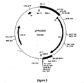
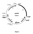
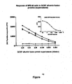
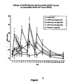
Classifications
-
- C—CHEMISTRY; METALLURGY
- C07—ORGANIC CHEMISTRY
- C07K—PEPTIDES
- C07K14/00—Peptides having more than 20 amino acids; Gastrins; Somatostatins; Melanotropins; Derivatives thereof
- C07K14/435—Peptides having more than 20 amino acids; Gastrins; Somatostatins; Melanotropins; Derivatives thereof from animals; from humans
- C07K14/76—Albumins
- C07K14/765—Serum albumin, e.g. HSA
-
- A—HUMAN NECESSITIES
- A61—MEDICAL OR VETERINARY SCIENCE; HYGIENE
- A61K—PREPARATIONS FOR MEDICAL, DENTAL OR TOILETRY PURPOSES
- A61K31/00—Medicinal preparations containing organic active ingredients
- A61K31/13—Amines
- A61K31/155—Amidines (), e.g. guanidine (H2N—C(=NH)—NH2), isourea (N=C(OH)—NH2), isothiourea (—N=C(SH)—NH2)
-
- A—HUMAN NECESSITIES
- A61—MEDICAL OR VETERINARY SCIENCE; HYGIENE
- A61K—PREPARATIONS FOR MEDICAL, DENTAL OR TOILETRY PURPOSES
- A61K31/00—Medicinal preparations containing organic active ingredients
- A61K31/33—Heterocyclic compounds
- A61K31/395—Heterocyclic compounds having nitrogen as a ring hetero atom, e.g. guanethidine or rifamycins
- A61K31/41—Heterocyclic compounds having nitrogen as a ring hetero atom, e.g. guanethidine or rifamycins having five-membered rings with two or more ring hetero atoms, at least one of which being nitrogen, e.g. tetrazole
- A61K31/425—Thiazoles
- A61K31/426—1,3-Thiazoles
-
- A—HUMAN NECESSITIES
- A61—MEDICAL OR VETERINARY SCIENCE; HYGIENE
- A61K—PREPARATIONS FOR MEDICAL, DENTAL OR TOILETRY PURPOSES
- A61K31/00—Medicinal preparations containing organic active ingredients
- A61K31/33—Heterocyclic compounds
- A61K31/395—Heterocyclic compounds having nitrogen as a ring hetero atom, e.g. guanethidine or rifamycins
- A61K31/435—Heterocyclic compounds having nitrogen as a ring hetero atom, e.g. guanethidine or rifamycins having six-membered rings with one nitrogen as the only ring hetero atom
- A61K31/44—Non condensed pyridines; Hydrogenated derivatives thereof
- A61K31/4427—Non condensed pyridines; Hydrogenated derivatives thereof containing further heterocyclic ring systems
- A61K31/4439—Non condensed pyridines; Hydrogenated derivatives thereof containing further heterocyclic ring systems containing a five-membered ring with nitrogen as a ring hetero atom, e.g. omeprazole
-
- A—HUMAN NECESSITIES
- A61—MEDICAL OR VETERINARY SCIENCE; HYGIENE
- A61K—PREPARATIONS FOR MEDICAL, DENTAL OR TOILETRY PURPOSES
- A61K31/00—Medicinal preparations containing organic active ingredients
- A61K31/33—Heterocyclic compounds
- A61K31/395—Heterocyclic compounds having nitrogen as a ring hetero atom, e.g. guanethidine or rifamycins
- A61K31/495—Heterocyclic compounds having nitrogen as a ring hetero atom, e.g. guanethidine or rifamycins having six-membered rings with two or more nitrogen atoms as the only ring heteroatoms, e.g. piperazine or tetrazines
- A61K31/4965—Non-condensed pyrazines
-
- A—HUMAN NECESSITIES
- A61—MEDICAL OR VETERINARY SCIENCE; HYGIENE
- A61K—PREPARATIONS FOR MEDICAL, DENTAL OR TOILETRY PURPOSES
- A61K38/00—Medicinal preparations containing peptides
- A61K38/04—Peptides having up to 20 amino acids in a fully defined sequence; Derivatives thereof
-
- A—HUMAN NECESSITIES
- A61—MEDICAL OR VETERINARY SCIENCE; HYGIENE
- A61K—PREPARATIONS FOR MEDICAL, DENTAL OR TOILETRY PURPOSES
- A61K38/00—Medicinal preparations containing peptides
- A61K38/16—Peptides having more than 20 amino acids; Gastrins; Somatostatins; Melanotropins; Derivatives thereof
- A61K38/17—Peptides having more than 20 amino acids; Gastrins; Somatostatins; Melanotropins; Derivatives thereof from animals; from humans
- A61K38/22—Hormones
- A61K38/28—Insulins
-
- A—HUMAN NECESSITIES
- A61—MEDICAL OR VETERINARY SCIENCE; HYGIENE
- A61K—PREPARATIONS FOR MEDICAL, DENTAL OR TOILETRY PURPOSES
- A61K39/00—Medicinal preparations containing antigens or antibodies
- A61K39/12—Viral antigens
- A61K39/21—Retroviridae, e.g. equine infectious anemia virus
-
- A—HUMAN NECESSITIES
- A61—MEDICAL OR VETERINARY SCIENCE; HYGIENE
- A61P—SPECIFIC THERAPEUTIC ACTIVITY OF CHEMICAL COMPOUNDS OR MEDICINAL PREPARATIONS
- A61P1/00—Drugs for disorders of the alimentary tract or the digestive system
- A61P1/02—Stomatological preparations, e.g. drugs for caries, aphtae, periodontitis
-
- A—HUMAN NECESSITIES
- A61—MEDICAL OR VETERINARY SCIENCE; HYGIENE
- A61P—SPECIFIC THERAPEUTIC ACTIVITY OF CHEMICAL COMPOUNDS OR MEDICINAL PREPARATIONS
- A61P1/00—Drugs for disorders of the alimentary tract or the digestive system
- A61P1/04—Drugs for disorders of the alimentary tract or the digestive system for ulcers, gastritis or reflux esophagitis, e.g. antacids, inhibitors of acid secretion, mucosal protectants
-
- A—HUMAN NECESSITIES
- A61—MEDICAL OR VETERINARY SCIENCE; HYGIENE
- A61P—SPECIFIC THERAPEUTIC ACTIVITY OF CHEMICAL COMPOUNDS OR MEDICINAL PREPARATIONS
- A61P1/00—Drugs for disorders of the alimentary tract or the digestive system
- A61P1/16—Drugs for disorders of the alimentary tract or the digestive system for liver or gallbladder disorders, e.g. hepatoprotective agents, cholagogues, litholytics
-
- A—HUMAN NECESSITIES
- A61—MEDICAL OR VETERINARY SCIENCE; HYGIENE
- A61P—SPECIFIC THERAPEUTIC ACTIVITY OF CHEMICAL COMPOUNDS OR MEDICINAL PREPARATIONS
- A61P1/00—Drugs for disorders of the alimentary tract or the digestive system
- A61P1/18—Drugs for disorders of the alimentary tract or the digestive system for pancreatic disorders, e.g. pancreatic enzymes
-
- A—HUMAN NECESSITIES
- A61—MEDICAL OR VETERINARY SCIENCE; HYGIENE
- A61P—SPECIFIC THERAPEUTIC ACTIVITY OF CHEMICAL COMPOUNDS OR MEDICINAL PREPARATIONS
- A61P11/00—Drugs for disorders of the respiratory system
-
- A—HUMAN NECESSITIES
- A61—MEDICAL OR VETERINARY SCIENCE; HYGIENE
- A61P—SPECIFIC THERAPEUTIC ACTIVITY OF CHEMICAL COMPOUNDS OR MEDICINAL PREPARATIONS
- A61P11/00—Drugs for disorders of the respiratory system
- A61P11/02—Nasal agents, e.g. decongestants
-
- A—HUMAN NECESSITIES
- A61—MEDICAL OR VETERINARY SCIENCE; HYGIENE
- A61P—SPECIFIC THERAPEUTIC ACTIVITY OF CHEMICAL COMPOUNDS OR MEDICINAL PREPARATIONS
- A61P11/00—Drugs for disorders of the respiratory system
- A61P11/06—Antiasthmatics
-
- A—HUMAN NECESSITIES
- A61—MEDICAL OR VETERINARY SCIENCE; HYGIENE
- A61P—SPECIFIC THERAPEUTIC ACTIVITY OF CHEMICAL COMPOUNDS OR MEDICINAL PREPARATIONS
- A61P13/00—Drugs for disorders of the urinary system
- A61P13/02—Drugs for disorders of the urinary system of urine or of the urinary tract, e.g. urine acidifiers
-
- A—HUMAN NECESSITIES
- A61—MEDICAL OR VETERINARY SCIENCE; HYGIENE
- A61P—SPECIFIC THERAPEUTIC ACTIVITY OF CHEMICAL COMPOUNDS OR MEDICINAL PREPARATIONS
- A61P13/00—Drugs for disorders of the urinary system
- A61P13/08—Drugs for disorders of the urinary system of the prostate
-
- A—HUMAN NECESSITIES
- A61—MEDICAL OR VETERINARY SCIENCE; HYGIENE
- A61P—SPECIFIC THERAPEUTIC ACTIVITY OF CHEMICAL COMPOUNDS OR MEDICINAL PREPARATIONS
- A61P13/00—Drugs for disorders of the urinary system
- A61P13/10—Drugs for disorders of the urinary system of the bladder
-
- A—HUMAN NECESSITIES
- A61—MEDICAL OR VETERINARY SCIENCE; HYGIENE
- A61P—SPECIFIC THERAPEUTIC ACTIVITY OF CHEMICAL COMPOUNDS OR MEDICINAL PREPARATIONS
- A61P13/00—Drugs for disorders of the urinary system
- A61P13/12—Drugs for disorders of the urinary system of the kidneys
-
- A—HUMAN NECESSITIES
- A61—MEDICAL OR VETERINARY SCIENCE; HYGIENE
- A61P—SPECIFIC THERAPEUTIC ACTIVITY OF CHEMICAL COMPOUNDS OR MEDICINAL PREPARATIONS
- A61P15/00—Drugs for genital or sexual disorders; Contraceptives
-
- A—HUMAN NECESSITIES
- A61—MEDICAL OR VETERINARY SCIENCE; HYGIENE
- A61P—SPECIFIC THERAPEUTIC ACTIVITY OF CHEMICAL COMPOUNDS OR MEDICINAL PREPARATIONS
- A61P15/00—Drugs for genital or sexual disorders; Contraceptives
- A61P15/08—Drugs for genital or sexual disorders; Contraceptives for gonadal disorders or for enhancing fertility, e.g. inducers of ovulation or of spermatogenesis
-
- A—HUMAN NECESSITIES
- A61—MEDICAL OR VETERINARY SCIENCE; HYGIENE
- A61P—SPECIFIC THERAPEUTIC ACTIVITY OF CHEMICAL COMPOUNDS OR MEDICINAL PREPARATIONS
- A61P15/00—Drugs for genital or sexual disorders; Contraceptives
- A61P15/10—Drugs for genital or sexual disorders; Contraceptives for impotence
-
- A—HUMAN NECESSITIES
- A61—MEDICAL OR VETERINARY SCIENCE; HYGIENE
- A61P—SPECIFIC THERAPEUTIC ACTIVITY OF CHEMICAL COMPOUNDS OR MEDICINAL PREPARATIONS
- A61P17/00—Drugs for dermatological disorders
-
- A—HUMAN NECESSITIES
- A61—MEDICAL OR VETERINARY SCIENCE; HYGIENE
- A61P—SPECIFIC THERAPEUTIC ACTIVITY OF CHEMICAL COMPOUNDS OR MEDICINAL PREPARATIONS
- A61P17/00—Drugs for dermatological disorders
- A61P17/02—Drugs for dermatological disorders for treating wounds, ulcers, burns, scars, keloids, or the like
-
- A—HUMAN NECESSITIES
- A61—MEDICAL OR VETERINARY SCIENCE; HYGIENE
- A61P—SPECIFIC THERAPEUTIC ACTIVITY OF CHEMICAL COMPOUNDS OR MEDICINAL PREPARATIONS
- A61P19/00—Drugs for skeletal disorders
- A61P19/02—Drugs for skeletal disorders for joint disorders, e.g. arthritis, arthrosis
-
- A—HUMAN NECESSITIES
- A61—MEDICAL OR VETERINARY SCIENCE; HYGIENE
- A61P—SPECIFIC THERAPEUTIC ACTIVITY OF CHEMICAL COMPOUNDS OR MEDICINAL PREPARATIONS
- A61P19/00—Drugs for skeletal disorders
- A61P19/04—Drugs for skeletal disorders for non-specific disorders of the connective tissue
-
- A—HUMAN NECESSITIES
- A61—MEDICAL OR VETERINARY SCIENCE; HYGIENE
- A61P—SPECIFIC THERAPEUTIC ACTIVITY OF CHEMICAL COMPOUNDS OR MEDICINAL PREPARATIONS
- A61P19/00—Drugs for skeletal disorders
- A61P19/06—Antigout agents, e.g. antihyperuricemic or uricosuric agents
-
- A—HUMAN NECESSITIES
- A61—MEDICAL OR VETERINARY SCIENCE; HYGIENE
- A61P—SPECIFIC THERAPEUTIC ACTIVITY OF CHEMICAL COMPOUNDS OR MEDICINAL PREPARATIONS
- A61P19/00—Drugs for skeletal disorders
- A61P19/08—Drugs for skeletal disorders for bone diseases, e.g. rachitism, Paget's disease
-
- A—HUMAN NECESSITIES
- A61—MEDICAL OR VETERINARY SCIENCE; HYGIENE
- A61P—SPECIFIC THERAPEUTIC ACTIVITY OF CHEMICAL COMPOUNDS OR MEDICINAL PREPARATIONS
- A61P21/00—Drugs for disorders of the muscular or neuromuscular system
-
- A—HUMAN NECESSITIES
- A61—MEDICAL OR VETERINARY SCIENCE; HYGIENE
- A61P—SPECIFIC THERAPEUTIC ACTIVITY OF CHEMICAL COMPOUNDS OR MEDICINAL PREPARATIONS
- A61P21/00—Drugs for disorders of the muscular or neuromuscular system
- A61P21/04—Drugs for disorders of the muscular or neuromuscular system for myasthenia gravis
-
- A—HUMAN NECESSITIES
- A61—MEDICAL OR VETERINARY SCIENCE; HYGIENE
- A61P—SPECIFIC THERAPEUTIC ACTIVITY OF CHEMICAL COMPOUNDS OR MEDICINAL PREPARATIONS
- A61P25/00—Drugs for disorders of the nervous system
-
- A—HUMAN NECESSITIES
- A61—MEDICAL OR VETERINARY SCIENCE; HYGIENE
- A61P—SPECIFIC THERAPEUTIC ACTIVITY OF CHEMICAL COMPOUNDS OR MEDICINAL PREPARATIONS
- A61P25/00—Drugs for disorders of the nervous system
- A61P25/08—Antiepileptics; Anticonvulsants
- A61P25/10—Antiepileptics; Anticonvulsants for petit-mal
-
- A—HUMAN NECESSITIES
- A61—MEDICAL OR VETERINARY SCIENCE; HYGIENE
- A61P—SPECIFIC THERAPEUTIC ACTIVITY OF CHEMICAL COMPOUNDS OR MEDICINAL PREPARATIONS
- A61P25/00—Drugs for disorders of the nervous system
- A61P25/14—Drugs for disorders of the nervous system for treating abnormal movements, e.g. chorea, dyskinesia
- A61P25/16—Anti-Parkinson drugs
-
- A—HUMAN NECESSITIES
- A61—MEDICAL OR VETERINARY SCIENCE; HYGIENE
- A61P—SPECIFIC THERAPEUTIC ACTIVITY OF CHEMICAL COMPOUNDS OR MEDICINAL PREPARATIONS
- A61P25/00—Drugs for disorders of the nervous system
- A61P25/28—Drugs for disorders of the nervous system for treating neurodegenerative disorders of the central nervous system, e.g. nootropic agents, cognition enhancers, drugs for treating Alzheimer's disease or other forms of dementia
-
- A—HUMAN NECESSITIES
- A61—MEDICAL OR VETERINARY SCIENCE; HYGIENE
- A61P—SPECIFIC THERAPEUTIC ACTIVITY OF CHEMICAL COMPOUNDS OR MEDICINAL PREPARATIONS
- A61P27/00—Drugs for disorders of the senses
- A61P27/02—Ophthalmic agents
-
- A—HUMAN NECESSITIES
- A61—MEDICAL OR VETERINARY SCIENCE; HYGIENE
- A61P—SPECIFIC THERAPEUTIC ACTIVITY OF CHEMICAL COMPOUNDS OR MEDICINAL PREPARATIONS
- A61P27/00—Drugs for disorders of the senses
- A61P27/02—Ophthalmic agents
- A61P27/06—Antiglaucoma agents or miotics
-
- A—HUMAN NECESSITIES
- A61—MEDICAL OR VETERINARY SCIENCE; HYGIENE
- A61P—SPECIFIC THERAPEUTIC ACTIVITY OF CHEMICAL COMPOUNDS OR MEDICINAL PREPARATIONS
- A61P27/00—Drugs for disorders of the senses
- A61P27/16—Otologicals
-
- A—HUMAN NECESSITIES
- A61—MEDICAL OR VETERINARY SCIENCE; HYGIENE
- A61P—SPECIFIC THERAPEUTIC ACTIVITY OF CHEMICAL COMPOUNDS OR MEDICINAL PREPARATIONS
- A61P29/00—Non-central analgesic, antipyretic or antiinflammatory agents, e.g. antirheumatic agents; Non-steroidal antiinflammatory drugs [NSAID]
-
- A—HUMAN NECESSITIES
- A61—MEDICAL OR VETERINARY SCIENCE; HYGIENE
- A61P—SPECIFIC THERAPEUTIC ACTIVITY OF CHEMICAL COMPOUNDS OR MEDICINAL PREPARATIONS
- A61P3/00—Drugs for disorders of the metabolism
-
- A—HUMAN NECESSITIES
- A61—MEDICAL OR VETERINARY SCIENCE; HYGIENE
- A61P—SPECIFIC THERAPEUTIC ACTIVITY OF CHEMICAL COMPOUNDS OR MEDICINAL PREPARATIONS
- A61P3/00—Drugs for disorders of the metabolism
- A61P3/04—Anorexiants; Antiobesity agents
-
- A—HUMAN NECESSITIES
- A61—MEDICAL OR VETERINARY SCIENCE; HYGIENE
- A61P—SPECIFIC THERAPEUTIC ACTIVITY OF CHEMICAL COMPOUNDS OR MEDICINAL PREPARATIONS
- A61P3/00—Drugs for disorders of the metabolism
- A61P3/06—Antihyperlipidemics
-
- A—HUMAN NECESSITIES
- A61—MEDICAL OR VETERINARY SCIENCE; HYGIENE
- A61P—SPECIFIC THERAPEUTIC ACTIVITY OF CHEMICAL COMPOUNDS OR MEDICINAL PREPARATIONS
- A61P3/00—Drugs for disorders of the metabolism
- A61P3/08—Drugs for disorders of the metabolism for glucose homeostasis
-
- A—HUMAN NECESSITIES
- A61—MEDICAL OR VETERINARY SCIENCE; HYGIENE
- A61P—SPECIFIC THERAPEUTIC ACTIVITY OF CHEMICAL COMPOUNDS OR MEDICINAL PREPARATIONS
- A61P3/00—Drugs for disorders of the metabolism
- A61P3/08—Drugs for disorders of the metabolism for glucose homeostasis
- A61P3/10—Drugs for disorders of the metabolism for glucose homeostasis for hyperglycaemia, e.g. antidiabetics
-
- A—HUMAN NECESSITIES
- A61—MEDICAL OR VETERINARY SCIENCE; HYGIENE
- A61P—SPECIFIC THERAPEUTIC ACTIVITY OF CHEMICAL COMPOUNDS OR MEDICINAL PREPARATIONS
- A61P3/00—Drugs for disorders of the metabolism
- A61P3/12—Drugs for disorders of the metabolism for electrolyte homeostasis
-
- A—HUMAN NECESSITIES
- A61—MEDICAL OR VETERINARY SCIENCE; HYGIENE
- A61P—SPECIFIC THERAPEUTIC ACTIVITY OF CHEMICAL COMPOUNDS OR MEDICINAL PREPARATIONS
- A61P3/00—Drugs for disorders of the metabolism
- A61P3/12—Drugs for disorders of the metabolism for electrolyte homeostasis
- A61P3/14—Drugs for disorders of the metabolism for electrolyte homeostasis for calcium homeostasis
-
- A—HUMAN NECESSITIES
- A61—MEDICAL OR VETERINARY SCIENCE; HYGIENE
- A61P—SPECIFIC THERAPEUTIC ACTIVITY OF CHEMICAL COMPOUNDS OR MEDICINAL PREPARATIONS
- A61P31/00—Antiinfectives, i.e. antibiotics, antiseptics, chemotherapeutics
-
- A—HUMAN NECESSITIES
- A61—MEDICAL OR VETERINARY SCIENCE; HYGIENE
- A61P—SPECIFIC THERAPEUTIC ACTIVITY OF CHEMICAL COMPOUNDS OR MEDICINAL PREPARATIONS
- A61P31/00—Antiinfectives, i.e. antibiotics, antiseptics, chemotherapeutics
- A61P31/04—Antibacterial agents
-
- A—HUMAN NECESSITIES
- A61—MEDICAL OR VETERINARY SCIENCE; HYGIENE
- A61P—SPECIFIC THERAPEUTIC ACTIVITY OF CHEMICAL COMPOUNDS OR MEDICINAL PREPARATIONS
- A61P31/00—Antiinfectives, i.e. antibiotics, antiseptics, chemotherapeutics
- A61P31/10—Antimycotics
-
- A—HUMAN NECESSITIES
- A61—MEDICAL OR VETERINARY SCIENCE; HYGIENE
- A61P—SPECIFIC THERAPEUTIC ACTIVITY OF CHEMICAL COMPOUNDS OR MEDICINAL PREPARATIONS
- A61P31/00—Antiinfectives, i.e. antibiotics, antiseptics, chemotherapeutics
- A61P31/12—Antivirals
-
- A—HUMAN NECESSITIES
- A61—MEDICAL OR VETERINARY SCIENCE; HYGIENE
- A61P—SPECIFIC THERAPEUTIC ACTIVITY OF CHEMICAL COMPOUNDS OR MEDICINAL PREPARATIONS
- A61P31/00—Antiinfectives, i.e. antibiotics, antiseptics, chemotherapeutics
- A61P31/12—Antivirals
- A61P31/14—Antivirals for RNA viruses
-
- A—HUMAN NECESSITIES
- A61—MEDICAL OR VETERINARY SCIENCE; HYGIENE
- A61P—SPECIFIC THERAPEUTIC ACTIVITY OF CHEMICAL COMPOUNDS OR MEDICINAL PREPARATIONS
- A61P31/00—Antiinfectives, i.e. antibiotics, antiseptics, chemotherapeutics
- A61P31/12—Antivirals
- A61P31/14—Antivirals for RNA viruses
- A61P31/16—Antivirals for RNA viruses for influenza or rhinoviruses
-
- A—HUMAN NECESSITIES
- A61—MEDICAL OR VETERINARY SCIENCE; HYGIENE
- A61P—SPECIFIC THERAPEUTIC ACTIVITY OF CHEMICAL COMPOUNDS OR MEDICINAL PREPARATIONS
- A61P31/00—Antiinfectives, i.e. antibiotics, antiseptics, chemotherapeutics
- A61P31/12—Antivirals
- A61P31/14—Antivirals for RNA viruses
- A61P31/18—Antivirals for RNA viruses for HIV
-
- A—HUMAN NECESSITIES
- A61—MEDICAL OR VETERINARY SCIENCE; HYGIENE
- A61P—SPECIFIC THERAPEUTIC ACTIVITY OF CHEMICAL COMPOUNDS OR MEDICINAL PREPARATIONS
- A61P31/00—Antiinfectives, i.e. antibiotics, antiseptics, chemotherapeutics
- A61P31/12—Antivirals
- A61P31/20—Antivirals for DNA viruses
-
- A—HUMAN NECESSITIES
- A61—MEDICAL OR VETERINARY SCIENCE; HYGIENE
- A61P—SPECIFIC THERAPEUTIC ACTIVITY OF CHEMICAL COMPOUNDS OR MEDICINAL PREPARATIONS
- A61P31/00—Antiinfectives, i.e. antibiotics, antiseptics, chemotherapeutics
- A61P31/12—Antivirals
- A61P31/20—Antivirals for DNA viruses
- A61P31/22—Antivirals for DNA viruses for herpes viruses
-
- A—HUMAN NECESSITIES
- A61—MEDICAL OR VETERINARY SCIENCE; HYGIENE
- A61P—SPECIFIC THERAPEUTIC ACTIVITY OF CHEMICAL COMPOUNDS OR MEDICINAL PREPARATIONS
- A61P33/00—Antiparasitic agents
-
- A—HUMAN NECESSITIES
- A61—MEDICAL OR VETERINARY SCIENCE; HYGIENE
- A61P—SPECIFIC THERAPEUTIC ACTIVITY OF CHEMICAL COMPOUNDS OR MEDICINAL PREPARATIONS
- A61P33/00—Antiparasitic agents
- A61P33/02—Antiprotozoals, e.g. for leishmaniasis, trichomoniasis, toxoplasmosis
-
- A—HUMAN NECESSITIES
- A61—MEDICAL OR VETERINARY SCIENCE; HYGIENE
- A61P—SPECIFIC THERAPEUTIC ACTIVITY OF CHEMICAL COMPOUNDS OR MEDICINAL PREPARATIONS
- A61P33/00—Antiparasitic agents
- A61P33/02—Antiprotozoals, e.g. for leishmaniasis, trichomoniasis, toxoplasmosis
- A61P33/06—Antimalarials
-
- A—HUMAN NECESSITIES
- A61—MEDICAL OR VETERINARY SCIENCE; HYGIENE
- A61P—SPECIFIC THERAPEUTIC ACTIVITY OF CHEMICAL COMPOUNDS OR MEDICINAL PREPARATIONS
- A61P35/00—Antineoplastic agents
-
- A—HUMAN NECESSITIES
- A61—MEDICAL OR VETERINARY SCIENCE; HYGIENE
- A61P—SPECIFIC THERAPEUTIC ACTIVITY OF CHEMICAL COMPOUNDS OR MEDICINAL PREPARATIONS
- A61P35/00—Antineoplastic agents
- A61P35/02—Antineoplastic agents specific for leukemia
-
- A—HUMAN NECESSITIES
- A61—MEDICAL OR VETERINARY SCIENCE; HYGIENE
- A61P—SPECIFIC THERAPEUTIC ACTIVITY OF CHEMICAL COMPOUNDS OR MEDICINAL PREPARATIONS
- A61P35/00—Antineoplastic agents
- A61P35/04—Antineoplastic agents specific for metastasis
-
- A—HUMAN NECESSITIES
- A61—MEDICAL OR VETERINARY SCIENCE; HYGIENE
- A61P—SPECIFIC THERAPEUTIC ACTIVITY OF CHEMICAL COMPOUNDS OR MEDICINAL PREPARATIONS
- A61P37/00—Drugs for immunological or allergic disorders
- A61P37/02—Immunomodulators
-
- A—HUMAN NECESSITIES
- A61—MEDICAL OR VETERINARY SCIENCE; HYGIENE
- A61P—SPECIFIC THERAPEUTIC ACTIVITY OF CHEMICAL COMPOUNDS OR MEDICINAL PREPARATIONS
- A61P37/00—Drugs for immunological or allergic disorders
- A61P37/02—Immunomodulators
- A61P37/04—Immunostimulants
-
- A—HUMAN NECESSITIES
- A61—MEDICAL OR VETERINARY SCIENCE; HYGIENE
- A61P—SPECIFIC THERAPEUTIC ACTIVITY OF CHEMICAL COMPOUNDS OR MEDICINAL PREPARATIONS
- A61P37/00—Drugs for immunological or allergic disorders
- A61P37/02—Immunomodulators
- A61P37/06—Immunosuppressants, e.g. drugs for graft rejection
-
- A—HUMAN NECESSITIES
- A61—MEDICAL OR VETERINARY SCIENCE; HYGIENE
- A61P—SPECIFIC THERAPEUTIC ACTIVITY OF CHEMICAL COMPOUNDS OR MEDICINAL PREPARATIONS
- A61P37/00—Drugs for immunological or allergic disorders
- A61P37/08—Antiallergic agents
-
- A—HUMAN NECESSITIES
- A61—MEDICAL OR VETERINARY SCIENCE; HYGIENE
- A61P—SPECIFIC THERAPEUTIC ACTIVITY OF CHEMICAL COMPOUNDS OR MEDICINAL PREPARATIONS
- A61P43/00—Drugs for specific purposes, not provided for in groups A61P1/00-A61P41/00
-
- A—HUMAN NECESSITIES
- A61—MEDICAL OR VETERINARY SCIENCE; HYGIENE
- A61P—SPECIFIC THERAPEUTIC ACTIVITY OF CHEMICAL COMPOUNDS OR MEDICINAL PREPARATIONS
- A61P5/00—Drugs for disorders of the endocrine system
-
- A—HUMAN NECESSITIES
- A61—MEDICAL OR VETERINARY SCIENCE; HYGIENE
- A61P—SPECIFIC THERAPEUTIC ACTIVITY OF CHEMICAL COMPOUNDS OR MEDICINAL PREPARATIONS
- A61P5/00—Drugs for disorders of the endocrine system
- A61P5/06—Drugs for disorders of the endocrine system of the anterior pituitary hormones, e.g. TSH, ACTH, FSH, LH, PRL, GH
-
- A—HUMAN NECESSITIES
- A61—MEDICAL OR VETERINARY SCIENCE; HYGIENE
- A61P—SPECIFIC THERAPEUTIC ACTIVITY OF CHEMICAL COMPOUNDS OR MEDICINAL PREPARATIONS
- A61P5/00—Drugs for disorders of the endocrine system
- A61P5/14—Drugs for disorders of the endocrine system of the thyroid hormones, e.g. T3, T4
-
- A—HUMAN NECESSITIES
- A61—MEDICAL OR VETERINARY SCIENCE; HYGIENE
- A61P—SPECIFIC THERAPEUTIC ACTIVITY OF CHEMICAL COMPOUNDS OR MEDICINAL PREPARATIONS
- A61P5/00—Drugs for disorders of the endocrine system
- A61P5/38—Drugs for disorders of the endocrine system of the suprarenal hormones
- A61P5/40—Mineralocorticosteroids, e.g. aldosterone; Drugs increasing or potentiating the activity of mineralocorticosteroids
-
- A—HUMAN NECESSITIES
- A61—MEDICAL OR VETERINARY SCIENCE; HYGIENE
- A61P—SPECIFIC THERAPEUTIC ACTIVITY OF CHEMICAL COMPOUNDS OR MEDICINAL PREPARATIONS
- A61P5/00—Drugs for disorders of the endocrine system
- A61P5/48—Drugs for disorders of the endocrine system of the pancreatic hormones
- A61P5/50—Drugs for disorders of the endocrine system of the pancreatic hormones for increasing or potentiating the activity of insulin
-
- A—HUMAN NECESSITIES
- A61—MEDICAL OR VETERINARY SCIENCE; HYGIENE
- A61P—SPECIFIC THERAPEUTIC ACTIVITY OF CHEMICAL COMPOUNDS OR MEDICINAL PREPARATIONS
- A61P7/00—Drugs for disorders of the blood or the extracellular fluid
-
- A—HUMAN NECESSITIES
- A61—MEDICAL OR VETERINARY SCIENCE; HYGIENE
- A61P—SPECIFIC THERAPEUTIC ACTIVITY OF CHEMICAL COMPOUNDS OR MEDICINAL PREPARATIONS
- A61P7/00—Drugs for disorders of the blood or the extracellular fluid
- A61P7/04—Antihaemorrhagics; Procoagulants; Haemostatic agents; Antifibrinolytic agents
-
- A—HUMAN NECESSITIES
- A61—MEDICAL OR VETERINARY SCIENCE; HYGIENE
- A61P—SPECIFIC THERAPEUTIC ACTIVITY OF CHEMICAL COMPOUNDS OR MEDICINAL PREPARATIONS
- A61P7/00—Drugs for disorders of the blood or the extracellular fluid
- A61P7/06—Antianaemics
-
- A—HUMAN NECESSITIES
- A61—MEDICAL OR VETERINARY SCIENCE; HYGIENE
- A61P—SPECIFIC THERAPEUTIC ACTIVITY OF CHEMICAL COMPOUNDS OR MEDICINAL PREPARATIONS
- A61P7/00—Drugs for disorders of the blood or the extracellular fluid
- A61P7/12—Antidiuretics, e.g. drugs for diabetes insipidus
-
- A—HUMAN NECESSITIES
- A61—MEDICAL OR VETERINARY SCIENCE; HYGIENE
- A61P—SPECIFIC THERAPEUTIC ACTIVITY OF CHEMICAL COMPOUNDS OR MEDICINAL PREPARATIONS
- A61P9/00—Drugs for disorders of the cardiovascular system
-
- A—HUMAN NECESSITIES
- A61—MEDICAL OR VETERINARY SCIENCE; HYGIENE
- A61P—SPECIFIC THERAPEUTIC ACTIVITY OF CHEMICAL COMPOUNDS OR MEDICINAL PREPARATIONS
- A61P9/00—Drugs for disorders of the cardiovascular system
- A61P9/02—Non-specific cardiovascular stimulants, e.g. drugs for syncope, antihypotensives
-
- A—HUMAN NECESSITIES
- A61—MEDICAL OR VETERINARY SCIENCE; HYGIENE
- A61P—SPECIFIC THERAPEUTIC ACTIVITY OF CHEMICAL COMPOUNDS OR MEDICINAL PREPARATIONS
- A61P9/00—Drugs for disorders of the cardiovascular system
- A61P9/04—Inotropic agents, i.e. stimulants of cardiac contraction; Drugs for heart failure
-
- A—HUMAN NECESSITIES
- A61—MEDICAL OR VETERINARY SCIENCE; HYGIENE
- A61P—SPECIFIC THERAPEUTIC ACTIVITY OF CHEMICAL COMPOUNDS OR MEDICINAL PREPARATIONS
- A61P9/00—Drugs for disorders of the cardiovascular system
- A61P9/06—Antiarrhythmics
-
- A—HUMAN NECESSITIES
- A61—MEDICAL OR VETERINARY SCIENCE; HYGIENE
- A61P—SPECIFIC THERAPEUTIC ACTIVITY OF CHEMICAL COMPOUNDS OR MEDICINAL PREPARATIONS
- A61P9/00—Drugs for disorders of the cardiovascular system
- A61P9/08—Vasodilators for multiple indications
-
- A—HUMAN NECESSITIES
- A61—MEDICAL OR VETERINARY SCIENCE; HYGIENE
- A61P—SPECIFIC THERAPEUTIC ACTIVITY OF CHEMICAL COMPOUNDS OR MEDICINAL PREPARATIONS
- A61P9/00—Drugs for disorders of the cardiovascular system
- A61P9/10—Drugs for disorders of the cardiovascular system for treating ischaemic or atherosclerotic diseases, e.g. antianginal drugs, coronary vasodilators, drugs for myocardial infarction, retinopathy, cerebrovascula insufficiency, renal arteriosclerosis
-
- A—HUMAN NECESSITIES
- A61—MEDICAL OR VETERINARY SCIENCE; HYGIENE
- A61P—SPECIFIC THERAPEUTIC ACTIVITY OF CHEMICAL COMPOUNDS OR MEDICINAL PREPARATIONS
- A61P9/00—Drugs for disorders of the cardiovascular system
- A61P9/12—Antihypertensives
-
- A—HUMAN NECESSITIES
- A61—MEDICAL OR VETERINARY SCIENCE; HYGIENE
- A61P—SPECIFIC THERAPEUTIC ACTIVITY OF CHEMICAL COMPOUNDS OR MEDICINAL PREPARATIONS
- A61P9/00—Drugs for disorders of the cardiovascular system
- A61P9/14—Vasoprotectives; Antihaemorrhoidals; Drugs for varicose therapy; Capillary stabilisers
-
- C—CHEMISTRY; METALLURGY
- C07—ORGANIC CHEMISTRY
- C07K—PEPTIDES
- C07K14/00—Peptides having more than 20 amino acids; Gastrins; Somatostatins; Melanotropins; Derivatives thereof
- C07K14/005—Peptides having more than 20 amino acids; Gastrins; Somatostatins; Melanotropins; Derivatives thereof from viruses
-
- C—CHEMISTRY; METALLURGY
- C07—ORGANIC CHEMISTRY
- C07K—PEPTIDES
- C07K14/00—Peptides having more than 20 amino acids; Gastrins; Somatostatins; Melanotropins; Derivatives thereof
- C07K14/435—Peptides having more than 20 amino acids; Gastrins; Somatostatins; Melanotropins; Derivatives thereof from animals; from humans
- C07K14/46—Peptides having more than 20 amino acids; Gastrins; Somatostatins; Melanotropins; Derivatives thereof from animals; from humans from vertebrates
- C07K14/47—Peptides having more than 20 amino acids; Gastrins; Somatostatins; Melanotropins; Derivatives thereof from animals; from humans from vertebrates from mammals
- C07K14/4701—Peptides having more than 20 amino acids; Gastrins; Somatostatins; Melanotropins; Derivatives thereof from animals; from humans from vertebrates from mammals not used
- C07K14/4713—Autoimmune diseases, e.g. Insulin-dependent diabetes mellitus, multiple sclerosis, rheumathoid arthritis, systemic lupus erythematosus; Autoantigens
-
- C—CHEMISTRY; METALLURGY
- C07—ORGANIC CHEMISTRY
- C07K—PEPTIDES
- C07K14/00—Peptides having more than 20 amino acids; Gastrins; Somatostatins; Melanotropins; Derivatives thereof
- C07K14/435—Peptides having more than 20 amino acids; Gastrins; Somatostatins; Melanotropins; Derivatives thereof from animals; from humans
- C07K14/46—Peptides having more than 20 amino acids; Gastrins; Somatostatins; Melanotropins; Derivatives thereof from animals; from humans from vertebrates
- C07K14/47—Peptides having more than 20 amino acids; Gastrins; Somatostatins; Melanotropins; Derivatives thereof from animals; from humans from vertebrates from mammals
- C07K14/4701—Peptides having more than 20 amino acids; Gastrins; Somatostatins; Melanotropins; Derivatives thereof from animals; from humans from vertebrates from mammals not used
- C07K14/4723—Cationic antimicrobial peptides, e.g. defensins
-
- C—CHEMISTRY; METALLURGY
- C07—ORGANIC CHEMISTRY
- C07K—PEPTIDES
- C07K14/00—Peptides having more than 20 amino acids; Gastrins; Somatostatins; Melanotropins; Derivatives thereof
- C07K14/435—Peptides having more than 20 amino acids; Gastrins; Somatostatins; Melanotropins; Derivatives thereof from animals; from humans
- C07K14/475—Growth factors; Growth regulators
- C07K14/50—Fibroblast growth factors [FGF]
-
- C—CHEMISTRY; METALLURGY
- C07—ORGANIC CHEMISTRY
- C07K—PEPTIDES
- C07K14/00—Peptides having more than 20 amino acids; Gastrins; Somatostatins; Melanotropins; Derivatives thereof
- C07K14/435—Peptides having more than 20 amino acids; Gastrins; Somatostatins; Melanotropins; Derivatives thereof from animals; from humans
- C07K14/475—Growth factors; Growth regulators
- C07K14/505—Erythropoietin [EPO]
-
- C—CHEMISTRY; METALLURGY
- C07—ORGANIC CHEMISTRY
- C07K—PEPTIDES
- C07K14/00—Peptides having more than 20 amino acids; Gastrins; Somatostatins; Melanotropins; Derivatives thereof
- C07K14/435—Peptides having more than 20 amino acids; Gastrins; Somatostatins; Melanotropins; Derivatives thereof from animals; from humans
- C07K14/475—Growth factors; Growth regulators
- C07K14/51—Bone morphogenetic factor; Osteogenins; Osteogenic factor; Bone-inducing factor
-
- C—CHEMISTRY; METALLURGY
- C07—ORGANIC CHEMISTRY
- C07K—PEPTIDES
- C07K14/00—Peptides having more than 20 amino acids; Gastrins; Somatostatins; Melanotropins; Derivatives thereof
- C07K14/435—Peptides having more than 20 amino acids; Gastrins; Somatostatins; Melanotropins; Derivatives thereof from animals; from humans
- C07K14/52—Cytokines; Lymphokines; Interferons
- C07K14/521—Chemokines
-
- C—CHEMISTRY; METALLURGY
- C07—ORGANIC CHEMISTRY
- C07K—PEPTIDES
- C07K14/00—Peptides having more than 20 amino acids; Gastrins; Somatostatins; Melanotropins; Derivatives thereof
- C07K14/435—Peptides having more than 20 amino acids; Gastrins; Somatostatins; Melanotropins; Derivatives thereof from animals; from humans
- C07K14/52—Cytokines; Lymphokines; Interferons
- C07K14/525—Tumour necrosis factor [TNF]
-
- C—CHEMISTRY; METALLURGY
- C07—ORGANIC CHEMISTRY
- C07K—PEPTIDES
- C07K14/00—Peptides having more than 20 amino acids; Gastrins; Somatostatins; Melanotropins; Derivatives thereof
- C07K14/435—Peptides having more than 20 amino acids; Gastrins; Somatostatins; Melanotropins; Derivatives thereof from animals; from humans
- C07K14/52—Cytokines; Lymphokines; Interferons
- C07K14/53—Colony-stimulating factor [CSF]
- C07K14/535—Granulocyte CSF; Granulocyte-macrophage CSF
-
- C—CHEMISTRY; METALLURGY
- C07—ORGANIC CHEMISTRY
- C07K—PEPTIDES
- C07K14/00—Peptides having more than 20 amino acids; Gastrins; Somatostatins; Melanotropins; Derivatives thereof
- C07K14/435—Peptides having more than 20 amino acids; Gastrins; Somatostatins; Melanotropins; Derivatives thereof from animals; from humans
- C07K14/52—Cytokines; Lymphokines; Interferons
- C07K14/54—Interleukins [IL]
- C07K14/5406—IL-4
-
- C—CHEMISTRY; METALLURGY
- C07—ORGANIC CHEMISTRY
- C07K—PEPTIDES
- C07K14/00—Peptides having more than 20 amino acids; Gastrins; Somatostatins; Melanotropins; Derivatives thereof
- C07K14/435—Peptides having more than 20 amino acids; Gastrins; Somatostatins; Melanotropins; Derivatives thereof from animals; from humans
- C07K14/52—Cytokines; Lymphokines; Interferons
- C07K14/54—Interleukins [IL]
- C07K14/55—IL-2
-
- C—CHEMISTRY; METALLURGY
- C07—ORGANIC CHEMISTRY
- C07K—PEPTIDES
- C07K14/00—Peptides having more than 20 amino acids; Gastrins; Somatostatins; Melanotropins; Derivatives thereof
- C07K14/435—Peptides having more than 20 amino acids; Gastrins; Somatostatins; Melanotropins; Derivatives thereof from animals; from humans
- C07K14/52—Cytokines; Lymphokines; Interferons
- C07K14/555—Interferons [IFN]
- C07K14/56—IFN-alpha
-
- C—CHEMISTRY; METALLURGY
- C07—ORGANIC CHEMISTRY
- C07K—PEPTIDES
- C07K14/00—Peptides having more than 20 amino acids; Gastrins; Somatostatins; Melanotropins; Derivatives thereof
- C07K14/435—Peptides having more than 20 amino acids; Gastrins; Somatostatins; Melanotropins; Derivatives thereof from animals; from humans
- C07K14/52—Cytokines; Lymphokines; Interferons
- C07K14/555—Interferons [IFN]
- C07K14/565—IFN-beta
-
- C—CHEMISTRY; METALLURGY
- C07—ORGANIC CHEMISTRY
- C07K—PEPTIDES
- C07K14/00—Peptides having more than 20 amino acids; Gastrins; Somatostatins; Melanotropins; Derivatives thereof
- C07K14/435—Peptides having more than 20 amino acids; Gastrins; Somatostatins; Melanotropins; Derivatives thereof from animals; from humans
- C07K14/575—Hormones
- C07K14/57563—Vasoactive intestinal peptide [VIP]; Related peptides
-
- C—CHEMISTRY; METALLURGY
- C07—ORGANIC CHEMISTRY
- C07K—PEPTIDES
- C07K14/00—Peptides having more than 20 amino acids; Gastrins; Somatostatins; Melanotropins; Derivatives thereof
- C07K14/435—Peptides having more than 20 amino acids; Gastrins; Somatostatins; Melanotropins; Derivatives thereof from animals; from humans
- C07K14/575—Hormones
- C07K14/5759—Products of obesity genes, e.g. leptin, obese (OB), tub, fat
-
- C—CHEMISTRY; METALLURGY
- C07—ORGANIC CHEMISTRY
- C07K—PEPTIDES
- C07K14/00—Peptides having more than 20 amino acids; Gastrins; Somatostatins; Melanotropins; Derivatives thereof
- C07K14/435—Peptides having more than 20 amino acids; Gastrins; Somatostatins; Melanotropins; Derivatives thereof from animals; from humans
- C07K14/575—Hormones
- C07K14/585—Calcitonins
-
- C—CHEMISTRY; METALLURGY
- C07—ORGANIC CHEMISTRY
- C07K—PEPTIDES
- C07K14/00—Peptides having more than 20 amino acids; Gastrins; Somatostatins; Melanotropins; Derivatives thereof
- C07K14/435—Peptides having more than 20 amino acids; Gastrins; Somatostatins; Melanotropins; Derivatives thereof from animals; from humans
- C07K14/575—Hormones
- C07K14/59—Follicle-stimulating hormone [FSH]; Chorionic gonadotropins, e.g. HCG; Luteinising hormone [LH]; Thyroid-stimulating hormone [TSH]
-
- C—CHEMISTRY; METALLURGY
- C07—ORGANIC CHEMISTRY
- C07K—PEPTIDES
- C07K14/00—Peptides having more than 20 amino acids; Gastrins; Somatostatins; Melanotropins; Derivatives thereof
- C07K14/435—Peptides having more than 20 amino acids; Gastrins; Somatostatins; Melanotropins; Derivatives thereof from animals; from humans
- C07K14/575—Hormones
- C07K14/60—Growth-hormone releasing factors (GH-RF) (Somatoliberin)
-
- C—CHEMISTRY; METALLURGY
- C07—ORGANIC CHEMISTRY
- C07K—PEPTIDES
- C07K14/00—Peptides having more than 20 amino acids; Gastrins; Somatostatins; Melanotropins; Derivatives thereof
- C07K14/435—Peptides having more than 20 amino acids; Gastrins; Somatostatins; Melanotropins; Derivatives thereof from animals; from humans
- C07K14/575—Hormones
- C07K14/605—Glucagons
-
- C—CHEMISTRY; METALLURGY
- C07—ORGANIC CHEMISTRY
- C07K—PEPTIDES
- C07K14/00—Peptides having more than 20 amino acids; Gastrins; Somatostatins; Melanotropins; Derivatives thereof
- C07K14/435—Peptides having more than 20 amino acids; Gastrins; Somatostatins; Melanotropins; Derivatives thereof from animals; from humans
- C07K14/575—Hormones
- C07K14/61—Growth hormones [GH] (Somatotropin)
-
- C—CHEMISTRY; METALLURGY
- C07—ORGANIC CHEMISTRY
- C07K—PEPTIDES
- C07K14/00—Peptides having more than 20 amino acids; Gastrins; Somatostatins; Melanotropins; Derivatives thereof
- C07K14/435—Peptides having more than 20 amino acids; Gastrins; Somatostatins; Melanotropins; Derivatives thereof from animals; from humans
- C07K14/575—Hormones
- C07K14/62—Insulins
-
- C—CHEMISTRY; METALLURGY
- C07—ORGANIC CHEMISTRY
- C07K—PEPTIDES
- C07K14/00—Peptides having more than 20 amino acids; Gastrins; Somatostatins; Melanotropins; Derivatives thereof
- C07K14/435—Peptides having more than 20 amino acids; Gastrins; Somatostatins; Melanotropins; Derivatives thereof from animals; from humans
- C07K14/575—Hormones
- C07K14/635—Parathyroid hormone (parathormone); Parathyroid hormone-related peptides
-
- C—CHEMISTRY; METALLURGY
- C07—ORGANIC CHEMISTRY
- C07K—PEPTIDES
- C07K14/00—Peptides having more than 20 amino acids; Gastrins; Somatostatins; Melanotropins; Derivatives thereof
- C07K14/435—Peptides having more than 20 amino acids; Gastrins; Somatostatins; Melanotropins; Derivatives thereof from animals; from humans
- C07K14/575—Hormones
- C07K14/65—Insulin-like growth factors (Somatomedins), e.g. IGF-1, IGF-2
-
- C—CHEMISTRY; METALLURGY
- C07—ORGANIC CHEMISTRY
- C07K—PEPTIDES
- C07K14/00—Peptides having more than 20 amino acids; Gastrins; Somatostatins; Melanotropins; Derivatives thereof
- C07K14/435—Peptides having more than 20 amino acids; Gastrins; Somatostatins; Melanotropins; Derivatives thereof from animals; from humans
- C07K14/575—Hormones
- C07K14/655—Somatostatins
-
- C—CHEMISTRY; METALLURGY
- C07—ORGANIC CHEMISTRY
- C07K—PEPTIDES
- C07K14/00—Peptides having more than 20 amino acids; Gastrins; Somatostatins; Melanotropins; Derivatives thereof
- C07K14/435—Peptides having more than 20 amino acids; Gastrins; Somatostatins; Melanotropins; Derivatives thereof from animals; from humans
- C07K14/665—Peptides having more than 20 amino acids; Gastrins; Somatostatins; Melanotropins; Derivatives thereof from animals; from humans derived from pro-opiomelanocortin, pro-enkephalin or pro-dynorphin
-
- C—CHEMISTRY; METALLURGY
- C07—ORGANIC CHEMISTRY
- C07K—PEPTIDES
- C07K14/00—Peptides having more than 20 amino acids; Gastrins; Somatostatins; Melanotropins; Derivatives thereof
- C07K14/435—Peptides having more than 20 amino acids; Gastrins; Somatostatins; Melanotropins; Derivatives thereof from animals; from humans
- C07K14/705—Receptors; Cell surface antigens; Cell surface determinants
- C07K14/715—Receptors; Cell surface antigens; Cell surface determinants for cytokines; for lymphokines; for interferons
- C07K14/7151—Receptors; Cell surface antigens; Cell surface determinants for cytokines; for lymphokines; for interferons for tumor necrosis factor [TNF], for lymphotoxin [LT]
-
- C—CHEMISTRY; METALLURGY
- C07—ORGANIC CHEMISTRY
- C07K—PEPTIDES
- C07K14/00—Peptides having more than 20 amino acids; Gastrins; Somatostatins; Melanotropins; Derivatives thereof
- C07K14/82—Translation products from oncogenes
-
- C—CHEMISTRY; METALLURGY
- C07—ORGANIC CHEMISTRY
- C07K—PEPTIDES
- C07K16/00—Immunoglobulins [IGs], e.g. monoclonal or polyclonal antibodies
-
- C—CHEMISTRY; METALLURGY
- C07—ORGANIC CHEMISTRY
- C07K—PEPTIDES
- C07K16/00—Immunoglobulins [IGs], e.g. monoclonal or polyclonal antibodies
- C07K16/18—Immunoglobulins [IGs], e.g. monoclonal or polyclonal antibodies against material from animals or humans
- C07K16/24—Immunoglobulins [IGs], e.g. monoclonal or polyclonal antibodies against material from animals or humans against cytokines, lymphokines or interferons
- C07K16/241—Tumor Necrosis Factors
-
- C—CHEMISTRY; METALLURGY
- C07—ORGANIC CHEMISTRY
- C07K—PEPTIDES
- C07K16/00—Immunoglobulins [IGs], e.g. monoclonal or polyclonal antibodies
- C07K16/18—Immunoglobulins [IGs], e.g. monoclonal or polyclonal antibodies against material from animals or humans
- C07K16/26—Immunoglobulins [IGs], e.g. monoclonal or polyclonal antibodies against material from animals or humans against hormones ; against hormone releasing or inhibiting factors
-
- C—CHEMISTRY; METALLURGY
- C07—ORGANIC CHEMISTRY
- C07K—PEPTIDES
- C07K7/00—Peptides having 5 to 20 amino acids in a fully defined sequence; Derivatives thereof
- C07K7/04—Linear peptides containing only normal peptide links
- C07K7/06—Linear peptides containing only normal peptide links having 5 to 11 amino acids
-
- C—CHEMISTRY; METALLURGY
- C12—BIOCHEMISTRY; BEER; SPIRITS; WINE; VINEGAR; MICROBIOLOGY; ENZYMOLOGY; MUTATION OR GENETIC ENGINEERING
- C12N—MICROORGANISMS OR ENZYMES; COMPOSITIONS THEREOF; PROPAGATING, PRESERVING, OR MAINTAINING MICROORGANISMS; MUTATION OR GENETIC ENGINEERING; CULTURE MEDIA
- C12N15/00—Mutation or genetic engineering; DNA or RNA concerning genetic engineering, vectors, e.g. plasmids, or their isolation, preparation or purification; Use of hosts therefor
- C12N15/09—Recombinant DNA-technology
- C12N15/11—DNA or RNA fragments; Modified forms thereof; Non-coding nucleic acids having a biological activity
- C12N15/62—DNA sequences coding for fusion proteins
-
- C—CHEMISTRY; METALLURGY
- C12—BIOCHEMISTRY; BEER; SPIRITS; WINE; VINEGAR; MICROBIOLOGY; ENZYMOLOGY; MUTATION OR GENETIC ENGINEERING
- C12N—MICROORGANISMS OR ENZYMES; COMPOSITIONS THEREOF; PROPAGATING, PRESERVING, OR MAINTAINING MICROORGANISMS; MUTATION OR GENETIC ENGINEERING; CULTURE MEDIA
- C12N7/00—Viruses; Bacteriophages; Compositions thereof; Preparation or purification thereof
-
- C—CHEMISTRY; METALLURGY
- C12—BIOCHEMISTRY; BEER; SPIRITS; WINE; VINEGAR; MICROBIOLOGY; ENZYMOLOGY; MUTATION OR GENETIC ENGINEERING
- C12N—MICROORGANISMS OR ENZYMES; COMPOSITIONS THEREOF; PROPAGATING, PRESERVING, OR MAINTAINING MICROORGANISMS; MUTATION OR GENETIC ENGINEERING; CULTURE MEDIA
- C12N9/00—Enzymes; Proenzymes; Compositions thereof; Processes for preparing, activating, inhibiting, separating or purifying enzymes
- C12N9/0004—Oxidoreductases (1.)
- C12N9/0006—Oxidoreductases (1.) acting on CH-OH groups as donors (1.1)
-
- C—CHEMISTRY; METALLURGY
- C12—BIOCHEMISTRY; BEER; SPIRITS; WINE; VINEGAR; MICROBIOLOGY; ENZYMOLOGY; MUTATION OR GENETIC ENGINEERING
- C12N—MICROORGANISMS OR ENZYMES; COMPOSITIONS THEREOF; PROPAGATING, PRESERVING, OR MAINTAINING MICROORGANISMS; MUTATION OR GENETIC ENGINEERING; CULTURE MEDIA
- C12N9/00—Enzymes; Proenzymes; Compositions thereof; Processes for preparing, activating, inhibiting, separating or purifying enzymes
- C12N9/0004—Oxidoreductases (1.)
- C12N9/0008—Oxidoreductases (1.) acting on the aldehyde or oxo group of donors (1.2)
-
- A—HUMAN NECESSITIES
- A61—MEDICAL OR VETERINARY SCIENCE; HYGIENE
- A61K—PREPARATIONS FOR MEDICAL, DENTAL OR TOILETRY PURPOSES
- A61K38/00—Medicinal preparations containing peptides
-
- C—CHEMISTRY; METALLURGY
- C07—ORGANIC CHEMISTRY
- C07K—PEPTIDES
- C07K2317/00—Immunoglobulins specific features
- C07K2317/10—Immunoglobulins specific features characterized by their source of isolation or production
-
- C—CHEMISTRY; METALLURGY
- C07—ORGANIC CHEMISTRY
- C07K—PEPTIDES
- C07K2317/00—Immunoglobulins specific features
- C07K2317/60—Immunoglobulins specific features characterized by non-natural combinations of immunoglobulin fragments
- C07K2317/62—Immunoglobulins specific features characterized by non-natural combinations of immunoglobulin fragments comprising only variable region components
- C07K2317/622—Single chain antibody (scFv)
-
- C—CHEMISTRY; METALLURGY
- C07—ORGANIC CHEMISTRY
- C07K—PEPTIDES
- C07K2317/00—Immunoglobulins specific features
- C07K2317/70—Immunoglobulins specific features characterized by effect upon binding to a cell or to an antigen
- C07K2317/76—Antagonist effect on antigen, e.g. neutralization or inhibition of binding
-
- C—CHEMISTRY; METALLURGY
- C07—ORGANIC CHEMISTRY
- C07K—PEPTIDES
- C07K2319/00—Fusion polypeptide
-
- C—CHEMISTRY; METALLURGY
- C07—ORGANIC CHEMISTRY
- C07K—PEPTIDES
- C07K2319/00—Fusion polypeptide
- C07K2319/20—Fusion polypeptide containing a tag with affinity for a non-protein ligand
-
- C—CHEMISTRY; METALLURGY
- C07—ORGANIC CHEMISTRY
- C07K—PEPTIDES
- C07K2319/00—Fusion polypeptide
- C07K2319/30—Non-immunoglobulin-derived peptide or protein having an immunoglobulin constant or Fc region, or a fragment thereof, attached thereto
-
- C—CHEMISTRY; METALLURGY
- C07—ORGANIC CHEMISTRY
- C07K—PEPTIDES
- C07K2319/00—Fusion polypeptide
- C07K2319/31—Fusion polypeptide fusions, other than Fc, for prolonged plasma life, e.g. albumin
-
- C—CHEMISTRY; METALLURGY
- C12—BIOCHEMISTRY; BEER; SPIRITS; WINE; VINEGAR; MICROBIOLOGY; ENZYMOLOGY; MUTATION OR GENETIC ENGINEERING
- C12N—MICROORGANISMS OR ENZYMES; COMPOSITIONS THEREOF; PROPAGATING, PRESERVING, OR MAINTAINING MICROORGANISMS; MUTATION OR GENETIC ENGINEERING; CULTURE MEDIA
- C12N2501/00—Active agents used in cell culture processes, e.g. differentation
- C12N2501/30—Hormones
- C12N2501/335—Glucagon; Glucagon-like peptide [GLP]; Exendin
-
- C—CHEMISTRY; METALLURGY
- C12—BIOCHEMISTRY; BEER; SPIRITS; WINE; VINEGAR; MICROBIOLOGY; ENZYMOLOGY; MUTATION OR GENETIC ENGINEERING
- C12N—MICROORGANISMS OR ENZYMES; COMPOSITIONS THEREOF; PROPAGATING, PRESERVING, OR MAINTAINING MICROORGANISMS; MUTATION OR GENETIC ENGINEERING; CULTURE MEDIA
- C12N2740/00—Reverse transcribing RNA viruses
- C12N2740/00011—Details
- C12N2740/10011—Retroviridae
- C12N2740/16011—Human Immunodeficiency Virus, HIV
- C12N2740/16111—Human Immunodeficiency Virus, HIV concerning HIV env
- C12N2740/16122—New viral proteins or individual genes, new structural or functional aspects of known viral proteins or genes
-
- C—CHEMISTRY; METALLURGY
- C12—BIOCHEMISTRY; BEER; SPIRITS; WINE; VINEGAR; MICROBIOLOGY; ENZYMOLOGY; MUTATION OR GENETIC ENGINEERING
- C12N—MICROORGANISMS OR ENZYMES; COMPOSITIONS THEREOF; PROPAGATING, PRESERVING, OR MAINTAINING MICROORGANISMS; MUTATION OR GENETIC ENGINEERING; CULTURE MEDIA
- C12N2740/00—Reverse transcribing RNA viruses
- C12N2740/00011—Details
- C12N2740/10011—Retroviridae
- C12N2740/16011—Human Immunodeficiency Virus, HIV
- C12N2740/16111—Human Immunodeficiency Virus, HIV concerning HIV env
- C12N2740/16134—Use of virus or viral component as vaccine, e.g. live-attenuated or inactivated virus, VLP, viral protein
-
- C—CHEMISTRY; METALLURGY
- C12—BIOCHEMISTRY; BEER; SPIRITS; WINE; VINEGAR; MICROBIOLOGY; ENZYMOLOGY; MUTATION OR GENETIC ENGINEERING
- C12N—MICROORGANISMS OR ENZYMES; COMPOSITIONS THEREOF; PROPAGATING, PRESERVING, OR MAINTAINING MICROORGANISMS; MUTATION OR GENETIC ENGINEERING; CULTURE MEDIA
- C12N2740/00—Reverse transcribing RNA viruses
- C12N2740/00011—Details
- C12N2740/10011—Retroviridae
- C12N2740/16011—Human Immunodeficiency Virus, HIV
- C12N2740/16111—Human Immunodeficiency Virus, HIV concerning HIV env
- C12N2740/16171—Demonstrated in vivo effect
-
- C—CHEMISTRY; METALLURGY
- C12—BIOCHEMISTRY; BEER; SPIRITS; WINE; VINEGAR; MICROBIOLOGY; ENZYMOLOGY; MUTATION OR GENETIC ENGINEERING
- C12Y—ENZYMES
- C12Y101/00—Oxidoreductases acting on the CH-OH group of donors (1.1)
- C12Y101/01—Oxidoreductases acting on the CH-OH group of donors (1.1) with NAD+ or NADP+ as acceptor (1.1.1)
- C12Y101/01105—Retinol dehydrogenase (1.1.1.105)
-
- C—CHEMISTRY; METALLURGY
- C12—BIOCHEMISTRY; BEER; SPIRITS; WINE; VINEGAR; MICROBIOLOGY; ENZYMOLOGY; MUTATION OR GENETIC ENGINEERING
- C12Y—ENZYMES
- C12Y102/00—Oxidoreductases acting on the aldehyde or oxo group of donors (1.2)
- C12Y102/01—Oxidoreductases acting on the aldehyde or oxo group of donors (1.2) with NAD+ or NADP+ as acceptor (1.2.1)
-
- C—CHEMISTRY; METALLURGY
- C12—BIOCHEMISTRY; BEER; SPIRITS; WINE; VINEGAR; MICROBIOLOGY; ENZYMOLOGY; MUTATION OR GENETIC ENGINEERING
- C12Y—ENZYMES
- C12Y207/00—Transferases transferring phosphorus-containing groups (2.7)
- C12Y207/01—Phosphotransferases with an alcohol group as acceptor (2.7.1)
- C12Y207/01095—Kanamycin kinase (2.7.1.95), i.e. neomycin-kanamycin phosphotransferase
Definitions
- the invention relates generally to Therapeutic proteins fused to albumin.
- the invention encompasses polynucleotides encoding therapeutic albumin fusion proteins, therapeutic albumin fusion proteins, for use in medicine.
- Host cells transformed with the polynucleotides encoding therapeutic albumin fusion proteins are also encompassed by the invention, as are methods of making the albumin fusion proteins of the invention using these polynucleotides, and/or host cells.
- HSA Human serum albumin
- HA Human serum albumin
- Figure 1 SEQ ID NO:1038
- rHA recombinant HA
- Therapeutic proteins in their native state or when recombinantly produced, such as interferons and growth hormones, are typically labile molecules exhibiting short shelf-lives, particularly when formulated in aqueous solutions.
- the instability in these molecules when formulated for administration dictates that many of the molecules must be lyophilized and refrigerated at all times during storage, thereby rendering the molecules difficult to transport and/or store. Storage problems are particularly acute when pharmaceutical formulations must be stored and dispensed outside of the hospital environment.
- a Therapeutic protein used in this specification refers to the mature form of granulocyte-colony stimulating factor (G-CSF).
- the present invention provides a nucleic acid molecule encoding a leader sequence and an albumin fusion protein, comprising a polynucleotide encoding a human serum albumin (HSA)/kex2 leader sequence, a polynucleotide encoding mature human serum albumin, a polynucleotide encoding a mature granulocyte-colony stimulating factor (G-CSF), and a polynucleotide sequence comprising a promoter sequence, a selectable marker sequence, and a region for termination of transcription, wherein the encoded HSA (human serum albumin)/kex2 leader sequence is fused to the N-terminus of said mature human serum albumin, wherein said mature human serum albumin is fused to the N-terminus of mature G-CSF, and wherein said albumin fusion protein has G-CSF activity.
- the nucleic acid molecule is part of an expression cassette. Additionally, the present invention provides a nucleic acid molecule comprising the ct
- the present invention also provides a vector comprising the nucleic acid molecules described above.
- the vector comprises a promoter and termination region operably associated to a nucleic acid molecule, the nucleic acid comprising the construct contained in ATCC Deposit No, PTA-3766, wherein said leader sequence is a hybrid HSA/kex2 leader sequence comprising the amino acids of SEQ ID NO: 1111.
- the vector comprises a promoter and termination region operably associated to a nucleic acid molecule encoding a leader sequence and an albumin fusion protein comprising amino acids 1 to 783 of SEQ ID NO:226, wherein said leader sequence is a hybrid HSA/kex2 leader sequence comprising the amino acids of SEQ ID NO: 1111.
- the promoter is a PRB1 promoter.
- the vector is a pSAC35 expression vector.
- the present invention provides a host cell comprising a vector as described above.
- the host cell is a yeast cell.
- the yeast cell is Saccharomyces cerevisiae.
- the yeast cell is glycosylation deficient and/or protease deficient.
- the present invention also provides a method of producing an albumin fusion protein comprising:
- the present invention also provides an albumin-G-CSF fusion protein encoded by the nucleic acid molecule for use in medicine.
- the albumin-G-CSF fusion protein is used in the treatment of inflammatory disorders, cancer, leukaemia, such as myelocytic leukaemia, acute myelogeneous leukaemia and acute lymphoblastic leukaemia, neutropenia, such as primary neutropenia (e.g.
- the albumin-G-CSF fusion protein encoded can be used in the diagnosis of inflammatory diseases.
- compositions comprising an albumin fusion protein of the invention and a pharmaceutically acceptable diluent or carrier.
- Such formulations may be in a kit or container.
- kit or container may be packaged with instructions pertaining to the extended shelf life of the Therapeutic protein.
- Such formulations may be used in methods of treating, preventing, ameliorating or diagnosing a disease or disease symptom in a patient, preferably a mammal, most preferably a human, comprising the step of administering the pharmaceutical formulation to the patient,
- the present invention encompasses albumin fusion proteins for use in treating a disease or disorder listed in the "Preferred Indication: Y" column of Table 1 wherein the albumin fusion protein is administered to a patient in which such treatment, prevention or amelioration is desired wherein the albumin fusion protein of the invention comprises a Therapeutic protein or portion corresponding to a Therapeutic protein disclosed in the "Therapeutic Protein: X" column of Table 1 (in the same row as the disease or disorder to be treated is listed in the "Preferred Indication: Y" column of Table 1) in an amount effective to treat, prevent or ameliorate the disease or disorder.
- the albumin fusion protein described in Table 1 or 2 has extended shelf life.
- the albumin fusion protein described in Table 1 or 2 is more stable than the corresponding unfused Therapeutic molecule described in Table 1.
- transgenic organisms modified to contain the nucleic acid molecules of the invention (the polynucleotides described in Tables 1 and 2), preferably modified to express an albumin fusion protein of the invention.
- polynucleotide refers to a nucleic acid molecule having a nucleotide sequence encoding a fusion protein comprising, or alternatively consisting of, one molecule of albumin joined in frame to at least one Therapeutic protein X a nucleic acid molecule having a nucleotide sequence encoding a fusion protein comprising, or alternatively consisting of, the amino acid sequence of SEQ ID NO:Y (as described in column 6 of Table 2) a nucleic acid molecule having a nucleotide sequence comprising or alternatively consisting of the sequence shown in SEQ ID NO:X; a nucleic acid molecule having a nucleotide sequence encoding a fusion protein comprising, or alternatively consisting of, the amino acid sequence of SEQ ID NO:Z; a nucleic acid molecule having a nucleotide sequence encoding an albumin fusion protein of the invention generated as described in Table 2 or in the Examples; a nucleic acid
- albumin fusion construct refers to a nucleic acid molecule comprising, or alternatively consisting of, a polynucleotide encoding one molecule of albumin joined in frame to one polynucleotide encoding one molecule of a Therapeutic protein; a nucleic acid molecule comprising, or alternatively consisting of, a polynucleotide encoding one, molecule of albumin joined in frame to one polynucleotide encoding one molecule of a Therapeutic protein generated as described in Table 2 or in the Examples; or a nucleic acid molecule comprising, or alternatively consisting of, a polynucleotide encoding one molecule of albumin joined in frame to one polynucleotide encoding one molecule of a Therapeutic protein further comprising, for example, one or more of the following elements: (1) a fimctional self-replicating vector (including but not limited to, a shuttle vector, an expression vector, an integration vector, and
- albumin fusion protein refers to a protein formed by the fusion of one molecule of albumin to one molecule of a Therapeutic protein.
- An albumin fusion protein of the invention comprises a Therapeutic protein and human serum albumin, which are associated with one another by genetic fusion (i.e., the albumin fusion protein is generated by translation of a nucleic acid in which a polynucleotide encoding a Therapeutic protein is joined in-frame with a polynucleotide encoding albumin).
- the Therapeutic protein and albumin protein once part of the albumin fusion protein, may each be referred to as a "portion", "region” or “moiety” of the albumin fusion protein (e.g., a "Therapeutic protein portion” or an "albumin protein portion”).
- an albumin fusion protein of the invention is processed by a host cell and secreted into the surrounding culture medium. Processing of the nascent albumin fusion protein that occurs in the secretory pathways of the host used for expression may include, but is not limited to signal peptide cleavage; formation of disulfide bonds; proper folding; addition and processing of carbohydrates (such as for example, N- and O- linked glycosylation); specific proteolytic cleavages; and assembly into multimeric proteins.
- An albumin fusion protein of the invention is preferably in the processed form.
- the "processed form of an albumin fusion protein” refers to an albumin fusion protein product which has undergone N- terminal signal peptide cleavage, herein also referred to as a "mature albumin fusion protein”.
- an albumin fusion construct of the invention was deposited with the American Type Culture Collection (herein referred to as "ATCC®"). Furthermore, it is possible to retrieve a given albumin fusion construct from the deposit by techniques known in the art and described elsewhere herein.
- the ATCC® is located at 10801 University Boulevard, Manassas, Virginia 20110-2209, USA. The ATCC® deposits were made pursuant to the terms of the Budapest Treaty on the international recognition of the deposit of microorganisms for the purposes of patent procedure.
- the invention provides a polynucleotide encoding an albumin fusion protein comprising, or alternatively consisting of, a Therapeutic protein and a serum albumin protein.
- the invention provides an albumin fusion protein comprising, or alternatively consisting of, a Therapeutic protein and a serum albumin protein.
- the invention provides an albumin fusion protein comprising, or alternatively consisting of, a Therapeutic protein and a serum albumin protein encoded by a polynucleotide described in Table 2.
- the. invention provides a polynucleotide encoding an albumin fusion protein whose sequence is shown as SEQ ID NO:Y in Table 2.
- the serum albumin protein component of the albumin fusion protein is the mature portion of serum albumin.
- the invention further encompasses polynucleotides encoding these albumin fusion proteins.
- the Therapeutic protein portion of the albumin fusion protein is the mature portion of the Therapeutic protein.
- the invention further encompasses polynucleotides encoding these albumin fusion proteins.
- a polynucleotide of the invention encodes a protein comprising or alternatively consisting of, a Therapeutic protein and human serum albumin, which are associated with one another, preferably by genetic fusion.
- Therapeutic protein refers to mature G-CSF protein, having one or more therapeutic and/or biological activities.
- a protein that is "therapeutically active” is meant a polypeptide that possesses one or more known biological and/or therapeutic activities associated with a therapeutic protein such as one or more of the Therapeutic proteins described herein or otherwise known in the art.
- a "Therapeutic protein” is a protein that is useful to treat, prevent or ameliorate a disease, condition or disorder.
- a "Therapeutic protein” may be one that binds specifically to a particular cell type (normal (e.g., lymphocytes) or abnormal e.g., (cancer cells)) and therefore may be used to target a compound (drug, or cytotoxic agent) to that cell type specifically.
- therapeutic activity or “activity” may refer to an activity whose effect is consistent with a desirable therapeutic outcome in humans, or to desired effects in non-human mammals or in other species or organisms.
- Therapeutic activity may be measured in vivo or in vitro. For example, a desirable effect may be assayed in cell culture. Examples of assays include, but are not limited to those described herein in the Examples section or in the "Exemplary Activity Assay” column (column 3) of Table 1.
- Therapeutic proteins corresponding to a Therapeutic protein portion of an albumin fusion protein of the invention are often modified by the attachment of one or more oligosaccharide groups.
- the modification referred to as glycosylation, can dramatically effect the physical properties of proteins and can be important in protein stability, secretion, and localization. Glycosylation occurs at specific locations along the polypeptide backbone.
- glycosylation characterized by O-linked oligosaccharides, which are attached to serine or threonine residues; and glycosylation characterized by N-linked oligosaccharides, which are attached to asparagine residues in an Asn-X-Ser or Asn-X-Thr sequence, where X can be any amino acid except proline.
- N-acetylneuramic acid also known as sialic acid
- Variables such as protein structure and cell type influence the number and nature of the carbohydrate units within the chains at different glycosylation sites. Glycosylation isomers are also common at the same site within a given cell type.
- Therapeutic proteins corresponding to a Therapeutic protein portion of an albumin fusion protein of the invention may be modified so that glycosylation at one or more sites is altered as a result of manipulation(s) of their nucleic acid sequence, by the host cell in which they are expressed, or due to other conditions of their expression.
- glycosylation isomers may be produced by abolishing or introducing glycosylation sites, e . g ., by substitution or deletion of amino acid residues, such as substitution of glutamine for asparagine, or unglycosylated recombinant proteins may be produced by expressing the proteins in host cells that will not glycosylate them, e.g. in E. coli or glycosylation-deficient yeast.
- Therapeutic proteins particularly those disclosed, in Table 1, and their nucleic acid and amino acid sequences are well known in the art and available in public databases such as Chemical Abstracts Services Databases (e.g., the CAS Registry), GenBank, and subscription provided databases such as GenSeq (e.g., Derwent).
- GenSeq e.g., Derwent
- Exemplary nucleotide sequences of Therapeutic proteins which may be used to derive a polynucleotide of the invention are shown in column 7, "SEQ ID NO:X,” of Table 2.
- Sequences shown as SEQ ID NO:X may be a wild type polynucleotide sequence encoding a given Therapeutic protein (e.g., either full length or mature), or in some instances the sequence may be a variant of said wild type polynucleotide sequence (e . g ., a polynucleotide which encodes the wild type Therapeutic protein, wherein the DNA sequence of said polynucleotide has been optimized, for example, for expression in a particular species; or a polynucleotide encoding a variant of the wild type Therapeutic protein (i.e., a site directed mutant; an allelic variant)).
- a variant of the wild type Therapeutic protein i.e., a site directed mutant; an allelic variant
- SEQ ID NO:X corresponds to a full length protein, but only a portion of that protein is used to generate the specific CID, it is within the skill of the art to rely on molecular biology techniques, such as PCR, to amplify the specific fragment and clone it into the appropriate vector.
- Table 1 provides a list of Therapeutic proteins that correspond to a Therapeutic protein portion of an albumin fusion protein of the invention, or an albumin fusion protein encoded by a polynucleotide of the invention.
- “Therapeutic protein X” as used herein may refer either to an individual Therapeutic protein molecule, or to the entire group of Therapeutic proteins associated with a given Therapeutic protein molecule disclosed in this column.
- the "Biological activity” column (column 2) describes Biological activities associated with the Therapeutic protein molecule.
- Example 3 “Exemplary Activity Assay,” provides references that describe assays which may be used to test the therapeutic and/or biological activity of a Therapeutic protein:X or an albumin fusion protein comprising a Therapeutic protein X portion.
- the "Construct ID” column (column 5) provides a link to an exemplary albumin fusion construct disclosed in Table 2 which encodes an albumin fusion protein comprising, or alternatively consisting of the referenced Therapeutic Protein X portion.
- G-CSF Granulocyte colony- Stimulates the proliferation and differentiation of the Proliferation of murine NFS-60 cells ( Weinstein et al, Proc Natl Acad Sci U S Chemoprotection; Adjunct to Chemotherapy; Inflammatory disorders; Cancer, Leukemia; Myelocytic leukemia; Neutropenia, Primary 1642,1643, 2363, 2373, 2387,2414,2441,2702, 2637,2700,2701,2703, See Table 2, SEQ ID NO:Z for particular Therapeutic Protein:X Biological.Activity Exemplary Activity Assay Preferred Indication:Y Construct ID Therapeutic Protein:Z stimulating factor; Granulokine; KRN 8601; Filgrastim; Lenograstim; Meograstim; Nartograstim; Neupogen; NOPIA; Gran; GRANOCYTE; Granulokine; Neutrogin; Neu-up; Neutromax) progenitor cells for granulocytes and monocytes-macr
- neutropenias e.g.; Kostmann syndrome,); Secondary neutropenia; Prevention of neutropenia; Prevention and treatment of neutropenia in HIV-infected patients; Prevention and treatment of neutropenia associated with chemotherapy; Infections associated with neutropenias; Myelopysplasia; Autoimmune disorders; Psoriasis; Mobilization of hematopoietic progenitor cells; Wound Healing; Autoimmune Disease; Transplants; Bone marrow transplants; Acute myelogeneous leukemia; Lymphoma, Non-Hodgkin's lymphoma; Acute lymphoblastic leukemia; Hodgkin's disease; Accelerated myeloid recovery; Glycogen storage disease.
- Table 2 provides the polynucleotide of the invention comprising, or alternatively consisting of, nucleic acid molecules encoding an albumin fusion protein.
- the first column, "Fusion No.” gives a fusion number to the polynucleotide.
- Column 2, "Construct ID” provides a unique numerical identifier for the polynucleotide of the invention. The Construct IDs may be used to identify the polynucleotide which encodes albumin fusion proteins comprising, or alternatively consisting of, a Therapeutic protein portion corresponding to a given Therapeutic Protein:X listed in the corresponding row of Table 1 wherein that Construct ID is listed in column 5.
- the "Construct Name” column (column 3) provides the name of a given albumin fusion construct or polynucleotide.
- an "expression cassette" comprising, or alternatively consisting of, one or more of (1) a polynucleotide encoding a given albumin fusion protein, (2) a leader sequence, (3) a promoter region, and (4) a transcriptional terminator, may be assembled in a convenient cloning vector and subsequently be moved into an alternative vector, such as, for example, an expression vector including, for example, a yeast expression vector or a mammalian expression vector.
- an expression cassette comprising, or alternatively consisting of, a nucleic acid molecule encoding an albumin fusion protein is cloned into pSAC35.
- an expression cassette comprising, or alternatively consisting of, a nucleic acid molecule encoding an albumin fusion protein is cloned into pC4.
- a polynucleotide comprising or alternatively consisting of a nucleic acid molecule encoding the Therapeutic protein portion of an albumin fusion protein is cloned into pC4:HSA.
- an expression cassette comprising, or alternatively consisting of, a nucleic acid molecule encoding an albumin fusion protein is cloned into pEE12.
- Other useful cloning and/or expression vectors will be known to the skilled artisan and are within the scope of the invention.
- SEQ ID NO:Y provides the full length amino acid sequence of the albumin fusion protein of the invention.
- SEQ ID NO:Y shows the unprocessed form of the albumin fusion protein encoded - in other words, SEQ ID NO:Y shows the signal sequence, a HSA portion, and a therapeutic portion all encoded by the particular construct Specifically contemplated by the present invention are all polynucleotides that encode SEQ ID NO:Y. When these polynucleotides are used to express the encoded protein from a cell, the cell's natural secretion and processing steps produces a protein that lacks the signal sequence listed in columns 4 and/or 11 of Table 2.
- compositions comprising these two preferred embodiments, including pharmaceutical compositions, are also disclosed.
- the seventh column, "SEQ ID NO:X,” provides the parent nucleic acid sequence from which a polynucleotide encoding a Therapeutic protein portion of a given albumin fusion protein may be derived.
- the parent nucleic acid sequence from which a polynucleotide encoding a Therapeutic protein portion of an albumin fusion protein may be derived comprises the wild type gene sequence encoding a Therapeutic protein shown in Table 1.
- SEQ ID NO:Z provides a predicted translation of the parent nucleic acid sequence (SEQ ID NO:X).
- This parent sequence is a full length parent protein used to derive the particular construct.
- One of skill in the art can use this amino acid sequence shown in SEQ ID NO:Z to determine which amino acid residues of an albumin fusion protein encoded by a given construct are provided by the therapeutic protein.
- SEQ ID NO:Z corresponds to a full length protein, but only a portion of that protein is used to generate the specific CID, it is within the skill of the art to rely on molecular biology techniques, such as PCR, to amplify the specific fragment and clone it into the appropriate vector.
- Amplification primers provided in columns 9 and 10, "SEQ ID NO:A” and “SEQ ID NO:B” respectively, are exemplary primers used to generate a polynucleotide comprising or alternatively consisting of a nucleic acid molecule encoding the Therapeutic protein portion of a given albumin fusion protein.
- oligonucleotide primers having the sequences shown in columns 9 and/or 10 (SEQ ID NOS:A and/or B) used to PCR amplify a polynucleotide encoding the Therapeutic protein portion of an albumin fusion protein using a nucleic acid molecule comprising or alternatively consisting of the nucleotide sequence provided in column 7 (SEQ ID NO:X) of the corresponding row as the template DNA.
- PCR methods are well-established in the art. Additional useful primer sequences could readily be envisioned and utilized by those of ordinary skill in the art.
- Oligonucleotide primers may be used in overlapping PCR reactions to generate mutations within a template DNA sequence. PCR methods are known in the art.
- the ATCC is located at 10801 University Boulevard, Manassas, Virginia 20110-2209, USA.
- the ATCC deposits were made pursuant to the terms of the Budapest Treaty on the international recognition of the deposit of microorganisms for the purposes of patent procedure.
- an "expression cassette" comprising, or alternatively consisting of one or more of (1) a polynucleotide encoding a given albumin fusion protein, (2) a leader sequence, (3) a promoter region, and (4) a transcriptional terminator can be moved or "subcloned" from one vector into another. Fragments to be subcloned may be generated by methods well known in the art, such as, for example, PCR amplification (e.g., using oligonucleotide primers having the sequence shown in SEQ ID NO:A or B), and/or restriction enzyme digestion.
- the albumin fusion proteins of the invention are capable of a therapeutic activity and/or biologic activity corresponding to the therapeutic activity and/or biologic activity of the Therapeutic protein corresponding to the Therapeutic protein portion of the albumin fusion protein listed in the corresponding row of Table 1.
- a polypeptide having functional activity refers to a polypeptide capable of displaying one or more known functional activities associated with the full-length, and/or mature form of a Therapeutic protein.
- Such functional activities include, but are not limited to, biological activity, antigenicity [ability to bind (or compete with a polypeptide for binding) to an anti-polypeptide antibody], immunogenicity (ability to generate antibody which binds to a specific polypeptide of the invention), ability to form multimers with polypeptides of the invention, and ability to bind to a receptor or ligand for a polypeptide.
- a polypeptide having biological activity refers to a polypeptide exhibiting activity similar to, but not necessarily identical to, an activity of a Therapeutic protein of the present invention, including mature forms, as measured in a particular biological assay, with or without dose dependency. In the case where dose dependency does exist, it need not be identical to that of the polypeptide, but rather substantially similar to the dose-dependence in a given activity as compared to the polypeptide of the present invention (i.e., the candidate polypeptide will exhibit greater activity or not more than about 25-fold less and, preferably, not more than about tenfold less activity, and most preferably, not more than about three-fold less activity relative to the polypeptide of the present invention).
- An albumin fusion protein of the invention has at least one biological and/or therapeutic activity associated with the Therapeutic protein portion when it is not fused to albumin.
- Albumin fusion proteins can be assayed for functional activity (e.g., biological activity) using or routinely modifying assays known in the art, as well as assays described herein. Additionally, one of skill in the art may routinely assay fragments of a Therapeutic protein corresponding to a Therapeutic protein portion of an albumin fusion protein, for activity using assays referenced in its corresponding row of Table 1 (e.g., in column 3 of Table 1). Further, one of skill in the art may routinely assay fragments of an albumin protein corresponding to an albumin protein portion of an albumin fusion protein, for activity using assays known in the art and/or as described in the Examples section below.
- immunoassays known in the art can be used, including but not limited to, competitive and non-competitive assay systems using techniques such as radioimmunoassays, ELISA (enzyme linked immunosorbent assay), "sandwich” immunoassays, immunoradiometric assays, gel diffusion precipitation reactions, immunodiffusion assays, in situ immunoassays (using colloidal gold, enzyme or radioisotope labels, for example), western blots, precipitation reactions, agglutination assays (e.g., gel agglutination assays, hemagglutination assays), complement fixation assays, immunofluorescence assays, protein A assays, and immunoelectrophoresis assays, etc.
- Antibody binding can be detected by detecting a label on the primary antibody.
- the primary antibody can be detected by detecting binding of a secondary antibody or reagent to the primary antibody.
- the secondary antibody can be labeled. Many means are known in the art for detecting binding in an immunoassay.
- binding partner e.g., a receptor or a ligand
- binding to that binding partner by an albumin fusion protein which comprises that Therapeutic protein as the Therapeutic protein portion of the fusion can be assayed, e.g., by means well-known in the art, such as, for example, reducing and non-reducing gel chromatography, protein affinity chromatography, and affinity blotting. See generally, Phizicky et al., Microbiol. Rev. 59:94-123 (1995 ).
- the ability of physiological correlates of an albumin fusion protein to bind to a substrate(s) of the Therapeutic polypeptide corresponding to the Therapeutic protein portion of the fusion can be routinely assayed using techniques known in the art.
- association with other components of the multimer can be assayed, e.g., by means well-known in the art, such as, for example, reducing and non-reducing gel chromatography, protein affinity chromatography, and affinity blotting. See generally, Phizicky et al., supra.
- Immunoassays which can be used to analyze (immunospecific) binding and cross-reactivity include, but are not limited to, competitive and non-competitive assay systems using techniques such as western blots, radioimmunoassays, ELISA (enzyme linked immunosorbent assay), "sandwich” immunoassays, immunoprecipitation assays, precipitin reactions, gel diffusion precipitin reactions, immunodiffusion assays, agglutination assays, complement-fixation assays, immunoradiometric assays, fluorescent immunoassays, and protein A immunoassays, to name but a few.
- Such assays are routine and well known in the art (see, e.g.
- Immunoprecipitation protocols generally comprise lysing a population of cells in a lysis buffer such as RIPA buffer (1% NP-40 or Triton X-100, 1% sodium deoxycholate, 0.1% SDS, 0.15 M NaCl, 0.01 M sodium phosphate at pH 7.2, 1% Trasylol) supplemented with protein phosphatase and/or protease inhibitors (e.g., EDTA, PMSF, aprotinin, sodium vanadate), adding the albumin fusion protein of the invention to the cell lysate, incubating for a period of time ( e.g ., 1 to 4 hours) at 40 degrees C, adding sepharose beads coupled to an anti-albumin antibody, for example, to the cell lysate, incubating for about an hour or more at 40 degrees C, washing the beads in lysis buffer and resuspending the beads in SDS/sample buffer.
- a lysis buffer such as RIPA buffer (1% NP-40 or
- albumin fusion protein to immunoprecipitate a particular antigen can be assessed by, e.g ., western blot analysis.
- One of skill in the art would be knowledgeable as to the parameters that can be modified to increase the binding of the albumin fusion protein to an antigen and decrease the background (e.g ., pre-clearing the cell lysate with sepharose beads).
- immunoprecipitation protocols see, e.g ., Ausubel et al, eds, 1994, Current Protocols in Molecular Biology, Vol. 1, John Wiley & Sons, Inc., New York at 10.16.1 .
- Western blot analysis generally comprises preparing protein samples, electrophoresis of the protein samples in a polyacrylamide gel (e.g ., 8%- 20%, SDS-PAGE depending on the molecular weight of the antigen), transferring the protein sample from the polyacrylamide gel to a membrane such as nitrocellulose, PVDF or nylon, blocking the membrane in blocking solution (e.g ., PBS with 3% BSA or non-fat milk), washing the membrane in washing buffer (e.g ., PBS-Tween 20), applying the albumin fusion protein of the invention (diluted in blocking buffer) to the membrane, washing the membrane in washing buffer, applying a secondary antibody (which recognizes the albumin fusion protein, e.g ., an anti-human serum albumin antibody) conjugated to an enzymatic substrate (e.g ., horseradish peroxidase or alkaline phosphatase) or radioactive molecule (e.g.
- a polyacrylamide gel e.g 8%- 20%, SDS-P
- ELISAs comprise preparing antigen, coating the well of a 96-well microtiter plate with the antigen, washing away antigen that did not bind the wells, adding the albumin fusion protein of the invention conjugated to a detectable compound such as an enzymatic substrate (e.g ., horseradish peroxidase or alkaline phosphatase) to the wells and incubating for a period of time, washing away unbound or non-specifically, bound albumin fusion proteins, and detecting the presence of the albumin fusion proteins Specifically bound to the antigen coating the well.
- an enzymatic substrate e.g ., horseradish peroxidase or alkaline phosphatase
- the albumin fusion protein does not have to be conjugated to a detectable compound; instead, a second antibody (which recognizes albumin fusion protein) conjugated to a detectable compound may be added to the well. Further, instead of coating the well with the antigen, the albumin fusion protein may be coated to the well.
- the detectable molecule could be the antigen conjugated to a detectable compound such as an enzymatic substrate (e.g ., horseradish peroxidase or alkaline phosphatase).
- an enzymatic substrate e.g ., horseradish peroxidase or alkaline phosphatase.
- the binding affinity of an albumin fusion protein to a protein, antigen, or epitope and the off-rate of an albumin fusion protein-protein/antigen/epitope interaction can be determined by competitive binding assays.
- a competitive binding assay is a radioimmunoassay comprising the incubation of labeled antigen (e.g ., 3 H or 125 I) with the albumin fusion protein of the invention in the presence of increasing amounts of unlabeled antigen, and the detection of the antibody bound to the labeled antigen.
- labeled antigen e.g ., 3 H or 125 I
- the affinity of the albumin fusion protein for a specific protein, antigen, or epitope and the binding off-rates can be determined from the data by Scatchard plot analysis.
- Competition with a second protein that binds the same protein, antigen or epitope as the albumin fusion protein can also be determined using radioimmunoassays.
- the protein, antigen or epitope incubated with an albumin fusion protein conjugated to a labeled compound (e.g ., 3 H or 125 I) in the presence of increasing amounts of an unlabeled second protein that binds the same protein, antigen, or epitope as the albumin fusion protein of the invention.
- a labeled compound e.g ., 3 H or 125 I
- BIAcore kinetic analysis can be used to determine the binding on and off rates of albumin fusion proteins of the invention to a protein, antigen or epitope.
- BIAcore kinetic analysis comprises analyzing the binding and dissociation of albumin fusion proteins, or specific polypeptides, antigens or epitopes from chips with immobilized specific polypeptides, antigens or epitopes or albumin fusion proteins, respectively, on their surface.
- an albumin fusion protein of the invention comprises a Therapeutic protein and human serum albumin, which are associated with one another, preferably by genetic fusion.
- human serum albumin HSA
- human albumin HA
- albumin and serum albumin are broader, and encompass human serum albumin
- albumin refers collectively to albumin protein or amino acid sequence, having one or more functional activities (e.g., biological activities) of albumin.
- albumin refers to human albumin (see for example, EP 201 239 , EP 322 094 WO 97/24445 , WO95/23857 ) especially the mature form of human albumin as shown in Figure 1 and SEQ ID NO: 1038.
- a portion of albumin sufficient to prolong the therapeutic activity or shelf-life of the Therapeutic protein refers to a portion of albumin sufficient in length or structure to stabilize or prolong the therapeutic activity of the protein so that the shelf life of the Therapeutic protein portion of the albumin fusion protein is prolonged or extended compared to the shelf-life in the non-fusion state.
- the albumin portion of the albumin fusion proteins comprises the full length of the HA sequence as described above,
- the compound is substantially purified (e.g., substantially free from substances that limit its effect or produce undesired side-effects).
- the subject is preferably an animal, including but not limited to animals such as cows, pigs, horses, chickens, cats, dogs, etc., and is preferably a mammal, and most preferably human.
- Formulations and methods of administration that can be employed when the compound comprises a nucleic acid described above; additional appropriate formulations and routes of administration can be selected from among those described herein below.
- a compound of the invention e.g., encapsulation in liposomes, microparticles, microcapsules, recombinant cells capable of expressing the compound, receptor-mediated endocytosis (see, e.g., Wu and Wu, J. Biol. Chem. 262:4429-4432 (1987 )), construction of a nucleic acid as part of a retroviral or other vector, etc.
- Methods of introduction include but are not limited to intradermal, intramuscular, intraperitoneal, intravenous, subcutaneous, intranasal, epidural, and oral routes.
- the compounds or compositions may be administered by any convenient route, for example by infusion or bolus injection, by absorption through epithelial or mucocutaneous linings (e.g., oral mucosa, rectal and intestinal mucosa, etc.) and may be administered together with other biologically active agents. Administration can be systemic or local.
- Pulmonary administration can also be employed, e.g., by use of an inhaler or nebulizer, and formulation with an aerosolizing agent.
- a protein, including an antibody, of the invention care must be taken to use materials to which the protein does not absorb.
- the compound or composition can be delivered in a vesicle, in particular a liposome (see Langer, Science 249:1527-1533 (1990 ); Treat et al., in Liposomes in the Therapy of Infectious Disease and Cancer, Lopez-Berestein and Fidler (eds.), Liss, New York, pp. 353- 365 (1989 ); Lopez-Berestein, ibid., pp. 317-327 ; see generally ibid.)
- the compound or composition can be delivered in a controlled release system.
- a pump may be used (see Langer, supra ; Sefton, CRC Crit. Ref. Biomed. Eng. 14:201 (1987 ); Buchwald et al., Surgery 88:507 (1980 ); Saudek et al., N. Engl. J. Med. 321:574 (1989 )).
- Polymeric materials can be used (see Medical Applications of Controlled Release, Langer and Wise (eds.), CRC Pres., Boca Raton, Florida (1974 ); Controlled Drug Bioavailability, Drug Product Design and Performance, Smolen and Ball (eds.), Wiley, New York (1984 ); Ranger and Peppas, J., Macromol. Sci. Rev.
- a controlled release system can be placed in proximity of the therapeutic target, e.g., the brain, thus requiring only a fraction of the systemic dose (see, e.g., Goodson, in Medical Applications of Controlled Release, supra, vol. 2, pp. 115-138 (1984 )).
- the nucleic acid can be administered in vivo to promote expression of its encoded protein, by constructing it as part of an appropriate nucleic acid expression vector and administering it so that it becomes intracellular, e.g., by use of a retroviral vector (see U.S. Patent No.
- a nucleic acid can be introduced intracellularly and incorporated within host cell DNA for expression, by homologous recombination.
- compositions comprise a therapeutically effective amount of a compound, and a pharmaceutically acceptable carrier.
- pharmaceutically acceptable means approved by a regulatory agency of the Federal or a state government or listed in the U.S. Pharmacopeia or other generally recognized pharmacopoeia for use in animals, and more particularly in humans.
- carrier refers to a diluent, adjuvant, excipient, or vehicle with which the therapeutic is administered.
- Such pharmaceutical carriers can be sterile liquids, such as water and oils, including those of petroleum, animal, vegetable or synthetic origin, such as peanut oil, soybean oil, mineral oil, sesame oil and the like. Water is a preferred carrier when the pharmaceutical composition is administered intravenously.
- Saline solutions and aqueous dextrose and glycerol solutions can also be employed as liquid carriers, particularly for injectable solutions.
- suitable pharmaceutical excipients include starch, glucose, lactose, sucrose, gelatin, malt, rice, flour, chalk, silica gel, sodium stearate, glycerol monostearate, talc, sodium chloride, dried skim milk, glycerol, propylene, glycol, water, ethanol and the like.
- the composition if desired, can also contain minor amounts of wetting or emulsifying agents, or pH buffering agents. These compositions can take the form of solutions, suspensions, emulsion, tablets, pills, capsules, powders, sustained-release formulations and the like.
- composition can be formulated as a suppository, with traditional binders and carriers such as triglycerides.
- Oral formulation can include standard carriers such as pharmaceutical grades of mannitol, lactose, starch, magnesium stearate, sodium saccharine, cellulose, magnesium carbonate, etc. Examples of suitable pharmaceutical carriers are described in "Remington's Pharmaceutical Sciences” by E.W. Martin.
- Such compositions will contain a therapeutically effective amount of the compound, preferably in purified form, together with a suitable amount of carrier so as to provide the form for proper administration to the patient.
- the formulation should suit the mode of administration.
- compositions for intravenous administration are solutions in sterile isotonic aqueous buffer.
- the composition may also include a solubilizing agent and a local anesthetic such as lignocaine to ease pain at the site of the injection.
- the ingredients are supplied either separately or mixed together in unit dosage form, for example, as a dry lyophilized powder or water free concentrate in a hermetically sealed container such as an ampoule or sachette indicating the quantity of active agent.
- the composition is to be administered by infusion, it can be dispensed with an infusion bottle containing sterile pharmaceutical grade water or saline.
- an ampoule of sterile water for injection or saline can be provided so that the ingredients may be mixed prior to administration.
- the compounds of the invention can be formulated as neutral or salt forms.
- Pharmaceutically acceptable salts include those formed with anions such as those derived from hydrochloric, phosphoric, acetic, oxalic, tartaric acids, etc., and those formed with cations such as those derived from sodium, potassium, ammonium, calcium, ferric hydroxides, isopropylamine, triethylamine, 2-ethylamino ethanol, histidine, procaine, etc.
- the amount of the compound of the invention which will be effective in the treatment, inhibition and prevention of a disease or disorder associated with aberrant expression and/or activity of a Therapeutic protein can be determined by standard clinical techniques.
- in vitro assays may optionally be employed to help identify optimal dosage ranges.
- the precise dose to be employed in the formulation will also depend on the route of administration, and the seriousness of the disease or disorder, and should be decided according to the judgment of the practitioner and each patient's circumstances. Effective doses may be extrapolated from dose-response curves derived from in vitro or animal model test systems..
- albumin fusion protein refers to a protein formed by the fusion of one molecule of albumin to one molecule of a Therapeutic protein.
- An albumin fusion protein of the invention comprises a Therapeutic protein and human serum albumin, which are associated with one another, preferably by genetic fusion (i.e., the albumin fusion protein is generated by translation of a nucleic acid in which a polynucleotide encoding a Therapeutic protein is joined in-frame with a polynucleotide encoding albumin) or to one another.
- the Therapeutic protein and albumin. protein, once part of the albumin fusion protein may each be referred to as a "portion", "region” or "moiety" of the albumin fusion protein.
- the invention provides an albumin fusion protein encoded by a polynucleotide or albumin fusion construct described in Table 1 or Table 2. Polynucleotides encoding these albumin fusion proteins are also encompassed by the invention.
- Albumin fusion proteins of the invention are albumin fusion proteins encoded by a nucleic acid molecule comprising, or alternatively consisting of, a polynucleotide encoding one molecule of albumin joined in frame to one polynucleotide encoding one molecule of a Therapeutic protein; a nucleic acid molecule comprising, or alternatively consisting of, a polynucleotide encoding one molecule of albumin joined in frame to one polynucleotide encoding one molecule of a Therapeutic protein generated as described in Table 1, Table 2 or in the Examples; or a nucleic acid molecule comprising, or alternatively consisting of, a polynucleotide encoding one molecule of albumin joined in frame to one polynucleotide encoding one molecule of a Therapeutic protein, further comprising, for example, a functional self-replicating vector (including but not limited to, a shuttle vector, an expression vector, an integration vector, and/or a replication system).
- the nucleic acid also comprises (2) a region for initiation of transcription (e.g., a promoter region, such as for example, a regulatable or inducible promoter, a constitutive promoter), (3) a region for termination of transcription, (4) a leader sequence, and (5) a selectable marker.
- a region for initiation of transcription e.g., a promoter region, such as for example, a regulatable or inducible promoter, a constitutive promoter
- a region for termination of transcription e.g., a promoter region, such as for example, a regulatable or inducible promoter, a constitutive promoter
- a leader sequence e.g., a promoter region, such as for example, a regulatable or inducible promoter, a constitutive promoter
- the invention provides an albumin fusion protein comprising, or alternatively consisting of, a Therapeutic protein (e.g., as described in Table 1) and a serum albumin protein.
- the serum albumin protein component of the albumin fusion protein is the mature portion of serum albumin.
- the Therapeutic protein portion of the albumin fusion protein is the mature portion of the Therapeutic protein.
- the invention provides an albumin fusion protein comprising, or alternatively consisting of, the mature portion of a Therapeutic protein and the mature portion of serum albumin.
- the albumin fusion protein comprises HA as the N-terminal portion, and a Therapeutic protein as the C-terminal portion.
- Albumin fusion proteins of the invention comprising a Therapeutic protein have extended shelf life compared to the shelf life the same Therapeutic protein when not fused to albumin. Shelf-life typically refers to the time period over which the therapeutic activity of a Therapeutic protein in solution or in some other storage formulation, is stable without undue loss of therapeutic activity. Many of the Therapeutic proteins are highly labile in their unfused state. As described below, the typical shelf-life of these Therapeutic proteins is markedly prolonged upon incorporation into the albumin fusion protein of the invention.
- Albumin fusion proteins of the invention with "prolonged” or “extended” shelf-life exhibit greater therapeutic activity relative to a standard that has been subjected to the same storage and handling conditions.
- the standard may be the unfused full-length Therapeutic protein.
- the Therapeutic protein portion of the albumin fusion protein is an analog, a variant, or is otherwise altered or does not include the complete sequence for that protein, the prolongation of therapeutic activity may alternatively be compared to the unfused equivalent of that analog, variant, altered peptide or incomplete sequence.
- an albumin fusion protein of the invention may retain greater than about 100% of the therapeutic activity, or greater than about 105%, 110%, 120%, 130%, 150% or 200% of the therapeutic activity of a standard when subjected to the same storage and handling conditions as the standard when compared at a given time point.
- Albumin fusion proteins of the invention with prolonged or extended shelf-life as exhibited by prolonged or extended therapeutic activity may retain greater than about 50% of the therapeutic activity, about 60%, 70%, 80%, or 90% or more of the therapeutic activity of the equivalent unfused Therapeutic protein when subjected to the same conditions.
- an albumin fusion protein of the invention comprising hGH fused to the full length HA sequence may retain about 80% or more of its original activity in solution for periods of up to 5 weeks or more under various temperature conditions.
- the albumin fusion proteins of the invention may be produced as recombinant molecules by secretion from yeast, a microorganism such as a bacterium, or a human or animal cell line.
- the polypeptide is secreted from the host cells.
- a particular aspect of the invention comprises a DNA construct encoding a signal sequence effective for directing secretion in yeast, particularly a yeast-derived signal sequence (especially one which is homologous to the yeast host), and the fused molecule of the first aspect of the invention, there being no yeast-derived pro sequence between the signal and the mature polypeptide.
- the present invention also includes a cell, preferably a yeast cell transformed to express an albumin fusion protein of the invention.
- a cell preferably a yeast cell transformed to express an albumin fusion protein of the invention.
- the present invention also contemplates a culture of those cells, preferably a monoclonal (clonally homogeneous) culture, or a culture derived from a monoclonal culture, in a nutrient medium. If the polypeptide is secreted, the medium will contain the polypeptide, with the cells, or without the cells if they have been filtered or centrifuged away.
- Many expression systems are known and may be used, including bacteria (for example E.
- yeasts for example Saccharomyces cerevisiae, Kluyveromyces lactis and Pichia pastoris, filamentous fungi (for example Aspergillus ), plant cells, animal cells and insect cells.
- D88 Preferred yeast strains to be used in the production of albumin fusion proteins are D88, DXY1 and BXP10.
- D88 [ leu2-3, leu2-122, can1, pra1, ubc4 ] is a derivative of parent strain AH22his + (also known as DB1; see, e.g., Sleep et al. Biotechnology 8:42-46 (1990 )).
- the strain contains a leu2 mutation which allows for auxotropic selection of 2 micron-based plasmids that contain the LEU2 gene.
- D88 also exhibits a derepression of PRB1 in glucose excess.
- the PRB1 promoter is normally controlled by two checkpoints that monitor glucose levels and growth stage.
- the promoter is activated in wild type yeast upon glucose depletion and entry into stationary phase.
- Strain D88 exhibits the repression by glucose but maintains the induction upon entry into stationary phase.
- the PRA1 gene encodes a yeast vacuolar protease, YscA endoprotease A, that is localized in the ER.
- the UBC4 gene is in the ubiquitination pathway and is involved in targeting short lived and abnormal proteins for ubiquitin dependant degradation. Isolation of this ubc4 mutation was found to increase the copy number of an expression plasmid in the cell and cause an increased level of expression of a desired protein expressed from the plasmid (see, e.g., International Publication No. WO99/00504 ).
- DXY1 a derivative of D88, has the following genotype: [ leu2-3, leu2-122, can1, pra1, ubc4, ura3::yap3 ].
- this strain also has a knockout of the YAP3 protease.
- This protease causes cleavage of mostly di-basic residues (RR, RK, KR, KK) but can also promote cleavage at single basic residues in proteins. Isolation of this yap3 mutation resulted in higher levels of full length HSA production (see, e.g., U.S. Patent No. 5,965,386 and Kerry-Williams et al., Yeast 14:161-169 (1998 )).
- BXP10 has the following genotype: leu2-3, leu2-122, can1, pra1, ubc4, ura3, yap3::URA3, lys2, hsp150::LYS2, pmt1::URA3.
- this strain also has a knockout of the PMT1 gene and the HSP150 gene.
- the PMT1 gene is a member of the evolutionary conserved family of dolichyl-phosphate-D-mannose protein O-mannosyltransferases (Pmts).
- Pmts dolichyl-phosphate-D-mannose protein O-mannosyltransferases
- This mutation serves to reduce/eliminate O-linked glycosylation of HSA fusions (see, e.g., International Publication No. WO00/44772 ).
- the mutation in the HSP150 gene removes a potential contaminant that has proven difficult to remove by standard purification techniques. See, e.g., U.S. Patent No. 5,783,423 .
- the desired protein is produced in conventional ways, for example from a coding sequence inserted in the host chromosome or on a free plasmid.
- the yeasts are transformed with a coding sequence for the desired protein in any of the usual ways, for example electroporation. Methods for transformation of yeast by electroporation are disclosed in Becker & Guarente (1990) Methods Enzymol. 194,182 .
- Successfully transformed cells i.e., cells that contain a DNA construct of the present invention
- cells resulting from the introduction of an expression construct can be grown to produce the desired polypeptide.
- Cells can be harvested and lysed and their DNA content examined for the presence of the DNA using a method such as that described by Southern (1975) J. Mol. Biol. 98, 503 or Berent et al. (1985) Biotech. 3,208 .
- the presence of the protein in the supernatant can be detected using antibodies.
- Useful yeast plasmid vectors include pRS403-406 and pRS413-416 and are generally available from Stratagene Cloning Systems, La Jolla, CA 92037, USA.
- Plasmids pRS403, pRS404, pRS405 and pRS406 are Yeast Integrating plasmids (YIps) and incorporate the yeast selectable markers HIS3, 7RP1, LEU2 and URA3.
- Plasmids pRS413-416 are Yeast Centromere plasmids (Ycps).
- Preferred vectors for making albumin fusion proteins for expression in yeast include pPPC0005, pScCHSA, pScNHSA, and pC4:HSA which are described in detail in Example 1.
- Figure 2 shows a map of the pPPC0005 plasmid that can be used as the base vector into which polynucleotides encoding Therapeutic proteins may be cloned to form HA-fusions. It contains a PRB1 S. cerevisiae promoter (PRB1p), a Fusion leader sequence (FL). DNA encoding HA (rHA) and an ADH1 S. cerevisiae terminator sequence.
- the sequence of the fusion leader sequence consists of the first 19 amino acids of the signal peptide of human serum albumin (SEQ ID NO:1094) and the last five amino acids of the mating factor alpha 1 promoter (SLDKR, see EP-A-387 319 ).
- the plasmids, pPPC0005, pScCHSA, pScNHSA, and pC4:HSA were deposited on April 11, 2001 at the American Type Culture Collection, 10801 University Boulevard, Manassas, Virginia 20110-2209 and given accession numbers ATCC PTA-3278, PTA-3276, PTA-3279, and PTA-3277, respectively.
- Another vector useful for expressing an albumin fusion protein in yeast the pSAC35 vector which is described in Sleep et al., BioTechnology 8:42 (1990 ).
- MET25 promoter Another yeast promoter that can be used to express the albumin fusion protein is the MET25 promoter. See, for example, Dominik Mumburg, Rolf Muller and Martin Flunk. Nucleic Acids Research, 1994, Vol. 22, No. 25, pp. 5767-5768 .
- the Met25 promoter is 383 bases long (bases -382 to -1) and the genes expressed by this promoter are also known as Met15, Met17, and YLR303W.
- a preferred embodiment uses the sequence below, where, at the 5' end of the sequence below, the Not 1 site used in the cloning is underlined and at the 3' end, the ATG start codon is underlined:
- a variety of methods have been developed to operably link DNA to vectors via complementary cohesive termini. For instance, complementary homopolymer tracts can be added to the DNA segment to be inserted to the vector DNA. The vector and DNA segment are then joined by hydrogen bonding between the complementary homopolymeric tails to form recombinant DNA molecules.
- Synthetic linkers containing one or more restriction sites provide an alternative method of joining the DNA segment to vectors.
- the DNA segment generated by endonuclease restriction digestion, is treated with bacteriophage T4 DNA polymerase or E. coli DNA polymerase I, enzymes that remove protruding, gamma-single-stranded termini with their 3' 5'-exonucleolytic activities, and fill in recessed 3'-ends with their polymerizing activities.
- the combination of these activities therefore generates blunt-ended DNA segments.
- the blunt-ended segments are then incubated with a large molar excess of linker molecules in the presence of an enzyme that is able to catalyze the ligation of blunt-ended DNA molecules, such as bacteriophage T4 DNA ligase.
- an enzyme that is able to catalyze the ligation of blunt-ended DNA molecules, such as bacteriophage T4 DNA ligase.
- the products of the reaction are DNA segments carrying polymeric linker sequences at their ends.
- These DNA segments are then cleaved with the appropriate restriction enzyme and ligated to an expression vector that has been cleaved with an enzyme that produces termini compatible with those of the DNA segment.
- Synthetic linkers containing a variety of restriction endonuclease sites are commercially available from a number of sources including International Biotechnologies Inc, New Haven, CT, USA.
- a desirable way to modify the DNA in accordance with the invention is to use the polymerase chain reaction as disclosed by Saiki et al. (1988) Science 239, 487-491 .
- the DNA to be enzymatically amplified is flanked by two specific oligonucleotide primers which themselves become incorporated into the amplified DNA.
- the specific primers may contain restriction endonuclease recognition sites which can be used for cloning into expression vectors using methods known in the art.
- Exemplary genera of yeast contemplated to be useful in the practice of the present invention as hosts for expressing the albumin fusion proteins are Pichia (Hansenula), Saccharomyces, Kluyveromyces, Candida, Torulopsis, Torulaspora, Schizosaccharomyces, Citeromyces, Pachysolen, Debaromyces, Metschunikowia, Rhodosporidium, Leucosporidium, Botryoascus, Sporidiobolus, Endomycopsis , and the like.
- Preferred genera are those selected from the group consisting of Saccharomyces , Schizosaccharomyces, Kluyveromyces, Pichia and Torulaspora. Examples of Saccharomyces spp . are S. cerevisiae, S. italicus and S. rouxii.
- Kluyveromyees spp. are K. fragilis, K. lactis and K. marxianus.
- a suitable Torulaspora species is T. delbrueckii.
- Examples of Pichia (Hansenula) spp. are P . angusta (formerly H. polymorpha), P. anomala (formerly H. anomala ) and P. pastoris.
- Methods for the transformation of S. cerevisiae are taught generally in EP 251 744 , EP 258 067 and WO 90/01063 , all of which are incorporated herein by reference.
- Preferred exemplary species of Saccharomyces include S. cerevisiae, S. italicus, S. diastaticus, and Zygosaccharomyces rouxii.
- Preferred exemplary species of Kluyveromyces include K. fragilis and K. lactis.
- Preferred exemplary species of Hansenula include H. polymorpha (now Pichia angusta), H. anomala (now Pichia anomala); and Pichia capsulata. Additional preferred exemplary species of Pichia include P. pastoris.
- Preferred exemplary species of Aspergillus include A. niger and A. nidulans.
- Preferred exemplary species of Yarrowia include Y. lipolytica.
- yeast species are available from the ATCC.
- the following preferred yeast species are available from the ATCC and are useful in the expression of albumin fusion proteins: Saccharomyces cerevisiae Hansen, teleomorph strain BY4743 yap3 mutant (ATCC Accession No. 4022731); Saccharomyces cerevisiae Hansen, teleomorph strain BY4743 hsp150 mutant (ATCC Accession No. 4021266); Saccharomyces cerevisiae Hansen, teleomorph strain BY4743 pmt1 mutant (ATCC Accession No. 4023792); Saccharomyces cerevisiae Hansen, teleomorph (ATCC Accession Nos.
- Suitable promoters for S. cerevisiae include those associated with the PGKI gene, GAL1 or GAL10 genes, CYCI, PHO5 TRPI, ADHI, ADH2, the genes for glyceraldehyde-3-phosphate dehydrogenase, hexokinase, pyruvate decarboxylase, phosphofructokinase, triose phosphate isomerase, phosphoglucose isomerase, glucokinase, alpha-mating factor pheromone, [a mating factor pheromone], the PRBI promoter, the GUT2 promoter, the GPDI promoter, and hybrid promoters involving hybrids of parts of 5' regulatory regions with parts of 5' regulatory regions of other promoters or with upstream activation sites (e.g . the promoter of EP-A-258 067 ).
- Convenient regulatable promoters for use in Schizosaccharomyces pombe are the thiamine-repressible promoter from the nmt gene as described by Maundrell (1990) J. Biol. Chem. 265,10857-10864 and the glucose repressible jbpl gene promoter as described by Hoffman & Winston (1990) Genetics 124,807-816 .
- Pichia expression kits are commercially available from Invitrogen BV, Leek, Netherlands, and Invitrogen Corp., San Diego, California.
- Suitable promoters include AOXI and AOX2.
- Gleeson et al. (1986) J. Gen. Microbiol. 132, 3459-3465 include information non Hansenula vectors and transformation, suitable promoters being MOX1 and FMD1; whilst EP 361 991, Fleer et al. (1991 ) and other- publications from Rhone-Poulenc Rorer teach how to express foreign proteins in Kluyveromyces spp., a suitable promoter being PGKI.
- the transcription termination signal is preferably the 3' flanking sequence of a eukaryotic gene which contains proper signals for transcription termination and polyadenylation.
- Suitable 3' flanking sequences may, for example, be those of the gene naturally linked to the expression control sequence used, i.e. may correspond to the promoter. Alternatively, they may be different in which case the termination signal of the S. cerevisiae ADHI gene is preferred.
- the desired albumin fusion protein is initially expressed with a secretion leader sequence, which is the HSA/MF ⁇ -1 hybrid signal sequence (also known as HSA/kex2)(e.g., MKWVSFISLLFLFSSAYSRSLDKR, SEQ ID NO:1111)
- HSA/MF ⁇ -1 hybrid signal sequence also known as HSA/kex2
- MKWVSFISLLFLFSSAYSRSLDKR SEQ ID NO:1111
- the present invention also relates to vectors containing a polynucleotide encoding an albumin fusion protein of the present invention, host cells, and the production of albumin fusion proteins by synthetic and recombinant techniques.
- the vector may be, for example, a phage, plasmid, viral, or retroviral vector.
- Retroviral vectors may be replication competent or replication defective. In the latter case, viral propagation generally will occur only in complementing host cells.
- the polynucleotides encoding albumin fusion proteins of the invention may be joined to a vector containing a selectable marker for propagation in a host.
- a plasmid vector is introduced in a precipitate, such as a calcium phosphate precipitate, or in a complex with a charged lipid. If the vector is a virus, it may be packaged in vitro using an appropriate packaging cell line and then transduced into host cells.
- the polynucleotide insert should be operatively linked to an appropriate promoter, such as the phage lambda PL promoter, the E. coli lac, trp, phoA and tac promoters, the SV40 early and late promoters and promoters of retroviral LTRs, to name a few. Other suitable promoters will be known to the skilled artisan.
- the expression constructs will further contain sites for transcription initiation, termination, and, in the transcribed region, a ribosome binding site for translation.
- the coding portion of the transcripts expressed by the constructs will preferably include a translation initiating codon at the beginning and a termination codon (UAA, UGA or UAG) appropriately positioned at the end of the polypeptide to be translated.
- the expression vectors will include at least one selectable marker.
- markers include dihydrofolate reductase, G418, glutamine synthase, or neomycin resistance for eukaryotic cell culture, and tetracycline, kanamycin or ampicillin resistance genes for culturing in E. coli and other bacteria.
- Representative examples of appropriate hosts include, but are not limited to, bacterial cells, such as E . coli, Streptomyces and Salmonella typhimurium cells; fungal cells, such as yeast cells (e.g., Saccharomyces cerevisiae or Pichia pastoris (ATCC Accession No.
- insect cells such as Drosophila S2 and Spodoptera Sf9 cells
- animal cells such as CHO, COS, NSO, 293, and Bowes melanoma cells
- plant cells Appropriate culture mediums and conditions for the above-described host cells are known in the art.
- vectors preferred for use in bacteria include pQE70, pQE60 and pQE-9, available from QIAGEN, Inc.; pBluescript vectors, Phagescript vectors, pNH8A, pNH16a, pNH18A, pNH46A, available from Stratagene Cloning Systems, Inc.; and ptrc99a, pKK223-3, pKK233-3, pDR540, pRIT5 available from Pharmacia Biotech, Inc.
- preferred eukaryotic vectors are pWLNEO, pSV2CAT, pOG44, pXT1 and pSG available from Stratagene; and pSVK3, pBPV, pMSG and pSVL available from Pharmacia.
- Preferred expression vectors for use in yeast systems include, but are not limited to pYES2, pYD1, pTEF1/Zeo, pYES2/GS, pPICZ, pGAPZ, pGAPZalph, pPIC9, pPIC3.5, pHIL-D2, pHIL-S1, pPIC3.5K, pPIC9K, and PAO815 (all available from Invitrogen, Carlbad, CA).
- Other suitable vectors will be readily apparent to the skilled artisan.
- Polynucleotides encoding an albumin fusion protein of the invention are fused to signal sequences which will direct the localization of a protein of the invention to particular compartments of a prokaryotic or eukaryotic cell and/or direct the secretion of a protein of the invention from a prokaryotic or eukaryotic cell. For example, in E. coli, one may wish to direct the expression of the protein to the periplasmic space.
- Examples of signal sequences or proteins (or fragments thereof) to which the albumin fusion proteins of the invention may be fused in order to direct the expression of the polypeptide to the periplasmic space of bacteria include, but are not limited to, the pelB signal sequence, the maltose binding protein (MBP) signal sequence MBP, the ompA signal sequence the signal sequence of the periplasmic E. coli heat-labile enterotoxin B-subunit, and the signal sequence of alkaline phosphatase.
- MBP maltose binding protein
- ompA signal sequence the signal sequence of the periplasmic E. coli heat-labile enterotoxin B-subunit
- alkaline phosphatase Several vectors are commercially available for the construction of fusion proteins which will direct the localization of a protein, such as the pMAL series of vectors (particularly the pMAL-p series) available from New England Biolabs.
- albumin fusion proteins of the invention may be fused to the pelB pectate lyase signal sequence to increase the efficiency of expression and purification of such polypeptides in Gram-negative bacteria.
- signal peptide that is fused to an albumin fusion protein of the invention in order to direct its secretion in mammalian cells is an HSA/MF ⁇ -1 hybrid signal sequence (also known as HSA/kex2) (e.g., MKWVSFISLLFLFSSAYSRSLDKR, SEQ ID NO:1111)
- Glutaminase GS
- DHFR DHFR
- An advantage of glutamine synthase based vectors are the availability of cell lines (e.g., the murine myeloma cell line, NSO) which are glutamine synthase negative.
- Glutamine synthase expression systems can also function in glutamine synthase expressing cells (e.g., Chinese Hamster Ovary (CHO) cells) by providing additional inhibitor to prevent the functioning of the endogenous gene.
- glutamine synthase expression system and components thereof are detailed in PCT publications: WO87/04462 ; WO86/05807 ; WO89/01036 ; WO89/10404 ; and WO91/06657 . Additionally, glutamine synthase expression vectors can be obtained from Lonza Biologics, Inc. (Portsmouth, NH). Expression and production of monoclonal antibodies using a GS expression system in murine myeloma cells is described in Bebbington et al., Bio/technology 10:169(1992 ) and in Biblia and Robinson Biotechnol. Prog. 11:1 (1995 ).
- the present invention also relates to host cells containing the above-described vector constructs described herein, and additionally encompasses host cells containing nucleotide sequences of the invention that are operably associated with one or more heterologous control regions (e.g., promoter and/or enhancer) using techniques known of in the art.
- the host cell can be a higher eukaryotic cell, such as a mammalian cell (e.g, a human derived cell), or a lower eukaryotic cell, such as a yeast cell, or the host cell can be a prokaryotic cell, such as a bacterial cell.
- a host strain may be chosen which modulates the expression of the inserted gene sequences, or modifies and processes the gene product in the specific fashion desired.
- Expression from certain promoters can be elevated in the presence of certain inducers; thus expression of the genetically engineered polypeptide may be controlled.
- different host cells have characteristics and specific mechanisms for the translational and post-translational processing and modification (e.g., phosphorylation, cleavage) of proteins. Appropriate cell lines can be chosen to ensure the desired modifications and processing of the foreign protein expressed.
- nucleic acids and nucleic acid constructs of the invention into the host cell can be effected by calcium phosphate transfection, DEAE-dextran mediated transfection, cationic lipid-mediated transfection, electroporation, transduction, infection, or other methods. Such methods are described in many standard laboratory manuals, such as Davis et al., Basic Methods In Molecular Biology (1986 ). It is specifically contemplated that the polypeptides of the present invention may in fact be expressed by a host cell lacking a recombinant vector.
- host cells containing the vector constructs discussed herein also disclosed are primary, secondary, and immortalized host cells of vertebrate origin, particularly mammalian origin, that have been engineered to delete or replace endogenous genetic material (e.g., the coding sequence corresponding to a Therapeutic protein may be replaced with an albumin fusion protein corresponding to the Therapeutic protein), and/or to include genetic material (e.g., heterologous polynucleotide sequences such as for example, an albumin fusion protein of the invention corresponding to the Therapeutic protein may be included).
- the genetic material operably associated with the endogenous polynucleotide may activate, alter, and/or amplify endogenous polynucleotides.
- heterologous polynucleotides e.g., polynucleotides encoding an albumin protein, and/or heterologous control regions (e.g., promoter and/or enhancer) with endogenous polynucleotide sequences encoding a Therapeutic protein via homologous recombination
- heterologous polynucleotides e.g., polynucleotides encoding an albumin protein, and/or heterologous control regions (e.g., promoter and/or enhancer)
- endogenous polynucleotide sequences encoding a Therapeutic protein via homologous recombination
- Albumin fusion proteins of the invention can be recovered and purified from recombinant cell cultures by well-known methods including ammonium sulfate or ethanol precipitation, acid extraction, anion or cation exchange chromatography, phosphocellulose chromatographs, hydrophobic interaction chromatography, affinity chromatography, hydroxylapatite chromatography, hydrophobic charge interaction chromatography and lectin chromatography. Most preferably, high performance liquid chromatography (“HPLC”) is employed for purification.
- HPLC high performance liquid chromatography
- the albumin fusion proteins of the invention can be purified using Anion Exchange Chromatography including, but not limited to, chromatography on Q-sepharose, DEAE sepharose, poros HQ, poros DEAE, Toyopearl Q, Toyopearl QAE, Toyopearl DEAE, Resource/Source Q and DEAE, Fractogel Q and DEAE columns.
- Anion Exchange Chromatography including, but not limited to, chromatography on Q-sepharose, DEAE sepharose, poros HQ, poros DEAE, Toyopearl Q, Toyopearl QAE, Toyopearl DEAE, Resource/Source Q and DEAE, Fractogel Q and DEAE columns.
- the albumin fusion proteins of the invention can be purified using Cation Exchange Chromatography including, but not limited to, SP-sepharose, CM sepharose, poros HS, poros CM, Toyopearl SP, Toyopearl CM, Resource/Source S and CM, Fractogel S and CM columns and their equivalents and comparables.
- the albumin fusion proteins of the invention can be purified using Hydrophobic Interaction Chromatography including, but not limited to, Phenyl, Butyl, Methyl, Octyl, Hexyl-sepharose, poros Phenyl, Butyl, Methyl, Octyl, Hexyl, Toyopearl Phenyl, Butyl, Methyl, Octyl, Hexyl Resource/Source Phenyl, Butyl, Methyl, Octyl, Hexyl, Fractogel Phenyl, Butyl, Methyl, Octyl, Hexyl columns and their equivalents and comparables.
- Hydrophobic Interaction Chromatography including, but not limited to, Phenyl, Butyl, Methyl, Octyl, Hexyl-sepharose, poros Phenyl, Butyl, Methyl, Octyl, Hexyl, Toyopearl Phenyl, Buty
- albumin fusion proteins of the invention can be purified using Size Exclusion Chromatography including, but not limited to, sepharose S100, S200, S300, superdex resin columns and their equivalents and comparables.
- albumin fusion proteins of the invention can be purified using Affinity Chromatography including, but not limited to, Mimetic Dye affinity, peptide affinity and antibody affinity columns that ate-selective for either the HSA or the "fusion target" molecules.
- Albumin fusion proteins of the invention can be purified using one or more Chromatography methods listed above. Albumin fusion proteins of the invention can be purified using one or more of the following Chromatography columns, Q sepharose FF column, SP Sepharose FF column, Q Sepharose High Performance Column, Blue Sepharose FF column, Blue Column, Phenyl Sepharose FF column, DEAE Sepharose FF, or Methyl Column.
- albumin fusion proteins of the invention may be purified using the process described in PCT International Publication WO 00/44772 .
- One of skill in the art could easily modify the process described therein for use in the purification of albumin fusion proteins of the invention.
- Albumin fusion proteins of the present invention may be recovered from: products of chemical synthetic procedures; and products produced by recombinant techniques from a prokaryotic or eukaryotic host, including, for example, bacterial, yeast, higher plant, insect, and mammalian cells. Depending upon the host employed in a recombinant production procedure, the polypeptides of the present invention may be glycosylated or may be non-glycosylated. In addition, albumin fusion proteins of the invention may also include an initial modified methionine residue, in some cases as a result of host-mediated processes.
- N-terminal methionine encoded by the translation initiation codon generally is removed with high efficiency from any protein after translation in all eukaryotic cells. While the N-terminal methionine on most proteins also is efficiently removed in most prokaryotes, for some proteins, this prokaryotic removal process is inefficient, depending on the nature of the amino acid to which the N-terminal methionine is covalently linked.
- the yeast Pichia pastoris is used to express albumin fusion proteins of the invention in a eukaryotic system.
- Pichia pastoris is a methylotrophic yeast which can metabolize methanol as its sole carbon source.
- a main step in the methanol metabolization pathway is the oxidation of methanol to formaldehyde using O 2 . This reaction is catalyzed by the enzyme alcohol oxidase.
- Pichia pastoris In order to metabolize methanol as its sole carbon source, Pichia pastoris must generate high levels of alcohol oxidase due, in part, to the relatively low affinity of alcohol oxidase for O 2 .
- alcohol oxidase produced from the AOX1 gene comprises up to approximately 30% of the total soluble protein in Pichia pastoris. See Ellis, S.B., et al., Mol. Cell. Biol. 5:1111-21 (1985 ); Koutz, P.J, et al., Yeast 5:167-77 (1989 ); Tschopp, J.F., et al., Nucl. Acids Res. 15:3859-76 (1987 ).
- heterologous coding sequence such as, for example, a polynucleotide of the present invention, under the transcriptional regulation of all or part of the AOXI regulatory sequence is expressed at exceptionally high levels in Pichia yeast grown in the presence of methanol.
- the plasmid vector pPIC9K is used to express DNA encoding an albumin fusion protein of the invention, as set forth herein, in a Pichea yeast system essentially as described in " Pichia Protocols: Methods in Molecular Biology," D.R. Higgins and J. Cregg, eds. The Humana Press, Totowa, NJ, 1998 .
- This expression vector allows expression and secretion of a polypeptide of the invention by virtue of the strong AOXl promoter linked to the Pichia pastoris alkaline phosphatase (PHO) secretory signal peptide (i.e., leader) located upstream of a multiple cloning site.
- PHO alkaline phosphatase
- yeast vectors could be used in place of pPIC9K, such as, pYES2, pYD1, pTEF1/Zeo, pYES2/GS, pPICZ, pGAPZ, pGAPZalpha, pPIC9, pPIC3.5, pHIL-D2, pHIL.S1, pPIC3.5K, and PAO815, as one skilled in the art would readily appreciate, as long as the proposed expression construct provides appropriately located signals for transcription, translation, secretion (if desired), and the like, including an in-frame AUG as required.
- high-level expression of a heterologous coding sequence such as, for example, a polynucleotide encoding an albumin fusion protein of the present invention, may be achieved by cloning the heterologous polynucleotide of the invention into an expression vector such as, for example, pGAPZ or pGAPZalpha, and growing the yeast culture in the absence of methanol.
- albumin fusion proteins of the present invention which are differentially modified during or after translation, e.g., by glycosylation, acetylation, phosphorylation, amidation, derivatization by known protecting/blocking groups, proteolytic cleavage, linkage to an antibody molecule or other cellular ligand, etc. Any of numerous chemical modifications may be carried out by known techniques, including but not limited, to specific chemical cleavage by cyanogen bromide, trypsin, chymotrypsin, papain, V8 protease, NaBH 4 ; acetylation, formylation, oxidation, reduction; metabolic synthesis in the presence of tunicamycin; etc.
- Additional post-translational modifications include, for example, e.g., N-linked or O-linked carbohydrate chains, processing of N-terminal or C-terminal ends), attachment of chemical moieties to the amino acid backbone, chemical modifications of N-linked or O-linked carbohydrate chains, and addition or deletion of an N-terminal methionine residue as a result of procaryotic host cell expression.
- the albumin fusion proteins may also be modified with a detectable label, such as an enzymatic, fluorescent, isotopic or affinity label to allow for detection and isolation of the protein.
- suitable enzymes include horseradish peroxidase alkaline phosphatase, beta-galactosidase, or acetylcholinesterase;
- suitable prosthetic group complexes include streptavidin/biotin and avidin/biotin;
- suitable fluorescent materials include umbelliferone, fluorescein, fluorescein isotluocyanate, rhodamine, dichlorotriazinylamine fluorescein, dansyl chloride or phycoerythrin;
- an example of a luminescent material includes luminol;
- examples of bioluminescent materials include, luciferase, luciferin, and aequorin, and examples of suitable radioactive material include iodine ( 121 I, l23 I, 125 I, 131 I), carbon ( 14 C), sulfur ( 35 S), tritium ( 3 H), indium ( 111 In, 112 In, 113m In, 115m In), technetium (
- Albumin fusion proteins can be attached to macrocyclic chelators that associate with radiometal ions, including but not limited to, 177 Lu, 90 Y, 166 Ho, and 153 Sm, to polypeptides.
- the radiometal ion associated with the macrocyclic chelators is 111 In.
- the radiometal ion associated with the macrocyclic chelator can be 90 Y.
- the macrocyclic chelator can be 1,4,7,10-tetraazacyclododecane-N,N',N",N'"-tetraacetic acid (DOTA).
- DOTA 1,4,7,10-tetraazacyclododecane-N,N',N",N'"-tetraacetic acid
- linker molecules useful for conjugating DOTA to a polypeptide are commonly known in the art - see, for example, DeNardo et al., Clin Cancer Res. 4(10):2483-90 (1998 ); Peterson et al., Bioconjug. Chem. 10(4):553-7 (1999 ); and Zimmerman et al, Nucl. Med. Biol. 26(8):943-50 (1999 ).
- albumin fusion proteins may be modified by either natural processes, such as post-translational processing, or by chemical modification techniques which are well known in the art. It will be appreciated that the same type of modification may be present in the same or varying degrees at several sites in a given polypeptide.
- Polypeptides may be branched, for example, as a result of ubiquitination, and they may be cyclic, with or without branching. Cyclic, branched, and branched cyclic polypeptides may result from posttranslation natural processes or may be made by synthetic methods.
- Modifications include acetylation, acylation, ADP-ribosylation, amidation, covalent attachment of flavin, covalent attachment of a heme moiety, covalent attachment of a nucleotide or nucleotide derivative, covalent attachment of a lipid or lipid derivative, covalent attachment of phosphotidylinositol, cross-linking, cyclization, disulfide bond formation, demethylation, formation of covalent cross-links, formation of cysteine, formation of pyroglutamate, formylation, gamma-carboxylation, glycosylation, GPI anchor formation, hydroxylation, iodination, methylation, myristylation, oxidation, pegylation, proteolytic processing, phosphorylation, prenylation, racemization, selenoylation, sulfation, transfer-RNA mediated addition of amino acids to proteins such as arginylation, and ubiquitination.
- Albumin fusion proteins can be fused to marker sequences, such as a peptide to facilitate purification.
- the marker amino acid sequence is a hexa-histidine peptide, such as the tag provided in a pQE vector (QIAGEN, Inc., 9259 Eton Avenue, Chatsworth, CA, 91311), among others, many of which are commercially available.
- hexa-histidine provides for convenient purification of the fusion protein.
- peptide tags useful for purification include, but are not limited to, the "HA” tag, which corresponds to an epitope derived from the influenza hemagglutinin protein ( Wilson et al., Cell 37:767 (1984 )) and the "flag" tag.
- Albumin fusion proteins may also be attached to solid supports, which are particularly useful for immunoassays or purification of polypeptides that are bound by, that bind to, or associate with albumin fusion proteins of the invention.
- solid supports include, but are not limited to, glass, cellulose, polyacrylamide, nylon, polystyrene, polyvinyl chloride or polypropylene.
- Albumin fusion proteins administered alone or in combination with cytotoxic factor(s) and/or cytokine(s) can be used as a therapeutic.
- polypeptides of the invention can be recovered and purified from recombinant cell cultures by standard methods which include, but are not limited to, ammonium sulfate or ethanol precipitation, acid extraction, anion or cation exchange chromatography, phosphocellulose chromatography, hydrophobic interaction chromatography, affinity chromatography, hydroxylapatite chromatography and lectin chromatography, Most preferably, high performance liquid chromatography ("HPLC") is employed for purification.
- HPLC high performance liquid chromatography
- Well known techniques for refolding protein may be employed to regenerate active conformation when the polypeptide is denatured during isolation and/or purification.
- the presence and quantity of albumin fusion proteins of the invention may be determined using ELISA, a well known immunoassay known in the art.
- ELISA protocol that would be useful for detecting/quantifying albumin fusion proteins of the invention, comprises the steps of coating an USA plate with an anti-human serum albumin antibody, blocking the plate to prevent non-specific binding, washing the ELISA plate, adding a solution containing the albumin fusion protein of the invention (at one or more different concentrations), adding a secondary anti-Therapeutic protein specific antibody coupled to a detectable label (as described herein or otherwise known in the art), and detecting the presence of the secondary antibody.
- the ELISA plate might be coated with the anti-Therapeutic protein specific antibody and the labeled secondary reagent might be the anti-human albumin specific antibody.
- polynucleotides of the present invention are useful to produce the albumin fusion proteins of the invention.
- polynucleotides of the invention encoding albumin fusion proteins
- Polynucleotides of the present invention are also useful in gene therapy.
- One goal of gene therapy is to insert a normal gene into an organism having a defective gene, in an effort to correct the genetic defect.
- the polynucleotides disclosed in the present invention offer a means of targeting such genetic defects in a highly accurate manner.
- Another goal is to insert a new gene that was not present in the host genome, thereby producing a new trait in the host cell. Additional non-limiting examples of gene therapy methods encompassed by the present invention are more thoroughly described elsewhere herein (see, e.g., the sections labeled "Gene Therapy", and Examples 63 and 64).
- polypeptides identified herein can be used in numerous ways. The following description should be considered exemplary and utilizes known techniques.
- Albumin fusion proteins are useful to provide immunological probes for differential identification of the tissue(s) (e.g., immunohistochemistry assays such as, for example, ABC immunoperoxidase ( Hsu et al., J. Histochem. Cytochem. 29:577-580 (1981 )) or cell type(s) (e.g., immunocytochemistry assays).
- tissue(s) e.g., immunohistochemistry assays such as, for example, ABC immunoperoxidase ( Hsu et al., J. Histochem. Cytochem. 29:577-580 (1981 )
- cell type(s) e.g., immunocytochemistry assays.
- Albumin fusion proteins can be used to assay levels of polypeptides in a biological sample using classical immunohistological methods known to those of skill in the art (e.g., see Jalkanen, et al., J. Cell. Biol. 101:976-985 (1985 ); Jalkanen, et al., J. Cell. Biol. 105:3087-3096 (1987 )).
- Other methods useful for detecting protein gene expression include immunoassays, such as the enzyme linked immunosorbent assay (ELISA) and the radioimmunoassay (RIA).
- ELISA enzyme linked immunosorbent assay
- RIA radioimmunoassay
- Suitable assay labels include enzyme labels, such as, glucose oxidase; radioisotopes, such as iodine ( 131 I, 125 I, 123 I, 121 I), carbon ( 14 C), sulfur ( 35 S), tritium ( 3 H), indium ( 115m In, 113m In, 112 In, 111 In), and technetium ( 99 Tc, 99m Tc), thallium ( 201 Ti), gallium ( 68 Ga, 67 Ga), palladium ( 103 Pd), molybdenum ( 99 Mo), xenon ( 113 Xe), fluorine ( 18 F), 153 Sm, 177 Lu, 159 Gd, 149 Pm, 140 La, 175 Yb, 166 Ho, 90 Y, 47 Sc, 186 Re, 188 Re, 142 Pr, 105 Rh, 97 Ru; luminescent labels, such as luminol; and fluorescent labels, such as fluorescein and rhodamine, and bio
- enzyme labels such
- Albumin fusion proteins can also be detected in vivo by imaging.
- Labels or markers for in vivo imaging of protein include those detectable by X-radiography, nuclear magnetic resonance (NMR) or electron spin relaxtion (ESR).
- suitable labels include radioisotopes such as barium or cesium, which emit detectable radiation but are not overtly harmful to the subject.
- suitable markers for NMR and ESR include those with a detectable characteristic spin, such as deuterium, which may be incorporated into the albumin fusion protein by labeling of nutrients given to a cell line expressing the albumin fusion protein of the invention.
- An albumin fusion protein which has been labeled with an appropriate detectable imaging moiety, such as a radioisotope (for example, 131 I, 112 In, 99m Tc, ( 131 I, 125 I, 123 I, 121 I), carbon ( 14 C), sulfur ( 35 S), tritium ( 3 H), indium ( 115m In, 113m In, 112 In, 111 In), and technetium ( 99 Tc, 99m Tc), thallium ( 20I Ti), gallium ( 68 Ga, 67 Ga), palladium ( 103 Pd), molybdenum ( 99 Mo), xenon ( 133 Xe), fluorine ( 18 F, 153 Sm, 177 Lu, 159 Gd, 149 Pm, 140 La, 175 Yb, 166 Ho, 90 Y, 47 Sc, 186 Re, 188 Re, 142 Pr, 105 Rh, 97 Ru), a radio-opaque substance, or a material detectable by nuclear magnetic resonance, is introduced
- the size of the subject and the imaging system used will determine the quantity of imaging moiety needed to produce diagnostic images.
- the quantity of radioactivity injected will normally range from about 5 to 20 millicuries of 99m Tc.
- the labeled albumin fusion protein will then preferentially accumulate at locations in the body (e.g., organs, cells, extracellular spaces or matrices) where one or more receptors, ligands or substrates (corresponding to that of the Therapeutic protein used to make the albumin fusion protein of In vivo tumor imaging is described in S.W.
- albumin fusion proteins e.g., polypeptides encoded by polynucleotides encoding albumin fusion proteins
- a Therapeutic protein is delivered into the targeted cell.
- a single stranded nucleic acid e.g., antisense or ribozymes
- double stranded nucleic acid e.g., DNA that can integrate into the cell's genome or replicate episomally and that can be transcribed
- Also disclosed is a method for the specific destruction of cells e.g., the destruction of tumor cells
- a method for the specific destruction of cells e.g., the destruction of tumor cells
- administering albumin fusion proteins in association with toxins or cytotoxic prodrugs e.g., the destruction of tumor cells
- toxin is meant one or more compounds that bind and activate endogenous cytotoxic effector systems, radioisotopes, holotoxins, modified toxins, catalytic subunits of toxins, or any molecules or enzymes not normally present in or on the surface of a cell that under defined conditions cause the cell's death.
- Toxins that may be used include, but are not limited to, radioisotopes known in the art, compounds such as, for example, antibodies (or complement fixing containing portions thereof) that bind an inherent or induced endogenous cytotoxic effector system, thymidine kinase, endonuclease, RNAse, alpha toxin, ricin, abrin, Pseudomonas exotoxin A, diphtheria toxin, saporin, momordin, gelonin, pokeweed antiviral protein, alpha-sarcin and cholera toxin.
- radioisotopes known in the art
- compounds such as, for example, antibodies (or complement fixing containing portions thereof) that bind an inherent or induced endogenous cytotoxic effector system, thymidine kinase, endonuclease, RNAse, alpha toxin, ricin, abrin, Pseudomonas exot
- Toxin also includes a cytostatic or cytocidal agent, a therapeutic agent or a radioactive metal ion, e.g., alpha-emitters such as, for example, 213 Bi, or other radioisotopes such as, for example, 103 Pd, 133 Xe, 131 I, 68 Ge, 57 Co, 65 Zn, 85 Sr, 32 P, 35 S, 90 Y, 153 Sm, 153 Gd, 169 Yb, 51 Cr, 54 Mn, 75 Se, 113 Sn, 90 Yttrium, 117 Tin, 186 Rhenium, 166 Holmium, and 188 Rhenium; luminescent labels, such as luminol; and fluorescent labels, such as fluorescein and rhodamine, and biotin.
- alpha-emitters such as, for example, 213 Bi
- radioisotopes such as, for example, 103 Pd, 133 Xe, 131 I, 68 Ge,
- Such techniques include, but are not limited to, the use of bifunctional conjugating agents (see e.g., U.S. Patent Nos. 5,756,065 ; 5,714,631 ; 5,696,239 ; 5,652,361 ; 5,505,931 ; 5,489,425 ; 5,435,990 ; 5,428,139 ; 5,342,604 ; 5,274,119 ; 4,994,560 ; and 5,808,003 ).
- albumin fusion proteins of the present invention are useful for diagnosis, treatment, prevention and/or prognosis of various disorders in mammals, preferably humans. Such disorders include, but are not limited to, those described herein under the section heading "Biological Activities,” below.
- a diagnostic method of a disorder which involves (a) assaying the expression level of a certain polypeptide in cells or body fluid of an individual using an albumin fusion protein of the invention; and (b) comparing the assayed polypeptide expression level with a standard polypeptide expression level, whereby an increase or decrease in the assayed polypeptide expression level compared to the standard expression level is indicative of a disorder.
- a relatively high amount of transcript in biopsied tissue from an individual may indicate a predisposition for the development of the disease, or may provide a means for detecting the disease prior to the appearance of actual clinical symptoms.
- a more definitive diagnosis of this type may allow health professionals to employ preventative measures or aggressive treatment earlier thereby preventing the development or further progression of the cancer.
- albumin fusion proteins of the present invention can be used to treat or prevent diseases or conditions such as, for example, neural disorders, immune system disorders, muscular disorders, reproductive disorders, gastrointestinal disorders, pulmonary disorders, cardiovascular disorders, renal disorders, proliferative disorders, and/or cancerous diseases and conditions.
- patients can be administered a polypeptide of the present invention in an effort to replace absent or decreased levels of the polypeptide), to supplement absent or decreased levels of a different polypeptide, to inhibit the activity of a polypeptide (e.g., an oncogene or tumor supressor), to activate the activity of a polypeptide (e.g., by binding to a receptor), to reduce the activity of a membrane bound receptor by competing with it for free ligand or to bring about a desired response (e.g., enhancement of the immune response to proliferative cells or tissues).
- a polypeptide e.g., an oncogene or tumor supressor
- the albumin fusion proteins can be used as molecular weight markers on SDS-PAGE gels or on molecular sieve gel filtration columns using methods well known to those of skill in the art.
- Albumin fusion proteins can also be used to raise antibodies, which in turn may be used to measure protein expression of the Therapeutic protein, albumin protein, and/or the albumin fusion protein from a recombinant cell, as a way of assessing transformation of the host cell, or in a biological sample.
- the albumin fusion proteins can be used to test the biological activities described herein.
- the disclosed albumin fusion proteins are useful in the diagnosis, of inflammatory diseases in mammals, preferably humans.
- substantially altered (increased or decreased) levels of gene expression can be detected in tissues, cells or bodily fluids (e.g., sera, plasma, urine, semen, synovial fluid or spinal fluid) taken from an individual having such a disorder, relative to a "standard" gene expression level, that is, the expression level in tissues or bodily fluids from an individual not having the disorder.
- a diagnostic method useful during diagnosis of a disorder involves measuring the expression level of the gene encoding a polypeptide in tissues, cells or body fluid from an individual and comparing the measured gene expression level with a standard gene expression level, whereby an increase or decrease in the gene expression level(s) compared to the standard is indicative of a disorder.
- diagnostic assays may be performed in vivo or in vitro , such as, for example, on blood samples, biopsy tissue or autopsy tissue.
- This method is also useful as a prognostic indicator, whereby patients exhibiting enhanced or depressed gene expression will experience a worse clinical outcome
- saying the expression level of the gene encoding a polypeptide is intended qualitatively or quantitatively measuring or estimating the level of a particular polypeptide (e.g. a polypeptide corresponding to a Therapeutic protein disclosed in Table 1) or the level of the mRNA encoding the polypeptide of the invention in a first biological sample either directly (e.g., by determining or estimating absolute protein level or mRNA level) or relatively (e.g., by comparing to the polypeptide level or mRNA level in a second biological sample).
- a particular polypeptide e.g. a polypeptide corresponding to a Therapeutic protein disclosed in Table 1
- the level of the mRNA encoding the polypeptide of the invention in a first biological sample either directly (e.g., by determining or estimating absolute protein level or mRNA level) or relatively (e.g., by comparing to the polypeptide level or mRNA level in a second biological sample).
- the polypeptide expression level or mRNA level in the first biological sample is measured or estimated and compared to a standard polypeptide level or mRNA level, the standard being taken from a second biological sample obtained from an individual not having the disorder or being determined by averaging levels from a population of individuals not having the disorder.
- a standard polypeptide level or mRNA level is known, it can be used repeatedly as a standard for comparison.
- biological sample any biological sample obtained from an individual, cell line, tissue culture, or other source containing polypeptides of the invention or mRNA.
- biological samples include body fluids (such as sera, plasma, urine, synovial fluid and spinal fluid) and tissue sources found to express the full length or fragments thereof of a polypeptide or mRNA. Methods for obtaining tissue biopsies and body fluids from mammals are well known in the art. Where the biological sample is to include mRNA, a tissue biopsy is the preferred source.
- Total cellular RNA can be isolated from a biological sample using any suitable technique such as the single-step guamdinium-thiocyanate-phenol-chloroform method described in Chomczynski and Sacchi, Anal. Biochem. 162:156-159 (1987 ). Levels of mRNA encoding the polypeptides of the invention are then assayed using any appropriate method. These include Northern blot analysis, S1 nuclease mapping, the polymerase chain reaction (PCR), reverse transcription in combination with the polymerase chain reaction (RT-PCR), and reverse transcription in combination with the ligase chain reaction (RT-LCR).
- PCR polymerase chain reaction
- RT-PCR reverse transcription in combination with the polymerase chain reaction
- RT-LCR reverse transcription in combination with the ligase chain reaction
- diagnostic assays such as quantitative and diagnostic assays for detecting levels of polypeptides that bind to, are bound by, or associate with albumin fusion proteins of the invention, in a biological sample (e.g., cells and tissues), including determination of normal and abnormal levels of polypeptides.
- a diagnostic assay for detecting abnormal expression of polypeptides that bind to, are bound by, or associate with albumin fusion proteins compared to normal control tissue samples may be used to detect the presence of tumors.
- Assay techniques that can be used to determine levels of a polypeptide that bind to, are bound by, or associate with albumin fusion proteins of the present invention in a sample derived from a host are well-known to those of skill in the art. Such assay methods include radioimmunoassays, competitive-binding assays, Western Blot analysis and ELISA assays. Assaying polypeptide levels in a biological sample can occur using any art-known method.
- Assaying polypeptide levels in a biological sample can occur using a variety of techniques. For example, polypeptide expression in tissues can be studied with classical immunohistological methods ( Jalkanen et al., J. Cell. Biol. 101:976-985 (1985 ); Jalkanen, M., et al., J. Cell. Biol. 105:3087-3096 (1987 )). Other methods useful for detecting polypeptide gene expression include immunoassays, such as the enzyme linked immunosorbent assay (ELISA) and the radioimmunoassay (RIA).
- ELISA enzyme linked immunosorbent assay
- RIA radioimmunoassay
- Suitable antibody assay labels include enzyme labels, such as, glucose oxidase, and radioisotopes, such as iodine ( 125 I, 121 I), carbon ( 14 C), sulfur ( 35 S), tritium ( 3 H), indium ( 112 In), and technetium ( 99m Tc), and fluorescent labels, such as fluorescein and rhodamine, and biotin.
- enzyme labels such as, glucose oxidase, and radioisotopes, such as iodine ( 125 I, 121 I), carbon ( 14 C), sulfur ( 35 S), tritium ( 3 H), indium ( 112 In), and technetium ( 99m Tc)
- fluorescent labels such as fluorescein and rhodamine, and biotin.
- the tissue or cell type to be analyzed will generally include those which are known, or suspected, to express the gene of interest (such as, for example, cancer).
- the protein isolation methods employed herein may, for example, be such as those described in Harlow and Lane ( Harlow, E. and Lane, D., 1988, “Antibodies: A Laboratory Manual", Cold Spring Harbor Laboratory Press, Cold Spring Harbor, New York ).
- the isolated cells can be derived from cell culture or from a patient.
- the analysis of cells taken from culture may be a necessary step in the assessment of cells that could be used as part of a cell-based gene therapy technique or, alternatively, to test the effect of compounds on the expression of the gene.
- albumin fusion proteins may be used to quantitatively or qualitatively detect the presence of polypeptides that bind to, are bound by, or associate with albumin fusion proteins. This can be accomplished, for example, by immunofluorescence techniques employing a fluorescently labeled albumin fusion protein coupled with light microscopic, flow cytometric, or fluorimetric detection.
- the albumin fusion proteins may, additionally, be employed histologically, as in immunofluorescence, immunoelectron microscopy or non-immunological assays, for in situ detection of polypeptides that bind to, are bound by, or associate with an albumin fusion protein.
- In situ detection may be accomplished by removing a histological specimen from a patient, and applying thereto a polypeptide.
- the albumin fusion proteins are preferably applied by overlaying the labeled albumin fusion proteins onto a biological sample. Through the use of such a procedure, it is possible to determine not only the presence of the polypeptides that bind to, are bound by, or associate with albumin fusion proteins, but also its distribution in the examined tissue.
- histological methods such as staining procedures
- Immunoassays and non-immunoassays that detect polypeptides that bind to, are bound by, or associate with albumin fusion proteins will typically comprise incubating a sample, such as a biological fluid, a tissue extract, freshly harvested cells, or lysates of cells which have been incubated in cell culture, in the presence of a detectably labeled antibody capable of binding gene products or conserved variants or peptide fragments thereof, and detecting the bound antibody by any of a number of techniques well-known in the art.
- the biological sample may be brought in contact with and immobilized onto a solid phase support or carrier such as nitrocellulose, or other solid support which is capable of immobilizing cells, cell particles or soluble proteins.
- a solid phase support or carrier such as nitrocellulose, or other solid support which is capable of immobilizing cells, cell particles or soluble proteins.
- the support may then be washed with suitable buffers followed by treatment with the detectably labeled albumin fusion protein of the invention.
- the solid phase support may then be washed with the buffer a second time to remove unbound antibody or polypeptide.
- the antibody is subsequently labeled.
- the amount of bound label on solid support may then be detected by conventional means.
- solid phase support or carrier any support capable of binding a polypeptide (e.g., an albumin fusion protein, or polypeptide that binds, is bound by, or associates with an albumin fusion protein of the invention.)
- a polypeptide e.g., an albumin fusion protein, or polypeptide that binds, is bound by, or associates with an albumin fusion protein of the invention.
- Well-known supports or carriers include glass, polystyrene, polypropylene, polyethylene, dextran, nylon, amylases, natural and modified celluloses, polyacrylamides, gabbros, and magnetite.
- the nature of the carrier can be either soluble to some extent or insoluble for the purposes of the present invention.
- the support material may have virtually any possible structural configuration so long as the coupled molecule is capable of binding to a polypeptide.
- the support configuration may be spherical, as in a bead, or cylindrical, as in the inside surface of a test tube, or the external surface of a rod.
- the surface may be flat such as a sheet, test strip, etc.
- Preferred supports include polystyrene beads.
- the binding activity of a given lot of albumin fusion protein may be determined according to well known methods. Those skilled in the art will be able to determine operative and optimal assay conditions for each determination by employing routine experimentation.
- polypeptide in addition to assaying polypeptide levels in a biological sample obtained from an individual, polypeptide can also be detected in vivo by imaging.
- albumin fusion proteins of the invention are used to image diseased or neoplastic cells.
- Labels or markers for in vivo imaging of albumin fusion proteins of the invention include those detectable by X-radiography, NMR, MRI, CAT-scans or ESR.
- suitable labels include radioisotopes such as barium or cesium, which emit detectable radiation but are not overtly harmful to the subject.
- Suitable markers for NMR and ESR include those with a detectable characteristic spin, such as deuterium, which may be incorporated into the albumin fusion protein by labeling of nutrients of a cell line (or bacterial or yeast strain) engineered.
- albumin fusion proteins of the invention whose presence can be detected, can be administered.
- albumin fusion proteins of the invention labeled with a radio-opaque or other appropriate compound can be administered and visualized in vivo , as discussed, above for labeled antibodies.
- polypeptides can be utilized for in vitro diagnostic procedures.
- a polypeptide-specific antibody or antibody fragment which has been labeled with an appropriate detectable imaging moiety such as a radioisotope (for example, 131 I, 112 In, 99m Tc), a radio-opaque substance, or a material detectable by nuclear magnetic resonance, is introduced (for example, parenterally, subcutaneously or intraperitoneally) into the mammal to be examined for a disorder.
- an appropriate detectable imaging moiety such as a radioisotope (for example, 131 I, 112 In, 99m Tc), a radio-opaque substance, or a material detectable by nuclear magnetic resonance, is introduced (for example, parenterally, subcutaneously or intraperitoneally) into the mammal to be examined for a disorder.
- a radioisotope for example, 131 I, 112 In, 99m Tc
- a radio-opaque substance for example, parenterally, subcutaneously or intraperitoneally
- the quantity of radioactivity injected will normally range from
- the labeled albumin fusion protein will then preferentially accumulate at the locations in the body which contain a polypeptide or other substance that binds to, is bound by or associates with an albumin fusion protein of the present invention.
- In vivo tumor imaging is described in S.W. Burchiel et al., "Immunopharmacokinetics of Radiolabeled Antibodies and Their Fragments" (Chapter 13 in Tumor Imaging: The Radiochemical Detection of Cancer, S.W. Burchiel and B. A. Rhodes, eds., Masson Publishing Inc. (1982 )).
- an albumin fusion protein of the present invention can be detectably labeled is by linking the same to a reporter enzyme and using the linked product in an enzyme immunoassay (EIA) ( Voller, A., "The Enzyme Linked Immunosorbent Assay (ELISA)", 1978, Diagnostic Horizons 2:1-7, Microbiological Associates Quarterly Publication, Walkersville, MD ); Voller et al., J. Clin. Pathol. 31:507-520 (1978 ); Butler, J.E., Meth. Enzymol. 73:482-523 (1981 ); Maggio, E. (ed.), 1980, Enzyme Immunoassay, CRC Press, Boca Raton, FL ,; Ishikawa, E.
- EIA enzyme immunoassay
- the reporter enzyme which is bound to the antibody will react with an appropriate substrate, preferably a chromogenic substrate, in such a manner as to produce a chemical moiety which can be detected, for example, by spectrophotometric, fluorimetric or by visual means.
- Reporter enzymes which can be used to detectably label the antibody include, but are not limited to, malate dehydrogenase, staphylococcal nuclease, delta-5-steroid isomerase, yeast alcohol dehydrogenase, alpha-glycerophosphate, dehydrogenase, triose phosphate isomerase, horseradish peroxidase, alkaline phosphatase, asparaginase, glucose oxidase, beta-galactosidase, ribonuclease, urease, catalyse, glucose-6-phosphate dehydrogeaase, glucoamylase and acetylcholinesterase. Additionally, the detection can be accomplished by colorimetric methods which employ a chromogenic substrate for the reporter enzyme. Detection may also be accomplished by visual comparison of the extent of enzymatic reaction of a substrate in comparison with similarly prepared standards.
- Albumin fusion proteins may also be radiolabeled and used in any of a variety of other immunoassays.
- RIA radioimmunoassay
- the radioactive isotope can be detected by means including, but not limited to, a gamma counter, a scintillation counter, or autoradiography.
- chelator molecules are known in the art and can be used to label the Albumin fusion proteins.
- Chelator molecules may be attached Albumin fusion proteins of the invention to facilitate labeling said protein with metal ions including radionuclides or fluorescent labels.
- metal ions including radionuclides or fluorescent labels.
- chelator which can be covalently bound to said Albumin fusion proteins may be used according to the present invention.
- the chelator may further comprise a linker moiety that connects the chelating moiety to the Albumin fusion protein.
- the Albumin fusion protein of the invention are attached to an acyclic chelator such as diethylene triamine-N,N,N',N",N"-pentaacetic acid (DPTA), analogues of DPTA, and derivatives of DPTA.
- acyclic chelator such as diethylene triamine-N,N,N',N",N"-pentaacetic acid (DPTA), analogues of DPTA, and derivatives of DPTA.
- the chelator may be 2-(p-isothiocyanatobenzyl)-6- methyldiethylenetriaminepentaacetic acid (1B4M-DPTA, also known as MX-DTPA), 2-methyl-6-(rho-nitrobenzyl)-1,4,7- triazaheptane-N,N,N',N",N"-pentaacetic acid (nitro-1B4M-DTPA or nitro-MX-DTPA); 2-(p-isothiocyanatobenzyl)-cyclohexyldiethylenetriaminepentaacetic acid (CHX-DTPA), or N-[2-amino-3-(rho-nitrophenyl)propyl]-trans-cyclohexane-1,2-diamine-N,N',N"-pentaacetic acid (nitro-CHX-A-DTPA).
- 1B4M-DPTA also known as MX-DTPA
- the Albumin fusion protein of the invention are attached to an acyclic terpyridine chelator such as 6,6"-bis[(N,N,N",N"-tetra(carboxymethyl)amino]methyl]-4'-(3-amino-4-methoxyphenyl)-2,2':6',2"-terpyridine (TMT-amine).
- TMT-amine 6,6"-bis[(N,N,N",N"-tetra(carboxymethyl)amino]methyl]-4'-(3-amino-4-methoxyphenyl)-2,2':6',2"-terpyridine
- the macrocyclic chelator which is attached to the the Albumin fusion protein of the invention is 1,4,7,10-tetraazacyclododecane-N,N',N",N'"-tetraacetic acid (DOTA).
- DOTA is attached to the the Albumin fusion protein of the invention via a linker molecule.
- linker molecules useful for conjugating DOTA to a polypeptide are commonly known in the art - see, for example, DeNardo et al., Clin. Cancer Res. 4(10):2483-90, 1998 ; Peterson et al., Bioconjug. Chem. 10(4):553-7, 1999 ; and Zimmerman et al., Nucl.
- Bifunctional chelators based on macrocyclic ligands in which conjugation is via an activated arm, or functional group, attached to the carbon backbone of the ligand can be employed as described by M. Moi et al., J. Amer. Chem. Soc. 49:2639 (1989 ) (2- p- nitrobenzyl-1,4,7,10-tetraazacyclododecane-N,N',N",N'"-tetraacetic acid); S. V. Deshpande et al., J. Nucl. Med. 31:473 (1990 ); G. Ruser et al., Bioconj. Chem. 1:345 (1990 ); C. J. Broan et al., J. C. S. Chem. Comm. 23:1739 (1990 ); and C. J. Anderson et al., J. Nucl. Med. 36:850 (1995 ).
- a macrocyclic chelator such as polyazamacrocyclic chelators, optionally containing one or more carboxy, amino, hydroxamate, phosphonate, or phosphate groups, are attached to the Albumin fusion protein of the invention.
- the chelator is a chelator selected from the group consisting of DOTA, analogues of DOTA, and derivatives of DOTA.
- suitable chelator molecules that may be attached to the the Albumin fusion protein of the invention include DOXA (1-oxa-4,7,10-triazacyclododecanetriacetic acid), NOTA (1,4,7-triazacyclononanetriacetic acid), TETA (1,4,8,11-tetraazacyclotetradecanetetraacetic acid), and THT (4'-(3-amino-4-methoxy-phenyl)-6,6"-bis(N',N'-dicarboxymethyl- N -methylhydra zino)-2,2':6',2"-terpyridine), and analogs and derivatives thereof. See, e.g., Ohmono et al., J. Med.
- Suitable chelators include chelating agents disclosed in U.S. Patent Nos. 4,647,447 ; 4,687,659 ; 4,885,363 ; EP-A-71564 ; WO89/00557 ; and EP-A-232751 .
- suitable macrocyclic carboxylic acid chelators which can be used in the present invention include 1,4,7,10-tetraazacyclododecane- N,N',N",N "-tetraacetic acid (DOTA); 1,4,8,12-tetraazacyclopentadecane- N,N',N",N '"-tetraacetic acid (15N4); 1,4,7-triazacyclononane-N,N',N"-triacetic acid (9N3); 1,5,9-triazacyclododecane-N,N',N"-triacetic acid (12N3); and 6-bromoacetamido-benzyl-1,4,8,11-tetraazacyclotetradecane- N,N,N',N",N"' -tetraacetic acid (BAT).
- DOTA 1,4,7,10-tetraazacyclododecane- N,N',N",N "-tetraacetic acid
- DOTA 1,
- a preferred chelator that can be attached to the Albumin Fusion protein of the invention is ⁇ -(5-isothiocyanato- 2-methoxyphenyl)-1,4,7,10-tetraazacyclododecane-1,4,7,10-tetraacetic acid, which is also known as MeO-DOTA-NCS.
- a salt or ester of ⁇ -(5-isothiocyanato- 2-methoxyphenyl)-1,4,7,10-tetraazacyclododecane-1,4,7,10-tetraacetic acid may also be used.
- Albumin fusion proteins of the invention to which chelators such as those decribed are covalently attached may be labeled (via the coordination site of the chelator) with radionuclides that are suitable for therapeutic, diagnostic, or both therapeutic and diagnostic purposes.
- radionuclides include Ag, At, Au, Bi, Cu, Ga, Ho, In, Lu, Pb, Pd, Pm, Pr, Rb, Re, Rh, Sc, Sr, Tc, Tl, Y, and Yb.
- radionuclide used for diagnostic purposes are Fe, Gd, 111 In, 67 Ga, or 68 Ga. In another embodiment, the radionuclide used for diagnostic purposes is 111 In, or 67 Ga.
- radionuclide used for therapeutic purposes examples include 166 Ho, 165 Dy, 90 Y, 115m In, 52 Fe, or 72 Ga.
- the radionuclide used for diagnostic purposes is 166 Ho or 90 Y.
- examples of the radionuclides used for both therapeutic and diagnostic purposes include 153 Sm, 177 Lu, 159 Gd, 175 Yb, or 47 Sc. In one embodiment, the radionuclide is 153 Sm, 177 Lu, 175 Yb, or 159 Gd.
- Preferred metal radionuclides include 90 Y, 99m Tc, 111 In, 47 Sc, 67 Ga, 51 Cr, 177m Sn, 67 Cu, 167 Tm, 97 Ru, 188 Re, 177 Lu, 199 Au, 47 Sc, 67 Ga, 51 Cr, 177m Sn, 67 Cu, 167 Tm, 95 Ru, 188 Re, 177 Lu, 199 Au, 203 Pb and 141 Ce.
- Albumin fusion proteins of the invention to which chelators are covalently attached may be labeled with a metal ion selected from the group consisting of 90 Y, 111 In, 177 Lu, 166 Ho, 215 Bi, and 225 Ac.
- ⁇ -emitting radionuclides such as 99m Tc, 111 In, 67 Ga, and 169 Yb have been approved or under investigation for diagnostic imaging, while ⁇ -emitters, such as 67 Cu, 111 Ag, 186 Re, and 90 Y are useful for the applications in tumor therapy.
- radionuclides include ⁇ -emitters, such as 99m Tc, 111 In, 67 Ga, and 169 Yb, and ⁇ -emitters, such as 67 Cu, 111 Ag, 186 Re, 188 Re and 90 Y as well as other radionuclides of interest such as 211 At, 212 Bi, 177 Lu, 86 Rb, 105 Rh, 153 Sm, 198 Au, 149 Pm, 85 Sr, 142 Pr, 214 Pb, 109 Pd, 166 Ho, 208 T1, and 44 Sc.
- Albumin fusion proteins of the invention to which chelators are covalently attached may be labeled with the radionuclides described above.
- Albumin fusion proteins of the invention to which chelators are covalently attached may be labeled with paramagnetic metal ions including ions of transition and lanthanide metal, such as metals having atomic numbers of 21-29, 42, 43, 44, or 57-71, in particular ions of Cr, V, Mn, Fe, Co, Ni, Cu, La, Ce, Pr, Nd, Pm, Sm, Eu, Gd, Tb, Dy, Ho, Er, Tm, Yb, and Lu.
- the paramagnetic metals used in compositions for magnetic resonance imaging include the elements having atomic numbers of 22 to 29, 42,44 and 58-70.
- Albumin fusion proteins of the invention to which chelators are covalently attached may be labeled with fluorescent metal ions including lanthanides, in particular La, Ce, Pr, Nd, Pm, Sm, Eu (e.g., 152 Eu), Gd, Tb, Dy, Ho, Er, Tm, Yb, and Lu.
- fluorescent metal ions including lanthanides, in particular La, Ce, Pr, Nd, Pm, Sm, Eu (e.g., 152 Eu), Gd, Tb, Dy, Ho, Er, Tm, Yb, and Lu.
- Albumin fusion proteins of the invention to which chelators are covalently attached may be labeled with heavy metal-containing reporters may include atoms of Mo, Bi, Si, and W.
- fluorescent labeling compounds fluorescein isothiocyanate, rhodamine, phycoerythrin, phycocyanin, allophycocyanin, ophthaldehyde and fluorescamine.
- the albumin fusion protein can also be detestably labeled using fluorescence emitting metals such as 152 Eu, or others of the lanthanide series. These metals can be attached to the antibody using such metal chelating groups as diethylenetriaminepentacetic acid (DTPA) or ethylenediaminetetraacetic acid (EDTA).
- DTPA diethylenetriaminepentacetic acid
- EDTA ethylenediaminetetraacetic acid
- the albumin fusion proteins can also can be detectably labeled by coupling it to a chemiluminescent compound. The presence of the chemiluminescent-tagged albumin fusion protein is then determined by detecting the presence of luminescence that arises during the course of a chemical reaction.
- chemiluminescent labeling compounds are luminol, isoluminol, theromatic acridinium ester, imidazole, acridinium salt and oxalate ester.
- a bioluminescent compound may be used to label albumin fusion proteins of the present invention.
- Bioluminescence is a type of chemiluminescence found in biological systems in, which a catalytic protein increases the efficiency of the chemiluminescent reaction. The presence of a bioluminescent protein is determined by detecting the presence of luminescence.
- Important bioluminescent compounds for purposes of labeling are luciferin, luciferase and aequorin.
- the present invention encompasses albumin fusion proteins for use in treating a disease or disorder listed in the "Preferred Indication Y" column of Table 1 in a patient in which such treatment, prevention or amelioration is desired in an amount effective to treat, prevent or ameliorate the disease or disorder.
- albumin fusion proteins produced by a cell when encoded by the polynucleotides that encode SEQ ID NO:Y.
- these polynucleotides are used to express the encoded protein from a cell, the cell's natural secretion and processing steps produces a protein that lacks the signal sequence explicitly listed in columns 4 and/or 11 of Table 2.
- the specific amino acid sequence of the listed signal sequence is shown in the specification or is well known in the art.
- most preferred embodiments of the present invention include the albumin fusion protein produced by a cell (which would lack the leader sequence shown in columns 4 and/or 11 of Table 2).
- polypeptides comprising SEQ ID NO:Y without the specific leader sequence listed in columns 4 and/or 11 of Table 2.
- compositions comprising these two preferred embodiments, including pharmaceutical compositions, are also preferred.
- albumin fusion proteins are specifically contemplated to treat, prevent, or ameliorate a disease or disorder listed for a particular Therapeutic protein in the "Preferred Indication:Y" column of Table 1.
- fusion proteins of the present invention may be used in the diagnosis, prognosis, prevention and/or treatment of diseases and/or disorders relating to diseases and disorders of the endocrine system (see, for example, "Endocrine Disorders” section below), the nervous system (see, for example, “Neurological Disorders” section below), the immune system (see, for example, “Immune Activity” section below), respiratory system (see, for example, “Respiratory Disorders” section below), cardiovascular system (see, for example, “Cardiovascular Disorders” section below), reproductive system (see, for example, “Reproductive System Disorders” section below) digestive system (see, for example, "Gastrointestinal Disorders” section below), diseases and/or disorders relating to cell proliferation (see, for example, “Hyperproliferative Disorders” section below), and/or diseases or disorders relating to the blood (see, for example, “Blood-Related Disorders” section below).
- an albumin fusion protein of the present invention may be used to diagnose and/or prognose diseases and/or disorders associated with the tissue(s) in which the gene corresponding to the Therapeutic protein portion of the fusion protein of the invention is expressed.
- fusion proteins of the invention and polynucleotides encoding albumin fusion proteins of the invention are useful in the diagnosis, detection and/or treatment of diseases and/or disorders associated with activities that include, but are not limited to, prohormone activation, neurotransmitter activity, cellular signaling, cellular proliferation, cellular differentiation, and cell migration.
- fusion proteins of the invention and polynucleotides encoding albumin fusion proteins of the invention may be useful for the diagnosis, prognosis, prevention and/or treatment of diseases and/or disorders associated with the following systems.
- Albumin fusion proteins of the invention and polynucleotides encoding albumin fusion proteins of the invention may be useful in treating, preventing, diagnosing and/or prognosing diseases, disorders, and/or conditions of the immune system, by, for example, activating or inhibiting the proliferation, differentiation, or mobilization (chemotaxis) of immune cells.
- Immune cells develop through a process called hematopoiesis, producing myeloid (platelets, red blood cells, neutrophils, and macrophages) and lymphoid (B and T lymphocytes) cells from pluripotent stem cells.
- etiology of these immune diseases, disorders, and/or conditions may be genetic, somatic, such as cancer and some autoimmune diseases, acquired (e.g., by chemotherapy or toxin), or infectious.
- fusion proteins of the invention and/or polynucleotides encoding albumin fusion proteins of the invention can be used as a marker or detector of a particular immune system disease or disorder.
- a fusion protein of the invention and/or polynucleotide encoding an albumin fusion proteins of the invention may be used to treat diseases and disorders of the immune system and/or to inhibit or enhance an immune response generated by cells associated with the tissue(s) in which the polypeptide of the invention is expressed.
- Albumin fusion proteins of the invention and/or polynucleotides encoding albumin fusion proteins of the invention may be useful in treating, preventing, diagnosing, and/or prognosing immunodeficiencies, including both congenital and acquired immunodeficiencies.
- B cell immunodeficiencies in which immunoglobulin levels B cell function and/or B cell numbers are decreased include: X-linked agammaglobulinemia (Bruton's disease), X-linked infantile agammaglobulinemia, X-lined immunodeficiency with hyper IgM, non X-linked immunodeficiency with hyper IgM, X-linked lymphoproliferative syndrome (XLP), agammaglobulinemia including congenital and acquired agammaglobulinemia, adult onset agammaglobulinemia, late-onset agammaglobulinemia, dysgammaglobulinemia, hypogammaglobulinemia, unspecified hypogammaglobulinemia, recessive agammaglobulinemia (Swiss type), Selective IgM deficiency, selective IgA deficiency, selective IgG subclass deficiencies, IgG subclass deficiency (with or without IgA deficiency),
- Ataxia-telangiectasia or conditions associated with ataxia-telangiectasia are treated, prevented, diagnosed, and/or prognosing using the, fusion proteins of the invention and/or polynucleotides encoding albumin fusion proteins of the invention.
- Examples of congenital immunodeficiencies in which T cell and/or B cell function and/or number is decreased include, but are not limited to: DiGeorge anomaly, severe combined immunodeficiencies (SCID) (including, but not limited to, X-linked SCID, autosomal recessive SCID, adenosine deaminase deficiency, purine nucleoside phosphorylase (PNP) deficiency, Class II MHC deficiency (Bare lymphocyte syndrome), Wiskott-Aldrich syndrome, and ataxia telangiectasia), thymic hypoplasia, third and fourth pharyngeal porch syndrome, 22q11.2 deletion, chronic mucocutaneous candidiasis, natural killer cell deficiency (NK), idiopathic CD4+ T-lymphocytopenia, immunodeficiency with predominant T cell defect (unspecified), and unspecified immunodeficiency of cell mediated immunity.
- SCID severe combined immunodeficiencies
- DiGeorge anomaly or conditions associated with DiGeorge anomaly are treated, prevented, diagnosed, and/or prognosed using fusion proteins of the invention and/or polynucleotides encoding albumin fusion proteins of the invention.
- immunodeficiencies that may be treated, prevented, diagnosed, and/or prognosed using fusion proteins of the invention and/or polynucleotides encoding albumin fusion proteins of the invention, include, but are not limited to, chronic granulomatous disease, Chédiak-Higashi syndrome, myeloperoxidase deficiency, leukocyte glucose-6-phosphate dehydrogenase deficiency, X-linked lymphoproliferative syndrome (XLP), leukocyte adhesion deficiency, complement component deficiencies (including C1, C2, C3, C4, C5, C6, C7, C8 and/or C9 deficiencies), reticular dysgenesis, thymic alymphoplasia-aplasia, immunodeficiency with thymoma, severe congenital leukopenia, dysplasia with immunodeficiency, neonatal neutropenia, short limbed dwarfism, and Nezelof syndrome-combined immunodeficiency with Ig
- the immunodeficiencies and/or conditions associated with the immunodeficiencies recited above are treated, prevented, diagnosed and/or prognosed using fusion proteins of the invention and/or polynucleotides encoding albumin fusion proteins of the invention.
- fusion proteins of the invention and/or polynucleotides encoding albumin fusion proteins of the invention could be used as an agent to boost immunoresponsiveness among immunodeficient individuals.
- fusion proteins of the invention and/or polynucleotides encoding albumin fusion proteins of the invention could be used as an agent to boost immunoresponsiveness among B cell and/or T cell immunodeficient individuals.
- albumin fusion proteins of the invention and/or polynucleotides encoding albumin fusion proteins of the invention may be useful in treating, preventing, diagnosing and/or prognosing autoimmune disorders.
- Many autoimmune disorders result from inappropriate recognition of self as foreign material by immune cells. This inappropriate recognition results in an immune response leading to the destruction of the host tissue. Therefore, the administration of fusion proteins of the invention and/or polynucleotides encoding albumin fusion proteins of the invention that can inhibit an immune response, particularly the proliferation, differentiation, or chemotaxis of T-cells, may be an effective therapy in preventing autoimmune disorders.
- Autoimmune diseases or disorders that may be treated, prevented, diagnosed and/or prognosed by fusion proteins of the invention and/or polynucleotides encoding albumin fusion proteins of the invention include, but are not limited to, one or more of the following: systemic lupus erythematosus, rheumatoid arthritis, ankylosing spondylitis, multiple sclerosis, autoimmune thyroiditis, Hashimoto's thyroiditis, autoimmune hemolytic anemia, hemolytic anemia, thrombocytopenia, autoimmune thrombocytopenia purpura, autoimmune neonatal thrombocytopenia, idiopathic thrombocytopenia purpura, purpura (eg., Henloch-Scoenlein purpura), autoimmunocytopenia, Goodpasture's syndrome, Pemphigus vulgaris, myasthenia gravis, Grave's disease (hyperthyroidism), and insulin-resistant diabetes mellitus.
- Additional disorders that are likely to have an autoimmune component that may be treated, prevented, and/or diagnosed with the albumin fusion proteins of the invention and/or polynucleotides encoding albumin fusion proteins of the invention include, but are not limited to, type II collagen-induced arthritis, antiphospholipid syndrome, dermatitis, allergic encephalomyelitis, myocarditis, relapsing polychondritis, rheumatic heart disease, neuritis, uveitis ophthalmia, polyendocrinopathies, Reiter's Disease, Stiff-Man Syndrome, autoimmune pulmonary inflammation, autism, Guillain-Barre Syndrome, insulin dependent diabetes mellitus, and autoimmune inflammatory eye disorders.
- Additional disorders that are likely to have an autoimmune component that may be treated, prevented, diagnosed and/or prognosed with the albumin fusion proteins of the invention and/or polynucleotides encoding albumin fusion proteins of the invention include, but are not limited to, scleroderma with anti-collagen antibodies (often characterized, e.g., by nucleolar and other nuclear Antibodies), mixed connective tissue disease (often characterized, e.g., by antibodies to extractable nuclear antigens (e.g., ribonucleoprotein)), polymyositis (often characterized, e.g., by nonhistone ANA), pernicious anemia (often characterized, e.g., by antiparietal cell, microsomes, and intrinsic factor antibodies), idiopathic Addison's disease (often characterized, e.g., by humoral and cell-mediated adrenal cytotoxicity, infertility (often characterized, e.g., by antispermatozo
- Additional disorders that may have an autoimmune component that may be treated, prevented, diagnosed and/or prognosed with the albumin fusion proteins of the invention and/or polynucleotides encoding albumin fusion proteins of the invention include, but are not limited to, chronic active hepatitis (often characterized, e.g., by smooth muscle antibodies), primary biliary cirrhosis (often characterized, e.g., by mitochondria antibodies), other endocrine gland failure (often characterized, e.g., by specific tissue antibodies in some cases), vitiligo (often characterized, e.g., by melanocyte antibodies), vasculitis (often characterized, e.g., by Ig and complement in vessel walls and/or low serum complement), post-MI (often characterized, e.g., by myocardial antibodies), cardiotomy syndrome (often characterized, e.g., by myocardial antibodies), urticaria (often characterized, e.g.,
- the autoimmune diseases and disorders and/or conditions associated with the diseases and disorders recited above are treated, prevented, diagnosed and/or prognosed using for example, fusion proteins of the invention and/or polynucleotides encoding albumin fusion proteins of the invention.
- rheumatoid arthritis is treated, prevented, and/or diagnosed using fusion proteins of the invention and/or polynucleotides encoding albumin fusion proteins of the invention.
- systemic lupus erythematosus is treated, prevented, and/or diagnosed using fusion proteins of the invention and/or polynucleotides encoding albumin fusion proteins of the invention.
- idiopathic thrombocytopenia purpura is treated, prevented, and/or diagnosed using fusion proteins of the invention and/or polynucleotides encoding albumin fusion proteins of the invention.
- IgA nephropathy is treated, prevented, and/or diagnosed using fusion proteins of the invention and/or polynucleotides encoding albumin fusion proteins of the invention.
- the autoimmune diseases and disorders and/or conditions associated with the diseases and disorders recited above are treated, prevented, diagnosed and/or prognosed using fusion proteins of the invention and/or polynucleotides encoding albumin fusion proteins of the invention.
- fusion proteins of the invention and/or polynucleotides encoding albumin fusion proteins of the invention are used as a immunosuppressive agent(s).
- Albumin fusion proteins of the invention and/or polynucleotides encoding albumin fusion proteins of the invention may be useful in treating, preventing, prognosing, and/or diagnosing diseases, disorders, and/or conditions of hematopoietic cells.
- Albumin fusion proteins of the invention and/or polynucleotides encoding albumin fusion proteins of the invention could be used to increase differentiation and proliferation of hematopoietic cells, including the pluripotent stem cells, in an effort to treat or prevent those diseases, disorders, and/or conditions associated with a decrease in certain (or many) types hematopoietic cells, including but not limited to, leukopenia, neutropenia, anemia, and thrombocytopenia.
- fusion proteins of the invention and/or polynucleotides encoding albumin fusion proteins of the invention could be used to increase differentiation and proliferation of hematopoietic cells, including the pluripotent stem cells, in an effort to treat or prevent those diseases, disorders, and/or conditions associated with an increase in certain (or many) types of hematopoietic cells, including but not limited to, histiocytosis.
- Allergic reactions and conditions such as asthma (particularly allergic asthma) or other respiratory problems, may also be treated, prevented, diagnosed and/or prognosed using fusion proteins of the invention and/or polynucleotides encoding albumin fusion proteins of the invention.
- these molecules can be used to treat, prevent, prognose, and/or diagnose anaphylaxis, hypersensitivity to an antigenic molecule, or blood group incompatibility.
- fusion proteins of the invention and/or polynucleotides encoding albumin fusion proteins of the invention may be used to treat, prevent, diagnose and/or prognose IgE-mediated allergic reactions.
- allergic reactions include, but are not limited to, asthma, rhinitis, and eczema.
- fusion proteins of the invention and/or polynucleotides encoding albumin fusion proteins of the invention may be used to modulate IgE concentrations in vitro or in vivo.
- fusion proteins of the invention and/or polynucleotides encoding albumin fusion proteins of the invention have uses in the diagnosis, prognosis, prevention, and/or treatment of inflammatory conditions.
- fusion proteins of the invention and/or polynucleotides encoding albumin fusion proteins of the invention may inhibit the activation, proliferation and/or differentiation of cells involved in an inflammatory response, these molecules can be used to prevent and/or treat chronic and acute inflammatory conditions.
- Such inflammatory conditions include, but are not limited to, for example, inflammation associated with infection (e.g., septic shock, sepsis, or systemic inflammatory response syndrome), ischemia-reperfusion injury, endotoxin lethality, complement-mediated hyperacute rejection, nephritis, cytokine or chemokine induced lung injury, inflammatory bowel disease, Crohn's disease, over production of cytokines (e.g., TNF or IL-1.), respiratory disorders (e.g., asthma and allergy); gastrointestinal disorders (e.g., inflammatory bowel disease); cancers (e.g., gastric, ovarian, lung, bladder, liver, and breast); CNS disorders (e.g., multiple sclerosis; ischemic brain injury and/or stroke, traumatic brain injury, neurodegenerative disorders (e.g., Parkinson's disease and Alzheimer's disease); AIDS-related dementia; and prion disease); cardiovascular disorders (e.g., atherosclerosis, myocarditis
- fusion proteins of the invention and/or polynucleotides encoding albumin fusion proteins of the invention have uses in the treatment of tissue-specific inflammatory disorders, including, but not limited to, adrenalitis, alveolitis, angiocholecystitis, appendicitis, balanitis, blepharitis, brunchitis, bursitis, carditis, cellulitis, cervicitis, cholecystitis, chorditis, cochlitis, colitis, conjunctivitis, cystitis, dermatitis, diverticulitis, encephalitis, endocarditis, esophagitis, eustachitis; fibrositis, folliculitis, gastritis, gastroenteritis, gingivitis, glossitis, hepatosplenitis, keratitis, labyrinthitis, lary
- fusion proteins of the invention and/or polynucleotides encoding albumin fusion proteins of the invention are useful to diagnose, prognose, prevent, and/or treat organ transplant rejections and graft-versus-host disease.
- Organ rejection occurs by host immune cell destruction of the transplanted tissue through an immune response.
- an immune response is also involved in GVHD, but, in this case, the foreign transplanted immune cells destroy the host tissues.
- Polypeptides, antibodies, or polynucleotides of the invention, and/or agonists or antagonists thereof, that inhibit an immune response, particularly the activation, proliferation, differentiation, or chemotaxis of T-cells, may be an effective therapy in preventing organ rejection or GVHD.
- fusion proteins of the invention and/or polynucleotides encoding albumin fusion proteins of the invention, that inhibit an immune response, particularly the activation, proliferation, differentiation, or chemotaxis of T-cells, may be an effective therapy in preventing experimental allergic and hyperacute xenograft rejection.
- fusion proteins of the invention and/or polynucleotides encoding albumin fusion proteins of the invention are useful to diagnose, prognose, prevent, and/or treat immune complex diseases, including, but not limited to, serum sickness, post streptococcal glomerulonephritis, polyarteritis nodosa, and immune complex-induced vasculitis.
- Albumin fusion proteins of the invention and/or polynucleotides encoding albumin fusion proteins of the invention can be used to treat, detect, and/or prevent infectious agents. For example, by increasing the immune response, particularly increasing the proliferation activation and/or differentiation of B and/or T cells, infectious diseases may be treated, detected, and/or prevented.
- the immune response may be increased by either enhancing an existing immune response, or by initiating a new immune response.
- fusion proteins of the invention and/or polynucleotides encoding albumin fusion proteins of the invention may also directly inhibit the infectious agent (refer to section of application listing infectious agents, etc), without necessarily eliciting an immune response.
- albumin fusion proteins of the invention and/or polynucleotides encoding albumin fusion proteins of the invention are used as a vaccine adjuvant that enhances immune responsiveness to an antigen.
- albumin fusion proteins of the invention and/or polynucleotides encoding albumin-fusion proteins of the invention are used as an adjuvant to enhance tumor-specific immune responses.
- albumin fusion proteins of the invention and/or polynucleotides encoding albumin fusion proteins of the invention are used as an adjuvant to enhance anti-viral immune responses.
- Anti-viral immune responses that may be enhanced using the compositions of the invention as an adjuvant include virus and virus associated diseases or symptoms described herein or otherwise known in the art.
- the compositions of the invention are used as an adjuvant to enhance an immune response to a virus, disease, or symptom selected from the group consisting of: AIDS, meningitis, Dengue, EBV, and hepatitis (e.g., hepatitis B).
- compositions of the invention are used as an adjuvant to enhance an immune response to a virus, disease, or symptom selected from the group consisting of: HIV/AIDS, respiratory syncytial virus, Dengue, rotavirus, Japanese B encephalitis, influenza A and B, parainfluenza, measles, cytomegalovirus, rabies, Junin, Chikungunya, Rift Valley Fever, herpes simplex, and yellow fever.
- a virus, disease, or symptom selected from the group consisting of: HIV/AIDS, respiratory syncytial virus, Dengue, rotavirus, Japanese B encephalitis, influenza A and B, parainfluenza, measles, cytomegalovirus, rabies, Junin, Chikungunya, Rift Valley Fever, herpes simplex, and yellow fever.
- albumin fusion proteins of the invention and/or polynucleotides encoding albumin fusion proteins of the invention are used as an adjuvant to enhance anti-bacterial or anti-fungal immune responses.
- Anti-bacterial or anti-fungal immune responses that may be enhanced using the compositions of the invention as an adjuvant include bacteria or fungus and bacteria or fungus associated diseases or symptoms described herein or otherwise known in the art.
- the compositions of the invention are used as an adjuvant to enhance an immune response to a bacteria or fungus, disease, or symptom selected from the group consisting of: tetanus, Diphtheria, botulism, and meningitis type B.
- compositions of the invention are used as an adjuvant to enhance an immune response to a bacteria or fungus, disease, or symptom selected from the group consisting of: Vibrio cholerae, Mycobacterium leprae, Salmonella typhi, Salmonella paratyphi, Meisseria meningitidis, Streptococcus pneumoniae, Group B streptococcus, Shigella spp., Enterotoxigenic Escherichia coli, Enterohemorrhagic E. coli, and Borrelia burgdorferi.
- albumin fusion proteins of the invention and/or polynucleotides encoding albumin fusion proteins of the invention are used as an adjuvant to enhance anti-parasitic immune responses.
- Anti-parasitic immune responses that may be enhanced using the compositions of the invention as an adjuvant include parasite and parasite associated diseases or symptoms described herein or otherwise known in the art.
- the compositions of the invention are used as an adjuvant to enhance an immune response to a parasite.
- the compositions of the invention are used as an adjuvant to enhance an immune response to Plasmodium (malaria) or Leishmania.
- albumin fusion proteins of the invention and/or polynucleotides encoding albumin fusion proteins of the invention may also be employed to treat infectious diseases including silicosis, sarcoidosis, and idiopathic pulmonary fibrosis; for example, by preventing the recruitment and activation of mononuclear phagocytes.
- albumin fusion proteins of the invention and/or polynucleotides encoding albumin fusion proteins of the invention are used as an antigen for the generation of antibodies to inhibit or enhance immune mediated responses against polypeptides of the invention.
- albumin fusion proteins of the invention and/or polynucleotides encoding albumin fusion proteins of the invention are administered to an animal (e.g., mouse, rat, rabbit, hamster, guinea pig, pigs, micro-pig, chicken, camel, goat, horse, cow, sheep, dog, cat, non-human primate, and human, most preferably human) to boost the immune system to produce increased quantities of one or more antibodies (e.g., IgG, IgA, IgM, and IgE), to induce higher affinity antibody production and immunoglobulin class switching (e.g., IgG, IgA, IgM, and IgE), and/or to increase an immune response.
- an animal e.g., mouse, rat, rabbit, hamster, guinea pig, pigs, micro-pig, chicken, camel, goat, horse, cow, sheep, dog, cat, non-human primate, and human, most preferably human
- an animal e.g.
- albumin fusion proteins of the invention and/or polynucleotides encoding albumin fusion proteins of the invention are used as a stimulator of B cell responsiveness to pathogens.
- albumin fusion proteins of the invention and/or polynucleotides encoding albumin fusion proteins of the invention are used as an activator of T cells.
- albumin fusion proteins of the invention and/or polynucleotides encoding albumin fusion proteins of the invention are used as an agent that elevates the immune status of an individual prior to their receipt of immunosuppressive therapies.
- albumin fusion proteins of the invention and/or polynucleotides encoding albumin fusion proteins of the invention are used as an agent to induce higher affinity antibodies.
- albumin fusion proteins of the invention and/or polynucleotides encoding albumin fusion proteins of the invention are used as an agent to increase serum immunoglobulin concentrations.
- albumin fusion proteins of the invention and/or polynucleotides encoding albumin fusion proteins of the invention are used as an agent to accelerate recovery of immunocompromised individuals.
- albumin fusion proteins of the invention and/or polynucleotides encoding albumin fusion proteins of the invention are used as an agent to boost immunoresponsiveness among aged populations and/or neonates.
- albumin fusion proteins of the invention and/or polynucleotides encoding albumin fusion proteins of the invention are used as an immune system enhancer prior to, during, or after bone marrow transplant and/or other transplants (e.g., allogeneic or xenogeneic organ transplantation).
- compositions of the invention may be administered prior to, concomitant with, and/or after transplantation.
- compositions of the invention are administered after transplantation, prior to the beginning of recovery of T-cell populations.
- compositions of the invention are first administered after transplantation after the beginning of recovery of T cell populations, but prior to full recovery of B cell populations.
- albumin fusion proteins of the invention and/or polynucleotides encoding albumin fusion proteins of the invention are used as an agent to boost immunoresponsiveness among individuals having an acquired loss of B cell function.
- Conditions resulting in an acquired loss of B cell function that may be ameliorated or treated by administering the albumin fusion proteins of the invention and/or polynucleotides encoding albumin fusion proteins of the invention, include, but are not limited to, HIV Infection, AIDS, bone marrow transplant, and B cell chronic lymphocytic leukemia (CLL).
- CLL B cell chronic lymphocytic leukemia
- albumin fusion proteins of the invention and/or polynucleotides encoding albumin fusion proteins of the invention are used as an agent to boost immunoresponsiveness among individuals having a temporary immune deficiency.
- Conditions resulting in a temporary immune deficiency that may be ameliorated or treated by administering the albumin fusion proteins of the invention and/or polynucleotides encoding albumin fusion proteins of the invention include, but are not limited to, recovery from viral infections (e.g., influenza), conditions associated with malnutrition, recovery from infectious mononucleosis, or conditions associated with stress, recovery from measles, recovery from blood transfusion, and recovery from surgery.
- albumin fusion proteins of the invention and/or polynucleotides encoding albumin fusion proteins of the invention are used as a regulator of antigen presentation by monocytes, dendritic cells, and/or B-cells.
- albumin fusion proteins of the invention and/or polynucleotides encoding albumin fusion proteins of the invention enhance antigen presentation or antagonize antigen presentation in vitro or in vivo.
- this enhancement or antagonism of antigen presentation may be useful as an anti-tumor treatment or to modulate the immune system.
- albumin fusion proteins of the invention and/or polynucleotides encoding albumin fusion proteins of the invention are used as an agent to direct an individual's immune system towards development of a humoral response (i.e. TH2) as opposed to a TH1 cellular response.
- a humoral response i.e. TH2
- albumin fusion proteins of the invention and/or polynucleotides encoding albumin fusion proteins of the invention are used as a means to induce tumor proliferation and thus make it more susceptible to anti-neoplastic agents.
- multiple myeloma is a slowly dividing disease and is thus refractory to virtually all anti-neoplastic regimens. If these cells were forced to proliferate more rapidly their susceptibility profile would likely change.
- albumin fusion proteins of the invention and/or polynucleotides encoding albumin fusion proteins of the invention are used as a stimulator of B cell production in pathologies such as AIDS, chronic lymphocyte disorder and/or Common Variable Immunodificiency.
- albumin fusion proteins of the invention and/or polynucleotides encoding albumin fusion proteins of the invention are used as a therapy for generation and/or regeneration of lymphoid tissues following surgery, trauma or genetic defect.
- albumin fusion proteins of the invention and/or polynucleotides encoding albumin fusion proteins of the invention are used in the pretreatment of bone marrow samples prior to transplant.
- albumin fusion proteins of the invention and/or polynucleotides encoding albumin fusion proteins of the invention are used as a gene-based therapy for genetically inherited disorders resulting in immuno-incompetence/immunodeficiency such as observed among SCID patients.
- albumin fusion proteins of the invention and/or polynucleotides encoding albumin fusion proteins of the invention are used as a means of activating monocytes/macrophages to defend against parasitic diseases that effect monocytes such as Leishmania.
- albumin fusion proteins of the invention and/or polynucleotides encoding albumin fusion proteins of the invention are used as a means of regulating secreted cytokines that are elicited by polypeptides of the invention.
- albumin fusion proteins of the invention and/or polynucleotides encoding albumin fusion proteins of the invention are used in one or more of the applications decribed herein, as they may apply to veterinary medicine.
- albumin fusion proteins of the invention and/or polynucleotides encoding albumin fusion proteins of the invention are used as a means of blocking various aspects of immune responses to foreign agents or self.
- diseases or conditions in which blocking of certain aspects of immune responses may be desired include autoimmune disorders such as lupus, and arthritis, as well as immunoresponsiveness to skin allergies, inflammation, bowel disease, injury and diseases/disorders associated with pathogens.
- albumin fusion proteins of the invention and/or polynucleotides encoding albumin fusion proteins of the invention are used as a therapy for preventing the B cell proliferation and Ig secretion associated with autoimmune diseases such as idiopathic thrombocytopenic purpura, systemic lupus erythematosus and multiple sclerosis.
- polypeptides, antibodies, polynucleotides and/or agonists or antagonists of the present fusion proteins of the invention and/or polynucleotides encoding albumin fusion proteins of the invention invention are used as a inhibitor of B and/or T cell migration in endothelial cells. This activity disrupts tissue architecture or cognate responses and is useful, for example in disrupting immune responses, and blocking sepsis.
- albumin fusion proteins of the invention and/or polynucleotides encoding albumin fusion proteins of the invention are used as a therapy for chronic hypergammaglobulinemia evident in such diseases as monoclonal gammopathy of undetermined significance (MGUS), Waldenstrom's disease, related idiopathic monoclonal gammopathies, and plasmacytomas.
- MGUS monoclonal gammopathy of undetermined significance
- Waldenstrom's disease related idiopathic monoclonal gammopathies
- plasmacytomas MGUS
- albumin fusion proteins of the invention and/or polynucleotides encoding albumin fusion proteins of the invention may be employed for instance to inhibit polypeptide chemotaxis and activation of macrophages and their precursors, and of neutrophils, basophils, B lymphocytes and some T-cell subsets, e.g., activated and CD8 cytotoxic T cells and natural killer cells, in certain autoimmune and chronic inflammatory and infective diseases. Examples of autoimmune diseases are described herein and include multiple sclerosis, and insulin-dependent diabetes.
- albumin fusion proteins of the invention and/or polynucleotides encoding albumin fusion proteins of the invention may also be employed to treat idiopathic hyper-eosinophilic syndrome by, for example, preventing eosinophil production and migration.
- albumin fusion proteins of the invention and/or polynucleotides encoding albumin fusion proteins of the invention are used to enhance or inhibit complement mediated cell lysis.
- albumin fusion proteins of the invention and/or polynucleotides encoding albumin fusion proteins of the invention are used to enhance or inhibit antibody dependent cellular cytotoxicity.
- albumin fusion proteins of the invention and/or polynucleotides encoding albumin fusion proteins of the invention may also be employed for treating atherosclerosis, for example, by preventing monocyte infiltration in the artery wall.
- albumin fusion proteins of the invention and/or polynucleotides encoding albumin fusion proteins of the invention may be employed to treat adult respiratory distress syndrome (ARDS).
- ARDS adult respiratory distress syndrome
- albumin fusion proteins of the invention and/or polynucleotides encoding albumin fusion proteins of the invention may be useful for stimulating wound and tissue repair, stimulating angiogenesis, and/or stimulating the repair of vascular or lymphatic diseases or disorders. Additionally, fusion proteins of the invention and/or polynucleotides encoding albumin fusion proteins of the invention may be used to stimulate the regeneration of mucosal surfaces.
- albumin fusion proteins of the invention and/or polynucleotides encoding albumin fusion proteins of the invention are used to diagnose, prognose, treat, and/or prevent a disorder characterized by primary or acquired immunodeficiency, deficient serum immunoglobulin production, recurrent infections, and/or immune system dysfunction.
- fusion proteins of the invention and/or polynucleotides encoding albumin fusion proteins of the invention may be used to treat or prevent infections of the joints, bones, skin, and/or parotid glands, blood-borne infections (e.g., sepsis, meningitis, septic arthritis, and/or osteomyelitis), autoimmune diseases (e.g., those disclosed herein), inflammatory disorders, and malignancies, and/or any disease or disorder or condition associated with these infections, diseases, disorders and/or malignancies) including, but not limited to, CVID, other primary immune deficiencies, HIV disease, CLL, recurrent bronchitis, sinusitis, otitis media, conjunctivitis, pneumonia, hepatitis, meningitis, herpes zoster (e.g., severe herpes zoster), and/or pneumocystis carnii.
- blood-borne infections e.g., sepsis, meningitis,
- fusion proteins of the invention include, but are not limited to, HIV infection, HTLV-BLV infection, lymphopenia, phagocyte bactericidal dysfunction anemia, thrombocytopenia, and hemoglobinuria.
- albumin fusion proteins of the invention and/or polynucleotides encoding albumin fusion proteins of the invention are used to treat, and/or diagnose an individual having common variable immunodeficiency disease ("CVID"; also known as “acquired agammaglobulinemia” and “acquired hypogammaglobulinemia”) or a subset of this disease.
- CVID common variable immunodeficiency disease
- albumin fusion proteins of the invention and/or polynucleotides encoding albumin fusion proteins of the invention may be used to diagnose, prognose, prevent, and/or treat cancers or neoplasms including immune cell or immune tissue-related cancers or neoplasms.
- cancers or neoplasms that may be prevented, diagnosed, or treated by fusion proteins of the invention and/or polynucleotides encoding albumin fusion proteins of the invention include, but are not limited to, acute myelogenous leukemia, chronic myelogenous leukemia, Hodgkin's disease, non-Hodgkin's lymphoma, acute lymphocytic anemia (ALL) Chronic lymphocyte leukemia, plasmacytomas, multiple myeloma, Burkitt's lymphoma, EBV-transformed diseases, and/or diseases and disorders described in the section entitled "Hypetproliferative Disorders" elsewhere herein.
- ALL acute lymphocytic anemia
- albumin fusion proteins of the invention and/or polynucleotides encoding albumin fusion proteins of the invention are used as a therapy for decreasing cellular proliferation of Large B-cell Lymphomas.
- albumin fusion proteins of the invention and/or polynucleotides encoding albumin fusion proteins of the invention are used as a means of decreasing the involvement of B cells and Ig associated with Chronic Myelogenous Leukemia.
- compositions of the invention are used as an agent to boost immunoresponsiveness among B cell immunodeficient individuals, such as, for example, an individual who has undergone a partial or complete splenectomy.
- albumin fusion proteins of the invention and/or polynucleotides encoding albumin fusion proteins of the invention may be used to modulate hemostatic (the stopping of bleeding) or thrombolytic (clot dissolving) activity.
- fusion proteins of the invention and/or polynucleotides encoding albumin fusion proteins of the invention could be used to treat or prevent blood coagulation diseases, disorders, and/or conditions (e.g., afibrinogenemia, factor deficiencies, hemophilia), blood platelet diseases, disorders, and/or conditions (e.g., thrombocytopenia), or wounds resulting from trauma, surgery, or other causes.
- fusion proteins of the invention and/or polynucleotides encoding albumin fusion proteins of the invention that can decrease hemostatic or thrombolytic activity could be used to inhibit or dissolve clotting. These molecules could be important in the treatment or prevention of heart attacks (infarction), strokes, or scarring.
- albumin fusion proteins of the invention and/or polynucleotides encoding albumin fusion proteins of the invention may be used to prevent, diagnose, prognose, and/or treat thrombosis, arterial thrombosis, venous thrombosis, thromboembolism, pulmonary embolism, atherosclerosis, myocardial infarction, transient ischemic attack, unstable angina.
- albumin fusion proteins of the invention and/or polynucleotides encoding albumin fusion proteins of the invention may be used for the prevention of occulsion of saphenous grafts, for reducing the risk of periprocedural thrombosis as might accompany angioplasty procedures, for reducing the risk of stroke in patients with atrial fibrillation including nonrheumatic atrial fibrillation, for reducing the risk of embolism associated with mechanical heart valves and or mitral valves disease.
- albumin fusion proteins of the invention include, but are not limited to, the prevention of occlusions in extrcorporeal devices (e.g., intravascular canulas, vascular access shunts in hemodialysis patients, hemodialysis machines, and cardiopulmonary bypass mashines).
- extrcorporeal devices e.g., intravascular canulas, vascular access shunts in hemodialysis patients, hemodialysis machines, and cardiopulmonary bypass mashines.
- albumin fusion proteins of the invention and/or polynucleotides encoding albumin fusion proteins of the invention may be used to prevent, diagnose, prognose, and/or treat diseases and disorders of the blood and/or blood forming organs associated with the tissue(s) in which the polypeptide of the invention is expressed.
- the fusion proteins of the invention and/or polynucleotides encoding albumin fusion proteins of the invention may be used to modulate hematopoietic activity (the formation of blood cells).
- the albumin fusion proteins of the invention and/or polynucleotides encoding albumin fusion proteins of the invention may be used to increase the quantity of all or subsets of blood cells, such as, for example, erythrocytes, lymphocytes (B or T cells), myeloid cells (e.g., basophils, eosinophils, neutrophils, mast cells, macrophages) and platelets.
- the ability to decrease the quantity of blood cells or subsets of blood cells may be useful in the prevention, detection, diagnosis and/or treatment of anemias and leukopenias described below.
- the albumin fusion proteins of the invention and/or polynucleotides encoding albumin fusion proteins of the invention may be used to decrease the quantity of all or subsets of blood cells, such as, for example, erythrocytes, lymphocytes (B or T cells), myeloid cells (e.g., basophils, eosinophils, neutrophils, mast cells, macrophages) and platelets.
- the ability to decrease the quantity of blood cells or subsets of blood cells may be useful in the prevention, detection, diagnosis and/or treatment of leukocytoses, such as, for example eosinophilia.
- the fusion proteins of the invention and/or polynucleotides encoding albumin fusion proteins of the invention may be used to prevent, treat, or diagnose blood dyscrasia.
- Anemias are conditions in which the number of red blood cells or amount of hemoglobin (the protein that carries oxygen) in them is below normal. Anemia may be caused by excessive bleeding, decreased red blood cell production, or increased red blood cell destruction (hemolysis).
- the albumin fusion proteins of the invention and/or polynucleotides encoding albumin fusion proteins of the invention may be useful in treating, preventing, and/or diagnosing anemias.
- Anemias that may be treated prevented or diagnosed by the albumin fusion proteins of the invention and/or polynucleotides encoding albumin fusion proteins of the invention include iron deficiency anemia, hypochromic anemia, microcytic anemia, chlorosis, hereditary siderob;astic anemia, idiopathic acquired sideroblastic anemia, red cell aplasia, megaloblastic anemia (e.g., pernicious anemia, (vitamin B12 deficiency) and folic acid deficiency anemia), aplastic anemia, hemolytic anemias (e.g., autoimmune helolytic anemia, microangiopathic hemolytic, anemia, and paroxysmal nocturnal hemoglobinuria).
- iron deficiency anemia e.g., hypochromic anemia, microcytic anemia, chlorosis, hereditary siderob;astic anemia, idiopathic acquired sideroblastic anemia, red cell aplasia
- albumin fusion proteins of the invention and/or polynucleotides encoding albumin fusion proteins of the invention may be useful in treating, preventing, and/or diagnosing anemias associated with diseases including but not limited to, anemias associated with systemic lupus erythematosus, cancers, lymphomas, chronic renal disease, and enlarged spleens.
- the albumin fusion proteins of the invention and/or polynucleotides encoding albumin fusion proteins of the invention may be useful in treating, preventing, and/or diagnosing anemias arising from drug treatments such as anemias associated with methyldopa, dapsone, and/or sulfadrugs.
- fusion proteins of the invention and/or polynucleotides encoding albumin fusion proteins of the invention may be useful in treating, preventing, and/or diagnosing anemias associated with abnormal red blood cell architecture including but not limited to, hereditary spherocytosis, hereditary elliptocytosis, glucose-6-phosphate dehydrogenase deficiency, and sickle cell anemia.
- albumin fusion proteins of the invention and/or polynucleotides encoding albumin fusion proteins of the invention may be useful in treating, preventing, and/or diagnosing hemoglobin abnormalities, (e.g., those associated with sickle cell anemia, hemoglobin C disease, hemoglobin S-C disease, and hemoglobin E disease). Additionally, the albumin fusion proteins of the invention and/or polynucleotides encoding albumin fusion proteins of the invention may be useful in diagnosing, prognosing, preventing, and/or treating thalassemias, including, but not limited to, major and minor forms of alpha-thalassemia and beta-thalassemia.
- albumin fusion proteins of the invention and/or polynucleotides encoding albumin fusion proteins of the invention may be useful in diagnosing, prognosing, preventing, and/or treating bleeding disorders including, but not limited to, thrombocytopenia (e.g., idiopathic thrombocytopenic purpura, and thrombotic thrombocytopenic purpura), Von Willebrand's disease, hereditary platelet disorders (e.g., storage pool disease such as Chediak-Higashi and Hermansky-Pudlak syndromes, thromboxane A2 dysfunction, thromboasthenia, and Bernard-Soulier syndrome), hemolytic-uremic syndrome, hemophelias such as hemophelia A or Factor VII deficiency and Christmas disease or Factor IX deficiency, Hereditary Hemorhhagic Telangiectsia, also knows as Rendu-Osler-Weber syndrome, allergic purpura (Henoch Schon), and Hambo
- albumin fusion proteins of the invention and/or polynucleotides encoding albumin fusion proteins of the invention on the clotting time of blood may be monitored using any of the clotting tests known in the art including, but not limited to, whole blood partial thromboplastin time (PTT), the activated partial thromboplastin time (aPTT), the activated clotting time (ACT), the recalcified activated clotting time, or the Lee-White Clotting time.
- PTT whole blood partial thromboplastin time
- aPTT activated partial thromboplastin time
- ACT activated clotting time
- Lee-White Clotting time the recalcified activated clotting time
- albumin fusion proteins of the invention and/or polynucleotides encoding albumin fusion proteins of the invention may be useful in diagnosing, prognosing, preventing, and/or treating acquired platelet dysfunction such as platelet dysfunction accompanying kidney failure, leukemia, multiple myeloma, cirrhosis of the liver, and systemic lupus erythematosus as well as platelet dysfunction associated with drug treatments, including treatment with aspirin, ticlopidine, nonsteroidal anti-inflammatory drugs (used for arthritis, pain, and sprains), and penicillin in high doses.
- the albumin fusion proteins of the invention and/or polynucleotides encoding albumin fusion proteins of the invention may be useful in diagnosing, prognosing, preventing, and/or treating diseases and disorders characterized by or associated with increased or decreased numbers of white blood cells.
- Leukopenia occurs when the number of white blood cells decreases below normal. Leukopenias include, but are not limited to, neutropenia and lymphocytopenia. An increase in the number of white blood cells compared to normal is known as leukocytosis. The body generates increased numbers of white blood cells during infection. Thus, leukocytosis may simply be a normal physiological parameter that reflects infection.
- leukocytosis may be an indicator of injury or other disease such as cancer.
- Leokocytoses include but are not limited to, eosinophilia, and accumulations of macrophages.
- the albumin fusion proteins of the invention and/or polynucleotides encoding albumin fusion proteins of the invention may be useful in diagnosing, prognosing, preventing, and/or treating leukopenia.
- the albumin fusion proteins of the invention and/or polynucleotides encoding albumin fusion proteins of the invention may be useful in diagnosing, prognosing, preventing, and/or treating leukocytosis.
- Leukopenia may be a generalized decreased in all types of white blood cells, or may be a specific depletion of particular types of white blood cells.
- the albumin fusion proteins of the invention and/or polynucleotides encoding albumin fusion proteins of the invention may be useful in diagnosing, prognosing, preventing, and/or treating decreases in neutrophil numbers, known as neutropenia.
- Neutropenias that may be diagnosed, prognosed, prevented, and/or treated by the albumin fusion proteins of the invention and/or polynucleotides encoding albumin fusion proteins of the invention include, but are not limited to, infantile genetic agranulocytosis, familial neutropenia, cyclic neutropenia, neutropenias resulting from or associated with dietary deficiencies (e.g., vitamin B 12 deficiency or folic acid deficiency), neutropenias resulting from or associated with drug treatments (e.g., antibiotic regimens such as penicillin treatment, sulfonamide treatment, anticoagulant treatment, anticonvulsant drugs, anti-thyroid drugs, and cancer chemotherapy), and neutropenias resulting from increased neutrophil destruction that may occur in association with some bacterial or viral infections, allergic disorders, autoimmune diseases, conditions in which an individual has an enlarged spleen (e.g., Felty syndrome, malaria and sarcoidosis), and some drug treatment regimens.
- the albumin fusion proteins of the invention and/or polynucleotides encoding albumin fusion proteins of the invention may be useful in diagnosing, prognosing, preventing, and/or treating lymphocytopenias (decreased numbers of B and/or T lymphocytes), including, but not limited to, lymphocytopenias resulting from or associated with stress, drug treatments (e.g., drug treatment with corticosteroids, cancer chemotherapies, and/or radiation therapies), AIDS infection and/or other diseases such as, for example, cancer, rheumatoid arthritis, systemic lupus erythematosus, chronic infections, some viral infections and/or hereditary disorders (e.g., DiGeorge syndrome, Wiskott-Aldrich Syndome, severe combined immunodeficiency, ataxia telangiectsia).
- lymphocytopenias decreased numbers of B and/or T lymphocytes
- drug treatments e.g., drug treatment with cortic
- albumin fusion proteins of the invention and/or polynucleotides encoding albumin fusion proteins of the invention may be useful in diagnosing, prognosing, preventing, and/or treating diseases and disorders associated with macrophage numbers and/or macrophage function including, but not limited to, Gaucher's disease, Niemann-Pick disease, Letterer-Siwe disease and Hand-Schuller-Christian disease.
- albumin fusion proteins of the invention and/or polynucleotides encoding albumin fusion proteins of the invention may be useful in diagnosing, prognosing, preventing, and/or treating diseases and disorders associated with eosinophil numbers and/or eosinophil function including, but not limited to, idiopathic hypereosinophilic syndrome, eosinophilia-myalgia syndrome, and Hand-Schuller-Christian disease.
- albumin fusion proteins of the invention and/or polynucleotides encoding albumin fusion proteins of the invention may be useful in diagnosing, prognosing, preventing, and/or treating leukemias and lymphomas including, but not limited to, acute lymphocytic (lymphpblastic) leukemia (ALL), acute myeloid (myelocytic, myelogenous, myeloblastic, or myelomonocytic) leukemia, chronic lymphocytic leukemia (e.g., B cell leukemias, T cell leukemias, Sezary syndrome, and Hairy cell leukenia), chronic myelocytic (myeloid, myelogenous, or granulocytic) leukemia, Hodgkin's lymphoma, non-hodgkin's lymphoma, Burkitt's lymphoma, and mycosis fungoides.
- ALL acute lymphocytic leukemia
- acute myeloid my
- albumin fusion proteins of the invention and/or polynucleotides encoding albumin fusion proteins of the invention may be useful in diagnosing, prognosing, preventing, and/or treating diseases and disorders of plasma cells including, but not limited to, plasma cell dyscrasias, monoclonal gammaopathies, monoclonal gammopathies of undetermined significance, multiple myeloma, macroglobulinemia, Waldenstrom's macroglobulinemia, cryoglobulinemia, and Raynaud's phenomenon.
- albumin fusion proteins of the invention and/or polynucleotides encoding albumin fusion proteins of the invention may be useful in treating, preventing, and/or diagnosing myeloproliferative disorders, including but not limited to, polycythemia vera, relative polycythemia, secondary polycythemia, myelofibrosis, acute myelofibrosis, agnogenic myeloid metaplasia, thrombocythemia, (including both primary and seconday thrombocythemia) and chronic myelocytic leukemia.
- myeloproliferative disorders including but not limited to, polycythemia vera, relative polycythemia, secondary polycythemia, myelofibrosis, acute myelofibrosis, agnogenic myeloid metaplasia, thrombocythemia, (including both primary and seconday thrombocythemia) and chronic myelocytic leukemia.
- albumin fusion proteins of the invention and/or polynucleotides encoding albumin fusion proteins of the invention may be useful as a treatment prior to surgery, to increase blood cell production.
- albumin fusion proteins of the invention and/or polynucleotides encoding albumin fusion proteins of the invention may be useful as an agent to enhance the migration, phagocytosis, superoxide production, antibody dependent cellular cytotoxicity of neutrophils, eosionophils and macrophages.
- albumin fusion proteins of the invention and/or polynucleotides encoding albumin fusion proteins of the invention may be useful as an agent to increase the number of stem cells in circulation prior to stem cells pheresis.
- albumin fusion proteins of the invention and/or polynucleotides encoding albumin fusion proteins of the invention may be useful as an agent to increase the number of stem cells in circulation prior to platelet pheresis.
- albumin fusion proteins of the invention and/or polynucleotides encoding albumin fusion proteins of the invention may be useful as an agent to increase cytokine production.
- albumin fusion proteins of the invention and/or polynucleotides encoding albumin fusion proteins of the invention may be useful in preventing, diagnosing, and/or treating primary hematopoietic disorders.
- Albumin fusion proteins of the invention and/or polynucleotides encoding albumin fusion proteins of the invention can be used to treat or detect infectious agents. For example, by increasing the immune response, particularly increasing the proliferation and differentiation of B and/or T cells, infectious diseases may be treated. The immune response may be increased by either enhancing an existing immune response, or by initiating a new immune response. Alternatively, fusion proteins of the invention and/or polynucleotides encoding albumin fusion proteins of the invention may also directly inhibit the infectious agent, without necessarily eliciting an immune response.
- viruses are one example of an infectious agent that can cause disease or symptoms that can be treated or detected by albumin fusion proteins of the invention and/or polynucleotides encoding albumin fusion proteins of the invention.
- viruses include, but are not limited to Examples of viruses, include, but are not limited to the following DNA and RNA viruses and viral families: Arbovirus, Adenoviridae, Arenaviridae, Arterivirus, Birnaviridae, Bunyaviridae, Caliciviridae, Circoviridae, Coronaviridae, Dengue, EBV, HIV, Flaviviridae, Hepadnaviridae (Hepatitis), Herpesviridae (such as, Cytomegalovirus, Herpes Simplex, Herpes Zoster), Mononegavirus (e.g., Paramyxoviridae, Morbillivirus, Rhabdoviridae), Orthomyxoviridae (e.g., Influenza A, Influenza B,
- Viruses falling within these families can cause a variety of diseases or symptoms, including, but not limited to: arthritis, bronchiollitis, respiratory syncytial virus, encephalitis, eye infections (e.g., conjunctivitis, keratitis), chronic fatigue syndrome, hepatitis (A, B, C, E, Chronic Active, Delta), Japanese B encephalitis, Junin, Chikungunya, Rift Valley fever, yellow fever, meningitis, opportunistic infections (e.g., AIDS), pneumonia, Burkitt's Lymphoma, chickenpox, hemorrhagic fever, Measles, Mumps, Parainfluenza, Rabies, the common cold, Polio, leukemia, Rubella, sexually transmitted diseases, skin diseases (e.g., Kaposi's, warts), and viremia.
- arthritis bronchiollitis
- respiratory syncytial virus e.g., respiratory syncytial virus,
- Albumin fusion proteins of the invention and/or polynucleotides encoding albumin fusion proteins of the invention can be used to treat or detect any of these symptoms or diseases.
- fusion proteins of the invention and/or polynucleotides encoding albumin fusion proteins of the invention are used to treat: meningitis, Dengue, EBV, and/or hepatitis (e.g., hepatitis B).
- fusion proteins of the invention and/or polynucleotides encoding albumin fusion proteins of the invention are used to treat patients nonresponsive to one or more other commercially available hepatitis vaccines.
- fusion proteins of the invention and/or polynucleotides encoding albumin fusion proteins of the invention are used to treat AIDS.
- bacterial and fungal agents that can cause disease or symptoms and that can be treated or detected by albumin fusion proteins of the invention and/or polynucleotides encoding albumin fusion proteins of the invention include, but not limited to, the following Gram-Negative and Gram-positive bacteria, bacterial families, and fungi: Actinomyces (e.g., Norcardia), Acinetobacter, Cryptococcus neoformans, Aspergillus, Bacillaceae (e.g., Bacillus anthrasis), Bacteroides (e.g., Bacteroides fragilis ), Blastomycosis, Bordetella, Borrelia (e.g., Borrelia burgdorferi ), Brucella, Candidia, Campylobacter, Chlamydia, Clostridium (e.g., Clostridium botulinum, Clostridium pulpe, Clostridium perfringens, Clostridium tetani ),
- Enterobacter e.g. Enterobacter aerogenes
- Enterobacteriaceae Klebsiella, Salmonella (e.g., Salmonella typhi, Salmonella enteritidis, Salmonella typhi ), Serratia, Yersinia, Shigella), Erysipelothrix, Haemophilus (e.g., Haemophilus influenza type B), Helicobacter, Legionella (e.g., Legionella pneumophila), Leptospira, Listeria (e.g., Listeria monocytogenes ), Mycoplasma, Mycobacterium (e.g., Mycobacterium leprae and Mycobacterium tuberculosis ), Vibrio (e.g., Vibrio cholerae ), Neisseriaceae (e.g., Neisseria gonor
- bacterial, parasitic, and fungal families can cause diseases or symptoms, including, but not limited to: antibiotic-resistant infections, bacteremia, endocarditis, septicemia, eye infections (e.g., conjunctivitis), uveitis, tuberculosis, gingivitis, bacterial diarrhea, opportunistic infections (e.g., AIDS related infections), paronychia, prosthesis-related infections, dental caries, Reiter's Disease, respiratory tract infections, such as Whooping Cough or Empyema, sepsis, Lyme Disease, Cat-Scratch Disease, disentery, paratyphoid fever, food poisoning, Legionella disease, chronic and acute inflammation, erythema, yeast infections, typhoid, pneumonia, gonorrhea, meningitis (e.g., mengitis types A and B), chlamydia, syphillis, diphtheria, leprosy, brucellos
- Albumin fusion proteins of the invention and/or polynucleotides encoding albumin fusion proteins of the invention can be used to treat or detect any of these symptoms or diseases.
- fusion proteins of the invention and/or polynucleotides encoding albumin fusion proteins of the invention are used to treat: tetanus, diptheria, botulism, and/or meningitis type B.
- parasitic agents causing disease or symptoms that can be treated, prevented, and/or diagnosed by fusion proteins of the invention and/or polynucleotides encoding albumin fusion proteins of the invention include, but not limited to, the following families or class: Amebiasis, Babesiosis, Coccidiosis, Cryptosporidiosis, Dientamoebiasis, Dourine, Ectoparasitic, Giardias, Helminthiasis, Leishmaniasis, Schistisoma, Theileriasis, Toxoplasmosis, Trypanosomiasis, and Trichomonas and Sporozoans (e.g., Plasmodium virax, Plasmodium falciparium, Plasmodium malariae and Plasmodium ovale ).
- These parasites can cause a variety of diseases or symptoms, including, but not limited to: Scabies, Trombiculiasis, eye infections, intestinal disease (e.g., dysentery, giardiasis), liver disease, lung disease, opportunistic infections (e.g., AIDS related), malaria, pregnancy complications, and toxoplasmosis.
- Albumin fusion proteins of the invention and/or polynucleotides encoding albumin fusion proteins of the invention can be used to treat, prevent, and/or diagnose any of these symptoms or diseases.
- fusion proteins of the invention and/or polynucleotides encoding albumin fusion proteins of the invention are used to treat, prevent, and/or diagnose malaria.
- Albumin fusion proteins of the invention and/or polynucleotides encoding albumin fusion proteins of the invention could either be by administering an effective amount of an albumin fusion protein of the invention to the patient, or by removing cells from the patient, supplying the cells with a polynucleotide of the present invention, and returning the engineered cells to the patient (ex vivo therapy).
- the albumin fusion proteins of the invention and/or polynucleotides encoding albumin fusion proteins of the invention can be used as an antigen in a vaccine to raise an immune response against infectious disease.
- Albumin fusion proteins of the invention and/or polynucleotides encoding albumin fusion proteins of the invention can be used to differentiate, proliferate, and attract cells, leading to the regeneration of tissues.
- the regeneration of tissues could be used to repair, replace, or protect tissue damaged by congenital defects, trauma (wounds, burns, incisions, or ulcers), age, disease (e.g. osteoporosis, osteocarthritis, periodontal disease, liver failure), surgery, including cosmetic plastic surgery, fibrosis, reperfusion injury, or systemic cytokine damage.
- Tissues that could be regenerated using the present invention include organs (e.g., pancreas, liver, intestine, kidney, skin, endothelium), muscle (smooth, skeletal or cardiac), vasculature (including vascular and lymphatics), nervous, hematopoietic, and skeletal (bone, cartilage, tendon, and ligament) tissue.
- organs e.g., pancreas, liver, intestine, kidney, skin, endothelium
- muscle smooth, skeletal or cardiac
- vasculature including vascular and lymphatics
- nervous hematopoietic
- hematopoietic skeletal
- skeletal bone, cartilage, tendon, and ligament
- fusion proteins of the invention and/or polynucleotides encoding albumin fusion proteins of the invention may increase regeneration of tissues difficult to heal. For example, increased tendon/ligament regeneration would quicken recovery time after damage.
- Albumin fusion proteins of the invention and/or polynucleotides encoding albumin fusion proteins of the invention could also be used prophylactically in an effort to avoid damage. Specific diseases that could be treated include of tendinitis, carpal tunnel syndrome, and other tendon or ligament defects.
- tissue regeneration of non-healing wounds includes pressure ulcers, ulcers associated with vascular insufficiency, surgical, and traumatic wounds.
- nerve and brain tissue could also be regenerated by using fusion proteins of the invention and/or polynucleotides encoding albumin fusion proteins of the invention, to proliferate and differentiate nerve cells.
- Diseases that could be treated using this method include central and peripheral nervous system diseases, neuropathies, or mechanical and traumatic disorders (e.g., spinal cord disorders, head trauma, cerebrovascular disease, and stoke).
- diseases associated with peripheral nerve injuries e.g., resulting from chemotherapy or other medical therapies
- peripheral neuropathy e.g., resulting from chemotherapy or other medical therapies
- localized neuropathies e.g., central nervous system diseases
- central nervous system diseases e.g., Alzheimer's disease, Parkinson's disease, Huntington's disease, amyotrophic lateral sclerosis, and Shy-Drager syndrome
- albumin fusion proteins of the invention e.g., resulting from chemotherapy or other medical therapies
- central nervous system diseases e.g., Alzheimer's disease, Parkinson's disease, Huntington's disease, amyotrophic lateral sclerosis, and Shy-Drager syndrome
- An albumin fusion protein of the invention and/or polynucleotide encoding an albumin fusion protein of the invention may be employed in treatment for stimulating re-vascularization of ischemic tissues due to various disease conditions such as thrombosis, arteriosclerosis, and other cardiovascular conditions.
- the albumin fusion proteins of the invention and/or polynucleotides encoding albumin fusion proteins of the invention may also be employed to stimulate angiogenesis and limb regeneration, as discussed above.
- An albumin fusion protein of the invention and/or polynucleotide encoding an albumin fusion protein of the invention may also be employed for treating wounds due to injuries, bums, post-operative tissue repair, and ulcers since they are mitogenic to various cells of different origins, such as fibroblast cells and skeletal muscle cells, and therefore, facilitate the repair or replacement of damaged or diseased tissue.
- An albumin fusion protein of the invention and/or polynucleotide encoding an albumin fusion protein of the invention may also be employed stimulate neuronal growth and to treat and prevent neuronal damage which occurs in certain neuronal disorders or neuro-degenerative conditions such as Alzheimer's disease, Parkinson's disease, and AIDS-related complex.
- An albumin fusion protein of the invention and/or polynucleotide encoding an albumin fusion protein of the invention may have the ability to stimulate chondrocyte growth, therefore, they may be employed to enhance bone and periodontal regeneration and aid in tissue transplants or bone grafts.
- An albumin fusion protein of the invention and/or polynucleotide encoding an albumin fusion protein of the invention may be also be employed to prevent skin aging due to sunburn by stimulating keratinocyte growth.
- An albumin fusion protein of the invention and/or polynucleotide encoding an albumin fusion protein of the invention may also be employed for preventing hair loss, since FGF family members activate hair-forming cells and promotes melanocyte growth.
- an albumin fusion protein of the invention and/or polynucleotide encoding an albumin fusion protein of the invention may be employed to stimulate growth and differentiation of hematopoietic cells and bone marrow cells when used in combination with other cytokines.
- An albumin fusion protein of the invention and/or polynucleotide encoding an albumin fusion protein of the invention may also be employed to maintain organs before transplantation or for supporting cell culture of primary tissues.
- An albumin fusion protein of the invention and/or polynucleotide encoding an albumin fusion protein of the invention may also be employed for inducing tissue of mesodermal origin to differentiate in early embryos.
- An albumin fusion protein of the invention and/or polynucleotide encoding an albumin fusion protein of the invention may also increase or decrease the differentiation or proliferation of embryonic stem cells, besides, as discussed above, hematopoietic linkage.
- An albumin fusion protein of the invention and/or polynucleotide encoding an albumin fusion protein of the invention may also be used to modulate mammalian characteristics, such as body height, weight, hair color, eye color, skin, percentage of adipose tissue, pigmentation, size, and shape (e.g., cosmetic surgery).
- an albumin fusion protein of the invention and/or polynucleotides encoding an albumin fusion protein of the invention may be used to modulate mammalian metabolism affecting catabolism, anabolism, processing, utilization, and storage of energy.
- An albumin fusion protein of the invention and/or polynucleotide encoding an albumin fusion protein of the invention may be used to change a mammal's mental state or physical state by influencing biorhythms, caricadic rhythms, depression (including depressive disorders), tendency for violence, tolerance for pain, reproductive capabilities (preferably by Activin or Inhibin-like activity), hormonal or endocrine levels, appetite, libido, memory, stress, or other cognitive qualities.
- An albumin fusion protein of the invention and/or polynucleotide encoding an albumin fusion protein of the invention may also be used as a food additive or preservative, such as to increase or decrease storage capabilities, fat content, lipid, protein, carbohydrate, vitamins, minerals, cofactors or other nutritional components.
- hosts include, but are not limited to, human, murine, rabbit, goat, guinea pig, camel, horse, mouse, rat, hamster, pig, micro-pig, chicken, goat, cow, sheep, dog, cat, non-human primate, and human.
- the host is a mouse, rabbit, goat, guinea pig, chicken, rat, hamster, pig, sheep, dog or cat.
- the host is a mammal.
- the host is a human.
- the vector pScNHSA (ATCC Deposit No. PTA-3279) is a derivative of pPPC0005 (ATCC Deposit No. PTA-3278) and is used as a cloning vector into which polynucleotides encoding a therapeutic protein or fragment or variant thereof is inserted adjacent to and in translation frame with polynucleotides encoding human serum albumin "HSA".
- pScNHSA may be used to generate HSA-Therapeutic protein fusions.
- a vector to facilitate cloning DNA encoding a Therapeutic protein portion C-terminal to DNA encoding the mature albumin protein was made by adding three, eight-base-pair restriction sites to pScCHSA.
- the Asc I, Fse I, and Pme I restriction sites were added in between the Bsu36 I and Hind III sites at the end of the nucleic acid sequence encoding the mature HSA protein. This was accomplished through the use of two complementary synthetic primers containing the Asc I, Fse I, and Pme I restriction sites underlined (SEQ ID NO:1043 and SEQ ID NO:1044).
- EXAMPLE 2 General Construct Generation for Yeast Transformation.
- the vector pScNHSA may be used as a cloning vector into which polynucleotides encoding a therapeutic protein or fragment or variant thereof is inserted adjacent to polynucleotides encoding mature human serum albumin "HSA".
- pScNHSA may be used to generate HSA-Therapeutic protein fusions.
- DNA encoding a Therapeutic protein may be PCR amplified using the primers which facilitate the generation of a fusion construct (e.g., by adding restriction sites, encoding seamless fusions, encoding linker sequences, etc.)
- a fusion construct e.g., by adding restriction sites, encoding seamless fusions, encoding linker sequences, etc.
- one skilled in the art could design a 5' primer that adds polynucleotides encoding the last four amino acids of the mature form of HSA (and containing the Bsu 361 site) onto the 5' end of DNA encoding a Therapeutic protein; and a 3' primer that adds a STOP codon and appropriate cloning sites onto the 3' end of the Therapeutic protein coding sequence.
- the forward primer used to amplify DNA encoding a Therapeutic protein might have the sequence, 5'-aagctG CCTTAGG CTTA(N) 15 -3' (SEQ ID NO:1045) where the underlined sequence is a Bsu 36I site, the upper case nucleotides encode the last four amino acids of the mature HSA protein (ALGL), and (N) 15 is identical to the first 15 nucleotides encoding the Therapetic protein of interest.
- the reverse primer used to amplify DNA encoding a Therapeutic protein might have the sequence, (SEQ ID NO:1046) where the italicized sequence is a Pme I site, the double underlined sequence is an Fse I site, the singly underlined sequence is an Asc I site, the boxed nucleotides are the reverse complement of two tandem stop codons, and (N) 15 is identical to the reverse complement of the last 15 nucleotides encoding the Therapeutic protein of interest.
- the PCR product Once the PCR product is amplified it may be cut with Bsu 36I and one of ( Asc I, Fse I, or Pme I) and ligated into pScNHSA.
- HSA-kex2 signal sequence The presence of the Xho I site in the HSA chimeric leader sequence creates a single amino acid change in the end of the chimeric signal sequence, i.e. the HSA-kex2 signal sequence, from LDKR (SEQ ID NO:2139) to LEKR (SEQ ID NO:2140).
- the Not I fragment containing the DNA encoding an N-terminal albumin fusion protein generated from pScNHSA may then be cloned into the Not I site of pSAC35 which has a LEU2 selectable marker.
- the resulting vector is then used in transformation of a yeast S. cerevisiae expression system.
- EXAMPLE 3 General Expression in Yeast S. cerevisiae.
- An expression vector compatible with yeast expression can be transformed into yeast S. cerevisiae by lithium acetate transformation, electroporation, or other methods known in the art and or as described in part in Sambrook, Fritsch, and Maniatis. 1989. "Molecular Cloning: A Laboratory Manual, 2nd edition", volumes 1-3 , and in Ausubel et al. 2000. Massachusetts General Hospital and Harvard Medical School “Current Protocols in Molecular Biology", volumes 1-4 . The expression vectors are introduced into S.
- individual transformants can be grown, for example, for 3 days at 30°C in 10 mL YEPD (1% w/v yeast extract, 2 % w/v, peptone, 2% w/v, dextrose), and cells can be collected at stationary phase after 60 hours of growth. Supernatants are collected by clarifying cells at 3000g for 10 minutes.
- YEPD 1% w/v yeast extract, 2 % w/v, peptone, 2% w/v, dextrose
- pSAC35 (Sleep et al., 1990, Biotechnology 8:42 and see Figure 3 ) comprises, in addition to the LEU2 selectable marker, the entire yeast 2 ⁇ m plasmid to provide replication functions, the PRB1 promoter, and the ADH1 termination signal.
- EXAMPLE 4 General Pacification of an Albumin Fusion Protein Expressed from an Albnmin Fusion in Yeast S. cerevisiae.
- Albumin fusion proteins of the invention comprise the mature form of HSA fused to the N- terminus of the mature form of a therapeutic protein (e.g., the mature form of a therapeutic protein listed in Table 1, or the mature form of a therapeutic protein shown in Table 2 as SEQ ID NO:Z).
- Albumin fusion proteins of the invention further comprise a signal sequence which directs the nascent fusion polypeptide in the secretory pathways of the host used for expression.
- the signal peptide encoded by the signal sequence is removed, and the mature albumin fusion protein is secreted directly into the culture medium.
- the fusion proteins of the invention further comprise anne methionine residue. Polynucleotides encoding these polypeptides, including fragments and/or variants, are also encompassed by the invention.
- Albumin fusion proteins expressed in yeast as described above can be purified on a small-scale over a Dyax peptide affinity column as follows. Supernatants yeast expressing an albumin fusion protein is diafiltrated against 3 mM phosphate buffer pH 6.2, 20 mM NaCl and 0.01% Tween 20 to reduce the volume and to remove the pigments. The solution is then filtered through a 0.22 ⁇ m device. The filtrate is loaded onto a Dyax peptide affinity column. The column is eluted with 100 mM Tris/HCl, pH 8.2 buffer. The peak fractions containing protein are collected and analyzed on SDS-PAGE after concentrating 5-fold.
- the supernatant in excess of 2 L is diafiltered and concentrated to 500 mL in 20 mM Tris/HCl pH 8.0.
- the concentrated protein solution is loaded onto a pre-equilibrated 50 mL DEAE-Sepharose Fast Flow column, the column is washed, and the protein is eluted with a linear gradient of NaCl from 0 to 0.4 M NaCl in 20 mM Tris/HCl, pH 8.0. Those fractions containing the protein are pooled, adjusted to pH 6.8 with 0.5 M sodium phosphate (NaH 2 PO 4 ).
- a final concentration of 0.9 M (NH 4 ) 2 SO 4 is added to the protein solution and the whole solution is loaded onto a pre-equilibrated 50 mL Butyl650S column.
- the protein is eluted with a linear gradient of ammonium sulfate (0.9 to 0 M (NH 4 ) 2 SO 4 ).
- Those fractions with the albumin fusion are again pooled, diafiltered against 10 mM Na 2 HPO 4 /citric acid buffer pH 5.75, and loaded onto a 50 mL pre-equilibrated SP-Sepharose Fast Flow column.
- the protein is eluted with a NaCl linear gradient from 0 to 0.5 M.
- the fractions containing the protein of interest are combined, the buffer is changed to 10 mM Na 2 HPO 4 /citric acid pH 6.25 with an Amicon concentrator, the conductivity is ⁇ 2.5 mS/cm.
- This protein solution is loaded onto a 15 mL pre-equilibrated Q-Sepharose high performance column, the column is washed, and the protein is eluted with a NaCl linear gradient from 0 to 0.15 M NaCl.
- the purified protein can then be formulated into a specific buffer composition by buffer exchange.
- Albumin fusion constructs can be generated in expression vectors for use in mammalian cell culture systems.
- DNA encoding a therapeutic protein is cloned C-terminus to HSA in a mammalian expression vector by standard methods known in the art (e.g., PCR amplification, restriction digestion, and ligation).
- PCR amplification e.g., PCR amplification, restriction digestion, and ligation.
- transfection into a mammalian expression system can proceed.
- Suitable vectors are known in the art including, but not limited to, for example, the pC4 vector, and/or vectors available from Lonza Biologics, Inc. (Portsmouth, NH).
- DHFR DiHydroFolate Reductase
- the pC4:HSA vector is suitable for expression of albumin fusion proteins in CHO cells.
- a fragment comprising, or alternatively consisting of, DNA which encodes for an albumin fusion protein into an alternative expression vector.
- a fragment comprising, or alternatively consisting, of DNA which encodes for a mature albumin fusion protein may be subcloned into another expression vector including, but not limited to, any of the mammalian expression vectors described herein.
- DNA encoding an albumin fusion construct is subcloned into vectors provided by Lonza Biologics, Inc. (Portsmouth, NH) by procedures known in the art for expression in NS0 cells.
- albumin fusion constructs can be generated in which the Therapeutic protein portion is C terminal to the mature albumin sequence.
- the same primer design used to clone into the yeast vector system (SEQ ID NO:1045 and 1046) may be employed (see Example 2).
- EXAMPLE 6 General Expression in Mammalian Cell-Lines .
- An albumin fusion construct generated in an expression vector compatible with expression in mammalian cell-lines can be transfected into appropriate cell-lines by calcium phosphate precipitation, lipofectamine, electroporation, or other transfection methods known in the art and/or as described in Sambrook, Fritsch, and Maniatis. 1989. "Molecular Cloning: A Laboratory Manual, 2nd editi on” and in Ausubel et al 2000. Massachusetts General Hospital and Harvard Medical School “Current Protocols in Molecular Biology", volumes 1-4 . The transfected cells are then selected for by the presence of a selecting agent determined by the selectable marker in the expression vector.
- the pC4 expression vector (ATCC Accession No. 209646) is a derivative of the plasmid pSV2-DHFR (ATCC Accession No. 37146) pC4 contains the strong promoter Long Terminal Repeats "LTR" of the Rous Sarcoma Virus ( Cullen et al., March 1985, Molecular and Cellular Biology, 438-447 ) and a fragment of the CytoMegaloVirus "CMV"-enhancer ( Boshart et al, 1985, Cell 41: 521-530 ). The vector also contains the 3' intron, the polyadenylation and termination signal of the rat preproinsulin gene, and the mouse DHFR gene under control of the SV40 early promoter..
- the pEE12.1 expression vector is provided by Lonza Biologics, Inc. (Portsmouth, NH) and is a derivative of pEE6 ( Stephens and Cockett, 1989, Nucl. Acids Res. 17: 7110 ).
- This vector comprises a promoter, enhancer and complete 5'-untranslated region of the Major Immediate Early gene of the human CytoMegaloVirus, "hCMV-MIE” (International Publication # WO89/01036 ), upstream of a sequence of interest, and a Glutamine Synthetase gene ( Murphy et al., 1991, Biochem J.
- albumin fusion protein may be analyzed, for example, by SDS-PAGE and Western blot, reversed phase HPLC analysis, or other methods known in the are
- Stable CHO and NS0 cell-lines transfected with albumin fusion constructs are generated by methods known in the art (e.g., lipofectamine transfection) and selected, for example, with 100 nM methotrexate for vectors having the DiHydroFolate Reductase 'DHFR' gene as a selectable marker or through growth in the absence of glutamine.
- Expression levels can be examined for example, by immunoblotting, primarily, with an anti-HSA serum as the primary antibody, or, secondarily, with serum containing antibodies directed to the Therapeutic protein portion of a given albumin fusion protein as the primary antibody.
- Expression levels are examined by immunoblot detection with anti-HSA serum as the primary antibody,
- the specific productivity rates are determined via ELISA in which the capture antibody can be a monoclonal antibody towards the therapeutic protein portion of the albumin fusion and the detecting antibody can be the monoclonal anti-HSA-biotinylated antibody (or vice versa), followed by horseradish peroxidase/streptavidin binding and analysis according to the manufacturer's protocol.
- EXAMPLE 7 General Purification of an Albumin Fusion Protein Expressed an Albumin Fusion Construct in Mammalian Cell-lines .
- Albumin fusion proteins of the invention comprise the mature form of HSA fused to the N- terminus of the mature form of a therapeutic protein (e.g., the mature form of a therapeutic protein listed in Table 1, or the mature form of a therapeutic protein shown in Table 2 as SEQ ID NO:Z).
- the albumin fusion proteins of the invention further comprise a signal sequence which directs the nascent fusion polypeptide in the secretory pathways of the host used for expression.
- the signal peptide encoded by the signal sequence is removed, and the mature albumin fusion protein is secreted directly into the culture medium.
- the fusion proteins of the invention further comprise an N-terminal methionine residue. Polynucleotides encoding these polypeptides, including fragments and/or variants, are also encompassed by the invention.
- Albumin fusion proteins mammalian cell-line supernatants are purified according to different protocols depending on the expression system used.
- Purification of an albumin fusion protein from CHO cell supernatant or from transiently transfected 293T cell supernatant may involve initial capture with an anionic HQ resin using a sodium phosphate buffer and a phosphate gradient elution, followed by affinity chromatography on a Blue Sepharose FF column using a salt gradient elution. Blue Sepharose FF removes the main BSA/fetuin contaminants. Further purification over the Poros PI 50 resin with a phosphate gradient may remove and lower endotoxin contamination as well as concentrate the albumin fusion protein.
- Purification of an albumin-fusion protein from NS0 cell supernatant may involve Q-Sepharose anion exchange chromatography, followed by SP-sepharose purification with a step elution, followed by Phenyl-650M purification with a step elution, and, ultimately, diafiltration.
- the purified protein may then be formulated by buffer exchange.
- COMPARATIVE EXAMPLE 11 Construct ID 1642, GCSF-HSA, Generation .
- Construct ID 1642, pSAC35:GCSF.T31-P204:HSA comprises DNA encoding a GCSF albumin fusion protein which has the HSA chimeric leader sequence, i.e., the HSA-kex2 signal peptide, the mature form of the "short form" of Granulocyte Colony Stimulating Factor, "G-CSF", protein, i.e., T31-P204, fused to the amino-terminus of the mature form of HSA in the yeast S. cerevisiae expression vector pSAC35.
- HSA chimeric leader sequence i.e., the HSA-kex2 signal peptide
- G-CSF Granulocyte Colony Stimulating Factor
- Construct ID #1642 comprises DNA which encodes an albumin fusion protein containing the chimeric leader sequence of HSA, the mature form of GCSF, followed by the mature HSA protein.
- oligonucleotides suitable for PCR amplification of a polynucleotide encoding the mature form of GCSF, G-CSF-1 and GCSF-2 were synthesized:
- GCSF-1 incorporates the Sal I cloning site (shown underlined), nucleotides encoding the last three amino acid residues of the HSA chimeric leader sequence, as well as 20 nucleotides encoding the first 6 amino acid residues of the mature form of GCSF.
- the Cla I site shown underlined
- the DNA following it are the reverse complement of the DNA encoding the first 10 amino acids of the mature HSA protein (SEQ ID NO:1038) and the last 21 nucleotides are the reverse complement of DNA encoding the last 7 amino acid residues of GCSF.
- PCR amplimer was generated, purified, digested with Sal I and Cla I restriction enzymes, and cloned into the Xho I and Cla I sites of the pScCHSA vector. After the sequence was confirmed, the Not I fragment containing the GCSF albumin fusion expression cassette was subcloned into pSAC35 cut with Not I.
- Transformation of construct 1642 into yeast S. cerevisiae strains D88, BXP10, and DXY1 - a YAP3 mutant was carried out by methods known in the art (see Example 3).
- a preliminary "Halo Assay” was carried out to assess if the transformed yeast are producing the proteins encoded by the fusion constructs.
- Secretion of HSA fusion proteins into agar media containing anti-HSA antibodies will result in the formation of an insoluble "precipitin" ring or halo.
- the size of the halo is proportional to the amount of HSA protein being produced.
- LEU2 + prototrophs were selected on synthetic complete leucine dropout medium containing dextrose, "SCD-Leu".
- the yeast transformants were inoculated in 1 L of BMM media at 150 rpm, 29.5°C. The culture was centrifuged and passed through a 0.45 m filter.
- the specific productivity rates can be determined via ELISA in which, for example, the capture antibody is the R&D Systems Clone 3316.111 monoclonal mouse anti-GCSF, the detecting antibody is the R&D Systems BAF214 (i.e., Clone ACN030081) biotinylated goat anti-human GCSF antibody, the conjugate is horseradish peroxidase/streptavidin (Vector Laboratories, #SA-5004), and the substrate is KPL TMB Peroxidase Substrate (KPL #50-76-01), where the analysis is carried out according to manufacturers' protocol and/or by methods known in the art.
- the capture antibody is the R&D Systems Clone 3316.111 monoclonal mouse anti-GCSF
- the detecting antibody is the R&D Systems BAF214 (i
- the yeast culture supernatant (3 L) containing GCSF-HSA encoded by construct 1642 was loaded onto a phenyl fast flow column with 1 M of ammonium sulfate in 50 mM Tris, pH 7.2. The column was washed with 1 M of ammonium sulfate in 50 mM Tris, pH 7.2, 0.2 M ammonium sulfate in 50 mM Tris, pH 7.2, and then washed with the buffer.
- the GCSF-HSA fusion protein was eluted with water (Water For Injection distilled water WFI).
- Step 2 SP Fast Flow Chromatography (Amersham Pharmacia Biotech)
- the eluate of Step 1 was mixed with an equal volume of a solution composed of 10.3 mM Na 2 HPO 4 and 4.85 mM citric acid, pH 5.0.
- the mixture was loaded at 300 cm/hr onto a SP fast flow column and eluted with a solution composed of 0.5 M NaCl in 103 mM Na 2 HPO 4 and 4.85 mM citric acid, pH 5.0.
- the column was then stripped with a solution composed of 1M NaCl in 10.3 mM Na 2 BPO 4 and 4.85 mM citric acid, pH 5.0.
- the eluate of Step 2 was titrated to a final concentration of 1 M ammonium sulfate (143 mS) in 50 mM Tris, pH 7.2 and loaded onto methyl HIC column.
- the column was washed to a baseline, then washed with 0.6 M ammonium sulfate in 50 mM Tris, pH 7.2.
- a gradient from 0.6 M ammonium sulfate to 0 M ammonium sulfate was initiated.
- the column was finally stripped with WFI and 0.5 M NaOH. A lot of the impurities in the sample eluted at the lower ammonium sulfate concentrations thereby affording the GCSF-HSA fusion high purity.
- Step 4 CM Fast Flow Chromatography (Amersham Pharmacia Biotech).
- the eluate of Step 3 was diluted with WFI to 5 mS, pH 5.5 and was loaded onto the CM column at 300 cm/hr.
- the column was eluted with 0.5 M NaCl in 11 mM Na 2 HPO 4 and 4 mM citric acid, pH 5.5.
- the column was stripped with 1 M NaCl in 11 mM Na 2 HPO 4 and 4 mM citric acid, pH 5.5.
- Step 5 Ultrafiltration / Diafiltration (Amersham Pharmacia Biotech)
- the purified product was ultrafiltered and diafiltered into Phosphate Buffered Sal ine, "PBS", pH 7.2.
- the purified GCSF albumin fusion protein encoded by construct 1642 was analyzed for purity on SDS/PAGE. It was > 95% pure. The protein was sequenced confirmed and also showed 90% purity on N-terminal sequencing with an N-terminal sequence of "TPLGP" (SEQ ID NO:2144).
- the activity of GCSF can be assayed, using an in vitro NFS-60 cell proliferation assay.
- NSF-60 cells a myeloid factor-dependent cell-line derived from Primary Lake Cascitus wild ecotropic virus-induced tumor of NFS mice, are employed.
- Cells are originally seeded in T-75 cm 2 flasks at approximately 1.5 x 10 4 cells/mL in growth media (RPMI 1640 containing 10% Fetal Bovine Serum, "FBS", 1x Penicillin/Streptomycin, 1x L-Glutamine (final concentration of 2 mM), and recombinant murine interleukin-3, (IL3) at 30 ng(mL). Cells are split anywhere from 1:10 to 1:20 every 2 days and reseeded in fresh medium.
- RPMI 1640 containing 10% Fetal Bovine Serum, "FBS”, 1x Penicillin/Streptomycin, 1x L-Glutamine (final concentration of 2 mM), and recombinant murine interleukin-3, (IL3) at 30 ng(mL).
- FBS Fetal Bovine Serum
- Penicillin/Streptomycin 1x Penicillin/Streptomycin
- 1x L-Glutamine final concentration of 2 mM
- IL3 murine interleukin-3
- the NFS-60 assay is performed as described in Weinstein et al. ( Weinstein et al., 1986, Proc. Natl. Acad. Sci. USA, 83, pp5010-4 ). Briefly, the day before the assay is to be performed, cells are reseeded to 1.0 x 10 5 in fresh assay growth medium containing IL3. The next day cells are transferred to 50 mL conical tubes, centrifuged at low speeds, and washed twice in plain RPMI without serum or growth factors: The pellet is resuspended in 25 mL and the cells are subsequently counted. The cells are spun once more and resuspended at the working concentration in growth medium (described above) but lacking IL3.
- the cells are plated in 96-well round-bottom TC-treated plates at 1 x 10 5 cells/well. Increasing doses of GCSF are added to each well to a final volume of 0.1 mL. The assay is done in triplicate. The cells are cultured for 24 hours to determine the level of cell proliferation. 3 H-Thymidine (5 ⁇ Ci/mL) is added 4 hours prior to the experiment termination. The cells are then harvested on glass fiber filters using a cell harvester and the amount of 3 H-Thymidine labeled DNA is counted using TOP-Count.
- the activity of GCSF albumin fusion encoded by construct ID # 1642 can be assayed using an in vitro NFS-60 cell proliferation assay.
- GCSF albumin fusion protein encoded by construct 1642 was tested in the in vitro NFS-60 cell proliferation bioassay described above.
- cells were reseeded to 1.0 x 10 5 in fresh assay growth medium containing IL3.
- the next day cells were transferred to 50 mL conical tubes, centrifuged at low speeds, and washed twice in plain RPMI without serum or growth factors. The pellet was resuspended in 25 mL and the cells were subsequently counted. The cells were spun once more and resuspended at the working concentration in growth medium (described above) but lacking IL3.
- the cells were plated in 96-well round-bottom TC-treated plates at 1 x 10 5 cells/well.
- HSA recombinant human GCSF
- rhGCSF recombinant human GCSF
- a partially purified GCSF albumin fusion protein from the yeast supernatant were added to individual wells to a final volume of 0.1 mL.
- the assay was done in triplicate.
- the cells were cultured for 24 hours to determine the level of cell proliferation.
- 3 H-Thymidine (5 Ci/mL) was added 4 hours prior to the experiment termination.
- the cells were then harvested on glass fiber filters using cell harvester and the amount of 3 H-Thymidine labeled DNA was counted using TOP-Count.
- Construct 1642 demonstrated NFS-60 cell proliferation activity in a dose dependent manner, while the control supernatant from yeast expressing HSA alone did not produce any activity (see Figure 3 ).
- GCSF The activity of GCSF can be assayed in vivo using C57BL / 6 mice:
- G-CSF is capable of mobilizing granulocytes to the periphery as well as increasing the total White Blood Cell, (WBC), count when administered to mice.
- WBC White Blood Cell
- rhGCSF Recombinant human GCSF
- nmGCSF recombinant murine GCSF
- GCSF albumin fusion protein encoded by construct ID # 1642 can be assayed in vivo using C57BL / 6 mice: GCSF-HSA as a Mobilizing Agent.
- mice are to be ear tagged before the injections are to begin.
- Additional control mice are to receive Hepes Buffered saline Solution, "HBSS”.
- peripheral blood can be drawn from the tail and analysed for the granulocyte content and total WBC count
- EXAMPLE 8 Construct ID 1643, HSA-GCSF, Generation.
- Construct ID 1643, pSAC35:HSA.GCSF.T31-P204 comprises DNA encoding a GCSF albumin fusion protein which has the full-length HSA protein that includes the HSA chimeric leader sequence, i.e., the HSA-kex2 signal peptide, fused to the amino-terminus of the mature form of the GCSF protein, i.e., A21-T153, in the yeast S. cerevisiae expression vector pSAC35.
- the polynucleotide encoding GCSF was PCR amplified using primers GCSF-3and GCSF-4, described below. The amplimer was cut with Bsu 36I/Asc I, and ligated into Bsu 36I / Asc I cut pScNHSA.
- Construct ID #1643 encodes an albumin fusion protein containing the chimeric leader sequence and mature form of HSA and the mature form of GCSF.
- GCSF-3 incorporates the Bsu 36I cloning site (shown underlined) and nucleotides encoding the last four amino acid residues of the mature form of HSA, as well vas 20 nucleotides encoding the first 6 amino acid residues of the mature form of GCSF.
- the Asc I site is underlined and the last 24 nucleotides are the reverse complement of DNA encoding the last 8 amino acid residues of GCSF.
- a PCR amplimer of HSA-GCSF was generated using these primers, purified, digested with Bsu 36I and Asc I restriction enzymes, and cloned into the Bsu 36I and Asc I sites of the pScNHSA vector. After the sequence was confirmed, the expression cassette encoding this GCSF albumin fusion protein was subcloned into pSAC35 as a Not I fragment.
- GCSF albumin fusion proteins of the invention comprise the mature form of HSA, i.e., Asp-25 to Leu-609, fused to the N- terminus of the mature form of GCSF, i.e., Thr-31 to Pro-204, GCSF albumin fusion proteins of the invention further comprise a signal sequence which directs the nascent fusion polypeptide in the secretory pathways of the host used for expression.
- the signal peptide encoded by the signal sequence is removed, and the mature GCSF albumin fusion protein is secreted directly into the culture medium.
- the GCSF albumin fusion proteins of the invention further comprise an N-terminal methionine residue. Polynucleotides encoding these polypeptides, including fragments and/or variants, are also encompassed by the invention.
- Transformation of construct 1643 into yeast S. cerevisiae was carried out by methods known in the art (see Example 3) and as previously described for construct ID 1642 (see Comparative Example 1).
- Example 4 A general procedure for purification of albumin fusion proteins is described in Example 4.
- the cell supernatant containing GCSF albumin fusion protein expressed from construct ID #1643 in yeast S. cerevisiae cells was purified according to the following method.
- Another purification scheme is described in Comparative Example 1.
- Step 1 Phenyl Sepharose Fast Flow (hs), pH 7.2
- the fermentation supernatant (3.5 L) was adjusted to 139 mS and pH 7.2 with ammonium sulfate to a final concentration of 1 M in 50 mM Tris, pH 7.2.
- the phenyl sepharose column was loaded at a flow rate of 300 cm/hr. The column was washed with 50 mM Tris-HCl, pH 7.2. A series of lower salt elutions were executed to remove contaminating proteins followed by a WFI elution to elute the target protein. A NaOH strip of the column revealed that a significant portion of the target protein was not removed by previous treatments.
- Step 2 Mimetic Blue, pH 6.5
- the eluted target protein was diafiltered with 20 mM citrate phosphate buffer, (CPB), pH 6.5 and then loaded onto a Mimetic Blue column previously equilibrated with 20 mM CPB, pH 6.5 buffer. The column was washed with equilibration buffer for 10 column volumes. The majority of the target protein was then eluted with a 0.2 M NaCl wash. Higher salt concentration elution solutions (1 M and 2 M NaCl) revealed some target protein. However, when HPLC-SEC was performed on these fractions the majority of the target protein was observed as aggregates. This purification step resulted in > 85% purity of the target protein.
- Step 3 Q HP, pH 6.5
- the target protein was diluted with 20 mM CPB, pH 6.5 (5-fold) to a conductivity of ⁇ 5 mS and loaded onto the Q HP resin. A series of elutions,100 mM, 200 mM, 500 mM, and 1 M NaCl, were performed. The target protein eluted with 100 mM NaCl.
- Step 4 SP FF, pH 5.5
- the target protein was diluted with 20 mM CPB, pH 5.0, and adjusted to pH 5.0.
- the target protein was loaded onto SP Sepharose FF column. The column was washed with 5 column volumes of equilibration buffer.
- the 45 kDa contaminating protein, a proteolyzed fragment of HSA, did not bind to the resin and was observed in the load flow thru (LFT).
- the target protein was eluted in a shallow gradient from 0 - 500 mM NaCl.
- the target protein eluted at about 250 mM NaCl.
- the target protein was diafiltered into the final storage buffer of 20 mM CPB, pH 6.5.
- the activity of GCSF albumin fusion encoded by construct ID # 1643 can be assayed using an in vitro NFS-60 cell proliferation assay.
- the GCSF albumin fusion protein encoded by construct 1643 was tested in the in vitro NFS-60 cell proliferation bioassay previously described in comparative Example 1 under subsection headings, "The activity of GCSF can be assayed using an in vitro NFS-60 cell proliferation assay” and "The activity of GCSF albumin fusion encoded by construct ID # 1642 can be assayed using an in vitro NFS-60 cell proliferation assay".
- Construct 1643 demonstrated the ability to cause NFS-60 cell proliferation in a dose dependent manner, while the control supernatant with HSA alone did not produce any activity (see Figure 3 ).
- GCSF albumin fusion encoded by construct ID # 1643 can be assayed in vivo using C57BL / 6 mice: GCSF-HSA as a Mobilizing Agent.
- the GCSF albumin fusion protein encoded by construct 1643 can be assayed according to the procedure as previously described in comparative Example 1 under subsection headings, "The activity of GCSF can be assayed in vivo using C57BL/6 mice: GCSF-HSA as a Mobilizing Agent" and "The activity of GCSF albumin fusion encoded by construct ID # 1642 can be assayed in vivo using C57BL/6 mice: GCSF-HSA as a Mobilizing Agent".
- EXAMPLE 9 Indications for GCSF albumin fusion proteins.
- GCSF albumin fusion proteins are useful in chemoprotection, treating, preventing, and/or diagnosing inflammatory disorders, myelocytic leukemia, primary neutropenias (e.g., Kostmann syndrome), secondary neutropenia, prevention of neutropenia, prevention and treatment of neutropenia in HIV-infected patients, prevention and treatment of neutropenia associated with chemotherapy, infections associated with neutropenias, myelopysplasia, and autoimmune disorders, mobilization of hematopoietic progenitor cells, bone marrow transplant, acute myelogeneous leukemia, non-Hodgkin's lymphoma, acute lymphoblastic leukemia, Hodgkin's disease, accelerated myeloid recovery, and glycogen storage disease.
- primary neutropenias e.g., Kostmann syndrome
- secondary neutropenia prevention of neutropenia
- prevention and treatment of neutropenia in HIV-infected patients prevention and treatment of neutropen
- Example 10 Isolation of a Selected cDNA Clone From the Deposited Sample.
- the albumin fusion construct of the invention has been deposited with the ATCC as shown in Table 3.
- the albumin fusion constructs may comprise any one of the following expression vectors: the yeast S . cerevisiae expression vector pSAC35, the mammalian expression vector pC4, or the mammalian expression vector pEE12.1.
- pSAC35 Sleep et al., 1990, Biotechnology 8:42
- pC4 ATCC Accession No. 209646; Cullen et al., Molecular and Cellular Biology, 438-447 (1985 ); Boshart et al., Cell 41: 521-530 (1985 )
- pEE12.1 Lonza Biologics, Inc.; Stephens and Cockett, Nucl. Acids Res. 17: 7110 (1989 ); International Publication # WO89/01036 ; Murphy et al., Biochem J.
- vectors comprise an ampicillin resistance gene for growth in bacterial cells.
- E. coli strain such as Stratagene XL-1 Blue (Stratagene Cloning Systems, Inc., 11011 N. Torrey Pines Road, La Jolla, CA, 92037) using techniques described in the art such as Hanaban, spread onto Luria-Broth agar plates containing 100 ⁇ g/mL ampicillin, and grown overnight at 37 °C.
- the deposited material in the sample assigned the ATCC Deposit Number cited in Table 3 for any given albumin fusion construct also may contain one or more additional albumin fusion constructs, each encoding different albumin fusion proteins.
- deposits sharing the same ATCC Deposit Number contain at least an albumin fusion construct identified in the corresponding row of Table 3.
- an albumin fusion construct may be directly isolated by screening the sample of deposited plasmid DNAs using a polynucleotide probe corresponding to SEQ ID NO:X for an individual construct ID number in Table 1, using methods known in the art.
- a specific polynucleotide with 30-40 nucleotides may be synthesized using an Applied Biosystems DNA synthesizer according to the sequence reported.
- the oligonucleotide can be labeled, for instance, with 32 P- ⁇ -ATP using T4 polynucleotide kinase and purified according to routine methods.
- the albumin fusion construct from a given ATCC deposit is transformed into a suitable host, as indicated above (such as XL-1 Blue (Stratagene)) using techniques known to those of skill in the art, such as those provided by the vector supplier or in related publications or patents cited above.
- the transformants are plated on 1.5% agar plates (containing the appropriate selection agent, e.g., ampicillin) to a density of about 150 transformants (colonies) per plate.
- DNA encoding a given albumin fusion protein may be amplified from a sample of a deposited albumin fusion construct with SEQ ID NO:X, for example, by using two primers of 17-20 nucleotides that hybridize to the deposited albumin fusion construct 5' and 3' to the DNA encoding a given albumin fusion protein.
- the polymerase chain reaction is carried out under routine conditions, for instance, in 25 ⁇ l of reaction mixture with 0.5 ug of the above cDNA template.
- a convenient reaction mixture is 1.5-5 mM MgCl 2 , 0.01% (w/v) gelatin, 20 ⁇ M each of dATP, dCTP, dGTP, dTTP, 25 pmol of each primer and 0.25 Unit of Taq polymerase.
- Thirty five cycles of PCR (denaturation at 94°C for 1 min; annealing at 55°C for 1 min, elongation at 72°C for 1 min) are performed with a Perkin-Elmer Cetus automated thermal cycler.
- the amplified product is analyzed by agarose gel electrophoresis and the DNA band with expected molecular weight is excised and purified.
- the PCR product is verified to be the selected sequence by subcloning and sequencing the DNA product.
- RNA oligonucleotide is ligated to the 5' ends of a population of RNA presumably containing full-length gene RNA transcripts.
- a primer set containing a primer specific to the ligated RNA oligonucleotide and a primer specific to a known sequence of the gene of interest is used to PCR amplify the 5' portion of the desired full-length gene. This amplified product may then be sequenced and used to generate the full length gene.
- RNA isolation can then be treated with phosphatase if necessary to eliminate 5' phosphate groups on degraded or damaged RNA which may interfere with the later RNA ligase step.
- the phosphatase should then be inactivated and the RNA treated with tobacco acid pyrophosphatase in order to remove the cap structure present at the 5' ends of messenger RNAs. This reaction leaves a 5' phosphate group at the 5' end of the cap cleaved RNA which can then be ligated to an RNA oligonucleotide using T4 RNA ligase.
- This modified RNA preparation is used as a template for first strand cDNA synthesis using a gene specific oligonucleotide.
- the first strand synthesis reaction is used as a template for PCR amplification of the desired 5' end using a primer specific to the ligated RNA oligonucleotide and a primer specific to the known sequence of the gene of interest.
- the resultant product is then sequenced and analyzed to confirm that the 5' end sequence belongs to the desired gene.
- Adipose, skeletal muscle, and liver are insulin-sensitive tissues. Insulin can stimulate glucose uptake/transport into these tissues. In the case of adipose and skeletal muscle, insulin initiates the signal transduction that eventually leads to the translocation of the glucose transporter 4 molecule, GLUT4, from a specialized intracellular compartment to the cell surface. Once on the cell surface, GLUT4 allows for glucose uptake/transport.
- a number of adipose and muscle related cell-lines can be used to test for glucose uptake/transport activity in the absence or presence of a combination of any one or more of the therapeutic drugs listed for the treatment of diabetes mellitus.
- the 3T3-L1 murine fibroblast cells and the L6 murine skeletal muscle cells can be differentiated into 3T3-L1 adipocytes and into myotubes, respectively, to serve as appropriate in vitro models for the [ 3 H]-2-deoxyglucose uptake assay ( Urso et al., J Biol Chem, 274(43): 30864-73 (1999 ); Wang et al., J Mol Endocrinol, 19(3): 241-8 (1997 ); Haspel et al., J Membr Biol, 169 (1): 45-53 (1999 ); Tsakiridis et al., Endocrinology, 136(10): 4315-22 (1995 )).
- the cells are first washed once with serum free low glucose DMEM medium and are then starved with 100 ⁇ L/well of the same medium and with 100 ⁇ L/well of either buffer or of a combination of any one or more of the therapeutic drugs listed for the treatment of diabetes mellitus, for example, increasing concentrations of 1 nM, 10 nM, and 100 nM of the therapeutics of the subject invention (e.g., specific fusions disclosed as SEQ ID NO:Y and fragments and variants thereof) for 16 hours at 37 °C in the absence or presence of 1 nM insulin.
- the plates are washed three times with 100 ⁇ L/well of HEPES buffered saline.
- Insulin is added at 1 nM in HEPES buffered saline for 30 min at 37 °C in the presence of 10 ⁇ M labeled [ 3 H]-2-deoxyglucose (Amersham, #TRK672) and 10 ⁇ M unlabeled 2-deoxyglucose (SIGMA, D-3179). As control, the same conditions are carried out except in the absence of insulin. A final concentration of 10 ⁇ M cytochalasin B (SIGMA, C6762) is added at 100 ⁇ L/well in a separate well to measure the non-specific uptake. The cells are washed three times with HEPES buffered saline.
- Labeled i.e., 10 ⁇ M of [ 3 H]-2-deoxyglucose
- unlabeled i.e., 10 ⁇ M of 2-deoxyglucose
- the cells are washed three times with cold Phosphate Buffered Sal ine, "PBS”.
- PBS cold Phosphate Buffered Sal ine
- the cells are lysed upon the addition of 150 ⁇ L/well of 0.2 N NaOH and subsequent incubation with shaking for 20 minutes at room temperature. Samples are then transferred to a scintillation vial to which is added 5 mL of scintillation fluid.
- the vials are counted in a Beta-Scintillation counter.
- the cells are allowed to become fully confluent in a T-75 cm 2 flask.
- the medium is removed and replaced with 25 mL of pre-differentiation medium for 48 hours.
- the cells are incubated at 37 °C, in 5% CO 2 , 85% humidity.
- the pre-differentiation medium is removed and replaced with 25 mL differentiation medium for 48 hours.
- the cells are again incubated at 37°C, in 5% CO 2 , 85% humidity.
- the medium is removed and replaced with 30 mL post-differentiation medium.
- Post-differentiation medium is maintained for 14-20 days or until complete differentiation is achieved.
- the medium is changed every 2-3 days.
- Human adipocytes can be purchased from Zen-Bio, INC (# SA-1096).
- Example 12 In vitro Assay of [ 3 H]-Thymidine Incorporation into Pancreatic Cell-lines.
- GLP-1 induces differentiation of the rat pancreatic ductal epithelial cell-line ARIP in a time- and dose-dependent manner which is associated with an increase in Islet Duodenal Homeobox-1 (IDX-1) and insulin mRNA levels ( Hui et al., 2001, Diabetes, 50(4): 785-96 ).
- IDX-1 in turn increases mRNA levels of the GLP-1 receptor.
- RIN-M cells These cells are available from the American Type Tissue Culture Collection (ATCC Cell Line Number CRL-2057).
- the RIN-M cell line was derived from a radiation induced transplantable rat islet cell tumor. The line was established from a nude mouse xenograft of the tumor. The cells produce and secrete islet polypeptide hormones, and produce L-dopa decarboxylase (a marker for cells having amine precursor uptake and decarboxylation, or APUD, activity).
- pancreatic exocrine cells of epithelial morphology available from the American Type Tissue Culture Collection (ATCC Cell Line Number CRL-1674). See also, references: Jessop, N.W. and Hay, RJ., "Characteristics of two rat pancreatic exocrine cell lines derived from transplantable tumors," In Vitro 16: 212, (1980 ); Cockell, M. et al., "Identification of a cell-specific DNA-binding activity that interacts with a transcriptional activator of genes expressed in the acinar pancreas," Mol. Cell. Biol. 9: 2464-2476, (1989 ); Roux, E., et al.
- the cell-specific transcription factor PTF1 contains two different subunits that interact with the DNA" Genes Dev. 3: 1613-1624, (1989 ); and, Hui, H., et al., "Glucagon-like peptide 1 induces differentiation of islet duodenal homeobox-1-positive pancreatic ductal cells into insulin-secreting cells," Diabetes 50: 785-796 (2001 ).
- the RIN-M cell-line is grown in RPMI 1640 medium (Hyclone, #SH300027.01) with 10% fetal bovine serum (HyClone, #SH30088.03) and is subcultured every 6 to 8 days at a ratio of 1:3 to 1:6. The medium is changed every 3 to 4 days.
- the ARIP (ATCC #CRL-1674) cell-line is grown in Ham's F12K medium (ATCC, #30-2004) with 2 mM L-glutamine adjusted to contain 1.5 g/L sodium bicarbonate and 10% fetal bovine serum.
- the ARIP cell-line is subcultured at a ratio of 1:3 to 1:6 twice per week. The medium is changed every 3 to 4 days.
- the cells are seeded at 4000 cells/well in 96-well plates and cultured for 48 to 72 hours to 50% confluence.
- the cells are switched to serum-free media at 100 ⁇ L/well.
- serum and/or the therapeutics of the subject invention e.g., albumin fusion proteins of the invention and fragments and variants thereof
- serum and/or the therapeutics of the subject invention e.g., albumin fusion proteins of the invention and fragments and variants thereof
- serum and/or the therapeutics of the subject invention e.g., albumin fusion proteins of the invention and fragments and variants thereof
- Incubation persists for an additional 36 hours.
- [ 3 H]-Thymidine (5-20 Ci/mmol) (Amersham Pharmacia, #TRK120) is diluted to 1 microCuries/5 microliters. After the 36 hour incubation, 5 microliters is added per well for a further 24 hours.
- the reaction is terminated by washing the cells gently with cold Phosphate-Buffered Sal ine, "PBS", once.
- the cells are then fixed with 100 microliters of 10% ice cold TCA for 15 min at 4 °C.
- the PBS is removed and 200 microliters of 0.2 N NaOH is added.
- the plates are incubated for 1 hour at room temperature with shaking.
- the solution is transferred to a scintillation vial and 5 mL of scintillation fluid compatible with aqueous solutions is added and mixed vigorously.
- the vials are counted in a beta scintillation counter.
- As negative control only buffer is used.
- As a positive control fetal calf serum is used.
- Example 13 Assaying for Glycosuria.
- Glycosuria i.e., excess sugar in the urine
- Excess urine in a patient sample as compared with a normal patient sample is symptomatic of IDDM and NIDDM. Efficacy of treatment of such a patient having IDDM and NIDDM is indicated by a resulting decrease in the amount of excess glucose in the urine.
- IDDM and NIDDM monitoring urine samples from patients are assayed for the presence of glucose using techniques known in the art.
- Glycosuria in humans is defined by a urinary glucose concentration exceeding 100 mg per 100 ml. Excess sugar levels in those patients exhibiting glycosuria can be measured even more precisely by obtaining blood samples and assaying serum glucose.
- Example 14 Occurrence of Diabetes in NOD Mice.
- NOD mice Female NOD (non-obese diabetic) mice are characterized by displaying IDDM with a course which is similar to that found in humans, although the disease is more pronounced in female than male NOD mice.
- NOD mouse refers to a female NOD mouse. NOD mice have a progressive destruction of beta cells which is caused by a chronic autoimmune disease. Thus, NOD mice begin life with euglycemia, or normal blood glucose levels. By about 15 to 16 weeks of age, however, NOD mice start becoming hyperglycemic, indicating the destruction of the majority of their pancreatic beta cells and the corresponding inability of the pancreas to produce sufficient insulin. Thus, both the cause and the progression of the disease are similar to human IDDM patients.
- NOD/LtJ mice commercially available from The Jackson Laboratory, Bar Harbor, Me.
- NOD mice are inbred and highly responsive to a variety of immunoregulatory strategies.
- Adult NOD mice (6-8 weeks of age) have an average mass of 20-25 g.
- mice can be either untreated (control), treated with the therapeutics of the subject invention (e.g., albumin fusion proteins of the invention and fragments and variants thereof), alone or in combination with other therapeutic compounds stated above.
- the therapeutics of the subject invention e.g., albumin fusion proteins of the invention and fragments and variants thereof
- the effect of these various treatments on the progression of diabetes can be measured as follows:
- the female NOD mice can be phenotyped according to glucose tolerance.
- Glucose tolerance can be measured with the intraperitoneal glucose tolerance test (IPGTT). Briefly, blood is drawn from the paraorbital plexus at 0 minutes and 60 minutes after the intraperitoneal injection of glucose (1 g/kg body weight). Normal tolerance is defined as plasma glucose at 0 minutes of less than 144 mg %, or at 60 minutes of less than 160 mg %. Blood glucose levels are determined with a Glucometer Elite apparatus.
- IPGTT intraperitoneal glucose tolerance test
- mice can be allocated to the different experimental groups. In particular, animals with more elevated blood glucose levels can be assigned to the impaired glucose tolerance group.
- the mice can be fed ad libitum and can be supplied with acidified water (pH 2.3).
- mice in the glucose tolerant and intolerant mice can be further subdivided into control, albumin fusion proteins of the subject invention, and albumin fusion proteins/therapeutic compounds combination groups.
- Mice in the control group can receive an interperitoneal injection of vehicle daily, six times per week.
- Mice in the albumin fusion group can receive an interperitoneal injection of the therapeutics of the subject invention (e.g., albumin fusion proteins of the invention and fragments and variants thereof) in vehicle daily, six times per week.
- the albumin fusion proteins/therapeutic compounds combination group can receive both albumin fusion proteins and combinations of therapeutic compounds as described above.
- the level of urine glucose in the NOD mice can be determined on a bi-weekly basis using Labstix (Bayer Diagnostics, Hampshire, England). Weight and fluid intake can also be determined on a bi-weekly basis.
- the onset of diabetes is defined after the appearance of glucosuria on two consecutive determinations. After 10 weeks of treatment, an additional IPGTT can be performed and animals can be sacrificed the following day.
- Results can be confirmed by the measurement of blood glucose levels in NOD mice, before and after treatment. Blood glucose levels are measured as described above in both glucose tolerant and intolerant mice in all groups described.
- the therapeutics of the subject invention can be quantified using spectrometric analysis and appropriate protein quantities can be resuspended prior to injection in 50 .mu.l phosphate buffered saline (PBS) per dose.
- PBS phosphate buffered saline
- Two injections, one week apart, can be administered subcutaneously under the dorsal skin of each mouse. Monitoring can be performed on two separate occasions prior to immunization and can be performed weekly throughout the treatment and continued thereafter.
- Urine can be tested for glucose every week (Keto-Diastix.RTM.; Miles Inc., Kankakee, Ill.) and glycosuric mice can be checked for serum glucose (ExacTech.RTM., MediSense, Inc., Waltham, Mass.). Diabetes is diagnosed when fasting glycemia is greater than 2.5g/L.
- Histological examination of tissue samples from NOD mice can demonstrate the ability of the compositions and/or a combination of the compositions with other therapeutic agents for diabetes, to increase the relative concentration of beta cells in the pancreas.
- the experimental method is as follows:
- mice from Example 14 can be sacrificed at the end of the treatment period and tissue samples can be taken from the pancreas.
- the samples can be fixed in 10% formalin in 0.9% saline and embedded in wax.
- Two sets of 5 serial 5 .mu.m sections can be cut for immunolabelling at a cutting interval of 150 .mu.m.
- Sections can be immunolabelled for insulin (guinea pig anti-insulin antisera dilution 1:1000, ICN Thames U.K.) and glucagon (rabbit anti-pancreatic glucagon antisera dilution 1:2000) and detected with peroxidase conjugated anti-guinea pig (Dako, High Wycombe, U.K.) or peroxidase conjugated anti-rabbit antisera (dilution 1:50, Dako).
- composition of the present invention may or may not have as strong an effect on the visible mass of beta cells as it does on the clinical manifestations of diabetes in glucose tolerant and glucose intolerant animals.
- Example 16 Pancreatic Beta-Cell Transplantation Combination Therapy .
- Transplantation is a common form of treatment of autoimmune disease, especially when the target self tissue has been severely damaged.
- pancreas transplantation and islet cell transplantation are common treatment options for IDDM (See, e.g., Stewart et al., Journal of Clinical Endocrinology & Metabolism 86 (3): 984-988 (2001 ); Brunicardi, Transplant. Proc. 28: 2138-40 (1996 ); Kendall & Robertson, Diabetes Metab. 22: 157-163 (1996 ); Hamano et al., Kobe J. Med. Sci. 42: 93-104 (1996 ); Larsen & Stratta, Diabetes Metab.
- transplantation therapies for autoimmune disease patients include treatments to minimize the risk of host rejection of the transplanted tissue.
- autoimmune disease involves the additional, independent risk that the pre-existing host autoimmune response which damaged the original self tissue will exert the same damaging effect on the transplanted tissue.
- the present invention encompasses methods and compositions for the treatment of autoimmune pancreatic disease using the albumin fusion proteins of the subject invention in combination with immunomodulators/immunosuppressants in individuals undergoing transplantation therapy of the autoimmune disease.
- the albumin fusion-based compositions and formulations described above are administered to prevent and treat damage to the transplanted organ, tissue, or cells resulting from the host individual's autoimmune response initially directed against the original self tissue. Administration may be carried out both prior and subsequent to transplantation in 2 to 4 doses each one week apart.
- immunomodulators/immunosuppressants including, but not limited to, AI-401, CDP-571 (anti-TNF monoclonal antibody), CG-1088, Diamyd (diabetes vaccine), ICM3 (anti-ICAM-3 monoclonal antibody), linomide (Roquinimex), NBI-6024 (altered peptide ligand), TM-27, VX-740 (HMR-3480), caspase 8 protease inhibitors, thalidomide, hOKT3gamma1 (Ala-ala) (anti-CD3 monoclonal antibody), Oral Interferon-Alpha, oral lactobacillus, and LymphoStat-BTM can be used together with the albumin fusion therapeutics of the subject invention in islet cell or pancreas transplantation.
- Example 17 In vivo Mouse Model of NIDDM.
- mice from Jackson Laboratory can be obtained at 3 weeks of age and fed on conventional chow or diets enriched in either fat (35.5% wt/wt; Bioserv.Frenchtown, NJ) or fructose (60% wt/wt; Harlan Teklad, Madison, WI).
- the regular chow is composed of 4.5% wt/wt fat, 23% wt/wt protein, 31.9% wt/wt starch, 3.7% wt/wt fructose, and 5.3% wt/wt fiber.
- the high-fat (lard) diet is composed of 35.5% wt/wt fat, 20% wt/wt protein, 36.4% wt/wt starch, 0.0% wt/wt fructose, and 0.1% wt/wt fiber.
- the high-fructose diet is composed of 5% wt/wt fat, 20% wt/wt protein, 0.0% wt/wt starch, 60% wt/wt fructose, and 9.4% wt/wt fiber.
- mice may be housed no more than five per cage at 22° +/- 3°C temperature- and 50% +/- 20% humidity-controlled room with a 12-hour light (6 am to 6 pm)/dark cycle ( Luo et al., 1998, Metabolism 47(6): 663-8, "Nongenetic mouse models of non-insulin-dependent diabetes mellitus "; Larsen et al., Diabetes 50(11): 2530-9 (2001), "Systemic administration of the long-acting GLP-1 derivative NN2211 induces lasting and reversible weight loss in both normal and obese rats "). After exposure to the respective diets for 3 weeks, mice can be injected intraperitoneally with either streptozotocin, "STZ" (Sigma, St.
- mice can be anesthetized with methoxyflurane (Pitman-Moor, Mundelein, IL) inhalation.
- Regular insulin (Sigma) can be injected intravenously ([IV] 0.1 U/kg body weight) through a tail vein, and blood can be collected 3, 6, 9, 12, and 15 minutes after the injection from a different tail vein.
- Plasma glucose concentrations can be determined on these samples, and the half-life (t1 ⁇ 2) of glucose disappearance from plasma can be calculated using WinNonlin (Scientific Consulting, Apex, NC), a pharmacokinetics/pharmacodynamics software program.
- mice can be anesthetized with intraperitoneal sodium pentobarbital (Sigma).
- the abdominal cavity is opened, and the main abdominal vein is exposed and catheterized with a 24-gauge IV catheter (Johnson-Johnson. Medical, Arlington, TX).
- the catheter is secured to muscle tissue adjacent to the abdominal vein, cut on the bottom of the syringe connection, and hooked to a prefilled PE50 plastic tube, which in turn is connected to a syringe with infusion solution.
- the abdominal cavity is then sutured closed. With this approach, there would be no blockage of backflow of the blood from the lower part of the body.
- Mice can be infused continuously with glucose (24.1 mg/kg/min) and insulin (10 mU/kg/min) at an infusion volume of 10 ⁇ L/min.
- Retro-orbital blood samples (70 ⁇ L each) can be taken 90, 105, 120, and 135 minutes after the start of infusion for measurement of plasma glucose and insulin concentrations. The mean of these four samples is used to estimate steady-state plasma glucose (SSPG) and insulin (SSPI) concentrations for each animal.
- SSPG steady-state plasma glucose
- SSPI insulin
- mice models that are STZ-injected (1) fat-fed C57BL/6J, and (2) fructose-fed C57BL/6J.
- Plasma glucose concentrations of the mice for these studies may range from 255 to 555 mg/dL.
- Mice are randomly assigned to treatment with either vehicle, albumin fusion therapeutics of the present invention either alone or in combination with any one or more of the therapeutic drugs listed for the treatment of diabetes mellitus. A total of three doses can be administered. Tail vein blood samples can be taken for measurement of the plasma glucose concentration before the first dose and 3 hours after the final dose.
- Plasma glucose concentrations can be determined using the Glucose Diagnostic Kit from Sigma (Sigma No. 315), an enzyme colorimetric assay. Plasma insulin levels can be determined using the Rat Insulin RIA Kit from Linco Research (#RI-13K; St. Charles, MO).
- Example 18 In vitro H4IIe -SEAP Reporter Assays Establishing Involvement In Insulin Action.
- H4IIe/rMEP-SEAP The malic enzyme promoter isolated from rat (rMEP) contains a PPAR-gamma element which is in the insulin pathway. This reporter construct is stably transfected into the liver H4IIe cell-line.
- H4IIe/SREBP-SEAP The sterol regulatory element binding protein (SREBP-1c) is a transcription factor which acts on the promoters of a number of insulin-responsive genes, for example, fatty acid synthetase (FAS), and which regulates expression of key genes in fatty acid metabolism in fibroblasts, adipocytes, and hepatocytes.
- SREBP-1c also known as the adipocyte determination and differentiation factor 1 (ADD-1), is considered as the primary mediator of insulin effects on gene expression in adipose cells. It's activity is modulated by the levels of insulin, sterols, and glucose. This reporter construct is stably transfected into the liver H4IIe cell-line.
- H4IIe/FAS-SEAP The fatty acid synthetase reporter constructs contain a minimal SREBP-responsive FAS promoter. This reporter construct is stably transfected into the liver H4IIe cell-line.
- H4IIe/PEPCK-SEAP The phosphoenolpyruvate carboxykinase (PEPCK) promoter is the primary site of hormonal regulation of PEPCK gene transcription modulating PEPCK activity. PEPCK catalyzes a committed and rate-limiting step in hepatic gluconeogenesis and must therefore be carefully controlled to maintain blood glucose levels within normal limits. This reporter construct is stably transfected into the liver H4IIe cell-line.
- reporter constructs can also be stably transfected into 3T3-L1 fibroblasts and L6 myoblasts. These stable cell-lines are then differentiated into 3T3-L1 adipocytes and L6 myotubes as previously described in Example 11. The differentiated cell-lines can then be used in the SEAP assay described below.
- the growth medium comprises 10% Fetal Bovine Serum (FBS), 10% Calf Serum, 1% NEAA, 1x penicillin/streptomycin, and 0.75 mg/mL G418 (for H4IIe/rFAS-SEAP and H4IIe/SREBP-SEAP) or 0.50 mg/mL G418 (for H4IIe/rMEP-SEAP).
- FBS Fetal Bovine Serum
- Calf Serum 1%
- NEAA NEAA
- 1x penicillin/streptomycin 1% NEAA
- penicillin/streptomycin 0.75 mg/mL G418
- 0.50 mg/mL G418 for H4IIe/rMEP-SEAP.
- H4IIe/PEPCK-SEAP the growth medium consists of 10% FBS, 1% penicillin/streptomycin, 15 mM HEPES buffered saline, and 0.50 mg/mL G418.
- the assay medium consists of low glucose DMEM medium (Life Technologies), 1% NEAA, 1x penicillin/streptomycin for the H4IIe/rFAS-SEAP, H4IIe/SREBP-SEAP, H4IIe/rMEP-SEAP reporters.
- the assay medium for H4IIe/PEPCK-SEAP reporter consists of 0.1% FBS, 1% penicillin/streptomycin, and 15 mM HEPES buffered saline.
- the 96-swell plates are seeded at 75,000 cells/well in 100 ⁇ L/well of growth medium until cells in log growth phase become adherent. Cells are starved for 48 hours by replacing growth medium with assay medium, 200 ⁇ L/well. (For H4IIe/PEPCK-SEAP cells, assay medium containing 0.5 ⁇ M dexamethasone is added at 100 ⁇ L/well and incubated for approximately 20 hours).
- the assay medium is replaced thereafter with 100 ⁇ L/well of fresh assay medium, and a 50 ⁇ L aliquot of cell supernatant obtained from transfected cell-lines expressing the therapeutics of the subject invention (e.g., albumin fusion proteins of the invention and fragments and variants thereof) is added to the well.
- the therapeutics of the subject invention e.g., albumin fusion proteins of the invention and fragments and variants thereof
- Supernatants from empty vector transfected cell-lines are used as negative control.
- Addition of 10 nM and/or 100 nM insulin to the wells is used as positive control.
- the conditioned media are harvested and SEAP activity measured (Phospha-Light System protocol, Tropix #BP2500).
- samples are diluted 1:4 in dilution buffer and incubated at 65 °C for 30 minutes to inactivate the endogenous non-placental form of SEAP.
- An aliquot of 50 ⁇ L of the diluted samples is mixed with 50 ⁇ L of SEAP Assay Buffer which contains a mixture of inhibitors active against the non-placental SEAP isoenzymes and is incubated for another 5 minutes.
- An aliquot of 50 ⁇ L of CSPD chemiluminescent substrate which is diluted 1:20 in Emerald luminescence enhancer is added to the mixture and incubated for 15-20 minutes. Plates are read in a Dynex plate luminometer.
- Example 19 Preparation of HA-cytokine or HA-growth factor fusion proteins (GCSF) .
- the cDNA for the cytokine or growth factor of interest can be isolated by a variety of means including from cDNA libraries, by RT-PCR and by PCR using a series of overlapping synthetic oligonucleotide primers, all using standard methods.
- the nucleotide sequences for all of these proteins are known and available, for instance, in U.S. Patent 4,810,643 .
- the cDNA can be tailored at the 5' and 3' ends to generate restriction sites, such that oligonucleotide linkers, can be used, for cloning of the cDNA into a vector containing the cDNA for HA. This can be at the N or C-terminus with or without the use of a spacer sequence.
- G-CSF cDNA is cloned into a vector such as pPPC0005 ( Figure 2 ), pScCHSA, pScNHSA , or pC4:HSA from which the complete expression cassette is then excised and inserted into the plasmid pSAC35 to allow the expression of the albumin fusion protein in yeast
- the albumin fusion protein secreted from the yeast can then be collected and purified from the media and tested for its biological activity.
- the expression cassette used employs a mammalian promoter, leader sequence and terminator (See Example 1). This expression cassette is then excised and inserted into a plasmid suitable for the transfection of mammalian cell lines.
- Example 20 Bacterial Expression of an Albumin Fusion Protein.
- a polynucleotide encoding an albumin fusion protein of the present invention comprising a bacterial signal sequence is amplified using PCR oligonucleotide primers corresponding to the 5' and 3' ends of the DNA sequence, to synthesize insertion fragments.
- the primers used to amplify the polynucleotide encoding insert should preferably contain restriction sites, such as BamHI and XbaI, at the 5' end of the primers in order to clone the amplified product into the expression vector.
- restriction sites such as BamHI and XbaI
- BamHI and XbaI correspond to the restriction enzyme sites on the bacterial expression vector pQE-9. (Qiagen, Inc., Chatsworth, CA).
- This plasmid vector encodes antibiotic resistance (Amp r ), a bacterial origin of replication (ori), an IPTG-regulatable promoter/operator (P/O), a ribosome binding site (RBS), a 6-bistidine tag (6-His), and restriction enzyme cloning sites.
- the pQE-9 vector is digested with BamHI and XbaI and the amplified fragment is ligated into the pQE-9 vector maintaining the reading frame initiated at the bacterial RBS.
- the ligation mixture is then used to transform the E. coli strain M15/rep4 (Qiagen, Inc.) which contains multiple copies of the plasmid pREP4, which expresses the lacI repressor and also confers kanamycin resistance (Kan r ).
- Transformants are identified by their ability to grow on LB plates and ampicillin/kanamycin resistant colonies are selected. Plasmid DNA is isolated and confirmed by restriction analysis.
- Clones containing the desired constructs are grown overnight (O/N) in liquid culture in LB media supplemented with both Amp (100 ug/ml) and Kan (25 ug/ml).
- the O/N culture is used to inoculate a large culture at a ratio of 1:100 to 1:250.
- the cells are grown to an optical density 600 (O.D. 600 ) of between 0.4 and 0.6.
- IPTG Isopropyl-B-D-thiogalacto pyranoside
- IPTG induces by inactivating the lacI repressor, clearing the P/O leading to increased gene expression.
- Cells are grown for an extra 3 to 4 hours. Cells are then harvested by centrifugation (20 mins at 6000Xg). The cell pellet is solubilized in the chaotropic agent 6 Molar Guanidine HCl or preferably in 8 M urea and concentrations greater than 0.14 M 2-mercaptoethanol by stirring for 3-4 hours at 4°C (see, e.g., Burton et al., Eur. J. Biochem. 179:379-387 (1989 )). The cell debris is removed by centrifugation, and the supernatant containing the polypeptide is loaded onto a nickel-nitrilo-tri-acetic acid (“Ni-NTA”) affinity resin column (available from QIAGEN, Inc., supra ). Proteins with a 6 x His tag bind to the Ni-NTA resin with high affinity and can be purified in a simple one-step procedure (for details see: The QIAexpressionist (1995) QIAGEN, Inc., supra ).
- Ni-NTA
- the supernatant is loaded onto the column in 6 M guanidino-HCl, pH 8.
- the column is first washed with 10 volumes of 6 M guanidine-HCl, pH 8, then washed with 10 volumes of 6 M guanidinc-HCl pH 6, and finally the polypeptide is eluted with 6 M guanidine-HCl, pH 5.
- the purified protein is then renatured by dialyzing it against phosphate-buffered saline (PBS) or 50 mM Na-acetate, pH 6 buffer plus 200 mM NaCl.
- PBS phosphate-buffered saline
- the protein can be successfully refolded while immobilized on the Ni-NTA column.
- Exemplary conditions are as follows: renature using a linear 6M-1M urea gradient in 500 mM NaCl, 20% glycerol, 20 mM Tris/HCl pH 7.4, containing protease inhibitors.
- the renaturation should be performed over a period of 1.5 hours or more.
- the proteins are eluted by the addition of 250 mM immidazole. Immidazole is removed by a final dialysing step against PBS or 50 mM sodium acetate pH 6 buffer plus 200 mM NaCl.
- the purified protein is stored at 4°C or frozen at -80°C
- pHE4a ATCC Accession Number 209645, deposited on February 25, 1998) which contains phage operator and promoter elements operatively linked to a polynucleotide encoding an albumin fusion protein of the present invention, called pHE4a.
- This vector contains: 1) a neomycinphosphotransferase gene as a selection marker, 2) an E. coli origin of replication, 3) a T5 phage promoter sequence, 4) two lac operator sequences, 5) a Shine-Delgamo sequence, and 6) the lactose operon repressor gene (lacIq).
- the origin of replication (oriC) is derived from pUC19 (LTI, Gaithersburg, MD). The promoter and operator sequences are made synthetically.
- DNA can be inserted into the pHE4a by restricting the vector with Ndel and XbaI, BamHI, XhoI, or Asp718, running the restricted product on a gel, and isolating the larger fragment (the stuffer fragment should be about 310 base pairs).
- the DNA insert is generated according to PCR protocols described herein or otherwise known in the art, using PCR primers having restriction sites for NdeI (5' primer) and XbaI, BamHI, XhoI, or Asp718 (3' primer).
- the PCR insert is gel purified and restricted with compatible enzymes.
- the insert and vector are ligated according to standard protocols.
- the engineered vector may be substituted in the above protocol to express protein in a bacterial system.
- Example 21 Expression of an Albumin Fusion Protein in Mammalian Cells .
- the albumin fusion proteins of the present invention can be expressed in a mammalian cell.
- a typical mammalian expression vector contains a promoter element, which mediates the initiation of transcription of mRNA, a protein coding sequence, and signals required for the termination of transcription and polyadenylation of the transcript. Additional elements include enhancers, Kozak sequences and intervening sequences flanked by donor and acceptor sites for RNA splicing. Highly efficient transcription is achieved with the early and late promoters from SV40, the long terminal repeats (LTRs) from Retroviruses, e.g., RSV, HTLVI, HIVI and the early promoter of the cytomegalovirus (CMV). However, cellular elements can also be used (e.g., the human actin promoter).
- Suitable expression vectors for use in practicing the present invention include, for example, vectors such as, pSVL and pMSG (Pharmacia, Uppsala, Sweden), pRSVcat (ATCC 37152), pSV2dhfr (ATCC 37146), pBC12MI (ATCC 67109), pCMVSport 2.0, and pCMVSport 3.0.
- Mammalian host cells that could be used include, but are not limited to, human Hela, 293, H9 and Jurkat cells, mouse NIH3T3 and C127 cells, Cos 1, Cos 7 and CV1, quail QC1-3 cells, mouse L cells and Chinese hamster ovary (CHO) cells.
- the albumin fusion protein can be expressed in stable cell lines containing the polynucleotide encoding the albumin fusion protein integrated into a chromosome.
- the co-transfection with a selectable marker such as DHFR, gpt, neomycin, or hygromycin allows the identification and isolation of the transfected cells.
- the transfected polynucleotide encoding the fusion protein can also be amplified to express large amounts of the encoded fusion protein.
- the DHFR (dihydrofolate reductase) marker is useful in developing cell lines that carry several hundred or even several thousand copies of the gene of interest. (See, e.g., Alt et aL, J. Biol. Chem. 253:1357-1370 (1978 ); Hamlin et al., Biochem. et Biophys. Acta, 1097:107-143 (1990 ); Page et aL, Biotechnology 9:64-68 (1991 )).
- Another useful selection marker is the enzyme glutamine synthase (GS) ( Murphy et al., Biochem J.
- Derivatives of the plasmid pSV2-dhfr (ATCC Accession No. 37146), the expression vectors pC4 (ATCC Accession No. 209646) and pC6 (ATCC Accession No.209647) contain the strong promoter (LTR) of the Rous Sarcoma Virus ( Cullen et al., Molecular and Cellular Biology, 438-447 (March, 1985 )) plus a fragment of the CMV-enhancer ( Boshart et al., Cell 41:521-530 (1985 )). Multiple cloning sites, e.g., with the restriction enzyme cleavage sites BamHI, XbaI and Asp718, facilitate the cloning of the gene of interest.
- the vectors also contain the 3' intron, the polyadenylation and termination signal of the rat preproinsulin gene, and the mouse DHFR gene under control of the SV40 early promoter.
- the plasmid pC6, for example, is digested with appropriate restriction enzymes and then dephosphorylated using calf intestinal phosphates by procedures known in the art.
- the vector is then isolated from a 1% agarose gel.
- a polynucleotide encoding an albumin fusion protein of the present invention is generated using techniques known in the art and this polynucleotide is amplified using PCR technology known in the art. If a naturally occurring signal sequence is used to produce the fusion protein of the present invention, the vector does not need a second signal peptide. Alternatively, if a naturally occurring signal sequence is not used, the vector can be modified to include a heterologous signal sequence. (See, e.g., International Publication No. WO 96/34891 .)
- the amplified fragment encoding the fusion protein of the invention is isolated from a 1% agarose gel using a commercially available kit (" Geneclean," BIO 101 Inc., La Jolla, Ca .). The fragment then is digested with appropriate restriction enzymes and again purified on a 1% agarose gel.
- the amplified fragment encoding the albumin fusion protein of the invention is then digested with the same restriction enzyme and purified on a 1% agarose gel.
- the isolated fragment and the dephosphorylated vector are then ligated with T4 DNA ligase.
- E. coli HB101 or XL-1 Blue cells are then transformed and bacteria are identified that contain the fragment inserted into plasmid pC6 using, for instance, restriction enzyme analysis.
- Chinese hamster ovary cells lacking an active DHFR gene is used for transfection.
- Five ⁇ g of the expression plasmid pC6 or pC4 is cotransfected with 0.5 ⁇ g of the plasmid pSVneo using lipofectin (Felgner et al., supra ).
- the plasmid pSV2-neo contains a dominant selectable marker, the neo gene from Tn5 encoding an enzyme that confers resistance to a group of antibiotics including G418.
- the cells are seeded in alpha minus MEM supplemented with 1 mg/ml G418.
- the cells are trypsinized and seeded in hybridoma cloning plates (Greiner, Germany) in alpha minus MEM supplemented with 10, 25, or 50 ag/ml of methotrexate plus 1 mg/ml G418. After about 10-14 days single clones are trypsinized and then seeded in 6-well petri dishes or 10 ml flasks using different concentrations of methotrexate (50 nM, 100 nM, 200 nM, 400 nM, 800 nM).
- Clones growing at the highest concentrations of methotrexate are then transferred to new 6-well plates containing even higher concentrations of methotrexate (1 ⁇ M, 2 ⁇ M, 5 ⁇ M, 10 mM, 20 mM). The same procedure is repeated until clones are obtained which grow at a concentration of 100 - 200 ⁇ M.
- Expression of the desired fusion protein is analyzed, for instance, by SDS-PAGE and Western blot or by reversed phase HPLC analysis.
- the albumin fusion proteins (e.g,. containing a Therapeutic protein fused to albumin may additionally be fused to other proteins to generate "multifusion proteins". These multifusion proteins can be used for a variety of applications. For example, fusion of the albumin fusion proteins of the invention to His-tag, HA-tag, protein A, IgG domains, and maltose binding protein facilitates purification. (See e.g,. EP A 394,827 ; Traunecker et al., Nature 331:84-86 (1988 )). Nuclear localization signals fused to the polypeptides of the present invention can target the protein to a specific subcellular localization, while covalent heterodimer or homodimers can increase or decrease the activity of an albumin fusion protein.
- the fusion of additional protein sequences to the albumin fusion proteins of the invention may further increase the solubility and/or stability of the fusion protein.
- the fusion proteins described above can be made using or routinely modifting techniques known in the art and/or by modifying the following protocol, which outlines the fusion of a polypeptide to an IgG molecule.
- the human Fc portion of the IgG molecule can be PCR amplified, using primers that span the 5' and 3' ends of the sequence described below. These primers also should have convenient restriction enzyme sites that will facilitate cloning into an expression vector, preferably a mammalian or yeast expression vector.
- the human Fc portion can be ligated into the BamHI cloning site. Note that the 3' BamHI site should be destroyed.
- the vector containing the human Fc portion is re-restricted with BamHI, linearizing the vector, and a polynucleotide encoding an albumin fusion protein of the present invention (generateed and isolated using techniques known in the art), is ligated into this BamHI site. Note that the polynucleotide encoding the fusion protein of the invention is cloned without a stop codon, otherwise a Fc containing fusion protein will not be produced.
- pC4 does not need a second signal peptide.
- the vector can be modified to include a heterologous signal sequence. (See, e.g., International Publication No. WO 96/34891 .)
- Example 23 Method of Treatment Using Gene Therapy-Ex Vivo.
- fibroblasts which are capable of expressing an albumin fusion protein of the present invention, onto a patient.
- fibroblasts are obtained from a subject by skin biopsy.
- the resulting tissue is placed in tissue-culture medium and separated into small pieces. Small chunks of the tissue are placed on a wet surface of a tissue culture flask, approximately ten pieces are placed in each flask.
- the flask is turned upside down, closed tight and left at room temperature over night. After 24 hours at room temperature, the flask is inverted and the chunks of tissue remain fixed to the bottom of the flask and flesh media (e.g., Ham's F12 media, with 10% FBS, penicillin and streptomycin) is added.
- flesh media e.g., Ham's F12 media, with 10% FBS, penicillin and streptomycin
- pMV-7 Kirschmeier, P.T. et al., DNA, 7:219-25 (1988 )
- flanked by the long terminal repeats of the Moloney murine sarcoma virus is digested with EcoRI and HindIII and subsequently treated with calf intestinal phosphatase.
- the linear vector is fractionated on agarose gel and purified, using glass beads.
- Polynucleotides encoding an albumin fusion protein of the invention can be generated using techniques known in the art amplified using PCR primers which correspond to the 5' and 3' end sequences and optionally having appropriate restriction sites and initiation/stop codons, if necessary.
- the 5' primer contains an EcoRI site and the 3' primer includes a HindIII site.
- Equal quantities of the Moloney murine sarcoma virus linear backbone and the amplified EcoRI and HindIII fragment are added together, in the presence of T4 DNA ligase. The resulting mixture is maintained under conditions appropriate for ligation of the two fragments.
- the ligation mixture is then used to transform bacteria HB101, which are then plated onto agar containing kanamycin for the purpose of confirming that the vector has the gene of interest properly inserted.
- the amphotropic pA317 or GP+am12 packaging cells are grown in tissue culture to confluent density in Dulbecco's Modified Eagles Medium (DMEM) with 10% calf serum (CS), penicillin and streptomycin.
- DMEM Dulbecco's Modified Eagles Medium
- CS calf serum
- penicillin and streptomycin The MSV vector containing the gene is then added to the media and the packaging cells hansduced with the vector.
- the packaging cells now produce infectious viral particles containing the gene (the packaging cells are now referred to as producer cells).
- Fresh media is added to the transduced producer cells, and subsequently, the media is harvested from a 10 cm plate of confluent producer cells.
- the spent media containing the infectious viral particles, is filtered through a millipore filter to remove detached producer cells and this media is then used to infect fibroblast cells.
- Media is removed from a sub-confluent plate of fibroblasts and quickly replaced with the media from the producer cells. This media is removed and replaced with fresh media. If the titer of virus is high, then virtually all fibroblasts will be infected and no selection is required. If the titer is very low, then it is necessary to use a retroviral vector that has a selectable marker, such as neo or his. Once the fibroblasts have been efficiently infected, the fibroblast are analyzed to determine whether the albumin fusion protein is produced.
- the engineered fibroblasts are then transplanted onto the host, either alone or after having been grown to confluence on cytodex 3 microcarrier beads.
- Another aspect of the present invention is using polynucleotides encoding albumin fusion proteins of the invention in in vivo gene therapy methods to treat disorders, diseases and conditions.
- the gene therapy method relates to the introduction of naked nucleic acid (DNA, RNA, and antisense DNA or RNA) sequences encoding an albumin fusion protein of the invention into an animal.
- Polynucleotides encoding albumin fusion proteins of the present invention may be operatively linked to (i.e., associated with) a promoter or any other genetic elements necessary for the expression of the polypeptide by the target tissue.
- Such gene therapy and delivery techniques and methods are known in the art, see, for example, WO90/11092 , WO98/11779 ; U.S. Patent NO.
- the polynucleotide constructs may be delivered by any method that delivers injectable materials to the cells of an animal, such as, injection into the interstitial space of tissues (heart, muscle, skin, lung, liver, intestine and the like).
- the polynucleotide constructs can be delivered in a pharmaceutically acceptable liquid or aqueous carrier.
- naked polynucleotide DNA or RNA refers to sequences that are free from any delivery vehicle that acts to assist, promote, or facilitate entry into the cell, including viral sequences, viral particles, liposome formulations, lipofectin or precipitating agents and the like.
- polynucleotides encoding albumin fusion proteins of the present invention may also be delivered in liposome formulations (such as those taught in Felgner P.L. et al. (1995) Ann. NY Acad. Sci. 772:126-139 and Abdallah B. et al. (1995) Biol. Cell 85(1):1-7 ) which can be prepared by methods well known to those skilled in the art.
- the polynucleotide vector constructs used in the gene therapy method are preferably constructs that will not integrate into the host genome nor will they contain sequences that allow for replication. Any strong promoter known to those skilled in the art can be used for driving the expression of DNA. Unlike other gene therapy techniques, one major advantage of introducing naked nucleic acid sequences into target cells is the transitory nature of the polynucleotide synthesis in the cells. Studies have shown that non-replicating DNA sequences can be introduced into cells to provide production of the desired polypeptide for periods of up to six months.
- the polynucleotide construct can be delivered to the interstitial space of tissues within an animal, including muscle, skin, brain, hmg, liver, spleen, bone marrow, thymus, heart, lymph, blood, bone, cartilage, pancreas, kidney, gall bladder, stomach, intestine, testis, ovary, uterus, rectum, nervous system, eye, gland, and connective tissue.
- Interstitial space of the tissues comprises the intercellular fluid, mucopolysaccharide matrix among the reticular fibers of organ tissues, elastic fibers in the walls of vessels or chambers, collagen fibers of fibrous tissues, or that same matrix within connective tissue ensheathing muscle cells or in the lacunae of bone.
- the space occupied by the plasma of the circulation and the lymph fluid of the lymphatic channels Delivery to the interstitial space of muscle tissue is preferred for the reasons discussed below. They may be conveniently delivered by injection into the tissues comprising these cells. They are preferably delivered to and expressed in persistent, non-dividing cells which are differentiated, although delivery and expression may be achieved in non-differentiated or less completely differentiated cells, such as, for example, stem cells of blood or skin fibroblasts. In vivo muscle cells are particularly competent in their ability to take up and express polynucleotides.
- an effective dosage amount of DNA or RNA will be in the range of from about 0.05 g/kg body weight to about 50 mg/kg body weight. Preferably the dosage will be from about 0.005 mg/kg to about 20 mg/kg and more preferably from about 0.05 mg/kg to about 5 mg/kg. Of course, as the artisan of ordinary skill will appreciate, this dosage will vary according to the tissue site of injection.
- the appropriate and effective dosage of nucleic acid sequence can readily be determined by those of ordinary skill in the art and may depend on the condition being treated and the route of administration. The preferred route of administration is by the parenteral route of injection into the interstitial space of tissues.
- parenteral routes may also be used, such as, inhalation of an aerosol formulation particularly for delivery to lungs or bronchial tissues, throat or mucous membranes of the nose.
- naked polynucleotide constructs can be delivered to arteries during angioplasty by the catheter used in the procedure.
- Suitable template DNA for production of mRNA coding for polypeptide of the present invention is prepared in accordance with a standard recombinant DNA methodology.
- the template DNA which may be either circular or linear, is either used as naked DNA or complexed with liposomes.
- the quadriceps muscles of mice are then injected with various amounts of the template DNA.
- mice Five to six week old female and male Balb/C mice are anesthetized by intraperitoneal injection with 0.3 ml of 2.5% Avertin. A 1.5 cm incision is made on the anterior thigh, and the quadriceps muscle is directly visualized. The template DNA is injected in 0.1 ml of carrier in a 1 cc syringe through a 27 gauge needle over one minute, approximately 0.5 cm from the distal insertion site of the muscle into the knee and about 0.2 cm deep. A suture is placed over the injection site for future localization, and the skin is closed with stainless steel clips.
- muscle extracts are prepared by excising the entire quadriceps. Every fifth 15 um cross-section of the individual quadriceps muscles is histochemically stained for protein expression. A time course for fusion protein expression may be done in a similar fashion except that quadriceps from different mice are harvested at different times. Persistence of DNA in muscle following injection may be determined by Southern blot analysis after preparing total cellular DNA and HIRT supernatant from injected and control mice. The results of the above experimentation in mice can be used to extrapolate proper dosages and other treatment parameters in humans and other animals using naked DNA.
- the albumin fusion proteins of the invention can also be expressed in transgenic animals.
- Animals of any species including, but not limited to, mice, rats, rabbits, hamsters, guinea pigs, pigs, micro-pigs, goats, sheep, cows and non-human primates, e.g., baboons, monkey, and chimpanzees may be used to generate transgenic animals.
- techniques described herein or otherwise known in the art are used to express fusion proteins of the invention in humans, as part of a gene therapy protocol.
- Any technique known in the art may be used to introduce the polynucleotides encoding the albumin fusion proteins of the invention into animals to produce the founder lines of transgenic animals.
- Such techniques include, but are not limited to, pronuclear microinjection ( Paterson et al., Appl. Microbiol. Biotechnol. 40:691-698 (1994 ); Carver et al., Biotechnology (NY) 11:1263-1270 (1993 ); Wright et al., Biotechnology (NY) 9:830-834 (1991 ); and Hoppe et al., U.S. Pat. No. 4,873,191 (1989 )); retrovirus mediated gene transfer into germ lines ( Van der Putten et al., Proc. Natl.
- transgenic clones containing polynucleotides encoding albumin fusion proteins of the invention for example, nuclear transfer into enucleated oocytes of nuclei from cultured embryonic, fetal, or adult cells induced to quiescence ( Campell et al., Nature 380:64-66 (1996 ); Wilmut et al., Nature 385:810-813 (1997 )).
- transgenic animals that carry the polynucleotides encoding the albumin fusion proteins of the invention in all their cells, as well as animals which carry these polynucleotides in some, but not all their cells, i.e. , mosaic animals or chimeric.
- the transgene may be integrated as a single transgene or as multiple copies such as in concatamers, e.g ., head-to-head tandems or head-to-tail tandems.
- the transgene may also be selectively introduced into and activated in a particular cell type by following, for example, the teaching of Lasko et al. ( Lasko et al., Proc. Natl. Acad. Sci.
- vectors containing some nucleotide sequences homologous to the endogenous gene are designed for the purpose of integrating, via homologous recombination with chromosomal sequences, into and disrupting the function of the nucleotide sequence of the endogenous gene.
- the transgene may also be selectively introduced into a particular cell type, thus inactivating the endogenous gene in only that cell type, by following, for example, the teaching of Gu et al. ( Gu et al., Science 265:103-106 (1994 )).
- the regulatory sequences required for such a cell-type specific inactivation will depend upon the particular cell type of interest, and will be apparent to those of skill in the art.
- the expression of the recombinant gene may be assayed utilizing standard techniques. Initial screening may be accomplished by Southern blot analysis or PCR techniques to analyze animal tissues to verify that integration of the polynucleotide encoding the fsuion protien of the invention has taken place. The level of mRNA expression of the polynucleotide encoding the fusion protein of the invention in the tissues of the transgenic animals may also be assessed using techniques which include, but are not limited to, Northern blot analysis of tissue samples obtained from the animal, in situ hybridization analysis, and reverse transcriptase-PCR (rt-PCR). Samples of fusion protein-expressing tissue may also be evaluated immunocytochemically or immunohistochemically using antibodies specific for the fusion protein.
- founder animals may be bred, inbred, outbred, or crossbred to produce colonies of the particular animal.
- breeding strategies include, but are not limited to: outbreeding of founder animals with more than one integration site in order to establish separate lines; inbreeding of separate lines in order to produce compound transgenics that express the transgene at higher levels because of the effects of additive expression of each transgene; crossing of heterozygous transgenic animals to produce animals homozygous for a given integration site in order to both augment expression and eliminate the need for screening of animals by DNA analysis; crossing of separate homozygous lines to produce compound heterozygous or homozygous lines; and breeding to place the transgene (i.e., polynucleotide encoding an albumin fusion protein of the invention) on a distinct background that is appropriate for an experimental model of interest.
- transgene i.e., polynucleotide encoding an albumin fusion protein of the invention
- Transgenic animals have uses which include, but are not limited to, animal model systems useful in elaborating the biological function of fusion proteins of the invention and the Therapeutic protein and/or albumin component of the fusion protein of the invention, studying conditions and/or disorders associated with aberrant expression, and in screening for compounds effective in ameliorating such conditions and/or disorders.
- Example 26 Assays Detecting Stimulation or Inhibition of B cell Proliferation and Differentiation.
- B-cell co-stimulatory proteins One of the best studied classes of B-cell co-stimulatory proteins is the TNF-superfamily. Within this family CD40, CD27, and CD30 along with their respective ligands CD154, CD70, and CD153 have been found to regulate a variety of immune responses. Assays which allow for the detection and/or observation of the proliferation and differentiation of these B-cell populations and their precursors are valuable tools in determining the effects various proteins may have on these B-cell populations in terms of proliferation and differentiation. Listed below are two assays designed to allow for the detection of the differentiation, proliferation, or inhibition of B-cell populations and their precursors.
- Albumin fusion proteins of the invention can be assessed for its ability to induce activation, proliferation, differentiation or inhibition and/or death in B-cell populations and their precursors.
- the activity of an albumin fusion protein of the invention on purified human tonsillar B cells is assessed in a standard B-lymphocyte co-stimulation assay in which purified tonsillar B cells are cultured in the presence of either formalin-fixed Staphylococcus aureus Cowan I (SAC) or immobilized anti-human IgM antibody as the priming agent.
- SAC formalin-fixed Staphylococcus aureus Cowan I
- Second signals such as IL-2 and IL-15 synergize with SAC and IgM crosslinking to elicit B cell proliferation as measured by tritiated-thymidine incorporation.
- Novel synergizing agents can be readily identified using this assay.
- the assay involves isolating human tonsillar B cells by magnetic bead (MACS) depletion of CD3-positive cells. The resulting cell population is greater than 95% B cells as assessed by expression of CD45R(B220).
- Various dilutions of each sample are placed into individual wells of a 96-well plate to which are added 10 5 B-cells suspended in culture medium (RPMI 1640 containing 10% FBS, 5 X 10 -5 M 2ME, 100U/ml penicillin, 10ug/ml streptomycin, and 10 -5 dilution of SAC) in a total volume of 150ul.
- Proliferation or inhibition is quantitated by a 20h pulse (1uCi/well) with 3H-thymidine (6.7 Ci/mM) beginning 72h post factor addition.
- the positive and negative controls are IL2 and medium respectively.
- In vivo Assay- BALB/c mice are injected (i.p.) twice per day with buffer only, or 2 mg/Kg of an albumin fusion protein of the invention (including fusion proteins containing fragments or variants of Therapeutic proteins and/or albumin or fragments or variants of albumin). Mice receive this treatment for 4 consecutive days, at which time they are sacrificed and various tissues and serum collected for analyses.
- an albumin fusion protein of the invention including fusion proteins containing fragments or variants of Therapeutic proteins and/or albumin or fragments or variants of albumin.
- Comparison of H&E sections from normal spleens and spleens treated with the albumin fusion protein of the invention identify the results of the activity of the fusion protein on spleen cells, such as the diffusion of peri-arterial lymphatic sheaths, and/or significant increases in the nucleated cellularity of the red pulp regions, which may indicate the activation of the differentiation and proliferation of B-cell populations.
- Immunohistochemical studies using a B cell marker, anti-CD45R(B220) are used to determine whether any physiological changes to splenic cells, such as splenic disorganization, are due to increased B-cell representation within loosely defined B-cell zones that infiltrate established T-cell regions.
- Flow cytometric analyses of the spleens from mice treated with the albumin fusion protein is used to indicate whether the albumin fusion protein specifically increases the proportion of ThB+, CD45R(B220)dull B cells over that which is observed in control mice.
- serum IgM and IgA levels are compared between buffer and fusion protein treated mice.
- Example 27 T Cell Proliferation Assay.
- a CD3-induced proliferation assay is performed on PBMCs and is measured by the uptake of 3 H-thymidine.
- the assay is performed as follows. Ninety-six well plates are coated with 100 ⁇ l/well of mAb to CD3 (HIT3a, Pharmingen) or isotype-matched control mAb (B33.1) overnight at 4 degrees C (1 ⁇ g/ml in .05M bicarbonate buffer, pH 9.5), then washed three times with PBS.
- PBMC are isolated by F/H gradient centrifugation from human peripheral blood and added to quadruplicate wells (5 x 10 4 /well) of mAb coated plates in RPMI containing 10% FCS and P/S in the presence of varying concentrations of an albumin fusion protein of the invention (including fusion proteins containing fragments or variants of Therapeutic proteins and/or albumin or fragments or variants of albumin) (total volume 200 ul). Relevant protein buffer and medium alone are controls. After 48 hr. culture at 37 degrees C, plates are spun for 2 min. at 1000 rpm and 100 ⁇ l of supernatant is removed and stored -20 degrees C for measurement of IL-2 (or other cytokines) if effect on proliferation is observed.
- an albumin fusion protein of the invention including fusion proteins containing fragments or variants of Therapeutic proteins and/or albumin or fragments or variants of albumin
- Wells are supplemented with 100 ul of medium containing 0.5 uCi of 3H-thymidine and cultured at 37 degrees C for 18-24 hr. Wells are harvested and incorporation of 3H-thymidine used as a measure of proliferation.
- Anti-CD3 alone is the positive control for proliferation.
- IL-2 (100 U/ml) is also used as a control which enhances proliferation. Control antibody which does not induce proliferation of T cells is used as the negative control for the effects of fusion proteins of the invention.
- Dendritic cells are generated by the expansion of proliferating precursors found in the peripheral blood: adherent PBMC or elutriated monocytic fractions are cultured for 7-10 days with GM-CSF (50 ng/ml) and IL-4 (20 ng/ml). These dendritic cells have the characteristic phenotype of immature cells (expression of CD1, CD80, CD86, CD40 and MHC class II antigens). Treatment with activating factors, such as TNF- ⁇ , causes a rapid change in surface phenotype (increased expression of MHC class I and II, costimulatory and adhesion molecules, downregulation of FC ⁇ RII, upregulation of CD83). These changes correlate with increased antigen-presenting capacity and with functional maturation of the dendritic cells.
- FACS analysis of surface antigens is performed as follows. Cells are treated 1-3 days with increasing concentrations of an albumin fusion protein of the invention or LPS (positive control), washed with PBS containing 1% BSA and 0.02 mM sodium azide, and then incubated with 1:20 dilution of appropriate FTTC- or PE-labeled monoclonal Antibodies for 30 minutes at 4 degrees C. After an additional wash, the labeled cells are analyzed by flow cytometry on a FACScan (Becton Dickinson).
- Cytokines generated by dendritic cells are important in the initiation of T-cell dependent immune responses.
- IL-12 strongly influences the development of Th1 helper T-cell immune response, and induces cytotoxic T and NK cell function.
- An ELISA is used to measure the IL-12 release as follows. Dendritic cells (10 6 /ml) are treated with increasing concentrations of an albumin fusion protein of the invention for 24 hours. LPS (100 ng/ml) is added to the cell culture as positive control. Supernatants from the cell cultures are then collected and analyzed for IL-12 content using commercial ELISA kit (e.g., R & D Systems (Minneapolis, MN)). The standard protocols provided with the kits are used.
- MHC Class II Costimulatory and adhesion molecules .
- Three major families of cell surface antigens can be identified on monocytes: adhesion molecules, molecules involved in antigen presentation, and Fc receptor. Modulation of the expression of MHC class II antigens and other costimulatory molecules, such as B7 and ICAM-1, may result in changes in the antigen presenting capacity of monocytes and ability to induce T cell activation. Increased expression of Fc receptors may correlate with improved monocyte cytotoxic activity, cytokine release and phagocytosis.
- FACS analysis is used to examine the surface antigens as follows. Monocytes are treated 1-5 days with increasing concentrations of an albumin fusion protein of the invention or LPS (positive control), washed with PBS containing 1% BSA and 0.02 mM sodium azide, and then incubated with 1:20 dilution of appropriate FTTC- or PE-labeled monoclonal antibodies for 30 minutes at 4 degrees C. After an additional wash, the labeled cells are analyzed by flow cytometry on a FACScan (Becton Dickinson).
- Monocyte activation and/or increased survival are known in the art and may routinely be applied to determine whether a molecule of the invention functions as an inhibitor or activator of monocytes.
- Albumin fusion proteins of the invention can be screened using the three assays described below. For each of these assays, Peripheral blood mononuclear cells (PBMC) are purified from single donor leukopacks (American Red Cross, Baltimore, MD) by centrifugation through a Histopaque gradient (Sigma). Monocytes are isolated from PBMC by counterflow centrifugal elutriation.
- PBMC Peripheral blood mononuclear cells
- Monocyte Survival Assay Human peripheral blood monocytes progressively lose viability when cultured in absence of serum or other stimuli. Their death results from internally regulated processes (apoptosis). Addition to the culture of activating factors, such as TNF-alpha dramatically improves cell survival and prevents DNA fragmentation. Propidium iodide (PI) staining is used to measure apoptosis as follows. Monocytes are cultured for 48 hours in polypropylene tubes in serum-free medium (positive control), in the presence of 100 ng/ml TNF-alpha (negative control), and in the presence of varying concentrations of the fusion protein to be tested.
- PI Propidium iodide
- Cells are suspended at a concentration of 2 x 10 6 /ml in PBS containing PI at a final concentration of 5 ⁇ g/ml, and then incubated at room temperature for 5 minutes before FACScan analysis. PI uptake has been demonstrated to correlate with DNA fragmentation in this experimental paradigm.
- cytokine release An important function of monocytes/macrophages is their regulatory activity on other cellular populations of the immune system through the release of cytokines after stimulation.
- An ELISA to measure cytokine release is performed as follows. Human monocytes are incubated at a density of 5x10 5 cells/ml with increasing concentrations of an albumin fusion protein of the invention and under the same conditions, but in the absence of the fusion protein. For IL-12 production, the cells are primed overnight with IFN (100 U/ml) in the presence of the fusion protein. LPS (10 ng/ml) is then added. Conditioned media are collected after 24h and kept frozen until use.
- TNF-alpha, IL-10, MCP-1 and IL-8 is then performed using a commercially available ELISA kit (e.g., R & D Systems (Minneapolis, MN)) and applying the standard protocols provided with the kit.
- a commercially available ELISA kit e.g., R & D Systems (Minneapolis, MN)
- Oxidative burst Purified monocytes are plated in 96-w plate at 2-1x10 5 cell/well. Increasing concentrations of an albumin fusion protein of the invention are added to the wells in a total volume of 0.2 ml culture medium (RPMI 1640 + 10% FCS, glutamine and antibiotics). After 3 days incubation, the plates are centrifuged and the medium is removed from the wells. To the macrophage monolayers, 0.2 ml per well of phenol red solution (140 mM NaCl, 10 mM potassium phosphate buffer pH 7.0, 5.5 mM dextrose, 0.56 mM phenol red and 19 U/ml of HRPO) is added, together with the stimulant (200 nM PMA).
- phenol red solution 140 mM NaCl, 10 mM potassium phosphate buffer pH 7.0, 5.5 mM dextrose, 0.56 mM phenol red and 19 U/ml of HRPO
- the plates are incubated at 37°C for 2 hours and the reaction is stopped by adding 20 ⁇ l 1N NaOH per well.
- the absorbance is read at 610 nm.
- a standard curve of a H 2 O 2 solution of known molarity is performed for each experiment.
- Albumin fusion proteins of the invention can be tested for activity in promoting the survival, neurite outgrowth, or phenotypic differentiation of cortical neuronal cells and for inducing the proliferation of glial fibrillary acidic protein immunopositive cells, astrocytes.
- the selection of cortical cells for the bioassay is based on the prevalent expression of FGF-1 and FGF-2 in cortical structures and on the previously reported enhancement of cortical neuronal survival resulting from FGF-2 treatment.
- a thymidine incorporation assay for example, can be used to elucidate an albumin fusion protein of the invention's activity on these cells.
- FGF-2 basic FGF
- studies describing the biological effects of FGF-2 (basic FGF) on cortical or hippocampal neurons in vitro have demonstrated increases in both neuron survival and neurite outgrowth ( Walicke et al., "Fibroblast growth factor promotes survival of dissociated hippocampal neurons and enhances neurite extension.” Proc. Natl. Acad. Sci. USA 83:3012-3016. (1986 ).
- reports from experiments done on PC-12 cells suggest that these two responses are not necessarily synonymous and may depend on not only which FGF is being tested but also on which receptor(s) are expressed on the target cells.
- the ability of an albumin fusion protein of the invention to induce neurite outgrowth can be compared to the response achieved with FGF-2 using, for example, a thymidine incorporation assay.
- Human lung fibroblasts are obtained from Clonetics (San Diego, CA) and maintained in growth media from Clonetics.
- Dermal microvascular endothelial cells are obtained from Cell Applications (San Diego, CA).
- the human lung fibroblasts and dermal microvascular endothelial cells can be cultured at 5,000 cells/well in a 96-well plate for one day in growth medium. The cells are then incubated for one day in 0.1% BSA basal medium. After replacing the medium with fresh 0.1% BSA medium, the cells are incubated with the test fusion protein of the invention proteins for 3 days. Alamar Blue (Alamar Biosciences, Sacramento, CA) is added to each well to a final concentration of 10%. The cells are incubated for 4 hr.
- Cell viability is measured by reading in a CytoFluor fluorescence reader.
- the human lung fibroblasts are cultured at 5,000 cells/well in a 96-well plate for one day. After a medium change to 0.1% BSA basal medium, the cells are incubated with FGF-2 or fusion protein of the invention with or without IL-1 ⁇ for 24 hours. The supernatants are collected and assayed for PGE 2 by EIA kit (Cayman, Ann Arbor, MI).
- EIA kit ayman, Ann Arbor, MI
- the human-lung fibroblasts are cultured at 5,000 cells/well in a 96-well plate for one day.
- the cells After a medium change to 0.1% BSA basal medium, the cells are incubated with FGF-2 or with or without an albumin fusion protein of the invention and/or IL-1 ⁇ for 24 hours. The supernatants are collected and assayed for IL-6 by ELISA kit (Endogen, Cambridge, MA).
- Human lung fibroblasts are cultured with FGF-2 or an albumin fusion protein of the invention for 3 days in basal medium before the addition of Alamar Blue to assess effects on growth of the fibroblasts.
- FGF-2 should show a stimulation at 10 - 2500 ng/ml which can be used to compare stimulation with the fusion protein of the invention.
- the following [3H]Thymidine incorporation assay can be used to measure the effect of a Therapeutic proteins, e.g., growth factor proteins, on the proliferation of cells such as fibroblast cells, epithelial cells or immature muscle cells.
- a Therapeutic proteins e.g., growth factor proteins
- Sub-confluent cultures are arrested in G1 phase by an 18 h incubation in serum-free medium. Therapeutic proteins are then added for 24 h and during the last 4 h, the cultures are labeled with [3H]thymidine, at a final concentration of 0.33 ⁇ M (25 Ci/mmol, Amersham, Arlington Heights, IL). The incorporated [3H]thymidine is precipitated with ice-cold 10% trichloroacetic acid for 24 h. Subsequently, the cells are rinsed sequentially with ice-cold 10% trichloroacetic acid and then with ice-cold water. Following lysis in 0.5 M NaOH, the lysates and PBS rinses (500 ml) are pooled, and the amount of radioactivity is measured.
- the loss of motor function in Parkinson's disease is attributed to a deficiency of striatal dopamine resulting from the degeneration of the nigrostriatal dopaminergic projection neurons.
- An animal model for Parkinson's that has been extensively characterized involves the systemic administration of 1-methyl-4 phenyl 1,2,3,6-tetrahydropyridine (MPTP).
- MPTP is taken-up by astrocytes and catabolized by monoamine oxidase B to 1-methyl-4-phenyl pyridine (MPP + ) and released.
- MPP + is actively accumulated in dopaminergic neurons by the high-affinity reuptake transporter for dopamine.
- MPP + is then concentrated in mitochondria by the electrochemical gradient and selectively inhibits nicotidamide adenine disphosphate: ubiquinone oxidoreductionase (complex I), thereby interfering with electron transport and eventually generating oxygen radicals.
- complex I nicotidamide adenine disphosphate
- FGF-2 basic FGF
- Dr. Unsicker's group has demonstrated that administering FGF-2 in gel foam implants in the striatum results in the near complete protection of nigral dopaminergic neurons from the toxicity associated with MPTP exposure ( Otto and Unsicker, J. Neuroscience,1990 ).
- an albumin fusion protein of the invention can be evaluated to determine whether it has an action similar to that of FGF-2 in enhancing dopaminergic neuronal survival in vitro and it can also be tested in vivo for protection of dopaminergic neurons in the striatum from the damage associated with MPTP treatment.
- the potential effect of an albumin fusion protein of the invention is first examined in vitro in a dopaminergic neuronal cell culture paradigm. The cultures are prepared by dissecting the midbrain floor plate from gestation day 14 Wistar rat embryos. The tissue is dissociated with trypsin and seeded at a density of 200,000 cells/cm 2 on polyorthinine-laminin coated glass coverslips.
- the cells are maintained in Dulbecco's Modified Eagle's medium and F12 medium containing hormonal supplements (N1).
- the cultures are fixed with paraformaldehyde after 8 days in vitro and are processed for tyrosine hydroxylase, a specific marker for dopaminergic neurons, immunohistochemical staining.
- Dissociated cell cultures are prepared from embryonic rats. The culture medium is changed every third day and the factors are also added at that time.
- the dopaminergic neurons are isolated from animals at gestation day 14, a developmental time which is past the stage when the dopaminergic precursor cells are proliferating, an increase in the number of tyrosine hydroxylase immunopositive neurons would represent an increase in the number of dopaminergic neurons surviving in vitro. Therefore, if a therapeutic protein of the invention acts to prolong the survival of dopaminergic neurons, it would suggest that the fusion protein may be involved in Parkinson's Disease.
- human umbilical vein endothelial cells are seeded at 2-5x10 4 cells/35 mm dish density in M199 medium containing 4% fetal bovine serum (FBS), 16 units/ml heparin, and 50 units/ml endothelial cell growth supplements (ECGS, Biotechnique, Inc.).
- FBS fetal bovine serum
- ECGS endothelial cell growth supplements
- the medium is replaced with M199 containing 10% FBS, 8 units/ml heparin.
- An albumin fusion protein of the invention, and positive controls, such as VEGF and basic FGF (bFGF) are added, at varying concentrations.
- VEGF vascular endothelial cells
- bFGF basic FGF
- HUVEC cells An increase in the number of HUVEC cells indicates that the fusion protein may proliferate vascular endothelial cells, while a decrease in the number of HUVEC cells indicates that the fusion protein inhibits vascular endothelial cells.
- Example 31 Rat Corneal Wound Healing Model .
- This animal model shows the effect of an albumin fusion protein of the invention on neovascularization.
- the experimental protocol includes:
- Treatment with an an albumin fusion protein of the invention can also be applied topically to the corneal wounds in a dosage range of 20mg - 500mg (daily treatment for five days).
- Example 32 Diabetic Mouse and Glucocorticoid-Impaired Wound Healing Models .
- the full thickness wound healing model in the db+/db+ mouse is a well characterized, clinically relevant and reproducible model of impaired wound healing.
- Healing of the diabetic wound is dependent on formation of granulation tissue and re-epithelialization rather than contraction ( Gartner, M.H. et al., J. Surg. Res. 52:389 (1992 ); Greenhalgh, D.G. et al., Am. J. Pathol.136:1235 (1990 )).
- the diabetic animals have many of the characteristic features observed in Type II diabetes mellitus. Homozygous (db+/db+) mice are obese in comparison to their normal heterozygous (db+/+m) littermates. Mutant diabetic (db+/db+) mice have a single autosomal recessive mutation on chromosome 4 (db+) ( Coleman et al. Proc. Natl. Acad. Sci. USA 77:283-293 (1982 )). Animals show polyphagia, polydipsia and polyuria. Mutant diabetic mice (db+/db+) have elevated blood glucose, increased or normal insulin levels, and suppressed cell-mediated immunity ( Mandel et al., J. Immunol.
- mice Genetically diabetic female C57BL/KsJ (db+/db+) mice and their non-diabetic (db+/+m) heterozygous littermates are used in this study (Jackson Laboratories). The animals are purchased at 6 weeks of age and are 8 weeks old at the beginning of the study. Animals are individually housed and received food and water ad libitum. All manipulations are performed using aseptic techniques. The experiments are conducted according to the rules and guidelines of Human Genome Sciences, Inc. Institutional Animal Care and Use Committee and the Guidelines for the Care and Use of Laboratory Animals.
- Wounding protocol is performed according to previously reported methods ( Tsuboi, R. and Rifkin, D.B., J. Exp. Med. 172:245-251 (1990 )). Briefly, on the day of wounding, animals are anesthetized with an intraperitoneal injection of Avertin (0.01 mg/mL), 2,2,2-tribromoethanol and 2-methyl-2-butanol dissolved in deionized water. The dorsal region of the animal is shaved and the skin washed with 70% ethanol solution and iodine. The surgical area is dried with sterile gauze prior to wounding. An 8 mm full-thickness wound is then created using a Keyes tissue punch.
- wounds are left open for the duration of the experiment.
- Application of the treatment is given topically for 5 consecutive days commencing on the day of wounding.
- wounds are gently cleansed with sterile saline and gauze sponges.
- Wounds are visually examined and photographed at a fixed distance at the day of surgery and at two day intervals thereafter. Wound closure is determined by daily measurement on days 1-5 and on day 8. Wounds are measured horizontally and vertically using a calibrated Jameson caliper. Wounds are considered healed if granulation tissue is no longer visible and the wound is covered by a continuous epithelium.
- An albumin fusion protein of the invention is administered using at a range different doses, from 4mg to 500mg per wound per day for 8 days in vehicle.
- Vehicle control groups received 50mL of vehicle solution.
- Animals are euthanized on day 8 with an intraperitoneal injection of sodium pentobarbital (300mg/kg). The wounds and surrounding skin are then harvested for histology and immunohistochemistry. Tissue specimens are placed in 10% neutral buffered formalin in tissue cassettes between biopsy sponges for further processing.
- Three groups of 10 animals each (5 diabetic and 5 non-diabetic controls) are evaluated: 1) Vehicle placebo control, 2) untreated group, and 3) treated group.
- Wound closure is analyzed by measuring the area in the vertical and horizontal axis and obtaining the total square area of the wound. Contraction is then estimated by establishing the differences between the initial wound area (day 0) and that of post treatment (day 8). The wound area on day 1 is 64mm 2 , the corresponding size of the dermal punch. Calculations are made using the following formula:
- Specimens are fixed in 10% buffered formalin and paraffin embedded blocks are sectioned perpendicular to the wound surface (5mm) and cut using a Reichert-Jung microtome. Routine hematoxylin-eosin (H&E) staining is performed on cross-sections of bisected wounds. Histologic examination of the wounds are used to assess whether the healing process and the morphologic appearance of the repaired skin is altered by treatment with an albumin fusion protein of the invention. This assessment included verification of the presence of cell accumulation, inflammatory cells, capillaries, fibroblasts, re-epithelialization and epidermal maturity ( Greenhalgh, D.G. et al., Am. J. Pathol. 136:1235 (1990 )). A calibrated lens micrometer is used by a blinded observer.
- Tissue sections are also stained immunohistochemically with a polyclonal rabbit anti-human keratin antibody using ABC Elite detection system. Human skin is used as a positive tissue control while non-immune IgG is used as a negative control. Keratinocyte growth is determined by evaluating the extent of reepithelialization of the wound using a calibrated lens micrometer.
- PCNA Proliferating cell nuclear antigen/cyclin
- albumin fusion protein of the invention can accelerate the healing process, the effects of multiple topical applications of the fusion protein on full thickness excisional skin wounds in rats in which healing has been impaired by the systemic administration of methylprednisolone is assessed.
- the wounding protocol is followed according to that described above.
- animals are anesthetized with an intramuscular injection of ketamine (50 mg/kg) and xylazine (5 mg/kg).
- the dorsal region of the animal is shaved and the skin washed with 70% ethanol and iodine solutions.
- the surgical area is dried with sterile gauze prior to wounding.
- An 8 mm full-thickness wound is created using a Keyes tissue punch.
- the wounds are left open for the duration of the experiment.
- Applications of the testing materials are given topically once a day for 7 consecutive days commencing on the day of wounding and subsequent to methylprednisolone administration.
- wounds Prior to treatment, wounds are gently cleansed with sterile saline and gauze sponges.
- Wounds are visually examined and photographed at a fixed distance at the day of wounding and at the end of treatment. Wound closure is determined by daily measurement on days 1-5 and on day 8. Wounds are measured horizontally and vertically using a calibrated Jameson caliper. Wounds are considered healed if granulation tissue is no longer visible and the wound is covered by a continuous epithelium.
- the fusion protein of the invention is administered using at a range different doses, from 4mg to 500mg per wound per day for 8 days in vehicle.
- Vehicle control groups received 50mL of vehicle solution.
- Animals are euthanized on day 8 with an intraperitoneal injection of sodium pentobarbital (340mg/kg). The wounds and surrounding skin are then harvested for histology. Tissue specimens are placed in 10% neutral buffered formalin in tissue cassettes between biopsy sponges for further processing.
- Wound closure is analyzed by measuring the area in the vertical and horizontal axis and obtaining the total area of the wound. Closure is then estimated by establishing the differences between the initial wound area (day 0) and that of post treatment (day 8). The wound area on day 1 is 64mm 2 , the corresponding size of the dermal punch. Calculations are made using the following formula:
- Specimens are fixed in 10% buffered formalin and paraffin embedded blocks are sectioned perpendicular to the wound surface (5mm) and cut using an Olympus microtome. Routine hematoxylin-eosin (H&E) staining is performed on cross-sections of bisected wounds. Histologic examination of the wounds allows assessment of whether the healing process and the morphologic appearance of the repaired skin is improved by treatment with an albumin fusion protein of the invention. A calibrated lens micrometer is used by a blinded observer to determine the distance of the wound gap.
- lymphedema model for testing the therapeutic effects of an albumin fusion protein of the invention in lymphangiogenesis and re-establishment of the lymphatic circulatory system in the rat hind limb. Effectiveness is measured by swelling volume of the affected limb, quantification of the amount of lymphatic vasculature, total blood plasma protein, and histopathology. Acute lymphedema is observed for 7-10 days. Perhaps more importantly, the chronic progress of the edema is followed for up to 3-4 weeks.
- a mid-leg inguinal incision is made circumferentially allowing the femoral vessels to be located. Forceps and hemostats are used to dissect and separate the skin flaps. After locating the femoral vessels, the lymphatic vessel that runs along side and underneath the vessel(s) is located. The main lymphatic vessels in this area are then electrically coagulated or suture ligated.
- Circumference Measurements Under brief gas anesthetic to prevent limb movement, a cloth tape is used to measure limb circumference. Measurements are done at the ankle bone and dorsal paw by 2 different people and those 2 readings are averaged. Readings are taken from both control and edematous limbs.
- Blood-plasma protein measurements Blood is drawn, spun, and serum separated prior to surgery and then at conclusion for total protein and Ca2 + comparison.
- Limb Weight Comparison After drawing blood, the animal is prepared for tissue collection. The limbs are amputated using a quillitine, then both experimental and control legs are cut at the ligature and weighed. A second weighing is done as the tibio-cacaneal joint is disarticulated and the foot is weighed.
- the transverse muscle located behind the knee (popliteal) area is dissected and arranged in a metal mold, filled with freezeGel, dipped into cold methylbutane, placed into labeled sample bags at - 80EC until sectioning. Upon sectioning, the muscle is observed under fluorescent microscopy for lymphatics.
- Example 34 Suppression of TNF alpha-Induced Adhesion Molecule Expression by an Albumin Fusion Protein of the Invention .
- CAMs cell surface adhesion molecules
- the adhesion process in both normal and pathological settings, follows a multi-step cascade that involves intercellular adhesion molecule-1 (ICAM-1), vascular cell adhesion molecule-1 (VCAM-1), and endothelial leukocyte adhesion molecule-1 (E-selectin) expression on endothelial cells (EC).
- ICM-1 intercellular adhesion molecule-1
- VCAM-1 vascular cell adhesion molecule-1
- E-selectin endothelial leukocyte adhesion molecule-1
- EC endothelial leukocyte adhesion molecule-1
- the expression of these molecules and others on the vascular endothelium determines the efficiency with which leukocytes may adhere to the local vasculature and extravasate into the local tissue during the development of an inflammatory response.
- the local concentration of cytokines and growth factor participate in the modulation of the expression of these CAMs.
- Tumor necrosis factor alpha (TNF-a), a potent proinflammatory cytokine, is a stimulator of all three CAMs on endothelial cells and may be involved in a wide variety of inflammatory responses, often resulting in a pathological outcome.
- albumin fusion protein of the invention mediate a suppression of TNF-a induced CAM expression can be examined.
- a modified ELISA assay which uses ECs as a solid phase absorbent is employed to measure the amount of CAM expression on TNF-a treated ECs when co-stimulated with a member of the FGF family of proteins.
- human umbilical vein endothelial cell (HUVEC) cultures are obtained from pooled cord harvests and maintained in growth medium (EGM-2; Clonetics, San Diego, CA) supplemented with 10% FCS and 1% penicillin/streptomycin in a 37 degree C humidified incubator containing 5% CO 2 .
- HUVECs are seeded in 96-well plates at concentrations of 1 x 10 4 cells/well in EGM medium at 37 degree C for 18-24 hrs or until confluent.
- the monolayers are subsequently washed 3 times with a serum-free solution of RPMI-1640 supplemented with 100 U/ml penicillin and 100 mg/ml streptomycin, and treated with a given cytokine and/or growth factor(s) for 24 h at 37 degree C. Following incubation, the cells are then evaluated for CAM expression.
- Human Umbilical Vein Endothelial cells are grown in a standard 96 well plate to confluence. Growth medium is removed from the cells and replaced with 90 ul of 199 Medium (10% FBS). Samples for testing and positive or negative controls are added to the plate in triplicate (in 10 ul volumes). Plates are incubated at 37 degree C for either 5 h (selectin and integrin expression) or 24 h (integrin expression only). Plates are aspirated to remove medium and 100 ⁇ l of 0.1% paraformaldehyde-PBS(with Ca ++ and Mg ++ ) is added to each well. Plates are held at 4°C for 30 min.
- Fixative is then removed from the wells and wells are washed 1X with PBS(+Ca,Mg)+0.5% BSA and drained. Do not allow the wells to dry. Add 10 ⁇ l of diluted primary antibody to the test and control wells. Anti-ICAM-1-Biotin, Anti-VCAM-1-Biotin and Anti-E-selectin-Biotin are used at a concentration of 10 ⁇ g/ml (1:10 dilution of 0.1. mg/ml stock antibody). Cells are incubated at 37°C for 30 min. in a humidified environment Wells are washed X3 with PBS(+Ca,Mg)+0.5% BSA.
- Jaks-STATs pathway One signal transduction pathway involved in the differentiation and proliferation of cells is called the Jaks-STATs pathway. Activated proteins in the Jaks-STATs pathway bind to gamma activation site "GAS” elements or interferon-sensitive responsive element ("ISRE"), located in the promoter of many genes. The binding of a protein to these elements alter the expression of the associated gene.
- GAS gamma activation site
- ISRE interferon-sensitive responsive element
- GAS and ISRE elements are recognized by a class of transcription factors called Signal Transducers and Activators of Transcription, or "STATs.”
- STATs Signal Transducers and Activators of Transcription
- Stat1 and Stat3 are present in many cell types, as is Stat2 (as response to IFN-alpha is widespread).
- Stat4 is more restricted and is not in many cell types though it has been found in T helper class I, cells after treatment with IL-12.
- Stat5 was originally called mammary growth factor, but has been found at higher concentrations in other cells including myeloid cells. It can be activated in tissue culture cells by many cytokines.
- the STATs are activated to translocate from the cytoplasm to the nucleus upon tyrosine phosphorylation by a set of kinases known as the Janus Kinase ("Jaks") family.
- Jaks represent a distinct family of soluble tyrosine kinases and include Tyk2, Jak1, Jak2, and Jak3. These kinases display significant sequence similarity and are generally catalytically inactive in resting cells.
- a cytokine receptor family capable of activating Jaks, is divided into two groups: (a) Class 1 includes receptors for IL-2, IL-3, IL-4, IL-6, IL-7, IL-9, IL-11, IL-12, IL-15, Epo, PRL, GH, G-CSF, GM-CSF, LIF, CNTF, and thrombopoietin; and (b) Class 2 includes IFN-a, IFN-g, and IL-10.
- the Class 1 receptors share a conserved cysteine motif (a set of four conserved cysteines and one tryptophan) and a WSXWS motif (a membrane-proximal region encoding Trp-Ser-Xaa Trp-Ser (SEQ ID NO:1113)).
- Jaks are activated, which in turn activate STATs, which then translocate and bind to GAS elements.
- This entire process is encompassed in the Jaks-STATs signal transduction pathway. Therefore, activation of the Jaks-STATs pathway, reflected by the binding of the GAS or the ISRE element, can be used to indicate proteins involved in the proliferation and differentiation of cells. For example, growth factors and cytokines are known to activate the Jaks-STATs pathway (See Table 5, below). Thus, by using GAS elements linked to reporter molecules, activators of the Jaks-STATs pathway can be identified.
- a PCR based strategy is employed to generate a GAS-SV40 promoter sequence.
- the 5' primer contains four tandem copies of the GAS binding site found in the IRF1 promoter and previously demonstrated to bind STATs upon induction with a range of cytokines ( Rothman et al., Immunity 1:457-468 (1994 ).), although other GAS or ISRE elements can be used instead.
- the 5' primer also contains 18bp of sequence complementary to the SV40 early promoter sequence and is flanked with an XhoI site. The sequence of the 5' primer is:
- the downstream primer is complementary to the SV40 promoter and is flanked with a Hind III site: 5':GCGGCAAGCTTTTTGCAAAGCCTAGGC:3' (SEQ ID NO: 1115)
- PCR amplification is performed using the SV40 promoter template present in the B-gal:promoter plasmid obtained from Clontech.
- the resulting PCR fragment is digested with XhoI/Hind III and subcloned into BLSK2-. (Stratagene.) Sequencing with forward and reverse primers confirms that the insert contains the following sequence:
- a GAS:SEAP2 reporter construct is next engineered.
- the reporter molecule is a secreted alkaline phosphatase, or "SEAP.”
- SEAP secreted alkaline phosphatase
- any reporter molecule can be instead of SEAP, in this or in any of the other Examples.
- Well known reporter molecules that can be used instead of SEAP include chloramphenicol acetyltransferase (CAT), luciferase, alkaline phosphatase, B-galactosidase, green fluorescent protein (GFP), or any protein detectable by an antibody.
- the above sequence confirmed synthetic GAS-SV40 promoter element is subcloned into the pSEAP-Promoter vector obtained from Clontech using HindIII and XhoI, effectively replacing the SV40 promoter with the amplified GAS:SV40 promoter element, to create the GAS-SEAP vector.
- this vector does not contain a neomycin resistance gene, and therefore, is not preferred for mammalian expression systems.
- the GAS-SEAP cassette is removed from the GAS-SEAP vector using SalI and NotI, and inserted into a backbone vector containing the neomycin resistance gene, such as pGFP-1 (Clontech), using these restriction sites in the multiple cloning site, to create the GAS-SEAP/Neo vector.
- this vector can then be used as a reporter molecule for GAS binding as described in Examples 78-80.
- HELA epidermal
- HUVEC endothelial
- Reh B-cell
- Saos-2 osteoblast
- HUVAC aortic
- Cardiomyocyte a cell line
- Example 36 Assay for SEAP Activity.
- SEAP activity is assayed using the Tropix Phospho-light Kit (Cat. BP-400) according to the following general procedure.
- the Tropix Phospho-light Kit supplies the Dilution, Assay, and Reaction Buffers used below.
- Example 37 Assay Identifying Neuronal Activity.
- EGR1 early growth response gene 1
- the promoter of EGR1 is responsible for such induction.
- EGR1 promoter linked to reporter molecules the ability of fusion proteins of the invention to activate cells can be assessed.
- PC12 cells rat phenochromocytoma cells
- TPA tetradecanoyl phorbol acetate
- NGF nerve growth factor
- EGF epidermal growth factor
- the EGR/SEAP reporter construct can be assembled by the following protocol.
- the EGR-1 promoter sequence (-633 to +1)( Sakamoto K et al., Oncogene 6:867-871 (1991 )) can be PCR amplified from human genomic DNA using the following primers:
- EGR1 amplified product can then be inserted into this vector.
- a coating solution (1:30 dilution of collagen type I (Upstate Biotech Inc. Cat#08-115) in 30% ethanol (filter sterilized)) is added per one 10 cm plate or 50 ml per well of the 96-well plate, and allowed to air dry for 2 hr.
- PC12 cells are routinely grown in RPMI-1640 medium (Bio Whittaker) containing 10% horse serum (JRH BIOSCIENCES, Cat. # 12449-78P), 5% heat-inactivated fetal bovine serum (FBS) supplemented with 100 units/ml penicillin and 100 ug/ml streptomycin on a precoated 10 cm tissue culture dish.
- FBS heat-inactivated fetal bovine serum
- EGR-SEAP/PC12 stable cells are obtained by growing the cells in 300 ug/ml G418.
- the G418-free medium is used for routine growth but every one to two months, the cells should be re-grown in 300 ug/ml G418 for couple of passages.
- a 10 cm plate with cells around 70 to 80% confluent is screened by removing the old medium. Wash the cells once with PBS (Phosphate buffered saline). Then starve the cells in low serum medium (RPMI-1640 containing 1% horse serum and 0.5% FBS with antibiotics) overnight.
- PBS Phosphate buffered saline
- SEAP assay may be routinely performed using techniques known in the art and/or as described in Example 36.
- T-cell activity is assessed using the GAS/SEAP/Neo construct produced in Example 35.
- factors that increase SEAP activity indicate the ability to activate the Jaks-STATS signal transduction pathway.
- the T-cell used in this assay is Jurkat T-cells (ATCC Accession No. TIB-152), although Molt-3 cells (ATCC Accession No. CRL-1552) and Molt-4 cells (ATCC Accession No. CRL-1582) cells can also be used.
- Jurkat T-cells are lymphoblastic CD4+Th1 helper cells.
- approximately 2 million Jurkat cells are transfected with the GAS-SEAP/neo vector using DMRIE-C (Life Technologies)(transfection procedure described below).
- the transfected cells are seeded to a density of approximately 20,000 cells per well and transfectants resistant to 1 mg/ml genticin selected. Resistant colonies are expanded and then tested for their response to increasing concentrations of interferon gamma. The dose response of a selected clone is demonstrated.
- the following protocol will yield sufficient cells for 75 wells containing 200 ul of cells. Thus, it is either scaled up, or performed in multiple to generate sufficient cells for multiple 96 well plates.
- Jurkat cells are maintained in RPMI + 10% serum with 1%Pen-Strep.
- OPTI-MEM Life Technologies
- the Jurkat:GAS-SEAP stable reporter lines are maintained in RPMI + 10% serum, 1 mg/ml Genticin, and 1% Pen-Strep. These cells are treated with varying concentrations of one or more fusion proteins of the present invention.
- the cells On the day of treatment with the fusion protein, the cells should be washed and resuspended in fresh RPMI + 10% serum to a density of 500,000 cells per ml The exact number of cells required will depend on the number of fusion proteins and the number of different concentrations of fusion proteins being screened. For one 96 well plate, approximately 10 million cells (for 10 plates,100 million cells) are required.
- the well dishes containing Jurkat cells treated with the fusion protein are placed in an incubator for 48 hrs (note: this time is variable between 48-72 hrs).
- 35 ul samples from each well are then transferred to an opaque 96 well plate using a 12 channel pipette.
- the opaque plates should be covered (using sellophene covers) and stored at 20 degree C until SEAP assays are performed according to Example 76.
- the plates containing the remaining treated cells are placed at 4 degree C and serve as a source of material for repeating the assay on a specific well if desired.
- 100 Unit/ml interferon gamma can be used which is known to activate Jurkat T cells. Over 30 fold induction is typically observed in the positive control wells.
- Example 39 Assay for T-cell Activity .
- NF-KB Nuclear Factor KB
- NF-KB is a transcription factor activated by a wide variety of agents including the inflammatory cytokines IL-1 and TNF, CD30 and CD40, lymphotoxin-alpha and lymphotoxin-beta, by exposure to LPS or thrombin, and by expression of certain viral gene products.
- NF-KB regulates the expression of genes involved in immune cell activation, control of apoptosis (NF- KB appears to shield cells from apoptosis), B and T-cell development, anti-viral and antimicrobial responses, and multiple stress responses.
- NF- KB is retained in the cytoplasm with I-KB (Inhibitor KB). However, upon stimulation, I- KB is phosphorylated and degraded, causing NF- KB to shuttle to the nucleus, thereby activating transcription of target genes.
- Target genes activated by NF- KB include IL-2, IL-6, GM-CSF, ICAM-1 and class 1 MHC.
- reporter constructs utilizing the NF-KB promoter element are used to screen the fusion protein.
- Activators or inhibitors of NF-KB would be useful in treating, preventing, and/or diagnosing diseases.
- inhibitors of NF-KB could be used to treat those diseases related to the acute or chronic activation of NF-KB, such as rheumatoid arthritis.
- the upstream primer contains four tandem copies of the NF-KB binding site (GGGGACTTTCCC) (SEQ ID NO: 1119),18 bp of sequence complementary to the 5' end of the SV40 early promoter sequence, and is flanked with an XhoI site:
- the downstream primer is complementary to the 3' end of the SV40 promoter and is flanked with a Hind III site:
- PCR amplification is performed using the SV40 promoter template present in the pB-gal:promoter plasmid obtained from Clontech.
- the resulting PCR fragment is digested with XhoI and Hind III and subcloned into BLSK2-. (Stratagene) Sequencing with the T7 and T3 primers confirms the insert contains the following sequence:
- the NF-KB/SV40/SEAP cassette is removed from the above NF-KB/SEAP vector using restriction enzymes SalI and NotI, and inserted into a vector containing neomycin resistance.
- the NF-KB/SV40/SEAP cassette was inserted into pGFP-1 (Clontech), replacing the GFP gene, after restricting pGFP-1 with SalI and NotI.
- NF-KB/SV40/SEAP/Neo vector Once NF-KB/SV40/SEAP/Neo vector is created, stable Jurkat T-cells are created and maintained according to the protocol described in Example 76. Similarly, the method for assaying fusion proteins with these stable Jurkat T-cells is also described in Example 76. As a positive control, exogenous TNF alpha (0.1,1,10 ng) is added to wells H9, H10, and H11, with a 5-10 fold activation typically observed.
- Example 40 Assay Identifying Myeloid Activity.
- the following protocol is used to assess myeloid activity of an albumin fusion protein of the present invention by determining whether the fusion protein proliferate and/or differentiates myeloid cells.
- Myeloid cell activity is assessed using the GAS/SEAP/Neo construct produced in Example 75.
- factors that increase SEAP activity indicate the ability to activate the Jaks-STATS signal transduction pathway.
- the myeloid cell used in this assay is U937, a pre-monocyte cell line, although TF-1, HL60, or KG1 can be used.
- a DEAE-Dextran method ( Kharbanda et. al., 1994, Cell Growth & Differentiation, 5:259-265 ) is used.
- the U937 cells are usually grown, in RPMI 1640 medium containing 10% heat-inactivated fetal bovine serum (FBS) supplemented with 100 units/ml penicillin and 100 mg/ml streptomycin.
- FBS heat-inactivated fetal bovine serum
- the GAS-SEAP/U937 stable cells are obtained by growing the cells in 400 ug/ml G418.
- the G418-free medium is used for routine growth but every one to two months, the cells should be re-grown in 400 ug/ml G418 for couple of passages.
- Example 41 Assay Identifying Changes in Small Molecule Concentration and Membrane Permeability.
- Binding of a ligand to a receptor is known to alter intracellular levels of small molecules, such as calcium, potassium, sodium, and pH, as well as alter membrane potential. These alterations can be measured in an assay to identify fusion proteins which bind to receptors of a particular cell.
- small molecules such as calcium, potassium, sodium, and pH
- these alterations can be measured in an assay to identify fusion proteins which bind to receptors of a particular cell.
- this protocol describes an assay for calcium, this protocol can easily be modified to detect changes in potassium, sodium, pH, membrane potential, or any other small molecule which is detectable by a fluorescent probe.
- the following assay uses Fluorometric Imaging Plate Reader ("FLIPR”) to measure changes in fluorescent molecules (Molecular Probes) that bind small molecules.
- FLIPR Fluorometric Imaging Plate Reader
- any fluorescent molecule detecting a small molecule can be used instead of the calcium fluorescent molecule, fluo-4 (Molecular Probes, Inc.; catalog no. F-14202), used here.
- adherent cells For adherent cells, seed the cells at 10,000 -20,000 cells/well in a Co-star black 96-well plate with clear bottom. The plate is incubated in a CO 2 incubator for 20 hours. The adherent cells are washed two times in Biotek washer with 200 ul of HBSS (Hank's Balanced Salt Solution) leaving 100 ul of buffer after the final wash.
- HBSS Hort's Balanced Salt Solution
- a stock solution of 1 mg/ml fluo-4 is made in 10% pluronic acid DMSO.
- 50 ul of 12 ug/ml fluo-4 is added to each well.
- the plate is incubated at 37 degrees C in a CO 2 incubator for 60 min.
- the plate is washed four times in the Biotek washer with HBSS leaving 100 ul of buffer.
- the cells are spun down from culture media.
- Cells are re-suspended to 2-5x10 6 cells/ml with HBSS in a 50-ml conical tube.
- 4 ul of 1 mg/ml fluo-4 solution in 10% pluronic acid DMSO is added to each ml of cell suspension.
- the tube is then placed in a 37 degrees C water bath for 30-60 min.
- the cells are washed twice with HBSS, resuspended to 1x10 6 cells/ml, and dispensed into a microplate, 100 ul/well.
- the plate is centrifuged at 1000 rpm for 5 min.
- the plate is then washed once in Denley Cell Wash with 200 ul, followed by an aspiration step to 100 ul final volume.
- each well contains a fluorescent molecule, such as fluo-4.
- the fusion protein of the invention is added to the well, and a change in fluorescence is detected.
- the FLIPR is set for the following parameters: (1) System gain is 300-800 mW; (2) Exposure time is 0.4 second; (3) Camera F/stop is F/2; (4) Excitation is 488 nm; (5) Emission is 530 nm; and (6) Sample addition is 50 ul. Increased emission at 530 nm indicates an extracellular signaling event caused by an albumin fusion protein of the present invention or a molecule induced by an albumin fusion protein of the present invention, which has resulted in an increase in the intracellular Ca ++ concentration.
- Example 42 Assay Identifying Tyrosine Kinase Activity.
- the Protein Tyrosine Kinases represent a diverse group of transmembrane and cytoplasmic kinases. Within the Receptor Protein Tyrosine Kinase (RPTK) group are receptors for a range of mitogenic and metabolic growth factors including the PDGF, FGF, EGF, NGF, HGF and Insulin receptor subfamilies. In addition there are a large family of RPTKs for which the corresponding ligand is unknown. Ligands for RPTKs include mainly secreted small proteins, but also membrane-bound and extracellular matrix proteins.
- cytoplasmic tyrosine kinases include receptor associated tyrosine kinases of the src-family (e.g., src, yes, lck, lyn, fyn) and non-receptor linked and cytosolic protein tyrosine kinases, such as the Jak family, members of which mediate signal transduction triggered by the cytokine superfamily of receptors (e.g., the Interleukins, Interferons, GM-CSF, and Leptin).
- src-family e.g., src, yes, lck, lyn, fyn
- non-receptor linked and cytosolic protein tyrosine kinases such as the Jak family, members of which mediate signal transduction triggered by the cytokine superfamily of receptors (e.g., the Interleukins, Interferons, GM-CSF, and Leptin).
- Seed target cells e.g., primary keratinocytes
- Loprodyne Silent Screen Plates purchased from Nalge Nunc (Naperville, IL). The plates are sterilized with two 30 minute rinses with 100% ethanol, rinsed with water and dried overnight. Some plates are coated for 2 hr with 100 ml of cell culture grade type I collagen (50 mg/ml), gelatin (2%) or polylysine (50 mg/ml), all of which can be purchased from Sigma Chemicals (St. Louis, MO) or 10% Matrigel purchased from Becton Dickinson (Bedford, MA), or calf serum, rinsed with PBS and stored at 4 degree C.
- Cell growth on these plates is assayed by seeding 5,000 cells/well in growth medium and indirect quantitation of cell number through use of alamarBlue as described by the manufacturer Alamar Biosciences, Inc. (Sacramento, CA) after 48 hr.
- Falcon plate covers #3071 from Becton Dickinson (Bedford,MA) are used to cover the Loprodyne Silent Screen Plates.
- Falcon Microtest III cell culture plates can also be used in some proliferation experiments.
- A431 cells are seeded onto the nylon membranes of Loprodyne plates (20,000/200ml/well) and cultured overnight in complete medium. Cells are quiesced by incubation in serum-free basal medium for 24 hr.
- the medium was removed and 100 ml of extraction buffer ((20 mM HEPES pH 7.5, 0.15 M NaCl, 1% Triton X-100, 0.1% SDS, 2 mM Na3VO4, 2 mM Na4P2O7 and a cocktail of protease inhibitors (# 1836170) obtained from Boeheringer Mannheim (Indianapolis, IN)) is added to each well and the plate is shaken on a rotating shaker for 5 minutes at 40C, The plate is then placed in a vacuum transfer manifold and the extract filtered through the 0.45 mm membrane bottoms of each well using house vacuum.
- extraction buffer ((20 mM HEPES pH 7.5, 0.15 M NaCl, 1% Triton X-100, 0.1% SDS, 2 mM Na3VO4, 2 mM Na4P2O7 and a cocktail of protease inhibitors (# 1836170) obtained from Boeheringer Mannheim (Indianapolis, IN)) is added to each
- Extracts are collected in a 96-well catch/assay plate in the bottom of the vacuum manifold and immediately placed on ice. To obtain extracts clarified by centrifugation, the content of each well, after detergent solubilization for 5 minutes, is removed and centrifuged for 15 minutes at 4 degree C at 16,000 x g.
- the tyrosine kinase activity of an albumin fusion protein of the invention is evaluated by determining its ability to phosphorylate a tyrosine residue on a specific substrate (a biotinylated peptide).
- Biotinylated peptides that can be used for this purpose include PSK1 (corresponding to amino acids 6-20 of the cell division kinase cdc2-p34) and PSK2 (corresponding to amino acids 1-17 of gastrin). Both peptides are substrates for a range of tyrosine Kinases and are available from Boehringer Mannheim.
- the tyrosine kinase reaction is set up by adding the following components in order. First, add 10ul of 5uM Biotinylated Peptide, then 10ul ATP/Mg 2+ (5mM ATP/50mM MgCl 2 ), then 10ul of 5x Assay Buffer (40mM imidazole hydrochloride, pH7.3, 40 mM beta-glycerophosphate, 1mM EGTA, 100mM MgCl 2 , 5 mM MnCl 2 , 0.5 mg/ml BSA), then 5ul of Sodium Vanadate(1mM), and then 5ul of water. Mix the components gently and preincubate the reaction mix at 30 degree C for 2 min. Initial the reaction by adding 10ul of the control enzyme or the filtered supernatant.
- 5x Assay Buffer 40mM imidazole hydrochloride, pH7.3, 40 mM beta-glycerophosphate, 1mM EGTA, 100mM Mg
- the tyrosine kinase assay reaction is then terminated by adding 10 ul of 120mm EDTA and place the reactions on ice.
- Tyrosine kinase activity is determined by transferring 50 ul aliquot of reaction mixture to a microtiter plate (MTP) module and incubating at 37 degree C for 20 min. This allows the streptavidin coated 96 well plate to associate with the biotinylated peptide. Wash the MTP module with 300ul/well of PBS four times. Next add 75 ul of anti-phospotyrisine antibody conjugated to horse radish peroxidase(anti-P-Tyr-POD(0.5u/ml)) to each well and incubate at 37 degree C for one hour. Wash the well as above.
- Example 43 Assay Identifying Phosphorylation Activity.
- an assay which detects activation (phosphorylation) of major intracellular signal transduction intermediates can also be used.
- an assay which detects activation (phosphorylation) of major intracellular signal transduction intermediates can also be used.
- one particular assay can detect tyrosine phosphorylation of the Erk-1 and Erk-2 kinases.
- phosphorylation of other molecules such as Raf, JNK, p38 MAP, Map kinase kinase (MEK), MEK kinase, Src, Muscle specific kinase (MuSK), IRAK, Tec, and Janus, as well as any other phosphoserine, phosphotyrosine, or phosphothreonine molecule, can be detected by substituting these molecules for Erk-1 or Erk-2 in the following assay.
- assay plates are made by coating the wells of a 96-well ELISA plate with 0.1ml of protein G (1ug/ml) for 2 hr at room temp, (RT). The plates are then rinsed with PBS and blocked with 3% BSA/PBS for 1 hr at RT. The protein G plates are then treated with 2 commercial monoclonal antibodies (100ng/well) against Erk-1 and Erk-2 (1 hr at RT) (Santa Cruz Biotechnology). (To detect other molecules, this step can easily be modified by substituting a monoclonal antibody detecting any of the above described molecules.) After 3-5 rinses with PBS, the plates are stored at 4 degree C until use.
- A431 cells are seeded at 20,000/well in a 96-well Loprodyne filterlate and cultured overnight in growth medium. The cells are then starved for 48 hr in basal medium (DMEM) and then treated with EGF (6ng/well) or varying concentrations of the fusion protein of the invention for 5-20 minutes. The cells are then solubilized and extracts filtered directly into the assay plate.
- DMEM basal medium
- EGF 6ng/well
- Example 44 Assay for the Stimulation of Bone Marrow CD34+ Cell Proliferation.
- This assay is based on the ability of human CD34+ to proliferate in the presence of hematopoietic growth factors and evaluates the ability of fusion proteins of the inventon to stimulate proliferation of CD34+ cells.
- the assay contains a given fusion protein of the invention in the presence or absence of hematopoietic growth factors.
- Isolated cells are cultured for 5 days in the presence of Stem Cell Factor (SCF) in combination with tested sample.
- SCF Stem Cell Factor
- BM bone marrow
- test fusion protein has a stimulatory effect on hematopoietic progenitors, such activity can be easily detected. Since normal BM cells have a low level of cycling cells, it is likely that any inhibitory effector a given fusion protein might not be detected. Accordingly, assays for an inhibitory effect on progenitors is preferably tested in cells that are first subjected to in vitro stimulation with SCF+IL+3, and then contacted with the compound that is being evaluated for inhibition of such induced proliferation.
- CD34+ cells are isolated using methods known in the art.
- the cells are thawed and resuspended in medium (QBSF 60 serum-free medium with 1% L-glutamine (500ml) Quality Biological, Inc., Gaithersburg, MD Cat# 160-204-101).
- medium QBSF 60 serum-free medium with 1% L-glutamine (500ml) Quality Biological, Inc., Gaithersburg, MD Cat# 160-204-101).
- After several gentle centrifugation steps at 200 x g cells are allowed to rest for one hour. The cell count is adjusted to 2.5 x 10 5 cells/ml. During this time, 100 ⁇ l of sterile water is added to the peripheral wells of a 96-well plate.
- the cytokines that can be tested with an albumin fusion protein of the invention in this assay is rhSCF (R&D Systems, Minneapolis, MN, Cat# 255-SC) at 50 ng/ml alone and in combination with rhSCF and rhIL-3 (R&D Systems, Minneapolis, MN, Cat# 203-ML) at 30 ng/ml.
- 10 ⁇ l of prepared cytokines, varying concentrations of an albumin fusion protein of the invention, and 20 ⁇ l of diluted cells are added to the media which is already present in the wells to allow for a final total volume of 100 ⁇ l.
- the plates are then placed in a 37°C/5% CO 2 incubator for five days.
- the studies described in this example test the activity of a given fusion protein to stimulate bone marrow CD34+ cell proliferation.
- One skilled in the art could easily modify the exemplified studies to test the activity of fusion porteins and polynucleotides of the invention (e.g., gene therapy) as well as agonists and antagonists thereof.
- the ability of an albumin fusion protein of the invention to stimulate the proliferation of bone marrow CD34+ cells indicates that the albumin fusion protein and/or polynucleotides corresponding to the fusion protein are useful for the diagnosis and treatment of disorders affecting the immune system and hematopoiesis. Representative uses are described in the "Immune Activity" and "Infectious Disease" sections above, and elsewhere herein.
- Example 45 Assay for Extracellular Matrix Enhances Cell Response (EMECR).
- EECR Extracellular Matrix Enhanced Cell Response
- fibroblasts and endothelial and epithelial stem cells fail to replicate in the absence of signals from the ECM.
- Hematopoietic stem cells can undergo self-renewal in the bone marrow, but not in in vitro suspension culture. The ability of stem cells to undergo self-renewal in vitro is dependent upon their interaction with the stromal cells and the ECM protein fibronectin (fn). Adhesion of cells to fn is mediated by the ⁇ 5 . ⁇ 1 and ⁇ 4 . ⁇ 1 integrin receptors, which are expressed by human and mouse hematopoietic stem cells.
- the factor(s) which integrate with the ECM environment and are responsible for stimulating stem cell self-renewal havea not yet been identified. Discovery of such factors should be of great interest in gene therapy and bone marrow transplant applications
- polystyrene, non tissue culture treated, 96-well plates are coated with fn fragments at a coating concentration of 0.2 ⁇ g/ cm 2 .
- Mouse bone marrow cells are plated (1,000 cells/well) in 0.2 ml of serum-free medium.
- Cells cultured in the presence of IL-3 (5 ng/ml) + SCF (50 ng/ml) would serve as the positive control, conditions under which little self-renewal but pronounced differentiation of the stem cells is to be expected.
- Albumin fusion proteins of the invention are tested with appropriate negative controls in the presence and absence of SCF (5.0 ng/ml), where volume of the administed composition containing the albumin fusion protein of the invention represents 10% of the total assay volume.
- the plated cells are then allowed to grow by incubating in a low oxygen environment (5% CO 2 , 7% O 2 , and 88% N 2 ) tissue culture incubator for 7 days.
- the number of proliferating cells within the wells is then quantitated by measuring thymidine incorporation into cellular DNA. Verification of the positive hits in the assay will require phenotypic characterization of the cells, which can be accomplished by scaling up of the culture system and using appropriate antibody reagents against cell surface antigens and FACScan.
- fusion protein of the present invention may be a stimulator of hematopoietic progenitors
- the fusion protein and polynucleotides corresponding to the fusion protein may be useful for example, in the diagnosis and treatment of disorders affecting the immune system and hematopoiesis. Representative uses are described in the "Immune Activity” and “Infectious Disease” sections above, and elsewhere herein.
- the fusion protein may also be useful in the expansion of stem cells and committed progenitors of various blood lineages, and in the differentiation and/or proliferation of various cell types.
- albumin fusion proteins of the invention and polynucleotides encoding albumin fusion proteins of the invention may also be employed to inhibit the proliferation and differentiation of hematopoietic cells and therefore may be employed to protect bone marrow stem cells from chemotherapeutic agents during chemotherapy. This antiproliferative effect may allow administration of higher doses of chemotherapeutic agents and, therefore, more effective chemotherapeutic treatment.
- fusion proteins of the invention and polynucleotides encoding albumin fusion proteins of the invention may also be useful for the treatment and diagnosis of hematopoietic related disorders such as, anemia, pancytopenia, leukopenia, thrombocytopenia or leukemia, since stromal cells are important in the production of cells of hematopoietic, lineages.
- the uses include bone marrow cell ex-vivo culture, bone marrow transplantation, bone marrow reconstitution, radiotherapy or chemotherapy of neoplasia.
- Example 46 Human Dermal Fibroblast and Aortic Smooth Muscle Cell Proliferation.
- An albumin fusion protein of the invention is added to cultures of normal human dermal fibroblasts (NHDF) and human aortic smooth muscle cells (AoSMC) and two co-assays are performed with each sample.
- the first assay examines the effect of the fusion protein on the proliferation of normal human dermal fibroblasts (NHDF) or aortic smooth muscle cells (AoSMC). Aberrant growth of fibroblast or smooth muscle cells is a part of several pathological processes, including fibrosis, and restenosis.
- the second assay examines IL6 production by both NHDF and SMC. IL6 production is an indication of functional activation.
- Activated cells will have increased production of a number of cytokines and other factors, which can result in a proinflammatory or immunomodulatory outcome. Assays are run with and without co-TNFa stimulation, in order to check for costimulatory or inhibitory activity.
- NHDF culture media contains: Clonetics FB basal media, 1mg/ml hFGF, 5mg/ml insulin, 50mg/ml gentamycin, 2%FBS, while AoSMC culture media contains Clonetics SM basal media, 0.5 ⁇ g/ml hEGF, 5mg/ml insulin, 1 ⁇ g/ml hFGF, 50mg/ml gentamycin, 50 ⁇ g/ml Amphotericin B, 5%FBS.
- culture media is aspirated and replaced with growth arrest media.
- Growth arrest media for NHDF contains fibroblast basal media, 50mg/ml gentamycin, 2% FBS, while growth arrest media for AoSMC contains SM basal media, 50mg/ml gentamycin, 50 ⁇ g/ml Amphotericin B, 0.4% FBS. Incubate at 37 °C until day 2.
- serial dilutions and templates of an albumin fusion protein of the invention are designed such that they always include media controls and known-protein controls.
- proteins are diluted in growth arrest media.
- TNFa is added to a final concentration of 2ng/ml (NHDF) or 5ng/ml (AoSMC).
- NHDF 2ng/ml
- AoSMC 5ng/ml
- the IL6 ELISA is performed by coating a 96 well plate with 50-100 ul/well of Anti-Human IL6 Monoclonal antibody diluted in PBS, pH 7.4, incubate ON at room temperature.
- Plates are washed with wash buffer and blotted on paper towels. Dilute EU-labeled Streptavidin 1:1000 in Assay buffer, and add 100 ⁇ l/well. Cover the plate and incubate 1 h at RT. Plates are again washed with wash buffer and blotted on paper towels.
- a positive result in this assay suggests AoSMC cell proliferation and that the albumin fusion protein may be involved in dermal fibroblast proliferation and/or smooth muscle cell proliferation.
- a positive result also suggests many potential uses of the fusion protein and polynucleotides encoding the albumin fusion protein. For example, inflammation and immune responses, wound healing, and angiogenesis, as detailed throughout this specification. Particularly, fusion proteins may be used in wound healing and dermal regeneration, as well as the promotion of vasculogenesis, both of the blood vessels and lymphatics. The growth of vessels can be used in the treatment of, for example, cardiovascular diseases.
- fusion proteins showing antagonistic activity in this assay may be useful in treating diseases, disorders, and/or conditions which involve angiogenesis by acting as an anti-vascular agent (e.g., anti-angiogenesis).
- diseases, disorders, and/or conditions are known in the art and/or are described herein, such as, for example, malignancies, solid tumors, benign tumors, for example hemangiomas, acoustic neuromas, neurofibromas, trachomas, and pyogenic granulomas; artheroscleric plaques; ocular angiogenic diseases, for example, diabetic retinopathy, retinopathy of prematurity, macular degeneration, corneal graft rejection, neovascular glaucoma, retrolental fibroplasia, rubeosis, retinoblastoma, uvietis and Pterygia (abnormal blood vessel growth) of the eye; rheumatoid arthritis; psoriasis; delayed wound
- Example 47 Cellular Adhesion Molecule (CAM) Expression on Endothelial Cells.
- CAMs cell surface adhesion molecules
- the adhesion process in both normal and pathological settings, follows a multi-step cascade that involves intercellular adhesion, molecule-1 (ICAM-1), vascular cell adhesion molecule-1 (VCAM-1), and endothelial leukocyte adhesion molecule-1 (E-selectin) expression on endothelial cells (EC).
- ICM-1 intercellular adhesion
- VCAM-1 vascular cell adhesion molecule-1
- E-selectin endothelial leukocyte adhesion molecule-1
- the expression of these molecules and others on the vascular endothelium determines the efficiency with which leukocytes may adhere to the local vasculature and extravasate into the local tissue during the development of an inflammatory response.
- the local concentration of cytokines and growth factor participate in the modulation of the expression of these CAMs.
- endothelial cells e.g., Human Umbilical Vein Endothelial cells (HUVECs)
- HAVECs Human Umbilical Vein Endothelial cells
- FBS fetal bovine serum
- Samples for testing containing an albumin fusion protein of the invention
- Positive or negative controls are added to the plate in triplicate (in 10 ⁇ l volumes). Plates are then incubated at 37°C for either 5 h (selectin and integrin expression) or 24 h (integrin expression only).
- Plates are aspirated to remove medium and 100 ⁇ l of 0.1% paraformaldehyde-pBS(with Ca++ and Mg++) is added to each well. Plates are held at 4°C for 30 min. Fixative is removed from the wells and wells are washed 1X with PBS(+Ca,Mg) + 0.5% BSA and drained. 10 ⁇ l of diluted primary antibody is added to the test and control wells. Anti-ICAM-1-Biotin, Anti-VCAM-1-Biotin and Anti-E-selectin-Biotin are used at a concentration of 10 ⁇ g/ml (1:10 dilution of 0.1 mg/ml stock antibody). Cells are incubated at 37°C for 30 min.
- the plate is read on a plate reader at 405 nm using the background subtraction option on blank wells filled with glycine buffer only. Additionally, the template is set up to indicate the concentration of AP-conjugate in each standard well [5.50 ng; 1.74 ng; 0.55 ng, 0.18 ng]. Results are indicated as amount of bound AP-conjugate in each sample.
- Example 48 Alamar Blue Endothelial Cells Proliferation Assay.
- This assay may be used to quantitatively determine protein mediated inhibition of bFGF-induced proliferation of Bovine Lymphatic Endothelial Cells (LECs), Bovine Aortic Endothelial Cells (BAECs) or Human Microvascular Uterine Myometrial Cells (UTMECs).
- LECs Bovine Lymphatic Endothelial Cells
- BAECs Bovine Aortic Endothelial Cells
- UBMECs Human Microvascular Uterine Myometrial Cells
- This assay incorporates a fluorometric growth indicator based on detection of metabolic activity.
- a standard Alamar Blue Proliferation Assay is prepared in EGM-2MV with 10 ng /ml of bFGF added as a source of endothelial cell stimulation.
- This assay may be used with a variety of endothelial cells with slight changes in growth medium and cell concentration Dilutions of protein batches to be tested are diluted as appropriate.
- LEC, BAECs or UTMECs are seeded in growth media at a density of 5000 to 2000 cells/well in a 96 well plate and placed at 37 degreesC overnight. After the overnight incubation of the cells, the growth media is removed and replaced with GIBCO EC-SFM. The cells are treated with the appropriate dilutions of an albumin fusion protein of the invention or control protein sample(s) (prepared in SFM) in triplicate wells with additional bFGF to a concentration of 10 ng/ ml. Once the cells have been treated with the samples, the plate(s) is/are placed back in the 37° C incubator for three days.
- Alamar blue is an oxidation-reduction indicator that both fluoresces and changes color in response to chemical reduction of growth medium resulting from cell growth.
- innate metabolic activity results in a chemical reduction of the immediate surrounding environment.
- Reduction related to growth causes the indicator to change from oxidized (non-fluorescent blue) form to reduced (fluorescent red) form (i.e., stimulated proliferation will produce a stronger signal and inhibited proliferation will produce a weaker signal and the total signal is prroportional to the total number of cells as well as their metabolic activity).
- the background level of activity is observed with the starvation medium alone. This is compared to the output observed from the positive control samples (bFGF in growth medium) and protein dilutions.
- Example 49 Detection of Inhibition of a Mixed Lymphocyte Reaction.
- This assay can be used to detect and evaluate inhibition of a Mixed Lymphocyte Reaction (MLR) by fusion proteins of the invention.
- MLR Mixed Lymphocyte Reaction
- Inhibition of a MLR may be due to a direct effect on cell proliferation and viability, modulation of costimulatory molecules on interacting cells, modulation of adhesiveness between lymphocytes and accessory cells, or modulation of cytokine production by accessory cells.
- Multiple cells may be targeted by the albumin fusion proteins that inhibit MLR since the peripheral blood mononuclear fraction used in this assay includes T, B and natural killer lymphocytes, as well as monocytes and dendritic cells.
- Albumin fusion proteins of the invention found to inhibit the MLR may find application in diseases associated with lymphocyte and monocyte activation or proliferation. These include, but are not limited to, diseases such as asthma, arthritis, diabetes, inflammatory skin conditions, psoriasis, eczema, systemic lupus erythematosus, multiple sclerosis, glomerulonephritis, inflammatory bowel disease, crohn's disease, ulcerative colitis, arteriosclerosis, cirrhosis, graft vs. host disease, host vs. graft disease, hepatitis, leukemia and lymphoma.
- diseases such as asthma, arthritis, diabetes, inflammatory skin conditions, psoriasis, eczema, systemic lupus erythematosus, multiple sclerosis, glomerulonephritis, inflammatory bowel disease, crohn's disease, ulcerative colitis, arteriosclerosis, cirrhosis, graft vs.
- PBMCs from human donors are purified by density gradient centrifugation using Lymphocyte Separation Medium (LSM®, density 1.0770 g/ml, Organon Teknika Corporation, West Chester, PA).
- LSM® Lymphocyte Separation Medium
- PBMCs from two donors are adjusted to 2 x 10 6 cells/ml in RPMI-1640 (Life Technologies, Grand Island, NY) supplemented with 10% FCS and 2 mM glutamine.
- PBMCs from a third donor is adjusted to 2 x 10 5 cells/ml.
- Fifty microliters of PBMCs from each donor is added to wells of a 96-well round bottom microtiter plate. Dilutions of the fusion protein test material (50 ⁇ l) is added in triplicate to microtiter wells.
- Test samples (of the protein of interest) are added for final dilution of 1:4; rhuIL-2 (R&D Systems, Minneapolis, MN, catalog number 202-IL) is added to a final concentration of 1 ⁇ g/ml; anti-CD4 mAb (R&D Systems, clone 34930.11, catalog number MAB379) is added to a final concentration of 10 ⁇ g/ml.
- Cells are cultured for 7-8 days at 37°C in 5% CO 2 , and 1 ⁇ C of [ 3 H] thymidine is added to wells for the last 16 hrs of culture. Cells are harvested and thymidine incorporation determined using a Packard TopCount. Data is expressed as the mean and standard deviation of triplicate determinations.
- Samples of the fusion protein of interest are screened in separate experiments and compared to the negative control treatment, anti-CD4 mAb, which inhibits proliferation of lymphocytes and the positive control treatment, IL-2 (either as recombinant material or supernatant), which enhances proliferation of lymphocytes.
- Example 50 Assays for Protease Activity.
- the following assay may be used to assess protease activity of an albumin fusion protein of the invention.
- Protease activity is also determined by monitoring the cleavage of n-a-benzoyl-L-arginine ethyl ester (BAEE) (Sigma B-4500. Reactions are set up in (25mMNaPO 4 ,1mM EDTA, and 1mM BAEE), pH 7.5. Samples are added and the change in adsorbance at 260nm is monitored on the Beckman DU-6 spectrophotometer in the time-drive mode. Trypsin is used as a positive control.
- BAEE n-a-benzoyl-L-arginine ethyl ester
- Example 51 Identifying Serine Protease Substrate Specificity.
- a preferred method of determining substrate specificity is by the use of positional scanning synthetic combinatorial libraries as described in GB 2 324 529 (incorporated herein in its entirety).
- the following assay may be used to assess ligand binding activity of an albumin fusion protein of the invention.
- Ligand binding assays provide a direct method for ascertaining receptor pharmacology and are adaptable to a high throughput format.
- the purified ligand for an albumin fusion protein of the invention is radiolabeled to high specific activity (50-2000 Ci/mmol) for binding studies. A determination is then made that the process of radiolabeling does not diminish the activity of the ligand towards the fusion protein.
- Assay conditions for buffers, ions, pH and other modulators such as nucleotides are optimized to establish a workable signal to noise ratio for both membrane and whole cell polypeptide sources.
- specific polypeptide binding is defined as total associated radioactivity minus the radioactivity measured in the presence of an excess of unlabeled completing ligand. Where possible, more than one competing ligand is used to define residual nonspecific binding.
- Example 53 Functional Assay In Xenopus Oocytes.
- RNA transcripts from linearized plasmid templates encoding an albumin fusion protein of the invention is synthesized in vitro with RNA polymerases in accordance with standard procedures.
- In vitro transcripts are suspended in water at a final concentration of 0.2 mg/mi.
- Ovarian lobes are removed from adult female toads, Stage V defolliculated oocytes are obtained, and RNA transcripts (10 ng/oocytc) are injected in a 50 nl bolus using a microinjection apparatus.
- Two electrode voltage clamps are used to measure the currents from individual Xenopus oocytes in response fusion protein and polypeptide agonist exposure. Recordings are made in Ca2+ free Barth's medium at room temperature.
- the Xenopus system can be used to screen known ligands and tissue/cell extracts for activating ligands.
- Example 54 Microphysiometric Assays.
- Activation of a wide variety of secondary messenger systems results in extrusion of small amounts of acid from a cell.
- the acid formed is largely as a result of the increased metabolic activity required to fuel the intracellular signaling process.
- the pH changes in the media surrounding the cell are very small but are detectable by the CYTOSENSOR microphysiometer (Molecular Devices Ltd, Menlo Park, Calif.).
- the CYTOSENSOR is thus capable of detecting the ability of an albumin fusion protein of the invention to activate secondary messengers that are coupled to an energy utilizing intracellular signaling pathway.
- Example 55 Extract/Cell Supernatant Screening.
- albumin fusion proteins of the invention can also be functionally screened (using calcium, cAMP, microphysiometer, oocyte electrophysiology, etc., functional screens) against tissue extracts to identify natural ligands for the Therapeutic protein portion and/or albumin protein portion of an albumin fusion protein of the invention. Extracts that produce positive functional responses can be sequentially subfractionated until an activating ligand is isolated and identified.
- Example 56 ATP-binding assay.
- the following assay may be used to assess ATP-binding activity of fusion proteins of the invention.
- ATP-binding activity of an albumin fusion protein of the invention may be detected using the ATP-binding assay described in U.S. Patent 5,858,719 , which is herein incorporated by reference in its entirety. Briefly, ATP-binding to an albumin fusion protein of the invention is measured via photoaffinity labeling with 8-azido-ATP in a competition assay. Reaction mixtures containing 1 mg/ml of ABC transport protein are incubated with varying concentrations of ATP, or the non-hydrolyzable ATP analog adenyl-5-imidodiphosphate for 10 minutes at 4°C. A mixture of 8-azido-ATP (Sigma Chem. Corp., St.
- a phosphorylation assay as described in U.S. Patent 5,958,405 (which is herein incorporated by reference) is utilized. Briefly, phosphorylation activity may be measured by phosphorylation of a protein substrate using gamma-labeled 32 P-ATP and quantitation of the incorporated radioactivity using a gamma radioisotope counter.
- the fusion portein of the invention is incubated with the protein substrate, 32 P-ATP, and a kinase buffer.
- the 32 P incorporated into the substrate is then separated from free 32 P-ATP by electrophoresis, and the incorporated 32 P is counted and compared to a negative control. Radioactivity counts above the negative control are indicative of phosphorylation activity of the fusion protein.
- Example 58 Detection of Phosphorylation Activity (Activation) of an Albumin Fusion Protein of the Invention in the Presence of Polypeptide Ligands.
- Example 59 Identification Of Signal Transduction Proteins That Interact With An albumin fusion protein Of The Present Invention .
- Albumin fusion proteins of the invention may serve as research tools for the identification, characterization and purification of signal transduction pathway proteins or receptor proteins.
- a labeled fusion protein of the invention is useful as a reagent for the purification of molecules with which it interacts.
- an albumin fusion protein of the invention is covalently coupled to a chromatography column.
- Cell-free extract derived from putative target cells, such as carcinoma tissues, is passed over the column, and molecules with appropriate affinity bind to the albumin fusion protein.
- the protein complex is recovered from the column, dissociated, and the recovered molecule subjected to N-terminal protein sequencing. This amino acid sequence is then used to identify the captured molecule or to design degenerate oligonucleotide probes for cloning the relevant gene from an appropriate cDNA library.
- Example 60 IL-6 Bioassay.
- assays are known in the art for testing the proliferative effects of an albumin fusion protein of the invention.
- one such asssay is the IL-6 Bioassay as described by Marz et al. (Proc. Natl. Acad. Sci., U.S.A., 95:3251-56 (1998 )).
- MTT tetrazolium salt thiazolyl blue
- B9 cells are lysed by SDS and optical density is measured at 570 nm.
- IL-6 dependent B9 murine cells are washed three times in IL-6 free medium and plated at a concentration of 5,000 cells per well in 50 ⁇ l, and 50 ⁇ l of fusion protein of the invention is added. utilized.
- Enhanced proliferation in the test sample(s) (containing an albumin fusion protein of the invention) relative to the negative control is indicative of proliferative effects mediated by the fusion protein.
- Example 61 Support of Chicken Embryo Neuron Survival.
- motor and sympathetic neurons are isolated from chicken embryos, resuspended in L15 medium (with 10% FCS, glucose, sodium selenite, progesterone, conalbumin, putrescine, and insulin; Life Technologies, Rockville, MD.) and Dulbecco's modified Eagles medium [with 10% FCS, glutamine, penicillin, and 25 mM Hepes buffer (pH 7.2); Life Technologies, Rockville, MD.], respectively, and incubated at 37°C in 5% CO 2 in the presence of different concentrations of the purified fusion protein of the invention, as well as a negative control lacking any cytokine.
- neuron survival is determined by evaluation of cellular morphology, and through the use of the colorimetric assay of Mosmann ( Mosmann, T., J. Immunol. Methods, 65:55-63 (1983 )).
- Enhanced neuronal cell viability as compared to the controls lacking cytokine is indicative of the ability of the albumin fusion protein to enhance the survival of neuronal cells.
- Example 62 Assay for Phosphatase Activity .
- the following assay may be used to assess serine/threonine phosphatase (PTPase) activity of an albumin fusion protein of the invention.
- PTPase serine/threonine phosphatase
- the serine/threonine phosphatase (PSPase) activity of an albumin fusion protein of the invention may be measured using a PSPase assay kit from New England Biolabs, Inc.
- Myelin basic protein (MyBP) a substrate for PSPase, is phosphorylated on serine and threonine residues with cAMP-dependent Protein Kinase in the presence of [ 32 P]ATP.
- Protein serine/threonine phosphatase activity is then determined by measuring the release of inorganic phosphate from 32 P-labeled MyBP.
- Example 63 Interaction of Serine/Threonine Phosphatases with other Proteins.
- Fusion protein of the invention having serine/threonine phosphatase activity are useful, for example, as research tools for the identification, characterization and purification of additional interacting proteins or receptor proteins, or other signal transduction pathway proteins.
- a labeled fusion protein of the invention is useful as a reagent for the purification of molecules with which it interacts.
- an albumin fusion protein of the invention is covalently coupled to a chromatography column. Cell-free extract derived from putative target cells, such as neural or liver cells, is passed over the column, and molecules with appropriate affinity bind to the fusion protein.
- the fusion protein-complex is recovered from the columns, dissociated, and the recovered molecule subjected to N-terminal protein sequencing. This amino acid sequence is then used to identify the captured molecule or to design degenerate oligonucleotide probes for cloning the relevant gene from an appropriate cDNA library.
- Example 64 Assying for Heparanase Activity .
- heparanase activity of an albumin fusion protein of the invention is assayed as described by Vlodavsky et al., ( Vlodavsky et al., Nat. Med., 5:793-802 (1999 )).
- cell lysates, conditioned media, intact cells (1 x 10 6 cells per 35-mm dish), cell culture supernatant, or purified fusion protein are incubated for 18 hrs at 37°C, pH 6.2-6.6, with 35 S-labeled ECM or soluble ECM derived peak I proteoglycans.
- the incubation medium is centrifuged and the supernatant is analyzed by gel filtration on a Sepharose CL-6B column (0.9 x 30 cm). Fractions are eluted with PBS and their radioactivity is measured. Degradation fragments of heparan sulfate side chains are eluted from Sepharose 6B at 0.5 ⁇ K av ⁇ 0.8 (peak II).
- Example 65 Immobilization of biomolecules.
- This example provides a method for the stabilization of an albumin fusion protein of the invention in non-host cell lipid bilayer constucts (see, e.g., Bieri et al., Nature Biotech 17:1105-1108 (1999 )) which can be adapted for the study of fusion proteins of the invention in the various functional assays described above. Briefly, carbohydrate-specific chemistry for biotinylation is used to confine a biotin tag to an albumin fusion protein of the invention, thus allowing uniform orientation upon immobilization.
- a 50uM solution of an albumin fusion protein of the invention in washed membranes is incubated with 20 mM NaIO4 and 1.5 mg/ml (4mM) BACH or 2 mg/ml (7.5mM) biotin-hydrazide for 1 hr at room temperature (reaction volume, 150ul). Then the sample is dialyzed (Pierce Slidealizer Cassett, 10 kDa cutoff; Pierce Chemical Co., Rockford IL) at 4C first for 5 h, exchanging the buffer after each hour, and finally for 12 h against 500 ml buffer R (0.15 M NaCl, 1 mM MgCl2, 10 mM sodium phosphate, pH7). Just before addition into a cuvette, the sample is diluted 1:5 in buffer ROG50 (Buffer R supplemented with 50 mM octylglucoside).
- Example 66 Assays for Metalloproteinase Activity.
- Metalloproteinases are peptide hydrolases which use metal ions, such as Zn 2+ , as the catalytic mechanism.
- Metalloproteinase activity of an albumin fusion protein of the present invention can be assayed according to methods known in the art. The following exemplary methods are provided:
- a purified fusion protein of the invention is mixed with the substrate alpha-2-macroglobulin (0.2 unit/ml; Boehringer Mannheim, Germany) in 1x assay buffer (50 mM HEPES, pH 7.5, 0.2 M NaCl, 10 mM CaCl 2 , 25 ⁇ M ZnCl 2 and 0.05% Brij-35) and incubated at 37°C for 1-5 days. Trypsin is used as positive control. Negative controls contain only alpha-2-macroglobulin in assay buffer. The samples are collected and boiled in SDS-PAGE sample buffer containing 5% 2-mercaptoethanol for 5-min, then loaded onto 8% SDS-polyacrylamide gel. After electrophoresis the proteins are visualized by silver staining. Proteolysis is evident by the appearance of lower molecular weight bands as compared to the negative control.
- 1x assay buffer 50 mM HEPES, pH 7.5, 0.2 M NaCl, 10 mM CaCl
- metalloproteinase inhibitors may also be used to characterize the proteolytic activity of an albumin fusion protein of the invention.
- metal chelators EDTA, EGTA, AND HgCl 2
- peptide metalloproteinase inhibitors TIMP-1 and TIMP-2
- commercial small molecule MMP inhibitors may also be used to characterize the proteolytic activity of an albumin fusion protein of the invention.
- the substrate specificity for fusion proteins of the invention with demonstrated metalloproteinase activity may be determined using techniques knonw in the art, such as using synthetic fluorogenic peptide substrates (purchased from BACHEM Bioscience Inc).
- Test substrates include, M-1985, M-2225, M-2105, M-2110, and M-2255. The first four are MMP substrates and the last one is a substrate of tumor necrosis factor- ⁇ (TNF- ⁇ ) converting enzyme (TACE). These substrastes are preferably prepared in 1:1 dimethyl sulfoxide (DMSO) and water. The stock solutions are 50-500 ⁇ M. Fluorescent assays are performed by using a Perkin Elmer LS 50B luminescence spectrometer equipped with a constant temperature water bath.
- the excitation ⁇ is 328 nm and the emission ⁇ is 393 nm.
- the assay is carried out by incubating 176 ⁇ l 1x HEPES buffer (0.2 M NaCl, 10 mM CaCl 2 , 0.05% Brij-35 and 50 mM HEPES, pH 7.5) with 4 ⁇ l of substrate solution (50 ⁇ M) at 25 °C for 15 minutes, and then adding 20 ⁇ l of a purified fusion protein of the invention into the assay cuvett. The final concentration of substrate is 1 ⁇ M.
- Initial hydrolysis rates are monitored for 30-min.
Description
- The invention relates generally to Therapeutic proteins fused to albumin. The invention encompasses polynucleotides encoding therapeutic albumin fusion proteins, therapeutic albumin fusion proteins, for use in medicine. Host cells transformed with the polynucleotides encoding therapeutic albumin fusion proteins are also encompassed by the invention, as are methods of making the albumin fusion proteins of the invention using these polynucleotides, and/or host cells.
- Human serum albumin (HSA, or HA), a protein of 585 amino acids in its mature form (as shown in
Figure 1 (SEQ ID NO:1038)), is responsible for a significant proportion of the osmotic pressure of serum and also functions as a carrier of endogenous and exogenous ligands. At present, HA for clinical use is produced by extraction from human blood. The production of recombinant HA (rHA) in microorganism has been disclosed inEP 330 451 EP 361 991 - Therapeutic proteins in their native state or when recombinantly produced, such as interferons and growth hormones, are typically labile molecules exhibiting short shelf-lives, particularly when formulated in aqueous solutions. The instability in these molecules when formulated for administration dictates that many of the molecules must be lyophilized and refrigerated at all times during storage, thereby rendering the molecules difficult to transport and/or store. Storage problems are particularly acute when pharmaceutical formulations must be stored and dispensed outside of the hospital environment.
- Few practical solutions to the storage problems of labile protein molecules have been proposed. Accordingly, there is a need for stabilized, long lasting formulations of proteinaceous therapeutic molecules that are easily dispensed, preferably with a simple formulation requiring minimal post-storage manipulation.
- The term "a Therapeutic protein" used in this specification refers to the mature form of granulocyte-colony stimulating factor (G-CSF).
- The present invention provides a nucleic acid molecule encoding a leader sequence and an albumin fusion protein, comprising a polynucleotide encoding a human serum albumin (HSA)/kex2 leader sequence, a polynucleotide encoding mature human serum albumin, a polynucleotide encoding a mature granulocyte-colony stimulating factor (G-CSF), and a polynucleotide sequence comprising a promoter sequence, a selectable marker sequence, and a region for termination of transcription, wherein the encoded HSA (human serum albumin)/kex2 leader sequence is fused to the N-terminus of said mature human serum albumin, wherein said mature human serum albumin is fused to the N-terminus of mature G-CSF, and wherein said albumin fusion protein has G-CSF activity. In one embodiment, the nucleic acid molecule is part of an expression cassette. Additionally, the present invention provides a nucleic acid molecule comprising the cDNA contained in ATCC Deposit No. PTA-3766.
- The present invention also provides a vector comprising the nucleic acid molecules described above. In one embodiment, the vector comprises a promoter and termination region operably associated to a nucleic acid molecule, the nucleic acid comprising the construct contained in ATCC Deposit No, PTA-3766, wherein said leader sequence is a hybrid HSA/kex2 leader sequence comprising the amino acids of SEQ ID NO: 1111. In another embodiment, the vector comprises a promoter and termination region operably associated to a nucleic acid molecule encoding a leader sequence and an albumin fusion protein comprising
amino acids 1 to 783 of SEQ ID NO:226, wherein said leader sequence is a hybrid HSA/kex2 leader sequence comprising the amino acids of SEQ ID NO: 1111. In one embodiment, the promoter is a PRB1 promoter. In one embodiment, the vector is a pSAC35 expression vector. - The present invention provides a host cell comprising a vector as described above. In one embodiment, the host cell is a yeast cell. In another embodiment, the yeast cell is Saccharomyces cerevisiae. In yet another embodiment, the yeast cell is glycosylation deficient and/or protease deficient.
- The present invention also provides a method of producing an albumin fusion protein comprising:
- (a) culturing a host cell as described in suitable conditions for expression of the albumin fusion protein; and
- (b) isolating the albumin fusion protein.
- The present invention also provides an albumin-G-CSF fusion protein encoded by the nucleic acid molecule for use in medicine. In one embodiment, the albumin-G-CSF fusion protein is used in the treatment of inflammatory disorders, cancer, leukaemia, such as myelocytic leukaemia, acute myelogeneous leukaemia and acute lymphoblastic leukaemia, neutropenia, such as primary neutropenia (e.g. Kostmann syndrome), secondary neutropenia, neutropenia in HIV-infected patients and neutropenia associated with chemotherapy, infections associated with neutropenia, myelodysplasia, autoimmune diseases and disorders, psoriasis, wound healing, lymphoma, such as non-Hodgkin's lymphoma, Hodgkin's disease or glycogen storage disease or for the prevention of inflammatory disorders, neutropenia, such as neutropenia in HIV-infected patients or neutropenia associated with chemotherapy. In another embodiment, the albumin-G-CSF fusion protein encoded can be used in the diagnosis of inflammatory diseases.
- Also disclosed herein are pharmaceutical formulations comprising an albumin fusion protein of the invention and a pharmaceutically acceptable diluent or carrier. Such formulations may be in a kit or container. Such kit or container may be packaged with instructions pertaining to the extended shelf life of the Therapeutic protein. Such formulations may be used in methods of treating, preventing, ameliorating or diagnosing a disease or disease symptom in a patient, preferably a mammal, most preferably a human, comprising the step of administering the pharmaceutical formulation to the patient,
- Also disclosed herein are methods of preventing, treating, or ameliorating a disease or disorder. In preferred embodiments, the present invention encompasses albumin fusion proteins for use in treating a disease or disorder listed in the "Preferred Indication: Y" column of Table 1 wherein the albumin fusion protein is administered to a patient in which such treatment, prevention or amelioration is desired wherein the albumin fusion protein of the invention comprises a Therapeutic protein or portion corresponding to a Therapeutic protein disclosed in the "Therapeutic Protein: X" column of Table 1 (in the same row as the disease or disorder to be treated is listed in the "Preferred Indication: Y" column of Table 1) in an amount effective to treat, prevent or ameliorate the disease or disorder.
- The albumin fusion protein described in Table 1 or 2 has extended shelf life.
- The albumin fusion protein described in Table 1 or 2 is more stable than the corresponding unfused Therapeutic molecule described in Table 1.
- Also disclosed herein are transgenic organisms modified to contain the nucleic acid molecules of the invention (the polynucleotides described in Tables 1 and 2), preferably modified to express an albumin fusion protein of the invention.
-
-
Figure 1A-D shows the amino acid sequence of the mature form of human albumin (SEQ ID NO:1038) and a polynucleotide encoding it (SEQ ID NO:1037). -
Figure 2 shows the restriction map of the pPPC0005 cloning vector ATCC deposit PTA-3278. -
Figure 3 shows the restriction map of the pSAC35 yeast S. cerevisiae expression vector (Sleep et al., BioTechnology 8:42 (1990)). -
Figure 4 shows the effect of various dilutions of GCSF albumin fusion proteins encoded by DNA comprised in CID 1642 and 1643 on NFS-60 cell proliferation (see Comparative Example 1 and Example 8). (■) CID 1642; (▲) = CID 1643; (○) = HSA. -
Figure 5 shows the effect of recombinant human GCSF (Neupogen) and GCSF albumin fusion protein on total white blood cell count (see Comparative Example 1). Total WBC (103 cells/ul) on each day are presented as the group mean ± SEM. GCSF albumin fusion protein was administered sc at either 25 or 100 ug/kg every 4 days x 4 (Q4D), or at 100 ug/kg every 7 days x 2 (Q7D). Data fromDays Days - The following definitions are provided to facilitate understanding of certain terms used throughout this specification.
- As used herein, "polynucleotide" refers to a nucleic acid molecule having a nucleotide sequence encoding a fusion protein comprising, or alternatively consisting of, one molecule of albumin joined in frame to at least one Therapeutic protein X a nucleic acid molecule having a nucleotide sequence encoding a fusion protein comprising, or alternatively consisting of, the amino acid sequence of SEQ ID NO:Y (as described in
column 6 of Table 2) a nucleic acid molecule having a nucleotide sequence comprising or alternatively consisting of the sequence shown in SEQ ID NO:X; a nucleic acid molecule having a nucleotide sequence encoding a fusion protein comprising, or alternatively consisting of, the amino acid sequence of SEQ ID NO:Z; a nucleic acid molecule having a nucleotide sequence encoding an albumin fusion protein of the invention generated as described in Table 2 or in the Examples; a nucleic acid molecule having a nucleotide sequence encoding a Therapeutic albumin fusion protein of the invention, a nucleic acid molecule having a nucleotide sequence contained in an albumin fusion construct described in Table 2, or a nucleic acid molecule having a nucleotide sequence contained in an albumin fusion construct deposited with the ATCC (as described in Table 3). - As used herein, "albumin fusion construct" refers to a nucleic acid molecule comprising, or alternatively consisting of, a polynucleotide encoding one molecule of albumin joined in frame to one polynucleotide encoding one molecule of a Therapeutic protein; a nucleic acid molecule comprising, or alternatively consisting of, a polynucleotide encoding one, molecule of albumin joined in frame to one polynucleotide encoding one molecule of a Therapeutic protein generated as described in Table 2 or in the Examples; or a nucleic acid molecule comprising, or alternatively consisting of, a polynucleotide encoding one molecule of albumin joined in frame to one polynucleotide encoding one molecule of a Therapeutic protein further comprising, for example, one or more of the following elements: (1) a fimctional self-replicating vector (including but not limited to, a shuttle vector, an expression vector, an integration vector, and/or a replication system), (2) a region for initiation of transcription (e.g., a promoter region, such as for example, a regulatable or inducible promoter, a constitutive promoter), (3) a region for termination of transcription, (4) a leader sequence, and (5) a selectable marker. The polynucleotide encoding the Therapeutic protein and albumin protein, once part of the albumin fusion construct, may each be referred to as a "portion," "regions" or "moiety" of the albumin fusion construct.
- The present invention relates generally to polynucleotides encoding albumin -G-CSF fusion proteins; and their use in treating, preventing, or ameliorating diseases or disorders. As used herein, "albumin fusion protein" refers to a protein formed by the fusion of one molecule of albumin to one molecule of a Therapeutic protein. An albumin fusion protein of the invention comprises a Therapeutic protein and human serum albumin, which are associated with one another by genetic fusion (i.e., the albumin fusion protein is generated by translation of a nucleic acid in which a polynucleotide encoding a Therapeutic protein is joined in-frame with a polynucleotide encoding albumin). The Therapeutic protein and albumin protein, once part of the albumin fusion protein, may each be referred to as a "portion", "region" or "moiety" of the albumin fusion protein (e.g., a "Therapeutic protein portion" or an "albumin protein portion").
- In a further preferred embodiment, an albumin fusion protein of the invention is processed by a host cell and secreted into the surrounding culture medium. Processing of the nascent albumin fusion protein that occurs in the secretory pathways of the host used for expression may include, but is not limited to signal peptide cleavage; formation of disulfide bonds; proper folding; addition and processing of carbohydrates (such as for example, N- and O- linked glycosylation); specific proteolytic cleavages; and assembly into multimeric proteins. An albumin fusion protein of the invention is preferably in the processed form. In a most preferred embodiment, the "processed form of an albumin fusion protein" refers to an albumin fusion protein product which has undergone N- terminal signal peptide cleavage, herein also referred to as a "mature albumin fusion protein".
- In several instances, a representative clone containing an albumin fusion construct of the invention was deposited with the American Type Culture Collection (herein referred to as "ATCC®"). Furthermore, it is possible to retrieve a given albumin fusion construct from the deposit by techniques known in the art and described elsewhere herein. The ATCC® is located at 10801 University Boulevard, Manassas, Virginia 20110-2209, USA. The ATCC® deposits were made pursuant to the terms of the Budapest Treaty on the international recognition of the deposit of microorganisms for the purposes of patent procedure.
- In one embodiment, the invention provides a polynucleotide encoding an albumin fusion protein comprising, or alternatively consisting of, a Therapeutic protein and a serum albumin protein. In a further embodiment, the invention provides an albumin fusion protein comprising, or alternatively consisting of, a Therapeutic protein and a serum albumin protein. In a preferred embodiment, the invention provides an albumin fusion protein comprising, or alternatively consisting of, a Therapeutic protein and a serum albumin protein encoded by a polynucleotide described in Table 2. In a further preferred embodiment, the. invention provides a polynucleotide encoding an albumin fusion protein whose sequence is shown as SEQ ID NO:Y in Table 2. The serum albumin protein component of the albumin fusion protein is the mature portion of serum albumin. The invention further encompasses polynucleotides encoding these albumin fusion proteins.
- The Therapeutic protein portion of the albumin fusion protein is the mature portion of the Therapeutic protein. The invention further encompasses polynucleotides encoding these albumin fusion proteins.
- As stated above, a polynucleotide of the invention encodes a protein comprising or alternatively consisting of, a Therapeutic protein and human serum albumin, which are associated with one another, preferably by genetic fusion.
- As used herein, "Therapeutic protein," refers to mature G-CSF protein, having one or more therapeutic and/or biological activities.
- By a protein that is "therapeutically active" is meant a polypeptide that possesses one or more known biological and/or therapeutic activities associated with a therapeutic protein such as one or more of the Therapeutic proteins described herein or otherwise known in the art. As a non-limiting example, a "Therapeutic protein" is a protein that is useful to treat, prevent or ameliorate a disease, condition or disorder. As a non-limiting example, a "Therapeutic protein" may be one that binds specifically to a particular cell type (normal (e.g., lymphocytes) or abnormal e.g., (cancer cells)) and therefore may be used to target a compound (drug, or cytotoxic agent) to that cell type specifically.
- As used herein, "therapeutic activity" or "activity" may refer to an activity whose effect is consistent with a desirable therapeutic outcome in humans, or to desired effects in non-human mammals or in other species or organisms. Therapeutic activity may be measured in vivo or in vitro. For example, a desirable effect may be assayed in cell culture. Examples of assays include, but are not limited to those described herein in the Examples section or in the "Exemplary Activity Assay" column (column 3) of Table 1.
- Therapeutic proteins corresponding to a Therapeutic protein portion of an albumin fusion protein of the invention, such as cell surface and secretory proteins, are often modified by the attachment of one or more oligosaccharide groups. The modification, referred to as glycosylation, can dramatically effect the physical properties of proteins and can be important in protein stability, secretion, and localization. Glycosylation occurs at specific locations along the polypeptide backbone. There are usually two major types of glycosylation: glycosylation characterized by O-linked oligosaccharides, which are attached to serine or threonine residues; and glycosylation characterized by N-linked oligosaccharides, which are attached to asparagine residues in an Asn-X-Ser or Asn-X-Thr sequence, where X can be any amino acid except proline. N-acetylneuramic acid (also known as sialic acid) is usually the terminal residue of both N-linked and 0-linked oligosaccharides. Variables such as protein structure and cell type influence the number and nature of the carbohydrate units within the chains at different glycosylation sites. Glycosylation isomers are also common at the same site within a given cell type.
- Therapeutic proteins corresponding to a Therapeutic protein portion of an albumin fusion protein of the invention, may be modified so that glycosylation at one or more sites is altered as a result of manipulation(s) of their nucleic acid sequence, by the host cell in which they are expressed, or due to other conditions of their expression. For example, glycosylation isomers may be produced by abolishing or introducing glycosylation sites, e.g., by substitution or deletion of amino acid residues, such as substitution of glutamine for asparagine, or unglycosylated recombinant proteins may be produced by expressing the proteins in host cells that will not glycosylate them, e.g. in E. coli or glycosylation-deficient yeast. These approaches are described in more detail below and are known in the art.
- Therapeutic proteins, particularly those disclosed, in Table 1, and their nucleic acid and amino acid sequences are well known in the art and available in public databases such as Chemical Abstracts Services Databases (e.g., the CAS Registry), GenBank, and subscription provided databases such as GenSeq (e.g., Derwent). Exemplary nucleotide sequences of Therapeutic proteins which may be used to derive a polynucleotide of the invention are shown in
column 7, "SEQ ID NO:X," of Table 2. Sequences shown as SEQ ID NO:X may be a wild type polynucleotide sequence encoding a given Therapeutic protein (e.g., either full length or mature), or in some instances the sequence may be a variant of said wild type polynucleotide sequence (e.g., a polynucleotide which encodes the wild type Therapeutic protein, wherein the DNA sequence of said polynucleotide has been optimized, for example, for expression in a particular species; or a polynucleotide encoding a variant of the wild type Therapeutic protein (i.e., a site directed mutant; an allelic variant)). It is well within the ability of the skilled artisan to use the sequence shown as SEQ ID NO:X to derive the construct described in the same mw. For example, if SEQ ID NO:X corresponds to a full length protein, but only a portion of that protein is used to generate the specific CID, it is within the skill of the art to rely on molecular biology techniques, such as PCR, to amplify the specific fragment and clone it into the appropriate vector. - Table 1 provides a list of Therapeutic proteins that correspond to a Therapeutic protein portion of an albumin fusion protein of the invention, or an albumin fusion protein encoded by a polynucleotide of the invention. The first column, "Therapeutic Protein X," disclosed Therapeutic protein molecules that may be followed by parentheses containing scientific and brand names of proteins that comprise, or alternatively consist of, that Therapeutic protein molecule. "Therapeutic protein X" as used herein may refer either to an individual Therapeutic protein molecule, or to the entire group of Therapeutic proteins associated with a given Therapeutic protein molecule disclosed in this column. The "Biological activity" column (column 2) describes Biological activities associated with the Therapeutic protein molecule.
Column 3, "Exemplary Activity Assay," provides references that describe assays which may be used to test the therapeutic and/or biological activity of a Therapeutic protein:X or an albumin fusion protein comprising a Therapeutic protein X portion. The fourth column, "Preferred Indication: Y," describes disease, disorders, and/or conditions that may be treated, prevented, diagnosed, and/or ameliorated by Therapeutic protein X or an albumin fusion protein comprising a Therapeutic protein X portion. The "Construct ID" column (column 5) provides a link to an exemplary albumin fusion construct disclosed in Table 2 which encodes an albumin fusion protein comprising, or alternatively consisting of the referenced Therapeutic Protein X portion.Table 1 G-CSF (Granulocyte colony- Stimulates the proliferation and differentiation of the Proliferation of murine NFS-60 cells (Weinstein et al, Proc Natl Acad Sci U S Chemoprotection; Adjunct to Chemotherapy; Inflammatory disorders; Cancer, Leukemia; Myelocytic leukemia; Neutropenia, Primary 1642,1643, 2363, 2373, 2387,2414,2441,2702, 2637,2700,2701,2703, See Table 2, SEQ ID NO:Z for particular Therapeutic Protein:X Biological.Activity Exemplary Activity Assay Preferred Indication:Y Construct ID Therapeutic Protein:Z stimulating factor; Granulokine; KRN 8601; Filgrastim; Lenograstim; Meograstim; Nartograstim; Neupogen; NOPIA; Gran; GRANOCYTE; Granulokine; Neutrogin; Neu-up; Neutromax) progenitor cells for granulocytes and monocytes-macrophages. A 1986; 83, pp5010-4) disease. neutropenias (e.g.; Kostmann syndrome,); Secondary neutropenia; Prevention of neutropenia; Prevention and treatment of neutropenia in HIV-infected patients; Prevention and treatment of neutropenia associated with chemotherapy; Infections associated with neutropenias; Myelopysplasia; Autoimmune disorders; Psoriasis; Mobilization of hematopoietic progenitor cells; Wound Healing; Autoimmune Disease; Transplants; Bone marrow transplants; Acute myelogeneous leukemia; Lymphoma, Non-Hodgkin's lymphoma; Acute lymphoblastic leukemia; Hodgkin's disease; Accelerated myeloid recovery; Glycogen storage disease. 2886, 2887, 2888, 2889, 2890, construct Table 2 Fusion No. Construct ID Construct Name Description Expression Vector SEQ ID NO:Y SEQ ID NO: X SEQ ID NO:Z SEQ ID NO: A SEQ ID NO: B Leader Sequence 10 1643 pSAC35:HSA.GCSF,T3 1-P204 Mature GCSF cloned downstream of the mature HSA and HSA/kex2 leader sequence. pSAC35 226 10 442 667 668 HSA/kex2 - Table 2 provides the polynucleotide of the invention comprising, or alternatively consisting of, nucleic acid molecules encoding an albumin fusion protein. The first column, "Fusion No." gives a fusion number to the polynucleotide.
Column 2, "Construct ID" provides a unique numerical identifier for the polynucleotide of the invention. The Construct IDs may be used to identify the polynucleotide which encodes albumin fusion proteins comprising, or alternatively consisting of, a Therapeutic protein portion corresponding to a given Therapeutic Protein:X listed in the corresponding row of Table 1 wherein that Construct ID is listed incolumn 5. The "Construct Name" column (column 3) provides the name of a given albumin fusion construct or polynucleotide. - The fourth column in Table 2, "Description" provides a general description of a given albumin fusion construct, and the fifth column, "Expression Vector" lists the vector into which a polynucleotide comprising, for alternatively consisting of, a nucleic acid molecule encoding a given albumin fusion protein was cloned. Vectors are known in the art, and are available commercially or described elsewhere. For example, as described in the Examples, an "expression cassette" comprising, or alternatively consisting of, one or more of (1) a polynucleotide encoding a given albumin fusion protein, (2) a leader sequence, (3) a promoter region, and (4) a transcriptional terminator, may be assembled in a convenient cloning vector and subsequently be moved into an alternative vector, such as, for example, an expression vector including, for example, a yeast expression vector or a mammalian expression vector. In one embodiment, for expression in S. cervisiae, an expression cassette comprising, or alternatively consisting of, a nucleic acid molecule encoding an albumin fusion protein is cloned into pSAC35. In another embodiment, for expression in CHO cells, an expression cassette comprising, or alternatively consisting of, a nucleic acid molecule encoding an albumin fusion protein is cloned into pC4. In a further embodiment, a polynucleotide comprising or alternatively consisting of a nucleic acid molecule encoding the Therapeutic protein portion of an albumin fusion protein is cloned into pC4:HSA. In a still further embodiment, for expression in NS0 cells, an expression cassette comprising, or alternatively consisting of, a nucleic acid molecule encoding an albumin fusion protein is cloned into pEE12. Other useful cloning and/or expression vectors will be known to the skilled artisan and are within the scope of the invention.
-
Column 6, "SEQ ID NO:Y," provides the full length amino acid sequence of the albumin fusion protein of the invention. In most instances, SEQ ID NO:Y shows the unprocessed form of the albumin fusion protein encoded - in other words, SEQ ID NO:Y shows the signal sequence, a HSA portion, and a therapeutic portion all encoded by the particular construct Specifically contemplated by the present invention are all polynucleotides that encode SEQ ID NO:Y. When these polynucleotides are used to express the encoded protein from a cell, the cell's natural secretion and processing steps produces a protein that lacks the signal sequence listed incolumns 4 and/or 11 of Table 2. The specific amino acid sequence of the listed signal sequence is shown later in the specification or is well known in the art. Thus, most preferred embodiments of the present invention include the albumin fusion protein produced by a cell (which would lack the leader sequence shown incolumns 4 and/or 11 of Table 2). Also most preferred are polypeptides comprising SEQ ID NO:Y without the specific leader sequence listed incolumns 4 and/or 11 of Table 2. Compositions comprising these two preferred embodiments, including pharmaceutical compositions, are also disclosed. - The seventh column, "SEQ ID NO:X," provides the parent nucleic acid sequence from which a polynucleotide encoding a Therapeutic protein portion of a given albumin fusion protein may be derived. In one embodiment, the parent nucleic acid sequence from which a polynucleotide encoding a Therapeutic protein portion of an albumin fusion protein may be derived comprises the wild type gene sequence encoding a Therapeutic protein shown in Table 1.
- The eighth column, "SEQ ID NO:Z," provides a predicted translation of the parent nucleic acid sequence (SEQ ID NO:X). This parent sequence is a full length parent protein used to derive the particular construct. One of skill in the art can use this amino acid sequence shown in SEQ ID NO:Z to determine which amino acid residues of an albumin fusion protein encoded by a given construct are provided by the therapeutic protein. Moreover, it is well within the ability of the skilled artisan to use the sequence shown as SEQ ID NO:Z to derive the construct described in the same row. For example, if SEQ ID NO:Z corresponds to a full length protein, but only a portion of that protein is used to generate the specific CID, it is within the skill of the art to rely on molecular biology techniques, such as PCR, to amplify the specific fragment and clone it into the appropriate vector.
- Amplification primers provided in
columns columns 9 and/or 10 (SEQ ID NOS:A and/or B) used to PCR amplify a polynucleotide encoding the Therapeutic protein portion of an albumin fusion protein using a nucleic acid molecule comprising or alternatively consisting of the nucleotide sequence provided in column 7 (SEQ ID NO:X) of the corresponding row as the template DNA. PCR methods are well-established in the art. Additional useful primer sequences could readily be envisioned and utilized by those of ordinary skill in the art. - Oligonucleotide primers may be used in overlapping PCR reactions to generate mutations within a template DNA sequence. PCR methods are known in the art.
- As shown in Table 3, the albumin fusion construct of the invention has been deposited with the ATCC®.
Table 3 Construct ID Construct Name ATCC Deposit No./ Date 1643 pSAC35:HSA.GCSF.T31-P204 PTA-3766 Oct. 5, 2001 - It is possible to retrieve a given albumin fusion construct from the deposit by techniques known in the art and described elsewhere herein (see, Example 10). The ATCC is located at 10801 University Boulevard, Manassas, Virginia 20110-2209, USA. The ATCC deposits were made pursuant to the terms of the Budapest Treaty on the international recognition of the deposit of microorganisms for the purposes of patent procedure.
- In a further embodiment of the invention, an "expression cassette" comprising, or alternatively consisting of one or more of (1) a polynucleotide encoding a given albumin fusion protein, (2) a leader sequence, (3) a promoter region, and (4) a transcriptional terminator can be moved or "subcloned" from one vector into another. Fragments to be subcloned may be generated by methods well known in the art, such as, for example, PCR amplification (e.g., using oligonucleotide primers having the sequence shown in SEQ ID NO:A or B), and/or restriction enzyme digestion.
- The albumin fusion proteins of the invention are capable of a therapeutic activity and/or biologic activity corresponding to the therapeutic activity and/or biologic activity of the Therapeutic protein corresponding to the Therapeutic protein portion of the albumin fusion protein listed in the corresponding row of Table 1.
- "A polypeptide having functional activity" refers to a polypeptide capable of displaying one or more known functional activities associated with the full-length, and/or mature form of a Therapeutic protein. Such functional activities include, but are not limited to, biological activity, antigenicity [ability to bind (or compete with a polypeptide for binding) to an anti-polypeptide antibody], immunogenicity (ability to generate antibody which binds to a specific polypeptide of the invention), ability to form multimers with polypeptides of the invention, and ability to bind to a receptor or ligand for a polypeptide.
- "A polypeptide having biological activity" refers to a polypeptide exhibiting activity similar to, but not necessarily identical to, an activity of a Therapeutic protein of the present invention, including mature forms, as measured in a particular biological assay, with or without dose dependency. In the case where dose dependency does exist, it need not be identical to that of the polypeptide, but rather substantially similar to the dose-dependence in a given activity as compared to the polypeptide of the present invention (i.e., the candidate polypeptide will exhibit greater activity or not more than about 25-fold less and, preferably, not more than about tenfold less activity, and most preferably, not more than about three-fold less activity relative to the polypeptide of the present invention).
- An albumin fusion protein of the invention has at least one biological and/or therapeutic activity associated with the Therapeutic protein portion when it is not fused to albumin.
- Albumin fusion proteins can be assayed for functional activity (e.g., biological activity) using or routinely modifying assays known in the art, as well as assays described herein. Additionally, one of skill in the art may routinely assay fragments of a Therapeutic protein corresponding to a Therapeutic protein portion of an albumin fusion protein, for activity using assays referenced in its corresponding row of Table 1 (e.g., in
column 3 of Table 1). Further, one of skill in the art may routinely assay fragments of an albumin protein corresponding to an albumin protein portion of an albumin fusion protein, for activity using assays known in the art and/or as described in the Examples section below. - For example, where one is assaying for the ability of an albumin fusion protein to bind or compete with a Therapeutic protein for binding to an anti-Therapeutic polypeptide antibody and/or anti-albumin antibody, various immunoassays known in the art can be used, including but not limited to, competitive and non-competitive assay systems using techniques such as radioimmunoassays, ELISA (enzyme linked immunosorbent assay), "sandwich" immunoassays, immunoradiometric assays, gel diffusion precipitation reactions, immunodiffusion assays, in situ immunoassays (using colloidal gold, enzyme or radioisotope labels, for example), western blots, precipitation reactions, agglutination assays (e.g., gel agglutination assays, hemagglutination assays), complement fixation assays, immunofluorescence assays, protein A assays, and immunoelectrophoresis assays, etc. Antibody binding can be detected by detecting a label on the primary antibody. The primary antibody can be detected by detecting binding of a secondary antibody or reagent to the primary antibody. The secondary antibody can be labeled. Many means are known in the art for detecting binding in an immunoassay.
- Where a binding partner (e.g., a receptor or a ligand) of a Therapeutic protein is identified, binding to that binding partner by an albumin fusion protein which comprises that Therapeutic protein as the Therapeutic protein portion of the fusion can be assayed, e.g., by means well-known in the art, such as, for example, reducing and non-reducing gel chromatography, protein affinity chromatography, and affinity blotting. See generally, Phizicky et al., Microbiol. Rev. 59:94-123 (1995). In another embodiment, the ability of physiological correlates of an albumin fusion protein to bind to a substrate(s) of the Therapeutic polypeptide corresponding to the Therapeutic protein portion of the fusion can be routinely assayed using techniques known in the art.
- Where the ability of an albumin fusion protein to multimerize is being evaluated, association with other components of the multimer can be assayed, e.g., by means well-known in the art, such as, for example, reducing and non-reducing gel chromatography, protein affinity chromatography, and affinity blotting. See generally, Phizicky et al., supra.
- Immunoassays which can be used to analyze (immunospecific) binding and cross-reactivity include, but are not limited to, competitive and non-competitive assay systems using techniques such as western blots, radioimmunoassays, ELISA (enzyme linked immunosorbent assay), "sandwich" immunoassays, immunoprecipitation assays, precipitin reactions, gel diffusion precipitin reactions, immunodiffusion assays, agglutination assays, complement-fixation assays, immunoradiometric assays, fluorescent immunoassays, and protein A immunoassays, to name but a few. Such assays are routine and well known in the art (see, e.g., Ausubel et al, eds, 1994, Current Protocols in Molecular Biology, Vol. 1, John Wiley & Sons, Inc., New York,. Exemplary immunoassays are described briefly below (but are not intended by way of limitation).
- Immunoprecipitation protocols generally comprise lysing a population of cells in a lysis buffer such as RIPA buffer (1% NP-40 or Triton X-100, 1% sodium deoxycholate, 0.1% SDS, 0.15 M NaCl, 0.01 M sodium phosphate at pH 7.2, 1% Trasylol) supplemented with protein phosphatase and/or protease inhibitors (e.g., EDTA, PMSF, aprotinin, sodium vanadate), adding the albumin fusion protein of the invention to the cell lysate, incubating for a period of time (e.g., 1 to 4 hours) at 40 degrees C, adding sepharose beads coupled to an anti-albumin antibody, for example, to the cell lysate, incubating for about an hour or more at 40 degrees C, washing the beads in lysis buffer and resuspending the beads in SDS/sample buffer. The ability of the albumin fusion protein to immunoprecipitate a particular antigen can be assessed by, e.g., western blot analysis. One of skill in the art would be knowledgeable as to the parameters that can be modified to increase the binding of the albumin fusion protein to an antigen and decrease the background (e.g., pre-clearing the cell lysate with sepharose beads). For further discussion regarding immunoprecipitation protocols see, e.g., Ausubel et al, eds, 1994, Current Protocols in Molecular Biology, Vol. 1, John Wiley & Sons, Inc., New York at 10.16.1.
- Western blot analysis generally comprises preparing protein samples, electrophoresis of the protein samples in a polyacrylamide gel (e.g., 8%- 20%, SDS-PAGE depending on the molecular weight of the antigen), transferring the protein sample from the polyacrylamide gel to a membrane such as nitrocellulose, PVDF or nylon, blocking the membrane in blocking solution (e.g., PBS with 3% BSA or non-fat milk), washing the membrane in washing buffer (e.g., PBS-Tween 20), applying the albumin fusion protein of the invention (diluted in blocking buffer) to the membrane, washing the membrane in washing buffer, applying a secondary antibody (which recognizes the albumin fusion protein, e.g., an anti-human serum albumin antibody) conjugated to an enzymatic substrate (e.g., horseradish peroxidase or alkaline phosphatase) or radioactive molecule (e.g., 32P or 125I) diluted in blocking buffer, washing the membrane in wash buffer, and detecting the presence of the antigen. One of skill in the art would be knowledgeable as to the parameters that can be modified to increase the signal detected and to reduce the background noise. For further discussion regarding western blot protocols see, e.g., Ausubel et al, eds, 1994, Current Protocols in Molecular Biology, Vol. 1, John Wiley & Sons, Inc., New York at 10.8.1.
- ELISAs comprise preparing antigen, coating the well of a 96-well microtiter plate with the antigen, washing away antigen that did not bind the wells, adding the albumin fusion protein of the invention conjugated to a detectable compound such as an enzymatic substrate (e.g., horseradish peroxidase or alkaline phosphatase) to the wells and incubating for a period of time, washing away unbound or non-specifically, bound albumin fusion proteins, and detecting the presence of the albumin fusion proteins Specifically bound to the antigen coating the well. In ELISAs the albumin fusion protein does not have to be conjugated to a detectable compound; instead, a second antibody (which recognizes albumin fusion protein) conjugated to a detectable compound may be added to the well. Further, instead of coating the well with the antigen, the albumin fusion protein may be coated to the well. In this case, the detectable molecule could be the antigen conjugated to a detectable compound such as an enzymatic substrate (e.g., horseradish peroxidase or alkaline phosphatase). One of skill in the art would be knowledgeable as to the parameters that can be modified to increase the signal detected as well as other variations of ELISAs known in the art. For further discussion regarding ELISAs see, e.g., Ausubel et al, eds, 1994, Current Protocols in Molecular Biology, Vol. 1, John Wiley & Sons, Inc., New York at 11.2.1.
- The binding affinity of an albumin fusion protein to a protein, antigen, or epitope and the off-rate of an albumin fusion protein-protein/antigen/epitope interaction can be determined by competitive binding assays. One example of a competitive binding assay is a radioimmunoassay comprising the incubation of labeled antigen (e.g., 3H or 125I) with the albumin fusion protein of the invention in the presence of increasing amounts of unlabeled antigen, and the detection of the antibody bound to the labeled antigen. The affinity of the albumin fusion protein for a specific protein, antigen, or epitope and the binding off-rates can be determined from the data by Scatchard plot analysis. Competition with a second protein that binds the same protein, antigen or epitope as the albumin fusion protein, can also be determined using radioimmunoassays. In this case, the protein, antigen or epitope incubated with an albumin fusion protein conjugated to a labeled compound (e.g., 3H or 125I) in the presence of increasing amounts of an unlabeled second protein that binds the same protein, antigen, or epitope as the albumin fusion protein of the invention.
- BIAcore kinetic analysis can be used to determine the binding on and off rates of albumin fusion proteins of the invention to a protein, antigen or epitope. BIAcore kinetic analysis comprises analyzing the binding and dissociation of albumin fusion proteins, or specific polypeptides, antigens or epitopes from chips with immobilized specific polypeptides, antigens or epitopes or albumin fusion proteins, respectively, on their surface.
- As described above, an albumin fusion protein of the invention comprises a Therapeutic protein and human serum albumin, which are associated with one another, preferably by genetic fusion.
- The terms, human serum albumin (HSA) and human albumin (HA) are used interchangeably herein. The terms, "albumin and "serum albumin" are broader, and encompass human serum albumin
- As used herein, "albumin" refers collectively to albumin protein or amino acid sequence, having one or more functional activities (e.g., biological activities) of albumin. In particular, "albumin" refers to human albumin (see for example,
EP 201 239EP 322 094 WO 97/24445 WO95/23857 Figure 1 and SEQ ID NO: 1038. - As used herein, a portion of albumin sufficient to prolong the therapeutic activity or shelf-life of the Therapeutic protein refers to a portion of albumin sufficient in length or structure to stabilize or prolong the therapeutic activity of the protein so that the shelf life of the Therapeutic protein portion of the albumin fusion protein is prolonged or extended compared to the shelf-life in the non-fusion state. The albumin portion of the albumin fusion proteins comprises the full length of the HA sequence as described above,
- Also described herein are methods of treatment, inhibition and prophylaxis by administration to a subject of an effective amount of a compound or pharmaceutical composition of the invention. The compound is substantially purified (e.g., substantially free from substances that limit its effect or produce undesired side-effects). The subject is preferably an animal, including but not limited to animals such as cows, pigs, horses, chickens, cats, dogs, etc., and is preferably a mammal, and most preferably human.
- Formulations and methods of administration that can be employed when the compound comprises a nucleic acid described above; additional appropriate formulations and routes of administration can be selected from among those described herein below.
- Various delivery systems are known and can be used to administer a compound of the invention, e.g., encapsulation in liposomes, microparticles, microcapsules, recombinant cells capable of expressing the compound, receptor-mediated endocytosis (see, e.g., Wu and Wu, J. Biol. Chem. 262:4429-4432 (1987)), construction of a nucleic acid as part of a retroviral or other vector, etc. Methods of introduction include but are not limited to intradermal, intramuscular, intraperitoneal, intravenous, subcutaneous, intranasal, epidural, and oral routes. The compounds or compositions may be administered by any convenient route, for example by infusion or bolus injection, by absorption through epithelial or mucocutaneous linings (e.g., oral mucosa, rectal and intestinal mucosa, etc.) and may be administered together with other biologically active agents. Administration can be systemic or local. In addition, it may be desirable to introduce the pharmaceutical compounds or compositions of the invention into the central nervous system by any suitable route, including intraventricular and intrathecal injection; intraventricular injection may be facilitated by an intraventricular catheter, for example, attached to a reservoir, such as an Ommaya reservoir. Pulmonary administration can also be employed, e.g., by use of an inhaler or nebulizer, and formulation with an aerosolizing agent.
- It may be desirable to administer the pharmaceutical compounds or compositions of the invention locally to the area in need of treatment; this may be achieved by, for example, and not by way of limitation, local infusion during surgery, topical application, e.g., in conjunction with a wound dressing after surgery, by injection, by means of a catheter, by means of a suppository, or by means of an implant, said implant being of a porous, non-porous, or gelatinous material, including membranes, such as sialastic membranes, or fibers. Preferably, when administering a protein, including an antibody, of the invention, care must be taken to use materials to which the protein does not absorb.
- The compound or composition can be delivered in a vesicle, in particular a liposome (see Langer, Science 249:1527-1533 (1990); Treat et al., in Liposomes in the Therapy of Infectious Disease and Cancer, Lopez-Berestein and Fidler (eds.), Liss, New York, pp. 353- 365 (1989); Lopez-Berestein, ibid., pp. 317-327; see generally ibid.)
- The compound or composition can be delivered in a controlled release system. A pump may be used (see Langer, supra; Sefton, CRC Crit. Ref. Biomed. Eng. 14:201 (1987); Buchwald et al., Surgery 88:507 (1980); Saudek et al., N. Engl. J. Med. 321:574 (1989)). Polymeric materials can be used (see Medical Applications of Controlled Release, Langer and Wise (eds.), CRC Pres., Boca Raton, Florida (1974); Controlled Drug Bioavailability, Drug Product Design and Performance, Smolen and Ball (eds.), Wiley, New York (1984); Ranger and Peppas, J., Macromol. Sci. Rev. Macromol. Chem. 23:61 (1983); see also Levy et al., Science 228:190 (1985); During et al., Ann. Neurol. 25:351 (1989); Howard et al., J. Neurosurg. 71:105 (1989)). A controlled release system can be placed in proximity of the therapeutic target, e.g., the brain, thus requiring only a fraction of the systemic dose (see, e.g., Goodson, in Medical Applications of Controlled Release, supra, vol. 2, pp. 115-138 (1984)).
- Other controlled release systems are discussed in the review by Langer (Science 249:1527-1533 (1990)).
- In a specific embodiment where the compound of the invention is a nucleic acid encoding a protein, the nucleic acid can be administered in vivo to promote expression of its encoded protein, by constructing it as part of an appropriate nucleic acid expression vector and administering it so that it becomes intracellular, e.g., by use of a retroviral vector (see
U.S. Patent No. 4,980,286 ), or by direct injection, or by use of microparticle bombardment (e.g., a gene gun; Biolistic, Dupont), or coating with lipids or cell-surface receptors or transfecting agents, or by administering it in linkage to a homeobox-like peptide which is known to enter the nucleus (see e.g., Joliot et al., Proc. Natl. Acad. Sci. USA 88:1864-1868 (1991)), etc. Alternatively, a nucleic acid can be introduced intracellularly and incorporated within host cell DNA for expression, by homologous recombination. - Also disclosed herein are pharmaceutical compositions. Such compositions comprise a therapeutically effective amount of a compound, and a pharmaceutically acceptable carrier. The term "pharmaceutically acceptable" means approved by a regulatory agency of the Federal or a state government or listed in the U.S. Pharmacopeia or other generally recognized pharmacopoeia for use in animals, and more particularly in humans. The term "carrier" refers to a diluent, adjuvant, excipient, or vehicle with which the therapeutic is administered. Such pharmaceutical carriers can be sterile liquids, such as water and oils, including those of petroleum, animal, vegetable or synthetic origin, such as peanut oil, soybean oil, mineral oil, sesame oil and the like. Water is a preferred carrier when the pharmaceutical composition is administered intravenously. Saline solutions and aqueous dextrose and glycerol solutions can also be employed as liquid carriers, particularly for injectable solutions. Suitable pharmaceutical excipients include starch, glucose, lactose, sucrose, gelatin, malt, rice, flour, chalk, silica gel, sodium stearate, glycerol monostearate, talc, sodium chloride, dried skim milk, glycerol, propylene, glycol, water, ethanol and the like. The composition, if desired, can also contain minor amounts of wetting or emulsifying agents, or pH buffering agents. These compositions can take the form of solutions, suspensions, emulsion, tablets, pills, capsules, powders, sustained-release formulations and the like. The composition can be formulated as a suppository, with traditional binders and carriers such as triglycerides. Oral formulation can include standard carriers such as pharmaceutical grades of mannitol, lactose, starch, magnesium stearate, sodium saccharine, cellulose, magnesium carbonate, etc. Examples of suitable pharmaceutical carriers are described in "Remington's Pharmaceutical Sciences" by E.W. Martin. Such compositions will contain a therapeutically effective amount of the compound, preferably in purified form, together with a suitable amount of carrier so as to provide the form for proper administration to the patient. The formulation should suit the mode of administration.
- The composition can be formulated in accordance with routine procedures as a pharmaceutical composition adapted for intravenous administration to human beings. Typically, compositions for intravenous administration are solutions in sterile isotonic aqueous buffer. Where necessary, the composition may also include a solubilizing agent and a local anesthetic such as lignocaine to ease pain at the site of the injection. Generally, the ingredients are supplied either separately or mixed together in unit dosage form, for example, as a dry lyophilized powder or water free concentrate in a hermetically sealed container such as an ampoule or sachette indicating the quantity of active agent. Where the composition is to be administered by infusion, it can be dispensed with an infusion bottle containing sterile pharmaceutical grade water or saline. Where the composition is administered by injection, an ampoule of sterile water for injection or saline can be provided so that the ingredients may be mixed prior to administration.
- The compounds of the invention can be formulated as neutral or salt forms. Pharmaceutically acceptable salts include those formed with anions such as those derived from hydrochloric, phosphoric, acetic, oxalic, tartaric acids, etc., and those formed with cations such as those derived from sodium, potassium, ammonium, calcium, ferric hydroxides, isopropylamine, triethylamine, 2-ethylamino ethanol, histidine, procaine, etc.
- The amount of the compound of the invention which will be effective in the treatment, inhibition and prevention of a disease or disorder associated with aberrant expression and/or activity of a Therapeutic protein can be determined by standard clinical techniques. In addition, in vitro assays may optionally be employed to help identify optimal dosage ranges. The precise dose to be employed in the formulation will also depend on the route of administration, and the seriousness of the disease or disorder, and should be decided according to the judgment of the practitioner and each patient's circumstances. Effective doses may be extrapolated from dose-response curves derived from in vitro or animal model test systems..
- The present invention relates generally to albumin fusion proteins and their use in treating, preventing, or ameliorating diseases or disorders. As used herein, "albumin fusion protein" refers to a protein formed by the fusion of one molecule of albumin to one molecule of a Therapeutic protein. An albumin fusion protein of the invention comprises a Therapeutic protein and human serum albumin, which are associated with one another, preferably by genetic fusion (i.e., the albumin fusion protein is generated by translation of a nucleic acid in which a polynucleotide encoding a Therapeutic protein is joined in-frame with a polynucleotide encoding albumin) or to one another. The Therapeutic protein and albumin. protein, once part of the albumin fusion protein, may each be referred to as a "portion", "region" or "moiety" of the albumin fusion protein.
- The invention provides an albumin fusion protein encoded by a polynucleotide or albumin fusion construct described in Table 1 or Table 2. Polynucleotides encoding these albumin fusion proteins are also encompassed by the invention.
- Albumin fusion proteins of the invention, are albumin fusion proteins encoded by a nucleic acid molecule comprising, or alternatively consisting of, a polynucleotide encoding one molecule of albumin joined in frame to one polynucleotide encoding one molecule of a Therapeutic protein; a nucleic acid molecule comprising, or alternatively consisting of, a polynucleotide encoding one molecule of albumin joined in frame to one polynucleotide encoding one molecule of a Therapeutic protein generated as described in Table 1, Table 2 or in the Examples; or a nucleic acid molecule comprising, or alternatively consisting of, a polynucleotide encoding one molecule of albumin joined in frame to one polynucleotide encoding one molecule of a Therapeutic protein, further comprising, for example, a functional self-replicating vector (including but not limited to, a shuttle vector, an expression vector, an integration vector, and/or a replication system). The nucleic acid also comprises (2) a region for initiation of transcription (e.g., a promoter region, such as for example, a regulatable or inducible promoter, a constitutive promoter), (3) a region for termination of transcription, (4) a leader sequence, and (5) a selectable marker.
- In one embodiment, the invention provides an albumin fusion protein comprising, or alternatively consisting of, a Therapeutic protein (e.g., as described in Table 1) and a serum albumin protein. The serum albumin protein component of the albumin fusion protein is the mature portion of serum albumin. The Therapeutic protein portion of the albumin fusion protein is the mature portion of the Therapeutic protein. The invention provides an albumin fusion protein comprising, or alternatively consisting of, the mature portion of a Therapeutic protein and the mature portion of serum albumin.
- The albumin fusion protein comprises HA as the N-terminal portion, and a Therapeutic protein as the C-terminal portion. Albumin fusion proteins of the invention comprising a Therapeutic protein have extended shelf life compared to the shelf life the same Therapeutic protein when not fused to albumin. Shelf-life typically refers to the time period over which the therapeutic activity of a Therapeutic protein in solution or in some other storage formulation, is stable without undue loss of therapeutic activity. Many of the Therapeutic proteins are highly labile in their unfused state. As described below, the typical shelf-life of these Therapeutic proteins is markedly prolonged upon incorporation into the albumin fusion protein of the invention.
- Albumin fusion proteins of the invention with "prolonged" or "extended" shelf-life exhibit greater therapeutic activity relative to a standard that has been subjected to the same storage and handling conditions. The standard may be the unfused full-length Therapeutic protein. When the Therapeutic protein portion of the albumin fusion protein is an analog, a variant, or is otherwise altered or does not include the complete sequence for that protein, the prolongation of therapeutic activity may alternatively be compared to the unfused equivalent of that analog, variant, altered peptide or incomplete sequence. As an example, an albumin fusion protein of the invention may retain greater than about 100% of the therapeutic activity, or greater than about 105%, 110%, 120%, 130%, 150% or 200% of the therapeutic activity of a standard when subjected to the same storage and handling conditions as the standard when compared at a given time point.
- Shelf-life may also be assessed in terms of therapeutic activity remaining after storage, normalized to therapeutic activity when storage began. Albumin fusion proteins of the invention with prolonged or extended shelf-life as exhibited by prolonged or extended therapeutic activity may retain greater than about 50% of the therapeutic activity, about 60%, 70%, 80%, or 90% or more of the therapeutic activity of the equivalent unfused Therapeutic protein when subjected to the same conditions. For example, as discussed in Example 38, an albumin fusion protein of the invention comprising hGH fused to the full length HA sequence may retain about 80% or more of its original activity in solution for periods of up to 5 weeks or more under various temperature conditions.
- The albumin fusion proteins of the invention may be produced as recombinant molecules by secretion from yeast, a microorganism such as a bacterium, or a human or animal cell line. Preferably, the polypeptide is secreted from the host cells.
- A particular aspect of the invention comprises a DNA construct encoding a signal sequence effective for directing secretion in yeast, particularly a yeast-derived signal sequence (especially one which is homologous to the yeast host), and the fused molecule of the first aspect of the invention, there being no yeast-derived pro sequence between the signal and the mature polypeptide.
- Conjugates of the kind prepared by Poznansky et al., (FEBS Lett. 239:18 (1988)), in which separately-prepared polypeptides are joined by chemical cross-linking, are not contemplated.
- The present invention also includes a cell, preferably a yeast cell transformed to express an albumin fusion protein of the invention. In addition to the transformed host cells themselves, the present invention also contemplates a culture of those cells, preferably a monoclonal (clonally homogeneous) culture, or a culture derived from a monoclonal culture, in a nutrient medium. If the polypeptide is secreted, the medium will contain the polypeptide, with the cells, or without the cells if they have been filtered or centrifuged away. Many expression systems are known and may be used, including bacteria (for example E. coli and Bacillus subtilis), yeasts (for example Saccharomyces cerevisiae, Kluyveromyces lactis and Pichia pastoris, filamentous fungi (for example Aspergillus), plant cells, animal cells and insect cells.
- Preferred yeast strains to be used in the production of albumin fusion proteins are D88, DXY1 and BXP10. D88 [leu2-3, leu2-122, can1, pra1, ubc4] is a derivative of parent strain AH22his + (also known as DB1; see, e.g., Sleep et al. Biotechnology 8:42-46 (1990)). The strain contains a leu2 mutation which allows for auxotropic selection of 2 micron-based plasmids that contain the LEU2 gene. D88 also exhibits a derepression of PRB1 in glucose excess. The PRB1 promoter is normally controlled by two checkpoints that monitor glucose levels and growth stage. The promoter is activated in wild type yeast upon glucose depletion and entry into stationary phase. Strain D88 exhibits the repression by glucose but maintains the induction upon entry into stationary phase. The PRA1 gene encodes a yeast vacuolar protease, YscA endoprotease A, that is localized in the ER. The UBC4 gene is in the ubiquitination pathway and is involved in targeting short lived and abnormal proteins for ubiquitin dependant degradation. Isolation of this ubc4 mutation was found to increase the copy number of an expression plasmid in the cell and cause an increased level of expression of a desired protein expressed from the plasmid (see, e.g., International Publication No.
WO99/00504 - DXY1, a derivative of D88, has the following genotype: [leu2-3, leu2-122, can1, pra1, ubc4, ura3::yap3]. In addition to the mutations isolated in D88, this strain also has a knockout of the YAP3 protease. This protease causes cleavage of mostly di-basic residues (RR, RK, KR, KK) but can also promote cleavage at single basic residues in proteins. Isolation of this yap3 mutation resulted in higher levels of full length HSA production (see, e.g.,
U.S. Patent No. 5,965,386 and Kerry-Williams et al., Yeast 14:161-169 (1998)). - BXP10 has the following genotype: leu2-3, leu2-122, can1, pra1, ubc4, ura3, yap3::URA3, lys2, hsp150::LYS2, pmt1::URA3. In addition to the mutations isolated in DXY1, this strain also has a knockout of the PMT1 gene and the HSP150 gene. The PMT1 gene is a member of the evolutionary conserved family of dolichyl-phosphate-D-mannose protein O-mannosyltransferases (Pmts). The transmembrane topology of Pmt1p suggests that it is an integral membrane protein of the endoplasmic reticulum with a role in O-linked glycosylation. This mutation serves to reduce/eliminate O-linked glycosylation of HSA fusions (see, e.g., International Publication No.
WO00/44772 U.S. Patent No. 5,783,423 . - The desired protein is produced in conventional ways, for example from a coding sequence inserted in the host chromosome or on a free plasmid. The yeasts are transformed with a coding sequence for the desired protein in any of the usual ways, for example electroporation. Methods for transformation of yeast by electroporation are disclosed in Becker & Guarente (1990) Methods Enzymol. 194,182.
- Successfully transformed cells, i.e., cells that contain a DNA construct of the present invention, can be identified by well known techniques. For example, cells resulting from the introduction of an expression construct can be grown to produce the desired polypeptide. Cells can be harvested and lysed and their DNA content examined for the presence of the DNA using a method such as that described by Southern (1975) J. Mol. Biol. 98, 503 or Berent et al. (1985) Biotech. 3,208. Alternatively, the presence of the protein in the supernatant can be detected using antibodies.
- Useful yeast plasmid vectors include pRS403-406 and pRS413-416 and are generally available from Stratagene Cloning Systems, La Jolla, CA 92037, USA. Plasmids pRS403, pRS404, pRS405 and pRS406 are Yeast Integrating plasmids (YIps) and incorporate the yeast selectable markers HIS3, 7RP1, LEU2 and URA3. Plasmids pRS413-416 are Yeast Centromere plasmids (Ycps).
- Preferred vectors for making albumin fusion proteins for expression in yeast include pPPC0005, pScCHSA, pScNHSA, and pC4:HSA which are described in detail in Example 1.
Figure 2 shows a map of the pPPC0005 plasmid that can be used as the base vector into which polynucleotides encoding Therapeutic proteins may be cloned to form HA-fusions. It contains a PRB1 S. cerevisiae promoter (PRB1p), a Fusion leader sequence (FL). DNA encoding HA (rHA) and an ADH1 S. cerevisiae terminator sequence. The sequence of the fusion leader sequence consists of the first 19 amino acids of the signal peptide of human serum albumin (SEQ ID NO:1094) and the last five amino acids of themating factor alpha 1 promoter (SLDKR, seeEP-A-387 319 - The plasmids, pPPC0005, pScCHSA, pScNHSA, and pC4:HSA were deposited on April 11, 2001 at the American Type Culture Collection, 10801 University Boulevard, Manassas, Virginia 20110-2209 and given accession numbers ATCC PTA-3278, PTA-3276, PTA-3279, and PTA-3277, respectively. Another vector useful for expressing an albumin fusion protein in yeast the pSAC35 vector which is described in Sleep et al., BioTechnology 8:42 (1990).
- Another yeast promoter that can be used to express the albumin fusion protein is the MET25 promoter. See, for example, Dominik Mumburg, Rolf Muller and Martin Flunk. Nucleic Acids Research, 1994, Vol. 22, No. 25, pp. 5767-5768. The Met25 promoter is 383 bases long (bases -382 to -1) and the genes expressed by this promoter are also known as Met15, Met17, and YLR303W. A preferred embodiment uses the sequence below, where, at the 5' end of the sequence below, the
Not 1 site used in the cloning is underlined and at the 3' end, the ATG start codon is underlined:
- A variety of methods have been developed to operably link DNA to vectors via complementary cohesive termini. For instance, complementary homopolymer tracts can be added to the DNA segment to be inserted to the vector DNA. The vector and DNA segment are then joined by hydrogen bonding between the complementary homopolymeric tails to form recombinant DNA molecules.
- Synthetic linkers containing one or more restriction sites provide an alternative method of joining the DNA segment to vectors. The DNA segment, generated by endonuclease restriction digestion, is treated with bacteriophage T4 DNA polymerase or E. coli DNA polymerase I, enzymes that remove protruding, gamma-single-stranded termini with their 3' 5'-exonucleolytic activities, and fill in recessed 3'-ends with their polymerizing activities.
- The combination of these activities therefore generates blunt-ended DNA segments. The blunt-ended segments are then incubated with a large molar excess of linker molecules in the presence of an enzyme that is able to catalyze the ligation of blunt-ended DNA molecules, such as bacteriophage T4 DNA ligase. Thus, the products of the reaction are DNA segments carrying polymeric linker sequences at their ends. These DNA segments are then cleaved with the appropriate restriction enzyme and ligated to an expression vector that has been cleaved with an enzyme that produces termini compatible with those of the DNA segment.
- Synthetic linkers containing a variety of restriction endonuclease sites are commercially available from a number of sources including International Biotechnologies Inc, New Haven, CT, USA.
- A desirable way to modify the DNA in accordance with the invention, if, for example, HA variants are to be prepared, is to use the polymerase chain reaction as disclosed by Saiki et al. (1988) Science 239, 487-491. In this method the DNA to be enzymatically amplified is flanked by two specific oligonucleotide primers which themselves become incorporated into the amplified DNA. The specific primers may contain restriction endonuclease recognition sites which can be used for cloning into expression vectors using methods known in the art.
- Exemplary genera of yeast contemplated to be useful in the practice of the present invention as hosts for expressing the albumin fusion proteins are Pichia (Hansenula), Saccharomyces, Kluyveromyces, Candida, Torulopsis, Torulaspora, Schizosaccharomyces, Citeromyces, Pachysolen, Debaromyces, Metschunikowia, Rhodosporidium, Leucosporidium, Botryoascus, Sporidiobolus, Endomycopsis, and the like. Preferred genera are those selected from the group consisting of Saccharomyces, Schizosaccharomyces, Kluyveromyces, Pichia and Torulaspora. Examples of Saccharomyces spp. are S. cerevisiae, S. italicus and S. rouxii.
- Examples of Kluyveromyees spp. are K. fragilis, K. lactis and K. marxianus. A suitable Torulaspora species is T. delbrueckii. Examples of Pichia (Hansenula) spp. are P. angusta (formerly H. polymorpha), P. anomala (formerly H. anomala) and P. pastoris. Methods for the transformation of S. cerevisiae are taught generally in
EP 251 744 EP 258 067 WO 90/01063 - Preferred exemplary species of Saccharomyces include S. cerevisiae, S. italicus, S. diastaticus, and Zygosaccharomyces rouxii. Preferred exemplary species of Kluyveromyces include K. fragilis and K. lactis. Preferred exemplary species of Hansenula include H. polymorpha (now Pichia angusta), H. anomala (now Pichia anomala); and Pichia capsulata. Additional preferred exemplary species of Pichia include P. pastoris. Preferred exemplary species of Aspergillus include A. niger and A. nidulans. Preferred exemplary species of Yarrowia include Y. lipolytica. Many preferred yeast species are available from the ATCC. For example, the following preferred yeast species are available from the ATCC and are useful in the expression of albumin fusion proteins: Saccharomyces cerevisiae Hansen, teleomorph strain BY4743 yap3 mutant (ATCC Accession No. 4022731); Saccharomyces cerevisiae Hansen, teleomorph strain BY4743 hsp150 mutant (ATCC Accession No. 4021266); Saccharomyces cerevisiae Hansen, teleomorph strain BY4743 pmt1 mutant (ATCC Accession No. 4023792); Saccharomyces cerevisiae Hansen, teleomorph (ATCC Accession Nos. 20626; 44773; 44774; and 62995); Saccharomyces diastaticus Andrews et Gilliland ex van der Walt, teleomorph (ATCC Accession No. 62987); Kluyveromyces lactis (Dombrowski) van der Walt, teleomorph (ATCC Accession No. 76492); Pichia angusta (Teunisson et al.) Kurtzman, teleomorph deposited as Hansenula polymorpha de Morais et Maia, teleomorph (ATCC Accession No. 26012); Aspergillus niger van Tieghem, anamorph (ATCC Accession No. 9029); Aspergillus niger van Tieghem, anamorph (ATCC Accession No. 16404); Aspergillus nidulans (Eidam) Winter, anamorph (ATCC Accession No. 48756); and Yarrowia lipolytica (Wickerham et al.) van der Walt et von Arx, teleomorph (ATCC Accession No. 201847).
- Suitable promoters for S. cerevisiae include those associated with the PGKI gene, GAL1 or GAL10 genes, CYCI, PHO5 TRPI, ADHI, ADH2, the genes for glyceraldehyde-3-phosphate dehydrogenase, hexokinase, pyruvate decarboxylase, phosphofructokinase, triose phosphate isomerase, phosphoglucose isomerase, glucokinase, alpha-mating factor pheromone, [a mating factor pheromone], the PRBI promoter, the GUT2 promoter, the GPDI promoter, and hybrid promoters involving hybrids of parts of 5' regulatory regions with parts of 5' regulatory regions of other promoters or with upstream activation sites (e.g. the promoter of
EP-A-258 067 - Convenient regulatable promoters for use in Schizosaccharomyces pombe are the thiamine-repressible promoter from the nmt gene as described by Maundrell (1990) J. Biol. Chem. 265,10857-10864 and the glucose repressible jbpl gene promoter as described by Hoffman & Winston (1990) Genetics 124,807-816.
- Methods of transforming Pichia for expression of foreign genes are taught in, for example, Cregg et al. (1993), and various Phillips patents (e.g.
US 4 857 467EP 361 991, Fleer et al. (1991 - The transcription termination signal is preferably the 3' flanking sequence of a eukaryotic gene which contains proper signals for transcription termination and polyadenylation. Suitable 3' flanking sequences may, for example, be those of the gene naturally linked to the expression control sequence used, i.e. may correspond to the promoter. Alternatively, they may be different in which case the termination signal of the S. cerevisiae ADHI gene is preferred.
- The desired albumin fusion protein is initially expressed with a secretion leader sequence, which is the HSA/MFα-1 hybrid signal sequence (also known as HSA/kex2)(e.g., MKWVSFISLLFLFSSAYSRSLDKR, SEQ ID NO:1111)
- The present invention also relates to vectors containing a polynucleotide encoding an albumin fusion protein of the present invention, host cells, and the production of albumin fusion proteins by synthetic and recombinant techniques. The vector may be, for example, a phage, plasmid, viral, or retroviral vector. Retroviral vectors may be replication competent or replication defective. In the latter case, viral propagation generally will occur only in complementing host cells.
- The polynucleotides encoding albumin fusion proteins of the invention may be joined to a vector containing a selectable marker for propagation in a host. Generally, a plasmid vector is introduced in a precipitate, such as a calcium phosphate precipitate, or in a complex with a charged lipid. If the vector is a virus, it may be packaged in vitro using an appropriate packaging cell line and then transduced into host cells.
- The polynucleotide insert should be operatively linked to an appropriate promoter, such as the phage lambda PL promoter, the E. coli lac, trp, phoA and tac promoters, the SV40 early and late promoters and promoters of retroviral LTRs, to name a few. Other suitable promoters will be known to the skilled artisan. The expression constructs will further contain sites for transcription initiation, termination, and, in the transcribed region, a ribosome binding site for translation. The coding portion of the transcripts expressed by the constructs will preferably include a translation initiating codon at the beginning and a termination codon (UAA, UGA or UAG) appropriately positioned at the end of the polypeptide to be translated.
- As indicated, the expression vectors will include at least one selectable marker. Such markers include dihydrofolate reductase, G418, glutamine synthase, or neomycin resistance for eukaryotic cell culture, and tetracycline, kanamycin or ampicillin resistance genes for culturing in E. coli and other bacteria. Representative examples of appropriate hosts include, but are not limited to, bacterial cells, such as E. coli, Streptomyces and Salmonella typhimurium cells; fungal cells, such as yeast cells (e.g., Saccharomyces cerevisiae or Pichia pastoris (ATCC Accession No. 201178)); insect cells such as Drosophila S2 and Spodoptera Sf9 cells; animal cells such as CHO, COS, NSO, 293, and Bowes melanoma cells; and plant cells. Appropriate culture mediums and conditions for the above-described host cells are known in the art.
- Among vectors preferred for use in bacteria include pQE70, pQE60 and pQE-9, available from QIAGEN, Inc.; pBluescript vectors, Phagescript vectors, pNH8A, pNH16a, pNH18A, pNH46A, available from Stratagene Cloning Systems, Inc.; and ptrc99a, pKK223-3, pKK233-3, pDR540, pRIT5 available from Pharmacia Biotech, Inc. Among preferred eukaryotic vectors are pWLNEO, pSV2CAT, pOG44, pXT1 and pSG available from Stratagene; and pSVK3, pBPV, pMSG and pSVL available from Pharmacia. Preferred expression vectors for use in yeast systems include, but are not limited to pYES2, pYD1, pTEF1/Zeo, pYES2/GS, pPICZ, pGAPZ, pGAPZalph, pPIC9, pPIC3.5, pHIL-D2, pHIL-S1, pPIC3.5K, pPIC9K, and PAO815 (all available from Invitrogen, Carlbad, CA). Other suitable vectors will be readily apparent to the skilled artisan.
- Polynucleotides encoding an albumin fusion protein of the invention are fused to signal sequences which will direct the localization of a protein of the invention to particular compartments of a prokaryotic or eukaryotic cell and/or direct the secretion of a protein of the invention from a prokaryotic or eukaryotic cell. For example, in E. coli, one may wish to direct the expression of the protein to the periplasmic space. Examples of signal sequences or proteins (or fragments thereof) to which the albumin fusion proteins of the invention may be fused in order to direct the expression of the polypeptide to the periplasmic space of bacteria include, but are not limited to, the pelB signal sequence, the maltose binding protein (MBP) signal sequence MBP, the ompA signal sequence the signal sequence of the periplasmic E. coli heat-labile enterotoxin B-subunit, and the signal sequence of alkaline phosphatase. Several vectors are commercially available for the construction of fusion proteins which will direct the localization of a protein, such as the pMAL series of vectors (particularly the pMAL-p series) available from New England Biolabs. Polynucleotides albumin fusion proteins of the invention may be fused to the pelB pectate lyase signal sequence to increase the efficiency of expression and purification of such polypeptides in Gram-negative bacteria. See,
U.S. Patent Nos. 5,576,195 and5,846,818 , signal peptide that is fused to an albumin fusion protein of the invention in order to direct its secretion in mammalian cells is an HSA/MFα-1 hybrid signal sequence (also known as HSA/kex2) (e.g., MKWVSFISLLFLFSSAYSRSLDKR, SEQ ID NO:1111) - Vectors which use glutamine synthase (GS) or DHFR as the selectable markers can be amplified in the presence of the drugs methionine sulphoximine or methotrexate, respectively. An advantage of glutamine synthase based vectors are the availability of cell lines (e.g., the murine myeloma cell line, NSO) which are glutamine synthase negative. Glutamine synthase expression systems can also function in glutamine synthase expressing cells (e.g., Chinese Hamster Ovary (CHO) cells) by providing additional inhibitor to prevent the functioning of the endogenous gene. A glutamine synthase expression system and components thereof are detailed in
PCT publications: WO87/04462 WO86/05807 WO89/01036 WO89/10404 WO91/06657 - The present invention also relates to host cells containing the above-described vector constructs described herein, and additionally encompasses host cells containing nucleotide sequences of the invention that are operably associated with one or more heterologous control regions (e.g., promoter and/or enhancer) using techniques known of in the art. The host cell can be a higher eukaryotic cell, such as a mammalian cell (e.g, a human derived cell), or a lower eukaryotic cell, such as a yeast cell, or the host cell can be a prokaryotic cell, such as a bacterial cell. A host strain may be chosen which modulates the expression of the inserted gene sequences, or modifies and processes the gene product in the specific fashion desired. Expression from certain promoters can be elevated in the presence of certain inducers; thus expression of the genetically engineered polypeptide may be controlled. Furthermore, different host cells have characteristics and specific mechanisms for the translational and post-translational processing and modification (e.g., phosphorylation, cleavage) of proteins. Appropriate cell lines can be chosen to ensure the desired modifications and processing of the foreign protein expressed.
- Introduction of the nucleic acids and nucleic acid constructs of the invention into the host cell can be effected by calcium phosphate transfection, DEAE-dextran mediated transfection, cationic lipid-mediated transfection, electroporation, transduction, infection, or other methods. Such methods are described in many standard laboratory manuals, such as Davis et al., Basic Methods In Molecular Biology (1986). It is specifically contemplated that the polypeptides of the present invention may in fact be expressed by a host cell lacking a recombinant vector.
- In addition to encompassing host cells containing the vector constructs discussed herein, also disclosed are primary, secondary, and immortalized host cells of vertebrate origin, particularly mammalian origin, that have been engineered to delete or replace endogenous genetic material (e.g., the coding sequence corresponding to a Therapeutic protein may be replaced with an albumin fusion protein corresponding to the Therapeutic protein), and/or to include genetic material (e.g., heterologous polynucleotide sequences such as for example, an albumin fusion protein of the invention corresponding to the Therapeutic protein may be included). The genetic material operably associated with the endogenous polynucleotide may activate, alter, and/or amplify endogenous polynucleotides.
- In addition, techniques known in the art may be used to operably associate heterologous polynucleotides (e.g., polynucleotides encoding an albumin protein, and/or heterologous control regions (e.g., promoter and/or enhancer) with endogenous polynucleotide sequences encoding a Therapeutic protein via homologous recombination (see, e.g.,
US Patent Number 5,641,670, issued June 24, 1997 ; International Publication NumberWO 96/29411 WO 94/12650 - Albumin fusion proteins of the invention can be recovered and purified from recombinant cell cultures by well-known methods including ammonium sulfate or ethanol precipitation, acid extraction, anion or cation exchange chromatography, phosphocellulose chromatographs, hydrophobic interaction chromatography, affinity chromatography, hydroxylapatite chromatography, hydrophobic charge interaction chromatography and lectin chromatography. Most preferably, high performance liquid chromatography ("HPLC") is employed for purification.
- The albumin fusion proteins of the invention can be purified using Anion Exchange Chromatography including, but not limited to, chromatography on Q-sepharose, DEAE sepharose, poros HQ, poros DEAE, Toyopearl Q, Toyopearl QAE, Toyopearl DEAE, Resource/Source Q and DEAE, Fractogel Q and DEAE columns.
- The albumin fusion proteins of the invention can be purified using Cation Exchange Chromatography including, but not limited to, SP-sepharose, CM sepharose, poros HS, poros CM, Toyopearl SP, Toyopearl CM, Resource/Source S and CM, Fractogel S and CM columns and their equivalents and comparables.
- The albumin fusion proteins of the invention can be purified using Hydrophobic Interaction Chromatography including, but not limited to, Phenyl, Butyl, Methyl, Octyl, Hexyl-sepharose, poros Phenyl, Butyl, Methyl, Octyl, Hexyl, Toyopearl Phenyl, Butyl, Methyl, Octyl, Hexyl Resource/Source Phenyl, Butyl, Methyl, Octyl, Hexyl, Fractogel Phenyl, Butyl, Methyl, Octyl, Hexyl columns and their equivalents and comparables.
- The albumin fusion proteins of the invention can be purified using Size Exclusion Chromatography including, but not limited to, sepharose S100, S200, S300, superdex resin columns and their equivalents and comparables.
- The albumin fusion proteins of the invention can be purified using Affinity Chromatography including, but not limited to, Mimetic Dye affinity, peptide affinity and antibody affinity columns that ate-selective for either the HSA or the "fusion target" molecules.
- Albumin fusion proteins of the invention can be purified using one or more Chromatography methods listed above. Albumin fusion proteins of the invention can be purified using one or more of the following Chromatography columns, Q sepharose FF column, SP Sepharose FF column, Q Sepharose High Performance Column, Blue Sepharose FF column, Blue Column, Phenyl Sepharose FF column, DEAE Sepharose FF, or Methyl Column.
- Additionally, albumin fusion proteins of the invention may be purified using the process described in
PCT International Publication WO 00/44772 - Albumin fusion proteins of the present invention may be recovered from: products of chemical synthetic procedures; and products produced by recombinant techniques from a prokaryotic or eukaryotic host, including, for example, bacterial, yeast, higher plant, insect, and mammalian cells. Depending upon the host employed in a recombinant production procedure, the polypeptides of the present invention may be glycosylated or may be non-glycosylated. In addition, albumin fusion proteins of the invention may also include an initial modified methionine residue, in some cases as a result of host-mediated processes. Thus, it is well known in the art that the N-terminal methionine encoded by the translation initiation codon generally is removed with high efficiency from any protein after translation in all eukaryotic cells. While the N-terminal methionine on most proteins also is efficiently removed in most prokaryotes, for some proteins, this prokaryotic removal process is inefficient, depending on the nature of the amino acid to which the N-terminal methionine is covalently linked.
- In one embodiment, the yeast Pichia pastoris is used to express albumin fusion proteins of the invention in a eukaryotic system. Pichia pastoris is a methylotrophic yeast which can metabolize methanol as its sole carbon source. A main step in the methanol metabolization pathway is the oxidation of methanol to formaldehyde using O2. This reaction is catalyzed by the enzyme alcohol oxidase. In order to metabolize methanol as its sole carbon source, Pichia pastoris must generate high levels of alcohol oxidase due, in part, to the relatively low affinity of alcohol oxidase for O2. Consequently, in a growth medium depending on methanol as a main carbon source, the promoter region of one of the two alcohol oxidase genes (AOX1) is highly active. In the presence of methanol, alcohol oxidase produced from the AOX1 gene comprises up to approximately 30% of the total soluble protein in Pichia pastoris. See Ellis, S.B., et al., Mol. Cell. Biol. 5:1111-21 (1985); Koutz, P.J, et al., Yeast 5:167-77 (1989); Tschopp, J.F., et al., Nucl. Acids Res. 15:3859-76 (1987). Thus, a heterologous coding sequence, such as, for example, a polynucleotide of the present invention, under the transcriptional regulation of all or part of the AOXI regulatory sequence is expressed at exceptionally high levels in Pichia yeast grown in the presence of methanol.
- In one example, the plasmid vector pPIC9K is used to express DNA encoding an albumin fusion protein of the invention, as set forth herein, in a Pichea yeast system essentially as described in " Pichia Protocols: Methods in Molecular Biology," D.R. Higgins and J. Cregg, eds. The Humana Press, Totowa, NJ, 1998. This expression vector allows expression and secretion of a polypeptide of the invention by virtue of the strong AOXl promoter linked to the Pichia pastoris alkaline phosphatase (PHO) secretory signal peptide (i.e., leader) located upstream of a multiple cloning site.
- Many other yeast vectors could be used in place of pPIC9K, such as, pYES2, pYD1, pTEF1/Zeo, pYES2/GS, pPICZ, pGAPZ, pGAPZalpha, pPIC9, pPIC3.5, pHIL-D2, pHIL.S1, pPIC3.5K, and PAO815, as one skilled in the art would readily appreciate, as long as the proposed expression construct provides appropriately located signals for transcription, translation, secretion (if desired), and the like, including an in-frame AUG as required.
- In another embodiment, high-level expression of a heterologous coding sequence, such as, for example, a polynucleotide encoding an albumin fusion protein of the present invention, may be achieved by cloning the heterologous polynucleotide of the invention into an expression vector such as, for example, pGAPZ or pGAPZalpha, and growing the yeast culture in the absence of methanol.
- Also disclosed are albumin fusion proteins of the present invention which are differentially modified during or after translation, e.g., by glycosylation, acetylation, phosphorylation, amidation, derivatization by known protecting/blocking groups, proteolytic cleavage, linkage to an antibody molecule or other cellular ligand, etc. Any of numerous chemical modifications may be carried out by known techniques, including but not limited, to specific chemical cleavage by cyanogen bromide, trypsin, chymotrypsin, papain, V8 protease, NaBH4; acetylation, formylation, oxidation, reduction; metabolic synthesis in the presence of tunicamycin; etc.
- Additional post-translational modifications include, for example, e.g., N-linked or O-linked carbohydrate chains, processing of N-terminal or C-terminal ends), attachment of chemical moieties to the amino acid backbone, chemical modifications of N-linked or O-linked carbohydrate chains, and addition or deletion of an N-terminal methionine residue as a result of procaryotic host cell expression. The albumin fusion proteins may also be modified with a detectable label, such as an enzymatic, fluorescent, isotopic or affinity label to allow for detection and isolation of the protein.
- Examples of suitable enzymes include horseradish peroxidase alkaline phosphatase, beta-galactosidase, or acetylcholinesterase; examples of suitable prosthetic group complexes include streptavidin/biotin and avidin/biotin; examples of suitable fluorescent materials include umbelliferone, fluorescein, fluorescein isotluocyanate, rhodamine, dichlorotriazinylamine fluorescein, dansyl chloride or phycoerythrin; an example of a luminescent material includes luminol; examples of bioluminescent materials include, luciferase, luciferin, and aequorin, and examples of suitable radioactive material include iodine (121I, l23I, 125I, 131I), carbon (14C), sulfur (35S), tritium (3H), indium (111In, 112In, 113mIn, 115mIn), technetium (99Tc,99mTc), thallium (201Ti), gallium (68Ga, 67Ga), palladium (103Pd), molybdenum (99Mo), xenon (133Xe), fluorine (18F), 15ISm, 177Lu, 159Gd, 149Pm, 140Yb, 166Ho, 90Y, 47Sc, 186Re, 188Re, 142Pr, 105Rh, and 97Ru.
- Albumin fusion proteins can be attached to macrocyclic chelators that associate with radiometal ions, including but not limited to, 177Lu, 90Y, 166Ho, and 153Sm, to polypeptides. In a preferred embodiment, the radiometal ion associated with the macrocyclic chelators is 111In. The radiometal ion associated with the macrocyclic chelator can be 90Y. The macrocyclic chelator can be 1,4,7,10-tetraazacyclododecane-N,N',N",N'"-tetraacetic acid (DOTA). Examples of linker molecules useful for conjugating DOTA to a polypeptide are commonly known in the art - see, for example, DeNardo et al., Clin Cancer Res. 4(10):2483-90 (1998); Peterson et al., Bioconjug. Chem. 10(4):553-7 (1999); and Zimmerman et al, Nucl. Med. Biol. 26(8):943-50 (1999).
- As mentioned, the albumin fusion proteins may be modified by either natural processes, such as post-translational processing, or by chemical modification techniques which are well known in the art. It will be appreciated that the same type of modification may be present in the same or varying degrees at several sites in a given polypeptide. Polypeptides may be branched, for example, as a result of ubiquitination, and they may be cyclic, with or without branching. Cyclic, branched, and branched cyclic polypeptides may result from posttranslation natural processes or may be made by synthetic methods. Modifications include acetylation, acylation, ADP-ribosylation, amidation, covalent attachment of flavin, covalent attachment of a heme moiety, covalent attachment of a nucleotide or nucleotide derivative, covalent attachment of a lipid or lipid derivative, covalent attachment of phosphotidylinositol, cross-linking, cyclization, disulfide bond formation, demethylation, formation of covalent cross-links, formation of cysteine, formation of pyroglutamate, formylation, gamma-carboxylation, glycosylation, GPI anchor formation, hydroxylation, iodination, methylation, myristylation, oxidation, pegylation, proteolytic processing, phosphorylation, prenylation, racemization, selenoylation, sulfation, transfer-RNA mediated addition of amino acids to proteins such as arginylation, and ubiquitination. (See, for instance, PROTEIN - STRUCTURE AND MOLECULAR PROPERTIES, 2nd Ed., T. E. Creighton, W. H. Freeman and Company, New York (1993); POST-TRANSLATIONAL COVALENT MODIFICATION OF PROTEINS, B. C. Johnson, Ed., Academic Press, New York, pgs. 1-12 (1983); Seifter et al., Meth. Enzymol. 182:626-646 (1990); Rattan et al., Ann. N.Y. Acad. Sci. 663:48-62 (1992)).
- Albumin fusion proteins can be fused to marker sequences, such as a peptide to facilitate purification. The marker amino acid sequence is a hexa-histidine peptide, such as the tag provided in a pQE vector (QIAGEN, Inc., 9259 Eton Avenue, Chatsworth, CA, 91311), among others, many of which are commercially available. As described in Gentz et al., Proc. NatL Acad. Sci. USA 86:821-824 (1989), for instance, hexa-histidine provides for convenient purification of the fusion protein. Other peptide tags useful for purification include, but are not limited to, the "HA" tag, which corresponds to an epitope derived from the influenza hemagglutinin protein (Wilson et al., Cell 37:767 (1984)) and the "flag" tag.
- Albumin fusion proteins may also be attached to solid supports, which are particularly useful for immunoassays or purification of polypeptides that are bound by, that bind to, or associate with albumin fusion proteins of the invention. Such solid supports include, but are not limited to, glass, cellulose, polyacrylamide, nylon, polystyrene, polyvinyl chloride or polypropylene.
- Albumin fusion proteins, administered alone or in combination with cytotoxic factor(s) and/or cytokine(s) can be used as a therapeutic.
- The polypeptides of the invention can be recovered and purified from recombinant cell cultures by standard methods which include, but are not limited to, ammonium sulfate or ethanol precipitation, acid extraction, anion or cation exchange chromatography, phosphocellulose chromatography, hydrophobic interaction chromatography, affinity chromatography, hydroxylapatite chromatography and lectin chromatography, Most preferably, high performance liquid chromatography ("HPLC") is employed for purification. Well known techniques for refolding protein may be employed to regenerate active conformation when the polypeptide is denatured during isolation and/or purification.
- The presence and quantity of albumin fusion proteins of the invention may be determined using ELISA, a well known immunoassay known in the art. In one ELISA protocol that would be useful for detecting/quantifying albumin fusion proteins of the invention, comprises the steps of coating an USA plate with an anti-human serum albumin antibody, blocking the plate to prevent non-specific binding, washing the ELISA plate, adding a solution containing the albumin fusion protein of the invention (at one or more different concentrations), adding a secondary anti-Therapeutic protein specific antibody coupled to a detectable label (as described herein or otherwise known in the art), and detecting the presence of the secondary antibody. In an alternate version of this protocol, the ELISA plate might be coated with the anti-Therapeutic protein specific antibody and the labeled secondary reagent might be the anti-human albumin specific antibody.
- Each of the polynucleotides identified herein can be used in numerous ways as reagents. The following description should be considered exemplary and utilizes known techniques.
- The polynucleotides of the present invention are useful to produce the albumin fusion proteins of the invention. As described in more detail below, polynucleotides of the invention (encoding albumin fusion proteins) may be used in recombinant DNA methods useful in genetic engineering to make cells, cell lines, or tissues that express the albumin fusion protein encoded by the polynucleotides encoding albumin fusion proteins of the invention.
- Polynucleotides of the present invention are also useful in gene therapy. One goal of gene therapy is to insert a normal gene into an organism having a defective gene, in an effort to correct the genetic defect. The polynucleotides disclosed in the present invention offer a means of targeting such genetic defects in a highly accurate manner. Another goal is to insert a new gene that was not present in the host genome, thereby producing a new trait in the host cell. Additional non-limiting examples of gene therapy methods encompassed by the present invention are more thoroughly described elsewhere herein (see, e.g., the sections labeled "Gene Therapy", and Examples 63 and 64).
- Each of the polypeptides identified herein can be used in numerous ways. The following description should be considered exemplary and utilizes known techniques.
- Albumin fusion proteins are useful to provide immunological probes for differential identification of the tissue(s) (e.g., immunohistochemistry assays such as, for example, ABC immunoperoxidase (Hsu et al., J. Histochem. Cytochem. 29:577-580 (1981)) or cell type(s) (e.g., immunocytochemistry assays).
- Albumin fusion proteins can be used to assay levels of polypeptides in a biological sample using classical immunohistological methods known to those of skill in the art (e.g., see Jalkanen, et al., J. Cell. Biol. 101:976-985 (1985); Jalkanen, et al., J. Cell. Biol. 105:3087-3096 (1987)). Other methods useful for detecting protein gene expression include immunoassays, such as the enzyme linked immunosorbent assay (ELISA) and the radioimmunoassay (RIA). Suitable assay labels are known in the art and include enzyme labels, such as, glucose oxidase; radioisotopes, such as iodine (131I, 125I, 123I, 121I), carbon (14C), sulfur (35S), tritium (3H), indium (115mIn, 113mIn, 112In, 111In), and technetium (99Tc, 99mTc), thallium (201Ti), gallium (68Ga, 67Ga), palladium (103Pd), molybdenum (99Mo), xenon (113Xe), fluorine (18F), 153Sm, 177Lu, 159Gd, 149Pm, 140La, 175Yb, 166Ho, 90Y, 47Sc, 186Re, 188Re, 142Pr, 105Rh, 97Ru; luminescent labels, such as luminol; and fluorescent labels, such as fluorescein and rhodamine, and biotin.
- Albumin fusion proteins can also be detected in vivo by imaging. Labels or markers for in vivo imaging of protein include those detectable by X-radiography, nuclear magnetic resonance (NMR) or electron spin relaxtion (ESR). For X-radiography, suitable labels include radioisotopes such as barium or cesium, which emit detectable radiation but are not overtly harmful to the subject. Suitable markers for NMR and ESR include those with a detectable characteristic spin, such as deuterium, which may be incorporated into the albumin fusion protein by labeling of nutrients given to a cell line expressing the albumin fusion protein of the invention.
- An albumin fusion protein which has been labeled with an appropriate detectable imaging moiety, such as a radioisotope (for example, 131I, 112In, 99mTc, (131I, 125I, 123I, 121I), carbon (14C), sulfur (35S), tritium (3H), indium (115mIn, 113mIn, 112In, 111In), and technetium (99Tc, 99mTc), thallium (20ITi), gallium (68Ga, 67Ga), palladium (103Pd), molybdenum (99Mo), xenon (133Xe), fluorine (18F, 153Sm, 177Lu, 159Gd, 149Pm, 140La, 175Yb, 166Ho, 90Y, 47Sc, 186Re, 188Re, 142Pr, 105Rh, 97Ru), a radio-opaque substance, or a material detectable by nuclear magnetic resonance, is introduced (for example, parenterally, subcutaneously or intraperitoneally) into the mammal to be examined for immune system disorder. It will be understood in the art that the size of the subject and the imaging system used will determine the quantity of imaging moiety needed to produce diagnostic images. In the case of a radioisotope moiety, for a human subject, the quantity of radioactivity injected will normally range from about 5 to 20 millicuries of 99mTc. The labeled albumin fusion protein will then preferentially accumulate at locations in the body (e.g., organs, cells, extracellular spaces or matrices) where one or more receptors, ligands or substrates (corresponding to that of the Therapeutic protein used to make the albumin fusion protein of In vivo tumor imaging is described in S.W. Burchiel et al., "Immunopharmacokinetics of Radiolabeled Antibodies and Their Fragments" ()). The protocols described therein could easily be modified by one of skill in the art for use with the albumin fusion proteins of the invention.
- Also disclosed herein is a method for the specific delivery of albumin fusion proteins to cells by administering albumin fusion proteins (e.g., polypeptides encoded by polynucleotides encoding albumin fusion proteins) that are associated with heterologous polypeptides or nucleic acids. In one example, a Therapeutic protein is delivered into the targeted cell. A single stranded nucleic acid (e.g., antisense or ribozymes) or double stranded nucleic acid (e.g., DNA that can integrate into the cell's genome or replicate episomally and that can be transcribed) is delivered into the targeted cell.
- Also disclosed is a method for the specific destruction of cells (e.g., the destruction of tumor cells) by administering albumin fusion proteins in association with toxins or cytotoxic prodrugs.
- By "toxin" is meant one or more compounds that bind and activate endogenous cytotoxic effector systems, radioisotopes, holotoxins, modified toxins, catalytic subunits of toxins, or any molecules or enzymes not normally present in or on the surface of a cell that under defined conditions cause the cell's death. Toxins that may be used include, but are not limited to, radioisotopes known in the art, compounds such as, for example, antibodies (or complement fixing containing portions thereof) that bind an inherent or induced endogenous cytotoxic effector system, thymidine kinase, endonuclease, RNAse, alpha toxin, ricin, abrin, Pseudomonas exotoxin A, diphtheria toxin, saporin, momordin, gelonin, pokeweed antiviral protein, alpha-sarcin and cholera toxin. "Toxin" also includes a cytostatic or cytocidal agent, a therapeutic agent or a radioactive metal ion, e.g., alpha-emitters such as, for example, 213Bi, or other radioisotopes such as, for example, 103Pd, 133Xe, 131I, 68Ge, 57Co, 65Zn, 85Sr, 32P, 35S, 90Y, 153Sm, 153Gd, 169Yb, 51Cr, 54Mn, 75Se, 113Sn, 90Yttrium, 117Tin, 186Rhenium, 166Holmium, and 188Rhenium; luminescent labels, such as luminol; and fluorescent labels, such as fluorescein and rhodamine, and biotin. Also disclosed is a method for the specific destruction of cells (e.g., the destruction of tumor cells) by administering polypeptides in association with the radioisotope 90Y. Also disclosed is a method for the specific destruction of cells (e.g., the destruction of tumor cells) by administering polypeptides in association with the radioisotope 111In. Also disclosed is a method for the specific destruction of cells (e.g., the destruction of tumor cells) by administering polypeptides in association with the radioisotope 131I.
- Techniques known in the art may be applied to label polypeptides. Such techniques include, but are not limited to, the use of bifunctional conjugating agents (see e.g.,
U.S. Patent Nos. 5,756,065 ;5,714,631 ;5,696,239 ;5,652,361 ;5,505,931 ;5,489,425 ;5,435,990 ;5,428,139 ;5,342,604 ;5,274,119 ;4,994,560 ; and5,808,003 ). - The albumin fusion proteins of the present invention are useful for diagnosis, treatment, prevention and/or prognosis of various disorders in mammals, preferably humans. Such disorders include, but are not limited to, those described herein under the section heading "Biological Activities," below.
- Also disclosed is a diagnostic method of a disorder, which involves (a) assaying the expression level of a certain polypeptide in cells or body fluid of an individual using an albumin fusion protein of the invention; and (b) comparing the assayed polypeptide expression level with a standard polypeptide expression level, whereby an increase or decrease in the assayed polypeptide expression level compared to the standard expression level is indicative of a disorder. With respect to cancer, the presence of a relatively high amount of transcript in biopsied tissue from an individual may indicate a predisposition for the development of the disease, or may provide a means for detecting the disease prior to the appearance of actual clinical symptoms. A more definitive diagnosis of this type may allow health professionals to employ preventative measures or aggressive treatment earlier thereby preventing the development or further progression of the cancer.
- Moreover, albumin fusion proteins of the present invention can be used to treat or prevent diseases or conditions such as, for example, neural disorders, immune system disorders, muscular disorders, reproductive disorders, gastrointestinal disorders, pulmonary disorders, cardiovascular disorders, renal disorders, proliferative disorders, and/or cancerous diseases and conditions. For example, patients can be administered a polypeptide of the present invention in an effort to replace absent or decreased levels of the polypeptide), to supplement absent or decreased levels of a different polypeptide, to inhibit the activity of a polypeptide (e.g., an oncogene or tumor supressor), to activate the activity of a polypeptide (e.g., by binding to a receptor), to reduce the activity of a membrane bound receptor by competing with it for free ligand or to bring about a desired response (e.g., enhancement of the immune response to proliferative cells or tissues).
- At the very least, the albumin fusion proteins can be used as molecular weight markers on SDS-PAGE gels or on molecular sieve gel filtration columns using methods well known to those of skill in the art. Albumin fusion proteins can also be used to raise antibodies, which in turn may be used to measure protein expression of the Therapeutic protein, albumin protein, and/or the albumin fusion protein from a recombinant cell, as a way of assessing transformation of the host cell, or in a biological sample. Moreover, the albumin fusion proteins can be used to test the biological activities described herein.
- The disclosed albumin fusion proteins are useful in the diagnosis, of inflammatory diseases in mammals, preferably humans.
- For a number of disorders, substantially altered (increased or decreased) levels of gene expression can be detected in tissues, cells or bodily fluids (e.g., sera, plasma, urine, semen, synovial fluid or spinal fluid) taken from an individual having such a disorder, relative to a "standard" gene expression level, that is, the expression level in tissues or bodily fluids from an individual not having the disorder. Thus, a diagnostic method useful during diagnosis of a disorder is disclosed, which involves measuring the expression level of the gene encoding a polypeptide in tissues, cells or body fluid from an individual and comparing the measured gene expression level with a standard gene expression level, whereby an increase or decrease in the gene expression level(s) compared to the standard is indicative of a disorder. These diagnostic assays may be performed in vivo or in vitro, such as, for example, on blood samples, biopsy tissue or autopsy tissue.
- This method is also useful as a prognostic indicator, whereby patients exhibiting enhanced or depressed gene expression will experience a worse clinical outcome
- By "assaying the expression level of the gene encoding a polypeptide" is intended qualitatively or quantitatively measuring or estimating the level of a particular polypeptide (e.g. a polypeptide corresponding to a Therapeutic protein disclosed in Table 1) or the level of the mRNA encoding the polypeptide of the invention in a first biological sample either directly (e.g., by determining or estimating absolute protein level or mRNA level) or relatively (e.g., by comparing to the polypeptide level or mRNA level in a second biological sample). Preferably, the polypeptide expression level or mRNA level in the first biological sample is measured or estimated and compared to a standard polypeptide level or mRNA level, the standard being taken from a second biological sample obtained from an individual not having the disorder or being determined by averaging levels from a population of individuals not having the disorder. As will be appreciated in the art, once a standard polypeptide level or mRNA level is known, it can be used repeatedly as a standard for comparison.
- By "biological sample" is intended any biological sample obtained from an individual, cell line, tissue culture, or other source containing polypeptides of the invention or mRNA. As indicated, biological samples include body fluids (such as sera, plasma, urine, synovial fluid and spinal fluid) and tissue sources found to express the full length or fragments thereof of a polypeptide or mRNA. Methods for obtaining tissue biopsies and body fluids from mammals are well known in the art. Where the biological sample is to include mRNA, a tissue biopsy is the preferred source.
- Total cellular RNA can be isolated from a biological sample using any suitable technique such as the single-step guamdinium-thiocyanate-phenol-chloroform method described in Chomczynski and Sacchi, Anal. Biochem. 162:156-159 (1987). Levels of mRNA encoding the polypeptides of the invention are then assayed using any appropriate method. These include Northern blot analysis, S1 nuclease mapping, the polymerase chain reaction (PCR), reverse transcription in combination with the polymerase chain reaction (RT-PCR), and reverse transcription in combination with the ligase chain reaction (RT-LCR).
- Also disclosed are diagnostic assays such as quantitative and diagnostic assays for detecting levels of polypeptides that bind to, are bound by, or associate with albumin fusion proteins of the invention, in a biological sample (e.g., cells and tissues), including determination of normal and abnormal levels of polypeptides. Thus, for instance, a diagnostic assay for detecting abnormal expression of polypeptides that bind to, are bound by, or associate with albumin fusion proteins compared to normal control tissue samples may be used to detect the presence of tumors. Assay techniques that can be used to determine levels of a polypeptide that bind to, are bound by, or associate with albumin fusion proteins of the present invention in a sample derived from a host are well-known to those of skill in the art. Such assay methods include radioimmunoassays, competitive-binding assays, Western Blot analysis and ELISA assays. Assaying polypeptide levels in a biological sample can occur using any art-known method.
- Assaying polypeptide levels in a biological sample can occur using a variety of techniques. For example, polypeptide expression in tissues can be studied with classical immunohistological methods (Jalkanen et al., J. Cell. Biol. 101:976-985 (1985); Jalkanen, M., et al., J. Cell. Biol. 105:3087-3096 (1987)). Other methods useful for detecting polypeptide gene expression include immunoassays, such as the enzyme linked immunosorbent assay (ELISA) and the radioimmunoassay (RIA). Suitable antibody assay labels are known in the art and include enzyme labels, such as, glucose oxidase, and radioisotopes, such as iodine (125I, 121I), carbon (14C), sulfur (35S), tritium (3H), indium (112In), and technetium (99mTc), and fluorescent labels, such as fluorescein and rhodamine, and biotin.
- The tissue or cell type to be analyzed will generally include those which are known, or suspected, to express the gene of interest (such as, for example, cancer). The protein isolation methods employed herein may, for example, be such as those described in Harlow and Lane (Harlow, E. and Lane, D., 1988, "Antibodies: A Laboratory Manual", Cold Spring Harbor Laboratory Press, Cold Spring Harbor, New York). The isolated cells can be derived from cell culture or from a patient. The analysis of cells taken from culture may be a necessary step in the assessment of cells that could be used as part of a cell-based gene therapy technique or, alternatively, to test the effect of compounds on the expression of the gene.
- For example, albumin fusion proteins may be used to quantitatively or qualitatively detect the presence of polypeptides that bind to, are bound by, or associate with albumin fusion proteins. This can be accomplished, for example, by immunofluorescence techniques employing a fluorescently labeled albumin fusion protein coupled with light microscopic, flow cytometric, or fluorimetric detection.
- The albumin fusion proteins may, additionally, be employed histologically, as in immunofluorescence, immunoelectron microscopy or non-immunological assays, for in situ detection of polypeptides that bind to, are bound by, or associate with an albumin fusion protein. In situ detection may be accomplished by removing a histological specimen from a patient, and applying thereto a polypeptide. The albumin fusion proteins are preferably applied by overlaying the labeled albumin fusion proteins onto a biological sample. Through the use of such a procedure, it is possible to determine not only the presence of the polypeptides that bind to, are bound by, or associate with albumin fusion proteins, but also its distribution in the examined tissue. Those of ordinary skill will readily perceive that any of a wide variety of histological methods (such as staining procedures) can be modified in order to achieve such in situ detection.
- Immunoassays and non-immunoassays that detect polypeptides that bind to, are bound by, or associate with albumin fusion proteins will typically comprise incubating a sample, such as a biological fluid, a tissue extract, freshly harvested cells, or lysates of cells which have been incubated in cell culture, in the presence of a detectably labeled antibody capable of binding gene products or conserved variants or peptide fragments thereof, and detecting the bound antibody by any of a number of techniques well-known in the art.
- The biological sample may be brought in contact with and immobilized onto a solid phase support or carrier such as nitrocellulose, or other solid support which is capable of immobilizing cells, cell particles or soluble proteins. The support may then be washed with suitable buffers followed by treatment with the detectably labeled albumin fusion protein of the invention. The solid phase support may then be washed with the buffer a second time to remove unbound antibody or polypeptide. Optionally the antibody is subsequently labeled. The amount of bound label on solid support may then be detected by conventional means.
- By "solid phase support or carrier" is intended any support capable of binding a polypeptide (e.g., an albumin fusion protein, or polypeptide that binds, is bound by, or associates with an albumin fusion protein of the invention.) Well-known supports or carriers include glass, polystyrene, polypropylene, polyethylene, dextran, nylon, amylases, natural and modified celluloses, polyacrylamides, gabbros, and magnetite. The nature of the carrier can be either soluble to some extent or insoluble for the purposes of the present invention. The support material may have virtually any possible structural configuration so long as the coupled molecule is capable of binding to a polypeptide. Thus, the support configuration may be spherical, as in a bead, or cylindrical, as in the inside surface of a test tube, or the external surface of a rod. Alternatively, the surface may be flat such as a sheet, test strip, etc. Preferred supports include polystyrene beads. Those skilled in the art will know many other suitable carriers for binding antibody or antigen, or will be able to ascertain the same by use of routine experimentation.
- The binding activity of a given lot of albumin fusion protein may be determined according to well known methods. Those skilled in the art will be able to determine operative and optimal assay conditions for each determination by employing routine experimentation.
- In addition to assaying polypeptide levels in a biological sample obtained from an individual, polypeptide can also be detected in vivo by imaging. For example, in one embodiment of the invention, albumin fusion proteins of the invention are used to image diseased or neoplastic cells.
- Labels or markers for in vivo imaging of albumin fusion proteins of the invention include those detectable by X-radiography, NMR, MRI, CAT-scans or ESR. For X-radiography, suitable labels include radioisotopes such as barium or cesium, which emit detectable radiation but are not overtly harmful to the subject. Suitable markers for NMR and ESR include those with a detectable characteristic spin, such as deuterium, which may be incorporated into the albumin fusion protein by labeling of nutrients of a cell line (or bacterial or yeast strain) engineered.
- Additionally, albumin fusion proteins of the invention whose presence can be detected, can be administered. For example, albumin fusion proteins of the invention labeled with a radio-opaque or other appropriate compound can be administered and visualized in vivo, as discussed, above for labeled antibodies. Further, such polypeptides can be utilized for in vitro diagnostic procedures.
- A polypeptide-specific antibody or antibody fragment which has been labeled with an appropriate detectable imaging moiety, such as a radioisotope (for example, 131I, 112In, 99mTc), a radio-opaque substance, or a material detectable by nuclear magnetic resonance, is introduced (for example, parenterally, subcutaneously or intraperitoneally) into the mammal to be examined for a disorder. It will be understood in the art that the size of the subject and the imaging system used will determine the quantity of imaging moiety needed to produce diagnostic images. In the case of a radioisotope moiety, for a human subject, the quantity of radioactivity injected will normally range from about 5 to 20 millicuries of 99mTc. The labeled albumin fusion protein will then preferentially accumulate at the locations in the body which contain a polypeptide or other substance that binds to, is bound by or associates with an albumin fusion protein of the present invention. In vivo tumor imaging is described in S.W. Burchiel et al., "Immunopharmacokinetics of Radiolabeled Antibodies and Their Fragments" ()).
- One of the ways in which an albumin fusion protein of the present invention can be detectably labeled is by linking the same to a reporter enzyme and using the linked product in an enzyme immunoassay (EIA) (Voller, A., "The Enzyme Linked Immunosorbent Assay (ELISA)", 1978, Diagnostic Horizons 2:1-7, Microbiological Associates Quarterly Publication, Walkersville, MD); Voller et al., J. Clin. Pathol. 31:507-520 (1978); Butler, J.E., Meth. Enzymol. 73:482-523 (1981); Maggio, E. (ed.), 1980, Enzyme Immunoassay, CRC Press, Boca Raton, FL,; Ishikawa, E. et al., (eds.), 1981, Enzyme Immunoassay, Kgaku Shoin, Tokyo). The reporter enzyme which is bound to the antibody will react with an appropriate substrate, preferably a chromogenic substrate, in such a manner as to produce a chemical moiety which can be detected, for example, by spectrophotometric, fluorimetric or by visual means. Reporter enzymes which can be used to detectably label the antibody include, but are not limited to, malate dehydrogenase, staphylococcal nuclease, delta-5-steroid isomerase, yeast alcohol dehydrogenase, alpha-glycerophosphate, dehydrogenase, triose phosphate isomerase, horseradish peroxidase, alkaline phosphatase, asparaginase, glucose oxidase, beta-galactosidase, ribonuclease, urease, catalyse, glucose-6-phosphate dehydrogeaase, glucoamylase and acetylcholinesterase. Additionally, the detection can be accomplished by colorimetric methods which employ a chromogenic substrate for the reporter enzyme. Detection may also be accomplished by visual comparison of the extent of enzymatic reaction of a substrate in comparison with similarly prepared standards.
- Albumin fusion proteins may also be radiolabeled and used in any of a variety of other immunoassays. For example, by radioactively labeling the albumin fusion proteins, it is possible to the use the albumin fusion proteins in a radioimmunoassay (RIA) (see, for example, Weintraub, B., Principles of Radioimmunoassays, Seventh Training Course on Radioligand Assay Techniques, The Endocrine Society, March, 1986). The radioactive isotope can be detected by means including, but not limited to, a gamma counter, a scintillation counter, or autoradiography.
- Additionally, chelator molecules, are known in the art and can be used to label the Albumin fusion proteins. Chelator molecules may be attached Albumin fusion proteins of the invention to facilitate labeling said protein with metal ions including radionuclides or fluorescent labels. For example, see Subramanian, R. and Meares, C.F., "Bifunctional Chelating Agents for Radiometal-labeled monoclonal Antibodies," in Cancer Imaging with Radiolabeled Antibodies (D. M. Goldenberg, Ed.) Kluwer Academic Publications, Boston; Saji, H., "Targeted delivery of radiolabeled imaging and therapeutic agents: bifunctional radiopharmaceuticals." Crit. Rev. Ther. Drug Carrier Syst. 16:209-244 (1999); Srivastava S.C. and Mease R.C., "Progress in research on ligands, nuclides and techniques for labeling monoclonal antibodies." Int. J. Rad. Appl. Instrum. B 18:589-603 (1991); and Liu, S. and Edwards, D.S., "Bifunctional chelators for therapeutic lanthanide radiopharmaceuticals." Bioconjug. Chem. 12:7-34 (2001). Any chelator which can be covalently bound to said Albumin fusion proteins may be used according to the present invention. The chelator may further comprise a linker moiety that connects the chelating moiety to the Albumin fusion protein.
- In one embodiment, the Albumin fusion protein of the invention are attached to an acyclic chelator such as diethylene triamine-N,N,N',N",N"-pentaacetic acid (DPTA), analogues of DPTA, and derivatives of DPTA. As non-limiting examples, the chelator may be 2-(p-isothiocyanatobenzyl)-6- methyldiethylenetriaminepentaacetic acid (1B4M-DPTA, also known as MX-DTPA), 2-methyl-6-(rho-nitrobenzyl)-1,4,7- triazaheptane-N,N,N',N",N"-pentaacetic acid (nitro-1B4M-DTPA or nitro-MX-DTPA); 2-(p-isothiocyanatobenzyl)-cyclohexyldiethylenetriaminepentaacetic acid (CHX-DTPA), or N-[2-amino-3-(rho-nitrophenyl)propyl]-trans-cyclohexane-1,2-diamine-N,N',N"-pentaacetic acid (nitro-CHX-A-DTPA).
- In another embodiment, the Albumin fusion protein of the invention are attached to an acyclic terpyridine chelator such as 6,6"-bis[(N,N,N",N"-tetra(carboxymethyl)amino]methyl]-4'-(3-amino-4-methoxyphenyl)-2,2':6',2"-terpyridine (TMT-amine).
- In specific embodiments, the macrocyclic chelator which is attached to the the Albumin fusion protein of the invention is 1,4,7,10-tetraazacyclododecane-N,N',N",N'"-tetraacetic acid (DOTA). In other specific embodiments, the DOTA is attached to the the Albumin fusion protein of the invention via a linker molecule. Examples of linker molecules useful for conjugating DOTA to a polypeptide are commonly known in the art - see, for example, DeNardo et al., Clin. Cancer Res. 4(10):2483-90, 1998; Peterson et al., Bioconjug. Chem. 10(4):553-7, 1999; and Zimmerman et al., Nucl. Med. Biol. 26(8):943-50, 1999. In addition,
U.S. Patents 5,652,361 and5,756,065 , which disclose chelating agents that may be conjugated to antibodies, and methods for making and using them, are hereby incorporated by reference in their entireties. ThoughU.S. Patents 5,652,361 and5,756,065 focus on conjugating chelating agents to antibodies, one skilled in the art could readily adapt the method disclosed therein in order to conjugate chelating agents to other polypeptides. - Bifunctional chelators based on macrocyclic ligands in which conjugation is via an activated arm, or functional group, attached to the carbon backbone of the ligand can be employed as described by M. Moi et al., J. Amer. Chem. Soc. 49:2639 (1989) (2-p-nitrobenzyl-1,4,7,10-tetraazacyclododecane-N,N',N",N'"-tetraacetic acid); S. V. Deshpande et al., J. Nucl. Med. 31:473 (1990); G. Ruser et al., Bioconj. Chem. 1:345 (1990); C. J. Broan et al., J. C. S. Chem. Comm. 23:1739 (1990); and C. J. Anderson et al., J. Nucl. Med. 36:850 (1995).
- In one embodiment, a macrocyclic chelator, such as polyazamacrocyclic chelators, optionally containing one or more carboxy, amino, hydroxamate, phosphonate, or phosphate groups, are attached to the Albumin fusion protein of the invention. In another embodiment, the chelator is a chelator selected from the group consisting of DOTA, analogues of DOTA, and derivatives of DOTA.
- In one embodiment, suitable chelator molecules that may be attached to the the Albumin fusion protein of the invention include DOXA (1-oxa-4,7,10-triazacyclododecanetriacetic acid), NOTA (1,4,7-triazacyclononanetriacetic acid), TETA (1,4,8,11-tetraazacyclotetradecanetetraacetic acid), and THT (4'-(3-amino-4-methoxy-phenyl)-6,6"-bis(N',N'-dicarboxymethyl-N-methylhydra zino)-2,2':6',2"-terpyridine), and analogs and derivatives thereof. See, e.g., Ohmono et al., J. Med. Chem. 35: 157-162 (1992); Kung et al., J. Nucl. Med. 25: 326-332 (1984); Jurisson et al., Chem. Rev. 93:1137-1156 (1993); and
U.S. Patent No. 5,367,080 . Other suitable chelators include chelating agents disclosed inU.S. Patent Nos. 4,647,447 ;4,687,659 ;4,885,363 ;EP-A-71564 WO89/00557 EP-A-232751 - In another embodiment, suitable macrocyclic carboxylic acid chelators which can be used in the present invention include 1,4,7,10-tetraazacyclododecane-N,N',N",N"-tetraacetic acid (DOTA); 1,4,8,12-tetraazacyclopentadecane-N,N',N",N'"-tetraacetic acid (15N4); 1,4,7-triazacyclononane-N,N',N"-triacetic acid (9N3); 1,5,9-triazacyclododecane-N,N',N"-triacetic acid (12N3); and 6-bromoacetamido-benzyl-1,4,8,11-tetraazacyclotetradecane-N,N,N',N",N"'-tetraacetic acid (BAT).
- A preferred chelator that can be attached to the Albumin Fusion protein of the invention is α-(5-isothiocyanato- 2-methoxyphenyl)-1,4,7,10-tetraazacyclododecane-1,4,7,10-tetraacetic acid, which is also known as MeO-DOTA-NCS. A salt or ester of α-(5-isothiocyanato- 2-methoxyphenyl)-1,4,7,10-tetraazacyclododecane-1,4,7,10-tetraacetic acid may also be used.
- Albumin fusion proteins of the invention to which chelators such as those decribed are covalently attached may be labeled (via the coordination site of the chelator) with radionuclides that are suitable for therapeutic, diagnostic, or both therapeutic and diagnostic purposes. Examples of appropriate metals include Ag, At, Au, Bi, Cu, Ga, Ho, In, Lu, Pb, Pd, Pm, Pr, Rb, Re, Rh, Sc, Sr, Tc, Tl, Y, and Yb. Examples of the radionuclide used for diagnostic purposes are Fe, Gd, 111In, 67Ga, or 68Ga. In another embodiment, the radionuclide used for diagnostic purposes is 111In, or 67Ga. Examples of the radionuclide used for therapeutic purposes are 166Ho, 165Dy, 90Y, 115mIn, 52Fe, or 72Ga. In one embodiment, the radionuclide used for diagnostic purposes is 166Ho or 90Y. Examples of the radionuclides used for both therapeutic and diagnostic purposes include 153Sm, 177Lu, 159Gd, 175Yb, or 47Sc. In one embodiment, the radionuclide is 153Sm, 177Lu, 175Yb, or 159Gd.
- Preferred metal radionuclides include 90Y, 99mTc, 111In, 47Sc, 67Ga, 51Cr, 177mSn, 67Cu, 167Tm, 97Ru, 188Re, 177Lu, 199Au, 47Sc, 67Ga, 51Cr, 177mSn, 67Cu, 167Tm, 95Ru, 188Re, 177Lu, 199Au, 203Pb and 141Ce.
- In a particular embodiment, Albumin fusion proteins of the invention to which chelators are covalently attached may be labeled with a metal ion selected from the group consisting of 90Y, 111In, 177Lu, 166Ho, 215Bi, and 225Ac.
- Moreover, γ-emitting radionuclides, such as 99mTc, 111In, 67Ga, and 169Yb have been approved or under investigation for diagnostic imaging, while β-emitters, such as 67Cu, 111Ag, 186Re, and 90Y are useful for the applications in tumor therapy. Also other useful radionuclides include γ-emitters, such as 99mTc, 111In, 67Ga, and 169Yb, and β-emitters, such as 67Cu, 111Ag, 186Re, 188Re and 90Y as well as other radionuclides of interest such as 211At, 212Bi, 177Lu, 86Rb, 105Rh, 153Sm, 198Au, 149Pm, 85Sr, 142Pr, 214Pb, 109Pd, 166Ho, 208T1, and 44Sc. Albumin fusion proteins of the invention to which chelators are covalently attached may be labeled with the radionuclides described above.
- In another embodiment, Albumin fusion proteins of the invention to which chelators are covalently attached may be labeled with paramagnetic metal ions including ions of transition and lanthanide metal, such as metals having atomic numbers of 21-29, 42, 43, 44, or 57-71, in particular ions of Cr, V, Mn, Fe, Co, Ni, Cu, La, Ce, Pr, Nd, Pm, Sm, Eu, Gd, Tb, Dy, Ho, Er, Tm, Yb, and Lu. The paramagnetic metals used in compositions for magnetic resonance imaging include the elements having atomic numbers of 22 to 29, 42,44 and 58-70.
- In another embodiment, Albumin fusion proteins of the invention to which chelators are covalently attached may be labeled with fluorescent metal ions including lanthanides, in particular La, Ce, Pr, Nd, Pm, Sm, Eu (e.g., 152Eu), Gd, Tb, Dy, Ho, Er, Tm, Yb, and Lu.
- In another embodiment, Albumin fusion proteins of the invention to which chelators are covalently attached may be labeled with heavy metal-containing reporters may include atoms of Mo, Bi, Si, and W.
- It is also possible to label the albumin fusion proteins with a fluorescent compound. When the fluorescently labeled antibody is exposed to light of the proper wave length, its presence can then be detected due to fluorescence. Among the most commonly used fluorescent labeling compounds are fluorescein isothiocyanate, rhodamine, phycoerythrin, phycocyanin, allophycocyanin, ophthaldehyde and fluorescamine.
- The albumin fusion protein can also be detestably labeled using fluorescence emitting metals such as 152Eu, or others of the lanthanide series. These metals can be attached to the antibody using such metal chelating groups as diethylenetriaminepentacetic acid (DTPA) or ethylenediaminetetraacetic acid (EDTA).
- The albumin fusion proteins can also can be detectably labeled by coupling it to a chemiluminescent compound. The presence of the chemiluminescent-tagged albumin fusion protein is then determined by detecting the presence of luminescence that arises during the course of a chemical reaction. Examples of particularly useful chemiluminescent labeling compounds are luminol, isoluminol, theromatic acridinium ester, imidazole, acridinium salt and oxalate ester.
- Likewise, a bioluminescent compound may be used to label albumin fusion proteins of the present invention. Bioluminescence is a type of chemiluminescence found in biological systems in, which a catalytic protein increases the efficiency of the chemiluminescent reaction. The presence of a bioluminescent protein is determined by detecting the presence of luminescence. Important bioluminescent compounds for purposes of labeling are luciferin, luciferase and aequorin.
- The present invention encompasses albumin fusion proteins for use in treating a disease or disorder listed in the "Preferred Indication Y" column of Table 1 in a patient in which such treatment, prevention or amelioration is desired in an amount effective to treat, prevent or ameliorate the disease or disorder.
- Specifically contemplated by the present invention are albumin fusion proteins produced by a cell when encoded by the polynucleotides that encode SEQ ID NO:Y. When these polynucleotides are used to express the encoded protein from a cell, the cell's natural secretion and processing steps produces a protein that lacks the signal sequence explicitly listed in
columns 4 and/or 11 of Table 2. The specific amino acid sequence of the listed signal sequence is shown in the specification or is well known in the art. Thus, most preferred embodiments of the present invention include the albumin fusion protein produced by a cell (which would lack the leader sequence shown incolumns 4 and/or 11 of Table 2). Also most preferred are polypeptides comprising SEQ ID NO:Y without the specific leader sequence listed incolumns 4 and/or 11 of Table 2. Compositions comprising these two preferred embodiments, including pharmaceutical compositions, are also preferred. These albumin fusion proteins are specifically contemplated to treat, prevent, or ameliorate a disease or disorder listed for a particular Therapeutic protein in the "Preferred Indication:Y" column of Table 1. - In preferred embodiments, fusion proteins of the present invention may be used in the diagnosis, prognosis, prevention and/or treatment of diseases and/or disorders relating to diseases and disorders of the endocrine system (see, for example, "Endocrine Disorders" section below), the nervous system (see, for example, "Neurological Disorders" section below), the immune system (see, for example, "Immune Activity" section below), respiratory system (see, for example, "Respiratory Disorders" section below), cardiovascular system (see, for example, "Cardiovascular Disorders" section below), reproductive system (see, for example, "Reproductive System Disorders" section below) digestive system (see, for example, "Gastrointestinal Disorders" section below), diseases and/or disorders relating to cell proliferation (see, for example, "Hyperproliferative Disorders" section below), and/or diseases or disorders relating to the blood (see, for example, "Blood-Related Disorders" section below).
- In certain embodiments, an albumin fusion protein of the present invention may be used to diagnose and/or prognose diseases and/or disorders associated with the tissue(s) in which the gene corresponding to the Therapeutic protein portion of the fusion protein of the invention is expressed.
- Thus, fusion proteins of the invention and polynucleotides encoding albumin fusion proteins of the invention are useful in the diagnosis, detection and/or treatment of diseases and/or disorders associated with activities that include, but are not limited to, prohormone activation, neurotransmitter activity, cellular signaling, cellular proliferation, cellular differentiation, and cell migration.
- More generally, fusion proteins of the invention and polynucleotides encoding albumin fusion proteins of the invention may be useful for the diagnosis, prognosis, prevention and/or treatment of diseases and/or disorders associated with the following systems.
- Albumin fusion proteins of the invention and polynucleotides encoding albumin fusion proteins of the invention may be useful in treating, preventing, diagnosing and/or prognosing diseases, disorders, and/or conditions of the immune system, by, for example, activating or inhibiting the proliferation, differentiation, or mobilization (chemotaxis) of immune cells. Immune cells develop through a process called hematopoiesis, producing myeloid (platelets, red blood cells, neutrophils, and macrophages) and lymphoid (B and T lymphocytes) cells from pluripotent stem cells. The etiology of these immune diseases, disorders, and/or conditions may be genetic, somatic, such as cancer and some autoimmune diseases, acquired (e.g., by chemotherapy or toxin), or infectious. Moreover, fusion proteins of the invention and/or polynucleotides encoding albumin fusion proteins of the invention can be used as a marker or detector of a particular immune system disease or disorder.
- In another embodiment, a fusion protein of the invention and/or polynucleotide encoding an albumin fusion proteins of the invention, may be used to treat diseases and disorders of the immune system and/or to inhibit or enhance an immune response generated by cells associated with the tissue(s) in which the polypeptide of the invention is expressed.
- Albumin fusion proteins of the invention and/or polynucleotides encoding albumin fusion proteins of the invention may be useful in treating, preventing, diagnosing, and/or prognosing immunodeficiencies, including both congenital and acquired immunodeficiencies. Examples of B cell immunodeficiencies in which immunoglobulin levels B cell function and/or B cell numbers are decreased include: X-linked agammaglobulinemia (Bruton's disease), X-linked infantile agammaglobulinemia, X-lined immunodeficiency with hyper IgM, non X-linked immunodeficiency with hyper IgM, X-linked lymphoproliferative syndrome (XLP), agammaglobulinemia including congenital and acquired agammaglobulinemia, adult onset agammaglobulinemia, late-onset agammaglobulinemia, dysgammaglobulinemia, hypogammaglobulinemia, unspecified hypogammaglobulinemia, recessive agammaglobulinemia (Swiss type), Selective IgM deficiency, selective IgA deficiency, selective IgG subclass deficiencies, IgG subclass deficiency (with or without IgA deficiency), Ig deficiency with increased IgM, IgG and IgA deficiency with increased IgM, antibody deficiency with normal or elevated Igs, Ig heavy chain deletions, kappa chain deficiency, B cell lymphoproliferative disorder (BLPD), common variable immunodeficiency (CVID), common variable immunodeficiency (CVI) (acquired), and transient hypogammaglobulinemia of infancy.
- In specific embodiments, ataxia-telangiectasia or conditions associated with ataxia-telangiectasia are treated, prevented, diagnosed, and/or prognosing using the, fusion proteins of the invention and/or polynucleotides encoding albumin fusion proteins of the invention.
- Examples of congenital immunodeficiencies in which T cell and/or B cell function and/or number is decreased include, but are not limited to: DiGeorge anomaly, severe combined immunodeficiencies (SCID) (including, but not limited to, X-linked SCID, autosomal recessive SCID, adenosine deaminase deficiency, purine nucleoside phosphorylase (PNP) deficiency, Class II MHC deficiency (Bare lymphocyte syndrome), Wiskott-Aldrich syndrome, and ataxia telangiectasia), thymic hypoplasia, third and fourth pharyngeal porch syndrome, 22q11.2 deletion, chronic mucocutaneous candidiasis, natural killer cell deficiency (NK), idiopathic CD4+ T-lymphocytopenia, immunodeficiency with predominant T cell defect (unspecified), and unspecified immunodeficiency of cell mediated immunity.
- In specific embodiments, DiGeorge anomaly or conditions associated with DiGeorge anomaly are treated, prevented, diagnosed, and/or prognosed using fusion proteins of the invention and/or polynucleotides encoding albumin fusion proteins of the invention.
- Other immunodeficiencies that may be treated, prevented, diagnosed, and/or prognosed using fusion proteins of the invention and/or polynucleotides encoding albumin fusion proteins of the invention, include, but are not limited to, chronic granulomatous disease, Chédiak-Higashi syndrome, myeloperoxidase deficiency, leukocyte glucose-6-phosphate dehydrogenase deficiency, X-linked lymphoproliferative syndrome (XLP), leukocyte adhesion deficiency, complement component deficiencies (including C1, C2, C3, C4, C5, C6, C7, C8 and/or C9 deficiencies), reticular dysgenesis, thymic alymphoplasia-aplasia, immunodeficiency with thymoma, severe congenital leukopenia, dysplasia with immunodeficiency, neonatal neutropenia, short limbed dwarfism, and Nezelof syndrome-combined immunodeficiency with Igs.
- In a preferred embodiment, the immunodeficiencies and/or conditions associated with the immunodeficiencies recited above are treated, prevented, diagnosed and/or prognosed using fusion proteins of the invention and/or polynucleotides encoding albumin fusion proteins of the invention.
- In a preferred embodiment fusion proteins of the invention and/or polynucleotides encoding albumin fusion proteins of the invention could be used as an agent to boost immunoresponsiveness among immunodeficient individuals. In specific embodiments, fusion proteins of the invention and/or polynucleotides encoding albumin fusion proteins of the invention could be used as an agent to boost immunoresponsiveness among B cell and/or T cell immunodeficient individuals.
- The albumin fusion proteins of the invention and/or polynucleotides encoding albumin fusion proteins of the invention may be useful in treating, preventing, diagnosing and/or prognosing autoimmune disorders. Many autoimmune disorders result from inappropriate recognition of self as foreign material by immune cells. This inappropriate recognition results in an immune response leading to the destruction of the host tissue. Therefore, the administration of fusion proteins of the invention and/or polynucleotides encoding albumin fusion proteins of the invention that can inhibit an immune response, particularly the proliferation, differentiation, or chemotaxis of T-cells, may be an effective therapy in preventing autoimmune disorders.
- Autoimmune diseases or disorders that may be treated, prevented, diagnosed and/or prognosed by fusion proteins of the invention and/or polynucleotides encoding albumin fusion proteins of the invention include, but are not limited to, one or more of the following: systemic lupus erythematosus, rheumatoid arthritis, ankylosing spondylitis, multiple sclerosis, autoimmune thyroiditis, Hashimoto's thyroiditis, autoimmune hemolytic anemia, hemolytic anemia, thrombocytopenia, autoimmune thrombocytopenia purpura, autoimmune neonatal thrombocytopenia, idiopathic thrombocytopenia purpura, purpura (eg., Henloch-Scoenlein purpura), autoimmunocytopenia, Goodpasture's syndrome, Pemphigus vulgaris, myasthenia gravis, Grave's disease (hyperthyroidism), and insulin-resistant diabetes mellitus.
- Additional disorders that are likely to have an autoimmune component that may be treated, prevented, and/or diagnosed with the albumin fusion proteins of the invention and/or polynucleotides encoding albumin fusion proteins of the invention include, but are not limited to, type II collagen-induced arthritis, antiphospholipid syndrome, dermatitis, allergic encephalomyelitis, myocarditis, relapsing polychondritis, rheumatic heart disease, neuritis, uveitis ophthalmia, polyendocrinopathies, Reiter's Disease, Stiff-Man Syndrome, autoimmune pulmonary inflammation, autism, Guillain-Barre Syndrome, insulin dependent diabetes mellitus, and autoimmune inflammatory eye disorders.
- Additional disorders that are likely to have an autoimmune component that may be treated, prevented, diagnosed and/or prognosed with the albumin fusion proteins of the invention and/or polynucleotides encoding albumin fusion proteins of the invention include, but are not limited to, scleroderma with anti-collagen antibodies (often characterized, e.g., by nucleolar and other nuclear Antibodies), mixed connective tissue disease (often characterized, e.g., by antibodies to extractable nuclear antigens (e.g., ribonucleoprotein)), polymyositis (often characterized, e.g., by nonhistone ANA), pernicious anemia (often characterized, e.g., by antiparietal cell, microsomes, and intrinsic factor antibodies), idiopathic Addison's disease (often characterized, e.g., by humoral and cell-mediated adrenal cytotoxicity, infertility (often characterized, e.g., by antispermatozoal antibodies), glomerulonephritis (often characterized, e.g., by glomerular basement membrane antibodies or immune complexes), bullous pemphigoid (often characterized, e.g., by IgG and complement in basement membrane), Sjogren's syndrome (often characterized, e.g., by multiple tissue antibodies, and/or a specific nonhistone ANA (SS-B)), diabetes mellitus (often characterized, e.g., by cell-mediated and humoral islet cell antibodies), and adrenergic drug resistance (including adrenergic drug resistance with asthma or cystic fibrosis) (often characterized, e.g., by beta-adrenergic receptor antibodies).
- Additional disorders that may have an autoimmune component that may be treated, prevented, diagnosed and/or prognosed with the albumin fusion proteins of the invention and/or polynucleotides encoding albumin fusion proteins of the invention include, but are not limited to, chronic active hepatitis (often characterized, e.g., by smooth muscle antibodies), primary biliary cirrhosis (often characterized, e.g., by mitochondria antibodies), other endocrine gland failure (often characterized, e.g., by specific tissue antibodies in some cases), vitiligo (often characterized, e.g., by melanocyte antibodies), vasculitis (often characterized, e.g., by Ig and complement in vessel walls and/or low serum complement), post-MI (often characterized, e.g., by myocardial antibodies), cardiotomy syndrome (often characterized, e.g., by myocardial antibodies), urticaria (often characterized, e.g., by IgG and IgM antibodies to IgE), atopic dermatitis (often characterized, e.g., by IgG and IgM antibodies to IgE), asthma (often characterized, e.g., by IgG and IgM anhbodies to IgE), and many other inflammatory, granulomatous, degenerative, and atrophic disorders.
- In a preferred embodiment, the autoimmune diseases and disorders and/or conditions associated with the diseases and disorders recited above are treated, prevented, diagnosed and/or prognosed using for example, fusion proteins of the invention and/or polynucleotides encoding albumin fusion proteins of the invention. In a specific preferred embodiment, rheumatoid arthritis is treated, prevented, and/or diagnosed using fusion proteins of the invention and/or polynucleotides encoding albumin fusion proteins of the invention.
- In another specific preferred embodiment, systemic lupus erythematosus is treated, prevented, and/or diagnosed using fusion proteins of the invention and/or polynucleotides encoding albumin fusion proteins of the invention. In another specific preferred embodiment, idiopathic thrombocytopenia purpura is treated, prevented, and/or diagnosed using fusion proteins of the invention and/or polynucleotides encoding albumin fusion proteins of the invention.
- In another specific preferred embodiment IgA nephropathy is treated, prevented, and/or diagnosed using fusion proteins of the invention and/or polynucleotides encoding albumin fusion proteins of the invention.
- In a preferred embodiment, the autoimmune diseases and disorders and/or conditions associated with the diseases and disorders recited above are treated, prevented, diagnosed and/or prognosed using fusion proteins of the invention and/or polynucleotides encoding albumin fusion proteins of the invention.
- In preferred embodiments, fusion proteins of the invention and/or polynucleotides encoding albumin fusion proteins of the invention are used as a immunosuppressive agent(s).
- Albumin fusion proteins of the invention and/or polynucleotides encoding albumin fusion proteins of the invention may be useful in treating, preventing, prognosing, and/or diagnosing diseases, disorders, and/or conditions of hematopoietic cells. Albumin fusion proteins of the invention and/or polynucleotides encoding albumin fusion proteins of the invention could be used to increase differentiation and proliferation of hematopoietic cells, including the pluripotent stem cells, in an effort to treat or prevent those diseases, disorders, and/or conditions associated with a decrease in certain (or many) types hematopoietic cells, including but not limited to, leukopenia, neutropenia, anemia, and thrombocytopenia. Alternatively, fusion proteins of the invention and/or polynucleotides encoding albumin fusion proteins of the invention could be used to increase differentiation and proliferation of hematopoietic cells, including the pluripotent stem cells, in an effort to treat or prevent those diseases, disorders, and/or conditions associated with an increase in certain (or many) types of hematopoietic cells, including but not limited to, histiocytosis.
- Allergic reactions and conditions, such as asthma (particularly allergic asthma) or other respiratory problems, may also be treated, prevented, diagnosed and/or prognosed using fusion proteins of the invention and/or polynucleotides encoding albumin fusion proteins of the invention. Moreover, these molecules can be used to treat, prevent, prognose, and/or diagnose anaphylaxis, hypersensitivity to an antigenic molecule, or blood group incompatibility.
- Additionally, fusion proteins of the invention and/or polynucleotides encoding albumin fusion proteins of the invention, may be used to treat, prevent, diagnose and/or prognose IgE-mediated allergic reactions. Such allergic reactions include, but are not limited to, asthma, rhinitis, and eczema. In specific embodiments, fusion proteins of the invention and/or polynucleotides encoding albumin fusion proteins of the invention may be used to modulate IgE concentrations in vitro or in vivo.
- Moreover, fusion proteins of the invention and/or polynucleotides encoding albumin fusion proteins of the invention have uses in the diagnosis, prognosis, prevention, and/or treatment of inflammatory conditions. For example, since fusion proteins of the invention and/or polynucleotides encoding albumin fusion proteins of the invention may inhibit the activation, proliferation and/or differentiation of cells involved in an inflammatory response, these molecules can be used to prevent and/or treat chronic and acute inflammatory conditions. Such inflammatory conditions include, but are not limited to, for example, inflammation associated with infection (e.g., septic shock, sepsis, or systemic inflammatory response syndrome), ischemia-reperfusion injury, endotoxin lethality, complement-mediated hyperacute rejection, nephritis, cytokine or chemokine induced lung injury, inflammatory bowel disease, Crohn's disease, over production of cytokines (e.g., TNF or IL-1.), respiratory disorders (e.g., asthma and allergy); gastrointestinal disorders (e.g., inflammatory bowel disease); cancers (e.g., gastric, ovarian, lung, bladder, liver, and breast); CNS disorders (e.g., multiple sclerosis; ischemic brain injury and/or stroke, traumatic brain injury, neurodegenerative disorders (e.g., Parkinson's disease and Alzheimer's disease); AIDS-related dementia; and prion disease); cardiovascular disorders (e.g., atherosclerosis, myocarditis, cardiovascular disease, and cardiopulmonary bypass complications); as well as many additional diseases, conditions, and disorders that are characterized by inflammation (e.g., hepatitis, rheumatoid arthritis, gout, trauma, pancreatitis, sarcoidosis, dermatitis, renal ischemia-reperfusion injury, Grave's disease, systemic lupus erythematosus, diabetes mellitus, and allogenic transplant rejection).
- Because inflammation is a fundamental defense mechanism, inflammatory disorders can effect virtually any tissue of the body. Accordingly, fusion proteins of the invention and/or polynucleotides encoding albumin fusion proteins of the invention, have uses in the treatment of tissue-specific inflammatory disorders, including, but not limited to, adrenalitis, alveolitis, angiocholecystitis, appendicitis, balanitis, blepharitis, brunchitis, bursitis, carditis, cellulitis, cervicitis, cholecystitis, chorditis, cochlitis, colitis, conjunctivitis, cystitis, dermatitis, diverticulitis, encephalitis, endocarditis, esophagitis, eustachitis; fibrositis, folliculitis, gastritis, gastroenteritis, gingivitis, glossitis, hepatosplenitis, keratitis, labyrinthitis, laryngitis, lymphangitis, mastitis, media otitis, meningitis, metritis, mucitis, myocarditis, myosititis, myringitis, nephritis, neuritis, orchitis, osteochondritis, otitis, pericarditis, peritendonitis, peritonitis, pharyngitis, phlebitis, poliomyelitis, prostatitis, pulpitis, retinitis, rhinitis, salpingitis, scleritis, sclerochoroiditis, scrotitis, sinusitis, spondylitis, steatitis, stomatitis, synovitis, syringitis, tendonitis, tonsillitis, urethritis, and vaginitis.
- In specific embodiments, fusion proteins of the invention and/or polynucleotides encoding albumin fusion proteins of the invention, are useful to diagnose, prognose, prevent, and/or treat organ transplant rejections and graft-versus-host disease. Organ rejection occurs by host immune cell destruction of the transplanted tissue through an immune response. Similarly, an immune response is also involved in GVHD, but, in this case, the foreign transplanted immune cells destroy the host tissues. Polypeptides, antibodies, or polynucleotides of the invention, and/or agonists or antagonists thereof, that inhibit an immune response, particularly the activation, proliferation, differentiation, or chemotaxis of T-cells, may be an effective therapy in preventing organ rejection or GVHD. In specific embodiments, fusion proteins of the invention and/or polynucleotides encoding albumin fusion proteins of the invention, that inhibit an immune response, particularly the activation, proliferation, differentiation, or chemotaxis of T-cells, may be an effective therapy in preventing experimental allergic and hyperacute xenograft rejection.
- In other embodiments, fusion proteins of the invention and/or polynucleotides encoding albumin fusion proteins of the invention, are useful to diagnose, prognose, prevent, and/or treat immune complex diseases, including, but not limited to, serum sickness, post streptococcal glomerulonephritis, polyarteritis nodosa, and immune complex-induced vasculitis.
- Albumin fusion proteins of the invention and/or polynucleotides encoding albumin fusion proteins of the invention can be used to treat, detect, and/or prevent infectious agents. For example, by increasing the immune response, particularly increasing the proliferation activation and/or differentiation of B and/or T cells, infectious diseases may be treated, detected, and/or prevented. The immune response may be increased by either enhancing an existing immune response, or by initiating a new immune response. Alternatively, fusion proteins of the invention and/or polynucleotides encoding albumin fusion proteins of the invention may also directly inhibit the infectious agent (refer to section of application listing infectious agents, etc), without necessarily eliciting an immune response.
- In another embodiment, albumin fusion proteins of the invention and/or polynucleotides encoding albumin fusion proteins of the invention are used as a vaccine adjuvant that enhances immune responsiveness to an antigen. In a specific embodiment, albumin fusion proteins of the invention and/or polynucleotides encoding albumin-fusion proteins of the invention are used as an adjuvant to enhance tumor-specific immune responses.
- In another specific embodiment, albumin fusion proteins of the invention and/or polynucleotides encoding albumin fusion proteins of the invention are used as an adjuvant to enhance anti-viral immune responses. Anti-viral immune responses that may be enhanced using the compositions of the invention as an adjuvant, include virus and virus associated diseases or symptoms described herein or otherwise known in the art. In specific embodiments, the compositions of the invention are used as an adjuvant to enhance an immune response to a virus, disease, or symptom selected from the group consisting of: AIDS, meningitis, Dengue, EBV, and hepatitis (e.g., hepatitis B). In another specific embodiment, the compositions of the invention are used as an adjuvant to enhance an immune response to a virus, disease, or symptom selected from the group consisting of: HIV/AIDS, respiratory syncytial virus, Dengue, rotavirus, Japanese B encephalitis, influenza A and B, parainfluenza, measles, cytomegalovirus, rabies, Junin, Chikungunya, Rift Valley Fever, herpes simplex, and yellow fever.
- In another specific embodiment, albumin fusion proteins of the invention and/or polynucleotides encoding albumin fusion proteins of the invention are used as an adjuvant to enhance anti-bacterial or anti-fungal immune responses. Anti-bacterial or anti-fungal immune responses that may be enhanced using the compositions of the invention as an adjuvant, include bacteria or fungus and bacteria or fungus associated diseases or symptoms described herein or otherwise known in the art. In specific embodiments, the compositions of the invention are used as an adjuvant to enhance an immune response to a bacteria or fungus, disease, or symptom selected from the group consisting of: tetanus, Diphtheria, botulism, and meningitis type B.
- In another specific embodiment, the compositions of the invention are used as an adjuvant to enhance an immune response to a bacteria or fungus, disease, or symptom selected from the group consisting of: Vibrio cholerae, Mycobacterium leprae, Salmonella typhi, Salmonella paratyphi, Meisseria meningitidis, Streptococcus pneumoniae, Group B streptococcus, Shigella spp., Enterotoxigenic Escherichia coli, Enterohemorrhagic E. coli, and Borrelia burgdorferi.
- In another specific embodiment, albumin fusion proteins of the invention and/or polynucleotides encoding albumin fusion proteins of the invention are used as an adjuvant to enhance anti-parasitic immune responses. Anti-parasitic immune responses that may be enhanced using the compositions of the invention as an adjuvant, include parasite and parasite associated diseases or symptoms described herein or otherwise known in the art. In specific embodiments, the compositions of the invention are used as an adjuvant to enhance an immune response to a parasite. In another specific embodiment, the compositions of the invention are used as an adjuvant to enhance an immune response to Plasmodium (malaria) or Leishmania.
- In another specific embodiment, albumin fusion proteins of the invention and/or polynucleotides encoding albumin fusion proteins of the invention may also be employed to treat infectious diseases including silicosis, sarcoidosis, and idiopathic pulmonary fibrosis; for example, by preventing the recruitment and activation of mononuclear phagocytes.
- In another specific embodiment, albumin fusion proteins of the invention and/or polynucleotides encoding albumin fusion proteins of the invention are used as an antigen for the generation of antibodies to inhibit or enhance immune mediated responses against polypeptides of the invention.
- In once embodiment, albumin fusion proteins of the invention and/or polynucleotides encoding albumin fusion proteins of the invention are administered to an animal (e.g., mouse, rat, rabbit, hamster, guinea pig, pigs, micro-pig, chicken, camel, goat, horse, cow, sheep, dog, cat, non-human primate, and human, most preferably human) to boost the immune system to produce increased quantities of one or more antibodies (e.g., IgG, IgA, IgM, and IgE), to induce higher affinity antibody production and immunoglobulin class switching (e.g., IgG, IgA, IgM, and IgE), and/or to increase an immune response.
- In another specific embodiment, albumin fusion proteins of the invention and/or polynucleotides encoding albumin fusion proteins of the invention are used as a stimulator of B cell responsiveness to pathogens.
- In another specific embodiment, albumin fusion proteins of the invention and/or polynucleotides encoding albumin fusion proteins of the invention are used as an activator of T cells.
- In another specific embodiment, albumin fusion proteins of the invention and/or polynucleotides encoding albumin fusion proteins of the invention are used as an agent that elevates the immune status of an individual prior to their receipt of immunosuppressive therapies.
- In another specific embodiment, albumin fusion proteins of the invention and/or polynucleotides encoding albumin fusion proteins of the invention are used as an agent to induce higher affinity antibodies.
- In another specific embodiment, albumin fusion proteins of the invention and/or polynucleotides encoding albumin fusion proteins of the invention are used as an agent to increase serum immunoglobulin concentrations.
- In another specific embodiment, albumin fusion proteins of the invention and/or polynucleotides encoding albumin fusion proteins of the invention are used as an agent to accelerate recovery of immunocompromised individuals.
- In another specific embodiment, albumin fusion proteins of the invention and/or polynucleotides encoding albumin fusion proteins of the invention are used as an agent to boost immunoresponsiveness among aged populations and/or neonates.
- In another specific embodiment, albumin fusion proteins of the invention and/or polynucleotides encoding albumin fusion proteins of the invention are used as an immune system enhancer prior to, during, or after bone marrow transplant and/or other transplants (e.g., allogeneic or xenogeneic organ transplantation). With respect to transplantation, compositions of the invention may be administered prior to, concomitant with, and/or after transplantation. In a specific embodiment, compositions of the invention are administered after transplantation, prior to the beginning of recovery of T-cell populations. In another specific embodiment, compositions of the invention are first administered after transplantation after the beginning of recovery of T cell populations, but prior to full recovery of B cell populations.
- In another specific embodiment, albumin fusion proteins of the invention and/or polynucleotides encoding albumin fusion proteins of the invention are used as an agent to boost immunoresponsiveness among individuals having an acquired loss of B cell function. Conditions resulting in an acquired loss of B cell function that may be ameliorated or treated by administering the albumin fusion proteins of the invention and/or polynucleotides encoding albumin fusion proteins of the invention, include, but are not limited to, HIV Infection, AIDS, bone marrow transplant, and B cell chronic lymphocytic leukemia (CLL).
- In another specific embodiment, albumin fusion proteins of the invention and/or polynucleotides encoding albumin fusion proteins of the invention are used as an agent to boost immunoresponsiveness among individuals having a temporary immune deficiency. Conditions resulting in a temporary immune deficiency that may be ameliorated or treated by administering the albumin fusion proteins of the invention and/or polynucleotides encoding albumin fusion proteins of the invention, include, but are not limited to, recovery from viral infections (e.g., influenza), conditions associated with malnutrition, recovery from infectious mononucleosis, or conditions associated with stress, recovery from measles, recovery from blood transfusion, and recovery from surgery.
- In another specific embodiment, albumin fusion proteins of the invention and/or polynucleotides encoding albumin fusion proteins of the invention are used as a regulator of antigen presentation by monocytes, dendritic cells, and/or B-cells. In one embodiment, albumin fusion proteins of the invention and/or polynucleotides encoding albumin fusion proteins of the invention enhance antigen presentation or antagonize antigen presentation in vitro or in vivo. Moreover, in related embodiments, this enhancement or antagonism of antigen presentation may be useful as an anti-tumor treatment or to modulate the immune system.
- In another specific embodiment, albumin fusion proteins of the invention and/or polynucleotides encoding albumin fusion proteins of the invention are used as an agent to direct an individual's immune system towards development of a humoral response (i.e. TH2) as opposed to a TH1 cellular response.
- In another specific embodiment, albumin fusion proteins of the invention and/or polynucleotides encoding albumin fusion proteins of the invention are used as a means to induce tumor proliferation and thus make it more susceptible to anti-neoplastic agents. For example, multiple myeloma is a slowly dividing disease and is thus refractory to virtually all anti-neoplastic regimens. If these cells were forced to proliferate more rapidly their susceptibility profile would likely change.
- In another specific embodiment, albumin fusion proteins of the invention and/or polynucleotides encoding albumin fusion proteins of the invention are used as a stimulator of B cell production in pathologies such as AIDS, chronic lymphocyte disorder and/or Common Variable Immunodificiency.
- In another specific embodiment, albumin fusion proteins of the invention and/or polynucleotides encoding albumin fusion proteins of the invention are used as a therapy for generation and/or regeneration of lymphoid tissues following surgery, trauma or genetic defect. In another specific embodiment, albumin fusion proteins of the invention and/or polynucleotides encoding albumin fusion proteins of the invention are used in the pretreatment of bone marrow samples prior to transplant.
- In another specific embodiment, albumin fusion proteins of the invention and/or polynucleotides encoding albumin fusion proteins of the invention are used as a gene-based therapy for genetically inherited disorders resulting in immuno-incompetence/immunodeficiency such as observed among SCID patients.
- In another specific embodiment, albumin fusion proteins of the invention and/or polynucleotides encoding albumin fusion proteins of the invention are used as a means of activating monocytes/macrophages to defend against parasitic diseases that effect monocytes such as Leishmania.
- In another specific embodiment, albumin fusion proteins of the invention and/or polynucleotides encoding albumin fusion proteins of the invention are used as a means of regulating secreted cytokines that are elicited by polypeptides of the invention.
- In another embodiment, albumin fusion proteins of the invention and/or polynucleotides encoding albumin fusion proteins of the invention are used in one or more of the applications decribed herein, as they may apply to veterinary medicine.
- In another specific embodiment, albumin fusion proteins of the invention and/or polynucleotides encoding albumin fusion proteins of the invention are used as a means of blocking various aspects of immune responses to foreign agents or self. Examples of diseases or conditions in which blocking of certain aspects of immune responses may be desired include autoimmune disorders such as lupus, and arthritis, as well as immunoresponsiveness to skin allergies, inflammation, bowel disease, injury and diseases/disorders associated with pathogens.
- In another specific embodiment, albumin fusion proteins of the invention and/or polynucleotides encoding albumin fusion proteins of the invention are used as a therapy for preventing the B cell proliferation and Ig secretion associated with autoimmune diseases such as idiopathic thrombocytopenic purpura, systemic lupus erythematosus and multiple sclerosis.
- In another specific embodiment, polypeptides, antibodies, polynucleotides and/or agonists or antagonists of the present fusion proteins of the invention and/or polynucleotides encoding albumin fusion proteins of the invention invention are used as a inhibitor of B and/or T cell migration in endothelial cells. This activity disrupts tissue architecture or cognate responses and is useful, for example in disrupting immune responses, and blocking sepsis.
- In another specific embodiment, albumin fusion proteins of the invention and/or polynucleotides encoding albumin fusion proteins of the invention are used as a therapy for chronic hypergammaglobulinemia evident in such diseases as monoclonal gammopathy of undetermined significance (MGUS), Waldenstrom's disease, related idiopathic monoclonal gammopathies, and plasmacytomas.
- In another specific embodiment, albumin fusion proteins of the invention and/or polynucleotides encoding albumin fusion proteins of the invention may be employed for instance to inhibit polypeptide chemotaxis and activation of macrophages and their precursors, and of neutrophils, basophils, B lymphocytes and some T-cell subsets, e.g., activated and CD8 cytotoxic T cells and natural killer cells, in certain autoimmune and chronic inflammatory and infective diseases. Examples of autoimmune diseases are described herein and include multiple sclerosis, and insulin-dependent diabetes.
- The albumin fusion proteins of the invention and/or polynucleotides encoding albumin fusion proteins of the invention may also be employed to treat idiopathic hyper-eosinophilic syndrome by, for example, preventing eosinophil production and migration.
- In another specific embodiment, albumin fusion proteins of the invention and/or polynucleotides encoding albumin fusion proteins of the invention are used to enhance or inhibit complement mediated cell lysis.
- In another specific embodiment, albumin fusion proteins of the invention and/or polynucleotides encoding albumin fusion proteins of the invention are used to enhance or inhibit antibody dependent cellular cytotoxicity.
- In another specific embodiment, albumin fusion proteins of the invention and/or polynucleotides encoding albumin fusion proteins of the invention may also be employed for treating atherosclerosis, for example, by preventing monocyte infiltration in the artery wall.
- In another specific embodiment, albumin fusion proteins of the invention and/or polynucleotides encoding albumin fusion proteins of the invention may be employed to treat adult respiratory distress syndrome (ARDS).
- In another specific embodiment, albumin fusion proteins of the invention and/or polynucleotides encoding albumin fusion proteins of the invention may be useful for stimulating wound and tissue repair, stimulating angiogenesis, and/or stimulating the repair of vascular or lymphatic diseases or disorders. Additionally, fusion proteins of the invention and/or polynucleotides encoding albumin fusion proteins of the invention may be used to stimulate the regeneration of mucosal surfaces.
- In a specific embodiment, albumin fusion proteins of the invention and/or polynucleotides encoding albumin fusion proteins of the invention are used to diagnose, prognose, treat, and/or prevent a disorder characterized by primary or acquired immunodeficiency, deficient serum immunoglobulin production, recurrent infections, and/or immune system dysfunction. Moreover, fusion proteins of the invention and/or polynucleotides encoding albumin fusion proteins of the invention may be used to treat or prevent infections of the joints, bones, skin, and/or parotid glands, blood-borne infections (e.g., sepsis, meningitis, septic arthritis, and/or osteomyelitis), autoimmune diseases (e.g., those disclosed herein), inflammatory disorders, and malignancies, and/or any disease or disorder or condition associated with these infections, diseases, disorders and/or malignancies) including, but not limited to, CVID, other primary immune deficiencies, HIV disease, CLL, recurrent bronchitis, sinusitis, otitis media, conjunctivitis, pneumonia, hepatitis, meningitis, herpes zoster (e.g., severe herpes zoster), and/or pneumocystis carnii. Other diseases and disorders that may be prevented, diagnosed, prognosed, and/or treated with fusion proteins of the invention and/or polynucleotides encoding albumin fusion proteins of the invention include, but are not limited to, HIV infection, HTLV-BLV infection, lymphopenia, phagocyte bactericidal dysfunction anemia, thrombocytopenia, and hemoglobinuria.
- In another embodiment, albumin fusion proteins of the invention and/or polynucleotides encoding albumin fusion proteins of the invention are used to treat, and/or diagnose an individual having common variable immunodeficiency disease ("CVID"; also known as "acquired agammaglobulinemia" and "acquired hypogammaglobulinemia") or a subset of this disease.
- In a specific embodiment, albumin fusion proteins of the invention and/or polynucleotides encoding albumin fusion proteins of the invention may be used to diagnose, prognose, prevent, and/or treat cancers or neoplasms including immune cell or immune tissue-related cancers or neoplasms. Examples of cancers or neoplasms that may be prevented, diagnosed, or treated by fusion proteins of the invention and/or polynucleotides encoding albumin fusion proteins of the invention include, but are not limited to, acute myelogenous leukemia, chronic myelogenous leukemia, Hodgkin's disease, non-Hodgkin's lymphoma, acute lymphocytic anemia (ALL) Chronic lymphocyte leukemia, plasmacytomas, multiple myeloma, Burkitt's lymphoma, EBV-transformed diseases, and/or diseases and disorders described in the section entitled "Hypetproliferative Disorders" elsewhere herein.
- In another specific embodiment, albumin fusion proteins of the invention and/or polynucleotides encoding albumin fusion proteins of the invention are used as a therapy for decreasing cellular proliferation of Large B-cell Lymphomas.
- In another specific embodiment, albumin fusion proteins of the invention and/or polynucleotides encoding albumin fusion proteins of the invention are used as a means of decreasing the involvement of B cells and Ig associated with Chronic Myelogenous Leukemia.
- In specific embodiments, the compositions of the invention are used as an agent to boost immunoresponsiveness among B cell immunodeficient individuals, such as, for example, an individual who has undergone a partial or complete splenectomy.
- The albumin fusion proteins of the invention and/or polynucleotides encoding albumin fusion proteins of the invention may be used to modulate hemostatic (the stopping of bleeding) or thrombolytic (clot dissolving) activity. For example, by increasing hemostatic or thrombolytic activity, fusion proteins of the invention and/or polynucleotides encoding albumin fusion proteins of the invention could be used to treat or prevent blood coagulation diseases, disorders, and/or conditions (e.g., afibrinogenemia, factor deficiencies, hemophilia), blood platelet diseases, disorders, and/or conditions (e.g., thrombocytopenia), or wounds resulting from trauma, surgery, or other causes. Alternatively, fusion proteins of the invention and/or polynucleotides encoding albumin fusion proteins of the invention that can decrease hemostatic or thrombolytic activity could be used to inhibit or dissolve clotting. These molecules could be important in the treatment or prevention of heart attacks (infarction), strokes, or scarring.
- In specific embodiments, the albumin fusion proteins of the invention and/or polynucleotides encoding albumin fusion proteins of the invention may be used to prevent, diagnose, prognose, and/or treat thrombosis, arterial thrombosis, venous thrombosis, thromboembolism, pulmonary embolism, atherosclerosis, myocardial infarction, transient ischemic attack, unstable angina. In specific embodiments, the albumin fusion proteins of the invention and/or polynucleotides encoding albumin fusion proteins of the invention may be used for the prevention of occulsion of saphenous grafts, for reducing the risk of periprocedural thrombosis as might accompany angioplasty procedures, for reducing the risk of stroke in patients with atrial fibrillation including nonrheumatic atrial fibrillation, for reducing the risk of embolism associated with mechanical heart valves and or mitral valves disease. Other uses for the albumin fusion proteins of the invention and/or polynucleotides encoding albumin fusion proteins of the invention, include, but are not limited to, the prevention of occlusions in extrcorporeal devices (e.g., intravascular canulas, vascular access shunts in hemodialysis patients, hemodialysis machines, and cardiopulmonary bypass mashines).
- In another embodiment, albumin fusion proteins of the invention and/or polynucleotides encoding albumin fusion proteins of the invention, may be used to prevent, diagnose, prognose, and/or treat diseases and disorders of the blood and/or blood forming organs associated with the tissue(s) in which the polypeptide of the invention is expressed.
- The fusion proteins of the invention and/or polynucleotides encoding albumin fusion proteins of the invention may be used to modulate hematopoietic activity (the formation of blood cells). For example, the albumin fusion proteins of the invention and/or polynucleotides encoding albumin fusion proteins of the invention may be used to increase the quantity of all or subsets of blood cells, such as, for example, erythrocytes, lymphocytes (B or T cells), myeloid cells (e.g., basophils, eosinophils, neutrophils, mast cells, macrophages) and platelets. The ability to decrease the quantity of blood cells or subsets of blood cells may be useful in the prevention, detection, diagnosis and/or treatment of anemias and leukopenias described below. Alternatively, the albumin fusion proteins of the invention and/or polynucleotides encoding albumin fusion proteins of the invention may be used to decrease the quantity of all or subsets of blood cells, such as, for example, erythrocytes, lymphocytes (B or T cells), myeloid cells (e.g., basophils, eosinophils, neutrophils, mast cells, macrophages) and platelets.. The ability to decrease the quantity of blood cells or subsets of blood cells may be useful in the prevention, detection, diagnosis and/or treatment of leukocytoses, such as, for example eosinophilia.
- The fusion proteins of the invention and/or polynucleotides encoding albumin fusion proteins of the invention may be used to prevent, treat, or diagnose blood dyscrasia.
- Anemias are conditions in which the number of red blood cells or amount of hemoglobin (the protein that carries oxygen) in them is below normal. Anemia may be caused by excessive bleeding, decreased red blood cell production, or increased red blood cell destruction (hemolysis). The albumin fusion proteins of the invention and/or polynucleotides encoding albumin fusion proteins of the invention may be useful in treating, preventing, and/or diagnosing anemias. Anemias that may be treated prevented or diagnosed by the albumin fusion proteins of the invention and/or polynucleotides encoding albumin fusion proteins of the invention include iron deficiency anemia, hypochromic anemia, microcytic anemia, chlorosis, hereditary siderob;astic anemia, idiopathic acquired sideroblastic anemia, red cell aplasia, megaloblastic anemia (e.g., pernicious anemia, (vitamin B12 deficiency) and folic acid deficiency anemia), aplastic anemia, hemolytic anemias (e.g., autoimmune helolytic anemia, microangiopathic hemolytic, anemia, and paroxysmal nocturnal hemoglobinuria). The albumin fusion proteins of the invention and/or polynucleotides encoding albumin fusion proteins of the invention may be useful in treating, preventing, and/or diagnosing anemias associated with diseases including but not limited to, anemias associated with systemic lupus erythematosus, cancers, lymphomas, chronic renal disease, and enlarged spleens. The albumin fusion proteins of the invention and/or polynucleotides encoding albumin fusion proteins of the invention may be useful in treating, preventing, and/or diagnosing anemias arising from drug treatments such as anemias associated with methyldopa, dapsone, and/or sulfadrugs. Additionally, fusion proteins of the invention and/or polynucleotides encoding albumin fusion proteins of the invention may be useful in treating, preventing, and/or diagnosing anemias associated with abnormal red blood cell architecture including but not limited to, hereditary spherocytosis, hereditary elliptocytosis, glucose-6-phosphate dehydrogenase deficiency, and sickle cell anemia.
- The albumin fusion proteins of the invention and/or polynucleotides encoding albumin fusion proteins of the invention may be useful in treating, preventing, and/or diagnosing hemoglobin abnormalities, (e.g., those associated with sickle cell anemia, hemoglobin C disease, hemoglobin S-C disease, and hemoglobin E disease). Additionally, the albumin fusion proteins of the invention and/or polynucleotides encoding albumin fusion proteins of the invention may be useful in diagnosing, prognosing, preventing, and/or treating thalassemias, including, but not limited to, major and minor forms of alpha-thalassemia and beta-thalassemia.
- In another embodiment, the albumin fusion proteins of the invention and/or polynucleotides encoding albumin fusion proteins of the invention may be useful in diagnosing, prognosing, preventing, and/or treating bleeding disorders including, but not limited to, thrombocytopenia (e.g., idiopathic thrombocytopenic purpura, and thrombotic thrombocytopenic purpura), Von Willebrand's disease, hereditary platelet disorders (e.g., storage pool disease such as Chediak-Higashi and Hermansky-Pudlak syndromes, thromboxane A2 dysfunction, thromboasthenia, and Bernard-Soulier syndrome), hemolytic-uremic syndrome, hemophelias such as hemophelia A or Factor VII deficiency and Christmas disease or Factor IX deficiency, Hereditary Hemorhhagic Telangiectsia, also knows as Rendu-Osler-Weber syndrome, allergic purpura (Henoch Schonlein purpura) and disseminated intravascular coagulation.
- The effect of the albumin fusion proteins of the invention and/or polynucleotides encoding albumin fusion proteins of the invention on the clotting time of blood may be monitored using any of the clotting tests known in the art including, but not limited to, whole blood partial thromboplastin time (PTT), the activated partial thromboplastin time (aPTT), the activated clotting time (ACT), the recalcified activated clotting time, or the Lee-White Clotting time.
- Several diseases and a variety of drugs can cause platelet dysfunction. Thus, in a specific embodiment, the albumin fusion proteins of the invention and/or polynucleotides encoding albumin fusion proteins of the invention may be useful in diagnosing, prognosing, preventing, and/or treating acquired platelet dysfunction such as platelet dysfunction accompanying kidney failure, leukemia, multiple myeloma, cirrhosis of the liver, and systemic lupus erythematosus as well as platelet dysfunction associated with drug treatments, including treatment with aspirin, ticlopidine, nonsteroidal anti-inflammatory drugs (used for arthritis, pain, and sprains), and penicillin in high doses.
- In another embodiment, the albumin fusion proteins of the invention and/or polynucleotides encoding albumin fusion proteins of the invention may be useful in diagnosing, prognosing, preventing, and/or treating diseases and disorders characterized by or associated with increased or decreased numbers of white blood cells. Leukopenia occurs when the number of white blood cells decreases below normal. Leukopenias include, but are not limited to, neutropenia and lymphocytopenia. An increase in the number of white blood cells compared to normal is known as leukocytosis. The body generates increased numbers of white blood cells during infection. Thus, leukocytosis may simply be a normal physiological parameter that reflects infection. Alternatively, leukocytosis may be an indicator of injury or other disease such as cancer. Leokocytoses, include but are not limited to, eosinophilia, and accumulations of macrophages. In specific embodiments, the albumin fusion proteins of the invention and/or polynucleotides encoding albumin fusion proteins of the invention may be useful in diagnosing, prognosing, preventing, and/or treating leukopenia. In other specific embodiments, the albumin fusion proteins of the invention and/or polynucleotides encoding albumin fusion proteins of the invention may be useful in diagnosing, prognosing, preventing, and/or treating leukocytosis.
- Leukopenia may be a generalized decreased in all types of white blood cells, or may be a specific depletion of particular types of white blood cells. Thus, is specific embodiments, the albumin fusion proteins of the invention and/or polynucleotides encoding albumin fusion proteins of the invention may be useful in diagnosing, prognosing, preventing, and/or treating decreases in neutrophil numbers, known as neutropenia. Neutropenias that may be diagnosed, prognosed, prevented, and/or treated by the albumin fusion proteins of the invention and/or polynucleotides encoding albumin fusion proteins of the invention include, but are not limited to, infantile genetic agranulocytosis, familial neutropenia, cyclic neutropenia, neutropenias resulting from or associated with dietary deficiencies (e.g.,
vitamin B 12 deficiency or folic acid deficiency), neutropenias resulting from or associated with drug treatments (e.g., antibiotic regimens such as penicillin treatment, sulfonamide treatment, anticoagulant treatment, anticonvulsant drugs, anti-thyroid drugs, and cancer chemotherapy), and neutropenias resulting from increased neutrophil destruction that may occur in association with some bacterial or viral infections, allergic disorders, autoimmune diseases, conditions in which an individual has an enlarged spleen (e.g., Felty syndrome, malaria and sarcoidosis), and some drug treatment regimens. - The albumin fusion proteins of the invention and/or polynucleotides encoding albumin fusion proteins of the invention may be useful in diagnosing, prognosing, preventing, and/or treating lymphocytopenias (decreased numbers of B and/or T lymphocytes), including, but not limited to, lymphocytopenias resulting from or associated with stress, drug treatments (e.g., drug treatment with corticosteroids, cancer chemotherapies, and/or radiation therapies), AIDS infection and/or other diseases such as, for example, cancer, rheumatoid arthritis, systemic lupus erythematosus, chronic infections, some viral infections and/or hereditary disorders (e.g., DiGeorge syndrome, Wiskott-Aldrich Syndome, severe combined immunodeficiency, ataxia telangiectsia).
- The albumin fusion proteins of the invention and/or polynucleotides encoding albumin fusion proteins of the invention may be useful in diagnosing, prognosing, preventing, and/or treating diseases and disorders associated with macrophage numbers and/or macrophage function including, but not limited to, Gaucher's disease, Niemann-Pick disease, Letterer-Siwe disease and Hand-Schuller-Christian disease.
- In another embodiment, the albumin fusion proteins of the invention and/or polynucleotides encoding albumin fusion proteins of the invention may be useful in diagnosing, prognosing, preventing, and/or treating diseases and disorders associated with eosinophil numbers and/or eosinophil function including, but not limited to, idiopathic hypereosinophilic syndrome, eosinophilia-myalgia syndrome, and Hand-Schuller-Christian disease.
- In yet another embodiment, the albumin fusion proteins of the invention and/or polynucleotides encoding albumin fusion proteins of the invention may be useful in diagnosing, prognosing, preventing, and/or treating leukemias and lymphomas including, but not limited to, acute lymphocytic (lymphpblastic) leukemia (ALL), acute myeloid (myelocytic, myelogenous, myeloblastic, or myelomonocytic) leukemia, chronic lymphocytic leukemia (e.g., B cell leukemias, T cell leukemias, Sezary syndrome, and Hairy cell leukenia), chronic myelocytic (myeloid, myelogenous, or granulocytic) leukemia, Hodgkin's lymphoma, non-hodgkin's lymphoma, Burkitt's lymphoma, and mycosis fungoides.
- In other embodiments, the albumin fusion proteins of the invention and/or polynucleotides encoding albumin fusion proteins of the invention may be useful in diagnosing, prognosing, preventing, and/or treating diseases and disorders of plasma cells including, but not limited to, plasma cell dyscrasias, monoclonal gammaopathies, monoclonal gammopathies of undetermined significance, multiple myeloma, macroglobulinemia, Waldenstrom's macroglobulinemia, cryoglobulinemia, and Raynaud's phenomenon.
- In other embodiments, the albumin fusion proteins of the invention and/or polynucleotides encoding albumin fusion proteins of the invention may be useful in treating, preventing, and/or diagnosing myeloproliferative disorders, including but not limited to, polycythemia vera, relative polycythemia, secondary polycythemia, myelofibrosis, acute myelofibrosis, agnogenic myeloid metaplasia, thrombocythemia, (including both primary and seconday thrombocythemia) and chronic myelocytic leukemia.
- In other embodiments, the albumin fusion proteins of the invention and/or polynucleotides encoding albumin fusion proteins of the invention may be useful as a treatment prior to surgery, to increase blood cell production.
- In other embodiments, the albumin fusion proteins of the invention and/or polynucleotides encoding albumin fusion proteins of the invention may be useful as an agent to enhance the migration, phagocytosis, superoxide production, antibody dependent cellular cytotoxicity of neutrophils, eosionophils and macrophages.
- In other embodiments, the albumin fusion proteins of the invention and/or polynucleotides encoding albumin fusion proteins of the invention may be useful as an agent to increase the number of stem cells in circulation prior to stem cells pheresis. In another specific embodiment, the albumin fusion proteins of the invention and/or polynucleotides encoding albumin fusion proteins of the invention may be useful as an agent to increase the number of stem cells in circulation prior to platelet pheresis.
- In other embodiments, the albumin fusion proteins of the invention and/or polynucleotides encoding albumin fusion proteins of the invention may be useful as an agent to increase cytokine production.
- In other embodiments, the albumin fusion proteins of the invention and/or polynucleotides encoding albumin fusion proteins of the invention may be useful in preventing, diagnosing, and/or treating primary hematopoietic disorders.
- Albumin fusion proteins of the invention and/or polynucleotides encoding albumin fusion proteins of the invention can be used to treat or detect infectious agents. For example, by increasing the immune response, particularly increasing the proliferation and differentiation of B and/or T cells, infectious diseases may be treated. The immune response may be increased by either enhancing an existing immune response, or by initiating a new immune response. Alternatively, fusion proteins of the invention and/or polynucleotides encoding albumin fusion proteins of the invention may also directly inhibit the infectious agent, without necessarily eliciting an immune response.
- Viruses are one example of an infectious agent that can cause disease or symptoms that can be treated or detected by albumin fusion proteins of the invention and/or polynucleotides encoding albumin fusion proteins of the invention. Examples of viruses, include, but are not limited to Examples of viruses, include, but are not limited to the following DNA and RNA viruses and viral families: Arbovirus, Adenoviridae, Arenaviridae, Arterivirus, Birnaviridae, Bunyaviridae, Caliciviridae, Circoviridae, Coronaviridae, Dengue, EBV, HIV, Flaviviridae, Hepadnaviridae (Hepatitis), Herpesviridae (such as, Cytomegalovirus, Herpes Simplex, Herpes Zoster), Mononegavirus (e.g., Paramyxoviridae, Morbillivirus, Rhabdoviridae), Orthomyxoviridae (e.g., Influenza A, Influenza B, and parainfluenza), Papiloma virus, Papovaviridae, Parvoviridae, Picomaviridae, Poxviridae (such as Smallpox or Vaccinia), Reoviridae (e.g., Rotavirus), Retroviridae (HTLV-I, HTLV-II, Lentivirus), and Togaviridae (e.g., Rubivirus). Viruses falling within these families can cause a variety of diseases or symptoms, including, but not limited to: arthritis, bronchiollitis, respiratory syncytial virus, encephalitis, eye infections (e.g., conjunctivitis, keratitis), chronic fatigue syndrome, hepatitis (A, B, C, E, Chronic Active, Delta), Japanese B encephalitis, Junin, Chikungunya, Rift Valley fever, yellow fever, meningitis, opportunistic infections (e.g., AIDS), pneumonia, Burkitt's Lymphoma, chickenpox, hemorrhagic fever, Measles, Mumps, Parainfluenza, Rabies, the common cold, Polio, leukemia, Rubella, sexually transmitted diseases, skin diseases (e.g., Kaposi's, warts), and viremia. Albumin fusion proteins of the invention and/or polynucleotides encoding albumin fusion proteins of the invention, can be used to treat or detect any of these symptoms or diseases. In specific embodiments, fusion proteins of the invention and/or polynucleotides encoding albumin fusion proteins of the invention are used to treat: meningitis, Dengue, EBV, and/or hepatitis (e.g., hepatitis B). In an additional specific embodiment fusion proteins of the invention and/or polynucleotides encoding albumin fusion proteins of the invention are used to treat patients nonresponsive to one or more other commercially available hepatitis vaccines. In a further specific embodiment, fusion proteins of the invention and/or polynucleotides encoding albumin fusion proteins of the invention are used to treat AIDS.
- Similarly, bacterial and fungal agents that can cause disease or symptoms and that can be treated or detected by albumin fusion proteins of the invention and/or polynucleotides encoding albumin fusion proteins of the invention include, but not limited to, the following Gram-Negative and Gram-positive bacteria, bacterial families, and fungi: Actinomyces (e.g., Norcardia), Acinetobacter, Cryptococcus neoformans, Aspergillus, Bacillaceae (e.g., Bacillus anthrasis), Bacteroides (e.g., Bacteroides fragilis), Blastomycosis, Bordetella, Borrelia (e.g., Borrelia burgdorferi), Brucella, Candidia, Campylobacter, Chlamydia, Clostridium (e.g., Clostridium botulinum, Clostridium dificile, Clostridium perfringens, Clostridium tetani), Coccidioides, Corynebacterium (e.g., Corynebacterium diptheriae), Cryptococcus, Dermatocycoses, E. coli (e.g., Enterotoxigenic E. coli and Enterohemorrhagic E. coli), Enterobacter (e.g. Enterobacter aerogenes), Enterobacteriaceae (Klebsiella, Salmonella (e.g., Salmonella typhi, Salmonella enteritidis, Salmonella typhi), Serratia, Yersinia, Shigella), Erysipelothrix, Haemophilus (e.g., Haemophilus influenza type B), Helicobacter, Legionella (e.g., Legionella pneumophila), Leptospira, Listeria (e.g., Listeria monocytogenes), Mycoplasma, Mycobacterium (e.g., Mycobacterium leprae and Mycobacterium tuberculosis), Vibrio (e.g., Vibrio cholerae), Neisseriaceae (e.g., Neisseria gonorrhea, Neisseria meningitidis), Pasteurellacea, Proteus, Pseudomonas (e.g., Pseudomonas aeruginosa), Rickettsiaceae, Spirochetes (e.g., Treponema spp., Leptospira spp., Borrelia spp.), Shigella spp., Staphylococcus (e.g., Staphylococcus aureus), Meningiococcus, Pneumococcus and Streptococcus (e.g., Streptococcus pneumoniae and Groups A, B, and C Streptococci), and Ureaplasmas. These bacterial, parasitic, and fungal families can cause diseases or symptoms, including, but not limited to: antibiotic-resistant infections, bacteremia, endocarditis, septicemia, eye infections (e.g., conjunctivitis), uveitis, tuberculosis, gingivitis, bacterial diarrhea, opportunistic infections (e.g., AIDS related infections), paronychia, prosthesis-related infections, dental caries, Reiter's Disease, respiratory tract infections, such as Whooping Cough or Empyema, sepsis, Lyme Disease, Cat-Scratch Disease, disentery, paratyphoid fever, food poisoning, Legionella disease, chronic and acute inflammation, erythema, yeast infections, typhoid, pneumonia, gonorrhea, meningitis (e.g., mengitis types A and B), chlamydia, syphillis, diphtheria, leprosy, brucellosis, peptic ulcers, anthrax, spontaneous abortions, birth defects, pneumonia, lung infections, ear infections, deafness, blindness, lethargy, malaise, vomiting, chronic diarrhea, Crohn's disease, colitis, vaginosis, sterility, pelvic inflammatory diseases, candidiasis, paratuberculosis, tuberculosis, lupus, botulism, gangrene, tetanus, impetigo, Rheumatic Fever, Scarlet Fever, sexually transmitted diseases, skin diseases (e.g., cellulitis, dermatocycoses), toxemia, urinary tract infections, wound infections, noscomial infections. Albumin fusion proteins of the invention and/or polynucleotides encoding albumin fusion proteins of the invention, can be used to treat or detect any of these symptoms or diseases. In specific embodiments, fusion proteins of the invention and/or polynucleotides encoding albumin fusion proteins of the invention are used to treat: tetanus, diptheria, botulism, and/or meningitis type B.
- Moreover, parasitic agents causing disease or symptoms that can be treated, prevented, and/or diagnosed by fusion proteins of the invention and/or polynucleotides encoding albumin fusion proteins of the invention include, but not limited to, the following families or class: Amebiasis, Babesiosis, Coccidiosis, Cryptosporidiosis, Dientamoebiasis, Dourine, Ectoparasitic, Giardias, Helminthiasis, Leishmaniasis, Schistisoma, Theileriasis, Toxoplasmosis, Trypanosomiasis, and Trichomonas and Sporozoans (e.g., Plasmodium virax, Plasmodium falciparium, Plasmodium malariae and Plasmodium ovale). These parasites can cause a variety of diseases or symptoms, including, but not limited to: Scabies, Trombiculiasis, eye infections, intestinal disease (e.g., dysentery, giardiasis), liver disease, lung disease, opportunistic infections (e.g., AIDS related), malaria, pregnancy complications, and toxoplasmosis. Albumin fusion proteins of the invention and/or polynucleotides encoding albumin fusion proteins of the invention, can be used to treat, prevent, and/or diagnose any of these symptoms or diseases. In specific embodiments, fusion proteins of the invention and/or polynucleotides encoding albumin fusion proteins of the invention are used to treat, prevent, and/or diagnose malaria.
- Albumin fusion proteins of the invention and/or polynucleotides encoding albumin fusion proteins of the invention could either be by administering an effective amount of an albumin fusion protein of the invention to the patient, or by removing cells from the patient, supplying the cells with a polynucleotide of the present invention, and returning the engineered cells to the patient (ex vivo therapy). Moreover, the albumin fusion proteins of the invention and/or polynucleotides encoding albumin fusion proteins of the invention can be used as an antigen in a vaccine to raise an immune response against infectious disease.
- Albumin fusion proteins of the invention and/or polynucleotides encoding albumin fusion proteins of the invention can be used to differentiate, proliferate, and attract cells, leading to the regeneration of tissues. (See, Science 276:59-87 (1997)). The regeneration of tissues could be used to repair, replace, or protect tissue damaged by congenital defects, trauma (wounds, burns, incisions, or ulcers), age, disease (e.g. osteoporosis, osteocarthritis, periodontal disease, liver failure), surgery, including cosmetic plastic surgery, fibrosis, reperfusion injury, or systemic cytokine damage.
- Tissues that could be regenerated using the present invention include organs (e.g., pancreas, liver, intestine, kidney, skin, endothelium), muscle (smooth, skeletal or cardiac), vasculature (including vascular and lymphatics), nervous, hematopoietic, and skeletal (bone, cartilage, tendon, and ligament) tissue. Preferably, regeneration occurs without or decreased scarring. Regeneration also may include angiogenesis.
- Moreover, fusion proteins of the invention and/or polynucleotides encoding albumin fusion proteins of the invention, may increase regeneration of tissues difficult to heal. For example, increased tendon/ligament regeneration would quicken recovery time after damage. Albumin fusion proteins of the invention and/or polynucleotides encoding albumin fusion proteins of the invention could also be used prophylactically in an effort to avoid damage. Specific diseases that could be treated include of tendinitis, carpal tunnel syndrome, and other tendon or ligament defects. A further example of tissue regeneration of non-healing wounds includes pressure ulcers, ulcers associated with vascular insufficiency, surgical, and traumatic wounds.
- Similarly, nerve and brain tissue could also be regenerated by using fusion proteins of the invention and/or polynucleotides encoding albumin fusion proteins of the invention, to proliferate and differentiate nerve cells. Diseases that could be treated using this method include central and peripheral nervous system diseases, neuropathies, or mechanical and traumatic disorders (e.g., spinal cord disorders, head trauma, cerebrovascular disease, and stoke). Specifically, diseases associated with peripheral nerve injuries, peripheral neuropathy (e.g., resulting from chemotherapy or other medical therapies), localized neuropathies, and central nervous system diseases (e.g., Alzheimer's disease, Parkinson's disease, Huntington's disease, amyotrophic lateral sclerosis, and Shy-Drager syndrome), could all be treated using the albumin fusion proteins of the invention and/or polynucleotides encoding albumin fusion proteins of the invention.
- An albumin fusion protein of the invention and/or polynucleotide encoding an albumin fusion protein of the invention, may be employed in treatment for stimulating re-vascularization of ischemic tissues due to various disease conditions such as thrombosis, arteriosclerosis, and other cardiovascular conditions. The albumin fusion proteins of the invention and/or polynucleotides encoding albumin fusion proteins of the invention may also be employed to stimulate angiogenesis and limb regeneration, as discussed above.
- An albumin fusion protein of the invention and/or polynucleotide encoding an albumin fusion protein of the invention may also be employed for treating wounds due to injuries, bums, post-operative tissue repair, and ulcers since they are mitogenic to various cells of different origins, such as fibroblast cells and skeletal muscle cells, and therefore, facilitate the repair or replacement of damaged or diseased tissue.
- An albumin fusion protein of the invention and/or polynucleotide encoding an albumin fusion protein of the invention may also be employed stimulate neuronal growth and to treat and prevent neuronal damage which occurs in certain neuronal disorders or neuro-degenerative conditions such as Alzheimer's disease, Parkinson's disease, and AIDS-related complex. An albumin fusion protein of the invention and/or polynucleotide encoding an albumin fusion protein of the invention may have the ability to stimulate chondrocyte growth, therefore, they may be employed to enhance bone and periodontal regeneration and aid in tissue transplants or bone grafts.
- An albumin fusion protein of the invention and/or polynucleotide encoding an albumin fusion protein of the invention may be also be employed to prevent skin aging due to sunburn by stimulating keratinocyte growth.
- An albumin fusion protein of the invention and/or polynucleotide encoding an albumin fusion protein of the invention may also be employed for preventing hair loss, since FGF family members activate hair-forming cells and promotes melanocyte growth. Along the same lines, an albumin fusion protein of the invention and/or polynucleotide encoding an albumin fusion protein of the invention may be employed to stimulate growth and differentiation of hematopoietic cells and bone marrow cells when used in combination with other cytokines.
- An albumin fusion protein of the invention and/or polynucleotide encoding an albumin fusion protein of the invention may also be employed to maintain organs before transplantation or for supporting cell culture of primary tissues. An albumin fusion protein of the invention and/or polynucleotide encoding an albumin fusion protein of the invention may also be employed for inducing tissue of mesodermal origin to differentiate in early embryos.
- An albumin fusion protein of the invention and/or polynucleotide encoding an albumin fusion protein of the invention may also increase or decrease the differentiation or proliferation of embryonic stem cells, besides, as discussed above, hematopoietic linkage.
- An albumin fusion protein of the invention and/or polynucleotide encoding an albumin fusion protein of the invention may also be used to modulate mammalian characteristics, such as body height, weight, hair color, eye color, skin, percentage of adipose tissue, pigmentation, size, and shape (e.g., cosmetic surgery). Similarly, an albumin fusion protein of the invention and/or polynucleotides encoding an albumin fusion protein of the invention may be used to modulate mammalian metabolism affecting catabolism, anabolism, processing, utilization, and storage of energy.
- An albumin fusion protein of the invention and/or polynucleotide encoding an albumin fusion protein of the invention may be used to change a mammal's mental state or physical state by influencing biorhythms, caricadic rhythms, depression (including depressive disorders), tendency for violence, tolerance for pain, reproductive capabilities (preferably by Activin or Inhibin-like activity), hormonal or endocrine levels, appetite, libido, memory, stress, or other cognitive qualities.
- An albumin fusion protein of the invention and/or polynucleotide encoding an albumin fusion protein of the invention may also be used as a food additive or preservative, such as to increase or decrease storage capabilities, fat content, lipid, protein, carbohydrate, vitamins, minerals, cofactors or other nutritional components.
- The above-recited applications have uses in a wide variety of hosts. Such hosts include, but are not limited to, human, murine, rabbit, goat, guinea pig, camel, horse, mouse, rat, hamster, pig, micro-pig, chicken, goat, cow, sheep, dog, cat, non-human primate, and human. In specific embodiments, the host is a mouse, rabbit, goat, guinea pig, chicken, rat, hamster, pig, sheep, dog or cat. In preferred embodiments, the host is a mammal. In most preferred embodiments, the host is a human.
- Having generally described the invention, the same will be more readily understood by reference to the following examples, which are provided by way of illustration and are not intended as limiting.
- The vector pScNHSA (ATCC Deposit No. PTA-3279) is a derivative of pPPC0005 (ATCC Deposit No. PTA-3278) and is used as a cloning vector into which polynucleotides encoding a therapeutic protein or fragment or variant thereof is inserted adjacent to and in translation frame with polynucleotides encoding human serum albumin "HSA". pScNHSA may be used to generate HSA-Therapeutic protein fusions.
- A vector to facilitate cloning DNA encoding a Therapeutic protein portion C-terminal to DNA encoding the mature albumin protein, was made by adding three, eight-base-pair restriction sites to pScCHSA. The Asc I, Fse I, and Pme I restriction sites were added in between the Bsu36 I and Hind III sites at the end of the nucleic acid sequence encoding the mature HSA protein. This was accomplished through the use of two complementary synthetic primers containing the Asc I, Fse I, and Pme I restriction sites underlined (SEQ ID NO:1043 and SEQ ID NO:1044). 5'-AAGCTGCCTTAGGCTTATAATAAGGCGCGCCGGCCGGCCGTTTAAACTAAGCT TAATTGT-3' (SEQ ID NO: 1043) and
5-AGAATTAAGCTTAGTTTAAACGGCCGGCCGGCGCGCCTTATTATAAGCCTAAG GCAGCTT-3' (SEQ ID NO:1044). These primers were annealed and digested with Bsu36 I and Hind III and ligated into the same sites in pScCHSA creating pScNHSA. - The vector pScNHSA may be used as a cloning vector into which polynucleotides encoding a therapeutic protein or fragment or variant thereof is inserted adjacent to polynucleotides encoding mature human serum albumin "HSA". pScNHSA may be used to generate HSA-Therapeutic protein fusions.
- DNA encoding a Therapeutic protein (e.g., sequences shown in SEQ ID NO:X or known in the art) may be PCR amplified using the primers which facilitate the generation of a fusion construct (e.g., by adding restriction sites, encoding seamless fusions, encoding linker sequences, etc.) For example, one skilled in the art could design a 5' primer that adds polynucleotides encoding the last four amino acids of the mature form of HSA (and containing the Bsu361 site) onto the 5' end of DNA encoding a Therapeutic protein; and a 3' primer that adds a STOP codon and appropriate cloning sites onto the 3' end of the Therapeutic protein coding sequence. For instance, the forward primer used to amplify DNA encoding a Therapeutic protein might have the sequence, 5'-aagctGCCTTAGGCTTA(N)15-3' (SEQ ID NO:1045) where the underlined sequence is a Bsu36I site, the upper case nucleotides encode the last four amino acids of the mature HSA protein (ALGL), and (N)15 is identical to the first 15 nucleotides encoding the Therapetic protein of interest. Similarly, the reverse primer used to amplify DNA encoding a Therapeutic protein might have the sequence,(SEQ ID NO:1046) where the italicized sequence is a Pme I site, the double underlined sequence is an Fse I site, the singly underlined sequence is an Asc I site, the boxed nucleotides are the reverse complement of two tandem stop codons, and (N)15 is identical to the reverse complement of the last 15 nucleotides encoding the Therapeutic protein of interest. Once the PCR product is amplified it may be cut with Bsu36I and one of (Asc I, Fse I, or Pme I) and ligated into pScNHSA.

- The presence of the Xho I site in the HSA chimeric leader sequence creates a single amino acid change in the end of the chimeric signal sequence, i.e. the HSA-kex2 signal sequence, from LDKR (SEQ ID NO:2139) to LEKR (SEQ ID NO:2140).
- The Not I fragment containing the DNA encoding an N-terminal albumin fusion protein generated from pScNHSA may then be cloned into the Not I site of pSAC35 which has a LEU2 selectable marker. The resulting vector is then used in transformation of a yeast S. cerevisiae expression system.
- An expression vector compatible with yeast expression can be transformed into yeast S. cerevisiae by lithium acetate transformation, electroporation, or other methods known in the art and or as described in part in Sambrook, Fritsch, and Maniatis. 1989. "Molecular Cloning: A Laboratory Manual, 2nd edition", volumes 1-3, and in Ausubel et al. 2000. Massachusetts General Hospital and Harvard Medical School "Current Protocols in Molecular Biology", volumes 1-4. The expression vectors are introduced into S. cerevisiae strains DXY1, D88, or BXP10 by transformation, individual transformants can be grown, for example, for 3 days at 30°C in 10 mL YEPD (1% w/v yeast extract, 2 % w/v, peptone, 2% w/v, dextrose), and cells can be collected at stationary phase after 60 hours of growth. Supernatants are collected by clarifying cells at 3000g for 10 minutes.
- pSAC35 (Sleep et al., 1990, Biotechnology 8:42 and see
Figure 3 ) comprises, in addition to the LEU2 selectable marker, theentire yeast 2 µm plasmid to provide replication functions, the PRB1 promoter, and the ADH1 termination signal. - Albumin fusion proteins of the invention comprise the mature form of HSA fused to the N- terminus of the mature form of a therapeutic protein (e.g., the mature form of a therapeutic protein listed in Table 1, or the mature form of a therapeutic protein shown in Table 2 as SEQ ID NO:Z). Albumin fusion proteins of the invention further comprise a signal sequence which directs the nascent fusion polypeptide in the secretory pathways of the host used for expression. In a preferred embodiment, the signal peptide encoded by the signal sequence is removed, and the mature albumin fusion protein is secreted directly into the culture medium. In preferred embodiments, the fusion proteins of the invention further comprise anne methionine residue. Polynucleotides encoding these polypeptides, including fragments and/or variants, are also encompassed by the invention.
- Albumin fusion proteins expressed in yeast as described above can be purified on a small-scale over a Dyax peptide affinity column as follows. Supernatants yeast expressing an albumin fusion protein is diafiltrated against 3 mM phosphate buffer pH 6.2, 20 mM NaCl and 0.01
% Tween 20 to reduce the volume and to remove the pigments. The solution is then filtered through a 0.22 µm device. The filtrate is loaded onto a Dyax peptide affinity column. The column is eluted with 100 mM Tris/HCl, pH 8.2 buffer. The peak fractions containing protein are collected and analyzed on SDS-PAGE after concentrating 5-fold. - For large scale purification, the following method can be utilized. The supernatant in excess of 2 L is diafiltered and concentrated to 500 mL in 20 mM Tris/HCl pH 8.0. The concentrated protein solution is loaded onto a pre-equilibrated 50 mL DEAE-Sepharose Fast Flow column, the column is washed, and the protein is eluted with a linear gradient of NaCl from 0 to 0.4 M NaCl in 20 mM Tris/HCl, pH 8.0. Those fractions containing the protein are pooled, adjusted to pH 6.8 with 0.5 M sodium phosphate (NaH2PO4). A final concentration of 0.9 M (NH4)2SO4 is added to the protein solution and the whole solution is loaded onto a pre-equilibrated 50 mL Butyl650S column. The protein is eluted with a linear gradient of ammonium sulfate (0.9 to 0 M (NH4)2SO4). Those fractions with the albumin fusion are again pooled, diafiltered against 10 mM Na2HPO4/citric acid buffer pH 5.75, and loaded onto a 50 mL pre-equilibrated SP-Sepharose Fast Flow column. The protein is eluted with a NaCl linear gradient from 0 to 0.5 M. The fractions containing the protein of interest are combined, the buffer is changed to 10 mM Na2HPO4/citric acid pH 6.25 with an Amicon concentrator, the conductivity is < 2.5 mS/cm. This protein solution is loaded onto a 15 mL pre-equilibrated Q-Sepharose high performance column, the column is washed, and the protein is eluted with a NaCl linear gradient from 0 to 0.15 M NaCl. The purified protein can then be formulated into a specific buffer composition by buffer exchange.
- Albumin fusion constructs can be generated in expression vectors for use in mammalian cell culture systems. DNA encoding a therapeutic protein is cloned C-terminus to HSA in a mammalian expression vector by standard methods known in the art (e.g., PCR amplification, restriction digestion, and ligation). Once the expression vector has been constructed, transfection into a mammalian expression system can proceed. Suitable vectors are known in the art including, but not limited to, for example, the pC4 vector, and/or vectors available from Lonza Biologics, Inc. (Portsmouth, NH).
- The DNA encoding human serum albumin has been cloned into the pC4 vector which is suitable for mammalian culture systems, creating plasmid pC4:HSA (ATCC Deposit # PTA-3277). This vector has a DiHydroFolate Reductase, "DHFR", gene that will allow for selection in the presence of methotrexate.
- The pC4:HSA vector is suitable for expression of albumin fusion proteins in CHO cells. For expression, in other mammalian cell culture systems, it may be desirable to subclone a fragment comprising, or alternatively consisting of, DNA which encodes for an albumin fusion protein into an alternative expression vector. For example, a fragment comprising, or alternatively consisting, of DNA which encodes for a mature albumin fusion protein may be subcloned into another expression vector including, but not limited to, any of the mammalian expression vectors described herein.
- In a preferred embodiment, DNA encoding an albumin fusion construct is subcloned into vectors provided by Lonza Biologics, Inc. (Portsmouth, NH) by procedures known in the art for expression in NS0 cells.
- Using pC4:HSA (ATCC Deposit # PTA-3277), albumin fusion constructs can be generated in which the Therapeutic protein portion is C terminal to the mature albumin sequence. For example, one can clone DNA encoding a Therapeutic protein of fragment or variant thereof between the Bsu 36I and Asc I restriction sites of the vector. When cloning into the Bsu 36I and Asc I, the same primer design used to clone into the yeast vector system (SEQ ID NO:1045 and 1046) may be employed (see Example 2).
- An albumin fusion construct generated in an expression vector compatible with expression in mammalian cell-lines can be transfected into appropriate cell-lines by calcium phosphate precipitation, lipofectamine, electroporation, or other transfection methods known in the art and/or as described in Sambrook, Fritsch, and Maniatis. 1989. "Molecular Cloning: A Laboratory Manual, 2nd edition" and in
Ausubel et al 2000. Massachusetts General Hospital and Harvard Medical School "Current Protocols in Molecular Biology", volumes 1-4 - The pC4 expression vector (ATCC Accession No. 209646) is a derivative of the plasmid pSV2-DHFR (ATCC Accession No. 37146) pC4 contains the strong promoter Long Terminal Repeats "LTR" of the Rous Sarcoma Virus (Cullen et al., March 1985, Molecular and Cellular Biology, 438-447) and a fragment of the CytoMegaloVirus "CMV"-enhancer (Boshart et al, 1985, Cell 41: 521-530). The vector also contains the 3' intron, the polyadenylation and termination signal of the rat preproinsulin gene, and the mouse DHFR gene under control of the SV40 early promoter.. Chinese hamster ovary "CHO" cells or other cell-lines lacking an active DHFR gene are used for transfection. Transfection of an albumin fusion construct in pC4 into CHO cells by methods known in the art will allow for the expression of the albumin fusion protein in CHO cells, followed by leader sequence cleavage, and secretion into the supernatant The albumin fusion protein is then further purified the supernatant.
- The pEE12.1 expression vector is provided by Lonza Biologics, Inc. (Portsmouth, NH) and is a derivative of pEE6 (Stephens and Cockett, 1989, Nucl. Acids Res. 17: 7110). This vector comprises a promoter, enhancer and complete 5'-untranslated region of the Major Immediate Early gene of the human CytoMegaloVirus, "hCMV-MIE" (International Publication #
WO89/01036 US 5,122,464 ) for purposes of selection of transfected cells in selective methionine sulphoximine containing medium. Transfection of albumin fusion constructs made in pEE12.1 into NS0 cells (International Publication #WO86/05807 - Expression of an albumin fusion protein may be analyzed, for example, by SDS-PAGE and Western blot, reversed phase HPLC analysis, or other methods known in the are
- Stable CHO and NS0 cell-lines transfected with albumin fusion constructs are generated by methods known in the art (e.g., lipofectamine transfection) and selected, for example, with 100 nM methotrexate for vectors having the DiHydroFolate Reductase 'DHFR' gene as a selectable marker or through growth in the absence of glutamine. Expression levels can be examined for example, by immunoblotting, primarily, with an anti-HSA serum as the primary antibody, or, secondarily, with serum containing antibodies directed to the Therapeutic protein portion of a given albumin fusion protein as the primary antibody.
- Expression levels are examined by immunoblot detection with anti-HSA serum as the primary antibody, The specific productivity rates are determined via ELISA in which the capture antibody can be a monoclonal antibody towards the therapeutic protein portion of the albumin fusion and the detecting antibody can be the monoclonal anti-HSA-biotinylated antibody (or vice versa), followed by horseradish peroxidase/streptavidin binding and analysis according to the manufacturer's protocol.
- Albumin fusion proteins of the invention comprise the mature form of HSA fused to the N- terminus of the mature form of a therapeutic protein (e.g., the mature form of a therapeutic protein listed in Table 1, or the mature form of a therapeutic protein shown in Table 2 as SEQ ID NO:Z). The albumin fusion proteins of the invention further comprise a signal sequence which directs the nascent fusion polypeptide in the secretory pathways of the host used for expression. In a preferred embodiment, the signal peptide encoded by the signal sequence is removed, and the mature albumin fusion protein is secreted directly into the culture medium. In preferred embodiments, the fusion proteins of the invention further comprise an N-terminal methionine residue. Polynucleotides encoding these polypeptides, including fragments and/or variants, are also encompassed by the invention.
- Albumin fusion proteins mammalian cell-line supernatants are purified according to different protocols depending on the expression system used.
- Purification of an albumin fusion protein from CHO cell supernatant or from transiently transfected 293T cell supernatant may involve initial capture with an anionic HQ resin using a sodium phosphate buffer and a phosphate gradient elution, followed by affinity chromatography on a Blue Sepharose FF column using a salt gradient elution. Blue Sepharose FF removes the main BSA/fetuin contaminants. Further purification over the Poros PI 50 resin with a phosphate gradient may remove and lower endotoxin contamination as well as concentrate the albumin fusion protein.
- Purification of an albumin-fusion protein from NS0 cell supernatant may involve Q-Sepharose anion exchange chromatography, followed by SP-sepharose purification with a step elution, followed by Phenyl-650M purification with a step elution, and, ultimately, diafiltration.
- The purified protein may then be formulated by buffer exchange.
- Construct ID 1642, pSAC35:GCSF.T31-P204:HSA, comprises DNA encoding a GCSF albumin fusion protein which has the HSA chimeric leader sequence, i.e., the HSA-kex2 signal peptide, the mature form of the "short form" of Granulocyte Colony Stimulating Factor, "G-CSF", protein, i.e., T31-P204, fused to the amino-terminus of the mature form of HSA in the yeast S. cerevisiae expression vector pSAC35.
- A polynucleotide encoding GCSF was PCR amplified using primers GCSF-1 and GCSF-2, described below. The amplimer was cut with Sal I/Cla I, and ligated into Xho I/Cla I cut pScCHSA. Construct ID #1642 comprises DNA which encodes an albumin fusion protein containing the chimeric leader sequence of HSA, the mature form of GCSF, followed by the mature HSA protein.
- Two oligonucleotides suitable for PCR amplification of a polynucleotide encoding the mature form of GCSF, G-CSF-1 and GCSF-2, were synthesized:
- GCSF-1: 5'- GAATTCGTCGACAAAAGAACCCCCCTGGGCCCTGCCAG -3' (SEQ ID NO:665)
- GCSF-2: 5'-AAGCTTATCGATGAGCAACCTCACTCTTGTGTGCATCGGGCTGGGC AAGGTGGCGTAG-3' (SEQ ID NO:666)
- GCSF-1 incorporates the Sal I cloning site (shown underlined), nucleotides encoding the last three amino acid residues of the HSA chimeric leader sequence, as well as 20 nucleotides encoding the first 6 amino acid residues of the mature form of GCSF. In GCSF-2, the Cla I site (shown underlined) and the DNA following it are the reverse complement of the DNA encoding the first 10 amino acids of the mature HSA protein (SEQ ID NO:1038) and the last 21 nucleotides are the reverse complement of DNA encoding the last 7 amino acid residues of GCSF. Using these primers, a PCR amplimer was generated, purified, digested with Sal I and Cla I restriction enzymes, and cloned into the Xho I and Cla I sites of the pScCHSA vector. After the sequence was confirmed, the Not I fragment containing the GCSF albumin fusion expression cassette was subcloned into pSAC35 cut with Not I.
- Further, analysis of the N-terminus of the expressed albumin fusion protein by amino acid sequencing confirmed the presence of the expected GCSF sequence (see below).
- Transformation of construct 1642 into yeast S. cerevisiae strains D88, BXP10, and DXY1 - a YAP3 mutant, was carried out by methods known in the art (see Example 3). A preliminary "Halo Assay" was carried out to assess if the transformed yeast are producing the proteins encoded by the fusion constructs. Secretion of HSA fusion proteins into agar media containing anti-HSA antibodies will result in the formation of an insoluble "precipitin" ring or halo. The size of the halo is proportional to the amount of HSA protein being produced. LEU2 + prototrophs were selected on synthetic complete leucine dropout medium containing dextrose, "SCD-Leu". Selected colonies as well as a positive control were gridded onto a BMMD plate containing anti-HSA antibody. After growth, the plates were incubated at 4°C to allow for precipitin ring formation. Based on the "Halo Assay", colonies transformation of construct 1642 produced protein. To establish the extent of secretion, transformed cells were collected at stationary phase after 48 hours of growth in suspension. Supernatants were collected by clarifying cells at 3000g for 10 min. Expression levels were examined by immunoblot detection with anti-HSA serum (Kent Laboratories) or with an antibody directed to the Therapeutic protein portion, i.e., GCSF, of the albumin fusion protein. The GCSF albumin fusion protein of approximate molecular weight of 88 kDa was obtained. To obtain workable quantities for purification, the yeast transformants were inoculated in 1 L of BMM media at 150 rpm, 29.5°C. The culture was centrifuged and passed through a 0.45 m filter. The specific productivity rates can be determined via ELISA in which, for example, the capture antibody is the R&D Systems Clone 3316.111 monoclonal mouse anti-GCSF, the detecting antibody is the R&D Systems BAF214 (i.e., Clone ACN030081) biotinylated goat anti-human GCSF antibody, the conjugate is horseradish peroxidase/streptavidin (Vector Laboratories, #SA-5004), and the substrate is KPL TMB Peroxidase Substrate (KPL #50-76-01), where the analysis is carried out according to manufacturers' protocol and/or by methods known in the art.
- A general purification procedure for albumin fusion proteins has been described in Example 4. The purification of GCSF albumin fusion protein is described specifically below.
- The yeast culture supernatant (3 L) containing GCSF-HSA encoded by construct 1642 was loaded onto a phenyl fast flow column with 1 M of ammonium sulfate in 50 mM Tris, pH 7.2. The column was washed with 1 M of ammonium sulfate in 50 mM Tris, pH 7.2, 0.2 M ammonium sulfate in 50 mM Tris, pH 7.2, and then washed with the buffer. The GCSF-HSA fusion protein was eluted with water (Water For Injection distilled water WFI).
- The eluate of
Step 1 was mixed with an equal volume of a solution composed of 10.3 mM Na2HPO4 and 4.85 mM citric acid, pH 5.0. The mixture was loaded at 300 cm/hr onto a SP fast flow column and eluted with a solution composed of 0.5 M NaCl in 103 mM Na2HPO4 and 4.85 mM citric acid, pH 5.0. The column was then stripped with a solution composed of 1M NaCl in 10.3 mM Na2BPO4 and 4.85 mM citric acid, pH 5.0. - The eluate of
Step 2 was titrated to a final concentration of 1 M ammonium sulfate (143 mS) in 50 mM Tris, pH 7.2 and loaded onto methyl HIC column. The column was washed to a baseline, then washed with 0.6 M ammonium sulfate in 50 mM Tris, pH 7.2. A gradient from 0.6 M ammonium sulfate to 0 M ammonium sulfate was initiated. The column was finally stripped with WFI and 0.5 M NaOH. A lot of the impurities in the sample eluted at the lower ammonium sulfate concentrations thereby affording the GCSF-HSA fusion high purity. - The eluate of
Step 3 was diluted with WFI to 5 mS, pH 5.5 and was loaded onto the CM column at 300 cm/hr. The column was eluted with 0.5 M NaCl in 11 mM Na2HPO4 and 4 mM citric acid, pH 5.5. The column was stripped with 1 M NaCl in 11 mM Na2HPO4 and 4 mM citric acid, pH 5.5. - The purified product was ultrafiltered and diafiltered into Phosphate Buffered Sal ine, "PBS", pH 7.2.
- The purified GCSF albumin fusion protein encoded by construct 1642 was analyzed for purity on SDS/PAGE. It was > 95% pure. The protein was sequenced confirmed and also showed 90% purity on N-terminal sequencing with an N-terminal sequence of "TPLGP" (SEQ ID NO:2144).
- To assess GCSF activity, NSF-60 cells, a myeloid factor-dependent cell-line derived from Primary Lake Cascitus wild ecotropic virus-induced tumor of NFS mice, are employed.
- Cells are originally seeded in T-75 cm2 flasks at approximately 1.5 x 10⁴ cells/mL in growth media (RPMI 1640 containing 10% Fetal Bovine Serum, "FBS", 1x Penicillin/Streptomycin, 1x L-Glutamine (final concentration of 2 mM), and recombinant murine interleukin-3, (IL3) at 30 ng(mL). Cells are split anywhere from 1:10 to 1:20 every 2 days and reseeded in fresh medium. NFS-60 Bioassay
- The NFS-60 assay is performed as described in Weinstein et al. (Weinstein et al., 1986, Proc. Natl. Acad. Sci. USA, 83, pp5010-4). Briefly, the day before the assay is to be performed, cells are reseeded to 1.0 x 10⁵ in fresh assay growth medium containing IL3. The next day cells are transferred to 50 mL conical tubes, centrifuged at low speeds, and washed twice in plain RPMI without serum or growth factors: The pellet is resuspended in 25 mL and the cells are subsequently counted. The cells are spun once more and resuspended at the working concentration in growth medium (described above) but lacking IL3. The cells are plated in 96-well round-bottom TC-treated plates at 1 x 10⁵ cells/well. Increasing doses of GCSF are added to each well to a final volume of 0.1 mL. The assay is done in triplicate. The cells are cultured for 24 hours to determine the level of cell proliferation. 3H-Thymidine (5 µCi/mL) is added 4 hours prior to the experiment termination. The cells are then harvested on glass fiber filters using a cell harvester and the amount of 3H-Thymidine labeled DNA is counted using TOP-Count.
- GCSF albumin fusion protein encoded by construct 1642 was tested in the in vitro NFS-60 cell proliferation bioassay described above.
- Cells were prepared as described above.
- The day before the assay was performed, cells were reseeded to 1.0 x 10⁵ in fresh assay growth medium containing IL3. The next day cells were transferred to 50 mL conical tubes, centrifuged at low speeds, and washed twice in plain RPMI without serum or growth factors. The pellet was resuspended in 25 mL and the cells were subsequently counted. The cells were spun once more and resuspended at the working concentration in growth medium (described above) but lacking IL3. The cells were plated in 96-well round-bottom TC-treated plates at 1 x 10⁵ cells/well. Increasing doses either of HSA, recombinant human GCSF (rhGCSF), or a partially purified GCSF albumin fusion protein from the yeast supernatant, were added to individual wells to a final volume of 0.1 mL. The assay was done in triplicate. The cells were cultured for 24 hours to determine the level of cell proliferation. 3H-Thymidine (5 Ci/mL) was added 4 hours prior to the experiment termination. The cells were then harvested on glass fiber filters using cell harvester and the amount of 3H-Thymidine labeled DNA was counted using TOP-Count.
- Construct 1642 demonstrated NFS-60 cell proliferation activity in a dose dependent manner, while the control supernatant from yeast expressing HSA alone did not produce any activity (see
Figure 3 ). - G-CSF is capable of mobilizing granulocytes to the periphery as well as increasing the total White Blood Cell, (WBC), count when administered to mice. Recombinant human GCSF, (rhGCSF), cross-reacts with recombinant murine GCSF, (nmGCSF).
- Mice are ear tagged before the injections start. Mice are injected intraperitoneally with rhGCSF (Neupogen, AMEN) at either 5 g (n=5) or 10 g (n=5) twice a day for 7 consecutive days. The control mice (n=3) receive Hepes Buffered Sal ine Solution, (HBSS). At 24 hours after the last rhGCSF administration, peripheral blood is drawn from the tail and analysed for the granulocyte content and total WBC count.
- Both doses of rhGCSF efficiently increase both the frequency and the total number of granulocytes as well as the total WBC count (see
Figure 4 ). This effect is apparent after 24 hours of the final rhGCSF intraperitoneal administration. This effect is transient and the number of granulocytes return to normal values byday 5. - The GCSF albumin fusion protein encoded by construct 1642 can be assayed according to the procedure described above. Briefly, mice are to be ear tagged before the injections are to begin. Mice are to be injected intraperitoneally with either rhGCSF, as a control, or the GCSF albumin fusion protein at either 5 g(n=5) or 10 g(n=5) twice a day for 7 consecutive days. Additional control mice (n=3) are to receive Hepes Buffered saline Solution, "HBSS". At 24 hours after the last GCSF administration, peripheral blood can be drawn from the tail and analysed for the granulocyte content and total WBC count
- Construct ID 1643, pSAC35:HSA.GCSF.T31-P204, comprises DNA encoding a GCSF albumin fusion protein which has the full-length HSA protein that includes the HSA chimeric leader sequence, i.e., the HSA-kex2 signal peptide, fused to the amino-terminus of the mature form of the GCSF protein, i.e., A21-T153, in the yeast S. cerevisiae expression vector pSAC35.
- The polynucleotide encoding GCSF was PCR amplified using primers GCSF-3and GCSF-4, described below. The amplimer was cut with Bsu 36I/Asc I, and ligated into Bsu 36I/Asc I cut pScNHSA. Construct ID #1643 encodes an albumin fusion protein containing the chimeric leader sequence and mature form of HSA and the mature form of GCSF.
- Two oligonucleotides suitable for PCR amplification of the polynucleotide encoding the mature form of GCSF, GCSF-3 and GCSF-4, were synthesized:
- GCSF-3: 5'-AAGCTGCCTTAGGCTTAACCCCCCTGGGCCCTGCCAG -3' (SEQ ID NO:667)
- GCSF-4: 5'-GCGCGCGGCGCGCCTCAGGGCTGGGCAAGGTGGCGTAG-3' (SEQ ID NO:668)
- GCSF-3 incorporates the Bsu 36I cloning site (shown underlined) and nucleotides encoding the last four amino acid residues of the mature form of HSA, as well vas 20 nucleotides encoding the first 6 amino acid residues of the mature form of GCSF. In GCSF-4, the Asc I site is underlined and the last 24 nucleotides are the reverse complement of DNA encoding the last 8 amino acid residues of GCSF. A PCR amplimer of HSA-GCSF was generated using these primers, purified, digested with Bsu 36I and Asc I restriction enzymes, and cloned into the Bsu 36I and Asc I sites of the pScNHSA vector. After the sequence was confirmed, the expression cassette encoding this GCSF albumin fusion protein was subcloned into pSAC35 as a Not I fragment.
- Further, analysis of the N-terminus of the expressed albumin fusion protein by amino acid sequencing confirmed the presence of the expected HSA sequence (see below).
- GCSF albumin fusion proteins of the invention comprise the mature form of HSA, i.e., Asp-25 to Leu-609, fused to the N- terminus of the mature form of GCSF, i.e., Thr-31 to Pro-204, GCSF albumin fusion proteins of the invention further comprise a signal sequence which directs the nascent fusion polypeptide in the secretory pathways of the host used for expression. In a further preferred embodiment, the signal peptide encoded by the signal sequence is removed, and the mature GCSF albumin fusion protein is secreted directly into the culture medium. In further preferred embodiments, the GCSF albumin fusion proteins of the invention further comprise an N-terminal methionine residue. Polynucleotides encoding these polypeptides, including fragments and/or variants, are also encompassed by the invention.
- Transformation of construct 1643 into yeast S. cerevisiae was carried out by methods known in the art (see Example 3) and as previously described for construct ID 1642 (see Comparative Example 1).
- A general procedure for purification of albumin fusion proteins is described in Example 4. The cell supernatant containing GCSF albumin fusion protein expressed from construct ID #1643 in yeast S. cerevisiae cells was purified according to the following method. Another purification scheme is described in Comparative Example 1.
- The fermentation supernatant (3.5 L) was adjusted to 139 mS and pH 7.2 with ammonium sulfate to a final concentration of 1 M in 50 mM Tris, pH 7.2. The phenyl sepharose column was loaded at a flow rate of 300 cm/hr. The column was washed with 50 mM Tris-HCl, pH 7.2. A series of lower salt elutions were executed to remove contaminating proteins followed by a WFI elution to elute the target protein. A NaOH strip of the column revealed that a significant portion of the target protein was not removed by previous treatments.
- The eluted target protein was diafiltered with 20 mM citrate phosphate buffer, (CPB), pH 6.5 and then loaded onto a Mimetic Blue column previously equilibrated with 20 mM CPB, pH 6.5 buffer. The column was washed with equilibration buffer for 10 column volumes. The majority of the target protein was then eluted with a 0.2 M NaCl wash. Higher salt concentration elution solutions (1 M and 2 M NaCl) revealed some target protein. However, when HPLC-SEC was performed on these fractions the majority of the target protein was observed as aggregates. This purification step resulted in > 85% purity of the target protein.
- The target protein was diluted with 20 mM CPB, pH 6.5 (5-fold) to a conductivity of < 5 mS and loaded onto the Q HP resin. A series of elutions,100 mM, 200 mM, 500 mM, and 1 M NaCl, were performed. The target protein eluted with 100 mM NaCl.
- The target protein was diluted with 20 mM CPB, pH 5.0, and adjusted to pH 5.0. The target protein was loaded onto SP Sepharose FF column. The column was washed with 5 column volumes of equilibration buffer. The 45 kDa contaminating protein, a proteolyzed fragment of HSA, did not bind to the resin and was observed in the load flow thru (LFT). The target protein was eluted in a shallow gradient from 0 - 500 mM NaCl. The target protein eluted at about 250 mM NaCl. The target protein was diafiltered into the final storage buffer of 20 mM CPB, pH 6.5.
- Analysis by SDS-PAGE identified an 88 kDa protein with > 95% purity. N-terminal sequencing resulted in the major sequence being "DAHKS" (SEQ ID NO:2143) which is the amino-terminus of the mature form of HSA. The final buffer composition is 20 mM CPB, pH 6.5. From 3.5 L of culture supernatant, 1.94 mg protein was purified.
- The GCSF albumin fusion protein encoded by construct 1643 was tested in the in vitro NFS-60 cell proliferation bioassay previously described in comparative Example 1 under subsection headings, "The activity of GCSF can be assayed using an in vitro NFS-60 cell proliferation assay" and "The activity of GCSF albumin fusion encoded by construct ID # 1642 can be assayed using an in vitro NFS-60 cell proliferation assay".
- Construct 1643 demonstrated the ability to cause NFS-60 cell proliferation in a dose dependent manner, while the control supernatant with HSA alone did not produce any activity (see
Figure 3 ). - The GCSF albumin fusion protein encoded by construct 1643 can be assayed according to the procedure as previously described in comparative Example 1 under subsection headings, "The activity of GCSF can be assayed in vivo using C57BL/6 mice: GCSF-HSA as a Mobilizing Agent" and "The activity of GCSF albumin fusion encoded by construct ID # 1642 can be assayed in vivo using C57BL/6 mice: GCSF-HSA as a Mobilizing Agent".
- Based on the activity of GCSF albumin fusion proteins in the above assays, GCSF albumin fusion proteins are useful in chemoprotection, treating, preventing, and/or diagnosing inflammatory disorders, myelocytic leukemia, primary neutropenias (e.g., Kostmann syndrome), secondary neutropenia, prevention of neutropenia, prevention and treatment of neutropenia in HIV-infected patients, prevention and treatment of neutropenia associated with chemotherapy, infections associated with neutropenias, myelopysplasia, and autoimmune disorders, mobilization of hematopoietic progenitor cells, bone marrow transplant, acute myelogeneous leukemia, non-Hodgkin's lymphoma, acute lymphoblastic leukemia, Hodgkin's disease, accelerated myeloid recovery, and glycogen storage disease.
- The albumin fusion construct of the invention has been deposited with the ATCC as shown in Table 3. The albumin fusion constructs may comprise any one of the following expression vectors: the yeast S. cerevisiae expression vector pSAC35, the mammalian expression vector pC4, or the mammalian expression vector pEE12.1.
- pSAC35 (Sleep et al., 1990, Biotechnology 8:42), pC4 (ATCC Accession No. 209646; Cullen et al., Molecular and Cellular Biology, 438-447 (1985); Boshart et al., Cell 41: 521-530 (1985)), and pEE12.1 (Lonza Biologics, Inc.; Stephens and Cockett, Nucl. Acids Res. 17: 7110 (1989); International Publication #
WO89/01036 US patent US 5,122,464 ; International Publication #WO86/05807 - The deposited material in the sample assigned the ATCC Deposit Number cited in Table 3 for any given albumin fusion construct also may contain one or more additional albumin fusion constructs, each encoding different albumin fusion proteins. Thus, deposits sharing the same ATCC Deposit Number contain at least an albumin fusion construct identified in the corresponding row of Table 3.
- Two approaches can be used to isolate a particular albumin fusion construct from the deposited sample of plasmid DNAs cited for that albumin fusion construct in Table 3.
- First, an albumin fusion construct may be directly isolated by screening the sample of deposited plasmid DNAs using a polynucleotide probe corresponding to SEQ ID NO:X for an individual construct ID number in Table 1, using methods known in the art. For example, a specific polynucleotide with 30-40 nucleotides may be synthesized using an Applied Biosystems DNA synthesizer according to the sequence reported. The oligonucleotide can be labeled, for instance, with 32P-γ-ATP using T4 polynucleotide kinase and purified according to routine methods. (E.g., Maniatis et al., Molecular Cloning: A Laboratory Manual, Cold Spring Harbor Press, Cold Spring, NY (1982)). The albumin fusion construct from a given ATCC deposit is transformed into a suitable host, as indicated above (such as XL-1 Blue (Stratagene)) using techniques known to those of skill in the art, such as those provided by the vector supplier or in related publications or patents cited above. The transformants are plated on 1.5% agar plates (containing the appropriate selection agent, e.g., ampicillin) to a density of about 150 transformants (colonies) per plate. These plates are screened using Nylon membranes according to routine methods for bacterial colony screening (e.g., Sambrook et al., Molecular Cloning: A Laboratory Manual, 2nd Edit., (1989), Cold Spring Harbor Laboratory Press, pages 1.93 to 1.104), or other techniques known to those of skill in the art.
- Alternatively, DNA encoding a given albumin fusion protein may be amplified from a sample of a deposited albumin fusion construct with SEQ ID NO:X, for example, by using two primers of 17-20 nucleotides that hybridize to the deposited albumin fusion construct 5' and 3' to the DNA encoding a given albumin fusion protein. The polymerase chain reaction is carried out under routine conditions, for instance, in 25 µl of reaction mixture with 0.5 ug of the above cDNA template. A convenient reaction mixture is 1.5-5 mM MgCl2, 0.01% (w/v) gelatin, 20 µM each of dATP, dCTP, dGTP, dTTP, 25 pmol of each primer and 0.25 Unit of Taq polymerase. Thirty five cycles of PCR (denaturation at 94°C for 1 min; annealing at 55°C for 1 min, elongation at 72°C for 1 min) are performed with a Perkin-Elmer Cetus automated thermal cycler. The amplified product is analyzed by agarose gel electrophoresis and the DNA band with expected molecular weight is excised and purified. The PCR product is verified to be the selected sequence by subcloning and sequencing the DNA product.
- Several methods are available for the identification of the 5' or 3' non-coding portions of a gene which may not be present in the deposited clone. These methods include but are not limited to, filter probing, clone enrichment using specific probes, and protocols similar or identical to 5' and 3' "RACE" protocols which are known in the art. For instance, a method similar to 5' RACE is available for generating the missing 5' end of a desired full-length transcript. (Fromont-Racine et al., Nucleic Acids Res., 21(7):1683-1684 (1993)).
- Briefly, a specific RNA oligonucleotide is ligated to the 5' ends of a population of RNA presumably containing full-length gene RNA transcripts. A primer set containing a primer specific to the ligated RNA oligonucleotide and a primer specific to a known sequence of the gene of interest is used to PCR amplify the 5' portion of the desired full-length gene. This amplified product may then be sequenced and used to generate the full length gene.
- This above method starts with total RNA isolated from the desired source, although poly-A+ RNA can be used. The RNA preparation can then be treated with phosphatase if necessary to eliminate 5' phosphate groups on degraded or damaged RNA which may interfere with the later RNA ligase step. The phosphatase should then be inactivated and the RNA treated with tobacco acid pyrophosphatase in order to remove the cap structure present at the 5' ends of messenger RNAs. This reaction leaves a 5' phosphate group at the 5' end of the cap cleaved RNA which can then be ligated to an RNA oligonucleotide using T4 RNA ligase.
- This modified RNA preparation is used as a template for first strand cDNA synthesis using a gene specific oligonucleotide. The first strand synthesis reaction is used as a template for PCR amplification of the desired 5' end using a primer specific to the ligated RNA oligonucleotide and a primer specific to the known sequence of the gene of interest. The resultant product is then sequenced and analyzed to confirm that the 5' end sequence belongs to the desired gene.
- Adipose, skeletal muscle, and liver are insulin-sensitive tissues. Insulin can stimulate glucose uptake/transport into these tissues. In the case of adipose and skeletal muscle, insulin initiates the signal transduction that eventually leads to the translocation of the
glucose transporter 4 molecule, GLUT4, from a specialized intracellular compartment to the cell surface. Once on the cell surface, GLUT4 allows for glucose uptake/transport. - A number of adipose and muscle related cell-lines can be used to test for glucose uptake/transport activity in the absence or presence of a combination of any one or more of the therapeutic drugs listed for the treatment of diabetes mellitus. In particular, the 3T3-L1 murine fibroblast cells and the L6 murine skeletal muscle cells can be differentiated into 3T3-L1 adipocytes and into myotubes, respectively, to serve as appropriate in vitro models for the [3H]-2-deoxyglucose uptake assay (Urso et al., J Biol Chem, 274(43): 30864-73 (1999); Wang et al., J Mol Endocrinol, 19(3): 241-8 (1997); Haspel et al., J Membr Biol, 169 (1): 45-53 (1999); Tsakiridis et al., Endocrinology, 136(10): 4315-22 (1995)). Briefly, 2 x 10⁵ cells/100 µL of adipocytes or differentiated L6 cells are transferred to 96-well Tissue-Culture, "TC", treated, i.e., coated with 50 µg/mL of poly-L-lysine, plates in post-differentiation medium and are incubated overnight at 37 °C in 5% CO2. The cells are first washed once with serum free low glucose DMEM medium and are then starved with 100 µL/well of the same medium and with 100 µL/well of either buffer or of a combination of any one or more of the therapeutic drugs listed for the treatment of diabetes mellitus, for example, increasing concentrations of 1 nM, 10 nM, and 100 nM of the therapeutics of the subject invention (e.g., specific fusions disclosed as SEQ ID NO:Y and fragments and variants thereof) for 16 hours at 37 °C in the absence or presence of 1 nM insulin. The plates are washed three times with 100 µL/well of HEPES buffered saline. Insulin is added at 1 nM in HEPES buffered saline for 30 min at 37 °C in the presence of 10 µM labeled [3H]-2-deoxyglucose (Amersham, #TRK672) and 10 µM unlabeled 2-deoxyglucose (SIGMA, D-3179). As control, the same conditions are carried out except in the absence of insulin. A final concentration of 10 µM cytochalasin B (SIGMA, C6762) is added at 100 µL/well in a separate well to measure the non-specific uptake. The cells are washed three times with HEPES buffered saline. Labeled, i.e., 10 µM of [3H]-2-deoxyglucose, and unlabeled, i.e., 10 µM of 2-deoxyglucose, are added for 10 minutes at room temperature. The cells are washed three times with cold Phosphate Buffered Sal ine, "PBS". The cells are lysed upon the addition of 150 µL/well of 0.2 N NaOH and subsequent incubation with shaking for 20 minutes at room temperature. Samples are then transferred to a scintillation vial to which is added 5 mL of scintillation fluid. The vials are counted in a Beta-Scintillation counter. Uptake in duplicate conditions, the difference being the absence or presence of insulin, is determined with the following equation: [(Insulin counts per minute "cpm" - Non-Specific cpm)/(No Insulin cpm - Non-Specific cpm)]. Average responses fall within the limits of about 5-fold and 3-fold that of controls for adipocytes and myotubes, respectively.
- The cells are allowed to become fully confluent in a T-75 cm2 flask. The medium is removed and replaced with 25 mL of pre-differentiation medium for 48 hours. The cells are incubated at 37 °C, in 5% CO2, 85% humidity. After 48 hours, the pre-differentiation medium is removed and replaced with 25 mL differentiation medium for 48 hours. The cells are again incubated at 37°C, in 5% CO2, 85% humidity. After 48 hours, the medium is removed and replaced with 30 mL post-differentiation medium. Post-differentiation medium is maintained for 14-20 days or until complete differentiation is achieved. The medium is changed every 2-3 days. Human adipocytes can be purchased from Zen-Bio, INC (# SA-1096).
- It has recently been shown that GLP-1 induces differentiation of the rat pancreatic ductal epithelial cell-line ARIP in a time- and dose-dependent manner which is associated with an increase in Islet Duodenal Homeobox-1 (IDX-1) and insulin mRNA levels (Hui et al., 2001, Diabetes, 50(4): 785-96). The IDX-1 in turn increases mRNA levels of the GLP-1 receptor.
- RIN-M cells: These cells are available from the American Type Tissue Culture Collection (ATCC Cell Line Number CRL-2057). The RIN-M cell line was derived from a radiation induced transplantable rat islet cell tumor. The line was established from a nude mouse xenograft of the tumor. The cells produce and secrete islet polypeptide hormones, and produce L-dopa decarboxylase (a marker for cells having amine precursor uptake and decarboxylation, or APUD, activity).
- ARIP cells: These are pancreatic exocrine cells of epithelial morphology available from the American Type Tissue Culture Collection (ATCC Cell Line Number CRL-1674). See also, references: Jessop, N.W. and Hay, RJ., "Characteristics of two rat pancreatic exocrine cell lines derived from transplantable tumors," In Vitro 16: 212, (1980); Cockell, M. et al., "Identification of a cell-specific DNA-binding activity that interacts with a transcriptional activator of genes expressed in the acinar pancreas," Mol. Cell. Biol. 9: 2464-2476, (1989); Roux, E., et al. "The cell-specific transcription factor PTF1 contains two different subunits that interact with the DNA" Genes Dev. 3: 1613-1624, (1989); and, Hui, H., et al., "Glucagon-).
- The RIN-M cell-line is grown in RPMI 1640 medium (Hyclone, #SH300027.01) with 10% fetal bovine serum (HyClone, #SH30088.03) and is subcultured every 6 to 8 days at a ratio of 1:3 to 1:6. The medium is changed every 3 to 4 days.
- The ARIP (ATCC #CRL-1674) cell-line is grown in Ham's F12K medium (ATCC, #30-2004) with 2 mM L-glutamine adjusted to contain 1.5 g/L sodium bicarbonate and 10% fetal bovine serum. The ARIP cell-line is subcultured at a ratio of 1:3 to 1:6 twice per week. The medium is changed every 3 to 4 days.
- The cells are seeded at 4000 cells/well in 96-well plates and cultured for 48 to 72 hours to 50% confluence. The cells are switched to serum-free media at 100 µL/well. After incubation for 48-72 hours, serum and/or the therapeutics of the subject invention (e.g., albumin fusion proteins of the invention and fragments and variants thereof) are added to the well. Incubation persists for an additional 36 hours. [3H]-Thymidine (5-20 Ci/mmol) (Amersham Pharmacia, #TRK120) is diluted to 1 microCuries/5 microliters. After the 36 hour incubation, 5 microliters is added per well for a further 24 hours. The reaction is terminated by washing the cells gently with cold Phosphate-Buffered Sal ine, "PBS", once. The cells are then fixed with 100 microliters of 10% ice cold TCA for 15 min at 4 °C. The PBS is removed and 200 microliters of 0.2 N NaOH is added. The plates are incubated for 1 hour at room temperature with shaking. The solution is transferred to a scintillation vial and 5 mL of scintillation fluid compatible with aqueous solutions is added and mixed vigorously. The vials are counted in a beta scintillation counter. As negative control, only buffer is used. As a positive control fetal calf serum is used.
- Glycosuria (i.e., excess sugar in the urine), can be readily assayed to provide an index of the disease state of diabetes mellitus. Excess urine in a patient sample as compared with a normal patient sample is symptomatic of IDDM and NIDDM. Efficacy of treatment of such a patient having IDDM and NIDDM is indicated by a resulting decrease in the amount of excess glucose in the urine. In a preferred embodiment for IDDM and NIDDM monitoring, urine samples from patients are assayed for the presence of glucose using techniques known in the art. Glycosuria in humans is defined by a urinary glucose concentration exceeding 100 mg per 100 ml. Excess sugar levels in those patients exhibiting glycosuria can be measured even more precisely by obtaining blood samples and assaying serum glucose.
- Female NOD (non-obese diabetic) mice are characterized by displaying IDDM with a course which is similar to that found in humans, although the disease is more pronounced in female than male NOD mice. Hereinafter, unless otherwise stated, the term "NOD mouse" refers to a female NOD mouse. NOD mice have a progressive destruction of beta cells which is caused by a chronic autoimmune disease. Thus, NOD mice begin life with euglycemia, or normal blood glucose levels. By about 15 to 16 weeks of age, however, NOD mice start becoming hyperglycemic, indicating the destruction of the majority of their pancreatic beta cells and the corresponding inability of the pancreas to produce sufficient insulin. Thus, both the cause and the progression of the disease are similar to human IDDM patients.
- In vivo assays of efficacy of the immunization regimens can be assessed in female NOD/LtJ mice (commercially available from The Jackson Laboratory, Bar Harbor, Me.). In the literature, it's reported that 80% of female mice develop diabetes by 24 weeks of age and onset of insulitis begins between 6-8 weeks age. NOD mice are inbred and highly responsive to a variety of immunoregulatory strategies. Adult NOD mice (6-8 weeks of age) have an average mass of 20-25 g.
- These mice can be either untreated (control), treated with the therapeutics of the subject invention (e.g., albumin fusion proteins of the invention and fragments and variants thereof), alone or in combination with other therapeutic compounds stated above. The effect of these various treatments on the progression of diabetes can be measured as follows:
- At 14 weeks of age, the female NOD mice can be phenotyped according to glucose tolerance. Glucose tolerance can be measured with the intraperitoneal glucose tolerance test (IPGTT). Briefly, blood is drawn from the paraorbital plexus at 0 minutes and 60 minutes after the intraperitoneal injection of glucose (1 g/kg body weight). Normal tolerance is defined as plasma glucose at 0 minutes of less than 144 mg %, or at 60 minutes of less than 160 mg %. Blood glucose levels are determined with a Glucometer Elite apparatus.
- Based upon this phenotypic analysis, animals can be allocated to the different experimental groups. In particular, animals with more elevated blood glucose levels can be assigned to the impaired glucose tolerance group. The mice can be fed ad libitum and can be supplied with acidified water (pH 2.3).
- The glucose tolerant and intolerant mice can be further subdivided into control, albumin fusion proteins of the subject invention, and albumin fusion proteins/therapeutic compounds combination groups. Mice in the control group can receive an interperitoneal injection of vehicle daily, six times per week. Mice in the albumin fusion group can receive an interperitoneal injection of the therapeutics of the subject invention (e.g., albumin fusion proteins of the invention and fragments and variants thereof) in vehicle daily, six times per week. Mice in the albumin fusion proteins/therapeutic compounds combination group can receive both albumin fusion proteins and combinations of therapeutic compounds as described above.
- The level of urine glucose in the NOD mice can be determined on a bi-weekly basis using Labstix (Bayer Diagnostics, Hampshire, England). Weight and fluid intake can also be determined on a bi-weekly basis. The onset of diabetes is defined after the appearance of glucosuria on two consecutive determinations. After 10 weeks of treatment, an additional IPGTT can be performed and animals can be sacrificed the following day.
- Over the 10 week course of treatment, control animals in both the glucose tolerant and glucose intolerant groups develop diabetes at a rate of 60% and 86%, respectively (see
US patent No. 5,866,546, Gross et al .). Thus, high rates of diabetes occur even in NOD mice which are initially glucose tolerant if no intervention is made. - Results can be confirmed by the measurement of blood glucose levels in NOD mice, before and after treatment. Blood glucose levels are measured as described above in both glucose tolerant and intolerant mice in all groups described.
- In an alternative embodiment, the therapeutics of the subject invention (e.g., specific fusions disclosed as SEQ ID NO:Y and fragments and variants thereof) can be quantified using spectrometric analysis and appropriate protein quantities can be resuspended prior to injection in 50 .mu.l phosphate buffered saline (PBS) per dose. Two injections, one week apart, can be administered subcutaneously under the dorsal skin of each mouse. Monitoring can be performed on two separate occasions prior to immunization and can be performed weekly throughout the treatment and continued thereafter. Urine can be tested for glucose every week (Keto-Diastix.RTM.; Miles Inc., Kankakee, Ill.) and glycosuric mice can be checked for serum glucose (ExacTech.RTM., MediSense, Inc., Waltham, Mass.). Diabetes is diagnosed when fasting glycemia is greater than 2.5g/L.
- Histological examination of tissue samples from NOD mice can demonstrate the ability of the compositions and/or a combination of the compositions with other therapeutic agents for diabetes, to increase the relative concentration of beta cells in the pancreas. The experimental method is as follows:
- The mice from Example 14 can be sacrificed at the end of the treatment period and tissue samples can be taken from the pancreas. The samples can be fixed in 10% formalin in 0.9% saline and embedded in wax. Two sets of 5
serial 5 .mu.m sections can be cut for immunolabelling at a cutting interval of 150 .mu.m. Sections can be immunolabelled for insulin (guinea pig anti-insulin antisera dilution 1:1000, ICN Thames U.K.) and glucagon (rabbit anti-pancreatic glucagon antisera dilution 1:2000) and detected with peroxidase conjugated anti-guinea pig (Dako, High Wycombe, U.K.) or peroxidase conjugated anti-rabbit antisera (dilution 1:50, Dako). - The composition of the present invention may or may not have as strong an effect on the visible mass of beta cells as it does on the clinical manifestations of diabetes in glucose tolerant and glucose intolerant animals.
- Transplantation is a common form of treatment of autoimmune disease, especially when the target self tissue has been severely damaged. For example, and not by way of limitation, pancreas transplantation and islet cell transplantation are common treatment options for IDDM (See, e.g., Stewart et al., Journal of Clinical Endocrinology & Metabolism 86 (3): 984-988 (2001); Brunicardi, Transplant. Proc. 28: 2138-40 (1996); Kendall & Robertson, Diabetes Metab. 22: 157-163 (1996); Hamano et al., Kobe J. Med. Sci. 42: 93-104 (1996); Larsen & Stratta, Diabetes Metab. 22:139-146 (1996); and Kinkhabwala, et al., Am. J. Surg. 171: 516-520 (1996)). As with any transplantation method, transplantation therapies for autoimmune disease patients include treatments to minimize the risk of host rejection of the transplanted tissue. However, autoimmune disease involves the additional, independent risk that the pre-existing host autoimmune response which damaged the original self tissue will exert the same damaging effect on the transplanted tissue. Accordingly, the present invention encompasses methods and compositions for the treatment of autoimmune pancreatic disease using the albumin fusion proteins of the subject invention in combination with immunomodulators/immunosuppressants in individuals undergoing transplantation therapy of the autoimmune disease.
- The albumin fusion-based compositions and formulations described above, are administered to prevent and treat damage to the transplanted organ, tissue, or cells resulting from the host individual's autoimmune response initially directed against the original self tissue. Administration may be carried out both prior and subsequent to transplantation in 2 to 4 doses each one week apart.
- The following immunomodulators/immunosuppressants including, but not limited to, AI-401, CDP-571 (anti-TNF monoclonal antibody), CG-1088, Diamyd (diabetes vaccine), ICM3 (anti-ICAM-3 monoclonal antibody), linomide (Roquinimex), NBI-6024 (altered peptide ligand), TM-27, VX-740 (HMR-3480),
caspase 8 protease inhibitors, thalidomide, hOKT3gamma1 (Ala-ala) (anti-CD3 monoclonal antibody), Oral Interferon-Alpha, oral lactobacillus, and LymphoStat-B™ can be used together with the albumin fusion therapeutics of the subject invention in islet cell or pancreas transplantation. - Male C57BL/6J mice from Jackson Laboratory (Bar Harbor, ME) can be obtained at 3 weeks of age and fed on conventional chow or diets enriched in either fat (35.5% wt/wt; Bioserv.Frenchtown, NJ) or fructose (60% wt/wt; Harlan Teklad, Madison, WI). The regular chow is composed of 4.5% wt/wt fat, 23% wt/wt protein, 31.9% wt/wt starch, 3.7% wt/wt fructose, and 5.3% wt/wt fiber. The high-fat (lard) diet is composed of 35.5% wt/wt fat, 20% wt/wt protein, 36.4% wt/wt starch, 0.0% wt/wt fructose, and 0.1% wt/wt fiber. The high-fructose diet is composed of 5% wt/wt fat, 20% wt/wt protein, 0.0% wt/wt starch, 60% wt/wt fructose, and 9.4% wt/wt fiber. The mice may be housed no more than five per cage at 22° +/- 3°C temperature- and 50% +/- 20% humidity-controlled room with a 12-hour light (6 am to 6 pm)/dark cycle (Luo et al., 1998, Metabolism 47(6): 663-8, "Nongenetic mouse models of non-insulin-dependent diabetes mellitus"; Larsen et al., Diabetes 50(11): 2530-9 (2001), "Systemic administration of the long-acting GLP-1 derivative NN2211 induces lasting and reversible weight loss in both normal and obese rats"). After exposure to the respective diets for 3 weeks, mice can be injected intraperitoneally with either streptozotocin, "STZ" (Sigma, St. Louis, MO), at 100 mg/kg body weight or vehicle (0.05 mol/L citric acid, pH 4.5) and kept on the same diet for the next 4 weeks. Under nonfasting conditions, blood is obtained 1, 2, and 4 weeks post-STZ by nipping the distal part of the tail. Samples are used to measure nonfasting plasma glucose and insulin concentrations. Body weight and food intake are recorded weekly.
- To directly determine the effect of the high-fat diet on the ability of insulin to stimulate glucose disposal, the experiments can be initiated on three groups of mice, fat-fed, chow-fed injected with vehicle, and fat-fed injected with STZ at the end of the 7-week period described above. Mice can be fasted for 4 hours before the experiments. In the first series of experiments, mice can be anesthetized with methoxyflurane (Pitman-Moor, Mundelein, IL) inhalation. Regular insulin (Sigma) can be injected intravenously ([IV] 0.1 U/kg body weight) through a tail vein, and blood can be collected 3, 6, 9, 12, and 15 minutes after the injection from a different tail vein. Plasma glucose concentrations can be determined on these samples, and the half-life (t½) of glucose disappearance from plasma can be calculated using WinNonlin (Scientific Consulting, Apex, NC), a pharmacokinetics/pharmacodynamics software program.
- In the second series of experiments, mice can be anesthetized with intraperitoneal sodium pentobarbital (Sigma). The abdominal cavity is opened, and the main abdominal vein is exposed and catheterized with a 24-gauge IV catheter (Johnson-Johnson. Medical, Arlington, TX). The catheter is secured to muscle tissue adjacent to the abdominal vein, cut on the bottom of the syringe connection, and hooked to a prefilled PE50 plastic tube, which in turn is connected to a syringe with infusion solution. The abdominal cavity is then sutured closed. With this approach, there would be no blockage of backflow of the blood from the lower part of the body. Mice can be infused continuously with glucose (24.1 mg/kg/min) and insulin (10 mU/kg/min) at an infusion volume of 10 µL/min. Retro-orbital blood samples (70 µL each) can be taken 90, 105, 120, and 135 minutes after the start of infusion for measurement of plasma glucose and insulin concentrations. The mean of these four samples is used to estimate steady-state plasma glucose (SSPG) and insulin (SSPI) concentrations for each animal.
- Finally, experiments to evaluate the ability of the albumin fusion proteins, the therapeutic compositions of the instant application, either alone or in combination with any one or more of the therapeutic drugs listed for the treatment of diabetes mellitus, to decrease plasma glucose can be performed in the following two groups of "NIDDM" mice models that are STZ-injected: (1) fat-fed C57BL/6J, and (2) fructose-fed C57BL/6J. Plasma glucose concentrations of the mice for these studies may range from 255 to 555 mg/dL. Mice are randomly assigned to treatment with either vehicle, albumin fusion therapeutics of the present invention either alone or in combination with any one or more of the therapeutic drugs listed for the treatment of diabetes mellitus. A total of three doses can be administered. Tail vein blood samples can be taken for measurement of the plasma glucose concentration before the first dose and 3 hours after the final dose.
- Plasma glucose concentrations can be determined using the Glucose Diagnostic Kit from Sigma (Sigma No. 315), an enzyme colorimetric assay. Plasma insulin levels can be determined using the Rat Insulin RIA Kit from Linco Research (#RI-13K; St. Charles, MO).
- H4IIe/rMEP-SEAP: The malic enzyme promoter isolated from rat (rMEP) contains a PPAR-gamma element which is in the insulin pathway. This reporter construct is stably transfected into the liver H4IIe cell-line.
- H4IIe/SREBP-SEAP: The sterol regulatory element binding protein (SREBP-1c) is a transcription factor which acts on the promoters of a number of insulin-responsive genes, for example, fatty acid synthetase (FAS), and which regulates expression of key genes in fatty acid metabolism in fibroblasts, adipocytes, and hepatocytes. SREBP-1c, also known as the adipocyte determination and differentiation factor 1 (ADD-1), is considered as the primary mediator of insulin effects on gene expression in adipose cells. It's activity is modulated by the levels of insulin, sterols, and glucose. This reporter construct is stably transfected into the liver H4IIe cell-line.
- H4IIe/FAS-SEAP: The fatty acid synthetase reporter constructs contain a minimal SREBP-responsive FAS promoter. This reporter construct is stably transfected into the liver H4IIe cell-line.
- H4IIe/PEPCK-SEAP: The phosphoenolpyruvate carboxykinase (PEPCK) promoter is the primary site of hormonal regulation of PEPCK gene transcription modulating PEPCK activity. PEPCK catalyzes a committed and rate-limiting step in hepatic gluconeogenesis and must therefore be carefully controlled to maintain blood glucose levels within normal limits. This reporter construct is stably transfected into the liver H4IIe cell-line.
- These reporter constructs can also be stably transfected into 3T3-L1 fibroblasts and L6 myoblasts. These stable cell-lines are then differentiated into 3T3-L1 adipocytes and L6 myotubes as previously described in Example 11. The differentiated cell-lines can then be used in the SEAP assay described below.
- The growth medium comprises 10% Fetal Bovine Serum (FBS), 10% Calf Serum, 1% NEAA, 1x penicillin/streptomycin, and 0.75 mg/mL G418 (for H4IIe/rFAS-SEAP and H4IIe/SREBP-SEAP) or 0.50 mg/mL G418 (for H4IIe/rMEP-SEAP). For H4IIe/PEPCK-SEAP, the growth medium consists of 10% FBS, 1% penicillin/streptomycin, 15 mM HEPES buffered saline, and 0.50 mg/mL G418.
- The assay medium consists of low glucose DMEM medium (Life Technologies), 1% NEAA, 1x penicillin/streptomycin for the H4IIe/rFAS-SEAP, H4IIe/SREBP-SEAP, H4IIe/rMEP-SEAP reporters. The assay medium for H4IIe/PEPCK-SEAP reporter consists of 0.1% FBS, 1% penicillin/streptomycin, and 15 mM HEPES buffered saline.
- The 96-swell plates are seeded at 75,000 cells/well in 100 µL/well of growth medium until cells in log growth phase become adherent. Cells are starved for 48 hours by replacing growth medium with assay medium, 200 µL/well. (For H4IIe/PEPCK-SEAP cells, assay medium containing 0.5 µM dexamethasone is added at 100 µL/well and incubated for approximately 20 hours). The assay medium is replaced thereafter with 100 µL/well of fresh assay medium, and a 50 µL aliquot of cell supernatant obtained from transfected cell-lines expressing the therapeutics of the subject invention (e.g., albumin fusion proteins of the invention and fragments and variants thereof) is added to the well. Supernatants from empty vector transfected cell-lines are used as negative control. Addition of 10 nM and/or 100 nM insulin to the wells is used as positive control. After 48 hours of incubation, the conditioned media are harvested and SEAP activity measured (Phospha-Light System protocol, Tropix #BP2500). Briefly, samples are diluted 1:4 in dilution buffer and incubated at 65 °C for 30 minutes to inactivate the endogenous non-placental form of SEAP. An aliquot of 50 µL of the diluted samples is mixed with 50 µL of SEAP Assay Buffer which contains a mixture of inhibitors active against the non-placental SEAP isoenzymes and is incubated for another 5 minutes. An aliquot of 50 µL of CSPD chemiluminescent substrate which is diluted 1:20 in Emerald luminescence enhancer is added to the mixture and incubated for 15-20 minutes. Plates are read in a Dynex plate luminometer.
- The cDNA for the cytokine or growth factor of interest, such as EPO, can be isolated by a variety of means including from cDNA libraries, by RT-PCR and by PCR using a series of overlapping synthetic oligonucleotide primers, all using standard methods. The nucleotide sequences for all of these proteins are known and available, for instance, in
U.S. Patent 4,810,643 . The cDNA can be tailored at the 5' and 3' ends to generate restriction sites, such that oligonucleotide linkers, can be used, for cloning of the cDNA into a vector containing the cDNA for HA. This can be at the N or C-terminus with or without the use of a spacer sequence. G-CSF cDNA is cloned into a vector such as pPPC0005 (Figure 2 ), pScCHSA, pScNHSA , or pC4:HSA from which the complete expression cassette is then excised and inserted into the plasmid pSAC35 to allow the expression of the albumin fusion protein in yeast The albumin fusion protein secreted from the yeast can then be collected and purified from the media and tested for its biological activity. For expression in mammalian cell lines, a similar procedure is adopted except that the expression cassette used employs a mammalian promoter, leader sequence and terminator (See Example 1). This expression cassette is then excised and inserted into a plasmid suitable for the transfection of mammalian cell lines. - A polynucleotide encoding an albumin fusion protein of the present invention comprising a bacterial signal sequence is amplified using PCR oligonucleotide primers corresponding to the 5' and 3' ends of the DNA sequence, to synthesize insertion fragments. The primers used to amplify the polynucleotide encoding insert should preferably contain restriction sites, such as BamHI and XbaI, at the 5' end of the primers in order to clone the amplified product into the expression vector. For example, BamHI and XbaI correspond to the restriction enzyme sites on the bacterial expression vector pQE-9. (Qiagen, Inc., Chatsworth, CA). This plasmid vector encodes antibiotic resistance (Ampr), a bacterial origin of replication (ori), an IPTG-regulatable promoter/operator (P/O), a ribosome binding site (RBS), a 6-bistidine tag (6-His), and restriction enzyme cloning sites.
- The pQE-9 vector is digested with BamHI and XbaI and the amplified fragment is ligated into the pQE-9 vector maintaining the reading frame initiated at the bacterial RBS. The ligation mixture is then used to transform the E. coli strain M15/rep4 (Qiagen, Inc.) which contains multiple copies of the plasmid pREP4, which expresses the lacI repressor and also confers kanamycin resistance (Kanr). Transformants are identified by their ability to grow on LB plates and ampicillin/kanamycin resistant colonies are selected. Plasmid DNA is isolated and confirmed by restriction analysis.
- Clones containing the desired constructs are grown overnight (O/N) in liquid culture in LB media supplemented with both Amp (100 ug/ml) and Kan (25 ug/ml). The O/N culture is used to inoculate a large culture at a ratio of 1:100 to 1:250. The cells are grown to an optical density 600 (O.D.600) of between 0.4 and 0.6. IPTG (Isopropyl-B-D-thiogalacto pyranoside) is then added to a final concentration of 1 mM. IPTG induces by inactivating the lacI repressor, clearing the P/O leading to increased gene expression.
- Cells are grown for an extra 3 to 4 hours. Cells are then harvested by centrifugation (20 mins at 6000Xg). The cell pellet is solubilized in the
chaotropic agent 6 Molar Guanidine HCl or preferably in 8 M urea and concentrations greater than 0.14 M 2-mercaptoethanol by stirring for 3-4 hours at 4°C (see, e.g., Burton et al., Eur. J. Biochem. 179:379-387 (1989)). The cell debris is removed by centrifugation, and the supernatant containing the polypeptide is loaded onto a nickel-nitrilo-tri-acetic acid ("Ni-NTA") affinity resin column (available from QIAGEN, Inc., supra). Proteins with a 6 x His tag bind to the Ni-NTA resin with high affinity and can be purified in a simple one-step procedure (for details see: The QIAexpressionist (1995) QIAGEN, Inc., supra). - Briefly, the supernatant is loaded onto the column in 6 M guanidino-HCl,
pH 8. The column is first washed with 10 volumes of 6 M guanidine-HCl,pH 8, then washed with 10 volumes of 6 M guanidinc-HCl pH 6, and finally the polypeptide is eluted with 6 M guanidine-HCl,pH 5. - The purified protein is then renatured by dialyzing it against phosphate-buffered saline (PBS) or 50 mM Na-acetate,
pH 6 buffer plus 200 mM NaCl. Alternatively, the protein can be successfully refolded while immobilized on the Ni-NTA column. Exemplary conditions are as follows: renature using a linear 6M-1M urea gradient in 500 mM NaCl, 20% glycerol, 20 mM Tris/HCl pH 7.4, containing protease inhibitors. The renaturation should be performed over a period of 1.5 hours or more. After renaturation the proteins are eluted by the addition of 250 mM immidazole. Immidazole is removed by a final dialysing step against PBS or 50 mMsodium acetate pH 6 buffer plus 200 mM NaCl. The purified protein is stored at 4°C or frozen at -80°C. - In addition to the above expression vector, also disclosed is an expression vector, called pHE4a (ATCC Accession Number 209645, deposited on February 25, 1998) which contains phage operator and promoter elements operatively linked to a polynucleotide encoding an albumin fusion protein of the present invention, called pHE4a. (ATCC Accession Number 209645, deposited on February 25, 1998.) This vector contains: 1) a neomycinphosphotransferase gene as a selection marker, 2) an E. coli origin of replication, 3) a T5 phage promoter sequence, 4) two lac operator sequences, 5) a Shine-Delgamo sequence, and 6) the lactose operon repressor gene (lacIq). The origin of replication (oriC) is derived from pUC19 (LTI, Gaithersburg, MD). The promoter and operator sequences are made synthetically.
- DNA can be inserted into the pHE4a by restricting the vector with Ndel and XbaI, BamHI, XhoI, or Asp718, running the restricted product on a gel, and isolating the larger fragment (the stuffer fragment should be about 310 base pairs). The DNA insert is generated according to PCR protocols described herein or otherwise known in the art, using PCR primers having restriction sites for NdeI (5' primer) and XbaI, BamHI, XhoI, or Asp718 (3' primer). The PCR insert is gel purified and restricted with compatible enzymes. The insert and vector are ligated according to standard protocols.
- The engineered vector may be substituted in the above protocol to express protein in a bacterial system.
- The albumin fusion proteins of the present invention can be expressed in a mammalian cell. A typical mammalian expression vector contains a promoter element, which mediates the initiation of transcription of mRNA, a protein coding sequence, and signals required for the termination of transcription and polyadenylation of the transcript. Additional elements include enhancers, Kozak sequences and intervening sequences flanked by donor and acceptor sites for RNA splicing. Highly efficient transcription is achieved with the early and late promoters from SV40, the long terminal repeats (LTRs) from Retroviruses, e.g., RSV, HTLVI, HIVI and the early promoter of the cytomegalovirus (CMV). However, cellular elements can also be used (e.g., the human actin promoter).
- Suitable expression vectors for use in practicing the present invention include, for example, vectors such as, pSVL and pMSG (Pharmacia, Uppsala, Sweden), pRSVcat (ATCC 37152), pSV2dhfr (ATCC 37146), pBC12MI (ATCC 67109), pCMVSport 2.0, and pCMVSport 3.0. Mammalian host cells that could be used include, but are not limited to, human Hela, 293, H9 and Jurkat cells, mouse NIH3T3 and C127 cells,
Cos 1,Cos 7 and CV1, quail QC1-3 cells, mouse L cells and Chinese hamster ovary (CHO) cells. - Alternatively, the albumin fusion protein can be expressed in stable cell lines containing the polynucleotide encoding the albumin fusion protein integrated into a chromosome. The co-transfection with a selectable marker such as DHFR, gpt, neomycin, or hygromycin allows the identification and isolation of the transfected cells.
- The transfected polynucleotide encoding the fusion protein can also be amplified to express large amounts of the encoded fusion protein. The DHFR (dihydrofolate reductase) marker is useful in developing cell lines that carry several hundred or even several thousand copies of the gene of interest. (See, e.g., Alt et aL, J. Biol. Chem. 253:1357-1370 (1978); Hamlin et al., Biochem. et Biophys. Acta, 1097:107-143 (1990); Page et aL, Biotechnology 9:64-68 (1991)). Another useful selection marker is the enzyme glutamine synthase (GS) (Murphy et al., Biochem J. 227:277-279 (1991); Bebbington et al., Bio/Technology 10:169-175 (1992). Using these markers, the mammalian cells are grown in selective medium and the cells with the highest resistance are selected. These cell lines contain the amplified gene(s) integrated into a chromosome. Chinese hamster ovary (CHO) and NSO cells are often used for the production of proteins.
- Derivatives of the plasmid pSV2-dhfr (ATCC Accession No. 37146), the expression vectors pC4 (ATCC Accession No. 209646) and pC6 (ATCC Accession No.209647) contain the strong promoter (LTR) of the Rous Sarcoma Virus (Cullen et al., Molecular and Cellular Biology, 438-447 (March, 1985)) plus a fragment of the CMV-enhancer (Boshart et al., Cell 41:521-530 (1985)). Multiple cloning sites, e.g., with the restriction enzyme cleavage sites BamHI, XbaI and Asp718, facilitate the cloning of the gene of interest. The vectors also contain the 3' intron, the polyadenylation and termination signal of the rat preproinsulin gene, and the mouse DHFR gene under control of the SV40 early promoter.
- Specifically, the plasmid pC6, for example, is digested with appropriate restriction enzymes and then dephosphorylated using calf intestinal phosphates by procedures known in the art. The vector is then isolated from a 1% agarose gel.
- A polynucleotide encoding an albumin fusion protein of the present invention is generated using techniques known in the art and this polynucleotide is amplified using PCR technology known in the art. If a naturally occurring signal sequence is used to produce the fusion protein of the present invention, the vector does not need a second signal peptide. Alternatively, if a naturally occurring signal sequence is not used, the vector can be modified to include a heterologous signal sequence. (See, e.g., International Publication No.
WO 96/34891 - The amplified fragment encoding the fusion protein of the invention is isolated from a 1% agarose gel using a commercially available kit ("Geneclean," .). The fragment then is digested with appropriate restriction enzymes and again purified on a 1% agarose gel.
- The amplified fragment encoding the albumin fusion protein of the invention is then digested with the same restriction enzyme and purified on a 1% agarose gel. The isolated fragment and the dephosphorylated vector are then ligated with T4 DNA ligase. E. coli HB101 or XL-1 Blue cells are then transformed and bacteria are identified that contain the fragment inserted into plasmid pC6 using, for instance, restriction enzyme analysis.
- Chinese hamster ovary cells lacking an active DHFR gene is used for transfection. Five µg of the expression plasmid pC6 or pC4 is cotransfected with 0.5 µg of the plasmid pSVneo using lipofectin (Felgner et al., supra). The plasmid pSV2-neo contains a dominant selectable marker, the neo gene from Tn5 encoding an enzyme that confers resistance to a group of antibiotics including G418. The cells are seeded in alpha minus MEM supplemented with 1 mg/ml G418. After 2 days, the cells are trypsinized and seeded in hybridoma cloning plates (Greiner, Germany) in alpha minus MEM supplemented with 10, 25, or 50 ag/ml of methotrexate plus 1 mg/ml G418. After about 10-14 days single clones are trypsinized and then seeded in 6-well petri dishes or 10 ml flasks using different concentrations of methotrexate (50 nM, 100 nM, 200 nM, 400 nM, 800 nM). Clones growing at the highest concentrations of methotrexate are then transferred to new 6-well plates containing even higher concentrations of methotrexate (1 µM, 2 µM, 5 µM, 10 mM, 20 mM). The same procedure is repeated until clones are obtained which grow at a concentration of 100 - 200 µM. Expression of the desired fusion protein is analyzed, for instance, by SDS-PAGE and Western blot or by reversed phase HPLC analysis.
- The albumin fusion proteins (e.g,. containing a Therapeutic protein fused to albumin may additionally be fused to other proteins to generate "multifusion proteins". These multifusion proteins can be used for a variety of applications. For example, fusion of the albumin fusion proteins of the invention to His-tag, HA-tag, protein A, IgG domains, and maltose binding protein facilitates purification. (See e.g,.
EP A 394,827 - Briefly, the human Fc portion of the IgG molecule can be PCR amplified, using primers that span the 5' and 3' ends of the sequence described below. These primers also should have convenient restriction enzyme sites that will facilitate cloning into an expression vector, preferably a mammalian or yeast expression vector.
- For example, if pC4 (ATCC Accession No. 209646) is used, the human Fc portion can be ligated into the BamHI cloning site. Note that the 3' BamHI site should be destroyed. Next, the vector containing the human Fc portion is re-restricted with BamHI, linearizing the vector, and a polynucleotide encoding an albumin fusion protein of the present invention (generateed and isolated using techniques known in the art), is ligated into this BamHI site. Note that the polynucleotide encoding the fusion protein of the invention is cloned without a stop codon, otherwise a Fc containing fusion protein will not be produced.
- If the naturally occurring signal sequence is used to produce the albumin fusion protein of the present invention, pC4 does not need a second signal peptide. Alternatively, if the naturally occurring signal sequence is not used, the vector can be modified to include a heterologous signal sequence. (See, e.g., International Publication No.
WO 96/34891 -


- One method of gene therapy transplants fibroblasts, which are capable of expressing an albumin fusion protein of the present invention, onto a patient. Generally, fibroblasts are obtained from a subject by skin biopsy. The resulting tissue is placed in tissue-culture medium and separated into small pieces. Small chunks of the tissue are placed on a wet surface of a tissue culture flask, approximately ten pieces are placed in each flask. The flask is turned upside down, closed tight and left at room temperature over night. After 24 hours at room temperature, the flask is inverted and the chunks of tissue remain fixed to the bottom of the flask and flesh media (e.g., Ham's F12 media, with 10% FBS, penicillin and streptomycin) is added. The flasks are then incubated at 37 degree C for approximately one week.
- At this time, fresh media is added and subsequently changed every several days. After an additional two weeks in culture, a monolayer of fibroblasts emerge. The monolayer is trypsinized and scaled into larger flasks.
- pMV-7 (Kirschmeier, P.T. et al., DNA, 7:219-25 (1988)), flanked by the long terminal repeats of the Moloney murine sarcoma virus, is digested with EcoRI and HindIII and subsequently treated with calf intestinal phosphatase. The linear vector is fractionated on agarose gel and purified, using glass beads.
- Polynucleotides encoding an albumin fusion protein of the invention can be generated using techniques known in the art amplified using PCR primers which correspond to the 5' and 3' end sequences and optionally having appropriate restriction sites and initiation/stop codons, if necessary. Preferably, the 5' primer contains an EcoRI site and the 3' primer includes a HindIII site. Equal quantities of the Moloney murine sarcoma virus linear backbone and the amplified EcoRI and HindIII fragment are added together, in the presence of T4 DNA ligase. The resulting mixture is maintained under conditions appropriate for ligation of the two fragments. The ligation mixture is then used to transform bacteria HB101, which are then plated onto agar containing kanamycin for the purpose of confirming that the vector has the gene of interest properly inserted.
- The amphotropic pA317 or GP+am12 packaging cells are grown in tissue culture to confluent density in Dulbecco's Modified Eagles Medium (DMEM) with 10% calf serum (CS), penicillin and streptomycin. The MSV vector containing the gene is then added to the media and the packaging cells hansduced with the vector. The packaging cells now produce infectious viral particles containing the gene (the packaging cells are now referred to as producer cells).
- Fresh media is added to the transduced producer cells, and subsequently, the media is harvested from a 10 cm plate of confluent producer cells. The spent media, containing the infectious viral particles, is filtered through a millipore filter to remove detached producer cells and this media is then used to infect fibroblast cells. Media is removed from a sub-confluent plate of fibroblasts and quickly replaced with the media from the producer cells. This media is removed and replaced with fresh media. If the titer of virus is high, then virtually all fibroblasts will be infected and no selection is required. If the titer is very low, then it is necessary to use a retroviral vector that has a selectable marker, such as neo or his. Once the fibroblasts have been efficiently infected, the fibroblast are analyzed to determine whether the albumin fusion protein is produced.
- The engineered fibroblasts are then transplanted onto the host, either alone or after having been grown to confluence on
cytodex 3 microcarrier beads. - Another aspect of the present invention is using polynucleotides encoding albumin fusion proteins of the invention in in vivo gene therapy methods to treat disorders, diseases and conditions. The gene therapy method relates to the introduction of naked nucleic acid (DNA, RNA, and antisense DNA or RNA) sequences encoding an albumin fusion protein of the invention into an animal. Polynucleotides encoding albumin fusion proteins of the present invention may be operatively linked to (i.e., associated with) a promoter or any other genetic elements necessary for the expression of the polypeptide by the target tissue. Such gene therapy and delivery techniques and methods are known in the art, see, for example,
WO90/11092 WO98/11779 U.S. Patent NO. 5693622 ,5705151 ,5580859 ; Tabata et al., Cardiovasc. Res. 35(3):470-479 (1997); Chao et al., Pharmacol. Res. 35(6):517-522 (1997); Wolff, Neuromuscul. Disord. 7(5):314-318 (1997); Schwartz et al., Gene Ther. 3(5):405-411 (1996); Tsurumi et al., Circulation 94(12):3281-3290 (1996). - The polynucleotide constructs may be delivered by any method that delivers injectable materials to the cells of an animal, such as, injection into the interstitial space of tissues (heart, muscle, skin, lung, liver, intestine and the like). The polynucleotide constructs can be delivered in a pharmaceutically acceptable liquid or aqueous carrier.
- The term "naked" polynucleotide, DNA or RNA, refers to sequences that are free from any delivery vehicle that acts to assist, promote, or facilitate entry into the cell, including viral sequences, viral particles, liposome formulations, lipofectin or precipitating agents and the like. However, polynucleotides encoding albumin fusion proteins of the present invention may also be delivered in liposome formulations (such as those taught in Felgner P.L. et al. (1995) Ann. NY Acad. Sci. 772:126-139 and Abdallah B. et al. (1995) Biol. Cell 85(1):1-7) which can be prepared by methods well known to those skilled in the art.
- The polynucleotide vector constructs used in the gene therapy method are preferably constructs that will not integrate into the host genome nor will they contain sequences that allow for replication. Any strong promoter known to those skilled in the art can be used for driving the expression of DNA. Unlike other gene therapy techniques, one major advantage of introducing naked nucleic acid sequences into target cells is the transitory nature of the polynucleotide synthesis in the cells. Studies have shown that non-replicating DNA sequences can be introduced into cells to provide production of the desired polypeptide for periods of up to six months.
- The polynucleotide construct can be delivered to the interstitial space of tissues within an animal, including muscle, skin, brain, hmg, liver, spleen, bone marrow, thymus, heart, lymph, blood, bone, cartilage, pancreas, kidney, gall bladder, stomach, intestine, testis, ovary, uterus, rectum, nervous system, eye, gland, and connective tissue. Interstitial space of the tissues comprises the intercellular fluid, mucopolysaccharide matrix among the reticular fibers of organ tissues, elastic fibers in the walls of vessels or chambers, collagen fibers of fibrous tissues, or that same matrix within connective tissue ensheathing muscle cells or in the lacunae of bone. It is similarly the space occupied by the plasma of the circulation and the lymph fluid of the lymphatic channels. Delivery to the interstitial space of muscle tissue is preferred for the reasons discussed below. They may be conveniently delivered by injection into the tissues comprising these cells. They are preferably delivered to and expressed in persistent, non-dividing cells which are differentiated, although delivery and expression may be achieved in non-differentiated or less completely differentiated cells, such as, for example, stem cells of blood or skin fibroblasts. In vivo muscle cells are particularly competent in their ability to take up and express polynucleotides.
- For the naked polynucleotide injection, an effective dosage amount of DNA or RNA will be in the range of from about 0.05 g/kg body weight to about 50 mg/kg body weight. Preferably the dosage will be from about 0.005 mg/kg to about 20 mg/kg and more preferably from about 0.05 mg/kg to about 5 mg/kg. Of course, as the artisan of ordinary skill will appreciate, this dosage will vary according to the tissue site of injection. The appropriate and effective dosage of nucleic acid sequence can readily be determined by those of ordinary skill in the art and may depend on the condition being treated and the route of administration. The preferred route of administration is by the parenteral route of injection into the interstitial space of tissues. However, other parenteral routes may also be used, such as, inhalation of an aerosol formulation particularly for delivery to lungs or bronchial tissues, throat or mucous membranes of the nose. In addition, naked polynucleotide constructs can be delivered to arteries during angioplasty by the catheter used in the procedure.
- The dose response effects of injected polynucleotide in muscle in vivo is determined as follows. Suitable template DNA for production of mRNA coding for polypeptide of the present invention is prepared in accordance with a standard recombinant DNA methodology. The template DNA, which may be either circular or linear, is either used as naked DNA or complexed with liposomes. The quadriceps muscles of mice are then injected with various amounts of the template DNA.
- Five to six week old female and male Balb/C mice are anesthetized by intraperitoneal injection with 0.3 ml of 2.5% Avertin. A 1.5 cm incision is made on the anterior thigh, and the quadriceps muscle is directly visualized. The template DNA is injected in 0.1 ml of carrier in a 1 cc syringe through a 27 gauge needle over one minute, approximately 0.5 cm from the distal insertion site of the muscle into the knee and about 0.2 cm deep. A suture is placed over the injection site for future localization, and the skin is closed with stainless steel clips.
- After an appropriate incubation time (e.g., 7 days) muscle extracts are prepared by excising the entire quadriceps. Every fifth 15 um cross-section of the individual quadriceps muscles is histochemically stained for protein expression. A time course for fusion protein expression may be done in a similar fashion except that quadriceps from different mice are harvested at different times. Persistence of DNA in muscle following injection may be determined by Southern blot analysis after preparing total cellular DNA and HIRT supernatant from injected and control mice. The results of the above experimentation in mice can be used to extrapolate proper dosages and other treatment parameters in humans and other animals using naked DNA.
- The albumin fusion proteins of the invention can also be expressed in transgenic animals. Animals of any species, including, but not limited to, mice, rats, rabbits, hamsters, guinea pigs, pigs, micro-pigs, goats, sheep, cows and non-human primates, e.g., baboons, monkey, and chimpanzees may be used to generate transgenic animals. In a specific embodiment, techniques described herein or otherwise known in the art, are used to express fusion proteins of the invention in humans, as part of a gene therapy protocol.
- Any technique known in the art may be used to introduce the polynucleotides encoding the albumin fusion proteins of the invention into animals to produce the founder lines of transgenic animals. Such techniques include, but are not limited to, pronuclear microinjection (Paterson et al., Appl. Microbiol. Biotechnol. 40:691-698 (1994); Carver et al., Biotechnology (NY) 11:1263-1270 (1993); Wright et al., Biotechnology (NY) 9:830-834 (1991); and
Hoppe et al., U.S. Pat. No. 4,873,191 (1989 )); retrovirus mediated gene transfer into germ lines (Van der Putten et al., Proc. Natl. Acad. Sci., USA 82:6148-6152 (1985)), blastocysts or embryos; gene targeting in embryonic stem cells (Thompson et al., Cell 56:313-321 (1989)); electroporation of cells or embryos (Lo, 1983, Mol Cell. Biol. 3:1803-1814 (1983)); introduction of the polynucleotides of the invention using a gene gun (see, e.g., Ulmer et al., Science 259:1745 (1993); introducing nucleic acid constructs into embryonic pleuripotent stem cells and transferring the stem cells back into the blastocyst; and sperm-mediated gene transfer (Lavitrano et aL, Cell 57:717-723 (1989); etc. For a review of such techniques, see Gordon, "Transgenic Animals," Intl. Rev. Cytol. 115:171-229 (1989). - Any technique known in the art may be used to produce transgenic clones containing polynucleotides encoding albumin fusion proteins of the invention, for example, nuclear transfer into enucleated oocytes of nuclei from cultured embryonic, fetal, or adult cells induced to quiescence (Campell et al., Nature 380:64-66 (1996); Wilmut et al., Nature 385:810-813 (1997)).
- Disclosed herein are transgenic animals that carry the polynucleotides encoding the albumin fusion proteins of the invention in all their cells, as well as animals which carry these polynucleotides in some, but not all their cells, i.e., mosaic animals or chimeric. The transgene may be integrated as a single transgene or as multiple copies such as in concatamers, e.g., head-to-head tandems or head-to-tail tandems. The transgene may also be selectively introduced into and activated in a particular cell type by following, for example, the teaching of Lasko et al. (Lasko et al., Proc. Natl. Acad. Sci. USA 89:6232-6236 (1992)). The regulatory sequences required for such a cell-type specific activation will depend upon the particular cell type of interest, and will be apparent to those of skill in the art. When it is desired that the polynucleotide encoding the fusion protein of the invention be integrated into the chromosomal site of the endogenous gene corresponding to the Therapeutic protein portion or ablumin portion of the fusion protein of the invention, gene targeting is preferred. Briefly, when such a technique is to be utilized, vectors containing some nucleotide sequences homologous to the endogenous gene are designed for the purpose of integrating, via homologous recombination with chromosomal sequences, into and disrupting the function of the nucleotide sequence of the endogenous gene. The transgene may also be selectively introduced into a particular cell type, thus inactivating the endogenous gene in only that cell type, by following, for example, the teaching of Gu et al. (Gu et al., Science 265:103-106 (1994)). The regulatory sequences required for such a cell-type specific inactivation will depend upon the particular cell type of interest, and will be apparent to those of skill in the art.
- Once transgenic animals have been generated, the expression of the recombinant gene may be assayed utilizing standard techniques. Initial screening may be accomplished by Southern blot analysis or PCR techniques to analyze animal tissues to verify that integration of the polynucleotide encoding the fsuion protien of the invention has taken place. The level of mRNA expression of the polynucleotide encoding the fusion protein of the invention in the tissues of the transgenic animals may also be assessed using techniques which include, but are not limited to, Northern blot analysis of tissue samples obtained from the animal, in situ hybridization analysis, and reverse transcriptase-PCR (rt-PCR). Samples of fusion protein-expressing tissue may also be evaluated immunocytochemically or immunohistochemically using antibodies specific for the fusion protein.
- Once the founder animals are produced, they may be bred, inbred, outbred, or crossbred to produce colonies of the particular animal. Examples of such breeding strategies include, but are not limited to: outbreeding of founder animals with more than one integration site in order to establish separate lines; inbreeding of separate lines in order to produce compound transgenics that express the transgene at higher levels because of the effects of additive expression of each transgene; crossing of heterozygous transgenic animals to produce animals homozygous for a given integration site in order to both augment expression and eliminate the need for screening of animals by DNA analysis; crossing of separate homozygous lines to produce compound heterozygous or homozygous lines; and breeding to place the transgene (i.e., polynucleotide encoding an albumin fusion protein of the invention) on a distinct background that is appropriate for an experimental model of interest.
- Transgenic animals have uses which include, but are not limited to, animal model systems useful in elaborating the biological function of fusion proteins of the invention and the Therapeutic protein and/or albumin component of the fusion protein of the invention, studying conditions and/or disorders associated with aberrant expression, and in screening for compounds effective in ameliorating such conditions and/or disorders.
- Generation of functional humoral immune responses requires both soluble and cognate signaling between B-lineage cells and their microenvironment. Signals may impart a positive stimulus that allows a B-lineage cell to continue its programmed development, or a negative stimulus that instructs the cell to arrest its current developmental pathway. To date, numerous stimulatory and inhibitory signals have been found to influence B cell responsiveness including IL-2, IL-4, IL-5, IL-6, IL-7, IL-10, IL-13, IL-14 and IL-15. Interestingly, these signals are by themselves weak effectors but can, in combination with various co-stimulatory proteins, induce activation, proliferation, differentiation, homing, tolerance and death among B cell populations.
- One of the best studied classes of B-cell co-stimulatory proteins is the TNF-superfamily. Within this family CD40, CD27, and CD30 along with their respective ligands CD154, CD70, and CD153 have been found to regulate a variety of immune responses. Assays which allow for the detection and/or observation of the proliferation and differentiation of these B-cell populations and their precursors are valuable tools in determining the effects various proteins may have on these B-cell populations in terms of proliferation and differentiation. Listed below are two assays designed to allow for the detection of the differentiation, proliferation, or inhibition of B-cell populations and their precursors.
- In Vitro Assay Albumin fusion proteins of the invention (including fusion proteins containing fragments or variants of Therapeutic proteins and/or albumin or fragments or variants of albumin) can be assessed for its ability to induce activation, proliferation, differentiation or inhibition and/or death in B-cell populations and their precursors. The activity of an albumin fusion protein of the invention on purified human tonsillar B cells, measured qualitatively over the dose range from 0.1 to 10,000 ng/mL, is assessed in a standard B-lymphocyte co-stimulation assay in which purified tonsillar B cells are cultured in the presence of either formalin-fixed Staphylococcus aureus Cowan I (SAC) or immobilized anti-human IgM antibody as the priming agent. Second signals such as IL-2 and IL-15 synergize with SAC and IgM crosslinking to elicit B cell proliferation as measured by tritiated-thymidine incorporation. Novel synergizing agents can be readily identified using this assay. The assay involves isolating human tonsillar B cells by magnetic bead (MACS) depletion of CD3-positive cells. The resulting cell population is greater than 95% B cells as assessed by expression of CD45R(B220).
- Various dilutions of each sample are placed into individual wells of a 96-well plate to which are added 105 B-cells suspended in culture medium (RPMI 1640 containing 10% FBS, 5 X 10⁻⁵M 2ME, 100U/ml penicillin, 10ug/ml streptomycin, and 10-5 dilution of SAC) in a total volume of 150ul. Proliferation or inhibition is quantitated by a 20h pulse (1uCi/well) with 3H-thymidine (6.7 Ci/mM) beginning 72h post factor addition. The positive and negative controls are IL2 and medium respectively.
- In vivo Assay- BALB/c mice are injected (i.p.) twice per day with buffer only, or 2 mg/Kg of an albumin fusion protein of the invention (including fusion proteins containing fragments or variants of Therapeutic proteins and/or albumin or fragments or variants of albumin). Mice receive this treatment for 4 consecutive days, at which time they are sacrificed and various tissues and serum collected for analyses. Comparison of H&E sections from normal spleens and spleens treated with the albumin fusion protein of the invention identify the results of the activity of the fusion protein on spleen cells, such as the diffusion of peri-arterial lymphatic sheaths, and/or significant increases in the nucleated cellularity of the red pulp regions, which may indicate the activation of the differentiation and proliferation of B-cell populations. Immunohistochemical studies using a B cell marker, anti-CD45R(B220), are used to determine whether any physiological changes to splenic cells, such as splenic disorganization, are due to increased B-cell representation within loosely defined B-cell zones that infiltrate established T-cell regions.
- Flow cytometric analyses of the spleens from mice treated with the albumin fusion protein is used to indicate whether the albumin fusion protein specifically increases the proportion of ThB+, CD45R(B220)dull B cells over that which is observed in control mice.
- Likewise, a predicted consequence of increased mature B-cell representation in vivo is a relative increase in serum Ig titers. Accordingly, serum IgM and IgA levels are compared between buffer and fusion protein treated mice.
- A CD3-induced proliferation assay is performed on PBMCs and is measured by the uptake of 3H-thymidine. The assay is performed as follows. Ninety-six well plates are coated with 100 µl/well of mAb to CD3 (HIT3a, Pharmingen) or isotype-matched control mAb (B33.1) overnight at 4 degrees C (1 µg/ml in .05M bicarbonate buffer, pH 9.5), then washed three times with PBS. PBMC are isolated by F/H gradient centrifugation from human peripheral blood and added to quadruplicate wells (5 x 10⁴/well) of mAb coated plates in RPMI containing 10% FCS and P/S in the presence of varying concentrations of an albumin fusion protein of the invention (including fusion proteins containing fragments or variants of Therapeutic proteins and/or albumin or fragments or variants of albumin) (total volume 200 ul). Relevant protein buffer and medium alone are controls. After 48 hr. culture at 37 degrees C, plates are spun for 2 min. at 1000 rpm and 100 µl of supernatant is removed and stored -20 degrees C for measurement of IL-2 (or other cytokines) if effect on proliferation is observed. Wells are supplemented with 100 ul of medium containing 0.5 uCi of 3H-thymidine and cultured at 37 degrees C for 18-24 hr. Wells are harvested and incorporation of 3H-thymidine used as a measure of proliferation. Anti-CD3 alone is the positive control for proliferation. IL-2 (100 U/ml) is also used as a control which enhances proliferation. Control antibody which does not induce proliferation of T cells is used as the negative control for the effects of fusion proteins of the invention.
- Dendritic cells are generated by the expansion of proliferating precursors found in the peripheral blood: adherent PBMC or elutriated monocytic fractions are cultured for 7-10 days with GM-CSF (50 ng/ml) and IL-4 (20 ng/ml). These dendritic cells have the characteristic phenotype of immature cells (expression of CD1, CD80, CD86, CD40 and MHC class II antigens). Treatment with activating factors, such as TNF-α, causes a rapid change in surface phenotype (increased expression of MHC class I and II, costimulatory and adhesion molecules, downregulation of FCγRII, upregulation of CD83). These changes correlate with increased antigen-presenting capacity and with functional maturation of the dendritic cells.
- FACS analysis of surface antigens is performed as follows. Cells are treated 1-3 days with increasing concentrations of an albumin fusion protein of the invention or LPS (positive control), washed with PBS containing 1% BSA and 0.02 mM sodium azide, and then incubated with 1:20 dilution of appropriate FTTC- or PE-labeled monoclonal Antibodies for 30 minutes at 4 degrees C. After an additional wash, the labeled cells are analyzed by flow cytometry on a FACScan (Becton Dickinson).
- Effect on the production of cytokines. Cytokines generated by dendritic cells, in particular IL-12, are important in the initiation of T-cell dependent immune responses. IL-12 strongly influences the development of Th1 helper T-cell immune response, and induces cytotoxic T and NK cell function. An ELISA is used to measure the IL-12 release as follows. Dendritic cells (106/ml) are treated with increasing concentrations of an albumin fusion protein of the invention for 24 hours. LPS (100 ng/ml) is added to the cell culture as positive control. Supernatants from the cell cultures are then collected and analyzed for IL-12 content using commercial ELISA kit (e.g., R & D Systems (Minneapolis, MN)). The standard protocols provided with the kits are used.
- Effect on the expression of MHC Class II, costimulatory and adhesion molecules. Three major families of cell surface antigens can be identified on monocytes: adhesion molecules, molecules involved in antigen presentation, and Fc receptor. Modulation of the expression of MHC class II antigens and other costimulatory molecules, such as B7 and ICAM-1, may result in changes in the antigen presenting capacity of monocytes and ability to induce T cell activation. Increased expression of Fc receptors may correlate with improved monocyte cytotoxic activity, cytokine release and phagocytosis.
- FACS analysis is used to examine the surface antigens as follows. Monocytes are treated 1-5 days with increasing concentrations of an albumin fusion protein of the invention or LPS (positive control), washed with PBS containing 1% BSA and 0.02 mM sodium azide, and then incubated with 1:20 dilution of appropriate FTTC- or PE-labeled monoclonal antibodies for 30 minutes at 4 degrees C. After an additional wash, the labeled cells are analyzed by flow cytometry on a FACScan (Becton Dickinson).
- Monocyte activation and/or increased survival. Assays for molecules that activate (or alternatively, inactivate) monocytes and/or increase monocyte survival (or alternatively, decrease monocyte survival) are known in the art and may routinely be applied to determine whether a molecule of the invention functions as an inhibitor or activator of monocytes. Albumin fusion proteins of the invention can be screened using the three assays described below. For each of these assays, Peripheral blood mononuclear cells (PBMC) are purified from single donor leukopacks (American Red Cross, Baltimore, MD) by centrifugation through a Histopaque gradient (Sigma). Monocytes are isolated from PBMC by counterflow centrifugal elutriation.
- Monocyte Survival Assay. Human peripheral blood monocytes progressively lose viability when cultured in absence of serum or other stimuli. Their death results from internally regulated processes (apoptosis). Addition to the culture of activating factors, such as TNF-alpha dramatically improves cell survival and prevents DNA fragmentation. Propidium iodide (PI) staining is used to measure apoptosis as follows. Monocytes are cultured for 48 hours in polypropylene tubes in serum-free medium (positive control), in the presence of 100 ng/ml TNF-alpha (negative control), and in the presence of varying concentrations of the fusion protein to be tested. Cells are suspended at a concentration of 2 x 10⁶/ml in PBS containing PI at a final concentration of 5 µg/ml, and then incubated at room temperature for 5 minutes before FACScan analysis. PI uptake has been demonstrated to correlate with DNA fragmentation in this experimental paradigm.
- Effect on cytokine release. An important function of monocytes/macrophages is their regulatory activity on other cellular populations of the immune system through the release of cytokines after stimulation. An ELISA to measure cytokine release is performed as follows. Human monocytes are incubated at a density of 5x10⁵ cells/ml with increasing concentrations of an albumin fusion protein of the invention and under the same conditions, but in the absence of the fusion protein. For IL-12 production, the cells are primed overnight with IFN (100 U/ml) in the presence of the fusion protein. LPS (10 ng/ml) is then added. Conditioned media are collected after 24h and kept frozen until use. Measurement of TNF-alpha, IL-10, MCP-1 and IL-8 is then performed using a commercially available ELISA kit (e.g., R & D Systems (Minneapolis, MN)) and applying the standard protocols provided with the kit.
- Oxidative burst. Purified monocytes are plated in 96-w plate at 2-1x10⁵ cell/well. Increasing concentrations of an albumin fusion protein of the invention are added to the wells in a total volume of 0.2 ml culture medium (RPMI 1640 + 10% FCS, glutamine and antibiotics). After 3 days incubation, the plates are centrifuged and the medium is removed from the wells. To the macrophage monolayers, 0.2 ml per well of phenol red solution (140 mM NaCl, 10 mM potassium phosphate buffer pH 7.0, 5.5 mM dextrose, 0.56 mM phenol red and 19 U/ml of HRPO) is added, together with the stimulant (200 nM PMA). The plates are incubated at 37°C for 2 hours and the reaction is stopped by adding 20 µl 1N NaOH per well. The absorbance is read at 610 nm. To calculate the amount of H2O2 produced by the macrophages, a standard curve of a H2O2 solution of known molarity is performed for each experiment.
- Albumin fusion proteins of the invention can be tested for activity in promoting the survival, neurite outgrowth, or phenotypic differentiation of cortical neuronal cells and for inducing the proliferation of glial fibrillary acidic protein immunopositive cells, astrocytes. The selection of cortical cells for the bioassay is based on the prevalent expression of FGF-1 and FGF-2 in cortical structures and on the previously reported enhancement of cortical neuronal survival resulting from FGF-2 treatment. A thymidine incorporation assay, for example, can be used to elucidate an albumin fusion protein of the invention's activity on these cells.
- Moreover, previous reports describing the biological effects of FGF-2 (basic FGF) on cortical or hippocampal neurons in vitro have demonstrated increases in both neuron survival and neurite outgrowth (Walicke et al., "Fibroblast growth factor promotes survival of dissociated hippocampal neurons and enhances neurite extension." Proc. Natl. Acad. Sci. USA 83:3012-3016. (1986). However, reports from experiments done on PC-12 cells suggest that these two responses are not necessarily synonymous and may depend on not only which FGF is being tested but also on which receptor(s) are expressed on the target cells. Using the primary cortical neuronal culture paradigm, the ability of an albumin fusion protein of the invention to induce neurite outgrowth can be compared to the response achieved with FGF-2 using, for example, a thymidine incorporation assay.
- Human lung fibroblasts are obtained from Clonetics (San Diego, CA) and maintained in growth media from Clonetics. Dermal microvascular endothelial cells are obtained from Cell Applications (San Diego, CA). For proliferation assays, the human lung fibroblasts and dermal microvascular endothelial cells can be cultured at 5,000 cells/well in a 96-well plate for one day in growth medium. The cells are then incubated for one day in 0.1% BSA basal medium. After replacing the medium with fresh 0.1% BSA medium, the cells are incubated with the test fusion protein of the invention proteins for 3 days. Alamar Blue (Alamar Biosciences, Sacramento, CA) is added to each well to a final concentration of 10%. The cells are incubated for 4 hr. Cell viability is measured by reading in a CytoFluor fluorescence reader. For the PGE2 assays, the human lung fibroblasts are cultured at 5,000 cells/well in a 96-well plate for one day. After a medium change to 0.1% BSA basal medium, the cells are incubated with FGF-2 or fusion protein of the invention with or without IL-1α for 24 hours. The supernatants are collected and assayed for PGE2 by EIA kit (Cayman, Ann Arbor, MI). For the IL-6 assays, the human-lung fibroblasts are cultured at 5,000 cells/well in a 96-well plate for one day. After a medium change to 0.1% BSA basal medium, the cells are incubated with FGF-2 or with or without an albumin fusion protein of the invention and/or IL-1α for 24 hours. The supernatants are collected and assayed for IL-6 by ELISA kit (Endogen, Cambridge, MA).
- Human lung fibroblasts are cultured with FGF-2 or an albumin fusion protein of the invention for 3 days in basal medium before the addition of Alamar Blue to assess effects on growth of the fibroblasts. FGF-2 should show a stimulation at 10 - 2500 ng/ml which can be used to compare stimulation with the fusion protein of the invention.
- The following [3H]Thymidine incorporation assay can be used to measure the effect of a Therapeutic proteins, e.g., growth factor proteins, on the proliferation of cells such as fibroblast cells, epithelial cells or immature muscle cells.
- Sub-confluent cultures are arrested in G1 phase by an 18 h incubation in serum-free medium. Therapeutic proteins are then added for 24 h and during the last 4 h, the cultures are labeled with [3H]thymidine, at a final concentration of 0.33 µM (25 Ci/mmol, Amersham, Arlington Heights, IL). The incorporated [3H]thymidine is precipitated with ice-cold 10% trichloroacetic acid for 24 h. Subsequently, the cells are rinsed sequentially with ice-cold 10% trichloroacetic acid and then with ice-cold water. Following lysis in 0.5 M NaOH, the lysates and PBS rinses (500 ml) are pooled, and the amount of radioactivity is measured.
- The loss of motor function in Parkinson's disease is attributed to a deficiency of striatal dopamine resulting from the degeneration of the nigrostriatal dopaminergic projection neurons. An animal model for Parkinson's that has been extensively characterized involves the systemic administration of 1-methyl-4
phenyl - It has been demonstrated in tissue culture paradigms that FGF-2 (basic FGF) has trophic activity towards nigral dopaminergic neurons (Ferrari et al., Dev. Biol. 1989). Recently, Dr. Unsicker's group has demonstrated that administering FGF-2 in gel foam implants in the striatum results in the near complete protection of nigral dopaminergic neurons from the toxicity associated with MPTP exposure (Otto and Unsicker, J. Neuroscience,1990).
- Based on the data with FGF-2, an albumin fusion protein of the invention can be evaluated to determine whether it has an action similar to that of FGF-2 in enhancing dopaminergic neuronal survival in vitro and it can also be tested in vivo for protection of dopaminergic neurons in the striatum from the damage associated with MPTP treatment. The potential effect of an albumin fusion protein of the invention is first examined in vitro in a dopaminergic neuronal cell culture paradigm. The cultures are prepared by dissecting the midbrain floor plate from
gestation day 14 Wistar rat embryos. The tissue is dissociated with trypsin and seeded at a density of 200,000 cells/cm2 on polyorthinine-laminin coated glass coverslips. The cells are maintained in Dulbecco's Modified Eagle's medium and F12 medium containing hormonal supplements (N1). The cultures are fixed with paraformaldehyde after 8 days in vitro and are processed for tyrosine hydroxylase, a specific marker for dopaminergic neurons, immunohistochemical staining. Dissociated cell cultures are prepared from embryonic rats. The culture medium is changed every third day and the factors are also added at that time. - Since the dopaminergic neurons are isolated from animals at
gestation day 14, a developmental time which is past the stage when the dopaminergic precursor cells are proliferating, an increase in the number of tyrosine hydroxylase immunopositive neurons would represent an increase in the number of dopaminergic neurons surviving in vitro. Therefore, if a therapeutic protein of the invention acts to prolong the survival of dopaminergic neurons, it would suggest that the fusion protein may be involved in Parkinson's Disease. - On
day 1, human umbilical vein endothelial cells (HUVEC) are seeded at 2-5x10⁴ cells/35 mm dish density in M199 medium containing 4% fetal bovine serum (FBS), 16 units/ml heparin, and 50 units/ml endothelial cell growth supplements (ECGS, Biotechnique, Inc.). Onday 2, the medium is replaced with M199 containing 10% FBS, 8 units/ml heparin. An albumin fusion protein of the invention, and positive controls, such as VEGF and basic FGF (bFGF) are added, at varying concentrations. Ondays day 8, cell number is determined with a Coulter Counter. - An increase in the number of HUVEC cells indicates that the fusion protein may proliferate vascular endothelial cells, while a decrease in the number of HUVEC cells indicates that the fusion protein inhibits vascular endothelial cells.
- This animal model shows the effect of an albumin fusion protein of the invention on neovascularization. The experimental protocol includes:
- Making a 1-1.5 mm long incision from the center of cornea into the stromal layer.
- Inserting a spatula below the lip of the incision facing the outer corner of the eye.
- Making a pocket (its base is 1-1.5 mm form the edge of the eye).
- Positioning a pellet, containing 50ng- 5ug of an albumin fusion protein of the invention, within the pocket.
- Treatment with an an albumin fusion protein of the invention can also be applied topically to the corneal wounds in a dosage range of 20mg - 500mg (daily treatment for five days).
- To demonstrate that an albumin fusion protein of the invention accelerates the healing process, the genetically diabetic mouse model of wound healing is used. The full thickness wound healing model in the db+/db+ mouse is a well characterized, clinically relevant and reproducible model of impaired wound healing. Healing of the diabetic wound is dependent on formation of granulation tissue and re-epithelialization rather than contraction (Gartner, M.H. et al., J. Surg. Res. 52:389 (1992); Greenhalgh, D.G. et al., Am. J. Pathol.136:1235 (1990)).
- The diabetic animals have many of the characteristic features observed in Type II diabetes mellitus. Homozygous (db+/db+) mice are obese in comparison to their normal heterozygous (db+/+m) littermates. Mutant diabetic (db+/db+) mice have a single autosomal recessive mutation on chromosome 4 (db+) (Coleman et al. Proc. Natl. Acad. Sci. USA 77:283-293 (1982)). Animals show polyphagia, polydipsia and polyuria. Mutant diabetic mice (db+/db+) have elevated blood glucose, increased or normal insulin levels, and suppressed cell-mediated immunity (Mandel et al., J. Immunol. 120:1375 (1978); Debray-Sachs, M. et al., Clin. Exp. Immunol. 51(1):1-7 (1983); Leiter et al., Am. J. of Pathol. 114:46-55 (1985)). Peripheral neuropathy, myocardial complications, and microvascular lesions, basement membrane thickening and glomerular filtration abnormalities have been described in these animals (Norido, F. et al., Exp. Neurol. 83(2):221-232 (1984); Robertson et al., Diabetes 29(1):60-67 (1980); Giacomelli et al., Lab Invest. 40(4):460-473 (1979); Coleman, D.L., Diabetes 31 (Suppl):1-6 (1982)). These homozygous diabetic mice develop hyperglycemia that is resistant to insulin analogous to human type II diabetes (Mandel et al., J. Immunol. 120:1375-1377 (1978)).
- The characteristics observed in these animals suggests that healing in this model may be similar to the healing observed in human diabetes (Greenhalgh, et al., Am. J. of Pathol. 136:1235-1246 (1990)).
- Genetically diabetic female C57BL/KsJ (db+/db+) mice and their non-diabetic (db+/+m) heterozygous littermates are used in this study (Jackson Laboratories). The animals are purchased at 6 weeks of age and are 8 weeks old at the beginning of the study. Animals are individually housed and received food and water ad libitum. All manipulations are performed using aseptic techniques. The experiments are conducted according to the rules and guidelines of Human Genome Sciences, Inc. Institutional Animal Care and Use Committee and the Guidelines for the Care and Use of Laboratory Animals.
- Wounding protocol is performed according to previously reported methods (Tsuboi, R. and Rifkin, D.B., J. Exp. Med. 172:245-251 (1990)). Briefly, on the day of wounding, animals are anesthetized with an intraperitoneal injection of Avertin (0.01 mg/mL), 2,2,2-tribromoethanol and 2-methyl-2-butanol dissolved in deionized water. The dorsal region of the animal is shaved and the skin washed with 70% ethanol solution and iodine. The surgical area is dried with sterile gauze prior to wounding. An 8 mm full-thickness wound is then created using a Keyes tissue punch. Immediately following wounding, the surrounding skin is gently stretched to eliminate wound expansion. The wounds are left open for the duration of the experiment. Application of the treatment is given topically for 5 consecutive days commencing on the day of wounding. Prior to treatment, wounds are gently cleansed with sterile saline and gauze sponges.
- Wounds are visually examined and photographed at a fixed distance at the day of surgery and at two day intervals thereafter. Wound closure is determined by daily measurement on days 1-5 and on
day 8. Wounds are measured horizontally and vertically using a calibrated Jameson caliper. Wounds are considered healed if granulation tissue is no longer visible and the wound is covered by a continuous epithelium. - An albumin fusion protein of the invention is administered using at a range different doses, from 4mg to 500mg per wound per day for 8 days in vehicle. Vehicle control groups received 50mL of vehicle solution.
- Animals are euthanized on
day 8 with an intraperitoneal injection of sodium pentobarbital (300mg/kg). The wounds and surrounding skin are then harvested for histology and immunohistochemistry. Tissue specimens are placed in 10% neutral buffered formalin in tissue cassettes between biopsy sponges for further processing. - Three groups of 10 animals each (5 diabetic and 5 non-diabetic controls) are evaluated: 1) Vehicle placebo control, 2) untreated group, and 3) treated group.
- Wound closure is analyzed by measuring the area in the vertical and horizontal axis and obtaining the total square area of the wound. Contraction is then estimated by establishing the differences between the initial wound area (day 0) and that of post treatment (day 8). The wound area on
day 1 is 64mm2, the corresponding size of the dermal punch. Calculations are made using the following formula: - a. [Open area on day 8] - [Open area on day 1]/[Open area on day 1]
- Specimens are fixed in 10% buffered formalin and paraffin embedded blocks are sectioned perpendicular to the wound surface (5mm) and cut using a Reichert-Jung microtome. Routine hematoxylin-eosin (H&E) staining is performed on cross-sections of bisected wounds. Histologic examination of the wounds are used to assess whether the healing process and the morphologic appearance of the repaired skin is altered by treatment with an albumin fusion protein of the invention. This assessment included verification of the presence of cell accumulation, inflammatory cells, capillaries, fibroblasts, re-epithelialization and epidermal maturity (Greenhalgh, D.G. et al., Am. J. Pathol. 136:1235 (1990)). A calibrated lens micrometer is used by a blinded observer.
- Tissue sections are also stained immunohistochemically with a polyclonal rabbit anti-human keratin antibody using ABC Elite detection system. Human skin is used as a positive tissue control while non-immune IgG is used as a negative control. Keratinocyte growth is determined by evaluating the extent of reepithelialization of the wound using a calibrated lens micrometer.
- Proliferating cell nuclear antigen/cyclin (PCNA) in skin specimens is demonstrated by using anti-PCNA antibody (1:50) with an ABC Elite detection system. Human colon cancer served as a positive tissue control and human brain tissue is used as a negative tissue control. Each specimen included a section with omission of the primary antibody and substitution with non-immune mouse IgG. Ranking of these sections is based on the extent of proliferation on a scale of 0-8, the lower side of the scale reflecting slight proliferation to the higher side reflecting intense proliferation.
- Experimental data are analyzed using an unpaired t test. A p value of < 0.05 is considered significant.
- The inhibition of wound healing by steroids has been well documented in various in vitro and in vivo systems (Wahl, Glucocorticoids and Wound healing. In: Anti-Inflammatory Steroid Action: Basic and Clinical Aspects. 280-302 (1989); Wahlet al., J. Immunol. 115: 476-481 (1975); Werb et al., J. Exp. Med. 147:1684-1694 (1978)). Glucocorticoids retard wound healing by inhibiting augiogenesis, .decreasing vascular permeability (Ebert et al., An. Intern. Med. 37:701-705 (1952)), fibroblast proliferation, and collagen synthesis (Beck et al., Growth Factors. 5: 295-304 (1991); Haynes et al., J. Clin. Invest. 61: 703-797 (1978)) and producing a transient reduction of circulating monocytes (Haynes et al., J. Clin. Invest. 61: 703-797 (1978); Wahl, "Glucocorticoids and wound healing", In: Antiinflammatory Steroid Action: Basic and Clinical Aspects, Academic Press, New York, pp. 280-302 (1989)). The systemic administration of steroids to impaired wound healing is a well establish phenomenon in rats (Beck et al., Growth Factors. 5: 295-304 (1991); Haynes et al., J. Clin. Invest. 61: 703-797 (1978); Wahl, "Glucocorticoids and wound healing", In: Antiinflammatory Steroid Action: Basic and Clinical Aspects, Academic Press, New York, pp. 280-302 (1989); Pierce et al., Proc. Natl. Acad. Sci. USA 86: 2229-2233 (1989)).
- To demonstrate that an albumin fusion protein of the invention can accelerate the healing process, the effects of multiple topical applications of the fusion protein on full thickness excisional skin wounds in rats in which healing has been impaired by the systemic administration of methylprednisolone is assessed.
- Young adult male Sprague Dawley rats weighing 250-300 g (Charles River Laboratories) are used in this example. The animals are purchased at 8 weeks of age and are 9 weeks old at the beginning of the study. The healing response of rats is impaired by the systemic administration of methylprednisolone (17mg/kg/rat intramuscularly) at the time of wounding. Animals are individually housed and received food and water ad libitum. All manipulations are performed using aseptic techniques. This study is conducted according to the rules and guidelines of Human Genome Sciences, Inc. Institutional Animal Care and Use Committee and the Guidelines for the Care and Use of Laboratory Animals.
- The wounding protocol is followed according to that described above. On the day of wounding, animals are anesthetized with an intramuscular injection of ketamine (50 mg/kg) and xylazine (5 mg/kg). The dorsal region of the animal is shaved and the skin washed with 70% ethanol and iodine solutions. The surgical area is dried with sterile gauze prior to wounding. An 8 mm full-thickness wound is created using a Keyes tissue punch. The wounds are left open for the duration of the experiment. Applications of the testing materials are given topically once a day for 7 consecutive days commencing on the day of wounding and subsequent to methylprednisolone administration. Prior to treatment, wounds are gently cleansed with sterile saline and gauze sponges.
- Wounds are visually examined and photographed at a fixed distance at the day of wounding and at the end of treatment. Wound closure is determined by daily measurement on days 1-5 and on
day 8. Wounds are measured horizontally and vertically using a calibrated Jameson caliper. Wounds are considered healed if granulation tissue is no longer visible and the wound is covered by a continuous epithelium. - The fusion protein of the invention is administered using at a range different doses, from 4mg to 500mg per wound per day for 8 days in vehicle. Vehicle control groups received 50mL of vehicle solution.
- Animals are euthanized on
day 8 with an intraperitoneal injection of sodium pentobarbital (340mg/kg). The wounds and surrounding skin are then harvested for histology. Tissue specimens are placed in 10% neutral buffered formalin in tissue cassettes between biopsy sponges for further processing. - Three groups of 10 animals each (5 with methylprednisolone and 5 without glucocorticoid) are evaluated: 1) Untreated group 2) Vehicle placebo control 3) treated groups.
- Wound closure is analyzed by measuring the area in the vertical and horizontal axis and obtaining the total area of the wound. Closure is then estimated by establishing the differences between the initial wound area (day 0) and that of post treatment (day 8). The wound area on
day 1 is 64mm2, the corresponding size of the dermal punch. Calculations are made using the following formula: - a. [Open area on day 8] - [Open area on day 1] / [Open area on day 1]
- Specimens are fixed in 10% buffered formalin and paraffin embedded blocks are sectioned perpendicular to the wound surface (5mm) and cut using an Olympus microtome. Routine hematoxylin-eosin (H&E) staining is performed on cross-sections of bisected wounds. Histologic examination of the wounds allows assessment of whether the healing process and the morphologic appearance of the repaired skin is improved by treatment with an albumin fusion protein of the invention. A calibrated lens micrometer is used by a blinded observer to determine the distance of the wound gap.
- Experimental data are analyzed using an unpaired t test. A p value of < 0.05 is considered significant.
- The purpose of this experimental approach is to create an appropriate and consistent lymphedema model for testing the therapeutic effects of an albumin fusion protein of the invention in lymphangiogenesis and re-establishment of the lymphatic circulatory system in the rat hind limb. Effectiveness is measured by swelling volume of the affected limb, quantification of the amount of lymphatic vasculature, total blood plasma protein, and histopathology. Acute lymphedema is observed for 7-10 days. Perhaps more importantly, the chronic progress of the edema is followed for up to 3-4 weeks.
- Prior to beginning surgery, blood sample is drawn for protein concentration analysis. Male rats weighing approximately ∼350g are dosed with Pentobarbital. Subsequently, the right legs are shaved from knee to hip. The shaved area is swabbed with gauze soaked in 70% EtOH. Blood is drawn for serum total protein testing. Circumference and volumetric measurements are made prior to injecting dye into paws after marking 2 measurement levels (0.5 cm above heel, at mid-pt of dorsal paw). The intradermal dorsum of both right and left paws are injected with 0.05 ml of 1% Evan's Blue. Circumference and volumetric measurements are then made following injection of dye into paws.
- Using the knee joint as a landmark, a mid-leg inguinal incision is made circumferentially allowing the femoral vessels to be located. Forceps and hemostats are used to dissect and separate the skin flaps. After locating the femoral vessels, the lymphatic vessel that runs along side and underneath the vessel(s) is located. The main lymphatic vessels in this area are then electrically coagulated or suture ligated.
- Using a microscope, muscles in back of the leg (near the semitendinosis and adductors) are bluntly dissected. The popliteal lymph node is then located. The 2 proximal and 2 distal lymphatic vessels and distal blood supply of the popliteal node are then ligated by suturing. The popliteal lymph node, and any accompanying adipose tissue, is then removed by cutting connective tissues.
- Care is taken to control any mild bleeding resulting from this procedure. After lymphatics are occluded, the skin flaps are sealed by using liquid skin (Vetbond) (AJ Buck). The separated skin edges are sealed to the underlying muscle tissue while leaving a gap of ∼0.5 cm around the leg. Skin also may be anchored by suturing to underlying muscle when necessary.
- To avoid infection, animals are housed individually with mesh (no bedding). Recovering animals are checked daily through the optimal edematous peak, which typically occurred by day.5-7. The plateau edematous peak are then observed. To evaluate the intensity of the lymphedema, the circumference and volumes of 2 designated places on each paw before operation and daily for 7 days are measured. The effect of plasma proteins on lymphedema is determined and whether protein analysis is a useful testing perimeter is also investigated. The weights of both control and edematous limbs are evaluated at 2 places. Analysis is performed in a blind manner.
- Circumference Measurements: Under brief gas anesthetic to prevent limb movement, a cloth tape is used to measure limb circumference. Measurements are done at the ankle bone and dorsal paw by 2 different people and those 2 readings are averaged. Readings are taken from both control and edematous limbs.
- Volumetric Measurements: On the day of surgery, animals are anesthetized with Pentobarbital and are tested prior to surgery. For daily volumetrics animals are under brief halothane anesthetic (rapid immobilization and quick recovery), and both legs are shaved and equally marked using waterproof marker on legs. Legs are first dipped in water, then dipped into instrument to each marked level then measured by Buxco edema software(Chen/Victor). Data is recorded by one person, while the other is dipping the limb to marked area.
- Blood-plasma protein measurements: Blood is drawn, spun, and serum separated prior to surgery and then at conclusion for total protein and Ca2+ comparison.
- Limb Weight Comparison: After drawing blood, the animal is prepared for tissue collection. The limbs are amputated using a quillitine, then both experimental and control legs are cut at the ligature and weighed. A second weighing is done as the tibio-cacaneal joint is disarticulated and the foot is weighed.
- Histological Preparations: The transverse muscle located behind the knee (popliteal) area is dissected and arranged in a metal mold, filled with freezeGel, dipped into cold methylbutane, placed into labeled sample bags at - 80EC until sectioning. Upon sectioning, the muscle is observed under fluorescent microscopy for lymphatics.
- The recruitment of lymphocytes to areas of inflammation and angiogenesis involves specific receptor-ligand interactions between cell surface adhesion molecules, (CAMs) on lymphocytes and the vascular endothelium. The adhesion process, in both normal and pathological settings, follows a multi-step cascade that involves intercellular adhesion molecule-1 (ICAM-1), vascular cell adhesion molecule-1 (VCAM-1), and endothelial leukocyte adhesion molecule-1 (E-selectin) expression on endothelial cells (EC). The expression of these molecules and others on the vascular endothelium determines the efficiency with which leukocytes may adhere to the local vasculature and extravasate into the local tissue during the development of an inflammatory response. The local concentration of cytokines and growth factor participate in the modulation of the expression of these CAMs.
- Tumor necrosis factor alpha (TNF-a), a potent proinflammatory cytokine, is a stimulator of all three CAMs on endothelial cells and may be involved in a wide variety of inflammatory responses, often resulting in a pathological outcome.
- The potential of an albumin fusion protein of the invention to mediate a suppression of TNF-a induced CAM expression can be examined. A modified ELISA assay which uses ECs as a solid phase absorbent is employed to measure the amount of CAM expression on TNF-a treated ECs when co-stimulated with a member of the FGF family of proteins.
- To perform the experiment, human umbilical vein endothelial cell (HUVEC) cultures are obtained from pooled cord harvests and maintained in growth medium (EGM-2; Clonetics, San Diego, CA) supplemented with 10% FCS and 1% penicillin/streptomycin in a 37 degree C humidified incubator containing 5% CO2. HUVECs are seeded in 96-well plates at concentrations of 1 x 10⁴ cells/well in EGM medium at 37 degree C for 18-24 hrs or until confluent. The monolayers are subsequently washed 3 times with a serum-free solution of RPMI-1640 supplemented with 100 U/ml penicillin and 100 mg/ml streptomycin, and treated with a given cytokine and/or growth factor(s) for 24 h at 37 degree C. Following incubation, the cells are then evaluated for CAM expression.
- Human Umbilical Vein Endothelial cells (HUVECs) are grown in a standard 96 well plate to confluence. Growth medium is removed from the cells and replaced with 90 ul of 199 Medium (10% FBS). Samples for testing and positive or negative controls are added to the plate in triplicate (in 10 ul volumes). Plates are incubated at 37 degree C for either 5 h (selectin and integrin expression) or 24 h (integrin expression only). Plates are aspirated to remove medium and 100 µl of 0.1% paraformaldehyde-PBS(with Ca++ and Mg++) is added to each well. Plates are held at 4°C for 30 min.
- Fixative is then removed from the wells and wells are washed 1X with PBS(+Ca,Mg)+0.5% BSA and drained. Do not allow the wells to dry. Add 10 µl of diluted primary antibody to the test and control wells. Anti-ICAM-1-Biotin, Anti-VCAM-1-Biotin and Anti-E-selectin-Biotin are used at a concentration of 10 µg/ml (1:10 dilution of 0.1. mg/ml stock antibody). Cells are incubated at 37°C for 30 min. in a humidified environment Wells are washed X3 with PBS(+Ca,Mg)+0.5% BSA.
- Then add 20 µl of diluted ExtrAvidin-Alkaline Phosphotase (1:5,000 dilution) to each well and incubated at 37°C for 30 min. Wells are washed X3 with PBS(+Ca,Mg)+0.5% BSA. 1 tablet of p-Nitrophenol Phosphate pNPP is dissolved in 5 ml of glycine buffer (pH 10.4). 100 µl of pNPP substrate in glycine buffer is added to each test well. Standard wells in triplicate are prepared from the working dilution of the ExtrAvidin-Alkaline Phosphotase in glycine buffer. 1:5,000 (100) > 10-0.5 > 10-1 > 10-1.5. 5 µl of each dilution is added to triplicate wells and the resulting AP content in each well is 5.50 ng, 1.74 ng, 0.55 ng, 0.18 ng. 100 µl of pNNP reagent must then be added to each of the standard wells. The plate must be incubated at 37°C for 4h. A volume of 50 µl of 3M NaOH is added to all wells. The results are quantified on a plate reader at 405 nm. The background subtraction option is used on blank wells filled with glycine buffer only. The template is set up to indicate the concentration of AP-conjugate in each standard well [5.50 ng; 1.74 ng, 0.55 ng; 0.18 ng]. Results are indicated as amount of bound AP-conjugate in each sample.
- One signal transduction pathway involved in the differentiation and proliferation of cells is called the Jaks-STATs pathway. Activated proteins in the Jaks-STATs pathway bind to gamma activation site "GAS" elements or interferon-sensitive responsive element ("ISRE"), located in the promoter of many genes. The binding of a protein to these elements alter the expression of the associated gene.
- GAS and ISRE elements are recognized by a class of transcription factors called Signal Transducers and Activators of Transcription, or "STATs." There are six members of the STATs family. Stat1 and Stat3 are present in many cell types, as is Stat2 (as response to IFN-alpha is widespread). Stat4 is more restricted and is not in many cell types though it has been found in T helper class I, cells after treatment with IL-12. Stat5 was originally called mammary growth factor, but has been found at higher concentrations in other cells including myeloid cells. It can be activated in tissue culture cells by many cytokines.
- The STATs are activated to translocate from the cytoplasm to the nucleus upon tyrosine phosphorylation by a set of kinases known as the Janus Kinase ("Jaks") family. Jaks represent a distinct family of soluble tyrosine kinases and include Tyk2, Jak1, Jak2, and Jak3. These kinases display significant sequence similarity and are generally catalytically inactive in resting cells.
- The Jaks are activated by a wide range of receptors summarized in the Table below. (Adapted from review by Schidler and Darnell, Ann. Rev. Biochem. 64:621-51 (1995)). A cytokine receptor family, capable of activating Jaks, is divided into two groups: (a)
Class 1 includes receptors for IL-2, IL-3, IL-4, IL-6, IL-7, IL-9, IL-11, IL-12, IL-15, Epo, PRL, GH, G-CSF, GM-CSF, LIF, CNTF, and thrombopoietin; and (b)Class 2 includes IFN-a, IFN-g, and IL-10. TheClass 1 receptors share a conserved cysteine motif (a set of four conserved cysteines and one tryptophan) and a WSXWS motif (a membrane-proximal region encoding Trp-Ser-Xaa Trp-Ser (SEQ ID NO:1113)). - Thus, on binding of a ligand to a receptor, Jaks are activated, which in turn activate STATs, which then translocate and bind to GAS elements. This entire process is encompassed in the Jaks-STATs signal transduction pathway. Therefore, activation of the Jaks-STATs pathway, reflected by the binding of the GAS or the ISRE element, can be used to indicate proteins involved in the proliferation and differentiation of cells. For example, growth factors and cytokines are known to activate the Jaks-STATs pathway (See Table 5, below). Thus, by using GAS elements linked to reporter molecules, activators of the Jaks-STATs pathway can be identified.
Table 5 JAKs STATS GAS(elements) or ISRE Ligand tyk2 Jak1 Jak2 Jak3 IFN family IFN-a/B + + - - 1,2,3 ISRE IFN-g + + - 1 GAS (IRF1>Lys6>IFP) Il-10 + ? ? - 1,3 gp130 family IL-6 (Pleiotropic) + + + ? 1,3 GAS(IRF1>Lys6>IFP) Il-11(Pleiotropic) ? + ? ? 1,3 OnM(Pleiotropic) ? + + ? 1,3 LIF(Pleiotropic) ? + + ? 1,3 CNTF(Pleiotropic) -/+ + + ? 1,3 G-CSF(Pleiotropic) ? + ? ? 1,3 IL-12(Pleiotropic) + - + + 1,3 g-C family IL-2 (lymphocytes) - + - + 1,3,5 GAS IL-4 (lymph/myeloid) - + - + 6 GAS(IRF1=IFP>>Ly6)(IgH) IL-7 (lymphocytes) - + - + 5 GAS IL-9 (lymphocytes) - + - + 5 GAS IL-13 (lymphocyte) - + ? ? 6 GAS IL-15 ? + ? + 5 GAS gp140 family IL-3 (myeloid) - - + - 5 GAS(IRF1>IFP>>Ly6) IL-5 (myeloid), - - + - 5 GAS GM-CSF (myeloid) - - + - 5 GAS Growth hormone family GH ? - + - 5 PRL ? +/- + - 1,3,5 EPO ? - + - 5 GAS (B-CAS>IRF1=IFP>>Ly6) Receptor Tyrosine Kinases EGF ? + + - 1,3 GAS(IRF1) PDGF ? + + - 1,3 CSF-1 ? + + - 1,3 GAS(not IRF1) - To construct a synthetic GAS containing promoter element, which is used in the Biological Assays described in Examples 78-80, a PCR based strategy is employed to generate a GAS-SV40 promoter sequence. The 5' primer contains four tandem copies of the GAS binding site found in the IRF1 promoter and previously demonstrated to bind STATs upon induction with a range of cytokines (Rothman et al., Immunity 1:457-468 (1994).), although other GAS or ISRE elements can be used instead. The 5' primer also contains 18bp of sequence complementary to the SV40 early promoter sequence and is flanked with an XhoI site. The sequence of the 5' primer is:

- The downstream primer is complementary to the SV40 promoter and is flanked with a Hind III site: 5':GCGGCAAGCTTTTTGCAAAGCCTAGGC:3' (SEQ ID NO: 1115)
- PCR amplification is performed using the SV40 promoter template present in the B-gal:promoter plasmid obtained from Clontech. The resulting PCR fragment is digested with XhoI/Hind III and subcloned into BLSK2-. (Stratagene.) Sequencing with forward and reverse primers confirms that the insert contains the following sequence:

- With this GAS promoter element linked to the SV40 promoter, a GAS:SEAP2 reporter construct is next engineered. Here, the reporter molecule is a secreted alkaline phosphatase, or "SEAP." Clearly, however, any reporter molecule can be instead of SEAP, in this or in any of the other Examples. Well known reporter molecules that can be used instead of SEAP include chloramphenicol acetyltransferase (CAT), luciferase, alkaline phosphatase, B-galactosidase, green fluorescent protein (GFP), or any protein detectable by an antibody.
- The above sequence confirmed synthetic GAS-SV40 promoter element is subcloned into the pSEAP-Promoter vector obtained from Clontech using HindIII and XhoI, effectively replacing the SV40 promoter with the amplified GAS:SV40 promoter element, to create the GAS-SEAP vector. However, this vector does not contain a neomycin resistance gene, and therefore, is not preferred for mammalian expression systems.
- Thus, in order to generate mammalian stable cell lines expressing the GAS-SEAP reporter, the GAS-SEAP cassette is removed from the GAS-SEAP vector using SalI and NotI, and inserted into a backbone vector containing the neomycin resistance gene, such as pGFP-1 (Clontech), using these restriction sites in the multiple cloning site, to create the GAS-SEAP/Neo vector. Once this vector is transfected into mammalian cells, this vector can then be used as a reporter molecule for GAS binding as described in Examples 78-80.
- Other constructs can be made using the above description and replacing GAS with a different promoter sequence. For example, construction of reporter molecules containing EGR and NF-KB promoter sequences are described in Examples 78-82. However, many other promoters can be substituted using the protocols described in these Examples. For instance, SRE, IL-2, NFAT, or Osteocalcin promoters can be substituted, alone or in combination (e.g., GAS/NF-KB/EGR, GAS/NF-KB, Il-2/NFAT, or NF-KB/GAS). Similarly, other cell lines can be used to test reporter construct activity, such as HELA (epithelial), HUVEC (endothelial), Reh (B-cell), Saos-2 (osteoblast), HUVAC (aortic), or Cardiomyocyte.
- As a reporter molecule for the assays described in examples disclosed herein, SEAP activity is assayed using the Tropix Phospho-light Kit (Cat. BP-400) according to the following general procedure. The Tropix Phospho-light Kit supplies the Dilution, Assay, and Reaction Buffers used below.
- Prime a dispenser with the 2.5x Dilution Buffer and dispense 15 ul of 2.5x dilution buffer into Optiplates containing 35 ul of a solution containing an albumin fusion protein of the invention. Seal the plates with a plastic sealer and incubate at 65 degree C for 30 min. Separate the Optiplates to avoid uneven heating.
- Cool the samples to room temperature for 15 minutes. Empty the dispenser and prime with the Assay Buffer. Add 50 ml Assay Buffer and incubate at
room temperature 5 min. Empty the dispenser and prime with the Reaction Buffer (see the Table below). Add 50 ul Reaction Buffer and incubate at room temperature for 20 minutes. Since the intensity of the chemiluminescent signal is time dependent, and it takes about 10 minutes to read 5 plates on a luminometer, thus one should treat 5 plates at each time and start thesecond set 10 minutes later. - Read the relative light unit in the luminometer. Set H12 as blank, and print the results. An increase in chemiluminescence indicates reporter activity.
Table 6 # of plates Rxn buffer diluent (ml) CSPD (ml) # of plates Rxn buffer diluent (ml) CSPD (mn) 10 60 3 31 165 8.25 11 65 3.25 32 170 8.5 12 70 3.5 33 175 8.75 13 75 3.75 34 180 9 14 80 4 35 185 9.25 15 85 4.25 36 190 9.5 16 90 4.5 37 195 9.75 17 95 4.75 38 200 10 18 100 5 39 205 10.25 19 105 5.25 40 210 10.5 20 110 5.5 41 215 10.75 21 115 5.75 42 220 11 22 120 6 43 225 11.25 23 125 6.25 44 230 11.5 24 130 6.5 45 235 11.75 25 135 6.75 46 240 12 26 140 7 47 245 12.25 27 145 7.25 48 250 12.5 28 150 7.5 49 255 12.75 29 155 7.75 50 260 13 30 160 8 - When cells undergo differentiation and proliferation, a group of genes are activated through many different signal transduction pathways. One of these genes, EGR1 (early growth response gene 1), is induced in various tissues and cell types upon activation. The promoter of EGR1 is responsible for such induction. Using the EGR1 promoter linked to reporter molecules, the ability of fusion proteins of the invention to activate cells can be assessed.
- Particularly, the following protocol is used to assess neuronal activity in
PC 12 cell lines. PC12 cells (rat phenochromocytoma cells) are known to proliferate and/or differentiate by activation with a number of mitogens, such as TPA (tetradecanoyl phorbol acetate), NGF (nerve growth factor), and EGF (epidermal growth factor). The EGR1 gene expression is activated during this treatment. Thus, by stably transfecting PC12 cells with a construct containing an EGR promoter linked to SEAP reporter, activation of PC12 cells by an albumin fusion protein of the present invention can be assessed. - The EGR/SEAP reporter construct can be assembled by the following protocol. The EGR-1 promoter sequence (-633 to +1)(Sakamoto K et al., Oncogene 6:867-871 (1991)) can be PCR amplified from human genomic DNA using the following primers:
- First primer: 5'GCGCTCGAGGGATGACAGCGATAGAACCCCGG-3' (SEQ ID NO: 1117)
- Second primer: 5' GCGAAGCTTCGCGACTCCCCGGATCCGCCTC-3' (SEQ ID NO:1118)
- Using the GAS:SEAP/Neo vector produced in Example 75, EGR1 amplified product can then be inserted into this vector. Linearize the GAS:SEAP/Neo vector using restriction enzymes XhoI/HindIII, removing. the GAS/SV40 stuffer. Restrict the EGR1 amplified product with these same enzymes. Ligate the vector and the EGR1 promoter.
- To prepare 96 well-plates for cell culture, two mls of a coating solution (1:30 dilution of collagen type I (Upstate Biotech Inc. Cat#08-115) in 30% ethanol (filter sterilized)) is added per one 10 cm plate or 50 ml per well of the 96-well plate, and allowed to air dry for 2 hr.
- PC12 cells are routinely grown in RPMI-1640 medium (Bio Whittaker) containing 10% horse serum (JRH BIOSCIENCES, Cat. # 12449-78P), 5% heat-inactivated fetal bovine serum (FBS) supplemented with 100 units/ml penicillin and 100 ug/ml streptomycin on a precoated 10 cm tissue culture dish. One to four split is done every three to four days. Cells are removed from the plates by scraping and resuspended with pipetting up and down for more than 15 times.
- Transfect the EGR/SEAP/Neo construct into PC12 using techniques known in the art. EGR-SEAP/PC12 stable cells are obtained by growing the cells in 300 ug/ml G418. The G418-free medium is used for routine growth but every one to two months, the cells should be re-grown in 300 ug/ml G418 for couple of passages.
- To assay for neuronal activity, a 10 cm plate with cells around 70 to 80% confluent is screened by removing the old medium. Wash the cells once with PBS (Phosphate buffered saline). Then starve the cells in low serum medium (RPMI-1640 containing 1% horse serum and 0.5% FBS with antibiotics) overnight.
- The next morning, remove the medium and wash the cells with PBS. Scrape off the cells from the plate, suspend the cells well in 2 ml low serum medium. Count the cell number and add more low serum medium to reach final cell density as 5x10⁵ cells/ml.
- Add 200 ul of the cell suspension to each well of 96-well plate (equivalent to 1x10⁵ cells/well). Add a series of different concentrations of an albumin fusion protein of the inventon, 37 degree C for 48 to 72 hr. As a positive control, a growth factor known to activate PC12 cells through EGR can be used, such as 50 ng/ul of Neuronal Growth Factor (NGF). Over fifty-fold induction of SEAP is typically seen in the positive control wells. SEAP assay may be routinely performed using techniques known in the art and/or as described in Example 36.
- The following protocol is used to assess T-cell activity by identifying factors, and determining whether an albumin fusion protein of the invention proliferates and/or differentiates T-cells. T-cell activity is assessed using the GAS/SEAP/Neo construct produced in Example 35. Thus, factors that increase SEAP activity indicate the ability to activate the Jaks-STATS signal transduction pathway. The T-cell used in this assay is Jurkat T-cells (ATCC Accession No. TIB-152), although Molt-3 cells (ATCC Accession No. CRL-1552) and Molt-4 cells (ATCC Accession No. CRL-1582) cells can also be used.
- Jurkat T-cells are lymphoblastic CD4+Th1 helper cells. In order to generate stable cell lines, approximately 2 million Jurkat cells are transfected with the GAS-SEAP/neo vector using DMRIE-C (Life Technologies)(transfection procedure described below). The transfected cells are seeded to a density of approximately 20,000 cells per well and transfectants resistant to 1 mg/ml genticin selected. Resistant colonies are expanded and then tested for their response to increasing concentrations of interferon gamma. The dose response of a selected clone is demonstrated.
- Specifically, the following protocol will yield sufficient cells for 75 wells containing 200 ul of cells. Thus, it is either scaled up, or performed in multiple to generate sufficient cells for multiple 96 well plates. Jurkat cells are maintained in RPMI + 10% serum with 1%Pen-Strep. Combine 2.5 mls of OPTI-MEM (Life Technologies) with 10 ug of plasmid DNA in a T25 flask. Add 2.5 ml OPTI-MEM containing 50 ul of DMRM-C and incubate at room temperature for 15-45 mins.
- During the incubation period, count cell concentration, spin down the required number of cells (107 per transfection), and resuspend in OPTI-MEM to a final concentration of 107 cells/ml. Then add 1ml of 1 x 10⁷ cells in OPTI-MEM to T25 flask and incubate at 37 degree C for 6 hrs. After the incubation, add 10 ml of RPMI + 15% serum.
- The Jurkat:GAS-SEAP stable reporter lines are maintained in RPMI + 10% serum, 1 mg/ml Genticin, and 1% Pen-Strep. These cells are treated with varying concentrations of one or more fusion proteins of the present invention.
- On the day of treatment with the fusion protein, the cells should be washed and resuspended in fresh RPMI + 10% serum to a density of 500,000 cells per ml The exact number of cells required will depend on the number of fusion proteins and the number of different concentrations of fusion proteins being screened. For one 96 well plate, approximately 10 million cells (for 10 plates,100 million cells) are required.
- The well dishes containing Jurkat cells treated with the fusion protein are placed in an incubator for 48 hrs (note: this time is variable between 48-72 hrs). 35 ul samples from each well are then transferred to an opaque 96 well plate using a 12 channel pipette. The opaque plates should be covered (using sellophene covers) and stored at 20 degree C until SEAP assays are performed according to Example 76. The plates containing the remaining treated cells are placed at 4 degree C and serve as a source of material for repeating the assay on a specific well if desired.
- As a positive control, 100 Unit/ml interferon gamma can be used which is known to activate Jurkat T cells. Over 30 fold induction is typically observed in the positive control wells.
- The above protocol may be used in the generation of both transient, as well as, stable transfected cells, which would be apparent to those of skill in the art.
- NF-KB (Nuclear Factor KB) is a transcription factor activated by a wide variety of agents including the inflammatory cytokines IL-1 and TNF, CD30 and CD40, lymphotoxin-alpha and lymphotoxin-beta, by exposure to LPS or thrombin, and by expression of certain viral gene products. As a transcription factor, NF-KB regulates the expression of genes involved in immune cell activation, control of apoptosis (NF- KB appears to shield cells from apoptosis), B and T-cell development, anti-viral and antimicrobial responses, and multiple stress responses.
- In non-stimulated conditions, NF- KB is retained in the cytoplasm with I-KB (Inhibitor KB). However, upon stimulation, I- KB is phosphorylated and degraded, causing NF- KB to shuttle to the nucleus, thereby activating transcription of target genes. Target genes activated by NF- KB include IL-2, IL-6, GM-CSF, ICAM-1 and
class 1 MHC. - Due to its central role and ability to respond to a range of stimuli, reporter constructs utilizing the NF-KB promoter element are used to screen the fusion protein. Activators or inhibitors of NF-KB would be useful in treating, preventing, and/or diagnosing diseases. For example, inhibitors of NF-KB could be used to treat those diseases related to the acute or chronic activation of NF-KB, such as rheumatoid arthritis.
- To construct a vector containing the NF-KB promoter element, a PCR based strategy is employed. The upstream primer contains four tandem copies of the NF-KB binding site (GGGGACTTTCCC) (SEQ ID NO: 1119),18 bp of sequence complementary to the 5' end of the SV40 early promoter sequence, and is flanked with an XhoI site:

- The downstream primer is complementary to the 3' end of the SV40 promoter and is flanked with a Hind III site:
- 5':GCGGCAAGCTTTTTGCAAAGCCTAGGC:3' (SEQ ID NO: 1115)
- PCR amplification is performed using the SV40 promoter template present in the pB-gal:promoter plasmid obtained from Clontech. The resulting PCR fragment is digested with XhoI and Hind III and subcloned into BLSK2-. (Stratagene) Sequencing with the T7 and T3 primers confirms the insert contains the following sequence:

- Next, replace the SV40 minimal promoter element present in the pSEAP2-promoter plasmid (Clontech) with this NF-KB/SV40 fragment using XhoI and HindIII. However, this vector does not contain a neomycin resistance gene, and therefore, is not preferred for mammalian expression systems.
- In order to generate stable mammalian cell lines, the NF-KB/SV40/SEAP cassette is removed from the above NF-KB/SEAP vector using restriction enzymes SalI and NotI, and inserted into a vector containing neomycin resistance. Particularly, the NF-KB/SV40/SEAP cassette was inserted into pGFP-1 (Clontech), replacing the GFP gene, after restricting pGFP-1 with SalI and NotI.
- Once NF-KB/SV40/SEAP/Neo vector is created, stable Jurkat T-cells are created and maintained according to the protocol described in Example 76. Similarly, the method for assaying fusion proteins with these stable Jurkat T-cells is also described in Example 76. As a positive control, exogenous TNF alpha (0.1,1,10 ng) is added to wells H9, H10, and H11, with a 5-10 fold activation typically observed.
- The following protocol is used to assess myeloid activity of an albumin fusion protein of the present invention by determining whether the fusion protein proliferate and/or differentiates myeloid cells. Myeloid cell activity is assessed using the GAS/SEAP/Neo construct produced in Example 75. Thus, factors that increase SEAP activity indicate the ability to activate the Jaks-STATS signal transduction pathway. The myeloid cell used in this assay is U937, a pre-monocyte cell line, although TF-1, HL60, or KG1 can be used.
- To transiently transfect U937 cells with the GAS/SEAP/Neo construct produced in Example 75, a DEAE-Dextran method (Kharbanda et. al., 1994, Cell Growth & Differentiation, 5:259-265) is used. First, harvest 2x10⁷ U937 cells and wash with PBS. The U937 cells are usually grown, in RPMI 1640 medium containing 10% heat-inactivated fetal bovine serum (FBS) supplemented with 100 units/ml penicillin and 100 mg/ml streptomycin.
- Next, suspend the cells in 1 ml of 20 mM Tris-HCl (pH 7.4) buffer containing 0.5 mg/ml DEAE-Dextran, 8 ug GAS-SEAP2 plasmid DNA, 140 mM NaCl, 5 mM KCl, 375 uM Na2HPO4.7H2O, 1 mM MgCl2, and 675 uM CaCl2. Incubate at 37 degrees C for 45 min.
- Wash the cells with RPMI 1640 medium containing 10% FBS and then resuspend in 10 ml complete medium and incubate at 37 degree C for 36 hr.
- The GAS-SEAP/U937 stable cells are obtained by growing the cells in 400 ug/ml G418. The G418-free medium is used for routine growth but every one to two months, the cells should be re-grown in 400 ug/ml G418 for couple of passages.
- These cells are tested by harvesting 1x10⁸ cells (this is enough for ten 96-well plates assay) and wash with PBS. Suspend the cells in 200 ml above described growth medium, with a final density of 5x10⁵ cells/ml. Plate 200 ul cells per well in the 96-well plate (or 1x10⁵ cells/well).
- Add different concentrations of the fusion protein. Incubate at 37 degee C for 48 to 72 hr. As a positive control, 100 Unit/ml interferon gamma can be used which is known to activate U937 cells. Over 30 fold induction is typically observed in the positive control wells. SEAP assay the supernatant according to methods known in the art and/or the protocol described in Example 76.
- Binding of a ligand to a receptor is known to alter intracellular levels of small molecules, such as calcium, potassium, sodium, and pH, as well as alter membrane potential. These alterations can be measured in an assay to identify fusion proteins which bind to receptors of a particular cell. Although the following protocol describes an assay for calcium, this protocol can easily be modified to detect changes in potassium, sodium, pH, membrane potential, or any other small molecule which is detectable by a fluorescent probe.
- The following assay uses Fluorometric Imaging Plate Reader ("FLIPR") to measure changes in fluorescent molecules (Molecular Probes) that bind small molecules. Clearly, any fluorescent molecule detecting a small molecule can be used instead of the calcium fluorescent molecule, fluo-4 (Molecular Probes, Inc.; catalog no. F-14202), used here.
- For adherent cells, seed the cells at 10,000 -20,000 cells/well in a Co-star black 96-well plate with clear bottom. The plate is incubated in a CO2 incubator for 20 hours. The adherent cells are washed two times in Biotek washer with 200 ul of HBSS (Hank's Balanced Salt Solution) leaving 100 ul of buffer after the final wash.
- A stock solution of 1 mg/ml fluo-4 is made in 10% pluronic acid DMSO. To load the cells with fluo-4, 50 ul of 12 ug/ml fluo-4 is added to each well. The plate is incubated at 37 degrees C in a CO2 incubator for 60 min. The plate is washed four times in the Biotek washer with HBSS leaving 100 ul of buffer.
- For non-adherent cells, the cells are spun down from culture media. Cells are re-suspended to 2-5x10⁶ cells/ml with HBSS in a 50-ml conical tube. 4 ul of 1 mg/ml fluo-4 solution in 10% pluronic acid DMSO is added to each ml of cell suspension. The tube is then placed in a 37 degrees C water bath for 30-60 min. The cells are washed twice with HBSS, resuspended to 1x10⁶ cells/ml, and dispensed into a microplate, 100 ul/well. The plate is centrifuged at 1000 rpm for 5 min. The plate is then washed once in Denley Cell Wash with 200 ul, followed by an aspiration step to 100 ul final volume.
- For a non-cell based assay, each well contains a fluorescent molecule, such as fluo-4. The fusion protein of the invention is added to the well, and a change in fluorescence is detected.
- To measure the fluorescence of intracellular calcium, the FLIPR is set for the following parameters: (1) System gain is 300-800 mW; (2) Exposure time is 0.4 second; (3) Camera F/stop is F/2; (4) Excitation is 488 nm; (5) Emission is 530 nm; and (6) Sample addition is 50 ul. Increased emission at 530 nm indicates an extracellular signaling event caused by an albumin fusion protein of the present invention or a molecule induced by an albumin fusion protein of the present invention, which has resulted in an increase in the intracellular Ca++ concentration.
- The Protein Tyrosine Kinases (PTK) represent a diverse group of transmembrane and cytoplasmic kinases. Within the Receptor Protein Tyrosine Kinase (RPTK) group are receptors for a range of mitogenic and metabolic growth factors including the PDGF, FGF, EGF, NGF, HGF and Insulin receptor subfamilies. In addition there are a large family of RPTKs for which the corresponding ligand is unknown. Ligands for RPTKs include mainly secreted small proteins, but also membrane-bound and extracellular matrix proteins.
- Activation of RPTK by ligands involves ligand-mediated receptor dimerization, resulting in transphosphorylation of the receptor subunits and activation of the cytoplasmic tyrosine kinases. The cytoplasmic tyrosine kinases include receptor associated tyrosine kinases of the src-family (e.g., src, yes, lck, lyn, fyn) and non-receptor linked and cytosolic protein tyrosine kinases, such as the Jak family, members of which mediate signal transduction triggered by the cytokine superfamily of receptors (e.g., the Interleukins, Interferons, GM-CSF, and Leptin).
- Because of the wide range of known factors capable of stimulating tyrosine kinase activity, identifying whether an albumin fusion protein of the present invention or a molecule induced by a fusion proetin of the present invention is capable of activating tyrosine kinase signal transduction pathways is of interest. Therefore, the following protocol is designed to identify such molecules capable of activating the tyrosine kinase signal transduction pathways.
- Seed target cells (e.g., primary keratinocytes) at a density of approximately 25,000 cells per well in a 96 well Loprodyne Silent Screen Plates purchased from Nalge Nunc (Naperville, IL). The plates are sterilized with two 30 minute rinses with 100% ethanol, rinsed with water and dried overnight. Some plates are coated for 2 hr with 100 ml of cell culture grade type I collagen (50 mg/ml), gelatin (2%) or polylysine (50 mg/ml), all of which can be purchased from Sigma Chemicals (St. Louis, MO) or 10% Matrigel purchased from Becton Dickinson (Bedford, MA), or calf serum, rinsed with PBS and stored at 4 degree C. Cell growth on these plates is assayed by seeding 5,000 cells/well in growth medium and indirect quantitation of cell number through use of alamarBlue as described by the manufacturer Alamar Biosciences, Inc. (Sacramento, CA) after 48 hr. Falcon plate covers #3071 from Becton Dickinson (Bedford,MA) are used to cover the Loprodyne Silent Screen Plates. Falcon Microtest III cell culture plates can also be used in some proliferation experiments.
- To prepare extracts, A431 cells are seeded onto the nylon membranes of Loprodyne plates (20,000/200ml/well) and cultured overnight in complete medium. Cells are quiesced by incubation in serum-free basal medium for 24 hr. After 5-20 minutes treatment with EGF (60ng/ml) or a different concentrations of an albumin fusion protein of the invention, the medium was removed and 100 ml of extraction buffer ((20 mM HEPES pH 7.5, 0.15 M NaCl, 1% Triton X-100, 0.1% SDS, 2 mM Na3VO4, 2 mM Na4P2O7 and a cocktail of protease inhibitors (# 1836170) obtained from Boeheringer Mannheim (Indianapolis, IN)) is added to each well and the plate is shaken on a rotating shaker for 5 minutes at 40C, The plate is then placed in a vacuum transfer manifold and the extract filtered through the 0.45 mm membrane bottoms of each well using house vacuum. Extracts are collected in a 96-well catch/assay plate in the bottom of the vacuum manifold and immediately placed on ice. To obtain extracts clarified by centrifugation, the content of each well, after detergent solubilization for 5 minutes, is removed and centrifuged for 15 minutes at 4 degree C at 16,000 x g.
- Test the filtered extracts for levels of tyrosine kinase activity. Although many methods of detecting tyrosine kinase activity are known, one method is described here.
- Generally, the tyrosine kinase activity of an albumin fusion protein of the invention is evaluated by determining its ability to phosphorylate a tyrosine residue on a specific substrate (a biotinylated peptide). Biotinylated peptides that can be used for this purpose include PSK1 (corresponding to amino acids 6-20 of the cell division kinase cdc2-p34) and PSK2 (corresponding to amino acids 1-17 of gastrin). Both peptides are substrates for a range of tyrosine Kinases and are available from Boehringer Mannheim.
- The tyrosine kinase reaction is set up by adding the following components in order. First, add 10ul of 5uM Biotinylated Peptide, then 10ul ATP/Mg2+ (5mM ATP/50mM MgCl2), then 10ul of 5x Assay Buffer (40mM imidazole hydrochloride, pH7.3, 40 mM beta-glycerophosphate, 1mM EGTA, 100mM MgCl2, 5 mM MnCl2, 0.5 mg/ml BSA), then 5ul of Sodium Vanadate(1mM), and then 5ul of water. Mix the components gently and preincubate the reaction mix at 30 degree C for 2 min. Initial the reaction by adding 10ul of the control enzyme or the filtered supernatant.
- The tyrosine kinase assay reaction is then terminated by adding 10 ul of 120mm EDTA and place the reactions on ice.
- Tyrosine kinase activity is determined by transferring 50 ul aliquot of reaction mixture to a microtiter plate (MTP) module and incubating at 37 degree C for 20 min. This allows the streptavidin coated 96 well plate to associate with the biotinylated peptide. Wash the MTP module with 300ul/well of PBS four times. Next add 75 ul of anti-phospotyrisine antibody conjugated to horse radish peroxidase(anti-P-Tyr-POD(0.5u/ml)) to each well and incubate at 37 degree C for one hour. Wash the well as above.
- Next add 100ul of peroxidase substrate solution (Boehringer Mannheim) and incubate at room temperature for at least 5 mins (up to 30 min). Measure the absorbance of the sample at 405 nm by using ELISA reader. The level of bound peroxidase activity is quantitated using an ELISA reader and reflects the level of tyrosine kinase activity.
- As a potential alternative and/or complement to the assay of protein tyrosine kinase activity described in Example 82, an assay which detects activation (phosphorylation) of major intracellular signal transduction intermediates can also be used. For example, as described below one particular assay can detect tyrosine phosphorylation of the Erk-1 and Erk-2 kinases. However, phosphorylation of other molecules, such as Raf, JNK, p38 MAP, Map kinase kinase (MEK), MEK kinase, Src, Muscle specific kinase (MuSK), IRAK, Tec, and Janus, as well as any other phosphoserine, phosphotyrosine, or phosphothreonine molecule, can be detected by substituting these molecules for Erk-1 or Erk-2 in the following assay.
- Specifically, assay plates are made by coating the wells of a 96-well ELISA plate with 0.1ml of protein G (1ug/ml) for 2 hr at room temp, (RT). The plates are then rinsed with PBS and blocked with 3% BSA/PBS for 1 hr at RT. The protein G plates are then treated with 2 commercial monoclonal antibodies (100ng/well) against Erk-1 and Erk-2 (1 hr at RT) (Santa Cruz Biotechnology). (To detect other molecules, this step can easily be modified by substituting a monoclonal antibody detecting any of the above described molecules.) After 3-5 rinses with PBS, the plates are stored at 4 degree C until use.
- A431 cells are seeded at 20,000/well in a 96-well Loprodyne filterlate and cultured overnight in growth medium. The cells are then starved for 48 hr in basal medium (DMEM) and then treated with EGF (6ng/well) or varying concentrations of the fusion protein of the invention for 5-20 minutes. The cells are then solubilized and extracts filtered directly into the assay plate.
- After incubation with the extract for 1 hr at RT, the wells are again rinsed. As a positive control, a commercial preparation of MAP kinase (10ng/well) is used in place of A431 extract. Plates are then treated with a commercial polyclonal (rabbit) antibody (1ug/ml) which specifically recognizes the phosphorylated epitope of the Prk-1 and Erk-2 kinases (1 hr at RT). This antibody is biotinylated by standard procedures. The bound polyclonal antibody is then quantitated by successive incubations with Europium-streptavidin and Europium fluorescence enhancing reagent in the Wallac DELFIA instrument (time-resolved fluorescence). An increased fluorescent signal over background indicates a phosphorylation by the fusion protein of the present invention or a molecule induced by an albumin fusion protein of the present invention.
- This assay is based on the ability of human CD34+ to proliferate in the presence of hematopoietic growth factors and evaluates the ability of fusion proteins of the inventon to stimulate proliferation of CD34+ cells.
- It has been previously shown that most mature precursors will respond to only a single signal. More immature precursors require at least two signals to respond, Therefore, to test the effect of fusion proteins of the invention on hematopoietic activity of a wide range of progenitor cells, the assay contains a given fusion protein of the invention in the presence or absence of hematopoietic growth factors. Isolated cells are cultured for 5 days in the presence of Stem Cell Factor (SCF) in combination with tested sample. SCF alone has a very limited effect on the proliferation of bone marrow (BM) cells, acting in such conditions only as a "survival' factor. However, combined with any factor exhibiting stimulatory effect on these cells (e.g., IL-3), SCF will cause a synergistic effect. Therefore, if the tested fusion protein has a stimulatory effect on hematopoietic progenitors, such activity can be easily detected. Since normal BM cells have a low level of cycling cells, it is likely that any inhibitory effector a given fusion protein might not be detected. Accordingly, assays for an inhibitory effect on progenitors is preferably tested in cells that are first subjected to in vitro stimulation with SCF+IL+3, and then contacted with the compound that is being evaluated for inhibition of such induced proliferation.
- Briefly, CD34+ cells are isolated using methods known in the art. The cells are thawed and resuspended in medium (
QBSF 60 serum-free medium with 1% L-glutamine (500ml) Quality Biological, Inc., Gaithersburg, MD Cat# 160-204-101). After several gentle centrifugation steps at 200 x g, cells are allowed to rest for one hour. The cell count is adjusted to 2.5 x 10⁵ cells/ml. During this time, 100 µl of sterile water is added to the peripheral wells of a 96-well plate. The cytokines that can be tested with an albumin fusion protein of the invention in this assay is rhSCF (R&D Systems, Minneapolis, MN, Cat# 255-SC) at 50 ng/ml alone and in combination with rhSCF and rhIL-3 (R&D Systems, Minneapolis, MN, Cat# 203-ML) at 30 ng/ml. After one hour, 10 µl of prepared cytokines, varying concentrations of an albumin fusion protein of the invention, and 20 µl of diluted cells are added to the media which is already present in the wells to allow for a final total volume of 100 µl. The plates are then placed in a 37°C/5% CO2 incubator for five days. - Eighteen hours before the assay is harvested, 0.5 µCi/well of [3H] Thymidine is added in a 10 µl volume to each well to determine the proliferation rate. The experiment is terminated by harvesting the cells from each 96-well plate to a filtermat, using the Tomtec Harvester 96. After harvesting, the filtermats are dried, trimmed and placed into OmniFilter assemblies consisting of one OmniFilter plate and one OmniFilter Tray. 60 µl Microscint is added to each well and the plate sealed with TopSeal-A press-on sealing film
A bar code 15 sticker is affixed to the first plate for counting. The sealed plates are then loaded and the level of radioactivity determined via the Packard Top Count and the printed data collected for analysis. The level of radioactivity reflects the amount of cell proliferation. - The studies described in this example test the activity of a given fusion protein to stimulate bone marrow CD34+ cell proliferation. One skilled in the art could easily modify the exemplified studies to test the activity of fusion porteins and polynucleotides of the invention (e.g., gene therapy) as well as agonists and antagonists thereof. The ability of an albumin fusion protein of the invention to stimulate the proliferation of bone marrow CD34+ cells indicates that the albumin fusion protein and/or polynucleotides corresponding to the fusion protein are useful for the diagnosis and treatment of disorders affecting the immune system and hematopoiesis. Representative uses are described in the "Immune Activity" and "Infectious Disease" sections above, and elsewhere herein.
- The objective of the Extracellular Matrix Enhanced Cell Response (EMECR) assay is to evaluate the ability of fusion proteins of the invention to act on hematopoietic stem cells in the context of the extracellular matrix (ECM) induced signal.
- Cells respond to the regulatory factors in the context of signal(s) received from the surrounding microenvironment. For example, fibroblasts, and endothelial and epithelial stem cells fail to replicate in the absence of signals from the ECM. Hematopoietic stem cells can undergo self-renewal in the bone marrow, but not in in vitro suspension culture. The ability of stem cells to undergo self-renewal in vitro is dependent upon their interaction with the stromal cells and the ECM protein fibronectin (fn). Adhesion of cells to fn is mediated by the α5.β1 and α4.β1 integrin receptors, which are expressed by human and mouse hematopoietic stem cells. The factor(s) which integrate with the ECM environment and are responsible for stimulating stem cell self-renewal havea not yet been identified. Discovery of such factors should be of great interest in gene therapy and bone marrow transplant applications
- Briefly, polystyrene, non tissue culture treated, 96-well plates are coated with fn fragments at a coating concentration of 0.2 µg/ cm2. Mouse bone marrow cells are plated (1,000 cells/well) in 0.2 ml of serum-free medium. Cells cultured in the presence of IL-3 (5 ng/ml) + SCF (50 ng/ml) would serve as the positive control, conditions under which little self-renewal but pronounced differentiation of the stem cells is to be expected. Albumin fusion proteins of the invention are tested with appropriate negative controls in the presence and absence of SCF (5.0 ng/ml), where volume of the administed composition containing the albumin fusion protein of the invention represents 10% of the total assay volume. The plated cells are then allowed to grow by incubating in a low oxygen environment (5% CO2, 7% O2, and 88% N2) tissue culture incubator for 7 days. The number of proliferating cells within the wells is then quantitated by measuring thymidine incorporation into cellular DNA. Verification of the positive hits in the assay will require phenotypic characterization of the cells, which can be accomplished by scaling up of the culture system and using appropriate antibody reagents against cell surface antigens and FACScan.
- If a particular fusion protein of the present invention is found to be a stimulator of hematopoietic progenitors, the fusion protein and polynucleotides corresponding to the fusion protein may be useful for example, in the diagnosis and treatment of disorders affecting the immune system and hematopoiesis. Representative uses are described in the "Immune Activity" and "Infectious Disease" sections above, and elsewhere herein. The fusion protein may also be useful in the expansion of stem cells and committed progenitors of various blood lineages, and in the differentiation and/or proliferation of various cell types.
- Additionally, the albumin fusion proteins of the invention and polynucleotides encoding albumin fusion proteins of the invention, may also be employed to inhibit the proliferation and differentiation of hematopoietic cells and therefore may be employed to protect bone marrow stem cells from chemotherapeutic agents during chemotherapy. This antiproliferative effect may allow administration of higher doses of chemotherapeutic agents and, therefore, more effective chemotherapeutic treatment.
- Moreover, fusion proteins of the invention and polynucleotides encoding albumin fusion proteins of the invention may also be useful for the treatment and diagnosis of hematopoietic related disorders such as, anemia, pancytopenia, leukopenia, thrombocytopenia or leukemia, since stromal cells are important in the production of cells of hematopoietic, lineages. The uses include bone marrow cell ex-vivo culture, bone marrow transplantation, bone marrow reconstitution, radiotherapy or chemotherapy of neoplasia.
- An albumin fusion protein of the invention is added to cultures of normal human dermal fibroblasts (NHDF) and human aortic smooth muscle cells (AoSMC) and two co-assays are performed with each sample. The first assay examines the effect of the fusion protein on the proliferation of normal human dermal fibroblasts (NHDF) or aortic smooth muscle cells (AoSMC). Aberrant growth of fibroblast or smooth muscle cells is a part of several pathological processes, including fibrosis, and restenosis. The second assay examines IL6 production by both NHDF and SMC. IL6 production is an indication of functional activation. Activated cells will have increased production of a number of cytokines and other factors, which can result in a proinflammatory or immunomodulatory outcome. Assays are run with and without co-TNFa stimulation, in order to check for costimulatory or inhibitory activity.
- Briefly, on
day 1, 96-well black plates are set up with 1000 cells/well (NHDF) or 2000 cells/well (AoSMC) in 100 µl culture media. NHDF culture media contains: Clonetics FB basal media, 1mg/ml hFGF, 5mg/ml insulin, 50mg/ml gentamycin, 2%FBS, while AoSMC culture media contains Clonetics SM basal media, 0.5 µg/ml hEGF, 5mg/ml insulin, 1µg/ml hFGF, 50mg/ml gentamycin, 50 µg/ml Amphotericin B, 5%FBS. After incubation at 37°C for at least 4-5 hours culture media is aspirated and replaced with growth arrest media. Growth arrest media for NHDF contains fibroblast basal media, 50mg/ml gentamycin, 2% FBS, while growth arrest media for AoSMC contains SM basal media, 50mg/ml gentamycin, 50µg/ml Amphotericin B, 0.4% FBS. Incubate at 37 °C untilday 2. - On
day 2, serial dilutions and templates of an albumin fusion protein of the invention are designed such that they always include media controls and known-protein controls. For both stimulation and inhibition experiments, proteins are diluted in growth arrest media. For inhibition experiments, TNFa is added to a final concentration of 2ng/ml (NHDF) or 5ng/ml (AoSMC). Add 1/3 vol media containing controls or an albumin fusion protein of the invention and incubate at 37 degrees C/5% CO2 untilday 5. - Transfer 60µl from each well to another labeled 96-well plate, cover with a plate-sealer, and store at 4 degrees C until Day 6 (for IL6 ELISA). To the remaining 100 µl in the cell culture plate, aseptically add Alamar Blue in an amount equal to 10% of the culture volume (10µl). Return plates to incubator for 3 to 4 hours. Then measure fluorescence with excitation at 530nm and emission at 590nm using the CytoFluor. This yields the growth stimulation/inhibition data.
- On
day 5, the IL6 ELISA is performed by coating a 96 well plate with 50-100 ul/well of Anti-Human IL6 Monoclonal antibody diluted in PBS, pH 7.4, incubate ON at room temperature. - On
day 6, empty the plates into the sink and blot on paper towels. Prepare Assay Buffer containing PBS with 4% BSA. Block the plates with 200 µl/well of Pierce Super Block blocking buffer in PBS for 1-2 hr and then wash plates with wash buffer (PBS, 0.05% Tween-20). Blot plates on paper towels. Then add 50 µl/well of diluted Anti-Human IL-6 Monoclonal, Biotin-labeled antibody at 0.50 mg/ml. Make dilutions of IL-6 stock in media (30, 10, 3, 1, 0.3, 0 ng/ml). Add duplicate samples to top row of plate. Cover the plates and incubate for 2 hours at RT on shaker. - Plates are washed with wash buffer and blotted on paper towels. Dilute EU-labeled Streptavidin 1:1000 in Assay buffer, and add 100 µl/well. Cover the plate and incubate 1 h at RT. Plates are again washed with wash buffer and blotted on paper towels.
- Add 100 µl/well of Enhancement Solution. Shake for 5 minutes. Read the plate on the Wallac DELFIA Fluorometer. Readings from triplicate samples in each assay were tabulated and averaged.
- A positive result in this assay suggests AoSMC cell proliferation and that the albumin fusion protein may be involved in dermal fibroblast proliferation and/or smooth muscle cell proliferation. A positive result also suggests many potential uses of the fusion protein and polynucleotides encoding the albumin fusion protein. For example, inflammation and immune responses, wound healing, and angiogenesis, as detailed throughout this specification. Particularly, fusion proteins may be used in wound healing and dermal regeneration, as well as the promotion of vasculogenesis, both of the blood vessels and lymphatics. The growth of vessels can be used in the treatment of, for example, cardiovascular diseases. Additionally, fusion proteins showing antagonistic activity in this assay may be useful in treating diseases, disorders, and/or conditions which involve angiogenesis by acting as an anti-vascular agent (e.g., anti-angiogenesis). These diseases, disorders, and/or conditions are known in the art and/or are described herein, such as, for example, malignancies, solid tumors, benign tumors, for example hemangiomas, acoustic neuromas, neurofibromas, trachomas, and pyogenic granulomas; artheroscleric plaques; ocular angiogenic diseases, for example, diabetic retinopathy, retinopathy of prematurity, macular degeneration, corneal graft rejection, neovascular glaucoma, retrolental fibroplasia, rubeosis, retinoblastoma, uvietis and Pterygia (abnormal blood vessel growth) of the eye; rheumatoid arthritis; psoriasis; delayed wound healing; endometriosis; vasculogenesis; granulations; hypertrophic scars (keloids); nonunion fractures; scleroderma; trachoma; vascular adhesions; myocardial angiogenesis; coronary collaterals; cerebral collaterals; arteriovenous malformations; ischemic limb angiogenesis; Osler-Webber Syndrome; plaque neovascularization; telangiectasia; hemophiliac joints; angiofibroma; fibromuscular dysplasia; wound granulation; Crohn's disease; and atherosclerosis. Moreover, albumin fusion proteins that act as antagonists in this assay may be useful in treating anti-hyperproliferative diseases and/or anti-inflammatory known in the art and/or described herein.
- The recruitment of lymphocytes to areas of inflammation and angiogenesis involves specific receptor-ligand interactions between cell surface adhesion molecules (CAMs) on lymphocytes and the vascular endothelium. The adhesion process, in both normal and pathological settings, follows a multi-step cascade that involves intercellular adhesion, molecule-1 (ICAM-1), vascular cell adhesion molecule-1 (VCAM-1), and endothelial leukocyte adhesion molecule-1 (E-selectin) expression on endothelial cells (EC). The expression of these molecules and others on the vascular endothelium determines the efficiency with which leukocytes may adhere to the local vasculature and extravasate into the local tissue during the development of an inflammatory response. The local concentration of cytokines and growth factor participate in the modulation of the expression of these CAMs.
- Briefly, endothelial cells (e.g., Human Umbilical Vein Endothelial cells (HUVECs)) are grown in a standard 96 well plate to confluence, growth medium is removed from the cells and replaced with 100 µl of 199 Medium (10% fetal bovine serum (FBS)). Samples for testing (containing an albumin fusion protein of the invention) and positive or negative controls are added to the plate in triplicate (in 10 µl volumes). Plates are then incubated at 37°C for either 5 h (selectin and integrin expression) or 24 h (integrin expression only). Plates are aspirated to remove medium and 100 µl of 0.1% paraformaldehyde-pBS(with Ca++ and Mg++) is added to each well. Plates are held at 4°C for 30 min. Fixative is removed from the wells and wells are washed 1X with PBS(+Ca,Mg) + 0.5% BSA and drained. 10 µl of diluted primary antibody is added to the test and control wells. Anti-ICAM-1-Biotin, Anti-VCAM-1-Biotin and Anti-E-selectin-Biotin are used at a concentration of 10 µg/ml (1:10 dilution of 0.1 mg/ml stock antibody). Cells are incubated at 37°C for 30 min. in a humidified environment Wells are washed three times with PBS(+Ca,Mg) + 0.5% BSA. 20 µl of diluted ExtrAvidin-Alkaline Phosphatase (1:5,000 dilution, referred to herein as the working dilution) are added to each well and incubated at 37°C for 30 min. Wells are washed three times with PBS(+Ca,Mg)+0.5% BSA. Dissolve 1 tablet of p-Nitrophenol Phosphate pNPP per 5 ml of glycine buffer (pH 10.4). 100 µl of pNPP substrate in glycine buffer is added to each test welL Standard wells in triplicate are prepared from the working dilution of the ExtrAvidin-Alkaline Phosphotase in glycine buffer: 1:5,000 (10°) > 10-0.5 > 10-1 > 10-1.5. 5 µl of each dilution is added to triplicate wells and the resulting AP content in each well is 5.50 ng, 1.74 ng, 0.55 ng, 0.18 ng. 100 µl of pNNP reagent is then added to each of the standard wells. The plate is incubated at 37°C for 4h. A volume of 50 µl of 3M NaOH is added to all wells. The plate is read on a plate reader at 405 nm using the background subtraction option on blank wells filled with glycine buffer only. Additionally, the template is set up to indicate the concentration of AP-conjugate in each standard well [5.50 ng; 1.74 ng; 0.55 ng, 0.18 ng]. Results are indicated as amount of bound AP-conjugate in each sample.
- This assay may be used to quantitatively determine protein mediated inhibition of bFGF-induced proliferation of Bovine Lymphatic Endothelial Cells (LECs), Bovine Aortic Endothelial Cells (BAECs) or Human Microvascular Uterine Myometrial Cells (UTMECs). This assay incorporates a fluorometric growth indicator based on detection of metabolic activity. A standard Alamar Blue Proliferation Assay is prepared in EGM-2MV with 10 ng /ml of bFGF added as a source of endothelial cell stimulation. This assay may be used with a variety of endothelial cells with slight changes in growth medium and cell concentration Dilutions of protein batches to be tested are diluted as appropriate. Serum-free medium (GIBCO SFM) without bFGF is used as a non-stimulated control and Angiostatin or TSP-1 are included as a known inhibitory controls.
- Briefly, LEC, BAECs or UTMECs are seeded in growth media at a density of 5000 to 2000 cells/well in a 96 well plate and placed at 37 degreesC overnight. After the overnight incubation of the cells, the growth media is removed and replaced with GIBCO EC-SFM. The cells are treated with the appropriate dilutions of an albumin fusion protein of the invention or control protein sample(s) (prepared in SFM) in triplicate wells with additional bFGF to a concentration of 10 ng/ ml. Once the cells have been treated with the samples, the plate(s) is/are placed back in the 37° C incubator for three days. After three
days 10 ml of stock alamar blue (Biosource Cat# DAL1100) is added to each well and the plate(s) is/are placed back in the 37°C incubator for four hours. The plate(s) are then read at 530nm excitation and 590nm emission using the CytoFluor fluorescence reader. Direct output is recorded in relative fluorescence units. - Alamar blue is an oxidation-reduction indicator that both fluoresces and changes color in response to chemical reduction of growth medium resulting from cell growth. As cells grow in culture, innate metabolic activity results in a chemical reduction of the immediate surrounding environment. Reduction related to growth causes the indicator to change from oxidized (non-fluorescent blue) form to reduced (fluorescent red) form (i.e., stimulated proliferation will produce a stronger signal and inhibited proliferation will produce a weaker signal and the total signal is prroportional to the total number of cells as well as their metabolic activity). The background level of activity is observed with the starvation medium alone. This is compared to the output observed from the positive control samples (bFGF in growth medium) and protein dilutions.
- This assay can be used to detect and evaluate inhibition of a Mixed Lymphocyte Reaction (MLR) by fusion proteins of the invention. Inhibition of a MLR may be due to a direct effect on cell proliferation and viability, modulation of costimulatory molecules on interacting cells, modulation of adhesiveness between lymphocytes and accessory cells, or modulation of cytokine production by accessory cells. Multiple cells may be targeted by the albumin fusion proteins that inhibit MLR since the peripheral blood mononuclear fraction used in this assay includes T, B and natural killer lymphocytes, as well as monocytes and dendritic cells.
- Albumin fusion proteins of the invention found to inhibit the MLR may find application in diseases associated with lymphocyte and monocyte activation or proliferation. These include, but are not limited to, diseases such as asthma, arthritis, diabetes, inflammatory skin conditions, psoriasis, eczema, systemic lupus erythematosus, multiple sclerosis, glomerulonephritis, inflammatory bowel disease, crohn's disease, ulcerative colitis, arteriosclerosis, cirrhosis, graft vs. host disease, host vs. graft disease, hepatitis, leukemia and lymphoma.
- Briefly, PBMCs from human donors are purified by density gradient centrifugation using Lymphocyte Separation Medium (LSM®, density 1.0770 g/ml, Organon Teknika Corporation, West Chester, PA). PBMCs from two donors are adjusted to 2 x 10⁶ cells/ml in RPMI-1640 (Life Technologies, Grand Island, NY) supplemented with 10% FCS and 2 mM glutamine. PBMCs from a third donor is adjusted to 2 x 10⁵ cells/ml. Fifty microliters of PBMCs from each donor is added to wells of a 96-well round bottom microtiter plate. Dilutions of the fusion protein test material (50 µl) is added in triplicate to microtiter wells. Test samples (of the protein of interest) are added for final dilution of 1:4; rhuIL-2 (R&D Systems, Minneapolis, MN, catalog number 202-IL) is added to a final concentration of 1 µg/ml; anti-CD4 mAb (R&D Systems, clone 34930.11, catalog number MAB379) is added to a final concentration of 10 µg/ml. Cells are cultured for 7-8 days at 37°C in 5% CO2, and 1 µC of [3H] thymidine is added to wells for the last 16 hrs of culture. Cells are harvested and thymidine incorporation determined using a Packard TopCount. Data is expressed as the mean and standard deviation of triplicate determinations.
- Samples of the fusion protein of interest are screened in separate experiments and compared to the negative control treatment, anti-CD4 mAb, which inhibits proliferation of lymphocytes and the positive control treatment, IL-2 (either as recombinant material or supernatant), which enhances proliferation of lymphocytes.
- The following assay may be used to assess protease activity of an albumin fusion protein of the invention.
- Gelatin and casein zymography are performed essentially as described (Heusen et al., Anal. Biochem., 102:196-202 (1980); Wilson et al., Journal of Urology, 149:653-658 (1993)). Samples are run on 10% polyacryamide/0.1% SDS gels containing 1% gelain orcasein, soaked in 2.5% triton at room temperature for 1 hour, and in 0.1M glycine, pH 8.3 at 37°
C 5 to 16 hours. After staining in amido black areas of proteolysis apear as clear areas agains the blue-black background. Trypsin (Sigma T8642) is used as a positive control. - Protease activity is also determined by monitoring the cleavage of n-a-benzoyl-L-arginine ethyl ester (BAEE) (Sigma B-4500. Reactions are set up in (25mMNaPO4,1mM EDTA, and 1mM BAEE), pH 7.5. Samples are added and the change in adsorbance at 260nm is monitored on the Beckman DU-6 spectrophotometer in the time-drive mode. Trypsin is used as a positive control.
- Additional assays based upon the release of acid-soluble peptides from casein or hemoglobin measured as adsorbance at 280 nm or colorimetrically using the Folin method are performed as described in Bergmeyer, et al., Methods of Enzymatic Analysis, 5 (1984). Other assays involve the solubilization of chromogenic substrates (Ward, Applied Science, 251-317 (1983)).
- Methods known in the art or described herein may be used to determine the substrate specificity of the albumin fusion proteins of the present invention having serine protease activity. A preferred method of determining substrate specificity is by the use of positional scanning synthetic combinatorial libraries as described in
GB 2 324 529 - The following assay may be used to assess ligand binding activity of an albumin fusion protein of the invention.
- Ligand binding assays provide a direct method for ascertaining receptor pharmacology and are adaptable to a high throughput format. The purified ligand for an albumin fusion protein of the invention is radiolabeled to high specific activity (50-2000 Ci/mmol) for binding studies. A determination is then made that the process of radiolabeling does not diminish the activity of the ligand towards the fusion protein. Assay conditions for buffers, ions, pH and other modulators such as nucleotides are optimized to establish a workable signal to noise ratio for both membrane and whole cell polypeptide sources. For these assays, specific polypeptide binding is defined as total associated radioactivity minus the radioactivity measured in the presence of an excess of unlabeled completing ligand. Where possible, more than one competing ligand is used to define residual nonspecific binding.
- Capped RNA transcripts from linearized plasmid templates encoding an albumin fusion protein of the invention is synthesized in vitro with RNA polymerases in accordance with standard procedures. In vitro transcripts are suspended in water at a final concentration of 0.2 mg/mi. Ovarian lobes are removed from adult female toads, Stage V defolliculated oocytes are obtained, and RNA transcripts (10 ng/oocytc) are injected in a 50 nl bolus using a microinjection apparatus. Two electrode voltage clamps are used to measure the currents from individual Xenopus oocytes in response fusion protein and polypeptide agonist exposure. Recordings are made in Ca2+ free Barth's medium at room temperature. The Xenopus system can be used to screen known ligands and tissue/cell extracts for activating ligands.
- Activation of a wide variety of secondary messenger systems results in extrusion of small amounts of acid from a cell. The acid formed is largely as a result of the increased metabolic activity required to fuel the intracellular signaling process. The pH changes in the media surrounding the cell are very small but are detectable by the CYTOSENSOR microphysiometer (Molecular Devices Ltd, Menlo Park, Calif.). The CYTOSENSOR is thus capable of detecting the ability of an albumin fusion protein of the invention to activate secondary messengers that are coupled to an energy utilizing intracellular signaling pathway.
- A large number of mammalian receptors exist for which there remains, as yet, no cognate activating ligand (agonist). Thus, active ligands for these receptors may not be included within the ligands banks as identified to date. Accordingly, the albumin fusion proteins of the invention can also be functionally screened (using calcium, cAMP, microphysiometer, oocyte electrophysiology, etc., functional screens) against tissue extracts to identify natural ligands for the Therapeutic protein portion and/or albumin protein portion of an albumin fusion protein of the invention. Extracts that produce positive functional responses can be sequentially subfractionated until an activating ligand is isolated and identified.
- The following assay may be used to assess ATP-binding activity of fusion proteins of the invention.
- ATP-binding activity of an albumin fusion protein of the invention may be detected using the ATP-binding assay described in
U.S. Patent 5,858,719 , which is herein incorporated by reference in its entirety. Briefly, ATP-binding to an albumin fusion protein of the invention is measured via photoaffinity labeling with 8-azido-ATP in a competition assay. Reaction mixtures containing 1 mg/ml of ABC transport protein are incubated with varying concentrations of ATP, or the non-hydrolyzable ATP analog adenyl-5-imidodiphosphate for 10 minutes at 4°C. A mixture of 8-azido-ATP (Sigma Chem. Corp., St. Louis, MO.) plus 8-azido-ATP (32P-ATP) (5 mCi/µmol, ICN, Irvine CA.) is added to a final concentration of 100 µM and 0.5 ml aliquots are placed in the wells of a porcelain spot plate on ice. The plate is irradiated using a short wave 254 nm UV lamp at a distance of 2.5 cm from the plate for two one-minute intervals with a one-minute cooling interval in between. The reaction is stopped by addition of dithiothreitol to a final concentration of 2mM. The incubations are subjected to SDS-PAGE electrophoresis, dried, and autoradiographed. Protein bands corresponding to the albumin fusion proteins of the invention are excised, and the radioactivity quantified. A decrease in radioactivity with increasing ATP or adenly 5'-imidodiphosphate provides a measure of ATP affinity to the fusion protein. - In order to assay for phosphorylation activity of an albumin fusion protein of the invention, a phosphorylation assay as described in
U.S. Patent 5,958,405 (which is herein incorporated by reference) is utilized. Briefly, phosphorylation activity may be measured by phosphorylation of a protein substrate using gamma-labeled 32P-ATP and quantitation of the incorporated radioactivity using a gamma radioisotope counter. The fusion portein of the invention is incubated with the protein substrate, 32P-ATP, and a kinase buffer. The 32P incorporated into the substrate is then separated from free 32P-ATP by electrophoresis, and the incorporated 32P is counted and compared to a negative control. Radioactivity counts above the negative control are indicative of phosphorylation activity of the fusion protein. - Methods known in the art or described herein may be used to determine the phosphorylation activity of an albumin fusion protein of the invention. A preferred method of determining phosphorylation activity is by the use of the tyrosine phosphorylation assay as described in
US 5,817,471 (incorporated herein by reference). - Albumin fusion proteins of the invention may serve as research tools for the identification, characterization and purification of signal transduction pathway proteins or receptor proteins. Briefly, a labeled fusion protein of the invention is useful as a reagent for the purification of molecules with which it interacts. In one embodiment of affinity purification, an albumin fusion protein of the invention is covalently coupled to a chromatography column. Cell-free extract derived from putative target cells, such as carcinoma tissues, is passed over the column, and molecules with appropriate affinity bind to the albumin fusion protein. The protein complex is recovered from the column, dissociated, and the recovered molecule subjected to N-terminal protein sequencing. This amino acid sequence is then used to identify the captured molecule or to design degenerate oligonucleotide probes for cloning the relevant gene from an appropriate cDNA library.
- A variety of assays are known in the art for testing the proliferative effects of an albumin fusion protein of the invention. For example, one such asssay is the IL-6 Bioassay as described by Marz et al. (Proc. Natl. Acad. Sci., U.S.A., 95:3251-56 (1998)). After 68 hrs. at 37°C, the number of viable cells is measured by adding the tetrazolium salt thiazolyl blue (MTT) and incubating for a further 4 hrs. at 37°C. B9 cells are lysed by SDS and optical density is measured at 570 nm. Controls containing IL-6 (positive) and no cytokine (negative) are Briefly, IL-6 dependent B9 murine cells are washed three times in IL-6 free medium and plated at a concentration of 5,000 cells per well in 50 µl, and 50 µl of fusion protein of the invention is added. utilized. Enhanced proliferation in the test sample(s) (containing an albumin fusion protein of the invention) relative to the negative control is indicative of proliferative effects mediated by the fusion protein.
- To test whether sympathetic neuronal cell viability is supported by an albumin fusion protein of the invention, the chicken embryo neuronal survival assay of Senaldi et al may be utilized (Proc. Natl. Acad. Sci., U.S.A., 96:11458-63 (1998)). Briefly, motor and sympathetic neurons are isolated from chicken embryos, resuspended in L15 medium (with 10% FCS, glucose, sodium selenite, progesterone, conalbumin, putrescine, and insulin; Life Technologies, Rockville, MD.) and Dulbecco's modified Eagles medium [with 10% FCS, glutamine, penicillin, and 25 mM Hepes buffer (pH 7.2); Life Technologies, Rockville, MD.], respectively, and incubated at 37°C in 5% CO2 in the presence of different concentrations of the purified fusion protein of the invention, as well as a negative control lacking any cytokine. After 3 days, neuron survival is determined by evaluation of cellular morphology, and through the use of the colorimetric assay of Mosmann (Mosmann, T., J. Immunol. Methods, 65:55-63 (1983)). Enhanced neuronal cell viability as compared to the controls lacking cytokine is indicative of the ability of the albumin fusion protein to enhance the survival of neuronal cells.
- The following assay may be used to assess serine/threonine phosphatase (PTPase) activity of an albumin fusion protein of the invention.
- In order to assay for serine/threonine phosphatase (PTPase) activity, assays can be utilized which are widely known to those skilled in the art. For example, the serine/threonine phosphatase (PSPase) activity of an albumin fusion protein of the invention may be measured using a PSPase assay kit from New England Biolabs, Inc. Myelin basic protein (MyBP), a substrate for PSPase, is phosphorylated on serine and threonine residues with cAMP-dependent Protein Kinase in the presence of [32P]ATP. Protein serine/threonine phosphatase activity is then determined by measuring the release of inorganic phosphate from 32P-labeled MyBP.
- Fusion protein of the invention having serine/threonine phosphatase activity ( e.g., as determined in Example 62) are useful, for example, as research tools for the identification, characterization and purification of additional interacting proteins or receptor proteins, or other signal transduction pathway proteins. Briefly, a labeled fusion protein of the invention is useful as a reagent for the purification of molecules with which it interacts. In one embodiment of affinity purification, an albumin fusion protein of the invention is covalently coupled to a chromatography column. Cell-free extract derived from putative target cells, such as neural or liver cells, is passed over the column, and molecules with appropriate affinity bind to the fusion protein. The fusion protein-complex is recovered from the columns, dissociated, and the recovered molecule subjected to N-terminal protein sequencing. This amino acid sequence is then used to identify the captured molecule or to design degenerate oligonucleotide probes for cloning the relevant gene from an appropriate cDNA library.
- There a numerous assays known in the art that may be employed to assay for heparanase activity of an albumin fusion protein of the invention. In one example, heparanase activity of an albumin fusion protein of the invention, is assayed as described by Vlodavsky et al., (Vlodavsky et al., Nat. Med., 5:793-802 (1999)). Briefly, cell lysates, conditioned media, intact cells (1 x 10⁶ cells per 35-mm dish), cell culture supernatant, or purified fusion protein are incubated for 18 hrs at 37°C, pH 6.2-6.6, with 35S-labeled ECM or soluble ECM derived peak I proteoglycans. The incubation medium is centrifuged and the supernatant is analyzed by gel filtration on a Sepharose CL-6B column (0.9 x 30 cm). Fractions are eluted with PBS and their radioactivity is measured. Degradation fragments of heparan sulfate side chains are eluted from Sepharose 6B at 0.5 < Kav < 0.8 (peak II). Each experiment is done at least three times. Degradation fragments corresponding to "peak II," as described by Vlodavsky et al., is indicative of the activity of an albumin fusion protein of the invention in cleaving heparan sulfate.
- This example provides a method for the stabilization of an albumin fusion protein of the invention in non-host cell lipid bilayer constucts (see, e.g., Bieri et al., Nature Biotech 17:1105-1108 (1999)) which can be adapted for the study of fusion proteins of the invention in the various functional assays described above. Briefly, carbohydrate-specific chemistry for biotinylation is used to confine a biotin tag to an albumin fusion protein of the invention, thus allowing uniform orientation upon immobilization. A 50uM solution of an albumin fusion protein of the invention in washed membranes is incubated with 20 mM NaIO4 and 1.5 mg/ml (4mM) BACH or 2 mg/ml (7.5mM) biotin-hydrazide for 1 hr at room temperature (reaction volume, 150ul). Then the sample is dialyzed (Pierce Slidealizer Cassett, 10 kDa cutoff; Pierce Chemical Co., Rockford IL) at 4C first for 5 h, exchanging the buffer after each hour, and finally for 12 h against 500 ml buffer R (0.15 M NaCl, 1 mM MgCl2, 10 mM sodium phosphate, pH7). Just before addition into a cuvette, the sample is diluted 1:5 in buffer ROG50 (Buffer R supplemented with 50 mM octylglucoside).
- Metalloproteinases are peptide hydrolases which use metal ions, such as Zn2+, as the catalytic mechanism. Metalloproteinase activity of an albumin fusion protein of the present invention can be assayed according to methods known in the art. The following exemplary methods are provided:
- To confirm protease activity, a purified fusion protein of the invention is mixed with the substrate alpha-2-macroglobulin (0.2 unit/ml; Boehringer Mannheim, Germany) in 1x assay buffer (50 mM HEPES, pH 7.5, 0.2 M NaCl, 10 mM CaCl2, 25 µM ZnCl2 and 0.05% Brij-35) and incubated at 37°C for 1-5 days. Trypsin is used as positive control. Negative controls contain only alpha-2-macroglobulin in assay buffer. The samples are collected and boiled in SDS-PAGE sample buffer containing 5% 2-mercaptoethanol for 5-min, then loaded onto 8% SDS-polyacrylamide gel. After electrophoresis the proteins are visualized by silver staining. Proteolysis is evident by the appearance of lower molecular weight bands as compared to the negative control.
- Known metalloproteinase inhibitors (metal chelators (EDTA, EGTA, AND HgCl2), peptide metalloproteinase inhibitors (TIMP-1 and TIMP-2), and commercial small molecule MMP inhibitors) may also be used to characterize the proteolytic activity of an albumin fusion protein of the invention. Three synthetic MMP inhibitors that may be used are: MMP inhibitor I, [IC50 = 1.0 µM against MMP-1 and MMP-8; IC50 = 30 µM against MMP-9; IC50 = 150 µM against MMP-3]; MMP-3 (stromelysin-1) inhibitor I [I50 = 5 µM against MMP-3], and MMP-3 inhibitor II Ki = 130 nM against MMP-3]; inhibitors available through Calbiochem, catalog # 444250, 444218, and 444225, respectively). Briefly, different concentrations of the small molecule MMP inhibitors are mixed with a purified fusion protein of the invention (50µg/ml) in 22.9 µl of 1x HEPES buffer (50 mM HEPES, pH 7.5, 0.2 M NaCl, 10 mM CaCl2, 25 µM ZnCl2 and 0.05%Brij-35) and incubated at room temperature (24 °C) for 2-hr, then 7.1 µl of substrate alpha-2-macroglobulin (0.2 unit/ml) is added and incubated at 37°C for 20-hr. The reactions are stopped by adding 4x sample buffer and boiled immediately for 5 minutes. After SDS-PAGE, the protein bands are visualized by silver stain.
- The substrate specificity for fusion proteins of the invention with demonstrated metalloproteinase activity may be determined using techniques knonw in the art, such as using synthetic fluorogenic peptide substrates (purchased from BACHEM Bioscience Inc). Test substrates include, M-1985, M-2225, M-2105, M-2110, and M-2255. The first four are MMP substrates and the last one is a substrate of tumor necrosis factor-α (TNF-α) converting enzyme (TACE). These substrastes are preferably prepared in 1:1 dimethyl sulfoxide (DMSO) and water. The stock solutions are 50-500 µM. Fluorescent assays are performed by using a Perkin Elmer LS 50B luminescence spectrometer equipped with a constant temperature water bath. The excitation λ is 328 nm and the emission λ is 393 nm. Briefly, the assay is carried out by incubating 176 µl 1x HEPES buffer (0.2 M NaCl, 10 mM CaCl2, 0.05% Brij-35 and 50 mM HEPES, pH 7.5) with 4 µl of substrate solution (50 µM) at 25 °C for 15 minutes, and then adding 20 µl of a purified fusion protein of the invention into the assay cuvett. The final concentration of substrate is 1 µM. Initial hydrolysis rates are monitored for 30-min.
-
- <110> Human Genome Sciences, Inc. et al
- <120> Albumin Fusion Proteins
- <130> DELBF/P35887EPdiv2
- <150>
EP02799966.3
<151> 2002-12-23 - <150>
10/775,204 - <150>
PCT/US02/40891 <151> 2002-12-23 - <150>
60/341,811 - <150>
60/360,000 - <150>
60/378,950 - <150>
60/398,008 - <150>
60/411,355 - <150>
60/414,984 - <150>
60/417,611 - <150>
60/420,246 - <150>
60/423,623 - <150>
60/351,360 - <150>
60/382,617 - <150>
60/383,123 - <150>
60/385,708 - <150>
60/394,625 - <150>
60/411,426 - <150>
60/350,358 - <150>
60/359,370 - <150>
60/367,500 - <150>
60/402,131 - <150>
60/402,708 - <150>
60/370,227 - <160> 30
- <170> Patentin ver. 2.0
- <210> 10
<211> 615
<212> DNA
<213> Homo sapiens - <400> 10

- <210> 226
<211> 783
<212> PRT
<213> Homo sapiens - <400> 226


- <210> 442
<211> 204
<212> PRT
<213> Homo sapiens - <400> 442


- <210> 665
<211> 38
<212> DNA
<213> Homo sapiens - <400> 665
gaattcgtcg acaaaagaac ccccctgggc cctgccag 38 - <210> 666
<211> 58
<212> DNA
<213> Homo sapiens - <400> 666
aagcttatcg atgagcaacc tcactcttgt gtgcatcggg ctgggcaagg tggcgtag 58 - <210> 667
<211> 37
<212> DNA
<213> Homo sapiens - <400> 667
aagctgcctt aggcttaacc cccctgggcc ctgccag 37 - <210> 668
<211> 38
<212> DNA
<213> Homo sapiens - <400> 668
gcgcgcggcg cgcctcaggg ctgggcaagg tggcgtag 38 - <210> 1037
<211> 1782
<212> DNA
<213> Homo sapiens - <400> 1037

- <210> 1038
<211> 585
<212> PRT
<213> Homo Sapiens - <400> 1038


- <210> 1043
<211> 60
<212> DNA
<213> Artificial sequence - <220>
<221> Misc_Structure
<223> Synthetic oligonucleotide used to alter restriction
sites in pPPC0007 - <400> 1043
aagctgcctt aggcttataa taaggcgcgc cggccggccg tttaaactaa gcttaattct 60 - <210> 1044
<211> 60
<212> DNA
<213> Artificial sequence - <220>
<221> Misc_Structure
<223> Synthetic oligonucleotide used to alter restriction
sites in pPPC0007 - <400> 1044
agaattaagc ttagtttaaa cggccggccg gcgcgcctta ttataagcct aaggcagctt 60 - <210> 1045
<211> 32
<212> DNA
<213> Artificial Sequence - <220>
<221> primer_bind
<223> forward primer useful for generation of albumin
fusion protein in which the albumin moiety is N-terminal 1 of the Therapeutic Protein - <220>
<221> misc_feature
<222> (18)
<223> n equals a,t,g, or c - <220>
<221> misc_feature
<222> (19)
<223> n equals a,t,g, or c - <220>
<221> misc_feature
<222> (20)
<223> n equals a,t,g, or c - <220>
<221> misc_feature
<222> (21)
<223> n equals a,t,g, or c - <220>
<221> misc_feature
<222> (22)
<223> n equals a,t,g, or c - <220>
<221> misc_feature
<222> (23)
<223> n equals a,t,g, or c - <220>
<221> misc_feature
<222> (24)
<223> n equals a,t,g, or c - <220>
<221> misc_feature
<222> (25)
<223> n equals a,t,g, or c - <220>
<221> misc_feature
<222> (26)
<223> n equals a,t,g, or c - <220>
<221> misc_feature
<222> (27)
<223> n equals a,t,g, or c - <220>
<221> misc_feature
<222> (28)
<223> n equals a,t,g, or c - <220>
<221> misc_feature
<222> (29)
<223> n equals a,t,g, or c - <220>
<221> misc_feature
<222> (30)
<223> n equals a,t,g, or c - <220>
<221> misc_feature
<222> (31)
<223> n equals a,t,g, or c - <220>
<221> misc_feature
<222> (32)
<223> n equals a,t,g, or c - <400> 1045
aagctgcctt aggcttannn nnnnnnnnnn nn 32 - <210> 1046
<211> 51
<212> DNA
<213> Artificial Sequence - <220>
<221> primer_bind
<223> reverse primer useful for generation of albumin fusion protein in which the albumin moiety is N-terminal of the Therapeutic Protein - <220>
<221> misc_feature
<222> (37)
<223> n equals a,t,g, or c - <220>
<221> misc_feature
<222> (38)
<223> n equals a,t,g, or c - <220>
<221> misc_feature
<222> (39)
<223> n equals a,t,g, or c - <220>
<221> misc_feature
<222> (40)
<223> n equals a,t,g, or c - <220>
<221> misc_feature
<222> (41)
<223> n equals a,t,g, or c - <220>
<221> misc_feature
<222> (42)
<223> n equals a,t,g, or c - <220>
<221> misc_feature
<222> (43)
<223> n equals a,t,g, or c - <220>
<221> misc_feature
<222> (44)
<223> n equals a,t,g, or c - <220>
<221> misc_feature
<222> (45)
<223> n equals a,t,g, or c - <220>
<221> misc_feature
<222> (46)
<223> n equals a,t,g, or c - <220>
<221> misc_feature
<222> (47)
<223> n equals a,t,g, or c - <220>
<221> misc_feature
<222> (48)
<223> n equals a,t,g, or c - <220>
<221> misc_feature
<222> (49)
<223> n equals a,t,g, or c - <220>
<221> misc_feature
<222> (50)
<223> n equals a,t,g, or c - <220>
<221> misc_feature
<222> (51)
<223> n equals a,t,g, or c - <400> 1046
gcgcgcgttt aaacggccgg ccggcgcgcc ttattannnn nnnnnnnnnn n 51 - <210> 1094
<211> 609
<212> PRT
<213> Homo sapiens - <400> 1094


- <210> 1111
<211> 24
<212> PRT
<213> Homo sapiens - <400> 1111

- <210> 1112
<211> 733
<212> DNA
<213> Homo sapiens - <400> 1112

- <210> 1113
<211> 5
<212> PRT
<213> Homo sapiens - <220>
<221> Site
<222> (3)
<223> xaa equals any of the twenty naturally ocurring L-amino acids - <400> 1113

- <210> 1114
<211> 86
<212> DNA
<213> Artificial Sequence - <220>
<221> Primer_Bind
<223> Synthetic sequence with 4 tandem copies of the GAS binding site found in the IRF1 promoter (Rothman et al., Immunity 1:457-468 (1994)), 18 nucleotides complementary to the SV40 early promoter, and a xho I restriction site. - <400> 1114

- <210> 1115
<211> 27
<212> DNA
<213> Artificial sequence - <220>
<221> primer_Bind
<223> synthetic sequence complementary to the SV40 promter; includes a Hind III restriction site. - <400> 1115
gcggcaagct ttttgcaaag cctaggc 27 - <210> 1116
<211> 271
<212> DNA
<213> Artificial sequence - <220>
<221> Protein_Bind
<223> Synthetic promoter for use in biological assays; includes GAS binding sites found in the IRF1 promoter (Rothman et al., Immunity 1:457-468 (1994)). - <400> 1116

- <210> 2138
<211> 394
<212> DNA
<213> Homo sapiens - <400> 2138

- <210> 2139
<211> 4
<212> PRT
<213> Homo sapiens - <400> 2139

- <210> 2140
<211> 4
<212> PRT
<213> Homo sapiens - <400> 2140

- <210> 2143
<211> 5
<212> PRT
<213> Homo sapiens - <400> 2143

- <210> 2144
<211> 5
<212> PRT
<213> Homo sapiens - <400> 2144

Claims (12)
- A nucleic acid molecule encoding a leader sequence and an albumin fusion protein comprising a polynucleotide encoding a HSA (human serum albumin)/kex2 leader sequence, a polynucleotide encoding mature human serum albumin, a polynucleotide encoding a mature granulocyte-colony stimulating factor (G-CSF), and a polynucleotide sequence comprising a promoter sequence, a selectable marker sequence, and a region for termination of transcription, wherein the encoded HSA (human serum albumin)/kex2 leader sequence is fused to the N-terminus of mature human serum albumin, wherein said mature human serum albumin is fused to the N-terminus of said mature G-CSF, and wherein said albumin fusion protein has G-CSF activity, optionally wherein the nucleic acid molecule is part of an expression cassette.
- A nucleic acid molecule comprising the cDNA contained in ATCC Deposit No. PTA-3766.
- A vector comprising the nucleic acid molecule of claim 1 or 2.
- A vector according to claim 3 comprising a promoter and termination region operably associated to a nucleic acid molecule , the nucleic acid comprising the construct contained in ATCC Deposit No. PTA-3766, wherein said leader sequence is a hybrid HSA/kex2 leader sequence comprising the amino acids of SEQ I D NO:1111.
- A vector according to claim 3 comprising a promoter and termination region operably associated to a nucleic acid molecule encoding a leader sequence and an albumin fusion protein comprising amino acids 1 to 783 of SEQ ID NO:226, wherein said leader sequence is a hybrid HSA/kex2 leader sequence comprising the amino acids of SEQ ID NO:1111.
- A vector according to any of claims 3 to 5, wherein said promoter is a PRB1 promoter.
- A vector according to any of claims 3 to 6, wherein said vector is a pSAC35 expression vector.
- A host cell comprising a vector according to any of claims 3 to 7.
- The host cell of claim 8, wherein said host cell is a yeast cell, optionally wherein the yeast cell is a Saccharomyces cerevisiae and/or wherein the yeast cell is glycosylation deficient and/or wherein the yeast cell is protease deficient.
- A method of producing an albumin fusion protein comprising:(a) culturing a host cell according to claim 8 or 9 in suitable conditions for expression of the albumin fusion protein; and(b) isolating the albumin fusion protein.
- The albumin-G-CSF fusion protein encoded by the nucleic acid molecule according to any one of claims 1 to 7 for use in medicine.
- The albumin-G-CSF fusion protein encoded by the nucleic acid molecule according to any one of claims 1 to 7 for use in the treatment of inflammatory disorders, cancer, leukaemia, such as myelocytic leukaemia, acute myelogeneous leukaemia and acute lymphoblastic leukaemia, neutropenia, such as primary neutropenia (e.g. Kostmann syndrome), secondary neutropenia, neutropenia in HIV-infected patients and neutropenia associated with chemotherapy, infections associated with neutropenia, myelodysplasia, autoimmune diseases and disorders, psoriasis, wound healing, lymphoma, such as non-Hodgkin's lymphoma, Hodgkin's disease or glycogen storage disease;
or for use in the prevention of inflammatory disorders, neutropenia, such as neutropenia in HIV-infected patients or neutropenia associated with chemotherapy;
or for use in the diagnosis of inflammatory diseases.
Priority Applications (4)
| Application Number | Priority Date | Filing Date | Title |
|---|---|---|---|
| EP15174476.0A EP2990417A1 (en) | 2001-12-21 | 2002-12-23 | Albumin insulin fusion protein |
| EP10075466.2A EP2277889B1 (en) | 2001-12-21 | 2002-12-23 | Fusion proteins of albumin and interferon beta |
| EP10075454A EP2277888A3 (en) | 2001-12-21 | 2002-12-23 | Fusion proteins of albumin and erythropoietin |
| EP10075467A EP2277910A1 (en) | 2001-12-21 | 2002-12-23 | Albumin fusion proteins |
Applications Claiming Priority (23)
| Application Number | Priority Date | Filing Date | Title |
|---|---|---|---|
| US34181101P | 2001-12-21 | 2001-12-21 | |
| US35035802P | 2002-01-24 | 2002-01-24 | |
| US35136002P | 2002-01-28 | 2002-01-28 | |
| US35937002P | 2002-02-26 | 2002-02-26 | |
| US36000002P | 2002-02-28 | 2002-02-28 | |
| US36750002P | 2002-03-27 | 2002-03-27 | |
| US37022702P | 2002-04-08 | 2002-04-08 | |
| US37895002P | 2002-05-10 | 2002-05-10 | |
| US38261702P | 2002-05-24 | 2002-05-24 | |
| US38312302P | 2002-05-28 | 2002-05-28 | |
| US38570802P | 2002-06-05 | 2002-06-05 | |
| US39462502P | 2002-07-10 | 2002-07-10 | |
| US39800802P | 2002-07-24 | 2002-07-24 | |
| US40213102P | 2002-08-09 | 2002-08-09 | |
| US40270802P | 2002-08-13 | 2002-08-13 | |
| US41135502P | 2002-09-18 | 2002-09-18 | |
| US41142602P | 2002-09-18 | 2002-09-18 | |
| US41498402P | 2002-10-02 | 2002-10-02 | |
| US41761102P | 2002-10-11 | 2002-10-11 | |
| US42024602P | 2002-10-23 | 2002-10-23 | |
| US42362302P | 2002-11-05 | 2002-11-05 | |
| EP08075724A EP1997829A1 (en) | 2001-12-21 | 2002-12-23 | Albumin fusion proteins |
| EP02799966.3A EP1463751B1 (en) | 2001-12-21 | 2002-12-23 | Albumin fusion proteins |
Related Parent Applications (4)
| Application Number | Title | Priority Date | Filing Date |
|---|---|---|---|
| EP02799966.3A Division EP1463751B1 (en) | 2001-12-21 | 2002-12-23 | Albumin fusion proteins |
| EP02799966.3 Division | 2002-12-23 | ||
| EP08075724A Division EP1997829A1 (en) | 2001-12-21 | 2002-12-23 | Albumin fusion proteins |
| EP08075724.8 Division | 2008-08-25 |
Related Child Applications (8)
| Application Number | Title | Priority Date | Filing Date |
|---|---|---|---|
| EP15174476.0A Division EP2990417A1 (en) | 2001-12-21 | 2002-12-23 | Albumin insulin fusion protein |
| EP10075454A Division-Into EP2277888A3 (en) | 2001-12-21 | 2002-12-23 | Fusion proteins of albumin and erythropoietin |
| EP10075467A Division-Into EP2277910A1 (en) | 2001-12-21 | 2002-12-23 | Albumin fusion proteins |
| EP10075466.2A Division-Into EP2277889B1 (en) | 2001-12-21 | 2002-12-23 | Fusion proteins of albumin and interferon beta |
| EP10075466.2A Division EP2277889B1 (en) | 2001-12-21 | 2002-12-23 | Fusion proteins of albumin and interferon beta |
| EP10075454.8 Division-Into | 2010-09-17 | ||
| EP10075466.2 Division-Into | 2010-09-21 | ||
| EP10075467.0 Division-Into | 2010-09-21 |
Publications (2)
| Publication Number | Publication Date |
|---|---|
| EP2261250A1 EP2261250A1 (en) | 2010-12-15 |
| EP2261250B1 true EP2261250B1 (en) | 2015-07-01 |
Family
ID=27586544
Family Applications (7)
| Application Number | Title | Priority Date | Filing Date |
|---|---|---|---|
| EP08075724A Withdrawn EP1997829A1 (en) | 2001-12-21 | 2002-12-23 | Albumin fusion proteins |
| EP10075030.6A Expired - Lifetime EP2261250B1 (en) | 2001-12-21 | 2002-12-23 | GCSF-Albumin fusion proteins |
| EP15174476.0A Withdrawn EP2990417A1 (en) | 2001-12-21 | 2002-12-23 | Albumin insulin fusion protein |
| EP10075467A Withdrawn EP2277910A1 (en) | 2001-12-21 | 2002-12-23 | Albumin fusion proteins |
| EP02799966.3A Expired - Lifetime EP1463751B1 (en) | 2001-12-21 | 2002-12-23 | Albumin fusion proteins |
| EP10075466.2A Expired - Lifetime EP2277889B1 (en) | 2001-12-21 | 2002-12-23 | Fusion proteins of albumin and interferon beta |
| EP10075454A Withdrawn EP2277888A3 (en) | 2001-12-21 | 2002-12-23 | Fusion proteins of albumin and erythropoietin |
Family Applications Before (1)
| Application Number | Title | Priority Date | Filing Date |
|---|---|---|---|
| EP08075724A Withdrawn EP1997829A1 (en) | 2001-12-21 | 2002-12-23 | Albumin fusion proteins |
Family Applications After (5)
| Application Number | Title | Priority Date | Filing Date |
|---|---|---|---|
| EP15174476.0A Withdrawn EP2990417A1 (en) | 2001-12-21 | 2002-12-23 | Albumin insulin fusion protein |
| EP10075467A Withdrawn EP2277910A1 (en) | 2001-12-21 | 2002-12-23 | Albumin fusion proteins |
| EP02799966.3A Expired - Lifetime EP1463751B1 (en) | 2001-12-21 | 2002-12-23 | Albumin fusion proteins |
| EP10075466.2A Expired - Lifetime EP2277889B1 (en) | 2001-12-21 | 2002-12-23 | Fusion proteins of albumin and interferon beta |
| EP10075454A Withdrawn EP2277888A3 (en) | 2001-12-21 | 2002-12-23 | Fusion proteins of albumin and erythropoietin |
Country Status (10)
| Country | Link |
|---|---|
| US (22) | US7141547B2 (en) |
| EP (7) | EP1997829A1 (en) |
| JP (9) | JP5424521B2 (en) |
| AU (1) | AU2002364586A1 (en) |
| CA (2) | CA2471363C (en) |
| CY (2) | CY1114265T1 (en) |
| DK (1) | DK1463751T3 (en) |
| ES (3) | ES2545090T3 (en) |
| PT (1) | PT1463751E (en) |
| WO (1) | WO2003060071A2 (en) |
Families Citing this family (342)
| Publication number | Priority date | Publication date | Assignee | Title |
|---|---|---|---|---|
| GB9526733D0 (en) | 1995-12-30 | 1996-02-28 | Delta Biotechnology Ltd | Fusion proteins |
| US20060084082A1 (en) * | 1997-03-07 | 2006-04-20 | Human Genome Sciences, Inc. | 186 human secreted proteins |
| US7393823B1 (en) | 1999-01-20 | 2008-07-01 | Oregon Health And Science University | HER-2 binding antagonists |
| US7625859B1 (en) | 2000-02-16 | 2009-12-01 | Oregon Health & Science University | HER-2 binding antagonists |
| US20030095967A1 (en) | 1999-01-25 | 2003-05-22 | Mackay Fabienne | BAFF, inhibitors thereof and their use in the modulation of B-cell response and treatment of autoimmune disorders |
| US20050100991A1 (en) * | 2001-04-12 | 2005-05-12 | Human Genome Sciences, Inc. | Albumin fusion proteins |
| AU2001264563A1 (en) * | 2000-04-12 | 2001-10-30 | Human Genome Sciences, Inc. | Albumin fusion proteins |
| US7632983B2 (en) | 2000-07-31 | 2009-12-15 | Biolex Therapeutics, Inc. | Expression of monoclonal antibodies in duckweed |
| US8022270B2 (en) | 2000-07-31 | 2011-09-20 | Biolex Therapeutics, Inc. | Expression of biologically active polypeptides in duckweed |
| DE60139430D1 (en) | 2000-12-07 | 2009-09-10 | Lilly Co Eli | GLP-1 fusion proteins |
| US8231878B2 (en) | 2001-03-20 | 2012-07-31 | Cosmo Research & Development S.P.A. | Receptor trem (triggering receptor expressed on myeloid cells) and uses thereof |
| US8981061B2 (en) | 2001-03-20 | 2015-03-17 | Novo Nordisk A/S | Receptor TREM (triggering receptor expressed on myeloid cells) and uses thereof |
| US7507413B2 (en) * | 2001-04-12 | 2009-03-24 | Human Genome Sciences, Inc. | Albumin fusion proteins |
| US20050244931A1 (en) * | 2001-04-12 | 2005-11-03 | Human Genome Sciences, Inc. | Albumin fusion proteins |
| US20050123925A1 (en) | 2002-11-15 | 2005-06-09 | Genentech, Inc. | Compositions and methods for the diagnosis and treatment of tumor |
| US20080194481A1 (en) | 2001-12-21 | 2008-08-14 | Human Genome Sciences, Inc. | Albumin Fusion Proteins |
| KR101271635B1 (en) * | 2001-12-21 | 2013-06-12 | 휴먼 게놈 사이언시즈, 인코포레이티드 | Albumin fusion proteins |
| EP1997829A1 (en) * | 2001-12-21 | 2008-12-03 | Human Genome Sciences, Inc. | Albumin fusion proteins |
| CA2484556A1 (en) | 2001-12-21 | 2003-07-24 | Human Genome Sciences, Inc. | Albumin fusion proteins |
| US20050222023A1 (en) * | 2002-02-07 | 2005-10-06 | Hans-Peter Hauser | Albumin-fused kunitz domain peptides |
| US20060241027A1 (en) * | 2002-02-07 | 2006-10-26 | Hans-Peter Hauser | Hiv inhibiting proteins |
| US20060122374A1 (en) * | 2002-02-07 | 2006-06-08 | Peter Mertins | Albumin-fused anti-angiogenesis peptides |
| CN101172091B (en) * | 2007-09-25 | 2011-04-27 | 北京美福源生物医药科技有限公司 | Technique for preparing amalgamation protein skin-protection product containing albuminar and skin cell growth factor, and uses of the same |
| CN1241946C (en) * | 2002-07-01 | 2006-02-15 | 美国福源集团 | Human serum albumins recombined merge protein having hyperplasia stimulation function to multiple cells |
| US7731947B2 (en) | 2003-11-17 | 2010-06-08 | Intarcia Therapeutics, Inc. | Composition and dosage form comprising an interferon particle formulation and suspending vehicle |
| CA2513213C (en) * | 2003-01-22 | 2013-07-30 | Human Genome Sciences, Inc. | Albumin fusion proteins |
| WO2004096113A2 (en) * | 2003-04-28 | 2004-11-11 | Medical Instill Technologies, Inc. | Container with valve assembly for filling and dispensing substances, and apparatus and method for filling |
| TWI353991B (en) | 2003-05-06 | 2011-12-11 | Syntonix Pharmaceuticals Inc | Immunoglobulin chimeric monomer-dimer hybrids |
| US20070161087A1 (en) * | 2003-05-29 | 2007-07-12 | Wolfgang Glaesner | Glp-1 fusion proteins |
| KR100758755B1 (en) * | 2003-06-12 | 2007-09-14 | 일라이 릴리 앤드 캄파니 | -1 glp-1 analog fusion proteins |
| US7176292B2 (en) * | 2003-09-11 | 2007-02-13 | Dialean, Ltd. | Ghrelin variant protein |
| EP1680138B1 (en) * | 2003-10-17 | 2013-07-17 | Novo Nordisk A/S | Combination therapy |
| CN1938333A (en) * | 2003-12-03 | 2007-03-28 | 达尔塔生物技术有限公司 | Interleukin-11 fusion proteins |
| PL1729795T3 (en) * | 2004-02-09 | 2016-08-31 | Human Genome Sciences Inc | Albumin fusion proteins |
| EP1789440A4 (en) * | 2004-02-11 | 2008-03-12 | Amylin Pharmaceuticals Inc | Pancreatic polypeptide family motifs and polypeptides comprising the same |
| WO2006097793A2 (en) | 2004-04-15 | 2006-09-21 | Chiasma, Ltd. | Compositions capable of facilitating penetration across a biological barrier |
| US20070219131A1 (en) * | 2004-04-15 | 2007-09-20 | Ben-Sasson Shmuel A | Compositions capable of facilitating penetration across a biological barrier |
| EP2348114B1 (en) | 2004-04-21 | 2018-07-25 | Alexion Pharmaceuticals, Inc. | Bone delivery conjugates and method of using same to target proteins to bone |
| CA2564859C (en) * | 2004-04-27 | 2013-04-09 | Vital Therapies, Inc. | Metabolic detoxification system and method |
| US8604185B2 (en) | 2004-07-20 | 2013-12-10 | Genentech, Inc. | Inhibitors of angiopoietin-like 4 protein, combinations, and their use |
| EP2361931B1 (en) | 2004-07-20 | 2017-12-06 | Genentech, Inc. | Inhibitors of angiopoietin-like 4 protein, combinations, and their use |
| RU2380411C2 (en) | 2004-07-20 | 2010-01-27 | Дженентек, Инк. | Method for inhibition of hepatocyte proliferation, method for inhibition of hepatocyte cell adhesion and method for inhibition of biological activity angptl4 in hepatocytes or hepatocyte precursors |
| AU2005295713B2 (en) | 2004-10-13 | 2011-06-16 | The Washington University | Use of BAFF to treat sepsis |
| US7572451B2 (en) | 2004-10-25 | 2009-08-11 | Cytos Biotechnology Ag | Gastric inhibitory polypeptide (GIP) antigen arrays and uses thereof |
| GB0426146D0 (en) | 2004-11-29 | 2004-12-29 | Bioxell Spa | Therapeutic peptides and method |
| EP1841796A2 (en) | 2004-12-02 | 2007-10-10 | Domantis Limited | Bispecific domain antibodies targeting serum albumin and glp-1 or pyy |
| WO2006067170A1 (en) * | 2004-12-23 | 2006-06-29 | Laboratoires Serono S.A. | G-csf polypeptides and uses thereof |
| US11246913B2 (en) | 2005-02-03 | 2022-02-15 | Intarcia Therapeutics, Inc. | Suspension formulation comprising an insulinotropic peptide |
| WO2006083761A2 (en) | 2005-02-03 | 2006-08-10 | Alza Corporation | Solvent/polymer solutions as suspension vehicles |
| US7767799B2 (en) * | 2005-06-29 | 2010-08-03 | Yeda Research And Development Co. Ltd. At The Weizmann Institute Of Science | Nucleic acid molecules encoding recombinant interferon-α2 (IFNα2) mutants |
| KR20080071119A (en) * | 2005-08-12 | 2008-08-01 | 휴먼 게놈 사이언시즈, 인코포레이티드 | Albumin fusion proteins |
| CN101282990B (en) * | 2005-08-26 | 2013-04-03 | 阿雷斯贸易股份有限公司 | Method for preparing glycosylated interferon-beta |
| WO2007049940A1 (en) * | 2005-10-27 | 2007-05-03 | Peptron Co., Ltd | Bioactive substance-blood protein conjugate and stabilization of a bioactive substance using the same |
| HUE029798T2 (en) | 2005-11-04 | 2017-03-28 | Glaxosmithkline Llc | Methods for administering hypoglycemic agents |
| BRPI0618449A2 (en) * | 2005-11-10 | 2011-08-30 | Receptor Biologix Inc | methods for producing receptor and binder isoforms |
| US20080280328A1 (en) * | 2005-11-18 | 2008-11-13 | Novozymes A/S | Glucoamylase Variants |
| JP2009517009A (en) * | 2005-11-24 | 2009-04-30 | ラボラトワ セローノ エス.エイ. | Erythropoietin polypeptides and their use |
| AU2006323925B2 (en) * | 2005-12-09 | 2012-08-02 | Ares Trading S.A. | Method for purifying FSH or a FSH mutant |
| US8841255B2 (en) * | 2005-12-20 | 2014-09-23 | Duke University | Therapeutic agents comprising fusions of vasoactive intestinal peptide and elastic peptides |
| US20130172274A1 (en) | 2005-12-20 | 2013-07-04 | Duke University | Methods and compositions for delivering active agents with enhanced pharmacological properties |
| WO2007073486A2 (en) | 2005-12-20 | 2007-06-28 | Duke University | Methods and compositions for delivering active agents with enhanced pharmacological properties |
| MX2008008076A (en) * | 2005-12-22 | 2008-11-28 | Conjuchem Biotechnologies Inc | Process for the production of preformed conjugates of albumin and a therapeutic agent. |
| US8759292B2 (en) | 2006-02-03 | 2014-06-24 | Prolor Biotech, Llc | Long-acting coagulation factors and methods of producing same |
| US9249407B2 (en) | 2006-02-03 | 2016-02-02 | Opko Biologics Ltd. | Long-acting coagulation factors and methods of producing same |
| US8304386B2 (en) * | 2006-02-03 | 2012-11-06 | Prolor Biotech, Inc. | Long-acting growth hormone and methods of producing same |
| US20150038413A1 (en) | 2006-02-03 | 2015-02-05 | Opko Biologics Ltd. | Long-acting polypeptides and methods of producing and administering same |
| US8946155B2 (en) | 2006-02-03 | 2015-02-03 | Opko Biologics Ltd. | Long-acting polypeptides and methods of producing and administering same |
| US8450269B2 (en) | 2006-02-03 | 2013-05-28 | Prolor Biotech Ltd. | Long-acting growth hormone and methods of producing same |
| WO2007092252A2 (en) * | 2006-02-03 | 2007-08-16 | Modigene Inc | Long-acting polypeptides and methods of producing same |
| US8048848B2 (en) * | 2006-02-03 | 2011-11-01 | Prolor Biotech Ltd. | Long-acting interferons and derivatives thereof and methods thereof |
| US9458444B2 (en) | 2006-02-03 | 2016-10-04 | Opko Biologics Ltd. | Long-acting coagulation factors and methods of producing same |
| US8048849B2 (en) | 2006-02-03 | 2011-11-01 | Modigene, Inc. | Long-acting polypeptides and methods of producing same |
| US20140113860A1 (en) | 2006-02-03 | 2014-04-24 | Prolor Biotech Ltd. | Long-acting polypeptides and methods of producing and administering same |
| US10351615B2 (en) | 2006-02-03 | 2019-07-16 | Opko Biologics Ltd. | Methods of treatment with long-acting growth hormone |
| US10221228B2 (en) | 2006-02-03 | 2019-03-05 | Opko Biologics Ltd. | Long-acting polypeptides and methods of producing and administering same |
| US8476234B2 (en) * | 2006-02-03 | 2013-07-02 | Prolor Biotech Inc. | Long-acting coagulation factors and methods of producing same |
| EP1816201A1 (en) | 2006-02-06 | 2007-08-08 | CSL Behring GmbH | Modified coagulation factor VIIa with extended half-life |
| US20080261893A1 (en) * | 2006-02-24 | 2008-10-23 | Denise Barbut | Topical corneal analgesia using neurotensin receptor agonists and synergistic neurotensin combinations without delaying wound healing |
| WO2007109354A2 (en) | 2006-03-21 | 2007-09-27 | Amylin Pharmaceuticals, Inc. | Peptide-peptidase inhibitor conjugates and methods of using same |
| WO2007112940A2 (en) * | 2006-03-31 | 2007-10-11 | Ablynx N.V. | Albumin-derived amino acid sequence, use thereof for increasing the half-life of therapeutic proteins and of other therapeutic compounds and entities, and constructs comprising the same |
| US9222728B2 (en) | 2006-04-24 | 2015-12-29 | Medinstill Development Llc | Penetrable and resealable lyophilization device |
| MX2008014870A (en) | 2006-05-30 | 2009-02-12 | Intarcia Therapeutics Inc | Two-piece, internal-channel osmotic delivery system flow modulator. |
| WO2007146038A2 (en) | 2006-06-07 | 2007-12-21 | Human Genome Sciences, Inc. | Albumin fusion proteins |
| US9505823B2 (en) | 2006-08-07 | 2016-11-29 | TEV A Biopharmaceuticals USA, Inc. | Albumin-insulin fusion proteins |
| ES2422864T3 (en) | 2006-08-09 | 2013-09-16 | Intarcia Therapeutics, Inc | Osmotic release systems and piston units |
| JO2945B1 (en) * | 2006-09-13 | 2016-03-15 | سميث كلاين بيتشام كوربوريشن | Methods For Administering Long-lasting Hypoglycemic Agents. |
| EP2423220B1 (en) | 2007-01-30 | 2015-04-29 | Epivax, Inc. | Regulatory t cell epitopes, compositions and uses thereof |
| CN101663407B (en) * | 2007-02-22 | 2017-08-08 | 健泰科生物技术公司 | method for detecting inflammatory bowel disease |
| EP2132300B1 (en) * | 2007-03-09 | 2011-01-12 | Vectorlogics, Inc. | Cells for adenovirus vector and protein production |
| AU2008225100A1 (en) * | 2007-03-12 | 2008-09-18 | Mayo Foundation For Medical Education And Research | Topical corneal analgesia using neurotensin receptor agonists and synergistic neurotensin combinations without delaying wound healing |
| WO2008133908A2 (en) | 2007-04-23 | 2008-11-06 | Intarcia Therapeutics, Inc. | Suspension formulations of insulinotropic peptides and uses thereof |
| US8114630B2 (en) * | 2007-05-02 | 2012-02-14 | Ambrx, Inc. | Modified interferon beta polypeptides and their uses |
| EP2195034A2 (en) | 2007-09-27 | 2010-06-16 | Amylin Pharmaceuticals, Inc. | Peptide-peptidase inhibitor conjugates and methods of making and using same |
| CA2702448A1 (en) * | 2007-10-22 | 2009-04-30 | Merck Serono S.A. | Method for purifying fc-fusion proteins |
| EP2203465A1 (en) * | 2007-10-22 | 2010-07-07 | Merck Serono S.A. | Method for purifying an fc-containing protein |
| US20090186819A1 (en) * | 2007-12-11 | 2009-07-23 | Marieve Carrier | Formulation of insulinotropic peptide conjugates |
| US20100297081A1 (en) * | 2007-12-18 | 2010-11-25 | Huang Yanshan | Pharmaceutical formulation containing recombinant human serum albumin-interferon alpha fusion protein |
| EP2240155B1 (en) | 2008-02-13 | 2012-06-06 | Intarcia Therapeutics, Inc | Devices, formulations, and methods for delivery of multiple beneficial agents |
| CN102046207B (en) | 2008-03-31 | 2013-08-28 | 葛兰素集团有限公司 | Drug fusions and conjugates |
| ES2525065T3 (en) * | 2008-04-11 | 2014-12-17 | Merrimack Pharmaceuticals, Inc. | Human serum albumin linkers and their conjugates |
| US8476408B2 (en) * | 2008-06-13 | 2013-07-02 | Alize Pharma Sas | Unacylated ghrelin and analogs as therapeutic agents for vascular remodeling in diabetic patients and treatment of cardiovascular disease |
| CN102131516B (en) | 2008-06-27 | 2016-03-16 | 杜克大学 | Comprise the therapeutic agent of elastin-like peptides |
| GB0815039D0 (en) * | 2008-08-19 | 2008-09-24 | Secr Defence | Protein expression |
| KR101683314B1 (en) * | 2008-09-17 | 2016-12-06 | 키아스마 인코포레이티드 | Pharmaceutical compositions and related methods of delivery |
| EP2352526A1 (en) * | 2008-09-19 | 2011-08-10 | Protox Therapeutics Inc. | Treating cancer stem cells using targeted cargo proteins |
| US8632986B2 (en) | 2008-10-28 | 2014-01-21 | Uladzimir A. Murauski | Methods and kits for detection of toxemia |
| US8889618B2 (en) | 2008-11-07 | 2014-11-18 | The General Hospital Corporation | C-terminal fragments of glucagon-like peptide-1 (GLP-1) |
| WO2010056732A1 (en) | 2008-11-12 | 2010-05-20 | Marv Enterprises Llc | Utilization of stents for the treatment of blood borne carcinomas |
| CN102282168A (en) | 2008-11-18 | 2011-12-14 | 梅里麦克制药股份有限公司 | Human serum albumin linkers and conjugates thereof |
| PT2373681T (en) * | 2008-12-10 | 2017-04-11 | Glaxosmithkline Llc | Pharmaceutical compositions of albiglutide |
| EA023344B1 (en) * | 2009-01-16 | 2016-05-31 | Тева Фармасьютикал Индастриз Лтд. | Method for treating or preventing neutropenia or leukopenia or decreasing the incidence of infection as manifested by febrile neutropenia |
| AU2014202190B2 (en) * | 2009-01-16 | 2016-03-17 | Teva Pharmaceutical Industries Ltd. | New Stable Formulations of Recombinant Human Albumin-Human Granulocyte Colony Stimulating Factor Fusion Protein |
| WO2010081494A1 (en) * | 2009-01-19 | 2010-07-22 | King Faisal Specialist Hospital And Research Centre | A method of increasing protein expression in eukaryotic cells |
| WO2010087994A2 (en) | 2009-01-30 | 2010-08-05 | Whitehead Institute For Biomedical Research | Methods for ligation and uses thereof |
| EP2396347B1 (en) | 2009-02-11 | 2017-04-12 | Albumedix A/S | Albumin variants and conjugates |
| WO2010096394A2 (en) | 2009-02-17 | 2010-08-26 | Redwood Biosciences, Inc. | Aldehyde-tagged protein-based drug carriers and methods of use |
| WO2010107789A1 (en) * | 2009-03-17 | 2010-09-23 | Marv Enterprises Llc | Sequential extracorporeal treatment of bodily fluids |
| UY32514A (en) | 2009-03-27 | 2010-10-29 | Glaxo Group Ltd | FUSIONS AND CONJUGATES OF AN INSULINOTROPIC DRUG AND dAb WITH SEMIVIDA IMPROVED SERIES |
| EP2427486B1 (en) | 2009-05-07 | 2015-02-25 | Novozymes Biopharma DK A/S | Method for purifying albumin |
| US9663778B2 (en) | 2009-07-09 | 2017-05-30 | OPKO Biologies Ltd. | Long-acting coagulation factors and methods of producing same |
| PL2464370T3 (en) | 2009-08-14 | 2017-08-31 | Phasebio Pharmaceuticals, Inc. | Modified vasoactive intestinal peptides |
| CN101993485B (en) | 2009-08-20 | 2013-04-17 | 重庆富进生物医药有限公司 | Peptide analog homologous dimer capable of accelerating insulin secretion and application thereof |
| EP3222287A1 (en) | 2009-08-24 | 2017-09-27 | Amunix Operating Inc. | Coagulation factor ix compositions and methods of making and using same |
| US8451450B2 (en) * | 2009-09-14 | 2013-05-28 | Bio-Rad Laboratories, Inc. | Near real time optical phase conjugation |
| WO2011037623A1 (en) | 2009-09-28 | 2011-03-31 | Intarcia Therapeutics, Inc. | Rapid establishment and/or termination of substantial steady-state drug delivery |
| SG10201406063XA (en) | 2009-09-30 | 2014-11-27 | Glaxo Group Ltd | Drug fusions and conjugates with extended half life |
| BR112012009450A2 (en) | 2009-10-30 | 2017-05-23 | Novozymes Biopharma Dk As | albumin variants |
| EP2494050A4 (en) * | 2009-10-30 | 2013-10-30 | Merck Sharp & Dohme | Granulocyte-colony stimulating factor produced in glycoengineered pichia pastoris |
| EP2506868B1 (en) | 2009-12-06 | 2017-11-15 | Bioverativ Therapeutics Inc. | Factor viii-fc chimeric and hybrid polypeptides, and methods of use thereof |
| CN102711795A (en) | 2009-12-08 | 2012-10-03 | 泰华制药工业有限公司 | BChE albumin fusions for the treatment of cocaine abuse |
| KR20130070576A (en) * | 2010-04-09 | 2013-06-27 | 노보자임스 바이오파마 디케이 에이/에스 | Albumin derivatives and variants |
| EP2566502A4 (en) | 2010-05-04 | 2013-10-09 | Glaxosmithkline Llc | Methods for treating or preventing cardiovascular disorders and providing cardiovascular protection |
| NZ605400A (en) | 2010-07-09 | 2015-05-29 | Biogen Idec Hemophilia Inc | Chimeric clotting factors |
| UA115120C2 (en) | 2010-07-09 | 2017-09-25 | Байоджен Хемофіліа Інк. | Factor ix polypeptides and methods fo use thereof |
| WO2012012271A2 (en) * | 2010-07-19 | 2012-01-26 | Sialix, Inc. | Novel glycosylated polypeptides |
| BR112013007385B1 (en) | 2010-09-28 | 2022-07-26 | Amylin Pharmaceuticals, Llc | CHIMERIC POLYPEPTIDE, ITS USE AND COMPOSITION THAT COMPRISES IT |
| US8892184B2 (en) | 2010-10-18 | 2014-11-18 | Siemens Medical Solutions Usa, Inc. | Systems and methods for reducing interference in a dual modality imaging system |
| US20130225496A1 (en) | 2010-11-01 | 2013-08-29 | Novozymes Biopharma Dk A/S | Albumin Variants |
| CN102453095A (en) * | 2010-11-01 | 2012-05-16 | 天津药物研究院 | Fusion protein containing GLP (glucagon-like peptide)-1 and preparation method and application thereof |
| CA2823066A1 (en) | 2010-12-27 | 2012-07-05 | Alexion Pharma International Sarl | Compositions comprising natriuretic peptides and methods of use thereof |
| CN103415621A (en) | 2011-01-14 | 2013-11-27 | 雷德伍德生物科技股份有限公司 | Aldehyde-tagged immunoglobulin polypeptides and method of use thereof |
| BR112013020383A2 (en) * | 2011-02-09 | 2017-06-13 | Glaxosmithkline Llc | lyophilized formulations. |
| US20120208755A1 (en) | 2011-02-16 | 2012-08-16 | Intarcia Therapeutics, Inc. | Compositions, Devices and Methods of Use Thereof for the Treatment of Cancers |
| US9273116B2 (en) | 2011-02-28 | 2016-03-01 | Korea University Research And Business Foundation | Fusion protein comprising albumin and retinol-binding protein |
| US10064915B2 (en) | 2011-02-28 | 2018-09-04 | Korea University Research And Business Foundation | Fusion protein comprising albumin and retinol-binding protein |
| US9701732B2 (en) | 2011-02-28 | 2017-07-11 | Korea University Research And Business Foundation | Fusion protein comprising albumin and retinol-binding protein |
| WO2012116453A1 (en) | 2011-03-03 | 2012-09-07 | Zymeworks Inc. | Multivalent heteromultimer scaffold design and constructs |
| US9089312B2 (en) | 2011-03-10 | 2015-07-28 | Western New England University | Tamponade for biopsy surgery and method of operation |
| US9205131B2 (en) * | 2011-03-30 | 2015-12-08 | The Regents Of The University Of California | Use of IL-17D for the treatment and prevention of cancers |
| WO2012136790A1 (en) | 2011-04-07 | 2012-10-11 | Glaxo Group Limited | Compositions comprising fusion proteins or conjugates with an improved half -life |
| WO2012136792A2 (en) | 2011-04-07 | 2012-10-11 | Glaxo Group Limited | Cck compositions |
| US8558546B2 (en) * | 2011-04-13 | 2013-10-15 | Mark Griswold | Relaxometry |
| US8822417B2 (en) | 2011-05-05 | 2014-09-02 | Novozymes Biopharma DIC A/S | Albumin variants |
| WO2012158493A2 (en) | 2011-05-19 | 2012-11-22 | Geysen Hendrik M | Compounds that bind to the erythropoietin receptor |
| CA2873553C (en) | 2011-06-06 | 2020-01-28 | Phasebio Pharmaceuticals, Inc. | Use of modified vasoactive intestinal peptides in the treatment of hypertension |
| EP3527218A1 (en) | 2011-06-10 | 2019-08-21 | Bioverativ Therapeutics Inc. | Pro-coagulant compounds and methods of use thereof |
| WO2012174056A1 (en) * | 2011-06-13 | 2012-12-20 | Neumedicines, Inc. | Mitigation of cutaneous injury with il-12 |
| CN103764665A (en) | 2011-06-28 | 2014-04-30 | 怀特黑德生物医学研究所 | Using sortases to install click chemistry handles for protein ligation |
| EP2729492B1 (en) | 2011-07-05 | 2019-01-02 | Albumedix Ltd | Albumin formulation and use |
| EP2729157B1 (en) | 2011-07-06 | 2019-01-16 | The General Hospital Corporation | A pentapeptide derived from the c-terminus of glucagon-like peptide 1 (glp-1) for use in treatment |
| PT3513804T (en) | 2011-07-08 | 2022-06-02 | Bioverativ Therapeutics Inc | Factor viii chimeric and hybrid polypeptides, and methods of use thereof |
| CN104271588B (en) * | 2011-07-08 | 2017-10-10 | 安米林药品有限责任公司 | The engineered polypeptide of immunogenicity with enhanced acting duration and reduction |
| WO2013016454A1 (en) | 2011-07-25 | 2013-01-31 | Biogen Idec Hemophilia Inc. | Assays to monitor bleeding disorders |
| CN103998470A (en) | 2011-08-17 | 2014-08-20 | 科罗拉多大学董事会法人团体 | Transferrin-tumstatin fusion protein and methods for producing and using the same |
| US20140193514A1 (en) * | 2011-08-19 | 2014-07-10 | Marv Enterprises | Method for the Treatment of Cancer |
| US9730913B2 (en) * | 2011-10-13 | 2017-08-15 | The Johns Hopkins University | High affinity beta lactamase inhibitors |
| US20140315817A1 (en) | 2011-11-18 | 2014-10-23 | Eleven Biotherapeutics, Inc. | Variant serum albumin with improved half-life and other properties |
| US20150011431A1 (en) | 2012-01-09 | 2015-01-08 | The Scripps Research Institute | Humanized antibodies |
| JP6684490B2 (en) | 2012-01-09 | 2020-04-22 | ザ・スクリップス・リサーチ・インスティテュート | Ultralong complementarity determining regions and uses thereof |
| LT2804623T (en) | 2012-01-12 | 2019-12-10 | Bioverativ Therapeutics Inc | Chimeric factor viii polypeptides and uses thereof |
| DK2822577T3 (en) | 2012-02-15 | 2019-04-01 | Bioverativ Therapeutics Inc | RECOMBINANT FACTOR VIII PROTEINS |
| US9550830B2 (en) | 2012-02-15 | 2017-01-24 | Novo Nordisk A/S | Antibodies that bind and block triggering receptor expressed on myeloid cells-1 (TREM-1) |
| ES2771208T3 (en) | 2012-02-15 | 2020-07-06 | Bioverativ Therapeutics Inc | Factor VIII compositions and methods of preparation and use thereof |
| SI2814844T1 (en) | 2012-02-15 | 2017-10-30 | Novo Nordisk A/S | Antibodies that bind and block triggering receptor expressed on myeloid cells-1 (trem-1) |
| SI2814842T1 (en) | 2012-02-15 | 2018-10-30 | Novo Nordisk A/S | Antibodies that bind peptidoglycan recognition protein 1 |
| EP2817626B1 (en) | 2012-02-20 | 2017-09-27 | Uladzimir A. Murauski | Methods for detection of active malignancy |
| JP6441682B2 (en) | 2012-03-16 | 2018-12-19 | アルブミディクス リミティド | Albumin variant |
| WO2013148405A2 (en) * | 2012-03-27 | 2013-10-03 | Felder Mitchell S | Treatment for atherosclerosis |
| WO2013148117A1 (en) | 2012-03-27 | 2013-10-03 | Ngm Biopharmaceuticals, Inc. | Compositions and methods of use for treating metabolic disorders |
| WO2013151649A1 (en) | 2012-04-04 | 2013-10-10 | Sialix Inc | Glycan-interacting compounds |
| WO2013157002A1 (en) | 2012-04-19 | 2013-10-24 | Prolor Biotech Inc. | Long-acting oxyntomodulin variants and methods of producing same |
| US20150342897A1 (en) | 2012-05-16 | 2015-12-03 | Glaxo Group Limited | Polypeptide loaded poca nanoparticles for oral administration |
| DK2849567T3 (en) | 2012-05-17 | 2022-06-20 | Extend Biosciences Inc | CARRIERS FOR IMPROVED MEDICINE ADMINISTRATION |
| EP2851429B1 (en) | 2012-05-18 | 2019-07-24 | Adda Biotech Inc. | Protein and protein conjugate for diabetes treatment, and applications thereof |
| US10052366B2 (en) | 2012-05-21 | 2018-08-21 | Alexion Pharmaceuticsl, Inc. | Compositions comprising alkaline phosphatase and/or natriuretic peptide and methods of use thereof |
| AU2013270682A1 (en) | 2012-06-08 | 2014-12-11 | Biogen Ma Inc. | Procoagulant compounds |
| EP2863940A4 (en) | 2012-06-08 | 2016-08-10 | Biogen Ma Inc | Chimeric clotting factors |
| US10023628B2 (en) | 2012-07-06 | 2018-07-17 | Bioverativ Therapeutics Inc. | Cell line expressing single chain factor VIII polypeptides and uses thereof |
| EA201792485A3 (en) | 2012-07-11 | 2019-03-29 | Амуникс Оперэйтинг Инк. | COMPLEX OF FACTOR VIII WITH XTEN AND PROTEIN FACTOR VILLEBRAND VON AND ITS APPLICATION (OPTIONS) |
| AU2013289881B2 (en) | 2012-07-13 | 2018-01-18 | Zymeworks Bc Inc. | Multivalent heteromultimer scaffold design and constructs |
| CN102816244A (en) * | 2012-08-23 | 2012-12-12 | 无锡和邦生物科技有限公司 | Fusion protein of exendin-4 peptide and human serum albumin (HSA) and preparation method thereof |
| US9345766B2 (en) | 2012-08-30 | 2016-05-24 | Merrimack Pharmaceuticals, Inc. | Combination therapies comprising anti-ERBB3 agents |
| TWI810729B (en) | 2012-09-25 | 2023-08-01 | 美商百歐維拉提夫治療公司 | Methods of using fix polypeptides |
| UA116217C2 (en) | 2012-10-09 | 2018-02-26 | Санофі | Exendin-4 derivatives as dual glp1/glucagon agonists |
| AU2013331000B2 (en) | 2012-10-18 | 2018-04-19 | Bioverativ Therapeutics Inc. | Methods of using a fixed dose of a clotting factor |
| EP2914293A4 (en) | 2012-10-30 | 2016-04-20 | Biogen Ma Inc | Methods of using fviii polypeptide |
| MX2015005363A (en) | 2012-11-08 | 2015-11-06 | Novozymes Biopharma Dk As | Albumin variants. |
| BR112015011583B1 (en) | 2012-11-20 | 2023-03-14 | Opko Biologics Ltd | METHODS FOR INCREASE THE HYDRODYNAMIC SIZE OR VOLUME OF HUMAN GROWTH HORMONE, METHOD FOR INCREASE THE APPARENT MOLECULAR WEIGHT OF A POLYPEPTIDE, AND METHOD FOR INCREASE THE HALF-LIFE OF A POLYPEPTIDE |
| EP2931298A4 (en) * | 2012-12-12 | 2016-07-06 | Teva Pharma | Fusion of human growth hormone and albumin, formulation and uses thereof |
| WO2014096149A1 (en) | 2012-12-21 | 2014-06-26 | Sanofi | Exendin-4 Derivatives |
| US10259863B2 (en) | 2013-01-11 | 2019-04-16 | The California Institute For Biomedical Research | Bovine fusion antibodies |
| WO2014113359A1 (en) | 2013-01-15 | 2014-07-24 | Teva Pharmaceutical Industries Ltd. | Formulations of albu-bche, preparation and uses thereof |
| US9161966B2 (en) | 2013-01-30 | 2015-10-20 | Ngm Biopharmaceuticals, Inc. | GDF15 mutein polypeptides |
| JP6272907B2 (en) | 2013-01-30 | 2018-01-31 | エヌジーエム バイオファーマシューティカルズ インコーポレイテッド | Compositions and methods of use in the treatment of metabolic disorders |
| LT3889173T (en) | 2013-02-15 | 2023-10-10 | Bioverativ Therapeutics Inc. | Optimized factor viii gene |
| WO2014125082A1 (en) | 2013-02-16 | 2014-08-21 | Novozymes Biopharma Dk A/S | Pharmacokinetic animal model |
| EP2964666B1 (en) * | 2013-03-06 | 2020-11-18 | GlaxoSmithKline LLC | Host cells and methods of use |
| PL2968470T3 (en) * | 2013-03-12 | 2021-05-31 | The General Hospital Corporation | Modified mullerian inhibiting substance (mis) proteins and uses thereof for the treatment of diseases |
| EP2968477B1 (en) | 2013-03-15 | 2019-12-04 | Bioverativ Therapeutics Inc. | Factor viii polypeptide formulations |
| JP6387392B2 (en) | 2013-03-15 | 2018-09-05 | バイオベラティブ セラピューティクス インコーポレイテッド | Factor IX polypeptide preparation |
| US20140271538A1 (en) | 2013-03-15 | 2014-09-18 | Teva Pharmaceutical Industries Ltd. | Recombinant Human Albumin-Human Granulocyte Colony Stimulating Factor for the Prevention of Neutropenia in Pediatric Patients |
| US9849066B2 (en) | 2013-04-24 | 2017-12-26 | Corning Incorporated | Delamination resistant pharmaceutical glass containers containing active pharmaceutical ingredients |
| EP2994483A4 (en) * | 2013-05-06 | 2017-02-08 | Cell Machines, Inc. | Methods and compositions related to large scale production of proteins |
| CN111518199A (en) | 2013-07-18 | 2020-08-11 | 图鲁斯生物科学有限责任公司 | Humanized antibodies with ultralong complementarity determining regions |
| US20160311885A1 (en) | 2013-08-14 | 2016-10-27 | Biogen Ma Inc. | Recombinant factor viii proteins |
| US10548953B2 (en) | 2013-08-14 | 2020-02-04 | Bioverativ Therapeutics Inc. | Factor VIII-XTEN fusions and uses thereof |
| WO2015041718A1 (en) | 2013-09-20 | 2015-03-26 | The General Hospital Corporation | Uses of modified mullerian inhibiting substance (mis) proteins for the treatment of neurodegenerative diseases |
| EP3048899B1 (en) | 2013-09-25 | 2021-09-08 | Bioverativ Therapeutics Inc. | On-column viral inactivation methods |
| CN103554266B (en) * | 2013-10-11 | 2015-05-27 | 哈尔滨医科大学 | Tumstatin fusogenic peptide with anti-tumor effect, and preparation method and application thereof |
| US20150158926A1 (en) | 2013-10-21 | 2015-06-11 | Opko Biologics, Ltd. | Long-acting polypeptides and methods of producing and administering same |
| CN103656618B (en) * | 2013-10-25 | 2016-06-08 | 广东药学院 | The treatment polypeptide nano fiber gel preparation of skin wound, preparation method and application |
| EP3065769A4 (en) | 2013-11-08 | 2017-05-31 | Biogen MA Inc. | Procoagulant fusion compound |
| EP4332839A2 (en) | 2013-12-06 | 2024-03-06 | Bioverativ Therapeutics Inc. | Population pharmacokinetics tools and uses thereof |
| EP3079712B1 (en) | 2013-12-11 | 2022-02-02 | The General Hospital Corporation | Use of mullerian inhibiting substance (mis) proteins for contraception and ovarian reserve preservation |
| EP3080154B1 (en) | 2013-12-13 | 2018-02-07 | Sanofi | Dual glp-1/gip receptor agonists |
| TW201609795A (en) | 2013-12-13 | 2016-03-16 | 賽諾菲公司 | EXENDIN-4 peptide analogues as dual GLP-1/GIP receptor agonists |
| WO2015086730A1 (en) | 2013-12-13 | 2015-06-18 | Sanofi | Non-acylated exendin-4 peptide analogues |
| WO2015086733A1 (en) | 2013-12-13 | 2015-06-18 | Sanofi | Dual glp-1/glucagon receptor agonists |
| IL246476B (en) | 2014-01-10 | 2022-06-01 | Biogen Ma Inc | Factor viii chimeric proteins and uses thereof |
| SG10201808249VA (en) | 2014-03-24 | 2018-10-30 | Bioverativ Therapeutics Inc | Lyophilized factor ix formulations |
| TW201625670A (en) | 2014-04-07 | 2016-07-16 | 賽諾菲公司 | Dual GLP-1/glucagon receptor agonists derived from EXENDIN-4 |
| TW201625669A (en) | 2014-04-07 | 2016-07-16 | 賽諾菲公司 | Peptidic dual GLP-1/glucagon receptor agonists derived from Exendin-4 |
| TW201625668A (en) | 2014-04-07 | 2016-07-16 | 賽諾菲公司 | Exendin-4 derivatives as peptidic dual GLP-1/glucagon receptor agonists |
| EP3137899A2 (en) | 2014-05-02 | 2017-03-08 | Cerenis Therapeutics Holding SA | Hdl therapy markers |
| WO2015172046A1 (en) | 2014-05-08 | 2015-11-12 | Phasebio Pharmaceuticals, Inc. | Methods and compositions for treating cystic fibrosis |
| US9932381B2 (en) | 2014-06-18 | 2018-04-03 | Sanofi | Exendin-4 derivatives as selective glucagon receptor agonists |
| WO2016004113A1 (en) | 2014-06-30 | 2016-01-07 | Biogen Ma Inc. | Optimized factor ix gene |
| WO2016007873A1 (en) | 2014-07-11 | 2016-01-14 | The Regents Of The University Of Michigan | Compositions and methods for treating craniosynostosis |
| FI3172232T3 (en) | 2014-07-17 | 2024-03-19 | Novo Nordisk As | Site directed mutagenesis of trem-1 antibodies for decreasing viscosity. |
| MX370115B (en) | 2014-07-30 | 2019-12-02 | Ngm Biopharmaceuticals Inc | Compositions and methods of use for treating metabolic disorders. |
| US9616114B1 (en) | 2014-09-18 | 2017-04-11 | David Gordon Bermudes | Modified bacteria having improved pharmacokinetics and tumor colonization enhancing antitumor activity |
| CR20170110A (en) | 2014-09-26 | 2017-05-08 | Bayer Pharma AG | STABILIZED DERIVATIVES OF ADRENOMEDULINE AND USE OF THE SAME |
| US9889085B1 (en) | 2014-09-30 | 2018-02-13 | Intarcia Therapeutics, Inc. | Therapeutic methods for the treatment of diabetes and related conditions for patients with high baseline HbA1c |
| EP3201225B1 (en) | 2014-10-03 | 2020-08-19 | NGM Biopharmaceuticals, Inc. | Angptl8 polypeptide and its use for treatment of conditions associated with elevated triglycerides |
| ES2753391T3 (en) | 2014-10-14 | 2020-04-08 | Halozyme Inc | Adenosine deaminase 2 (ADA2) compositions, variants thereof and methods of use thereof |
| EP3220961B1 (en) | 2014-10-22 | 2023-07-05 | Extend Biosciences, Inc. | Therapeutic vitamin d conjugates |
| US9789197B2 (en) | 2014-10-22 | 2017-10-17 | Extend Biosciences, Inc. | RNAi vitamin D conjugates |
| WO2016065052A1 (en) | 2014-10-22 | 2016-04-28 | Extend Biosciences, Inc. | Insulin vitamin d conjugates |
| MA40835A (en) | 2014-10-23 | 2017-08-29 | Biogen Ma Inc | ANTI-GPIIB / IIIA ANTIBODIES AND THEIR USES |
| MX357994B (en) | 2014-10-31 | 2018-08-01 | Ngm Biopharmaceuticals Inc | COMPOSITIONS and METHODS OF USE FOR TREATING METABOLIC DISORDERS. |
| MA40861A (en) | 2014-10-31 | 2017-09-05 | Biogen Ma Inc | ANTI-GLYCOPROTEIN IIB / IIIA ANTIBODIES |
| EP3218005B1 (en) | 2014-11-12 | 2023-01-04 | Seagen Inc. | Glycan-interacting compounds and methods of use |
| US9879087B2 (en) | 2014-11-12 | 2018-01-30 | Siamab Therapeutics, Inc. | Glycan-interacting compounds and methods of use |
| BR112017011900A2 (en) | 2014-12-05 | 2018-02-27 | Alexion Pharma Inc | treatment of recombinant alkaline phosphatase attacks |
| JP6868561B2 (en) | 2015-01-28 | 2021-05-12 | アレクシオン ファーマシューティカルズ, インコーポレイテッド | How to treat subjects with alkaline phosphatase deficiency |
| EP3253401A4 (en) | 2015-02-03 | 2018-11-21 | Chiasma Inc. | Method of treating diseases |
| WO2016130518A2 (en) | 2015-02-09 | 2016-08-18 | Phasebio Pharmaceuticals, Inc. | Methods and compositions for treating muscle disease and disorders |
| CN113598842A (en) | 2015-06-03 | 2021-11-05 | 因塔西亚制药公司 | Implant placement and removal system |
| AR105319A1 (en) | 2015-06-05 | 2017-09-27 | Sanofi Sa | PROPHARMS THAT INCLUDE A DUAL AGONIST GLU-1 / GLUCAGON CONJUGATE HIALURONIC ACID CONNECTOR |
| PL3310347T3 (en) | 2015-06-19 | 2021-12-27 | Opko Biologics Ltd. | Long-acting coagulation factors and methods of producing same |
| TW201706291A (en) | 2015-07-10 | 2017-02-16 | 賽諾菲公司 | New EXENDIN-4 derivatives as selective peptidic dual GLP-1/glucagon receptor agonists |
| KR20180029262A (en) | 2015-08-03 | 2018-03-20 | 바이오버라티브 테라퓨틱스 인크. | Factor IX fusion proteins and methods for their manufacture and use |
| AU2016308624B2 (en) | 2015-08-17 | 2022-06-23 | Alexion Pharmaceuticals, Inc. | Manufacturing of alkaline phosphatases |
| US10633428B2 (en) | 2015-08-20 | 2020-04-28 | Albumedix Ltd | Albumin variants and conjugates |
| AU2016319540B2 (en) * | 2015-09-08 | 2021-08-26 | Jcr Pharmaceuticals Co., Ltd. | Novel human serum albumin mutant |
| WO2017049132A1 (en) * | 2015-09-18 | 2017-03-23 | DNARx | Systems and methods for nucleic acid expression in vivo |
| EP3355904A4 (en) | 2015-09-28 | 2019-06-12 | Alexion Pharmaceuticals, Inc. | Identifying effective dosage regimens for tissue non-specific alkaline phosphatase (tnsalp)-enzyme replacement therapy of hypophosphatasia |
| CN106589130B (en) * | 2015-10-14 | 2019-11-01 | 阿思科力(苏州)生物科技有限公司 | A kind of Slit2D2-HSA recombinant protein and its application in treatment pyemia |
| US10618952B2 (en) * | 2015-10-30 | 2020-04-14 | Medimmune Limited | Prevention of N-terminal truncation in IgG light chains |
| EP3368062A4 (en) | 2015-10-30 | 2019-07-03 | Alexion Pharmaceuticals, Inc. | Methods for treating craniosynostosis in a patient |
| MX2018005061A (en) | 2015-11-12 | 2019-02-28 | Siamab Therapeutics Inc | Glycan-interacting compounds and methods of use. |
| AU2017214378B2 (en) | 2016-02-01 | 2023-05-04 | Bioverativ Therapeutics Inc. | Optimized Factor VIII genes |
| EP3426286A4 (en) | 2016-03-08 | 2019-12-04 | Alexion Pharmaceuticals, Inc. | Methods for treating hypophosphatasia in children |
| JP2017165713A (en) | 2016-03-14 | 2017-09-21 | Jcrファーマ株式会社 | Serum albumin-20K growth hormone fusion protein |
| JP7021099B2 (en) | 2016-03-31 | 2022-02-18 | エヌジーエム バイオファーマシューティカルス,インコーポレーテッド | Bound protein and how to use it |
| WO2017173395A1 (en) | 2016-04-01 | 2017-10-05 | Alexion Pharmaceuticals, Inc. | Methods for treating hypophosphatasia in adolescents and adults |
| CN109152820A (en) | 2016-04-01 | 2019-01-04 | 阿雷克森制药公司 | It is powerless with alkaline phosphatase enzyme treatment muscle |
| US11208632B2 (en) | 2016-04-26 | 2021-12-28 | R.P. Scherer Technologies, Llc | Antibody conjugates and methods of making and using the same |
| KR102449167B1 (en) | 2016-05-06 | 2022-09-28 | 파세비오 파마수티컬스 인코포레이티드 | ELP fusion protein for controlled sustained release |
| EP3458084B1 (en) | 2016-05-16 | 2020-04-01 | Intarcia Therapeutics, Inc | Glucagon-receptor selective polypeptides and methods of use thereof |
| USD840030S1 (en) | 2016-06-02 | 2019-02-05 | Intarcia Therapeutics, Inc. | Implant placement guide |
| USD860451S1 (en) | 2016-06-02 | 2019-09-17 | Intarcia Therapeutics, Inc. | Implant removal tool |
| US10988744B2 (en) | 2016-06-06 | 2021-04-27 | Alexion Pharmaceuticals, Inc. | Method of producing alkaline phosphatase |
| WO2018026742A1 (en) * | 2016-08-01 | 2018-02-08 | Askgene Pharma Inc. | Novel antibody-albumin-drug conjugates (aadc) and methods for using them |
| US11116821B2 (en) | 2016-08-18 | 2021-09-14 | Alexion Pharmaceuticals, Inc. | Methods for treating tracheobronchomalacia |
| CN110337590A (en) | 2016-11-04 | 2019-10-15 | 奥胡斯大学 | The identification and treatment of tumour characterized by neonatal Fc receptor is overexpressed |
| EP3541847A4 (en) | 2016-11-17 | 2020-07-08 | Seattle Genetics, Inc. | Glycan-interacting compounds and methods of use |
| WO2018102743A1 (en) | 2016-12-02 | 2018-06-07 | Bioverativ Therapeutics Inc. | Methods of treating hemophilic arthropathy using chimeric clotting factors |
| EP3548063A1 (en) | 2016-12-02 | 2019-10-09 | Bioverativ Therapeutics Inc. | Methods of inducing immune tolerance to clotting factors |
| US11180535B1 (en) | 2016-12-07 | 2021-11-23 | David Gordon Bermudes | Saccharide binding, tumor penetration, and cytotoxic antitumor chimeric peptides from therapeutic bacteria |
| US11129906B1 (en) | 2016-12-07 | 2021-09-28 | David Gordon Bermudes | Chimeric protein toxins for expression by therapeutic bacteria |
| EP3554532A2 (en) | 2016-12-13 | 2019-10-23 | Defensin Therapeutics ApS | Methods for treating inflammatory conditions of the lungs |
| US11518793B2 (en) | 2016-12-14 | 2022-12-06 | The General Hospital Corporation | Mullerian inhibiting substance (MIS) proteins for ovarian and uterine oncoprotection, and ovarian reserve and uterine preservation |
| MA47163A (en) | 2016-12-16 | 2019-11-06 | Biogen Ma Inc | PROTEOLYTICALLY STABILIZED ACTIVATED GROWTH DIFFERENTIATION FACTOR 11 |
| EP3565580B1 (en) | 2017-01-03 | 2024-03-06 | i2o Therapeutics, Inc. | Continuous administration of exenatide and co-adminstration of acetaminophen, ethinylestradiol or levonorgestrel |
| MX2019009063A (en) | 2017-01-31 | 2019-10-21 | Bioverativ Therapeutics Inc | Factor ix fusion proteins and methods of making and using same. |
| US20180251522A1 (en) * | 2017-02-21 | 2018-09-06 | The Regents Of The University Of Michigan | Il-22bp compositions and method for the treatment of disease therewith |
| BR112019018043A2 (en) | 2017-03-03 | 2020-04-07 | Seattle Genetics Inc | cancer treatment method, and, antibody-drug conjugate |
| CN116392609A (en) | 2017-03-23 | 2023-07-07 | Dnarx公司 | Systems and methods for in vivo nucleic acid expression |
| US10767164B2 (en) | 2017-03-30 | 2020-09-08 | The Research Foundation For The State University Of New York | Microenvironments for self-assembly of islet organoids from stem cells differentiation |
| AU2018243320A1 (en) | 2017-03-31 | 2019-10-10 | Alexion Pharmaceuticals, Inc. | Methods for treating hypophosphatasia (HPP) in adults and adolescents |
| EP3628009A4 (en) * | 2017-05-19 | 2021-03-24 | The Regents of The University of Colorado, A Body Corporate | Compositions and methods for improving cognition |
| TW201920255A (en) | 2017-08-09 | 2019-06-01 | 美商生物化學醫療公司 | Nucleic acid molecules and uses thereof |
| AU2018318313A1 (en) | 2017-08-18 | 2020-03-19 | Neutrolis Inc.. | Engineered DNase enzymes and use in therapy |
| WO2019081643A1 (en) * | 2017-10-27 | 2019-05-02 | Euro-Diagnostica Ab | Secreted reporter-peptides for optimizing cell-based assays for analysis on immuno-assay platforms |
| US10774139B2 (en) | 2017-11-10 | 2020-09-15 | Ngm Biopharmaceuticals, Inc. | ANGPTL8-binding agents and methods of use thereof |
| BR112020015228A2 (en) | 2018-02-01 | 2020-12-29 | Bioverativ Therapeutics Inc. | USE OF LENTIVIRAL VECTORS THAT EXPRESS FACTOR VIII |
| EP3774861A1 (en) | 2018-03-28 | 2021-02-17 | Bristol-Myers Squibb Company | Interleukin-2/interleukin-2 receptor alpha fusion proteins and methods of use |
| EP3773684A1 (en) | 2018-03-30 | 2021-02-17 | Alexion Pharmaceuticals, Inc. | Manufacturing of glycoproteins |
| SG11202009625WA (en) | 2018-04-02 | 2020-10-29 | Bristol Myers Squibb Co | Anti-trem-1 antibodies and uses thereof |
| CA3100007A1 (en) | 2018-05-14 | 2019-11-21 | Werewolf Therapeutics, Inc. | Activatable interleukin-2 polypeptides and methods of use thereof |
| BR112020023167A2 (en) | 2018-05-14 | 2021-02-09 | Werewolf Therapeutics, Inc. | activatable cytokine polypeptides and methods of using these |
| KR20210020030A (en) | 2018-05-18 | 2021-02-23 | 바이오버라티브 테라퓨틱스 인크. | How to treat hemophilia A |
| BR112020026512A2 (en) | 2018-07-03 | 2021-04-06 | Bristol-Myers Squibb Company | FGF-21 FORMULATIONS |
| AU2019308089A1 (en) | 2018-07-19 | 2021-01-21 | D&D Pharmatech Inc. | Pharmaceutical composition comprising polypeptide |
| KR20200135618A (en) | 2019-05-23 | 2020-12-03 | ㈜ 디앤디파마텍 | Pharmaceutical composition for preventing or treating nonalcoholic fatty liver disease comprising a polypeptide |
| MX2021001599A (en) | 2018-08-09 | 2021-07-02 | Bioverativ Therapeutics Inc | Nucleic acid molecules and uses thereof for non-viral gene therapy. |
| WO2020056242A1 (en) * | 2018-09-14 | 2020-03-19 | Billion Bottle Project | Ultraviolet (uv) dosimetry |
| US20200115430A1 (en) | 2018-10-11 | 2020-04-16 | Intarcia Therapeutics, Inc. | Human amylin analog polypeptides and methods of use |
| CN109762069B (en) * | 2018-11-05 | 2022-01-14 | 广州领晟医疗科技有限公司 | Fusion protein, pharmaceutical composition and application thereof |
| WO2020099949A1 (en) * | 2018-11-14 | 2020-05-22 | Sudershan Biotech Pvt Ltd | Recombinant vector, host cell and process for production of human serum albumin |
| EP3891289A2 (en) | 2018-12-06 | 2021-10-13 | Bioverativ Therapeutics Inc. | Use of lentiviral vectors expressing factor ix |
| CN109627343A (en) * | 2018-12-27 | 2019-04-16 | 北京美福源生物医药科技有限公司 | Long-acting cytokine gene derivative fusion protein |
| CN113840625A (en) | 2019-01-29 | 2021-12-24 | 夏尔-Nps医药品有限公司 | Parathyroid hormone variants |
| MX2021013766A (en) | 2019-05-14 | 2022-02-21 | Werewolf Therapeutics Inc | Separation moieties and methods and use thereof. |
| CN114174326A (en) | 2019-06-18 | 2022-03-11 | 拜耳公司 | Long-term stable adrenomedullin analogues and uses thereof |
| WO2021044360A1 (en) | 2019-09-06 | 2021-03-11 | Novartis Ag | Therapeutic fusion proteins |
| TW202126284A (en) | 2019-09-30 | 2021-07-16 | 美商百歐維拉提夫治療公司 | Lentiviral vector formulations |
| CA3157859A1 (en) * | 2019-10-15 | 2021-04-22 | Modernatx, Inc. | Mrnas encoding immune modulating polypeptides and uses thereof |
| WO2021075526A1 (en) | 2019-10-17 | 2021-04-22 | Jcrファーマ株式会社 | Method for producing fusion protein of serum albumin and growth hormone |
| JP2021070700A (en) | 2019-10-30 | 2021-05-06 | Jcrファーマ株式会社 | Aqueous pharmaceutical compositions comprising fusion protein between serum albumin and growth hormone |
| AU2020393434A1 (en) * | 2019-11-27 | 2022-06-09 | Revelations Biotech Pvt Ltd | Nucleic acids, vectors, host cells and methods for production of fructosyltransferase from aspergillus japonicus |
| CN114761553A (en) * | 2019-11-27 | 2022-07-15 | 启示录生物技术私人有限公司 | Nucleic acids, vectors, host cells and methods for producing beta-fructofuranosidase from aspergillus niger |
| EP4069200A1 (en) | 2019-12-04 | 2022-10-12 | Albumedix Ltd | Methods and compositions produced thereby |
| CN115605502A (en) * | 2020-04-14 | 2023-01-13 | 希内丽思有限公司(Kr) | Recombinant fusion protein for preventing or treating fibrotic diseases |
| WO2022032187A1 (en) | 2020-08-07 | 2022-02-10 | Bristol-Myers Squibb Company | Fgf21 combined with ccr2/5 antagonists for the treatment of fibrosis |
| CN112724259B (en) * | 2020-11-16 | 2022-12-20 | 天津林达生物科技有限公司 | Fusion protein of human serum albumin and interleukin 2 and application thereof |
| WO2022115597A1 (en) | 2020-11-25 | 2022-06-02 | Bristol-Myers Squibb Company | Methods of treating liver diseases |
| US11141457B1 (en) | 2020-12-28 | 2021-10-12 | Amryt Endo, Inc. | Oral octreotide therapy and contraceptive methods |
| WO2022159414A1 (en) | 2021-01-22 | 2022-07-28 | University Of Rochester | Erythropoietin for gastroinfestinal dysfunction |
| CN112915106A (en) * | 2021-02-05 | 2021-06-08 | 张虎山 | Preparation and application of tumor immune microenvironment regulator |
| CN114539426B (en) * | 2022-03-02 | 2023-07-07 | 山东仙普爱瑞科技股份有限公司 | Fusion protein containing interferon alpha, recombinant strain expressing fusion protein and preparation method of recombinant strain |
Family Cites Families (643)
| Publication number | Priority date | Publication date | Assignee | Title |
|---|---|---|---|---|
| US1172046A (en) * | 1915-07-30 | 1916-02-15 | Henry Psencik | Plow. |
| US5625041A (en) * | 1990-09-12 | 1997-04-29 | Delta Biotechnology Limited | Purification of proteins |
| US3773919A (en) | 1969-10-23 | 1973-11-20 | Du Pont | Polylactide-drug mixtures |
| US4179337A (en) | 1973-07-20 | 1979-12-18 | Davis Frank F | Non-immunogenic polypeptides |
| DE2449885C3 (en) * | 1974-10-21 | 1980-04-30 | Biotest-Serum-Institut Gmbh, 6000 Frankfurt | Process for the production of chemically modified, long-life hemoglobin preparations as well as the modified hemoglobin preparation produced by this process |
| US4002531A (en) | 1976-01-22 | 1977-01-11 | Pierce Chemical Company | Modifying enzymes with polyethylene glycol and product produced thereby |
| US4363877B1 (en) | 1977-09-23 | 1998-05-26 | Univ California | Recombinant dna transfer vectors |
| US4264731A (en) * | 1977-05-27 | 1981-04-28 | The Regents Of The University Of California | DNA Joining method |
| US4440859A (en) * | 1977-05-27 | 1984-04-03 | The Regents Of The University Of California | Method for producing recombinant bacterial plasmids containing the coding sequences of higher organisms |
| US4407948A (en) | 1977-09-23 | 1983-10-04 | The Regents Of The University Of California | Purification of nucleotide sequences suitable for expression in bacteria |
| US4283489A (en) | 1977-09-23 | 1981-08-11 | The Regents Of The University Of California | Purification of nucleotide sequences suitable for expression in bacteria |
| US4366246A (en) | 1977-11-08 | 1982-12-28 | Genentech, Inc. | Method for microbial polypeptide expression |
| US4263428A (en) | 1978-03-24 | 1981-04-21 | The Regents Of The University Of California | Bis-anthracycline nucleic acid function inhibitors and improved method for administering the same |
| US4447538A (en) * | 1978-04-19 | 1984-05-08 | Regents Of The University Of California | Microorganism containing gene for human chorionic somatomammotropin |
| US4652525A (en) * | 1978-04-19 | 1987-03-24 | The Regents Of The University Of California | Recombinant bacterial plasmids containing the coding sequences of insulin genes |
| US4246731A (en) * | 1978-06-12 | 1981-01-27 | Miro Carl F | Window frame assembly |
| US4342832A (en) | 1979-07-05 | 1982-08-03 | Genentech, Inc. | Method of constructing a replicable cloning vehicle having quasi-synthetic genes |
| AU538665B2 (en) | 1979-10-30 | 1984-08-23 | Juridical Foundation, Japanese Foundation For Cancer Research | Human interferon dna |
| US4444887A (en) | 1979-12-10 | 1984-04-24 | Sloan-Kettering Institute | Process for making human antibody producing B-lymphocytes |
| NO164037C (en) | 1980-01-08 | 1990-08-22 | Biogen Inc | PROCEDURE FOR PREPARING A POLYPEPTIDE OF INTERFERON ALFA (IFNALFA) TYPE. |
| IE57069B1 (en) | 1980-04-03 | 1992-04-22 | Biogen Inc | Dna sequences,recombinant dna molecules and processes for producing human fibroblast interferon |
| US4414510A (en) | 1980-05-28 | 1983-11-08 | General Electric Company | Low cost sensing system and method employing anistropic magneto-resistive ferrite member |
| DE3023787A1 (en) | 1980-06-25 | 1982-01-21 | Studiengesellschaft Kohle mbH, 4330 Mülheim | METHOD FOR INCREASING THE INCORPORATION AND EXPRESSION OF GENETIC MATERIAL IN THE CORE OF INTACT CELLS BY LIPOSOME |
| US6610830B1 (en) | 1980-07-01 | 2003-08-26 | Hoffman-La Roche Inc. | Microbial production of mature human leukocyte interferons |
| FR2490675B1 (en) | 1980-09-25 | 1985-11-15 | Genentech Inc | MICROBIAL PRODUCTION OF HUMAN FIBROPLASTER INTERFERON |
| DE3169595D1 (en) | 1980-11-10 | 1985-05-02 | Gersonde Klaus | Method of preparing lipid vesicles by ultrasonic treatment, the use of this method and apparatus for its application |
| WO1982002715A1 (en) | 1981-02-04 | 1982-08-19 | Sugano Haruo | Human interferon-beta gene |
| IE52535B1 (en) | 1981-02-16 | 1987-12-09 | Ici Plc | Continuous release pharmaceutical compositions |
| NZ199722A (en) | 1981-02-25 | 1985-12-13 | Genentech Inc | Dna transfer vector for expression of exogenous polypeptide in yeast;transformed yeast strain |
| JPS57149228A (en) | 1981-03-11 | 1982-09-14 | Ajinomoto Co Inc | Novel erythropoietin and its preparation |
| US4792602A (en) | 1981-06-19 | 1988-12-20 | Cornell Research Foundation, Inc. | Adaptors, and synthesis and cloning of proinsulin genes |
| CA1204682A (en) | 1981-06-19 | 1986-05-20 | Saran A. Narang | Adaptors, and synthesis and cloning of proinsulin genes |
| US4474893A (en) | 1981-07-01 | 1984-10-02 | The University of Texas System Cancer Center | Recombinant monoclonal antibodies |
| US4714681A (en) | 1981-07-01 | 1987-12-22 | The Board Of Reagents, The University Of Texas System Cancer Center | Quadroma cells and trioma cells and methods for the production of same |
| US4485045A (en) | 1981-07-06 | 1984-11-27 | Research Corporation | Synthetic phosphatidyl cholines useful in forming liposomes |
| DE3129906C3 (en) | 1981-07-24 | 1996-12-19 | Schering Ag | Paramagnetic complex salts, their preparation and agents for use in NMR diagnostics |
| US4647447A (en) | 1981-07-24 | 1987-03-03 | Schering Aktiengesellschaft | Diagnostic media |
| IL66614A (en) | 1981-08-28 | 1985-09-29 | Genentech Inc | Method of constructing a dna sequence encoding a polypeptide,microbial production of human serum albumin,and pharmaceutical compositions comprising it |
| AU561343B2 (en) | 1981-10-19 | 1987-05-07 | Genentech Inc. | Human immune interferon by recombinant dna |
| EP0079739A3 (en) | 1981-11-12 | 1984-08-08 | The Upjohn Company | Albumin-based nucleotides, their replication and use, and plasmids for use therein |
| EP0091527A3 (en) | 1981-12-14 | 1984-07-25 | The President And Fellows Of Harvard College | Dna sequences, recombinant dna molecules and processes for producing human serum albumin-like polypeptides |
| JPS58118008A (en) | 1982-01-06 | 1983-07-13 | Nec Corp | Data processor |
| EP0098876A1 (en) | 1982-01-19 | 1984-01-25 | Cetus Corporation | Multiclass hybrid interferons |
| DE3374837D1 (en) | 1982-02-17 | 1988-01-21 | Ciba Geigy Ag | Lipids in the aqueous phase |
| US4450103A (en) * | 1982-03-01 | 1984-05-22 | Cetus Corporation | Process for recovering human IFN-β from a transformed microorganism |
| US4775622A (en) | 1982-03-08 | 1988-10-04 | Genentech, Inc. | Expression, processing and secretion of heterologous protein by yeast |
| DE3377363D1 (en) | 1982-03-31 | 1988-08-18 | Ajinomoto Kk | Gene coding for interleukin-2 polypeptide, recombinant dna carrying said gene, cell lines possessing the recombinant dna,and method for producing interleukin-2 using said cells |
| CA1341562C (en) | 1982-03-31 | 2007-11-27 | Tadatsugu Taniguchi | Gene coded for interleukin-2 polypeptide, recombinant dna carrying the said gene, a living cell line possessing the recombinant dna, and method for producing interleukin-2 using the said cell |
| US4778879A (en) | 1982-04-20 | 1988-10-18 | Sloan-Kettering Institute For Cancer Research | Highly purified human interleukin 2 and method |
| US4925919A (en) * | 1984-04-25 | 1990-05-15 | Roland Mertelsmann | Purified interleukin 2 |
| US5824330A (en) | 1982-04-20 | 1998-10-20 | Sloan-Kettering Institute For Cancer Research | Highly purified interleukin-2 and method |
| US4499188A (en) * | 1982-05-05 | 1985-02-12 | Cetus Corporation | Bacterial production of heterologous polypeptides under the control of a repressible promoter-operator |
| US6936694B1 (en) | 1982-05-06 | 2005-08-30 | Intermune, Inc. | Manufacture and expression of large structural genes |
| DE3218121A1 (en) | 1982-05-14 | 1983-11-17 | Leskovar, Peter, Dr.-Ing., 8000 München | Pharmaceutical compositions for tumour treatment |
| EP0102324A3 (en) | 1982-07-29 | 1984-11-07 | Ciba-Geigy Ag | Lipids and surfactants in an aqueous medium |
| US4716111A (en) | 1982-08-11 | 1987-12-29 | Trustees Of Boston University | Process for producing human antibodies |
| JPS5945835U (en) | 1982-09-16 | 1984-03-27 | 株式会社東芝 | Arc extinguishing device for circuit breakers and breakers |
| US4490289A (en) | 1982-09-16 | 1984-12-25 | Hoffmann-La Roche Inc. | Homogeneous human interleukin 2 |
| US4992271A (en) | 1982-09-23 | 1991-02-12 | Cetus Corporation | Formulation for lipophilic IL-2 proteins |
| US4462940A (en) | 1982-09-23 | 1984-07-31 | Cetus Corporation | Process for the recovery of human β-interferon-like polypeptides |
| FI82266C (en) | 1982-10-19 | 1991-02-11 | Cetus Corp | Process for Preparation of IL-2 Mutein |
| US4741900A (en) | 1982-11-16 | 1988-05-03 | Cytogen Corporation | Antibody-metal ion complexes |
| EP0116201B1 (en) | 1983-01-12 | 1992-04-22 | Chiron Corporation | Secretory expression in eukaryotes |
| US4840934A (en) * | 1983-01-25 | 1989-06-20 | Eleanor Roosevelt Institute For Cancer Research, Inc. | Therapeutic method using T cell growth factor |
| GB8308235D0 (en) | 1983-03-25 | 1983-05-05 | Celltech Ltd | Polypeptides |
| US5015575A (en) * | 1983-04-07 | 1991-05-14 | Chiron Corporation | Hybrid DNA synthesis of insulin |
| US4914026A (en) * | 1983-04-07 | 1990-04-03 | Chiron Corporation | Alpha factor leader sequence directed secretion of insulin |
| DK172738B1 (en) | 1983-04-07 | 1999-06-21 | Chiron Corp | Method for expressing mature insulin and DNA structure for use in the method |
| US4816567A (en) | 1983-04-08 | 1989-03-28 | Genentech, Inc. | Recombinant immunoglobin preparations |
| WO1984004330A1 (en) | 1983-04-22 | 1984-11-08 | Amgen | Secretion of exogenous polypeptides from yeast |
| NZ207926A (en) | 1983-04-25 | 1988-04-29 | Genentech Inc | Use of yeast #a#-factor to assist in expression of proteins heterologus to yeast |
| US5010003A (en) | 1983-04-25 | 1991-04-23 | Genentech, Inc. | Use of yeast homologous signals to secrete heterologous proteins |
| CA1196863A (en) * | 1983-06-08 | 1985-11-19 | Mattheus F.A. Goosen | Slow release injectable insulin composition |
| US4544545A (en) | 1983-06-20 | 1985-10-01 | Trustees University Of Massachusetts | Liposomes containing modified cholesterol for organ targeting |
| HUT35524A (en) | 1983-08-02 | 1985-07-29 | Hoechst Ag | Process for preparing pharmaceutical compositions containing regulatory /regulative/ peptides providing for the retarded release of the active substance |
| JPS6087792A (en) | 1983-09-23 | 1985-05-17 | ジェネックス・コーポレイション | Variant control region |
| DE3486459D1 (en) | 1983-09-26 | 1997-12-11 | Udo Dr Med Ehrenfeld | Means and product for the diagnosis and therapy of tumors and for the treatment of weaknesses in cellular and humoral immune defense |
| DE3474511D1 (en) | 1983-11-01 | 1988-11-17 | Terumo Corp | Pharmaceutical composition containing urokinase |
| JPS60115528A (en) | 1983-11-28 | 1985-06-22 | Takeda Chem Ind Ltd | Human interleukin-2 protein, its production and pharmacological composition containing the same |
| GB8334102D0 (en) | 1983-12-21 | 1984-02-01 | Searle & Co | Interferons with cysteine pattern |
| NZ210501A (en) * | 1983-12-13 | 1991-08-27 | Kirin Amgen Inc | Erythropoietin produced by procaryotic or eucaryotic expression of an exogenous dna sequence |
| US4855238A (en) | 1983-12-16 | 1989-08-08 | Genentech, Inc. | Recombinant gamma interferons having enhanced stability and methods therefor |
| GB8334261D0 (en) | 1983-12-22 | 1984-02-01 | Bass Plc | Fermentation processes |
| IT1185503B (en) | 1984-01-11 | 1987-11-12 | Univ New York | HUMAN Erythropietine ODNA CLONES |
| US4908433A (en) * | 1984-04-25 | 1990-03-13 | Sloan-Kettering Institute For Cancer Research | Uses of interleukin-2 |
| US4908434A (en) * | 1984-04-25 | 1990-03-13 | Sloan-Kettering Institute For Cancer Research | Process for preparing purified interleukin-2 |
| CA1213537A (en) | 1984-05-01 | 1986-11-04 | Canadian Patents And Development Limited - Societe Canadienne Des Brevets Et D'exploitation Limitee | Polypeptide expression method |
| DK58285D0 (en) | 1984-05-30 | 1985-02-08 | Novo Industri As | PEPTIDES AND MANUFACTURING AND USING THEREOF |
| EP0172619A1 (en) | 1984-06-20 | 1986-02-26 | Takeda Chemical Industries, Ltd. | Novel transformant and use thereof |
| US4736866A (en) | 1984-06-22 | 1988-04-12 | President And Fellows Of Harvard College | Transgenic non-human mammals |
| JPS61227526A (en) | 1984-07-25 | 1986-10-09 | Chugai Pharmaceut Co Ltd | Novel csf and method of collecting same |
| US5807715A (en) | 1984-08-27 | 1998-09-15 | The Board Of Trustees Of The Leland Stanford Junior University | Methods and transformed mammalian lymphocyte cells for producing functional antigen-binding protein including chimeric immunoglobulin |
| US4687659A (en) | 1984-11-13 | 1987-08-18 | Salutar, Inc. | Diamide-DTPA-paramagnetic contrast agents for MR imaging |
| US4959314A (en) | 1984-11-09 | 1990-09-25 | Cetus Corporation | Cysteine-depleted muteins of biologically active proteins |
| IL77081A (en) | 1984-12-04 | 1999-10-28 | Genetics Inst | Dna sequence encoding human erythropoietin process for the preparation thereof and a pharmaceutical composition of human erythropoietin |
| US4946787A (en) | 1985-01-07 | 1990-08-07 | Syntex (U.S.A.) Inc. | N-(ω,(ω-1)-dialkyloxy)- and N-(ω,(ω-1)-dialkenyloxy)-alk-1-yl-N,N,N-tetrasubstituted ammonium lipids and uses therefor |
| US4897355A (en) | 1985-01-07 | 1990-01-30 | Syntex (U.S.A.) Inc. | N[ω,(ω-1)-dialkyloxy]- and N-[ω,(ω-1)-dialkenyloxy]-alk-1-yl-N,N,N-tetrasubstituted ammonium lipids and uses therefor |
| US5049386A (en) | 1985-01-07 | 1991-09-17 | Syntex (U.S.A.) Inc. | N-ω,(ω-1)-dialkyloxy)- and N-(ω,(ω-1)-dialkenyloxy)Alk-1-YL-N,N,N-tetrasubstituted ammonium lipids and uses therefor |
| US4677195A (en) | 1985-01-11 | 1987-06-30 | Genetics Institute, Inc. | Method for the purification of erythropoietin and erythropoietin compositions |
| US4970300A (en) | 1985-02-01 | 1990-11-13 | New York University | Modified factor VIII |
| GB8504099D0 (en) | 1985-02-18 | 1985-03-20 | Wellcome Found | Physiologically active substances |
| DK129385A (en) | 1985-03-22 | 1986-09-23 | Novo Industri As | PEPTIDES AND PREPARATION THEREOF |
| FR2579224B1 (en) * | 1985-03-25 | 1987-05-22 | Genetica | PROCESS FOR THE MICROBIOLOGICAL PREPARATION OF HUMAN SERUM-ALBUMIN |
| US4751180A (en) * | 1985-03-28 | 1988-06-14 | Chiron Corporation | Expression using fused genes providing for protein product |
| DE3679343D1 (en) | 1985-03-28 | 1991-06-27 | Chiron Corp | EXPRESSION BY USING FUSION GENES FOR PROTEIN PRODUCTION. |
| DE3668186D1 (en) | 1985-04-01 | 1990-02-15 | Celltech Ltd | TRANSFORMED MYELOMA CELL LINE AND METHOD USING THE SAME FOR EXPRESSING A GENE ENCODING AN EUKARYONTIC POLYPEPTIDE. |
| GB8510219D0 (en) | 1985-04-22 | 1985-05-30 | Bass Plc | Isolation of fermentation products |
| KR870000428A (en) | 1985-06-17 | 1987-02-18 | . | Method of manufacturing human serum albumin |
| US4980286A (en) | 1985-07-05 | 1990-12-25 | Whitehead Institute For Biomedical Research | In vivo introduction and expression of foreign genetic material in epithelial cells |
| US4810643A (en) | 1985-08-23 | 1989-03-07 | Kirin- Amgen Inc. | Production of pluripotent granulocyte colony-stimulating factor |
| US5102872A (en) * | 1985-09-20 | 1992-04-07 | Cetus Corporation | Controlled-release formulations of interleukin-2 |
| US4676980A (en) | 1985-09-23 | 1987-06-30 | The United States Of America As Represented By The Secretary Of The Department Of Health And Human Services | Target specific cross-linked heteroantibodies |
| JPH0618778B2 (en) | 1985-10-04 | 1994-03-16 | 中外製薬株式会社 | Leukopenia treatment |
| JPS6296086A (en) | 1985-10-21 | 1987-05-02 | Agency Of Ind Science & Technol | Composite plasmid |
| US5139941A (en) | 1985-10-31 | 1992-08-18 | University Of Florida Research Foundation, Inc. | AAV transduction vectors |
| CA1295563C (en) | 1985-11-01 | 1992-02-11 | Robert T. Garvin | Production of active proteins containing cystine residues |
| US5576195A (en) | 1985-11-01 | 1996-11-19 | Xoma Corporation | Vectors with pectate lyase signal sequence |
| US5641663A (en) * | 1985-11-06 | 1997-06-24 | Cangene Corporation | Expression system for the secretion of bioactive human granulocyte macrophage colony stimulating factor (GM-CSF) and other heterologous proteins from steptomyces |
| CA1295566C (en) | 1987-07-21 | 1992-02-11 | Robert T. Garvin | Characterization and structure of genes for protease a and protease b from streptomyces griseus |
| US5013824A (en) * | 1985-11-19 | 1991-05-07 | Schering Corporation | Human interleukin-4 peptides and conjugates thereof |
| JPH0780930B2 (en) | 1985-12-26 | 1995-08-30 | 三井石油化学工業株式会社 | Method for polymerizing α-olefin |
| JPH0645551B2 (en) | 1986-01-07 | 1994-06-15 | 塩野義製薬株式会社 | Interleukin-2 composition |
| EP0231819B1 (en) | 1986-01-22 | 1992-04-01 | Chugai Seiyaku Kabushiki Kaisha | Pharmaceutical agent for the treatment of myelogenous leukemia |
| ES2039209T5 (en) | 1986-01-22 | 1996-07-16 | Chugai Pharmaceutical Co Ltd | PHARMACEUTICAL COMPOUND THAT PROMOTES THE RESTORATION OF HEMOPOYETIC CAPACITY. |
| US4885363A (en) | 1987-04-24 | 1989-12-05 | E. R. Squibb & Sons, Inc. | 1-substituted-1,4,7-triscarboxymethyl-1,4,7,10-tetraazacyclododecane and analogs |
| GB8601597D0 (en) | 1986-01-23 | 1986-02-26 | Wilson R H | Nucleotide sequences |
| DE3772785D1 (en) | 1986-01-23 | 1991-10-17 | Squibb & Sons Inc | 1-SUBSTITUTED-4,7,10-TRISCARBOXYMETHYL-1,4,7,10-TETRAAZACYCLODODECAN AND ANALOG. |
| DE3604868A1 (en) * | 1986-02-15 | 1987-08-20 | Behringwerke Ag | INSULINE DERIVATIVES, METHOD FOR THEIR PRODUCTION AND THEIR USE |
| FR2594846B1 (en) * | 1986-02-21 | 1989-10-20 | Genetica | PROCESS FOR THE PREPARATION OF MATURE HUMAN ALBUMIN SERUM |
| EP0237019A3 (en) | 1986-03-14 | 1988-03-09 | Toray Industries, Inc. | Interferon conjugate and production thereof using recombinant gene |
| IT1203758B (en) | 1986-03-27 | 1989-02-23 | Univ Roma | CLONING AND EXPRESSION VECTORS OF HETEROLOGICAL GENES IN YEASTS AND YEASES TRANSFORMED WITH SUCH CARRIERS |
| US5225539A (en) | 1986-03-27 | 1993-07-06 | Medical Research Council | Recombinant altered antibodies and methods of making altered antibodies |
| GB8607679D0 (en) | 1986-03-27 | 1986-04-30 | Winter G P | Recombinant dna product |
| DK179286D0 (en) | 1986-04-18 | 1986-04-18 | Nordisk Gentofte | INSULIN PREPARATION |
| US4765980A (en) | 1986-04-28 | 1988-08-23 | International Minerals & Chemical Corp. | Stabilized porcine growth hormone |
| US4859609A (en) | 1986-04-30 | 1989-08-22 | Genentech, Inc. | Novel receptors for efficient determination of ligands and their antagonists or agonists |
| US5028422A (en) | 1986-05-27 | 1991-07-02 | Schering Corporation | Treatment of basal cell carcinoma intralesionally with recombinant human alpha interferon |
| US5985599A (en) | 1986-05-29 | 1999-11-16 | The Austin Research Institute | FC receptor for immunoglobulin |
| GB8615701D0 (en) | 1986-06-27 | 1986-08-06 | Delta Biotechnology Ltd | Stable gene integration vector |
| IT1196484B (en) | 1986-07-11 | 1988-11-16 | Sclavo Spa | YEAST EXPRESSION AND SECRETION VECTOR, USEFUL FOR THE PREPARATION OF HETEROLOGICAL PROTEINS |
| GR871067B (en) | 1986-07-18 | 1987-11-19 | Chugai Pharmaceutical Co Ltd | Process for producing stable pharmaceutical preparation containing granulocyte colony stimulating factor |
| US4857467A (en) | 1986-07-23 | 1989-08-15 | Phillips Petroleum Company | Carbon and energy source markers for transformation of strains of the genes Pichia |
| US4801575A (en) * | 1986-07-30 | 1989-01-31 | The Regents Of The University Of California | Chimeric peptides for neuropeptide delivery through the blood-brain barrier |
| US5002764A (en) * | 1986-08-12 | 1991-03-26 | Schering Corporation | Treatment of actinic keratoses with alpha2 interferon |
| GB8620926D0 (en) | 1986-08-29 | 1986-10-08 | Delta Biotechnology Ltd | Yeast promoter |
| US4946778A (en) | 1987-09-21 | 1990-08-07 | Genex Corporation | Single polypeptide chain binding molecules |
| US4929442A (en) * | 1986-09-26 | 1990-05-29 | Exovir, Inc. | Compositions suitable for human topical application including a growth factor and/or related materials |
| US5508031A (en) * | 1986-11-21 | 1996-04-16 | Cetus Oncology Corporation | Method for treating biological damage using a free-radial scavenger and interleukin-2 |
| AU624789B2 (en) | 1987-02-19 | 1992-06-25 | Amgen, Inc. | Purified platelet-derived growth factor and methods for purification thereof |
| US4835260A (en) * | 1987-03-20 | 1989-05-30 | Genetics Institute, Inc. | Erythropoietin composition |
| GB8707398D0 (en) * | 1987-03-27 | 1987-04-29 | Coopers Animal Health | Biologically active molecules |
| BR8806573A (en) | 1987-04-09 | 1989-10-31 | Delta Biotechnology Ltd | LIGHT VECTOR |
| EP0682110A1 (en) | 1987-04-22 | 1995-11-15 | Chiron Corporation | Recombinant DNA encoding PDGF A-chain polypeptides |
| US5258498A (en) | 1987-05-21 | 1993-11-02 | Creative Biomolecules, Inc. | Polypeptide linkers for production of biosynthetic proteins |
| US5489425A (en) | 1987-06-24 | 1996-02-06 | The Dow Chemical Company | Functionalized polyamine chelants |
| US4994560A (en) | 1987-06-24 | 1991-02-19 | The Dow Chemical Company | Functionalized polyamine chelants and radioactive rhodium complexes thereof for conjugation to antibodies |
| ES2086445T3 (en) | 1987-07-16 | 1996-07-01 | Nycomed Imaging As | AMINO CARBOXYLIC ACIDS AND THEIR DERIVATIVES. |
| GB8717430D0 (en) | 1987-07-23 | 1987-08-26 | Celltech Ltd | Recombinant dna product |
| JPH02476A (en) | 1987-07-28 | 1990-01-05 | Gist Brocades Nv | Kluyveromyces as host cell |
| JPH0622784Y2 (en) | 1987-08-04 | 1994-06-15 | 東洋シヤッター株式会社 | Height adjustment device for movable center pillar for over door |
| JP2627899B2 (en) | 1987-08-19 | 1997-07-09 | 株式会社 ビタミン研究所 | Production method of gene-encapsulated liposome |
| SE459586B (en) | 1987-09-14 | 1989-07-17 | Mta Szegedi Biolog Koezponti | THE STRUCTURES CODING FOR AUTHENTIC HUMAN SERUM ALBUMIN AND PROCEDURES FOR ITS PREPARATION |
| PT88641B (en) | 1987-10-02 | 1993-04-30 | Genentech Inc | METHOD FOR PREPARING A VARIETY OF ADHESION |
| US5336603A (en) | 1987-10-02 | 1994-08-09 | Genentech, Inc. | CD4 adheson variants |
| GB8725529D0 (en) * | 1987-10-30 | 1987-12-02 | Delta Biotechnology Ltd | Polypeptides |
| JPH0811074B2 (en) | 1987-10-30 | 1996-02-07 | 財団法人化学及血清療法研究所 | Recombinant plasmid incorporating a gene encoding prealbumin and method for producing prealbumin using the same |
| GB8726953D0 (en) | 1987-11-18 | 1987-12-23 | Delta Biotechnology Ltd | Yeast expression system |
| JP2791418B2 (en) | 1987-12-02 | 1998-08-27 | 株式会社ミドリ十字 | Method for producing heterologous protein, recombinant DNA, transformant |
| PT89484B (en) | 1988-01-22 | 1994-03-31 | Gen Hospital Corp | GENE CLONED CODES OF FUSAO PROTEINS IG-CD4 AND ITS UTILIZATION |
| JPH01240191A (en) | 1988-02-16 | 1989-09-25 | Green Cross Corp:The | Novel signal peptide functioning by yeast and secretory manifestation of foreign protein using same |
| JPH01215289A (en) | 1988-02-22 | 1989-08-29 | Toa Nenryo Kogyo Kk | Production of normal human serum albumin a through gene recombination |
| JPH01302543A (en) | 1988-02-26 | 1989-12-06 | Fujitsu Ltd | Tracking error detecting method and diffraction grating and optical detector used for said detecting method |
| US4999339A (en) * | 1988-03-28 | 1991-03-12 | Cetus Corporation | Combination therapy of IL-2 and DTIC for the treatment of melanoma |
| US5066489A (en) | 1988-03-28 | 1991-11-19 | Cetus Corporation | Combination therapy of IL-2 and DTIC for the treatment of melanoma |
| US5981485A (en) | 1997-07-14 | 1999-11-09 | Genentech, Inc. | Human growth hormone aqueous formulation |
| US5096885A (en) * | 1988-04-15 | 1992-03-17 | Genentech, Inc. | Human growth hormone formulation |
| US5096707A (en) * | 1988-04-15 | 1992-03-17 | The United States Of America As Represented By The Department Of Health And Human Services | Flavone-8-acetic acid and interleukin-2 in a method of treating certain cancers |
| US5763394A (en) * | 1988-04-15 | 1998-06-09 | Genentech, Inc. | Human growth hormone aqueous formulation |
| US5061488A (en) | 1988-04-15 | 1991-10-29 | The United States Of America As Represented Department Of Health & Human Services | Flavone-8-acetic acid and interleukin-2 for cancer therapy |
| GB8809129D0 (en) | 1988-04-18 | 1988-05-18 | Celltech Ltd | Recombinant dna methods vectors and host cells |
| IL89992A0 (en) | 1988-04-25 | 1989-12-15 | Phillips Petroleum Co | Expression of human serum albumin in methylotrophic yeasts |
| IL89989A0 (en) | 1988-04-25 | 1989-12-15 | Phillips Petroleum Co | Expression of human interleukin-2 in methylotrophic yeasts |
| US5126129A (en) * | 1988-05-23 | 1992-06-30 | The Government Of The United States Of America As Represented By The Secretary Of The Department Of Health & Human Services | Cancer therapy using interleukin-2 and flavone compounds |
| DE58906966D1 (en) | 1988-06-23 | 1994-03-24 | Hoechst Ag | Mini-proinsulin, its production and use. |
| ZA894792B (en) | 1988-06-24 | 1991-04-24 | Dow Chemical Co | Macrocyclic bifunctional chelants,complexes thereof and their antibody conjugates |
| US5756065A (en) | 1988-06-24 | 1998-05-26 | The Dow Chemical Company | Macrocyclic tetraazacyclododecane conjugates and their use as diagnostic and therapeutic agents |
| NZ229700A (en) | 1988-06-24 | 1993-01-27 | Dow Chemical Co | Tetraazacyclododecane derivatives containing a linker/spacer moiety capable of forming antibody conjugates; complexes with radionuclides and conjugates of such compounds and complexes with antibodies or antibody fragments |
| US5274119A (en) | 1988-07-01 | 1993-12-28 | The Dow Chemical Company | Vicinal diols |
| ATE135045T1 (en) * | 1988-07-23 | 1996-03-15 | Delta Biotechnology Ltd | SECRETORY LEADER SEQUENCES |
| US4925648A (en) | 1988-07-29 | 1990-05-15 | Immunomedics, Inc. | Detection and treatment of infectious and inflammatory lesions |
| FR2635115B1 (en) | 1988-08-05 | 1992-04-03 | Rhone Poulenc Sante | PROCESS FOR THE PREPARATION OF HUMAN ALBUMIN SERUM FROM YEAST |
| FR2649991B2 (en) | 1988-08-05 | 1994-03-04 | Rhone Poulenc Sante | USE OF STABLE DERIVATIVES OF PLASMID PKD1 FOR THE EXPRESSION AND SECRETION OF HETEROLOGOUS PROTEINS IN YEASTS OF THE GENUS KLUYVEROMYCES |
| US5601819A (en) | 1988-08-11 | 1997-02-11 | The General Hospital Corporation | Bispecific antibodies for selective immune regulation and for selective immune cell binding |
| WO1990001540A1 (en) | 1988-08-11 | 1990-02-22 | California Biotechnology Inc. | Method for stabilizing heterologous protein expression and vectors for use therein |
| US5223409A (en) | 1988-09-02 | 1993-06-29 | Protein Engineering Corp. | Directed evolution of novel binding proteins |
| EP1892296A1 (en) | 1988-09-02 | 2008-02-27 | Dyax Corporation | Generation and selection of recombinant varied binding proteins |
| US5260202A (en) | 1988-09-07 | 1993-11-09 | Delta Biotechnology Limited | Fermentation method |
| JPH02227079A (en) | 1988-10-06 | 1990-09-10 | Tonen Corp | Human serum albumin fragment |
| US5298243A (en) | 1988-10-20 | 1994-03-29 | Denki Kagaku Kogyo Kabushiki Kaisha | Colony stimulating factor-gelatin conjugate |
| US5349052A (en) | 1988-10-20 | 1994-09-20 | Royal Free Hospital School Of Medicine | Process for fractionating polyethylene glycol (PEG)-protein adducts and an adduct for PEG and granulocyte-macrophage colony stimulating factor |
| US5759802A (en) | 1988-10-26 | 1998-06-02 | Tonen Corporation | Production of human serum alubumin A |
| JPH02117384A (en) | 1988-10-26 | 1990-05-01 | Tonen Corp | Production of human serum albumin a by yeast host |
| DE68929151T2 (en) | 1988-10-28 | 2000-07-20 | Genentech Inc | METHOD FOR DETECTING ACTIVE DOMAINS AND AMINO ACID RESIDUES IN POLYPEPTIDES AND HORMONE VARIANTS |
| US5342604A (en) | 1988-10-31 | 1994-08-30 | The Dow Chemical Company | Complexes possessing ortho ligating functionality |
| US5696239A (en) | 1988-10-31 | 1997-12-09 | The Dow Chemical Company | Conjugates possessing ortho ligating functionality and complexes thereof |
| US5256410A (en) | 1988-12-01 | 1993-10-26 | Schering Corporation | Treatment of squamous cell carcinoma intralesionally with recombinant human alpha interferon |
| JP2989002B2 (en) | 1988-12-22 | 1999-12-13 | キリン―アムジエン・インコーポレーテツド | Chemically modified granulocyte colony stimulating factor |
| US5530101A (en) | 1988-12-28 | 1996-06-25 | Protein Design Labs, Inc. | Humanized immunoglobulins |
| DK105489D0 (en) | 1989-03-03 | 1989-03-03 | Novo Nordisk As | POLYPEPTIDE |
| US5116964A (en) * | 1989-02-23 | 1992-05-26 | Genentech, Inc. | Hybrid immunoglobulins |
| US5705363A (en) * | 1989-03-02 | 1998-01-06 | The Women's Research Institute | Recombinant production of human interferon τ polypeptides and nucleic acids |
| DE68929217T2 (en) | 1989-03-20 | 2000-11-30 | Gen Hospital Corp | INSULINOTROPES HORMON |
| US5703055A (en) | 1989-03-21 | 1997-12-30 | Wisconsin Alumni Research Foundation | Generation of antibodies through lipid mediated DNA delivery |
| US5693622A (en) | 1989-03-21 | 1997-12-02 | Vical Incorporated | Expression of exogenous polynucleotide sequences cardiac muscle of a mammal |
| AU5344190A (en) | 1989-03-21 | 1990-10-22 | Vical, Inc. | Expression of exogenous polynucleotide sequences in a vertebrate |
| US5231020A (en) | 1989-03-30 | 1993-07-27 | Dna Plant Technology Corporation | Genetic engineering of novel plant phenotypes |
| US5328470A (en) | 1989-03-31 | 1994-07-12 | The Regents Of The University Of Michigan | Treatment of diseases by site-specific instillation of cells or site-specific transformation of cells and kits therefor |
| ATE79763T1 (en) | 1989-04-11 | 1992-09-15 | Boehringer Ingelheim Int | USE OF AT LEAST ONE CYTOKINE IN THE MANUFACTURE OF A MEDICATION FOR THE SYSTEMIC TREATMENT OF PRAENEOPLASTIC LESIONS. |
| EP0395918A3 (en) | 1989-04-13 | 1991-10-23 | Vascular Laboratory, Inc. | Plasminogen activator complex of pure pro-urokinase covalently bound by a disulfide bridge to human serum albumin |
| US5324844A (en) | 1989-04-19 | 1994-06-28 | Enzon, Inc. | Active carbonates of polyalkylene oxides for modification of polypeptides |
| GB8909916D0 (en) | 1989-04-29 | 1989-06-14 | Delta Biotechnology Ltd | Polypeptides |
| ATE92107T1 (en) | 1989-04-29 | 1993-08-15 | Delta Biotechnology Ltd | N-TERMINAL FRAGMENTS OF HUMAN SERUM ALBUMIN-CONTAINING FUSION PROTEINS. |
| US5766883A (en) * | 1989-04-29 | 1998-06-16 | Delta Biotechnology Limited | Polypeptides |
| US5332671A (en) | 1989-05-12 | 1994-07-26 | Genetech, Inc. | Production of vascular endothelial cell growth factor and DNA encoding same |
| CA2017379C (en) | 1989-05-24 | 2002-06-25 | Kenneth A. Thomas, Jr. | Purification and characterization of a glioma-derived growth factor |
| US5808003A (en) | 1989-05-26 | 1998-09-15 | Perimmune Holdings, Inc. | Polyaminocarboxylate chelators |
| ATE125869T1 (en) | 1989-06-09 | 1995-08-15 | Gropep Pty Ltd | GROWTH HORMONE FUSION PROTEINS. |
| EP0479909B1 (en) | 1989-06-29 | 1996-10-30 | Medarex, Inc. | Bispecific reagents for aids therapy |
| US5413923A (en) | 1989-07-25 | 1995-05-09 | Cell Genesys, Inc. | Homologous recombination for universal donor cells and chimeric mammalian hosts |
| DE3924746A1 (en) * | 1989-07-26 | 1991-01-31 | Behringwerke Ag | ERTHROPOIETIN (EPO) PEPTIDES AND ANTIBODIES THEREFOR |
| FR2650598B1 (en) | 1989-08-03 | 1994-06-03 | Rhone Poulenc Sante | DERIVATIVES OF ALBUMIN WITH THERAPEUTIC FUNCTION |
| CU22222A1 (en) | 1989-08-03 | 1995-01-31 | Cigb | PROCEDURE FOR THE EXPRESSION OF HETEROLOGICAL PROTEINS PRODUCED IN A FUSION FORM IN ESCHERICHIA COLI, ITS USE, EXPRESSION VECTORS AND RECOMBINANT STRAINS |
| EP0489116B1 (en) | 1989-08-22 | 1994-04-06 | Immunex Corporation | Fusion proteins comprising gm-csf and il-3 |
| US5073627A (en) | 1989-08-22 | 1991-12-17 | Immunex Corporation | Fusion proteins comprising GM-CSF and IL-3 |
| US5436146A (en) | 1989-09-07 | 1995-07-25 | The Trustees Of Princeton University | Helper-free stocks of recombinant adeno-associated virus vectors |
| US6063373A (en) * | 1989-09-19 | 2000-05-16 | Maxim Pharmaceuticals, Inc. | Enhanced activation of NK cells using an NK cell activator and a hydrogen peroxide scavenger or inhibitor |
| CA1340994C (en) | 1989-09-21 | 2000-05-16 | Rudolf Edgar Dr. Falk | Treatment of conditions and disease |
| WO1991005052A1 (en) | 1989-09-28 | 1991-04-18 | Leningradsky Gosudarstvenny Universitet | Method for obtaining a polypeptide with human-interleukin-2 activity, secreted by yeast cells, saccharomyces cerevisiae |
| FR2653020B1 (en) | 1989-10-17 | 1993-03-26 | Roussel Uclaf | USE OF A POLYPEPTIDE HAVING THE ACTIVITY OF HUMAN INTERLEUKIN 2 FOR THE PREPARATION OF PHARMACEUTICAL COMPOSITIONS FOR THE TREATMENT OF LEUKEMIA. |
| US5667986A (en) * | 1989-10-18 | 1997-09-16 | Delta Biotechnology Limited | Yeast promoter for expressing heterologous polypeptides |
| GB8924021D0 (en) | 1989-10-25 | 1989-12-13 | Celltech Ltd | Recombinant dna method and vectors for the use therein |
| AU625013B2 (en) | 1989-11-03 | 1992-06-25 | Vanderbilt University | Method of in vivo delivery of functioning foreign genes |
| US5676954A (en) | 1989-11-03 | 1997-10-14 | Vanderbilt University | Method of in vivo delivery of functioning foreign genes |
| DK608589D0 (en) | 1989-12-01 | 1989-12-01 | Holm Arne | CHEMICAL PROCEDURE |
| GB8928874D0 (en) | 1989-12-21 | 1990-02-28 | Celltech Ltd | Humanised antibodies |
| US5116944A (en) * | 1989-12-29 | 1992-05-26 | Neorx Corporation | Conjugates having improved characteristics for in vivo administration |
| JPH03201987A (en) | 1989-12-29 | 1991-09-03 | Tonen Corp | Human serum albumin fragment |
| US5780225A (en) | 1990-01-12 | 1998-07-14 | Stratagene | Method for generating libaries of antibody genes comprising amplification of diverse antibody DNAs and methods for using these libraries for the production of diverse antigen combining molecules |
| WO1991010737A1 (en) | 1990-01-11 | 1991-07-25 | Molecular Affinities Corporation | Production of antibodies using gene libraries |
| DE69133566T2 (en) | 1990-01-12 | 2007-12-06 | Amgen Fremont Inc. | Formation of xenogenic antibodies |
| US6075181A (en) | 1990-01-12 | 2000-06-13 | Abgenix, Inc. | Human antibodies derived from immunized xenomice |
| CA2073856C (en) | 1990-01-24 | 2002-12-03 | Douglas I. Buckley | Glp-1 analogs useful for diabetes treatment |
| US5545618A (en) | 1990-01-24 | 1996-08-13 | Buckley; Douglas I. | GLP-1 analogs useful for diabetes treatment |
| JPH04211375A (en) | 1990-02-05 | 1992-08-03 | Ajinomoto Co Inc | Synthetic gene and production of human serum albumin using the synthetic gene |
| US5208018A (en) * | 1990-03-19 | 1993-05-04 | Brigham And Women's Hospital | Treatment of cachexia with interleukin 2 |
| FR2660863B1 (en) * | 1990-04-17 | 1994-01-21 | Roussel Uclaf | USE OF A POLYPEPTIDE HAVING THE ACTIVITY OF HUMAN INTERLEUKIN 2 FOR THE PREPARATION OF A PHARMACEUTICAL COMPOSITION FOR THE TREATMENT OF PRIMITIVE CANCERS OF THE POWDER. |
| US5264618A (en) | 1990-04-19 | 1993-11-23 | Vical, Inc. | Cationic lipids for intracellular delivery of biologically active molecules |
| US5427908A (en) | 1990-05-01 | 1995-06-27 | Affymax Technologies N.V. | Recombinant library screening methods |
| AU7906691A (en) | 1990-05-23 | 1991-12-10 | United States of America, as represented by the Secretary, U.S. Department of Commerce, The | Adeno-associated virus (aav)-based eucaryotic vectors |
| GB9013017D0 (en) * | 1990-06-11 | 1990-08-01 | Mars Uk Ltd | Compounds |
| AU650893B2 (en) | 1990-07-10 | 1994-07-07 | Boehringer Ingelheim International Gmbh | O-glycosylated alpha-2 interferon |
| GB9015198D0 (en) | 1990-07-10 | 1990-08-29 | Brien Caroline J O | Binding substance |
| US5225341A (en) | 1990-07-19 | 1993-07-06 | The Regents Of The University Of California | Biologically safe plant transformation system using a ds transposon |
| US5202239A (en) * | 1990-08-07 | 1993-04-13 | Scios Nova Inc. | Expression of recombinant polypeptides with improved purification |
| US5071872A (en) | 1990-08-14 | 1991-12-10 | The Ohio State University Research Foundation | Method for improving interleukin-2 activity using aci-reductone compounds |
| US5545806A (en) | 1990-08-29 | 1996-08-13 | Genpharm International, Inc. | Ransgenic non-human animals for producing heterologous antibodies |
| US5625126A (en) | 1990-08-29 | 1997-04-29 | Genpharm International, Inc. | Transgenic non-human animals for producing heterologous antibodies |
| US5633425A (en) | 1990-08-29 | 1997-05-27 | Genpharm International, Inc. | Transgenic non-human animals capable of producing heterologous antibodies |
| US5661016A (en) | 1990-08-29 | 1997-08-26 | Genpharm International Inc. | Transgenic non-human animals capable of producing heterologous antibodies of various isotypes |
| US5814318A (en) | 1990-08-29 | 1998-09-29 | Genpharm International Inc. | Transgenic non-human animals for producing heterologous antibodies |
| JP2938569B2 (en) | 1990-08-29 | 1999-08-23 | ジェンファーム インターナショナル,インコーポレイティド | Method for producing xenogeneic immunoglobulin and transgenic mouse |
| IT1242149B (en) | 1990-09-27 | 1994-02-16 | Consiglio Nazionale Ricerche | CODING NUCLEOTID SEQUENCE FOR A HUMAN PROTEIN WITH REGULATORY PROPERTIES OF ANGIOGENESIS |
| US5698426A (en) | 1990-09-28 | 1997-12-16 | Ixsys, Incorporated | Surface expression libraries of heteromeric receptors |
| ATE175118T1 (en) | 1990-10-05 | 1999-01-15 | Medarex Inc | TARGETED IMMUNOSTIMULATION WITH BISPECIFIC SUBSTANCES |
| JPH07108232B2 (en) | 1990-10-09 | 1995-11-22 | エム・ディ・リサーチ株式会社 | Method for producing peptide or protein |
| JPH0638771Y2 (en) | 1990-10-26 | 1994-10-12 | 株式会社カンセイ | Airbag control device |
| EP0557300B1 (en) | 1990-10-29 | 1997-11-19 | Chiron Corporation | Bispecific antibodies, method of production, and uses thereof |
| US5173414A (en) | 1990-10-30 | 1992-12-22 | Applied Immune Sciences, Inc. | Production of recombinant adeno-associated virus vectors |
| FR2668368B1 (en) | 1990-10-30 | 1995-03-10 | Roussel Uclaf | USE OF A POLYPEPTIDE HAVING THE ACTIVITY OF HUMAN INTERLEUKIN 2 FOR PREPARING A PHARMACEUTICAL COMPOSITION FOR THE TREATMENT OF MALIGNANT EPITHELIAL TUMORS. |
| US5353535A (en) | 1990-11-05 | 1994-10-11 | Plumly George W | Floor type advertising apparatus |
| US5367080A (en) | 1990-11-08 | 1994-11-22 | Sterling Winthrop Inc. | Complexing agents and targeting radioactive immunoreagents useful in therapeutic and diagnostic imaging compositions and methods |
| US5830452A (en) | 1990-11-20 | 1998-11-03 | Chiron Corporation | Method for enhancing the anti-tumor therapeutic index of interleukin-2 |
| WO1992009690A2 (en) | 1990-12-03 | 1992-06-11 | Genentech, Inc. | Enrichment method for variant proteins with altered binding properties |
| US5272070A (en) | 1991-03-08 | 1993-12-21 | Board Of Regents, The University Of Texas System | Method for the preparation of cell lines producing Man3 GlcNac 2 asparagine-linked gylcans and cell lines produced thereby |
| EP0503583A1 (en) | 1991-03-12 | 1992-09-16 | Takeda Chemical Industries, Ltd. | Composition for sustained release of erythropoietin |
| CA2064331C (en) | 1991-03-28 | 2003-02-18 | Marvin L. Bayne | Vascular endothelial cell growth factor c subunit |
| JP3672306B2 (en) | 1991-04-10 | 2005-07-20 | ザ スクリップス リサーチ インスティテュート | Heterodimeric receptor library using phagemids |
| EP0509841A3 (en) | 1991-04-18 | 1993-08-18 | Tonen Corporation | Co-expression system of protein disulfide isomerase gene and useful polypeptide gene and process for producing the polypeptide using its system |
| CA2058820C (en) | 1991-04-25 | 2003-07-15 | Kotikanyad Sreekrishna | Expression cassettes and vectors for the secretion of human serum albumin in pichia pastoris cells |
| EP0511011B1 (en) | 1991-04-26 | 1996-10-23 | Surface Active Limited | Novel antibodies and methods for their use |
| US5330901A (en) | 1991-04-26 | 1994-07-19 | Research Corporation Technologies, Inc. | Expression of human serum albumin in Pichia pastoris |
| US5646012A (en) | 1991-04-30 | 1997-07-08 | Rhone-Poulenc Rorer S.A. | Yeast promoter and use thereof |
| FR2676070B1 (en) | 1991-04-30 | 1994-09-30 | Rhone Poulenc Rorer Sa | YEAST PROMOTER AND ITS USE. |
| EP0519596B1 (en) | 1991-05-17 | 2005-02-23 | Merck & Co. Inc. | A method for reducing the immunogenicity of antibody variable domains |
| US5304473A (en) * | 1991-06-11 | 1994-04-19 | Eli Lilly And Company | A-C-B proinsulin, method of manufacturing and using same, and intermediates in insulin production |
| CA2110799A1 (en) | 1991-06-14 | 1992-12-23 | Arnold H. Horwitz | Microbially-produced antibody fragments and their conjugates |
| FR2677996B1 (en) | 1991-06-21 | 1993-08-27 | Rhone Poulenc Rorer Sa | CLONING AND / OR EXPRESSION VECTORS PREPARATION AND USE. |
| US5844095A (en) | 1991-06-27 | 1998-12-01 | Bristol-Myers Squibb Company | CTLA4 Ig fusion proteins |
| US5851795A (en) | 1991-06-27 | 1998-12-22 | Bristol-Myers Squibb Company | Soluble CTLA4 molecules and uses thereof |
| WO1993000109A1 (en) | 1991-06-28 | 1993-01-07 | Genentech, Inc. | Method of stimulating immune response using growth hormone |
| US5633146A (en) * | 1991-07-02 | 1997-05-27 | Rhone-Poulenc Rorer S.A. | Method for producing recombinant proteins and host cells used therein |
| US5223408A (en) * | 1991-07-11 | 1993-06-29 | Genentech, Inc. | Method for making variant secreted proteins with altered properties |
| JPH05292972A (en) | 1991-07-29 | 1993-11-09 | Tonen Corp | Improved yeast expression system |
| CA2114112A1 (en) | 1991-07-31 | 1993-02-18 | David R. Hurwitz | Transgenic protein production |
| US5723719A (en) | 1991-08-08 | 1998-03-03 | Health Research Inc. | Transgenic mouse as model for diseases involving dopaminergic dysfunction |
| DE4126968A1 (en) | 1991-08-14 | 1993-02-18 | Detlev Prof Dr Med Ganten | Transgenic rats that contain at least one human gene in their genome that is involved in blood pressure regulation |
| US5565332A (en) | 1991-09-23 | 1996-10-15 | Medical Research Council | Production of chimeric antibodies - a combinatorial approach |
| US5641670A (en) | 1991-11-05 | 1997-06-24 | Transkaryotic Therapies, Inc. | Protein production and protein delivery |
| US6270989B1 (en) | 1991-11-05 | 2001-08-07 | Transkaryotic Therapies, Inc. | Protein production and delivery |
| NZ245015A (en) * | 1991-11-05 | 1995-12-21 | Transkaryotic Therapies Inc | Delivery of human growth hormone through the administration of transfected cell lines encoding human growth hormone, which are physically protected from host immune response; the transfected cells and their production |
| AU665599B2 (en) | 1991-11-08 | 1996-01-11 | Hemosol Inc. | Hemoglobins as drug delivery agents |
| DK1024191T3 (en) | 1991-12-02 | 2008-12-08 | Medical Res Council | Preparation of autoantibodies displayed on phage surfaces from antibody segment libraries |
| US6348327B1 (en) * | 1991-12-06 | 2002-02-19 | Genentech, Inc. | Non-endocrine animal host cells capable of expressing variant proinsulin and processing the same to form active, mature insulin and methods of culturing such cells |
| US5428139A (en) | 1991-12-10 | 1995-06-27 | The Dow Chemical Company | Bicyclopolyazamacrocyclophosphonic acid complexes for use as radiopharmaceuticals |
| AU671093B2 (en) | 1992-01-07 | 1996-08-15 | Elan Pharmaceuticals, Inc. | Transgenic animal models for alzheimer's disease |
| GB9200417D0 (en) | 1992-01-09 | 1992-02-26 | Bagshawe Kenneth D | Cytotoxic drug therapy |
| FR2686620B1 (en) * | 1992-01-27 | 1995-06-23 | Rhone Poulenc Rorer Sa | HUMAN SERUM-ALBUMIN, PREPARATION AND USE. |
| FR2686901A1 (en) | 1992-01-31 | 1993-08-06 | Rhone Poulenc Rorer Sa | NOVEL ANTITHROMBOTIC POLYPEPTIDES, THEIR PREPARATION AND PHARMACEUTICAL COMPOSITIONS CONTAINING THEM. |
| FR2686899B1 (en) * | 1992-01-31 | 1995-09-01 | Rhone Poulenc Rorer Sa | NOVEL BIOLOGICALLY ACTIVE POLYPEPTIDES, THEIR PREPARATION AND PHARMACEUTICAL COMPOSITIONS CONTAINING THEM. |
| FR2686900B1 (en) * | 1992-01-31 | 1995-07-21 | Rhone Poulenc Rorer Sa | NOVEL POLYPEPTIDES HAVING GRANULOCYTE COLONY STIMULATION ACTIVITY, THEIR PREPARATION AND PHARMACEUTICAL COMPOSITIONS CONTAINING THEM. |
| ATE239506T1 (en) | 1992-03-05 | 2003-05-15 | Univ Texas | USE OF IMMUNOCONJUGATES FOR THE DIAGNOSIS AND/OR THERAPY OF VASCULARIZED TUMORS |
| US5230886A (en) | 1992-03-18 | 1993-07-27 | Trustees Of Boston University | Tumor cell suppression |
| USRE37302E1 (en) | 1992-03-19 | 2001-07-31 | Novo Nordisk A/S | Peptide |
| DK36392D0 (en) * | 1992-03-19 | 1992-03-19 | Novo Nordisk As | USE OF CHEMICAL COMPOUND |
| DK36492D0 (en) | 1992-03-19 | 1992-03-19 | Novo Nordisk As | PREPARATION |
| US5733743A (en) | 1992-03-24 | 1998-03-31 | Cambridge Antibody Technology Limited | Methods for producing members of specific binding pairs |
| US5460954A (en) | 1992-04-01 | 1995-10-24 | Cheil Foods & Chemicals, Inc. | Production of human proinsulin using a novel vector system |
| US5229109A (en) | 1992-04-14 | 1993-07-20 | Board Of Regents, The University Of Texas System | Low toxicity interleukin-2 analogues for use in immunotherapy |
| US5775745A (en) | 1992-06-09 | 1998-07-07 | Hoppe Holding Ag | Latch and lockset system |
| AU671117B2 (en) | 1992-06-15 | 1996-08-15 | Scios Inc. | Glucagon-like peptide and insulinotropin derivatives |
| US5686268A (en) * | 1992-06-19 | 1997-11-11 | Pfizer Inc. | Fused proteins |
| US5505931A (en) | 1993-03-04 | 1996-04-09 | The Dow Chemical Company | Acid cleavable compounds, their preparation and use as bifunctional acid-labile crosslinking agents |
| FR2694294B1 (en) * | 1992-07-30 | 1994-09-09 | Rhone Poulenc Rorer Sa | Yeast promoter and its user. |
| NZ255158A (en) | 1992-07-31 | 1997-09-22 | Genentech Inc | Human growth hormone; stable pharmaceutically acceptable aqueous formulation and means for preparing and storing it |
| US5602307A (en) | 1992-08-12 | 1997-02-11 | Baylor College Of Medicine | Non-human animal having predefined allele of a cellular adhesion gene |
| DE4244915C2 (en) * | 1992-08-14 | 1998-12-03 | Widmar Prof Dr Tanner | Fungal cells contg. mutated DPM2 mannosyl transferase gene |
| US5639641A (en) | 1992-09-09 | 1997-06-17 | Immunogen Inc. | Resurfacing of rodent antibodies |
| US5728553A (en) * | 1992-09-23 | 1998-03-17 | Delta Biotechnology Limited | High purity albumin and method of producing |
| US5731490A (en) | 1992-09-29 | 1998-03-24 | The Ontario Cancer Institute | Mutant mouse lacking the expression of interferon regulatory factor 1 (IRF-1) |
| AU5171293A (en) | 1992-10-14 | 1994-05-09 | Regents Of The University Of Colorado, The | Ion-pairing of drugs for improved efficacy and delivery |
| FR2697752B1 (en) * | 1992-11-10 | 1995-04-14 | Rhone Poulenc Rorer Sa | Antitumor compositions containing taxane derivatives. |
| TW402639B (en) | 1992-12-03 | 2000-08-21 | Transkaryotic Therapies Inc | Protein production and protein delivery |
| US5478745A (en) | 1992-12-04 | 1995-12-26 | University Of Pittsburgh | Recombinant viral vector system |
| US5441050A (en) | 1992-12-18 | 1995-08-15 | Neoprobe Corporation | Radiation responsive surgical instrument |
| US6221958B1 (en) * | 1993-01-06 | 2001-04-24 | Societe De Conseils De Recherches Et D'applications Scientifiques, Sas | Ionic molecular conjugates of biodegradable polyesters and bioactive polypeptides |
| US5489743A (en) | 1993-01-19 | 1996-02-06 | Amgen Inc. | Transgenic animal models for thrombocytopenia |
| US5593972A (en) | 1993-01-26 | 1997-01-14 | The Wistar Institute | Genetic immunization |
| US5441734A (en) | 1993-02-25 | 1995-08-15 | Schering Corporation | Metal-interferon-alpha crystals |
| US5780021A (en) | 1993-03-05 | 1998-07-14 | Georgetown University | Method for treating type 1 diabetes using α-interferon and/or β-i |
| HU225496B1 (en) | 1993-04-07 | 2007-01-29 | Scios Inc | Pharmaceutical compositions of prolonged delivery, containing peptides |
| US6284727B1 (en) | 1993-04-07 | 2001-09-04 | Scios, Inc. | Prolonged delivery of peptides |
| WO1994024160A2 (en) | 1993-04-21 | 1994-10-27 | Brigham And Women's Hospital | Erythropoietin muteins with enhanced activity |
| GB9308581D0 (en) | 1993-04-26 | 1993-06-09 | Univ Manitoba | A method for induction of antigen-specific suppression of immune-response |
| ATE301201T1 (en) | 1993-06-07 | 2005-08-15 | Vical Inc | PLASMIDS USABLE FOR GENE THERAPY |
| DK82893D0 (en) * | 1993-07-08 | 1993-07-08 | Novo Nordisk As | PEPTIDE |
| US5512459A (en) | 1993-07-20 | 1996-04-30 | Bionebraska, Inc. | Enzymatic method for modification or recombinant polypeptides |
| RU2153332C2 (en) | 1993-08-09 | 2000-07-27 | Орион Корпорейшн | Method of sensitization of cancer cells to lysis mediated by killer-cells |
| IL192290A0 (en) | 1993-08-17 | 2008-12-29 | Kirin Amgen Inc | Erythropoietin analogs |
| AU7531094A (en) | 1993-08-24 | 1995-03-21 | Novo Nordisk A/S | Protracted glp-1 |
| GB9317618D0 (en) | 1993-08-24 | 1993-10-06 | Royal Free Hosp School Med | Polymer modifications |
| US5521086A (en) | 1993-09-16 | 1996-05-28 | Cephalon, Inc. | Secretion sequence for the production of a heterologous protein in yeast |
| US5643575A (en) | 1993-10-27 | 1997-07-01 | Enzon, Inc. | Non-antigenic branched polymer conjugates |
| US5459031A (en) | 1993-11-05 | 1995-10-17 | Amgen Inc. | Methods for controlling sialic acid derivatives in recombinant glycoproteins |
| JPH09506262A (en) | 1993-12-08 | 1997-06-24 | ジェンザイム・コーポレイション | Method for producing specific antibody |
| CA2137206A1 (en) | 1993-12-09 | 1995-06-10 | John A. Galloway | Glucagon-like insulinotropic peptides, compositions and methods |
| US5705483A (en) | 1993-12-09 | 1998-01-06 | Eli Lilly And Company | Glucagon-like insulinotropic peptides, compositions and methods |
| AU1272295A (en) | 1993-12-17 | 1995-07-03 | Novo Nordisk A/S | Proinsulin-like compounds |
| DK144093D0 (en) | 1993-12-23 | 1993-12-23 | Novo Nordisk As | |
| DK1231268T3 (en) | 1994-01-31 | 2005-11-21 | Univ Boston | Polyclonal antibody libraries |
| GB9404270D0 (en) | 1994-03-05 | 1994-04-20 | Delta Biotechnology Ltd | Yeast strains and modified albumins |
| US6608182B1 (en) | 1994-03-08 | 2003-08-19 | Human Genome Sciences, Inc. | Human vascular endothelial growth factor 2 |
| US5629286A (en) * | 1994-03-31 | 1997-05-13 | Brewitt; Barbara | Homeopathic dilutions of growth factors |
| US5652352A (en) | 1994-03-31 | 1997-07-29 | Amgen Inc. | Afamin: a human serum albumin-like gene |
| US5646113A (en) | 1994-04-07 | 1997-07-08 | Genentech, Inc. | Treatment of partial growth hormone insensitivity syndrome |
| FR2719593B1 (en) | 1994-05-06 | 1996-05-31 | Rhone Poulenc Rorer Sa | New biologically active polypeptides, their preparation and pharmaceutical composition containing them. |
| GB9409496D0 (en) | 1994-05-12 | 1994-06-29 | London Health Ass | Method for improving glycaemic control in diabetes |
| GB9411356D0 (en) | 1994-06-07 | 1994-07-27 | Delta Biotechnology Ltd | Yeast strains |
| US5516637A (en) | 1994-06-10 | 1996-05-14 | Dade International Inc. | Method involving display of protein binding pairs on the surface of bacterial pili and bacteriophage |
| US5985660A (en) | 1994-06-15 | 1999-11-16 | Systemix, Inc. | Method of identifying biological response modifiers involved in dendritic and/or lymphoid progenitor cell proliferation and/or differentiation |
| US5639642A (en) | 1994-06-16 | 1997-06-17 | Novo Nordisk A/S | Synthetic leader peptide sequences |
| US5623054A (en) | 1994-06-23 | 1997-04-22 | The General Hospital Corporation | Crucifer AFT proteins and uses thereof |
| NZ290089A (en) | 1994-07-27 | 1999-05-28 | Queensland Inst Med Res | Recombinant polyepitope cytotoxic t lymphocyte (ctl) vaccines |
| JPH0853500A (en) | 1994-08-11 | 1996-02-27 | Asahi Glass Co Ltd | Fused protein and gene coding said protein |
| JPH0851982A (en) | 1994-08-11 | 1996-02-27 | Asahi Glass Co Ltd | Modified gene coding human serum albumen |
| JPH0859509A (en) | 1994-08-16 | 1996-03-05 | Teijin Ltd | Agent for treating cytomegalovirus antigenemia and agent for treating cytomegalovirus infectious disease |
| US5574008A (en) | 1994-08-30 | 1996-11-12 | Eli Lilly And Company | Biologically active fragments of glucagon-like insulinotropic peptide |
| US6071923A (en) | 1994-09-16 | 2000-06-06 | Bar-Ilan University | Retinoyloxy aryl-substituted alkylene butyrates useful for the treatment of cancer and other proliferative diseases |
| US5512549A (en) * | 1994-10-18 | 1996-04-30 | Eli Lilly And Company | Glucagon-like insulinotropic peptide analogs, compositions, and methods of use |
| FR2726576B1 (en) | 1994-11-07 | 1997-01-31 | Pf Medicament | PRODUCTION OF HYDROPHOBIC PEPTIDE-LIKE PEPTIDES, RECOMBINANT PEPTIDE, CORRESPONDING DNA SEQUENCE |
| FR2726471B1 (en) | 1994-11-07 | 1997-01-31 | Pf Medicament | PROCESS FOR IMPROVING THE IMMUNOGENICITY OF AN IMMUNOGENIC COMPOUND OR A HAPTENA AND APPLICATION TO THE PREPARATION OF VACCINES |
| ES2285701T3 (en) | 1994-11-07 | 2007-11-16 | Human Genome Sciences, Inc. | TUMOR NECROSIS FACTOR-GAMMA. |
| AT403167B (en) | 1994-11-14 | 1997-11-25 | Immuno Ag | SELECTION AND EXPRESSION OF FOREIGN PROTEINS BY MEANS OF A SELECTION-AMPLIFICATION SYSTEM |
| JPH10510155A (en) | 1994-12-07 | 1998-10-06 | バイオネブラスカ・インコーポレイテッド | Method for producing peptide using recombinant fusion protein construct |
| AU4415796A (en) | 1994-12-07 | 1996-06-26 | Bionebraska, Inc. | Production of c-terminal amidated peptides from recombinant protein constructs |
| EP1621206A1 (en) * | 1994-12-12 | 2006-02-01 | Beth Israel Deaconess Medical Center, Inc. | Chimeric cytokines and uses thereof |
| WO1996020005A1 (en) | 1994-12-23 | 1996-07-04 | Novo Nordisk A/S | Protracted glp-1 compositions |
| US20010006943A1 (en) | 1994-12-23 | 2001-07-05 | Ejvind Jensen | Protracted GLP-1 compositions |
| US5652224A (en) | 1995-02-24 | 1997-07-29 | The Trustees Of The University Of Pennsylvania | Methods and compositions for gene therapy for the treatment of defects in lipoprotein metabolism |
| US5928939A (en) | 1995-03-01 | 1999-07-27 | Ludwig Institute For Cancer Research | Vascular endothelial growth factor-b and dna coding therefor |
| JP2758154B2 (en) | 1995-04-06 | 1998-05-28 | エフ・ホフマン−ラ ロシユ アーゲー | Liquid preparations containing interferon |
| US6184201B1 (en) | 1995-04-14 | 2001-02-06 | Nps Allelix Corp. | Intestinotrophic glucagon-like peptide-2 analogs |
| EP0822830B1 (en) | 1995-04-27 | 2008-04-02 | Amgen Fremont Inc. | Human anti-IL-8 antibodies, derived from immunized xenomice |
| AU2363695A (en) | 1995-04-27 | 1996-11-18 | Human Genome Sciences, Inc. | Human tumor necrosis factor receptors |
| WO1996034096A1 (en) | 1995-04-28 | 1996-10-31 | Abgenix, Inc. | Human antibodies derived from immunized xenomice |
| AU5724996A (en) | 1995-05-05 | 1996-11-21 | Eli Lilly And Company | Single chain insulin with high bioactivity |
| US5728915A (en) | 1995-05-08 | 1998-03-17 | Children's Hospital, Inc. | Transgenic mice which express simian SV 40 T-antigen under control of the retinoblastoma gene promoter |
| US6387365B1 (en) * | 1995-05-19 | 2002-05-14 | Schering Corporation | Combination therapy for chronic hepatitis C infection |
| US5741815A (en) | 1995-06-02 | 1998-04-21 | Lai; Ching-San | Methods for in vivo reduction of nitric oxide levels and compositions useful therefor |
| US5804162A (en) | 1995-06-07 | 1998-09-08 | Alliance Pharmaceutical Corp. | Gas emulsions stabilized with fluorinated ethers having low Ostwald coefficients |
| US5728707A (en) * | 1995-07-21 | 1998-03-17 | Constantia Gruppe | Treatment and prevention of primary and metastatic neoplasms with salts of aminoimidazole carboxamide |
| US5811097A (en) | 1995-07-25 | 1998-09-22 | The Regents Of The University Of California | Blockade of T lymphocyte down-regulation associated with CTLA-4 signaling |
| US5840542A (en) | 1995-07-28 | 1998-11-24 | Mogam Biotechnology Research Institute | Method for manufacture of proinsulin with high export yield |
| JPH0940699A (en) * | 1995-07-28 | 1997-02-10 | Sanwa Kagaku Kenkyusho Co Ltd | Polypeptide and its use |
| DE19530865A1 (en) | 1995-08-22 | 1997-02-27 | Michael Dr Med Nauck | Active ingredient and agent for parenteral nutrition |
| JPH0995455A (en) | 1995-09-29 | 1997-04-08 | Sumitomo Pharmaceut Co Ltd | Renal function improving agent |
| US5766620A (en) | 1995-10-23 | 1998-06-16 | Theratech, Inc. | Buccal delivery of glucagon-like insulinotropic peptides |
| US5849322A (en) | 1995-10-23 | 1998-12-15 | Theratech, Inc. | Compositions and methods for buccal delivery of pharmaceutical agents |
| US5702717A (en) | 1995-10-25 | 1997-12-30 | Macromed, Inc. | Thermosensitive biodegradable polymers based on poly(ether-ester)block copolymers |
| PT858343E (en) | 1995-11-02 | 2004-07-30 | Schering Corp | THERAPY FOR CONTINUOUS INFUSE OF A LOW DOSE OF CYTOKINE |
| US6048964A (en) | 1995-12-12 | 2000-04-11 | Stryker Corporation | Compositions and therapeutic methods using morphogenic proteins and stimulatory factors |
| JP3533857B2 (en) * | 1995-12-25 | 2004-05-31 | 富士レビオ株式会社 | Treponema pallidum fusion antigen and method for measuring anti-treponema pallidum antibody using the fusion antigen |
| GB9526733D0 (en) * | 1995-12-30 | 1996-02-28 | Delta Biotechnology Ltd | Fusion proteins |
| US6087129A (en) | 1996-01-19 | 2000-07-11 | Betagene, Inc. | Recombinant expression of proteins from secretory cell lines |
| US6110707A (en) | 1996-01-19 | 2000-08-29 | Board Of Regents, The University Of Texas System | Recombinant expression of proteins from secretory cell lines |
| CA2246431A1 (en) | 1996-01-19 | 1997-07-24 | Board Of Regents, The University Of Texas System | Recombinant expression of proteins from secretory cell lines |
| US6150337A (en) | 1996-01-23 | 2000-11-21 | Icn Pharmaceuticals, Inc. | Specific modulation of Th1/Th2 cytokine expression by Ribavirin in activated T-lymphocytes |
| US5767097A (en) * | 1996-01-23 | 1998-06-16 | Icn Pharmaceuticals, Inc. | Specific modulation of Th1/Th2 cytokine expression by ribavirin in activated T-lymphocytes |
| JP2978435B2 (en) | 1996-01-24 | 1999-11-15 | チッソ株式会社 | Method for producing acryloxypropyl silane |
| US5616724A (en) | 1996-02-21 | 1997-04-01 | Cephalon, Inc. | Fused pyrrolo[2,3-c]carbazole-6-ones |
| DE19639601A1 (en) | 1996-02-28 | 1997-09-04 | Bayer Ag | Parapox viruses that contain foreign DNA, their production and their use in vaccines |
| US6045788A (en) * | 1996-02-28 | 2000-04-04 | Cornell Research Foundation, Inc. | Method of stimulation of immune response with low doses of IL-2 |
| US5912229A (en) | 1996-03-01 | 1999-06-15 | Novo Nordisk Als | Use of a pharmaceutical composition comprising an appetite-suppressing peptide |
| ES2187756T3 (en) | 1996-03-01 | 2003-06-16 | Novo Nordisk As | USE OF A PHARMACEUTICAL COMPOSITION THAT INCLUDES AN APPETIZER SUPPRESSING PEPTIDE. |
| JP3794748B2 (en) * | 1996-03-04 | 2006-07-12 | 第一アスビオファーマ株式会社 | Method for culturing microorganisms with methanol metabolism |
| WO1997033904A1 (en) | 1996-03-12 | 1997-09-18 | Human Genome Sciences, Inc. | Death domain containing receptors |
| ES2200153T3 (en) | 1996-03-13 | 2004-03-01 | Delta Biotechnology Limited | FERMENTATION CONTROL. |
| WO1997033899A1 (en) | 1996-03-14 | 1997-09-18 | Human Genome Sciences, Inc. | Apoptosis inducing molecule i |
| CA2248136A1 (en) | 1996-03-21 | 1997-09-25 | Human Genome Sciences, Inc. | Human endometrial specific steroid-binding factor i, ii and iii |
| CA2248868A1 (en) | 1996-03-22 | 1997-09-25 | Human Genome Sciences, Inc. | Apoptosis inducing molecule ii |
| AU2667197A (en) | 1996-04-16 | 1997-11-07 | University Of Miami | Stabilized preparations of human troponins and modifications thereof, diagnostic assay methods and assay kits |
| US5919815A (en) | 1996-05-22 | 1999-07-06 | Neuromedica, Inc. | Taxane compounds and compositions |
| JP4172726B2 (en) | 1996-05-22 | 2008-10-29 | ルイトポルド・ファーマシューティカルズ・インコーポレーテッド | Formulation containing a covalent complex of cis-docosahexaenoic acid and docetaxel |
| US6300065B1 (en) * | 1996-05-31 | 2001-10-09 | Board Of Trustees Of The University Of Illinois | Yeast cell surface display of proteins and uses thereof |
| ES2150832B1 (en) | 1996-06-12 | 2001-06-16 | Fichtel & Sachs Ag | OPERATING DEVICE FOR THE OPERATION, IN PARTICULAR PNEUMATIC OPERATION, OF A FRICTION CLUTCH. |
| US6110891A (en) | 1996-06-21 | 2000-08-29 | Alizyme Therapeutics Ltd. | Lectin compositions and uses thereof |
| GB2318352A (en) | 1996-06-25 | 1998-04-22 | Dejan Markovic | Polypeptides mimicking the activity of human erythropoietin |
| GB9613858D0 (en) | 1996-07-02 | 1996-09-04 | Cortecs Ltd | Hydrophobic preparations |
| US5932547A (en) | 1996-07-03 | 1999-08-03 | Alza Corporation | Non-aqueous polar aprotic peptide formulations |
| US6110703A (en) | 1996-07-05 | 2000-08-29 | Novo Nordisk A/S | Method for the production of polypeptides |
| ES2242227T5 (en) | 1996-07-15 | 2011-12-09 | Chugai Seiyaku Kabushiki Kaisha | NEW VEGF TYPE FACTOR. |
| AR008077A1 (en) | 1996-07-26 | 1999-12-09 | Talarico Salinas Laura Beatriz | A FUSION POLYPEPTIDE OR A SALT OF THE SAME, ITS USE, A PROCESS TO PREPARE THEM, A PHARMACEUTICAL COMPOSITION THAT UNDERSTANDS THEM, AND A VECTOR. |
| US5959075A (en) * | 1996-08-02 | 1999-09-28 | Zymogenetics, Inc. | Testis-specific insulin homolog polypeptides |
| AU4055697A (en) | 1996-08-16 | 1998-03-06 | Schering Corporation | Mammalian cell surface antigens; related reagents |
| JP2000516474A (en) | 1996-08-16 | 2000-12-12 | ヒューマン ジノーム サイエンシーズ,インコーポレイテッド | Human endokine alpha |
| ATE434034T1 (en) | 1996-08-23 | 2009-07-15 | Vegenics Ltd | RECOMBINANT VASCULAR ENDOTHELIAL CELL GROWTH FACTOR D (VEGF-D) |
| US6458924B2 (en) | 1996-08-30 | 2002-10-01 | Novo Nordisk A/S | Derivatives of GLP-1 analogs |
| US6006753A (en) | 1996-08-30 | 1999-12-28 | Eli Lilly And Company | Use of GLP-1 or analogs to abolish catabolic changes after surgery |
| US6277819B1 (en) | 1996-08-30 | 2001-08-21 | Eli Lilly And Company | Use of GLP-1 or analogs in treatment of myocardial infarction |
| US5922761A (en) | 1996-09-06 | 1999-07-13 | Medinox, Inc. | Methods for in vivo reduction of iron levels and compositions useful therefor |
| AU4199597A (en) | 1996-09-09 | 1998-04-02 | Novo Nordisk A/S | A cdna and peptide with relation to cancer and weight loss |
| US20020052309A1 (en) | 1996-09-11 | 2002-05-02 | Athanasius A. Anagnostou | Method of treating endothelial injury |
| WO1998012344A1 (en) | 1996-09-18 | 1998-03-26 | Human Genome Sciences, Inc. | Human tumor necrosis factor receptor-like genes |
| US5916771A (en) | 1996-10-11 | 1999-06-29 | Abgenix, Inc. | Production of a multimeric protein by cell fusion method |
| US5955508A (en) * | 1996-10-15 | 1999-09-21 | Loyola University Of Chicago | Method for the enhancement of lymphocyte activity against opportunistic microbial pathogens |
| WO1998017281A1 (en) | 1996-10-24 | 1998-04-30 | Vion Pharmaceuticals, Inc. | MONOPHOSPHATE PRODRUGS OF β-L-FD4C AND β-L-FddC AS POTENT ANTIVIRAL AGENTS |
| JP2001503263A (en) | 1996-10-25 | 2001-03-13 | ヒューマン ジノーム サイエンシーズ,インコーポレイテッド | Neutrokine α |
| US5908830A (en) | 1996-10-31 | 1999-06-01 | Merck & Co., Inc. | Combination therapy for the treatment of diabetes and obesity |
| EP1306092A3 (en) | 1996-11-05 | 2003-05-07 | Eli Lilly & Company | Use of GLP-1 and analogs administered peripherally, in regulation of obesity |
| UA65549C2 (en) * | 1996-11-05 | 2004-04-15 | Елі Ліллі Енд Компані | Use of glucagon-like peptides such as glp-1, glp-1 analog, or glp-1 derivative in methods and compositions for reducing body weight |
| ATE366584T1 (en) | 1996-11-12 | 2007-08-15 | Novo Nordisk As | USE OF GLP-1 PEPTIDES |
| ES2301183T3 (en) | 1996-12-03 | 2008-06-16 | Amgen Fremont Inc. | COMPLETELY HUMAN ANTIBODY THAT JOINS THE EGFR RECEIVER. |
| US6130248A (en) | 1996-12-30 | 2000-10-10 | Bar-Ilan University | Tricarboxylic acid-containing oxyalkyl esters and uses thereof |
| US5833994A (en) | 1997-01-08 | 1998-11-10 | Paracelsian, Inc. | Use of the AH receptor and AH receptor ligands to treat or prevent cytopathicity of viral infection |
| KR20000070174A (en) | 1997-01-14 | 2000-11-25 | 벤슨 로버트 에이치. | Tumor necrosis factor receptors 6α and 6β |
| JP2001505060A (en) | 1997-01-14 | 2001-04-17 | ヒューマン ジノーム サイエンシーズ,インコーポレイテッド | Tumor necrosis factor receptor 5 |
| WO1998032867A1 (en) | 1997-01-24 | 1998-07-30 | Novo Nordisk A/S | Synthetic leader peptide sequences |
| JP4450870B2 (en) | 1997-01-28 | 2010-04-14 | ヒューマン ジノーム サイエンシーズ, インコーポレイテッド | Binding to death domain containing receptor 4 (DR4: death receptor 4), a member of the TNF-receptor superfamily and TRAIL |
| EP0921817B1 (en) | 1997-01-29 | 2001-03-28 | PolyMASC Pharmaceuticals plc | Pegylation process |
| WO1998036085A1 (en) | 1997-02-13 | 1998-08-20 | Applied Phytologics, Inc. | Production of mature proteins in plants |
| US7026447B2 (en) * | 1997-10-09 | 2006-04-11 | Human Genome Sciences, Inc. | 53 human secreted proteins |
| US6110970A (en) | 1997-03-11 | 2000-08-29 | Beacon Laboratories, Inc. | Nitrogen-containing oxyalkylene esters and uses thereof |
| US6124495A (en) | 1997-03-11 | 2000-09-26 | Beacon Laboratories, Inc. | Unsaturated oxyalkylene esters and uses thereof |
| US6110955A (en) | 1997-03-11 | 2000-08-29 | Beacon Laboratories, Inc. | Metabolically stabilized oxyalkylene esters and uses thereof |
| US5939455A (en) | 1997-03-11 | 1999-08-17 | Beacon Laboratories, Inc. | Therapeutic augmentation of oxyalkylene diesters and butyric acid derivatives |
| US6030961A (en) * | 1997-03-11 | 2000-02-29 | Bar-Ilan Research & Development Co., Ltd. | Oxyalkylene phosphate compounds and uses thereof |
| US5846774A (en) | 1997-03-21 | 1998-12-08 | Bionebraska, Inc. | Chlorella virus promoters |
| US6627729B1 (en) * | 1997-03-24 | 2003-09-30 | Zymogenetics, Inc. | TML peptides |
| JP2002510193A (en) | 1997-03-31 | 2002-04-02 | イーライ・リリー・アンド・カンパニー | Glucagon-like peptide-1 analog |
| SI0970126T1 (en) | 1997-04-14 | 2001-08-31 | Micromet Ag | Novel method for the production of antihuman antigen receptors and uses thereof |
| TW577759B (en) | 1997-04-18 | 2004-03-01 | Ipsen Pharma Biotech | Sustained release compositions in the form of microcapsules or implants and the process for their preparation |
| AU7273598A (en) | 1997-05-05 | 1998-11-27 | Regents Of The University Of California, The | Naphthols useful in antiviral methods |
| US6235883B1 (en) | 1997-05-05 | 2001-05-22 | Abgenix, Inc. | Human monoclonal antibodies to epidermal growth factor receptor |
| CA2292790A1 (en) | 1997-05-30 | 1998-12-03 | Human Genome Sciences, Inc. | Human tumor necrosis factor receptor tr10 |
| US6071743A (en) | 1997-06-02 | 2000-06-06 | Subsidiary No. 3, Inc. | Compositions and methods for inhibiting human immunodeficiency virus infection by down-regulating human cellular genes |
| AU7830398A (en) | 1997-06-11 | 1998-12-30 | Human Genome Sciences, Inc. | Human tumor necrosis factor receptor tr9 |
| GB9713412D0 (en) | 1997-06-26 | 1997-08-27 | Delta Biotechnology Ltd | Improved protein expression strains |
| ATE255634T1 (en) | 1997-09-01 | 2003-12-15 | Aventis Pharma Gmbh | RECOMBINANT HUMAN ERYTHROPOIETIN WITH ADVANTAGEOUS GLYCOSYLATION PATTERN |
| JP3201987B2 (en) | 1997-09-09 | 2001-08-27 | 株式会社オーレック | Mowing equipment |
| HUP9701554D0 (en) | 1997-09-18 | 1997-11-28 | Human Oltoanyagtermeloe Gyogys | Pharmaceutical composition containing plazma proteins |
| US6472373B1 (en) | 1997-09-21 | 2002-10-29 | Schering Corporation | Combination therapy for eradicating detectable HCV-RNA in antiviral treatment naive patients having chronic hepatitis C infection |
| US6172046B1 (en) * | 1997-09-21 | 2001-01-09 | Schering Corporation | Combination therapy for eradicating detectable HCV-RNA in patients having chronic Hepatitis C infection |
| EP1136075B1 (en) | 1997-09-21 | 2003-01-15 | Schering Corporation | Combination therapy for eradicating detectable HCV-RNA in patients having chronic hepatitis C infection |
| ES2181281T5 (en) | 1997-09-23 | 2008-04-16 | Rentschler Biotechnologie Gmbh | BETA INTERFER LIQUID FORMULATIONS. |
| US6117949A (en) | 1998-10-01 | 2000-09-12 | Macromed, Inc. | Biodegradable low molecular weight triblock poly (lactide-co-glycolide) polyethylene glycol copolymers having reverse thermal gelation properties |
| US6201072B1 (en) * | 1997-10-03 | 2001-03-13 | Macromed, Inc. | Biodegradable low molecular weight triblock poly(lactide-co- glycolide) polyethylene glycol copolymers having reverse thermal gelation properties |
| US6004573A (en) | 1997-10-03 | 1999-12-21 | Macromed, Inc. | Biodegradable low molecular weight triblock poly(lactide-co-glycolide) polyethylene glycol copolymers having reverse thermal gelation properties |
| CA2305690A1 (en) | 1997-10-09 | 1999-04-22 | Human Genome Sciences, Inc. | 53 human secreted proteins |
| CN1174993C (en) | 1997-11-03 | 2004-11-10 | 人体基因组科学有限公司 | VEGI, an inhibitor of angiogenesis and tumor growth |
| ES2523599T3 (en) | 1997-12-03 | 2014-11-27 | Roche Diagnostics Gmbh | Erythropoietin of great specific activity |
| CA2312190A1 (en) | 1997-12-05 | 1999-06-17 | Eli Lilly And Company | Glp-1 formulations |
| WO1999029732A2 (en) | 1997-12-08 | 1999-06-17 | Lexigen Pharmaceuticals Corporation | Heterodimeric fusion proteins useful for targeted immune therapy and general immune stimulation |
| AU738804B2 (en) * | 1997-12-08 | 2001-09-27 | Bristol-Myers Squibb Company | Novel salts of metformin and method |
| US6380357B2 (en) | 1997-12-16 | 2002-04-30 | Eli Lilly And Company | Glucagon-like peptide-1 crystals |
| KR100253916B1 (en) | 1997-12-29 | 2000-05-01 | 김충환 | A process for preparing human proinsulin |
| US5951996A (en) | 1998-02-04 | 1999-09-14 | Czeizler Zaharia; Veronica L. | Treatment of chronic diffuse GI bleeding with erythropoietin |
| US6017545A (en) * | 1998-02-10 | 2000-01-25 | Modi; Pankaj | Mixed micellar delivery system and method of preparation |
| US6221378B1 (en) * | 1998-02-10 | 2001-04-24 | Generex Pharmaceuticals Incorporated | Mixed micellar delivery system and method of preparation |
| ATE366115T1 (en) | 1998-02-13 | 2007-07-15 | Amylin Pharmaceuticals Inc | INOTROPIC AND DIURETIC EFFECTS OF EXENDIN AND GLP-1 |
| AU2610699A (en) | 1998-02-27 | 1999-09-15 | Novo Nordisk A/S | Derivatives of glp-1 analogs |
| WO1999043705A1 (en) * | 1998-02-27 | 1999-09-02 | Novo Nordisk A/S | N-terminally truncated glp-1 derivatives |
| WO1999047160A1 (en) | 1998-03-13 | 1999-09-23 | Novo Nordisk A/S | Stabilized aqueous peptide solutions |
| AU3087599A (en) | 1998-03-19 | 1999-10-11 | Bionebraska, Inc. | Human appetite control by glucagon-like peptide receptor binding compounds |
| DE19813802A1 (en) | 1998-03-27 | 1999-11-11 | Retro Tech Gmbh | Anti-viral effects of propolis through inhibition of viral nucleic acid polymerases |
| WO1999053064A2 (en) | 1998-04-13 | 1999-10-21 | Modex Therapeutiques, S.A. | Methods of delivering glp-1 |
| CA2270320A1 (en) | 1998-04-28 | 1999-10-28 | Patricia Lee Brubaker | Novel microsphere composition |
| US6251868B1 (en) | 1998-04-30 | 2001-06-26 | Teijin Limited | Method for treating a human immunodeficiency virus infection |
| CN1119352C (en) | 1998-05-15 | 2003-08-27 | 中国科学院上海生物化学研究所 | Express and purification of human serum albumin in pichia |
| TWI277424B (en) | 1998-05-15 | 2007-04-01 | Schering Corp | Combination therapy for eradicating detectable NCV-RNA in antiviral treatment naive patients having chronic hepatitis C infection |
| SE9802080D0 (en) | 1998-06-11 | 1998-06-11 | Hellstroem | Pharmaceutical composition for the treatment of functional dyspepsia and / or irritable bowel syndrome and new use of substances therein |
| SK19042000A3 (en) | 1998-06-12 | 2001-11-06 | Bionebraska, Inc. | Glucagon-like peptide-1 improves beta-cell response to glucose in subjects with impaired glucose tolerance |
| CA2330527A1 (en) * | 1998-06-15 | 1999-12-23 | Genzyme Transgenics Corporation | Erythropoietin analog-human serum albumin fusion |
| CN1105727C (en) | 1998-06-17 | 2003-04-16 | 上海海济医药生物工程有限公司 | Process for preparing recombined human serum albumin |
| JP2002518452A (en) | 1998-06-24 | 2002-06-25 | エモリ ユニバーシティ | Use of 3'-azido-2 ', 3'-dideoxyuridine in combination with other anti-HIV drugs for the manufacture of a therapeutic drug for HIV |
| EP1091979B1 (en) | 1998-06-30 | 2006-09-20 | Novo Nordisk A/S | Seeding crystals for the preparation of peptides or proteins |
| WO2000004171A1 (en) | 1998-07-15 | 2000-01-27 | Wisconsin Alumni Research Foundation | Treatment of diabetes with synthetic beta cells |
| AU5027299A (en) | 1998-07-31 | 2000-02-28 | Novo Nordisk A/S | Use of glp-1 and analogues for preventing type ii diabetes |
| GB9817084D0 (en) | 1998-08-06 | 1998-10-07 | Wood Christopher B | A method for promoting extra-heptic production of proteins for the correction of hypoalbuminaemia,anaemia,thrombocytopenia and/or coagulation disorders |
| ES2335066T3 (en) | 1998-08-10 | 2010-03-18 | The Government Of The Usa, As Represented By The Secretary, Department Of Health And Human Services | DIFFERENTIATION OF NON-INSULIN CELL PRODUCTS IN GLP-1 OR EXENDIN-4 INSULIN CELLS AND USING THEMSELVES. |
| US6346543B1 (en) * | 1998-08-17 | 2002-02-12 | Aventis Pharma S.A. | Use of a taxoid to treat abnormal cell proliferation in the brain |
| KR20010073033A (en) | 1998-08-28 | 2001-07-31 | 피터 지. 스트링거 | Method for Administering Insulinotropic Peptides |
| MY155270A (en) | 1998-09-24 | 2015-09-30 | Lilly Co Eli | Use of glp-1 or analogs in treatment of stroke |
| US6193997B1 (en) * | 1998-09-27 | 2001-02-27 | Generex Pharmaceuticals Inc. | Proteinic drug delivery system using membrane mimetics |
| US6290987B1 (en) | 1998-09-27 | 2001-09-18 | Generex Pharmaceuticals, Inc. | Mixed liposome pharmaceutical formulation with amphiphiles and phospholipids |
| US6284725B1 (en) | 1998-10-08 | 2001-09-04 | Bionebraska, Inc. | Metabolic intervention with GLP-1 to improve the function of ischemic and reperfused tissue |
| US7259136B2 (en) | 1999-04-30 | 2007-08-21 | Amylin Pharmaceuticals, Inc. | Compositions and methods for treating peripheral vascular disease |
| US6429197B1 (en) | 1998-10-08 | 2002-08-06 | Bionebraska, Inc. | Metabolic intervention with GLP-1 or its biologically active analogues to improve the function of the ischemic and reperfused brain |
| AU772460B2 (en) | 1998-10-16 | 2004-04-29 | Immunex Corporation | Inhibitors of platelet activation and recruitment |
| ES2347884T3 (en) | 1998-10-23 | 2010-11-22 | Amgen Inc. | METHOD AND COMPOSITIONS FOR THE PREVENTION AND TREATMENT OF THE ANEMIA. |
| CA2348822A1 (en) | 1998-10-30 | 2000-05-11 | Novozymes A/S | Glycosylated proteins having reduced allergenicity |
| EP1135493A2 (en) | 1998-11-30 | 2001-09-26 | Eli Lilly And Company | Erythropoietic compounds |
| US6048891A (en) | 1998-12-17 | 2000-04-11 | Loma Linda University Medical Center | Use of γ-tocopherol and its oxidative metabolite LLU-α in the treatment of natriuretic disease |
| US6294153B1 (en) | 1998-12-21 | 2001-09-25 | Generex Pharmaceuticals, Inc. | Aerosol pharmaceutical formulation for pulmonary and nasal delivery |
| US6312665B1 (en) | 1998-12-21 | 2001-11-06 | Generex Pharmaceuticals Incorporated | Aerosol formulations for buccal and pulmonary application |
| US6271200B1 (en) | 1998-12-21 | 2001-08-07 | Generex Pharmaceuticals Inc. | Proteinic drug delivery system using aerosolized membrane-mimetic amphiphiles |
| EP1140148B1 (en) | 1998-12-22 | 2005-10-26 | Eli Lilly And Company | Shelf-stable solution formulation of glucagon-like peptide-1 |
| GB9902000D0 (en) | 1999-01-30 | 1999-03-17 | Delta Biotechnology Ltd | Process |
| WO2000062759A1 (en) | 1999-04-16 | 2000-10-26 | Novo Nordisk A/S | Dry, mouldable drug formulation |
| US6287588B1 (en) | 1999-04-29 | 2001-09-11 | Macromed, Inc. | Agent delivering system comprised of microparticle and biodegradable gel with an improved releasing profile and methods of use thereof |
| DE19921537A1 (en) | 1999-05-11 | 2000-11-23 | Dieter Hoersch | Treating carbohydrate metabolism disorders, especially diabetes, comprises activating insulin-secreting b-cells using glucagon-related peptide, glucose-dependent insulinotropic polypeptide, exendin-4 or related drugs |
| US6348192B1 (en) * | 1999-05-11 | 2002-02-19 | Bayer Corporation | Interleukin-2 mutein expressed from mammalian cells |
| US6514500B1 (en) | 1999-10-15 | 2003-02-04 | Conjuchem, Inc. | Long lasting synthetic glucagon like peptide {GLP-!} |
| JP4115671B2 (en) * | 1999-05-17 | 2008-07-09 | コンジュケム バイオテクノロジーズ インコーポレイテッド | Long-lasting insulinotropic peptides |
| AU777963B2 (en) | 1999-05-19 | 2004-11-04 | Merck Patent Gmbh | Expression and export of interferon-alpha proteins as Fc fusion proteins |
| US6344180B1 (en) | 1999-06-15 | 2002-02-05 | Bionebraska, Inc. | GLP-1 as a diagnostic test to determine β-cell function and the presence of the condition of IGT and type II diabetes |
| PT1133312E (en) | 1999-06-21 | 2007-11-30 | Lilly Co Eli | Synergistic use of thiazolidinediones with glucagon-like peptide-1 and agonists thereof to treat non-insulin dependent diabetes |
| PE20010288A1 (en) | 1999-07-02 | 2001-03-07 | Hoffmann La Roche | ERYTHROPOYETIN DERIVATIVES |
| CA2376641A1 (en) | 1999-07-19 | 2001-01-25 | Aaron Morris | Chimeric polypeptides of serum albumin and uses related thereto |
| US20020048571A1 (en) * | 1999-07-19 | 2002-04-25 | Jeno Gyuris | Chimeric polypeptides of serum albumin and uses related thereto |
| US7144574B2 (en) * | 1999-08-27 | 2006-12-05 | Maxygen Aps | Interferon β variants and conjugates |
| TWI260321B (en) | 1999-09-22 | 2006-08-21 | Bristol Myers Squibb Co | Substituted acid derivatives useful as antidiabetic and antiobesity agents and method |
| PH12000002657B1 (en) | 1999-10-12 | 2006-02-21 | Bristol Myers Squibb Co | C-aryl glucoside SGLT2 inhibitors |
| WO2001029242A2 (en) | 1999-10-21 | 2001-04-26 | Monsanto Company | Post-translational modification of recombinant proteins produced in plants |
| WO2001030320A1 (en) | 1999-10-22 | 2001-05-03 | Amgen Inc. | Biodegradable microparticles with novel erythropoietin stimulating protein |
| DE60037535D1 (en) | 1999-11-03 | 2008-01-31 | Novo Nordisk As | USE OF A GROWTH HORMONE OR A GROWTH HORMONE SECRETION THROUGH MEANS OF APPETITE SUPPRESSION OR SATURATION INDUCTION |
| CA2324801A1 (en) | 1999-11-10 | 2001-05-10 | Andrew Gordon Swick | Use of apo b secretion/mtp inhibitors and anti-obesity agents |
| AU2154401A (en) | 1999-11-12 | 2001-05-30 | Merck Patent Gmbh | Erythropoietin forms with improved properties |
| US6569832B1 (en) * | 1999-11-12 | 2003-05-27 | Novo Nordisk A/S | Inhibition of beta cell degeneration |
| US6866843B2 (en) | 1999-12-06 | 2005-03-15 | Viacell, Inc. | Method of transplanting in a mammal and treating diabetes mellitus by administering a pseudo-islet like aggregate differentiated from a nestin-positive pancreatic stem cell |
| US20020025306A1 (en) | 2000-01-07 | 2002-02-28 | Baetge Edward E. | Methods of increasing the glucose responsiveness of pancreatic ss-cells |
| EP1125579A3 (en) | 2000-01-18 | 2003-01-02 | Pfizer Products Inc. | Uses of agrp-melanocortin receptor binding modulating compounds |
| PT1257577E (en) | 2000-01-27 | 2004-08-31 | Lilly Co Eli | METHOD FOR SOLUBILIZING GLUCAGON TYPE 1 PEPTIDE COMPOUNDS |
| AU2001228327A1 (en) | 2000-01-31 | 2001-08-14 | Novo-Nordisk A/S | Crystallisation of a glp-1 analogue |
| WO2001068112A2 (en) | 2000-03-14 | 2001-09-20 | Goeke Burkhard | Effects of glucagon-like peptide-1 (7-36) on antro-pyloro-duodenal motility |
| JP4237375B2 (en) * | 2000-03-31 | 2009-03-11 | アスビオファーマ株式会社 | Pharmaceutical composition used for treatment or prevention of ischemic disease |
| US8017357B2 (en) * | 2000-04-07 | 2011-09-13 | Eiken Kagaku Kabushiki Kaisha | Method of amplifying nucleic acid by using double-stranded nucleic acid as template |
| US6946134B1 (en) * | 2000-04-12 | 2005-09-20 | Human Genome Sciences, Inc. | Albumin fusion proteins |
| AU2001264563A1 (en) | 2000-04-12 | 2001-10-30 | Human Genome Sciences, Inc. | Albumin fusion proteins |
| US20050100991A1 (en) | 2001-04-12 | 2005-05-12 | Human Genome Sciences, Inc. | Albumin fusion proteins |
| DK1274728T3 (en) | 2000-04-21 | 2008-09-01 | Amgen Inc | Methods and compositions for the prevention and treatment of anemia |
| IL142707A0 (en) | 2000-04-27 | 2002-03-10 | Pfizer Prod Inc | Methods of treating obesity using a neurotensin receptor ligand |
| CA2408685C (en) * | 2000-05-15 | 2011-02-01 | F. Hoffmann-La Roche Ag | Liquid pharmaceutical composition containing an erythropoietin derivate |
| WO2001087322A2 (en) | 2000-05-17 | 2001-11-22 | Bionebraska, Inc. | Peptide pharmaceutical formulations |
| DE60134403D1 (en) | 2000-05-19 | 2008-07-24 | Amylin Pharmaceuticals Inc | TREATMENT OF ACUTE CORONARY SYNDROME WITH GLP-1 |
| DE60124710T2 (en) | 2000-06-16 | 2007-09-13 | Eli Lilly And Co., Indianapolis | ANALOG OF GLUCAGON SIMILAR PEPTIDE-1 |
| DK1326630T3 (en) | 2000-09-18 | 2008-09-15 | Sanos Bioscience As | Use of GLP-2 peptides |
| DE60142351D1 (en) | 2000-10-20 | 2010-07-22 | Amylin Pharmaceuticals Inc | TREATMENT OF HYPOACTIVE MYOKARD AND DIABETIC HEART MYOPATHY WITH A GLP-1 PEPTIDE |
| US7101561B2 (en) * | 2000-12-01 | 2006-09-05 | Innogenetics N.V. | Purified hepatitis C virus envelope proteins for diagnostic and therapeutic use |
| WO2002045712A1 (en) | 2000-12-04 | 2002-06-13 | Biovitrum Ab | Novel method and use |
| DE60139430D1 (en) * | 2000-12-07 | 2009-09-10 | Lilly Co Eli | GLP-1 fusion proteins |
| EP1390061A2 (en) | 2000-12-13 | 2004-02-25 | Eli Lilly And Company | Chronic treatment regimen using glucagon-like insulinotropic peptides |
| US7199217B2 (en) | 2000-12-13 | 2007-04-03 | Eli Lilly And Company | Amidated glucagon-like peptide-1 |
| WO2002066062A2 (en) | 2001-02-01 | 2002-08-29 | Drucker Daniel J | Enhancement of glp-2 activity |
| KR100399337B1 (en) | 2001-02-07 | 2003-09-26 | 드림바이오젠 주식회사 | Method for Cell-free Protein Post-translational Modification |
| JP2004518756A (en) | 2001-03-07 | 2004-06-24 | ノボ ノルディスク アクティーゼルスカブ | Use of a combination of a GLP-1 analog and a derivative of a PPAR ligand |
| US20020169128A1 (en) | 2001-04-09 | 2002-11-14 | Geroge Sigounas | Erythropoietin ameliorates chemotherapy-induced toxicity in vivo |
| US20060084794A1 (en) * | 2001-04-12 | 2006-04-20 | Human Genome Sciences, Inc. | Albumin fusion proteins |
| US7507413B2 (en) * | 2001-04-12 | 2009-03-24 | Human Genome Sciences, Inc. | Albumin fusion proteins |
| US20050054051A1 (en) * | 2001-04-12 | 2005-03-10 | Human Genome Sciences, Inc. | Albumin fusion proteins |
| US20050244931A1 (en) * | 2001-04-12 | 2005-11-03 | Human Genome Sciences, Inc. | Albumin fusion proteins |
| WO2002085406A1 (en) | 2001-04-24 | 2002-10-31 | Restoragen, Inc. | Methods and compositions for treating conditions associated with insulin resistance |
| EP1401477A4 (en) | 2001-05-25 | 2005-02-02 | Human Genome Sciences | Chemokine beta-1 fusion proteins |
| WO2002098348A2 (en) | 2001-06-01 | 2002-12-12 | Eli Lilly And Company | Glp-1 formulations with protracted time action |
| WO2003003971A2 (en) | 2001-06-20 | 2003-01-16 | Merck Sante | Use of antidiabetics for making a medicine with cicatrizing effect |
| DK1412384T3 (en) | 2001-06-28 | 2008-04-28 | Novo Nordisk As | Stable formulation of modified GLP-1 |
| CA2455963C (en) | 2001-07-31 | 2017-07-04 | The Government Of The United States Of America As Represented By The Secretary, Department Of Health And Human Services | Glp-1, exendin-4, peptide analogs and uses thereof |
| JP2005509409A (en) | 2001-08-08 | 2005-04-14 | ジェンザイム・コーポレーション | Methods for treating diabetes and other glycemic disorders |
| JP2004537580A (en) | 2001-08-10 | 2004-12-16 | エピックス メディカル, インコーポレイテッド | Polypeptide conjugates with extended circulating half-life |
| HUP0501192A3 (en) | 2001-08-23 | 2006-06-28 | Lilly Co Eli | Glucagon-like peptide-1 analogs |
| AU2002332041A1 (en) * | 2001-10-05 | 2003-04-22 | Human Genome Sciences, Inc. | Albumin fusion proteins |
| EP2042196B1 (en) * | 2001-10-10 | 2016-07-13 | ratiopharm GmbH | Remodelling and glycoconjugation of Granulocyte Colony Stimulating Factor (G-CSF) |
| US20080167238A1 (en) * | 2001-12-21 | 2008-07-10 | Human Genome Sciences, Inc. | Albumin Fusion Proteins |
| CA2484556A1 (en) | 2001-12-21 | 2003-07-24 | Human Genome Sciences, Inc. | Albumin fusion proteins |
| KR101271635B1 (en) * | 2001-12-21 | 2013-06-12 | 휴먼 게놈 사이언시즈, 인코포레이티드 | Albumin fusion proteins |
| WO2003054182A2 (en) | 2001-12-21 | 2003-07-03 | Nexia Biotechnologies, Inc. | Production of butyrylcholinesterases in transgenic mammals |
| US20080194481A1 (en) | 2001-12-21 | 2008-08-14 | Human Genome Sciences, Inc. | Albumin Fusion Proteins |
| EP1997829A1 (en) | 2001-12-21 | 2008-12-03 | Human Genome Sciences, Inc. | Albumin fusion proteins |
| US6682190B2 (en) * | 2002-01-31 | 2004-01-27 | Hewlett-Packard Development Company, L.P. | Controlling media curl in print-zone |
| AU2003210806A1 (en) | 2002-03-05 | 2003-09-22 | Eli Lilly And Company | Heterologous g-csf fusion proteins |
| WO2003084563A1 (en) | 2002-04-04 | 2003-10-16 | Novo Nordisk A/S | Glp-1 agonist and cardiovascular complications |
| CN1241946C (en) | 2002-07-01 | 2006-02-15 | 美国福源集团 | Human serum albumins recombined merge protein having hyperplasia stimulation function to multiple cells |
| JP4211375B2 (en) | 2002-12-10 | 2009-01-21 | セイコーエプソン株式会社 | Data transfer control method, data transfer circuit device, and printing apparatus having the same |
| US6913890B2 (en) | 2002-12-18 | 2005-07-05 | Palo Alto Research Center Incorporated | Process for preparing albumin protein conjugated oligonucleotide probes |
| CA2513213C (en) | 2003-01-22 | 2013-07-30 | Human Genome Sciences, Inc. | Albumin fusion proteins |
| KR100758755B1 (en) | 2003-06-12 | 2007-09-14 | 일라이 릴리 앤드 캄파니 | -1 glp-1 analog fusion proteins |
| PL1729795T3 (en) * | 2004-02-09 | 2016-08-31 | Human Genome Sciences Inc | Albumin fusion proteins |
| US20060051859A1 (en) * | 2004-09-09 | 2006-03-09 | Yan Fu | Long acting human interferon analogs |
| WO2006073890A2 (en) | 2004-12-24 | 2006-07-13 | Amylin Pharmaceuticals, Inc. | Use of glp-1 and agonists thereof to prevent cardiac myocyte apoptosis |
| DK1888103T3 (en) | 2005-04-11 | 2012-04-23 | Amylin Pharmaceuticals Inc | USE OF GLP-1, EXENDIN AND AGONISTS THEREOF TO DELAY OR PREVENT CARDIAL REMODELING |
| KR20080071119A (en) | 2005-08-12 | 2008-08-01 | 휴먼 게놈 사이언시즈, 인코포레이티드 | Albumin fusion proteins |
| US8008257B2 (en) * | 2005-10-20 | 2011-08-30 | University Of Ottawa Heart Institute | ANF fusion proteins |
| KR20090031368A (en) | 2006-05-26 | 2009-03-25 | 아밀린 파마슈티칼스, 인크. | Compositions and method for treatment of congestive heart failure |
| WO2007146038A2 (en) | 2006-06-07 | 2007-12-21 | Human Genome Sciences, Inc. | Albumin fusion proteins |
| WO2008019143A2 (en) | 2006-08-04 | 2008-02-14 | Amylin Pharmaceuticals, Inc | Use of exendins and glp-i receptor agonists for altering lipoprotein particle size and subclass composition |
-
2002
- 2002-12-23 EP EP08075724A patent/EP1997829A1/en not_active Withdrawn
- 2002-12-23 PT PT2799966T patent/PT1463751E/en unknown
- 2002-12-23 ES ES10075030.6T patent/ES2545090T3/en not_active Expired - Lifetime
- 2002-12-23 EP EP10075030.6A patent/EP2261250B1/en not_active Expired - Lifetime
- 2002-12-23 CA CA2471363A patent/CA2471363C/en not_active Expired - Fee Related
- 2002-12-23 EP EP15174476.0A patent/EP2990417A1/en not_active Withdrawn
- 2002-12-23 EP EP10075467A patent/EP2277910A1/en not_active Withdrawn
- 2002-12-23 DK DK02799966.3T patent/DK1463751T3/en active
- 2002-12-23 ES ES02799966T patent/ES2425738T3/en not_active Expired - Lifetime
- 2002-12-23 AU AU2002364586A patent/AU2002364586A1/en not_active Abandoned
- 2002-12-23 JP JP2003560158A patent/JP5424521B2/en not_active Expired - Fee Related
- 2002-12-23 EP EP02799966.3A patent/EP1463751B1/en not_active Expired - Lifetime
- 2002-12-23 EP EP10075466.2A patent/EP2277889B1/en not_active Expired - Lifetime
- 2002-12-23 EP EP10075454A patent/EP2277888A3/en not_active Withdrawn
- 2002-12-23 ES ES10075466.2T patent/ES2500918T3/en not_active Expired - Lifetime
- 2002-12-23 CA CA2841097A patent/CA2841097A1/en not_active Abandoned
- 2002-12-23 WO PCT/US2002/040891 patent/WO2003060071A2/en active Search and Examination
-
2004
- 2004-02-11 US US10/775,204 patent/US7141547B2/en active Active
-
2005
- 2005-12-19 JP JP2005365640A patent/JP2006176514A/en active Pending
-
2006
- 2006-05-08 US US11/429,276 patent/US7592010B2/en not_active Expired - Lifetime
- 2006-05-08 US US11/429,373 patent/US7238667B2/en not_active Expired - Lifetime
- 2006-05-08 US US11/429,374 patent/US7847079B2/en not_active Expired - Fee Related
-
2007
- 2007-03-07 US US11/714,841 patent/US20070244047A1/en not_active Abandoned
- 2007-04-09 US US11/783,419 patent/US20070259815A1/en not_active Abandoned
- 2007-07-02 US US11/772,643 patent/US7799759B2/en not_active Expired - Fee Related
- 2007-10-30 US US11/929,714 patent/US20080161243A1/en not_active Abandoned
- 2007-10-30 US US11/929,828 patent/US8012464B2/en active Active
- 2007-10-30 US US11/929,953 patent/US20080167240A1/en not_active Abandoned
- 2007-10-30 US US11/929,939 patent/US8211439B2/en active Active
- 2007-10-30 US US11/929,702 patent/US20090093402A1/en not_active Abandoned
- 2007-10-30 US US11/929,946 patent/US20090099073A1/en not_active Abandoned
-
2009
- 2009-04-28 JP JP2009109615A patent/JP5705418B2/en not_active Expired - Fee Related
-
2010
- 2010-07-14 US US12/836,447 patent/US8071539B2/en not_active Expired - Fee Related
- 2010-08-20 JP JP2010185419A patent/JP2011015690A/en active Pending
-
2011
- 2011-04-01 US US13/078,708 patent/US8252739B2/en not_active Expired - Fee Related
- 2011-04-01 US US13/078,827 patent/US8513189B2/en not_active Expired - Fee Related
-
2013
- 2013-05-07 JP JP2013097822A patent/JP2013208118A/en active Pending
- 2013-05-07 JP JP2013097824A patent/JP2013172734A/en active Pending
- 2013-06-27 US US13/929,310 patent/US8993517B2/en not_active Expired - Fee Related
- 2013-08-13 US US13/966,186 patent/US20140179596A1/en not_active Abandoned
- 2013-08-14 CY CY20131100706T patent/CY1114265T1/en unknown
- 2013-10-01 JP JP2013206507A patent/JP5829245B2/en not_active Expired - Lifetime
-
2014
- 2014-09-18 CY CY2014040C patent/CY2014040I2/en unknown
-
2015
- 2015-01-19 JP JP2015007992A patent/JP2015120714A/en active Pending
- 2015-02-02 US US14/611,810 patent/US9221896B2/en not_active Expired - Fee Related
- 2015-03-24 JP JP2015061582A patent/JP2015126746A/en active Pending
- 2015-08-05 US US14/818,972 patent/US9296809B2/en not_active Expired - Lifetime
-
2016
- 2016-02-11 US US15/041,517 patent/US20160152687A1/en not_active Abandoned
- 2016-12-16 US US15/381,347 patent/US20170096472A1/en not_active Abandoned
Also Published As
Similar Documents
| Publication | Publication Date | Title |
|---|---|---|
| EP2261250B1 (en) | GCSF-Albumin fusion proteins | |
| AU2007258609B2 (en) | Albumin fusion proteins | |
| DK1729795T3 (en) | Albumin fusion proteins | |
| US7189690B2 (en) | Albumin fusion proteins | |
| US7521424B2 (en) | Albumin fusion proteins | |
| EP1924596A2 (en) | Albumin fusion proteins | |
| US20030143191A1 (en) | Chemokine beta-1 fusion proteins |
Legal Events
| Date | Code | Title | Description |
|---|---|---|---|
| PUAI | Public reference made under article 153(3) epc to a published international application that has entered the european phase |
Free format text: ORIGINAL CODE: 0009012 |
|
| 17P | Request for examination filed |
Effective date: 20100204 |
|
| AC | Divisional application: reference to earlier application |
Ref document number: 1463751 Country of ref document: EP Kind code of ref document: P Ref document number: 1997829 Country of ref document: EP Kind code of ref document: P |
|
| AK | Designated contracting states |
Kind code of ref document: A1 Designated state(s): AT BE BG CH CY CZ DE DK EE ES FI FR GB GR IE IT LI LU MC NL PT SE SI SK TR |
|
| RAP1 | Party data changed (applicant data changed or rights of an application transferred) |
Owner name: HUMAN GENOME SCIENCES, INC. Owner name: NOVOZYMES BIOPHARMA DK A/S |
|
| REG | Reference to a national code |
Ref country code: HK Ref legal event code: DE Ref document number: 1147500 Country of ref document: HK |
|
| RIN1 | Information on inventor provided before grant (corrected) |
Inventor name: BALLANCE, DAVID JAMES Inventor name: TURNER, ANDREW JOHN Inventor name: ROSEN, CRAIG A Inventor name: HASELTINE, WILLIAM A Inventor name: RUBEN, STEVEN M |
|
| 17Q | First examination report despatched |
Effective date: 20130429 |
|
| GRAP | Despatch of communication of intention to grant a patent |
Free format text: ORIGINAL CODE: EPIDOSNIGR1 |
|
| INTG | Intention to grant announced |
Effective date: 20150130 |
|
| GRAS | Grant fee paid |
Free format text: ORIGINAL CODE: EPIDOSNIGR3 |
|
| GRAA | (expected) grant |
Free format text: ORIGINAL CODE: 0009210 |
|
| AC | Divisional application: reference to earlier application |
Ref document number: 1997829 Country of ref document: EP Kind code of ref document: P Ref document number: 1463751 Country of ref document: EP Kind code of ref document: P |
|
| AK | Designated contracting states |
Kind code of ref document: B1 Designated state(s): AT BE BG CH CY CZ DE DK EE ES FI FR GB GR IE IT LI LU MC NL PT SE SI SK TR |
|
| REG | Reference to a national code |
Ref country code: GB Ref legal event code: FG4D |
|
| REG | Reference to a national code |
Ref country code: AT Ref legal event code: REF Ref document number: 733922 Country of ref document: AT Kind code of ref document: T Effective date: 20150715 Ref country code: CH Ref legal event code: EP |
|
| REG | Reference to a national code |
Ref country code: IE Ref legal event code: FG4D |
|
| REG | Reference to a national code |
Ref country code: DE Ref legal event code: R096 Ref document number: 60247291 Country of ref document: DE |
|
| REG | Reference to a national code |
Ref country code: ES Ref legal event code: FG2A Ref document number: 2545090 Country of ref document: ES Kind code of ref document: T3 Effective date: 20150908 |
|
| REG | Reference to a national code |
Ref country code: NL Ref legal event code: FP |
|
| REG | Reference to a national code |
Ref country code: AT Ref legal event code: MK05 Ref document number: 733922 Country of ref document: AT Kind code of ref document: T Effective date: 20150701 |
|
| PG25 | Lapsed in a contracting state [announced via postgrant information from national office to epo] |
Ref country code: GR Free format text: LAPSE BECAUSE OF FAILURE TO SUBMIT A TRANSLATION OF THE DESCRIPTION OR TO PAY THE FEE WITHIN THE PRESCRIBED TIME-LIMIT Effective date: 20151002 Ref country code: FI Free format text: LAPSE BECAUSE OF FAILURE TO SUBMIT A TRANSLATION OF THE DESCRIPTION OR TO PAY THE FEE WITHIN THE PRESCRIBED TIME-LIMIT Effective date: 20150701 |
|
| REG | Reference to a national code |
Ref country code: FR Ref legal event code: PLFP Year of fee payment: 14 |
|
| PG25 | Lapsed in a contracting state [announced via postgrant information from national office to epo] |
Ref country code: SE Free format text: LAPSE BECAUSE OF FAILURE TO SUBMIT A TRANSLATION OF THE DESCRIPTION OR TO PAY THE FEE WITHIN THE PRESCRIBED TIME-LIMIT Effective date: 20150701 Ref country code: PT Free format text: LAPSE BECAUSE OF FAILURE TO SUBMIT A TRANSLATION OF THE DESCRIPTION OR TO PAY THE FEE WITHIN THE PRESCRIBED TIME-LIMIT Effective date: 20151102 Ref country code: AT Free format text: LAPSE BECAUSE OF FAILURE TO SUBMIT A TRANSLATION OF THE DESCRIPTION OR TO PAY THE FEE WITHIN THE PRESCRIBED TIME-LIMIT Effective date: 20150701 |
|
| REG | Reference to a national code |
Ref country code: DE Ref legal event code: R097 Ref document number: 60247291 Country of ref document: DE |
|
| PG25 | Lapsed in a contracting state [announced via postgrant information from national office to epo] |
Ref country code: SK Free format text: LAPSE BECAUSE OF FAILURE TO SUBMIT A TRANSLATION OF THE DESCRIPTION OR TO PAY THE FEE WITHIN THE PRESCRIBED TIME-LIMIT Effective date: 20150701 Ref country code: CZ Free format text: LAPSE BECAUSE OF FAILURE TO SUBMIT A TRANSLATION OF THE DESCRIPTION OR TO PAY THE FEE WITHIN THE PRESCRIBED TIME-LIMIT Effective date: 20150701 Ref country code: DK Free format text: LAPSE BECAUSE OF FAILURE TO SUBMIT A TRANSLATION OF THE DESCRIPTION OR TO PAY THE FEE WITHIN THE PRESCRIBED TIME-LIMIT Effective date: 20150701 Ref country code: EE Free format text: LAPSE BECAUSE OF FAILURE TO SUBMIT A TRANSLATION OF THE DESCRIPTION OR TO PAY THE FEE WITHIN THE PRESCRIBED TIME-LIMIT Effective date: 20150701 |
|
| PLBE | No opposition filed within time limit |
Free format text: ORIGINAL CODE: 0009261 |
|
| STAA | Information on the status of an ep patent application or granted ep patent |
Free format text: STATUS: NO OPPOSITION FILED WITHIN TIME LIMIT |
|
| PG25 | Lapsed in a contracting state [announced via postgrant information from national office to epo] |
Ref country code: BE Free format text: LAPSE BECAUSE OF NON-PAYMENT OF DUE FEES Effective date: 20151231 |
|
| 26N | No opposition filed |
Effective date: 20160404 |
|
| PG25 | Lapsed in a contracting state [announced via postgrant information from national office to epo] |
Ref country code: MC Free format text: LAPSE BECAUSE OF FAILURE TO SUBMIT A TRANSLATION OF THE DESCRIPTION OR TO PAY THE FEE WITHIN THE PRESCRIBED TIME-LIMIT Effective date: 20150701 Ref country code: LU Free format text: LAPSE BECAUSE OF FAILURE TO SUBMIT A TRANSLATION OF THE DESCRIPTION OR TO PAY THE FEE WITHIN THE PRESCRIBED TIME-LIMIT Effective date: 20151223 |
|
| REG | Reference to a national code |
Ref country code: CH Ref legal event code: PL |
|
| PG25 | Lapsed in a contracting state [announced via postgrant information from national office to epo] |
Ref country code: SI Free format text: LAPSE BECAUSE OF FAILURE TO SUBMIT A TRANSLATION OF THE DESCRIPTION OR TO PAY THE FEE WITHIN THE PRESCRIBED TIME-LIMIT Effective date: 20150701 |
|
| REG | Reference to a national code |
Ref country code: IE Ref legal event code: MM4A |
|
| PG25 | Lapsed in a contracting state [announced via postgrant information from national office to epo] |
Ref country code: IE Free format text: LAPSE BECAUSE OF NON-PAYMENT OF DUE FEES Effective date: 20151223 Ref country code: CH Free format text: LAPSE BECAUSE OF NON-PAYMENT OF DUE FEES Effective date: 20151231 Ref country code: LI Free format text: LAPSE BECAUSE OF NON-PAYMENT OF DUE FEES Effective date: 20151231 |
|
| REG | Reference to a national code |
Ref country code: FR Ref legal event code: PLFP Year of fee payment: 15 |
|
| PG25 | Lapsed in a contracting state [announced via postgrant information from national office to epo] |
Ref country code: BE Free format text: LAPSE BECAUSE OF FAILURE TO SUBMIT A TRANSLATION OF THE DESCRIPTION OR TO PAY THE FEE WITHIN THE PRESCRIBED TIME-LIMIT Effective date: 20150701 |
|
| PG25 | Lapsed in a contracting state [announced via postgrant information from national office to epo] |
Ref country code: BG Free format text: LAPSE BECAUSE OF FAILURE TO SUBMIT A TRANSLATION OF THE DESCRIPTION OR TO PAY THE FEE WITHIN THE PRESCRIBED TIME-LIMIT Effective date: 20150701 |
|
| PG25 | Lapsed in a contracting state [announced via postgrant information from national office to epo] |
Ref country code: CY Free format text: LAPSE BECAUSE OF FAILURE TO SUBMIT A TRANSLATION OF THE DESCRIPTION OR TO PAY THE FEE WITHIN THE PRESCRIBED TIME-LIMIT Effective date: 20150701 |
|
| REG | Reference to a national code |
Ref country code: HK Ref legal event code: WD Ref document number: 1147500 Country of ref document: HK |
|
| PG25 | Lapsed in a contracting state [announced via postgrant information from national office to epo] |
Ref country code: TR Free format text: LAPSE BECAUSE OF FAILURE TO SUBMIT A TRANSLATION OF THE DESCRIPTION OR TO PAY THE FEE WITHIN THE PRESCRIBED TIME-LIMIT Effective date: 20150701 |
|
| REG | Reference to a national code |
Ref country code: FR Ref legal event code: PLFP Year of fee payment: 16 |
|
| PGFP | Annual fee paid to national office [announced via postgrant information from national office to epo] |
Ref country code: NL Payment date: 20181203 Year of fee payment: 17 Ref country code: DE Payment date: 20181114 Year of fee payment: 17 |
|
| PGFP | Annual fee paid to national office [announced via postgrant information from national office to epo] |
Ref country code: IT Payment date: 20181213 Year of fee payment: 17 Ref country code: GB Payment date: 20181129 Year of fee payment: 17 Ref country code: FR Payment date: 20181120 Year of fee payment: 17 |
|
| PGFP | Annual fee paid to national office [announced via postgrant information from national office to epo] |
Ref country code: ES Payment date: 20190102 Year of fee payment: 17 |
|
| REG | Reference to a national code |
Ref country code: DE Ref legal event code: R119 Ref document number: 60247291 Country of ref document: DE |
|
| REG | Reference to a national code |
Ref country code: NL Ref legal event code: MM Effective date: 20200101 |
|
| GBPC | Gb: european patent ceased through non-payment of renewal fee |
Effective date: 20191223 |
|
| PG25 | Lapsed in a contracting state [announced via postgrant information from national office to epo] |
Ref country code: NL Free format text: LAPSE BECAUSE OF NON-PAYMENT OF DUE FEES Effective date: 20200101 |
|
| PG25 | Lapsed in a contracting state [announced via postgrant information from national office to epo] |
Ref country code: FR Free format text: LAPSE BECAUSE OF NON-PAYMENT OF DUE FEES Effective date: 20191231 Ref country code: GB Free format text: LAPSE BECAUSE OF NON-PAYMENT OF DUE FEES Effective date: 20191223 Ref country code: IT Free format text: LAPSE BECAUSE OF NON-PAYMENT OF DUE FEES Effective date: 20191223 Ref country code: DE Free format text: LAPSE BECAUSE OF NON-PAYMENT OF DUE FEES Effective date: 20200701 |
|
| REG | Reference to a national code |
Ref country code: ES Ref legal event code: FD2A Effective date: 20210524 |
|
| PG25 | Lapsed in a contracting state [announced via postgrant information from national office to epo] |
Ref country code: ES Free format text: LAPSE BECAUSE OF NON-PAYMENT OF DUE FEES Effective date: 20191224 |