EP2281580A2 - Labeled gastrin releasing peptides (GRP) - Google Patents
Labeled gastrin releasing peptides (GRP) Download PDFInfo
- Publication number
- EP2281580A2 EP2281580A2 EP10178206A EP10178206A EP2281580A2 EP 2281580 A2 EP2281580 A2 EP 2281580A2 EP 10178206 A EP10178206 A EP 10178206A EP 10178206 A EP10178206 A EP 10178206A EP 2281580 A2 EP2281580 A2 EP 2281580A2
- Authority
- EP
- European Patent Office
- Prior art keywords
- bbn
- acid
- gly
- do3a
- monoamide
- Prior art date
- Legal status (The legal status is an assumption and is not a legal conclusion. Google has not performed a legal analysis and makes no representation as to the accuracy of the status listed.)
- Withdrawn
Links
- MABMESWGWURACR-RKZPFCEFSA-N CC(C)C(C(NCCC(NC(Cc1c[nH]cn1)C(N/C(/C(NC(CCSC)C(N)=O)=O)=C/C(C)C)=O)=O)=O)NC([C@H](C)NC(C(Cc1c[nH]c2ccccc12)NC(C(CCC(N)=O)NC(COc1ccc(CNC(CN2CCN(CC(O)=O)CCN(CC(O)=O)CCN(CC(O)=O)CC2)=O)cc1)=O)=O)=O)=O Chemical compound CC(C)C(C(NCCC(NC(Cc1c[nH]cn1)C(N/C(/C(NC(CCSC)C(N)=O)=O)=C/C(C)C)=O)=O)=O)NC([C@H](C)NC(C(Cc1c[nH]c2ccccc12)NC(C(CCC(N)=O)NC(COc1ccc(CNC(CN2CCN(CC(O)=O)CCN(CC(O)=O)CCN(CC(O)=O)CC2)=O)cc1)=O)=O)=O)=O MABMESWGWURACR-RKZPFCEFSA-N 0.000 description 1
- DWVPHTMQGGUZNH-QLZRHIAJSA-N CC(C)CC(C(NC(CCSC)C(N)=O)=O)NC(C(Cc1c[nH]cn1)NC(CNC(C(C(C)C)NC(C(C)NC(C(Cc1c[nH]c2c1cccc2)NC(C(CCC(N)=O)NC(CC[C@@H](C)C(CCC1C(CC2)C(C3)C(C)(CC4)C2CC4NC(CNC(CN2CCN(CC(O)=O)CCN(CC(O)=O)CCN(CC(O)=O)CC2)=O)=O)C1(C)[C@H]3O)=O)=O)=O)=O)=O)=O)=O Chemical compound CC(C)CC(C(NC(CCSC)C(N)=O)=O)NC(C(Cc1c[nH]cn1)NC(CNC(C(C(C)C)NC(C(C)NC(C(Cc1c[nH]c2c1cccc2)NC(C(CCC(N)=O)NC(CC[C@@H](C)C(CCC1C(CC2)C(C3)C(C)(CC4)C2CC4NC(CNC(CN2CCN(CC(O)=O)CCN(CC(O)=O)CCN(CC(O)=O)CC2)=O)=O)C1(C)[C@H]3O)=O)=O)=O)=O)=O)=O)=O DWVPHTMQGGUZNH-QLZRHIAJSA-N 0.000 description 1
- DMQITQFXYXFGRE-VASYQKPSSA-N CC(C)CC(C(NC(CCSC)C(N)=O)=O)NC(C(Cc1c[nH]cn1)NC(CNC(C(C(C)C)NC(C(C)NC(C(Cc1c[nH]c2ccccc12)NC(C(CCC(N)=O)NC(CC[C@@H](C)C(CCC1C(C(C2)C(C)(CCC(C3)NC(CNC(CN4CCN(CC(O)=O)CCN(CC(O)=O)CCN(CC(O)=O)CC4)=O)=O)C3C3)[C@@H]3O)C1(C)[C@H]2O)=O)=O)=O)=O)=O)=O)=O Chemical compound CC(C)CC(C(NC(CCSC)C(N)=O)=O)NC(C(Cc1c[nH]cn1)NC(CNC(C(C(C)C)NC(C(C)NC(C(Cc1c[nH]c2ccccc12)NC(C(CCC(N)=O)NC(CC[C@@H](C)C(CCC1C(C(C2)C(C)(CCC(C3)NC(CNC(CN4CCN(CC(O)=O)CCN(CC(O)=O)CCN(CC(O)=O)CC4)=O)=O)C3C3)[C@@H]3O)C1(C)[C@H]2O)=O)=O)=O)=O)=O)=O)=O DMQITQFXYXFGRE-VASYQKPSSA-N 0.000 description 1
- DMQITQFXYXFGRE-BUHJAARHSA-N CC(C)CC(C(N[C@@H](CCSC)C(N)=O)=O)NC([C@H](Cc1c[nH]cn1)NC(CNC([C@H](C(C)C)NC(C(C)NC([C@H](Cc1c[nH]c2ccccc12)NC(C(CCC(N)=O)NC(CC[C@@H](C)C(CCC1C(C(C2)C(C)(CCC(C3)NC(CNC(CN4CCN(CC(O)=O)CCN(CC(O)=O)CCN(CC(O)=O)CC4)=O)=O)C3C3)[C@@H]3O)C1(C)[C@H]2O)=O)=O)=O)=O)=O)=O)=O Chemical compound CC(C)CC(C(N[C@@H](CCSC)C(N)=O)=O)NC([C@H](Cc1c[nH]cn1)NC(CNC([C@H](C(C)C)NC(C(C)NC([C@H](Cc1c[nH]c2ccccc12)NC(C(CCC(N)=O)NC(CC[C@@H](C)C(CCC1C(C(C2)C(C)(CCC(C3)NC(CNC(CN4CCN(CC(O)=O)CCN(CC(O)=O)CCN(CC(O)=O)CC4)=O)=O)C3C3)[C@@H]3O)C1(C)[C@H]2O)=O)=O)=O)=O)=O)=O)=O DMQITQFXYXFGRE-BUHJAARHSA-N 0.000 description 1
Images


Classifications
-
- C—CHEMISTRY; METALLURGY
- C07—ORGANIC CHEMISTRY
- C07K—PEPTIDES
- C07K14/00—Peptides having more than 20 amino acids; Gastrins; Somatostatins; Melanotropins; Derivatives thereof
- C07K14/435—Peptides having more than 20 amino acids; Gastrins; Somatostatins; Melanotropins; Derivatives thereof from animals; from humans
- C07K14/575—Hormones
- C07K14/595—Gastrins; Cholecystokinins [CCK]
-
- A—HUMAN NECESSITIES
- A61—MEDICAL OR VETERINARY SCIENCE; HYGIENE
- A61K—PREPARATIONS FOR MEDICAL, DENTAL OR TOILETRY PURPOSES
- A61K51/00—Preparations containing radioactive substances for use in therapy or testing in vivo
- A61K51/02—Preparations containing radioactive substances for use in therapy or testing in vivo characterised by the carrier, i.e. characterised by the agent or material covalently linked or complexing the radioactive nucleus
- A61K51/04—Organic compounds
- A61K51/08—Peptides, e.g. proteins, carriers being peptides, polyamino acids, proteins
- A61K51/088—Peptides, e.g. proteins, carriers being peptides, polyamino acids, proteins conjugates with carriers being peptides, polyamino acids or proteins
-
- A—HUMAN NECESSITIES
- A61—MEDICAL OR VETERINARY SCIENCE; HYGIENE
- A61K—PREPARATIONS FOR MEDICAL, DENTAL OR TOILETRY PURPOSES
- A61K51/00—Preparations containing radioactive substances for use in therapy or testing in vivo
- A61K51/02—Preparations containing radioactive substances for use in therapy or testing in vivo characterised by the carrier, i.e. characterised by the agent or material covalently linked or complexing the radioactive nucleus
- A61K51/04—Organic compounds
- A61K51/08—Peptides, e.g. proteins, carriers being peptides, polyamino acids, proteins
-
- A—HUMAN NECESSITIES
- A61—MEDICAL OR VETERINARY SCIENCE; HYGIENE
- A61P—SPECIFIC THERAPEUTIC ACTIVITY OF CHEMICAL COMPOUNDS OR MEDICINAL PREPARATIONS
- A61P1/00—Drugs for disorders of the alimentary tract or the digestive system
- A61P1/18—Drugs for disorders of the alimentary tract or the digestive system for pancreatic disorders, e.g. pancreatic enzymes
-
- A—HUMAN NECESSITIES
- A61—MEDICAL OR VETERINARY SCIENCE; HYGIENE
- A61P—SPECIFIC THERAPEUTIC ACTIVITY OF CHEMICAL COMPOUNDS OR MEDICINAL PREPARATIONS
- A61P13/00—Drugs for disorders of the urinary system
- A61P13/08—Drugs for disorders of the urinary system of the prostate
-
- A—HUMAN NECESSITIES
- A61—MEDICAL OR VETERINARY SCIENCE; HYGIENE
- A61P—SPECIFIC THERAPEUTIC ACTIVITY OF CHEMICAL COMPOUNDS OR MEDICINAL PREPARATIONS
- A61P35/00—Antineoplastic agents
-
- A—HUMAN NECESSITIES
- A61—MEDICAL OR VETERINARY SCIENCE; HYGIENE
- A61P—SPECIFIC THERAPEUTIC ACTIVITY OF CHEMICAL COMPOUNDS OR MEDICINAL PREPARATIONS
- A61P43/00—Drugs for specific purposes, not provided for in groups A61P1/00-A61P41/00
-
- C—CHEMISTRY; METALLURGY
- C07—ORGANIC CHEMISTRY
- C07K—PEPTIDES
- C07K7/00—Peptides having 5 to 20 amino acids in a fully defined sequence; Derivatives thereof
- C07K7/04—Linear peptides containing only normal peptide links
- C07K7/08—Linear peptides containing only normal peptide links having 12 to 20 amino acids
- C07K7/086—Bombesin; Related peptides
Definitions
- This invention relates to novel gastrin releasing peptide (GRP) compounds which are useful as diagnostic imaging agents or radiotherapeutic agents.
- GRP compounds are labeled with radionuclides or labels detectable by in vivo light imaging and include the use of novel linkers between the label and the targeting peptide, which provides for improved pharmacokinetics.
- radiopharmaceuticals e.g. , diagnostic imaging agents, radiotherapeutic agents
- These newer radiopharmaceutical agents typically consist of a targeting agent connected to a metal chelator, which can be chelated to ( e.g. , complexed with) a diagnostic metal radionuclide such as, for example, technetium or indium, or a therapeutic metal radionuclide such as, for example, lutetium, yttrium, or rhenium.
- a diagnostic metal radionuclide such as, for example, technetium or indium
- a therapeutic metal radionuclide such as, for example, lutetium, yttrium, or rhenium.
- the role of the metal chelator is to hold ( i.e ., chelate) the metal radionuclide as the radiopharmaceutical agent is delivered to the desired site.
- a metal chelator which does not bind strongly to the metal radionuclide would render the radiopharmaceutical agent ineffective for its desired use since the metal radionuclide would therefore not reach its desired site.
- metal chelators such as that reported in U.S. Pat. No. 5,662,885 to Pollak et. al. , hereby incorporated by reference, which exhibited strong binding affinity for metal radionuclides and the ability to conjugate with the targeting agent.
- the concept of using a "spacer" to create a physical separation between the metal chelator and the targeting agent was further introduced, for example in U.S. Pat. 5,976,495 to Pollak et. al. , hereby incorporated by reference.
- the role of the targeting agent is to direct the diagnostic agent, such as a radiopharmaceutical agent containing the metal radionuclide, to the desired site for detection or treatment.
- the targeting agent may include a protein, a peptide, or other macromolecule which exhibits a specific affinity for a given receptor.
- Other known targeting agents include monoclonal antibodies (MAbs), antibody fragments (F ab 's and (F ab ) 2 's), and receptor-avid peptides.
- MAbs monoclonal antibodies
- F ab 's and (F ab ) 2 's antibody fragments
- receptor-avid peptides receptor-avid peptides.
- GRP gastrin releasing peptide
- GRP-R gastrin releasing peptide receptors
- BBN bombesin
- tetradecapeptide 14 amino acid peptide isolated from frog skin which is an analogue of human GRP and which binds to GRP receptors with high specificity and with an affinity similar to GRP.
- Bombesin and GRP analogues may take the form of agonists or antagonists. Binding of GRP or BBN agonists to the GRP receptor increases the rate of cell division of these cancer cells and such agonists are internalized by the cell, while binding of GRP or BBN antagonists generally does not result in either internalization by the cell or increased rates of cell division. Such antagonists are designed to competitively inhibit endogenous GRP binding to GRP receptors and reduce the rate of cancer cell proliferation. See, e.g., Hoffken, K.; Peptides in Oncology II, Somatostatin Analogues and Bombesin Antagonists (1993), pp. 87-112 .
- BBN or GRP analogues that are antagonists.
- Davis et al. Metabolic Stability and Tumor Inhibition of Bombesin/GRP Receptor Antagonists, Peptides, vol. 13, pp. 401-407, 1992 .
- the drug In designing an effective compound for use as a diagnostic or therapeutic agent for cancer, it is important that the drug have appropriate in vivo targeting and pharmacokinetic properties. For example, it is preferable that for a radiopharmaceutical, the radiolabeled peptide have high specific uptake by the cancer cells ( e.g. , via GRP receptors). In addition, it is also preferred that once the radionuclide localizes at a cancer site, it remains there for a desired amount of time to deliver a highly localized radiation dose to the site.
- radiolabeled peptides that are cleared efficiently from normal tissues is also an important factor for radiopharmaceutical agents.
- biomolecules e.g. , MAb, F ab or peptides
- metallic radionuclides via a chelate conjugation
- a large percentage of the metallic radionuclide in some chemical form can become "trapped" in either the kidney or liver parenchyma (i.e., is not excreted into the urine or bile).
- New and improved radiopharmaceutical and other diagnostic compounds which have improved pharmacokinetics and improved kidney excretion (i.e. , lower retention of the radioactive metal in the kidney) have now been found for diagnostic imaging and therapeutic uses.
- diagnostic imaging rapid renal excretion and low retained levels of radioactivity are critical for improved images.
- radiotherapeutic use slower blood clearance to allow for higher tumor uptake and better tumor targeting with low kidney retention are critical.
- new and improved compounds for use in diagnostic imaging or radiotherapy include a chemical moiety capable of complexing a medically useful metal ion or radionuclide (metal chelator) attached to a GRP receptor targeting peptide by a linker or spacer group.
- these compounds include an optical label (e.g. a photolabel or other label detectable by light imaging, optoacoustical imaging or photoluminescence) attached to a GRP receptor targeting peptide by a linker or spacer group.
- compounds of the present invention may have the formula: M-N-O-P-G wherein M is the metal chelator (in the form complexed with a metal radionuclide or not), or the optical label, N-O-P is the linker, and G is the GRP receptor targeting peptide.
- the metal chelator M may be any of the metal chelators known in the art for complexing with a medically useful metal ion or radionuclide.
- Preferred chelators include DTPA, DOTA, DO3A, HP-DO3A, EDTA, TETA, EHPG, HBED, NOTA, DOTMA, TETMA, PDTA, TTHA, LICAM, MECAM, or peptide chelators, such as, for example, those discussed herein.
- the metal chelator may or may not be complexed with a metal radionuclide, and may include an optional spacer such as a single amino acid.
- Preferred metal radionuclides for scintigraphy or radiotherapy include 99m Tc, 51 Cr, 67 Ga, 68 Ga, 47 Sc, 51 Cr, 167 Tm, 141 Ce, 1111 In, 168 Yb, 175 Yb, 140 La, 90 Y, 88 Y, 153 Sm, 166 Ho, 165 Dy, 166 Dy, 62 Cu, 64 Cu, 67 Cu, 97 RU, 103 RU, 186 Re, 188 Re, 203 Pb, 211 Bi, 212 Bi, 213 Bi, 214 Bi, 105 Rh, 109 Pd, 117m Sn, 149 Pm, 161 Tb, 177 Lu, 198 Au and 199 Au.
- the preferred radionuclides include 64 Cu, 67 Ga, 68 Ga, 99m Tc, and 111 In, with 99m Tc, and 111 In being particularly preferred.
- the preferred radionuclides include 64 Cu, 90 Y, 105 Rh, 111 In, 117m Sn, 149 Pm, 153 Sm, 161 Tb, 166 Dy, 166 Ho, 175 Yb, 177 Lu, 186/188 Re, and 199 Au, with 177 Lu and 90 Y being particularly preferred.
- a most preferred chelator used in compounds of t h invention is 1-substituted 4,7,10-tricarboxymethyl 1,4, 7,10 tetraazacyclododecane triacetic acid (DO3A).
- the optical label M may be any of various optical labels known in the art.
- Preferred labels include, without limitation, optical dyes, including organic chromophores or fluorophores, such as cyanine dyes light absorbing compounds, light reflecting and scattering compounds, and bioluminescent molecules.
- the linker N-O-P contains at least one non-alpha amino acid.
- the linker N-O-P contains at least one substituted bile acid.
- the linker N-O-P contains at least one non-alpha amino acid with a cyclic group.
- the GRP receptor targeting peptide may be GRP, bombesin or any derivatives or analogues thereof.
- the GRP receptor targeting peptide is a GRP or bombesin analogue which acts as an agonist.
- the GRP receptor targeting peptide is a bombesin agonist binding moiety disclosed in U.S. Pat. 6,200,546 and US 2002/0054855 , incorporated herein by reference.
- a single or multi-vial kit that contains all of the components needed to prepare the diagnostic or therapeutic agents of the invention is provided in an exemplary embodiment of the present invention.
- a novel method for preparing a diagnostic imaging agent comprising the step of adding to an injectable imaging medium a substance containing the compounds of the present invention.
- a novel method of radiotherapy using the compounds of the invention is also provided, as is a novel method for preparing a radiotherapeutic agent comprising the step of adding to an injectable therapeutic medium a substance comprising a compound of the invention.
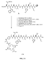
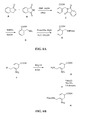
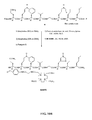
- FIG. 1A is a graphical representation of a series of chemical reactions for the synthesis of intermediate C ((3 ⁇ , 5, ⁇ )-3-(9 H -Fluoren-9-ylmethoxy)aminocholan-24-oic acid), from A (Methyl-(3 ⁇ , 5 ⁇ )-3-aminocholan-24-ate) and B ( (3 ⁇ , 5 ⁇ )-3-aminocholan-24-oic acid), as described in Example I.
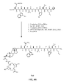
- FIG. 1B is a graphical representation of the sequential reaction for the synthesis of N -[(3, ⁇ 5 ⁇ )-3-[[[[4,7,10-Tris(carboxymethyl)-1,4,7,10-tetraazacyclododec-1-yl]acetyl]amino] acetyl]amino] cholan-24-yl]-L-glutaminyl-L-tryptophyl-L-alanyl-L-valyl-glycyl-L-histidyl-L-leucyl-L-methioninamide (L62), as described in Example I.
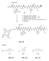
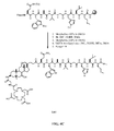
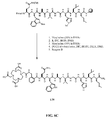
- FIG. 2A is a graphical representation of the sequential reaction for the synthesis of N -[4-[[[[[[4,7,10-Tris(carboxymethyl)-1,4,7,10-tetraazacyclododec-1-yl]acetyl]amino]acetyl]amino]benzoyl]-L-glutaminyl-L-tryptophyl-L-alanyl-L-valyl-glycyl-L-histidyl-L-leucyl-L-methioninamide (L70), as described in Example II.
- FIG. 2B is a general graphical representation of the sequential reaction for the synthesis of N- [4-[2-[[[4,7,10-Tris(carboxymethyl)-1,4,7,10-tetraazacyclododec-1-yl]acetyl]amino]ethoxy]benzoyl]-L-glutaminyl-L-tryptophyl-L-alanyl-L-valyl-glycyl-L-histidyl-L-leucyl-L-methioninamide (L73), N -[3-[[[[[4,7,10-Tris(carboxymethyl)-1,4,7,10-tetraazacyclododec-1-yl]acetyl]amino]methyl]benzoyl]-L-glutaminyl-L-tryptophyl-L-alanyl-L-valyl-glycyl-L-histidyl-L-
- FIG. 2C is a chemical structure of the linker used in the synthesis reaction of FIG. 2B for synthesis of N -[4-[2-[[[4,7,10-Tris(carboxymethyl)-1,4,7,10-tetraazacyclododec-1-yl]acetyl]amino]ethoxy]benzoyl]-L-glutaminyl-L-tryptophyl-L-alanyl-L-valyl-glycyl-L-histidyl-L-leucyl-L-methioninamide (L73), as described in Example II.
- FIG. 2D is a chemical structure of the linker used in the synthesis reaction of FIG. 2B for synthesis of N -[3-[[[[4,7,10-Tris(carboxymethyl)-1,4,7,10-tetraazacyclododec-1-yl]acetyl]amino]methyl]benzoyl]-L-glutaminyl-L-tryptophyl-L-alanyl-L-valyl-glycyl-L-histidyl-L-leucyl-L-methioninamide (L115), as described in Example II.
- FIG. 2E is a chemical structure of the linker used in the synthesis reaction of FIG. 2B for synthesis of N -[4-[[[[4,7,10-Tris(carboxymethyl)-1,4,7,10-tetraazacyclododec-1-yl]acetyl]amino]methyl]phenylacetyl]-L-glutaminyl-L-tryptophyl-L-alanyl-L-valyl-glycyl-L-histidyl-L-leucyl-L-methioninamide (L116), as described in Example II.
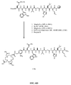
- FIG. 2F is a graphical representation of the sequential reaction for the synthesis of N -[[4,7,10-Tris(carboxymethyl)-1,4,7,10-tetraazacyclododec-1-yl]acetyl]glycyl-4-piperidinecarbonyl-L-glutaminyl-L-tryptophyl-L-alanyl-L-valyl-glycyl-L-histidyl-L-leucyl-L-methioninamide (L74), as described in Example II.
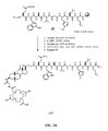
- FIG. 3A is a graphical representation of a series of chemical reactions for the synthesis of intermediate (3 ⁇ ,5 ⁇ )-3-[[(9 H -Fluoren-9-ylmethoxy)amino]acetyl]amino-12-oxocholan-24-oic acid (C), as described in Example III.
- FIG. 3B is a graphical representation of the sequential reaction for the synthesis of N -[(3 ⁇ ,5 ⁇ )-3-[[[[[[4,7,10-Tris(carboxymethyl)-1,4,7,10-tetraazacyclododec-1-yl]acetyl]amino]acetyl]amino]-12,24-dioxocholan-24-yl]-L-glutaminyl-L-tryptophyl-L-alanyl-L-valyl-glycyl-L-histidyl-L-leucyl-L-methioninamide (L67), as described in Example III.
- FIG. 3C is a chemical structure of (3 ⁇ ,5 ⁇ )-3-Amino-12-oxocholan-24-oic acid (B), as described in Example III.
- FIG. 3D is a chemical structure of (3 ⁇ ,5 ⁇ )-3-[[(9 H -Fluoren-9-ylmethoxy)amino]acetyl]amino-12-oxocholan-24-oic acid (C), as described in Example III.
- FIG 3E is a chemical structure of N-[(3 ⁇ ,5 ⁇ )-3-[[[[[[4,7,10-Tris(carboxymethyl)-1,4,7,10-tetraazacyclododec-1-yl]acetyl]amino]acetyl]amino]-12,24-dioxocholan-24-yl]-L-glutaminyl-L-tryptophyl-L-alanyl-L-valyl-glycyl-L-histidyl-L-leucyl-L-methioninamide (L67), as described in Example III.
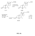
- FIG. 4A is a graphical representation of a sequence of reactions to obtain intermediates (3 ⁇ ,5 ⁇ ,12 ⁇ )-3-[[(9H-Fluoren-9-ylmethoxy)amino]acetyl]amino-12-hydroxycholan-24-oic acid (3a) and (3 ⁇ ,5 ⁇ ,7 ⁇ ,12 ⁇ )-3-[[(9H-Fluoren-9-ylmethoxy)amino] acetyl] amino-7,12-dihydroxycholan-24-oic acid (3b), as described in Example IV.
- FIG. 4B is a graphical representation of the sequential reaction for the synthesis of N -[(3 ⁇ ,5 ⁇ ,12 ⁇ )-3-[[[[[4,7,10-Tris(carboxymethyl)-1,4,7,10-tetraazacyclododec-1-yl] acetyl] amino] acetyl]amino]-12-hydroxy-24-oxocholan-24-yl]-L-glutaminyl-L-tryptophyl-L-alanyl-L-valyl-glycyl-L-histidyl-L-leucyl-L-methioninamide (L63), as described in Example IV.
- FIG. 4C is a graphical representation of the sequential reaction for the synthesis of N -[(3 ⁇ ,5 ⁇ ,7a,12a)-3-[[[[[4,7,10-Tris(carboxymethyl)-1,4,7,10- tetraazacyclo dodec -1-yl]acetyl]amino]acetyl]amino]-7,12-dihydroxy-24-oxocholan-24-yl]-L-glutaminyl-L-tryptophyl-L-alanyl-L-valyl-glycyl-L-histidyl-L-leucyl-L-methioninamide (L64), as described in Example IV.
- FIG. 4D is a chemical structure of (3 ⁇ ,5 ⁇ ,7 ⁇ ,12 ⁇ )-3-amino-7,12-dihydroxycholan-24-oic acid (2b), as described in Example IV.
- FIG. 4E is a chemical structure of (3 ⁇ ,5 ⁇ ,12 ⁇ )-3-[[(9H-Fluoren-9-ylmethoxy)amino]acetyl]amino-12-hydroxycholan-24-oic acid (3a), as described in Example IV;
- FIG. 4F is a chemical structure of (3 ⁇ ,5 ⁇ ,7 ⁇ ,12 ⁇ )-3-[[(9 H -Fluoren-9-ylmethoxy)amino]acetyl]amino-7,12-dihydroxycholan-24-oic acid (3b), as described in Example IV.
- FIG. 4G is a chemical structure of N- [(3 ⁇ ,5 ⁇ ,12 ⁇ )-3-[[[[[[4,7,10-Tris(carboxymethyl)-1,4,7,10-tetraazacyclododec-1-yl]acetyl]amino]acetyl]amino]-12-hydroxy-24-oxocholan-24-yl]-L-glutaminyl-L-tryptophyl-L-alanyl-L-valyl-glycyl-L-histidyl-L-leucyl-L-methioninamide (L63), as described in Example IV.
- FIG. 4H is a chemical structure of N- [(3 ⁇ ,5 ⁇ ,7 ⁇ ,12 ⁇ )-3-[[[[[[4,7,10-Tris(carboxymethyl)-1,4,7,10-tetraazacyclo dodec-1-yl]acetyl]amino]acetyl]amino]-7,12-dihydroxy-24-oxocholan-24-yl]-L-glutaminyl-L-tryptophyl-L-alanyl-L-valyl-glycyl-L-histidyl-L-leucyl-L-methioninamide (L64), as described in Example IV.
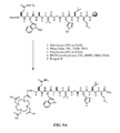
- FIG. 5A is a general graphical representation of the sequential reaction for the synthesis of 4-[[[[4,7,10-Tris(carboxymethyl)-1,4,7,10-tetraazacyclododec-1-yl]acetyl]amino]methyl]benzoyl-L-glutaminyl-L-tryptophyl-L-alanyl-L-valyl-glycyl-L-histidyl-L-leucyl-L-methioninamide (L71); and Trans -4-[[[[4,7,10-tris(carboxymethyl)-1,4,7,10-tetraazacyclododec-1-yl]acetyl]amino]methyl]cyclohexylcarbonyl-L-glutaminyl-L-tryptophyl-L-alanyl-L-valyl-glycyl-L-histidyl-L-leucyl-
- FIG. 5B is a chemical structure of the linker used in compound L71 as shown in Fig. 5A and as described in Example V.
- FIG. 5C is a chemical structure of the linker used in compound L72 as shown in Fig. 5A and as described in Example V.
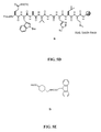
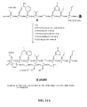
- FIG. 5D is a chemical structure of Rink amide resin functionalised with bombesin[7-14] (B), as described in Example V.
- FIG. 5E is a chemical structure of Trans -4-[[[(9H-fluoren-9-ylmethoxy)carbonyl]amino]methyl]cyclohexanecarboxylic acid (D), as described in Example V;
- FIG. 6A is a graphical representation of a sequence of reactions for the synthesis of intermediate linker 2-[[[9 H -Fluoren-9-ylmethoxy)carbonyl]amino]methyl]benzoic acid (E), as described in Example VI.
- FIG. 6B is a graphical representation of a sequence of reactions for the synthesis of intermediate linker 4-[[[9 H -Fluoren-9-ylmethoxy)carbonyl]amino]methyl]-3-nitrobenzoic acid (H), as described in Example VI.
- FIG. 6C is a graphical representation of the synthesis of N -[2-[[[[4,7,10-Tris(carboxymethyl)-1,4,7,10-tetraazacyclododec-1-yl]acetyl]amino]methyl]benzoyl]-L-glutaminyl-L-tryptophyl-L-alanyl-L-valyl-glycyl-L-histidyl-L-leucyl-L-methioninamide (L75), as described in Example VI.
- FIG. 6D is a graphical representation of the synthesis of N -[4-[[[[4,7,10-Tris(carboxymethyl)-1,4,7,10-tetraazacyclododec-1-yl]acetyl]amino]methyl]-3-nitrobenzoyl]-L-glutaminyl-L-tryptophyl-L-alanyl-L-valyl-glycyl-L-histidyl-L-leucyl-L-methioninamide (L76), as described in Example VI.
- FIG. 7A is a graphical representation of a sequence of reactions for the synthesis of intermediate linker [4-[[[9 H -Fluoren-9-ylmethoxy)carbonyl]amino]methyl]phenoxy]acetic acid (E), as described in Example VII.
- FIG. 7B is a graphical representation of the synthesis of N -[[4-[[[[4,7,10-Tris(carboxymethyl)-1,4,7,10-tetraazacyclododec-1-yl]acetyl]amino]methyl]phenoxy]acetyl]-L-glutaminyl-L-tryptophyl-L-alanyl-L-valyl-glycyl-L-histidyl-L-leucyl-L-methioninamide (L124), as described in Example VII.
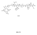
- FIG. 7C is a chemical structure of N -[[4-[[[[[4,7,10-Tris(carboxymethyl)-1,4,7,10-tetraazacyclododec-1-yl]acetyl]amino]methyl]phenoxy]acetyl]-L-glutaminyl-L-tryptophyl-L-alanyl-L-valyl-glycyl-L-histidyl-L-leucyl-L-methioninamide (L124), as described in Example VII.
- Fig. 8A is a graphical representation of a sequence of reactions for the synthesis of intermediate 4-[[[9 H -Fluoren-9-ylmethoxy)carbonyl]amino]methyl]-3-methoxybenzoic acid (E), as described in Example VIII.
- FIG. 8B is a graphical representation of the synthesis of N -[4-[[[[4,7,10-Tris(carboxymethyl)-1,4,7,10-tetraazacyclododec-1-yl]acetyl]amino]methyl]-3-methoxybenzoyl]-L-glutaminyl-L-tryptophyl-L-alanyl-L-valyl-glycyl-L-histidyl-L-leucyl-L-methioninamide, (L125), as described in Example VIII.
- FIG. 8C is a chemical structure of N -[4-[[[[4,7,10-Tris(carboxymethyl)-1,4,7,10-tetraazacyclododec-1-yl]acetyl]amino]methyl]-3-methoxybenzoyl]-L-glutaminyl-L-tryptophyl-L-alanyl-L-valyl-glycyl-L-histidyl-L-leucyl-L-methioninamide, (L125), as described in Example VIII.
- FIG. 9A is a graphical representation of a reaction for the synthesis of 3-[[[(9 H- Fluoren-9-ylmethoxy)carbonyl]amino]acetyl]aminobenzoic acid, (B), as described in Example IX.
- FIG. 9B is a graphical representation of a reaction for the synthesis of 6-[[[(9 H- Fluoren-9-ylmethoxy)carbonyl]amino]acetyl]aminonaphthoic acid (C), as described in Example IX.
- FIG. 9C is a graphical representation of a reaction for the synthesis of 4-[[[[(9 H- Fluoren-9-ylmethoxy)carbonyl]amino]acetyl]methylamino]benzoic acid , (D), as described in Example IX.
- FIG. 9D is a graphical representation of a reaction for the synthesis of N -[4-[[[[[[4,7,10-Tris(carboxymethyl)-1,4,7,10-tetraazacyclododec-1-yl]acetyl]amino]acetyl]amino]phenylacetyl]-L-glutaminyl-L-tryptophyl-L-alanyl-L-valyl-glycyl-L-histidyl-L-leucyl-L-methioninamide, (L146); N -[3-[[[[[[4,7,10-Tris(carboxymethyl)-1,4,7,10-tetraazacyclododec-1-yl]acetyl]amino]acetyl]amino]benzoyl]-L-glutaminyl-L-tryptophyl-L-alanyl-L-valyl-
- FIG. 10A is a graphical representation of a reaction for the synthesis of 7-[[Bis(1,1-dimethylethoxy)phosphinyl]methyl]-1,4,7,10-tetraazacyclododecane-1,4,10-triacetic acid 4,10-bis(1,1-dimethylethyl) ester H, as described in Example X.
- FIG. 10B is a graphical representation of a reaction for the synthesis of N -[4-[[[[[[4,10-Bis(carboxymethyl)-7-(dihydroxyphosphinyl)methyl-1,4,7,10-tetraazacyclododec-1-yl]acetyl] amino] acetyl] amino]benzoyl]-L-glutaminyl-L-tryptophyl-L-alanyl-L-valyl-glycyl-L-histidyl-L-leucil-L-methioninamide, (L237), as described in Example X.
- FIG. 11A is a graphical representation of a reaction for the synthesis of N,N- Dimethylglycyl-L-serinyl-[S-[(acetylamino)methyl]]-L-cysteinyl-glycyl-4-aminobenzoyl-L-glutaminyl-L-tryptophyl-L-alanyl-L-valyl-glycyl-L-histidyl-L-leucyl-L-methioninamide (L238), as described in Example XI.
- FIG. 11B is a graphical representation of a reaction for the synthesis of N,N- Dimethylglycyl-L-serinyl-[S-[(acetylamino)methyl]]-L-cysteinyl-glycyl-(3 ⁇ ,5 ⁇ ,7 ⁇ ,12 ⁇ )-3-amino-7,12-dihydroxy-24-oxocholan-24-yl-L-glutaminyl-L-tryptophyl-L-alanyl-L-valyl-glycyl-L-histidyl-L-leucyl-L-methioninamide, (L239), as described in Example XI.
- FIG. 12A is a graphical representation of a reaction for the synthesis of 4-[[[(9 H- Fluoren-9-ylmethoxy)carbonyl]amino]acetyl]amino-3-methoxybenzoic acid (A), as described in Example XII.
- FIG. 12B is a graphical representation of a reaction for the synthesis of 4-[[[(9 H- Fluoren-9-ylmethoxy)carbonyl]amino]acetyl]amino-3-chlorobenzoic acid, (D), as described in Example XII.
- FIG. 12C is a graphical representation of a reaction for the synthesis of 4-[[[(9 H- Fluoren-9-ylmethoxy)carbonyl]amino]acetyl]amino-3-methylbenzoic acid (E), as described in Example XII.
- FIG. 12D is a chemical structure of N -[4-[[[[4,7,10-Tris(carboxymethyl)-1,4,7,10-tetraazacyclododec-1-yl]acetyl]glycyl]amino ]-3-methoxybenzoyl]-L-glutaminyl-L-tryptophyl-1-alanyl-L-valyl-glycyl-L-histidyl-L-leucyl-L-methioninamide (L240) as described in Example XII.
- FIG. 12E is a chemical structure of compound N -[4-[[[[4,7,10-Tris(carboxymethyl)-1,4,7,10tetraazacyclododec-1-yl] acetyl]glycyl]amino]3-chlorobenzoyl]L-glutaminyl-L-tryptophyl-1-alanyl-L-valyl-glycyl-L-histidyl-L-leucyl-L-methioninamide, (L241) as described in Example XII.
- FIG. 12F is a chemical structure of N-[4-[[[[4,7,10-Tris(carboxymethyl)-1,4,7,10tetraazacyclododec-1-yl acetyl]glycyl]amino]3-methylbenzoyl]L-glutaminyl-L-tryptophyl-1-alanyl-L-valyl-glycyl-L-histidyl-leucyl-L-methioninamide (L242), as described in Example XII.
- FIG. 13A is a graphical representation of a reaction for the synthesis of 4- [ N,N'- Bis[2-[(9- H -fluoren-9-ylmethoxy)carbonyl]aminoethyl]amino]-4-oxobutanoic acid, (D), as described in Example XIII.
- FIG. 13B is a graphical representation of a reaction for the synthesis of N -[4-[[4-[Bis[2-[[[4,7,10-tris(carboxymethyl)-1,4,7,10-tetraazacyclododec-1-yl] acetyl]amino]ethyl]amino-1,4-dioxobutyl]amino]benzoyl]-L-glutaminyl-L-tryptophyl-L-alanyl-L-valyl-glycyl-L-histidyl-L-leucyl-L-methioninamide, (L244), as described in Example XIII.
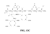
- FIG. 13C is a chemical structure of compound L244, as described in Example XIII.
- FIG. 14A and FIG. 14B are graphical representations of the binding and competition curves described in Example XLIII.
- FIG. 15A is a graphical representation of the results of radiotherapy experiments described in Example LV.
- FIG. 15B is a graphical representation of the results of other radiotherapy experiments described in Example LV.
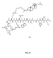
- FIG. 16 is a chemical structure of N-[4-[[[[4,7,10-Tris(carboxymethyl)-1,4,7,10tetraazacyclododec-1-yl] acetyl]glycyl]amino]-L-Lysinyl-(3,6,9)-trioxaundecane-1,11-dicarboxylic acid-3,7-dideoxy-3-aminocholic acid)-L-arginyl-L-glutaminyl-L-triptophyl-L-alanyl-L-valyl-glycyl-L-histidyl-L-leucyl-L-methioninamide (L65).
- FIG. 17 is a chemical structure of N -[2-S-[[[[[[12 ⁇ -Hydroxy-17a-(1-methyl-3-carboxypropyl)etiocholan-3 ⁇ -carbamoylmethoxyethoxyethoxyacetyl]-amino -6-[4,7,10-tris(carboxymethyl)-1,4,7,10-tetraazacyclododec-1-yl]acetyl]amino]acetyl]amino] hexanoyl-L-glutaminyl-L-tryptophyl-L-alanyl-L-valyl-glycyl-L-histidyl-L-leucyl-L-methioninamide (L66).
- FIG. 18A is a chemical structure of N -[4-[[[[[[4,7,10-Tris(carboxymethyl)-1,4,7,10-tetraazacyclododec-1-yl]acetyl]amino]acetyl]amino]benzoyl]-L-glutaminyl-L-tryptophyl-L-alanyl-L-valyl-glycyl-L-histidyl-L-leucyl-L-methioninamide (L70).
- FIG. 18B is a chemical structure N -[4-[[[[[[4,7,10-Tris(carboxymethyl)-1,4,7,10-tetraazacyclododec-1-yl]-3-carboxypropionyl]amino]acetyl]amino]benzoyl]-L-glutaminyl-L-tryptophyl-L-alanyl-L-valyl-glycyl-L-histidyl-L-leucyl-L-methioninamide (L114).
- FIG. 18C is a chemical structure N -[4-[[4,7,10-Tris(carboxymethyl)-1,4,7,10-tetraazacyclododec-1-yl]-2-hydroxy-3-propoxy]benzoyl]-L-glutaminyl-L-tryptophyl-L-alanyl-L-valyl-glycyl-L-histidyl-L-leucyl-L-methioninamide (L144).
- FIG. 18D is a chemical structure N -[(3 ⁇ ,5 ⁇ ,7 ⁇ ,12 ⁇ )-3-[[[[[[[4,7,10-Tris(carboxymethyl)-1,4,7,10-tetraazacyclododec-1-yl]acetyl]amino]ethoxyethoxy]acetyl]amino]-7,12-dihydroxycholan-24-yl]-L-glutaminyl-L-tryptophyl-L-alanyl-L-valyl-glycyl-L-histidyl-L-leucyl-L-methioninamide (L69).
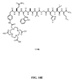
- FIG. 18E is a chemical structure of N -[4-[[[[[[4,7,10-Tris(carboxymethyl)-1,4,7,10-tetraazacyclododec-1-yl]acetyl]amino]acetyl]amino]phenylacetyl]-L-glutaminyl-L-tryptophyl-L-alanyl-L-valyl-glycyl-L-histidyl-L-leucyl-L-methioninamide (L146).
- FIG. 19 dicloses chemical structures of intermediates which may be used to prepare compounds L64 and L70 as described in Example LVI.
- FIG. 20 is a graphical representation of the preparation of L64 using segment coupling as described in Example LVI.
- FIG. 21 is a graphical representation of the preparation of (1R)-1-(Bis ⁇ 2-[bis(carboxymethyl)amino] ethyl ⁇ amino)propane-3-carboxylic acid-1-carboxyl-glycyl-4-aminobenzoyl-L-glutaminyl-L-tryptophyl-L-alanyl-L-valyl-glycyl-L-histidyl-L-leucyl-L-methioninamide (L201).
- FIG. 22A is a graphical representation of chemical structure of chemical intermediates used to prepare L202.
- FIG. 22B is a graphical representation of the preparation of N-[(3 ⁇ ,5 ⁇ ,12 ⁇ )-3-[[[[[[4,7,10-Tris(carboxymethyl)-1,4,7,10-tetraazacyclododec-1-yl]acetyl]amino]acetyl]amino]-4-hydrazinobenzoyl-L-glutaminyl-L-tryptophyl-L-alanyl-L-valyl-glycyl-L-histidyl-L-leucyl-L-methioninamide (L202).
- FIG. 23A is a graphical representation of chemical structure of chemical intermediates used to prepare L203.
- FIG. 23B is a graphical representation of the preparation of N-[(3 ⁇ ,5 ⁇ ,12 ⁇ )-3-[[[4,7,10-Tris(carboxymethyl)-1,4,7,10-tetraazacyclododec-1-yl]acetyl]amino]-4-aminobenzoyl-L-glutaminyl-L-tryptophyl-L-alanyl-L-valyl-glycyl-L-histidyl-L-leucyl-L-methioninamide (L203).
- FIG. 24 is a graphical representation of the preparation of N-[(3 ⁇ ,5 ⁇ ,12 ⁇ )-3-[[[4,7,10-Tris(carboxymethyl)-1,4,7,10-tetraazacyclododec-1-yl]acetyl]amino]-4-aminobenzoyl-glycyl-L-glutaminyl-L-tryptophyl-L-alanyl-L-valyl-glycyl-L-histidyl-L-leucyl-L-methioninamide (L204).
- FIG. 25 is a graphical representation of the preparation of N-[(3 ⁇ ,5 ⁇ ,12 ⁇ )-3-[[[4,7,10-Tris(carboxymethyl)-1,4,7,10-tetraazacyclododec-1-yl]acetyl]amino]-4-aminobenzoyl-glycyl-L-glutaminyl-L-tryptophyl-L-alanyl-L-valyl-glycyl-L-histidyl-L-leucyl-L-methioninamide (L205).
- FIG. 26A is a graphical representation of chemical structures of chemical intermediates used to prepare L206.
- FIG. 26B is a graphical representation of the preparation of N-[(3 ⁇ ,5 ⁇ ,12 ⁇ )-3-[[[[[4,7,10-Tris(carboxymethyl)-1,4,7,10-tetraazacyclododec-1-yl]acetyl]amino]acetyl]amino]- [4'-Amino-2'-methyl biphenyl-4-carboxyl]-L-glutaminyl-L-tryptophyl-L-alanyl-L-valyl-glycyl-L-histidyl-L-leucyl-L-methioninamide (L206).
- FIG. 27A is a graphical representation of chemical structures of chemical intermediates used to prepare L207.
- FIG. 27B is a graphical representation of the preparation of N-[(3 ⁇ ,5 ⁇ ,12 ⁇ )-3-[[[[[4,7,10-Tris(carboxymethyl)-1,4,7,10-tetraazacyclododec-1-yl] acetyl] amino] acetyl]] amino]- [3'-amino-biphenyl-3-carboxyl]-L-glutaminyl-L-tryptophyl-L-alanyl-L-valyl-glycyl-L-histidyl-L-leucyl-L-methioninamide (L207).
- FIG. 28 is a graphical representation of the preparation of N-[(3 ⁇ ,5 ⁇ ,12 ⁇ )-3-[[[[[4,7,10-Tris(carboxymethyl)-1,4,7,10-tetraazacyclododec-1-yl]acetyl]amino]acetyl]amino]- [1,2-diaminoethyl-terephthalyl]-L-glutaminyl-L-tryptophyl-L-alanyl-L-valyl-glycyl-L-histidyl-L-leucyl-L-methioninamide (L208).
- FIG. 29A is a graphical representation of chemical structures of chemical intermediates used to prepare L209.
- FIG. 29B is a graphical representation of the preparation of L209.
- FIG. 30A is a graphical representation of chemical structures of chemical intermediates used to prepare L210.
- FIG. 30B is a chemical structure of L210.
- FIG. 31 is a chemical structure of N-[(3 ⁇ ,5 ⁇ ,12 ⁇ )-3-[[[4,7,10-Tris(carboxymethyl)-1,4,7,10-tetraazacyclododec-1-yl]acetyl]ammo]-glycyl-glycyl-4-aminobenzoyl-L-glutaminyl-L-tryptophyl-L-alanyl-L-valyl-glycyl-L-histidyl-L-leucyl-L-methioninamide L211.
- FIG. 32 is a chemical structure of N-[(3 ⁇ ,5 ⁇ ,12 ⁇ )-3-[[[4,7,10-Tris(carboxymethyl)-1,4,7,10-tetraazacyclododec-1-yl]acetyl]amino]-glycyl-4-aminobenzoyl-L-glutamyl-L-tryptophyl-L-alanyl-L-valyl-glycyl-L-histidyl-L-leucyl-L-methioninamide L212.
- FIG. 33 is a chemical structure of N-[(3 ⁇ ,5 ⁇ ,12 ⁇ )-3-[[[4,7,10-Tris(carboxymethyl)-1,4,7,10-tetraazacyclododec-1-yl]acetyl]ammo]-glycyl-4-aminobenzoyl-L-glutaminyl-L-tryptophyl-L-alanyl-L-valyl-glycyl-L-histidyl-L-leucyl-L-methionine carboxylate L213.
- FIG. 34 is a chemical structure of N-[(3 ⁇ ,5 ⁇ ,12 ⁇ )-3-[[[4,7,10-Tris(carboxymethyl)-1,4,7,10-tetraazacyclododec-1-yl]acetyl]ammo]-glycyl-4-aminobenzoyl-D-phenylalanyl-L-glutaminyl-L-tryptophyl-L-alanyl-L-valyl-glycyl-L-histidyl-L-leucyl-L-methioninamide L214.
- FIG. 35 is a chemical structure of N-[(3 ⁇ ,5 ⁇ ,12 ⁇ )-3-[[[4,7,10-Tris(carboxymethyl)-1,4,7,10-tetraazacyclododec-1-yl]acetyl]ammo]-glycyl-4-aminobenzoyl-L-glutaminyl-L-arginyl-L-leucyl-glycyl-L-asparginyl-L-glutaminyl-L-tryptophyl-L-alanyl-L-valyl-glycyl-L-histidyl-L-leucyl-L-methioninamide L215.
- FIG. 36 is a chemical structure of N-[(3 ⁇ ,5 ⁇ ,12 ⁇ )-3-[[[4,7,10-Tris(carboxymethyl)-1,4,7,10-tetraazacyclododec-1-yl]acetyl]amino]-glycyl-4-aminobenzoyl-L-glutaminyl-arginyl-L-tyrosinyl-glycyl-L-asparginyl-L-glutaminyl-L-tryptophyl-L-alanyl-L-valyl-glycyl-L-histidyl-L-leucyl-L-methioninamide L216.
- FIG. 37 is a chemical structure of N-[(3 ⁇ ,5 ⁇ ,12 ⁇ )-3-[[[4,7,10-Tris(carboxymethyl)-1,4,7,10-tetraazacyclododec-1-yl]acetyl]ammo]-glycyl-4-aminobenzoyl-L-glutaminyl-L-lysyl-L-tyrosinyl-glycyl-L-glutaminyl-L-tryptophyl-L-alanyl-L-valyl-glycyl-L-histidyl-L-leucyl-L-methioninamide L217.
- FIG. 38 is a chemical structure of L218.
- FIG. 39 is a chemical structure of N-[(3 ⁇ ,5 ⁇ ,12 ⁇ )-3-[[[4,7,10-Tris(carboxymethyl)-1,4,7,10-tetraazacyclododec-1-yl]acetyl]amino]-glycyl-4-aminobenzoyl-D-phenylalanyl-L-glutaminyl-L-tryptophyl-L-alanyl-L-valyl-glycyl-L-histidyl-L-leucyl-aminopentyl, L219.
- FIG. 40 is a chemical structure of N-[(3 ⁇ ,5 ⁇ ,12 ⁇ )-3-[[[4,7,10-Tris(carboxymethyl)-1,4,7,10-tetraazacyclododec-1-yl]acetyl]amino]-glycyl-4-aminobenzoyl-L-glutaminyl-L-tryptophyl-L-serinyl-L-valyl-D-alanyl-L-histidyl-L-leucyl-L-methioninamide, L220.
- FIG. 41 is a chemical structure of N-[(3 ⁇ ,5 ⁇ ,12 ⁇ )-3-[[[4,7,10-Tris(carboxymethyl)-1,4,7,10-tetraazacyclododec-1-yl]acetyl]amino]-glycyl-4-aminobenzoyl-D-phenylalanyl-L-glutaminyl-L-tryptophyl-L-alanyl-L-valyl-glycyl-L-histidyl-L-leucyl-L-leucinamide, L221.
- FIG. 42 is a chemical structure of N-[(3 ⁇ ,5 ⁇ ,12 ⁇ )-3-[[[4,7,10-Tris(carboxymethyl)-1,4,7,10-tetraazacyclododec-1-yl]acetyl]amino]-glycyl-4-aminobenzoyl-D-tyrosinyl-L-glutaminyl-L-tryptophyl-L-alanyl-L-valyl-betaalanyl-L-histidyl-L-phenylalanyl-L-norleucinamide, L222.
- FIG. 43 is a chemical structure of N-[(3 ⁇ ,5 ⁇ ,12 ⁇ )-3-[[[4,7,10-Tris(carboxymethyl)-1,4,7,10-tetraazacyclododec-1-yl]acetyl]amino]-glycyl-4-aminobenzoyl-L-phenylalanyl-L-glutaminyl-L-tryptophyl-L-alanyl-L-valyl-betaalanyl-L-histidyl-L-phenylalanyl-L-norleucinamide, L223.
- FIG. 44 is a chemical structure of N-[(3 ⁇ ,5 ⁇ ,12 ⁇ )-3-[[[4,7,10-Tris(carboxymethyl)-1,4,7,10-tetraazacyclododec-1-yl]acetyl]amino]-glycyl-4-aminobenzoyl-L-glutaminyl-L-tryptophyl-L-alanyl-glycyl-L-histidyl-L-phenylalanyl-L-leucinamide, L224.
- FIG. 45 is a chemical structure of N-[(3 ⁇ ,5 ⁇ ,12 ⁇ )-3-[[[4,7,10-Tris(carboxymethyl)-1,4,7,10-tetraazacyclododec-1-yl]acetyl]amino]-glycyl-4-aminobenzoyl-L-leucyl-L-tryptophyl-L-alanyl-L-valinyl-glycyl-L-serinyl-L-phenylalanyl-L-methioninamide, L225.
- FIG. 46 is a chemical structure of N-[(3 ⁇ ,5 ⁇ ,12 ⁇ )-3-[[[4,7,10-Tris(carboxymethyl)-1,4,7,10-tetraazacyclododec-1-yl]acetyl]amino]-glycyl-4-aminobenzoyl-L-histidyl-L-tryptophyl-L-alanyl-L-valyl-glycyl-L-histidyl-L-leucyl-L-methioninamide, L226.
- FIG. 47 is a chemical structure of N-[(3 ⁇ ,5 ⁇ ,12 ⁇ )-3-[[[4,7,10-Tris(carboxymethyl)-1,4,7,10-tetraazacyclododec-1-yl]acetyl]amino]-glycyl-4-aminobenzoyl-L-leucyl-L-tryptophyl-L-alanyl-L-valyl-glycyl-L-serinyl-L-phenylalanyl-L-methioninamide L227.
- FIG. 48 is a chemical structure of N-[(3 ⁇ ,5 ⁇ ,12 ⁇ )-3-[[[4,7,10-Tris(carboxymethyl)-1,4,7,10-tetraazacyclododec-1-yl]acetyl]amino]-glycyl-4-aminobenzoyl-L-glutaminyl-L-tryptophyl-L-alanyl-L-valyl-glycyl-L-histidyl-L-phenylalanyl-L-methioninamide, L228.
- FIG. 49 is a graphical representation of a reaction for the synthesis of (3 ⁇ ,5 ⁇ ,7 ⁇ ,12 ⁇ )-3-(9H-Fluoren-9-ylmethoxy)amino-7,12-dihydroxycholan-24-oic acid (B) as described in Example LVII.
- FIG. 50 is a graphical representation of a reaction for the synthesis of N-[3 ⁇ ,5 ⁇ ,7 ⁇ ,12 ⁇ )-3-[[[2-[2-[[[4,7,10-Tris(carboxymethyl)-1,4,7,10-tetraazacyclododec-1-yl]acetyl]amino] ethoxy] ethoxy] acetyl] amino]-7,12-dihydroxy-24-oxocholan-24-yl]-L-glutaminyl-L-tryptophyl-L-alanyl-L-valyl-glycyl-L-histidyl-L-leucyl-L-methioninamide, (L69), as described in Example LVII.
- FIG. 51 is a graphical representation of a reaction for the synthesis of N-[4-[2-Hydroxy-3-[4,7,10-tris(carboxymethyl)-1,4,7,10-tetraazacyclododec-1-yl]propoxy]benzoyl]-L-glutaminyl-L-tryptophyl-L-alanyl-L-valyl-glycyl-L-histidyl-L-leucyl-L-methioninamide (L144), as described in Example LVIII.
- the compounds include an optical label or a chemical moiety capable of complexing a medically useful metal ion or radionuclide (metal chelator) attached to a GRP receptor targeting peptide by a linker or spacer group.
- metal ion or radionuclide metal chelator
- compounds of the present invention may have the formula: M-N-O-P-G wherein M is the metal chelator (in the form complexed with a metal radionuclide or not), or an optical label, N-O-P is the linker, and G is the GRP receptor targeting peptide.
- M is the metal chelator (in the form complexed with a metal radionuclide or not), or an optical label
- N-O-P is the linker
- G is the GRP receptor targeting peptide.
- linkers of the present invention may have the formula: N-O-P wherein each ofN, O and P are defined throughout the specification.
- metal chelator refers to a molecule that forms a complex with a metal atom, wherein said complex is stable under physiological conditions. That is, the metal will remain complexed to the chelator backbone in vivo. More particularly, a metal chelator is a molecule that complexes to a radionuclide metal to form a metal complex that is stable under physiological conditions and which also has at least one reactive functional group for conjugation with the linker N-O-P.
- the metal chelator M may be any of the metal chelators known in the art for complexing a medically useful metal ion or radionuclide. The metal chelator may or may not be complexed with a metal radionuclide.
- the metal chelator can include an optional spacer such as, for example, a single amino acid (e.g. , Gly) which does not complex with the metal, but which creates a physical separation between the metal chelator and the linker.
- the metal chelators of the invention may include, for example, linear, macrocyclic, terpyridine, and N 3 S, N 2 S 2 , or N 4 chelators ( see also, U.S. 5,367,080 , U.S. 5,364,613 , U.S. 5,021,556 , U.S. 5,075,099 , U.S. 5,886,142 , the disclosures of which are incorporated by reference herein in their entirety), and other chelators known in the art including, but not limited to, HYNIC, DTPA, EDTA, DOTA, TETA, and bisamino bisthiol (BAT) chelators ( see also U.S. 5,720,934 ).
- BAT bisamino bisthiol
- N 4 chelators are described in U.S. Patent Nos. 6,143,274 ; 6,093,382 ; 5,608,110 ; 5,665,329 ; 5,656,254 ; and 5,688,487 , the disclosures of which are incorporated by reference herein in their entirety.
- Certain N 3 S chelators are described in PCT/CA94/00395 , PCT/CA94/00479 , PCT/CA95/00249 and in U.S. Patent Nos. 5,662,885 ; 5,976,495 ; and 5,780,006 , the disclosures of which are incorporated by reference herein in their entirety.
- the chelator may also include derivatives of the chelating ligand mercapto-acetyl-glycyl-glycyl-glycine (MAG3), which contains an N 3 S, and N 2 S 2 systems such as MAMA (monoamidemonoaminedithiols), DADS (N 2 S diaminedithiols), CODADS and the like.
- MAG3 chelating ligand mercapto-acetyl-glycyl-glycyl-glycine
- MAMA monoamidemonoaminedithiols
- DADS N 2 S diaminedithiols
- CODADS CODADS
- the metal chelator may also include complexes containing ligand atoms that are not donated to the metal in a tetradentate array. These include the boronic acid adducts of technetium and rhenium dioximes, such as those described in U.S. Patent Nos. 5,183,653 ; 5,387,409 ; and 5,118,797 , the disclosures of which are incorporated by reference herein, in their entirety.
- chelators include, but are not limited to, diethylenetriamine pentaacetic acid (DTPA), 1,4,7,10-tetraazacyclotetradecane-1,4,7,10-tetraacetic acid (DOTA), 1-substituted 1,4,7,-tricarboxymethyl 1,4,7,10-tetraazacyclododecane triacetic acid (DO3A), ethylenediaminetetraacetic acid (EDTA), 4-carbonylmethyl-10-phosponomethyl-1,4,7,10-Tetraazacyclododecane-1,7-diacetic acid (Cm4pm10d2a); and 1,4,8,11-tetraazacyclotetradecane-1,4,8,11-tetraacetic acid (TETA).
- DTPA diethylenetriamine pentaacetic acid
- DOTA 1,4,7,10-tetraazacyclotetradecane-1,4,7,10-tetraacetic acid
- DO3A 1-sub
- Additional chelating ligands are ethylenebis-(2-hydroxy-phenylglycine) (EHPG), and derivatives thereof, including 5-C1-EHPG, 5-Br-EHPG, 5-Me-EHPG, 5-t-Bu-EHPG, and 5-sec-Bu-EHPG; benzodiethylenetriamine pentaacetic acid (benzo-DTPA) and derivatives thereof, including dibenzo-DTPA, phenyl-DTPA, diphenyl-DTPA, benzyl-DTPA, and dibenzyl-DTPA; bis-2 (hydroxybenzyl)-ethylene-diaminediacetic acid (HBED) and derivatives thereof; the class of macrocyclic compounds which contain at least 3 carbon atoms, more preferably at least 6, and at least two heteroatoms (O and/or N), which macrocyclic compounds can consist of one ring, or two or three rings joined together at the hetero ring elements, e.g., benzo-DOTA, dibenzo-DOTA,
- Particularly preferred metal chelators include those of Formula 1, 2 and 3 (for 111 In and radioactive lanthanides, such as, for example 177 Lu, 90 Y, 153 Sm, and 166 Ho) and those of Formula 4, 5 and 6 (for radioactive 99m Tc, 186 Re, and 188 Re) set forth below.
- These and other metal chelating groups are described in U.S. Patent Nos. 6,093,382 and 5,608,110 , which are incorporated by reference herein in their entirety. Additionally, the chelating group of formula 3 is described in, for example, U.S. Patent No. 6,143,274 ; the chelating group of formula 5 is described in, for example, U.S. Patent Nos.
- spacers which do not actually complex with the metal radionuclide such as an extra single amino acid Gly may be attached to these metal chelators (e.g. , N,N-dimethylGly-Ser-Cys-Gly; N,N-dimethylGly-Thr-Cys-Gly; N,N-diethylGly-Ser-Cys-Gly; N,N-dibenzylGly-Ser-Cys-Gly).
- metal chelators e.g. , N,N-dimethylGly-Ser-Cys-Gly; N,N-dimethylGly-Thr-Cys-Gly; N,N-diethylGly-Ser-Cys-Gly; N,N-dibenzylGly-Ser-Cys-Gly.
- Other useful metal chelators such as all of those disclosed in U.S. Pat. No. 6,334,996 , also incorporated by reference (
- sulfur protecting groups such as Acm (acetamidomethyl), trityl or other known alkyl, aryl, acyl, alkanoyl, aryloyl, mercaptoacyl and organothiol groups may be attached to the cysteine amino acid of these metal chelators.
- R is alkyl, preferably methyl.
- X is either CH 2 or O;
- Y is C 1 -C 10 branched or unbranched alkyl; aryl, aryloxy, arylamino, arylaminoacyl; arylalkyl - where the alkyl group or groups attached to the aryl group are C 1 -C 10 branched or unbranched alkyl groups, C 1 -C 10 branched or unbranched hydroxy or polyhydroxyalkyl groups or polyalkoxyalkyl or polyhydroxy-polyalkoxyalkyl groups;
- the metal chelator includes cyclic or acyclic polyaminocarboxylic acids such as DOTA (1,4,7,10-tetraazacyclododecane -1,4,7,10-tetraacetic acid), DTPA (diethylenetriaminepentaacetic acid), DTPA-bismethylamide, DTPA-bismorpholineamide, Cm4pm10d2a (4-carbonylmethyl-10-phosponomethyl-1,4,7,10-Tetraazacyclododecane-1,7-diacetic acid), DO3A N -[[4,7,10-Tris(carboxymethyl)-1,4,7,10-tetraazacyclododec-1-yl]acetyl, HP-DO3A, DO3A-monoamide and derivatives thereof.
- DOTA 1,4,7,10-tetraazacyclododecane -1,4,7,10-tetraacetic acid
- DTPA diethylenetriaminep
- Preferred metal radionuclides for scintigraphy or radiotherapy include 99m Tc, 51 Cr, 67 Ga, 68 Ga, 47 Sc, 51 Cr, 167 Tm, 141 Ce, 111 In, 168 Yb, 175 Yb, 140 La, 90 Y, 88 Y, 153 Sm, 166 Ho, 165 Dy, 166 Dy, 62 Cu, 64 Cu, 67 Cu, 97 Ru, 103 Ru, 186 Re, 188 Re, 203 Pb, 211 Bi, 212 Bi, 213 Bi, 214 Bi, 105 Rh, 109 Pd, 117m Sn, 149 Pm, 161 Tb, 177 Lu, 198 Au and 199 Au and oxides or nitrides thereof.
- the choice of metal will be determined based on the desired therapeutic or diagnostic application.
- the preferred radionuclides include 64 Cu, 67 Ga, 68 Ga, 99m Tc, and 111 In, with 99m Tc and 111 In being especially preferred.
- therapeutic purposes e.g.
- the preferred radionuclides include 64 Cu, 90 Y, 105 Rh, 111 In, 117m Sn, 149 Pm, 153 Sm, 161 Tb, 166 Dy, 166 Ho, 175 Yb, 177 Lu, 186/188 Re, and 199 Au, with 177 Lu and 90 Y being particularly preferred.
- 99m Tc is particularly useful and is a preferred for diagnostic radionuclide because of its low cost, availability, imaging properties, and high specific activity. The nuclear and radioactive properties of 99m Tc make this isotope an ideal scintigraphic imaging agent.
- This isotope has a single photon energy of 140 keV and a radioactive half-life of about 6 hours, and is readily available from a 99 Mo- 99m Tc generator.
- the 99 m Tc labeled peptide can be used to diagnose and monitor therapeutic progress in primary tumors and metastases.
- Peptides labeled with 177 Lu, 90 Y or other therapeutic radionuclides can be used to provide radiotherapy for primary tumors and metastasis related to cancers of the prostate, breast, lung, etc.
- the compounds of the invention may be conjugated with photolabels, such as optical dyes, including organic chromophores or fluorophores, having extensive delocalized ring systems and having absorption or emission maxima in the range of 400-1500 nm.
- photolabels such as optical dyes, including organic chromophores or fluorophores, having extensive delocalized ring systems and having absorption or emission maxima in the range of 400-1500 nm.
- the compounds of the invention may alternatively be derivatized with a bioluminescent molecule.
- the preferred range of absorption maxima for photolabels is between 600 and 1000 nm to minimize interference with the signal from hemoglobin.
- photoabsorption labels have large molar absorptivities, e.g. > 10 5 cm -1 M -1 , while fluorescent optical dyes will have high quantum yields.
- optical dyes include, but are not limited to those described in WO 98/18497 , WO 98/18496 , WO 98/18495 , WO 98/18498 , WO 98/53857 , WO 96/17628 , WO 97/18841 , WO 96/23524 , WO 98/47538 , and references cited therein.
- the photolabels may be covalently linked directly to compounds of the invention, such as, for example, compounds comprised of GRP receptor targeting peptides and linkers of the invention.
- indocyanine green which absorbs and emits in the NIR region has been used for monitoring cardiac output, hepatic functions, and liver blood flow and its functionalized derivatives have been used to conjugate biomolecules for diagnostic purposes
- the linker N-O-P contains at least one non-alpha amino acid.
- N is 0 (where 0 means it is absent), an alpha or non-alpha amino acid or other linking group; O is an alpha or non-alpha amino acid; and P is 0, an alpha or non-alpha amino acid or other linking group, wherein at least one ofN, O or P is a non-alpha amino acid.
- N Gly
- O a non-alpha amino acid
- P 0.
- Alpha amino acids are well known in the art, and include naturally occurring and synthetic amino acids.
- Non-alpha amino acids are also known in the art and include those which are naturally occurring or synthetic.
- Preferred non-alpha amino acids include:
- HPLC RT refers to the retention time of the compound in the HPLC.
- MS refers to mass spectra where molecular weight is calculated from mass/unit charge (m/e).
- IC 50 refers to the concentration of compound to inhibit 50% binding of iodinated bombesin to a GRP receptor on cells.
- the linker N-O-P contains at least one substituted bile acid.
- N is 0 (where 0 means it is absent), an alpha amino acid, a substituted bile acid or other linking group
- O is an alpha amino acid or a substituted bile acid
- P is 0, an alpha amino acid, a substituted bile acid or other linking group, wherein at least one ofN, O or P is a substituted acid.
- Bile acids are found in bile (a secretion of the liver) and are steroids having a hydroxyl group and a five carbon atom side chain terminating in a carboxyl group.
- substituted bile acids at least one atom such as a hydrogen atom of the bile acid is substituted with another atom, molecule or chemical group.
- substituted bile acids include those having a 3-amino, 24-carboxyl function optionally substituted at positions 7 and 12 with hydrogen, hydroxyl or keto functionality.
- substituted bile acids in the present invention include substituted cholic acids and derivatives thereof Specific substituted cholic acid derivatives include:
- HPLC RT refers to the retention time of the compound in the HPLC.
- MS refers to mass spectra where molecular weight is calculated from mass/unit charge (m/e).
- IC 50 refers to the concentration of compound to inhibit 50% binding of iodinated bombesin to a GRP receptor on cells.
- the linker N-O-P contains at least one non-alpha amino acid with a cyclic group.
- N is 0 (where 0 means it is absent), an alpha amino acid, a non-alpha amino acid with a cyclic group or other linking group
- O is an alpha amino acid or a non-alpha amino acid with a cyclic group
- P is 0, an alpha amino acid, a non-alpha amino acid with a cyclic group, or other linking group, wherein at least one of N, O or P is a non-alpha amino acid with a cyclic group.
- Non-alpha amino acids with a cyclic group include substituted phenyl, biphenyl, cyclohexyl or other amine and carboxyl containing cyclic aliphatic or heterocyclic moieties. Examples of such include:
- HPLC method refers to the 10 minute time for the HPLC gradient.
- HPLC RT refers to the retention time of the compound in the HPLC.
- MS refers to mass spectra where molecular weight is calculated from mass/unit charge (m/e).
- IC 50 refers to the concentration of compound to inhibit 50% binding of iodinated bombesin to a GRP receptor on cells.
- linking groups which may be used within the linker N-O-P include a chemical group that serves to couple the GRP receptor targeting peptide to the metal chelator or optical label while not adversely affecting either the targeting function of the GRP receptor targeting peptide or the metal complexing function of the metal chelator or the detectability of the optical label.
- Suitable other linking groups include peptides ( i.e ., amino acids linked together) alone, a non-peptide group ( e.g ., hydrocarbon chain) or a combination of an amino acid sequence and a non-peptide spacer.
- linking groups for use within the linker N-O-P include L-glutamine and hydrocarbon chains, or a combination thereof.
- linking groups for use within the linker N-O-P include a pure peptide linking group consisting of a series of amino acids (e.g ., diglycine, triglycine, gly-gly-glu, gly-ser-gly, etc.), in which the total number of atoms between the N-terminal residue of the GRP receptor targeting peptide and the metal chelator or the optical label in the polymeric chain is ⁇ 12 atoms.
- R 1 is a group (e.g ., H 2 N-, HS-, -COOH) that can be used as a site for covalently linking the ligand backbone or the preformed metal chelator or metal complexing backbone or optical label
- R 2 is a group
- linking groups for use within the linker N-O-P may be formed from linker precursors having electrophiles or nucleophiles as set forth below:
- the preferred nucleophiles Nu1/Nu2 include-OH, -NH, -NR, -SH, -HN-NH 2 , -RN-NH 2 , and -RN-NHR', in which R' and R are independently selected from the definitions for R given above, but for R' is not H.
- the GRP receptor targeting peptide (i.e., G in the formula M-N-O-P-G) is any peptide, equivalent, derivative or analogue thereof which has a binding affinity for the GRP receptor family.
- the GRP receptor targeting peptide may take the form of an agonist or an antagonist.
- a GRP receptor targeting peptide agonist is known to "activate” the cell following binding with high affinity and may be internalized by the cell.
- GRP receptor targeting peptide antagonists are known to bind only to the GRP receptor on the cell without being internalized by the cell and without “activating" the cell.
- the GRP receptor targeting peptide is an agonist.
- the GRP agonist is a bombesin (BBN) analogue and/or a derivative thereof.
- BBN derivative or analog thereof preferably contains either the same primary structure of the BBN binding region (i.e., BBN(7-14) [SEQ ID NO:1]) or similar primary structures, with specific amino acid substitutions that will specifically bind to GRP receptors with better or similar binding affinities as BBN alone ( i.e ., Kd ⁇ 25nM).
- Suitable compounds include peptides, peptidomimetics and analogues and derivatives thereof.
- Analogues of BBN receptor targeting peptides include molecules that target the GRP receptors with avidity that is greater than or equal to BBN, as well as muteins, retropeptides and retro-inverso-peptides of GRP or BBN.
- these analogues may also contain modifications which include substitutions, and/or deletions and/or additions of one or several amino acids, insofar that these modifications do not negatively alter the biological activity of the peptides described therein. These substitutions may be carried out by replacing one or more amino acids by their synonymous amino acids.
- Synonymous amino acids within a group are defined as amino acids that have sufficient physicochemical properties to allow substitution between members of a group in order to preserve the biological function of the molecule.
- Deletions or insertions of amino acids may also be introduced into the defined sequences provided they do not alter the biological functions of said sequences. Preferentially such insertions or deletions should be limited to 1, 2, 3, 4 or 5 amino acids and should not remove or physically disturb or displace amino acids which are critical to the functional conformation.
- Muteins of the GRP receptor targeting peptides described herein may have a sequence homologous to the sequence disclosed in the present specification in which amino acid substitutions, deletions, or insertions are present at one or more amino acid positions. Muteins may have a biological activity that is at least 40%, preferably at least 50%, more preferably 60-70%, most preferably 80-90% of the peptides described herein.
- Analogues of GRP receptor targeting peptides also include peptidomimetics or pseudopeptides incorporating changes to the amide bonds of the peptide backbone, including thioamides, methylene amines, and E-olefins. Also peptides based on the structure of GRP, BBN or their peptide analogues with amino acids replaced by N-substituted hydrazine carbonyl compounds (also known as aza amino acids) are included in the term analogues as used herein.
- the GRP receptor targeting peptide can be prepared by various methods depending upon the selected chelator.
- the peptide can generally be most conveniently prepared by techniques generally established and known in the art of peptide synthesis, such as the solid-phase peptide synthesis (SPPS) approach.
- SPPS solid-phase peptide synthesis
- SPPS involves the stepwise addition of amino acid residues to a growing peptide chain that is linked to an insoluble support or matrix, such as polystyrene.
- the C-terminal residue of the peptide is first anchored to a commercially available support with its amino group protected with an N-protecting agent such as a t-butyloxycarbonyl group (Boc) or a fluorenylmethoxycarbonyl (Fmoc) group.
- Boc t-butyloxycarbonyl group
- Fmoc fluorenylmethoxycarbonyl
- the amino protecting group is removed with suitable deprotecting agents such as TFA in the case of Boc or piperidine for Fmoc and the next amino acid residue (in N-protected form) is added with a coupling agent such as N,N'-dicyclohexylcarbodiimide (DCC), or N,N'-diisopropylcarbodiimide (DIC) or 2-(1H-benzotriazol-1-yl)-1,1,3,3-tetramethyluronium hexafluorophosphate (HBTU).
- DCC N,N'-dicyclohexylcarbodiimide
- DIC N,N'-diisopropylcarbodiimide
- a suitable reagent such as trifluoroacetic acid (TFA) or hydrogen fluoride (HF).
- the linker may then be coupled to form a conjugate by reacting the free amino group of the Trp 8 residue of the GRP receptor targeting peptide with an appropriate functional group of the linker.
- the entire construct of chelator, linker and targeting moiety discussed above may also be assembled on resin and then cleaved by agency of suitable reagents such as trifluoroacetic acid or HF, as well.
- Incorporation of the metal within the radiopharmaceutical conjugates can be achieved by various methods commonly known in the art of coordination chemistry.
- the metal is 99m Tc, a preferred radionuclide for diagnostic imaging
- the following general procedure can be used to form a technetium complex.
- a peptide-chelator conjugate solution is formed by initially dissolving the conjugate in water, dilute acid, or in an aqueous solution of an alcohol such as ethanol. The solution is then optionally degassed to remove dissolved oxygen.
- a thiol protecting group such as Acm (acetamidomethyl), trityl or other thiol protecting group may optionally be used to protect the thiol from oxidation.
- the thiol protecting group(s) are removed with a suitable reagent, for example with sodium hydroxide, and are then neutralized with an organic acid such as acetic acid (pH 6.0-6.5). Alternatively, the thiol protecting group can be removed in situ during technetium chelation.
- a suitable reagent for example with sodium hydroxide
- an organic acid such as acetic acid (pH 6.0-6.5)
- the thiol protecting group can be removed in situ during technetium chelation.
- sodium pertechnetate obtained from a molybdenum generator is added to a solution of the conjugate with a sufficient amount of a reducing agent, such as stannous chloride, to reduce technetium and is either allowed to stand at room temperature or is heated.
- the labeled conjugate can be separated from the contaminants 99m TcO 4 - and colloidal 99m TcO 2 chromatographically, for example with a C-18 Sep Pak cartridge [Millipore Corporation, Waters Chromatography Division, 34 Maple Street, Milford, Massachusetts 01757] or by HPLC using methods known to those skilled in the art.
- the labeling can be accomplished by a transchelation reaction.
- the technetium source is a solution of technetium that is reduced and complexed with labile ligands prior to reaction with the selected chelator, thus facilitating ligand exchange with the selected chelator.
- suitable ligands for transchelation includes tartrate, citrate, gluconate, and heptagluconate.
- the conjugate can be labeled using the techniques described above, or alternatively, the chelator itself may be labeled and subsequently coupled to the peptide to form the conjugate; a process referred to as the "prelabeled chelate" method.
- Re and Tc are both in row VIIB of the Periodic Table and they are chemical congeners.
- the complexation chemistry of these two metals with ligand frameworks that exhibit high in vitro and in vivo stabilities are the same [Eckelman, 1995] and similar chelators and procedures can be used to label with Re.
- This oxidation state makes it possible to selectively place 99m Tc- or 186/188 Re into ligand frameworks already conjugated to the biomolecule, constructed from a variety of 99m Tc(V) and/or 186/188 Re(V) weak chelates (e.g., 99m Tc- glucoheptonate, citrate, gluconate, etc.) [Eckelman, 1995; Lister-James et al., 1997; Pollak et al., 1996].
- compounds of the present invention can be used to treat and/or detect any pathology involving overexpression of GRP receptors (or NMB receptors) by procedures established in the art of radiodiagnostics, radiotherapeutics and optical imaging.
- optical dyes include, but are not limited to those described in WO 98/18497 , WO 98/18496 , WO 98/18495 , WO 98/18498 , WO 98/53857 , WO 96/17628 , WO 97/18841 , WO 96/23524 , WO 98/47538 , and references cited therein.
- GRP-R expression is highly upregulated in a variety of human tumors. See e.g., WO 99/62563 .
- compounds of the invention may be widely useful in treating and diagnosing cancers, including prostate cancer (primary and metastatic), breast cancer (primary and metastatic), colon cancer, gastric cancer, pancreatic cancer, non small cell lung cancer, small cell lung cancer, gastrinomas, melanomas, glioblastomas, neuroblastomas, uterus leiomyosarcoma tumors, prostatic intraepithelial neoplasias [PIN], and ovarian cancer. Additionally, compounds of the invention may be useful to distinguish between conditions in which GRP receptors are upregulated and those in which they are not (e.g. chronic pancreatitis and ductal pancreatic carcinoma, respectively
- the compounds of the invention which, as explained in more detail in the Examples, show higher uptake in tumors in vivo than compounds without the novel linkers disclosed herein, exhibit an improved ability to target GRP receptor-expressing tumors and thus to image or deliver radiotherapy to these tissues. Indeed, as shown in the Examples, radiotherapy is more effective (and survival time increased) using compounds of the invention.
- the diagnostic application of these compounds can be as a first line diagnostic screen for the presence of neoplastic cells using scintigraphic, optical, sonoluminescence or photoacoustic imaging, as an agent for targeting neoplastic tissue using hand-held radiation detection instrumentation in the field of radioimmuno guided surgery (RIGS), as a means to obtain dosimetry data prior to administration of the matched pair radiotherapeutic compound, and as a means to assess GRP receptor population as a function of treatment over time.
- RIGS radioimmuno guided surgery
- the therapeutic application of these compounds can be defined as an agent that will be used as a first line therapy in the treatment of cancer, as combination therapy where these agents could be utilized in conjunction with adjuvant chemotherapy, and/or as a matched pair therapeutic agent.
- the matched pair concept refers to a single unmetallated compound which can serve as both a diagnostic and a therapeutic agent depending on the radiometal that has been selected for binding to the appropriate chelate. If the chelator cannot accommodate the desired metals, appropriate substitutions can be made to accommodate the different metal while maintaining the pharmacology such that the behavior of the diagnostic compound in vivo can be used to predict the behavior of the radiotherapeutic compound.
- any suitable chemotherapeutic may be used, including for example, antineoplastic agents, such as platinum compounds (e.g., spiroplatin, cisplatin, and carboplatin), methotrexate, adriamycin, mitomycin, ansamitocin, bleomycin, cytosine, arabinoside, arabinosyl adenine, mercaptopolylysine, vincristine, busulfan, chlorambucil, melphalan (e.g., PAM, a, L -PAM or phennylalanine mustard), mercaptopurine, mitotane.
- platinum compounds e.g., spiroplatin, cisplatin, and carboplatin
- methotrexate e.g., spiroplatin, cisplatin, and carboplatin
- methotrexate e.g., spiroplatin, cisplatin, and carboplatin
- the therapeutic may be monoclonal antibody, such as a monoclonal antibody capable of binding to melanoma antigen.
- a conjugate labeled with a radionuclide metal such as 99m Tc
- a radionuclide metal such as 99m Tc
- a pharmaceutically acceptable carrier and/or solution such as salt solutions like isotonic saline.
- Radiolabeled scintigraphic imaging agents provided by the present invention are provided having a suitable amount of radioactivity.
- 99m Tc radioactive complexes it is generally preferred to form radioactive complexes in solutions containing radioactivity at concentrations of from about 0.01 millicurie (mCi) to 100 mCi per mL.
- the unit dose to be administered has a radioactivity of about 0.01 mCi to about 100 mCi, preferably 1 mCi to 30 mCi.
- the solution to be injected at unit dosage is from about 0.01 mL to about 10 mL.
- the amount of labeled conjugate appropriate for administration is dependent upon the distribution profile of the chosen conjugate in the sense that a rapidly cleared conjugate may need to be administered in higher doses than one that clears less rapidly. In vivo distribution and localization can be tracked by standard scintigraphic techniques at an appropriate time subsequent to administration; typically between thirty minutes and 180 minutes depending upon the rate of accumulation at the target site with respect to the rate of clearance at non-target tissue.
- a gamma camera calibrated for the gamma ray energy of the nuclide incorporated in the imaging agent can be used to image areas of uptake of the agent and quantify the amount of radioactivity present in the site. Imaging of the site in vivo can take place in a few minutes. However, imaging can take place, if desired, hours or even longer, after the radiolabeled peptide is injected into a patient. In most instances, a sufficient amount of the administered dose will accumulate in the area to be imaged within about 0.1 hour to permit the taking of scintiphotos.
- the compounds of the present invention can be administered to a patient alone or as part of a composition that contains other components such as excipients, diluents, radical scavengers, stabilizers, and carriers, all of which are well-known in the art.
- the compounds can be administered to patients either intravenously or intraperitoneally.
- the compounds made in accordance with the present invention form stable, well-defined 99m Tc or 186/188 Re labeled compounds. Similar compounds of the invention can also be made by using appropriate chelator frameworks for the respective radiometals, to form stable, well-defined products labeled with 153 Sm, 90 Y, 166 Ho, 105 Rh, 199 Au, 149 Pm, 177 Lu, 111 In or other radiometal.
- the radiolabeled GRP receptor targeting peptides selectively bind to neoplastic cells expressing GRP receptors, and if an agonist is used, become internalized, and are retained in the tumor cells for extended time periods.
- the radioactive material that does not reach (i.e., does not bind) the cancer cells is preferentially excreted efficiently into the urine with minimal retention of the radiometal in the kidneys.
- a number of optical parameters may be employed to determine the location of a target with in vivo light imaging after injection of the subject with an optically-labeled compound of the invention.
- Optical parameters to be detected in the preparation of an image may include transmitted radiation, absorption, fluorescent or phosphorescent emission, light reflection, changes in absorbance amplitude or maxima, and elastically scattered radiation.
- biological tissue is relatively translucent to light in the near infrared (NIR) wavelength range of 650-1000 nm. NIR radiation can penetrate tissue up to several centimeters, permitting the use of compounds of the present invention to image target-containing tissue in vivo.
- NIR near infrared
- the use of visible and near-infrared (NIR) light in clinical practice is growing rapidly.
- Compounds absorbing or emitting in the visible, NIR, or long-wavelength (UV-A, >350 nm) region of the electromagnetic spectrum are potentially useful for optical tomographic imaging, endoscopic visualization, and phototherapy.
- the compounds of the invention may be conjugated with photolabels, such as optical dyes, including organic chromophores or fluorophores, having extensive delocalized ring systems and having absorption or emission maxima in the range of 400-1500 nm.
- photolabels such as optical dyes, including organic chromophores or fluorophores, having extensive delocalized ring systems and having absorption or emission maxima in the range of 400-1500 nm.
- the compounds of the invention may alternatively be derivatized with a bioluminescent molecule.
- the preferred range of absorption maxima for photolabels is between 600 and 1000 nm to minimize interference with the signal from hemoglobin.
- photoabsorption labels have large molar absorptivities, e.g . > 10 5 cm -1 M -1 , while fluorescent optical dyes will have high quantum yields.
- optical dyes include, but are not limited to those described in WO 98/18497 , WO 98/18496 , WO 98/18495 , WO 98/18498 , WO 98/53857 , WO 96/17628 , WO 97/18841 , WO 96/23524 , WO 98/47538 , and references cited therein.
- the photolabels may be covalently linked directly to compounds of the invention, such as, for example, compounds comprised of GRP receptor targeting peptides and linkers of the invention.
- indocyanine green which absorbs and emits in the NIR region has been used for monitoring cardiac output, hepatic functions, and liver blood flow
- indocyanine green which absorbs and emits in the NIR region has been used for monitoring cardiac output, hepatic functions, and liver blood flow
- Y-L. He, H. Tanigami, H. Ueyama, T. Mashimo, and I. Yoshiya Measurement of blood volume using indocyanine green measured with pulse-spectrometry: Its reproducibility and reliability.
- Critical Care Medicine 1998, 26(8), 1446-1451 ; J. Caesar, S. Shaldon, L. Chiandussi, et al., The use of Indocyanine green in the measurement of hepatic blood flow and as a test of hepatic function. Clin. Sci.
- the patient is scanned with one or more light sources (e.g ., a laser) in the wavelength range appropriate for the photolabel employed in the agent.
- the light used may be monochromatic or polychromatic and continuous or pulsed. Transmitted, scattered, or reflected light is detected via a photodetector tuned to one or multiple wavelengths to determine the location of target-containing tissue (e.g ., tissue containing GRP) in the subject. Changes in the optical parameter may be monitored over time to detect accumulation of the optically-labeled reagent at the target site (e.g. the tumor or other site with GRP receptors). Standard image processing and detecting devices may be used in conjunction with the optical imaging reagents of the present invention.
- optical imaging reagents described above may also be used for acousto-optical or sonoluminescent imaging performed with optically-labeled imaging agents (see, U.S. 5,171,298 , WO 98/57666 , and references therein).
- acousto-optical imaging ultrasound radiation is applied to the subject and affects the optical parameters of the transmitted, emitted, or reflected light.
- sonoluminescent imaging the applied ultrasound actually generates the light detected. Suitable imaging methods using such techniques are described in WO 98/57666 .
- Radioisotope therapy involves the administration of a radiolabeled compound in sufficient quantity to damage or destroy the targeted tissue.
- the radiolabeled pharmaceutical localizes preferentially at the disease site (in this instance, tumor tissue or other tissue that expresses the GRP receptor). Once localized, the radiolabeled compound then damages or destroys the diseased tissue with the energy that is released during the radioactive decay of the isotope that is administered.
- the invention also encompasses use of radiotherapy in combination with adjuvant chemotherapy (or in combination with any other appropriate therapeutic agent).
- the present invention provides radiotherapeutic agents that satisfy all three of the above criteria, through proper selection of targeting group, radionuclide, metal chelate and linker.
- Radiotherapeutic agents may contain a chelated 3+ metal ion from the class of elements known as the lanthanides (elements of atomic number 57-71) and their analogs (i.e. M 3+ metals such as yttrium and indium).
- Typical radioactive metals in this class include the isotopes 90-Yttrium, 111-Indium, 149-Promethium, 153-Samarium, 166-Dysprosium, 166-Holmium, 175-Ytterbium, and 177-Lutetium.
- chelating ligands encapsulate the radiometal by binding to it via multiple nitrogen and oxygen atoms, thus preventing the release of free (unbound) radiometal into the body. This is important, as in vivo dissociation of 3 + radiometals from their chelate can result in uptake of the radiometal in the liver, bone and spleen [ Brechbiel MW, Gansow OA, "Backbone-substituted DTPA ligands for 90Y radioimmunotherapy", Bioconj. Chem.
- any of the chelators for therapeutic radionuclides disclosed herein may be used.
- forms of the DOTA chelate [Tweedle MF, Gaughan GT, Hagan JT, "1-Substituted-1,4,7-triscarboxymethyl-1,4,7,10-tetraazacyclododecane and analogs.” US Patent 4,885,363, Dec. 5, 1989 ] are particularly preferred, as the DOTA chelate is expected to de-chelate less in the body than DTPA or other linear chelates.
- lanthanides and lanthanoids include radioisotopes that have nuclear properties that make them suitable for use as radiotherapeutic agents, as they emit beta particles. Some of these are listed in the table below. Isotope Half -Life (days) Max b- energy (MeV) Gamma energy (keV) Approximate range of b-particle (cell diameters) 149 -Pm 2.21 1.1 286 60 153 -Sm 1.93 0.69 103 30 166 -Dy 3.40 0.40 82.5 15 166 -Ho 1.12 1.8 80.6 117 175 -Yb 4.19 0.47 396 17 177 -Lu 6.71 0.50 208 20 90 -Y 2.67 2.28 - 150 111 -In 2.810 Auger electron emitter 173,247 ⁇ 5 ⁇ m Pm:Promethium, Sm:Samarium, Dy:Dysprosium, Ho:Holmium, Yb:Ytterbium, Lu:Lute
- Radiotherapeutic derivatives of the invention containing beta-emitting isotopes of rhenium are also particularly preferred.
- Proper dose schedules for the compounds of the present invention are known to those skilled in the art.
- the compounds can be administered using many methods which include, but are not limited to, a single or multiple IV or IP injections.
- a quantity of radioactivity that is sufficient to permit imaging or, in the case of radiotherapy, to cause damage or ablation of the targeted GRP-R bearing tissue, but not so much that substantive damage is caused to non-target (normal tissue).
- the quantity and dose required for scintigraphic imaging is discussed supra.
- the quantity and dose required for radiotherapy is also different for different constructs, depending on the energy and half-life of the isotope used, the degree of uptake and clearance of the agent from the body and the mass of the tumor. In general, doses can range from a single dose of about 30-50 mCi to a cumulative dose of up to about 3 Curies.
- compositions of the invention can include physiologically acceptable buffers, and can require radiation stabilizers to prevent radiolytic damage to the compound prior to injection.
- Radiation stabilizers are known to those skilled in the art, and may include, for example, para-aminobenzoic acid, ascorbic acid, gentistic acid and the like.
- kits that contains all of the components needed to prepare the diagnostic or therapeutic agents of this invention is an integral part of this invention.
- such kits will often include all necessary ingredients except the radionuclide.
- a single-vial kit for preparing a radiopharmaceutical of the invention preferably contains a chelator/linker/targeting peptide conjugate of the formula M-N-O-P-G, a source of stannous salt (if reduction is required, e.g ., when using technetium), or other pharmaceutically acceptable reducing agent, and is appropriately buffered with pharmaceutically acceptable acid or base to adjust the pH to a value of about 3 to about 9.
- the quantity and type of reducing agent used will depend highly on the nature of the exchange complex to be formed. The proper conditions are well known to those that are skilled in the art. It is preferred that the kit contents be in lyophilized form.
- Such a single vial kit may optionally contain labile or exchange ligands such as glucoheptonate, gluconate, mannitol, malate, citric or tartaric acid and can also contain reaction modifiers such as diethylenetriamine-pentaacetic acid (DPTA), ethylenediamine tetraacetic acid (EDTA), or ⁇ , ⁇ , or ⁇ -cyclodextrin that serve to improve the radiochemical purity and stability of the final product.
- the kit may also contain stabilizers, bulking agents such as mannitol, that are designed to aid in the freeze-drying process, and other additives known to those skilled in the art.
- a multi-vial kit preferably contains the same general components but employs more than one vial in reconstituting the radiopharmaceutical.
- one vial may contain all of the ingredients that are required to form a labile Tc(V) complex on addition of pertechnetate (e.g . the stannous source or other reducing agent).
- pertechnetate e.g . the stannous source or other reducing agent.
- Pertechnetate is added to this vial, and after waiting an appropriate period of time, the contents of this vial are added to a second vial that contains the chelator and targeting peptide, as well as buffers appropriate to adjust the pH to its optimal value. After a reaction time of about 5 to 60 minutes, the complexes of the present invention are formed. It is advantageous that the contents of both vials of this multi-vial kit be lyophilized.
- reaction modifiers, exchange ligands, stabilizers, bulking agents, etc. may be present in either or both vials.
- the compounds of the present invention can be prepared by various methods depending upon the selected chelator.
- the peptide portion of the compound can be most conveniently prepared by techniques generally established and known in the art of peptide synthesis, such as the solid-phase peptide synthesis (SPPS) approach. Because it is amenable to solid phase synthesis, employing alternating FMOC protection and deprotection is the preferred method of making short peptides. Recombinant DNA technology is preferred for producing proteins and long fragments thereof.
- SPPS solid-phase peptide synthesis
- Solid-phase peptide synthesis involves the stepwise addition of amino acid residues to a growing peptide chain that is linked to an insoluble support or matrix, such as polystyrene.
- the C-terminal residue of the peptide is first anchored to a commercially available support with its amino group protected with an N-protecting agent such as a t-butyloxycarbonyl group (Boc) or a fluorenylmethoxycarbonyl (Fmoc) group.
- Boc t-butyloxycarbonyl group
- Fmoc fluorenylmethoxycarbonyl
- the amino protecting group is removed with suitable deprotecting agents such as TFA in the case of Boc or piperidine for Fmoc and the next amino acid residue (in N-protected form) is added with a coupling agent such as diisopropylcarbodiimide (DIC).
- DIC diisopropylcarbodiimide
- the reagents are washed from the support.
- the peptide is cleaved from the support with a suitable reagent such as trifluoroacetic acid (TFA) or hydrogen fluoride (HF).
- the compounds of the invention may also be prepared by the process known in the art as segment coupling or fragment condensation ( Barlos, K. and Gatos, D. ; 2002 "Convergent Peptide Synthesis” in Fmoc Solid Phase Synthesis - A Practical Approach; Eds. Chan, W.C. and White, P.D.; Oxford University Press, New York; Chap. 9, pp 215-228 ).
- segments of the peptide usually in side-chain protected form are prepared separately by either solution phase synthesis or solid phase synthesis or a combination of the two methods.
- the choice of segments is crucial and is made using a division strategy that can provide a manageable number of segments whose C-terminal residues and N-terminal residues are projected to provide the cleanest coupling in peptide synthesis.
- the C-terminal residues of the best segments are either devoid of chiral alpha carbons (glycine or other moieties achiral at the carbon ⁇ to the carboxyl group to be activated in the coupling step) or are compromised of amino acids whose propensity to racemization during activation and coupling is lowest of the possible choices.
- the choice of N-terminal amino acid for each segment is based on the ease of coupling of an activated acyl intermediate to the amino group.
- each of the segments is chosen based on the synthetic accessibility of the required intermediates and the relative ease of manipulation and purification of the resulting products (if needed).
- the segments are then coupled together, both in solution, or one on solid phase and the other in solution to prepare the final structure in fully or partially protected form.
- each segment can be purified separately, allowing the removal of side products such as deletion sequences resulting from incomplete couplings or those derived from reactions such as side-chain amide dehydration during coupling steps, or internal cyclization of side-chains (such as that of Gln) to the alpha amino group during deprotection of Fmoc groups.
- side products such as deletion sequences resulting from incomplete couplings or those derived from reactions such as side-chain amide dehydration during coupling steps, or internal cyclization of side-chains (such as that of Gln) to the alpha amino group during deprotection of Fmoc groups.
- Such side products would all be present in the final product of a conventional resin-based 'straight through' peptide chain assembly whereas removal of these materials can be performed, if needed, at many stages in a segment coupling strategy.
- segment coupling strategy Another important advantage of the segment coupling strategy is that different solvents, reagents and conditions can be applied to optimize the synthesis of each of the segments to high purity and yield resulting in improved purity and yield of the final product. Other advantages realized are decreased consumption of reagents and lower costs.
- the Fmoc-protected amino acids used were purchased from Nova-Biochem (San Diego, CA, USA), Advanced Chem Tech (Louisville, KY., USA), Chem-Impex International (Wood Dale Ill., USA), and Multiple Peptide Systems (San Diego, CA., USA).
- Other chemicals, reagents and adsorbents required for the syntheses were procured from Aldrich Chemical Co. (Milwaukee, WI, USA) and VWR Scientific Products (Bridgeport, NJ., USA).
- Solvents for peptide synthesis were obtained from Pharmco Co. (Brookfield CT., USA). Columns for HPLC analysis and purification were obtained from Waters Co. (Milford, MA., USA). Experimental details are given below for those that were not commercially available.
- Peptides were prepared using an Advanced ChemTech 496 ⁇ , MOS synthesizer, an Advanced ChemTech 357 FBS synthesizer and/or by manual peptide synthesis. However the protocols for iterative deprotection and chain extension employed were the same for all.
- the synthesis was run with Symphony Version 3 supplied by Protein Technologies Inc. Novagel TGR resin, with a substitution of 0.25 mmol/g, was used, and each well contained 0.2 g of the resin (50 gmol).
- the amino acids were dissolved in NMP and the concentration was 0.25M.
- a 0.25M solution of HBTU and N-Methylmorpholine in DMF was prepared and used for the coupling. All the couplings were carried out for 2.0 h. The cleavage was done outside the machine by transferring the resin to another reaction vessel and using reagent B as in the manual synthesis
- Analytical HPLC was performed using a Shimadzu-LC-10A dual pump gradient analytical LC system employing Shimadzu-ClassVP software version 4.1 for system control, data acquisition, and post run processing.
- Mass spectra were acquired on a Hewlett-Packard Series 1100 MSD mass spectrometer interfaced with a Hewlett-Packard Series 1100 dual pump gradient HPLC system fitted with an Agilent Technologies 1100 series autosampler fitted for either direct flow injection or injection onto a Waters Associates XTerra MS C18 column (4.6 mm x 50 mm, 5 ⁇ particle, 1201 ⁇ pore).
- the instrument was driven by a HP Kayak workstation using 'MSD Suite' software for sample submission and HP Chemstation software for instrument control and data acquisition.
- Micro-analysis data was obtained from Quantitative Technologies Inc. Whitehouse NJ.
- Preparative HPLC was performed on a Shimadzu-LC-8A dual pump gradient preparative HPLC system employing Shimadzu-ClassVP software version 4.3 for system control, data acquisition, fraction collection and post run processing
- the first amino acid was loaded onto 0.1 g of the resin (0.06 mmol).
- the appropriate Fmoc-amino acid in NMP (0.25M solution; 0.960 mL was added to the resin followed by N-hydroxybenzotriazole (0.5M in NMP; 0.48 mL)) and the reaction block (in the case of automated peptide synthesis) or individual reaction vessel (in the case of manual peptide synthesis) was shaken for about 2 min.
- diisopropylcarbodiimide 0.5M in NMP; 0.48 mL
- the reaction mixture was shaken for 4h at ambient temperature.
- the reaction block or the individual reaction vessel was purged of reactants by application of a positive pressure of dry nitrogen.
- the resin containing the Fmoc-protected amino acid was treated with 1.5 mL of 20% piperidine in DMF (v/v) and the reaction block or individual manual synthesis vessel was shaken for 15 min. The solution was drained from the resin. This procedure was repeated once and the resin was washed employing the washing procedure described above.
- the N-terminal amino group of the resin bound peptide linker construct was deblocked and the resin was washed.
- a 0.25M solution of the desired ligand and HBTU in NMP was made, and was treated with a two-fold equivalency of DIEA.
- the resulting solution of activated ligand was added to the the resin (1.972 mL; 0.48 mmol) and the reaction mixture was shaken at ambient temperature for 24-30 h. The solution was drained and the resin was washed. The final wash of the resin was conducted with 1.5 mL dichloromethane (3X).
- a solution of reagent B (2 mL; 88:5:5:2 - TFA:Phenol:Water:TIPS) was added to the resin and the reaction block or individual vessel was shaken for 4.5h at ambient temperature.
- the resulting solution containing the deprotected peptide was drained into a vial. This procedure was repeated two more times with 1 mL of reagent B.
- the combined filtrate was concentrated under reduced pressure using a Genevac HT-12 series II centrifugal concentrator. The residue in each vial was then triturated with 2 mL of Et 2 O and the supernatant was decanted. This procedure was repeated twice to provide the peptides as colorless solids.
- the crude peptides were dissolved in water/acetonitrile and purified using either a Waters XTerra MS C18 preparative HPLC column (50 mm x 19 mm, 5 micron particle size, 120 ⁇ pore size) or a Waters-YMC C18 ODS column (250 mm x 30 mm i.d., 10 micron particle size. 120 ⁇ pore size).
- the fractions with the products were collected and analyzed by HPLC.
- the fractions with >95% purity were pooled and the peptides isolated by lyophilization..
- Conditions for Preparative HPLC Waters XTerra Column):
- L62 was prepared using the following steps: Hydrolysis of (3 ⁇ ,5 ⁇ )-3-aminocholan-24-oic acid methyl ester A with NaOH gave the corresponding acid B, which was then reacted with Fmoc-Cl to give intermediate C. Rink amide resin functionalised with the octapeptide Gln-Trp-Ala-Val-Gly-His-Leu-Met-NH 2 (BBN[7-14] [SEQ ID No:1]) was sequentially reacted with C, Fmoc-glycine and DOTA tri-t-butyl ester. After cleavage and deprotection with reagent B the crude was purified by preparative HPLC to give L62. Overall yield: 2.5%. More details are provided below:
- the resin was shaken with 25% piperidine in DMF (45 mL) for 4 min, the solution was emptied and fresh 25% piperidine in DMF (45 mL) was added. The suspension was shaken for 10 min, then the solution was emptied and the resin was washed with DMF (5 x 45 mL).
- N - ⁇ -Fmoc-L-methionine N - ⁇ -Fmoc-L-leucine
- N - ⁇ -Fmoc- N -im-trityl-L-histidine N - ⁇ -Fmoc-glycine
- N- ⁇ -Fmoc-L-valine N - ⁇ -Fmoc-L-alanine
- N - ⁇ -Fmoc- N -in-Boc-L-tryptophan N - ⁇ -Fmoc-L-methionine
- N - ⁇ -Fmoc-L-leucine N - ⁇ -Fmoc- N -im-trityl-L-histidine
- N - ⁇ -Fmoc-glycine N- ⁇ -Fmoc-L-valine
- N - ⁇ -Fmoc-L-alanine N - ⁇ -Fmoc- N -in-Boc-L-try
- N - ⁇ -Fmoc- N - ⁇ -trityl-L-glutamine (14.6 g; 24 mmol), HOBt (3.67 g; 24 mmol), and DIC (3.75 mL; 24 mmol) were added to the resin in DMF (45 mL).
- the mixture was shaken for 3 h at room temperature, the solution was emptied and the resin was washed with DMF (5 x 45 mL), CH 2 Cl 2 (5 x 45 mL) and vacuum dried.
- Resin A (0.5 g; 0.3 mmol) was shaken in a solid phase peptide synthesis vessel with 50% morpholine in DMA (7 mL) for 10 min, the solution was emptied and fresh 50% morpholine in DMA (7 mL) was added. The suspension was shaken for 20 min then the solution was emptied and the resin washed with DMA (5 x 7 mL).
- the resin was then shaken with 50% morpholine in DMA (7 mL) for 10 min, the solution was emptied, fresh 50% morpholine in DMA (7 mL) was added and the mixture shaken for another 20 min. The solution was emptied and the resin washed with DMA (5 x 7 mL). N - ⁇ -Fmoc-glycine (0.79 g; 1.2 mmol), HOBT (0.18 g; 1.2 mmol), DIC (0.19 mL: 1.2 mmol) and DMA (7 mL) were added to the resin. The mixture was shaken for 3 h at room temperature, the solution was emptied and the resin washed with DMA (5 x 7 mL).
- the resin was then shaken with 50% morpholine in DMA (7 mL) for 10 min, the solution was emptied, fresh 50% morpholine in DMA (7 mL) was added and the mixture shaken for another 20 min.
- the solution was emptied and the resin washed with DMA (5 x 7 mL) followed by addition of 1,4,7,10-Tetraazacyclododecane-1,4,7,10-tetraacetic acid tris(1,1-dimethylethyl) ester adduct with NaCl (0.79 g; 1.2 mmol), HOBT (0.18 g; 1.2 mmol), DIC (0.19 mL: 1.2 mmol), DIEA (0.40 mL; 2.4 mmol) and DMA (7 mL) to the resin.
- the products were obtained by coupling of the octapeptide Gln-Trp-Ala-Val-Gly-His-Leu-Met-NH 2 (BBN[7-14] [SEQ ID NO:1]) (with appropriate side chain protection) on the Rink amide resin with different linkers, followed by functionalization with DOTA tri-t-butyl ester. After cleavage and deprotection with reagent B the final products were purified by preparative HPLC. Overall yields 3-9%.
- Resin A (0.5 g; 0.3 mmol) was shaken in a solid phase peptide synthesis vessel with 50% morpholine in DMA (7 mL) for 10 min, the solution was emptied and fresh 50% morpholine in DMA (7 mL) was added. The suspension was stirred for 20 min then the solution was emptied and the resin washed with DMA (5 x 7 mL).
- DMA 1,4,7,10-Tetraazacyclododecane-1,4,7,10-tetraacetic acid tris(1,1-dimethylethyl) ester adduct with NaCl (0.79 g; 1.2 mmol), HOBT (0.18
- the resin was shaken in a flask with reagent B (25 mL) for 4 h.
- the resin was filtered and the solution was evaporated under reduced pressure to afford an oily crude that was triturated with Et 2 O (5 mL).
- the precipitate was collected by centrifugation and washed with Et 2 O (5 x 5 mL), then analysed by HPLC and purified by preparative HPLC. The fractions containing the product were lyophilised.
- Resin A (0.5 g; 0.3 mmol) was shaken in a solid phase peptide synthesis vessel with 50% morpholine in DMA (7 mL) for 10 min, the solution was emptied and fresh 50% morpholine in DMA (7 mL) was added. The suspension was stirred for 20 min then the solution was emptied and the resin was washed with DMA (5 x 7 mL).
- Tributylamine (0.7 mL; 3.1 mmol) was added dropwise to a solution of N - ⁇ -Fmoc-glycine (0.9 g; 3.1 mmol) in THF (25 mL) stirred at 0 °C.
- Isobutyl chloroformate (0.4 mL; 3.1 mmol) was subsequently added and, after 10 min, a suspension of tributylamine (0.6 mL; 2.6 mmol) and (3 ⁇ ,5 ⁇ )-3-amino-12-oxocholan-24-oic acid B (1.0 g; 2.6 mmol) in DMF (30 mL) was added dropwise, over 1 h, into the cooled solution.
- Resin D (0.5 g; 0.3 mmol) was shaken in a solid phase peptide synthesis vessel with 50% morpholine in DMA (7 mL) for 10 min, the solution was emptied and fresh 50% morpholine in DMA (7 mL) was added. The suspension was stirred for 20 min then the solution was emptied and the resin was washed with DMA (5 x 7 mL).
- the resin was shaken with 50% morpholine in DMA (7 mL) for 10 min, the solution was emptied, fresh 50% morpholine in DMA (7 mL) was added and the mixture was shaken for 20 min. The solution was emptied and the resin was washed with DMA (5 x 7 mL).
- Tributylamine (4.8 mL; 20.2 mmol) was added dropwise to a solution of N- ⁇ -Fmoc-glycine (6.0 g; 20.2 mmol) in THF (120 mL) stirred at 0°C.
- Isobutyl chloroformate (2.6 mL; 20.2 mmol) was subsequently added and, after 10 min, a suspension of tributylamine (3.9 mL; 16.8 mmol) and (3 ⁇ ,5 ⁇ ,12 ⁇ )-3-amino-12-hydroxycholan-24-oic acid 2a (6.6 g; 16.8 mmol) in DMF (120 mL) was added dropwise, over 1 h, into the cooled solution.
- Tributylamine (3.2 mL; 13.5 mmol) was added dropwise to a solution of N- ⁇ -Fmoc-glycine (4.0 g; 13.5 mmol) in THF (80 mL) stirred at 0°C.
- Isobutyl chloroformate (1.7 mL; 13.5 mmol) was subsequently added and, after 10 min, a suspension of tributylamine (2.6 mL; 11.2 mmol) and (3 ⁇ ,5 ⁇ ,7 ⁇ ,12 ⁇ )-3-amino-7,12-dihydroxycholan-24-oic acid 3a (4.5 g; 11.2 mmol) in DMF (80 mL) was added dropwise, over 1 h, into the cooled solution.
- Resin A (0.5 g; 0.3 mmol) was shaken in a solid phase peptide synthesis vessel with 50% morpholine in DMA (7 mL) for 10 min, the solution was emptied and fresh 50% morpholine in DMA (7 mL) was added. The suspension was stirred for 20 min then the solution was emptied and the resin washed with DMA (5 x 7 mL).
- the resin was then shaken with 50% morpholine in DMA (7 mL) for 10 min, the solution was emptied, fresh 50% morpholine in DMA (7 mL) was added and the mixture was shaken for 20 min. The solution was emptied and the resin washed with DMA (5 x 7 mL).
- DMA 1,4,7,10-Tetraazacyclododecane-1,4,7,10-tetraacetic acid tris(1,1-dimethylethyl) ester adduct with NaCl (0.79 g; 1.2 mmol), HOBT (0.18
- Resin A (0.5 g; 0.3 mmol) was shaken in a solid phase peptide synthesis vessel with 50% morpholine in DMA (7 mL) for 10 min, the solution was emptied and fresh 50% morpholine in DMA (7 mL) was added. The suspension was stirred for 20 min, the solution was emptied and the resin was washed with DMA (5 x 7 mL).
- the resin was shaken with 50% morpholine in DMA (7 mL) for 10 min, the solution was emptied, fresh 50% morpholine in DMA (7 mL) was added and the mixture was shaken for 20 min. The solution was emptied and the resin was washed with DMA (5 x 7 mL).
- DMA 1,4,7,10-Tetraazacyclododecane-1,4,7,10-tetraacetic acid tris(1,1-dimethylethyl) ester adduct with NaCl (0.79 g; 1.2 mmol), HOBT (0.18
- the products were obtained in two steps.
- the first step was the solid phase synthesis of the octapeptide Gln-Trp-Ala-Val-Gly-His-Leu-Met-NH 2 (BBN[7-14] [SEQ ID NO:1]) (with appropriate side chain protecting groups) on the Rink amide resin discussed supra .
- the second step was the coupling with different linkers followed by functionalization with DOTA tri-t-butyl ester. After cleavage and deprotection with reagent B the final products were purified by preparative HPLC. Overall yields 3-9%.
- the resin B (0.5 g; 0.3 mmol) was shaken in a solid phase peptide synthesis vessel with 50% morpholine in DMA (7 mL) for 10 min, the solution was emptied and fresh 50% morpholine in DMA (7 mL) was added. The suspension was stirred for 20 min then the solution was emptied and the resin was washed with DMA (5 x 7 mL).
- the Fmoc-linker-OH (1.2 mmol), HOBT (0.18 g; 1.2 mmol), DIC (0.19 mL; 1.2 mmol) and DMA (7 mL) were added to the resin.
- the mixture was shaken for 3 h at room temperature, the solution was emptied and the resin washed with DMA (5 x 7 mL). The resin was then shaken with 50% morpholine in DMA (7 mL) for 10 min, the solution was emptied, fresh 50% morpholine in DMA (7 mL) was added and the mixture was shaken for 20 min. The solution was emptied and the resin was washed with DMA (5 x 7 mL).
- the resin was shaken in a flask with reagent B (25 mL) for 4 h.
- the resin was filtered and the filtrate was evaporated under reduced pressure to afford an oily crude that was triturated with ether (5 mL).
- the precipitate was collected by centrifugation and washed with ether (5 x 5 mL), then analyzed by analytical HPLC and purified by preparative HPLC. The fractions containing the product were lyophilized.
- the two products were obtained by coupling of the octapeptide Gln-Trp-Ala-Val-Gly-His-Leu-Met-NH2 (BBN[7-14] [SEQ ID NO:1]) (A) on the Rink amide resin with the two linkers E and H, followed by functionalization with DOTA tri-t-butyl ester. After cleavage and deprotection with reagent B the final products were purified by preparative HPLC. Overall yields: 8.5% (L75) and 5.6% (L76).
- Resin A (0.5 g; 0.3 mmol) was shaken in a solid phase peptide synthesis vessel with 50% morpholine in DMA (7 mL) for 10 min, the solution was emptied and fresh 50% morpholine in DMA (7 mL) was added. The suspension was stirred for 20 min then the solution was emptied and the resin washed with DMA (5 x 7 mL).
- the resin was then shaken with 50% morpholine in DMA (7 mL) for 10 min, the solution was emptied, fresh 50% morpholine in DMA (7 mL) was added and the mixture shaken for 20 min. The solution was emptied and the resin washed with DMA (5 x 7 mL).
- DMA 1,4,7,10-Tetraazacyclododecane-1,4,7,10-tetraacetic acid tris(1,1-dimethylethyl) ester adduct with NaCl (DOTA tri-
- Resin A (0.5 g; 0.3 mmol) was shaken in a solid phase peptide synthesis vessel with 50% morpholine in DMA (7 mL) for 10 min, the solution was emptied and fresh 50% morpholine in DMA (7 mL) was added. The suspension was stirred for 20 min then the solution was emptied and the resin was washed with DMA (5 x 7 mL).
- Resin A (480 mg; 0.29 mmol) was shaken in a solid phase peptide synthesis vessel with 50% morpholine in DMA (7 mL) for 10 min, the solution was emptied and fresh 50 % morpholine in DMA (7 mL) was added. The suspension was stirred for 20 min, the solution was emptied and the resin was washed with DMA (5 x 7 mL).
- the resin was then shaken with 50% morpholine in DMA (6 mL) for 10 min, the solution was emptied, fresh 50% morpholine in DMA (6 mL) was added and the mixture was shaken for 20 min. The solution was emptied and the resin was washed with DMA (5 x 7 mL).
- Triphenylphosphine (6.06 g; 23 mmol) was added to a solution of (4-azidomethyl)-3-methoxybenzoic acid methyl ester B (5 g; 22 mmol) in THF (50 mL): hydrogen evolution and formation of a white solid was observed. The mixture was stirred under nitrogen at room temperature. After 24 h more triphenylphosphine (0.6 g; 2.3 mmol) was added. After 24 h the azide was consumed and H 2 O (10 mL) was added. After 4 h the white solid disappeared. The mixture was heated at 45 °C for 3 h and was stirred overnight at room temperature. The solution was evaporated to dryness and the crude was purified by flash chromatography to give 4-(aminomethyl)-3-methoxybenzoic acid methyl ester C (1.2 g; 6.1 mmol). Yield 28%.
- Resin A (410 mg; 0.24 mmol) was shaken in a solid phase peptide synthesis vessel with 50% morpholine in DMA (7 mL) for 10 min, the solution was emptied and fresh 50 % morpholine in DMA (7 mL) was added. The suspension was stirred for 20 min then the solution was emptied and the resin was washed with DMA (5 x 7 mL).
- Resin A (0.5 g; 0.3 mmol) was shaken in a solid phase peptide synthesis vessel with 50% morpholine in DMA (7 mL) for 10 min, the solution emptied and fresh 50% morpholine in DMA (7 mL) was added. The suspension was stirred for another 20 min then the solution was emptied and the resin washed with DMA (5 x 7 mL).
- Resin A (0.5 g; 0.3 mmol) was shaken in a solid phase peptide synthesis vessel with 50% morpholine in DMA (7 mL) for 10 min, the solution flushed off and fresh 50% morpholine in DMA (7 mL) was added. The suspension was stirred for another 20 min then the solution was flushed off and the resin washed with DMA (5 x 7 mL).
- DOTA tri-t-butyl ester adduct with NaCl (0.79 g; 1.2 mmol), HOBt (0.18 g; 1.2 mmol), DIC (0.19 mL; 1.2 mmol), DIEA (0.40 mL; 2.4 mmol) and DMA (7 mL) were added to the resin.
- the mixture was shaken for 24 h at room temperature, flushed off and the resin washed with DMA (5 x 7 mL), CH2Cl2 (5 x 7 mL) and vacuum dried.
- the resin was shaken in a flask with Reagent B (25 mL) for 4.5 h.
- Resin A 500 mg; 0.3 mmol was shaken in a solid phase peptide synthesis vessel with 50% morpholine in DMA (7 mL) for 10 min, the solution emptied and fresh 50 % morpholine in DMA (7 mL) was added. The suspension was stirred for another 20 min then the solution was emptied and the resin washed with DMA (5 x 7 mL).
- Resin A 500 mg; 0.3 mmol was shaken in a solid phase peptide synthesis vessel with 50% morpholine in DMA (7 mL) for 10 min, the solution emptied and fresh 50 % morpholine in DMA (7 mL) was added. The suspension was stirred for another 20 min then the solution was emptied and the resin washed with DMA (5 x 7 mL).
- N [4-[[[[[[4,10-Bis(carboxymethyl)-7-(dihydroxyphosphinyl)methyl-1,4,7,10-tetraazacyclododec-1-yl]acetyl]amino]acetyl]amino]benzoyl]-L-glutaminyl-L-tryptophyl-L-alanyl-L-valyl-glycyl-L-histidyl-L-leucil-L-methioninamide, L 237 (FIG. 10B)
- Resin A (330 mg; 0.20 mmol) (17) was shaken in a solid phase peptide synthesis vessel with 50% morpholine in DMA (5 mL) for 10 min, the solution emptied and fresh 50 % morpholine in DMA (5 mL) was added. The suspension was stirred for another 20 min then the solution was emptied and the resin washed with DMA (5 x 5 mL).
- Fmoc-4-aminobenzoic acid (290 mg; 0.80 mmol), HOBT (120 mg; 0.80 mmol), DIC (130 ⁇ L; 0.80 mmol) and DMA (5 mL) were added to the resin, the mixture shaken for 3 h at room temperature, emptied and the resin washed with DMA (5 x 5 mL). The resin was then shaken with 50% morpholine in DMA (5 mL) for 10 min, the solution emptied, fresh 50% morpholine in DMA (5 mL) was added and the mixture shaken for another 20 min. The solution was emptied and the resin washed with DMA (5 x 5 mL).
- Resin A (0.5 g; 0.3 mmol) was shaken in a solid phase peptide synthesis vessel with 50% morpholine in DMA (7 mL) for 10 min, the solution emptied and fresh 50% morpholine in DMA (7 mL) was added. The suspension was stirred for another 20 min then the solution was emptied and the resin washed with DMA (5 x 7 mL).
- Fmoc-glycine (0.36 g; 1.2 mmol), HATU (0.46 g; 1.2 mmol) and N- ethyldiisopropylamine (0.40 mL; 2.4 mmol) were stirred for 15 min in DMA (7 mL) then the solution was added to the resin, the mixture shaken for 2 h at room temperature, emptied and the resin washed with DMA (5 x 7 mL). The resin was then shaken with 50% morpholine in DMA (7 mL) for 10 min, the solution emptied, fresh 50% morpholine in DMA (7 mL) was added and the mixture shaken for another 20 min.
- Resin A (0.5 g; 0.3 mmol) was shaken in a solid phase peptide synthesis vessel with 50% morpholine in DMA (7 mL) for 10 min, the solution emptied and fresh 50% morpholine in DMA (7 mL) was added. The suspension was stirred for another 20 min then the solution was emptied and the resin washed with DMA (5 x 7 mL).
- the resin was then shaken with 50% morpholine in DMA (7 mL) for 10 min, the solution emptied, fresh 50% morpholine in DMA (7 mL) was added and the mixture shaken for another 20 min. The solution was emptied and the resin washed with DMA (5 x 7 mL).
- N - ⁇ -Fmoc-S-acetamidomethyl-L-cysteine (0.50 g; 1.2 mmol), HOBT (0.18 g; 1.2 mmol), DIC (0.19 mL; 1.2 mmol) and DMA (7 mL) were added to the resin, the mixture was shaken for 3 h at room temperature, emptied and the resin washed with DMA (5 x 7 mL). The resin was then shaken with 50% morpholine in DMA (7 mL) for 10 min, the solution emptied, fresh 50% morpholine in DMA (7 mL) was added and the mixture shaken for another 20 min.
- Resin A (0.5 g; 0.3 mmol) was shaken in a solid phase peptide synthesis vessel with 50% morpholine in DMA (7 mL) for 10 min, the solution emptied and fresh 50% morpholine in DMA (7 mL) was added. The suspension was stirred for another 20 min then the solution was emptied and the resin washed with DMA (5 x 7 mL).
- Resin A (0.5 g; 0.3 mmol) was shaken in a solid phase peptide synthesis vessel with 50% morpholine in DMA (7 mL) for 10 min, the solution emptied and fresh 50% morpholine in DMA (7 mL) was added. The suspension was stirred for another 20 min then the solution was emptied and the resin washed with DMA (5 x 7 mL).
- Resin A (0.5 g; 0.3 mmol) was shaken in a solid phase peptide synthesis vessel with 50% morpholine in DMA (7 mL) for 10 min, the solution emptied and fresh 50% morpholine in DMA (7 mL) was added. The suspension was stirred for another 20 min then the solution was emptied and the resin washed with DMA (5 x 7 mL).
- the product was obtained in several steps starting from the octapeptide GlnTrpAlaValGlyHisLeuMetNH 2 (BBN[7-14]) on the Rink amide resin (A).
- the final coupling step with DOTA tri-t-butyl ester was done in solution phase after cleavage and deprotection with reagent B of Linker-Bombesin [7-14].
- the crude was purified by preparative HPLC to give L244. Overall yield: 0.4%.
- Trifluoroacetic acid ethyl ester (50 g; 0.35 mol) was dropped into a solution of diethylenetriamine (18 g; 0.175 mol) in THF (180 mL) at 0°C in 1 h. After 20 h at room temperature, the mixture was evaporated to an oily residue (54 g). T he oil was crystallized from Et 2 O (50 mL), filtered, washed with cooled Et 2 O (2 x 30 mL) and dried to obtain A as a white solid (46 g; 0.156 mol). Yield 89%.
- N N -[4-[[4-[Bis[2-[[[4,7,10-tris(carboxymethyl)-1,4,7,10-tetraazacyclododec-1-yl]acetyl]amino]ethyl]amino-1,4-dioxobutyl]amino]benzoyl]-L-glutaminyl-L-tryptophyl-L-alanyl-L-valyl-glycyl-L-histidyl-L-leucyl-L-methioninamide, L244 (FIG. 13B)
- Resin A (0.5 g; 0.3 mmol) was shaken in a solid phase peptide synthesis vessel with 50% morpholine in DMA (7 mL) for 10 min, the solution emptied and fresh 50% morpholine in DMA (7 mL) was added. The suspension was stirred for another 20 min then the solution was emptied and the resin washed with DMA (5 x 7 mL).
- DOTA tri-t-butyl ester (112 mg; 0.178 mmol) HATU (70 mg; 0.178 mmol) and DIEA (60 ⁇ L; 0.356 mmol) were added to a solution of F (50 mg; 0.0445 mmol) in DMA (3 mL) and CH 2 Cl 2 (2 mL) and stirred for 24 h at room temperature.
- the crude was evaporated to reduced volume (1 mL) and shaken with Reagent B (25 mL) for 4.5 h. After evaporation of the solvent, the residue was treated with Et 2 O (20 mL) to give a precipitate.
- Fmoc-Q(Trt)-W(Boc)-A-V-G-H(Trt)-L-M-resin (0.5 g, 0.2 mmol) (resin A) was used.
- Fmoc-6-aminonicotinic acid 1 was prepared as described in the literature (" Synthesis of diacylhydrazine compiounds for therapeutic use”. Hoelzemann, G.; Goodman, S. (Merck Patent G.m.b.H.., Germany). Ger.Offen. 2000, 16 pp .
- 3'-Fmoc-amino-biphenyl-3-carboxylic acid was prepared from the corresponding amine using the procedure described above. See “ Synthesis of 3'-methyl-4-'-nitrobiphenylcarboxylic acids by the reaction of 3-methyl-4-nitrobenzenenediazonium acetate with methyl benzoate", Boyland, E. and Gorrod, J., J. Chem. Soc., Abstracts (1962), 2209-11 . 0.7G of the amine yielded 0.81 g of the Fmoc-derivative (58%) (Compound B, Fig. 27A).
- Fmoc-Q(Trt)-W(Boc)-A-V-G-H(Trt)-L-M-resin (0.2g, 0.08 mmol) A was deblocked and coupled to terephthalic acid employing HATU as the coupling agent. The resulting acid on the resin was activated with DIC and NHS and then coupled to ethylenediamine. DOTA-mono acid was finally coupled to the amine on the resin.
- the bis-acid (13.5 mg, 0.0166 mmol) E was dissolved in 100 ⁇ L of dry acetonitrile and treated with NHS (4.0 mg, 0.035 mmol) and DIC (5.05 mg, 0.04 mmol) and stirred for 24h at RT.
- NHS 4.0 mg, 0.035 mmol
- DIC DIC
- the free amine F 51.0 mg, 0.41 mmol[generated from the TFA salt by treatment with saturated sodium bicarbonate and freeze drying the solution to yield the amine as a fluffy solid] was added followed by 100 ⁇ L of NMP and the stirring was continued for 40h more at RT.
- Fmoc-Q(Trt)-W(Boc)-A-V-G-H(Trt)-L-M-resin A (0.2g, 0.08 mmol) was used.
- Fmoc-Lys(ivDde) was employed for the introduction of lysine.
- the protecting group of the lysine was removed using 10% hydrazine in DMF (2 x 10 mL; 10 min each and then washed). The rest of the amino acids were then introduced using procedures described in the "general" section to complete the required peptide sequence.
- L218 in Figure 38 as obtained in a yield of 40.0 mg (23.2%).
- 4-Sulfamylbutyryl AM Novagel resin was used (1.1 mmol/g; 0.5 g; 0.55 mmol). The first amino acid was loaded on to this resin at -20° C for 20h. The rest of the sequence was completed utilizing normal coupling procedures. After washing, the resin was alkylated with 20.0 eq. of iodoacetonitrile and 10.0 equivalents of DIEA for 20h. The resin was then drained of the liquids and washed and then cleaved with 2.0 eq. of pentylamine in 5.0 mL of THF for 20h. The resin was then washed with 2 x 5.0 mL of THF and all the filtrates were combined.
- reaction mixture was kept at reflux under nitrogen until the starting bromobenzoate was consumed as determined by TLC analysis (2-3 h).
- the reaction mixture was then diluted with 250 mL of water and extracted with ethyl acetate (3 x 50 mL). The organic layers were combined and washed with saturated sodium bicarbonate solution (2 x 50 mL) and dried (Na 2 SO 4 ). The solvent was removed under reduced pressure and the residue was chromatographed over flash silica gel (100 g). Elution with 40% ethyl acetate in hexanes yielded the product either as a solid or oil. Yield: B2 -0.45g (31%); m. p. - 170-171 0 C.
- the azidocarboxylic acid was loaded on the resin using the standard peptide coupling protocols. After washing, the resin containing the azide was shaken with 20 equivalents of triphenylphosphine in THF/water (95:5) for 24 h. The solution was drained under a positive pressure of nitrogen and then washed with the standard washing procedure. The resulting amine was employed in the next coupling.
- Tributylamine (3.2 mL); 13.5 mmol) was added dropwise to a solution of Fmoc-glycine (4.0 g, 13.5 mmol) in THF (80 mL) stirred at 0° C.
- Isobutylchloroformate (1.7 mL; 13.5 mmol) was subsequently added and, after 10 min, a suspension of tributylamine (2.6 mL; 11.2 mmol) and (3 ⁇ , 5 ⁇ , 7 ⁇ , 12 ⁇ )-3-amino-7,12-dihydroxycholan-24-oic acid (4.5 g; 11.2 mmol) in DMF (80 mL) was added dropwise, over 1 h, into the cooled solution.
- Example XLIII In vitro Binding Assay for GRP Receptors in PC3 Cell Lines - Figures 14 A-B
- an in vitro assay that identifies compounds with high affinity for GRP-R was used. Since the PC3 cell line, derived from human prostate cancer, is known to exhibit high expression of GRP-R on the cell surface, a radio ligand binding assay in a 96-well plate format was developed and validated to measure the binding of 125 I-BBN to GRP-R positive PC3 cells and the ability of the compounds of the invention to inhibit this binding. This assay was used to measure the IC 50 for RP527 ligand, L134 ligand (controls) and compounds of the invention which inhibit the binding of 125 I-BBN to GRP-R.
- the Radioligand Binding Plate Assay was validated for BBN and BBN analogues (including commercially available BBN and L1) and also using 99m Tc RP527 as the radioligand.
- PC3 human prostate cancer cell line
- FBS heat inactivated FBS
- 10 mM HEPES GibcoBRL, 15630-080
- antibiotic/antimycotic GibcoBRL, 15240-062
- RPMI 1640 supplemented with 20mM HEPES, 0.1% BSA (w/v), 0.5 mM PMSF (AEBSF), bacitracin (50 mg/500 ml), pH 7.4.
- 125 I-BBN carrier free, 2200 Ci/mmole was obtained from Perkin-Elmer.
- a 96-well plate assay was used to determine the IC 50 of various compounds of the invention to inhibit binding of 125 I-BBN to human GRP-R. The following general procedure was followed:
- Peptide solutions of compounds of the invention identified in Table 6 were prepared at a concentration of 1 mg/mL in 0.1% aqueous TFA.
- a stannous chloride solution was prepared by dissolving SnCl 2 .2H 2 O (20 mg/mL) in 1 N HCl.
- Stannous gluconate solutions containing 20 ⁇ g of SnCl 2 .2H 2 O/100 ⁇ L were prepared by adding an aliquot of the SnCl 2 solution (10 ⁇ L) to a sodium gluconate solution prepared by dissolving 13 mg of sodium gluconate in water.
- a hydroxypropyl gamma cyclodextrin [HP- ⁇ -CD] solution was prepared by dissolving 50 mg of HP- ⁇ -CD in 1 mL of water.
- the 99m Tc labeled compounds identified below were prepared by mixing 20 ⁇ L of solution of the unlabeled compounds (20 ⁇ g), 50 ⁇ L of HP- ⁇ -CD solution, 100 ⁇ L of Sn-gluconate solution and 20 to 50 ⁇ L of 99m Tc pertechnetate (5 to 8 mCi, Syncor). The final volume was around 200 ⁇ L and final pH was 4.5 - 5. The reaction mixture was heated at 100°C for 15 to 20 min. and then analyzed by reversed phase HPLC to determine radiochemical purity (RCP).
- RCP radiochemical purity
- the desired product peaks were isolated by HPLC, collected into a stabilizing buffer containing 5 mg/mL ascorbic acid, 16 mg/mL HP- ⁇ -CD and 50 mM phosphate buffer, pH 4.5, and concentrated using a speed vacuum to remove acetonitrile.
- the HPLC system used for analysis and purification was as follows: C18 Vydac column, 4.6 x 250 mm, aqueous phase: 0.1% TFA in water, organic phase: 0.085% TFA in acetonitrile. Flow rate: 1 mL/min. Isocratic elution at 20% - 25% acetonitrile/0.085% TFA was used, depending on the nature of individual peptide.
- the ligand used to prepare the 99mTc complex of L2 was N,N'-dimethylglycine-Ser-Cys (Acm)-Gly-RJQWAVGHLM.
- the Acm group was removed during chelation to Tc.
- J refers to 8-amino-3,6-dioxaoctanoic acid and "a” refers to D-alanine.
- 3 Initial RCP measurement taken immediately after heating and prior to HPL purification. 4: RCP determined following HPLC isolation and acetonitrile removal via speed vacuum.
- This compound was synthesized by incubating 10 ⁇ g L64 ligand (10 ⁇ L of a 1 mg/mL solution in water), 100 ⁇ L ammonium acetate buffer (0.2M, pH 5.2) and -1-2 mCi of 177 LuCl 3 in 0.05N HCl (MURR) at 90°C for 15 min. Free 177 Lu was scavenged by adding 20 ⁇ L of a 1% Na 2 EDTA.2H 2 O (Aldrich) solution in water. The resulting radiochemical purity (RCP) was ⁇ 95%.
- the radiolabeled product was separated from unlabeled ligand and other impurities by HPLC, using a YMC Basic C8 column [4.6 x 150 mm], a column temperature of 30°C and a flow rate of 1 mL/min, with a gradient of 68%A/32%B to 66%A/34%B over 30 min., where A is citrate buffer (0.02M, pH 3.0), and B is 80% CH 3 CN/20% CH 3 OH.
- the isolated compound had an RCP of ⁇ 100% and an HPLC retention time of 23.4 minutes.
- Samples for biodistribution and cell binding studies were prepared by collecting the desired HPLC peak into 1000 ⁇ L of citrate buffer (0.05 M, pH 5.3, containing 1% ascorbic acid, and 0.1% HSA). The organic eluent in the collected eluate was removed by centrifugal concentration for 30 min.
- the purified sample was diluted with cell-binding media to a concentration of 1.5 ⁇ Ci/mL within 30 minutes of the in vitro study.
- the sample was diluted with citrate buffer (0.05 M, pH 5.3, containing 1% sodium ascorbic acid and 0.1% HSA) to a final concentration of 50 ⁇ Ci/mL within 30 minutes of the in vivo study.
- This compound was synthesized by incubating 70 ⁇ g L64 ligand (70 ⁇ L of a 1 mg/mL solution in water), 200 ⁇ L ammonium acetate buffer (0.2M, pH 5.2) and -30 - 40 mCi of 177 LuCl 3 in 0.05N HCl (MURR) at 85°C for 10 min. After cooling to room temperature, free 177 Lu was scavenged by adding 20 ⁇ L of a 2% Na 2 EDTA.2H 2 O (Aldrich) solution in water. The resulting radiochemical purity (RCP) was ⁇ 95%.
- the radiolabeled product was separated from unlabeled ligand and other impurities by HPLC, using a 300VHP Anion Exchange column (7.5 x 50 mm) (Vydac) that was sequentially eluted at a flow rate of 1 mL/min with water, 50% acetonitrile/water and then 1g/L aqueous ammonium acetate solution.
- the desired compound was eluted from the column with 50% CH 3 CN and mixed with ⁇ 1 mL of citrate buffer (0.05 M, pH 5.3) containing 5% ascorbic acid, 0.2% HSA, and 0.9% (v:v) benzyl alcohol.
- the organic part of the isolated fraction was removed by spin vacuum for 40 min, and the concentrated solution (-20-25 mCi) was adjusted within 30 minutes of the in vivo study to a concentration of 7.5 mCi/mL using citrate buffer (0.05 M, pH 5.3) containing 5% ascorbic acid, 0.2% HSA, and 0.9% (v:v) benzyl alcohol.
- the resulting compound had an RCP of>95%.
- This compound was synthesized by incubating 10 ⁇ g L64 ligand (5 ⁇ L of a 2 mg/mL solution in 0.01 N HCl), 60 ⁇ L ethanol, 1.12 mCi of 111 InCl 3 in 0.05N HCl (80 ⁇ L) and 155 ⁇ L sodium acetate buffer (0.5M, pH 4.5) at 85°C for 30 min. Free 111 In was scavenged by adding 20 ⁇ L of a 1% Na 2 EDTA.2H 2 O (Aldrich) solution in water. The resulting radiochemical purity (RCP) was 87%.
- the radio labeled product was separated from unlabeled ligand and other impurities by HPLC, using a Vydac C18 column, [4.6x250 mm], a column temperature of 50°C and a flow rate of 1.5 mL/min. with a gradient of 75%A/25%B to 65%A/35%B over 20 min where A is 0.1% TFA in water, B is 0.085% TFA in acetonitrile. With this system, the retention time for 111 In-L64 is 15.7 min. The isolated compound had an RCP of 96.7%.
- a stock solution of peptide was prepared by dissolving L134 ligand (prepared as described in US Application Publication No. 2002/0054855 and WO 02/87637 , both incorporated by reference) in 0.01 N HCl to a concentration of 1 mg/mL.
- 177 Lu-L134 was prepared by mixing the following reagents in the order shown. 0.2 M NH 4 OAc, pH 6.8 100 ⁇ l Peptide stock, 1 mg/mL, in 0.01 N HCl 5 ⁇ l 177 LuCl 3 (MURR) in 0.05M HCl 1.2 ⁇ l (1.4 mCi)
- the reaction mixture was incubated at 85 °C for 10 min. After cooling down to room temperature in a water bath, 20 ⁇ l of a 1% EDTA solution and 20 ⁇ l of EtOH were added.
- the compound was analyzed by HPLC using a C18 column (VYDAC Cat # 218TP54) that was eluted at flow rate of 1 mL/min with a gradient of 21 to 25% B over 20 min, where A is 0.1%TFA/H 2 O and B is 0.1%TFA/CH 3 CN).
- 177 Lu-L134 was formed in 97.1% yield (RCP) and had a retention time of ⁇ 16.1 min on this system.
- This compound was prepared as described for 177 Lu-L134.
- the compound was analyzed by HPLC using a C18 column (VYDAC Cat # 218TP54) that was eluted at flow rate of 1 mL/min with a gradient of 30-34% B over 20 min (where solvent is A. 0.1%TFA/H 2 O and B is 0.1%TFA/CH 3 CN).
- the 177 Lu-L63 that formed had an RCP of 97.8% and a retention time of ⁇ 14.2 min on this system.
- This compound was prepared following the procedures described above, but substituting L70 (the ligand of Example II). Purification was performed using a YMC Basic C8 column (4.6 x 150 mm), a column temperature of 30°C and a flow rate of 1 mL/min. with a gradient of 80%A/20%B to 75%A/25%B over 40 min., where A is citrate buffer (0.02M, pH 4.5), and B is 80% CH 3 CN/20% CH 3 OH. The isolated compound had an RCP of ⁇ 100% and an HPLC retention time of 25.4 min.
- This compound was prepared as described above for L64.
- This compound was synthesized by incubating 10 ⁇ g L70 ligand (10 ⁇ L of a 1 mg/mL solution in 0.01 N HCl), 180 ⁇ L ammonium acetate buffer (0.2M, pH 5.3), 1.1 mCi of 111 InCl 3 in 0.05N HCl (61 ⁇ L, Mallinckrodt) and 50 ⁇ L of saline at 85°C for 30 min. Free 111 In was scavenged by adding 20 ⁇ L of a 1% Na 2 EDTA.2H 2 O (Aldrich) solution in water. The resulting radiochemical purity (RCP) was 86%.
- the radiolabeled product was separated from unlabeled ligand and other impurities by HPLC, using a Waters XTerra C18 cartridge linked to a Vydac strong anion exchange column [7.5 x 50 mm], a column temperature of 30°C and a flow rate of 1 mL/min. with the gradient listed in the Table below, where A is 0.1 mM NaOH in water, pH 10.0, B is 1 g/L ammonium acetate in water, pH 6.7 and C is acetonitrile. With this system, the retention time for 111 In-L70 is 15 min while the retention time for L70 ligand is 27 to 28 min. The isolated compound had an RCP of 96%.
- Samples for biodistribution and cell binding studies were prepared by collecting the desired HPLC peak into 500 ⁇ L of citrate buffer (0.05 M, pH 5.3, containing 5% ascorbic acid, 1 mg/mL L-methionine and 0.2% HSA). The organic part of the collection was removed by spin vacuum for 30 min.
- the purified, concentrated sample was used within 30 minutes of the in vitro study.
- the sample was diluted with citrate buffer (0.05 M, pH 5.3, containing 5% sodium ascorbic acid and 0.2% HSA) to a final concentration of 10 ⁇ Ci/mL within 30 minutes of the in vivo study. Time, min A B C 0-10 100% 10-11 100-50% 0-50% 11-21 50% 50% 21-22 50-0% 0-50% 50% 22-32 50% 50%
- GRP-R GRP-preferring receptor
- NMB-R neuromedin-B preferring receptor
- BB3-R bombesin receptor subtype 3
- BB4 receptor subtype BB4 receptor subtype
- the subtype specificity of the Lutetium complex of L245 [ 177 Lu-L24] was determined by in vitro receptor autoradiography using the procedure described in Reubi et al., "Bombesin Receptor Subtypes in Human Cancers: Detection with the Universal Radioligand (125)I-[D-TYR(6), beta-ALA, PHE(13), NLE(14)] Bombesin (6-14)", Clin.Cancer Res. 8:1139-1146 (2002 ) and tissue samples that had been previously found to express only one subtype of GRP receptor.
- Human ileal carcinoid tissue was used as a source for NMB-R, human prostate carcinoma for GRP-R and human bronchial carcinoid for BB3-R subtype receptors.
- L245 derivatives are expected to bind well to human prostate carcinoma, which primarily expresses GRP-R. They also indicate that L245 derivatives are not expected to bind well to normal human pancreas (which primarily expresses the BB3-R receptor), or to cancers which primarily express the BB3-R receptor subtype.
- mice were administered 100 ⁇ L of 177 Lu-labelled compound of the invention at 30 mCi/kg, i.v, or s.c. under sterile conditions. The subjects were housed in a barrier environment for up to 30 days. Body weight and tumor size (by caliper measurement) were collected on each subject 3 times per week for the duration of the study.
- Criteria for early termination included: death; loss of total body weight (TBW) equal to or greater than 20%; tumor size equal to or greater than 2 cm 3 .
- Results are displayed in Figure 15A . These results show that animals treated with L70, L64 or L63 have increased survival over the control animals given no treatment and over those animals given the same dose of L134.
- Compounds L64 and L70 can be prepared employing the collection of intermediates generally represented by A-D ( Figure 19 ), which themselves are prepared by standard methods known in the art of solid and solution phase peptide synthesis ( Synthetic Peptides - A User's Guide 1992, Grant, G., Ed. WH. Freeman Co., NY, Chap 3 and Chap 4 pp 77 - 258 ; Chan, W.C. and White, P.D. Basic Procedures in Fmoc Solid Phase Peptide Synthesis - A Practical Approach 2002, Chan, W.C. and White, P.D. Eds Oxford University Press, New York, Chap. 3 pp 41 - 76 ; Barlos, K and Gatos, G.
- Resin A (0.5 g; 0.3 mmol) was shaken in a solid phase peptide synthesis vessel with 50% morpholine in DMA (7 mL) for 10 min, the solution flushed off and fresh 50% morpholine in DMA (7 mL) was added. The suspension was stirred for another 20 min then the solution was flushed off and the resin washed with DMA (5 x 7 mL).
- the mixture was shaken for 3 h at room temperature, emptied and the resin washed with DMA (5 x 7 mL). The resin was then shaken with 50% morpholine in DMA (7 mL) for 10 min, the solution flushed off, fresh 50% morpholine in DMA (7 mL) was added and the mixture shaken for another 20 min.
- N [4-[2-Hydroxy-3-[4,7,10-tris(carboxymethyl)-1,4,7,10-tetraazacyclododec-1-yl]propoxy]benzoyl]-L-glutaminyl-L-tryptophyl-L-alanyl-L-valyl-glycyl-L-histidyl-L-leucyl-L-methioninamide, L144 (FIG 51)
- Resin A (0.4 g; 0.24 mmol) was shaken in a solid phase peptide synthesis vessel with 50% morpholine in DMA (7 mL) for 10 min, the solution flushed off and fresh 50% morpholine in DMA (7 mL) was added. The suspension was stirred for another 20 min then the solution was flushed off and the resin washed with DMA (5 x 7 mL).
Abstract
Description
- This application claims the benefit of the filing date of
U.S. Provisional Application No. 10/341,577, filed January 13, 2003 - This invention relates to novel gastrin releasing peptide (GRP) compounds which are useful as diagnostic imaging agents or radiotherapeutic agents. These GRP compounds are labeled with radionuclides or labels detectable by in vivo light imaging and include the use of novel linkers between the label and the targeting peptide, which provides for improved pharmacokinetics.
- The use of radiopharmaceuticals (e.g., diagnostic imaging agents, radiotherapeutic agents) to detect and treat cancer is well known. In more recent years, the discovery of site-directed radiopharmaceuticals for cancer detection and/or treatment has gained popularity and continues to grow as the medical profession better appreciates the specificity, efficacy and utility of such compounds.
- These newer radiopharmaceutical agents typically consist of a targeting agent connected to a metal chelator, which can be chelated to (e.g., complexed with) a diagnostic metal radionuclide such as, for example, technetium or indium, or a therapeutic metal radionuclide such as, for example, lutetium, yttrium, or rhenium. The role of the metal chelator is to hold (i.e., chelate) the metal radionuclide as the radiopharmaceutical agent is delivered to the desired site. A metal chelator which does not bind strongly to the metal radionuclide would render the radiopharmaceutical agent ineffective for its desired use since the metal radionuclide would therefore not reach its desired site. Thus, further research and development led to the discovery of metal chelators, such as that reported in
U.S. Pat. No. 5,662,885 to Pollak et. al. , hereby incorporated by reference, which exhibited strong binding affinity for metal radionuclides and the ability to conjugate with the targeting agent. Subsequently, the concept of using a "spacer" to create a physical separation between the metal chelator and the targeting agent was further introduced, for example inU.S. Pat. 5,976,495 to Pollak et. al. , hereby incorporated by reference. - The role of the targeting agent, by virtue of its affinity for certain binding sites, is to direct the diagnostic agent, such as a radiopharmaceutical agent containing the metal radionuclide, to the desired site for detection or treatment. Typically, the targeting agent may include a protein, a peptide, or other macromolecule which exhibits a specific affinity for a given receptor. Other known targeting agents include monoclonal antibodies (MAbs), antibody fragments (Fab 's and (Fab)2 's), and receptor-avid peptides. Donald J. Buchsbaum, "Cancer Therapy with Radiolabeled Antibodies; Pharmacokinetics of Antibodies and Their Radiolabels; Experimental Radioimmunotherapy and Methods to Increase Therapeutic Efficacy," CRC Press, Boca Raton, ); Fischman, et al. "A Ticket to Ride: Peptide Radiopharmaceuticals," The Journal of Nuclear Medicine, vol. 34, No. 12, (Dec. 1993).
- In recent years, it has been learned that some cancer cells contain gastrin releasing peptide (GRP) receptors (GRP-R) of which there are a number of subtypes. In particular, it has been shown that several types of cancer cells have over-expressed or uniquely expressed GRP receptors. For this reason, much research and study have been done on GRP and GRP analogues which bind to the GRP receptor family. One such analogue is bombesin (BBN), a 14 amino acid peptide (i.e., tetradecapeptide) isolated from frog skin which is an analogue of human GRP and which binds to GRP receptors with high specificity and with an affinity similar to GRP.
- Bombesin and GRP analogues may take the form of agonists or antagonists. Binding of GRP or BBN agonists to the GRP receptor increases the rate of cell division of these cancer cells and such agonists are internalized by the cell, while binding of GRP or BBN antagonists generally does not result in either internalization by the cell or increased rates of cell division. Such antagonists are designed to competitively inhibit endogenous GRP binding to GRP receptors and reduce the rate of cancer cell proliferation. See, e.g., Hoffken, K.; Peptides in Oncology II, Somatostatin Analogues and Bombesin Antagonists (1993), pp. 87-112. For this reason, a great deal of work has been, and is being pursued to develop BBN or GRP analogues that are antagonists. E.g., Davis et al., Metabolic Stability and Tumor Inhibition of Bombesin/GRP Receptor Antagonists, Peptides, vol. 13, pp. 401-407, 1992.
- In designing an effective compound for use as a diagnostic or therapeutic agent for cancer, it is important that the drug have appropriate in vivo targeting and pharmacokinetic properties. For example, it is preferable that for a radiopharmaceutical, the radiolabeled peptide have high specific uptake by the cancer cells (e.g., via GRP receptors). In addition, it is also preferred that once the radionuclide localizes at a cancer site, it remains there for a desired amount of time to deliver a highly localized radiation dose to the site.
- Moreover, developing radiolabeled peptides that are cleared efficiently from normal tissues is also an important factor for radiopharmaceutical agents. When biomolecules (e.g., MAb, Fab or peptides) labeled with metallic radionuclides (via a chelate conjugation), are administered to an animal such as a human, a large percentage of the metallic radionuclide (in some chemical form) can become "trapped" in either the kidney or liver parenchyma (i.e., is not excreted into the urine or bile). Duncan et al.; Indium-111-Diethylenetriaminepentaacetic Acid-Octreotide Is Delivered in Vivo to Pancreatic, Tumor Cell, Renal, and Hepatocyte Lysosomes, Cancer Research 57, pp. 659-671, (Feb. 15, 1997). For the smaller radiolabeled biomolecules (i.e., peptides or Fab), the major route of clearance of activity is through the kidneys which can also retain high levels of the radioactive metal (i.e., normally >10-15% of the injected dose). Retention of metal radionuclides in the kidney or liver is clearly undesirable. Conversely, clearance of the radiopharmaceutical from the blood stream too quickly by the kidney is also undesirable if longer diagnostic imaging or high tumor uptake for radiotherapy is needed.
- Subsequent work, such as that in
U.S. Pat. 6,200,546 andUS 2002/0054855 to Hoffman, et. al , hereby incorporated by reference, has attempted to overcome this problem by forming a compound having the general formula X-Y-B wherein X is a group capable of complexing a metal, Y is a covalent bond on a spacer group and B is a bombesin agonist binding moiety. Such compounds were reported to have high binding affinities to GRP receptors, and the radioactivity was retained inside of the cells for extended time periods. In addition, in vivo studies in normal mice have shown that retention of the radioactive metal in the kidneys was lower than that known in the art, with the majority of the radioactivity excreted into the urine. - New and improved radiopharmaceutical and other diagnostic compounds which have improved pharmacokinetics and improved kidney excretion (i.e., lower retention of the radioactive metal in the kidney) have now been found for diagnostic imaging and therapeutic uses. For diagnostic imaging, rapid renal excretion and low retained levels of radioactivity are critical for improved images. For radiotherapeutic use, slower blood clearance to allow for higher tumor uptake and better tumor targeting with low kidney retention are critical.
- In an embodiment of the present invention, there is provided new and improved compounds for use in diagnostic imaging or radiotherapy. The compounds include a chemical moiety capable of complexing a medically useful metal ion or radionuclide (metal chelator) attached to a GRP receptor targeting peptide by a linker or spacer group. In another embodiment, these compounds include an optical label (e.g. a photolabel or other label detectable by light imaging, optoacoustical imaging or photoluminescence) attached to a GRP receptor targeting peptide by a linker or spacer group.
- In general, compounds of the present invention may have the formula: M-N-O-P-G
wherein M is the metal chelator (in the form complexed with a metal radionuclide or not), or the optical label, N-O-P is the linker, and G is the GRP receptor targeting peptide. - The metal chelator M may be any of the metal chelators known in the art for complexing with a medically useful metal ion or radionuclide. Preferred chelators include DTPA, DOTA, DO3A, HP-DO3A, EDTA, TETA, EHPG, HBED, NOTA, DOTMA, TETMA, PDTA, TTHA, LICAM, MECAM, or peptide chelators, such as, for example, those discussed herein. The metal chelator may or may not be complexed with a metal radionuclide, and may include an optional spacer such as a single amino acid. Preferred metal radionuclides for scintigraphy or radiotherapy include 99mTc, 51Cr, 67Ga, 68Ga, 47Sc, 51Cr, 167Tm, 141Ce, 1111In, 168Yb, 175Yb, 140La, 90Y, 88Y, 153Sm, 166Ho, 165Dy, 166Dy, 62Cu, 64Cu, 67Cu, 97RU, 103RU, 186Re, 188Re, 203Pb, 211Bi, 212Bi, 213Bi, 214Bi, 105Rh, 109Pd, 117mSn, 149Pm, 161Tb, 177Lu, 198Au and 199Au. The choice of metal will be determined based on the desired therapeutic or diagnostic application. For example, for diagnostic purposes the preferred radionuclides include 64Cu, 67Ga, 68Ga, 99mTc, and 111In, with 99mTc, and 111In being particularly preferred. For therapeutic purposes, the preferred radionuclides include 64Cu, 90Y, 105Rh, 111In, 117mSn, 149Pm, 153Sm, 161Tb, 166Dy, 166Ho, 175Yb, 177Lu, 186/188Re, and 199Au, with 177Lu and 90Y being particularly preferred. A most preferred chelator used in compounds of t h invention is 1-substituted 4,7,10-
tricarboxymethyl - The optical label M may be any of various optical labels known in the art. Preferred labels include, without limitation, optical dyes, including organic chromophores or fluorophores, such as cyanine dyes light absorbing compounds, light reflecting and scattering compounds, and bioluminescent molecules.
- In one embodiment, the linker N-O-P contains at least one non-alpha amino acid.
- In another embodiment, the linker N-O-P contains at least one substituted bile acid.
- In yet another embodiment, the linker N-O-P contains at least one non-alpha amino acid with a cyclic group.
- The GRP receptor targeting peptide may be GRP, bombesin or any derivatives or analogues thereof. In a preferred embodiment, the GRP receptor targeting peptide is a GRP or bombesin analogue which acts as an agonist. In a particularly preferred embodiment, the GRP receptor targeting peptide is a bombesin agonist binding moiety disclosed in
U.S. Pat. 6,200,546 andUS 2002/0054855 , incorporated herein by reference. - There is also provided a novel method of imaging using the compounds of the present invention.
- A single or multi-vial kit that contains all of the components needed to prepare the diagnostic or therapeutic agents of the invention is provided in an exemplary embodiment of the present invention.
- There is further provided a novel method for preparing a diagnostic imaging agent comprising the step of adding to an injectable imaging medium a substance containing the compounds of the present invention.
- A novel method of radiotherapy using the compounds of the invention is also provided, as is a novel method for preparing a radiotherapeutic agent comprising the step of adding to an injectable therapeutic medium a substance comprising a compound of the invention.
-
FIG. 1A is a graphical representation of a series of chemical reactions for the synthesis of intermediate C ((3β, 5,β)-3-(9H-Fluoren-9-ylmethoxy)aminocholan-24-oic acid), from A (Methyl-(3β, 5β)-3-aminocholan-24-ate) and B ( (3β, 5β)-3-aminocholan-24-oic acid), as described in Example I. -
FIG. 1B is a graphical representation of the sequential reaction for the synthesis of N-[(3,β5β)-3-[[[[4,7,10-Tris(carboxymethyl)-1,4,7,10-tetraazacyclododec-1-yl]acetyl]amino] acetyl]amino] cholan-24-yl]-L-glutaminyl-L-tryptophyl-L-alanyl-L-valyl-glycyl-L-histidyl-L-leucyl-L-methioninamide (L62), as described in Example I. -
FIG. 2A is a graphical representation of the sequential reaction for the synthesis of N-[4-[[[[[4,7,10-Tris(carboxymethyl)-1,4,7,10-tetraazacyclododec-1-yl]acetyl]amino]acetyl]amino]benzoyl]-L-glutaminyl-L-tryptophyl-L-alanyl-L-valyl-glycyl-L-histidyl-L-leucyl-L-methioninamide (L70), as described in Example II. -
FIG. 2B is a general graphical representation of the sequential reaction for the synthesis of N-[4-[2-[[[4,7,10-Tris(carboxymethyl)-1,4,7,10-tetraazacyclododec-1-yl]acetyl]amino]ethoxy]benzoyl]-L-glutaminyl-L-tryptophyl-L-alanyl-L-valyl-glycyl-L-histidyl-L-leucyl-L-methioninamide (L73), N-[3-[[[[4,7,10-Tris(carboxymethyl)-1,4,7,10-tetraazacyclododec-1-yl]acetyl]amino]methyl]benzoyl]-L-glutaminyl-L-tryptophyl-L-alanyl-L-valyl-glycyl-L-histidyl-L-leucyl-L-methioninamide (L115), and N-[4-[[[[4,7,10-Tris(carboxymethyl)-1,4,7,10-tetraazacyclododec-1-yl]acetyl]amino]methyl]phenylacetyl]-L-glutaminyl-L-tryptophyl-L-alanyl-L-valyl-glycyl-L-histidyl-L-leucyl-L-methioninamide (L116), as described in Example II. -
FIG. 2C is a chemical structure of the linker used in the synthesis reaction ofFIG. 2B for synthesis of N-[4-[2-[[[4,7,10-Tris(carboxymethyl)-1,4,7,10-tetraazacyclododec-1-yl]acetyl]amino]ethoxy]benzoyl]-L-glutaminyl-L-tryptophyl-L-alanyl-L-valyl-glycyl-L-histidyl-L-leucyl-L-methioninamide (L73), as described in Example II. -
FIG. 2D is a chemical structure of the linker used in the synthesis reaction ofFIG. 2B for synthesis of N-[3-[[[[4,7,10-Tris(carboxymethyl)-1,4,7,10-tetraazacyclododec-1-yl]acetyl]amino]methyl]benzoyl]-L-glutaminyl-L-tryptophyl-L-alanyl-L-valyl-glycyl-L-histidyl-L-leucyl-L-methioninamide (L115), as described in Example II. -
FIG. 2E is a chemical structure of the linker used in the synthesis reaction ofFIG. 2B for synthesis of N-[4-[[[[4,7,10-Tris(carboxymethyl)-1,4,7,10-tetraazacyclododec-1-yl]acetyl]amino]methyl]phenylacetyl]-L-glutaminyl-L-tryptophyl-L-alanyl-L-valyl-glycyl-L-histidyl-L-leucyl-L-methioninamide (L116), as described in Example II. -
FIG. 2F is a graphical representation of the sequential reaction for the synthesis of N-[[4,7,10-Tris(carboxymethyl)-1,4,7,10-tetraazacyclododec-1-yl]acetyl]glycyl-4-piperidinecarbonyl-L-glutaminyl-L-tryptophyl-L-alanyl-L-valyl-glycyl-L-histidyl-L-leucyl-L-methioninamide (L74), as described in Example II. -
FIG. 3A is a graphical representation of a series of chemical reactions for the synthesis of intermediate (3β,5β)-3-[[(9H-Fluoren-9-ylmethoxy)amino]acetyl]amino-12-oxocholan-24-oic acid (C), as described in Example III. -
FIG. 3B is a graphical representation of the sequential reaction for the synthesis of N-[(3β,5β)-3-[[[[[4,7,10-Tris(carboxymethyl)-1,4,7,10-tetraazacyclododec-1-yl]acetyl]amino]acetyl]amino]-12,24-dioxocholan-24-yl]-L-glutaminyl-L-tryptophyl-L-alanyl-L-valyl-glycyl-L-histidyl-L-leucyl-L-methioninamide (L67), as described in Example III. -
FIG. 3C is a chemical structure of (3β,5β)-3-Amino-12-oxocholan-24-oic acid (B), as described in Example III. -
FIG. 3D is a chemical structure of (3β,5β)-3-[[(9H-Fluoren-9-ylmethoxy)amino]acetyl]amino-12-oxocholan-24-oic acid (C), as described in Example III. -
FIG 3E is a chemical structure of N-[(3β,5β)-3-[[[[[4,7,10-Tris(carboxymethyl)-1,4,7,10-tetraazacyclododec-1-yl]acetyl]amino]acetyl]amino]-12,24-dioxocholan-24-yl]-L-glutaminyl-L-tryptophyl-L-alanyl-L-valyl-glycyl-L-histidyl-L-leucyl-L-methioninamide (L67), as described in Example III. -
FIG. 4A is a graphical representation of a sequence of reactions to obtain intermediates (3β,5β,12α)-3-[[(9H-Fluoren-9-ylmethoxy)amino]acetyl]amino-12-hydroxycholan-24-oic acid (3a) and (3β,5β,7α,12α)-3-[[(9H-Fluoren-9-ylmethoxy)amino] acetyl] amino-7,12-dihydroxycholan-24-oic acid (3b), as described in Example IV. -
FIG. 4B is a graphical representation of the sequential reaction for the synthesis of N-[(3β,5β,12α)-3-[[[[[4,7,10-Tris(carboxymethyl)-1,4,7,10-tetraazacyclododec-1-yl] acetyl] amino] acetyl]amino]-12-hydroxy-24-oxocholan-24-yl]-L-glutaminyl-L-tryptophyl-L-alanyl-L-valyl-glycyl-L-histidyl-L-leucyl-L-methioninamide (L63), as described in Example IV. -
FIG. 4C is a graphical representation of the sequential reaction for the synthesis of N-[(3β,5β,7a,12a)-3-[[[[[4,7,10-Tris(carboxymethyl)-1,4,7,10-tetraazacyclo dodec-1-yl]acetyl]amino]acetyl]amino]-7,12-dihydroxy-24-oxocholan-24-yl]-L-glutaminyl-L-tryptophyl-L-alanyl-L-valyl-glycyl-L-histidyl-L-leucyl-L-methioninamide (L64), as described in Example IV. -
FIG. 4D is a chemical structure of (3β,5β,7α,12α)-3-amino-7,12-dihydroxycholan-24-oic acid (2b), as described in Example IV. -
FIG. 4E is a chemical structure of (3β,5β,12α)-3-[[(9H-Fluoren-9-ylmethoxy)amino]acetyl]amino-12-hydroxycholan-24-oic acid (3a), as described in Example IV; -
FIG. 4F is a chemical structure of (3β,5β,7α,12α)-3-[[(9H-Fluoren-9-ylmethoxy)amino]acetyl]amino-7,12-dihydroxycholan-24-oic acid (3b), as described in Example IV. -
FIG. 4G is a chemical structure of N-[(3β,5β,12α)-3-[[[[[4,7,10-Tris(carboxymethyl)-1,4,7,10-tetraazacyclododec-1-yl]acetyl]amino]acetyl]amino]-12-hydroxy-24-oxocholan-24-yl]-L-glutaminyl-L-tryptophyl-L-alanyl-L-valyl-glycyl-L-histidyl-L-leucyl-L-methioninamide (L63), as described in Example IV. -
FIG. 4H is a chemical structure of N-[(3β,5β,7α,12α)-3-[[[[[4,7,10-Tris(carboxymethyl)-1,4,7,10-tetraazacyclo dodec-1-yl]acetyl]amino]acetyl]amino]-7,12-dihydroxy-24-oxocholan-24-yl]-L-glutaminyl-L-tryptophyl-L-alanyl-L-valyl-glycyl-L-histidyl-L-leucyl-L-methioninamide (L64), as described in Example IV. -
FIG. 5A is a general graphical representation of the sequential reaction for the synthesis of 4-[[[[4,7,10-Tris(carboxymethyl)-1,4,7,10-tetraazacyclododec-1-yl]acetyl]amino]methyl]benzoyl-L-glutaminyl-L-tryptophyl-L-alanyl-L-valyl-glycyl-L-histidyl-L-leucyl-L-methioninamide (L71); and Trans-4-[[[[4,7,10-tris(carboxymethyl)-1,4,7,10-tetraazacyclododec-1-yl]acetyl]amino]methyl]cyclohexylcarbonyl-L-glutaminyl-L-tryptophyl-L-alanyl-L-valyl-glycyl-L-histidyl-L-leucyl-L-methioninamide (L72) as described in Example V, wherein the linker is fromFIG 5B and 5C , respectively. -
FIG. 5B is a chemical structure of the linker used in compound L71 as shown inFig. 5A and as described in Example V. -
FIG. 5C is a chemical structure of the linker used in compound L72 as shown inFig. 5A and as described in Example V. -
FIG. 5D is a chemical structure of Rink amide resin functionalised with bombesin[7-14] (B), as described in Example V. -
FIG. 5E is a chemical structure of Trans-4-[[[(9H-fluoren-9-ylmethoxy)carbonyl]amino]methyl]cyclohexanecarboxylic acid (D), as described in Example V; -
FIG. 6A is a graphical representation of a sequence of reactions for the synthesis of intermediate linker 2-[[[9H-Fluoren-9-ylmethoxy)carbonyl]amino]methyl]benzoic acid (E), as described in Example VI. -
FIG. 6B is a graphical representation of a sequence of reactions for the synthesis of intermediate linker 4-[[[9H-Fluoren-9-ylmethoxy)carbonyl]amino]methyl]-3-nitrobenzoic acid (H), as described in Example VI. -
FIG. 6C is a graphical representation of the synthesis of N-[2-[[[[4,7,10-Tris(carboxymethyl)-1,4,7,10-tetraazacyclododec-1-yl]acetyl]amino]methyl]benzoyl]-L-glutaminyl-L-tryptophyl-L-alanyl-L-valyl-glycyl-L-histidyl-L-leucyl-L-methioninamide (L75), as described in Example VI. -
FIG. 6D is a graphical representation of the synthesis of N-[4-[[[[4,7,10-Tris(carboxymethyl)-1,4,7,10-tetraazacyclododec-1-yl]acetyl]amino]methyl]-3-nitrobenzoyl]-L-glutaminyl-L-tryptophyl-L-alanyl-L-valyl-glycyl-L-histidyl-L-leucyl-L-methioninamide (L76), as described in Example VI. -
FIG. 7A is a graphical representation of a sequence of reactions for the synthesis of intermediate linker [4-[[[9H-Fluoren-9-ylmethoxy)carbonyl]amino]methyl]phenoxy]acetic acid (E), as described in Example VII. -
FIG. 7B is a graphical representation of the synthesis of N-[[4-[[[[4,7,10-Tris(carboxymethyl)-1,4,7,10-tetraazacyclododec-1-yl]acetyl]amino]methyl]phenoxy]acetyl]-L-glutaminyl-L-tryptophyl-L-alanyl-L-valyl-glycyl-L-histidyl-L-leucyl-L-methioninamide (L124), as described in Example VII. -
FIG. 7C is a chemical structure of N-[[4-[[[[4,7,10-Tris(carboxymethyl)-1,4,7,10-tetraazacyclododec-1-yl]acetyl]amino]methyl]phenoxy]acetyl]-L-glutaminyl-L-tryptophyl-L-alanyl-L-valyl-glycyl-L-histidyl-L-leucyl-L-methioninamide (L124), as described in Example VII. -
Fig. 8A is a graphical representation of a sequence of reactions for the synthesis of intermediate 4-[[[9H-Fluoren-9-ylmethoxy)carbonyl]amino]methyl]-3-methoxybenzoic acid (E), as described in Example VIII. -
FIG. 8B is a graphical representation of the synthesis of N-[4-[[[[4,7,10-Tris(carboxymethyl)-1,4,7,10-tetraazacyclododec-1-yl]acetyl]amino]methyl]-3-methoxybenzoyl]-L-glutaminyl-L-tryptophyl-L-alanyl-L-valyl-glycyl-L-histidyl-L-leucyl-L-methioninamide, (L125), as described in Example VIII. -
FIG. 8C is a chemical structure of N-[4-[[[[4,7,10-Tris(carboxymethyl)-1,4,7,10-tetraazacyclododec-1-yl]acetyl]amino]methyl]-3-methoxybenzoyl]-L-glutaminyl-L-tryptophyl-L-alanyl-L-valyl-glycyl-L-histidyl-L-leucyl-L-methioninamide, (L125), as described in Example VIII. -
FIG. 9A is a graphical representation of a reaction for the synthesis of 3-[[[(9H-Fluoren-9-ylmethoxy)carbonyl]amino]acetyl]aminobenzoic acid, (B), as described in Example IX. -
FIG. 9B is a graphical representation of a reaction for the synthesis of 6-[[[(9H-Fluoren-9-ylmethoxy)carbonyl]amino]acetyl]aminonaphthoic acid (C), as described in Example IX. -
FIG. 9C is a graphical representation of a reaction for the synthesis of 4-[[[[(9H-Fluoren-9-ylmethoxy)carbonyl]amino]acetyl]methylamino]benzoic acid, (D), as described in Example IX. -
FIG. 9D is a graphical representation of a reaction for the synthesis of N-[4-[[[[[4,7,10-Tris(carboxymethyl)-1,4,7,10-tetraazacyclododec-1-yl]acetyl]amino]acetyl]amino]phenylacetyl]-L-glutaminyl-L-tryptophyl-L-alanyl-L-valyl-glycyl-L-histidyl-L-leucyl-L-methioninamide, (L146); N-[3-[[[[[4,7,10-Tris(carboxymethyl)-1,4,7,10-tetraazacyclododec-1-yl]acetyl]amino]acetyl]amino]benzoyl]-L-glutaminyl-L-tryptophyl-L-alanyl-L-valyl-glycyl-L-histidyl-L-leucyl-L-methioninamide (L233); N-[6-[[[[[4,7,10-Tris(carboxymethyl)-1,4,7,10-tetraazacyclododec-1-yl]acetyl]amino]acetyl] amino]naphthoyl]-L-glutaminyl-L-tryptophyl-L-alanyl-L-valyl-glycyl-L-histidyl-L-leucyl-L-methioninamide, (L234), and N-[4-[[[[[4,7,10-Tris(carboxymethyl)-1,4,7,10-tetraazacyclododec-1-yl]acetyl]amino]acetyl] methylamino]benzoyl]-L-glutaminyl-L-tryptophyl-L-alanyl-L-valyl-glycyl-L-histidyl-L-leucyl-L-methioninamide, (L235), as described in Example IX. -
FIG. 10A is a graphical representation of a reaction for the synthesis of 7-[[Bis(1,1-dimethylethoxy)phosphinyl]methyl]-1,4,7,10-tetraazacyclododecane-1,4,10-triacetic acid 4,10-bis(1,1-dimethylethyl) ester H, as described in Example X. -
FIG. 10B is a graphical representation of a reaction for the synthesis of N-[4-[[[[[4,10-Bis(carboxymethyl)-7-(dihydroxyphosphinyl)methyl-1,4,7,10-tetraazacyclododec-1-yl]acetyl] amino] acetyl] amino]benzoyl]-L-glutaminyl-L-tryptophyl-L-alanyl-L-valyl-glycyl-L-histidyl-L-leucil-L-methioninamide, (L237), as described in Example X. -
FIG. 11A is a graphical representation of a reaction for the synthesis of N,N-Dimethylglycyl-L-serinyl-[S-[(acetylamino)methyl]]-L-cysteinyl-glycyl-4-aminobenzoyl-L-glutaminyl-L-tryptophyl-L-alanyl-L-valyl-glycyl-L-histidyl-L-leucyl-L-methioninamide (L238), as described in Example XI. -
FIG. 11B is a graphical representation of a reaction for the synthesis of N,N-Dimethylglycyl-L-serinyl-[S-[(acetylamino)methyl]]-L-cysteinyl-glycyl-(3β,5β,7α,12α)-3-amino-7,12-dihydroxy-24-oxocholan-24-yl-L-glutaminyl-L-tryptophyl-L-alanyl-L-valyl-glycyl-L-histidyl-L-leucyl-L-methioninamide, (L239), as described in Example XI. -
FIG. 12A is a graphical representation of a reaction for the synthesis of 4-[[[(9 H- Fluoren-9-ylmethoxy)carbonyl]amino]acetyl]amino-3-methoxybenzoic acid (A), as described in Example XII. -
FIG. 12B is a graphical representation of a reaction for the synthesis of 4-[[[(9H-Fluoren-9-ylmethoxy)carbonyl]amino]acetyl]amino-3-chlorobenzoic acid, (D), as described in Example XII. -
FIG. 12C is a graphical representation of a reaction for the synthesis of 4-[[[(9H-Fluoren-9-ylmethoxy)carbonyl]amino]acetyl]amino-3-methylbenzoic acid (E), as described in Example XII. -
FIG. 12D is a chemical structure of N-[4-[[[[4,7,10-Tris(carboxymethyl)-1,4,7,10-tetraazacyclododec-1-yl]acetyl]glycyl]amino ]-3-methoxybenzoyl]-L-glutaminyl-L-tryptophyl-1-alanyl-L-valyl-glycyl-L-histidyl-L-leucyl-L-methioninamide (L240) as described in Example XII. -
FIG. 12E is a chemical structure of compound N-[4-[[[[4,7,10-Tris(carboxymethyl)-1,4,7,10tetraazacyclododec-1-yl] acetyl]glycyl]amino]3-chlorobenzoyl]L-glutaminyl-L-tryptophyl-1-alanyl-L-valyl-glycyl-L-histidyl-L-leucyl-L-methioninamide, (L241) as described in Example XII. -
FIG. 12F is a chemical structure of N-[4-[[[[4,7,10-Tris(carboxymethyl)-1,4,7,10tetraazacyclododec-1-yl acetyl]glycyl]amino]3-methylbenzoyl]L-glutaminyl-L-tryptophyl-1-alanyl-L-valyl-glycyl-L-histidyl-leucyl-L-methioninamide (L242), as described in Example XII. -
FIG. 13A is a graphical representation of a reaction for the synthesis of 4-[N,N'-Bis[2-[(9-H-fluoren-9-ylmethoxy)carbonyl]aminoethyl]amino]-4-oxobutanoic acid, (D), as described in Example XIII. -
FIG. 13B is a graphical representation of a reaction for the synthesis of N-[4-[[4-[Bis[2-[[[4,7,10-tris(carboxymethyl)-1,4,7,10-tetraazacyclododec-1-yl] acetyl]amino]ethyl]amino-1,4-dioxobutyl]amino]benzoyl]-L-glutaminyl-L-tryptophyl-L-alanyl-L-valyl-glycyl-L-histidyl-L-leucyl-L-methioninamide, (L244), as described in Example XIII. -
FIG. 13C is a chemical structure of compound L244, as described in Example XIII. -
FIG. 14A and FIG. 14B are graphical representations of the binding and competition curves described in Example XLIII. -
FIG. 15A is a graphical representation of the results of radiotherapy experiments described in Example LV. -
FIG. 15B is a graphical representation of the results of other radiotherapy experiments described in Example LV. -
FIG. 16 is a chemical structure of N-[4-[[[[4,7,10-Tris(carboxymethyl)-1,4,7,10tetraazacyclododec-1-yl] acetyl]glycyl]amino]-L-Lysinyl-(3,6,9)-trioxaundecane-1,11-dicarboxylic acid-3,7-dideoxy-3-aminocholic acid)-L-arginyl-L-glutaminyl-L-triptophyl-L-alanyl-L-valyl-glycyl-L-histidyl-L-leucyl-L-methioninamide (L65). -
FIG. 17 is a chemical structure of N-[2-S-[[[[[12α-Hydroxy-17a-(1-methyl-3-carboxypropyl)etiocholan-3β-carbamoylmethoxyethoxyethoxyacetyl]-amino -6-[4,7,10-tris(carboxymethyl)-1,4,7,10-tetraazacyclododec-1-yl]acetyl]amino]acetyl]amino] hexanoyl-L-glutaminyl-L-tryptophyl-L-alanyl-L-valyl-glycyl-L-histidyl-L-leucyl-L-methioninamide (L66). -
FIG. 18A is a chemical structure of N-[4-[[[[[4,7,10-Tris(carboxymethyl)-1,4,7,10-tetraazacyclododec-1-yl]acetyl]amino]acetyl]amino]benzoyl]-L-glutaminyl-L-tryptophyl-L-alanyl-L-valyl-glycyl-L-histidyl-L-leucyl-L-methioninamide (L70). -
FIG. 18B is a chemical structure N-[4-[[[[[4,7,10-Tris(carboxymethyl)-1,4,7,10-tetraazacyclododec-1-yl]-3-carboxypropionyl]amino]acetyl]amino]benzoyl]-L-glutaminyl-L-tryptophyl-L-alanyl-L-valyl-glycyl-L-histidyl-L-leucyl-L-methioninamide (L114). -
FIG. 18C is a chemical structure N-[4-[[4,7,10-Tris(carboxymethyl)-1,4,7,10-tetraazacyclododec-1-yl]-2-hydroxy-3-propoxy]benzoyl]-L-glutaminyl-L-tryptophyl-L-alanyl-L-valyl-glycyl-L-histidyl-L-leucyl-L-methioninamide (L144). -
FIG. 18D is a chemical structure N-[(3β,5β,7α,12α)-3-[[[[[[4,7,10-Tris(carboxymethyl)-1,4,7,10-tetraazacyclododec-1-yl]acetyl]amino]ethoxyethoxy]acetyl]amino]-7,12-dihydroxycholan-24-yl]-L-glutaminyl-L-tryptophyl-L-alanyl-L-valyl-glycyl-L-histidyl-L-leucyl-L-methioninamide (L69). -
FIG. 18E is a chemical structure of N-[4-[[[[[4,7,10-Tris(carboxymethyl)-1,4,7,10-tetraazacyclododec-1-yl]acetyl]amino]acetyl]amino]phenylacetyl]-L-glutaminyl-L-tryptophyl-L-alanyl-L-valyl-glycyl-L-histidyl-L-leucyl-L-methioninamide (L146). -
FIG. 19 dicloses chemical structures of intermediates which may be used to prepare compounds L64 and L70 as described in Example LVI. -
FIG. 20 is a graphical representation of the preparation of L64 using segment coupling as described in Example LVI. - FIG. 21 is a graphical representation of the preparation of (1R)-1-(Bis{2-[bis(carboxymethyl)amino] ethyl} amino)propane-3-carboxylic acid-1-carboxyl-glycyl-4-aminobenzoyl-L-glutaminyl-L-tryptophyl-L-alanyl-L-valyl-glycyl-L-histidyl-L-leucyl-L-methioninamide (L201).
- FIG. 22A is a graphical representation of chemical structure of chemical intermediates used to prepare L202.
- FIG. 22B is a graphical representation of the preparation of N-[(3β,5β,12α)-3-[[[[[4,7,10-Tris(carboxymethyl)-1,4,7,10-tetraazacyclododec-1-yl]acetyl]amino]acetyl]amino]-4-hydrazinobenzoyl-L-glutaminyl-L-tryptophyl-L-alanyl-L-valyl-glycyl-L-histidyl-L-leucyl-L-methioninamide (L202).
- FIG. 23A is a graphical representation of chemical structure of chemical intermediates used to prepare L203.
- FIG. 23B is a graphical representation of the preparation of N-[(3β,5β,12α)-3-[[[4,7,10-Tris(carboxymethyl)-1,4,7,10-tetraazacyclododec-1-yl]acetyl]amino]-4-aminobenzoyl-L-glutaminyl-L-tryptophyl-L-alanyl-L-valyl-glycyl-L-histidyl-L-leucyl-L-methioninamide (L203).
- FIG. 24 is a graphical representation of the preparation of N-[(3β,5β,12α)-3-[[[4,7,10-Tris(carboxymethyl)-1,4,7,10-tetraazacyclododec-1-yl]acetyl]amino]-4-aminobenzoyl-glycyl-L-glutaminyl-L-tryptophyl-L-alanyl-L-valyl-glycyl-L-histidyl-L-leucyl-L-methioninamide (L204).
- FIG. 25 is a graphical representation of the preparation of N-[(3β,5β,12α)-3-[[[4,7,10-Tris(carboxymethyl)-1,4,7,10-tetraazacyclododec-1-yl]acetyl]amino]-4-aminobenzoyl-glycyl-L-glutaminyl-L-tryptophyl-L-alanyl-L-valyl-glycyl-L-histidyl-L-leucyl-L-methioninamide (L205).
- FIG. 26A is a graphical representation of chemical structures of chemical intermediates used to prepare L206.
- FIG. 26B is a graphical representation of the preparation of N-[(3β,5β,12α)-3-[[[[[4,7,10-Tris(carboxymethyl)-1,4,7,10-tetraazacyclododec-1-yl]acetyl]amino]acetyl]amino]- [4'-Amino-2'-methyl biphenyl-4-carboxyl]-L-glutaminyl-L-tryptophyl-L-alanyl-L-valyl-glycyl-L-histidyl-L-leucyl-L-methioninamide (L206).
- FIG. 27A is a graphical representation of chemical structures of chemical intermediates used to prepare L207.
- FIG. 27B is a graphical representation of the preparation of N-[(3β,5β,12α)-3-[[[[[4,7,10-Tris(carboxymethyl)-1,4,7,10-tetraazacyclododec-1-yl] acetyl] amino] acetyl]] amino]- [3'-amino-biphenyl-3-carboxyl]-L-glutaminyl-L-tryptophyl-L-alanyl-L-valyl-glycyl-L-histidyl-L-leucyl-L-methioninamide (L207).
- FIG. 28 is a graphical representation of the preparation of N-[(3β,5β,12α)-3-[[[[[4,7,10-Tris(carboxymethyl)-1,4,7,10-tetraazacyclododec-1-yl]acetyl]amino]acetyl]amino]- [1,2-diaminoethyl-terephthalyl]-L-glutaminyl-L-tryptophyl-L-alanyl-L-valyl-glycyl-L-histidyl-L-leucyl-L-methioninamide (L208).
- FIG. 29A is a graphical representation of chemical structures of chemical intermediates used to prepare L209.
- FIG. 29B is a graphical representation of the preparation of L209.
- FIG. 30A is a graphical representation of chemical structures of chemical intermediates used to prepare L210.
- FIG. 30B is a chemical structure of L210.
- FIG. 31 is a chemical structure of N-[(3β,5β,12α)-3-[[[4,7,10-Tris(carboxymethyl)-1,4,7,10-tetraazacyclododec-1-yl]acetyl]ammo]-glycyl-glycyl-4-aminobenzoyl-L-glutaminyl-L-tryptophyl-L-alanyl-L-valyl-glycyl-L-histidyl-L-leucyl-L-methioninamide L211.
- FIG. 32 is a chemical structure of N-[(3β,5β,12α)-3-[[[4,7,10-Tris(carboxymethyl)-1,4,7,10-tetraazacyclododec-1-yl]acetyl]amino]-glycyl-4-aminobenzoyl-L-glutamyl-L-tryptophyl-L-alanyl-L-valyl-glycyl-L-histidyl-L-leucyl-L-methioninamide L212.
- FIG. 33 is a chemical structure of N-[(3β,5β,12α)-3-[[[4,7,10-Tris(carboxymethyl)-1,4,7,10-tetraazacyclododec-1-yl]acetyl]ammo]-glycyl-4-aminobenzoyl-L-glutaminyl-L-tryptophyl-L-alanyl-L-valyl-glycyl-L-histidyl-L-leucyl-L-methionine carboxylate L213.
- FIG. 34 is a chemical structure of N-[(3β,5β,12α)-3-[[[4,7,10-Tris(carboxymethyl)-1,4,7,10-tetraazacyclododec-1-yl]acetyl]ammo]-glycyl-4-aminobenzoyl-D-phenylalanyl-L-glutaminyl-L-tryptophyl-L-alanyl-L-valyl-glycyl-L-histidyl-L-leucyl-L-methioninamide L214.
- FIG. 35 is a chemical structure of N-[(3β,5β,12α)-3-[[[4,7,10-Tris(carboxymethyl)-1,4,7,10-tetraazacyclododec-1-yl]acetyl]ammo]-glycyl-4-aminobenzoyl-L-glutaminyl-L-arginyl-L-leucyl-glycyl-L-asparginyl-L-glutaminyl-L-tryptophyl-L-alanyl-L-valyl-glycyl-L-histidyl-L-leucyl-L-methioninamide L215.
- FIG. 36 is a chemical structure of N-[(3β,5β,12α)-3-[[[4,7,10-Tris(carboxymethyl)-1,4,7,10-tetraazacyclododec-1-yl]acetyl]amino]-glycyl-4-aminobenzoyl-L-glutaminyl-arginyl-L-tyrosinyl-glycyl-L-asparginyl-L-glutaminyl-L-tryptophyl-L-alanyl-L-valyl-glycyl-L-histidyl-L-leucyl-L-methioninamide L216.
- FIG. 37 is a chemical structure of N-[(3β,5β,12α)-3-[[[4,7,10-Tris(carboxymethyl)-1,4,7,10-tetraazacyclododec-1-yl]acetyl]ammo]-glycyl-4-aminobenzoyl-L-glutaminyl-L-lysyl-L-tyrosinyl-glycyl-L-glutaminyl-L-tryptophyl-L-alanyl-L-valyl-glycyl-L-histidyl-L-leucyl-L-methioninamide L217.
- FIG. 38 is a chemical structure of L218.
- FIG. 39 is a chemical structure of N-[(3β,5β,12α)-3-[[[4,7,10-Tris(carboxymethyl)-1,4,7,10-tetraazacyclododec-1-yl]acetyl]amino]-glycyl-4-aminobenzoyl-D-phenylalanyl-L-glutaminyl-L-tryptophyl-L-alanyl-L-valyl-glycyl-L-histidyl-L-leucyl-aminopentyl, L219.
- FIG. 40 is a chemical structure of N-[(3β,5β,12α)-3-[[[4,7,10-Tris(carboxymethyl)-1,4,7,10-tetraazacyclododec-1-yl]acetyl]amino]-glycyl-4-aminobenzoyl-L-glutaminyl-L-tryptophyl-L-serinyl-L-valyl-D-alanyl-L-histidyl-L-leucyl-L-methioninamide, L220.
- FIG. 41 is a chemical structure of N-[(3β,5β,12α)-3-[[[4,7,10-Tris(carboxymethyl)-1,4,7,10-tetraazacyclododec-1-yl]acetyl]amino]-glycyl-4-aminobenzoyl-D-phenylalanyl-L-glutaminyl-L-tryptophyl-L-alanyl-L-valyl-glycyl-L-histidyl-L-leucyl-L-leucinamide, L221.
- FIG. 42 is a chemical structure of N-[(3β,5β,12α)-3-[[[4,7,10-Tris(carboxymethyl)-1,4,7,10-tetraazacyclododec-1-yl]acetyl]amino]-glycyl-4-aminobenzoyl-D-tyrosinyl-L-glutaminyl-L-tryptophyl-L-alanyl-L-valyl-betaalanyl-L-histidyl-L-phenylalanyl-L-norleucinamide, L222.
- FIG. 43 is a chemical structure of N-[(3β,5β,12α)-3-[[[4,7,10-Tris(carboxymethyl)-1,4,7,10-tetraazacyclododec-1-yl]acetyl]amino]-glycyl-4-aminobenzoyl-L-phenylalanyl-L-glutaminyl-L-tryptophyl-L-alanyl-L-valyl-betaalanyl-L-histidyl-L-phenylalanyl-L-norleucinamide, L223.
- FIG. 44 is a chemical structure of N-[(3β,5β,12α)-3-[[[4,7,10-Tris(carboxymethyl)-1,4,7,10-tetraazacyclododec-1-yl]acetyl]amino]-glycyl-4-aminobenzoyl-L-glutaminyl-L-tryptophyl-L-alanyl-glycyl-L-histidyl-L-phenylalanyl-L-leucinamide, L224.
- FIG. 45 is a chemical structure of N-[(3β,5β,12α)-3-[[[4,7,10-Tris(carboxymethyl)-1,4,7,10-tetraazacyclododec-1-yl]acetyl]amino]-glycyl-4-aminobenzoyl-L-leucyl-L-tryptophyl-L-alanyl-L-valinyl-glycyl-L-serinyl-L-phenylalanyl-L-methioninamide, L225.
- FIG. 46 is a chemical structure of N-[(3β,5β,12α)-3-[[[4,7,10-Tris(carboxymethyl)-1,4,7,10-tetraazacyclododec-1-yl]acetyl]amino]-glycyl-4-aminobenzoyl-L-histidyl-L-tryptophyl-L-alanyl-L-valyl-glycyl-L-histidyl-L-leucyl-L-methioninamide, L226.
- FIG. 47 is a chemical structure of N-[(3β,5β,12α)-3-[[[4,7,10-Tris(carboxymethyl)-1,4,7,10-tetraazacyclododec-1-yl]acetyl]amino]-glycyl-4-aminobenzoyl-L-leucyl-L-tryptophyl-L-alanyl-L-valyl-glycyl-L-serinyl-L-phenylalanyl-L-methioninamide L227.
- FIG. 48 is a chemical structure of N-[(3β,5β,12α)-3-[[[4,7,10-Tris(carboxymethyl)-1,4,7,10-tetraazacyclododec-1-yl]acetyl]amino]-glycyl-4-aminobenzoyl-L-glutaminyl-L-tryptophyl-L-alanyl-L-valyl-glycyl-L-histidyl-L-phenylalanyl-L-methioninamide, L228.
- FIG. 49 is a graphical representation of a reaction for the synthesis of (3β,5β ,7α,12α)-3-(9H-Fluoren-9-ylmethoxy)amino-7,12-dihydroxycholan-24-oic acid (B) as described in Example LVII.
- FIG. 50 is a graphical representation of a reaction for the synthesis of N-[3β,5β ,7α,12α)-3-[[[2-[2-[[[4,7,10-Tris(carboxymethyl)-1,4,7,10-tetraazacyclododec-1-yl]acetyl]amino] ethoxy] ethoxy] acetyl] amino]-7,12-dihydroxy-24-oxocholan-24-yl]-L-glutaminyl-L-tryptophyl-L-alanyl-L-valyl-glycyl-L-histidyl-L-leucyl-L-methioninamide, (L69), as described in Example LVII.
- FIG. 51 is a graphical representation of a reaction for the synthesis of N-[4-[2-Hydroxy-3-[4,7,10-tris(carboxymethyl)-1,4,7,10-tetraazacyclododec-1-yl]propoxy]benzoyl]-L-glutaminyl-L-tryptophyl-L-alanyl-L-valyl-glycyl-L-histidyl-L-leucyl-L-methioninamide (L144), as described in Example LVIII.
- In the following description, various aspects of the present invention will be further elaborated. For purposes of explanation, specific configurations and details are set forth in order to provide a thorough understanding of the present invention. However, it will also be apparent to one skilled in the art that the present invention may be practiced without the specific details. Furthermore, well known features may be omitted or simplified in order not to obscure the present invention.
- In an embodiment of the present invention, there are provided new and improved compounds for use in diagnostic imaging or radiotherapy. The compounds include an optical label or a chemical moiety capable of complexing a medically useful metal ion or radionuclide (metal chelator) attached to a GRP receptor targeting peptide by a linker or spacer group.
- In general, compounds of the present invention may have the formula:
M-N-O-P-G
wherein M is the metal chelator (in the form complexed with a metal radionuclide or not), or an optical label, N-O-P is the linker, and G is the GRP receptor targeting peptide. Each of the metal chelator, optical label, linker, and GRP receptor targeting peptide is described in the discussion that follow. - In another embodiment of the present invention, there is provided a new and improved linker or spacer group which is capable of linking an optical label or a metal chelator to a GRP receptor targeting peptide. In general, linkers of the present invention may have the formula:
N-O-P
wherein each ofN, O and P are defined throughout the specification. - Compounds meeting the criteria defined herein were discovered to have improved pharmacokinetic properties compared to other GRP receptor targeting peptide conjugates known in the art. For example, compounds containing the linkers of the present invention were retained in the bloodstream longer, and thus had a longer half life than prior known compounds. The longer half life was medically beneficial because it permitted better tumor targeting which is useful for diagnostic imaging, and especially for therapeutic uses, where the cancerous cells and tumors receive greater amounts of the radiolabeled peptides.
- The term "metal chelator" refers to a molecule that forms a complex with a metal atom, wherein said complex is stable under physiological conditions. That is, the metal will remain complexed to the chelator backbone in vivo. More particularly, a metal chelator is a molecule that complexes to a radionuclide metal to form a metal complex that is stable under physiological conditions and which also has at least one reactive functional group for conjugation with the linker N-O-P. The metal chelator M may be any of the metal chelators known in the art for complexing a medically useful metal ion or radionuclide. The metal chelator may or may not be complexed with a metal radionuclide. Furthermore, the metal chelator can include an optional spacer such as, for example, a single amino acid (e.g., Gly) which does not complex with the metal, but which creates a physical separation between the metal chelator and the linker.
- The metal chelators of the invention may include, for example, linear, macrocyclic, terpyridine, and N3S, N2S2, or N4 chelators (see also,
U.S. 5,367,080 ,U.S. 5,364,613 ,U.S. 5,021,556 ,U.S. 5,075,099 ,U.S. 5,886,142 , the disclosures of which are incorporated by reference herein in their entirety), and other chelators known in the art including, but not limited to, HYNIC, DTPA, EDTA, DOTA, TETA, and bisamino bisthiol (BAT) chelators (see alsoU.S. 5,720,934 ). For example, N4 chelators are described inU.S. Patent Nos. 6,143,274 ;6,093,382 ;5,608,110 ;5,665,329 ;5,656,254 ; and5,688,487 , the disclosures of which are incorporated by reference herein in their entirety. Certain N3S chelators are described inPCT/CA94/00395 ,PCT/CA94/00479 ,PCT/CA95/00249 and inU.S. Patent Nos. 5,662,885 ;5,976,495 ; and5,780,006 , the disclosures of which are incorporated by reference herein in their entirety. The chelator may also include derivatives of the chelating ligand mercapto-acetyl-glycyl-glycyl-glycine (MAG3), which contains an N3S, and N2S2 systems such as MAMA (monoamidemonoaminedithiols), DADS (N2S diaminedithiols), CODADS and the like. These ligand systems and a variety of others are described in Liu and Edwards, Chem Rev. 1999, 99, 2235-2268 and references therein, the disclosures of which are incorporated by reference herein in their entirety. - The metal chelator may also include complexes containing ligand atoms that are not donated to the metal in a tetradentate array. These include the boronic acid adducts of technetium and rhenium dioximes, such as those described in
U.S. Patent Nos. 5,183,653 ;5,387,409 ; and5,118,797 , the disclosures of which are incorporated by reference herein, in their entirety. - Examples of preferred chelators include, but are not limited to, diethylenetriamine pentaacetic acid (DTPA), 1,4,7,10-tetraazacyclotetradecane-1,4,7,10-tetraacetic acid (DOTA), 1-substituted 1,4,7,-
tricarboxymethyl WO 98/18496 WO 86/06605 WO 91/03200 WO 95/28179 WO 96/23526 WO 97/36619 PCT/US98/01473 PCT/US98/20182 U.S. 4,899,755 ,U.S. 5,474,756 ,U.S. 5,846,519 andU.S. 6,143,274 , each of which is hereby incorporated by reference in its entirety. - Particularly preferred metal chelators include those of
Formula Formula U.S. Patent Nos. 6,093,382 and5,608,110 , which are incorporated by reference herein in their entirety. Additionally, the chelating group offormula 3 is described in, for example,U.S. Patent No. 6,143,274 ; the chelating group offormula 5 is described in, for example,U.S. Patent Nos. 5,627,286 and6,093,382 , and the chelating group offormula 6 is described in, for example,U.S. Patent Nos. 5,662,885 ;5,780,006 ; and5,976,495 , all of which are incorporated by reference. Specific metal chelators offormula 6 include N,N-dimethylGly-Ser-Cys; N,N-dimethylGly-Thr-Cys ; N,N-diethylGly-Ser-Cys; N,N-dibenzylGly-Ser-Cys; and other variations thereof. For example, spacers which do not actually complex with the metal radionuclide such as an extra single amino acid Gly, may be attached to these metal chelators (e.g., N,N-dimethylGly-Ser-Cys-Gly; N,N-dimethylGly-Thr-Cys-Gly; N,N-diethylGly-Ser-Cys-Gly; N,N-dibenzylGly-Ser-Cys-Gly). Other useful metal chelators such as all of those disclosed inU.S. Pat. No. 6,334,996 , also incorporated by reference (e.g., Dimethylgly-L-t-Butylgly-L-Cys-Gly; Dimethylgly-D-t-Butylgly-L-Cys-Gly; Dimethylgly-L-t-Butylgly-L-Cys, etc.) - Furthermore, sulfur protecting groups such as Acm (acetamidomethyl), trityl or other known alkyl, aryl, acyl, alkanoyl, aryloyl, mercaptoacyl and organothiol groups may be attached to the cysteine amino acid of these metal chelators.
- Additionally, other useful metal chelators include:


- In the
above Formulas U.S. Patent No. 6,093,382 . InFormula 6, the group S-NHCOCH3 may be replaced with SH or S-Z wherein Z is any of the known sulfur protecting groups such as those described above.Formula 7 illustrates one embodiment of t-butyl compounds useful as a metal chelator. The disclosures of each of the foregoing patents, applications and references are incorporated by reference herein, in their entirety. - In a preferred embodiment, the metal chelator includes cyclic or acyclic polyaminocarboxylic acids such as DOTA (1,4,7,10-tetraazacyclododecane -1,4,7,10-tetraacetic acid), DTPA (diethylenetriaminepentaacetic acid), DTPA-bismethylamide, DTPA-bismorpholineamide, Cm4pm10d2a (4-carbonylmethyl-10-phosponomethyl-1,4,7,10-Tetraazacyclododecane-1,7-diacetic acid), DO3A N-[[4,7,10-Tris(carboxymethyl)-1,4,7,10-tetraazacyclododec-1-yl]acetyl, HP-DO3A, DO3A-monoamide and derivatives thereof.
- Preferred metal radionuclides for scintigraphy or radiotherapy include 99mTc, 51Cr, 67Ga, 68Ga, 47Sc, 51Cr, 167Tm, 141Ce, 111In, 168Yb, 175Yb, 140La, 90Y, 88Y, 153Sm, 166Ho, 165Dy, 166Dy, 62Cu, 64Cu, 67Cu, 97Ru, 103Ru, 186Re, 188Re, 203Pb, 211Bi, 212Bi, 213Bi, 214Bi, 105Rh, 109Pd, 117mSn, 149Pm, 161Tb, 177Lu, 198Au and 199Au and oxides or nitrides thereof. The choice of metal will be determined based on the desired therapeutic or diagnostic application. For example, for diagnostic purposes (e.g., to diagnose and monitor therapeutic progress in primary tumors and metastases), the preferred radionuclides include 64Cu, 67Ga, 68Ga, 99mTc, and 111In, with 99mTc and 111In being especially preferred. For therapeutic purposes (e.g., to provide radiotherapy for primary tumors and metastasis related to cancers of the prostate, breast, lung, etc.), the preferred radionuclides include 64Cu, 90Y, 105Rh, 111In, 117mSn, 149Pm, 153Sm, 161Tb, 166Dy, 166Ho, 175Yb, 177Lu, 186/188Re, and 199Au, with 177Lu and 90Y being particularly preferred. 99mTc is particularly useful and is a preferred for diagnostic radionuclide because of its low cost, availability, imaging properties, and high specific activity. The nuclear and radioactive properties of 99mTc make this isotope an ideal scintigraphic imaging agent. This isotope has a single photon energy of 140 keV and a radioactive half-life of about 6 hours, and is readily available from a 99Mo-99mTc generator. For example, the 99m Tc labeled peptide can be used to diagnose and monitor therapeutic progress in primary tumors and metastases. Peptides labeled with 177Lu, 90Y or other therapeutic radionuclides can be used to provide radiotherapy for primary tumors and metastasis related to cancers of the prostate, breast, lung, etc.
- In an exemplary embodiment, the compounds of the invention may be conjugated with photolabels, such as optical dyes, including organic chromophores or fluorophores, having extensive delocalized ring systems and having absorption or emission maxima in the range of 400-1500 nm. The compounds of the invention may alternatively be derivatized with a bioluminescent molecule. The preferred range of absorption maxima for photolabels is between 600 and 1000 nm to minimize interference with the signal from hemoglobin. Preferably, photoabsorption labels have large molar absorptivities, e.g. > 105 cm-1M-1, while fluorescent optical dyes will have high quantum yields. Examples of optical dyes include, but are not limited to those described in
WO 98/18497 WO 98/18496 WO 98/18495 WO 98/18498 WO 98/53857 WO 96/17628 WO 97/18841 WO 96/23524 WO 98/47538 U.S. Pat. No. 5,453,505 ; Eric Hohenschuh, et al. "Light imaging contrast agents",WO 98/48846 WO 98/22146 WO 96/17628 WO 98/48838 U.S. Patents 6,663,847 ,6,656,451 ,6,641,798 ,6,485,704 ,6,423,547 ,6,395,257 ,6,280,703 ,6,277,841 ,6,264,920 ,6,264,919 ,6,228,344 ,6,217,848 ,6,190,641 ,6,183,726 ,6,180,087 ,6,180,086 ,6,180,085 ,6,013,243 , and publishedU.S. Patent Applications 2003185756 ,20031656432 ,2003158127 ,2003152577 ,2003143159 ,2003105300 ,2003105299 ,2003072763 ,2003036538 ,2003031627 ,2003017164 ,2002169107 ,2002164287 , and2002156117 . - In one embodiment of the invention, the linker N-O-P contains at least one non-alpha amino acid. Thus, in this embodiment of the linker N-O-P,
N is 0 (where 0 means it is absent), an alpha or non-alpha amino acid or other linking group;
O is an alpha or non-alpha amino acid; and
P is 0, an alpha or non-alpha amino acid or other linking group,
wherein at least one ofN, O or P is a non-alpha amino acid.
Thus, in one example, N = Gly, O = a non-alpha amino acid, and P= 0. - Alpha amino acids are well known in the art, and include naturally occurring and synthetic amino acids.
- Non-alpha amino acids are also known in the art and include those which are naturally occurring or synthetic. Preferred non-alpha amino acids include:
- 8-amino-3,6-dioxaoctanoic acid;
- N-4-aminoethyl-N-1-acetic acid; and
- polyethylene glycol derivatives having the formula NH2-(CH2CH2O)n-CH2CO2H or NH2-(CH2CH2O)n-CH2CH2CO2H where n = 2 to 100.
- Examples of compounds having the formula M-N-O-P-G which contain linkers with at least one non-alpha amino acid are listed in Table 1. These compounds may be prepared using the methods disclosed herin, particularly in the Examples, as well as by similar methods known to one skilled in the art.
TABLE 1 Compounds Containing Linkers With At Least One Non-alpha Amino Acid Comp ound HPLC method1 HPL C RT2 MS3 IC5 05 M N O P G* L1 10-40%B 5.43 1616.6 5 N,N-dimethylglycine-Ser-Cys(Acm)-Gly Lys 8-amino-3,6-dioxaoctanoic acid none BBN(7-14) L2 10-40%B 5.47 1644.7 3 N,N-dimethylglycine-Ser-Cys(Acm)-Gly Arg 8-amino-3,6-dioxaoctanoic acid none BBN(7-14) L3 10-40%B 5.97 1604.6 >50 N,N-dimethylglycine-Ser-Cys(Acm)-Gly Asp 8-amino-3,6-dioxaoctanoic acid none BBN(7-14) L4 10-40%B 5.92 1575.5 4 N,N-dimethylglycine-Ser-Cys(Acm)-Gly Ser 8-amino-3,6-dioxaoctanoic acid none BBN(7-14) L5 10-40%B 5.94 1545.5 9 N,N-dimethylglycine-Ser-Cys(Acm)-Gly Gly 8-amino-3,6-dioxaoctanoic acid none BBN(7-14) L6 10-30%B 7.82 1639(M+ Na) >50 N,N-dimethylglycine-Ser-Cys(Acm)-Gly Glu 8-amino-3,6-dioxaoctanoic acid none BBN(7-14) L7 10-30%B 8.47 1581 (M+Na) 7 N,N-dimethylglycine-Ser-Cys(Acm)-Gly Dala 8-amino-3,6-dioxaoctanoic acid none BBN(7-14) L8 10-30%B 6.72 1639 (M+Na) 4 N,N-dimethylglycine-Ser-Cys(Acm)-Gly 8-amino-3,6-dioxaoctan oic acid Lys none BBN(7-14) L9 10-30%B 7.28 8(M+2/2) 23.3 6 N,N-dimethylglycine-Ser-Cys(Acm)-Gly 8-amino-3,6-dioxaoctan oic acid Arg none BBN(7-14) L10 10-30%B 7.94 1625.6 (M+Na) >50 N,N-dimethylglycine-Ser-Cys(Acm)-Gly 8-amino-3,6-dioxaoctan oic acid Asp none BBN(7-14) L11 10-30%B 7.59 1575.6 36 N,N-dimethylglycine-Ser-Cys(Acm)-Gly 8-amino-3,6-dioxaoctan oic acid Ser none BBN(7-14) L12 10-30%B 7.65 1567.5 (M+Na) >50 N,N-dimethylglycine-Ser-Cys(Acm)-Gly 8-amino-3,6-dioxaoctan oic acid Gly none BBN(7-14) L13 10-30%B 7.86 1617.7 >50 N,N-dimethylglycine-Ser-Cys(Acm)-Gly 8-amino-3,6-dioxaoctan oic acid Glu none BBN(7-14) L14 10-30%B 7.9 1581.7 (M+Na) 11 N,N-dimethylglycine-Ser-Cys(Acm)-Gly 8-amino-3,6-dioxaoctan oic acid Dala none BBN(7-14) L15 10-30%B 7.84 1656.8 (M+Na) 11.5 N,N-dimethylglycine-Ser-Cys(Acm)-Gly 8-amino-3,6-dioxaoctan oic acid 8-amino-3,6-dioxaoctanoic acid none BBN(7-14) L16 10-30%B 6.65 1597.4 (M+Na) 17 N,N-dimethylglycine-Ser-Cys(Acm)-Gly 8-amino-3,6-dioxaoctan oic acid 2,3-diaminopropio nic acid none BBN(7-14) L17 10-30%B 7.6 1488.6 8 N,N-dimethylglycine-Ser-Cys(Acm)-Gly none 8-amino-3,6-dioxaoctanoic acid none BBN(7-14) L18 10-30%B 7.03 1574.6 7.8 N,N-dimethylglycine-Ser-Cys(Acm)-Gly 2,3-diaminopro pionic acid 8-amino-3,6-dioxaoctanoic acid none BBN(7-14) L19 10-35%B 5.13 1603.6 >50 N,N-dimethylglycine-Ser-Cys(Acm)-Gly Asp 8-amino-3,6-dioxaoctanoic acid Gly BBN(7-14) L20 10-35%B 5.19 1603.6 37 N,N-dimethylglycine-Ser-Cys(Acm)-Gly 8-amino-3,6-dioxaoctan oic acid Asp Gly BBN(7-14) L21 10-35%B 5.04 1575.7 46 N,N-dimethylglycine-Ser-Cys(Acm)-Gly 8-amino-3,6-dioxaoctan oic acid Ser Gly BBN(7-14) L22 10-35%B 4.37 1644.7 36 N,N-dimethylglycine-Ser-Cys(Acm)-Gly 8-amino-3,6-dioxaoctan oic acid Arg Gly BBN(7-14) L23 10-35%B 5.32 1633.7 >50 N,N-dimethylglycine-Ser-Cys(Acm)-Gly 8-amino-3,6-dioxaoctan oic acid 8-amino-3,6-dioxaoctanoic acid Gly BBN(7-14) L24 10-35%B 4.18 1574.6 38 N,N-dimethylglycine-Ser-Cys(Acm)-Gly 8-amino-3,6-dioxaoctan oic acid 2,3-diaminopropio nic acid Gly BBN(7-14) L25 10-35%B 4.24 1616.6 26 N,N-dimethylglycine-Ser-Cys(Acm)-Gly 8-amino-3,6-dioxaoctan oic acid Lys Gly BBN(7-14) L26 10-35%B 4.45 1574.6 30 N,N-dimethylglycine-Ser-Cys(Acm)-Gly 2,3-diaminopro pionic acid 8-amino-3,6-dioxaoctanoic acid Gly BBN(7-14) L27 10-35%B 4.38 1627.3 >50 N,N-dimethylglycine-Ser-Cys(Acm)-Gly N-4-aminoethyl -N-1-piperazinea cetic acid Asp none BBN(7-14) L28 10-35%B 4.1 1600.3 25 N,N-dimethylglycine-Ser-Cys(Acm)-Gly N-4-aminoethyl -N-1-piperazinea cetic acid Ser none BBN(7-14) L29 10-35%B 3.71 1669.4 36 N,N-dimethylglycine-Ser-Cys(Acm)-Gly N-4-aminoethyl -N-1-piperazinea cetic acid Arg none BBN(7-14) L30 10-35%B 4.57 1657.2 36 N,N-dimethylglycine-Ser-Cys(Acm)-Gly N-4-aminoethyl -N-1-piperazinea cetic acid 8-amino-3,6-dioxaoctanoic acid none BBN(7-14) L31 10-35%B 3.69 1598.3 >50 N,N-dimethylglycine-Ser-Cys(Acm)-Gly N-4-aminoethyl -N-1-piperazinea cetic acid 2,3-diaminopropio nic acid none BBN(7-14) L32 10-35%B 3.51 1640.3 34 N,N-dimethylglycine-Ser-Cys(Acm)-Gly N-4-aminoethyl -N-1-piperazinea cetic acid Lys none BBN(7-14) L33 10-35%B 4.29 1584.5 >50 N,N-dimethylglycine-Ser-Cys(Acm)-Gly N-1-piperazinea cetic acid Asp none BBN(7-14) L34 10-35%B 4.07 1578.7 (M+Na) 38 N,N-dimethylglycine-Ser-Cys(Acm)-Gly N-1-piperazinea cetic acid Ser none BBN(7-14) L35 10-35%B 3.65 1625.6 26 N,N-dimethylglycine-Ser-Cys(Acm)-Gly N-1-piperazinea cetic acid Arg none BBN(7-14) L36 10-35%B 4.43 1636.6 7 N,N-dimethylglycine-Ser-Cys(Acm)-Gly N-1-piperazinea cetic acid 8-amino-3,6-dioxaoctanoic acid none BBN(7-14) L37 10-35%B 3.66 1555.7 23 N,N-dimethylglycine-Ser-Cys(Acm)-Gly N-1-piperazinea cetic acid 2,3-diaminopropio nic acid none BBN(7-14) L38 10-35%B 3.44 1619.6 7 N,N-dimethylglycine-Ser-Cys(Acm)-Gly N-1-piperazinea cetic acid Lys none BBN(7-14) L42 30-50%B 5.65 1601.6 25 N,N-dimethylglycine-Ser-Cys(Acm)-Gly 4-Hydroxypr oline 8-amino-3,6-dioxaoctanoic acid none BBN(7-14) L48 30-50%B 4.47 1600.5 40 N,N-dimethylglycine-Ser-Cys(Acm)-Gly 4-aminoproli ne 8-amino-3,6-dioxaoctanoic acid none BBN(7-14) L51 15-35%B 5.14 1673.7 49 N,N-dimethylglycine-Ser-Cys(Acm)-Gly Lys 8-amino-3,6-dioxaoctanoic acid Gly BBN(7-14) L52 15-35%B 6.08 1701.6 14 N,N-dimethylglycine-Ser-Cys(Acm)-Gly Arg 8-amino-3,6-dioxaoctanoic acid Gly BBN(7-14) L53 15-35%B 4.16 1632.6 10 N,N-dimethylglycine-Ser-Cys(Acm)-Gly Ser 8-amino-3,6-dioxaoctanoic acid Gly BBN(7-14) L54 15-35%B 4.88 1661.6 >50 N,N-dimethylglycine-Ser-Cys(Acm)-Gly Asp 8-amino-3,6-dioxaoctanoic acid Gly BBN(7-14) L55 15-35%B 4.83 1683.4 (M+Na) 43 N,N-dimethylglycine-Ser-Cys(Acm)-Gly 8-amino-3,6-dioxaoctan oic acid Asp Gly BBN(7-14) L56 15-35%B 4.65 1655.7 (M+Na) 4 N,N-dimethylglycine-Ser-Cys(Acm)-Gly 8-amino-3,6-dioxaoctan oic acid Ser Gly BBN(7-14) L57 15-35%B 4.9 1701.8 50 N,N-dimethylglycine-Ser-Cys(Acm)-Gly 8-amino-3,6-dioxaoctan oic acid Arg Gly BBN(7-14) L58 15-35%B 4.22 846.4 (M+H/2) >50 N,N-dimethylglycine-Ser-Cys(Acm)-Gly 8-amino-3,6-dioxaoctan oic acid 8-amino-3,6-dioxaoctanoic acid Gly BBN(7-14) L59 15-35%B 4.03 1635.5 42 N,N-dimethylglycine-Ser-Cys(Acm)-Gly 8-amino-3,6-dioxaoctan oic acid 2,3-diaminopropio nic acid Gly BBN(7-14) L60 15-35%B 4.11 1696.6 (M+Na) 20 N,N-dimethylglycine-Ser-Cys(Acm)-Gly 8-amino-3,6-dioxaoctan oic acid Lys Gly BBN(7-14) L61 15-35%B 4.32 1631.4 43 N,N-dimethylglycine-Ser-Cys(Acm)-Gly 2,3-diaminopro pionic acid 8-amino-3,6-dioxaoctanoic acid Gly BBN(7-14) L78 20-40%B 6.13 1691.4 (M+Na) 35 D03A-monoamide 8-amino-3,6-dioxaoctan oic acid Diaminopropio nic acid none BBN(7-14) L79 20-40%B 7.72 1716.8 (M+Na) 42 DO3A-monoamide 8-amino-3,6-dioxaoctan oic acid Biphenylalanin e none BBN(7-14) L80 20-40%B 7.78 1695.9 >50 DO3A-monoamide 8-amino-3,6-dioxaoctan oic acid Diphenylalani ne none BBN(7-14) L81 20-40%B 7.57 1513.6 37.5 DO3A-monoamide 8-amino-3,6-dioxaoctan oic acid 4-Benzoylphenyl alanine none BBN(7-14) L92 15-30%B 5.63 1571.6 5 DO3A-monoamide 5-aminopent anoic acid 8-amino-3,6-dioxaoctanoic acid none BBN(7-14) L94 20-36%B 4.19 1640.8 (M+Na) 6.2 DO3A-monoamide 8-amino-3,6-dioxaoctan oic acid D-Phenylalanine none BBN(7-14) L110 15-45%B 5.06 1612.7 36 DO3A-monoamide 8-aminoocta noic acid 8-amino-3,6-dioxaoctanoic acid none BBN(7-14) L209 20-40%B over 6 minutes 4.62 3072.54 37 DO3A-monoamide E(G8-amino-3,6-dioxaoctan oic acid-8-amino-3,6-dioxaoctan oic acid QWAVGH LM-NH2) 8-aminooctanoic acid 8-aminoo ctanoic acid BBN(7-14) L210 20-50%B over 10 minutes 6.18 3056.76 11 DO3A-monoamide E(G-Aoa-Aoa-QWAVGH LM-NH2) 8-aminooctanoic acid 8-aminoo ctanoic acid BBN(7-14) *BBN(7-14) is [SEQ ID NO:1]
1 HPLC method refers to the 10 minute time for the HPLC gradient.
2 HPLC RT refers to the retention time of the compound in the HPLC.
3 MS refers to mass spectra where molecular weight is calculated from mass/unit charge (m/e).
4 IC50 refers to the concentration of compound to inhibit 50% binding of iodinated bombesin to a GRP receptor on cells. - In another embodiment of the present invention, the linker N-O-P contains at least one substituted bile acid. Thus, in this embodiment of the linker N-O-P,
N is 0 (where 0 means it is absent), an alpha amino acid, a substituted bile acid or other linking group;
O is an alpha amino acid or a substituted bile acid; and
P is 0, an alpha amino acid, a substituted bile acid or other linking group,
wherein at least one ofN, O or P is a substituted acid. - Bile acids are found in bile (a secretion of the liver) and are steroids having a hydroxyl group and a five carbon atom side chain terminating in a carboxyl group. In substituted bile acids, at least one atom such as a hydrogen atom of the bile acid is substituted with another atom, molecule or chemical group. For example, substituted bile acids include those having a 3-amino, 24-carboxyl function optionally substituted at
positions 7 and 12 with hydrogen, hydroxyl or keto functionality. - Other useful substituted bile acids in the present invention include substituted cholic acids and derivatives thereof Specific substituted cholic acid derivatives include:
- (3β,5β)-3-aminocholan-24-oic acid;
- (3β,5β,12α)-3-amino-12-hydroxycholan-24-oic acid;
- (3β,5β,7α,12α)-3-amino-7,12-dihydroxycholan-24-oic acid;
- Lys-(3,6,9)-trioxaundecane-1,11-dicarbonyl-3,7-dideoxy-3-aminocholic acid);
- (3β,5β,7α)-3-amino-7-hydroxy-12-oxocholan-24-oic acid; and
- (3β,5β,7α)-3-amino-7-hydroxycholan-24-oic acid.
- Examples of compounds having the formula M-N-O-P-G which contain linkers with at least one substituted bile acid are listed in Table 2. These compounds may be prepared using the methods disclosed herein, particularly in the Examples, as well as by similar methods known to one skilled in the art.
TABLE 2 Compounds Containing Linkers With At Least One Substituted Bile Acid Compound HPLC method1 HPL C RT2 MS3 IC505 M N O P G* L62 20-80%B 3.79 1741.2 >50 DO3A-monoamide Gly (3β,5β)-3-aminocholan-24-oic acid none BBN(7-14) L63 20-80%B 3.47 1757.0 23 DO3A-monoamide Gly (3β,5β,12α)-3-amino-12-hydroxycholan -24-oic acid none BBN(7-14) L64 20-50%B 5.31 1773.7 8.5 DO3A-monoamide Gly (3β,5β,7α,12α) -3-amino-7,12-dihydroxychol an-24-oic acid none BBN(7-14) L65 20-80%B 3.57 2246.2 >50 DO3A-monoamide Gly Lys-(3,6,9-trioxaundecane -1,11-dicarbonyl-3,7-dideoxy-3-aminocholic acid) Arg BBN(7-14) L66 20-80% 3.79 2245.8 >50 (3β,5β,7α,12α)-3-amino-7,12-dihydroxycholan-24-oic acid-3,6,9-trioxaundecane-1,11-dicarbonyl Lys(DO3A-monoamide-Gly) Arg BBN(7-14) L67 20-80% 3.25 1756.9 4.5 DO3A-monoamide Gly (3β,5β,7α,12α) -3-amino-12-oxacholan-24-oic acid none BBN(7-14) L69 20-80% 3.25 1861.27 8 DO3A-monoamide 1-amino-3,6-dioxao ctanoic acid (3β,5β,7α,12α) -3-amino-7,12-dihydroxychol an-24-oic acid none BBN(7-14) *BBN(7-14) is [SEQ ID NO:1]
1 HPLC method refers to the 10 minute time for the HPLC gradient.
2 HPLC RT refers to the retention time of the compound in the HPLC.
3 MS refers to mass spectra where molecular weight is calculated from mass/unit charge (m/e).
4 IC50 refers to the concentration of compound to inhibit 50% binding of iodinated bombesin to a GRP receptor on cells. - In yet another embodiment of the present invention, the linker N-O-P contains at least one non-alpha amino acid with a cyclic group. Thus, in this embodiment of the linker N-O-P,
N is 0 (where 0 means it is absent), an alpha amino acid, a non-alpha amino acid with a cyclic group or other linking group;
O is an alpha amino acid or a non-alpha amino acid with a cyclic group; and
P is 0, an alpha amino acid, a non-alpha amino acid with a cyclic group, or other linking group,
wherein at least one of N, O or P is a non-alpha amino acid with a cyclic group. - Non-alpha amino acids with a cyclic group include substituted phenyl, biphenyl, cyclohexyl or other amine and carboxyl containing cyclic aliphatic or heterocyclic moieties. Examples of such include:
- 4-aminobenzoic acid
- 3-aminobenzoic acid
- 4-aminomethyl benzoic acid
- 8-aminooctanoic acid
- trans-4-aminomethylcyclohexane carboxylic acid
- 4-(2-aminoethoxy)benzoic acid
- isonipecotic acid
- 2-aminomethylbenzoic acid
- 4-amino-3-nitrobenzoic acid
- 4-(3-carboxymethyl-2-keto-1-benzimidazolyl-piperidine
- 6-(piperazin-1-yl)-4-(3H)-quinazolinone-3-acetic acid
- (2S,5S)-5-amino-1,2,4,5,6,7-hexahydro-azepino[3,21-hi]indole-4-one-2-carboxylic acid
- (4S,7R)-4-amino-6-aza-5-oxo-9-thiabicyclo[4.3.0]nonane-7-carboxylic acid
- 3-carboxymethyl-1-phenyl-1,3,8-triazaspiro[4.5]decan-4-one
- N1-piperazineacetic acid
- N-4-aminoethyl-N-1-piperazineacetic acid
- (3S)-3-amino-1-carboxymethylcaprolactam
- (2S,6S,9)-6-amino-2-carboxymethyl-3,8-diazabicyclo-[4,3,0]-nonane-1,4-dione
- 3-amino-3-deoxycholic acid
- 4-hydroxybenzoic acid
- 4-aminophenylacetic acid
- 3-hydroxy-4-aminobenzoic acid
- 3-methyl-4-aminobenzoic acid
- 3-chloro-4-aminobenzoic acid
- 3-methoxy-4-aminobenzoic acid
- 6-aminonaphthoic acid
- N,N'-Bis(2-aminoethyl)-succinamic acid
- Examples of compounds having the formula M-N-O-P-G which contain linkers with at least one alpha amino acid with a cyclic group are listed in Table 3. These compounds may be prepared using the methods disclosed herein, particularly in the Examples, as well as by similar methods known to one skilled in the art.
TABLE 3 Compounds Containing Linkers Related To Amino-(Phenyl, Biphenyl, Cycloalkyl Or Heterocyclic) Carboxylates Compound HPLC method1 HPLC RT2 MS3 IC50 M N O P G* L70 10-40%B 6.15 1502.6 5 DO3A-monoamide Gly 4-aminobenzoic acid none BBN(7-14) L71 1482.2 (M+Na) 7 DO3A-monoamide none 4-aminomethyl benzoic acid none BBN(7-14) L72 1504.0 (M+K) 8 DO3A-monoamide none trans-4-aminomethylcy clohexyl carboxylic acid none BBN(7-14) L73 5-35% 7.01 1489.8 5 DO3A-monoamide none 4-(2-aminoethoxy)be nzoic acid none BBN(7-14) L74 5-35% 6.49 1494.8 7 DO3A-monoamide Gly isonipecotic acid none BBN(7-14) L75 5-35% 6.96 1458.0 23 DO3A-monoamide none 2-aminomethylbe nzoic acid none BBN(7-14) L76 5-35% 7.20] 1502.7 4 DO3A-monoamide none 4-aminomethyl-3-nitrobenzoic acid none BBN(7-14) L77 20-40%B 6.17 1691.8 (M+Na) 17.5 DO3A-monoamide 8-amino-3,6-dioxaoct anoic acid 1-Naphthylalanin e none BBN(7-14) L82 20-40%B 6.18 1584.6 8 DO3A-monoamide none 4-(3-carboxymethyl-2-keto-1-benzimidazolyl-piperidine none BBN(7-14) L83 20-40%B 5.66 1597.5 >50 DO3A-monoamide none 6-(piperazin-1-yl)-4-(3H)-quinazolinone-3-acetic acid none BBN(7-14) L84 20-40%B 6.31 1555.5 >50 DO3A-monoamide none (2S,5S)-5-amino-1,2,4,5,6,7-hexahydro-azepino[3,21-hi]indole-4-one-2-carboxylic acid none BBN(7-14) L85 20-40%B 5.92 1525.5 >50 DO3A-monoamide none (4S,7R)-4-amino-6-aza-5-oxo-9-thiabicyclo[4.3. 0]nonane-7-carboxylic acid none BBN(7-14) L86 20-40%B 6.46 1598.6 >50 DO3A-monoamide none N,N-dimethylglycine none BBN(7-14) L87 20-40%B 5.47 1593.8 (M+Na) >50 DO3A-monoamide none 3-carboxymethyl-1-phenyl-1,3,8-triazaspiro[4.5] decan-4-one none BBN(7-14) L88 20-40%B 3.84 1452.7 >50 DO3A-monoamide none N1-piperazineacetic acid none BBN(7-14) L89 20-40%B 5.68 1518.5 (M+Na) 23 DO3A-monoamide none N-4-aminoethyl-N-1-piperazine-acetic acid none BBN(7-14) L90 20-40%B 7.95 1495.4 50 DO3A-monoamide none (3S)-3-amino-1-carboxymethylc aprolactam none BBN(7-14) L91 20-40%B 3.97 1535.7 >50 DO3A-monoamide none (2S,6S,9)-6-amino-2-carboxymethyl-3,8-diazabicyclo-[4,3,0]-nonane-1,4-dione none BBN(7-14) L93 15-30%B 7.57 1564.7 5.8 DO3A-monoamide 5-aminope ntanoic acid trans-4-aminomethylcy clohexane-1-carboxylic acid none BBN(7-14) L95 15-35%B 5.41 1604.6 14 DO3A-monoamide trans-4-aminomethylcyc lohexan e-1-carboxyl ic acid D-Phenylalanine none BBN(7-14) L96 20-36%B 4.75 1612.7 35 DO3A-monoamide 4-aminom ethylben zoic acid 8-amino-3,6-dioxaoctanoic acid none BBN(7-14) L97 15-35%B 5.86 1598.8 4.5 DO3A-monoamide 4-benzoyl-(L)-phenylal anine trans-4-aminomethylcy clohexane-1-carboxylic acid none BBN(7-14) L98 15-35%B 4.26 1622.7 16 DO3A-monoamide trans-4-aminom ethylcyc lohexan e-1-carboxyl ic acid Arg none BBN(7-14) L99 15-35%B 4.1 1594.7 22 DO3A-monoamide trans-4-aminom ethylcyc lohexan e-1-carboxyl ic acid Lys none BBN(7-14) L100 15-35%B 4.18 1613.6 10 DO3A-monoamide trans-4-aminom ethylcyc lohexan e-1-carboxyl ic acid Diphenylalanin e none BBN(7-14) L101 15-35%B 5.25 1536.7 25 DO3A-monoamide trans-4-aminom ethylcyc lohexan e-1-carboxyl ic acid 1-Naphthylalanin e none BBN(7-14) L102 15-35%B 5.28 1610.8 9.5 DO3A-monoamide trans-4-aminomethylcyc lohexan e-1-carboxyl ic acid 8-amino-3,6-dioxaoctanoic acid none BBN(7-14) L103 15-35%B 4.75 1552.7 24 DO3A-monoamide trans-4-aminom ethylcyc lohexan e-1-carboxyl ic acid Ser none BBN(7-14) L104 15-35%B 3.91 1551.7 32 DO3A-monoamide trans-4-aminom ethylcyc lohexan e-1-carboxyl ic acid 2,3-diaminopropion ic acid none BBN(7-14) L105 20-45%B 7.68 1689.7 3.5 DO3A-monoamide trans-4-aminom ethylcyc lohexan e-1-carboxyl ic acid Biphenylalanin e none BBN(7-14) L106 20-45%B 6.97 1662.7 3.8 DO3A-monoamide trans-4-aminom ethylcyc lohexan e-1-carboxyl ic acid (2S,5S)-5-amino-1,2,4,5,6,7-hexahydro-azepino[3,21-hi]indole-4-one-2-carboxylic acid none BBN(7-14) L107 15-35%B 5.79 1604.7 5 DO3A-monoamide trans-4-aminom ethylcyc lohexan e-1-carboxyl ic acid trans-4-aminomethylcy clohexane-1-carboxylic acid none BBN(7-14) L108 15-45%B 6.38 1618.7 10 DO3A-monoamide 8-amino-3,6-dioxaoctanoic acid Phenylalanine none BBN(7-14) L109 15-45%B 6.85 1612.7 6 DO3A-monoamide trans-4-aminom ethylcyc lohexan e-1-carboxyl ic acid Phenylalanine none BBN(7-14) L111 20-45%B 3.75 1628.6 8 DO3A-monoamide 8-aminooc tanoic acid trans-4-aminomethylcy clohexane-1-carboxylic acid none BBN(7-14) L112 20-47%B in 9 min 3.6 1536.5 4.5 DO3A-monoamide none 4'-aminomethyl-biphenyl-1-carboxylic acid none BBN(7-14) L113 20-47%B in 9 min 3.88 1558.6 (M+Na) 5 DO3A-monoamide none 3'-aminomethyl-biphenyl-3-carboxylic acid none BBN(7-14) L114 10-40%B 5.47 1582.8 4.5 CMDOTA Gly 4-aminobenzoic acid none BBN(7-14) L124 5-35%B 7.04 1489.9 8.0 DO3A-monoamide none 4-aminomethylph enoxyacetic acid none BBN(7-14) L143 5-35%B 6.85 1516.8 11 DO3A-monoamide Gly 4-aminophenylac etic acid none BBN(7-14) L144 5-35%B 6.85 1462.7 9 HPDO3A none 4-phenoxy none BBN(7-14) L145 20-80%B 1.58 1459.8 5 DO3A-monoamide none 3-aminomethylbe nzoic acid none BBN(7-14) L146 20-80%B 1.53 1473.7 9 DO3A-monoamide none 4-aminomethylph enylacetic acid none BBN(7-14) L147 20-80%B 1.68 1489.7 3.5 DO3A-monoamide none 4-aminomethyl-3-methoxybenzoi c acid none BBN(7-14) L201 10-46%B over 12 minutes 5.77 1563.7 36 Boa*** none Gly 4-amino benzoi c acid BBN(7-14) L202 10-46%B over 12 minutes 5.68 1517.74 13 DO3A-monoamide none Gly 4-hydraz inoben zoyl BBN(7-14) L203 10-46%B over 12 minutes 5.98 1444.69 9 DO3A-monoamide none none 4-amino benzoi c acid BBN(7-14) L204 10-46%B over 12 minutes 5.82 1502.73 50 DO3A-monoamide none 4-aminobenzoic acid Gly BBN(7-14) L205 10-46%B over 12 minutes 5.36 1503.72 45 DO3A-monoamide Gly 6-Aminonicotinic acid none BBN(7-14) L206 10-46%B over 12 minutes 7.08 1592.85 4.5 DO3A-monoamide Gly 4'-Amino-2'-methyl biphenyl-4-carboxylic acid none BBN(7-14) L207 10-46%B over 12 minutes 7.59 1578.83 2.5 DO3A-monoamide Gly 3'-Aminobiphenyl -3-carboxylic acid none BBN(7-14) L208 10-46%B over 12 minutes 5.9 1516.75 7.5 DO3A-monoamide Gly 1,2-diaminoethyl Terep hthalic acid BBN(7-14) L211 10-46%B over 12 minutes 5.76 1560.77 4 DO3A-monoamide Gly Gly 4-amino benzoi c acid BBN(7-14) L212 10-46%B over 12 minutes 6.05 1503.71 NT** DO3A-monoamide none Gly 4-amino benzoi c acid EWAVGH LM-NH2 L213 10-46%B over 12 minutes 5.93 1503.71 NT** DO3A-monoamide Gly 4-aminobenzoic acid none QWAVGH LM-OH L214 10-46%B over 12 minutes 7.36 1649.91 NT** DO3A-monoamide Gly 4-aminobenzoic acid (D)-Phe BBN(7-14) L215 10-46%B over 12 minutes 5.08 2071.37 NT** DO3A-monoamide Gly 4-aminobenzoic acid none QRLGNQ WAVGHL M-NH2 L216 10-46%B over 12 minutes 4.94 2121.38 NT** DO3A-monoamide Gly 4-aminobenzoic acid none QRYGNQ WAVGHL M-NH2 L217 10-46%B over 12 minutes 4.38 2093.37 NT** DO3A-monoamide Gly 4-aminobenzoic acid none QKYGNQ WAVGHL M-NH2 L218 10-46%B over 12 minutes 6.13 2154.45 NT** DO3A-monoamide Gly 4-aminobenzoic acid none See scheme for structure L219 10-46%B over 12 minutes 8.61 1588.84 NT** DO3A-monoamide Gly 4-aminobenzoic acid (D)-Phe QWAVGH L-NH-Pentyl L220 10-46%B over 12 minutes 5.96 1516.75 NT** DO3A-monoamide Gly 4-aminobenzoic acid none QWSVaHL M-NH2 L221 10-46%B over 12 minutes 7.96 1631.87 NT** DO3A-monoamide Gly 4-aminobenzoic acid (D)-Phe QWAVGH LL-NH2 L222 10-46%B over 12 minutes 6.61 1695.91 NT** DO3A-monoamide Gly 4-aminobenzoic acid (D)-Tyr QWAV-Bala-HF-Nle-NH2 L223 10-46%B over 12 minutes 7.48 1679.91 NT** DO3A-monoamide Gly 4-aminobenzoic acid Phe QWAV-Bala-HF-Nle-NH2 L224 10-46%B over 12 minutes 5.40 1419.57 NT** DO3A-monoamide Gly 4-aminobenzoic acid none QWAGHFL -NH2 L225 10-46%B over 12 minutes 8.27 1471.71 NT** DO3A-monoamide Gly 4-aminobenzoic acid none LWAVGSF M-NH2 L226 10-46%B over 12 minutes 5.12 1523.75 NT** DO3A-monoamide Gly 4-aminobenzoic acid none HWAVGH LM-NH2 L227 10-46%B over 12 minutes 6.61 1523.75 NT** DO3A-monoamide Gly 4-aminobenzoic acid none LWAVGSF M-NH2 L228 10-46%B over 12 minutes 5.77 1511 NT** DO3A-monoamide Gly 4-aminobenzoic acid none QWAVGH FM-NH2 L233 5-35%B over 10 min 7.04 1502.71 4.8 DO3A-monoamide Gly 3-aminobenzoic acid none BBN(7-14) L234 20-80% over 10 minutes 1.95 1552.76 3 DO3A-monoamide Gly 6-aminonaphthoic acid none BBN(7-14) L235 20-80% over 10 minutes 1.95 1515.72 7 DO3A-monoamide Gly 4-methylaminobe nzoic acid none BBN(7-14) L237 20-80% over 10 minutes 1.52 1538.68 5 Cm4pm10d2a Gly 4-aminobenzoicnone acid BBN(7-14) L238 5-35%B over 10 min 7.17 1462.70 1.5 N,N-dimethylglycine -Ser-Cys(Acm)-Gly Gly 4-aminobenzoic acid none BBN(7-14) L239 20-80% over 10 minutes 3.36 1733.16 4.5 N,N-dimethylglycine -Ser-Cys(Acm)-Gly Gly 3-amino-3-deoxycholic acid none BBN(7-14) L240 20-80% over 10 minutes 1.55 1532.73 4 DO3A-monoamide Gly 3-methoxy-4-aminobenzoic acid none BBN(7-14) L241 20-80% over 10 minutes 1.63 1535.68 4 DO3A-monoamide Gly 3-chloro-4-aminobenzoic acid none BBN(7-14) L242 20-80% over 10 minutes 1.55 1516.75 5 DO3A-monoamide Gly 3-methyl-4-aminobenzoic acid none BBN(7-14) L243 20-80% over 10 minutes 1.57 1518.70 14 DO3A-monoamide Gly 3-hydroxy-4-aminobenzoic acid none BBN(7-14) L244 5-50% over 10 minutes 4.61 1898.16 >50 (DO3A-monoamide)2 N,N'-Bis(2-aminoet hyl)-succina mic acid none none BBN(7-14) L280 DO3A-monoamide Gly Adca3 Q-W-A-V-a-H-L-M-NH2 L281 DO3A-monoamide Gly Adca3 f Q-W-A-V-G-H-L-M-NH2 L282 DO3A-monoamide Gly Adca3 f Q-W-A-V-G-H-L-L-NH2 L283 DO3A-monoamide Gly Adca3 f Q-W-A-V-G-H-L-NH-pentyl L284 DO3A-monoamide Gly Adca3 y Q-W-A-V-Bala-H-F-Nle-NH2 L285 DO3A-monoamide Gly Adca3 f Q-W-A-V-Bala-H-F-Nle-NH2 L286 DO3A-monoamide Gly Adca3 Q-W-A-V-G-H-F-L-NH2 L287 DO3A-monoamide Gly Adca3 Q-W-A-V-G-NMeHis-L-M-NH2 L288 DO3A-monoamide Gly Adca3 L-W-A-V-G-S-F-M-NH2 L289 DO3A-monoamide Gly Adca3 H-W-A-V-G-H-L-M-NH2 L290 DO3A-monoamide Gly Adca3 L-W-A-T-G-H-F-M-NH2 L291 DO3A-monoamide Gly Adca3 Q-W-A-V-G-H-F-M-NH2 L292 DO3A-monoamide Gly Adca3 Q-R-L-G-N Q-W-A-V-G-H-L-M-NH2 L293 DO3A-monoamide Gly Adca3 N Q-R-Y-G- Q-W-A-V-G-H-L-M-NH2 L294 DO3A-monoamide Gly Adca3 N Q-K-Y-G- Q-W-A-V-G-H-L-M-NH2 L295 Pglu-Q-Lys (DO3A-monoamide) Gly Adca3 L-G-N Q-W-A-V-G-H-L-M-NH2 *BBN(7-14) is [SEQ ID NO:1]
**NT is defined as "not tested."
***BOA is defined as (1R)-1-(Bis{2-[bis(carboxymethyl)amino]ethyl}amino)propane-1,3-dicarboxylic acid.
1 HPLC method refers to the 10 minute time for the HPLC gradient.
2 HPLC RT refers to the retention time of the compound in the HPLC.
3 MS refers to mass spectra where molecular weight is calculated from mass/unit charge (m/e).
4 IC50 refers to the concentration of compound to inhibit 50% binding of iodinated bombesin to a GRP receptor on cells. - A subset of compounds containing preferred linkers related to amino-(phenyl, biphenyl, cycloalkyl or heterocyclic) carboxylates and various GRP receptor targeting peptides are set forth in Table 4. These compounds may be prepared using the methods disclosed herein, particularly in the Examples, as well as by similar methods known to one skilled in the art.
TABLE 4 Compounds Containing Linkers Related To Amino-(Phenyl, Biphenyl, Cycloalkyl Or Heterocyclic) Carboxylates Comp ound HPLC method1 HPL C RT2 MS3 IC505 M N O P G* L214 10-46%B 12 minutes over 7.36 1649.91 NT** DO3A-monoamide Gly 4-aminobenzoic acid (D)-Phe BBN(7-14) L215 10-46%B over 12 minutes 5.08 2071.37 NT** DO3A-monoamide Gly 4-aminobenzoic acid none QRLGNQ WAVGHL M-NH2 L216 10-46%B 12 minutes over 4.94 2121.38 NT** DO3A-monoamide Gly 4-aminobenzoic acid none QRYGNQ WAVGHL M-NH2 L217 10-46%B 12 minutes over 4.38 2093.37 NT** DO3A-monoamide Gly 4-aminobenzoic acid none QKYGNQ WAVGHL M-NH2 L218 10-46%B over 12 minutes 6.13 2154.45 NT** DO3A-monoamide Gly 4-aminobenzoic acid none See FIG 38 L219 10-46%B 12 minutes over 8.61 1588.84 NT** DO3A-monoamide Gly 4-aminobenzoic acid (D)-Phe QWAVGH L-NH-Pentyl L220 10-46%B over 12 minutes 5.96 1516.75 NT** DO3A-monoamide Gly 4-aminobenzoic acid none QWAVaHL M-NH2 L221 10-46%B over 12 minutes 7.96 1631.87 NT** DO3A-monoamide Gly 4-aminobenzoic acid (D)-Phe QWAVGH LL-NH2 L222 10-46%B 12 minutes over 6.61 1695.91 NT** DO3A-monoamide Gly 4-aminobenzoic acid (D)-Tyr QWAV-Bala-HF-Nle-NH2 L223 10-46%B 12 minutes over 7.48 1679.91 NT** DO3A-monoamide Gly 4-aminobenzoic Phe acid QWAV-Bala-HF-Nle-NH2 L224 10-46%B over 12 minutes 5.40 1419.57 NT** DO3A-monoamide Gly 4-aminobenzoic acid none QWAGHFL -NH2 L225 10-46%B 12 minutes over 8.27 1471.71 NT** DO3A-monoamide Gly 4-aminobenzoicnone acid LWAVGSF M-NH2 L226 10-46%B 12 minutes over 5.12 1523.75 NT** DO3A-monoamide Gly 4-aminobenzoic none acid HWAVGH LM-NH2 L227 10-46%B 12 minutes over 6.61 1523.75 NT** DO3A-monoamide Gly 4-aminobenzoic none acid LWATGHF M-NH2 L228 10-46%B 12 minutes over 5.77 1511 NT** DO3A-monoamide Gly 4-aminobenzoic none acid QWAVGH FM-NH2 - Other linking groups which may be used within the linker N-O-P include a chemical group that serves to couple the GRP receptor targeting peptide to the metal chelator or optical label while not adversely affecting either the targeting function of the GRP receptor targeting peptide or the metal complexing function of the metal chelator or the detectability of the optical label. Suitable other linking groups include peptides (i.e., amino acids linked together) alone, a non-peptide group (e.g., hydrocarbon chain) or a combination of an amino acid sequence and a non-peptide spacer.
- In one embodiment, other linking groups for use within the linker N-O-P include L-glutamine and hydrocarbon chains, or a combination thereof.
- In another embodiment, other linking groups for use within the linker N-O-P include a pure peptide linking group consisting of a series of amino acids (e.g., diglycine, triglycine, gly-gly-glu, gly-ser-gly, etc.), in which the total number of atoms between the N-terminal residue of the GRP receptor targeting peptide and the metal chelator or the optical label in the polymeric chain is ≤ 12 atoms.
- In yet a further embodiment, other linking groups for use within the linker N-O-P can also include a hydrocarbon chain [i.e., R1-(CH2)n-R2] wherein n is 0-10, preferably n = 3 to 9, R1 is a group (e.g., H2N-, HS-, -COOH) that can be used as a site for covalently linking the ligand backbone or the preformed metal chelator or metal complexing backbone or optical label; and R2 is a group that is used for covalent coupling to the N-terminal NH2-group of the GRP receptor targeting peptide (e.g., R2 is an activated COOH group). Several chemical methods for conjugating ligands (i.e., chelators) or preferred metal chelates to biomolecules have been well described in the literature [Wilbur, 1992; Parker, 1990; Hermanson, 1996; Frizberg et al., 1995]. One or more of these methods could be used to link either the uncomplexed ligand (chelator) or the radiometal chelate or optical label to the linker or to link the linker to the GRP receptor targeting peptides. These methods include the formation of acid anhydrides, aldehydes, arylisothiocyanates, activated esters, or N-hydroxysuccinimides [Wilbur, 1992; Parker, 1990; Hermanson, 1996; Frizberg et al., 1995].
- In a preferred embodiment, other linking groups for use within the linker N-O-P may be formed from linker precursors having electrophiles or nucleophiles as set forth below:
- L1: a linker precursor having on at least two locations of the linker the same electrophile E1 or the same nucleophile Nu1;
- LP2: a linker precursor having an electrophile E1 and on another location of the linker a different electrophile E2;
- LP3: a linker precursor having a nucleophile Nu1 and on another location of the linker a different nucleophile Nu2; or
- LP4: a linker precursor having one end functionalized with an electrophile E1 and the other with a nucleophile Nu1.
- The preferred nucleophiles Nu1/Nu2 include-OH, -NH, -NR, -SH, -HN-NH2, -RN-NH2, and -RN-NHR', in which R' and R are independently selected from the definitions for R given above, but for R' is not H.
- The preferred electrophiles E1/E2 include -COOH, -CH=O (aldehyde), -CR=OR' (ketone), -RN-C=S, -RN-C=O,-S-S-2-pyridyl, -SO2-Y, -CH2C(=O)Y , and


- The GRP receptor targeting peptide (i.e., G in the formula M-N-O-P-G) is any peptide, equivalent, derivative or analogue thereof which has a binding affinity for the GRP receptor family.
- The GRP receptor targeting peptide may take the form of an agonist or an antagonist. A GRP receptor targeting peptide agonist is known to "activate" the cell following binding with high affinity and may be internalized by the cell. Conversely, GRP receptor targeting peptide antagonists are known to bind only to the GRP receptor on the cell without being internalized by the cell and without "activating" the cell. In a preferred embodiment, the GRP receptor targeting peptide is an agonist.
- In a more preferred embodiment of the present invention, the GRP agonist is a bombesin (BBN) analogue and/or a derivative thereof. The BBN derivative or analog thereof preferably contains either the same primary structure of the BBN binding region (i.e., BBN(7-14) [SEQ ID NO:1]) or similar primary structures, with specific amino acid substitutions that will specifically bind to GRP receptors with better or similar binding affinities as BBN alone (i.e., Kd<25nM). Suitable compounds include peptides, peptidomimetics and analogues and derivatives thereof. The presence of L-methionine (Met) at position BBN-14 will generally confer agonistic properties while the absence of this residue at BBN-14 generally confers antagonistic properties [Hoffken, 1994]. Some useful bombesin analogues are disclosed in
U.S. Patent Pub. 2003/0224998 , incorporated herein in its entirety. - It is well documented in the art that there are a few and selective number of specific amino acid substitutions in the BBN (8-14) binding region (e.g., D-Ala11 for L-Gly11 or D-Trp8 for L-Trp8), which can be made without decreasing binding affinity [Leban et al., 1994; Qin et al., 1994; Jensen et al., 1993]. In addition, attachment of some amino acid chains or other groups to the N-terminal amine group at position BBN-8 (i.e., the Trp8 residue) can dramatically decrease the binding affinity of BBN analogues to GRP receptors [Davis et al., 1992; Hoffken, 1994; Moody et al., 1996; Coy, et al., 1988; Cai et al., 1994]. In a few cases, it is possible to append additional amino acids or chemical moieties without decreasing binding affinity.
- Analogues of BBN receptor targeting peptides include molecules that target the GRP receptors with avidity that is greater than or equal to BBN, as well as muteins, retropeptides and retro-inverso-peptides of GRP or BBN. One of ordinary skill will appreciate that these analogues may also contain modifications which include substitutions, and/or deletions and/or additions of one or several amino acids, insofar that these modifications do not negatively alter the biological activity of the peptides described therein. These substitutions may be carried out by replacing one or more amino acids by their synonymous amino acids. Synonymous amino acids within a group are defined as amino acids that have sufficient physicochemical properties to allow substitution between members of a group in order to preserve the biological function of the molecule.
- Deletions or insertions of amino acids may also be introduced into the defined sequences provided they do not alter the biological functions of said sequences. Preferentially such insertions or deletions should be limited to 1, 2, 3, 4 or 5 amino acids and should not remove or physically disturb or displace amino acids which are critical to the functional conformation. Muteins of the GRP receptor targeting peptides described herein may have a sequence homologous to the sequence disclosed in the present specification in which amino acid substitutions, deletions, or insertions are present at one or more amino acid positions. Muteins may have a biological activity that is at least 40%, preferably at least 50%, more preferably 60-70%, most preferably 80-90% of the peptides described herein. However, they may also have a biological activity greater than the peptides specifically exemplified, and thus do not necessarily have to be identical to the biological function of the exemplified peptides. Analogues of GRP receptor targeting peptides also include peptidomimetics or pseudopeptides incorporating changes to the amide bonds of the peptide backbone, including thioamides, methylene amines, and E-olefins. Also peptides based on the structure of GRP, BBN or their peptide analogues with amino acids replaced by N-substituted hydrazine carbonyl compounds (also known as aza amino acids) are included in the term analogues as used herein.
- The GRP receptor targeting peptide can be prepared by various methods depending upon the selected chelator. The peptide can generally be most conveniently prepared by techniques generally established and known in the art of peptide synthesis, such as the solid-phase peptide synthesis (SPPS) approach. Solid-phase peptide synthesis (SPPS) involves the stepwise addition of amino acid residues to a growing peptide chain that is linked to an insoluble support or matrix, such as polystyrene. The C-terminal residue of the peptide is first anchored to a commercially available support with its amino group protected with an N-protecting agent such as a t-butyloxycarbonyl group (Boc) or a fluorenylmethoxycarbonyl (Fmoc) group. The amino protecting group is removed with suitable deprotecting agents such as TFA in the case of Boc or piperidine for Fmoc and the next amino acid residue (in N-protected form) is added with a coupling agent such as N,N'-dicyclohexylcarbodiimide (DCC), or N,N'-diisopropylcarbodiimide (DIC) or 2-(1H-benzotriazol-1-yl)-1,1,3,3-tetramethyluronium hexafluorophosphate (HBTU). Upon formation of a peptide bond, the reagents are washed from the support. After addition of the final residue, the peptide is cleaved from the support with a suitable reagent such as trifluoroacetic acid (TFA) or hydrogen fluoride (HF).
- The linker may then be coupled to form a conjugate by reacting the free amino group of the Trp8 residue of the GRP receptor targeting peptide with an appropriate functional group of the linker. The entire construct of chelator, linker and targeting moiety discussed above may also be assembled on resin and then cleaved by agency of suitable reagents such as trifluoroacetic acid or HF, as well.
- Incorporation of the metal within the radiopharmaceutical conjugates can be achieved by various methods commonly known in the art of coordination chemistry. When the metal is 99mTc, a preferred radionuclide for diagnostic imaging, the following general procedure can be used to form a technetium complex. A peptide-chelator conjugate solution is formed by initially dissolving the conjugate in water, dilute acid, or in an aqueous solution of an alcohol such as ethanol. The solution is then optionally degassed to remove dissolved oxygen. When an -SH group is present in the peptide, a thiol protecting group such as Acm (acetamidomethyl), trityl or other thiol protecting group may optionally be used to protect the thiol from oxidation. The thiol protecting group(s) are removed with a suitable reagent, for example with sodium hydroxide, and are then neutralized with an organic acid such as acetic acid (pH 6.0-6.5). Alternatively, the thiol protecting group can be removed in situ during technetium chelation. In the labeling step, sodium pertechnetate obtained from a molybdenum generator is added to a solution of the conjugate with a sufficient amount of a reducing agent, such as stannous chloride, to reduce technetium and is either allowed to stand at room temperature or is heated. The labeled conjugate can be separated from the contaminants 99mTcO4- and colloidal 99mTcO2 chromatographically, for example with a C-18 Sep Pak cartridge [Millipore Corporation, Waters Chromatography Division, 34 Maple Street, Milford, Massachusetts 01757] or by HPLC using methods known to those skilled in the art.
- In an alternative method, the labeling can be accomplished by a transchelation reaction. In this method, the technetium source is a solution of technetium that is reduced and complexed with labile ligands prior to reaction with the selected chelator, thus facilitating ligand exchange with the selected chelator. Examples of suitable ligands for transchelation includes tartrate, citrate, gluconate, and heptagluconate. It will be appreciated that the conjugate can be labeled using the techniques described above, or alternatively, the chelator itself may be labeled and subsequently coupled to the peptide to form the conjugate; a process referred to as the "prelabeled chelate" method. Re and Tc are both in row VIIB of the Periodic Table and they are chemical congeners. Thus, for the most part, the complexation chemistry of these two metals with ligand frameworks that exhibit high in vitro and in vivo stabilities are the same [Eckelman, 1995] and similar chelators and procedures can be used to label with Re. Many 99mTc or 186/188Re complexes, which are employed to form stable radiometal complexes with peptides and proteins, chelate these metals in their +5 oxidation state [Lister-James et al., 1997]. This oxidation state makes it possible to selectively place 99mTc- or 186/188Re into ligand frameworks already conjugated to the biomolecule, constructed from a variety of 99mTc(V) and/or 186/188Re(V) weak chelates (e.g., 99mTc- glucoheptonate, citrate, gluconate, etc.) [Eckelman, 1995; Lister-James et al., 1997; Pollak et al., 1996].
- When labeled with diagnostically and/or therapeutically useful metals or optical labels, compounds of the present invention can be used to treat and/or detect any pathology involving overexpression of GRP receptors (or NMB receptors) by procedures established in the art of radiodiagnostics, radiotherapeutics and optical imaging. [See e.g., Bushbaum, 1995; Fischman et al., 1993; Schubiger et al., 1996; Lowbertz et al., 1994; Krenning et al., 1994; examples of optical dyes include, but are not limited to those described in
WO 98/18497 WO 98/18496 WO 98/18495 WO 98/18498 WO 98/53857 WO 96/17628 WO 97/18841 WO 96/23524 WO 98/47538 - GRP-R expression is highly upregulated in a variety of human tumors. See e.g.,
WO 99/62563 - The compounds of the invention, which, as explained in more detail in the Examples, show higher uptake in tumors in vivo than compounds without the novel linkers disclosed herein, exhibit an improved ability to target GRP receptor-expressing tumors and thus to image or deliver radiotherapy to these tissues. Indeed, as shown in the Examples, radiotherapy is more effective (and survival time increased) using compounds of the invention.
- The diagnostic application of these compounds can be as a first line diagnostic screen for the presence of neoplastic cells using scintigraphic, optical, sonoluminescence or photoacoustic imaging, as an agent for targeting neoplastic tissue using hand-held radiation detection instrumentation in the field of radioimmuno guided surgery (RIGS), as a means to obtain dosimetry data prior to administration of the matched pair radiotherapeutic compound, and as a means to assess GRP receptor population as a function of treatment over time.
- The therapeutic application of these compounds can be defined as an agent that will be used as a first line therapy in the treatment of cancer, as combination therapy where these agents could be utilized in conjunction with adjuvant chemotherapy, and/or as a matched pair therapeutic agent. The matched pair concept refers to a single unmetallated compound which can serve as both a diagnostic and a therapeutic agent depending on the radiometal that has been selected for binding to the appropriate chelate. If the chelator cannot accommodate the desired metals, appropriate substitutions can be made to accommodate the different metal while maintaining the pharmacology such that the behavior of the diagnostic compound in vivo can be used to predict the behavior of the radiotherapeutic compound. When utilized in conjunction with adjuvant chemotherapy any suitable chemotherapeutic may be used, including for example, antineoplastic agents, such as platinum compounds (e.g., spiroplatin, cisplatin, and carboplatin), methotrexate, adriamycin, mitomycin, ansamitocin, bleomycin, cytosine, arabinoside, arabinosyl adenine, mercaptopolylysine, vincristine, busulfan, chlorambucil, melphalan (e.g., PAM, a, L -PAM or phennylalanine mustard), mercaptopurine, mitotane. procarbazine hydrochloride, dactinomycin (actinomycin D), daunorubcin hydrochloride, doxorubicin hydrochloride, taxol, mitomycin, plicamycin (mithramycin), aminoglutethimide, estramustine phosphate sodium, flutamide, leuprolide acetate, megestrol acetate, tamoxifen citrate, testolactone, trilostane, amsacrine (m-AMSA), asparaginase (L-asparaginase) Erwina aparaginase, etoposide -(VP-16), interferon α-2a, interferon α-2b, teniposide (VM-26), vinblastine sulfate (VLB), and arabinosyl. In certain embodiments, the therapeutic may be monoclonal antibody, such as a monoclonal antibody capable of binding to melanoma antigen.
- A conjugate labeled with a radionuclide metal, such as 99mTc, can be administered to a mammal, including human patients or subjects, by intravenous, subcutaneous or intraperitoneal injection in a pharmaceutically acceptable carrier and/or solution such as salt solutions like isotonic saline. Radiolabeled scintigraphic imaging agents provided by the present invention are provided having a suitable amount of radioactivity. In forming 99mTc radioactive complexes, it is generally preferred to form radioactive complexes in solutions containing radioactivity at concentrations of from about 0.01 millicurie (mCi) to 100 mCi per mL. Generally, the unit dose to be administered has a radioactivity of about 0.01 mCi to about 100 mCi, preferably 1 mCi to 30 mCi. The solution to be injected at unit dosage is from about 0.01 mL to about 10 mL. The amount of labeled conjugate appropriate for administration is dependent upon the distribution profile of the chosen conjugate in the sense that a rapidly cleared conjugate may need to be administered in higher doses than one that clears less rapidly. In vivo distribution and localization can be tracked by standard scintigraphic techniques at an appropriate time subsequent to administration; typically between thirty minutes and 180 minutes depending upon the rate of accumulation at the target site with respect to the rate of clearance at non-target tissue. For example, after injection of the diagnostic radionuclide-labeled compounds of the invention into the patient, a gamma camera calibrated for the gamma ray energy of the nuclide incorporated in the imaging agent can be used to image areas of uptake of the agent and quantify the amount of radioactivity present in the site. Imaging of the site in vivo can take place in a few minutes. However, imaging can take place, if desired, hours or even longer, after the radiolabeled peptide is injected into a patient. In most instances, a sufficient amount of the administered dose will accumulate in the area to be imaged within about 0.1 hour to permit the taking of scintiphotos.
- The compounds of the present invention can be administered to a patient alone or as part of a composition that contains other components such as excipients, diluents, radical scavengers, stabilizers, and carriers, all of which are well-known in the art. The compounds can be administered to patients either intravenously or intraperitoneally.
- There are numerous advantages associated with the present invention. The compounds made in accordance with the present invention form stable, well-defined 99mTc or 186/188Re labeled compounds. Similar compounds of the invention can also be made by using appropriate chelator frameworks for the respective radiometals, to form stable, well-defined products labeled with 153Sm, 90Y, 166Ho, 105Rh, 199Au, 149Pm, 177Lu, 111In or other radiometal. The radiolabeled GRP receptor targeting peptides selectively bind to neoplastic cells expressing GRP receptors, and if an agonist is used, become internalized, and are retained in the tumor cells for extended time periods. The radioactive material that does not reach (i.e., does not bind) the cancer cells is preferentially excreted efficiently into the urine with minimal retention of the radiometal in the kidneys.
- In accordance with the present invention, a number of optical parameters may be employed to determine the location of a target with in vivo light imaging after injection of the subject with an optically-labeled compound of the invention. Optical parameters to be detected in the preparation of an image may include transmitted radiation, absorption, fluorescent or phosphorescent emission, light reflection, changes in absorbance amplitude or maxima, and elastically scattered radiation. For example, biological tissue is relatively translucent to light in the near infrared (NIR) wavelength range of 650-1000 nm. NIR radiation can penetrate tissue up to several centimeters, permitting the use of compounds of the present invention to image target-containing tissue in vivo. The use of visible and near-infrared (NIR) light in clinical practice is growing rapidly. Compounds absorbing or emitting in the visible, NIR, or long-wavelength (UV-A, >350 nm) region of the electromagnetic spectrum are potentially useful for optical tomographic imaging, endoscopic visualization, and phototherapy.
- A major advantage of biomedical optics lies in its therapeutic potential. Phototherapy has been demonstrated to be a safe and effective procedure for the treatment of various surface lesions, both external and internal. Dyes are important to enhance signal detection and/or photosensitizing of tissues in optical imaging and phototherapy. Previous studies have shown that certain dyes can localize in tumors and serve as a powerful probe for the detection and treatment of small cancers (D. A. Bellnier et al., Murine pharmacokinetics and antitumor efficacy of the photodynamic sensitizer 2-[1-hexyloxyethyl]-2-devinyl pyropheophorbide-a, J. Photochem. Photobiol., 1993, 20, pp. 55-61; G. A. Wagnieres et al., In vivo fluorescence spectroscopy and imaging for oncological applications, Photochem. Photobiol., 1998, 68, pp. 603-632; J. S. Reynolds et al., Imaging of spontaneous canine mammary tumors using fluorescent contrast agents, Photochem. Photobiol., 1999, 70, pp. 87-94). However, these dyes do not localize preferentially in malignant tissues.
- In an exemplary embodiment, the compounds of the invention may be conjugated with photolabels, such as optical dyes, including organic chromophores or fluorophores, having extensive delocalized ring systems and having absorption or emission maxima in the range of 400-1500 nm. The compounds of the invention may alternatively be derivatized with a bioluminescent molecule. The preferred range of absorption maxima for photolabels is between 600 and 1000 nm to minimize interference with the signal from hemoglobin. Preferably, photoabsorption labels have large molar absorptivities, e.g. > 105 cm-1M-1, while fluorescent optical dyes will have high quantum yields. Examples of optical dyes include, but are not limited to those described in
WO 98/18497 WO 98/18496 WO 98/18495 WO 98/18498 WO 98/53857 WO 96/17628 WO 97/18841 WO 96/23524 WO 98/47538 U.S. Pat. No. 5,453,505 ; Eric Hohenschuh, et al. "Light imaging contrast agents",WO 98/48846 WO 98/22146 WO 96/17628 WO 98/48838 - After injection of the optically-labeled compound, the patient is scanned with one or more light sources (e.g., a laser) in the wavelength range appropriate for the photolabel employed in the agent. The light used may be monochromatic or polychromatic and continuous or pulsed. Transmitted, scattered, or reflected light is detected via a photodetector tuned to one or multiple wavelengths to determine the location of target-containing tissue (e.g., tissue containing GRP) in the subject. Changes in the optical parameter may be monitored over time to detect accumulation of the optically-labeled reagent at the target site (e.g. the tumor or other site with GRP receptors). Standard image processing and detecting devices may be used in conjunction with the optical imaging reagents of the present invention.
- The optical imaging reagents described above may also be used for acousto-optical or sonoluminescent imaging performed with optically-labeled imaging agents (see,
U.S. 5,171,298 ,WO 98/57666 WO 98/57666 - Various imaging techniques and reagents are described in
U.S. Patents 6,663,847 ,6,656,451 ,6,641,798 ,6,485,704 ,6,423,547 ,6,395,257 ,6,280,703 ,6,277,841 ,6,264,920 ,6,264,919 ,6,228,344 ,6,217,848 ,6,190,641 ,6,183,726 ,6,180,087 ,6,180,086 ,6,180,085 ,6,013,243 , and publishedU.S. Patent Applications 2003185756 ,20031656432 ,2003158127 ,2003152577 ,2003143159 ,2003105300 ,2003105299 ,2003072763 ,2003036538 ,2003031627 ,2003017164 ,2002169107 ,2002164287 , and2002156117 . - Radioisotope therapy involves the administration of a radiolabeled compound in sufficient quantity to damage or destroy the targeted tissue. After administration of the compound (by e.g., intravenous, subcutaneous, or intraperitonal injection), the radiolabeled pharmaceutical localizes preferentially at the disease site (in this instance, tumor tissue or other tissue that expresses the GRP receptor). Once localized, the radiolabeled compound then damages or destroys the diseased tissue with the energy that is released during the radioactive decay of the isotope that is administered. As discussed herein, the invention also encompasses use of radiotherapy in combination with adjuvant chemotherapy (or in combination with any other appropriate therapeutic agent).
- The design of a successful radiotherapeutic involves several critical factors:
- 1. selection of an appropriate targeting group to deliver the radioactivity to the disease site;
- 2. selection of an appropriate radionuclide that releases sufficient energy to damage that disease site, without substantially damaging adjacent normal tissues; and
- 3. selection of an appropriate combination of the targeting group and the radionuclide without adversely affecting the ability of this conjugate to localize at the disease site. For radiometals, this often involves a chelating group that coordinates tightly to the radionuclide, combined with a linker that couples said chelate to the targeting group, and that affects the overall biodistribution of the compound to maximize uptake in target tissues and minimize uptake in normal, non-target organs.
- The present invention provides radiotherapeutic agents that satisfy all three of the above criteria, through proper selection of targeting group, radionuclide, metal chelate and linker.
- Radiotherapeutic agents may contain a chelated 3+ metal ion from the class of elements known as the lanthanides (elements of atomic number 57-71) and their analogs (i.e. M3+ metals such as yttrium and indium). Typical radioactive metals in this class include the isotopes 90-Yttrium, 111-Indium, 149-Promethium, 153-Samarium, 166-Dysprosium, 166-Holmium, 175-Ytterbium, and 177-Lutetium. All of these metals (and others in the lanthanide series) have very similar chemistries, in that they remain in the +3 oxidation state, and prefer to chelate to ligands that bear hard (oxygen/nitrogen) donor atoms, as typified by derivatives of the well known chelate DTPA (diethylenetriaminepentaacetic acid) and polyaza-polycarboxylate macrocycles such as DOTA (1,4,7,10-tetrazacyclododecane-N, N',N",N"'-tetraacetic acid and its close analogs. The structures of these chelating ligands, in their fully deprotonated form are shown below.
DTPA DOTA 

- These chelating ligands encapsulate the radiometal by binding to it via multiple nitrogen and oxygen atoms, thus preventing the release of free (unbound) radiometal into the body. This is important, as in vivo dissociation of 3+ radiometals from their chelate can result in uptake of the radiometal in the liver, bone and spleen [Brechbiel MW, Gansow OA, "Backbone-substituted DTPA ligands for 90Y radioimmunotherapy", Bioconj. Chem. 1991; 2: 187-194; Li, WP, Ma DS, Higginbotham C, Hoffman T, Ketring AR, Cutler CS, Jurisson, SS, " Development of an in vitro model for assessing the in vivo stability of lanthanide chelates." Nucl. Med. Biol. 2001; 28(2): 145-154; Kasokat T, Urich K. Arzneim.-Forsch, "Quantification of dechelation of gadopentetate dimeglumine in rats". 1992; 42(6): 869-76]. Unless one is specifically targeting these organs, such non-specific uptake is highly undesirable, as it leads to non-specific irradiation of non-target tissues, which can lead to such problems as hematopoietic suppression due to irradiation of bone marrow.
- For radiotherapy applications any of the chelators for therapeutic radionuclides disclosed herein may be used. However, forms of the DOTA chelate [Tweedle MF, Gaughan GT, Hagan JT, "1-Substituted-1,4,7-triscarboxymethyl-1,4,7,10-tetraazacyclododecane and analogs."
US Patent 4,885,363, Dec. 5, 1989 ] are particularly preferred, as the DOTA chelate is expected to de-chelate less in the body than DTPA or other linear chelates. - General methods for coupling DOTA-type macrocycles to targeting groups through a linker (e.g. by activation of one of the carboxylates of the DOTA to form an active ester, which is then reacted with an amino group on the linker to form a stable amide bond), are known to those skilled in the art. (See e.g.
Tweedle et al. US Patent 4,885,363 ). Coupling can also be performed on DOTA-type macrocycles that are modified on the backbone of the polyaza ring. - The selection of a proper nuclide for use in a particular radiotherapeutic application depends on many factors, including:
- a. Physical half-life - This should be long enough to allow synthesis and purification of the radiotherapeutic construct from radiometal and conjugate, and delivery of said construct to the site of injection, without significant radioactive decay prior to injection. Preferably, the radionuclide should have a physical half-life between about 0.5 and 8 days.
- b. Energy of the emission(s) from the radionuclide - Radionuclides that are particle emitters (such as alpha emitters, beta emitters and Auger electron emitters) are particularly useful, as they emit highly energetic particles that deposit their energy over short distances, thereby producing highly localized damage. Beta emitting radionuclides are particularly preferred, as the energy from beta particle emissions from these isotopes is deposited within 5 to about 150 cell diameters. Radiotherapeutic agents prepared from these nuclides are capable of killing diseased cells that are relatively close to their site of localization, but cannot travel long distances to damage adjacent normal tissue such as bone marrow.
- c. Specific activity (i.e. radioactivity per mass of the radionuclide) - Radionuclides that have high specific activity (e.g. generator produced 90-Y, 111-In, 177-Lu) are particularly preferred. The specific activity of a radionuclide is determined by its method of production, the particular target that is used to produce it, and the properties of the isotope in question.
- Many of the lanthanides and lanthanoids include radioisotopes that have nuclear properties that make them suitable for use as radiotherapeutic agents, as they emit beta particles. Some of these are listed in the table below.
Isotope Half -Life (days) Max b- energy (MeV) Gamma energy (keV) Approximate range of b-particle (cell diameters) 149-Pm 2.21 1.1 286 60 153-Sm 1.93 0.69 103 30 166-Dy 3.40 0.40 82.5 15 166-Ho 1.12 1.8 80.6 117 175-Yb 4.19 0.47 396 17 177-Lu 6.71 0.50 208 20 90-Y 2.67 2.28 - 150 111-In 2.810 Auger electron emitter 173,247 < 5µm Pm:Promethium, Sm:Samarium, Dy:Dysprosium, Ho:Holmium, Yb:Ytterbium, Lu:Lutetium, Y:Yttrium, In:Indium - Methods for the preparation of radiometals such as beta-emitting lanthanide radioisotopes are known to those skilled in the art, and have been described elsewhere [e.g. Cutler C S, Smith CJ, Ehrhardt GJ.; Tyler TT, Jurisson SS, Deutsch E. "Current and potential therapeutic uses of lanthanide radioisotopes." Cancer Biother. Radiopharm. 2000; 15(6): 531-545]. Many of these isotopes can be produced in high yield for relatively low cost, and many (e.g. 90-Y, 149-Pm, 177-Lu) can be produced at close to carrier-free specific activities (i.e. the vast majority of atoms are radioactive). Since non-radioactive atoms can compete with their radioactive analogs for binding to receptors on the target tissue, the use of high specific activity radioisotope is important, to allow delivery of as high a dose of radioactivity to the target tissue as possible.
- Radiotherapeutic derivatives of the invention containing beta-emitting isotopes of rhenium (186-Re and 188-Re) are also particularly preferred.
- Proper dose schedules for the compounds of the present invention are known to those skilled in the art. The compounds can be administered using many methods which include, but are not limited to, a single or multiple IV or IP injections. For radiopharmaceuticals, one administers a quantity of radioactivity that is sufficient to permit imaging or, in the case of radiotherapy, to cause damage or ablation of the targeted GRP-R bearing tissue, but not so much that substantive damage is caused to non-target (normal tissue). The quantity and dose required for scintigraphic imaging is discussed supra. The quantity and dose required for radiotherapy is also different for different constructs, depending on the energy and half-life of the isotope used, the degree of uptake and clearance of the agent from the body and the mass of the tumor. In general, doses can range from a single dose of about 30-50 mCi to a cumulative dose of up to about 3 Curies.
- The compositions of the invention can include physiologically acceptable buffers, and can require radiation stabilizers to prevent radiolytic damage to the compound prior to injection. Radiation stabilizers are known to those skilled in the art, and may include, for example, para-aminobenzoic acid, ascorbic acid, gentistic acid and the like.
- A single, or multi-vial kit that contains all of the components needed to prepare the diagnostic or therapeutic agents of this invention is an integral part of this invention. In the case of radiopharmaceuticals, such kits will often include all necessary ingredients except the radionuclide.
- For example, a single-vial kit for preparing a radiopharmaceutical of the invention preferably contains a chelator/linker/targeting peptide conjugate of the formula M-N-O-P-G, a source of stannous salt (if reduction is required, e.g., when using technetium), or other pharmaceutically acceptable reducing agent, and is appropriately buffered with pharmaceutically acceptable acid or base to adjust the pH to a value of about 3 to about 9. The quantity and type of reducing agent used will depend highly on the nature of the exchange complex to be formed. The proper conditions are well known to those that are skilled in the art. It is preferred that the kit contents be in lyophilized form. Such a single vial kit may optionally contain labile or exchange ligands such as glucoheptonate, gluconate, mannitol, malate, citric or tartaric acid and can also contain reaction modifiers such as diethylenetriamine-pentaacetic acid (DPTA), ethylenediamine tetraacetic acid (EDTA), or α, β, or γ-cyclodextrin that serve to improve the radiochemical purity and stability of the final product. The kit may also contain stabilizers, bulking agents such as mannitol, that are designed to aid in the freeze-drying process, and other additives known to those skilled in the art.
- A multi-vial kit preferably contains the same general components but employs more than one vial in reconstituting the radiopharmaceutical. For example, one vial may contain all of the ingredients that are required to form a labile Tc(V) complex on addition of pertechnetate (e.g. the stannous source or other reducing agent). Pertechnetate is added to this vial, and after waiting an appropriate period of time, the contents of this vial are added to a second vial that contains the chelator and targeting peptide, as well as buffers appropriate to adjust the pH to its optimal value. After a reaction time of about 5 to 60 minutes, the complexes of the present invention are formed. It is advantageous that the contents of both vials of this multi-vial kit be lyophilized. As above, reaction modifiers, exchange ligands, stabilizers, bulking agents, etc. may be present in either or both vials.
- The compounds of the present invention can be prepared by various methods depending upon the selected chelator. The peptide portion of the compound can be most conveniently prepared by techniques generally established and known in the art of peptide synthesis, such as the solid-phase peptide synthesis (SPPS) approach. Because it is amenable to solid phase synthesis, employing alternating FMOC protection and deprotection is the preferred method of making short peptides. Recombinant DNA technology is preferred for producing proteins and long fragments thereof.
- Solid-phase peptide synthesis (SPPS) involves the stepwise addition of amino acid residues to a growing peptide chain that is linked to an insoluble support or matrix, such as polystyrene. The C-terminal residue of the peptide is first anchored to a commercially available support with its amino group protected with an N-protecting agent such as a t-butyloxycarbonyl group (Boc) or a fluorenylmethoxycarbonyl (Fmoc) group. The amino protecting group is removed with suitable deprotecting agents such as TFA in the case of Boc or piperidine for Fmoc and the next amino acid residue (in N-protected form) is added with a coupling agent such as diisopropylcarbodiimide (DIC). Upon formation of a peptide bond, the reagents are washed from the support. After addition of the final residue, the peptide is cleaved from the support with a suitable reagent such as trifluoroacetic acid (TFA) or hydrogen fluoride (HF).
- The compounds of the invention may also be prepared by the process known in the art as segment coupling or fragment condensation (Barlos, K. and Gatos, D. ; 2002 "Convergent Peptide Synthesis" in Fmoc Solid Phase Synthesis - A Practical Approach; Eds. Chan, W.C. and White, P.D.; Oxford University Press, New York; Chap. 9, pp 215-228). In this method segments of the peptide usually in side-chain protected form, are prepared separately by either solution phase synthesis or solid phase synthesis or a combination of the two methods. The choice of segments is crucial and is made using a division strategy that can provide a manageable number of segments whose C-terminal residues and N-terminal residues are projected to provide the cleanest coupling in peptide synthesis. The C-terminal residues of the best segments are either devoid of chiral alpha carbons (glycine or other moieties achiral at the carbon ∝ to the carboxyl group to be activated in the coupling step) or are compromised of amino acids whose propensity to racemization during activation and coupling is lowest of the possible choices. The choice of N-terminal amino acid for each segment is based on the ease of coupling of an activated acyl intermediate to the amino group. Once the division strategy is selected the method of coupling of each of the segments is chosen based on the synthetic accessibility of the required intermediates and the relative ease of manipulation and purification of the resulting products (if needed). The segments are then coupled together, both in solution, or one on solid phase and the other in solution to prepare the final structure in fully or partially protected form.
- The protected target compound is then subjected to removal of protecting groups, purified and isolated to give the final desired compound. Advantages of the segment coupling approach are that each segment can be purified separately, allowing the removal of side products such as deletion sequences resulting from incomplete couplings or those derived from reactions such as side-chain amide dehydration during coupling steps, or internal cyclization of side-chains (such as that of Gln) to the alpha amino group during deprotection of Fmoc groups. Such side products would all be present in the final product of a conventional resin-based 'straight through' peptide chain assembly whereas removal of these materials can be performed, if needed, at many stages in a segment coupling strategy. Another important advantage of the segment coupling strategy is that different solvents, reagents and conditions can be applied to optimize the synthesis of each of the segments to high purity and yield resulting in improved purity and yield of the final product. Other advantages realized are decreased consumption of reagents and lower costs.
- Further embodiments of the invention are enlisted as embodiments 20-42, 44-45, 51, 71, 73, 75, 77, 81, 82, 85 as follows:
- 20. A compound of the general formula:
M-N-O-P-G
Wherein :- M is an optical label or a metal chelator, optionally complexed with a radionuclide;
- N is 0, an alpha amino acid, a substituted bile acid or other linking group;
- O is an alpha amino acid or a substituted bile acid; and
- P is 0, an alpha amino acid, a substituted bile acid or other linking group; and
- G is a GRP receptor targeting peptide, and
- 21. The compound of
embodiment 20, wherein G is an agonist or a peptide which confers agonist activity. - 22. The compound of
embodiment 20, wherein the substituted bile acid is selected from the group consisting of:- 3β-amino-3-deoxycholic acid;
- (3β,5β)-3-aminocholan-24-oic acid;
- (3β,5β,12α)-3-amino-12-hydroxycholan-24-oic acid;
- (3β,5β,7α,12α)-3-amino-7,12-dihydroxycholan-24-oic acid;
- Lys-(3,6,9)-trioxaundecane-1,11-dicarbonyl-3,7-dideoxy-3-aminocholic acid);
- (3β,5β,7α,12α)-3-amino-7-hydroxy-12-oxocholan-24-oic acid; and
- (3β,5β,7α)-3-amino-7-hydroxycholan-24-oic acid.
- 23. The compound of
embodiment 20, wherein M is selected from the group consisting of: DTPA, DOTA, DO3A, HPDO3A, EDTA, TETA and CMDOTA. - 24. The compound of
embodiment 20, wherein M is selected from the group consisting of EHPG and derivatives thereof. - 25. The compound of
embodiment 20, wherein M is selected from the group consisting of 5-C1-EHPG, 5-Br-EHPG, 5-Me-EHPG, 5-t-Bu-EHPG, and 5-sec-Bu-EHPG. - 26. The compound of
embodiment 20, wherein M is selected from the group consisting of benzodiethylenetriamine pentaacetic acid (benzo-DTPA) and derivatives thereof. - 27. The compound of
embodiment 20, wherein M is selected from the group consisting of dibenzo-DTPA, phenyl-DTPA, diphenyl-DTPA, benzyl-DTPA, and dibenzyl DTPA. - 28. The compound of
embodiment 20, wherein M is selected from the group consisting of HBED and derivatives thereof. - 29. The compound of
embodiment 20, wherein M is a macrocyclic compound which contains at least 3 carbon atoms and at least two heteroatoms (O and/or N), which macrocyclic compounds can consist of one ring, or two or three rings joined together at the hetero ring elements. - 30. The compound of
embodiment 20, wherein M is selected from the group consisting of benzo-DOTA, dibenzo-DOTA, and benzo-NOTA, benzo-TETA, benzo-DOTMA, and benzo-TETMA. - 31. The compound of
embodiment 20, wherein M is selected from the group consisting of derivatives of 1,3-propylenediaminetetraacetic acid (PDTA) and triethylenetetraaminehexaacetic acid (TTHA); derivatives of 1,5,10-N,N',N"-tris(2,3-dihydroxybenzoyl)-tricatecholate (LICAM) and 1,3,5-N,N',N"-tris(2,3-dihydroxybenzoyl) aminomethylbenzene (MECAM). - 32. A compound of
embodiment 20 selected from the group consisting of:- DO3A-monoamide-Gly-(3β,5β)-3-aminocholan-24-oic acid-BBN(7-14) wherein the BBN(7-14) sequence is SEQ. ID NO: 1;
- DO3A-monoamide-Gly-(3β,5β,12α)-3-amino-12-hydroxycholan-24-oic acid-BBN(7-14) wherein the BBN(7-14) sequence is SEQ. ID NO: 1;
- DO3A-monoamide-Gly-(3β,5β,7α,12α)-3-amino-7,12-dihydroxycholan-24-oic acid-BBN(7-14) wherein the BBN(7-14) sequence is SEQ. ID NO: 1;
- DO3A-monoamide-Gly-Lys-(3,6,9)-trioxaundecane-1,11 -dicarbonyl-3,7-dideoxy-3-aminocholic acid)-Arg-BBN(7-14) wherein the BBN(7-14) sequence is SEQ. ID NO: 1;
- (3β,5β,7α,12α)-3-amino-7,12-dihydroxycholan-24-oic acid-3,6,9-trioxaundecane-1,11-dicarbonyl
- Lys(DO3A-monoamide-Gly)-Arg-BBN(7-14) wherein the BBN(7-14) sequence is SEQ. ID NO: 1;
- DO3A-monoamide-Gly-(3β,5β,7α,12α)-3-amino-12-oxocholan-24-oic acid-BBN(7-14) wherein the BBN(7-14) sequence is SEQ. ID NO: 1; and
- D03A-monoamide-1-amino-3,6-dioxaoctanoic acid-(3β,5β,7α,12α)-3-amino-7,12-dihydroxycholan-24-oic acid-BBN(7-14) wherein the BBN(7-14) sequence is SEQ. ID NO: 1.
- 33. The compound of any one of
embodiments 20, 21 or 22, wherein the optical label is selected from the group consisting of organic chromophores, organic fluorophores, light-absorbing compounds, light-reflecting compounds, light-scattering compounds, and bioluminescent molecules. - 34. A method of imaging comprising the steps of:
- administering to a patient a diagnostic imaging agent comprising the compound of
embodiment 20 wherein M is a metal chelator complexed with a diagnostic radionuclide, and - imaging said patient.
- administering to a patient a diagnostic imaging agent comprising the compound of
- 35. A method of imaging comprising the steps of:
- administering to a patient a diagnostic imaging agent comprising the compound of embodiment 32, and imaging said patient.
- 36. A method of imaging comprising the steps of:
- administering to a patient a diagnostic imaging agent comprising the compound of
embodiment 20 wherein M is an optical label, and imaging said patient.
- administering to a patient a diagnostic imaging agent comprising the compound of
- 37. A method of imaging comprising the steps of:
- administering to a patient a diagnostic imaging agent comprising the compound of embodiment 33, and imaging said patient.
- 38. A method for preparing a diagnostic imaging agent comprising the step of adding to an injectable medium a substance comprising the compound of
embodiment 20. - 39. A method of treating a patient comprising the step of administering to a patient a radiotherapeutic agent comprising the compound of
embodiment 20 complexed with a therapeutic radionuclide. - 40. A method of preparing a radiotherapeutic agent comprising the step of adding to an injectable medium a substance comprising the compound of
embodiment 20. - 41. A compound DO3A-monoamide-Gly-(3β,5β,7α,12α)-3-amino-7,12-dihydroxycholan-24-oic acid-BBN(7-14) wherein the BBN(7-14) sequence is SEQ. ID NO: 1.
- 42. A method of imaging comprising the steps of:
- administering to a patient a diagnostic imaging agent comprising DO3A-monoamide-Gly-(3β,5β,7α,12α)-3-amino-7,12-dihydroxycholan-24-oic acid-BBN(7-14) complexed with a diagnostic radionuclide, wherein the BBN(7-14) sequence is SEQ. ID NO: 1, and imaging said patient.
- 44. A method of treating a patient comprising the step of administering to a patient a radiotherapeutic agent comprising the compound of embodiment 41 complexed with a therapeutic radionuclide.
- 45. A method of preparing a radiotherapeutic agent comprising the step of adding to an injectable medium a substance comprising the compound of embodiment 41.
- 51. A compound of the general formula:
M-N-O-P-G
wherein
M is an optical label or a metal chelator optionally complexed with a radionuclide;
N is 0, an alpha amino acid, a non-alpha amino acid with a cyclic group or other linking group;
O is an alpha amino acid or a non-alpha amino acid with a cyclic group;
P is 0, an alpha amino acid, a non-alpha amino acid with a cyclic group, or other linking group; and
G is a GRP receptor targeting peptide,
wherein at least one ofN, O or P is a non-alpha amino acid with a cyclic group. - 71. A method of synthesizing DO3A-monoamide-Gly-(3β,5β,7α,12α)-3-amino-7,12-dihydroxycholan-24-oic acid-BBN(7-14) wherein the BBN(7-14) sequence is SEQ. ID NO: 1 comprising the steps of:
- (a) shaking a solution in a solid phase peptide synthesis vessel, said solution comprising a resin and at least one peptide building ingredient,
- (b) flushing said solution, and
- (c) washing said resin with DMA,
wherein each of steps (a), (b) and (c) are repeated until the compound DO3A-monoamide-Gly-(3β,5β,7α,12α)-3-amino-7,12-dihydroxycholan-24-oic acid-BBN(7-14) wherein the BBN(7-14) sequence is SEQ. ID NO: 1 is obtained. - 73. A method for labeling DO3A-monoamide-Gly-(3β,5β,7α,12α)-3-amino-7,12-dihydroxycholan-24-oic acid-BBN(7-14) wherein the BBN(7-14) sequence is SEQ. ID NO: 1 comprising the steps of incubating a first solution comprising DO3A-monoamide-Gly-(3β,5β,7α,12α)-3-amino-7,12-dihydroxycholan-24-oic acid-BBN(7-14) wherein the BBN(7-14) sequence is SEQ. ID NO: 1, ammonium acetate, a radioactive metal precursor selected from the group consisting of 177LuCl3 or 111InCl3, HCl, and adding to said first solution a second solution comprising Na2EDTA•2H2O and water to obtain a radiochemical purity greater than 95%.
- 75. A method of synthesizing DO3A-monoamide-Gly-(3β,5β,7α,12α)-3-amino-7,12-dihydroxycholan-24-oic acid-BBN(7-14) wherein the BBN(7-14) sequence is SEQ. ID NO: 1 by coupling of individual amino acids, protected amino acids or modified amino acids, with any required additional treatments with reagents or processing steps before or after the coupling steps in solution.
- 77. A method of synthesizing DO3A-monoamide-Gly-(3β,5β,7α,12α)-3-amino-7,12-dihydroxycholan-24-oic acid-BBN(7-14) wherein the BBN(7-14) sequence is SEQ. ID NO: 1 by coupling of individual amino acids protected amino acids or modified amino acids, with any required additional treatments with reagents or processing steps before or after the coupling steps in solution.
- 81. A compound of the general formula:
M-N-O-P-G
wherein
M is DO3A, optionally complexed with a radionuclide;
N is 0, an alpha or non-alpha amino acid or other linking group;
O is an alpha or non-alpha amino acid; and
P is 0, an alpha or non-alpha amino acid or other linking group,
and G is a GRP receptor targeting peptide,
wherein at least one ofN, O or P is (3β,5β,12α)-3-amino-12-hydroxycholan-24-oic acid. - 82. The compound of embodiment 81, wherein the GRP receptor targeting peptide is selected from the group consisting of QWAVGHLM-OH, QRLGNQWAVGHLM-NH2, QRYGNQWAVGHLM-NH2, QKYGNQWAVGHLM-NH2, QWAVGHL-NH-Pentyl, QWSVaHLM-NH2, QWAVGHLL-NH2, QWAV-Bala-HF-Nle-NH2 QWAV-Bala-HF-Nle-NH2, QWAGHFL-NH2, LWAVGSFM-NH2, HWAVGHLM-NH2, LWAVGSFM-NH2, and QWAVGHFM-NH2.
- 85. A method of phototherapy comprising administering to a patient a compound of
embodiment 20 wherein M is an optical label useful in phototherapy. - 86. Compound of embodiment 51 selected from the group consisting of:
- DOTA-Gly-3-amino-3-deoxycholic acid-Q-W-A-V-a-H-L-M-NH2
- DOTA-Gly-3-amino-3-deoxycholic acid-f-Q-W-A-V-Gly-H-L-M-NH2
- DOTA-Gly-3-amino-3-deoxycholic acid-f-Q-W-A-V-Gly-H-L-L-NH2
- DOTA-Gly-3-amino-3-deoxycholic acid-f-Q-W-A-V-Gly-H-L-NH-pentyl
- DOTA-Gly-3-amino-3-deoxycholic acid-y-Q-W-A-V-Bala-H-F-Nle-NH2
- DOTA-Gly-3-amino-3-deoxycholic acid-f-Q-W-A-V-Bala-H-F-Nle-NH2
- DOTA-Gly-3-amino-3-deoxycholic acid-Q-W-A-V-Gly-H-F-L-NH2
- DOTA-Gly-3-amino-3-deoxycholic acid-Q-W-A-V-Gly-NMeHis-L-M-NH2
- DOTA-Gly-3-amino-3-deoxycholic acid-L-W-A-V-Gly-S-F-M-NH2
- DOTA-Gly-3-amino-3-deoxycholic acid-H-W-A-V-Gly-H-L-M-NH2
- DOTA-Gly-3-amino-3-deoxycholic acid-L-W-A-T-Gly-H-F-M-NH2
- DOTA-Gly-3-amino-3-deoxycholic acid-Q-W-A-V-Gly-H-F-M-NH2
- DOTA-Gly-3-amino-3-deoxycholic acid-Q-R-L-Gly-N-Q-W-A-V-Gly-H-L-M-NH2
- DOTA-Gly-3-amino-3-deoxycholic acid-Q-R-Y-Gly-N-Q-W-A-V-Gly-H-L-M-NH2
- DOTA-Gly-3-amino-3-deoxycholic acid-Q-K-Y-Gly-N-Q-W-A-V-Gly-H-L-M-NH2
- Pglu-Q-Lys(DOTA-Gly-3-amino-3-deoxycholic acid)-L-Gly-N-Q-W-A-V-Gly-H-L-M-NH2
- 87. The method of any one of embodiments 39, 44, 49 further comprising administering a chemotherapeutic or other therapeutic agent.
- The following examples are provided as examples of different methods which can be used to prepare various compounds of the present invention. Within each example, there are compounds identified in single bold capital letter (e.g., A, B, C), which correlate to the same labeled corresponding compounds in the drawings identified.
- The following common abbreviations are used throughout this specification:
- 1,1-dimethylethoxycarbonyl (Boc or Boc);
- 9-fluorenylmethyloxycarbonyl (Fmoc);
- 1-hydroxybenozotriazole (HOBT);
- N,N'-diisopropylcarbodiimide (DIC);
- N-methylpyrrolidinone (NMP);
- acetic anhydride (Ac2O);
- (4,4-dimethyl-2,6-dioxocyclohex-1-ylidene)-3-methylbutyl (iv-Dde);
- trifluoroacetic acid (TFA);
- Reagent B (TFA:H2O:phenol:triisopropylsilane, 88:5:5:2);
- diisopropylethylamine (DIEA);
- O-(1H-benzotriazole-1-yl)-N,N,N',N'-tetramethyluronium
- hexafluorophosphate (HBTU);
- O-(7-azabenzotriazol-1-yl)-1,1,3,3-tetramethyluronium
- hexafluorphosphate (HATU);
- N-hydroxysuccinimide (NHS);
- solid phase peptide synthesis (SPPS);
- dimethylsulfoxide (DMSO);
- dichloromethane (DCM);
- dimethylformamide (DMF);
- dimethylacetamide (DMA);
- 1,4,7,10-tetraazacyclotetradecane-1,4,7,10-tetraacetic acid (DOTA);
- Triisopropylsilane (TIPS);
- 1,4,7,10-tetraazacyclotetradecane-1,4,7,10-tetraacetic acid (DOTA)
- (1R)-1-[1,4,7,10-tetraaza-4,7,10-tris(carboxymethyl)cyclododecyl]
- ethane-1,2-dicarboxylic acid (CMDOTA);
- fetal bovine serum (FBS);
- human serum albumin (HSA);
- human prostate cancer cell line (PC3);
- isobutylchloroformate (IBCF);
- tributyl amine (TBA);
- radiochemical purity (RCP); and
- high performance liquid chromatography (HPLC).
- The Fmoc-protected amino acids used were purchased from Nova-Biochem (San Diego, CA, USA), Advanced Chem Tech (Louisville, KY., USA), Chem-Impex International (Wood Dale Ill., USA), and Multiple Peptide Systems (San Diego, CA., USA). Other chemicals, reagents and adsorbents required for the syntheses were procured from Aldrich Chemical Co. (Milwaukee, WI, USA) and VWR Scientific Products (Bridgeport, NJ., USA). Solvents for peptide synthesis were obtained from Pharmco Co. (Brookfield CT., USA). Columns for HPLC analysis and purification were obtained from Waters Co. (Milford, MA., USA). Experimental details are given below for those that were not commercially available.
- Peptides were prepared using an Advanced ChemTech 496 Ω, MOS synthesizer, an Advanced ChemTech 357 FBS synthesizer and/or by manual peptide synthesis. However the protocols for iterative deprotection and chain extension employed were the same for all.
- The synthesis was run with
Symphony Version 3 supplied by Protein Technologies Inc. Novagel TGR resin, with a substitution of 0.25 mmol/g, was used, and each well contained 0.2 g of the resin (50 gmol). The amino acids were dissolved in NMP and the concentration was 0.25M. A 0.25M solution of HBTU and N-Methylmorpholine in DMF was prepared and used for the coupling. All the couplings were carried out for 2.0 h. The cleavage was done outside the machine by transferring the resin to another reaction vessel and using reagent B as in the manual synthesis - Analytical HPLC was performed using a Shimadzu-LC-10A dual pump gradient analytical LC system employing Shimadzu-ClassVP software version 4.1 for system control, data acquisition, and post run processing. Mass spectra were acquired on a Hewlett-Packard Series 1100 MSD mass spectrometer interfaced with a Hewlett-Packard Series 1100 dual pump gradient HPLC system fitted with an Agilent Technologies 1100 series autosampler fitted for either direct flow injection or injection onto a Waters Associates XTerra MS C18 column (4.6 mm x 50 mm, 5µ particle, 1201Å pore). The instrument was driven by a HP Kayak workstation using 'MSD Anyone' software for sample submission and HP Chemstation software for instrument control and data acquisition. In most cases the samples were introduced via direct injection using a 5 µL injection of sample solution at a concentration of 1 mg/mL and analyzed using positive ion electrospray to obtain m/e and m/z (multiply charged) ions for confirmation of structure. 1H-NMR spectra were obtained on a Varian Innova spectrometer at 500 MHz. 13C-NMR spectra were obtained on the same instrument at 125.73 MHz. Generally the residual 1H absorption, or in the case of 13C-NMR, the 13C absorption of the solvent employed, was used as an internal reference; in other cases tetramethylsilane (µ = 0.00 ppm) was employed. Resonance values are given in µ units. Micro-analysis data was obtained from Quantitative Technologies Inc. Whitehouse NJ. Preparative HPLC was performed on a Shimadzu-LC-8A dual pump gradient preparative HPLC system employing Shimadzu-ClassVP software version 4.3 for system control, data acquisition, fraction collection and post run processing
- Rink Amide-Novagel HL resin (0.6 mmol/g) was used as the solid support.
- In a typical experiment, the first amino acid was loaded onto 0.1 g of the resin (0.06 mmol). The appropriate Fmoc-amino acid in NMP (0.25M solution; 0.960 mL was added to the resin followed by N-hydroxybenzotriazole (0.5M in NMP; 0.48 mL)) and the reaction block (in the case of automated peptide synthesis) or individual reaction vessel (in the case of manual peptide synthesis) was shaken for about 2 min. To the above mixture, diisopropylcarbodiimide (0.5M in NMP; 0.48 mL) was added and the reaction mixture was shaken for 4h at ambient temperature. Then the reaction block or the individual reaction vessel was purged of reactants by application of a positive pressure of dry nitrogen.
- Each well of the reaction block was filled with 1.2 mL of NMP and the block was shaken for 5 min. The solution was drained under positive pressure of nitrogen. This procedure was repeated three times. The same procedure was used, with an appropriate volume of NMP, in the case of manual synthesis using individual vessels.
- The resin containing the Fmoc-protected amino acid was treated with 1.5 mL of 20% piperidine in DMF (v/v) and the reaction block or individual manual synthesis vessel was shaken for 15 min. The solution was drained from the resin. This procedure was repeated once and the resin was washed employing the washing procedure described above.
- The N-terminal amino group of the resin bound peptide linker construct was deblocked and the resin was washed. A 0.25M solution of the desired ligand and HBTU in NMP was made, and was treated with a two-fold equivalency of DIEA. The resulting solution of activated ligand was added to the the resin (1.972 mL; 0.48 mmol) and the reaction mixture was shaken at ambient temperature for 24-30 h. The solution was drained and the resin was washed. The final wash of the resin was conducted with 1.5 mL dichloromethane (3X).
- A solution of reagent B (2 mL; 88:5:5:2 - TFA:Phenol:Water:TIPS) was added to the resin and the reaction block or individual vessel was shaken for 4.5h at ambient temperature. The resulting solution containing the deprotected peptide was drained into a vial. This procedure was repeated two more times with 1 mL of reagent B. The combined filtrate was concentrated under reduced pressure using a Genevac HT-12 series II centrifugal concentrator. The residue in each vial was then triturated with 2 mL of Et2O and the supernatant was decanted. This procedure was repeated twice to provide the peptides as colorless solids. The crude peptides were dissolved in water/acetonitrile and purified using either a Waters XTerra MS C18 preparative HPLC column (50 mm x 19 mm, 5 micron particle size, 120Å pore size) or a Waters-YMC C18 ODS column (250 mm x 30 mm i.d., 10 micron particle size. 120 Å pore size). The fractions with the products were collected and analyzed by HPLC. The fractions with >95% purity were pooled and the peptides isolated by lyophilization..
Conditions for Preparative HPLC (Waters XTerra Column): - Elution rate: 50 mL/min
- Detection: UV, λ = 220 nm
- Eluent A: 0.1% aq. TFA ; Solvent B: Acetonitrile (0.1% TFA).
- Column: Waters XTerra (Waters Co..; 4.6 x 50 mm; MS C18; 5 micron particle, 120 Å pore).
- Elution rate: 3 mL/min; Detection: UV, λ = 220 nm.
- Eluent A:0.1% aq. TFA ; Solvent B: Acetonitrile (0.1% TFA).
- Summary: As shown in
Figures 1A-B , L62 was prepared using the following steps: Hydrolysis of (3β,5β)-3-aminocholan-24-oic acid methyl ester A with NaOH gave the corresponding acid B, which was then reacted with Fmoc-Cl to give intermediate C. Rink amide resin functionalised with the octapeptide Gln-Trp-Ala-Val-Gly-His-Leu-Met-NH2 (BBN[7-14] [SEQ ID No:1]) was sequentially reacted with C, Fmoc-glycine and DOTA tri-t-butyl ester. After cleavage and deprotection with reagent B the crude was purified by preparative HPLC to give L62. Overall yield: 2.5%. More details are provided below: - In a solid phase peptide synthesis vessel (see enclosure No. 1) Fmoc-aminoacid (24 mmol), N-hydroxybenzotriazole (HOBt) (3.67 g; 24 mmol), and N,N'-diisopropylcarbodiimide (DIC) (3.75 mL; 24 mmol) were added sequentially to a suspension of Rink amide NovaGel™ resin (10 g; 6.0 mmol) A in DMF (45 mL). The mixture was shaken for 3 h at room temperature using a bench top shaker, then the solution was emptied and the resin was washed with DMF (5 x 45 mL). The resin was shaken with 25% piperidine in DMF (45 mL) for 4 min, the solution was emptied and fresh 25% piperidine in DMF (45 mL) was added. The suspension was shaken for 10 min, then the solution was emptied and the resin was washed with DMF (5 x 45 mL).
This procedure was applied sequentially for the following amino acids: N-α-Fmoc-L-methionine, N-α-Fmoc-L-leucine, N-α-Fmoc-N-im-trityl-L-histidine, N-α-Fmoc-glycine, N-α-Fmoc-L-valine, N-α-Fmoc-L-alanine, N-α-Fmoc-N-in-Boc-L-tryptophan.
In the last coupling reaction N-α-Fmoc-N-γ-trityl-L-glutamine (14.6 g; 24 mmol), HOBt (3.67 g; 24 mmol), and DIC (3.75 mL; 24 mmol) were added to the resin in DMF (45 mL). The mixture was shaken for 3 h at room temperature, the solution was emptied and the resin was washed with DMF (5 x 45 mL), CH2Cl2 (5 x 45 mL) and vacuum dried. - A1M solution of NaOH (16.6 mL; 16.6 mmol) was added dropwise to a solution of (3β,5β)-3-aminocholan-24-oic acid methyl ester (5.0 g; 12.8 mmol) in MeOH (65 mL) at 45 °C. After 3 h stirring at 45 °C, the mixture was concentrated to 25 mL and H2O (40 mL) and 1 M HCl (22 mL) were added. The precipitated solid was filtered, washed with H2O (2 x 50 mL) and vacuum dried to give B as a white solid (5.0 g; 13.3 mmol).
Yield 80%. - A solution of 9-fluorenylmethoxycarbonyl chloride (0.76 g; 2.93 mmol) in 1,4-dioxane (9 mL) was added dropwise to a suspension of (3β,5β)-3-aminocholan-24-oic acid B (1.0 g; 2.66 mmol) in 10% aq. Na2CO3 (16 mL) and 1,4-dioxane (9 mL) stirred at 0 °C. After 6 h stirring at room temperature H2O (90 mL) was added, the aqueous phase washed with Et2O (2 x 90 mL) and then 2 M HCl (15 mL) was added (final pH: 1.5). The aqueous phase was extracted with EtOAc (2 x 100 mL), the organic phase dried over Na2SO4 and evaporated. The crude was purified by flash chromatography to give C as a white solid (1.2 g; 2.0 mmol). Yield 69%.
- Resin A (0.5 g; 0.3 mmol) was shaken in a solid phase peptide synthesis vessel with 50% morpholine in DMA (7 mL) for 10 min, the solution was emptied and fresh 50% morpholine in DMA (7 mL) was added. The suspension was shaken for 20 min then the solution was emptied and the resin washed with DMA (5 x 7 mL). (3β,Sβ)-3-(9H-Fluoren-9-ylmethoxy)aminocholan-24-oic acid C (0.72 g; 1.2 mmol), N-hydroxybenzotriazole (HOBT) (0.18 g; 1.2 mmol), N,N'-diisopropylcarbodiimide (DIC) (0.19 mL; 1.2 mmol) and DMA (7 mL) were added to the resin, the mixture shaken for 24 h at room temperature, and the solution was emptied and the resin washed with DMA (5 x 7 mL). The resin was then shaken with 50% morpholine in DMA (7 mL) for 10 min, the solution was emptied, fresh 50% morpholine in DMA (7 mL) was added and the mixture shaken for another 20 min. The solution was emptied and the resin washed with DMA (5 x 7 mL). N-α-Fmoc-glycine (0.79 g; 1.2 mmol), HOBT (0.18 g; 1.2 mmol), DIC (0.19 mL: 1.2 mmol) and DMA (7 mL) were added to the resin. The mixture was shaken for 3 h at room temperature, the solution was emptied and the resin washed with DMA (5 x 7 mL). The resin was then shaken with 50% morpholine in DMA (7 mL) for 10 min, the solution was emptied, fresh 50% morpholine in DMA (7 mL) was added and the mixture shaken for another 20 min. The solution was emptied and the resin washed with DMA (5 x 7 mL) followed by addition of 1,4,7,10-Tetraazacyclododecane-1,4,7,10-tetraacetic acid tris(1,1-dimethylethyl) ester adduct with NaCl (0.79 g; 1.2 mmol), HOBT (0.18 g; 1.2 mmol), DIC (0.19 mL: 1.2 mmol), DIEA (0.40 mL; 2.4 mmol) and DMA (7 mL) to the resin. The mixture was shaken for 24 h at room temperature, the solution was emptied and the resin washed with DMA (5 x 7 mL), CH2Cl2 (5 x 7 mL) and vacuum dried. The resin was shaken in a flask with reagent B (25 mL) for 4.5 h. The resin was filtered and the solution was evaporated under reduced pressure to afford an oily crude which was triturated with Et2O (20 mL) gave a precipitate. The precipitate was collected by centrifugation and washed with Et2O (3 x 20 mL), then analysed by HPLC and purified by preparative HPLC. The fractions containing the product were lyophilised to give L62 (6.6 mg; 3.8 x 10⁻³ mmol) as a white solid. Yield 4.5%.
- Summary: The products were obtained by coupling of the octapeptide Gln-Trp-Ala-Val-Gly-His-Leu-Met-NH2 (BBN[7-14] [SEQ ID NO:1]) (with appropriate side chain protection) on the Rink amide resin with different linkers, followed by functionalization with DOTA tri-t-butyl ester. After cleavage and deprotection with reagent B the final products were purified by preparative HPLC. Overall yields 3-9%.
- Resin A (0.5 g; 0.3 mmol) was shaken in a solid phase peptide synthesis vessel with 50% morpholine in DMA (7 mL) for 10 min, the solution was emptied and fresh 50% morpholine in DMA (7 mL) was added. The suspension was stirred for 20 min then the solution was emptied and the resin washed with DMA (5 x 7 mL). Fmoc-4-aminobenzoic acid (0.43 g; 1.2 mmol), HOBT (0.18 g; 1.2 mmol), DIC (0.19 mL; 1.2 mmol) and DMA (7 mL) were added to the resin, the mixture shaken for 3 h at room temperature, the solution was emptied and the resin washed with DMA (5 x 7 mL). The resin was then shaken with 50% morpholine in DMA (7 mL) for 10 min, the solution was emptied, fresh 50% morpholine in DMA (7 mL) was added and the mixture was shaken for 20 min. The solution was emptied and the resin washed with DMA (5 x 7 mL). Fmoc-glycine (0.36 g; 1.2 mmol) HATU (0.46 g; 1.2 mmol) and DIEA (0.40 mL; 2.4 mmol) were stirred for 15 min in DMA (7 mL) then the solution was added to the resin, the mixture shaken for 2 h at room temperature, the solution was emptied and the resin washed with DMA (5 x 7 mL). The resin was then shaken with 50% morpholine in DMA (7 mL) for 10 min, the solution was emptied, fresh 50% morpholine in DMA (7 mL) was added and the mixture shaken for 20 min. The solution was emptied and the resin washed with DMA (5 x 7 mL). 1,4,7,10-Tetraazacyclododecane-1,4,7,10-tetraacetic acid tris(1,1-dimethylethyl) ester adduct with NaCl (0.79 g; 1.2 mmol), HOBT (0.18 g; 1.2 mmol), DIC (0.19 mL: 1.2 mmol), DIEA (0.40 mL; 2.4 mmol) and DMA (7 mL) were added to the resin. The mixture was shaken for 24 h at room temperature, the solution was emptied and the resin washed with DMA (5 x 7 mL), CH2Cl2 (5 x 7 mL) and vacuum dried. The resin was shaken in a flask with reagent B (25 mL) for 4 h. The resin was filtered and the filtrate solution was evaporated under reduced pressure to afford an oily crude that was triturated with Et2O (5 mL). The precipitate was collected by centrifugation and washed with Et2O (5 x 5 mL), then analysed by HPLC and purified by preparative HPLC. The fractions containing the product were lyophilised to give L70 as a white fluffy solid (6.8 mg; 0.005 mmol).
Yield 3%. - Resin A (0.5 g; 0.3 mmol) was shaken in a solid phase peptide synthesis vessel with 50% morpholine in DMA (7 mL) for 10 min, the solution was emptied and fresh 50% morpholine in DMA (7 mL) was added. The suspension was stirred for 20 min then the solution was emptied and the resin washed with DMA (5 x 7 mL). Fmoc-linker-OH (1.2 mmol), HOBT (0.18 g; 1.2 mmol), DIC (0.19 mL; 1.2 mmol) and DMA (7 mL) were added to the resin, the mixture was shaken for 3 h at room temperature, the solution was emptied and the resin was washed with DMA (5 x 7 mL). The resin was shaken with 50% morpholine in DMA (7 mL) for 10 min, the solution was emptied, fresh 50% morpholine in DMA (7 mL) was added and the mixture was shaken for 20 min. The solution was emptied and the resin washed with DMA (5 x 7 mL). 1,4,7,10-Tetraazacyclododecane-1,4,7,10-tetraacetic acid tris(1,1-dimethylethyl) ester adduct with NaCl (0.79 g; 1.2 mmol), HOBT (0.18 g; 1.2 mmol), DIC (0.19 mL: 1.2 mmol), DIEA (0.40 mL; 2.4 mmol) and DMA (7 mL) were added to the resin. The mixture was shaken for 24 h at room temperature, the solution was emptied and the resin washed with DMA (5 x 7 mL), CH2Cl2 (5 x 7 mL) and vacuum dried. The resin was shaken in a flask with reagent B (25 mL) for 4 h. The resin was filtered and the solution was evaporated under reduced pressure to afford an oily crude that was triturated with Et2O (5 mL). The precipitate was collected by centrifugation and washed with Et2O (5 x 5 mL), then analysed by HPLC and purified by preparative HPLC. The fractions containing the product were lyophilised.
- Resin A (0.5 g; 0.3 mmol) was shaken in a solid phase peptide synthesis vessel with 50% morpholine in DMA (7 mL) for 10 min, the solution was emptied and fresh 50% morpholine in DMA (7 mL) was added. The suspension was stirred for 20 min then the solution was emptied and the resin was washed with DMA (5 x 7 mL). Fmoc-isonipecotic acid (0.42 g; 1.2 mmol), HOBT (0.18 g; 1.2 mmol), DIC (0.19 mL; 1.2 mmol) and DMA (7 mL) were added to the resin, the mixture was shaken for 3 h at room temperature, the solution was emptied and the resin was washed with DMA (5 x 7 mL). The resin was shaken with 50% morpholine in DMA (7 mL) for 10 min, the solution was emptied, fresh 50% morpholine in DMA (7 mL) was added and the mixture was shaken for 20 min. The solution was emptied and the resin was washed with DMA (5 x 7 mL). Fmoc-glycine (0.36 g; 1.2 mmol), HOBT (0.18 g; 1.2 mmol), DIC (0.19 mL; 1.2 mmol) and DMA (7 mL) were added to the resin, the mixture was shaken for 3 h at room temperature, the solution was emptied and the resin washed with DMA (5 x 7 mL). The resin was then shaken with 50% morpholine in DMA (7 mL) for 10 min, the solution was emptied, fresh 50% morpholine in DMA (7 mL) was added and the mixture shaken for 20 minutes. The solution was emptied and the resin was washed with DMA (5 x 7 mL). 1,4,7,10-Tetraazacyclododecane-1,4,7,10-tetraacetic acid tris(1,1-dimethylethyl) ester adduct with NaCl (0.79 g; 1.2 mmol), HOBT (0.18 g; 1.2 mmol), DIC (0.19 mL: 1.2 mmol), DIEA (0.40 mL; 2.4 mmol) and DMA (7 mL) were added to the resin. The mixture was shaken for 24 h at room temperature, the solution was emptied and the resin was washed with DMA (5 x 7 mL), CH2Cl2 (5 x 7 mL) and vacuum dried. The resin was shaken in a flask with reagent B (25 mL) for 4 h. The resin was filtered and the solution was evaporated under reduced pressure to afford an oily crude that was triturated with Et2O (5 mL). The precipitate was collected by centrifugation and washed with Et2O (5 x 5 mL), then analysed by HPLC and purified by HPPLC. The fractions containing the product were lyophilised to give L74 as a white fluffy solid (18.0 mg; 0.012 mmol). Yield 8%.
- Summary: Hydrolysis of (3β,5β)-3-amino-12-oxocholan-24-oic acid methyl ester A with NaOH gave the corresponding acid B, which was then reacted with Fmoc-Glycine to give intermediate C. Rink amide resin functionalised with the octapeptide Gln-Trp-Ala-Val-Gly-His-Leu-Met-NH2 (BBN[7-14] [SEQ ID NO:1]) was sequentially reacted with C, and DOTA tri-t-butyl ester. After cleavage and deprotection with reagent B the crude was purified by preparative HPLC to give L67. Overall yield: 5.2%.
- A 1 M solution of NaOH (6.6 mL; 6.6 mmol) was added dropwise to a solution of (3β,5β)-3-amino-12-oxocholan-24-oic acid methyl ester A (2.1 g; 5.1 mmol) in MeOH (15 mL) at 45 °C. After 3 h stirring at 45 °C, the mixture was concentrated to 25 mL then H2O (25 mL) and 1 M HCl (8 mL) were added. The precipitated solid was filtered, washed with H2O (2 x 30 mL) and vacuum dried to give B as a white solid (1.7 g; 4.4 mmol). Yield 88%.
- Tributylamine (0.7 mL; 3.1 mmol) was added dropwise to a solution of N-α-Fmoc-glycine (0.9 g; 3.1 mmol) in THF (25 mL) stirred at 0 °C. Isobutyl chloroformate (0.4 mL; 3.1 mmol) was subsequently added and, after 10 min, a suspension of tributylamine (0.6 mL; 2.6 mmol) and (3β,5β)-3-amino-12-oxocholan-24-oic acid B (1.0 g; 2.6 mmol) in DMF (30 mL) was added dropwise, over 1 h, into the cooled solution. The mixture was allowed to warm up and after 6 h the solution was concentrated to 40 mL, then H2O (50 mL) and 1 N HCl (10 mL) were added (final pH: 1.5). The precipitated solid was filtered, washed with H2O (2 x 50 mL), vacuum dried and purified by flash chromatography to give C as a white solid (1.1 g; 1.7 mmol). Yield 66%.
- Resin D (0.5 g; 0.3 mmol) was shaken in a solid phase peptide synthesis vessel with 50% morpholine in DMA (7 mL) for 10 min, the solution was emptied and fresh 50% morpholine in DMA (7 mL) was added. The suspension was stirred for 20 min then the solution was emptied and the resin was washed with DMA (5 x 7 mL). (3β,5β)-3-[[(9H-Fluoren-9-ylmethoxy)amino]acetyl]amino]-12-oxocholan-24-oic acid C (0.80 g; 1.2 mmol), HOBT (0.18 g; 1.2 mmol), DIC (0.19 mL; 1.2 mmol) and DMA (7 mL) were added to the resin, the mixture was shaken for 24 h at room temperature, the solution was emptied and the resin was washed with DMA (5 x 7 mL). The resin was shaken with 50% morpholine in DMA (7 mL) for 10 min, the solution was emptied, fresh 50% morpholine in DMA (7 mL) was added and the mixture was shaken for 20 min. The solution was emptied and the resin was washed with DMA (5 x 7 mL). 1,4,7,10-Tetraazacyclododecane-1,4,7,10-tetraacetic acid tris(1,1-dimethylethyl) ester adduct with NaCl (0.79 g; 1.2 mmol), HOBT (0.18 g; 1.2 mmol), DIC (0.19 mL: 1.2 mmol), DIEA (0.40 mL; 2.4 mmol) and DMA (7 mL) were added to the resin. The mixture was shaken for 24 h at room temperature, the solution was emptied and the resin was washed with DMA (5 x 7 mL), CH2Cl2 (5 x 7 mL) and vacuum dried. The resin was shaken in a flask with reagent B (25 mL) for 4.5 h. The resin was filtered and the solution was evaporated under reduced pressure to afford an oily crude that was triturated with Et2O (20 mL).
- Summary: Hydrolysis of (3β,5β,7α,12α)-3-amino-7,12-dihydroxycholan-24-oic
acid methyl ester 1b with NaOH gave the intermediate 2b, which was then reacted with Fmoc-glycine to give 3b. Rink amide resin functionalised with the octapeptide Gln-Trp-Ala-Val-Gly-His-Leu-Met-NH2 (BBN[7-14] [SEQ ID NO:1]) was reacted with 3b and then with DOTA tri-t-butyl ester. After cleavage and deprotection with reagent B the crude was purified by preparative HPLC to give L64. The same procedure was repeated starting from intermediate 2a, already available, to give L63. Overall yields: 9 and 4%, respectively. - A 1 M solution of NaOH (130 mL; 0.13 mol) was added dropwise to a solution of (3β,5β,7α,12α)-3-amino-7,12-dihydroxycholan-24-oic
acid methyl ester 1b (42.1 g; 0.10 mol) in MeOH (300 mL) heated at 45 °C. After 3 h stirring at 45°C, the mixture was concentrated to 150 mL and H2O (350 mL) was added. After extraction with CH2Cl2 (2 x 100 mL) the aqueous solution was concentrated to 200 mL and 1 M HCl (150 mL) was added. The precipitated solid was filtered, washed with H2O (2 x 100 mL) and vacuum dried to give 2b as a white solid (34.8 g; 0.08 mol).Yield 80%. - Tributylamine (4.8 mL; 20.2 mmol) was added dropwise to a solution of N-α-Fmoc-glycine (6.0 g; 20.2 mmol) in THF (120 mL) stirred at 0°C. Isobutyl chloroformate (2.6 mL; 20.2 mmol) was subsequently added and, after 10 min, a suspension of tributylamine (3.9 mL; 16.8 mmol) and (3β,5β,12α)-3-amino-12-hydroxycholan-24-
oic acid 2a (6.6 g; 16.8 mmol) in DMF (120 mL) was added dropwise, over 1 h, into the cooled solution. The mixture was allowed to warm up and after 6 h the solution was concentrated to 150 mL, then H2O (250 mL) and 1 N HCl (40 mL) were added (final pH: 1.5). The precipitated solid was filtered, washed with H2O (2 x 100 mL), vacuum dried and purified by flash chromatography to give 3a as a white solid (3.5 g; 5.2 mmol). Yield 31%. - Tributylamine (3.2 mL; 13.5 mmol) was added dropwise to a solution of N-α-Fmoc-glycine (4.0 g; 13.5 mmol) in THF (80 mL) stirred at 0°C. Isobutyl chloroformate (1.7 mL; 13.5 mmol) was subsequently added and, after 10 min, a suspension of tributylamine (2.6 mL; 11.2 mmol) and (3β,5β,7α,12α)-3-amino-7,12-dihydroxycholan-24-
oic acid 3a (4.5 g; 11.2 mmol) in DMF (80 mL) was added dropwise, over 1 h, into the cooled solution. The mixture was allowed to warm up and after 6 h the solution was concentrated to 120 mL, then H2O (180 mL) and 1 N HCl (30 mL) were added (final pH: 1.5). The precipitated solid was filtered, washed with H2O (2 x 100 mL), vacuum dried and purified by flash chromatography to give 3a as a white solid (1.9 g; 2.8 mmol).Yield 25%.
In an alternative method, (3β,5β,7α,12α)-3-[[(9H-Fluoren-9-ylmethoxy)amino]acetyl]amino-7,12-dihydroxycholan-24-oic acid, (3b) can be prepared as follows: - N-Hydroxysuccinimide (1.70 g, 14.77 mmol) and DIC (1.87 g, 14.77 mmol) were added sequentially to a stirred solution of Fmoc-Gly-OH (4.0 g, 13.45 mmol) in dichloromethane (15 mL); the resulting mixture was stirred at room temperature for 4 h. The N,N'-diisopropylurea formed was removed by filtration and the solid was washed with ether (20 mL). The volatiles were removed and the solid Fmoc-Gly-succinimidyl ester formed was washed with ether (3 x 20 mL). Fmoc-Gly-succinimidyl ester was then redissolved in dry DMF (15 mL) and 3-aminodeoxycholic acid (5.21 g, 12.78 mmol) was added to the clear solution. The reaction mixture was stirred at room temperature for 4 h, water (200 mL) was added and the precipitated solid was filtered, washed with water, dried and purified by silica gel chromatography (TLC (silica): (Rf: 0.50, silica gel, CH2Cl2/CH3OH, 9:1) (eluant: CH2Cl2/CH3OH (9:1)) to give (3β,5β,7α,12α)-3-[[(9H-Fluoren-9-ylmethoxy)amino]acetyl]amino-7,12-dihydroxycholan-24-oic acid as a colorless solid.
Yield: 7.46 g (85 %). - Resin A (0.5 g; 0.3 mmol) was shaken in a solid phase peptide synthesis vessel with 50% morpholine in DMA (7 mL) for 10 min, the solution was emptied and fresh 50% morpholine in DMA (7 mL) was added. The suspension was stirred for 20 min then the solution was emptied and the resin washed with DMA (5 x 7 mL). (3β,5β,12α)-3-[[(9H-Fluoren-9-ylmethoxy)amino]acetyl]amino-12-hydroxycholan-24-
oic acid 3a (0.82 g; 1.2 mmol), HOBT (0.18 g; 1.2 mmol), DIC (0.19 mL; 1.2 mmol) and DMA (7 mL) were added to the resin, the mixture was shaken for 24 h at room temperature, the solution was emptied and the resin was washed with DMA (5 x 7 mL). The resin was then shaken with 50% morpholine in DMA (7 mL) for 10 min, the solution was emptied, fresh 50% morpholine in DMA (7 mL) was added and the mixture was shaken for 20 min. The solution was emptied and the resin washed with DMA (5 x 7 mL). 1,4,7,10-Tetraazacyclododecane-1,4,7,10-tetraacetic acid tris(1,1-dimethylethyl) ester adduct with NaCl (0.79 g; 1.2 mmol), HOBT (0.18 g; 1.2 mmol), DIC (0.19 mL: 1.2 mmol), DIEA (0.40 mL; 2.4 mmol) and DMA (7 mL) were added to the resin. The mixture was shaken for 24 h at room temperature, the solution was emptied and the resin washed with DMA (5 x 7 mL), CH2Cl2 (5 x 7 mL) and vacuum dried. The resin was shaken in a flask with reagent B (25 mL) for 4 h. The resin was filtered and the solution was evaporated under reduced pressure to afford an oily crude that after treatment with Et2O (5 mL) gave a precipitate. The precipitate was collected by centrifugation and washed with Et2O (5 x 5 mL), then analysed and purified by HPLC. The fractions containing the product were lyophilised to give L63 as a white fluffy solid (12.8 mg; 0.0073 mmol).Yield 9%. - Resin A (0.5 g; 0.3 mmol) was shaken in a solid phase peptide synthesis vessel with 50% morpholine in DMA (7 mL) for 10 min, the solution was emptied and fresh 50% morpholine in DMA (7 mL) was added. The suspension was stirred for 20 min, the solution was emptied and the resin was washed with DMA (5 x 7 mL). (3β,5β,7α,12α)-3-[[(9H-Fluoren-9-ylmethoxy)amino]acetyl]amino-7,12-dihydroxycholan-24-
oic acid 3b (0.81 g; 1.2 mmol), HOBT (0.18 g; 1.2 mmol), DIC (0.19 mL; 1.2 mmol) and DMA (7 mL) were added to the resin, the mixture was shaken for 24 h at room temperature, the solution was emptied and the resin was washed with DMA (5 x 7 mL). The resin was shaken with 50% morpholine in DMA (7 mL) for 10 min, the solution was emptied, fresh 50% morpholine in DMA (7 mL) was added and the mixture was shaken for 20 min. The solution was emptied and the resin was washed with DMA (5 x 7 mL). 1,4,7,10-Tetraazacyclododecane-1,4,7,10-tetraacetic acid tris(1,1-dimethylethyl) ester adduct with NaCl (0.79 g; 1.2 mmol), HOBT (0.18 g; 1.2 mmol), DIC (0.19 mL: 1.2 mmol), DIEA (0.40 mL; 2.4 mmol) and DMA (7 mL) were added to the resin. The mixture was shaken for 24 h at room temperature, the solution was emptied and the resin washed with DMA (5 x 7 mL), CH2Cl2 (5 x 7 mL) and vacuum dried. The resin was shaken in a flask with reagent B (25 mL) for 4 h. The resin was filtered and the solution was evaporated under reduced pressure to afford an oily crude that was triturated with Et2O (5 mL). The precipitate was collected by centrifugation and washed with Et2O (5 x 5 mL). Then it was dissolved in H2O (20 mL), and Na2CO3 (0.10 g; 0.70 mmol) was added; the resulting mixture was stirred 4 h at room temperature. This solution was purified by HPLC, the fractions containing the product lyophilised to give L64 as a white fluffy solid (3.6 mg; 0.0021 mmol).Yield 4%. - Summary: The products were obtained in two steps. The first step was the solid phase synthesis of the octapeptide Gln-Trp-Ala-Val-Gly-His-Leu-Met-NH2 (BBN[7-14] [SEQ ID NO:1]) (with appropriate side chain protecting groups) on the Rink amide resin discussed supra. The second step was the coupling with different linkers followed by functionalization with DOTA tri-t-butyl ester. After cleavage and deprotection with reagent B the final products were purified by preparative HPLC. Overall yields 3-9%.
- The resin B (0.5 g; 0.3 mmol) was shaken in a solid phase peptide synthesis vessel with 50% morpholine in DMA (7 mL) for 10 min, the solution was emptied and fresh 50% morpholine in DMA (7 mL) was added. The suspension was stirred for 20 min then the solution was emptied and the resin was washed with DMA (5 x 7 mL). The Fmoc-linker-OH (1.2 mmol), HOBT (0.18 g; 1.2 mmol), DIC (0.19 mL; 1.2 mmol) and DMA (7 mL) were added to the resin. The mixture was shaken for 3 h at room temperature, the solution was emptied and the resin washed with DMA (5 x 7 mL). The resin was then shaken with 50% morpholine in DMA (7 mL) for 10 min, the solution was emptied, fresh 50% morpholine in DMA (7 mL) was added and the mixture was shaken for 20 min. The solution was emptied and the resin was washed with DMA (5 x 7 mL). 1,4,7,10-Tetraazacyclododecane-1,4,7,10-tetraacetic acid tris(1,1-dimethylethyl) ester adduct with NaCl C (0.79 g; 1.2 mmol), HOBT (0.18 g; 1.2 mmol), DIC (0.19 mL: 1.2 mmol), DIEA (0.40 mL; 2.4 mmol) and DMA (7 mL) were added to the resin. The mixture was shaken for 24 h at room temperature. The solution was emptied and the resin washed with DMA (5 x 7 mL), CH2Cl2 (5 x 7 mL) and vacuum dried. The resin was shaken in a flask with reagent B (25 mL) for 4 h. The resin was filtered and the filtrate was evaporated under reduced pressure to afford an oily crude that was triturated with ether (5 mL). The precipitate was collected by centrifugation and washed with ether (5 x 5 mL), then analyzed by analytical HPLC and purified by preparative HPLC. The fractions containing the product were lyophilized.
- The product was obtained as a white fluffy solid (7.3 mg; 0.005 mmol). Yield 7.5%.
- The product was obtained as a white fluffy solid (7.0 mg; 0.005 mmol). Yield 4.8%.
- A solution of N-(9-fluorenylmethoxycarbonyloxy)succinimide (4.4 g; 14.0 mmol) in 1,4-dioxane (40 mL) was added dropwise to a solution of trans-4-(aminomethyl)cyclohexanecarboxylic acid (2.0 g; 12.7 mmol) in 10% Na2CO3 (30 mL) cooled to 0 °C. The mixture was then allowed to warm to ambient temperature and after 1 h stirring at room temperature was treated with 1 N HCl (32 mL) until the final pH was 2. The resulting solution was extracted with n-BuOH (100 mL); the volatiles were removed and the crude residue was purified by flash chromatography to give D as a white solid (1.6 g; 4.2 mmol). Yield 33%.
- Summary: The two products were obtained by coupling of the octapeptide Gln-Trp-Ala-Val-Gly-His-Leu-Met-NH2 (BBN[7-14] [SEQ ID NO:1]) (A) on the Rink amide resin with the two linkers E and H, followed by functionalization with DOTA tri-t-butyl ester. After cleavage and deprotection with reagent B the final products were purified by preparative HPLC. Overall yields: 8.5% (L75) and 5.6% (L76).
- The product was synthesized following the procedure reported in the literature (Bornstein, J; Drummon, P. E.; Bedell, S. F. Org Synth. Coll. Vol. IV 1963, 810-812).
- A 40% solution of methylamine (6.14 mL; 7.1mmol) was added to 2-[(1,3-dihydro-1,3-dioxo-2H-isoindol-2-yl)methyl]benzoic acid C (2 g; 7.1 mmol) and then EtOH (30 mL) was added. After 5 minutes stirring at room temperature the reaction mixture was heated at 50 °C. After 2.5 h, the mixture was cooled and the solvent was evaporated under reduced pressure. The crude product was suspended in 50 mL of absolute ethanol and the suspension was stirred at room temperature for 1 h. The solid was filtered and washed with EtOH to afford 2-(aminomethyl)benzoic acid D (0.87 g; 5.8 mmol). Yield 81%.
- The product was synthesized following the procedure reported in the literature (Sun, J-H.; Deneker, W. F. Synth. Commun. 1998, 28, 4525-4530).
- 4-(Bromomethyl)-3-nitrobenzoic acid (3.2 g; 12.3 mmol) was dissolved in 8% NH3 in EtOH (300 mL) and the resulting solution was stirred at room temperature. After 22 h the solution was evaporated and the residue suspended in H2O (70 mL). The suspension was stirred for 15 min and filtered. The collected solid was suspended in H2O (40 mL) and dissolved by the addition of few drops of 25% aq. NH4OH (pH 12), then the pH of the solution was adjusted to 6 by addition of 6 N HCl. The precipitated solid was filtered, and washed sequentially with MeOH (3 x 5 mL), and Et2O (10 mL) and was vacuum dried (1.3 kPa; P2O5) to give 4-(aminomethyl)-3-nitrobenzoic acid as a pale brown solid (1.65 g; 8.4 mmol). Yield 68%.
- 4-(Aminomethyl)-3-nitrobenzoic acid G (0.8 g; 4 mmol) was dissolved in 10% aq. Na2CO3 (25 mL) and 1,4-dioxane (10 mL) and the solution was cooled to 0 °C. A solution of 9-fluorenylmethyl chloroformate (Fmoc-Cl) (1.06 g; 4 mmol) in 1,4-dioxane (10 mL) was added dropwise for 20 min. After 2 h at 0-5 °C and 1 h at 10 °C the reaction mixture was filtered and the solution was acidified to
pH 5 by addition of 1 N HCl. The precipitate was filtered, washed with H2O (2 x 2 mL) dried under vacuum (1.3 kPa; P2O5) to give 4-[[[9H-fluoren-9-ylmethoxy)carbonyl]amino]methyl]-3-nitrobenzoic acid as a white solid (1.6 g; 3.7 mmol). Yield 92%. - Resin A (0.5 g; 0.3 mmol) was shaken in a solid phase peptide synthesis vessel with 50% morpholine in DMA (7 mL) for 10 min, the solution was emptied and fresh 50% morpholine in DMA (7 mL) was added. The suspension was stirred for 20 min then the solution was emptied and the resin washed with DMA (5 x 7 mL). 2-[[[9H-Fluoren-9-ylmethoxy)carbonyl]amino]methyl]benzoic acid, E (0.45 g; 1.2 mmol), N-hydroxybenzotriazole (HOBT) (0.18 g; 1.2 mmol), N,N'-diisopropylcarbodiimide (DIC) (0.19 mL; 1.2 mmol) and DMA (7 mL) were added to the resin, the mixture shaken for 24 h at room temperature, the solution was emptied and the resin was washed with DMA (5 x 7 mL). The resin was then shaken with 50% morpholine in DMA (7 mL) for 10 min, the solution was emptied, fresh 50% morpholine in DMA (7 mL) was added and the mixture shaken for 20 min. The solution was emptied and the resin washed with DMA (5 x 7 mL). 1,4,7,10-Tetraazacyclododecane-1,4,7,10-tetraacetic acid tris(1,1-dimethylethyl) ester adduct with NaCl (DOTA tri-t-butyl ester) (0.79 g; 1.2 mmol), HOBT (0.18 g; 1.2 mmol), DIC (0.19 mL: 1.2 mmol), DIEA (0.40 mL; 2.4 mmol) and DMA (7 mL) were added to the resin. The mixture was shaken for 24 h at room temperature, the solution was emptied and the resin was washed with DMA (5 x 7 mL), CH2Cl2 (5 x 7 mL) and vacuum dried. The resin was shaken in a flask with reagent B (25 mL) for 4.5 h. The resin was filtered and the filtrate was evaporated under reduced pressure to afford an oily crude that after treatment with Et2O (20 mL) gave a precipitate. The resulting precipitate was collected by centrifugation and was washed with Et2O (3 x 20 mL) to give L75 (190 mg; 0.13 mmol) as a white solid. Yield 44%.
- Resin A (0.5 g; 0.3 mmol) was shaken in a solid phase peptide synthesis vessel with 50% morpholine in DMA (7 mL) for 10 min, the solution was emptied and fresh 50% morpholine in DMA (7 mL) was added. The suspension was stirred for 20 min then the solution was emptied and the resin was washed with DMA (5 x 7 mL). 4-[[[9H-Fluoren-9-ylmethoxy)carbonyl]amino]methyl]-3-nitrobenzoic acid, H (0.50 g;1.2 mmol), HOBT (0.18 g; 1.2 mmol), DIC (0.19 mL; 1.2 mmol) and DMA (7 mL) were added to the resin, the mixture was shaken for 24 h at room temperature, the solution was emptied and the resin was washed with DMA (5 x 7 mL). The resin was then shaken with 50% morpholine in DMA (7 mL) for 10 min, the solution was emptied, fresh 50% morpholine in DMA (7 mL) was added and the mixture was shaken for 20 min. The solution was emptied and the resin was washed with DMA (5 x 7 mL). DOTA tri-t-butyl ester (0.79 g; 1.2 mmol), HOBT (0.18 g; 1.2 mmol), DIC (0.19 mL: 1.2 mmol), DIEA (0.40 mL; 2.4 mmol) and DMA (7 mL) were added to the resin. The mixture was shaken for 24 h at room temperature, the solution was emptied and the resin was washed with DMA (5 x 7 mL), CH2Cl2 (5 x 7 mL) and vacuum dried. The resin was shaken in a flask with reagent B (25 mL) for 4.5 h. The resin was filtered and the solution was evaporated under reduced pressure to afford an oily crude that was triturated with Et2O (20 mL). The precipitate was collected by centrifugation and was washed with Et2O (3 x 20 mL) to give a solid (141 mg) which was analysed by HPLC. A 37 mg portion of the crude was purified by preparative HPLC. The fractions containing the product were lyophilised to give L76 (10.8 mg; 7.2 x 10⁻³ mmol) as a white solid.
Yield 9%. - Summary: 4-Cyanophenol A was reacted with ethyl bromoacetate and K2CO3 in acetone to give the intermediated B, which was hydrolysed with NaOH to the corresponding acid C. Successive hydrogenation of C with H2 and PtO2 at 355 kPa in EtOH/CHCl3 gave the corresponding aminoacid D, which was directly protected with FmocOSu to give E. Rink amide resin functionalised with the octapeptide Gln-Trp-Ala-Val-Gly-His-Leu-Met-NH2 (BBN[7-14] [SEQ ID NO:1]) was reacted with E and then with DOTA tri-t-butyl ester. After cleavage and deprotection with reagent B the crude was purified by preparative HPLC to give L124. Overall yield: 1.3%
- The product was synthesized following the procedure reported in the literature (
Archimbault, P.; LeClerc, G.; Strosberg, A. D.; Pietri-Rouxel, F. PCT Int. Appl. WO 980005 1998 - A 1 N solution of NaOH (7.6 mL; 7.6 mmol) was added dropwise to a solution of (4-cyanophenoxy)acetic acid ethyl ester B (1.55 g; 7.6 mmol) in MeOH (15 mL). After 1 h the solution was acidified with 1 N HCl (7.6 mL; 7.6 mmol) and evaporated. The residue was taken up with water (20 mL) and extracted with CHCl3 (2 x 30 mL). The organic phases were evaporated and the crude was purified by flash chromatography to give (4-cyanophenoxy)acetic acid C (0.97 g; 5.5 mmol) as a white solid. Yield 72%.
- PtO2 (150 mg) was added to a solution of (4-cyanophenoxy)acetic acid C (1.05 g; 5.9 mmol) in EtOH (147 mL) and CHCl3 (3 mL). The suspension was stirred 30 h under a hydrogen atmosphere (355 kPa; 20 °C). The mixture was filtered through a Celite® pad and the solution evaporated under vacuum. The residue was purified by flash chromatography to give acid D (0.7 g) which was dissolved in H2O (10 mL), MeCN (2 mL) and Et3N (0.6 mL) at 0 °C, then a solution of N-(9-fluorenylmethoxycarbonyloxy)succinimide (1.3 g; 3.9 mmol) in MeCN (22 mL) was added dropwise. After stirring 16 h at room temperature the reaction mixture was filtered and the volatiles were removed under vacuum. The residue was treated with 1 N HCl (10 mL) and the precipitated solid was filtered and purified by flash chromatography to give [4-[[[9H-fluoren-9-ylmethoxy)carbonyl]amino]methyl]phenoxy]acetic acid E (0.56 g; 1.4 mmol) as a white solid. Overall yield 24%.
- Resin A (480 mg; 0.29 mmol) was shaken in a solid phase peptide synthesis vessel with 50% morpholine in DMA (7 mL) for 10 min, the solution was emptied and fresh 50 % morpholine in DMA (7 mL) was added. The suspension was stirred for 20 min, the solution was emptied and the resin was washed with DMA (5 x 7 mL). [4-[[[9H-Fluoren-9-ylmethoxy)carbonyl]amino]methyl]phenoxy]acetic acid E (480 mg; 1.19 mmol), N-hydroxybenzotriazole (HOBT) (182 mg; 1.19 mmol), N,N'-diisopropylcarbodiimide (DIC) (185 µL; 1.19 mmol) and DMA (7 mL) were added to the resin, the mixture was shaken for 24 h at room temperature, the solution was emptied and the resin was washed with DMA (5 x 7 mL). The resin was then shaken with 50% morpholine in DMA (6 mL) for 10 min, the solution was emptied, fresh 50% morpholine in DMA (6 mL) was added and the mixture was shaken for 20 min. The solution was emptied and the resin was washed with DMA (5 x 7 mL). 1,4,7,10-Tetraazacyclododecane-1,4,7,10-tetraacetic acid tris(1,1-dimethylethyl) ester adduct with NaCl (750 mg; 1.19 mmol), HOBT (182 mg; 1.19 mmol), DIEA (404 µL; 2.36 mmol), DIC (185 µL; 1.19 mmol) and DMA (6 mL) were added to the resin. The mixture was shaken for 24 h at room temperature, the solution was emptied, the resin was washed with DMA (2 x 7 mL), CH2Cl2 (5 x 7 mL) and vacuum dried. The resin was shaken in a flask with Reagent B (25 mL) for 4 h. The resin was filtered and the filtrate was evaporated under reduced pressure to afford an oily crude that was triturated with Et2O (5 mL). The precipitate was collected by centrifugation and washed with Et2O (5 x 5 mL) to give a solid (148 mg) which was analysed by HPLC. A 65 mg portion of the crude was purified by preparative HPLC. The fractions containing the product were lyophilised to give L124 (
FIG. 7C ) as a white solid (15 mg; 0.01 mmol). Yield 7.9%. - Summary: 4-(Bromomethyl)-3-methoxybenzoic acid methyl ester A was reacted with NaN3 in DMF to give the intermediate azide B, which was then reduced with Ph3P and H2O to amine C. Hydrolysis of C with NaOH gave acid D, which was directly protected with FmocOSu to give E. Rink amide resin functionalised with the octapeptide Gln-Trp-Ala-Val-Gly-His-Leu-Met-NH2 (BBN[7-14] [SEQ ID NO:1]) (A) was reacted with E and then with DOTA tri-t-butyl ester. After cleavage and deprotection with reagent B the crude was purified by preparative HPLC to give L125. Overall yield: 0.2%.
- A solution of 4-(bromomethyl)-3-methoxybenzoic acid methyl ester (8 g; 31 mmol) and NaN3(2 g; 31 mmol) in DMF (90 mL) was stirred overnight at room temperature. The volatiles were removed under vacuum and the crude product was dissolved in EtOAc (50 mL). The solution was washed with water (2 x 50 mL) and dried. The volatiles were evaporated to provide 4-(azidomethyl)-3-methoxybenzoic acid methyl ester (6.68 g; 30 mmol). Yield 97%.
- Triphenylphosphine (6.06 g; 23 mmol) was added to a solution of (4-azidomethyl)-3-methoxybenzoic acid methyl ester B (5 g; 22 mmol) in THF (50 mL): hydrogen evolution and formation of a white solid was observed. The mixture was stirred under nitrogen at room temperature. After 24 h more triphenylphosphine (0.6 g; 2.3 mmol) was added. After 24 h the azide was consumed and H2O (10 mL) was added. After 4 h the white solid disappeared. The mixture was heated at 45 °C for 3 h and was stirred overnight at room temperature. The solution was evaporated to dryness and the crude was purified by flash chromatography to give 4-(aminomethyl)-3-methoxybenzoic acid methyl ester C (1.2 g; 6.1 mmol). Yield 28%.
- A 1 N solution of NaOH (6.15 mL; 6.14 mmol) was added dropwise to a solution of 4-(aminomethyl)-3-methoxybenzoic acid methyl ester C (1.2 g; 6.14 mmol) in MeOH (25 mL) heated at 40 °C. After stirring 8 h at 45 °C the solution was stirred over night at room temperature. A 1 N solution of NaOH (0.6 mL; 0.6 mmol) was added and the mixture heated at 40 °C for 4 h. The solution was concentrated, acidified with 1 N HCl (8 mL; 8 mmol), extracted with EtOAc (2 x 10 mL) then the aqueous layer was concentrated to 15 mL. This solution (pH 4.5) was cooled at 0 °C and Et3N (936 µL; 6.75 mmol) was added (pH 11). A solution of N-(9-fluorenylmethoxycarbonyloxy)succinimide (3.04 g; 9 mmol) in MeCN (30 mL) was added dropwise (final pH 9) and a white solid precipitated. After stirring 1 h at room temperature the solid was filtered, suspended in 1N HCl (15 mL) and the suspension was stirred for 30 min. The solid was filtered to provide 4-[[[9H-fluoren-9-ylmethoxy)carbonyl]amino]methyl]-3-methoxybenzoic acid E as a white solid (275 mg; 0.7 mmol).
The filtrate was evaporated under vacuum and the resulting white residue was suspended in 1N HCl (20 mL) and stirred for 30 minutes. The solid was filtered and purified by flash chromatography to give more acid E (198 mg; 0.5 mmol). Overall yield 20%. - Resin A (410 mg; 0.24 mmol) was shaken in a solid phase peptide synthesis vessel with 50% morpholine in DMA (7 mL) for 10 min, the solution was emptied and fresh 50 % morpholine in DMA (7 mL) was added. The suspension was stirred for 20 min then the solution was emptied and the resin was washed with DMA (5 x 7 mL). 4-[[[9H-Fluoren-9-ylmethoxy)carbonyl]amino]methyl]-3-methoxybenzoic acid E (398 mg; 0.98 mmol), HOBT (151 mg; 0.98 mmol), DIC (154 µL; 0.98 mmol) and DMA (6 mL) were added to the resin; the mixture was shaken for 24 h at room temperature, the solution was emptied and the resin was washed with DMA (5 x 7 mL). The resin was then shaken with 50% morpholine in DMA (6 mL) for 10 min, the solution was emptied, fresh 50% morpholine in DMA (6 mL) was added and the mixture was shaken for 20 min. The solution was emptied and the resin washed with DMA (5 x 7 mL). 1,4,7,10-Tetraazacyclododecane-1,4,7,10-tetraacetic acid tris(1,1-dimethylethyl) ester adduct with NaCl (618 mg; 0.98 mmol), HOBT (151 mg; 0.98 mmol), DIC (154 µL; 0.98 mmol), DIEA (333 µL; 1.96 mmol) and DMA (6 mL) were added to the resin. The mixture was shaken for 24 h at room temperature, the solution was emptied and the resin was washed with DMA (5 x 7 mL), CH2Cl2 (5 x 7 mL) and vacuum dried. The resin was shaken in a flask with reagent B (25 mL) for 4 h. The resin was filtered and the solution was evaporated under reduced pressure to afford an oily crude that was triturated with Et2O (5 mL). The resulting precipitate was collected by centrifugation, was washed with Et2O (5 x 5 mL), was analysed by HPLC and purified by preparative HPLC. The fractions containing the product were lyophilised to give L125 (
FIG. 8C ) as a white solid (15.8 mg; 0.011 mmol). Yield 4.4%. - Summary: The products were obtained in several steps starting from the octapeptide GlnTrpAlaValGlyHisLeuMetNH2(BBN[7-14]) (A) on the Rink amide resin. After final cleavage and deprotection with reagent B the crudes were purified by preparative HPLC to give L146, L233, L234 and L235. Overall yields: 10%, 11%, 4.5%, 5.7% respectively.
- A solution of 3-aminobenzoic acid (0.5 g; 3.8 mmol) and N-ethyldiisopropylamine (DIEA) (0.64 mL; 3.8 mmol) in THF (20 mL) was added dropwise to a solution of Fmoc-glycine chloride (1.2 g; 4.0 mmol) (3) in THF (10 mL) and CH2Cl2 (10 mL). After 24 h stirring at room temperature 1 M HCl (50 mL) was added (final pH: 1.5). The precipitate was filtered, washed with H2O (2 x 100 mL), vacuum dried and crystallised from CHCl3/CH3OH (1:1) to give B as a white solid (0.7 g; 1.6 mmol). Yield 43%.
- Resin A (0.5 g; 0.3 mmol) was shaken in a solid phase peptide synthesis vessel with 50% morpholine in DMA (7 mL) for 10 min, the solution emptied and fresh 50% morpholine in DMA (7 mL) was added.
The suspension was stirred for another 20 min then the solution was emptied and the resin washed with DMA (5 x 7 mL). 3-[[[(9H-Fluoren-9-ylmethoxy)carbonyl]amino]acetyl]aminobenzoic acid, B (0.50 g;1.2 mmol), HOBT (0.18 g; 1.2 mmol), DIC (0.19 mL; 1.2 mmol) and DMA (7 mL) were added to the resin, the mixture shaken for 6 h at room temperature, emptied and the resin washed with DMA (5 x 7 mL). The resin was then shaken with 50% morpholine in DMA (7 mL) for 10 min, the solution emptied, fresh 50% morpholine in DMA (7 mL) was added and the mixture shaken for another 20 min. The solution was emptied and the resin washed with DMA (5 x 7 mL). DOTA tri-t-butyl ester adduct with NaCl2 (0.79 g; 1.2 mmol) (5), HOBT (0.18 g; 1.2 mmol), DIC (0.19 mL: 1.2 mmol), DIEA (0.40 mL; 2.4 mmol) and DMA (7 mL) were added to the resin. The mixture was shaken for 24 h at room temperature, emptied and the resin washed with DMA (5 x 7 mL), CH2Cl2 (5 x 7 mL) and vacuum dried. The resin was shaken in a flask with Reagent B (25 mL) for 4.5 h. The resin was filtered and the solution was evaporated under reduced pressure to afford an oily crude that after treatment with Et2O (20 mL) gave a precipitate. The precipitate was collected by centrifugation and washed with Et2O (3 x 20 mL) to give a solid (152 mg) which was analysed by HPLC. An amount of crude (50 mg) was purified by preparative HPLC. The fractions containing the product were lyophilised to give L233 (17.0 mg; 11.3 x 10⁻³ mmol) as a white solid. Yield 11%. - Resin A (0.5 g; 0.3 mmol) was shaken in a solid phase peptide synthesis vessel with 50% morpholine in DMA (7 mL) for 10 min, the solution flushed off and fresh 50% morpholine in DMA (7 mL) was added. The suspension was stirred for another 20 min then the solution was flushed off and the resin washed with DMA (5 x 7 mL). Fmoc-4-aminophenylacetic acid (0.45 g; 1.2 mmol), HOBt (0.18 g; 1.2 mmol), DIC (0.19 mL; 1.2 mmol) and DMA (7 mL) were added to the resin, the mixture shaken for 6 h at room temperature, flushed off and the resin washed with DMA (5 x 7 mL). The resin was then shaken with 50% morpholine in DMA (7 mL) for 10 min, the solution flushed off, fresh 50% morpholine in DMA (7 mL) was added and the mixture shaken for another 20 min. The solution was flushed off and the resin washed with DMA (5 x 7 mL). Fmoc-glycine (0.36 g; 1.2 mmol), HATU (0.46 g; 1.2 mmol) and DIEA (0.40 mL; 2.4 mmol) were stirred for 15 min in DMA (7 mL) then the solution was added to the resin, the mixture shaken for 2 h at room temperature, flushed off and the resin washed with DMA (5 x 7 mL). The resin was then shaken with 50% morpholine in DMA (7 mL) for 10 min, the solution flushed off, fresh 50% morpholine in DMA (7 mL) was added and the mixture shaken for another 20 min. The solution was flushed off and the resin washed with DMA (5 x 7 mL). DOTA tri-t-butyl ester adduct with NaCl (0.79 g; 1.2 mmol), HOBt (0.18 g; 1.2 mmol), DIC (0.19 mL; 1.2 mmol), DIEA (0.40 mL; 2.4 mmol) and DMA (7 mL) were added to the resin. The mixture was shaken for 24 h at room temperature, flushed off and the resin washed with DMA (5 x 7 mL), CH2Cl2 (5 x 7 mL) and vacuum dried. The resin was shaken in a flask with Reagent B (25 mL) for 4.5 h. The resin was filtered and the solution was evaporated under reduced pressure to afford an oily crude that after treatment with Et2O (20 mL) gave a precipitate. The precipitate was collected by centrifugation and washed with Et2O (3 x 20 mL) to give a solid (203 mg) which was analysed by HPLC. An amount of crude (50 mg) was purified by preparative HPLC. The fractions containing the product were lyophilised to give L146 (11.2 mg; 7.4 x 10⁻³ mmol) as a white solid.
Yield 10%. - A solution of 6-aminonaphthoic acid (500 mg; 2.41 mmol); and DIEA (410 µL 2.41 mmol) in THF (20 mL) was added dropwise to a solution of Fmoc-glycine chloride (760 mg; 2.41 mmol) in CH2Cl2/THF 1:1 (10 mL) and stirred at room temperature. After 24 h the solvent was evaporated under vacuum. The residue was taken up with 0.5 N HCl (50 mL) and stirred for 1 h. The white solid precipitated was filtered and dried. The white solid was suspended in methanol (30 mL) and boiled for 5 min, then was filtered to give product C (690 mg; 1.48 mmol). Yield 62%.
- Resin A (500 mg; 0.3 mmol) was shaken in a solid phase peptide synthesis vessel with 50% morpholine in DMA (7 mL) for 10 min, the solution emptied and fresh 50 % morpholine in DMA (7 mL) was added. The suspension was stirred for another 20 min then the solution was emptied and the resin washed with DMA (5 x 7 mL). 6-[[[(9H-Fluoren-9-ylmethoxy)carbonyl]amino]acetyl]aminonaphthoic acid C (560 mg; 1.2 mmol), HOBT (184 mg; 1.2 mmol), DIC (187 µL; 1.2 mmol) and DMA (7 mL) were added to the resin, the mixture shaken for 6 h at room temperature, emptied and the resin washed with DMA (5 x 7 mL). The resin was then shaken with 50% morpholine in DMA (6 L) for 10 min, the solution emptied, fresh 50% morpholine in DMA (7 mL) was added and the mixture shaken for another 20 min. The solution was emptied and the resin washed with DMA (5 x 7 mL). DOTA tri-t-butyl ester adduct with NaCl (757 mg; 1.2 mmol), HOBT (184 mg; 1.2 mmol), DIC (187 µL; 1.2 mmol), and DIEA (537 µL; 2.4 mmol) and DMA (7 mL) were added to the resin. The mixture was shaken in a flask, emptied and the resin washed with DMA (2 x 7 mL), CH2Cl2 (5 x 7 mL) and vacuum dried. The resin was shaken in a flask with Reagent B (25 mL) for 4.5 h. The resin was filtrated and the solution was evaporated under reduced pressure to afford an oil crude that after treatment with Et2O (20 mL) gave a precipitate. The precipitate was collected by centrifugation and washed with Et2O (3 x 20 mL) to give a solid (144 mg) which was analysed by HPLC. An amount of crude (54 mg) was purified by preparative HPLC. The fractions containing the product were lyophilised to give L234 (8 mg; 5.1 x 10⁻³ mmol) as a white solid. Yield 4.5%.
- A solution of 4-N-methylaminonaphthoic acid (500 mg; 3.3 mmol) and DIEA (562 µL 3.3 mmol) in THF (20 mL) was added to a solution of Fmoc-glycine chloride (1.04 g; 3.3 mmol) in CH2Cl2/THF 1:1 (10 mL) and stirred at room temperature. After 24 h the solvent was evaporated under vacuum. The residue was taken up with 0.5 N HCl (30 mL) and was stirred for 3 h at 0 °C. The white solid precipitated was filtered and dried. The crude was purified by flash chromatography to give compound D (350 mg; 0.81 mmol).
Yield 25%. - Resin A (500 mg; 0.3 mmol) was shaken in a solid phase peptide synthesis vessel with 50% morpholine in DMA (7 mL) for 10 min, the solution emptied and fresh 50 % morpholine in DMA (7 mL) was added. The suspension was stirred for another 20 min then the solution was emptied and the resin washed with DMA (5 x 7 mL). 4-[[[[9H-Fluoren-9-ylmethoxy)carbonyl]amino]acetyl]-N-methyl]amino-benzoic acid D (510 mg; 1.2 mmol), HOBT (184 mg; 1.2 mmol), DIC (187 µL; 1.2 mmol) and DMA (7 mL) were added to the resin, the mixture shaken for 6 h at room temperature, emptied and the resin washed with DMA (5 x 7 mL). The resin was then shaken with 50% morpholine in DMA (7mL) for 10 min, the solution emptied, fresh 50% morpholine in DMA (7 mL) was added and the mixture shaken for another 20 min. The solution was emptied and the resin washed with DMA (5 x 7 mL). DOTA tri-t-butyl ester adduct with NaCl (757 mg; 1.2 mmol), HOBT (184 mg; 1.2 mmol), DIC (187 µL; 1.2 mmol), and DIEA (537 µL; 2.4 mmol) and DMA (7 mL) were added to the resin. The mixture was shaken in a flask, emptied and the resin washed with DMA (2 x 7 mL), CH2Cl2 (5 x 7 mL) and vacuum dried. The resin was shaken in a flask with Reagent B (25 mL) for 4.5 h. The resin was filtrated and the solution was evaporated under reduced pressure to afford an oil crude that after treatment with Et2O (20 mL) gave a precipitate.
The precipitate was collected by centrifugation and washed with Et2O (3 x 20 mL) to give a solid (126 mg) which was analysed by HPLC. An amount of crude (53 mg) was purified by preparative HPLC. The fractions containing the product were lyophilised to give L235 (11 mg; 7.2 x 10⁻³ mmol) as a white solid. Yield 5.7%. - Summary: 1-Formyl-1,4,7,10-tetraazacyclododecane (A) was selectively protected with benzyl chloroformate at
pH 3 to give B, which was alkylated with t-butyl bromoacetate and deformylated with hydroxylamine hydrochloride to give D. Reaction with P(OtBu)3 and paraformaldehyde gave E, which was deprotected by hydrogenation and alkylated with benzyl bromoacetate to give G, which was finally hydrogenated to H. Rink amide resin functionalized with the octapeptide GlnTrpAlaValGlyHisLeuMetNH2 (BBN[7-14]) (A) was sequentially reacted with Fmoc-4-aminobenzoic acid, Fmoc-glycine and H. After cleavage and deprotection with reagent B the crude was purified by preparative HPLC to give L237. Overall yield 0.21 %. - 1-Formyl-1,4,7,10-tetraazacyclododecane A (14 g; 69.9 mmol) was dissolved in H2O (100 mL) and 12 N HCl (11 mL) was added until
pH 3 then 1,4-dioxane (220 mL) was added. A solution of benzyl chloroformate (13.8 g; 77 mmol) in 1,4-dioxane (15 mL) was slowly added dropwise in 3.5 h, constantly maintaining the reaction mixture atpH 3 by continuous addition of 2 N NaOH (68.4 mL) with a pHstat apparatus. At the end of the addition the reaction was stirred for 1 h then washed with n-hexane (4 x 100 mL) and i Pr2O (4 x 100 mL). The aqueous phase was brought to pH 13 by addition of 10 N NaOH (6.1 mL) and extracted with CHCl3 (4 x 100 mL). The organic phase was washed with brine (100 mL), dried (Na2SO4), filtered and evaporated. The oily residue was dissolved in acetone (200 mL) and 6 N HCl (26 mL) was added. The solid precipitated was filtered, washed with acetone (2 x 50 mL) and dried under vacuum to give compound B (23.6 g; 58 mmol) as a white crystalline solid. Yield 83%. - A solution of B (14.4 g; 35.3 mmol) in H2O (450 mL) and 1 N NaOH (74 mL; 74 mmol) was stirred for 20 min then extracted with CHCl3 (4 x 200 mL). The organic layer was evaporated to obtain an oily residue (12.3 g) which was dissolved in CH3CN (180 mL) and N-ethyldiisopropylamine (DIEA) (15 mL; 88.25 mmol). A solution of t-butyl bromoacetate (16.8 g; 86.1 mmol) in CH3CN (15 mL) was added dropwise to the previous solution in 2.5 h. After 20 h at room temperature the solvent was evaporated and the oily residue was dissolved in CHCl3 (150 mL) and washed with H2O (5 x 100 mL). The organic layer was dried (Na2SO4), filtered and evaporated to dryness to give C as a yellow oil. Crude C (22 g) was dissolved in EtOH (250 mL), NH2OH.HCl (2.93g; 42.2 mmol) was added and the solution heated to reflux. After 48 h the solvent was evaporated and the residue dissolved in CH2Cl2 (250 mL), washed with H2O (3 x 250 mL) then with brine (3 x 250 mL). The organic layer was dried (Na2SO4), filtered and evaporated. The oily residue (18.85 g) was purified by flash chromatography. The fractions containing the product were collected and evaporated to obtain a glassy white solid (17.62 g) which was dissolved in H2O (600 mL) and 1 N NaOH (90 mL; 90 mmol) and extracted with CHCl3 (3 x 250 ml). The organic layer was dried (Na2SO4) and evaporated to dryness to give D (16.6 g; 31 mmol) as an oil. Yield 88%.
- A mixture of compound D (13.87 g; 26 mmol), P(OtBu)3 (7.6 g; 28.6 mmol) (10) and paraformaldeyde (0.9 g; 30 mmol) was heated at 60 °C. After 16 h more P(OtBu)3 (1 g; 3.76 mmol) and paraformaldeyde (0.1 g; 3.33 mmol) were added. The reaction was heated at 60 °C for another 20 h then at 80 °C for 8 h under vacuum to eliminate the volatile impurities. The crude was purified by flash chromatography to give E (9.33 g; 8 mmol) as an oil. Yield 31%.
- To the solution of E (6.5 g; 5.53 mmol) in CH3OH (160 mL) 5% Pd/C (1 g; 0.52 mmol) was added and the mixture was stirred under hydrogen atmosphere at room temperature. After 4 h (consumed H2 165 mL;6.7 mmol) the mixture was filtered through a Millipore® filter (FT 0.45 µm) and the solution evaporated under reduced pressure. The crude (5.9 g) was purified by flash chromatography to give F (4.2 g) as an oil. Benzyl bromoacetate (1.9 g; 8.3 mmol) dissolved in CH3CN (8 mL) was added dropwise in 1 h to a solution of F (4.2 g) in CH3CN (40 mL) and DIEA (1.5 mL; 8.72 mmol). After 36 h at room temperature the solvent was evaporated and the residue (5.76 g) dissolved in CHCl3 (100 mL), washed with H2O (2 x 100 mL) then with brine (2 x 70 mL). The organic layer was dried (Na2SO4), filtered and evaporated. The crude (5.5 g) was purified twice by flash chromatography, the fractions were collected and evaporated to dryness to afford G (1.12 g; 1.48 mmol) as an oil. Yield 27%.
- 5% Pd/C (0.2 g; 0.087 mmol) was added to a solution of G (1.12 g; 1.48 mmol) in CH3OH (27 mL) and the mixture was stirred under hydrogen atmosphere at room temperature. After 2 h (consumed H2 35 mL; 1.43 mmol) the mixture was filtered through a Millipore® filter (FT 0.45 µm) and the solution evaporated to dryness to give H (0.94 g; 1.41 mmol) as a pale yellow oil. Yield 97%.
- Resin A (330 mg; 0.20 mmol) (17) was shaken in a solid phase peptide synthesis vessel with 50% morpholine in DMA (5 mL) for 10 min, the solution emptied and fresh 50 % morpholine in DMA (5 mL) was added. The suspension was stirred for another 20 min then the solution was emptied and the resin washed with DMA (5 x 5 mL). Fmoc-4-aminobenzoic acid (290 mg; 0.80 mmol), HOBT (120 mg; 0.80 mmol), DIC (130 µL; 0.80 mmol) and DMA (5 mL) were added to the resin, the mixture shaken for 3 h at room temperature, emptied and the resin washed with DMA (5 x 5 mL). The resin was then shaken with 50% morpholine in DMA (5 mL) for 10 min, the solution emptied, fresh 50% morpholine in DMA (5 mL) was added and the mixture shaken for another 20 min. The solution was emptied and the resin washed with DMA (5 x 5 mL). Fmoc-glycine (240 mg; 0.8 mmol), HATU (310 mg; 0.8 mmol) and DIEA (260 µL; 1.6 mmol) were stirred for 15 min in DMA (5 mL) then the solution was added to the resin, the mixture shaken for 2 h at room temperature, emptied and the resin washed with DMA (5 x 5 mL). The resin was then shaken with 50% morpholine in DMA (5 mL) for 10 min, the solution emptied, fresh 50% morpholine in DMA (5 mL) was added and the mixture shaken for another 20 min. The solution was emptied and the resin washed with DMA (5 x 5 mL). H (532 mg; 0.80 mmol), HOBT (120 mg; 0.80 mmol), DIC (130 µL; 0.80 mmol), and DIEA (260 µL; 1.6 mmol) and DMA (5 mL) were added to the resin. The mixture was shaken in a flask for 40 h at room temperature, emptied and the resin washed with DMA (5 x 5 mL), CH2Cl2 (5 x 5 mL) and vacuum dried. The resin was shaken in a flask with Reagent B (25 mL) for 4 h. The resin was filtered and the solution was evaporated under reduced pressure to afford an oily crude that after treatment with Et2O (20 mL) gave a precipitate. The precipitate was collected by centrifugation and washed with Et2O (3 x 20 mL) to give a solid (90 mg) which was analysed by HPLC. An amount of crude (50 mg) was purified by preparative HPLC. The fractions containing the product were lyophilised to give L237 (6 mg; 3.9 x 10⁻³ mmol) as a white solid. Yield 3.5%.
- Summary: The products were obtained in several steps starting from the octapeptide GlnTrpAlaValGlyHisLeuMetNH2 (BBN[7-14]) (A) on the Rink amide resin. After cleavage and deprotection with reagent B the crude was purified by preparative HPLC to give L238 and L239. Overall yields: 14 and 9%, respectively.
- Resin A (0.5 g; 0.3 mmol) was shaken in a solid phase peptide synthesis vessel with 50% morpholine in DMA (7 mL) for 10 min, the solution emptied and fresh 50% morpholine in DMA (7 mL) was added. The suspension was stirred for another 20 min then the solution was emptied and the resin washed with DMA (5 x 7 mL). Fmoc-4-aminobenzoic acid (0.43 g; 1.2 mmol), HOBT (0.18 g; 1.2 mmol), DIC (0.19 mL; 1.2 mmol) and DMA (7 mL) were added to the resin, the mixture shaken for 3 h at room temperature, emptied and the resin washed with DMA (5 x 7 mL). The resin was then shaken with 50% morpholine in DMA (7 mL) for 10 min, the solution emptied, fresh 50% morpholine in DMA (7 mL) was added and the mixture shaken for another 20 min. The solution was emptied and the resin washed with DMA (5 x 7 mL). Fmoc-glycine (0.36 g; 1.2 mmol), HATU (0.46 g; 1.2 mmol) and N-ethyldiisopropylamine (0.40 mL; 2.4 mmol) were stirred for 15 min in DMA (7 mL) then the solution was added to the resin, the mixture shaken for 2 h at room temperature, emptied and the resin washed with DMA (5 x 7 mL). The resin was then shaken with 50% morpholine in DMA (7 mL) for 10 min, the solution emptied, fresh 50% morpholine in DMA (7 mL) was added and the mixture shaken for another 20 min. The solution was emptied and the resin washed with DMA (5 x 7 mL). N-α-Fmoc-S-acetamidomethyl-L-cysteine (0.50 g; 1.2 mmol), HOBT (0.18 g; 1.2 mmol), DIC (0.19 mL; 1.2 mmol) and DMA (7 mL) were added to the resin, the mixture shaken for 3 h at room temperature, emptied and the resin washed with DMA (5 x 7 mL). The resin was then shaken with 50% morpholine in DMA (7 mL) for 10 min, the solution emptied, fresh 50% morpholine in DMA (7 mL) was added and the mixture shaken for another 20 min. The solution was emptied and the resin washed with DMA (5 x 7 mL). N-α-Fmoc-O-t-butyl-L-serine (0.46 g; 1.2 mmol), HOBT (0.18 g; 1.2 mmol), DIC (0.19 mL: 1.2 mmol), and DMA (7 mL) were added to the resin, the mixture was shaken for 3 h at room temperature, emptied and the resin washed with DMA (5 x 7 mL). The resin was then shaken with 50% morpholine in DMA (7 mL) for 10 min, the solution emptied, fresh 50% morpholine in DMA (7 mL) was added and the mixture shaken for another 20 min.
The solution was emptied and the resin washed with DMA (5 x 7 mL). N,N-Dimethylglycine (0.12 g; 1.2 mmol), HATU (0.46 g; 1.2 mmol) and N-ethyldiisopropylamine (0.40 mL; 2.4 mmol) were stirred for 15 min in DMA (7 mL) then the solution was added to the resin. The mixture was shaken for 2 h at room temperature, emptied and the resin washed with DMA (5 x 7 mL), CH2Cl2 (5 x 7 mL) and vacuum dried. The resin was shaken in a flask with reagent B (25 mL) for 4.5 h. The resin was filtered and the solution was evaporated under reduced pressure to afford an oily crude that after treatment with Et2O (20 mL) gave a precipitate. The precipitate was collected by centrifugation and washed with Et2O (3 x 20 mL) to give a solid (169 mg) which was analysed by HPLC. An amount of crude (60 mg) was purified by preparative HPLC. The fractions containing the product were lyophilised to give L238 (22.0 mg; 0.015 mmol) as a white solid. Yield 14%. - Resin A (0.5 g; 0.3 mmol) was shaken in a solid phase peptide synthesis vessel with 50% morpholine in DMA (7 mL) for 10 min, the solution emptied and fresh 50% morpholine in DMA (7 mL) was added. The suspension was stirred for another 20 min then the solution was emptied and the resin washed with DMA (5 x 7 mL). (3β,5β,7α,12α)-3-[[(9H-Fluoren-9-ylmethoxy)amino]acetyl]amino-7,12-dihydroxycholan-24-oic acid B (0.82 g; 1.2 mmol) (7), HOBT (0.18 g; 1.2 mmol), DIC (0.19 mL; 1.2 mmol) and DMA (7 mL) were added to the resin, the mixture shaken for 24 h at room temperature, emptied and the resin washed with DMA (5 x 7 mL). The resin was then shaken with 50% morpholine in DMA (7 mL) for 10 min, the solution emptied, fresh 50% morpholine in DMA (7 mL) was added and the mixture shaken for another 20 min. The solution was emptied and the resin washed with DMA (5 x 7 mL). N-α-Fmoc-S-acetamidomethyl-L-cysteine (0.50 g; 1.2 mmol), HOBT (0.18 g; 1.2 mmol), DIC (0.19 mL; 1.2 mmol) and DMA (7 mL) were added to the resin, the mixture was shaken for 3 h at room temperature, emptied and the resin washed with DMA (5 x 7 mL). The resin was then shaken with 50% morpholine in DMA (7 mL) for 10 min, the solution emptied, fresh 50% morpholine in DMA (7 mL) was added and the mixture shaken for another 20 min. The solution was emptied and the resin washed with DMA (5 x 7 mL). N-α-Fmoc-O-t-butyl-L-serine (0.46 g; 1.2 mmol), HOBT (0.18 g; 1.2 mmol), DIC (0.19 mL: 1.2 mmol), and DMA (7 mL) were added to the resin, the mixture was shaken for 3 h at room temperature, emptied and the resin washed with DMA (5 x 7 mL). The resin was then shaken with 50% morpholine in DMA (7 mL) for 10 min, the solution emptied, fresh 50% morpholine in DMA (7 mL) was added and the mixture shaken for another 20 min. The solution was emptied and the resin washed with DMA (5 x 7 mL). N,N-Dimethylglycine (0.12 g; 1.2 mmol), HATU (0.46 g; 1.2 mmol) and N-ethyldiisopropylamine (0.40 mL; 2.4 mmol) were stirred for 15 min in DMA (7 mL) then the solution was added to the resin. Resin A (0.5 g; 0.3 mmol) was shaken in a solid phase peptide synthesis vessel with 50% morpholine in DMA (7 mL) for 10 min, the solution emptied and fresh 50% morpholine in DMA (7 mL) was added. The suspension was stirred for another 20 min then the solution was emptied and the resin washed with DMA (5 x 7 mL). (3β,5β,7α,12α)-3-[[(9H-Fluoren-9-ylmethoxy)amino]acetyl]amino-7,12-dihydroxycholan-24-oic acid B (0.82 g; 1.2 mmol) HOBT (0.18 g; 1.2 mmol), DIC (0.19 mL; 1.2 mmol) and DMA (7 mL) were added to the resin, the mixture shaken for 24 h at room temperature, emptied and the resin washed with DMA (5 x 7 mL). The resin was then shaken with 50% morpholine in DMA (7 mL) for 10 min, the solution emptied, fresh 50% morpholine in DMA (7 mL) was added and the mixture shaken for another 20 min. The solution was emptied and the resin washed with DMA (5 x 7 mL). N-α-Fmoc-S-acetamidomethyl-L-cysteine (0.50 g; 1.2 mmol), HOBT (0.18 g; 1.2 mmol), DIC (0.19 mL; 1.2 mmol) and DMA (7 mL) were added to the resin, the mixture was shaken for 3 h at room temperature, emptied and the resin washed with DMA (5 x 7 mL). The resin was then shaken with 50% morpholine in DMA (7 mL) for 10 min, the solution emptied, fresh 50% morpholine in DMA (7 mL) was added and the mixture shaken for another 20 min. The solution was emptied and the resin washed with DMA (5 x 7 mL). N-α-Fmoc-O-t-butyl-L-serine (0.46 g; 1.2 mmol), HOBT (0.18 g; 1.2 mmol), DIC (0.19 mL: 1.2 mmol), and DMA (7 mL) were added to the resin, the mixture was shaken for 3 h at room temperature, emptied and the resin washed with DMA (5 x 7 mL). The resin was then shaken with 50% morpholine in DMA (7 mL) for 10 min, the solution emptied, fresh 50% morpholine in DMA (7 mL) was added and the mixture shaken for another 20 min. The solution was emptied and the resin washed with DMA (5 x 7 mL). N,N-Dimethylglycine (0.12 g; 1.2 mmol), HATU (0.46 g; 1.2 mmol) and N-ethyldiisopropylamine (0.40 mL; 2.4 mmol) were stirred for 15 min in DMA (7 mL) then the solution was added to the resin.
- Summary: The products were obtained in several steps starting from the octapeptide GlnTrpAlaValGlyHisLeu MetNH2 (BBN[7-14]) (A) on the Rink amide resin. After cleavage and deprotection with reagent B the crudes were purified by preparative HPLC to give L240, L241, and L242. Overall yields: 7.4, 3.2, 1.3% respectively.
- A solution of 4-amino-3-methoxybenzoic acid (1.0 g; 5.9 mmol); and N-ethyldiisopropylamine (1.02 mL 5.9 mmol) in THF (20 mL) was added dropwise to a solution of Fmoc-glycylchloride (1.88 g; 5.9 mmol) in CH2Cl2 /THF 1:1 (20 mL) and stirred at room temperature under N2. After 3 h the solvent was evaporated under vacuum. The residue was taken up with 0.5 N HCl (50 mL), was stirred for 1 h at 0 °C then filtered and dried. The white solid was suspended in MeOH (30 mL) and stirred for 1 h, then was filtered and suspended in a solution of CHCl3/hexane 1:4 (75 mL). The suspension was filtered to give compound A as a with solid (1.02 g; 2.28 mmol). Yield 39%.
- Resin A (0.5 g; 0.3 mmol) was shaken in a solid phase peptide synthesis vessel with 50% morpholine in DMA (7 mL) for 10 min, the solution emptied and fresh 50% morpholine in DMA (7 mL) was added. The suspension was stirred for another 20 min then the solution was emptied and the resin washed with DMA (5 x 7 mL). 4-[[[ (9H-Fluoren-9-ylmethoxy)carbonyl] amino] acetyl] amino-3-methoxybenzoic acid, A (0.50 g; 1.2 mmol), HOBT (0.18 g; 1.2 mmol), DIC (0.19 mL; 1.2 mmol) and DMA (7 mL) were added to the resin, the mixture shaken for 5 h at room temperature, emptied and the resin washed with DMA (5 x 7 mL). The resin was then shaken with 50% morpholine in DMA (7 mL) for 10 min, the solution emptied, fresh 50% morpholine in DMA (7 mL) was added and the mixture shaken for another 20 min. The solution was emptied and the resin washed with DMA (5 x 7 mL).1,4,7,10-Tetraazacyclododecane-1,4,7,10-tetraacetic acid tris(1,1-dimethylethyl) ester adduct with NaCl (0.79 g; 1.2 mmol), HOBT (0.18 g; 1.2 mmol), DIC (0.19 mL: 1.2 mmol), N-ethyldiisopropylamine (0.40 mL; 2.4 mmol) and DMA (7 mL) were added to the resin. The mixture was shaken for 24 h at room temperature, emptied and the resin washed with DMA (5 x 7 mL), CH2Cl2 (5 x 7 mL) and vacuum dried. The resin was shaken in a flask with Reagent B (25 mL) for 4.5 h. The resin was filtered and the solution was evaporated under reduced pressure to afford an oil crude that after treatment with Et2O (20 mL) gave a precipitate. The precipitate was collected by centrifugation and washed with Et2O (5 x 20 mL) to give a solid (152 mg) which was analysed by HPLC. An amount of crude (52 mg) was purified by preparative HPLC. The fractions containing the product were lyophilised to give L240 (12.0 mg; 7.8 x 10⁻³ mmol) as a white solid. Yield 7.4%.
- 1 N NaOH (11 mL; 11 mmol) was added to a solution of methyl 4-amino-3-chlorobenzoate (2 g; 10.8 mmoli) in MeOH (20 mL) at 45 °C. The reaction mixture was stirred for 5 h at 45 °C and overnight at room temperature. More 1N NaOH was added (5 mL; 5 mmol) and the reaction was stirred at 45 °C for 2 h. After concentration of solvent was added 1N HCl (16 ml). The solid precipitate was filtered and dried to give 4-amino-3-chlorobenzoic acid, C, as a with solid (1,75 g; 10.2 mmol). Yield 94.6%.
- A solution of 4-amino-3-chlorobenzoic acid (1.5 g; 8.75 mmol) and N-ethyldiisopropylamine (1.46 mL 8.75 mmol) in THF (50 mL) was added dropwise to a solution of Fmoc-glycylchloride (2.76 g; 8.75 mmol) in CH2Cl2 /THF 1:1 (30 mL) and stirred at room temperature under N2. After 3 h the solvent was evaporated under vacuum. The residue was taken up with 0.5N HCl (50 mL), filtered and dried.
The white solid was suspended in MeOH (30 mL) and stirred for 1 h, then was filtered and dried to give 4-[[[(9H-fluoren-9-ylmethoxy)carbonyl]amino]acetyl]amino-3-chlorobenzoic acid (2.95 g; 6.5 mmol). Yield 75%. - Resin A (0.5 g; 0.3 mmol) was shaken in a solid phase peptide synthesis vessel with 50% morpholine in DMA (7 mL) for 10 min, the solution emptied and fresh 50% morpholine in DMA (7 mL) was added. The suspension was stirred for another 20 min then the solution was emptied and the resin washed with DMA (5 x 7 mL). 4-[[[(9H-Fluoren-9-ylmethoxy)carbonyl]amino]acetyl]amino-3-chlorobenzoic acid, D (0.54 g; 1.2 mmol), HOBT (0.18 g; 1.2 mmol), DIC (0.19 mL; 1.2 mmol) and DMA (7 mL) were added to the resin, the mixture shaken for 5 h at room temperature, emptied and the resin washed with DMA (5 x 7 mL).
The resin was then shaken with 50% morpholine in DMA (7 mL) for 10 min, the solution emptied, fresh 50% morpholine in DMA (7 mL) was added and the mixture shaken for another 20 min. The solution was emptied and the resin washed with DMA (5 x 7 mL). 1,4,7,10-Tetraazacyclododecane-1,4,7,10-tetraacetic acid tris(1,1-dimethylethyl) ester adduct with NaCl (0.79 g; 1.2 mmol), HOBT (0.18 g; 1.2 mmol), DIC (0.19 mL: 1.2 mmol), N-ethyldiisopropylamine (0.40 mL; 2.4 mmol) and DMA (7 mL) were added to the resin. The mixture was shaken for 40 h at room temperature, emptied and the resin washed with DMA (5 x 7 mL), CH2Cl2 (5 x 7 mL) and vacuum dried. The resin was shaken in a flask with Reagent B (25 mL) for 4.5 h. The resin was filtered and the solution was evaporated under reduced pressure to afford an oil crude that after treatment with Et2O (20 mL) gave a precipitate. The precipitate was collected by centrifugation and washed with Et2O (5 x 20 mL) to give a solid (151 mg) which was analysed by HPLC. An amount of crude (56 mg) was purified by preparative HPLC. The fractions containing the product were lyophilised to give L241 (5.6 mg; 3.6 x 10⁻³ mmol) as a white solid. Yield 3.2%. - A solution of 4-amino-3-methylbenzoic acid (0.81 g; 5.35 mmol) and N-ethyldiisopropylamine (0.9 mL 5.35 mmol) in THF (30 mL) was added dropwise to a solution of Fmoc-glycylchloride (1.69 g; 5.35 mmol) in CH2Cl2 /THF 1:1 (20 mL) and stirred at room temperature under N2. After 3 h the solvent was evaporated under vacuum. The residue was taken up with HCl 0.5 N (50 mL)and was stirred for 3 h at 0°C. then was filtered and dried. The white solid was suspended in MeOH (50 mL) and stirred for 1 h, then filtered and dried to give compound E (1.69 g; 3.9 mmol). Yield 73%.
- Resin A (0.5 g; 0.3 mmol) was shaken in a solid phase peptide synthesis vessel with 50% morpholine in DMA (7 mL) for 10 min, the solution emptied and fresh 50% morpholine in DMA (7 mL) was added. The suspension was stirred for another 20 min then the solution was emptied and the resin washed with DMA (5 x 7 mL). 4-[[(9H-Fluoren-9-ylmethoxy)amino]acetyl]amino-3-methylbenzoic acid, E (0.52 g; 1.2 mmol), HOBT (0.18 g; 1.2 mmol), DIC (0.19 mL; 1.2 mmol) and DMA (7 mL) were added to the resin, the mixture shaken for 5 h at room temperature, emptied and the resin washed with DMA (5 x 7 mL). The resin was then shaken with 50% morpholine in DMA (7 mL) for 10 min, the solution emptied, fresh 50% morpholine in DMA (7 mL) was added and the mixture shaken for another 20 min. The solution was emptied and the resin washed with DMA (5 x 7 mL).1,4,7,10-Tetraazacyclododecane-1,4,7,10-tetraacetic acid tris(1,1-dimethylethyl) ester adduct with NaCl (0.76 g; 1.2 mmol), HOBT (0.18 g; 1.2 mmol), DIC (0.19 mL; 1.2 mmol), N-ethyldiisopropylamine (0.40 mL; 2.4 mmol) and DMA (7 mL) were added to the resin. The mixture was shaken for 40 h at room temperature, emptied and the resin washed with DMA (5 x 7 mL), CH2Cl2 (5 x 7 mL) and vacuum dried. The resin was shaken in a flask with Reagent B (25 mL) for 4.5 h. The resin was filtered and the solution was evaporated under reduced pressure to afford an oil crude that after treatment with Et2O (20 mL) gave a precipitate. The precipitate was collected by centrifugation and washed with Et2O (5 x 20 mL) to give a solid (134 mg) which was analysed by HPLC. An amount of crude (103 mg) was purified by preparative HPLC. The fractions containing the product were lyophilised to give L242 (4.5 mg; 2.9 x 10⁻³ mmol) as a white solid. Yield 1.3%.
- Summary: The product was obtained in several steps starting from the octapeptide GlnTrpAlaValGlyHisLeuMetNH2 (BBN[7-14]) on the Rink amide resin (A). The final coupling step with DOTA tri-t-butyl ester was done in solution phase after cleavage and deprotection with reagent B of Linker-Bombesin [7-14]. The crude was purified by preparative HPLC to give L244. Overall yield: 0.4%.
- Trifluoroacetic acid ethyl ester (50 g; 0.35 mol) was dropped into a solution of diethylenetriamine (18 g; 0.175 mol) in THF (180 mL) at 0°C in 1 h. After 20 h at room temperature, the mixture was evaporated to an oily residue (54 g). T he oil was crystallized from Et2O (50 mL), filtered, washed with cooled Et2O (2 x 30 mL) and dried to obtain A as a white solid (46 g; 0.156 mol). Yield 89%.
- Succinic anhydride (0.34 g; 3.4 mmol) was added in a solution of A (1 g; 3.4 mmol) in THF (5 mL) at room temperature. After 28 h the crude was concentrated to residue (1.59 g), washed with EtOAc (2 x 10 mL) and 1 N HCl (2 x 15 mL). The organic layer was dried on Na2SO4, filtered and evaporated to give an oily residue (1.3 g) that was purified by flash chromatography (5) to afford B as an oil (0.85 g; 2.15 mmol). Yield 63%.
- Succinic anhydride (2 g; 20 mmol) was added in a solution of A (5 g; 16.94 mmol) in THF (25 mL) at room temperature. After 28 h the crude was concentrated to residue (7 g), washed in ethyl acetate (100 mL) and in 1 N HCl (2 x 50 mL). The organic layer was dried on Na2SO4, filtered and evaporated to give crude B as an oily residue (6.53 g). 2 N NaOH (25 mL) was added to suspension of crude B (5 g) in EtOH (35 mL) obtaining a complete solution after 1 h at room temperature. After 20 h the solvent was evaporated to obtain C as an oil (8.48 g). A solution of 9-fluorenylmethyl chloroformate (6.54 g, 25.3 mmol) in 1,4-dioxane (30 mL), was dropped in the solution of C in 10% aq. Na2CO3 (30 mL) in 1 h at 0°C. After 20 h at r.t. a gelatinous suspension was obtained and filtered to give a white solid (3.5 g) and a yellow solution. The solution was evaporated and the remaining aqueous solution was diluted in H2O (150 mL) and extracted with EtOAc (70 mL). Fresh EtOAc (200 mL) was added to aqueous phase, obtaining a suspension which was cooled to 0°C and acidified to
pH 2 with conc. HC1. The organic layer was washed with H2O (5 x 200 mL) until neutral pH, then dried to give a glassy solid (6.16 g). The compound was suspended in boiling n-Hexane (60 mL) for 1 h, filtered to give D as a white solid (5.53 g, 8.54 mmol). Overall yield 50%. - Resin A (0.5 g; 0.3 mmol) was shaken in a solid phase peptide synthesis vessel with 50% morpholine in DMA (7 mL) for 10 min, the solution emptied and fresh 50% morpholine in DMA (7 mL) was added. The suspension was stirred for another 20 min then the solution was emptied and the resin washed with DMA (5 x 7 mL). 4-[N,N'-Bis[2-[(9-H-fluoren-9-yl methoxy) carbonyl] aminoethyl] amino]-4-oxo butanoic acid (777.3 mg; 1.2 mmol), HOBT (184 mg; 1.2 mmol), DIC (187 µL; 1.2 mmol) and DMA (7 mL) were added to the resin, the mixture shaken for 40 h at room temperature, emptied and the resin washed with DMA (5 x 7 mL). The resin was then shaken with 50% morpholine in DMA (7 mL) for 10 min, the solution emptied, fresh 50% morpholine in DMA (7 mL) was added and the mixture shaken for 20 min. The solution was emptied and the resin washed with DMA (2 x 7 mL) and with CH2Cl2 (5 x 7 mL) then it was shaken in a flask with Reagent B (25 mL) for 4.5 h. The resin was filtered and the solution was evaporated under reduced pressure to afford an oily crude that after treatment with Et2O (20 mL) gave a precipitate. The precipitate was collected by centrifugation and washed with Et2O (5 x 20 mL) to give F as a white solid (140 mg). DOTA tri-t-butyl ester (112 mg; 0.178 mmol) HATU (70 mg; 0.178 mmol) and DIEA (60 µL; 0.356 mmol) were added to a solution of F (50 mg; 0.0445 mmol) in DMA (3 mL) and CH2Cl2 (2 mL) and stirred for 24 h at room temperature. The crude was evaporated to reduced volume (1 mL) and shaken with Reagent B (25 mL) for 4.5 h. After evaporation of the solvent, the residue was treated with Et2O (20 mL) to give a precipitate. The precipitate was collected by centrifugation and washed with Et2O (5 x 20 mL) to afford a beige solid (132 mg) that was analyzed by HPLC. An amount of crude (100 mg) was purified by preparative HPLC. The fractions containing the product were lyophilized to give L244 (
FIG. 13C ) (3.5 mg; 1.84 x 10⁻³ mmol) as a white solid. Yield 0.8 %. - 6.0 equivalents of the appropriately protected amino acid was treated with 6.0 equivalents each of HOBT and DIC and activated outside the reaction vessel. This activated carboxylic acid in NMP was then transferred to the resin containing the amine and the reaction was carried out for 4-6 h and then the resin was drained and washed.
Special coupling of Fmoc-Gly-OH to 4-aminobenzoic acid and Aminobiphenylcarboxylic acid amides: - Fmoc-Gly-OH (10.0 equiv.) was treated with HATU (10.0 equiv.) and DIEA (20.0 equiv.) in NMP (10 mL of NMP was used for one gram of the amino acid by weight) and the solution was stirred for 10-15 min at RT before transferring to the vessel containing the amine loaded resin. The volume of the solution was made to 15.0 ml for every gram of the resin. The coupling was continued for 20h at RT and the resin was drained of all the reactants. This procedure was repeated one more time and then washed with NMP before moving on to the next step.
- 8.0 equivalents of DOTA mono acid was dissolved in NMP and treated with 8.0 equivalents of HBTU and 16.0 equivalents of DIEA. This solution was stirred for 15 min at RT and then transferred to the amine on the resin and the coupling was continued for 24h at RT. The resin was then drained, washed and then the peptide was cleaved and purified.
- The resin was suspended in Reagent B (15.0 ml/g) and shaken for 4h at RT. The resin was then drained and washed with 2 x 5 mL of Reagent B again and combined with the previous filtrate. The filtrate was then concentrated under reduced pressure to a paste/liquid at RT and triturated with 25.0 mL of anhydrous ether (for every gram of the resin used). The suspension was then centrifuged and the ether layer was decanted. This procedure was repeated two more times and the colorless precipitate after ether wash was purified by preparative HPLC.
- 0.5 g of the Fmoc-Q(Trt)-W(Boc)-A-V-G-H(Trt)-M-Resin (0.4 mmol/g, 0.5g, 0.2 mmol) (Resin A) was used. The rest of the amino acid units were added as described in the general procedure to prepare (1R)-1-(Bis{2-[bis(carboxymethyl)amino]ethyl}amino)propane-3-carboxylic acid-1-carboxyl-glycyl-4-aminobenzoyl-L-glutaminyl-L-tryptophyl-L-alanyl-L-valyl-glycyl-L-histidyl-L-leucyl-L-methioninamide (L201), Yield: 17.0 mg (5.4%)
- A suspension of 4-hydrazinobenzoic acid (5.0 g, 32.9 mmol) in water (100 ml) was treated with cesium carbonate (21.5 g, 66.0 mmol). Fmoc-Cl (9.1 g, 35.0 mmol) in THF (25 mL) was added dropwise to the above solution with stirring over a period of 1h. The solution was stirred for 4h more after the addition and the reaction mixture was concentrated to about 75 mL and extracted with ether (2 x 100 mL). The ether layer was discarded and the aqueous layer was acidified with 2N HCl. The separated solid was filtered, washed with water (5 x 100 mL) and then recrystallized from acetonitrile to yield the product (compound B) as a colorless solid. Yield: 11.0 g (89%). 1H NMR (DMSO-d6) δ 4.5 (m, 1H, Ar-CH 2-CH), 4.45 (m, 2H, Ar-CH 2), 6.6 (bs, 1H, Ar-H), 7.4 - 7.9 (m, 9, Ar-H and Ar-CH 2), 8.3 (s, 2H, Ar-H), 9.6 (s, 2H, Ar-H). M. S. - m/z 373.2 [M-H]
0.5 g of the Fmoc-Q(Trt)-W(Boc)-A-V-G-H(Trt)-M-Resin (0.4 mmol/g, 0.5g, 0.2 mmol) (Resin A) was used. The amino acid units were added as described in the general procedure, including Compound B to prepare N-[(3β,5β,12α)-3-[[[[[4,7,10-Tris(carboxymethyl)-1,4,7,10-tetraazacyclododec-1-yl]acetyl]amino]acetyl]amino]-4-hydrazinobenzoyl-L-glutaminyl-L-tryptophyl-L-alanyl-L-valyl-glycyl-L-histidyl-L-leucyl-L-methioninamide (L202) (Fig. 22B), Yield: 25.0 mg (8.3%) - Preparation of 4-Boc-aminobenzyl benzoate Compound B Fig 23A:
- A suspension of 4-boc-aminobenzoic acid (0.95 g, 4.0 mmol) in dry acetonitrile (10.0 mL) was treated with powdered cesium carbonate (1.3 g, 4.0 mmol) and stirred vigorously under nitrogen. Benzyl bromide (0.75 g, 4.4 mmol) was added and the reaction mixture was refluxed for 20h under nitrogen. The reaction mixture was then poured into ice cold water (200 mL) and the solid separated was filtered and washed with water (5 x 50 mL). The crude material was then recrystallized from aqueous methanol to yield the product as a colorless solid (Compound B). Yield: 0.8 g (61%). 1H NMR (CDCl3): δ 1.5 (s, 9H, Tertiary methyls), 5.4 (s, 2H, Ar-CH 2), 7.4 (m, 7H, Ar-H) and 8.0 (m, 2H, Ar-H). M. S. - m/z 326.1 [M+H].
- 4-Boc-aminobenzyl benzoate (0.8 g, 2.5 mmol) was dissolved in DCM (20 mL) containing TFA (25% by volume) and stirred for 2h at RT. The reaction mixture was poured into 100.0 g of crushed ice and neutralized with saturated sodium bicarbonate solution until the pH reached about 8.5. The organic layer was separated and the aqueous layer was extracted with DCM (3 x 20 mL) and all the organic layers were combined. The DCM layer was then washed with 1 x 50 mL of saturated sodium bicarbonate, water ( 2 x 50 mL) and dried (sodium sulfate). Removal of the solvent yielded a colorless solid (compound C) that was taken to the next step without further purification. Yield: 0.51 g (91%). 1H NMR (CDCl3): δ 5.3 (s, 2H, Ar-CH 2), 6.6 (d, 2H, Ar-H, j =1.0 Hz), 7.4 (m, 5H, Ar-H, J = 1.0 Hz) and 7.9 (d, 2H, Ar-H, J = 1.0 Hz).
- The amine (0.51 g, 2.2 mmol) was dissolved in dry dimethylacetamide ( 5.0 mL) and cooled in ice. Chloroacetyl chloride (0.28 g, 2.5 mmol) was added dropwise via a syringe and the solution was allowed to come to RT and stirred for 2h. An additional, 2.5 mmol of chloroacetyl chloride was added and stirring was continued for 2h more. The reaction mixture was then poured into ice cold water (100 mL). The precipitated solid was filtered and washed with water and then recrystallized from hexane/ether to yield a colorless solid (compound D). Yield: 0.38 g ( 56% ). 1H NMR (CDCl3): δ 4.25 (s, 2H, CH 2-Cl), 5.4 (s, 2H, Ar-H), 7.4 (m, 5H, Ar-H), 7.6 (d, 2H, Ar-H), 8.2 (d, 2H, Ar-H) and 8.4 (s, 1H, -CONH). ter-
B u t y 1 2-{1,4,7,10-tetraaza-7,10-bis{[(tert-butyl)oxycarbonyl]methyl}-4-[(N-{4-[benzyloxycarbonyl]phenyl}carbamoyl]cyclododecyl} acetate, compound E Fig. 23A: DO3A-tri-t-butyl ester.HCl (5.24 g, 9.5 mmol) was suspended in 30.0 mL of dry acetonitrile and anhydrous potassium carbonate (2.76 g, 20 mmol) was added and stirred for 30 min. The chloroacetamide D (2.8 g, 9.2 mmol) in dry acetonitrile (20.0 mL) was then added dropwise to the above mixture for 10 min. The reaction mixture was then stirred overnight. The solution was filtered and then concentrated under reduced pressure to a paste. The paste was dissolved in about 200.0 mL of water and extracted with 5 x 50 mL of ethyl acetate. The combined organic layer was washed with water ( 2 x 100 mL) and dried (sodium sulfate). The solution was filtered and evaporated under reduced pressure to a paste and the paste was chromatographed over flash silica gel (600.0 g). Elution with 5% methanol in DCM eluted the product. All the fractions that were homogeneous on TLC were pooled and evaporated to yield a colorless gum. The gum was recrystallized from isopropylether and DCM to prepare compound E. Yield: 4.1 g (55%). 1H NMR (CDCl3): δ 1.5 (s, 27H, methyls), 2.0 - 3.75 (m, 24H, NCH 2s), 5.25 (d, 2H, Ar-CH 2), 7.3 (m, 5H, Ar-H), 7.8 (d, 2H, Ar-H) and 7.95(d, 2H, Ar-H). M. S. - m/z 804.3 [M+H]. - The benzyl ester E from above (1.0 g, 1.24 mmol) was dissolved in methanol-water mixture (10.0 mL, 95:5) and palladium on carbon was added (10%, 0.2 g). The solution was then hydrogenated using a Parr apparatus at 50.0 psi for 8h. The solution was filtered off the catalyst and then concentrated under reduced pressure to yield a colorless fluffy solid F. It was not purified further and was taken to the next step immediately. MS: m/z 714.3 [M+Na].
- The above acid F was coupled to the amine on the resin [H-Q(Trt)-W(Boc)-A-V-G-H(Trt)-L-M-Resin] Resin A and F from above using standard coupling procedures described above. 0.5 g (0.2 mmol) of the resin yielded 31.5 mg of the final purified peptide (10.9%) N-[(3β,5β,12α)-3-[[[4,7,10-Tris(carboxymethyl)-1,4,7,10-tetraazacyclododec-1-yl]acetyl]amino]-4-aminobenzoyl-L-glutaminyl-L-tryptophyl-L-alanyl-L-valyl-glycyl-L-histidyl-L-leucyl-L-methioninamide (L203) (Fig. 23B).
- Fmoc-Q(Trt)-W(Boc)-A-V-G-H(Trt)-L-M-resin (0.5 g, 0.2 mmol) (resin A) was used. Fmoc-Gly-OH was loaded first followed by F from the above procedure (fig. 23A) employing standard coupling conditions. Yield: 24.5 mg (8.16%) of N-[(3β,5β,12α)-3-[[[4,7,10-Tris(carboxymethyl)-1,4,7,10-tetraazacyclododec-1-yl]acetyl]amino]-4-aminobenzoyl-glycyl-L-glutaminyl-L-tryptophyl-L-alanyl-L-valyl-glycyl-L-histidyl-L-leucyl-L-methioninamide (L204) (Fig. 24).
- Fmoc-6-aminonicotinic acid1 was prepared as described in the literature ("Synthesis of diacylhydrazine compiounds for therapeutic use". Hoelzemann, G.; Goodman, S. (Merck Patent G.m.b.H.., Germany). Ger.Offen. 2000, 16 pp. CODEN: GWXXBX
DE 19831710 A1 20000120 ) and coupled with preloaded Fmoc-Q(Trt)-W(Boc)-A-V-G-H(Trt)-L-M-resin (0.5 g, 0.2 mmol) resin A, followed by the other amino groups as above to prepare N-[(3β,5β,12α)-3-[[[4,7,10-Tris(carboxymethyl)-1,4,7,10-tetraazacyclododec-1-yl]acetyl]amino]-4-aminobenzoyl-glycyl-L-glutaminyl-L-tryptophyl-L-alanyl-L-valyl-glycyl-L-histidyl-L-leucyl-L-methioninamide (L205) Yield: 1.28 mg (0.4%). - 4'-Fmoc-amino-3'-methylbiphenyl-4-carboxylic acid B:
- The amino acid (0.41 g, 1.8 mmol) was dissolved in a solution of cesium carbonate (0.98 g, 3.0 mmol) in 10.0 mL of water. See "Rational Design of Diflunisal Analogues with Reduced Affinity for Human Serum Albumin " Mao, H. et al J. Am. Chem. Soc., 2001, 123(43), 10429-10435. This solution was cooled in an ice bath and a solution of Fmoc-Cl (0.52 g, 2.0 mmol) in THF (10.0 mL) was added dropwise with vigorous stirring. After the addition, the reaction mixture was stirred at RT for 20h. The solution was then acidified with 2N HCl. The precipitated solid was filtered and washed with water (3 x 20 mL) and air dried. The crude solid was then recrystallized from acetonitrile to yield a colorless fluffy solid B Fig 26A. Yield: 0.66 g (75%). 1H NMR (DMSO-d6): δ 2.2 (s, Ar-Me), 4.25 (t, 1H, Ar-CH, j = 5Hz), 4.5 (d, 2H, O-CH2, j = 5.0 Hz), 7.1 (bs, 1H, CONH), 7.4 - 8.0 (m, 8H, Ar-H) and 9.75 (bs, 1H, -COOH). M. S.: m/z 472.0 [M-H].
- The acid B from above was coupled to Fmoc-Q(Trt)-W(Boc)-A-V-G-H(Trt)-L-M-resin (0.2g, 0.08 mmol) resin A with the standard coupling conditions. Additional groups were added as above to prepare N-[(3β,5β,12α)-3-[[[[[4,7,10-Tris(carboxymethyl)-1,4,7,10-tetraazacyclododec-1-yl]acetyl]amino]acetyl]amino]- [4'-Amino-2'-methyl biphenyl-4-carboxyl]-L-glutaminyl-L-tryptophyl-L-alanyl-L-valyl-glycyl-L-histidyl-L-leucyl-L-methioninamide (L206). yielded 30.5 mg (24%)
- 3'-Fmoc-amino-biphenyl-3-carboxylic acid was prepared from the corresponding amine using the procedure described above. See "Synthesis of 3'-methyl-4-'-nitrobiphenylcarboxylic acids by the reaction of 3-methyl-4-nitrobenzenenediazonium acetate with methyl benzoate", Boyland, E. and Gorrod, J., J. Chem. Soc., Abstracts (1962), 2209-11. 0.7G of the amine yielded 0.81 g of the Fmoc-derivative (58%) (Compound B, Fig. 27A). 1H NMR (DMSO-d6): δ 4.3 (t, 1H, Ar-CH), 4.5 (d, 2H, O-CH 2), 7.25-8.25 (m, 16H, Ar-H) and 9.9 (s, 1H, -COOH). M. S. -m/z 434 [M-H]
Fmoc-Q(Trt)-W(Boc)-A-V-G-H(Trt)-L-M-resin (0.2g, 0.08 mmol) resin A was coupled to the above acid B and additional groups as above (Fig. 27B). 29.0 mg of N-[(3β,5β,12α)-3-[[[[[4,7,10-Tris(carboxymethyl)-1,4,7,10-tetraazacyclododec-1-yl]acetyl]amino]acetyl]amino]- [3'-amino-biphenyl-3-carboxyl]-L-glutaminyl-L-tryptophyl-L-alanyl-L-valyl-glycyl-L-histidyl-L-leucyl-L-methioninamide (L207) was prepared (23%). - Fmoc-Q(Trt)-W(Boc)-A-V-G-H(Trt)-L-M-resin (0.2g, 0.08 mmol) A was deblocked and coupled to terephthalic acid employing HATU as the coupling agent. The resulting acid on the resin was activated with DIC and NHS and then coupled to ethylenediamine. DOTA-mono acid was finally coupled to the amine on the resin. N-[(3β,5β,12α)-3-[[[[[4,7,10-Tris(carboxymethyl)-1,4,7,10-tetraazacyclododec-1-yl]acetyl]amino]acetyl]amino]- [1,2-diaminoethyl-terephthalyl]-L-glutaminyl-L-tryptophyl-L-alanyl-L-valyl-glycyl-L-histidyl-L-leucyl-L-methioninamide (L208) was prepared for a yield of 17.5 mg (14%)
- Boc-Glu(G-OBn)-G-OBn:
- Boc-Glutamic acid (5.0 g, 20.2 mol) was dissolved in THF (50.0 mL) and cooled to 0° C in an ice bath. HATU (15.61 g, 41.0 mmol) was added followed by DIEA (6.5 g, 50.0 mmol).
- The reaction mixture was stirred at 0° C for 30 min. Benzyl ester of glycine [8.45 g, 50 mmol, generated from neutralizing benzyl glycine hydrochloride with sodium carbonate and by extraction with DCM and solvent removal] was added in THF (25.0 mL). The reaction mixture was allowed to come to RT and stirred for 20h at RT. All the volatiles were removed under reduced pressure. The residue was treated with saturated sodium carbonate solution (100 mL) and extracted with ethyl acetate (3 x 100 mL). The organic layers were combined and washed with 1N HCl (
2x 100 mL) and water (2 x 100 mL) and dried (sodium sulfate). - The solution was filtered and solvent was removed under reduced pressure to yield a paste that was chromatographed over flash silica gel (500.0 g). Elution with 2% methanol in DCM yielded the product as a colorless paste (compound B, Figure 29A). Yield: 8.5 g (74.5%). 1H NMR (CDCl3): δ 1.4 (s, 9H, -CH 3s), 2.0 - 2.5 (m, 4H,-CH-CH 2 and CO-CH), 4.2 (m, 5H, N-CH 2 -CO), 5.15 (s, 4H, Ar-CH 2), 5.45 (bs, 1H, Boc-NH), 7.3 (m, 10H, Ar-H) and 7.6 (2bs, 2H, CONH). M. S. - m/z 564.1 [M+H]. Analytical HPLC retention time - 8.29 min (>97% pure, 20-65%B over 15 min).
- The fully protected glutamic acid derivative (1.7 g, 3.2 mmol) B from above was dissolved in DCM/TFA (4:1, 20 mL) and stirred until the starting material disappeared on TLC (2h). The reaction mixture was poured into ice cold saturated sodium bicarbonate solution (200 mL) and the organic layer was separated and the aqueous layer was extracted with 2 x 50 mL of DCM and combined with the organic layer. The DCM layer was washed with saturated sodium bicarbonate (2 x 100 mL), water (2 x 100 mL) and dried (sodium sulfate). The solution was filtered and evaporated under reduced pressure and the residue was dried under vacuum to yield a glass (compound C, Figure 29A) that was taken to the next step without further purification. Yield: 0.72 g (95%). M. S. -m/z 442.2 [M+H].
- The amine C from above (1.33 g, 3 mmol) in anhydrous DCM (10.0 mL) was added to an activated solution of DOTA-tri-t-butyl ester [2.27 g, 3.6 mmol was treated with HBTU, 1.36 g, 3.6 mmol and DIEA 1.04 g, 8 mmol and stirred for 30 min at RT in 25 mL of dry DCM] and stirred at RT for 20h]. The reaction mixture was diluted with 200 mL of DCM and washed with saturated sodium carbonate (2 x 150 mL) and dried (sodium sulfate). The solution was filtered and solvent was removed under reduced pressure to yield a brown paste.
- The crude product was chromatographed over flash silica gel (500.0 g). Elution with 2% methanol in DCM furnished the product as a colorless gum (compound D, Figure 29A).
- Yield: 1.7 g (56.8%). 1H NMR (CDCl3): δ 1.3 and 1.4 (2s, 9H, three methyls each from the free base and the sodium adduct of DOTA), 2.0 - 3.5 (m, 20H, N-CH 2s and -CH-CH 2and CO-CH 2), 3.75 - 4.5 (m, 13H, N-CH 2-CO), 5.2 (m, 4H, Ar-CH 2) and 7.25 (m, 10H, Ar-H). M. S. m/z - 1018.3 [M+Na] and 996.5 [M+H] and 546.3 [M+Na+H]/2. HPLC - Retention Time: 11.24 min (>90% , 20-80% B over 30 min).
- The bis benzyl ester (0.2 g, 0.2 mmol) D from above was dissolved in methanol-water (20 mL, 9: 1) and hydrogenated at 50 psi in the presence of 10% Pd/C catalyst (0.4 g, 50% by wt. water). After the starting material disappeared on HPLC and TLC (4h), the solution was filtered off the catalyst and the solvent was removed under reduced pressure and the residue was dried under high vacuum for about 20h (<0.1 mm) to yield the product as a colorless foam (Compound E, Figure 29A). Yield: 0.12 g (73.5%). 1H NMR (DMSO-d6): δ 1.3 and 1.4 (2s, 9H corresponding to methyls of free base and the sodium adduct of DOTA), 1.8 - 4.7 (m, 33H, NCH 2s, COCH 2 and CH-CH2 and NH-CH-CO), 8.1, 8.2 and 8.4 (3bs, NHCO). M. S.: m/z - 816.3 [M+H] and 838.3 [M+Na]. HPLC Retention Time: 3.52 min (20-80% B over 30 min, > 95% pure).
- Fmoc-Q(Trt)-W(Boc)-A-V-G-H(Trt)-L-M-resin (0.5 g, 0.2 mmol) A was deblocked and coupled twice sequentially to 8-amino-3,6-dioxaoctanoic acid to yield the above deprotected peptide (compound F, Figure 29B) after preparative HPLC purification. Yield: 91.0 mg (37%).
- HPLC Retention Time: 8.98 min (>95% purity, 10-40% B in over 10 min). M. S.: m/z - 1230.6 [M+H], 615.9 [M+2H]/2.
- The bis-acid (13.5 mg, 0.0166 mmol) E was dissolved in 100µL of dry acetonitrile and treated with NHS (4.0 mg, 0.035 mmol) and DIC (5.05 mg, 0.04 mmol) and stirred for 24h at RT. To the above activated acid, the free amine F (51.0 mg, 0.41 mmol)[generated from the TFA salt by treatment with saturated sodium bicarbonate and freeze drying the solution to yield the amine as a fluffy solid] was added followed by 100µL of NMP and the stirring was continued for 40h more at RT. The solution was diluted with anhydrous ether (10 mL) and the precipitate was collected by centrifugation and washed with 2 x 10 mL of anhydrous ether again. The crude solid was then purified by preparative HPLC to yield the product as a colorless fluffy solid L209 as in Figure 29B with a yield of 7.5 mg (14.7%).
- H-8-aminooctanoyl-8-aminooctanoyl-Gln-Trp-Ala-Val-Gly-His-Leu-Met-NH2:
- This was also prepared exactly the same way as in the case of compound F Fig. 29B, but using 1-aminooctanoic acid and the amine (compound B, Fig. 30A) was purified by preparative HPLC. Yield: 95.0 mg (38.9%). HPLC Retention Time: 7.49 min (>95% purity; 10-40%B over 10.0 min). M. S.: m/z - 1222.7 [M+H], 611.8 [M+2H]/2.
- (DOTA-tri-t-butyl)-Glu-(G-OH)-G-OH (0.0163 g, 0.02 mmol) was converted to its bis-NHS ester as in the case of L209 in 100µL of acetonitrile and treated with the free base, Compound B (60.0 mg, 0.05 mmol) in 100µL of NMP and the reaction was continued for 40h and then worked up and purified as above to prepare L210 Fig. 30B for a yield of 11.0 mg (18%).
- Prepared from 0.2 g 0f the Fmoc-Q(Trt)-W(Boc)-A-V-G-H(Trt)-L-M-resin (0.08 mmol) using standard protocols. N-[(3β,5β,12α)-3-[[[4,7,10-Tris(carboxymethyl)-1,4,7,10-tetraazacyclododec-1-yl]acetyl]amino]-glycyl-glycyl-4-aminobenzoyl-L-glutaminyl-L-tryptophyl-L-alanyl-L-valyl-glycyl-L-histidyl-L-leucyl-L-methioninamide L211 was prepared in a yield of 4.7 mg (3.7%) (Fig. 31).
- Prepared from Rink Amide Novagel resin (0.47 mmol/g, 0.2 g, 0.094 mmol) by building the sequence on the resin by standard protocols. N-[(3β,5β,12α)-3-[[[4,7,10-Tris(carboxymethyl)-1,4,7,10-tetraazacyclododec-1-yl]acetyl]amino]-glycyl-4-aminobenzoyl-L-glutamyl-L-tryptophyl-L-alanyl-L-valyl-glycyl-L-histidyl-L-leucyl-L-methioninamide L212 was prepared for a yield of 25.0 mg (17.7%) (Fig. 32).
- Prepared from Fmoc-Met-2-chlorotrityl chloride resin (NovaBioChem, 0.78 mmol/g, 0.26 g, 0.2 mmol) and the rest of the sequence were built using standard methodology. N-[(3β,5β,12α)-3-[[[4,7,10-Tris(carboxymethyl)-1,4,7,10-tetraazacyclododec-1-yl]acetyl]amino]-glycyl-4-aminobenzoyl-L-glutaminyl-L-tryptophyl-L-alanyl-L-valyl-glycyl-L-histidyl-L-leucyl-L-methionine L213 was prepared for a yield of 49.05 mg (16.4%) (Fig. 33).
- Fmoc-Q(Trt)-W(Boc)-A-V-G-H(Trt)-L-M-resin (0.2 g, 0.08 mmol) A was used to prepare N-[(3β,5β,12α)-3-[[[4,7,10-Tris(carboxymethyl)-1,4,7,10-tetraazacyclododec-1-yl]acetyl]amino]-glycyl-4-aminobenzoyl-D-phenylalanyl-L-glutaminyl-L-tryptophyl-L-alanyl-L-valyl-glycyl-L-histidyl-L-leucyl-L-methioninamide L214 using standard conditions. 8.5 mg of the product (6.4%) was obtained (Fig. 34).
- Fmoc-Q(Trt)-W(Boc)-A-V-G-H(Trt)-L-M-resin (0.2 g, 0.08 mmol) A was used to prepare N-[(3β,5β,12α)-3-[[[4,7,10-Tris(carboxymethyl)-1,4,7,10-tetraazacyclododec-1-yl]acetyl]amino]-glycyl-4-aminobenzoyl-L-glutaminyl-L-arginyl-L-leucyl-glycyl-L-asparginyl-L-glutaminyl-L-tryptophyl-L-alanyl-L-valyl-glycyl-L-histidyl-L-leucyl-L-methioninamide L215. 9.2 mg (5.5%) was obtained (Figure 35).
- Fmoc-Q(Trt)-W(Boc)-A-V-G-H(Trt)-L-M-resin (0.2 g, 0.08 mmol) A was used to prepare N-[(3β,5β,12α)-3-[[[4,7,10-Tris(carboxymethyl)-1,4,7,10-tetraazacyclododec-1-yl]acetyl]amino]-glycyl-4-aminobenzoyl-L-glutaminyl-arginyl-L-tyrosinyl-glycyl-L-asparginyl-L-glutaminyl-L-tryptophyl-L-alanyl-L-valyl-glycyl-L-histidyl-L-leucyl-L-methioninamide L216. 25.0 mg (14.7%) was obtained (Figure 36).
- Fmoc-Q(Trt)-W(Boc)-A-V-G-H(Trt)-L-M-resin A (0.2g, 0.08 mmol) was used to prepare N-[(3β,5β,12α)-3-[[[4,7,10-Tris(carboxymethyl)-1,4,7,10-tetraazacyclododec-1-yl]acetyl]amino]-glycyl-4-aminobenzoyl-L-glutaminyl-L-lysyl-L-tyrosinyl-glycyl-L-glutaminyl-L-tryptophyl-L-alanyl-L-valyl-glycyl-L-histidyl-L-leucyl-L-methioninamide L217. 58.0 mg (34.7%) was obtained (Figure 37).
- Fmoc-Q(Trt)-W(Boc)-A-V-G-H(Trt)-L-M-resin A (0.2g, 0.08 mmol) was used. Fmoc-Lys(ivDde) was employed for the introduction of lysine. After the linear sequence was completed, the protecting group of the lysine was removed using 10% hydrazine in DMF (2 x 10 mL; 10 min each and then washed). The rest of the amino acids were then introduced using procedures described in the "general" section to complete the required peptide sequence. L218 in Figure 38 as obtained in a yield of 40.0 mg (23.2%).
- 4-Sulfamylbutyryl AM Novagel resin was used (1.1 mmol/g; 0.5 g; 0.55 mmol). The first amino acid was loaded on to this resin at -20° C for 20h. The rest of the sequence was completed utilizing normal coupling procedures. After washing, the resin was alkylated with 20.0 eq. of iodoacetonitrile and 10.0 equivalents of DIEA for 20h. The resin was then drained of the liquids and washed and then cleaved with 2.0 eq. of pentylamine in 5.0 mL of THF for 20h. The resin was then washed with 2 x 5.0 mL of THF and all the filtrates were combined. THF was then evaporated under reduced pressure and the residue was then deblocked with 10.0 mL of Reagent B and the peptide N-[(3β,5β,12α)-3-[[[4,7,10-Tris(carboxymethyl)-1,4,7,10-tetraazacyclododec-1-yl]acetyl]amino]-glycyl-4-aminobenzoyl-D-phenylalanyl-L-glutaminyl-L-tryptophyl-L-alanyl-L-valyl-glycyl-L-histidyl-L-leucyl-aminopentyl, L219 was purified as previously described. 28.0 mg (2.8%) was obtained (Figure 39)
- NovaSyn TGR (0.25 mmol/g; 0.15 g, 0.05 mmol) resin A was used to prepare N-[(3β,5β,12α)-3-[[[4,7,10-Tris(carboxymethyl)-1,4,7,10-tetraazacyclododec-1-yl]acetyl]amino]-glycyl-4-aminobenzoyl-L-glutaminyl-L-tryptophyl-L-alanyl-L-valyl-D-alanyl-L-histidyl-L-leucyl-L-methioninamide, L220. 31.5 mg (41.4%) was obtained (Figure 40).
- NovaSyn TGR (0.25 mmol/g; 0.15 g, 0.05 mmol) resin A was used to prepare N-[(3β,5β,12α)-3-[[[4,7,10-Tris(carboxymethyl)-1,4,7,10-tetraazacyclododec-1-yl]acetyl]amino]-glycyl-4-aminobenzoyl-D-phenylalanyl-L-glutaminyl-L-tryptophyl-L-alanyl-L-valyl-glycyl-L-histidyl-L-leucyl-L-leucinamide, L221. 28.0 mg (34.3%) was obtained (Figure 41).
- NovaSyn TGR (0.25 mmol/g; 0.15 g, 0.05 mmol) resin A was used to prepare N-[(3β,5β,12α)-3-[[[4,7,10-Tris(carboxymethyl)-1,4,7,10-tetraazacyclododec-1-yl]acetyl]amino]-glycyl-4-aminobenzoyl-D-tyrosinyl-L-glutaminyl-L-tryptophyl-L-alanyl-L-valyl-betaalanyl-L-histidyl-L-phenylalanyl-L-norleucinamide, L222. 34.0 mg (40.0%) was obtained (Figure 42).
- NovaSyn TGR (0.25 mmol/g; 0.15 g, 0.05 mmol) resin A was used to prepare N-[(3β,5β,12α)-3-[[[4,7,10-Tris(carboxymethyl)-1,4,7,10-tetraazacyclododec-1-yl]acetyl]amino]-glycyl-4-aminobenzoyl-L-phenylalanyl-L-glutaminyl-L-tryptophyl-L-alanyl-L-valyl-betaalanyl-L-histidyl-L-phenylalanyl-L-norleucinamide, L223. 31.2 mg (37.1%) was obtained (Figure 43).
- NovaSyn TGR (0.25 mmol/g; 0.15 g, 0.05 mmol) resin A was used to prepare N-[(3β,5β,12α)-3-[[[4,7,10-Tris(carboxymethyl)-1,4,7,10-tetraazacyclododec-1-yl]acetyl]amino]-glycyl-4-aminobenzoyl-L-glutaminyl-L-tryptophyl-L-alanyl-glycyl-L-histidyl-L-phenylalanyl-L-leucinamide, L224. 30.0 mg (42.2%) was obtained (Figure 44).
- NovaSyn TGR (0.25 mmol/g; 0.15 g, 0.05 mmol) resin A was used to prepare N-[(3β,5β,12α)-3-[[[4,7,10-Tris(carboxymethyl)-1,4,7,10-tetraazacyclododec-1-yl]acetyl]amino]-glycyl-4-aminobenzoyl-L-leucyl-L-tryptophyl-L-alanyl-L-valinyl-glycyl-L-serinyl-L-phenylalanyl-L-methioninamide, L225. 15.0 mg (20.4%) was obtained (Figure 45).
- NovaSyn TGR (0.25 mmol/g; 0.15 g, 0.05 mmol) resin A was used to prepare N-[(3β,5β,12α)-3-[[[4,7,10-Tris(carboxymethyl)-1,4,7,10-tetraazacyclododec-1-yl]acetyl]amino]-glycyl-4-aminobenzoyl-L-histidyl-L-tryptophyl-L-alanyl-L-valyl-glycyl-L-histidyl-L-leucyl-L-methioninamide, L226. 40.0 mg (52.9%) was obtained (Figure 46).
- NovaSyn TGR (0.25 mmol/g; 0.15 g, 0.05 mmol) resin A was used to prepare N-[(3β,5β,12α)-3-[[[4,7,10-Tris(carboxymethyl)-1,4,7,10-tetraazacyclododec-1-yl]acetyl]amino]-glycyl-4-aminobenzoyl-L-leucyl-L-tryptophyl-L-alanyl-L-threonyll-glycyl-L-histidyl-L-phenylalanyl-L-methioninamide L227. 28.0 mg (36.7%) was obtained (Figure 47).
- NovaSyn TGR (0.25 mmol/g; 0.15 g, 0.05 mmol) resin A was used to prepare N-[(3β,5β,12α)-3-[[[4,7,10-Tris(carboxymethyl)-1,4,7,10-tetraazacyclododec-1-yl]acetyl]amino]-glycyl-4-aminobenzoyl-L-glutaminyl-L-tryptophyl-L-alanyl-L-valyl-glycyl-L-histidyl-L-phenylalanyl-L-methioninamide, L228. 26.0 mg (33.8%) was obtained (Figure 48).
- Commercially available (Aldrich Chemical Co.) 4-hydroxymethylphenylboric acid or 3-hydroxymethylphenylboric acid (1.0 g, 6.58 mmol) was stirred with isopropanol (10 mL) and 2M sodium carbonate (16 mL) until the solution became homogeneous. The solution was degassed by passing nitrogen through the solution and then treated with solid methyl-3-bromobenzoate, or methyl-4-bromobenzoate (1.35 g, 6.3 mmol) followed by the Pd (0) catalyst {[(C6H5)3P]4Pd; 0.023g, 0.003 mmol}. The reaction mixture was kept at reflux under nitrogen until the starting bromobenzoate was consumed as determined by TLC analysis (2-3 h). The reaction mixture was then diluted with 250 mL of water and extracted with ethyl acetate (3 x 50 mL). The organic layers were combined and washed with saturated sodium bicarbonate solution (2 x 50 mL) and dried (Na2SO4). The solvent was removed under reduced pressure and the residue was chromatographed over flash silica gel (100 g). Elution with 40% ethyl acetate in hexanes yielded the product either as a solid or oil.
Yield:
B2 -0.45g (31%); m. p. - 170-1710 C.
B3 - 0.69 g (62%); oil.
1H NMR (CDCl3) δ B2- 3.94 (s,3H, -COOCH3), 4.73 (s, 2H, -CH2-Ph), 7.475 (d, 2H, J= 5Hz), 7.6 (d, 2H, J = 10 Hz), 7.65 (d, 2H, J = 5Hz) and 8.09 (d, 2H, J = 10 Hz).
M. S. - m/e - 243.0 [M+H]
B3 - 3.94 (s, 3H, -COOCH3), 4.76 (s, 2H, -CH2-Ph), 7.50 (m, 4H), 7.62 (s, 1H), 7.77 (s, 1H), 8.00 (s, 1H) and 8.27 (s, 1H).
M. S. - m/e - 243.2 [M+H] - The above biphenyl alcohols (2.0 mmol) in dry dichloromethane (10 mL) were cooled in ice and treated with diphenylphosphoryl azide (2.2 mol) and DBU (2.0 mmol) and stirred under nitrogen for 24h. The reaction mixture was diluted with water and extracted with ethyl acetate (2 x 25 mL).The organic layers were combined and washed successfully with 0.5 M citric acid solution (2 x 25 mL), water (2 x 25 mL) and dried (Na2SO4). The solution was filtered and evaporated under reduced pressure to yield the crude product. The 4,4'-isomer was crystallized from hexane/ether and the 3,3'-isomer was triturated with isopropyl ether to remove all the impurities; the product was homogeneous as determined on TLC analysis and further purification was not required.
Yield:
Methyl-4-azidomethyl-4-biphenylcaroxylate - 0.245 g (46%); m. p. - 106-108° C.
Methyl-4-azidomethyl-4-biphenylcaroxylate - 0.36 g (59%, oil)
1H NMR (CDCl3) δ - 4,4'-isomer - 3.95 (s, 3H, -COOCH3), 4.41 (s, 2H, -CH2N3), 7.42 (d, 2H, J = 5 Hz), 7.66 (m, 4H) and 8.11 (d, 2H, J = 5 Hz)
3,3'-Isomer - 3.94 (s, 3H, -COOCH3), 4.41 (s, 2H, -CH2N3), 7.26-7.6 (m, 5H), 7.76 (d, 1H, J = 10 Hz), 8.02 (d, 1H, J = 5 Hz) and 8.27 (s, 1H). - About 4 mmol of the methyl esters were treated with 20 mL of 2M lithium hydroxide solution and stirred until the solution was homogeneous (20-24 h). The aqueous layer was extracted with 2 x 50 mL of ether and the organic layer was discarded. The aqueous layer was then acidified with 0.5 M citric acid and the precipitated solid was filtered and dried. No other purification was necessary and the acids were taken to the next step.
Yield:
4,4'-isomer - 0.87 g of methyl ester yielded 0.754 g of the acid (86.6%); m. p. - 205-2100C 3,3'-isomer - 0.48 g of the methyl ester furnished 0.34 g of the acid (63.6%); m. p. - 102-1050 C.
1H NMR (DMSO-d6)δ 4,4'-isomer - 4.52 (s, 2H, -CH2N3), 7.50 (d, 2H, J = 5 Hz), 7.9 (m, 4H), and 8.03 (d, 2H, J = 10 Hz)
3,3'-isomer - 4.54 (s, 2H, -CH2N3), 7.4 ( d, 1H, J = 10 Hz), 7.5-7.7 (m, 4H), 7.92 (ABq, 2H) and 8.19 (s, 1H). - This was carried out on the solid phase and the amine was never isolated. The azidocarboxylic acid was loaded on the resin using the standard peptide coupling protocols. After washing, the resin containing the azide was shaken with 20 equivalents of triphenylphosphine in THF/water (95:5) for 24 h. The solution was drained under a positive pressure of nitrogen and then washed with the standard washing procedure. The resulting amine was employed in the next coupling.
- Tributylamine (3.2 mL); 13.5 mmol) was added dropwise to a solution of Fmoc-glycine (4.0 g, 13.5 mmol) in THF (80 mL) stirred at 0° C. Isobutylchloroformate (1.7 mL; 13.5 mmol) was subsequently added and, after 10 min, a suspension of tributylamine (2.6 mL; 11.2 mmol) and (3β, 5β, 7α, 12α)-3-amino-7,12-dihydroxycholan-24-oic acid (4.5 g; 11.2 mmol) in DMF (80 mL) was added dropwise, over 1 h, into the cooled solution. The mixture was allowed to warm up to ambient temperature and after 6 h, the solution was concentrated to 120 mL, then water (180 mL) and 1N HCl (30 mL) were added (final pH 1.5). The precipitated solid was filtered, washed with water (2 x 100 mL), vacuum dried and purified by flash chromatography. Elution with chloroform/methanol (8:2) yielded the product as a colorless solid.
Yield: 1.9 g (25%). TLC: Rf 0.30 (CHCl3/MeOH/NH4OH - 6:3:1). - To identify potential lead compounds, an in vitro assay that identifies compounds with high affinity for GRP-R was used. Since the PC3 cell line, derived from human prostate cancer, is known to exhibit high expression of GRP-R on the cell surface, a radio ligand binding assay in a 96-well plate format was developed and validated to measure the binding of125I-BBN to GRP-R positive PC3 cells and the ability of the compounds of the invention to inhibit this binding. This assay was used to measure the IC50 for RP527 ligand, L134 ligand (controls) and compounds of the invention which inhibit the binding of 125I-BBN to GRP-R. (RP527 =N,N-dimethylglycine-Ser-Cys(Acm)-Gly-5-aminopentanoic acid-BBN (7-14) [SEQ. ID.NO: 1], which has MS = 1442.6 and IC50 - 0.84). Van de Wiele C, Dumont F et al., Technetium-99m RP527, a GRP analogue for visualization of GRP receptor-expressing malignancies: a feasibility study. Eur.J.Nucl.Med., (2000) 27; 1694-1699.; L134= DO3A-monoamide-8-amino-octanoic acid-BBN (7-14) [SEQ. ID. NO: 1], which has MS=1467.0. L134 is DO3A monoamide-aminooctanyl-BBN[7-14].
- The Radioligand Binding Plate Assay was validated for BBN and BBN analogues (including commercially available BBN and L1) and also using 99mTc RP527 as the radioligand.
- PC3 (human prostate cancer cell line) were obtained from the American Type Culture Collection and cultured in RPMI 1640 in tissue culture flasks (Coming). This growth medium was supplemented with 10% heat inactivated FBS (Hyclone, SH30070.03), 10 mM HEPES (GibcoBRL, 15630-080), and antibiotic/antimycotic (GibcoBRL, 15240-062) for a final concentration of penicillin-streptomycin (100 units/mL), and fungizone (0.25 µg/mL). All cultures were maintained in a humidified atmosphere containing 5% CO2/95% air at 37°C, and passaged routinely using 0.05% trypsin/EDTA (GibcoBRL 25300-054) where indicated. Cells for experiments were plated at a concentration of 2.0x10⁴ /well either in 96-well white /clear bottom microtiter plates (Falcon Optilux-I) or 96 well black/clear collagen I cellware plates (Beckton Dickinson Biocoat). Plates were used for binding studies on
day - RPMI 1640 supplemented with 20mM HEPES, 0.1% BSA (w/v), 0.5 mM PMSF (AEBSF), bacitracin (50 mg/500 ml), pH 7.4.
125I-BBN (carrier free, 2200 Ci/mmole) was obtained from Perkin-Elmer. - A 96-well plate assay was used to determine the IC50 of various compounds of the invention to inhibit binding of 125I-BBN to human GRP-R. The following general procedure was followed:
- All compounds tested were dissolved in binding buffer and appropriate dilutions were also done in binding buffer. PC3 cells (human prostate cancer cell line) for assay were plated at a concentration of 2.0x10⁴ /well either in 96-well white /clear bottomed microtiter plates (Falcon Optilux-I) or 96 well black/clear collagen I cellware plates (Beckton Dickinson Biocoat). Plates were used for binding studies on
day - Competition binding curves for RP527 (control) and L70, a compound of the invention can be found in
Figures 14A-B These data show that the IC50 of the RP527 control is 2.5nM and that of L70, a compound of this invention is 5nM. The IC50 of the L134 control was 5nM. IC50 values for those compounds of the invention tested can be found in Tables 1-3, supra, and show that they are comparable to that of the controls and thus would be expected to have sufficient affinity for the receptor to allow uptake by receptor bearing cells in vivo. - These studies were conducted in a 96-well plate. After washing to remove serum proteins, PC3 cells were incubated with 125I-BBN, 177Lu-L134 or radiolabelled compounds of this invention for 40 min, at 37 °C. Incubations were stopped by the addition of 200 µl of ice-cold binding buffer. Plates were washed twice with binding buffer. To remove surface-bound radioligand, the cells were incubated with 0.2M acetic acid (in saline), pH 2.8 for 2 min. Plates were centrifuged and the acid wash media were collected to determine the amount of radioactivity which was not internalized. The cells were collected to determine the amount of internalized 125I-BBN, and all samples were analyzed in the gamma counter. Data for the internalization assay was normalized by comparing counts obtained at the various time points with the counts obtained at the final time point (T40 min).
- For the efflux studies, after loading the PC3 cells with 125I-BBN or radiolabelled compounds of the invention for 40 min at 37°c, the unbound material was washed off, and the % of internalization was determined as above. The cells were then resuspended in binding buffer at 37°C for up to 3h At 0.5, 1, 2, or 3 h. the amount remaining internalized relative to the initial loading level was determined as above and used to calculate the percent efflux recorded in Table 5.
Table 5 Internalisation and efflux ofI-BBN and the Lu-177 complexes of L134 (control) and compounds of this invention I-BBN L134 L63 L64 L70 Internalisation (40 minutes) 59 89 64 69 70 Efflux (2h) 35 28 0 20 12 - These data show that the compounds of this invention are internalized and retained by the PC3 cells to a similar extent to the controls.
- Peptide solutions of compounds of the invention identified in Table 6 were prepared at a concentration of 1 mg/mL in 0.1% aqueous TFA. A stannous chloride solution was prepared by dissolving SnCl2.2H2O (20 mg/mL) in 1 N HCl. Stannous gluconate solutions containing 20 µg of SnCl2.2H2O/100 µL were prepared by adding an aliquot of the SnCl2 solution (10 µL) to a sodium gluconate solution prepared by dissolving 13 mg of sodium gluconate in water. A hydroxypropyl gamma cyclodextrin [HP-γ-CD] solution was prepared by dissolving 50 mg of HP-γ-CD in 1 mL of water.
- The 99mTc labeled compounds identified below were prepared by mixing 20 µL of solution of the unlabeled compounds (20 µg), 50 µL of HP-γ-CD solution, 100 µL of Sn-gluconate solution and 20 to 50 µL of 99mTc pertechnetate (5 to 8 mCi, Syncor). The final volume was around 200 µL and final pH was 4.5 - 5. The reaction mixture was heated at 100°C for 15 to 20 min. and then analyzed by reversed phase HPLC to determine radiochemical purity (RCP). The desired product peaks were isolated by HPLC, collected into a stabilizing buffer containing 5 mg/mL ascorbic acid, 16 mg/mL HP-γ-CD and 50 mM phosphate buffer, pH 4.5, and concentrated using a speed vacuum to remove acetonitrile. The HPLC system used for analysis and purification was as follows: C18 Vydac column, 4.6 x 250 mm, aqueous phase: 0.1% TFA in water, organic phase: 0.085% TFA in acetonitrile. Flow rate: 1 mL/min. Isocratic elution at 20% - 25% acetonitrile/0.085% TFA was used, depending on the nature of individual peptide.
- Labeling results are summarized in Table 6.
Table 6 Compound1 Sequence2 HPLC retention time (min) Initial RCP3 (%) RCP4 (%) immediately following purification L2 -RJQWAVGHLM 5.47 89.9 95.6 L4 -SJQWAVGHLM 5.92 65 97 L8 -JKQWAVGHLM 6.72 86 94 L1 -KJQWAVGHLM 5.43 88.2 92.6 L9 -JRQWAVGHLM 7.28 91.7 96.2 L7 -aJQWAVGHLM 8.47 88.6 95.9 n.d. = not detected
1: All compounds were conjugated with an N,N'-dimethylglycine-Ser-Cys-Gly metal chelator. The Acm protected form of the ligand was used. Hence, the ligand used to prepare the 99mTc complex of L2 was N,N'-dimethylglycine-Ser-Cys (Acm)-Gly-RJQWAVGHLM. The Acm group was removed during chelation to Tc.
2: In the Sequence, "J" refers to 8-amino-3,6-dioxaoctanoic acid and "a" refers to D-alanine.
3: Initial RCP measurement taken immediately after heating and prior to HPL purification.
4: RCP determined following HPLC isolation and acetonitrile removal via speed vacuum. - This compound was synthesized by incubating 10 µg L64 ligand (10 µL of a 1 mg/mL solution in water), 100 µL ammonium acetate buffer (0.2M, pH 5.2) and -1-2 mCi of 177LuCl3 in 0.05N HCl (MURR) at 90°C for 15 min. Free 177Lu was scavenged by adding 20 µL of a 1% Na2EDTA.2H2O (Aldrich) solution in water. The resulting radiochemical purity (RCP) was ~95%. The radiolabeled product was separated from unlabeled ligand and other impurities by HPLC, using a YMC Basic C8 column [4.6 x 150 mm], a column temperature of 30°C and a flow rate of 1 mL/min, with a gradient of 68%A/32%B to 66%A/34%B over 30 min., where A is citrate buffer (0.02M, pH 3.0), and B is 80% CH3CN/20% CH3OH. The isolated compound had an RCP of~100% and an HPLC retention time of 23.4 minutes.
- Samples for biodistribution and cell binding studies were prepared by collecting the desired HPLC peak into 1000 µL of citrate buffer (0.05 M, pH 5.3, containing 1% ascorbic acid, and 0.1% HSA). The organic eluent in the collected eluate was removed by centrifugal concentration for 30 min. For cell binding studies, the purified sample was diluted with cell-binding media to a concentration of 1.5 µCi/mL within 30 minutes of the in vitro study. For biodistribution studies, the sample was diluted with citrate buffer (0.05 M, pH 5.3, containing 1% sodium ascorbic acid and 0.1% HSA) to a final concentration of 50 µCi/mL within 30 minutes of the in vivo study.
- This compound was synthesized by incubating 70 µg L64 ligand (70 µL of a 1 mg/mL solution in water), 200 µL ammonium acetate buffer (0.2M, pH 5.2) and -30 - 40 mCi of 177LuCl3 in 0.05N HCl (MURR) at 85°C for 10 min. After cooling to room temperature, free 177Lu was scavenged by adding 20 µL of a 2% Na2EDTA.2H2O (Aldrich) solution in water. The resulting radiochemical purity (RCP) was ~95%. The radiolabeled product was separated from unlabeled ligand and other impurities by HPLC, using a 300VHP Anion Exchange column (7.5 x 50 mm) (Vydac) that was sequentially eluted at a flow rate of 1 mL/min with water, 50% acetonitrile/water and then 1g/L aqueous ammonium acetate solution. The desired compound was eluted from the column with 50% CH3CN and mixed with ~1 mL of citrate buffer (0.05 M, pH 5.3) containing 5% ascorbic acid, 0.2% HSA, and 0.9% (v:v) benzyl alcohol. The organic part of the isolated fraction was removed by spin vacuum for 40 min, and the concentrated solution (-20-25 mCi) was adjusted within 30 minutes of the in vivo study to a concentration of 7.5 mCi/mL using citrate buffer (0.05 M, pH 5.3) containing 5% ascorbic acid, 0.2% HSA, and 0.9% (v:v) benzyl alcohol. The resulting compound had an RCP of>95%.
- This compound was synthesized by incubating 10 µg L64 ligand (5 µL of a 2 mg/mL solution in 0.01 N HCl), 60 µL ethanol, 1.12 mCi of 111InCl3 in 0.05N HCl (80 µL) and 155 µL sodium acetate buffer (0.5M, pH 4.5) at 85°C for 30 min. Free 111In was scavenged by adding 20 µL of a 1% Na2EDTA.2H2O (Aldrich) solution in water. The resulting radiochemical purity (RCP) was 87%. The radio labeled product was separated from unlabeled ligand and other impurities by HPLC, using a Vydac C18 column, [4.6x250 mm], a column temperature of 50°C and a flow rate of 1.5 mL/min. with a gradient of 75%A/25%B to 65%A/35%B over 20 min where A is 0.1% TFA in water, B is 0.085% TFA in acetonitrile. With this system, the retention time for 111In-L64 is 15.7 min. The isolated compound had an RCP of 96.7%.
- A stock solution of peptide was prepared by dissolving L134 ligand (prepared as described in
US Application Publication No. 2002/0054855 andWO 02/87637 0.2 M NH4OAc, pH 6.8 100 µl Peptide stock, 1 mg/mL, in 0.01 N HCl 5 µl 177LuCl3 (MURR) in 0.05M HCl 1.2 µl (1.4 mCi) - The reaction mixture was incubated at 85 °C for 10 min. After cooling down to room temperature in a water bath, 20 µl of a 1% EDTA solution and 20 µl of EtOH were added. The compound was analyzed by HPLC using a C18 column (VYDAC Cat # 218TP54) that was eluted at flow rate of 1 mL/min with a gradient of 21 to 25% B over 20 min, where A is 0.1%TFA/H2O and B is 0.1%TFA/CH3CN). 177Lu-L134 was formed in 97.1% yield (RCP) and had a retention time of~16.1 min on this system.
- This compound was prepared as described for 177Lu-L134. The compound was analyzed by HPLC using a C18 column (VYDAC Cat # 218TP54) that was eluted at flow rate of 1 mL/min with a gradient of 30-34% B over 20 min (where solvent is A. 0.1%TFA/H2O and B is 0.1%TFA/CH3CN). The 177Lu-L63 that formed had an RCP of 97.8% and a retention time of~14.2 min on this system.
- This compound was prepared following the procedures described above, but substituting L70 (the ligand of Example II). Purification was performed using a YMC Basic C8 column (4.6 x 150 mm), a column temperature of 30°C and a flow rate of 1 mL/min. with a gradient of 80%A/20%B to 75%A/25%B over 40 min., where A is citrate buffer (0.02M, pH 4.5), and B is 80% CH3CN/20% CH3OH. The isolated compound had an RCP of~100% and an HPLC retention time of 25.4 min.
- This compound was prepared as described above for L64.
- This compound was synthesized by incubating 10 µg L70 ligand (10 µL of a 1 mg/mL solution in 0.01 N HCl), 180 µL ammonium acetate buffer (0.2M, pH 5.3), 1.1 mCi of 111InCl3 in 0.05N HCl (61 µL, Mallinckrodt) and 50 µL of saline at 85°C for 30 min. Free 111In was scavenged by adding 20 µL of a 1% Na2EDTA.2H2O (Aldrich) solution in water. The resulting radiochemical purity (RCP) was 86%. The radiolabeled product was separated from unlabeled ligand and other impurities by HPLC, using a Waters XTerra C18 cartridge linked to a Vydac strong anion exchange column [7.5 x 50 mm], a column temperature of 30°C and a flow rate of 1 mL/min. with the gradient listed in the Table below, where A is 0.1 mM NaOH in water, pH 10.0, B is 1 g/L ammonium acetate in water, pH 6.7 and C is acetonitrile. With this system, the retention time for 111In-L70 is 15 min while the retention time for L70 ligand is 27 to 28 min. The isolated compound had an RCP of 96%.
- Samples for biodistribution and cell binding studies were prepared by collecting the desired HPLC peak into 500 µL of citrate buffer (0.05 M, pH 5.3, containing 5% ascorbic acid, 1 mg/mL L-methionine and 0.2% HSA). The organic part of the collection was removed by spin vacuum for 30 min. For cell binding studies, the purified, concentrated sample was used within 30 minutes of the in vitro study. For biodistribution studies, the sample was diluted with citrate buffer (0.05 M, pH 5.3, containing 5% sodium ascorbic acid and 0.2% HSA) to a final concentration of 10 µCi/mL within 30 minutes of the in vivo study.
Time, min A B C 0-10 100% 10-11 100-50% 0-50% 11-21 50% 50% 21-22 50-0% 0-50% 50% 22-32 50% 50% - Low dose pharmacokinetic studies (e.g., biodistribution studies) were performed using the below-identified compounds of the invention in xenografted, PC3 tumor-bearing nude mice ([Ncr]-Foxnl<nu>). In all studies, mice were administered 100 µL of 177Lu-Iabelled test compound at 200 µCi/kg, i.v., with a residence time of 1 and 24 h per group (n=3-4). Tissues were analyzed in an LKB 1282 CompuGamma counter with appropriate standards.
-
Table 7 Pharmacokinetic comparison at 1 and 24 h in PC3 tumor-bearing nude mice (200 µCi/kg; values as % ID/g) of Lu-177 labelled compounds of this invention compared to control Tissue L134 control L63 L64 L70 1 hr 24 hr 1 hr 24 hr 1 hr 24 hr 1 hr 24 hr Blood 0.44 0.03 7.54 0.05 1.87 0.02 0.33 0.03 Liver 0.38 0.04 12.15 0.20 2.89 0.21 0.77 0.10 Kidneys 7.65 1.03 7.22 0.84 10.95 1.45 6.01 2.31 Tumor 3.66 1.52 9.49 2.27 9.83 3.60 6.42 3.50 Pancreas 28.60 1.01 54.04 1.62 77.78 6.56 42.34 40.24 - Whereas the distribution of radioactivity in the blood, liver and kidneys after injection of L64 and L70 is similar to that of the control compound, L134, the uptake in the tumor is much higher at 1 and 24 h for both L64 and L70. L63 also shows high tumour uptake although with increased blood and liver values at early times. Uptake in the mouse pancreas, a normal organ known to have GRP receptors is much higher for L64, L70 and L63 than for L134.
- Currently, four mammalian members of the GRP receptor family are known: the GRP-preferring receptor (GRP-R), neuromedin-B preferring receptor (NMB-R), the bombesin receptor subtype 3 (BB3-R) and the BB4 receptor subtype. The receptor subtype specificity of 177Lu-L245 was investigated. The results indicate 177Lu-L245 binds specifically to GRP-R and NMB-R, and has little affinity for BB3-R.
- The subtype specificity of the Lutetium complex of L245 [177Lu-L24] (prepared as described supra) was determined by in vitro receptor autoradiography using the procedure described in Reubi et al., "Bombesin Receptor Subtypes in Human Cancers: Detection with the Universal Radioligand (125)I-[D-TYR(6), beta-ALA, PHE(13), NLE(14)] Bombesin (6-14)", Clin.Cancer Res. 8:1139-1146 (2002) and tissue samples that had been previously found to express only one subtype of GRP receptor. Human ileal carcinoid tissue was used as a source for NMB-R, human prostate carcinoma for GRP-R and human bronchial carcinoid for BB3-R subtype receptors. A compound known as the Universal ligand, 125I-[DTyr6, βAla11, Phe13, Nle14]-BBN(6-14), which binds to all three subsets of GRP-R, was used as a positive control. The following results were seen.
Table 8 Receptor Subtype Specificity of 177Lu-L245 as Measured by Competition Assay with Universal Ligand on Human Tissue Samples IC50 ( nM) Compound NMB-R GRP-R BB3-R L245-19 0.9 ± 0.1 (4) 0.8 ± 0.1 (4) >1,000 (3) Universal ligand 0.8 ± 0.1 (3) 0.7 ± 0.1 (3) 1.1 ± 0.1 (3) - These results indicate that L245 derivatives are expected to bind well to human prostate carcinoma, which primarily expresses GRP-R. They also indicate that L245 derivatives are not expected to bind well to normal human pancreas (which primarily expresses the BB3-R receptor), or to cancers which primarily express the BB3-R receptor subtype.
- Radiotherapy studies were performed using the PC3 tumor-bearing nude mouse model. Lu-177 labelled compounds of the invention L64, L70, L63 and the treatment control compound L134 were compared to an untreated control group. (n=12 for each treatment group and n=36 for the untreated control group). For the first study, mice were administered 100 µL of 177Lu-labelled compound of the invention at 30 mCi/kg, i.v, or s.c. under sterile conditions. The subjects were housed in a barrier environment for up to 30 days. Body weight and tumor size (by caliper measurement) were collected on each subject 3 times per week for the duration of the study. Criteria for early termination included: death; loss of total body weight (TBW) equal to or greater than 20%; tumor size equal to or greater than 2 cm3. Results are displayed in
Figure 15A . These results show that animals treated with L70, L64 or L63 have increased survival over the control animals given no treatment and over those animals given the same dose of L134. - A repeat study was performed with L64 and L70 using the same dose as before but using more animals per compound (n=46) and following them for longer. The results of the repeat study are displayed in
Figure 15B . Relative to the same controls as before (n=36), both L64 and L70 treatment give significantly increased survival (p<0.0001) with L70 being better than L64 (p0.079). - Compounds L64 and L70 can be prepared employing the collection of intermediates generally represented by A-D (
Figure 19 ), which themselves are prepared by standard methods known in the art of solid and solution phase peptide synthesis (Synthetic Peptides - A User's Guide 1992, Grant, G., Ed. WH. Freeman Co., NY, ; Chan, W.C. and White, P.D. Basic Procedures in Fmoc Solid Phase Peptide Synthesis - A Practical Approach 2002, Chan, W.C. and White, P.D. Eds Oxford University Press, New York, Chap. 3 pp 41 - 76; Barlos, K and Gatos, G. Convergent Peptide Synthesis in Fmoc Solid Phase Peptide Synthesis - A Practical Approach 2002, Chan, W.C. and White, P.D. Eds Oxford University Press, New York, Chap. 9 pp 216 - 228.) which are incorporated herein by reference. - These methods include Aloc, Boc, Fmoc or benzyloxycarbonyl-based peptide synthesis strategies or judiciously chosen combinations of those methods on solid phase or in solution. The intermediates to be employed for a given step are chosen based on the selection of appropriate protecting groups for each position in the molecule, which may be selected from the list of groups shown in
Figure 1 . Those of ordinary skill in the art will also understand that intermediates, compatible with peptide synthesis methodology, comprised of alternative protecting groups can also be employed and that the listed options for protecting groups shown above serves as illustrative and not inclusive, and that such alternatives are well known in the art. - This is amply illustrated in
Figure 20 which outlines the approach. Substitution of the intermediate C2 in place of C1 shown in the synthesis of L64, provides L70 when the same synthetic strategies are applied. - Summary: Reaction of (3β,5β,7α,12α)-3-amino-7,12-dihydroxycholan-24-oic acid A with Fmoc-C1 gave intermediate B. Rink amide resin functionalised with the octapeptide GlnTrpAlaValGlyHisLeuMetNH2 (BBN[7-14]) (A), was sequentially reacted with B, Fmoc-8-amino-3,6-dioxaoctanoic acid and DOTA tri-t-butyl ester. After cleavage and deprotection with reagent B the crude was purified by preparative HPLC to give L230 B 24280. Overall yield: 4.2%.
- A solution of 9-fluorenylmethoxycarbonyl chloride (1.4 g; 5.4 mmol) in 1,4-dioxane (18 mL) was added dropwise to a suspension of (3β,5β,7α,12α)-3-amino-7,12-dihydroxycholan-24-oic acid A (2.0 g; 4.9 mmol) (3) in 10% aq. Na2CO3 (30 mL) and 1,4-dioxane (18 mL) stirred at 0 °C. After 6 h stirring at room temperature H2O (100 mL) was added, the aqueous phase washed with Et2O (2 x 90 mL) and then 2 M HCl (15 mL) was added (final pH: 1.5). The precipitated solid was filtered, washed with H2O (3 x 100 mL), vacuum dried and then purified by flash chromatography to give B as a white solid (2.2 g; 3.5 mmol). Yield 71 %.
- Resin A (0.5 g; 0.3 mmol) was shaken in a solid phase peptide synthesis vessel with 50% morpholine in DMA (7 mL) for 10 min, the solution flushed off and fresh 50% morpholine in DMA (7 mL) was added. The suspension was stirred for another 20 min then the solution was flushed off and the resin washed with DMA (5 x 7 mL). (3β,5β,7α,12α)-3-(9H-Fluoren-9-ylmethoxy)amino-7,12-dihydroxycholan-24-oic acid B (0.75 g; 1.2 mmol), N-hydroxybenzotriazole (HOBt) (0.18 g; 1.2 mmol), N,N'-diisopropylcarbodiimide (DIC) (0.19 mL; 1.2 mmol) and DMA (7 mL) were added to the resin, the mixture shaken for 24 h at room temperature, emptied and the resin washed with DMA (5 x 7 mL). The resin was then shaken with 50% morpholine in DMA (7 mL) for 10 min, the solution emptied, fresh 50% morpholine in DMA (7 mL) was added and the mixture shaken for another 20 min. The solution was emptied and the resin washed with DMA (5 x 7 mL). Fmoc-8-amino-3,6-dioxaoctanoic acid (0.79 g; 1.2 mmol), HOBt (0.18 g; 1.2 mmol), DIC (0.19 mL; 1.2 mmol) and DMA (7 mL) were added to the resin. The mixture was shaken for 3 h at room temperature, emptied and the resin washed with DMA (5 x 7 mL). The resin was then shaken with 50% morpholine in DMA (7 mL) for 10 min, the solution flushed off, fresh 50% morpholine in DMA (7 mL) was added and the mixture shaken for another 20 min. The solution was flushed off and the resin washed with DMA (5 x 7 mL) 1,4,7,10-Tetraazacyclododecane-1,4,7,10-tetraacetic acid tris(1,1-dimethylethyl) ester adduct with NaCl (0.79 g; 1.2 mmol), HOBt (0.18 g; 1.2 mmol), DIC (0.19 mL: 1.2 mmol), N-ethyldiisopropylamine (0.40 mL; 2.4 mmol) and DMA (7 mL) were added to the resin. The mixture was shaken for 24 h at room temperature, flushed off and the resin washed with DMA (5 x 7 mL), CH2Cl2 (5 x 7 mL) and vacuum dried. The resin was shaken in a flask with Reagent B (25 mL) (2) for 4.5 h. The resin was filtered and the solution was evaporated under reduced pressure to afford an oily crude that after treatment with Et2O (20 mL) gave a precipitate. The precipitate was collected by centrifugation and washed with Et2O (3 x 20 mL) to give a solid (248 mg) which was analysed by HPLC . An amount of crude (50 mg) was purified by preparative HPLC. The fractions containing the product were lyophilised to give L69 (6.5 mg; 3.5 x 10⁻³ mmol) (FIG 50) as a white solid. Yield 5.8%.
- Summary: Rink amide resin functionalised with the octapeptide GlnTrpAlaValGlyHisLeuMetNH2 (BBN[7-14]) (A) was reacted with 4-[2-hydroxy-3-[4,7,10-tris[2-(1,1-dimethylethoxy)-2-oxoethyl]-1,4,7,10-tetrazacyclododec-1-yl]propoxy] benzoic acid. After cleavage and deprotection with reagent B (2) the crude was purified by preparative HPLC to give L144. Overall yield: 12%.
- Resin A (0.4 g; 0.24 mmol) was shaken in a solid phase peptide synthesis vessel with 50% morpholine in DMA (7 mL) for 10 min, the solution flushed off and fresh 50% morpholine in DMA (7 mL) was added. The suspension was stirred for another 20 min then the solution was flushed off and the resin washed with DMA (5 x 7 mL). 4-[2-Hydroxy-3-[4,7,10-tris[2-(1,1-dimethylethoxy)-2-oxoethyl]-1,4,7,10-tetrazacyclododec-1-yl]propoxy] benzoic acid B (0.5 g; 0.7 mmol), HOBt (0.11 g; 0.7 mmol), DIC (0.11 mL; 0.7 mmol)), N-ethyldiisopropylamine (0.24 mL; 1.4 mmol) and DMA (7 mL) were added to the resin. The mixture was shaken for 24 h at room temperature, emptied and the resin washed with DMA (5 x 7 mL), CH2Cl2 (5 x 7 mL) and vacuum dried. The resin was shaken in a flask with Reagent B (25 mL) (2) for 4.5 h. The resin was filtered and the solution was evaporated under reduced pressure to afford an oily crude that after treatment with Et2O (20 mL) gave a precipitate. The precipitate was collected by centrifugation and washed with Et2O (3 x 20 mL) to give a solid (240 mg) which was analysed by HPLC . An amount of crude (60 mg) was purified by preparative HPLC. The fractions containing the product were lyophilised to give L144 (10.5 mg; 7.2 x 10⁻³ mmol) as a white solid. Yield 12%.


Claims (26)
- A compound of general formula:
M-N-O-P-G
wherein:M is an optical label or a metal chelator, optionally complexed with a radionuclide;N is 0 (absent), an alpha amino acid, a non-alpha amino acid with a cyclic group or other linking group;O is an alpha amino acid or a non-alpha amino acid with a cyclic group;P is 0 (absent), an alpha amino acid, a non-alpha amino acid with a cyclic group, or other linking group; andG is a GRP receptor targeting peptide,wherein at least one of N, O or P is a non-alpha amino acid with a cyclic group. - A compound according to claim 1, wherein the non-alpha amino acid with a cyclic group is selected from the group consisting of
4-aminobenzoic acid;
4-aminomethyl benzoic acid;
trans-4-aminomethylcyclohexane carboxylic acid;
4-(2-aminoethoxy)benzoic acid ;
isonipecotic acid;
2-aminomethylbenzoic acid;
4-amino-3-nitrobenzoic acid;
4-(3-carboxymethyl-2-keto-1-benzimidazolyl)-piperidine;
6-(piperazin-1-yl)-4-(311)-quinazolinone-3 -acetic acid;
(2S,5S)-5-amino-1,2,4,5,6,7-hexahydro-azepino[3,21-hi]indole-4-one-2-carboxylic acid;
(4S,7R)-4-amino-6-aza-5-oxo-9-thiabicyclo[4.3.0]nonane-7-carboxylic acid;
3 -carboxymethyl-1-phenyl-1,3,8-triazaspiro[4.5]decan-4-one;
N1-piperazineacetic acid;
N-4-amino ethyl-N-1-acetic acid;
(3S)-3-amino-1-carboxymethylcaprolactam; and
(2S,6S,9)-6-amino-2-carboxymethyl-3,8-diazabicyclo- [4,3,0]-nonane-1,4-dione. - A compound according to claim 2 wherein said non-alpha amino acid with a cyclic Group is 4-aminobenzoic acid.
- A compound according to any one of claims 1-3 wherein said GRP receptor targeting peptide is selected from the group consisting of SEQ ID NO 1, QWAVGHLM-OH, QRLGNQWAVGHLM-NH2, QRYGNQWAVGHLM-NH2, QKYGNQWAVGHLM-NH2, QWAVGHL-NH-Pentyl, QWSVaHLM-NH2, QWAVGHLL-NH2, QWAV-Bala-HF-Nle-NH2, QWAV-Bala-HF-Nle-NH2, QWAGHFL-NH2, LWAVGSFM-NH2, HWAVGHLM-NH2, LWAVGSFM-NH2, and QWAVGHFM-NH2.
- The compound according to any one of claims 1-4 wherein M is a metal chelator selected from the group consisting of:- DTPA, DOTA, DO3A, HPDO3A, EDTA, TETA and CMDOTA;- EHPG and derivatives thereof selected from the group consisting of: 5-C1-EHPG, 5-Br-EHPG, 5-Me-EHPG, 5-t-Bu-EHPG, and 5-sec-Bu-EHPG;;- benzodiethylenetriamine pentaacetic acid (benzo-DTPA) and derivatives thereof selected from the group consisting of: dibenzo-DTPA, phenyl-DTPA, diphenyl-DTPA, benzyl-DTPA, and dibenzyl DTPA;- HBED and derivatives thereof;- macrocyclic compound which contains at least 3 carbon atoms and at least two heteroatoms (O and/or N), which macrocyclic compounds can consist of one ring, or two or three rings joined together at the hetero ring elements;- benzo-DOTA, dibenzo-DOTA, benzo-NOTA, benzo-TETA, benzo-DOTMA, and benzo-TETMA;- derivatives of 1,3-propylenediaminetetraacetic acid (PDTA) and- triethylenetetraaminehexaacetic acid (TTHA); derivatives of- 1,5,10-N,N',N"-tris(2,3-dihydroxybenzoyl)-tricatecholate (LICAM) and 1,3 ,5-N,N' ,N"-tris (2,3 -dihydroxybenzoyl) aminomethylbenzene (MECAM).
- The compound according to any one of claims 3-5 selected from the group consisting of:(a)
DO3A-monoamide-Gly-4-aminobenzoic acid-BBN(7-14) wherein the BBN(7-14) sequence is SEQ. ID NO: 1;
DO3A-monoamide-4-aminomethyl benzoic acid-BBN(7-14) wherein the BBN(7-14) sequence is SEQ. ID NO: 1;
DO3A-monoamide-trans-4-aminomethylcyclohexyl carboxylic acid-BBN(7-14) wherein the BBN(7-14) sequence is SEQ. ID NO: 1;
DO3A-monoamide-4-(2-aminoethoxy)benzoic acid-BBN(7-14) wherein the BBN(7-14) sequence is SEQ. ID NO: 1;
DO3A-monoamide-Gly-isonipecotic acid-BBN(7-14) wherein the BBN(7-14) sequence is SEQ. ID NO: 1;
DO3A-monoamide-2-aminomethylbenzoic acid-BBN(7-14) wherein the BBN(7-14) sequence is SEQ. ID NO: 1;
DO3A-monoamide-4-aminomethyl-3-nitrobenzoic acid-BBN(7-14) wherein the BBN(7-14) sequence is SEQ. ID NO: 1;
DO3A-monoamide-8-amino-3,6-dioxaoctanoic acid-1-naphthylalanine-BBN(7-14) wherein the BBN(7-14) sequence is SEQ. ID NO: 1;
DO3A-monoamide-4-(3-carboxymethyl-2-keto-1-benzimidazolyl-piperidine-BBN(7-14) wherein the BBN(7-14) sequence is SEQ. ID NO: 1;
DO3A-monoamide-6-(piperazin-1-yl)-4-(3H)-quinazolinone-3-acetic acid-BBN(7-14) wherein the BBN(7-14) sequence is SEQ. ID NO: 1;
DO3A-monoamide-(2S,5S)-5-amino-l,2,4,5,6,7-hexahydro-azepino[3,21-hi]indole-4-one-2-carboxylic acid-BBN(7-14) wherein the BBN(7-14) sequence is SEQ. ID NO: 1;
DO3A-monoamide-(4S,7R)-4-amino-6-aza-5-oxo-9-thiabicyclo[4.3.0]nonane-7-carboxylic acid-BBN(7-14) wherein the BBN(7-14) sequence is SEQ. ID NO: 1;
DO3A-monoamide-3-carboxymethyl-1-phenyl-1,3,8-triazaspiro[4.5]decan-4-one-BBN(7-14) wherein the BBN(7-14) sequence is SEQ. ID NO: 1;
DO3A-monoamide-N1-piperazineacetic acid-BBN(7-14) wherein the BBN(7-14) sequence is SEQ. ID NO: 1;
DO3A-monoamide-N-4-ammoethyl-N-1-piperazineacetic acid-BBN(7-14) wherein the BBN(7-14) sequence is SEQ. ID NO: 1;
DO3A-monoamide-(3S)-3-amino-1-carboxymethylcaprolactam-BBN(7-14) wherein the BBN(7-14) sequence is SEQ. ID NO: 1;
DO3A-monoamide-(2S,5S,9)-6-amino-2-carboxymethyl-3,8-diazabicyclo-[4,3,0]-nonane-1,4- dione-BBN(7-14) wherein the BBN(7-14) sequence is SEQ. ID NO: 1;
DO3A-monoamide-5-aminopentanoic acid-trans-4-aminomethylcyclohexane-1-carboxyfic acid-BBN(7-14) wherein the BBN(7-14) sequence is SEQ. ID NO:1;
DO3A-monoamide-trans-4-aminomethylcyclohexane-1-carboxylic acid-D-phenylalanine-BBN(7-14) wherein the BBN(7-14) sequence is SEQ. ID NO: 1;
DO3A-monoamide-4-aminomethylbenzoic acid-8-amino-3,6-dioxaoctanoic acid-BBN(7-14) wherein the BBN(7-14) sequence is SEQ. ID NO: 1;
DO3A-monoamide-4-benzoyl-(L)-phenylalanine-trans-4-aminomethylcyclohexane-1-carboxylic acid-BBN(7-14) wherein the BBN(7-14) sequence is SEQ. ID NO: 1;
DO3A-monoamide-trans-4-aminomethylcyclohexane- 1 -carboxylic acid-Arg-BBN(7-14) wherein the BBN(7-14) sequence is SEQ. ID NO: 1;
DO3A-monoamide-trans-4-aminomethylcyclohexane- 1 -carboxylic acid-Lys-BBN(7-14) wherein the BBN(7-14) sequence is SEQ.ID NO: 1;
DO3A-monoamide-trans-4-aminomethylcyclohexane-1-carboxylic acid-diphenylalanine-BBN(7-14) wherein the BBN(7-14) sequence is SEQ. ID NO: 1;
DO3A-monoamide-trans-4-aminomethylcyclohexane-1-carboxylic acid-1 -naphthylalanine-BBN-(7-14) wherein the BBN(7-14) sequence is SEQ. ID NO: 1;
DO3A-monoamide-trans-4-aminomethylcyclohexane-1-carboxylic acid-8-amino-3,6-dioxaoctanoic acid-BBN(7-14) wherein the BBN(7-14) sequence is SEQ. ID NO: 1;
DO3A-monoamide-trans-4-aminomethylcyclohexane- 1 -carboxylic acid-Ser-BBN(7-14) wherein the BBN(7-14) sequence is SEQ. ID NO: 1;
DO3A-monoamide-trans-4-aminomethylcyclohexane-1-carboxylic acid-2,3-diaminopropionic acid-BBN(7-14) wherein the BBN(7-14) sequence is SEQ. ID NO: 1;
DO3A-monoamide-trans-4-aminomethylcyclohexane-1-carboxylic acid-biphenylalanine-BBN(7-14) wherein the BBN(7-14) sequence is SEQ. ID NO: 1;
DO3A-monoamide-trans-4-aminomethylcyclohexane-1-carboxylic acid-(2S,SS)-5-amino-1,2,4,5,6,7-hexahydro-azepino[3,21-hi]indole-4-one-2-carboxylic acid-BBN(7-14) wherein the BBN(7-14) sequence is SEQ. ID NO: 1;
DO3A-monoamide-trans-4-aminomethylcyclohexane-1-carboxylic acid-trans-4-aminomethylcyclohexane-1-carboxylic acid-BBN(7-14) wherein the BBN(7-14) sequence is SEQ. ID NO: 1;
DO3A-monoamide-8-amino-3,6-dioxaoctanoic acid-phenylalanine-BBN(7-14) wherein the BBN(7-14) sequence is SEQ. ID NO: 1;
DO3A-monoamide-trans-4-aminomethylcyclohexane-1-carboxylic acid-phenylalanine-BBN(7-14) wherein the BBN(7-14) sequence is SEQ. ID NO: 1;
DO3A-monoamide-8-aminooctanoic acid-trans-4-aminomethylcyclohexane-1-carboxylic acid-BBN(7-14) wherein the BBN(7-14) sequence is SEQ. ID NO: 1;
DO3A-monoamide-4'-aminomethyl-biphenyl-1-carboxyfic acid-BBN(7-14) wherein the BBN(7-14) sequence is SEQ. ID NO: 1;
DO3A-monoamide-3'-aminomethyl-biphenyl-3-carboxylic acid-BBN(7-14) wherein the BBN(7-14) sequence is SEQ. ID NO: 1;
CMDOTA-Gly-4-aminobenzoic acid-BBN(7-14) wherein the BBN(7-14) sequence is SEQ. ID NO: 1;
DO3A-monoamide-4-aminomethylphenoxyacetic acid-BBN(7-14) wherein the BBN(7-14) sequence is SEQ. ID NO: 1;
DO3A-monoamide-Gly-4-aminophenylacetic acid-BBN(7-14) wherein the BBN(7-14) sequence is SEQ. ID NO: 1;
HPDO3A-4-phenoxy-BBN(7-14) wherein the BBN(7-14) sequence is SEQ. ID NO: 1;
DO3A-monoamide-3-aminomethylbenzoic acid-BBN(7-14) wherein the BBN(7-14) sequence is SEQ. ID NO: 1;
DO3A-monoamide-4-aminomethylphenylacetic acid-BBN(7-14) wherein the BBN(7-14) sequence is SEQ. ID NO: 1;
DO3A-monoamide-4-aminomethyl-3-methoxybenzoic acid-BBN(7-14) wherein the BBN(7-14) sequence is SEQ. ID NO: 1
Boa-Gly-4-aminobenzoic acid-BBN(7-14)
DO3A-monoamide-Gly,-4-hydrazinobenzoyl-BBN(7-14)
DO3A-monoamide-4-aminobenzoic acid-BBN(7-14)
DO3A-monoamide-4-aminobenzoic acid-Gly-BBN(7-14)
DO3A-monoamide-Gly-6-Aminonicotinic acid-BBN(7-14)
DO3A-monoamide-Gly-4'-Amino-2'-methyl biphenyl-4-carboxylic acid-BBN(7-14)
DO3A-monoamide-Gly-3'-Aminobiphenyl-3-carboxylic acid-BBN(7-14)
DO3A-monoamide-Gly-1,2-diaminoethyl-Terephthalic acid-BBN(7-14)
DO3A-monoamide-Gly-Gly-4-aminobenzoic acid-BBN(7-14)
DO3A-monoamide-Gly-4-aminobenzoic acid-EWAVGHLM-N112
DO3A-monoamide-Gly-4-aminobenzoic acid-QWAVGHLM-011
DO3A-monoamide-Gly-4-aminobenzoic acid-(D)-Phe-BBN(7-14)
DO3A-monoamide-Gly-4-aminobenzoic acid-QRLGNQWAVGHLM-NH2
DO3A-monoamide-Gly-4-aminobenzoic acid-QRYGNQWAVGHLM-NH2
DO3A-monoamide-Gly-4-aminobenzoic acid-QKYGNQWAVGHLM-NH2
DO3A-monoamide-Gly-4-aminobenzoi acid-(D)-Phe-QWAVGHL-N11-Pentyl
DO3A-monoamide-Gly-4-aminobenzoic acid-QWSVaHLM-N112
DO3A-monoamide-Gly-4-aminobenzoi acid-(D)-Phe-QWAVGHLL-N112
DO3A-monoamide-Gly-4-aminobenz acid-(D)-Tyr-QWAV-Bala-HF-Nle-N112
DO3A-monoamide-Gly-4-aminobenzoic acid-Phe-QWAV-Bala-HF-Nle-NH2
DO3A-monoamide-Gly-4-aminobenzoic acid-QWAGHFL-NH2
DO3A-monoamide-Gly-4-aminobenzoic acid-LWAVGSFM-N112
DO3A-monoamide-Gly-4-aminobenzoic acid HWAVGHLM-NH2
DO3A-monoamide-Gly-4-aminobenzoic acid-LWAVGSFM-N112
DO3A-monoamide-Gly-4-aminobenzoic acid-QWAVGHFM-NH2
DO3A-monoamide-Gly-3 -aminobenzoic acid-BBN(7-14)
DO3A-monoamide-Gly-6-aminonaphthoic acid-BBN(7-14)
DO3A-monoamide-Gly-4-methylaminobenzoic acid-BBN(7-14)
Cm4pm10d2a-Gly-4-aminobenzoic acid-BBN(7-14).
N,N-dimethylglycine-Ser-Cys(Acm)-Gly-Gly-4-aminobenzoic acid-BBN(7-14)
DO3A-monoamide-Gly-3-methoxy-4-aminobenzoi acid-BBN(7-14)
DO3A-monoamide-Gly-3-chloro-4-aminobenzoi acid-BBN(7-14)
DO3A-monoamide-Gly-3-methyl-4-aminobenzoic acid-BBN(7-14)
DO3A-monoamide-Gly-3-hydroxy-4-aminobenzoic acid-BBN(7-14)
(DO3A-monoamide)2-N,N'-Bis(2-aminoethyl)-succinamic acid-BBN(7-14); and(b)
DOTA-Gly-4-aminobenzoic acid-Q-W-A-V-a-H-L-M-NH2
DOTA-Gly-4-aminobenzoic acid-f-Q-W-A-V-Gly-H-L-M-NH2
DOTA-Gly-4-aminobenzoic acid-f-Q-W-A-V-Gly-H-L-L-NH2
DOTA-Gly-4-aminobenzoic acid-f-Q-W-A-V-Gly-H-L-NH-pentyl
DOTA-Gly-4-aminobenzoic acid-y-Q-W-A-V-Bala-H-F-Nle-NH2
DOTA-Gly-4-aminobenzoic acid-f-Q-W-A-V-Bala-H-F-Nle-NH2
DOTA-Gly-4-aminobenzoic acid-Q-W-A-V-Gly-H-F-L-NH2
DOTA-Gly-4-aminobenzoic acid-Q-W-A-V-Gly-NMeHis-L-M-NH2
DOTA-Gly-4-aminobenzoic acid-L-W-A-V-Gly-S-F-M-NH2
DOTA-Gly-4-aminobenzoic acid-H-W-A-V-Gly-H-L-M-NH2
DOTA-Gly-4-aminobenzoic acid-L-W-A-T-Gly-H-F-M-NH2
DOTA-Gly-4-aminobenzoic acid-Q-W-A-V-Gly-H-F-M-NH2
DOTA-Gly-4-aminobenzoic acid-Q-R-L-Gly-N-Q-W-A-V-Gly-H-L-M-NH2 DOTA-Gly-4-aminobenzoic acid-Q-R-Y-Gly-N-Q-W-A-V-Gly-H-L-M-NH2 DOTA-Gly-4-aminobenzoic acid-Q-K-Y-Gly-N-Q-W-A-V-Gly-H-L-M-NH2
Pglu-Q-Lys(DOTA-Gly-4-aminobenzoic acid)-LrGly-N-Q-W-A-V-Gly-TI-LrM-NH2. - The compound according to claim 6 which is D03A-monoamide-Gly-4-aminobenzoic acid-BBN(7-14) wherein the BBN(7-14) sequence is SEQ. ID NO: 1.
- The compound of any one of claims 1-7 wherein said metal chelator is complexed with a radionuclide selected from the group consisting of: 99mTc, 51Cr, 67Ga, 68Ga 47Sc, 51Cr, 167Tm, 141Ce, 111In, 168Yb, 175Yb, 140La, 90Y, 88Y, 153Sm, 166Ho, 165Dy 166Dy, 62Cu, 64Cu, 67Cu, 97Ru, 103Ru, 186Re, 188Re, 203Pb, 211Bi, 212Bi, 213Bi, 214Bi, 105Rh, 109Pd, 117mSn, 149Pm, 161Tb, 177Lu, 198Au and 199Au, oxides or nitrides thereof .
- The compound of claim 8 wherein said radionuclide is therapeutic and is selected from the group consisting of: 177Lu and 90Y.
- The compound of claim 8 wherein said radionuclide is diagnostic and is selected from the group consisting of 64Cu, 67Ga 68Ga, 99mTc and 111In.
- A compound according to any one of claims 1-4 , wherein M is an optical label and is selected from the group consisting of organic chromophores, organic fluorophores, light-absorbing compounds, light-reflecting compounds, light-scattering compounds, and bioluminescent molecules.
- A process for preparing the compound according to claim 7 comprising the steps of:(a) shaking a solution in a solid phase peptide synthesis vessel or reaction block, said solution comprising a resin and at least one peptide building ingredient,(b) flushing said solution, and(c) washing said resin with DMA,wherein said at least one peptide building ingredient includes DMA, morpholine, Fmoc -4-aminobenzoic acid, HOBT, DIC, HBTU, HATU or mixtures thereof, and
wherein each of steps (a), (b) and (c) are repeated until the compound of claim 7 is obtained. - A process for labeling the compound of claim 7 comprising the steps of:incubating a first solution comprising the compound of claim 7, ammonium acetate, a radioactive metal precursor selected from the group consisting of 177LuCl3 or 111InCl3, and HCl; and adding to said first solution a second solution comprising Na2EDTA-2H2O and water to obtain a radiochemical purity greater than 95%.
- A compound according to any one of claims 1-11 for use as a diagnostic imaging agent.
- A pharmaceutical composition for diagnostic imaging comprising a compound according to any one of claims 1-11 and one or more pharmaceutically acceptable carriers, diluents and/or excipients.
- The pharmaceutical composition of claim 15 further comprising one or more radiation stabilizers.
- The pharmaceutical composition of claims 15-16 further comprising a chemotherapeutic agent.
- Use of a compound according to any one of claims 1-11 for the preparation of a composition for diagnostic imaging.
- The compound according to any one of claims 8-9 or 11, wherein M is a metal chelator complexed with a radiotherapeutic radionuclide or optical label useful in phototherapy, for use as a medicament.
- Use of a compound according to any one of claims 8-9 or 11 wherein M is a metal chelator complexed with a radiotherapeutic radionuclide or optical label useful in phototherapy, in the preparation of a composition for radiotherapy or phototherapy.
- Use according to claim 20 further comprising an additional therapeutic or chemotherapeutic agent.
- A kit comprising (i) a compound according to any one of claims 1-11 wherein M is a metal chelator complexed with a radiotherapeutic radionuclide or optical label useful in phototherapy, and optional acceptable carriers, diluents and/or excipients, and (ii) an injectable medium.
- A kit comprising (i) a compound according to any one of claims 1-11 and optional acceptable carriers, diluents and/or excipients, and (ii) an injectable medium.
- A method of imaging GRP-R expressing tissue in a patient comprising imaging a patient pre-administered with the compounds of any one of claims 1-11 or the composition according to any one of claims 15-17.
- The method according according to claim 24 wherein said tissue overexpresses GRP- R.
- The method according according to claim 25 wherein said GRP- R overexpressing tissue is a tumor and is selected from the group consisting of: prostate cancer (primary and metastatic), breast cancer (primary and metastatic), colon cancer, gastric cancer, pancreatic cancer, non small cell lung cancer, small cell lung cancer, gastrinomas, melanomas, glioblastomas, neuroblastomas, uterus leiomyosarcoma tumors, prostatic intraepithelial neoplasias [PIN], and ovarian cancer.
Priority Applications (1)
| Application Number | Priority Date | Filing Date | Title |
|---|---|---|---|
| EP12159175A EP2500040A1 (en) | 2003-01-13 | 2003-12-24 | Improved Gastrin Releasing Peptide Receptor-Binding Compounds |
Applications Claiming Priority (2)
| Application Number | Priority Date | Filing Date | Title |
|---|---|---|---|
| US10/341,577 US7226577B2 (en) | 2003-01-13 | 2003-01-13 | Gastrin releasing peptide compounds |
| EP03800223A EP1585549A4 (en) | 2003-01-13 | 2003-12-24 | Improved gastrin releasing peptide compounds |
Related Parent Applications (1)
| Application Number | Title | Priority Date | Filing Date |
|---|---|---|---|
| EP03800223.4 Division | 2003-12-24 |
Related Child Applications (1)
| Application Number | Title | Priority Date | Filing Date |
|---|---|---|---|
| EP12159175A Division EP2500040A1 (en) | 2003-01-13 | 2003-12-24 | Improved Gastrin Releasing Peptide Receptor-Binding Compounds |
Publications (2)
| Publication Number | Publication Date |
|---|---|
| EP2281580A2 true EP2281580A2 (en) | 2011-02-09 |
| EP2281580A3 EP2281580A3 (en) | 2012-03-07 |
Family
ID=32711539
Family Applications (3)
| Application Number | Title | Priority Date | Filing Date |
|---|---|---|---|
| EP10178206A Withdrawn EP2281580A3 (en) | 2003-01-13 | 2003-12-24 | Labeled gastrin releasing peptides (GRP) |
| EP12159175A Withdrawn EP2500040A1 (en) | 2003-01-13 | 2003-12-24 | Improved Gastrin Releasing Peptide Receptor-Binding Compounds |
| EP03800223A Withdrawn EP1585549A4 (en) | 2003-01-13 | 2003-12-24 | Improved gastrin releasing peptide compounds |
Family Applications After (2)
| Application Number | Title | Priority Date | Filing Date |
|---|---|---|---|
| EP12159175A Withdrawn EP2500040A1 (en) | 2003-01-13 | 2003-12-24 | Improved Gastrin Releasing Peptide Receptor-Binding Compounds |
| EP03800223A Withdrawn EP1585549A4 (en) | 2003-01-13 | 2003-12-24 | Improved gastrin releasing peptide compounds |
Country Status (12)
| Country | Link |
|---|---|
| US (3) | US7226577B2 (en) |
| EP (3) | EP2281580A3 (en) |
| JP (2) | JP4836455B2 (en) |
| KR (2) | KR101040559B1 (en) |
| CN (3) | CN101219225B (en) |
| AU (1) | AU2003299957B2 (en) |
| BR (1) | BR0317987A (en) |
| CA (1) | CA2512482A1 (en) |
| IL (2) | IL169154A (en) |
| MX (1) | MXPA05007439A (en) |
| RU (1) | RU2330859C2 (en) |
| WO (1) | WO2004065407A2 (en) |
Families Citing this family (45)
| Publication number | Priority date | Publication date | Assignee | Title |
|---|---|---|---|---|
| US7893223B2 (en) * | 2001-07-17 | 2011-02-22 | Bracco Imaging S.P.A. | Multidentate AZA ligands able to complex metal ions and the use thereof in diagnostics and therapy |
| US8420050B2 (en) * | 2003-01-13 | 2013-04-16 | Bracco Imaging S.P.A. | Gastrin releasing peptide compounds |
| US7226577B2 (en) * | 2003-01-13 | 2007-06-05 | Bracco Imaging, S. P. A. | Gastrin releasing peptide compounds |
| US20110250133A1 (en) * | 2003-01-13 | 2011-10-13 | Bracco Imaging S.P.A. | Gastrin releasing peptide compounds |
| US7850947B2 (en) | 2003-01-13 | 2010-12-14 | Bracco Imaging S.P.A. | Gastrin releasing peptide compounds |
| US20060239923A1 (en) * | 2003-01-13 | 2006-10-26 | Bracco Imaging S.P.A. | Gastrin releasing peptide compounds |
| US7922998B2 (en) * | 2003-01-13 | 2011-04-12 | Bracco Imaging S.P.A. | Gastrin releasing peptide compounds |
| US7611692B2 (en) * | 2003-01-13 | 2009-11-03 | Bracco Imaging S.P.A. | Gastrin releasing peptide compounds |
| US20080008649A1 (en) * | 2003-01-13 | 2008-01-10 | Bracco Imaging S.P.A. | Gastrin Releasing Peptide Compounds |
| EP1452185A1 (en) * | 2003-02-28 | 2004-09-01 | Euratom | Activation and production of radiolabeled particles |
| CN100418585C (en) * | 2003-07-24 | 2008-09-17 | 伯拉考成像股份公司 | Stable radiopharmaceutical compositions and methods for preparation |
| CA2549318A1 (en) * | 2003-12-24 | 2005-07-28 | Enrico Cappelletti | Improved gastrin releasing peptide compounds |
| US20060160157A1 (en) * | 2005-01-19 | 2006-07-20 | Zuckerman Mathew M | Method, compositions and classification for tumor diagnostics and treatment |
| EP3858347A3 (en) | 2007-08-17 | 2021-12-01 | Purdue Research Foundation | Psma binding ligand-linker conjugates and methods for using |
| EP2100900A1 (en) * | 2008-03-07 | 2009-09-16 | Universitätsspital Basel | Bombesin analog peptide antagonist conjugates |
| EP2291075A4 (en) * | 2008-05-19 | 2012-08-22 | Bracco Imaging Spa | Gastrin releasing peptide compounds |
| CN101597323B (en) * | 2008-06-05 | 2012-05-30 | 北京大学第一医院 | Radioactive isotope labeling polypeptide for tumor imaging |
| EP2147684A1 (en) | 2008-07-22 | 2010-01-27 | Bracco Imaging S.p.A | Diagnostic Agents Selective Against Metalloproteases |
| CN102115486B (en) * | 2009-12-30 | 2015-06-03 | 上海特化医药科技有限公司 | Preparation method of 3-beta-peanut amide-7alpha, 12alpha, 5beta-cholane-24-carboxylic acid |
| US9951324B2 (en) | 2010-02-25 | 2018-04-24 | Purdue Research Foundation | PSMA binding ligand-linker conjugates and methods for using |
| CN102250252A (en) * | 2010-05-17 | 2011-11-23 | 中国人民解放军军事医学科学院军事兽医研究所 | Gastrin-releasing peptide (GRP) guided fusion protein |
| KR101236142B1 (en) * | 2010-09-30 | 2013-02-21 | 경북대학교 산학협력단 | MRI contrast agents comprising Gd-complexes |
| DE102011051868B4 (en) * | 2011-07-15 | 2013-02-21 | ITM Isotopen Technologien München AG | Process for the preparation of carrier-free high-purity 177Lu compounds and carrier-free 177Lu compounds |
| SG10201706618UA (en) | 2012-11-15 | 2017-09-28 | Endocyte Inc | Conjugates for treating diseases caused by psma expressing cells |
| RU2669800C2 (en) | 2013-03-15 | 2018-10-16 | Брихэм Янг Юниверсити | Methods for treating inflammation, autoimmune disorders and pain |
| US11524015B2 (en) | 2013-03-15 | 2022-12-13 | Brigham Young University | Methods for treating inflammation, autoimmune disorders and pain |
| US10617755B2 (en) * | 2013-08-30 | 2020-04-14 | Genentech, Inc. | Combination therapy for the treatment of glioblastoma |
| US11690855B2 (en) | 2013-10-17 | 2023-07-04 | Brigham Young University | Methods for treating lung infections and inflammation |
| KR101947053B1 (en) * | 2013-10-18 | 2019-02-12 | 도이체스 크렙스포르슝스첸트룸 | Labeled inhibitors of prostate specific membrane antigen (psma), their use as imaging agents and pharmaceutical agents for the treatment of prostate cancer |
| US20150203527A1 (en) | 2014-01-23 | 2015-07-23 | Brigham Young University | Cationic steroidal antimicrobials |
| BR102014007315A2 (en) * | 2014-03-27 | 2016-08-09 | Ziel Biosciences Pesquisa Desenvolvimento E Diagnostico Ltda | kit and method for detecting uterine dysplastic and / or neoplastic lesions, antigenic peptide, gene construction, antibody and process for obtaining them |
| US10227376B2 (en) * | 2014-08-22 | 2019-03-12 | Brigham Young University | Radiolabeled cationic steroid antimicrobials and diagnostic methods |
| US10188759B2 (en) | 2015-01-07 | 2019-01-29 | Endocyte, Inc. | Conjugates for imaging |
| EP3101012A1 (en) | 2015-06-04 | 2016-12-07 | Bayer Pharma Aktiengesellschaft | New gadolinium chelate compounds for use in magnetic resonance imaging |
| US10226550B2 (en) | 2016-03-11 | 2019-03-12 | Brigham Young University | Cationic steroidal antimicrobial compositions for the treatment of dermal tissue |
| WO2018096082A1 (en) | 2016-11-28 | 2018-05-31 | Bayer Pharma Aktiengesellschaft | High relaxivity gadolinium chelate compounds for use in magnetic resonance imaging |
| US10959433B2 (en) | 2017-03-21 | 2021-03-30 | Brigham Young University | Use of cationic steroidal antimicrobials for sporicidal activity |
| EP3459559A1 (en) * | 2017-09-21 | 2019-03-27 | Paul Scherrer Institut | Minigastrin derivates, in particular for use in cck2 receptor positive tumour diagnosis and/or treatment |
| KR20200088374A (en) * | 2017-11-10 | 2020-07-22 | 위스콘신 얼럼나이 리서어치 화운데이션 | Methods of using targeted radiotherapy (TRT) to induce an anti-tumor immune response to immunotherapy |
| US10596276B2 (en) | 2018-07-25 | 2020-03-24 | Advanced Accelerator Applications (Italy) S.R.L. | Stable, concentrated radionuclide complex solutions |
| JP2022508185A (en) | 2018-11-23 | 2022-01-19 | バイエル アクチェンゲゼルシャフト | Preparation of contrast medium and its preparation method |
| CN110845572B (en) * | 2019-11-28 | 2023-03-21 | 中国药科大学 | Tumor-targeted GRP (glass-fiber reinforced Polypropylene) analogue and application thereof |
| CN117750979A (en) * | 2021-05-17 | 2024-03-22 | 拜奥海芬治疗学有限公司 | Agents for directional conjugation techniques and conjugation products |
| TW202317204A (en) * | 2021-06-11 | 2023-05-01 | 加拿大商融合製藥公司 | Radioimmunoconjugates and checkpoint inhibitor combination therapy |
| CN116549227B (en) * | 2023-07-10 | 2023-09-26 | 成都四海万联智能电子设备有限公司 | Automatic cockpit |
Citations (70)
| Publication number | Priority date | Publication date | Assignee | Title |
|---|---|---|---|---|
| WO1986006605A1 (en) | 1985-05-08 | 1986-11-20 | The General Hospital Corporation | Hepatobiliary nmr contrast agents |
| US4885363A (en) | 1987-04-24 | 1989-12-05 | E. R. Squibb & Sons, Inc. | 1-substituted-1,4,7-triscarboxymethyl-1,4,7,10-tetraazacyclododecane and analogs |
| WO1991003200A1 (en) | 1989-08-28 | 1991-03-21 | The General Hospital Corporation | Hydroxy-aryl metal chelates for diagnostic nmr imaging |
| US5021556A (en) | 1987-07-22 | 1991-06-04 | Neorx Corporation | Method of radiolabeling chelating compounds comprising sulfur atoms with metal radionuclides |
| US5075099A (en) | 1988-05-31 | 1991-12-24 | Neorx Corporation | Metal radionuclide chelating compounds for improved chelation kinetics |
| US5118797A (en) | 1989-08-28 | 1992-06-02 | E. R. Squibb & Sons, Inc. | Rhenium tris dioxime complexes |
| US5183653A (en) | 1990-04-13 | 1993-02-02 | E. R. Squibb & Sons, Inc. | Boronic acid adducts of metal dioxime complexes useful in labelling proteins and other amine-containing compounds |
| US5364613A (en) | 1989-04-07 | 1994-11-15 | Sieving Paul F | Polychelants containing macrocyclic chelant moieties |
| US5367080A (en) | 1990-11-08 | 1994-11-22 | Sterling Winthrop Inc. | Complexing agents and targeting radioactive immunoreagents useful in therapeutic and diagnostic imaging compositions and methods |
| US5387409A (en) | 1990-01-18 | 1995-02-07 | Bracco International B.V. | Boronic acid adducts of rhenium dioxime and technetium-99m dioxime complexes containing a biochemically active group |
| US5453505A (en) | 1994-06-30 | 1995-09-26 | Biometric Imaging, Inc. | N-heteroaromatic ion and iminium ion substituted cyanine dyes for use as fluorescence labels |
| WO1995028179A1 (en) | 1994-04-15 | 1995-10-26 | Metasyn, Inc. | Contrast agents for diagnostic imaging |
| US5474756A (en) | 1986-01-23 | 1995-12-12 | Bracco International B.V. | Method for imaging mammalian tissue using 1-substituted-1,4,7-tricarboxymethyl-1,4,7,10-tetraazacyclododecane and analogs |
| WO1996017628A1 (en) | 1994-12-07 | 1996-06-13 | Institut für Diagnostikforschung GmbH an der Freien Universität Berlin | In-vivo diagnostic process by near infrared radiation |
| WO1996023526A2 (en) | 1995-02-01 | 1996-08-08 | Epix Medical, Inc. | Diagnostic imaging contrast agents with extended blood retention |
| WO1996023524A1 (en) | 1995-02-02 | 1996-08-08 | Nycomed Imaging A/S | Contrast media for in vivo imaging based on light transmission on reflection |
| US5608110A (en) | 1993-06-15 | 1997-03-04 | Bracco International B.V. | Heteroatom-bearing ligands and metal complexes thereof |
| WO1997018841A1 (en) | 1994-07-01 | 1997-05-29 | The Board Of Trustees Of The Leland Stanford Junior University | Non-invasive localization of a light-emitting conjugate in a mammal |
| US5662885A (en) | 1994-07-22 | 1997-09-02 | Resolution Pharmaceuticals Inc. | Peptide derived radionuclide chelators |
| WO1997036619A2 (en) | 1996-04-01 | 1997-10-09 | Epix Medical, Inc. | Bioactivated diagnostic imaging contrast agents |
| US5688487A (en) | 1991-10-29 | 1997-11-18 | Bracco International B.V. | Diagnostic imaging methods using rhenium and technetium complexes containing a hypoxia-localizing moiety |
| WO1998000005A1 (en) | 1996-07-02 | 1998-01-08 | Avery Dennison Company | Improved applicator die |
| US5720934A (en) | 1992-04-30 | 1998-02-24 | Diatide, Inc. | Technetium-99M labeled peptides for imaging |
| WO1998018497A2 (en) | 1996-10-28 | 1998-05-07 | Nycomed Imaging As | Contrast agents |
| WO1998018498A2 (en) | 1996-10-28 | 1998-05-07 | Marsden, John, Christopher | Improvements in or relating to diagnostic/therapeutic agents |
| WO1998018496A2 (en) | 1996-10-28 | 1998-05-07 | Nycomed Imaging As | Contrast agents |
| WO1998018495A2 (en) | 1996-10-28 | 1998-05-07 | Marsden, John, Christopher | Improvements in or relating to diagnostic/therapeutic agents |
| WO1998022146A2 (en) | 1996-11-19 | 1998-05-28 | Institut für Diagnostikforschung GmbH an der Freien Universität Berlin | Optical diagnostic agents for diagnosis of neurodegenerative diseases by means of near infra-red radiation (nir radiation) |
| WO1998047538A2 (en) | 1997-04-23 | 1998-10-29 | Institut für Diagnostikforschung GmbH an der Freien Universität Berlin | Acid-labile and enzymatically divisible dye compounds for diagnosis with near infrared light and for therapy |
| WO1998048846A1 (en) | 1997-04-29 | 1998-11-05 | Nycomed Imaging As | Light imaging contrast agents |
| WO1998048838A1 (en) | 1997-04-29 | 1998-11-05 | Nycomed Imaging As | Compounds |
| WO1998053857A1 (en) | 1997-05-27 | 1998-12-03 | Mallinckrodt Medical, Inc. | Selectively binding ultrasound contrast agents |
| WO1998057666A1 (en) | 1997-06-16 | 1998-12-23 | Nycomed Imaging As | Use of acousto-optical and sonoluminescence contrast agents |
| US5886142A (en) | 1997-05-20 | 1999-03-23 | Thomas Jefferson University | Radiolabeled thrombus imaging agents |
| WO1999062563A2 (en) | 1998-06-05 | 1999-12-09 | Mallinckrodt Inc. | Radiolabeled peptides for the diagnosis and treatment of breast and prostate tumors and metastases of such tumors |
| US6013243A (en) | 1995-06-07 | 2000-01-11 | Mallinckrodt Inc. | Gaseous inhalable ultrasound contrast agents and method therefor |
| DE19831710A1 (en) | 1998-07-15 | 2000-01-20 | Merck Patent Gmbh | New diacyl-hydrazine derivatives, are integrin inhibitors useful for treating e.g. thrombosis, cardiac infarction, tumors, osteoporosis, inflammation or infection |
| US6093382A (en) | 1998-05-16 | 2000-07-25 | Bracco Research Usa Inc. | Metal complexes derivatized with folate for use in diagnostic and therapeutic applications |
| US6180086B1 (en) | 2000-01-18 | 2001-01-30 | Mallinckrodt Inc. | Hydrophilic cyanine dyes |
| US6180087B1 (en) | 2000-01-18 | 2001-01-30 | Mallinckrodt Inc. | Tunable indocyanine dyes for biomedical applications |
| US6180085B1 (en) | 2000-01-18 | 2001-01-30 | Mallinckrodt Inc. | Dyes |
| US6183726B1 (en) | 2000-01-18 | 2001-02-06 | Mallinckrodt Inc. | Versatile hydrophilic dyes |
| US6190641B1 (en) | 2000-01-18 | 2001-02-20 | Mallinckrodt Inc. | Indocyanine dyes |
| US6200546B1 (en) | 1997-04-22 | 2001-03-13 | The Curators Of The University Of Missouri | Gastrin receptor-avid peptide conjugates |
| US6217848B1 (en) | 1999-05-20 | 2001-04-17 | Mallinckrodt Inc. | Cyanine and indocyanine dye bioconjugates for biomedical applications |
| US6228344B1 (en) | 1997-03-13 | 2001-05-08 | Mallinckrodt Inc. | Method of measuring physiological function |
| US6277841B1 (en) | 2000-03-02 | 2001-08-21 | Mallinckrodt Inc. | Quinoline ligands and metal complexes for diagnosis and therapy |
| US6280703B1 (en) | 1997-03-13 | 2001-08-28 | Mallinckrodt Inc. | Simultaneous multimodal measurement of physiological function |
| US6334996B1 (en) | 1997-12-24 | 2002-01-01 | Resolution Pharmaceuticals Inc. | Chelators that predominantely form a single stereoisomeric species upon coordination to a metal center |
| US20020054855A1 (en) | 1997-04-22 | 2002-05-09 | Hoffman Timothy J. | Gastrin receptor-avid peptide conjugates |
| US6395257B1 (en) | 2000-01-18 | 2002-05-28 | Mallinckrodt Inc. | Dendrimer precursor dyes for imaging |
| US6423547B1 (en) | 1998-04-03 | 2002-07-23 | Mallinckrodt Inc. | Non-covalent bioconjugates useful for diagnosis and therapy |
| US20020164287A1 (en) | 2001-05-04 | 2002-11-07 | Mallinckrodt Inc. | Azo compounds for type i phototherapy |
| WO2002087637A1 (en) | 2001-04-25 | 2002-11-07 | Oxford Instruments Plasma Technology Limited | Sterilization method |
| US20020169107A1 (en) | 2001-01-19 | 2002-11-14 | Mallinckrodt Inc. | Novel aromatic azides for type I phototherapy |
| US20030017164A1 (en) | 2001-07-03 | 2003-01-23 | Mallinckrodt Inc. | Dye-azide compounds for dual phototherapy |
| US20030031627A1 (en) | 2001-07-31 | 2003-02-13 | Mallinckrodt Inc. | Internal image antibodies for optical imaging and therapy |
| US20030036538A1 (en) | 2000-01-18 | 2003-02-20 | Mallinckrodt Inc. | Dye-sulfenates for dual phototherapy |
| US20030072763A1 (en) | 2001-05-04 | 2003-04-17 | Mallinckrodt Inc. | Azo compounds for type I phototheraphy |
| US20030105299A1 (en) | 2001-10-17 | 2003-06-05 | Mallinckrodt Inc. | Carbocyanine dyes for tandem, photodiagnostic and therapeutic applications |
| US20030105300A1 (en) | 2001-10-17 | 2003-06-05 | Mallinckrodt Inc. | Tumor targeted photodiagnostic-phototherapeutic agents |
| US20030152577A1 (en) | 2002-02-07 | 2003-08-14 | Mallinckrodt Inc. | Dye-bioconjugates for simultaneous optical diagnostic and therapeutic applications |
| US20030158127A1 (en) | 2001-07-03 | 2003-08-21 | Mallinckrodt Inc. | Aromatic sulfenates for type I phototherapy |
| US20030165432A1 (en) | 2000-01-18 | 2003-09-04 | Achilefu Samuel I. | Versatile hydrophilic dyes |
| US20030185756A1 (en) | 2001-10-17 | 2003-10-02 | Mallinckrodt Inc | Pathological tissue detection and treatment employing targeted benzoindole optical agents |
| US6656451B1 (en) | 2000-10-16 | 2003-12-02 | Mallinckrodt, Inc. | Indole compounds as novel dyes for organ function monitoring |
| US20030224998A1 (en) | 1998-06-05 | 2003-12-04 | Mallinckrodt Inc. | Radiolabeled peptides for the diagnosis and treatment of breast and prostate tumors and metastases of such tumors |
| US6663847B1 (en) | 2000-10-13 | 2003-12-16 | Mallinckrodt Inc. | Dynamic organ function monitoring agents |
| US9801473B2 (en) | 2012-09-13 | 2017-10-31 | Kids Ii, Inc. | Play yard with removable liner |
| US9820182B2 (en) | 2013-07-12 | 2017-11-14 | Telefonaktiebolaget Lm Ericsson (Publ) | Method for enabling control of data packet flows belonging to different access technologies |
Family Cites Families (49)
| Publication number | Priority date | Publication date | Assignee | Title |
|---|---|---|---|---|
| US5393512A (en) * | 1985-01-14 | 1995-02-28 | Vanderheyden; Jean-Luc | Stable therapeutic radionuclide compositions and methods for preparation thereof |
| US5534497A (en) * | 1986-05-28 | 1996-07-09 | Mallinckrodt Medical, Inc. | Technetium chelates to be used for determining the renal function |
| WO1989002897A1 (en) | 1987-09-24 | 1989-04-06 | The Administrators Of The Tulane Educational Fund | Therapeutic peptides |
| US5084555A (en) | 1989-08-21 | 1992-01-28 | The Administrators Of The Tulane Educational Fund | An octapeptide bombesin analog |
| US5877277A (en) | 1987-09-24 | 1999-03-02 | Biomeasure, Inc. | Octapeptide bombesin analogs |
| US5723578A (en) | 1987-09-24 | 1998-03-03 | The Administrators Of Tulane Educational Fund | Peptide analogs of bombesin |
| AU618029B2 (en) * | 1987-11-02 | 1991-12-12 | Imperial Chemical Industries Plc | Polypeptide compounds |
| US4988496A (en) * | 1988-05-31 | 1991-01-29 | Neorx Corporation | Metal radionuclide chelating compounds for improved chelation kinetics |
| US5135736A (en) | 1988-08-15 | 1992-08-04 | Neorx Corporation | Covalently-linked complexes and methods for enhanced cytotoxicity and imaging |
| ATE165836T1 (en) | 1988-10-14 | 1998-05-15 | Univ Tulane | PEPTIDES AS MEDICINAL PRODUCTS |
| ES2070329T3 (en) | 1989-07-20 | 1995-06-01 | Sandoz Ltd | DERIVATIVES OF POLYPEPTIDES. |
| US5686410A (en) * | 1989-07-20 | 1997-11-11 | Novartis Ag | Polypeptide derivatives |
| US5217955A (en) * | 1989-09-15 | 1993-06-08 | Biomeasure, Inc. | Treatment of cancer with peptide analog of bombesin, grp, litorin or neuromedin |
| US6075121A (en) * | 1990-05-15 | 2000-06-13 | Chiron Corporation | Modified peptide and peptide libraries with protease resistance, derivatives thereof and methods of producing and screening such |
| US5219556A (en) * | 1990-07-09 | 1993-06-15 | Mallinckrodt Medical, Inc. | Stabilized therapeutic radiopharmaceutical complexes |
| US5834433A (en) * | 1990-07-26 | 1998-11-10 | Merrell Pharmaceuticals Inc. | Compounds and pharmaceutical uses of peptides of bombesin and GRP |
| US5620959A (en) * | 1990-07-31 | 1997-04-15 | Glaxo Wellcome Inc. | Bombesin antagonists |
| US5369094A (en) * | 1990-11-29 | 1994-11-29 | The Administrators Of The Tulane Educational Fund | Polypeptide bombesin antagonists |
| US5244883A (en) * | 1990-11-29 | 1993-09-14 | The Administrators Of The Tulane Educational Fund | Nonapeptide bombesin antagonists |
| DK0585397T3 (en) * | 1991-05-23 | 1997-08-25 | Merrell Pharma Inc | bombesin analogs |
| US6875864B2 (en) * | 1991-08-01 | 2005-04-05 | Bracco International B.V. | Aminocarboxylate ligands having substituted aromatic amide moieties |
| JPH07505865A (en) * | 1992-02-07 | 1995-06-29 | メレルダウファーマスーティカルズ インコーポレイテッド | Phenylalanine analogs of bombesin |
| US5620955A (en) * | 1993-06-18 | 1997-04-15 | Peptide Technologies Corporation | Bombesin receptor antagonists and uses thereof |
| US5965595A (en) * | 1993-07-01 | 1999-10-12 | The Procter & Gamble Company | 2-Imidazolinylamino heterocyclic compounds useful as alpha-2 adrenoceptor agonists |
| NZ270267A (en) | 1993-12-30 | 1997-03-24 | Hoechst Ag | 3-hydroxypyridin-2yl (and -quinolin-2-yl) carboxamide derivatives and pharmaceutical compositions |
| US5410018A (en) * | 1994-02-25 | 1995-04-25 | Oregon Regional Primate Research Center | Bombesin-related peptides |
| CA2183268C (en) | 1994-03-07 | 2001-05-15 | Edward D. Ball | Bispecific molecules having clinical utilities |
| IT1269839B (en) * | 1994-05-26 | 1997-04-15 | Bracco Spa | CONJUGATES OF BILIARY ACIDS, THEIR DERIVATIVES WITH METALLIC COMPLEXES AND RELATED USES |
| US5981504A (en) | 1996-10-09 | 1999-11-09 | Uab Research Foundation | Genetic induction of receptors for targeted radiotherapy |
| IT1304501B1 (en) | 1998-12-23 | 2001-03-19 | Bracco Spa | USE OF BILIARY ACID DERIVATIVES CONJUGATED WITH METALLIC COMPLEXES LIKE "BLOOD POOL AGENTS" FOR THE DIAGNOSTIC INVESTIGATION THROUGH RESONANCE |
| JP4808846B2 (en) | 1999-02-24 | 2011-11-02 | ザ ユーエイビー リサーチ ファンデイション | Taxane derivatives for targeted treatment of cancer |
| GB2355009A (en) * | 1999-07-30 | 2001-04-11 | Univ Glasgow | Peptides conjugated to bile acids/salts |
| US7361338B2 (en) | 1999-10-05 | 2008-04-22 | Agensys, Inc. | Methods to inhibit growth of prostate cancer cells |
| WO2001052900A2 (en) * | 2000-01-21 | 2001-07-26 | Mallinckrodt Inc. | Novel orthogonally protected amino acid chelators for biomedical applications |
| AU2000265053A1 (en) | 2000-02-24 | 2001-09-03 | Dabur Research Foundation | Bombesin analogs for treatment of cancer |
| IT1317862B1 (en) * | 2000-02-29 | 2003-07-15 | Bracco Spa | CONJUGATES OF BILIARY ACIDS WITH COMPLEX CHELATES OF METAL IONS AND THEIR USE. |
| GR1003661B (en) * | 2000-05-25 | 2001-09-05 | Εθνικο Κεντρο Ερευνας Φυσικων Επιστημων (Εκεφε) "Δημοκριτος"... | SYNTHESIS, BIOLOGICAL AND PRECLINICAL EVALUATION OF (D-PHE6, LEU-NHEt13, des-Met14)BOMBESIN(6-14) ANALOGS MIDIFIED WITH 1,4,8,11-TETRAAZAUNDECANE DERIVATIVES AND LABELLED WITH TECHNETIUM-99M ÷OR APPLICATION IN ONCOLOGY |
| EP1385556A4 (en) | 2001-05-02 | 2005-10-19 | Univ Missouri System At Columb | Gastrin-receptor-avid peptide conjugates |
| ITMI20011518A1 (en) | 2001-07-17 | 2003-01-17 | Bracco Imaging Spa | MULTIDENTATED NITROGEN BINDERS CAPABLE OF COMPLEXING METAL IONS AND THEIR USE IN DIAGNOSTICS AND THERAPY |
| US20050171014A1 (en) | 2002-02-27 | 2005-08-04 | Government Of The United States Of America, Represented By The Secretary, Department Of Health | Conjugates of ligand linker and cytotoxic agent and related composition and methods of use |
| AU2003239351B2 (en) | 2002-05-03 | 2008-03-13 | Bracco Imaging S.P.A. | Radiopharmaceutical formulations |
| US7989417B2 (en) | 2003-01-13 | 2011-08-02 | Bracco Imaging S.P.A. | Linkers for radiopharmaceutical compounds |
| US7850947B2 (en) | 2003-01-13 | 2010-12-14 | Bracco Imaging S.P.A. | Gastrin releasing peptide compounds |
| US20080008649A1 (en) | 2003-01-13 | 2008-01-10 | Bracco Imaging S.P.A. | Gastrin Releasing Peptide Compounds |
| US7922998B2 (en) | 2003-01-13 | 2011-04-12 | Bracco Imaging S.P.A. | Gastrin releasing peptide compounds |
| US7611692B2 (en) * | 2003-01-13 | 2009-11-03 | Bracco Imaging S.P.A. | Gastrin releasing peptide compounds |
| US7226577B2 (en) * | 2003-01-13 | 2007-06-05 | Bracco Imaging, S. P. A. | Gastrin releasing peptide compounds |
| CN100418585C (en) | 2003-07-24 | 2008-09-17 | 伯拉考成像股份公司 | Stable radiopharmaceutical compositions and methods for preparation |
| WO2008060307A2 (en) | 2006-01-09 | 2008-05-22 | Wyeth | Methods of labeling transiently expressed proteins in large-scale eukaryotic cell cultures |
-
2003
- 2003-01-13 US US10/341,577 patent/US7226577B2/en not_active Expired - Fee Related
- 2003-12-24 BR BR0317987-7A patent/BR0317987A/en not_active IP Right Cessation
- 2003-12-24 KR KR1020057012631A patent/KR101040559B1/en not_active IP Right Cessation
- 2003-12-24 CN CN2007101886575A patent/CN101219225B/en not_active Expired - Fee Related
- 2003-12-24 EP EP10178206A patent/EP2281580A3/en not_active Withdrawn
- 2003-12-24 CN CN2010105146853A patent/CN102167728B/en not_active Expired - Fee Related
- 2003-12-24 EP EP12159175A patent/EP2500040A1/en not_active Withdrawn
- 2003-12-24 RU RU2005125730/04A patent/RU2330859C2/en not_active IP Right Cessation
- 2003-12-24 EP EP03800223A patent/EP1585549A4/en not_active Withdrawn
- 2003-12-24 CN CNA2003801087159A patent/CN1738650A/en active Pending
- 2003-12-24 KR KR1020117004278A patent/KR101201282B1/en not_active IP Right Cessation
- 2003-12-24 MX MXPA05007439A patent/MXPA05007439A/en active IP Right Grant
- 2003-12-24 AU AU2003299957A patent/AU2003299957B2/en not_active Ceased
- 2003-12-24 WO PCT/US2003/041328 patent/WO2004065407A2/en active Search and Examination
- 2003-12-24 CA CA002512482A patent/CA2512482A1/en not_active Abandoned
- 2003-12-24 JP JP2004566951A patent/JP4836455B2/en not_active Expired - Fee Related
-
2005
- 2005-06-14 IL IL169154A patent/IL169154A/en not_active IP Right Cessation
-
2006
- 2006-08-25 US US11/467,301 patent/US8414864B2/en not_active Expired - Fee Related
- 2006-08-25 US US11/467,237 patent/US20070231257A1/en not_active Abandoned
-
2010
- 2010-01-27 JP JP2010015759A patent/JP5140690B2/en not_active Expired - Fee Related
- 2010-09-02 IL IL207953A patent/IL207953A0/en unknown
Patent Citations (84)
| Publication number | Priority date | Publication date | Assignee | Title |
|---|---|---|---|---|
| US4899755A (en) | 1985-05-08 | 1990-02-13 | The General Hospital Corporation | Hepatobiliary NMR contrast agents |
| WO1986006605A1 (en) | 1985-05-08 | 1986-11-20 | The General Hospital Corporation | Hepatobiliary nmr contrast agents |
| US5474756A (en) | 1986-01-23 | 1995-12-12 | Bracco International B.V. | Method for imaging mammalian tissue using 1-substituted-1,4,7-tricarboxymethyl-1,4,7,10-tetraazacyclododecane and analogs |
| US6143274A (en) | 1986-01-23 | 2000-11-07 | Tweedle; Michael F. | Method for imaging and radiopharmaceutical therapy using 1-substituted-4,7,10-tricarboxymethyl-1,4,7,10-tetraazacyclododecane and analogs |
| US5846519A (en) | 1986-01-23 | 1998-12-08 | Bracco Diagnostics Inc. | Method for imaging mammalian tissue using 1-substituted-1,4,7-tricarboxymethyl-1,4,7,10-tetraazacyclododecane and analogs |
| US4885363A (en) | 1987-04-24 | 1989-12-05 | E. R. Squibb & Sons, Inc. | 1-substituted-1,4,7-triscarboxymethyl-1,4,7,10-tetraazacyclododecane and analogs |
| US5021556A (en) | 1987-07-22 | 1991-06-04 | Neorx Corporation | Method of radiolabeling chelating compounds comprising sulfur atoms with metal radionuclides |
| US5075099A (en) | 1988-05-31 | 1991-12-24 | Neorx Corporation | Metal radionuclide chelating compounds for improved chelation kinetics |
| US5364613A (en) | 1989-04-07 | 1994-11-15 | Sieving Paul F | Polychelants containing macrocyclic chelant moieties |
| US5118797A (en) | 1989-08-28 | 1992-06-02 | E. R. Squibb & Sons, Inc. | Rhenium tris dioxime complexes |
| WO1991003200A1 (en) | 1989-08-28 | 1991-03-21 | The General Hospital Corporation | Hydroxy-aryl metal chelates for diagnostic nmr imaging |
| US5387409A (en) | 1990-01-18 | 1995-02-07 | Bracco International B.V. | Boronic acid adducts of rhenium dioxime and technetium-99m dioxime complexes containing a biochemically active group |
| US5183653A (en) | 1990-04-13 | 1993-02-02 | E. R. Squibb & Sons, Inc. | Boronic acid adducts of metal dioxime complexes useful in labelling proteins and other amine-containing compounds |
| US5367080A (en) | 1990-11-08 | 1994-11-22 | Sterling Winthrop Inc. | Complexing agents and targeting radioactive immunoreagents useful in therapeutic and diagnostic imaging compositions and methods |
| US5688487A (en) | 1991-10-29 | 1997-11-18 | Bracco International B.V. | Diagnostic imaging methods using rhenium and technetium complexes containing a hypoxia-localizing moiety |
| US5720934A (en) | 1992-04-30 | 1998-02-24 | Diatide, Inc. | Technetium-99M labeled peptides for imaging |
| US5608110A (en) | 1993-06-15 | 1997-03-04 | Bracco International B.V. | Heteroatom-bearing ligands and metal complexes thereof |
| US5627286A (en) | 1993-06-15 | 1997-05-06 | Bracco International B.V. | Heteroatom-bearing ligands and metal complexes thereof |
| US5656254A (en) | 1993-06-15 | 1997-08-12 | Bracco International B.V. | Polyaza heteroatom-bearing ligands and metal complexes thereof for imaging or radiotherapy |
| US5665329A (en) | 1993-06-15 | 1997-09-09 | Bracco International B.V. | Heteroatom-bearing ligands and metal complexes thereof |
| WO1995028179A1 (en) | 1994-04-15 | 1995-10-26 | Metasyn, Inc. | Contrast agents for diagnostic imaging |
| US5453505A (en) | 1994-06-30 | 1995-09-26 | Biometric Imaging, Inc. | N-heteroaromatic ion and iminium ion substituted cyanine dyes for use as fluorescence labels |
| WO1997018841A1 (en) | 1994-07-01 | 1997-05-29 | The Board Of Trustees Of The Leland Stanford Junior University | Non-invasive localization of a light-emitting conjugate in a mammal |
| US5662885A (en) | 1994-07-22 | 1997-09-02 | Resolution Pharmaceuticals Inc. | Peptide derived radionuclide chelators |
| US5780006A (en) | 1994-07-22 | 1998-07-14 | Resolution Pharmaceuticals Inc. | Peptide derived radionuclide chelators |
| US5976495A (en) | 1994-07-22 | 1999-11-02 | Resolution Pharmaceuticals, Inc. | Peptide derived radionuclide chelators |
| WO1996017628A1 (en) | 1994-12-07 | 1996-06-13 | Institut für Diagnostikforschung GmbH an der Freien Universität Berlin | In-vivo diagnostic process by near infrared radiation |
| WO1996023526A2 (en) | 1995-02-01 | 1996-08-08 | Epix Medical, Inc. | Diagnostic imaging contrast agents with extended blood retention |
| WO1996023524A1 (en) | 1995-02-02 | 1996-08-08 | Nycomed Imaging A/S | Contrast media for in vivo imaging based on light transmission on reflection |
| US6013243A (en) | 1995-06-07 | 2000-01-11 | Mallinckrodt Inc. | Gaseous inhalable ultrasound contrast agents and method therefor |
| WO1997036619A2 (en) | 1996-04-01 | 1997-10-09 | Epix Medical, Inc. | Bioactivated diagnostic imaging contrast agents |
| WO1998000005A1 (en) | 1996-07-02 | 1998-01-08 | Avery Dennison Company | Improved applicator die |
| WO1998018498A2 (en) | 1996-10-28 | 1998-05-07 | Marsden, John, Christopher | Improvements in or relating to diagnostic/therapeutic agents |
| WO1998018496A2 (en) | 1996-10-28 | 1998-05-07 | Nycomed Imaging As | Contrast agents |
| WO1998018497A2 (en) | 1996-10-28 | 1998-05-07 | Nycomed Imaging As | Contrast agents |
| WO1998018495A2 (en) | 1996-10-28 | 1998-05-07 | Marsden, John, Christopher | Improvements in or relating to diagnostic/therapeutic agents |
| WO1998022146A2 (en) | 1996-11-19 | 1998-05-28 | Institut für Diagnostikforschung GmbH an der Freien Universität Berlin | Optical diagnostic agents for diagnosis of neurodegenerative diseases by means of near infra-red radiation (nir radiation) |
| US6228344B1 (en) | 1997-03-13 | 2001-05-08 | Mallinckrodt Inc. | Method of measuring physiological function |
| US6280703B1 (en) | 1997-03-13 | 2001-08-28 | Mallinckrodt Inc. | Simultaneous multimodal measurement of physiological function |
| US6200546B1 (en) | 1997-04-22 | 2001-03-13 | The Curators Of The University Of Missouri | Gastrin receptor-avid peptide conjugates |
| US20020054855A1 (en) | 1997-04-22 | 2002-05-09 | Hoffman Timothy J. | Gastrin receptor-avid peptide conjugates |
| WO1998047538A2 (en) | 1997-04-23 | 1998-10-29 | Institut für Diagnostikforschung GmbH an der Freien Universität Berlin | Acid-labile and enzymatically divisible dye compounds for diagnosis with near infrared light and for therapy |
| WO1998048846A1 (en) | 1997-04-29 | 1998-11-05 | Nycomed Imaging As | Light imaging contrast agents |
| WO1998048838A1 (en) | 1997-04-29 | 1998-11-05 | Nycomed Imaging As | Compounds |
| US5886142A (en) | 1997-05-20 | 1999-03-23 | Thomas Jefferson University | Radiolabeled thrombus imaging agents |
| WO1998053857A1 (en) | 1997-05-27 | 1998-12-03 | Mallinckrodt Medical, Inc. | Selectively binding ultrasound contrast agents |
| WO1998057666A1 (en) | 1997-06-16 | 1998-12-23 | Nycomed Imaging As | Use of acousto-optical and sonoluminescence contrast agents |
| US6334996B1 (en) | 1997-12-24 | 2002-01-01 | Resolution Pharmaceuticals Inc. | Chelators that predominantely form a single stereoisomeric species upon coordination to a metal center |
| US6423547B1 (en) | 1998-04-03 | 2002-07-23 | Mallinckrodt Inc. | Non-covalent bioconjugates useful for diagnosis and therapy |
| US6093382A (en) | 1998-05-16 | 2000-07-25 | Bracco Research Usa Inc. | Metal complexes derivatized with folate for use in diagnostic and therapeutic applications |
| WO1999062563A2 (en) | 1998-06-05 | 1999-12-09 | Mallinckrodt Inc. | Radiolabeled peptides for the diagnosis and treatment of breast and prostate tumors and metastases of such tumors |
| US20030224998A1 (en) | 1998-06-05 | 2003-12-04 | Mallinckrodt Inc. | Radiolabeled peptides for the diagnosis and treatment of breast and prostate tumors and metastases of such tumors |
| DE19831710A1 (en) | 1998-07-15 | 2000-01-20 | Merck Patent Gmbh | New diacyl-hydrazine derivatives, are integrin inhibitors useful for treating e.g. thrombosis, cardiac infarction, tumors, osteoporosis, inflammation or infection |
| US6217848B1 (en) | 1999-05-20 | 2001-04-17 | Mallinckrodt Inc. | Cyanine and indocyanine dye bioconjugates for biomedical applications |
| US20030036538A1 (en) | 2000-01-18 | 2003-02-20 | Mallinckrodt Inc. | Dye-sulfenates for dual phototherapy |
| US6264920B1 (en) | 2000-01-18 | 2001-07-24 | Mallinckrodt Inc. | Tunable indocyanine dyes for biomedical applications |
| US20030165432A1 (en) | 2000-01-18 | 2003-09-04 | Achilefu Samuel I. | Versatile hydrophilic dyes |
| US6180085B1 (en) | 2000-01-18 | 2001-01-30 | Mallinckrodt Inc. | Dyes |
| US6180087B1 (en) | 2000-01-18 | 2001-01-30 | Mallinckrodt Inc. | Tunable indocyanine dyes for biomedical applications |
| US6180086B1 (en) | 2000-01-18 | 2001-01-30 | Mallinckrodt Inc. | Hydrophilic cyanine dyes |
| US6395257B1 (en) | 2000-01-18 | 2002-05-28 | Mallinckrodt Inc. | Dendrimer precursor dyes for imaging |
| US6183726B1 (en) | 2000-01-18 | 2001-02-06 | Mallinckrodt Inc. | Versatile hydrophilic dyes |
| US20020156117A1 (en) | 2000-01-18 | 2002-10-24 | Mallinckrodt Inc. | Receptor-avid exogenous optical contrast and therapeutic agents |
| US6641798B2 (en) | 2000-01-18 | 2003-11-04 | Mallinckrodt Inc. | Tumor-targeted optical contrast agents |
| US20030143159A1 (en) | 2000-01-18 | 2003-07-31 | Mallinckrodt Inc. | Hydrophilic cyanine dyes |
| US6264919B1 (en) | 2000-01-18 | 2001-07-24 | Mallinckrodt Inc. | Indocyanine dyes |
| US6190641B1 (en) | 2000-01-18 | 2001-02-20 | Mallinckrodt Inc. | Indocyanine dyes |
| US6277841B1 (en) | 2000-03-02 | 2001-08-21 | Mallinckrodt Inc. | Quinoline ligands and metal complexes for diagnosis and therapy |
| US6663847B1 (en) | 2000-10-13 | 2003-12-16 | Mallinckrodt Inc. | Dynamic organ function monitoring agents |
| US6656451B1 (en) | 2000-10-16 | 2003-12-02 | Mallinckrodt, Inc. | Indole compounds as novel dyes for organ function monitoring |
| US20020169107A1 (en) | 2001-01-19 | 2002-11-14 | Mallinckrodt Inc. | Novel aromatic azides for type I phototherapy |
| WO2002087637A1 (en) | 2001-04-25 | 2002-11-07 | Oxford Instruments Plasma Technology Limited | Sterilization method |
| US20030072763A1 (en) | 2001-05-04 | 2003-04-17 | Mallinckrodt Inc. | Azo compounds for type I phototheraphy |
| US6485704B1 (en) | 2001-05-04 | 2002-11-26 | Mallinckrodt Inc. | Azo compound for type I pototherapy |
| US20020164287A1 (en) | 2001-05-04 | 2002-11-07 | Mallinckrodt Inc. | Azo compounds for type i phototherapy |
| US20030017164A1 (en) | 2001-07-03 | 2003-01-23 | Mallinckrodt Inc. | Dye-azide compounds for dual phototherapy |
| US20030158127A1 (en) | 2001-07-03 | 2003-08-21 | Mallinckrodt Inc. | Aromatic sulfenates for type I phototherapy |
| US20030031627A1 (en) | 2001-07-31 | 2003-02-13 | Mallinckrodt Inc. | Internal image antibodies for optical imaging and therapy |
| US20030185756A1 (en) | 2001-10-17 | 2003-10-02 | Mallinckrodt Inc | Pathological tissue detection and treatment employing targeted benzoindole optical agents |
| US20030105300A1 (en) | 2001-10-17 | 2003-06-05 | Mallinckrodt Inc. | Tumor targeted photodiagnostic-phototherapeutic agents |
| US20030105299A1 (en) | 2001-10-17 | 2003-06-05 | Mallinckrodt Inc. | Carbocyanine dyes for tandem, photodiagnostic and therapeutic applications |
| US20030152577A1 (en) | 2002-02-07 | 2003-08-14 | Mallinckrodt Inc. | Dye-bioconjugates for simultaneous optical diagnostic and therapeutic applications |
| US9801473B2 (en) | 2012-09-13 | 2017-10-31 | Kids Ii, Inc. | Play yard with removable liner |
| US9820182B2 (en) | 2013-07-12 | 2017-11-14 | Telefonaktiebolaget Lm Ericsson (Publ) | Method for enabling control of data packet flows belonging to different access technologies |
Non-Patent Citations (27)
| Title |
|---|
| "Synthetic Peptides - A User's Guide", 1992, WH. FREEMAN CO., pages: 77 - 258 |
| B. C. WILSON: "Encyclopedia of Human Biology", vol. 5, 1991, article "Optical properties of tissues", pages: 587 - 597 |
| BARLOS, K.; GATOS, D.: "Fmoc Solid Phase Synthesis - A Practical Approach", 2002, OXFORD UNIVERSITY PRESS, article "Convergent Peptide Synthesis", pages: 215 - 228 |
| BARLOS, K; GATOS, G.: "Convergent Peptide Synthesis in Fmoc Solid Phase Peptide Synthesis - A Practical Approach", 2002, OXFORD UNIVERSITY PRESS, pages: 216 - 228 |
| BOYLAND, E.; GORROD, J.: "Synthesis of 3'-methyl-4-'- nitrobiphenylcarboxylic acids by the reaction of 3-methyl-4-nitrobenzenenediazonium acetate with methyl benzoate", J. CHEM. SOC., 1962, pages 2209 - 11 |
| BRECHBIEL MW; GANSOW OA: "Backbone-substituted DTPA ligands for 90y radioimmunotherapy", BIOCONJ. CHEM., vol. 2, 1991, pages 187 - 194 |
| CHAN, W.C.; WHITE, P.D.: "Basic Procedures in Fmoc Solid Phase Peptide Synthesis - A Practical Approach", 2002, OXFORD UNIVERSITY PRESS, pages: 41 - 76 |
| CUTLER C S; SMITH CJ; EHRHARDT GJ; TYLER TT; JURISSON SS; DEUTSCH E: "Current and potential therapeutic uses of lanthanide radioisotopes", CANCER BIOTHER. RADIOPHARM., vol. 15, no. 6, 2000, pages 531 - 545 |
| D. A. BELLNIER ET AL.: "Murine pharmacokinetics and antitumor efficacy of the photodynamic sensitizer 2-[1-hexyloxyethyl]-2-devinyl pyropheophorbide-a", J. PHOTOCHEM. PHOTOBIOL., vol. 20, 1993, pages 55 - 61 |
| DAVIS ET AL., METABOLIC STABILITY AND TUMOR INHIBITION OF BOMBESIN/GRP RECEPTOR ANTAGONISTS, PEPTIDES, vol. 13, 1992, pages 401 - 407 |
| DONALD J. BUCHSBAUM: "Cancer Therapy with Radiolabeled Antibodies; Pharmacokinetics of Antibodies and Their Radiolabels; Experimental Radioimmunotherapy and Methods to Increase Therapeutic Efficacy", 1995, CRC PRESS, pages: 115 - 140 |
| DUNCAN ET AL.: "Indium-111-Diethylenetriaminepentaacetic Acid-Octreotide Is Delivered in Vivo to Pancreatic", TUMOR CELL, RENAL, AND HEPATOCYTE LYSOSOMES, CANCER RESEARCH, vol. 57, 15 February 1997 (1997-02-15), pages 659 - 671 |
| FISCHMAN ET AL.: "A Ticket to Ride: Peptide Radiopharmaceuticals", THE JOURNAL OF NUCLEAR MEDICINE, vol. 34, no. 12, December 1993 (1993-12-01) |
| G. A. WAGNIERES ET AL.: "In vivo fluorescence spectroscopy and imaging for oncological applications", PHOTOCHEM. PHOTOBIOL., vol. 68, 1998, pages 603 - 632 |
| HOELZEMANN, G.; GOODMAN, S.: "Synthesis of diacylhydrazine compiounds for therapeutic use", 2000, MERCK PATENT G.M.B.H.., GERMANY, pages: 16 |
| HOFFKEN, K., PEPTIDES IN ONCOLOGY II, SOMATOSTATIN ANALOGUES AND BOMBESIN ANTAGONISTS, 1993, pages 87 - 112 |
| J. CAESAR; S. SHALDON; L. CHIANDUSSI ET AL.: "The use of Indocyanine green in the measurement of hepatic blood flow and as a test of hepatic function", CLIN. SCI., vol. 21, 1961, pages 43 - 57 |
| J. S. REYNOLDS ET AL.: "Imaging of spontaneous canine mammary tumors using fluorescent contrast agents", PHOTOCHEM. PHOTOBIOL., vol. 70, 1999, pages 87 - 94 |
| KASOKAT T; URICH K; ARZNEIM.-FORSCH, QUANTIFICATION OF DECHELATION OF GADOPENTETATE DIMEGLUMINE IN RATS, vol. 42, no. 6, 1992, pages 869 - 76 |
| LI, WP; MA DS; HIGGINBOTHAM C; HOFFMAN T; KETRING AR; CUTLER CS; JURISSON, SS: "Development of an in vitro model for assessing the in vivo stability of lanthanide chelates", NUCL. MED. BIOL., vol. 28, no. 2, 2001, pages 145 - 154 |
| LIU; EDWARDS, CHEM REV., vol. 99, 1999, pages 2235 - 2268 |
| MAO, H. ET AL.: "Rational Design of Diflunisal Analogues with Reduced Affinity for Human Serum Albumin", J. AM. CHEM. SOC., vol. 123, no. 43, 2001, pages 10429 - 10435 |
| R. B. MUJUMDAR; L. A. ERNST; S. R. MUJUMDAR ET AL.: "Cyanine dye labeling reagents: Sulfoindocyanine succinimidyl esters", BIOCONJUGATE CHEMISTRY, vol. 4, no. 2, 1993, pages 105 - 111 |
| REUBI ET AL.: "Bombesin Receptor Subtypes in Human Cancers: Detection with the Universal Radioligand (125)I-[D-TYR(6), beta-ALA, PHE(13), NLE(14)] Bombesin (6-14)", CLIN.CANCER RES., vol. 8, 2002, pages 1139 - 1146 |
| SUN, J-H.; DENEKER, W. F., SYNTH. COMMUN., vol. 28, 1998, pages 4525 - 4530 |
| VAN DE WIELE C; DUMONT F ET AL.: "Technetium-99m RP527, a GRP analogue for visualization of GRP receptor-expressing malignancies: a feasibility study", EUR.J.NUCL.MED., vol. 27, 2000, pages 1694 - 1699 |
| Y-L. HE; H. TANIGAMI; H. UEYAMA; T. MASHIMO; I. YOSHIYA: "Measurement of blood volume using indocyanine green measured with pulse-spectrometry: Its reproducibility and reliability", CRITICAL CARE MEDICINE, vol. 26, no. 8, 1998, pages 1446 - 1451 |
Also Published As
Similar Documents
| Publication | Publication Date | Title |
|---|---|---|
| AU2003299957B2 (en) | Improved gastrin releasing peptide compounds | |
| US7922998B2 (en) | Gastrin releasing peptide compounds | |
| US8420053B2 (en) | Gastrin releasing peptide compounds | |
| US8444954B2 (en) | Gastrin releasing peptide compounds | |
| US20080008649A1 (en) | Gastrin Releasing Peptide Compounds | |
| US8420050B2 (en) | Gastrin releasing peptide compounds | |
| US20130136691A1 (en) | Gastrin releasing peptide compounds | |
| US20110250133A1 (en) | Gastrin releasing peptide compounds | |
| US20060239923A1 (en) | Gastrin releasing peptide compounds | |
| AU2004314112A1 (en) | Improved gastrin releasing peptide compounds | |
| MXPA06007322A (en) | Improved gastrin releasing peptide compounds |
Legal Events
| Date | Code | Title | Description |
|---|---|---|---|
| PUAI | Public reference made under article 153(3) epc to a published international application that has entered the european phase |
Free format text: ORIGINAL CODE: 0009012 |
|
| AC | Divisional application: reference to earlier application |
Ref document number: 1585549 Country of ref document: EP Kind code of ref document: P |
|
| AK | Designated contracting states |
Kind code of ref document: A2 Designated state(s): AT BE BG CH CY CZ DE DK EE ES FI FR GB GR HU IE IT LI LU MC NL PT RO SE SI SK TR |
|
| PUAL | Search report despatched |
Free format text: ORIGINAL CODE: 0009013 |
|
| AK | Designated contracting states |
Kind code of ref document: A3 Designated state(s): AT BE BG CH CY CZ DE DK EE ES FI FR GB GR HU IE IT LI LU MC NL PT RO SE SI SK TR |
|
| RIC1 | Information provided on ipc code assigned before grant |
Ipc: A61K 49/00 20060101AFI20120201BHEP Ipc: C07K 7/08 20060101ALI20120201BHEP |
|
| 17P | Request for examination filed |
Effective date: 20120319 |
|
| 17Q | First examination report despatched |
Effective date: 20121128 |
|
| STAA | Information on the status of an ep patent application or granted ep patent |
Free format text: STATUS: THE APPLICATION IS DEEMED TO BE WITHDRAWN |
|
| 18D | Application deemed to be withdrawn |
Effective date: 20140701 |