EP2336163A1 - Sclerostin binding agents - Google Patents
Sclerostin binding agents Download PDFInfo
- Publication number
- EP2336163A1 EP2336163A1 EP20100014375 EP10014375A EP2336163A1 EP 2336163 A1 EP2336163 A1 EP 2336163A1 EP 20100014375 EP20100014375 EP 20100014375 EP 10014375 A EP10014375 A EP 10014375A EP 2336163 A1 EP2336163 A1 EP 2336163A1
- Authority
- EP
- European Patent Office
- Prior art keywords
- seq
- cdr
- sclerostin
- nos
- cdr sequences
- Prior art date
- Legal status (The legal status is an assumption and is not a legal conclusion. Google has not performed a legal analysis and makes no representation as to the accuracy of the status listed.)
- Withdrawn
Links
Images


Classifications
-
- C—CHEMISTRY; METALLURGY
- C07—ORGANIC CHEMISTRY
- C07K—PEPTIDES
- C07K16/00—Immunoglobulins [IGs], e.g. monoclonal or polyclonal antibodies
- C07K16/18—Immunoglobulins [IGs], e.g. monoclonal or polyclonal antibodies against material from animals or humans
- C07K16/22—Immunoglobulins [IGs], e.g. monoclonal or polyclonal antibodies against material from animals or humans against growth factors ; against growth regulators
-
- C—CHEMISTRY; METALLURGY
- C07—ORGANIC CHEMISTRY
- C07K—PEPTIDES
- C07K16/00—Immunoglobulins [IGs], e.g. monoclonal or polyclonal antibodies
- C07K16/18—Immunoglobulins [IGs], e.g. monoclonal or polyclonal antibodies against material from animals or humans
- C07K16/28—Immunoglobulins [IGs], e.g. monoclonal or polyclonal antibodies against material from animals or humans against receptors, cell surface antigens or cell surface determinants
-
- A—HUMAN NECESSITIES
- A61—MEDICAL OR VETERINARY SCIENCE; HYGIENE
- A61K—PREPARATIONS FOR MEDICAL, DENTAL OR TOILETRY PURPOSES
- A61K39/00—Medicinal preparations containing antigens or antibodies
- A61K39/395—Antibodies; Immunoglobulins; Immune serum, e.g. antilymphocytic serum
-
- A—HUMAN NECESSITIES
- A61—MEDICAL OR VETERINARY SCIENCE; HYGIENE
- A61K—PREPARATIONS FOR MEDICAL, DENTAL OR TOILETRY PURPOSES
- A61K39/00—Medicinal preparations containing antigens or antibodies
- A61K39/395—Antibodies; Immunoglobulins; Immune serum, e.g. antilymphocytic serum
- A61K39/39533—Antibodies; Immunoglobulins; Immune serum, e.g. antilymphocytic serum against materials from animals
- A61K39/3955—Antibodies; Immunoglobulins; Immune serum, e.g. antilymphocytic serum against materials from animals against proteinaceous materials, e.g. enzymes, hormones, lymphokines
-
- A—HUMAN NECESSITIES
- A61—MEDICAL OR VETERINARY SCIENCE; HYGIENE
- A61K—PREPARATIONS FOR MEDICAL, DENTAL OR TOILETRY PURPOSES
- A61K45/00—Medicinal preparations containing active ingredients not provided for in groups A61K31/00 - A61K41/00
- A61K45/06—Mixtures of active ingredients without chemical characterisation, e.g. antiphlogistics and cardiaca
-
- A—HUMAN NECESSITIES
- A61—MEDICAL OR VETERINARY SCIENCE; HYGIENE
- A61K—PREPARATIONS FOR MEDICAL, DENTAL OR TOILETRY PURPOSES
- A61K47/00—Medicinal preparations characterised by the non-active ingredients used, e.g. carriers or inert additives; Targeting or modifying agents chemically bound to the active ingredient
- A61K47/50—Medicinal preparations characterised by the non-active ingredients used, e.g. carriers or inert additives; Targeting or modifying agents chemically bound to the active ingredient the non-active ingredient being chemically bound to the active ingredient, e.g. polymer-drug conjugates
- A61K47/51—Medicinal preparations characterised by the non-active ingredients used, e.g. carriers or inert additives; Targeting or modifying agents chemically bound to the active ingredient the non-active ingredient being chemically bound to the active ingredient, e.g. polymer-drug conjugates the non-active ingredient being a modifying agent
- A61K47/56—Medicinal preparations characterised by the non-active ingredients used, e.g. carriers or inert additives; Targeting or modifying agents chemically bound to the active ingredient the non-active ingredient being chemically bound to the active ingredient, e.g. polymer-drug conjugates the non-active ingredient being a modifying agent the modifying agent being an organic macromolecular compound, e.g. an oligomeric, polymeric or dendrimeric molecule
- A61K47/59—Medicinal preparations characterised by the non-active ingredients used, e.g. carriers or inert additives; Targeting or modifying agents chemically bound to the active ingredient the non-active ingredient being chemically bound to the active ingredient, e.g. polymer-drug conjugates the non-active ingredient being a modifying agent the modifying agent being an organic macromolecular compound, e.g. an oligomeric, polymeric or dendrimeric molecule obtained otherwise than by reactions only involving carbon-to-carbon unsaturated bonds, e.g. polyureas or polyurethanes
- A61K47/60—Medicinal preparations characterised by the non-active ingredients used, e.g. carriers or inert additives; Targeting or modifying agents chemically bound to the active ingredient the non-active ingredient being chemically bound to the active ingredient, e.g. polymer-drug conjugates the non-active ingredient being a modifying agent the modifying agent being an organic macromolecular compound, e.g. an oligomeric, polymeric or dendrimeric molecule obtained otherwise than by reactions only involving carbon-to-carbon unsaturated bonds, e.g. polyureas or polyurethanes the organic macromolecular compound being a polyoxyalkylene oligomer, polymer or dendrimer, e.g. PEG, PPG, PEO or polyglycerol
-
- A—HUMAN NECESSITIES
- A61—MEDICAL OR VETERINARY SCIENCE; HYGIENE
- A61P—SPECIFIC THERAPEUTIC ACTIVITY OF CHEMICAL COMPOUNDS OR MEDICINAL PREPARATIONS
- A61P1/00—Drugs for disorders of the alimentary tract or the digestive system
- A61P1/02—Stomatological preparations, e.g. drugs for caries, aphtae, periodontitis
-
- A—HUMAN NECESSITIES
- A61—MEDICAL OR VETERINARY SCIENCE; HYGIENE
- A61P—SPECIFIC THERAPEUTIC ACTIVITY OF CHEMICAL COMPOUNDS OR MEDICINAL PREPARATIONS
- A61P1/00—Drugs for disorders of the alimentary tract or the digestive system
- A61P1/04—Drugs for disorders of the alimentary tract or the digestive system for ulcers, gastritis or reflux esophagitis, e.g. antacids, inhibitors of acid secretion, mucosal protectants
-
- A—HUMAN NECESSITIES
- A61—MEDICAL OR VETERINARY SCIENCE; HYGIENE
- A61P—SPECIFIC THERAPEUTIC ACTIVITY OF CHEMICAL COMPOUNDS OR MEDICINAL PREPARATIONS
- A61P1/00—Drugs for disorders of the alimentary tract or the digestive system
- A61P1/16—Drugs for disorders of the alimentary tract or the digestive system for liver or gallbladder disorders, e.g. hepatoprotective agents, cholagogues, litholytics
-
- A—HUMAN NECESSITIES
- A61—MEDICAL OR VETERINARY SCIENCE; HYGIENE
- A61P—SPECIFIC THERAPEUTIC ACTIVITY OF CHEMICAL COMPOUNDS OR MEDICINAL PREPARATIONS
- A61P15/00—Drugs for genital or sexual disorders; Contraceptives
-
- A—HUMAN NECESSITIES
- A61—MEDICAL OR VETERINARY SCIENCE; HYGIENE
- A61P—SPECIFIC THERAPEUTIC ACTIVITY OF CHEMICAL COMPOUNDS OR MEDICINAL PREPARATIONS
- A61P15/00—Drugs for genital or sexual disorders; Contraceptives
- A61P15/12—Drugs for genital or sexual disorders; Contraceptives for climacteric disorders
-
- A—HUMAN NECESSITIES
- A61—MEDICAL OR VETERINARY SCIENCE; HYGIENE
- A61P—SPECIFIC THERAPEUTIC ACTIVITY OF CHEMICAL COMPOUNDS OR MEDICINAL PREPARATIONS
- A61P17/00—Drugs for dermatological disorders
-
- A—HUMAN NECESSITIES
- A61—MEDICAL OR VETERINARY SCIENCE; HYGIENE
- A61P—SPECIFIC THERAPEUTIC ACTIVITY OF CHEMICAL COMPOUNDS OR MEDICINAL PREPARATIONS
- A61P19/00—Drugs for skeletal disorders
-
- A—HUMAN NECESSITIES
- A61—MEDICAL OR VETERINARY SCIENCE; HYGIENE
- A61P—SPECIFIC THERAPEUTIC ACTIVITY OF CHEMICAL COMPOUNDS OR MEDICINAL PREPARATIONS
- A61P19/00—Drugs for skeletal disorders
- A61P19/02—Drugs for skeletal disorders for joint disorders, e.g. arthritis, arthrosis
-
- A—HUMAN NECESSITIES
- A61—MEDICAL OR VETERINARY SCIENCE; HYGIENE
- A61P—SPECIFIC THERAPEUTIC ACTIVITY OF CHEMICAL COMPOUNDS OR MEDICINAL PREPARATIONS
- A61P19/00—Drugs for skeletal disorders
- A61P19/08—Drugs for skeletal disorders for bone diseases, e.g. rachitism, Paget's disease
-
- A—HUMAN NECESSITIES
- A61—MEDICAL OR VETERINARY SCIENCE; HYGIENE
- A61P—SPECIFIC THERAPEUTIC ACTIVITY OF CHEMICAL COMPOUNDS OR MEDICINAL PREPARATIONS
- A61P19/00—Drugs for skeletal disorders
- A61P19/08—Drugs for skeletal disorders for bone diseases, e.g. rachitism, Paget's disease
- A61P19/10—Drugs for skeletal disorders for bone diseases, e.g. rachitism, Paget's disease for osteoporosis
-
- A—HUMAN NECESSITIES
- A61—MEDICAL OR VETERINARY SCIENCE; HYGIENE
- A61P—SPECIFIC THERAPEUTIC ACTIVITY OF CHEMICAL COMPOUNDS OR MEDICINAL PREPARATIONS
- A61P25/00—Drugs for disorders of the nervous system
-
- A—HUMAN NECESSITIES
- A61—MEDICAL OR VETERINARY SCIENCE; HYGIENE
- A61P—SPECIFIC THERAPEUTIC ACTIVITY OF CHEMICAL COMPOUNDS OR MEDICINAL PREPARATIONS
- A61P25/00—Drugs for disorders of the nervous system
- A61P25/08—Antiepileptics; Anticonvulsants
-
- A—HUMAN NECESSITIES
- A61—MEDICAL OR VETERINARY SCIENCE; HYGIENE
- A61P—SPECIFIC THERAPEUTIC ACTIVITY OF CHEMICAL COMPOUNDS OR MEDICINAL PREPARATIONS
- A61P25/00—Drugs for disorders of the nervous system
- A61P25/30—Drugs for disorders of the nervous system for treating abuse or dependence
- A61P25/32—Alcohol-abuse
-
- A—HUMAN NECESSITIES
- A61—MEDICAL OR VETERINARY SCIENCE; HYGIENE
- A61P—SPECIFIC THERAPEUTIC ACTIVITY OF CHEMICAL COMPOUNDS OR MEDICINAL PREPARATIONS
- A61P29/00—Non-central analgesic, antipyretic or antiinflammatory agents, e.g. antirheumatic agents; Non-steroidal antiinflammatory drugs [NSAID]
-
- A—HUMAN NECESSITIES
- A61—MEDICAL OR VETERINARY SCIENCE; HYGIENE
- A61P—SPECIFIC THERAPEUTIC ACTIVITY OF CHEMICAL COMPOUNDS OR MEDICINAL PREPARATIONS
- A61P3/00—Drugs for disorders of the metabolism
-
- A—HUMAN NECESSITIES
- A61—MEDICAL OR VETERINARY SCIENCE; HYGIENE
- A61P—SPECIFIC THERAPEUTIC ACTIVITY OF CHEMICAL COMPOUNDS OR MEDICINAL PREPARATIONS
- A61P3/00—Drugs for disorders of the metabolism
- A61P3/02—Nutrients, e.g. vitamins, minerals
-
- A—HUMAN NECESSITIES
- A61—MEDICAL OR VETERINARY SCIENCE; HYGIENE
- A61P—SPECIFIC THERAPEUTIC ACTIVITY OF CHEMICAL COMPOUNDS OR MEDICINAL PREPARATIONS
- A61P3/00—Drugs for disorders of the metabolism
- A61P3/04—Anorexiants; Antiobesity agents
-
- A—HUMAN NECESSITIES
- A61—MEDICAL OR VETERINARY SCIENCE; HYGIENE
- A61P—SPECIFIC THERAPEUTIC ACTIVITY OF CHEMICAL COMPOUNDS OR MEDICINAL PREPARATIONS
- A61P3/00—Drugs for disorders of the metabolism
- A61P3/08—Drugs for disorders of the metabolism for glucose homeostasis
- A61P3/10—Drugs for disorders of the metabolism for glucose homeostasis for hyperglycaemia, e.g. antidiabetics
-
- A—HUMAN NECESSITIES
- A61—MEDICAL OR VETERINARY SCIENCE; HYGIENE
- A61P—SPECIFIC THERAPEUTIC ACTIVITY OF CHEMICAL COMPOUNDS OR MEDICINAL PREPARATIONS
- A61P37/00—Drugs for immunological or allergic disorders
-
- A—HUMAN NECESSITIES
- A61—MEDICAL OR VETERINARY SCIENCE; HYGIENE
- A61P—SPECIFIC THERAPEUTIC ACTIVITY OF CHEMICAL COMPOUNDS OR MEDICINAL PREPARATIONS
- A61P43/00—Drugs for specific purposes, not provided for in groups A61P1/00-A61P41/00
-
- A—HUMAN NECESSITIES
- A61—MEDICAL OR VETERINARY SCIENCE; HYGIENE
- A61P—SPECIFIC THERAPEUTIC ACTIVITY OF CHEMICAL COMPOUNDS OR MEDICINAL PREPARATIONS
- A61P5/00—Drugs for disorders of the endocrine system
- A61P5/14—Drugs for disorders of the endocrine system of the thyroid hormones, e.g. T3, T4
- A61P5/16—Drugs for disorders of the endocrine system of the thyroid hormones, e.g. T3, T4 for decreasing, blocking or antagonising the activity of the thyroid hormones
-
- A—HUMAN NECESSITIES
- A61—MEDICAL OR VETERINARY SCIENCE; HYGIENE
- A61P—SPECIFIC THERAPEUTIC ACTIVITY OF CHEMICAL COMPOUNDS OR MEDICINAL PREPARATIONS
- A61P5/00—Drugs for disorders of the endocrine system
- A61P5/18—Drugs for disorders of the endocrine system of the parathyroid hormones
-
- A—HUMAN NECESSITIES
- A61—MEDICAL OR VETERINARY SCIENCE; HYGIENE
- A61P—SPECIFIC THERAPEUTIC ACTIVITY OF CHEMICAL COMPOUNDS OR MEDICINAL PREPARATIONS
- A61P7/00—Drugs for disorders of the blood or the extracellular fluid
-
- A—HUMAN NECESSITIES
- A61—MEDICAL OR VETERINARY SCIENCE; HYGIENE
- A61P—SPECIFIC THERAPEUTIC ACTIVITY OF CHEMICAL COMPOUNDS OR MEDICINAL PREPARATIONS
- A61P7/00—Drugs for disorders of the blood or the extracellular fluid
- A61P7/06—Antianaemics
-
- C—CHEMISTRY; METALLURGY
- C07—ORGANIC CHEMISTRY
- C07K—PEPTIDES
- C07K14/00—Peptides having more than 20 amino acids; Gastrins; Somatostatins; Melanotropins; Derivatives thereof
- C07K14/435—Peptides having more than 20 amino acids; Gastrins; Somatostatins; Melanotropins; Derivatives thereof from animals; from humans
- C07K14/76—Albumins
-
- C—CHEMISTRY; METALLURGY
- C07—ORGANIC CHEMISTRY
- C07K—PEPTIDES
- C07K14/00—Peptides having more than 20 amino acids; Gastrins; Somatostatins; Melanotropins; Derivatives thereof
- C07K14/435—Peptides having more than 20 amino acids; Gastrins; Somatostatins; Melanotropins; Derivatives thereof from animals; from humans
- C07K14/79—Transferrins, e.g. lactoferrins, ovotransferrins
-
- C—CHEMISTRY; METALLURGY
- C07—ORGANIC CHEMISTRY
- C07K—PEPTIDES
- C07K16/00—Immunoglobulins [IGs], e.g. monoclonal or polyclonal antibodies
-
- C—CHEMISTRY; METALLURGY
- C07—ORGANIC CHEMISTRY
- C07K—PEPTIDES
- C07K16/00—Immunoglobulins [IGs], e.g. monoclonal or polyclonal antibodies
- C07K16/18—Immunoglobulins [IGs], e.g. monoclonal or polyclonal antibodies against material from animals or humans
-
- C—CHEMISTRY; METALLURGY
- C12—BIOCHEMISTRY; BEER; SPIRITS; WINE; VINEGAR; MICROBIOLOGY; ENZYMOLOGY; MUTATION OR GENETIC ENGINEERING
- C12N—MICROORGANISMS OR ENZYMES; COMPOSITIONS THEREOF; PROPAGATING, PRESERVING, OR MAINTAINING MICROORGANISMS; MUTATION OR GENETIC ENGINEERING; CULTURE MEDIA
- C12N15/00—Mutation or genetic engineering; DNA or RNA concerning genetic engineering, vectors, e.g. plasmids, or their isolation, preparation or purification; Use of hosts therefor
- C12N15/09—Recombinant DNA-technology
- C12N15/63—Introduction of foreign genetic material using vectors; Vectors; Use of hosts therefor; Regulation of expression
-
- A—HUMAN NECESSITIES
- A61—MEDICAL OR VETERINARY SCIENCE; HYGIENE
- A61K—PREPARATIONS FOR MEDICAL, DENTAL OR TOILETRY PURPOSES
- A61K39/00—Medicinal preparations containing antigens or antibodies
- A61K2039/505—Medicinal preparations containing antigens or antibodies comprising antibodies
-
- C—CHEMISTRY; METALLURGY
- C07—ORGANIC CHEMISTRY
- C07K—PEPTIDES
- C07K14/00—Peptides having more than 20 amino acids; Gastrins; Somatostatins; Melanotropins; Derivatives thereof
- C07K14/435—Peptides having more than 20 amino acids; Gastrins; Somatostatins; Melanotropins; Derivatives thereof from animals; from humans
- C07K14/475—Growth factors; Growth regulators
- C07K14/51—Bone morphogenetic factor; Osteogenins; Osteogenic factor; Bone-inducing factor
-
- C—CHEMISTRY; METALLURGY
- C07—ORGANIC CHEMISTRY
- C07K—PEPTIDES
- C07K2317/00—Immunoglobulins specific features
- C07K2317/20—Immunoglobulins specific features characterized by taxonomic origin
- C07K2317/24—Immunoglobulins specific features characterized by taxonomic origin containing regions, domains or residues from different species, e.g. chimeric, humanized or veneered
-
- C—CHEMISTRY; METALLURGY
- C07—ORGANIC CHEMISTRY
- C07K—PEPTIDES
- C07K2317/00—Immunoglobulins specific features
- C07K2317/30—Immunoglobulins specific features characterized by aspects of specificity or valency
- C07K2317/33—Crossreactivity, e.g. for species or epitope, or lack of said crossreactivity
-
- C—CHEMISTRY; METALLURGY
- C07—ORGANIC CHEMISTRY
- C07K—PEPTIDES
- C07K2317/00—Immunoglobulins specific features
- C07K2317/30—Immunoglobulins specific features characterized by aspects of specificity or valency
- C07K2317/34—Identification of a linear epitope shorter than 20 amino acid residues or of a conformational epitope defined by amino acid residues
-
- C—CHEMISTRY; METALLURGY
- C07—ORGANIC CHEMISTRY
- C07K—PEPTIDES
- C07K2317/00—Immunoglobulins specific features
- C07K2317/50—Immunoglobulins specific features characterized by immunoglobulin fragments
- C07K2317/56—Immunoglobulins specific features characterized by immunoglobulin fragments variable (Fv) region, i.e. VH and/or VL
- C07K2317/565—Complementarity determining region [CDR]
-
- C—CHEMISTRY; METALLURGY
- C07—ORGANIC CHEMISTRY
- C07K—PEPTIDES
- C07K2317/00—Immunoglobulins specific features
- C07K2317/70—Immunoglobulins specific features characterized by effect upon binding to a cell or to an antigen
- C07K2317/76—Antagonist effect on antigen, e.g. neutralization or inhibition of binding
-
- C—CHEMISTRY; METALLURGY
- C07—ORGANIC CHEMISTRY
- C07K—PEPTIDES
- C07K2317/00—Immunoglobulins specific features
- C07K2317/90—Immunoglobulins specific features characterized by (pharmaco)kinetic aspects or by stability of the immunoglobulin
- C07K2317/92—Affinity (KD), association rate (Ka), dissociation rate (Kd) or EC50 value
-
- C—CHEMISTRY; METALLURGY
- C07—ORGANIC CHEMISTRY
- C07K—PEPTIDES
- C07K2319/00—Fusion polypeptide
- C07K2319/30—Non-immunoglobulin-derived peptide or protein having an immunoglobulin constant or Fc region, or a fragment thereof, attached thereto
Definitions
- the present invention relates generally to epitopes of sclerostin protein, including human sclerostin protein, and binding agents (such as antibodies) capable of binding to sclerostin or fragments thereof.
- the first phase occurs in both men and women and proceeds to attainment of a peak bone mass. This first phase is achieved through linear growth of the endochondral growth plates and radial growth due to a rate of periosteal apposition.
- the second phase begins around age 30 for trabecular bone (flat bones such as the vertebrae and pelvis) and about age 40 for cortical bone ( e.g ., long bones found in the limbs) and continues to old age.
- This phase is characterized by slow bone loss and occurs in both men and women.
- a third phase of bone loss also occurs, most likely due to postmenopausal estrogen deficiencies. During this phase alone, women may lose an additional bone mass from the cortical bone and from the trabecular compartment ( see Riggs, supra ).
- Osteoporosis is a debilitating disease in humans and is characterized by marked decreases in skeletal bone mass and mineral density, structural deterioration of bone, including degradation of bone microarchitecture and corresponding increases in bone fragility (i.e ., decreases in bone strength), and susceptibility to fracture in afflicted individuals.
- Osteoporosis in humans is generally preceded by clinical osteopenia (bone mineral density that is greater than one standard deviation but less than 2.5 standard deviations below the mean value for young adult bone), a condition found in approximately 25 million people in the United States.
- osteoporosis defined as bone mineral content greater than 2.5 standard deviations below that of mature young adult bone.
- the frequency of osteoporosis in the human population increases with age.
- osteoporosis is predominant in women who, in the United States, comprise 80% of the osteoporosis patient pool.
- the increased fragility and susceptibility to fracture of skeletal bone in the aged is aggravated by the greater risk of accidental falls in this population.
- Fractured hips, wrists, and vertebrae are among the most common injuries associated with osteoporosis. Hip fractures in particular are extremely uncomfortable and expensive for the patient, and for women, correlate with high rates of mortality and morbidity.
- osteoporosis has been regarded as an increase in the risk of fracture due to decreased bone mass
- few of the presently available treatments for skeletal disorders can increase the bone density of adults, and most of the presently available treatments work primarily by inhibiting further bone resorption rather than stimulating new bone formation.
- Estrogen is now being prescribed to retard bone loss.
- use of estrogen is believed to increase the risk of breast and endometrial cancer.
- Calcitonin, osteocalcin with vitamin K, or high doses of dietary calcium, with or without vitamin D have also been suggested for postmenopausal women. High doses of calcium, however, often have undesired gastrointestinal side effects, and serum and urinary calcium levels must be continuously monitored ( e.g ., Khosla and Riggs, Mayo Clin. Proc. 70:978982, 1995 ).
- Sclerostin the product of the SOST gene, is absent in sclerosteosis, a skeletal disease characterized by bone overgrowth and strong dense bones ( Brunkow et al., Am. J. Hum. Genet., 68:577-589, 2001 ; Balemans et al., Hum. Mol. Genet, 10:537-543, 2001 ).
- the amino acid sequence of human sclerostin is reported by Brunkow et al. ibid and is disclosed herein as SEQ ID NO:1.
- compositions and methods that can be used to increase at least one of bone formation, bone mineral density, bone mineral content, bone mass, bone quality and bone strength, and that therefore may be used to treat a wide variety of conditions in which an increase in at least one of bone formation, bone mineral density, bone mineral content, bone mass, bone quality and bone strength is desirable.
- the present invention also offers other related advantages described herein.
- the invention relates to regions (epitopes) of human sclerostin recognized by the binding agents disclosed herein, methods of using these epitopes, and methods of making such epitopes.
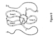
- the invention also relates to epitopes specific to the region of sclerostin identified as Loop 2, and binding agents which specifically bind to that region.
- the invention also relates to epitopes specific to the cystine-knot region of sclerostin, and binding agents such as antibodies specifically binding to that region.
- the invention relates to binding agents, such as antibodies, that specifically bind to sclerostin.
- the binding agents can be characterized by their ability to cross-block the binding of at least one antibody disclosed herein to sclerostin and/or to be cross-blocked from binding sclerostin by at least one antibody disclosed herein.
- the antibodies and other binding agents can also be characterized by their binding pattern to human sclerostin peptides in a "human sclerostin peptide epitope competition binding assay" as disclosed herein.
- the invention relates to binding agents, such as antibodies, that can increase at least one of bone formation, bone mineral density, bone mineral content, bone mass, bone quality and bone strength in a mammal.
- the invention relates to binding agents, such as antibodies, that can block the inhibitory effect of sclerostin in a cell based mineralization assay.
- the invention further relates to polypeptide constructs comprising two, three, or four polypeptide fragments linked by at least one disulfide bond, representing a core region of the cystine-knot of sclerostin, and antibodies capable of specifically binding thereto.
- the invention relates to methods of obtaining epitopes suitable for use as immunogens for generating, in mammals, binding agents, such as antibodies capable of binding specifically to sclerostin; in certain embodiments the binding agents generated are capable of neutralizing sclerostin activity in vivo.
- the invention relates to a composition for eliciting an antibody specific for sclerostin when the composition is administered to an animal, the composition comprising a polypeptide having the amino acid sequence of SEQ ID NO:6, SEQ ID NO:63, SEQ ID NO:64, SEQ ID NO:65, SEQ ID NO:66, SEQ ID NO:67, SEQ ID NO:68, or SEQ ID NO:69.
- the invention also relates to a composition for eliciting an antibody specific for sclerostin when the composition is administered to an animal, the composition comprising at least one polypeptide consisting essentially of the amino acid sequence of SEQ ID NO:2, SEQ ID NO:3, SEQ ID NO:4 or SEQ ID NO:5; the composition may comprise at least two or at least three of the amino acid sequences of SEQ ID NO:2, SEQ ID NO:3, SEQ ID NO:4 and SEQ ID NO:5, and the composition may comprise all four of the amino acid sequences of SEQ ID NO:2, SEQ ID NO:3, SEQ ID NO:4 and SEQ ID NO:5.
- the invention further relates to a composition for eliciting an antibody specific for sclerostin when the composition is administered to an animal, the composition comprising a polypeptide having the amino acid sequences of SEQ ID NO:2, SEQ ID NO:3, SEQ ID NO:4 and SEQ ID NO:5, wherein SEQ ID NO:2 and 4 are joined by a disulfide bond at amino acid positions 57 and 111 with reference to SEQ ID NO:1, and SEQ ID NO:3 and 5 are joined by at least one of (a) a disulfide bond at amino acid positions 82 and 142 with reference to SEQ ID NO:1, and (b) a disulfide bond at amino acid positions 86 and 144 with reference to SEQ ID NO:1; the polypeptide may retain the tertiary structure of the corresponding polypeptide region of human sclerostin of SEQ ID NO:1.
- polypeptide T20.6 consisting essentially of a multiply truncated human sclerostin protein of SEQ ID NO:1, wherein amino acids 1-50, 65-72, 91-100, 118-137, and 150-190 of SEQ ID NO:1 are absent from the polypeptide; this polypeptide may be obtained by tryptic digestion of human sclerostin, and the protein may be isolated by HPLC fractionation.
- the invention further relates to immunogenic portion T20.6 of human sclerostin comprising amino acids 51-64, 73-90, 101-117, and 138-149 of SEQ ID NO:1, wherein the immunogenic portion comprises at least one of:
- the invention further relates to an immunogenic portion T20.6 derivative of human sclerostin comprising amino acids 57-64, 73-86, 111-117, and 138-144 of SEQ ID NO:1, wherein the immunogenic portion comprises at least one of:
- the invention yet further relates to a polypeptide consisting essentially of a human sclerostin protein of SEQ ID NO:1 truncated at the C-terminal and N-terminal ends, wherein amino acids 1-85 and 112-190 of SEQ ID NO: 1 are absent from the polypeptide.
- the invention also relates to an immunogenic portion of human sclerostin, comprising amino acids 86-111 of SEQ ID NO:1; the immunogenic portion may consist essentially of contiguous amino acids CGPARLLPNAIGRGKWWRPSGPDFRC (SEQ ID NO:6).
- the invention further relates to an immunogenic portion of rat sclerostin, comprising amino acids 92-109 of SEQ ID NO:98; the immunogenic portion may consist essentially of contiguous amino acids PNAIGRVKWWRPNGPDFR (SEQ ID NO:96).
- the invention still further relates to an immunogenic portion of rat sclerostin, comprising amino acids 99-120 of SEQ ID NO:98; the immunogenic portion may consist essentially of contiguous amino acids KWWRPNGPDFRCIPDRYRAQRV (SEQ ID NO:97).
- the invention relates to a method of producing an immunogenic portion of human sclerostin, comprising the steps of:
- the invention relates to a method of generating an antibody capable of specifically binding to sclerostin, comprising:
- the invention also relates to a method of generating an antibody capable of specifically binding to sclerostin, the method comprising:
- the invention further relates to a method of detecting an anti-sclerostin antibody in a biological sample, comprising the steps of
- the invention also relates to a method of detecting an anti-sclerostin antibody in a biological sample, comprising the steps of
- the invention further relates to a sclerostin binding agent, such as an antibody, that cross-blocks the binding of at least one of antibodies Ab-A, Ab-B, Ab-C, or Ab-D to a sclerostin protein.
- the sclerostin binding agent may also be cross-blocked from binding to sclerostin by at least one of antibodies Ab-A, Ab-B, Ab-C, or Ab-D.
- the isolated antibody, or an antigen-binding fragment thereof may be a polyclonal antibody, a monoclonal antibody, a humanized antibody, a human antibody, a chimeric antibody or the like.
- the invention further relates to a sclerostin binding agent, such as an antibody, that is cross-blocked from binding to sclerostin by at least one of antibodies Ab-A, Ab-B, Ab-C, or Ab-D.
- a sclerostin binding agent such as an antibody
- the isolated antibody, or an antigen-binding fragment thereof may be a polyclonal antibody, a monoclonal antibody, a humanized antibody, a human antibody, a chimeric antibody or the like.
- the invention further relates to a sclerostin binding agent, such as an isolated antibody, that cross-blocks the binding of at least one of antibodies 1-24 (Ab-1 to Ab-24) to a sclerostin protein.
- the sclerostin binding agent may also be cross-blocked from binding to sclerostin by at least one of antibodies 1-24 (Ab-1 to Ab-24).
- the isolated antibody, or an antigen-binding fragment thereof may be a polyclonal antibody, a monoclonal antibody, a humanized antibody, a human antibody, or a chimeric antibody.
- the invention further relates to a sclerostin binding agent, such as an isolated antibody, that is cross-blocked from binding to sclerostin by at least one of antibodies 1-24 (Ab-1 to Ab-24); the isolated antibody, or an antigen-binding fragment thereof, may be a polyclonal antibody, a monoclonal antibody, a humanized antibody, a human antibody, or a chimeric antibody.
- a sclerostin binding agent such as an isolated antibody, that is cross-blocked from binding to sclerostin by at least one of antibodies 1-24 (Ab-1 to Ab-24); the isolated antibody, or an antigen-binding fragment thereof, may be a polyclonal antibody, a monoclonal antibody, a humanized antibody, a human antibody, or a chimeric antibody.
- the invention further relates to a binding agent, such as an isolated antibody that exhibits a similar binding pattern to human sclerostin peptides in a "human sclerostin peptide epitope competition binding assay" as that exhibited by at least one of the antibodies Ab-A, Ab-B, Ab-C or Ab-D; the isolated antibody, or an antigen-binding fragment thereof, may be a polyclonal antibody, a monoclonal antibody, a humanized antibody, a human antibody, or a chimeric antibody.
- a binding agent such as an isolated antibody that exhibits a similar binding pattern to human sclerostin peptides in a "human sclerostin peptide epitope competition binding assay" as that exhibited by at least one of the antibodies Ab-A, Ab-B, Ab-C or Ab-D; the isolated antibody, or an antigen-binding fragment thereof, may be a polyclonal antibody, a monoclonal antibody, a humanized antibody,
- the invention still further relates to a method for treating a bone disorder associated with at least one of low bone formation, low bone mineral density, low bone mineral content, low bone mass, low bone quality and low bone strength in a mammalian subject which comprises providing to a subject in need of such treatment an amount of an anti-sclerostin binding agent sufficient to increase at least one of bone formation, bone mineral density, bone mineral content, bone mass, bone quality and bone strength wherein the anti-sclerostin binding agent comprises an antibody, or sclerostin-binding fragment thereof.
- the invention also relates to an isolated sclerostin polypeptide or fragments thereof, wherein the polypeptide contains 6 conserved cysteine residues and the fragments thereof comprise from 7 to 14 amino acids of SEQ ID NO:2; 8 to 17 amino acids of SEQ ID NO:3; 8 to 18 residues of SEQ ID NO:4; and 6 to 12 residues of SEQ ID NO:5, and the polypeptide or fragments thereof are stabilized by disulfide bonds between SEQ ID NO:2 and 4, and between SEQ ID NO:3 and 5; the polypeptide or fragments may comprise 10-14 amino acids of SEQ ID NO:2; 14 to 17 amino acids of SEQ ID NO:3; 13 to 18 amino acids of SEQ ID NO:4;, and 8 to 12 residues of SEQ ID NO:5; and the polypeptide or fragments may comprise SEQ ID NO:2, SEQ ID NO:3, SEQ ID NO:4, and SEQ ID NO:5.
- antibodies that specifically bind to human sclerostin.
- the antibodies are characterized by their ability to cross-block the binding of at least one antibody disclosed herein to human sclerostin and/or to be cross-blocked from binding human sclerostin by at least one antibody disclosed herein.
- an isolated antibody or an antigen-binding fragment thereof, that can increase at least one of bone formation, bone mineral density, bone mineral content, bone mass, bone quality and bone strength in a mammal.
- an isolated antibody or an antigen-binding fragment thereof, that can block the inhibitory effect of sclerostin in a cell based mineralization assay.
- a binding agent such as an antibody, that specifically binds to human sclerostin and has at least one CDR sequence selected from SEQ ID NOs: 39, 40, 41, 42, 43, 44, 45, 46, 47, 48, 49, 50, 51, 52, 53, 54, 55, 56, 57, 58, 59, 60, 61, 62, 78, 79, 80, 81, 99, 100, 101, 102, 103, 104, 105, 106, 107, 108, 109, 110, 111, 112, 113, 114, 115, 116, 237, 238, 239, 240, 241, 242, 243, 244, 245, 246, 247, 248, 249, 250, 251, 252, 253, 254, 255, 256, 257, 258, 259, 260, 261, 262, 263, 264, 265, 266, 267, 268, 269, 270, 271, 272, 273, 274, 275, 276, 277, 27
- a binding agent such as an antibody, that specifically binds to human sclerostin and has at least one CDR sequence selected from SEQ ID NOs:39, 40, 41, 42, 43, 44, 45, 46, 47, 48, 49, 50, 51, 52, 53, 54, 55, 56, 57, 58, 59, 60, 61, 62, 78, 79, 80, 81, 99, 100, 101, 102, 103, 104, 105, 106, 107, 108, 109, 110, 111, 112, 113, 114, 115, 116, 237, 238, 239, 240, 241, 242, 243, 244, 245, 246, 247, 248, 249, 250, 251, 252, 253, 254, 255, 256, 257, 258, 259, 260, 261, 262, 263, 264, 265, 266, 267, 268, 269, 270, 271, 272; 273, 274, 275, 276, 277,
- regions of human sclerostin which are important for the in vivo activity of the protein.
- the present invention relates to regions of the human sclerostin protein that contain epitopes recognized by antibodies that also bind to full-length sclerostin, and methods of making and using these epitopes.
- the invention also provides binding agents (such as antibodies) that specifically bind to sclerostin or portions of sclerostin, and methods for using such binding agents.
- the binding agents are useful to block or impair binding of human sclerostin to one or more ligand.
- Recombinant human sclerostin/SOST is commercially available from R&D Systems (Minneapolis, MN, USA; 2006 cat# 1406-ST-025). Additionally, recombinant mouse sclerostin/SOST is commercially available from R&D Systems (Minneapolis, MN, USA; 2006 cat# 1589-ST-025). Research grade sclerostin binding monoclonal antibodies are commercially available from R&D Systems (Minneapolis, MN, USA; mouse monoclonal: 2006 cat# MAB 1406; rat monoclonal: 2006 cat# MAB 1589).
- U.S. Patent Nos. 6,395,511 and 6,803,453 and U.S. Patent Publications 20040009535 and 20050106683 refer to anti-sclerostin antibodies generally.
- human sclerostin is intended to include the protein of SEQ ID NO: 1 and allelic variants thereof.
- Sclerostin can be purified from 293T host cells that have been transfected by a gene encoding sclerostin by elution of filtered supernatant of host cell culture fluid using a Heparin HP column, using a salt gradient. The preparation and further purification using cation exchange chromatography are described in Examples 1 and 2.
- Binding agents of the invention are preferably antibodies, as defined herein.
- the term "antibody” refers to an intact antibody, or a binding fragment thereof.
- An antibody may comprise a complete antibody molecule (including polyclonal, monoclonal, chimeric, humanized, or human versions having full length heavy and/or light chains), or comprise an antigen binding fragment thereof.
- Antibody fragments include F(ab') 2 , Fab, Fab', Fv, Fc, and Fd fragments, and can be incorporated into single domain antibodies, single-chain antibodies, maxibodies, minibodies, intrabodies, diabodies, triabodies, tetrabodies, v-NAR and bis-scFv (See e.g .,, Hollinger and Hudson, 2005, Nature Biotechnology, 23, 9, 1126-1136 ).
- Antibody polypeptides are also disclosed in U. S. Patent No. 6,703,199 , including fibronectin polypeptide monobodies. Other antibody polypeptides are disclosed in U.S. Patent Publication 2005/0238646 , which are single-chain polypeptides.
- Antigen binding fragments derived from an antibody can be obtained, for example, by proteolytic hydrolysis of the antibody, for example, pepsin or papain digestion of whole antibodies according to conventional methods.
- antibody fragments can be produced by enzymatic cleavage of antibodies with pepsin to provide a 5S fragment termed F(ab') 2 .
- This fragment can be further cleaved using a thiol reducing agent to produce 3.5S Fab' monovalent fragments.
- the cleavage reaction can be performed using a blocking group for the sulfhydryl groups that result from cleavage of disulfide linkages.
- an enzymatic cleavage using papain produces two monovalent Fab fragments and an Fc fragment directly.
- These methods are described, for example, by Goldenberg, U.S. Patent No. 4,331,647 , Nisonoff et al., Arch. Biochem. Biophys. 89:230, 1960 ; Porter, Biochem. J. 73:119, 1959 ; Edelman et al., in Methods in Enzymology 1:422 (Academic Press 1967 ); and by Andrews, S.M. and Titus, J.A. in Current Protocols in Immunology (Coligan J.E., et al., eds), John Wiley & Sons, New York (2003).
- antibody fragment may also be any synthetic or genetically engineered protein.
- antibody fragments include isolated fragments consisting of the light chain variable region, "Fv" fragments consisting of the variable regions of the heavy and light chains, recombinant single chain polypeptide molecules in which light and heavy variable regions are connected by a peptide linker (scFv proteins).
- CDRs complementarity determining regions
- Such polynucleotides are prepared, for example, by using the polymerase chain reaction to synthesize the variable region using mRNA of antibody-producing cells as a template (see, for example, Larrick et al., Methods: A Companion to Methods in Enzymology 2:106, 1991 ; Courtenay-Luck, "Genetic Manipulation of Monoclonal Antibodies,” in Monoclonal Antibodies: Production, Engineering and Clinical Application, Ritter et al.
- the binding agent comprises at least one CDR as described herein.
- the binding agent may comprise at least two, three, four, five or six CDR's as described herein.
- the binding agent further may comprise at least one variable region domain of an antibody described herein.
- the variable region domain may be of any size or amino acid composition and will generally comprise at least one CDR sequence responsible for binding to human sclerostin, for example CDR-H1, CDR-H2, CDR-H3 and/or the light chain CDRs specifically described herein and which is adjacent to or in frame with one or more framework sequences.
- the variable (V) region domain may be any suitable arrangement of immunoglobulin heavy (V H ) and/or light (V L ) chain variable domains.
- the V region domain may be monomeric and be a V H or V L domain, which is capable of independently binding human sclerostin with an affinity at least equal to 1 x 10 -7 M or less as describedbelow.
- the V region domain may be dimeric and contain V H -V H , V H -V L , or V L -V L , dimers.
- the V region dimer comprises at least one V H and at least one V L chain that may be non-covalently associated (hereinafter referred to as F V ).
- the chains may be covalently coupled either directly, for example via a disulfide bond between the two variable domains, or through a linker, for example a peptide linker, to form a single chain Fv (scF V ).
- variable region domain may be any naturally occurring variable domain or an engineered version thereof.
- engineered version is meant a variable region domain that has been created using recombinant DNA engineering techniques.
- engineered versions include those created, for example, from a specific antibody variable region by insertions, deletions, or changes in or to the amino acid sequences of the specific antibody.
- Particular examples include engineered variable region domains containing at least one CDR and optionally one or more framework amino acids from a first antibody and the remainder of the variable region domain from a second antibody.
- variable region domain may be covalently attached at a C-terminal amino acid to at least one other antibody domain or a fragment thereof.
- a VH domain that is present in the variable region domain may be linked to an immunoglobulin CH1 domain, or a fragment thereof.
- a V L domain may be linked to a C K domain or a fragment thereof.
- the antibody may be a Fab fragment wherein the antigen binding domain contains associated V H and V L domains covalently linked at their C-termini to a CH1 and C K domain, respectively.
- the CH1 domain may be extended with further amino acids, for example to provide a hinge region or a portion of a hinge region domain as found in a Fab' fragment, or to provide further domains, such as antibody CH2 and CH3 domains.
- binding agents comprise at least one of these CDRs.
- one or more CDR may be incorporated into known antibody framework regions (IgG1, IgG2, etc.), or conjugated to a suitable vehicle to enhance the half-life thereof.
- suitable vehicles include, but are not limited to Fc, polyethylene glycol (PEG), albumin, transferrin, and the like. These and other suitable vehicles are known in the art.
- conjugated CDR peptides may be in monomeric, dimeric, tetrameric, or other form.
- one or more water-soluble polymer is bonded at one or more specific position, for example at the amino terminus, of a binding agent.
- a binding agent comprises one or more water soluble polymer attachments, including, but not limited to, polyethylene glycol, polyoxyethylene glycol, or polypropylene glycol. See, e.g., U.S. Pat. Nos. 4,640,835 , 4,496,689 , 4,301,144 , 4,670,417 , 4,791,192 and 4,179,337 .
- a derivative binding agent comprises one or more of monomethoxy-polyethylene glycol, dextran, cellulose, or other carbohydrate based polymers, poly-(N-vinyl pyrrolidone)polyethylene glycol, propylene glycol homopolymers, a polypropylene oxide/ethylene oxide co-polymer, polyoxyethylated polyols ( e.g ., glycerol) and polyvinyl alcohol, as well as mixtures of such polymers.
- one or more water-soluble polymer is randomly attached to one or more side chains.
- PEG can act to improve the therapeutic capacity for a binding agent, such as an antibody. Certain such methods are discussed, for example, in U.S. Pat. No. 6,133,426 , which is hereby incorporated by reference for any purpose.
- a binding agent of the present invention may have at least one amino acid substitution, providing that the binding agent retains binding specificity. Therefore, modifications to the binding agent structures are encompassed within the scope of the invention. These may include amino acid substitutions, which may be conservative or non-conservative, that do not destroy the sclerostin binding capability of a binding agent. Conservative amino acid substitutions may encompass non-naturally occurring amino acid residues, which are typically incorporated by chemical peptide synthesis rather than by synthesis in biological systems. These include peptidomimetics and other reversed or inverted forms of amino acid moieties. A conservative amino acid substitution may also involve a substitution of a native amino acid residue with a normative residue such that there is little or no effect on the polarity or charge of the amino acid residue at that position.
- Non-conservative substitutions may involve the exchange of a member of one class of amino acids or amino acid mimetics for a member from another class with different physical properties (e.g . size, polarity, hydrophobicity, charge). Such substituted residues may be introduced into regions of the human antibody that are homologous with non-human antibodies, or into the non-homologous regions of the molecule.
- test variants containing a single amino acid substitution at each desired amino acid residue.
- the variants can then be screened using activity assays known to those skilled in the art.
- Such variants could be used to gather information about suitable variants. For example, if one discovered that a change to a particular amino acid residue resulted in destroyed, undesirably reduced, or unsuitable activity, variants with such a change may be avoided. In other words, based on information gathered from such routine experiments, one skilled in the art can readily determine the amino acids where further substitutions should be avoided either alone or in combination with other mutations.
- polypeptide structure A skilled artisan will be able to determine suitable variants of the polypeptide as set forth herein using well-known techniques.
- one skilled in the art may identify suitable areas of the molecule that may be changed without destroying activity by targeting regions not believed to be important for activity.
- even areas that may be important for biological activity or for structure may be subject to conservative amino acid substitutions without destroying the biological activity or without adversely affecting the polypeptide structure.
- One skilled in the art can also analyze the three-dimensional structure and amino acid sequence in relation to that structure in similar polypeptides. In view of such information, one skilled in the art may predict the alignment of amino acid residues of an antibody with respect to its three dimensional structure. In certain embodiments, one skilled in the art may choose not to make radical changes to amino acid residues predicted to be on the surface of the protein, since such residues may be involved in important interactions with other molecules.
- One method of predicting secondary structure is based upon homology modeling. For example, two polypeptides or proteins which have a sequence identity of greater than 30%, or similarity greater than 40% often have similar structural topologies.
- the recent growth of the protein structural database (PDB) has provided enhanced predictability of secondary structure, including the potential number of folds within a polypeptide's or protein's structure. See Holm et al., Nucl. Acid. Res., 27(1):244-247 (1999 ). It has been suggested ( Brenner et al., Curr. Op. Struct. Biol., 7(3):369-376 (1997 )) that there are a limited number of folds in a given polypeptide or protein and that once a critical number of structures have been resolved, structural prediction will become dramatically more accurate.
- Additional methods of predicting secondary structure include “threading” ( Jones, D., Curr. Opin. Struct. Biol., 7(3):377-87 (1997 ); Sippl et al., Structure, 4(1):15-19 (1996 )), “profile analysis” ( Bowie et al., Science, 253:164-170 (1991 ); Gribskov et al., Meth. Enzym., 183:146-159 (1990 ); Gribskov et al., Proc. Nat. Acad. Sci., 84(13):4355-4358 (1987 )), and “evolutionary linkage” (See Holm, supra (1999), and Brenner, supra (1997)).
- variants of binding agents include glycosylation variants wherein the number and/or type of glycosylation site has been altered compared to the amino acid sequences of a parent polypeptide.
- variants comprise a greater or a lesser number of N-linked glycosylation sites than the native protein.
- An N-linked glycosylation site is characterized by the sequence: Asn-X-Ser or Asn-X-Thr, wherein the amino acid residue designated as X may be any amino acid residue except proline.
- the substitution of amino acid residues to create this sequence provides a potential new site for the addition of an N-linked carbohydrate chain. Alternatively, substitutions which eliminate this sequence will remove an existing N-linked carbohydrate chain.
- N-linked carbohydrate chains wherein one or more N-linked glycosylation sites (typically those that are naturally occurring) are eliminated and one or more new N-linked sites are created.
- Additional preferred antibody variants include cysteine variants wherein one or more cysteine residues are deleted from or substituted for another amino acid (e.g ., serine) as compared to the parent amino acid sequence. Cysteine variants may be useful when antibodies must be refolded into a biologically active conformation such as after the isolation of insoluble inclusion bodies. Cysteine variants generally have fewer cysteine residues than the native protein, and typically have an even number to minimize interactions resulting from unpaired cysteines.
- amino acid substitutions can be used to identify important residues of antibodies to sclerostin, or to increase or decrease the affinity of the antibodies to sclerostin described herein.
- preferred amino acid substitutions are those which: (1) reduce susceptibility to proteolysis, (2) reduce susceptibility to oxidation, (3) alter binding affinity for forming protein complexes, (4) alter binding affinities, and/or (4) confer or modify other physiochemical or functional properties on such polypeptides
- single or multiple amino acid substitutions may be made in the naturally-occurring sequence (in certain embodiments, in the portion of the polypeptide outside the domain(s) forming intermolecular contacts).
- a conservative amino acid substitution typically may not substantially change the structural characteristics of the parent sequence (e.g ., a replacement amino acid should not tend to break a helix that occurs in the parent sequence, or disrupt other types of secondary structure that characterizes the parent sequence).
- a replacement amino acid should not tend to break a helix that occurs in the parent sequence, or disrupt other types of secondary structure that characterizes the parent sequence.
- Examples of art-recognized polypeptide secondary and tertiary structures are described in Proteins, Structures and Molecular Principles (Creighton, Ed., W. H. Freeman and Company, New York (1984 )); Introduction to Protein Structure (C. Branden and J. Tooze, eds., Garland Publishing, New York, N.Y. (1991 )); and Thornton et al. Nature 354:105 (1991 ), which are each incorporated herein by reference.
- binding agents of the invention may be chemically bonded with polymers, lipids, or other moieties.
- the binding agents may comprise at least one of the CDRs described herein incorporated into a biocompatible framework structure.
- the biocompatible framework structure comprises a polypeptide or portion thereof that is sufficient to form a conformationally stable structural support, or framework, or scaffold, which is able to display one or more sequences of amino acids that bind to an antigen (e.g ., CDRs, a variable region, etc.) in a localized surface region.
- an antigen e.g ., CDRs, a variable region, etc.
- Such structures can be a naturally occurring polypeptide or polypeptide "fold" (a structural motif), or can have one or more modifications, such as additions, deletions or substitutions of amino acids, relative to a naturally occurring polypeptide or fold.
- These scaffolds can be derived from a polypeptide of any species (or of more than one species), such as a human, other mammal, other vertebrate, invertebrate, plant, bacteria or virus.
- the biocompatible framework structures are based on protein scaffolds or skeletons other than immunoglobulin domains.
- those based on fibronectin, ankyrin, lipocalin, neocarzinostain, cytochrome b, CP1 zinc finger, PST1, coiled coil, LACI-D1, Z domain and tendramisat domains may be used (See e.g ., , Nygren and Uhlen, 1997, Current Opinion in Structural Biology, 7, 463-469 ).
- the binding agents of the invention include the humanized antibodies described herein.
- Humanized antibodies such as those described herein can be produced using techniques known to those skilled in the art ( Zhang, W., et al., Molecular Immunology. 42(12):1445-1451, 2005 ; Hwang W. et al., Methods. 36(1):35-42, 2005 ; Dall'Acqua WF, et al., Methods 36(1):43-60, 2005 ; and Clark, M., Immunology Today. 21(8):397-402, 2000 ).
- suitable binding agents include portions of these antibodies, such as one or more of CDR-H1, CDR-H2, CDR-H3, CDR-L1, CDR-L2 and CDR-L3 as specifically disclosed herein. At least one of the regions of CDR-H1, CDR-H2, CDR-H3, CDR-L1, CDR-L2 and CDR-L3 may have at least one amino acid substitution, provided that the binding agent retains the binding specificity of the non-substituted CDR.
- the non-CDR portion of the binding agent may be a non-protein molecule, wherein the binding agent cross-blocks the binding of an antibody disclosed herein to sclerostin and/or neutralizes sclerostin.
- the non-CDR portion of the binding agent may be a non-protein molecule in which the binding agent exhibits a similar binding pattern to human sclerostin peptides in a "human sclerostin peptide epitope competition binding assay" as that exhibited by at least one of antibodies Ab-A, Ab-B, Ab-C, Ab-D, Ab-1, Ab-2, Ab-3, Ab-4, Ab-5, Ab-6, Ab-7, Ab-8, Ab-9, Ab-10, Ab-11, Ab-12, Ab-13, Ab-14, Ab-15, Ab-16, Ab-17, Ab-18, Ab-19, Ab-20, Ab-21, Ab-22, Ab-23, and Ab-24, and/or neutralizes sclerostin.
- the non-CDR portion of the binding agent may be composed of amino acids, wherein the binding agent is a recombinant binding protein or a synthetic peptide, and the recombinant binding protein cross-blocks the binding of an antibody disclosed herein to sclerostin and/or neutralizes sclerostin.
- the non-CDR portion of the binding agent may be composed of amino acids, wherein the binding agent is a recombinant binding protein, and the recombinant binding protein exhibits a similar binding pattern to human sclerostin peptides in the human sclerostin peptide epitope competition binding assay (described hereinbelow) as that exhibited by at least one of the antibodies Ab-A, Ab-B, Ab-C, Ab-D, Ab-1, Ab-2, Ab-3, Ab-4, Ab-5, Ab-6, Ab-7, Ab-8, Ab-9, Ab-10, Ab-11, Ab-12, Ab-13, Ab-14, Ab-15, Ab-16, Ab-17, Ab-18, Ab-19, Ab-20, Ab-21, Ab-22, Ab-23, and Ab-24, and/or neutralizes sclerostin.
- the binding agent is a recombinant binding protein
- the recombinant binding protein exhibits a similar binding pattern to human sclerostin peptides in the human sclerost
- an antibody comprises one or more of CDR H1, CDR-H2, CDR-H3, CDR-L1, CDR-L2 and CDR-L3 as described above, it may be obtained by expression from a host cell containing DNA coding for these sequences.
- a DNA coding for each CDR sequence may be determined on the basis of the amino acid sequence of the CDR and synthesized together with any desired antibody variable region framework and constant region DNA sequences using oligonucleotide synthesis techniques, site-directed mutagenesis and polymerase chain reaction (PCR) techniques as appropriate.
- DNA coding for variable region frameworks and constant regions is widely available to those skilled in the art from genetic sequences databases such as GenBank®.
- Each of the above-mentioned CDRs will be typically located in a variable region framework at positions 31-35 (CDR-H1), 50-65 (CDR-H2) and 95-102 (CDR-H3) of the heavy chain and positions 24-34 (CDR-L1), 50-56 (CDR-L2) and 89-97 (CDR-L3) of the light chain according to the Kabat numbering system ( Kabat et al., 1987 in Sequences of Proteins of Immunological Interest, U.S. Department of Health and Human Services, NIH, USA ).
- the DNA encoding an antibody of the invention or fragment thereof may be propagated and expressed according to any of a variety of well-known procedures for nucleic acid excision, ligation, transformation, and transfection using any number of known expression vectors.
- expression of an antibody fragment may be preferred in a prokaryotic host, such as Escherichia coli ( see, e.g., Pluckthun et al., 1989 Methods Enzymol. 178:497-515 ).
- expression of the antibody or a fragment thereof may be preferred in a eukaryotic host cell, including yeast (e.g ., Saccharomyces cerevisiae, Schizosaccharomyces pombe, and Pichia pastoris ), animal cells (including mammalian cells) or plant cells.
- yeast e.g ., Saccharomyces cerevisiae, Schizosaccharomyces pombe, and Pichia pastoris
- animal cells including mammalian cells
- suitable animal cells include, but are not limited to, myeloma (such as a mouse NSO line), COS, CHO, or hybridoma cells.
- plant cells include tobacco, corn, soybean, and rice cells.
- One or more replicable expression vectors containing DNA encoding an antibody variable and/or constant region may be prepared and used to transform an appropriate cell line, for example, a non-producing myeloma cell line, such as a mouse NSO line or a bacteria, such as E. coli, in which production of the antibody will occur.
- an appropriate cell line for example, a non-producing myeloma cell line, such as a mouse NSO line or a bacteria, such as E. coli, in which production of the antibody will occur.
- the DNA sequence in each vector should include appropriate regulatory sequences, particularly a promoter and leader sequence operatively linked to the variable domain sequence.
- Particular methods for producing antibodies in this way are generally well-known and routinely used. For example, basic molecular biology procedures are described by Maniatis et al.
- DNA sequencing can be performed as described in Sanger et al. (PNAS 74:5463, (1977 )) and the Amersham International pic sequencing handbook, and site directed mutagenesis can be carried out according to methods known in the art ( Kramer et al., Nucleic Acids Res. 12:9441, (1984 ); Kunkel Proc. Natl. Acad Sci. USA 82:488-92 (1985 ); Kunkel et al., Methods in Enzymol.
- affinity maturation protocols including maintaining the CDRs ( Yang et al., J. Mol. Biol., 254, 392-403, 1995 ), chain shuffling ( Marks et al., Bio/Technology, 10, 779-783, 1992 ), use of mutation strains of E. coli. ( Low et al., J. Mol. Biol., 250, 350-368, 1996 ), DNA shuffling ( Patten et al., Curr. Opin. Biotechnol., 8, 724-733, 1997 ), phage display ( Thompson et al., J. Mol.
- antibodies according to the invention may be obtained by conventional immunization and cell fusion procedures as described herein and known in the art.
- Monoclonal antibodies of the invention may be generated using a variety of known techniques.
- monoclonal antibodies that bind to specific antigens may be obtained by methods known to those skilled in the art (see, for example, Kohler et al., Nature 256:495, 1975 ; Coligan et al. (eds.), Current Protocols in Immunology, 1:2.5.12.6.7 (John Wiley & Sons 1991 ); U.S. Patent Nos.
- Antibody fragments may be derived therefrom using any suitable standard technique such as proteolytic digestion, or optionally, by proteolytic digestion (for example, using papain or pepsin) followed by mild reduction of disulfide bonds and alkylation. Alternatively, such fragments may also be generated by recombinant genetic engineering techniques as described herein.
- Monoclonal antibodies can be obtained by injecting an animal, for example, a rat, hamster, a rabbit, or preferably a mouse, including for example a transgenic or a knock-out, as known in the art, with an immunogen comprising human sclerostin of SEQ ID NO:1, or a fragment thereof, according to methods known in the art and described herein.
- the presence of specific antibody production may be monitored after the initial injection and/or after a booster injection by obtaining a serum sample and detecting the presence of an antibody that binds to human sclerostin or peptide using any one of several immunodetection methods known in the art and described herein.
- lymphoid cells most commonly cells from the spleen or lymph node, are removed to obtain B-lymphocytes.
- the B lymphocytes are then fused with a drug-sensitized myeloma cell fusion partner, preferably one that is syngeneic with the immunized animal and that optionally has other desirable properties (e.g ., inability to express endogenous Ig gene products, e.g ., P3X63 - Ag 8.653 (ATCC No. CRL 1580); NSO, SP20) to produce hybridomas, which are immortal eukaryotic cell lines.
- a drug-sensitized myeloma cell fusion partner preferably one that is syngeneic with the immunized animal and that optionally has other desirable properties (e.g ., inability to express endogenous Ig gene products, e.g ., P3X63 - Ag 8.653 (ATCC No. CRL 1580); NSO, SP20) to produce
- the lymphoid (e.g ., spleen) cells and the myeloma cells may be combined for a few minutes with a membrane fusion-promoting agent, such as polyethylene glycol or a nonionic detergent, and then plated at low density on a selective medium that supports the growth of hybridoma cells but not unfused myeloma cells.
- a preferred selection media is HAT (hypoxanthine, aminopterin, thymidine). After a sufficient time, usually about one to two weeks, colonies of cells are observed. Single colonies are isolated, and antibodies produced by the cells may be tested for binding activity to human sclerostin, using any one of a variety of immunoassays known in the art and described herein.
- the hybridomas are cloned ( e.g ., by limited dilution cloning or by soft agar plaque isolation) and positive clones that produce an antibody specific to sclerostin are selected and cultured.
- the monoclonal antibodies from the hybridoma cultures may be isolated from the supernatants of hybridoma cultures.
- An alternative method for production of a murine monoclonal antibody is to inject the hybridoma cells into the peritoneal cavity of a syngeneic mouse, for example, a mouse that has been treated ( e.g ., pristane-primed) to promote formation of ascites fluid containing the monoclonal antibody.
- Monoclonal antibodies can be isolated and purified by a variety of well-established techniques. Such isolation techniques include affinity chromatography with Protein-A Sepharose, size-exclusion chromatography, and ion-exchange chromatography (see, for example, Coligan at pages 2.7.1-2.7.12 and pages 2.9.1-2.9.3; Baines et al., "Purification of Immunoglobulin G (IgG),” in Methods in Molecular Biology, Vol. 10, pages 79-104 (The Humana Press, Inc. 1992 )). Monoclonal antibodies may be purified by affinity chromatography using an appropriate ligand selected based on particular properties of the antibody ( e.g ., heavy or light chain isotype, binding specificity, etc.).
- a suitable ligand immobilized on a solid support, include Protein A, Protein G, an anticonstant region (light chain or heavy chain) antibody, an anti-idiotype antibody, and a TGF-beta binding protein, or fragment or variant thereof.
- An antibody of the present invention may also be a human monoclonal antibody.
- Human monoclonal antibodies may be generated by any number of techniques with which those having ordinary skill in the art will be familiar. Such methods include, but are not limited to, Epstein Barr Virus (EBV) transformation of human peripheral blood cells (e.g ., containing B lymphocytes), in vitro immunization of human B cells, fusion of spleen cells from immunized transgenic mice carrying inserted human immunoglobulin genes, isolation from human immunoglobulin V region phage libraries, or other procedures as known in the art and based on the disclosure herein.
- EBV Epstein Barr Virus
- human monoclonal antibodies may be obtained from transgenic mice that have been engineered to produce specific human antibodies in response to antigenic challenge.
- human immunoglobulin transgenes may be mini-gene constructs, or transloci on yeast artificial chromosomes, which undergo B cell-specific DNA rearrangement and hypermutation in the mouse lymphoid tissue.
- Human monoclonal antibodies may be obtained by immunizing the transgenic mice, which may then produce human antibodies specific for sclerostin. Lymphoid cells of the immunized transgenic mice can be used to produce human antibody-secreting hybridomas according to the methods described herein. Polyclonal sera containing human antibodies may also be obtained from the blood of the immunized animals.
- Another method for generating human antibodies of the invention includes immortalizing human peripheral blood cells by EBV transformation. See, e.g., U.S. Patent No. 4,464,456 .
- Such an immortalized B cell line (or lymphoblastoid cell line) producing a monoclonal antibody that specifically binds to sclerostin can be identified by immunodetection methods as provided herein, for example, an ELISA, and then isolated by standard cloning techniques.
- the stability of the lymphoblastoid cell line producing an anti-sclerostin antibody may be improved by fusing the transformed cell line with a murine myeloma to produce a mouse-human hybrid cell line according to methods known in the art (see, e.g., Glasky et al., Hybridoma 8:377-89 (1989 )).
- Still another method to generate human monoclonal antibodies is in vitro immunization, which includes priming human splenic B cells with human sclerostin, followed by fusion of primed B cells with a heterohybrid fusion partner. See, e.g., Boerner et al., 1991 J. Immunol. 147:86-95 .
- a B cell that is producing an anti-human sclerostin antibody is selected and the light chain and heavy chain variable regions are cloned from the B cell according to molecular biology techniques known in the art ( WO 92/02551 ; US patent 5,627,052 ; Babcook et al., Proc. Natl. Acad. Sci. USA 93:7843-48 (1996 )) and described herein.
- B cells from an immunized animal may be isolated from the spleen, lymph node, or peripheral blood sample by selecting a cell that is producing an antibody that specifically binds to sclerostin.
- B cells may also be isolated from humans, for example, from a peripheral blood samples.
- Methods for detecting single B cells that are producing an antibody with the desired specificity are well known in the art, for example, by plaque formation, fluorescence-activated cell sorting, in vitro stimulation followed by detection of specific antibody, and the like.
- Methods for selection of specific antibody-producing B cells include, for example, preparing a single cell suspension of B cells in soft agar that contains human sclerostin. Binding of the specific antibody produced by the B cell to the antigen results in the formation of a complex, which may be visible as an immunoprecipitate.
- the specific antibody genes may be cloned by isolating and amplifying DNA or mRNA according to methods known in the art and described herein.
- An additional method for obtaining antibodies of the invention is by phage display. See, e.g., Winter et al., 1994 Annu. Rev. Immunol. 12:433-55 ; Burton et al., 1994 Adv. Immunol. 57:191-280 .
- Human or murine immunoglobulin variable region gene combinatorial libraries may be created in phage vectors that can be screened to select Ig fragments (Fab, Fv, sFv, or multimers thereof) that bind specifically to TGF-beta binding protein or variant or fragment thereof. See, e.g., U.S. Patent No.
- a library containing a plurality of polynucleotide sequences encoding Ig variable region fragments may be inserted into the genome of a filamentous bacteriophage, such as M13 or a variant thereof, in frame with the sequence encoding a phage coat protein.
- a fusion protein may be a fusion of the coat protein with the light chain variable region domain and/or with the heavy chain variable region domain.
- immunoglobulin Fab fragments may also be displayed on a phage particle ( see, e.g., U.S. Patent No. 5,698,426 ).
- Heavy and light chain immunoglobulin cDNA expression libraries may also be prepared in lambda phage, for example, using ⁇ lmmunoZap TM (H) and ⁇ ImmunoZap TM (L) vectors (Stratagene, La Jolla, California). Briefly, mRNA is isolated from a B cell population, and used to create heavy and light chain immunoglobulin cDNA expression libraries in the ⁇ ImmunoZap(H) and ⁇ ImmunoZap(L) vectors. These vectors may be screened individually or co-expressed to form Fab fragments or antibodies (see Huse et al., supra ; see also Sastry et al., supra ). Positive plaques may subsequently be converted to a non-lytic plasmid that allows high level expression of monoclonal antibody fragments from E. coli.
- variable regions of a gene expressing a monoclonal antibody of interest are amplified using nucleotide primers.
- primers may be synthesized by one of ordinary skill in the art, or may be purchased from commercially available sources. ( See, e.g., Stratagene (La Jolla, California), which sells primers for mouse and human variable regions including, among others, primers for V Ha , V Hb , V Hc , V Hd , C H1 , V L and C L regions.)
- These primers may be used to amplify heavy or light chain variable regions, which may then be inserted into vectors such as ImmunoZAP TM H or ImmunoZAP TM L (Stratagene), respectively.
- vectors may then be introduced into E. coli, yeast, or mammalian-based systems for expression. Large amounts of a single-chain protein containing a fusion of the V H and V L domains may be produced using these methods (see Bird et al., Science 242:423-426, 1988 ).
- the specific antibody genes may be cloned by isolating and amplifying DNA or mRNA therefrom according to standard procedures as described herein.
- the antibodies produced therefrom may be sequenced and the CDRs identified and the DNA coding for the CDRs may be manipulated as described previously to generate other antibodies according to the invention.
- the binding agents specifically bind to sclerostin.
- the term "specifically binds” refers to the ability of a binding agent to bind to sclerostin, preferably human sclerostin, with greater affinity than it binds to an unrelated control protein.
- the control protein is hen egg white lysozyme.
- the binding agents bind to sclerostin with an affinity that is at least, 50, 100, 250, 500, 1000, or 10,000 times greater than the affinity for a control protein.
- a binding agent may have a binding affinity for human sclerostin of less than or equal to 1 x 10 -7 M, less than or equal to 1 x 10 -8 M, less than or equal to 1 x 10 -9 M, less than or equal to 1 x 10 -10 M, less than or equal to 1 x 10 -11 M, or less than or equal to 1 x 10 -12 M.
- affinity may be determined by an affinity ELISA assay.
- affinity may be determined by a BIAcore assay.
- affinity may be determined by a kinetic method.
- affinity may be determined by an equilibrium/solution method. Such methods are described in further detail herein or known in the art.
- Sclerostin binding agents of the present invention preferably modulate sclerostin function in the cell-based assay described herein and/or the in vivo assay described herein and/or bind to one or more of the epitopes described herein and/or cross-block the binding of one of the antibodies described in this application and/or are cross-blocked from binding sclerostin by one of the antibodies described in this application. Accordingly such binding agents can be identified using the assays described herein.
- binding agents are generated by first identifying antibodies that bind to one more of the epitopes provided herein and/or neutralize in the cell-based and/or in vivo assays described herein and/or cross-block the antibodies described in this application and/or are cross-blocked from binding sclerostin by one of the antibodies described in this application.
- the CDR regions from these antibodies are then used to insert into appropriate biocompatible frameworks to generate sclerostin binding agents.
- the non-CDR portion of the binding agent may be composed of amino acids, or may be a non-protein molecule.
- the assays described herein allow the characterization of binding agents.
- the binding agents of the present invention are antibodies as defined herein.
- proteins may undergo a variety of posttranslational modifications.
- the type and extent of these modifications often depends on the host cell line used to express the protein as well as the culture conditions.
- modifications may include variations in glycosylation, methionine oxidation, diketopiperizine formation, aspartate isomerization and asparagine deamidation.
- a frequent modification is the loss of a carboxy-terminal basic residue (such as lysine or arginine) due to the action of carboxypeptidases (as described in Harris, RJ. Journal of Chromatography 705:129-134, 1995 ).
- Antibodies referred to as Ab-A, Ab-B, Ab-C, Ab-D and Ab-1 are described below.
- HC refers to the heavy chain
- LC refers to the light chain.
- CDRs are box shaded and the constant (C) regions are shown in bold italics.
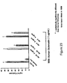
- Antibody D (also referred to herein as Ab-D and Mab-D) is a mouse antibody which exhibits high affinity binding to sclerostin.
- the BIAcore binding pattern of Ab-D is shown in Figure 18 .
- Nucleic acid sequence encoding the mature form (signal peptide removed) of Ab-D LC is as follows:
- the amino acid sequence of Ab-D LC including signal peptide is as follows:
- amino acid sequence of the mature form (signal peptide removed) of Ab-D HC heavy chain is as follows:
- the nucleic acid sequence encoding the mature form (signal peptide removed) of Ab-D HC is:
- the amino acid sequence of Ab-D HC including signal peptide is:
- the nucleic acid sequence of Ab-D HC including signal peptide encoding sequence is:
- CDR-H1 DHYMS (SEQ ID NO:39)
- CDR-H2 DINPYSGETTYNQKFKG (SEQ ID NO:40)
- CDR-H3 DDYDASPFAY (SEQ ID NO:41)
- the light chain variable region CDR sequences of Ab-D are: CDR-L1: QASQGTSINLN (SEQ ID NO:42) CDR-L2: GSSNLED (SEQ ID NO:43) CDR-L3: LQHSYLPYT (SEQ ID NO:44)
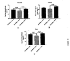
- Antibody C (also referred to herein as Ab-C and Mab-C) is a mouse antibody which exhibits high affinity binding to sclerostin.
- the BIAcore binding pattern of Ab-C is shown in Figure 17 .
- the amino acid sequence of the mature form (signal peptide removed) of Ab-C Light Chain is as follows:
- the nucleic acid sequence encoding the mature form (signal peptide removed) of Ab-C LC is:
- the amino acid sequence of Ab-C LC including signal peptide is:
- the nucleic acid sequence of Ab-C LC including signal peptide encoding sequence is:
- the amino acid sequence of the mature form (signal peptide removed) of Ab-C HC is:
- the nucleic acid sequence encoding the mature form (signal peptide removed) of Ab-C HC is as follows:
- the amino acid sequence of Ab-C HC including signal peptide is:
- the nucleic acid sequence of Ab-C HC including signal peptide encoding sequence is:
- CDR-H1 DCYMN (SEQ ID NO:45)
- CDR-H2 DINPFNGGTTYNQKFKG (SEQ ID NO:46)
- CDR-H3 SHYYFDGRVPWDAMDY (SEQ ID NO:47)
- the light chain variable region CDR sequences of Ab-C are: CDR-L1: KASQSVDYDGDSYMN (SEQ ID NO:48) CDR-L2: AASNLES (SEQ ID NO:49) CDR-L3: QQSNEDPWT (SEQ ID NO:50)
- Antibody A (also referred to herein as Ab-A and Mab-A) is a rabbit-mouse chimeric antibody which exhibits high affinity binding to sclerostin.
- the BIAcore binding pattern of Ab-A is shown in Figure 15 .
- the amino acid sequence of Ab-A LC including signal peptide is:
- the nucleic acid sequence of Ab-A LC including signal peptide encoding sequence is:
- the amino acid sequence of the mature form (signal peptide removed) of Ab-A HC is:
- the amino acid sequence of the Ab-A HC including signal peptide is:
- CDR-H1 SYWMN (SEQ ID NO:51)
- CDR-H2 TIDSGGRTDYASWAKG (SEQ ID NO:52)
- CDR-H3 NWNL (SEQ ID NO:53)
- the light chain variable region CDR sequences of Ab-A are: CDR-L1: QSSQSVYDNNWLA (SEQ ID NO:54) CDR-L2: DASDLAS (SEQ ID NO:55) CDR-L3: QGAYNDVIYA (SEQ ID NO:56)
- Ab-A was humanized, and is referred to as Antibody 1 (also referred to herein as Ab-1), having the following sequences:
- the nucleic acid sequence of the Ab-1 LC variable region including signal peptide encoding sequence is
- the amino acid sequence of Ab-1 LC variable region including signal peptide is:
- the nucleic acid sequence of Ab-1 HC variable region including signal peptide encoding sequence is: Amino acid sequence of Ab-1 HC variable region including signal peptide
- CDR-H1 SYWMN (SEQ ID NO:51)
- CDR-H2 TIDSGGRTDYASWAKG (SEQ ID NO:52)
- CDR-H3 NWNL (SEQ ID NO:53)
- the light chain variable region CDR sequences of Ab-1 are: CDR-L1: QSSQSVYDNNWLA (SEQ ID NO:54) CDR-L2: DASDLAS (SEQ ID NO:55) CDR-L3: QGAYNDVIYA (SEQ ID NO:56)
- Antibody B (also referred to herein as Ab-B and Mab-B) is a mouse antibody which exhibits high affinity binding to sclerostin.
- the BIAcore binding pattern of Ab-B is shown in Figure 16 .
- the amino acid sequence of the mature form (signal peptide removed) of the Ab-B LC is:
- the nucleic acid sequence encoding the mature form (signal peptide removed) of Ab-B LC is:
- the amino acid sequence of Ab-B LC including signal peptide is:
- the nucleic acid sequence of Ab-B LC including signal peptide encoding sequence is:
- CDR-H1 TSGMGVG (SEQ ID NO:57)
- CDR-H2 HIWWDDVKRYNPVLKS (SEQ ID NO:58)
- CDR-H3 EDFDYDEEYYAMDY (SEQ ID NO:59)
- the light chain variable region CDR sequences of Ab-B are: CDR-L1: SASSSVSFVD (SEQ ID NO:60) CDR-L2: RTSNLGF (SEQ ID NO:61) CDR-L3: QQRSTYPPT (SEQ ID NO:62)
- Antibodies disclosed herein bind to regions of human sclerostin which are important for the in vivo activity of the protein. Binding of an antibody to sclerostin can be correlated with increases in, for example, the bone mineral density achieved by use of the antibody in vivo such as described in Examples 5 and 9 (mice) and Example 12 (monkey). Increases in at least one of bone formation, bone mineral content, bone mass, bone quality and bone strength can also be achieved by use of the antibody in vivo such as described in Examples 5 and 9 (mice) and Example 12 (monkey).
- an antibody for practicing the invention may be generated with all or some of the disclosed CDR sequences in an appropriate framework, wherein the antibody retains the ability to bind specifically to sclerostin, and can be expected to achieve increases in, for example, bone mineral density.
- Such antibodies are useful in the treatment of human or animal conditions that are caused by, associated with, or result in at least one of low bone formation, low bone mineral density, low bone mineral content, low bone mass, low bone quality and low bone strength.
- the present invention therefore relates in one embodiment to an isolated antibody, including Ab-A, or an antigen binding fragment thereof, which specifically binds to sclerostin and wherein the variable domain of the heavy chain comprises at least one CDR having the sequences given in SEQ ID NO:51 for CDR-H1, SEQ ID NO:52 for CDR-H2 and SEQ ID NO:53 for CDR-H3.
- the antibody or antigen binding fragment thereof may comprise a heavy chain variable domain in which the CDRs consist of at least one of the peptides of SEQ ID NO:51 for CDR-H1, SEQ ID NO:52 for CDR-H2 and SEQ ID NO:53 for CDR-H3.
- the light chain may be any suitable complementary chain and may in particular be selected from a light chain wherein the variable domain comprises at least one CDR having the sequences given in SEQ ID NO:54 for CDR-L1, SEQ ID NO:55 for CDR-L2 and SEQ ID NO:56 for CDR-L3.
- the antibody or antigen binding fragment thereof may comprise a light chain variable domain in which the CDRs consist of at least one of the peptides of SEQ ID NO:54 for CDR-L1, SEQ ID NO:55 for CDR-L2 and SEQ ID NO: 56 for CDR L3..
- the present invention further relates to an isolated antibody, including Ab-B, or an antigen binding fragment hereof, which specifically binds to sclerostin and wherein the variable domain of the heavy chain comprises at least one CDR having the sequences given in SEQ ID NO:57 for CDR-H1, SEQ ID NO:58 for CDR-H2 and SEQ ID NO:59 for CDR-H3.
- the antibody or antigen binding fragment thereof may comprise a heavy chain variable domain in which the CDRs consist of at least one of the peptides of SEQ ID NO:57 for CDR-H1, SEQ ID NO:58 for CDR-H2 and SEQ ID NO:59 for CDR-H3.
- the light chain may be any suitable complementary chain and may in particular be selected from a light chain wherein the variable domain comprises at least one CDR having the sequences given in SEQ ID NO:60 for CDR-L1, SEQ ID NO:61 for CDR-L2 and SEQ ID NO:62 for CDR-L3.
- the antibody or antigen binding fragment thereof may comprise a light chain variable domain in which the CDRs consist of at least one of the peptides of SEQ ID NO:60 for CDR-L1, SEQ ID NO:61 for CDR-L2 and SEQ ID NO:62 for CDR-L3.
- the present invention still further relates to an isolated antibody, including Ab-C, or an antigen binding fragment hereof, which specifically binds to sclerostin and wherein the variable domain of the heavy chain comprises at least one CDR having the sequences given in SEQ ID NO:45 for CDR-H1, SEQ ID NO:46 for CDR-H2 and SEQ ID NO:47 for CDR-H3.
- the antibody or antigen binding fragment thereof may comprise a heavy chain variable domain in which the CDRs consist of at least one of the peptides of SEQ ID NO:45 for CDR-H1, SEQ ID NO:46 for CDR-H2 and SEQ ID NO:47 for CDR-H3.
- the light chain may be any suitable complementary chain and may in particular be selected from a light chain wherein the variable domain comprises at least one CDR having the sequences given in SEQ ID NO:48 for CDR-L1, SEQ ID NO:49 for CDR-L2 and SEQ ID NO:50 for CDR-L3.
- the antibody or antigen binding fragment thereof may comprise a light chain variable domain in which the CDRs consist of at least one of the peptides of SEQ ID NO:48 for CDR-L1, SEQ ID NO:49 for CDR-L2 and SEQ ID NO:50 for CDR-L3.
- the present invention also relates to an isolated antibody, including Ab-D, or an antigen binding fragment hereof, which specifically binds to sclerostin and wherein the variable domain of the heavy chain comprises at least one CDR having the sequences given in SEQ ID NO:39 for CDR-H1, SEQ ID NO:40 for CDR-H2 and SEQ ID NO:41 for CDR-H3.
- the antibody or antigen binding fragment thereof may comprise a heavy chain variable domain in which the CDRs consist of at least one of the peptides of SEQ ID NO:39 for CDR-H1, SEQ ID NO:40 for CDR-H2 and SEQ ID NO:41 for CDR-H3.
- the light chain may be any suitable complementary chain and may in particular be selected from a light chain wherein the variable domain comprises at least one CDR having the sequences given in SEQ ID NO:42 for CDR-L1, SEQ ID NO:43 for CDR-L2 and SEQ ID NO:44 for CDR-L3.
- the antibody or antigen binding fragment thereof may comprise a light chain variable domain in which the CDRs consist of at least one of the peptides of SEQ ID NO:42 for CDR-L1, SEQ ID NO:43 for CDR-L2 and SEQ ID NO:44 for CDR-L3.
- CDRs complementarity-determining regions
- the sequences of the Antibody 2 (also referred to as Ab-2) LC and HC are as follows:
- Nucleic acid sequence encoding the mature form (signal peptide removed) of the Ab-2 LC Amino acid sequence of the Ab-2 LC including signal peptide: Nucleic acid sequence of the Ab-2 LC including signal peptide encoding sequence:
- the sequences of the Antibody 3 (also referred to herein as Ab-3) LC and HC are as follows:
- the sequences of the Antibody 4 (also referred to herein as Ab-4) LC and HC are as follows:
- Ab-4 was humanized to generate Ab-5.
- the sequences of the Antibody 5 (also referred to herein as Ab-5) LC and HC are as follows:
- CDR-H1 DYNNMH (SEQ ID NO:245)
- CDR-H2 EINPNSGGAGYNQKFKG (SEQ ID NO:246)
- CDR-H3 LGYDDIYDDWYFDV (SEQ ID NO:247)
- the light chain variable region CDR sequences of Ab-5 are: CDR-L1: RASQDISNYLN (SEQ ID NO:78) CDR-L2: YTSRLLS (SEQ ID NO:79) CDR-L3: QQGDTLPYT (SEQ ID NO:80)
- Antibody 6 also referred to herein as Ab-6
- LC and HC are as follows:
- the sequences of the Antibody 7 (also referred to herein as Ab-7) LC and HC are as follows:
- Antibody 8 also referred to herein as Ab-8
- LC and HC are as follows:
- the sequences of the Antibody 9 (also referred to herein as Ab-9) LC and HC are as follows:
- the sequences of the Antibody 10 (also referred to herein as Ab-10) LC and HC are as follows:
- the sequences of the Antibody 11 (also referred to herein as Ab-11) LC and HC are as follows:
- the sequences of the Antibody 12 (also referred to herein as Ab-12) LC and HC are as follows:
- the sequences of the Antibody 13 (also referred to herein as Ab-13) LC and HC are as follows:
- Ab-13 was humanized to generate Ab-14.
- the sequences of the Antibody 14 (also referred to herein as Ab-14) LC and HC are as follows:
- CDR-H1 DYYMN (SEQ ID NO:296)
- CDR-H2 DINPYNDDTTYNHKFKG (SEQ ID NO:297)
- CDR-H3 ETAVITTNAMD (SEQ ID NO:298)
- the light chain variable region CDR sequences of Ab-14 are: CDR-L1: RASSSVTSSYLN (SEQ ID NO:284) CDR-L2: STSNLAS (SEQ ID NO:285) CDR-L3: QQYDFFPST (SEQ ID NO:286)
- Ab-3 was humanized to generate Ab-15.
- the sequences of the Antibody 15 (also referred to herein as Ab-15) LC and HC are as follows:
- CDR-H1 DFYLH (SEQ ID NO:290)
- CDR-H2 RIDPENGDTLYDPKFQD (SEQ ID NO:291)
- CDR-H3 EADYFHDGTSYWYFDV (SEQ ID NO:292)
- the light chain variable region CDR sequences of Ab-15 are: CDR-L1: SVSSTISSNHLH (SEQ ID NO:278) CDR-L2: GTSNLAS (SEQ ID NO:279) CDR-L3: QQWSSYPLT (SEQ ID NO:280)
- Ab-11 was humanized to generate Ab-16.
- the sequences of the Antibody 16 (also referred to herein as Ab-16) LC and HC are as follows:
- CDR-H1 DYYIH (SEQ ID NO:293)
- CDR-H2 RVDPDNGETEFAPKFPG (SEQ ID NO:294)
- CDR-H3 EDYDGTYTWFPY (SEQ ID NO:295)
- the light chain variable region CDR sequences of Ab-16 are: CDR-L1: RASSSISYIH (SEQ ID NO:281) CDR-L2: ATSNLAS (SEQ ID NO:282) CDR-L3: QQWSSDPLT (SEQ ID NO:283)
- Antibodies 17-22 also referred to herein as Ab-17, Ab-18, Ab-19, Ab-20, Ab-21, and Ab-22.
- the Kappa Constant region for all VK regions of Ab-17, Ab-19, and Ab-21 is as follows:
- the Heavy Constant Region for all VH regions of antibodies 17, 19 and 21 is as follows:
- the boxed-shaded amino acids represent complement-determining regions (CDRs) and the underlined amino acids represent signal peptide.
- Ab-17 was humanized to generate Ab-18.
- Ab-19 was humanized to generate Antibody 20 (also referred to herein as Ab-20) and Antibody 23 (also referred to herein as Ab-23).
- CDR-H1 DYIMH(SEQ ID NO:269)
- CDR-H2 YINPYNDDTEYNEKFKG (SEQ ID NO:270)
- CDR-H3 SIYYYDAPFAY (SEQ ID NO:271)
- the light chain variable region CDR sequences of Ab-23 are: CDR-L1: RASQDISSYLN (SEQ ID NO:239) CDR-L2: STSRLNS (SEQ ID NO:240) CDR-L3: QQDIKHPT (SEQ ID NO:241)
- Ab-21 was humanized to yield Ab-22.
- the light chain human kappa constant region is as follows: and the heavy chain human gamma-4 constant region is as follows:
- the hinge region contains the Ser-241-Pro mutation to improve hinge stability ( Angal S et al, (1993), Mol Immunol, 30(1), 105-108 ).
- Antibody 24 also referred to herein as Ab-24
- LC and HC are as follows:
- CDR-L1 KASQSVDYDGTSYMN (SEQ ID NO:351)
- CDR-L2 AASNLES (SEQ ID NO:352)
- CDR-L3 QQSNEDPFT (SEQ ID NO:353)
- CDR-H1 TYWMN(SEQ ID NO:358)
- CDR-H2 MIHPSASEIRLDQKFKD (SEQ ID NO:359)
- CDR-H3 SGEWGSMDY (SEQ ID NO:360)
- Table 1 below provides the SEQ ID NOs and amino acid sequences of the CDR's of Ab-A, Ab-B, Ab-C, Ab-D, Ab-1, Ab-2, Ab-3, Ab-4, Ab-5, Ab-6, Ab-7, Ab-8, Ab-9, Ab-10, Ab-11, Ab-12, Ab-13, Ab-14, Ab-15, Ab-16, Ab-17, Ab-18, Ab-19, Ab-20, Ab-21, Ab-22, Ab-23, and Ab-24.
- L1, L2, and L3 refer to light chain CDR's 1, 2, and 3, and H1, H2, and H3 refer to heavy chain CDR's 1, 2, and 3 according to the Kabat numbering system ( Kabat et al., 1987 in Sequences of Proteins of Immunological Interest, U.S.
- oligopeptide or polypeptide is within the scope of the invention if it has an amino acid sequence that is at least 75%, 76%, 77%, 78%, 79%, 80%, 81%, 82%, 83%, 84%, 85%, 86%, 87%, 88%, 89%, 90%, 91%, 92%, 93%, 94%, 95%, 96%, 97%, 98% or 99% identical to least one of the CDR's of Table 1 above; and/or to a CDR of a sclerostin binding agent that cross-blocks the binding of at least one of antibodies Ab-A, Ab-B, Ab-C, Ab-D, Ab-1, Ab-2, Ab-3, Ab-4, Ab-5, Ab-6, Ab-7, Ab-8, Ab-9, Ab-10, Ab-11, Ab-12, Ab-13, Ab-14, Ab-15, Ab-16, Ab-17, Ab-18, Ab-19, Ab-20, Ab-21, Ab-22, Ab-23, and Ab-24 to sclerostin, and/or is cross-blocked from
- a sclerostin neutralizing binding agent e . a sclerostin neutralizing binding agent
- a CDR of a sclerostin binding agent that binds to a Loop 2 epitope and/or to a CDR of a sclerostin binding agent that binds to a T20.6 epitope; and/or to a CDR of a sclerostin binding agent that binds to a "T20.6 derivative (cystine-knot + 4 arms)" epitope.
- Sclerostin binding agent polypeptides and antibodies are within the scope of the invention if they have amino acid sequences that are at least 85%, 86%, 87%, 88%, 89%, 90%, 91%, 92%, 93%, 94%, 95%, 96%, 97%, 98% or 99% identical to a variable region of at least one of antibodies Ab-A, Ab-B, Ab-C, Ab-D, Ab-1, Ab-2, Ab-3, Ab-4, Ab-5, Ab-6, Ab-7, Ab-8, Ab-9, Ab-10, Ab-11, Ab-12, Ab-13, Ab-14, Ab-15, Ab-16, Ab-17, Ab-18, Ab-19, Ab-20, Ab-21, Ab-22, Ab-23, and Ab-24, and cross-block the binding of at least one of antibodies Ab-A, Ab-B, Ab-C, Ab-D, Ab-1, Ab-2, Ab-3, Ab-4, Ab-5, Ab-6, Ab-7, Ab-8, Ab-9, Ab-10, Ab-11, Ab-12, Ab-13, Ab-14, Ab-15, Ab-16, Ab-17
- a sclerostin neutralizing binding agent binds to a Loop 2 epitope; and/or bind to a T20.6 epitope; and/or bind to a "T20.6 derivative (cystine-knot + 4 arms)" epitope.
- Polynucleotides encoding sclerostin binding agents are within the scope of the invention if they have polynucleotide sequences that are at least 85%, 86%, 87%, 88%, 89%, 90%, 91%, 92%, 93%, 94%, 95%, 96%, 97%, 98% or 99% identical to a polynucleotide encoding a variable region of at least one of antibodies Ab-A, Ab-B, Ab-C, Ab-D, Ab-1, Ab-2, Ab-3, Ab-4, Ab-5, Ab-6, Ab-7, Ab-8, Ab-9, Ab-10, Ab-11, Ab-12, Ab-13, Ab-14, Ab-15, Ab-16, Ab-17, Ab-18, Ab-19, Ab-20, Ab-21, Ab-22, Ab-23, and Ab-24, and wherein the encoded sclerostin binding agents cross-block the binding of at least one of antibodies Ab-A, Ab-B, Ab-C, Ab-D, Ab-1, Ab-2, Ab-3, Ab-4, Ab-5
- a sclerostin neutralizing binding agent binds to a Loop 2 epitope; and/or bind to a T20.6 epitope; and/or bind to a "T20.6 derivative (cystine-knot + 4 arms)" epitope.
- Antibodies according to the invention may have a binding affinity for human sclerostin of less than or equal to 1 x 10 -7 M, less than or equal to 1 x 10 -8 M, less than or equal to 1 x 10 -9 M, less than or equal to 1 x 10 -10 M, less than or equal to 1 x 10 -11 M, or less than or equal to 1x10 -12 M.
- the affinity of a binding agent such as an antibody or binding partner, as well as the extent to which a binding agent (such as an antibody) inhibits binding, can be determined by one of ordinary skill in the art using conventional techniques, for example those described by Scatchard et al. (Ann. N.Y. Acad Sci. 51:660-672 (1949 )) or by surface plasmon resonance (SPR; BIAcore, Biosensor, Piscataway, NJ).
- SPR surface plasmon resonance
- target molecules are immobilized on a solid phase and exposed to ligands in a mobile phase running along a flow cell.
- the local refractive index changes, leading to a change in SPR angle, which can be monitored in real time by detecting changes in the intensity of the reflected light.
- the rates of change of the SPR signal can be analyzed to yield apparent rate constants for the association and dissociation phases of the binding reaction. The ratio of these values gives the apparent equilibrium constant (affinity) (see, e.g., Wolff et al., Cancer Res. 53:2560-65 (1993 )).
- An antibody according to the present invention may belong to any immunoglobin class, for example IgG, IgE, IgM, IgD, or IgA. It may be obtained from or derived from an animal, for example, fowl (e.g., chicken) and mammals, which includes but is not limited to a mouse, rat, hamster, rabbit, or other rodent, cow, horse, sheep, goat, camel, human, or other primate.
- the antibody may be an internalizing antibody. Production of antibodies is disclosed generally in U.S. Patent Publication No. 2004/0146888 A1 .
- Mature form human sclerostin is a 190 amino acid glycoprotein with a cystine-knot structure ( Figures 8 and 9 ).
- the protein is characterized as having three loops designated as Loop 1, Loop 2 and Loop 3.
- Human sclerostin was subjected to proteolytic digestion to produce fragments. Briefly, using different proteases, including trypsin, aspN, and lysC, fragments with various cleavage sites and sizes were generated. The sequences and mass for various human sclerostin peptides were determined. Antibody protection was evaluated to determine the effect on accessibility for proteolysis, including clipped site masking and peptide shifting. Finally, a BIAcore-based "human sclerostin peptide epitope competition assay" was performed.
- T20.6 epitope is a complex of four separate peptide sequences which are joined by the three disulfide bonds of the cystine-knot region. Two of the peptides are joined by two disulfide bonds. The other two peptides are linked by one disulfide bond that, schematically, bisects the first two polypeptides.
- the T20.6 epitope that was generated by trypsin digestion retains the cystine-knot structure of the native polypeptide and is recognized by antibodies Ab-C and Ab-D.
- a derivative of epitope T20.6 consists of the cystine-knot region and amino acids 58-64, 73-81, 112-117 and 138-141 in sequence position with reference to SEQ ID NO:1. This derivative epitope is shown in Figure 21 .
- An epitope comprising the cystine-knot region may have one or more amino acids that is present in the T20.6 epitope ( Figure 19B ) but not present in the T20.6 derivative epitope ( Figure 21 ).
- a Loop 2 epitope comprises amino acids 86-111 of SEQ ID NO:1 (C4GPARLLPNAIGRGKWWRPSGPDFRC5, SEQ ID NO:6).
- the Loop 2-containing structure is defined at one end by a disulfide bond between cysteine at position 86 (C4) and cysteine at position 144 (C8), and at the other end by a disulfide bond between cysteine at position 111 (C5) and cysteine at position 57 (C1).
- the peptides generated by aspN cleavage of human sclerostin are shown in Figure 12 .
- these peptides are designated as AspN14.6, AspN18.6, and AspN22.7-23.5, and are also referred to herein as N14.6, N18.6, and N22.7-23.5, respectively.
- One group of antibodies exhibits a specific pattern of binding to certain epitopes as evidenced by a Biacore-based "human sclerostin peptide epitope competition binding assay.” Briefly, the antibody is preincubated with the epitope to be tested, at concentrations that will saturate the epitope-binding sites on the antibody. The antibody is then exposed to sclerostin bound to a chip surface. After the appropriate incubation and washing procedures, a pattern of competitive binding is established. As shown in Figure 18 , exemplary antibody Ab-D bound to sclerostin molecules attached to the surface of the chip.
- Preincubation of antibody Ab-D with sclerostin decreased the binding of the antibody to the sclerostin on the chip to close to zero.
- Preincubation with a peptide consisting of epitope T19.2 showed that T19.2 did not compete with sclerostin for antibody binding.
- preincubation with any one of the epitopes designated T20, T20.6, T21-22, or N22.7-23.5 abolished a large proportion of the binding of antibody to sclerostin on the chip.
- preincubation of the antibody with any one of the epitopes designated T19.2, N14.6 or N18.6 did not abolish the ability of the antibody to bind to sclerostin.
- a second exemplary antibody with this binding profile is Ab-C.
- Antibody Ab-D therefore is exemplary and representative of a group of antibodies that bind to the epitopes T20, T20.6, T21-22, and N22.7-23.5, and have minimal detectable binding to epitopes T19.2, N14.6 and N18.6, as measured by the ability to block antibody binding to sclerostin.
- Antibodies having this characteristic binding pattern may or may not share amino acid sequence in one or more regions of the antibody molecule.
- Antibody similarity is determined functionally such as by the ability to bind to sclerostin following preincubation with each of the epitopes described above.
- Antibodies that exhibit a binding pattern similar or identical to that of antibody Ab-D are included in the invention.
- the antibody will exhibit binding to each of the polypeptides T20, T20.6, T21-22 and N22.7-23.5 whereby this binding will specifically compete out at least 50% of the antibody's binding to sclerostin that would otherwise occur in the absence of preincubation with sclerostin or a sclerostin peptide.
- the antibody will also exhibit little or no detectable binding to polypeptides T19.2, N14.6 and N18.6, resulting in a reduction of 30% or less of the binding that would occur in the absence of preincubation with sclerostin or a sclerostin peptide.
- the antibody binding pattern of Figure 18 suggests that the epitope space to which antibody Ab-D and other antibodies having the epitope binding pattern of Ab-D bind consists of a polypeptide comprising the cystine-knot region of sclerostin.
- an exemplary T20.6 epitope comprises four peptide chains attached via three separate disulfide bonds.
- Peptide chain SAKPVTELVC3SGQC4GPAR (SEQ ID NO:3) is attached to peptide chain LVASC7KC8KRLTR (SEQ ID NO:5) by disulfide bonds from C3 to C7, and from C4 to C8.
- Peptide chain DVSEYSC1RELHFTR (SEQ ID NO:2) is attached to peptide chain WWRPSGPDFRC5IPDRYR (SEQ ID NO:4) by a disulfide bond from C1 to C5.
- polypeptides of SEQ ID NOs:3 and 5 remain associated with the polypeptides of SEQ ID NOs:2 and 4 through a steric construct whereby the C1-C5 bond crosses the plane of the C4-C8 and C3-C7 bonds and is located between them, as illustrated in Figure 19B .
- an exemplary derivative epitope of T20.6 comprises four peptide chains attached via three separate disulfide bonds.
- Peptide chain SAKPVTELVC3SGQC4 (SEQ ID NO:70) is attached to peptide chain LVASC7KC8 (SEQ ID NO:71) by disulfide bonds from C3 to C7, and from C4 to C8.
- Peptide chain C1RELHFTR (SEQ ID NO:72) is attached to peptide chain C5IPDRYR (SEQ ID NO:73) by a disulfide bond from C1 to C5.
- polypeptides of SEQ ID NOs:70 and 71 remain associated with the polypeptides of SEQ ID NOs:72 and 73 through a steric construct whereby the C1-C5 bond crosses the plane of the C4-C8 and C3-C7 bonds and is located between them, as illustrated in Figure 21 .
- Antibody Ab-A is exemplary and representative of a second group of antibodies that have a characteristic binding pattern to human sclerostin peptides that is distinct from that obtained for antibodies Ab-C and Ab-D.
- Ab-A and the group of antibodies it represents bind to the N22.7-23.5 epitope and have minimal detectable binding to epitopes T19.2, T20, T20.6, T21-22, N14.6 or N18.6, as measured by the ability to block antibody binding to sclerostin ( Fig 15 ).
- a second exemplary antibody with this binding profile ( Fig. 16 ) is Ab-B.
- Antibodies having this characteristic binding pattern may or may not share amino acid sequence in one or more regions of the antibody molecule.
- Antibody similarity is determined functionally such as by the ability to bind to sclerostin following preincubation with each of the epitopes described above.
- Antibodies that exhibit a binding pattern similar or identical to that of antibody Ab-A are included in the invention.
- similar to is meant, for example, the antibody will exhibit binding to the N22.7-23.5 polypeptide whereby this binding will specifically compete out at least 50% of the antibody's binding to sclerostin that would otherwise occur in the absence of preincubation with sclerostin or a sclerostin peptide.
- the antibody will also exhibit little or no detectable binding to polypeptides T19.2, T20, T20.6, T21-22, N14.6 and N18.6, resulting in a reduction of 30% or less of the binding that would occur in the absence of preincubation with sclerostin or a sclerostin peptide.
- the antibody binding pattern of Figure 15 suggests that the epitope space to which antibody Ab-A and other antibodies having the epitope binding pattern of Ab-A bind consists of a polypeptide comprising the Loop 2 region of sclerostin.
- the Loop 2 region can be described as a linear peptide, but it acquires a tertiary structure when it is present in native sclerostin or a cystine-knot-containing portion of sclerostin in which the native disulfide bond structure is maintained.
- the linear or tertiary structure of the Loop 2 epitope can affect antibody binding thereto, as discussed in the Examples.
- a Loop 2 region can comprise the following amino acid sequence: C4GPARLLPNAIGRGKWWRPSGPDFRC5 (SEQ ID NO:6).
- C4 refers to a cysteine residue located at position 86 with reference to SEQ ID NO:1.
- C5 refers to a cysteine residue located at position 111 with reference to SEQ ID NO:1.
- C4 is linked to a cysteine at position 144 (C8) by a disulfide bond
- C5 is linked to a cysteine at position 57 (C1) by a disulfide bond.
- Epitopes derived from the Loop 2 region include CGPARLLPNAIGRGKWWRPS (SEQ ID NO:63); GPARLLPNAIGRGKWWRPSG (SEQ ID NO:64); PARLLPNAIGRGKWWRPSGP (SEQ ID NO:65); ARLLPNAIGRGKWWRPSGPD (SEQ ID NO:66); RLLPNAIGRGKWWRPSGPDF (SEQ ID NO:67); LLPNAIGRGKWWRPSGPDFR (SEQ ID NO:68); and LPNAIGRGKWWRPSGPDFRC (SEQ ID NO:69)
- cross-block means the ability of an antibody or other binding agent to interfere with the binding of other antibodies or binding agents to sclerostin.
- the extent to which an antibody or other binding agent is able to interfere with the binding of another to sclerostin, and therefore whether it can be said to cross-block according to the invention, can be determined using competition binding assays.
- One particularly suitable quantitative assay uses a Biacore machine which can measure the extent of interactions using surface plasmon resonance technology.
- Another suitable quantitative cross-blocking assay uses an ELISA-based approach to measure competition between antibodies or other binding agents in terms of their binding to sclerostin.
- Biacore assay for determining whether an antibody or other binding agent cross-blocks or is capable of cross-blocking according to the invention.
- the Biacore machine for example the Biacore 3000 is operated in line with the manufacturer's recommendations.
- sclerostin is coupled to a CM5 Biacore chip using standard amine coupling chemistry to generate a sclerostin-coated surface.
- a CM5 Biacore chip using standard amine coupling chemistry to generate a sclerostin-coated surface.
- 200-800 resonance units of sclerostin would be coupled to the chip (an amount that gives easily measurable levels of binding but that is readily saturable by the concentrations of test reagent being used).
- the two antibodies (termed A* and B*) to be assessed for their ability to cross-block each other are mixed at a one to one molar ratio of binding sites in a suitable buffer to create the test mixture.
- concentrations on a binding site basis the molecular weight of an antibody is assumed to be the total molecular weight of the antibody divided by the number of sclerostin binding sites on that antibody.
- each antibody in the test mix should be high enough to readily saturate the binding sites for that antibody on the sclerostin molecules captured on the Biacore chip.
- the antibodies in the mixture are at the same molar concentration (on a binding basis) and that concentration would typically be between 1.00 and 1.5 micromolar (on a binding site basis).
- Antibody A* and antibody B* in these solutions should be in the same buffer and at the same concentration as in the test mix.
- test mixture is passed over the sclerostin-coated Biacore chip and the total amount of binding recorded.
- the chip is then treated in such a way as to remove the bound antibodies without damaging the chip-bound sclerostin. Typically this is done by treating the chip with 30 mM HCl for 60 seconds.
- the maximum theoretical binding of the mixture of antibody A* and antibody B* is next calculated, and is the sum of the binding of each antibody when passed over the sclerostin surface alone. If the actual recorded binding of the mixture is less than this theoretical maximum then the two antibodies are cross-blocking each other.
- a cross-blocking antibody or other binding agent according to the invention is one which will bind to sclerostin in the above Biacore cross-blocking assay such that during the assay and in the presence of a second antibody or other binding agent of the invention the recorded binding is between 80% and 0.1% ( e.g. 80% to 4%) of the maximum theoretical binding, specifically between 75% and 0.1% ( e.g. 75% to 4%) of the maximum theoretical binding, and more specifically between 70% and 0.1% (e.g. 70% to 4%) of maximum theoretical binding (as just defined above) of the two antibodies or binding agents in combination.
- the Biacore assay described above is a primary assay used to determine if antibodies or other binding agents cross-block each other according to the invention. On rare occasions particular antibodies or other binding agents may not bind to sclerostin coupled via amine chemistry to a CM5 Biacore chip (this usually occurs when the relevant binding site on sclerostin is masked or destroyed by the coupling to the chip). In such cases cross-blocking can be determined using a tagged version of Sclerostin, for example N-terminal His-tagged Sclerostin (R & D Systems, Minneapolis, MN, USA; 2005 cat# 1406-ST-025).
- an anti-His antibody would be coupled to the Biacore chip and then the His-tagged Sclerostin would be passed over the surface of the chip and captured by the anti-His antibody.
- the cross blocking analysis would be carried out essentially as described above, except that after each chip regeneration cycle, new His-tagged sclerostin would be loaded back onto the anti-His antibody coated surface.
- C-terminal His-tagged sclerostin could alternatively be used.
- various other tags and tag binding protein combinations that are known in the art could be used for such a cross-blocking analysis (e.g. HA tag with anti-HA antibodies; FLAG tag with anti-FLAG antibodies; biotin tag with streptavidin).
- the following generally describes an ELISA assay for determining whether an anti-sclerostin antibody or other sclerostin binding agent cross-blocks or is capable of cross-blocking according to the invention.
- an ELISA assay for determining whether an anti-sclerostin antibody or other sclerostin binding agent cross-blocks or is capable of cross-blocking according to the invention.
- reference is made to two antibodies (Ab-X and Ab-Y), but it will be appreciated that the assay can be used with any of the sclerostin binding agents described herein.
- the general principal of the assay is to have an anti-sclerostin antibody coated onto the wells of an ELISA plate. An excess amount of a second, potentially cross-blocking, anti-sclerostin antibody is added in solution ( i . e . not bound to the ELISA plate). A limited amount of sclerostin is then added to the wells. The coated antibody and the antibody in solution compete for binding of the limited number of sclerostin molecules. The plate is washed to remove sclerostin that has not been bound by the coated antibody and to also remove the second, solution phase antibody as well as any complexes formed between the second, solution phase antibody and sclerostin.
- the amount of bound sclerostin is then measured using an appropriate sclerostin detection reagent.
- An antibody in solution that is able to cross-block the coated antibody will be able to cause a decrease in the number of sclerostin molecules that the coated antibody can bind relative to the number of sclerostin molecules that the coated antibody can bind in the absence of the second, solution phase, antibody.
- Ab-X is chosen to be the immobilized antibody
- it is coated onto the wells of the ELISA plate, after which the plates are blocked with a suitable blocking solution to minimize non-specific binding of reagents that are subsequently added.
- An excess amount of Ab-Y is then added to the ELISA plate such that the moles of Ab-Y sclerostin binding sites per well are at least 10 fold higher than the moles of Ab-X sclerostin binding sites that were used, per well, during the coating of the ELISA plate.
- Sclerostin is then added such that the moles of sclerostin added per well are at least 25-fold lower than the moles of Ab-X sclerostin binding sites that were used for coating each well.
- the ELISA plate is washed and a sclerostin detection reagent is added to measure the amount of sclerostin specifically bound by the coated anti-sclerostin antibody (in this case Ab-X).
- the background signal for the assay is defined as the signal obtained in wells with the coated antibody (in this case Ab-X), second solution phase antibody (in this case Ab-Y), sclerostin buffer only ( i . e .
- the positive control signal for the assay is defined as the signal obtained in wells with the coated antibody (in this case Ab-X), second solution phase antibody buffer only ( i . e . no second solution phase antibody), sclerostin and sclerostin detection reagents.
- the ELISA assay needs to be run in such a manner so as to have the positive control signal be at least 6 times the background signal.
- the cross-blocking assay needs to be run in two formats:
- Ab-X and Ab-Y are defined as cross-blocking if, either in format 1 or in format 2, the solution phase anti-sclerostin antibody is able to cause a reduction of between 60% and 100%, specifically between 70% and 100%, and more specifically between 80% and 100%, of the sclerostin detection signal ( i.e. the amount of sclerostin bound by the coated antibody) as compared to the sclerostin detection signal obtained in the absence of the solution phase anti-sclerostin antibody ( i . e . the positive control wells).
- the sclerostin detection signal i.e. the amount of sclerostin bound by the coated antibody
- Example 7 An example of such an ELISA-based cross blocking assay can be found in Example 7 ("ELISA-based cross-blocking assay").
- Mineralization by osteoblast-lineage cells in culture is used as an in vitro model of bone formation. Mineralization takes from about one to six weeks to occur beginning with the induction of osteoblast-lineage cell differentiation by one or more differentiation agents.
- the overall sequence of events involves cell proliferation, differentiation, extracellular matrix production, matrix maturation and finally deposition of mineral, which refers to crystallization and/or deposition of calcium phosphate. This sequence of events starting with cell proliferation and differentiation, and ending with deposition of mineral is referred to herein as mineralization.
- Measurement of calcium (mineral) is the output of the assay.
- MC3T3-E1 cells Sudo H, Kodama H-A, Amagai Y, Yamamoto S, Kasai S. 1983. In vitro differentiation and calcification in a new clonal osteogenic cell line derived from newborn mouse calvaria. J. Cell Biol. 96:191-198 ) and subclones of the original cell line can form mineral in culture upon growth in the presence of differentiating agents. Such subclones include MC3T3-E1-BF ( Smith E, Redman R, Logg C, Coetzee G, Kasahara N, Frenkel B. 2000. Glucocorticoids inhibit developmental stage-specific osteoblast cell cycle. J. Biol. Chem. 275:19992-20001 ).
- sclerostin can inhibit one or more of the sequence of events leading up to and including mineral deposition (i . e . sclerostin inhibits mineralization).
- Anti-sclerostin antibodies that are able to neutralize sclerostin's inhibitory activity allow for mineralization of the culture in the presence of sclerostin such that there is a statistically significant increase in deposition of calcium phosphate (measured as calcium) as compared to the amount of calcium measured in the sclerostin-only ( i . e . no antibody) treatment group.
- the antibodies used in the cell based mineralization assay experiments shown in Figures 22 , 23 and 24 have molecular weights of about 145 Kd and have 2 sclerostin binding sites per antibody molecule.
- the amount of sclerostin used in the assay needs to be the minimum amount of sclerostin that causes at least a 70%, statistically significant, reduction in deposition of calcium phosphate (measured as calcium) in the sclerostin-only group, as compared to the amount of calcium measured in the no sclerostin group.
- An anti-sclerostin neutralizing antibody or an anti-sclerostin neutralizing binding agent is defined as one that causes a statistically significant increase in deposition of calcium phosphate (measured as calcium) as compared to the amount of calcium measured in the sclerostin-only ( i . e . no antibody, no binding agent) treatment group.
- the amount of anti-sclerostin antibody or anti-sclerostin binding agent used in the assay needs to be such that there is an excess of moles of sclerostin binding sites per well as compared to the number of moles of sclerostin per well.
- the fold excess that may be required can be 24, 18, 12, 6, 3, or 1.5, and one of skill is familiar with the routine practice of testing more than one concentration of binding agent.
- a very potent anti-sclerostin neutralizing antibody or anti-sclerostin neutralizing binding agent will be able to neutralize sclerostin even when there is less than a 6-fold excess of moles of sclerostin binding sites per well as compared to the number of moles of sclerostin per well.
- a less potent anti-sclerostin neutralizing antibody or anti-sclerostin neutralizing binding agent will be able to neutralize sclerostin only at a 12, 18 or 24 fold excess.
- Sclerostin binding agents within this full range of potencies are suitable as neutralizing sclerostin binding agents. Exemplary cell based mineralization assays are described in detail in Example 8.
- Anti-sclerostin antibodies and derivatives thereof that can neutralize human sclerostin, and sclerostin binding agents that can neutralize human sclerostin may be of use in the treatment of human conditions/disorders that are caused by, associated with, or result in at least one of low bone formation, low bone mineral density, low bone mineral content, low bone mass, low bone quality and low bone strength.
- Increases in various parameters associated with, or that result from, the stimulation of new bone formation can be measured as an output from in vivo testing of sclerostin binding agents in order to identify those binding agents that are able to neutralize sclerostin and thus able to cause stimulation of new bone formation.
- Such parameters include various serum anabolic markers [ e.g . osteocalcin, P1NP (n-terminal propeptide of type 1 procollagen)], histomorphometric markers of bone formation (e.g. osteoblast surface/bone surface; bone formation rate/bone surface; trabecular thickness), bone mineral density, bone mineral content, bone mass, bone quality and bone strength.
- a sclerostin neutralizing binding agent is defined as one capable of causing a statistically significant increase, as compared to vehicle treated animals, in any parameter associated with, or that results from, the stimulation of new bone formation.
- Such in vivo testing can be performed in any suitable mammal ( e.g. mouse, rat, monkey).
- An example of such in vivo testing can be found in Example 5 ("In vivo testing of anti-sclerostin monoclonal antibodies").
- sclerostin binding agent that can neutralize, in vivo, the sclerostin of a certain species (e.g. mouse) and that also can bind human sclerostin in vitro is very likely to be able to neutralize human sclerostin in vivo.
- a human sclerostin binding agent e.g.
- anti-human sclerostin antibody may be of use in the treatment of human conditions/disorders that are caused by, associated with, or result in at least one of low bone formation, low bone mineral density, low bone mineral content, low bone mass, low bone quality and low bone strength.
- Mice in which homologous recombination had been used to delete the mouse sclerostin gene and insert the human sclerostin gene in its place i . e . human sclerostin gene knock-in mice or human SOST knock-in mice
- compositions comprising one of the above-described binding agents such as at least one of antibody Ab-A, Ab-B, Ab-C, Ab-D and Ab-1 toAb-24 to human sclerostin, along with a pharmaceutically or physiologically acceptable carrier, excipient, or diluent.
- Pharmaceutical compositions and methods of treatment are disclosed in copending Application Serial No. 10/868,497, filed June 16, 2004 , which claims priority to Serial No. 60/478,977 , both of which are incorporated by reference herein.
- compositions disclosed herein may be delivered via oral administration to an animal.
- these compositions may be formulated with an inert diluent or with an assimilable edible carrier, or they may be enclosed in hard- or soft-shell gelatin capsule, or they may be compressed into tablets, or they may be incorporated directly with the food of the diet.
- solutions of the active compounds as free base or pharmacologically acceptable salts may be prepared in water suitably mixed with a surfactant, such as hydroxypropylcellulose.
- Dispersions may also be prepared in glycerol, liquid polyethylene glycols, and mixtures thereof and in oils. Under ordinary conditions of storage and use, these preparations generally will contain a preservative to prevent the growth of microorganisms.
- Illustrative pharmaceutical forms suitable for injectable use include sterile aqueous solutions or dispersions and sterile powders for the extemporaneous preparation of sterile injectable solutions or dispersions (for example, see U.S. Patent No. 5,466,468 ).
- the form must be sterile and must be fluid to the extent that easy syringability exists. It must be stable under the conditions of manufacture and storage and must be preserved against the contaminating action of microorganisms, such as bacteria and fungi.
- the carrier can be a solvent or dispersion medium containing, for example, water, ethanol, polyol (e.g ., glycerol, propylene glycol, and liquid polyethylene glycol, and the like), suitable mixtures thereof, and/or vegetable oils.
- polyol e.g ., glycerol, propylene glycol, and liquid polyethylene glycol, and the like
- suitable mixtures thereof e.g ., glycerol, propylene glycol, and liquid polyethylene glycol, and the like
- vegetable oils e.g glycerol, propylene glycol, and liquid polyethylene glycol, and the like
- Proper fluidity may be maintained, for example, by the use of a coating, such as lecithin, by the maintenance of the required particle size in the case of dispersion and/or by the use of surfactants.
- the prevention of the action of microorganisms can be facilitated by various antibacterial and antifungal agents, for example, parabens
- isotonic agents for example, sugars or sodium chloride.
- Prolonged absorption of the injectable compositions can be brought about by the use in the compositions of agents delaying absorption, for example, aluminum monostearate and gelatin.
- the solution for parenteral administration in an aqueous solution, the solution should be suitably buffered if necessary and the liquid diluent first rendered isotonic with sufficient saline or glucose.
- aqueous solutions are especially suitable for intravenous, intramuscular, subcutaneous and intraperitoneal administration.
- a sterile aqueous medium that can be employed will be known to those of skill in the art in light of the present disclosure.
- one dosage may be dissolved in 1 ml of isotonic NaCl solution and either added to 1000 ml of hypodermoclysis fluid or injected at the proposed site of infusion, (see for example, Remington's Pharmaceutical Sciences, 15th ed., pp.
- compositions disclosed herein may be formulated in a neutral or salt form.
- Illustrative pharmaceutically-acceptable salts include the acid addition salts (formed with the free amino groups of the protein) and which are formed with inorganic acids such as, for example, hydrochloric or phosphoric acids, or such organic acids as acetic, oxalic, tartaric, mandelic, and the like. Salts formed with the free carboxyl groups can also be derived from inorganic bases such as, for example, sodium, potassium, ammonium, calcium, or ferric hydroxides, and such organic bases as isopropylamine, trimethylamine, histidine, procaine and the like. Upon formulation, solutions will be administered in a manner compatible with the dosage formulation and in such amount as is therapeutically effective.
- the carriers can further comprise any and all solvents, dispersion media, vehicles, coatings, diluents, antibacterial and antifungal agents, isotonic and absorption delaying agents, buffers, carrier solutions, suspensions, colloids, and the like.
- solvents dispersion media, vehicles, coatings, diluents, antibacterial and antifungal agents, isotonic and absorption delaying agents, buffers, carrier solutions, suspensions, colloids, and the like.
- the use of such media and agents for pharmaceutical active substances is well known in the art. Except insofar as any conventional media or agent is incompatible with the active ingredient, its use in the therapeutic compositions is contemplated. Supplementary active ingredients can also be incorporated into the compositions.
- pharmaceutically-acceptable refers to molecular entities and compositions that do not produce an allergic or similar untoward reaction when administered to a human.
- compositions of the present invention are used for the introduction of the compositions of the present invention into suitable host cells/organisms.
- the compositions of the present invention may be formulated for delivery either encapsulated in a lipid particle, a liposome, a vesicle, a nanosphere, or a nanoparticle or the like.
- compositions of the present invention can be bound, either covalently or non-covalently, to the surface of such carrier vehicles.
- liposomes are formed from phospholipids that are dispersed in an aqueous medium and spontaneously form multilamellar concentric bilayer vesicles (also termed multilamellar vesicles (MLVs)).
- MLVs multilamellar vesicles
- the invention provides for pharmaceutically-acceptable nanocapsule formulations of the compositions of the present invention.
- Nanocapsules can generally entrap compounds in a stable and reproducible way (see, for example, Quintanar-Guerrero et al., Drug Dev. Ind Pharm. 24(12):1113-28, 1998 ).
- ultrafine particles sized around 0.1 ⁇ m
- Such particles can be made as described, for example, by Couvreur et al., Crit. Rev. Ther. Drug Carrier Syst. 5(1):1-20, 1988 ; zur Muhlen et al., Eur. J. Pharm. Biopharm. 45(2):149-55, 1998 ; Zambaux et al., J. Controlled Release 50(1-3):31-40, 1998 ; and U.S. Patent No. 5,145,684 .
- compositions of the present invention may be placed within containers, along with packaging material that provides instructions regarding the use of such pharmaceutical compositions.
- instructions will include a tangible expression describing the reagent concentration, as well as within certain embodiments, relative amounts of excipient ingredients or diluents (e.g ., water, saline or PBS) that may be necessary to reconstitute the pharmaceutical composition.
- the dose administered may range from 0.01 mg/kg to 100 mg/kg of body weight.
- the amount and frequency of administration will depend, of course, on such factors as the nature and severity of the indication being treated, the desired response, the condition of the patient, and so forth.
- the compositions may be administered by a variety of techniques, as noted above.
- Increases in bone mineral content and/or bone mineral density may be determined directly through the use of X-rays (e.g ., Dual Energy X-ray Absorptometry or "DEXA”), or by inference through the measurement of 1) markers of bone formation and/or osteoblast activity, such as, but not limited to, osteoblast specific alkaline phosphatase, osteocalcin, type 1 procollagen C' propeptide (PICP), total alkaline phosphatase ( see Comier, Curr. Opin. in Rheu.
- X-rays e.g ., Dual Energy X-ray Absorptometry or "DEXA”
- markers of bone formation and/or osteoblast activity such as, but not limited to, osteoblast specific alkaline phosphatase, osteocalcin, type 1 procollagen C' propeptide (PICP), total alkaline phosphatase ( see Comier, Curr. Opin. in Rheu.
- serum procollagen 1 N-terminal propeptide P1NP
- markers of bone resorption and/or osteoclast activity including, but not limited to, pyridinoline, deoxypryridinoline, N-telopeptide, urinary hydroxyproline, plasma tartrate-resistant acid phosphatases, and galactosyl hydroxylysine; ( see Comier, id ) , serum TRAP 5b (tartrate-resistant acid phosphatase isoform 5b) and serum cross-linked C-telopeptide (sCTXI).
- the amount of bone mass may also be calculated from body weights or by using other methods (see Guinness-Hey, Metab. Bone Dis.
- compositions of the present invention include dysplasias, wherein growth or development of bone is abnormal and a wide variety of causes of osteopenia, osteoporosis and bone loss.
- Representative examples of such conditions include achondroplasia, cleidocranial dysostosis, enchondromatosis, fibrous dysplasia, Gaucher's Disease, hypophosphatemic rickets, Marfan's syndrome, multiple hereditary exotoses, neurofibromatosis, osteogenesis imperfecta, osteopetrosis, osteopoikilosis, sclerotic lesions, pseudoarthrosis, and pyogenic osteomyelitis, periodontal disease, anti-epileptic drug induced bone loss, primary and secondary hyperparathyroidism, familial hyperparathyroidism syndromes, weightlessness induced bone loss, osteoporosis in men, postmenopausal bone loss, osteoarthritis, renal osteodystrophy, infiltrative disorders of bone, oral bone
- compositions of the present invention may also be useful for improving outcomes in orthopedic procedures, dental procedures, implant surgery, joint replacement, bone grafting, bone cosmetic surgery and bone repair such as fracture healing, nonunion healing, delayed union healing and facial reconstruction.
- One or more compositions may be administered before, during and/or after the procedure, replacement, graft, surgery or repair.
- the invention also provides a diagnostic kit comprising at least one anti-sclerostin binding agent according to the present invention.
- the binding agent may be an antibody.
- such a kit may optionally comprise one or more of the following:
- the binding agent(s) itself can be labeled with one or more of a detectable marker(s), e.g. a chemiluminescent, enzymatic, fluorescent, or radioactive moiety.
- a detectable marker e.g. a chemiluminescent, enzymatic, fluorescent, or radioactive moiety.
- Recombinant human sclerostin/SOST is commercially available from R&D Systems (Minneapolis, MN, USA; 2006 cat# 1406-ST-025). Additionally, recombinant mouse sclerostin/SOST is commercially available from R&D Systems (Minneapolis, MN, USA; 2006 cat# 1589-ST-025).
- the different species of sclerostin can be expressed transiently in serum-free suspension adapted 293T or 293EBNA cells. Transfections can be performed as 500 mL or 1L cultures. The following reagents and materials are available from Gibco BRL (now Invitrogen, Carlsbad, CA). Catalog numbers are listed in parentheses: serum-free DMEM (21068-028); DMEM/F12 (3:1) (21068/11765); 1X Insulin-Transferrin-Selenium Supplement (51500-056); 1X Pen Strep Glut (10378-016); 2mM 1-Glutamine (25030-081); 20 mM HEPES (15630-080); 0.01% Pluronic F68 (24040-032). Briefly, the cell inoculum (5.0-10.0 X 10 5 cells/mL X culture volume) is centrifuged at 2,500 RPM for 10 minutes at 4°C to remove the conditioned medium.
- Gibco BRL now Invitrogen, Carlsbad,
- the cells are resuspended in serum-free DMEM and centrifuged again at 2,500 RPM for 10 minutes at 4°C. After aspirating the wash solution, the cells are resuspended in growth medium [DMEM/F12 (3:1) + 1X Insulin-Transferrin-Selenium Supplement + 1X Pen Strep Glut + 2mM L-Glutamine + 20 mM HEPES + 0.01% Pluronic F68] in a 1L or 3L spinner flask culture. The spinner flask culture is maintained on magnetic stir plate at 125 RPM which is placed in a humidified incubator maintained at 37°C and 5% CO 2 .
- the mammalian expression plasmid DNA e.g .
- pcDNA3.1, pCEP4, Invitrogen Life Technologies, Carlsbad, CA containing the complete coding region (and stop codon) of sclerostin with a Kozak consensus sequence (e.g ., CCACC) directly 5' of the start site ATG, is complexed to the transfection reagent in a 50 mL conical tube.
- the DNA-transfection reagent complex can be prepared in 5-10% of the final culture volume in serum-free DMEM or OPTI-MEM.
- the transfection reagents that can be used for this purpose include X-tremeGene RO-1539 (Roche Applied Science, Indianapolis, IN), FuGene6 (Roche Applied Science, Indianapolis, IN), Lipofectamine 2000 (Invitrogen, Carlsbad, CA) and 293fectin (Invitrogen, Carlsbad, CA). 1-5 ⁇ g plasmid DNA/mL culture is first added to serum-free DMEM, followed by 1-5 ⁇ l transfection reagent/mL culture.
- the complexes can be incubated at room temperature for approximately 10-30 minutes and then added to the cells in the spinner flask.
- the transfection/expression can be performed for 4-7 days, after which the conditioned medium (CM) is harvested by centrifugation at 4,000 RPM for 60 minutes at 4°C.
- CM conditioned medium
- Recombinant sclerostin was purified from mammalian host cells as follows. All purification processes were carried out at room temperature. One purification scheme was used to purify various species of sclerostin, including murine and human sclerostin. The purification scheme used affinity chromatography followed by cation exchange chromatography.
- CM mammalian host cell conditioned medium
- Beckman J6-M1 centrifuge at 4000 rpm for 1hour at 4°C to remove cell debris.
- the CM supernatant was then filtered through a sterile 0.2 ⁇ m filter. (At this point the sterile filtered CM may be optionally stored frozen until purification.) If the CM was frozen, it was thawed at the following temperatures, or combination thereof: 4°C, room temperature or warm water. Following thawing the CM was filtered through a sterile 0.2 ⁇ m filter and optionally concentrated by tangential flow ultrafiltration (TFF) using a 10 kD molecular weight cut-off membrane.
- TMF tangential flow ultrafiltration
- CM concentrate was filtered through a sterile 0.2 ⁇ m filter and then loaded onto a Heparin High Performance (Heparin HP) column (GE Healthcare, formerly Amersham Biosciences) equilibrated in PBS.
- Heparin HP Heparin High Performance
- the filtered CM supernatant may be loaded directly onto the Heparin HP column equilibrated in PBS.
- the Heparin HP column was washed with PBS until the absorbance at 280 nm of the flow-through returned to baseline (i.e., absorbance measured before loading CM supernatant).
- the sclerostin was then eluted from the column using a linear gradient from 150 mM to 2M sodium chloride in PBS.
- the absorbance at 280 nm of the eluate was monitored and fractions containing protein were collected.
- the fractions were then assayed by Coomassie-stained SDS-PAGE to identify fractions containing a polypeptide that migrates at the size of glycosylated sclerostin.
- the appropriate fractions from the column were combined to make the Heparin HP pool.
- the sclerostin eluted from the Heparin HP column was further purified by cation exchange chromatography using SP High Performance (SPHP) chromatography media (GE Healthcare, formerly Amersham Biosciences).
- SPHP SP High Performance
- the Heparin HP pool was buffer exchanged into PBS by dialysis using 10,000 MWCO membranes (Pierce Slide-A-Lyzer).
- the dialyzed Heparin HP pool was then loaded onto an SPHP column equilibrated in PBS. After loading, the column was washed with PBS until the absorbance at 280 nm of the flow-through returned to baseline.
- the sclerostin was then eluted from the SPHP column using a linear gradient from 150 mM to 1 M sodium chloride in PBS.
- the absorbance at 280 nm of the eluate was monitored and the eluted sclerostin was collected in fractions.
- the fractions were then assayed by Coomassie-stained SDS-PAGE to identify fractions containing a polypeptide that migrates at the size of glycosylated sclerostin.
- the appropriate fractions from the column were combined to make the SPHP pool.
- the SPHP pool was formulated in PBS by dialysis using 10,000 MWCO membranes (Pierce Slide-A-Lyzer). If concentration of sclerostin was necessary, a centrifugal device (Amicon Centricon or Centriprep) with a 10,000 MWCO membrane was used. Following formulation the sclerostin was filtered through a sterile 0.2 ⁇ m filter and stored at 4°C or frozen.
- a series of overlapping peptides (each peptide being approximately 20-25 amino acids long) were synthesized based on the known amino acid sequence of rat sclerostin (SEQ ID NO:98).
- the peptides were designed such that they all contained a reduced cysteine residue; an additional cysteine was included at the C-terminus of each peptide which did not already contain one in its sequence. This enabled the peptides to be bound to the assay plates by covalent coupling, using commercially available sulfhydryl binding plates (Costar), at a concentration of 1 ⁇ g/ml, in phosphate buffered saline (PBS: pH 6.5) containing 1 mM EDTA.
- PBS phosphate buffered saline
- Antibodies to be tested were diluted to 1 ⁇ g/ml in PBS containing 0.5% fish skin gelatin and incubated with the peptide-coated plates for 1 hour at room temperature. Excess antibody was removed by three washes with PBS, 0.5% Tween 20. The plates were then incubated with an appropriate secondary antibody conjugated to horseradish peroxidase (diluted appropriately in PBS containing 0.5% Tween 20) and capable of binding to the antibody of interest. The plates were then washed three times: once with PBS containing 0.5% Tween 20, and twice with PBS. Finally the plates were incubated with a horseradish peroxidase chromogenic substrate (TMB-Stable Stop, RDI) for 5 minutes at room temperature, the color development was stopped with acid, and the plates' optical density measured at 450nm.
- TMB-Stable Stop, RDI horseradish peroxidase chromogenic substrate
- a high-affinity neutralizing antibody (Ab-19) bound to two overlapping peptide sequences: PNAIGRVKWWRPNGPDFR (SEQ ID NO:96) and KWWRPNGPDFRCIPDRYRAQRV (SEQ ID NO:97).
- Mature form (signal peptide removed) human sclerostin is a 190 amino acid protein ( Figure 8 ).
- Figure 9 shows a schematic of the general structure of sclerostin with an N-terminal arm (from the N-terminal Q to Cysteinel) and a C-terminal arm (from Cysteine8 to the terminal Y). Sandwiched in between these two arms there is the cystine-knot structure and three loops which are designated Loop1, Loop2 and Loop 3.
- the four disulfide bonds in sclerostin are Cys1 at sequence position 57 linked to Cys5 at sequence position 111 (referred to as C1-CS), Cys2 at sequence position 71 linked to Cys6 at sequence position 125 (referred to as C2-C6), Cys3 at sequence position 82 linked to Cys7 at sequence position 142 (referred to as C3-C7), Cys4 at sequence position 86 linked to Cys8 at sequence position 144 (referred to as C4-C8).
- the eight-membered ring structure is formed via C3-C7 and C4-C8 disulfide bonding. This ring structure, together with the C1-C5 disulfide bond penetrating through the ring, forms a typical cystine-knot.
- C2-C6 which is not part of the cystine-knot, brings two large loop structures, loop 1 (residues 57 to 82) and loop 3 (residues 111 to 142) close together.
- Loop 2 goes from C4 (residue 86) to C5 (residue 111).
- the general approach for characterizing the epitopes bound by anti-sclerostin monoclonal antibodies involved fragmenting human Sclerostin into peptides with different proteases, determining the sequence of the various human sclerostin peptides, isolating these peptides and testing each of them for their ability to bind to a particular monoclonal antibody using a Biacore-based "human sclerostin peptide epitope competition binding assay.” The resulting data permitted the location of the binding epitope to be determined.
- HPLC peptide mapping was performed using a reverse-phase C8 column (2.1 mm i.d. x 15 cm length).
- HPLC peptide mapping was performed with a linear gradient from 0.05% trifloroacetic acid (mobile phase A) to 90% acetonitrile in 0.05% trifuoroacetic acid. Columns were developed over 50 minutes at a flow rate of 0.2 ml/min.
- Mature form human sclerostin was digested with trypsin, which cleaves after arginine and lysine, or with AspN. About 200 ⁇ g of sclerostin at 0.5-1.0 mg/ml was incubated in PBS (pH 7.2) for 20 hrs at 37°C with 8 ⁇ g of either trypsin or AspN.
- T19.2 contains two peptides (one from loop 1 and one from loop 3) linked by the C2-C6 disulfide bond.
- T20 contains two peptides held together by the cystine-knot structure, with intact loops 1 and 3 held together by the C2-C6 disulfide and with most of loop 2 absent.
- T20.6 contains four sequences held together by the cystine-knot structure, but is missing part of loop 1 and 3 (the T19.2 part) and is missing most of loop 2.
- T21-22 is almost identical to T20 but has 3 additional amino acids in the loop 2 region.
- AspN14.6 contains three short peptides from both the N- and C-terminal arms of sclerostin, while AspN18.6 is a larger peptide from the N-terminal arm of sclerostin.
- AspN22.7-23.5 contains a single peptide fragment of 104 amino acids the encompasses all eight cysteines (the four disulfide bonds), the cystine-knot and all of loops 1,2 and 3.
- the strategy for characterizing the epitopes was to use these various trypsin and AspN generated human sclerostin peptides and determine which peptides could still be bound by the various Antibodies (Ab-A, Ab-B, Ab-C and Ab-D). Specifically this was tested in a Biacore-based "human sclerostin peptide epitope competition binding assay" where the binding of a particular monoclonal antibody to human sclerostin immobilized on the Biacore chip was determine in the presence or absence of each of the various isolated trypsin and AspN HPLC peptide fractions.
- the particular monoclonal antibody was able to bind the human sclerostin on the chip and produce a resonance unit, RU, response.
- Immobilization of mature form human sclerostin to a BIAcore sensor chip (CM5) surface was performed according to manufacturer's instructions. Briefly, carboxyl groups on the sensor chip surfaces were activated by injecting 60 ⁇ L of a mixture containing 0.2 M N-ethyl-N'-(dimethylaminopropyl) carbodiimide (EDC) and 0.05 M N-hydroxysuccinimide (NHS). Human sclerostin was diluted in 10 mM sodium acetate, pH 4.0 at a concentration of 20 ⁇ g/mL followed by injecting over the activated CM5 surface. Excess reactive groups on the surfaces were deactivated by injecting 60 ⁇ L of 1 M ethanolamine. Final immobilized levels were ⁇ 5000 resonance units (RU) for the human sclerostin surface. A blank, mock-coupled reference surface was also prepared on the sensor chips.
- EDC N-ethyl-N'-(dimethylaminopropyl)
- the sclerostin peptide binding pattern for two representative antibodies were virtually identical ( Fig. 15 and Fig. 16 ) and showed that both of these Antibodies could only bind the AspN22.7-23.5 peptide.
- the unique difference between AspN22.7-23.5 and all the other sclerostin peptides is that AspN22.7-23.5 contains an intact loop 2. This shows that Ab-A and Ab-B bind the loop 2 region of sclerostin thus defining the loop 2 epitope ( Fig. 19A ).
- the sclerostin peptide binding pattern for Ab-C and Ab-D were virtually identical to each other ( Fig. 17 and Fig.
- the general principle of this assay is that binding of a Mab to sclerostin can result in protection of certain specific protease cleavage sites and this information can be used to determine the region of sclerostin to where the Mab binds.
- Figure 20 shows the HPLC peptide maps for a human sclerostin Ab-D complex ( Fig 20A : human sclerostin was preincubated at a 1:1 molar ratio with Ab-D prior to digestion with trypsin as described above) and human sclerostin alone ( Fig 20B : human sclerostin was digested with trypsin as described above).
- the peptide peaks of T19.2 and T20.6 in Figure 20A showed a clear reduction in their respective peak height, as compared to Figure 20B . This reduction in peak heights was accompanied by an increase in peak height for peptides T20 and T21-22.
- mice Four week-old BDF1 male mice were obtained from Charles River Laboratories (Raleigh, NC) and housed in clean caging, five animals per cage. Room temperature was maintained between 68 and 72°F, and relative humidity was maintained between 34 and 73%. The laboratory housing the cages had a 12-hour light/dark cycle and met all AAALAC specifications. Clinical observations of all mice on study occurred once daily.
- Purified anti-sclerostin monoclonal antibodies (Ab-A Fig.1 ; Ab-B Fig.2 ; Ab-C Fig.3 ; Ab-D Fig.4 ) were diluted in sterile Dulbecco's phosphate buffered saline. Mice were injected with anti-sclerostin Antibodies or PBS vehicle subcutaneously at 21 ⁇ l per gram body weight, two times per week (Monday and Thursday) at 25 mg/kg.
- Bone mineral density was determined weekly at the proximal tibial metaphysis and lumbar vertebrae by peripheral Dual Energy X-ray Absorptometry (pDEXA) with the PIXImus2 system from GE/Lunar Medical Systems, Madison, WI.
- a 25mm 2 region of interest (ROI) was placed to include the proximal articular surface, the epiphysis, and the proximal end on the metaphysis of the tibia.
- a region of interest (ROI) was placed to include the lumbar vertebrae (L1-L5). The proximal tibia and lumbar regions were analyzed to determine total bone mineral density. Group means were reported ⁇ Standard Deviation and compared to the vehicle treatment group for statistical analysis.
- Human sclerostin was coupled to a CM5 Biacore chip using standard amine coupling chemistry to generate a sclerostin coated surface. 300 resonance units of sclerostin were coupled to the surface.
- the antibodies to be tested were diluted to a concentration of 200ug/ml in HBS-EP buffer (being 10 mM HEPES pH 7.4, 150 mM NaCl, 3 mM EDTA, 0.005% (v/v) Surfactant P20) and then mixed in a one to one molar ratio (on a binding site basis) to generate the test mixture.
- This test mixture thus contained each antibody at a concentration of 100ug/ml (1.3um on a binding site basis).
- Separate solutions containing each of the antibodies in the test mix alone were also prepared. These solutions contained the individual antibodies in HBS-EP buffer at a concentration of 100ug/ml (1.3um on a binding site basis).
- the table below show the results from cross-blocking assays on a range of different antibodies.
- the values in each square of the table represent the amount of binding (in RU) seen when the antibodies (at 1.3 ⁇ M on a binding site basis) or buffer indicated in the top row of the table were mixed with the antibodies (at 1.3uM on a binding site basis) or buffer indicated in the first column of the table.
- Liquid volumes used in this example would be those typically used in 96-well plate ELISAs (e.g . 50-200 ⁇ l/well).
- Ab-X and Ab-Y, in this example are assumed to have molecular weights of about 145 Kd and to have 2 sclerostin binding sites per antibody molecule.
- An anti-sclerostin antibody (Ab-X) is coated ( e.g. 50 ⁇ of 1 ⁇ g/ml) onto a 96-well ELISA plate [ e.g. Coming 96 Well EIA/RIA Flat Bottom Microplate (Product # 3590), Coming Inc., Acton, MA] for at least one hour.
- the antibody solution is removed, the plate is washed once or twice with wash solution (e.g. , PBS and 0.05% Tween 20) and is then blocked using an appropriate blocking solution (e.g. , PBS, 1% BSA, 1% goat serum and 0.5% Tween 20) and procedures known in the art.
- Blocking solution is then removed from the ELISA plate and a second anti-sclerostin antibody (Ab-Y), which is being tested for it's ability to cross-block the coated antibody, is added in excess ( e.g. 50 ⁇ l of 10 ⁇ g/ml) in blocking solution to the appropriate wells of the ELISA plate. Following this, a limited amount (e.g.
- sclerostin detection reagent e.g. , biotinylated anti-sclerostin polyclonal antibody that has been pre-complexed with an appropriate amount of a streptavidin-horseradish peroxidase (HRP) conjugate
- HRP streptavidin-horseradish peroxidase
- the plate is then washed at least 4 times with wash solution and is developed with an appropriate reagent [e.g. HRP substrates such as TMB (colorimetric) or various HRP luminescent substrates].
- the background signal for the assay is defined as the signal obtained in wells with the coated antibody (in this case Ab-X), second solution phase antibody (in this case Ab-Y), sclerostin buffer only ( i.e. no sclerostin) and sclerostin detection reagents.
- the positive control signal for the assay is defined as the signal obtained in wells with the coated antibody (in this case Ab-X), second solution phase antibody buffer only ( i.e. no second solution phase antibody), sclerostin and sclerostin detection reagents.
- the ELISA assay needs to be run in such a manner so as to have the positive control signal be at least 6 times the background signal.
- the cross-blocking assay needs to be run in two formats:
- Ab-X and Ab-Y are defined as cross-blocking if, either in format 1 or in format 2, the solution phase anti-sclerostin antibody is able to cause a reduction of between 60% and 100%, specifically between 70% and 100%, and more specifically between 80% and 100%, of the sclerostin detection signal ( i.e. the amount of sclerostin bound by the coated antibody) as compared to the sclerostin detection signal obtained in the absence of the solution phase anti-sclerostin antibody ( i.e. the positive control wells).
- the sclerostin detection signal i.e. the amount of sclerostin bound by the coated antibody
- a tagged version of sclerostin such as a N-terminal His-tagged Sclerostin (R&D Systems, Minneapolis, MN, USA; 2005 cat# 1406-ST-025)
- an appropriate type of sclerostin detection reagent would include an HRP labeled anti-His antibody.
- N-terminal His-tagged Sclerostin one could also use C-terminal His-tagged Sclerostin.
- tags and tag binding protein combinations that are known in the art could be used in this ELISA-based cross-blocking assay (e.g. , HA tag with anti-HA antibodies; FLAG tag with anti-FLAG antibodies; biotin tag with streptavidin).
- Mineralization by osteoblast-lineage cells in culture is used as an in vitro model of bone formation. Mineralization takes from about one to six weeks to occur beginning with the induction of osteoblast-lineage cell differentiation by one or more differentiation agents.
- the overall sequence of events involves cell proliferation, differentiation, extracellular matrix production, matrix maturation and finally deposition of mineral, which refers to crystallization and/or deposition of calcium phosphate. This sequence of events starting with cell proliferation and differentiation, and ending with deposition of mineral is referred to herein as mineralization.
- Measurement of calcium (mineral) is the output of the assay.
- Deposition of mineral has a strong biophysical characteristic, in that once mineral "seeds" begin to form, the total amount of mineral that will be deposited in the entire culture can sometimes be deposited quite rapidly, such as within a few days thereafter.
- the timing and extent of mineral deposition in culture is influenced, in part, by the particular osteoblast-lineage cells/cell-line being used, the growth conditions, the choice of differentiation agents and the particular lot number of serum used in the cell culture media. For osteoblast-lineage cell/cell-line mineralization cultures, at least eight to fifteen serum lots from more than one supplier should be tested in order to identify a particular serum lot that allows for mineralization to take place.
- MC3T3-E1 cells Sudo H et al., In vitro differentiation and calcification in a new clonal osteogenic cell line detived from newborn mouse calvaria. J. Cell Biol. 96:191-198 ) and subclones of the original cell line can form mineral in culture upon growth in the presence of differentiating agents.
- Such subclones include MC3T3-E1-BF ( Smith E, Redman R, Logg C, Coetzee G, Kasahara N, Frenkel B. 2000. Glucocorticoids inhibit developmental stage-specific osteoblast cell cycle. J Biol Chem 275:19992-20001 ).
- MC3T3-E1-BF cells were used for the mineralization assay. Ascorbic acid and B-glycerophosphate were used to induce MC3T3-E1-BF cell differentiation leading to mineral deposition.
- the specific timing, and extent, of mineral deposition may vary depending, in part, on the particular serum lot number being used. Control experiments will allow such variables to be accounted for, as is well know in the art of cell culture experimentation generally.
- sclerostin inhibited one or more of the sequence of events leading up to and including mineral deposition i . e. , sclerostin inhibited mineralization.
- Anti-sclerostin antibodies that were able to neutralize sclerostin's inhibitory activity allowed for mineralization of the culture in the presence of sclerostin such that there was a statistically significant increase in deposition of calcium phosphate (measured as calcium) as compared to the amount of calcium measured in the sclerostin-only (i.e., no antibody) treatment group.
- a 1-way-ANOVA followed by Dunnett's comparison was used to determine differences between groups.
- Figure 23 shows a very similar result using recombinant human sclerostin and two humanized anti-sclerostin Mabs.
- Figure 24 also shows a very similar result using recombinant human sclerostin and mouse and humanized anti-sclerostin Mabs as indicated.
- the antibodies used for the experiments shown in Fig 22 , 23 and 24 have molecular weights of about 145 Kd and have 2 sclerostin binding sites per antibody molecule.
- Alpha-MEM is usually manufactured with a 1 year expiration date. Alpha-MEM that was not older than 6-months post-manufacture date was used for the cell culture.
- Expansion Medium (Alpha-MEM/10%FBS/PenStrepGlu) was prepared as follows:
- Differentiation Medium (Alpha-MEM/10%FBS/PenStrepGlu, + 50 ⁇ g/ml ascorbic acid, + 10 mM beta-glycerophosphate) was prepared as follows: 100 mls of Differentiation Medium was prepared by supplementing 100 mls of Expansion Medium with ascorbic acid and beta-glycerophosphate as follows: Stock conc Volume Final Conc. (see below) Ascorbic acid 10 mg/ml 0.5 mls 100 ⁇ g/ml (50ug/ml + 50 ⁇ g/ml) ⁇ -glycerophosphate 1 M 1.0 mls 10 mM
- Differentiation Medium was made by supplementing Expansion Medium only on the day that the Differentiation media was going to be used for cell culture.
- the final concentration of ascorbic acid in Differentiation medium is 100 ⁇ g/ml because Alpha-MEM already contains 50 ⁇ g/ml ascorbic acid.
- Ascorbic acid stock solution (10 mg/ml) was made and aliquoted for freezing at -80°C. Each aliquot was only used once ( i.e. not refrozen).
- Beta-glycerophosphate stock solution (1 M) was made and aliquoted for freezing at -20°C. Each aliquot was frozen and thawed a maximum of 5 times before being discarded.
- Cell culture was performed at 37°C and 5% CO 2 .
- a cell bank was generated for the purposes of screening for sclerostin neutralizing antibodies.
- the cell bank was created as follows:
- the cells were trypsinized with trypsin/EDTA (0.05% Trypsin; 0.53 mM EDTA), gently spun down for 5 minutes and then resuspended in 5 mls Alpha-MEW10%FBS/PenStrepGlu. After determining the number of cells using trypan blue and hemacytometer, cells were plated at 1 x 10 6 cells in 50 mls Alpha-MEM/10%FBS/PenStrepGlu media per one T175 flask. The number of T175 flasks used for plating at this point depended upon the total cell number available and the desired number of flasks that were to be taken forward to the next passage. Extra cells were frozen down at 1-2x10 6 live cells/ml in 90%FBS/10%DMSQ.
- the cells were trypsinized with trypsin/EDTA (0.05% Trypsin; 0.53 mM EDTA), gently spun down for 5 minutes and then resuspended in 5 mls Alpha-MEM/10%FBS/PenStrepGlu. After determining the number of cells using trypan blue and hemacytometer, cells were plated at 1 x 10 6 cells in 50 mls Alpha-MEM/10%FBS/PenStrepGlu media per one T175 flask. The number of T175 flasks used for plating at this point depended upon the total cell number available and the desired number of flasks that were to be taken forward to the next passage. Extra cells were frozen down at 1-2x10 6 live cells/ml in 90%FBS/10%DMSO.
- the cells were trypsinized with trypsin/EDTA (0.05% Trypsin; 0.53 mM EDTA), gently spun down for 5 minutes and then resuspended in 5 mls Alpha-MEM/10%FBS/PenStrepGlu. After determining the number of cells using trypan blue and hemacytometer, cells were plated at 1 x 10 6 cells in 50 mls Alpha-MEM/10%FBS/PenstrepGlu media per one T175 flask. The number of T175 flasks used for plating at this point depended upon the total cell number available and the desired number of flasks that were to be taken forward to the next passage. Extra cells were frozen down at 1-2x10 6 live cells/ml in 90%FBS/10%DMSO.
- the cells When this passage was confluent (about 3-4 days), the cells were trypsinized with trypsin/EDTA (0.05% Trypsin; 0.53 mM EDTA), gently spun down for 5 minutes and then resuspended in 5 mls Alpha-MEM/10%FBS/PenStrepGlu. After determining the number of cells using trypan blue and hemacytometer, the cells were frozen down at 1-2x10 6 live cells/ml in 90%FBS/10%DMSO. This "final passage" of frozen cells was the passage that was used for the screening assay.
- Cell culture was performed at 37°C and 5% CO 2 . It is desirable to minimize temperature and % CO 2 fluctuations during the mineralization cell culture procedure. This can be achieved by minimizing the time that plates spend out of the incubator during feeding and also by minimizing the number of times the incubator door is opened and closed during the mineralization cell culture procedure. In this regard having a tissue culture incubator that is dedicated exclusively for the mineralization cell culture (and thus not opened and closed more than is necessary) can be helpful.
- the starting day for plating the cells is indicated to be a Wednesday. If a different day of the week is used as the starting day for plating the cells, that day will trigger the daily schedule for removing and adding media during the entire process as indicated below. For example, if the cells are plated on a Tuesday, media should not be removed and added on the first Friday and Saturday, nor on the second Friday and Saturday. With a Tuesday start, the plates would be prepared for the calcium assay on the final Sunday.
- CPC Calcium (CPC) Liquicolor
- sclerostin inhibits one or more of the sequence of events leading up to and including mineral deposition (i.e. sclerostin inhibits mineralization).
- sclerostin inhibits one or more of the sequence of events leading up to and including mineral deposition
- the recombinant sclerostin was added to the media starting on the first Thursday and every feeding day thereafter.
- an anti-sclerostin monoclonal antibody (Mab) was being tested for the ability to neutralize sclerostin, i.e. allow for mineralization by neutralizing sclerostin's ability to inhibit mineralization
- the Mab was added to the media starting on the first Thursday and every feeding day thereafter. According to the protocol, this was accomplished as follows: the Mab was preincubated with the recombinant sclerostin in Differentiation media for 45-60 minutes at 37°C and then this media was used for feeding the cells.
- MC3T3-E1-BF cells Described above is a 12-day mineralization protocol for MC3T3-E1-BF cells. Using the same reagents and feeding protocol, the original MC3T3-E1 cells ( Sudo H, Kodama H-A, Amagai Y, Yamamoto S, Kasai S. 1983. In vitro differentiation and calcification in a new clonal osteogenic cell line derived from newborn mouse calvaria.
- C.B-17 scid mice Injection of the CD45RB high subset of CD4+ T cells into C.B-17 scid mice results in chronic intestinal inflammation with characteristics similar to those of human inflammatory bowel disease (IBD). Diarrhoea and wasting disease is noted 3-5 weeks after cell transfer with severe leukocyte infiltration into the colon accompanied by epithelial cell hyperplasia and granuloma formation. C.B-17 scid mice which receive the reciprocal subset of CD4+ cells, those which express CD45RB low , do not exhibit colitis and have a weight gain indistinguishable from uninjected scid mice.
- IBD inflammatory bowel disease
- the CD4+ CD45RB high T cell transfer model of colitis is accompanied by a reduction in bone mineral density (BMD), thought to be primarily through inflammatory mechanisms rather than dietary malabsorption ( Byrne, F. R. et al., Gut 54:78-86, 2005 ).
- BMD bone mineral density
- Spleens were taken from female balb/c mice and disrupted through a 70 ⁇ m cell strainer.
- the CD4+ population was then enriched by negative selection with Dynabeads using antibodies against B220, MAC-1, CD8 and I-A d .
- the enriched population was then stained with FITC conjugated anti-CD4 and PE conjugated anti-CD45RB and fractionated into CD4+CD45RB high and CD4+CD45RB low populations by two-color sorting on a Moflo (Dakocytomation).
- the CD45RB high and CD45RB low populations were defined as the brightest staining 40% and the dullest staining 20% of CD4+ cells respectively.
- 5 x 10 5 cells were then injected i.p. into C.B-17 scid mice on day 0 and the development of colitis was monitored through the appearance of soft stools or diarrhoea and weight loss. Bone mineral density measurements were taken at the termination of the study (day 88).
- Ab-A IgG was dosed at 10mg/kg s.c. from the day prior to CD4+CD45RB high cell transfer and compared with mice which received the negative control antibody 101.4 also dosed at 10mg/kg s.c.. The antibodies were dosed weekly thereafter. A group of mice which received non-pathogenic CD4+CD45RB low cells and were dosed with 10mg/kg 101.4 was studied as a control. At the termination of the study (day 88) the bone mineral density was measured and sections of the colon taken for analysis of cell infiltration and assessment of histological damage.
- 10 pM, 30 pM, or 100 pM of the antibody was mixed with various concentrations of human sclerostin, ranging in concentration from 0.1 pM to 1 nM and equilibrated at room temperature for over 8 hours in PBS with 0.1 mg/ml BSA and 0.005% P20. The mixtures were then passed over the human sclerostin coated beads. The amount of bead-bound anti-sclerostin antibody was quantified using fluorescent Cy5-labeled goat anti-mouse-IgG or fluorescent Cy5-labeled goat anti-human-IgG antibodies (Jackson Immuno Research, West Grove, PA for the mouse or human antibody samples, respectively.
- the amount of fluorescent signal measured was proportional to the concentration of free anti-sclerostin antibody in each reaction mixture at equilibrium.
- the dissociation equilibrium constant (K D ) was obtained from nonlinear regression of the competition curves using a n-curve one-site homogeneous binding model provided in the KinExA Pro software. Results of the KinExA assays for the selected antibodies are summarized in the table below.
- Antibodies Antigen K D 95% confidence interval Ab-13 Human Sclerostin 0.6 0.4 ⁇ 0.8 pM Ab-4 Human Sclerostin 3 1.8 ⁇ 4 pM Ab-19 Human Sclerostin 3 1.7 ⁇ 4 pM Ab-14 Human Sclerostin 1 0.5 ⁇ 2 pM Ab-5 Human Sclerostin 6 4.3 ⁇ 8 pM Ab-23 Human Sclerostin 4 2.1 ⁇ 8 pM
- the BIAcore technology monitors the binding between biomolecules in real time and without the requirement for labelling.
- One of the interactants termed the ligand
- the analyte flows in solution over the captured surface.
- the sensor detects the change in mass on the sensor surface as the analyte binds to the ligand to form a complex on the surface. This corresponds to the association process.
- the dissociation process is monitored when the analyte is replaced by buffer.
- the ligand is the anti-selerostin antibody and the analyte is sclerostin.
- CM5 search grade
- Uppsala Sweden. Chips were stored at 4 °C.
- EDC Ethyl-3-(3-dimethylaminopropyl) carbodiimide hydrochloride
- N-Hydroxysuccinimide (NHS). Made up to 11.5 mg/mL in distilled water and stored in 200 ⁇ L aliquots at -7°C.
- Running buffer for immobilising capture antibody HBS-EP (being 0.01 M HEPES pH 7.4, 0.15 M NaCl, 3 mM EDTA, 0.005 % Surfactant P20). Catalogue Number: BR-1001-88, Biacore AB, Uppsala, Sweden. Buffer stored at 4 °C.
- Immobilisation buffer Acetate 5.0 (being 10 mM sodium acetate pH 5.0). Catalogue number: BR-1003-51, Biacore AB, Uppsala, Sweden. Buffer stored at 4°C.
- Running buffer for binding assay HBS-EP (being 0.01 M HEPES pH 7.4, 0.15 M NaCl, 3 mM EDTA, 0.005 % Surfactant P20, Catalogue Number: BR-1001-88, Biacore AB, Uppsala, Sweden) with CM-Dextran added at 1 mg/mL (Catalogue Number 27560, Fluka BioChemika, Buchs, Switzerland). Buffer stored at 4 °C.
- the assay format was capture of the anti-sclerostin antibody by immobilised anti-human IgG-Fc then titration of the sclerostin over the captured surface.
- PTH 1-34 was dosed everyday, monoclonal antibodies (Mabs) were dosed twice (first dose at the beginning of the study and second dose at the one month time point).
- Mabs monoclonal antibodies
- pQCT peripheral quantitative computed tomography
- DXA dual energy X-ray absorptiometry
- Osteocalcin DSL Osteocalcin Radioimmunoassay Kit; Diagnostic Systems Laboratories, Inc.,Webster, TX, USA
- PINP Type I Procollagen
- C-telopeptide fragments of collagen type I al chains (sCTXI) (Serum CrossLaps ® ELISA; Nordic Bioscience Diagnostics A/S, Herlev, Denmark).
- pQCT and DXA scans yielded data on various bone parameters (including bone mineral density (BMD) and bone mineral content) across numerous skeletal sites (including tibial metaphysis and diaphysis, radial metaphysis and diaphysis, femur neck, lumbar vertebrae).
- BMD bone mineral density
- skeletal sites including tibial metaphysis and diaphysis, radial metaphysis and diaphysis, femur neck, lumbar vertebrae.
- OC, P1NP anabolic serum biomarker data
- Serum bone formation markers (P1NP, osteocalcin) were increased (p ⁇ 0.05 vs vehicle (VEH)) at various time points and doses, but particularly in the 30 mg/kg groups for Ab-23 and Ab-5. Histomorphometric analysis revealed dramatic increases (p ⁇ 0.05 vs VEH) in bone formation rates in cancellous bone at lumbar vertebra and proximal tibia (up to 5-fold increase), as well as at the endocortical surface of the femur midshaft (up to 10-fold increase) at the higher doses of Ab-23 and Ab-5. Trabecular thickness was increased with high dose Ab-23 and Ab-5 in lumbar vertebrae 60%, p ⁇ 0.05 vs VEH).
- areal BMD as percent change from baseline, was increased (p ⁇ 0.05 vs VEH) at the femur neck, ultra-distal radius (Ab-23, 30 mg/kg), and lumbar vertebrae (Ab-5, 30 mg/kg).
- the increases in areal BMD at the lumbar vertebrae were accompanied by increases in vertebral strength (97% increase in vertebral maximal load for Ab-23, 30 mg/kg; p ⁇ 0.05 vs VEH); baseline values for lumbar areal BMD prior to Mab dosing were statistically similar across all groups.
- short-term administration of sclerostin-neutralizing Mabs in cynomolgous monkeys resulted, in part, in increases in bone formation, BMD and vertebral bone strength.
Abstract
Description
- The present invention relates generally to epitopes of sclerostin protein, including human sclerostin protein, and binding agents (such as antibodies) capable of binding to sclerostin or fragments thereof.
- Two or three distinct phases of changes to bone mass occur over the life of an individual (see Riggs, West J. Med. 154:63-77 (1991)). The first phase occurs in both men and women and proceeds to attainment of a peak bone mass. This first phase is achieved through linear growth of the endochondral growth plates and radial growth due to a rate of periosteal apposition. The second phase begins around
age 30 for trabecular bone (flat bones such as the vertebrae and pelvis) and aboutage 40 for cortical bone (e.g., long bones found in the limbs) and continues to old age. This phase is characterized by slow bone loss and occurs in both men and women. In women, a third phase of bone loss also occurs, most likely due to postmenopausal estrogen deficiencies. During this phase alone, women may lose an additional bone mass from the cortical bone and from the trabecular compartment (see Riggs, supra). - Loss of bone mineral content can be caused by a wide variety of conditions and may result in significant medical problems. For example, osteoporosis is a debilitating disease in humans and is characterized by marked decreases in skeletal bone mass and mineral density, structural deterioration of bone, including degradation of bone microarchitecture and corresponding increases in bone fragility (i.e., decreases in bone strength), and susceptibility to fracture in afflicted individuals. Osteoporosis in humans is generally preceded by clinical osteopenia (bone mineral density that is greater than one standard deviation but less than 2.5 standard deviations below the mean value for young adult bone), a condition found in approximately 25 million people in the United States. Another 7-8 million patients in the United States have been diagnosed with clinical osteoporosis (defined as bone mineral content greater than 2.5 standard deviations below that of mature young adult bone). The frequency of osteoporosis in the human population increases with age. Among Caucasians, osteoporosis is predominant in women who, in the United States, comprise 80% of the osteoporosis patient pool. The increased fragility and susceptibility to fracture of skeletal bone in the aged is aggravated by the greater risk of accidental falls in this population. Fractured hips, wrists, and vertebrae are among the most common injuries associated with osteoporosis. Hip fractures in particular are extremely uncomfortable and expensive for the patient, and for women, correlate with high rates of mortality and morbidity.
- Although osteoporosis has been regarded as an increase in the risk of fracture due to decreased bone mass, few of the presently available treatments for skeletal disorders can increase the bone density of adults, and most of the presently available treatments work primarily by inhibiting further bone resorption rather than stimulating new bone formation. Estrogen is now being prescribed to retard bone loss. However, some controversy exists over whether patients gain any long-term benefit and whether estrogen has any effect on patients over 75 years old. Moreover, use of estrogen is believed to increase the risk of breast and endometrial cancer. Calcitonin, osteocalcin with vitamin K, or high doses of dietary calcium, with or without vitamin D, have also been suggested for postmenopausal women. High doses of calcium, however, often have undesired gastrointestinal side effects, and serum and urinary calcium levels must be continuously monitored (e.g., Khosla and Riggs, Mayo Clin. Proc. 70:978982, 1995).
- Other current therapeutic approaches to osteoporosis include bisphosphonates (e.g., Fosamax™, Actonel™, Bonviva™, Zometa™, olpadronate, neridronate, skelid, bonefos), parathyroid hormone, calcilytics, calcimimetics (e.g., cinacalcet), statins, anabolic steroids, lanthanum and strontium salts, and sodium fluoride. Such therapeutics, however, are often associated with undesirable side effects (see Khosla and Riggs, supra).
- Sclerostin, the product of the SOST gene, is absent in sclerosteosis, a skeletal disease characterized by bone overgrowth and strong dense bones (Brunkow et al., Am. J. Hum. Genet., 68:577-589, 2001; Balemans et al., Hum. Mol. Genet, 10:537-543, 2001). The amino acid sequence of human sclerostin is reported by Brunkow et al. ibid and is disclosed herein as SEQ ID NO:1.
- Disclosed herein are compositions and methods that can be used to increase at least one of bone formation, bone mineral density, bone mineral content, bone mass, bone quality and bone strength, and that therefore may be used to treat a wide variety of conditions in which an increase in at least one of bone formation, bone mineral density, bone mineral content, bone mass, bone quality and bone strength is desirable. The present invention also offers other related advantages described herein.
- The invention relates to regions (epitopes) of human sclerostin recognized by the binding agents disclosed herein, methods of using these epitopes, and methods of making such epitopes.
- The invention also relates to epitopes specific to the region of sclerostin identified as
Loop 2, and binding agents which specifically bind to that region. - The invention also relates to epitopes specific to the cystine-knot region of sclerostin, and binding agents such as antibodies specifically binding to that region.
- The invention relates to binding agents, such as antibodies, that specifically bind to sclerostin. The binding agents can be characterized by their ability to cross-block the binding of at least one antibody disclosed herein to sclerostin and/or to be cross-blocked from binding sclerostin by at least one antibody disclosed herein. The antibodies and other binding agents can also be characterized by their binding pattern to human sclerostin peptides in a "human sclerostin peptide epitope competition binding assay" as disclosed herein.
- The invention relates to binding agents, such as antibodies, that can increase at least one of bone formation, bone mineral density, bone mineral content, bone mass, bone quality and bone strength in a mammal.
- The invention relates to binding agents, such as antibodies, that can block the inhibitory effect of sclerostin in a cell based mineralization assay.
- The invention further relates to polypeptide constructs comprising two, three, or four polypeptide fragments linked by at least one disulfide bond, representing a core region of the cystine-knot of sclerostin, and antibodies capable of specifically binding thereto.
- The invention relates to methods of obtaining epitopes suitable for use as immunogens for generating, in mammals, binding agents, such as antibodies capable of binding specifically to sclerostin; in certain embodiments the binding agents generated are capable of neutralizing sclerostin activity in vivo.
- The invention relates to a composition for eliciting an antibody specific for sclerostin when the composition is administered to an animal, the composition comprising a polypeptide having the amino acid sequence of SEQ ID NO:6, SEQ ID NO:63, SEQ ID NO:64, SEQ ID NO:65, SEQ ID NO:66, SEQ ID NO:67, SEQ ID NO:68, or SEQ ID NO:69.
- The invention also relates to a composition for eliciting an antibody specific for sclerostin when the composition is administered to an animal, the composition comprising at least one polypeptide consisting essentially of the amino acid sequence of SEQ ID NO:2, SEQ ID NO:3, SEQ ID NO:4 or SEQ ID NO:5; the composition may comprise at least two or at least three of the amino acid sequences of SEQ ID NO:2, SEQ ID NO:3, SEQ ID NO:4 and SEQ ID NO:5, and the composition may comprise all four of the amino acid sequences of SEQ ID NO:2, SEQ ID NO:3, SEQ ID NO:4 and SEQ ID NO:5.
- The invention further relates to a composition for eliciting an antibody specific for sclerostin when the composition is administered to an animal, the composition comprising a polypeptide having the amino acid sequences of SEQ ID NO:2, SEQ ID NO:3, SEQ ID NO:4 and SEQ ID NO:5, wherein SEQ ID NO:2 and 4 are joined by a disulfide bond at amino acid positions 57 and 111 with reference to SEQ ID NO:1, and SEQ ID NO:3 and 5 are joined by at least one of (a) a disulfide bond at amino acid positions 82 and 142 with reference to SEQ ID NO:1, and (b) a disulfide bond at amino acid positions 86 and 144 with reference to SEQ ID NO:1; the polypeptide may retain the tertiary structure of the corresponding polypeptide region of human sclerostin of SEQ ID NO:1.
- The invention also relates to polypeptide T20.6 consisting essentially of a multiply truncated human sclerostin protein of SEQ ID NO:1, wherein amino acids 1-50, 65-72, 91-100, 118-137, and 150-190 of SEQ ID NO:1 are absent from the polypeptide; this polypeptide may be obtained by tryptic digestion of human sclerostin, and the protein may be isolated by HPLC fractionation.
- The invention further relates to immunogenic portion T20.6 of human sclerostin comprising amino acids 51-64, 73-90, 101-117, and 138-149 of SEQ ID NO:1, wherein the immunogenic portion comprises at least one of:
- (a) a disulfide bond between amino acids 57 and 111;
- (b) a disulfide bond between amino acids 82 and 142; and
- (c) a disulfide bond between amino acids 86 and 144;
- The invention further relates to an immunogenic portion T20.6 derivative of human sclerostin comprising amino acids 57-64, 73-86, 111-117, and 138-144 of SEQ ID NO:1, wherein the immunogenic portion comprises at least one of:
- (a) a disulfide bond between amino acids 57 and 111;
- (b) a disulfide bond between amino acids 82 and 142; and
- (c) a disulfide bond between amino acids 86 and 144;
- The invention yet further relates to a polypeptide consisting essentially of a human sclerostin protein of SEQ ID NO:1 truncated at the C-terminal and N-terminal ends, wherein amino acids 1-85 and 112-190 of SEQ ID NO: 1 are absent from the polypeptide.
- The invention also relates to an immunogenic portion of human sclerostin, comprising amino acids 86-111 of SEQ ID NO:1; the immunogenic portion may consist essentially of contiguous amino acids CGPARLLPNAIGRGKWWRPSGPDFRC (SEQ ID NO:6).
- The invention further relates to an immunogenic portion of rat sclerostin, comprising amino acids 92-109 of SEQ ID NO:98; the immunogenic portion may consist essentially of contiguous amino acids PNAIGRVKWWRPNGPDFR (SEQ ID NO:96).
- The invention still further relates to an immunogenic portion of rat sclerostin, comprising amino acids 99-120 of SEQ ID NO:98; the immunogenic portion may consist essentially of contiguous amino acids KWWRPNGPDFRCIPDRYRAQRV (SEQ ID NO:97).
- The invention relates to a method of producing an immunogenic portion of human sclerostin, comprising the steps of:
- (a) treating human sclerostin to achieve complete tryptic digestion;
- (b) collecting the tryptic digest sample having average molecular weight of 7,122.0 Daltons (theoretical mass 7121.5 Daltons) or retention time of about 20.6 minutes as determined by elution from a reverse-phase HPLC column with linear gradient from 0.05% trifluoroacetic acid to 90% acetonitrile in 0.05% TFA at a flow rate of 0.2ml/min; and
- (c) purifying the immunogenic portion.
- The invention relates to a method of generating an antibody capable of specifically binding to sclerostin, comprising:
- (a) immunizing an animal with a composition comprising a polypeptide of SEQ ID NO:6, SEQ ID NO:63, SEQ ID NO:64, SEQ ID NO:65, SEQ ID NO:66, SEQ ID NO:67, SEQ ID NO:68, SEQ ID NO:69, SEQ ID NO:96, or SEQ ID NO:97;
- (b) collecting sera from the animal; and
- (c) isolating from the sera an antibody capable of specifically binding to sclerostin.
- The invention also relates to a method of generating an antibody capable of specifically binding to sclerostin, the method comprising:
- (a) immunizing an animal with a composition comprising polypeptide T20.6 or a derivative of T20.6;
- (b) collecting sera from the animal; and
- (c) isolating from the sera an antibody capable of specifically binding to sclerostin.
- The invention further relates to a method of detecting an anti-sclerostin antibody in a biological sample, comprising the steps of
- (a) contacting the biological sample with a polypeptide consisting essentially of SEQ ID NO:6, SEQ ID NO:63, SEQ ID NO:64, SEQ ID NO:65, SEQ ID NO:66, SEQ ID NO:67, SEQ ID NO:68, SEQ ID NO:69, SEQ ID NO:96, or SEQ ID NO:97 under conditions allowing a complex to form between the antibody and the polypeptide; and
- (b) detecting the presence or absence of the complex,
- The invention also relates to a method of detecting an anti-sclerostin antibody in a biological sample, comprising the steps of
- (a) contacting the biological sample with polypeptide T20.6 or a derivative of T20.6 under conditions allowing a complex to form between the antibody and the polypeptide; and
- (b) detecting the presence or absence of the complex,
- The invention further relates to a sclerostin binding agent, such as an antibody, that cross-blocks the binding of at least one of antibodies Ab-A, Ab-B, Ab-C, or Ab-D to a sclerostin protein. The sclerostin binding agent may also be cross-blocked from binding to sclerostin by at least one of antibodies Ab-A, Ab-B, Ab-C, or Ab-D. The isolated antibody, or an antigen-binding fragment thereof, may be a polyclonal antibody, a monoclonal antibody, a humanized antibody, a human antibody, a chimeric antibody or the like.
- The invention further relates to a sclerostin binding agent, such as an antibody, that is cross-blocked from binding to sclerostin by at least one of antibodies Ab-A, Ab-B, Ab-C, or Ab-D. The isolated antibody, or an antigen-binding fragment thereof, may be a polyclonal antibody, a monoclonal antibody, a humanized antibody, a human antibody, a chimeric antibody or the like.
- The invention further relates to a sclerostin binding agent, such as an isolated antibody, that cross-blocks the binding of at least one of antibodies 1-24 (Ab-1 to Ab-24) to a sclerostin protein. The sclerostin binding agent may also be cross-blocked from binding to sclerostin by at least one of antibodies 1-24 (Ab-1 to Ab-24). The isolated antibody, or an antigen-binding fragment thereof, may be a polyclonal antibody, a monoclonal antibody, a humanized antibody, a human antibody, or a chimeric antibody.
- The invention further relates to a sclerostin binding agent, such as an isolated antibody, that is cross-blocked from binding to sclerostin by at least one of antibodies 1-24 (Ab-1 to Ab-24); the isolated antibody, or an antigen-binding fragment thereof, may be a polyclonal antibody, a monoclonal antibody, a humanized antibody, a human antibody, or a chimeric antibody.
- The invention further relates to a binding agent, such as an isolated antibody that exhibits a similar binding pattern to human sclerostin peptides in a "human sclerostin peptide epitope competition binding assay" as that exhibited by at least one of the antibodies Ab-A, Ab-B, Ab-C or Ab-D; the isolated antibody, or an antigen-binding fragment thereof, may be a polyclonal antibody, a monoclonal antibody, a humanized antibody, a human antibody, or a chimeric antibody.
- The invention still further relates to a method for treating a bone disorder associated with at least one of low bone formation, low bone mineral density, low bone mineral content, low bone mass, low bone quality and low bone strength in a mammalian subject which comprises providing to a subject in need of such treatment an amount of an anti-sclerostin binding agent sufficient to increase at least one of bone formation, bone mineral density, bone mineral content, bone mass, bone quality and bone strength wherein the anti-sclerostin binding agent comprises an antibody, or sclerostin-binding fragment thereof.
- The invention also relates to an isolated sclerostin polypeptide or fragments thereof, wherein the polypeptide contains 6 conserved cysteine residues and the fragments thereof comprise from 7 to 14 amino acids of SEQ ID NO:2; 8 to 17 amino acids of SEQ ID NO:3; 8 to 18 residues of SEQ ID NO:4; and 6 to 12 residues of SEQ ID NO:5, and the polypeptide or fragments thereof are stabilized by disulfide bonds between SEQ ID NO:2 and 4, and between SEQ ID NO:3 and 5; the polypeptide or fragments may comprise 10-14 amino acids of SEQ ID NO:2; 14 to 17 amino acids of SEQ ID NO:3; 13 to 18 amino acids of SEQ ID NO:4;, and 8 to 12 residues of SEQ ID NO:5; and the polypeptide or fragments may comprise SEQ ID NO:2, SEQ ID NO:3, SEQ ID NO:4, and SEQ ID NO:5.
- Provided herein are antibodies that specifically bind to human sclerostin. The antibodies are characterized by their ability to cross-block the binding of at least one antibody disclosed herein to human sclerostin and/or to be cross-blocked from binding human sclerostin by at least one antibody disclosed herein.
- Also provided is an isolated antibody, or an antigen-binding fragment thereof, that can increase at least one of bone formation, bone mineral density, bone mineral content, bone mass, bone quality and bone strength in a mammal.
- Also provided in an isolated antibody, or an antigen-binding fragment thereof, that can block the inhibitory effect of sclerostin in a cell based mineralization assay.
- Also provided is a binding agent, such as an antibody, that specifically binds to human sclerostin and has at least one CDR sequence selected from SEQ ID NOs: 39, 40, 41, 42, 43, 44, 45, 46, 47, 48, 49, 50, 51, 52, 53, 54, 55, 56, 57, 58, 59, 60, 61, 62, 78, 79, 80, 81, 99, 100, 101, 102, 103, 104, 105, 106, 107, 108, 109, 110, 111, 112, 113, 114, 115, 116, 237, 238, 239, 240, 241, 242, 243, 244, 245, 246, 247, 248, 249, 250, 251, 252, 253, 254, 255, 256, 257, 258, 259, 260, 261, 262, 263, 264, 265, 266, 267, 268, 269, 270, 271, 272, 273, 274, 275, 276, 277, 278, 279, 280, 281, 282, 283, 284, 285, 286, 287, 288, 289, 290, 291, 292, 293, 294, 295, 296, 297, 298, 351, 352, 353, 358, 359, and 360, and variants thereof, wherein the antibody or antigen-binding fragment thereof neutralizes sclerostin.
- Also provided is a binding agent, such as an antibody, that specifically binds to human sclerostin and has at least one CDR sequence selected from SEQ ID NOs:39, 40, 41, 42, 43, 44, 45, 46, 47, 48, 49, 50, 51, 52, 53, 54, 55, 56, 57, 58, 59, 60, 61, 62, 78, 79, 80, 81, 99, 100, 101, 102, 103, 104, 105, 106, 107, 108, 109, 110, 111, 112, 113, 114, 115, 116, 237, 238, 239, 240, 241, 242, 243, 244, 245, 246, 247, 248, 249, 250, 251, 252, 253, 254, 255, 256, 257, 258, 259, 260, 261, 262, 263, 264, 265, 266, 267, 268, 269, 270, 271, 272; 273, 274, 275, 276, 277, 278, 279, 280, 281, 282, 283, 284, 285, 286, 287, 288, 289, 290, 291, 292, 293, 294, 295, 296, 297, 298, 351, 352, 353, 358, 359, and 360, and variants thereof.
- Also provided are regions of human sclerostin which are important for the in vivo activity of the protein.
- These and other aspects of the present invention will become apparent upon reference to the following detailed description and attached drawings. All references disclosed herein are hereby incorporated by reference in their entireties as if each was incorporated individually.
-
-
Figure 1 depicts the amino acid sequences of the mature form (signal peptides cleaved off) of the light chain (Figure 1A ) (SEQ ID NO:23) and heavy chain (Figure 1B ) (SEQ ID NO:27) for the anti-human sclerostin and anti-mouse sclerostin antibody Ab-A. -
Figure 2 depicts the amino acid sequences of the mature form (signal peptides cleaved off) of the light chain (Figure 2A ) (SEQ ID NO:31) and heavy chain (Figure 2B ) (SEQ ID NO:35) for the anti-human sclerostin and anti-mouse sclerostin antibody Ab-B. -
Figure 3 depicts the amino acid sequences of the mature form (signal peptides cleaved off) of the light chain (Figure 3A ) (SEQ ID NO:15) and heavy chain (Figure 3B ) (SEQ ID NO:19) for the anti-human sclerostin and anti-mouse sclerostin antibody Ab-C. -
Figure 4 depicts the amino acid sequences of the mature form (signal peptides cleaved off) of the light chain (Figure 4A ) (SEQ ID NO:7) and heavy chain (Figure 4B ) (SEQ ID NO:11) for the anti-human sclerostin and anti-mouse sclerostin antibody Ab-D. -
Figure 5 depicts bone mineral density in mice measured at two skeletal sites (lumbar vertebrae and tibial metaphysis) after 3 weeks of treatment with vehicle, PTH (1-34), Ab-A or Ab-B. -
Figure 6 shows bone mineral density in mice measured at two skeletal sites (lumbar vertebrae and tibial metaphysis) after 2 weeks of treatment with vehicle, PTH (1-34) or Ab-C. -
Figure 7 depicts bone mineral density in mice measured at two skeletal sites (lumbar vertebrae and tibial metaphysis) after 3 weeks of treatment with vehicle or Ab-D. -
Figure 8 depicts the amino acid sequence of the mature form (signal peptide cleaved off) of human sclerostin (SEQ ID NO:1). Also depicted is the nucleotide sequence of the human sclerostin coding region that encodes the mature form of human sclerostin. The eight cysteines are numbered C1 through C8. The cystine-knot is formed by three disulfide bonds (C1-C5; C3-C7; C4-C8). C2 and C6 also form a disulfide bond, however this disulfide is not part of the cystine-knot. -
Figure 9 depicts a schematic of the basic structure of human sclerostin. There is an N-terminal arm (from the first Q to C1) and a C-terminal arm (from C8 to the terminal Y). In between these arms there is the cystine-knot structure (formed by three disulfides: C1-C5; C3-C7; C4-C8) and three loops which are designated Loop1,Loop 2 andLoop 3. The distal regions ofLoop 1 andLoop 3 are linked by the C2-C6 disulfide. Potential trypsin cleavage sites are indicated (arginine=R and lysine=K). Some of the potential AspN cleavage sites are indicated (only aspartic acid (D) residues are shown). -
Figure 10 depicts the HPLC peptide maps of human sclerostin after digestion with either trypsin or AspN. The human sclerostin peptides generated by trypsin digestion are indicated (T19.2, T20, T20.6 and T21-22) as are the human sclerostin peptides generated by AspN digestion (AspN14.6, AspN18.6 and AspN22.7-23.5). -
Figure 11 depicts sequence and mass information for the isolated human sclerostin disulfide linked peptides generated by trypsin digestion. Seq. pos. = sequence position. Obs. = observed. Observed mass was determined by ESI-LC-MS analysis. -
Figure 12 depicts sequence and mass information for the isolated human sclerostin peptides generated by AspN digestion. The AspN22.7-23.5 peptide contains the 4 disulfide bonds. Seq. pos. = sequence position. Obs. = observed. Observed mass was determined by ESI-LC-MS analysis. -
Figure 13 shows a linear schematic of four human sclerostin peptides (T19.2, T20, T20.6 and T21-22) generated by trypsin digestion. -
Figure 14 shows a linear schematic of five human sclerostin peptides (AspN14.6, AspN18.6 and AspN22.7-23.5) generated by AspN digestion. The AspN14.6 HPLC peak is composed of three peptides not linked by any disulfide bonds. -
Figure 15 shows the resonance unit (Ru) signal from the Biacore-based "human sclerostin peptide epitope competition binding assay." Relative Mab binding to various human sclerostin-peptides (in solution) versus Mab binding to intact mature form human sclerostin (immobilized on Biacore chip) was assessed. Data shown is for Ab-A. Human sclerostin peptides used were T19.2, T20, T20.6, T21-22, AspN14.6, AspN18.6 and AspN22.7-23.5. -
Figure 16 shows the resonance unit (Ru) signal from the Biacore-based "human sclerostin peptide epitope competition binding assay." Relative Mab binding to various human sclerostin-peptides (in solution) versus Mab binding to intact mature form human sclerostin (immobilized on Biacore chip) was assessed. Data shown is for Ab-B. Human sclerostin peptides used were T19.2, T20, T20.6, T21-22, AspN14.6, AspN18.6 and AspN22.7-23.5. -
Figure 17 shows the resonance unit (Ru) signal from the Biacore-based "human sclerostin peptide epitope competition binding assay." Relative Mab binding to various human sclerostin-peptides (in solution) versus Mab binding to intact mature form human sclerostin (immobilized on Biacore chip) was assessed. Data shown is for Ab-C. Human sclerostin peptides used were T19.2, T20, T20.6, T21-22, AspN14.6, AspN18.6 and AspN22.7-23.5. -
Figure 18 shows the resonance unit (Ru) signal from Biacore-based "human sclerostin peptide epitope competition binding assay." Relative Mab binding to various human sclerostin-peptides (in solution) versus Mab binding to intact mature form human sclerostin (immobilized on Biacore chip) was assessed. Data shown is for Ab-D. Human sclerostin peptides used were T19.2, T20, T20.6, T21-22, AspN14.6, AspN18.6 and AspN22.7-23.5. -
Figure 19 shows two Mab binding epitopes of human sclerostin.Figure 19A shows sequence of theLoop 2 epitope for binding of Ab-A and Ab-B to human sclerostin (SEQ ID NO:6).Figure 19B shows sequence, disulfide bonding and schematic of the T20.6 epitope for binding of Ab-C and Ab-D to human sclerostin (SEQ ID NO:2-5). -
Figure 20 depicts the HPLC peptide maps of human sclerostin after digestion with trypsin.Figure 20A shows digestion of the human sclerostin Ab-D complex.Figure 20B shows digestion of human sclerostin alone. The T19.2, T20, T20.6 and T21-22 peptide peaks are indicated. -
Figure 21 shows the sequence, disulfide bonding and schematic of the "T20.6 derivative 1 (cystine-knot + 4 arms)" epitope for binding of Ab-D to human sclerostin. (SEQ ID NO:70-73). -
Figure 22 shows results from the MC3T3-E1-BF osteoblast cell line mineralization assay used for identifying anti-sclerostin neutralizing Mabs. Mouse sclerostin (Scl) was used at 1 µg/ml. Monoclonal antibodies were used at 10 and 5 µg/ml. Extent of mineralization (various types of insoluble calcium phosphate) was quantitated by measuring calcium. -
Figure 23 depicts results from the MC3T3-E1-BF osteoblast cell line mineralization assay used for identifying anti-sclerostin neutralizing Mabs. Human sclerostin (Scl) was used at 1 µg/ml. Monoclonal antibodies were used at 8 and 4 µg/ml. Extent of mineralization (various types of insoluble calcium phosphate) was quantitated by measuring calcium. -
Figure 24 shows results from the MC3T3-E1-BF osteoblast cell line mineralization assay used for identifying anti-sclerostin neutralizing Mabs. Human sclerostin (Scl) was used at 1 µg/ml. Monoclonal antibodies were used at 10 µg/ml. Extent of mineralization (various types of insoluble calcium phosphate) was quantitated by measuring calcium. -
Figure 25 depicts results from an inflammation-induced bone loss SCID mouse model. Ab-A treatment protected mice from inflammation-related bone loss associated with colitis when measured as total bone mineral density (Figure 25A ), vertebral bone density (Figure 25B ), and femur bone density (Figure 25C ). - The present invention relates to regions of the human sclerostin protein that contain epitopes recognized by antibodies that also bind to full-length sclerostin, and methods of making and using these epitopes. The invention also provides binding agents (such as antibodies) that specifically bind to sclerostin or portions of sclerostin, and methods for using such binding agents. The binding agents are useful to block or impair binding of human sclerostin to one or more ligand.
- Recombinant human sclerostin/SOST is commercially available from R&D Systems (Minneapolis, MN, USA; 2006 cat# 1406-ST-025). Additionally, recombinant mouse sclerostin/SOST is commercially available from R&D Systems (Minneapolis, MN, USA; 2006 cat# 1589-ST-025). Research grade sclerostin binding monoclonal antibodies are commercially available from R&D Systems (Minneapolis, MN, USA; mouse monoclonal: 2006 cat# MAB 1406; rat monoclonal: 2006 cat# MAB 1589).
U.S. Patent Nos. 6,395,511 and6,803,453 , andU.S. Patent Publications 20040009535 and20050106683 refer to anti-sclerostin antibodies generally. - As used herein, the term human sclerostin is intended to include the protein of SEQ ID NO: 1 and allelic variants thereof. Sclerostin can be purified from 293T host cells that have been transfected by a gene encoding sclerostin by elution of filtered supernatant of host cell culture fluid using a Heparin HP column, using a salt gradient. The preparation and further purification using cation exchange chromatography are described in Examples 1 and 2.
- Binding agents of the invention are preferably antibodies, as defined herein. The term "antibody" refers to an intact antibody, or a binding fragment thereof. An antibody may comprise a complete antibody molecule (including polyclonal, monoclonal, chimeric, humanized, or human versions having full length heavy and/or light chains), or comprise an antigen binding fragment thereof. Antibody fragments include F(ab')2, Fab, Fab', Fv, Fc, and Fd fragments, and can be incorporated into single domain antibodies, single-chain antibodies, maxibodies, minibodies, intrabodies, diabodies, triabodies, tetrabodies, v-NAR and bis-scFv (See e.g.,, Hollinger and Hudson, 2005, Nature Biotechnology, 23, 9, 1126-1136). Antibody polypeptides are also disclosed in
U. S. Patent No. 6,703,199 , including fibronectin polypeptide monobodies. Other antibody polypeptides are disclosed inU.S. Patent Publication 2005/0238646 , which are single-chain polypeptides. - Antigen binding fragments derived from an antibody can be obtained, for example, by proteolytic hydrolysis of the antibody, for example, pepsin or papain digestion of whole antibodies according to conventional methods. By way of example, antibody fragments can be produced by enzymatic cleavage of antibodies with pepsin to provide a 5S fragment termed F(ab')2. This fragment can be further cleaved using a thiol reducing agent to produce 3.5S Fab' monovalent fragments. Optionally, the cleavage reaction can be performed using a blocking group for the sulfhydryl groups that result from cleavage of disulfide linkages. As an alternative, an enzymatic cleavage using papain produces two monovalent Fab fragments and an Fc fragment directly. These methods are described, for example, by
Goldenberg, U.S. Patent No. 4,331,647 , Nisonoff et al., Arch. Biochem. Biophys. 89:230, 1960; Porter, Biochem. J. 73:119, 1959; Edelman et al., in Methods in Enzymology 1:422 (Academic Press 1967); and by Andrews, S.M. and Titus, J.A. in Current Protocols in Immunology (Coligan J.E., et al., eds), John Wiley & Sons, New York (2003). pages 2.8.1-2.8.10 and 2.10A.1-2.10A.5. Other methods for cleaving antibodies, such as separating heavy chains to form monovalent light-heavy chain fragments (Fd), further cleaving of fragments, or other enzymatic, chemical, or genetic techniques may also be used, so long as the fragments bind to the antigen that is recognized by the intact antibody. - An antibody fragment may also be any synthetic or genetically engineered protein. For example, antibody fragments include isolated fragments consisting of the light chain variable region, "Fv" fragments consisting of the variable regions of the heavy and light chains, recombinant single chain polypeptide molecules in which light and heavy variable regions are connected by a peptide linker (scFv proteins).
- Another form of an antibody fragment is a peptide comprising one or more complementarity determining regions (CDRs) of an antibody. CDRs (also termed "minimal recognition units", or "hypervariable region") can be obtained by constructing polynucleotides that encode the CDR of interest. Such polynucleotides are prepared, for example, by using the polymerase chain reaction to synthesize the variable region using mRNA of antibody-producing cells as a template (see, for example, Larrick et al., Methods: A Companion to Methods in Enzymology 2:106, 1991; Courtenay-Luck, "Genetic Manipulation of Monoclonal Antibodies," in Monoclonal Antibodies: Production, Engineering and Clinical Application, Ritter et al. (eds.), page 166 (Cambridge University Press 1995); and Ward et al., "Genetic Manipulation and Expression of Antibodies," in Monoclonal Antibodies: Principles and Applications, Birch et al., (eds.), page 137 (Wiley-Liss, Inc. 1995)).
- Thus, in one embodiment, the binding agent comprises at least one CDR as described herein. The binding agent may comprise at least two, three, four, five or six CDR's as described herein. The binding agent further may comprise at least one variable region domain of an antibody described herein. The variable region domain may be of any size or amino acid composition and will generally comprise at least one CDR sequence responsible for binding to human sclerostin, for example CDR-H1, CDR-H2, CDR-H3 and/or the light chain CDRs specifically described herein and which is adjacent to or in frame with one or more framework sequences. In general terms, the variable (V) region domain may be any suitable arrangement of immunoglobulin heavy (VH) and/or light (VL) chain variable domains. Thus, for example, the V region domain may be monomeric and be a VH or VL domain, which is capable of independently binding human sclerostin with an affinity at least equal to 1 x 10⁻⁷M or less as describedbelow. Alternatively, the V region domain may be dimeric and contain VH-VH, VH-VL, or VL-VL, dimers. The V region dimer comprises at least one VH and at least one VL chain that may be non-covalently associated (hereinafter referred to as FV). If desired, the chains may be covalently coupled either directly, for example via a disulfide bond between the two variable domains, or through a linker, for example a peptide linker, to form a single chain Fv (scFV).
- The variable region domain may be any naturally occurring variable domain or an engineered version thereof. By engineered version is meant a variable region domain that has been created using recombinant DNA engineering techniques. Such engineered versions include those created, for example, from a specific antibody variable region by insertions, deletions, or changes in or to the amino acid sequences of the specific antibody. Particular examples include engineered variable region domains containing at least one CDR and optionally one or more framework amino acids from a first antibody and the remainder of the variable region domain from a second antibody.
- The variable region domain may be covalently attached at a C-terminal amino acid to at least one other antibody domain or a fragment thereof. Thus, for example, a VH domain that is present in the variable region domain may be linked to an immunoglobulin CH1 domain, or a fragment thereof. Similarly a VL domain may be linked to a CK domain or a fragment thereof. In this way, for example, the antibody may be a Fab fragment wherein the antigen binding domain contains associated VH and VL domains covalently linked at their C-termini to a CH1 and CK domain, respectively. The CH1 domain may be extended with further amino acids, for example to provide a hinge region or a portion of a hinge region domain as found in a Fab' fragment, or to provide further domains, such as antibody CH2 and CH3 domains.
- As described herein, binding agents comprise at least one of these CDRs. For example, one or more CDR may be incorporated into known antibody framework regions (IgG1, IgG2, etc.), or conjugated to a suitable vehicle to enhance the half-life thereof. Suitable vehicles include, but are not limited to Fc, polyethylene glycol (PEG), albumin, transferrin, and the like. These and other suitable vehicles are known in the art. Such conjugated CDR peptides may be in monomeric, dimeric, tetrameric, or other form. In one embodiment, one or more water-soluble polymer is bonded at one or more specific position, for example at the amino terminus, of a binding agent.
- In certain preferred embodiments, a binding agent comprises one or more water soluble polymer attachments, including, but not limited to, polyethylene glycol, polyoxyethylene glycol, or polypropylene glycol. See, e.g.,
U.S. Pat. Nos. 4,640,835 ,4,496,689 ,4,301,144 ,4,670,417 ,4,791,192 and4,179,337 . In certain embodiments, a derivative binding agent comprises one or more of monomethoxy-polyethylene glycol, dextran, cellulose, or other carbohydrate based polymers, poly-(N-vinyl pyrrolidone)polyethylene glycol, propylene glycol homopolymers, a polypropylene oxide/ethylene oxide co-polymer, polyoxyethylated polyols (e.g., glycerol) and polyvinyl alcohol, as well as mixtures of such polymers. In certain embodiments, one or more water-soluble polymer is randomly attached to one or more side chains. In certain embodiments, PEG can act to improve the therapeutic capacity for a binding agent, such as an antibody. Certain such methods are discussed, for example, inU.S. Pat. No. 6,133,426 , which is hereby incorporated by reference for any purpose. - It will be appreciated that a binding agent of the present invention may have at least one amino acid substitution, providing that the binding agent retains binding specificity. Therefore, modifications to the binding agent structures are encompassed within the scope of the invention. These may include amino acid substitutions, which may be conservative or non-conservative, that do not destroy the sclerostin binding capability of a binding agent. Conservative amino acid substitutions may encompass non-naturally occurring amino acid residues, which are typically incorporated by chemical peptide synthesis rather than by synthesis in biological systems. These include peptidomimetics and other reversed or inverted forms of amino acid moieties. A conservative amino acid substitution may also involve a substitution of a native amino acid residue with a normative residue such that there is little or no effect on the polarity or charge of the amino acid residue at that position.
- Non-conservative substitutions may involve the exchange of a member of one class of amino acids or amino acid mimetics for a member from another class with different physical properties (e.g. size, polarity, hydrophobicity, charge). Such substituted residues may be introduced into regions of the human antibody that are homologous with non-human antibodies, or into the non-homologous regions of the molecule.
- Moreover, one skilled in the art may generate test variants containing a single amino acid substitution at each desired amino acid residue. The variants can then be screened using activity assays known to those skilled in the art. Such variants could be used to gather information about suitable variants. For example, if one discovered that a change to a particular amino acid residue resulted in destroyed, undesirably reduced, or unsuitable activity, variants with such a change may be avoided. In other words, based on information gathered from such routine experiments, one skilled in the art can readily determine the amino acids where further substitutions should be avoided either alone or in combination with other mutations.
- A skilled artisan will be able to determine suitable variants of the polypeptide as set forth herein using well-known techniques. In certain embodiments, one skilled in the art may identify suitable areas of the molecule that may be changed without destroying activity by targeting regions not believed to be important for activity. In certain embodiments, one can identify residues and portions of the molecules that are conserved among similar polypeptides. In certain embodiments, even areas that may be important for biological activity or for structure may be subject to conservative amino acid substitutions without destroying the biological activity or without adversely affecting the polypeptide structure.
- Additionally, one skilled in the art can review structure-function studies identifying residues in similar polypeptides that are important for activity or structure. In view of such a comparison, one can predict the importance of amino acid residues in a protein that correspond to amino acid residues which are important for activity or structure in similar proteins. One skilled in the art may opt for chemically similar amino acid substitutions for such predicted important amino acid residues.
- One skilled in the art can also analyze the three-dimensional structure and amino acid sequence in relation to that structure in similar polypeptides. In view of such information, one skilled in the art may predict the alignment of amino acid residues of an antibody with respect to its three dimensional structure. In certain embodiments, one skilled in the art may choose not to make radical changes to amino acid residues predicted to be on the surface of the protein, since such residues may be involved in important interactions with other molecules.
- A number of scientific publications have been devoted to the prediction of secondary structure. See Moult J., Curr. Op. in Biotech., 7(4):422-427 (1996), Chou et al., Biochemistry, 13(2):222-245 (1974); Chou et al., Biochemistry, 113(2):211-222 (1974); Chou et al., Adv. Enzymol. Relat Areas Mol. Biol., 47:45-148 (1978); Chou et al., Ann. Rev. Biochem., 47:251-276 and Chou et al., Biophys. J., 26:367-384 (1979). Moreover, computer programs are currently available to assist with predicting secondary structure. One method of predicting secondary structure is based upon homology modeling. For example, two polypeptides or proteins which have a sequence identity of greater than 30%, or similarity greater than 40% often have similar structural topologies. The recent growth of the protein structural database (PDB) has provided enhanced predictability of secondary structure, including the potential number of folds within a polypeptide's or protein's structure. See Holm et al., Nucl. Acid. Res., 27(1):244-247 (1999). It has been suggested (Brenner et al., Curr. Op. Struct. Biol., 7(3):369-376 (1997)) that there are a limited number of folds in a given polypeptide or protein and that once a critical number of structures have been resolved, structural prediction will become dramatically more accurate.
- Additional methods of predicting secondary structure include "threading" (Jones, D., Curr. Opin. Struct. Biol., 7(3):377-87 (1997); Sippl et al., Structure, 4(1):15-19 (1996)), "profile analysis" (Bowie et al., Science, 253:164-170 (1991); Gribskov et al., Meth. Enzym., 183:146-159 (1990); Gribskov et al., Proc. Nat. Acad. Sci., 84(13):4355-4358 (1987)), and "evolutionary linkage" (See Holm, supra (1999), and Brenner, supra (1997)).
- In certain embodiments, variants of binding agents include glycosylation variants wherein the number and/or type of glycosylation site has been altered compared to the amino acid sequences of a parent polypeptide. In certain embodiments, variants comprise a greater or a lesser number of N-linked glycosylation sites than the native protein. An N-linked glycosylation site is characterized by the sequence: Asn-X-Ser or Asn-X-Thr, wherein the amino acid residue designated as X may be any amino acid residue except proline. The substitution of amino acid residues to create this sequence provides a potential new site for the addition of an N-linked carbohydrate chain. Alternatively, substitutions which eliminate this sequence will remove an existing N-linked carbohydrate chain. Also provided is a rearrangement of N-linked carbohydrate chains wherein one or more N-linked glycosylation sites (typically those that are naturally occurring) are eliminated and one or more new N-linked sites are created. Additional preferred antibody variants include cysteine variants wherein one or more cysteine residues are deleted from or substituted for another amino acid (e.g., serine) as compared to the parent amino acid sequence. Cysteine variants may be useful when antibodies must be refolded into a biologically active conformation such as after the isolation of insoluble inclusion bodies. Cysteine variants generally have fewer cysteine residues than the native protein, and typically have an even number to minimize interactions resulting from unpaired cysteines.
- Desired amino acid substitutions (whether conservative or non-conservative) can be determined by those skilled in the art at the time such substitutions are desired. In certain embodiments, amino acid substitutions can be used to identify important residues of antibodies to sclerostin, or to increase or decrease the affinity of the antibodies to sclerostin described herein.
- According to certain embodiments, preferred amino acid substitutions are those which: (1) reduce susceptibility to proteolysis, (2) reduce susceptibility to oxidation, (3) alter binding affinity for forming protein complexes, (4) alter binding affinities, and/or (4) confer or modify other physiochemical or functional properties on such polypeptides According to certain embodiments, single or multiple amino acid substitutions (in certain embodiments, conservative amino acid substitutions) may be made in the naturally-occurring sequence (in certain embodiments, in the portion of the polypeptide outside the domain(s) forming intermolecular contacts). In certain embodiments, a conservative amino acid substitution typically may not substantially change the structural characteristics of the parent sequence (e.g., a replacement amino acid should not tend to break a helix that occurs in the parent sequence, or disrupt other types of secondary structure that characterizes the parent sequence). Examples of art-recognized polypeptide secondary and tertiary structures are described in Proteins, Structures and Molecular Principles (Creighton, Ed., W. H. Freeman and Company, New York (1984)); Introduction to Protein Structure (C. Branden and J. Tooze, eds., Garland Publishing, New York, N.Y. (1991)); and Thornton et al. Nature 354:105 (1991), which are each incorporated herein by reference.
- In certain embodiments, binding agents of the invention may be chemically bonded with polymers, lipids, or other moieties.
- The binding agents may comprise at least one of the CDRs described herein incorporated into a biocompatible framework structure. In one example, the biocompatible framework structure comprises a polypeptide or portion thereof that is sufficient to form a conformationally stable structural support, or framework, or scaffold, which is able to display one or more sequences of amino acids that bind to an antigen (e.g., CDRs, a variable region, etc.) in a localized surface region. Such structures can be a naturally occurring polypeptide or polypeptide "fold" (a structural motif), or can have one or more modifications, such as additions, deletions or substitutions of amino acids, relative to a naturally occurring polypeptide or fold. These scaffolds can be derived from a polypeptide of any species (or of more than one species), such as a human, other mammal, other vertebrate, invertebrate, plant, bacteria or virus.
- Typically the biocompatible framework structures are based on protein scaffolds or skeletons other than immunoglobulin domains. For example, those based on fibronectin, ankyrin, lipocalin, neocarzinostain, cytochrome b, CP1 zinc finger, PST1, coiled coil, LACI-D1, Z domain and tendramisat domains may be used (See e.g., , Nygren and Uhlen, 1997, Current Opinion in Structural Biology, 7, 463-469).
- In preferred embodiments, it will be appreciated that the binding agents of the invention include the humanized antibodies described herein. Humanized antibodies such as those described herein can be produced using techniques known to those skilled in the art (Zhang, W., et al., Molecular Immunology. 42(12):1445-1451, 2005; Hwang W. et al., Methods. 36(1):35-42, 2005; Dall'Acqua WF, et al., Methods 36(1):43-60, 2005; and Clark, M., Immunology Today. 21(8):397-402, 2000).
- Additionally, one skilled in the art will recognize that suitable binding agents include portions of these antibodies, such as one or more of CDR-H1, CDR-H2, CDR-H3, CDR-L1, CDR-L2 and CDR-L3 as specifically disclosed herein. At least one of the regions of CDR-H1, CDR-H2, CDR-H3, CDR-L1, CDR-L2 and CDR-L3 may have at least one amino acid substitution, provided that the binding agent retains the binding specificity of the non-substituted CDR. The non-CDR portion of the binding agent may be a non-protein molecule, wherein the binding agent cross-blocks the binding of an antibody disclosed herein to sclerostin and/or neutralizes sclerostin. The non-CDR portion of the binding agent may be a non-protein molecule in which the binding agent exhibits a similar binding pattern to human sclerostin peptides in a "human sclerostin peptide epitope competition binding assay" as that exhibited by at least one of antibodies Ab-A, Ab-B, Ab-C, Ab-D, Ab-1, Ab-2, Ab-3, Ab-4, Ab-5, Ab-6, Ab-7, Ab-8, Ab-9, Ab-10, Ab-11, Ab-12, Ab-13, Ab-14, Ab-15, Ab-16, Ab-17, Ab-18, Ab-19, Ab-20, Ab-21, Ab-22, Ab-23, and Ab-24, and/or neutralizes sclerostin. The non-CDR portion of the binding agent may be composed of amino acids, wherein the binding agent is a recombinant binding protein or a synthetic peptide, and the recombinant binding protein cross-blocks the binding of an antibody disclosed herein to sclerostin and/or neutralizes sclerostin. The non-CDR portion of the binding agent may be composed of amino acids, wherein the binding agent is a recombinant binding protein, and the recombinant binding protein exhibits a similar binding pattern to human sclerostin peptides in the human sclerostin peptide epitope competition binding assay (described hereinbelow) as that exhibited by at least one of the antibodies Ab-A, Ab-B, Ab-C, Ab-D, Ab-1, Ab-2, Ab-3, Ab-4, Ab-5, Ab-6, Ab-7, Ab-8, Ab-9, Ab-10, Ab-11, Ab-12, Ab-13, Ab-14, Ab-15, Ab-16, Ab-17, Ab-18, Ab-19, Ab-20, Ab-21, Ab-22, Ab-23, and Ab-24, and/or neutralizes sclerostin.
- Where an antibody comprises one or more of CDR H1, CDR-H2, CDR-H3, CDR-L1, CDR-L2 and CDR-L3 as described above, it may be obtained by expression from a host cell containing DNA coding for these sequences. A DNA coding for each CDR sequence may be determined on the basis of the amino acid sequence of the CDR and synthesized together with any desired antibody variable region framework and constant region DNA sequences using oligonucleotide synthesis techniques, site-directed mutagenesis and polymerase chain reaction (PCR) techniques as appropriate. DNA coding for variable region frameworks and constant regions is widely available to those skilled in the art from genetic sequences databases such as GenBank®. Each of the above-mentioned CDRs will be typically located in a variable region framework at positions 31-35 (CDR-H1), 50-65 (CDR-H2) and 95-102 (CDR-H3) of the heavy chain and positions 24-34 (CDR-L1), 50-56 (CDR-L2) and 89-97 (CDR-L3) of the light chain according to the Kabat numbering system (Kabat et al., 1987 in Sequences of Proteins of Immunological Interest, U.S. Department of Health and Human Services, NIH, USA).
- Once synthesized, the DNA encoding an antibody of the invention or fragment thereof may be propagated and expressed according to any of a variety of well-known procedures for nucleic acid excision, ligation, transformation, and transfection using any number of known expression vectors. Thus, in certain embodiments expression of an antibody fragment may be preferred in a prokaryotic host, such as Escherichia coli (see, e.g., Pluckthun et al., 1989 Methods Enzymol. 178:497-515). In certain other embodiments, expression of the antibody or a fragment thereof may be preferred in a eukaryotic host cell, including yeast (e.g., Saccharomyces cerevisiae, Schizosaccharomyces pombe, and Pichia pastoris), animal cells (including mammalian cells) or plant cells. Examples of suitable animal cells include, but are not limited to, myeloma (such as a mouse NSO line), COS, CHO, or hybridoma cells. Examples of plant cells include tobacco, corn, soybean, and rice cells.
- One or more replicable expression vectors containing DNA encoding an antibody variable and/or constant region may be prepared and used to transform an appropriate cell line, for example, a non-producing myeloma cell line, such as a mouse NSO line or a bacteria, such as E. coli, in which production of the antibody will occur. In order to obtain efficient transcription and translation, the DNA sequence in each vector should include appropriate regulatory sequences, particularly a promoter and leader sequence operatively linked to the variable domain sequence. Particular methods for producing antibodies in this way are generally well-known and routinely used. For example, basic molecular biology procedures are described by Maniatis et al. (Molecular Cloning, A Laboratory Manual, 2nd ed., Cold Spring Harbor Laboratory, New York, 1989; see also Maniatis et al, 3rd ed., Cold Spring Harbor Laboratory, New York, (2001)). DNA sequencing can be performed as described in Sanger et al. (PNAS 74:5463, (1977)) and the Amersham International pic sequencing handbook, and site directed mutagenesis can be carried out according to methods known in the art (Kramer et al., Nucleic Acids Res. 12:9441, (1984); Kunkel Proc. Natl. Acad Sci. USA 82:488-92 (1985); Kunkel et al., Methods in Enzymol. 154:367-82 (1987); the Anglian Biotechnology Ltd handbook). Additionally, numerous publications describe techniques suitable for the preparation of antibodies by manipulation of DNA, creation of expression vectors, and transformation and culture of appropriate cells (Mountain A and Adair, J R in Biotechnology and Genetic Engineering Reviews (ed. Tombs, M P, 10, ); "Current Protocols in Molecular Biology", 1999, F.M. Ausubel (ed.), Wiley Interscience, New York).
- Where it is desired to improve the affinity of antibodies according to the invention containing one or more of the above-mentioned CDRs can be obtained by a number of affinity maturation protocols including maintaining the CDRs (Yang et al., J. Mol. Biol., 254, 392-403, 1995), chain shuffling (Marks et al., Bio/Technology, 10, 779-783, 1992), use of mutation strains of E. coli. (Low et al., J. Mol. Biol., 250, 350-368, 1996), DNA shuffling (Patten et al., Curr. Opin. Biotechnol., 8, 724-733, 1997), phage display (Thompson et al., J. Mol. Biol., 256, 7-88, 1996) and sexual PCR (Crameri, et al., Nature, 391, 288-291, 1998). All of these methods of affinity maturation are discussed by Vaughan et al. (Nature Biotechnology, 16, 535-539, 1998).
- Other antibodies according to the invention may be obtained by conventional immunization and cell fusion procedures as described herein and known in the art. Monoclonal antibodies of the invention may be generated using a variety of known techniques. In general, monoclonal antibodies that bind to specific antigens may be obtained by methods known to those skilled in the art (see, for example, Kohler et al., Nature 256:495, 1975; Coligan et al. (eds.), Current Protocols in Immunology, 1:2.5.12.6.7 (John Wiley & Sons 1991);
U.S. Patent Nos. RE 32,011 ,4,902,614 ,4,543,439 , and4,411,993 ; Monoclonal Antibodies, Hybridomas: A New Dimension in Biological Analyses, Plenum Press, Kennett, McKearn, and Bechtol (eds.) (1980); and Antibodies: A Laboratory Manual, Harlow and Lane (eds.), Cold Spring Harbor Laboratory Press (1988); Picksley et al., "Production of monoclonal antibodies against proteins expressed in E. coli," in DNA Cloning 2: Expression Systems, 2nd Edition, Glover et al. (eds.), page 93 (Oxford University Press 1995)). Antibody fragments may be derived therefrom using any suitable standard technique such as proteolytic digestion, or optionally, by proteolytic digestion (for example, using papain or pepsin) followed by mild reduction of disulfide bonds and alkylation. Alternatively, such fragments may also be generated by recombinant genetic engineering techniques as described herein. - Monoclonal antibodies can be obtained by injecting an animal, for example, a rat, hamster, a rabbit, or preferably a mouse, including for example a transgenic or a knock-out, as known in the art, with an immunogen comprising human sclerostin of SEQ ID NO:1, or a fragment thereof, according to methods known in the art and described herein. The presence of specific antibody production may be monitored after the initial injection and/or after a booster injection by obtaining a serum sample and detecting the presence of an antibody that binds to human sclerostin or peptide using any one of several immunodetection methods known in the art and described herein. From animals producing the desired antibodies, lymphoid cells, most commonly cells from the spleen or lymph node, are removed to obtain B-lymphocytes. The B lymphocytes are then fused with a drug-sensitized myeloma cell fusion partner, preferably one that is syngeneic with the immunized animal and that optionally has other desirable properties (e.g., inability to express endogenous Ig gene products, e.g., P3X63 - Ag 8.653 (ATCC No. CRL 1580); NSO, SP20) to produce hybridomas, which are immortal eukaryotic cell lines. The lymphoid (e.g., spleen) cells and the myeloma cells may be combined for a few minutes with a membrane fusion-promoting agent, such as polyethylene glycol or a nonionic detergent, and then plated at low density on a selective medium that supports the growth of hybridoma cells but not unfused myeloma cells. A preferred selection media is HAT (hypoxanthine, aminopterin, thymidine). After a sufficient time, usually about one to two weeks, colonies of cells are observed. Single colonies are isolated, and antibodies produced by the cells may be tested for binding activity to human sclerostin, using any one of a variety of immunoassays known in the art and described herein. The hybridomas are cloned (e.g., by limited dilution cloning or by soft agar plaque isolation) and positive clones that produce an antibody specific to sclerostin are selected and cultured. The monoclonal antibodies from the hybridoma cultures may be isolated from the supernatants of hybridoma cultures. An alternative method for production of a murine monoclonal antibody is to inject the hybridoma cells into the peritoneal cavity of a syngeneic mouse, for example, a mouse that has been treated (e.g., pristane-primed) to promote formation of ascites fluid containing the monoclonal antibody. Monoclonal antibodies can be isolated and purified by a variety of well-established techniques. Such isolation techniques include affinity chromatography with Protein-A Sepharose, size-exclusion chromatography, and ion-exchange chromatography (see, for example, Coligan at pages 2.7.1-2.7.12 and pages 2.9.1-2.9.3; Baines et al., "Purification of Immunoglobulin G (IgG)," in Methods in Molecular Biology, Vol. 10, pages 79-104 (The Humana Press, Inc. 1992)). Monoclonal antibodies may be purified by affinity chromatography using an appropriate ligand selected based on particular properties of the antibody (e.g., heavy or light chain isotype, binding specificity, etc.). Examples of a suitable ligand, immobilized on a solid support, include Protein A, Protein G, an anticonstant region (light chain or heavy chain) antibody, an anti-idiotype antibody, and a TGF-beta binding protein, or fragment or variant thereof.
- An antibody of the present invention may also be a human monoclonal antibody. Human monoclonal antibodies may be generated by any number of techniques with which those having ordinary skill in the art will be familiar. Such methods include, but are not limited to, Epstein Barr Virus (EBV) transformation of human peripheral blood cells (e.g., containing B lymphocytes), in vitro immunization of human B cells, fusion of spleen cells from immunized transgenic mice carrying inserted human immunoglobulin genes, isolation from human immunoglobulin V region phage libraries, or other procedures as known in the art and based on the disclosure herein. For example, human monoclonal antibodies may be obtained from transgenic mice that have been engineered to produce specific human antibodies in response to antigenic challenge. Methods for obtaining human antibodies from transgenic mice are described, for example, by Green et al., Nature Genet. 7:13, 1994; Lonberg et al., Nature 368:856, 1994; Taylor et al., Int. Immun. 6:579, 1994;
U.S. Patent No. 5,877,397 ; Bruggemann et al., 1997 Curr. Opin. Biotechnol. 8:455-58; Jakobovits et al., 1995 Ann. N. Y. Acad. Sci. 764:525-35. In this technique, elements of the human heavy and light chain locus are introduced into strains of mice derived from embryonic stem cell lines that contain targeted disruptions of the endogenous heavy chain and light chain loci (see also Bruggemann et al., Curr. Opin. Biotechnol. 8:455-58 (1997)). For example, human immunoglobulin transgenes may be mini-gene constructs, or transloci on yeast artificial chromosomes, which undergo B cell-specific DNA rearrangement and hypermutation in the mouse lymphoid tissue. Human monoclonal antibodies may be obtained by immunizing the transgenic mice, which may then produce human antibodies specific for sclerostin. Lymphoid cells of the immunized transgenic mice can be used to produce human antibody-secreting hybridomas according to the methods described herein. Polyclonal sera containing human antibodies may also be obtained from the blood of the immunized animals. - Another method for generating human antibodies of the invention includes immortalizing human peripheral blood cells by EBV transformation. See, e.g.,
U.S. Patent No. 4,464,456 . Such an immortalized B cell line (or lymphoblastoid cell line) producing a monoclonal antibody that specifically binds to sclerostin can be identified by immunodetection methods as provided herein, for example, an ELISA, and then isolated by standard cloning techniques. The stability of the lymphoblastoid cell line producing an anti-sclerostin antibody may be improved by fusing the transformed cell line with a murine myeloma to produce a mouse-human hybrid cell line according to methods known in the art (see, e.g., Glasky et al., Hybridoma 8:377-89 (1989)). Still another method to generate human monoclonal antibodies is in vitro immunization, which includes priming human splenic B cells with human sclerostin, followed by fusion of primed B cells with a heterohybrid fusion partner. See, e.g., Boerner et al., 1991 J. Immunol. 147:86-95. - In certain embodiments, a B cell that is producing an anti-human sclerostin antibody is selected and the light chain and heavy chain variable regions are cloned from the B cell according to molecular biology techniques known in the art (
WO 92/02551 US patent 5,627,052 ; Babcook et al., Proc. Natl. Acad. Sci. USA 93:7843-48 (1996)) and described herein. B cells from an immunized animal may be isolated from the spleen, lymph node, or peripheral blood sample by selecting a cell that is producing an antibody that specifically binds to sclerostin. B cells may also be isolated from humans, for example, from a peripheral blood samples. Methods for detecting single B cells that are producing an antibody with the desired specificity are well known in the art, for example, by plaque formation, fluorescence-activated cell sorting, in vitro stimulation followed by detection of specific antibody, and the like. Methods for selection of specific antibody-producing B cells include, for example, preparing a single cell suspension of B cells in soft agar that contains human sclerostin. Binding of the specific antibody produced by the B cell to the antigen results in the formation of a complex, which may be visible as an immunoprecipitate. After the B cells producing the desired antibody are selected, the specific antibody genes may be cloned by isolating and amplifying DNA or mRNA according to methods known in the art and described herein. - An additional method for obtaining antibodies of the invention is by phage display. See, e.g., Winter et al., 1994 Annu. Rev. Immunol. 12:433-55; Burton et al., 1994 Adv. Immunol. 57:191-280. Human or murine immunoglobulin variable region gene combinatorial libraries may be created in phage vectors that can be screened to select Ig fragments (Fab, Fv, sFv, or multimers thereof) that bind specifically to TGF-beta binding protein or variant or fragment thereof. See, e.g.,
U.S. Patent No. 5,223,409 ; Huse et al., 1989 Science 246:1275-81; Sastry et al., Proc. Natl. Acad. Sci. USA 86:5728-32 (1989); Alting-Mees et al., Strategies in Molecular Biology 3:1-9 (1990); Kang et al., 1991 Proc. Natl. Acad. Sci. USA 88:4363-66; Hoogenboom et al., 1992 J. Molec. Biol. 227:381-388; Schlebusch et al., 1997 Hybridoma 16:47-52 and references cited therein. For example, a library containing a plurality of polynucleotide sequences encoding Ig variable region fragments may be inserted into the genome of a filamentous bacteriophage, such as M13 or a variant thereof, in frame with the sequence encoding a phage coat protein. A fusion protein may be a fusion of the coat protein with the light chain variable region domain and/or with the heavy chain variable region domain. According to certain embodiments, immunoglobulin Fab fragments may also be displayed on a phage particle (see, e.g.,U.S. Patent No. 5,698,426 ). - Heavy and light chain immunoglobulin cDNA expression libraries may also be prepared in lambda phage, for example, using λlmmunoZap™(H) and λImmunoZap™(L) vectors (Stratagene, La Jolla, California). Briefly, mRNA is isolated from a B cell population, and used to create heavy and light chain immunoglobulin cDNA expression libraries in the λImmunoZap(H) and λImmunoZap(L) vectors. These vectors may be screened individually or co-expressed to form Fab fragments or antibodies (see Huse et al., supra; see also Sastry et al., supra). Positive plaques may subsequently be converted to a non-lytic plasmid that allows high level expression of monoclonal antibody fragments from E. coli.
- In one embodiment, in a hybridoma the variable regions of a gene expressing a monoclonal antibody of interest are amplified using nucleotide primers. These primers may be synthesized by one of ordinary skill in the art, or may be purchased from commercially available sources. (See, e.g., Stratagene (La Jolla, California), which sells primers for mouse and human variable regions including, among others, primers for VHa, VHb, VHc, VHd, CH1, VL and CL regions.) These primers may be used to amplify heavy or light chain variable regions, which may then be inserted into vectors such as ImmunoZAP™H or ImmunoZAP™L (Stratagene), respectively. These vectors may then be introduced into E. coli, yeast, or mammalian-based systems for expression. Large amounts of a single-chain protein containing a fusion of the VH and VL domains may be produced using these methods (see Bird et al., Science 242:423-426, 1988).
- Once cells producing antibodies according to the invention have been obtained using any of the above-described immunization and other techniques, the specific antibody genes may be cloned by isolating and amplifying DNA or mRNA therefrom according to standard procedures as described herein. The antibodies produced therefrom may be sequenced and the CDRs identified and the DNA coding for the CDRs may be manipulated as described previously to generate other antibodies according to the invention.
- Preferably the binding agents specifically bind to sclerostin. As with all binding agents and binding assays, one of skill in this art recognizes that the various moieties to which a binding agent should not detectably bind in order to be therapeutically effective and suitable would be exhaustive and impractical to list. Therefore, for a binding agent disclosed herein, the term "specifically binds" refers to the ability of a binding agent to bind to sclerostin, preferably human sclerostin, with greater affinity than it binds to an unrelated control protein. Preferably the control protein is hen egg white lysozyme. Preferably the binding agents bind to sclerostin with an affinity that is at least, 50, 100, 250, 500, 1000, or 10,000 times greater than the affinity for a control protein. A binding agent may have a binding affinity for human sclerostin of less than or equal to 1 x 10⁻⁷M, less than or equal to 1 x 10⁻⁸M, less than or equal to 1 x 10⁻⁹M, less than or equal to 1 x 10⁻¹⁰M, less than or equal to 1 x 10-11M, or less than or equal to 1 x 10-12 M.
- Affinity may be determined by an affinity ELISA assay. In certain embodiments, affinity may be determined by a BIAcore assay. In certain embodiments, affinity may be determined by a kinetic method. In certain embodiments, affinity may be determined by an equilibrium/solution method. Such methods are described in further detail herein or known in the art.
- Sclerostin binding agents of the present invention preferably modulate sclerostin function in the cell-based assay described herein and/or the in vivo assay described herein and/or bind to one or more of the epitopes described herein and/or cross-block the binding of one of the antibodies described in this application and/or are cross-blocked from binding sclerostin by one of the antibodies described in this application. Accordingly such binding agents can be identified using the assays described herein.
- In certain embodiments, binding agents are generated by first identifying antibodies that bind to one more of the epitopes provided herein and/or neutralize in the cell-based and/or in vivo assays described herein and/or cross-block the antibodies described in this application and/or are cross-blocked from binding sclerostin by one of the antibodies described in this application. The CDR regions from these antibodies are then used to insert into appropriate biocompatible frameworks to generate sclerostin binding agents. The non-CDR portion of the binding agent may be composed of amino acids, or may be a non-protein molecule. The assays described herein allow the characterization of binding agents. Preferably the binding agents of the present invention are antibodies as defined herein.
- It will be understood by one skilled in the art that some proteins, such as antibodies, may undergo a variety of posttranslational modifications. The type and extent of these modifications often depends on the host cell line used to express the protein as well as the culture conditions. Such modifications may include variations in glycosylation, methionine oxidation, diketopiperizine formation, aspartate isomerization and asparagine deamidation. A frequent modification is the loss of a carboxy-terminal basic residue (such as lysine or arginine) due to the action of carboxypeptidases (as described in Harris, RJ. Journal of Chromatography 705:129-134, 1995).
- Antibodies referred to as Ab-A, Ab-B, Ab-C, Ab-D and Ab-1 are described below. "HC" refers to the heavy chain and "LC" refers to the light chain. For some antibodies below, the CDRs are box shaded and the constant (C) regions are shown in bold italics.
- Antibody D (also referred to herein as Ab-D and Mab-D) is a mouse antibody which exhibits high affinity binding to sclerostin. The BIAcore binding pattern of Ab-D is shown in
Figure 18 . - The amino acid sequence of the mature form (signal peptide removed) of Ab-D light chain:

- Nucleic acid sequence encoding the mature form (signal peptide removed) of Ab-D LC is as follows:

- The amino acid sequence of Ab-D LC including signal peptide is as follows:

- Nucleic acid sequence of Ab-D LC including signal peptide encoding sequence:


- The amino acid sequence of the mature form (signal peptide removed) of Ab-D HC heavy chain is as follows:

- The nucleic acid sequence encoding the mature form (signal peptide removed) of Ab-D HC is:


- The amino acid sequence of Ab-D HC including signal peptide is:

- The nucleic acid sequence of Ab-D HC including signal peptide encoding sequence is:

- The CDR (complementarity determining region) sequences in the variable region of the heavy chain of Ab-D are as follows:
CDR-H1: DHYMS (SEQ ID NO:39) CDR-H2: DINPYSGETTYNQKFKG (SEQ ID NO:40) CDR-H3: DDYDASPFAY (SEQ ID NO:41) - The light chain variable region CDR sequences of Ab-D are:
CDR-L1: QASQGTSINLN (SEQ ID NO:42) CDR-L2: GSSNLED (SEQ ID NO:43) CDR-L3: LQHSYLPYT (SEQ ID NO:44) - Antibody C (also referred to herein as Ab-C and Mab-C) is a mouse antibody which exhibits high affinity binding to sclerostin. The BIAcore binding pattern of Ab-C is shown in
Figure 17 . The amino acid sequence of the mature form (signal peptide removed) of Ab-C Light Chain is as follows:
- The nucleic acid sequence encoding the mature form (signal peptide removed) of Ab-C LC is:

- The amino acid sequence of Ab-C LC including signal peptide is:


- The nucleic acid sequence of Ab-C LC including signal peptide encoding sequence is:

- The amino acid sequence of the mature form (signal peptide removed) of Ab-C HC is:

- The nucleic acid sequence encoding the mature form (signal peptide removed) of Ab-C HC is as follows:


- The amino acid sequence of Ab-C HC including signal peptide is:

- The nucleic acid sequence of Ab-C HC including signal peptide encoding sequence is:


- The CDR (complementarity determining region) sequences in the variable region of the heavy chain of Ab-C are as follows:
CDR-H1: DCYMN (SEQ ID NO:45) CDR-H2: DINPFNGGTTYNQKFKG (SEQ ID NO:46) CDR-H3: SHYYFDGRVPWDAMDY (SEQ ID NO:47) - The light chain variable region CDR sequences of Ab-C are:
CDR-L1: KASQSVDYDGDSYMN (SEQ ID NO:48) CDR-L2: AASNLES (SEQ ID NO:49) CDR-L3: QQSNEDPWT (SEQ ID NO:50) - Antibody A (also referred to herein as Ab-A and Mab-A) is a rabbit-mouse chimeric antibody which exhibits high affinity binding to sclerostin. The BIAcore binding pattern of Ab-A is shown in
Figure 15 . - The amino acid sequence of the mature form (signal peptide removed) of Ab-A LC:

- The nucleic acid sequence encoding the mature form (signal peptide removed) of Ab-A LC:


- The amino acid sequence of Ab-A LC including signal peptide is:

- The nucleic acid sequence of Ab-A LC including signal peptide encoding sequence is:

- The amino acid sequence of the mature form (signal peptide removed) of Ab-A HC is:


- The nucleic acid sequence encoding the mature form (signal peptide removed) of Ab-A HC:

- The amino acid sequence of the Ab-A HC including signal peptide is:

- The nucleic acid sequence of Ab-A HC including signal peptide encoding sequence:


- The CDR (complementarity determining region) sequences in the variable region of the heavy chain of Ab-A are as follows:
CDR-H1: SYWMN (SEQ ID NO:51) CDR-H2: TIDSGGRTDYASWAKG (SEQ ID NO:52) CDR-H3: NWNL (SEQ ID NO:53) - The light chain variable region CDR sequences of Ab-A are:
CDR-L1: QSSQSVYDNNWLA (SEQ ID NO:54) CDR-L2: DASDLAS (SEQ ID NO:55) CDR-L3: QGAYNDVIYA (SEQ ID NO:56) - Ab-A was humanized, and is referred to as Antibody 1 (also referred to herein as Ab-1), having the following sequences:
- The nucleic acid sequence of the Ab-1 LC variable region including signal peptide encoding sequence is


- The amino acid sequence of Ab-1 LC variable region including signal peptide is:

- The nucleic acid sequence of Ab-1 HC variable region including signal peptide encoding sequence is:Amino acid sequence of Ab-1 HC variable region including signal peptide


- The CDR (complementarity determining region) sequences in the variable region of the heavy chain of Ab-1 are as follows:
CDR-H1: SYWMN (SEQ ID NO:51) CDR-H2: TIDSGGRTDYASWAKG (SEQ ID NO:52) CDR-H3: NWNL (SEQ ID NO:53) - The light chain variable region CDR sequences of Ab-1 are:
CDR-L1: QSSQSVYDNNWLA (SEQ ID NO:54) CDR-L2: DASDLAS (SEQ ID NO:55) CDR-L3: QGAYNDVIYA (SEQ ID NO:56) - Antibody B (also referred to herein as Ab-B and Mab-B) is a mouse antibody which exhibits high affinity binding to sclerostin. The BIAcore binding pattern of Ab-B is shown in
Figure 16 . - The amino acid sequence of the mature form (signal peptide removed) of the Ab-B LC is:

- The nucleic acid sequence encoding the mature form (signal peptide removed) of Ab-B LC is:

- The amino acid sequence of Ab-B LC including signal peptide is:

- The nucleic acid sequence of Ab-B LC including signal peptide encoding sequence is:


- The amino acid sequence of the mature form (signal peptide removed) of Ab-B HC:

- The nucleic acid sequence encoding the mature form (signal peptide removed) of Ab-B HC:


- The amino acid sequence of Ab-B HC including signal peptide:

- The nucleic acid sequence of Ab-B HC including signal peptide encoding sequence:

- The CDR (complementarity determining region) sequences in the variable region of the heavy chain of Ab-B are as follows:
CDR-H1: TSGMGVG (SEQ ID NO:57) CDR-H2: HIWWDDVKRYNPVLKS (SEQ ID NO:58) CDR-H3: EDFDYDEEYYAMDY (SEQ ID NO:59) - The light chain variable region CDR sequences of Ab-B are:
CDR-L1: SASSSVSFVD (SEQ ID NO:60) CDR-L2: RTSNLGF (SEQ ID NO:61) CDR-L3: QQRSTYPPT (SEQ ID NO:62) - Antibodies disclosed herein bind to regions of human sclerostin which are important for the in vivo activity of the protein. Binding of an antibody to sclerostin can be correlated with increases in, for example, the bone mineral density achieved by use of the antibody in vivo such as described in Examples 5 and 9 (mice) and Example 12 (monkey). Increases in at least one of bone formation, bone mineral content, bone mass, bone quality and bone strength can also be achieved by use of the antibody in vivo such as described in Examples 5 and 9 (mice) and Example 12 (monkey). Since the binding of an antibody to sclerostin is primarily determined by its CDR sequences, an antibody for practicing the invention may be generated with all or some of the disclosed CDR sequences in an appropriate framework, wherein the antibody retains the ability to bind specifically to sclerostin, and can be expected to achieve increases in, for example, bone mineral density. Such antibodies are useful in the treatment of human or animal conditions that are caused by, associated with, or result in at least one of low bone formation, low bone mineral density, low bone mineral content, low bone mass, low bone quality and low bone strength. Methods of constructing and expressing antibodies and fragments thereof comprising CDR's of the present invention are known to those of skill in the art.
- The present invention therefore relates in one embodiment to an isolated antibody, including Ab-A, or an antigen binding fragment thereof, which specifically binds to sclerostin and wherein the variable domain of the heavy chain comprises at least one CDR having the sequences given in SEQ ID NO:51 for CDR-H1, SEQ ID NO:52 for CDR-H2 and SEQ ID NO:53 for CDR-H3. The antibody or antigen binding fragment thereof may comprise a heavy chain variable domain in which the CDRs consist of at least one of the peptides of SEQ ID NO:51 for CDR-H1, SEQ ID NO:52 for CDR-H2 and SEQ ID NO:53 for CDR-H3.
- When in antibodies of the invention a light chain is present the light chain may be any suitable complementary chain and may in particular be selected from a light chain wherein the variable domain comprises at least one CDR having the sequences given in SEQ ID NO:54 for CDR-L1, SEQ ID NO:55 for CDR-L2 and SEQ ID NO:56 for CDR-L3. The antibody or antigen binding fragment thereof may comprise a light chain variable domain in which the CDRs consist of at least one of the peptides of SEQ ID NO:54 for CDR-L1, SEQ ID NO:55 for CDR-L2 and SEQ ID NO: 56 for CDR L3..
- The present invention further relates to an isolated antibody, including Ab-B, or an antigen binding fragment hereof, which specifically binds to sclerostin and wherein the variable domain of the heavy chain comprises at least one CDR having the sequences given in SEQ ID NO:57 for CDR-H1, SEQ ID NO:58 for CDR-H2 and SEQ ID NO:59 for CDR-H3. The antibody or antigen binding fragment thereof may comprise a heavy chain variable domain in which the CDRs consist of at least one of the peptides of SEQ ID NO:57 for CDR-H1, SEQ ID NO:58 for CDR-H2 and SEQ ID NO:59 for CDR-H3.
- When in antibodies of the invention a light chain is present the light chain may be any suitable complementary chain and may in particular be selected from a light chain wherein the variable domain comprises at least one CDR having the sequences given in SEQ ID NO:60 for CDR-L1, SEQ ID NO:61 for CDR-L2 and SEQ ID NO:62 for CDR-L3. The antibody or antigen binding fragment thereof may comprise a light chain variable domain in which the CDRs consist of at least one of the peptides of SEQ ID NO:60 for CDR-L1, SEQ ID NO:61 for CDR-L2 and SEQ ID NO:62 for CDR-L3.
- The present invention still further relates to an isolated antibody, including Ab-C, or an antigen binding fragment hereof, which specifically binds to sclerostin and wherein the variable domain of the heavy chain comprises at least one CDR having the sequences given in SEQ ID NO:45 for CDR-H1, SEQ ID NO:46 for CDR-H2 and SEQ ID NO:47 for CDR-H3. The antibody or antigen binding fragment thereof may comprise a heavy chain variable domain in which the CDRs consist of at least one of the peptides of SEQ ID NO:45 for CDR-H1, SEQ ID NO:46 for CDR-H2 and SEQ ID NO:47 for CDR-H3.
- When in antibodies of the invention a light chain is present the light chain may be any suitable complementary chain and may in particular be selected from a light chain wherein the variable domain comprises at least one CDR having the sequences given in SEQ ID NO:48 for CDR-L1, SEQ ID NO:49 for CDR-L2 and SEQ ID NO:50 for CDR-L3. The antibody or antigen binding fragment thereof may comprise a light chain variable domain in which the CDRs consist of at least one of the peptides of SEQ ID NO:48 for CDR-L1, SEQ ID NO:49 for CDR-L2 and SEQ ID NO:50 for CDR-L3.
- The present invention also relates to an isolated antibody, including Ab-D, or an antigen binding fragment hereof, which specifically binds to sclerostin and wherein the variable domain of the heavy chain comprises at least one CDR having the sequences given in SEQ ID NO:39 for CDR-H1, SEQ ID NO:40 for CDR-H2 and SEQ ID NO:41 for CDR-H3. The antibody or antigen binding fragment thereof may comprise a heavy chain variable domain in which the CDRs consist of at least one of the peptides of SEQ ID NO:39 for CDR-H1, SEQ ID NO:40 for CDR-H2 and SEQ ID NO:41 for CDR-H3.
- When in antibodies of the invention a light chain is present the light chain may be any suitable complementary chain and may in particular be selected from a light chain wherein the variable domain comprises at least one CDR having the sequences given in SEQ ID NO:42 for CDR-L1, SEQ ID NO:43 for CDR-L2 and SEQ ID NO:44 for CDR-L3. The antibody or antigen binding fragment thereof may comprise a light chain variable domain in which the CDRs consist of at least one of the peptides of SEQ ID NO:42 for CDR-L1, SEQ ID NO:43 for CDR-L2 and SEQ ID NO:44 for CDR-L3.
- Additional anti-sclerostin antibodies are described below. For some of the amino acid sequences the complementarity-determining regions (CDRs) are boxed-shaded and the constant regions are in bold-italics.
- The sequences of the Antibody 2 (also referred to as Ab-2) LC and HC are as follows:
- Amino acid sequence of the mature form (signal peptide removed) of the Ab-2 LC:

- Nucleic acid sequence encoding the mature form (signal peptide removed) of the Ab-2 LC:
 Amino acid sequence of the Ab-2 LC including signal peptide:
 Nucleic acid sequence of the Ab-2 LC including signal peptide encoding sequence:

- Amino acid sequence of the mature form (signal peptide removed) of the Ab-2 HC:


- Amino acid sequence of the Ab-2 HC including signal peptide:

- Nucleic acid sequence of the Ab-2 HC including signal peptide encoding sequence:


- The sequences of the Antibody 3 (also referred to herein as Ab-3) LC and HC are as follows:
- Amino acid sequence of the mature form (signal peptide removed) of the Ab-3 LC:

- Nucleic acid sequence encoding the mature form (signal peptide removed) of the Ab-3 LC:

- Amino acid sequence of the Ab-3 LC including signal peptide:

- Nucleic acid sequence of the Ab-3 LC including signal peptide encoding sequence:

- Amino acid sequence of the mature form (signal peptide removed) of the Ab-3 HC:

- Nucleic acid sequence encoding the mature form (signal peptide removed) of the Ab-3 HC:


- Amino acid sequence of the Ab-3 HC including signal peptide:

- Nucleic acid sequence of the Ab-3 HC including signal peptide encoding sequence:


- The sequences of the Antibody 4 (also referred to herein as Ab-4) LC and HC are as follows:
- Amino acid sequence of the mature form (signal peptide removed) of the Ab-4 LC:

- Nucleic acid sequence encoding the mature form (signal peptide removed) of the Ab-4 LC:

- Amino acid sequence of the Ab-4 LC including signal peptide:

- Nucleic acid sequence of the Ab-4 LC including signal peptide encoding sequence:


- Amino acid sequence of the mature form (signal peptide removed) of the Ab-4 HC:

- Nucleic acid sequence encoding the mature form (signal peptide removed) of the Ab-4 HC:


- Amino acid sequence of the Ab-4 HC including signal peptide:

- Nucleic acid sequence of the Ab-4 HC including signal peptide encoding sequence:


- Ab-4 was humanized to generate Ab-5.
- The sequences of the Antibody 5 (also referred to herein as Ab-5) LC and HC are as follows:
- Amino acid sequence of the mature form (signal peptide removed) of the Ab-5 LC:

- Nucleic acid sequence encoding the mature form (signal peptide removed) of the Ab-5 LC:

- Amino acid sequence of the Ab-5 LC including signal peptide:

- Nucleic acid sequence of the Ab-5 LC including signal peptide encoding sequence:


- Amino acid sequence of the mature form (signal peptide removed) of the Ab-5 HC:

- Amino acid sequence of the mature form (signal peptide removed) of the Ab-5 HC without carboxy-terminal lysine:

- Nucleic acid sequence encoding the mature form (signal peptide removed) of the Ab-5 HC:


- Amino acid sequence of the Ab-5 HC including signal peptide:

- Nucleic acid sequence of the Ab-5 HC including signal peptide encoding sequence:


- Ab-5 light chain variable domain amino acid sequence (without signal sequence):

- Ab-5 light chain variable domain DNA sequence (without signal sequence):

- Ab-5 heavy chain variable domain amino acid sequence (without signal sequence):

- Ab-5 heavy chain variable domain DNA sequence (without signal sequence):

- The CDR (complementarity determining region) sequences in the variable region of the heavy chain of Ab-5 are as follows:
CDR-H1: DYNNMH (SEQ ID NO:245) CDR-H2: EINPNSGGAGYNQKFKG (SEQ ID NO:246) CDR-H3: LGYDDIYDDWYFDV (SEQ ID NO:247) - The light chain variable region CDR sequences of Ab-5 are:
CDR-L1: RASQDISNYLN (SEQ ID NO:78) CDR-L2: YTSRLLS (SEQ ID NO:79) CDR-L3: QQGDTLPYT (SEQ ID NO:80) - The sequences of the Antibody 6 (also referred to herein as Ab-6) LC and HC are as follows:
- Amino acid sequence of the mature form (signal peptide removed) of the Ab-6 LC:

- Nucleic acid sequence encoding the mature form (signal peptide removed) of the Ab-6 LC:

- Amino acid sequence of the Ab-6 LC including signal peptide:

- Nucleic acid sequence of the Ab-6 LC including signal peptide encoding sequence:

- Amino acid sequence of the mature form (signal peptide removed) of the Ab-6 HC:

- Nucleic acid sequence encoding the mature form (signal peptide removed) of the Ab-6 HC:


- Amino acid sequence of the Ab-6 HC including signal peptide:

- Nucleic acid sequence of the Ab-6 HC including signal peptide encoding sequence:


- The sequences of the Antibody 7 (also referred to herein as Ab-7) LC and HC are as follows:
- Amino acid sequence of the mature form (signal peptide removed) of the Ab-7 LC:

- Nucleic acid sequence encoding the mature form (signal peptide removed) of the Ab-7 LC:

- Amino acid sequence of the Ab-7 LC including signal peptide:

- Nucleic acid sequence of the Ab-7 LC including signal peptide encoding sequence:


- Amino acid sequence of the mature form (signal peptide removed) of the Ab-7 HC:

- Nucleic acid sequence encoding the mature form (signal peptide removed) of the Ab-7 HC:


- Amino acid sequence of the Ab-7 HC including signal peptide:

- Nucleic acid sequence of the Ab-7 HC including signal peptide encoding sequence:

- The sequences of the Antibody 8 (also referred to herein as Ab-8) LC and HC are as follows:
- Amino acid sequence of the mature form (signal peptide removed) of the Ab-8 LC:

- Nucleic acid sequence encoding the mature form (signal peptide removed) of the Ab-8 LC:

- Amino acid sequence of the Ab-8 LC including signal peptide:

- Nucleic acid sequence of the Ab-8 LC including signal peptide encoding sequence:

- Amino acid sequence of the mature form (signal peptide removed) of the Ab-8 HC:
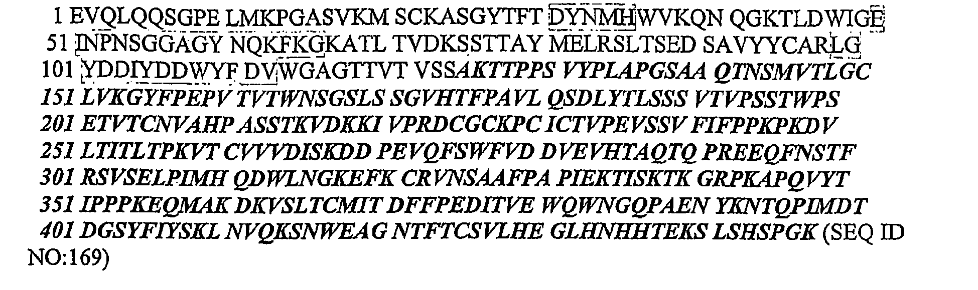
- Nucleic acid sequence encoding the mature form (signal peptide removed) of the Ab-8 HC:

- Amino acid sequence of the Ab-8 HC including signal peptide:


- Nucleic acid sequence of the Ab-8 HC including signal peptide encoding sequence:

- The sequences of the Antibody 9 (also referred to herein as Ab-9) LC and HC are as follows:
- Amino acid sequence of the mature form (signal peptide removed) of the Ab-9 LC:

- Nucleic acid sequence encoding the mature form (signal peptide removed) of the Ab-9 LC:

- Amino acid sequence of the Ab-9 LC including signal peptide:

- Nucleic acid sequence of the Ab-9 LC including signal peptide encoding sequence:

- Amino acid sequence of the mature form (signal peptide removed) of the Ab-9 HC:


- Nucleic acid sequence encoding the mature form (signal peptide removed) of the Ab-9 HC:

- Amino acid sequence of the Ab-9 HC including signal peptide:

- Nucleic acid sequence of the Ab-9 HC including signal peptide encoding sequence:


- The sequences of the Antibody 10 (also referred to herein as Ab-10) LC and HC are as follows:
- Amino acid sequence of the mature form (signal peptide removed) of the Ab-10 LC:

- Nucleic acid sequence encoding the mature form (signal peptide removed) of the Ab-10 LC:


- Amino acid sequence of the Ab-10 LC including signal peptide:

- Nucleic acid sequence of the Ab-10 LC including signal peptide encoding sequence:

- Amino acid sequence of the mature form (signal peptide removed) of the Ab-10 HC:

- Nucleic acid sequence encoding the mature form (signal peptide removed) of the Ab-10 HC:


- Amino acid sequence of the Ab-10 HC including signal peptide:

- Nucleic acid sequence of the Ab-10 HC including signal peptide encoding sequence:


- The sequences of the Antibody 11 (also referred to herein as Ab-11) LC and HC are as follows:
- Amino acid sequence of the mature form (signal peptide removed) of the Ab-11 LC:

- Nucleic acid sequence encoding the mature form (signal peptide removed) of the Ab-11 LC:

- Amino acid sequence of the Ab-11 LC including signal peptide:


- Nucleic acid sequence of the Ab-11 LC including signal peptide encoding sequence:

- Amino acid sequence of the mature form (signal peptide removed) of the Ab-11 HC:

- Nucleic acid sequence encoding the mature form (signal peptide removed) of the Ab-11 HC:


- Amino acid sequence of the Ab-11 HC including signal peptide:

- Nucleic acid sequence of the Ab-11 HC including signal peptide encoding sequence:


- The sequences of the Antibody 12 (also referred to herein as Ab-12) LC and HC are as follows:
- Amino acid sequence of the mature form (signal peptide removed) of the Ab-12 LC:

- Nucleic acid sequence encoding the mature form (signal peptide removed) of the Ab-12 LC:

- Amino acid sequence of the Ab-12 LC including signal peptide:

- Nucleic acid sequence of the Ab-12 LC including signal peptide encoding sequence:


- Amino acid sequence of the mature form (signal peptide removed) of the Ab-12 HC:

- Nucleic acid sequence encoding the mature form (signal peptide removed) of the Ab-12 HC:


- Amino acid sequence of the Ab-12 HC including signal peptide:

- Nucleic acid sequence of the Ab-12 HC including signal peptide encoding sequence:

- The sequences of the Antibody 13 (also referred to herein as Ab-13) LC and HC are as follows:
- Amino acid sequence of the mature form (signal peptide removed) of the Ab-13 LC:

- Nucleic acid sequence encoding the mature form (signal peptide removed) of the Ab-13 LC:

- Amino acid sequence of the Ab-13 LC including signal peptide:

- Nucleic acid sequence of the Ab-13 LC including signal peptide encoding sequence:


- Amino acid sequence of the mature form (signal peptide removed) of the Ab-13 HC:

- Nucleic acid sequence encoding the mature form (signal peptide removed) of the Ab-13 HC:

- Amino acid sequence of the Ab-13 HC including signal peptide:

- Nucleic acid sequence of the Ab-13 HC including signal peptide encoding sequence:

- Ab-13 was humanized to generate Ab-14.
- The sequences of the Antibody 14 (also referred to herein as Ab-14) LC and HC are as follows:
- Amino acid sequence of the mature form (signal peptide removed) of the Ab-14 LC:

- Nucleic acid sequence encoding the mature form (signal peptide removed) of the Ab-14 LC:

- Amino acid sequence of the Ab-14 LC including signal peptide:

- Nucleic acid sequence of the Ab-14 LC including signal peptide encoding sequence:

- Amino acid sequence of the mature form (signal peptide removed) of the Ab-14 HC:

- Amino acid sequence of the mature form (signal peptide removed) of the Ab-14 HC without carboxy-terminal lysine:

- Nucleic acid sequence encoding the mature form (signal peptide removed) of the Ab-14 HC:


- Amino acid sequence of the Ab-14 HC including signal peptide:

- Nucleic acid sequence of the Ab-14 HC including signal peptide encoding sequence:


- The CDR sequences in the variable region of the heavy chain of Ab-14 are:
CDR-H1: DYYMN (SEQ ID NO:296) CDR-H2: DINPYNDDTTYNHKFKG (SEQ ID NO:297) CDR-H3: ETAVITTNAMD (SEQ ID NO:298) - The light chain variable region CDR sequences of Ab-14 are:
CDR-L1: RASSSVTSSYLN (SEQ ID NO:284) CDR-L2: STSNLAS (SEQ ID NO:285) CDR-L3: QQYDFFPST (SEQ ID NO:286) - Ab-14 light chain variable domain amino acid sequence (without signal sequence):

- Ab-14 light chain variable domain DNA sequence (without signal sequence):

- Ab-14 heavy chain variable domain amino acid sequence (without signal sequence):

- Ab-14 heavy chain variable domain DNA sequence (without signal sequence):


- Ab-3 was humanized to generate Ab-15.
- The sequences of the Antibody 15 (also referred to herein as Ab-15) LC and HC are as follows:
- Amino acid sequence of the mature form (signal peptide removed) of the Ab-15 LC:

- Nucleic acid sequence encoding the mature form (signal peptide removed) of the Ab-15 LC:

- Amino acid sequence of the Ab-15 LC including signal peptide:

- Nucleic acid sequence of the Ab-15 LC including signal peptide encoding sequence:


- Amino acid sequence of the mature form (signal peptide removed) of Ab-15 HC.

- Amino acid sequence of the mature form (signal peptide removed) of Ab-15 HC without carboxy-terminal lysine:

- Nucleic acid sequence encoding the mature form (signal peptide removed) of the Ab-15 HC:


- Amino acid sequence of the Ab-15 HC including signal peptide:

- Nucleic acid sequence of the Ab-15 HC including signal peptide encoding sequence:


- The CDR sequences in the variable region of the heavy chain of Ab-15 are:
CDR-H1: DFYLH (SEQ ID NO:290) CDR-H2: RIDPENGDTLYDPKFQD (SEQ ID NO:291) CDR-H3: EADYFHDGTSYWYFDV (SEQ ID NO:292) - The light chain variable region CDR sequences of Ab-15 are:
CDR-L1: SVSSTISSNHLH (SEQ ID NO:278) CDR-L2: GTSNLAS (SEQ ID NO:279) CDR-L3: QQWSSYPLT (SEQ ID NO:280) - Ab-15 light chain variable domain amino acid sequence (without signal sequence):

- Ab-15 light chain variable domain DNA sequence (without signal sequence):


- Ab-15 heavy chain variable domain amino acid sequence (without signal sequence):

- Ab-15 heavy chain variable domain DNA sequence (without signal sequence):

- Ab-11 was humanized to generate Ab-16.
- The sequences of the Antibody 16 (also referred to herein as Ab-16) LC and HC are as follows:
- Amino acid sequence of the mature form (signal peptide removed) of the Ab-16 LC:

- Nucleic acid sequence encoding the mature form (signal peptide removed) of the Ab-16 LC:


- Amino acid sequence of the Ab-16 LC including signal peptide:

- Nucleic acid sequence of the Ab-16 LC including signal peptide encoding sequence:

- Amino acid sequence of the mature form (signal peptide removed) of the Ab-16 HC:


- Amino acid sequence of the mature form (signal peptide removed) of the Ab-16 HC without carboxy-terminal lysine:

- Nucleic acid sequence encoding the mature form (signal peptide removed) of the Ab-16 HC:

- Amino acid sequence of the Ab-16 HC including signal peptide:

- Nucleic acid sequence of the Ab-16 HC including signal peptide encoding sequence:

- The CDR sequences in the variable region of the heavy chain of Ab-16 are:
CDR-H1: DYYIH (SEQ ID NO:293) CDR-H2: RVDPDNGETEFAPKFPG (SEQ ID NO:294) CDR-H3: EDYDGTYTWFPY (SEQ ID NO:295) - The light chain variable region CDR sequences of Ab-16 are:
CDR-L1: RASSSISYIH (SEQ ID NO:281) CDR-L2: ATSNLAS (SEQ ID NO:282) CDR-L3: QQWSSDPLT (SEQ ID NO:283) - Ab-16 light chain variable domain amino acid sequence (without signal sequence):

- Ab-16 light chain variable domain DNA sequence (without signal sequence):

- Ab-16 heavy chain variable domain amino acid sequence (without signal sequence):

- Ab-16 heavy chain variable domain DNA sequence (without signal sequence):

- Additional antibodies are referred to herein as Antibodies 17-22 (also referred to herein as Ab-17, Ab-18, Ab-19, Ab-20, Ab-21, and Ab-22). The Kappa Constant region for all VK regions of Ab-17, Ab-19, and Ab-21 is as follows:

- The Heavy Constant Region for all VH regions of
antibodies 
- In the following antibody amino acid sequences, the boxed-shaded amino acids represent complement-determining regions (CDRs) and the underlined amino acids represent signal peptide.
- Amino acid sequence of the Ab-17 LC including signal peptide:

- Nucleic acid sequence of the Ab-17 LC including signal peptide:

- Amino acid sequence of the Ab-17 HC including signal peptide:

- Nucleic acid sequence of the Ab-17 HC including signal peptide:


- Ab-17 was humanized to generate Ab-18.
- Amino acid sequence of the Ab-18 LC including signal peptide:

- Nucleic acid sequence of the Ab-18 LC including signal peptide:

- Amino acid sequence of the Ab-18 HC including signal peptide:

- Nucleic acid sequence of the Ab-18 HC including signal peptide:

- Ab-18 light chain variable domain amino acid sequence (without signal sequence):

- Ab-18 light chain variable domain DNA sequence (without signal sequence):

- Ab-18 heavy chain variable domain amino acid sequence (without signal sequence):

- Ab-18 heavy chain variable domain DNA sequence (without signal sequence):

- Amino acid sequence of the Ab-19 LC including signal peptide:

- Nucleic acid sequence of the Ab-19 LC including signal peptide:

- Amino acid sequence of the Ab-19 HC including signal peptide:

- Nucleic acid sequence of the Ab-19 HC including signal peptide:

- Ab-19 was humanized to generate Antibody 20 (also referred to herein as Ab-20) and Antibody 23 (also referred to herein as Ab-23).
- Amino acid sequence of the Ab-20 LC including signal peptide:

- Nucleic acid sequence of the Ab-20 LC including signal peptide:

- Amino acid sequence of the Ab-20 HC including signal peptide:

- Nucleic acid sequence of the Ab-20 HC including signal peptide:


- Amino acid sequence of the mature form (signal peptide removed) of the Ab-23 LC:

- Nucleic acid sequence encoding the mature form (signal peptide removed) of the Ab-23 LC:

- Amino acid sequence of the Ab-23 LC including signal peptide:

- Nucleic acid sequence of the Ab-23 LC including signal peptide encoding sequence:


- Amino acid sequence of the mature form (signal peptide removed) of the Ab-23 HC:

- Amino acid sequence of the mature form (signal peptide removed) of the Ab-23 HC without carboxy-terminal lysine:

- Nucleic acid sequence encoding the mature form (signal peptide removed) of the Ab-23 HC:


- Amino acid sequence of the Ab-23 HC including signal peptide:

- Nucleic acid sequence of the Ab-23 HC including signal peptide encoding sequence:


- The CDR (complementarity determining region) sequences in the variable region of the heavy chain of Ab-23 are as follows:
CDR-H1: DYIMH(SEQ ID NO:269) CDR-H2: YINPYNDDTEYNEKFKG (SEQ ID NO:270) CDR-H3: SIYYYDAPFAY (SEQ ID NO:271) - The light chain variable region CDR sequences of Ab-23 are:
CDR-L1: RASQDISSYLN (SEQ ID NO:239) CDR-L2: STSRLNS (SEQ ID NO:240) CDR-L3: QQDIKHPT (SEQ ID NO:241) - Ab-23 light chain variable domain amino acid sequence (without signal sequence):

- Ab-23 light chain variable domain DNA sequence (without signal sequence):

- Ab-23 heavy chain variable domain amino acid sequence (without signal sequence):

- Ab-23 heavy chain variable domain DNA sequence (without signal sequence):

- Amino acid sequence of the Ab-21 LC including signal peptide:

- Nucleic acid sequence of the Ab-21 LC including signal peptide:

- Amino acid sequence of the Ab-21 HC including signal peptide:

- Nucleic acid sequence of the Ab-21 HC including signal peptide:

- Ab-21 was humanized to yield Ab-22.
- Amino acid sequence of the Ab-22 LC including signal peptide:

- Nucleic acid sequence of the Ab-22 LC including signal peptide:

- Amino acid sequence of the Ab-22 HC including signal peptide:

- Nucleic acid sequence of the Ab-22 HC including signal peptide:

- Ab-22 light chain variable domain amino acid sequence (without signal sequence):

- Ab-22 light chain variable domain DNA sequence (without signal sequence):


- Ab-22 heavy chain variable domain amino acid sequence (without signal sequence):

- Ab-22 heavy chain variable domain DNA sequence (without signal sequence):

- For Ab-18, Ab-20, and Ab-22, the light chain human kappa constant region is as follows:and the heavy chain human gamma-4 constant region is as follows:


- The hinge region contains the Ser-241-Pro mutation to improve hinge stability (Angal S et al, (1993), Mol Immunol, 30(1), 105-108).
- The sequences of Antibody 24 (also referred to herein as Ab-24) LC and HC are as follows:
- Amino acid sequence of the mature form (signal peptide removed) of the Ab-24 LC:


- Nucleic acid sequence encoding the mature form (signal peptide removed) of the Ab-24 LC:

- Amino acid sequence of the Ab-24 LC including signal peptide:

- Nucleic acid sequence of the Ab-24 LC including signal peptide encoding sequence:

- Ab-24 Heavy Chain:
- Amino acid sequence of the mature form (signal peptide removed) of the Ab-24 HC:

- Nucleic acid sequence encoding the mature form (signal peptide removed) of the Ab-24 HC:

- Amino acid sequence of the Ab-24 HC including signal peptide:


- Nucleic acid sequence of the Ab-24 HC including signal peptide encoding sequence:

- The CDR sequences in the variable region of the light chain of Ab-24 are as follows:
CDR-L1: KASQSVDYDGTSYMN (SEQ ID NO:351) CDR-L2: AASNLES (SEQ ID NO:352) CDR-L3: QQSNEDPFT (SEQ ID NO:353) - The CDR sequences in the variable region of the heavy chain of Ab-24 are as follows:
CDR-H1: TYWMN(SEQ ID NO:358) CDR-H2: MIHPSASEIRLDQKFKD (SEQ ID NO:359) CDR-H3: SGEWGSMDY (SEQ ID NO:360) - Table 1 below provides the SEQ ID NOs and amino acid sequences of the CDR's of Ab-A, Ab-B, Ab-C, Ab-D, Ab-1, Ab-2, Ab-3, Ab-4, Ab-5, Ab-6, Ab-7, Ab-8, Ab-9, Ab-10, Ab-11, Ab-12, Ab-13, Ab-14, Ab-15, Ab-16, Ab-17, Ab-18, Ab-19, Ab-20, Ab-21, Ab-22, Ab-23, and Ab-24. L1, L2, and L3 refer to light chain CDR's 1, 2, and 3, and H1, H2, and H3 refer to heavy chain CDR's 1, 2, and 3 according to the Kabat numbering system (Kabat et al., 1987 in Sequences of Proteins of Immunological Interest, U.S. Department of Health and Human Services, NIH, USA).
Table 1 SEQ ID NO DESCRIPTION AMINO ACID SEQUENCE 54 Ab-A and Ab-1 CDR-L1 QSSQSVYDNNWLA 55 Ab-A and Ab-1 CDR-L2 DASDLAS 56 Ab-A and Ab-1 CDR-L3 QGAYNDVIYA 51 Ab-A and Ab-1 CDR-H1 SYWMN 52 Ab-A and Ab-1 CDR-H2 TIDSGGRTDYASWAKG 53 Ab-A and Ab-1 CDR-H3 NWNL 60 Ab-B CDR-L1 SASSSVSFVD 61 Ab-B CDR-L2 RTSNLGF 62 Ab-B CDR-L3 QQRSTYPPT 57 Ab-B CDR-H1 TSGMGVG 58 Ab-B CDR-H2 HIWWDDVKRYNPVLKS 59 Ab-B CDR-H3 EDFDYDEEYYAMDY 48 Ab-C CDR-L1 KASQSVDYDGDSYMN 49 Ab-C CDR-L2 AASNLES 50 Ab-C CDR-L3 QQSNEDPWT 45 Ab-C CDR-H1 DCYMN 46 Ab-C CDR-H2 DINPFNGGTTYNQKFKG 47 Ab-C CDR-H3 SHYYFDGRVPWDAMDY 42 Ab-D CDR-L1 QASQGTSINLN 43 Ab-D CDR-L2 GSSNLED 44 Ab-D CDR-L3 LQHSYLPYT 39 Ab-D CDR-H1 DHYMS 40 Ab-D CDR-H2 DINPYSGETTYNQKFKG 41 Ab-D CDR-H3 DDYDASPFAY 275 Ab-2 CDR-L1 RASSSVYYYMH 276 Ab-2 CDR-L2 ATSNLAS 277 Ab-2 CDR-L3 QQWSSDPLT 287 Ab-2 CDR-H1 DYFIH 288 Ab-2 CDR-H2 RLDPEDGESDYAPKFQD 289 Ab-2 CDR-H3 EDYDGTYTFFPY 278 Ab-3 and Ab-15 CDR-L1 SVSSTISSNHLH 279 Ab-3 and Ab-15 CDR-L2 GTSNLAS 280 Ab-3 and Ab-15 CDR-L3 QQWSSYPLT 290 Ab-3 and Ab-15 CDR-H1 DFYLH 291 Ab-3 and Ab-15 CDR-H2 RIDPENGDTLYDPKFQD 292 Ab-3 and Ab-15 CDR-H3 EADYFHDGTSYWYFDV 78 Ab-4 and Ab-5 CDR-L1 RASQDISNYLN 79 Ab-4 and Ab-5 CDR-L2 YTSRLLS 80 Ab-4 and Ab-5 CDR-L3 QQGDTLPYT 245 Ab-4 and Ab-5 CDR-H1 DYNMH 246 Ab-4 and Ab-5 CDR-H2 EINPNSGGAGYNQKFKG 247 Ab-4 and Ab-5 CDR-H3 LGYDDIYDDWYFDV 81 Ab-6 CDR-L1 RASQDISNYLN 99 Ab-6 CDR-L2 YTSRLHS 100 Ab-6 CDR-L3 QQGDTLPYT 248 Ab-6 CDR-H1 DYNMH 249 Ab-6 CDR-H2 EINPNSGGSGYNQKFKG 250 Ab-6 CDR-H3 LVYDGSYEDWYFDV 101 Ab-7 CDR-L1 RASQVITNYLY 102 Ab-7 CDR-L2 YTSRLHS 103 Ab-7 CDR-L3 QQGDTLPYT 251 Ab-7 CDR-H1 DYNMH 252 Ab-7 CDR-H2 EINPNSGGAGYNQQFKG 253 Ab-7 CDR-H3 LGYVGNYEDWYFDV 104 Ab-8 CDR-L1 RASQDISNYLN 105 Ab-8 CDR-L2 YTSRLLS 106 Ab-8 CDR-L3 QQGDTLPYT 254 Ab-8 CDR-H1 DYNMH 255 Ab-8 CDR-H2 EINPNSGGAGYNQKFKG 256 Ab-8 CDR-H3 LGYDDIYDDWYFDV 107 Ab-9 CDR-L1 RASQDISNYLN 108 Ab-9 CDR-L2 YTSRLFS 109 Ab-9 CDR-L3 QQGDTLPYT 257 Ab-9 CDR-H1 DYNMH 258 Ab-9 CDR-H2 EINPNSGGAGYNQKFKG 259 Ab-9 CDR-H3 LGYDDIYDDWYFDV 110 Ab-10 CDR-L1 RASQDISNYLN 111 Ab-10 CDR-L2 YTSRLLS 112 Ab-10 CDR-L3 QQGDTLPYT 260 Ab-10 CDR-H1 DYNMH 261 Ab-10 CDR-H2 EINPNSGGAGYNQKFKG 262 Ab-10 CDR-H3 LGYDDIYDDWYFDV 281 Ab-11 and Ab-16 CDR-L1 RASSSISYIH 282 Ab-11 and Ab-16 CDR-L2 ATSNLAS 283 Ab-11 and Ab-16 CDR-L3 QQWSSDPLT 293 Ab-11 and Ab-16 CDR-H1 DYYIH 294 Ab-11 and Ab-16 CDR-H2 RVDPDNGETEFAPKFPG 295 Ab-11 and Ab-16 CDR-H3 EDYDGTYTWFPY 113 Ab-12 CDR-L1 RASQDISNYLN 114 Ab-12 CDR-L2 YTSTLQS 115 Ab-12 CDR-L3 QQGDTLPYT 263 Ab-12 CDR-H1 DYNMH 264 Ab-12 CDR-H2 EINPNSGGSGYNQKFKG 265 Ab-12 CDR-H3 LGYYGNYEDWYFDV 284 Ab-13 and Ab-14 CDR-L1 RASSSVTSSYLN 285 Ab-13 and Ab-14 CDR-L2 STSNLAS 286 Ab-13 and Ab-14 CDR-L3 QQYDFFPST 296 Ab-13 and Ab-14 CDR-H1 DYYMN 297 Ab-13 and Ab-14 CDR-H2 DINPYNDDTTYNHKFKG 298 Ab-13 and Ab-14 CDR-H3 ETAVITTNAMD 116 Ab-17 and Ab-18 CDR-L1 SVSSSISSSNLH 237 Ab-17 and Ab-18 CDR-L2 GTSNLAS 238 Ab-17 and Ab-18 CDR-L3 QQWTTTYT 266 Ab-17 and Ab-18 CDR-H1 DYYIH 267 Ab-17 and Ab-18 CDR-H2 RIDPDNGESTYVPKFQG 268 Ab-17 and Ab-18 CDR-H3 EGLDYGDYYAVDY 239 Ab-19, Ab-20 and Ab-23 CDR-L1 RASQDISSYLN 240 Ab-19, Ab-20 and Ab-23 CDR-L2 STSRLNS 241 Ab-19, Ab-20 and Ab-23 CDR-L3 QQDIKHPT 269 Ab-19, Ab-20 and Ab-23 CDR-H1 DYIMH 270 Ab-19, Ab-20 and Ab-23 CDR-H2 YINPYNDDTEYNEKFKG 271 Ab-19, Ab-20 and Ab-23 CDR-H3 SIYYYDAPFAY 242 Ab-21 and Ab-22 CDR-L1 KASQDVFTAVA 243 Ab-21 and Ab-22 CDR-L2 WASTRHT 244 Ab-21 and Ab-22 CDR-L3 QQYSSYPLT 272 Ab-21 and Ab-22 CDR-H1 DYYMH 273 Ab-21 and Ab-22 CDR-H2 RIDPENGDIIYDPKFQG 274 Ab-21 and Ab-22 CDR-H3 DAGDPAWFTY 351 Ab-24 CDR-L1 KASQSVDYDGTSYMN 352 Ab-24 CDR-L2 AASNLES 353 Ab-24 CDR-L3 QQSNEDPFT TYWMN 358 Ab-24 CDR-H1 Ab-24 CDR-H1 TYWMN 359 Ab-24 CDR-H2 MIHPSASEIRLDQKFKD 360 Ab-24 CDR-H3 SGEWGSMDY - An oligopeptide or polypeptide is within the scope of the invention if it has an amino acid sequence that is at least 75%, 76%, 77%, 78%, 79%, 80%, 81%, 82%, 83%, 84%, 85%, 86%, 87%, 88%, 89%, 90%, 91%, 92%, 93%, 94%, 95%, 96%, 97%, 98% or 99% identical to least one of the CDR's of Table 1 above; and/or to a CDR of a sclerostin binding agent that cross-blocks the binding of at least one of antibodies Ab-A, Ab-B, Ab-C, Ab-D, Ab-1, Ab-2, Ab-3, Ab-4, Ab-5, Ab-6, Ab-7, Ab-8, Ab-9, Ab-10, Ab-11, Ab-12, Ab-13, Ab-14, Ab-15, Ab-16, Ab-17, Ab-18, Ab-19, Ab-20, Ab-21, Ab-22, Ab-23, and Ab-24 to sclerostin, and/or is cross-blocked from binding to sclerostin by at least one of antibodies Ab-A, Ab-B, Ab-C, Ab-D, Ab-1, Ab-2, Ab-3, Ab-4, Ab-5, Ab-6, Ab-7, Ab-8, Ab-9, Ab-10, Ab-11, Ab-12, Ab-13, Ab-14, Ab-15, Ab-16, Ab-17, Ab-18, Ab-19, Ab-20, Ab-21, Ab-22, Ab-23, and Ab-24; and/or to a CDR of a sclerostin binding agent wherein the binding agent can block the inhibitory effect of sclerostin in a cell based mineralization assay (i.e. a sclerostin neutralizing binding agent); and/or to a CDR of a sclerostin binding agent that binds to a Loop 2 epitope; and/or to a CDR of a sclerostin binding agent that binds to a T20.6 epitope; and/or to a CDR of a sclerostin binding agent that binds to a "T20.6 derivative (cystine-knot + 4 arms)" epitope.
- Sclerostin binding agent polypeptides and antibodies are within the scope of the invention if they have amino acid sequences that are at least 85%, 86%, 87%, 88%, 89%, 90%, 91%, 92%, 93%, 94%, 95%, 96%, 97%, 98% or 99% identical to a variable region of at least one of antibodies Ab-A, Ab-B, Ab-C, Ab-D, Ab-1, Ab-2, Ab-3, Ab-4, Ab-5, Ab-6, Ab-7, Ab-8, Ab-9, Ab-10, Ab-11, Ab-12, Ab-13, Ab-14, Ab-15, Ab-16, Ab-17, Ab-18, Ab-19, Ab-20, Ab-21, Ab-22, Ab-23, and Ab-24, and cross-block the binding of at least one of antibodies Ab-A, Ab-B, Ab-C, Ab-D, Ab-1, Ab-2, Ab-3, Ab-4, Ab-5, Ab-6, Ab-7, Ab-8, Ab-9, Ab-10, Ab-11, Ab-12, Ab-13, Ab-14, Ab-15, Ab-16, Ab-17, Ab-18, Ab-19, Ab-20, Ab-21, Ab-22, Ab-23, and Ab-24 to sclerostin, and/or are cross-blocked from binding to sclerostin by at least one of antibodies Ab-A, Ab-B, Ab-C, Ab-D, Ab-1, Ab-2, Ab-3, Ab-4, Ab-5, Ab-6, Ab-7, Ab-8, Ab-9, Ab-10, Ab-11, Ab-12, Ab-13, Ab-14, Ab-15, Ab-16, Ab-17, Ab-18, Ab-19, Ab-20, Ab-21, Ab-22, Ab-23, and Ab-24; and/or can block the inhibitory effect of sclerostin in a cell based mineralization assay (i.e. a sclerostin neutralizing binding agent); and/or bind to a Loop 2 epitope; and/or bind to a T20.6 epitope; and/or bind to a "T20.6 derivative (cystine-knot + 4 arms)" epitope.
- Polynucleotides encoding sclerostin binding agents are within the scope of the invention if they have polynucleotide sequences that are at least 85%, 86%, 87%, 88%, 89%, 90%, 91%, 92%, 93%, 94%, 95%, 96%, 97%, 98% or 99% identical to a polynucleotide encoding a variable region of at least one of antibodies Ab-A, Ab-B, Ab-C, Ab-D, Ab-1, Ab-2, Ab-3, Ab-4, Ab-5, Ab-6, Ab-7, Ab-8, Ab-9, Ab-10, Ab-11, Ab-12, Ab-13, Ab-14, Ab-15, Ab-16, Ab-17, Ab-18, Ab-19, Ab-20, Ab-21, Ab-22, Ab-23, and Ab-24, and wherein the encoded sclerostin binding agents cross-block the binding of at least one of antibodies Ab-A, Ab-B, Ab-C, Ab-D, Ab-1, Ab-2, Ab-3, Ab-4, Ab-5, Ab-6, Ab-7, Ab-8, Ab-9, Ab-10, Ab-11, Ab-12, Ab-13, Ab-14, Ab-15, Ab-16, Ab-17, Ab-18, Ab-19, Ab-20, Ab-21, Ab-22, Ab-23, and Ab-24 to sclerostin, and/or are cross-blocked from binding to sclerostin by at least one of antibodies Ab-A, Ab-B, Ab-C, Ab-D, Ab-1, Ab-2, Ab-3, Ab-4, Ab-5, Ab-6, Ab-7, Ab-8, Ab-9, Ab-10, Ab-11, Ab-12, Ab-13, Ab-14, Ab-15, Ab-16, Ab-17, Ab-18, Ab-19, Ab-20, Ab-21, Ab-22, Ab-23, and Ab-24; and/or can block the inhibitory effect of sclerostin in a cell based mineralization assay(i.e. a sclerostin neutralizing binding agent); and/or bind to a Loop 2 epitope; and/or bind to a T20.6 epitope; and/or bind to a "T20.6 derivative (cystine-knot + 4 arms)" epitope.
- Antibodies according to the invention may have a binding affinity for human sclerostin of less than or equal to 1 x 10⁻⁷M, less than or equal to 1 x 10⁻⁸M, less than or equal to 1 x 10⁻⁹M, less than or equal to 1 x 10⁻¹⁰M, less than or equal to 1 x 10-11M, or less than or equal to 1x10-12M.
- The affinity of a binding agent such as an antibody or binding partner, as well as the extent to which a binding agent (such as an antibody) inhibits binding, can be determined by one of ordinary skill in the art using conventional techniques, for example those described by Scatchard et al. (Ann. N.Y. Acad Sci. 51:660-672 (1949)) or by surface plasmon resonance (SPR; BIAcore, Biosensor, Piscataway, NJ). For surface plasmon resonance, target molecules are immobilized on a solid phase and exposed to ligands in a mobile phase running along a flow cell. If ligand binding to the immobilized target occurs, the local refractive index changes, leading to a change in SPR angle, which can be monitored in real time by detecting changes in the intensity of the reflected light. The rates of change of the SPR signal can be analyzed to yield apparent rate constants for the association and dissociation phases of the binding reaction. The ratio of these values gives the apparent equilibrium constant (affinity) (see, e.g., Wolff et al., Cancer Res. 53:2560-65 (1993)).
- An antibody according to the present invention may belong to any immunoglobin class, for example IgG, IgE, IgM, IgD, or IgA. It may be obtained from or derived from an animal, for example, fowl (e.g., chicken) and mammals, which includes but is not limited to a mouse, rat, hamster, rabbit, or other rodent, cow, horse, sheep, goat, camel, human, or other primate. The antibody may be an internalizing antibody. Production of antibodies is disclosed generally in
U.S. Patent Publication No. 2004/0146888 A1 . - In the methods described above to generate antibodies according to the invention, including the manipulation of the specific Ab-A, Ab-B, Ab-C, Ab-D, and Antibody 1-24 (Ab-1 to Ab-24) CDRs into new frameworks and/or constant regions, appropriate assays are available to select the desired antibodies or binding agents (i.e. assays for determining binding affinity to sclerostin; cross-blocking assays; Biacore-based "human sclerostin peptide epitope competition binding assay;" MC3T3-E1 cell based assay; in vivo assays).
- Mature form human sclerostin is a 190 amino acid glycoprotein with a cystine-knot structure (
Figures 8 and9 ). In addition to the cystine-knot structure, the protein is characterized as having three loops designated asLoop 1,Loop 2 andLoop 3. Human sclerostin was subjected to proteolytic digestion to produce fragments. Briefly, using different proteases, including trypsin, aspN, and lysC, fragments with various cleavage sites and sizes were generated. The sequences and mass for various human sclerostin peptides were determined. Antibody protection was evaluated to determine the effect on accessibility for proteolysis, including clipped site masking and peptide shifting. Finally, a BIAcore-based "human sclerostin peptide epitope competition assay" was performed. - Exposure of sclerostin to trypsin cleavage resulted in a pattern of peptide fragments as summarized in
Figure 13 . The fragments are referred to as T19.2, T20, T20.6, and T21-22. As shown schematically inFigure 19B , the T20.6 epitope is a complex of four separate peptide sequences which are joined by the three disulfide bonds of the cystine-knot region. Two of the peptides are joined by two disulfide bonds. The other two peptides are linked by one disulfide bond that, schematically, bisects the first two polypeptides. - The T20.6 epitope that was generated by trypsin digestion retains the cystine-knot structure of the native polypeptide and is recognized by antibodies Ab-C and Ab-D. A derivative of epitope T20.6 consists of the cystine-knot region and amino acids 58-64, 73-81, 112-117 and 138-141 in sequence position with reference to SEQ ID NO:1. This derivative epitope is shown in
Figure 21 . An epitope comprising the cystine-knot region may have one or more amino acids that is present in the T20.6 epitope (Figure 19B ) but not present in the T20.6 derivative epitope (Figure 21 ). - Another epitope-containing region was identified in the
Loop 2 region of human sclerostin (Figure 19A ) and is recognized by antibodies Ab-A and Ab-B. A Loop 2 epitope comprises amino acids 86-111 of SEQ ID NO:1 (C4GPARLLPNAIGRGKWWRPSGPDFRC5, SEQ ID NO:6). Sterically, with reference to full-length sclerostin of SEQ ID NO:1, the Loop 2-containing structure is defined at one end by a disulfide bond between cysteine at position 86 (C4) and cysteine at position 144 (C8), and at the other end by a disulfide bond between cysteine at position 111 (C5) and cysteine at position 57 (C1). - The peptides generated by aspN cleavage of human sclerostin are shown in
Figure 12 . In the Figure, these peptides are designated as AspN14.6, AspN18.6, and AspN22.7-23.5, and are also referred to herein as N14.6, N18.6, and N22.7-23.5, respectively. - One group of antibodies exhibits a specific pattern of binding to certain epitopes as evidenced by a Biacore-based "human sclerostin peptide epitope competition binding assay." Briefly, the antibody is preincubated with the epitope to be tested, at concentrations that will saturate the epitope-binding sites on the antibody. The antibody is then exposed to sclerostin bound to a chip surface. After the appropriate incubation and washing procedures, a pattern of competitive binding is established. As shown in
Figure 18 , exemplary antibody Ab-D bound to sclerostin molecules attached to the surface of the chip. Preincubation of antibody Ab-D with sclerostin decreased the binding of the antibody to the sclerostin on the chip to close to zero. Preincubation with a peptide consisting of epitope T19.2 showed that T19.2 did not compete with sclerostin for antibody binding. However, preincubation with any one of the epitopes designated T20, T20.6, T21-22, or N22.7-23.5 abolished a large proportion of the binding of antibody to sclerostin on the chip. In contrast, preincubation of the antibody with any one of the epitopes designated T19.2, N14.6 or N18.6 did not abolish the ability of the antibody to bind to sclerostin. A second exemplary antibody with this binding profile (Fig. 17 ) is Ab-C. - Antibody Ab-D therefore is exemplary and representative of a group of antibodies that bind to the epitopes T20, T20.6, T21-22, and N22.7-23.5, and have minimal detectable binding to epitopes T19.2, N14.6 and N18.6, as measured by the ability to block antibody binding to sclerostin. Antibodies having this characteristic binding pattern may or may not share amino acid sequence in one or more regions of the antibody molecule. Antibody similarity is determined functionally such as by the ability to bind to sclerostin following preincubation with each of the epitopes described above. Antibodies that exhibit a binding pattern similar or identical to that of antibody Ab-D are included in the invention. By "similar to" is meant, for example, the antibody will exhibit binding to each of the polypeptides T20, T20.6, T21-22 and N22.7-23.5 whereby this binding will specifically compete out at least 50% of the antibody's binding to sclerostin that would otherwise occur in the absence of preincubation with sclerostin or a sclerostin peptide. The antibody will also exhibit little or no detectable binding to polypeptides T19.2, N14.6 and N18.6, resulting in a reduction of 30% or less of the binding that would occur in the absence of preincubation with sclerostin or a sclerostin peptide.
- For example, without being bound by a particular mechanism, the antibody binding pattern of
Figure 18 suggests that the epitope space to which antibody Ab-D and other antibodies having the epitope binding pattern of Ab-D bind consists of a polypeptide comprising the cystine-knot region of sclerostin. - Thus, as disclosed herein and with reference to
Figure 19B , an exemplary T20.6 epitope comprises four peptide chains attached via three separate disulfide bonds. Peptide chain SAKPVTELVC3SGQC4GPAR (SEQ ID NO:3) is attached to peptide chain LVASC7KC8KRLTR (SEQ ID NO:5) by disulfide bonds from C3 to C7, and from C4 to C8. Peptide chain DVSEYSC1RELHFTR (SEQ ID NO:2) is attached to peptide chain WWRPSGPDFRC5IPDRYR (SEQ ID NO:4) by a disulfide bond from C1 to C5. The polypeptides of SEQ ID NOs:3 and 5 remain associated with the polypeptides of SEQ ID NOs:2 and 4 through a steric construct whereby the C1-C5 bond crosses the plane of the C4-C8 and C3-C7 bonds and is located between them, as illustrated inFigure 19B . - As disclosed herein and with reference to
Figure 21 , an exemplary derivative epitope of T20.6 comprises four peptide chains attached via three separate disulfide bonds. Peptide chain SAKPVTELVC3SGQC4 (SEQ ID NO:70) is attached to peptide chain LVASC7KC8 (SEQ ID NO:71) by disulfide bonds from C3 to C7, and from C4 to C8. Peptide chain C1RELHFTR (SEQ ID NO:72) is attached to peptide chain C5IPDRYR (SEQ ID NO:73) by a disulfide bond from C1 to C5. The polypeptides of SEQ ID NOs:70 and 71 remain associated with the polypeptides of SEQ ID NOs:72 and 73 through a steric construct whereby the C1-C5 bond crosses the plane of the C4-C8 and C3-C7 bonds and is located between them, as illustrated inFigure 21 . - Antibody Ab-A is exemplary and representative of a second group of antibodies that have a characteristic binding pattern to human sclerostin peptides that is distinct from that obtained for antibodies Ab-C and Ab-D. Ab-A and the group of antibodies it represents bind to the N22.7-23.5 epitope and have minimal detectable binding to epitopes T19.2, T20, T20.6, T21-22, N14.6 or N18.6, as measured by the ability to block antibody binding to sclerostin (
Fig 15 ). A second exemplary antibody with this binding profile (Fig. 16 ) is Ab-B. Antibodies having this characteristic binding pattern may or may not share amino acid sequence in one or more regions of the antibody molecule. Antibody similarity is determined functionally such as by the ability to bind to sclerostin following preincubation with each of the epitopes described above. Antibodies that exhibit a binding pattern similar or identical to that of antibody Ab-A are included in the invention. By "similar to" is meant, for example, the antibody will exhibit binding to the N22.7-23.5 polypeptide whereby this binding will specifically compete out at least 50% of the antibody's binding to sclerostin that would otherwise occur in the absence of preincubation with sclerostin or a sclerostin peptide. The antibody will also exhibit little or no detectable binding to polypeptides T19.2, T20, T20.6, T21-22, N14.6 and N18.6, resulting in a reduction of 30% or less of the binding that would occur in the absence of preincubation with sclerostin or a sclerostin peptide. - For example, without being bound by a particular mechanism, the antibody binding pattern of
Figure 15 suggests that the epitope space to which antibody Ab-A and other antibodies having the epitope binding pattern of Ab-A bind consists of a polypeptide comprising theLoop 2 region of sclerostin. Thus, as disclosed herein and with reference toFigure 19A , theLoop 2 region can be described as a linear peptide, but it acquires a tertiary structure when it is present in native sclerostin or a cystine-knot-containing portion of sclerostin in which the native disulfide bond structure is maintained. The linear or tertiary structure of theLoop 2 epitope can affect antibody binding thereto, as discussed in the Examples. ALoop 2 region can comprise the following amino acid sequence: C4GPARLLPNAIGRGKWWRPSGPDFRC5 (SEQ ID NO:6). "C4" refers to a cysteine residue located at position 86 with reference to SEQ ID NO:1. "C5" refers to a cysteine residue located at position 111 with reference to SEQ ID NO:1. In native sclerostin protein, C4 is linked to a cysteine at position 144 (C8) by a disulfide bond, and C5 is linked to a cysteine at position 57 (C1) by a disulfide bond. Epitopes derived from theLoop 2 region include CGPARLLPNAIGRGKWWRPS (SEQ ID NO:63); GPARLLPNAIGRGKWWRPSG (SEQ ID NO:64); PARLLPNAIGRGKWWRPSGP (SEQ ID NO:65); ARLLPNAIGRGKWWRPSGPD (SEQ ID NO:66); RLLPNAIGRGKWWRPSGPDF (SEQ ID NO:67); LLPNAIGRGKWWRPSGPDFR (SEQ ID NO:68); and LPNAIGRGKWWRPSGPDFRC (SEQ ID NO:69) - The terms "cross-block", "cross-blocked" and "cross-blocking" are used interchangeably herein to mean the ability of an antibody or other binding agent to interfere with the binding of other antibodies or binding agents to sclerostin.
- The extent to which an antibody or other binding agent is able to interfere with the binding of another to sclerostin, and therefore whether it can be said to cross-block according to the invention, can be determined using competition binding assays. One particularly suitable quantitative assay uses a Biacore machine which can measure the extent of interactions using surface plasmon resonance technology. Another suitable quantitative cross-blocking assay uses an ELISA-based approach to measure competition between antibodies or other binding agents in terms of their binding to sclerostin.
- The following generally describes a suitable Biacore assay for determining whether an antibody or other binding agent cross-blocks or is capable of cross-blocking according to the invention. For convenience reference is made to two antibodies, but it will be appreciated that the assay can be used with any of the sclerostin binding agents described herein. The Biacore machine (for example the Biacore 3000) is operated in line with the manufacturer's recommendations.
- Thus in one cross-blocking assay, sclerostin is coupled to a CM5 Biacore chip using standard amine coupling chemistry to generate a sclerostin-coated surface. Typically 200-800 resonance units of sclerostin would be coupled to the chip (an amount that gives easily measurable levels of binding but that is readily saturable by the concentrations of test reagent being used).
- The two antibodies (termed A* and B*) to be assessed for their ability to cross-block each other are mixed at a one to one molar ratio of binding sites in a suitable buffer to create the test mixture. When calculating the concentrations on a binding site basis the molecular weight of an antibody is assumed to be the total molecular weight of the antibody divided by the number of sclerostin binding sites on that antibody.
- The concentration of each antibody in the test mix should be high enough to readily saturate the binding sites for that antibody on the sclerostin molecules captured on the Biacore chip. The antibodies in the mixture are at the same molar concentration (on a binding basis) and that concentration would typically be between 1.00 and 1.5 micromolar (on a binding site basis).
- Separate solutions containing antibody A* alone and antibody B* alone are also prepared. Antibody A* and antibody B* in these solutions should be in the same buffer and at the same concentration as in the test mix.
- The test mixture is passed over the sclerostin-coated Biacore chip and the total amount of binding recorded. The chip is then treated in such a way as to remove the bound antibodies without damaging the chip-bound sclerostin. Typically this is done by treating the chip with 30 mM HCl for 60 seconds.
- The solution of antibody A* alone is then passed over the sclerostin-coated surface and the amount of binding recorded. The chip is again treated to remove all of the bound antibody without damaging the chip-bound sclerostin.
- The solution of antibody B* alone is then passed over the sclerostin-coated surface and the amount of binding recorded.
- The maximum theoretical binding of the mixture of antibody A* and antibody B* is next calculated, and is the sum of the binding of each antibody when passed over the sclerostin surface alone. If the actual recorded binding of the mixture is less than this theoretical maximum then the two antibodies are cross-blocking each other.
- Thus, in general, a cross-blocking antibody or other binding agent according to the invention is one which will bind to sclerostin in the above Biacore cross-blocking assay such that during the assay and in the presence of a second antibody or other binding agent of the invention the recorded binding is between 80% and 0.1% (e.g. 80% to 4%) of the maximum theoretical binding, specifically between 75% and 0.1% (e.g. 75% to 4%) of the maximum theoretical binding, and more specifically between 70% and 0.1% (e.g. 70% to 4%) of maximum theoretical binding (as just defined above) of the two antibodies or binding agents in combination.
- The Biacore assay described above is a primary assay used to determine if antibodies or other binding agents cross-block each other according to the invention. On rare occasions particular antibodies or other binding agents may not bind to sclerostin coupled via amine chemistry to a CM5 Biacore chip (this usually occurs when the relevant binding site on sclerostin is masked or destroyed by the coupling to the chip). In such cases cross-blocking can be determined using a tagged version of Sclerostin, for example N-terminal His-tagged Sclerostin (R & D Systems, Minneapolis, MN, USA; 2005 cat# 1406-ST-025). In this particular format, an anti-His antibody would be coupled to the Biacore chip and then the His-tagged Sclerostin would be passed over the surface of the chip and captured by the anti-His antibody. The cross blocking analysis would be carried out essentially as described above, except that after each chip regeneration cycle, new His-tagged sclerostin would be loaded back onto the anti-His antibody coated surface. In addition to the example given using N-terminal His-tagged Sclerostin, C-terminal His-tagged sclerostin could alternatively be used. Furthermore, various other tags and tag binding protein combinations that are known in the art could be used for such a cross-blocking analysis (e.g. HA tag with anti-HA antibodies; FLAG tag with anti-FLAG antibodies; biotin tag with streptavidin).
- The following generally describes an ELISA assay for determining whether an anti-sclerostin antibody or other sclerostin binding agent cross-blocks or is capable of cross-blocking according to the invention. For convenience, reference is made to two antibodies (Ab-X and Ab-Y), but it will be appreciated that the assay can be used with any of the sclerostin binding agents described herein.
- The general principal of the assay is to have an anti-sclerostin antibody coated onto the wells of an ELISA plate. An excess amount of a second, potentially cross-blocking, anti-sclerostin antibody is added in solution (i.e. not bound to the ELISA plate). A limited amount of sclerostin is then added to the wells. The coated antibody and the antibody in solution compete for binding of the limited number of sclerostin molecules. The plate is washed to remove sclerostin that has not been bound by the coated antibody and to also remove the second, solution phase antibody as well as any complexes formed between the second, solution phase antibody and sclerostin. The amount of bound sclerostin is then measured using an appropriate sclerostin detection reagent. An antibody in solution that is able to cross-block the coated antibody will be able to cause a decrease in the number of sclerostin molecules that the coated antibody can bind relative to the number of sclerostin molecules that the coated antibody can bind in the absence of the second, solution phase, antibody.
- This assay is described in more detail further below for Ab-X and Ab-Y. In the instance where Ab-X is chosen to be the immobilized antibody, it is coated onto the wells of the ELISA plate, after which the plates are blocked with a suitable blocking solution to minimize non-specific binding of reagents that are subsequently added. An excess amount of Ab-Y is then added to the ELISA plate such that the moles of Ab-Y sclerostin binding sites per well are at least 10 fold higher than the moles of Ab-X sclerostin binding sites that were used, per well, during the coating of the ELISA plate. Sclerostin is then added such that the moles of sclerostin added per well are at least 25-fold lower than the moles of Ab-X sclerostin binding sites that were used for coating each well. Following a suitable incubation period the ELISA plate is washed and a sclerostin detection reagent is added to measure the amount of sclerostin specifically bound by the coated anti-sclerostin antibody (in this case Ab-X). The background signal for the assay is defined as the signal obtained in wells with the coated antibody (in this case Ab-X), second solution phase antibody (in this case Ab-Y), sclerostin buffer only (i.e. no sclerostin) and sclerostin detection reagents. The positive control signal for the assay is defined as the signal obtained in wells with the coated antibody (in this case Ab-X), second solution phase antibody buffer only (i.e. no second solution phase antibody), sclerostin and sclerostin detection reagents. The ELISA assay needs to be run in such a manner so as to have the positive control signal be at least 6 times the background signal.
- To avoid any artifacts (e.g. significantly different affinities between Ab-X and Ab-Y for sclerostin) resulting from the choice of which antibody to use as the coating antibody and which to use as the second (competitor) antibody, the cross-blocking assay needs to be run in two formats:
- 1)
format 1 is where Ab-X is the antibody that is coated onto the ELISA plate and Ab-Y is the competitor antibody that is in solution and - 2)
format 2 is where Ab-Y is the antibody that is coated onto the ELISA plate and Ab-X is the competitor antibody that is in solution. - Ab-X and Ab-Y are defined as cross-blocking if, either in
format 1 or informat 2, the solution phase anti-sclerostin antibody is able to cause a reduction of between 60% and 100%, specifically between 70% and 100%, and more specifically between 80% and 100%, of the sclerostin detection signal (i.e. the amount of sclerostin bound by the coated antibody) as compared to the sclerostin detection signal obtained in the absence of the solution phase anti-sclerostin antibody (i.e. the positive control wells). - An example of such an ELISA-based cross blocking assay can be found in Example 7 ("ELISA-based cross-blocking assay").
- Mineralization by osteoblast-lineage cells in culture, either primary cells or cell lines, is used as an in vitro model of bone formation. Mineralization takes from about one to six weeks to occur beginning with the induction of osteoblast-lineage cell differentiation by one or more differentiation agents. The overall sequence of events involves cell proliferation, differentiation, extracellular matrix production, matrix maturation and finally deposition of mineral, which refers to crystallization and/or deposition of calcium phosphate. This sequence of events starting with cell proliferation and differentiation, and ending with deposition of mineral is referred to herein as mineralization. Measurement of calcium (mineral) is the output of the assay.
- MC3T3-E1 cells (Sudo H, Kodama H-A, Amagai Y, Yamamoto S, Kasai S. 1983. In vitro differentiation and calcification in a new clonal osteogenic cell line derived from newborn mouse calvaria. J. Cell Biol. 96:191-198) and subclones of the original cell line can form mineral in culture upon growth in the presence of differentiating agents. Such subclones include MC3T3-E1-BF (Smith E, Redman R, Logg C, Coetzee G, Kasahara N, Frenkel B. 2000. Glucocorticoids inhibit developmental stage-specific osteoblast cell cycle. J. Biol. Chem. 275:19992-20001). For both the MC3T3-E1-BF subclone as well as the original MC3T3-E1 cells, sclerostin can inhibit one or more of the sequence of events leading up to and including mineral deposition (i.e. sclerostin inhibits mineralization). Anti-sclerostin antibodies that are able to neutralize sclerostin's inhibitory activity allow for mineralization of the culture in the presence of sclerostin such that there is a statistically significant increase in deposition of calcium phosphate (measured as calcium) as compared to the amount of calcium measured in the sclerostin-only (i.e. no antibody) treatment group. The antibodies used in the cell based mineralization assay experiments shown in
Figures 22 ,23 and24 have molecular weights of about 145 Kd and have 2 sclerostin binding sites per antibody molecule. - When running the assay with the goal of determining whether a particular anti-sclerostin antibody or anti-sclerostin binding agent can neutralize sclerostin (i.e., is a sclerostin neutralizing antibody or derivative thereof, or is a sclerostin neutralizing binding agent), the amount of sclerostin used in the assay needs to be the minimum amount of sclerostin that causes at least a 70%, statistically significant, reduction in deposition of calcium phosphate (measured as calcium) in the sclerostin-only group, as compared to the amount of calcium measured in the no sclerostin group. An anti-sclerostin neutralizing antibody or an anti-sclerostin neutralizing binding agent is defined as one that causes a statistically significant increase in deposition of calcium phosphate (measured as calcium) as compared to the amount of calcium measured in the sclerostin-only (i.e. no antibody, no binding agent) treatment group. To determine whether an anti-sclerostin antibody or an anti-sclerostin binding agent is neutralizing or not, the amount of anti-sclerostin antibody or anti-sclerostin binding agent used in the assay needs to be such that there is an excess of moles of sclerostin binding sites per well as compared to the number of moles of sclerostin per well. Depending on the potency of the antibody, the fold excess that may be required can be 24, 18, 12, 6, 3, or 1.5, and one of skill is familiar with the routine practice of testing more than one concentration of binding agent. For example, a very potent anti-sclerostin neutralizing antibody or anti-sclerostin neutralizing binding agent will be able to neutralize sclerostin even when there is less than a 6-fold excess of moles of sclerostin binding sites per well as compared to the number of moles of sclerostin per well. A less potent anti-sclerostin neutralizing antibody or anti-sclerostin neutralizing binding agent will be able to neutralize sclerostin only at a 12, 18 or 24 fold excess. Sclerostin binding agents within this full range of potencies are suitable as neutralizing sclerostin binding agents. Exemplary cell based mineralization assays are described in detail in Example 8.
- Anti-sclerostin antibodies and derivatives thereof that can neutralize human sclerostin, and sclerostin binding agents that can neutralize human sclerostin may be of use in the treatment of human conditions/disorders that are caused by, associated with, or result in at least one of low bone formation, low bone mineral density, low bone mineral content, low bone mass, low bone quality and low bone strength.
- Increases in various parameters associated with, or that result from, the stimulation of new bone formation can be measured as an output from in vivo testing of sclerostin binding agents in order to identify those binding agents that are able to neutralize sclerostin and thus able to cause stimulation of new bone formation. Such parameters include various serum anabolic markers [e.g. osteocalcin, P1NP (n-terminal propeptide of
type 1 procollagen)], histomorphometric markers of bone formation (e.g. osteoblast surface/bone surface; bone formation rate/bone surface; trabecular thickness), bone mineral density, bone mineral content, bone mass, bone quality and bone strength. A sclerostin neutralizing binding agent is defined as one capable of causing a statistically significant increase, as compared to vehicle treated animals, in any parameter associated with, or that results from, the stimulation of new bone formation. Such in vivo testing can be performed in any suitable mammal (e.g. mouse, rat, monkey). An example of such in vivo testing can be found in Example 5 ("In vivo testing of anti-sclerostin monoclonal antibodies"). - Although the amino acid sequence of sclerostin is not 100% identical across mammalian species (e.g. mouse sclerostin is not 100% identical to human sclerostin), it will be appreciated by one skilled in the art that a sclerostin binding agent that can neutralize, in vivo, the sclerostin of a certain species (e.g. mouse) and that also can bind human sclerostin in vitro is very likely to be able to neutralize human sclerostin in vivo. Thus, such a human sclerostin binding agent (e.g. anti-human sclerostin antibody) may be of use in the treatment of human conditions/disorders that are caused by, associated with, or result in at least one of low bone formation, low bone mineral density, low bone mineral content, low bone mass, low bone quality and low bone strength. Mice in which homologous recombination had been used to delete the mouse sclerostin gene and insert the human sclerostin gene in its place (i.e. human sclerostin gene knock-in mice or human SOST knock-in mice) would be an example of an additional in vivo system.
- Pharmaceutical compositions are provided, comprising one of the above-described binding agents such as at least one of antibody Ab-A, Ab-B, Ab-C, Ab-D and Ab-1 toAb-24 to human sclerostin, along with a pharmaceutically or physiologically acceptable carrier, excipient, or diluent. Pharmaceutical compositions and methods of treatment are disclosed in copending Application Serial No.
10/868,497, filed June 16, 2004 60/478,977 - The development of suitable dosing and treatment regimens for using the particular compositions described herein in a variety of treatment regimens, including e.g., subcutaneous, oral, parenteral, intravenous, intranasal, and intramuscular administration and formulation, is well known in the art, some of which are briefly discussed below for general purposes of illustration.
- In certain applications, the pharmaceutical compositions disclosed herein may be delivered via oral administration to an animal. As such, these compositions may be formulated with an inert diluent or with an assimilable edible carrier, or they may be enclosed in hard- or soft-shell gelatin capsule, or they may be compressed into tablets, or they may be incorporated directly with the food of the diet.
- In certain circumstances it will be desirable to deliver the pharmaceutical compositions disclosed herein subcutaneously, parenterally, intravenously, intramuscularly, or even intraperitoneally. Such approaches are well known to the skilled artisan, some of which are further described, for example, in
U.S. Patent No. 5,543,158 ;U.S. Patent No. 5,641,515 andU.S. Patent No. 5,399,363 . In certain embodiments, solutions of the active compounds as free base or pharmacologically acceptable salts may be prepared in water suitably mixed with a surfactant, such as hydroxypropylcellulose. Dispersions may also be prepared in glycerol, liquid polyethylene glycols, and mixtures thereof and in oils. Under ordinary conditions of storage and use, these preparations generally will contain a preservative to prevent the growth of microorganisms. - Illustrative pharmaceutical forms suitable for injectable use include sterile aqueous solutions or dispersions and sterile powders for the extemporaneous preparation of sterile injectable solutions or dispersions (for example, see
U.S. Patent No. 5,466,468 ). In all cases the form must be sterile and must be fluid to the extent that easy syringability exists. It must be stable under the conditions of manufacture and storage and must be preserved against the contaminating action of microorganisms, such as bacteria and fungi. The carrier can be a solvent or dispersion medium containing, for example, water, ethanol, polyol (e.g., glycerol, propylene glycol, and liquid polyethylene glycol, and the like), suitable mixtures thereof, and/or vegetable oils. Proper fluidity may be maintained, for example, by the use of a coating, such as lecithin, by the maintenance of the required particle size in the case of dispersion and/or by the use of surfactants. The prevention of the action of microorganisms can be facilitated by various antibacterial and antifungal agents, for example, parabens, chlorobutanol, phenol, sorbic acid, thimerosal, and the like. In many cases, it will be preferable to include isotonic agents, for example, sugars or sodium chloride. Prolonged absorption of the injectable compositions can be brought about by the use in the compositions of agents delaying absorption, for example, aluminum monostearate and gelatin. - In one embodiment, for parenteral administration in an aqueous solution, the solution should be suitably buffered if necessary and the liquid diluent first rendered isotonic with sufficient saline or glucose. These particular aqueous solutions are especially suitable for intravenous, intramuscular, subcutaneous and intraperitoneal administration. In this connection, a sterile aqueous medium that can be employed will be known to those of skill in the art in light of the present disclosure. For example, one dosage may be dissolved in 1 ml of isotonic NaCl solution and either added to 1000 ml of hypodermoclysis fluid or injected at the proposed site of infusion, (see for example, Remington's Pharmaceutical Sciences, 15th ed., pp. 1035-1038 and 1570-1580). Some variation in dosage will necessarily occur depending on the condition of the subject being treated. Moreover, for human administration, preparations will of course preferably meet sterility, pyrogenicity, and the general safety and purity standards as required by FDA Office of Biologics standards.
- In another embodiment of the invention, the compositions disclosed herein may be formulated in a neutral or salt form. Illustrative pharmaceutically-acceptable salts include the acid addition salts (formed with the free amino groups of the protein) and which are formed with inorganic acids such as, for example, hydrochloric or phosphoric acids, or such organic acids as acetic, oxalic, tartaric, mandelic, and the like. Salts formed with the free carboxyl groups can also be derived from inorganic bases such as, for example, sodium, potassium, ammonium, calcium, or ferric hydroxides, and such organic bases as isopropylamine, trimethylamine, histidine, procaine and the like. Upon formulation, solutions will be administered in a manner compatible with the dosage formulation and in such amount as is therapeutically effective.
- The carriers can further comprise any and all solvents, dispersion media, vehicles, coatings, diluents, antibacterial and antifungal agents, isotonic and absorption delaying agents, buffers, carrier solutions, suspensions, colloids, and the like. The use of such media and agents for pharmaceutical active substances is well known in the art. Except insofar as any conventional media or agent is incompatible with the active ingredient, its use in the therapeutic compositions is contemplated. Supplementary active ingredients can also be incorporated into the compositions. The phrase "pharmaceutically-acceptable" refers to molecular entities and compositions that do not produce an allergic or similar untoward reaction when administered to a human.
- In certain embodiments, liposomes, nanocapsules, microparticles, lipid particles, vesicles, and the like, are used for the introduction of the compositions of the present invention into suitable host cells/organisms. In particular, the compositions of the present invention may be formulated for delivery either encapsulated in a lipid particle, a liposome, a vesicle, a nanosphere, or a nanoparticle or the like. Alternatively, compositions of the present invention can be bound, either covalently or non-covalently, to the surface of such carrier vehicles.
- The formation and use of liposome and liposome-like preparations as potential drug carriers is generally known to those of skill in the art (see for example, Lasic, Trends Biotechnol. 16(7):307-21, 1998; Takakura, Nippon Rinsho 56(3):691-95, 1998; Chandran et al., Indian J. Exp. Biol. 35(8):801-09, 1997; Margalit, Crit. Rev. Ther: Drug Carrier Syst. 12(2-3):233-61, 1995;
U.S. Patent No. 5,567,434 ;U.S. Patent No. 5,552,157 ;U.S. Patent No. 5,565,213 ;U.S. Patent No. 5,738,868 andU.S. Patent No. 5,795,587 , each specifically incorporated herein by reference in its entirety). The use of liposomes does not appear to be associated with autoimmune responses or unacceptable toxicity after systemic delivery. In certain embodiments, liposomes are formed from phospholipids that are dispersed in an aqueous medium and spontaneously form multilamellar concentric bilayer vesicles (also termed multilamellar vesicles (MLVs)). - Alternatively, in other embodiments, the invention provides for pharmaceutically-acceptable nanocapsule formulations of the compositions of the present invention. Nanocapsules can generally entrap compounds in a stable and reproducible way (see, for example, Quintanar-Guerrero et al., Drug Dev. Ind Pharm. 24(12):1113-28, 1998). To avoid side effects due to intracellular polymeric overloading, such ultrafine particles (sized around 0.1µm) may be designed using polymers able to be degraded in vivo. Such particles can be made as described, for example, by Couvreur et al., Crit. Rev. Ther. Drug Carrier Syst. 5(1):1-20, 1988; zur Muhlen et al., Eur. J. Pharm. Biopharm. 45(2):149-55, 1998; Zambaux et al., J. Controlled Release 50(1-3):31-40, 1998; and
U.S. Patent No. 5,145,684 . - In addition, pharmaceutical compositions of the present invention may be placed within containers, along with packaging material that provides instructions regarding the use of such pharmaceutical compositions. Generally, such instructions will include a tangible expression describing the reagent concentration, as well as within certain embodiments, relative amounts of excipient ingredients or diluents (e.g., water, saline or PBS) that may be necessary to reconstitute the pharmaceutical composition.
- The dose administered may range from 0.01 mg/kg to 100 mg/kg of body weight. As will be evident to one of skill in the art, the amount and frequency of administration will depend, of course, on such factors as the nature and severity of the indication being treated, the desired response, the condition of the patient, and so forth. Typically, the compositions may be administered by a variety of techniques, as noted above.
- Increases in bone mineral content and/or bone mineral density may be determined directly through the use of X-rays (e.g., Dual Energy X-ray Absorptometry or "DEXA"), or by inference through the measurement of 1) markers of bone formation and/or osteoblast activity, such as, but not limited to, osteoblast specific alkaline phosphatase, osteocalcin,
type 1 procollagen C' propeptide (PICP), total alkaline phosphatase (see Comier, Curr. Opin. in Rheu. 7:243(1995)) and serum procollagen 1 N-terminal propeptide (P1NP) and/or 2) markers of bone resorption and/or osteoclast activity including, but not limited to, pyridinoline, deoxypryridinoline, N-telopeptide, urinary hydroxyproline, plasma tartrate-resistant acid phosphatases, and galactosyl hydroxylysine; (see Comier, id), serum TRAP 5b (tartrate-resistant acid phosphatase isoform 5b) and serum cross-linked C-telopeptide (sCTXI). The amount of bone mass may also be calculated from body weights or by using other methods (see Guinness-Hey, Metab. Bone Dis. Relat. Res. 5:177-181, 1984).Animals and particular animal models are used in the art for testing the effect of the compositions and methods of the invention on, for example, parameters of bone loss, bone resorption, bone formation, bone strength or bone mineralization that mimic conditions of human disease such as osteoporosis and osteopenias. Examples of such models include the ovariectomized rat model (Kalu, D.N., The ovariectomized rat model of postmenopausal bone loss. Bone and Mineral 15:175-192 (1991); Frost, H.M. and Jee, W.S.S. On the rat model of human osteopenias and osteoporosis. Bone and Mineral 18:227-236 (1992); and Jee, W.S.S. and Yao, W., Overview: animal models of osteopenia and osteoporosis. J. Musculoskel. Neuron. Interact. 1:193-207 (2001)). - Particular conditions which may be treated by the compositions of the present invention include dysplasias, wherein growth or development of bone is abnormal and a wide variety of causes of osteopenia, osteoporosis and bone loss. Representative examples of such conditions include achondroplasia, cleidocranial dysostosis, enchondromatosis, fibrous dysplasia, Gaucher's Disease, hypophosphatemic rickets, Marfan's syndrome, multiple hereditary exotoses, neurofibromatosis, osteogenesis imperfecta, osteopetrosis, osteopoikilosis, sclerotic lesions, pseudoarthrosis, and pyogenic osteomyelitis, periodontal disease, anti-epileptic drug induced bone loss, primary and secondary hyperparathyroidism, familial hyperparathyroidism syndromes, weightlessness induced bone loss, osteoporosis in men, postmenopausal bone loss, osteoarthritis, renal osteodystrophy, infiltrative disorders of bone, oral bone loss, osteonecrosis of the jaw, juvenile Paget's disease, melorheostosis, metabolic bone diseases, mastocytosis, sickle cell anemia/disease, organ transplant related bone loss, kidney transplant related bone loss, systemic lupus erythematosus, ankylosing spondylitis, epilepsy, juvenile arthritides, thalassemia, mucopolysaccharidoses, fabry disease, turner syndrome, Down Syndrome, Klinefelter Syndrome, leprosy, Perthes' Disease, adolescent idiopathic scoliosis, infantile onset multi-system inflammatory disease, Winchester Syndrome, Menkes Disease, Wilson's Disease, ischemic bone disease (such as Legg-Calve-Perthes disease, regional migratory osteoporosis), anemic states, conditions caused by steroids, glucocorticoid-induced bone loss, heparin-induced bone loss, bone marrow disorders, scurvy, malnutrition, calcium deficiency, idiopathic osteopenia or osteoporosis, congenital osteopenia or osteoporosis, alcoholism, chronic liver disease, postmenopausal state, chronic inflammatory conditions, rheumatoid arthritis, inflammatory bowel disease, ulcerative colitis, inflammatory colitis, Crohn's disease, oligomenorrhea, amenorrhea, pregnancy, diabetes mellitus, hyperthyroidism, thyroid disorders, parathyroid disorders, Cushing's disease, acromegaly, hypogonadism, immobilization or disuse, reflex sympathetic dystrophy syndrome, regional osteoporosis, osteomalacia, bone loss associated with joint replacement, HIV associated bone loss, bone loss associated with loss of growth hormone, bone loss associated with cystic fibrosis, fibrous dysplasia, chemotherapy associated bone loss, tumor induced bone loss, cancer-related bone loss, hormone ablative bone loss, multiple myeloma, drug-induced bone loss, anorexia nervosa, disease associated facial bone loss, disease associated cranial bone loss, disease associated bone loss of the jaw, disease associated bone loss of the skull, and bone loss associated with space travel. Further conditions relate to bone loss associated with aging, including facial bone loss associated with aging, cranial bone loss associated with aging, jaw bone loss associated with aging, and skull bone loss associated with aging.
- Compositions of the present invention may also be useful for improving outcomes in orthopedic procedures, dental procedures, implant surgery, joint replacement, bone grafting, bone cosmetic surgery and bone repair such as fracture healing, nonunion healing, delayed union healing and facial reconstruction. One or more compositions may be administered before, during and/or after the procedure, replacement, graft, surgery or repair.
- The invention also provides a diagnostic kit comprising at least one anti-sclerostin binding agent according to the present invention. The binding agent may be an antibody. In addition, such a kit may optionally comprise one or more of the following:
- (1) instructions for using the one or more binding agent(s) for screening, diagnosis, prognosis, therapeutic monitoring or any combination of these applications;
- (2) a labeled binding partner to the anti-sclerostin binding agent(s);
- (3) a solid phase (such as a reagent strip) upon which the anti-sclerostin binding agent(s) is immobilized; and
- (4) a label or insert indicating regulatory approval for screening, diagnostic, prognostic or therapeutic use or any combination thereof.
- If no labeled binding partner to the binding agent(s) is provided, the binding agent(s) itself can be labeled with one or more of a detectable marker(s), e.g. a chemiluminescent, enzymatic, fluorescent, or radioactive moiety.
- The following examples are offered by way of illustration, and not by way of limitation.
- Recombinant human sclerostin/SOST is commercially available from R&D Systems (Minneapolis, MN, USA; 2006 cat# 1406-ST-025). Additionally, recombinant mouse sclerostin/SOST is commercially available from R&D Systems (Minneapolis, MN, USA; 2006 cat# 1589-ST-025).
- Alternatively, the different species of sclerostin can be expressed transiently in serum-free suspension adapted 293T or 293EBNA cells. Transfections can be performed as 500 mL or 1L cultures. The following reagents and materials are available from Gibco BRL (now Invitrogen, Carlsbad, CA). Catalog numbers are listed in parentheses: serum-free DMEM (21068-028); DMEM/F12 (3:1) (21068/11765); 1X Insulin-Transferrin-Selenium Supplement (51500-056); 1X Pen Strep Glut (10378-016); 2mM 1-Glutamine (25030-081); 20 mM HEPES (15630-080); 0.01% Pluronic F68 (24040-032). Briefly, the cell inoculum (5.0-10.0
X 105 cells/mL X culture volume) is centrifuged at 2,500 RPM for 10 minutes at 4°C to remove the conditioned medium. - The cells are resuspended in serum-free DMEM and centrifuged again at 2,500 RPM for 10 minutes at 4°C. After aspirating the wash solution, the cells are resuspended in growth medium [DMEM/F12 (3:1) + 1X Insulin-Transferrin-Selenium Supplement + 1X Pen Strep Glut + 2mM L-Glutamine + 20 mM HEPES + 0.01% Pluronic F68] in a 1L or 3L spinner flask culture. The spinner flask culture is maintained on magnetic stir plate at 125 RPM which is placed in a humidified incubator maintained at 37°C and 5% CO2. The mammalian expression plasmid DNA (e.g. pcDNA3.1, pCEP4, Invitrogen Life Technologies, Carlsbad, CA), containing the complete coding region (and stop codon) of sclerostin with a Kozak consensus sequence (e.g., CCACC) directly 5' of the start site ATG, is complexed to the transfection reagent in a 50 mL conical tube.
- The DNA-transfection reagent complex can be prepared in 5-10% of the final culture volume in serum-free DMEM or OPTI-MEM. The transfection reagents that can be used for this purpose include X-tremeGene RO-1539 (Roche Applied Science, Indianapolis, IN), FuGene6 (Roche Applied Science, Indianapolis, IN), Lipofectamine 2000 (Invitrogen, Carlsbad, CA) and 293fectin (Invitrogen, Carlsbad, CA). 1-5 µg plasmid DNA/mL culture is first added to serum-free DMEM, followed by 1-5 µl transfection reagent/mL culture. The complexes can be incubated at room temperature for approximately 10-30 minutes and then added to the cells in the spinner flask. The transfection/expression can be performed for 4-7 days, after which the conditioned medium (CM) is harvested by centrifugation at 4,000 RPM for 60 minutes at 4°C.
- Recombinant sclerostin was purified from mammalian host cells as follows. All purification processes were carried out at room temperature. One purification scheme was used to purify various species of sclerostin, including murine and human sclerostin. The purification scheme used affinity chromatography followed by cation exchange chromatography.
- The mammalian host cell conditioned medium (CM) was centrifuged in a Beckman J6-M1 centrifuge at 4000 rpm for 1hour at 4°C to remove cell debris. The CM supernatant was then filtered through a sterile 0.2 µm filter. (At this point the sterile filtered CM may be optionally stored frozen until purification.) If the CM was frozen, it was thawed at the following temperatures, or combination thereof: 4°C, room temperature or warm water. Following thawing the CM was filtered through a sterile 0.2 µm filter and optionally concentrated by tangential flow ultrafiltration (TFF) using a 10 kD molecular weight cut-off membrane. The CM concentrate was filtered through a sterile 0.2 µm filter and then loaded onto a Heparin High Performance (Heparin HP) column (GE Healthcare, formerly Amersham Biosciences) equilibrated in PBS. Alternatively, the filtered CM supernatant may be loaded directly onto the Heparin HP column equilibrated in PBS.
- After loading, the Heparin HP column was washed with PBS until the absorbance at 280 nm of the flow-through returned to baseline (i.e., absorbance measured before loading CM supernatant). The sclerostin was then eluted from the column using a linear gradient from 150 mM to 2M sodium chloride in PBS. The absorbance at 280 nm of the eluate was monitored and fractions containing protein were collected. The fractions were then assayed by Coomassie-stained SDS-PAGE to identify fractions containing a polypeptide that migrates at the size of glycosylated sclerostin. The appropriate fractions from the column were combined to make the Heparin HP pool.
- The sclerostin eluted from the Heparin HP column was further purified by cation exchange chromatography using SP High Performance (SPHP) chromatography media (GE Healthcare, formerly Amersham Biosciences). The Heparin HP pool was buffer exchanged into PBS by dialysis using 10,000 MWCO membranes (Pierce Slide-A-Lyzer). The dialyzed Heparin HP pool was then loaded onto an SPHP column equilibrated in PBS. After loading, the column was washed with PBS until the absorbance at 280 nm of the flow-through returned to baseline. The sclerostin was then eluted from the SPHP column using a linear gradient from 150 mM to 1 M sodium chloride in PBS. The absorbance at 280 nm of the eluate was monitored and the eluted sclerostin was collected in fractions. The fractions were then assayed by Coomassie-stained SDS-PAGE to identify fractions containing a polypeptide that migrates at the size of glycosylated sclerostin. The appropriate fractions from the column were combined to make the SPHP pool.
- Following purification, the SPHP pool was formulated in PBS by dialysis using 10,000 MWCO membranes (Pierce Slide-A-Lyzer). If concentration of sclerostin was necessary, a centrifugal device (Amicon Centricon or Centriprep) with a 10,000 MWCO membrane was used. Following formulation the sclerostin was filtered through a sterile 0.2 µm filter and stored at 4°C or frozen.
- A series of overlapping peptides (each peptide being approximately 20-25 amino acids long) were synthesized based on the known amino acid sequence of rat sclerostin (SEQ ID NO:98). The peptides were designed such that they all contained a reduced cysteine residue; an additional cysteine was included at the C-terminus of each peptide which did not already contain one in its sequence. This enabled the peptides to be bound to the assay plates by covalent coupling, using commercially available sulfhydryl binding plates (Costar), at a concentration of 1µg/ml, in phosphate buffered saline (PBS: pH 6.5) containing 1 mM EDTA. Following incubation for 1 hour at room temperature, the plates were washed three times with PBS containing 0.5
% Tween 20. The plates were blocked by incubation with a PBS solution containing 0.5% fish skin gelatin (Sigma) for 30 minutes at room temperature and then washed three times in PBS containing 0.5% Tween 20. - Antibodies to be tested were diluted to 1µg/ml in PBS containing 0.5% fish skin gelatin and incubated with the peptide-coated plates for 1 hour at room temperature. Excess antibody was removed by three washes with PBS, 0.5
% Tween 20. The plates were then incubated with an appropriate secondary antibody conjugated to horseradish peroxidase (diluted appropriately in PBS containing 0.5% Tween 20) and capable of binding to the antibody of interest. The plates were then washed three times: once with PBS containing 0.5% Tween 20, and twice with PBS. Finally the plates were incubated with a horseradish peroxidase chromogenic substrate (TMB-Stable Stop, RDI) for 5 minutes at room temperature, the color development was stopped with acid, and the plates' optical density measured at 450nm. -
- Costar's Sulfhydryl Binding Plates (VWR # 29442-278)
- Coating Buffer: 1XPBS PH 6.5 + 1mM EDTA
- Blocking Buffer: 1X PBS + 0.5% Fish Skin Gelatin (PBS from CS; FSG from Sigma# G 7765)
- Wash Buffer: 1X PBS + 0.5
% Tween 20 - Rat Sclerostin peptides
- Antibody Samples: Transient Ab, Purified recombinant Ab, rabbit Serum, etc.
- Appropriate secondary Ab: Goat-anti-Rabbit/Mouse-HRP (Jackson Immuno Research,115-036-072)
- TMB-Stable Stop (RDI# RDI-TMBSX-1L) 0.5M HCl
-
- 1. Coat plates with 100µl/well of rat sclerostin peptide diluted in 1XPBS PH 6.5 + 1mM EDTA at 1µg/ml.
Incubate plates 1 hour at room temperature. (Plates should be used within 30 minutes of opening). - 2. Wash plates 3X with wash buffer.
- 3. Block plates with 200ul/well blocking buffer.
Incubate plates 30 minutes at room temp. - 4. Repeat washing as described in (2).
- 5. Incubate plates with 50ul/well of samples diluted in blocking buffer - Serum titers starting at 1:100; Transient Recombinant Ab use neat; Purified recombinant Ab use at 1µg/ml (all samples run in duplicates). Incubate plates 1h at room temp.
- 6. Wash plates as described in (2).
- 7. Incubate plates with 50µl/well of appropriate Secondary Antibody (HRP labeled) diluted 1:1600 in Blocking Buffer.
Incubate plates 1 hour at room temperature. - 8. Wash plates 1X wash buffer, 2x PBS
- 9. Incubate plates with 50µl/well of TMB, 5 minutes at room temp.
- 10. Stop reaction with 50µl/well 0.5M HCl.
- 11. Read plates at 450 nm wavelength.
- The following peptides sequences were screened as described above:
- QGWQAFKNDATEIIPGLREYPEPP(SEQ ID NO:82)
- TEIIPGLREYPEPPQELENN (SEQ ID NO:83)
- PEPPQELENNQTMNRAENGG (SEQ ID NO:84)
- ENGGRPPHHPYDTKDVSEYS (SEQ ID NO:85)
- CRELHYTRFVTDGP (SEQ ID NO:86)
- CRELHYTRFVTDGPSRSAKPVTELV (SEQ ID NO:87)
- CRSAKPVTELVSSGQSGPRARLL (SEQ ID NO:88)
- CGPARLLPNAIGRVKWWRPNGPDFR (SEQ ID NO:89)
- RAQRVQLLCPGGAAPRSRKV (SEQ ID NO:90)
- PGGAAPRSRKVRLVAS (SEQ ID NO:91)
- KRLTRFHNQSELKDFGPETARPQ (SEQ ID NO:92)
- IPDRYAQRVQLLSPGG (SEQ ID NO:93)
- SELKDFGPETARPQKGRKPRPRAR (SEQ ID NO:94)
- KGRKPRPRARGAKANQAELENAY (SEQ ID NO:95)
- PNAIGRVKWWRPNGPDFR (SEQ ID NO:96)
- KWWRPNGPDFRCIPDRYRAQRV (SEQ ID NO:97).
- A high-affinity neutralizing antibody (Ab-19) bound to two overlapping peptide sequences: PNAIGRVKWWRPNGPDFR (SEQ ID NO:96) and KWWRPNGPDFRCIPDRYRAQRV (SEQ ID NO:97).
- This procedure allows the recognition of epitopes for antibodies that react with apparent linear epitopes. Peptides that contain all or part of the antibody binding site will bind antibody and thus be detected.
- Mature form (signal peptide removed) human sclerostin is a 190 amino acid protein (
Figure 8 ).Figure 9 shows a schematic of the general structure of sclerostin with an N-terminal arm (from the N-terminal Q to Cysteinel) and a C-terminal arm (from Cysteine8 to the terminal Y). Sandwiched in between these two arms there is the cystine-knot structure and three loops which are designated Loop1, Loop2 andLoop 3. The four disulfide bonds in sclerostin are Cys1 at sequence position 57 linked to Cys5 at sequence position 111 (referred to as C1-CS), Cys2 at sequence position 71 linked to Cys6 at sequence position 125 (referred to as C2-C6), Cys3 at sequence position 82 linked to Cys7 at sequence position 142 (referred to as C3-C7), Cys4 at sequence position 86 linked to Cys8 at sequence position 144 (referred to as C4-C8). The eight-membered ring structure is formed via C3-C7 and C4-C8 disulfide bonding. This ring structure, together with the C1-C5 disulfide bond penetrating through the ring, forms a typical cystine-knot. C2-C6, which is not part of the cystine-knot, brings two large loop structures, loop 1 (residues 57 to 82) and loop 3 (residues 111 to 142) close together.Loop 2 goes from C4 (residue 86) to C5 (residue 111). - The general approach for characterizing the epitopes bound by anti-sclerostin monoclonal antibodies involved fragmenting human Sclerostin into peptides with different proteases, determining the sequence of the various human sclerostin peptides, isolating these peptides and testing each of them for their ability to bind to a particular monoclonal antibody using a Biacore-based "human sclerostin peptide epitope competition binding assay.". The resulting data permitted the location of the binding epitope to be determined.
- The peptide digests were subjected to HPLC peptide mapping; the individual peaks were collected, and the peptides identified and mapped by matrix assisted laser desorption mass spectrometry (MALDI-MS) and electrospray ionization LC-MS (ESI-LC-MS) analyses and/or by N-terminal sequencing. All HPLC analyses for these studies were performed using a reverse-phase C8 column (2.1 mm i.d. x 15 cm length). HPLC peptide mapping was performed with a linear gradient from 0.05% trifloroacetic acid (mobile phase A) to 90% acetonitrile in 0.05% trifuoroacetic acid. Columns were developed over 50 minutes at a flow rate of 0.2 ml/min.
- Mature form human sclerostin was digested with trypsin, which cleaves after arginine and lysine, or with AspN. About 200 µg of sclerostin at 0.5-1.0 mg/ml was incubated in PBS (pH 7.2) for 20 hrs at 37°C with 8 µg of either trypsin or AspN.
- HPLC chromatography of the trypsin digests yielded several major peaks (
Fig. 10A ). Sequence analysis was conducted on the peptide peaks recovered from HPLC after trypsin digestion. On-line ESI LC-MS analysis of the peptide digest was also performed to determine the precise mass of the peptides that were separated by HPLC. The identity of the peptides present in the peptide peaks was thus determined (Fig. 11 ).Figure 13 shows the alignment of various peptide sequences (T19.2, T20, T20.6, T21-22) along the sclerostin sequence. The number following each T (e.g., T19.2) reflects the retention time. T19.2 contains two peptides (one fromloop 1 and one from loop 3) linked by the C2-C6 disulfide bond. T20 contains two peptides held together by the cystine-knot structure, withintact loops loop 2 absent. T20.6 contains four sequences held together by the cystine-knot structure, but is missing part ofloop 1 and 3 (the T19.2 part) and is missing most ofloop 2. T21-22 is almost identical to T20 but has 3 additional amino acids in theloop 2 region. - HPLC chromatography of the AspN digests yielded several major peaks (
Fig. 10B ). Sequence analysis was conducted on the peptide peaks recovered from HPLC. On-line ESILC-MS analysis of the peptide digest was also performed to determine the precise mass of the peptides that were separated by HPLC. The identity of the peptides present in the peptide peaks from the AspN digestion was thus determined (Fig. 12 ).Figure 14 shows the alignment of various peptide sequences (AspN14.6, AspN18.6, AspN22.7-23.5) along the sclerostin sequence. The number following each AspN (e.g. AspN18.6) reflects the retention time. AspN14.6 contains three short peptides from both the N- and C-terminal arms of sclerostin, while AspN18.6 is a larger peptide from the N-terminal arm of sclerostin. AspN22.7-23.5 contains a single peptide fragment of 104 amino acids the encompasses all eight cysteines (the four disulfide bonds), the cystine-knot and all ofloops - The strategy for characterizing the epitopes was to use these various trypsin and AspN generated human sclerostin peptides and determine which peptides could still be bound by the various Antibodies (Ab-A, Ab-B, Ab-C and Ab-D). Specifically this was tested in a Biacore-based "human sclerostin peptide epitope competition binding assay" where the binding of a particular monoclonal antibody to human sclerostin immobilized on the Biacore chip was determine in the presence or absence of each of the various isolated trypsin and AspN HPLC peptide fractions. In the absence of any competing peptides, the particular monoclonal antibody was able to bind the human sclerostin on the chip and produce a resonance unit, RU, response. Preincubation of the particular monoclonal antibody with intact human sclerostin in solution, followed by testing of binding to the chip, demonstrated that the binding of the Mab to human sclerostin in solution prevented the binding of the Mab to the human sclerostin on the chip, thus validating the general principal of this competition assay.
- This general procedure was repeated individually for each peptide. A robust RU response was taken to indicate that the particular peptide being tested could not bind the Mab in solution (hence the Mab was free to bind the human sclerostin that had been immobilized on the chip). Conversely, the absence of a robust RU response indicated that the Mab was able to bind the sclerostin peptide in solution. These binding patterns, couple with the known identity of the various sclerostin peptides, were used to determine the epitopes of sclerostin that were bound by anti-sclerostin antibodies Ab-A, Ab-B, Ab-C and Ab-D.
- Immobilization of mature form human sclerostin to a BIAcore sensor chip (CM5) surface was performed according to manufacturer's instructions. Briefly, carboxyl groups on the sensor chip surfaces were activated by injecting 60 µL of a mixture containing 0.2 M N-ethyl-N'-(dimethylaminopropyl) carbodiimide (EDC) and 0.05 M N-hydroxysuccinimide (NHS). Human sclerostin was diluted in 10 mM sodium acetate, pH 4.0 at a concentration of 20 µg/mL followed by injecting over the activated CM5 surface. Excess reactive groups on the surfaces were deactivated by injecting 60 µL of 1 M ethanolamine. Final immobilized levels were ∼ 5000 resonance units (RU) for the human sclerostin surface. A blank, mock-coupled reference surface was also prepared on the sensor chips.
- 1X Phosphate-buffered saline without calcium chloride or magnesium chloride was from Gibco/Invitrogen, Carlsbad, CA. Bovine serum albumin, fraction V, IgG-free was from Sigma-Aldrich, St. Louis, MO. Each Mab (2 nM) was separately incubated with 20 nM human sclerostin or a particular human sclerostin peptide (note: there are 3 unlinked peptides in AspN14.6) in sample buffer (1X PBS + 0.005% P-20 + 0.1 mg/mL BSA) before injection over the immobilized human sclerostin surface. The flow rate for sample injection was 5 µL/min followed by surface regeneration using 1 M NaCl in 8 mM Glycine, pH 2.0 at 30 µL/min for 30 seconds. The data was analyzed using BIAevaluation 3.2, and is presented in
Figure 15 (Ab-A),Figure 16 (Ab-B),Figure 17 (Ab-C) andFigure 18 (Ab-D). - The sclerostin peptide binding pattern for two representative antibodies (Ab-A and Ab-B) were virtually identical (
Fig. 15 andFig. 16 ) and showed that both of these Antibodies could only bind the AspN22.7-23.5 peptide. The unique difference between AspN22.7-23.5 and all the other sclerostin peptides is that AspN22.7-23.5 contains anintact loop 2. This shows that Ab-A and Ab-B bind theloop 2 region of sclerostin thus defining theloop 2 epitope (Fig. 19A ). The sclerostin peptide binding pattern for Ab-C and Ab-D were virtually identical to each other (Fig. 17 andFig. 18 ) but completely distinct from that found for Ab-A and Ab-B. Of the peptides tested in this Example, the most diminutive peptide that Ab-C and Ab-D could bind to was the T20.6 peptide. This result defines the T20.6 epitope (Fig. 19B ). - The general principle of this assay is that binding of a Mab to sclerostin can result in protection of certain specific protease cleavage sites and this information can be used to determine the region of sclerostin to where the Mab binds.
-
Figure 20 shows the HPLC peptide maps for a human sclerostin Ab-D complex (Fig 20A : human sclerostin was preincubated at a 1:1 molar ratio with Ab-D prior to digestion with trypsin as described above) and human sclerostin alone (Fig 20B : human sclerostin was digested with trypsin as described above). The peptide peaks of T19.2 and T20.6 inFigure 20A showed a clear reduction in their respective peak height, as compared toFigure 20B . This reduction in peak heights was accompanied by an increase in peak height for peptides T20 and T21-22. These data indicate that basic amino acid residues inloop 1 andloop 3, which in the absence of Ab-D were cleaved by trypsin to generate peptides T19.2 and T20.6, were resistant to cleavage by trypsin when Ab-D was prebound to sclerostin. The presence of T20, T20.6 and T21-22 indicates thatloop 2 was still cleaved efficiently when Ab-D was prebound to sclerostin. These data indicate that Ab-D bound on theloop 1 andloop 3 side of the T20.6 epitope thus defining the smaller "T20.6 derivative 1 (cystine-knot + 4 arms)" epitope shown inFigure 21 . - Four week-old BDF1 male mice were obtained from Charles River Laboratories (Raleigh, NC) and housed in clean caging, five animals per cage. Room temperature was maintained between 68 and 72°F, and relative humidity was maintained between 34 and 73%. The laboratory housing the cages had a 12-hour light/dark cycle and met all AAALAC specifications. Clinical observations of all mice on study occurred once daily.
- Purified anti-sclerostin monoclonal antibodies (Ab-A
Fig.1 ; Ab-BFig.2 ; Ab-CFig.3 ; Ab-DFig.4 ) were diluted in sterile Dulbecco's phosphate buffered saline. Mice were injected with anti-sclerostin Antibodies or PBS vehicle subcutaneously at 21 µl per gram body weight, two times per week (Monday and Thursday) at 25 mg/kg. Human PTH (1-34) was diluted in PTH buffer (0.001 N HCl, 0.15 M NaCl, 2% BSA), and dosed subcutaneously at 21 µl per gram body weight five times per week (Monday, Tuesday, Wednesday, Thursday, Friday) at 100 µg/kg as a positive control (Figures 5 and6 ). Number of mice per group was N=5 inFig 5 and6 , and N=6 inFigure 7 . - Bone mineral density (BMD) was determined weekly at the proximal tibial metaphysis and lumbar vertebrae by peripheral Dual Energy X-ray Absorptometry (pDEXA) with the PIXImus2 system from GE/Lunar Medical Systems, Madison, WI. A 25mm2 region of interest (ROI) was placed to include the proximal articular surface, the epiphysis, and the proximal end on the metaphysis of the tibia. A region of interest (ROI) was placed to include the lumbar vertebrae (L1-L5). The proximal tibia and lumbar regions were analyzed to determine total bone mineral density. Group means were reported ± Standard Deviation and compared to the vehicle treatment group for statistical analysis.
- Statistical analysis was performed with a Dunnett's and Tukey-Kramer (using MS Excel and JMP v. 5.0. for the BMD data). Group means for each data set were considered significantly different when the P value was less than 0.05 (P < 0.05).
- The statistically significant increases in BMD as compared to vehicle seen for each of Ab-A (
Figure 5 ), Ab-B (Figure 5 ), Ab-C (Figure 6 ) and Ab-D (Figure 7 ) demonstrates that these four antibodies are sclerostin neutralizing antibodies. Furthermore this data shows that, for anti-sclerostin antibodies that bind mouse sclerostin, treatment and analysis of mice as described above can be used to identify sclerostin neutralizing antibodies. - Human sclerostin was coupled to a CM5 Biacore chip using standard amine coupling chemistry to generate a sclerostin coated surface. 300 resonance units of sclerostin were coupled to the surface.
- The antibodies to be tested were diluted to a concentration of 200ug/ml in HBS-EP buffer (being 10 mM HEPES pH 7.4, 150 mM NaCl, 3 mM EDTA, 0.005% (v/v) Surfactant P20) and then mixed in a one to one molar ratio (on a binding site basis) to generate the test mixture. This test mixture thus contained each antibody at a concentration of 100ug/ml (1.3um on a binding site basis). Separate solutions containing each of the antibodies in the test mix alone were also prepared. These solutions contained the individual antibodies in HBS-EP buffer at a concentration of 100ug/ml (1.3um on a binding site basis).
- 20 µL of the test mixture was passed over the sclerostin-coated chip at a flow rate of 10 µL/min and the amount of binding recorded. The chip was then treated with two 60 second pulses of 30 mM HCl to remove all of the bound antibody. A solution containing only one of the antibodies of the test mixture (at 1.3µM in the same buffer as the test mixture on a binding site basis) was then passed over the chip in the same manner as the test mixture and the amount of binding recorded. The chip was again treated to remove all of the bound antibody and finally a solution containing the other antibody from the test mixture alone (at 1.3µM in the same buffer as the test mixture on a binding site basis) was passed over the chip and the amount of binding recorded.
- The table below show the results from cross-blocking assays on a range of different antibodies. The values in each square of the table represent the amount of binding (in RU) seen when the antibodies (at 1.3µM on a binding site basis) or buffer indicated in the top row of the table were mixed with the antibodies (at 1.3uM on a binding site basis) or buffer indicated in the first column of the table.
Bluffer Ab-4 Ab-13 Ab-A Ab-3 Ab-19 Buffer -0.5 693 428.5 707.3 316.1 649.9 Ab-4 687.7 795.1 1018.2 860.5 869.3 822.5 Ab-13 425.6 1011.3 442.7 1108.4 431.9 1042.4 Ab-A 692.4 833.1 1080.4 738.5 946.2 868.1 Ab-3 305.5 845.1 428.2 952.2 344.4 895.7 Ab-19 618.1 788.6 1022.5 863.3 891.5 658.7 - Using the mean binding value (in RU) for each combination of antibodies in the above table (since each combination appears twice) it is possible to calculate the percentage of the theoretical binding shown by each combination of antibodies. The theoretical binding being calculated as the sum of the average values for the components of each test mixture when assayed alone (i.e., antibody and buffer).
Buffer Ab-4 Ab-13 Ab-A Ab-3 Ab-19 Buffer Ab-4 90.75 60.75 85.4 60.75 Ab-13 96.9 58.0 97.0 Ab-A 93.5 65.0 Ab-3 94.4 Ab-19 - From the above data it is clear that Ab-4, Ab-A and Ab-19 cross-block each other. Similarly Ab-13 and Ab-3 cross block each other.
- Liquid volumes used in this example would be those typically used in 96-well plate ELISAs (e.g. 50-200 µl/well). Ab-X and Ab-Y, in this example are assumed to have molecular weights of about 145 Kd and to have 2 sclerostin binding sites per antibody molecule. An anti-sclerostin antibody (Ab-X) is coated (e.g. 50µ of 1 µg/ml) onto a 96-well ELISA plate [e.g. Coming 96 Well EIA/RIA Flat Bottom Microplate (Product # 3590), Coming Inc., Acton, MA] for at least one hour. After this coating step the antibody solution is removed, the plate is washed once or twice with wash solution (e.g., PBS and 0.05% Tween 20) and is then blocked using an appropriate blocking solution (e.g., PBS, 1% BSA, 1% goat serum and 0.5% Tween 20) and procedures known in the art. Blocking solution is then removed from the ELISA plate and a second anti-sclerostin antibody (Ab-Y), which is being tested for it's ability to cross-block the coated antibody, is added in excess (e.g. 50µl of 10µg/ml) in blocking solution to the appropriate wells of the ELISA plate. Following this, a limited amount (e.g. 50µl of 10 ng/ml) of sclerostin in blocking solution is then added to the appropriate wells and the plate is incubated for at least one hour at room temperature while shaking. The plate is then washed 2-4 times with wash solution. An appropriate amount of a sclerostin detection reagent [e.g., biotinylated anti-sclerostin polyclonal antibody that has been pre-complexed with an appropriate amount of a streptavidin-horseradish peroxidase (HRP) conjugate] in blocking solution is added to the ELISA plate and incubated for at least one hour at room temperature. The plate is then washed at least 4 times with wash solution and is developed with an appropriate reagent [e.g. HRP substrates such as TMB (colorimetric) or various HRP luminescent substrates]. The background signal for the assay is defined as the signal obtained in wells with the coated antibody (in this case Ab-X), second solution phase antibody (in this case Ab-Y), sclerostin buffer only (i.e. no sclerostin) and sclerostin detection reagents. The positive control signal for the assay is defined as the signal obtained in wells with the coated antibody (in this case Ab-X), second solution phase antibody buffer only (i.e. no second solution phase antibody), sclerostin and sclerostin detection reagents. The ELISA assay needs to be run in such a manner so as to have the positive control signal be at least 6 times the background signal.
- To avoid any artifacts (e.g. significantly different affinities between Ab-X and Ab-Y for sclerostin) resulting from the choice of which antibody to use as the coating antibody and which to use as the second (competitor) antibody, the cross-blocking assay needs to be run in two formats:
- 1)
format 1 is where Ab-X is the antibody that is coated onto the ELISA plate and Ab-Y is the competitor antibody that is in solution and - 2)
format 2 is where Ab-Y is the antibody that is coated onto the ELISA plate and Ab-X is the competitor antibody that is in solution. - Ab-X and Ab-Y are defined as cross-blocking if, either in
format 1 or informat 2, the solution phase anti-sclerostin antibody is able to cause a reduction of between 60% and 100%, specifically between 70% and 100%, and more specifically between 80% and 100%, of the sclerostin detection signal (i.e. the amount of sclerostin bound by the coated antibody) as compared to the sclerostin detection signal obtained in the absence of the solution phase anti-sclerostin antibody (i.e. the positive control wells). - In the event that a tagged version of sclerostin is used in the ELISA, such as a N-terminal His-tagged Sclerostin (R&D Systems, Minneapolis, MN, USA; 2005 cat# 1406-ST-025) then an appropriate type of sclerostin detection reagent would include an HRP labeled anti-His antibody. In addition to using N-terminal His-tagged Sclerostin, one could also use C-terminal His-tagged Sclerostin. Furthermore, various other tags and tag binding protein combinations that are known in the art could be used in this ELISA-based cross-blocking assay (e.g., HA tag with anti-HA antibodies; FLAG tag with anti-FLAG antibodies; biotin tag with streptavidin).
- Mineralization by osteoblast-lineage cells in culture, either primary cells or cell lines, is used as an in vitro model of bone formation. Mineralization takes from about one to six weeks to occur beginning with the induction of osteoblast-lineage cell differentiation by one or more differentiation agents. The overall sequence of events involves cell proliferation, differentiation, extracellular matrix production, matrix maturation and finally deposition of mineral, which refers to crystallization and/or deposition of calcium phosphate. This sequence of events starting with cell proliferation and differentiation, and ending with deposition of mineral is referred to herein as mineralization. Measurement of calcium (mineral) is the output of the assay.
- Deposition of mineral has a strong biophysical characteristic, in that once mineral "seeds" begin to form, the total amount of mineral that will be deposited in the entire culture can sometimes be deposited quite rapidly, such as within a few days thereafter. The timing and extent of mineral deposition in culture is influenced, in part, by the particular osteoblast-lineage cells/cell-line being used, the growth conditions, the choice of differentiation agents and the particular lot number of serum used in the cell culture media. For osteoblast-lineage cell/cell-line mineralization cultures, at least eight to fifteen serum lots from more than one supplier should be tested in order to identify a particular serum lot that allows for mineralization to take place.
- MC3T3-E1 cells (Sudo H et al., In vitro differentiation and calcification in a new clonal osteogenic cell line detived from newborn mouse calvaria. J. Cell Biol. 96:191-198) and subclones of the original cell line can form mineral in culture upon growth in the presence of differentiating agents. Such subclones include MC3T3-E1-BF (Smith E, Redman R, Logg C, Coetzee G, Kasahara N, Frenkel B. 2000. Glucocorticoids inhibit developmental stage-specific osteoblast cell cycle. J Biol Chem 275:19992-20001).
- MC3T3-E1-BF cells were used for the mineralization assay. Ascorbic acid and B-glycerophosphate were used to induce MC3T3-E1-BF cell differentiation leading to mineral deposition. The specific screening protocol, in 96-well format, involved plating cells on a Wednesday, followed by seven media changes (as described further below) over a 12-day period with most of the mineral deposition taking place in the final approximately eighteen hours (e.g. Sunday night through Monday). For any given treatment, 3 wells were used (N=3). The specific timing, and extent, of mineral deposition may vary depending, in part, on the particular serum lot number being used. Control experiments will allow such variables to be accounted for, as is well know in the art of cell culture experimentation generally.
- In this assay system sclerostin inhibited one or more of the sequence of events leading up to and including mineral deposition (i.e., sclerostin inhibited mineralization). Anti-sclerostin antibodies that were able to neutralize sclerostin's inhibitory activity allowed for mineralization of the culture in the presence of sclerostin such that there was a statistically significant increase in deposition of calcium phosphate (measured as calcium) as compared to the amount of calcium measured in the sclerostin-only (i.e., no antibody) treatment group. For statistical analysis (using MS Excel and JMP) a 1-way-ANOVA followed by Dunnett's comparison was used to determine differences between groups. Group means for each data set were considered significantly different when the P value was less than 0.05 (P < 0.05). A representative result from running this assay is shown in
Figure 22 . In the absence of recombinant mouse sclerostin, the sequence of events leading up to and including mineral deposition proceeded normally. Calcium levels in each treatment group are shown as means ± Standard Error of the Mean (SEM). In this exemplary experiment calcium levels from the calcium assay were ∼31 µg/ml. However, addition of recombinant mouse sclerostin caused inhibition of mineralization, and calcium was reduced by - 85%. Addition of anti-sclerostin monoclonal antibody Ab-19 or Ab-4 along with the recombinant sclerostin resulted in a statistically significant increase in mineral deposition, as compared to the sclerostin-only group, because the inhibitory activity of sclerostin was neutralized by either antibody. The results from this experiment indicate that Ab-19 and Ab-4 are sclerostin neutralizing monoclonal antibodies (Mabs). -
Figure 23 shows a very similar result using recombinant human sclerostin and two humanized anti-sclerostin Mabs.Figure 24 also shows a very similar result using recombinant human sclerostin and mouse and humanized anti-sclerostin Mabs as indicated. - The antibodies used for the experiments shown in
Fig 22 ,23 and24 have molecular weights of about 145 Kd and have 2 sclerostin binding sites per antibody molecule. - A detailed MC3T3-E1-BF cell culture protocol is described below.
-
Reagents Company Catalog # Alpha-MEM Gibco-Invitrogen 12571-048 Ascorbic acid Sigma A4544 Beta-glycerophosphate Sigma G6376 100X PenStrepGlutamine Gibco-Invitrogen 10378-016 Dimethylsulphoxide (DMSO) Sigma D5879 or D2650 Fetal bovine serum (FBS) Cansera CS-C08-500 (lot # SF50310) or Fetal bovine serum (FBS) TerraCell Int. CS-C08-1000A (lot # SF-20308) - Alpha-MEM is usually manufactured with a 1 year expiration date. Alpha-MEM that was not older than 6-months post-manufacture date was used for the cell culture.
- Expansion Medium (Alpha-MEM/10%FBS/PenStrepGlu) was prepared as follows:
- A 500 ml bottle of FBS was thawed and filter sterilized through a 0.22 micron filter. 100 mls of this FBS was added to 1 liter of Alpha-MEM followed by the addition of 10 mls of 100x PenStrepGlutamine. Unused FBS was aliquoted and refrozen for later use.
- Differentiation Medium (Alpha-MEM/10%FBS/PenStrepGlu, + 50 µg/ml ascorbic acid, + 10 mM beta-glycerophosphate) was prepared as follows:
100 mls of Differentiation Medium was prepared by supplementing 100 mls of Expansion Medium with ascorbic acid and beta-glycerophosphate as follows:Stock conc Volume Final Conc. (see below) Ascorbic acid 10 mg/ml 0.5 mls 100 µg/ml (50ug/ml + 50µg/ml) β-glycerophosphate 1 M 1.0 mls 10 mM - Differentiation Medium was made by supplementing Expansion Medium only on the day that the Differentiation media was going to be used for cell culture. The final concentration of ascorbic acid in Differentiation medium is 100 µg/ml because Alpha-MEM already contains 50 µg/ml ascorbic acid. Ascorbic acid stock solution (10 mg/ml) was made and aliquoted for freezing at -80°C. Each aliquot was only used once (i.e. not refrozen). Beta-glycerophosphate stock solution (1 M) was made and aliquoted for freezing at -20°C. Each aliquot was frozen and thawed a maximum of 5 times before being discarded.
- Cell culture was performed at 37°C and 5% CO2. A cell bank was generated for the purposes of screening for sclerostin neutralizing antibodies. The cell bank was created as follows:
- One vial of frozen MC3T3-E1-BF cells was thawed by agitation in a 37°C water bath. The thawed cells were put into 10 mls of Expansion Medium (Alpha-MEM/10%FBS/PenStrepGlu) in a 50 ml tube and gently spun down for 5 minutes. The cells were then resuspended in 4 mls of Alpha-MENV10%FBS/PenStrepGlu. After determining the number of cells using trypan blue and hemacytometer, 1 x 10⁶ cells were plated in 50 mls Alpha-MEM/10%FBS/PenStrepGlu media in one T175 flask.
- When this passage was confluent (at approximately 7 days), the cells were trypsinized with trypsin/EDTA (0.05% Trypsin; 0.53 mM EDTA), gently spun down for 5 minutes and then resuspended in 5 mls Alpha-MEW10%FBS/PenStrepGlu. After determining the number of cells using trypan blue and hemacytometer, cells were plated at 1 x 10⁶ cells in 50 mls Alpha-MEM/10%FBS/PenStrepGlu media per one T175 flask. The number of T175 flasks used for plating at this point depended upon the total cell number available and the desired number of flasks that were to be taken forward to the next passage. Extra cells were frozen down at 1-2x10⁶ live cells/ml in 90%FBS/10%DMSQ.
- When this passage was confluent (about 3-4 days), the cells were trypsinized with trypsin/EDTA (0.05% Trypsin; 0.53 mM EDTA), gently spun down for 5 minutes and then resuspended in 5 mls Alpha-MEM/10%FBS/PenStrepGlu. After determining the number of cells using trypan blue and hemacytometer, cells were plated at 1 x 10⁶ cells in 50 mls Alpha-MEM/10%FBS/PenStrepGlu media per one T175 flask. The number of T175 flasks used for plating at this point depended upon the total cell number available and the desired number of flasks that were to be taken forward to the next passage. Extra cells were frozen down at 1-2x10⁶ live cells/ml in 90%FBS/10%DMSO.
- When this passage was confluent (about 3-4 days), the cells were trypsinized with trypsin/EDTA (0.05% Trypsin; 0.53 mM EDTA), gently spun down for 5 minutes and then resuspended in 5 mls Alpha-MEM/10%FBS/PenStrepGlu. After determining the number of cells using trypan blue and hemacytometer, cells were plated at 1 x 10⁶ cells in 50 mls Alpha-MEM/10%FBS/PenstrepGlu media per one T175 flask. The number of T175 flasks used for plating at this point depended upon the total cell number available and the desired number of flasks that were to be taken forward to the next passage. Extra cells were frozen down at 1-2x10⁶ live cells/ml in 90%FBS/10%DMSO.
- When this passage was confluent (about 3-4 days), the cells were trypsinized with trypsin/EDTA (0.05% Trypsin; 0.53 mM EDTA), gently spun down for 5 minutes and then resuspended in 5 mls Alpha-MEM/10%FBS/PenStrepGlu. After determining the number of cells using trypan blue and hemacytometer, the cells were frozen down at 1-2x10⁶ live cells/ml in 90%FBS/10%DMSO. This "final passage" of frozen cells was the passage that was used for the screening assay.
- Cell Culture for mineralizing MC3T3-E1-BF cells.
- Cell culture was performed at 37°C and 5% CO2. It is desirable to minimize temperature and % CO2 fluctuations during the mineralization cell culture procedure. This can be achieved by minimizing the time that plates spend out of the incubator during feeding and also by minimizing the number of times the incubator door is opened and closed during the mineralization cell culture procedure. In this regard having a tissue culture incubator that is dedicated exclusively for the mineralization cell culture (and thus not opened and closed more than is necessary) can be helpful.
- An appropriate number of "final passage" vials prepared as described above were thawed by agitation in a 37°C water bath. The thawed cells were put into 10 mls of Expansion Medium (Alpha-MEM/10%FBS/PenStrepGlu) in a 50 ml tube and gently spun down for 5 minutes. The cells were then resuspended in 4 mls of Alpha-MEM/10%FBS/PenStrepGlu. After determining the number of cells by trypan blue and hemacytometer, 2500 cells were plated in 200 microliters of Expansion media per well on collagen I coated 96-well plates (Becton Dickinson Labware, cat # 354407).
- To avoid a mineralization plate-edge effect, cells were not plated in the outermost row/column all the way around the plate. Instead 200 microliters of PBS was added to these wells.
- In the following procedure, the starting day for plating the cells is indicated to be a Wednesday. If a different day of the week is used as the starting day for plating the cells, that day will trigger the daily schedule for removing and adding media during the entire process as indicated below. For example, if the cells are plated on a Tuesday, media should not be removed and added on the first Friday and Saturday, nor on the second Friday and Saturday. With a Tuesday start, the plates would be prepared for the calcium assay on the final Sunday.
- Cells were plated on a Wednesday at 2500 cells in 200 µl of Expansion media.
- On Thursday all of the Expansion media was removed and 200 µl of differentiation Media was added.
- On
Friday 100 µl of media was removed and 100 µl of fresh Differentiation Media was added. OnMonday 100 µl of media was removed and 100 µl of fresh Differentiation Media was added. OnTuesday 100 µl of media was removed and 100 µl of fresh Differentiation Media was added. OnWednesday 100 µl of media was removed and 100 µl of fresh Differentiation Media was added. - On
Thursday 100 µl of media was removed and 100 µl of fresh Differentiation Media was added. - On
Friday 100 µl of media was removed and 100 µl of fresh Differentiation Media was added. On the following Monday plates were prepared for the calcium assay as follows: - Plates were washed once with 10 mM Tris, HCl pH 7-8.
- Working under a fume hood, 200 µl of 0.5 N HCl was added per well. Plates were then frozen at-80°C.
- Just prior to measuring calcium, the plates were freeze-thawed twice, and then trituration with a multichannel pipette was used to disperse the contents of the plate. The contents of the plate was then allowed to settle at 4°C for 30 minutes at which point an appropriate amount of supernatant was removed for measuring calcium using a commercially available calcium kit. An exemplary and not-limiting kit is Calcium (CPC) Liquicolor, Cat. No. 0150-250, Stanbio Laboratory, Boerne, TX.
- In this cell based assay, sclerostin inhibits one or more of the sequence of events leading up to and including mineral deposition (i.e. sclerostin inhibits mineralization). Thus, in experiments where sclerostin was included in the particular cell culture experiment, the recombinant sclerostin was added to the media starting on the first Thursday and every feeding day thereafter. In cases where an anti-sclerostin monoclonal antibody (Mab) was being tested for the ability to neutralize sclerostin, i.e. allow for mineralization by neutralizing sclerostin's ability to inhibit mineralization, the Mab was added to the media starting on the first Thursday and every feeding day thereafter. According to the protocol, this was accomplished as follows: the Mab was preincubated with the recombinant sclerostin in Differentiation media for 45-60 minutes at 37°C and then this media was used for feeding the cells.
- Described above is a 12-day mineralization protocol for MC3T3-E1-BF cells. Using the same reagents and feeding protocol, the original MC3T3-E1 cells (Sudo H, Kodama H-A, Amagai Y, Yamamoto S, Kasai S. 1983. In vitro differentiation and calcification in a new clonal osteogenic cell line derived from newborn mouse calvaria. J Cell Biol 96:191-198) which we obtained from the RIKEN Cell Bank (RCB 1126, RIKEN BioResource Center 3-1-1 Koyadai,Tsukuba-shi, Ibaraki 305-0074 Japan) took longer to mineralize (20 days total for mineralization) than the MC3T3-E1-BF cells. Mineralization of the original MC3T3-E1 cells was inhibited by recombinant sclerostin and this inhibition was blocked using a sclerostin neutralizing antibody.
- Injection of the CD45RBhigh subset of CD4+ T cells into C.B-17 scid mice results in chronic intestinal inflammation with characteristics similar to those of human inflammatory bowel disease (IBD). Diarrhoea and wasting disease is noted 3-5 weeks after cell transfer with severe leukocyte infiltration into the colon accompanied by epithelial cell hyperplasia and granuloma formation. C.B-17 scid mice which receive the reciprocal subset of CD4+ cells, those which express CD45RBlow, do not exhibit colitis and have a weight gain indistinguishable from uninjected scid mice. In addition to colitis symptoms, the CD4+ CD45RBhigh T cell transfer model of colitis is accompanied by a reduction in bone mineral density (BMD), thought to be primarily through inflammatory mechanisms rather than dietary malabsorption (Byrne, F. R. et al., Gut 54:78-86, 2005).
- Spleens were taken from female balb/c mice and disrupted through a 70µm cell strainer. The CD4+ population was then enriched by negative selection with Dynabeads using antibodies against B220, MAC-1, CD8 and I-Ad. The enriched population was then stained with FITC conjugated anti-CD4 and PE conjugated anti-CD45RB and fractionated into CD4+CD45RBhigh and CD4+CD45RBlow populations by two-color sorting on a Moflo (Dakocytomation). The CD45RBhigh and CD45RBlow populations were defined as the
brightest staining 40% and thedullest staining 20% of CD4+ cells respectively. 5 x 10⁵ cells were then injected i.p. into C.B-17 scid mice onday 0 and the development of colitis was monitored through the appearance of soft stools or diarrhoea and weight loss. Bone mineral density measurements were taken at the termination of the study (day 88). - Ab-A IgG was dosed at 10mg/kg s.c. from the day prior to CD4+CD45RBhigh cell transfer and compared with mice which received the negative control antibody 101.4 also dosed at 10mg/kg s.c.. The antibodies were dosed weekly thereafter. A group of mice which received non-pathogenic CD4+CD45RBlow cells and were dosed with 10mg/kg 101.4 was studied as a control. At the termination of the study (day 88) the bone mineral density was measured and sections of the colon taken for analysis of cell infiltration and assessment of histological damage.
- Typical colitis symptoms such as weight loss and infiltration of inflammatory cells into the colon were unaffected by treatment with Ab-A. Similarly there was no improvement of histological damage to the colon after treatment with Ab-A.
- On day 88 after transfer of cells into C.B-17 scid mice, the bone mineral density was measured (total BMD, vertebrae BMD and femur BMD). In comparison to control mice which received CD4+CD45RBlow non-pathogenic cells, mice which received CD4+ CD45RBhigh T cells and the negative control antibody 101.4 had reduced bone mineral density; as shown in
Figure 25 . In contrast, no reduction in BMD was noted after treatment with Ab-A. Total, vertebrae and femur measurements of BMD were significantly higher in mice receiving CD4+ CD45RBhigh T cells and treated with Ab-A than mice receiving CD4+ CD45RBhigh T cells and treated with 101.4 (P<0.001 by Bonferroni multiple comparison test). - The affinity of several anti-sclerostin antibodies to human sclerostin was assessed by a solutio equilibrium binding analysis using KinExA® 3000 (Sapidyne Instruments Inc., Boise, ID). For these measurements, Reacti-Gel 6x beads (Pierce, Rockford, IL) were pre-coated with 40 µg/ml human sclerostin in 50 mM Na2CO3, pH 9.6 at 4°C overnight. The beads were then blocked with 1 mg/ml BSA in 1 M Tris-HCl, pH 7.5 at 4°C for two hours. 10 pM, 30 pM, or 100 pM of the antibody was mixed with various concentrations of human sclerostin, ranging in concentration from 0.1 pM to 1 nM and equilibrated at room temperature for over 8 hours in PBS with 0.1 mg/ml BSA and 0.005% P20. The mixtures were then passed over the human sclerostin coated beads. The amount of bead-bound anti-sclerostin antibody was quantified using fluorescent Cy5-labeled goat anti-mouse-IgG or fluorescent Cy5-labeled goat anti-human-IgG antibodies (Jackson Immuno Research, West Grove, PA for the mouse or human antibody samples, respectively. The amount of fluorescent signal measured was proportional to the concentration of free anti-sclerostin antibody in each reaction mixture at equilibrium. The dissociation equilibrium constant (KD) was obtained from nonlinear regression of the competition curves using a n-curve one-site homogeneous binding model provided in the KinExA Pro software. Results of the KinExA assays for the selected antibodies are summarized in the table below.
Antibodies Antigen KD (pM) 95% confidence interval Ab-13 Human Sclerostin 0.6 0.4 ∼ 0.8 pM Ab- 4 Human Sclerostin 3 1.8 ∼ 4 pM Ab- 19 Human Sclerostin 3 1.7 ∼ 4 pM Ab-14 Human Sclerostin 1 0.5 ∼ 2 pM Ab- 5 Human Sclerostin 6 4.3 ∼ 8 pM Ab- 23 Human Sclerostin 4 2.1 ∼ 8 pM - The BIAcore technology monitors the binding between biomolecules in real time and without the requirement for labelling. One of the interactants, termed the ligand, is either immobilised directly or captured on the immobilised surface while the other, termed the analyte, flows in solution over the captured surface. The sensor detects the change in mass on the sensor surface as the analyte binds to the ligand to form a complex on the surface. This corresponds to the association process. The dissociation process is monitored when the analyte is replaced by buffer. In the affinity BIAcore assay, the ligand is the anti-selerostin antibody and the analyte is sclerostin.
- Biacore ® 3000, Biacore AB, Uppsala, Sweden
- CM5 (research grade) Catalogue Number: BR-1001-14, Biacore AB, Uppsala, Sweden. Chips were stored at 4 °C.
- 70% (w/w) Glycerol. Part of BIAmaintenance Kit Catalogue Number: BR-1002-51, Biacore AB, Uppsala, Sweden. The BIAmaintenance kit was stored at 4°C.
- Catalogue Number: BR-1000-50, Biacore AB, Uppsala, Sweden.
- Ethyl-3-(3-dimethylaminopropyl) carbodiimide hydrochloride (EDC). Made up to 75 mg/mL in distilled water and stored in 200 µL aliquots at -70°C.
- N-Hydroxysuccinimide (NHS). Made up to 11.5 mg/mL in distilled water and stored in 200 µL aliquots at -7°C.
- I M Ethanolamine hydrochloride-NaOH pH 8.5. Stored in 200 µL aliquots at -70°C.
- Running buffer for immobilising capture antibody: HBS-EP (being 0.01 M HEPES pH 7.4, 0.15 M NaCl, 3 mM EDTA, 0.005 % Surfactant P20). Catalogue Number: BR-1001-88, Biacore AB, Uppsala, Sweden. Buffer stored at 4 °C.
- Immobilisation buffer: Acetate 5.0 (being 10 mM sodium acetate pH 5.0). Catalogue number: BR-1003-51, Biacore AB, Uppsala, Sweden. Buffer stored at 4°C.
- Running buffer for binding assay: HBS-EP (being 0.01 M HEPES pH 7.4, 0.15 M NaCl, 3 mM EDTA, 0.005 % Surfactant P20, Catalogue Number: BR-1001-88, Biacore AB, Uppsala, Sweden) with CM-Dextran added at 1 mg/mL (Catalogue Number 27560, Fluka BioChemika, Buchs, Switzerland). Buffer stored at 4 °C.
- Affinipure F(ab')2 fragment goat anti-human IgG, Fc fragment specific. Jackson ImmunoResearch Inc (Pennsylvania, USA) Catalogue number: 109-006-098. Reagent stored at 4°C.
- Humanised anti-human sclerostin antibodies Ab5, Ab14 and Ab20.
- Recombinant human sclerostin. Aliquots stored at -70°C and thawed once for each assay.
- 40 mM HCl prepared by dilution with distilled water from an 11.6 M stock solution (BDH, Poole, England. Catalogue number: 101254H).
- 5 mM NaOH prepared by dilution with distilled water from a 50 mM stock solution. Catalogue number: BR-1003-58, Biacore AB, Uppsala, Sweden.
- The assay format was capture of the anti-sclerostin antibody by immobilised anti-human IgG-Fc then titration of the sclerostin over the captured surface.
- An example of the procedure is given below:
- BIA (Biamolecular Interaction Analysis) was performed using a BIAcore 3000 (BIAcore AB). Affinipure F(ab')2 Fragment goat anti-human IgG, Fc fragment specific (Jackson ImmunoResearch) was immobilised on a CM5 Sensor Chip via amine coupling chemistry to a capture level of≈4000 response units (RUs). HBS-EP buffer (10mM HEPES pH 7.4, 0.15 M NaCl, 3 mM EDTA, 0.005 % Surfactant P20, BIAcore AB) containing 1 mg/mL CM-Dextran was used as the running buffer with a flow rate of 10 µl/min. A 10 µl injection of the anti-sclerostin antibody at ∼5 µg/mL was used for capture by the immobilised anti-human IgG-Fc. Antibody capture levels were typically 100-200 RU. Sclerostin was titrated over the captured anti-sclerostin antibody at various concentrations at a flow rate of 30 µL/min. The surface was regenerated by two 10 µL injections of 40 mM HCl, followed by a 5 µL injection of 5 mM NaOH at a flowrate of 10µL/min.
- Background subtraction binding curves were analysed using the BIAevaluation software (version 3.2) following standard procedures. Kinetic parameters were determined from the fitting algorithm.
- The kinetic data and calculated dissociation constants are given in Table 2.
TABLE 2: Affinity of anti-sclerostin antibodies for sclerostin Antibody ka(1/Ms) kd (1/s) Kd (pM) Ab-5 1.78E+06 1.74E-04 97.8 Ab-14 3.30E+06 4.87E-06 1.48 Ab-20 2.62E+06 4.16E-05 15.8 - Thirty-three, approximately 3-5 year old, female cynomolgus monkeys (Macaca fascicularis) were used in this 2- month study. The study contained 1 1 groups:
- Group 1: vehicle (N=4)
- Group 2: Ab-23 (N=2, dose 3 mg/kg)
- Group 3: Ab-23 (N=3, dose 10 mg/kg)
- Group 4: Ab-23 (N=3, dose 30 mg/kg)
- Group 5: Ab-5 (N=3, dose 3 mg/kg)
- Group 6: Ab-5 (N=3, dose 10 mg/kg)
- Group 7: Ab-5 (N=3, dose 30 mg/kg)
- Group 8: Ab-14 (N=3, dose 3 mg/kg)
- Group 9: Ab-14 (N=3, dose 10 mg/kg)
- Group 10: Ab-14 (N=3, dose 30 mg/kg)
- Group 11: Parathyroid Hormone (1-34) [PTH (1-34)] (N=3, dose 10 ug/kg)
- All dosing was subcutaneous. PTH (1-34) was dosed everyday, monoclonal antibodies (Mabs) were dosed twice (first dose at the beginning of the study and second dose at the one month time point). For assessment of bone parameters (e.g. bone mineral density) pQCT (peripheral quantitative computed tomography) and DXA (dual energy X-ray absorptiometry) scans were performed prior to the beginning of the study (to obtain baseline values) and after a month (prior to the second dose of Mab) and finally at the end of the study (2-month time point) at which point the monkeys were necropsied for further analysis (e.g. histomorphometric analysis). Animals were fluorochrome labeled (
days 14, 24, 47, and 57) for dynamic histomorphometry. Serum was collected at various time points during the study [day 1 pre-dose (the day of the first Mab dose),day 1 twelve hours post-dose,day 2,day 3,day 5, day 7,day 14,day 21,day 28, day 29 twelve hours post-dose (day 29 was the day of the second and final Mab dose),day 30, day 31, day 33,day 35, day 42, day 49 and day 56]. - Three bone-related serum biomarkers were measured using commercially available kits: Osteocalcin (OC) (DSL Osteocalcin Radioimmunoassay Kit; Diagnostic Systems Laboratories, Inc.,Webster, TX, USA)
- N-terminal Propeptide of Type I Procollagen (PINP) (PINP Radioimmunoassay Kit; Orion Diagnostica Espoo, Finland)
- C-telopeptide fragments of collagen type I al chains (sCTXI) (Serum CrossLaps® ELISA; Nordic Bioscience Diagnostics A/S, Herlev, Denmark).
- pQCT and DXA scans yielded data on various bone parameters (including bone mineral density (BMD) and bone mineral content) across numerous skeletal sites (including tibial metaphysis and diaphysis, radial metaphysis and diaphysis, femur neck, lumbar vertebrae). Analysis of this bone data (percent change from baseline for each animal) and the anabolic (OC, P1NP) serum biomarker data (percent change from baseline for each animal) revealed statistically significant increases, versus the vehicle group, in some parameters at some of the time points and doses for each Mab. This bone parameter data, serum biomarker data, as well as the histomorphometric data, indicated that each of the 3 Mabs (Ab-23, Ab-5 and Ab-14) was able to neutralize sclerostin in cynomolgous monkeys. This activity was most robust for Ab-23 and Ab-5, particularly at the highest dose (30 mg/kg), with a clear increase in bone formation (anabolic effect) as well as net gains in bone (e.g. BMD). Statistically significant increases in bone parameters and anabolic histomorphometric parameters were also found for the positive control group (PTH (1-34)).
- Serum bone formation markers (P1NP, osteocalcin) were increased (p<0.05 vs vehicle (VEH)) at various time points and doses, but particularly in the 30 mg/kg groups for Ab-23 and Ab-5. Histomorphometric analysis revealed dramatic increases (p<0.05 vs VEH) in bone formation rates in cancellous bone at lumbar vertebra and proximal tibia (up to 5-fold increase), as well as at the endocortical surface of the femur midshaft (up to 10-fold increase) at the higher doses of Ab-23 and Ab-5. Trabecular thickness was increased with high dose Ab-23 and Ab-5 in
lumbar vertebrae 60%, p<0.05 vs VEH). By study end (2 months), areal BMD, as percent change from baseline, was increased (p<0.05 vs VEH) at the femur neck, ultra-distal radius (Ab-23, 30 mg/kg), and lumbar vertebrae (Ab-5, 30 mg/kg). The increases in areal BMD at the lumbar vertebrae were accompanied by increases in vertebral strength (97% increase in vertebral maximal load for Ab-23, 30 mg/kg; p<0.05 vs VEH); baseline values for lumbar areal BMD prior to Mab dosing were statistically similar across all groups. In summary, short-term administration of sclerostin-neutralizing Mabs in cynomolgous monkeys resulted, in part, in increases in bone formation, BMD and vertebral bone strength. - From the foregoing, although specific embodiments of the invention have been described herein for purposes of illustration, various modifications may be made without deviating from the spirit and scope of the invention. Accordingly, the invention is not limited except as by the appended claims. All publications, published patent applications, and patent documents disclosed herein are hereby incorporated by reference.


Claims (18)
- A sclerostin-binding agent, such as an antibody, that(a) cross-blocks the binding of at least one of antibodies Ab-23, Ab-20, Ab-19, Ab-A, Ab-B, Ab-C, Ab-D, Ab-1, Ab-2, Ab-3, Ab-4, Ab-5, Ab-6, Ab-7, Ab-8, Ab-9, Ab-10, Ab-11, Ab-12, Ab-13, Ab-14, Ab-15, Ab-16, Ab-17, Ab-18, Ab-21, Ab-22 and Ab-24 to sclerostin; and/or(b) is cross-blocked from binding to sclerostin by at least one of antibodies Ab-23, Ab-20, Ab-19, Ab-A, Ab-B, Ab-C, Ab-D, Ab-1, Ab-2, Ab-3, Ab-4, Ab-5, Ab-6, Ab-7, Ab-8, Ab-9, Ab-10, Ab-11, Ab-12, Ab-13, Ab-14, Ab-15, Ab-16, Ab-17, Ab-18, Ab-21, Ab-22, and Ab-24;optionally wherein the ability of said sclerostin binding agent to cross-block or to be cross-blocked is detected in a Biacore assay or an ELISA assay.
- The sclerostin-binding agent of claim 1 wherein said sclerostin-binding agent can:(a) increase at least one of bone formation, bone mineral density, bone mineral content, bone mass, bone quality and bone strength in a mammal; or(b) block the inhibitory effect of sclerostin in a cell-based mineralization assay.
- A sclerostin-binding agent, such as an antibody, wherein said sclerostin-binding agent:(a) can block the inhibitory effect of sclerostin in a cell-based mineralization assay; or(b) binds to a Loop 2 epitope, a T20.6 epitope, or a "T20.6 derivative 1 (cystine-knot + 4 arms)" epitope.
- A sclerostin-binding agent, such as an antibody, that comprises:(a) at least one CDR sequence having at least 75%, at least 80% or at least 95% identity to a CDR selected from SEQ ID NOs: 239, 240, 241, 269, 270, 271, 39, 40, 41, 42, 43, 44, 45, 46, 47, 48, 49, 50, 51, 52, 53, 54, 55, 56, 57, 58, 59, 60, 61, 62, 78, 79, 80, 81, 99, 100, 101, 102, 103, 104, 105, 106, 107, 108, 109, 110, 111, 112, 113, 114, 115, 116, 237, 238, 242, 243, 244, 245, 246, 247, 248, 249, 250, 251, 252, 253, 254, 255, 256, 257, 258, 259, 260, 261, 262, 263, 264, 265, 266, 267, 268, 272, 273, 274, 275, 276, 277, 278, 279, 280, 281, 282, 283, 284, 285, 286, 287, 288, 289, 290, 291, 292, 293, 294, 295, 296, 297, 298, 351, 352, 353, 358, 359, and 360;(b) at least two of said CDRs of (a);(c) six of said CDRs of (a);(d) CDR sequences of SEQ ID NOs:239, 240, and 241;(e) CDR sequences of SEQ ID NOs:269, 270, and 271;(f) CDR sequences of SEQ ID NOs:45, 46, and 47;(g) CDR sequences of SEQ ID NOs:48, 49, and 50;(h) CDR sequences of SEQ ID NOs:51, 52, and 53;(i) CDR sequences of SEQ ID NOs:54, 55, and 56;(j) CDR sequences of SEQ ID NOs:57, 58, and 59;(k) CDR sequences of SEQ ID NOs:60, 61, and 62;(l) CDR sequences of SEQ ID NOs:275, 276, and 277;(m) CDR sequences of SEQ ID NOs:287, 288, and 289;(n) CDR sequences of SEQ ID NOs:278, 279, and 280;(o) CDR sequences of SEQ ID NOs:290, 291, and 292;(p) CDR sequences of SEQ ID NOs:78, 79, and 80;(q) CDR sequences of SEQ ID NOs:245, 246, and 247;(r) CDR sequences of SEQ ID NOs:81, 99, and 100;(s) CDR sequences of SEQ ID NOs:248, 249, and 250;(t) CDR sequences of SEQ ID NOs:101, 102, and 103;(u) CDR sequences of SEQ ID NOs:251, 252, and 253;(v) CDR sequences of SEQ ID NOs:104, 105, and 106;(w) CDR sequences of SEQ ID NOs:254, 255, and 256;(x) CDR sequences of SEQ ID NOs:107, 108, and 109;(y) CDR sequences of SEQ ID NOs:257, 258, and 259;(z) CDR sequences of SEQ ID NOs:110, 111, and 112;(aa) CDR sequences of SEQ ID NOs:260, 261, and 262;(bb) CDR sequences of SEQ ID NOs:281, 282, and 283;(cc) CDR sequences of SEQ ID NOs:293, 294, and 295;(dd) CDR sequences of SEQ ID NOs:113, 114, and 115;(ee) CDR sequences of SEQ ID NOs:263, 264, and 265;(ff) CDR sequences of SEQ ID NOs:284, 285, and 286;(gg) CDR sequences of SEQ ID NOs:296, 297, and 298;(hh) CDR sequences of SEQ ID NOs:116, 237, and 238;(ii) CDR sequences of SEQ ID NOs:266, 267, and 268;(jj) CDR sequences of SEQ ID NOs:39, 40, and 41;(kk) CDR sequences of SEQ ID NOs:42, 43 and 44;(11) CDR sequences of SEQ ID NOs:272, 273, and 274;(mm) CDR sequences of SEQ ID NOs:242, 243, and 244;(nn) CDR sequences of SEQ ID NOs:351, 352, and 353;(oo) CDR sequences of SEQ ID NOs:358, 359, and 360;(pp) CDR sequences of SEQ ID NOs:54, 55, and 56 and CDR sequences of SEQ ID NOs:51, 52, and 53;(qq) CDR sequences of SEQ ID NOs:60, 61, and 62 and CDR sequences of SEQ ID NOs:57, 58, and 59;(rr) CDR sequences of SEQ ID NOs:48, 49, and 50 and CDR sequences of SEQ ID NOs:45, 46, and 47;(ss) CDR sequences of SEQ ID NOs:42, 43, and 44 and CDR sequences of SEQ ID NOs:39, 40, and 41;(tt) CDR sequences of SEQ ID NOs:275, 276, and 277 and CDR sequences of SEQ ID NOs:287, 288, and 289;(uu) CDR sequences of SEQ ID NOs:278, 279, and 280 and CDR sequences of SEQ ID NOs:290, 291, and 292;(vv) CDR sequences of SEQ ID NOs:78, 79, and 80 and CDR sequences of SEQ ID NOs: 245, 246, and 247;(vv) CDR sequences of SEQ ID NOs:81, 99, and 100 and CDR sequences of SEQ ID NOs:248, 249, and 250;(xx) CDR sequences of SEQ ID NOs:101, 102, and 103 and CDR sequences of SEQ ID NOs:251, 252, and 253;(ww) CDR sequences of SEQ ID NOs:104, 105, and 106 and CDR sequences of SEQ ID NOs:254, 255, and 256;(xx) CDR sequences of SEQ ID NOs:107, 108, and 109 and CDR sequences of SEQ ID NOs:257, 258, and 259;(aaa) CDR sequences of SEQ ID NOs:110, 111, and 112 and CDR sequences of SEQ ID NOs:260, 261, and 262;(bbb) CDR sequences of SEQ ID NOs:281, 282, and 283 and CDR sequences of SEQ ID NOs:293, 294, and 295;(ccc) CDR sequences of SEQ ID NOs:113, 114, and 115 and CDR sequences of SEQ ID NOs:263, 264, and 265;(ddd) CDR sequences of SEQ ID NOs:284, 285, and 286 and CDR sequences of SEQ ID NOs:296, 297, and 298;(eee) CDR sequences of SEQ ID NOs:116, 237, and 238 and CDR sequences of SEQ ID NOs:266, 267, and 268;(fff) CDR sequences of SEQ ID NOs:239, 240, and 241 and CDR sequences of SEQ ID NOs:269, 270, and 271;(ggg) CDR sequences of SEQ ID NOs:242, 243, and 244 and CDR sequences of SEQ ID NOs:272, 273, and 274; or(hhh) CDR sequences of SEQ ID NOs:351, 352, and 353 and CDR sequences of SEQ ID NOs:358, 359, and 360.
- A sclerostin-binding agent, such as an antibody comprising:(a) at least one CDR sequence having at least 75% identity to a CDR selected from CDR-H1, CDR-H2, CDR-H3, CDR-L1, CDR-L2 and CDR-L3 wherein, CDR-H1 has the sequence given in SEQ ID NO:245 or SEQ ID NO:269, CDR-H2 has the sequence given in SEQ ID NO:246 or SEQ ID NO:270, CDR-H3 has the sequence given in SEQ ID NO:247 or SEQ ID NO:271, CDR-L1 has the sequence given in SEQ ID NO:78 or SEQ ID NO:239, CDR-L2 has the sequence given in SEQ ID NO:79 or SEQ ID NO:240 and CDR-L3 has the sequence given in SEQ ID NO:80 or SEQ ID NO 241;(b) three CDRs, CDR-H1, CDR-H2 and CDR-H3, wherein (i) CDR-H1 is SEQ ID NO:245, CDR-H2 is SEQ ID NO:246 and CDR-H3 is SEQ ID NO:247 or (ii) CDR-H1 is SEQ ID NO:269, CDR-H2 is SEQ ID NO:270 and CDR-H3 is SEQ ID NO:271;(c) three CDRs, CDR-L1, CDR-L2 and CDR-L3, wherein (i) CDRL1 is SEQ ID NO:78, CDR-L2 is SEQ ID NO:79 and CDR-L3 is SEQ ID NO:80 or (ii) CDRL1 is SEQ ID NO:239, CDR-L2 is SEQ ID NO:240 and CDR-L3 is SEQ ID NO:241; or(d) six CDRs, CDRH-1, CDR-H2, CDR-H3, CDR-L1 CDR-L2 and CDR-L3 wherein (i) CDR-H1 is SEQ ID NO:245, CDR-H2 is SEQ ID NO:246, CDR-H3 is SEQ ID NO:247, CDR-L1 is SEQ ID NO:78, CDR-L2 is SEQ ID NO:79 and CDR-L3 is SEQ ID NO:80 or (ii) CDR-H1 is SEQ ID NO:269, CDR-H2 is SEQ ID NO:270, CDR-H3 is SEQ ID NO:271, CDR-L1 is SEQ ID NO:239, CDR-L2 is SEQ ID NO:240 and CDR-L3 is SEQ ID NO:241;(e) a heavy chain wherein said heavy chain comprises a polypeptide having at least 85% identity to the sequence given in SEQ ID NO:333; SEQ ID NO:378; SEQ ID NO:327; SEQ ID NO:329; or SEQ ID NO:366;(f) a light chain wherein said light chain comprises a polypeptide having at least 85% identity to the sequence given in SEQ ID NO:332; SEQ ID NO:376; SEQ ID NO:314; SEQ ID NO:328; or SEQ ID NO:364; or(g) both a heavy chain and a light chain wherein (i) the heavy chain comprises a polypeptide having at least 85% identity to the sequence given in SEQ ID NO:333 and the light chain comprises a polypeptide having at least 85% identity to the sequence given in SEQ ID NO:332; or (ii) the heavy chain comprises a polypeptide having at least 85% identity to the sequence given in SEQ ID NO:378 and the light chain comprises a polypeptide having at least 85% identity to the sequence given in SEQ ID NO:376; or (iii) the heavy chain comprises a polypeptide having at least 85% identity to the sequence given in SEQ ID NO:327 and the light chain comprises a polypeptide having at least 85% identity to the sequence given in SEQ ID NO:314; or (iv) the heavy chain comprises a polypeptide having at least 85% identity to the sequence given in SEQ ID NO:329 and the light chain comprises a polypeptide having at least 85% identity to the sequence given in SEQ ID NO:328; or (v) the heavy chain comprises a polypeptide having at least 85% identity to the sequence given in SEQ ID NO:366 and the light chain comprises a polypeptide having at least 85% identity to the sequence given in SEQ ID NO:364;wherein said sclerostin-binding agent is optionally attached to one or more effector or reporter molecule(s).
- The sclerostin-binding agent of claim 5 which comprises a light chain and/or heavy chain constant region, optionally the IgG4 or the IgG2 constant region.
- A sclerostin-binding agent:(a) having a heavy chain comprising CDRs H1, H2 and H3 and comprising a polypeptide having the sequence provided in SEQ ID NO:137 or a variant thereof in which said CDRs are at least 75% identical to SEQ ID NO:245, 246 and 247, respectively, and a light chain comprising CDRs L1, L2 and L3 and comprising a polypeptide having the sequence provided in SEQ ID NO:133 or a variant thereof in which said CDRs are at least 75% identical to SEQ ID NO:78, 79 and 80, respectively;(b) having a heavy chain comprising CDRs H1, H2 and H3 and comprising a polypeptide having the sequence provided in SEQ ID NO:145 or 392 or a variant thereof in which said CDRs are at least 75% identical to SEQ ID NO:245, 246 and 247, respectively, and a light chain comprising CDRs L1, L2 and L3 and comprising a polypeptide having the sequence provided in SEQ ID NO:141 or a variant thereof in which said CDRs are at least 75% identical to SEQ ID NO:78, 79 and 80, respectively;(c) having a heavy chain comprising CDRs H1, H2 and H3 and comprising a polypeptide having the sequence provided in SEQ ID NO:335 or a variant thereof in which said CDRs are at least 75% identical to SEQ ID NO:269, 270 and 271, respectively, and a light chain comprising CDRs L1, L2 and L3 and comprising a polypeptide having the sequence provided in SEQ ID NO:334 or a variant thereof in which said CDRs are at least 75% identical to SEQ ID NO:239, 240 and 241, respectively;(d) having a heavy chain comprising CDRs H1, H2 and H3 and comprising a polypeptide having the sequence provided in SEQ ID NO:331 or a variant thereof in which said CDRs are at least 75% identical to SEQ ID NO:269, 270 and 271, respectively, and a light chain comprising CDRs L1, L2 and L3 and comprising a polypeptide having the sequence provided in SEQ ID NO:330 or a variant thereof in which said CDRs are at least 75% identical to SEQ ID NO:239, 240 and 241, respectively;(e) having a heavy chain comprising CDRs H1, H2 and H3 and comprising a polypeptide having the sequence provided in SEQ ID NO:345 or 396 or a variant thereof in which said CDRs are at least 75% identical to SEQ ID NO:269, 270 and 271, respectively, and a light chain comprising CDRs L1, L2 and L3 and comprising a polypeptide having the sequence provided in SEQ ID NO:341 or a variant thereof in which said CDRs are at least 75% identical to SEQ ID NO:239, 240 and 241, respectively;(f) having a heavy chain comprising a polypeptide having the sequence provided in SEQ ID NO:137, and a light chain comprising a polypeptide having the sequence provided in SEQ ID NO:133;(g) having a heavy chain comprising a polypeptide having the sequence provided in SEQ ID NO:145 or 392, and a light chain comprising a polypeptide having the sequence provided in SEQ ID NO: 141;(h) having a heavy chain comprising a polypeptide having the sequence provided in SEQ ID NO:335, and a light chain comprising a polypeptide having the sequence provided in SEQ ID NO:334;(i) having a heavy chain comprising a polypeptide having the sequence provided in SEQ ID NO:331, and a light chain comprising a polypeptide having the sequence provided in SEQ ID NO:330; or(j) having a heavy chain comprising a polypeptide having the sequence provided in SEQ ID NO:345 or 396, and a light chain comprising a polypeptide having the sequence provided in SEQ ID NO:341;wherein said sclerostin-binding agent is optionally attached to one or more effector or reporter molecule(s).
- An isolated polynucleotide sequence encoding the sclerostin-binding agent according to any one of the preceding clams, or a cloning or expression vector comprising one or more of said polynucleotide sequences optionally wherein the vector comprises at least one sequence given in SEQ ID NO:134, 136, 138, 140, 142, 144, 146, 148, 308, 310, 312, 342, 344, 346, 348, 349, 365, 367, 373, 375, and 379.
- A host cell comprising one or more cloning or expression vectors according to claim 8, or a process for the production of the sclerostin-binding agent of any one of claims 1 to 7 comprising culturing the host cell and isolating the sclerostin-binding agent.
- A pharmaceutical composition comprising a sclerostin-binding agent according to any one of claims 1 to 7 in combination with one or more of a pharmaceutically acceptable excipient, diluent or carrier, and optionally other active ingredients.
- A sclerostin-binding agent according to any one of claims 1 to 7 or a pharmaceutical composition according to claim 10, for use in the treatment or prophylaxis of a pathological disorder that is mediated by sclerostin or that is associated with an increased level of sclerostin.
- A sclerostin-binding aent according to any one of claims 1 to 7 or a pharmaceutical composition according to claim 10, for use in a method for treating a bone-related disorder in a mammalian subject.
- The sclerostin-binding agent or pharmaceutical composition for use in a method according to claim 12 wherein the bone-related disorder is at least one of achondroplasia, cleidocranial dysostosis, enchondromatosis, fibrous dysplasia, Gaucher's Disease, hypophosphatemic rickets, Marfan's syndrome, multiple hereditary exotoses, neurofibromatosis, osteogenesis imperfecta, osteopetrosis, osteopoikilosis, sclerotic lesions, pseudoarthrosis, pyogenic osteomyelitis, periodontal disease, anti-epileptic drug induced bone loss, primary and secondary hyperparathyroidism, familial hyperparathyroidism syndromes, weightlessness induced bone loss, osteoporosis in men, postmenopausal bone loss, osteoarthritis, renal osteodystrophy, infiltrative disorders of bone, oral bone loss, osteonecrosis of the jaw, juvenile Paget's disease, melorheostosis, metabolic bone diseases, mastocytosis, sickle cell anemia/disease, organ transplant related bone loss, kidney transplant related bone loss, systemic lupus erythematosus, ankylosing spondylitis, epilepsy, juvenile arthritides, thalassemia, mucopolysaccharidoses, Fabry Disease, Turner Syndrome, Down Syndrome, Klinefelter Syndrome, leprosy, Perthes' Disease, adolescent idiopathic scoliosis, infantile onset multi-system inflammatory disease, Winchester Syndrome, Menkes Disease, Wilson's Disease, ischemic bone disease (such as Legg-Calve-Perthes disease, regional migratory osteoporosis), anemic states, conditions caused by steroids, glucocorticoid-induced bone loss, heparin-induced bone loss, bone marrow disorders, scurvy, malnutrition, calcium deficiency, osteoporosis, osteopenia, alcoholism, chronic liver disease, postmenopausal state, chronic inflammatory conditions, rheumatoid arthritis, inflammatory bowel disease, ulcerative colitis, inflammatory colitis, Crohn's disease, oligomenorrhea, amenorrhea, pregnancy, diabetes mellitus, hyperthyroidism, thyroid disorders, parathyroid disorders, Cushing's disease, acromegaly, hypogonadism, immobilization or disuse, reflex sympathetic dystrophy syndrome, regional osteoporosis, osteomalacia, bone loss associated with joint replacement, HIV associated bone loss, bone loss associated with loss of growth hormone, bone loss associated with cystic fibrosis, chemotherapy associated bone loss, tumor induced bone loss, cancer-related bone loss, hormone ablative bone loss, multiple myeloma, drug-induced bone loss, anorexia nervosa, disease associated facial bone loss, disease associated cranial bone loss, disease associated bone loss of the jaw, disease associated bone loss of the skull, bone loss associated with aging, facial bone loss associated with aging, cranial bone loss associated with aging, jaw bone loss associated with aging, and skull bone loss associated with aging and bone loss associated with space travel.
- A sclerostin-binding agent according to any one of claims 1 to 7 or a pharmaceutical composition according to claim 10, for use in a method of increasing at least one of bone formation, bone mineral content, bone mass, bone mineral density, bone quality, and bone strength in a mammal.
- A sclerostin-binding agent according to any one of claims 1 to 7 or a pharmaceutical composition according to claim 10, for use in a method of improving the outcome in a mammal undergoing one or more of an orthopedic procedure, dental procedure, implant surgery, joint replacement, bone grafting, bone cosmetic surgery and bone repair such as fracture healing, nonunion healing, delayed union healing and facial reconstruction, comprising administering said agent or composition to said mammal before, during and/or after said procedure, replacement, graft, surgery or repair.
- An antibody, wherein said antibody is Ab-A, Ab-B, Ab-C, Ab-D, Ab-1, Ab-2, Ab-3, Ab-4, Ab-5, Ab-6, Ab-7, Ab-8, Ab-9, Ab-10, Ab-11, Ab-12, Ab-13, Ab-14, Ab-15, Ab-16, Ab-17, Ab-18, Ab-19, Ab-20, Ab-21, Ab-22, Ab-23, or Ab-24.
- A diagnostic kit comprising a sclerostin-binding agent according to any one of claims 1 to 7.
- A polypeptide comprising at least one of SEQ ID NOs:39, 40, 41, 42, 43, 44, 45, 46, 47, 48, 49, 50, 51, 52, 53, 54, 55, 56, 57, 58, 59, 60, 61, 62, 78, 79, 80, 81, 99, 100, 101, 102, 103, 104, 105, 106, 107, 108, 109, 110, 111, 112, 113, 114, 115, 116, 237, 238, 239, 240, 241, 242, 243, 244, 245, 246, 247, 248, 249, 250, 251, 252, 253, 254, 255, 256, 257, 258, 259, 260, 261, 262, 263, 264, 265, 266, 267, 268, 269, 270, 271, 272, 273, 274, 275, 276, 277, 278, 279, 280, 281, 282, 283, 284, 285, 286, 287, 288, 289, 290, 291, 292, 293, 294, 295, 296, 297, 298, 351, 352, 353, 358, 359, and 360, wherein said polypeptide is optionally conjugated to at least one of Fc, PEG, albumin, and transferrin.
Applications Claiming Priority (6)
| Application Number | Priority Date | Filing Date | Title |
|---|---|---|---|
| US67758305P | 2005-05-03 | 2005-05-03 | |
| US77684706P | 2006-02-24 | 2006-02-24 | |
| US78224406P | 2006-03-13 | 2006-03-13 | |
| US79264506P | 2006-04-17 | 2006-04-17 | |
| US11/411,003 US7592429B2 (en) | 2005-05-03 | 2006-04-25 | Sclerostin-binding antibody |
| EP06751903.3A EP1891101B8 (en) | 2005-05-03 | 2006-04-28 | Sclerostin binding agents |
Related Parent Applications (1)
| Application Number | Title | Priority Date | Filing Date |
|---|---|---|---|
| EP06751903.3 Division | 2006-04-28 |
Publications (1)
| Publication Number | Publication Date |
|---|---|
| EP2336163A1 true EP2336163A1 (en) | 2011-06-22 |
Family
ID=36992590
Family Applications (7)
| Application Number | Title | Priority Date | Filing Date |
|---|---|---|---|
| EP06751903.3A Active EP1891101B8 (en) | 2005-05-03 | 2006-04-28 | Sclerostin binding agents |
| EP20100014374 Withdrawn EP2325200A1 (en) | 2005-05-03 | 2006-04-28 | Sclerostin binding agents |
| EP20100014375 Withdrawn EP2336163A1 (en) | 2005-05-03 | 2006-04-28 | Sclerostin binding agents |
| EP10014362.7A Revoked EP2325199B1 (en) | 2005-05-03 | 2006-04-28 | Sclerostin binding agents |
| EP10008795.6A Active EP2264063B1 (en) | 2005-05-03 | 2006-04-28 | Sclerostin binding antibody |
| EP19211623.4A Active EP3670528B1 (en) | 2005-05-03 | 2006-04-28 | Sclerostin binding agents |
| EP21188463.0A Pending EP3985015A1 (en) | 2005-05-03 | 2006-04-28 | Sclerostin binding agents |
Family Applications Before (2)
| Application Number | Title | Priority Date | Filing Date |
|---|---|---|---|
| EP06751903.3A Active EP1891101B8 (en) | 2005-05-03 | 2006-04-28 | Sclerostin binding agents |
| EP20100014374 Withdrawn EP2325200A1 (en) | 2005-05-03 | 2006-04-28 | Sclerostin binding agents |
Family Applications After (4)
| Application Number | Title | Priority Date | Filing Date |
|---|---|---|---|
| EP10014362.7A Revoked EP2325199B1 (en) | 2005-05-03 | 2006-04-28 | Sclerostin binding agents |
| EP10008795.6A Active EP2264063B1 (en) | 2005-05-03 | 2006-04-28 | Sclerostin binding antibody |
| EP19211623.4A Active EP3670528B1 (en) | 2005-05-03 | 2006-04-28 | Sclerostin binding agents |
| EP21188463.0A Pending EP3985015A1 (en) | 2005-05-03 | 2006-04-28 | Sclerostin binding agents |
Country Status (42)
Cited By (1)
| Publication number | Priority date | Publication date | Assignee | Title |
|---|---|---|---|---|
| CN111537737A (en) * | 2020-05-18 | 2020-08-14 | 巴迪泰(广西)生物科技有限公司 | Procalcitonin and C-reactive protein joint detection kit |
Families Citing this family (206)
| Publication number | Priority date | Publication date | Assignee | Title |
|---|---|---|---|---|
| US20040009535A1 (en) * | 1998-11-27 | 2004-01-15 | Celltech R&D, Inc. | Compositions and methods for increasing bone mineralization |
| ATE338120T2 (en) * | 1998-11-27 | 2006-09-15 | Ucb Sa | COMPOSITIONS AND METHODS FOR INCREASE BONE MINERALIZATION |
| AU2003221841A1 (en) | 2002-04-03 | 2003-10-27 | Celltech R And D, Inc. | Association of polymorphisms in the sost gene region with bone mineral density |
| US7585501B2 (en) | 2002-06-14 | 2009-09-08 | Stowers Institute For Medical Research | Compositions and methods for treating kidney disease |
| US20040023356A1 (en) | 2002-06-14 | 2004-02-05 | Robb Krumlauf | Wise/Sost nucleic acid sequences and amino acid sequences |
| US7893218B2 (en) | 2003-06-16 | 2011-02-22 | Stowers Institute For Medical Research | Antibodies that specifically bind SOST peptides |
| CA2529623A1 (en) | 2003-06-16 | 2005-02-17 | Celltech R & D, Inc. | Antibodies specific for sclerostin and methods for increasing bone mineralization |
| US8461155B2 (en) * | 2003-09-22 | 2013-06-11 | University Of Connecticut | Sclerostin and the inhibition of WNT signaling and bone formation |
| ITMI20050739A1 (en) * | 2005-04-22 | 2006-10-23 | Effebi Spa | VALVE-ACTUATOR CONNECTION PLATE |
| US8003108B2 (en) * | 2005-05-03 | 2011-08-23 | Amgen Inc. | Sclerostin epitopes |
| US7592429B2 (en) * | 2005-05-03 | 2009-09-22 | Ucb Sa | Sclerostin-binding antibody |
| ITTO20050905A1 (en) * | 2005-12-23 | 2007-06-24 | Univ Degli Studi Torino | SYNTHETIC BINDERS THAT CAN TIE THE OCRATOSSIN AND ITS USES |
| EP2094731A2 (en) * | 2006-11-10 | 2009-09-02 | UCB Pharma S.A. | Anti human sclerostin antibodies |
| EP2423226A3 (en) * | 2006-11-10 | 2012-05-30 | Amgen Inc. | Antibody-based diagnostics and therapeutics |
| EP2144613B1 (en) * | 2006-12-29 | 2018-03-21 | OstéoQC Inc. | Methods of altering bone growth by administration of sost or wise antagonist or agonist |
| EP2131860B1 (en) | 2007-03-20 | 2013-12-18 | Eli Lilly & Company | Anti-sclerostin antibodies |
| EP2599791A1 (en) * | 2007-04-27 | 2013-06-05 | Genentech, Inc. | Potent, stable and non-immunosuppressive anti-CD4 antibodies |
| US7977313B2 (en) * | 2007-04-27 | 2011-07-12 | Affinergy, Inc. | Methods and compositions for promoting localization of pharmaceutically active agents to bone |
| EP2197421A1 (en) * | 2007-08-31 | 2010-06-23 | Amgen, Inc | Solid-state protein formulation |
| US7981415B2 (en) * | 2007-09-07 | 2011-07-19 | Cisthera, Inc. | Humanized PAI-1 antibodies |
| CL2008002775A1 (en) | 2007-09-17 | 2008-11-07 | Amgen Inc | Use of a sclerostin binding agent to inhibit bone resorption. |
| CA2701032C (en) | 2007-09-27 | 2021-01-26 | Amgen Inc. | Pharmaceutical formulations |
| AR068767A1 (en) | 2007-10-12 | 2009-12-02 | Novartis Ag | ANTIBODIES AGAINST SCLEROSTIN, COMPOSITIONS AND METHODS OF USE OF THESE ANTIBODIES TO TREAT A PATHOLOGICAL DISORDER MEDIATIONED BY SCLEROSTIN |
| US8796206B2 (en) | 2007-11-15 | 2014-08-05 | Amgen Inc. | Aqueous formulation of erythropoiesis stimulating protein stabilised by antioxidants for parenteral administration |
| BRPI0819688A2 (en) | 2007-12-14 | 2015-06-16 | Amgen Inc | Process for treating bone fracture with anti-sclerostin antibodies. |
| SI2275443T1 (en) | 2008-04-11 | 2016-03-31 | Chugai Seiyaku Kabushiki Kaisha | Antigen-binding molecule capable of binding to two or more antigen molecules repeatedly |
| BRPI0910482A2 (en) * | 2008-04-29 | 2019-09-24 | Abbott Lab | double variable domain immunoglobins and their uses |
| SG191625A1 (en) | 2008-06-03 | 2013-07-31 | Abbott Lab | Dual variable domain immunoglobulins and uses thereof |
| EP2297209A4 (en) | 2008-06-03 | 2012-08-01 | Abbott Lab | Dual variable domain immunoglobulins and uses thereof |
| US8822645B2 (en) | 2008-07-08 | 2014-09-02 | Abbvie Inc. | Prostaglandin E2 dual variable domain immunoglobulins and uses thereof |
| HUE042114T2 (en) * | 2009-02-17 | 2019-06-28 | Ucb Biopharma Sprl | Antibody molecules having specificity for human ox40 |
| WO2010115932A1 (en) | 2009-04-08 | 2010-10-14 | Novartis Ag | Combination for the treatment of bone loss |
| CA2760332A1 (en) * | 2009-05-01 | 2010-11-04 | Abbott Laboratories | Dual variable domain immunoglobulins and uses thereof |
| US9775819B2 (en) | 2009-09-16 | 2017-10-03 | R.P. Scherer Technologies, Llc | Oral solid dosage form containing nanoparticles and process of formulating the same using fish gelatin |
| JP2013507928A (en) * | 2009-10-15 | 2013-03-07 | アボット・ラボラトリーズ | Dual variable domain immunoglobulins and uses thereof |
| US9662271B2 (en) | 2009-10-23 | 2017-05-30 | Amgen Inc. | Vial adapter and system |
| UY32979A (en) * | 2009-10-28 | 2011-02-28 | Abbott Lab | IMMUNOGLOBULINS WITH DUAL VARIABLE DOMAIN AND USES OF THE SAME |
| KR20150038556A (en) | 2010-04-07 | 2015-04-08 | 애브비 인코포레이티드 | TNF-α BINDING PROTEINS |
| MX2012012050A (en) | 2010-04-16 | 2012-11-22 | Novartis Ag | Methods and compositions for improving implant osseointegration. |
| AU2011250920B2 (en) | 2010-05-14 | 2015-05-21 | Amgen Inc. | High concentration antibody formulations |
| US9493541B2 (en) * | 2010-06-07 | 2016-11-15 | Joshua Rabbani | Antibodies specific for sulfated sclerostin |
| US9403882B2 (en) * | 2010-06-07 | 2016-08-02 | Joshua Rabbani | Sulfation of Wnt pathway proteins |
| US9617323B2 (en) * | 2010-06-07 | 2017-04-11 | Joshua Rabbani | Sulfonated sclerostin, antibodies, epitopes and methods for identification and use therefor |
| US11167011B2 (en) | 2010-06-07 | 2021-11-09 | Enzo Biochem, Inc. | Methods for treating bone loss using sclerostin peptides |
| ES2545411T3 (en) | 2010-06-07 | 2015-09-10 | Amgen Inc. | Drug administration device |
| CA2807014A1 (en) | 2010-08-03 | 2012-02-09 | Abbvie Inc. | Dual variable domain immunoglobulins and uses thereof |
| US9046513B2 (en) | 2010-08-26 | 2015-06-02 | Abbvie Inc. | Dual variable domain immunoglobulins and uses thereof |
| TWI636994B (en) | 2010-10-27 | 2018-10-01 | 安美基公司 | Dkk1 antibodies and methods of use |
| US20120258108A1 (en) * | 2010-11-02 | 2012-10-11 | Abbott Laboratories | Dual Variable Domain Immunoglobulins and Uses Thereof |
| ES2733712T3 (en) | 2010-11-05 | 2019-12-02 | Novartis Ag | Methods to treat ankylosing spondylitis using anti-IL-17 antibodies |
| TWI654204B (en) | 2010-11-30 | 2019-03-21 | 中外製藥股份有限公司 | Antibody with calcium-dependent antigen binding ability |
| MX2013007392A (en) * | 2010-12-22 | 2013-11-01 | Cephalon Australia Pty Ltd | Modified antibody with improved half-life. |
| AU2012223358A1 (en) * | 2011-03-01 | 2013-09-05 | Amgen Inc. | Sclerostin and DKK-1 bispecific binding agents |
| MX338078B (en) * | 2011-03-25 | 2016-04-01 | Amgen Inc | Anti - sclerostin antibody crystals and formulations thereof. |
| WO2012135315A1 (en) | 2011-03-31 | 2012-10-04 | Amgen Inc. | Vial adapter and system |
| CA2833289A1 (en) | 2011-04-19 | 2012-10-26 | Amgen Inc. | Method for treating osteoporosis |
| ES2725785T3 (en) | 2011-04-20 | 2019-09-27 | Amgen Inc | Self-injector device |
| EP2703486B1 (en) | 2011-04-25 | 2018-03-07 | Daiichi Sankyo Company, Limited | Anti-b7-h3 antibody |
| EP2702408A1 (en) | 2011-04-29 | 2014-03-05 | Novartis AG | Methods of treating squamous cell carcinoma related applications |
| MX355816B (en) * | 2011-08-04 | 2018-05-02 | Amgen Inc | Method for treating bone gap defects. |
| DK3045187T3 (en) | 2011-10-14 | 2019-06-11 | Amgen Inc | INJECTOR AND COLLECTION PROCEDURE |
| US8999331B2 (en) | 2011-10-24 | 2015-04-07 | Abbvie Inc. | Immunobinders directed against sclerostin |
| JP2014534218A (en) | 2011-10-24 | 2014-12-18 | アッヴィ・インコーポレイテッド | Immunobinding agents targeting TNF |
| RU2014121043A (en) | 2011-10-24 | 2015-12-10 | Эббви Инк. | BISPECIFIC IMMUNO-BINDING AGENTS AIMED AGAINST TNF AND IL-17 |
| UA112203C2 (en) | 2011-11-11 | 2016-08-10 | Юсб Фарма С.А. | Fusion protein of a biospecific antibody that binds to human OX40 and serum human albumin |
| IN2014CN04634A (en) * | 2011-12-28 | 2015-09-18 | Amgen Inc | |
| MX2014008101A (en) | 2011-12-30 | 2014-09-25 | Abbvie Inc | Dual variable domain immunoglobulins against il-13 and/or il-17. |
| AU2013232266A1 (en) | 2012-03-14 | 2014-10-02 | Regeneron Pharmaceuticals, Inc. | Multispecific antigen-binding molecules and uses thereof |
| EP3626267A1 (en) | 2012-07-05 | 2020-03-25 | UCB Pharma, S.A. | Treatment for bone diseases |
| RU2636043C2 (en) | 2012-11-01 | 2017-11-17 | Эббви Инк. | Anti-vegf/dll4-immunoglobulins with double variable domains and their application |
| UY35148A (en) * | 2012-11-21 | 2014-05-30 | Amgen Inc | HETERODIMERIC IMMUNOGLOBULINS |
| EP2922590B1 (en) | 2012-11-21 | 2020-02-05 | Amgen Inc. | Drug delivery device |
| WO2014118705A1 (en) | 2013-01-31 | 2014-08-07 | Novartis Ag | Methods of treating chronic kidney disease-mineral and bone disorder using sclerostin antagonists |
| US9708375B2 (en) | 2013-03-15 | 2017-07-18 | Amgen Inc. | Inhibitory polypeptides specific to WNT inhibitors |
| TWI614041B (en) | 2013-03-15 | 2018-02-11 | 安美基公司 | Cassette for an injector |
| BR112015023797A2 (en) | 2013-03-15 | 2017-10-24 | Abbvie Inc | dual specificity binding proteins directed against il-1b and / or il-17 |
| JP2016515544A (en) * | 2013-03-18 | 2016-05-30 | バイオセルオーエックス プロダクツ ビー.ブイ. | Humanized anti-CD134 (OX40) antibody and use thereof |
| SG10201701985YA (en) * | 2013-03-20 | 2017-04-27 | Genzyme Corp | Methods for treating osteogenesis imperfecta |
| US10850037B2 (en) | 2013-03-22 | 2020-12-01 | Amgen Inc. | Injector and method of assembly |
| WO2014155278A2 (en) | 2013-03-26 | 2014-10-02 | Novartis Ag | Methods of treating autoimmune diseases using il-17 antagonists |
| CA2923145A1 (en) | 2013-09-05 | 2015-03-12 | Amgen Inc. | Fc-containing molecules exhibiting predictable, consistent, and reproducible glycoform profiles |
| EP3957345A1 (en) | 2013-10-24 | 2022-02-23 | Amgen, Inc | Drug delivery system with temperature-sensitive control |
| SG11201602876WA (en) | 2013-10-24 | 2016-05-30 | Amgen Inc | Injector and method of assembly |
| WO2015087187A1 (en) | 2013-12-10 | 2015-06-18 | Rinat Neuroscience Corp. | Anti-sclerostin antibodies |
| WO2015119906A1 (en) | 2014-02-05 | 2015-08-13 | Amgen Inc. | Drug delivery system with electromagnetic field generator |
| CN110642944B (en) * | 2014-03-07 | 2021-06-11 | 神州细胞工程有限公司 | Antibody for neutralizing human infected H7N9 influenza A virus and application thereof |
| BR112016023046A8 (en) | 2014-04-04 | 2021-05-11 | Mayo Found Medical Education & Res | kit comprising antibodies and reducing agents |
| CN106413779B (en) | 2014-05-07 | 2020-06-05 | 安姆根有限公司 | Autoinjector with shock absorbing element |
| WO2015175861A1 (en) | 2014-05-16 | 2015-11-19 | Amgen Inc. | Assay for detecting th1 and th2 cell populations |
| EP3152694B1 (en) | 2014-06-03 | 2024-03-06 | Amgen Inc. | Systems and methods for remotely processing data collected by a drug delivery device |
| EP3206739B1 (en) | 2014-10-14 | 2021-12-01 | Amgen Inc. | Drug injection device with visual and audio indicators |
| EP3909611B1 (en) | 2014-10-23 | 2023-08-30 | Amgen Inc. | Reducing viscosity of pharmaceutical formulations |
| US10093733B2 (en) | 2014-12-11 | 2018-10-09 | Abbvie Inc. | LRP-8 binding dual variable domain immunoglobulin proteins |
| MA41142A (en) | 2014-12-12 | 2017-10-17 | Amgen Inc | ANTI-SCLEROSTINE ANTIBODIES AND THE USE OF THEM TO TREAT BONE CONDITIONS AS PART OF THE TREATMENT PROTOCOL |
| JP2017538512A (en) | 2014-12-19 | 2017-12-28 | アムジエン・インコーポレーテツド | Drug delivery device including live button or user interface field |
| EP3233163B1 (en) | 2014-12-19 | 2021-10-13 | Amgen Inc. | Drug delivery device with proximity sensor |
| AR103173A1 (en) | 2014-12-22 | 2017-04-19 | Novarits Ag | PHARMACEUTICAL PRODUCTS AND STABLE LIQUID COMPOSITIONS OF ANTIBODIES IL-17 |
| MX2017010466A (en) | 2015-02-17 | 2018-06-06 | Amgen Inc | Drug delivery device with vacuum assisted securement and/or feedback. |
| WO2016138434A1 (en) | 2015-02-27 | 2016-09-01 | Amgen Inc. | Drug delivery device having a needle guard mechanism with a tunable threshold of resistance to needle guard movement |
| RS61158B1 (en) * | 2015-03-13 | 2021-01-29 | Jiangsu Hengrui Medicine Co | Anti-sclerostin antibody, antigen binding fragment and medical use thereof |
| TW201710286A (en) | 2015-06-15 | 2017-03-16 | 艾伯維有限公司 | Binding proteins against VEGF, PDGF, and/or their receptors |
| IL296285A (en) | 2015-07-06 | 2022-11-01 | Regeneron Pharma | Multispecific antigen-binding molecules and uses thereof |
| WO2017039786A1 (en) | 2015-09-02 | 2017-03-09 | Amgen Inc. | Syringe assembly adapter for a syringe |
| JP7082568B2 (en) | 2015-12-09 | 2022-06-08 | アムジエン・インコーポレーテツド | Automatic syringe with signal transduction cap |
| WO2017120178A1 (en) | 2016-01-06 | 2017-07-13 | Amgen Inc. | Auto-injector with signaling electronics |
| CA3015619A1 (en) | 2016-03-01 | 2017-09-08 | Yissum Research Development Company Of The Hebrew University Of Jerusalem Ltd. | Antibodies specific to human poliovirus receptor (pvr) |
| GB201604124D0 (en) | 2016-03-10 | 2016-04-27 | Ucb Biopharma Sprl | Pharmaceutical formulation |
| US11200298B2 (en) | 2016-03-15 | 2021-12-14 | Amgen Inc. | Reducing probability of glass breakage in drug delivery devices |
| RS61412B1 (en) * | 2016-03-17 | 2021-03-31 | Tillotts Pharma Ag | Anti-tnf alpha-antibodies and functional fragments thereof |
| US11352446B2 (en) | 2016-04-28 | 2022-06-07 | Regeneron Pharmaceuticals, Inc. | Methods of making multispecific antigen-binding molecules |
| US11541168B2 (en) | 2016-04-29 | 2023-01-03 | Amgen Inc. | Drug delivery device with messaging label |
| WO2017192287A1 (en) | 2016-05-02 | 2017-11-09 | Amgen Inc. | Syringe adapter and guide for filling an on-body injector |
| CA3018426A1 (en) | 2016-05-13 | 2017-11-16 | Amgen Inc. | Vial sleeve assembly |
| WO2017200989A1 (en) | 2016-05-16 | 2017-11-23 | Amgen Inc. | Data encryption in medical devices with limited computational capability |
| WO2017209899A1 (en) | 2016-06-03 | 2017-12-07 | Amgen Inc. | Impact testing apparatuses and methods for drug delivery devices |
| EP3478342A1 (en) | 2016-07-01 | 2019-05-08 | Amgen Inc. | Drug delivery device having minimized risk of component fracture upon impact events |
| CA3032348A1 (en) | 2016-08-08 | 2018-02-15 | Amgen Inc. | Method of improving connective tissue attachment using anti-sclerostin antibodies |
| WO2018034784A1 (en) | 2016-08-17 | 2018-02-22 | Amgen Inc. | Drug delivery device with placement detection |
| JP6918808B2 (en) * | 2016-08-31 | 2021-08-11 | 栄研化学株式会社 | Antibody measurement method using antigen-supporting insoluble carrier particles in which antigens are immobilized by different methods, reagents for antibody measurement |
| EP3532127A1 (en) | 2016-10-25 | 2019-09-04 | Amgen Inc. | On-body injector |
| WO2018089305A1 (en) | 2016-11-08 | 2018-05-17 | University Of Miami | Anti-secretogranin iii (scg3) antibodies and uses thereof |
| JP2020502218A (en) | 2016-12-21 | 2020-01-23 | メレオ バイオファーマ 3 リミテッド | Use of anti-sclerostin antibodies in the treatment of osteogenesis imperfecta |
| EP3868780A1 (en) * | 2016-12-21 | 2021-08-25 | Mereo Biopharma 3 Limited | Use of anti-sclerostin antibodies in the treatment of osteogenesis imperfecta |
| EP3570917A1 (en) | 2017-01-17 | 2019-11-27 | Amgen Inc. | Injection devices and related methods of use and assembly |
| EP3574010A4 (en) * | 2017-01-30 | 2020-12-16 | Chugai Seiyaku Kabushiki Kaisha | Anti-sclerostin antibodies and methods of use |
| WO2018151890A1 (en) | 2017-02-17 | 2018-08-23 | Amgen Inc. | Drug delivery device with sterile fluid flowpath and related method of assembly |
| CA3052204A1 (en) | 2017-02-17 | 2018-08-23 | Amgen Inc. | Insertion mechanism for drug delivery device |
| MX2019010544A (en) | 2017-03-06 | 2019-10-21 | Amgen Inc | Drug delivery device with activation prevention feature. |
| AU2018230546B2 (en) | 2017-03-07 | 2024-03-21 | Amgen Inc. | Needle insertion by overpressure |
| WO2018165499A1 (en) | 2017-03-09 | 2018-09-13 | Amgen Inc. | Insertion mechanism for drug delivery device |
| CA3056011A1 (en) | 2017-03-14 | 2018-09-20 | Amgen Inc. | Control of total afucosylated glycoforms of antibodies produced in cell culture |
| ES2959935T3 (en) | 2017-03-28 | 2024-02-29 | Amgen Inc | System and method of plunger rod and syringe assembly |
| BR112019022515A2 (en) * | 2017-04-27 | 2020-06-16 | Tesaro, Inc. | ANTIBODY AGENTS TARGETED AGAINST Lymphocyte Activation Gene 3 (LAG-3) AND USES OF THE SAME |
| AU2018258676A1 (en) | 2017-04-28 | 2019-10-31 | Amgen Inc. | N-acetylated and non-acetylated dipeptides containing arginine to reduce the viscosity of viscous compositions of therapeutic polypeptides |
| CA3066399A1 (en) | 2017-06-08 | 2018-12-13 | Amgen Inc. | Torque driven drug delivery device |
| WO2018226515A1 (en) | 2017-06-08 | 2018-12-13 | Amgen Inc. | Syringe assembly for a drug delivery device and method of assembly |
| EP3641857A1 (en) | 2017-06-22 | 2020-04-29 | Amgen Inc. | Device activation impact/shock reduction |
| CA3063921A1 (en) | 2017-06-23 | 2018-12-27 | Amgen Inc. | Electronic drug delivery device comprising a cap activated by a switch assembly |
| JP7408398B2 (en) | 2017-07-14 | 2024-01-05 | アムジエン・インコーポレーテツド | Needle insertion and retraction system with dual torsion spring system |
| MA49626A (en) | 2017-07-21 | 2020-05-27 | Amgen Inc | GAS PERMEABLE SEALING ELEMENT FOR DRUG CONTAINER AND ASSEMBLY PROCEDURES |
| EP3658206A1 (en) | 2017-07-25 | 2020-06-03 | Amgen Inc. | Drug delivery device with container access system and related method of assembly |
| EP4085942A1 (en) | 2017-07-25 | 2022-11-09 | Amgen Inc. | Drug delivery device with gear module and related method of assembly |
| TW201909913A (en) | 2017-07-27 | 2019-03-16 | 大陸商江蘇恆瑞醫藥股份有限公司 | SOST antibody pharmaceutical composition and use thereof |
| KR102577670B1 (en) | 2017-08-01 | 2023-09-12 | 암젠 인크 | Systems and methods for performing real-time glycan analysis of samples |
| EA202090399A1 (en) | 2017-08-01 | 2020-05-20 | Эмджен Инк. | SYSTEMS AND METHODS FOR TRAINING IN REAL TIME OF A POLYPEPTIDE SAMPLE FOR ANALYSIS USING MASS SPECTROMETRY |
| MA49838A (en) | 2017-08-09 | 2020-06-17 | Amgen Inc | DRUG DELIVERY SYSTEM WITH CHAMBER HYDRAULIC-PNEUMATIC PRESSURE |
| MA49897A (en) | 2017-08-18 | 2020-06-24 | Amgen Inc | ON-BODY INJECTOR WITH STERILE ADHESIVE PATCH |
| US11103636B2 (en) | 2017-08-22 | 2021-08-31 | Amgen Inc. | Needle insertion mechanism for drug delivery device |
| US11788093B2 (en) | 2017-09-07 | 2023-10-17 | University Of Florida Research Foundation, Inc. | Chimeric antigen receptor t-cells expressing interleukin-8 receptor |
| US11946937B2 (en) | 2017-09-13 | 2024-04-02 | Mayo Foundation For Medical Education And Research | Identification and monitoring of apoptosis inhibitor of macrophage |
| ES2939292T3 (en) | 2017-10-04 | 2023-04-20 | Amgen Inc | Flow adapter for drug delivery device |
| EP3691716B1 (en) | 2017-10-06 | 2023-11-29 | Amgen Inc. | Drug delivery device with interlock assembly and related method of assembly |
| US11464903B2 (en) | 2017-10-09 | 2022-10-11 | Amgen Inc. | Drug delivery device with drive assembly and related method of assembly |
| WO2019090079A1 (en) | 2017-11-03 | 2019-05-09 | Amgen Inc. | System and approaches for sterilizing a drug delivery device |
| CA3079197A1 (en) | 2017-11-06 | 2019-05-09 | Amgen Inc. | Drug delivery device with placement and flow sensing |
| EP3707075A1 (en) | 2017-11-06 | 2020-09-16 | Amgen Inc. | Fill-finish assemblies and related methods |
| AU2018364933B2 (en) | 2017-11-10 | 2024-01-25 | Amgen Inc. | Plungers for drug delivery devices |
| MX2020005066A (en) | 2017-11-16 | 2020-08-20 | Amgen Inc | Autoinjector with stall and end point detection. |
| AU2018368340B2 (en) | 2017-11-16 | 2024-03-07 | Amgen Inc. | Door latch mechanism for drug delivery device |
| KR20200131266A (en) | 2018-03-13 | 2020-11-23 | 암젠 인크 | Sequential digestion of polypeptides for mass spectrometry analysis |
| EP3765856B1 (en) | 2018-03-13 | 2023-09-27 | Amgen Inc. | Methods for the preparation of trypsin-resistant polypeptides for mass spectrometric analysis |
| CN111954719A (en) | 2018-03-26 | 2020-11-17 | 美国安进公司 | Total defucosylated glycoforms of antibodies produced in cell culture |
| SG11202008206TA (en) | 2018-03-30 | 2020-09-29 | Amgen Inc | C-terminal antibody variants |
| US10835685B2 (en) | 2018-05-30 | 2020-11-17 | Amgen Inc. | Thermal spring release mechanism for a drug delivery device |
| US11083840B2 (en) | 2018-06-01 | 2021-08-10 | Amgen Inc. | Modular fluid path assemblies for drug delivery devices |
| GB201810746D0 (en) | 2018-06-29 | 2018-08-15 | Mereo Biopharma 3 Ltd | Use of sclerostin antagonist |
| CN112584859A (en) | 2018-07-02 | 2021-03-30 | 美国安进公司 | anti-STEAP 1 antigen binding proteins |
| EP3826701A1 (en) | 2018-07-24 | 2021-06-02 | Amgen Inc. | Delivery devices for administering drugs |
| MX2021000749A (en) | 2018-07-24 | 2021-03-29 | Amgen Inc | Delivery devices for administering drugs. |
| US20210260279A1 (en) | 2018-07-24 | 2021-08-26 | Amgen Inc. | Hybrid drug delivery devices with optional grip portion and related method of preparation |
| US20210228815A1 (en) | 2018-07-24 | 2021-07-29 | Amgen Inc. | Hybrid drug delivery devices with grip portion |
| EP3829692A1 (en) | 2018-07-31 | 2021-06-09 | Amgen Inc. | Fluid path assembly for a drug delivery device |
| WO2020033788A1 (en) | 2018-08-10 | 2020-02-13 | Amgen Inc. | Method of preparing an antibody pharmaceutical formulation |
| WO2020068623A1 (en) | 2018-09-24 | 2020-04-02 | Amgen Inc. | Interventional dosing systems and methods |
| AU2019350660A1 (en) | 2018-09-28 | 2021-03-18 | Amgen Inc. | Muscle wire escapement activation assembly for a drug delivery device |
| AU2019352616A1 (en) | 2018-10-02 | 2021-03-18 | Amgen Inc. | Injection systems for drug delivery with internal force transmission |
| TWI824026B (en) | 2018-10-05 | 2023-12-01 | 美商安進公司 | Drug delivery device having dose indicator |
| CN117138169A (en) | 2018-10-15 | 2023-12-01 | 安进公司 | drug delivery device |
| CA3109988A1 (en) | 2018-10-15 | 2020-04-23 | Amgen Inc. | Platform assembly process for drug delivery device |
| EP3873567A1 (en) | 2018-11-01 | 2021-09-08 | Amgen Inc. | Drug delivery devices with partial needle retraction |
| AU2019370159A1 (en) | 2018-11-01 | 2021-04-22 | Amgen Inc. | Drug delivery devices with partial drug delivery member retraction |
| MA54048A (en) | 2018-11-01 | 2022-02-09 | Amgen Inc | DRUG DELIVERY DEVICES WITH PARTIAL RETRACTION OF DRUG DELIVERY BODY |
| WO2020124008A1 (en) | 2018-12-14 | 2020-06-18 | Amgen Inc. | System suitability method for use with protein concentration determination by slope |
| CA3128233A1 (en) | 2019-02-14 | 2020-08-20 | Amgen Inc. | Systems and methods for preparing a sample and performing a real-time assay of the sample |
| WO2020172293A1 (en) | 2019-02-20 | 2020-08-27 | Amgen Inc. | Methods of determining protein stability |
| MX2021010414A (en) | 2019-03-04 | 2021-09-14 | Amgen Inc | In vivo reversibility of high molecular weight species. |
| CN109897105B (en) * | 2019-03-22 | 2022-05-20 | 天承南运(天津)科技有限公司 | Monoclonal antibody of EV71 virus, preparation method and detection test strip thereof |
| EP3948242A1 (en) | 2019-03-27 | 2022-02-09 | Amgen Inc. | Methods of fingerprinting therapeutic proteins via a two-dimensional (2d) nuclear magnetic resonance technique at natural abundance for formulated biopharmaceutical products |
| AU2020263289A1 (en) | 2019-04-24 | 2021-09-16 | Amgen Inc. | Syringe sterilization verification assemblies and methods |
| MX2021014733A (en) | 2019-06-05 | 2022-04-06 | Amgen Inc | Methods of identifying attributes of therapeutic proteins. |
| US20220275073A1 (en) | 2019-08-12 | 2022-09-01 | Amgen Inc. | Anti-Sclerostin Antibody Formulations |
| EP4017560A2 (en) | 2019-08-23 | 2022-06-29 | Amgen, Inc | Drug delivery device with configurable needle shield engagement components and related methods |
| CN110499287B (en) * | 2019-08-30 | 2021-07-23 | 博雅干细胞科技有限公司 | Method for simply preparing placenta mesenchymal stem cell exosome |
| WO2021062372A1 (en) | 2019-09-26 | 2021-04-01 | Amgen Inc. | Methods of producing antibody compositions |
| CA3181566A1 (en) * | 2020-04-30 | 2021-11-04 | Board Of Regents, The University Of Texas System | Anti-cd79b antibodies and chimeric antigen receptors and methods of use thereof |
| WO2021247892A1 (en) | 2020-06-04 | 2021-12-09 | Amgen Inc. | Assessment of cleaning procedures of a biotherapeutic manufacturing process |
| MX2023003164A (en) | 2020-09-18 | 2023-05-04 | Amgen Inc | Methods of processing a sample for peptide mapping analysis. |
| MX2023004364A (en) | 2020-10-15 | 2023-05-03 | Amgen Inc | Relative unpaired glycans in antibody production methods. |
| WO2022098595A1 (en) | 2020-11-05 | 2022-05-12 | Amgen Inc. | Materials and methods for protein processing |
| KR102255372B1 (en) | 2020-11-24 | 2021-05-25 | (주)발맥스기술 | Portable BOG Heat Exchanging Apparatus |
| CN117320964A (en) | 2021-05-21 | 2023-12-29 | 美国安进公司 | Method for optimizing filling scheme of medicine container |
| WO2022261021A1 (en) | 2021-06-07 | 2022-12-15 | Amgen Inc. | Using fucosidase to control afucosylation level of glycosylated proteins |
| CA3233279A1 (en) | 2021-10-05 | 2023-04-13 | Amagen, Inc. | Fc-gamma receptor ii binding and glycan content |
| TW202325853A (en) | 2021-11-09 | 2023-07-01 | 美商安進公司 | Production of therapeutic proteins |
| WO2023179791A1 (en) * | 2022-03-24 | 2023-09-28 | Angitia Biomedicines Limited | Treatment of musculoskeletal disorders |
| WO2023215725A1 (en) | 2022-05-02 | 2023-11-09 | Fred Hutchinson Cancer Center | Compositions and methods for cellular immunotherapy |
Citations (34)
| Publication number | Priority date | Publication date | Assignee | Title |
|---|---|---|---|---|
| US32011A (en) | 1861-04-09 | Coffee-pot | ||
| US4179337A (en) | 1973-07-20 | 1979-12-18 | Davis Frank F | Non-immunogenic polypeptides |
| US4301144A (en) | 1979-07-11 | 1981-11-17 | Ajinomoto Company, Incorporated | Blood substitute containing modified hemoglobin |
| US4331647A (en) | 1980-03-03 | 1982-05-25 | Goldenberg Milton David | Tumor localization and therapy with labeled antibody fragments specific to tumor-associated markers |
| US4411993A (en) | 1981-04-29 | 1983-10-25 | Steven Gillis | Hybridoma antibody which inhibits interleukin 2 activity |
| US4464456A (en) | 1982-01-06 | 1984-08-07 | Toray Industries, Inc. | Photosensitive polymer compositions comprising polyether-ester amides |
| US4496689A (en) | 1983-12-27 | 1985-01-29 | Miles Laboratories, Inc. | Covalently attached complex of alpha-1-proteinase inhibitor with a water soluble polymer |
| US4543439A (en) | 1982-12-13 | 1985-09-24 | Massachusetts Institute Of Technology | Production and use of monoclonal antibodies to phosphotyrosine-containing proteins |
| US4640835A (en) | 1981-10-30 | 1987-02-03 | Nippon Chemiphar Company, Ltd. | Plasminogen activator derivatives |
| US4670417A (en) | 1985-06-19 | 1987-06-02 | Ajinomoto Co., Inc. | Hemoglobin combined with a poly(alkylene oxide) |
| US4791192A (en) | 1986-06-26 | 1988-12-13 | Takeda Chemical Industries, Ltd. | Chemically modified protein with polyethyleneglycol |
| US4902614A (en) | 1984-12-03 | 1990-02-20 | Teijin Limited | Monoclonal antibody to human protein C |
| WO1992002551A1 (en) | 1990-08-02 | 1992-02-20 | B.R. Centre Limited | Methods for the production of proteins with a desired function |
| US5145684A (en) | 1991-01-25 | 1992-09-08 | Sterling Drug Inc. | Surface modified drug nanoparticles |
| US5223409A (en) | 1988-09-02 | 1993-06-29 | Protein Engineering Corp. | Directed evolution of novel binding proteins |
| US5399363A (en) | 1991-01-25 | 1995-03-21 | Eastman Kodak Company | Surface modified anticancer nanoparticles |
| US5466468A (en) | 1990-04-03 | 1995-11-14 | Ciba-Geigy Corporation | Parenterally administrable liposome formulation comprising synthetic lipids |
| US5543158A (en) | 1993-07-23 | 1996-08-06 | Massachusetts Institute Of Technology | Biodegradable injectable nanoparticles |
| US5552157A (en) | 1990-08-27 | 1996-09-03 | Kabushiki Kaisha Vitamin Kenkyusya | Liposome for entrapping gene, liposomal preparation and process for the manufacture of the preparation |
| US5565213A (en) | 1990-07-26 | 1996-10-15 | Taisho Pharmaceutical Co., Ltd. | Stable liposome aqueous suspension |
| US5567434A (en) | 1989-03-31 | 1996-10-22 | The Regents Of The University Of California | Preparation of liposome and lipid complex compositions |
| US5641515A (en) | 1995-04-04 | 1997-06-24 | Elan Corporation, Plc | Controlled release biodegradable nanoparticles containing insulin |
| US5698426A (en) | 1990-09-28 | 1997-12-16 | Ixsys, Incorporated | Surface expression libraries of heteromeric receptors |
| US5738868A (en) | 1995-07-18 | 1998-04-14 | Lipogenics Ltd. | Liposome compositions and kits therefor |
| US5795965A (en) * | 1991-04-25 | 1998-08-18 | Chugai Seiyaku Kabushiki Kaisha | Reshaped human to human interleukin-6 receptor |
| US5795587A (en) | 1995-01-23 | 1998-08-18 | University Of Pittsburgh | Stable lipid-comprising drug delivery complexes and methods for their production |
| US5877397A (en) | 1990-08-29 | 1999-03-02 | Genpharm International Inc. | Transgenic non-human animals capable of producing heterologous antibodies of various isotypes |
| US6133426A (en) | 1997-02-21 | 2000-10-17 | Genentech, Inc. | Humanized anti-IL-8 monoclonal antibodies |
| US6395511B1 (en) | 1998-11-27 | 2002-05-28 | Darwin Discovery, Ltd. | Nucleic acids encoding a novel family of TGF-β binding proteins from humans |
| US20040009535A1 (en) | 1998-11-27 | 2004-01-15 | Celltech R&D, Inc. | Compositions and methods for increasing bone mineralization |
| US6703199B1 (en) | 1997-06-12 | 2004-03-09 | Research Corporation Technologies, Inc. | Artificial antibody polypeptides |
| US20040146888A1 (en) | 2000-06-01 | 2004-07-29 | Paszty Christopher J. | Cystine-knot polypeptides: cloaked-2 molecules and uses thereof |
| WO2005014650A2 (en) * | 2003-06-16 | 2005-02-17 | Celltech R & D, Inc. | Antibodies specific for sclerostin and methods for increasing bone mineralization |
| US20050238646A1 (en) | 2001-01-17 | 2005-10-27 | Trubion Pharmaceuticals, Inc. | Binding domain-immunoglobulin fusion proteins |
Family Cites Families (86)
| Publication number | Priority date | Publication date | Assignee | Title |
|---|---|---|---|---|
| US4376110A (en) | 1980-08-04 | 1983-03-08 | Hybritech, Incorporated | Immunometric assays using monoclonal antibodies |
| US4427115A (en) | 1981-10-19 | 1984-01-24 | Laipply Thomas C | One piece alcohol preparation device |
| USRE32011E (en) | 1981-12-14 | 1985-10-22 | Scripps Clinic And Research Foundation | Ultrapurification of factor VIII using monoclonal antibodies |
| JPS5955608A (en) * | 1982-09-24 | 1984-03-30 | Hitachi Ltd | Amplifier |
| US6054561A (en) | 1984-02-08 | 2000-04-25 | Chiron Corporation | Antigen-binding sites of antibody molecules specific for cancer antigens |
| DE3417525C1 (en) | 1984-05-11 | 1986-01-09 | Matter + Siegmann Ag, Wohlen | Device for the quantitative and qualitative detection of hydrocarbon-containing suspended particles in gases |
| US5571714A (en) | 1988-12-22 | 1996-11-05 | Celtrix Pharmaceuticals, Inc. | Monoclonal antibodies which bind both transforming growth factors β1 and β2 and methods of use |
| US5530101A (en) | 1988-12-28 | 1996-06-25 | Protein Design Labs, Inc. | Humanized immunoglobulins |
| US5177197A (en) | 1990-02-27 | 1993-01-05 | Ludwig Institute For Cancer Research | Isolated nucleotide sequence expressing human transforming growth factor-β1-binding protein |
| GB9015198D0 (en) * | 1990-07-10 | 1990-08-29 | Brien Caroline J O | Binding substance |
| JPH04141095A (en) | 1990-10-02 | 1992-05-14 | Chemo Sero Therapeut Res Inst | Hiv-resistant recombined modified antibody and method for preparing modified antibody |
| US5070108A (en) | 1990-10-12 | 1991-12-03 | Trustees Of The University Of Pennsylvania | Methods of treating osteoporosis, increasing bone mineral content and preventing the occurrence of compression fractures in a mammal |
| EP0554376A4 (en) | 1990-10-22 | 1994-08-24 | Fox Chase Cancer Center | Dna construct for providing rna therapy |
| EP0708659A4 (en) | 1993-06-07 | 2000-08-23 | Genentech Inc | Hiv envelope polypeptides |
| US5453492A (en) | 1993-07-28 | 1995-09-26 | La Jolla Cancer Research Foundation | 60 kDa transforming growth factor-β-binding protein and its use to detect or purify TGF-β |
| US5605793A (en) | 1994-02-17 | 1997-02-25 | Affymax Technologies N.V. | Methods for in vitro recombination |
| US5837458A (en) | 1994-02-17 | 1998-11-17 | Maxygen, Inc. | Methods and compositions for cellular and metabolic engineering |
| DE69535641D1 (en) | 1994-04-29 | 2007-12-27 | Curis Inc | Morphogenic protein-specific cell surface receptors and their uses |
| DE4427221A1 (en) | 1994-08-01 | 1996-02-08 | Gsf Forschungszentrum Umwelt | Retrovirus-induced osteoclast modulation for osteoporosis therapy |
| US5846770A (en) | 1994-11-22 | 1998-12-08 | Genetics Institute, Inc. | DNA molecules encoding human chordin |
| US6057421A (en) | 1994-11-30 | 2000-05-02 | Immpheron, Inc. | Variable heavy and light chain regions of murine monoclonal antibody 1F7 |
| CA2220912A1 (en) | 1995-06-05 | 1996-12-12 | Gregg A. Hastings | Human ccn-like growth factor |
| KR20000015893A (en) | 1996-05-22 | 2000-03-15 | 댄 마이클 | Antigen binding fragments that specifically detect cancer cells, nucleotides encoding the fragments, and use thereof for the prophylaxis and detection of cancers |
| WO2000075317A2 (en) | 1999-06-09 | 2000-12-14 | Genentech, Inc. | Compositions and methods for the treatment of tumor |
| US5989909A (en) | 1997-09-26 | 1999-11-23 | Millennium Biotherapeutics, Inc. | Huchordin and uses thereof |
| WO1998046588A2 (en) | 1997-04-11 | 1998-10-22 | Neorx Corporation | Compounds and therapies for the prevention of vascular and non-vascular pathologies |
| US6075007A (en) | 1997-07-17 | 2000-06-13 | Regeneron Pharmaceuticals, Inc. | Modified noggin polypeptide and compositions |
| JP2001512016A (en) | 1997-08-01 | 2001-08-21 | ジェンセット | 5'EST of secreted proteins expressed in muscle and other mesodermal tissues |
| US6815201B2 (en) | 1997-09-08 | 2004-11-09 | The Public Health Research Institute Of The City Of New York, Inc. | HIV-1 gp120 V1/V2 domain epitopes capable of generating neutralizing antibodies |
| GB9818881D0 (en) | 1998-08-28 | 1998-10-21 | Glaxo Group Ltd | Compounds |
| US6544485B1 (en) | 2001-01-29 | 2003-04-08 | Sharper Image Corporation | Electro-kinetic device with enhanced anti-microorganism capability |
| JP2002536968A (en) * | 1999-01-29 | 2002-11-05 | イムクローン システムズ インコーポレイティド | Antibodies specific for KDR and uses thereof |
| US20090087878A9 (en) * | 1999-05-06 | 2009-04-02 | La Rosa Thomas J | Nucleic acid molecules associated with plants |
| JP4141095B2 (en) | 1999-10-29 | 2008-08-27 | 三洋電機株式会社 | Semiconductor device and manufacturing method thereof |
| AU4196801A (en) | 2000-03-02 | 2001-09-12 | Amgen Inc | Chordin-like-2 molecules and uses thereof |
| EP1366156A2 (en) | 2000-06-19 | 2003-12-03 | F. Hoffmann-La Roche Ag | Osteolevin gene polymorphisms |
| AU2002216610A1 (en) | 2000-09-01 | 2002-04-02 | Genentech Inc. | Secreted and transmembrane polypeptides and nucleic acids encoding the same |
| AU1344102A (en) | 2000-10-12 | 2002-04-22 | Genentech Inc | Reduced-viscosity concentrated protein formulations |
| CA2374027A1 (en) | 2001-03-13 | 2002-09-13 | The Minister Of National Defence | Cloning, expression, sequencing, and functional enhancement of monoclonal scfv antibody against venezuelan equine encephalitis virus(vee) |
| DE10145772A1 (en) | 2001-09-17 | 2003-04-10 | Bayer Cropscience Ag | DELTA · 1 · -pyrrolines |
| JP2005512962A (en) * | 2001-09-20 | 2005-05-12 | アレクシオン ファーマシューティカルズ, インコーポレイテッド | Method for producing anti-PDGF antibody and designed antibody |
| AUPR837201A0 (en) | 2001-10-19 | 2001-11-15 | BUKILIC, Aleksandar | Method and apparatus for cleaning and disinfecting a toothbrush |
| EP1451560B1 (en) | 2001-12-06 | 2017-11-29 | BioControl Systems, Inc. | Sample collection and testing system |
| US20030186915A1 (en) | 2002-02-11 | 2003-10-02 | Yang Pan | Regulatory polynucleotides and uses thereof |
| EP2277522B1 (en) * | 2002-03-01 | 2012-11-21 | UCB Manufacturing, Inc. | Methods for increasing or decreasing bone density and identifying molecules |
| AU2003221841A1 (en) | 2002-04-03 | 2003-10-27 | Celltech R And D, Inc. | Association of polymorphisms in the sost gene region with bone mineral density |
| FR2838379B1 (en) | 2002-04-12 | 2005-06-24 | Valeo Climatisation | DEVICE FOR PURIFYING THE AIR OF THE CABIN OF A MOTOR VEHICLE |
| US7893218B2 (en) | 2003-06-16 | 2011-02-22 | Stowers Institute For Medical Research | Antibodies that specifically bind SOST peptides |
| US20040023356A1 (en) | 2002-06-14 | 2004-02-05 | Robb Krumlauf | Wise/Sost nucleic acid sequences and amino acid sequences |
| JP4976653B2 (en) | 2002-11-01 | 2012-07-18 | ザ ガバメント オブ ザ ユナイテッド ステイツ オブ アメリカ アズ リプレゼンティッド バイ ザ セクレタリー オブ ザ デパートメント オブ ヘルス アンド ヒューマン サービシス | Method for preventing infections caused by bioterrorism pathogens using immunostimulatory CpG oligonucleotides |
| DE10255152A1 (en) | 2002-11-26 | 2004-06-03 | Von Langen Ursula Lang | pollutant sucker |
| US7642238B2 (en) | 2002-12-05 | 2010-01-05 | Shaughnessy John D | Molecular determinants of myeloma bone disease and uses thereof |
| US20040141875A1 (en) | 2003-01-15 | 2004-07-22 | Rajiv Doshi | System and method for treating microorganisms within motor vehicle heating, ventilation, and air conditioning units |
| PT1608399E (en) | 2003-03-14 | 2012-04-13 | Ucb Mfg Inc | Complex of sclerostin and noggin or chordin, and agents that modulate the formation of said complex |
| US20050158303A1 (en) | 2003-04-04 | 2005-07-21 | Genentech, Inc. | Methods of treating IgE-mediated disorders comprising the administration of high concentration anti-IgE antibody formulations |
| CU23403A1 (en) * | 2003-04-23 | 2009-08-04 | Centro Inmunologia Molecular | RECOMBINANT ANTIBODIES AND FRAGMENTS RECOGNIZING GANGLIOSIDE N-GLICOLIL GM3 AND ITS USE FOR DIAGNOSIS AND TUMOR TREATMENT |
| WO2005018912A1 (en) * | 2003-07-21 | 2005-03-03 | Alpla-Werke Alwin Lehner Gmbh & Co. Kg | Method for producing a preform for plastic bottles |
| US8461155B2 (en) | 2003-09-22 | 2013-06-11 | University Of Connecticut | Sclerostin and the inhibition of WNT signaling and bone formation |
| US20050267233A1 (en) | 2004-05-25 | 2005-12-01 | Joshi Ashok V | Anti-microbial handle system |
| ES2414460T3 (en) | 2004-08-04 | 2013-07-19 | Amgen Inc. | Antibodies for Dkk-1 |
| US8003108B2 (en) * | 2005-05-03 | 2011-08-23 | Amgen Inc. | Sclerostin epitopes |
| US7592429B2 (en) | 2005-05-03 | 2009-09-22 | Ucb Sa | Sclerostin-binding antibody |
| WO2007080129A1 (en) | 2006-01-13 | 2007-07-19 | A Chan Holding B.V. | Treatment and diagnosis of abnormal bone density with an inhibitor of the glypican-sclerostin interaction |
| EP2094731A2 (en) | 2006-11-10 | 2009-09-02 | UCB Pharma S.A. | Anti human sclerostin antibodies |
| EP2423226A3 (en) | 2006-11-10 | 2012-05-30 | Amgen Inc. | Antibody-based diagnostics and therapeutics |
| EP2144613B1 (en) | 2006-12-29 | 2018-03-21 | OstéoQC Inc. | Methods of altering bone growth by administration of sost or wise antagonist or agonist |
| CN101616684A (en) | 2007-02-02 | 2009-12-30 | 诺瓦提斯公司 | The sclerosis protein binding gametophyte regulator that is used for the treatment of the bone photo related disorders |
| EP2131860B1 (en) | 2007-03-20 | 2013-12-18 | Eli Lilly & Company | Anti-sclerostin antibodies |
| CL2008002775A1 (en) | 2007-09-17 | 2008-11-07 | Amgen Inc | Use of a sclerostin binding agent to inhibit bone resorption. |
| AR068767A1 (en) | 2007-10-12 | 2009-12-02 | Novartis Ag | ANTIBODIES AGAINST SCLEROSTIN, COMPOSITIONS AND METHODS OF USE OF THESE ANTIBODIES TO TREAT A PATHOLOGICAL DISORDER MEDIATIONED BY SCLEROSTIN |
| KR20100091170A (en) | 2007-11-02 | 2010-08-18 | 노파르티스 아게 | Molecules and methods for modulating low-density-lipoprotein receptor-related protein 6 (lrp6) |
| BRPI0819688A2 (en) | 2007-12-14 | 2015-06-16 | Amgen Inc | Process for treating bone fracture with anti-sclerostin antibodies. |
| EP2297209A4 (en) | 2008-06-03 | 2012-08-01 | Abbott Lab | Dual variable domain immunoglobulins and uses thereof |
| AR075715A1 (en) | 2009-03-05 | 2011-04-20 | Novartis Ag | FORMULATION OF LIOFILIZED ANTIBODY |
| WO2010100179A2 (en) | 2009-03-05 | 2010-09-10 | Novartis Ag | Self-forming gel system for sustained drug delivery |
| WO2010115932A1 (en) | 2009-04-08 | 2010-10-14 | Novartis Ag | Combination for the treatment of bone loss |
| WO2010130830A2 (en) | 2009-05-15 | 2010-11-18 | Ablynx N.V. | Amino acid sequences directed against sclerostin and polypeptides comprising the same for the treatment of bone diseases and disorders |
| AU2011250920B2 (en) * | 2010-05-14 | 2015-05-21 | Amgen Inc. | High concentration antibody formulations |
| WO2012028683A1 (en) | 2010-09-02 | 2012-03-08 | Novartis Ag | Antibody gel system for sustained drug delivery |
| TWI636994B (en) | 2010-10-27 | 2018-10-01 | 安美基公司 | Dkk1 antibodies and methods of use |
| AU2012223358A1 (en) * | 2011-03-01 | 2013-09-05 | Amgen Inc. | Sclerostin and DKK-1 bispecific binding agents |
| MX338078B (en) * | 2011-03-25 | 2016-04-01 | Amgen Inc | Anti - sclerostin antibody crystals and formulations thereof. |
| MX355816B (en) * | 2011-08-04 | 2018-05-02 | Amgen Inc | Method for treating bone gap defects. |
| IN2014CN04634A (en) * | 2011-12-28 | 2015-09-18 | Amgen Inc | |
| UY35148A (en) * | 2012-11-21 | 2014-05-30 | Amgen Inc | HETERODIMERIC IMMUNOGLOBULINS |
| SG11202008206TA (en) * | 2018-03-30 | 2020-09-29 | Amgen Inc | C-terminal antibody variants |
-
2006
- 2006-04-25 US US11/411,003 patent/US7592429B2/en active Active
- 2006-04-28 MX MX2013011425A patent/MX358705B/en unknown
- 2006-04-28 HR HRP20211904TT patent/HRP20211904T1/en unknown
- 2006-04-28 EP EP06751903.3A patent/EP1891101B8/en active Active
- 2006-04-28 ES ES10008795.6T patent/ES2689143T3/en active Active
- 2006-04-28 SI SI200631933T patent/SI2325199T1/en unknown
- 2006-04-28 CN CN201710738235.4A patent/CN107446043A/en active Pending
- 2006-04-28 LT LTEP19211623.4T patent/LT3670528T/en unknown
- 2006-04-28 AU AU2006242431A patent/AU2006242431B2/en active Active
- 2006-04-28 EP EP20100014374 patent/EP2325200A1/en not_active Withdrawn
- 2006-04-28 MX MX2007013759A patent/MX2007013759A/en active IP Right Grant
- 2006-04-28 CA CA2607197A patent/CA2607197C/en active Active
- 2006-04-28 EP EP20100014375 patent/EP2336163A1/en not_active Withdrawn
- 2006-04-28 PT PT192116234T patent/PT3670528T/en unknown
- 2006-04-28 SI SI200632414T patent/SI3670528T1/en unknown
- 2006-04-28 BR BRPI0610197A patent/BRPI0610197B8/en active IP Right Grant
- 2006-04-28 ME MEP-2015-373A patent/ME02247B/en unknown
- 2006-04-28 HU HUE19211623A patent/HUE057097T2/en unknown
- 2006-04-28 PT PT100143627T patent/PT2325199E/en unknown
- 2006-04-28 RS RS20211488A patent/RS62685B1/en unknown
- 2006-04-28 EP EP10014362.7A patent/EP2325199B1/en not_active Revoked
- 2006-04-28 EP EP10008795.6A patent/EP2264063B1/en active Active
- 2006-04-28 EA EA201400933A patent/EA037044B1/en unknown
- 2006-04-28 MY MYPI20061977A patent/MY163480A/en unknown
- 2006-04-28 RS RS20130358A patent/RS52919B/en unknown
- 2006-04-28 GE GEAP200612335A patent/GEP20166454B/en unknown
- 2006-04-28 ME MEP-2013-358A patent/ME02085B/en unknown
- 2006-04-28 CA CA2947080A patent/CA2947080A1/en not_active Abandoned
- 2006-04-28 PL PL10014362T patent/PL2325199T3/en unknown
- 2006-04-28 WO PCT/US2006/016441 patent/WO2006119107A2/en active Application Filing
- 2006-04-28 EP EP19211623.4A patent/EP3670528B1/en active Active
- 2006-04-28 ES ES06751903T patent/ES2412857T3/en active Active
- 2006-04-28 DK DK10014362.7T patent/DK2325199T3/en active
- 2006-04-28 SI SI200631623T patent/SI1891101T1/en unknown
- 2006-04-28 PL PL19211623T patent/PL3670528T3/en unknown
- 2006-04-28 ES ES10014362.7T patent/ES2537278T3/en active Active
- 2006-04-28 DK DK19211623.4T patent/DK3670528T3/en active
- 2006-04-28 NZ NZ591636A patent/NZ591636A/en unknown
- 2006-04-28 EA EA200702402A patent/EA021539B1/en not_active IP Right Cessation
- 2006-04-28 BR BR122020023315-0A patent/BR122020023315B1/en active IP Right Grant
- 2006-04-28 PT PT67519033T patent/PT1891101E/en unknown
- 2006-04-28 JP JP2008510083A patent/JP5462484B2/en active Active
- 2006-04-28 AU AU2006242476A patent/AU2006242476B2/en active Active
- 2006-04-28 KR KR1020157026845A patent/KR101653122B1/en active IP Right Grant
- 2006-04-28 KR KR1020077028092A patent/KR101450888B1/en active IP Right Grant
- 2006-04-28 CN CN201410042536.XA patent/CN103819558B/en active Active
- 2006-04-28 CN CN200680024330.8A patent/CN101287756B/en active Active
- 2006-04-28 HU HUE10014362A patent/HUE025530T2/en unknown
- 2006-04-28 NZ NZ563539A patent/NZ563539A/en unknown
- 2006-04-28 PL PL06751903T patent/PL1891101T3/en unknown
- 2006-04-28 RS RS20150373A patent/RS54049B1/en unknown
- 2006-04-28 DK DK06751903.3T patent/DK1891101T3/en active
- 2006-04-28 GE GEAP200610403A patent/GEP20125594B/en unknown
- 2006-04-28 EP EP21188463.0A patent/EP3985015A1/en active Pending
- 2006-04-28 CN CN201310191210.9A patent/CN103435698B/en active Active
- 2006-04-28 UA UAA201202580A patent/UA123695C2/en unknown
- 2006-04-28 KR KR1020147006101A patent/KR101624809B1/en active IP Right Grant
- 2006-04-28 SG SG10201403400UA patent/SG10201403400UA/en unknown
- 2006-04-28 NO NO20201120A patent/NO346914B1/en unknown
- 2006-04-28 KR KR1020137029088A patent/KR20130126752A/en not_active Application Discontinuation
- 2006-04-28 ES ES19211623T patent/ES2902548T3/en active Active
- 2006-05-02 UY UY29514A patent/UY29514A1/en not_active Application Discontinuation
- 2006-05-02 PE PE2006000459A patent/PE20070317A1/en active IP Right Grant
- 2006-05-02 UY UY0001038207A patent/UY38207A/en active IP Right Grant
- 2006-05-03 GT GT200600186A patent/GT200600186A/en unknown
- 2006-05-03 TW TW101141376A patent/TW201323438A/en unknown
- 2006-05-03 TW TW111132950A patent/TW202323271A/en unknown
- 2006-05-03 TW TW109138424A patent/TWI778444B/en active
- 2006-05-03 TW TW106129848A patent/TWI712613B/en active
- 2006-05-03 TW TW095115748A patent/TWI494320B/en active
- 2006-05-03 AR ARP060101798A patent/AR053266A1/en active IP Right Grant
-
2007
- 2007-10-31 TN TNP2007000406A patent/TNSN07406A1/en unknown
- 2007-11-01 MX MX2018005512A patent/MX2018005512A/en unknown
- 2007-11-01 IL IL187106A patent/IL187106A/en active Protection Beyond IP Right Term
- 2007-11-02 ZA ZA2007/09480A patent/ZA200709480B/en unknown
- 2007-11-15 CR CR9524A patent/CR9524A/en unknown
- 2007-11-23 NO NO20076046A patent/NO345294B1/en active Protection Beyond IP Right Term
- 2007-11-29 MA MA30433A patent/MA29528B1/en unknown
-
2008
- 2008-03-10 HK HK08102741.0A patent/HK1108705A1/en unknown
- 2008-03-10 HK HK11105794.4A patent/HK1151803A1/en unknown
- 2008-11-24 US US12/276,889 patent/US7872106B2/en active Active
-
2010
- 2010-11-19 US US12/950,094 patent/US8383801B2/en active Active
-
2012
- 2012-11-30 US US13/691,344 patent/US9296812B2/en active Active
-
2013
- 2013-01-15 US US13/741,887 patent/US8637643B2/en active Active
- 2013-02-27 IL IL224967A patent/IL224967B/en active IP Right Grant
- 2013-02-27 IL IL224968A patent/IL224968B/en active IP Right Grant
- 2013-07-26 JP JP2013155968A patent/JP5409952B2/en active Active
- 2013-07-26 JP JP2013155969A patent/JP5391360B2/en active Active
- 2013-08-21 CY CY20131100711T patent/CY1114350T1/en unknown
- 2013-08-22 HR HRP20130803TT patent/HRP20130803T1/en unknown
- 2013-11-28 JP JP2013245908A patent/JP5789652B2/en active Active
-
2014
- 2014-11-26 HK HK14111934A patent/HK1198444A1/en unknown
-
2015
- 2015-06-03 CY CY20151100488T patent/CY1116525T1/en unknown
- 2015-06-03 HR HRP20150597TT patent/HRP20150597T1/en unknown
- 2015-08-20 PH PH12015501848A patent/PH12015501848A1/en unknown
-
2016
- 2016-02-15 US US15/043,925 patent/US10562964B2/en active Active
-
2019
- 2019-10-23 US US16/661,669 patent/US11939372B2/en active Active
-
2020
- 2020-03-11 HU HUS2000007C patent/HUS2000007I1/en unknown
- 2020-03-16 LU LU00151C patent/LUC00151I2/fr unknown
- 2020-03-16 NL NL301034C patent/NL301034I2/en unknown
- 2020-04-02 LT LTPA2020506C patent/LTC1891101I2/en unknown
- 2020-05-04 CY CY2020009C patent/CY2020009I2/en unknown
-
2021
- 2021-03-24 NO NO2021014C patent/NO2021014I1/en unknown
- 2021-12-02 CY CY20211101057T patent/CY1124802T1/en unknown
-
2023
- 2023-01-31 NO NO20230084A patent/NO20230084A1/en unknown
Patent Citations (38)
| Publication number | Priority date | Publication date | Assignee | Title |
|---|---|---|---|---|
| US32011A (en) | 1861-04-09 | Coffee-pot | ||
| US4179337A (en) | 1973-07-20 | 1979-12-18 | Davis Frank F | Non-immunogenic polypeptides |
| US4301144A (en) | 1979-07-11 | 1981-11-17 | Ajinomoto Company, Incorporated | Blood substitute containing modified hemoglobin |
| US4331647A (en) | 1980-03-03 | 1982-05-25 | Goldenberg Milton David | Tumor localization and therapy with labeled antibody fragments specific to tumor-associated markers |
| US4411993A (en) | 1981-04-29 | 1983-10-25 | Steven Gillis | Hybridoma antibody which inhibits interleukin 2 activity |
| US4640835A (en) | 1981-10-30 | 1987-02-03 | Nippon Chemiphar Company, Ltd. | Plasminogen activator derivatives |
| US4464456A (en) | 1982-01-06 | 1984-08-07 | Toray Industries, Inc. | Photosensitive polymer compositions comprising polyether-ester amides |
| US4543439A (en) | 1982-12-13 | 1985-09-24 | Massachusetts Institute Of Technology | Production and use of monoclonal antibodies to phosphotyrosine-containing proteins |
| US4496689A (en) | 1983-12-27 | 1985-01-29 | Miles Laboratories, Inc. | Covalently attached complex of alpha-1-proteinase inhibitor with a water soluble polymer |
| US4902614A (en) | 1984-12-03 | 1990-02-20 | Teijin Limited | Monoclonal antibody to human protein C |
| US4670417A (en) | 1985-06-19 | 1987-06-02 | Ajinomoto Co., Inc. | Hemoglobin combined with a poly(alkylene oxide) |
| US4791192A (en) | 1986-06-26 | 1988-12-13 | Takeda Chemical Industries, Ltd. | Chemically modified protein with polyethyleneglycol |
| US5223409A (en) | 1988-09-02 | 1993-06-29 | Protein Engineering Corp. | Directed evolution of novel binding proteins |
| US5567434A (en) | 1989-03-31 | 1996-10-22 | The Regents Of The University Of California | Preparation of liposome and lipid complex compositions |
| US5466468A (en) | 1990-04-03 | 1995-11-14 | Ciba-Geigy Corporation | Parenterally administrable liposome formulation comprising synthetic lipids |
| US5565213A (en) | 1990-07-26 | 1996-10-15 | Taisho Pharmaceutical Co., Ltd. | Stable liposome aqueous suspension |
| WO1992002551A1 (en) | 1990-08-02 | 1992-02-20 | B.R. Centre Limited | Methods for the production of proteins with a desired function |
| US5627052A (en) | 1990-08-02 | 1997-05-06 | B.R. Centre, Ltd. | Methods for the production of proteins with a desired function |
| US5552157A (en) | 1990-08-27 | 1996-09-03 | Kabushiki Kaisha Vitamin Kenkyusya | Liposome for entrapping gene, liposomal preparation and process for the manufacture of the preparation |
| US5877397A (en) | 1990-08-29 | 1999-03-02 | Genpharm International Inc. | Transgenic non-human animals capable of producing heterologous antibodies of various isotypes |
| US5698426A (en) | 1990-09-28 | 1997-12-16 | Ixsys, Incorporated | Surface expression libraries of heteromeric receptors |
| US5145684A (en) | 1991-01-25 | 1992-09-08 | Sterling Drug Inc. | Surface modified drug nanoparticles |
| US5399363A (en) | 1991-01-25 | 1995-03-21 | Eastman Kodak Company | Surface modified anticancer nanoparticles |
| US5795965A (en) * | 1991-04-25 | 1998-08-18 | Chugai Seiyaku Kabushiki Kaisha | Reshaped human to human interleukin-6 receptor |
| US5543158A (en) | 1993-07-23 | 1996-08-06 | Massachusetts Institute Of Technology | Biodegradable injectable nanoparticles |
| US5795587A (en) | 1995-01-23 | 1998-08-18 | University Of Pittsburgh | Stable lipid-comprising drug delivery complexes and methods for their production |
| US5641515A (en) | 1995-04-04 | 1997-06-24 | Elan Corporation, Plc | Controlled release biodegradable nanoparticles containing insulin |
| US5738868A (en) | 1995-07-18 | 1998-04-14 | Lipogenics Ltd. | Liposome compositions and kits therefor |
| US6133426A (en) | 1997-02-21 | 2000-10-17 | Genentech, Inc. | Humanized anti-IL-8 monoclonal antibodies |
| US6703199B1 (en) | 1997-06-12 | 2004-03-09 | Research Corporation Technologies, Inc. | Artificial antibody polypeptides |
| US20040009535A1 (en) | 1998-11-27 | 2004-01-15 | Celltech R&D, Inc. | Compositions and methods for increasing bone mineralization |
| US6395511B1 (en) | 1998-11-27 | 2002-05-28 | Darwin Discovery, Ltd. | Nucleic acids encoding a novel family of TGF-β binding proteins from humans |
| US6803453B1 (en) | 1998-11-27 | 2004-10-12 | Darwin Discovery Ltd. | Antibodies associated with alterations in bone density |
| US20040146888A1 (en) | 2000-06-01 | 2004-07-29 | Paszty Christopher J. | Cystine-knot polypeptides: cloaked-2 molecules and uses thereof |
| US20050238646A1 (en) | 2001-01-17 | 2005-10-27 | Trubion Pharmaceuticals, Inc. | Binding domain-immunoglobulin fusion proteins |
| WO2005003158A2 (en) * | 2003-06-16 | 2005-01-13 | Celltech R & D, Inc. | Compositions and methods for increasing bone mineralization |
| WO2005014650A2 (en) * | 2003-06-16 | 2005-02-17 | Celltech R & D, Inc. | Antibodies specific for sclerostin and methods for increasing bone mineralization |
| US20050106683A1 (en) | 2003-06-16 | 2005-05-19 | Celltech R & D Limited | Antibodies specific for sclerostin and methods for increasing bone mineralization |
Non-Patent Citations (98)
| Title |
|---|
| "Antibodies: A Laboratory Manual", 1988, COLD SPRING HARBOR LABORATORY PRESS |
| "Current Protocols in Immunology", vol. 1, 1991, JOHN WILEY & SONS, pages: 2.5.1 - 2.6.7 |
| "Current Protocols in Molecular Biology", 1999, WILEY INTERSCIENCE |
| "Monoclonal Antibodies, Hybridomas: A New Dimension in Biological Analyses", 1980, PLENUM PRESS |
| "Protein Structure", 1991, GARLAND PUBLISHING |
| "Proteins, Structures and Molecular Principles", 1984, FREEMAN AND COMPANY |
| "Remington's Pharmaceutical Sciences", pages: 1035 - 1038,157 |
| ALTING-MEES ET AL., STRATEGIES IN MOLECULAR BIOLOGY, vol. 3, 1990, pages 1 - 9 |
| ANDREWS, S.M.; TITUS, J.A. ET AL.: "Current Protocols in Immunoloogy", 2003, JOHN WILEY & SONS, pages: 2.8.1 - 2.8.10,2 |
| ANGAL S ET AL., MOL IMMUNOL, vol. 30, no. 1, 1993, pages 105 - 108 |
| AVSIAN-KRETCHMER ORNA ET AL: "Comparative genomic analysis of the eight-membered ring cystine knot-containing bone morphogenetic protein antagonists.", MOLECULAR ENDOCRINOLOGY, vol. 18, no. 1, January 2004 (2004-01-01), pages 1 - 12, XP002400896, ISSN: 0888-8809 * |
| BABCOOK ET AL., PROC. NATL. ACAD. SCI. USA, vol. 93, 1996, pages 7843 - 48 |
| BAINES ET AL.: "Methods in Molecular Biology", vol. 10, 1992, HUMANA PRESS, INC., article "Purification of Immunoglobulin G (IgG)", pages: 79 - 104 |
| BALEMANS ET AL., HUM. MOL. GENET., vol. 10, 2001, pages 537 - 543 |
| BIRD, SCIENCE, vol. 242, 1988, pages 423 - 426 |
| BOERNER ET AL., J. IMMUNOL., vol. 147, 1991, pages 86 - 95 |
| BOWIE ET AL., SCIENCE, vol. 253, 1991, pages 164 - 170 |
| BRENNER ET AL., CURR. OP. STRUCT. BIOL., vol. 7, no. 3, 1997, pages 369 - 376 |
| BRUGGEMANN ET AL., CURR OPIN. BIOTECHNOL., vol. 8, 1997, pages 455 - 58 |
| BRUGGEMANN ET AL., CURR. OPIN. BIOTECHNOL., vol. 8, 1997, pages 455 - 58 |
| BRUNKOW ET AL., AM. J. HUM. GENET., vol. 68, 2001, pages 577 - 589 |
| BURTON ET AL., ADV. IMMUNOL., vol. 57, 1994, pages 191 - 280 |
| BYRNE, F. R. ET AL., GUT, vol. 54, 2005, pages 78 - 86 |
| CHANDRAN ET AL., INDIAN J. EXP. BIOL., vol. 35, no. 8, 1997, pages 801 - 09 |
| CHOU ET AL., ADV. ENZYMOL. RELAT. AREAS MOL. BIOL., vol. 47, 1978, pages 45 - 148 |
| CHOU ET AL., ANN. REV. BIOCHEM., vol. 47, pages 251 - 276 |
| CHOU ET AL., BIOCHEMISTRY, vol. 113, no. 2, 1974, pages 211 - 222 |
| CHOU ET AL., BIOCHEMISTRY, vol. 13, no. 2, 1974, pages 222 - 245 |
| CHOU ET AL., BIOPHYS. J., vol. 26, 1979, pages 367 - 384 |
| CLARK, M., IMMUNOLOGY TODAY., vol. 21, no. 8, 2000, pages 397 - 402 |
| COMIER, CURR. OPIN. IN RHEU., vol. 7, 1995, pages 243 |
| COURTENAY-LUCK ET AL.: "Monoclonal Antibodies: Production, Engineering and Clinical Application", 1995, CAMBRIDGE UNIVERSITY PRESS, article "Genetic Manipulation of Monoclonal Antibodies", pages: 166 |
| COUVREUR ET AL., CRIT. REV. THER. DRUG CARRIER SYST., vol. 5, no. 1, 1988, pages 1 - 20 |
| CRAMERI ET AL., NATURE, vol. 391, 1998, pages 288 - 291 |
| DALL'ACQUA WF ET AL., METHODS, vol. 36, no. 1, 2005, pages 43 - 60 |
| EDELMAN ET AL.: "Methods in Enzymology", vol. 1, 1967, ACADEMIC PRESS, pages: 422 |
| FROST, H.M.; JEE, W.S.S.: "On the rat model of human osteopenias and osteoporosis", BONE AND MINERAL, vol. 18, 1992, pages 227 - 236, XP023072088, DOI: doi:10.1016/0169-6009(92)90809-R |
| GLASKY ET AL., HYBRIDOMA, vol. 8, 1989, pages 377 - 89 |
| GREEN ET AL., NATURE GENET., vol. 7, 1994, pages 13 |
| GRIBSKOV ET AL., METH. ENZYM., vol. 183, 1990, pages 146 - 159 |
| GRIBSKOV ET AL., PROC. NAT. ACAD. SCI., vol. 84, no. 13, 1987, pages 4355 - 4358 |
| GUINNESS-HEY, METAB. BONE DIS. RELAT. RES., vol. 5, 1984, pages 177 - 181 |
| HARRIS, RJ., JOURNAL OF CHROMATOGRAPHY, vol. 705, 1995, pages 129 - 134 |
| HOLLINGER; HUDSON, NATURE BIOTECHNOLOGY, vol. 23, no. 9, 2005, pages 1126 - 1136 |
| HOLM ET AL., NUCL. ACID. RES., vol. 27, no. 1, 1999, pages 244 - 247 |
| HOOGENBOOM ET AL., J. MOLEC. BIOL., vol. 227, 1992, pages 381 - 388 |
| HUSE ET AL., SCIENCE, vol. 246, 1989, pages 1275 - 81 |
| HWANG W. ET AL., METHODS., vol. 36, no. 1, 2005, pages 35 - 42 |
| JAKOBOVITS ET AL., ANN. N. Y. ACAD. SCI., vol. 764, 1995, pages 525 - 35 |
| JEE, W.S.S.; YAO, W.: "Overview: animal models of osteopenia and osteoporosis", J. MUSCULOSKEL. NEURON. INTERACT., vol. 1, 2001, pages 193 - 207 |
| JONES, D., CURR. OPIN. STRUCT. BIOL., vol. 7, no. 3, 1997, pages 377 - 87 |
| KABAT ET AL., SEQUENCES OF PROTEINS OF IMMUNOLOGICAL INTEREST, 1987 |
| KALU, D.N.: "The ovariectomized rat model ofpostmenopausal bone loss", BONE ANDMINERAL, vol. 15, 1991, pages 175 - 192 |
| KANG ET AL., PROC. NATL. ACAD SCI. USA, vol. 88, 1991, pages 4363 - 66 |
| KHOSLA; RIGGS, MAYO CLIN. PROC., vol. 70, 1995, pages 978 - 982 |
| KOHLER ET AL., NATURE, vol. 256, 1975, pages 495 |
| KRAMER ET AL., NUCLEIC ACIDS RES., vol. 12, 1984, pages 9441 |
| KUNKEL ET AL., METHODS IN ENZYMOL., vol. 154, 1987, pages 367 - 82 |
| KUNKEL, PROC. NATL. ACAD SCI. USA, vol. 82, 1985, pages 488 - 92 |
| LARRICK ET AL., METHODS: A COMPANION TO METHODS IN ENZYMOLOGY, vol. 2, 1991, pages 106 |
| LASIC, TRENDS BIOTECHNOL., vol. 16, no. 7, 1998, pages 307 - 21 |
| LONBERG ET AL., NATURE, vol. 368, 1994, pages 856 |
| LOW ET AL., J. MOL. BIOL., vol. 250, 1996, pages 350 - 368 |
| MANIATIS ET AL.: "Molecular Cloning, A Laboratory Manual", 1989, COLD SPRING HARBOR LABORATORY |
| MARGALIT, CRIT. REV. THER DRUG CARRIER SYST., vol. 12, no. 2-3, 1995, pages 233 - 61 |
| MARKS ET AL., BIOLTECHNOLOGY, vol. 10, 1992, pages 779 - 783 |
| MOULT J., CURR. OP. IN BIOTECH., vol. 7, no. 4, 1996, pages 422 - 427 |
| MOUNTAIN A; ADAIR, J R: "Biotechnology and Genetic Engineering Reviews", 1992, INTERCEPT, ANDOVER |
| NISONOFF ET AL., ARCH BIOCHENI. BIOPHYS., vol. 89, 1960, pages 230 |
| NYGREN; ULILEN, CURRENT OPINION IN STRUCTURAL BIOLOGY, vol. 7, 1997, pages 463 - 469 |
| PATTEN ET AL., CURR. OPIN. BIOTECHNOL., vol. 8, 1997, pages 724 - 733 |
| PICKSLEYET ET AL.: "DNA Cloning 2: Expression Systems", 1995, OXFORD UNIVERSITY PRESS, article "Production of monoclonal antibodies against proteins expressed in E. coli", pages: 93 |
| PLUCKTHUN ET AL., METHODS ENZYMOL., vol. 178, 1989, pages 497 - 515 |
| PORTER, BIOCHENI. J., vol. 73, 1959, pages 119 |
| QUINTANAR-GUERRERO ET AL., DRUG DEV. IND PHARM., vol. 24, no. 12, 1998, pages 1113 - 28 |
| RIGGS, WEST J MED., vol. 154, 1991, pages 63 - 77 |
| SANGER ET AL., PNAS, vol. 74, 1977, pages 5463 |
| SASTRY ET AL., PROC. NATL. ACAD. SCL USA, vol. 86, 1989, pages 5728 - 32 |
| SCATCHARD ET AL., ANN. N.Y. ACAD. SCI., vol. 51, 1949, pages 660 - 672 |
| SCHLEBUSCH ET AL., HYBRIDOMA, vol. 16, 1997, pages 47 - 52 |
| SIPPL ET AL., STRUCTURE, vol. 4, no. 1, 1996, pages 15 - 19 |
| SMITH E; REDMAN R; LOGG C; COETZEE G; KASAHARA N; FRENKEL B.: "Glucocorticoids inhibit developmental stage-specific osteoblast cell cycle", J. BIOL. CHEM., vol. 275, 2000, pages 19992 - 20001 |
| SMITH E; REDMAN R; LOGG C; COETZEE G; KASAHARA N; FRENKEL B: "Glucocorticoids inhibit developmental stage-specific osteoblast cell cycle", J BIOL CHEM, vol. 275, 2000, pages 19992 - 20001 |
| SUDO H ET AL.: "In vitro differentiation and calcification in a new clonal osteogenic cell line derivedfrom newborn mouse calvaria", J. CELL BIOL., vol. 96, pages 191 - 198, XP002000687, DOI: doi:10.1083/jcb.96.1.191 |
| SUDO H; KODAMA H-A; AMAGAI Y; YAMAMOTO S; KASAI S.: "In vitro differentiation and calcification in a new clonal osteogenic cell line derived from newborn mouse calvaria", J CELL BIOL, vol. 96, 1983, pages 191 - 198, XP002000687, DOI: doi:10.1083/jcb.96.1.191 |
| SUDO H; KODAMA H-A; AMAGAI Y; YAMAMOTO S; KASAI S.: "In vitro differentiation and calcification in a new clonal osteogenic cell line derivedfrom newborn mouse calvaria", J. CELL BIOL., vol. 96, 1983, pages 191 - 198, XP002000687, DOI: doi:10.1083/jcb.96.1.191 |
| TAKAKURA, NIPPON RINSHO, vol. 56, no. 3, 1998, pages 691 - 95 |
| TAYLOR ET AL., INT. IMMZNZ., vol. 6, 1994, pages 579 |
| THOMPSON ET AL., J. MOL. BIOL., vol. 256, 1996, pages 7 - 88 |
| THORNTON ET AL., NATURE, vol. 354, 1991, pages 105 |
| VAUGHAN ET AL., NATURE BIOTECHNOLOGY, vol. 16, 1998, pages 535 - 539 |
| WARD ET AL.: "Monoclonal Antibodies: Principles and Applications", 1995, WILEY-LISS, INC., article "Genetic Manipulation and Expression of Antibodies", pages: 137 |
| WINTER ET AL., ANNU. REV. IMMUNOL., vol. 12, 1994, pages 433 - 55 |
| WOLFF ET AL., CANCER RES., vol. 53, 1993, pages 2560 - 65 |
| YANG ET AL., J. MOL. BIOL., vol. 254, 1995, pages 392 - 403 |
| ZAMBAUX ET AL., J. CONTROLLED RELEASE, vol. 50, no. 1-3, 1998, pages 31 - 40 |
| ZHANG, W. ET AL., MOLECULAR IMMUNOLOGY., vol. 42, no. 12, 2005, pages 1445 - 1451 |
| ZUR MUHLEN ET AL., EUR. J. PHARM. BIOPHARM., vol. 45, no. 2, 1998, pages 149 - 55 |
Cited By (2)
| Publication number | Priority date | Publication date | Assignee | Title |
|---|---|---|---|---|
| CN111537737A (en) * | 2020-05-18 | 2020-08-14 | 巴迪泰(广西)生物科技有限公司 | Procalcitonin and C-reactive protein joint detection kit |
| CN111537737B (en) * | 2020-05-18 | 2023-05-12 | 巴迪泰(广西)生物科技有限公司 | Procalcitonin and C-reactive protein combined detection kit |
Also Published As
Similar Documents
| Publication | Publication Date | Title |
|---|---|---|
| JP7361086B2 (en) | sclerostin epitope | |
| EP2325199B1 (en) | Sclerostin binding agents | |
| EP2423226A2 (en) | Antibody-based diagnostics and therapeutics |
Legal Events
| Date | Code | Title | Description |
|---|---|---|---|
| PUAI | Public reference made under article 153(3) epc to a published international application that has entered the european phase |
Free format text: ORIGINAL CODE: 0009012 |
|
| AC | Divisional application: reference to earlier application |
Ref document number: 1891101 Country of ref document: EP Kind code of ref document: P |
|
| AK | Designated contracting states |
Kind code of ref document: A1 Designated state(s): AT BE BG CH CY CZ DE DK EE ES FI FR GB GR HU IE IS IT LI LT LU LV MC NL PL PT RO SE SI SK TR |
|
| AX | Request for extension of the european patent |
Extension state: AL BA HR MK YU |
|
| 17P | Request for examination filed |
Effective date: 20111222 |
|
| REG | Reference to a national code |
Ref country code: HK Ref legal event code: DE Ref document number: 1152954 Country of ref document: HK |
|
| 17Q | First examination report despatched |
Effective date: 20120504 |
|
| STAA | Information on the status of an ep patent application or granted ep patent |
Free format text: STATUS: THE APPLICATION IS DEEMED TO BE WITHDRAWN |
|
| 18D | Application deemed to be withdrawn |
Effective date: 20150929 |
|
| REG | Reference to a national code |
Ref country code: HK Ref legal event code: WD Ref document number: 1152954 Country of ref document: HK |