EP2358737B1 - Rgd-containing peptidomimetics and uses thereof - Google Patents
Rgd-containing peptidomimetics and uses thereof Download PDFInfo
- Publication number
- EP2358737B1 EP2358737B1 EP09774982.4A EP09774982A EP2358737B1 EP 2358737 B1 EP2358737 B1 EP 2358737B1 EP 09774982 A EP09774982 A EP 09774982A EP 2358737 B1 EP2358737 B1 EP 2358737B1
- Authority
- EP
- European Patent Office
- Prior art keywords
- conjugate
- conjugates
- bta
- moiety
- payload
- Prior art date
- Legal status (The legal status is an assumption and is not a legal conclusion. Google has not performed a legal analysis and makes no representation as to the accuracy of the status listed.)
- Active
Links
- 0 CCC(C1C)C(C=C(C(C)[C@@]2C(NCC*)=O)NC2=C(CC(OC)=O)C(C(CCC(N*)=O)[C@]2C)=NC2=C2)=NC1=C[C@@]1NC2=C(C)C1C(C)=O Chemical compound CCC(C1C)C(C=C(C(C)[C@@]2C(NCC*)=O)NC2=C(CC(OC)=O)C(C(CCC(N*)=O)[C@]2C)=NC2=C2)=NC1=C[C@@]1NC2=C(C)C1C(C)=O 0.000 description 3
Images


Classifications
-
- C—CHEMISTRY; METALLURGY
- C07—ORGANIC CHEMISTRY
- C07K—PEPTIDES
- C07K7/00—Peptides having 5 to 20 amino acids in a fully defined sequence; Derivatives thereof
- C07K7/64—Cyclic peptides containing only normal peptide links
-
- A—HUMAN NECESSITIES
- A61—MEDICAL OR VETERINARY SCIENCE; HYGIENE
- A61K—PREPARATIONS FOR MEDICAL, DENTAL OR TOILETRY PURPOSES
- A61K47/00—Medicinal preparations characterised by the non-active ingredients used, e.g. carriers or inert additives; Targeting or modifying agents chemically bound to the active ingredient
- A61K47/50—Medicinal preparations characterised by the non-active ingredients used, e.g. carriers or inert additives; Targeting or modifying agents chemically bound to the active ingredient the non-active ingredient being chemically bound to the active ingredient, e.g. polymer-drug conjugates
- A61K47/51—Medicinal preparations characterised by the non-active ingredients used, e.g. carriers or inert additives; Targeting or modifying agents chemically bound to the active ingredient the non-active ingredient being chemically bound to the active ingredient, e.g. polymer-drug conjugates the non-active ingredient being a modifying agent
- A61K47/62—Medicinal preparations characterised by the non-active ingredients used, e.g. carriers or inert additives; Targeting or modifying agents chemically bound to the active ingredient the non-active ingredient being chemically bound to the active ingredient, e.g. polymer-drug conjugates the non-active ingredient being a modifying agent the modifying agent being a protein, peptide or polyamino acid
- A61K47/64—Drug-peptide, drug-protein or drug-polyamino acid conjugates, i.e. the modifying agent being a peptide, protein or polyamino acid which is covalently bonded or complexed to a therapeutically active agent
-
- A—HUMAN NECESSITIES
- A61—MEDICAL OR VETERINARY SCIENCE; HYGIENE
- A61K—PREPARATIONS FOR MEDICAL, DENTAL OR TOILETRY PURPOSES
- A61K49/00—Preparations for testing in vivo
- A61K49/001—Preparation for luminescence or biological staining
- A61K49/0013—Luminescence
- A61K49/0017—Fluorescence in vivo
- A61K49/005—Fluorescence in vivo characterised by the carrier molecule carrying the fluorescent agent
- A61K49/0056—Peptides, proteins, polyamino acids
-
- A—HUMAN NECESSITIES
- A61—MEDICAL OR VETERINARY SCIENCE; HYGIENE
- A61P—SPECIFIC THERAPEUTIC ACTIVITY OF CHEMICAL COMPOUNDS OR MEDICINAL PREPARATIONS
- A61P27/00—Drugs for disorders of the senses
- A61P27/02—Ophthalmic agents
-
- A—HUMAN NECESSITIES
- A61—MEDICAL OR VETERINARY SCIENCE; HYGIENE
- A61P—SPECIFIC THERAPEUTIC ACTIVITY OF CHEMICAL COMPOUNDS OR MEDICINAL PREPARATIONS
- A61P3/00—Drugs for disorders of the metabolism
- A61P3/04—Anorexiants; Antiobesity agents
-
- A—HUMAN NECESSITIES
- A61—MEDICAL OR VETERINARY SCIENCE; HYGIENE
- A61P—SPECIFIC THERAPEUTIC ACTIVITY OF CHEMICAL COMPOUNDS OR MEDICINAL PREPARATIONS
- A61P35/00—Antineoplastic agents
-
- A—HUMAN NECESSITIES
- A61—MEDICAL OR VETERINARY SCIENCE; HYGIENE
- A61P—SPECIFIC THERAPEUTIC ACTIVITY OF CHEMICAL COMPOUNDS OR MEDICINAL PREPARATIONS
- A61P43/00—Drugs for specific purposes, not provided for in groups A61P1/00-A61P41/00
-
- C—CHEMISTRY; METALLURGY
- C07—ORGANIC CHEMISTRY
- C07K—PEPTIDES
- C07K1/00—General methods for the preparation of peptides, i.e. processes for the organic chemical preparation of peptides or proteins of any length
- C07K1/107—General methods for the preparation of peptides, i.e. processes for the organic chemical preparation of peptides or proteins of any length by chemical modification of precursor peptides
- C07K1/113—General methods for the preparation of peptides, i.e. processes for the organic chemical preparation of peptides or proteins of any length by chemical modification of precursor peptides without change of the primary structure
- C07K1/1133—General methods for the preparation of peptides, i.e. processes for the organic chemical preparation of peptides or proteins of any length by chemical modification of precursor peptides without change of the primary structure by redox-reactions involving cystein/cystin side chains
-
- C—CHEMISTRY; METALLURGY
- C07—ORGANIC CHEMISTRY
- C07K—PEPTIDES
- C07K7/00—Peptides having 5 to 20 amino acids in a fully defined sequence; Derivatives thereof
- C07K7/50—Cyclic peptides containing at least one abnormal peptide link
- C07K7/54—Cyclic peptides containing at least one abnormal peptide link with at least one abnormal peptide link in the ring
Landscapes
- Health & Medical Sciences (AREA)
- Chemical & Material Sciences (AREA)
- Life Sciences & Earth Sciences (AREA)
- Organic Chemistry (AREA)
- General Health & Medical Sciences (AREA)
- Proteomics, Peptides & Aminoacids (AREA)
- Medicinal Chemistry (AREA)
- Engineering & Computer Science (AREA)
- Animal Behavior & Ethology (AREA)
- Public Health (AREA)
- Veterinary Medicine (AREA)
- Chemical Kinetics & Catalysis (AREA)
- Molecular Biology (AREA)
- Bioinformatics & Cheminformatics (AREA)
- Pharmacology & Pharmacy (AREA)
- General Chemical & Material Sciences (AREA)
- Genetics & Genomics (AREA)
- Biochemistry (AREA)
- Biophysics (AREA)
- Epidemiology (AREA)
- Nuclear Medicine, Radiotherapy & Molecular Imaging (AREA)
- Biomedical Technology (AREA)
- Analytical Chemistry (AREA)
- Ophthalmology & Optometry (AREA)
- Obesity (AREA)
- Hematology (AREA)
- Diabetes (AREA)
- Child & Adolescent Psychology (AREA)
- Peptides Or Proteins (AREA)
- Medicines That Contain Protein Lipid Enzymes And Other Medicines (AREA)
- Medicines Containing Antibodies Or Antigens For Use As Internal Diagnostic Agents (AREA)
- Radiology & Medical Imaging (AREA)
- Physics & Mathematics (AREA)
- Optics & Photonics (AREA)
- Medicinal Preparation (AREA)
- Pharmaceuticals Containing Other Organic And Inorganic Compounds (AREA)
Description
- The present invention relates to novel arginine-glycine-aspartic acid (RGD)-containing cyclic peptidomimetics and uses thereof, e.g., in cancer diagnostics and treatment.
- Abbreviations: AcOH, acetic acid; Alloc, allyloxy carbonyl; Bpheide, Bacteriopheophorbide; BTA, (BPheide taurine amide), 31-oxo-15-methoxy carbonylmethyl-rhodobacterioclorin 131-(2-sulfoethyl) amide; BTC, Bis (trichloromethyl) carbonate; Dab, diaminobutyric acid; Dap, diaminopropionic acid; DCM, dichloromethane; Dde, 1-(4,4-dimethyl-2,6-dioxocyclohexylidene) ethyl; DIC, diisopropylcarbodiimide; DIEA, diisopropylethylamine; DMBA, dimethylbarbituric acid; DMF, N,N-dimethyl formamide; DMSO, dimethyl sulfoxide; DOTA, 1,4,7,10-tetraazacyclododecane-1,4,7,10-tetraacetic acid; DTPA, diethylenetriaminepentaacetic acid; Et2O, diethyl ether; FITC, fluoresceinisothiocyanate; Fmoc, fluorenylmethoxycarbonyl; GABA, γ-aminobutyric acid; HATU, 0-(7-azabenzotriazol-1-yel)-1,1,3,3-tetramethyl-uronium hexafluorophosphate; HOAt, 1-hydroxy-7-azabenzotriazole; HOBt, N-hydroxybenzotriazole; Lys, lysine; MeOH, methanol; Nal, naphthylalanine; Orn, ornithine; Pbf, 2,2,4,6,7-pentamethyl-dihydrobenzofurane-5-sulfonyl; PyBOP, benzotriazole-1-yl-oxy-tris-pyrrolidino-phosphonium hexafluorophosphate; RP-HPLC, reverse phase high performance liquid chromatography; RT, room temperature; TFA, trifluoroacetic acid, TFE, trifluoroethanol; TIS, triisopropylsilane.
- The arginine-glycine-aspartic acid (Arg-Gly-Asp; RGD) motif of extracellular matrix (ECM) components such as fibronectin (Pierschbacher and Ruoslahti, 1984) and vitronectin binds to integrins (Ruoslahti and Pierschbacher, 1987; D'Souza SE et al., 1991; Joshi et al, 1993; Koivunen et al., 1994). Integrin-mediated adhesion leads to intracellular signaling events that regulate cell survival, proliferation and migration. About 25 integrins are known, and at least eight of them bind the RGD motif as the primary recognition sequence in their ligands.
- Data obtained by phage display methods (Pasqualini and Ruoslahti, 1996) screening for RGD-containing peptides have shown their selective binding to endothelial lining of tumor blood vessels (Ruoslahti, 1996; Pasqualini et al., 1997).
- Because the expression of integrins is reported to be high on activated, but more restricted on quiescent, endothelial cells (ECs), small synthetic RGD-containing peptides have been proposed as antagonists impairing the growth of vascular endothelial and tumor cells. RGD peptides also retard signal transmission, affect cell migration and induce tumor cell regression or apoptosis (Su et al., 2002). RGD-analogues are used in tumor imaging (Haubner et al., 2001), anti-angiogenesis approaches (Kawaguchi et al., 2001; Pasqualini et al., 2000), and in tumor targeting of radionucleotides (van Hagen et al., 2000) and chemotherapeutic drugs (Arap et al., 1998; Zitzmann et al., 2002).
- Integrins are also expressed on cancer cells and play an important role in the invasion, metastasis, proliferation and apoptosis of cancer cells. Metastatic invasion of tumor cells into preferred organs may represent cell-homing phenomena that depend on the adhesive interaction between the tumor cells and organ-specific endothelial markers (Ruoslahti and Rajotte, 2000). By binding to integrin of either endothelial or tumor cells, RGD peptides are capable of modulating in vivo cell traffic by inhibition of tumor cell-ECM and tumor cell-EC attachments, which are obligatory for metastatic processes. Several studies have indicated that RGD-containing compounds can interfere with tumor cell metastatic processes in vitro (Goligorsky et al., 1998; Romanov and Goligorsky 1999) and in vivo (Saiki et al., 1989; Hardan et al., 1993).
- Peptides that are specific for individual integrins are of considerable interest and of possible medical significance. The αvβ3 integrin was the first integrin shown to be associated with tumor angiogenesis. RGD peptides that specifically block the αvβ3 integrin show promise as inhibitors of tumor and retinal angiogenesis, of osteoporosis and in targeting drugs to tumor vasculature (Assa-Munt et al., 2001). Coupling of the anticancer drug doxorubicin or a pro-apoptotic peptide to an αvβ3 integrin-binding RGD peptide yields compounds that are more active and less toxic than unmodified drugs when tested against xenograft tumors in mice (Ruoslahti, 2000; Arap et al., 1998; Arap et al., 2002; Ellerby et al., 1999). Consequently, a great amount of work was invested in designing and producing integrin-binding peptides and peptidomimetics (Haubner et al., 1996; Locardi et al., 1999; Lark et al., 1999; Raboisson et al., 2006; Belvisi et al., 2005; Dijkgraaf et al., 2006; Banfi et al., 2007;
US 5,849,692 ). -
US 6,576,239 ,EP 0927045 andWO 98/010795 -
WO 2008/023378 discloses a conjugate of an RGD-containing peptide or an RGD peptidomimetic and a photosensitizer selected from a porphyrin, a chlorophyll or a bacteriochlorophyll. - In one aspect, the present invention relates to an RGD-containing cyclic peptidomimetic of the general formula I:

- the arginine residue is linked via its α-amino group to the backbone C=O;
- X is -NH-R-, -O-R-, or -S-R-, wherein R is a hydrocarbylene radical derived from ethane, ethene or cyclopropane; and
- A1 is an amino acid residue bearing an amino group on its side chain, linked via its carboxyl group to the backbone NH and via its side chain amino group to the α-carboxyl group of the aspartic acid residue, said amino acid is selected from diaminopropionic acid (Dap), ornithine (Orn) or lysine (Lys).
- In another aspect, the present invention relates to a conjugate of the RGD-containing cyclic peptidomimetic defined above and a moiety of a payload selected from a fluorescent probe, a photosensitizer, a chelating agent or a cytotoxic agent, linked to the α-amino group of the amino acid residue A1 in the peptidomimetic, optionally via a spacer.
- In a further aspect, the present invention provides a pharmaceutical composition comprising a conjugate of an RGD-containing cyclic peptidomimetic and a payload moiety as defined above, or a pharmaceutically acceptable salt thereof, and a pharmaceutically acceptable carrier.
- The pharmaceutical compositions of the present invention may be used for various purposes, e.g., (i) for diagnostic purposes, in particular, for visualization of organs and tissues and for diagnosis of tumors, when the payload is a fluorescent probe; (ii) for photodynamic therapy (PDT), in particular, for PDT of tumors or nonneoplastic tissues, when the payload is a photosensitizer; (iii) for radio imaging or radiotherapy, when the payload is a chelating agent; and (iv) for targeted chemotherapy, when the payload is a cytotoxic agent.
- In still a further aspect, the present invention relates to a conjugate of an RGD-containing cyclic peptidomimetic and a payload moiety as defined above, or a pharmaceutically acceptable salt thereof for diagnostic purposes, photodynamic therapy (PDT), radio imaging or radiotherapy, or targeted chemotherapy.
-
-
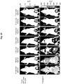
Figs. 1A-1C show the accumulation patterns ofconjugates day 1 to 7 using the Xenograph IVIS® system (color scale in units of photon/sec/cm2/steradian). Upper panel shows the fluorescent signals generated by the tumor (red fluorescence imaging) and lower panel shows the fluorescent signal generated by the conjugate (near-infrared fluorescence imaging). Matching of the signals generated by the tumor and by the conjugate suggests accumulation of the conjugate in the tumors. -
Figs. 2A-2C show the accumulation ofconjugates -
Figs. 3A-3C show the accumulation ofconjugates conjugate 4; and 8, 12 and 24 hrs for conjugate 41) post injection using the Xenograph IVIS® system. The accumulation profiles of the conjugates in prostate (upper panel) and ovarian (lower panel) tumors were nearly the same, wherein in both cases, the highest fluorescent level was observed at 8-11 (conjugate 1), 8-14 (conjugate 4) or 8-12 (conjugate 41 (Reference)) hrs after injection and the conjugate stayed in the tumor up to 48 hrs in the case ofconjugates - In one aspect, the present invention provides novel arginine-glycine-aspartic acid (Arg-Gly-Asp; RGD)-containing cyclic peptidomimetics, which are αvβ3 and αvβ5 integrin ligands, as defined above.
- The terms "RGD-containing cyclic peptidomimetic", "cyclic peptidomimetic" and "αvβ3 and αvβ5 integrin ligand" used herein interchangeably refer to a cyclic non-peptidic compound containing the RGD sequence, also referred to as the RGD motif, which mimics peptides having the RGD motif. The cyclic peptidomimetic of the present invention may be any cyclic compound having the general formula I, as defined above.
- As shown in detail in
Scheme 1 hereinafter, the RGD-containing cyclic peptidomimetic of the general formula I is a cyclic compound containing the RGD motif, in which a residue of an amino acid having a side chain amino group (A1) is linked by amide bonds, via its side chain amino group to the α-carboxyl group of the aspartic acid residue in the RGD motif on one side, and via its carboxyl group to a backbone NH on the other side, and said backbone NH is linked to the α-amino group of the arginine residue in the RGD motif via various possible bridging units. - The term "hydrocarbylene" refers to a divalent radical containing only carbon and hydrogen atoms that may be saturated or unsaturated, cyclic or acyclic, which may be derived from ethane, ethene or cyclopropane.

- In the group NHR, R is a hydrocarbylene as defined above.
- The term "amino acid" refers to both natural and non-natural amino acids in their L and D stereoisomers, and includesamino acids having a side chain amino group,more particularly lysine (Lys), diaminopropionic acid (Dap) and ornithine (Orn).
- In one embodiment, the RGD-containing cyclic peptidomimetic of the present invention is a cyclic compound of the general formula I, wherein X is -NH-R-, i.e., an urea moiety is formed with the α-amino group of the arginine residue, and R is a hydrocarbylene derived from ethane, ethene or cyclopropane.
- In another embodiment, the RGD-containing cyclic peptidomimetic of the present invention is a cyclic compound of the general formula I, wherein X is -O-R-, i.e., a carbamate moiety is formed with the α-amino group of the arginine residue, and R is a hydrocarbylene derived from ethane, ethene or cyclopropane.
- In yet another embodiment, the RGD-containing cyclic peptidomimetic of the present invention is a cyclic compound of the general formula I, wherein X is-S- or -S-R-, i.e., a carbamothio moiety is formed with the α-amino group of the arginine residue, and R is a hydrocarbylene derived from ethane, ethene or cyclopropane.
- The RGD-containing cyclic peptidomimetics of the present invention may be prepared by any method known in the art, e.g., as described in Materials and Methods hereinafter.
- In certain preferred embodiments, the RGD-containing cyclic peptidomimetic of the present invention is a cyclic compound of the general formula I, wherein X is -NH-R-, R is a hydrocarbylene derived from ethane and A1 is Dap, Orn or Lys.
- In other preferred embodiments, the RGD-containing cyclic peptidomimetic of the present invention is a cyclic compound of the general formula I, wherein X is -NH-R-, R is a hydrocarbylene derived from cyclopropane and A1 is Orn.
- In further preferred embodiments, the RGD-containing cyclic peptidomimetic of the present invention is a cyclic compound of the general formula I, wherein X is -O-R-, R is a hydrocarbylene derived from ethane and A1 is Dap or Lys.
- The αvβ3 and αvβ5 integrin ligands of the present invention accumulate in tumors expressing αvβ3 and αvβ5 such as ovarian carcinoma, colon, breast and prostate cancer, and therefore can be used in both diagnostic and therapeutic methods by conjugation to various "payload" moieties.
- In another aspect, the present invention thus relates to a conjugate of an RGD-containing cyclic peptidomimetic defined above, i.e., a cyclic peptidomimetic of the general formula I, and a moiety of a payload selected from a fluorescent probe, a photosensitizer, a chelating agent or a cytotoxic agent, linked to the α-amino group of the amino acid residue A1 in the peptidomimeticoptionally via a spacer.
- In one embodiment, the payload moiety of the conjugate is linked directly to the amino acid residue A1 of the cyclic peptidomimetic.
- In another embodiment, the payload moiety is linked to the amino acid residue A1 of the cyclic peptidomimetic via a spacer.
- The spacer linking the payload moiety to the amino acid residue A1 in the cyclic peptidomimetic of the present invention may be selected from a moiety of a natural or non-natural amino acid, a moiety of a small peptide having not more than 8 amino acids, a diamine residue, a C1-C25 hydrocarbylene, or a soluble polymer.
- In one embodiment, the spacer is a moiety of a natural or non-natural amino acid such as Gly, β-alanine (β-Ala), Phe, D-Phe, 1-naphthylalanine (1-Nal), D-1-naphthylalanine (D-1-Nal), γ-aminobutiric acid (GABA) and 3-(aminomethyl) benzoic acid. These spacers are linked via their α-carboxyl group to the α-amino group of A1 and via their α-amino group to a carboxyl group of the payload.
- In another embodiment, the spacer is a moiety of a small peptide having not more than eight amino acids. These spacers are linked via their C-terminal carboxyl group to the α-amino group of A1 and via their N-terminal amino group to a carboxyl group of the payload.
- In a further embodiment, the spacer is a diamine residue of the general formula -HN-R'-NH-, wherein R' is absent or is a divalent radical containing only carbon and hydrogen atoms that may be saturated or unsaturated, linear or branched, cyclic or acyclic, or aromatic, which may be derived from a C1-C12 alkane, a C2-C12 alkene, a C2-C12 alkyne, a C3-C10 cycloalkane, a C3-C10 cycloalkene, a C6-C14 mono- or polycyclic aromatic hydrocarbon, or a C6-C14 mono- or polycyclic aromatic hydrocarbon substituted by one or two C1-C2 alkyl, C2 alkenyl or C2 alkynyl. Examples of diamines from which such residues may be derived include hydrazine, 1,2-ethylenediamine, 1,3-propylenediamine, 1,4-diaminobutane, 1,5-diaminopentane, 1,6-diaminohexane, 1,7-diaminoheptane, 1,8-diaminooctane, 1,9-diaminononane, 1,10-diaminodecane, 1,11-diaminoundecane, 1,12-diaminododecane, p-phenylenediamine,
cyclopentane 1,3-diamine,cyclohexane 1,4-diamine,cycloheptane 1,4-diamine,cyclooctane 1,5-diamine, naphthalene-2,6-diamine and 9H-fluorene-3-6-diamine. - In still another embodiment, the spacer is a C1-C25 hydrocarbylene, preferably a C1-C10 alkylene or phenylene, substituted by two end functional groups through which the spacer is bound to the α-amino of the amino acid A1 of the cyclic peptidomimetic on one hand, and to the payload moiety on the other hand. Such end functional groups may be selected from OH, COOH, SO3H, COSH or NH2, thus forming an ether, ester, amide, urea, thioamide or sulfonamide group.
- In yet another embodiment, the spacer is a soluble polymer such as linear or branched polyethylene glycol (PEG) or copolymers thereof, polylactide (PLA) or copolymers thereof, polyesters having suitable functional groups based on PLA, polyglycolide (PGA), polycaprolactone (PCL), or their copolymers, or polyamides based on polymethacrylamide or their copolymers, said polymers having suitable functional groups for linking to the amino acid residue A1 of the cyclic peptidomimetic and to the payload moiety, said functional groups being, e.g., hydroxy, amino, carboxyl, mercapto, sulfonic acid group.
- Example 1 hereinafter describes the synthesis of various conjugates, herein identified by the Arabic numbers 1-36 in bold, in which different αvβ3 and αvβ5 integrin ligands of the general formula I are linked either directly or via a spacer to a fluorescent probe, in particular, BTA, FITC or dansyl; a bacteriochlorophyll derivative, in particular, Pd-BTA; or a chelating agent, in particular, DTPA or DOTA. The list of conjugates prepared, as well as their structural characteristics, is summarized in Table 1. Reference Example 2 describes the synthesis of various conjugates, herein identified by the Arabic numbers 41-48 (Reference) in bold, in which different αvβ3 and αvβ5 integrin ligands of the general formula II are linked directly to a moiety of the fluorescent probe BTA as a model payload. The list of conjugates prepared, as well as their structural characteristics, is summarized in Table 2. The chemical structures of the various payload moieties used, when linked to a cyclic peptidomimetic, are depicted in
Scheme 2. - Conjugates 1-36 were tested for binding to MLS human ovarian carcinoma cells, using both in vitro integrin binding assay and in vivo ovarian carcinoma model. Some of these conjugates were tested for binding to HT29 human colon carcinoma cell as well, both in vivo and in vitro, and conjugates 1 and 4 were further tested for binding to LNCaP prostate cancer cells, both in vitro and in vivo. Conjugates 41-48 (Reference) were tested for binding to MLS human ovarian carcinoma cells, using in vitro integrin binding assay, and the active conjugates were tested using an in vivo ovarian carcinoma model as well. Conjugates 41 (Reference) and 42 (Reference) were tested for binding to HT29 human colon carcinoma cells, both in vivo and in vitro, and conjugate 41 (Reference) was further tested for binding to LNCaP prostate cancer cells, both in vitro and in vivo.
- When screening the biological activity of different conjugates based on RGD-containing cyclic peptidomimetics of the general formula I, it has been found that certain structural characteristics of the cyclic peptidomimetic, i.e., the ring size of the cyclic compound and the size and structure of the diamine residue present in some of the cyclic compounds, as well as the spacer linking the cyclic compound and the payload moiety, may affect the biological activity of the conjugate as described hereinbelow.
- Example 3 hereinafter shows the biological activity of various fluorescent probe-conjugates comprising cyclic peptidomimetics of the general formula I with different ring sizes. The ring size of the cyclic peptidomimetic was altered by changing two structural parameters of the cyclic compound, in particular, (i) the amino acid residue linked via its α- or side-chain carboxyl group to the backbone NH and via its α- or side-chain amino group to the α-carboxyl group of the aspartic acid residue, i.e., A1 in the general formula I; and (ii) the radical bridging the backbone carbonyl and the backbone NH, i.e., radical X in the general formula I. The specific amino acid residues A1 used were residues of Dap, Dab (Reference), Orn or Lys, having one to four methylene units in the side chain, respectively; and the different radicals X used were -NH- (Reference), -NH(CH2)2-4- and -NH(CH2)6-, which, together with the backbone NH, form a moiety of either hydrazine or a certain alkyldiamine. As particularly shown, the biological activity of the conjugates tested increased with increasing the ring size of the cyclic peptidomimetic from 16 atoms to 19-20 atoms; however, it decreased with further increasing the ring size. These results indicate that whereas the urea bond bridging the α-amino group of the arginine residue and radical X makes the cyclic compound more rigid, a larger ring having up to 19-20 atoms is more flexible to adopt the desired conformation for binding to the integrin. On the other hand, in cases wherein the ring size of the cyclic peptidomimetic is higher than 20 atoms, the cyclic compound probably cannot adopt the desired conformation for binding to the integrin.
- Example 4 shows the biological activity of various BTA-conjugates comprising cyclic peptidomimetics of the general formula I having different diamine residues linked by amide bonds to either the α- or side-chain carboxyl group of the amino acid residue A1 and, via the backbone C=O, to the α-amino group of the arginine residue. The specific conjugates tested were such in which the amino acid residue A1 is Orn, the BTA moiety is directly linked to the N-terminal of the peptidomimetic ring, and the radical designated X is a radical of the formula - NH(CH2)2-4-, 1,3-dimethylbenzene-1,3-diyl or piperidine-1,4-diyl. As particularly shown, the biological activity of the conjugates in which an alkyldiamine residue is bridging A1 and the backbone C=O decreased with increasing the length of the alkyl chain. Furthermore, in cases the radical designated X was derived from m-xylene or piperidine, no biological activity was measured, indicating that the peptidomimetic rings in such conjugates are rigid and adopt a conformation undesirable for the interaction with the integrin.
- Example 5 shows the biological activity of various fluorescent probe-conjugates comprising cyclic peptidomimetics of the general formula I having different spacers linking the N-terminal of the cyclic peptidomimetic and the fluorescent probe moiety. The specific spacers used were moieties of different natural or non-natural amino acids, in particular, Gly, β-Ala, Phe, D-Phe, 1-Nal, D-1-Nal, GABA and 3-(aminomethyl) benzoic acid, or residues of different diamines, in particular, 1,2-ethylenediamine and 1,4-diaminobutane. As shown, BTA conjugates in which the fluorescent probe moiety is directly linked to the cyclic peptidomimetic showed high biological activity, probably because the BTA moiety does not interfere with the binding of the cyclic compound to the integrin. Contrary to that, conjugates in which Gly or β-Ala moieties were used as spacers, having an increased distance between the cyclic peptidomimetic and the BTA moiety, showed lower activity, probably due to the bulkiness of the BTA moiety. Interestingly, when the distance between the cyclic peptidomimetic and the BTA moiety was further increased using a GABA moiety as a spacer, the biological activity of the conjugate was higher than that of the conjugates in which Gly or β-Ala moieties were used as spacers, possibly indicating that GABA is long enough for giving more freedom to the cyclic peptidomimetic to bind to the integrin; however, not too long to cause folding of the BTA moiety over the peptidomimetic ring. In cases FITC and dansyl, which are smaller than BTA, were used, the distance between the fluorescent probe moiety and the N-terminal of the cyclic peptidomimetic had no influence on the biological activity of the conjugate. As further shown, BTA-conjugates in which Phe, 1-Nal, D-Phe or D-1-Nal moieties were used as spacers were more active than the corresponding conjugate in which a Gly moiety was used, probably because of the aromatic side chain of phenylalanine or naphthylalanine, which provides interaction with a hydrophobic pocket of the integrin. It is worth noting that the biological activity of the conjugate in which a D-Phe moiety was used as a spacer was higher than that of the conjugates in which Phe or 1-Nal moieties were used, indicating that the D configuration may fit the hydrophobic pocket of the integrin better than the L configuration. D-1-Nal is less reactive than D-Phe, indicating that the phenyl ring fit the hydrophobic pocket better than the naphthyl. It should be noted that conjugates 32 and 33, in which residues of 1,2-ethylenediamine or 1,4-diaminobutane, respectively, were used as spacers and an urea bond is formed between the cyclic peptidomimetic and the spacer, had biological activity similar to that of
conjugates 10 and 11, indicating that the urea bond has nearly the same activity as the amide bond and it does not influence the conformation of the peptidomimetic. The strong biological activity of conjugate 33, compared with that of conjugate 32, may be due to the distance of four methylene units between the peptidomimetic ring and the payload moiety, which gives more freedom to the peptidomimetic ring to interact with the binding site of the integrin. - On the other hand, Example 6 shows that BTA-conjugates comprising cyclic peptidomimetics of the general formula I in which an urea moiety is formed with the α-amino of the arginine residue had a biological activity similar to that of the corresponding conjugates in which a carbamate moiety is formed, indicating that the nature of the moiety formed with the α-amino of the arginine residue has no effect on the biological activity of the conjugate.
- Example 7 describes the synthesis of four unmetalated bacteriochlorophyll derivative-conjugates herein identified by the Arabic numbers 37-40, consisting of different αvβ3 and αvβ5 integrin ligands of the general formula I linked directly to a BTA derivative moiety in which the taurine was replaced by a different nucleophile. As shown, the biological activity of these conjugates, measured using an in vitro integrin binding assay, was similar, indicating that in these cases, the amino group has no effect on the biological activity and its behavior is nearly the same as that of the sulphonate in taurine.
- When screening the biological activity of different conjugates based on RGD-containing cyclic peptidomimetics of the general formula II, it has been found that certain structural characteristics of the cyclic peptidomimetic, i.e., the ring size of the cyclic compound and the characteristics of the amino acid residue A2, may affect the biological activity of the conjugate as described hereinbelow.
- Reference Examples 8-9 hereinafter show the biological activity of various BTA-conjugates comprising cyclic peptidomimetics of the general formula II with different ring size. The ring size of the cyclic peptidomimetic was altered by changing two structural parameters of the cyclic compound, in particular, (i) the amino acid residue linked to the α-carboxyl group of the aspartic acid residue and to the carboxyl group of A2, i.e. the amino acid residue A3; and (ii) the amino acid residue linked via its α-amino group to the backbone C=O and via its α-carboxyl group to the amino acid residue A3, i.e., the amino acid residue A2. In order to study the effect of A3 on the conjugate activity, BTA-conjugates having the same amino acid residues A1 and A2 but different amino acid residue A3, in particular, Dap, Dab, Orn and Lys, having one to four methylene units in the side chain, respectively, were tested. Similarly, in order to study the effect of A2 on the conjugate activity, BTA-conjugates having the same amino acid residues A1 and A3 but different amino acid residue A2, in particular, Phe, Val, D-Phe, Gly and Asp, were tested. As shown in Example 8, the biological activity of the conjugates tested decreased with increasing the ring size of the cyclic peptidomimetic from 20 atoms to 23 atoms, indicating that the optimum ring size is 20 atoms and that larger ring sizes do not fit the binding site of the integrin. Example 9 shows that the biological activity of conjugates with a hydrophobic amino acid residue A2 was higher than that of conjugates which are more polar, possibly due to the hydrophobic interactions with the hydrophobic pocket in the binding site of the integrin, and further suggests that the D configuration does not fit completely to the hydrophobic pocket.
- Example 10 shows the competitive binding level of certain conjugates of the present invention to human αvβ5 integrin, using an in vitro assay, and specifically demonstrates that
conjugates conjugates 5 and 11. - Example 11 describes a study in which the accumulation patterns of
conjugates day 1 to 7 post injection. As shown, these conjugates accumulated in the necrotic area of the tumor, indicating that the conjugates of the present invention can be used for diagnostic uses since the detection of necrotic cores is an important prognosis marker in various types of cancer, e.g., breast cancer, and the detection of tumor margins is essential for total removal of the tumor. - Example 12 describes a study in which the accumulation patterns of
conjugates conjugates - Example 13 describes a toxicity study of
conjugates - In view of all the aforesaid, in one embodiment, the payload moiety of the conjugate of the present invention is a moiety of a fluorescent probe such as BTA, FITC, dansyl, rhodamine, eosin and erythrosine.
- In preferred embodiments, the conjugate of the present invention is a conjugate of the RGD-containing cyclic peptidomimetic of the general formula I and a moiety of a fluorescent probe, wherein said fluorescent probe is BTA, linked directly to A1, X is -NH-R-, R is a hydrocarbylene derived from ethane, and A1 is Dap, Orn, or Lys (herein identified
conjugates - In other preferred embodiments, the conjugate of the present invention is a conjugate of the RGD-containing cyclic peptidomimetic of the general formula I and a moiety of a fluorescent probe, wherein said fluorescent probe is dansyl, linked directly to A1, X is -NH-R-, R is a hydrocarbylene derived from ethane, and A1 is Dap, Orn or Lys (herein identified conjugates 19, 18 and 16, respectively).
- In further preferred embodiments, the conjugate of the present invention is a conjugate of the RGD-containing cyclic peptidomimetic of the general formula I and a moiety of a fluorescent probe, wherein said fluorescent probe is BTA, linked directly to A1, X is -O-R-, R is a hydrocarbylene derived from ethane, and A1 is Dap or Lys (herein identified
conjugates 7 and 8, respectively). - In yet other preferred embodiments, the conjugate of the present invention is a conjugate of the RGD-containing cyclic peptidomimetic of the general formula I and a moiety of a fluorescent probe, wherein said fluorescent probe is BTA, linked via a spacer to A1, X is -NH-R-, R is a hydrocarbylene derived from ethane, A1 is Dap, and the spacer is a moiety of GABA or D-Phe, or a residue of 1,4-diaminobutane (herein identified conjugates 11, 28 and 33, respectively).
- In still further preferred embodiments, the conjugate of the present invention is a conjugate of the RGD-containing cyclic peptidomimetic of the general formula I and a moiety of a fluorescent probe, wherein said fluorescent probe is FITC, linked via a spacer to A1, X is -NH-R-, R is a hydrocarbylene derived from ethane, and (a) A1 is Dap, and the spacer is a β-Ala moiety (herein identified conjugate 12); or (b) A1 is Lys, and the spacer is a moiety of β-Ala or GABA (herein identified conjugates 13 and 14, respectively).
- In yet further preferred embodiments, the conjugate of the present invention is a conjugate of the RGD-containing cyclic peptidomimetic of the general formula I and a moiety of a fluorescent probe, wherein said fluorescent probe is dansyl, linked via a spacer to A1, X is -NH-R-, R is a hydrocarbylene derived from ethane, A1 is Dap or Lys, and the spacer is a Gly moiety (herein identified
conjugates 20 and 17, respectively). - Photodynamic therapy (PDT) is a non-surgical treatment of tumors in which non-toxic drugs, called photosensitizing agents, are administered along with light to generate cytotoxic reactive oxygen species in situ, which can inactivate cells. Being a binary treatment modality, PDT allows for greater specificity and has the potential of being more selective, yet not less destructive, when compared with commonly used chemotherapy or radiotherapy.
- Porphyrins have been employed as the primary photosensitizing agents in clinics. Optimal tissue penetration by light apparently occurs between 650-800 nm. Porfimer sodium (Photofrin®, Axcan Pharma Inc.) is a complex and inseparable mixture of monomers, dimers, and higher oligomers obtained from hematoporphyrin-IX by treatment with acids that has received FDA approval for treatment of esophageal and endobronchial non-small cell lung cancers.
- Due to their intense absorption in favorable spectral regions (650-850 nm) and their ready degradation after treatment, chlorophyll and bacteriochlorophyll derivatives have been identified as excellent sensitizers for PDT of tumors and to have superior properties in comparison to porphyrins. In particular, bacteriochlorophylls are of potential advantage compared to the chlorophylls as they show intense near-infrared bands, i.e., at considerably longer wavelengths than chlorophyll derivatives.
- Targeting photodynamic reagents for destruction of the tumor vasculature, as opposed to the tumor cells themselves, may offer therapeutic advantages since tumor-cell growth and development critically depend on continuous oxygen and nutrient supply. Furthermore, targeting the tumor vascular endothelial cell (EC) layer is expected to circumvent the poor penetration of tumor stroma by the therapeutic macromolecules. Although tumor blood vessels might be affected by the tumor microenvironment and acquire a tumor associated "signature", they are not malignant and less likely to develop drug resistance. Furthermore, when a targeted antivascular agent is also active against the tumor cells, additional gains in efficacy can be expected. Thus, by combining antivascular properties with antitumor cytotoxic activities in one drug, its efficacy can be expected to increase and the required effective cytotoxic dose may, consequently, decrease.
- Selective vascular targeting can rely on the differential susceptibility and consequent response to therapeutic agents of tumor and normal blood vessels. Alternatively, differential endocytosis may promote selective uptake of cytotoxic or other therapeutic agents. The integrins αvβ3, αvβ5 and α5β1 have been identified in expression patterns typical for angiogenic vascular endothelial cells associated, e.g., with tumors.
- Different strategies have been pursued to achieve this goal. Circulating peptides, peptidomimetics or antibodies that target specific sites in the vasculature are attractive as carriers for therapeutics and diagnostic agents offering theoretical advantages over such conjugates that directly target tumor cells, mostly situated beyond physiological barriers such as the blood vessel wall.
- Chaleix et al. (2003) disclose the synthesis of RGD-porphyrin conjugates as potential candidates for PDT application, in which the unmetalated porphyrin macrocycle is substituted at each of the
positions position 5 by a residue of a linear RGD-containing peptide linked to the macrocycle via a spacer arm. - In another embodiment, the payload moiety of the conjugate of the present invention is thus a moiety of a photosensitizer such as a porphyrin, a chlorophyll or a bacteriochlorophyll.
- It is an object of the present invention to provide photosensitizer conjugates that specifically target the sensitizer to the tumor vasculature. There are some advantages for vascular photosensitizer targeting over vascular targeting with conventional chemotherapy. First, during accumulation of a targeted conventional drug, it is often active, unless it is a prodrug, while the targeted photosensitizer is not active until locally illuminated. Second, a targeted conventional drug will bind and act also at undesirable targets presenting the homing address whereas the targeted photosensitizer will be activated only at the relevant illuminated site. Furthermore, PDT with photosensitizers targeted to the neovascular endothelial signatures in tumor may be remarkably selective in inducing photodynamic endothelial cell injury.
- Since the integrin avβ3 has been reported to play an important role in tumor metastasis and angiogenesis, which involve growth of new blood vessels from preexisting vasculatures during tumor growth, it may be a viable marker for tumor growth and spread. Therefore, noninvasive imaging methods for visual monitoring of αvβ3 integrin expression in real-time provides opportunities for assessing therapeutic intervention as well as for detection of metastasis.
- Integrins link the intracellular cytoskeleton of cells with the extracellular matrix by recognizing the RGD motif. RGD peptides interact with the integrin receptor sites, which can initiate cell-signaling processes and influence many different diseases. Thus, the integrin RGD binding site is an attractive pharmaceutical target. The integrin αvβ3 has an RGD binding site and peptides or peptidomimetics containing the RGD sequence home to, and act as antagonists of, αvβ3 integrin.
- In the bifunctional conjugates of the present invention, the homing property is provided by the RGD-containing cyclic peptidomimetic while the PDT effect is provided by the photosensitizer. These conjugates should be able to target the sensitizer to neovessels of primary solid tumors and possibly respective metastases for the purpose of diagnosis and for photodynamic destruction. They can further act as antiangiogenic agents and initiate apoptotic destruction of neo-endothelial and blood exposed tumor cells.
- In preferred embodiments, the payload moiety is a porphyrin, a chlorophyll or bacteriochlorophyll derivative that may be metalated or unmetalated and optionally substituted in the periphery by different substituents such as alkyl, aryl, heteroaryl and/or functional groups. These functional groups may be selected from positively charged groups, negatively charged groups, basic groups that are converted to positively charged groups under physiological conditions, and acidic groups that are converted to negatively charged groups under physiological conditions.
- The term "a positively charged group" refers to a cation derived from an N-containing group or from an onium group not containing N. Since tumor endothelium is characterized by an increased number of anionic sites, positively charged groups or basic groups that are converted to positively charged groups under physiological conditions may enhance the targeting efficiency of the conjugates of the present invention.
- The term "a negatively charged group" refers to an anion derived from an acid and includes carboxylate (COO-), thiocarboxylate (COS-), sulfonate (SO3 -), and phosphonate (PO3 2-), and the "acidic group that is converted to a negatively charged group under physiological conditions" includes the carboxylic (-COOH), thiocarboxylic (-COSH), sulfonic (-SO3H) and phosphonic (-PO3H2) acid groups.
- In more preferred embodiments, the payload moiety is a chlorophyll or, most preferably, a bacteriochlorophyll derivative that may be a natural or a synthetic non-natural derivative of chlorophyll or bacteriochlorophyll, including compounds in which modifications have been made in the macrocycle, and/or in the periphery and/or the central Mg atom may be absent or it is replaced by other metal atom suitable for the purpose of diagnosis and/or for the purpose of PDT. Examples of such metals include Pd, Pt, Co, Ni, Sn, Cu, Zn, Mn, In, Eu, Fe, Au, Al, Gd, Er, Yb, Lu, Ga, Y, Rh, Ru, Si, Ge, Cr, Mo, Re, Tl and Tc and isotopes thereof.
- In one particular preferred embodiment, the conjugate of the present invention is a conjugate of the RGD-containing cyclic peptidomimetic of the general formula I and a moiety of a bacteriochlorophyll derivative, wherein said bacteriochlorophyll derivative is Pd-BTA, linked directly to A1, X is -NH-R-, R is a hydrocarbylene derived from ethane and A1 is Dap (herein identified conjugate 34).
- In a further embodiment, the payload moiety of the conjugate of the present invention is a chelating agent, i.e., an agent capable of chelating a radionuclide such as technetium-99m (99mTc). Examples of such chelating agents include DTPA and DOTA. Such conjugates may be useful as radio imaging and radiotherapeutic agents.
- In preferred embodiments, the conjugate of the present invention is a conjugate of the RGD-containing cyclic peptidomimetic of the general formula I and a moiety of a chelating agent, wherein said chelating agent is DTPA or DOTA, linked directly to A1, X is -NH-R-, R is a hydrocarbylene derived from ethane and A1 is Dap (herein identified conjugates 35 and 36, respectively).
- Since most of the currently used chemotherapeutic agents are toxic also to normal cells, the development of targeted chemotherapy, i.e., chemotherapeutic drugs specifically targeted to tumor cells, is of high importance. Targeted cytotoxic peptide conjugates are hybrid molecules composed of a peptide carrier, which binds to receptors on tumor cells and a cytotoxic moiety. This approach effectively increases the specificity and efficacy of the cytotoxic agent in chemotherapy, and should decrease toxic side effects as well.
- Thus, in still a further embodiment, the payload moiety of the conjugate of the present invention is a cytotoxic agent.
- In one preferred embodiment, the cytotoxic agent of the present invention is an anthracycline chemotherapeutic agent. The anthracycline chemotherapeutic agent may be any chemotherapeutic agent of the anthracycline family including doxorubicin (also known as adriamycin), daunorubicin, epirubicin, idarubicin and mitoxantrone. In a more preferred embodiment, the anthracycline chemotherapeutic agent is doxorubicin, which is a quinine-containing anthracycline and is the most widely prescribed and effective chemotherapeutic agent utilized in oncology. Doxorubicin is indicated in a wide range of human malignancies, including tumors of the bladder, stomach, ovary, lung and thyroid, and is one of the most active agents available for treatment of breast cancer and other indications, including acute lymphoblastic and myelogenous leukemias, Hodgkin's and non-Hodgkin's lymphomas, Ewing's and osteogenic bone tumors, soft tissue sarcomas, and pediatric cancers such as neuroblastoma and Wilms' tumors.
- In other preferred embodiments, the cytotoxic agent is a mitotic inhibitor such as paclitaxel, currently used for the treatment of patients with lung, ovarian, breast cancer, head and neck cancer, and advanced forms of Kaposi's sarcoma, as well as for the prevention of restenosis, a topoisomerase I inhibitor such as camptothecin, or a topoisomerase II inhibitor such as ellipticine.
- In a further aspect, the present invention provides a pharmaceutical composition comprising a conjugate of an RGD-containing cyclic peptidomimetic and a payload moiety as defined above, or a pharmaceutical acceptable salt thereof, and a pharmaceutically acceptable carrier.
- In one embodiment, the pharmaceutical composition comprises a conjugate of a cyclic peptidomimetic as defined above, i.e., a cyclic peptidomimetic of the general formula I, and a moiety of a fluorescent probe. In preferred embodiments, the pharmaceutical composition comprises a conjugate selected from the group of conjugates consisting of
conjugates - In another embodiment, the pharmaceutical composition comprises a conjugate of a cyclic peptidomimetic as defined above, i.e., a cyclic peptidomimetic of the general formula I, and a moiety of a photosensitizer as defined above. In a preferred embodiment, the pharmaceutical composition comprises a conjugate selected from the group of conjugates consisting of conjugates 34 and 37-40 defined above. Such compositions may be used in photodynamic therapy (PDT). In one embodiment, the pharmaceutical composition is for use in oncology, particularly for PDT of tumors. Any suitable solid tumor is encompassed by the invention, both primary tumors and metastasis, of tumors selected frommelanoma, colon, breast, lung, prostate, brain or head and neck cancer. In another embodiment, the pharmaceutical composition is for use in non-oncologic diseases, for PDT of nonneoplastic tissue or organ. In one embodiment, the pharmaceutical composition is used for treatment of vascular diseases such as age-related macular degeneration (AMD) or disorders such as obesity by limiting vascular supply to adipose tissue and thus inhibiting its growth.
- In a further embodiment, the pharmaceutical composition comprises a conjugate of a cyclic peptidomimetic as defined above, i.e., a cyclic peptidomimetic of the general formula I, and a moiety of an agent capable of chelating a radionuclide. In a preferred embodiment, the pharmaceutical composition comprises conjugate 35 or 36 defined above. Such compositions, when labeled with suitable radionuclides, may be used for radio imaging or radiotherapy.
- In yet another embodiment, the pharmaceutical composition comprises a conjugate of a cyclic peptidomimetic as defined above, i.e., a cyclic peptidomimetic of the general formula I, and a moiety of a cytotoxic agent as defined above. Such compositions may be used for targeted chemotherapy.
- The pharmaceutical composition provided by the present invention may be prepared by conventional techniques, e.g., as described in Remington: The Science and Practice of Pharmacy, 19th Ed., 1995. The composition may be in solid, semisolid or liquid form and may further include pharmaceutically acceptable fillers, carriers or diluents, and other inert ingredients and excipients. Furthermore, the pharmaceutical composition can be designed for a slow release of the conjugate. The composition can be administered by any suitable route, e.g. intravenously, orally, parenterally, rectally, or transdermally. The dosage will depend on the state of the patient, and will be determined as deemed appropriate by the practitioner.
- The route of administration may be any route, which effectively transports the active compound to the appropriate or desired site of action, the intravenous route being preferred. If a solid carrier is used for oral administration, the preparation may be tabletted, placed in a hard gelatin capsule in powder or pellet form or it can be in the form of a lozenge. If a liquid carrier is used, the preparation may be in the form of a syrup, emulsion or soft gelatin capsule. Tablets, dragees or capsules having talc and/or a carbohydrate carrier or binder are particularly suitable for oral application. Preferable carriers for tablets, dragees or capsules include lactose, cornstarch and/or potato starch.
- In still a further aspect, the present invention relates to a conjugate of an RGD-containing cyclic peptidomimetic and a payload moiety as defined above, or a pharmaceutically acceptable salt thereof for diagnostic purposes, photodynamic therapy (PDT), radio imaging or radiotherapy, or targeted chemotherapy.
- The invention will now be illustrated by the following Examples.
-
- (i) Materials. 2-Chlorotritylchloride resin, Fmoc-Asp-O-Allyl, Fmoc-Gly-OH, Fmoc-Arg(Pbf)-OH, Fmoc-β-Ala-OH, Fmoc-GABA-OH, mono Fmoc-diamines, HOBt, PyBOP, HATU and HOAt were purchased from Novabiochem (USA). Fmoc-Dap(Alloc)-OH, Fmoc-Dab(Alloc)-OH and Fmoc-Lys(Alloc)-OH were purchased from Bachem (Switzerland). Fmoc-Orn(Alloc)-OH, 1-Fmoc-4-aminopiperidine hydrochloride and 4-(Boc-aminomethyl)-aniline were purchased from NeoMPS (France). FITC, dansyl chloride, DIEA, DIC, DMBA, diethyldithiocarbamic acid sodium salt, TFE, TIS, TFA, dry DCM and MeOH were purchased from Sigma (USA). Tetrakis (triphenylphosphine) palladium was purchased from Acros (Belgium). DMF, DCM and acetonitrile were purchased from J.T.Baker (USA). DTPA and DOTA were purchased from Macrocyclics (USA).
UV-Vis spectra were obtained using a Shimadzu 1240UV-Vis spectrophotometer. HPLC MS analysis was obtained using an Agilent 1100 HPLC equipped with an YMC Pro-RP-C18 reverse phase column, connected to an Applied Biosystems 150EX single-quad mass spectrometer. HPLC analyses were conducted (unless noted otherwise) at standard conditions: 20-95% acetonitrile in water (pH=4.5, maintained by acetic acid) gradient over 30 minutes, at a flow rate of 0.2 ml/min. Preparative HPLC was performed using Waters Delta Prep 4000 system equipped with a Waters 486 UV-VIS tunable absorbance detector and Waters fraction collector, controlled by Millenium v3.05 program. The flow rate was set to 75 ml/min, using a preparative column (Vydac C18, 218TP101550, 50 x 250mm, 10-15 µm). Solvents used in the HPLC purification were Solvent A (50 mM solution of ammonium acetate in H2O) and Solvent B (acetonitrile). ELISA plates were read on a Thermo Labsystems Multiscan Spectrum instrument. Fluorescent imaging was carried out using aXenogen IVIS ® 100 Series Imaging System (Alameda, California). - (ii) General procedure for the coupling of mono Fmoc-diamine to H-Arg(Pbf)-Gly-Asp(aO-Allyl)-2-chlorotrityl resin. Mono Fmoc-diamine hydrochloride (1.05 mmol) was dissolved in DCM (10 ml). DIEA (1.26 mmol) was added to the solution and stirred for 1 min followed by addition of BTC (0.35 mmol) and DIEA (3.15 mmol). The solution obtained was added to 0.21 mmol peptidyl-resin, pre-washed with DCM, and was allowed to react for 1 hr. After coupling, the resin was washed with DCM (3x6 ml, 1 min each) and DMF (6x6 ml, 1 min each). Coupling completion was monitored by qualitative ninhydrin test (Kaiser Test).
- (iii) General procedure for the coupling of BTA or Pd- BTA to peptidyl-resin. BTA (0.42 mmol), PyBOP (0.42 mmol) and HOBt (0.42 mmol) were dissolved in DMF (10 ml), and DIEA (1.89 mmol) was then added to the solution and stirred for 5 min. The solution obtained was added to 0.21 mmol peptidyl-resin and was shaken for 2 hrs under argon. After coupling, the resin was washed with DMF (6-8x6 ml, 1 min). Coupling completion was monitored by qualitative ninhydrin test. The coupling of Pd-BTA to the cyclic peptidyl resin was performed under the same coupling conditions as described for BTA.
- (iv) General procedure for the coupling of FITC to peptidyl-resin. A solution of FITC (0.63 mmol) in DMF (5 ml) was added to 0.21 mmol peptidyl-resin and was shaken for 1.5 hrs. After coupling, the resin was washed with DMF (6x6 ml, 1 min each). Coupling completion was monitored by qualitative ninhydrin test.
- (v) General procedure for the coupling of dansyl chloride to peptidyl-resin. A solution of dansyl chloride (1.05 mmol) and DIEA (1.47 mmol) in DCM (5 ml) was added to 0.21 mmol peptidyl-resin, pre-washed with DCM, and was allowed to react for 1 hr. After coupling, the resin was washed with DCM (5x5 ml, 1 min) and DMF (2x5 ml, 1 min). Coupling completion was monitored by qualitative ninhydrin test.
- (vi) Reparation of protected dipeptide Fmoc-Arg(Pbf)-Gly-OH, a building block for the peptide synthesis. A solution of Fmoc-Gly-OH (4.162 gr; 14 mmol) and DIEA (9.755 gr; 56 mmol) in dry DCM (100 ml) was stirred with 10 gr of 2-chlorotrityl chloride resin (substitution 1.4 mmol/gr) for 1 hr at RT. The mixture was transferred to a reactor equipped with a sintered glass bottom and the resin was washed with DCM/MeOH/DIEA (17:2:1) (3x100 ml), DCM (3x100 ml), DMF (3x100 ml), DCM (2x100 ml), MeOH (2x100 ml) and DMF/DCM (1:1) (3x100 ml). Fmoc-group was removed by treatment with 5% piperidine in DMF/DCM (1:1) (100 ml, 10 min), followed by 20% piperidine in DMF (100 ml, 5 min and 2x15 min) and washing the resin with DMF (7x100 ml). Fmoc-Arg(Pbf)-OH (18.17 gr; 28 mmol) in DMF (130 ml) was activated with DIC (4.34 ml; 28 mmol) and HOBt (4.29 gr; 28 mmol) for 15 min at RT and was added to the reaction vessel. The mixture was shaken for 2 hrs at RT. The peptidyl-resin was washed with DMF (5x100 ml), DCM (3x100 ml), MeOH (2x100 ml) and DCM (3x100 ml), and was dried in vacuum for 3 hrs. The protected dipeptide was cleaved from the resin by stirring with a solution of AcOH/TFE/DCM (1:1:3) (250 ml) for 1 hr at RT. The resin was filtered and washed with the same solution (3x50 ml). The combined filtrates were mixed with n-hexane to remove AcOH as an azeotrope and were evaporated to give an oily residue, which solidified upon treatment with cold ether (1 1). Filtration and washing with cold ether (150 ml) afforded a white powder (8.64 g; 87.5%) with homogeneity of about 99% (HPLC). C36H43N5O8S. MS (LC-MS) calculated m/z=705.84; found: 706.30 (M+H). The product was used without further purification.
- (vii) General procedure for the cleavage of the peptide conjugate from the resin. After conjugation, the peptidyl-resin was washed with DMF (5x3 ml) and DCM (5x3 ml), and was then dried under reduced pressure for 3 hrs. The peptide conjugate was cleaved from the resin using a cleavage cocktail of TFA/thioanisole/H2O/TIS (85:5:5:5) (6 ml) for 5 min at 0°C and then for 1 hr at RT. The resin was filtered and washed with the same cleavage cocktail (4 ml). The combined filtrates were evaporated by a stream of N2 to about half of the volume, and the peptide was precipitated by addition of cold ether (25 ml). Centrifugation and decantation of ether layer and additional treatment with cold ether (2x25 ml) afforded the unprotected peptide that was dried in vacuum for 6 hrs. The crude product was purified by RP-HPLC.
- (viii) Integrin binding test by ELISA. Nunc immuno-module strips (Nunclon, Cat#167008, Daniel Biotech, Israel) were coated for overnight with 2 µg/ml human integrin αvβ3 (Chemicon, Cat#CC1020, Biotest, Israel) dissolved in 0.06 M carbonate-bicarbonate buffer. Strips were blocked for 2 hrs at RT with 2% bovine serum albumin (BSA) (Sigma, Cat#A-9647, Israel) in phosphate buffered saline (PBS) (Biological Industries, Israel). A mixture of c[RGDfK]-biotin (10-3 M) and a test compound at different concentrations (10-2, 10-3, 10-4 and 10-5 M) diluted in assay buffer (50 mM Tris HCl, pH=7.7, 0.5% BSA, 0.15 M NaCl, 0.01% Tween 20) was added to the coated strips and was incubated overnight at RT with shaking. After washing with PBS, anti-biotin antibodies labeled by alkaline phosphatase (1:200) (Miltenyi Biotec, Almog, Israel) were added and incubated for 1 hr at RT. The samples were incubated with p-nitrophenyl phosphate substrate (p-NPP, Calbiochem, Mercury, Israel) and read at 405 nm.
- (ix) In-vivo ovarian carcinoma model. Female CD-1 nude mice (7-9 weeks old, 23-28 gr) were anaesthetized and subcutaneously (SC) implanted with MLS human ovarian carcinoma cells (obtained from Prof. M. Neeman, the Weizmann Institute of Science, Israel) suspension (2-3x10⁶ cells/mouse). Tumors reached treatment size, diameter 6-8 mm, within 2-3 weeks.
Animals were anaesthetized by gas with mixture of 7:3 N2O:O2 containing 2% isofluorane (Medeva, Bethlehem, PA) or by intraperitoneal (IP) injection with mixture of 5 mg/kg ketamine (Rhone Merieux, Lyon, France) and 1 mg/kg pompun (Bayer, Leverkusen, Germany) (85:15, v:v). - (x) In-vivo colon carcinoma model. This model is similar to the in vivo ovarian carcinoma model described in (ix) above, except for the fact that HT29 human colon carcinoma cells (ATCC, USA, 2-3x10⁶ cells/mouse) were used instead of MLS human ovarian carcinoma cells.
- (xi) In-vivo prostate cancer model. LNCaP cells (3x10⁶ cells/mouse) were SC implanted on back of severe combined immunodeficiency (SCID) mice. Tumors were allowed to grow for 60-70 days. When tumor reaches the treatment size (0.7-0.8 cm3), the animals were anaesthetized and the test compound solution was intravenously (IV) injected. The images on IVIS were taken at 8, 11, 14, 24 and 48 hrs after injection.
- (xii) In-vivo breast cancer model. Female CD-1 nude mice (6-8 weeks old, 20-25 g, obtained from Harlan Biotech Israel, Rehovot, Israel) were implanted with MDA-MB-231-RFP human breast cancer cells (4x10⁶ cells/mouse). These cells are, in fact, MDA-MB-231 human breast cancer cells (ATCC, USA) transfected with red fluorescence protein (RFP) gene thus possessing red fluorescence. When tumors reached the size of 1-1.5 cm3 for necrotic tumors, mice were anaesthetized by IP injection of 30 µl mixture of 85:15 ketamine:xylazine, and the test conjugate (15 mg/kg) was then injected to the tail vein.
- (xiii) Fluorescent imaging protocol for BTA- RGD conjugates. Test compounds (8 mg/kg) were injected into the tail vein of IP anaesthetized tumor-bearing mice. Images of gas-anaesthetized animals were taken at 6, 8, 10, 12, 14 and 24 hrs (in some cases also at 48 and 72 hrs) after
injection using IVIS ® 100 Series Imaging System. The excitation and emission filters were set in the IVIS to 710-760 nm and to 810-860 nm, respectively. The emission filter with wavelength closest to the emission peak of the compound was selected among available filters in the standard configuration of IVIS. - (xiv) Fluorescent imaging protocol for FITC-RGD conjugates. Test compounds (8 mg/kg) were injected into the tail vein of IP anaesthetized tumor-bearing mice. Images of gas-anaesthetized animals were taken at 6 and 8 hrs after injection. Animals were sacrificed at 8 hrs, the organs (tumor, kidney, liver) were excited and images of the organs were taken using
IVIS ® 100 Series Imaging System. The excitation and emission filters were set in the IVIS to 445-490 nm and to 515-575 nm, respectively. The emission filter with wavelength closest to the emission peak of FITC was selected among available filters in the standard configuration of IVIS. - (xv) In-vitro binding assay. MLS human ovarian carcinoma cells were cultured as monolayers in minimum essential medium (MEM-alpha) containing 1 g/l D-glucose, pH 7.4, 10% fetal calf serum (FCS), glutamine (2 mM), penicillin (0.06 mg/ml) and streptomycin (0.1 mg/ml), and were grown at 37°C in 5% CO2-humidified atmosphere. At 48 hrs before experiment, cells were seeded at 6 well plates (3x10⁵ cells/well).
Expression of α v β 3 integrin on MLS cells. The cells were grown on cover slips. Following overnight serum starvation, fixation with 4% paraformaldehyde (Sigma, Israel) and permeabilization with 0.2% Triton X-100 (Sigma, Israel), cells were incubated in blocking solution (10% of horse serum) (Biological Industries, Israel) for 1 hr at RT. Cells were then incubated with mouse anti-human αvβ3 integrin antibodies (1:100) (Chemicon, Biotest, Israel) for 1 hr at RT. Secondary rabbit FITC-labeled anti-mouse IgG (1:200) (Sigma, Israel) were applied to cells for 1 hr at RT. Imaging was performed by fluorescent microscope (Nikon Optiphot2, Japan) equipped with a digital camera (DVC Company, Inc., Austin, TX).
In vitro binding assay. RGD-conjugates were initially dissolved in DMSO to yields 4x10⁻³ M. The stock solutions were then diluted 1:40 in culture medium and added into MLS or HT29 cells (100 µM/well). Cells were incubated at 37°C in 5% CO2-humidified atmosphere for 3 hrs. Cells were then washed 3 times with PBS and images were performed on aXenogen IVIS ® 100 Series Imaging System. The excitation and emission filters set in the IVIS for BTA-RGD were 710-760 nm and 810-860 nm, respectively, and for FITC-RGD were 445-490 nm and 515-575 nm, respectively. - (xvi) Competitive binding experiments (determination of IC50). Immuno-module strips MAXISORP (Nunc, Danyel Biotech, Israel) were coated with 50 µl/well of 2 µg/ml human αvβ3 integrin (Chemicon, USA) overnight and blocked with 2% BSA (Sigma, Israel) for 2 hrs at RT. After wash with Tris buffered saline-Tween (TBST) buffer, a mixture of RGD peptide c[RGDfK]-biotin (10-3M) and the tested RGD-conjugate at different concentrations (10-2, 10-3, 10-4 and 10-5 M) was added in triplicates and was incubated overnight and shaken at RT. After wash with PBS buffer, anti-biotin antibodies labeled by alkaline phosphatase (1:200) (Miltenyi Biotec, Germany) were added and incubated for 1 hr at RT. The samples were incubated with p-NPP substrate and read at 405 nm on Multiscan Spectrum (Labotal, Israel). The data were plotted to graph of dependence of binding percent out of concentration of RGD-conjugate and IC50 value was determined.
- Tripeptide Fmoc-Arg(Pbf)-Gly-Asp(αO-Allyl)-2-chlorotrityl resin was prepared on a solid phase by coupling of Fmoc-Arg(Pbf)-Gly-OH to resin bound H-Asp-O-Allyl residue.
- Attachment of the first amino acid was performed by stirring 2-chlorotrityl chloride resin (300 mg, substitution 1.4 mmol/gr) with a solution of Fmoc-Asp-O-Allyl (83 mg, 0.21 mmol) and DIEA (147 µl, 0.84 mmol) in 5 ml dry DCM for 1 hr at RT to give a loading of about 0.7 mmol/g. Upon coupling completion, the resin was treated (washes and Fmoc-removal) as described in Materials and Methods with corresponding volumes of solvents and reagents solution. Fmoc-Arg(Pbf)-Gly-OH (223 mg, 0.315 mmol), HOBt (48 mg, 0.315 mmol) and DIC (49 µl, 0.315 mmol) were dissolved in 5 ml DMF and stirred at RT for 20 min. The resulting solution was added to the washed H-Asp-O-Allyl-resin and the mixture was shaken for 2 hrs at RT. The peptidyl-resin was washed with DMF (5x5 ml). Removal of Fmoc group was carried out by addition of 20% piperidine (5 ml) in DMF (2x15 min) followed by DMF wash (7x5 ml, 1 min). The coupling of mono Fmoc-diamine was performed as described in Materials and Methods followed by Fmoc deprotection and DMF wash. Coupling of Fmoc-Lys(Alloc)-OH, as well as of Fmoc-Dap(Alloc)-OH, Fmoc-Dab(Alloc)-OH and Fmoc-Orn(Alloc)-OH, to the tetra-peptide was performed by addition of a DMF solution (5 ml) of Fmoc-Lys(Alloc)-OH (0.63 mmol), pre-activated (for 15 min) with HOBt (0.63 mmol) and DIC (0.63 mmol), and coupling time of 1 hr. After coupling, the resin was washed with DMF (6x5 ml, 1 min). Coupling completion was monitored by qualitative ninhydrin test (Kaiser Test). Allyl and Alloc deprotection took place by stirring the peptidyl-resin with a solution of [(C6H5)3P]4Pd0 (0.252 mmol) and DMBA (3.57 mmol) in DCM (5 ml) for 2 hrs at RT under argon. The resin was washed with DCM (3x5 ml, 1 min), DMF (3x5 ml, 1 min), diethyldithiocarbamic acid sodium salt (0.5% in DMF, 4x5 ml, 2 min) and finally with DMF (5x5ml, 1 min). On-resin cyclization was done by using a solution of PyBOP (0.63 mmol) and DIEA (1.26 mmol) in DMF (4 ml) for 2 hrs at RT. After Fmoc deprotection, conjugation of BTA to the unprotected peptidyl-resin, as well as cleavage of the peptide from the resin, were performed as described in Materials and Methods. The products were purified by RP-HPLC.
- The synthesis of H-Arg(Pbf)-Gly-Asp(αO-Allyl)-2-chlorotrityl resin was carried out as described above for Method A.
- Coupling of Fmoc-NH-NH2 hydrochloride to the unprotected tripeptide. Mono Fmoc-hydrazine hydrochloride (1.05 mmol) was dissolved in a 1:1 mixture of dioxane and 1,3-dichloropropane (10 ml). DIEA (1.26 mmol) was added to this solution and was stirred for 1 minute followed by addition of BTC (0.35 mmol) and DIEA (3.15 mmol). The solution was added to the peptidyl-resin (0.21 mmol) (prewashed with 1:1 dioxane: 1,3-dichloropropane) and was allowed to react for 1 hr at 55°C. After coupling, the resin was washed with DCM (3x6 ml, 1 min) followed by DMF (6x6 ml, 1 min). Coupling completion was monitored by qualitative ninhydrin test (Kaiser Test). The rest of the synthesis was as described for Method A. The product was purified by RP-HPLC.
- The synthesis of the cyclopentapeptide was carried out as described above for Method A.
- Coupling of the amino acid spacers to the cyclopentapeptide. Fmoc amino acid (0.63 mmol of Fmoc-Gly-OH, Fmoc-β-Ala-OH, Fmoc-GABA-OH, Fmoc-Phe-OH, Fmoc-D-Phe-OH, Fmoc-1-Nal-OH, Fmoc-D-1-Nal-OH or Fmoc-3-aminomethylbenzoic acid) was dissolved in DMF (5 ml) and HOBt (0.63 mmol) and DIC (0.63 mmol) were then added and allowed to react for 15 min. The solution was added to the Fmoc-deprotected cyclopentapeptide-2-chlorotrityl resin (0.21 mmol) and was shaken for 1 hr. The resin was washed with DMF (6x5 ml, 1 min) followed by Fmoc deprotection. Coupling of BTA and cleavage of the peptide from the resin were performed as described in Materials and Methods. The products were purified by RP-HPLC.
- Coupling of the diamines spacers to the cyclopentapeptide. Mono Fmoc-diamine hydrochloride (1.05 mmol Fmoc-ethylenediamine hydrochloride or Fmoc-diaminobutane hydrochloride) was dissolved in DCM (10 ml). DIEA (1.26 mmol) was added to the solution and stirred for 1 min followed by addition of BTC (0.35 mmol) and DIEA (3.15 mmol). The solution obtained was added to 0.21 mmol peptidyl-resin, pre-washed with DCM, and was allowed to react for 1 hr. After coupling, the resin was washed with DCM (3x6 ml, 1 min each) and DMF (6x6 ml, 1 min each). Coupling completion was monitored by qualitative ninhydrin test (Kaiser Test). Coupling of BTA and cleavage of the peptide from the resin were performed as described in Materials and Methods. The products were purified by RP-HPLC.
- The synthesis of H-Arg(Pbf)-Gly-Asp(αO-Allyl)-2-chlorotrityl resin was carried out as described above for Method A.
- Coupling of Fmoc-Glycinol to the Fmoc - deprotected tripeptide . Fmoc-Glycinol (1.05 mmol) was dissolved in DCM (10 ml), and BTC (0.35 mmol) was then added to this solution followed by addition of DIEA (3.15 mmol). After stirring for 5 min, the solution obtained was added to peptidyl-resin (0.21 mmol) pre-washed with DCM and allowed to react for 1 hr at RT. The resin was washed with DCM (3x6 ml, 1 min) and DMF (
6x6 ml 1 min). Coupling completion was monitored by qualitative ninhydrin test (Kaiser Test). The rest of the synthesis was carried out as described for Method A. Cleavage of the peptide conjugate from the resin was performed with TFA solution (6 ml) containing 15% DCM + 5% TIS and 5% thioanisole. The products were purified by RP-HPLC. - The synthesis of cyclopentapeptide was carried out as described above for Method A. After Fmoc deprotection, a solution of Fmoc-β-Ala-OH or Fmoc-GABA-OH (0.63 mmol), HOBt (0.63 mmol) and DIC (0.63 mmol) in DMF (5 ml) was mixed for 15 min, was added to the peptidyl-resin (0.21 mmol) and was allowed to react for 1 hr. The resin was washed with DMF (6x5 ml, 1 min). Removal of Fmoc group was carried out by addition of 20% piperidine in DMF (2x15 ml, 15 min) followed by DMF wash (6x5ml, 1 min). The coupling of FITC to the unprotected peptidyl-resin and cleavage of the peptide from the resin were performed as described in Materials and Methods. The products were purified by RP-HPLC.
- The synthesis of cyclic peptide was carried out as described above for Method A, and after Fmoc-deprotection, the compounds were directly reacted with dansyl chloride as described in Materials and Methods.
- Compounds containing a spacer were reacted first with Fmoc-Gly-OH or, alternatively, with Fmoc-β-Ala-OH, under the same conditions as described for Method E, followed by coupling with dansyl chloride. Cleavage of the peptide from the resin was performed as described in Materials and Methods. The products were purified by RP-HPLC.
- The synthesis of cyclic peptide was carried out as described above for Method A. After Fmoc-deprotection, a solution of DTPA-tetra (t-Bu ester) (0.42 mmol) in DMF (3 ml) activated by HATU (0.42 mmol), HOAt (0.42 mmol) and DIEA (0.42 mmol) was added to the peptidyl resin (0.14 mmol) and shaken for 2 hrs at RT. The resin was washed with DMF (4 ml, 5 times, 1 min each time). Cleavage of the peptide from the resin was performed as described in Materials and Methods.
- The synthesis of cyclic peptide was carried out as described above for Method A. After Fmoc-deprotection, a solution of DOTA-tris (t-Bu ester) (0.42 mmol) in DMF (3 ml) activated by HATU (0.42 mmol), HOAt (0.42 mmol) and DIEA (0.42 mmol) was added to the peptidyl resin (0.14 mmol) and shaken for 3 hrs at 60°C. The resin was washed with DMF (4 ml, 5x1 min). Cleavage of the peptide from the resin was performed as described in Materials and Methods.
- Table 1 lists the conjugates synthesized and the structural characteristics thereof.
Table 1: Conjugates based on cyclic peptidomimetics of the general formula I synthesized and method of synthesis Conjugate X* R* A1* Spacer Payload Method MW 1 NHR (CH2)2 Dap - BTA A 1217.3 2 (Reference) NH - Dap - BTA B 1189.8 3 (Reference) NHR (CH2)2 Dab - BTA A 1231.3 4 NHR (CH2)2 Orn - BTA A 1245.3 5 NHR (CH2)2 Lys - BTA A 1259.4 6 (Reference) NHR (CH2)2 Lys - BTA A 1259.4 7 OR (CH2)2 Dap - BTA D 1218.2 8 OR (CH2)2 Lys - BTA D 1260.3 9 NHR (CH2)2 Dap Gly moiety BTA C 1275.2 10 NHR (CH2)2 Dap β-Ala moiety BTA C 1289.2 11 NHR (CH2)2 Dap GABA moiety BTA C 1303.2 12 NHR (CH2)2 Dap β-Ala moiety FITC E 960.7 13 NHR (CH2)2 Lys β-Ala moiety FITC E 1002.7 14 NHR (CH2)2 Lys GABA moiety FITC E 1016.7 15 (Reference) NHR 
Orn - BTA A 1321.2 16 NHR (CH2)2 Lys - Dansyl F 776.3 17 NHR (CH2)2 Lys Gly moiety Dansyl F 833.6 18 NHR (CH2)2 Orn - Dansyl F 762.3 19 NHR (CH2)2 Dap - Dansyl F 734.3 20 NHR (CH2)2 Dap Gly moiety Dansyl F 791.5 21 (Reference) NHR (CH2)4 Orn - Dansyl F 789.7 22 (Reference) NHR (CH2)4 Orn β-Ala moiety Dansyl F 860.72 23** (Reference) NHR 
Orn - BTA A 1285.4 24 (Reference) NHR (CH2)3 Orn - BTA A 1259.8 25 (Reference) NHR (CH2)4 Orn - BTA A 1273.4 26 (Reference) NHR (CH2)6 Orn - BTA A 1301.4 27 NHR (CH2)2 Dap Phe moiety BTA C 1364.4 28 NHR (CH2)2 Dap D-Phe moiety BTA C 1364.4 29 NHR (CH2)2 Dap 1-Nal moiety BTA C 1414.4 30 NHR (CH2)2 Dap D-1-Nal moiety BTA C 1414.4 31 NHR (CH2)2 Dap 3-(aminomethyl) benzoic acid moiety BTA C 1350.4 32 NHR (CH2)2 Dap -HN-(CH2)2-NH- BTA C 1304.2 33 NHR (CH2)2 Dap -HN-(CH2)4-NH- BTA C 1332.2 34 NHR (CH2)2 Dap - Pd-BTA A 1321.3 35 NHR (CH2)2 Dap - DTPA G 875.0 36 NHR (CH2)2 Dap - DOTA H 887.0 * X, R and A1 are defined according to the definitions of the general formula I.
** R together with the nitrogen atom attached thereto form a saturated heterocyclic ring. - In a reaction vessel equipped with a sintered glass bottom, rink amide MBHA resin (300 mg, substitution 0.58 mmol/g) was swelled in DMF by agitation overnight. The Fmoc group was removed from the resin upon treatment with 20% piperidine in DMF for 15 min (3 ml). This action was repeated twice. The resin was washed with DMF (4 ml, 2 min, 5 times). Coupling of Fmoc-Dap(Alloc)-OH, as well as of Fmoc-Dab(Alloc)-OH, Fmoc-Orn(Alloc)-OH and Fmoc-Lys(Alloc)-OH, to the resin was performed by addition of a DMF solution (2.5 ml) of Fmoc-Dap(Alloc)-OH (0.52 mmol), preactivated (for 15 min) with HOBt (0.52 mmol) and DIC (0.52 mmol), and
coupling time 1 hr. After coupling, the resin was washed with DMF (4 ml, 2 min, 5 times). Coupling completion was monitored by qualitative ninhydrin test (Kaiser Test). Fmoc removal and DMF wash after Fmoc deprotection were carried out as described above. Coupling of Fmoc-Asp(O-tBu)-OH, Fmoc-Gly-OH, Fmoc-Arg(Pbf)-OH and Fmoc-Lys(Dde)-OH, and the Fmoc deprotection between each coupling, were performed under the same conditions as described for Fmoc-Dap(Alloc)-OH. - Amino acid allyl ester as a TsOH salt (0.87 mmol) was dissolved in DCM (7 ml). DIEA (1.05 mmol) was added to the solution and stirred for 1 min followed by addition of BTC (0.29 mmol) and DIEA (2.6 mmol). The solution obtained was added to 0.174 mmol peptidyl-resin, pre-washed with DCM, and was allowed to react for 1 hr. After coupling, the resin was washed with DCM (3x6 ml, 1 min each) and DMF (6 times x 6 ml, 1 min each). Coupling completion was monitored by qualitative ninhydrin test.
- After coupling of amino acid allyl ester and DMF wash, the resin was washed with DCM (4 times, 4 ml, 1 min each). Allyl and Alloc deprotection took place by stirring the peptidyl-resin with a solution of [(C6H5)3P]4Pd0 (0.21 mmol) and DMBA (2.61 mmol) in DCM (5 ml) for 2 hrs at RT under argon. The resin was washed with DCM (3x5 ml, 1 min), DMF (3x5 ml, 1 min), diethyldithiocarbamic acid sodium salt (0.5% in DMF, 4x5 ml, 2 min) and finally with DMF (5x5ml, 1 min). On-resin cyclization was done by using a solution of PyBOP (0.52 mmol) and DIEA (1.04 mmol) in DMF (4 ml) for 2 hrs at RT. After cyclization, the Dde group was removed by addition of 2% hydrazine monohydrate in DMF (3 times, 3 min each time) followed by DMF wash (4 ml, 6 times, 2 min each time). Conjugation of BTA to the unprotected peptidyl-resin, as well as cleavage of the peptide from the resin, were performed as described in Materials and Methods. The products were purified by RP-HPLC.
- Table 2 lists the conjugates synthesized and the structural characteristics thereof.
Table 2: Conjugates based on cyclic peptidomimetics of the general formula II synthesized Conjugate A1 * A2 * A3* Spacer Payload MW 41 (Reference) Lys Phe Dap - BTA 1449.4 42 (Reference) Lys Val Dap - BTA 1401.38 43 (Reference) Lys D-Phe Dap - BTA 1449.38 44 (Reference) Lys Gly Dap - BTA 1359.33 45 (Reference) Lys Asp Dap - BTA 1418.31 46 (Reference) Lys Phe Dab - BTA 1463.4 47 (Reference) Lys Phe Orn - BTA 1477.4 48 (Reference) Lys Phe Lys - BTA 1491.4 * A1, A2 and A3 are defined according to the definitions of the general formula II. - In order to examine whether the ring size of the RGD-containing cyclic peptidomimetic of the general formula I affects the biological activity of the conjugate, the activity of various fluorescent probe-conjugates based on cyclic peptidomimetics of the general formula I having different ring size was tested in both in vivo ovarian carcinoma model and in vitro binding assay using human ovarian carcinoma cells, as described in Materials and Methods.
- The ring size of the RGD-containing cyclic peptidomimetic was changed by altering two structural parameters of the cyclic compound, in particular, (i) the amino acid residue linked by amide bonds via its α- or side-chain carboxyl group to the backbone NH and via its α- or side-chain amino group to the α-carboxyl group of the aspartic acid residue, i.e., radical A1 in the general formula I; and (ii) the radical bridging the backbone carbonyl and the backbone NH, i.e., radical X in the general formula I. The specific amino acid residues used were residues of diaminopropionic acid (Dap), diaminobutyric acid (Dab), ornithine (Orn) and lysine (Lys), having one to four methylene units, respectively, in the side chain; and the different radicals X used were NH, NH(CH2)2, NH(CH2)3, NH(CH2)4 and NH(CH2)6, which, together with the backbone NH, form a moiety of hydrazine, diaminoethane, diaminopropane, diaminobutane and diaminohexane, respectively, bridging A1 and the backbone carbonyl.
- Table 3 shows the various fluorescent probe-conjugates tested and the biological activity thereof. As shown, the activity of the conjugates tested increased with increasing the ring size of the cyclic peptidomimetic from 16 atoms to 18-20 atoms; however, it decreased with further increasing the ring size. These results indicate that whereas the urea bond bridging the α-amino group of the arginine residue and radical X makes the cyclic peptidomimetic more rigid, a larger ring having up to 18-20 atoms is more flexible to adopt the desired conformation for binding to the integrin. On the other hand, in cases wherein the ring size of the cyclic peptidomimetic is higher than 20 atoms, the cyclic peptidomimetic probably cannot adopt the desired conformation for binding to the integrin.
Table 3: The biological activity of fluorescent probe-conjugates based on cyclic peptidomimetics of the general formula I having different ring size Conjugate Ring size (atoms) Ovarian carcinoma model (in vivo) Integrin binding assay (in vitro) 2 (Reference) 16 Fluorescence spreads all over the body up to 6 hrs. At 10 hrs, low signal in liver and tumor. Complete clearance at 21 hrs. No binding 6 (Reference) 17 Fluorescence spreads all over the body up to 6 hrs. At 8 hrs, strong signal in liver. Complete clearance at 15 hrs. No binding 1 18 Fluorescence spreads all over the body up to 10 hrs with strong signals in liver and tumor. At 24-72 hrs, only in tumor. Binding 19 18 At 8 hrs, strong accumulation in tumor but not in kidney or liver. 3 (Reference) 19 Fluorescence spreads all over the body during 8 hrs. At 10 hrs, strong signal in liver and tumor. At 14 hrs, only in tumor. Most of the drug is cleared at 23 hrs. No binding 4 20 Fluorescence spreads all over the body during 8 hrs. At 10 hrs, strong signal in liver and tumor. At 14 hrs, only in tumor. Most of the drug is cleared at 23 hrs. Binding 18 20 At 8 hrs, strong accumulation in tumor but not in kidney or liver. 5 21 Fluorescence spreads all over the body up to 8 hrs with high spots in liver and tumor. At 10 rsh, only in liver and tumor. At 12-23 hrs, only in tumor. Binding 16 21 At 8 hrs, strong accumulation in tumor but weak in kidney and liver. 24 (Reference) 21 Accumulation in tumor and liver up to 14 hrs with maximum level at 11 hrs. Complete clearance at 48 hrs. Weak binding 25 (Reference) 22 Accumulation in tumor up to 28 hrs with maximum level at 8-11 hrs. Complete clearance at 48 hrs. Weak binding 21 (Reference) 22 At 8 hrs, strong accumulation in tumor but not in kidney or liver. 26 (Reference) 24 Accumulation in tumor and liver up to 14 hrs with maximum level at 8 hrs. Complete clearance at 24 hrs. No binding * The characterization of each conjugate is presented in Table 1 hereinbefore. - In order to examine whether the size and structure of the diamine residue, linked by amide bonds to either the α- or side-chain carboxyl group of the amino acid residue A1 in the general formula I and, via the backbone carbonyl, to the α-amino group of the arginine residue, affects the biological activity of the conjugate, the activity of various BTA-conjugates based on cyclic peptidomimetics of the general formula I having different diamine residues as defined above was tested in both in vivo ovarian carcinoma model and in vitro binding assay using human ovarian carcinoma cells, as described in Materials and Methods.
- The specific conjugates tested were
conjugates 4, 24 (Reference), 25 (Reference), 15 (Reference) and 23 (Reference), in which the amino acid residue A1 is ornithine; the BTA molecule is linked to the N-terminal of the peptidomimetic ring without a spacer; and the radical designated X in the general formula I is - NH(CH2)2-4-, 1,3-dimethylbenzene-1,3-diyl or piperidine-1,4-diyl, respectively. - Table 4 shows the various conjugates tested and the biological activity thereof. As shown, the biological activity of
conjugate 4 was the highest among the conjugates in which alkyldiamine residues are bridging A1 and the backbone C=O, indicating that the biological activity of these conjugates decrease as the length of the alkyl chain increase. Furthermore, when the peptidomimetic ring becomes rigid, as in the cases of conjugates 15 (Reference) and 23 (Reference) wherein radicals other than alkyldiamine residues were used, no biological activity was measured, indicating that the peptidomimetic ring in such conjugates adopts a conformation that is undesirable for the interaction with the integrin.Table 4: The biological activity of BTA-conjugates based on cyclic peptidomimetics of the general formula I having different diamine units linking via amide bonds the arginine residue and the amino acid residue designated A1 Conjugate Diamine residue Ovarian carcinoma model (in vivo) Integrin binding assay (in vitro) 4 -NH-(CH2)2-NH- At 10 hrs, strong signal in liver and tumor. At 14 hrs, only in tumor. Most of the drug is cleared at 23 hrs.Binding 24 (Reference) -NH-(CH2)3-NH- Accumulation in tumor and liver up to 14 hrs with maximum level at 11 hrs. Complete clearance at 48 hrs. Weak binding 25 (Reference) -NH-(CH2)4-NH- Accumulation in tumor up to 28 hrs with maximum level at 8-11 hrs. Complete clearance at 48h. Weak binding 15 (Reference) 
Strong accumulation in tumor up to 24 hrs. No binding 23 (Reference) 
High fluorescence spreads all over the body up to 48 hrs. No binding * The characterization of each conjugate is presented in Table 1 hereinbefore. - In this experiment, the biological activity of various fluorescent probe-conjugates based on cyclic peptidomimetics of the general formula I having different spacers linking the fluorescent probe moiety and the N-terminal of the cyclic peptidomimetic, i.e., either the α- or side-chain amino group of the amino acid residue A1, was tested in both in vivo ovarian carcinoma model and in vitro binding assay using human ovarian carcinoma cells, as described in Materials and Methods. The specific spacers used were moieties of various natural or synthetic amino acids, in particular, glycine, β-alanine, phenylalanine, D-phenylalanine, 1-naphthylalanine (1-Nal), D-1-naphthylalanine (D-1-Nal), γ-aminobutiric acid (GABA) and 3-(aminomethyl) benzoic acid.
- Table 5 shows the various conjugates tested and the biological activity thereof. As shown,
Conjugate 1 having no spacer between the BTA molecule and the cyclic peptidomimetic showed high biological activity, probably due to the fact that the BTA molecule does not interfere with the binding of the cyclic peptidomimetic to the integrin. Contrary to that, conjugates in which glycine or β-alanine moieties were used as spacers, having an increased distance between the cyclic peptidomimetic and the BTA molecule, showed lower biological activity, probably because of the bulkiness of the BTA molecule. In the case of conjugate 11, in which a GABA moiety was used as a spacer, i.e., the distance between the cyclic peptidomimetic and the BTA molecule was further increased, both in vitro and in vivo results showed high fluorescence, possibly indicating that GABA is long enough for giving more freedom to the peptidomimetic ring to bind to the integrin; however, not too long to cause folding of the BTA molecule over the peptidomimetic ring. - In cases smaller fluorescent probes, i.e., FITC or dansyl, were used, the distance between the fluorescent probe and the N-terminal of the peptidomimetic ring had no influence on the biological activity of the conjugate.
- Conjugates 27 and 28, in which phenylalanine and D-phenylalanine moieties, respectively, were used as spacers, were more active than conjugate 9, in which a glycine moiety was used as the spacer, probably because of the aromatic side chain of phenylalanine, which provides interaction with the hydrophobic pocket of the integrin. The biological activity of conjugate 28, which was higher than that of conjugate 27 may further indicate that the D configuration of the phenylalanine may fit the hydrophobic pocket of the integrin better than the L configuration, thus improve binding.
- Conjugates 32 and 33, in which residues of 1,2-ethylenediamine and 1,4-diaminobutane, respectively, were used as spacers and an urea bond was formed between the peptidomimetic ring and the BTA moiety, had nearly the same biological activity as
conjugates 10 and 11, indicating that the urea bond has nearly the same activity as the amide bond and it does not influence the conformation of the peptidomimetic.Table 5: The biological activity of fluorescent probe-conjugates based on cyclic peptidomimetics of the general formula I having different spacers Conjugate Spacer Ovarian carcinoma model (in vivo) Integrin binding assay (in vitro) 1 - Fluorescence spreads all over the body up to 10 hrs with strong signals in liver and tumor. At 24-72 hrs, only in tumor. Binding 19 - At 8 hrs, strong accumulation in tumor but not in kidney or liver. 9 Gly moiety Complete clearance at 8 hrs. Low binding 20 Gly moiety At 8 hrs, strong accumulation in tumor but not in kidney or liver. 10 β-Ala moiety Fluorescence spreads all over the body and clears very fast. No binding 12 β-Ala moiety At 8 hrs, accumulation in tumor and kidney. Binding 11 GABA moiety Fluorescence spreads all over the body up to 12 hrs with strong signals in liver and tumor. At 24 hrs, mainly in tumor. Strong binding 27 Phe moiety At 8-14 hrs, only in tumor. Complete clearance at 24 hrs. Weak binding 28 D-Phe moiety At 8-24 hrs, only in tumor. Complete clearance at 48 hrs. Strong binding 16 - At 8 hrs, strong accumulation in tumor but weak in kidney and liver. 17 Gly moiety At 8 hrs, strong accumulation in tumor but not in kidney or liver. 13 β-Ala moiety At 8 hrs, accumulation in tumor and in kidney. Binding 14 GABA moiety At 8 hrs, weak accumulation in tumor and strong in kidney. Binding 29 1-Nal moiety At 8 hrs, accumulation only in tumor and stays there up to 24 hrs. Weak binding 30 D-1-Nal moiety At 24 hrs, accumulation in tumor. Signal from the body is high. Weak binding 31 3-(aminomethyl) benzoic acid moiety At 8 hrs, accumulation only in tumor and stays there up to 24 hrs. Weak binding 32 NH-(CH2)2-NH At 24 hrs, accumulation in tumor and stays there for more than 72 hrs. Weak binding 33 NH-(CH2)4-NH At 24 hrs, accumulation in tumor and stays there for more than 5 days. Strong binding * The characterization of each conjugate is presented in Table 1 hereinbefore. - In this experiment, the biological activity of conjugates, in particular BTA-conjugates, based on cyclic peptidomimetics of the general formula I in which an urea moiety is formed with the α-amino group of the arginine residue was compared with that of conjugates in which a carbamate moiety is formed.
- The specific conjugates tested were
conjugates conjugates 7 and 8 having a similar structure wherein the radical designated X is -O(CH2)2- instead of -NH(CH2)2-. The biological activity of the conjugates was tested in colon carcinoma model as well as in ovarian carcinoma in both in vivo and in vitro binding assay using human ovarian carcinoma cells, as described in Materials and Methods. - The biological activity of
conjugates Table 6: The biological activity of BTA-conjugates based on cyclic peptidomimetics of the general formula I in which an urea vs. a carbamate moiety is formed with the α-amino group of the arginine residue Conjugate Colon/Ovarian carcinoma model (in vivo) Integrin binding assay (in vitro) 1 Colon - Orthotopic: accumulation in tumor up to 8 hrs. Complete clearance at 12 hrs. Binding 5 Colon - SC: fluorescence spreads all over the body during 8 hrs. At 10 hrs, strong signal in liver and tumor. At 14-24 hrs, only in tumor (n=1); Orthotopic: accumulation in tumor up to 8 hrs. Complete clearance at 12 hrs. Binding 7 Colon - fluorescence spreads all over the body up to 8 hrs. At 10-24 hrs, only in tumor. Ovarian - fluorescence spreads all over the body up to 8 hrs. At 10-24 hrs, only in tumor. Binding 8 Colon - fluorescence spreads all over the body up to 6 hrs. At 8 hrs, accumulates in tumor and liver. At 12-14 hrs, only in tumor. Almost complete clearance at 24 hrs. Ovarian - fluorescence spreads all over the body up to 6 hrs. At 8 hrs, in tumor and liver. At 14 hrs, only in tumor. Almost complete clearance at 24 hrs. Binding * The characterization of each conjugate is presented in Table 1 hereinbefore. - Four different bacteriochlorophyll derivative-conjugates based on cyclic peptidomimetics of the general formula I, herein identified conjugates 37-40, were synthesized, and their biological activity in MLS human ovarian carcinoma cells was tested using the integrin binding assay. These conjugates were based on
conjugates - The cyclic peptidomimetic for these conjugates was synthesized according to Method A described above. After cyclization and Dde removal, a solution of Bpheide (2 eq), activated by PyBoP (2 eq), HOBt (2 eq), DIEA (6 eq) in DMF was added to the peptidyl resin and shaken for 2 hrs under argon. The resin was washed with DMF eight times (monitoring by ninhydrin test). A solution of diamine (30 eq) in DMF was added to the peptidyl resin and shaken for 1 hr under argon followed by DMF wash. The peptide was cleaved from the resin as described in Materials and Methods, and the crude conjugates were purified by RP-HPLC. As shown in Table 7, the biological activity of all these conjugates was similar, indicating that in these cases, the amino group has no effect on the biological activity and its behavior is nearly the same as that of the sulphonate in taurin.
Table 7: The biological activity of various substituted BTA-conjugates based on cyclic peptidomimetics of the general formula I Conjugate X* A1* Spacer Probe J* Integrin binding assay (in vitro) 1 NH(CH2)2 Dap - BTA - Binding 37 NH(CH2)2 Dap - Substituted BTA -NH-(CH2)2-NH2 Binding 38 NH(CH2)2 Dap - Substituted BTA -NH-(CH2)2-NH-CH3 Binding 39 NH(CH2)2 Orn - Substituted BTA -NH-(CH2)2-NH2 Binding 40 NH(CH2)2 Orn - Substituted BTA -NH-(CH2)2-NH-CH3 Binding * X and A1 are defined according to the definitions of the general formula I.
* represents the specific nucleophile replacing taurine in BTA. - In order to examine whether the ring size of the cyclic peptidomimetic of the general formula II affects the biological activity of the conjugate, the activity of various BTA-conjugates based on cyclic peptidomimetics of the general formula II having the same amino acid residues A1 (Lys) and A2 (Phe), but different amino acid residues A3, in particular, Dap, Dab, Orn and Lys, having one to four methylene units, respectively, in the side chain, was tested in both in vivo ovarian carcinoma model and in vitro binding assay using human ovarian carcinoma cells, as described in Materials and Methods.
- Table 8 shows the various BTA-conjugates tested and the biological activity thereof. As shown, the biological activity of the conjugates tested decreased with increasing the ring size of the cyclic peptidomimetic from 20 atoms to 23 atoms, indicating that a ring size larger than 20 atoms does not fit the binding site of the integrin.
Table 8: The biological activity of BTA-conjugates based on cyclic peptidomimetic of the general formula II having different ring size Conjugate Ring size (No. of atoms) Ovarian carcinoma model (in vivo) Integrin binding assay (in vitro) 41 (Reference) 20 Accumulation in tumor up to 8 hrs. Complete clearance at 12 hrs. Binding 46 (Reference) 21 Weak binding 47 (Reference) 22 Weak binding 48 (Reference) 23 Weak binding * The characterization of each conjugate is presented in Table 2 hereinbefore. - In order to examine whether the size and the structure of the amino acid residue A2 in the cyclic peptidomimetic of the general formula II, linked via its α-amino group to the backbone C=O and via its α-carboxyl group to the amino acid residue A3, affects the biological activity of the conjugate, the activity of various BTA-conjugates based on cyclic peptidomimetics of the general formula II having the same amino acid residues A1 (Lys) and A3 (Dap), but different amino acid residues A2, in particular, Phe, Val, D-Phe, Gly and Asp, was tested in both in vivo ovarian carcinoma model and in vitro binding assay using human ovarian carcinoma cells, as described in Materials and Methods.
- Table 9 shows the various BTA-conjugates tested and the biological activity thereof. As shown, the biological activity of conjugates 41 (Reference), 42 (Reference) and 43 (Reference), having hydrophobic amino acid residues A2, was higher than that of conjugates 44 (Reference) and 45 (Reference), which are more polar, possibly due to the hydrophobic interactions with the hydrophobic pocket in the binding site of the integrin. The fact that conjugate 43 (Reference) was less active than conjugate 41 (Reference) may be due to the configuration of the former, suggesting that the D configuration does not fit completely to the hydrophobic pocket.
Table 9: The biological activity of BTA-conjugates based on cyclic peptidomimetic of the general formula II having different amino acid residues A2 Conjugate A2 Biological activity (ovarian carcinoma) 41 (Reference) Phe In vitro : binding In vivo : accumulates in MLS ovarian carcinoma, stays in tumor up to 24 hrs after injection and cleared from the body and from tumor at 48 hrs. Orthotopic: At 11 hrs after injection the high fluorescence is detectable in tumor area ether SC or orthotopic. At 24 hrs the drug was cleared from the body but in tumor area the fluorescent level was still detectable for up to 3 days after injection. Drug accumulation doesn't depend on site of implantation. 42 (Reference) Val In vitro: binding In vivo : This compound accumulates in ovarian and colon tumors at 24 hrs after injection and stays there up to 72h. 43 (Reference) D-Phe In vitro: weak binding 44 (Reference) Gly In vitro: no binding 45 (Reference) Asp In vitro: no binding * The characterization of each conjugate is presented in Table 2 hereinbefore. - In this study, the competitive binding level, i.e., the IC50, of various BTA-conjugates of the present invention to human αvβ3 integrin was tested against biotin-c[RGDfK], as described in Material and Methods. The specific conjugates tested were
conjugates - As shown, conjugates 1, 7, 28 and 41 (Reference) showed the lowest IC50 (10-4 M), i.e., the highest biological activity; the IC50 of
conjugate 4 was slightly higher (3x10⁻⁴ M); and the IC50 ofconjugates 5 and 11 was the highest (3x10⁻³ M).Table 10: The competitive binding level (IC50) of various BTA-conjugates of the present invention to human αvβ3 integrin Conjugate IC50 (M) to human αvβ3 integrin 1 10-4 4 3x10 -45 3x10 -37 10-4 11 3x10⁻³ 28 10-4 41 (Reference) 10-4 * The characterization of each one of the conjugates conjugate 41, is presented in Table 1 and Table 2, respectively, hereinbefore. - In this study, the specific accumulation pattern of
conjugates IVIS ® 100 imaging system fromday 1 to 7. -
Figs. 1A ,1B and1C show the accumulation patterns ofconjugates - All conjugates accumulated in the tumor while completely clearing from the liver, providing a selective tumor imaging at ≥3 days to the end of the follow up period at 7 days post injection and an extremely slow clearance thereafter. Tumor size and location did not change throughout the experiment as seen by the red in vivo whole body images. As shown in
Figs. 2A, 2B and 2C , the localization of these conjugates six days post injection is in the necrotic area of the tumor. - In this study, the biological activity of
conjugates - The accumulation of the conjugates in the implanted tumor was monitored at 8, 11, 14, 24 and 48 hrs post injection using the Xenograph IVIS® system, and as shown in
Figs. 3A, 3B and3C , referring toconjugates conjugates - Toxicity study of
conjugates 
-
- Arap W., Haedicke W., Bernasconi M., Kain R., Rajotte D., Krajewski S., Ellerby H.M., Bredesen D.E., Pasqualini R., Ruoslahti E., Proc. Natl. Acad. Sci. U.S.A., 2002, 99, 1527-1531
- Arap W., Pasqualini R., Ruoslahti E., Science, 1998, 279, 377-380
- Assa-Munt N., Jia X., Laakkonen P., Ruoslahti E., Biochemistry, 2001, 40, 2373-2378
- Banfi, L. Basso A., Damonte G., De Pellegrini F., Galatini A., Guanti G., Monfardini I., Riva R., Scapolla C., Bioorganic & Medicinal Chemistry Letters, 2007, 17, 1341-1345
- Belvisi L., Riccioni T., Marcellini M., Vesci L., Chiarucci I., Efrati D., Potenza D., Scolastico C., Manzoni L., Lombardo K., Stasi M.A., Orlandi A., Ciucci A., Nico B., Ribatti D., Giannini G., Presta M., Carminati P., Pisano C., Mol. Cancer Ther., 2005, 4, 11
- Chaleix V., Sol V., Huang Y.M., Guilloton M., Granet R., Blais J.C., Krausz P., Eur. J. Org. Chem., 2003, 1486-1493
- Dijkgraaf I., Kruijtzer J. A.W., Frielink C., Soede A.C., Hilbers H.W., Oyen W.J.G., Corstens F.H.M., Liskamp R.M.J., Boerman O.C., Nuclear Medicine and Biology, 2006, 33, 953-961
- D'Souza S.E., Ginsberg M.H., Plow E.F., Trends Biochem. Sci., 1991, 16, 246-250
- Ellerby H.M., Arap W., Ellerby L.M., Kain R., Andrusiak R., Del Rio G., Krajewski S., Rao R., Ruoslahti E., Bredesen D.E., Pasqualini R., Nat. Med., 1999, 5, 1032-1038
- Goligorsky M.S., Kessler H., Romanov V.I., Nephrol. Dial. Transplant., 1998, 13, 254-263
- Hardan I., Weiss L., Hershkovitz R., Greenspoon N., Alon R., Cahalon L., Reich S., Slavin S., Lider O., Int. J. Cancer, 1993, 55, 1023-1028
- Haubner R., Gratias R., Diefenbach B., Goodman S. L., Jonczyk A., Kessler H., J. Am. Chem. Soc., 1996, 118, 7461-7472
- Haubner R., Wester H.J., Weber W.A., Mang C., Ziegler S.L., Goodman S.L., Senekowitsch-Schmidtke R., Kessler H., Schwaiger M., Cancer res., 2001, 61, 1781-1785
- Joshi P., Chung C.Y., Aukhil I., Erickson H.P., J Cell Sci., 1993, 106, 389-400
- Kawaguchi M., Hosotani R., Ohishi S., Fujii N., Tulachan S.S., Koizumi M., Toyoda E., Masui T., Nakajima S., Tsuji S., Ida J., Fujimoto K., Wada M., Doi R., Imamura M., Biochem. Biophys. res. com., 2001, 288, 711-717
- Koivunen E., Wang B., Dickinson C.D., Ruoslahti E., Methods Enzymol., 1994, 245, 346-369
- Lark M.W., Stroup G.B., Hwang S.M., James I.E., Rieman D.J., Drake F.H., Bradbeer J.N., Mathur A., Erhard K.F., Newlander K.A., Ross S.T., Salyers K.L., Smith B.R, Miller W.H., Huffman W.F., Gowen M., JPET, 1999, 291, 612-617
- Locardi E., Mullen D.G., Mattern R., Goodman M., J. Peptide Sci., 1999, 5, 491-506
- Pasqualini R., Ruoslahti E., Nature, 1996, 380, 364-366
- Pasqualini R., Koivunen E., Ruoslahti E., Nat. Biotechnol., 1997, 15, 542-546
- Pasqualini R., Koivunen E., Kain R., Lahdenranta J., Sakamoto M., Stryhn A., Ashmun R.H., Shapiro L.H., Arap W., Rouslahti E., Cancer res., 2000, 60, 722-727
- Pierschbacher M., Ruoslahti E., Nature, 1984, 309, 30-33
- Pierschbacher M.D., Rouslahti E., J. Biol. Chem., 1987, 262, 17294-17298
- Raboisson P., Manthey C.L., Chaikin M., Lattanze J., Crysler C., Leonard K., Pan W., Tomczuk B.E., Marugán J.J., Eur. J. Med. Chem., 2006, 41, 847-861
- Romanov V.I., Goligorsky M.S., The prostate, 1999, 39, 108-118
- Ruoslahti E., Annual Rev. Cell Dev. Biol., 1996, 12, 697-715
- Ruoslahti E., Seminars in cancer biology, 2000, 10, 435-442
- Ruoslahti E., DDT, 2002, 7, 1138-1143
- Ruoslahti E., Pierschbacher M.D., Science, 1987, 238, 4826, 491-497
- Ruoslahti E., Rajotte D., Annu. Rev. Immunol., 2000, 18, 813-827
- Saiki I., Murata J., Iida J., Sakurai T., Nishi N., Matsuno K., Azuma I., Br. J. Cancer, 1989, 60, 722-728
- Su Z.F., Liu G., Gupta S., Zhu Z., Rusckowski M., Hnatowich D.J., Bioconjug. Chem., 2002, 13, 561-570
- van Hagen P.M., Breeman W.A., Bernard H.F., Schaar M., Mooij C.M., Srinivasan A., Schmidt M.A., Krenning E.P., de Jong M., Int. J. Cancer, 2000, 90, 186-198
Claims (18)
- An arginine-glycine-aspartic acid (RGD)-containing cyclic peptidomimetic of the general formula I:
 the arginine residue is linked via its α-amino group to the backbone C=O;X is -NH-R-, -O-R-, or -S-R-, wherein R is a hydrocarbylene radical derived from ethane, ethene or cyclopropane; andA1 is an amino acid residue bearing an amino group on its side chain, linked via its carboxyl group to the backbone NH and via its side chain amino group to the α-carboxyl group of the aspartic acid residue, said amino acid is selected from diaminopropionic acid (Dap), ornithine (Orn) or lysine (Lys).
 - The RGD-containing cyclic peptidomimetic of claim 1, wherein X is -NH-R, -O-R- or -S-R-, and R is a hydrocarbylene derived from ethane.
- The RGD-containing cyclic peptidomimetic of claim 1 or 2, wherein:(i) X is -NH-R-, R is a hydrocarbylene derived from ethane, and A1 is Dap, Orn or Lys; or(ii) X is -O-R-, R is a hydrocarbylene derived from ethane, and A1 is Dap or Lys.
- A conjugate of the RGD-containing cyclic peptidomimetic of any one of claims 1 to 3 and a moiety of a payload selected from a fluorescent probe, a photosensitizer, a chelating agent, or a cytotoxic agent, linked to the α-amino group of the amino acid residue A1 in the peptidomimetic, optionally via a spacer.
- The conjugate of claim 4, wherein said spacer is selected from a moiety of a natural or non-natural amino acid, a moiety of a small peptide having not more than 8 amino acids, a diamine residue, a C1-C25 hydrocarbylene, or a soluble polymer.
- The conjugate of claim 5, wherein said amino acid is glycine (Gly), β-alanine (β-Ala), phenylalanine (Phe), D-phenylalanine (D-Phe), 1-naphthylalanine (1-Nal), D-1-naphthylalanine (D-1-Nal), γ-aminobutiric acid (GABA) or 3-(aminomethyl) benzoic acid; said diamine residue is -HN-(CH2)2-NH- or -HN-(CH2)4-NH-; said C1-C25 hydrocarbylene is a C1-C10 alkylene or phenylene, substituted by two end functional groups such as OH, COOH, SO3H, COSH or NH2, thus forming ether, ester, amide, thioamide or sulfonamide groups; and said soluble polymer is selected from linear or branched polyethylene glycol (PEG) or copolymers thereof, polylactide (PLA) or copolymers thereof, polyesters having suitable functional groups based on PLA, polyglycolide (PGA), polycaprolactone (PCL), or their copolymers, or polyamides based on polymethacrylamide or their copolymers, said polymers having suitable functional groups.
- The conjugate of any one of claims 4 to 6, wherein the payload is a fluorescent probe, a photosensitizer, a chelating agent, or a cytotoxic agent.
- The conjugate of claim 7, wherein said fluorescent probe is BPheide taurine amide (BTA), fluorenyl isothiocyanate (FITC), dansyl, rhodamine, eosin or erythrosine; said photosensitizer is a porphyrin, a chlorophyll or a bacteriochlorophyll, preferably a chlorophyll or a bacteriochlorophyll derivative, more preferably a bacteriochlorophyll derivative; said chelating agent is DTPA or DOTA; and said cytotoxic agent is an anthracycline chemotherapeutic agent selected from doxorubicin, daunorubicin, epirubicin, idarubicin or mitoxantrone, preferably doxorubicin, a mitotic inhibitor, preferably paclitaxel, a topoisomerase I inhibitor, preferably camptothecin, or a topoisomerase II inhibitor, preferably ellipticine.
- The conjugate of claim 4, wherein:(i) the payload is BTA, linked directly to A1, X is -NH-R-, R is a hydrocarbylene derived from ethane, and A1 is Dap, Orn, or Lys;(ii) the payload is dansyl, linked directly to A1, X is -NH-R-, R is a hydrocarbylene derived from ethane, and A1 is Dap, Orn or Lys;(iii) the payload is BTA, linked directly to A1, X is -O-R-, R is a hydrocarbylene derived from ethane, and A1 is Dap or Lys;(iv) the payload is BTA, linked via a spacer to A1, X is -NH-R-, R is a hydrocarbylene derived from ethane, A1 is Dap, and the spacer is a moiety of GABA or D-Phe, or a residue of 1,4-diaminobutane;(v) the payload is FITC, linked via a spacer to A1, X is -NH-R-, R is a hydrocarbylene derived from ethane, and:(a) A1 is Dap, and the spacer is a β-Ala moiety; or(b) A1 is Lys, and the spacer is a moiety of β-Ala or GABA;(vi) the payload is dansyl, linked via a spacer to A1, X is -NH-R-, R is a hydrocarbylene derived from ethane, A1 is Dap or Lys, and the spacer is a Gly moiety;(vii) the payload is the bacteriochlorophyll derivative Pd-BTA, linked directly to A1, X is -NH-R-, R is a hydrocarbylene derived from ethane, and A1 is Dap;(viii) the payload is DTPA or DOTA, linked directly to A1, X is -NH-R-, R is a hydrocarbylene derived from ethane, and A1 is Dap;(ix) the payload is a substituted BTA in which the taurine residue is replaced by -NH-(CH2)2-NH2, said substituted BTA is linked directly to A1, X is -NH-R-, R is a hydrocarbylene derived from ethane, and A1 is Dap or Orn; or(x) the payload is a substituted BTA in which the taurine residue is replaced by -NH-(CH2)2-NH-CH3, said substituted BTA is linked directly to A1, X is -NH-R-, R is a hydrocarbylene derived from ethane, and A1 is Dap or Orn.
- A pharmaceutical composition comprising a conjugate as defined in any one of claims 4 to 9, or a pharmaceutical acceptable salt thereof, and a pharmaceutically acceptable carrier.
- The pharmaceutical composition of claim 10, wherein the payload is a fluorescent probe.
- The pharmaceutical composition of claim 11, for diagnostic purposes, preferably for visualization of organs and tissues, or for diagnosis of tumors, more preferably for tumor diagnosis by dynamic fluorescence imaging, radiodiagnostic technique such as positron emission tomography (PET) or single photon emission tomography (SPET), or molecular magnetic resonance imaging (MRI).
- The pharmaceutical composition of claim 10, wherein the payload is a photosensitizer.
- The pharmaceutical composition of claim 13, for photodynamic therapy (PDT), preferably for PDT of tumors or nonneoplastic tissue, more preferably for PDT of a primary tumor or a metastasis from melanoma, colon, breast, lung, prostate, brain or head and neck cancer, or for treatment of age-related macular degeneration, or for treatment of obesity by limiting vascular supply to adipose tissue.
- The pharmaceutical composition of claim 10, wherein the payload is a chelating agent.
- The pharmaceutical composition of claim 15, for use in radio imaging or radiotherapy.
- The pharmaceutical composition of claim 10, wherein the payload is a cytotoxic agent, preferably for use in targeted chemotherapy.
- A conjugate as defined in any one of claims 4 to 9, or a pharmaceutical acceptable salt thereof, for diagnostic purposes, photodynamic therapy (PDT), radio imaging or radiotherapy, or targeted chemotherapy.
Priority Applications (1)
| Application Number | Priority Date | Filing Date | Title |
|---|---|---|---|
| PL09774982T PL2358737T3 (en) | 2008-10-23 | 2009-10-22 | Rgd-containing peptidomimetics and uses thereof |
Applications Claiming Priority (2)
| Application Number | Priority Date | Filing Date | Title |
|---|---|---|---|
| US10795208P | 2008-10-23 | 2008-10-23 | |
| PCT/IL2009/000995 WO2010046900A2 (en) | 2008-10-23 | 2009-10-22 | Rgd-containing peptidomimetics and uses thereof |
Publications (2)
| Publication Number | Publication Date |
|---|---|
| EP2358737A2 EP2358737A2 (en) | 2011-08-24 |
| EP2358737B1 true EP2358737B1 (en) | 2017-03-01 |
Family
ID=42077845
Family Applications (1)
| Application Number | Title | Priority Date | Filing Date |
|---|---|---|---|
| EP09774982.4A Active EP2358737B1 (en) | 2008-10-23 | 2009-10-22 | Rgd-containing peptidomimetics and uses thereof |
Country Status (17)
| Country | Link |
|---|---|
| US (2) | US8673270B2 (en) |
| EP (1) | EP2358737B1 (en) |
| JP (1) | JP5773495B2 (en) |
| CN (1) | CN102264756B (en) |
| AU (1) | AU2009305893B2 (en) |
| BR (1) | BRPI0920108A2 (en) |
| CA (1) | CA2741207C (en) |
| DK (1) | DK2358737T3 (en) |
| ES (1) | ES2627377T3 (en) |
| HU (1) | HUE033118T2 (en) |
| LT (1) | LT2358737T (en) |
| MX (1) | MX2011004252A (en) |
| PL (1) | PL2358737T3 (en) |
| PT (1) | PT2358737T (en) |
| RU (1) | RU2519736C2 (en) |
| WO (1) | WO2010046900A2 (en) |
| ZA (1) | ZA201103712B (en) |
Families Citing this family (17)
| Publication number | Priority date | Publication date | Assignee | Title |
|---|---|---|---|---|
| ITMI20050328A1 (en) | 2005-03-03 | 2006-09-04 | Univ Degli Studi Milano | PEPTIDOMIMETRIC COMPOUNDS AND PREPARATION OF BIOLOGICALLY ACTIVE DERIVATIVES |
| JP2017502950A (en) * | 2013-12-11 | 2017-01-26 | ユニバーシティー オブ マサチューセッツUniversity of Massachusetts | Compositions and methods for treating diseases using Salmonella T3SS effector protein (SipA) |
| GB201402267D0 (en) * | 2014-02-10 | 2014-03-26 | Cambridge Entpr Ltd | Antibacterial agents |
| CN104004514B (en) * | 2014-06-06 | 2015-07-29 | 福建师范大学 | A kind of detect trivalent bismuth ion symmetric double Rhodamine fluorescent probe and preparation method and purposes |
| CN105859832A (en) * | 2015-01-19 | 2016-08-17 | 复旦大学 | Polypeptides using RGD as active site and application thereof to preparation of targeted medicament for treating ischemic stroke |
| CN106518965A (en) * | 2015-09-10 | 2017-03-22 | 华东理工大学 | Polypeptide probe for specific identification of lysophosphatidic acid, preparation and applications thereof |
| CA3020067A1 (en) | 2016-04-10 | 2017-10-19 | Yeda Research And Development Co. Ltd. | Combinational therapies for treatment of cancer comprising a bacteriochlorophyll derivative |
| CN105820215A (en) * | 2016-05-09 | 2016-08-03 | 中国人民解放军第二军医大学 | Method for efficient synthesis of Stylissatin A natural cyclo-heptapeptide |
| US10933134B2 (en) | 2017-03-16 | 2021-03-02 | Memorial Sloan Kettering Cancer Center | Combination therapies for treatment of cancer |
| US11965041B2 (en) | 2017-09-19 | 2024-04-23 | Technische Universitaet Muenchen | N-methylated cyclic peptides and their prodrugs |
| CN111163807A (en) * | 2017-09-19 | 2020-05-15 | 耶路撒冷希伯来大学伊森姆研究发展有限公司 | Lipophilic peptide prodrugs |
| CN109091683B (en) * | 2018-08-10 | 2019-10-18 | 北京大学 | A kind of ring type polypeptide radiopharmaceutical and preparation method thereof for αvβ6 Integrin targeting |
| CN108948142B (en) * | 2018-09-04 | 2022-02-22 | 川北医学院 | Fluorescent probe targeting tumor cells and new vessels and preparation method thereof |
| CN113166101A (en) * | 2018-11-27 | 2021-07-23 | 诺华股份有限公司 | Cyclic pentameric compounds as proprotein convertase subtilisin/kexin type 9 (PCSK9) inhibitors for the treatment of metabolic disorders |
| UY38485A (en) | 2018-11-27 | 2020-06-30 | Novartis Ag | CYCLIC TETRAMER COMPOUNDS AS PROPROTEIN CONVERTASE SUBTILISIN / KEXIN TYPE 9 (PCSK9) INHIBITORS, METHOD OF TREATMENT, USE AND PREPARATION |
| JP2022526115A (en) * | 2019-03-19 | 2022-05-23 | エンゼン・バイオサイエンシズ・リミテッド | Glucagon-like peptide-1 (GLP-1) receptor agonist and its analog manufacturing process |
| RU2735649C1 (en) * | 2019-10-08 | 2020-11-05 | Федеральное государственное бюджетное научное учреждение Уфимский федеральный исследовательский центр Российской академии наук | Combined coatings based on biologically active rgd-functionalised aminoacid bisphosphonates and peo-sublayer for titanium |
Citations (2)
| Publication number | Priority date | Publication date | Assignee | Title |
|---|---|---|---|---|
| US5849692A (en) * | 1993-04-01 | 1998-12-15 | Merck Patent Gesellschaft Mit Beschrankter Haftung | Cyclic peptides containing Arg-Gly-Asp, and derivatives thereof, as adhesion inhibitors |
| US20020068695A1 (en) * | 1998-11-16 | 2002-06-06 | Carlo Scolastico | Peptido-mimetic compounds containing RGD sequence useful as integrin inhibitors |
Family Cites Families (4)
| Publication number | Priority date | Publication date | Assignee | Title |
|---|---|---|---|---|
| US5770565A (en) * | 1994-04-13 | 1998-06-23 | La Jolla Cancer Research Center | Peptides for reducing or inhibiting bone resorption |
| DE69734887T2 (en) * | 1996-09-10 | 2006-08-24 | The Burnham Institute, La Jolla | TUMOR FINDING MOLECULES, DIVIDING CONJUGATES, AND METHOD FOR USE THEREOF |
| AU2002316855B2 (en) * | 2001-04-24 | 2008-03-13 | Merck Patent Gmbh | Combination therapy using anti-angiogenic agents and TNFalpha |
| CA2661319C (en) * | 2006-08-23 | 2015-12-01 | Yeda Research And Development Co. Ltd | Conjugates of rgd peptides and porphyrin or (bacterio)chlorophyll photosynthesizers and their uses |
-
2009
- 2009-10-22 BR BRPI0920108A patent/BRPI0920108A2/en not_active IP Right Cessation
- 2009-10-22 CA CA2741207A patent/CA2741207C/en active Active
- 2009-10-22 AU AU2009305893A patent/AU2009305893B2/en not_active Ceased
- 2009-10-22 EP EP09774982.4A patent/EP2358737B1/en active Active
- 2009-10-22 ES ES09774982.4T patent/ES2627377T3/en active Active
- 2009-10-22 HU HUE09774982A patent/HUE033118T2/en unknown
- 2009-10-22 US US13/125,371 patent/US8673270B2/en active Active
- 2009-10-22 PT PT97749824T patent/PT2358737T/en unknown
- 2009-10-22 LT LTEP09774982.4T patent/LT2358737T/en unknown
- 2009-10-22 JP JP2011532768A patent/JP5773495B2/en not_active Expired - Fee Related
- 2009-10-22 MX MX2011004252A patent/MX2011004252A/en active IP Right Grant
- 2009-10-22 RU RU2011120332/04A patent/RU2519736C2/en active
- 2009-10-22 DK DK09774982.4T patent/DK2358737T3/en active
- 2009-10-22 CN CN200980152371.9A patent/CN102264756B/en active Active
- 2009-10-22 PL PL09774982T patent/PL2358737T3/en unknown
- 2009-10-22 WO PCT/IL2009/000995 patent/WO2010046900A2/en active Application Filing
-
2011
- 2011-05-20 ZA ZA2011/03712A patent/ZA201103712B/en unknown
-
2014
- 2014-02-07 US US14/175,302 patent/US9119885B2/en active Active
Patent Citations (2)
| Publication number | Priority date | Publication date | Assignee | Title |
|---|---|---|---|---|
| US5849692A (en) * | 1993-04-01 | 1998-12-15 | Merck Patent Gesellschaft Mit Beschrankter Haftung | Cyclic peptides containing Arg-Gly-Asp, and derivatives thereof, as adhesion inhibitors |
| US20020068695A1 (en) * | 1998-11-16 | 2002-06-06 | Carlo Scolastico | Peptido-mimetic compounds containing RGD sequence useful as integrin inhibitors |
Non-Patent Citations (1)
| Title |
|---|
| L. BELVISI: "BIOLOGICAL AND MOLECULAR PROPERTIES OF A NEW ALPHAVBETA3/ALPHAVBETA5 INTEGRIN ANTAGONIST", MOLECULAR CANCER THERAPEUTICS, vol. 4, no. 11, 1 November 2005 (2005-11-01), pages 1670 - 1680, XP055020573, ISSN: 1535-7163, DOI: 10.1158/1535-7163.MCT-05-0120 * |
Also Published As
| Publication number | Publication date |
|---|---|
| AU2009305893A1 (en) | 2010-04-29 |
| EP2358737A2 (en) | 2011-08-24 |
| US9119885B2 (en) | 2015-09-01 |
| ES2627377T3 (en) | 2017-07-27 |
| RU2519736C2 (en) | 2014-06-20 |
| LT2358737T (en) | 2017-06-26 |
| JP5773495B2 (en) | 2015-09-02 |
| HUE033118T2 (en) | 2017-11-28 |
| US20140154181A1 (en) | 2014-06-05 |
| ZA201103712B (en) | 2013-03-27 |
| PT2358737T (en) | 2017-06-12 |
| US20110250144A1 (en) | 2011-10-13 |
| AU2009305893B2 (en) | 2014-03-27 |
| CN102264756B (en) | 2014-08-06 |
| MX2011004252A (en) | 2012-01-27 |
| PL2358737T3 (en) | 2017-10-31 |
| DK2358737T3 (en) | 2017-06-19 |
| CN102264756A (en) | 2011-11-30 |
| RU2011120332A (en) | 2012-11-27 |
| WO2010046900A3 (en) | 2010-08-19 |
| US8673270B2 (en) | 2014-03-18 |
| CA2741207A1 (en) | 2010-04-29 |
| BRPI0920108A2 (en) | 2016-12-13 |
| WO2010046900A2 (en) | 2010-04-29 |
| CA2741207C (en) | 2017-03-21 |
| JP2012506419A (en) | 2012-03-15 |
Similar Documents
| Publication | Publication Date | Title |
|---|---|---|
| EP2358737B1 (en) | Rgd-containing peptidomimetics and uses thereof | |
| US10689415B2 (en) | Conjugates of RGD peptides and (bacterio)chlorophyll photosensitizers | |
| AU2007287205B2 (en) | Conjugates of RGD peptides and porphyrin or (bacterio)chlorophyll photosynthesizers and their uses | |
| RU2518296C2 (en) | Rgd-(bacterio)chlorophyll conjugates for photodynamic therapy and necrotic tumour imaging | |
| AU2005287934B2 (en) | Urokinase plasminogen activator receptor targeted contrast agent | |
| US20210330819A1 (en) | Design and development of neurokinin-1 receptor-binding agent delivery conjugates | |
| MX2007006050A (en) | Contrast agents to target extracellular matrix. | |
| JP5280682B2 (en) | Peptide compounds | |
| CA3119793A1 (en) | Radiopharmaceuticals and methods of use thereof | |
| JP2010513476A (en) | Contrast agent |
Legal Events
| Date | Code | Title | Description |
|---|---|---|---|
| PUAI | Public reference made under article 153(3) epc to a published international application that has entered the european phase |
Free format text: ORIGINAL CODE: 0009012 |
|
| 17P | Request for examination filed |
Effective date: 20110520 |
|
| AK | Designated contracting states |
Kind code of ref document: A2 Designated state(s): AT BE BG CH CY CZ DE DK EE ES FI FR GB GR HR HU IE IS IT LI LT LU LV MC MK MT NL NO PL PT RO SE SI SK SM TR |
|
| RIN1 | Information on inventor provided before grant (corrected) |
Inventor name: SALITRA, YOSEPH Inventor name: KOUDINOVA, NATALIA Inventor name: YECHEZKEL, TAMAR Inventor name: EREN, DORON |
|
| DAX | Request for extension of the european patent (deleted) | ||
| 17Q | First examination report despatched |
Effective date: 20120314 |
|
| REG | Reference to a national code |
Ref country code: HK Ref legal event code: DE Ref document number: 1161883 Country of ref document: HK |
|
| RAP1 | Party data changed (applicant data changed or rights of an application transferred) |
Owner name: STEBA BIOTECH S.A. |
|
| GRAP | Despatch of communication of intention to grant a patent |
Free format text: ORIGINAL CODE: EPIDOSNIGR1 |
|
| INTG | Intention to grant announced |
Effective date: 20160921 |
|
| GRAS | Grant fee paid |
Free format text: ORIGINAL CODE: EPIDOSNIGR3 |
|
| GRAA | (expected) grant |
Free format text: ORIGINAL CODE: 0009210 |
|
| AK | Designated contracting states |
Kind code of ref document: B1 Designated state(s): AT BE BG CH CY CZ DE DK EE ES FI FR GB GR HR HU IE IS IT LI LT LU LV MC MK MT NL NO PL PT RO SE SI SK SM TR |
|
| REG | Reference to a national code |
Ref country code: GB Ref legal event code: FG4D |
|
| REG | Reference to a national code |
Ref country code: CH Ref legal event code: EP Ref country code: AT Ref legal event code: REF Ref document number: 871209 Country of ref document: AT Kind code of ref document: T Effective date: 20170315 |
|
| REG | Reference to a national code |
Ref country code: IE Ref legal event code: FG4D |
|
| REG | Reference to a national code |
Ref country code: DE Ref legal event code: R096 Ref document number: 602009044506 Country of ref document: DE |
|
| REG | Reference to a national code |
Ref country code: NL Ref legal event code: FP |
|
| REG | Reference to a national code |
Ref country code: PT Ref legal event code: SC4A Ref document number: 2358737 Country of ref document: PT Date of ref document: 20170612 Kind code of ref document: T Free format text: AVAILABILITY OF NATIONAL TRANSLATION Effective date: 20170530 |
|
| REG | Reference to a national code |
Ref country code: DK Ref legal event code: T3 Effective date: 20170614 |
|
| REG | Reference to a national code |
Ref country code: SE Ref legal event code: TRGR |
|
| REG | Reference to a national code |
Ref country code: ES Ref legal event code: FG2A Ref document number: 2627377 Country of ref document: ES Kind code of ref document: T3 Effective date: 20170727 |
|
| PG25 | Lapsed in a contracting state [announced via postgrant information from national office to epo] |
Ref country code: GR Free format text: LAPSE BECAUSE OF FAILURE TO SUBMIT A TRANSLATION OF THE DESCRIPTION OR TO PAY THE FEE WITHIN THE PRESCRIBED TIME-LIMIT Effective date: 20170602 Ref country code: HR Free format text: LAPSE BECAUSE OF FAILURE TO SUBMIT A TRANSLATION OF THE DESCRIPTION OR TO PAY THE FEE WITHIN THE PRESCRIBED TIME-LIMIT Effective date: 20170301 Ref country code: NO Free format text: LAPSE BECAUSE OF FAILURE TO SUBMIT A TRANSLATION OF THE DESCRIPTION OR TO PAY THE FEE WITHIN THE PRESCRIBED TIME-LIMIT Effective date: 20170601 |
|
| PG25 | Lapsed in a contracting state [announced via postgrant information from national office to epo] |
Ref country code: BG Free format text: LAPSE BECAUSE OF FAILURE TO SUBMIT A TRANSLATION OF THE DESCRIPTION OR TO PAY THE FEE WITHIN THE PRESCRIBED TIME-LIMIT Effective date: 20170601 |
|
| REG | Reference to a national code |
Ref country code: FR Ref legal event code: PLFP Year of fee payment: 9 |
|
| PG25 | Lapsed in a contracting state [announced via postgrant information from national office to epo] |
Ref country code: SK Free format text: LAPSE BECAUSE OF FAILURE TO SUBMIT A TRANSLATION OF THE DESCRIPTION OR TO PAY THE FEE WITHIN THE PRESCRIBED TIME-LIMIT Effective date: 20170301 Ref country code: EE Free format text: LAPSE BECAUSE OF FAILURE TO SUBMIT A TRANSLATION OF THE DESCRIPTION OR TO PAY THE FEE WITHIN THE PRESCRIBED TIME-LIMIT Effective date: 20170301 Ref country code: RO Free format text: LAPSE BECAUSE OF FAILURE TO SUBMIT A TRANSLATION OF THE DESCRIPTION OR TO PAY THE FEE WITHIN THE PRESCRIBED TIME-LIMIT Effective date: 20170301 |
|
| REG | Reference to a national code |
Ref country code: HU Ref legal event code: AG4A Ref document number: E033118 Country of ref document: HU |
|
| PG25 | Lapsed in a contracting state [announced via postgrant information from national office to epo] |
Ref country code: IS Free format text: LAPSE BECAUSE OF FAILURE TO SUBMIT A TRANSLATION OF THE DESCRIPTION OR TO PAY THE FEE WITHIN THE PRESCRIBED TIME-LIMIT Effective date: 20170701 Ref country code: SM Free format text: LAPSE BECAUSE OF FAILURE TO SUBMIT A TRANSLATION OF THE DESCRIPTION OR TO PAY THE FEE WITHIN THE PRESCRIBED TIME-LIMIT Effective date: 20170301 |
|
| REG | Reference to a national code |
Ref country code: DE Ref legal event code: R097 Ref document number: 602009044506 Country of ref document: DE |
|
| PLBE | No opposition filed within time limit |
Free format text: ORIGINAL CODE: 0009261 |
|
| STAA | Information on the status of an ep patent application or granted ep patent |
Free format text: STATUS: NO OPPOSITION FILED WITHIN TIME LIMIT |
|
| 26N | No opposition filed |
Effective date: 20171204 |
|
| PG25 | Lapsed in a contracting state [announced via postgrant information from national office to epo] |
Ref country code: SI Free format text: LAPSE BECAUSE OF FAILURE TO SUBMIT A TRANSLATION OF THE DESCRIPTION OR TO PAY THE FEE WITHIN THE PRESCRIBED TIME-LIMIT Effective date: 20170301 |
|
| REG | Reference to a national code |
Ref country code: HK Ref legal event code: GR Ref document number: 1161883 Country of ref document: HK |
|
| PG25 | Lapsed in a contracting state [announced via postgrant information from national office to epo] |
Ref country code: MT Free format text: LAPSE BECAUSE OF NON-PAYMENT OF DUE FEES Effective date: 20171022 |
|
| REG | Reference to a national code |
Ref country code: FR Ref legal event code: PLFP Year of fee payment: 10 |
|
| PG25 | Lapsed in a contracting state [announced via postgrant information from national office to epo] |
Ref country code: CY Free format text: LAPSE BECAUSE OF NON-PAYMENT OF DUE FEES Effective date: 20170301 |
|
| PGFP | Annual fee paid to national office [announced via postgrant information from national office to epo] |
Ref country code: LT Payment date: 20190919 Year of fee payment: 11 |
|
| PG25 | Lapsed in a contracting state [announced via postgrant information from national office to epo] |
Ref country code: MK Free format text: LAPSE BECAUSE OF FAILURE TO SUBMIT A TRANSLATION OF THE DESCRIPTION OR TO PAY THE FEE WITHIN THE PRESCRIBED TIME-LIMIT Effective date: 20170301 |
|
| PGFP | Annual fee paid to national office [announced via postgrant information from national office to epo] |
Ref country code: LU Payment date: 20191021 Year of fee payment: 11 |
|
| PGFP | Annual fee paid to national office [announced via postgrant information from national office to epo] |
Ref country code: FI Payment date: 20191022 Year of fee payment: 11 Ref country code: MC Payment date: 20191014 Year of fee payment: 11 Ref country code: SE Payment date: 20191021 Year of fee payment: 11 |
|
| PGFP | Annual fee paid to national office [announced via postgrant information from national office to epo] |
Ref country code: DK Payment date: 20191023 Year of fee payment: 11 Ref country code: BE Payment date: 20191021 Year of fee payment: 11 Ref country code: PL Payment date: 20191004 Year of fee payment: 11 Ref country code: LV Payment date: 20191016 Year of fee payment: 11 |
|
| PG25 | Lapsed in a contracting state [announced via postgrant information from national office to epo] |
Ref country code: TR Free format text: LAPSE BECAUSE OF FAILURE TO SUBMIT A TRANSLATION OF THE DESCRIPTION OR TO PAY THE FEE WITHIN THE PRESCRIBED TIME-LIMIT Effective date: 20170301 |
|
| PGFP | Annual fee paid to national office [announced via postgrant information from national office to epo] |
Ref country code: AT Payment date: 20191022 Year of fee payment: 11 Ref country code: CH Payment date: 20191021 Year of fee payment: 11 |
|
| REG | Reference to a national code |
Ref country code: DK Ref legal event code: EBP Effective date: 20201031 |
|
| REG | Reference to a national code |
Ref country code: CH Ref legal event code: PL Ref country code: FI Ref legal event code: MAE |
|
| REG | Reference to a national code |
Ref country code: SE Ref legal event code: EUG |
|
| REG | Reference to a national code |
Ref country code: AT Ref legal event code: MM01 Ref document number: 871209 Country of ref document: AT Kind code of ref document: T Effective date: 20201022 Ref country code: AT Ref legal event code: UEP Ref document number: 871209 Country of ref document: AT Kind code of ref document: T Effective date: 20170301 |
|
| PG25 | Lapsed in a contracting state [announced via postgrant information from national office to epo] |
Ref country code: MC Free format text: LAPSE BECAUSE OF NON-PAYMENT OF DUE FEES Effective date: 20201103 Ref country code: LU Free format text: LAPSE BECAUSE OF NON-PAYMENT OF DUE FEES Effective date: 20201022 |
|
| REG | Reference to a national code |
Ref country code: BE Ref legal event code: MM Effective date: 20201031 |
|
| PG25 | Lapsed in a contracting state [announced via postgrant information from national office to epo] |
Ref country code: FI Free format text: LAPSE BECAUSE OF NON-PAYMENT OF DUE FEES Effective date: 20201022 |
|
| REG | Reference to a national code |
Ref country code: LT Ref legal event code: MM4D Effective date: 20201022 |
|
| PG25 | Lapsed in a contracting state [announced via postgrant information from national office to epo] |
Ref country code: LV Free format text: LAPSE BECAUSE OF NON-PAYMENT OF DUE FEES Effective date: 20201022 Ref country code: CH Free format text: LAPSE BECAUSE OF NON-PAYMENT OF DUE FEES Effective date: 20201031 Ref country code: BE Free format text: LAPSE BECAUSE OF NON-PAYMENT OF DUE FEES Effective date: 20201031 Ref country code: AT Free format text: LAPSE BECAUSE OF NON-PAYMENT OF DUE FEES Effective date: 20201022 Ref country code: LI Free format text: LAPSE BECAUSE OF NON-PAYMENT OF DUE FEES Effective date: 20201031 Ref country code: SE Free format text: LAPSE BECAUSE OF NON-PAYMENT OF DUE FEES Effective date: 20201023 |
|
| PG25 | Lapsed in a contracting state [announced via postgrant information from national office to epo] |
Ref country code: LT Free format text: LAPSE BECAUSE OF NON-PAYMENT OF DUE FEES Effective date: 20201022 |
|
| PG25 | Lapsed in a contracting state [announced via postgrant information from national office to epo] |
Ref country code: DK Free format text: LAPSE BECAUSE OF NON-PAYMENT OF DUE FEES Effective date: 20201031 |
|
| PG25 | Lapsed in a contracting state [announced via postgrant information from national office to epo] |
Ref country code: PL Free format text: LAPSE BECAUSE OF NON-PAYMENT OF DUE FEES Effective date: 20201022 |
|
| PGFP | Annual fee paid to national office [announced via postgrant information from national office to epo] |
Ref country code: NL Payment date: 20221019 Year of fee payment: 14 Ref country code: FR Payment date: 20221031 Year of fee payment: 14 |
|
| PGFP | Annual fee paid to national office [announced via postgrant information from national office to epo] |
Ref country code: PT Payment date: 20221013 Year of fee payment: 14 Ref country code: IT Payment date: 20221026 Year of fee payment: 14 Ref country code: IE Payment date: 20221020 Year of fee payment: 14 Ref country code: GB Payment date: 20221020 Year of fee payment: 14 Ref country code: ES Payment date: 20221222 Year of fee payment: 14 Ref country code: DE Payment date: 20221019 Year of fee payment: 14 Ref country code: CZ Payment date: 20221017 Year of fee payment: 14 |
|
| PGFP | Annual fee paid to national office [announced via postgrant information from national office to epo] |
Ref country code: HU Payment date: 20221016 Year of fee payment: 14 |