EP2474286A1 - Bone graft substitute - Google Patents
Bone graft substitute Download PDFInfo
- Publication number
- EP2474286A1 EP2474286A1 EP12151422A EP12151422A EP2474286A1 EP 2474286 A1 EP2474286 A1 EP 2474286A1 EP 12151422 A EP12151422 A EP 12151422A EP 12151422 A EP12151422 A EP 12151422A EP 2474286 A1 EP2474286 A1 EP 2474286A1
- Authority
- EP
- European Patent Office
- Prior art keywords
- bone
- restorative
- bone graft
- biocompatible
- graft material
- Prior art date
- Legal status (The legal status is an assumption and is not a legal conclusion. Google has not performed a legal analysis and makes no representation as to the accuracy of the status listed.)
- Withdrawn
Links
Images


Classifications
-
- A—HUMAN NECESSITIES
- A61—MEDICAL OR VETERINARY SCIENCE; HYGIENE
- A61F—FILTERS IMPLANTABLE INTO BLOOD VESSELS; PROSTHESES; DEVICES PROVIDING PATENCY TO, OR PREVENTING COLLAPSING OF, TUBULAR STRUCTURES OF THE BODY, e.g. STENTS; ORTHOPAEDIC, NURSING OR CONTRACEPTIVE DEVICES; FOMENTATION; TREATMENT OR PROTECTION OF EYES OR EARS; BANDAGES, DRESSINGS OR ABSORBENT PADS; FIRST-AID KITS
- A61F2/00—Filters implantable into blood vessels; Prostheses, i.e. artificial substitutes or replacements for parts of the body; Appliances for connecting them with the body; Devices providing patency to, or preventing collapsing of, tubular structures of the body, e.g. stents
- A61F2/02—Prostheses implantable into the body
- A61F2/28—Bones
-
- A—HUMAN NECESSITIES
- A61—MEDICAL OR VETERINARY SCIENCE; HYGIENE
- A61B—DIAGNOSIS; SURGERY; IDENTIFICATION
- A61B17/00—Surgical instruments, devices or methods, e.g. tourniquets
- A61B17/56—Surgical instruments or methods for treatment of bones or joints; Devices specially adapted therefor
- A61B17/58—Surgical instruments or methods for treatment of bones or joints; Devices specially adapted therefor for osteosynthesis, e.g. bone plates, screws, setting implements or the like
- A61B17/68—Internal fixation devices, including fasteners and spinal fixators, even if a part thereof projects from the skin
- A61B17/80—Cortical plates, i.e. bone plates; Instruments for holding or positioning cortical plates, or for compressing bones attached to cortical plates
-
- A—HUMAN NECESSITIES
- A61—MEDICAL OR VETERINARY SCIENCE; HYGIENE
- A61L—METHODS OR APPARATUS FOR STERILISING MATERIALS OR OBJECTS IN GENERAL; DISINFECTION, STERILISATION OR DEODORISATION OF AIR; CHEMICAL ASPECTS OF BANDAGES, DRESSINGS, ABSORBENT PADS OR SURGICAL ARTICLES; MATERIALS FOR BANDAGES, DRESSINGS, ABSORBENT PADS OR SURGICAL ARTICLES
- A61L27/00—Materials for grafts or prostheses or for coating grafts or prostheses
- A61L27/40—Composite materials, i.e. containing one material dispersed in a matrix of the same or different material
- A61L27/44—Composite materials, i.e. containing one material dispersed in a matrix of the same or different material having a macromolecular matrix
- A61L27/46—Composite materials, i.e. containing one material dispersed in a matrix of the same or different material having a macromolecular matrix with phosphorus-containing inorganic fillers
-
- A—HUMAN NECESSITIES
- A61—MEDICAL OR VETERINARY SCIENCE; HYGIENE
- A61L—METHODS OR APPARATUS FOR STERILISING MATERIALS OR OBJECTS IN GENERAL; DISINFECTION, STERILISATION OR DEODORISATION OF AIR; CHEMICAL ASPECTS OF BANDAGES, DRESSINGS, ABSORBENT PADS OR SURGICAL ARTICLES; MATERIALS FOR BANDAGES, DRESSINGS, ABSORBENT PADS OR SURGICAL ARTICLES
- A61L27/00—Materials for grafts or prostheses or for coating grafts or prostheses
- A61L27/50—Materials characterised by their function or physical properties, e.g. injectable or lubricating compositions, shape-memory materials, surface modified materials
- A61L27/54—Biologically active materials, e.g. therapeutic substances
-
- A—HUMAN NECESSITIES
- A61—MEDICAL OR VETERINARY SCIENCE; HYGIENE
- A61L—METHODS OR APPARATUS FOR STERILISING MATERIALS OR OBJECTS IN GENERAL; DISINFECTION, STERILISATION OR DEODORISATION OF AIR; CHEMICAL ASPECTS OF BANDAGES, DRESSINGS, ABSORBENT PADS OR SURGICAL ARTICLES; MATERIALS FOR BANDAGES, DRESSINGS, ABSORBENT PADS OR SURGICAL ARTICLES
- A61L27/00—Materials for grafts or prostheses or for coating grafts or prostheses
- A61L27/50—Materials characterised by their function or physical properties, e.g. injectable or lubricating compositions, shape-memory materials, surface modified materials
- A61L27/56—Porous materials, e.g. foams or sponges
-
- A—HUMAN NECESSITIES
- A61—MEDICAL OR VETERINARY SCIENCE; HYGIENE
- A61L—METHODS OR APPARATUS FOR STERILISING MATERIALS OR OBJECTS IN GENERAL; DISINFECTION, STERILISATION OR DEODORISATION OF AIR; CHEMICAL ASPECTS OF BANDAGES, DRESSINGS, ABSORBENT PADS OR SURGICAL ARTICLES; MATERIALS FOR BANDAGES, DRESSINGS, ABSORBENT PADS OR SURGICAL ARTICLES
- A61L27/00—Materials for grafts or prostheses or for coating grafts or prostheses
- A61L27/50—Materials characterised by their function or physical properties, e.g. injectable or lubricating compositions, shape-memory materials, surface modified materials
- A61L27/58—Materials at least partially resorbable by the body
-
- A—HUMAN NECESSITIES
- A61—MEDICAL OR VETERINARY SCIENCE; HYGIENE
- A61B—DIAGNOSIS; SURGERY; IDENTIFICATION
- A61B17/00—Surgical instruments, devices or methods, e.g. tourniquets
- A61B17/56—Surgical instruments or methods for treatment of bones or joints; Devices specially adapted therefor
- A61B17/58—Surgical instruments or methods for treatment of bones or joints; Devices specially adapted therefor for osteosynthesis, e.g. bone plates, screws, setting implements or the like
- A61B17/68—Internal fixation devices, including fasteners and spinal fixators, even if a part thereof projects from the skin
- A61B17/80—Cortical plates, i.e. bone plates; Instruments for holding or positioning cortical plates, or for compressing bones attached to cortical plates
- A61B17/8095—Wedge osteotomy devices
-
- A—HUMAN NECESSITIES
- A61—MEDICAL OR VETERINARY SCIENCE; HYGIENE
- A61B—DIAGNOSIS; SURGERY; IDENTIFICATION
- A61B17/00—Surgical instruments, devices or methods, e.g. tourniquets
- A61B17/56—Surgical instruments or methods for treatment of bones or joints; Devices specially adapted therefor
- A61B17/58—Surgical instruments or methods for treatment of bones or joints; Devices specially adapted therefor for osteosynthesis, e.g. bone plates, screws, setting implements or the like
- A61B17/68—Internal fixation devices, including fasteners and spinal fixators, even if a part thereof projects from the skin
- A61B17/84—Fasteners therefor or fasteners being internal fixation devices
- A61B17/86—Pins or screws or threaded wires; nuts therefor
-
- A—HUMAN NECESSITIES
- A61—MEDICAL OR VETERINARY SCIENCE; HYGIENE
- A61B—DIAGNOSIS; SURGERY; IDENTIFICATION
- A61B17/00—Surgical instruments, devices or methods, e.g. tourniquets
- A61B2017/00004—(bio)absorbable, (bio)resorbable, resorptive
-
- A—HUMAN NECESSITIES
- A61—MEDICAL OR VETERINARY SCIENCE; HYGIENE
- A61F—FILTERS IMPLANTABLE INTO BLOOD VESSELS; PROSTHESES; DEVICES PROVIDING PATENCY TO, OR PREVENTING COLLAPSING OF, TUBULAR STRUCTURES OF THE BODY, e.g. STENTS; ORTHOPAEDIC, NURSING OR CONTRACEPTIVE DEVICES; FOMENTATION; TREATMENT OR PROTECTION OF EYES OR EARS; BANDAGES, DRESSINGS OR ABSORBENT PADS; FIRST-AID KITS
- A61F2/00—Filters implantable into blood vessels; Prostheses, i.e. artificial substitutes or replacements for parts of the body; Appliances for connecting them with the body; Devices providing patency to, or preventing collapsing of, tubular structures of the body, e.g. stents
- A61F2/02—Prostheses implantable into the body
- A61F2/28—Bones
- A61F2/2803—Bones for mandibular reconstruction
-
- A—HUMAN NECESSITIES
- A61—MEDICAL OR VETERINARY SCIENCE; HYGIENE
- A61F—FILTERS IMPLANTABLE INTO BLOOD VESSELS; PROSTHESES; DEVICES PROVIDING PATENCY TO, OR PREVENTING COLLAPSING OF, TUBULAR STRUCTURES OF THE BODY, e.g. STENTS; ORTHOPAEDIC, NURSING OR CONTRACEPTIVE DEVICES; FOMENTATION; TREATMENT OR PROTECTION OF EYES OR EARS; BANDAGES, DRESSINGS OR ABSORBENT PADS; FIRST-AID KITS
- A61F2/00—Filters implantable into blood vessels; Prostheses, i.e. artificial substitutes or replacements for parts of the body; Appliances for connecting them with the body; Devices providing patency to, or preventing collapsing of, tubular structures of the body, e.g. stents
- A61F2/02—Prostheses implantable into the body
- A61F2/28—Bones
- A61F2/2875—Skull or cranium
-
- A—HUMAN NECESSITIES
- A61—MEDICAL OR VETERINARY SCIENCE; HYGIENE
- A61F—FILTERS IMPLANTABLE INTO BLOOD VESSELS; PROSTHESES; DEVICES PROVIDING PATENCY TO, OR PREVENTING COLLAPSING OF, TUBULAR STRUCTURES OF THE BODY, e.g. STENTS; ORTHOPAEDIC, NURSING OR CONTRACEPTIVE DEVICES; FOMENTATION; TREATMENT OR PROTECTION OF EYES OR EARS; BANDAGES, DRESSINGS OR ABSORBENT PADS; FIRST-AID KITS
- A61F2/00—Filters implantable into blood vessels; Prostheses, i.e. artificial substitutes or replacements for parts of the body; Appliances for connecting them with the body; Devices providing patency to, or preventing collapsing of, tubular structures of the body, e.g. stents
- A61F2/02—Prostheses implantable into the body
- A61F2/30—Joints
- A61F2/30767—Special external or bone-contacting surface, e.g. coating for improving bone ingrowth
-
- A—HUMAN NECESSITIES
- A61—MEDICAL OR VETERINARY SCIENCE; HYGIENE
- A61F—FILTERS IMPLANTABLE INTO BLOOD VESSELS; PROSTHESES; DEVICES PROVIDING PATENCY TO, OR PREVENTING COLLAPSING OF, TUBULAR STRUCTURES OF THE BODY, e.g. STENTS; ORTHOPAEDIC, NURSING OR CONTRACEPTIVE DEVICES; FOMENTATION; TREATMENT OR PROTECTION OF EYES OR EARS; BANDAGES, DRESSINGS OR ABSORBENT PADS; FIRST-AID KITS
- A61F2/00—Filters implantable into blood vessels; Prostheses, i.e. artificial substitutes or replacements for parts of the body; Appliances for connecting them with the body; Devices providing patency to, or preventing collapsing of, tubular structures of the body, e.g. stents
- A61F2/02—Prostheses implantable into the body
- A61F2/30—Joints
- A61F2/3094—Designing or manufacturing processes
- A61F2/30965—Reinforcing the prosthesis by embedding particles or fibres during moulding or dipping
-
- A—HUMAN NECESSITIES
- A61—MEDICAL OR VETERINARY SCIENCE; HYGIENE
- A61F—FILTERS IMPLANTABLE INTO BLOOD VESSELS; PROSTHESES; DEVICES PROVIDING PATENCY TO, OR PREVENTING COLLAPSING OF, TUBULAR STRUCTURES OF THE BODY, e.g. STENTS; ORTHOPAEDIC, NURSING OR CONTRACEPTIVE DEVICES; FOMENTATION; TREATMENT OR PROTECTION OF EYES OR EARS; BANDAGES, DRESSINGS OR ABSORBENT PADS; FIRST-AID KITS
- A61F2/00—Filters implantable into blood vessels; Prostheses, i.e. artificial substitutes or replacements for parts of the body; Appliances for connecting them with the body; Devices providing patency to, or preventing collapsing of, tubular structures of the body, e.g. stents
- A61F2/02—Prostheses implantable into the body
- A61F2/30—Joints
- A61F2/32—Joints for the hip
- A61F2/34—Acetabular cups
-
- A—HUMAN NECESSITIES
- A61—MEDICAL OR VETERINARY SCIENCE; HYGIENE
- A61F—FILTERS IMPLANTABLE INTO BLOOD VESSELS; PROSTHESES; DEVICES PROVIDING PATENCY TO, OR PREVENTING COLLAPSING OF, TUBULAR STRUCTURES OF THE BODY, e.g. STENTS; ORTHOPAEDIC, NURSING OR CONTRACEPTIVE DEVICES; FOMENTATION; TREATMENT OR PROTECTION OF EYES OR EARS; BANDAGES, DRESSINGS OR ABSORBENT PADS; FIRST-AID KITS
- A61F2/00—Filters implantable into blood vessels; Prostheses, i.e. artificial substitutes or replacements for parts of the body; Appliances for connecting them with the body; Devices providing patency to, or preventing collapsing of, tubular structures of the body, e.g. stents
- A61F2/02—Prostheses implantable into the body
- A61F2/30—Joints
- A61F2/32—Joints for the hip
- A61F2/36—Femoral heads ; Femoral endoprostheses
-
- A—HUMAN NECESSITIES
- A61—MEDICAL OR VETERINARY SCIENCE; HYGIENE
- A61F—FILTERS IMPLANTABLE INTO BLOOD VESSELS; PROSTHESES; DEVICES PROVIDING PATENCY TO, OR PREVENTING COLLAPSING OF, TUBULAR STRUCTURES OF THE BODY, e.g. STENTS; ORTHOPAEDIC, NURSING OR CONTRACEPTIVE DEVICES; FOMENTATION; TREATMENT OR PROTECTION OF EYES OR EARS; BANDAGES, DRESSINGS OR ABSORBENT PADS; FIRST-AID KITS
- A61F2/00—Filters implantable into blood vessels; Prostheses, i.e. artificial substitutes or replacements for parts of the body; Appliances for connecting them with the body; Devices providing patency to, or preventing collapsing of, tubular structures of the body, e.g. stents
- A61F2/02—Prostheses implantable into the body
- A61F2/30—Joints
- A61F2/38—Joints for elbows or knees
- A61F2/389—Tibial components
-
- A—HUMAN NECESSITIES
- A61—MEDICAL OR VETERINARY SCIENCE; HYGIENE
- A61F—FILTERS IMPLANTABLE INTO BLOOD VESSELS; PROSTHESES; DEVICES PROVIDING PATENCY TO, OR PREVENTING COLLAPSING OF, TUBULAR STRUCTURES OF THE BODY, e.g. STENTS; ORTHOPAEDIC, NURSING OR CONTRACEPTIVE DEVICES; FOMENTATION; TREATMENT OR PROTECTION OF EYES OR EARS; BANDAGES, DRESSINGS OR ABSORBENT PADS; FIRST-AID KITS
- A61F2/00—Filters implantable into blood vessels; Prostheses, i.e. artificial substitutes or replacements for parts of the body; Appliances for connecting them with the body; Devices providing patency to, or preventing collapsing of, tubular structures of the body, e.g. stents
- A61F2/02—Prostheses implantable into the body
- A61F2/30—Joints
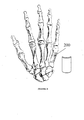
- A61F2/42—Joints for wrists or ankles; for hands, e.g. fingers; for feet, e.g. toes
- A61F2/4241—Joints for wrists or ankles; for hands, e.g. fingers; for feet, e.g. toes for hands, e.g. fingers
-
- A—HUMAN NECESSITIES
- A61—MEDICAL OR VETERINARY SCIENCE; HYGIENE
- A61F—FILTERS IMPLANTABLE INTO BLOOD VESSELS; PROSTHESES; DEVICES PROVIDING PATENCY TO, OR PREVENTING COLLAPSING OF, TUBULAR STRUCTURES OF THE BODY, e.g. STENTS; ORTHOPAEDIC, NURSING OR CONTRACEPTIVE DEVICES; FOMENTATION; TREATMENT OR PROTECTION OF EYES OR EARS; BANDAGES, DRESSINGS OR ABSORBENT PADS; FIRST-AID KITS
- A61F2/00—Filters implantable into blood vessels; Prostheses, i.e. artificial substitutes or replacements for parts of the body; Appliances for connecting them with the body; Devices providing patency to, or preventing collapsing of, tubular structures of the body, e.g. stents
- A61F2/02—Prostheses implantable into the body
- A61F2/30—Joints
- A61F2/46—Special tools or methods for implanting or extracting artificial joints, accessories, bone grafts or substitutes, or particular adaptations therefor
- A61F2/4644—Preparation of bone graft, bone plugs or bone dowels, e.g. grinding or milling bone material
-
- A—HUMAN NECESSITIES
- A61—MEDICAL OR VETERINARY SCIENCE; HYGIENE
- A61F—FILTERS IMPLANTABLE INTO BLOOD VESSELS; PROSTHESES; DEVICES PROVIDING PATENCY TO, OR PREVENTING COLLAPSING OF, TUBULAR STRUCTURES OF THE BODY, e.g. STENTS; ORTHOPAEDIC, NURSING OR CONTRACEPTIVE DEVICES; FOMENTATION; TREATMENT OR PROTECTION OF EYES OR EARS; BANDAGES, DRESSINGS OR ABSORBENT PADS; FIRST-AID KITS
- A61F2/00—Filters implantable into blood vessels; Prostheses, i.e. artificial substitutes or replacements for parts of the body; Appliances for connecting them with the body; Devices providing patency to, or preventing collapsing of, tubular structures of the body, e.g. stents
- A61F2/02—Prostheses implantable into the body
- A61F2/28—Bones
- A61F2002/2817—Bone stimulation by chemical reactions or by osteogenic or biological products for enhancing ossification, e.g. by bone morphogenetic or morphogenic proteins [BMP] or by transforming growth factors [TGF]
-
- A—HUMAN NECESSITIES
- A61—MEDICAL OR VETERINARY SCIENCE; HYGIENE
- A61F—FILTERS IMPLANTABLE INTO BLOOD VESSELS; PROSTHESES; DEVICES PROVIDING PATENCY TO, OR PREVENTING COLLAPSING OF, TUBULAR STRUCTURES OF THE BODY, e.g. STENTS; ORTHOPAEDIC, NURSING OR CONTRACEPTIVE DEVICES; FOMENTATION; TREATMENT OR PROTECTION OF EYES OR EARS; BANDAGES, DRESSINGS OR ABSORBENT PADS; FIRST-AID KITS
- A61F2/00—Filters implantable into blood vessels; Prostheses, i.e. artificial substitutes or replacements for parts of the body; Appliances for connecting them with the body; Devices providing patency to, or preventing collapsing of, tubular structures of the body, e.g. stents
- A61F2/02—Prostheses implantable into the body
- A61F2/28—Bones
- A61F2002/2825—Femur
-
- A—HUMAN NECESSITIES
- A61—MEDICAL OR VETERINARY SCIENCE; HYGIENE
- A61F—FILTERS IMPLANTABLE INTO BLOOD VESSELS; PROSTHESES; DEVICES PROVIDING PATENCY TO, OR PREVENTING COLLAPSING OF, TUBULAR STRUCTURES OF THE BODY, e.g. STENTS; ORTHOPAEDIC, NURSING OR CONTRACEPTIVE DEVICES; FOMENTATION; TREATMENT OR PROTECTION OF EYES OR EARS; BANDAGES, DRESSINGS OR ABSORBENT PADS; FIRST-AID KITS
- A61F2/00—Filters implantable into blood vessels; Prostheses, i.e. artificial substitutes or replacements for parts of the body; Appliances for connecting them with the body; Devices providing patency to, or preventing collapsing of, tubular structures of the body, e.g. stents
- A61F2/02—Prostheses implantable into the body
- A61F2/28—Bones
- A61F2002/2835—Bone graft implants for filling a bony defect or an endoprosthesis cavity, e.g. by synthetic material or biological material
-
- A—HUMAN NECESSITIES
- A61—MEDICAL OR VETERINARY SCIENCE; HYGIENE
- A61F—FILTERS IMPLANTABLE INTO BLOOD VESSELS; PROSTHESES; DEVICES PROVIDING PATENCY TO, OR PREVENTING COLLAPSING OF, TUBULAR STRUCTURES OF THE BODY, e.g. STENTS; ORTHOPAEDIC, NURSING OR CONTRACEPTIVE DEVICES; FOMENTATION; TREATMENT OR PROTECTION OF EYES OR EARS; BANDAGES, DRESSINGS OR ABSORBENT PADS; FIRST-AID KITS
- A61F2/00—Filters implantable into blood vessels; Prostheses, i.e. artificial substitutes or replacements for parts of the body; Appliances for connecting them with the body; Devices providing patency to, or preventing collapsing of, tubular structures of the body, e.g. stents
- A61F2/02—Prostheses implantable into the body
- A61F2/28—Bones
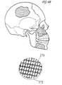
- A61F2/2875—Skull or cranium
- A61F2002/2882—Malar or zygomatic implants
-
- A—HUMAN NECESSITIES
- A61—MEDICAL OR VETERINARY SCIENCE; HYGIENE
- A61F—FILTERS IMPLANTABLE INTO BLOOD VESSELS; PROSTHESES; DEVICES PROVIDING PATENCY TO, OR PREVENTING COLLAPSING OF, TUBULAR STRUCTURES OF THE BODY, e.g. STENTS; ORTHOPAEDIC, NURSING OR CONTRACEPTIVE DEVICES; FOMENTATION; TREATMENT OR PROTECTION OF EYES OR EARS; BANDAGES, DRESSINGS OR ABSORBENT PADS; FIRST-AID KITS
- A61F2/00—Filters implantable into blood vessels; Prostheses, i.e. artificial substitutes or replacements for parts of the body; Appliances for connecting them with the body; Devices providing patency to, or preventing collapsing of, tubular structures of the body, e.g. stents
- A61F2/02—Prostheses implantable into the body
- A61F2/30—Joints
- A61F2002/30001—Additional features of subject-matter classified in A61F2/28, A61F2/30 and subgroups thereof
- A61F2002/30003—Material related properties of the prosthesis or of a coating on the prosthesis
- A61F2002/30004—Material related properties of the prosthesis or of a coating on the prosthesis the prosthesis being made from materials having different values of a given property at different locations within the same prosthesis
- A61F2002/30011—Material related properties of the prosthesis or of a coating on the prosthesis the prosthesis being made from materials having different values of a given property at different locations within the same prosthesis differing in porosity
-
- A—HUMAN NECESSITIES
- A61—MEDICAL OR VETERINARY SCIENCE; HYGIENE
- A61F—FILTERS IMPLANTABLE INTO BLOOD VESSELS; PROSTHESES; DEVICES PROVIDING PATENCY TO, OR PREVENTING COLLAPSING OF, TUBULAR STRUCTURES OF THE BODY, e.g. STENTS; ORTHOPAEDIC, NURSING OR CONTRACEPTIVE DEVICES; FOMENTATION; TREATMENT OR PROTECTION OF EYES OR EARS; BANDAGES, DRESSINGS OR ABSORBENT PADS; FIRST-AID KITS
- A61F2/00—Filters implantable into blood vessels; Prostheses, i.e. artificial substitutes or replacements for parts of the body; Appliances for connecting them with the body; Devices providing patency to, or preventing collapsing of, tubular structures of the body, e.g. stents
- A61F2/02—Prostheses implantable into the body
- A61F2/30—Joints
- A61F2002/30001—Additional features of subject-matter classified in A61F2/28, A61F2/30 and subgroups thereof
- A61F2002/30003—Material related properties of the prosthesis or of a coating on the prosthesis
- A61F2002/30004—Material related properties of the prosthesis or of a coating on the prosthesis the prosthesis being made from materials having different values of a given property at different locations within the same prosthesis
- A61F2002/30014—Material related properties of the prosthesis or of a coating on the prosthesis the prosthesis being made from materials having different values of a given property at different locations within the same prosthesis differing in elasticity, stiffness or compressibility
-
- A—HUMAN NECESSITIES
- A61—MEDICAL OR VETERINARY SCIENCE; HYGIENE
- A61F—FILTERS IMPLANTABLE INTO BLOOD VESSELS; PROSTHESES; DEVICES PROVIDING PATENCY TO, OR PREVENTING COLLAPSING OF, TUBULAR STRUCTURES OF THE BODY, e.g. STENTS; ORTHOPAEDIC, NURSING OR CONTRACEPTIVE DEVICES; FOMENTATION; TREATMENT OR PROTECTION OF EYES OR EARS; BANDAGES, DRESSINGS OR ABSORBENT PADS; FIRST-AID KITS
- A61F2/00—Filters implantable into blood vessels; Prostheses, i.e. artificial substitutes or replacements for parts of the body; Appliances for connecting them with the body; Devices providing patency to, or preventing collapsing of, tubular structures of the body, e.g. stents
- A61F2/02—Prostheses implantable into the body
- A61F2/30—Joints
- A61F2002/30001—Additional features of subject-matter classified in A61F2/28, A61F2/30 and subgroups thereof
- A61F2002/30003—Material related properties of the prosthesis or of a coating on the prosthesis
- A61F2002/3006—Properties of materials and coating materials
- A61F2002/30062—(bio)absorbable, biodegradable, bioerodable, (bio)resorbable, resorptive
-
- A—HUMAN NECESSITIES
- A61—MEDICAL OR VETERINARY SCIENCE; HYGIENE
- A61F—FILTERS IMPLANTABLE INTO BLOOD VESSELS; PROSTHESES; DEVICES PROVIDING PATENCY TO, OR PREVENTING COLLAPSING OF, TUBULAR STRUCTURES OF THE BODY, e.g. STENTS; ORTHOPAEDIC, NURSING OR CONTRACEPTIVE DEVICES; FOMENTATION; TREATMENT OR PROTECTION OF EYES OR EARS; BANDAGES, DRESSINGS OR ABSORBENT PADS; FIRST-AID KITS
- A61F2/00—Filters implantable into blood vessels; Prostheses, i.e. artificial substitutes or replacements for parts of the body; Appliances for connecting them with the body; Devices providing patency to, or preventing collapsing of, tubular structures of the body, e.g. stents
- A61F2/02—Prostheses implantable into the body
- A61F2/30—Joints
- A61F2002/30001—Additional features of subject-matter classified in A61F2/28, A61F2/30 and subgroups thereof
- A61F2002/30003—Material related properties of the prosthesis or of a coating on the prosthesis
- A61F2002/3006—Properties of materials and coating materials
- A61F2002/3008—Properties of materials and coating materials radio-opaque, e.g. radio-opaque markers
-
- A—HUMAN NECESSITIES
- A61—MEDICAL OR VETERINARY SCIENCE; HYGIENE
- A61F—FILTERS IMPLANTABLE INTO BLOOD VESSELS; PROSTHESES; DEVICES PROVIDING PATENCY TO, OR PREVENTING COLLAPSING OF, TUBULAR STRUCTURES OF THE BODY, e.g. STENTS; ORTHOPAEDIC, NURSING OR CONTRACEPTIVE DEVICES; FOMENTATION; TREATMENT OR PROTECTION OF EYES OR EARS; BANDAGES, DRESSINGS OR ABSORBENT PADS; FIRST-AID KITS
- A61F2/00—Filters implantable into blood vessels; Prostheses, i.e. artificial substitutes or replacements for parts of the body; Appliances for connecting them with the body; Devices providing patency to, or preventing collapsing of, tubular structures of the body, e.g. stents
- A61F2/02—Prostheses implantable into the body
- A61F2/30—Joints
- A61F2002/30001—Additional features of subject-matter classified in A61F2/28, A61F2/30 and subgroups thereof
- A61F2002/30003—Material related properties of the prosthesis or of a coating on the prosthesis
- A61F2002/3006—Properties of materials and coating materials
- A61F2002/30092—Properties of materials and coating materials using shape memory or superelastic materials, e.g. nitinol
-
- A—HUMAN NECESSITIES
- A61—MEDICAL OR VETERINARY SCIENCE; HYGIENE
- A61F—FILTERS IMPLANTABLE INTO BLOOD VESSELS; PROSTHESES; DEVICES PROVIDING PATENCY TO, OR PREVENTING COLLAPSING OF, TUBULAR STRUCTURES OF THE BODY, e.g. STENTS; ORTHOPAEDIC, NURSING OR CONTRACEPTIVE DEVICES; FOMENTATION; TREATMENT OR PROTECTION OF EYES OR EARS; BANDAGES, DRESSINGS OR ABSORBENT PADS; FIRST-AID KITS
- A61F2/00—Filters implantable into blood vessels; Prostheses, i.e. artificial substitutes or replacements for parts of the body; Appliances for connecting them with the body; Devices providing patency to, or preventing collapsing of, tubular structures of the body, e.g. stents
- A61F2/02—Prostheses implantable into the body
- A61F2/30—Joints
- A61F2002/30001—Additional features of subject-matter classified in A61F2/28, A61F2/30 and subgroups thereof
- A61F2002/30108—Shapes
- A61F2002/3011—Cross-sections or two-dimensional shapes
- A61F2002/30112—Rounded shapes, e.g. with rounded corners
- A61F2002/30113—Rounded shapes, e.g. with rounded corners circular
- A61F2002/30118—Rounded shapes, e.g. with rounded corners circular concentric circles
-
- A—HUMAN NECESSITIES
- A61—MEDICAL OR VETERINARY SCIENCE; HYGIENE
- A61F—FILTERS IMPLANTABLE INTO BLOOD VESSELS; PROSTHESES; DEVICES PROVIDING PATENCY TO, OR PREVENTING COLLAPSING OF, TUBULAR STRUCTURES OF THE BODY, e.g. STENTS; ORTHOPAEDIC, NURSING OR CONTRACEPTIVE DEVICES; FOMENTATION; TREATMENT OR PROTECTION OF EYES OR EARS; BANDAGES, DRESSINGS OR ABSORBENT PADS; FIRST-AID KITS
- A61F2/00—Filters implantable into blood vessels; Prostheses, i.e. artificial substitutes or replacements for parts of the body; Appliances for connecting them with the body; Devices providing patency to, or preventing collapsing of, tubular structures of the body, e.g. stents
- A61F2/02—Prostheses implantable into the body
- A61F2/30—Joints
- A61F2002/30001—Additional features of subject-matter classified in A61F2/28, A61F2/30 and subgroups thereof
- A61F2002/30108—Shapes
- A61F2002/3011—Cross-sections or two-dimensional shapes
- A61F2002/30138—Convex polygonal shapes
- A61F2002/30153—Convex polygonal shapes rectangular
-
- A—HUMAN NECESSITIES
- A61—MEDICAL OR VETERINARY SCIENCE; HYGIENE
- A61F—FILTERS IMPLANTABLE INTO BLOOD VESSELS; PROSTHESES; DEVICES PROVIDING PATENCY TO, OR PREVENTING COLLAPSING OF, TUBULAR STRUCTURES OF THE BODY, e.g. STENTS; ORTHOPAEDIC, NURSING OR CONTRACEPTIVE DEVICES; FOMENTATION; TREATMENT OR PROTECTION OF EYES OR EARS; BANDAGES, DRESSINGS OR ABSORBENT PADS; FIRST-AID KITS
- A61F2/00—Filters implantable into blood vessels; Prostheses, i.e. artificial substitutes or replacements for parts of the body; Appliances for connecting them with the body; Devices providing patency to, or preventing collapsing of, tubular structures of the body, e.g. stents
- A61F2/02—Prostheses implantable into the body
- A61F2/30—Joints
- A61F2002/30001—Additional features of subject-matter classified in A61F2/28, A61F2/30 and subgroups thereof
- A61F2002/30108—Shapes
- A61F2002/30199—Three-dimensional shapes
- A61F2002/30224—Three-dimensional shapes cylindrical
- A61F2002/30225—Flat cylinders, i.e. discs
-
- A—HUMAN NECESSITIES
- A61—MEDICAL OR VETERINARY SCIENCE; HYGIENE
- A61F—FILTERS IMPLANTABLE INTO BLOOD VESSELS; PROSTHESES; DEVICES PROVIDING PATENCY TO, OR PREVENTING COLLAPSING OF, TUBULAR STRUCTURES OF THE BODY, e.g. STENTS; ORTHOPAEDIC, NURSING OR CONTRACEPTIVE DEVICES; FOMENTATION; TREATMENT OR PROTECTION OF EYES OR EARS; BANDAGES, DRESSINGS OR ABSORBENT PADS; FIRST-AID KITS
- A61F2/00—Filters implantable into blood vessels; Prostheses, i.e. artificial substitutes or replacements for parts of the body; Appliances for connecting them with the body; Devices providing patency to, or preventing collapsing of, tubular structures of the body, e.g. stents
- A61F2/02—Prostheses implantable into the body
- A61F2/30—Joints
- A61F2002/30001—Additional features of subject-matter classified in A61F2/28, A61F2/30 and subgroups thereof
- A61F2002/30108—Shapes
- A61F2002/30199—Three-dimensional shapes
- A61F2002/30224—Three-dimensional shapes cylindrical
- A61F2002/30235—Three-dimensional shapes cylindrical tubular, e.g. sleeves
- A61F2002/30237—Three-dimensional shapes cylindrical tubular, e.g. sleeves partial tubes
-
- A—HUMAN NECESSITIES
- A61—MEDICAL OR VETERINARY SCIENCE; HYGIENE
- A61F—FILTERS IMPLANTABLE INTO BLOOD VESSELS; PROSTHESES; DEVICES PROVIDING PATENCY TO, OR PREVENTING COLLAPSING OF, TUBULAR STRUCTURES OF THE BODY, e.g. STENTS; ORTHOPAEDIC, NURSING OR CONTRACEPTIVE DEVICES; FOMENTATION; TREATMENT OR PROTECTION OF EYES OR EARS; BANDAGES, DRESSINGS OR ABSORBENT PADS; FIRST-AID KITS
- A61F2/00—Filters implantable into blood vessels; Prostheses, i.e. artificial substitutes or replacements for parts of the body; Appliances for connecting them with the body; Devices providing patency to, or preventing collapsing of, tubular structures of the body, e.g. stents
- A61F2/02—Prostheses implantable into the body
- A61F2/30—Joints
- A61F2002/30001—Additional features of subject-matter classified in A61F2/28, A61F2/30 and subgroups thereof
- A61F2002/30108—Shapes
- A61F2002/30199—Three-dimensional shapes
- A61F2002/30242—Three-dimensional shapes spherical
- A61F2002/30245—Partial spheres
-
- A—HUMAN NECESSITIES
- A61—MEDICAL OR VETERINARY SCIENCE; HYGIENE
- A61F—FILTERS IMPLANTABLE INTO BLOOD VESSELS; PROSTHESES; DEVICES PROVIDING PATENCY TO, OR PREVENTING COLLAPSING OF, TUBULAR STRUCTURES OF THE BODY, e.g. STENTS; ORTHOPAEDIC, NURSING OR CONTRACEPTIVE DEVICES; FOMENTATION; TREATMENT OR PROTECTION OF EYES OR EARS; BANDAGES, DRESSINGS OR ABSORBENT PADS; FIRST-AID KITS
- A61F2/00—Filters implantable into blood vessels; Prostheses, i.e. artificial substitutes or replacements for parts of the body; Appliances for connecting them with the body; Devices providing patency to, or preventing collapsing of, tubular structures of the body, e.g. stents
- A61F2/02—Prostheses implantable into the body
- A61F2/30—Joints
- A61F2002/30001—Additional features of subject-matter classified in A61F2/28, A61F2/30 and subgroups thereof
- A61F2002/30108—Shapes
- A61F2002/30199—Three-dimensional shapes
- A61F2002/30261—Three-dimensional shapes parallelepipedal
- A61F2002/30265—Flat parallelepipeds
-
- A—HUMAN NECESSITIES
- A61—MEDICAL OR VETERINARY SCIENCE; HYGIENE
- A61F—FILTERS IMPLANTABLE INTO BLOOD VESSELS; PROSTHESES; DEVICES PROVIDING PATENCY TO, OR PREVENTING COLLAPSING OF, TUBULAR STRUCTURES OF THE BODY, e.g. STENTS; ORTHOPAEDIC, NURSING OR CONTRACEPTIVE DEVICES; FOMENTATION; TREATMENT OR PROTECTION OF EYES OR EARS; BANDAGES, DRESSINGS OR ABSORBENT PADS; FIRST-AID KITS
- A61F2/00—Filters implantable into blood vessels; Prostheses, i.e. artificial substitutes or replacements for parts of the body; Appliances for connecting them with the body; Devices providing patency to, or preventing collapsing of, tubular structures of the body, e.g. stents
- A61F2/02—Prostheses implantable into the body
- A61F2/30—Joints
- A61F2002/30001—Additional features of subject-matter classified in A61F2/28, A61F2/30 and subgroups thereof
- A61F2002/30108—Shapes
- A61F2002/30199—Three-dimensional shapes
- A61F2002/3028—Three-dimensional shapes polyhedral different from parallelepipedal and pyramidal
- A61F2002/30281—Three-dimensional shapes polyhedral different from parallelepipedal and pyramidal wedge-shaped
-
- A—HUMAN NECESSITIES
- A61—MEDICAL OR VETERINARY SCIENCE; HYGIENE
- A61F—FILTERS IMPLANTABLE INTO BLOOD VESSELS; PROSTHESES; DEVICES PROVIDING PATENCY TO, OR PREVENTING COLLAPSING OF, TUBULAR STRUCTURES OF THE BODY, e.g. STENTS; ORTHOPAEDIC, NURSING OR CONTRACEPTIVE DEVICES; FOMENTATION; TREATMENT OR PROTECTION OF EYES OR EARS; BANDAGES, DRESSINGS OR ABSORBENT PADS; FIRST-AID KITS
- A61F2/00—Filters implantable into blood vessels; Prostheses, i.e. artificial substitutes or replacements for parts of the body; Appliances for connecting them with the body; Devices providing patency to, or preventing collapsing of, tubular structures of the body, e.g. stents
- A61F2/02—Prostheses implantable into the body
- A61F2/30—Joints
- A61F2002/30001—Additional features of subject-matter classified in A61F2/28, A61F2/30 and subgroups thereof
- A61F2002/30108—Shapes
- A61F2002/30199—Three-dimensional shapes
- A61F2002/30291—Three-dimensional shapes spirally-coiled, i.e. having a 2D spiral cross-section
-
- A—HUMAN NECESSITIES
- A61—MEDICAL OR VETERINARY SCIENCE; HYGIENE
- A61F—FILTERS IMPLANTABLE INTO BLOOD VESSELS; PROSTHESES; DEVICES PROVIDING PATENCY TO, OR PREVENTING COLLAPSING OF, TUBULAR STRUCTURES OF THE BODY, e.g. STENTS; ORTHOPAEDIC, NURSING OR CONTRACEPTIVE DEVICES; FOMENTATION; TREATMENT OR PROTECTION OF EYES OR EARS; BANDAGES, DRESSINGS OR ABSORBENT PADS; FIRST-AID KITS
- A61F2/00—Filters implantable into blood vessels; Prostheses, i.e. artificial substitutes or replacements for parts of the body; Appliances for connecting them with the body; Devices providing patency to, or preventing collapsing of, tubular structures of the body, e.g. stents
- A61F2/02—Prostheses implantable into the body
- A61F2/30—Joints
- A61F2002/30001—Additional features of subject-matter classified in A61F2/28, A61F2/30 and subgroups thereof
- A61F2002/30316—The prosthesis having different structural features at different locations within the same prosthesis; Connections between prosthetic parts; Special structural features of bone or joint prostheses not otherwise provided for
- A61F2002/30317—The prosthesis having different structural features at different locations within the same prosthesis
- A61F2002/30324—The prosthesis having different structural features at different locations within the same prosthesis differing in thickness
-
- A—HUMAN NECESSITIES
- A61—MEDICAL OR VETERINARY SCIENCE; HYGIENE
- A61F—FILTERS IMPLANTABLE INTO BLOOD VESSELS; PROSTHESES; DEVICES PROVIDING PATENCY TO, OR PREVENTING COLLAPSING OF, TUBULAR STRUCTURES OF THE BODY, e.g. STENTS; ORTHOPAEDIC, NURSING OR CONTRACEPTIVE DEVICES; FOMENTATION; TREATMENT OR PROTECTION OF EYES OR EARS; BANDAGES, DRESSINGS OR ABSORBENT PADS; FIRST-AID KITS
- A61F2/00—Filters implantable into blood vessels; Prostheses, i.e. artificial substitutes or replacements for parts of the body; Appliances for connecting them with the body; Devices providing patency to, or preventing collapsing of, tubular structures of the body, e.g. stents
- A61F2/02—Prostheses implantable into the body
- A61F2/30—Joints
- A61F2002/30001—Additional features of subject-matter classified in A61F2/28, A61F2/30 and subgroups thereof
- A61F2002/30621—Features concerning the anatomical functioning or articulation of the prosthetic joint
- A61F2002/30622—Implant for fusing a joint or bone material
-
- A—HUMAN NECESSITIES
- A61—MEDICAL OR VETERINARY SCIENCE; HYGIENE
- A61F—FILTERS IMPLANTABLE INTO BLOOD VESSELS; PROSTHESES; DEVICES PROVIDING PATENCY TO, OR PREVENTING COLLAPSING OF, TUBULAR STRUCTURES OF THE BODY, e.g. STENTS; ORTHOPAEDIC, NURSING OR CONTRACEPTIVE DEVICES; FOMENTATION; TREATMENT OR PROTECTION OF EYES OR EARS; BANDAGES, DRESSINGS OR ABSORBENT PADS; FIRST-AID KITS
- A61F2/00—Filters implantable into blood vessels; Prostheses, i.e. artificial substitutes or replacements for parts of the body; Appliances for connecting them with the body; Devices providing patency to, or preventing collapsing of, tubular structures of the body, e.g. stents
- A61F2/02—Prostheses implantable into the body
- A61F2/30—Joints
- A61F2002/30001—Additional features of subject-matter classified in A61F2/28, A61F2/30 and subgroups thereof
- A61F2002/30667—Features concerning an interaction with the environment or a particular use of the prosthesis
- A61F2002/30677—Means for introducing or releasing pharmaceutical products, e.g. antibiotics, into the body
-
- A—HUMAN NECESSITIES
- A61—MEDICAL OR VETERINARY SCIENCE; HYGIENE
- A61F—FILTERS IMPLANTABLE INTO BLOOD VESSELS; PROSTHESES; DEVICES PROVIDING PATENCY TO, OR PREVENTING COLLAPSING OF, TUBULAR STRUCTURES OF THE BODY, e.g. STENTS; ORTHOPAEDIC, NURSING OR CONTRACEPTIVE DEVICES; FOMENTATION; TREATMENT OR PROTECTION OF EYES OR EARS; BANDAGES, DRESSINGS OR ABSORBENT PADS; FIRST-AID KITS
- A61F2/00—Filters implantable into blood vessels; Prostheses, i.e. artificial substitutes or replacements for parts of the body; Appliances for connecting them with the body; Devices providing patency to, or preventing collapsing of, tubular structures of the body, e.g. stents
- A61F2/02—Prostheses implantable into the body
- A61F2/30—Joints
- A61F2/30721—Accessories
- A61F2/30734—Modular inserts, sleeves or augments, e.g. placed on proximal part of stem for fixation purposes or wedges for bridging a bone defect
- A61F2002/30736—Augments or augmentation pieces, e.g. wedges or blocks for bridging a bone defect
-
- A—HUMAN NECESSITIES
- A61—MEDICAL OR VETERINARY SCIENCE; HYGIENE
- A61F—FILTERS IMPLANTABLE INTO BLOOD VESSELS; PROSTHESES; DEVICES PROVIDING PATENCY TO, OR PREVENTING COLLAPSING OF, TUBULAR STRUCTURES OF THE BODY, e.g. STENTS; ORTHOPAEDIC, NURSING OR CONTRACEPTIVE DEVICES; FOMENTATION; TREATMENT OR PROTECTION OF EYES OR EARS; BANDAGES, DRESSINGS OR ABSORBENT PADS; FIRST-AID KITS
- A61F2/00—Filters implantable into blood vessels; Prostheses, i.e. artificial substitutes or replacements for parts of the body; Appliances for connecting them with the body; Devices providing patency to, or preventing collapsing of, tubular structures of the body, e.g. stents
- A61F2/02—Prostheses implantable into the body
- A61F2/30—Joints
- A61F2/30767—Special external or bone-contacting surface, e.g. coating for improving bone ingrowth
- A61F2/30771—Special external or bone-contacting surface, e.g. coating for improving bone ingrowth applied in original prostheses, e.g. holes or grooves
- A61F2002/30772—Apertures or holes, e.g. of circular cross section
- A61F2002/30784—Plurality of holes
- A61F2002/30785—Plurality of holes parallel
-
- A—HUMAN NECESSITIES
- A61—MEDICAL OR VETERINARY SCIENCE; HYGIENE
- A61F—FILTERS IMPLANTABLE INTO BLOOD VESSELS; PROSTHESES; DEVICES PROVIDING PATENCY TO, OR PREVENTING COLLAPSING OF, TUBULAR STRUCTURES OF THE BODY, e.g. STENTS; ORTHOPAEDIC, NURSING OR CONTRACEPTIVE DEVICES; FOMENTATION; TREATMENT OR PROTECTION OF EYES OR EARS; BANDAGES, DRESSINGS OR ABSORBENT PADS; FIRST-AID KITS
- A61F2/00—Filters implantable into blood vessels; Prostheses, i.e. artificial substitutes or replacements for parts of the body; Appliances for connecting them with the body; Devices providing patency to, or preventing collapsing of, tubular structures of the body, e.g. stents
- A61F2/02—Prostheses implantable into the body
- A61F2/30—Joints
- A61F2/30767—Special external or bone-contacting surface, e.g. coating for improving bone ingrowth
- A61F2/30771—Special external or bone-contacting surface, e.g. coating for improving bone ingrowth applied in original prostheses, e.g. holes or grooves
- A61F2002/3082—Grooves
- A61F2002/30827—Plurality of grooves
- A61F2002/30828—Plurality of grooves parallel
-
- A—HUMAN NECESSITIES
- A61—MEDICAL OR VETERINARY SCIENCE; HYGIENE
- A61F—FILTERS IMPLANTABLE INTO BLOOD VESSELS; PROSTHESES; DEVICES PROVIDING PATENCY TO, OR PREVENTING COLLAPSING OF, TUBULAR STRUCTURES OF THE BODY, e.g. STENTS; ORTHOPAEDIC, NURSING OR CONTRACEPTIVE DEVICES; FOMENTATION; TREATMENT OR PROTECTION OF EYES OR EARS; BANDAGES, DRESSINGS OR ABSORBENT PADS; FIRST-AID KITS
- A61F2/00—Filters implantable into blood vessels; Prostheses, i.e. artificial substitutes or replacements for parts of the body; Appliances for connecting them with the body; Devices providing patency to, or preventing collapsing of, tubular structures of the body, e.g. stents
- A61F2/02—Prostheses implantable into the body
- A61F2/30—Joints
- A61F2/30767—Special external or bone-contacting surface, e.g. coating for improving bone ingrowth
- A61F2/30771—Special external or bone-contacting surface, e.g. coating for improving bone ingrowth applied in original prostheses, e.g. holes or grooves
- A61F2002/30838—Microstructures
-
- A—HUMAN NECESSITIES
- A61—MEDICAL OR VETERINARY SCIENCE; HYGIENE
- A61F—FILTERS IMPLANTABLE INTO BLOOD VESSELS; PROSTHESES; DEVICES PROVIDING PATENCY TO, OR PREVENTING COLLAPSING OF, TUBULAR STRUCTURES OF THE BODY, e.g. STENTS; ORTHOPAEDIC, NURSING OR CONTRACEPTIVE DEVICES; FOMENTATION; TREATMENT OR PROTECTION OF EYES OR EARS; BANDAGES, DRESSINGS OR ABSORBENT PADS; FIRST-AID KITS
- A61F2/00—Filters implantable into blood vessels; Prostheses, i.e. artificial substitutes or replacements for parts of the body; Appliances for connecting them with the body; Devices providing patency to, or preventing collapsing of, tubular structures of the body, e.g. stents
- A61F2/02—Prostheses implantable into the body
- A61F2/30—Joints
- A61F2/30767—Special external or bone-contacting surface, e.g. coating for improving bone ingrowth
- A61F2/30907—Nets or sleeves applied to surface of prostheses or in cement
- A61F2002/30909—Nets
-
- A—HUMAN NECESSITIES
- A61—MEDICAL OR VETERINARY SCIENCE; HYGIENE
- A61F—FILTERS IMPLANTABLE INTO BLOOD VESSELS; PROSTHESES; DEVICES PROVIDING PATENCY TO, OR PREVENTING COLLAPSING OF, TUBULAR STRUCTURES OF THE BODY, e.g. STENTS; ORTHOPAEDIC, NURSING OR CONTRACEPTIVE DEVICES; FOMENTATION; TREATMENT OR PROTECTION OF EYES OR EARS; BANDAGES, DRESSINGS OR ABSORBENT PADS; FIRST-AID KITS
- A61F2/00—Filters implantable into blood vessels; Prostheses, i.e. artificial substitutes or replacements for parts of the body; Appliances for connecting them with the body; Devices providing patency to, or preventing collapsing of, tubular structures of the body, e.g. stents
- A61F2/02—Prostheses implantable into the body
- A61F2/30—Joints
- A61F2/30767—Special external or bone-contacting surface, e.g. coating for improving bone ingrowth
- A61F2002/30925—Special external or bone-contacting surface, e.g. coating for improving bone ingrowth etched
-
- A—HUMAN NECESSITIES
- A61—MEDICAL OR VETERINARY SCIENCE; HYGIENE
- A61F—FILTERS IMPLANTABLE INTO BLOOD VESSELS; PROSTHESES; DEVICES PROVIDING PATENCY TO, OR PREVENTING COLLAPSING OF, TUBULAR STRUCTURES OF THE BODY, e.g. STENTS; ORTHOPAEDIC, NURSING OR CONTRACEPTIVE DEVICES; FOMENTATION; TREATMENT OR PROTECTION OF EYES OR EARS; BANDAGES, DRESSINGS OR ABSORBENT PADS; FIRST-AID KITS
- A61F2/00—Filters implantable into blood vessels; Prostheses, i.e. artificial substitutes or replacements for parts of the body; Appliances for connecting them with the body; Devices providing patency to, or preventing collapsing of, tubular structures of the body, e.g. stents
- A61F2/02—Prostheses implantable into the body
- A61F2/30—Joints
- A61F2/46—Special tools or methods for implanting or extracting artificial joints, accessories, bone grafts or substitutes, or particular adaptations therefor
- A61F2002/4631—Special tools or methods for implanting or extracting artificial joints, accessories, bone grafts or substitutes, or particular adaptations therefor the prosthesis being specially adapted for being cemented
-
- A—HUMAN NECESSITIES
- A61—MEDICAL OR VETERINARY SCIENCE; HYGIENE
- A61F—FILTERS IMPLANTABLE INTO BLOOD VESSELS; PROSTHESES; DEVICES PROVIDING PATENCY TO, OR PREVENTING COLLAPSING OF, TUBULAR STRUCTURES OF THE BODY, e.g. STENTS; ORTHOPAEDIC, NURSING OR CONTRACEPTIVE DEVICES; FOMENTATION; TREATMENT OR PROTECTION OF EYES OR EARS; BANDAGES, DRESSINGS OR ABSORBENT PADS; FIRST-AID KITS
- A61F2210/00—Particular material properties of prostheses classified in groups A61F2/00 - A61F2/26 or A61F2/82 or A61F9/00 or A61F11/00 or subgroups thereof
- A61F2210/0004—Particular material properties of prostheses classified in groups A61F2/00 - A61F2/26 or A61F2/82 or A61F9/00 or A61F11/00 or subgroups thereof bioabsorbable
-
- A—HUMAN NECESSITIES
- A61—MEDICAL OR VETERINARY SCIENCE; HYGIENE
- A61F—FILTERS IMPLANTABLE INTO BLOOD VESSELS; PROSTHESES; DEVICES PROVIDING PATENCY TO, OR PREVENTING COLLAPSING OF, TUBULAR STRUCTURES OF THE BODY, e.g. STENTS; ORTHOPAEDIC, NURSING OR CONTRACEPTIVE DEVICES; FOMENTATION; TREATMENT OR PROTECTION OF EYES OR EARS; BANDAGES, DRESSINGS OR ABSORBENT PADS; FIRST-AID KITS
- A61F2210/00—Particular material properties of prostheses classified in groups A61F2/00 - A61F2/26 or A61F2/82 or A61F9/00 or A61F11/00 or subgroups thereof
- A61F2210/0014—Particular material properties of prostheses classified in groups A61F2/00 - A61F2/26 or A61F2/82 or A61F9/00 or A61F11/00 or subgroups thereof using shape memory or superelastic materials, e.g. nitinol
-
- A—HUMAN NECESSITIES
- A61—MEDICAL OR VETERINARY SCIENCE; HYGIENE
- A61F—FILTERS IMPLANTABLE INTO BLOOD VESSELS; PROSTHESES; DEVICES PROVIDING PATENCY TO, OR PREVENTING COLLAPSING OF, TUBULAR STRUCTURES OF THE BODY, e.g. STENTS; ORTHOPAEDIC, NURSING OR CONTRACEPTIVE DEVICES; FOMENTATION; TREATMENT OR PROTECTION OF EYES OR EARS; BANDAGES, DRESSINGS OR ABSORBENT PADS; FIRST-AID KITS
- A61F2230/00—Geometry of prostheses classified in groups A61F2/00 - A61F2/26 or A61F2/82 or A61F9/00 or A61F11/00 or subgroups thereof
- A61F2230/0002—Two-dimensional shapes, e.g. cross-sections
- A61F2230/0004—Rounded shapes, e.g. with rounded corners
- A61F2230/0006—Rounded shapes, e.g. with rounded corners circular
-
- A—HUMAN NECESSITIES
- A61—MEDICAL OR VETERINARY SCIENCE; HYGIENE
- A61F—FILTERS IMPLANTABLE INTO BLOOD VESSELS; PROSTHESES; DEVICES PROVIDING PATENCY TO, OR PREVENTING COLLAPSING OF, TUBULAR STRUCTURES OF THE BODY, e.g. STENTS; ORTHOPAEDIC, NURSING OR CONTRACEPTIVE DEVICES; FOMENTATION; TREATMENT OR PROTECTION OF EYES OR EARS; BANDAGES, DRESSINGS OR ABSORBENT PADS; FIRST-AID KITS
- A61F2230/00—Geometry of prostheses classified in groups A61F2/00 - A61F2/26 or A61F2/82 or A61F9/00 or A61F11/00 or subgroups thereof
- A61F2230/0002—Two-dimensional shapes, e.g. cross-sections
- A61F2230/0017—Angular shapes
- A61F2230/0019—Angular shapes rectangular
-
- A—HUMAN NECESSITIES
- A61—MEDICAL OR VETERINARY SCIENCE; HYGIENE
- A61F—FILTERS IMPLANTABLE INTO BLOOD VESSELS; PROSTHESES; DEVICES PROVIDING PATENCY TO, OR PREVENTING COLLAPSING OF, TUBULAR STRUCTURES OF THE BODY, e.g. STENTS; ORTHOPAEDIC, NURSING OR CONTRACEPTIVE DEVICES; FOMENTATION; TREATMENT OR PROTECTION OF EYES OR EARS; BANDAGES, DRESSINGS OR ABSORBENT PADS; FIRST-AID KITS
- A61F2230/00—Geometry of prostheses classified in groups A61F2/00 - A61F2/26 or A61F2/82 or A61F9/00 or A61F11/00 or subgroups thereof
- A61F2230/0063—Three-dimensional shapes
- A61F2230/0069—Three-dimensional shapes cylindrical
-
- A—HUMAN NECESSITIES
- A61—MEDICAL OR VETERINARY SCIENCE; HYGIENE
- A61F—FILTERS IMPLANTABLE INTO BLOOD VESSELS; PROSTHESES; DEVICES PROVIDING PATENCY TO, OR PREVENTING COLLAPSING OF, TUBULAR STRUCTURES OF THE BODY, e.g. STENTS; ORTHOPAEDIC, NURSING OR CONTRACEPTIVE DEVICES; FOMENTATION; TREATMENT OR PROTECTION OF EYES OR EARS; BANDAGES, DRESSINGS OR ABSORBENT PADS; FIRST-AID KITS
- A61F2230/00—Geometry of prostheses classified in groups A61F2/00 - A61F2/26 or A61F2/82 or A61F9/00 or A61F11/00 or subgroups thereof
- A61F2230/0063—Three-dimensional shapes
- A61F2230/0071—Three-dimensional shapes spherical
-
- A—HUMAN NECESSITIES
- A61—MEDICAL OR VETERINARY SCIENCE; HYGIENE
- A61F—FILTERS IMPLANTABLE INTO BLOOD VESSELS; PROSTHESES; DEVICES PROVIDING PATENCY TO, OR PREVENTING COLLAPSING OF, TUBULAR STRUCTURES OF THE BODY, e.g. STENTS; ORTHOPAEDIC, NURSING OR CONTRACEPTIVE DEVICES; FOMENTATION; TREATMENT OR PROTECTION OF EYES OR EARS; BANDAGES, DRESSINGS OR ABSORBENT PADS; FIRST-AID KITS
- A61F2230/00—Geometry of prostheses classified in groups A61F2/00 - A61F2/26 or A61F2/82 or A61F9/00 or A61F11/00 or subgroups thereof
- A61F2230/0063—Three-dimensional shapes
- A61F2230/0082—Three-dimensional shapes parallelepipedal
-
- A—HUMAN NECESSITIES
- A61—MEDICAL OR VETERINARY SCIENCE; HYGIENE
- A61F—FILTERS IMPLANTABLE INTO BLOOD VESSELS; PROSTHESES; DEVICES PROVIDING PATENCY TO, OR PREVENTING COLLAPSING OF, TUBULAR STRUCTURES OF THE BODY, e.g. STENTS; ORTHOPAEDIC, NURSING OR CONTRACEPTIVE DEVICES; FOMENTATION; TREATMENT OR PROTECTION OF EYES OR EARS; BANDAGES, DRESSINGS OR ABSORBENT PADS; FIRST-AID KITS
- A61F2230/00—Geometry of prostheses classified in groups A61F2/00 - A61F2/26 or A61F2/82 or A61F9/00 or A61F11/00 or subgroups thereof
- A61F2230/0063—Three-dimensional shapes
- A61F2230/0086—Pyramidal, tetrahedral, or wedge-shaped
-
- A—HUMAN NECESSITIES
- A61—MEDICAL OR VETERINARY SCIENCE; HYGIENE
- A61F—FILTERS IMPLANTABLE INTO BLOOD VESSELS; PROSTHESES; DEVICES PROVIDING PATENCY TO, OR PREVENTING COLLAPSING OF, TUBULAR STRUCTURES OF THE BODY, e.g. STENTS; ORTHOPAEDIC, NURSING OR CONTRACEPTIVE DEVICES; FOMENTATION; TREATMENT OR PROTECTION OF EYES OR EARS; BANDAGES, DRESSINGS OR ABSORBENT PADS; FIRST-AID KITS
- A61F2230/00—Geometry of prostheses classified in groups A61F2/00 - A61F2/26 or A61F2/82 or A61F9/00 or A61F11/00 or subgroups thereof
- A61F2230/0063—Three-dimensional shapes
- A61F2230/0091—Three-dimensional shapes helically-coiled or spirally-coiled, i.e. having a 2-D spiral cross-section
-
- A—HUMAN NECESSITIES
- A61—MEDICAL OR VETERINARY SCIENCE; HYGIENE
- A61F—FILTERS IMPLANTABLE INTO BLOOD VESSELS; PROSTHESES; DEVICES PROVIDING PATENCY TO, OR PREVENTING COLLAPSING OF, TUBULAR STRUCTURES OF THE BODY, e.g. STENTS; ORTHOPAEDIC, NURSING OR CONTRACEPTIVE DEVICES; FOMENTATION; TREATMENT OR PROTECTION OF EYES OR EARS; BANDAGES, DRESSINGS OR ABSORBENT PADS; FIRST-AID KITS
- A61F2250/00—Special features of prostheses classified in groups A61F2/00 - A61F2/26 or A61F2/82 or A61F9/00 or A61F11/00 or subgroups thereof
- A61F2250/0014—Special features of prostheses classified in groups A61F2/00 - A61F2/26 or A61F2/82 or A61F9/00 or A61F11/00 or subgroups thereof having different values of a given property or geometrical feature, e.g. mechanical property or material property, at different locations within the same prosthesis
- A61F2250/0018—Special features of prostheses classified in groups A61F2/00 - A61F2/26 or A61F2/82 or A61F9/00 or A61F11/00 or subgroups thereof having different values of a given property or geometrical feature, e.g. mechanical property or material property, at different locations within the same prosthesis differing in elasticity, stiffness or compressibility
-
- A—HUMAN NECESSITIES
- A61—MEDICAL OR VETERINARY SCIENCE; HYGIENE
- A61F—FILTERS IMPLANTABLE INTO BLOOD VESSELS; PROSTHESES; DEVICES PROVIDING PATENCY TO, OR PREVENTING COLLAPSING OF, TUBULAR STRUCTURES OF THE BODY, e.g. STENTS; ORTHOPAEDIC, NURSING OR CONTRACEPTIVE DEVICES; FOMENTATION; TREATMENT OR PROTECTION OF EYES OR EARS; BANDAGES, DRESSINGS OR ABSORBENT PADS; FIRST-AID KITS
- A61F2250/00—Special features of prostheses classified in groups A61F2/00 - A61F2/26 or A61F2/82 or A61F9/00 or A61F11/00 or subgroups thereof
- A61F2250/0014—Special features of prostheses classified in groups A61F2/00 - A61F2/26 or A61F2/82 or A61F9/00 or A61F11/00 or subgroups thereof having different values of a given property or geometrical feature, e.g. mechanical property or material property, at different locations within the same prosthesis
- A61F2250/0023—Special features of prostheses classified in groups A61F2/00 - A61F2/26 or A61F2/82 or A61F9/00 or A61F11/00 or subgroups thereof having different values of a given property or geometrical feature, e.g. mechanical property or material property, at different locations within the same prosthesis differing in porosity
-
- A—HUMAN NECESSITIES
- A61—MEDICAL OR VETERINARY SCIENCE; HYGIENE
- A61F—FILTERS IMPLANTABLE INTO BLOOD VESSELS; PROSTHESES; DEVICES PROVIDING PATENCY TO, OR PREVENTING COLLAPSING OF, TUBULAR STRUCTURES OF THE BODY, e.g. STENTS; ORTHOPAEDIC, NURSING OR CONTRACEPTIVE DEVICES; FOMENTATION; TREATMENT OR PROTECTION OF EYES OR EARS; BANDAGES, DRESSINGS OR ABSORBENT PADS; FIRST-AID KITS
- A61F2250/00—Special features of prostheses classified in groups A61F2/00 - A61F2/26 or A61F2/82 or A61F9/00 or A61F11/00 or subgroups thereof
- A61F2250/0014—Special features of prostheses classified in groups A61F2/00 - A61F2/26 or A61F2/82 or A61F9/00 or A61F11/00 or subgroups thereof having different values of a given property or geometrical feature, e.g. mechanical property or material property, at different locations within the same prosthesis
- A61F2250/0036—Special features of prostheses classified in groups A61F2/00 - A61F2/26 or A61F2/82 or A61F9/00 or A61F11/00 or subgroups thereof having different values of a given property or geometrical feature, e.g. mechanical property or material property, at different locations within the same prosthesis differing in thickness
-
- A—HUMAN NECESSITIES
- A61—MEDICAL OR VETERINARY SCIENCE; HYGIENE
- A61F—FILTERS IMPLANTABLE INTO BLOOD VESSELS; PROSTHESES; DEVICES PROVIDING PATENCY TO, OR PREVENTING COLLAPSING OF, TUBULAR STRUCTURES OF THE BODY, e.g. STENTS; ORTHOPAEDIC, NURSING OR CONTRACEPTIVE DEVICES; FOMENTATION; TREATMENT OR PROTECTION OF EYES OR EARS; BANDAGES, DRESSINGS OR ABSORBENT PADS; FIRST-AID KITS
- A61F2250/00—Special features of prostheses classified in groups A61F2/00 - A61F2/26 or A61F2/82 or A61F9/00 or A61F11/00 or subgroups thereof
- A61F2250/0058—Additional features; Implant or prostheses properties not otherwise provided for
- A61F2250/0096—Markers and sensors for detecting a position or changes of a position of an implant, e.g. RF sensors, ultrasound markers
- A61F2250/0098—Markers and sensors for detecting a position or changes of a position of an implant, e.g. RF sensors, ultrasound markers radio-opaque, e.g. radio-opaque markers
-
- A—HUMAN NECESSITIES
- A61—MEDICAL OR VETERINARY SCIENCE; HYGIENE
- A61F—FILTERS IMPLANTABLE INTO BLOOD VESSELS; PROSTHESES; DEVICES PROVIDING PATENCY TO, OR PREVENTING COLLAPSING OF, TUBULAR STRUCTURES OF THE BODY, e.g. STENTS; ORTHOPAEDIC, NURSING OR CONTRACEPTIVE DEVICES; FOMENTATION; TREATMENT OR PROTECTION OF EYES OR EARS; BANDAGES, DRESSINGS OR ABSORBENT PADS; FIRST-AID KITS
- A61F2310/00—Prostheses classified in A61F2/28 or A61F2/30 - A61F2/44 being constructed from or coated with a particular material
- A61F2310/00005—The prosthesis being constructed from a particular material
- A61F2310/00011—Metals or alloys
- A61F2310/00017—Iron- or Fe-based alloys, e.g. stainless steel
-
- A—HUMAN NECESSITIES
- A61—MEDICAL OR VETERINARY SCIENCE; HYGIENE
- A61F—FILTERS IMPLANTABLE INTO BLOOD VESSELS; PROSTHESES; DEVICES PROVIDING PATENCY TO, OR PREVENTING COLLAPSING OF, TUBULAR STRUCTURES OF THE BODY, e.g. STENTS; ORTHOPAEDIC, NURSING OR CONTRACEPTIVE DEVICES; FOMENTATION; TREATMENT OR PROTECTION OF EYES OR EARS; BANDAGES, DRESSINGS OR ABSORBENT PADS; FIRST-AID KITS
- A61F2310/00—Prostheses classified in A61F2/28 or A61F2/30 - A61F2/44 being constructed from or coated with a particular material
- A61F2310/00005—The prosthesis being constructed from a particular material
- A61F2310/00011—Metals or alloys
- A61F2310/00023—Titanium or titanium-based alloys, e.g. Ti-Ni alloys
-
- A—HUMAN NECESSITIES
- A61—MEDICAL OR VETERINARY SCIENCE; HYGIENE
- A61F—FILTERS IMPLANTABLE INTO BLOOD VESSELS; PROSTHESES; DEVICES PROVIDING PATENCY TO, OR PREVENTING COLLAPSING OF, TUBULAR STRUCTURES OF THE BODY, e.g. STENTS; ORTHOPAEDIC, NURSING OR CONTRACEPTIVE DEVICES; FOMENTATION; TREATMENT OR PROTECTION OF EYES OR EARS; BANDAGES, DRESSINGS OR ABSORBENT PADS; FIRST-AID KITS
- A61F2310/00—Prostheses classified in A61F2/28 or A61F2/30 - A61F2/44 being constructed from or coated with a particular material
- A61F2310/00005—The prosthesis being constructed from a particular material
- A61F2310/00179—Ceramics or ceramic-like structures
- A61F2310/00293—Ceramics or ceramic-like structures containing a phosphorus-containing compound, e.g. apatite
-
- A—HUMAN NECESSITIES
- A61—MEDICAL OR VETERINARY SCIENCE; HYGIENE
- A61F—FILTERS IMPLANTABLE INTO BLOOD VESSELS; PROSTHESES; DEVICES PROVIDING PATENCY TO, OR PREVENTING COLLAPSING OF, TUBULAR STRUCTURES OF THE BODY, e.g. STENTS; ORTHOPAEDIC, NURSING OR CONTRACEPTIVE DEVICES; FOMENTATION; TREATMENT OR PROTECTION OF EYES OR EARS; BANDAGES, DRESSINGS OR ABSORBENT PADS; FIRST-AID KITS
- A61F2310/00—Prostheses classified in A61F2/28 or A61F2/30 - A61F2/44 being constructed from or coated with a particular material
- A61F2310/00005—The prosthesis being constructed from a particular material
- A61F2310/00365—Proteins; Polypeptides; Degradation products thereof
- A61F2310/00383—Gelatin
-
- A—HUMAN NECESSITIES
- A61—MEDICAL OR VETERINARY SCIENCE; HYGIENE
- A61F—FILTERS IMPLANTABLE INTO BLOOD VESSELS; PROSTHESES; DEVICES PROVIDING PATENCY TO, OR PREVENTING COLLAPSING OF, TUBULAR STRUCTURES OF THE BODY, e.g. STENTS; ORTHOPAEDIC, NURSING OR CONTRACEPTIVE DEVICES; FOMENTATION; TREATMENT OR PROTECTION OF EYES OR EARS; BANDAGES, DRESSINGS OR ABSORBENT PADS; FIRST-AID KITS
- A61F2310/00—Prostheses classified in A61F2/28 or A61F2/30 - A61F2/44 being constructed from or coated with a particular material
- A61F2310/00389—The prosthesis being coated or covered with a particular material
- A61F2310/00395—Coating or prosthesis-covering structure made of metals or of alloys
- A61F2310/00407—Coating made of titanium or of Ti-based alloys
-
- A—HUMAN NECESSITIES
- A61—MEDICAL OR VETERINARY SCIENCE; HYGIENE
- A61L—METHODS OR APPARATUS FOR STERILISING MATERIALS OR OBJECTS IN GENERAL; DISINFECTION, STERILISATION OR DEODORISATION OF AIR; CHEMICAL ASPECTS OF BANDAGES, DRESSINGS, ABSORBENT PADS OR SURGICAL ARTICLES; MATERIALS FOR BANDAGES, DRESSINGS, ABSORBENT PADS OR SURGICAL ARTICLES
- A61L2300/00—Biologically active materials used in bandages, wound dressings, absorbent pads or medical devices
- A61L2300/40—Biologically active materials used in bandages, wound dressings, absorbent pads or medical devices characterised by a specific therapeutic activity or mode of action
- A61L2300/412—Tissue-regenerating or healing or proliferative agents
-
- A—HUMAN NECESSITIES
- A61—MEDICAL OR VETERINARY SCIENCE; HYGIENE
- A61L—METHODS OR APPARATUS FOR STERILISING MATERIALS OR OBJECTS IN GENERAL; DISINFECTION, STERILISATION OR DEODORISATION OF AIR; CHEMICAL ASPECTS OF BANDAGES, DRESSINGS, ABSORBENT PADS OR SURGICAL ARTICLES; MATERIALS FOR BANDAGES, DRESSINGS, ABSORBENT PADS OR SURGICAL ARTICLES
- A61L2300/00—Biologically active materials used in bandages, wound dressings, absorbent pads or medical devices
- A61L2300/60—Biologically active materials used in bandages, wound dressings, absorbent pads or medical devices characterised by a special physical form
- A61L2300/64—Animal cells
-
- A—HUMAN NECESSITIES
- A61—MEDICAL OR VETERINARY SCIENCE; HYGIENE
- A61L—METHODS OR APPARATUS FOR STERILISING MATERIALS OR OBJECTS IN GENERAL; DISINFECTION, STERILISATION OR DEODORISATION OF AIR; CHEMICAL ASPECTS OF BANDAGES, DRESSINGS, ABSORBENT PADS OR SURGICAL ARTICLES; MATERIALS FOR BANDAGES, DRESSINGS, ABSORBENT PADS OR SURGICAL ARTICLES
- A61L2430/00—Materials or treatment for tissue regeneration
- A61L2430/02—Materials or treatment for tissue regeneration for reconstruction of bones; weight-bearing implants
Definitions
- This invention relates to biocompatible bone graft materials for repairing bone defects and the application of the bone graft materials disclosed herein.
- the present invention incorporates the benefits of inorganic shaped bodies having macro, meso, and microporosity and polymers such as collagen.
- the bone restoratives may also be useful as delivery vehicles for therapeutic materials such as bone marrow aspirate, blood, plasma, cells, cell signaling materials, growth factors, proteins, or medicaments.
- Vitoss® Scaffold Synthetic Cancellous Bone Void Filler (Orthovita, Inc., Malvern, PA, assignee of the present application).
- Synthetic graft materials like autograft and allograft, serve as osteoconductive scaffolds that promote the ingrowth of bone. As bone growth is promoted and increases, the graft material resorbs and is eventually replaced with new bone.
- Many synthetic bone grafts include materials that closely mimic mammalian bone, such as compositions containing calcium phosphates.
- Exemplary calcium phosphate compositions contain type-B carbonated hydroxyapatite [Ca 5 (PO 4 ) 3x (CO 3 ) x (OH)], which is the principal mineral phase found in the mammalian body.
- the ultimate composition, crystal size, morphology, and structure of the body portions formed from the hydroxyapatite are determined by variations in the protein and organic content.
- Calcium phosphate ceramics have been fabricated and implanted in mammals in various forms including, but not limited to, shaped bodies and cements.
- hydroxyapatite HAP
- TCP tricalcium phosphate
- TTCP tetracalcium phosphate
- Other calcium phosphate salts and minerals have all been employed to match the adaptability, biocompatibility, structure, and strength of natural bone.
- the role of pore size and porosity in promoting revascularization, healing, and remodeling of bone has been recognized as a critical property for bone grafting materials.
- the preparation of exemplary porous calcium phosphate materials that closely resemble bone have been disclosed, for instance, in U.S. Patent Nos. 6,383,519 (hereinafter the '519 patent) and 6,521,246, (hereinafter the '246 patent) incorporated herein by reference in their entirety.
- the system is a resorbable graft containment system composed of various sized porous sheets and sleeves, non-porous sheets and sleeves, and associated fixation screws and tacks made from polylactic acid (PLA).
- PLA polylactic acid
- the sheets are limited in that they can only be shaped for the body when heated.
- the Synthes SynMeshTM consists of flat, round, and oval shaped cylinders customized to fit the geometry of a patient's anatomical defect.
- the intended use is for reinforcement of weak bony tissue and is made of commercially pure titanium.
- this mesh may be load bearing, it is not made entirely of resorbable materials that are flexible and also lacks an absorbant component for the delivery of materials of the types described herein.
- bone graft materials have limited interconnectedness that substantially limits their ability to retain and deliver therapeutic materials and fluids at a bony site. As such, these graft materials would not be suitable as carriers for therapeutic materials and fluids such as cells, cell signaling materials, proteins, bone marrow aspirate, and blood. It is also known that most bone graft materials lack the structural integrity necessary to provide support.
- metals which are capable of providing structural support typically are not readily absorbent and cannot retain fluid. This is also due in part to their low porosity or macro-hole structures.
- graft materials for the retention and delivery of therapeutic materials or fluids.
- bone grafts often are incapable of adequately retaining fluids once a surgeon attempts to implant the graft into a bony space. The majority of the fluids are flushed out of the graft when manipulated by the surgeon.
- a bone graft capable of retaining and delivering therapeutic materials that are at least partially load bearing.
- bone grafts often are incapable of retaining fluids once a surgeon attempts to implant the graft into a bony space. The fluids are flushed out of the graft when manipulated by the surgeon.
- the present invention is directed to biocompatible bone graft materials that comprise a biocompatible, resorbable polymer and the oxidation-reduction reaction product of at least one metal cation, at least one oxidizing agent, and at least one oxidizable precursor anion.
- the reaction product may be selected to suit the needs of one skilled in the art but may be inorganic compositions comprising calcium phosphate, biphasic calcium phosphate, or beta tri-calcium phosphate ( ⁇ -TCP).
- the present invention is also directed to pliable bone restorative comprising an osteoconductive foam comprising biocompatible, resorbable polymer and calcium phosphate that at least partially surrounds a biocompatible mesh.
- pliable bone restoratives comprising a biocompatible mesh and at least partially surrounding the mesh, a biocompatible, resorbable polymer, and the oxidation-reduction reaction product of at least one metal cation, at least one oxidizing agent, and at least one oxidizable precursor anion.
- pliable bone restoratives comprising a biocompatible mesh and a bone graft material comprising biocompatible, resorbable collagen and calcium phosphate.
- a further embodiment of the present invention is a pliable bone restorative comprising a biocompatible mesh and a biocompatible bone graft material comprising biocompatible, resorbable collagen and calcium phosphate having macro, meso, and microporosity.
- pliable bone restoratives comprising a biocompatible, resorbable substantially homogenous blend of a first polymeric material and a second material having interconnected macro, meso-, and microporosity, with said blend at least partially surrounding a biocompatible mesh.
- a further embodiment that may be preferred is in the form of a shaped body selected to conform generally to a mammalian, anatomical tissue structure.
- the shaped body comprises a polymer and beta tricalcium phosphate partially surrounding a biocompatible mesh.
- the graft may have interconnected macro-, meso-, and microporosity.
- Suitable polymers may include structural proteins such as collagen.
- Still other embodiments of the present invention are directed to methods for delivering therapeutic materials comprising: providing a bone restorative comprising biocompatible, resorbable polymer, and the oxidation-reduction reaction product of at least one metal cation, at least one oxidizing agent, and at least one oxidizable precursor anion; imbibing said bone restorative with a therapeutic material; and placing said bone restorative into a bony space.
- the bone restorative may be pliable for optimum shaping capability.
- the present invention is also directed to methods where the bone restorative also has macro-, meso-, and microporosity; and to methods where the bone restorative is capable of wicking and delivering materials via its interconnected structure.
- the therapeutic materials may comprise cells, cell signaling materials, proteins, bone marrow aspirate, plasma, blood, growth factors, or medicaments.
- the cells may comprises stem cells.
- the selected polymer may be collagen.
- the reaction product may be calcium phosphate or ⁇ - tricalcium phosphate in other embodiments.
- the bone restorative may comprise cell wells for containing therapeutic materials or an admixture of autogenous bone chips, synthetic bone graft, or medicaments. The therapeutic materials imbibed into the bone restorative or those contained within the cell wells may release them over time.
- the present invention is an improvement upon the shaped bodies disclosed in U.S. Patent Nos. 6,383,519 ("'519 patent”) and 6,521,246 ("'246 patent”), and the RPR process disclosed in U.S. Patent Nos. 5,939,039 (“039 patent”) and 6,325,987 (“'987 patent”), all assigned to the present assignee and incorporated herein by reference in their entirety.
- the oxidation-reduction reaction product of the present invention shares the same unique porosity of those shaped bodies of the '519 and '246 patents.
- the reaction product grants the present invention graft material macro, meso, and microporosity, which allow the graft material to have extraordinary imbibation and absorption properties.
- the graft materials can have a finite shape for some applications and are compression resistant or at least partially load bearing. When imbibed with fluids, the bone graft materials are flexible, bendable/deformable, and scalpable, without crumbling or falling apart. Some embodiments have a mesh or plate affixed to the bone graft material for added support.
- the bone graft materials may be imbibed with fluids such as bone marrow aspirate, blood, or saline.
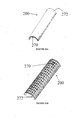
- the graft materials may be provided in any basic shape, including cylinders, blocks, strips, sheets, and wedges. In one embodiment, the graft materials are provided in basic cylinder or strip form.
- the graft materials may have a finite shape or custom shape for specific applications (e.g., semi-spherical for graft acetabular containment, half-tubular long bone wrap or sleeve), or may be "shredded” and housed within a delivery vessel.
- the graft materials may serve as a coating on any orthopaedic appliance such as an intermedullary rod, pedicle screw, plate, hip stem, acetabular cup component and the like.
- the bone graft materials of the present invention also have the ability to attach to Bone Morphogenic Proteins (BMP).
- BMP Bone Morphogenic Proteins
- biocompatible, resorbable composites may have up to about 30% by weight of the biocompatible polymer and 70% by weight of the reaction product.
- the amount of biocompatible polymer within the bone graft materials may also be up to about 20% by weight or up to about 10% by weight, or alternatively up to about 50% by weight.
- Figure 1A illustrates one basic form of the biocompatible graft material in cylinder form.
- Figure 1B depicts the graft material in cylindrical form 80 inserted into a bone void 83 below the femur 81 in the tibial plateau 82 within a human knee.
- Figure 2 illustrates another basic form of the present invention in strip form.
- Figure 3A illustrates one embodiment of the biocompatible graft material of the present invention in semi-spherical form used as a graft containment device.
- Figure 3B depicts a semi-spherical form of the graft material 102 used to accommodate an artificial implant 103.
- the graft material 102 contains an acetabular cup 106, which holds a polyethylene cup 105, in this embodiment.
- Figure 4A illustrates the bone restorative of the present invention in disc form.
- Figure 4B illustrates another embodiment of the biocompatible graft material of the present invention used as a cranio-maxillofacial 76, zygomatic reconstruction 72, and mandibular implant 74.
- Figure 5 illustrates one embodiment of a bone graft material described shaped into a block/wedge form and used as a tibial plateau reconstruction that is screwed, bonded, cemented, pinned, anchored, or otherwise attached in place.
- Figures 6A and 6B illustrate synthetic resorbable defect filling bone graft materials 272 for bone restoration having mesh 270 attached to one side.
- Figure 6C depicts a synthetic resorbable defect filling bone graft material block in which the mesh 270 is placed between the graft material 272.
- Figure 7A, 7B, and 7C illustrate the shapes of some embodiments in semi-tubular form used as a long bone reinforcement sleeve.
- the semi-tube may have a moon cross-section with a uniform thickness ( Figure 7A ); or a crescent moon cross-section with a tapered radius that comes to a point ( Figure 7B ) or a tapered radius that is rounded on the edges ( Figure 7C ).
- Figure 8 depicts the semi-tubular shaped embodiment 200 placed on a metacarpal bone.
- Figure 9 depicts a tubular shaped embodiment 200 fitted around the femur.
- Figures 10A and 10B depicts semi-tubular embodiments 200 showing different configurations for placing the biocompatible mesh 270 and graft material 272.
- Figure 11 is a representative XRD spectra of a bone graft material of the present invention (top) vs. ⁇ -TCP (bottom).
- Figure 12 is a representative FTIR spectrum of bone graft material of the present invention vs. ⁇ -TCP (beta-TCP) and Predicate.
- Figure 13 is an SEM of the bone graft material, 20x.
- Figure 14 is an SEM of the bone graft material, 50x.
- Figure 15 is an SEM of the bone graft material, 250x.
- Figure 16 depicts the Ultimate Indentation Strength for one embodiment of the bone graft material vs. control normalized by adjacent bone at 12 weeks.
- Figure 17 is an SEM of air-dried gelatin treated inorganic material, 23x.
- Figure 18 is an SEM of sheep trabecular bone, 25x.
- Figure 19 is an SEM of the material shown in Figure 14 , 2000x
- Figure 20A depicts an embodiment of the bone restorative 200 having channels 255 in the foam 272 so that a surgeon can see the fixation holes 260 in the mesh 270 to accommodate mechanical fixation with screws. Channels 255 may also used to soak and hold therapeutic materials.
- Figure 20B depicts a side view of the restorative 200.
- Figure 21 depicts an embodiment having wells 265 to soak and hold therapeutic material and also may be used for assisting in fixation.
- Figures 22 and 23 depict the restorative with crimp zones 275 for localized bending.
- Figure 24 depicts a discoid shaped embodiment placed at appropriate sites on the femur; note the cut line 160 for guiding a surgeon to shape the restorative 200 for optimal fitting at appropriate sites on the femur.
- Figure 25 depicts the restorative 200 used on the iliac crest.
- Figures 26A, 26B , and 26C depict an embodiment 200 having crimp zones 275 that guide a surgeon to forming a bowl shaped restorative 200.
- Figure 26A shows the restorative with mesh 270 side up and 26B shows the restorative 200 with foam 272 side up.
- Figure 26C depicts the embodiment 200 after being guided into a bowl shape.
- Figure 27 depicts an embodiment of the present invention having a gradient of interconnectedness.
- bone graft material and “foam” may be used interchangeably in this description.
- biocompatible bone graft material may comprise resorbable polymer, such as collagen, and certain inorganic materials, especially calcium phosphate.
- the present invention provides improvements to bone graft materials, by integrating mesh or other flexible support that result in the present bone restoratives.
- One class of these advancements that are of particular utility are pliable bone restoratives comprising an osteoconductive foam comprising biocompatible, resorbable polymer and calcium phosphate that at least partially surrounds a biocompatible mesh.
- the present invention also provides improvements to bone graft materials having exceptional carrier properties and are suited for use in methods for delivering therapeutic materials to a bony site.
- the present invention finds utility in a wide variety of applications and may provide an alternative to autografts and other implantation materials comprised of cadaver bone, bovine bone, or the like.
- the porous bone restoratives formed herein can be used in medicine, such as, but not limited to, the restoration of bony defects.
- the bone restoratives can also be used for the delivery of medicaments that are internal to the defect, or can be used to promote cellular, bone, or tissue growth.
- the larger porous spaces within some of the bone restoratives of the present invention can be used for culturing cells within the human body.
- the larger spaces are amenable to the growth of cells and can be permeated readily by bodily fluids such as certain blood components.
- bodily fluids such as certain blood components.
- growing cells can be implanted in an animal through the aegis of implants in accordance with the present invention.
- These bone restoratives are implants that give rise to important biochemical or therapeutic uses.
- the present bone restoratives are exceptional fluid carrier support systems.
- the bone restoratives can retain and deliver fluids to a bone defect site due to the porous and interconnected structure of the carrier, the material composition of the carrier, and the design of the carrier. Additionally, the bone restoratives may have structural integrity that is at least partially load-bearing with a mesh component.
- the graft materials can be shaped or formed and are pliable. It will be appreciated that one particularly beneficial aspect of some embodiments of this invention is that it provides unprecedented utility in the surgical operation where a reconstructive surgeon, relying upon the pliability of the bone restorative, may manipulate the restorative into shapes which are particularly amenable to the bony areas to be reconstructed.
- the restoratives have the capability of being fashioned into a new form and have varying degrees of pliability. It may be preferred that the restorative is deformable by human finger pressure and, when in the shape of a strip, can be rolled upon itself when wetted.
- simple hand tools such as forceps, and other common tools used in the operatory, may be employed to shape restoratives of the invention.
- this enables the surgeon to tailor the precise shape of the restorative to that which is required in a particular circumstance very conveniently and at the point of use.
- the pliability of the restoratives of the present invention makse this possible.
- Relatively hard restoratives which cannot be molded conveniently by hand or with the use of common hand tools, require extraordinary processing techniques including machinery, heat, or highly leveraged manipulative devices that are much less useful than the pliable restoratives of the present invention.
- Other relatively brittle bone graft materials in the art are not shapeable without crumbling.
- pliable bone restoratives comprising a biocompatible mesh at least partially surrounded by a biocompatible, resorbable polymer and the oxidation-reduction reaction product of at least one metal cation, at least one oxidizing agent, and at least one oxidizable precursor anion.
- radiopaque embodiments There may be radiopaque embodiments. Other embodiments may be coated with titanium plasma spray to significantly increases implant surface area and mechanical retention in the bone at the time of placement.
- the mesh may also be acid etched titanium or sodium treated titanium to aid in mechanical interlock of the foam.
- graft materials comprising a biocompatible polymer such as collagen, the oxidation-reduction reaction product of at least one metal cation, at least one oxidizing agent, and at least one oxidizable precursor anion. Graft materials are also provided that comprise a collagen and macro-, meso-, and microporous calcium phosphate. Some embodiments may comprise up to 100% Type I collagen. In other embodiments, the collagens used may be predominantly, or up to about 90%, of Type I collagen with up to about 5% of Type III collagen or up to about 5% of other types of collagen.
- the Type I bovine collagen may be native fibrous insoluble collagen, soluble collagen, reconstituted collagen, or combinations thereof.
- the biocompatible polymer may be combined with the reaction product in slurry form, or combined by blending or kneading, to form a substantially homogenous mixture.
- substantially homogenous means that the ratio of components within the mixture is the same throughout. This, upon treatment using various preferred freeze-drying and crosslinking techniques, produces a form of the present invention graft material that may be preferred.
- Collagen has been found to be particularly suitable in the present invention for service as the biocompatible polymer.
- the admixture of the collagen with the highly porous reaction product results in a graft that is highly porous with a broad pore size distribution, increased handling properties, and pliability beyond that which is achievable with some forms of the reaction product alone, for instance calcium phosphate.
- the resorption profile of some of the embodiments of the present invention may vary depending upon the amount, nature, and source of the collagen or other polymer used. Typically, by twelve weeks in vivo about 80%-90% of the present invention is resorbed. One reason that may explain the superior resorption properties of the present invention is the high degree of porosity retained even upon admixing the collagen with the reaction product.
- the collagen may be in a polymerized fibrous form that has a long three-dimensional architecture with multiple cross-links.
- Preferable collagens have beneficial biochemical attributes such as 10% to 20% nitrogen, 10% to 15% of hydroxyproline, or up to 2.5% of ash content.
- the collagens may be 10.5% to 17% nitrogen, 10.5% to 14% of hydroxyproline, or up to 2.5% of ash content.
- the percent nitrogen of a collagen is a measurement of nitrogen in a sample.
- the amino nitrogen of organic material is converted to ammonium sulfate.
- the ammonium sulfate is distilled from an alkaline medium, and further decomposes from which the ammonia is absorbed into a boric acid solution containing a pH indicator.
- the ammonia (nitrogen) concentration determined colorimetrically by back titrating the boric acid solution with a standard acid.
- the percent hydroxyproline of a collagen is a measure of hydroxyproline in a sample.
- Collagen is hydrolyzed with dilute Hydrochloric Acid, filtered and diluted. The solution is reacted with several reagents and then measured using ultraviolet (UV)/Vis analysis along with a standard hydroxyproline solution. Using the sample and standard absorbances, the percentage of hydroxyproline can be calculated [(Sample Abs)(Std)(Weight)(dilution factor)]/[(Sample weight)(Std. Abs)(dilution factor)].
- the ash content of collagen is a measure of the amount of residual elements in collagen materials. When collagen is heated to extremely high temperatures, it is converted to mainly carbon dioxide and water. Elements other than collagen and hydrogen are converted to oxides and salts. A small sample of material is heated until there is only ash left. The weight of this ash is considered the gross amount of inorganic/organic material of the original sample.
- Bone graft materials of this invention that may be preferred are held together in surgically relevant shapes and sizes by foaming the inorganic reaction product with the collagen.
- the resulting osteoconductive foam articles retain substantially all of the biological and chemical properties of the shaped bodies taught in the '519 and '246 patents, while forming a shapeable, flexible unit dose.
- the osteoconductive foam or bone graft materials may be manufactured (with or without mesh) into strips and cylinders of prescribed dimensions and volumes.
- Other shapes include but are not limited to block, hemisphere, half pipe, rod, funnel, cup, sleeve, or discoid.
- the half pipe shaped embodiment 200 has a mesh on top of the foam portion of the restorative.
- the graft material portion is in contact with the metacarpal bone and the mesh is outward facing.
- a full pipe embodiment 200 may be seen in Figure 9 that completely surrounds the femur. This shape may be called a bone cuff.
- the foam 272 could completely surround the mesh. The foam aids in assisting bony incorporation of the mesh and eliminates the surgical step of having to add graft material to the structural mesh portion of the restorative device.
- the graft material will resorb following delivery in the surgical site and exhibit the same beneficial biological responses (e.g., bone formation) as the aforementioned shaped bodies.
- the foam may be further manufactured to have a number of physical features that may assist in placing the restorative in the bony site adding support to surrounding bone.
- the foam may have channels 255 or wells 265 as seen in Figures 20A and 21 . In embodiments where the mesh is embedded within the foam, these wells 265 allow a surgeon to see the mesh and the location on the mesh where a screw or suture will be fixated. These channels 255 and wells 265 may also soak and hold therapeutic materials, as seen in Figure 20 , and may aid in the delivery of therapeutic materials to the bony site.
- the wells may also vary in size and diameter such that they are suitable for helping in the fixation of surgical screws, sutures, or wires.
- channels 255 and wells 265 serve not only as a micro-repository for cells, but also as macro-encasements for admixtures of autogenous bone chips, synthetic bone grafts, or other medicaments.
- the admixture of the latter can be considered a bone graft paté.
- These chambers may also serve as time-release depositories in which medicaments or therapeutic materials are released over time.
- the channels 255 expose the mesh so that an operator can easily affix a screw, suture, or the like to the mesh.
- the wells 265 may allow for easy fixation of a screw through the foam portion directly to the mesh.
- the channels 255 will allow for easy fixation of wires and sutures through the foam.
- Certain presently preferred embodiments of the present invention may be described as a pliable bone restorative comprising a biocompatible mesh and a bone graft material comprising biocompatible, resorbable collagen and calcium phosphate.
- Other embodiments may be a pliable bone restorative comprising a biocompatible, resorbable substantially homogenous blend of a first polymeric material and a second material having interconnected macro, meso-, and microporosity, said blend at least partially surrounding a biocompatible mesh with said bone restorative.
- the first polymeric material may be collagen.
- the second material may comprise calcium phosphate.
- the bone graft materials may have up to about 30% by weight of biocompatible polymer.
- the biocompatible polymer may also be up to about 25% by weight in other embodiments. It will be appreciated that embodiments exist wherein the bone graft materials have up to about 20% or 10% by weight of a biocompatible polymer.
- the polymer chosen is a collagen
- the present invention exhibits a unique mineral ( ⁇ -TCP) to collagen ratio that is unlike the ratios shared by other bone grafts.
- ⁇ -TCP unique mineral
- One skilled in the art may obtain bone graft materials of variable ratios depending on their particular needs. In one effective embodiment, the mass ratio of the reaction product and the collagen is 80:20. In others, it may be 90: 10 or 70:30.
- the mass ratio may be altered without unreasonable testing using methods readily available in the art. It will be appreciated that this ratio is contrary to the mineral ⁇ -TCP to collagen ratios one skilled in the art would find in previous bone grafts while still maintaining all the properties (e.g., porosity, pore size distribution) that attribute to an effective bone graft (e.g., simultaneous bone formation, strength and graft resorption).
- properties e.g., porosity, pore size distribution
- the implant Due to the high porosity and broad pore size distribution (1 ⁇ m - 1000 ⁇ m) of the present invention graft, the implant is not only able to wick/soak/imbibe materials very quickly, but is also capable of retaining them.
- fluids could be used with the present invention including blood, bone marrow aspirate, cell concentrate, liquid hemostat, fibrin, sealant, saline, antibiotics and proteins such as bone morphogenetic proteins (BMPs).
- Materials of the present invention can also be imbibed with blood, cells (e.g. fibroblasts, mesenchymal, stromal, marrow and stem cells), protein rich plasma other biological fluids and any combination of the above.
- cells e.g. fibroblasts, mesenchymal, stromal, marrow and stem cells
- protein rich plasma other biological fluids any combination of the above.
- This capability has utility in cell-seeding, drug delivery, and delivery of biologic molecules as well as in the application of bone tissue engineering, orthopaedics, and carriers of pharmaceuticals.
- materials or fluids are materials such as bone marrow aspirate (BMPs), blood, plasma or protein rich plasma, cells, cell signaling materials, growth factors or hormones, proteins, antibiotics, or medicaments.
- BMPs bone marrow aspirate
- Cells useful in this invention comprise fibroblasts, mesenchymal, stromal, marrow, adipose, myoblasts, lysosomes, and stem cells.
- Suitable stem cells may be stem cells of embryonic, fetal, or adult tissue lineage, such as embryonic stem cells, fetal stem cells or mesenchymal stem cells. Also suitable would be cells derived from these lineages such as osteoprogenitors, osteoblasts, osteocytes, adipocytes, myoblasts, chondrocytes, lysosomes, and the like.
- stem cells may be considered those undifferentiated cells capable of self-renewal and differentiation into multiple lineages of mature cells.
- Cell signaling materials may be described as those materials capable of provoking a cell to react.
- Signaling materials, growth factors, and proteins may include signaling molecules under the Transforming Growth Factor (TGF) Superfamily of proteins, specifically proteins under the TGF-beta (TGF- ⁇ ), Osteogenic Protein (OP)/Bone Morphogenic Protein (BMP), VEGF (VEGF-1 and VEGF-2 proteins) and Inhibin/activtin (Inhibin-beta A, Inhibin-beta B, Inhibin-alpha, and MIS proteins) subfamilies.
- TGF Transforming Growth Factor
- TGF- ⁇ TGF-beta
- OP Osteogenic Protein
- BMP BMP
- VEGF VEGF-1 and VEGF-2 proteins
- Inhibin/activtin Inhibin-beta A, Inhibin-beta B, Inhibin-alpha, and MIS proteins
- the exemplary therapeutic materials are proteins under the TGF- ⁇ and OP/BMP sub
- the TGF- ⁇ subfamily includes the proteins Beta-2, Beta-3, Beta-4 (chicken), Beta-1, Beta-5 (xenopus) and HIF-1 alpha.
- the OP/BMP subfamily includes the proteins BMP-2, BMP-4, DPP, BMP-5, Vgr-1, OP-1/BMP-7, Drosophila 60A, GDF-1, Xenopus Vg-1 and BMP-3.
- Representative proteins of these types include: OP-1/rhBMP-7 (Stryker Corporation, Kalamazoo, MI), rhBMP-2 (Genetics Institute/American Home Products, Madison, NJ), rhIGF-1 (Insulin-like Growth Factor-1) (Cephalon, West Chester, PA), TGF beta (Genentech, San Francisco, CA), MP52 (Biopharm GmbH, Heidelberg, Germany/DePuy Acromed, Raynham, MA).
- Other proteins, genes and cells outside the TGF Superfamily may also be included in the exemplary types of therapeutic materials to be used in conjunction with the present invention.
- PepGen P-15 (Ceramed, Lakewood, CO); LMP-1 (LIM Mineralized Protein-1 gene) (Emory University, Atlanta, GA/Medtronic Sofamor Danek, Minneapolis, MN); Chrysalin TP 508 Synthetic Peptide (Chrysalis Biotechnology, Galveston, TX); GAM (parathyroid hormone) (Selective Genetics, San Diego, CA); rhGDF-5 (Orquest, Mountain View, CA/DePuy Acromed, Raynham, MA); cells lines and FGF (Fibroblast Growth Factor), such as BFGF (Basic Fibroblast Growth Factor), FGF-A (Fibroblast Growth Factor Acidic), and FGFR (Fibroblast Growth Factor Receptor); and certain cell lines such as osteosarcoma cell lines.
- LMP-1 LIM Mineralized Protein-1 gene
- Chrysalin TP 508 Synthetic Peptide Chrysalin TP 508 Synthetic Peptide (Chrysalis Biotechnology, Gal
- the therapeutic materials to be used with the present invention material may be combinations of those listed above.
- Such mixtures include products like Ne-Osteo GFm (growth factor mixture) (Sulzer Orthopaedics, Austin, TX/Zimmer, Warsaw, IN) or mixtures of growth factors, proteins, genes, and cells produced by devices such as AGF (Autologous Growth Factor) (Interpore Cross International, Irvine, CA/EBI, Parsippany, NJ), Symphony Platelet Concentrate System (Harvest Technologies, Belton, TX/DePuy, Warsaw, IN), GPS (Gravitational Platelet System) (Biomet, Warsaw, IN), Magellan platelet separator (Medtronic), and the like.
- the materials to be used with the present invention material may also be combinations of those listed above.
- Such mixtures include products like Ne-Osteo GFm (growth factor mixture) (Sulzer/Zimmer), or mixtures of growth factors, proteins, and genes produced by devices such as AGF (Interpore Cross International/EBI), Symphony BM Concentrator (DePuy), and the like. Further, materials such as ascorbic acid, anti-bone resorption drugs, chemotherapeutic agents, chemicals, genes, fibrin sealants, liquid hemostats, vectors, vitamin D, and sodium fluoride may also be used.
- Ne-Osteo GFm growth factor mixture
- AGF Interpore Cross International/EBI
- Symphony BM Concentrator DePuy
- materials such as ascorbic acid, anti-bone resorption drugs, chemotherapeutic agents, chemicals, genes, fibrin sealants, liquid hemostats, vectors, vitamin D, and sodium fluoride may also be used.
- Bone graft materials of the present invention that may be preferred exhibit high degrees of porosity. It is also preferred that the porosity occur in a broad range of effective pore sizes.
- preferred embodiments of the invention may have, at once, macroporosity, mesoporosity, and microporosity.
- Macroporosity is characterized by pore diameters greater than about 100 ⁇ m and, in some embodiments, up to about 1000 ⁇ m to 2000 ⁇ m.
- Mesoporosity is characterized by pore diameters between about 100 ⁇ m and 10 ⁇ m, while microporosity occurs when pores have diameters below about 10 ⁇ m.
- macro-, meso-, and microporosity occur simultaneously and are interconnected in products of the invention. It is not necessary to quantify each type of porosity to a high degree. Rather, persons skilled in the art can easily determine whether a material has each type of porosity through examination, such as through the preferred methods of mercury intrusion porosimetry, helium pycnometry and scanning electron microscopy. While it is certainly true that more than one or a few pores within the requisite size range are needed in order to characterize a sample as having a substantial degree of that particular form of porosity, no specific number or percentage is called for. Rather, a qualitative evaluation by persons skilled in the art shall be used to determine macro-, meso-, and microporosity.
- some embodiments of the present invention include a pliable bone restorative comprising a biocompatible mesh and a biocompatible bone graft material comprising biocompatible, resorbable collagen and calcium phosphate having macro, meso, and microporosity.
- Porosity is measured by Helium Pycnometry. This procedure determines the density and true volume of a sample by measuring the pressure change of helium in a calibrated volume. A sample of known weight and dimensions is placed in the pycnometer, which determines density and volume. From the samples mass, the pycnometer determines true density and volume. From measured dimensions, apparent density and volume can be determined. Porosity of the sample is then calculated using (apparent volume - measured volume)/apparent volume. Porosity and pore size distribution may also be measured by mercury intrusion porosimetry.
- Pore volumes in excess of about 30% may be achieved in accordance with this invention while materials having pore volumes in excess of 50% or 60% may also be routinely attainable.
- Some embodiments of the invention may have pore volumes of at least about 70%. Some embodiments that may be preferred have pore volumes in excess of about 75%, with 80% being still more preferred. Pore volumes greater than about 90% are possible as are volumes greater than about 92%. In some preferred cases, such high pore volumes are attained while also attaining the presence of macro- meso-, and microporosity as well as physical stability of the materials produced. It is believed to be a great advantage to prepare graft materials having macro-, meso-, and microporosity simultaneously with high pore volumes that also retain some compression resistance and flexibility when wetted. It is also an advantage to prepare graft materials with interconnected porosity, which increases the capillary action and wicking capabilities of the material.
- One embodiment of the present invention is capable of rapidly wicking and retaining materials, and then allowing for sustained release over
- a reactive blend in accordance with the invention may be imbibed into a material that is capable of absorbing it. It may be preferred that the material have significant porosity, be capable of absorbing significant amounts of the reactive blend via capillary action, and that the same be substantially inert to reaction with the blend prior to its autologous oxidation-reduction reaction. Due to this porosity, the bone graft materials disclosed herein may soak and hold fluids. Fluids would not be squeezed out as seen in other bone grafts found in the art. The restorative soaks and retains an approximate 1:1 volume of fluids. There are embodiments that retain over 95% soaked fluid with an applied 500g mass.
- Fluids that may be used in the present invention may be bone marrow aspirate, blood, saline, antibiotics and proteins such as bone morphogenetic proteins (BMPs) and the like.
- Wettability determines the amount of fluid taken up by sample material and if the material absorbs an appropriate amount of fluid within a specified time. Pieces of the material are randomly selected, weighed, and placed in a container of fluid for 120 seconds. If the samples adequately take up fluid, they are then weighed again to determine the percentage of mass increase from fluid absorption.
- Some embodiments may be described as pliable bone restoratives comprising a biocompatible mesh and, at least partially surrounding said mesh, a biocompatible, resorbable polymer and the oxidation-reduction reaction product of at least one metal cation, at least one oxidizing agent, and at least one oxidizable precursor anion. Still further, other embodiments may be described as bone graft materials partially comprised of materials, or morsels, resulting from an oxidation-reduction reaction. These materials may be produced by methods comprising preparing an aqueous solution of a metal cation and at least one oxidizing agent. The solution is augmented with at least one soluble precursor anion oxidizable by said oxidizing agent to give rise to the precipitant oxoanion.
- the oxidation-reduction reaction thus contemplated is conveniently initiated by heating the solution under conditions of temperature and pressure effective to give rise to said reaction.
- the oxidation-reduction reaction causes at least one gaseous product to evolve and the desired intermediate precursor mineral to precipitate from the solution.
- the intermediate precursor mineral thus prepared can either be used "as is” or can be treated in a number of ways. Thus, it may be heat-treated greater than about 800°C or, preferably, greater than about 1100°C in accordance with one or more paradigms to give rise to a preselected crystal structure or other preselected morphological structures therein.
- the oxidizing agent is nitrate ion and the gaseous product is a nitrogen oxide, generically depicted as NO x(g) .
- the precursor mineral provided by the present methods be substantially homogenous. As used in this context, substantially homogenous means that the porosity and pore size distribution throughout the precursor mineral is the same throughout.
- the intermediate precursor mineral provided by the present invention may be any calcium salt.
- Subsequent modest heat treatments convert the intermediate material to e.g. novel monophasic calcium phosphate minerals or novel biphasic ⁇ -tricalcium phosphate ( ⁇ - TCP)+type-B, carbonated apatite (c-HAp) [ ⁇ -Ca 3 (PO 4 ) 2 +Ca 5 (PO 4 ) 3-x (CO 3 ) x (OH)] particulates. More preferably, the heat treatment converts the intermediate material to a predominantly ⁇ -TCP material.
- the porosity is similar to that of inorganic shaped bodies disclosed in the '519 and '246 patents.
- the bone graft materials of the present invention are indeed improvements on the shaped bodies disclosed in the '519 and '246 patents.
- the shaped bodies of the '519 and '246 patents are modified using various natural and synthetic polymers, film forming materials, resins, slurries, aqueous mixtures, pre-polymers, organic materials, metals, and other adjuvants.
- Materials such as collagen, wax, glycerin, gelatin, polycaprolactone, pre-polymeric materials such as precursors to various nylons, acrylics, epoxies, polyalkylenes, and the like, were caused to permeate all or part of the shaped bodies formed in accordance with the '519 and '246 patents.
- the soak and hold properties of some graft materials disclosed herein exhibit at least a greater than 100% mass increase of blood.
- Many of the bone graft materials have a tough structural integrity with improved clinical handling when compared to the bodies of the '519 and '246 patents.
- An embodiment of the present invention includes pliable restoratives for the restoration of bone in the form of a shaped body, the shaped body selected to conform generally to a mammalian, anatomical tissue structure, said body comprising a polymer and beta tricalcium phosphate, and partially surrounding a biocompatible mesh; the graft having interconnected macro-, meso-, and microporosity.
- the bone restoratives have improved handling that can provide a unit dose delivery.
- the addition of a polymer in the graft material greatly enhances the ability of the product to be shaped or cut without crumbling.
- the bone restorative (graft materials) may be shaped or cut using various instruments such as a scapel or scissors. This feature finds utility in a variety of surgical applications, particularly since the bone graft can be formed "in situ" in an operating room to suit the needs of the patient in cases where the bone void to be filled is an irregular shape.
- Some graft materials disclosed may also be delivered into the bony site directly, shaped, and allowed to wick bodily fluids by an operator while during an operation.
- the present invention is also osteoconductive with a structure capable of supporting revascularization unlike metals and low porosity materials that lack an interconnected structure.
- the restoratives may be described as being very pliable, malleable, or even formable.
- pliable means supple enough to bend freely without breaking.
- malleable means capable of being altered or controlled by outside forces or influence.
- formable means to become formed or shaped. It can also be said that the restoratives have high ductility, which means easily molded or shaped in the present context. A surgeon using one of the embodiments of the present invention should be able to shape and form the restorative using the force of his hands or fingers.
- the bone graft materials may be sterilized and may be preferably gamma irradiated at a range of about 25kGy to 40kGy.
- inventions disclosed herein are to fill bony voids and defects and may not be intrinsic to the stability of the surgical site. It will be appreciated that applications for the embodiments of the present invention include, but are not limited to, filling interbody fusion devices/cages (ring cages, cylindrical cages), placement adjacent to cages (i.e., in front cages), placement in the posterolateral gutters in posteriolateral fusion (PLF) procedures, backfilling the iliac crest, acetabular reconstruction and revision hips and knees, large tumor voids, use in high tibial osteotomy, burr hole filling, and use in other cranial defects.
- interbody fusion devices/cages ring cages, cylindrical cages
- placement adjacent to cages i.e., in front cages
- placement in the posterolateral gutters in posteriolateral fusion (PLF) procedures backfilling the iliac crest, acetabular reconstruction and revision hips and knees
- large tumor voids use in high tibial
- the bone graft material strips may be suited for use in PLF by placement in the posterolateral gutters, and in onlay fusion grafting. Additional uses may include craniofacial and trauma procedures that require covering or wrapping of the injured/void site.
- the bone graft material cylinders may be suited to fill spinal cages and large bone voids, and for placement along the posterolateral gutters in the spine.
- the present invention graft material could be made in a wide variety of shapes and sizes via standard molding techniques.
- blocks and cylinders of the present invention may find utility in bone void filling and filling of interbody fusion devices; wedge shaped devices of the present invention may find utility in high tibial osteotomies; and strips may find utility in cranial defect repairs.
- the bone restorative may take a variety of forms including cylindrical, block, or discoid shapes.
- graft materials may be used as semi-spherical ( Figure 3A ), semi-tubular ( Figures 7A-7C ) or disc-shaped ( Figure 4A ) strips for graft containment devices.
- An embodiment of the semi-spherical form 102 in use is depicted in Figure 3B .
- Some embodiments are ring shaped with the mesh being partially surrounded by the graft material.
- the graft material may also be surrounded by the mesh in this embodiment.
- the benefits of the graft containment materials that, for instance, may be used in acetabular reconstruction made from the present invention are several-fold.
- the graft materials may act as both a barrier to prevent migration of other implants or graft materials and serves as an osteoconductive resorbable bone graft capable of promoting bone formation.
- the graft containment device may be relatively non-load bearing, or partially load bearing, or may be reinforced to be fully load bearing as described below. Depending on the form, the graft materials have barrier properties because it maintains its structural integrity.
- the graft materials of the present invention may have meshes or plates affixed.
- the meshes or plates may be of metal, such as titanium or stainless steel, or of a polymer or composite polymer such as polyetheretherketone (PEEK), or nitinol.
- PEEK polyetheretherketone
- a metallic mesh 270 may be placed to one side of the bone graft material 272 to add strength and load bearing properties to the implant.
- the mesh plate 270 sits affixed to one surface of the graft material 272.
- the mesh plate 270 penetrates one surface of the graft material 272 with one side of mesh exposed on top.
- FIG 6C the mesh plate 270 is immersed more deeply than in Figure 6B within the graft material 272.
- a mesh may be affixed to a surface for further support in long bone reinforcement. Due to the unique properties of the present invention graft material, the mesh may be affixed in the body using sutures, staples, screws, cerclage wire or the like.
- the mesh may be placed in any-location necessary for a selected procedure in a selected bodily void.
- a composite of mesh and graft material could be used in a craniomaxillofacial skull defect with the more pliable graft surface being placed in closer proximity to the brain and the more resilient mesh surface mating with the resilient cortical bone of the skull.
- the mesh or plate may be affixed to one side of the graft material.
- the mesh or plate may be affixed to both sides of the graft material in sandwich fashion.
- graft material could be affixed to both sides of the mesh or plate.
- the mesh may be immersed within the graft material.
- the meshes may be flat or may be shaped to outline the graft material such as in a semi-spherical, semi-tubular, or custom form. These embodiments may be unique due to their integral relation between the graft material and the mesh.
- the mesh may also comprise crimped areas for localized bending or shaping as shown in Figure 22 .
- This crimp line may also guide a surgeon in cutting the restorative before placing it on bone. These zones assist an operator in manipulating the restorative into predetermined shapes.
- the disc is crimped or scored in concentric circles so that an operator will be guided to bend the disc to make a cup.
- the bone restorative may exhibit a gradient of interconnectedness with tuneable properties. This embodiment is one in which the restorative exhibits a designated porosity in one area of the bone restorative and the porosity gradually changes towards another area of the restorative.
- the gradient may represent an integration of materials and properties such that the left-most portion of the restorative is comprised of a first relatively dense material with a first porosity (p1), the left middle portion of the restorative is the same first relatively dense material but with a second porosity (p2), the right middle portion of the restorative is a second relatively porous material with a third porosity (p3), and the right-most portion of the restorative is the same second relatively porous material but with a fourth porosity (p4), wherein p4>p3>p2>p1, thus creating a gradient.
- p1 first relatively dense material with a first porosity
- p2 the left middle portion of the restorative is the same first relatively dense material but with a second porosity
- p3 the right middle portion of the restorative is a second relatively porous material with a third porosity (p3)
- the right-most portion of the restorative is the same second relatively porous material but with a fourth poros
- the gradient is one of stiffness or of load bearing capabilities that gradually increases or decreases from one portion of the restorative to the other portion of the restorative.
- the materials and their properties, such as porosity, to be integrated may vary. That is, the first material of the bone restorative may be comprised of a metal, polylactic acid, carbon-fiber reinforced composite, collagen, or mesh that is integrated as described above with the second material comprising calcium phosphate, bone graft materials, bone graft substitutes, or porous resorbable structures.
- the gradient could be one of both porosity and stiffness.
- the entire mesh material in some embodiments will be uniform throughout.
- the porosity of the device will be from about 30% to about 95%.
- some embodiments may have meshes having multiple zones of porosity and thickness. A lower degree of porosity may be needed in an area of the restorative where that area will be used for load bearing applications. In non-load bearing zones, the restorative may have increased mesh porosity.
- the mesh on some embodiments with have a thickness between about 0.1mm to about 2.5mm. In other embodiments that may be preferred, the thickness can be about 0.5mm.
- the thickness of the mesh may be equal throughout or may vary as with porosity such that it is thicker in areas requiring load-bearing capabilities and thinner in non-load bearing zones. Total device thickness may be from about 1mm to about 4cm. In some embodiments that may be preferred, the total thickness maybe 4mm.
- a bone graft for long bone reinforcement comprising a biocompatible, resorbable semi-tubular shape, or sleeve, of a polymer and beta-tricalcium phosphate, the graft having interconnected macro-, meso-, and microporosity.
- a mesh may be affixed to the surface of the sleeve or may be immersed in the sleeve.
- the mesh may be made of titanium, stainless steel, nitinol, a composite polymer, or polyetheretherketone.
- the polymer may be collagen.
- the beta-tricalcium phosphate and polymer may be in a mass ratio of about 90:10 to about 70:10, or about 85:15 to about 75:25.
- the cross-section of the sleeve may be in the shape of a crescent shape moon ( Figure 7B ).
- the mesh may also comprise crimped areas for localized bending or shaping as shown in Figure 22 .
- This crimp line may also guide a surgeon in cutting the restorative before placing it on bone. These zones assist an operator in manipulating the restorative into predetermined shapes.
- the disc is crimped or scored in concentric circles so that an operator will be guided to bend the disc to make a cup.
- the foam portion of the bone restorative may exhibit a gradient of interconnectedness with tuneable properties. This embodiment is one in which restorative exhibits a designated porosity in one area of the bone restorative and the porosity gradually changes towards another area of the restorative.
- the gradient may represent an integration of materials and properties such that the left-most portion of the restorative is comprised of a first relatively dense material with a first porosity (p1), the left middle portion of the restorative is the same first relatively dense material but with a second porosity (p2), the right middle portion of the restorative is a second relatively porous material with a third porosity (p3), and the right-most portion of the restorative is the same second relatively porous material but with a fourth porosity (p4), wherein p4>p3>p2>p1, which creates the gradient.
- the materials and their properties, such as porosity, to be integrated may vary. That is, the first material of the bone restorative may be comprised of a metal, polylactic acid, carbon-fiber reinforced composite, collagen, or mesh that is integrated as described above with the second material calcium phosphate, bone graft materials, bone graft substitutes or porous resorbable structures.
- the gradient could be one of both porosity and stiffness. In this manner, the type of material, the thickness of the material, and the porosity all play a role.
- the mesh material may also exhibit variable porosity.
- the entire mesh material in some embodiments will be uniform throughout. In some embodiments, the porosity of the device will be from about 30% to about 95%. However, it will be appreciated that some embodiments may have meshes having multiple zones of porosity and thickness. A lower degree of porosity may be needed in an area of the restorative where that area will be used for load bearing applications. In non-load bearing zones, the restorative may have increased mesh porosity.
- the mesh on some embodiments with have a thickness between about 0.1mm to about 2.5mm. In other embodiments that may be preferred, the thickness can be about 0.5mm.
- the thickness of the mesh may be equal throughout or may vary as with porosity such that it is thicker in areas requiring load-bearing capabilities and thinner in non-load bearing zones.
- Total device thickness may be from about 1mm to about 4cm. In some embodiments that may be preferred, the total thickness maybe 4mm.
- the surface texture of the mesh may also vary depending on the need and depending upon the degree of adhesion that is required between the mesh and the foam to be integrated.
- the surface texture may be measured by a roughness measurement. It will be appreciated that in some embodiments, the roughness measurement will be high like that of sandpaper. In other embodiments, the mesh may have a low roughness measurement like that of a sheet of ice. It is foreseeable that one skilled in the art will vary the surface texture to fit their particular need.
- a graft for the restoration of bone in the form of a shaped body, the shaped body comprising a polymer and beta-tricalcium phosphate, the material of the graft having interconnected macro-, meso-, and microporosity; the body shape being selected to conform generally to a malian, anatomical bone structure.
- the shapes will vary depending on the area of the body being repaired. Some basic shapes may be a disk, semi-sphere, semi-tubular, or torus. In some embodiments, the shape will conform generally to the acetabulum.
- graft materials of the present invention having load-bearing capabilities may be open framed, such that the bone graft material is embedded in the central opening of the frame.
- the frame may be made of a metal such as titanium or of a load-bearing resorbable composite such as PEEK or a composite of some form of poly-lactic acid (PLA).
- PEEK poly-lactic acid
- the acid from the PLA co-acts, or interacts with the calcium phosphate of the embedded bone graft material to provide an implant with superior resorption features.
- the graft materials can also be imbibed with any bioabsorbable polymer or film-forming agent such as polycaprolactones (PCL), polyglycolic acid (PGA), poly-L-Lactic acid (PL-LA), polysulfones, polyolefins, polyvinyl alcohol (PVA), polyalkenoics, polyacrylic acids (PAA), polyesters and the like.
- PCL polycaprolactones
- PGA polyglycolic acid
- PL-LA poly-L-Lactic acid
- PVA polysulfones
- polyolefins polyvinyl alcohol
- PAA polyalkenoics
- polyesters polyesters and the like.
- the resultant graft material is strong, carveable, and compressible.
- the grafts of the present invention coated with agents such as the aforementioned may still absorb blood.
- the graft materials may be used as an attachment or coating to any orthopaedic implant such as a metal hip stem, acetabular component, humeral or metatarsal implant, vertebral body replacement device, pedicle screw, general fixation screw, plate or the like.
- the coating may be formed by dipping or suspending the implant for a period of time in a substantially homogenous slurry of polymer and mineral and then processing via freeze-drying/lypholization and crosslinking techniques.
- substantially homogenous means that the ratio of elements within the slurry is the same throughout.
- a female mold may be made of the implant and the slurry may be poured into the mold and processed, as described above, to form the coating.
- the graft material may be shredded or cut into small pieces. These smaller shredded pieces could then be used as filler or could be placed in a syringe body. In this fashion, fluids could be directly aspirated into or injected into the syringe body thereby forming a cohesive, shapeable bone graft mass "in situ" depending upon the application requirements.
- the shredded pieces find particular use as filler for irregular bone void defects. Further, unlike traditional bone graft substitutes they are highly compressible and therefore can be packed/impacted to insure maximum contact with adjacent bone for beneficial healing.
- a method for restoring or repairing bone in an animal comprising accessing a site to be restored; and implanting into a bony space a bone graft material comprising biocompatible, resorbable collagen, the oxidation-reduction reaction product of at least one metal cation, at least one oxidizing agent, and at least one oxidizable precursor anion.
- the graft material used in this method may be chosen by one skilled among those disclosed in the present application.
- One embodiment was comprised of ⁇ -TCP, with a cation to anion ratio of Ca 3 (PO 4 ) 2 ; and medical grade Type I bovine collagen, manufactured in the following manner.
- Inorganic scaffolds were made using the RPR process disclosed in U.S. Patent Nos. 5,939,039 and 6,325,987 .
- the resultant inorganic scaffolds were crushed and sieved to obtain morsels in the size range of .25mm-4mm.
- the morsels were added to a fibrous collagen slurry in a wet processing room and the resultant slurry was further mixed and casted/molded into various shapes in a cleanroom.
- the shapes were freeze-dried and crosslinked using dehydrothermal (DHT) treatment to produce resultant bone graft material shaped products.
- DHT dehydrothermal
- ⁇ -TCP ⁇ -TCP
- XRD X-ray diffraction
- ASTM F1088-87 Standard Specification for Beta-Tricalcium Phosphate for Surgical Implantation.
- ICP Inductively Coupled Plasma Chromatography
- FTIR Fourier Transform Infrared Spectroscopy
- the quantitative XRD results show that the mineral component of the bone graft material is 98.25% pure ⁇ -TCP, which matches well with the ICDS standard plot for ⁇ -TCP pictured with the representative XRD pattern of the bone graft material ( Figure 11 ).
- the ICP results for the bone graft material show that the levels of heavy metal contaminants- arsenic (As), cadmium (Cd), mercury (Hg), and lead (Pb), are below the method detection limits of 2.25ppm, 1.80ppm, 2.25ppm and 4.5ppm, respectively, thus below the limits set forth in ASTM F-1088-87.
- Qualitative FTIR results show a 95% match of the bone graft material to greater than 99% pure ⁇ -TCP.
- a representative FTIR spectrum is shown in Figure 12 .
- pore diameters in the graft range from 1 ⁇ m to 1000 ⁇ m. Approximately 5% to 15 % of the pores are greater than 100 ⁇ m, approximately 50%-70% of the pores are between 10 ⁇ m -100 ⁇ m, and approximately 20%-35% of the pores are less than 10 ⁇ m.
- the larger macro pores greater than 100 ⁇ m) allow bone to grow in apposition to the calcium phosphate surfaces of the implant.
- the smaller meso (10 ⁇ m-100 ⁇ m) and micro (less than 10 ⁇ m) interconnected pores allow for fluid communication and nutrient transport. Total porosity is approximately 70%-80%.
- a GLP animal study was performed at North American Science Associates, Inc. (NAMSA), Northwood, OH, to evaluate the biological effects of the bone graft material and a control in metaphyseal defects of adult dogs. Sixteen dogs were implanted both with one embodiment of the present invention and the control. Animals were sacrificed at each of the time periods of 3, 6, 12, and 24 weeks. Gross evaluation, radiographic assessment, histological evaluation, histomorphometry, and mechanical evaluations were performed.
- control was placed in the proximal humerus, and the present invention was placed in the femoral condyle.
- a piece of the inorganic material was immersed in a solution prepared by dissolving 7.1g food-grade gelatin (CAS #9000-70-0) (Knox Unflavored Gelatin, Nabisco Inc., East Hanover, N.J. 07936) in 100.0g deionized water at approximately 90°C.
- the inorganic material readily imbibed the warm gelatin solution and, after several minutes, the largely intact piece of inorganic material was carefully removed from the solution and allowed to cool and dry overnight at room temperature.
- the gelatin solution gelled on cooling and imparted additional strength and improved handling properties to the inorganic material.
- the material exhibited considerable improvement in compression strength and a dramatically reduced tendency to shed particulate debris when cut with a knife or fine-toothed saw. It is presumed that the film-forming tendency of the gelatin on drying induced compressive forces on the internal cellular elements of the inorganic sponge material, thereby strengthening the overall structure.
- Cylindrical plugs could be cored from pieces of the air dried gelatin-treated inorganic material using hollow punch tools ranging from 1/2 inch down to 1/8 inch in diameter.
- Figure 17 is a SEM of the air-dried gelatin treated inorganic material.
- Figure 18 is a SEM of sheep trabecular bone.
- the highly porous macrostructure of sheep trabecular bone is representative of the anatomical structure of cancellous bone of higher mammals, including humans.
- the sample of sheep trabecular bone was prepared for SEM analysis by sputter coating a cross-sectional cut from a desiccated sheep humerus.
- Figure 19 is a higher magnification SEM of the air-dried gelatin treated inorganic material depicted in Figure 17 . From this SEM micrograph, the presence of meso- and microporosity in the calcium phosphate matrix is readily apparent.
- a reactant solution was prepared as described in the '162 patent.
- a variety of shapes, including disks, squares, and triangles, were cut from a sheet of 3/32 inch thick sponge material (Spontex, Inc., P.O. Box 561, Santa Fe Pike, Columbia, Tenn. 38402) using either scissors or hollow punches.
- the cut pieces of compressed sponge were fully imbibed with the reactant solution after which they swelled to form cylinders, cubes, and wedges.
- These solution saturated sponge articles were placed into an oven preheated to 500°C and held at that temperature for 1 hour. After cooling, the inorganic sponge pieces were carefully removed from the considerable amount of crusty white solid resulting from the exudate material.
- the vestigial structures represented positive versions of the original sponge structures with faithful replication of the cellular elements and porosity.
- the vestigial masses were fragile with very low apparent density, but they were robust enough to be handled as coherent blocks of highly porous solid once they were removed from the exudate material.
- the integrity of the various samples made from the controlled porosity cellulose sponge was improved over corresponding samples prepared from the commercial cellulose sponge materials. The samples were then crushed and sieved to obtain morsels in the size range of 0.25mm-4mm.
- the morsels were added to a collagen slurry in a wet processing room and the resultant slurry was further mixed and casted/molded into various shapes in a cleanroom.
- the shapes were freeze-dried and crosslinked to produce resultant bone graft material shaped products.
- Pieces of an inorganic sponge material were immersed in a gelatin solution prepared as described in Example 7 except that 7.1g of Knox gelatin was dissolved in 200g deionized water rather than 100g of deionized water.
- the inorganic sponge material readily imbibed the warm gelatin solution and, after several minutes, the largely intact pieces of inorganic sponge material were carefully removed from the solution and allowed to cool and dry at room temperature.
- Significant additional strength and improved handling properties were noted in the gelatin-treated inorganic sponge material after the gelatin was allowed to thoroughly dry for several days. The material exhibited considerable improvement in compression strength and a dramatically reduced tendency to shed particulate debris when cut with a knife or fine-toothed saw.
- the surface of the treated inorganic sponge material had a glassy appearance, and significant additional strength and improved handling properties were noted.
- LeicaTM zoom stereo microscope the presence of a glassy surface was confirmed and rounding of the features was evident indicating that some level of sintering had occurred. Considerable shrinkage of the pieces was also noted.
- a reactant solution was prepared as described in the '519 patent. Disks were cut from a sheet of 3/32 inch thick compressed sponge using a 3/8 inch diameter hollow punch and a model No. 3393 Carver hydraulic press (Carver Inc., 1569 Morris St., P.O. Box 544, Wabash, Ind. 46992) to ensure uniform sizing. The disks were distended by immersion in deionized water and the resulting sponge cylinders, each approximately 3/8 inch diameter by 1 inch length, were then blotted on paper towel to remove as much excess water as possible. The damp sponge cylinders were then imbibed with approximately seven times their weight of the reactant liquid.
- the furnace temperature was ramped at 40°C/minute to 800°C and held at 800°C for 45 minutes.
- the resultant cylindrical white porous inorganic sponge samples were robust and exhibited strengths qualitatively similar to those attained from the fully dried gelatin-treated samples prepared as described in Example 10.
- a 100mm x 100mm square mesh was formed.
- the foam portion had a thickness of 3mm to 3.5mm and the mesh portion had a thickness of 0.5mm to 1mm.
- Ovoid shaped holes were made in the thin, malleable mesh.
- the bone restorative, mesh plus foam, had a total thickness of 4mm. The mesh was then embedded in the foam portion.
- a 50mm x 100mm mesh was formed.
- the approximate thickness of the foam portion was about 3mm thick.
- a mesh shaped foam portion was formed having the characteristic variety of pore sizes.
- a slightly malleable mesh having about 1mm thickness was formed and placed on top portion of the foam. Screw holes to accommodate standard titanium trauma screws were formed extending through the bone restorative.
Abstract
Description
- This invention relates to biocompatible bone graft materials for repairing bone defects and the application of the bone graft materials disclosed herein. The present invention incorporates the benefits of inorganic shaped bodies having macro, meso, and microporosity and polymers such as collagen. The bone restoratives may also be useful as delivery vehicles for therapeutic materials such as bone marrow aspirate, blood, plasma, cells, cell signaling materials, growth factors, proteins, or medicaments.
- There has been a continuing need for improved bone graft materials. Although autograft, the current gold standard, has the ideal properties and radiopacity, the use of autogenous bone exposes the patient to risk of a second surgery, pain, and morbidity at the donor site. Allograft devices, which are processed from donor bone, also have ideal radiopacity, but carry the risk of disease transmission. The devices are restricted in terms of variations on shape and size and have sub-optimal strength properties that decrease after implantation. The quality of the allograft devices varies because they are natural. Also, since companies that provide allograft implants obtain their supply from donor tissue banks, there tend to be limitations on supply. In recent years, synthetic materials have become a viable alternative to autograft and allograft devices. One such synthetic material is Vitoss® Scaffold Synthetic Cancellous Bone Void Filler (Orthovita, Inc., Malvern, PA, assignee of the present application). Synthetic graft materials, like autograft and allograft, serve as osteoconductive scaffolds that promote the ingrowth of bone. As bone growth is promoted and increases, the graft material resorbs and is eventually replaced with new bone.
- Many synthetic bone grafts include materials that closely mimic mammalian bone, such as compositions containing calcium phosphates. Exemplary calcium phosphate compositions contain type-B carbonated hydroxyapatite [Ca5(PO4)3x(CO3)x(OH)], which is the principal mineral phase found in the mammalian body. The ultimate composition, crystal size, morphology, and structure of the body portions formed from the hydroxyapatite are determined by variations in the protein and organic content. Calcium phosphate ceramics have been fabricated and implanted in mammals in various forms including, but not limited to, shaped bodies and cements. Different stoichiometric compositions such as hydroxyapatite (HAp), tricalcium phosphate (TCP), tetracalcium phosphate (TTCP), and other calcium phosphate salts and minerals, have all been employed to match the adaptability, biocompatibility, structure, and strength of natural bone. The role of pore size and porosity in promoting revascularization, healing, and remodeling of bone has been recognized as a critical property for bone grafting materials. The preparation of exemplary porous calcium phosphate materials that closely resemble bone have been disclosed, for instance, in
U.S. Patent Nos. 6,383,519 (hereinafter the '519 patent) and 6,521,246, (hereinafter the '246 patent) incorporated herein by reference in their entirety. - There has been a continued need for improved bone graft systems. Although calcium phosphate bone graft materials are widely accepted, they lack the strength, handling and flexibility necessary to be used in a wide array of clinical applications. Heretofore, calcium phosphate bone graft substitutes have been used in predominantly non-load bearing applications as simple bone void fillers and the like. For more clinically challenging applications that require the graft material to take on load, bone reconstruction systems that pair a bone graft material to traditional rigid fixation systems are used. The prior art discloses such bone reconstruction systems. For instance, MacroPore OS™ Reconstruction System is intended to reinforce and maintain the relative position of weak bony tissue such as bone graft substitutes or bone fragments from comminuted fractures. The system is a resorbable graft containment system composed of various sized porous sheets and sleeves, non-porous sheets and sleeves, and associated fixation screws and tacks made from polylactic acid (PLA). However, the sheets are limited in that they can only be shaped for the body when heated.
- The Synthes SynMesh™ consists of flat, round, and oval shaped cylinders customized to fit the geometry of a patient's anatomical defect. The intended use is for reinforcement of weak bony tissue and is made of commercially pure titanium. Although this mesh may be load bearing, it is not made entirely of resorbable materials that are flexible and also lacks an absorbant component for the delivery of materials of the types described herein.
- Many bone graft materials have limited interconnectedness that substantially limits their ability to retain and deliver therapeutic materials and fluids at a bony site. As such, these graft materials would not be suitable as carriers for therapeutic materials and fluids such as cells, cell signaling materials, proteins, bone marrow aspirate, and blood. It is also known that most bone graft materials lack the structural integrity necessary to provide support.
- Conversely, metals, which are capable of providing structural support typically are not readily absorbent and cannot retain fluid. This is also due in part to their low porosity or macro-hole structures.
- It would be of great benefit in the art to use graft materials for the retention and delivery of therapeutic materials or fluids. Currently, bone grafts often are incapable of adequately retaining fluids once a surgeon attempts to implant the graft into a bony space. The majority of the fluids are flushed out of the graft when manipulated by the surgeon. Thus, there is a need in the art for a bone graft capable of retaining and delivering therapeutic materials that are at least partially load bearing.
- There is a need for resorbable bone grafts with improved handling, which are flexible and not brittle, and are compression resistant. It has been discovered that admixing highly porous resorbable inorganic bodies with resorbable polymeric materials greatly improves upon handling, yet still provides an osteoconductive implant with good resorption and bone formation properties. It will be appreciated that such an implant would offer an easy-to-use dose of composite material and would be an advancement over current bone reconstruction systems for certain clinical applications in that it eliminates the need to have both a graft material and rigid fixation system.
- There is a need in the art to provide biocompatible graft materials with exceptional osteoconductive properties; provide pre-sized graft materials in a variety of forms, including strips and cylinders for restoring defects in bone; to provide bone graft materials that can be shaped; and to provide bone graft materials with improved handling properties, so that the graft material can be cut dry or after being wetted and does not crumble.
- Currently, bone grafts often are incapable of retaining fluids once a surgeon attempts to implant the graft into a bony space. The fluids are flushed out of the graft when manipulated by the surgeon. There is also a need to provide bone graft materials with some compression resistance, such that the brittleness often associated with inorganic or ceramic bone graft materials is eliminated; to provide bone graft materials with integrity that are at least partially load bearing; to provide bone graft materials with improved pliability that still retain high degrees of porosity over a broad pore size distribution to maintain superior resorption and bone ingrowth properties; to provide bone graft materials with fluid wicking and retention properties even under compressive loads; and to provide bone grafts that provide easy implantation into a bony space and with decreased tendency to wash away when imbibed with fluid. Additional objects, advantages, and novel features of this invention will become apparent to those skilled in the art upon examination of the following descriptions, figures and claims thereof, which are not intended to be limiting.
- The present invention is directed to biocompatible bone graft materials that comprise a biocompatible, resorbable polymer and the oxidation-reduction reaction product of at least one metal cation, at least one oxidizing agent, and at least one oxidizable precursor anion. The reaction product may be selected to suit the needs of one skilled in the art but may be inorganic compositions comprising calcium phosphate, biphasic calcium phosphate, or beta tri-calcium phosphate (β-TCP). The present invention is also directed to pliable bone restorative comprising an osteoconductive foam comprising biocompatible, resorbable polymer and calcium phosphate that at least partially surrounds a biocompatible mesh.
- Other embodiments of the present invention include pliable bone restoratives comprising a biocompatible mesh and at least partially surrounding the mesh, a biocompatible, resorbable polymer, and the oxidation-reduction reaction product of at least one metal cation, at least one oxidizing agent, and at least one oxidizable precursor anion. Disclosed herein are also pliable bone restoratives comprising a biocompatible mesh and a bone graft material comprising biocompatible, resorbable collagen and calcium phosphate. A further embodiment of the present invention is a pliable bone restorative comprising a biocompatible mesh and a biocompatible bone graft material comprising biocompatible, resorbable collagen and calcium phosphate having macro, meso, and microporosity. Also disclosed within are pliable bone restoratives comprising a biocompatible, resorbable substantially homogenous blend of a first polymeric material and a second material having interconnected macro, meso-, and microporosity, with said blend at least partially surrounding a biocompatible mesh. A further embodiment that may be preferred is in the form of a shaped body selected to conform generally to a mammalian, anatomical tissue structure. The shaped body comprises a polymer and beta tricalcium phosphate partially surrounding a biocompatible mesh. The graft may have interconnected macro-, meso-, and microporosity. Suitable polymers may include structural proteins such as collagen.
- Still other embodiments of the present invention are directed to methods for delivering therapeutic materials comprising: providing a bone restorative comprising biocompatible, resorbable polymer, and the oxidation-reduction reaction product of at least one metal cation, at least one oxidizing agent, and at least one oxidizable precursor anion; imbibing said bone restorative with a therapeutic material; and placing said bone restorative into a bony space. In some embodiments that may be preferred the bone restorative may be pliable for optimum shaping capability. The present invention is also directed to methods where the bone restorative also has macro-, meso-, and microporosity; and to methods where the bone restorative is capable of wicking and delivering materials via its interconnected structure. The therapeutic materials may comprise cells, cell signaling materials, proteins, bone marrow aspirate, plasma, blood, growth factors, or medicaments. The cells may comprises stem cells. In some embodiments, the selected polymer may be collagen. In many embodiments that may be preferred the reaction product may be calcium phosphate or β- tricalcium phosphate in other embodiments. The bone restorative may comprise cell wells for containing therapeutic materials or an admixture of autogenous bone chips, synthetic bone graft, or medicaments. The therapeutic materials imbibed into the bone restorative or those contained within the cell wells may release them over time.
- The present invention is an improvement upon the shaped bodies disclosed in
U.S. Patent Nos. 6,383,519 ("'519 patent") and6,521,246 ("'246 patent"), and the RPR process disclosed inU.S. Patent Nos. 5,939,039 ("039 patent") and6,325,987 ("'987 patent"), all assigned to the present assignee and incorporated herein by reference in their entirety. The oxidation-reduction reaction product of the present invention shares the same unique porosity of those shaped bodies of the '519 and '246 patents. The reaction product grants the present invention graft material macro, meso, and microporosity, which allow the graft material to have extraordinary imbibation and absorption properties. Further, the inclusion of a polymer in the present invention material lends improved handling and flexibility. The graft materials can have a finite shape for some applications and are compression resistant or at least partially load bearing. When imbibed with fluids, the bone graft materials are flexible, bendable/deformable, and scalpable, without crumbling or falling apart. Some embodiments have a mesh or plate affixed to the bone graft material for added support. The bone graft materials may be imbibed with fluids such as bone marrow aspirate, blood, or saline. The graft materials may be provided in any basic shape, including cylinders, blocks, strips, sheets, and wedges. In one embodiment, the graft materials are provided in basic cylinder or strip form. In other embodiments, the graft materials may have a finite shape or custom shape for specific applications (e.g., semi-spherical for graft acetabular containment, half-tubular long bone wrap or sleeve), or may be "shredded" and housed within a delivery vessel. Yet, in other embodiments, the graft materials may serve as a coating on any orthopaedic appliance such as an intermedullary rod, pedicle screw, plate, hip stem, acetabular cup component and the like. The bone graft materials of the present invention also have the ability to attach to Bone Morphogenic Proteins (BMP). - This invention gives rise to biocompatible, resorbable composites that may have up to about 30% by weight of the biocompatible polymer and 70% by weight of the reaction product. The amount of biocompatible polymer within the bone graft materials may also be up to about 20% by weight or up to about 10% by weight, or alternatively up to about 50% by weight.
-
Figure 1A illustrates one basic form of the biocompatible graft material in cylinder form.Figure 1B depicts the graft material incylindrical form 80 inserted into abone void 83 below thefemur 81 in thetibial plateau 82 within a human knee. -
Figure 2 illustrates another basic form of the present invention in strip form. -
Figure 3A illustrates one embodiment of the biocompatible graft material of the present invention in semi-spherical form used as a graft containment device.Figure 3B depicts a semi-spherical form of the graft material 102 used to accommodate anartificial implant 103. The graft material 102 contains anacetabular cup 106, which holds apolyethylene cup 105, in this embodiment. -
Figure 4A illustrates the bone restorative of the present invention in disc form.Figure 4B illustrates another embodiment of the biocompatible graft material of the present invention used as a cranio-maxillofacial 76, zygomatic reconstruction 72, and mandibular implant 74. -
Figure 5 illustrates one embodiment of a bone graft material described shaped into a block/wedge form and used as a tibial plateau reconstruction that is screwed, bonded, cemented, pinned, anchored, or otherwise attached in place. -
Figures 6A and 6B illustrate synthetic resorbable defect fillingbone graft materials 272 for bonerestoration having mesh 270 attached to one side.Figure 6C depicts a synthetic resorbable defect filling bone graft material block in which themesh 270 is placed between thegraft material 272. -
Figure 7A, 7B, and 7C illustrate the shapes of some embodiments in semi-tubular form used as a long bone reinforcement sleeve. As shown in the figures, the semi-tube may have a moon cross-section with a uniform thickness (Figure 7A ); or a crescent moon cross-section with a tapered radius that comes to a point (Figure 7B ) or a tapered radius that is rounded on the edges (Figure 7C ). -
Figure 8 depicts the semi-tubularshaped embodiment 200 placed on a metacarpal bone. -
Figure 9 depicts a tubular shapedembodiment 200 fitted around the femur. -
Figures 10A and 10B depictssemi-tubular embodiments 200 showing different configurations for placing thebiocompatible mesh 270 andgraft material 272. -
Figure 11 is a representative XRD spectra of a bone graft material of the present invention (top) vs. β-TCP (bottom). -
Figure 12 is a representative FTIR spectrum of bone graft material of the present invention vs. β-TCP (beta-TCP) and Predicate. -
Figure 13 is an SEM of the bone graft material, 20x. -
Figure 14 is an SEM of the bone graft material, 50x. -
Figure 15 is an SEM of the bone graft material, 250x. -
Figure 16 depicts the Ultimate Indentation Strength for one embodiment of the bone graft material vs. control normalized by adjacent bone at 12 weeks. -
Figure 17 is an SEM of air-dried gelatin treated inorganic material, 23x. -
Figure 18 is an SEM of sheep trabecular bone, 25x. -
Figure 19 is an SEM of the material shown inFigure 14 , 2000x -
Figure 20A depicts an embodiment of thebone restorative 200 havingchannels 255 in thefoam 272 so that a surgeon can see the fixation holes 260 in themesh 270 to accommodate mechanical fixation with screws.Channels 255 may also used to soak and hold therapeutic materials.Figure 20B depicts a side view of the restorative 200. -
Figure 21 depicts anembodiment having wells 265 to soak and hold therapeutic material and also may be used for assisting in fixation. -
Figures 22 and23 depict the restorative withcrimp zones 275 for localized bending. -
Figure 24 depicts a discoid shaped embodiment placed at appropriate sites on the femur; note thecut line 160 for guiding a surgeon to shape the restorative 200 for optimal fitting at appropriate sites on the femur. -
Figure 25 depicts the restorative 200 used on the iliac crest. -
Figures 26A, 26B , and26C depict anembodiment 200 havingcrimp zones 275 that guide a surgeon to forming a bowl shapedrestorative 200.Figure 26A shows the restorative withmesh 270 side up and 26B shows the restorative 200 withfoam 272 side up.Figure 26C depicts theembodiment 200 after being guided into a bowl shape. -
Figure 27 depicts an embodiment of the present invention having a gradient of interconnectedness. - The terms "bone graft material" and "foam" may be used interchangeably in this description. Disclosed in that application were, inter alia, biocompatible bone graft material may comprise resorbable polymer, such as collagen, and certain inorganic materials, especially calcium phosphate. The present invention provides improvements to bone graft materials, by integrating mesh or other flexible support that result in the present bone restoratives. One class of these advancements that are of particular utility are pliable bone restoratives comprising an osteoconductive foam comprising biocompatible, resorbable polymer and calcium phosphate that at least partially surrounds a biocompatible mesh. The present invention also provides improvements to bone graft materials having exceptional carrier properties and are suited for use in methods for delivering therapeutic materials to a bony site.
- The present invention finds utility in a wide variety of applications and may provide an alternative to autografts and other implantation materials comprised of cadaver bone, bovine bone, or the like. The porous bone restoratives formed herein can be used in medicine, such as, but not limited to, the restoration of bony defects. The bone restoratives can also be used for the delivery of medicaments that are internal to the defect, or can be used to promote cellular, bone, or tissue growth. In this way, the can be partially filled with materials that either comprise or carry a medicament or therapeutic such as proteins, growth hormones, antibiotics, or cell signaling materials. Indeed, the larger porous spaces within some of the bone restoratives of the present invention can be used for culturing cells within the human body. In this regard, the larger spaces are amenable to the growth of cells and can be permeated readily by bodily fluids such as certain blood components. In this way, growing cells can be implanted in an animal through the aegis of implants in accordance with the present invention. These bone restoratives are implants that give rise to important biochemical or therapeutic uses.
- The present invention finds utility in a wide variety of applications and may provide an alternative to autografts and other implantation materials comprised of cadaver bone, bovine bone, or the like. The porous bone restoratives formed herein can be used in medicine, such as, but not limited to, the restoration of bony defects. The bone restoratives can also be used for the delivery of medicaments that are internal to the defect, or can be used to promote cellular, bone, or tissue growth. In this way, the can be partially filled with materials that either comprise or carry a medicament or therapeutic such as proteins, growth hormones, antibiotics, or cell signaling materials. Indeed, the larger porous spaces within some of the bone restoratives of the present invention can be used for culturing cells within the human body. In this regard, the larger spaces are amenable to the growth of cells and can be permeated readily by bodily fluids such as certain blood components. In this way, growing cells can be implanted in an animal through the aegis of implants in accordance with the present invention. These bone restoratives are implants that give rise to important biochemical or therapeutic uses.
- The present bone restoratives are exceptional fluid carrier support systems. The bone restoratives can retain and deliver fluids to a bone defect site due to the porous and interconnected structure of the carrier, the material composition of the carrier, and the design of the carrier. Additionally, the bone restoratives may have structural integrity that is at least partially load-bearing with a mesh component.
- Preferably, the graft materials can be shaped or formed and are pliable. It will be appreciated that one particularly beneficial aspect of some embodiments of this invention is that it provides unprecedented utility in the surgical operation where a reconstructive surgeon, relying upon the pliability of the bone restorative, may manipulate the restorative into shapes which are particularly amenable to the bony areas to be reconstructed. The restoratives have the capability of being fashioned into a new form and have varying degrees of pliability. It may be preferred that the restorative is deformable by human finger pressure and, when in the shape of a strip, can be rolled upon itself when wetted. Alternatively, simple hand tools such as forceps, and other common tools used in the operatory, may be employed to shape restoratives of the invention. As will readily be perceived, this enables the surgeon to tailor the precise shape of the restorative to that which is required in a particular circumstance very conveniently and at the point of use. The pliability of the restoratives of the present invention makse this possible. Relatively hard restoratives, which cannot be molded conveniently by hand or with the use of common hand tools, require extraordinary processing techniques including machinery, heat, or highly leveraged manipulative devices that are much less useful than the pliable restoratives of the present invention. Other relatively brittle bone graft materials in the art are not shapeable without crumbling.
- In accordance with some embodiments of the present application, there are pliable bone restoratives comprising a biocompatible mesh at least partially surrounded by a biocompatible, resorbable polymer and the oxidation-reduction reaction product of at least one metal cation, at least one oxidizing agent, and at least one oxidizable precursor anion.
- It will be appreciated that a number of alterations may be made to customize the restoratives for specific needs. There may be radiopaque embodiments. Other embodiments may be coated with titanium plasma spray to significantly increases implant surface area and mechanical retention in the bone at the time of placement. The mesh may also be acid etched titanium or sodium treated titanium to aid in mechanical interlock of the foam.
- In accordance with the present invention, graft materials are provided comprising a biocompatible polymer such as collagen, the oxidation-reduction reaction product of at least one metal cation, at least one oxidizing agent, and at least one oxidizable precursor anion. Graft materials are also provided that comprise a collagen and macro-, meso-, and microporous calcium phosphate. Some embodiments may comprise up to 100% Type I collagen. In other embodiments, the collagens used may be predominantly, or up to about 90%, of Type I collagen with up to about 5% of Type III collagen or up to about 5% of other types of collagen. The Type I bovine collagen may be native fibrous insoluble collagen, soluble collagen, reconstituted collagen, or combinations thereof. The biocompatible polymer may be combined with the reaction product in slurry form, or combined by blending or kneading, to form a substantially homogenous mixture. As used in this context, substantially homogenous means that the ratio of components within the mixture is the same throughout. This, upon treatment using various preferred freeze-drying and crosslinking techniques, produces a form of the present invention graft material that may be preferred.
- Collagen has been found to be particularly suitable in the present invention for service as the biocompatible polymer. The admixture of the collagen with the highly porous reaction product results in a graft that is highly porous with a broad pore size distribution, increased handling properties, and pliability beyond that which is achievable with some forms of the reaction product alone, for instance calcium phosphate. The resorption profile of some of the embodiments of the present invention may vary depending upon the amount, nature, and source of the collagen or other polymer used. Typically, by twelve weeks in vivo about 80%-90% of the present invention is resorbed. One reason that may explain the superior resorption properties of the present invention is the high degree of porosity retained even upon admixing the collagen with the reaction product. The collagen may be in a polymerized fibrous form that has a long three-dimensional architecture with multiple cross-links.
- Preferable collagens have beneficial biochemical attributes such as 10% to 20% nitrogen, 10% to 15% of hydroxyproline, or up to 2.5% of ash content. In some embodiments, the collagens may be 10.5% to 17% nitrogen, 10.5% to 14% of hydroxyproline, or up to 2.5% of ash content. The percent nitrogen of a collagen is a measurement of nitrogen in a sample. In the presence of sulfuric acid, the amino nitrogen of organic material is converted to ammonium sulfate. The ammonium sulfate is distilled from an alkaline medium, and further decomposes from which the ammonia is absorbed into a boric acid solution containing a pH indicator. The ammonia (nitrogen) concentration determined colorimetrically by back titrating the boric acid solution with a standard acid.
- The percent hydroxyproline of a collagen is a measure of hydroxyproline in a sample. Collagen is hydrolyzed with dilute Hydrochloric Acid, filtered and diluted. The solution is reacted with several reagents and then measured using ultraviolet (UV)/Vis analysis along with a standard hydroxyproline solution. Using the sample and standard absorbances, the percentage of hydroxyproline can be calculated [(Sample Abs)(Std)(Weight)(dilution factor)]/[(Sample weight)(Std. Abs)(dilution factor)].
- The ash content of collagen is a measure of the amount of residual elements in collagen materials. When collagen is heated to extremely high temperatures, it is converted to mainly carbon dioxide and water. Elements other than collagen and hydrogen are converted to oxides and salts. A small sample of material is heated until there is only ash left. The weight of this ash is considered the gross amount of inorganic/organic material of the original sample.
- Bone graft materials of this invention that may be preferred are held together in surgically relevant shapes and sizes by foaming the inorganic reaction product with the collagen. The resulting osteoconductive foam articles retain substantially all of the biological and chemical properties of the shaped bodies taught in the '519 and '246 patents, while forming a shapeable, flexible unit dose. The osteoconductive foam or bone graft materials may be manufactured (with or without mesh) into strips and cylinders of prescribed dimensions and volumes. Other shapes include but are not limited to block, hemisphere, half pipe, rod, funnel, cup, sleeve, or discoid. As seen in
Figure 8 , the half pipe shapedembodiment 200 has a mesh on top of the foam portion of the restorative. The graft material portion is in contact with the metacarpal bone and the mesh is outward facing. Afull pipe embodiment 200 may be seen inFigure 9 that completely surrounds the femur. This shape may be called a bone cuff. Alternatively, thefoam 272 could completely surround the mesh. The foam aids in assisting bony incorporation of the mesh and eliminates the surgical step of having to add graft material to the structural mesh portion of the restorative device. The graft material will resorb following delivery in the surgical site and exhibit the same beneficial biological responses (e.g., bone formation) as the aforementioned shaped bodies. - The foam may be further manufactured to have a number of physical features that may assist in placing the restorative in the bony site adding support to surrounding bone. The foam may have
channels 255 orwells 265 as seen inFigures 20A and21 . In embodiments where the mesh is embedded within the foam, thesewells 265 allow a surgeon to see the mesh and the location on the mesh where a screw or suture will be fixated. Thesechannels 255 andwells 265 may also soak and hold therapeutic materials, as seen inFigure 20 , and may aid in the delivery of therapeutic materials to the bony site. The wells may also vary in size and diameter such that they are suitable for helping in the fixation of surgical screws, sutures, or wires. Thesechannels 255 andwells 265 serve not only as a micro-repository for cells, but also as macro-encasements for admixtures of autogenous bone chips, synthetic bone grafts, or other medicaments. The admixture of the latter can be considered a bone graft paté. These chambers may also serve as time-release depositories in which medicaments or therapeutic materials are released over time. - Another useful aspect of the
wells 265 andchannels 255 will be appreciated in those embodiments where the mesh is embedded within the foam material. Thechannels 255, for instance, expose the mesh so that an operator can easily affix a screw, suture, or the like to the mesh. Thewells 265 may allow for easy fixation of a screw through the foam portion directly to the mesh. Thechannels 255 will allow for easy fixation of wires and sutures through the foam. - Certain presently preferred embodiments of the present invention may be described as a pliable bone restorative comprising a biocompatible mesh and a bone graft material comprising biocompatible, resorbable collagen and calcium phosphate. Other embodiments may be a pliable bone restorative comprising a biocompatible, resorbable substantially homogenous blend of a first polymeric material and a second material having interconnected macro, meso-, and microporosity, said blend at least partially surrounding a biocompatible mesh with said bone restorative. The first polymeric material may be collagen. The second material may comprise calcium phosphate.
- In some embodiments, the bone graft materials may have up to about 30% by weight of biocompatible polymer. The biocompatible polymer may also be up to about 25% by weight in other embodiments. It will be appreciated that embodiments exist wherein the bone graft materials have up to about 20% or 10% by weight of a biocompatible polymer. In other embodiments where the polymer chosen is a collagen, the present invention exhibits a unique mineral (β-TCP) to collagen ratio that is unlike the ratios shared by other bone grafts. One skilled in the art may obtain bone graft materials of variable ratios depending on their particular needs. In one effective embodiment, the mass ratio of the reaction product and the collagen is 80:20. In others, it may be 90: 10 or 70:30. The mass ratio may be altered without unreasonable testing using methods readily available in the art. It will be appreciated that this ratio is contrary to the mineral β-TCP to collagen ratios one skilled in the art would find in previous bone grafts while still maintaining all the properties (e.g., porosity, pore size distribution) that attribute to an effective bone graft (e.g., simultaneous bone formation, strength and graft resorption).
- Due to the high porosity and broad pore size distribution (1µm - 1000µm) of the present invention graft, the implant is not only able to wick/soak/imbibe materials very quickly, but is also capable of retaining them. A variety of fluids could be used with the present invention including blood, bone marrow aspirate, cell concentrate, liquid hemostat, fibrin, sealant, saline, antibiotics and proteins such as bone morphogenetic proteins (BMPs).
- Materials of the present invention can also be imbibed with blood, cells (e.g. fibroblasts, mesenchymal, stromal, marrow and stem cells), protein rich plasma other biological fluids and any combination of the above. This capability has utility in cell-seeding, drug delivery, and delivery of biologic molecules as well as in the application of bone tissue engineering, orthopaedics, and carriers of pharmaceuticals. As used herein materials or fluids are materials such as bone marrow aspirate (BMPs), blood, plasma or protein rich plasma, cells, cell signaling materials, growth factors or hormones, proteins, antibiotics, or medicaments. Cells useful in this invention comprise fibroblasts, mesenchymal, stromal, marrow, adipose, myoblasts, lysosomes, and stem cells. Suitable stem cells may be stem cells of embryonic, fetal, or adult tissue lineage, such as embryonic stem cells, fetal stem cells or mesenchymal stem cells. Also suitable would be cells derived from these lineages such as osteoprogenitors, osteoblasts, osteocytes, adipocytes, myoblasts, chondrocytes, lysosomes, and the like. As used herein, stem cells may be considered those undifferentiated cells capable of self-renewal and differentiation into multiple lineages of mature cells.
- Cell signaling materials may be described as those materials capable of provoking a cell to react. Signaling materials, growth factors, and proteins may include signaling molecules under the Transforming Growth Factor (TGF) Superfamily of proteins, specifically proteins under the TGF-beta (TGF-β), Osteogenic Protein (OP)/Bone Morphogenic Protein (BMP), VEGF (VEGF-1 and VEGF-2 proteins) and Inhibin/activtin (Inhibin-beta A, Inhibin-beta B, Inhibin-alpha, and MIS proteins) subfamilies. In may be preferred in many embodiments that the exemplary therapeutic materials are proteins under the TGF-β and OP/BMP subfamilies. The TGF-β subfamily includes the proteins Beta-2, Beta-3, Beta-4 (chicken), Beta-1, Beta-5 (xenopus) and HIF-1 alpha. The OP/BMP subfamily includes the proteins BMP-2, BMP-4, DPP, BMP-5, Vgr-1, OP-1/BMP-7, Drosophila 60A, GDF-1, Xenopus Vg-1 and BMP-3. Representative proteins of these types include: OP-1/rhBMP-7 (Stryker Corporation, Kalamazoo, MI), rhBMP-2 (Genetics Institute/American Home Products, Madison, NJ), rhIGF-1 (Insulin-like Growth Factor-1) (Cephalon, West Chester, PA), TGF beta (Genentech, San Francisco, CA), MP52 (Biopharm GmbH, Heidelberg, Germany/DePuy Acromed, Raynham, MA). Other proteins, genes and cells outside the TGF Superfamily may also be included in the exemplary types of therapeutic materials to be used in conjunction with the present invention. These other proteins and genes include PepGen P-15 (Ceramed, Lakewood, CO); LMP-1 (LIM Mineralized Protein-1 gene) (Emory University, Atlanta, GA/Medtronic Sofamor Danek, Minneapolis, MN); Chrysalin TP 508 Synthetic Peptide (Chrysalis Biotechnology, Galveston, TX); GAM (parathyroid hormone) (Selective Genetics, San Diego, CA); rhGDF-5 (Orquest, Mountain View, CA/DePuy Acromed, Raynham, MA); cells lines and FGF (Fibroblast Growth Factor), such as BFGF (Basic Fibroblast Growth Factor), FGF-A (Fibroblast Growth Factor Acidic), and FGFR (Fibroblast Growth Factor Receptor); and certain cell lines such as osteosarcoma cell lines. The therapeutic materials to be used with the present invention material may be combinations of those listed above. Such mixtures include products like Ne-Osteo GFm (growth factor mixture) (Sulzer Orthopaedics, Austin, TX/Zimmer, Warsaw, IN) or mixtures of growth factors, proteins, genes, and cells produced by devices such as AGF (Autologous Growth Factor) (Interpore Cross International, Irvine, CA/EBI, Parsippany, NJ), Symphony Platelet Concentrate System (Harvest Technologies, Belton, TX/DePuy, Warsaw, IN), GPS (Gravitational Platelet System) (Biomet, Warsaw, IN), Magellan platelet separator (Medtronic), and the like. The materials to be used with the present invention material may also be combinations of those listed above. Such mixtures include products like Ne-Osteo GFm (growth factor mixture) (Sulzer/Zimmer), or mixtures of growth factors, proteins, and genes produced by devices such as AGF (Interpore Cross International/EBI), Symphony BM Concentrator (DePuy), and the like. Further, materials such as ascorbic acid, anti-bone resorption drugs, chemotherapeutic agents, chemicals, genes, fibrin sealants, liquid hemostats, vectors, vitamin D, and sodium fluoride may also be used.
- Bone graft materials of the present invention that may be preferred exhibit high degrees of porosity. It is also preferred that the porosity occur in a broad range of effective pore sizes. In this regard, persons skilled in the art will appreciate that preferred embodiments of the invention may have, at once, macroporosity, mesoporosity, and microporosity. Macroporosity is characterized by pore diameters greater than about 100 µm and, in some embodiments, up to about 1000µm to 2000µm. Mesoporosity is characterized by pore diameters between about 100µm and 10µm, while microporosity occurs when pores have diameters below about 10µm. It is preferred that macro-, meso-, and microporosity occur simultaneously and are interconnected in products of the invention. It is not necessary to quantify each type of porosity to a high degree. Rather, persons skilled in the art can easily determine whether a material has each type of porosity through examination, such as through the preferred methods of mercury intrusion porosimetry, helium pycnometry and scanning electron microscopy. While it is certainly true that more than one or a few pores within the requisite size range are needed in order to characterize a sample as having a substantial degree of that particular form of porosity, no specific number or percentage is called for. Rather, a qualitative evaluation by persons skilled in the art shall be used to determine macro-, meso-, and microporosity. Therefore, some embodiments of the present invention include a pliable bone restorative comprising a biocompatible mesh and a biocompatible bone graft material comprising biocompatible, resorbable collagen and calcium phosphate having macro, meso, and microporosity.
- It will be appreciated that in some embodiments of the overall porosity of materials prepared in accordance with this invention be high. This characteristic is measured by pore volume, expressed as a percentage. Zero percent pore volume refers to a fully dense material, which, perforce, has no pores at all. One hundred percent pore volume cannot meaningfully exist since the same would refer to "all pores" or air. Persons skilled in the art understand the concept of pore volume, however and can easily calculate and apply it. For example, pore volume may be determined in accordance with W. D. Kingery, Introduction to Ceramics, 1960 p. 416 (Wiley, 1060), who provides a formula for determination of porosity. Expressing porosity as a percentage yields pore volume. The formula is: Pore Volume=(1-fp) 100%, where fp is fraction of theoretical density achieved.
- Porosity is measured by Helium Pycnometry. This procedure determines the density and true volume of a sample by measuring the pressure change of helium in a calibrated volume. A sample of known weight and dimensions is placed in the pycnometer, which determines density and volume. From the samples mass, the pycnometer determines true density and volume. From measured dimensions, apparent density and volume can be determined. Porosity of the sample is then calculated using (apparent volume - measured volume)/apparent volume. Porosity and pore size distribution may also be measured by mercury intrusion porosimetry.
- Pore volumes in excess of about 30% may be achieved in accordance with this invention while materials having pore volumes in excess of 50% or 60% may also be routinely attainable. Some embodiments of the invention may have pore volumes of at least about 70%. Some embodiments that may be preferred have pore volumes in excess of about 75%, with 80% being still more preferred. Pore volumes greater than about 90% are possible as are volumes greater than about 92%. In some preferred cases, such high pore volumes are attained while also attaining the presence of macro- meso-, and microporosity as well as physical stability of the materials produced. It is believed to be a great advantage to prepare graft materials having macro-, meso-, and microporosity simultaneously with high pore volumes that also retain some compression resistance and flexibility when wetted. It is also an advantage to prepare graft materials with interconnected porosity, which increases the capillary action and wicking capabilities of the material. One embodiment of the present invention is capable of rapidly wicking and retaining materials, and then allowing for sustained release over time
- In accordance with certain preferred embodiments of the present invention, a reactive blend in accordance with the invention may be imbibed into a material that is capable of absorbing it. It may be preferred that the material have significant porosity, be capable of absorbing significant amounts of the reactive blend via capillary action, and that the same be substantially inert to reaction with the blend prior to its autologous oxidation-reduction reaction. Due to this porosity, the bone graft materials disclosed herein may soak and hold fluids. Fluids would not be squeezed out as seen in other bone grafts found in the art. The restorative soaks and retains an approximate 1:1 volume of fluids. There are embodiments that retain over 95% soaked fluid with an applied 500g mass. Some embodiments exhibit a wettability wherein bone graft material becomes fully saturated within 120 seconds with at least a 100% mass increase. In some embodiments, the graft material experiences a 150% mass increase and yet, in others, an approximate 200%-300% mass increase. Fluids that may be used in the present invention may be bone marrow aspirate, blood, saline, antibiotics and proteins such as bone morphogenetic proteins (BMPs) and the like.
- Wettability determines the amount of fluid taken up by sample material and if the material absorbs an appropriate amount of fluid within a specified time. Pieces of the material are randomly selected, weighed, and placed in a container of fluid for 120 seconds. If the samples adequately take up fluid, they are then weighed again to determine the percentage of mass increase from fluid absorption.
- Some embodiments may be described as pliable bone restoratives comprising a biocompatible mesh and, at least partially surrounding said mesh, a biocompatible, resorbable polymer and the oxidation-reduction reaction product of at least one metal cation, at least one oxidizing agent, and at least one oxidizable precursor anion. Still further, other embodiments may be described as bone graft materials partially comprised of materials, or morsels, resulting from an oxidation-reduction reaction. These materials may be produced by methods comprising preparing an aqueous solution of a metal cation and at least one oxidizing agent. The solution is augmented with at least one soluble precursor anion oxidizable by said oxidizing agent to give rise to the precipitant oxoanion. The oxidation-reduction reaction thus contemplated is conveniently initiated by heating the solution under conditions of temperature and pressure effective to give rise to said reaction. In accordance with preferred embodiments of the invention, the oxidation-reduction reaction causes at least one gaseous product to evolve and the desired intermediate precursor mineral to precipitate from the solution.
- The intermediate precursor mineral thus prepared can either be used "as is" or can be treated in a number of ways. Thus, it may be heat-treated greater than about 800°C or, preferably, greater than about 1100°C in accordance with one or more paradigms to give rise to a preselected crystal structure or other preselected morphological structures therein. In accordance with preferred embodiments, the oxidizing agent is nitrate ion and the gaseous product is a nitrogen oxide, generically depicted as NOx(g). It is preferred that the precursor mineral provided by the present methods be substantially homogenous. As used in this context, substantially homogenous means that the porosity and pore size distribution throughout the precursor mineral is the same throughout.
- In accordance with other preferred embodiments, the intermediate precursor mineral provided by the present invention may be any calcium salt. Subsequent modest heat treatments convert the intermediate material to e.g. novel monophasic calcium phosphate minerals or novel biphasic β-tricalcium phosphate (β - TCP)+type-B, carbonated apatite (c-HAp) [β -Ca3 (PO4)2 +Ca5(PO4)3-x (CO3)x(OH)] particulates. More preferably, the heat treatment converts the intermediate material to a predominantly β -TCP material.
- It will be appreciated that the porosity is similar to that of inorganic shaped bodies disclosed in the '519 and '246 patents. The bone graft materials of the present invention are indeed improvements on the shaped bodies disclosed in the '519 and '246 patents. For some embodiments of the present invention, the shaped bodies of the '519 and '246 patents are modified using various natural and synthetic polymers, film forming materials, resins, slurries, aqueous mixtures, pre-polymers, organic materials, metals, and other adjuvants. Materials such as collagen, wax, glycerin, gelatin, polycaprolactone, pre-polymeric materials such as precursors to various nylons, acrylics, epoxies, polyalkylenes, and the like, were caused to permeate all or part of the shaped bodies formed in accordance with the '519 and '246 patents. The soak and hold properties of some graft materials disclosed herein exhibit at least a greater than 100% mass increase of blood. Many of the bone graft materials have a tough structural integrity with improved clinical handling when compared to the bodies of the '519 and '246 patents.
- An embodiment of the present invention includes pliable restoratives for the restoration of bone in the form of a shaped body, the shaped body selected to conform generally to a mammalian, anatomical tissue structure, said body comprising a polymer and beta tricalcium phosphate, and partially surrounding a biocompatible mesh; the graft having interconnected macro-, meso-, and microporosity.
- The bone restoratives have improved handling that can provide a unit dose delivery. The addition of a polymer in the graft material greatly enhances the ability of the product to be shaped or cut without crumbling. The bone restorative (graft materials) may be shaped or cut using various instruments such as a scapel or scissors. This feature finds utility in a variety of surgical applications, particularly since the bone graft can be formed "in situ" in an operating room to suit the needs of the patient in cases where the bone void to be filled is an irregular shape. Some graft materials disclosed may also be delivered into the bony site directly, shaped, and allowed to wick bodily fluids by an operator while during an operation. The present invention is also osteoconductive with a structure capable of supporting revascularization unlike metals and low porosity materials that lack an interconnected structure.
- It will be appreciated that the handling ability of the restoratives will fall under a number of descriptions. The restoratives may be described as being very pliable, malleable, or even formable. As used herein, pliable means supple enough to bend freely without breaking. In the context of the present application, malleable means capable of being altered or controlled by outside forces or influence. As used herein, formable means to become formed or shaped. It can also be said that the restoratives have high ductility, which means easily molded or shaped in the present context. A surgeon using one of the embodiments of the present invention should be able to shape and form the restorative using the force of his hands or fingers.
- The bone graft materials may be sterilized and may be preferably gamma irradiated at a range of about 25kGy to 40kGy.
- Many of the embodiments disclosed herein are to fill bony voids and defects and may not be intrinsic to the stability of the surgical site. It will be appreciated that applications for the embodiments of the present invention include, but are not limited to, filling interbody fusion devices/cages (ring cages, cylindrical cages), placement adjacent to cages (i.e., in front cages), placement in the posterolateral gutters in posteriolateral fusion (PLF) procedures, backfilling the iliac crest, acetabular reconstruction and revision hips and knees, large tumor voids, use in high tibial osteotomy, burr hole filling, and use in other cranial defects. The bone graft material strips may be suited for use in PLF by placement in the posterolateral gutters, and in onlay fusion grafting. Additional uses may include craniofacial and trauma procedures that require covering or wrapping of the injured/void site. The bone graft material cylinders may be suited to fill spinal cages and large bone voids, and for placement along the posterolateral gutters in the spine.
- Due to the wide range of applications for the embodiments of the present invention, it should be understood that the present invention graft material could be made in a wide variety of shapes and sizes via standard molding techniques. For instance, blocks and cylinders of the present invention may find utility in bone void filling and filling of interbody fusion devices; wedge shaped devices of the present invention may find utility in high tibial osteotomies; and strips may find utility in cranial defect repairs. In general, the bone restorative may take a variety of forms including cylindrical, block, or discoid shapes. Of particular interest, may be the use of some of the graft materials as semi-spherical (
Figure 3A ), semi-tubular (Figures 7A-7C ) or disc-shaped (Figure 4A ) strips for graft containment devices. An embodiment of the semi-spherical form 102 in use is depicted inFigure 3B . Some embodiments are ring shaped with the mesh being partially surrounded by the graft material. The graft material may also be surrounded by the mesh in this embodiment. - It will be appreciated that these shapes are not intended to limit the scope of the invention as modifications to these shapes may occur to fulfill the needs of one skilled in the art. The benefits of the graft containment materials that, for instance, may be used in acetabular reconstruction made from the present invention are several-fold. The graft materials may act as both a barrier to prevent migration of other implants or graft materials and serves as an osteoconductive resorbable bone graft capable of promoting bone formation. The graft containment device may be relatively non-load bearing, or partially load bearing, or may be reinforced to be fully load bearing as described below. Depending on the form, the graft materials have barrier properties because it maintains its structural integrity.
- In applications requiring graft materials with load bearing capabilities, the graft materials of the present invention may have meshes or plates affixed. The meshes or plates may be of metal, such as titanium or stainless steel, or of a polymer or composite polymer such as polyetheretherketone (PEEK), or nitinol. As depicted in
Figures 6A and 6B , ametallic mesh 270 may be placed to one side of thebone graft material 272 to add strength and load bearing properties to the implant. InFigure 6A , themesh plate 270 sits affixed to one surface of thegraft material 272. InFigure 6B , themesh plate 270 penetrates one surface of thegraft material 272 with one side of mesh exposed on top. InFigure 6C , themesh plate 270 is immersed more deeply than inFigure 6B within thegraft material 272.Figures 7A-7C depict another embodiment of thegraft material 272 in semi-tubular form. A mesh may be affixed to a surface for further support in long bone reinforcement. Due to the unique properties of the present invention graft material, the mesh may be affixed in the body using sutures, staples, screws, cerclage wire or the like. - One skilled in the art may place the mesh in any-location necessary for a selected procedure in a selected bodily void. For instance, a composite of mesh and graft material could be used in a craniomaxillofacial skull defect with the more pliable graft surface being placed in closer proximity to the brain and the more resilient mesh surface mating with the resilient cortical bone of the skull. In this manner, the mesh or plate may be affixed to one side of the graft material. Alternatively, the mesh or plate may be affixed to both sides of the graft material in sandwich fashion. Likewise, graft material could be affixed to both sides of the mesh or plate. In some embodiments, the mesh may be immersed within the graft material. The meshes may be flat or may be shaped to outline the graft material such as in a semi-spherical, semi-tubular, or custom form. These embodiments may be unique due to their integral relation between the graft material and the mesh.
- The mesh may also comprise crimped areas for localized bending or shaping as shown in
Figure 22 . This crimp line may also guide a surgeon in cutting the restorative before placing it on bone. These zones assist an operator in manipulating the restorative into predetermined shapes. For instance, as shown inFigure 22 , the disc is crimped or scored in concentric circles so that an operator will be guided to bend the disc to make a cup. In some embodiments of the present invention as shown inFigure 27 , the bone restorative may exhibit a gradient of interconnectedness with tuneable properties. This embodiment is one in which the restorative exhibits a designated porosity in one area of the bone restorative and the porosity gradually changes towards another area of the restorative. For instance, the gradient may represent an integration of materials and properties such that the left-most portion of the restorative is comprised of a first relatively dense material with a first porosity (p1), the left middle portion of the restorative is the same first relatively dense material but with a second porosity (p2), the right middle portion of the restorative is a second relatively porous material with a third porosity (p3), and the right-most portion of the restorative is the same second relatively porous material but with a fourth porosity (p4), wherein p4>p3>p2>p1, thus creating a gradient. In other embodiments, the gradient is one of stiffness or of load bearing capabilities that gradually increases or decreases from one portion of the restorative to the other portion of the restorative. In order to have such a porosity, stiffness or load-bearing gradient, the materials and their properties, such as porosity, to be integrated may vary. That is, the first material of the bone restorative may be comprised of a metal, polylactic acid, carbon-fiber reinforced composite, collagen, or mesh that is integrated as described above with the second material comprising calcium phosphate, bone graft materials, bone graft substitutes, or porous resorbable structures. In other embodiments, the gradient could be one of both porosity and stiffness. In this manner, the type of material, the thickness of the material, and the porosity all play a role. Such an embodiment should be useful in applications requiring controlled release of therapeutics, drug delivery applications, and even bone reconstruction in which the properties of the local tissues vary and, therefore, require a restorative with a gradient of properties. - The entire mesh material in some embodiments will be uniform throughout. In some embodiments, the porosity of the device will be from about 30% to about 95%. However, it will be appreciated that some embodiments may have meshes having multiple zones of porosity and thickness. A lower degree of porosity may be needed in an area of the restorative where that area will be used for load bearing applications. In non-load bearing zones, the restorative may have increased mesh porosity. The mesh, on some embodiments with have a thickness between about 0.1mm to about 2.5mm. In other embodiments that may be preferred, the thickness can be about 0.5mm. The thickness of the mesh may be equal throughout or may vary as with porosity such that it is thicker in areas requiring load-bearing capabilities and thinner in non-load bearing zones. Total device thickness may be from about 1mm to about 4cm. In some embodiments that may be preferred, the total thickness maybe 4mm.
- This is contrary to other products in the field in which the graft material is placed adjacent to the structural implant or, in the case of a cage, within the implant, with distinct boundaries between the graft material and the structural implant.
- In accordance with the present invention, another embodiment provides a bone graft for long bone reinforcement comprising a biocompatible, resorbable semi-tubular shape, or sleeve, of a polymer and beta-tricalcium phosphate, the graft having interconnected macro-, meso-, and microporosity. A mesh may be affixed to the surface of the sleeve or may be immersed in the sleeve. The mesh may be made of titanium, stainless steel, nitinol, a composite polymer, or polyetheretherketone. In some embodiments that may be preferred, the polymer may be collagen. The beta-tricalcium phosphate and polymer may be in a mass ratio of about 90:10 to about 70:10, or about 85:15 to about 75:25. The cross-section of the sleeve may be in the shape of a crescent shape moon (
Figure 7B ). - The mesh may also comprise crimped areas for localized bending or shaping as shown in
Figure 22 . This crimp line may also guide a surgeon in cutting the restorative before placing it on bone. These zones assist an operator in manipulating the restorative into predetermined shapes. For instance, as shown inFigure 22 , the disc is crimped or scored in concentric circles so that an operator will be guided to bend the disc to make a cup. As shown inFigure 27 , the foam portion of the bone restorative may exhibit a gradient of interconnectedness with tuneable properties. This embodiment is one in which restorative exhibits a designated porosity in one area of the bone restorative and the porosity gradually changes towards another area of the restorative. For instance, the gradient may represent an integration of materials and properties such that the left-most portion of the restorative is comprised of a first relatively dense material with a first porosity (p1), the left middle portion of the restorative is the same first relatively dense material but with a second porosity (p2), the right middle portion of the restorative is a second relatively porous material with a third porosity (p3), and the right-most portion of the restorative is the same second relatively porous material but with a fourth porosity (p4), wherein p4>p3>p2>p1, which creates the gradient. In other embodiments, there is a stiffness gradient that is a measure of load bearing capabilities that gradually increases or decreases from one portion of the restorative to the other portion of the restorative. In order to have such a porosity, stiffness, or load-bearing gradient, the materials and their properties, such as porosity, to be integrated may vary. That is, the first material of the bone restorative may be comprised of a metal, polylactic acid, carbon-fiber reinforced composite, collagen, or mesh that is integrated as described above with the second material calcium phosphate, bone graft materials, bone graft substitutes or porous resorbable structures. In other embodiments, the gradient could be one of both porosity and stiffness. In this manner, the type of material, the thickness of the material, and the porosity all play a role. - The mesh material may also exhibit variable porosity. The entire mesh material in some embodiments will be uniform throughout. In some embodiments, the porosity of the device will be from about 30% to about 95%. However, it will be appreciated that some embodiments may have meshes having multiple zones of porosity and thickness. A lower degree of porosity may be needed in an area of the restorative where that area will be used for load bearing applications. In non-load bearing zones, the restorative may have increased mesh porosity. The mesh, on some embodiments with have a thickness between about 0.1mm to about 2.5mm. In other embodiments that may be preferred, the thickness can be about 0.5mm. The thickness of the mesh may be equal throughout or may vary as with porosity such that it is thicker in areas requiring load-bearing capabilities and thinner in non-load bearing zones. Total device thickness may be from about 1mm to about 4cm. In some embodiments that may be preferred, the total thickness maybe 4mm.
- The surface texture of the mesh may also vary depending on the need and depending upon the degree of adhesion that is required between the mesh and the foam to be integrated. The surface texture may be measured by a roughness measurement. It will be appreciated that in some embodiments, the roughness measurement will be high like that of sandpaper. In other embodiments, the mesh may have a low roughness measurement like that of a sheet of ice. It is foreseeable that one skilled in the art will vary the surface texture to fit their particular need.
- In other embodiments, there is a graft for the restoration of bone in the form of a shaped body, the shaped body comprising a polymer and beta-tricalcium phosphate, the material of the graft having interconnected macro-, meso-, and microporosity; the body shape being selected to conform generally to a mamalian, anatomical bone structure. The shapes will vary depending on the area of the body being repaired. Some basic shapes may be a disk, semi-sphere, semi-tubular, or torus. In some embodiments, the shape will conform generally to the acetabulum.
- Other graft materials of the present invention having load-bearing capabilities may be open framed, such that the bone graft material is embedded in the central opening of the frame. The frame may be made of a metal such as titanium or of a load-bearing resorbable composite such as PEEK or a composite of some form of poly-lactic acid (PLA). In the case of the latter, the acid from the PLA co-acts, or interacts with the calcium phosphate of the embedded bone graft material to provide an implant with superior resorption features.
- The graft materials can also be imbibed with any bioabsorbable polymer or film-forming agent such as polycaprolactones (PCL), polyglycolic acid (PGA), poly-L-Lactic acid (PL-LA), polysulfones, polyolefins, polyvinyl alcohol (PVA), polyalkenoics, polyacrylic acids (PAA), polyesters and the like. The resultant graft material is strong, carveable, and compressible. The grafts of the present invention coated with agents such as the aforementioned may still absorb blood.
- In another embodiment of the present invention, the graft materials may be used as an attachment or coating to any orthopaedic implant such as a metal hip stem, acetabular component, humeral or metatarsal implant, vertebral body replacement device, pedicle screw, general fixation screw, plate or the like. The coating may be formed by dipping or suspending the implant for a period of time in a substantially homogenous slurry of polymer and mineral and then processing via freeze-drying/lypholization and crosslinking techniques. As used in this context, substantially homogenous means that the ratio of elements within the slurry is the same throughout. Alternatively, a female mold may be made of the implant and the slurry may be poured into the mold and processed, as described above, to form the coating.
- In yet another embodiment of the present invention, the graft material may be shredded or cut into small pieces. These smaller shredded pieces could then be used as filler or could be placed in a syringe body. In this fashion, fluids could be directly aspirated into or injected into the syringe body thereby forming a cohesive, shapeable bone graft mass "in situ" depending upon the application requirements. The shredded pieces find particular use as filler for irregular bone void defects. Further, unlike traditional bone graft substitutes they are highly compressible and therefore can be packed/impacted to insure maximum contact with adjacent bone for beneficial healing.
- It will be appreciated that methods of treating bony defects are foreseen by the embodiments of the present invention. A method for restoring or repairing bone in an animal comprising accessing a site to be restored; and implanting into a bony space a bone graft material comprising biocompatible, resorbable collagen, the oxidation-reduction reaction product of at least one metal cation, at least one oxidizing agent, and at least one oxidizable precursor anion. The graft material used in this method may be chosen by one skilled among those disclosed in the present application.
- One embodiment was comprised of β-TCP, with a cation to anion ratio of Ca3(PO4)2; and medical grade Type I bovine collagen, manufactured in the following manner. Inorganic scaffolds were made using the RPR process disclosed in
U.S. Patent Nos. 5,939,039 and6,325,987 . The resultant inorganic scaffolds were crushed and sieved to obtain morsels in the size range of .25mm-4mm. The morsels were added to a fibrous collagen slurry in a wet processing room and the resultant slurry was further mixed and casted/molded into various shapes in a cleanroom. The shapes were freeze-dried and crosslinked using dehydrothermal (DHT) treatment to produce resultant bone graft material shaped products. - Approximately 78%-82% by weight of some bone graft materials of the present invention is β-TCP, with the cation to anion ratio of Ca3(PO4)2. Each lot of the mineral component of these bone graft materials was tested using X-ray diffraction (XRD) to confirm phase pure β-TCP in accordance with ASTM F1088-87, Standard Specification for Beta-Tricalcium Phosphate for Surgical Implantation. In addition to XRD, Inductively Coupled Plasma Chromatography (ICP) was used to demonstrate that the levels of heavy metals in the predicate bone graft material are below those established in ASTM F-1088-87. Fourier Transform Infrared Spectroscopy (FTIR) analyses of the bone graft material were also performed.
- The quantitative XRD results show that the mineral component of the bone graft material is 98.25% pure β-TCP, which matches well with the ICDS standard plot for β-TCP pictured with the representative XRD pattern of the bone graft material (
Figure 11 ). The ICP results for the bone graft material show that the levels of heavy metal contaminants- arsenic (As), cadmium (Cd), mercury (Hg), and lead (Pb), are below the method detection limits of 2.25ppm, 1.80ppm, 2.25ppm and 4.5ppm, respectively, thus below the limits set forth in ASTM F-1088-87. Qualitative FTIR results show a 95% match of the bone graft material to greater than 99% pure β-TCP. A representative FTIR spectrum is shown inFigure 12 . - Bulk density of bone graft material was calculated from three representative samples. Each sample was measured in triplicate to provide an average calculated density of 0.46 g/cc +/- 0.03 g/cc.
- In one embodiment of the present invention, as determined by mercury intrusion porosimetry, pore diameters in the graft range from 1µm to 1000µm. Approximately 5% to 15 % of the pores are greater than 100µm, approximately 50%-70% of the pores are between 10µm -100µm, and approximately 20%-35% of the pores are less than 10µm. The larger macro pores (greater than 100µm) allow bone to grow in apposition to the calcium phosphate surfaces of the implant. The smaller meso (10µm-100µm) and micro (less than 10µm) interconnected pores allow for fluid communication and nutrient transport. Total porosity is approximately 70%-80%.
- Scanning electron micrographs (SEM) of one embodiment of the present invention graft material are provided in
Figures 13, 14 , and15 . - A GLP animal study was performed at North American Science Associates, Inc. (NAMSA), Northwood, OH, to evaluate the biological effects of the bone graft material and a control in metaphyseal defects of adult dogs. Sixteen dogs were implanted both with one embodiment of the present invention and the control. Animals were sacrificed at each of the time periods of 3, 6, 12, and 24 weeks. Gross evaluation, radiographic assessment, histological evaluation, histomorphometry, and mechanical evaluations were performed.
- In this animal study, the control was placed in the proximal humerus, and the present invention was placed in the femoral condyle.
- Qualitatively, by 12 weeks approximately 80%-90% of the bone graft material implant was resorbed and the amount of new bone in the implant was approximately 20%-25%. For the predicate (control) at 12 weeks, approximately 80%-90% of the implant was resorbed and the amount of new bone in the implant was approximately 30%-35%. By 24 weeks, the estimated amount of new bone in the implant was approximately 25-35% for both, with equivalent resorption of each material.
- In addition to histology, half of each specimen from the animal study was utilized for biomechanical indentation testing. In brief, a flat-head indentor with a diameter equal to half the diameter of the defect (e.g., 5mm diameter indentor for 10mm humeral defects and 4mm diameter indentor for 8mm femoral condyle defects) was lowered (compression) into the center of the defect in order to evaluate the structural properties of the repaired defect at 3, 6, 12, and 24-week time points. For comparison purposes, the indentor was also lowered in an area adjacent to the defect to evaluate the structural properties of the adjacent bone. Ultimate indentation load, yield load, stiffness, and ultimate indentation strength were quantified.
- By twelve weeks, strength between the bone graft material and control was similar, and not significantly different. In addition, the strength and stiffness of each material at this time point were statistically similar to the respective adjacent bone.
- The similarities in strength and stiffness between the bone graft material repaired defect site and the control repaired defect site are readily apparent after normalization with the adjacent bone.
- A piece of the inorganic material was immersed in a solution prepared by dissolving 7.1g food-grade gelatin (CAS #9000-70-0) (Knox Unflavored Gelatin, Nabisco Inc., East Hanover, N.J. 07936) in 100.0g deionized water at approximately 90°C. The inorganic material readily imbibed the warm gelatin solution and, after several minutes, the largely intact piece of inorganic material was carefully removed from the solution and allowed to cool and dry overnight at room temperature. The gelatin solution gelled on cooling and imparted additional strength and improved handling properties to the inorganic material. Although no pH or electrolyte/nonelectrolyte concentration adjustments were made to the system described in this example, it is anticipated that such adjustments away from the isoelectric point of the gelatin would impart additional rigidity to the gelatin gel and, thereby, to the gelatin-treated inorganic material. Significant additional strength and improved handling properties were noted in the gelatin-treated inorganic material after the gelatin was allowed to thoroughly dry for several days at room temperature. Some shrinkage of the gelatin-treated inorganic materials was noted on drying. The shrinkage was nonuniform with the greatest contraction noted near the center of the body. This central region was, of course, the last area to dry and, as such, was surrounded by hardened inorganic material which could not readily conform to the contraction of the core as it dehydrated. The material exhibited considerable improvement in compression strength and a dramatically reduced tendency to shed particulate debris when cut with a knife or fine-toothed saw. It is presumed that the film-forming tendency of the gelatin on drying induced compressive forces on the internal cellular elements of the inorganic sponge material, thereby strengthening the overall structure.
- Cylindrical plugs could be cored from pieces of the air dried gelatin-treated inorganic material using hollow punch tools ranging from 1/2 inch down to 1/8 inch in diameter.
-
Figure 17 is a SEM of the air-dried gelatin treated inorganic material.Figure 18 is a SEM of sheep trabecular bone. The highly porous macrostructure of sheep trabecular bone is representative of the anatomical structure of cancellous bone of higher mammals, including humans. The sample of sheep trabecular bone was prepared for SEM analysis by sputter coating a cross-sectional cut from a desiccated sheep humerus.Figure 19 is a higher magnification SEM of the air-dried gelatin treated inorganic material depicted inFigure 17 . From this SEM micrograph, the presence of meso- and microporosity in the calcium phosphate matrix is readily apparent. - Samples of gelatin-treated inorganic material were prepared as described in Example 7 and allowed to thoroughly dry at room temperature for longer than one week. Pieces of this dry gelatin-treated material were subjected to prolonged oven treatments in an air atmosphere within a Vulcan model 3-550 oven to simulate conditions typically encountered in "dry heat" sterilization procedures. The following table summarizes these experiments
Temperature (°C) Time (h) Observations 130 3 No color change 130 6 Very slight yellowing 130 15 Very slight yellowing 150 4 Very slight yellowing 170 1 Very slight yellowing 170 3.5 Pale yellow at surface, white interior - It was assumed that temperature equilibration between the samples and the oven was rapidly attained due to the significant porosity and low thermal mass of the materials. Clearly, there was no significant degradation of the gelatin under these heat treatment regimens. Furthermore, a subjective assessment of the strength of these dry heat treated specimens showed no apparent changes.
- A reactant solution was prepared as described in the '162 patent. A variety of shapes, including disks, squares, and triangles, were cut from a sheet of 3/32 inch thick sponge material (Spontex, Inc., P.O. Box 561, Santa Fe Pike, Columbia, Tenn. 38402) using either scissors or hollow punches. The cut pieces of compressed sponge were fully imbibed with the reactant solution after which they swelled to form cylinders, cubes, and wedges. These solution saturated sponge articles were placed into an oven preheated to 500°C and held at that temperature for 1 hour. After cooling, the inorganic sponge pieces were carefully removed from the considerable amount of crusty white solid resulting from the exudate material. All samples had been converted to an inorganic replica of the original organic sponge structures. The vestigial structures represented positive versions of the original sponge structures with faithful replication of the cellular elements and porosity. The vestigial masses were fragile with very low apparent density, but they were robust enough to be handled as coherent blocks of highly porous solid once they were removed from the exudate material. After refiring the samples between 800°C to 1100°C (Vulcan furnace) for 15 minutes, the final inorganic sponge samples were completely white. The integrity of the various samples made from the controlled porosity cellulose sponge was improved over corresponding samples prepared from the commercial cellulose sponge materials. The samples were then crushed and sieved to obtain morsels in the size range of 0.25mm-4mm. The morsels were added to a collagen slurry in a wet processing room and the resultant slurry was further mixed and casted/molded into various shapes in a cleanroom. The shapes were freeze-dried and crosslinked to produce resultant bone graft material shaped products.
- Pieces of an inorganic sponge material were immersed in a gelatin solution prepared as described in Example 7 except that 7.1g of Knox gelatin was dissolved in 200g deionized water rather than 100g of deionized water. The inorganic sponge material readily imbibed the warm gelatin solution and, after several minutes, the largely intact pieces of inorganic sponge material were carefully removed from the solution and allowed to cool and dry at room temperature. Significant additional strength and improved handling properties were noted in the gelatin-treated inorganic sponge material after the gelatin was allowed to thoroughly dry for several days. The material exhibited considerable improvement in compression strength and a dramatically reduced tendency to shed particulate debris when cut with a knife or fine-toothed saw.
- Several pieces of gelatin treated sponge which had been drying in air for over 1 week were subjected to a burnout of the organic material at 800°C (Vulcan furnace) for 30 minutes. The snow white inorganic sponge samples were weighed after firing and it was determined that the level of gelatin in the treated samples was 13.8+/-1.0 wt % (with respect to the inorganic sponge material).
- Several pieces of air-dried gelatin-treated inorganic sponge material from Example 7 were placed in deionized water to assess the rewetting/rehydration behavior. Initially, the pieces floated at the water surface but, after approximately 2 hours, the sponge pieces began to float lower in the water indicating liquid uptake. After 24 hours, the samples were still floating, but greater than 50% of the sponge volume was below the liquid surface. After 48 hours, the inorganic sponge samples were completely submerged suggesting complete rehydration of the gelatin and complete water ingress into the structure via interconnected porosity.
- Several pieces of the inorganic sponge material made from
U.S. Patent Nos. 5,939,039 and6,325,987 were immersed in a 50 wt % solution of disodium glycerophosphate hydrate in 10.0g deionized water. The inorganic sponge material readily imbibed the disodium glycerophosphate solution and, after several minutes, the largely intact pieces of saturated inorganic sponge material were carefully removed from the solution. The wetted pieces were placed in a Vulcan model 3-550 oven preheated to 150°C. Immediately, temperature was ramped to 850°C followed by a 60 minute hold. After cooling to room temperature, the surface of the treated inorganic sponge material had a glassy appearance, and significant additional strength and improved handling properties were noted. Upon examination of the pieces with a Leica™ zoom stereo microscope, the presence of a glassy surface was confirmed and rounding of the features was evident indicating that some level of sintering had occurred. Considerable shrinkage of the pieces was also noted. - A reactant solution was prepared as described in the '519 patent. Disks were cut from a sheet of 3/32 inch thick compressed sponge using a 3/8 inch diameter hollow punch and a model No. 3393 Carver hydraulic press (Carver Inc., 1569 Morris St., P.O. Box 544, Wabash, Ind. 46992) to ensure uniform sizing. The disks were distended by immersion in deionized water and the resulting sponge cylinders, each approximately 3/8 inch diameter by 1 inch length, were then blotted on paper towel to remove as much excess water as possible. The damp sponge cylinders were then imbibed with approximately seven times their weight of the reactant liquid. Nine of the solution imbibed pieces were placed horizontally and spaced uniformly in a 100mm x 20mm Pyrex petri dish. Two petri dishes, containing a total of 18 imbibed sponge cylinders, were irradiated for a total of two minutes. After 30 seconds of exposure, the reactant liquid, which had exuded from the sponge cylinders, had reacted/dehydrated to form a crusty white deposit in the petri dishes. After several additional cycles of exposure, the fully dried sponge cylinders were removed. The dried, solid-filled cylindrical sponge pieces were arrayed in a rectangular alumina crucible (21/2" W x 6" L x 1/2" D) and placed in a furnace preheated to 500°C. The furnace temperature was ramped at 40°C/minute to 800°C and held at 800°C for 45 minutes. The resultant cylindrical white porous inorganic sponge samples were robust and exhibited strengths qualitatively similar to those attained from the fully dried gelatin-treated samples prepared as described in Example 10.
- A 100mm x 100mm square mesh was formed. The foam portion had a thickness of 3mm to 3.5mm and the mesh portion had a thickness of 0.5mm to 1mm. Ovoid shaped holes were made in the thin, malleable mesh. The bone restorative, mesh plus foam, had a total thickness of 4mm. The mesh was then embedded in the foam portion.
- A 50mm x 100mm mesh was formed. The approximate thickness of the foam portion was about 3mm thick. A mesh shaped foam portion was formed having the characteristic variety of pore sizes. A slightly malleable mesh having about 1mm thickness was formed and placed on top portion of the foam. Screw holes to accommodate standard titanium trauma screws were formed extending through the bone restorative.
-
- 1. A biocompatible bone graft material comprising a biocompatible, resorbable polymer, and the oxidation-reduction reaction product of at least one metal cation, at least one oxidizing agent, and at least one oxidizable precursor anion.
- 2. The graft material of
embodiment 1 wherein said polymer is collagen. - 3. The graft material of
embodiment 1 wherein the polymer is at least 85% Type I bovine collagen. - 4. The graft material of embodiment 3 wherein said Type I bovine collagen is native fibrous collagen, soluble collagen, reconstituted collagen, or combinations thereof.
- 5. The graft material of embodiment 4 wherein said Type I bovine collagen is predominantly fibrous collagen.
- 6. The graft material of embodiment 2 wherein the reaction product and collagen have a mass ratio of about 70:30.
- 7. The graft material of embodiment 2 wherein the reaction product and collagen have a mass ratio of about 80:20.
- 8. The graft material of embodiment 2 wherein said reaction product and collagen have a mass ratio of about 90:10.
- 9. The graft material of
embodiment 1 having up to about 30% by weight of biocompatible polymer. - 10. The graft material of
embodiment 1 having up to about 20% by weight of biocompatible polymer. - 11. The graft material of
embodiment 1 having up to about 10% by weight of biocompatible polymer. - 12. The graft material of
embodiment 1 having macro-, meso-, and microporosity. - 13. The graft material of
embodiment 1 wherein said reaction product is calcium phosphate. - 14. The graft material of
embodiment 1 wetted with a fluid comprising bone marrow aspirate, blood, or saline. - 15. The graft material of
embodiment 1 having a cylindrical, block, or discoid shape. - 16. The graft material of
embodiment 1 further comprising a mesh or plate. - 17. The graft material of embodiment 16 comprising a mesh or plate comprised of a metal or polymer.
- 18. A biocompatible bone graft material comprising biocompatible, resorbable collagen, and calcium phosphate.
- 19. The bone graft material of embodiment 18 wherein said collagen is at least 85% Type I bovine collagen.
- 20. The bone graft material of embodiment 19 wherein said Type I bovine collagen is a mixture of native fibrous collagen, soluble collagen, or reconstituted collagen.
- 21. The bone graft material of embodiment 18 wherein said reaction product and collagen have a mass ratio of about 70:30.
- 22. The bone graft material of embodiment 18 wherein said reaction product and collagen have a mass ratio of about 80:20.
- 23. The bone graft material of embodiment 18 wherein said reaction product and collagen have a mass ratio of about 90:10.
- 24. The bone graft material of embodiment 18 having up to about 30% by weight of collagen.
- 25. The bone graft material of embodiment 18 having up to about 20% by weight of collagen.
- 26. The bone graft material of embodiment 18 having up to about 10% by weight of collagen.
- 27. The bone graft material of embodiment 18 having macro-, meso-, and microporosity.
- 28. The bone graft material of embodiment 18 wetted with a fluid comprising bone marrow aspirate, blood, or saline.
- 29. The bone graft material of embodiment 18 having a cylindrical, block, or discoid shape.
- 30. The bone graft material of embodiment 18 also comprising a mesh or plate.
- 31. The bone graft material of
embodiment 30 comprising a mesh or plate comprised of a metal or polymer. - 32. A biocompatible bone graft material comprising biocompatible, resorbable collagen and calcium phosphate having macro-, meso-, and microporosity.
- 33. The bone graft material of embodiment 32 wherein said collagen is Type I bovine collagen.
- 34. The bone graft material of embodiment 32 wherein said phosphate and collagen have a mass ratio of about 90:10 to about 70:30.
- 35. The bone graft material of embodiment 34 wherein said phosphate and collagen have a mass ratio of about 85:15 to about 75:25.
- 36. The bone graft material of embodiment 32 having up to about 30% by weight of collagen.
- 37. The bone graft material of embodiment 32 having up to about 20% by weight of collagen.
- 38. The bone graft material of embodiment 32 having up to about 10% by weight of collagen.
- 39. The bone graft material of embodiment 32 wetted with a fluid comprising bone marrow aspirate, blood, or saline.
- 40. The bone graft material of embodiment 32 having a cylindrical, block, or discoid shape.
- 41. The bone graft material of embodiment 32 also comprising a metal mesh.
- 42. The bone graft material of embodiment 41 wherein said metal comprises titanium.
- 43. The bone graft material of embodiment 32 wherein the bone graft material is shredded.
- 44. A composite biocompatible graft comprising a biocompatible, resorbable substantially homogenous blend of a first polymeric material and a second material having interconnected macro-, meso- and microporosity.
- 45. The graft of embodiment 44 wherein the polymeric material is Type I bovine collagen.
- 46. The graft of embodiment 44 having up to about 80% of the second material.
- 47. The graft of embodiment 46 having a total porosity of at least 70%.
- 48. The graft of embodiment 47 wherein the second material is beta tri-calcium phosphate.
- 49. The graft of embodiment 47 further comprising a titanium mesh affixed to the graft.
- 50. A method for restoring or repairing bone in a mammal comprising:
- placing into a bony space a bone graft material comprising biocompatible, resorbable collagen, the oxidation-reduction reaction product of at least one metal cation, at least one oxidizing agent, and at least one oxidizable precursor anion.
- 51. The method of
embodiment 50 wherein said collagen is at least 85% Type I bovine collagen. - 52. The method of embodiment 51 wherein said Type I bovine collagen is a mixture of native fibrous collagen, soluble collagen, or reconstituted collagen.
- 53. The method of
embodiment 50 wherein said reaction product and collagen have a mass ratio of about 90:10 to about 70:30. - 54. The method of
embodiment 50 wherein said reaction product and collagen have a mass ratio of about 85:15 to about 75:25. - 55. The method of
embodiment 50 having up to about 30% by weight of collagen. - 56. The method of
embodiment 50 having up to about 20% by weight of collagen. - 57. The method of
embodiment 50 having up to about 10% by weight of collagen. - 58. The method of
embodiment 50 wherein said graft material has macro-, meso-, and microporosity. - 59. The method of
embodiment 50 wherein said reaction product is calcium phosphate. - 60. The method of
embodiment 50 further comprising allowing said graft material to be wetted with a fluid comprising bone marrow aspirate, blood, or saline. - 61. The method of
embodiment 50 wherein said graft material has a cylindrical, block, or discoid shape. - 62. The method of
embodiment 50 also comprising a mesh or plate comprised of a metal or polymer. - 63. A bone graft for long bone reinforcement comprising a biocompatible, resorbable sleeve of a polymer and beta-tricalcium phosphate, the graft having interconnected macro-, meso-, and microporosity.
- 64. The bone graft of embodiment 63 further comprising a mesh affixed to the surface of the sleeve.
- 65. The bone graft of embodiment 63 wherein said mesh is immersed in the graft.
- 66. The bone graft of embodiment 64 wherein the mesh is of titanium, stainless steel, nitinol, a composite polymer, or polyetheretherketone.
- 67. The bone graft of embodiment 63 wherein the polymer is collagen.
- 68. The bone graft of embodiment 63 wherein the beta-tricalcium phosphate and polymer are in a mass ratio of about 90: 10 to about 70: 10.
- 69. The bone graft of embodiment 69 wherein the beta-tricalcium phosphate and polymer are in a mass ratio of about 85:15 to about 75:25.
- 70. The bone graft of embodiment 63 wherein the cross-section of the sleeve is in the shape of a crescent shape moon.
- 71. A graft for the restoration of bone in the form of a shaped body, the shaped body comprising a polymer and beta-tricalcium phosphate, the graft having interconnected macro-, meso-, and microporosity; the body shape being selected to conform generally to a mammalian, anatomical tissue structure.
- 72. The graft of embodiment 72 further comprising a mesh affixed to a side of the polymer.
- 73. The graft of embodiment 73 wherein the mesh is of titanium, stainless steel, nitinol, a composite polymer, or polyetheretherketone.
- 74. The graft of embodiment 72 wherein the polymer is collagen.
- 75. The graft of embodiment 72 wherein the body shape is a disk, semi-sphere, semi-tubular, or torus.
- 76. The graft of embodiment 72 wherein the body shape conforms to the acetabulum.
- 77. The graft of embodiment 72 wherein the beta-tricalcium phosphate and polymer are in a mass ratio of about 90:10 to about 70:10.
- 78. The graft of embodiment 77 wherein the beta-tricalcium phosphate and polymer are in a mass ratio of about 85:15 to about 75:25.
- 79. A pliable bone restorative comprising an osteoconductive foam comprising biocompatible, resorbable polymer and calcium phosphate that at least partially surrounds a biocompatible mesh.
- 80. The bone restorative of embodiment 79 wherein said polymer is collagen.
- 81. The bone restorative of embodiment 79 having macro-, meso-, and microporosity.
- 82. The bone restorative of embodiment 79 wetted with a fluid comprising bone marrow aspirate, cell concentrate, liquid hemostat, fibrin sealant, blood, or saline.
- 83. The bone restorative of embodiment 79 having a cylindrical, block, wedge, sheet, hemisphere, half pipe, rod, funnel, or discoid shape.
- 84. The bone restorative of embodiment 79 wherein said mesh comprises titanium, stainless steel, nitinol, a composite polymer, or polyetheretherketone.
- 85. The bone restorative of embodiment 84 wherein said mesh comprises acid etched titanium or sodium treated titanium.
- 86. The bone restorative of embodiment 79 wherein said restorative is coated with titanium plasma spray.
- 87. The bone restorative of embodiment 79 that is radiopaque.
- 88. The bone restorative of embodiment 79 wherein said foam completely surrounds said mesh.
- 89. A pliable bone restorative comprising a biocompatible mesh and at least partially surrounding said mesh, a biocompatible, resorbable polymer, and the oxidation- reduction reaction product of at least one metal cation, at least one oxidizing agent, and at least one oxidizable precursor anion.
- 90. The bone restorative of embodiment 89 wherein said polymer is collagen.
- 91. The bone restorative of embodiment 89 having macro-, meso-, and microporosity.
- 92. The bone restorative of embodiment 89 wherein said reaction product is calcium phosphate.
- 93. The bone restorative of embodiment 89 wetted with a fluid comprising bone marrow aspirate, cell concentrate, liquid hemostat, fibrin sealant, blood, or saline.
- 94. The bone restorative of embodiment 89 having a cylindrical, block, wedge, sheet, hemisphere, half pipe, rod, funnel, or discoid shape.
- 95. The bone restorative of embodiment 79 wherein said mesh comprises titanium, stainless steel, nitinol, a composite polymer, or polyetheretherketone. (17)
- 96. The bone restorative of embodiment 95 wherein said mesh comprises acid etched titanium or sodium treated titanium.
- 97. The bone restorative of embodiment 79 wherein said restorative is coated with titanium plasma spray.
- 98. The bone restorative of embodiment 79 that is radiopaque.
- 99. A pliable bone restorative comprising a biocompatible mesh and a bone graft material comprising biocompatible, resorbable collagen and calcium phosphate.
- 100. The bone restorative of embodiment 21 having macro-, meso-, and microporosity.
- 101. The bone restorative of embodiment 21 wetted with a fluid comprising bone marrow aspirate, cell concentrate, liquid hemostat, fibrin sealant, blood, or saline.
- 102. The bone restorative of embodiment 21 having a cylindrical, block, wedge, sheet, hemisphere, half pipe, rod, funnel, or discoid shape.
- 103. The bone restorative of embodiment 21 wherein said mesh comprises titanium, stainless steel, nitinol, a composite polymer, or polyetheretherketone.
- 104. The bone restorative of
embodiment 103 wherein said mesh comprises acid etched titanium or sodium treated titanium. - 105. The bone restorative of embodiment 21 wherein said restorative is coated with titanium plasma spray.
- 106. The bone restorative of embodiment 21 that is radiopaque.
- 107. A pliable bone restorative comprising a biocompatible mesh and a biocompatible bone graft material comprising biocompatible, resorbable collagen and calcium phosphate having macro, meso, and microporosity.
- 108. The bone restorative of embodiment 107 wetted with a fluid comprising bone marrow aspirate, cell concentrate, liquid hemostat, fibrin sealant, blood, or saline.
- 109. The bone restorative of embodiment 107 having a cylindrical, block, wedge, sheet, hemisphere, half pipe, rod, funnel, or discoid shape.
- 110. The bone restorative of embodiment 107 wherein said mesh comprises titanium, stainless steel, nitinol, a composite polymer, or polyetheretherketone.
- 111. The bone restorative of embodiment 110 wherein said mesh comprises acid etched titanium or sodium treated titanium.
- 112. The bone restorative of embodiment 107 wherein said restorative is coated with titanium plasma spray.
- 113. The bone restorative of embodiment 107 that is radiopaque.
- 114. A pliable bone restorative comprising a biocompatible, resorbable substantially homogenous blend of a first polymeric material and a second material having interconnected macro, meso-, and microporosity, said blend at least partially surrounding a biocompatible mesh with said bone restorative.
- 115. The bone restorative of embodiment 114 wherein said polymeric material is collagen.
- 116. The bone restorative of embodiment 115 wherein said second material comprises calcium phosphate.
- 117. The bone restorative of embodiment 115 wetted with a fluid comprising bone marrow aspirate, cell concentrate, liquid hemostat, fibrin sealant, blood, or saline.
- 118. The bone restorative of embodiment 115 having a cylindrical, block, wedge, sheet, hemisphere, half pipe, rod, funnel, or discoid shape.
- 119. The bone restorative of embodiment 115 wherein said mesh comprises titanium, stainless steel, nitinol, a composite polymer, or polyetheretherketone.
- 120. The bone restorative of embodiment 119 wherein said mesh comprises acid etched titanium or sodium treated titanium.
- 121. The bone restorative of embodiment 115 wherein said restorative is coated with titanium plasma spray.
- 122. The bone restorative of embodiment 115 that is radiopaque.
- 123. A pliable bone restorative for the restoration of bone in the form of a shaped body, the shaped body selected to conform generally to a mammalian, anatomical tissue structure, said body comprising a polymer and beta tricalcium phosphate, and partially surrounding a biocompatible mesh; the bone restorative having interconnected macro-, meso-, and microporosity.
- 124. The bone restorative of embodiment 123 wherein said polymer comprises collagen.
- 125. The bone restorative of embodiment 123 having a cylindrical, block, wedge, sheet, hemisphere, half pipe, rod, funnel, or discoid shape.
- 126. The bone restorative of embodiment 123 wetted with a fluid comprising bone marrow aspirate, cell concentrate, liquid hemostat, fibrin sealant, blood, or saline.
- 127. The bone restorative of embodiment 123 wherein said mesh comprises titanium, stainless steel, nitinol, a composite polymer, or polyetheretherketone.
- 128. The bone restorative of embodiment 127 wherein said mesh comprises acid etched titanium or sodium treated titanium.
- 129. The bone restorative of embodiment 123 wherein said restorative is coated with titanium plasma spray.
- 130. The bone restorative of embodiment 123 that is radiopaque.
- 131. A method for delivering therapeutic material comprising
- providing a bone restorative comprising biocompatible, resorbable polymer, the oxidation-reduction reaction product of at least one metal cation, at least one oxidizing agent, and at least one oxidizable precursor anion;
- imbibing said bone restorative with a therapeutic material; and
- placing said bone restorative into a bony space.
- 132. The method of embodimentl31 wherein said therapeutic material comprises blood, cells, protein rich plasma, growth hormones, antibiotics, cell signaling materials, anti-bone resoprtion drugs, chemotherapeutic agents, chemicals, genes, chemicals, genes, fibrin sealants, liquid hemostats, vectors, vitamin D, or sodium fluoride.
- 133. The method of embodiment 132 wherein said cells comprise fibroblasts, mesenchymal, stromal, marrow, adipose, myoblast, lyosomes, or stem cells. 134. The method of embodimentl31 wherein said polymer is collagen. 135. The method of embodimentl31 wherein said reaction product is calcium phosphate.
- 136. The method of embodimentl31 wherein said reaction product is β-tricalcium phosphate.
- 137. The method of embodimentl31 wherein said bone restorative comprises wells or channels.
- 138. The method of embodiment 137 wherein said wells or channels contain therapeutic material or admixtures of autogenous bone chips and synthetic bone grafts.
- 139. The method of embodimentl31 further comprising releasing said therapeutic materials over time.
- 140. The method of embodimentl31 wherein the therapeutic material is imbibed in a 1 : 1 volume with the bone restorative.
- 141. A method for delivering therapeutic material comprising
- providing a bone restorative comprising biocompatible, resorbable collagen, the oxidation-reduction reaction product of at least one metal cation, at least one oxidizing agent, and at least one oxidizable precursor anion;
- imbibing said bone restorative with a therapeutic material; and
- placing said bone restorative into a bony space.
- 142. The method of embodiment 141 wherein said therapeutic material comprises blood, cells, protein rich plasma, growth hormones, antibiotics, cell signaling materials, anti-bone resoprtion drugs, chemotherapeutic agents, chemicals, genes, chemicals, genes, fibrin sealants, liquid hemostats, vectors, vitamin D, or sodium fluoride.
- 143. The method of embodiment 141 wherein said cells comprise fibroblasts, mesenchymal, stromal, marrow, adipose, myoblast, lyosomes, or stem cells.
- 144. The method of embodiment 141 wherein said reaction product is calcium phosphate.
- 145. The method of embodiment 141 wherein said reaction product is β-tricalcium phosphate.
- 146. The method of embodiment 141 wherein said bone restorative comprises wells or channels.
- 147. The method of embodiment 146 wherein said wells or channels contain therapeutic material or admixtures of autogenous bone chips and synthetic bone grafts.
- 148. The method of embodiment 141 further comprising releasing said therapeutic materials over time.
- 149. The method of embodiment 141 wherein the therapeutic material is imbibed in a 1 : 1 volume with the bone restorative.
- 150. A method for delivering therapeutic material comprising
- providing a pliable bone restorative comprising biocompatible, resorbable
collagen and calcium phosphate; - imbibing said bone restorative with said therapeutic material; and
- placing said bone restorative into a bony space.
- providing a pliable bone restorative comprising biocompatible, resorbable
- 151. The method of embodiment 150 wherein said therapeutic material comprises blood, cells, protein rich plasma, growth hormones, antibiotics, cell signaling materials, anti-bone resoprtion drugs, chemotherapeutic agents, chemicals, genes, chemicals, genes, fibrin sealants, liquid hemostats, vectors, vitamin D, or sodium fluoride.
- 152. The method of embodiment 150 wherein said cells comprise fibroblasts, mesenchymal, stromal, marrow, adipose, myoblast, lyosomes, or stem cells.
- 153. The method of embodiment 150 wherein said calcium phosphate is β-tricalcium phosphate.
- 154. The method of embodiment 150 wherein said bone restorative comprises wells or channels.
- 155. The method of embodiment 154 wherein said wells or channels contain therapeutic material or admixtures of autogenous bone chips and synthetic bone grafts.
- 156. The method of embodiment 150 further comprising releasing said therapeutic materials over time.
- 157. The method of embodiment 150 wherein the therapeutic material is imbibed in a 1 : 1 volume with the bone restorative.
- 158. A method for delivering therapeutic material comprising
- providing a pliable bone restorative comprising biocompatible, resorbable
collagen and calcium phosphate having macro-, meso-, and microporosity; - imbibing said bone restorative with a therapeutic fluid; and
- placing said bone restorative into a bony space.
- providing a pliable bone restorative comprising biocompatible, resorbable
- 159. The method of embodiment 158 wherein said therapeutic material comprises blood, cells, protein rich plasma, growth hormones, antibiotics, cell signaling materials, anti-bone resoprtion drugs, chemotherapeutic agents, chemicals, genes, chemicals, genes, fibrin sealants, liquid hemostats, vectors, vitamin D, or sodium fluoride.
- 160. The method of embodiment 158 wherein said cells comprise fibroblasts, mesenchymal, stromal, marrow, adipose, myoblast, lyosomes, or stem cells. 161. The method of embodiment 158 wherein said reaction product is calcium phosphate.
- 162. The method of embodiment 158 wherein said reaction product is β-tricalcium phosphate.
- 163. The method of embodiment 158 wherein said bone restorative comprises wells or channels.
- 164. The method of embodiment 163 wherein said wells or channels contain therapeutic material or admixtures of autogenous bone chips and synthetic bone grafts.
- 165. The method of embodiment 158 further comprising releasing said therapeutic materials over time.
- 166. The method of embodiment 158 wherein the therapeutic material is imbibed in a 1 : 1 volume with the bone restorative.
- 167. A method for delivering therapeutic material comprising
- ductile bone restorative comprising a biocompatible mesh and a bone graft
material comprising biocompatible, resorbable collagen and calcium
phosphate and wells for containing therapeutic materials; - imbibing said bone restorative with a therapeutic fluid; and
- placing said bone restorative into a bony space.
- ductile bone restorative comprising a biocompatible mesh and a bone graft
- 168. The method of embodiment 167 wherein said therapeutic material comprises blood, cells, protein rich plasma, growth hormones, antibiotics, cell signaling materials, anti-bone resoprtion drugs, chemotherapeutic agents, chemicals, genes, chemicals, genes, fibrin sealants, liquid hemostats, vectors, vitamin D, or sodium fluoride.
- 169. The method of embodiment 167 wherein said cells comprise fibroblasts, mesenchymal, stromal, marrow, adipose, myoblast, lyosomes, or stem cells. 170. The method of embodiment 167 wherein said reaction product is β-tricalcium phosphate.
- 171. The method of embodiment 167 wherein said bone restorative comprises wells or channels.
- 172. The method of embodiment 171 wherein said wells or channels contain therapeutic material or admixtures of autogenous bone chips and synthetic bone grafts.
- 173. The method of embodiment 167 further comprising releasing said therapeutic materials over time.
- 174. The method of embodiment 167 wherein the therapeutic material is imbibed in a 1 : 1 volume with the bone restorative.
Claims (13)
- A biocompatible bone graft material comprising a homogeneous blend of biocompatible, resorbable polymer and an oxidation-reaction product of at least one metal cation, at least one oxidizing agent, and at least one oxidizable precursor anion, the bone graft having a first porosity in one area which changes towards another area of the bone graft, wherein the biocompatible bone graft material has macro-, meso- and micro-porosity.
- A biocompatible bone graft material according to claim 1, having a porosity gradient created by varying the porosity in the material.
- A biocompatible bone graft material according to claim 1 or claim 2, wherein the graft has first, second, third and fourth areas of different porosity, to create a porosity gradient.
- A biocompatible bone graft material, according to any preceding claim, wherein said polymer is collagen.
- A biocompatible bone graft material, according to any preceding claim, wherein the porosity of the material is from 30% to 95%.
- A biocompatible bone graft material, according to any preceding claim, wherein the macro-, meso- and micro-porosity are interconnected.
- A biocompatible bone graft material, according to any preceding claim, wherein the oxidation-reaction product comprises calcium phosphate.
- A biocompatible bone graft material, according to claim 7, wherein the calcium phosphate is ß-tricalcium phosphate.
- A biocompatible bone graft material, according to any preceding claim, further comprising a mesh or plate.
- A biocompatible bone graft material, wherein the total porosity of the bone graft material is from 70% to 80%.
- An open-framed implant comprising the bone graft material, according to any preceding claim, embedded in a central opening of a frame, wherein the frame contains a metal or a load-bearing composite.
- The open-framed implant of claim 11, wherein the metal is titanium.
- The open-framed implant of claim 11, wherein the load-bearing composite is PEEK.
Applications Claiming Priority (4)
| Application Number | Priority Date | Filing Date | Title |
|---|---|---|---|
| US10/771,077 US7189263B2 (en) | 2004-02-03 | 2004-02-03 | Biocompatible bone graft material |
| US10/973,781 US7534451B2 (en) | 2004-02-03 | 2004-10-26 | Bone restorative carrier mediums |
| US10/973,972 US7531004B2 (en) | 2004-02-03 | 2004-10-26 | Pliable conformable bone restorative |
| EP05712625.2A EP1727489B1 (en) | 2004-02-03 | 2005-02-03 | Bone graft substitute |
Related Parent Applications (2)
| Application Number | Title | Priority Date | Filing Date |
|---|---|---|---|
| EP05712625.2 Division | 2005-02-03 | ||
| EP05712625.2A Division-Into EP1727489B1 (en) | 2004-02-03 | 2005-02-03 | Bone graft substitute |
Publications (1)
| Publication Number | Publication Date |
|---|---|
| EP2474286A1 true EP2474286A1 (en) | 2012-07-11 |
Family
ID=34808456
Family Applications (3)
| Application Number | Title | Priority Date | Filing Date |
|---|---|---|---|
| EP12151422A Withdrawn EP2474286A1 (en) | 2004-02-03 | 2005-02-03 | Bone graft substitute |
| EP05712625.2A Active EP1727489B1 (en) | 2004-02-03 | 2005-02-03 | Bone graft substitute |
| EP16175575.6A Active EP3090702B1 (en) | 2004-02-03 | 2005-02-03 | Bone graft substitute |
Family Applications After (2)
| Application Number | Title | Priority Date | Filing Date |
|---|---|---|---|
| EP05712625.2A Active EP1727489B1 (en) | 2004-02-03 | 2005-02-03 | Bone graft substitute |
| EP16175575.6A Active EP3090702B1 (en) | 2004-02-03 | 2005-02-03 | Bone graft substitute |
Country Status (8)
| Country | Link |
|---|---|
| US (4) | US7189263B2 (en) |
| EP (3) | EP2474286A1 (en) |
| AU (2) | AU2005209861B2 (en) |
| CA (1) | CA2556183C (en) |
| IL (3) | IL177177A (en) |
| MX (1) | MXPA06008776A (en) |
| WO (1) | WO2005074614A2 (en) |
| ZA (1) | ZA200606420B (en) |
Cited By (5)
| Publication number | Priority date | Publication date | Assignee | Title |
|---|---|---|---|---|
| US8795377B2 (en) | 2010-03-10 | 2014-08-05 | Ossdsign Ab | Implants and methods for correcting tissue defects |
| US9463046B2 (en) | 2011-08-22 | 2016-10-11 | Ossdsign Ab | Implants and methods for using such implants to fill holes in bone tissue |
| US10076416B2 (en) | 2013-02-12 | 2018-09-18 | Ossdsign Ab | Mosaic implants, kits and methods for correcting bone defects |
| US10881519B2 (en) | 2014-08-14 | 2021-01-05 | Ossdsign Ab | Bone implants for correcting bone defects |
| US10898332B2 (en) | 2015-11-24 | 2021-01-26 | Ossdsign Ab | Bone implants and methods for correcting bone defects |
Families Citing this family (183)
| Publication number | Priority date | Publication date | Assignee | Title |
|---|---|---|---|---|
| US20040081704A1 (en) * | 1998-02-13 | 2004-04-29 | Centerpulse Biologics Inc. | Implantable putty material |
| US6383519B1 (en) | 1999-01-26 | 2002-05-07 | Vita Special Purpose Corporation | Inorganic shaped bodies and methods for their production and use |
| US6458162B1 (en) | 1999-08-13 | 2002-10-01 | Vita Special Purpose Corporation | Composite shaped bodies and methods for their production and use |
| US9522217B2 (en) | 2000-03-15 | 2016-12-20 | Orbusneich Medical, Inc. | Medical device with coating for capturing genetically-altered cells and methods for using same |
| US8088060B2 (en) | 2000-03-15 | 2012-01-03 | Orbusneich Medical, Inc. | Progenitor endothelial cell capturing with a drug eluting implantable medical device |
| US20020114795A1 (en) | 2000-12-22 | 2002-08-22 | Thorne Kevin J. | Composition and process for bone growth and repair |
| US8123814B2 (en) | 2001-02-23 | 2012-02-28 | Biomet Manufacturing Corp. | Method and appartus for acetabular reconstruction |
| US7597715B2 (en) | 2005-04-21 | 2009-10-06 | Biomet Manufacturing Corp. | Method and apparatus for use of porous implants |
| US20020120340A1 (en) * | 2001-02-23 | 2002-08-29 | Metzger Robert G. | Knee joint prosthesis |
| TW200400062A (en) | 2002-04-03 | 2004-01-01 | Mathys Medizinaltechnik Ag | Kneadable, pliable bone replacement material |
| US7166133B2 (en) | 2002-06-13 | 2007-01-23 | Kensey Nash Corporation | Devices and methods for treating defects in the tissue of a living being |
| ATE504262T1 (en) | 2002-07-17 | 2011-04-15 | Proxy Biomedical Ltd | MEMBRANE FOR MEDICAL IMPLANTATION |
| US20060147332A1 (en) | 2004-12-30 | 2006-07-06 | Howmedica Osteonics Corp. | Laser-produced porous structure |
| ATE287307T1 (en) | 2002-11-08 | 2005-02-15 | Howmedica Osteonics Corp | LASER CREATED POROUS SURFACE |
| US20050085922A1 (en) * | 2003-10-17 | 2005-04-21 | Shappley Ben R. | Shaped filler for implantation into a bone void and methods of manufacture and use thereof |
| WO2005037137A2 (en) * | 2003-10-17 | 2005-04-28 | Therics, Inc. | Spinal cage insert, filler piece and method of manufacturing |
| US7189263B2 (en) * | 2004-02-03 | 2007-03-13 | Vita Special Purpose Corporation | Biocompatible bone graft material |
| EP1763703A4 (en) * | 2004-05-12 | 2010-12-08 | Massachusetts Inst Technology | Manufacturing process, such as three-dimensional printing, including solvent vapor filming and the like |
| US7621952B2 (en) * | 2004-06-07 | 2009-11-24 | Dfine, Inc. | Implants and methods for treating bone |
| US20060095138A1 (en) * | 2004-06-09 | 2006-05-04 | Csaba Truckai | Composites and methods for treating bone |
| US7744635B2 (en) * | 2004-06-09 | 2010-06-29 | Spinal Generations, Llc | Spinal fixation system |
| US9220595B2 (en) | 2004-06-23 | 2015-12-29 | Orthovita, Inc. | Shapeable bone graft substitute and instruments for delivery thereof |
| US20060039949A1 (en) * | 2004-08-20 | 2006-02-23 | Nycz Jeffrey H | Acetabular cup with controlled release of an osteoinductive formulation |
| US20060106459A1 (en) * | 2004-08-30 | 2006-05-18 | Csaba Truckai | Bone treatment systems and methods |
| WO2006031196A1 (en) * | 2004-09-14 | 2006-03-23 | Agency For Science, Technology And Research | Porous biomaterial-filler composite and a method for making the same |
| US7722895B1 (en) * | 2004-09-20 | 2010-05-25 | Warsaw Orthopedic, Inc. | Osteogenic implants with combined implant materials, and materials and methods for same |
| US7473678B2 (en) * | 2004-10-14 | 2009-01-06 | Biomimetic Therapeutics, Inc. | Platelet-derived growth factor compositions and methods of use thereof |
| US7799754B2 (en) | 2004-10-14 | 2010-09-21 | Biomimetic Therapeutics, Inc. | Compositions and methods for treating bone |
| US8048083B2 (en) | 2004-11-05 | 2011-11-01 | Dfine, Inc. | Bone treatment systems and methods |
| US9452001B2 (en) * | 2005-02-22 | 2016-09-27 | Tecres S.P.A. | Disposable device for treatment of infections of human limbs |
| US8266780B2 (en) | 2005-04-21 | 2012-09-18 | Biomet Manufacturing Corp. | Method and apparatus for use of porous implants |
| US8021432B2 (en) * | 2005-12-05 | 2011-09-20 | Biomet Manufacturing Corp. | Apparatus for use of porous implants |
| US8292967B2 (en) | 2005-04-21 | 2012-10-23 | Biomet Manufacturing Corp. | Method and apparatus for use of porous implants |
| US8066778B2 (en) | 2005-04-21 | 2011-11-29 | Biomet Manufacturing Corp. | Porous metal cup with cobalt bearing surface |
| US8506646B2 (en) * | 2005-04-29 | 2013-08-13 | Warsaw Orthopedic, Inc. | Multi-purpose medical implant devices |
| US8128696B2 (en) * | 2005-05-11 | 2012-03-06 | Hermann Mayr | System and implant for ligament reconstruction or bone reconstruction |
| DE102005038381A1 (en) * | 2005-08-13 | 2007-02-15 | Amedo Gmbh | Spongy implant |
| US8066712B2 (en) * | 2005-09-01 | 2011-11-29 | Dfine, Inc. | Systems for delivering bone fill material |
| US20090216338A1 (en) * | 2005-09-12 | 2009-08-27 | Peter Gingras | Soft tissue implants and methods for making same |
| AU2006289855B2 (en) * | 2005-09-13 | 2011-07-28 | Teijin Medical Technologies Co., Ltd | Composite porous material |
| BRPI0618794A2 (en) * | 2005-11-17 | 2011-09-13 | Biomimetic Therapeutics Inc | use of a biocompatible matrix, kit and composition for bone augmentation, especially for maxillofacial bone augmentation |
| US8728387B2 (en) | 2005-12-06 | 2014-05-20 | Howmedica Osteonics Corp. | Laser-produced porous surface |
| US20070142920A1 (en) * | 2005-12-20 | 2007-06-21 | Niemi Willard J | Metatarsal implant |
| US8690957B2 (en) | 2005-12-21 | 2014-04-08 | Warsaw Orthopedic, Inc. | Bone graft composition, method and implant |
| US20100040668A1 (en) * | 2006-01-12 | 2010-02-18 | Rutgers, The State University Of New Jersey | Biomimetic Hydroxyapatite Composite Materials and Methods for the Preparation Thereof |
| US8287914B2 (en) | 2006-01-12 | 2012-10-16 | Rutgers, The State University Of New Jersey | Biomimetic hydroxyapatite synthesis |
| US20070233250A1 (en) * | 2006-02-07 | 2007-10-04 | Shadduck John H | Systems for treating bone |
| WO2007105600A1 (en) * | 2006-03-10 | 2007-09-20 | Takiron Co., Ltd. | Composite implant material |
| US20070260250A1 (en) * | 2006-05-05 | 2007-11-08 | Sdgi Holdings, Inc. | Reinforcement of boney material surrounding a bone implant |
| EP2422822A1 (en) | 2006-06-29 | 2012-02-29 | Orthovita, Inc. | Bioactive bone graft substitute |
| WO2008005427A2 (en) | 2006-06-30 | 2008-01-10 | Biomimetic Therapeutics, Inc. | Pdgf-biomatrix compositions and methods for treating rotator cuff injuries |
| US9161967B2 (en) | 2006-06-30 | 2015-10-20 | Biomimetic Therapeutics, Llc | Compositions and methods for treating the vertebral column |
| US20110276147A1 (en) * | 2006-07-20 | 2011-11-10 | Nuvasive, Inc. | Bone Graft Substitutes and Methods Thereof |
| WO2008011665A1 (en) * | 2006-07-24 | 2008-01-31 | Advanced Surgical Design & Manufacture Limited | A bone reinforcement device |
| US8524265B2 (en) * | 2006-08-17 | 2013-09-03 | Warsaw Orthopedic, Inc. | Medical implant sheets useful for tissue regeneration |
| AU2007296573B2 (en) | 2006-09-11 | 2013-01-17 | Enbio Limited | Method of doping surfaces |
| EP3181157B1 (en) | 2006-11-03 | 2019-08-14 | BioMimetic Therapeutics, LLC | Compositions and methods for arthrodetic procedures |
| NL1032851C2 (en) * | 2006-11-10 | 2008-05-14 | Fondel Finance B V | Kit and method for fixing a prosthesis or part thereof and / or filling bony defects. |
| US7718616B2 (en) | 2006-12-21 | 2010-05-18 | Zimmer Orthobiologics, Inc. | Bone growth particles and osteoinductive composition thereof |
| LT3345607T (en) * | 2006-12-29 | 2023-01-10 | Ossifi-Mab Llc | Methods of altering bone growth by administration of sost or wise antagonist or agonist |
| US8157869B2 (en) | 2007-01-10 | 2012-04-17 | Biomet Manufacturing Corp. | Knee joint prosthesis system and method for implantation |
| US8187280B2 (en) | 2007-10-10 | 2012-05-29 | Biomet Manufacturing Corp. | Knee joint prosthesis system and method for implantation |
| US8163028B2 (en) | 2007-01-10 | 2012-04-24 | Biomet Manufacturing Corp. | Knee joint prosthesis system and method for implantation |
| US8562616B2 (en) | 2007-10-10 | 2013-10-22 | Biomet Manufacturing, Llc | Knee joint prosthesis system and method for implantation |
| US8328873B2 (en) | 2007-01-10 | 2012-12-11 | Biomet Manufacturing Corp. | Knee joint prosthesis system and method for implantation |
| US20080188858A1 (en) * | 2007-02-05 | 2008-08-07 | Robert Luzzi | Bone treatment systems and methods |
| WO2008106625A2 (en) * | 2007-02-28 | 2008-09-04 | University Of Notre Dame Du Lac | Porous composite biomaterials and related methods |
| US8431148B2 (en) * | 2007-03-08 | 2013-04-30 | Warsaw Orthopedic, Inc. | Bone void filler |
| WO2008148026A1 (en) * | 2007-05-24 | 2008-12-04 | The Trustees Of Columbia University In The City Of New York | Hybrid soft tissue implants from progenitor cells and biomaterials |
| US8066770B2 (en) * | 2007-05-31 | 2011-11-29 | Depuy Products, Inc. | Sintered coatings for implantable prostheses |
| US9295564B2 (en) * | 2007-10-19 | 2016-03-29 | Spinesmith Partners, L.P. | Fusion methods using autologous stem cells |
| US20090112210A1 (en) | 2007-10-30 | 2009-04-30 | Hipco, Inc. | Femoral Neck Support Structure, System, and Method of Use |
| WO2009095960A1 (en) | 2008-01-28 | 2009-08-06 | Ngk Spark Plug Co., Ltd. | Surface foamed article, biological implant and method of producing the same |
| US8487021B2 (en) * | 2008-02-01 | 2013-07-16 | Dfine, Inc. | Bone treatment systems and methods |
| US9445854B2 (en) * | 2008-02-01 | 2016-09-20 | Dfine, Inc. | Bone treatment systems and methods |
| AU2009212151C1 (en) | 2008-02-07 | 2015-09-17 | Biomimetic Therapeutics, Llc | Compositions and methods for distraction osteogenesis |
| GB0809721D0 (en) * | 2008-05-28 | 2008-07-02 | Univ Bath | Improvements in or relating to joints and/or implants |
| US8114156B2 (en) * | 2008-05-30 | 2012-02-14 | Edwin Burton Hatch | Flexibly compliant ceramic prosthetic meniscus for the replacement of damaged cartilage in orthopedic surgical repair or reconstruction of hip, knee, ankle, shoulder, elbow, wrist and other anatomical joints |
| AU2009201915C1 (en) | 2008-08-22 | 2015-02-26 | Regeneus Ltd | Therapeutic methods |
| WO2010028070A1 (en) * | 2008-09-02 | 2010-03-11 | Life Spine, Inc. | Peek spinal mesh and peek spinal mesh applicator |
| BR122020000059B8 (en) | 2008-09-09 | 2021-06-22 | Biomimetic Therapeutics Inc | composition comprising a biocompatible matrix and a platelet-derived growth factor and kit |
| US9011537B2 (en) * | 2009-02-12 | 2015-04-21 | Warsaw Orthopedic, Inc. | Delivery system cartridge |
| US9433707B2 (en) * | 2009-02-25 | 2016-09-06 | Orthovita, Inc. | Bone graft material containment structures |
| US8828311B2 (en) * | 2009-05-15 | 2014-09-09 | Board Of Regents, The University Of Texas System | Reticulated mesh arrays and dissimilar array monoliths by additive layered manufacturing using electron and laser beam melting |
| US8870488B2 (en) * | 2009-06-19 | 2014-10-28 | Duracase Proprietary Llc | Joint assembly with reinforcing member and foam |
| FI20096202A0 (en) * | 2009-11-19 | 2009-11-19 | Tampereen Yliopisto | Biological regenerate |
| US8778378B2 (en) | 2009-12-21 | 2014-07-15 | Orthovita, Inc. | Bioactive antibacterial bone graft materials |
| US20110151027A1 (en) | 2009-12-21 | 2011-06-23 | Theodore D Clineff | Strontium-doped calcium phosphate bone graft materials |
| US8994666B2 (en) * | 2009-12-23 | 2015-03-31 | Colin J. Karpfinger | Tactile touch-sensing interface system |
| US8758791B2 (en) * | 2010-01-26 | 2014-06-24 | Warsaw Orthopedic, Inc. | Highly compression resistant matrix with porous skeleton |
| US8834568B2 (en) | 2010-02-04 | 2014-09-16 | Paul S. Shapiro | Surgical technique using a contoured allograft cartilage as a spacer of the carpo-metacarpal joint of the thumb or tarso-metatarsal joint of the toe |
| US9732321B2 (en) * | 2010-02-16 | 2017-08-15 | The General Hospital Corporation | Engineered craniofacial constructs with composite scaffold |
| US8444699B2 (en) | 2010-02-18 | 2013-05-21 | Biomet Manufacturing Corp. | Method and apparatus for augmenting bone defects |
| AU2011217784B2 (en) | 2010-02-22 | 2014-10-09 | Biomimetic Therapeutics, Llc. | Platelet-derived growth factor compositions and methods for the treatment of tendinopathies |
| IT1398443B1 (en) * | 2010-02-26 | 2013-02-22 | Lima Lto S P A Ora Limacorporate Spa | INTEGRATED PROSTHETIC ELEMENT |
| US9199032B2 (en) | 2010-03-03 | 2015-12-01 | Novabone Products, Llc | System and kit for delivering collagen bioglass composite bone grafting materials for regenerating hard tissues |
| US9144629B2 (en) | 2010-03-03 | 2015-09-29 | Novabone Products, Llc | Ionically crosslinked materials and methods for production |
| EP2542187B1 (en) * | 2010-03-03 | 2019-11-20 | Novabone Products, LLC | Composition for the regeneration of bony defects |
| MX2012012001A (en) | 2010-04-16 | 2013-05-01 | Apatech Ltd | Biomaterial. |
| US20110288652A1 (en) * | 2010-05-20 | 2011-11-24 | Indiana University Research & Technology Corporation | Materials and methods for treating critically sized defects in mouse bone |
| FR2961387B1 (en) * | 2010-06-17 | 2013-06-07 | Thomas Gradel | CERAMIC COTYL WITH EXTERNAL FASTENING |
| US8877220B2 (en) | 2010-08-05 | 2014-11-04 | Collagen Matrix, Inc. | Self-expandable biopolymer-mineral composite |
| ES2714701T3 (en) | 2010-11-10 | 2019-05-29 | Stryker European Holdings I Llc | Process for the preparation of a polymeric bone foam |
| US8613938B2 (en) | 2010-11-15 | 2013-12-24 | Zimmer Orthobiologics, Inc. | Bone void fillers |
| US8551525B2 (en) | 2010-12-23 | 2013-10-08 | Biostructures, Llc | Bone graft materials and methods |
| US9358122B2 (en) | 2011-01-07 | 2016-06-07 | K2M, Inc. | Interbody spacer |
| CA2823203C (en) * | 2011-01-12 | 2019-02-12 | Bio2 Technologies, Inc. | Devices and methods for tissue engineering |
| US9034048B2 (en) * | 2011-01-26 | 2015-05-19 | John A. Choren | Orthopaedic implants and methods of forming implant structures |
| US8926699B2 (en) * | 2011-01-28 | 2015-01-06 | SpineSmith Partners, LP | Composite bone graft kit |
| US9623148B2 (en) * | 2011-01-28 | 2017-04-18 | Spinesmith Partners, L.P. | Composite bone graft device |
| EP3733099B1 (en) * | 2011-02-28 | 2022-08-31 | DePuy Synthes Products, Inc. | Modular tissue scaffolds |
| EP2529764A1 (en) | 2011-05-31 | 2012-12-05 | Curasan AG | Biodegradable composite material |
| US10285798B2 (en) | 2011-06-03 | 2019-05-14 | Merit Medical Systems, Inc. | Esophageal stent |
| US9089438B2 (en) | 2011-06-28 | 2015-07-28 | Spinal Elements, Inc. | Apparatus for promoting movement of nutrients to intervertebral space and method of use |
| EP2606917A1 (en) | 2011-12-20 | 2013-06-26 | Steinmüller-Nethl, Doris | Porous scaffold with carbon-based nanoparticles |
| WO2013116057A1 (en) | 2012-02-03 | 2013-08-08 | Orthovita, Inc. | Bioactive antibacterial bone graft materials containing silver |
| DE102012006454A1 (en) * | 2012-03-30 | 2013-10-02 | Heraeus Medical Gmbh | Anti-infective spacer for osteosynthesis plates |
| KR20150015532A (en) * | 2012-05-30 | 2015-02-10 | 뉴욕 유니버시티 | Tissue repair devices and scaffolds |
| US9687367B2 (en) | 2012-06-05 | 2017-06-27 | Merit Medical Systems, Inc. | Esophageal stent |
| US10207027B2 (en) | 2012-06-11 | 2019-02-19 | Globus Medical, Inc. | Bioactive bone graft substitutes |
| US9272072B1 (en) | 2012-10-19 | 2016-03-01 | Nuvasive, Inc. | Osteoinductive bone graft substitute |
| EP2931326A1 (en) * | 2012-12-11 | 2015-10-21 | Dr.h.c. Robert Mathys Stiftung | Bone substitute and method for producing same |
| US9220597B2 (en) | 2013-02-12 | 2015-12-29 | Ossdsign Ab | Mosaic implants, kits and methods for correcting bone defects |
| CA2891225C (en) * | 2013-03-05 | 2021-03-02 | Merit Medical Systems, Inc. | Reinforced valve |
| US9604184B2 (en) | 2013-03-06 | 2017-03-28 | Orthovita, Inc. | Mixing system and valve assembly |
| US9040093B2 (en) | 2013-03-13 | 2015-05-26 | Orthovita, Inc. | Bone graft materials containing calcium phosphate and chitosan |
| US9138317B2 (en) | 2013-03-14 | 2015-09-22 | Osteoceramics, Inc | Conduits for enhancing tissue regeneration |
| SI2968284T1 (en) | 2013-03-14 | 2021-08-31 | Osteoqc Inc. | Alkyl-amine harmine derivatives for promoting bone growth |
| EP2967927A4 (en) | 2013-03-15 | 2016-10-12 | Merit Medical Systems Inc | Esophageal stent |
| JP2016513551A (en) * | 2013-03-15 | 2016-05-16 | マコ サージカル コーポレーション | Unicondylar tibial knee implant |
| WO2014150658A2 (en) | 2013-03-15 | 2014-09-25 | Osteoceramics, Inc. | Devices, systems, and methods for elongating bone |
| US9486483B2 (en) | 2013-10-18 | 2016-11-08 | Globus Medical, Inc. | Bone grafts including osteogenic stem cells, and methods relating to the same |
| US9539286B2 (en) | 2013-10-18 | 2017-01-10 | Globus Medical, Inc. | Bone grafts including osteogenic stem cells, and methods relating to the same |
| US20150112352A1 (en) | 2013-10-23 | 2015-04-23 | Stryker Spine | Percutaneous bone graft delivery system and method |
| US9579421B2 (en) | 2014-02-07 | 2017-02-28 | Globus Medical Inc. | Bone grafts and methods of making and using bone grafts |
| US9463264B2 (en) | 2014-02-11 | 2016-10-11 | Globus Medical, Inc. | Bone grafts and methods of making and using bone grafts |
| KR101533094B1 (en) * | 2014-03-13 | 2015-07-02 | 전북대학교산학협력단 | Long Bone Filler for Tissue Engineering and Preparing Method thereof |
| US9833245B2 (en) * | 2014-09-29 | 2017-12-05 | Biomet Sports Medicine, Llc | Tibial tubercule osteotomy |
| KR20220150998A (en) | 2014-11-06 | 2022-11-11 | 테크레스 에스.피.에이. | Absorbing eluting spacer device |
| US10194964B2 (en) * | 2014-11-14 | 2019-02-05 | Warsaw Orthopedic, Inc. | Shaped bone graft materials and methods of use |
| US10238507B2 (en) | 2015-01-12 | 2019-03-26 | Surgentec, Llc | Bone graft delivery system and method for using same |
| US10219986B2 (en) | 2015-03-04 | 2019-03-05 | Modern Ideas LLC | Stabilized calcium phosphate and methods of forming same |
| US10195305B2 (en) | 2015-03-24 | 2019-02-05 | Orthovita, Inc. | Bioactive flowable wash-out resistant bone graft material and method for production thereof |
| US10016529B2 (en) | 2015-06-10 | 2018-07-10 | Globus Medical, Inc. | Biomaterial compositions, implants, and methods of making the same |
| US11426489B2 (en) | 2015-06-10 | 2022-08-30 | Globus Medical, Inc. | Biomaterial compositions, implants, and methods of making the same |
| CN105012002A (en) * | 2015-07-01 | 2015-11-04 | 陈伟 | Anti-infection internal fixation apparatus for long bones of limbs |
| RU2598641C1 (en) * | 2015-07-08 | 2016-09-27 | Федеральное государственное бюджетное учреждение "Саратовский научно-исследовательский институт травматологии и ортопедии" Министерства здравоохранения Российской Федерации (ФГБУ "СарНИИТО" Минздрава России) | Method for increasing bone mass of the cotyloid cavity during hip replacement in patients with osteoporosis |
| CA2938576A1 (en) | 2015-08-12 | 2017-02-12 | Howmedica Osteonics Corp. | Methods for forming scaffolds |
| US11331191B2 (en) | 2015-08-12 | 2022-05-17 | Howmedica Osteonics Corp. | Bioactive soft tissue implant and methods of manufacture and use thereof |
| US20170056190A1 (en) * | 2015-08-27 | 2017-03-02 | Wright Medical Technology, Inc. | Subtalar biofoam wedge |
| CN105311681B (en) * | 2015-12-07 | 2018-12-25 | 杭州华迈医疗器械有限公司 | Injectable composite material for bone repair and preparation method thereof |
| US10596660B2 (en) | 2015-12-15 | 2020-03-24 | Howmedica Osteonics Corp. | Porous structures produced by additive layer manufacturing |
| US9956314B2 (en) | 2016-01-26 | 2018-05-01 | Modern Ideas LLC | Adhesive for use with bone and bone-like structures |
| EP3241571B1 (en) | 2016-05-02 | 2020-07-22 | Howmedica Osteonics Corporation | Bioactive soft tissue implant and methods of manufacture and use thereof |
| AU2017204355B2 (en) | 2016-07-08 | 2021-09-09 | Mako Surgical Corp. | Scaffold for alloprosthetic composite implant |
| US10231846B2 (en) | 2016-08-19 | 2019-03-19 | Stryker European Holdings I, Llc | Bone graft delivery loading assembly |
| ES2939164T3 (en) * | 2016-08-22 | 2023-04-19 | Link Waldemar Gmbh Co | coating for an implant |
| AU2017353846A1 (en) * | 2016-11-03 | 2019-05-16 | DePuy Synthes Products, Inc. | Fold-up containment device for bone defects |
| US10251744B2 (en) * | 2017-01-27 | 2019-04-09 | Onkos Surgical, Inc. | Soft tissue fixation device |
| CN106983911B (en) * | 2017-02-22 | 2020-02-11 | 中南大学 | Titanium/tricalcium phosphate/titanium mesh composite material applied to bone implant material and preparation method thereof |
| GB201708909D0 (en) * | 2017-06-05 | 2017-07-19 | Orthox Ltd | Implantable tissue repair devices and methods for manufacturing the same |
| US11628517B2 (en) | 2017-06-15 | 2023-04-18 | Howmedica Osteonics Corp. | Porous structures produced by additive layer manufacturing |
| KR20190017513A (en) | 2017-08-11 | 2019-02-20 | 주식회사 바이오알파 | Granule aggregate for substituting bone and manufacturing method thereof |
| AU2018256556B2 (en) | 2017-11-03 | 2024-04-04 | Howmedica Osteonics Corp. | Flexible construct for femoral reconstruction |
| US11071630B2 (en) * | 2017-11-09 | 2021-07-27 | DePuy Synthes Products, Inc. | Orthopaedic prosthesis for an interphalangeal joint and associated method |
| US11866611B2 (en) * | 2017-12-08 | 2024-01-09 | Tomita Pharmaceutical Co., Ltd. | Plasma spray material |
| US11147679B2 (en) | 2018-02-05 | 2021-10-19 | Paragon Advanced Technologies, Inc. | Bone fixation device |
| CN110314251B (en) * | 2018-03-30 | 2022-04-05 | 北京纳通医学科技研究院有限公司 | Composite bone repair material and preparation method thereof |
| US10687828B2 (en) | 2018-04-13 | 2020-06-23 | Surgentec, Llc | Bone graft delivery system and method for using same |
| US11116647B2 (en) | 2018-04-13 | 2021-09-14 | Surgentec, Llc | Bone graft delivery system and method for using same |
| WO2020014248A1 (en) | 2018-07-09 | 2020-01-16 | Arthrosurface, Inc. | Molds for forming dermal allograft implants and dermal allograft implants formed from the same |
| CN112996506A (en) | 2018-08-14 | 2021-06-18 | 奥斯特克有限公司 | Pyrrolodipyridine compounds |
| JP2021535091A (en) | 2018-08-14 | 2021-12-16 | オステオーク インコーポレイティド | Fluoroβ-carboline compound |
| US11123163B2 (en) | 2019-04-04 | 2021-09-21 | Dentsply Sirona Inc. | Custom dental membrane |
| US11311647B2 (en) * | 2019-07-26 | 2022-04-26 | Warsaw Orthopedic, Inc. | Implantable calcium phosphate compositions and methods |
| US11498880B2 (en) | 2019-07-26 | 2022-11-15 | Warsaw Orthopedic, Inc. | Calcium phosphate granules and methods of making them |
| US11779465B2 (en) * | 2019-12-31 | 2023-10-10 | Ruxandra Vidu | Biocompatible medical device and method of making same |
| AU2021200320A1 (en) * | 2020-01-22 | 2021-08-05 | Howmedica Osteonics Corp. | Femoral implant |
| CN111346260B (en) * | 2020-03-11 | 2021-09-14 | 四川大学 | Mechanical adaptive bioactive ceramic armor and preparation method thereof |
| US11896736B2 (en) | 2020-07-13 | 2024-02-13 | Globus Medical, Inc | Biomaterial implants and methods of making the same |
| KR102482319B1 (en) * | 2020-09-29 | 2022-12-29 | 주식회사 시지바이오 | Injectable composition for bone defect having high elasticity comprising calcium phosphates and preparation method thereof |
| US11547567B2 (en) * | 2020-12-28 | 2023-01-10 | Majmaah University | Artificial bone structure and method of manufacturing artificial bone structure |
| WO2023033515A1 (en) * | 2021-08-30 | 2023-03-09 | 주식회사 오스펌 | Bone graft material comprising whitlockite |
Citations (9)
| Publication number | Priority date | Publication date | Assignee | Title |
|---|---|---|---|---|
| FR2664501A1 (en) * | 1990-07-16 | 1992-01-17 | Osteal Medical Laboratoires | Composite material for osseous implant and method for using it as a covering |
| US5939039A (en) | 1997-01-16 | 1999-08-17 | Orthovita, Inc. | Methods for production of calcium phosphate |
| WO2000042991A1 (en) * | 1999-01-26 | 2000-07-27 | Partisyn, Inc. | Inorganic shaped bodies and methods for their production and use |
| WO2000045871A1 (en) * | 1999-02-04 | 2000-08-10 | Sdgi Holdings, Inc. | Highly-mineralized osteogenic sponge compositions, and uses thereof |
| JP2001206787A (en) * | 2000-01-19 | 2001-07-31 | Natl Inst For Research In Inorganic Materials Mext | Calcium phosphate porous sintered compact and method of producing the same |
| US20020062154A1 (en) * | 2000-09-22 | 2002-05-23 | Ayers Reed A. | Non-uniform porosity tissue implant |
| US6458162B1 (en) * | 1999-08-13 | 2002-10-01 | Vita Special Purpose Corporation | Composite shaped bodies and methods for their production and use |
| WO2003053290A1 (en) * | 2001-12-12 | 2003-07-03 | Vita Special Purpose Corporation | Bioactive spinal implants and method of manufacture thereof |
| US6607557B1 (en) * | 1995-06-07 | 2003-08-19 | Howmedica Osteonics Corp. | Artificial bone graft implant |
Family Cites Families (139)
| Publication number | Priority date | Publication date | Assignee | Title |
|---|---|---|---|---|
| US180344A (en) * | 1876-07-25 | Improvesvlent in stone-sawing wiachsnes | ||
| BE557975A (en) * | 1956-06-04 | 1957-11-30 | ||
| US3090094A (en) * | 1961-02-21 | 1963-05-21 | Gen Motors Corp | Method of making porous ceramic articles |
| US3443261A (en) | 1967-09-01 | 1969-05-13 | Fmc Corp | Prosthetic structures from microcrystalline collagen |
| US4007020A (en) * | 1970-02-02 | 1977-02-22 | Kaman Sciences Corporation | Refractory abrasive body containing chromium oxide and method of producing it |
| US3679360A (en) * | 1970-06-26 | 1972-07-25 | Nasa | Process for the preparation of brushite crystals |
| BE793985A (en) * | 1972-01-14 | 1973-05-02 | Foseco Int | TREATMENT OF PERMEABLE MATERIALS |
| BE793984A (en) * | 1972-01-14 | 1973-05-02 | Foseco Int | NEW MANUFACTURING OF POROUS CERAMIC PRODUCTS |
| BE793983A (en) * | 1972-01-14 | 1973-05-02 | Foseco Int | MANUFACTURE OF NEW POROUS CERAMIC PRODUCTS |
| US3833386A (en) * | 1972-07-07 | 1974-09-03 | Grace W R & Co | Method of prepairing porous ceramic structures by firing a polyurethane foam that is impregnated with inorganic material |
| US3981736A (en) * | 1973-05-23 | 1976-09-21 | Ernst Leitz G.M.B.H. | Biocompatible glass ceramic material |
| US3906550A (en) | 1973-12-27 | 1975-09-23 | William Rostoker | Prosthetic device having a porous fiber metal structure |
| US4045238A (en) * | 1974-05-20 | 1977-08-30 | Avicon, Inc. | Regenerated cellulose sponge |
| JPS5264199A (en) * | 1975-11-21 | 1977-05-27 | Tokyo Ika Shika Daigakuchiyou | Artificial bone and dental root with sintered apatite and method of producing same |
| US4149983A (en) * | 1978-04-03 | 1979-04-17 | Merck & Co., Inc. | Antimicrobial additive for metal working fluids |
| DE2917446A1 (en) | 1979-04-28 | 1980-11-06 | Merck Patent Gmbh | SURGICAL MATERIAL |
| US4328034A (en) * | 1980-05-27 | 1982-05-04 | Ferguson Charles N | Foam composition and process |
| US4491453A (en) * | 1980-08-29 | 1985-01-01 | Dentsply Research & Development Corp. | Process for restoring teeth with visible light curable compositions |
| DE3106917C2 (en) * | 1981-02-25 | 1984-04-05 | Schuett Und Grundei Gmbh Medizintechnische Fabrikation, 2400 Luebeck | Process for the production of an implant as a bone substitute |
| US4673355A (en) * | 1982-10-25 | 1987-06-16 | Farris Edward T | Solid calcium phosphate materials |
| DE3241589A1 (en) | 1982-11-10 | 1984-05-17 | Pfaudler-Werke Ag, 6830 Schwetzingen | IMPLANTS AND METHOD FOR THE PRODUCTION THEREOF |
| US4613627A (en) * | 1982-12-13 | 1986-09-23 | Usg Acoustical Products Company | Process for the manufacture of shaped fibrous products and the resultant product |
| US4609923A (en) * | 1983-09-09 | 1986-09-02 | Harris Corporation | Gold-plated tungsten knit RF reflective surface |
| US4612053A (en) * | 1983-10-06 | 1986-09-16 | American Dental Association Health Foundation | Combinations of sparingly soluble calcium phosphates in slurries and pastes as mineralizers and cements |
| US4491517A (en) * | 1983-12-23 | 1985-01-01 | W. S. Tyler Incorporated | Multi-dimensional screen |
| US4619655A (en) * | 1984-01-26 | 1986-10-28 | University Of North Carolina | Plaster of Paris as a bioresorbable scaffold in implants for bone repair |
| US4775646A (en) * | 1984-04-27 | 1988-10-04 | University Of Florida | Fluoride-containing Bioglass™ compositions |
| CA1264674A (en) | 1984-10-17 | 1990-01-23 | Paul Ducheyne | Porous flexible metal fiber material for surgical implantation |
| US4563350A (en) | 1984-10-24 | 1986-01-07 | Collagen Corporation | Inductive collagen based bone repair preparations |
| US4888366A (en) | 1984-10-24 | 1989-12-19 | Collagen Corporation | Inductive collagen-based bone repair preparations |
| US4643982A (en) * | 1984-12-05 | 1987-02-17 | Hoya Corporation | High-strength glass-ceramic containing anorthite crystals and process for producing the same |
| JPS6267451A (en) | 1985-09-20 | 1987-03-27 | Kanto Kagaku Kk | Packing agent for chromatography |
| WO1986003733A1 (en) * | 1984-12-18 | 1986-07-03 | Kanto Kagaku Kabushiki Kaisha | Calcium-phosphorus type apatite having novel properties and process for its production |
| JPS61201612A (en) | 1985-03-01 | 1986-09-06 | Kanto Kagaku Kk | Production of calcium-phosphorous series apatite |
| US4722870A (en) | 1985-01-22 | 1988-02-02 | Interpore International | Metal-ceramic composite material useful for implant devices |
| CA1260391A (en) * | 1985-03-28 | 1989-09-26 | Karl A. Piez | Xenogeneic collagen/mineral preparations in bone repair |
| US5276068A (en) * | 1985-03-29 | 1994-01-04 | Jeneric/Pentron, Inc. | Dental resin materials |
| US4652534A (en) * | 1985-04-30 | 1987-03-24 | Hoya Corporation | High-strength glass ceramic containing apatite crystals and a large quantity of wollastonite crystals and process for producing same |
| US5034352A (en) * | 1985-06-25 | 1991-07-23 | Lifecore Biomedical, Inc. | Calcium phosphate materials |
| FR2583985B1 (en) * | 1985-06-27 | 1988-08-05 | Nivarox Sa | STYLET FOR ELECTRODE IMPLANTABLE IN THE BODY |
| JPS6239219A (en) * | 1985-08-14 | 1987-02-20 | Tokuyama Soda Co Ltd | Oriented polyolefin film |
| US4776890A (en) | 1985-12-18 | 1988-10-11 | Collagen Corporation | Preparation of collagen hydroxyapatite matrix for bone repair |
| US4780450A (en) | 1985-12-20 | 1988-10-25 | The University Of Maryland At Baltimore | Physically stable composition and method of use thereof for osseous repair |
| JPH0762674B2 (en) | 1986-03-07 | 1995-07-05 | 株式会社高研 | Chromatographic adsorbent, method for producing the same, and chromatographic column using the adsorbent |
| US5236786A (en) * | 1986-05-08 | 1993-08-17 | Lanxide Technology Company, Lp | Shaped ceramic composites with a barrier |
| DE3787077T2 (en) * | 1986-05-15 | 1994-01-05 | Sumitomo Cement Co | BONE IMPLANT. |
| US4803075A (en) | 1986-06-25 | 1989-02-07 | Collagen Corporation | Injectable implant composition having improved intrudability |
| US4891164A (en) * | 1986-08-28 | 1990-01-02 | The Standard Oil Company | Method for separating and immobilizing radioactive materials |
| JP2543685B2 (en) * | 1986-10-31 | 1996-10-16 | 旭光学工業株式会社 | Method for producing calcium phosphate |
| US4861733A (en) * | 1987-02-13 | 1989-08-29 | Interpore International | Calcium phosphate bone substitute materials |
| US4812854A (en) * | 1987-05-05 | 1989-03-14 | Harris Corp. | Mesh-configured rf antenna formed of knit graphite fibers |
| US4859383A (en) * | 1987-06-01 | 1989-08-22 | Bio Med Sciences, Inc. | Process of producing a composite macrostructure of organic and inorganic materials |
| US4983573A (en) * | 1987-06-09 | 1991-01-08 | E. I. Du Pont De Nemours And Company | Process for making 90° K. superconductors by impregnating cellulosic article with precursor solution |
| US4868580A (en) * | 1987-11-23 | 1989-09-19 | Lockheed Missiles & Space Company, Inc. | Radio-frequency reflective fabric |
| DE3806448A1 (en) * | 1988-02-29 | 1989-09-07 | Espe Stiftung | COMPATIBLE MATERIAL AND MATERIALS AVAILABLE THEREFROM |
| GB2215209B (en) | 1988-03-14 | 1992-08-26 | Osmed Inc | Method and apparatus for biodegradable, osteogenic, bone graft substitute device |
| JPH062154B2 (en) * | 1988-03-30 | 1994-01-12 | 工業技術院長 | Powder for coating calcium phosphate-based substance, coating method and composite bioceramics |
| US4880610A (en) * | 1988-04-20 | 1989-11-14 | Norian Corporation | In situ calcium phosphate minerals--method and composition |
| US5047031A (en) * | 1988-04-20 | 1991-09-10 | Norian Corporation | In situ calcium phosphate minerals method |
| US5129905A (en) * | 1988-04-20 | 1992-07-14 | Norian Corporation | Methods for in situ prepared calcium phosphate minerals |
| US5053212A (en) * | 1988-04-20 | 1991-10-01 | Norian Corporation | Intimate mixture of calcium and phosphate sources as precursor to hydroxyapatite |
| US4849193A (en) * | 1988-05-02 | 1989-07-18 | United States Gypsum Company | Process of preparing hydroxylapatite |
| JPH0773602B2 (en) * | 1988-07-23 | 1995-08-09 | 新田ゼラチン株式会社 | Medical and dental curable materials |
| US5573771A (en) * | 1988-08-19 | 1996-11-12 | Osteomedical Limited | Medicinal bone mineral products |
| JPH085712B2 (en) * | 1988-09-15 | 1996-01-24 | 旭光学工業株式会社 | Oriented calcium phosphate compound moldings and sintered bodies, and methods for producing the same |
| US5207710A (en) | 1988-09-29 | 1993-05-04 | Collagen Corporation | Method for improving implant fixation |
| US5108436A (en) | 1988-09-29 | 1992-04-28 | Collagen Corporation | Implant fixation |
| US5324519A (en) * | 1989-07-24 | 1994-06-28 | Atrix Laboratories, Inc. | Biodegradable polymer composition |
| US5236458A (en) * | 1989-09-06 | 1993-08-17 | S.A. Fbfc International | Bioreactive material for a prosthesis or composite implants |
| CA2027921C (en) * | 1989-10-19 | 1997-12-09 | Nobuo Nakabayashi | Bone cement composition, cured product thereof, implant material and process for the preparation of the same |
| US5112354A (en) * | 1989-11-16 | 1992-05-12 | Northwestern University | Bone allograft material and method |
| US5322675A (en) * | 1990-11-09 | 1994-06-21 | Director-General Of Agency Of Industrial Science And Technology | Method of preparing calcium phosphate |
| US5221558A (en) * | 1990-01-12 | 1993-06-22 | Lanxide Technology Company, Lp | Method of making ceramic composite bodies |
| US5290289A (en) * | 1990-05-22 | 1994-03-01 | Sanders Albert E | Nitinol spinal instrumentation and method for surgically treating scoliosis |
| GB2246770B (en) * | 1990-07-27 | 1995-03-29 | Osaka Cement | Tetracalcium phosphate-based hardening materials |
| DE4034786A1 (en) * | 1990-11-02 | 1992-05-07 | Merck Patent Gmbh | METHOD AND DEVICE FOR THE PRODUCTION OF POWDER-SHAPED METAL OXIDES FOR CERAMIC MASSES |
| FR2678776B1 (en) * | 1991-07-01 | 1993-09-03 | Cit Alcatel | METHOD FOR MANUFACTURING A FOAM TYPE SUPPORT FOR ELECTROCHEMICAL GENERATOR ELECTRODE. |
| US5306307A (en) * | 1991-07-22 | 1994-04-26 | Calcitek, Inc. | Spinal disk implant |
| GB2260538B (en) | 1991-10-15 | 1995-08-16 | Peter Gant | Ceramic block for liquid retention |
| US5338356A (en) * | 1991-10-29 | 1994-08-16 | Mitsubishi Materials Corporation | Calcium phosphate granular cement and method for producing same |
| SE469653B (en) * | 1992-01-13 | 1993-08-16 | Lucocer Ab | POROEST IMPLANT |
| US5211664A (en) | 1992-01-14 | 1993-05-18 | Forschungsinstitut, Davos Laboratorium Fur Experimentelle Chirugie | Shell structure for bone replacement |
| US5338334A (en) * | 1992-01-16 | 1994-08-16 | Institute Of Gas Technology | Process for preparing submicron/nanosize ceramic powders from precursors incorporated within a polymeric foam |
| US5204382A (en) | 1992-02-28 | 1993-04-20 | Collagen Corporation | Injectable ceramic compositions and methods for their preparation and use |
| ATE208217T1 (en) | 1992-02-28 | 2001-11-15 | Cohesion Tech Inc | INJECTABLE CERAMIC COMPOUNDS AND METHOD FOR THEIR PRODUCTION AND USE |
| US5320844A (en) | 1992-03-12 | 1994-06-14 | Liu Sung Tsuen | Composite materials for hard tissue replacement |
| US5346492A (en) | 1992-03-30 | 1994-09-13 | Timesh, Inc. | Perforated metallic panels and strips for internal fixation of bone fractures and for reconstructive surgery |
| US5302362A (en) * | 1992-04-10 | 1994-04-12 | Uop | Crystalline microporous metallo-zinc phosphate compositions |
| US5298205A (en) * | 1992-05-11 | 1994-03-29 | Polyceramics, Inc. | Ceramic filter process |
| JP2692026B2 (en) * | 1992-09-08 | 1997-12-17 | 日本開発銀行 | Cellulose sponge made of non-wood material and method for producing the same |
| JPH0798650B2 (en) * | 1993-01-11 | 1995-10-25 | 工業技術院長 | Method for producing plate-shaped hydroxyapatite |
| US5522893A (en) * | 1993-03-12 | 1996-06-04 | American Dental Association Health Foundation | Calcium phosphate hydroxyapatite precursor and methods for making and using the same |
| US5336642A (en) * | 1993-09-01 | 1994-08-09 | Corning Incorporated | Canasite-apatite glass-ceramics |
| US5531794A (en) * | 1993-09-13 | 1996-07-02 | Asahi Kogaku Kogyo Kabushiki Kaisha | Ceramic device providing an environment for the promotion and formation of new bone |
| US5525148A (en) * | 1993-09-24 | 1996-06-11 | American Dental Association Health Foundation | Self-setting calcium phosphate cements and methods for preparing and using them |
| US5468544A (en) * | 1993-11-15 | 1995-11-21 | The Trustees Of The University Of Pennsylvania | Composite materials using bone bioactive glass and ceramic fibers |
| US5503164A (en) | 1994-01-28 | 1996-04-02 | Osteogenics, Inc. | Device and method for repair of craniomaxillofacial bone defects including burr holes |
| US5626861A (en) * | 1994-04-01 | 1997-05-06 | Massachusetts Institute Of Technology | Polymeric-hydroxyapatite bone composite |
| US5591453A (en) | 1994-07-27 | 1997-01-07 | The Trustees Of The University Of Pennsylvania | Incorporation of biologically active molecules into bioactive glasses |
| US5496399A (en) * | 1994-08-23 | 1996-03-05 | Norian Corporation | Storage stable calcium phosphate cements |
| US5707962A (en) | 1994-09-28 | 1998-01-13 | Gensci Regeneration Sciences Inc. | Compositions with enhanced osteogenic potential, method for making the same and therapeutic uses thereof |
| US6180606B1 (en) | 1994-09-28 | 2001-01-30 | Gensci Orthobiologics, Inc. | Compositions with enhanced osteogenic potential, methods for making the same and uses thereof |
| US6027742A (en) | 1995-05-19 | 2000-02-22 | Etex Corporation | Bioresorbable ceramic composites |
| US5676976A (en) | 1995-05-19 | 1997-10-14 | Etex Corporation | Synthesis of reactive amorphous calcium phosphates |
| US6287341B1 (en) | 1995-05-19 | 2001-09-11 | Etex Corporation | Orthopedic and dental ceramic implants |
| US5984969A (en) | 1995-06-01 | 1999-11-16 | Johnson & Johnson Professional, Inc. | Joint prosthesis augmentation system |
| US5702449A (en) | 1995-06-07 | 1997-12-30 | Danek Medical, Inc. | Reinforced porous spinal implants |
| US5660778A (en) * | 1995-06-26 | 1997-08-26 | Corning Incorporated | Method of making a cross-flow honeycomb structure |
| US5984968A (en) * | 1995-09-29 | 1999-11-16 | Park; Joon B. | Reinforcement for an orthopedic implant |
| US5776193A (en) * | 1995-10-16 | 1998-07-07 | Orquest, Inc. | Bone grafting matrix |
| US5728753A (en) | 1995-11-09 | 1998-03-17 | University Of London | Bioactive composite material for repair of hard and soft tissues |
| US5681872A (en) | 1995-12-07 | 1997-10-28 | Orthovita, Inc. | Bioactive load bearing bone graft compositions |
| US5782930A (en) * | 1996-02-13 | 1998-07-21 | Hommedica Inc. | Polyetheretherketone (PEEK) retaining ring for an acetabular cup assembly |
| WO1997045147A1 (en) | 1996-05-28 | 1997-12-04 | 1218122 Ontario Inc. | Resorbable implant biomaterial made of condensed calcium phosphate particles |
| US6051247A (en) | 1996-05-30 | 2000-04-18 | University Of Florida Research Foundation, Inc. | Moldable bioactive compositions |
| US5824084A (en) | 1996-07-03 | 1998-10-20 | The Cleveland Clinic Foundation | Method of preparing a composite bone graft |
| US5834008A (en) | 1996-09-19 | 1998-11-10 | U.S. Biomaterials Corp. | Composition and method for acceleration of wound and burn healing |
| US6756060B1 (en) | 1996-09-19 | 2004-06-29 | Usbiomaterials Corp. | Anti-inflammatory and antimicrobial uses for bioactive glass compositions |
| US5914356A (en) | 1996-12-06 | 1999-06-22 | Orthovita, Inc. | Bioactive load bearing bone bonding compositions |
| FR2758988B1 (en) | 1997-02-05 | 2000-01-21 | S H Ind | PROCESS FOR THE PREPARATION OF SYNTHETIC BONE SUBSTITUTES OF PERFECTLY MASTERED POROUS ARCHITECTURE |
| US5977204A (en) | 1997-04-11 | 1999-11-02 | Osteobiologics, Inc. | Biodegradable implant material comprising bioactive ceramic |
| US6136029A (en) | 1997-10-01 | 2000-10-24 | Phillips-Origen Ceramic Technology, Llc | Bone substitute materials |
| US6019765A (en) | 1998-05-06 | 2000-02-01 | Johnson & Johnson Professional, Inc. | Morsellized bone allograft applicator device |
| US6328765B1 (en) | 1998-12-03 | 2001-12-11 | Gore Enterprise Holdings, Inc. | Methods and articles for regenerating living tissue |
| US6696073B2 (en) * | 1999-02-23 | 2004-02-24 | Osteotech, Inc. | Shaped load-bearing osteoimplant and methods of making same |
| US6294187B1 (en) | 1999-02-23 | 2001-09-25 | Osteotech, Inc. | Load-bearing osteoimplant, method for its manufacture and method of repairing bone using same |
| US6288043B1 (en) | 1999-06-18 | 2001-09-11 | Orquest, Inc. | Injectable hyaluronate-sulfated polysaccharide conjugates |
| CA2398517C (en) | 2000-01-28 | 2009-08-11 | Dot Dunnschicht-Und Oberflachen-Technologie Gmbh | Inorganic resorbable bone substitute material and production method |
| CA2415389C (en) | 2000-07-14 | 2009-02-17 | Kyphon Inc. | Systems and methods for treating vertebral bodies |
| US7045125B2 (en) * | 2000-10-24 | 2006-05-16 | Vita Special Purpose Corporation | Biologically active composites and methods for their production and use |
| US6599323B2 (en) | 2000-12-21 | 2003-07-29 | Ethicon, Inc. | Reinforced tissue implants and methods of manufacture and use |
| US6723131B2 (en) | 2001-02-28 | 2004-04-20 | The Cleveland Clinic Foundation | Composite bone marrow graft material with method and kit |
| US6709744B1 (en) | 2001-08-13 | 2004-03-23 | The Curators Of The University Of Missouri | Bioactive materials |
| US6582438B2 (en) | 2001-09-28 | 2003-06-24 | Imp, Inc. | Bone graft inserter device |
| US20030180344A1 (en) * | 2002-02-05 | 2003-09-25 | Cambridge Scientific, Inc. | Bioresorbable osteoconductive compositions for bone regeneration |
| US7931687B2 (en) * | 2002-05-13 | 2011-04-26 | Articular Engineering, Llc | Tissue engineered osteochondral implant |
| US7166133B2 (en) * | 2002-06-13 | 2007-01-23 | Kensey Nash Corporation | Devices and methods for treating defects in the tissue of a living being |
| US6652887B1 (en) | 2002-06-24 | 2003-11-25 | Wright Medical Technology, Inc. | Bone graft substitute composition |
| US7189263B2 (en) * | 2004-02-03 | 2007-03-13 | Vita Special Purpose Corporation | Biocompatible bone graft material |
-
2004
- 2004-02-03 US US10/771,077 patent/US7189263B2/en not_active Expired - Lifetime
- 2004-10-26 US US10/973,781 patent/US7534451B2/en active Active
- 2004-10-26 US US10/973,972 patent/US7531004B2/en not_active Expired - Fee Related
-
2005
- 2005-02-03 CA CA2556183A patent/CA2556183C/en not_active Expired - Fee Related
- 2005-02-03 EP EP12151422A patent/EP2474286A1/en not_active Withdrawn
- 2005-02-03 WO PCT/US2005/003251 patent/WO2005074614A2/en active Application Filing
- 2005-02-03 AU AU2005209861A patent/AU2005209861B2/en active Active
- 2005-02-03 MX MXPA06008776A patent/MXPA06008776A/en active IP Right Grant
- 2005-02-03 EP EP05712625.2A patent/EP1727489B1/en active Active
- 2005-02-03 EP EP16175575.6A patent/EP3090702B1/en active Active
-
2006
- 2006-07-31 IL IL177177A patent/IL177177A/en active IP Right Grant
- 2006-08-02 ZA ZA200606420A patent/ZA200606420B/en unknown
-
2008
- 2008-12-10 US US12/331,764 patent/US8287915B2/en active Active
-
2010
- 2010-03-22 IL IL204661A patent/IL204661A/en not_active IP Right Cessation
- 2010-03-22 IL IL204660A patent/IL204660A/en not_active IP Right Cessation
- 2010-12-17 AU AU2010257282A patent/AU2010257282B2/en not_active Ceased
Patent Citations (12)
| Publication number | Priority date | Publication date | Assignee | Title |
|---|---|---|---|---|
| FR2664501A1 (en) * | 1990-07-16 | 1992-01-17 | Osteal Medical Laboratoires | Composite material for osseous implant and method for using it as a covering |
| US6607557B1 (en) * | 1995-06-07 | 2003-08-19 | Howmedica Osteonics Corp. | Artificial bone graft implant |
| US5939039A (en) | 1997-01-16 | 1999-08-17 | Orthovita, Inc. | Methods for production of calcium phosphate |
| US6325987B1 (en) | 1997-01-16 | 2001-12-04 | Vita Licensing, Inc. | Minerals and methods for their production and use |
| WO2000042991A1 (en) * | 1999-01-26 | 2000-07-27 | Partisyn, Inc. | Inorganic shaped bodies and methods for their production and use |
| US6383519B1 (en) | 1999-01-26 | 2002-05-07 | Vita Special Purpose Corporation | Inorganic shaped bodies and methods for their production and use |
| US6521246B2 (en) | 1999-01-26 | 2003-02-18 | Vita Special Purpose Corporation | Inorganic shaped bodies and methods for their production and use |
| WO2000045871A1 (en) * | 1999-02-04 | 2000-08-10 | Sdgi Holdings, Inc. | Highly-mineralized osteogenic sponge compositions, and uses thereof |
| US6458162B1 (en) * | 1999-08-13 | 2002-10-01 | Vita Special Purpose Corporation | Composite shaped bodies and methods for their production and use |
| JP2001206787A (en) * | 2000-01-19 | 2001-07-31 | Natl Inst For Research In Inorganic Materials Mext | Calcium phosphate porous sintered compact and method of producing the same |
| US20020062154A1 (en) * | 2000-09-22 | 2002-05-23 | Ayers Reed A. | Non-uniform porosity tissue implant |
| WO2003053290A1 (en) * | 2001-12-12 | 2003-07-03 | Vita Special Purpose Corporation | Bioactive spinal implants and method of manufacture thereof |
Non-Patent Citations (9)
| Title |
|---|
| DATABASE EPODOC EUROPEAN PATENT OFFICE, THE HAGUE, NL; XP002676998 * |
| DATABASE MEDLINE [online] US NATIONAL LIBRARY OF MEDICINE (NLM), BETHESDA, MD, US; 15 February 2002 (2002-02-15), SUH DAN Y ET AL: "Delivery of recombinant human bone morphogenetic protein-2 using a compression-resistant matrix in posterolateral spine fusion in the rabbit and in the non-human primate.", XP002537215, Database accession no. NLM11840099 * |
| DATABASE MEDLINE [online] US NATIONAL LIBRARY OF MEDICINE (NLM), BETHESDA, MD, US; 2003, LING XIANG ET AL: "Expression of TGF-beta in region of bone defect repaired by collagen/nano-beta-tricalcium phosphate composite artificial bone.", XP002537216, Database accession no. NLM14526442 * |
| DATABASE MEDLINE [online] US NATIONAL LIBRARY OF MEDICINE (NLM), BETHESDA, MD, US; March 1996 (1996-03-01), VICENTE V ET AL: "Ultrastructural study of the osteointegration of bioceramics (whitlockite and composite beta-TCP + collagen) in rabbit bone.", XP002537217, Database accession no. NLM8882364 * |
| DATABASE WPI Week 200172, Derwent World Patents Index; AN 2001-620274, XP002676997 * |
| JOURNAL OF HUAZHONG UNIVERSITY OF SCIENCE AND TECHNOLOGY. MEDICAL SCIENCES = HUA ZHONG KE JI DA XUE XUE BAO. YI XUE YING DE WEN BAN = HUAZHONG KEJI DAXUE XUEBAO. YIXUE YINGDEWEN BAN 2003, vol. 23, no. 3, 2003, pages 302 - 305, ISSN: 1672-0733 * |
| SPINE 15 FEB 2002, vol. 27, no. 4, 15 February 2002 (2002-02-15), pages 353 - 360, ISSN: 1528-1159 * |
| ULTRASTRUCTURAL PATHOLOGY 1996 MAR-APR, vol. 20, no. 2, March 1996 (1996-03-01), pages 179 - 188, ISSN: 0191-3123 * |
| W. D. KINGERY: "Introduction to Ceramics", 1960, WILEY, pages: 416 |
Cited By (7)
| Publication number | Priority date | Publication date | Assignee | Title |
|---|---|---|---|---|
| US8795377B2 (en) | 2010-03-10 | 2014-08-05 | Ossdsign Ab | Implants and methods for correcting tissue defects |
| US9445900B2 (en) | 2010-03-10 | 2016-09-20 | Ossdsign Ab | Implants and methods for correcting tissue defects |
| US9463046B2 (en) | 2011-08-22 | 2016-10-11 | Ossdsign Ab | Implants and methods for using such implants to fill holes in bone tissue |
| US10076416B2 (en) | 2013-02-12 | 2018-09-18 | Ossdsign Ab | Mosaic implants, kits and methods for correcting bone defects |
| US10881519B2 (en) | 2014-08-14 | 2021-01-05 | Ossdsign Ab | Bone implants for correcting bone defects |
| US10898332B2 (en) | 2015-11-24 | 2021-01-26 | Ossdsign Ab | Bone implants and methods for correcting bone defects |
| US11865005B2 (en) | 2015-11-24 | 2024-01-09 | Ossdsign Ab | Bone implants and methods for correcting bone defects |
Also Published As
| Publication number | Publication date |
|---|---|
| AU2010257282A1 (en) | 2011-01-13 |
| CA2556183C (en) | 2012-07-17 |
| US8287915B2 (en) | 2012-10-16 |
| AU2010257282B2 (en) | 2012-08-02 |
| IL204661A (en) | 2011-02-28 |
| US20050169956A1 (en) | 2005-08-04 |
| WO2005074614A3 (en) | 2005-10-20 |
| CA2556183A1 (en) | 2005-08-18 |
| US7189263B2 (en) | 2007-03-13 |
| EP1727489B1 (en) | 2016-08-03 |
| ZA200606420B (en) | 2008-09-25 |
| EP3090702B1 (en) | 2019-03-27 |
| EP1727489A4 (en) | 2009-09-02 |
| WO2005074614A2 (en) | 2005-08-18 |
| MXPA06008776A (en) | 2007-06-19 |
| US20050214340A1 (en) | 2005-09-29 |
| IL204660A (en) | 2011-02-28 |
| US20090157182A1 (en) | 2009-06-18 |
| US7531004B2 (en) | 2009-05-12 |
| IL177177A (en) | 2011-02-28 |
| US20050169893A1 (en) | 2005-08-04 |
| AU2005209861B2 (en) | 2010-09-23 |
| US7534451B2 (en) | 2009-05-19 |
| EP3090702A1 (en) | 2016-11-09 |
| IL177177A0 (en) | 2006-12-10 |
| EP1727489A2 (en) | 2006-12-06 |
| AU2005209861A1 (en) | 2005-08-18 |
Similar Documents
| Publication | Publication Date | Title |
|---|---|---|
| EP3090702B1 (en) | Bone graft substitute | |
| US8460686B2 (en) | Bioactive bone graft substitute | |
| EP2675490B1 (en) | Non-resorbable polymer - ceramic composite implant materials | |
| Hutchens et al. | Efficacy of silicate-substituted calcium phosphate with enhanced strut porosity as a standalone bone graft substitute and autograft extender in an ovine distal femoral critical defect model | |
| AU2012244219B2 (en) | Bone graft substitute | |
| Biomate’riaux | THE IVIICRO IVIACROPOROUS BIPHASIC CALCIUIVI PHOSPHATE CONCEPT FOR BONE RECONSTRUCTION AND TISSUE ENGINEERING |
Legal Events
| Date | Code | Title | Description |
|---|---|---|---|
| PUAI | Public reference made under article 153(3) epc to a published international application that has entered the european phase |
Free format text: ORIGINAL CODE: 0009012 |
|
| AC | Divisional application: reference to earlier application |
Ref document number: 1727489 Country of ref document: EP Kind code of ref document: P |
|
| AK | Designated contracting states |
Kind code of ref document: A1 Designated state(s): AT BE BG CH CY CZ DE DK EE ES FI FR GB GR HU IE IS IT LI LT LU MC NL PL PT RO SE SI SK TR |
|
| AX | Request for extension of the european patent |
Extension state: AL BA HR LV MK YU |
|
| RAP1 | Party data changed (applicant data changed or rights of an application transferred) |
Owner name: ORTHOVITA, INC. |
|
| 17P | Request for examination filed |
Effective date: 20121212 |
|
| STAA | Information on the status of an ep patent application or granted ep patent |
Free format text: STATUS: THE APPLICATION IS DEEMED TO BE WITHDRAWN |
|
| 18D | Application deemed to be withdrawn |
Effective date: 20150901 |